EP2401394B1 - Verfahren für den nachweis von metastasen eines magendarmtumors - Google Patents
Verfahren für den nachweis von metastasen eines magendarmtumors Download PDFInfo
- Publication number
- EP2401394B1 EP2401394B1 EP10745780.6A EP10745780A EP2401394B1 EP 2401394 B1 EP2401394 B1 EP 2401394B1 EP 10745780 A EP10745780 A EP 10745780A EP 2401394 B1 EP2401394 B1 EP 2401394B1
- Authority
- EP
- European Patent Office
- Prior art keywords
- gcc
- gusb
- sample
- cancer
- delta
- Prior art date
- Legal status (The legal status is an assumption and is not a legal conclusion. Google has not performed a legal analysis and makes no representation as to the accuracy of the status listed.)
- Not-in-force
Links
Images


Classifications
-
- G—PHYSICS
- G01—MEASURING; TESTING
- G01N—INVESTIGATING OR ANALYSING MATERIALS BY DETERMINING THEIR CHEMICAL OR PHYSICAL PROPERTIES
- G01N33/00—Investigating or analysing materials by specific methods not covered by groups G01N1/00 - G01N31/00
- G01N33/48—Biological material, e.g. blood, urine; Haemocytometers
- G01N33/50—Chemical analysis of biological material, e.g. blood, urine; Testing involving biospecific ligand binding methods; Immunological testing
- G01N33/53—Immunoassay; Biospecific binding assay; Materials therefor
- G01N33/574—Immunoassay; Biospecific binding assay; Materials therefor for cancer
- G01N33/57407—Specifically defined cancers
- G01N33/57419—Specifically defined cancers of colon
-
- C—CHEMISTRY; METALLURGY
- C12—BIOCHEMISTRY; BEER; SPIRITS; WINE; VINEGAR; MICROBIOLOGY; ENZYMOLOGY; MUTATION OR GENETIC ENGINEERING
- C12Q—MEASURING OR TESTING PROCESSES INVOLVING ENZYMES, NUCLEIC ACIDS OR MICROORGANISMS; COMPOSITIONS OR TEST PAPERS THEREFOR; PROCESSES OF PREPARING SUCH COMPOSITIONS; CONDITION-RESPONSIVE CONTROL IN MICROBIOLOGICAL OR ENZYMOLOGICAL PROCESSES
- C12Q1/00—Measuring or testing processes involving enzymes, nucleic acids or microorganisms; Compositions therefor; Processes of preparing such compositions
- C12Q1/34—Measuring or testing processes involving enzymes, nucleic acids or microorganisms; Compositions therefor; Processes of preparing such compositions involving hydrolase
-
- C—CHEMISTRY; METALLURGY
- C12—BIOCHEMISTRY; BEER; SPIRITS; WINE; VINEGAR; MICROBIOLOGY; ENZYMOLOGY; MUTATION OR GENETIC ENGINEERING
- C12Q—MEASURING OR TESTING PROCESSES INVOLVING ENZYMES, NUCLEIC ACIDS OR MICROORGANISMS; COMPOSITIONS OR TEST PAPERS THEREFOR; PROCESSES OF PREPARING SUCH COMPOSITIONS; CONDITION-RESPONSIVE CONTROL IN MICROBIOLOGICAL OR ENZYMOLOGICAL PROCESSES
- C12Q1/00—Measuring or testing processes involving enzymes, nucleic acids or microorganisms; Compositions therefor; Processes of preparing such compositions
- C12Q1/527—Measuring or testing processes involving enzymes, nucleic acids or microorganisms; Compositions therefor; Processes of preparing such compositions involving lyase
-
- G—PHYSICS
- G01—MEASURING; TESTING
- G01N—INVESTIGATING OR ANALYSING MATERIALS BY DETERMINING THEIR CHEMICAL OR PHYSICAL PROPERTIES
- G01N33/00—Investigating or analysing materials by specific methods not covered by groups G01N1/00 - G01N31/00
- G01N33/48—Biological material, e.g. blood, urine; Haemocytometers
- G01N33/50—Chemical analysis of biological material, e.g. blood, urine; Testing involving biospecific ligand binding methods; Immunological testing
- G01N33/53—Immunoassay; Biospecific binding assay; Materials therefor
- G01N33/573—Immunoassay; Biospecific binding assay; Materials therefor for enzymes or isoenzymes
-
- G—PHYSICS
- G01—MEASURING; TESTING
- G01N—INVESTIGATING OR ANALYSING MATERIALS BY DETERMINING THEIR CHEMICAL OR PHYSICAL PROPERTIES
- G01N2800/00—Detection or diagnosis of diseases
- G01N2800/52—Predicting or monitoring the response to treatment, e.g. for selection of therapy based on assay results in personalised medicine; Prognosis
Definitions
- the present invention provides a method for detecting the presence of Guanylyl cyclase C (GCC or GUCY2C) expressing cells in human tissues or biological fluids where GCC is not normally expressed. More particularly, the present invention provides a method for detecting the presence of metastatic cancer cells originating from cancerous lesions of the gastro-intestinal (GI) tract, particularly of colorectal cancer type, in a lymph node, blood, or another tissue sample obtained from a patient. Also, the invention relates to the use of RT-qPCR methods for the quantification of GCC mRNA for the staging of cancer patients or to predict the likelihood that colorectal cancer patients will relapse after curative surgery.
- GCC Guanylyl cyclase C
- CRC Colorectal cancer
- lymph node (LN) examination can be difficult in the presence of single or even small clumps of tumor cells from other cell types.
- the current method to identify the presence of CRC metastases in LNs is histopathological examination of tissue sections stained with Hematoxylin and Eosin (H&E). This technique is limited by the fact that only a small proportion, typically one or two tissue sections of 4-5 ⁇ m, of each LN is usually assessed for the presence of CRC metastases, leaving most volume (typically > 99%) of each node unexamined.
- guanylyl cyclase C aka GUCY2C, more commonly GCC, also known as ST receptor
- CRCA-1 its alternative transcript, CRCA-1
- the transcription product of the GCC gene is uniquely expressed by intestinal epithelium and is endogenous downstream target of the transcription factor CDX2.
- the expression of GCC is preserved throughout the transition from adenoma to carcinoma in colorectal tissues. More recently, screening and diagnostic methods based on guanylyl cyclase C or on CRCA-1 for primary and/or metastatic stomach or esophageal cancer have been disclosed.
- GCC and other targets in the diagnosis, staging and monitoring of cancer requires the development of effective and efficient systems, processes and methods which utilize fresh or archived tissue samples.
- FPE paraffin-embedded
- RNA degradation and chemical modifications that occur in FPE clinical samples can affect target detection accuracy in RT-qPCR.
- the same group also demonstrated that it is possible to measure gene expression in archival paraffin-embedded tissue using probe and primer sets for RT-qPCRs based on amplicons that are both short and homogenous in length, which enable the accurate detection in specimens that differ substantially in term of storage time.
- RNA degradation and chemical modifications that occur in FPE clinical samples can affect target detection accuracy in RT-qPCR.
- the applicant has previously developed a clinical test for colorectal cancer staging that uses the ability of RT-qPCR to detect very small fragments of GCC mRNA to accommodate specimens with degraded RNA.
- the analytical performance of the assay and its clinical utility for te molecular detection of colorectal cancer lymph node metastases is disclosed in details in Beaulieu et al. (J Mol Diagn. 2008 Oct 29;10(6):557-626 ) and Beaulieu et al. (Diagn Mol Pathol. 2010 Mar 1;19(1):20-27 ).
- the GCC duplex assay using the ScorpionsTM technology for monitoring the qPCR reaction, with beta-actin ( ⁇ -actin or ACTB) as an internal control RNA, circumvents most of the frequent problems associated with absolute quantification by taking into account the efficiency of the reverse transcriptase and PCR reactions and monitoring for the presence of reverse transcriptase and PCR inhibitors.
- ACTB because of its high abundance, ACTB needs to be adjusted to prevent competition with GCC in the duplex assay. Therefore, use of ACTB as a reference gene to monitor RNA degradation or RNA input is currently limiting because the assay is insensitive to variations such as time-dependent degradation and harsh fixation conditions.
- RNA transcript levels of one or more prognostic RNA transcripts does not account to produce a diagnostic test of sufficient sensitivity and specificity to determine a clinical outcome associated with molecular markers.
- Raw data obtained from real-time PCR must be processed to obtain the relative quantity of target mRNA.
- Absolute quantification requires a standard curve in each experiment in order to determine by interpolation the number of mRNA copies in a given sample relative to known amounts of a synthetic DNA or RNA transcript.
- U.S. Provisional Application No. 2004/018494 teaches that absolute quantification can also be achieved by using an internal control RNA that differs from the target gene and is spiked into the sample at a known copy number.
- the quantitation of the target gene is achieved by calculating the ratio resulting from the target RNA signal over the internal control RNA signal in two independent amplification reactions.
- Such techniques are, however, sometimes unreliable for normalizing RT-qPCR assays as they do not take into account the amount of RNA input of the target and are performed in two separate reactions with different amplification efficacies. In such circumstances,"... (delta Ct ( ⁇ Ct)) may be used to determine the changes in mRNA level of a target gene (i.e. GCC) across samples by expressing this change in relation to the levels of an endogenous reference genetinct ".
- delta Ct ( ⁇ Ct) may be used to determine the changes in mRNA level of a target gene (i.e. GCC) across samples by expressing this change in relation to the levels of an endogenous reference gene (RG; sometimes referred to as housekeeping gene), used as an internal control RNA.
- GCC target gene
- RG endogenous reference gene
- ACTB housekeeping gene
- the difference (delta-delta Ct ( ⁇ Ct)) between the average delta Ct ( ⁇ Ct) value of a target sample and the average delta-Ct ( ⁇ Ct) for the corresponding control sample is used to calculate expression fold change value.
- beta-glucuronidase is particularly useful as a reference gene for normalization of PCR data.
- GUSB provides unforeseen advantages such as allowing one to measure much lower limits of detection (LOD) than could be obtained with other reference genes.
- the invention provides a method for the detection of colorectal, stomach, small intestine, esophageal or pancreatic cancer cells or GCC expressing cells in a sample, which method detects and/or measures/quantifies GCC from processed harvested tissue or biological fluid, in combination with the detection and/or quantification of a reference gene in the same sample: GUSB (beta-glucuronidase), which was found to be a superior reference gene.
- RT and PCR reactions were designed to obtain an efficient duplex test simultaneously amplifying GCC and GUSB mRNAs. The analytical performance of this test was verified, showing better strength compared to the GCC/ACTB test.
- the invention also provides a diagnostic method for detection of GCC in a sample collected from a patient, comprising the following steps: detecting GCC in the sample; detecting (either simultaneously or sequentially) beta-glucuronidase (GUSB) in the same sample, and establishing relative quantification of GCC versus GUSB wherein presence of GCC versus GUSB above a fixed threshold is indicative of the presence of GCC positive cells in the sample.
- a diagnostic method for detection of GCC in a sample collected from a patient comprising the following steps: detecting GCC in the sample; detecting (either simultaneously or sequentially) beta-glucuronidase (GUSB) in the same sample, and establishing relative quantification of GCC versus GUSB wherein presence of GCC versus GUSB above a fixed threshold is indicative of the presence of GCC positive cells in the sample.
- GUSB beta-glucuronidase
- a method of measuring GCC in a sample collected from a patient comprising the following steps: measuring GCC in the sample by RT-qPCR to establish a Ct GCC ; measuring GUSB in the same sample RT-qPCR to establish a Ct GUSB ; and establishing relative quantification (delta-Ct) of CT GUSB minus CT GCC wherein a delta-Ct of above about -12 is indicative of the presence of GCC positive cells in the sample.
- the invention also provides a diagnostic method for the detection of GCC that uses the expression fold change (delta-delta-Ct) to determine the changes in mRNA level of GCC across samples by expressing this change in relation to the RNA levels of GUSB.
- a method of staging or monitoring a patient already diagnosed with GI tract cancer comprising the steps of: detecting GCC in the sample (consisting of human tissues or biological fluids in which GCC is not normally expressed, such as particularly extra-colorectal/intestinal tissue); detecting GUSB in the same sample; and establishing a relative quantification (delta-Ct) of Ct GUSB - Ct GCC , wherein a delta-Ct of above -12 is indicative of the presence of GCC positive cells in the sample, wherein the presence of GCC positive cells is indicative of metastasized colorectal, stomach, small intestinal, pancreatic or esophageal cancer.
- a method of diagnosing a patient suspected of having a primary stomach, esophageal or pancreatic cancer comprising the steps of: measuring GCC in an extra-intestinal/colorectal sample; measuring GUSB in the same sample; and establishing a relative quantification (delta-Ct) of Ct GUSB - Ct GCC , wherein a delta-Ct of above -12 is indicative of the presence of GCC positive cells in the sample, wherein the presence of GCC positive cells is indicative of a primary stomach, esophageal or pancreatic cancer.
- the method of the present invention allows detection of the presence or absence of GCC in lymph nodes harvested following a stomach, small intestine, esophageal, pancreatic or colorectal resection, thereby allowing molecular staging of a cancer.
- a method comprising the steps described above, to discriminate between cancer patients with histopathologically negative and histopathologically positive lymph nodes.
- cancer patients with GCC positive cells in one or several of their lymph nodes have a risk of recurrence and survival comparable to those of patients considered as having a higher risk by histopathology, thereby indicating that these patients might benefit from treatment with adjuvant chemotherapy.
- cancer patients with all LNs negative for GCC are at a lower risk of disease recurrence and would not require adjuvant chemotherapy, consequently avoiding the negative side effects of these treatments.
- the method of the present invention detects the presence of GCC in blood from patient previously diagnosed with cancer patients to predict the risk of recurrence, monitor the recurrence and the response to cancer therapy of the cancer.
- a method of prognosticating a patient with cancer comprising the steps as defined above, wherein the presence of GCC positive cells is indicative of a poor prognosis.
- a method of determining if a patient already diagnosed with GI tract cancer will benefit from a treatment comprising the steps of: measuring the expression level of GCC in an extra-intestinal/colorectal sample collected from the patient; measuring the expression level of GUSB in the same extra-intestinal/colorectal sample; and determining the quantity of GCC relative to the quantity of GUSB in the extra-intestinal/colorectal sample; wherein if the quantity of GCC in the extra-intestinal/colorectal sample is above a given level when compared to GUSB, the patient will benefit from the treatment.
- the invention provides a method of determining the quantity of GCC in an extra-intestinal/colorectal sample collected from a patient already diagnosed with GI tract cancer, comprising the steps of: measuring the expression level of GCC mRNA in the sample; measuring the expression level of GUSB mRNA in the same sample; and using a mathematical calculation to normalize the expression level of GCC mRNA to the expression level of GUSB mRNA to next establish a relative GCC expression (GUSB level - GCC level); wherein if the relative GCC expression is higher than the limit of detection of an external standard, the absolute quantity of GCC in the sample is calculated and expressed in number of GCC copies.
- the invention provides a method of predicting the likelihood of cancer recurrence in a patient already diagnosed with GI tract cancer, comprising the steps of: determining the quantity of GCC as defined above in one or more lymph nodes collected from the patient; classifying each of the one or more lymph nodes as GCC-negative or GCC-positive; and establishing a lymph node ratio, wherein the lymph node ratio is the number of GCC-positive nodes over the total of GCC-negative and GCC-positive lymph nodes; whereby the larger the lymph node ratio means the greater the likelihood of cancer recurrence.
- the invention provides a method of predicting the likelihood of cancer recurrence in a patient already diagnosed with GI tract cancer, comprising the steps of: measuring the expression level of GCC in one or more lymph nodes collected from the patient; measuring the expression level of GUSB in the same one or more lymph nodes; and determining the quantity of GCC relative to the quantity of GUSB in each individual lymph nodes; wherein a relative quantity of GCC above a pre-established cut-off is indicative of the presence of GCC in a lymph node, whereby if GCC is present in one lymph node or more, the patient has an increased likelihood of cancer recurrence.
- the pre-established cut-off level could be between about -6 and -3. More particularly, the cut-off level could be selected from the group consisting of: -5.9, -5.5, -5.0; -4.5; - 4.0; -3.5; and -3.0.
- the invention provides a method of predicting the likelihood of cancer recurrence in a patient already diagnosed with GI tract cancer, comprising the steps of: quantifying by RT-qPCR RNA levels of GCC in an extra-intestinal/colorectal sample collected from the patient to establish a cycle threshold for GCC (Ct GCC ); quantifying by RT-qPCR RNA levels of GUSB in the same sample to establish a cycle threshold for GUSB (Ct GUSB ); and calculating a relative quantification of Ct GUSB minus Ct GCC (delta-Ct); wherein a delta-Ct equal or higher than about -6 is indicative of the presence of GCC positive cells in the sample, whereby the presence of GCC positive cells is indicative that the patient has increased risk of recurrence of cancer.
- the invention provides a method of determining if a patient already diagnosed with cancer has GCC nodal involvement, comprising the steps of: measuring the expression level of GCC in a lymph node collected from the patient to establish a cycle threshold for GCC (Ct GCC ); measuring the expression level of GUSB in the same lymph node collected from the patient to establish a cycle threshold for GUSB (Ct GUSB ); and determining a relative quantification of Ct GUSB minus Ct GCC (delta-Ct); wherein if delta-Ct is equal or higher than about -6, the lymph node is GCC-positive.
- Ct GCC cycle threshold for GCC
- delta-Ct a relative quantification of Ct GUSB minus Ct GCC
- the invention provides a method of determining the GCC burden of a patient already diagnosed with cancer, comprising the steps of: measuring the expression level of GCC mRNA in an extra-intestinal/colorectal sample collected from the patient to establish a cycle threshold for GCC (Ct GCC ); measuring the expression level of GUSB in same extra-intestinal/colorectal sample collected from the patient to establish a cycle threshold for GUSB (Ct GUSSB ); and calculating a relative quantification of Ct GUSB minus Ct GCC (delta-Ct); wherein if delta-Ct is equal or higher than about -12, the quantity of GCC mRNA may be calculated in terms of number of copies, whereby the GCC burden is expressed in number of GCC copies in the sample.
- the invention provides a method of staging or monitoring a patient already diagnosed with colorectal cancer, comprising the steps: obtaining an extra-intestinal/colorectal sample taken from the patient; measuring GCC in the sample to establish a cycle threshold for GCC (Ct GCC ); measuring GUSB in the same sample to establish a cycle threshold for GUSB (Ct GUSB ); and establishing relative quantification (delta-Ct) of Ct GUSB minus Ct GCC , wherein a delta-Ct of higher than about -6 is indicative of the presence of GCC positive cells in the sample, wherein the presence of GCC positive cells is indicative of metastasized colorectal cancer.
- the detection of the presence of GCC positive cells in tissues harboring metastases of an unknown origin is a confirmation that the primary cancer is a colorectal, a stomach, an intestinal, a pancreatic or an esophageal cancer.
- the materials for use in the methods of the present invention are suited for preparation of a kit.
- a kit for the detection, diagnosis, prediction of recurrence and/or staging of a cancer in a patient comprising: PCR reagents for detecting GCC in the sample, PCR reagents for detecting GUSB in the same sample, and instructions on how to calculate (delta-Ct) or (delta-delta-Ct) between GCC and GUSB.
- kits could comprise reagents, which may include gene-specific probes and/or primers, for quantifying the expression of the disclosed genes for predicting the likelihood of developing disease recurrence.
- Such kits may optionally contain reagents for the extraction of RNA from a sample, in particular fixed paraffin-embedded tissue samples and/or reagents for RNA amplification.
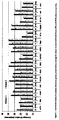
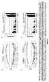
- Figure 1 represents the expression levels of putative reference genes, presented as average Ct values in matched fresh frozen (FF) and FFPE colon cancer LNs;
- Figure 2 represents the expression levels of putative reference genes, presented as average Ct values in GCC negative and positive FFPE LNs. Targeted Ct range was delimited by the two dotted lines;
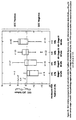
- Figure 3 represents the average expression stability values of control genes
- Figure 4 represents the determination of the optimal number of control genes for normalization
- Figure 5 represents the expression profile of selected reference gene in GCC positive and negative LNs.
- Figure 6 represents the effect of NaOH treatment on RNA quality and gene expression measures.
- the extent of RNA degradation following NaOH treatment was determined by capillary electrophoresis using the Agilent 2100 Bioanalyzer.
- Panel A) show a representative RNA fragmentation profile compared to intact RNA isolated from fresh frozen colon tissues and matched FFPE material.
- Figure 8 represents the effect of carbonate buffer alkaline treatment on RNA quality and gene expression pattern.
- the quality of RNA isolated at indicated time points was measured by capillary electrophoresis using the Agilent 2100 Bioanalyzer.
- Figure 10 represents the gene expression profiling in frozen and fixed, paraffin-embedded (FPE) tissues using TaqMan simplex assays and the GCC/ACTB duplex assay.
- the quality of RNA isolated from each condition was measured by capillary electrophoresis using the Agilent 2100 Bioanalyzer (Condition A and E are TRIzolTM extract, B: Neutral buffered formalin 10%, C: Non-buffered formalin 10%; D: Bouin's solution).
- Expression (Ct values) of GCC and 4 reference genes (GUSB, HPRT1, PGK1 and TBP) in TaqMan simplex reactions was compared to the GCC/ACTB duplex assay;
- the reverse transcription reactions were performed with the SuperscriptTM III First-Strand Synthesis SuperMix (Invitrogen) according to the manufacturer's recommendation, using gene-specific primers (2 ⁇ M) and 500 ng of total RNA based on Quant-IT data.
- the real-time PCR were carried out in a 20- ⁇ l reaction volume with the Applied Biosystems 7900HT Fast Real-Time PCR Systems using either TaqMan simplex or the GCC/ACTB duplex assay. Primers and probes concentration for each simplex assays were 900 nM and 250 nM respectively. All reactions were performed in duplicate;
- Figure 12 shows the comparison of GCC Taqman simplex and duplex amplifications in RNA extracted from a FFPE colon tissue.
- Upper panel shows GCC and various RG PCR amplifications in FFPE samples tested in simplex and duplex reactions using gene-specific reverse primers at 2 ⁇ M and 1.25 ⁇ g of RNA.
- Lower panel shows a comparison of minus RT-PCR amplification for GCC and various reference genes in simplex and duplex reactions;
- Figure 13 shows the comparison of GCC and GUSB Ct values between duplex and simplex reactions.
- the real-time PCR were carried out in duplex or simplex reactions of 20 ⁇ l with the Applied Biosystems 7900HT Fast Real-Time PCR Systems using TaqMan Fast Universal PCR Master Mix. Synthesis of cDNA was performed with 250 ng/ ⁇ l (A) or 25 ng/ ⁇ l (B) of uRNA spiked or not with 1x10⁶ GCC IVT.
- primers concentration was 900 nM and FAM or VIC-labeled probes concentration was 250 nM.
- For duplex reactions 4 concentrations of reverse and forward primers were tested while both FAM and VIC-labeled probes were fixed at 200 nM in a 20 ⁇ l PCR reaction;
- Figure 14 represents the GCC and RG delta-Ct variation in duplex and simplex assays during NaOH degradation.
- Expression of GCC was normalized with either GUSB or HPRT1.
- Variations in GCC relative quantification [(Ct GCC -Ct RG ) tx -(Ct GCC -Ct RG ) to ] were determined at indicated time points during NaOH hydrolysis. Variation between non-degraded and degraded samples should be lower than 1 delta-Ct to be considered not significant;
- Figure 15 represents the comparison of FFPE colon cancer LNs tested with TaqMan and ScorpionsTM duplex assays in minus RT condition.
- Panel A and C show Ct values for ACTB, GUSB, HPRT1 and GCC in 8 FFPE samples tested with 312.5 ng of cDNA in duplex reaction.
- minus RT experiments B and D
- no RT enzyme was added during the cDNA synthesis step;
- Figure 16 shows the comparison of GCC expression levels (delta-Ct) in cryosections of colon cancer tissue samples fixed or not.
- Delta Ct values ACt (Ct GCC -Ct RG ) were determined for each RNA extract using either GUSB (A and B) or HPRT1 (C and D).
- the reverse transcription reactions were performed in simplex using gene-specific primers at 2 ⁇ M and in duplex with indicated primers concentration.
- the real-time PCR were carried out in a 20 ⁇ l reaction volume with the Applied Biosystems 7900HT Fast Real-Time PCR Systems using either TaqMan® simplex or duplex assay were compared to ScorpionsTM duplex reaction. All reactions were performed in triplicate. Variation of less than 1 delta-Ct between frozen and fixed samples was considered not significant;
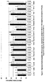
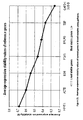
- Figure 17 represents the amplification of reference genes in RNA extracted from 55 FPE horronic lymph node tissues with different archiving times from 1 month to 22 years.
- Figure 18 shows the comparison of ACTB and GUSB Ct value observed in group of blocks with different archiving time. Box-and-Whisker plots of the ACTB (A) and GUSB (B) mRNA expression (Ct value) in FPE colon lymph node tissues. For multiple comparisons, one-way ANOVA and post-hoc Turkey's test were used and P ⁇ 0.05 was considered statistically significant;
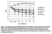
- Figure 19 represents the GCC relative expression levels in histopathology-negative and GCC/ACTB-negative (pN0(mol-)) and stage III GCC/ACTB-Positive (pN1-2(mol+)) LNs tested with the TaqMan GCC/GUSB assay.
- ROC receiver operating characteristic
- Figure 20 represents the GCC relative expression levels evaluated with the TaqMan GCC/GUSB duplex assay.
- GCC relative expression ACt Ct GUSB -Ct GCC
- HP- histopathology negative
- HP+ stage III positive
- GCC mRNA positive status was based on the analytical cut-off value of -5.9;
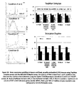
- Figure 21 shows the average expression stability values of control genes in blood samples using geNorm
- Figure 22 shows the determination of the optimal number of reference genes for normalization using geNorm
- Figure 23 represents the GCC expression level in blood samples. GCC mRNA positive status was based on the analytical cut-off value of 75 GCC units/mL;
- Figure 24 shows the distribution of A) ACTB Ct values and B) Glucuronidase GUSB Ct values for two different patient cohorts.
- Sensitivity is defined as the detection rate of recurrent cases (after 36 months) while specificity is defined as the proportion of negative cases without recurrence;
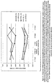
- Figure 26 illustrates the relation between risk of recurrence for patients with a GCC positive test result and the GCC expression level used to determine test positivity
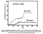
- Figure 27 is a Kaplan-Meier graphical analysis of time to recurrence based on GCC positivity.
- the GCC Negative cases are represented by the straight black line and the GCC Positive cases by the gray dashed line. Patients lost to follow-up were censored-out and are represented by straight up marks;
- Figure 28 is a Kaplan-Meier graphical analysis of RFS based on the GCC positivity for the GCC/GUSB test with -5.9 ⁇ Ct for cut-off.
- the GCC Negative cases are represented by the straight line and the GCC Positive cases by the dashed line.
- Censored-out cases are represented by straight up marks;
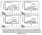
- Figure 29 is a Kaplan-Meier graphical analysis of time to recurrence with 2 levels of stratification for GCC positive patients.
- Figure 30 is a Kaplan-Meier graphical analysis of time to recurrence with 2 levels of stratification for GCC positive patients.
- Figure 31 is a Kaplan-Meier graphical analysis of time to recurrence with stratification for the number of GCC positive LNs per patients.
- GCC is meant to refer to the gene transcription product (RNA) expressing the cellular protein guanylate cyclase 2C (GUCY2C also referred to as the heat stable enterotoxin receptor or ST receptor), which is expressed by normal colorectal cells, as well as primary and metastasized colorectal, intestinal, stomach and esophageal cancer cells.
- RNA gene transcription product
- ST receptor heat stable enterotoxin receptor
- GCC also includes fragments of a GCC gene transcript which are functional with respect to nucleic acid molecules with full length sequence, such as a functional fragment which may be useful as an oligonucleotide or nucleic acid probe, a primer, an antisense oligonucleotide or nucleic acid molecule or a coding sequence.
- GCC also comprises the CRCA-1 alternative transcript.
- colonal cancer is meant to include the well-accepted medical definition that defines colorectal cancer as a medical condition characterized by presence of cancer cells in the intestinal tract below the small intestine (i.e. the large intestine (colon), including the caecum, ascending colon, transverse colon, descending colon, and sigmoid colon, and rectum). Additionally, as used herein, the term “colorectal cancer” is meant to further include medical conditions which are characterized by presence of cancer cells in the duodenum and small intestine (jejunum and ileum). The definition of colorectal cancer used herein is more expansive than the common medical definition but is provided as such since the cells of the duodenum and small intestine also contain GCC.
- GI tract cancer or "gastro-intestinal cancer” is meant to include the medical conditions which are characterized by presence of cancer cells in the esophagus, the stomach, the pancreas, the small intestine as well as in colon and rectum. Additionally, as used herein, the term “GI tract cancer” in meant to further include medical conditions which are characterized by presence of cancer cells in the pancreas, which like liver and gallbladder is an accessory organ of the GI tract. The definition of GI tract cancer used herein is more expansive that the common medical definition but is provided as such since pancreatic cancer cells are known to express GCC.
- upper GI tract consists of the mouth cavity, salivary glands, pharynx, esophagus, diaphragm, stomach, gall bladder, bile duct, liver, and duodenum.
- upper GI tract cancer as used herein particularly refers to the esophagus, stomach and pancreas.
- lower GI tract means of the bowel or intestines and the rectum and comprises the small intestine including duodenum, jejunum, ileum; and the large intestine or colon including caecum (and appendix); colon (ascending, transverse and descending) and the rectum (anus).
- esophageal cancer is meant to include the well-accepted medical definition that defines esophageal cancer as a medical condition characterized by presence of cancer cells in the esophagus.
- pancreatic cancer is meant to include the well-accepted medical definition that defines pancreatic cancer as a medical condition characterized by presence of cancer cells in the pancreas.
- Metastasis is meant to refer to the process in which cancer cells originating in one organ or part of the body, with or without transit by a body fluid, and relocate to another part of the body and continue to replicate. Metastasized cells can subsequently form tumors which may further metastasize. Metastasis thus refers to the spread of cancer, from the part of the body where it originally occurred, to other parts of the body.
- Metastasized colorectal cancer cells is meant to refer to colorectal cancer cells which have metastasized. Metastasized colorectal cancer cells are localized in a part of the body or body fluid other than the duodenum, small intestine (jejunum and ileum), large intestine (colon), including the caecum, ascending colon, transverse colon, descending colon, and sigmoid colon, and rectum.
- Metastasized stomach cancer cells are meant to refer to stomach cancer cells which have metastasized. Metastasized stomach cancer cells are localized in a part of the body other than the stomach.
- metastasized esophageal cancer cells is meant to refer to esophageal cancer cells which have metastasized. Metastasized esophageal cancer cells are localized in a part of the body other than the esophagus.
- pancreatic cancer cells As used herein, the term “metastasized pancreatic cancer cells” is meant to refer to pancreatic cancer cells which have metastasized. Metastasized pancreatic cancer cells are localized in a part of the body other than the pancreas.
- non-intestinal/rectal and “extra-intestinal/colorectal” are used herein interchangeably and are meant to refer to a sample of tissue or body fluid from a source other than intestinal (small intestine and colon) and rectal tissue.
- the extra-intestinal/colorectal sample is a sample of tissue such as lymph nodes.
- the non-intestinal/rectal sample is a sample of extra-intestinal/colorectal tissue which is an adenocarcinoma of unconfirmed origin.
- the non-intestinal/rectal tissue is a biopsy of a suspected stomach, pancreatic or esophagus cancer.
- the non-intestinal/rectal sample is a blood sample.
- an individual suffering from an adenocarcinoma of unconfirmed origin or “cancer of unknown primary origin” (CUP) is meant to refer to an individual who has a tumor in which the origin has not been definitively identified.
- CUP cancer of unknown primary origin
- the terms “subject” and “patient” refer to any animal, such as a mammal like livestock, pets, and preferably a human. Specific examples of “subjects” and “patients” include, but are not limited, to individuals requiring medical assistance, and in particular, patients with cancer.
- target or “target marker” or “biomarker target” refers to any molecule that can be derived from a eukaryotic cell.
- Targets include but are not limited to proteins or nucleic acid molecules.
- the level of a messenger RNA that is specifically expressed in cells of gastrointestinal origin is measured.
- a tissue specific protein or DNA alteration e.g. methylation or mutation
- single targets such as mRNA are detected individually.
- multiple targets are detected in combination.
- the terms "reference gene” or “reference marker” or “reference target” or “control” or “control marker” or “control target” refers to a reference molecule that controls and/or can be used to control for potential process interfering factors and/or provides one or more indications about the sample quality, the effective sample preparation and/or assembly of the RT-PCR reaction in the sample.
- a control may either be co-detected or detected separately from targets.
- sample refers to a biological material containing cells or other material retrieved from the patient.
- Sample material includes but is not limited to: tissue such as lymph node tissue; biopsy material; exhaled breath; or fluids such as blood (including serum or plasma); urine; semen; sputum, saliva; and combinations of these.
- the sample is processed (e.g. a lymph node is separated from other tissue and/or cut in multiple sections or cores, exposed or not to a chemical reaction, subjected to a separation process or blood is enriched in tumor circulating cells). Each process may result in a portion of the sample remaining, hereinafter referred to as “remaining sample” or simply "sample”.
- Sample may be defined as a single tissue sample, such as a single lymph node, or sample may define multiple samples, such as multiple lymph nodes or lymph node chain.
- single samples such as single lymph nodes are processed individually.
- multiple samples are "pooled" or processed together.
- the sample includes at least one entire lymph node.
- external standard means a synthetic DNA or RNA transcript in known amount(s) or concentration(s) that is tested separately from the test sample, i.e. through interpolation or extrapolation to a standard curve.
- parameters include one or more variables used in the method and system of the present invention to detect one or more targets.
- Parameters include but are not limited to: primer type; probe type; amplicon type; concentration of a substance; mass or weight of a substance; time for a process; temperature for a process; cycle threshold (Ct); activity during a process such as centrifugation, rotating, shaking, cutting, grinding, liquefying, precipitating, dissolving, electrically modifying, chemically modifying, mechanically modifying, heating, cooling, preserving (e.g. for days, weeks, months and even years) and maintaining in a still (unagitated) state.
- Parameters may further include a variable in one or more mathematical formulas used in the method of the present invention.
- Parameters may include a threshold used to determine the value of one or more parameters in a subsequent step of the method of the present invention.
- the threshold is a cycle count threshold.
- cycle threshold refers to the threshold in qPCR at which the fluorescence generated within a reaction well exceeds an established threshold or cutoff level.
- the cycle threshold refers to the same value than the terms “crossing point' (Cp) and "take-off point” (TOP) used by competing manufacturers of real-time PCR instruments for reasons of product differentiation.
- Cp crossing point'
- TOP take-off point
- hybridization is to be understood as a bond of an oligonucleotide to a complementary sequence along the lines of the Watson-Crick base pairings in the sample DNA, forming a duplex structure.
- “Stringent hybridization conditions,” as defined herein, involve hybridizing at 68°C in 5X SSC/ 5X Denhardt's solution/ 1.0% SDS, and washing in 0.2X SSC/ 0.1 % SDS at room temperature, or involve the art-recognized equivalent thereof (e.g., conditions in which a hybridization is carried out at 60°C in 2.5X SSC buffer, followed by several washing steps at 37°C in a low buffer concentration, and remains stable).
- Moderately stringent conditions as defined herein, involve including washing in 3X SSC at 42°C, or the art-recognized equivalent thereof.
- the parameters of salt concentration and temperature can be varied to achieve the optimal level of identity between the probe and the target nucleic acid.
- clinical assessment is meant to include a potential range or continuous or discrete values used for the screening, diagnosis, staging, prognosis, treatment planning, monitoring and surveillance of a cancer patient.
- time-to-Recurrence is defined as the time to any event related to the same cancer. All same cancer recurrences and deaths from the same cancer are events. Second primary same cancers and other primary cancers are ignored. Deaths from other cancers, non-cancer-related deaths, treatment-related deaths, and loss to follow-up are censored observations.
- Relapse-Free Survival or “Recurrence-Free Survival” (RFS) is defined as the time to any event, irrespective of the cause of this event, except for any second primary cancer. Recurrence of or death from the same cancer and all treatment-related deaths or deaths from other causes are events. Second primary from the same cancers and other primary cancers are ignored, and loss to follow-up is censored.
- CCS Cancer-Specific Survival
- DFS Disease-Free Survival
- OS Overall Survival
- the "staging" or “stage” of a cancer refers to the TNM (for tumors/nodes/metastases) system, from the American_Joint Committee on Cancer (AJCC) ( Greene et al. (eds.), AJCC Cancer Staging Manual, 6th edition, New York, NY: Springer; 2002 ), which depends on the extent of local invasion, the degree of lymph node involvement and whether there is distant metastasis. Staging is done after surgery has been performed and pathology reports reviewed.
- TNM denotes the degree of invasion of the intestinal wall
- N the degree of lymphatic node involvement
- M the degree of metastasis.
- the broader stage of a cancer is usually quoted as a number I, II, III, IV derived from the TNM value grouped by prognosis; a higher number indicates a more advanced cancer and likely a worse outcome.
- Details of this system for colorectal cancer are the following: AJCC stage TNM stage TNM stage criteria for colorectal cancer Dukes Astler-Coller Stage 0 Tis N0 M0 Tis: Tumor confined to mucosa; cancer- in-situ - - Stage I T1 N0 M0 T1: Tumor invades submucosa A A Stage I T2 N0 M0 T2: Tumor invades muscularis basement A B1 Stage II-A T3 N0 M0 T3: Tumor invades subserosa or beyond (without other organs involved) B B2 Stage II-B T4 N0 M0 T4: Tumor invades adjacent organs or perforates the visceral peritoneum B B3 Stage III-A T1-2 N1 M0 N1: Meta
- C C1 Stage III-B T3-4 N1 M0 N1 Metastasis to 1 to 3 regional lymph nodes.
- C C2, C3 Stage III-C any T, N2 M0 N2 Metastasis to 4 or more regional lymph nodes.
- Any T. C C1, C2, C3 Stage IV any T, any N, M1 M1 Distant metastases present. Any T, any N. - D
- the stage can also be reported in letters rather than numbers, according to the Dukes and Astler-Coller staging systems, which often combine different AJCC stage groupings and are not as precise, as shown in the above table.
- the survival rates for colon cancer are from a study of the National Cancer Institute's SEER database, looking at nearly 120,000 people diagnosed with colon cancer between 1991 and 2000. In this study, survival was better for stage IIIA than for stage IIB. Stage 5-year Survival Rate I 93% IIA 85% IIB 72% IIIA 83% IIIB 64% IIIC 44% IV 8%
- lymph node involvement refers to a qualitative notion about the presence of metastases in lymph nodes as determined visually through a histopathology procedure.
- a patient harboring no involved nodes is designated “N0" or pN0.
- the lymph node involvement is designated “N1" or "pN1”.
- N2 or “pN2” is used to designate a lymph node involvement, or presence of metastases, in 4 or more regional lymph nodes.
- the lymph node involvement is a criteria used by clinicians to determine whether or not a patient should receive adjuvant chemotherapy.
- GCC nodal involvement refers to a qualitative notion about the presence or absence of Guanylyl cyclase C (GCC or GUCY2C) mRNA in an individual lymph node, which is indicative of the presence or absence of nodal metastases including occult metastases i.e. metastases or a cluster of cancer cells that cannot be detected by histopathology.
- GCC Guanylyl cyclase C
- GUCY2C Guanylyl cyclase C
- the terms "GCC burden” or "GCC load” refer to a quantification of the amount of GCC expressing cells found in a particular lymph node or to a total amount of GCC mRNA in a group of lymph nodes of a patient.
- the GCC burden is not significant or not clinically significant when the detectable quantity of GCC mRNA in a given node or in all the lymph nodes collectively is below a given level.
- the GCC burden is significant or clinically significant and can be used to discriminate between patients with a lower risk of recurrence from those with a higher risk.
- the level of GCC mRNA in a given lymph node or in a group of lymph nodes can be expressed in many ways, such as in terms of copies or copies per lymph node mass (absolute quantification) or in terms of delta Ct ( ⁇ Ct). delta-delta Ct ( ⁇ Ct), or fold change (2 - ⁇ Ct ) (2 exponent minus delta-delta Ct), these last three parameters being based on the expression level of GCC relative to the expression level of a reference gene (relative quantification), such as, but not limited to, GUSB, in the same lymph node.
- lymph node ratio refers to the number of GCC-positive lymph nodes over the total number of measurable lymph nodes tested for a given patient.
- the invention provides a method selected from the ones as defined herein and more particularly as defined above, wherein one or more reference genes is normally expressed in normal cells of the extra-intestinal/colorectal sample.
- the reference gene is beta-glucuronidase (GUSB). More particularly, the measuring of expression levels is carried out using RT-qPCR.
- a method of detecting GCC in a sample collected from a patient comprising the following sequential steps:
- a method for the detection of GCC in an extra-intestinal/colorectal sample collected from a subject comprising the steps of:
- a method for the measurement of GCC in a sample comprising the steps of :
- a method for the measurement of GCC in a sample comprising the following steps:
- the method as defined above uses the expression fold change (delta-delta-Ct) to determine the changes in mRNA level of GCC in said sample and expresses it relative to the mRNA level of beta-glucuronidase (GUSB) in the same sample.
- a method of determining the GCC burden of a patient diagnosed with cancer comprising carrying the steps of the method as defined herein, wherein if delta-Ct is equal or higher than about -12, the quantity of GCC mRNA is calculated in terms of number of copies in relation to an external standard, whereby the GCC burden is expressed in number of GCC copies in the sample
- a method of diagnosing cancer in a patient suspected of having cancer comprising the steps of quantifying GCC in an extra-intestinal/colorectal sample of said patient in accordance with the method of the invention; and determining whether said sample harbors GCC positive cells, whereby the presence of GCC positive cells is indicative of colorectal, stomach, small intestine, esophageal or pancreatic cancer.
- a method of staging a human patient already diagnosed with cancer comprising the steps of:
- a method of monitoring, or diagnosing metastasis in, a human already diagnosed with cancer comprising the steps:
- a method to select among cancer patients having histopathologically negative lymph nodes those who can benefit from a course of treatment comprising:
- a method of predicting the risk of cancer recurrence for a patient already diagnosed with cancer comprising carrying the steps according to the method of the invention, wherein a delta-delta-Ct between -6 and -3 is indicative of the presence of GCC positive cells in the sample, whereby the presence of GCC positive cells is indicative that the patient has increased risk of recurrence of cancer.
- a method of determining the GCC burden of a patient diagnosed with cancer comprising carrying the steps of the methods as defined herein, wherein if delta-Ct is equal or higher than about -12, the quantity of GCC mRNA is calculated in terms of number of copies in relation to an external standard, whereby the GCC burden is expressed in number of GCC copies in the sample.
- the threshold or cut-off for positive identification of GCC positive cells is a delta-Ct above about -12. More particularly, the threshold for positive identification of GCC positive cells is a delta-Ct above about -10. More particularly, the threshold for positive identification of GCC positive cells is a delta-Ct above about -8. Still, more particularly, the threshold for positive identification of GCC positive cells is a delta-Ct above about -6. Even more particularly, the threshold for positive identification of GCC positive cells is a delta-Ct above about -4. Most particularly, the threshold for positive identification of GCC positive cells is a delta-Ct above -2.
- the method as defined herein may include one or more analyses or algorithms used to detect a target or perform an analysis based on the detection of the at least two targets (GCC and GUSB).
- Such analysis or algorithm may have a bias, such as a false-positive or false-negative bias.
- the analysis or algorithm may take into account a combination of disease factors or clinical factors such as: age, race, an existing patient condition, use of adjuvant therapy, heredity; and so on.
- the method may comprise the inclusion of multiple parameters used to perform a step of a procedure or used by an algorithm of the procedure such as multiple reference genes detected and measured in addition to GUSB.
- the invention is applicable to be performed with a sample from a patient with cancer, particularly GI tract cancer of a wide variety of stages, level of aggressiveness, level of illness, symptomatic or asymptomatic, or other adverse conditions.
- the patient has a GI tract cancer from the upper or lower GI tract.
- the cancer may be selected from a colorectal cancer, a small intestine, a stomach cancer, a pancreatic cancer, or an esophageal cancer.
- the patient has a stage I or stage II cancer. More particularly, the cancer is colorectal cancer.
- the patient may be referred to as a cancer patient such as a colorectal cancer patient.
- This terminology shall include patients in whom the presence of cancer has been confirmed, currently or historically, as well as patients that may, for any reason, be suspected of having cancer or otherwise receive a cancer diagnostic test of the present invention. Positive detection of the target may correlate to the presence of cancer; a specific prognosis or diagnosis of the cancer; or other clinical assessment or recommendation.
- the method of the invention can be carried out on numerous forms of samples such as extra-intestinal/colorectal sample including but not limited to tissue or biological fluid.
- the sample is taken from an organ that does not normally express GCC.
- a sample can be a tissue which has been preserved or otherwise archived.
- the sample may be one or more lymph nodes collected from a single patient, particularly during a resection procedure. More particularly, the lymph nodes are collected during a colorectal, esophagus, stomach or pancreatic resection.
- H&E Hematoxylin and Eosin
- the method of the present invention avoids and/or reduces these issues, and can detect one or more targets indicative of numerous patient adverse conditions including but not limited to the presence of: metastases; micrometastases; occult metastases; isolated tumor cells; clusters of tumor cells and combinations of these.
- the molecular evaluation of the current invention provides more systematic, repeatable, automatable tests that can be performed with high accuracy, sensitivity and repeatability.
- the sample is particularly an archived lymph node (e.g. a fixed, formalin-embedded sample including one or more lymph nodes), but may be fresh or frozen tissue.
- the sample may include tissue from one or more of the following anatomical locations/organ: breast, prostate, stomach, esophagus, pancreas, kidney, spleen, cervix, vagina, ovary, bladder, thyroid, colon, rectum, small intestine, brain, skin, liver and lung.
- the sample includes multiple nodes which are "pooled” or processed together.
- the number of copies detected is correlated to a specific assessment of patient condition including but not limited to cancer stage or therapy outcome.
- lymph nodes including a peri-colonic lymph node
- other lymph nodes other tissue and other samples may be processed by the method described herein.
- the method of the present invention may provide an analysis of a sample that is a cancer of unknown origin.
- a cancer sample such as a brain, lung or liver tumor, is processed to detect GCC to determine that the origin of the cancer as the colon or rectum or stomach, esophagus, small intestine or pancreas (e.g. vs. the lung, liver, brain or other location).
- the method of present invention produces results from one or more molecular tests, such as a molecular test for GCC in a lymph node harvested in a surgical procedure removing a portion of a patient's colon.
- a molecular test for GCC in a lymph node harvested in a surgical procedure removing a portion of a patient's colon.
- the lymph nodes or other tissues are also histologically analyzed and the results of both the molecular test(s) and histological test(s) are combined to perform a subsequent assessment.
- the number of GCC copies is correlated with the number of cells identified as cancerous in the histological analysis.
- the correlation can be made on a first patient, or a first set of patients.
- the number of copies detected can be determined via molecular testing, and correlated to a predicted number of cells that would be identified in histological tests. This predicted number, combined with or without a histological examination for cells, is used to produce a more specific assessment of patient condition including but not limited to cancer stage or therapy outcome.
- the sample may include other body tissues, or biological fluids such as exhaled breath, blood, urine, sputum, saliva and/or semen.
- the sample is blood.
- precautions are taken throughout each step to avoid cross-contamination of tissue, such as contamination between tissue samples received from the patient (e.g. two lymph nodes), or contamination from a first patient to a second patient.
- the sample is retrieved from the patient in a clinical setting such as a hospital, and one or more further processing steps are also performed at that or an additional clinical setting.
- the sample is then transferred to a clinical or medical laboratory, such as a CLIA laboratory, for further processing. Results of the further processing may be analyzed, at the laboratory and/or a clinical setting (e.g. by a clinician of the patient).
- the sample consists of multiple patient lymph nodes collected in a colorectal resection procedure, typically consisting of 12 lymph nodes but optionally from 1 up to 100 or more, including sentinel nodes.
- the sample is processed (e.g. physically divided such as a lymph node separated from other tissue and/or a lymph node cut in multiple sections or cores with a scalpel; exposed to a physicochemical reaction such as a deparaffinization and/or a precipitation procedure; exposed to a separation process such as separation in a centrifuge; exposed to a washing procedure; and the like).
- a physicochemical reaction such as a deparaffinization and/or a precipitation procedure
- a separation process such as separation in a centrifuge
- washing procedure and the like.
- Sample may be defined as a single tissue sample, such as a single lymph node, or sample may define multiple samples, such as multiple lymph nodes.
- single samples such as single lymph nodes are processed individually.
- multiple samples are processed in combination.
- the sample includes at least one entire lymph node, such as to avoid testing a first portion of a lymph node that does not include the target wherein a second portion does include the target.
- Samples may be preserved (an "archived sample”) such as to prevent degradation over time.
- Preservation methods include but are not limited to: refrigeration such as freezing; use of a preservative tissue solution; dehydration; and combinations of these.
- tissue preservative solutions include but are not limited to: commercial products such as formalin (a buffered or non-buffered aqueous solution of formaldehyde); Bouin's solution (consisting of a mixture of picric acid and formaldehyde); PAXgene Tissue Fix, PAX gene Tissue Stabilizer, RNARetainTM solution; RNALaterTM solution; nonaqueous solutions such as that described in US7,138,226 ; and combinations of these. Paraffin-embedding and/or other similar material-embedding may or may not be performed after tissue fixation, such as to assist in the creation of sections such as slide sections, to facilitate transport and non-detrimental storage.
- the extra-intestinal/colorectal sample is a lymph node or blood.
- the extra-intestinal/colorectal sample is a lymph node; more particularly, one single lymph node, most particularly two or more lymph nodes, still most particularly at least four lymph nodes, and even most particularly at least twelve lymph nodes.
- the relative risk of recurrence for this patient according to the GCC/GUSB test is intermediate. More particularly, when the positive results are found in 4 or more lymph nodes of the same patient, the relative risk of recurrence for this patient according to the GCC/GUSB test is high. Still more particularly, the method allows to discriminate between cancer patients having histopathologically negative lymph nodes, wherein cancer patients with GCC positive cells in at least one lymph node have a risk of recurrence and survival rate comparable to that of patients considered at higher risk by histopathology, thereby indicating that these patients might benefit from treatment with adjuvant chemotherapy. In contrast, cancer patients with all lymph nodes being GCC negative are at a lower risk of disease recurrence and can avoid negative side effects of treatment with adjuvant chemotherapy. Most particularly, the presence of GCC positive cells is indicative of a poor prognosis.
- the quantity of GCC detected is calculated for each individual lymph node. More particularly, the quantity of GCC is the sum of the individual quantities of GCC in all lymph nodes of the patient.
- the GCC burden is established for each individual lymph node. More particularly, the GCC burden is established on the basis of the total amount of GCC in all lymph nodes of the patient. Still, more particularly, the GCC burden is determined and a GCC burden above a given number of GCC copies is indicative of an increased likelihood of cancer recurrence.
- a GCC RT-qPCR test is used to detect the presence of GCC in lymph nodes, tissues or biological fluids obtained from a colon-, rectum-, esophagus-, small intestine-, pancreas-, or stomach-cancer patient, the detection correlating to one or more clinical assessment related to that patient's cancer.
- FFPE formalin-fixed paraffin-embedded lymph nodes
- a RT-qPCR assay is used to quantitatively detect GCC.
- the processing includes homogenization of the lymph node tissue followed by nucleic acid (e.g. RNA) extraction.
- the RT-qPCR assay may use a non-specific (e.g. SYBR green) or specific (e.g. ScorpionsTM, Molecular Beacons, Locked Nucleic Acid (LNA) Fluorescent Probes, Amplifluor, or Taqman) detection chemistries.
- SYBR green non-specific
- specific e.g. ScorpionsTM, Molecular Beacons, Locked Nucleic Acid (LNA) Fluorescent Probes, Amplifluor, or Taqman
- GCC is quantified relative to the average expression of beta-glucuronidase (GUSB) as a reference or control gene.
- PCR efficiency correction is used.
- the assay is a duplex assay detecting GCC and GUSB as reference gene.
- GCC and GUSB are detected from simplex assays.
- the assay is a triplex assay detecting GCC, GUSB and another reference marker, such as: GAPDH, HPRT1, PGK1 and TBP and/or a spiked internal control.
- GAPDH GAPDH
- HPRT1 HPRT1, PGK1 and TBP
- Particular examples of endogenous genes that can be used as reference genes are those associated with SEQ ID NOs: 2-16 and listed in Table 1.
- Typical supporting material is paraffin wax.
- other materials can be used such as certain inert plastics or epoxies, or other supportive material lacking reactivity with the sample.
- Table 1 Endogenous reference genes evaluated in this study. SEQ ID No.
- PCR methods and more specifically RT-qPCR, are preferred, other similarly reliable, sensitive and specific amplification and detection methods such as Rolling Circle Amplification methods (RCA), Branched-Chain DNA Amplification (BCA), Ligase Chain Reaction methods (LCR), Strand Displacement Amplification methods (SDA), Nucleic Acid Sequence Based Amplification methods (NASBA), Transcription-Mediated Amplification methods (TMA) and others can also be used.
- Detection technologies which may or may not follow nucleic acid amplification, may include MALDI-TOF mass spectrometry, capillary electrophoresis, and similar detection methods.
- GCC is quantified relative to the average expression GUSB as a reference or control gene.
- the primers for GCC and GUSB are RT primers and are added in the same assay at a predetermined ratio in order to optimize the detection of each marker by itself and in relation to the other marker.
- the RT primers for GCC are selected from polynucleotides capable of hybridizing to: NM_004963 (SEQ ID No.1) and are added to the assay in a quantity of about 10 ⁇ M to 30 ⁇ M.
- the primers for GUSB are selected from polynucleotides capable of hybridizing to: NM_000181 (SEQ ID No.2) and are added to the assay in a quantity of about 1 ⁇ M to 3 ⁇ M.
- NM_000181 SEQ ID No.2
- primers are capable of hybridizing to a location on the GCC or GUSB coding regions, preferably such primers are spanning two exons. More particularly, such primers are free from single nucleotide polymorphism (SNP).
- primers and probes of the useful for this method comprise polynucleotides having 90% identity to SEQ ID NOs: 17-43. More particularly, primers comprising polynucleotides having 90% identity to SEQ ID NOs: 17, 18, 20, 21, 23, 24, 26, 26, 29, 30, 32, 33, 35, 36, 38, 39, 41 and 42 can be useful for this method. Particular examples of polynucleotide primers and probes of the invention are shown as SEQ ID NOs: 17-43 and are listed in Table 2. More particularly, primers and probes of SEQ ID NOs. 20, 21, 22, 38, 39 and 40 are useful for the present method.
- the RT primers for GCC are present at about 10-20 ⁇ M and the primers for GUSB are added to the assay at about 1-4 ⁇ M.
- RT primers for GCC:GUSB are present in the assay at a ratio of about 10:1.
- probes specific for GCC or GUSB are selected from the group consisting of: polynucleotides capable of hybridizing to GCC short coding sequences or GUSB short coding sequences under stringent conditions. Particular examples of these probes are listed in Table 2.
- probes specific for GCC are added to the assay in an amount of about 200 nM.
- probes specific for GUSB are added to the assay in an amount of about 200 nM.
- a short GCC amplicon length and a short GUSB amplicon length are used to detect GCC expressing cells.
- a test of the present invention analyzes RNA of less than 100 nucleotides in a sample.
- a test of the present invention analyzes RNA of less than 80 nucleotides.
- a test of the present invention analyzes RNA of less than 70 nucleotides. Table 3. Comparison of recommended conditions for the GCC/GUSB TaqMan assay and GCC/ACTB ScorpionsTM.
- the present method provides the detection of the GCC gene transcription product in a sample.
- detection particularly refers to identifying, locating, obtaining a positive signal, measuring, or quantifying.
- Positive detection of the target may include the detection of one or more targets. Positive detection of the target may require detection of the target above a threshold. Positive detection may be a direct measure of finding cancer cells (e.g. target is cancer cell), and/or a direct measure that provides a diagnosis or prognosis of cancer. Alternatively, positive detection of the marker may be associated with a surrogate to an assessment, the surrogate being the measure of finding cancer cells, and/or surrogate measure that provides a diagnosis or prognosis of cancer.
- the present invention provides a method for detecting GCC in a sample collected from a patient.
- Detection of GCC includes a quantification of GCC relative to the quantification of GUSB found in the same sample, and may be used to diagnose, stage, prognosticate, monitor and/or manage the treatment of an adverse patient condition such as the presence of cancer.
- the method of the present invention includes one or more analyses or algorithms used to detect a target or perform an analysis based on the detection of the at least two targets (GCC and GUSB).
- the cycle threshold (Ct) or cycle number in qPCR is the threshold at which the fluorescence generated within a reaction exceeds an established threshold or cutoff level. Positive and negative signals are respectively defined as being beyond and below an established cutoff level (threshold).
- the cutoff level is established by testing two populations of samples with known conditions, one collected from donors having the condition (positive) and the second one collected from donors not having the condition (negative).
- the population of positive samples may be lymph nodes collected from colorectal cancer patients and having been identified as pN1 or pN2 by histopathology; the population of negative samples may be lymph nodes collected from patients having other conditions than colorectal cancer, such as breast cancer, lung cancer, gastrointestinal inflammatory conditions, etc.
- the population of positive samples may be lymph nodes collected from colorectal cancer patients having recurred from the disease during the 5 years following the resection of the primary tumor and having been identified as pN1 or pN2 by histopathology;
- the population of negative samples may be lymph nodes collected from colorectal cancer patients having not recurred or died from the disease during the 5 years following the resection of the primary tumor and having been identified as pN0 by histopathology.
- the population of positive samples may be blood samples collected from colorectal cancer patients having recurred or died from the disease during the 5 years following the resection of the primary tumor;
- the population of negative samples may be blood samples collected from colorectal cancer patients having not recurred from the disease during the 5 years following the resection of the primary tumor.
- An analysis or algorithm may have a bias, such as the false-positive or false-negative bias.
- An analysis or algorithm may be modified by an existing patient condition, such as has been described hereinabove.
- the method of the present invention includes multiple parameters used to perform a step of a procedure or used by an algorithm of the procedure.
- the parameters may be established and/or modified through testing of various types of tissue not from the patient of the present invention, such as lymph nodes or other tissue harvested from other humans, pigs and cows.
- a delta-Ct equal or higher than -6 (if Ct GUSB -Ct GCC ) or equal or lower than +6 (if Ct GCC minus Ct GUSB ) is indicative of the presence of GCC positive cells in the sample, whereby the presence of GCC positive cells is indicative that the patient has increased risk of recurrence of cancer.
- a delta-Ct equal or higher than -5.9 represents a GCC positive result
- a delta-Ct lower than -5.9 represents a GCC negative result, whereby said result allow discrimination for risk of recurrence and relapse-free survival (RFS) between GCC-negative and GCC-positive results.
- RFS relapse-free survival
- a delta-Ct equal or higher than about -6 represents a GCC positive result and a delta-Ct lower than about -6 represents a GCC negative result, where the result allow discrimination for risk of recurrence and relapse-free survival (RFS) between GCC-negative and GCC-positive results.
- a delta-Ct equal or higher than -5.9 represents a GCC positive result and a delta-Ct lower than -5.9 represents a GCC negative result, where the result allows discrimination for risk of recurrence and relapse-free survival (RFS) between GCC-negative and GCC-positive results.
- a pre-established cut-off level for delta-Ct of between about -6 and -3 is suitable to determine the status of GCC positive or GCC-negative cells. More particularly, the cut-off level is selected from the group consisting of: -5.9, -5.5, -5.0; -4.5; - 4.0; -3.5; and -3.0.
- kit may include one or more components, supports, vials, substances or reagents as well as instructions booklet, as is described in detail herein.
- the kit for the detection, diagnosis, prognosis, monitoring and/or staging of a cancer in a patient comprises reagents for detecting GCC in an extra-intestinal/colorectal sample from the patient; reagents for detecting GUSB in the same sample; and instructions on how to quantify GCC in relation to GUSB.
- the kit for the detection, diagnosis, prognosis, monitoring and/or staging of a cancer in a patient comprises: PCR reagents for detecting GCC in an extra-intestinal/colorectal sample from the patient; instructions on how to determine a cycle threshold for GCC (Ct GCC ); PCR reagents for detecting GUSB in the same sample; instructions on how to determine a cycle threshold for GUSB (Ct GUSB ); and instructions on how to calculate (delta-Ct) or (delta-delta-Ct) between Ct GCC and Ct GUSB .
- RT-qPCR real-time quantitative reverse transcription PCR
- RNA integrity Several parameters have been standardized in order to obtain reliable quantitative expression measures for reference genes analysis including initial sample amount, RNA integrity and efficiency of cDNA synthesis.
- the reverse transcription reactions were performed with the SuperscriptTM III First-Strand Synthesis SuperMix (Invitrogen) according to the manufacturer's recommendation, using random hexamers (50 ng/ ⁇ l) and 1 ⁇ g total RNA.
- the real-time PCR were carried out in a 20- ⁇ l reaction volume with the Applied Biosystems 7900 HT Fast Real-Time PCR Systems using TaqMan Fast Universal PCR Master Mix and 50 ng cDNA. Primers and FAM-labeled probes concentration for each PCR assay was 900 nM and 250 nM respectively. All reactions were performed in duplicate, and results were averaged. PCR efficiencies in individual samples were evaluated for each gene using the LinReg software.
- GeNorm uses a pair-wise comparison model to select the gene pair showing the least variation in expression ratio across samples.
- the software computes a measure of gene stability (M) for each endogenous reference gene.
- Figure 3 shows the M values for all tested genes.
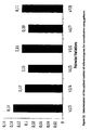
- GAPDH and PGK1 were identified as the most stable gene pair followed by HPRT1, TBP and GUSB ( Figure 3 ).
- ACTB on which is based the GCC/ACTB ScorpionsTM duplex assay is in eleventh position and PP1A showed the highest variability in expression in the FFPE colon LNs.
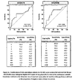
- the geNorm software calculates a pairwise variation V for each sequentially increasing number of reference genes added.
- Figure 4 shows a graph of the pairwise variation calculated by the geNorm software. The geNorm default V value of 0.15 was used as a cutoff to determine the optimal number of genes. This analysis reveals that the optimal number of reference genes is three (GAPDH, PGK1 and HPRT1) when using RNA extracted from FFPE colon cancer LNs ( Figure 4 ).
- the optimal reference genes among those tested for GCC relative quantification analysis using FFPE colon cancer LNs were GAPDH, PGK1, HPRT1, TBP and GUSB. Besides, these five endogenous reference genes are less abundant than ACTB in FPE LNs and are not as affected by the presence of GCC mRNA in the LN. Since these reference genes have expression levels 3-6 cycles lower than ACTB, they are less likely to compete with GCC in the reverse transcription reaction. This suggests that in contrast to ACTB, the primer concentration in the RT reaction should not be limited. Due to its high expression, ACTB is not optimal for relative quantification, although we found its expression to be stable in horronic LNs. For that reason, the combination of three genes rather than ACTB alone was considered for the development of a test format using relative quantification as opposed to a standard curve-based quantification.
- oligonucleotide primers and probe of SEQ ID NO:38-40 useful here for detecting GUSB gene expression in FFPE colon cancer LNs were also disclosed to be useful in the international application WO 2008/045133 to study gene expression in fresh frozen colorectal cancer tissue sample.
- uRNA commercial human Universal RNA
- serial dilutions of a commercial human Universal RNA (uRNA) allowed us to perform an initial qualification of the PCR efficiency of the assay designs.
- Serial dilutions of a commercial human Universal RNA (uRNA) allowed us to perform an initial qualification of the PCR efficiency of the assay designs.
- the reverse transcription reactions were performed with the SuperscriptTM III First-Strand Synthesis SuperMix (Invitrogen) according to the manufacturer's recommendation, using gene-specific reverse primers (2 ⁇ M) and RNA input ranged from 1250 ng to 0.125 ng.
- the real-time PCR were carried out in simplex reactions of 20 ⁇ l with the Applied Biosystems 7900HT Fast Real-Time PCR Systems using TaqMan® Fast Universal PCR Master Mix. Primers and FAM or VIC-labeled probes concentration for each PCR assay was 900 nM and 250 nM respectively as recommended. Apart for 1 HPRT1 design, all other assays produced PCR efficiencies between 90-110% (Table 1).
- Expression levels (Ct values) of GUSB, HPRT1, PGK1 and TBP ranged between 21-23 with 1250 ng and 34-36 with 0.125 ng. Both GAPDH designs had Ct values lower than our primary specification (Ct values between 23-36).
- the 5 selected reference genes were evaluated in FFPE RNA extracted from GCC positive and negative LN in order to select a reference gene with the lowest Ct variation and standard deviation.
- expression levels of the reference genes were determined in a serial dilution of RNA from GCC positive LNs using 10 different TaqMan MGB assays (Table 2).
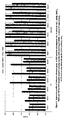
- the Ct values for all 10 reference gene assays ranged from 18 to 34 (Table 5).
- GAPDH assays gave Ct values lower than our specification and both GUSB_Tq2 and HPRT1_Tq2 had PCR efficiencies outside the 90-110% range.
- Minus RT reactions were also performed in parallel and we consistently obtained Ct values below 35 for TBP_Tq1 and Tq2, GAPDH_Tq1 and HPRT1_Tq2 (Table 5), suggesting a non-specific amplification of genomic DNA or PCR by products by these assays.
- Stable relative expression levels independent of RNA input between GCC and each reference gene were obtained in RNA sequentially diluted.
- the smaller amplicons tended to have less Ct variation than the larger ones (Table 5). Table 5.
- RNA extracted from FFPE LNs of colon cancer patients was then confirmed in RNA extracted from FFPE LNs of colon cancer patients and used to measure relative expression of GCC mRNA.
- RNA from 3 GCC positive (Cybrdi) and 3 GCC negative (ABS, McGill and Tristar) LNs were tested.
- the nucleic acid remains from 3 GCC negative lymph nodes were selected from a previous experiment in which ACTB expression could be detected in minus RT controls. Minus RT reactions were also performed in parallel.
- the Ct values measured for each reference gene were between 22 and 26 cycles compared to 20 for ACTB using the ScorpionsTM duplex assay ( Figure 5 ).
- GUSB, HPRT1, PGK1 and TBP were evaluated to identify the reference gene having a loss of signal similar to GCC in degraded samples.
- degradation experiments were optimized to obtain GCC levels that cover the dynamic range of the GCC/ACTB ScorpionsTM duplex assay, i.e. with at least 5 degradation points with measurable GCC levels.
- RNA from a fresh frozen colon tissue sample was treated with 0.1 N NaOH at 60°C.
- an equal volume of ice-cold 0.1 M HCl was added to the sample to neutralize NaOH and stop RNA degradation.
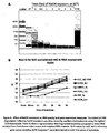
- the resulting degraded RNA was characterized on the Agilent 2100 Bioanalyzer. Controlled nucleic acid degradation at elevated pH generated RNA fragments from 50 to 300 nucleotides depending on the time of hydrolysis. Substantial RNA degradation with more than 50% of RNA fragments below 150 nucleotides was observed after 3 h of treatment ( Figure 6A ).
- RNA from each time point was amplified using the GCC TaqMan® assay and the four selected reference gene designs (GUSB, HPRT1, PGK1 and TBP).
- GUSB, HPRT1, PGK1 and TBP the GCC ScorlpionsTM duplex with ACTB was also tested. Because we designed GCC and RG assays with short amplicons, measurable expression could be detected even after 5 hours of treatment. As expected, a constant increase of GCC values was observed throughout treatment while only a slight increase can be observed for ACTB Ct values ( Figure 6B ). The increase of the 4 other reference gene Ct values was very similar to GCC mRNA in this model ( Figure 6B ).
- RNA from a TRIzolTM -extracted fresh frozen colon tissue sample was dissolved in an equal volume of NaHCO3/Na 2 CO 3 buffer (pH10) and incubated at 60°C for various time points (30 min, 1h, 2h, 3h and 4 h).
- 1/10 volume of 3M CH 3 COONa pH 5.2 was added on ice followed by precipitation with 3 volumes of 99% ethanol.
- Small fragmented RNAs were recovered in 50 ⁇ l and purified on mini Quick Spin RNA Columns (Roche Applied Science, IN).
- RNA was DNase-treated using the TURBO DNA free kit (Ambion).
- RNA samples were then subjected to RT-qPCR and Ct values for GCC and five reference genes are shown for a representative experiment ( Figure 8C ). In this model, GCC Ct values increased by more than 6 cycles after 4h of treatment. Most of the reference genes tested, including ACTB, behave similarly to GCC in this model ( Figure 8C ).
- pre-fixation parameters are critical for RNA quality and that once the tissue is fixed, dehydrated and embedded in paraffin, further degradation is limited.
- one OCT-embedded colon tumor tissue was sectioned in 40 slices of 20 ⁇ m. Eight noncontinuous sections were pooled and either fixed or extracted with TRIzolTM reagent. Effects of tissue fixative on RNA quality were measured in cryo-sections that were fixed for 16 hrs with either neutral buffered formalin, non-buffered formalin or Bouin's solution by using the Agilent 2100 Bioanalyzer ( Figure 10 ). As expected, each condition generated a unique RNA profile.
- RNA extracted with TRIzolTM reagent showed the typical 18S and 28S fragments.
- RNA extracted from sections fixed with neutral buffered formalin appeared to be smaller than 400 bp, while non-buffered formalin seemed to have retained genomic DNA along with RNA molecules with overall less fragmentation.
- Bouin's solution was the most destructive fixative with a majority of RNA molecules smaller than 150 bp.
- FIG. 6 shows Ct values for GCC and 4 reference genes detected with TaqMan® simplex assay compared to the GCC/ACTB ScorpionsTM duplex assay.
- delta-Ct was rather stable with less than 0.5 delta-Ct variation between fresh frozen and neutral buffered formalin fixed samples when normalized with GUSB, HPRT1 and TBP ( Figure 11 ). This suggests that variations in Ct values caused by pre-analytical stress factors can be compensated with a suitable normalization procedure.
- the concentration of primers in the qPCR reaction was adjusted in the duplex assay to minimize the competition between GCC primers and reference gene primers.
- Primer titration was performed in human universal RNA (uRNA) spiked or not with 1x10 6 in vitro transcribed (IVT) GCC molecules.
- uRNA human universal RNA
- IVTT in vitro transcribed
- Two uRNA inputs 250 ng/ ⁇ l and 25 ng/ ⁇ l were used in order to obtain data with respectively high and low reference gene expression.
- the reverse transcription reactions were performed in duplex reactions using the SuperscriptTM III First-Strand Synthesis SuperMix (Invitrogen) with both GCC and reference gene specific reverse primers at 2 ⁇ M.
- qPCR primers titration allowed us to determine the most favorable conditions for duplex amplification.
- the selected conditions were the same for both GUSB and HPRT1 duplex assays: GCC forward and reverse primers at 900 nM and reference gene forward and reverse primer at 300 nM with both TaqMan® MGB probes at 200 nM ( Figures 13-14 ).
- RT primers were adjusted in a duplex assay to provide a saturated RT reaction for each gene.
- RT primers titration was performed in a duplex reaction using fresh frozen colon RNA treated with NaOH.
- One goal was also to reduce the ⁇ Ct variation due to RNA degradation.
- GUSB is a superior reference gene for the GCC assay
- each gene-specific primers/probes set was tested for sensitivity using a serial dilution.
- the duplex reaction was performed in commercial LN RNA (250 ng/ ⁇ l) spiked with 1 x 10 5 GCC IVTs and serially diluted until a theoretical GCC copy number of less than 1 should be reached. These experiments were performed using 5 replicates per dilution point. Quantification of GCC copies was obtained by interpolation of the Ct value using a GCC standard curve. The performance characteristics of the calibration curves obtained for these assays are presented in Table 6 .
- the LOD was defined as the lowest dilution point giving a signal different from the background on at least 4 out of 5 replicates, whereas limit of quantitation (LOQ) was defined as the lowest concentration at which fluorescence could be detected consistently in all specimens.
- LOQ limit of quantitation
- the Ct value of GUSB at LOQ (0.025 ng/ ⁇ l RNA input) was 33.3 whereas the Ct value of GCC was 32.9 corresponding to 9 GCC copies. Both GCC and GUSB Ct values had CV ⁇ 1.5 % at LOQ.
- the LOQ determined with the GCC/HPRT1 was higher at 0.25 ng/ ⁇ l of RNA input and a GCC Ct value of 30.5 was reached, which corresponds to 82 GCC copies.
- the relative expression of GCC calculated with either GUSB or HPRT1 was found to be stable with less than 1 delta-Ct within LOQ range.
- the GCC/GUSB duplex gave a better analytical performance as it could bring down the LOQ to approximately 10 GCC copies per reaction (vs. 95 copies for the GCC/ACTB duplex assay).
- GCC and GUSB mRNA expression was first evaluated in FFPE LNs from 35 stage I-II histology- and GCC-negative samples (pN0(mol-) and 38 stage III histology- and GCC-positive samples (pN1-2(mol+) to determine an analytical cut-off for the new duplex assay.
- a receiver operating characteristic (ROC) analysis was used to determine the optimal cut-off (delta-Ct of -5.9).
- Figure 19 shows a Box-and-Whisker plot of the signals expressed as GCC delta-Ct (CT GUSB - Ct GCC ) using -5.9 delta-Ct value as the selected cutoff.
- the estimated detection rate of that assay for the 38 GCC-ACTB-positive samples (sensitivity) was 97% (37/38; 95%Cl: 86.1-99.6) while that for the 35 GCC/ACTB-negative samples was 14% (5/35: 95% Cl: 6.6-33.7).
- the GCC/GUSB duplex assay was next tested with nucleic acid extracts from 43 rectal cancer LNs including 24 LNs harvested from patients treated with neo-adjuvant radio and/or chemotherapy.
- the informative rate in this selected population was higher when samples were tested with the GCC/GUSB duplex assay ( Table 8 ; 81% (8/43) vs 70% (13/43)).
- Only 8 RNA extracts were considered inadequate with the GCC/GUSB assay compared to 13 with the GCC/ACTB ScorpionsTM assay, corresponding to a 38% reduction.
- the number of LNs found positive for both histopathology and GCC was consistently higher (6 to 12%) and was not affected by the fact that some patients had received neo-adjuvant therapy.
- GUSB as a reference gene not affected by the presence of GCC expressing cells in LN and with an analytical behavior similar to GCC irrespective of the pre-analytical conditions affecting RNA integrity.
- RT and PCR reactions were optimized to obtain an efficient duplex assay simultaneously amplifying GCC and GUSB mRNAs.
- Selected specimens previously processed under various pre-fixation conditions were tested with this new assay and the results demonstrated that robust relative quantification (delta-Ct) could be achieved.
- the sensitivity and the linear dynamic range of the GCC/GUSB assay were improved when compared to the GCC/ACTB assay using delta-Ct of 0 as the cut-off value.
- Our preliminary results strongly suggest that the GCC/GUSB assay, combined with an optimized nucleic acid extraction process, increases the informative rate.
- Circulating tumor cells are known to exist at ultra-low concentrations in peripheral blood of patients with carcinomas. Effective detection of those cells is most useful for monitoring response to therapy and detecting early relapse. Clinical outcomes in patients with colon cancer could be substantially improved using such a test.
- the low level of ectopic transcription of GCC by CD34+ progenitor cells in healthy donors was reduced to undetectable levels by using a limiting quantity of mononuclear cell total RNA ( ⁇ 0.8 ⁇ g) in the nested RT-PCR.
- the PAXgene Blood RNA System (Qiagen, cat# 762164) consists in (1) a blood collection tube, intended for blood collection, storage and RNA stabilization, and (2) a nucleic acid purification kit for extraction and purification of intracellular RNA from whole blood for subsequent testing with RT-PCR.
- GUSB and GAPDH were found to be the most stable reference or reference genes (RG) in blood among the 7 genes tested (HPRT1, ACTB, B2M, PGK1, RPLPO, TBP, GAPDH and GUSB) ( Figure 21 ).
- the geNorm pairwise variation V was used to calculate the optimal number of reference genes required for the quantitative PCR normalization ( Figure 22 ), Using the geNorm default V value cutoff of 0.15, the analysis reveals that the optimal number of reference genes is 2 (GUSB and GAPDH) when using RNA extracted from blood samples.
- GCC Ct titer is converted into GCC units/mL of blood (GCC copies per per ⁇ g/mL of blood). All specimens were found adequate using GUSB simplex assay.
- Circulating tumor cells being at very low concentrations in peripheral blood, the difference in the GCC level of expression between healthy donors and metastatic colon cancer patients is very low. Nevertheless, our results confirm the potential of the GCC RT-qPCR assay to detect metastatic circulating cancer cells in colon cancer patients using the PAXgene Blood System (Qiagen) for RNA stabilization and purification.
- Qiagen PAXgene Blood System
- GCC is expressed in other tissues of the GI tract
- All other non-tumor containing organs tested were GCC mRNA-negative, including liver (1), esophagus (2), lung (4), pancreas (1), adrenal gland (2), thyroid (2), brain (1), breast (1), spleen (1), skeletal muscle (1), skin (2), prostate (2), ovary (2), bladder (2), and kidney (2).
- GCC mRNA-negative organs including liver (1), esophagus (2), lung (4), pancreas (1), adrenal gland (2), thyroid (2), brain (1), breast (1), spleen (1), skeletal muscle (1), skin (2), prostate (2), ovary (2), bladder (2), and kidney (2).
- matching tumor tissues were also tested.
- strong GCC mRNA signals were detected in all of the colon and rectal tumor tissues as well as from metastatic liver specimens tested, which were clearly identified as originating from a primary colon cancer ( Table 4 ). Three of 4 pancreatic cancer tissues tested gave significant GCC signals.
- FFPE formalin-fixed paraffin-embedded
- a second cohort of 25 patients diagnosed with Stage I and II colon cancer between 1999 and 2005 was obtained from the archives of the Pathology Department of the eau-Dieu de Québec Hospital (HDQ, Québec, Qc, Canada). Following surgical resection of the tumor, none of the 123 patients from these two cohorts received adjuvant therapy. Of them, a selection of 73 cases (1283 LNs) was constituted in order to include only cases with at least (1) distal or proximal recurrence or 36 months of follow-up for the non-recurrent cases and (2) a minimum of 10 LNs tested by qRT-qPCR.
- the method used to measure the GCC mRNA expression levels is RT-qPCR.
- the first step which includes the gene-specific duplex cDNA synthesis with GCC reverse primer and either human ACTB or human beta-glucuronidase (GUSB) reverse primers, was performed using nucleic acid extract and the SuperScriptTM III First-Strand Synthesis SuperMix (Invitrogen, Carlsbad, CA, USA) in 20 ⁇ L reaction volumes as recommended by the manufacturer.
- the cDNA product (including those from samples, external standards and controls) were next used in triplicate to conduct duplex real-time PCR and establish a cycle threshold (Ct) value for GCC, ACTB and GUSB.
- Ct cycle threshold
- the RT-qPCR plate setup was designed to include at least but not exclusively: control materials made with three different concentration of GCC in vitro transcripts (IVTs) added to human lymph node total RNA, a calibration curve build from a serial dilution of purified GCC IVTs diluted in yeast RNA and a no template control. Control materials used in each plate served to validate each run and to monitor variability.
- IVTTs GCC in vitro transcripts
- the GCC/GUSB assay increased the area under the curve (AUC) index by 11 % compared to the reference GCC/ACTB test (0.692 vs. 0.623), which correlates with a better sensitivity for a given cut-off.
- AUC area under the curve
- the sensitivity with the GCC/GUSB assay would be 82% (95% Cl: 57%-96%) compared to 65% (95%Cl: 38%-86%) for the GCC/ACTB reference test.
- Table 10 shows the association between detection rates at pre-selected cut-offs and the proportion of patients at risk of developing recurrent disease. Subsequent analyses were done with two different cut-off values: (1) 100 GCC copies and (2) 25 GCC copies. Using a cut-off value of 100 GCC copies, 32 patients (44%) were GCC-positive with GCC/GUSB compared to 12 (16 %) for the GCC/ACTB test. A similar difference was observed with the cut-off value of 25 GCC copies: 43 cases (59%) were GCC positive with the GCC/GUSB assay as opposed to 27 (37%) with the GCC/ACTB assay ( Table 10 ). These results show a clear increase of GCC-detection rate with the GCC/GUSB test.
- the superior sensitivity of the GCC/GUSB test allows identifying GCC mRNA level in LNs at a threshold that is still predictive of disease recurrence as illustrated in Figure 26 .
- a strong correlation (r 2 > 0.95) between the risk of recurrence for a patient with a GCC-positive test and the GCC mRNA levels used to determine what a GCC-positive test result is could only be obtained with the GCC/GUSB assay ( Figure 26 ).
- the stratification of relative risk of recurrence according to GCC status was assessed for different cut-off values ( Table 11 ). All analyses showed discrimination for risk of recurrence and recurrence- or relapse-free survival (RFS) between GCC-negative and GCC-positive results tested with both RT-qPCR assays.
- the performance of the GCC/GUSB test was compared to the GCC/ACTB reference test using the 100 GCC copies and 25 GCC copies cut-off values. Table 11 shows that at cutoff values of 100 and 25 copies respectively, GCC-negative patients tested with the GCC/GUSB test have higher RFS (88% and 90%) than if the reference GCC/ACTB test was used (80% and 85%). This example illustrates how the GCC/GUSB test could be beneficial to determine the negative predictive value (i.e. the proportion of patients with a GCC-negative test result who will not develop disease) compared to the reference test.
- Another improvement of the GCC/GUSB test is the possibility to take advantage of a relative quantification based on a reliable reference gene that compensates for variations observed with different amounts and quality of RNA input in the reaction, a feature that is not available with ACTB because its high expression rendered this reference gene stable towards various stress factors (age, type of fixative, temperature, etc) creating a bias when tested in a duplex assay with GCC.
- a method based on relative quantification risks of recurrence were calculated ( Table 11 ) and a Kaplan-Meier analysis was performed ( Figure 28 ) using a cutoff corresponding to a ⁇ Ct of -5.9 (Ct GUSB -Ct GCC ).
- the number of patients with at least 2 GCC-positive LNs is 4 times higher with the GCC/GUSB assay, indicating an increase of not only the number of GCC-positive patients but also of the number of GCC-positive LNs for a given patient.
- the overall recurrence rate for GCC-positive patients is still lower due to a fairly increased number of positive cases that will not develop recurrent disease.
- Figure 31 shows that the relative risk of recurrence for patients with 1 to 3 positive LNs according to the GCC/GUSB test is clearly lower than for patients having ⁇ 4 LNs positive (26% vs 44%). Together, these results demonstrate that the GCC/GUSB test can improve the prognostic risk stratification by integrating the number of involved LNs.
Claims (22)
- Verfahren zum Nachweisen von Guanylylcyclase C (GCC) in einer von einem Subjekt gewonnenen Probe, umfassend die Schritte:• Messen des Expressionsniveaus von Guanylylcyclase-C(GCC)-mRNA in der Probe;• Messen des Expressionsniveaus von beta-Glucuronidase(GUSB)-mRNA in der gleichen Probe;• Verwenden einer mathematischen Berechnung zum Normalisieren des Expressionsniveaus von GCC-mRNA auf das Expressionsniveau von GUSB, um eine relative GCC-Expression (GUSB-Niveau minus GCC-Niveau) oder (GCC-Niveau minus GUSB-Niveau) zu bestimmen.
- Verfahren gemäß Anspruch 1, umfassend folgende Schritte:• Messen des Expressionsniveaus von GCC in der Probe durch Echtzeit-quantitative-Polymerase-Kettenreaktion (RT-qPCR) zum Bestimmen eines Zyklusschwellenwerts für GCC (CtGCC);• Messen des Expressionsniveaus von GUSB in der gleichen Probe durch RT-qPCR zum Bestimmen eines Zyklusschwellenwerts für GUSB (CtGUSB) ;wobei das Nachweisen von GCC relatives Quantifizieren von delta-Ct zum Bestimmen der Veränderungen des mRNA-Niveaus von GCC in einer Probe verwendet und es relativ zu den mRNA-Niveaus von beta-Glucuronidase (GUSB) ausdrückt, wobei delta-Ct als CtGUSB minus CtGCC berechnet wird.
- Verfahren gemäß Anspruch 2, wobei ein delta-Ct von über -12 indikativ für das Vorhandensein von GCC-positiven Zellen in der Probe ist.
- Verfahren gemäß Anspruch 1, wobei das Nachweisen von GCC als ein Ausdruck des fache-Veränderung-Werts von delta-delta-Ct zum Bestimmen der Veränderungen des mRNA-Niveaus von GCC in der Probe gemessen wird, wobei delta-delta-Ct aus dem Unterschied zwischen der relativen GCC-Expression (delta-Ct-Wert) einer Zielprobe und der relativen GCC-Expression (delta-Ct-Wert) einer entsprechenden Kontrollprobe berechnet wird.
- Verfahren gemäß einem der Ansprüche 1 bis 4, wobei die Probe eine extra-intestinale/kolorektale Probe ist und das Subjekt ein Mensch ist.
- Verfahren gemäß Anspruch 5, wobei der Mensch in Verdacht steht, einen Krebs aufzuweisen, ausgewählt aus der Gruppe bestehend aus: Kolorektal-, Magen-, Dünndarm-, Ösophagus- und Pankreaskrebs.
- Verfahren gemäß Anspruch 6, wobei die extra-intestinale/kolorektale Probe ausgewählt ist aus der Gruppe bestehend aus: Biopsiegewebe, einem oder mehreren Lymphknoten und Blut.
- Verfahren zum Diagnostizieren von Gastrointestinalkrebs (GI-Trakt-Krebs) bei einem Patienten, der in Verdacht steht, einen derartigen Krebs aufzuweisen, umfassend die Schritte des Quantifizierens von Guanylylcyclase C (GCC) in einer extra-intestinalen/kolorektalen Probe des Patienten gemäß einem der Ansprüche 1 bis 4 zum Bestimmen, ob die Probe GCC-positive Zellen enthält, wobei das Vorhandensein von GCC-positiven Zellen indikativ für Kolorektal-, Magen-, Dünndarm-, Ösophagus- oder Pankreaskrebs ist,
wobei die extra-intestinale/kolorektale Probe ausgewählt ist aus der Gruppe bestehend aus: Gewebebiopsie, Lymphknoten und Blut. - Verfahren zum Überwachen oder Diagnostizieren von Metastasierung bei einem Menschen, bei dem bereits metastasierter GI-Trakt-Krebs diagnostiziert wurde, umfassend die Schritte:• Messen von Guanylylcyclase C (GCC) durch RT-qPCR, um einen Zyklusschwellenwert für GCC (CtGCC) in einer extra-intestinalen/kolorektalen Probe des Patienten zu bestimmen, wobei die extra-intestinale/kolorektale Probe ausgewählt ist aus der Gruppe bestehend aus: Gewebebiopsie, Lymphknoten und Blut;• Messen von beta-Glucuronidase (GUSB) in der Probe durch RT-qPCR, um einen Zyklusschwellenwert für GUSB (CtGUSB) in der Probe zu bestimmen; und• Bestimmen einer relativen Quantifizierung (delta-Ct) von CtGUSS-CtGCC;wobei ein delta-Ct von über etwa -12 indikativ für das Vorhandensein von GCC-positiven Zellen in der Probe ist, wobei das Vorhandensein von GCC-positiven Zellen indikativ für metastasierten Kolorektal-, Magen-, Dünndarm-, Pankreas- oder Ösophaguskrebs ist.
- Verfahren gemäß Anspruch 9, wobei die extra-intestinale/kolorektale Probe Blut ist.
- Verfahren gemäß Ansprüchen 1 bis 4, wobei das Nachweisen des Vorhandenseins von GCC-positiven Zellen in einer Gewebeprobe eines Patienten mit einem Krebs mit unbekanntem Primärursprung (CUP) eine Bestätigung ist, dass der Primärkrebs ein Kolorektal-, ein Magen-, ein Dünndarm-, ein Pankreas- oder ein Ösophaguskrebs ist.
- Verfahren gemäß Anspruch 1, wobei, wenn die relative GCC-Expression höher als die Nachweisgrenze für GUSB ist, die absolute Menge von GCC in der Probe berechnet und als Anzahl von GCC-Kopien bezogen auf einen externen Standard ausgedrückt wird.
- Kit zum Nachweisen, zur Diagnose, zum Vorhersagen des Wiederkehrens, zum Überwachen und/oder zum Staging eines Krebses bei einem Patienten nach dem Verfahren gemäß einem der Ansprüche 1, 9, 11 wobei das Kit umfasst:• Vorwärts- und Rückwärts-Primer, die mit Guanylylcyclase-C(GCC)-mRNA hybridisieren, und eine Sonde zum Messen ihrer Expressionsniveaus in einer extra-intestinale/kolorektale Probe des Patienten;• Anleitungen zum Bestimmen eines Zyklusschwellenwerts für GCC (CtGCC);• Vorwärts- und Rückwärts-Primer, die mit GUSB-mRNA hybridisieren, und eine Sonde zum Messen ihrer Expressionsniveaus in der gleichen Probe;• Anleitungen zum Bestimmen eines Zyklusschwellenwerts für GUSB (CtGUSB) ; und• Anleitungen zum Berechnen von (delta-Ct) oder (delta-delta-Ct) zwischen CtGCC und CtGUSS.
- Kit gemäß Anspruch 13, wobei die Reagenzien ein Polynukleotid umfassen, ausgewählt aus der Gruppe bestehend aus: SEQ ID NO 17 bis 43.
- Kit gemäß Anspruch 13, wobei die Reagenzien einen Polynukleotid-Primer umfassen, ausgewählt aus der Gruppe bestehend aus: SEQ ID NO 17, 18, 20, 21, 23, 24, 26, 27, 29, 30, 32, 33, 35, 36, 38, 39, 41 und 42.
- Kit gemäß Anspruch 13 oder 15, wobei die Reagenzien eine Polynukleotid-Sonde umfassen, ausgewählt aus der Gruppe bestehend aus: SEQ ID NO 19, 22, 25, 28, 31, 34, 37, 40 und 43.
- Kit gemäß Anspruch 15 oder 16, wobei der Primer ein Polynukleotid von SEQ ID NO 20, 21, 38 und 39 umfasst und die Sonde ein Polynukleotid von SEQ ID NO 22 und 40 umfasst.
- Verfahren gemäß einem der Ansprüche 1 bis 4, wobei das Nachweisen oder Messen mit einem Polynukleotid durchgeführt wird, ausgewählt aus der Gruppe bestehend aus: SEQ ID NO 17 bis 43.
- Verfahren gemäß Anspruch 18, wobei das Nachweisen oder Messen des Expressionsniveaus von GCC- oder GUSB-mRNA unter Verwendung einer Polynukleotid-Sonde durchgeführt wird, ausgewählt aus der Gruppe bestehend aus: SEQ ID NO 17, 18, 20, 21, 23, 24, 26, 27, 29, 30, 32, 33, 35, 36, 38, 39, 41 und 42.
- Verfahren gemäß Anspruch 18, wobei das Nachweisen oder Messen des Expressionsniveaus von GCC- oder GUSB-mRNA unter Verwendung einer Polynukleotid-Sonde durchgeführt wird, ausgewählt aus der Gruppe bestehend aus: SEQ ID NO 19, 22, 25, 28, 31, 34, 37, 40 und 43.
- Verfahren gemäß Anspruch 18, wobei das Polynukleotid ausgewählt ist aus der Gruppe bestehend aus: SEQ ID NO 20, 21, 22, 38, 39 und 40.
- Verfahren zum Vorhersagen des Wiederkehrens von Krebs bei einem Patienten, bei dem Krebs diagnostiziert wurde, umfassend die Schritte:• Bestimmen der Guanylylcyclase-C(GCC)-mRNA-Expressionsniveaus in einem oder mehreren dem Patienten entnommenen Lymphknoten bezogen auf GUSB-mRNA-Expressionsniveaus in den gleichen Lymphknoten des Patienten;• Einstufen jedes des einen oder der mehreren Lymphknoten als GCC-negativ oder GCC-positiv; und• Bestimmen des Lymphknoten-Verhältnisses, wobei das Lymphknoten-Verhältnis die Anzahl von GCC-positiven Knoten geteilt durch die Summe von GCC-negativen und GCC-positiven Lymphknoten ist;wobei der Wert des Lymphknoten-Verhältnisses prädiktiv für das Wiederkehren von Krebs ist.
Priority Applications (1)
| Application Number | Priority Date | Filing Date | Title |
|---|---|---|---|
| EP13185224.6A EP2677036A3 (de) | 2009-02-25 | 2010-02-24 | Verfahren zur Vorhersage des Risikos eines erneuten Auftretens von Krebs |
Applications Claiming Priority (3)
| Application Number | Priority Date | Filing Date | Title |
|---|---|---|---|
| US15517209P | 2009-02-25 | 2009-02-25 | |
| US24619709P | 2009-09-28 | 2009-09-28 | |
| PCT/CA2010/000277 WO2010096929A1 (en) | 2009-02-25 | 2010-02-24 | Method for detecting metastasis of gi cancer |
Related Child Applications (2)
| Application Number | Title | Priority Date | Filing Date |
|---|---|---|---|
| EP13185224.6A Division EP2677036A3 (de) | 2009-02-25 | 2010-02-24 | Verfahren zur Vorhersage des Risikos eines erneuten Auftretens von Krebs |
| EP13185224.6 Division-Into | 2013-09-19 |
Publications (3)
| Publication Number | Publication Date |
|---|---|
| EP2401394A1 EP2401394A1 (de) | 2012-01-04 |
| EP2401394A4 EP2401394A4 (de) | 2012-07-11 |
| EP2401394B1 true EP2401394B1 (de) | 2013-11-27 |
Family
ID=42664977
Family Applications (2)
| Application Number | Title | Priority Date | Filing Date |
|---|---|---|---|
| EP13185224.6A Withdrawn EP2677036A3 (de) | 2009-02-25 | 2010-02-24 | Verfahren zur Vorhersage des Risikos eines erneuten Auftretens von Krebs |
| EP10745780.6A Not-in-force EP2401394B1 (de) | 2009-02-25 | 2010-02-24 | Verfahren für den nachweis von metastasen eines magendarmtumors |
Family Applications Before (1)
| Application Number | Title | Priority Date | Filing Date |
|---|---|---|---|
| EP13185224.6A Withdrawn EP2677036A3 (de) | 2009-02-25 | 2010-02-24 | Verfahren zur Vorhersage des Risikos eines erneuten Auftretens von Krebs |
Country Status (8)
| Country | Link |
|---|---|
| US (1) | US20110306055A1 (de) |
| EP (2) | EP2677036A3 (de) |
| JP (1) | JP2012518389A (de) |
| CN (1) | CN102333887B (de) |
| AU (1) | AU2010217160C1 (de) |
| CA (1) | CA2752668A1 (de) |
| HK (1) | HK1165836A1 (de) |
| WO (1) | WO2010096929A1 (de) |
Families Citing this family (13)
| Publication number | Priority date | Publication date | Assignee | Title |
|---|---|---|---|---|
| CA2774032C (en) | 2009-10-23 | 2019-03-26 | Millennium Pharmaceuticals, Inc. | Anti-gcc antibody molecules and related compositions and methods |
| US9416410B2 (en) * | 2012-02-14 | 2016-08-16 | Genetics Development Corporation | Cutoff point delta Ct. method for HER2 PCR testing in breast cancer |
| PL2841575T3 (pl) | 2012-04-27 | 2020-01-31 | Millennium Pharmaceuticals, Inc. | Cząsteczki przeciwciała anty-gcc i ich zastosowanie do badania podatności na terapię ukierunkowaną na gcc |
| WO2016028141A1 (en) * | 2014-08-18 | 2016-02-25 | Universiti Kebangsaan Malaysia | Method for determining prognosis of colorectal cancer |
| JP6648120B2 (ja) | 2014-09-25 | 2020-02-14 | プロジェニティ, インコーポレイテッド | 局所化能力を有する電気機械丸薬デバイス |
| EP3500177A1 (de) * | 2016-08-18 | 2019-06-26 | Mitchell Lawrence Jones | Systeme zur gewinnung von proben mithilfe einnehmbarer vorrichtungen |
| CN109862821A (zh) | 2016-09-09 | 2019-06-07 | 普罗根尼蒂公司 | 用于递送可配发物质的机电可摄入装置 |
| EP3551034A1 (de) | 2016-12-07 | 2019-10-16 | Progenity, Inc. | Verfahren, vorrichtungen und systeme zur detektion des magen-darm-trakts |
| US11761032B2 (en) * | 2017-03-11 | 2023-09-19 | Yan Wang | Methods and devices for performing real time digital PCR |
| EP3600009B1 (de) | 2017-03-31 | 2020-12-30 | Progenity, Inc. | Schluckbare vorrichtung mit lokalisierungsfähigkeit |
| WO2020106757A1 (en) | 2018-11-19 | 2020-05-28 | Progenity, Inc. | Ingestible device for delivery of therapeutic agent to the gastrointestinal tract |
| CN110819715A (zh) * | 2019-11-26 | 2020-02-21 | 华夏帮服科技有限公司 | 用于结直肠癌检测的免疫基因标志物及试剂盒 |
| CN113355413B (zh) * | 2020-03-02 | 2024-04-30 | 腾辰生物科技(上海)有限公司 | 一种分子标志物及试剂盒在辅助诊断癌症中的应用 |
Family Cites Families (10)
| Publication number | Priority date | Publication date | Assignee | Title |
|---|---|---|---|---|
| US5601990A (en) | 1994-09-13 | 1997-02-11 | Thomas Jefferson University | Methods of diagnosing colorectal tumors and metastasis thereof |
| EP0734264B1 (de) | 1993-10-26 | 2004-02-18 | Thomas Jefferson University | Verbindungen die kolorektale krebszellen spezifisch binden und verfahren für ihre herstellung |
| WO1997042506A1 (en) | 1996-05-03 | 1997-11-13 | Thomas Jefferson University | Methods of and kits and compositions for diagnosing colorectal tumors and metastasis thereof |
| US7135333B1 (en) | 1997-08-07 | 2006-11-14 | Thomas Jefferson University | Compositions that specifically bind to colorectal cancer cells and methods of using the same |
| US6120995A (en) | 1997-08-07 | 2000-09-19 | Thomas Jefferson University | Compositions that specifically bind to colorectal cancer cells and methods of using the same |
| CA2404428A1 (en) | 2000-03-27 | 2001-10-04 | Thomas Jefferson University | High specificity marker detection |
| US7138226B2 (en) | 2002-05-10 | 2006-11-21 | The University Of Miami | Preservation of RNA and morphology in cells and tissues |
| US20040018494A1 (en) * | 2002-07-25 | 2004-01-29 | Shurtliff Roxanne N | Isothermal transcription based amplification assay for detection and quantification of guanylyl cyclase C |
| DE102004024617A1 (de) * | 2004-05-18 | 2005-12-29 | Ganymed Pharmaceuticals Ag | Differentiell in Tumoren exprimierte Genprodukte und deren Verwendung |
| JP2009528825A (ja) * | 2006-03-03 | 2009-08-13 | ベリデックス・エルエルシー | デュークスb大腸がんの再発を予測するための分子的解析 |
-
2010
- 2010-02-24 CN CN201080009215.XA patent/CN102333887B/zh not_active Expired - Fee Related
- 2010-02-24 CA CA2752668A patent/CA2752668A1/en not_active Abandoned
- 2010-02-24 JP JP2011550395A patent/JP2012518389A/ja active Pending
- 2010-02-24 EP EP13185224.6A patent/EP2677036A3/de not_active Withdrawn
- 2010-02-24 AU AU2010217160A patent/AU2010217160C1/en not_active Ceased
- 2010-02-24 US US12/935,201 patent/US20110306055A1/en not_active Abandoned
- 2010-02-24 EP EP10745780.6A patent/EP2401394B1/de not_active Not-in-force
- 2010-02-24 WO PCT/CA2010/000277 patent/WO2010096929A1/en active Application Filing
-
2012
- 2012-07-03 HK HK12106479.3A patent/HK1165836A1/xx not_active IP Right Cessation
Also Published As
| Publication number | Publication date |
|---|---|
| CA2752668A1 (en) | 2010-09-02 |
| AU2010217160A1 (en) | 2011-09-22 |
| EP2677036A3 (de) | 2014-01-22 |
| AU2010217160C1 (en) | 2015-07-02 |
| JP2012518389A (ja) | 2012-08-16 |
| EP2677036A2 (de) | 2013-12-25 |
| AU2010217160B2 (en) | 2014-11-20 |
| EP2401394A1 (de) | 2012-01-04 |
| CN102333887A (zh) | 2012-01-25 |
| EP2401394A4 (de) | 2012-07-11 |
| US20110306055A1 (en) | 2011-12-15 |
| CN102333887B (zh) | 2014-04-02 |
| HK1165836A1 (en) | 2012-10-12 |
| WO2010096929A1 (en) | 2010-09-02 |
Similar Documents
| Publication | Publication Date | Title |
|---|---|---|
| EP2401394B1 (de) | Verfahren für den nachweis von metastasen eines magendarmtumors | |
| EP2653546B1 (de) | Marker zur prognose von magenkrebs und verfahren zur prognose von magenkrebs | |
| Belge et al. | Upregulation of HMGA2 in thyroid carcinomas: a novel molecular marker to distinguish between benign and malignant follicular neoplasias | |
| Kanda et al. | The expression of melanoma-associated antigen D2 both in surgically resected and serum samples serves as clinically relevant biomarker of gastric cancer progression | |
| Bournet et al. | Gene expression signature of advanced pancreatic ductal adenocarcinoma using low density array on endoscopic ultrasound-guided fine needle aspiration samples | |
| US20140302042A1 (en) | Methods of predicting prognosis in cancer | |
| US20070231822A1 (en) | Methods for the detection and treatment of cancer | |
| US20110152113A1 (en) | Genomic fingerprint of breast cancer | |
| EP1572105B1 (de) | Nachweis von micrometastasierung von melanomen und brustkrebs an in paraffin eingegbetteten, von tumoren abführenden lymphknoten mittels quantitativer multimaker rt-pcr | |
| JP2016536004A (ja) | 乳癌再発の予測 | |
| ES2914727T3 (es) | Algoritmos y métodos para evaluar los criterios clínicos tardíos en el cáncer de próstata | |
| EP2550534B1 (de) | Prognose von ösophagealem und gastroösophagealem gelenkkrebs | |
| JP2010523134A (ja) | Egfr/csf−1/caix発現に基づき非小細胞肺癌の化学療法計画および生存余命を決定する方法 | |
| US20130303400A1 (en) | Multimarker panel | |
| EP2041308B1 (de) | Procédé de détection et de diagnostic du cancer impliquant des amorces et des sondes pour la détection spécifique du marqueur MAGE A3 | |
| EP2309002A1 (de) | Signatur zur Diagnose der Aggressivität eines kolorektalen Karzinoms | |
| WO2017165629A1 (en) | Methods of detecting apobec3 expression and predicting clinical outcomes | |
| AU2009216723B2 (en) | BRCA1 mRNA expression predicts survival in patients with bladder cancer treated with neoadjuvant cisplatin-based chemotherapy | |
| EP4317458A1 (de) | Follikularer schilddrüsenkrebsspezifischer marker | |
| EP4310501A1 (de) | Begleitmarker zur beurteilung der fähigkeit von lymphknotenmetastasen von endometriumkrebs | |
| US7358060B2 (en) | Method for monitoring and prognosis of disease course of gastrointestinal tumors | |
| US20180010191A1 (en) | Cutoff point delta ct method for genetic pcr testing in human cancer | |
| KR102036219B1 (ko) | 유전자 프로모터의 CpG 메틸화 변화를 이용한 만성신장질환의 악화 예측용 조성물 및 이의 이용 | |
| US20160369349A1 (en) | Novel lung cancer biomarker (liph) | |
| CN111518897A (zh) | 分子标志物及其应用 |
Legal Events
| Date | Code | Title | Description |
|---|---|---|---|
| PUAI | Public reference made under article 153(3) epc to a published international application that has entered the european phase |
Free format text: ORIGINAL CODE: 0009012 |
|
| 17P | Request for examination filed |
Effective date: 20110915 |
|
| AK | Designated contracting states |
Kind code of ref document: A1 Designated state(s): AT BE BG CH CY CZ DE DK EE ES FI FR GB GR HR HU IE IS IT LI LT LU LV MC MK MT NL NO PL PT RO SE SI SK SM TR |
|
| DAX | Request for extension of the european patent (deleted) | ||
| A4 | Supplementary search report drawn up and despatched |
Effective date: 20120608 |
|
| RIC1 | Information provided on ipc code assigned before grant |
Ipc: C12Q 1/527 20060101AFI20120601BHEP Ipc: G01N 33/573 20060101ALI20120601BHEP Ipc: C12Q 1/68 20060101ALI20120601BHEP Ipc: G01N 33/574 20060101ALI20120601BHEP Ipc: C12Q 1/34 20060101ALI20120601BHEP |
|
| REG | Reference to a national code |
Ref country code: HK Ref legal event code: DE Ref document number: 1165836 Country of ref document: HK |
|
| GRAP | Despatch of communication of intention to grant a patent |
Free format text: ORIGINAL CODE: EPIDOSNIGR1 |
|
| INTG | Intention to grant announced |
Effective date: 20130510 |
|
| GRAS | Grant fee paid |
Free format text: ORIGINAL CODE: EPIDOSNIGR3 |
|
| GRAP | Despatch of communication of intention to grant a patent |
Free format text: ORIGINAL CODE: EPIDOSNIGR1 |
|
| GRAA | (expected) grant |
Free format text: ORIGINAL CODE: 0009210 |
|
| INTG | Intention to grant announced |
Effective date: 20131002 |
|
| AK | Designated contracting states |
Kind code of ref document: B1 Designated state(s): AT BE BG CH CY CZ DE DK EE ES FI FR GB GR HR HU IE IS IT LI LT LU LV MC MK MT NL NO PL PT RO SE SI SK SM TR |
|
| REG | Reference to a national code |
Ref country code: GB Ref legal event code: FG4D |
|
| REG | Reference to a national code |
Ref country code: CH Ref legal event code: EP |
|
| REG | Reference to a national code |
Ref country code: AT Ref legal event code: REF Ref document number: 642747 Country of ref document: AT Kind code of ref document: T Effective date: 20131215 |
|
| REG | Reference to a national code |
Ref country code: IE Ref legal event code: FG4D |
|
| REG | Reference to a national code |
Ref country code: DE Ref legal event code: R096 Ref document number: 602010012085 Country of ref document: DE Effective date: 20140123 |
|
| REG | Reference to a national code |
Ref country code: NL Ref legal event code: T3 |
|
| RIN2 | Information on inventor provided after grant (corrected) |
Inventor name: BERTRAND, NICOLAS Inventor name: HOLZER, TIMOTHY J. Inventor name: BEAULIEU, MARTIN Inventor name: BEAUDRY, GUILLAUME Inventor name: HOUDE, MICHEL Inventor name: GARON, GENEVIEVE Inventor name: HAINCE, JEAN-FRANCOIS |
|
| REG | Reference to a national code |
Ref country code: SE Ref legal event code: TRGR |
|
| REG | Reference to a national code |
Ref country code: AT Ref legal event code: MK05 Ref document number: 642747 Country of ref document: AT Kind code of ref document: T Effective date: 20131127 |
|
| REG | Reference to a national code |
Ref country code: HK Ref legal event code: GR Ref document number: 1165836 Country of ref document: HK |
|
| REG | Reference to a national code |
Ref country code: LT Ref legal event code: MG4D |
|
| PG25 | Lapsed in a contracting state [announced via postgrant information from national office to epo] |
Ref country code: FI Free format text: LAPSE BECAUSE OF FAILURE TO SUBMIT A TRANSLATION OF THE DESCRIPTION OR TO PAY THE FEE WITHIN THE PRESCRIBED TIME-LIMIT Effective date: 20131127 Ref country code: NO Free format text: LAPSE BECAUSE OF FAILURE TO SUBMIT A TRANSLATION OF THE DESCRIPTION OR TO PAY THE FEE WITHIN THE PRESCRIBED TIME-LIMIT Effective date: 20140227 Ref country code: LT Free format text: LAPSE BECAUSE OF FAILURE TO SUBMIT A TRANSLATION OF THE DESCRIPTION OR TO PAY THE FEE WITHIN THE PRESCRIBED TIME-LIMIT Effective date: 20131127 Ref country code: IS Free format text: LAPSE BECAUSE OF FAILURE TO SUBMIT A TRANSLATION OF THE DESCRIPTION OR TO PAY THE FEE WITHIN THE PRESCRIBED TIME-LIMIT Effective date: 20140327 Ref country code: HR Free format text: LAPSE BECAUSE OF FAILURE TO SUBMIT A TRANSLATION OF THE DESCRIPTION OR TO PAY THE FEE WITHIN THE PRESCRIBED TIME-LIMIT Effective date: 20131127 |
|
| PG25 | Lapsed in a contracting state [announced via postgrant information from national office to epo] |
Ref country code: CY Free format text: LAPSE BECAUSE OF FAILURE TO SUBMIT A TRANSLATION OF THE DESCRIPTION OR TO PAY THE FEE WITHIN THE PRESCRIBED TIME-LIMIT Effective date: 20131127 Ref country code: ES Free format text: LAPSE BECAUSE OF FAILURE TO SUBMIT A TRANSLATION OF THE DESCRIPTION OR TO PAY THE FEE WITHIN THE PRESCRIBED TIME-LIMIT Effective date: 20131127 Ref country code: LV Free format text: LAPSE BECAUSE OF FAILURE TO SUBMIT A TRANSLATION OF THE DESCRIPTION OR TO PAY THE FEE WITHIN THE PRESCRIBED TIME-LIMIT Effective date: 20131127 Ref country code: BE Free format text: LAPSE BECAUSE OF FAILURE TO SUBMIT A TRANSLATION OF THE DESCRIPTION OR TO PAY THE FEE WITHIN THE PRESCRIBED TIME-LIMIT Effective date: 20131127 Ref country code: AT Free format text: LAPSE BECAUSE OF FAILURE TO SUBMIT A TRANSLATION OF THE DESCRIPTION OR TO PAY THE FEE WITHIN THE PRESCRIBED TIME-LIMIT Effective date: 20131127 |
|
| PG25 | Lapsed in a contracting state [announced via postgrant information from national office to epo] |
Ref country code: PT Free format text: LAPSE BECAUSE OF FAILURE TO SUBMIT A TRANSLATION OF THE DESCRIPTION OR TO PAY THE FEE WITHIN THE PRESCRIBED TIME-LIMIT Effective date: 20140327 |
|
| PG25 | Lapsed in a contracting state [announced via postgrant information from national office to epo] |
Ref country code: EE Free format text: LAPSE BECAUSE OF FAILURE TO SUBMIT A TRANSLATION OF THE DESCRIPTION OR TO PAY THE FEE WITHIN THE PRESCRIBED TIME-LIMIT Effective date: 20131127 |
|
| REG | Reference to a national code |
Ref country code: DE Ref legal event code: R097 Ref document number: 602010012085 Country of ref document: DE |
|
| PG25 | Lapsed in a contracting state [announced via postgrant information from national office to epo] |
Ref country code: RO Free format text: LAPSE BECAUSE OF FAILURE TO SUBMIT A TRANSLATION OF THE DESCRIPTION OR TO PAY THE FEE WITHIN THE PRESCRIBED TIME-LIMIT Effective date: 20131127 Ref country code: CZ Free format text: LAPSE BECAUSE OF FAILURE TO SUBMIT A TRANSLATION OF THE DESCRIPTION OR TO PAY THE FEE WITHIN THE PRESCRIBED TIME-LIMIT Effective date: 20131127 Ref country code: SK Free format text: LAPSE BECAUSE OF FAILURE TO SUBMIT A TRANSLATION OF THE DESCRIPTION OR TO PAY THE FEE WITHIN THE PRESCRIBED TIME-LIMIT Effective date: 20131127 Ref country code: PL Free format text: LAPSE BECAUSE OF FAILURE TO SUBMIT A TRANSLATION OF THE DESCRIPTION OR TO PAY THE FEE WITHIN THE PRESCRIBED TIME-LIMIT Effective date: 20131127 |
|
| PG25 | Lapsed in a contracting state [announced via postgrant information from national office to epo] |
Ref country code: MC Free format text: LAPSE BECAUSE OF FAILURE TO SUBMIT A TRANSLATION OF THE DESCRIPTION OR TO PAY THE FEE WITHIN THE PRESCRIBED TIME-LIMIT Effective date: 20131127 Ref country code: LU Free format text: LAPSE BECAUSE OF FAILURE TO SUBMIT A TRANSLATION OF THE DESCRIPTION OR TO PAY THE FEE WITHIN THE PRESCRIBED TIME-LIMIT Effective date: 20140224 Ref country code: DK Free format text: LAPSE BECAUSE OF FAILURE TO SUBMIT A TRANSLATION OF THE DESCRIPTION OR TO PAY THE FEE WITHIN THE PRESCRIBED TIME-LIMIT Effective date: 20131127 |
|
| PLBE | No opposition filed within time limit |
Free format text: ORIGINAL CODE: 0009261 |
|
| STAA | Information on the status of an ep patent application or granted ep patent |
Free format text: STATUS: NO OPPOSITION FILED WITHIN TIME LIMIT |
|
| 26N | No opposition filed |
Effective date: 20140828 |
|
| REG | Reference to a national code |
Ref country code: IE Ref legal event code: MM4A |
|
| REG | Reference to a national code |
Ref country code: DE Ref legal event code: R097 Ref document number: 602010012085 Country of ref document: DE Effective date: 20140828 |
|
| PG25 | Lapsed in a contracting state [announced via postgrant information from national office to epo] |
Ref country code: IE Free format text: LAPSE BECAUSE OF NON-PAYMENT OF DUE FEES Effective date: 20140224 |
|
| REG | Reference to a national code |
Ref country code: FR Ref legal event code: PLFP Year of fee payment: 6 |
|
| PG25 | Lapsed in a contracting state [announced via postgrant information from national office to epo] |
Ref country code: SI Free format text: LAPSE BECAUSE OF FAILURE TO SUBMIT A TRANSLATION OF THE DESCRIPTION OR TO PAY THE FEE WITHIN THE PRESCRIBED TIME-LIMIT Effective date: 20131127 |
|
| PG25 | Lapsed in a contracting state [announced via postgrant information from national office to epo] |
Ref country code: IT Free format text: LAPSE BECAUSE OF FAILURE TO SUBMIT A TRANSLATION OF THE DESCRIPTION OR TO PAY THE FEE WITHIN THE PRESCRIBED TIME-LIMIT Effective date: 20131127 |
|
| PGFP | Annual fee paid to national office [announced via postgrant information from national office to epo] |
Ref country code: NL Payment date: 20150210 Year of fee payment: 6 |
|
| PGFP | Annual fee paid to national office [announced via postgrant information from national office to epo] |
Ref country code: CH Payment date: 20150218 Year of fee payment: 6 Ref country code: DE Payment date: 20150218 Year of fee payment: 6 |
|
| PGFP | Annual fee paid to national office [announced via postgrant information from national office to epo] |
Ref country code: FR Payment date: 20150204 Year of fee payment: 6 Ref country code: SE Payment date: 20150212 Year of fee payment: 6 Ref country code: GB Payment date: 20150218 Year of fee payment: 6 |
|
| PG25 | Lapsed in a contracting state [announced via postgrant information from national office to epo] |
Ref country code: MT Free format text: LAPSE BECAUSE OF FAILURE TO SUBMIT A TRANSLATION OF THE DESCRIPTION OR TO PAY THE FEE WITHIN THE PRESCRIBED TIME-LIMIT Effective date: 20131127 |
|
| PG25 | Lapsed in a contracting state [announced via postgrant information from national office to epo] |
Ref country code: SM Free format text: LAPSE BECAUSE OF FAILURE TO SUBMIT A TRANSLATION OF THE DESCRIPTION OR TO PAY THE FEE WITHIN THE PRESCRIBED TIME-LIMIT Effective date: 20131127 |
|
| PG25 | Lapsed in a contracting state [announced via postgrant information from national office to epo] |
Ref country code: BG Free format text: LAPSE BECAUSE OF FAILURE TO SUBMIT A TRANSLATION OF THE DESCRIPTION OR TO PAY THE FEE WITHIN THE PRESCRIBED TIME-LIMIT Effective date: 20131127 Ref country code: GR Free format text: LAPSE BECAUSE OF FAILURE TO SUBMIT A TRANSLATION OF THE DESCRIPTION OR TO PAY THE FEE WITHIN THE PRESCRIBED TIME-LIMIT Effective date: 20140228 |
|
| PG25 | Lapsed in a contracting state [announced via postgrant information from national office to epo] |
Ref country code: HU Free format text: LAPSE BECAUSE OF FAILURE TO SUBMIT A TRANSLATION OF THE DESCRIPTION OR TO PAY THE FEE WITHIN THE PRESCRIBED TIME-LIMIT; INVALID AB INITIO Effective date: 20100224 Ref country code: TR Free format text: LAPSE BECAUSE OF FAILURE TO SUBMIT A TRANSLATION OF THE DESCRIPTION OR TO PAY THE FEE WITHIN THE PRESCRIBED TIME-LIMIT Effective date: 20131127 |
|
| REG | Reference to a national code |
Ref country code: DE Ref legal event code: R119 Ref document number: 602010012085 Country of ref document: DE |
|
| REG | Reference to a national code |
Ref country code: CH Ref legal event code: PL |
|
| REG | Reference to a national code |
Ref country code: SE Ref legal event code: EUG |
|
| GBPC | Gb: european patent ceased through non-payment of renewal fee |
Effective date: 20160224 |
|
| PG25 | Lapsed in a contracting state [announced via postgrant information from national office to epo] |
Ref country code: LI Free format text: LAPSE BECAUSE OF NON-PAYMENT OF DUE FEES Effective date: 20160229 Ref country code: CH Free format text: LAPSE BECAUSE OF NON-PAYMENT OF DUE FEES Effective date: 20160229 |
|
| REG | Reference to a national code |
Ref country code: NL Ref legal event code: MM Effective date: 20160301 |
|
| REG | Reference to a national code |
Ref country code: FR Ref legal event code: ST Effective date: 20161028 |
|
| PG25 | Lapsed in a contracting state [announced via postgrant information from national office to epo] |
Ref country code: SE Free format text: LAPSE BECAUSE OF NON-PAYMENT OF DUE FEES Effective date: 20160225 |
|
| PG25 | Lapsed in a contracting state [announced via postgrant information from national office to epo] |
Ref country code: DE Free format text: LAPSE BECAUSE OF NON-PAYMENT OF DUE FEES Effective date: 20160901 Ref country code: FR Free format text: LAPSE BECAUSE OF NON-PAYMENT OF DUE FEES Effective date: 20160229 Ref country code: GB Free format text: LAPSE BECAUSE OF NON-PAYMENT OF DUE FEES Effective date: 20160224 Ref country code: NL Free format text: LAPSE BECAUSE OF NON-PAYMENT OF DUE FEES Effective date: 20160301 |
|
| PG25 | Lapsed in a contracting state [announced via postgrant information from national office to epo] |
Ref country code: MK Free format text: LAPSE BECAUSE OF FAILURE TO SUBMIT A TRANSLATION OF THE DESCRIPTION OR TO PAY THE FEE WITHIN THE PRESCRIBED TIME-LIMIT Effective date: 20131127 |