EP2388270A2 - Modulation of SIRPa - CD47 interaction for increasing human hematopoietic stem cell engraftment and compounds therefor - Google Patents
Modulation of SIRPa - CD47 interaction for increasing human hematopoietic stem cell engraftment and compounds therefor Download PDFInfo
- Publication number
- EP2388270A2 EP2388270A2 EP11169933A EP11169933A EP2388270A2 EP 2388270 A2 EP2388270 A2 EP 2388270A2 EP 11169933 A EP11169933 A EP 11169933A EP 11169933 A EP11169933 A EP 11169933A EP 2388270 A2 EP2388270 A2 EP 2388270A2
- Authority
- EP
- European Patent Office
- Prior art keywords
- human
- nod
- polypeptide
- sirpα
- cells
- Prior art date
- Legal status (The legal status is an assumption and is not a legal conclusion. Google has not performed a legal analysis and makes no representation as to the accuracy of the status listed.)
- Withdrawn
Links
Images


Classifications
-
- A—HUMAN NECESSITIES
- A61—MEDICAL OR VETERINARY SCIENCE; HYGIENE
- A61K—PREPARATIONS FOR MEDICAL, DENTAL OR TOILETRY PURPOSES
- A61K39/00—Medicinal preparations containing antigens or antibodies
- A61K39/395—Antibodies; Immunoglobulins; Immune serum, e.g. antilymphocytic serum
- A61K39/39533—Antibodies; Immunoglobulins; Immune serum, e.g. antilymphocytic serum against materials from animals
- A61K39/3955—Antibodies; Immunoglobulins; Immune serum, e.g. antilymphocytic serum against materials from animals against proteinaceous materials, e.g. enzymes, hormones, lymphokines
-
- C—CHEMISTRY; METALLURGY
- C07—ORGANIC CHEMISTRY
- C07K—PEPTIDES
- C07K14/00—Peptides having more than 20 amino acids; Gastrins; Somatostatins; Melanotropins; Derivatives thereof
- C07K14/435—Peptides having more than 20 amino acids; Gastrins; Somatostatins; Melanotropins; Derivatives thereof from animals; from humans
- C07K14/705—Receptors; Cell surface antigens; Cell surface determinants
- C07K14/70596—Molecules with a "CD"-designation not provided for elsewhere
-
- A—HUMAN NECESSITIES
- A61—MEDICAL OR VETERINARY SCIENCE; HYGIENE
- A61K—PREPARATIONS FOR MEDICAL, DENTAL OR TOILETRY PURPOSES
- A61K38/00—Medicinal preparations containing peptides
- A61K38/16—Peptides having more than 20 amino acids; Gastrins; Somatostatins; Melanotropins; Derivatives thereof
- A61K38/17—Peptides having more than 20 amino acids; Gastrins; Somatostatins; Melanotropins; Derivatives thereof from animals; from humans
- A61K38/1703—Peptides having more than 20 amino acids; Gastrins; Somatostatins; Melanotropins; Derivatives thereof from animals; from humans from vertebrates
- A61K38/1709—Peptides having more than 20 amino acids; Gastrins; Somatostatins; Melanotropins; Derivatives thereof from animals; from humans from vertebrates from mammals
-
- A—HUMAN NECESSITIES
- A61—MEDICAL OR VETERINARY SCIENCE; HYGIENE
- A61K—PREPARATIONS FOR MEDICAL, DENTAL OR TOILETRY PURPOSES
- A61K47/00—Medicinal preparations characterised by the non-active ingredients used, e.g. carriers or inert additives; Targeting or modifying agents chemically bound to the active ingredient
- A61K47/50—Medicinal preparations characterised by the non-active ingredients used, e.g. carriers or inert additives; Targeting or modifying agents chemically bound to the active ingredient the non-active ingredient being chemically bound to the active ingredient, e.g. polymer-drug conjugates
- A61K47/51—Medicinal preparations characterised by the non-active ingredients used, e.g. carriers or inert additives; Targeting or modifying agents chemically bound to the active ingredient the non-active ingredient being chemically bound to the active ingredient, e.g. polymer-drug conjugates the non-active ingredient being a modifying agent
- A61K47/68—Medicinal preparations characterised by the non-active ingredients used, e.g. carriers or inert additives; Targeting or modifying agents chemically bound to the active ingredient the non-active ingredient being chemically bound to the active ingredient, e.g. polymer-drug conjugates the non-active ingredient being a modifying agent the modifying agent being an antibody, an immunoglobulin or a fragment thereof, e.g. an Fc-fragment
- A61K47/6801—Drug-antibody or immunoglobulin conjugates defined by the pharmacologically or therapeutically active agent
- A61K47/6803—Drugs conjugated to an antibody or immunoglobulin, e.g. cisplatin-antibody conjugates
- A61K47/6811—Drugs conjugated to an antibody or immunoglobulin, e.g. cisplatin-antibody conjugates the drug being a protein or peptide, e.g. transferrin or bleomycin
-
- A—HUMAN NECESSITIES
- A61—MEDICAL OR VETERINARY SCIENCE; HYGIENE
- A61K—PREPARATIONS FOR MEDICAL, DENTAL OR TOILETRY PURPOSES
- A61K47/00—Medicinal preparations characterised by the non-active ingredients used, e.g. carriers or inert additives; Targeting or modifying agents chemically bound to the active ingredient
- A61K47/50—Medicinal preparations characterised by the non-active ingredients used, e.g. carriers or inert additives; Targeting or modifying agents chemically bound to the active ingredient the non-active ingredient being chemically bound to the active ingredient, e.g. polymer-drug conjugates
- A61K47/51—Medicinal preparations characterised by the non-active ingredients used, e.g. carriers or inert additives; Targeting or modifying agents chemically bound to the active ingredient the non-active ingredient being chemically bound to the active ingredient, e.g. polymer-drug conjugates the non-active ingredient being a modifying agent
- A61K47/68—Medicinal preparations characterised by the non-active ingredients used, e.g. carriers or inert additives; Targeting or modifying agents chemically bound to the active ingredient the non-active ingredient being chemically bound to the active ingredient, e.g. polymer-drug conjugates the non-active ingredient being a modifying agent the modifying agent being an antibody, an immunoglobulin or a fragment thereof, e.g. an Fc-fragment
- A61K47/6835—Medicinal preparations characterised by the non-active ingredients used, e.g. carriers or inert additives; Targeting or modifying agents chemically bound to the active ingredient the non-active ingredient being chemically bound to the active ingredient, e.g. polymer-drug conjugates the non-active ingredient being a modifying agent the modifying agent being an antibody, an immunoglobulin or a fragment thereof, e.g. an Fc-fragment the modifying agent being an antibody or an immunoglobulin bearing at least one antigen-binding site
-
- A—HUMAN NECESSITIES
- A61—MEDICAL OR VETERINARY SCIENCE; HYGIENE
- A61P—SPECIFIC THERAPEUTIC ACTIVITY OF CHEMICAL COMPOUNDS OR MEDICINAL PREPARATIONS
- A61P35/00—Antineoplastic agents
-
- A—HUMAN NECESSITIES
- A61—MEDICAL OR VETERINARY SCIENCE; HYGIENE
- A61P—SPECIFIC THERAPEUTIC ACTIVITY OF CHEMICAL COMPOUNDS OR MEDICINAL PREPARATIONS
- A61P37/00—Drugs for immunological or allergic disorders
- A61P37/02—Immunomodulators
-
- A—HUMAN NECESSITIES
- A61—MEDICAL OR VETERINARY SCIENCE; HYGIENE
- A61P—SPECIFIC THERAPEUTIC ACTIVITY OF CHEMICAL COMPOUNDS OR MEDICINAL PREPARATIONS
- A61P43/00—Drugs for specific purposes, not provided for in groups A61P1/00-A61P41/00
-
- A—HUMAN NECESSITIES
- A61—MEDICAL OR VETERINARY SCIENCE; HYGIENE
- A61P—SPECIFIC THERAPEUTIC ACTIVITY OF CHEMICAL COMPOUNDS OR MEDICINAL PREPARATIONS
- A61P7/00—Drugs for disorders of the blood or the extracellular fluid
-
- A—HUMAN NECESSITIES
- A61—MEDICAL OR VETERINARY SCIENCE; HYGIENE
- A61P—SPECIFIC THERAPEUTIC ACTIVITY OF CHEMICAL COMPOUNDS OR MEDICINAL PREPARATIONS
- A61P7/00—Drugs for disorders of the blood or the extracellular fluid
- A61P7/06—Antianaemics
-
- A—HUMAN NECESSITIES
- A61—MEDICAL OR VETERINARY SCIENCE; HYGIENE
- A61K—PREPARATIONS FOR MEDICAL, DENTAL OR TOILETRY PURPOSES
- A61K38/00—Medicinal preparations containing peptides
-
- C—CHEMISTRY; METALLURGY
- C07—ORGANIC CHEMISTRY
- C07K—PEPTIDES
- C07K2319/00—Fusion polypeptide
- C07K2319/30—Non-immunoglobulin-derived peptide or protein having an immunoglobulin constant or Fc region, or a fragment thereof, attached thereto
-
- C—CHEMISTRY; METALLURGY
- C07—ORGANIC CHEMISTRY
- C07K—PEPTIDES
- C07K2319/00—Fusion polypeptide
- C07K2319/32—Fusion polypeptide fusions with soluble part of a cell surface receptor, "decoy receptors"
Definitions
- the invention relates to modulating the SIRP ⁇ - CD47 interaction in order to increase hematopoietic stem cell engraftment and compounds therefor.
- isolated SIRP ⁇ and CD47 polypeptides, fragments and fusion proteins for enhancing hematopoietic stem cell engraftment.
- methods for increasing hematopoietic stem cell engraftment through administration of the above polypeptides are provided.
- HSC human hematopoietic stem cells
- BM bone marrow
- PB peripheral blood
- HSC transplantation in individuals with neoplastic disease enables the use of a high dose chemotherapy regimen and subsequent HSC rescue to overcome the resultant hematopoietic failure due to chemotherapy, enhancing cure rates for both hematologic and non-hematologic tumors.
- genetic diseases such as thalassemia and certain immune-deficiencies can be managed by autologous transplantation of gene-corrected HSC or by transplantation of allogeneic HSC.
- HLA human leukocyte antigen
- HSC reside in supportive microenvironmental niches comprised of fibroblastic stroma, osteoblasts, osteoclasts, macrophages, and endothelial cells ( Suda, T. et al. (2005) Trends Immunol 26, 426-33 ). Abrogation of HSC interaction with such niches, for example by blocking specific adhesion proteins or chemokine receptors, prevents HSC engraftment ( Lapidot, T. et al. (2005) Blood 106, 1901-10 ). Additionally, natural killer (NK) cells and macrophages, both components of the innate immune system, have been shown to play a role in murine HSC transplantation ( Murphy, W.J. et al.
- Xenotransplantation in the non-obese diabetic/severe combine immune-deficient NOD .Pdck sc / sc (NOD. SCID ) mouse has become the "gold standard" assay for human HSC engraftment.
- the assay is based on the ability of human HSC to repopulate the immune system of these animals following intravenous injection ( Wang, J.C.Y. et al. "Normal and leukemic human stem cells assayed in immune-deficient mice" in: Hematopoiesis - A Developmental Approach (ed. Zon, L.I.) 99-118 (Oxford University Press, New York, USA, 2001 ).
- the cells that initiate the human xenograft are operationally defined as SCID-repopulating cells (SRC), possess properties attributed to HSC, and are distinct from more mature progenitors assayed in vitro.
- SRC SCID-repopulating cells
- SIRPs Signal regulatory proteins
- myeloid including macrophages, granulocytes, myeloid dendritic cells, and mast cells
- neuronal cells summarized in Barclay, A.N. & Brown, M.H., Nat Rev Immunol 6, 457-64 (2006 ); see also WO 97/48723 ).
- SIRPs constitute a diverse multigene family of immune receptors encompassing inhibitory (SIRP ⁇ ), activating (SIRP ⁇ ), nonsignaling (SIRP ⁇ ) and soluble (SIRP ⁇ ) members.
- CD47 a broadly expressed transmembrane glycoprotein, functions as a cellular ligand for SIRP ⁇ and binds to the NH 2 -terminal extracellular terminus of SIRP ⁇ .
- SIRP ⁇ 's role has been best documented in respect of its inhibitory role in the phagocytosis of host cells by macrophages.
- the binding of SIRP ⁇ on macrophages by CD47 expressed on target cells generates an inhibitory signal that negatively regulates phagocytosis.
- more recent findings have also demonstrated additional positive signaling effects mediated through SIRP ⁇ binding ( Shultz, L.D. et al. (1995) J Immunol 154, 180-91 ).
- an isolated polypeptide selected from the group consisting of a polypeptide consisting of a) the amino acid sequence of SEQ ID NO. 1; b) a polypeptide consisting of a fragment of the amino acid sequence of SEQ ID NO. 1, wherein the fragment comprises at least one of residues 31, 32, 34, 37, 74, 77, 83, 84, 86, 87, 90, 91, 96, 100, 102, 114, 118, 126 of SEQ ID NO.
- polypeptide in a) and b) with up to 1 amino acid insertion, deletion or substitution for every 7 amino acids in length of the polypeptide, wherein the polypeptide comprises at least one of residues 31, 32, 34, 37, 74, 77, 83, 84, 86, 87, 90, 91, 96, 100, 102, 114, 118, 126 of SEQ ID NO. 1 and binds human CD47.
- an isolated polypeptide selected from the group consisting of a) a polypeptide consisting of the amino acid sequence of SEQ ID NO. 2; b) a polypeptide consisting of a fragment of the amino acid sequence of SEQ ID NO. 2; and c) one of the polypeptide in a) and b) with up to 1 amino acid insertion, deletion or substitution for every 7 amino acids in length of the polypeptide; wherein i) at least one of residues at positions 31, 32, 34, 37, 74, 77, 83, 84, 86, 87, 90, 91, 96, 100, 102, 114, 118, 126 of SEQ ID NO.
- an isolated polypeptide selected from the group consisting of a) a polypeptide consisting of an amino acid sequence selected from the group consisting of SEQ ID NOS. 4-13; b) a polypeptide consisting of a fragment of an amino acid sequence selected from the group consisting of SEQ ID NOS. 4-13; and c) one of the polypeptide in a) and b) with up to 1 amino acid insertion, deletion or substitution for every 7 amino acids in length of the polypeptide, wherein the polypeptide binds human CD47.
- polypeptide capable of binding to human Sirp ⁇ ; and antibodies to human Sirp ⁇ .
- the polypeptide is the extracellular domain of human CD47 fused to the Fc portion of IgG.
- composition comprising a polypeptide described herein and a pharmaceutically acceptable carrier.
- a method for increasing hematopoietic stem cell engraftment in a mammal comprising administering to the mammal a therapeutically effective amount of a polypeptide described herein.
- a polypeptide described herein for increasing hematopoietic stem cell engraftment in a mammal or in the preparation of a medicament for increasing hematopoietic stem cell engraftment in a mammal is provided.
- polypeptide described herein for increasing hematopoietic stem cell engraftment in a mammal.
- an isolated nucleic acid comprising a sequence that encodes a polypeptide described herein.
- an expression vector comprising a nucleic acid described herein.
- a cultured cell comprising a vector described herein.
- a method of producing a polypeptide comprising culturing a cell described herein under conditions permitting expression of the polypeptide.
- a method for increasing hematopoietic stem cell engraftment in a human comprising modulating the interaction between human Sirp ⁇ and human CD47.
- a method of identifying a compound that increases hematopoietic stem cell engraftment in a human comprising a) contacting at least one of the extracellular domain of human Sirp ⁇ and human CD47 with at least one test compound; b) determining the at least one test compound as binding to the at least one of human Sirp ⁇ and human CD47; c) contacting the test compound with human hematopoietic cells in a stromal environment; and d) determining whether hematopoietic stem cell engraftment is increased in the presence of the test compound.
- a method of determining genetic polymorphisms in humans affecting hematopoietic stem cell engraftment comprising a) sequencing the Sirp ⁇ gene from a plurality of humans having undergone hematopoietic transplantation; b) determining nucleotide differences in the Sirp ⁇ gene within the plurality of humans; and c) correlating the nucleotide differences with hematopoietic stem cell engraftment to determine relevant polymorphisms.
- a method of determining likelihood of hematopoietic stem cell engraftment in a recipient comprising a) sequencing the Sirp ⁇ gene from the recipient; and b) determining whether the relevant polymorphisms exist.
- conservative amino acid substitution refers to grouping of amino acids on the basis of certain common properties.
- a functional way to define common properties between individual amino acids is to analyze the normalized frequencies of amino acid changes between corresponding proteins of homologous organisms (Schulz, G. E. and R. H. Schirmer., Principles of Protein Structure, Springer-Verlag).
- groups of amino acids may be defined where amino acids within a group exchange preferentially with each other, and therefore resemble each other most in their impact on the overall protein structure (Schulz, G. E. and R. H. Schirmer., Principles of Protein Structure, Springer-Verlag). Examples of amino acid groups defined in this manner include:
- each amino acid residue may form its own group, and the group formed by an individual amino acid may be referred to simply by the one and/or three letter abbreviation for that amino acid commonly used in the art.
- a " cultured cell” means a cell which has been maintained and/or propagated in vitro. Cultured cells include primary cultured cells and cell lines. As used herein, " culturing the cell” means providing culture conditions that are conducive to polypeptide expression. Such culturing conditions are well known in the art.
- engrafting means placing the stem cell into an animal, e.g., by injection, wherein the stem cell persists in vivo. This can be readily measured by the ability of the hematopoietic stem cell, for example, to contribute to the ongoing blood cell formation.
- fragment relating to a polypeptide or polynucleotide means a polypeptide or polynucleotide consisting of only a part of the intact polypeptide sequence and structure, or the nucleotide sequence and structure, of the reference gene.
- the polypeptide fragment can include a C-terminal deletion and/or N-terminal deletion of the native polypeptide, or can be derived from an internal portion of the molecule.
- a polynucleotide fragment can include a 3' and/or a 5' deletion of the native polynucleotide, or can be derived from an internal portion of the molecule.
- fusion protein refers to a composite polypeptide, i.e., a single contiguous amino acid sequence, made up of two (or more) distinct, heterologous polypeptides which are not normally fused together in a single amino acid sequence.
- a fusion protein may include a single amino acid sequence that contains two entirely distinct amino acid sequences or two similar or identical polypeptide sequences, provided that these sequences are not normally found together in the same configuration in a single amino acid sequence found in nature.
- Fusion proteins may generally be prepared using either recombinant nucleic acid methods, i.e., as a result of transcription and translation of a recombinant gene fusion product, which fusion comprises a segment encoding a polypeptide of the invention and a segment encoding a heterologous polypeptide, or by chemical synthesis methods well known in the art. Fusion proteins may also contain a linker polypeptide in between the constituent polypeptides of the fusion protein.
- the term " fusion construct " or " fusion protein construct " is generally meant to refer to a polynucleotide encoding a fusion protein. In one embodiment, the fusion protein is a polypeptide as described herein fused to a portion of an Ig molecule.
- the Ig portion of the fusion protein can include an immunoglobulin constant region, e.g. a human C ⁇ 1 domain or a C ⁇ 4 domain (e.g. the hinge, CH2, and CH3 regions of human Ig ⁇ C1 or human IgC ⁇ 4 (see e.g., Capon et al., U.S. Pat. Nos. 5,116,964 ; 5,580,756 ; 5,844,095 , and the like).
- Ig fusion proteins include a polypeptide as described herein coupled to an immunoglobulin constant region (e.g., the Fc region).
- hematopoietic stem cell refers to a cell of bone marrow, liver, spleen or cord blood in origin, capable of developing into any mature myeloid and/or lymphoid cell.
- isolated with reference to a nucleic acid molecule, polypeptide, or other biomolecule, means that the nucleic acid or polypeptide has separated from the genetic environment from which the polypeptide or nucleic acid were obtained. It can also mean altered from the natural state. For example, a polynucleotide or a polypeptide naturally present in a living animal is not “ isolated ,” but the same polynucleotide or polypeptide separated from the coexisting materials of its natural state is “ isolated ", as the term is employed herein. Thus, a polypeptide or polynucleotide produced and/or contained within a recombinant host cell is considered isolated. Also intended as an " isolated polypeptide " or an " isolated nucleic acid molecules " are polypeptides or nucleic acid molecules that have been purified, partially or substantially, from a recombinant host cell or from a native source.
- the peptides of the invention may exhibit the ability to modulate biological, such as intracellular, events.
- modulate refers to a stimulatory or inhibitory effect on the biological process of interest relative to the level or activity of such a process in the absence of a peptide of the invention.
- macrophage suppression or “ suppression of macrophages” refers to the reduction or prevention of the role, activity and/or effect of macrophages.
- the " nucleic acid molecule means DNA molecules (e.g., a cDNA) and RNA molecules (e.g., an mRNA) and analogs of the DNA or RNA generated, e.g., by the use of nucleotide analogs.
- the nucleic acid molecule can be an oligonucleotide or polynucleotide and can be single-stranded or double-stranded.
- operably linked refers to an arrangement of elements wherein the components so described are configured so as to perform their usual function.
- control elements operably linked to a coding sequence are capable of effecting the expression of the coding sequence.
- the control elements need not be contiguous with the coding sequence, so long as they function to direct the expression thereof.
- intervening untranslated yet transcribed sequences can be present between a promoter and the coding sequence and the promoter can still be considered “ operable linked " to the coding sequence.
- control elements compatible with expression in a subject are those which are capable of effecting the expression of the coding sequence in that subject.
- pharmaceutically acceptable carrier means any and all solvents, dispersion media, coatings, antibacterial and antifungal agents, isotonic and absorption delaying agents, and the like that are physiologically compatible.
- pharmaceutically acceptable carriers include one or more of water, saline, phosphate buffered saline, dextrose, glycerol, ethanol and the like, as well as combinations thereof.
- isotonic agents for example, sugars, polyalcohols such as mannitol, sorbitol, or sodium chloride in the composition.
- Pharmaceutically acceptable carriers may further comprise minor amounts of auxiliary substances such as wetting or emulsifying agents, preservatives or buffers, which enhance the shelf life or effectiveness of the pharmacological agent.
- polypeptide and protein are used interchangeably and mean proteins, protein fragments, modified proteins, amino acid sequences and synthetic amino acid sequences.
- the polypeptide can be glycosylated or not.
- stroma refers to the supporting tissue or matrix of an organ, distinguishable from the functional elements of the organ or the parenchyma.
- therapeutically effective amount refers to an amount effective, at dosages and for a particular period of time necessary, to achieve the desired therapeutic result.
- a therapeutically effective amount of the pharmacological agent may vary according to factors such as the disease state, age, sex, and weight of the individual, and the ability of the pharmacological agent to elicit a desired response in the individual.
- a therapeutically effective amount is also one in which any toxic or detrimental effects of the pharmacological agent are outweighed by the therapeutically beneficial effects.
- an isolated polypeptide selected from the group consisting of a polypeptide consisting of a) the amino acid sequence of SEQ ID NO. 1; b) a polypeptide consisting of a fragment of the amino acid sequence of SEQ ID NO. 1, wherein the fragment comprises at least one of residues 31, 32, 34, 37, 74, 77, 83, 84, 86, 87, 90, 91, 96, 100, 102, 114, 118, 126 of SEQ ID NO.
- polypeptide in a) and b) with up to 1 amino acid insertion, deletion or substitution for every 7 amino acids in length of the polypeptide, wherein the polypeptide comprises at least one of residues 31, 32, 34, 37, 74, 77, 83, 84, 86, 87, 90, 91, 96, 100, 102, 114, 118, 126 of SEQ ID NO. 1 and binds human CD47.
- an isolated polypeptide selected from the group consisting of a) a polypeptide consisting of the amino acid sequence of SEQ ID NO. 2; b) a polypeptide consisting of a fragment of the amino acid sequence of SEQ ID NO. 2; and c) one of the polypeptide in a) and b) with up to 1 amino acid insertion, deletion or substitution for every 7 amino acids in length of the polypeptide; wherein i) at least one of residues at positions 31, 32, 34, 37, 74, 77, 83, 84, 86, 87, 90, 91, 96, 100, 102, 114, 118, 126 of SEQ ID NO.
- an isolated polypeptide selected from the group consisting of a) a polypeptide consisting of an amino acid sequence selected from the group consisting of SEQ ID NOS. 4-13; b) a polypeptide consisting of a fragment of an amino acid sequence selected from the group consisting of SEQ ID NOS. 4-13; and c) one of the polypeptide in a) and b) with up to 1 amino acid insertion, deletion or substitution for every 7 amino acids in length of the polypeptide, wherein the polypeptide binds human CD47.
- polypeptide capable of binding to human Sirp ⁇ ; and antibodies to human Sirp ⁇ .
- the polypeptide is the extracellular domain of human CD47 fused to the Fc portion of IgG.
- the polypeptide is the fragment thereof and comprises at least 3 consecutive amino acids in at least one of a region between residues 50-57, 63-71, 74-80, 88-92, 95-100, 103-109, 114-125 or 128-141, inclusive of SEQ ID NO. 1, between residues 50-57, 63-71, 74-80, 88-92, 95-100, 103-109, 114-125 or 128-143, inclusive of SEQ ID NO. 2, between residues 24-31, 37-45, 48-54, 62-66, 69-74, 77-83, 88-99 or 102-116, inclusive, of any one of SEQ ID NOs.
- the amino acid insertion, deletion or substitution is a conservative amino acid substitution.
- the polypeptide binds human CD47.
- the polypeptide binds human SIRP ⁇ .
- the polypeptide in increasing preferability, is the fragment and is between 6 and 30 amino acids in length, between 8 and 28 amino acids in length, between 10 and 26 amino acids in length, between 12 and 24 amino acids in length and between 14 and 22 amino acids in length.
- polypeptides described herein may be fused to a second polypeptide that is preferably the Fc portion of IgG.
- polypeptide comprising the extracellular domain of CD47 fused to the Fc portion of IgG, preferably for increasing hematopoietic stem cell engraftment in a human.
- composition comprising a polypeptide described herein and a pharmaceutically acceptable carrier.
- a method for increasing hematopoietic stem cell engraftment in a mammal comprising administering to the mammal a therapeutically effective amount of a polypeptide described herein.
- the increased hematopoietic stem cell engraftment results from suppression of macrophages.
- a polypeptide described herein for increasing hematopoietic stem cell engraftment in a mammal or in the preparation of a medicament for increasing hematopoietic stem cell engraftment in a mammal is provided.
- polypeptide described herein for increasing hematopoietic stem cell engraftment in a mammal.
- an isolated nucleic acid comprising a sequence that encodes a polypeptide described herein.
- an expression vector comprising a nucleic acid described herein, preferably comprising a Kozak consensus.
- a cultured cell comprising a vector described herein.
- a method of producing a polypeptide comprising culturing a cell described herein under conditions permitting expression of the polypeptide.
- a method for increasing hematopoietic stem cell engraftment in a human comprising modulating the interaction between human Sirp ⁇ and human CD47.
- the interaction between human Sirp ⁇ and human CD47 is modulated by administering a therapeutically effective amount of at least one of a) a polypeptide capable of binding to the extracellular domain of human Sirp ⁇ ; b) antibodies to human Sirp ⁇ ; c) a polypeptide capable of binding to the extracellular domain of human CD47; and d) antibodies to human CD47.
- the increased hematopoietic stem cell engraftment results from suppression of macrophages.
- the polypeptide capable of binding to the extracellular domain of human Sirp ⁇ comprises soluble human CD47, or a fragment thereof, preferably the extracellular domain of CD47.
- the soluble human CD47, or a fragment thereof is fused to a second protein.
- the polypeptide capable of binding to the extracellular domain of human CD47 comprises soluble human Sirp ⁇ , or a fragment thereof, preferably the extracellular domain of Sirp ⁇ .
- the soluble human Sirp ⁇ , or a fragment thereof is fused to a second protein.
- the second protein is the Fc portion of IgG.
- a method of identifying a compound that increases hematopoietic stem cell engraftment in a human comprising a) contacting at least one of the extracellular domain of human Sirp ⁇ and human CD47 with at least one test compound; b) determining the at least one test compound as binding to the at least one of human Sirp ⁇ and human CD47; c) contacting the test compound with human hematopoietic cells in a stromal environment; and d) determining whether hematopoietic stem cell engraftment is increased in the presence of the test compound.
- a method of determining genetic polymorphisms in humans affecting hematopoietic stem cell engraftment comprising a) sequencing the Sirp ⁇ gene from a plurality of humans having undergone hematopoietic transplantation; b) determining nucleotide differences in the Sirp ⁇ gene within the plurality of humans; and c) correlating the nucleotide differences with hematopoietic stem cell engraftment to determine relevant polymorphisms.
- the nucleotide differences result in amino acid differences.
- a method of determining likelihood of hematopoietic stem cell engraftment in a recipient comprising a) sequencing the Sirp ⁇ gene from the recipient; and b) determining whether the relevant polymorphisms exist.
- the relevant polymorphisms result in an amino acid difference that is preferably at least one of (a) replacement of at least one of residues at positions 31, 32, 34, 37, 74, 77, 83, 84, 86, 87, 90, 91, 96, 100, 102, 114, 118, 126 of SEQ ID NO.
- NOD, NOD.SCID, NOD.NOR- Idd13 , NOD.NOR- Idd4 , NOD.NOR- Idd5 , NOD.NOR- Idd9 , NOR.NOD- Idd13 , and B6.NOD- Idd13 mice were maintained in either specific pathogen-free or barrier conditions at the Hospital for Sick Children (Toronto, Ontario).
- All congenic mice were generated with marker-directed selection of breeders from an initial intercross of NOD/Jsd and NOR/Lt progenitors.
- NOD and NOR genomes are approximately 88% identical by descent, and the differential portions of their genomes are distributed on chromosomes 1, 2, 4, 5, 7, 10, 11, 12, 14 and 18.
- Breeders were screened with microsatellite markers for all genetic loci that differ between NOD and NOR, such that the founders of all congenic lines were homozygous for background alleles.
- the microsatellite markers used to define the congenic mice used in this study are listed in (Table 1. Table 1. Microsatellite markers used to map congenic mice. Markers used to define congenic intervals Locus Marker Chr.
- NOD.NOR- Idd13.SCID mice were generated from an intercross of NOD.NOR- Idd13 mice to NOD. SCID mice, followed by brother-sister matings screened by marker assisted genotyping until homozygosity was achieved at both Idd13 and SCID loci.
- genomic DNA was prepared from 0.6 - 0.8 cm tail snips that were digested in a solution of proteinase K and digestion buffer (AutoGen AG00122) overnight at 55°C. The crude digests were diluted and heated to inactivate the proteinase K. PCR was performed with oligonucleotide primers modified at their 5' ends with fluorescent tags. PCR conditions were the same for all microsatellite primers: 10 cycles of 30 s at 94°C, 30 s at 50°C, and 1 min at 72°C, followed by 35 cycles of 30 s at 94°C, 30 s at 55°C, and 1 min at 72°C in 1.5 mM MgCl 2 . PCR amplicons were detected with the ABI 3730XL sequencer and alleles were called with the aid of the GeneMapper software version 3.0 (Applied Biosystems, Foster City, CA).
- MS-5 murine stromal cells For LTC on MS-5 murine stromal cells, non-irradiated MS-5 cells were seeded into 96 well tissue culture plates (2000 cells/well) as described in Gan, O.I. et al. (1999) Exp Hematol 27, 1097-106 . Three days later, murine peritoneal macrophages were seeded at doses of 20-20,000 cells/well with 5 replicates per dose. The next day, 2000 human Lin CB cells per well were added in 200 ⁇ l human LTBMC medium without hydrocortisone. Cultures were maintained at 33°C (or 37°C for gene transfer experiments) with weekly half-medium change. After 3-4 weeks, cells were trypsinized and half of the cells from each well were plated into progenitor assays as described.
- Murine peritoneal macrophages were obtained by peritoneal levage with 10 ml of ⁇ MEM with 10% FCS.
- BM-derived macrophages were obtained by flushing femurs, tibias, and iliac crests with 10 ml of DMEM supplemented with 10% FCS, 10 mM HEPES (pH 7.0), 50 nM 2-mercaptoethanol, 2 mM glutamine, 1 ⁇ nonessential amino acids, and 1% penicillin/streptomycin ("complete DMEM").
- BM cells were plated at a concentration of 1 ⁇ 10 6 cells/ml in complete DMEM containing 10 ng/ml recombinant mouse GM-CSF (R&D Systems, Minneapolis, MN). Medium was changed with fresh complete DMEM with GM-CSF every other day, and macrophages were harvested by gentle scraping after 7 days.
- the coding region of Sirp ⁇ was sequenced from NOD, NOR and BALB/c strains.
- cDNA was prepared from BM-derived macrophages and used as a PCR template under the following conditions: 30 s at 94°C, 30 s at 60°C, and 1 min at 68°C in 2 mM MgSO 4 for 30 cycles using High-Fidelity Taq polymerase (Invitrogen Life Technologies, Carlsbad, CA).
- the untranslated (UTR) region of Sirp ⁇ was also amplified and sequenced in NOD and NOR strains, using genomic DNA as a template and the same PCR conditions. Sequencing primers were designed using Primer Express 1.5 software (Applied Biosystems) and are listed in Table 2.
- PCR products were purified according the manufacturer's instructions (Qiagen, Valencia, CA) and sequenced on both strands at the Center for Applied Genomics, Hospital for Sick Children, Toronto, Canada (TCAG; www.tcag.ca).
- B6, BALB/c, and 129/Sv mouse Sirp ⁇ sequence files were obtained from EnsEMBL ( www.ensembl.org ) and NCBI ( www.ncbi.nlm.nih.gov ) databases. Sequence files were aligned and analyzed with Lasergene software (DNASTAR, Madison, WI). Table 2.
- BM-derived macrophages were washed with ice-cold PBS and then lysed in buffer (20 mM Tris-HCl pH 7.6, 140 mM NaCl, 1 mM EDTA, 1% NP-40) containing 5 mM NaF, aprotinin (10 ⁇ g/ml) and 1 mM sodium vanadate.
- the lysates were centrifuged at 10 000 g for 15 min at 4°C and the resulting supernatants subjected to immunoblot analysis as follows: proteins (20 ⁇ g per well) were electrophoresed in 10% sodium dodecyl sulphate (SDS)-polyacrylamide gels and transferred onto 0.2 ⁇ M nitrocellulose membranes (Bio-Rad, Hercules, CA). Western blots were analyzed using anti-SIRP ⁇ antibody (Stressgen, Victoria, BC, Canada) and developed with the HRP-ECL system (Amersham Biosciences, Piscataway, NJ).
- SDS sodium dodecyl sulphate
- N-linked oligosaccharides were removed from SIRP ⁇ by boiling cell lysates for 5 minutes in 0.5% SDS and 1% ⁇ mercaptoethanol to denature the glycoproteins, and then incubating with N-glycosidase F (40 units/ml, Roche Applied Science, Indianapolis, IN) for 12 hours at 37°C.
- N-glycosidase F 40 units/ml, Roche Applied Science, Indianapolis, IN
- 250 ⁇ g of BM-derived macrophage lysate was added to either hCD47-Fc (10 ⁇ g/ml) or the control human IgG Fc (10 ⁇ g/ml) with 50 ⁇ l of protein G MicroBeads (Miltenyi Biotec, Auburn, CA).
- the immune complexes were collected on a ⁇ MACS separator following manufacturer's instructions (Miltenyi Biotec, Auburn, CA) and eluted from the column with SDS-PAGE gel loading buffer.
- NOD Sirp ⁇ cDNA was cloned downstream of an E1F ⁇ promoter in a third-generation lentiviral vector backbone containing a reporter Gfp gene driven by the human PGK promoter (SIRP).
- the control vector (CEP) contained only Gfp.
- Vector construction was confirmed by sequencing. Viruses pseudotyped with the vesicular stomatitis virus G protein were generated by transient transfection of 293T cells, as described in Guenechea, G. et al. (2000) Mol Ther 1, 566-73 , and concentrated by ultracentrifugation.
- the functional titers of SIRP and CEP virus as determined by infection of HeLa cells were 2.1-5.1 ⁇ 10 8 and 4-7.6 ⁇ 10 8 infectious particles per ml, respectively.
- Virus was added to flasks of NOR BM-derived macrophages after 5 days of culture at a multiplicity of infection of 20 to 25, and infected macrophages were harvested on day 12.
- Gene transfer efficiency into macrophages was 51-88% as assessed by GFP fluorescence using flow cytometry.
- Infected macrophages were stained with PE-conjugated anti-mouse CD11b mAb (Beckman Coulter, Fullerton, CA) and sorted on a Dako Cytomation MoFlo (Fort Collins, CO) to obtain GFP + CD11b + cells. Uninfected control macrophages were stained and sorted to obtain CAD11b + cells. Purified macrophages (purity 97-99%) were used for further experiments.
- 293T cells were transfected with pcDNA-CD47Fc containing the human CD47 extracellular domain fused to human IgG1 Fc directed by the CMV promoter ( Latour, S. et al. (2001) J Immunol 167, 2547-54 ) using a Superfect Transfection Kit (Qiagen) and selected with 100 ⁇ g/ml Zeocin (Invitrogen, Carlsbad, CA).
- Cell lines producing soluble human CD47-Fc (hCD47-Fc) were identified by immunoblot analysis of culture supernatants.
- hCD47-Fc protein was purified on Protein G-Sepharose (Pierce, Rockford, IL) and covalently conjugated to biotin for use in flow cytometric analyses.
- Flow cytometric analysis was performed using a FACSCalibur (BD, Franklin Lakes, NJ).
- FACSCalibur BD, Franklin Lakes, NJ.
- cells harvested from murine BM were stained with phycoerythrin (PE) cyanine5-conjugated anti-human CD45 antibody (Beckman Coulter).
- PE phycoerythrin
- hCD47-Fc For analysis of cell surface expression of SIRP ⁇ and binding of hCD47-Fc, 1 ⁇ 10 6 murine BM leukocytes were stained with purified anti-SIRP ⁇ mAb P84 (BD) with Alexa 633-conjugated goat anti-rat (Molecular Probes, Carlsbad, CA), FITC-conjugated anti-CD11b mAb (SWRI, San Antonio, TX), and either biotinylated hCD47-Fc with avidin-APC or a control human IgG Fc fragment with biotinylated anti-human IgG mAb and avidin-APC. Viable leukocytes were selected by exclusion of propidium iodide. Isotype controls were mouse or rat IgG conjugated to the appropriate fluorochrome. GFP fluorescence was detected in FL1 calibrated to the fluorescein isothiocyanate emission profile.
- the human CD47 IgV domain cDNA sequence was cloned in-frame upstream of a human IgG Fc cDNA sequence, inserted into appropriate expression vectors, transfected into 293F cells, and the resulting fusion protein (hCD47-Fc) purified from culture supernatant by affinity chromatography on protein G beads.
- Mouse CD47-Fc (mCD47-Fc) fusion protein was prepared using the same strategy.
- Figure 14 is a schematic representation of an assay designed to quantify the binding affinity of hCD47-Fc and mCD47-Fc (not shown) to the NOD and NOR mouse versions of SIRP ⁇ . Macrophages obtained from NOD or NOR mice were allowed to adhere to tissue culture plates for 2 hours then washed with buffered saline. Graded amounts of purified hCD47-Fc or mCD47-Fc fusion protein was added to the wells, allowed to bind at 37°C for 20 minutes, and the wells washed extensively with buffered saline.
- HRP horseradish peroxidase
- NOD mice homozygous for the Prkdc scid (SCID) mutation permit superior human hematopoietic cell engraftment compared to SCID mice on other strain backgrounds, including CB17 ( Greiner, D.L. et al. (1995) Am J Pathol 146, 888-902 ; Larochelle, A. et al. (1996) Nat Med 2, 1329-37 ).
- CB17 Greiner, D.L. et al. (1995) Am J Pathol 146, 888-902 ; Larochelle, A. et al. (1996) Nat Med 2, 1329-37 ).
- the molecular and cellular basis for this strain difference in xenograft efficacy is not known.
- NOD. SCID mice have a high incidence of spontaneous thymic lymphoma that limits their usefulness for long-term experiments.
- mice carrying null alleles of the recombinase activating gene 2 ( Rag-2 ) on the C57BL/6 (B6) background have similar blockade in T and B cell development to CB17.
- SCID mice but do not display spontaneous lymphoid malignancies ( Shinkai, Y. et al. (1992) Cell 68, 855-67 ).
- Rag-2 - / - (RAG-2) mice also carrying homozygous null alleles in ⁇ 2 microglobulin (RAG-2. ⁇ 2 m - /- ) or perforin (RAG-2. Prf - / - ) possess additional defects in natural killer (NK) cell function ( Zijlstra, M. et al. (1990) Nature 344, 742-6 ; Kagi, D. et al. (1994) Nature 369, 31-7 ).
- NK natural killer
- LTC-IC Long-term culture initiating cells
- CFC colony-forming cells
- NOR/Lt mice were a recombinant inbred strain 88% identical by descent to NOD, differing only at four Idd loci ( Idd4 , Idd5 , Idd9 , and Idd13 ) ( Prochazka, M. et al. (1992) Diabetes 41, 98-106 ).
- NOD NOR cultures supported LTC-IC only when seeded with 10-fold greater numbers of Lin - CB cells ( Fig. 1C ), and resulted in small-sized CFC.
- NOD mice congenic for NOR alleles at Idd4 , Idd5 , and Idd9 displayed equivalent LTC-IC outcomes to NOD parental animals.
- stromal cells isolated from NOD.NOR- Idd13 mice Idd13 -BB genotype
- B6 and NOR mice congenic for NOD Idd13 Idd13 -NN genotype; B6.NOD- Idd13 and NOR.NOD- Idd13
- the NOD-derived Idd13 locus on chromosome 2 conferred dramatically enhanced capacity of BM stromal cells to support human LTC-IC in vitro.
- Idd13 genotype determines support of human hematopoietic stem cell engraftment in vivo
- SCID mice supported human engraftment over the entire CB cell dose range. In contrast, no human cell engraftment was detected in NOD.NOR- Idd13-SCID mice. These results confirm the in vitro LTC-IC data and establish that NOD alleles at the Idd13 locus confer support of human hematopoiesis in vivo.
- Sirp ⁇ (also called Shps-1 and Ptpns1 ) was the only gene expressed in BM stroma that showed coding sequence polymorphism between NOD and NOR and, thus, was selected as the most promising candidate for further study.
- Table 3. Idd13 candidate interval supporting human LTC-IC. Chr Start position Genomic marker EnsEMBL gene ID Gene symbol Primer 1 Primer 2 SEQ ID NO.
- the candidate interval supporting human LTC-IC is bounded by Fliz1 and D2Ngul1849 , (see Fig. 3A ) defining a 0.96 Mb interval on chromosome 2 containing 15 genes.
- a minimal candidate locus was defined by microsatellite markers D2Ngul1725 and D2Jyh3941 and contained 13 genes. All genomic positions and gene attributes are based on EnsEMBL version 26.33b.1.
- Fliz1 primers were designed to detect a polymorphism between NOD and NOR strains in the 5' UTR. Table 4. Expression of annotated genes in Idd13 candidate interval.
- SIRP ⁇ Signal regulatory protein ⁇
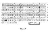
- the NOR sequence is identical to B6 with the exception of 4 additional amino acids in the B6-derived cytoplasmic domain ( Fig. 4 ) that likely reflects variation between two splice donor sites in exon 7 ( Sano, S. et al. (1999) Biochem J 344 Pt 3, 667-75 ).
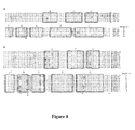
- Comparison of the Sirp ⁇ coding sequence between NOD and NOR revealed 24 amino acid differences, 20 of these in the extracellular IgV-like domain of molecule where the NOD sequence displays 18 substitutions and two deletions compared to NOR and B6 ( Fig. 4 and Fig. 8A ).
- SIRP ⁇ sequence variation was examined by immunoblotting with polyclonal antibodies directed against the cytoplasmic domain which is sequence identical in NOD and NOR mice ( Fig. 4 ). No quantitative strain differences in SIRP ⁇ expression were detected under the conditions examined. Electrophoretic mobility of the NOD protein was slightly less than that of the NOR protein ( Fig. 5A ). SIRP ⁇ is glycosylated (van den Nieuwenhof, I.M. et al. (2001) J Cell Sci 114, 1321-9 ), so we examined the possibility that strain differences in the apparent molecular weight reflected differential glycosylation.
- Murine macrophages display strain-specific variation in support of human LTC-IC
- Murine SIRP ⁇ is most abundantly expressed in neurons and myeloid cells ( Veillette, A. et al. (1998) J Biol Chem 273, 22719-28 ) with evidence of differentially glycosylated forms in heart, liver, kidney and other tissues ( Sano, S. et al. (1999) Biochem J 344 Pt 3, 667-75 ).
- SIRP ⁇ ligation regulates multiple macrophage functions including inhibition of phagocytosis ( Oldenborg, P.A. et al. (2001) J Exp Med 193, 855-62 ; Blazar, B.R. et al. (2001) J Exp Med 194, 541-9 ) and TNF ⁇ production ( Smith, R.E. et al.
- macrophages are a component of hematopoietically supportive BM stromal cultures ( Hauser, S.P. et al. (1995) J Histochem Cytochem 43, 371-9 ). Based on these observations, we tested whether macrophages conferred the strain-specific differences in human hematopoietic support we observed in chimeric LTC-IC assays.
- Peritoneal macrophages from NOD, NOR, and NOR congenic mice heterozygous at the Idd13 locus (NOR.BN- Idd13 ) were harvested and added to human LTC-IC cultures employing MS-5 stromal cells, a murine cell line that supports human hematopoiesis ( Issaad, C.
- Murine macrophages were seeded onto established MS-5 cultures at doses of 200, 2,000 and 20,000 cells per well, followed 24 hours later by addition of 2,000 Lin - CB cells. Macrophages of all strains had a dose-dependent effect on the number of CFC generated after 3 to 4 weeks in LTC ( Fig. 6A ). NOR macrophages exerted greater suppression of human LTC at all cell doses than NOD macrophages; at 2,000 and 20,000 macrophages/well, the strain difference was significant (p ⁇ 0.008).
- NOR.BN- Idd13 had an intermediate effect between NOR and NOD macrophages consistent with our previous observations of a (co)-dominant effect of Sirp ⁇ alleles on support of human hematopoiesis.
- Replicate experiments performed with BM-derived macrophages showed similar results (data not shown).
- Sirp ⁇ confers differential capacity of NOD macrophages to support human hematopoiesis
- NOR BM-derived macrophages were infected with SIRP or control CEP lentivirus prior to seeding on MS-5 stromal cultures.
- Human Lin - CB cells were added the next day and generation of CFC was assessed 25 to 31 days later. Cell dose effects were again evident in this setting where seeding 20,000 macrophages/well suppressed human hematopoiesis regardless of Sirp ⁇ genotype.
- expression of NOD-derived SIRP ⁇ by NOR macrophages resulted in substantially greater human CFC support compared to either non-manipulated or CEP-infected NOR macrophages ( Fig. 6C and data not shown).
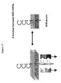
- the cellular ligand of SIRP ⁇ is the ubiquitously expressed integrin-associated protein CD47, also a member of the Ig superfamily ( Seiffert, M. et al. (1999) Blood 94, 3633-43 ).
- CD47 binds to the extracellular IgV-like domain of SIRP ⁇ ( Seiffert, M. et al. (2001) Blood 97, 2741-9 ; Vernon-Wilson, E.F. et al. (2000) Eur J Immunol 30, 2130-7 ).
- Previous studies have shown that human SIRP ⁇ displays cross-species binding to porcine CD47, but not to CD47 from mouse (B6), rat or cow ( Subramanian, S. et al. (2006) Blood 107, 2548-56 ).
- NOD-derived SIRP ⁇ displays far greater reactivity with human CD47 than does the NOR (or B6)-derived protein, providing a molecular mechanism for the enhanced support of human hematopoesis in NOD. SCID mice.
- SIRP ⁇ is primarily expressed on myeloid cells and contains cytoplasmic immunoreceptor tyrosine-based inhibition motifs (ITIM) which mediate inhibitory signals leading to reduced phagocytosis by macrophages, inhibition of neutrophil migration, and attenuated production of the inflammatory cytokine TNF ⁇ (reviewed in Barclay, A.N. & Brown, M.H. (2006) Nat Rev Immunol 6, 457-64 ).
- ITIM cytoplasmic immunoreceptor tyrosine-based inhibition motifs
- CD47 the only known cellular ligand of SIRP ⁇ , is ubiquitously expressed and modulates multiple cellular actions on hematopoietic cells including platelet activation, migration, and adhesion, leukocyte adhesion and cytokine production, and T cell responsiveness (reviewed in Brown, E.J. & Frazier, W.A. (2001) Trends Cell Biol 11, 130-5 ). Both SIRP ⁇ and CD47 are immunoglobulin superfamily members and their interaction is mediated through their respective IgV-like domains ( Seiffert, M. et al. (2001) Blood 97, 2741-9 ).
- CD47-SIRP ⁇ interaction has been implicated in regulation of phagocytosis-mediated clearance of red blood cells ( Oldenborg, P.A. et al. (2000) Science 288, 2051-4 ) and leukocytes ( Gardai, S.J. et al. (2005) Cell 123, 321-34 ), wherein expression of CD47 on the hematopoietic cells serves as a "marker of self", inhibiting phagocytosis by macrophages expressing SIRP ⁇ .
- macrophages from all mouse strains displayed a dose-dependent suppressive effect on the growth of human LTC-ICs, suggesting that SIRP ⁇ signals inhibit macrophage secretion of inflammatory cytokines such as TNF ⁇ , as previously shown for macrophages stimulated by pathogen products ( Smith, R.E. et al. (2003) Blood 102, 2532-40 ) .
- SIRP ⁇ -CD47 binding may activate CD47-induced signalling pathways within the human hematopoietic cells.
- any number of the pleiotropic effects of SIRP ⁇ -CD47 interactions may influence human HSC survival and engraftment.
- SIRP ⁇ polymorphism in human populations.
- CEU Caucasian
- YRI African
- CHB Chinese
- JPT Japanese
- a recently reported high-resolution X-ray crystal structure of the SIRP ⁇ IgV domain predicts the critical residues that contribute to the CD47 binding surface ( Subramanian, S. et al. (2006) Blood Cells Mol Dis 36, 364-72 ).
- the need for therapeutic engagement of SIRP ⁇ might decrease as the established graft produces large numbers of CD47+ cells producing a favorable ratio relative to the remaining recipient-derived macrophages.
- a person skilled in the art would understand that one could continue to analyze the degree of human polymorphism in SIRP ⁇ by sequencing normal, unrelated individuals, for example, through the available human HapMap samples obtainable through the Sick kids Centre for Applied Genomics (Toronto, Canada). For example, a 1-2microgram DNA sample from donor and recipient archival blood samples in the HLA lab would be used for sequencing SIRP ⁇ using standard sequencing facilities. The donor/recipient frozen blood sample pairs would be given a unique anonymous ID lacking any diagnosis or any outcome details.
- sequence exon 3 of this gene which encodes the IgV domain that is critical to ligand binding and function, and also to sequence two additional exons that encode the intracellular regions of the gene which include important amino acid residue for conveyance of signals emanating from SIRP ⁇ .
- sequence exon 3 of this gene which encodes the IgV domain that is critical to ligand binding and function
- sequence two additional exons that encode the intracellular regions of the gene which include important amino acid residue for conveyance of signals emanating from SIRP ⁇ .
- Figure 10a is a graphic depiction of the CD47 protein including extracellular IgV-like loop, five membrane spanning regions and short cytoplasmic tail.
- Figure 10b depicts the CD47-Fc fusion protein representing the CD47 IgV-like domain fused to a human Ig Fc region.
- CD47-Fc fusion proteins were prepared as described in the above methods and materials.
- Figure 11a shows a histogram depicting protein production in the supernatant of cultured cells transfected with mCD47-Fc and two hCD47-Fc constructs prepared with different plasmid backbones (pcDNA and pIAP369).
- Figure 11b is an immunoblot of an SDS-PAGE depicting the fusion proteins following purification from culture supernatant. Neither hCD47-Fc construct produced as well as the mCD47-Fc construct prompting further optimization.
- the pIAP369 plasmid construct containing mCD47-Fc was used as a scaffold to introduce a eukaryotic translation initiation site ( Kozak, M Nucleic Acids Res. 1984, 12(2):857-72 ) called a Kozak sequence.
- the resulting plasmid was then digested with restriction enzymes to remove the mCD47 domain which was replaced with the hCD47 IgV-like domain sequence by ligation and re-cloning in bacteria.
- the resulting plasmid was sequenced to ensure in-frame introduction of the hCD47 sequence and verification of the Kozak sequence.
- Figure 12b shows the sequence hCD47-Fc inserted into the pIAP369 plasmid, showing the Kozak consensus (SEQ ID NO.123), hCD47 fragment (SEQ ID NO.124), linker (SEQ ID NO.125) and Fc (SEQ ID NO.126).
- Figure 13a shows the production of mouse and human CD47-Fc assayed in the supernatant of cells transfected with one of four different constructs prepared as above.
- the data show that introduction of the eukaryotic initiation (Kozak) sequence enhanced production of hCD47-Fc (compare two right hand columns) to 30mg/L of supernatant.
- Figure 13b shows immunoblot displaying an anti-human Fc antibody reacting with the protein G-purified CD47-Fc proteins. Note that differential post-translational additional of carbohydrate moieties produces differences in SDS-PAGE mobilities for mouse and human CD47-Fc.
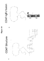
- FIG. 16 shows preparation of a truncated version of the human HSC "permissive" NOD version of SIRP ⁇ ("NOD- ⁇ -cyto") (left side) incapable of transmitting signals by intentional deletion of the two immuno tyrosine inhibitory motifs (ITIM) required to link to critical downstream phosphatases SHP-1 and SHP-2 (right side).
- ITIM immuno tyrosine inhibitory motifs
- LTC-IC long-term culture initiating cell
- infection of NOR macrophages with lentivirus containing a full length NOD SIRP ⁇ construct significantly enhances human HSC survival compared to NOR macrophages infected with the empty virus (squares v. diamonds).
- infection of NOR macrophages with a lentivirus containing the cytoplasmic truncated version of NOD SIRP ⁇ is not distinguishable from infection of NOR macrophages with empty virus (diamonds v. open triangles).
- Figure 19 is a general outline of a study designed to evaluate in vivo the effect of CD47 fusion protein on HSC engraftment.
- the hCD47-Fc fusion protein was injected into NOD.
- SCID hosts together with introduction of graded doses of human newborn cord blood hematopoietic stem cells.
- Human HSC were prepared by removal of mature lineage-committed cells using a cocktail of monoclonal antibodies and immunomagnetic beads as previously described ( J. L. McKenzie et al., Nat Immunol. 2006, 11:1225-33 ).
- HSC engraftment is followed by flow cytometric examination with human-specific antibodies to cell surface markers (CD34, CD45) in blood, spleen and bone marrow of the recipient mice.
- CD34, CD45 cell surface markers
- mice were sacrificed and bone marrow cells prepared from the tibia and femur.
- the bone marrow cells were stained with a series of monoclonal antibodies and analyzed by flow cytometry. Cells staining with anti-human CD45 were quantified to determine the extent of human HSC engraftment.
- the present invention relates to the following items:
Abstract
Description
- The invention relates to modulating the SIRPα - CD47 interaction in order to increase hematopoietic stem cell engraftment and compounds therefor. In some embodiments, there is provided isolated SIRPα and CD47 polypeptides, fragments and fusion proteins for enhancing hematopoietic stem cell engraftment. Further there is provided methods for increasing hematopoietic stem cell engraftment through administration of the above polypeptides.
- The transplantation of human hematopoietic stem cells (HSC) from bone marrow (BM) or G-CSF mobilized peripheral blood (PB) has been one of the most important clinical applications of stem cell biology. HSC transplantation in individuals with neoplastic disease enables the use of a high dose chemotherapy regimen and subsequent HSC rescue to overcome the resultant hematopoietic failure due to chemotherapy, enhancing cure rates for both hematologic and non-hematologic tumors. Additionally, genetic diseases such as thalassemia and certain immune-deficiencies can be managed by autologous transplantation of gene-corrected HSC or by transplantation of allogeneic HSC. A key discovery enabling successful HSC transplantation was identification of the human leukocyte antigen (HLA) system as the human major histocompatibility complex. Although HLA disparity between donor and host plays a major role in graft rejection, graft failure can occur even in patients receiving an unmanipulated, HLA-identical transplant (Thomas, E.D. et al. (1977) Blood 49, 511-33; Storb, R. et al. (1977) N Engl J Med 296, 61-6). Graft rejection in this setting may be related in part to mismatch at minor histocompatibility antigens (Gale, R.P. et al. (1981) Blood 57, 9-12). Additional genes other than the HLA haplotypes that modulate HSC engraftment have not been characterized.
- HSC reside in supportive microenvironmental niches comprised of fibroblastic stroma, osteoblasts, osteoclasts, macrophages, and endothelial cells (Suda, T. et al. (2005) Trends Immunol 26, 426-33). Abrogation of HSC interaction with such niches, for example by blocking specific adhesion proteins or chemokine receptors, prevents HSC engraftment (Lapidot, T. et al. (2005) Blood 106, 1901-10). Additionally, natural killer (NK) cells and macrophages, both components of the innate immune system, have been shown to play a role in murine HSC transplantation (Murphy, W.J. et al. (1987) J Exp Med 165, 1212-7) and human hematopoietic xenotransplantation (McKenzie, J.L. et al. (2005) Blood 106, 1259-61), respectively. A better understanding of the mechanisms underlying hematopoietic stem cell engraftment following transplantation, including genes that control regulatory pathways, could ultimately translate into better clinical outcomes.
- Xenotransplantation in the non-obese diabetic/severe combine immune-deficient NOD.Pdck sc/sc (NOD.SCID) mouse has become the "gold standard" assay for human HSC engraftment. The assay is based on the ability of human HSC to repopulate the immune system of these animals following intravenous injection (Wang, J.C.Y. et al. "Normal and leukemic human stem cells assayed in immune-deficient mice" in: Hematopoiesis - A Developmental Approach (ed. Zon, L.I.) 99-118 (Oxford University Press, New York, USA, 2001). The cells that initiate the human xenograft are operationally defined as SCID-repopulating cells (SRC), possess properties attributed to HSC, and are distinct from more mature progenitors assayed in vitro. This system has enabled analysis of the proliferation, differentiation, and self renewal properties of SRC within the human HSC compartment.
- Signal regulatory proteins (SIRPs) constitute a family of cell surface glycoproteins which are expressed on myeloid (including macrophages, granulocytes, myeloid dendritic cells, and mast cells) and neuronal cells (summarized in Barclay, A.N. & Brown, M.H., Nat Rev Immunol 6, 457-64 (2006); see also
WO 97/48723 - A variety of approaches have been proposed to disrupt the CD47- SIRPα interaction in an effort to effect a biological outcome. These encompass the use of fragmented/truncated SIRPα and/or CD47 proteins and antibodies thereto (see, for example, International Patent Application Publication No.
WO 99/40940 U.S. Patent Application Publication Nos. 2003/0026803 and2006/0135749 ; andU.S. Patent No. 6,913,894 ), for modifying immune function (see, for example, International Patent Application Publication No.WO 99/40940 U.S. Patent Application Publication Nos. 2003/0026803 and2007/0113297 ), and for xenotransplantation (see, for example,U.S. Patent Application Publication Nos. 2005/0255550 , and2007/0157328 ). - There remains a need to identify molecules capable of supporting HSC engraftment and hematopoiesis upon transplantation.
- According to one aspect, there is provided an isolated polypeptide selected from the group consisting of a polypeptide consisting of a) the amino acid sequence of SEQ ID NO. 1; b) a polypeptide consisting of a fragment of the amino acid sequence of SEQ ID NO. 1, wherein the fragment comprises at least one of
residues residues - According to a further aspect, there is provided an isolated polypeptide selected from the group consisting of a) a polypeptide consisting of the amino acid sequence of SEQ ID NO. 2; b) a polypeptide consisting of a fragment of the amino acid sequence of SEQ ID NO. 2; and c) one of the polypeptide in a) and b) with up to 1 amino acid insertion, deletion or substitution for every 7 amino acids in length of the polypeptide; wherein i) at least one of residues at
positions corresponding residues residues 129 and 130 of SEQ ID NO. 2 in the polypeptide is deleted. - According to a further aspect, there is provided an isolated polypeptide selected from the group consisting of a) a polypeptide consisting of an amino acid sequence selected from the group consisting of SEQ ID NOS. 4-13; b) a polypeptide consisting of a fragment of an amino acid sequence selected from the group consisting of SEQ ID NOS. 4-13; and c) one of the polypeptide in a) and b) with up to 1 amino acid insertion, deletion or substitution for every 7 amino acids in length of the polypeptide, wherein the polypeptide binds human CD47.
- According to a further aspect there is provided a polypeptide capable of binding to human Sirpα; and antibodies to human Sirpα. Preferably, the polypeptide is the extracellular domain of human CD47 fused to the Fc portion of IgG.
- According to a further aspect, there is provided a pharmaceutical composition comprising a polypeptide described herein and a pharmaceutically acceptable carrier.
- According to a further aspect, there is provided a method for increasing hematopoietic stem cell engraftment in a mammal comprising administering to the mammal a therapeutically effective amount of a polypeptide described herein.
- According to a further aspect, there is provided a use of a polypeptide described herein for increasing hematopoietic stem cell engraftment in a mammal or in the preparation of a medicament for increasing hematopoietic stem cell engraftment in a mammal.
- According to a further aspect, there is provided a polypeptide described herein for increasing hematopoietic stem cell engraftment in a mammal.
- According to a further aspect, there is provided an isolated nucleic acid comprising a sequence that encodes a polypeptide described herein.
- According to a further aspect, there is provided an expression vector comprising a nucleic acid described herein.
- According to a further aspect, there is provided a cultured cell comprising a vector described herein.
- According to a further aspect, there is provided a method of producing a polypeptide comprising culturing a cell described herein under conditions permitting expression of the polypeptide.
- According to a further aspect, there is provided a method for increasing hematopoietic stem cell engraftment in a human comprising modulating the interaction between human Sirpα and human CD47.
- According to a further aspect, there is provided a method of identifying a compound that increases hematopoietic stem cell engraftment in a human comprising a) contacting at least one of the extracellular domain of human Sirpα and human CD47 with at least one test compound; b) determining the at least one test compound as binding to the at least one of human Sirpα and human CD47; c) contacting the test compound with human hematopoietic cells in a stromal environment; and d) determining whether hematopoietic stem cell engraftment is increased in the presence of the test compound.
- According to a further aspect, there is provided a method of determining genetic polymorphisms in humans affecting hematopoietic stem cell engraftment comprising a) sequencing the Sirpα gene from a plurality of humans having undergone hematopoietic transplantation; b) determining nucleotide differences in the Sirpα gene within the plurality of humans; and c) correlating the nucleotide differences with hematopoietic stem cell engraftment to determine relevant polymorphisms.
- According to a further aspect, there is provided a method of determining likelihood of hematopoietic stem cell engraftment in a recipient comprising a) sequencing the Sirpα gene from the recipient; and b) determining whether the relevant polymorphisms exist.
- These and other features of the preferred embodiments of the invention will become more apparent in the following detailed description in which reference is made to the appended drawings wherein:
-
Figure 1 illustrates the support of human hematopoiesis in vivo and in vitro in the NOD strain background. (a) Southern blot analysis of DNA extracted from the BM of mice four weeks post-transplant with human BM (top panel; cell dose: RAG-2knockout strains 40 × 106 mononuclear cells, SCID and NOD/SCID mice 20 × 106 cells) or CB (bottom panel, 15 × 106 mononuclear cells per recipient) cells. The level of human cell engraftment was determined by comparing the intensity of the 2.7 kb α-satellite band (arrow) in the sample lanes with the control human/mouse DNA mixtures. (b, c) Number of CFC generated in chimeric LTC of human Lin- CB cells on irradiated mouse BM stroma. BM cells from NOD, NOR, B6, ICR, C3H, and BALB/c male mice as indicated were cultured for 5 weeks followed by irradiation and subsequent inoculation with 1.1 × 10⁵ (b) or 0.6 - 6.0 × 10⁵ (c) Lin- CB cells. After 5 weeks of culture, cells were harvested and the total number of CFC was determined in standard clonogenic methylcellulose assays. The data is shown as the mean of 3 to 6 independent experiments. -
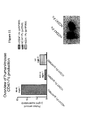
Figure 2 illustrates the effects of strain-specific differences at the Idd13 locus on support of human hematopoietic engraftment in vitro and in vivo. (a) Generation of CFC in chimeric LTC of human Lin- CB cells on mouse stroma from NOD congenic (NOD.NOR-Idd4, NOD.NOR-Idd5, NOD.NOR-Idd9, NOD.NOR-Idd13), NOR congenic (NOR.NOD-Idd13), and B6.NOD-Idd13 mice. Murine BM stromal layers were established as described inFig. 1b and seeded with 0.36 - 2.0 × 10⁵ human Lin- CB cells. After 5 weeks of culture, cells were harvested and the number of CFC was determined by plating in methylcellulose assays. In each experiment, NOD stroma was used as a positive control, and B6 and NOR stromal cells were used as negative controls. The data is normalized to the number of CFC generated in NOD cultures (=100%), and is shown as the mean of at least 3 independent cultures. BB, Idd13 homozygous B6 BM stroma; NN, Idd13 genotype homozygous NOD BM stroma. (b) Eight- to 9-week-old NOD.SCID (n=11) or NOD.NOR-Idd13.SCID (n=8) mice were irradiated with 350 cGy and injected intravenously with 0.37 -1.5 × 10⁵ Lin-CD34+ cells). Six to 7 weeks post-transplantation, mice were sacrificed and the recipients' BM was assessed for human CD45+ cell engraftment by flow cytometry. -
Figure 3 illustrates the identification of the critical region of Idd13 associated with support of human hematopoiesis. (a) Graphical representation ofchromosome 2 in various congenic strains on both the NOD and NOR backgrounds. Genetic mapping of the candidate interval is denoted by the striped bars to the right of the chromosomal representations. Formal congenic strain designations are as follows: 1. NOD.NOR-D2Jyh443-D2Mit452, 2. NOR.NOD-D2Jyh443-D2Jyh1493, 3. NOR.BN-D2Jyh443-D2Jyh1493, 4. NOR.NOD-D1Ngul46-Ramp1/D2Jyh443-D2Jyh1192, 5. NOD.NOR-D2Jyh443-D2Gul482, 6. NOD.NOR-D2Jyh443-D2Jyh3941, 7. NOR.NOD-DlNgul46-Ramp1/D2Gul188-D2Jyh1493, 8. NOR.BN-D2Jyh767-Flizl. (b) Generation of CFC in chimeric LTC of human Lin- CB cells on mouse stroma from NOD or NOR Idd13 subcongenic mice depicted in a. Cultures performed and data expressed as inFig. 2a . Strain designations as described in a. -
Figure 4 illustrates protein sequence alignment of SIRPα. cDNA prepared from BM-derived macrophages was used as a template for PCR amplification of Sirpα transcripts from NOD and NOR mice. The B6 mouse sequence is from the EnsEMBL database. Protein domains are indicated by open boxes. -
Figure 5 illustrates immunoblot analysis of SIRPα in BM-derived macrophages from NOD, NOR, and congenic NOR.NOD-Idd13 mouse strains. (a) Lysates prepared from BM-derived macrophages, either untreated or treated with LPS (100ng/ml) and IFN-γ (10ng/ml), were subjected to immunoblot analysis using polyclonal antibodies directed against the cytoplasmic domain of SIRPα, or glyceraldehyde-3-phosphate dehydrogenase (GAPDH) as a loading control. (b) Lysates prepared from BM-derived macrophages were incubated in the absence (-) or presence (+) of N-glycosidase F for 12 hours at 37°C and then subjected to immunoblot analysis using polyclonal antibodies directed against the cytoplasmic domain of SIRPα. N-glycosidase F treated and untreated samples were co-electrophoresed. Molecular size standards are indicated in kilo Daltons (kD). -
Figure 6 illustrates Sirpα modulation of murine macrophage-mediated suppression of human hematopoiesis. (a) Macrophage-mediated suppression of human hematopoiesis in LTC. MS-5 cells were seeded into 96-well tissue culture plates (2000 cells/well). Peritoneal macrophages harvested from NOD, NOR, and NOR.NOD-Idd13 heterozygous mice were seeded at doses of 200-20,000 cells/well (5 replicates per dose) with addition of 2000 human Lin- CB cells per well the next day. After 3-4 weeks of culture, cells were harvested and the number of human CFC was determined by plating in methylcellulose assays. Error bars represent standard deviations. (b) Western blot analysis showing SIRPα expression in Jurkat cells and NOR BM-derived macrophages before and after infection with control virus (CEP) or virus expressing NOD-derived Sirpα (SIRP). Protein lysates were immunoblotted with polyclonal antibody directed against SIRPα. Lane 1: Jurkat cells; lane 2: Jurkat cells infected with CEP; lane 3: Jurkat cells infected with SIRP; lane 4: NOR macrophages; lane 5: NOR macrophages infected with SIRP; lane 6: NOD macrophages. Infection with SIRP lentivirus resulted in high level expression of NOD SIRPα in NOR macrophages (lane 5). (c) Effect of Sirpα on macrophage-mediated suppression of human hematopoiesis. NOR BM-derived macrophages were infected with control (NOR-CEP) lentivirus or lentivirus expressing the NOD-derived Sirpα gene (NOR-NOD SIRP) prior to seeding onto established MS-5 stromal cultures at doses of 20 to 20,000 cells/well (5 replicates per dose). 2000 Lin- CB cells/well were added the next day and human CFC generated after 3.5 weeks in culture were assayed as described. Support of human hematopoiesis in NOR-NOD SIRP wells was significantly better than in NOR-CEP wells (P=0.032 at 20 cells/well; P=0.008 at 2000 and 20,000 cells/well). Error bars represent standard deviations. -
Figure 7 illustrates conferral of enhanced cross species reactivity with human CD47 by NOD SIRPα. (a-d) Flow cytometric analysis of human CD47 (hCD47) binding to murine SIRPα on BM-derived macrophages from NOD and NOD.NOR-Idd13 congenic mice. (a) Histogram of CD11b expression; horizontal bar indicates CD11b+ gate. Plots in b through d are gated on CD11b. (b) Two dimensional contour plots showing SIRPα and hCD47-Fc staining. (c) Histogram of SIRPα expression (grey) compared to isotype control (black). (d) Histogram of hCD47-Fc staining (grey) overlaid with human IgG-Fc control (black). (e) Immunoprecipitation of murine SIRPα with hCD47-Fc. Protein extracts from BM-derived macrophages from NOD, NOR and NOD.NOR-Idd13 congenic mice were electrophoresed on SDS-PAGE and immunoblotted with polyclonal anti-SIRPα antibody. Each panel displays total lysate, immunoprecipitates with control human IgG-Fc protein (IP hFc) and immunoprecipitates with hCD47-Fc fusion protein (IP hCD47-Fc). The top panel compares NOD and NOR extracts, and the bottom panel compares NOD and NOD.NOR-Idd13 (Idd13cg) extracts. -
Figure 8 illustrates protein sequence alignments of murine and human SIRPα IgV domains. (a) cDNA prepared from BM macrophages was used as a template for PCR amplification of Sirpα transcripts from NOD and NOR mice. The C57BL/6 (B6), BALB/c and 129/Sv sequences were obtained from EnsEMBL and NCBI databases. Open boxes represent b-pleated sheets identified in the X-ray crystal structure of SIRPα and correspond to similar regions in the Ig heavy chain variable region. Amino acids that vary between mouse strains are shaded. B6 was used as the parental sequence. (b)Exon 3 of SIRPα containing the IgV domain was PCR amplified from genomic DNA of 37 individuals from thehuman HapMal phase 1 release. Open boxed regions and shaded amino acids represent the same features as in (a), with V1 serving as the parental sequence. The residue marked * in an orthologous position between species, is polymorphic between NOD and other strains, and between humans. The residues marked + are polymorphic in humans and reside on the "top" solvent exposed surface of the protein where CD47 is predicted to bind. -
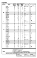
Figure 9 illustrates the validation of the discrimination power of three SNP assays by independent sequencing of the SIRPα IgV domain PCR amplified from each of the human HapMap samples shown (left side of the table). -
Figure 10 illustrates the CD47 protein including extracellular IgV-like loop, five membrane spanning regions and short cytoplasmic tail. -
Figure 11 illustrates (a) a histogram depicting protein production in the supernatant of cultured cells transfected with mCD47-Fc and two hCD47-Fc constructs prepared with different plasmid backbones (pcDNA and pIAP369) and (b) an immunoblot of an SDS-PAGE depicting the fusion proteins following purification from culture supernatant. -
Figure 12 illustrates (a) the introduction of a Kozak sequence into the pIAP369 plasmid construct containing mCD47-Fc and (b) the sequence hCD47-Fc inserted into the pIAP369 plasmid, showing the Kozak consensus, hCD47 fragment, linker and Fc. -
Figure 13 illustrates (a) the production of mouse and human CD47-Fc assayed in the supernatant of cells transfected with the Kozak containing plasmid and (b) an immunoblot displaying an anti-human Fc antibody reacting with the protein G-purified CD47-Fc proteins. -
Figure 14 illustrates an assay designed to quantify the binding affinity of hCD47-Fc and mCD47-Fc (not shown) to the NOD and NOR mouse versions of SIRPα. -
Figure 15 illustrates (a) results of binding of mCD47 to NOD and NOR SIRPα; and (b) results of binding of hCD47 to NOD and NOR SIRPα. -
Figure 16 is a schematic showing SIRPα-CD47 mediated signaling. -
Figure 17 shows the preparation of a truncated version of the human HSC "permissive" NOD version of SIRPα ("NOD-Δ-cyto") (left side). -
Figure 18 illustrates human HSC survival with NOR macrophages infected with an "empty" lentivirus (NOR-CEP "diamonds"), NOD macrophages not infected with virus (NOD uninf; filled triangles), NOD macrophages infected with "empty" lentivirus (NOD-CEP; circles), NOR macrophages infected with lentivirus containing full length NOD SIRPα (NOR-SIRP; squares), and NOR macrophages infected with lentivirus containing truncated NOD SIRPα (NOD-Δ-cyto; open triangles). -
Figure 19 illustrates a schematic of a study to evaluate in vivo the effect of CD47 fusion protein on HSC engraftment. - In the following description, numerous specific details are set forth to provide a thorough understanding of the invention. However, it is understood that the invention may be practiced without these specific details.
- As used herein "conservative amino acid substitution" refers to grouping of amino acids on the basis of certain common properties. A functional way to define common properties between individual amino acids is to analyze the normalized frequencies of amino acid changes between corresponding proteins of homologous organisms (Schulz, G. E. and R. H. Schirmer., Principles of Protein Structure, Springer-Verlag). According to such analyses, groups of amino acids may be defined where amino acids within a group exchange preferentially with each other, and therefore resemble each other most in their impact on the overall protein structure (Schulz, G. E. and R. H. Schirmer., Principles of Protein Structure, Springer-Verlag). Examples of amino acid groups defined in this manner include:
- (i) a charged group, consisting of Glu and Asp, Lys, Arg and His,
- (ii) a positively-charged group, consisting of Lys, Arg and His,
- (iii) a negatively-charged group, consisting of Glu and Asp,
- (iv) an aromatic group, consisting of Phe, Tyr and Trp,
- (v) a nitrogen ring group, consisting of His and Trp,
- (vi) a large aliphatic nonpolar group, consisting of Val, Leu and Ile,
- (vii) a slightly-polar group, consisting of Met and Cys,
- (viii) a small-residue group, consisting of Ser, Thr, Asp, Asn, Gly, Ala, Glu, Gln and Pro,
- (ix) an aliphatic group consisting of Val, Leu, Ile, Met and Cys, and
- (x) a small hydroxyl group consisting of Ser and Thr.
- In addition to the groups presented above, each amino acid residue may form its own group, and the group formed by an individual amino acid may be referred to simply by the one and/or three letter abbreviation for that amino acid commonly used in the art.
- As used herein, a "cultured cell" means a cell which has been maintained and/or propagated in vitro. Cultured cells include primary cultured cells and cell lines. As used herein, "culturing the cell" means providing culture conditions that are conducive to polypeptide expression. Such culturing conditions are well known in the art.
- As used herein "engrafting" a stem cell, preferably an expanded hematopoietic stem cell, means placing the stem cell into an animal, e.g., by injection, wherein the stem cell persists in vivo. This can be readily measured by the ability of the hematopoietic stem cell, for example, to contribute to the ongoing blood cell formation.
- As used herein "fragment" relating to a polypeptide or polynucleotide means a polypeptide or polynucleotide consisting of only a part of the intact polypeptide sequence and structure, or the nucleotide sequence and structure, of the reference gene. The polypeptide fragment can include a C-terminal deletion and/or N-terminal deletion of the native polypeptide, or can be derived from an internal portion of the molecule. Similarly, a polynucleotide fragment can include a 3' and/or a 5' deletion of the native polynucleotide, or can be derived from an internal portion of the molecule.
- As used herein "fusion protein" refers to a composite polypeptide, i.e., a single contiguous amino acid sequence, made up of two (or more) distinct, heterologous polypeptides which are not normally fused together in a single amino acid sequence. Thus, a fusion protein may include a single amino acid sequence that contains two entirely distinct amino acid sequences or two similar or identical polypeptide sequences, provided that these sequences are not normally found together in the same configuration in a single amino acid sequence found in nature. Fusion proteins may generally be prepared using either recombinant nucleic acid methods, i.e., as a result of transcription and translation of a recombinant gene fusion product, which fusion comprises a segment encoding a polypeptide of the invention and a segment encoding a heterologous polypeptide, or by chemical synthesis methods well known in the art. Fusion proteins may also contain a linker polypeptide in between the constituent polypeptides of the fusion protein. The term "fusion construct" or "fusion protein construct" is generally meant to refer to a polynucleotide encoding a fusion protein. In one embodiment, the fusion protein is a polypeptide as described herein fused to a portion of an Ig molecule. The Ig portion of the fusion protein can include an immunoglobulin constant region, e.g. a human Cγ1 domain or a Cγ4 domain (e.g. the hinge, CH2, and CH3 regions of human IgγC1 or human IgCγ4 (see e.g.,
Capon et al., U.S. Pat. Nos. 5,116,964 ;5,580,756 ;5,844,095 , and the like). In one preferred embodiment, Ig fusion proteins include a polypeptide as described herein coupled to an immunoglobulin constant region (e.g., the Fc region). - As used herein "hematopoietic stem cell" refers to a cell of bone marrow, liver, spleen or cord blood in origin, capable of developing into any mature myeloid and/or lymphoid cell.
- As used herein "isolated" with reference to a nucleic acid molecule, polypeptide, or other biomolecule, means that the nucleic acid or polypeptide has separated from the genetic environment from which the polypeptide or nucleic acid were obtained. It can also mean altered from the natural state. For example, a polynucleotide or a polypeptide naturally present in a living animal is not "isolated," but the same polynucleotide or polypeptide separated from the coexisting materials of its natural state is "isolated", as the term is employed herein. Thus, a polypeptide or polynucleotide produced and/or contained within a recombinant host cell is considered isolated. Also intended as an "isolated polypeptide" or an "isolated nucleic acid molecules" are polypeptides or nucleic acid molecules that have been purified, partially or substantially, from a recombinant host cell or from a native source.
- It is also contemplated that the peptides of the invention may exhibit the ability to modulate biological, such as intracellular, events. As used herein "modulate" refers to a stimulatory or inhibitory effect on the biological process of interest relative to the level or activity of such a process in the absence of a peptide of the invention.
- The term "macrophage suppression" or "suppression of macrophages" as used herein refers to the reduction or prevention of the role, activity and/or effect of macrophages. As used herein, the "nucleic acid molecule" means DNA molecules (e.g., a cDNA) and RNA molecules (e.g., an mRNA) and analogs of the DNA or RNA generated, e.g., by the use of nucleotide analogs. The nucleic acid molecule can be an oligonucleotide or polynucleotide and can be single-stranded or double-stranded.
- As used herein "operably linked" refers to an arrangement of elements wherein the components so described are configured so as to perform their usual function. Thus, control elements operably linked to a coding sequence are capable of effecting the expression of the coding sequence. The control elements need not be contiguous with the coding sequence, so long as they function to direct the expression thereof. Thus, for example, intervening untranslated yet transcribed sequences can be present between a promoter and the coding sequence and the promoter can still be considered "operable linked" to the coding sequence. Similarly, "control elements compatible with expression in a subject" are those which are capable of effecting the expression of the coding sequence in that subject.
- As used herein, "pharmaceutically acceptable carrier" means any and all solvents, dispersion media, coatings, antibacterial and antifungal agents, isotonic and absorption delaying agents, and the like that are physiologically compatible. Examples of pharmaceutically acceptable carriers include one or more of water, saline, phosphate buffered saline, dextrose, glycerol, ethanol and the like, as well as combinations thereof. In many cases, it will be preferable to include isotonic agents, for example, sugars, polyalcohols such as mannitol, sorbitol, or sodium chloride in the composition. Pharmaceutically acceptable carriers may further comprise minor amounts of auxiliary substances such as wetting or emulsifying agents, preservatives or buffers, which enhance the shelf life or effectiveness of the pharmacological agent.
- As used herein, "polypeptide" and "protein" are used interchangeably and mean proteins, protein fragments, modified proteins, amino acid sequences and synthetic amino acid sequences. The polypeptide can be glycosylated or not.
- As used herein "stroma" refers to the supporting tissue or matrix of an organ, distinguishable from the functional elements of the organ or the parenchyma.
- As used herein, "therapeutically effective amount" refers to an amount effective, at dosages and for a particular period of time necessary, to achieve the desired therapeutic result. A therapeutically effective amount of the pharmacological agent may vary according to factors such as the disease state, age, sex, and weight of the individual, and the ability of the pharmacological agent to elicit a desired response in the individual. A therapeutically effective amount is also one in which any toxic or detrimental effects of the pharmacological agent are outweighed by the therapeutically beneficial effects.
- According to one aspect, there is provided an isolated polypeptide selected from the group consisting of a polypeptide consisting of a) the amino acid sequence of SEQ ID NO. 1; b) a polypeptide consisting of a fragment of the amino acid sequence of SEQ ID NO. 1, wherein the fragment comprises at least one of
residues residues - According to a further aspect, there is provided an isolated polypeptide selected from the group consisting of a) a polypeptide consisting of the amino acid sequence of SEQ ID NO. 2; b) a polypeptide consisting of a fragment of the amino acid sequence of SEQ ID NO. 2; and c) one of the polypeptide in a) and b) with up to 1 amino acid insertion, deletion or substitution for every 7 amino acids in length of the polypeptide; wherein i) at least one of residues at
positions corresponding residues residues 129 and 130 of SEQ ID NO. 2 in the polypeptide is deleted. - According to a further aspect, there is provided an isolated polypeptide selected from the group consisting of a) a polypeptide consisting of an amino acid sequence selected from the group consisting of SEQ ID NOS. 4-13; b) a polypeptide consisting of a fragment of an amino acid sequence selected from the group consisting of SEQ ID NOS. 4-13; and c) one of the polypeptide in a) and b) with up to 1 amino acid insertion, deletion or substitution for every 7 amino acids in length of the polypeptide, wherein the polypeptide binds human CD47.
- According to a further aspect there is provided a polypeptide capable of binding to human Sirpα; and antibodies to human Sirpα. Preferably, the polypeptide is the extracellular domain of human CD47 fused to the Fc portion of IgG.
- In some embodiments, the polypeptide is the fragment thereof and comprises at least 3 consecutive amino acids in at least one of a region between residues 50-57, 63-71, 74-80, 88-92, 95-100, 103-109, 114-125 or 128-141, inclusive of SEQ ID NO. 1, between residues 50-57, 63-71, 74-80, 88-92, 95-100, 103-109, 114-125 or 128-143, inclusive of SEQ ID NO. 2, between residues 24-31, 37-45, 48-54, 62-66, 69-74, 77-83, 88-99 or 102-116, inclusive, of any one of SEQ ID NOs. 4, 7, 8, 9 and 12; or between residues 24-31, 37-45, 48-54, 62-66, 69-74, 77-83, 88-99 or 102-115, inclusive, of SEQ ID NOs. 5, 6, 10, 11 and 13, as the case may be.
- In some embodiments, the amino acid insertion, deletion or substitution is a conservative amino acid substitution.
- In a preferred embodiment, the polypeptide binds human CD47.
- In a preferred embodiment, the polypeptide binds human SIRPα.
- In some embodiments, in increasing preferability, the polypeptide is the fragment and is between 6 and 30 amino acids in length, between 8 and 28 amino acids in length, between 10 and 26 amino acids in length, between 12 and 24 amino acids in length and between 14 and 22 amino acids in length.
- The polypeptides described herein may be fused to a second polypeptide that is preferably the Fc portion of IgG.
- According to one preferred embodiment, there is provided a polypeptide comprising the extracellular domain of CD47 fused to the Fc portion of IgG, preferably for increasing hematopoietic stem cell engraftment in a human.
- According to a further aspect, there is provided a pharmaceutical composition comprising a polypeptide described herein and a pharmaceutically acceptable carrier.
- According to a further aspect, there is provided a method for increasing hematopoietic stem cell engraftment in a mammal comprising administering to the mammal a therapeutically effective amount of a polypeptide described herein. Preferably, the increased hematopoietic stem cell engraftment results from suppression of macrophages.
- According to a further aspect, there is provided a use of a polypeptide described herein for increasing hematopoietic stem cell engraftment in a mammal or in the preparation of a medicament for increasing hematopoietic stem cell engraftment in a mammal.
- According to a further aspect, there is provided a polypeptide described herein for increasing hematopoietic stem cell engraftment in a mammal.
- According to a further aspect, there is provided an isolated nucleic acid comprising a sequence that encodes a polypeptide described herein.
- According to a further aspect, there is provided an expression vector comprising a nucleic acid described herein, preferably comprising a Kozak consensus.
- According to a further aspect, there is provided a cultured cell comprising a vector described herein.
- According to a further aspect, there is provided a method of producing a polypeptide comprising culturing a cell described herein under conditions permitting expression of the polypeptide.
- According to a further aspect, there is provided a method for increasing hematopoietic stem cell engraftment in a human comprising modulating the interaction between human Sirpα and human CD47. Preferably, the interaction between human Sirpα and human CD47 is modulated by administering a therapeutically effective amount of at least one of a) a polypeptide capable of binding to the extracellular domain of human Sirpα; b) antibodies to human Sirpα ; c) a polypeptide capable of binding to the extracellular domain of human CD47; and d) antibodies to human CD47. Correspondingly, there is also provided use of the foregoing for increasing hematopoietic stem cell engraftment in a human or for preparing medicament for the same. Preferably, the increased hematopoietic stem cell engraftment results from suppression of macrophages.
- In one embodiment, the polypeptide capable of binding to the extracellular domain of human Sirpα comprises soluble human CD47, or a fragment thereof, preferably the extracellular domain of CD47. Preferably, the soluble human CD47, or a fragment thereof, is fused to a second protein. In another embodiment, the polypeptide capable of binding to the extracellular domain of human CD47 comprises soluble human Sirpα, or a fragment thereof, preferably the extracellular domain of Sirpα. Preferably, the soluble human Sirpα, or a fragment thereof, is fused to a second protein. In preferred embodiments, the second protein is the Fc portion of IgG.
- According to a further aspect, there is provided a method of identifying a compound that increases hematopoietic stem cell engraftment in a human comprising a) contacting at least one of the extracellular domain of human Sirpα and human CD47 with at least one test compound; b) determining the at least one test compound as binding to the at least one of human Sirpα and human CD47; c) contacting the test compound with human hematopoietic cells in a stromal environment; and d) determining whether hematopoietic stem cell engraftment is increased in the presence of the test compound.
- According to a further aspect, there is provided a method of determining genetic polymorphisms in humans affecting hematopoietic stem cell engraftment comprising a) sequencing the Sirpα gene from a plurality of humans having undergone hematopoietic transplantation; b) determining nucleotide differences in the Sirpα gene within the plurality of humans; and c) correlating the nucleotide differences with hematopoietic stem cell engraftment to determine relevant polymorphisms. Preferably, the nucleotide differences result in amino acid differences.
- According to a further aspect, there is provided a method of determining likelihood of hematopoietic stem cell engraftment in a recipient comprising a) sequencing the Sirpα gene from the recipient; and b) determining whether the relevant polymorphisms exist. Preferably, the relevant polymorphisms result in an amino acid difference that is preferably at least one of (a) replacement of at least one of residues at
positions residues residues 129 and 130 of SEQ ID NO. 2. - The advantages of the present invention are further illustrated by the following examples. The examples and their particular details set forth herein are presented for illustration only and should not be construed as a limitation on the claims of the present invention.
- The CB17-scid/scid (SCID), NOD/LtSz-Prdkc sc/sc (NOD.SCID) (Shultz, L.D. et al. (1995) J Immunol 154, 180-91), C57BL/6 (B6), C3H, ICR, and BALB/c mice were bred and maintained under sterile conditions at the Ontario Cancer Institute (Toronto, Canada). Rag-2 -/- (RAG-2), RAG-2.Prf -/-, and RAG-2.β 2m -/- mice, all on B6 background, were obtained from GenPharm International (Mountain View, CA). NOD, NOD.SCID, NOD.NOR-Idd13, NOD.NOR-Idd4, NOD.NOR-Idd5, NOD.NOR-Idd9, NOR.NOD-Idd13, and B6.NOD-Idd13 mice were maintained in either specific pathogen-free or barrier conditions at the Hospital for Sick Children (Toronto, Ontario).
- All congenic mice were generated with marker-directed selection of breeders from an initial intercross of NOD/Jsd and NOR/Lt progenitors. The NOD and NOR genomes are approximately 88% identical by descent, and the differential portions of their genomes are distributed on
chromosomes Table 1. Microsatellite markers used to map congenic mice. Markers used to define congenic intervals Locus Marker Chr. Position (Mb) Primer 1Primer 2SEQ ID NO. IBD D1Gul11 7 1 72614682 

19/20 Idd5 D1Ngul4 6 1 72932895 

21/22 Idd5 D1Mit77 1 74154726 

23/24 Idd5 D1Mit13 2 1 77603656 

25/26 Idd5 D1Mit21 6 1 80318888 

27/28 Idd5 D1Mit8 1 83489415 

29/30 Idd5 D1Mit44 0 1 88698913 

31/32 Idd5 Ramp1 1 91094432 

33/34 IBD D1Mit84 1 91834941 

35/36 IBD D2Jyh43 9 2 11898874 8 
GTGCGCCACCAATGC 37/38 Idd13 D2Jyh44 3 2 11904730 1 

39/40 Idd13 D2Mit17 2 12256306 9 

41/42 Idd13 D2Mit25 6 2 12636563 4 

43/44 Idd13 D2Mit44 7 2 12853044 2 

45/46 Idd13 D2Mit33 8 2 13057830 9 

47/48 Idd13 D2Mit49 0 2 13864439 8 

49/50 Idd13 D2Mit45 2 2 16245001 5 

51/52 IBD D2Mit51 2 16321366 1 GTGAGGGGTCAATGCCAC 
53/54 IBD D4Mit33 1 4 10224868 2 CCTAACCCTCCCCACACC 
55/56 Idd9 D4Mit31 4 10545792 0 

57/58 Idd9 D4Mit30 8 4 12253146 4 

59/60 Idd9 D4Mit20 4 4 13188076 3 

61/62 Idd9 D4Mit25 1 4 13538496 3 

63/64 Idd9 D4Mit25 9 4 14277093 7 

65/66 Idd9 D4Mit31 0 4 14658249 7 

67/68 Idd9 D4Mit18 9 4 14699441 1 

69/70 IBD D4Ngul7 383 4 14777240 5 

71/72 Idd4 D11Mit2 17 11 37214368 

73/74 Idd4 D11Mit3 11 53814488 

75/76 10 

Idd4 D11Mit9 0 11 70112196 

77/78 Idd4 D11Jyh1 081 11 80495333 

79/80 IBD D11Mit4 2 11 11279582 4 

81/82 Markers used to screen for background alleles in congenic mice locus marker Chr. position (cM) primer1 primer2 SEQ ID NO. non-Idd D1Mit430 1 6.6 

83/84 non-Idd D1Mit122 1 20.4 

85/86 non-Idd D5Mit11 5 17.5 

87/88 non-Idd D7Mit124 7 37.2 

89/90 non-Idd D10Mit86 10 12 

91/92 non-Idd D11Mit62 11 2.2 

93/94 non-Idd D12Mit76 12 38.3 

95/96 non-Idd D14Mit194 14 52.5 

97/98 non-Idd D18Mit4 18 37.2 

99/100 IBD = identical by descent; non-Idd = regions of non-identity between NOD and NOR outside the four Idd regions - NOD.NOR-Idd13.SCID mice were generated from an intercross of NOD.NOR-Idd13 mice to NOD.SCID mice, followed by brother-sister matings screened by marker assisted genotyping until homozygosity was achieved at both Idd13 and SCID loci.
- For microsatellite genotyping, genomic DNA was prepared from 0.6 - 0.8 cm tail snips that were digested in a solution of proteinase K and digestion buffer (AutoGen AG00122) overnight at 55°C. The crude digests were diluted and heated to inactivate the proteinase K. PCR was performed with oligonucleotide primers modified at their 5' ends with fluorescent tags. PCR conditions were the same for all microsatellite primers: 10 cycles of 30 s at 94°C, 30 s at 50°C, and 1 min at 72°C, followed by 35 cycles of 30 s at 94°C, 30 s at 55°C, and 1 min at 72°C in 1.5 mM MgCl2. PCR amplicons were detected with the ABI 3730XL sequencer and alleles were called with the aid of the GeneMapper software version 3.0 (Applied Biosystems, Foster City, CA).
- For LTC on MS-5 murine stromal cells, non-irradiated MS-5 cells were seeded into 96 well tissue culture plates (2000 cells/well) as described in Gan, O.I. et al. (1999) Exp Hematol 27, 1097-106. Three days later, murine peritoneal macrophages were seeded at doses of 20-20,000 cells/well with 5 replicates per dose. The next day, 2000 human Lin CB cells per well were added in 200 µl human LTBMC medium without hydrocortisone. Cultures were maintained at 33°C (or 37°C for gene transfer experiments) with weekly half-medium change. After 3-4 weeks, cells were trypsinized and half of the cells from each well were plated into progenitor assays as described.
- Seven thousand five hundred cells from T25 flasks, or half well content from 96-well plates, were plated in 1 ml methylcellulose cultures under conditions that are selective for human cells as previously described in Larochelle, A. et al. (1996) . The cultures were incubated for 14 days at 37°C and the total number of colonies produced by colony-forming cells (CFC) was counted. Cells from T25 flasks were plated in triplicate.
- Murine peritoneal macrophages were obtained by peritoneal levage with 10 ml of αMEM with 10% FCS. BM-derived macrophages were obtained by flushing femurs, tibias, and iliac crests with 10 ml of DMEM supplemented with 10% FCS, 10 mM HEPES (pH 7.0), 50 nM 2-mercaptoethanol, 2 mM glutamine, 1× nonessential amino acids, and 1% penicillin/streptomycin ("complete DMEM"). BM cells were plated at a concentration of 1 × 10⁶ cells/ml in complete DMEM containing 10 ng/ml recombinant mouse GM-CSF (R&D Systems, Minneapolis, MN). Medium was changed with fresh complete DMEM with GM-CSF every other day, and macrophages were harvested by gentle scraping after 7 days.
- Expression of genes within the 0.96 Mb critical Idd13 interval was analyzed by PCR amplification of cDNA prepared from BM stroma and purified macrophages. Real time PCR was performed using an ABI/PRIZM 7900 HT cycler (Applied Biosystems) under the following conditions: 95°C for 15 s and 59°C for 1 min for 40 cycles, with addition of SYBR Green buffer (Applied Biosystems). Serial dilutions of a plasmid containing the gene sequence were used as a reference for standard curve calculation. The fluorescence thresholds and corresponding copy number were calculated using SDS 2.0 software (Applied Biosystems). Reactions were performed in triplicate, and transcript quantity was normalized to β-actin.
- The coding region of Sirpα was sequenced from NOD, NOR and BALB/c strains. cDNA was prepared from BM-derived macrophages and used as a PCR template under the following conditions: 30 s at 94°C, 30 s at 60°C, and 1 min at 68°C in 2 mM MgSO4 for 30 cycles using High-Fidelity Taq polymerase (Invitrogen Life Technologies, Carlsbad, CA). The untranslated (UTR) region of Sirpα was also amplified and sequenced in NOD and NOR strains, using genomic DNA as a template and the same PCR conditions. Sequencing primers were designed using Primer Express 1.5 software (Applied Biosystems) and are listed in Table 2. PCR products were purified according the manufacturer's instructions (Qiagen, Valencia, CA) and sequenced on both strands at the Center for Applied Genomics, Hospital for Sick Children, Toronto, Canada (TCAG; www.tcag.ca). B6, BALB/c, and 129/Sv mouse Sirpα sequence files were obtained from EnsEMBL (www.ensembl.org) and NCBI (www.ncbi.nlm.nih.gov) databases. Sequence files were aligned and analyzed with Lasergene software (DNASTAR, Madison, WI).
Table 2. Primers used to PCR amplify and sequence coding and untranslated regions of murine and human SIRPα. Forward Primer Reverse Primer SEQ ID NO. Mouse Sirpα-1 
TGTACAGAAACAGGACGCGGA 101/102 Sirpα-2 TCTCCCTCCTTGCTCTGCAG 
103/104 Sirpα-3 

105/106 Sirpα-4 

107/108 Sirpα-5 

109/110 Sirpα-6 

111/112 Sirpα-7 AACTTGTCTTTGTCCGGGCC GGCACAGTTCATCCTCACCC 113/114 Human Sirpα 

115/116 - Thirty-seven unrelated individuals from the HapMap collection were chosen for sequencing based on genotype variation at two markers flanking the IgV domain encoded in exon 3 (rs6075340 and rs611968). DNA samples were obtained from TCAG and
exon 3 was amplified by PCR using High-Fidelity Taq polymerase (Invitrogen Life Technologies). The resulting amplicons were cloned andexon 3 regions from 2 to 4 independent clones per individual were sequenced at TCAG on both strands using primers flanking the plasmid multiple cloning site (Table 2). Sequences were aligned using Lasergene software. - BM-derived macrophages were washed with ice-cold PBS and then lysed in buffer (20 mM Tris-HCl pH 7.6, 140 mM NaCl, 1 mM EDTA, 1% NP-40) containing 5 mM NaF, aprotinin (10 µg/ml) and 1 mM sodium vanadate. The lysates were centrifuged at 10 000 g for 15 min at 4°C and the resulting supernatants subjected to immunoblot analysis as follows: proteins (20 µg per well) were electrophoresed in 10% sodium dodecyl sulphate (SDS)-polyacrylamide gels and transferred onto 0.2 µM nitrocellulose membranes (Bio-Rad, Hercules, CA). Western blots were analyzed using anti-SIRPα antibody (Stressgen, Victoria, BC, Canada) and developed with the HRP-ECL system (Amersham Biosciences, Piscataway, NJ). N-linked oligosaccharides were removed from SIRPα by boiling cell lysates for 5 minutes in 0.5% SDS and 1% βmercaptoethanol to denature the glycoproteins, and then incubating with N-glycosidase F (40 units/ml, Roche Applied Science, Indianapolis, IN) for 12 hours at 37°C. For immunoprecipitation, 250 µg of BM-derived macrophage lysate was added to either hCD47-Fc (10µg/ml) or the control human IgG Fc (10µg/ml) with 50µl of protein G MicroBeads (Miltenyi Biotec, Auburn, CA). Following a 60-minute incubation on ice, the immune complexes were collected on a µMACS separator following manufacturer's instructions (Miltenyi Biotec, Auburn, CA) and eluted from the column with SDS-PAGE gel loading buffer.
- NOD Sirpα cDNA was cloned downstream of an E1Fα promoter in a third-generation lentiviral vector backbone containing a reporter Gfp gene driven by the human PGK promoter (SIRP). The control vector (CEP) contained only Gfp. Vector construction was confirmed by sequencing. Viruses pseudotyped with the vesicular stomatitis virus G protein were generated by transient transfection of 293T cells, as described in Guenechea, G. et al. (2000) , and concentrated by ultracentrifugation. The functional titers of SIRP and CEP virus as determined by infection of HeLa cells were 2.1-5.1 × 10⁸ and 4-7.6 × 10⁸ infectious particles per ml, respectively. Virus was added to flasks of NOR BM-derived macrophages after 5 days of culture at a multiplicity of infection of 20 to 25, and infected macrophages were harvested on
day 12. Gene transfer efficiency into macrophages was 51-88% as assessed by GFP fluorescence using flow cytometry. Infected macrophages were stained with PE-conjugated anti-mouse CD11b mAb (Beckman Coulter, Fullerton, CA) and sorted on a Dako Cytomation MoFlo (Fort Collins, CO) to obtain GFP+CD11b+ cells. Uninfected control macrophages were stained and sorted to obtain CAD11b+cells. Purified macrophages (purity 97-99%) were used for further experiments. - 293T cells were transfected with pcDNA-CD47Fc containing the human CD47 extracellular domain fused to human IgG1 Fc directed by the CMV promoter (Latour, S. et al. (2001) J Immunol 167, 2547-54) using a Superfect Transfection Kit (Qiagen) and selected with 100µg/ml Zeocin (Invitrogen, Carlsbad, CA). Cell lines producing soluble human CD47-Fc (hCD47-Fc) were identified by immunoblot analysis of culture supernatants. hCD47-Fc protein was purified on Protein G-Sepharose (Pierce, Rockford, IL) and covalently conjugated to biotin for use in flow cytometric analyses.
- Flow cytometric analysis was performed using a FACSCalibur (BD, Franklin Lakes, NJ). For analysis of human cell engraftment in mice, cells harvested from murine BM were stained with phycoerythrin (PE) cyanine5-conjugated anti-human CD45 antibody (Beckman Coulter). For analysis of cell surface expression of SIRPα and binding of hCD47-Fc, 1 × 10⁶ murine BM leukocytes were stained with purified anti-SIRPα mAb P84 (BD) with Alexa 633-conjugated goat anti-rat (Molecular Probes, Carlsbad, CA), FITC-conjugated anti-CD11b mAb (SWRI, San Antonio, TX), and either biotinylated hCD47-Fc with avidin-APC or a control human IgG Fc fragment with biotinylated anti-human IgG mAb and avidin-APC. Viable leukocytes were selected by exclusion of propidium iodide. Isotype controls were mouse or rat IgG conjugated to the appropriate fluorochrome. GFP fluorescence was detected in FL1 calibrated to the fluorescein isothiocyanate emission profile.
- Groups were compared using one-way ANOVA and all pairwise multiple comparisons were done using Dunn's method when differences were found. For gene transfer experiments, CEP and SIRP conditions were compared using the Wilcoxon rank test.
- The human CD47 IgV domain cDNA sequence was cloned in-frame upstream of a human IgG Fc cDNA sequence, inserted into appropriate expression vectors, transfected into 293F cells, and the resulting fusion protein (hCD47-Fc) purified from culture supernatant by affinity chromatography on protein G beads. Mouse CD47-Fc (mCD47-Fc) fusion protein was prepared using the same strategy.
-
Figure 14 is a schematic representation of an assay designed to quantify the binding affinity of hCD47-Fc and mCD47-Fc (not shown) to the NOD and NOR mouse versions of SIRPα. Macrophages obtained from NOD or NOR mice were allowed to adhere to tissue culture plates for 2 hours then washed with buffered saline. Graded amounts of purified hCD47-Fc or mCD47-Fc fusion protein was added to the wells, allowed to bind at 37°C for 20 minutes, and the wells washed extensively with buffered saline. A commercially available anti-human Fc antibody covalently conjugated to horseradish peroxidase (HRP) was added and allowed to bind, and the plates washed extensively with buffered saline. The quantity of bound hCD47 was evaluated by colorimetric assay following HRP reaction with hydrogen peroxide and peroxidase treatment using a spectrophotometer. - Several groups including our own have shown that NOD mice homozygous for the Prkdcscid (SCID) mutation permit superior human hematopoietic cell engraftment compared to SCID mice on other strain backgrounds, including CB17 (Greiner, D.L. et al. (1995) Am J Pathol 146, 888-902; Larochelle, A. et al. (1996) ). The molecular and cellular basis for this strain difference in xenograft efficacy is not known. However, NOD.SCID mice have a high incidence of spontaneous thymic lymphoma that limits their usefulness for long-term experiments. Mice carrying null alleles of the recombinase activating gene 2 (Rag-2) on the C57BL/6 (B6) background have similar blockade in T and B cell development to CB17.SCID mice but do not display spontaneous lymphoid malignancies (Shinkai, Y. et al. (1992) Cell 68, 855-67). Rag-2 -/- (RAG-2) mice also carrying homozygous null alleles in β2 microglobulin (RAG-2.β 2m -/-) or perforin (RAG-2.Prf -/- ) possess additional defects in natural killer (NK) cell function (Zijlstra, M. et al. (1990) Nature 344, 742-6; Kagi, D. et al. (1994) Nature 369, 31-7). We therefore tested several other immunodeficient strains of mice for their ability to support human hematopoiesis.
- Human BM and CB cells were transplanted into mice from each strain and engraftment was assessed by Southern blot analysis (
Fig. 1A ). Consistently high levels of engraftment were detected in NOD.SCID mice, whereas CB17.SCID mice showed a lower frequency and level of engraftment, as previously reported (Larochelle, A. et al. (1996) ). As expected, no engraftment was detected in immune-competent NOD recipients. Surprisingly, despite their profound immune-deficiency, engraftment failed in RAG-2, RAG-2.Prf -/- , and RAG-2.β 2m -/- mice on the B6 background. In contrast, NOD.Rag-1 -/- mice have been shown to support high levels of human hematopoietic cell engraftment (Shultz, L.D. et al. (2000) J Immunol 164, 2496-507). These observations suggest that additional factors other than immune-deficiency in the mouse strain background and/or host BM microenvironment dramatically impact hematopoietic engraftment in the xenotransplant setting. - Long-term culture initiating cells (LTC-IC) are the most primitive human hematopoietic cells that can be assayed in vitro (Wang, J.C.Y. et al. "Normal and leukemic human stem cells assayed in immune-deficient mice" in: Hematopoiesis - A Developmental Approach (ed. Zon, L.I.) 99-118 (Oxford University Press, New York, USA, 2001), and serve as a surrogate measure of human hematopoiesis under controlled microenvironmental conditions. LTC-IC are quantified based on their ability to generate colony-forming cells (CFC) after 5 weeks of stromal culture. To characterize strain differences in the support of human hematopoiesis, we compared the ability of BM stromal cells from NOD/Jsd (NOD) and other strains to support LTC-IC. Stromal layers from NOD mice supported human CFC production for 5 weeks of culture, whereas BM stroma from all of the other strains did not support human LTC-IC following inoculation with equivalent numbers of Lin- CB cells (
Fig. 1B ). NOD stroma was supportive even at low input numbers of Lin- CB cells (0.6 × 10⁵;Fig. 1C ). We examined NOR/Lt (NOR) mice in this assay because they are a recombinant inbred strain 88% identical by descent to NOD, differing only at four Idd loci (Idd4, Idd5, Idd9, and Idd13) (Prochazka, M. et al. (1992) Diabetes 41, 98-106). In contrast to NOD, NOR cultures supported LTC-IC only when seeded with 10-fold greater numbers of Lin- CB cells (Fig. 1C ), and resulted in small-sized CFC. These data suggest that the unique support of primitive human hematopoiesis observed in NOD mice in vitro and in vivo is controlled by regions of genetic non-identity between NOD and NOR mice. To map the genetic loci that conferred the superior ability of the NOD strain to support human hematopoiesis, stromal cells from NOD and NOR parental mice, a series of NOD and NOR-Idd congenic strains, and B6.NOD-Idd13 mice were tested in chimeric LTC (Fig. 2A ). Neither the B6 or NOR parental strains were able to support development of human LTC-IC compared to NOD stroma. NOD mice congenic for NOR alleles at Idd4, Idd5, and Idd9 displayed equivalent LTC-IC outcomes to NOD parental animals. In contrast, stromal cells isolated from NOD.NOR-Idd13 mice (Idd13-BB genotype) were unable to support human LTC-IC whereas both B6 and NOR mice congenic for NOD Idd13 (Idd13-NN genotype; B6.NOD-Idd13 and NOR.NOD-Idd13) were supportive. Thus, the NOD-derived Idd13 locus on chromosome 2 (Serreze, D.V. et al. (1994) J. Exp. Med. 180, 1553-1558) conferred dramatically enhanced capacity of BM stromal cells to support human LTC-IC in vitro. - To test whether the Idd13 locus controls support of human hematopoietic cells capable of repopulation in vivo (termed SCID-repopulating cells, SRC), we generated an immune-deficient NOD congenic strain homozygous for NOR-derived Idd13 (NOD.NOR-Idd13-SCID). Sublethally irradiated NOD.SCID and NOD.NOR-Idd13-SCID mice were transplanted intravenously with Lin- CB cells (equivalent to 0.37-1.5 × 10⁵ CD34+ cells) and human CD45+ cell engraftment was assessed after 6-7 weeks by flow cytometry (
Fig. 2B ). As expected, NOD.SCID mice supported human engraftment over the entire CB cell dose range. In contrast, no human cell engraftment was detected in NOD.NOR-Idd13-SCID mice. These results confirm the in vitro LTC-IC data and establish that NOD alleles at the Idd13 locus confer support of human hematopoiesis in vivo. - To refine the location of gene(s) at Idd13 that regulated support of human hematopoiesis, we used a mapping strategy involving generation of Idd13 congenic strains on both the NOD or NOR background (
Fig. 3A ). Stromal cells from these congenic animals were tested in chimeric LTC assays (Fig. 3A and B ). Interestingly stromal cells from Idd13 heterozygous mice (Idd13-BN) supported human LTC-IC, however the number of CFC generated was consistently ~50% of that seen in NOD cultures (Fig. 3A and B; third bars from left). These data were consistent with a dominant effect of NOD Idd13 alleles on support of human LTC-IC. By repeating these analyses on eight novel Idd13 congenic strains on both the NOD and NOR backgrounds, the support of human LTC-IC phenotype was resolved to a 960 kb region (Fig. 3A and B ), defined by a single nucleotide polymorphism in Fliz1 and the microsatellite D2Ngul1849 (Table 3). Of 15 genes in the interval, eight had functional annotation. Expression of seven of the eight genes in BM stroma and macrophages of NOD and NOR strains was assessed using real time PCR (Table 4). Sirpα (also called Shps-1 and Ptpns1) was the only gene expressed in BM stroma that showed coding sequence polymorphism between NOD and NOR and, thus, was selected as the most promising candidate for further study.Table 3. Idd13 candidate interval supporting human LTC-IC. Chr Start position Genomic marker EnsEMBL gene ID Gene symbol Primer 1 Primer 2SEQ ID NO. 2 12867529 3 Fliz1 ENSMUSG 0000002738 7 Zc3hdc8 2 12871643 0 ENSMUSG 0000004285 1 ZCC6_MOUS E 2 12874454 7 D2Ngul172 5

117/118 2 12881500 7 ENSMUSG 0000002739 4 Ttl 2 12885003 7 ENSMUSG 0000002739 5 Rpo1-2 2 12887884 0 ENSMUSG 0000003793 8 Chchd5 2 12891796 9 ENSMUSG 0000004340 4 - 2 12894782 3 ENSMUSG 0000002739 7 Slc20a1 2 12896799 2 ENSMUSG 0000005673 8 A730036I17Ri k 2 12901790 4 ENSMUSG 0000004832 7 2610318C08Ri k 2 12904863 3 ENSMUSG 0000002739 9 Il1α 2 12911361 4 ENSMUSG 0000002739 8 Il1β 2 12911892 0 ENSMUSG 0000005663 5 NM_177848 2 12920738 2 ENSMUSG 0000004372 7 F830045P16Ri k 2 12934260 1 ENSMUSG 0000003790 2 Sirpα= Ptpns1 2 12943558 9 ENSMUSG 0000002740 0 NDDB_MOUS E 2 12953089 8 D2Jyh3941 

119/120 2 12954977 5 ENSMUSG 0000003788 5 Stk35 2 12963501 8 D2Ngul184 9

121/122 - The candidate interval supporting human LTC-IC is bounded by Fliz1 and D2Ngul1849, (see
Fig. 3A ) defining a 0.96 Mb interval onchromosome 2 containing 15 genes. A minimal candidate locus was defined by microsatellite markers D2Ngul1725 and D2Jyh3941 and contained 13 genes. All genomic positions and gene attributes are based on EnsEMBL version 26.33b.1. Fliz1 primers were designed to detect a polymorphism between NOD and NOR strains in the 5' UTR.Table 4. Expression of annotated genes in Idd13 candidate interval. Gene symbol EnsEMBL ID Genotype Cell type Mean copy number ± SD Mean copy number normalized to β-actin ± SD (× 10-3) Fliz1 ENSMUSG00000027387 NOR stroma 2180 ± 177 2.04 ± 0.17 NOR.NOD- stroma 2107 ± 257 1.97 ± 0.24 Idd13 macrophage UD UD NOR macrophage UD UD NOR.NOD- Idd13 Rpo1-2 ENSMUSG00000027395 NOR stroma NOR.NOD- stroma 9955 ± 1836 7.66 ± 1.41 Idd13 macrophage 4938 ± 1234 6.86 ± 1.7.1 NOR macrophage 399 ± 23 0.40 ± 0.02 NOR.NOD- 225 ± 39 0.54 ± 0.09 Idd13 Chchd5 ENSMUSG00000037938 NOR stroma UD UD NOR.NOD- stroma UD UD Idd13 macrophage UD UD NOR macrophage UD UD NOR.NOD- Idd13 Slc20a1 ENSMUSG00000027397 NOR stroma 13762 ± 467 23.80 ± 0.81 NOR.NOD- stroma 19926 ± 1767 22.39 ± 1.99 Idd13 macrophage ND ND NOR macrophage ND ND NOR.NOD- Idd13 Il1α ENSMUSG00000027399 NOR stroma 12104 ± 1206 10.09 ± 1.01 NOR.NOD- stroma 11193 ± 3229 9.33 ± 2.69 Idd13 macrophage 2321338 ± 1304.12 ± 320.79 NOR macrophage 571008 1198.81 ± 53.24 NOR.NOD- 4303725 ± Idd13 191113 Il1β ENSMUSG00000027398 NOR stroma 19813 ± 1701 17.08 ± 1.40 NOR.NOD- stroma 12324 ± 3185 15.40 ± 3.90 Idd13 macrophage 9167234 ± 7051.72 ± 94.46 NOR macrophage 122794 4595.18 ± 320.08 NOR.NOD- 8730824 ± Idd13 608149 Stk35 ENSMUSG00000037885 NOR stroma 221 ± 16 0.44 ± 0.44 NOR.NOD- stroma 400 ± 15 0.57 ± 0.02 Idd13 macrophage ND ND NOR macrophage ND ND NOR.NOD- Idd13 UD = below limit of detection
ND= not done
See Materials and Methods for description of real time PCR - Signal regulatory protein α (SIRPα) is an Ig-superfamily transmembrane protein with intracellular docking sites for two Src homology domain-containing protein tyrosine phosphatases SHP-1 and SHP-2 (Barclay, A.N. & Brown, M.H. (2006) ). Sirpα is abundantly expressed in BM stroma and hematopoietic cells (data not shown). We used PCR amplification and bi-directional sequencing of cDNA from BM-derived macrophages to identify variations in the coding regions of NOD, NOR and BALB/c mice. The NOR sequence is identical to B6 with the exception of 4 additional amino acids in the B6-derived cytoplasmic domain (
Fig. 4 ) that likely reflects variation between two splice donor sites in exon 7 (Sano, S. et al. (1999) Biochem J 344 ). Comparison of the Sirpα coding sequence between NOD and NOR revealed 24 amino acid differences, 20 of these in the extracellular IgV-like domain of molecule where the NOD sequence displays 18 substitutions and two deletions compared to NOR and B6 (Fig. 4 andFig. 8A ). This observed variation in the Sirpα the N-terminal IgV-like domain between NOD and NOR or B6 is more extensive than that previously reported in this region amongst the B6, BALB/c and 129/Sv strains (Sano, S. et al. (1999) Biochem J 344 ). - To assess the consequences of SIRPα sequence variation between NOD and NOR mice, protein lysates were prepared from BM-derived macrophages, and SIRPα expression was examined by immunoblotting with polyclonal antibodies directed against the cytoplasmic domain which is sequence identical in NOD and NOR mice (
Fig. 4 ). No quantitative strain differences in SIRPα expression were detected under the conditions examined. Electrophoretic mobility of the NOD protein was slightly less than that of the NOR protein (Fig. 5A ). SIRPα is glycosylated (van den Nieuwenhof, I.M. et al. (2001) J Cell Sci 114, 1321-9), so we examined the possibility that strain differences in the apparent molecular weight reflected differential glycosylation. Protein lysates from BM-derived macrophages were treated with N-glycosidase F and examined by immunoblotting (Fig. 5B ). Following removal of N-linked glycans, SIRPα from both strains migrated faster, with roughly equal mobility. These results suggest that the extensive polymorphism we detected in the extracellular portion of the NOD and NOR proteins results in differential glycosylation. Thus, the amino acid sequence and post-translational modifications dissimilarities between NOD and NOR strains are consistent with Sirpα being the Idd13 gene conferring strain-specific differences in human hematopoietic support. - Murine SIRPα is most abundantly expressed in neurons and myeloid cells (Veillette, A. et al. (1998) J Biol Chem 273, 22719-28) with evidence of differentially glycosylated forms in heart, liver, kidney and other tissues (Sano, S. et al. (1999) Biochem J 344 ). SIRPα ligation regulates multiple macrophage functions including inhibition of phagocytosis (Oldenborg, P.A. et al. (2001) J Exp Med 193, 855-62; Blazar, B.R. et al. (2001) J Exp Med 194, 541-9) and TNFα production (Smith, R.E. et al. (2003) ), and increasing nitric oxide production (Alblas, J. et al. (2005) ). We and others have shown that a human CD122+ cell subset distinct from NK cells, likely macrophages, may play a role in host resistance to human hematopoietic engraftment in the xenotransplantation setting (McKenzie, J.L. et al. (2005) Blood 106, 1259-61; Shultz, L.D. et al. (2003) Exp Hematol 31, 551-8). In addition, macrophages are a component of hematopoietically supportive BM stromal cultures (Hauser, S.P. et al. (1995) J Histochem Cytochem 43, 371-9). Based on these observations, we tested whether macrophages conferred the strain-specific differences in human hematopoietic support we observed in chimeric LTC-IC assays. Peritoneal macrophages from NOD, NOR, and NOR congenic mice heterozygous at the Idd13 locus (NOR.BN-Idd13) were harvested and added to human LTC-IC cultures employing MS-5 stromal cells, a murine cell line that supports human hematopoiesis (Issaad, C. et al. (1993) Blood 81, 2916-24). Murine macrophages were seeded onto established MS-5 cultures at doses of 200, 2,000 and 20,000 cells per well, followed 24 hours later by addition of 2,000 Lin- CB cells. Macrophages of all strains had a dose-dependent effect on the number of CFC generated after 3 to 4 weeks in LTC (
Fig. 6A ). NOR macrophages exerted greater suppression of human LTC at all cell doses than NOD macrophages; at 2,000 and 20,000 macrophages/well, the strain difference was significant (p<0.008). NOR.BN-Idd13 had an intermediate effect between NOR and NOD macrophages consistent with our previous observations of a (co)-dominant effect of Sirpα alleles on support of human hematopoiesis. Replicate experiments performed with BM-derived macrophages showed similar results (data not shown). These findings demonstrate a Idd13 genotype-specific, dose-dependent suppressive effect of murine macrophages on human hematopoiesis in vitro. - To directly test whether the NOD Sirpα variant confers the observed strain-specific effects of macrophage-mediated suppression of human hematopoiesis, we constructed lentivirus vectors expressing NOD-derived Sirpα plus Gfp (SIRP), or Gfp alone (CEP). Infection of Jurkat cells or NOR BM-derived macrophages with SIRP resulted in high level expression of SIRPα protein, which in macrophages exhibited NOD-type mobility on Western blot analysis suggesting that it was appropriately glycosylated in these cells (
Fig. 6B ) To assess the effect of Sirpα in chimeric LTC, NOR BM-derived macrophages were infected with SIRP or control CEP lentivirus prior to seeding on MS-5 stromal cultures. Human Lin- CB cells were added the next day and generation of CFC was assessed 25 to 31 days later. Cell dose effects were again evident in this setting where seeding 20,000 macrophages/well suppressed human hematopoiesis regardless of Sirpα genotype. However, expression of NOD-derived SIRPα by NOR macrophages resulted in substantially greater human CFC support compared to either non-manipulated or CEP-infected NOR macrophages (Fig. 6C and data not shown). Results from these gain-of-function experiments confirm that SIRPα confers strain-specific, macrophage-mediated effects in chimeric LTC, providing direct support that this gene is responsible for the Idd13 locus effect on the support of human hematopoesis both in vitro and in vivo. - The cellular ligand of SIRPα is the ubiquitously expressed integrin-associated protein CD47, also a member of the Ig superfamily (Seiffert, M. et al. (1999) Blood 94, 3633-43). CD47 binds to the extracellular IgV-like domain of SIRPα (Seiffert, M. et al. (2001) Blood 97, 2741-9; Vernon-Wilson, E.F. et al. (2000) ). Previous studies have shown that human SIRPα displays cross-species binding to porcine CD47, but not to CD47 from mouse (B6), rat or cow (Subramanian, S. et al. (2006) Blood 107, 2548-56). Having identified 20 amino acid differences in the IgV-like domain between the NOD and NOR alleles of SIRPα (
Fig. 4 ), we asked whether these variations conferred differential binding of murine SIRPα to human CD47. BM-derived macrophages from NOD and NOD.NOR-Idd13 mice had similar proportions of CD11b+ cells (Fig. 7A ), and these cells expressed equivalent amounts of SIRPα (Fig. 7B and C ). However, the human CD47 extracellular domain fused to human IgG1 Fc (hCD47-Fc) (Latour, S. et al. (2001) J Immunol 167, 2547-54) showed far greater binding to NOD compared to NOD.NOR-Idd13 macrophages (Fig. 7B and D ), demonstrating Idd13-genotype-dependent interaction between murine SIRPα and human CD47. NOD, NOR and NOD.NOR-Idd13 macrophage extracts contained equal amounts of SIRPα, but immunoprecipitation of SIRPα by hCD47-Fc was neglible from both NOR and NOD.NOR-Idd13 lysates, in contrast to the efficient recovery from NOD lysates (Fig. 7E ). Taken together, these data indicate that NOD-derived SIRPα displays far greater reactivity with human CD47 than does the NOR (or B6)-derived protein, providing a molecular mechanism for the enhanced support of human hematopoesis in NOD.SCID mice. - Above, we have used a positional genetics approach combined with functional assays of hematopoiesis to identify a Sirpα as an important regulator of interactions between human stem and progenitor cells and the BM microenvironment in vitro and in vivo. We show that expression on murine macrophages of a SIRPα protein variant capable of binding human CD47 is both necessary and sufficient for support of human hematopoiesis in chimeric LTC. These findings implicate macrophages as important mediators of engraftment following hematopoietic stem cell transplantation.
- The superior human hematopoietic engraftment achieved in NOD.SCID mice compared to other immune-deficient mouse strains suggests that additional host strain-specific factors affect hematopoietic engraftment. We found a robust genetic association between the Idd13 locus in murine macrophages and support of human hematopoiesis in chimeric cultures and in vivo. Direct gain-of-function studies using lentiviral gene transfer confirm the in vitro LTC-IC data and implicate Sirpα as the gene responsible for the Idd13 locus effect. In the hematopoietic system, SIRPα is primarily expressed on myeloid cells and contains cytoplasmic immunoreceptor tyrosine-based inhibition motifs (ITIM) which mediate inhibitory signals leading to reduced phagocytosis by macrophages, inhibition of neutrophil migration, and attenuated production of the inflammatory cytokine TNFα (reviewed in Barclay, A.N. & Brown, M.H. (2006) ). CD47, the only known cellular ligand of SIRPα, is ubiquitously expressed and modulates multiple cellular actions on hematopoietic cells including platelet activation, migration, and adhesion, leukocyte adhesion and cytokine production, and T cell responsiveness (reviewed in Brown, E.J. & Frazier, W.A. (2001) ). Both SIRPα and CD47 are immunoglobulin superfamily members and their interaction is mediated through their respective IgV-like domains (Seiffert, M. et al. (2001) Blood 97, 2741-9). We identified 20 amino acid differences in the IgV-like domain between NOD and NOR SIRPα, as well as dissimilar glycosylation between the two proteins, suggesting that differential interaction with human CD47 could underlie the strong effect of Sirpα variation on support of human hematopoiesis in vitro and in vivo. This mechanism is supported by our biochemical and flow cytometric analyses demonstrating enhanced binding to human CD47 by the NOD-derived SIRPα compared to the NOR-derived protein.
- CD47-SIRPα interaction has been implicated in regulation of phagocytosis-mediated clearance of red blood cells (Oldenborg, P.A. et al. (2000) Science 288, 2051-4) and leukocytes (Gardai, S.J. et al. (2005) ), wherein expression of CD47 on the hematopoietic cells serves as a "marker of self", inhibiting phagocytosis by macrophages expressing SIRPα. A similar mechanism was suggested to modulate phagocytosis of a porcine lymphoblastoid cell line (LCL) by human macrophages recovered from liver allografts, based on in vitro observations that phagocytosis was attenuated by pre-incubation of the human macrophages with human CD47-Fc, or by ectopic expression of human CD47 on LCL transfectants (Ide, K. et al. (2007) Proc Natl ). Our evidence extends these prior observations by demonstrating that genetic variation in the Sirpα IgV domain impacts the degree of CD47 interaction and can exert an "all or nothing" effect on engraftment of human HSC, linking the previous findings to an in vivo transplantation setting in which outcome is modulated by host macrophages. Importantly, without being bound to any theory, macrophage-mediated phagocytosis may not be the sole or primary effector mechanism underlying hematopoietic support in vitro or in vivo, as we observed a gradual disappearance of human hematopoietic cells in NOR chimeric cultures (data not shown), an observation not readily consistent with rapid phagocytosis-based clearance. Moreover, macrophages from all mouse strains displayed a dose-dependent suppressive effect on the growth of human LTC-ICs, suggesting that SIRPα signals inhibit macrophage secretion of inflammatory cytokines such as TNFα, as previously shown for macrophages stimulated by pathogen products (Smith, R.E. et al. (2003) ) . In addition to SIRPα effects on macrophages, SIRPα-CD47 binding may activate CD47-induced signalling pathways within the human hematopoietic cells. Thus, any number of the pleiotropic effects of SIRPα-CD47 interactions may influence human HSC survival and engraftment.
- Our observations in the xenotransplantation setting may have broader implications for clinical HSC transplantation. There is a large body of evidence demonstrating that the observed behavior of primitive hematopoietic stem/progenitor cells transplanted into NOD.SCID mice is predictive of the clinical setting (Wang, J.C.Y. et al. "Normal and leukemic human stem cells assayed in immune-deficient mice" in: Hematopoiesis - A Developmental Approach (ed. Zon, L.I.) 99-118 (Oxford University Press, New York, USA, 2001).
- To further explore whether the functional variations we have observed in the murine system are relevant in humans, we investigated SIRPα polymorphism in human populations. We sequenced the SIRPα IgV domain from 37 unrelated normal Caucasian (CEU), African (YRI), Chinese (CHB) and Japanese (JPT) individuals from the human HapMap genome project (Table 5) and identified 10 distinct SIRPα IgV alleles reflecting combinatorial variation at 18 amino acids (
Fig. 8b ). A recently reported high-resolution X-ray crystal structure of the SIRPα IgV domain predicts the critical residues that contribute to the CD47 binding surface (Subramanian, S. et al. (2006) Blood Cells Mol Dis 36, 364-72). Strikingly, we observed human allelic variations at predicted CD47 binding residues (Fig. 8b ). Moreover, we observed human allelic variations clustered in the same sub-regions of the SIRPα IgV domain that distinguish NOD from NOR alleles (as well as other mouse strains;Fig. 4 andFig. 8b ). Human polymorphism was also seen in the orthologous position to a variation between NOD and NOR (amino acid 72 serine v. asparagine) which may contribute to the distinct patterns of SIRPα N-linked carbohydrate addition we observed in these mouse strains (Fig. 5b andFig. 8b ). Collectively, these data show that, as in laboratory mice, human SIRPα is highly polymorphic in the IgV domain, and suggest that allelic variants will impact posttranslational processing and CD47 ligand interaction. - Our findings identify a new axis of genetically regulated HSC engraftment. Classic graft rejection is an immunologic phenomenon mediated by residual host T cells or NK cells, and rejection rate is related to the degree of HLA disparity between recipient and donor. We show that in addition to T and NK cells, host macrophages play an important role in engraftment mediated through SIRPα-CD47 interactions. Moreover, our findings may be relevant for BM failure syndromes with possible immune-mediated pathogenesis.
- It is shown that the effect of mouse macrophages on support of human HSC depends upon SIRPα alleles (is sequence-dependent), and on the number of mouse macrophages in the LTC-IC assays. At high input cell numbers even NOD-derived macrophages reduced support for human HSC in these assays. In clinical settings where the number of human HSC transplanted is limiting and the number of recipient SIRPα bearing macrophages is large, enhanced engraftment could be achieved by treating the recipient with therapeutic doses of CD47-Fc or an equivalent soluble reagent capable of efficient and specific triggering of SIRPα inhibitory signals in recipient macrophages. Once the HSC and their lineage committed progeny engraft and repopulate the recipient, the need for therapeutic engagement of SIRPα might decrease as the established graft produces large numbers of CD47+ cells producing a favorable ratio relative to the remaining recipient-derived macrophages.
- A person skilled in the art would understand that one could continue to analyze the degree of human polymorphism in SIRPα by sequencing normal, unrelated individuals, for example, through the available human HapMap samples obtainable through the Sick Kids Centre for Applied Genomics (Toronto, Canada). For example, a 1-2microgram DNA sample from donor and recipient archival blood samples in the HLA lab would be used for sequencing SIRPα using standard sequencing facilities. The donor/recipient frozen blood sample pairs would be given a unique anonymous ID lacking any diagnosis or any outcome details. Particular attention would be paid to sequence
exon 3 of this gene which encodes the IgV domain that is critical to ligand binding and function, and also to sequence two additional exons that encode the intracellular regions of the gene which include important amino acid residue for conveyance of signals emanating from SIRPα. One would create a SIRPα sequence database of these individuals, stripped of any patient identifiers, and perform statistical analysis for association between SIRPα variations and bone marrow engraftment outcomes in the donor-host pairs. - Referring to
Figure 9 , three SNP assays (given Ds# notations) were established to detect variable nucleotides underlying several non-synonymous coding polymorphisms we detected among the human HapMap samples. Polymerase chain reaction (PCR) amplification primers and detection probes were designed based on Applied Biosystems TaqMan system (www.appliedbiosystems.com). The discrimination between SIRPα variants (V1, V2... etc) is shown at bottom of the Table inFigure 9. Figure 9 shows the validation of the discrimination power of each SNP assay by independent sequencing of the SIRPα IgV domain PCR amplified from each of the human HapMap samples shown at the left side of the table. The extent of normal human polymorphism in SIRPα IgV domain sequence is to be determined and use of both SNP assays and sequencing for use in clinical studies is to be optimized. The methods and data employed are widely accessible and known by those of ordinary skill in the art, see for example the International HapMap Project (www.hapmap.org). -
Figure 10a is a graphic depiction of the CD47 protein including extracellular IgV-like loop, five membrane spanning regions and short cytoplasmic tail.Figure 10b depicts the CD47-Fc fusion protein representing the CD47 IgV-like domain fused to a human Ig Fc region. - CD47-Fc fusion proteins were prepared as described in the above methods and materials.
-
Figure 11a shows a histogram depicting protein production in the supernatant of cultured cells transfected with mCD47-Fc and two hCD47-Fc constructs prepared with different plasmid backbones (pcDNA and pIAP369). Figure 11b is an immunoblot of an SDS-PAGE depicting the fusion proteins following purification from culture supernatant. Neither hCD47-Fc construct produced as well as the mCD47-Fc construct prompting further optimization. - Applicants introduced a consensus initiation site to enhance protein production. Referring to
Figure 12a , the pIAP369 plasmid construct containing mCD47-Fc was used as a scaffold to introduce a eukaryotic translation initiation site (Kozak, M Nucleic Acids Res. 1984, 12(2):857-72) called a Kozak sequence. The resulting plasmid was then digested with restriction enzymes to remove the mCD47 domain which was replaced with the hCD47 IgV-like domain sequence by ligation and re-cloning in bacteria. The resulting plasmid was sequenced to ensure in-frame introduction of the hCD47 sequence and verification of the Kozak sequence. The foregoing utilized typical molecular cloning techniques as would be known to a person of ordinary skill in the art.Figure 12b shows the sequence hCD47-Fc inserted into the pIAP369 plasmid, showing the Kozak consensus (SEQ ID NO.123), hCD47 fragment (SEQ ID NO.124), linker (SEQ ID NO.125) and Fc (SEQ ID NO.126). -
Figure 13a shows the production of mouse and human CD47-Fc assayed in the supernatant of cells transfected with one of four different constructs prepared as above. The data show that introduction of the eukaryotic initiation (Kozak) sequence enhanced production of hCD47-Fc (compare two right hand columns) to 30mg/L of supernatant.Figure 13b shows immunoblot displaying an anti-human Fc antibody reacting with the protein G-purified CD47-Fc proteins. Note that differential post-translational additional of carbohydrate moieties produces differences in SDS-PAGE mobilities for mouse and human CD47-Fc. - Applicant have succeeded in producing significant amounts of highly purified CD47-Fc fusion proteins suitable for in vivo testing in the NOD.SCID xenotransplant model.
- Assay of mCD47-Fc binding to SIRPα on the surface of freshly prepared NOD and NOR macrophages was determined using the assay described in the materials and methods section in relation to
Figure 14 .Figure 15 shows the results that the Kd of NOD SIRPα-mCD47 binding is with a greater than 4 fold (about 4.7 fold) less than the Kd of NOR SIRPα-mCD47 interaction representing a greater than 4 fold higher affinity of interaction by the NOD compared to the NOR variant of SIRPα. These data suggest that the SIRPα variations we have observed in human SIRPα IgV domain sequence is predicted to confer significant differences in binding to human CD47 which may affect the efficacy of HSC transplantation. - Assay of hCD47-Fc binding to SIRPα on the surface of freshly prepared NOD and NOR macrophages was determined using the assay described in the materials and methods section in relation to
Figure 14 . Two conditions were tested: first, binding to SIRPα on unmanipulated fresh macrophages ("unclustered; triangles) and second, to macrophages that had been pre-treated with a monoclonal antibody to SIRPα ectodomain ("clustered"; squares). The latter condition was examined to mimic the effect of engaging CD47 displayed on the surface of human HSC and their progeny cells. In that setting, the CD47 binding would create a higher valency, "clustering" the bound SIRPα proteins on the mouse macrophage. InFigure 15b , the results show that the Kd of NOD SIRPα-hCD47 binding is 1.6 nM, representing a high affinity interaction. Strikingly, under the clustering conditions, the affinity rise by more than 10-fold providing a Kd of 0.16nM. These data suggest that the human CD47 binding to NOD-derived SIRPα IgV domain is very high and predictive of a strong therapeutic effect of the CD47-Fc fusion proteins in vivo. - Other studies suggest that SIRPα-CD47 binding elicits bi-directional signals through each of the proteins (represented by the schematic shown in
Figure 16 ). Applicants evaluated whether signals through SIRPα, mediated by CD47 binding, are required for HSC engraftment.Figure 17 shows preparation of a truncated version of the human HSC "permissive" NOD version of SIRPα ("NOD-Δ-cyto") (left side) incapable of transmitting signals by intentional deletion of the two immuno tyrosine inhibitory motifs (ITIM) required to link to critical downstream phosphatases SHP-1 and SHP-2 (right side). - A long-term culture initiating cell (LTC-IC) assay (performed using method described in Takenaka et al Nature Immunol 2007 Dec, 8(12): 1313-23) to determine requirement for SIRPα-CD47 mediated signaling in human HSC survival.
- This experiment compares the capacity of full length cytoplasmic truncated NOD SIRPα to support human HSC survival in LTC-IC assays. Five conditions were tested using graded doses of mouse macrophages of either NOD or NOR origin transduced with different lentiviruses:
- 1) NOR macrophages infected with an "empty" lentivirus (NOR-CEP "diamonds")
- 2) NOD macrophages not infected with virus (NOD uninf; filled triangles)
- 3) NOD macrophages infected with "empty" lentivirus (NOD-CEP; circles)
- 4) NOR macrophages infected with lentivirus containing full length NOD SIRPα (NOR-SIRP; squares)
- 5) NOR macrophages infected with lentivirus containing truncated NOD SIRPα (NOD-Δ-cyto; open triangles)
- Referring to
Figure 18 , infection of NOR macrophages with lentivirus containing a full length NOD SIRPα construct significantly enhances human HSC survival compared to NOR macrophages infected with the empty virus (squares v. diamonds). In contrast, infection of NOR macrophages with a lentivirus containing the cytoplasmic truncated version of NOD SIRPα is not distinguishable from infection of NOR macrophages with empty virus (diamonds v. open triangles). These data show that support of human HSC depends upon CD47-dependent signaling through SIRPα. Therefore, therapeutics development to enhance human HSC transplantation, survival and differentiation in patients should focus on manipulation of signals through SIRPα. - Applicants reasoned that the NOD.SCID xenotransplantation data reported in Takenaka et al (supra) emulate clinical settings where the patient receiving a small population of HSC has abundant macrophages expressing SIRPα. In this "non-myeloablative" setting, binding affinity between CD47 on the donor HSC and SIRPα expressed on the patient's macrophages could control the efficiency of triggering SIRPα-mediated signals and could determine whether the HSC persisted or eliminated.
-
Figure 19 is a general outline of a study designed to evaluate in vivo the effect of CD47 fusion protein on HSC engraftment. The hCD47-Fc fusion protein was injected into NOD.SCID hosts together with introduction of graded doses of human newborn cord blood hematopoietic stem cells. Human HSC were prepared by removal of mature lineage-committed cells using a cocktail of monoclonal antibodies and immunomagnetic beads as previously described (J. L. McKenzie et al., Nat Immunol. 2006, 11:1225-33). HSC engraftment is followed by flow cytometric examination with human-specific antibodies to cell surface markers (CD34, CD45) in blood, spleen and bone marrow of the recipient mice. - A study performed was to determine whether treatment with human CD47-Fc fusion protein would influence engraftment of human cord blood stem cells upon transplantation into NOD.SCID mice. Human newborn cord blood cells were depleted with a cocktail of antibodies against blood cell lineage markers using our previously published methods (Vormoor J, Lapidot T, Pflumio F, Risdon G, Patterson B, Broxmeyer HE, Dick JE. Blood. 1994, 83(9):2489-97) resulting in an enriched populations of HSC. Cohorts of age matched female NOD.SCID mice were preconditioned with low dose irradiation as we previously described (Vormoor J, Lapidot T, Pflumio F, Risdon G, Patterson B, Broxmeyer HE, Dick JE. Blood. 1994, 83(9):2489-97). Twenty to forty thousand lineage depleted CB cells were injected into NOD.SCID mice that were injected intraperitoneally with either purified human CD47-Fc protein or an irrelevant human Fc isotype control preparation. The treatment was administered three hours before injection of the CB cells, and then 3 times/week for 3 weeks (7.5-30 micrograms/dose). At the conclusion of the experiment, all mice were sacrificed and bone marrow cells prepared from the tibia and femur. The bone marrow cells were stained with a series of monoclonal antibodies and analyzed by flow cytometry. Cells staining with anti-human CD45 were quantified to determine the extent of human HSC engraftment.
- Referring to
Figure 20 , Human CD45+ cells were not detectable in eight NOD.SCID mice that received the control human Fc protein. In contrast, two of three mice treated with hCD47-Fc protein displayed clear evidence of human HSC engraftment. These data are consistent with the idea that treatment with protein or small molecule agonists of SIRPα can promote engraftment of human HSC in settings of clinical transplantation. - The present invention relates to the following items:
- 1. An isolated polypeptide selected from the group consisting of:
- a) a polypeptide consisting of the amino acid sequence of SEQ ID NO. 1;
- b) a polypeptide consisting of a fragment of the amino acid sequence of SEQ ID NO. 1, wherein the fragment comprises at least one of
residues - c) one of the polypeptide in a) and b) with up to 1 amino acid insertion, deletion or substitution for every 7 amino acids in length of the polypeptide, wherein the polypeptide comprises at least one of
residues
- 2. An isolated polypeptide selected from the group consisting of:
- a) a polypeptide consisting of the amino acid sequence of SEQ ID NO. 2;
- b) a polypeptide consisting of a fragment of the amino acid sequence of SEQ ID NO. 2; and
- c) one of the polypeptide in a) and b) with up to 1 amino acid insertion, deletion or substitution for every 7 amino acids in length of the polypeptide;
wherein:- i. at least one of residues at
positions corresponding residues - ii. at least one of
residues 129 and 130 of SEQ ID NO. 2 in the polypeptide is deleted.
- i. at least one of residues at
- 3. An isolated polypeptide selected from the group consisting of:
- a) a polypeptide consisting of an amino acid sequence selected from the group consisting of SEQ ID NOS. 4-13;
- b) a polypeptide consisting of a fragment of an amino acid sequence selected from the group consisting of SEQ ID NOS. 4-13; and
- c) one of the polypeptide in a) and b) with up to 1 amino acid insertion, deletion or substitution for every 7 amino acids in length of the polypeptide, wherein the polypeptide binds human CD47.
- 4. The polypeptide of
item 1, wherein the polypeptide is the fragment and comprises at least 3 consecutive amino acids in at least one of a region between residues 50-57, 63-71, 74-80, 88-92, 95-100, 103-109, 114-125 or 128-141, inclusive of SEQ ID NO. 1. - 5. The polypeptide of
item 2, wherein the polypeptide is the fragment and comprises at least 3 consecutive amino acids in at least one of a region between residues 50-57, 63-71, 74-80, 88-92, 95-100, 103-109, 114-125 or 128-143, inclusive of SEQ ID NO. 2. - 6. The polypeptide of
item 3, wherein the polypeptide is the fragment and comprises at least 3 consecutive amino acids in at least one of a region between residues 24-31, 37-45, 48-54, 62-66, 69-74, 77-83, 88-99 or 102-116, inclusive, of any one of SEQ ID NOs. 4, 7, 8, 9 and 12; or between residues 24-31, 37-45, 48-54, 62-66, 69-74, 77-83, 88-99 or 102-115, inclusive, of SEQ ID NOs. 5, 6, 10, 11 and 13. - 7. The polypeptide of any one of
items 1 to 6, wherein the amino acid insertion, deletion or substitution is a conservative amino acid substitution. - 8. The polypeptide of any one of
items 1 to 6, having no amino acid insertion, deletion or substitution. - 9. The polypeptide of any one of
items 1 to 6, wherein the polypeptide binds human CD47. - 10. The polypeptide of any one of
items 1 to 9, wherein the polypeptide is the fragment and is between 6 and 30 amino acids in length. - 11. The polypeptide of
item 10, wherein the fragment is between 8 and 28 amino acids in length. - 12. The polypeptide of
item 11, wherein the fragment is between 10 and 26 amino acids in length. - 13. The polypeptide of
item 12, wherein the fragment is between 12 and 24 amino acids in length. - 14. The polypeptide of
item 13, wherein the fragment is between 14 and 22 amino acids in length. - 15. A polypeptide comprising the polypeptide of any one of items 1-14 fused to a second polypeptide.
- 16. The polypeptide of
item 15, wherein the second protein is the Fc portion of IgG. - 17. A pharmaceutical composition comprising the polypeptide of any one of items 1-16 and a pharmaceutically acceptable carrier.
- 18. A method for increasing hematopoietic stem cell engraftment in a mammal comprising administering to the mammal a therapeutically effective amount of the polypeptide of any one of items 1-16.
- 19. The method of
item 18, wherein the increased hematopoietic stem cell engraftment results from suppression of macrophages. - 20. Use of the polypeptide of any one of items 1-16, for increasing hematopoietic stem cell engraftment in a mammal.
- 21. Use of the polypeptide of any one of items 1-16, in the preparation of a medicament for increasing hematopoietic stem cell engraftment in a mammal.
- 22. The polypeptide of any one of items 1-16, for increasing hematopoietic stem cell engraftment in a mammal.
- 23. An isolated nucleic acid comprising a sequence that encodes the polypeptide of any one of items 1-16.
- 24. An expression vector comprising the nucleic acid of item 23 operably linked to an expression control sequence.
- 25. A cultured cell comprising the vector of item 24.
- 26. A method of producing a polypeptide comprising culturing the cell of
item 25 under conditions permitting expression of the polypeptide. - 27. A method for increasing hematopoietic stem cell engraftment in a human comprising modulating the interaction between human Sirpα and human CD47.
- 28. The method of item 27, wherein the interaction between human Sirpα and human CD47 is modulated by administering a therapeutically effective amount of at least one of:
- a) a polypeptide capable of binding to the extracellular domain of human CD47;
and - b) antibodies to human CD47.
- a) a polypeptide capable of binding to the extracellular domain of human CD47;
- 29. The method of item 28, wherein the polypeptide capable of binding to the extracellular domain of human CD47 comprises soluble human Sirpα, or a fragment thereof.
- 30. The method of item 28, wherein the polypeptide is the extracellular domain of human Sirpα.
- 31. The method of any one of
items 29 and 30, wherein the polypeptide is fused to a second protein. - 32. The method of item 31, wherein the second protein is the Fc portion of IgG.
- 33. The method of item 27, wherein the interaction between human Sirpα and human CD47 is modulated by administering a therapeutically effective amount of at least one of:
- a) a polypeptide capable of binding to the extracellular domain of human Sirpα;
and - b) antibodies to human Sirpα.
- a) a polypeptide capable of binding to the extracellular domain of human Sirpα;
- 34. The method of item 33, wherein the polypeptide capable of binding to the extracellular domain of Sirpα comprises soluble human CD47, or a fragment thereof.
- 35. The method of item 34, wherein the polypeptide is the extracellular domain of human Sirpα.
- 36. The method of any one of
items 34 and 35, wherein the polypeptide is fused to a second protein. - 37. The method of item 36, wherein the second protein is the Fc portion of IgG.
- 38. The method of any one of items 27-37, wherein the increased hematopoietic stem cell engraftment results from suppression of macrophages.
- 39. Use of a compound for increasing hematopoietic stem cell engraftment in a human, the compound comprising:
- a) a polypeptide capable of binding to the extracellular domain of human CD47;
and - b) antibodies to human CD47.
- a) a polypeptide capable of binding to the extracellular domain of human CD47;
- 40. Use of a compound in the preparation of a medicament for increasing hematopoietic stem cell engraftment in a human, the compound comprising:
- a) a polypeptide capable of binding to the extracellular domain of human CD47;
and - b) antibodies to human CD47.
- a) a polypeptide capable of binding to the extracellular domain of human CD47;
- 41. The use of any one of
items 39 and 40, wherein the polypeptide capable of binding to the extracellular domain of human CD47 comprises soluble human Sirpα, or a fragment thereof. - 42. The use of item 41, wherein the polypeptide is the extracellular domain of human Sirpα.
- 43. The use of any one of items 41 and 42, wherein the polypeptide is fused to a second protein.
- 44. The use of item 43, wherein the second protein is the Fc portion of IgG.
- 45. Use of a compound for increasing hematopoietic stem cell engraftment in a human, the compound comprising:
- a) a polypeptide capable of binding to the extracellular domain of human Sirpα;
and - b) antibodies to human Sirpα.
- a) a polypeptide capable of binding to the extracellular domain of human Sirpα;
- 46. Use of a compound in the preparation of a medicament for increasing hematopoietic stem cell engraftment in a human, the compound comprising:
- a) a polypeptide capable of binding to the extracellular domain of human Sirpα;
and - b) antibodies to human Sirpα.
- a) a polypeptide capable of binding to the extracellular domain of human Sirpα;
- 47. The use of any one of items 45 and 46, wherein the polypeptide capable of binding to the extracellular domain of Sirpα comprises soluble human CD47, or a fragment thereof.
- 48. The use of item 47, wherein the polypeptide is the extracellular domain of human Sirpα.
- 49. The use of any one of items 47 and 48, wherein the polypeptide is fused to a second protein.
- 50. The use of item 49, wherein the second protein is the Fc portion of IgG.
- 51. The use of any one of items 39-50, additionally for suppression of macrophages.
- 52. A polypeptide comprising the extracellular domain of CD47 fused to the Fc portion of IgG.
- 53. The polypeptide of 52, wherein the said polypeptide is encoded consecutively by SEQ ID NOs. 124, 125 and 126.
- 54. A pharmaceutical composition comprising the polypeptide of any one of items 52 and 53 and a pharmaceutically acceptable carrier.
- 55. The polypeptide of any one of items 52 and 53 for increasing hematopoietic stem cell engraftment in a human.
- 56. An isolated nucleic acid comprising a sequence that encodes the polypeptide of any one of items 52 and 53.
- 57. An expression vector comprising the nucleic acid of item 56 operably linked to an expression control sequence.
- 58. The expression vector of item 57, further comprising a Kozak consensus sequence of nucleic acids.
- 59. A cultured cell comprising the vector of any on of items 57 and 58.
- 60. A method of producing a polypeptide comprising culturing the cell of item 59 under conditions permitting expression of the polypeptide.
- 61. A method of identifying a compound that increases hematopoietic stem cell engraftment in a human comprising:
- a) contacting at least one of the extracellular domain of human Sirpα and human CD47 with at least one test compound;
- b) determining the at least one test compound as binding to the at least one of human Sirpα and human CD47;
- c) contacting the test compound with human hematopoietic cells in a stromal environment; and
- d) determining whether hematopoietic stem cell engraftment is increased in the presence of the test compound.
- 62. A method of determining genetic polymorphisms in humans affecting hematopoietic stem cell engraftment comprising:
- a) sequencing the Sirpα gene from a plurality of humans having undergone hematopoietic transplantation;
- b) determining nucleotide differences in the Sirpα gene within the plurality of humans; and
- c) correlating the nucleotide differences with hematopoietic stem cell engraftment to determine relevant polymorphisms.
- 63. The method of item 62, wherein the nucleotide differences result in amino acid differences.
- 64. A method of determining likelihood of hematopoietic stem cell engraftment in a recipient comprising:
- a) sequencing the Sirpα gene from the recipient; and
- b) determining whether the relevant polymorphisms of item 62 exist.
- 65. The method of item 64, wherein the nucleotide differences result in amino acid differences.
- 66. The method of
item 65, wherein the amino acid differences is at least one of:- a) replacement of at least one of residues at
positions residues - b) deletion of at least one of
residues 129 and 130 of SEQ ID NO. 2.
- a) replacement of at least one of residues at


Claims (10)
- Compound for use in increasing hematopoietic stem cell engraftment in a human, the compound being a polypeptide capable of binding to the extracellular domain of human Sirpα or an antibody to human Sirpα.
- The compound of claim 1, wherein the polypeptide capable of binding to the extracellular domain of Sirpα comprises soluble human CD47, or a fragment thereof.
- The compound of claim 2, wherein the polypeptide is the extracellular domain of human CD47,
- The compound of claim 3, wherein the polypeptide is the extracellular domain of human CD47 fused to a second protein.
- The compound of claim 4, wherein the polypeptide is the extracellular domain of human CD47 fused to the Fc portion of IgG.
- The compound of any one of claims 1-5, additionally for suppression of macrophages.
- A polypeptide comprising the extracellular domain of CD47 fused to the Fc portion of IgG.
- The polypeptide of 7, wherein the said polypeptide is encoded consecutively by SEQ ID NOs. 124, 125 and 126.
- A pharmaceutical composition comprising the polypeptide of any one of claims 7 and 8 and a pharmaceutically acceptable carrier.
- The polypeptide of any one of claims 7 and 8 for use in increasing hematopoietic stem cell engraftment in a human.
Priority Applications (1)
| Application Number | Priority Date | Filing Date | Title |
|---|---|---|---|
| EP12189613A EP2573112A1 (en) | 2007-10-11 | 2008-10-10 | Modulation of sirpa - cd47 interaction for increasing human hematopoietic stem cell engraftment and compounds therefor |
Applications Claiming Priority (2)
| Application Number | Priority Date | Filing Date | Title |
|---|---|---|---|
| US96072407P | 2007-10-11 | 2007-10-11 | |
| EP08838285A EP2207797A4 (en) | 2007-10-11 | 2008-10-10 | Modulation of sirpalpha - cd47 interaction for increasing human hematopoietic stem cell engraftment and compounds therefor |
Related Parent Applications (1)
| Application Number | Title | Priority Date | Filing Date |
|---|---|---|---|
| EP08838285.8 Division | 2008-10-10 |
Publications (2)
| Publication Number | Publication Date |
|---|---|
| EP2388270A2 true EP2388270A2 (en) | 2011-11-23 |
| EP2388270A3 EP2388270A3 (en) | 2012-04-25 |
Family
ID=40548916
Family Applications (3)
| Application Number | Title | Priority Date | Filing Date |
|---|---|---|---|
| EP12189613A Withdrawn EP2573112A1 (en) | 2007-10-11 | 2008-10-10 | Modulation of sirpa - cd47 interaction for increasing human hematopoietic stem cell engraftment and compounds therefor |
| EP11169933A Withdrawn EP2388270A3 (en) | 2007-10-11 | 2008-10-10 | Modulation of SIRPa - CD47 interaction for increasing human hematopoietic stem cell engraftment and compounds therefor |
| EP08838285A Withdrawn EP2207797A4 (en) | 2007-10-11 | 2008-10-10 | Modulation of sirpalpha - cd47 interaction for increasing human hematopoietic stem cell engraftment and compounds therefor |
Family Applications Before (1)
| Application Number | Title | Priority Date | Filing Date |
|---|---|---|---|
| EP12189613A Withdrawn EP2573112A1 (en) | 2007-10-11 | 2008-10-10 | Modulation of sirpa - cd47 interaction for increasing human hematopoietic stem cell engraftment and compounds therefor |
Family Applications After (1)
| Application Number | Title | Priority Date | Filing Date |
|---|---|---|---|
| EP08838285A Withdrawn EP2207797A4 (en) | 2007-10-11 | 2008-10-10 | Modulation of sirpalpha - cd47 interaction for increasing human hematopoietic stem cell engraftment and compounds therefor |
Country Status (7)
| Country | Link |
|---|---|
| US (2) | US20100239578A1 (en) |
| EP (3) | EP2573112A1 (en) |
| JP (1) | JP2011500005A (en) |
| CN (2) | CN101970478A (en) |
| AU (1) | AU2008310263B2 (en) |
| CA (1) | CA2702217A1 (en) |
| WO (1) | WO2009046541A1 (en) |
Families Citing this family (73)
| Publication number | Priority date | Publication date | Assignee | Title |
|---|---|---|---|---|
| PL2056845T3 (en) | 2006-08-08 | 2018-01-31 | Univ Bonn Rheinische Friedrich Wilhelms | Structure and use of 5' phosphate oligonucleotides |
| CA3108119C (en) | 2008-01-15 | 2024-01-16 | The Board Of Trustees Of The Leland Stanford Junior University | Markers of acute myeloid leukemia stem cells |
| US11072655B2 (en) | 2008-01-15 | 2021-07-27 | The Board Of Trustees Of The Leland Stanford Junior University | Markers of acute myeloid leukemia stem cells |
| ES2740823T3 (en) | 2008-01-15 | 2020-02-06 | Univ Leland Stanford Junior | Methods for manipulating phagocytosis mediated by CD47 |
| AU2016203572A1 (en) * | 2008-01-15 | 2016-06-16 | The Board Of Trustees Of The Leland Stanford Junior University | Methods for manipulating phagocytosis mediated by CD47 |
| WO2009141146A1 (en) | 2008-05-21 | 2009-11-26 | Gunther Hartmann | 5' triphosphate oligonucleotide with blunt end and uses thereof |
| BRPI0923442A2 (en) * | 2008-12-19 | 2016-01-12 | Novartis Ag | soluble polypeptides for use in treating autoimmune and inflammatory disorders. |
| DK2995315T3 (en) | 2009-05-15 | 2024-02-19 | Univ Health Network | Compositions and methods for treating hematological cancers targeting interaction between Sirp alpha and CD47 |
| KR20120107122A (en) | 2009-12-22 | 2012-09-28 | 노파르티스 아게 | Tetravalent cd47-antibody constant region fusion protein for use in therapy |
| EP2508530A1 (en) | 2011-03-28 | 2012-10-10 | Rheinische Friedrich-Wilhelms-Universität Bonn | Purification of triphosphorylated oligonucleotides using capture tags |
| CA2838478A1 (en) | 2011-06-16 | 2012-12-20 | Novartis Ag | Soluble proteins for use as therapeutics |
| DK2804617T3 (en) * | 2012-01-17 | 2020-08-10 | Univ Leland Stanford Junior | High affinity sirp alpha reagents |
| JP6283031B2 (en) * | 2012-09-07 | 2018-02-21 | イエール ユニバーシティ | Genetically modified non-human animals and uses thereof |
| EP2712870A1 (en) | 2012-09-27 | 2014-04-02 | Rheinische Friedrich-Wilhelms-Universität Bonn | Novel RIG-I ligands and methods for producing them |
| RU2019118257A (en) | 2012-12-03 | 2019-06-24 | Новиммун С.А. | ANTI-CD47 ANTIBODIES AND METHODS OF THEIR APPLICATION |
| SI3575326T1 (en) * | 2012-12-17 | 2022-08-31 | Pf Argentum Ip Holdings Llc | Treatment of cd47+ disease cells with sirp alpha-fc fusions |
| WO2014121093A1 (en) * | 2013-01-31 | 2014-08-07 | Thomas Jefferson University | Fusion proteins that facilitate cancer cell destruction |
| DK2970493T3 (en) | 2013-03-15 | 2019-05-13 | Univ Leland Stanford Junior | PROCEDURES FOR RETIRING THERAPEUTIC EFFICIENT DOSAGE OF ANTI-CD47 MEANS |
| CN103665165B (en) * | 2013-08-28 | 2016-02-24 | 江苏匡亚生物医药科技有限公司 | Bi-specific antibody of a kind of targeted human CD47-SIRP signal α path and its production and use |
| EP4269430A3 (en) * | 2013-09-23 | 2024-01-10 | Regeneron Pharmaceuticals, Inc. | Non-human animals having a humanized signal-regulatory protein gene |
| US10081680B2 (en) | 2014-03-11 | 2018-09-25 | The Board Of Trustees Of The Leland Stanford Junior University | Anti-SIRP-alpha antibodies and bispecific macrophage enhancing antibodies |
| LT3180363T (en) | 2014-08-15 | 2019-11-25 | Merck Patent Gmbh | Sirp-alpha immunoglobulin fusion proteins |
| WO2016179399A1 (en) | 2015-05-06 | 2016-11-10 | The Board Of Trustees Of The Leland Stanford Junior University | High affinity cd47 analogs |
| JP6898303B2 (en) | 2015-08-07 | 2021-07-07 | エーエルエックス オンコロジー インコーポレイテッド | SIRP-Structures with alpha domain or variants thereof |
| PT3402820T (en) * | 2016-01-11 | 2020-08-20 | Forty Seven Inc | Humanized, mouse or chimeric anti-cd47 monoclonal antibodies |
| CN109071664B (en) * | 2016-04-14 | 2023-02-21 | Ose免疫疗法 | Novel anti-SIRPa antibodies and therapeutic uses thereof |
| US11649284B2 (en) | 2016-04-18 | 2023-05-16 | Baylor College Of Medicine | Cancer gene therapy targeting CD47 |
| WO2017220989A1 (en) | 2016-06-20 | 2017-12-28 | Kymab Limited | Anti-pd-l1 and il-2 cytokines |
| US10277886B2 (en) | 2016-07-19 | 2019-04-30 | Gopro, Inc. | Mapping of spherical image data into rectangular faces for transport and decoding across networks |
| JOP20190009A1 (en) | 2016-09-21 | 2019-01-27 | Alx Oncology Inc | Antibodies against signal-regulatory protein alpha and methods of use |
| EP3534964A4 (en) | 2016-11-03 | 2020-07-15 | Trillium Therapeutics Inc. | Enhancement of cd47 blockade therapy by proteasome inhibitors |
| KR102650524B1 (en) | 2016-12-09 | 2024-03-26 | 알렉터 엘엘씨 | Anti-SIRP-alpha antibody and methods of use thereof |
| EP3658589B1 (en) | 2017-07-26 | 2023-09-27 | Forty Seven, Inc. | Anti-sirp-alpha antibodies and related methods |
| JP7262440B2 (en) | 2017-08-02 | 2023-04-21 | フェインズ セラピューティクス,インコーポレーテッド | Anti-CD47 antibody and uses thereof |
| US11713356B2 (en) | 2017-10-13 | 2023-08-01 | Ose Immunotherapeutics | Modified bifunctional anti-human signal regulatory protein alpha (SIRPa) antibody and method of use thereof for treating cancer |
| AR113862A1 (en) | 2017-12-01 | 2020-06-17 | Seattle Genetics Inc | ANTI-CD47 ANTIBODIES AND THEIR USES TO TREAT CANCER |
| WO2019129054A1 (en) | 2017-12-27 | 2019-07-04 | 信达生物制药(苏州)有限公司 | Triabody, preparation method and use thereof |
| CN108484774B (en) * | 2018-03-09 | 2021-11-05 | 上海高菲生物科技有限公司 | SIRP alpha fusion protein and preparation method and application thereof |
| EA202091859A1 (en) | 2018-03-13 | 2021-02-04 | Осе Иммьюнотерапьютикс | APPLICATION OF ANTIBODIES TO HUMAN SIRPA V1 AND METHOD OF OBTAINING ANTIBODIES TO SIRPA V1 |
| WO2019183266A1 (en) | 2018-03-21 | 2019-09-26 | ALX Oncology Inc. | Antibodies against signal-regulatory protein alpha and methods of use |
| US11319373B2 (en) | 2018-05-25 | 2022-05-03 | Alector Llc | Anti-SIRPA antibodies and methods of use thereof |
| CA3108795A1 (en) * | 2018-08-08 | 2020-02-13 | Orionis Biosciences, Inc. | Sirp1a targeted chimeric proteins and uses thereof |
| WO2020068431A1 (en) * | 2018-09-28 | 2020-04-02 | The Board Of Trustees Of The Leland Stanford Junior University | Analysis of polymorphisms in sirp-alpha for evaluating response to immunotherapy |
| MA56045A (en) | 2019-05-31 | 2022-04-06 | Alx Oncology Inc | METHODS OF TREATING CANCER WITH A SIRPALPHA-FC FUSION PROTEIN IN COMBINATION WITH AN IMMUNE CHECKPOINT INHIBITOR |
| CN114245807A (en) | 2019-06-25 | 2022-03-25 | 吉利德科学公司 | FLT3L-FC fusion proteins and methods of use |
| AU2020304112A1 (en) | 2019-06-25 | 2022-01-27 | Innovent Biologics (Suzhou) Co., Ltd. | Formulations containing anti-CD47/PD-L1 bispecific antibody and preparation method therefor and use thereof |
| KR20220047277A (en) | 2019-07-16 | 2022-04-15 | 길리애드 사이언시즈, 인코포레이티드 | HIV Vaccines, and Methods of Making and Using the Same |
| KR20220085796A (en) | 2019-10-18 | 2022-06-22 | 포티 세븐, 인코포레이티드 | Combination Therapy to Treat Myelodysplastic Syndrome and Acute Myelogenous Leukemia |
| AU2020374947A1 (en) | 2019-10-31 | 2022-03-31 | Forty Seven, Inc. | Anti-CD47 and anti-CD20 based treatment of blood cancer |
| AU2020412875A1 (en) | 2019-12-24 | 2022-06-23 | Carna Biosciences, Inc. | Diacylglycerol kinase modulating compounds |
| MX2022009947A (en) | 2020-02-14 | 2022-11-07 | Jounce Therapeutics Inc | Antibodies and fusion proteins that bind to ccr8 and uses thereof. |
| EP4304633A1 (en) | 2021-03-12 | 2024-01-17 | Mendus B.V. | Methods of vaccination and use of cd47 blockade |
| TW202302145A (en) | 2021-04-14 | 2023-01-16 | 美商基利科學股份有限公司 | Co-inhibition of cd47/sirpα binding and nedd8-activating enzyme e1 regulatory subunit for the treatment of cancer |
| TW202313094A (en) | 2021-05-18 | 2023-04-01 | 美商基利科學股份有限公司 | Methods of using flt3l-fc fusion proteins |
| WO2022271684A1 (en) | 2021-06-23 | 2022-12-29 | Gilead Sciences, Inc. | Diacylglyercol kinase modulating compounds |
| EP4359411A1 (en) | 2021-06-23 | 2024-05-01 | Gilead Sciences, Inc. | Diacylglyercol kinase modulating compounds |
| IL309378A (en) | 2021-06-23 | 2024-02-01 | Gilead Sciences Inc | Diacylglyercol kinase modulating compounds |
| CN117396478A (en) | 2021-06-23 | 2024-01-12 | 吉利德科学公司 | Diacylglycerol kinase modulating compounds |
| KR20240025597A (en) | 2021-06-29 | 2024-02-27 | 씨젠 인크. | Methods of treating cancer with a combination of afucosylated anti-CD70 antibody and CD47 antagonist |
| WO2023020459A1 (en) | 2021-08-17 | 2023-02-23 | 杭州九源基因工程有限公司 | MONOCLONAL ANTIBODY TARGETING SIRPα AND USE THEREOF |
| WO2023076983A1 (en) | 2021-10-28 | 2023-05-04 | Gilead Sciences, Inc. | Pyridizin-3(2h)-one derivatives |
| WO2023077030A1 (en) | 2021-10-29 | 2023-05-04 | Gilead Sciences, Inc. | Cd73 compounds |
| WO2023122581A2 (en) | 2021-12-22 | 2023-06-29 | Gilead Sciences, Inc. | Ikaros zinc finger family degraders and uses thereof |
| WO2023122615A1 (en) | 2021-12-22 | 2023-06-29 | Gilead Sciences, Inc. | Ikaros zinc finger family degraders and uses thereof |
| TW202340168A (en) | 2022-01-28 | 2023-10-16 | 美商基利科學股份有限公司 | Parp7 inhibitors |
| EP4245756A1 (en) | 2022-03-17 | 2023-09-20 | Gilead Sciences, Inc. | Ikaros zinc finger family degraders and uses thereof |
| WO2023183817A1 (en) | 2022-03-24 | 2023-09-28 | Gilead Sciences, Inc. | Combination therapy for treating trop-2 expressing cancers |
| TW202345901A (en) | 2022-04-05 | 2023-12-01 | 美商基利科學股份有限公司 | Combination therapy for treating colorectal cancer |
| WO2023205719A1 (en) | 2022-04-21 | 2023-10-26 | Gilead Sciences, Inc. | Kras g12d modulating compounds |
| US20240116928A1 (en) | 2022-07-01 | 2024-04-11 | Gilead Sciences, Inc. | Cd73 compounds |
| WO2024015741A1 (en) | 2022-07-12 | 2024-01-18 | Gilead Sciences, Inc. | Hiv immunogenic polypeptides and vaccines and uses thereof |
| US20240091351A1 (en) | 2022-09-21 | 2024-03-21 | Gilead Sciences, Inc. | FOCAL IONIZING RADIATION AND CD47/SIRPa DISRUPTION ANTICANCER COMBINATION THERAPY |
| WO2024008979A1 (en) * | 2022-09-30 | 2024-01-11 | Novo Nordisk A/S | A sirp-alpha binding chimeric protein |
Citations (10)
| Publication number | Priority date | Publication date | Assignee | Title |
|---|---|---|---|---|
| US5116964A (en) | 1989-02-23 | 1992-05-26 | Genentech, Inc. | Hybrid immunoglobulins |
| US5580756A (en) | 1990-03-26 | 1996-12-03 | Bristol-Myers Squibb Co. | B7Ig fusion protein |
| WO1997048723A2 (en) | 1996-06-17 | 1997-12-24 | MAX-PLANCK-GESELLSCHAFT ZUR FÖRDERUNG DER WISSENSCHAFTEN E.V. Hofgartenstrasse 2 | Ptp-20, pcp-2, bdp1, clk and sirp proteins and related products |
| US5844095A (en) | 1991-06-27 | 1998-12-01 | Bristol-Myers Squibb Company | CTLA4 Ig fusion proteins |
| WO1999040940A1 (en) | 1998-02-16 | 1999-08-19 | Marie Sarfati | Ligands of the cd47 antigen, agents binding the ligands of the cd47 antigen and uses thereof |
| US20030026803A1 (en) | 1999-12-24 | 2003-02-06 | Medical Research Council | Compositions for inhibiting macrophage activity |
| US6913894B2 (en) | 1999-11-30 | 2005-07-05 | Eberhard-Karls-Universitat Tubingen Universitatsklinikum | Antibodies directed against signal regulator proteins |
| US20050255550A1 (en) | 1998-11-20 | 2005-11-17 | Imperial College Innovations Ltd. | Suppression of xenotransplant rejection |
| US20060135749A1 (en) | 2003-06-02 | 2006-06-22 | Takashi Matozaki | Cd47 partal peptdie and anti-shps-1 monoclonal antibody |
| US20070113297A1 (en) | 2005-09-13 | 2007-05-17 | Yongguang Yang | Methods and compositions for inhibition of immune responses |
Family Cites Families (4)
| Publication number | Priority date | Publication date | Assignee | Title |
|---|---|---|---|---|
| US6541615B1 (en) * | 1996-11-15 | 2003-04-01 | Max-Planck-Gellschaft Zur Foderung Der Wissenschaften E.V. | SIRP proteins and uses thereof |
| US20030108959A1 (en) * | 2001-10-26 | 2003-06-12 | Johnson Richard S. | Treating diseases mediated by metalloprotease-shed proteins |
| AU2003259285A1 (en) * | 2002-07-29 | 2004-02-16 | Hmgene, Inc. | Methods of identifying adipocyte specific genes, the genes identified, and their uses |
| JP2009537145A (en) * | 2006-05-15 | 2009-10-29 | バイラル ロジック システムズ テクノロジー コーポレーション | Compositions and methods associated with CD47 for treating immunological diseases and disorders |
-
2008
- 2008-10-10 AU AU2008310263A patent/AU2008310263B2/en not_active Expired - Fee Related
- 2008-10-10 JP JP2010528256A patent/JP2011500005A/en active Pending
- 2008-10-10 CA CA2702217A patent/CA2702217A1/en not_active Abandoned
- 2008-10-10 CN CN2008801197876A patent/CN101970478A/en active Pending
- 2008-10-10 WO PCT/CA2008/001814 patent/WO2009046541A1/en active Application Filing
- 2008-10-10 CN CN2013101029534A patent/CN103242444A/en active Pending
- 2008-10-10 EP EP12189613A patent/EP2573112A1/en not_active Withdrawn
- 2008-10-10 EP EP11169933A patent/EP2388270A3/en not_active Withdrawn
- 2008-10-10 US US12/682,307 patent/US20100239578A1/en not_active Abandoned
- 2008-10-10 EP EP08838285A patent/EP2207797A4/en not_active Withdrawn
-
2012
- 2012-10-30 US US13/663,801 patent/US20130189253A1/en not_active Abandoned
Patent Citations (11)
| Publication number | Priority date | Publication date | Assignee | Title |
|---|---|---|---|---|
| US5116964A (en) | 1989-02-23 | 1992-05-26 | Genentech, Inc. | Hybrid immunoglobulins |
| US5580756A (en) | 1990-03-26 | 1996-12-03 | Bristol-Myers Squibb Co. | B7Ig fusion protein |
| US5844095A (en) | 1991-06-27 | 1998-12-01 | Bristol-Myers Squibb Company | CTLA4 Ig fusion proteins |
| WO1997048723A2 (en) | 1996-06-17 | 1997-12-24 | MAX-PLANCK-GESELLSCHAFT ZUR FÖRDERUNG DER WISSENSCHAFTEN E.V. Hofgartenstrasse 2 | Ptp-20, pcp-2, bdp1, clk and sirp proteins and related products |
| WO1999040940A1 (en) | 1998-02-16 | 1999-08-19 | Marie Sarfati | Ligands of the cd47 antigen, agents binding the ligands of the cd47 antigen and uses thereof |
| US20050255550A1 (en) | 1998-11-20 | 2005-11-17 | Imperial College Innovations Ltd. | Suppression of xenotransplant rejection |
| US20070157328A1 (en) | 1998-11-20 | 2007-07-05 | Imperial College Innovations Limited | Suppression of xenotransplant rejection |
| US6913894B2 (en) | 1999-11-30 | 2005-07-05 | Eberhard-Karls-Universitat Tubingen Universitatsklinikum | Antibodies directed against signal regulator proteins |
| US20030026803A1 (en) | 1999-12-24 | 2003-02-06 | Medical Research Council | Compositions for inhibiting macrophage activity |
| US20060135749A1 (en) | 2003-06-02 | 2006-06-22 | Takashi Matozaki | Cd47 partal peptdie and anti-shps-1 monoclonal antibody |
| US20070113297A1 (en) | 2005-09-13 | 2007-05-17 | Yongguang Yang | Methods and compositions for inhibition of immune responses |
Non-Patent Citations (46)
| Title |
|---|
| ALBLAS, J. ET AL., MOL CELL BIOL, vol. 25, 2005, pages 7181 - 92 |
| BARCLAY, A.N., BROWN, M.H., NAT REV IMMUNOL, vol. 6, 2006, pages 457 - 64 |
| BLAZAR, B.R. ET AL., J EXP MED, vol. 194, 2001, pages 541 - 9 |
| BROWN, E.J., FRAZIER, W.A., TRENDS CELL BIOL, vol. 11, 2001, pages 130 - 5 |
| GALE, R.P. ET AL., BLOOD, vol. 57, 1981, pages 9 - 12 |
| GAN, O.I. ET AL., EXP HEMATOL, vol. 27, 1999, pages 1097 - 106 |
| GARDAI, S.J. ET AL., CELL, vol. 123, 2005, pages 321 - 34 |
| GREINER, D.L. ET AL., AM JPATHOL, vol. 146, 1995, pages 888 - 902 |
| GUENECHEA, G. ET AL., MOL THER, vol. 1, 2000, pages 566 - 73 |
| HAUSER, S.P. ET AL., J HISTOCHEM CYTOCHEM, vol. 43, 1995, pages 371 - 9 |
| IDE, K. ET AL., PROC NATL ACAD SCI U S A, vol. 104, 2007, pages 5062 - 6 |
| ISSAAD, C. ET AL., BLOOD, vol. 81, 1993, pages 2916 - 24 |
| J. L. MCKENZIE ET AL., NAT IMMUNOL., vol. 11, 2006, pages 1225 - 33 |
| KAGI, D. ET AL., NATURE, vol. 369, 1994, pages 31 - 7 |
| KOZAK, M, NUCLEIC ACIDS RES., vol. 12, no. 2, 1984, pages 857 - 72 |
| LAPIDOT, T. ET AL., BLOOD, vol. 106, 2005, pages 1901 - 10 |
| LAROCHELLE, A. ET AL., NAT MED, vol. 2, 1996, pages 1329 - 37 |
| LATOUR, S. ET AL., J IMMUNOL, vol. 167, 2001, pages 2547 - 54 |
| MCKENZIE, J.L. ET AL., BLOOD, vol. 106, 2005, pages 1259 - 61 |
| MURPHY, W.J. ET AL., JEXP MED, vol. 165, 1987, pages 1212 - 7 |
| OLDENBORG, P.A. ET AL., J EXP MED, vol. 193, 2001, pages 855 - 62 |
| OLDENBORG, P.A. ET AL., SCIENCE, vol. 288, 2000, pages 2051 - 4 |
| PROCHAZKA, M. ET AL., DIABETES, vol. 41, 1992, pages 98 - 106 |
| SANO, S. ET AL., BIOCHEM J, vol. 344, 1999, pages 667 - 75 |
| SANO, S. ET AL., BIOCHEM, vol. J344, 1999, pages 667 - 75 |
| SEIFFERT, M. ET AL., BLOOD, vol. 94, 1999, pages 3633 - 43 |
| SEIFFERT, M. ET AL., BLOOD, vol. 97, 2001, pages 2741 - 9 |
| SERREZE, D.V. ET AL., J. EXP. MED., vol. 180, 1994, pages 1553 - 1558 |
| SHINKAI, Y. ET AL., CELL, vol. 68, 1992, pages 855 - 67 |
| SHULTZ, L.D. ET AL., EXP HEMATOL, vol. 31, 2003, pages 551 - 8 |
| SHULTZ, L.D. ET AL., J IMMUNOL, vol. 154, 1995, pages 180 - 91 |
| SHULTZ, L.D. ET AL., J IMMUNOL, vol. 164, 2000, pages 2496 - 507 |
| SMITH, R.E. ET AL., BLOOD, vol. 102, 2003, pages 2532 - 40 |
| STORB, R. ET AL., NENGL JMED, vol. 296, 1977, pages 61 - 6 |
| SUBRAMANIAN, S. ET AL., BLOOD CELLS MOL DIS, vol. 36, 2006, pages 364 - 72 |
| SUBRAMANIAN, S. ET AL., BLOOD, vol. 107, 2006, pages 2548 - 56 |
| SUDA, T. ET AL., TRENDS IMMUNOL, vol. 26, 2005, pages 426 - 33 |
| TAKENAKA ET AL., NATURE IMMUNOL, vol. 8, no. 12, December 2007 (2007-12-01), pages 1313 - 23 |
| THOMAS, E.D. ET AL., BLOOD, vol. 49, 1977, pages 511 - 33 |
| VAN DEN NIEUWENHOF, I.M. ET AL., J CELL SCI, vol. 114, 2001, pages 1321 - 9 |
| VEILLETTE, A. ET AL., J BIOL CHEM, vol. 273, 1998, pages 22719 - 28 |
| VERNON-WILSON, E.F. ET AL., EUR J IMMUNOL, vol. 30, 2000, pages 2130 - 7 |
| VORMOOR J, LAPIDOT T, PFLUMIO F, RISDON G, PATTERSON B, BROXMEYER HE, DICK JE, BLOOD, vol. 83, no. 9, 1994, pages 2489 - 97 |
| VORMOOR J, LAPIDOT T, PFLUMIO F, RISDON G, PATTERSON B, BROXMEYER HE, DICK JE., BLOOD, vol. 83, no. 9, 1994, pages 2489 - 97 |
| WANG, J.C.Y. ET AL.: "Hematopoiesis - A Developmental Approach", 2001, OXFORD UNIVERSITY PRESS, article "Normal and leukemic human stem cells assayed in immune-deficient mice", pages: 99 - 118 |
| ZIJLSTRA, M. ET AL., NATURE, vol. 344, 1990, pages 742 - 6 |
Also Published As
| Publication number | Publication date |
|---|---|
| EP2207797A1 (en) | 2010-07-21 |
| US20100239578A1 (en) | 2010-09-23 |
| JP2011500005A (en) | 2011-01-06 |
| EP2573112A1 (en) | 2013-03-27 |
| WO2009046541A1 (en) | 2009-04-16 |
| CN101970478A (en) | 2011-02-09 |
| CA2702217A1 (en) | 2009-04-16 |
| AU2008310263B2 (en) | 2014-09-11 |
| CN103242444A (en) | 2013-08-14 |
| AU2008310263A1 (en) | 2009-04-16 |
| EP2388270A3 (en) | 2012-04-25 |
| US20130189253A1 (en) | 2013-07-25 |
| EP2207797A4 (en) | 2010-12-22 |
Similar Documents
| Publication | Publication Date | Title |
|---|---|---|
| AU2008310263B2 (en) | Modulation of SIRPALPHA - CD47 interaction for increasing human hematopoietic stem cell engraftment and compounds therefor | |
| US20240084399A1 (en) | Compositions and methods for treating hematological cancers targeting the sirpalpha - cd47 interaction | |
| Takenaka et al. | Polymorphism in Sirpa modulates engraftment of human hematopoietic stem cells | |
| EP3258943B1 (en) | Combination immune therapy and cytokine control therapy for cancer treatment | |
| JP2011500005A5 (en) | ||
| AU2019390394B2 (en) | Genetically modified HSPCs resistant to ablation regime | |
| KR20220003586A (en) | Allogeneic cell therapy of B cell malignancies using genetically engineered T cells targeting CD19 | |
| KR20230110737A (en) | Allogeneic cell therapy of B-cell malignancies using genetically engineered T cells targeting CD19 | |
| EP3810189A1 (en) | Compositions and methods of use of il-10 agents in conjunction with chimeric antigen receptor cell therapy | |
| AU2022256193A1 (en) | Compositions and methods for treating hematologic cancers targeting the SIRPa-CD47 interaction | |
| JP2021523892A (en) | OCA-B Peptide Conjugates and Treatment Methods | |
| Sun | Establishing a Superior Tetracycline-Inducible Gene Expression System for Mouse CD200, a Potential Therapeutic Immunosuppressive Molecule |
Legal Events
| Date | Code | Title | Description |
|---|---|---|---|
| AC | Divisional application: reference to earlier application |
Ref document number: 2207797 Country of ref document: EP Kind code of ref document: P |
|
| AK | Designated contracting states |
Kind code of ref document: A2 Designated state(s): AT BE BG CH CY CZ DE DK EE ES FI FR GB GR HR HU IE IS IT LI LT LU LV MC MT NL NO PL PT RO SE SI SK TR |
|
| AX | Request for extension of the european patent |
Extension state: AL BA MK RS |
|
| PUAI | Public reference made under article 153(3) epc to a published international application that has entered the european phase |
Free format text: ORIGINAL CODE: 0009012 |
|
| RIN1 | Information on inventor provided before grant (corrected) |
Inventor name: TAKENAKA, KATSUTO Inventor name: PRASOLAVA, TATIANA Inventor name: DICK, JOHN Inventor name: DANSKA, JANE |
|
| PUAL | Search report despatched |
Free format text: ORIGINAL CODE: 0009013 |
|
| AK | Designated contracting states |
Kind code of ref document: A3 Designated state(s): AT BE BG CH CY CZ DE DK EE ES FI FR GB GR HR HU IE IS IT LI LT LU LV MC MT NL NO PL PT RO SE SI SK TR |
|
| AX | Request for extension of the european patent |
Extension state: AL BA MK RS |
|
| RIC1 | Information provided on ipc code assigned before grant |
Ipc: C12Q 1/68 20060101ALI20120322BHEP Ipc: C12N 15/12 20060101ALI20120322BHEP Ipc: C07K 19/00 20060101ALI20120322BHEP Ipc: C07K 16/28 20060101ALI20120322BHEP Ipc: A61K 47/48 20060101ALI20120322BHEP Ipc: A61K 39/395 20060101ALI20120322BHEP Ipc: A61K 38/17 20060101ALI20120322BHEP Ipc: C07K 14/705 20060101AFI20120322BHEP |
|
| STAA | Information on the status of an ep patent application or granted ep patent |
Free format text: STATUS: THE APPLICATION IS DEEMED TO BE WITHDRAWN |
|
| 18D | Application deemed to be withdrawn |
Effective date: 20121026 |