EP2380979A1 - Protein substance having triple helix structure and manufacturing method therefor - Google Patents
Protein substance having triple helix structure and manufacturing method therefor Download PDFInfo
- Publication number
- EP2380979A1 EP2380979A1 EP09834882A EP09834882A EP2380979A1 EP 2380979 A1 EP2380979 A1 EP 2380979A1 EP 09834882 A EP09834882 A EP 09834882A EP 09834882 A EP09834882 A EP 09834882A EP 2380979 A1 EP2380979 A1 EP 2380979A1
- Authority
- EP
- European Patent Office
- Prior art keywords
- collagen
- human
- domain gene
- wako
- helix structure
- Prior art date
- Legal status (The legal status is an assumption and is not a legal conclusion. Google has not performed a legal analysis and makes no representation as to the accuracy of the status listed.)
- Granted
Links
Images


Classifications
-
- C—CHEMISTRY; METALLURGY
- C07—ORGANIC CHEMISTRY
- C07K—PEPTIDES
- C07K14/00—Peptides having more than 20 amino acids; Gastrins; Somatostatins; Melanotropins; Derivatives thereof
- C07K14/435—Peptides having more than 20 amino acids; Gastrins; Somatostatins; Melanotropins; Derivatives thereof from animals; from humans
- C07K14/78—Connective tissue peptides, e.g. collagen, elastin, laminin, fibronectin, vitronectin, cold insoluble globulin [CIG]
-
- C—CHEMISTRY; METALLURGY
- C12—BIOCHEMISTRY; BEER; SPIRITS; WINE; VINEGAR; MICROBIOLOGY; ENZYMOLOGY; MUTATION OR GENETIC ENGINEERING
- C12N—MICROORGANISMS OR ENZYMES; COMPOSITIONS THEREOF; PROPAGATING, PRESERVING, OR MAINTAINING MICROORGANISMS; MUTATION OR GENETIC ENGINEERING; CULTURE MEDIA
- C12N15/00—Mutation or genetic engineering; DNA or RNA concerning genetic engineering, vectors, e.g. plasmids, or their isolation, preparation or purification; Use of hosts therefor
- C12N15/09—Recombinant DNA-technology
- C12N15/10—Processes for the isolation, preparation or purification of DNA or RNA
- C12N15/102—Mutagenizing nucleic acids
-
- C—CHEMISTRY; METALLURGY
- C07—ORGANIC CHEMISTRY
- C07K—PEPTIDES
- C07K14/00—Peptides having more than 20 amino acids; Gastrins; Somatostatins; Melanotropins; Derivatives thereof
- C07K14/435—Peptides having more than 20 amino acids; Gastrins; Somatostatins; Melanotropins; Derivatives thereof from animals; from humans
- C07K14/46—Peptides having more than 20 amino acids; Gastrins; Somatostatins; Melanotropins; Derivatives thereof from animals; from humans from vertebrates
- C07K14/47—Peptides having more than 20 amino acids; Gastrins; Somatostatins; Melanotropins; Derivatives thereof from animals; from humans from vertebrates from mammals
- C07K14/4701—Peptides having more than 20 amino acids; Gastrins; Somatostatins; Melanotropins; Derivatives thereof from animals; from humans from vertebrates from mammals not used
- C07K14/4726—Lectins
-
- C—CHEMISTRY; METALLURGY
- C12—BIOCHEMISTRY; BEER; SPIRITS; WINE; VINEGAR; MICROBIOLOGY; ENZYMOLOGY; MUTATION OR GENETIC ENGINEERING
- C12N—MICROORGANISMS OR ENZYMES; COMPOSITIONS THEREOF; PROPAGATING, PRESERVING, OR MAINTAINING MICROORGANISMS; MUTATION OR GENETIC ENGINEERING; CULTURE MEDIA
- C12N15/00—Mutation or genetic engineering; DNA or RNA concerning genetic engineering, vectors, e.g. plasmids, or their isolation, preparation or purification; Use of hosts therefor
- C12N15/09—Recombinant DNA-technology
- C12N15/11—DNA or RNA fragments; Modified forms thereof; Non-coding nucleic acids having a biological activity
- C12N15/113—Non-coding nucleic acids modulating the expression of genes, e.g. antisense oligonucleotides; Antisense DNA or RNA; Triplex- forming oligonucleotides; Catalytic nucleic acids, e.g. ribozymes; Nucleic acids used in co-suppression or gene silencing
-
- C—CHEMISTRY; METALLURGY
- C12—BIOCHEMISTRY; BEER; SPIRITS; WINE; VINEGAR; MICROBIOLOGY; ENZYMOLOGY; MUTATION OR GENETIC ENGINEERING
- C12N—MICROORGANISMS OR ENZYMES; COMPOSITIONS THEREOF; PROPAGATING, PRESERVING, OR MAINTAINING MICROORGANISMS; MUTATION OR GENETIC ENGINEERING; CULTURE MEDIA
- C12N15/00—Mutation or genetic engineering; DNA or RNA concerning genetic engineering, vectors, e.g. plasmids, or their isolation, preparation or purification; Use of hosts therefor
- C12N15/09—Recombinant DNA-technology
- C12N15/63—Introduction of foreign genetic material using vectors; Vectors; Use of hosts therefor; Regulation of expression
-
- C—CHEMISTRY; METALLURGY
- C12—BIOCHEMISTRY; BEER; SPIRITS; WINE; VINEGAR; MICROBIOLOGY; ENZYMOLOGY; MUTATION OR GENETIC ENGINEERING
- C12P—FERMENTATION OR ENZYME-USING PROCESSES TO SYNTHESISE A DESIRED CHEMICAL COMPOUND OR COMPOSITION OR TO SEPARATE OPTICAL ISOMERS FROM A RACEMIC MIXTURE
- C12P21/00—Preparation of peptides or proteins
- C12P21/02—Preparation of peptides or proteins having a known sequence of two or more amino acids, e.g. glutathione
-
- C—CHEMISTRY; METALLURGY
- C07—ORGANIC CHEMISTRY
- C07K—PEPTIDES
- C07K2319/00—Fusion polypeptide
-
- C—CHEMISTRY; METALLURGY
- C07—ORGANIC CHEMISTRY
- C07K—PEPTIDES
- C07K2319/00—Fusion polypeptide
- C07K2319/01—Fusion polypeptide containing a localisation/targetting motif
- C07K2319/02—Fusion polypeptide containing a localisation/targetting motif containing a signal sequence
Definitions
- the present invention relates to proteins having a triple-helix structure and methods for producing them. More specifically, the present invention relates to human-type collagen analogs and methods for producing them.
- An objective of the present invention is to provide collagen analogs composed of human-type recombinant proteins which are safe for living organisms and can be easily purified and obtained, and methods for producing them. More specifically, the present invention provides methods for producing collagen analogs composed of a recombinant protein in which the introduced genes are all human-type, wherein the method is carried out by stable transduction of a mammalian expression vector inserted with a cDNA of a human-type collagen-comprising recombinant protein into Chinese hamster ovary (CHO) cells.
- CHO Chinese hamster ovary
- Collagen is a representative protein distributed in nearly all tissues (skin, bone, cartilage, and such) in living organisms, and it is well known that it has important functions in living organisms such as maintaining the structure of biological tissues and organs by becoming a scaffold for cells. In addition, it has various physiological functions that regulate the proliferation, differentiation, and migration of cells. From these facts, it is receiving attention in the field of regenerative medicine through its use together with cells, growth factors, and such in tissue engineering medicine.
- Patent Document 1 collagen has been used widely in the medical field as artificial organ implants (Patent Document 1), sustained drug release matrices (Patent Document 2), artificial skin (Patent Document 3), and components of biocompatible materials for use in bandage matrices for wounds and matrices for wound treatment (Patent Document 4).
- collagen is important in the development of artificial skin.
- collagen is used as a biomaterial for repairing damages in organisms.
- it is used as a coating material for sites of skin lesion such as a burn, and healing and improvement have been reported (Non-Patent Documents 1 and 2).
- it is utilized as a material useful in techniques for culturing cells and organs (Patent Documents 5 and 6).
- Non-Patent Document 3 it has been pointed out that oral ingestion of collagen (type II collagen) and such may be used to suppress rheumatoid arthritis. Furthermore, it has been reported that it is possible to treat by designing a gene to express a partial peptide of human collagen (type VII collagen), and introducing a low-molecular-weight collagen gene into epidermolysis bullosa cells (Non-Patent Document 4).
- Patent Document 7 a method for producing recombinant human collagen having a triple helix structure equivalent to that in a human body by infecting insect cells with a recombinant virus inserted with a cDNA encoding human collagen, and have applied for a patent.
- Patent Document 8 methods for producing human collagen using mammalian cells or yeast cells have also been devised.
- collagen is a substance useful as a pharmaceutical product or a biomaterial for live-donor transplantation or regenerative medicine; however, conventionally used collagen is derived from tissues of non-human mammalian species such as pigs and cattle.
- Collagen is originally a protein with low immunogenicity, and is being transplanted, embedded or administered into the human body as a biomaterial.
- immune reactions are evoked by collagen derived from tissues of non-human mammalian species ( J. Immunol., 136, 877-882 (1986 ); Biomaterials, 11, 176-180 (1990 )).
- human-derived collagen is desirable as a biomaterial to be used directly on humans.
- Human derived collagen may be purified from human sources (such as human placenta) ( U.S. Patent Nos. 5,002,071 and 5,428,022 ).
- human sources such as human placenta
- U.S. Patent Nos. 5,002,071 and 5,428,022 U.S. Patent Nos. 5,002,071 and 5,428,022 .
- pathogenic viruses such as hepatitis viruses and human immunodeficiency viruses (HIV)
- HIV human immunodeficiency viruses
- the types of collagen collected from the placenta are disproportionate and the qualities are not completely identical
- there are ethical problems regarding the extraction and purification of collagen from humans The qualitative problem also exists as purification becomes difficult due to formation of unspecified bridges in the obtained collagen.
- An objective of the present invention is to provide safe human-type collagen analog proteins having a triple helix structure, and methods for producing them.
- the present inventors conducted various examinations to solve the above-mentioned problems and successfully produced easily purified collagen analogs (mini collagens) that have a triple helix structure and a molecular weight smaller than those of naturally-occurring-type collagens, by introducing into host cells a construct produced by fusing a signal peptide domain gene of human collectin and a cysteine-rich domain gene of human collectin to the amino-terminal side, and a neck domain gene of human collectin and a carbohydrate recognition domain gene of human collectin to the carboxy-terminal side of the collagen domain of the collagen gene which is a protein having triple helix structure.
- Examples of known proteins having a triple helix structure include human mannan-binding lectin (MBL) and conglutinin.
- MBL human mannan-binding lectin
- conglutinin conglutinin
- Naturally-occurring human collagen has poor water solubility, whereas the collagen analogs of the present invention show high water solubility since they comprise the water-soluble cysteine-rich domain, neck domain, and carbohydrate recognition domain of human collectin. Therefore, they are easier to handle compared to naturally-occurring collagens which have a high molecular weight.
- the present inventors precipitated the collagen analogs by promoting fibril formation by adding a high concentration of neutral salt, and successfully and easily purified fibrous collagen analogs having a triple helix structure by centrifugation.
- the present inventors successfully purified water-soluble collagen analogs by a simple one-step purification method, using mannan agarose which utilizes the binding of the carbohydrate recognition domain to mannan.
- the present inventors successfully purified two collagen analogs having different physical properties: a fibrous collagen analog with high physical strength; and a water-soluble collagen with high solubility that binds to mannan. These collagen analogs were shown to have the same degrees of cellular adhesiveness and elongation properties as naturally-occurring human collagen when used as biomaterial in human adherent cells. Collagen analogs of the present invention can be expected to be useful as replacements for conventionally used collagen derived from non-human mammalian species, or as biomaterials for use in humans.
- the present inventors invented a collagen gene construct that can be easily purified and which maintain a triple helix structure equivalent to that of naturally-occurring collagen while having a low molecular weight.
- CR-D a signal peptide
- affinity purification is enabled.
- the present inventors successfully produced large amounts of human collagen analogs by introducing a construct in which a collagen analog gene of the present invention is contained in a vector capable of highly expressing a foreign gene using as host Chinese hamster ovary (CHO) cells which (1) have been used for producing pharmaceuticals and are confirmed to be safe and (2) are thought to have sugar chain modifications and such of proteins that are close to those of humans since they are mammalian cells.
- CHO Chinese hamster ovary
- the present inventors successfully developed a method for producing large quantities of collagen analogs of the present invention without the need for complicated purification steps, by minimizing the mixture of host-derived collagen and foreign gene-derived collagen, using mammalian cells that have a low expression level of collagen (a protein having triple helix structure) as host. From the above, the present invention was completed.
- the present invention provides the following:
- the present invention relates to a recombinant protein having a triple helix structure, which comprises a protein encoded by a polynucleotide comprising (i) to (v) below in order from the amino terminus:
- a "protein having a triple helix structure” may be a protein in which a triple helix is constructed at the stage of production by culturing, or a protein in which a triple helix structure is formed through operations such as purification after production by culturing. Although it is a protein that may take a triple-helix structure, it may be produced in large amounts in the form of a single-stranded structure. The protein that may form a triple helix structure may be part of the expressed proteins.
- the "signal peptide domain gene of human collectin” is not particularly limited, but is preferably exemplified by the “signal peptide domain gene of human surfactant protein D (SP-D)” or more preferably a polynucleotide comprising the nucleotide sequence of SEQ ID NO: 4.
- the "cysteine-rich domain gene of human collectin” is not particularly limited, but is preferably exemplified by the “cysteine-rich domain gene of human surfactant protein D (SP-D)” or more preferably a polynucleotide comprising the nucleotide sequence of SEQ ID NO: 5.
- SP-D human surfactant protein D
- the "neck domain gene of human collectin” is not particularly limited, but is preferably exemplified by the “neck domain gene of human mannan-binding lectin (MBL)” or more preferably a polynucleotide comprising the nucleotide sequence of SEQ ID NO: 6.
- the "carbohydrate recognition domain gene of human collectin” is not particularly limited, but is preferably exemplified by the “carbohydrate recognition domain gene of human mannan-binding lectin (MBL)" or more preferably a polynucleotide comprising the nucleotide sequence of SEQ ID NO: 7.
- the "collagen domain gene of human collagen” is not particularly limited, but the gene preferably comprises at least one or more types of collagen domain genes of ⁇ -chain human collagens. Furthermore, this gene is preferably a collagen domain gene of human type I collagen composed of ⁇ chain human collagen.
- An example of the collagen domain gene of ⁇ -chain human collagen of the present invention is more preferably a polynucleotide comprising the nucleotide sequence of SEQ ID NO: 8.
- it may be a collagen domain gene lacking the region from the C-terminal region to the GPP region of the collagen domain gene.
- An example of such collagen domain gene lacking the portion from the C-terminal region to the GPP region is more preferably a polynucleotide comprising the nucleotide sequence of SEQ ID NO: 15.
- More than 20 different types of collagen and about 25 types of constituting ⁇ chains are known. Genes encoding them have been cloned and nucleotide sequences thereof have been elucidated (" Connective Tissue and Its Heritable Disorders", pp145-165, published by Weily-Liss Inc. (1992 )). These genes can be introduced into a vector used in the present invention that can highly express foreign genes by gene recombination techniques known to those skilled in the art (for example, " Molecular Cloning” second edition, published by Cold Spring Harbor Laboratory Press (1989 )).
- the human collagen cDNA used in the present invention may be any one of these cloned cDNAs of collagen, and includes cDNAs of partial collagen peptides.
- the type of the collagen of the present invention is not specifically limited, but mammalian-type collagen is preferable, and human-type collagen is more preferable.
- the protein having a triple helix structure of the present invention also includes a protein having a triple helix structure of the present invention whose amino acid sequence is partially modified by substitution, deletion, or such.
- a protein having a triple helix structure of the present invention whose amino acid sequence is partially modified by substitution, deletion, or such.
- a "protein having a triple helix structure" of the present invention is more preferably exemplified by a recombinant protein having a triple helix structure, including a protein comprising the amino acid sequence of SEQ ID NO: 1 and a protein encoded by a polynucleotide comprising the nucleotide sequence of SEQ ID NO: 3.
- the present invention relates to a method for producing a protein having a triple helix structure, comprising the steps of:
- the following method can be used to examine whether a protein having a triple helix structure is synthesized as a recombinant protein by cells introduced with the above-mentioned vector.
- collagen peptides can be identified by immunochemical methods such as Western blotting by using commercially available antibodies that specifically bind to human collagen. Collagen usually does not migrate according to molecular weight in SDS-polyacrylamide gel electrophoresis ( Nature, 227, 680-685 (1970 )).
- the reactivity of a sample with an anti-collagen antibody can be examined after the sample is electrophoresed simultaneously with collagen as a marker and transferred to a nylon membrane or a nitrocellulose membrane according to the method by Matsudaira et al. (J. Biol. Chem., 261, 10035-10038 (1987 )).
- whether a molecule having a triple-helix structure is present in the recombinant collagen products generated by the expression vector can be examined as follows.
- Typical fibrous collagen is a three-chain molecule formed from three subunits ( ⁇ chains), and has an intramolecular triple-helix structure. Further, collagen having a triple-helix structure is known to be resistant to pepsin digestion. Thus, the presence of three-chain molecules in a protein sample can be confirmed by digesting culture supernatants of cells introduced with the above-mentioned high exogenous gene expression vector with pepsin in an acidic condition, and examining whether the sample has a pepsin-resistant structure.
- the expression vector of the present invention has the ability to synthesize in host cells, collagen that has resistance to pepsin, which is a characteristic equivalent to collagen found in the living body.
- Mammalian cells used for culture as a host cell in the present invention are not particularly limited, but are preferably CHO cells.
- Large-scale culture of CHO cells used in the present invention can be done by suspension culture.
- 1 x 10 8 to 1 x 10 9 recombinant CHO cells introduced with a human collagen-expression vector containing a weakened neomycin phosphotransferase gene, mouse dihydrofolate reductase gene, and cDNA encoding human collagen or a partial peptide thereof can be cultured in a shaker flask or a spinner flask using 100 ml to 1 L of culture medium. After culturing these cells for an appropriate period of time, proteins can be extracted from the collected culture supernatants in large quantities.
- This treatment can at the same time degrade and remove the proteins in the culture supernatants other than three-chain protein molecules having a triple-helix structure.
- non-collagen proteins as well as proteins lacking a triple-helix structure can be digested and removed by direct pepsin treatment of total proteins present in the culture supernatants of recombinant CHO cells introduced with a human collagen expression vector containing a weakened neomycin phosphotransferase gene, mouse dihydrofolate reductase gene, and cDNA encoding human collagen or a partial peptide thereof.
- the human collagen of interest is all human collagens including the type I to XXI collagens that are currently known, and also includes partial peptides thereof.
- the type of the collagen of the present invention is not particularly limited but includes, as representative examples, type I, type II, type III, type IV, type V, type VII, type IX, type XI, type XII, type XVII, and type XVIII, and preferably type I, type II, type III.
- Types I, IV, V, IX, and XI consist of two or three types of ⁇ chains, and types II, III, VII, XII, XVII, and XVIII consist of one type of ⁇ chain.
- molecular compositions type I: [ ⁇ 1(I)] 2 ⁇ 2(I), type II: [ ⁇ 1(II)] 3, type III: [ ⁇ 1l(III)] 3, type IV: [ ⁇ 1(IV)] 2 ⁇ 2(IV), type V: [ ⁇ 1(V)] 2 ⁇ 2(V) and ⁇ 1(V) ⁇ 2(V) ⁇ 3(V), type VII: [ ⁇ 1(VII)] 3, type IX: ⁇ 1(IX) ⁇ 2(IX) ⁇ 3(IX), type XI: ⁇ 1(XI) ⁇ 2(XI) ⁇ 3(XI), type XII: [ ⁇ 1(XII)] 3, type XVII: [ ⁇ 1(XVII)] 3, or type XVIII: [ ⁇ 1(XVIII)] 3 ; however, the molecular composition of the collagen of the present invention is not particularly limited. Further, the molecular composition of the collagen of the present invention is not restricted to that of natural collagen, and
- the nucleotide sequence of a DNA encoding the ⁇ 1 chain of type I collagen of the present invention is indicated in SEQ ID NO: 10
- the nucleotide sequence of a DNA encoding the ⁇ 2 chain of type I collagen is indicated in SEQ ID NO: 11
- the nucleotide sequence of a DNA encoding the ⁇ 1 chain of type II collagen is indicated in SEQ ID NO: 12
- the nucleotide sequence of a DNA encoding the ⁇ 1 chain of type III collagen is indicated in SEQ ID NO: 13.
- DNAs encoding the collagen of the present invention include oligonucleotides comprising any one of the nucleotide sequences of SEQ ID NOs: 10 to 13, and preferably include oligonucleotides that selectively hybridize to oligonucleotides comprising any one of the nucleotide sequences of SEQ ID NOs: 10 to 13.
- "Selectively hybridizing” refers to nucleic acid molecules that hybridize with, form double strands with, or bind substantially to a molecule having a predetermined sequence (i.e. a second polypeptide) present in a DNA or RNA sample under hybridization conditions of appropriate stringency.
- the stringent conditions are, for example, usually conditions of 42°C, 2x SSC, and 0.1% SDS, preferably conditions of 50°C, 2x SSC, and 0.1% SDS, and more preferably conditions of 65°C, 0.1x SSC, and 0.1% SDS, but are not particularly limited to these conditions.
- Factors affecting hybridization stringency may include plural factors such as temperature and salt concentration, and those skilled in the art can appropriately select these factors to achieve the most appropriate stringency.
- the proteins having a triple helix structure produced by the present invention may be procollagen molecules in which a propeptide is linked to the N- and C-termini in the collagen domain, or may be in a form in which the propeptide is removed.
- partial peptides of collagen refer to polypeptides that are encoded by 20% or more (for example, 20, 30, 40, 50, 60, 70, 80, or 90%) of the polynucleotides of a collagen-encoding cDNA (hereinafter referred to as mini-collagen).
- the peptides also include those in which the collagen amino acid sequences are partially modified or those that have an added non-collagen amino acid sequence.
- mammalian cells with low collagen expression refer to cells producing 50 ng/mL of collagen or less when cultured at 1 x 10 6 cells/mL; and preferred examples are CHO cells.
- high expression refers to expression of 1 ⁇ g/mL of mini-collagen or more, preferably expression of 5 ⁇ g/mL or more of mini-collagen by 5.0 x 10 5 cells/mL gene-introduced CHO cells at 72 hours of culture.
- vectors that can highly express foreign genes refers to, for example, vectors comprising a marker gene for drug selection in mammalian cells with a weak activity, such that insertion selectively occurs into an actively transcribed region on the chromosome of the mammalian cells.
- Such vectors preferably include the pNC1 vector (SEQ ID NO: 2), and more preferably include the pDC6/CF vector (SEQ ID NO: 9).
- Examples of the expression vectors of the present invention include the expression vectors specifically described in the Examples, but are not limited thereto.
- the culture method may be either suspension or adhesion culture.
- pNC1/Mini-Collagen Type I ( Fig. 1 ) was constructed by substituting nucleotide sequence No. 1274 of the pNC1 vector described in SEQ ID NO: 2 with the mini-collagen-encoding cDNA of SEQ ID NO: 3 (hereinafter described as Mini-Collagen Type I).
- Example 2 Introduction of pNC1/Mini-Collagen Type I into CHO cells, and G418 selection using a CD medium or a medium produced by adding a non-animal-based additive to a CD medium
- pNC1/Mini-Collagen Type I 10 ⁇ g of pNC1/Mini-Collagen Type I was transfected into 5.0 x 10 5 CHO cells (CHO DG44 cells) in 25 cm 2 -culture flasks using the Lipofectin method (Lipofectamine TM LTX, Invitrogen was used). The transfection method followed the manufacturer's instructions. 48 hours after transfection, the cell number was determined, and then the cells were diluted in an IS CHO-CD w/ Hydrolysate medium (IS Japan) containing 4 mM Gluta MAX TM -I (Invitrogen).
- the cells were plated at concentrations of 1000 cells/well and 100 cells/well into five 96-well microtiter plates each for a total of ten plates (960 wells), and when cultured in the presence of 5% carbon dioxide gas at 37°C for approximately three weeks, surviving cells were observed (G418-resistant clones). 72 G418-resistant clones were arbitrarily selected from the surviving cells, and subsequently the production levels of mini-collagen in the culture supernatants were determined.
- mini-collagen contains a carbohydrate recognition domain of human MBL at its C terminal portion
- human MBL antibodies were used for the detection of mini-collagen.
- 1 ⁇ g/mL of an anti-human MBL antibody gift from Dr. Otani at Asahikawa Medical University, Japan
- a coating buffer 15 mM, Na 2 CO 3 , 35 mM NaHCO 3 , 0.05% NaN 3 , pH 9.6
- 96-well plates F96 MAXI SORP Nunc-Immunoplate, Cat. no. 442404, Nunc
- VECTASTAION Elite ABC kit STANDARD (2 drops of Reagent A, 2 drops of Regent B / 5 mL, Vector), which had been incubated at 37°C for 30 minutes, was applied at 100 ⁇ L/well, and this was allowed to react at 37°C for 45 minutes.
- PEROXIDASE SUBSTRATE KIT TMB (2 drops of Buffer, 3 drops of TMB, 2 drops of HYDROGEN PEROXIDE / 5 mL, Vector), which had been incubated at room temperature for 30 minutes, was further applied at 100 ⁇ L/well.
- Mini-collagen concentration was calculated from the calibration curve of purified human MBL by using a microplate reader (Model 680, manufactured by BioRad) and measuring the absorbance at 450 nm. Top ten samples with the highest mini-collagen production levels were determined according to the results obtained by ELISA. The top ten samples were further passaged, transferred to 24-well plates together with IS CHO-CD w/ Hydrolysate medium (IS Japan) containing 4 mM Gluta MAX TM -I (Invitrogen), and cells were cultured until they occupied 1/3 or more of each well.
- each line was placed into a sterilized tube, and centrifuged at 200 x g for two minutes. The supernatant was discarded, cells were suspended in a fresh medium (IS CHO-CD w/ Hydrolysate medium (IS Japan) containing 4 mM Gluta MAX TM -I (Invitrogen)), and the cell count was determined. Then the cell number was adjusted to 5 x 10 5 cells/mL by dilution in the medium, 0.2 mL of this was transferred to new 24-well plates, and incubated in the presence of 5% carbon dioxide gas at 37°C for 72 hours. After centrifugation at 9300 x g for two minutes, the supernatant was collected. Subsequently, the production level of mini-collagen in the culture supernatant was determined.
- a fresh medium IS CHO-CD w/ Hydrolysate medium (IS Japan) containing 4 mM Gluta MAX TM -I (Invitrogen)
- the production level was examined by ELISA. Using 1 ⁇ g/mL of an anti-human MBL antibody (gift from Dr. Otani at Asahikawa Medical University, Japan) diluted with a coating buffer (15 mM, Na 2 CO 3 , 35 mM NaHCO 3 , 0.05% NaN 3 , pH 9.6), 96-well plates (F96 MAXI SORP Nunc-Immunoplate, Cat. no. 442404, Nunc) were coated at 4°C for 16 hours.
- a coating buffer 15 mM, Na 2 CO 3 , 35 mM NaHCO 3 , 0.05% NaN 3 , pH 9.6
- VECTASTAION Elite ABC kit STANDARD (2 drops of Reagent A, 2 drops of Regent B / 5 mL, Vector), which had been incubated at 37°C for 30 minutes, was applied at 100 ⁇ L/well, and this was allowed to react at 37°C for 45 minutes.
- PEROXIDASE SUBSTRATE KIT TMB (2 drops of Buffer, 3 drops of TMB, 2 drops of HYDROGEN PEROXIDE / 5 mL, Vector), which had been incubated at room temperature for 30 minutes, was further applied at 100 ⁇ L/well.
- Mini-collagen concentration was calculated from the calibration curve of purified human MBL by using a microplate reader (Model 680, manufactured by BioRad) and measuring the absorbance at 450 nm. The top ten samples with the highest mini-collagen production levels determined according to the results obtained by ELISA are shown in Table 1.
- pDC6/CF_Mini-Collagen Type I ( Fig. 2 ) was constructed by substituting nucleotide sequence No. 1059 of the pDC6/CF vector described in SEQ ID NO: 9 with the mini-collagen-encoding cDNA of SEQ ID NO: 3 (hereinafter described as Mini-Collagen Type I).
- pNC1/Mini-Collagen Type I 10 ⁇ g of pNC1/Mini-Collagen Type I was transfected into 5.0 x 10 5 CHO cells (CHO DG44 cells) in 25 cm 2 -culture flasks using the Lipofectin method (Lipofectamine TM LTX, Invitrogen was used). The transfection method followed the manufacturer's instructions. 48 hours after transfection, the cell number was determined, and then the cells were diluted in an IS CHO-CD w/ Hydrolysate medium (IS Japan) containing 4 mM Gluta MAX TM -I (Invitrogen).
- the cells were plated at concentrations of 4000 cells/well and 1000 cells/well into five 96-well microtiter plates each for a total of ten plates (960 wells), and when cultured in the presence of 5% carbon dioxide gas at 37°C for approximately three weeks, surviving cells were observed (surviving clones). 157 surviving clones were arbitrarily selected from the surviving cells, and subsequently the production levels of mini-collagen in the culture supernatants were determined.
- mini-collagen contains the carbohydrate recognition domain of human MBL at its C terminal portion
- human MBL antibodies were used for the detection of mini-collagen.
- a coating buffer 15 mM Na 2 CO 3 , 35 mM NaHCO 3 , 0.05% NaN 3 , pH 9.6
- 96-well plates F96 MAXI SORP Nunc-Immunoplate, Cat. no. 442404, Nunc
- VECTASTAION Elite ABC kit STANDARD (2 drops of Reagent A, 2 drops of Regent B / 5 mL, Vector), which had been incubated at 37°C for 30 minutes, was applied at 100 ⁇ L/well, and this was allowed to react at 37°C for 45 minutes.
- PEROXIDASE SUBSTRATE KIT TMB (2 drops of Buffer, 3 drops of TMB, 2 drops of HYDROGEN PEROXIDE / 5 mL, Vector), which had been incubated at room temperature for 30 minutes, was further applied at 100 ⁇ L/well.
- Mini-collagen concentration was calculated from the calibration curve of purified human MBL by using a microplate reader (Model 680, manufactured by BioRad) and measuring the absorbance at 450 nm. Top ten samples with the highest mini-collagen production levels were determined according to the results obtained by ELISA. The top ten samples were further passaged, transferred to 24-well plates together with IS CHO-CD w/ Hydrolysate medium (IS Japan) containing 4 mM Gluta MAX TM -I (Invitrogen), and cells were cultured until they occupied 1/3 or more of each well.
- each line was placed into a sterilized tube, and centrifuged at 200 x g for two minutes. The supernatant was discarded, cells were suspended in 0.1 mL of fresh medium (IS CHO-CD w/ Hydrolysate medium (IS Japan) containing 4 mM Gluta MAX TM -I (Invitrogen)), and the cell count was determined. Then the cell number was adjusted to 5.0 x 10 5 cells/mL by dilution in the medium, 0.2 mL of this was transferred to new 24-well plates, and incubated in the presence of 5% carbon dioxide gas at 37°C for 72 hours. After centrifugation at 9300 x g for two minutes, the supernatant was collected. Subsequently, the production level of mini-collagen in the culture supernatant was determined.
- fresh medium IS CHO-CD w/ Hydrolysate medium (IS Japan) containing 4 mM Gluta MAX TM -I (Invitrogen)
- the production level was examined by ELISA. Using 1 ⁇ g/mL of an anti-human MBL antibody (gift from Dr. Otani at Asahikawa Medical University, Japan) diluted with a coating buffer (15 mM, Na 2 CO 3 , 35 mM NaHCO 3 , 0.05% NaN 3 , pH 9.6), 96-well plates (F96 MAXI SORP Nunc-Immunoplate, Cat. no. 442404, Nunc) were coated at 4°C for 16 hours.
- a coating buffer 15 mM, Na 2 CO 3 , 35 mM NaHCO 3 , 0.05% NaN 3 , pH 9.6
- VECTASTAION Elite ABC kit STANDARD (2 drops of Reagent A, 2 drops of Regent B / 5 mL, Vector), which had been incubated at 37°C for 30 minutes, was applied at 100 ⁇ L/well, and this was allowed to react at 37°C for 45 minutes.
- PEROXIDASE SUBSTRATE KIT TMB (2 drops of Buffer, 3 drops of TMB, 2 drops of HYDROGEN PEROXIDE / 5 mL, Vector), which had been incubated at room temperature for 30 minutes, was further applied at 100 ⁇ L/well.
- Mini-collagen concentration was calculated from the calibration curve of purified human MBL by using a microplate reader (Model 680, manufactured by BioRad) and measuring the absorbance at 450 nm. The top ten samples with the highest mini-collagen production levels determined according to the results obtained by ELISA are shown in Table 2.
- Mini-collagen-expressing CHO cells (pNC1/Mini-collagen Type I-21) were adjusted to 2.0 x 10 5 cells/mL with IS CHO-CD w/ Hydrolysate (IS JAPAN) medium supplemented to have final concentrations of 4 mM Gluta MAX TM -I (GIBCO), 0.4 mg G418 Sulfate Cell Culture Tested (CALBIOCHEM), and 1 x HT supplement solution (GIBCO), and cultured by stationary culture in T-75 flasks (FALCON) at 37°C in the presence of 5% carbon dioxide for 14 days (HERA cell 150, Heraeus). The following steps were carried out at 4°C unless specified otherwise.
- the culture solutions were collected and centrifuged at 1,750 x g for one hour (EX-126, TOMY) to separate the cells and supernatant.
- EX-126, TOMY sodium chloride
- the precipitates formed in this process were collected by centrifugation (EX-126, TOMY) at 1,750 x g for one hour.
- the supernatant was dialyzed for three days (Spectra/Pro TM Biotech Dialysis Membranes; 10,000 MWCO; Spectrum Laboratories, Inc.) against TBS (TBS powder, Takara) containing 5 mM EDTA (Dojindo), and calcium chloride (Wako) and sodium chloride (Wako) were added to obtain 20 mM and 2 M, respectively, to cause precipitation. This was centrifuged at 1,750 x g for one hour (EX-126, TOMY) to separate the precipitates and supernatant.
- an Econo-Column (BIO-RAD) was filled with 4.5 mL of mannan agarose gel (SIGMA), the gel was washed and equilibrated with 45 mL of TBS (TBS powder, Takara) containing 5 mM EDTA (Dojindo) and TBS (TBS powder, Takara) containing 5 mM calcium chloride (Wako), and the supernatant was loaded by circulation at a flow rate of 1.0 mL/min for 17.5 hours.
- SIGMA mannan agarose gel
- Proteins purified from the culture supernatant and water-soluble mini-collagen were analyzed by SDS polyacrylamide gel electrophoresis under reducing conditions.
- An electrophoresis buffer Tris/Glycine/SDS, BIO-RAD
- Super Sep TM 5% to 20% 17 well (Wako) were placed in an electrophoresis vessel (DPE-1020, DAIICHI PURE CHEMICALS CO., LTD)
- 10 ⁇ L of heat-treated sample solutions were applied to Super Sep TM 5% to 20% 17 well (Wako)
- electrophoresis was carried out at 40 mA (My Run, COSMO BIO CO., LTD) for 50 minutes. Thereafter, the gel was washed with 25 mL of DW (MILLIPORE) while shaking for five minutes, and this was repeated three times.
- the gel was stained in 25 mL of Quick-CBB PLUS (Wako) for one hour, and then destained in 25 mL of DW (MILLIPORE) for one hour (see Fig 4 ).
- Proteins purified from the culture supernatant and water-soluble mini-collagen were analyzed by SDS polyacrylamide gel electrophoresis under non-reducing conditions.
- An electrophoresis buffer Tris/Glycine/SDS, BIO-RAD
- Super Sep TM 3% to 10% 17 well (Wako) were placed in an electrophoresis vessel (DPE-1020, DAIICHI PURE CHEMICALS CO., LTD), and 10 ⁇ L of the heat-treated sample solutions were applied to Super Sep TM 3% to 10% 17 well (Wako), and electrophoresis was carried out at 40 mA (My Run, COSMO BIO CO., LTD) for 50 minutes.
- the gel was washed with 25 mL of DW (MILLIPORE) while shaking (ROTO-SHAKE GENIE, Scientific Industries) for five minutes, and this was repeated three times.
- the gel was stained in 25 mL of Quick-CBB PLUS (Wako) for one hour, and then destained in 25 mL of DW (MILLIPORE) for one hour (see Fig 5 ).
- Proteins purified from the culture supernatant and water-soluble mini-collagen were analyzed by native polyacrylamide gel electrophoresis.
- An electrophoresis buffer Tris/Glycine/SDS, BIO-RAD
- Super Sep TM 3% to 10% 17 well (Wako) were placed in an electrophoresis vessel (DPE-1020, DAIICHI PURE CHEMICALS CO., LTD)
- 10 ⁇ L of the prepared sample solutions were applied to Super Sep TM 3% to 10% 17 well (Wako)
- electrophoresis was carried out at 40 mA (My Run, COSMO BIO CO., LTD) for 50 minutes.
- the gel was washed with 25 mL of DW (MILLIPORE) while shaking (ROTO-SHAKE GENIE, Scientific Industries) for five minutes, and this was repeated three times.
- the gel was stained in 25 mL of Quick-CBB PLUS (Wako) for one hour, and then destained in 25 mL of DW (MILLIPORE) for one hour (see Fig 6 ).
- the mini-collagen encodes the carbohydrate recognition domain (CRD) of MBL
- the CRD domain is included in the expressed mini-collagen. Therefore, anti-MBL (CRD domain-recognizing) antibodies can bind thereto. This was utilized to perform Western blotting under reducing conditions using a rabbit anti-MBL (CRD domain) polyclonal antibody (gift from Dr. Otani at Asahikawa Medical University), and the purified proteins and water-soluble mini-collagen were identified by chemiluminescence detection.
- An electrophoresis buffer Tris/Glycine/SDS, BIO-RAD
- Super Sep TM 5% to 20% 17 well (Wako) were placed in an electrophoresis vessel (DPE-1020, DAIICHI PURE CHEMICALS CO., LTD), and 10 ⁇ L of the heat-treated sample solutions were applied to Super Sep TM 5% to 20% 17 well (Wako), and electrophoresis was carried out at 40 mA (My Run, COSMO BIO CO., LTD) for 50 minutes.
- the Immobilon-P Transfer Membrane (MILLIPORE) was soaked in 8 mL of ImmunoBlock (registered trademark, Laboratory Products division of Dainippon Sumitomo Pharma Co., Ltd.) and blocked at 4°C for 18 hours, then washed three times by shaking for five minutes in 8 mL of TBS (TBS powder, Takara) containing 0.05% Tween 20 (Polyoxyethylene (20) Sorbitan Monolaurate, Wako). 8 mL of rabbit anti-MBL (CRD domain) polyclonal antibody (gift from Dr.
- ImmunoBlock registered trademark, Laboratory Products division of Dainippon Sumitomo Pharma Co., Ltd.
- TBS TBS powder, Takara
- Tween 20 Polyoxyethylene (20) Sorbitan Monolaurate, Wako
- 8 mL of rabbit anti-MBL (CRD domain) polyclonal antibody gift from Dr.
- TBS TBS powder, Takara
- Tween 20 Polyoxyethylene (20) Sorbitan Monolaurate, Wako
- the membrane was washed three times by shaking for five minutes in 8 mL of TBS (TBS powder, Takara) containing 0.05% Tween 20 (Polyoxyethylene (20) Sorbitan Monolaurate, Wako).
- the membrane was washed three times by shaking for ten minutes in 24 mL of TBS (TBS powder, Takara) containing 0.05% Tween 20 (Polyoxyethylene (20) Sorbitan Monolaurate, Wako). 1 mL of Immobilon TM Western Chemiluminescent HRP Substrate (MILLIPORE) was added for chemiluminescence, and a one-minute photograph was taken using Light-Capture ATTO Cooled CCD Camera System (ATTO) at its normal settings (see Fig. 7 ).
- TBS TBS powder, Takara
- Tween 20 Polyoxyethylene (20) Sorbitan Monolaurate, Wako
- the mini-collagen encodes the carbohydrate recognition domain (CRD) of MBL
- the CRD domain is included in the expressed mini-collagen. Therefore, anti-MBL (CRD domain-recognizing) antibodies can bind thereto. This was utilized to perform Western blotting under non-reducing conditions using a rabbit anti-MBL (CRD domain) polyclonal antibody (gift from Dr. Otani at Asahikawa Medical University), and the purified proteins and water-soluble mini-collagen were identified by chemiluminescence detection.
- An electrophoresis buffer Tris/Glycine/SDS, BIO-RAD
- Super Sep TM 3% to 10% 17 well (Wako) were placed in an electrophoresis vessel (DPE-1020, DAIICHI PURE CHEMICALS CO., LTD), and 10 ⁇ L of the heat-treated sample solutions were applied to Super Sep TM 3% to 10% 17 well (Wako), and electrophoresis was carried out at 40 mA (My Run, COSMO BIO CO., LTD) for 50 minutes.
- the Immobilon-P Transfer Membrane (MILLIPORE) was soaked in 8 mL of ImmunoBlock (registered trademark, Laboratory Products division of Dainippon Sumitomo Pharma Co., Ltd.) and blocked at 4°C for 18 hours, then washed three times by shaking for five minutes in 8 mL of TBS (TBS powder, Takara) containing 0.05% Tween 20 (Polyoxyethylene (20) Sorbitan Monolaurate, Wako). 8 mL of a rabbit anti-MBL (CRD domain) polyclonal antibody (gift from Dr.
- ImmunoBlock registered trademark, Laboratory Products division of Dainippon Sumitomo Pharma Co., Ltd.
- TBS TBS powder, Takara
- Tween 20 Polyoxyethylene (20) Sorbitan Monolaurate, Wako
- TBS TBS powder, Takara
- Tween 20 Polyoxyethylene (20) Sorbitan Monolaurate, Wako
- the membrane was washed three times by shaking for five minutes in 8 mL of TBS (TBS powder, Takara) containing 0.05% Tween 20 (Polyoxyethylene (20) Sorbitan Monolaurate, Wako).
- the membrane was washed three times by shaking for ten minutes in 24 mL of TBS (TBS powder, Takara) containing 0.05% Tween 20 (Polyoxyethylene (20) Sorbitan Monolaurate, Wako). 1 mL of Immobilon TM Western Chemiluminescent HRP Substrate (MILLIPORE) was added for chemiluminescence, and a one-minute photograph was taken using Light-Capture ATTO Cooled CCD Camera System (ATTO) at its normal settings (see Fig. 8 ).
- TBS TBS powder, Takara
- Tween 20 Polyoxyethylene (20) Sorbitan Monolaurate, Wako
- the purified proteins and naturally-occurring human atelocollagen type I (Collagen, Type I, Acid Soluble, From Human Skin, SIGMA-ALDRICH) were digested with pepsin under acidic conditions, and resistance against cleavage by pepsin was verified from SDS polyacrylamide electrophoresis images.
- Laemmli Sample Buffer BIO-RAD containing 5% of 2-mercaptoethanol (Wako) was added at an amount of 19 ⁇ L to the purified protein samples and the samples of pepsin alone, and 23 ⁇ L to naturally-occurring human atelocollagen type I (Collagen, Type I, Acid Soluble, From Human Skin, SIGMA-ALDRICH) for reduction by heating (TaKaRa PCR Thermal Cycler PERSONAL; TaKaRa BIOMEDICALS) at 98°C for five minutes.
- An electrophoresis buffer Tris/Glycine/SDS, BIO-RAD
- Super Sep TM 5% to 20% 17 well (Wako) were placed in an electrophoresis vessel (DPE-1020, DAIICHI PURE CHEMICALS CO., LTD)
- 10 ⁇ L of the heat-treated sample solutions were applied to Super Sep TM 5% to 20% 17 well (Wako)
- electrophoresis was carried out at 40 mA (My Run, COSMO BIO CO., LTD) for 50 minutes.
- the gel was washed in 25 mL of DW (MILLIPORE) while shaking for five minutes (ROTO-SHAKE GENIE, Scientific Industries), and this was repeated three times.
- the gel was stained for one hour in 25 mL of Quick-CBB PLUS (Wako), and then destained in 25 mL of DW (MILLIPORE) for one hour (see Fig 9 ).
- human atelocollagen type I Collagen, Type I, Acid Soluble, From Human Skin, SIGMA-ALDRICH
- a band was observed at 50 kDa for mini-collagen, and since the non-collagen domains were cleaved and eliminated by pepsin digestion, a band was observed at 30 kDa for the collagen domain alone.
- mini-collagen is resistant against cleavage by pepsin (Pepsin, From Porcine Stomach Mucosa, 3370 units/mg protein; SIGMA-ALDRICH) and is correctly folded into a triple helix structure.
- Stable collagen correctly folded into a triple helix structure is resistant against cleavage by proteases such as trypsin and chymotrypsin.
- proteases such as trypsin and chymotrypsin.
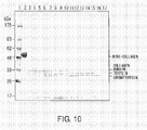
- the thermal stability of the purified proteins was assayed by utilizing enzyme treatment with high concentrations of trypsin (Trypsin, Type IX-S, From Porcine Pancreas, 13100 units/mg solid, protein; SIGMA-ALDRICH) and chymotrypsin ( ⁇ -Chymotrypsin, Type I-S: From Bovine Pancreas, 58 units/mg protein; SIGMA) under conditions in which only collagen is resistant to cleavage.
- trypsin Trypsin
- Type IX-S From Porcine Pancreas, 13100 units/mg solid, protein
- SIGMA-ALDRICH chymotrypsin
- ⁇ -Chymotrypsin, Type I-S From Bo
- Tris (2-Amino-2-hydroxymethyl-1,3-propanediol (Tris aminomethane); Wako) solution was added to 10 ⁇ L of the purified proteins (0.5 mg/mL) to adjust the pH to 7.
- An electrophoresis buffer Tris/Glycine/SDS, BIO-RAD
- Super Sep TM 10% to 20% 17 well (Wako) were placed in an electrophoresis vessel (DPE-1020, DAIICHI PURE CHEMICALS CO., LTD), and 6.5 ⁇ L of the heat-treated sample solutions were applied to Super Sep TM 10% to 20% 17 well (Wako), and electrophoresis was carried out at 40 mA (My Run, COSMO BIO CO., LTD) for 60 minutes.
- the gel was washed in 25 mL of DW (MILLIPORE) while shaking (ROTO-SHAKE GENIE, Scientific Industries) for five minutes, and this was repeated three times.
- the gel was stained for one hour in 25 mL of Quick-CBB PLUS (Wako), and then destained in 25 mL of DW (MILLIPORE) for one hour (see Fig 10 ).
- Fig. 11 shows the result of plotting a melting curve from quantifying the collagen domain bands at the respective heating temperatures, based on the results of the thermal stability assay performed in this Example for the proteins purified from the culture supernatant using trypsin and chymotrypsin, and defining the value obtained by quantifying the collagen domain band at the heat-treatment temperature of 30°C as 100%.
- the heat denaturation temperature heat-treatment temperature at which 50% is digested by the enzyme
- the heat denaturation temperature of naturally-occurring human atelocollagen type I is 41.9°C ( J.
- the purified protein has a thermotolerance that is equivalent or higher than that of naturally-occurring human atelocollagen type I, and is considered to be forming a stable triple helix structure.
- Mini-collagen-expressing CHO cells (pNC7/MC-21) were adjusted to 2.0 x 10 5 cells/mL with IS CHO-CD w/ Hydrolysate (IS JAPAN) medium supplemented to have final concentrations of 4 mM Gluta MAX TM -I (GIBCO), 0.4 mg/mL G418 Sulfate Cell Culture Tested (CALBIOCHEM), and 1 x HT supplement solution (GIBCO), and cultured by stationary culture in a T-75 flask (FALCON) at 37°C in the presence of 5% carbon dioxide for 14 days (HERA cell 150, Heraeus). The following steps were carried out at 4°C unless specified otherwise.
- IS JAPAN IS CHO-CD w/ Hydrolysate
- the culture solution was collected and centrifuged at 1,750 x g for ten minutes (EX-126, TOMY) to separate the cells and supernatant.
- EX-126, TOMY sodium chloride
- To this supernatant (1.35 L) sodium chloride (Wako) was added to obtain 0.4 M and the pH was adjusted (F-51, HORIBA) to 7.4 at 4°C using sodium hydroxide (Wako), and this was stored at 4°C.
- This supernatant was centrifuged at 10,000 x g for 30 minutes (EX-126, TOMY) to remove the precipitates, and the supernatant (1.35 L) was collected.
- mini-collagen-expressing CHO cells (pNC7/MC-21) were adjusted to 2.0 x 10 5 cells/mL with IS CHO-CD w/ Hydrolysate (IS JAPAN) medium supplemented to have final concentrations of 4 mM Gluta MAX TM -I (GIBCO), 0.4 mg/mL G418 Sulfate Cell Culture Tested (CALBIOCHEM), and 1 x HT supplement solution (GIBCO), and cultured by stationary culture in a T-75 flask (FALCON) at 37°C in the presence of 5% carbon dioxide for 14 days (HERA cell 150, Heraeus).
- IS JAPAN IS CHO-CD w/ Hydrolysate
- the culture solution was collected and centrifuged at 1,750 x g for ten minutes (EX-126, TOMY) to separate the cells and supernatant (1.87 L).
- This supernatant (1.87 L) and the aforementioned supernatant (1.35 L) were combined (3.22 L) and concentrated to a volume of 320 mL using cross flow filtration (VIVAFLOW200; 30,000 MWCO PES; VIVASIENCE), and sodium chloride (Wako) was added to obtain a final concentration of 4 M.
- the pH was adjusted (F-51, HORIBA) to 7.4 at 4°C using sodium hydroxide (Wako), and this was incubated at 25°C for four days.
- Precipitates formed in this process were collected by centrifugation at 9,400 x g for 30 minutes (EX-126, TOMY).
- EX-126, TOMY To the precipitates, 1.5 mL of 50 mM acetic acid (Wako) solution was added, and the whole amount was dialyzed (Spectra/Pro TM Biotech Cellulose Ester (CE) Dialysis Membranes; 10,000 MWCO; Spectrum Laboratories, Inc.) against 50 mM acetic acid (Wako) solution for five days. Then, the dialyzed sample solution was collected and subjected to centrifugation at 9,400 x g for 30 minutes (EX-126, TOMY) to collect the precipitates.
- CE Biotech Cellulose Ester
- Mini-collagen-expressing CHO cells (pNC7/MC-21) were adjusted to 2.0 x 10 5 cells/mL with IS CHO-CD w/ Hydrolysate (IS JAPAN) medium supplemented to have final concentrations of 4 mM Gluta MAX TM -I (GIBCO), 0.4 mg/mL G418 Sulfate Cell Culture Tested (CALBIOCHEM), and 1 x HT supplement solution (GIBCO), and cultured by stationary culture in T-75 flasks (FALCON) at 37°C in the presence of 5% carbon dioxide for 14 days (HERA cell 150, Heraeus). The following steps were carried out at 4°C unless specified otherwise.
- IS JAPAN IS CHO-CD w/ Hydrolysate
- the culture solution was collected and centrifuged at 1,750 x g for ten minutes (EX-126, TOMY) to separate the cells and supernatant.
- EX-126, TOMY sodium chloride
- To this supernatant (1.35 L) sodium chloride (Wako) was added to obtain 0.4 M and the pH was adjusted (F-51, HORIBA) to 7.4 at 4°C using sodium hydroxide (Wako), and this was stored at 4°C.
- This supernatant was centrifuged at 10,000 x g for 30 minutes (EX-126, TOMY) to remove the precipitates, and the supernatant (1.35 L) was collected.
- mini-collagen-expressing CHO cells (pNC7/MC-21) were adjusted to 2.0 x 10 5 cells/mL with IS CHO-CD w/ Hydrolysate (IS JAPAN) medium supplemented to have final concentrations of 4 mM Gluta MAX TM -I (GIBCO), 0.4 mg/mL G418 Sulfate Cell Culture Tested (CALBIOCHEM), and 1 x HT supplement solution (GIBCO), and cultured by stationary culture in a T-75 flask (FALCON) at 37°C in the presence of 5% carbon dioxide for 14 days (HERA cell 150, Heraeus).
- IS JAPAN IS CHO-CD w/ Hydrolysate
- the culture solution was collected and centrifuged at 1,750 x g for ten minutes (EX-126, TOMY) to separate the cells and supernatant (1.87 L).
- This supernatant (1.87 L) and the aforementioned supernatant (1.35 L) were combined (3.22 L) and concentrated to a volume of 320 mL using cross flow filtration (VIVAFLOW200; 30,000 MWCO PES; VIVASIENCE), and sodium chloride (Wako) was added to obtain a final concentration of 4 M.
- the pH was adjusted (F-51, HORIBA) to 7.4 at 4°C using sodium hydroxide (Wako), and this was incubated at 25°C for four days.
- Precipitates formed in this process were removed by centrifugation at 9,400 x g for 30 minutes (EX-126, TOMY). To this supernatant (320 mL), 1 M calcium chloride solution was added to obtain 20 mM, and this was incubated at 4°C for 18 hours and centrifuged at 9,400 x g for 30 minutes (EX-126, TOMY) to separate the precipitates and the supernatant.
- This supernatant (320 mL) was concentrated to a volume of 56 mL using cross flow filtration (VIVAFLOW200; 30,000 MWCO PES; VIVASIENCE), and then dialyzed (Spectra/Pro TM Biotech Dialysis Membranes; 10,000 MWCO; Spectrum Laboratories, Inc.) against TBS (TBS powder, Takara) containing 5 mM EDTA (Dojindo) for seven days.
- TBS TBS powder, Takara
- 5 mM EDTA Diojindo
- Mini-collagen remaining in this supernatant (86 mL) was purified using a mannan agarose column using the binding with mannan.
- An Econo-Column (Bio-RAD) was filled with 5 mL of mannan agarose gel (SIGMA), and the gel was washed and equilibrated with 15 mL of TBS (TBS powder, Takara) containing 5 mM EDTA (Dojindo) and 45 mL of TBS (TBS powder, Takara) containing 5 mM calcium chloride (Wako).
- TBS TBS powder, Takara
- Mini-collagen was eluted using 15 mL of TBS (TBS powder, Takara) containing 5 mM EDTA (Dojindo), and the first peak (9 mL) was collected.
- the eluate was dialyzed (Spectra/Pro TM Biotech Dialysis Membranes; 10,000 MWCO; Spectrum Laboratories, Inc.) against 0.4 M sodium chloride, 0.1 M Tris-hydrochloride buffer (pH7.4 at 4°C) for five days, and 3.9 mg of water-soluble mini-collagen having the activity to bind to mannan (hereinafter, MC-Man) was collected (see Fig. 13 ).
- MG-63 cells which are adherent cells, adhere to 96-well microplates coated with naturally-occurring human atelocollagen type I, naturally-occurring bovine atelocollagen type I, or purified mini-collagens (MC-salt, MC-Man).
- atelocollagen type I Cold Skin, Typel, Acid Soluble, From Human Skin; SIGMA-ALDRICH
- bovine atelocollagen type I From Calf Skin, Cell culture tested; SIGMA
- MC-salt MC-Man
- MC-Man MC-Man
- MG-63 cells human osteoblasts (MG-63 cells, ATCC) were made to adhere for one hour at 37°C, and cells that remained after removal of unadhered cells by washing (see Fig. 15 ) as well as cells that were subsequently incubated at 37°C for three hours (see Fig. 16 ) were observed under a phase contrast microscope.
- the image was observed using an inverted microscope (Nikon ECLIPSE TE2000-S, manufactured by Nikon) equipped with a high-definition color camera head (DS-Fi1, manufactured by Nikon) and a control unit (DS-L2, manufactured by Nikon).
- the absorbances in wells coated with naturally-occurring human atelocollagen type I, naturally-occurring bovine atelocollagen type I, MC-salt, and MC-Man were twice or higher than those of the (PBS) wells not coated with collagen, and high level of adhesion of human osteoblasts due to collagen coating was observed.
- human osteoblasts were made to adhere for one hour, and cells that remained after removal of unadhered cells by washing (see Fig. 15 ) as well as cells that were subsequently incubated at 37°C for three hours (see Fig. 16 ) were observed on a phase contrast microscope.
- Human osteoblasts showed adhesion and elongation in wells coated with naturally-occurring human atelocollagen type I, MC-salt, and MC-Man. However, while adhesion of human osteoblasts was observed with naturally-occurring bovine atelocollagen type I, elongation was hardly seen. From the above, MC-salt and MC-Man were found to have properties comparable to those of naturally-occurring human atelocollagen with regard to adhesion and elongation of human osteoblasts.
- MC-GPP a protein in which the portion from the C-terminal region to the GPP region of the mini-collagen is deleted.
- Fig. 17 shows each of the regions of the mini-collagen (Mini-Collagen Type I) and MC-GPP.
- pDC6/MC-GPP ( Fig. 18 ) was constructed by substituting the sequence of nucleotides Nos. 1267-1275 of the pDC6 vector as described in SEQ ID NO: 14 with the MC-GPP-encoding cDNA as described in SEQ ID NO: 15.
- pDC6/MC-GPP 2.5 ⁇ g of pDC6/MC-GPP was transfected into 4,000,000 CHO cells (CHO DG44 cells) in 25 cm 2 -culture flasks using the Lipofectin method (Lipofectamine TM LTX, Invitrogen was used). The transfection method followed the manufacturer's instructions. 48 hours after transfection, the cell number was determined, and then the cells were diluted in an IS CHO-CD w/ H medium (IS Japan) containing 4 mM Gluta MAX TM -I (Invitrogen).
- the cells were plated into five 96-well microtiter plates at concentrations of 4000 cells/well (480 wells), and when cultured in the presence of 5% carbon dioxide gas at 37°C for approximately three weeks, surviving cells were observed (cell lines growing in the HT-free medium).
- Western blotting was carried out under reducing conditions to verify the expression of the protein of interest in the surviving cell lines. Specifically, 10 ⁇ L of Laemmli Sample Buffer (BIO-RAD) containing 5% 2-mercaptoethanol (Wako) was mixed with 10 ⁇ L each of the culture supernatants of the cell lines found to proliferate, for reduction by heating at 98°C for five minutes (DTU-18, TAITEC).
- An electrophoresis buffer Tris/Glycine/SDS, BIO-RAD
- Super Sep TM Ace 10% to 20% 17 well (Wako) were placed in an electrophoresis vessel (DPE-1020, DAIICHI PURE CHEMICALS CO., LTD), 20 ⁇ L of the heat-treated sample solutions were applied to Super Sep TM Ace 10% to 20% 17 well (Wako), and electrophoresis was carried out at 40 mA (My Run, COSMO BIO CO., LTD) for 55 minutes.
- the gel was removed from the glass plates, and soaked for five minutes while shaking (Wave-S1, TAITEC) in 10 mL of transfer buffer (Tris/Glycine Buffer (BIO-RAD) containing methanol (Wako) at 30%).
- the Immobilon-P Transfer Membrane (MILLIPORE) was soaked while shaking (Wave-S1, TAITEC) in 10 mL of methanol (Wako) for 15 seconds, 10 mL of ultrapure water (ELGA) for two minutes, and 10 mL of transfer buffer (Tris/Glycine Buffer (BIO-RAD) containing 30% methanol (Wako)) for five minutes.
- the Immobilon-P Transfer Membrane (MILLIPORE) was soaked in 10 mL of ImmunoBlock (Laboratory Products division of Dainippon Sumitomo Pharma Co., Ltd.) and blocked at 4°C for 18 hours, then washed three times by shaking (Wave-S1, TAITEC) for five minutes in 10 mL of PBS (Wako) containing 0.05% Tween 20 (Polyoxyethylene (20) Sorbitan Monolaurate, Wako).
- MC-GPP-expressing CHO cells (pDC6/MC-GPP-3) were cultured with IS CHO-CD w/Hydrolysate medium (IS JAPAN) by stationary culture in T-75 flasks (FALCON) at 37°C in the presence of 5% carbon dioxide (HERA cell 150, Heraeus).
- the culture solutions were collected and centrifuged at 1,750 x g for ten minutes (EX-126, TOMY) to separate the cells and the supernatant, and this supernatant was loaded onto a Ni column to purify MC-GPP.
- a Poly empty column (BIO-RAD) was filled with 1 mL of Ni-NTA agarose gel (Invitrogen) and the gel was washed with 6 mL of ultrapure water (BMS). Subsequently, the gel was washed three times with 6 mL of Native binding buffer (0.25 M sodium dihydrogen phosphate (Wako), 2.5 M sodium chloride (Wako), 0.01 M imidazole (Wako), pH8.0), and 8 mL of the culture supernatant was loaded onto the column. The column was capped and binding took place while mixing at 4°C for 60 minutes (Aikuru, IWAKI).
- the gel was washed nine times with 6 mL of Native wash buffer (0.25 M sodium dihydrogen phosphate (Wako), 2.5 M sodium chloride (Wako), 0.02 M imidazole (Wako), pH8.0), and elution was carried out six times, 1 mL at a time using Native elution buffer (0.23 M sodium dihydrogen phosphate (Wako), 2.3 M sodium chloride (Wako), 0.25 M imidazole (Wako), pH8.0). The initial 2 mL of eluate was dialyzed against 0.02 M acetic acid solution at 4°C for three days, and then the MC-GPP solution was collected.
- Native wash buffer 0.25 M sodium dihydrogen phosphate (Wako), 2.5 M sodium chloride (Wako), 0.02 M imidazole (Wako), pH8.0
- Native elution buffer (0.23 M sodium dihydrogen phosphate (Wako), 2.3 M sodium chloride (Wako), 0.25 M imid
- Example 20 SDS polyacrylamide gel electrophoresis of MC-GPP under reducing conditions
- Purified MC-GPP was analyzed by SDS polyacrylamide gel electrophoresis under reducing conditions. Specifically, 10 ⁇ L of Laemmli Sample Buffer (BIO-RAD) containing 5% of 2-mercaptoethanol (Wako) was added to 10 ⁇ L of purified MC-GPP for reduction by heating at 98°C for five minutes (DTU-18, TAITEC).
- BIO-RAD Laemmli Sample Buffer
- Wako 2-mercaptoethanol
- An electrophoresis buffer Tris/Glycine/SDS, BIO-RAD
- Super Sep TM Ace 10% to 20% 17 well (Wako) were placed in an electrophoresis vessel (DPE-1020, DAIICHI PURE CHEMICALS CO., LTD), and 15 ⁇ L of the heat-treated sample solutions were applied to Super Sep TM Ace 10% to 20% 17 well (Wako), and electrophoresis was carried out at 40 mA (My Run, COSMO BIO CO., LTD) for 55 minutes. Then, silver staining was carried out using 2D-Silver Stain Reagent II (COSMO BIO CO., LTD).
- the gel was fixed by shaking for 20 minutes in 40 mL of fixing solution-I (50% methanol (Wako), 10% acetic acid (Wako), and 40% water (BMS)).
- fixing solution-I 50% methanol (Wako), 10% acetic acid (Wako), and 40% water (BMS)
- the gel was fixed by shaking for 30 minutes in 40 mL of fixing solution-II (30% methanol (Wako)-10% acetic acid (Wako), 5% fixing agent (2D-Silver Stain Reagent II, COSMO BIO CO., LTD.), and 55% ultrapure water (BMS)).
- pretreatment was carried out by shaking for 20 minutes in 40 mL of pretreatment solution (50% methanol (Wako), 5% pretreatment agent (2D-Silver Stain Reagent II, COSMO BIO CO., LTD.), and 45% ultrapure water (BMS)).
- pretreatment solution 50% methanol (Wako), 5% pretreatment agent (2D-Silver Stain Reagent II, COSMO BIO CO., LTD.), and 45% ultrapure water (BMS)).
- the gel was washed for ten minutes with 40 mL of ultrapure water (BMS), stained for 30 minutes using 40 mL of silver staining solution (5% staining solution A (2D-Silver Stain Reagent II, COSMO BIO CO., LTD.), 5% staining solution B (2D-Silver Stain Reagent II, COSMO BIO CO., LTD.), and 90% ultrapure water (BMS)), and then washed for five minutes using 40 mL of ultrapure water (BMS). The wash was repeated three times.
- BMS ultrapure water
- the gel was developed for eight minutes using 40 mL of developing solution (5% developing stock solution (2D-Silver Stain Reagent II, COSMO BIO CO., LTD.) and 95% ultrapure water (BMS)), and 2 mL of stop solution (2D-Silver Stain Reagent II, COSMO BIO CO., LTD.)) was added to stop the development. Finally, the gel was washed for ten minutes using 40 mL of ultrapure water (BMS), and an image was scanned (see Fig. 19 ) using a scanner (GT-X900, EPSON).
- Purified MC-GPP was analyzed by SDS polyacrylamide gel electrophoresis under non-reducing conditions. Specifically, 10 ⁇ L of Laemmli Sample Buffer (BIO-RAD) was added to 10 ⁇ L of purified MC-GPP, and this was subjected to heat treatment at 98°C for five minutes (DTU-18, TAITEC).
- BIO-RAD Laemmli Sample Buffer
- An electrophoresis buffer Tris/Glycine/SDS, BIO-RAD
- Super Sep TM Ace 5% to 20% 17 well (Wako) were placed in an electrophoresis vessel (DPE-1020, DAIICHI PURE CHEMICALS CO., LTD), and 15 ⁇ L of the heat-treated sample solutions were applied to Super Sep TM Ace 5% to 20% 17 well (Wako), and electrophoresis was carried out at 40 mA (My Run, COSMO BIO CO., LTD) for 55 minutes. Then, silver staining was carried out using 2D-Silver Stain Reagent II (COSMO BIO CO., LTD).
- the gel was fixed by shaking for 20 minutes in 40 mL of fixing solution-I (50% methanol (Wako), 10% acetic acid (Wako), and 40% water (BMS)).
- fixing solution-I 50% methanol (Wako), 10% acetic acid (Wako), and 40% water (BMS)
- the gel was fixed by shaking for 30 minutes in 40 mL of fixing solution-II (30% methanol (Wako), 10% acetic acid (Wako), 5% fixing agent (2D-Silver Stain Reagent II, COSMO BIO CO., LTD.), and 55% ultrapure water (BMS)).
- pretreatment was carried out by shaking for 20 minutes in 40 mL of pretreatment solution (50% methanol (Wako), 5% pretreatment agent (2D- Silver Stain Reagent II, COSMO BIO CO., LTD.), and 45% ultrapure water (BMS)).
- pretreatment solution 50% methanol (Wako), 5% pretreatment agent (2D- Silver Stain Reagent II, COSMO BIO CO., LTD.), and 45% ultrapure water (BMS)).
- the gel was washed for ten minutes with 40 mL of ultrapure water (BMS), stained for 30 minutes using 40 mL of silver staining solution (5% staining solution A (2D-Silver Stain Reagent II, COSMO BIO CO., LTD.), 5% staining solution B (2D-Silver Stain Reagent II, COSMO BIO CO., LTD.), and 90% ultrapure water (BMS)), then washed for five minutes using 40 mL of ultrapure water (BMS). The wash was repeated three times.
- BMS ultrapure water
- the gel was developed for eight minutes using 40 mL of developing solution (5% developing stock solution (2D-Silver Stain Reagent II, COSMO BIO CO., LTD.) and 95% ultrapure water (BMS)), and 2 mL of stop solution (2D-Silver Stain Reagent II, COSMO BIO CO., LTD.)) was added to stop the development. Finally, the gel was washed for ten minutes using 40 mL of ultrapure water (BMS), and an image was scanned (see Fig. 20 ) using a scanner (GT-X900, EPSON).
- Purified MC-GPP was analyzed by native polyacrylamide gel electrophoresis. Specifically, 10 ⁇ L of Native Sample Buffer (BIO-RAD) was added to 10 ⁇ L of purified MC-GPP. An electrophoresis buffer (Tris/Glycine/SDS, BIO-RAD) and Super Sep TM Ace 5% to 20% 17 well (Wako) were placed in an electrophoresis vessel (DPE-1020, DAIICHI PURE CHEMICALS CO., LTD), 15 ⁇ L of the prepared sample solutions were applied to Super Sep TM Ace 5% to 20% 17 well (Wako), and electrophoresis was carried out at 40 mA (My Run, COSMO BIO CO., LTD) for 55 minutes.
- BIO-RAD Native Sample Buffer
- An electrophoresis buffer Tris/Glycine/SDS, BIO-RAD
- Super Sep TM Ace 5% to 20% 17 well (Wako) were placed in an electrophoresis vessel (DPE-1020, DAII
- Pretreatment was carried out by shaking for 20 minutes in 40 mL of pretreatment solution (50% methanol (Wako), 5% pretreatment agent (2D- Silver Stain Reagent II, COSMO BIO CO., LTD.), and 45% ultrapure water (BMS)).
- pretreatment solution 50% methanol (Wako), 5% pretreatment agent (2D- Silver Stain Reagent II, COSMO BIO CO., LTD.), and 45% ultrapure water (BMS)).
- the gel was washed for ten minutes with 40 mL of ultrapure water (BMS), stained for 30 minutes using 40 mL of silver staining solution (5% staining solution A (2D-Silver Stain Reagent II, COSMO BIO CO., LTD.), 5% staining solution B (2D-Silver Stain Reagent II, COSMO BIO CO., LTD.), and 90% ultrapure water (BMS)), then washed for five minutes using 40 mL of ultrapure water (BMS). The wash was repeated three times.
- BMS ultrapure water
- the gel was developed for eight minutes using 40 mL of developing solution (5% developing stock solution (2D-Silver Stain Reagent II, COSMO BIO CO., LTD.) and 95% ultrapure water (BMS)), and 2 mL of stop solution (2D-Silver Stain Reagent II, COSMO BIO CO., LTD.)) was added to stop the development. Finally, the gel was washed for ten minutes using 40 mL of ultrapure water (BMS), and an image was scanned (see Fig. 21 ) using a scanner (GT-X900, EPSON).
- MC-GPP Since MC-GPP has a His-tag on its C-terminal side, anti-His antibodies can bind to it. Western blotting was carried out under reducing conditions by utilizing this property, and purified MC-GPP was detected and identified by chemiluminescence. Specifically, 10 ⁇ L of Laemmli Sample Buffer (BIO-RAD) containing 5% 2-mercaptoethanol (Wako) was added to 10 ⁇ L of purified MC-GPP for reduction by heating at 98°C for five minutes (DTU-18, TAITEC).
- BIO-RAD Laemmli Sample Buffer
- Wako 2-mercaptoethanol
- An electrophoresis buffer Tris/Glycine/SDS, BIO-RAD
- Super Sep TM Ace 5% to 20% 17 well (Wako) were placed in an electrophoresis vessel (DPE-1020, DAIICHI PURE CHEMICALS CO., LTD), and 15 ⁇ L of the heat-treated sample solutions were applied to Super Sep TM Ace 5% to 20% 17 well (Wako), and electrophoresis was carried out at 40 mA (My Run, COSMO BIO CO., LTD) for 55 minutes.
- the gel was removed from the glass plates, and soaked for five minutes while shaking (Wave-S1, TAITEC) in 10 mL of transfer buffer (Tris/Glycine Buffer (BIO-RAD) containing 30% methanol (Wako)).
- Immobilon-P Transfer Membrane MILLIPORE
- Wave-S1, TAITEC was soaked while shaking (Wave-S1, TAITEC) in 10 mL of methanol (Wako) for 15 seconds, 10 mL of ultrapure water (ELGA) for two minutes, and 10 mL of transfer buffer (Tris/Glycine Buffer (BIO-RAD) containing 30% methanol (Wako)) for five minutes.
- the Immobilon-P Transfer Membrane (MILLIPORE) was soaked in 10 mL of ImmunoBlock (Laboratory Products division of Dainippon Sumitomo Pharma Co., Ltd.) and blocked at 4°C for 18 hours, then washed three times by shaking (Wave-S1, TAITEC) for five minutes in 10 mL of PBS (Wako) containing 0.05% Tween 20 (Polyoxyethylene (20) Sorbitan Monolaurate, Wako).
- MC-GPP Since MC-GPP has a His-tag on its C-terminal side, anti-His antibodies can bind to it. Western blotting was carried out under non-reducing conditions by utilizing this property, and purified MC-GPP was detected and identified by chemiluminescence. Specifically, 10 ⁇ L of Laemmli Sample Buffer (BIO-RAD) was added to 10 ⁇ L of purified MC-GPP for heat treatment at 98°C for five minutes (DTU-18, TAITEC).
- BIO-RAD Laemmli Sample Buffer
- An electrophoresis buffer Tris/Glycine/SDS, BIO-RAD
- Super Sep TM Ace 5% to 20% 17 well (Wako) were placed in an electrophoresis vessel (DPE-1020, DAIICHI PURE CHEMICALS CO., LTD), and 15 ⁇ L of the heat-treated sample solutions were applied to Super Sep TM Ace 5% to 20% 17 well (Wako), and electrophoresis was carried out at 40 mA (My Run, COSMO BIO CO., LTD) for 55 minutes.
- the gel was removed from the glass plates, and soaked for five minutes while shaking (Wave-S1, TAITEC) in a transfer buffer (Tris/Glycine Buffer (BIO-RAD) containing 30% methanol (Wako)).
- Immobilon-P Transfer Membrane MILLIPORE
- Wave-S1, TAITEC was soaked while shaking (Wave-S1, TAITEC) in 10 mL of methanol (Wako) for 15 seconds, 10 mL of ultrapure water (ELGA) for two minutes, and 10 mL of transfer buffer (Tris/Glycine Buffer (BIO-RAD) containing 30% methanol (Wako)) for five minutes.
- the Immobilon-P Transfer Membrane (MILLIPORE) was soaked in 10 mL of ImmunoBlock (Laboratory Products division of Dainippon Sumitomo Pharma Co., Ltd.) and blocked at 4°C for 18 hours, then washed three times by shaking (Wave-S1, TAITEC) for five minutes in 10 mL of PBS (Wako) containing 0.05% Tween20 (Polyoxyethylene (20) Sorbitan Monolaurate, Wako).
- Collagen that forms a triple helix structure is resistant against cleavage by pepsin. Therefore, purified MC-GPP, naturally-occurring human atelocollagen type I (Collagen, Type I, Acid Soluble, From Human Skin, SIGMA-ALDRICH), and purified fibrous mini-collagen (Example 7) were digested with pepsin under acidic conditions, and resistance against cleavage by pepsin was verified from SDS polyacrylamide electrophoresis images.
- 3 ⁇ L of 0.3 M hydrochloric acid solution was added to 10 ⁇ L each of purified MC-GPP (0.028 mg/mL), naturally-occurring human atelocollagen type I (Collagen, Type I, Acid Soluble, From Human Skin, SIGMA-ALDRICH) (0.1 mg/mL), or fibrous mini-collagen (Example 7) (0.1 mg/mL) to adjust the pH to 2, 3 ⁇ L of pepsin (Pepsin, From Porcine Stomach Mucosa, 3370 units/mg protein; SIGMA-ALDRICH) solution (the amount of pepsin is three times that of each protein when converted into moles) was added, and pepsin digestion was carried out at 20°C (2720 Thermal cycler, Applied Biosystems) for two hours.
- SIGMA-ALDRICH pepsin
- preparations of each sample with no pepsin added preparations of pepsin (Pepsin, From Porcine Stomach Mucosa, 3370 units/mg protein; SIGMA-ALDRICH) alone (same amount as the amount used to digest each sample), and preparations containing neither sample nor pepsin were prepared as controls. 10 mM acetic acid solution was added instead of the pepsin solution or sample, and incubation was carried out at 20°C for two hours.
- An electrophoresis buffer Tris/Glycine/SDS, BIO-RAD
- Super Sep TM Ace 10% to 20% 17 well (Wako) were placed in an electrophoresis vessel (DPE-1020, DAIICHI PURE CHEMICALS CO., LTD), and 18 ⁇ L of the heat-treated sample solutions were applied to Super Sep TM Ace 10% to 20% 17 well (Wako), and electrophoresis was carried out at 40 mA (My Run, COSMO BIO CO., LTD) for 55 minutes. Then, silver staining was carried out using 2D-Silver Stain Reagent II (COSMO BIO CO., LTD).
- the gel was fixed by shaking for 20 minutes in 40 mL of fixing solution-I (50% methanol (Wako), 10% acetic acid (Wako), and 40% water (BMS)).
- fixing solution-I 50% methanol (Wako), 10% acetic acid (Wako), and 40% water (BMS)
- the gel was fixed by shaking for 30 minutes in 40 mL of fixing solution-II (30% methanol (Wako), 10% acetic acid (Wako), 5% fixing agent (2D-Silver Stain Reagent II, COSMO BIO CO., LTD.), and 55% ultrapure water (BMS)).
- Pretreatment was carried out by shaking for 20 minutes in 40 mL of pretreatment solution (50% methanol (Wako), 5% pretreatment agent (2D-Silver Stain Reagent II, COSMO BIO CO., LTD.), and 45% ultrapure water (BMS)).
- pretreatment solution 50% methanol (Wako), 5% pretreatment agent (2D-Silver Stain Reagent II, COSMO BIO CO., LTD.), and 45% ultrapure water (BMS)).
- the gel was washed for ten minutes with 40 mL of ultrapure water (BMS), stained for 30 minutes using 40 mL of silver staining solution (5% staining solution A (2D-Silver Stain Reagent II, COSMO BIO CO., LTD.), 5% staining solution B (2D-Silver Stain Reagent II, COSMO BIO CO., LTD.), and 90% ultrapure water (BMS)), then washed for five minutes using 40 mL of ultrapure water (BMS). The wash was repeated three times.
- BMS ultrapure water
- the gel was developed for eight minutes using 40 mL of developing solution (5% developing stock solution (2D-Silver Stain Reagent II, COSMO BIO CO., LTD.) and 95% ultrapure water (BMS)), and 2 mL of stop solution (2D-Silver Stain Reagent II, COSMO BIO CO., LTD.)) was added to stop the development. Finally, the gel was washed for ten minutes using 40 mL of ultrapure water (BMS), and an image was scanned ( Fig. 24 ) using a scanner (GT-X900, EPSON).
- the bands in the lanes to which MC-GPP, pepsin-digested MC-GPP, an equivalent amount of pepsin only to that used to digest MC-GPP, purified fibrous mini-collagen (Example 7), pepsin-digested fibrous mini-collagen (Example 7), and an equivalent amount of pepsin only to that used to digest fibrous mini-collagen (Example 7) were applied were analyzed using ImageJ (see Fig. 25 ).
- the present invention can provide advanced human collagen analogs that have a triple helix structure similar to that of naturally occurring ones, which can be more easily handled than the naturally-occurring ones, expression vectors that enable production thereof, and human collagen analog-producing cells.
- the production methods of the present invention can be applied not only to collagen, but also to proteins that have a triple helix structure, such as collectin.
- the collagen analogs of the present invention have lower molecular weights than those of naturally-occurring collagens, they are easily purified and easily handled. It is considered that these novel collagen analogs having a triple helix structure have properties that are different from those of known collagens, and their applications as novel biomaterials are expected.
Abstract
Description
- The present invention relates to proteins having a triple-helix structure and methods for producing them. More specifically, the present invention relates to human-type collagen analogs and methods for producing them. An objective of the present invention is to provide collagen analogs composed of human-type recombinant proteins which are safe for living organisms and can be easily purified and obtained, and methods for producing them. More specifically, the present invention provides methods for producing collagen analogs composed of a recombinant protein in which the introduced genes are all human-type, wherein the method is carried out by stable transduction of a mammalian expression vector inserted with a cDNA of a human-type collagen-comprising recombinant protein into Chinese hamster ovary (CHO) cells.
- In recent years, an example of one of the most important materials in regenerative medicine is collagen. Collagen is a representative protein distributed in nearly all tissues (skin, bone, cartilage, and such) in living organisms, and it is well known that it has important functions in living organisms such as maintaining the structure of biological tissues and organs by becoming a scaffold for cells. In addition, it has various physiological functions that regulate the proliferation, differentiation, and migration of cells. From these facts, it is receiving attention in the field of regenerative medicine through its use together with cells, growth factors, and such in tissue engineering medicine. So far, collagen has been used widely in the medical field as artificial organ implants (Patent Document 1), sustained drug release matrices (Patent Document 2), artificial skin (Patent Document 3), and components of biocompatible materials for use in bandage matrices for wounds and matrices for wound treatment (Patent Document 4).
- Forty percent of all collagen of a living organism is in the skin, and 70% or more of the dry weight of the skin/tendon is collagen. Therefore, collagen is important in the development of artificial skin. In particular, collagen is used as a biomaterial for repairing damages in organisms. For example, it is used as a coating material for sites of skin lesion such as a burn, and healing and improvement have been reported (Non-Patent
Documents 1 and 2). This means that one can have great hope for applications in the current significantly progressed field of regenerative medicine. Furthermore, it is utilized as a material useful in techniques for culturing cells and organs (Patent Documents 5 and 6). In addition, it has been pointed out that oral ingestion of collagen (type II collagen) and such may be used to suppress rheumatoid arthritis (Non-Patent Document 3). Furthermore, it has been reported that it is possible to treat by designing a gene to express a partial peptide of human collagen (type VII collagen), and introducing a low-molecular-weight collagen gene into epidermolysis bullosa cells (Non-Patent Document 4). - Many of the collagens used at present are derived from non-human mammalian species such as bovine or pigs. It is reported that when these collagens are transplanted into humans, allergic reaction occurs in approximately 3% of the patients (
Non-Patent Documents 5 and 6). Furthermore, in recent years, the risk of contamination of collagen derived from non-human mammalian species with prions or pathogens has become a major problem. Therefore, a system for producing safe human-type collagens with low antigenicity and free of risk of pathogen contamination is strongly desired. - To avoid such problems, some inventors have invented a method for producing recombinant human collagen having a triple helix structure equivalent to that in a human body by infecting insect cells with a recombinant virus inserted with a cDNA encoding human collagen, and have applied for a patent (Patent Document 7). Furthermore, methods for producing human collagen using mammalian cells or yeast cells have also been devised (Patent Document 8).
-
- [Patent Document 1] Japanese Patent Application Kokai Publication No. (
JP-A) 2007-204881 - [Patent Document 2] JP-A (Kokai) 2001-316282
- [Patent Document 3] JP-A (Kokai) 2005-314
- [Patent Document 4] JP-A (Kokai) 2007-160092
- [Patent Document 5] JP-A (Kokai) 2002-142753
- [Patent Document 6] Japanese Patent Application Saikohyo Publication No. (
JP-A) 2005-014774 - [Patent Document 7]
JP-A (Kokai) 8-23979 - [Patent Document 8] Japanese Patent Application Kohyo Publication No. (
JP-A) 7-501939 - [Non-Patent Documents]
- [Non-Patent Document 1] Surg. Forum ,10, 303 (1960)
- [Non-Patent Document 2] J. Surg.Res., 10, 485-491 (1960)
- [Non-Patent Document 3] Science, 261, 1727-1730 (1993)
- [Non-Patent Document 4] THE JOURNAL OF BIOLOGICAL CHEMISTRY Vol. 275, No. 32, Issue of August 11, pp. 24429-24435, 2000
- [Non-Patent Document 5] J.Immuno1.136:877-882, 1986
- [Non-Patent Document 6] Biomaterials 11:176-180, 1990
- In this way, collagen is a substance useful as a pharmaceutical product or a biomaterial for live-donor transplantation or regenerative medicine; however, conventionally used collagen is derived from tissues of non-human mammalian species such as pigs and cattle. Collagen is originally a protein with low immunogenicity, and is being transplanted, embedded or administered into the human body as a biomaterial. However, although in low frequency, there are reports that immune reactions are evoked by collagen derived from tissues of non-human mammalian species (J. Immunol., 136, 877-882 (1986); Biomaterials, 11, 176-180 (1990)). Furthermore, due to the possibility of prion contamination suggested in the case of cattle, it is not possible to use cattle-derived collagen. In addition, there is no guarantee that unknown contaminants (pathogenic viruses and such) like prion contamination are not contained in mammals such as pigs which are currently used for the purification and extraction of collagen, and safety problems are being raised with using collagen derived from non-human mammals to humans. Additionally, a problem with biologically-derived collagen is that multi-step purification becomes necessary during purification due to the inclusion of large amount of contaminant proteins, and the purification method becomes complicated.
- In light of the above-mentioned issues, human-derived collagen is desirable as a biomaterial to be used directly on humans. Human derived collagen may be purified from human sources (such as human placenta) (
U.S. Patent Nos. 5,002,071 and5,428,022 ). However, there are several problems in the use of human-derived collagen: (1) since the material is human tissue, the material has limited supply; (2) one cannot completely eliminate the possibility of contamination with pathogenic viruses such as hepatitis viruses and human immunodeficiency viruses (HIV); (3) the types of collagen collected from the placenta are disproportionate and the qualities are not completely identical; and (4) there are ethical problems regarding the extraction and purification of collagen from humans. The qualitative problem also exists as purification becomes difficult due to formation of unspecified bridges in the obtained collagen. - So far, methods that use genetic engineering techniques for producing collagen have been investigated to eliminate the risk of pathogen contamination and to obtain large amounts of collagen for which the isolation and purification steps are easy (Biochem. Soc., 28, 350-353 (2000)). However, the molecular weight of a collagen molecule is 100,000 or more and is very large, and production of an expression vector for introduction into host cells is very complicated. In addition, conventional methods did not yield production levels that can sustain practical applications. Furthermore, collagen is a molecule that adopts a triple helix structure in which three polypeptide chains are assembled, and such structure is formed by undergoing a number of posttranslational modifications (N. Engl. J. Med., 311, 376-386 (1984)), but only specific cells are expected to possess such modification abilities.
- It is known that in order for collagen to form a triple helix structure, prolines in the collagen domain must be hydroxylated. To produce collagen having a triple helix structure, a method for producing recombinant collagen by coexpressing human collagen and proline hydroxylase in insect cells was provided (
JP-A (Kokai) 2002-315580 - Production of human-derived recombinant collagen using hamster embryonic cells, mouse fibroblast cells, and such as hosts has been tested from before (Proc. Natl. Acad. Sci. USA., 84, 764-768 (1987); J. Biol. Chem., 264, 20683-20687 (1989)). The molecular structures of collagens obtained in these examples were normal, but host cell-derived collagen and foreign gene-derived collagen were mixed. Furthermore, in an example where type II collagen was expressed in human fibrosarcoma cells HT1080 (Biochem. J., 298, 31-37 (1994)), the production level was low (0.5 to 1 mg per 1 L of culture), and this was not sustainable for practical use. Furthermore, an equal amount of human fibrosarcoma cell HT1080-derived type IV collagen as that of the foreign gene-derived type II collagen was observed. Therefore, the foreign gene-derived type II collagen had to be separated from endogenous type IV collagen, and it was also impractical in this regard. Therefore, even if an expression system is used, it becomes necessary to examine the meticulous purification conditions, and it was considered that even under conditions of mixed contaminants, a simple and enabling purification method is necessary.
- In addition to the above, there are examples in which human collagen was expressed using yeast (
JP-A (Kohyo) H07-501939 JP-A (Kokai) H08-23979 JP-A (Kokai) H11-178574 JP-A (Kokai) 2002-325584 - In view of the above circumstances, the applicants have investigated production of human type I collagen by applying genetic engineering techniques to obtain non-antigenic collagen, eliminate the danger of pathogen contamination, and obtain collagen that is easy to isolate and purify (International Publication
WO 2006/106970 ). Although conventional methods could secure a certain level of production, a system in which more triple helix structures are formed was needed. Improvement was considered necessary also in terms of expression level. - The present invention was achieved in view of the above circumstances. An objective of the present invention is to provide safe human-type collagen analog proteins having a triple helix structure, and methods for producing them.
- The present inventors conducted various examinations to solve the above-mentioned problems and successfully produced easily purified collagen analogs (mini collagens) that have a triple helix structure and a molecular weight smaller than those of naturally-occurring-type collagens, by introducing into host cells a construct produced by fusing a signal peptide domain gene of human collectin and a cysteine-rich domain gene of human collectin to the amino-terminal side, and a neck domain gene of human collectin and a carbohydrate recognition domain gene of human collectin to the carboxy-terminal side of the collagen domain of the collagen gene which is a protein having triple helix structure.
- Examples of known proteins having a triple helix structure include human mannan-binding lectin (MBL) and conglutinin. By reducing the molecular weight of the collagen analogs of the present invention to be close to those of these proteins, collagen analog proteins having a triple helix structure with reduced molecular weight, which had been difficult to realize to date, were successfully produced. Furthermore, these collagen analog proteins were shown to have a triple helix structure and thermal stability.
- Naturally-occurring human collagen has poor water solubility, whereas the collagen analogs of the present invention show high water solubility since they comprise the water-soluble cysteine-rich domain, neck domain, and carbohydrate recognition domain of human collectin. Therefore, they are easier to handle compared to naturally-occurring collagens which have a high molecular weight.
- The present inventors precipitated the collagen analogs by promoting fibril formation by adding a high concentration of neutral salt, and successfully and easily purified fibrous collagen analogs having a triple helix structure by centrifugation.
- Furthermore, the present inventors successfully purified water-soluble collagen analogs by a simple one-step purification method, using mannan agarose which utilizes the binding of the carbohydrate recognition domain to mannan.
- Using the different purification methods described above, the present inventors successfully purified two collagen analogs having different physical properties: a fibrous collagen analog with high physical strength; and a water-soluble collagen with high solubility that binds to mannan. These collagen analogs were shown to have the same degrees of cellular adhesiveness and elongation properties as naturally-occurring human collagen when used as biomaterial in human adherent cells. Collagen analogs of the present invention can be expected to be useful as replacements for conventionally used collagen derived from non-human mammalian species, or as biomaterials for use in humans.
- As a result of conducting various examinations to solve the above-mentioned problems, the present inventors invented a collagen gene construct that can be easily purified and which maintain a triple helix structure equivalent to that of naturally-occurring collagen while having a low molecular weight. Specifically, because CR-D (a signal peptide) has a carbohydrate recognition domain, one-step purification by affinity purification is enabled. By substituting a portion of a human collagen structural gene of the present invention with the collagen-like structural gene portion of MBL, it has become possible to obtain low-molecular-weight collagens maintaining a triple helix structure in large quantities and with high purity.
- More specifically, the present inventors successfully produced large amounts of human collagen analogs by introducing a construct in which a collagen analog gene of the present invention is contained in a vector capable of highly expressing a foreign gene using as host Chinese hamster ovary (CHO) cells which (1) have been used for producing pharmaceuticals and are confirmed to be safe and (2) are thought to have sugar chain modifications and such of proteins that are close to those of humans since they are mammalian cells.
- More specifically, the present inventors successfully developed a method for producing large quantities of collagen analogs of the present invention without the need for complicated purification steps, by minimizing the mixture of host-derived collagen and foreign gene-derived collagen, using mammalian cells that have a low expression level of collagen (a protein having triple helix structure) as host. From the above, the present invention was completed.
- Specifically, the present invention provides the following:
- [1] a recombinant protein having a triple helix structure, which comprises a protein encoded by a polynucleotide comprising (i) to (v) below in order from the amino terminus:
- (i) a signal peptide domain gene of human collectin;
- (ii) a cysteine-rich domain gene of human collectin;
- (iii) a collagen domain gene of human collagen;
- (iv) a neck domain gene of human collectin; and
- (v) a carbohydrate recognition domain gene of human collectin;
- [2] the recombinant protein having a triple helix structure of [1], wherein the signal peptide domain gene of human collectin is a signal peptide domain gene of human surfactant protein D (SP-D) and a polynucleotide comprising the nucleotide sequence of SEQ ID NO: 4;
- [3] the recombinant protein having a triple helix structure of [1], wherein the cysteine-rich domain gene of human collectin is a cysteine-rich domain gene of human surfactant protein D (SP-D) and a polynucleotide comprising the nucleotide sequence of SEQ ID NO: 5;
- [4] the recombinant protein having a triple helix structure of [1], wherein the neck domain gene of human collectin is a neck domain gene of human mannan-binding lectin (MBL) and a polynucleotide comprising the nucleotide sequence of SEQ ID NO: 6;
- [5] the recombinant protein having a triple helix structure of [1], wherein the carbohydrate recognition domain gene of human collectin is a carbohydrate recognition domain gene of human mannan-binding lectin (MBL) and a polynucleotide comprising the nucleotide sequence of SEQ ID NO: 7;
- [6] the recombinant protein having a triple helix structure of [1], wherein the collagen domain gene of human collagen comprises at least one or more types of collagen domain genes of α-chain human collagens;
- [7] the recombinant protein having a triple helix structure of [1], wherein the collagen domain gene of human collagen is a collagen domain gene of a human type I collagen comprising an α-chain human collagen;
- [8] the recombinant protein having a triple helix structure of [6] or [7], wherein the collagen domain gene of an α-chain human collagen is a polynucleotide comprising the nucleotide sequence of SEQ ID NO: 8;
- [9] the recombinant protein having a triple helix structure of [1], which comprises a protein comprising the amino acid sequence of SEQ ID NO: 1;
- [10] the recombinant protein having a triple helix structure of [1], wherein the polynucleotide is a polynucleotide comprising the nucleotide sequence of SEQ ID NO: 3;
- [11] a method for producing a protein having a triple helix structure, wherein the method comprises the steps of:
- (a) introducing into a vector a polynucleotide comprising (i) to (v) below in order from the amino terminus:
- (i) a signal peptide domain gene of human collectin;
- (ii) a cysteine-rich domain gene of human collectin;
- (iii) a collagen domain gene of human collagen;
- (iv) a neck domain gene of human collectin; and
- (v) a carbohydrate recognition domain gene of human collectin;
- (b) transforming a host cell by gene introduction using the vector; and
- (c) culturing or breeding the transformant, and collecting a protein having a triple helix structure from the cell or its culture supernatant;
- (a) introducing into a vector a polynucleotide comprising (i) to (v) below in order from the amino terminus:
- [12] the method of [11], wherein the signal peptide domain gene of human collectin is a signal peptide domain gene of human surfactant protein D (SP-D) and a polynucleotide comprising the nucleotide sequence of SEQ ID NO: 4;
- [13] the method of [11], wherein the cysteine-rich domain gene of human collectin is a cysteine-rich domain gene of human surfactant protein D (SP-D) and a polynucleotide comprising the nucleotide sequence of SEQ ID NO: 5;
- [14] the method of [11], wherein the neck domain gene of human collectin is a neck domain gene of human mannan-binding lectin (MBL) and a polynucleotide comprising the nucleotide sequence of SEQ ID NO: 6;
- [15] the method of [11], wherein the carbohydrate recognition domain gene of human collectin is a carbohydrate recognition domain gene of human mannan-binding lectin (MBL) and a polynucleotide comprising the nucleotide sequence of SEQ ID NO: 7;
- [16] the method of [11], wherein the collagen domain gene of human collagen comprises at least one or more types of collagen domain genes of α-chain human collagens;
- [17] the method of [11], wherein the collagen domain gene of human collagen is a collagen domain gene of a human type I collagen comprising an α-chain human collagen;
- [18] the method of [16] or [17], wherein the collagen domain gene of an α-chain human collagen is a polynucleotide comprising the nucleotide sequence of SEQ ID NO: 8;
- [19] the method of [11], wherein the vector used in step (a) is pNC1 of SEQ ID NO: 2; and
- [20] the method of [11], wherein the vector used in step (a) is pDC6/CF of SEQ ID NO: 9.
-
-
Fig. 1 shows the pNC1/Mini-Collagen Type I construct with the respective abbreviations shown below. PCMV: cytomegalovirus promoter; INRBG: rabbit growth hormone intron; Mini-Collagen Type I: mini-collagen DNA; PABGH: bovine growth hormone gene polyA addition signal; PdSV: enhancer-deletedsimian virus 40 promoter; NPT: neomycin phosphotransferase cDNA; PASV:simian virus 40 polyA addition signal; and Ampr: selection marker (ampicillin resistance) in E. coli. -
Fig. 2 shows the pDC6/CF_Mini-Collagen Type I construct with the respective abbreviations shown below. PCMV5:cytomegalovirus 5 promoter; Mini-Collagen Type I: mini-collagen DNA; PABGH: bovine growth hormone gene polyA addition signal; PdSV: enhancer-deletedsimian virus 40 promoter; cd180DHFR: translation-impaired NPT gene produced by altering codons in the range of 180 bases from the 5' end of the nucleotide sequence of DHFR to the least frequently used codons in mammals; PASV:simian virus 40 polyA addition signal; and Ampr: selection marker (ampicillin resistance) in E. coli. -
Fig. 3 is a flow chart of mini-collagen purification. All steps were carried out at 4°C unless specified otherwise. In the figure, * 1 indicates the step of adjusting cells to 2.0 x 10⁵ cells/mL with IS CHO-CD w/ Hydrolysate (IS JAPAN) medium supplemented to have final concentrations of 4 mM Gluta MAX™-I (GIBCO), 0.4 mg G418 Sulfate Cell Culture Tested (CALBIOCHEM), and 1 x HT supplement solution (GIBCO), and culturing by stationary culture in T-75 flasks (FALCON) at 37°C in the presence of 5% carbon dioxide for 14 days (HERA cell 150, Heraeus). *2 indicates the step of centrifuging at 1,750 x g for one hour (EX-126, TOMY). *3 indicates the step of adding sodium chloride (Wako) to the supernatant (1.4 L) to obtain 0.4 M. *4 indicates the step of adjusting the pH to 7.4 (F-51, HORIBA) at 4°C using sodium hydroxide (Wako). *5 indicates the step of concentrating the culture supernatant to 1/20 its volume by using cross flow filtration (VIVAFLOW50; 10,000 MWCO PES; VIVASIENCE). *6 indicates the step of dialyzing (Spectra/Pro™ Biotech Dialysis Membranes; 10,000 MWCO; Spectrum Laboratories, Inc.) for three days against TBS (TBS powder, Takara) containing 5 mM EDTA (Dojindo). *7 indicates the step of adding calcium chloride (Wako) and sodium chloride (Wako) to obtain 20 mM and at 2 M, respectively. *8 indicates the step of dialyzing (Spectra/Pro™ Biotech Cellulose Ester (CE) Dialysis Membranes; 25,000 MWCO; Spectrum Laboratories, Inc.) against MilliQ water (MILLIPORE) for five days. *9 indicates the steps of filling an Econo-Column (Bio-RAD) with 4.5 mL of mannan agarose gel (SIGMA), washing and equilibrating the gel with 45 mL of TBS (TBS powder, Takara) containing 5 mM EDTA (Dojindo) and TBS (TBS powder, Takara) containing 5 mM calcium chloride (Wako), loading the supernatant by circulation at a flow rate of 1.0 mL/min for 17.5 hours, removing the supernatant, then washing with 10 mL of TBS (TBS powder, Takara) containing 5 mM calcium chloride (Wako), and eluting mini-collagen with 20 mL of TBS (TBS powder, Takara) containing 5 mM EDTA (Dojindo). * 10 indicates the step of dialyzing (Spectra/Pro™ Biotech Dialysis Membranes; 10,000 MWCO; Spectrum Laboratories, Inc.) against 0.4 M sodium chloride-0.1 M Tris-hydrochloride buffer (pH7.4 at 4°C) for five days. *11 indicates the step of concentrating to 1/10 the volume by ultrafiltration at 1,750 x g for 30 minutes using Amicon Ultra-15 (10,000 MWCO; MILLIPORE). -
Fig. 4 shows in a photograph the analysis result of SDS polyacrylamide gel electrophoresis under reducing conditions (2-mercaptoethanol added) of proteins purified from the culture supernatant and water-soluble mini-collagen.Lane 1 shows proteins purified as a precipitate andlane 2 is water-soluble mini-collagen purified on a mannan agarose column. The molecular weight and the mini-collagen oligomer are indicated on the photograph. -
Fig. 5 shows in a photograph the analysis result of SDS polyacrylamide gel electrophoresis under non-reducing conditions (no 2-mercaptoethanol added) of proteins purified from the culture supernatant and water-soluble mini-collagen.Lane 1 shows proteins purified as a precipitate andlane 2 is water-soluble mini-collagen purified on a mannan agarose column. The molecular weight and the mini-collagen oligomers are indicated on the photograph. -
Fig. 6 shows in a photograph the analysis result of polyacrylamide gel electrophoresis under native conditions (no 2-mercaptoethanol and SDS added) of proteins purified from the culture supernatant and water-soluble mini-collagen.Lane 1 shows proteins purified as a precipitate andlane 2 is water-soluble mini-collagen purified on a mannan agarose column. The molecular weight and the mini-collagen oligomers are indicated on the photograph. -
Fig. 7 shows in a photograph results obtained by performing SDS polyacrylamide gel electrophoresis under reducing conditions (2-mercaptoethanol added) of proteins purified from the culture supernatant and water-soluble mini-collagen, performing Western blotting using a rabbit anti-MBL (carbohydrate recognition domain (CRD)) polyclonal antibody, and reversing the contrast of the photograph of chemiluminescence detection.Lane 1 shows proteins purified as a precipitate andlane 2 is water-soluble mini-collagen purified on a mannan agarose column. The molecular weight and the mini-collagen oligomer are indicated on the photograph. -
Fig. 8 shows in a photograph the result obtained by performing SDS polyacrylamide gel electrophoresis under non-reducing conditions (no 2-mercaptoethanol added) of proteins purified from the culture supernatant and water-soluble mini-collagen, performing Western blotting using a rabbit anti-MBL (carbohydrate recognition domain (CRD)) polyclonal antibody, and then reversing the contrast of the photograph of chemiluminescence detection.Lane 1 shows proteins purified as a precipitate andlane 2 is water-soluble mini-collagen purified on a mannan agarose column. The molecular weight and the mini-collagen oligomers are indicated on the photograph. -
Fig. 9 shows in a photograph the analysis result of SDS polyacrylamide gel electrophoresis under reducing conditions (2-mercaptoethanol added) of the purified protein and naturally-occurring human atelocollagen type I digested with pepsin under acidic conditions. Positions of the bands for mini-collagen, the collagen domain of mini-collagen remaining after digestion, and pepsin are indicated on the photograph. Lane 1: molecular weight marker; lane 2: purified protein not digested with pepsin; lane 3: purified protein digested with pepsin; lane 4: naturally-occurring human atelocollagen type I not digested with pepsin; lane 5: naturally-occurring human atelocollagen type I digested with pepsin; and lane 6: pepsin alone added. -
Fig. 10 shows in a photograph the results of analyzing thermal stability by subjecting the purified proteins to heat treatment in a temperature range of 30°C to 50°C, performing enzyme treatment using a combination of highly concentrated trypsin and chymotrypsin under conditions in which collagen is not digested, and performing SDS polyacrylamide gel electrophoresis under reducing conditions (2-mercaptoethanol added). Positions of the bands for mini-collagen, the collagen domain of mini-collagen remaining after digestion, trypsin, and chymotrypsin are indicated on the photograph.Lane 1 is the molecular weight marker,lane 2 is the purified protein not subjected to enzyme treatment,lanes 3 to 15 are the purified proteins subjected to heat treatment in a temperature range of 30°C to 50°C, then subjected to trypsin and chymotrypsin enzyme treatments,lane 16 is trypsin alone, andlane 17 is chymotrypsin alone. -
Fig. 11 is a melting curve diagram showing the proportion of the collagen domain band digested according to the heating temperature, based on the result of the thermal stability assay performed on the purified protein using trypsin and chymotrypsin ofFig. 10 . -
Fig. 12 is a flow chart of mini-collagen purification using precipitation. All steps were carried out at 4°C unless specified otherwise. In the figure, * 1 indicates the step of adjusting mini-collagen-expressing CHO cells (pNC7/MC-21) to 2.0 x 10⁵ cells/mL with IS CHO-CD w/Hydrolysate (IS JAPAN) medium supplemented to have final concentrations of 4 mM Gluta MAX™-I (GIBCO), 0.4 mg/mL G418 Sulfate Cell Culture Tested (CALBIOCHEM), and 1 x HT supplement solution (GIBCO), and culturing by stationary culture in T-75 flasks (FALCON) at 37°C in the presence of 5% carbon dioxide for 14 days (HERA cell 150, Heraeus). *2 indicates the step of centrifuging at 1,750 x g for 10 minutes (EX-126, TOMY). *3 indicates the step of adding sodium chloride (Wako) to the supernatant to obtain 0.4 M (pH7.4) and incubating at 4°C. *4 indicates the step of centrifuging at 10,000 x g for 30 minutes (EX-126, TOMY). *5 indicates the step of concentrating the culture supernatant to a volume of 320 mL by using cross flow filtration (VIVAFLOW50; 10,000 MWCO PES; VIVASIENCE). *6 indicates the step of adding sodium chloride (Wako) to obtain 4 M (pH7.4) and incubating at 4°C. *7 indicates the step of centrifuging at 9,400 x g for 30 minutes (EX-126, TOMY). *8 indicates the step of adding 1.5 mL of 50 mM acetic acid (Wako) solution to the precipitate. *9 indicates the step of dialyzing (Spectra/Pro™ Biotech Cellulose Ester (CE) Dialysis Membranes; 10,000 MWCO; Spectrum Laboratories, Inc.) against a 50 mM acetic acid (Wako) solution for five days. *10 indicates the step of adding 7.4 mL of 50 mM acetic acid solution to the precipitate. -
Fig. 13 is a flow chart of mini-collagen purification using the binding with mannan. All steps were carried out at 4°C unless specified otherwise. In the figure, *1 indicates the step of adjusting mini-collagen-expressing CHO cells (pNC7/MC-21) to 2.0 x 10⁵ cells/mL with IS CHO-CD w/ Hydrolysate (IS JAPAN) medium supplemented to have final concentrations of 4 mM Gluta MAX™-I (GIBCO), 0.4 mg/mL G418 Sulfate Cell Culture Tested (CALBIOCHEM), and 1 x HT supplement solution (GIBCO), and culturing by stationary culture in T-75 flasks (FALCON) at 37°C in the presence of 5% carbon dioxide for 14 days (HERA cell 150, Heraeus).- *2 indicates the step of centrifuging at 1,750 x g for 10 minutes (EX-126, TOMY). *3 indicates the step of adding sodium chloride (Wako) to the supernatant to obtain 0.4 M (pH7.4) and incubating at 4°C. *4 indicates the step of centrifuging at 10,000 x g for 30 minutes (EX-126, TOMY). *5 indicates the step of concentrating the culture supernatant to a volume of 320 mL by using cross flow filtration (VIVAFLOW50; 10,000 MWCO PES; VIVASIENCE). *6 indicates the step of adding sodium chloride (Wako) to obtain 4 M (pH7.4) and incubating at 4°C.
- *7 indicates the step of centrifuging at 9,400 x g for 30 minutes (EX-126, TOMY). *8 indicates the step of adding a 1 M calcium chloride solution to obtain 20 mM and then incubating at 4°C for 18 hours. *9 indicates the step of concentrating the volume to 56 mL by using cross flow filtration (VIVAFLOW200; 30,000 MWCO PES; VIVASIENCE). *10 indicates the step of dialyzing (Spectra/Pro™ Biotech Dialysis Membranes; 10,000 MWCO; Spectrum Laboratories, Inc.) against TBS (TBS powder, Takara) containing 5 mM EDTA (Dojindo) for seven days.
- *11 indicates the steps of filling an Econo-Column (Bio-RAD) with 5 mL of mannan agarose gel (SIGMA), washing and equilibrating the gel with 15 mL of TBS (TBS powder, Takara) containing 5 mM EDTA (Dojindo) and 45 mL of TBS (TBS powder, Takara) containing 5 mM calcium chloride (Wako), loading the supernatant at a flow rate of 1.0 mL/min, then washing with 40 mL of TBS (TBS powder, Takara) containing 5 mM calcium chloride (Wako), and eluting the mini-collagen with 15 mL of TBS (TBS powder, Takara) containing 5 mM EDTA (Dojindo) to collect the first peak (9 mL). *12 indicates the step of dialyzing (Spectra/Pro™ Biotech Dialysis Membranes; 10,000 MWCO; Spectrum Laboratories, Inc.) the eluate against 0.4 M sodium chloride, 0.1 M Tris-hydrochloride buffer (pH7.4 at 4°C) for five days.
-
Fig. 14 shows the results of absorbance measured in human osteoblasts which are adherent cells (MG-63 cell line, ATCC), after they were adhered at 37°C for one hour to plates coated with naturally-occurring human atelocollagen type I, naturally-occurring bovine atelocollagen type I, MC-salt, MC-Man, 3% (w/v) heat-denatured BSA solution, or PBS, non-adhered cells were removed by washing, MTS was added, and plates were cultured at 37°C for three hours. The vertical axis shows the absorbance measured at the wavelength of 490 nm with the wavelength of 655 nm as control, and the horizontal axis shows the names of the each of the samples coated onto the plates. -
Fig. 15 shows in photographs phase contrast micrograms of cells after naturally-occurring human atelocollagen type I, naturally-occurring bovine atelocollagen type I, MC-salt, MC-Man, 3% (w/v) heat-denatured BSA solution, and PBS were coated onto the plates, human osteoblasts were made to adhere at 37°C for one hour, and non-adhered cells were removed by washing. The photographs show the states of the human osteoblasts made to adhere onto the plates coated with: 1) naturally-occurring human atelocollagen type I; 2) naturally-occurring bovine atelocollagen type I; 3) MC-salt; 4) MC-Man; 5) 3% (w/v) heat-denatured BSA solution; and 6) PBS. Cells for which elongation was observed are indicated by arrows. -
Fig. 16 . shows in photographs phase contrast micrograms of cells after naturally-occurring human atelocollagen type I, naturally-occurring bovine atelocollagen type I, MC-salt, MC-Man, 3% (w/v) heat-denatured BSA solution, and PBS were coated onto the plates, human osteoblasts were made to adhere at 37°C for one hour, non-adhered cells were removed by washing, and the plates were incubated at 37°C for three hours. The photographs show the states of the human osteoblasts made to adhere onto the plates which were coated with: 1) naturally-occurring human atelocollagen type I; 2) naturally-occurring bovine atelocollagen type I; 3) MC-salt; 4) MC-Man; 5) 3% (w/v) heat-denatured BSA solution; and 6) PBS. Cells for which elongation was observed are indicated by arrows. -
Fig. 17 shows the structures of Mini-Collagen Type I and MC-GPP. A: signal peptide domain of human surfactant protein D (SP-D); B: cysteine-rich domain of SP-D; C: human type I collagen (COL1A1) triple helix (593-769); D: COL1A1 triple helix (1178-1192); E: neck domain of human mannose binding lectin (MBL); F: carbohydrate recognition domain of MBL; and G: 6 x His region are shown. -
Fig. 18 shows the pDC6/MC-GPP construct with the respective abbreviations shown below. PCMV: cytomegalovirus promoter; INRBG: rabbit growth hormone intron; MC-GPP: cDNA of mini-collagen lacking the portion from the C-terminal region to the GPP region; PABGH: bovine growth hormone gene poly A addition signal; PdSV: enhancer-deletedsimian virus 40 promoter; cd180DHFR: translation-impaired DHFR gene produced by altering the codons in the range of 180 bases from the 5' end of the nucleotide sequence of DHFR to the least frequently used codons in mammals; PASV:simian virus 40 poly A addition signal; and Ampr: selection marker (ampicillin resistance) in E. coli. -
Fig. 19 shows in a photograph the analysis result of SDS polyacrylamide gel electrophoresis under reducing conditions (2-mercaptoethanol added) of MC-GPP purified from the culture supernatant.Lane 1 is the purified MC-GPP, and the molecular weight and MC-GPP oligomers are indicated on the photograph. -
Fig. 20 shows in a photograph the analysis result of SDS polyacrylamide gel electrophoresis under non-reducing conditions (no 2-mercaptoethanol added) of MC-GPP purified from the culture supernatant.Lane 1 is the purified MC-GPP, and the molecular weight and MC-GPP oligomers are indicated on the photograph. -
Fig. 21 shows in a photograph the analysis result of polyacrylamide gel electrophoresis under native conditions (no 2-mercaptoethanol and SDS added) of MC-GPP purified from the culture supernatant.Lane 1 is the purified MC-GPP, and the molecular weight and MC-GPP oligomers are indicated on the photograph. -
Fig. 22 shows in a photograph the result obtained by performing Western blotting under reducing conditions (2-mercaptoethanol added) of MC-GPP purified from the culture supernatant and reversing the contrast of the photograph of chemiluminescence detection.Lane 1 is the purified MC-GPP, and the molecular weight and MC-GPP oligomer are indicated on the photograph. -
Fig. 23 shows in a photograph the result obtained by performing Western blotting under non-reducing conditions (no 2-mercaptoethanol added) of MC-GPP purified from the culture supernatant and reversing the contrast of the photograph of chemiluminescence detection.Lane 1 is the purified MC-GPP, and the molecular weight and MC-GPP oligomers are indicated on the photograph. -
Fig. 24 shows in a photograph the analysis result of SDS polyacrylamide gel electrophoresis under reducing conditions (2-mercaptoethanol added) performed on MC-GPP, naturally-occurring human atelocollagen type I, and purified fibrous mini-collagen digested with pepsin under acidic conditions. Positions of the bands for MC-GPP, naturally-occurring human atelocollagen (α1, α2, β, and γ chains), mini-collagen, the remaining collagen domain of mini-collagen or MC-GPP after digestion, and pepsin are indicated on the photograph. Lane 1: MC-GPP addition; lane 2: addition of pepsin-digested MC-GPP; lane 3: addition of pepsin alone (same amount as in lane 2); lane 4: addition of naturally-occurring human atelocollagen type I; lane 5: addition of pepsin-digested naturally-occurring human atelocollagen type I; lane 6: addition of pepsin alone (same amount as in lane 5); lane 7: addition of purified fibrous mini-collagen; lane 8: addition of pepsin-digested purified fibrous mini-collagen; lane 9: addition of pepsin alone (same amount as in lane 8); and lane 10: no addition of MC-GPP, naturally-occurring human atelocollagen type I, mini-collagen, and pepsin. -
Fig. 25 is a diagram obtained after digesting MC-GPP and purified fibrous mini-collagen with pepsin under acidic conditions and analyzing using ImageJ the bands from images of SDS polyacrylamide gel electrophoresis performed under reducing conditions (2-mercaptoethanol added). *1 is MC-GPP, *2 is pepsin-digested MC-GPP, 3* is pepsin alone (same amount as in *2), *4 is purified fibrous mini-collagen, *5 is pepsin-digested purified fibrous mini-collagen, and *6 is pepsin alone (same amount as in *5). The marker bands and their molecular weights are analyzed as shown in the chart. - Herein below, modes for carrying out the present invention will be shown, and the present invention will be explained in more detail.
- The present invention relates to a recombinant protein having a triple helix structure, which comprises a protein encoded by a polynucleotide comprising (i) to (v) below in order from the amino terminus:
- (i) the signal peptide domain gene of human collectin;
- (ii) the cysteine-rich domain gene of human collectin;
- (iii) the collagen domain gene of human collagen;
- (iv) the neck domain gene of human collectin; and
- (v) the carbohydrate recognition domain gene of human collectin.
- In the present invention a "protein having a triple helix structure" may be a protein in which a triple helix is constructed at the stage of production by culturing, or a protein in which a triple helix structure is formed through operations such as purification after production by culturing. Although it is a protein that may take a triple-helix structure, it may be produced in large amounts in the form of a single-stranded structure. The protein that may form a triple helix structure may be part of the expressed proteins.
- In the present invention, the "signal peptide domain gene of human collectin" is not particularly limited, but is preferably exemplified by the "signal peptide domain gene of human surfactant protein D (SP-D)" or more preferably a polynucleotide comprising the nucleotide sequence of SEQ ID NO: 4.
- In the present invention, the "cysteine-rich domain gene of human collectin" is not particularly limited, but is preferably exemplified by the "cysteine-rich domain gene of human surfactant protein D (SP-D)" or more preferably a polynucleotide comprising the nucleotide sequence of SEQ ID NO: 5.
- In the present invention, the "neck domain gene of human collectin" is not particularly limited, but is preferably exemplified by the "neck domain gene of human mannan-binding lectin (MBL)" or more preferably a polynucleotide comprising the nucleotide sequence of SEQ ID NO: 6.
- In the present invention, the "carbohydrate recognition domain gene of human collectin" is not particularly limited, but is preferably exemplified by the "carbohydrate recognition domain gene of human mannan-binding lectin (MBL)" or more preferably a polynucleotide comprising the nucleotide sequence of SEQ ID NO: 7.
- In the present invention, the "collagen domain gene of human collagen" is not particularly limited, but the gene preferably comprises at least one or more types of collagen domain genes of α-chain human collagens. Furthermore, this gene is preferably a collagen domain gene of human type I collagen composed of α chain human collagen. An example of the collagen domain gene of α-chain human collagen of the present invention is more preferably a polynucleotide comprising the nucleotide sequence of SEQ ID NO: 8. Furthermore, it may be a collagen domain gene lacking the region from the C-terminal region to the GPP region of the collagen domain gene. An example of such collagen domain gene lacking the portion from the C-terminal region to the GPP region is more preferably a polynucleotide comprising the nucleotide sequence of SEQ ID NO: 15.
- More than 20 different types of collagen and about 25 types of constituting α chains are known. Genes encoding them have been cloned and nucleotide sequences thereof have been elucidated ("Connective Tissue and Its Heritable Disorders", pp145-165, published by Weily-Liss Inc. (1992)). These genes can be introduced into a vector used in the present invention that can highly express foreign genes by gene recombination techniques known to those skilled in the art (for example, "Molecular Cloning" second edition, published by Cold Spring Harbor Laboratory Press (1989)). The human collagen cDNA used in the present invention may be any one of these cloned cDNAs of collagen, and includes cDNAs of partial collagen peptides.
- The type of the collagen of the present invention is not specifically limited, but mammalian-type collagen is preferable, and human-type collagen is more preferable.
- Furthermore, the protein having a triple helix structure of the present invention also includes a protein having a triple helix structure of the present invention whose amino acid sequence is partially modified by substitution, deletion, or such. In addition, there are known methods for obtaining transduced cells expressing protein molecules by introducing a vector into host mammalian cells. Similar methods can be applied to the present invention.
- A "protein having a triple helix structure" of the present invention is more preferably exemplified by a recombinant protein having a triple helix structure, including a protein comprising the amino acid sequence of SEQ ID NO: 1 and a protein encoded by a polynucleotide comprising the nucleotide sequence of SEQ ID NO: 3.
- Furthermore, the present invention relates to a method for producing a protein having a triple helix structure, comprising the steps of:
- (a) introducing into a vector a polynucleotide comprising (i) to (v) below in order from the amino terminus:
- (i) the signal peptide domain gene of human collectin;
- (ii) the cysteine-rich domain gene of human collectin;
- (iii) the collagen domain gene of human collagen;
- (iv) the neck domain gene of human collectin; and
- (v) the carbohydrate recognition domain gene of human collectin;
- (b) transforming a host cell by gene introduction using the vector; and
- (c) culturing or breeding the transformant, and collecting proteins having a triple helix structure from these cells or from their culture supernatant.
- The following method can be used to examine whether a protein having a triple helix structure is synthesized as a recombinant protein by cells introduced with the above-mentioned vector. Specifically, collagen peptides can be identified by immunochemical methods such as Western blotting by using commercially available antibodies that specifically bind to human collagen. Collagen usually does not migrate according to molecular weight in SDS-polyacrylamide gel electrophoresis (Nature, 227, 680-685 (1970)). Thus, the reactivity of a sample with an anti-collagen antibody can be examined after the sample is electrophoresed simultaneously with collagen as a marker and transferred to a nylon membrane or a nitrocellulose membrane according to the method by Matsudaira et al. (J. Biol. Chem., 261, 10035-10038 (1987)). Further, whether a molecule having a triple-helix structure is present in the recombinant collagen products generated by the expression vector can be examined as follows.
- Typical fibrous collagen is a three-chain molecule formed from three subunits (α chains), and has an intramolecular triple-helix structure. Further, collagen having a triple-helix structure is known to be resistant to pepsin digestion. Thus, the presence of three-chain molecules in a protein sample can be confirmed by digesting culture supernatants of cells introduced with the above-mentioned high exogenous gene expression vector with pepsin in an acidic condition, and examining whether the sample has a pepsin-resistant structure.
- Specifically, in the present invention, pepsin-treated protein samples were subjected to SDS-polyacrylamide gel electrophoresis under reducing conditions. As a result, the obtained recombinant collagen was shown to have pepsin resistance similar to that of natural collagen, and thus collagen peptides having a pepsin-resistant property were expected to be contained in culture supernatants of cells introduced with the high exogenous gene expression vector. The above-mentioned results show that the expression vector of the present invention has the ability to synthesize in host cells, collagen that has resistance to pepsin, which is a characteristic equivalent to collagen found in the living body.
- Methods of producing and purifying the proteins of the present invention having a triple helix structure are shown below, without being limited thereto.
- Mammalian cells used for culture as a host cell in the present invention are not particularly limited, but are preferably CHO cells.
- Large-scale culture of CHO cells used in the present invention can be done by suspension culture. For example, 1 x 10⁸ to 1 x 10⁹ recombinant CHO cells introduced with a human collagen-expression vector containing a weakened neomycin phosphotransferase gene, mouse dihydrofolate reductase gene, and cDNA encoding human collagen or a partial peptide thereof can be cultured in a shaker flask or a spinner flask using 100 ml to 1 L of culture medium. After culturing these cells for an appropriate period of time, proteins can be extracted from the collected culture supernatants in large quantities.
- In the culture supernatants of recombinant CHO cells introduced with the human collagen-expression vector containing a weakened neomycin phosphotransferase gene, mouse dihydrofolate reductase gene, and cDNA encoding human collagen or a partial peptide thereof, there exist not only three-chain protein molecules with a triple-helix structure, but also proteins that have not formed into normal three-chain molecules. As mentioned above, collagen-like proteins that do not have a triple-helix structure are digested by pepsin. Thus, collagen-like proteins lacking a triple-helix structure can be removed by pepsin digestion. This treatment can at the same time degrade and remove the proteins in the culture supernatants other than three-chain protein molecules having a triple-helix structure. By using the above-mentioned characteristics, non-collagen proteins as well as proteins lacking a triple-helix structure can be digested and removed by direct pepsin treatment of total proteins present in the culture supernatants of recombinant CHO cells introduced with a human collagen expression vector containing a weakened neomycin phosphotransferase gene, mouse dihydrofolate reductase gene, and cDNA encoding human collagen or a partial peptide thereof.
- In the present invention, the human collagen of interest is all human collagens including the type I to XXI collagens that are currently known, and also includes partial peptides thereof. The type of the collagen of the present invention is not particularly limited but includes, as representative examples, type I, type II, type III, type IV, type V, type VII, type IX, type XI, type XII, type XVII, and type XVIII, and preferably type I, type II, type III. Types I, IV, V, IX, and XI consist of two or three types of α chains, and types II, III, VII, XII, XVII, and XVIII consist of one type of α chain. They each have the following molecular composition: type I: [α1(I)]2α2(I), type II: [α1(II)]3, type III: [α1l(III)]3, type IV: [α1(IV)]2α2(IV), type V: [α1(V)]2α2(V) and α1(V)α2(V)α3(V), type VII: [α1(VII)]3, type IX: α1(IX)α2(IX)α3(IX), type XI: α1(XI)α2(XI)α3(XI), type XII: [α1(XII)]3, type XVII: [α1(XVII)]3, or type XVIII: [α1(XVIII)]3; however, the molecular composition of the collagen of the present invention is not particularly limited. Further, the molecular composition of the collagen of the present invention is not restricted to that of natural collagen, and may be artificially composed of three different types of α chains.
- The nucleotide sequence of a DNA encoding the α1 chain of type I collagen of the present invention is indicated in SEQ ID NO: 10, the nucleotide sequence of a DNA encoding the α2 chain of type I collagen is indicated in SEQ ID NO: 11, the nucleotide sequence of a DNA encoding the α1 chain of type II collagen is indicated in SEQ ID NO: 12, and the nucleotide sequence of a DNA encoding the α1 chain of type III collagen is indicated in SEQ ID NO: 13.
- DNAs encoding the collagen of the present invention include oligonucleotides comprising any one of the nucleotide sequences of SEQ ID NOs: 10 to 13, and preferably include oligonucleotides that selectively hybridize to oligonucleotides comprising any one of the nucleotide sequences of SEQ ID NOs: 10 to 13. "Selectively hybridizing" refers to nucleic acid molecules that hybridize with, form double strands with, or bind substantially to a molecule having a predetermined sequence (i.e. a second polypeptide) present in a DNA or RNA sample under hybridization conditions of appropriate stringency. The stringent conditions are, for example, usually conditions of 42°C, 2x SSC, and 0.1% SDS, preferably conditions of 50°C, 2x SSC, and 0.1% SDS, and more preferably conditions of 65°C, 0.1x SSC, and 0.1% SDS, but are not particularly limited to these conditions. Factors affecting hybridization stringency may include plural factors such as temperature and salt concentration, and those skilled in the art can appropriately select these factors to achieve the most appropriate stringency.
- The proteins having a triple helix structure produced by the present invention may be procollagen molecules in which a propeptide is linked to the N- and C-termini in the collagen domain, or may be in a form in which the propeptide is removed.
- In the present invention, "partial peptides of collagen" refer to polypeptides that are encoded by 20% or more (for example, 20, 30, 40, 50, 60, 70, 80, or 90%) of the polynucleotides of a collagen-encoding cDNA (hereinafter referred to as mini-collagen). The peptides also include those in which the collagen amino acid sequences are partially modified or those that have an added non-collagen amino acid sequence.
- In the present invention, "mammalian cells with low collagen expression" refer to cells producing 50 ng/mL of collagen or less when cultured at 1 x 10⁶ cells/mL; and preferred examples are CHO cells. In the present invention, "high expression" refers to expression of 1 µg/mL of mini-collagen or more, preferably expression of 5 µg/mL or more of mini-collagen by 5.0 x 10⁵ cells/mL gene-introduced CHO cells at 72 hours of culture.
- In the present invention, "vectors that can highly express foreign genes" refers to, for example, vectors comprising a marker gene for drug selection in mammalian cells with a weak activity, such that insertion selectively occurs into an actively transcribed region on the chromosome of the mammalian cells. Such vectors preferably include the pNC1 vector (SEQ ID NO: 2), and more preferably include the pDC6/CF vector (SEQ ID NO: 9). Examples of the expression vectors of the present invention include the expression vectors specifically described in the Examples, but are not limited thereto. In the present invention, the culture method may be either suspension or adhesion culture.
- All prior art literatures cited in the present specification are incorporated herein by reference.
- Using methods well known to those skilled in the art, pNC1/Mini-Collagen Type I (
Fig. 1 ) was constructed by substituting nucleotide sequence No. 1274 of the pNC1 vector described in SEQ ID NO: 2 with the mini-collagen-encoding cDNA of SEQ ID NO: 3 (hereinafter described as Mini-Collagen Type I). - 10 µg of pNC1/Mini-Collagen Type I was transfected into 5.0 x 10⁵ CHO cells (CHO DG44 cells) in 25 cm2-culture flasks using the Lipofectin method (Lipofectamine™ LTX, Invitrogen was used). The transfection method followed the manufacturer's instructions. 48 hours after transfection, the cell number was determined, and then the cells were diluted in an IS CHO-CD w/ Hydrolysate medium (IS Japan) containing 4 mM Gluta MAX™-I (Invitrogen). The cells were plated at concentrations of 1000 cells/well and 100 cells/well into five 96-well microtiter plates each for a total of ten plates (960 wells), and when cultured in the presence of 5% carbon dioxide gas at 37°C for approximately three weeks, surviving cells were observed (G418-resistant clones). 72 G418-resistant clones were arbitrarily selected from the surviving cells, and subsequently the production levels of mini-collagen in the culture supernatants were determined.
- The production level was examined by ELISA. As indicated in
Fig. 1 , since mini-collagen contains a carbohydrate recognition domain of human MBL at its C terminal portion, human MBL antibodies were used for the detection of mini-collagen. Using 1 µg/mL of an anti-human MBL antibody (gift from Dr. Otani at Asahikawa Medical University, Japan) diluted with a coating buffer (15 mM, Na2CO3, 35 mM NaHCO3, 0.05% NaN3, pH 9.6), 96-well plates (F96 MAXI SORP Nunc-Immunoplate, Cat. no. 442404, Nunc) were coated at 4°C for 16 hours. After blocking with 4% Block Ace (Dainippon Sumitomo Pharma Co., Ltd.), 100 µL each ofculture supernatants 14 days after transfection (1/10 dilution), two-fold dilution series (20 to 0.3125 ng/mL) of purified human MBL (gift from Dr. Otani at Asahikawa Medical University) in IS CHO-CD w/ Hydrolysate medium (IS Japan) which is a serum-free medium for CHO cells, and IS CHO w/ Hydrolysate medium (IS Japan) were applied, and incubation was carried out at 37°C for one hour. Furthermore, 0.1 µg/mL of a biotinylated human MBL monoclonal antibody (gift from Dr. Otani at Asahikawa Medical University) was applied at 100 µL/well and this was incubated at 37°C for one hour. VECTASTAION Elite ABC kit STANDARD (2 drops of Reagent A, 2 drops of Regent B / 5 mL, Vector), which had been incubated at 37°C for 30 minutes, was applied at 100 µL/well, and this was allowed to react at 37°C for 45 minutes. PEROXIDASE SUBSTRATE KIT TMB (2 drops of Buffer, 3 drops of TMB, 2 drops of HYDROGEN PEROXIDE / 5 mL, Vector), which had been incubated at room temperature for 30 minutes, was further applied at 100 µL/well. After this was reacted at room temperature for 15 minutes, 1 M phosphoric acid was added at 100 µL/well to stop the reaction. Mini-collagen concentration was calculated from the calibration curve of purified human MBL by using a microplate reader (Model 680, manufactured by BioRad) and measuring the absorbance at 450 nm. Top ten samples with the highest mini-collagen production levels were determined according to the results obtained by ELISA. The top ten samples were further passaged, transferred to 24-well plates together with IS CHO-CD w/ Hydrolysate medium (IS Japan) containing 4 mM Gluta MAX™-I (Invitrogen), and cells were cultured until they occupied 1/3 or more of each well. 0.4 mL of each line was placed into a sterilized tube, and centrifuged at 200 x g for two minutes. The supernatant was discarded, cells were suspended in a fresh medium (IS CHO-CD w/ Hydrolysate medium (IS Japan) containing 4 mM Gluta MAX™-I (Invitrogen)), and the cell count was determined. Then the cell number was adjusted to 5 x 10⁵ cells/mL by dilution in the medium, 0.2 mL of this was transferred to new 24-well plates, and incubated in the presence of 5% carbon dioxide gas at 37°C for 72 hours. After centrifugation at 9300 x g for two minutes, the supernatant was collected. Subsequently, the production level of mini-collagen in the culture supernatant was determined. - The production level was examined by ELISA. Using 1 µg/mL of an anti-human MBL antibody (gift from Dr. Otani at Asahikawa Medical University, Japan) diluted with a coating buffer (15 mM, Na2CO3, 35 mM NaHCO3, 0.05% NaN3, pH 9.6), 96-well plates (F96 MAXI SORP Nunc-Immunoplate, Cat. no. 442404, Nunc) were coated at 4°C for 16 hours. After blocking with 4% Block Ace (Dainippon Sumitomo Pharma Co., Ltd.), 100 µL each of 72-hour culture supernatants (1/1000 dilution), two-fold dilution series (20 to 0.3125 ng/mL) of purified human MBL (gift from Dr. Otani at Asahikawa Medical University) in IS CHO-CD w/Hydrolysate medium (IS Japan) which is a serum-free medium for CHO cells, and IS CHO w/Hydrolysate medium (IS Japan) were applied, and incubation was carried out at 37°C for one hour. Furthermore, 0.1 µg/mL of a biotinylated human MBL monoclonal antibody (gift from Dr. Otani at Asahikawa Medical University) was applied at 100 µL/well and this was incubated at 37°C for one hour. VECTASTAION Elite ABC kit STANDARD (2 drops of Reagent A, 2 drops of Regent B / 5 mL, Vector), which had been incubated at 37°C for 30 minutes, was applied at 100 µL/well, and this was allowed to react at 37°C for 45 minutes. PEROXIDASE SUBSTRATE KIT TMB (2 drops of Buffer, 3 drops of TMB, 2 drops of HYDROGEN PEROXIDE / 5 mL, Vector), which had been incubated at room temperature for 30 minutes, was further applied at 100 µL/well. After this was reacted at room temperature for 15 minutes, 1 M phosphoric acid was added at 100 µL/well to stop the reaction. Mini-collagen concentration was calculated from the calibration curve of purified human MBL by using a microplate reader (Model 680, manufactured by BioRad) and measuring the absorbance at 450 nm. The top ten samples with the highest mini-collagen production levels determined according to the results obtained by ELISA are shown in Table 1.
[Table 1] MINI-COLLAGEN PRODUCTION LEVELS OF
G418-RESISTANT CLONESCLONE NAME PRODUCTION LEVEL (µg/mL) MC 12.6 MC 105.2 MC 111.8 MC 123.8 MC 134.5 MC 21 6.3 MC 24 3.5 MC 34 2.9 MC 51 0.6 MC 64 4.5 - Using methods well known to those skilled in the art, pDC6/CF_Mini-Collagen Type I (
Fig. 2 ) was constructed by substituting nucleotide sequence No. 1059 of the pDC6/CF vector described in SEQ ID NO: 9 with the mini-collagen-encoding cDNA of SEQ ID NO: 3 (hereinafter described as Mini-Collagen Type I). - 10 µg of pNC1/Mini-Collagen Type I was transfected into 5.0 x 10⁵ CHO cells (CHO DG44 cells) in 25 cm2-culture flasks using the Lipofectin method (Lipofectamine™ LTX, Invitrogen was used). The transfection method followed the manufacturer's instructions. 48 hours after transfection, the cell number was determined, and then the cells were diluted in an IS CHO-CD w/ Hydrolysate medium (IS Japan) containing 4 mM Gluta MAX™-I (Invitrogen). The cells were plated at concentrations of 4000 cells/well and 1000 cells/well into five 96-well microtiter plates each for a total of ten plates (960 wells), and when cultured in the presence of 5% carbon dioxide gas at 37°C for approximately three weeks, surviving cells were observed (surviving clones). 157 surviving clones were arbitrarily selected from the surviving cells, and subsequently the production levels of mini-collagen in the culture supernatants were determined.
- The production level was examined by ELISA. As indicated in
Fig. 2 , since mini-collagen contains the carbohydrate recognition domain of human MBL at its C terminal portion, human MBL antibodies were used for the detection of mini-collagen. Using 1 µg/mL of an anti-human MBL antibody (gift from Dr. Otani at Asahikawa Medical University, Japan) diluted with a coating buffer (15 mM Na2CO3, 35 mM NaHCO3, 0.05% NaN3, pH 9.6), 96-well plates (F96 MAXI SORP Nunc-Immunoplate, Cat. no. 442404, Nunc) were coated at 4°C for 16 hours. After blocking with 4% Block Ace (Dainippon Sumitomo Pharma Co., Ltd.), 100 µL each ofculture supernatants 14 days after transfection (1/1000 dilution), two-fold dilution series (20 to 0.3125 ng/mL) of purified human MBL (gift from Dr. Otani at Asahikawa Medical University) in IS CHO-CD w/ Hydrolysate medium (IS Japan) which is a serum-free medium for CHO cells, and IS CHO w/ Hydrolysate medium (IS Japan) were applied, and incubation was carried out at 37°C for one hour. Furthermore, 0.1 µg/mL of a biotinylated human MBL monoclonal antibody (gift from Dr. Otani at Asahikawa Medical University) was applied at 100 µL/well and this was incubated at 37°C for one hour. VECTASTAION Elite ABC kit STANDARD (2 drops of Reagent A, 2 drops of Regent B / 5 mL, Vector), which had been incubated at 37°C for 30 minutes, was applied at 100 µL/well, and this was allowed to react at 37°C for 45 minutes. PEROXIDASE SUBSTRATE KIT TMB (2 drops of Buffer, 3 drops of TMB, 2 drops of HYDROGEN PEROXIDE / 5 mL, Vector), which had been incubated at room temperature for 30 minutes, was further applied at 100 µL/well. After this was reacted at room temperature for 15 minutes, 1 M phosphoric acid was added at 100 µL/well to stop the reaction. Mini-collagen concentration was calculated from the calibration curve of purified human MBL by using a microplate reader (Model 680, manufactured by BioRad) and measuring the absorbance at 450 nm. Top ten samples with the highest mini-collagen production levels were determined according to the results obtained by ELISA. The top ten samples were further passaged, transferred to 24-well plates together with IS CHO-CD w/ Hydrolysate medium (IS Japan) containing 4 mM Gluta MAX™-I (Invitrogen), and cells were cultured until they occupied 1/3 or more of each well. 0.4 mL of each line was placed into a sterilized tube, and centrifuged at 200 x g for two minutes. The supernatant was discarded, cells were suspended in 0.1 mL of fresh medium (IS CHO-CD w/ Hydrolysate medium (IS Japan) containing 4 mM Gluta MAX™-I (Invitrogen)), and the cell count was determined. Then the cell number was adjusted to 5.0 x 10⁵ cells/mL by dilution in the medium, 0.2 mL of this was transferred to new 24-well plates, and incubated in the presence of 5% carbon dioxide gas at 37°C for 72 hours. After centrifugation at 9300 x g for two minutes, the supernatant was collected. Subsequently, the production level of mini-collagen in the culture supernatant was determined. - The production level was examined by ELISA. Using 1 µg/mL of an anti-human MBL antibody (gift from Dr. Otani at Asahikawa Medical University, Japan) diluted with a coating buffer (15 mM, Na2CO3, 35 mM NaHCO3, 0.05% NaN3, pH 9.6), 96-well plates (F96 MAXI SORP Nunc-Immunoplate, Cat. no. 442404, Nunc) were coated at 4°C for 16 hours. After blocking with 4% Block Ace (Dainippon Sumitomo Pharma Co., Ltd.), 100 µL each of 72-hour culture supernatants (1/1000 dilution), two-fold dilution series (20 to 0.3125 ng/mL) of purified human MBL (gift from Dr. Otani at Asahikawa Medical University) in IS CHO-CD w/Hydrolysate medium (IS Japan) which is a serum-free medium for CHO cells, and IS CHO w/Hydrolysate medium (IS Japan) were applied, and incubation was carried out at 37°C for one hour. Furthermore, 0.1 µg/mL of a biotinylated human MBL monoclonal antibody (gift from Dr. Otani at Asahikawa Medical University) was applied at 100 µL/well and this was incubated at 37°C for one hour. VECTASTAION Elite ABC kit STANDARD (2 drops of Reagent A, 2 drops of Regent B / 5 mL, Vector), which had been incubated at 37°C for 30 minutes, was applied at 100 µL/well, and this was allowed to react at 37°C for 45 minutes. PEROXIDASE SUBSTRATE KIT TMB (2 drops of Buffer, 3 drops of TMB, 2 drops of HYDROGEN PEROXIDE / 5 mL, Vector), which had been incubated at room temperature for 30 minutes, was further applied at 100 µL/well. After this was reacted at room temperature for 15 minutes, 1 M phosphoric acid was added at 100 µL/well to stop the reaction. Mini-collagen concentration was calculated from the calibration curve of purified human MBL by using a microplate reader (Model 680, manufactured by BioRad) and measuring the absorbance at 450 nm. The top ten samples with the highest mini-collagen production levels determined according to the results obtained by ELISA are shown in Table 2.
[Table 2] MINI-COLLAGEN PRODUCTION LEVELS OF
CLONES GROWING IN FIT-FREE MEDIUMCLONE NAME PRODUCTION LEVEL (µg/mL) MC6-22 8.6 MC6-33 8.5 MC6-35 7.1 MC6-55 9.7 MC6-57 9.9 MC6-97 8.6 MC6-116 9.9 MC6-126 8.4 MC6-137 5.6 MC6-145 9.5 - Mini-collagen-expressing CHO cells (pNC1/Mini-collagen Type I-21) were adjusted to 2.0 x 10⁵ cells/mL with IS CHO-CD w/ Hydrolysate (IS JAPAN) medium supplemented to have final concentrations of 4 mM Gluta MAX™-I (GIBCO), 0.4 mg G418 Sulfate Cell Culture Tested (CALBIOCHEM), and 1 x HT supplement solution (GIBCO), and cultured by stationary culture in T-75 flasks (FALCON) at 37°C in the presence of 5% carbon dioxide for 14 days (HERA cell 150, Heraeus). The following steps were carried out at 4°C unless specified otherwise. The culture solutions were collected and centrifuged at 1,750 x g for one hour (EX-126, TOMY) to separate the cells and supernatant. To this supernatant (1.4 L), sodium chloride (Wako) was added to obtain 0.4 M and the pH was adjusted (F-51, HORIBA) to 7.4 at 4°C using sodium hydroxide (Wako), and this was concentrated to 1/20 its volume by using cross flow filtration (VIVAFLOW50; 10,000 MWCO PES; VIVASIENCE). The precipitates formed in this process were collected by centrifugation (EX-126, TOMY) at 1,750 x g for one hour. The supernatant was dialyzed for three days (Spectra/Pro™ Biotech Dialysis Membranes; 10,000 MWCO; Spectrum Laboratories, Inc.) against TBS (TBS powder, Takara) containing 5 mM EDTA (Dojindo), and calcium chloride (Wako) and sodium chloride (Wako) were added to obtain 20 mM and 2 M, respectively, to cause precipitation. This was centrifuged at 1,750 x g for one hour (EX-126, TOMY) to separate the precipitates and supernatant. These precipitates were combined with the previously collected precipitates, dialyzed (Spectra/Pro™ Biotech Cellulose Ester (CE) Dialysis Membranes; 25,000 MWCO; Spectrum Laboratories, Inc.) against MilliQ water (MILLIPORE) for five days, and freeze dried (Concentrator 5301, Eppendorf) to obtain purified proteins. 1.47 mg of the purified proteins was dissolved in 1.47 mL of 50 mM acetic acid (Wako) solution, and this was used in the following assays. Furthermore, mini-collagen remaining in the supernatant was purified using a mannan agarose column by utilizing the binding with mannan. First, an Econo-Column (BIO-RAD) was filled with 4.5 mL of mannan agarose gel (SIGMA), the gel was washed and equilibrated with 45 mL of TBS (TBS powder, Takara) containing 5 mM EDTA (Dojindo) and TBS (TBS powder, Takara) containing 5 mM calcium chloride (Wako), and the supernatant was loaded by circulation at a flow rate of 1.0 mL/min for 17.5 hours. Removal of the supernatant was followed by washing with 10 mL of TBS (TBS powder, Takara) containing 5 mM calcium chloride (Wako) and eluting the mini-collagen with 20 mL of TBS (TBS powder, Takara) containing 5 mM EDTA (Dojindo). The eluate was dialyzed (Spectra/Pro™ Biotech Dialysis Membranes; 10,000 MWCO; Spectrum Laboratories, Inc.) against 0.4 M sodium chloride-0.1 M Tris-hydrochloride buffer (pH7.4 at 4°C) for five days. Thereafter, the volume was concentrated to 1/10 by ultrafiltration at 1,750 x g for 30 minutes using Amicon Ultra-15 (10,000 MWCO; MILLIPORE). Ultimately, 7.7 mg of fibrous mini-collagen was collected as precipitate from 1.4 L of the culture supernatant, and subsequently, 2.5 mg of water-soluble mini-collagen having the activity of binding to mannan was collected from the remaining supernatant (see
Fig. 3 ). - Proteins purified from the culture supernatant and water-soluble mini-collagen were analyzed by SDS polyacrylamide gel electrophoresis under reducing conditions.
- More specifically, 10 µL of Laemmli Sample Buffer (BIO-RAD) containing 5% 2-mercaptoethanol (Wako) was added to 10 µL each of the purified proteins and water-soluble mini-collagen (each diluted ten times with TBS (TBS powder, Takara) containing 20 mM calcium chloride (Wako)) for reduction by heating at 98°C for five minutes (TaKaRa PCR Thermal Cycler PERSONAL, TaKaRa BIOMEDICALS). An electrophoresis buffer (Tris/Glycine/SDS, BIO-RAD) and
Super Sep ™ 5% to 20% 17 well (Wako) were placed in an electrophoresis vessel (DPE-1020, DAIICHI PURE CHEMICALS CO., LTD), 10 µL of heat-treated sample solutions were applied toSuper Sep ™ 5% to 20% 17 well (Wako), and electrophoresis was carried out at 40 mA (My Run, COSMO BIO CO., LTD) for 50 minutes. Thereafter, the gel was washed with 25 mL of DW (MILLIPORE) while shaking for five minutes, and this was repeated three times. The gel was stained in 25 mL of Quick-CBB PLUS (Wako) for one hour, and then destained in 25 mL of DW (MILLIPORE) for one hour (seeFig 4 ). - Proteins purified from the culture supernatant and water-soluble mini-collagen were analyzed by SDS polyacrylamide gel electrophoresis under non-reducing conditions.
- More specifically, 10 µL of Laemmli Sample Buffer (BIO-RAD) not containing 2-mercaptoethanol was added to 10 µL each of the purified proteins and water-soluble mini-collagen (each diluted ten times with TBS (TBS powder, Takara) containing 20 mM calcium chloride (Wako)), and this was treated by heating at 98°C for five minutes (TaKaRa PCR Thermal Cycler PERSONAL, TaKaRa BIOMEDICALS). An electrophoresis buffer (Tris/Glycine/SDS, BIO-RAD) and
Super Sep ™ 3% to 10% 17 well (Wako) were placed in an electrophoresis vessel (DPE-1020, DAIICHI PURE CHEMICALS CO., LTD), and 10 µL of the heat-treated sample solutions were applied toSuper Sep ™ 3% to 10% 17 well (Wako), and electrophoresis was carried out at 40 mA (My Run, COSMO BIO CO., LTD) for 50 minutes. Then, the gel was washed with 25 mL of DW (MILLIPORE) while shaking (ROTO-SHAKE GENIE, Scientific Industries) for five minutes, and this was repeated three times. The gel was stained in 25 mL of Quick-CBB PLUS (Wako) for one hour, and then destained in 25 mL of DW (MILLIPORE) for one hour (seeFig 5 ). - Proteins purified from the culture supernatant and water-soluble mini-collagen were analyzed by native polyacrylamide gel electrophoresis.
- More specifically, 10 µL of Native Sample Buffer (BIO-RAD) containing neither 2-mercaptoethanol nor SDS was added to 10 µL each of the purified proteins and water-soluble mini-collagen (each diluted ten times with TBS (TBS powder, Takara) containing 20 mM calcium chloride (Wako)). An electrophoresis buffer (Tris/Glycine/SDS, BIO-RAD) and
Super Sep ™ 3% to 10% 17 well (Wako) were placed in an electrophoresis vessel (DPE-1020, DAIICHI PURE CHEMICALS CO., LTD), 10 µL of the prepared sample solutions were applied toSuper Sep ™ 3% to 10% 17 well (Wako), and electrophoresis was carried out at 40 mA (My Run, COSMO BIO CO., LTD) for 50 minutes. Then, the gel was washed with 25 mL of DW (MILLIPORE) while shaking (ROTO-SHAKE GENIE, Scientific Industries) for five minutes, and this was repeated three times. The gel was stained in 25 mL of Quick-CBB PLUS (Wako) for one hour, and then destained in 25 mL of DW (MILLIPORE) for one hour (seeFig 6 ). - Since the mini-collagen encodes the carbohydrate recognition domain (CRD) of MBL, the CRD domain is included in the expressed mini-collagen. Therefore, anti-MBL (CRD domain-recognizing) antibodies can bind thereto. This was utilized to perform Western blotting under reducing conditions using a rabbit anti-MBL (CRD domain) polyclonal antibody (gift from Dr. Otani at Asahikawa Medical University), and the purified proteins and water-soluble mini-collagen were identified by chemiluminescence detection.
- More specifically, 10 µL of Laemmli Sample Buffer (BIO-RAD) containing 5% of 2-mercaptoethanol (Wako) was mixed into 10 µL each of the purified proteins and water-soluble mini-collagen (each diluted 500 times with TBS (TBS powder, Takara) containing 20 mM calcium chloride (Wako)) for reduction by heating at 98°C for five minutes (TaKaRa PCR Thermal Cycler PERSONAL, TaKaRa BIOMEDICALS). An electrophoresis buffer (Tris/Glycine/SDS, BIO-RAD) and
Super Sep ™ 5% to 20% 17 well (Wako) were placed in an electrophoresis vessel (DPE-1020, DAIICHI PURE CHEMICALS CO., LTD), and 10 µL of the heat-treated sample solutions were applied toSuper Sep ™ 5% to 20% 17 well (Wako), and electrophoresis was carried out at 40 mA (My Run, COSMO BIO CO., LTD) for 50 minutes. Then, the gel was removed from the glass plates, and soaked for five minutes while shaking (ROTO-SHAKE GENIE, Scientific Industries) in a transfer buffer (Tris/Glycine Buffer (BIO-RAD) containing 30% methanol (Wako)). Immobilon-P Transfer Membrane (MILLIPORE) was soaked while shaking (ROTO-SHAKE GENIE, Scientific Industries) in 8 mL of methanol (Wako) for 15 seconds, 8 mL of MilliQ water (MILLIPORE) for two minutes, and 8 mL of transfer buffer (Tris/Glycine Buffer (BIO-RAD) containing 30% methanol (Wako)) for five minutes. In a transfer apparatus (TRANS-BLO, SD SEMI-DRY TRANSFER CELL, BIO-RAD), transfer buffer (Tris/Glycine Buffer (BIO-RAD) containing 30% methanol (Wako))-soaked filter papers (Extra Thick Blot Paper Criterion™ Size, BIO-RAD), Immobilon-P Transfer Membrane (MILLIPORE), gel, and filter papers (Extra Thick Blot Paper Criterion™ Size, BIO-RAD) were laid in order from the minus side, a cover was placed, and electrophoresis was carried out at 80 mA (PowerPac HC™, BIO-RAD) for two hours to transfer the separated proteins onto the Immobilon-P Transfer Membrane (MILLIPORE). After transfer, the Immobilon-P Transfer Membrane (MILLIPORE) was soaked in 8 mL of ImmunoBlock (registered trademark, Laboratory Products division of Dainippon Sumitomo Pharma Co., Ltd.) and blocked at 4°C for 18 hours, then washed three times by shaking for five minutes in 8 mL of TBS (TBS powder, Takara) containing 0.05% Tween 20 (Polyoxyethylene (20) Sorbitan Monolaurate, Wako). 8 mL of rabbit anti-MBL (CRD domain) polyclonal antibody (gift from Dr. Otani at Asahikawa Medical University) diluted 2,000 times with TBS (TBS powder, Takara) containing 0.05% Tween 20 (Polyoxyethylene (20) Sorbitan Monolaurate, Wako) and the proteins on the membrane were reacted for one hour at room temperature while shaking (ROTO-SHAKE GENIE, Scientific Industries). After the unbound antibodies were removed, the membrane was washed three times by shaking for five minutes in 8 mL of TBS (TBS powder, Takara) containing 0.05% Tween 20 (Polyoxyethylene (20) Sorbitan Monolaurate, Wako). 8 mL of a peroxidase-conjugated AffiniPure F(ab')2 Fragment Donkey Anti-Rabbit IgG(H+L) (Jackson ImmunoResearch) diluted 20,000 times in TBS (TBS powder, Takara) containing 0.05% Tween 20 (Polyoxyethylene (20) Sorbitan Monolaurate, Wako) was added, and reaction was allowed to take place at room temperature for one hour while shaking (ROTO-SHAKE GENIE, Scientific Industries). After the unbound antibodies were removed, the membrane was washed three times by shaking for ten minutes in 24 mL of TBS (TBS powder, Takara) containing 0.05% Tween 20 (Polyoxyethylene (20) Sorbitan Monolaurate, Wako). 1 mL of Immobilon™ Western Chemiluminescent HRP Substrate (MILLIPORE) was added for chemiluminescence, and a one-minute photograph was taken using Light-Capture ATTO Cooled CCD Camera System (ATTO) at its normal settings (seeFig. 7 ). - Since the mini-collagen encodes the carbohydrate recognition domain (CRD) of MBL, the CRD domain is included in the expressed mini-collagen. Therefore, anti-MBL (CRD domain-recognizing) antibodies can bind thereto. This was utilized to perform Western blotting under non-reducing conditions using a rabbit anti-MBL (CRD domain) polyclonal antibody (gift from Dr. Otani at Asahikawa Medical University), and the purified proteins and water-soluble mini-collagen were identified by chemiluminescence detection.
- More specifically, 10 µL of Laemmli Sample Buffer (BIO-RAD) not containing 5% of 2-mercaptoethanol was mixed into 10 µL each of the purified proteins and water-soluble mini-collagen (each diluted 500 times with TBS (TBS powder, Takara) containing 20 mM calcium chloride (Wako)) and treated by heating at 98°C for five minutes (TaKaRa PCR Thermal Cycler PERSONAL, TaKaRa BIOMEDICALS). An electrophoresis buffer (Tris/Glycine/SDS, BIO-RAD) and
Super Sep ™ 3% to 10% 17 well (Wako) were placed in an electrophoresis vessel (DPE-1020, DAIICHI PURE CHEMICALS CO., LTD), and 10 µL of the heat-treated sample solutions were applied toSuper Sep ™ 3% to 10% 17 well (Wako), and electrophoresis was carried out at 40 mA (My Run, COSMO BIO CO., LTD) for 50 minutes. Thereafter, the gel was removed from the glass plates, and soaked for five minutes while shaking (ROTO-SHAKE GENIE, Scientific Industries) in a transfer buffer (Tris/Glycine Buffer (BIO-RAD) containing 30% methanol (Wako)). Immobilon-P Transfer Membrane (MILLIPORE) was soaked while shaking (ROTO-SHAKE GENIE, Scientific Industries) in 8 mL of methanol (Wako) for 15 seconds, 8 mL of MilliQ water (MILLIPORE) for two minutes, and 8 mL of transfer buffer (Tris/Glycine Buffer (BIO-RAD) containing 30% methanol (Wako)) for five minutes. In a transfer apparatus (TRANS-BLO SD SEMI-DRY TRANSFER CELL, BIO-RAD), transfer buffer (Tris/Glycine Buffer (BIO-RAD) containing 30% methanol (Wako))-soaked filter papers (Extra Thick Blot Paper Criterion™ Size, BIO-RAD), Immobilon-P Transfer Membrane (MILLIPORE), gel, and filter papers (Extra Thick Blot Paper Criterion™ Size, BIO-RAD) were laid in order from the minus side, a cover was placed, and electrophoresis was carried out at 80 mA (PowerPac HC™, BIO-RAD) for two hours to transfer the separated proteins onto the Immobilon-P Transfer Membrane (MILLIPORE). After transfer, the Immobilon-P Transfer Membrane (MILLIPORE) was soaked in 8 mL of ImmunoBlock (registered trademark, Laboratory Products division of Dainippon Sumitomo Pharma Co., Ltd.) and blocked at 4°C for 18 hours, then washed three times by shaking for five minutes in 8 mL of TBS (TBS powder, Takara) containing 0.05% Tween 20 (Polyoxyethylene (20) Sorbitan Monolaurate, Wako). 8 mL of a rabbit anti-MBL (CRD domain) polyclonal antibody (gift from Dr. Otani at Asahikawa Medical University) diluted 2,000 times with TBS (TBS powder, Takara) containing 0.05% Tween 20 (Polyoxyethylene (20) Sorbitan Monolaurate, Wako) and the proteins on the membrane were reacted for one hour at room temperature while shaking (ROTO-SHAKE GENIE, Scientific Industries). After the unbound antibodies, the membrane was washed three times by shaking for five minutes in 8 mL of TBS (TBS powder, Takara) containing 0.05% Tween 20 (Polyoxyethylene (20) Sorbitan Monolaurate, Wako). 8 mL of a peroxidase-conjugated AffiniPure F(ab')2 Fragment Donkey Anti-Rabbit IgG(H+L) (Jackson ImmunoResearch) diluted 20,000 times in TBS (TBS powder, Takara) containing 0.05% Tween 20 (Polyoxyethylene (20) Sorbitan Monolaurate, Wako) was added, and reaction was allowed to take place at room temperature for one hour while shaking (ROTO-SHAKE GENIE, Scientific Industries). After the unbound antibodies were removed, the membrane was washed three times by shaking for ten minutes in 24 mL of TBS (TBS powder, Takara) containing 0.05% Tween 20 (Polyoxyethylene (20) Sorbitan Monolaurate, Wako). 1 mL of Immobilon™ Western Chemiluminescent HRP Substrate (MILLIPORE) was added for chemiluminescence, and a one-minute photograph was taken using Light-Capture ATTO Cooled CCD Camera System (ATTO) at its normal settings (seeFig. 8 ). - The purified proteins and naturally-occurring human atelocollagen type I (Collagen, Type I, Acid Soluble, From Human Skin, SIGMA-ALDRICH) were digested with pepsin under acidic conditions, and resistance against cleavage by pepsin was verified from SDS polyacrylamide electrophoresis images.
- More specifically, 3 µL of 0.3 M hydrochloric acid solution was added to 10 µL each of the purified proteins (0.5 mg/mL) or naturally-occurring human atelocollagen type I (Collagen, Type I, Acid Soluble, From Human Skin, SIGMA-ALDRICH) (1 mg/mL) to adjust the pH to 2, 5 µL of 2 mg/mL pepsin (Pepsin, From Porcine Stomach Mucosa, 3370 units/mg protein; SIGMA-ALDRICH) solution were added respectively, and pepsin digestion was carried out at 20°C (TaKaRa PCR Thermal Cycler PERSONAL; TaKaRa BIOMEDICALS) for two hours. Here, samples that did not have pepsin added to the purified proteins and such, and samples that had only pepsin (Pepsin, From Porcine Stomach Mucosa, 3370 units/mg protein; SIGMA-ALDRICH) but no addition of the purified proteins and such were prepared as controls, 5 µL of a 10 mM acetic acid solution was added instead of the pepsin solution and 10 µL of a 10 mM acetic acid solution was added instead of the purified proteins and such, and incubation was carried out at 20°C for two hours. 1 µL of 1 M Tris (2-Amino-2-hydroxymethyl-1,3-propanediol (Tris aminomethane); Wako) solution was added to the purified protein samples and the samples of pepsin alone (Pepsin, From Porcine Stomach Mucosa, 3370 units/mg protein; SIGMA-ALDRICH), and 5 µL of this solution was added to naturally-occurring human atelocollagen (Collagen, Type I, Acid Soluble, From Human Skin, SIGMA-ALDRICH) to stop the reaction, and then collagen was refibrillized irreversibly by incubation at 4°C for 18 hours. Laemmli Sample Buffer (BIO-RAD) containing 5% of 2-mercaptoethanol (Wako) was added at an amount of 19 µL to the purified protein samples and the samples of pepsin alone, and 23 µL to naturally-occurring human atelocollagen type I (Collagen, Type I, Acid Soluble, From Human Skin, SIGMA-ALDRICH) for reduction by heating (TaKaRa PCR Thermal Cycler PERSONAL; TaKaRa BIOMEDICALS) at 98°C for five minutes. An electrophoresis buffer (Tris/Glycine/SDS, BIO-RAD) and
Super Sep ™ 5% to 20% 17 well (Wako) were placed in an electrophoresis vessel (DPE-1020, DAIICHI PURE CHEMICALS CO., LTD), 10 µL of the heat-treated sample solutions were applied toSuper Sep ™ 5% to 20% 17 well (Wako), and electrophoresis was carried out at 40 mA (My Run, COSMO BIO CO., LTD) for 50 minutes. Then, the gel was washed in 25 mL of DW (MILLIPORE) while shaking for five minutes (ROTO-SHAKE GENIE, Scientific Industries), and this was repeated three times. The gel was stained for one hour in 25 mL of Quick-CBB PLUS (Wako), and then destained in 25 mL of DW (MILLIPORE) for one hour (seeFig 9 ). As a result, naturally-occurring human atelocollagen type I (Collagen, Type I, Acid Soluble, From Human Skin, SIGMA-ALDRICH) was not cleaved by pepsin digestion. A band was observed at 50 kDa for mini-collagen, and since the non-collagen domains were cleaved and eliminated by pepsin digestion, a band was observed at 30 kDa for the collagen domain alone. This showed that in a similar manner to naturally-occurring human atelocollagen type I (Collagen, Type I, Acid Soluble, From Human Skin, SIGMA-ALDRICH), mini-collagen is resistant against cleavage by pepsin (Pepsin, From Porcine Stomach Mucosa, 3370 units/mg protein; SIGMA-ALDRICH) and is correctly folded into a triple helix structure. - Stable collagen correctly folded into a triple helix structure is resistant against cleavage by proteases such as trypsin and chymotrypsin. In this Example, the thermal stability of the purified proteins was assayed by utilizing enzyme treatment with high concentrations of trypsin (Trypsin, Type IX-S, From Porcine Pancreas, 13100 units/mg solid, protein; SIGMA-ALDRICH) and chymotrypsin (α-Chymotrypsin, Type I-S: From Bovine Pancreas, 58 units/mg protein; SIGMA) under conditions in which only collagen is resistant to cleavage.
- More specifically, 1 µL of 1 M Tris (2-Amino-2-hydroxymethyl-1,3-propanediol (Tris aminomethane); Wako) solution was added to 10 µL of the purified proteins (0.5 mg/mL) to adjust the pH to 7. Samples were heat treated (TaKaRa PCR Thermal Cycler PERSONAL; TaKaRa BIOMEDICALS) for ten minutes at each of the temperatures of 30°C, 35°C, 36°C, 37°C, 38°C, 39°C, 40°C, 41°C, 42°C, 43°C, 44°C, 45°C, and 50°C, then immediately cooled to 20°C (TaKaRa PCR Thermal Cycler PERSONAL; TaKaRa BIOMEDICALS), 1 µL each of 1 mg/mL of trypsin (Trypsin, Type IX-S, From Porcine Pancreas, 13100 units/mg solid, protein; SIGMA-ALDRICH) and chymotrypsin (α-Chymotrypsin, Type I-S: From Bovine Pancreas, 58 units/mg protein; SIGMA) was added, and enzyme treatment was carried out at 20°C (TaKaRa PCR Thermal Cycler PERSONAL; TaKaRa BIOMEDICALS) for two minutes. Here, samples that did not have enzyme addition and samples that had trypsin (Trypsin, Type IX-S, From Porcine Pancreas, 13100 units/mg solid protein; SIGMA-ALDRICH) or chymotrypsin (α-Chymotrypsin, Type I-S: From Bovine Pancreas, 58 units/mg protein; SIGMA) alone were prepared, 2 µL of 0.4 M sodium chloride-0.1 M Tris hydrochloride buffer (pH7.4 at 4°C) was added instead of the enzyme and 12 µL of 0.4 M sodium chloride-0.1 M Tris hydrochloride buffer (pH7.4 at 4°C) was added instead of the purified proteins and such, and incubation was carried out at 20°C (TaKaRa PCR Thermal Cycler PERSONAL; TaKaRa BIOMEDICALS) for two minutes. To each sample solution, 13 µL of Laemmli Sample Buffer (BIO-RAD) containing 5% of 2-mercaptoethanol (Wako) was added for reduction by heating (TaKaRa PCR Thermal Cycler PERSONAL; TaKaRa BIOMEDICALS) at 98°C for five minutes. An electrophoresis buffer (Tris/Glycine/SDS, BIO-RAD) and
Super Sep ™ 10% to 20% 17 well (Wako) were placed in an electrophoresis vessel (DPE-1020, DAIICHI PURE CHEMICALS CO., LTD), and 6.5 µL of the heat-treated sample solutions were applied toSuper Sep ™ 10% to 20% 17 well (Wako), and electrophoresis was carried out at 40 mA (My Run, COSMO BIO CO., LTD) for 60 minutes. Then, the gel was washed in 25 mL of DW (MILLIPORE) while shaking (ROTO-SHAKE GENIE, Scientific Industries) for five minutes, and this was repeated three times. The gel was stained for one hour in 25 mL of Quick-CBB PLUS (Wako), and then destained in 25 mL of DW (MILLIPORE) for one hour (seeFig 10 ). -
Fig. 11 shows the result of plotting a melting curve from quantifying the collagen domain bands at the respective heating temperatures, based on the results of the thermal stability assay performed in this Example for the proteins purified from the culture supernatant using trypsin and chymotrypsin, and defining the value obtained by quantifying the collagen domain band at the heat-treatment temperature of 30°C as 100%. As a result, the heat denaturation temperature (heat-treatment temperature at which 50% is digested by the enzyme) of the collagen domain of the purified protein was 42.9°C (Fig. 11 ). Since the heat denaturation temperature of naturally-occurring human atelocollagen type I is 41.9°C (J. Biochem, 115, 853-857 (1994)), the purified protein has a thermotolerance that is equivalent or higher than that of naturally-occurring human atelocollagen type I, and is considered to be forming a stable triple helix structure. - Mini-collagen-expressing CHO cells (pNC7/MC-21) were adjusted to 2.0 x 10⁵ cells/mL with IS CHO-CD w/ Hydrolysate (IS JAPAN) medium supplemented to have final concentrations of 4 mM Gluta MAX™-I (GIBCO), 0.4 mg/mL G418 Sulfate Cell Culture Tested (CALBIOCHEM), and 1 x HT supplement solution (GIBCO), and cultured by stationary culture in a T-75 flask (FALCON) at 37°C in the presence of 5% carbon dioxide for 14 days (HERA cell 150, Heraeus). The following steps were carried out at 4°C unless specified otherwise. The culture solution was collected and centrifuged at 1,750 x g for ten minutes (EX-126, TOMY) to separate the cells and supernatant. To this supernatant (1.35 L), sodium chloride (Wako) was added to obtain 0.4 M and the pH was adjusted (F-51, HORIBA) to 7.4 at 4°C using sodium hydroxide (Wako), and this was stored at 4°C. This supernatant was centrifuged at 10,000 x g for 30 minutes (EX-126, TOMY) to remove the precipitates, and the supernatant (1.35 L) was collected. Furthermore, mini-collagen-expressing CHO cells (pNC7/MC-21) were adjusted to 2.0 x 10⁵ cells/mL with IS CHO-CD w/ Hydrolysate (IS JAPAN) medium supplemented to have final concentrations of 4 mM Gluta MAX™-I (GIBCO), 0.4 mg/mL G418 Sulfate Cell Culture Tested (CALBIOCHEM), and 1 x HT supplement solution (GIBCO), and cultured by stationary culture in a T-75 flask (FALCON) at 37°C in the presence of 5% carbon dioxide for 14 days (HERA cell 150, Heraeus). The culture solution was collected and centrifuged at 1,750 x g for ten minutes (EX-126, TOMY) to separate the cells and supernatant (1.87 L). This supernatant (1.87 L) and the aforementioned supernatant (1.35 L) were combined (3.22 L) and concentrated to a volume of 320 mL using cross flow filtration (VIVAFLOW200; 30,000 MWCO PES; VIVASIENCE), and sodium chloride (Wako) was added to obtain a final concentration of 4 M. The pH was adjusted (F-51, HORIBA) to 7.4 at 4°C using sodium hydroxide (Wako), and this was incubated at 25°C for four days. Precipitates formed in this process were collected by centrifugation at 9,400 x g for 30 minutes (EX-126, TOMY). To the precipitates, 1.5 mL of 50 mM acetic acid (Wako) solution was added, and the whole amount was dialyzed (Spectra/Pro™ Biotech Cellulose Ester (CE) Dialysis Membranes; 10,000 MWCO; Spectrum Laboratories, Inc.) against 50 mM acetic acid (Wako) solution for five days. Then, the dialyzed sample solution was collected and subjected to centrifugation at 9,400 x g for 30 minutes (EX-126, TOMY) to collect the precipitates. To the precipitates, 7.4 mL of 50 mM acetic acid solution was added, and the whole amount was dialyzed (Spectra/Pro™ Biotech Cellulose Ester (CE) Dialysis Membranes; 10,000 MWCO; Spectrum Laboratories, Inc.) against 50 mM acetic acid solution for five days to collect 3.3 mg of the mini-collagen (hereinafter, MC-salt) (see
Fig. 12 ). - Mini-collagen-expressing CHO cells (pNC7/MC-21) were adjusted to 2.0 x 10⁵ cells/mL with IS CHO-CD w/ Hydrolysate (IS JAPAN) medium supplemented to have final concentrations of 4 mM Gluta MAX™-I (GIBCO), 0.4 mg/mL G418 Sulfate Cell Culture Tested (CALBIOCHEM), and 1 x HT supplement solution (GIBCO), and cultured by stationary culture in T-75 flasks (FALCON) at 37°C in the presence of 5% carbon dioxide for 14 days (HERA cell 150, Heraeus). The following steps were carried out at 4°C unless specified otherwise. The culture solution was collected and centrifuged at 1,750 x g for ten minutes (EX-126, TOMY) to separate the cells and supernatant. To this supernatant (1.35 L), sodium chloride (Wako) was added to obtain 0.4 M and the pH was adjusted (F-51, HORIBA) to 7.4 at 4°C using sodium hydroxide (Wako), and this was stored at 4°C. This supernatant was centrifuged at 10,000 x g for 30 minutes (EX-126, TOMY) to remove the precipitates, and the supernatant (1.35 L) was collected. Furthermore, mini-collagen-expressing CHO cells (pNC7/MC-21) were adjusted to 2.0 x 10⁵ cells/mL with IS CHO-CD w/ Hydrolysate (IS JAPAN) medium supplemented to have final concentrations of 4 mM Gluta MAX™-I (GIBCO), 0.4 mg/mL G418 Sulfate Cell Culture Tested (CALBIOCHEM), and 1 x HT supplement solution (GIBCO), and cultured by stationary culture in a T-75 flask (FALCON) at 37°C in the presence of 5% carbon dioxide for 14 days (HERA cell 150, Heraeus). The culture solution was collected and centrifuged at 1,750 x g for ten minutes (EX-126, TOMY) to separate the cells and supernatant (1.87 L). This supernatant (1.87 L) and the aforementioned supernatant (1.35 L) were combined (3.22 L) and concentrated to a volume of 320 mL using cross flow filtration (VIVAFLOW200; 30,000 MWCO PES; VIVASIENCE), and sodium chloride (Wako) was added to obtain a final concentration of 4 M. The pH was adjusted (F-51, HORIBA) to 7.4 at 4°C using sodium hydroxide (Wako), and this was incubated at 25°C for four days. Precipitates formed in this process were removed by centrifugation at 9,400 x g for 30 minutes (EX-126, TOMY). To this supernatant (320 mL), 1 M calcium chloride solution was added to obtain 20 mM, and this was incubated at 4°C for 18 hours and centrifuged at 9,400 x g for 30 minutes (EX-126, TOMY) to separate the precipitates and the supernatant. This supernatant (320 mL) was concentrated to a volume of 56 mL using cross flow filtration (VIVAFLOW200; 30,000 MWCO PES; VIVASIENCE), and then dialyzed (Spectra/Pro™ Biotech Dialysis Membranes; 10,000 MWCO; Spectrum Laboratories, Inc.) against TBS (TBS powder, Takara) containing 5 mM EDTA (Dojindo) for seven days. To the dialyzed sample solution, 1 M calcium chloride solution was added to obtain 20 mM, and this was incubated at 4°C for 18 hours and then centrifuged at 9,400 x g for 30 minutes (EX-126, TOMY) to separate the precipitates and the supernatant. Mini-collagen remaining in this supernatant (86 mL) was purified using a mannan agarose column using the binding with mannan. An Econo-Column (Bio-RAD) was filled with 5 mL of mannan agarose gel (SIGMA), and the gel was washed and equilibrated with 15 mL of TBS (TBS powder, Takara) containing 5 mM EDTA (Dojindo) and 45 mL of TBS (TBS powder, Takara) containing 5 mM calcium chloride (Wako). The supernatant was loaded at a flow rate of 1.0 mL/min, and then washed with 40 mL of TBS (TBS powder, Takara) containing 5 mM calcium chloride (Wako). Mini-collagen was eluted using 15 mL of TBS (TBS powder, Takara) containing 5 mM EDTA (Dojindo), and the first peak (9 mL) was collected. The eluate was dialyzed (Spectra/Pro™ Biotech Dialysis Membranes; 10,000 MWCO; Spectrum Laboratories, Inc.) against 0.4 M sodium chloride, 0.1 M Tris-hydrochloride buffer (pH7.4 at 4°C) for five days, and 3.9 mg of water-soluble mini-collagen having the activity to bind to mannan (hereinafter, MC-Man) was collected (see
Fig. 13 ). - Cell adhesion properties were examined by making human osteoblasts (MG-63 cells, ATCC), which are adherent cells, adhere to 96-well microplates coated with naturally-occurring human atelocollagen type I, naturally-occurring bovine atelocollagen type I, or purified mini-collagens (MC-salt, MC-Man).
- Specifically, naturally-occurring human atelocollagen type I (Collagen, Typel, Acid Soluble, From Human Skin; SIGMA-ALDRICH), naturally-occurring bovine atelocollagen type I (From Calf Skin, Cell culture tested; SIGMA), MC-salt, and MC-Man were prepared by dilution to 0.1 mg/mL in 0.1 M acetic acid (Wako). These collagen solutions, 3% (w/v) heat-denatured BSA (Invitrogen) solution, and PBS (Wako) were added to 96-well plates (F96 MAXISORP Nunc-Immuno plate, Nunc) at 100 µL/well and the wells were coated at room temperature for 13 hours (n = 3). The coated wells were washed three times with PBS (Wako), and 1% (w/v) heat-denatured BSA (Invitrogen) solution was added at 300 mL/well for blocking at 37°C for one hour. After blocking, the wells were washed once with PBS (Wako), and a human osteoblast (MG-63 cells, ATCC) solution adjusted to 2.5 x 10⁵ cells/mL in an RPMI-1640 medium (Invitrogen) was seeded at 100 µL/well to make human osteoblasts (MG-63 cells, ATCC) adhere at 37°C for one hour. After removing the unadhered human osteoblasts (MG-63 cell line, ATCC) by a single wash using a 1% (w/v) heat-denatured BSA (Invitrogen) solution, RPMI-1640 medium (Invitrogen) was added at 100 µL/well, and 20 µL of CellTiter 96™ Aqueous One Solution Reagent (MTS, Promega) was added. After incubation at 37°C for three hours, the absorbance was measured at the wavelength of 490 nm with the wavelength of 655 nm as control using a microplate reader (Model 680, manufactured by BioRad) (see
Fig. 14 ). Furthermore, human osteoblasts (MG-63 cells, ATCC) were made to adhere for one hour at 37°C, and cells that remained after removal of unadhered cells by washing (seeFig. 15 ) as well as cells that were subsequently incubated at 37°C for three hours (seeFig. 16 ) were observed under a phase contrast microscope. The image was observed using an inverted microscope (Nikon ECLIPSE TE2000-S, manufactured by Nikon) equipped with a high-definition color camera head (DS-Fi1, manufactured by Nikon) and a control unit (DS-L2, manufactured by Nikon). - As a result, the absorbances in wells coated with naturally-occurring human atelocollagen type I, naturally-occurring bovine atelocollagen type I, MC-salt, and MC-Man were twice or higher than those of the (PBS) wells not coated with collagen, and high level of adhesion of human osteoblasts due to collagen coating was observed. Furthermore, human osteoblasts were made to adhere for one hour, and cells that remained after removal of unadhered cells by washing (see
Fig. 15 ) as well as cells that were subsequently incubated at 37°C for three hours (seeFig. 16 ) were observed on a phase contrast microscope. Human osteoblasts showed adhesion and elongation in wells coated with naturally-occurring human atelocollagen type I, MC-salt, and MC-Man. However, while adhesion of human osteoblasts was observed with naturally-occurring bovine atelocollagen type I, elongation was hardly seen. From the above, MC-salt and MC-Man were found to have properties comparable to those of naturally-occurring human atelocollagen with regard to adhesion and elongation of human osteoblasts. - To specify the region necessary for the triple helix structure of the mini-collagen, the present inventors constructed a protein in which the portion from the C-terminal region to the GPP region of the mini-collagen is deleted (hereinafter, abbreviated as MC-GPP).
Fig. 17 shows each of the regions of the mini-collagen (Mini-Collagen Type I) and MC-GPP. - Using methods well known to those skilled in the art, pDC6/MC-GPP (
Fig. 18 ) was constructed by substituting the sequence of nucleotides Nos. 1267-1275 of the pDC6 vector as described in SEQ ID NO: 14 with the MC-GPP-encoding cDNA as described in SEQ ID NO: 15. - 2.5 µg of pDC6/MC-GPP was transfected into 4,000,000 CHO cells (CHO DG44 cells) in 25 cm2-culture flasks using the Lipofectin method (Lipofectamine™ LTX, Invitrogen was used). The transfection method followed the manufacturer's instructions. 48 hours after transfection, the cell number was determined, and then the cells were diluted in an IS CHO-CD w/ H medium (IS Japan) containing 4 mM Gluta MAX™-I (Invitrogen). The cells were plated into five 96-well microtiter plates at concentrations of 4000 cells/well (480 wells), and when cultured in the presence of 5% carbon dioxide gas at 37°C for approximately three weeks, surviving cells were observed (cell lines growing in the HT-free medium). Western blotting was carried out under reducing conditions to verify the expression of the protein of interest in the surviving cell lines. Specifically, 10 µL of Laemmli Sample Buffer (BIO-RAD) containing 5% 2-mercaptoethanol (Wako) was mixed with 10 µL each of the culture supernatants of the cell lines found to proliferate, for reduction by heating at 98°C for five minutes (DTU-18, TAITEC). An electrophoresis buffer (Tris/Glycine/SDS, BIO-RAD) and Super Sep™ Ace 10% to 20% 17 well (Wako) were placed in an electrophoresis vessel (DPE-1020, DAIICHI PURE CHEMICALS CO., LTD), 20 µL of the heat-treated sample solutions were applied to Super Sep™ Ace 10% to 20% 17 well (Wako), and electrophoresis was carried out at 40 mA (My Run, COSMO BIO CO., LTD) for 55 minutes. Thereafter, the gel was removed from the glass plates, and soaked for five minutes while shaking (Wave-S1, TAITEC) in 10 mL of transfer buffer (Tris/Glycine Buffer (BIO-RAD) containing methanol (Wako) at 30%). The Immobilon-P Transfer Membrane (MILLIPORE) was soaked while shaking (Wave-S1, TAITEC) in 10 mL of methanol (Wako) for 15 seconds, 10 mL of ultrapure water (ELGA) for two minutes, and 10 mL of transfer buffer (Tris/Glycine Buffer (BIO-RAD) containing 30% methanol (Wako)) for five minutes. In a transfer apparatus (TRANS-BLO, SD SEMI-DRY TRANSFER CELL, BIO-RAD), transfer buffer (Tris/Glycine Buffer (BIO-RAD) containing 30% methanol (Wako))-soaked filter papers (Extra Thick Blot Paper Protean™ XL Size, BIO-RAD), Immobilon-P Transfer Membrane (MILLIPORE), gel, and filter papers (Extra Thick Blot Paper Protean™ XL Size, BIO-RAD) were laid in order from the minus side, a cover was placed, and electrophoresis was carried out at 80 mA (PowerPac HC™, BIO-RAD) for 90 minutes to transfer the separated proteins onto the Immobilon-P Transfer Membrane (MILLIPORE). After transfer, the Immobilon-P Transfer Membrane (MILLIPORE) was soaked in 10 mL of ImmunoBlock (Laboratory Products division of Dainippon Sumitomo Pharma Co., Ltd.) and blocked at 4°C for 18 hours, then washed three times by shaking (Wave-S1, TAITEC) for five minutes in 10 mL of PBS (Wako) containing 0.05% Tween 20 (Polyoxyethylene (20) Sorbitan Monolaurate, Wako). 10 mL of a 6-His monoclonal antibody (COVANCE) diluted 1,000 times with PBS (Wako) containing 0.05% Tween 20 (Polyoxyethylene (20) Sorbitan Monolaurate, Wako) and the proteins on the membrane were reacted for one hour at room temperature while shaking (Wave-S1, TAITEC). After the unbound antibodies were removed, the membrane was washed three times by shaking for five minutes (Wave-S1, TAITEC) in 10 mL of PBS (Wako) containing 0.05% Tween 20 (Polyoxyethylene (20) Sorbitan Monolaurate, Wako). 10 mL of a goat anti-mouse IgG(H+L)HRP (Jackson ImmunoResearch) diluted 5,000 times in PBS (Wako) containing 0.05% Tween 20 (Polyoxyethylene (20) Sorbitan Monolaurate, Wako) was added, and reaction was allowed to take place at room temperature for one hour while shaking (Wave-S1, TAITEC). After the unbound antibodies were removed, the membrane was washed three times by shaking (Wave-S1, TAITEC) for ten minutes in 24 mL of PBS (Wako) containing 0.05% Tween 20 (Polyoxyethylene (20) Sorbitan Monolaurate, Wako). 2 mL of Immobilon Western Chemiluminescent HRP Substrate (MILLIPORE) was added for chemiluminescence, and a 10-second to one-minute photograph was taken using Light-Capture ATTO Cooled CCD Camera System (ATTO) at its normal settings. Cells for which MC-GPP expression was detected were transferred to 24-well plates together with IS CHO-CD w/ Hydrolysate medium (IS Japan) containing 4 mM Gluta MAX™-I (Invitrogen), and the cells were cultured until they occupied 1/3 or more of each well. Western blotting was carried out under reducing conditions as described above, and cells in the wells in which MC-GPP expression was detected were transferred to 6-well plates together with IS CHO-CD w/ Hydrolysate medium (IS Japan) containing 4 mM Gluta MAX™-I (Invitrogen), and the cells were cultured until they occupied 1/3 or more of each well. Cell lines for which further proliferation was observed and MC-GPP expression was detected by Western blotting under reducing conditions were transferred to T-75 Flasks (BD) together with IS CHO-CD w/ Hydrolysate medium (IS Japan) containing 4 mM Gluta MAX™-I (Invitrogen), and the cells were cultured until they reached 1.0 x 10⁶ cells/mL in each well.
- MC-GPP-expressing CHO cells (pDC6/MC-GPP-3) were cultured with IS CHO-CD w/Hydrolysate medium (IS JAPAN) by stationary culture in T-75 flasks (FALCON) at 37°C in the presence of 5% carbon dioxide (HERA cell 150, Heraeus). The culture solutions were collected and centrifuged at 1,750 x g for ten minutes (EX-126, TOMY) to separate the cells and the supernatant, and this supernatant was loaded onto a Ni column to purify MC-GPP. Specifically, a Poly empty column (BIO-RAD) was filled with 1 mL of Ni-NTA agarose gel (Invitrogen) and the gel was washed with 6 mL of ultrapure water (BMS). Subsequently, the gel was washed three times with 6 mL of Native binding buffer (0.25 M sodium dihydrogen phosphate (Wako), 2.5 M sodium chloride (Wako), 0.01 M imidazole (Wako), pH8.0), and 8 mL of the culture supernatant was loaded onto the column. The column was capped and binding took place while mixing at 4°C for 60 minutes (Aikuru, IWAKI). The gel was washed nine times with 6 mL of Native wash buffer (0.25 M sodium dihydrogen phosphate (Wako), 2.5 M sodium chloride (Wako), 0.02 M imidazole (Wako), pH8.0), and elution was carried out six times, 1 mL at a time using Native elution buffer (0.23 M sodium dihydrogen phosphate (Wako), 2.3 M sodium chloride (Wako), 0.25 M imidazole (Wako), pH8.0). The initial 2 mL of eluate was dialyzed against 0.02 M acetic acid solution at 4°C for three days, and then the MC-GPP solution was collected.
- Purified MC-GPP was analyzed by SDS polyacrylamide gel electrophoresis under reducing conditions. Specifically, 10 µL of Laemmli Sample Buffer (BIO-RAD) containing 5% of 2-mercaptoethanol (Wako) was added to 10 µL of purified MC-GPP for reduction by heating at 98°C for five minutes (DTU-18, TAITEC). An electrophoresis buffer (Tris/Glycine/SDS, BIO-RAD) and Super Sep™ Ace 10% to 20% 17 well (Wako) were placed in an electrophoresis vessel (DPE-1020, DAIICHI PURE CHEMICALS CO., LTD), and 15 µL of the heat-treated sample solutions were applied to Super Sep™ Ace 10% to 20% 17 well (Wako), and electrophoresis was carried out at 40 mA (My Run, COSMO BIO CO., LTD) for 55 minutes. Then, silver staining was carried out using 2D-Silver Stain Reagent II (COSMO BIO CO., LTD). First, the gel was fixed by shaking for 20 minutes in 40 mL of fixing solution-I (50% methanol (Wako), 10% acetic acid (Wako), and 40% water (BMS)). Next, the gel was fixed by shaking for 30 minutes in 40 mL of fixing solution-II (30% methanol (Wako)-10% acetic acid (Wako), 5% fixing agent (2D-Silver Stain Reagent II, COSMO BIO CO., LTD.), and 55% ultrapure water (BMS)). Then, pretreatment was carried out by shaking for 20 minutes in 40 mL of pretreatment solution (50% methanol (Wako), 5% pretreatment agent (2D-Silver Stain Reagent II, COSMO BIO CO., LTD.), and 45% ultrapure water (BMS)). The gel was washed for ten minutes with 40 mL of ultrapure water (BMS), stained for 30 minutes using 40 mL of silver staining solution (5% staining solution A (2D-Silver Stain Reagent II, COSMO BIO CO., LTD.), 5% staining solution B (2D-Silver Stain Reagent II, COSMO BIO CO., LTD.), and 90% ultrapure water (BMS)), and then washed for five minutes using 40 mL of ultrapure water (BMS). The wash was repeated three times. The gel was developed for eight minutes using 40 mL of developing solution (5% developing stock solution (2D-Silver Stain Reagent II, COSMO BIO CO., LTD.) and 95% ultrapure water (BMS)), and 2 mL of stop solution (2D-Silver Stain Reagent II, COSMO BIO CO., LTD.)) was added to stop the development. Finally, the gel was washed for ten minutes using 40 mL of ultrapure water (BMS), and an image was scanned (see
Fig. 19 ) using a scanner (GT-X900, EPSON). - Purified MC-GPP was analyzed by SDS polyacrylamide gel electrophoresis under non-reducing conditions. Specifically, 10 µL of Laemmli Sample Buffer (BIO-RAD) was added to 10 µL of purified MC-GPP, and this was subjected to heat treatment at 98°C for five minutes (DTU-18, TAITEC). An electrophoresis buffer (Tris/Glycine/SDS, BIO-RAD) and Super Sep™ Ace 5% to 20% 17 well (Wako) were placed in an electrophoresis vessel (DPE-1020, DAIICHI PURE CHEMICALS CO., LTD), and 15 µL of the heat-treated sample solutions were applied to Super Sep™ Ace 5% to 20% 17 well (Wako), and electrophoresis was carried out at 40 mA (My Run, COSMO BIO CO., LTD) for 55 minutes. Then, silver staining was carried out using 2D-Silver Stain Reagent II (COSMO BIO CO., LTD). First, the gel was fixed by shaking for 20 minutes in 40 mL of fixing solution-I (50% methanol (Wako), 10% acetic acid (Wako), and 40% water (BMS)). Next, the gel was fixed by shaking for 30 minutes in 40 mL of fixing solution-II (30% methanol (Wako), 10% acetic acid (Wako), 5% fixing agent (2D-Silver Stain Reagent II, COSMO BIO CO., LTD.), and 55% ultrapure water (BMS)). Then, pretreatment was carried out by shaking for 20 minutes in 40 mL of pretreatment solution (50% methanol (Wako), 5% pretreatment agent (2D- Silver Stain Reagent II, COSMO BIO CO., LTD.), and 45% ultrapure water (BMS)). The gel was washed for ten minutes with 40 mL of ultrapure water (BMS), stained for 30 minutes using 40 mL of silver staining solution (5% staining solution A (2D-Silver Stain Reagent II, COSMO BIO CO., LTD.), 5% staining solution B (2D-Silver Stain Reagent II, COSMO BIO CO., LTD.), and 90% ultrapure water (BMS)), then washed for five minutes using 40 mL of ultrapure water (BMS). The wash was repeated three times. The gel was developed for eight minutes using 40 mL of developing solution (5% developing stock solution (2D-Silver Stain Reagent II, COSMO BIO CO., LTD.) and 95% ultrapure water (BMS)), and 2 mL of stop solution (2D-Silver Stain Reagent II, COSMO BIO CO., LTD.)) was added to stop the development. Finally, the gel was washed for ten minutes using 40 mL of ultrapure water (BMS), and an image was scanned (see
Fig. 20 ) using a scanner (GT-X900, EPSON). - Purified MC-GPP was analyzed by native polyacrylamide gel electrophoresis. Specifically, 10 µL of Native Sample Buffer (BIO-RAD) was added to 10 µL of purified MC-GPP. An electrophoresis buffer (Tris/Glycine/SDS, BIO-RAD) and Super Sep™ Ace 5% to 20% 17 well (Wako) were placed in an electrophoresis vessel (DPE-1020, DAIICHI PURE CHEMICALS CO., LTD), 15 µL of the prepared sample solutions were applied to Super Sep™ Ace 5% to 20% 17 well (Wako), and electrophoresis was carried out at 40 mA (My Run, COSMO BIO CO., LTD) for 55 minutes. Then, silver staining was carried out using 2D-Silver Stain Reagent II (COSMO BIO CO., LTD). First, the gel was fixed by shaking for 20 minutes in 40 mL of fixing solution-I (50% methanol (Wako), 10% acetic acid (Wako), and 40% water (BMS)). Next, the gel was fixed by shaking for 30 minutes in 40 mL of fixing solution-II (30% methanol (Wako), 10% acetic acid (Wako), 5% fixing agent (2D-Silver Stain Reagent II, COSMO BIO CO., LTD.), and 55% ultrapure water (BMS)). Pretreatment was carried out by shaking for 20 minutes in 40 mL of pretreatment solution (50% methanol (Wako), 5% pretreatment agent (2D- Silver Stain Reagent II, COSMO BIO CO., LTD.), and 45% ultrapure water (BMS)). The gel was washed for ten minutes with 40 mL of ultrapure water (BMS), stained for 30 minutes using 40 mL of silver staining solution (5% staining solution A (2D-Silver Stain Reagent II, COSMO BIO CO., LTD.), 5% staining solution B (2D-Silver Stain Reagent II, COSMO BIO CO., LTD.), and 90% ultrapure water (BMS)), then washed for five minutes using 40 mL of ultrapure water (BMS). The wash was repeated three times. The gel was developed for eight minutes using 40 mL of developing solution (5% developing stock solution (2D-Silver Stain Reagent II, COSMO BIO CO., LTD.) and 95% ultrapure water (BMS)), and 2 mL of stop solution (2D-Silver Stain Reagent II, COSMO BIO CO., LTD.)) was added to stop the development. Finally, the gel was washed for ten minutes using 40 mL of ultrapure water (BMS), and an image was scanned (see
Fig. 21 ) using a scanner (GT-X900, EPSON). - Since MC-GPP has a His-tag on its C-terminal side, anti-His antibodies can bind to it. Western blotting was carried out under reducing conditions by utilizing this property, and purified MC-GPP was detected and identified by chemiluminescence. Specifically, 10 µL of Laemmli Sample Buffer (BIO-RAD) containing 5% 2-mercaptoethanol (Wako) was added to 10 µL of purified MC-GPP for reduction by heating at 98°C for five minutes (DTU-18, TAITEC). An electrophoresis buffer (Tris/Glycine/SDS, BIO-RAD) and Super Sep™ Ace 5% to 20% 17 well (Wako) were placed in an electrophoresis vessel (DPE-1020, DAIICHI PURE CHEMICALS CO., LTD), and 15 µL of the heat-treated sample solutions were applied to Super Sep™ Ace 5% to 20% 17 well (Wako), and electrophoresis was carried out at 40 mA (My Run, COSMO BIO CO., LTD) for 55 minutes. Then, the gel was removed from the glass plates, and soaked for five minutes while shaking (Wave-S1, TAITEC) in 10 mL of transfer buffer (Tris/Glycine Buffer (BIO-RAD) containing 30% methanol (Wako)). Immobilon-P Transfer Membrane (MILLIPORE) was soaked while shaking (Wave-S1, TAITEC) in 10 mL of methanol (Wako) for 15 seconds, 10 mL of ultrapure water (ELGA) for two minutes, and 10 mL of transfer buffer (Tris/Glycine Buffer (BIO-RAD) containing 30% methanol (Wako)) for five minutes. In a transfer apparatus (TRANS-BLO, SD SEMI-DRY TRANSFER CELL, BIO-RAD), transfer buffer (Tris/Glycine Buffer (BIO-RAD) containing 30% methanol (Wako))-soaked filter papers (Extra Thick Blot Paper Protean™ XL Size, BIO-RAD), Immobilon-P Transfer Membrane (MILLIPORE), gel, and filter papers (Extra Thick Blot Paper Protean™ XL Size, BIO-RAD) were laid in order from the minus side, a cover was placed, and electrophoresis was carried out at 80 mA (PowerPac HC™, BIO-RAD) for 90 minutes to transfer the separated proteins onto the Immobilon-P Transfer Membrane (MILLIPORE). After transfer, the Immobilon-P Transfer Membrane (MILLIPORE) was soaked in 10 mL of ImmunoBlock (Laboratory Products division of Dainippon Sumitomo Pharma Co., Ltd.) and blocked at 4°C for 18 hours, then washed three times by shaking (Wave-S1, TAITEC) for five minutes in 10 mL of PBS (Wako) containing 0.05% Tween 20 (Polyoxyethylene (20) Sorbitan Monolaurate, Wako). 10 mL of a 6-His monoclonal antibody (COVANCE) diluted 1,000 times with PBS (Wako) containing 0.05% Tween 20 (Polyoxyethylene (20) Sorbitan Monolaurate, Wako) and the proteins on the membrane were reacted for one hour at room temperature while shaking (Wave-
S 1, TAITEC). After the unbound antibodies were removed, the membrane was washed three times by shaking (Wave-S1, TAITEC) for five minutes in 10 mL of PBS (Wako) containing 0.05% Tween 20 (Polyoxyethylene (20) Sorbitan Monolaurate, Wako). 10 mL of a goat anti-mouse IgG(H+L)HRP (Jackson ImmunoResearch) diluted 5,000 times in PBS (Wako) containing 0.05% Tween 20 (Polyoxyethylene (20) Sorbitan Monolaurate, Wako) was added, and reaction took place at room temperature while shaking for one hour (Wave-S1, TAITEC). After the unbound antibodies were removed, the membrane was washed three times by shaking (Wave-S1, TAITEC) for ten minutes in 24 mL of PBS (Wako) containing 0.05% Tween 20 (Polyoxyethylene (20) Sorbitan Monolaurate, Wako). 2 mL of Immobilon Western Chemiluminescent HRP Substrate (MILLIPORE) was added for chemiluminescence, and a 30-second photograph (seeFig. 22 ) was taken using Light-Capture ATTO Cooled CCD Camera System (ATTO) at its normal settings. - Since MC-GPP has a His-tag on its C-terminal side, anti-His antibodies can bind to it. Western blotting was carried out under non-reducing conditions by utilizing this property, and purified MC-GPP was detected and identified by chemiluminescence. Specifically, 10 µL of Laemmli Sample Buffer (BIO-RAD) was added to 10 µL of purified MC-GPP for heat treatment at 98°C for five minutes (DTU-18, TAITEC). An electrophoresis buffer (Tris/Glycine/SDS, BIO-RAD) and Super Sep™ Ace 5% to 20% 17 well (Wako) were placed in an electrophoresis vessel (DPE-1020, DAIICHI PURE CHEMICALS CO., LTD), and 15 µL of the heat-treated sample solutions were applied to Super Sep™ Ace 5% to 20% 17 well (Wako), and electrophoresis was carried out at 40 mA (My Run, COSMO BIO CO., LTD) for 55 minutes. Then, the gel was removed from the glass plates, and soaked for five minutes while shaking (Wave-S1, TAITEC) in a transfer buffer (Tris/Glycine Buffer (BIO-RAD) containing 30% methanol (Wako)). Immobilon-P Transfer Membrane (MILLIPORE) was soaked while shaking (Wave-S1, TAITEC) in 10 mL of methanol (Wako) for 15 seconds, 10 mL of ultrapure water (ELGA) for two minutes, and 10 mL of transfer buffer (Tris/Glycine Buffer (BIO-RAD) containing 30% methanol (Wako)) for five minutes. In a transfer apparatus (TRANS-BLO, SD SEMI-DRY TRANSFER CELL, BIO-RAD), transfer buffer (Tris/Glycine Buffer (BIO-RAD) containing 30% methanol (Wako))-soaked filter papers (Extra Thick Blot Paper Protean™ XL Size, BIO-RAD), Immobilon-P Transfer Membrane (MILLIPORE), gel, and filter papers (Extra Thick Blot Paper Protean™ XL Size, BIO-RAD) were laid in order from the minus side, a cover was placed, and electrophoresis was carried out at 80 mA (PowerPac HC™, BIO-RAD) for 90 minutes to transfer the separated proteins onto the Immobilon-P Transfer Membrane (MILLIPORE). After transfer, the Immobilon-P Transfer Membrane (MILLIPORE) was soaked in 10 mL of ImmunoBlock (Laboratory Products division of Dainippon Sumitomo Pharma Co., Ltd.) and blocked at 4°C for 18 hours, then washed three times by shaking (Wave-S1, TAITEC) for five minutes in 10 mL of PBS (Wako) containing 0.05% Tween20 (Polyoxyethylene (20) Sorbitan Monolaurate, Wako). 10 mL of a 6-His monoclonal antibody (COVANCE) diluted 1,000 times with PBS (Wako) containing 0.05% Tween 20 (Polyoxyethylene (20) Sorbitan Monolaurate, Wako) and the proteins on the membrane were reacted for one hour at room temperature while shaking (Wave-S1, TAITEC). After the unbound antibodies were removed, the membrane was washed three times by shaking (Wave-S1, TAITEC) for five minutes in 10 mL of PBS (Wako) containing 0.05% Tween 20 (Polyoxyethylene (20) Sorbitan Monolaurate, Wako). 10 mL of a goat anti-mouse IgG(H+L)HRP (Jackson ImmunoResearch) diluted 5,000 times in PBS (Wako) containing 0.05% Tween 20 (Polyoxyethylene (20) Sorbitan Monolaurate, Wako) was added, and reaction took place at room temperature while shaking for one hour (Wave-S1, TAITEC). After the unbound antibodies were removed, the membrane was washed three times by shaking (Wave-
S 1, TAITEC) for ten minutes in 24 mL of PBS (Wako) containing 0.05% Tween 20 (Polyoxyethylene (20) Sorbitan Monolaurate, Wako). 2 mL of Immobilon Western Chemiluminescent HRP Substrate (MILLIPORE) was added for chemiluminescence, and a 30-second photograph (seeFig. 23 ) was taken using Light-Capture ATTO Cooled CCD Camera System (ATTO) at its normal settings. - Collagen that forms a triple helix structure is resistant against cleavage by pepsin. Therefore, purified MC-GPP, naturally-occurring human atelocollagen type I (Collagen, Type I, Acid Soluble, From Human Skin, SIGMA-ALDRICH), and purified fibrous mini-collagen (Example 7) were digested with pepsin under acidic conditions, and resistance against cleavage by pepsin was verified from SDS polyacrylamide electrophoresis images. More specifically, 3 µL of 0.3 M hydrochloric acid solution was added to 10 µL each of purified MC-GPP (0.028 mg/mL), naturally-occurring human atelocollagen type I (Collagen, Type I, Acid Soluble, From Human Skin, SIGMA-ALDRICH) (0.1 mg/mL), or fibrous mini-collagen (Example 7) (0.1 mg/mL) to adjust the pH to 2, 3 µL of pepsin (Pepsin, From Porcine Stomach Mucosa, 3370 units/mg protein; SIGMA-ALDRICH) solution (the amount of pepsin is three times that of each protein when converted into moles) was added, and pepsin digestion was carried out at 20°C (2720 Thermal cycler, Applied Biosystems) for two hours. Here, preparations of each sample with no pepsin added, preparations of pepsin (Pepsin, From Porcine Stomach Mucosa, 3370 units/mg protein; SIGMA-ALDRICH) alone (same amount as the amount used to digest each sample), and preparations containing neither sample nor pepsin were prepared as controls. 10 mM acetic acid solution was added instead of the pepsin solution or sample, and incubation was carried out at 20°C for two hours. 1 µL of 1 M Tris (2-Amino-2-hydroxymethyl-1,3-propanediol (Tris aminomethane); Wako) solution was added to stop the reaction, and collagen was refibrillized irreversibly by incubation at 4°C for 18 hours. 17 µL of Laemmli Sample Buffer (BIO-RAD) containing 5% 2-mercaptoethanol (Wako) was added for reduction by heating (DTU-18, TAITEC) at 98°C for five minutes. An electrophoresis buffer (Tris/Glycine/SDS, BIO-RAD) and Super Sep™ Ace 10% to 20% 17 well (Wako) were placed in an electrophoresis vessel (DPE-1020, DAIICHI PURE CHEMICALS CO., LTD), and 18 µL of the heat-treated sample solutions were applied to Super Sep™ Ace 10% to 20% 17 well (Wako), and electrophoresis was carried out at 40 mA (My Run, COSMO BIO CO., LTD) for 55 minutes. Then, silver staining was carried out using 2D-Silver Stain Reagent II (COSMO BIO CO., LTD). First the gel was fixed by shaking for 20 minutes in 40 mL of fixing solution-I (50% methanol (Wako), 10% acetic acid (Wako), and 40% water (BMS)). Next, the gel was fixed by shaking for 30 minutes in 40 mL of fixing solution-II (30% methanol (Wako), 10% acetic acid (Wako), 5% fixing agent (2D-Silver Stain Reagent II, COSMO BIO CO., LTD.), and 55% ultrapure water (BMS)). Pretreatment was carried out by shaking for 20 minutes in 40 mL of pretreatment solution (50% methanol (Wako), 5% pretreatment agent (2D-Silver Stain Reagent II, COSMO BIO CO., LTD.), and 45% ultrapure water (BMS)). The gel was washed for ten minutes with 40 mL of ultrapure water (BMS), stained for 30 minutes using 40 mL of silver staining solution (5% staining solution A (2D-Silver Stain Reagent II, COSMO BIO CO., LTD.), 5% staining solution B (2D-Silver Stain Reagent II, COSMO BIO CO., LTD.), and 90% ultrapure water (BMS)), then washed for five minutes using 40 mL of ultrapure water (BMS). The wash was repeated three times. The gel was developed for eight minutes using 40 mL of developing solution (5% developing stock solution (2D-Silver Stain Reagent II, COSMO BIO CO., LTD.) and 95% ultrapure water (BMS)), and 2 mL of stop solution (2D-Silver Stain Reagent II, COSMO BIO CO., LTD.)) was added to stop the development. Finally, the gel was washed for ten minutes using 40 mL of ultrapure water (BMS), and an image was scanned (
Fig. 24 ) using a scanner (GT-X900, EPSON). The bands in the lanes to which MC-GPP, pepsin-digested MC-GPP, an equivalent amount of pepsin only to that used to digest MC-GPP, purified fibrous mini-collagen (Example 7), pepsin-digested fibrous mini-collagen (Example 7), and an equivalent amount of pepsin only to that used to digest fibrous mini-collagen (Example 7) were applied were analyzed using ImageJ (seeFig. 25 ). - As a result, naturally-occurring human atelocollagen type I was not cleaved by pepsin digestion. Purified fibrous mini-collagen showed a band near 50 kDa, but regions other than the collagen domain were cleaved and eliminated by pepsin digestion such that a band for the collagen domain was observed around 30 kDa. Also with MC-GPP, bands other than that of the collagen domain were eliminated such that a band for the collagen domain was observed near 30 kDa. The above revealed that MC-GPP is resistant against pepsin cleavage and is correctly folded into a triple helix structure, and suggested that the presence of the cysteine-rich domain of SP-D leads to formation of a triple helix structure of the collagen portion.
- Using mammalian cells as host, the present invention can provide advanced human collagen analogs that have a triple helix structure similar to that of naturally occurring ones, which can be more easily handled than the naturally-occurring ones, expression vectors that enable production thereof, and human collagen analog-producing cells.
- The production methods of the present invention can be applied not only to collagen, but also to proteins that have a triple helix structure, such as collectin.
- Since the collagen analogs of the present invention have lower molecular weights than those of naturally-occurring collagens, they are easily purified and easily handled. It is considered that these novel collagen analogs having a triple helix structure have properties that are different from those of known collagens, and their applications as novel biomaterials are expected.


Claims (20)
- A recombinant protein having a triple helix structure, which comprises a protein encoded by a polynucleotide comprising (i) to (v) below in order from the amino terminus:(i) a signal peptide domain gene of human collectin;(ii) a cysteine-rich domain gene of human collectin;(iii) a collagen domain gene of human collagen;(iv) a neck domain gene of human collectin; and(v) a carbohydrate recognition domain gene of human collectin.
- The recombinant protein having a triple helix structure of claim 1, wherein the signal peptide domain gene of human collectin is a signal peptide domain gene of human surfactant protein D (SP-D) and a polynucleotide comprising the nucleotide sequence of SEQ ID NO: 4.
- The recombinant protein having a triple helix structure of claim 1, wherein the cysteine-rich domain gene of human collectin is a cysteine-rich domain gene of human surfactant protein D (SP-D) and a polynucleotide comprising the nucleotide sequence of SEQ ID NO: 5.
- The recombinant protein having a triple helix structure of claim 1, wherein the neck domain gene of human collectin is a neck domain gene of human mannan-binding lectin (MBL) and a polynucleotide comprising the nucleotide sequence of SEQ ID NO: 6.
- The recombinant protein having a triple helix structure of claim 1, wherein the carbohydrate recognition domain gene of human collectin is a carbohydrate recognition domain gene of human mannan-binding lectin (MBL) and a polynucleotide comprising the nucleotide sequence of SEQ ID NO: 7.
- The recombinant protein having a triple helix structure of claim 1, wherein the collagen domain gene of human collagen comprises at least one or more types of collagen domain genes of α-chain human collagens.
- The recombinant protein having a triple helix structure of claim 1, wherein the collagen domain gene of human collagen is a collagen domain gene of a human type I collagen comprising an α-chain human collagen.
- The recombinant protein having a triple helix structure of claim 6 or 7, wherein the collagen domain gene of an α-chain human collagen is a polynucleotide comprising the nucleotide sequence of SEQ ID NO: 8.
- The recombinant protein having a triple helix structure of claim 1, which comprises a protein comprising the amino acid sequence of SEQ ID NO: 1.
- The recombinant protein having a triple helix structure of claim 1, wherein the polynucleotide is a polynucleotide comprising the nucleotide sequence of SEQ ID NO: 3.
- A method for producing a protein having a triple helix structure, wherein the method comprises the steps of:(a) introducing into a vector a polynucleotide comprising (i) to (v) below in order from the amino terminus:(i) a signal peptide domain gene of human collectin;(ii) a cysteine-rich domain gene of human collectin;(iii) a collagen domain gene of human collagen;(iv) a neck domain gene of human collectin; and(v) a carbohydrate recognition domain gene of human collectin;(b) transforming a host cell by gene introduction using the vector; and(c) culturing or breeding the transformant, and collecting a protein having a triple helix structure from the cell or its culture supernatant.
- The method of claim 11, wherein the signal peptide domain gene of human collectin is a signal peptide domain gene of human surfactant protein D (SP-D) and a polynucleotide comprising the nucleotide sequence of SEQ ID NO: 4.
- The method of claim 11, wherein the cysteine-rich domain gene of human collectin is a cysteine-rich domain gene of human surfactant protein D (SP-D) and a polynucleotide comprising the nucleotide sequence of SEQ ID NO: 5.
- The method of claim 11, wherein the neck domain gene of human collectin is a neck domain gene of human mannan-binding lectin (MBL) and a polynucleotide comprising the nucleotide sequence of SEQ ID NO: 6.
- The method of claim 11, wherein the carbohydrate recognition domain gene of human collectin is a carbohydrate recognition domain gene of human mannan-binding lectin (MBL) and a polynucleotide comprising the nucleotide sequence of SEQ ID NO: 7.
- The method of claim 11, wherein the collagen domain gene of human collagen comprises at least one or more types of collagen domain genes of α-chain human collagens.
- The method of claim 11, wherein the collagen domain gene of human collagen is a collagen domain gene of a human type I collagen comprising an α-chain human collagen.
- The method of claim 16 or 17, wherein the collagen domain gene of an α-chain human collagen is a polynucleotide comprising the nucleotide sequence of SEQ ID NO: 8.
- The method of claim 11, wherein the vector used in step (a) is pNC1 of SEQ ID NO: 2.
- The method of claim 11, wherein the vector used in step (a) is pDC6/CF of SEQ ID NO: 9.
Applications Claiming Priority (2)
| Application Number | Priority Date | Filing Date | Title |
|---|---|---|---|
| JP2008325691 | 2008-12-22 | ||
| PCT/JP2009/071327 WO2010074081A1 (en) | 2008-12-22 | 2009-12-22 | Protein substance having triple helix structure and manufacturing method therefor |
Publications (3)
| Publication Number | Publication Date |
|---|---|
| EP2380979A1 true EP2380979A1 (en) | 2011-10-26 |
| EP2380979A4 EP2380979A4 (en) | 2012-07-25 |
| EP2380979B1 EP2380979B1 (en) | 2015-04-15 |
Family
ID=42287689
Family Applications (1)
| Application Number | Title | Priority Date | Filing Date |
|---|---|---|---|
| EP09834882.4A Active EP2380979B1 (en) | 2008-12-22 | 2009-12-22 | Protein substance having triple helix structure and manufacturing method therefor |
Country Status (12)
| Country | Link |
|---|---|
| US (1) | US8470555B2 (en) |
| EP (1) | EP2380979B1 (en) |
| JP (1) | JP5645187B2 (en) |
| KR (1) | KR101686688B1 (en) |
| CN (2) | CN102325882A (en) |
| AU (1) | AU2009331327B2 (en) |
| CA (1) | CA2748015C (en) |
| DK (1) | DK2380979T3 (en) |
| ES (1) | ES2536198T3 (en) |
| HK (1) | HK1163153A1 (en) |
| PT (1) | PT2380979E (en) |
| WO (1) | WO2010074081A1 (en) |
Families Citing this family (8)
| Publication number | Priority date | Publication date | Assignee | Title |
|---|---|---|---|---|
| KR101655492B1 (en) | 2008-12-22 | 2016-09-07 | 국립대학법인 홋가이도 다이가쿠 | Expression vector for producing protein derived from foreign gene in large quantity using animal cells, and use thereof |
| WO2011090954A2 (en) | 2010-01-19 | 2011-07-28 | President And Fellows Of Harvard College | Engineered opsonin for pathogen detection and treatment |
| SG10201608671SA (en) | 2011-07-18 | 2016-12-29 | Harvard College | Engineered Microbe-Targeting Molecules and Uses Thereof |
| CN102351954B (en) * | 2011-10-26 | 2013-12-18 | 陕西巨子生物技术有限公司 | Recombinant collagen |
| US9988617B2 (en) | 2013-05-21 | 2018-06-05 | President And Fellows Of Harvard College | Engineered heme-binding compositions and uses thereof |
| CN108289928A (en) * | 2015-08-06 | 2018-07-17 | 哈佛大学校长及研究员协会 | Improved microorganism-binding molecule and its purposes |
| CA3012006A1 (en) * | 2017-07-31 | 2019-01-31 | Modern Meadow, Inc. | Yeast strains and methods for controlling hydroxylation of recombinant collagen |
| JP2023548655A (en) * | 2020-10-08 | 2023-11-20 | ジェラテック インコーポレイテッド | Production, isolation and/or extraction of collagen and/or gelatin from animal cell lines and/or tissue explants |
Citations (3)
| Publication number | Priority date | Publication date | Assignee | Title |
|---|---|---|---|---|
| WO1995031540A1 (en) * | 1994-05-16 | 1995-11-23 | Medical Research Council | Trimerising polypeptides, their manufacture and use |
| EP0856580A1 (en) * | 1995-08-17 | 1998-08-05 | Fuso Pharmaceutical Industries Ltd. | Recombinant conglutinin and process for producing the same |
| EP1870460A1 (en) * | 2005-03-31 | 2007-12-26 | Fuso Pharmaceutical Industries, Ltd. | Methods of producing proteins having triple-helix structure |
Family Cites Families (17)
| Publication number | Priority date | Publication date | Assignee | Title |
|---|---|---|---|---|
| GB8708009D0 (en) | 1987-04-03 | 1987-05-07 | Clayton Found Res | Injectable soft tissue augmentation materials |
| DE69230502T3 (en) | 1991-10-23 | 2006-05-24 | Thomas Jefferson University | Synthesis of Human Procollagenes and Collagen in Recombinant DNA Systems |
| US5428022A (en) | 1992-07-29 | 1995-06-27 | Collagen Corporation | Composition of low type III content human placental collagen |
| JPH0823979A (en) | 1994-07-15 | 1996-01-30 | Terumo Corp | Human collagen expression vector and production of human collagen |
| AU751173B2 (en) * | 1998-08-24 | 2002-08-08 | Fuso Pharmaceutical Industries, Ltd. | Novel collectin |
| JP2001316282A (en) | 2000-05-11 | 2001-11-13 | Yoshihiko Shimizu | Formed collagen containing physiologically active peptide and method for producing the formed collagen |
| JP3603103B2 (en) | 2000-11-15 | 2004-12-22 | 独立行政法人農業・生物系特定産業技術研究機構 | Co-culture carrier for fertilized eggs of animals (excluding humans) and method for culturing fertilized eggs of animals (excluding humans) using this carrier |
| EP1349573A1 (en) | 2000-11-27 | 2003-10-08 | Jens Christian Jensenius | Collectins as adjuvants |
| JP2002325584A (en) | 2000-11-30 | 2002-11-12 | Japan Science & Technology Corp | Recombinant human iv type collagen peptide and method for producing the same |
| JP4701336B2 (en) | 2001-04-18 | 2011-06-15 | 独立行政法人農業生物資源研究所 | Transformed silkworm producing human collagen |
| US20060188963A1 (en) * | 2002-09-10 | 2006-08-24 | Natlmmune A/S | Collectin-complement activating protein chimeras |
| JP4681214B2 (en) | 2003-06-10 | 2011-05-11 | グンゼ株式会社 | Method for producing collagen sponge and method for producing artificial skin |
| JP2005014774A (en) | 2003-06-26 | 2005-01-20 | Kubota Corp | Axle case supporting structure for working vehicle |
| GB2433029A (en) | 2005-12-09 | 2007-06-13 | Ethicon Inc | Wound dressings comprising oxidized cellulose and human recombinant collagen |
| JP4677559B2 (en) | 2006-02-02 | 2011-04-27 | 財団法人ヒューマンサイエンス振興財団 | Vitrigel of arbitrary shape and method for producing the vitrigel |
| JPWO2007102200A1 (en) | 2006-03-07 | 2009-07-23 | 国立大学法人大阪大学 | Anti-CD20 monoclonal antibody |
| CN102016025B (en) | 2008-02-27 | 2015-09-16 | 国立大学法人北海道大学 | For producing the protein expression carrier and application thereof that are derived from foreign gene in a large number with zooblast |
-
2009
- 2009-12-22 WO PCT/JP2009/071327 patent/WO2010074081A1/en active Application Filing
- 2009-12-22 EP EP09834882.4A patent/EP2380979B1/en active Active
- 2009-12-22 DK DK09834882.4T patent/DK2380979T3/en active
- 2009-12-22 CN CN2009801572452A patent/CN102325882A/en active Pending
- 2009-12-22 CA CA2748015A patent/CA2748015C/en active Active
- 2009-12-22 AU AU2009331327A patent/AU2009331327B2/en active Active
- 2009-12-22 CN CN201610630765.2A patent/CN106243229B/en active Active
- 2009-12-22 PT PT98348824T patent/PT2380979E/en unknown
- 2009-12-22 KR KR1020117016943A patent/KR101686688B1/en active IP Right Grant
- 2009-12-22 ES ES09834882.4T patent/ES2536198T3/en active Active
- 2009-12-22 US US13/141,138 patent/US8470555B2/en active Active
- 2009-12-22 JP JP2010544082A patent/JP5645187B2/en active Active
-
2012
- 2012-03-07 HK HK12102349.0A patent/HK1163153A1/en unknown
Patent Citations (3)
| Publication number | Priority date | Publication date | Assignee | Title |
|---|---|---|---|---|
| WO1995031540A1 (en) * | 1994-05-16 | 1995-11-23 | Medical Research Council | Trimerising polypeptides, their manufacture and use |
| EP0856580A1 (en) * | 1995-08-17 | 1998-08-05 | Fuso Pharmaceutical Industries Ltd. | Recombinant conglutinin and process for producing the same |
| EP1870460A1 (en) * | 2005-03-31 | 2007-12-26 | Fuso Pharmaceutical Industries, Ltd. | Methods of producing proteins having triple-helix structure |
Non-Patent Citations (1)
| Title |
|---|
| See also references of WO2010074081A1 * |
Also Published As
| Publication number | Publication date |
|---|---|
| US20120059152A1 (en) | 2012-03-08 |
| CN102325882A (en) | 2012-01-18 |
| AU2009331327A1 (en) | 2011-07-14 |
| AU2009331327A8 (en) | 2011-07-28 |
| CN106243229A (en) | 2016-12-21 |
| EP2380979B1 (en) | 2015-04-15 |
| HK1163153A1 (en) | 2012-09-07 |
| KR101686688B1 (en) | 2016-12-14 |
| CA2748015A1 (en) | 2010-07-01 |
| CN106243229B (en) | 2019-09-06 |
| US8470555B2 (en) | 2013-06-25 |
| WO2010074081A1 (en) | 2010-07-01 |
| CA2748015C (en) | 2017-02-28 |
| KR20110119650A (en) | 2011-11-02 |
| ES2536198T3 (en) | 2015-05-21 |
| PT2380979E (en) | 2015-06-26 |
| JP5645187B2 (en) | 2014-12-24 |
| JPWO2010074081A1 (en) | 2012-06-21 |
| EP2380979A4 (en) | 2012-07-25 |
| AU2009331327B2 (en) | 2014-04-24 |
| DK2380979T3 (en) | 2015-06-22 |
Similar Documents
| Publication | Publication Date | Title |
|---|---|---|
| EP2380979B1 (en) | Protein substance having triple helix structure and manufacturing method therefor | |
| Sharapova et al. | Production of the recombinant human bone morphogenetic protein-2 in Escherichia coli and testing of its biological activity in vitro and in vivo | |
| US6171827B1 (en) | Procollagens | |
| JP2008086319A (en) | Polypeptide variant with raised heparin-binding capacity | |
| GB2485385A (en) | Trimeric fusion protein comprising collagen and a prokaryotic/ viral trimerisation domain | |
| JP4892474B2 (en) | Method for producing protein having triple helical structure | |
| US6958223B2 (en) | Methods for producing extracellular matrix proteins | |
| CA2216165C (en) | Ndf peptides | |
| US20060178506A1 (en) | Amino acid modified polypeptides | |
| US9534038B2 (en) | Isolated laminin-421 | |
| Ji-Dong et al. | Expression of Recombinant Human BMP-6 in CHO Cell by Fusion of a BMP-6 Mature Peptide to the Signal Peptide and Propeptide of Another Homologue Protein |
Legal Events
| Date | Code | Title | Description |
|---|---|---|---|
| PUAI | Public reference made under article 153(3) epc to a published international application that has entered the european phase |
Free format text: ORIGINAL CODE: 0009012 |
|
| 17P | Request for examination filed |
Effective date: 20110715 |
|
| AK | Designated contracting states |
Kind code of ref document: A1 Designated state(s): AT BE BG CH CY CZ DE DK EE ES FI FR GB GR HR HU IE IS IT LI LT LU LV MC MK MT NL NO PL PT RO SE SI SK SM TR |
|
| AX | Request for extension of the european patent |
Extension state: AL BA RS |
|
| RIN1 | Information on inventor provided before grant (corrected) |
Inventor name: SUZUKI, YASUHIKO Inventor name: SUZUKI, YUSUKE Inventor name: KITAHARA, YUZURU Inventor name: TAHARA, HIROSHI Inventor name: YAMAMOTO, KEIICHI |
|
| A4 | Supplementary search report drawn up and despatched |
Effective date: 20120625 |
|
| RIC1 | Information provided on ipc code assigned before grant |
Ipc: C07K 14/78 20060101ALI20120619BHEP Ipc: C07K 19/00 20060101ALI20120619BHEP Ipc: C12N 15/09 20060101AFI20120619BHEP Ipc: C12P 21/02 20060101ALI20120619BHEP |
|
| RAP1 | Party data changed (applicant data changed or rights of an application transferred) |
Owner name: NATIONAL UNIVERSITY CORPORATION HOKKAIDO UNIVERSIT Owner name: FUSO PHARMACEUTICAL INDUSTRIES, LTD. |
|
| REG | Reference to a national code |
Ref country code: HK Ref legal event code: DE Ref document number: 1163153 Country of ref document: HK |
|
| GRAP | Despatch of communication of intention to grant a patent |
Free format text: ORIGINAL CODE: EPIDOSNIGR1 |
|
| INTG | Intention to grant announced |
Effective date: 20140611 |
|
| GRAP | Despatch of communication of intention to grant a patent |
Free format text: ORIGINAL CODE: EPIDOSNIGR1 |
|
| INTG | Intention to grant announced |
Effective date: 20141119 |
|
| GRAS | Grant fee paid |
Free format text: ORIGINAL CODE: EPIDOSNIGR3 |
|
| GRAA | (expected) grant |
Free format text: ORIGINAL CODE: 0009210 |
|
| AK | Designated contracting states |
Kind code of ref document: B1 Designated state(s): AT BE BG CH CY CZ DE DK EE ES FI FR GB GR HR HU IE IS IT LI LT LU LV MC MK MT NL NO PL PT RO SE SI SK SM TR |
|
| AX | Request for extension of the european patent |
Extension state: AL BA RS |
|
| REG | Reference to a national code |
Ref country code: GB Ref legal event code: FG4D Ref country code: CH Ref legal event code: EP |
|
| REG | Reference to a national code |
Ref country code: IE Ref legal event code: FG4D |
|
| REG | Reference to a national code |
Ref country code: AT Ref legal event code: REF Ref document number: 722006 Country of ref document: AT Kind code of ref document: T Effective date: 20150515 |
|
| REG | Reference to a national code |
Ref country code: ES Ref legal event code: FG2A Ref document number: 2536198 Country of ref document: ES Kind code of ref document: T3 Effective date: 20150521 |
|
| REG | Reference to a national code |
Ref country code: DE Ref legal event code: R096 Ref document number: 602009030725 Country of ref document: DE Effective date: 20150528 |
|
| REG | Reference to a national code |
Ref country code: NL Ref legal event code: T3 |
|
| REG | Reference to a national code |
Ref country code: DK Ref legal event code: T3 Effective date: 20150618 |
|
| REG | Reference to a national code |
Ref country code: PT Ref legal event code: SC4A Free format text: AVAILABILITY OF NATIONAL TRANSLATION Effective date: 20150521 |
|
| REG | Reference to a national code |
Ref country code: CH Ref legal event code: NV Representative=s name: PATENTANWAELTE SCHAAD, BALASS, MENZL AND PARTN, CH |
|
| REG | Reference to a national code |
Ref country code: SE Ref legal event code: TRGR |
|
| REG | Reference to a national code |
Ref country code: HK Ref legal event code: GR Ref document number: 1163153 Country of ref document: HK |
|
| REG | Reference to a national code |
Ref country code: LT Ref legal event code: MG4D |
|
| PG25 | Lapsed in a contracting state [announced via postgrant information from national office to epo] |
Ref country code: HR Free format text: LAPSE BECAUSE OF FAILURE TO SUBMIT A TRANSLATION OF THE DESCRIPTION OR TO PAY THE FEE WITHIN THE PRESCRIBED TIME-LIMIT Effective date: 20150415 Ref country code: LT Free format text: LAPSE BECAUSE OF FAILURE TO SUBMIT A TRANSLATION OF THE DESCRIPTION OR TO PAY THE FEE WITHIN THE PRESCRIBED TIME-LIMIT Effective date: 20150415 Ref country code: NO Free format text: LAPSE BECAUSE OF FAILURE TO SUBMIT A TRANSLATION OF THE DESCRIPTION OR TO PAY THE FEE WITHIN THE PRESCRIBED TIME-LIMIT Effective date: 20150715 |
|
| PG25 | Lapsed in a contracting state [announced via postgrant information from national office to epo] |
Ref country code: LV Free format text: LAPSE BECAUSE OF FAILURE TO SUBMIT A TRANSLATION OF THE DESCRIPTION OR TO PAY THE FEE WITHIN THE PRESCRIBED TIME-LIMIT Effective date: 20150415 Ref country code: IS Free format text: LAPSE BECAUSE OF FAILURE TO SUBMIT A TRANSLATION OF THE DESCRIPTION OR TO PAY THE FEE WITHIN THE PRESCRIBED TIME-LIMIT Effective date: 20150815 Ref country code: GR Free format text: LAPSE BECAUSE OF FAILURE TO SUBMIT A TRANSLATION OF THE DESCRIPTION OR TO PAY THE FEE WITHIN THE PRESCRIBED TIME-LIMIT Effective date: 20150716 |
|
| REG | Reference to a national code |
Ref country code: FR Ref legal event code: PLFP Year of fee payment: 7 |
|
| REG | Reference to a national code |
Ref country code: DE Ref legal event code: R097 Ref document number: 602009030725 Country of ref document: DE |
|
| PG25 | Lapsed in a contracting state [announced via postgrant information from national office to epo] |
Ref country code: EE Free format text: LAPSE BECAUSE OF FAILURE TO SUBMIT A TRANSLATION OF THE DESCRIPTION OR TO PAY THE FEE WITHIN THE PRESCRIBED TIME-LIMIT Effective date: 20150415 |
|
| PLBE | No opposition filed within time limit |
Free format text: ORIGINAL CODE: 0009261 |
|
| STAA | Information on the status of an ep patent application or granted ep patent |
Free format text: STATUS: NO OPPOSITION FILED WITHIN TIME LIMIT |
|
| PG25 | Lapsed in a contracting state [announced via postgrant information from national office to epo] |
Ref country code: PL Free format text: LAPSE BECAUSE OF FAILURE TO SUBMIT A TRANSLATION OF THE DESCRIPTION OR TO PAY THE FEE WITHIN THE PRESCRIBED TIME-LIMIT Effective date: 20150415 Ref country code: RO Free format text: LAPSE BECAUSE OF NON-PAYMENT OF DUE FEES Effective date: 20150415 Ref country code: CZ Free format text: LAPSE BECAUSE OF FAILURE TO SUBMIT A TRANSLATION OF THE DESCRIPTION OR TO PAY THE FEE WITHIN THE PRESCRIBED TIME-LIMIT Effective date: 20150415 Ref country code: SK Free format text: LAPSE BECAUSE OF FAILURE TO SUBMIT A TRANSLATION OF THE DESCRIPTION OR TO PAY THE FEE WITHIN THE PRESCRIBED TIME-LIMIT Effective date: 20150415 |
|
| 26N | No opposition filed |
Effective date: 20160118 |
|
| PG25 | Lapsed in a contracting state [announced via postgrant information from national office to epo] |
Ref country code: SI Free format text: LAPSE BECAUSE OF FAILURE TO SUBMIT A TRANSLATION OF THE DESCRIPTION OR TO PAY THE FEE WITHIN THE PRESCRIBED TIME-LIMIT Effective date: 20150415 |
|
| PG25 | Lapsed in a contracting state [announced via postgrant information from national office to epo] |
Ref country code: LU Free format text: LAPSE BECAUSE OF FAILURE TO SUBMIT A TRANSLATION OF THE DESCRIPTION OR TO PAY THE FEE WITHIN THE PRESCRIBED TIME-LIMIT Effective date: 20151222 Ref country code: MC Free format text: LAPSE BECAUSE OF FAILURE TO SUBMIT A TRANSLATION OF THE DESCRIPTION OR TO PAY THE FEE WITHIN THE PRESCRIBED TIME-LIMIT Effective date: 20150415 |
|
| REG | Reference to a national code |
Ref country code: IE Ref legal event code: MM4A |
|
| PG25 | Lapsed in a contracting state [announced via postgrant information from national office to epo] |
Ref country code: IE Free format text: LAPSE BECAUSE OF NON-PAYMENT OF DUE FEES Effective date: 20151222 |
|
| REG | Reference to a national code |
Ref country code: FR Ref legal event code: PLFP Year of fee payment: 8 |
|
| REG | Reference to a national code |
Ref country code: AT Ref legal event code: UEP Ref document number: 722006 Country of ref document: AT Kind code of ref document: T Effective date: 20150415 |
|
| PG25 | Lapsed in a contracting state [announced via postgrant information from national office to epo] |
Ref country code: SM Free format text: LAPSE BECAUSE OF FAILURE TO SUBMIT A TRANSLATION OF THE DESCRIPTION OR TO PAY THE FEE WITHIN THE PRESCRIBED TIME-LIMIT Effective date: 20150415 Ref country code: HU Free format text: LAPSE BECAUSE OF FAILURE TO SUBMIT A TRANSLATION OF THE DESCRIPTION OR TO PAY THE FEE WITHIN THE PRESCRIBED TIME-LIMIT; INVALID AB INITIO Effective date: 20091222 Ref country code: BG Free format text: LAPSE BECAUSE OF FAILURE TO SUBMIT A TRANSLATION OF THE DESCRIPTION OR TO PAY THE FEE WITHIN THE PRESCRIBED TIME-LIMIT Effective date: 20150415 |
|
| PG25 | Lapsed in a contracting state [announced via postgrant information from national office to epo] |
Ref country code: CY Free format text: LAPSE BECAUSE OF FAILURE TO SUBMIT A TRANSLATION OF THE DESCRIPTION OR TO PAY THE FEE WITHIN THE PRESCRIBED TIME-LIMIT Effective date: 20150415 |
|
| PG25 | Lapsed in a contracting state [announced via postgrant information from national office to epo] |
Ref country code: MT Free format text: LAPSE BECAUSE OF FAILURE TO SUBMIT A TRANSLATION OF THE DESCRIPTION OR TO PAY THE FEE WITHIN THE PRESCRIBED TIME-LIMIT Effective date: 20150415 Ref country code: TR Free format text: LAPSE BECAUSE OF FAILURE TO SUBMIT A TRANSLATION OF THE DESCRIPTION OR TO PAY THE FEE WITHIN THE PRESCRIBED TIME-LIMIT Effective date: 20150415 |
|
| REG | Reference to a national code |
Ref country code: FR Ref legal event code: PLFP Year of fee payment: 9 |
|
| PG25 | Lapsed in a contracting state [announced via postgrant information from national office to epo] |
Ref country code: MK Free format text: LAPSE BECAUSE OF FAILURE TO SUBMIT A TRANSLATION OF THE DESCRIPTION OR TO PAY THE FEE WITHIN THE PRESCRIBED TIME-LIMIT Effective date: 20150415 |
|
| PGFP | Annual fee paid to national office [announced via postgrant information from national office to epo] |
Ref country code: NL Payment date: 20181219 Year of fee payment: 10 Ref country code: PT Payment date: 20181121 Year of fee payment: 10 Ref country code: DK Payment date: 20181221 Year of fee payment: 10 Ref country code: DE Payment date: 20181210 Year of fee payment: 10 Ref country code: AT Payment date: 20181220 Year of fee payment: 10 Ref country code: SE Payment date: 20181219 Year of fee payment: 10 Ref country code: FI Payment date: 20181220 Year of fee payment: 10 |
|
| PGFP | Annual fee paid to national office [announced via postgrant information from national office to epo] |
Ref country code: BE Payment date: 20181217 Year of fee payment: 10 Ref country code: CH Payment date: 20181218 Year of fee payment: 10 Ref country code: GB Payment date: 20181218 Year of fee payment: 10 Ref country code: FR Payment date: 20181220 Year of fee payment: 10 |
|
| PGFP | Annual fee paid to national office [announced via postgrant information from national office to epo] |
Ref country code: ES Payment date: 20190123 Year of fee payment: 10 Ref country code: IT Payment date: 20181220 Year of fee payment: 10 |
|
| REG | Reference to a national code |
Ref country code: DE Ref legal event code: R119 Ref document number: 602009030725 Country of ref document: DE |
|
| REG | Reference to a national code |
Ref country code: FI Ref legal event code: MAE |
|
| REG | Reference to a national code |
Ref country code: DK Ref legal event code: EBP Effective date: 20191231 |
|
| REG | Reference to a national code |
Ref country code: SE Ref legal event code: EUG |
|
| PG25 | Lapsed in a contracting state [announced via postgrant information from national office to epo] |
Ref country code: FI Free format text: LAPSE BECAUSE OF NON-PAYMENT OF DUE FEES Effective date: 20191222 |
|
| REG | Reference to a national code |
Ref country code: CH Ref legal event code: PL |
|
| REG | Reference to a national code |
Ref country code: NL Ref legal event code: MM Effective date: 20200101 |
|
| REG | Reference to a national code |
Ref country code: AT Ref legal event code: MM01 Ref document number: 722006 Country of ref document: AT Kind code of ref document: T Effective date: 20191222 |
|
| REG | Reference to a national code |
Ref country code: BE Ref legal event code: MM Effective date: 20191231 |
|
| GBPC | Gb: european patent ceased through non-payment of renewal fee |
Effective date: 20191222 |
|
| PG25 | Lapsed in a contracting state [announced via postgrant information from national office to epo] |
Ref country code: NL Free format text: LAPSE BECAUSE OF NON-PAYMENT OF DUE FEES Effective date: 20200101 |
|
| PG25 | Lapsed in a contracting state [announced via postgrant information from national office to epo] |
Ref country code: FR Free format text: LAPSE BECAUSE OF NON-PAYMENT OF DUE FEES Effective date: 20191231 Ref country code: DE Free format text: LAPSE BECAUSE OF NON-PAYMENT OF DUE FEES Effective date: 20200701 Ref country code: IT Free format text: LAPSE BECAUSE OF NON-PAYMENT OF DUE FEES Effective date: 20191222 Ref country code: PT Free format text: LAPSE BECAUSE OF NON-PAYMENT OF DUE FEES Effective date: 20200723 Ref country code: GB Free format text: LAPSE BECAUSE OF NON-PAYMENT OF DUE FEES Effective date: 20191222 Ref country code: SE Free format text: LAPSE BECAUSE OF NON-PAYMENT OF DUE FEES Effective date: 20191223 |
|
| PG25 | Lapsed in a contracting state [announced via postgrant information from national office to epo] |
Ref country code: LI Free format text: LAPSE BECAUSE OF NON-PAYMENT OF DUE FEES Effective date: 20191231 Ref country code: CH Free format text: LAPSE BECAUSE OF NON-PAYMENT OF DUE FEES Effective date: 20191231 Ref country code: BE Free format text: LAPSE BECAUSE OF NON-PAYMENT OF DUE FEES Effective date: 20191231 Ref country code: AT Free format text: LAPSE BECAUSE OF NON-PAYMENT OF DUE FEES Effective date: 20191222 |
|
| PG25 | Lapsed in a contracting state [announced via postgrant information from national office to epo] |
Ref country code: DK Free format text: LAPSE BECAUSE OF NON-PAYMENT OF DUE FEES Effective date: 20191231 |
|
| REG | Reference to a national code |
Ref country code: ES Ref legal event code: FD2A Effective date: 20210525 |
|
| PG25 | Lapsed in a contracting state [announced via postgrant information from national office to epo] |
Ref country code: ES Free format text: LAPSE BECAUSE OF NON-PAYMENT OF DUE FEES Effective date: 20191223 |