EP2370091B1 - Zusammensetzungen und verfahren im zusammenhang mit mir-31 - Google Patents
Zusammensetzungen und verfahren im zusammenhang mit mir-31 Download PDFInfo
- Publication number
- EP2370091B1 EP2370091B1 EP09831269.7A EP09831269A EP2370091B1 EP 2370091 B1 EP2370091 B1 EP 2370091B1 EP 09831269 A EP09831269 A EP 09831269A EP 2370091 B1 EP2370091 B1 EP 2370091B1
- Authority
- EP
- European Patent Office
- Prior art keywords
- mir
- cells
- tumor
- expression
- cell
- Prior art date
- Legal status (The legal status is an assumption and is not a legal conclusion. Google has not performed a legal analysis and makes no representation as to the accuracy of the status listed.)
- Not-in-force
Links
Images


Classifications
-
- C—CHEMISTRY; METALLURGY
- C12—BIOCHEMISTRY; BEER; SPIRITS; WINE; VINEGAR; MICROBIOLOGY; ENZYMOLOGY; MUTATION OR GENETIC ENGINEERING
- C12N—MICROORGANISMS OR ENZYMES; COMPOSITIONS THEREOF; PROPAGATING, PRESERVING, OR MAINTAINING MICROORGANISMS; MUTATION OR GENETIC ENGINEERING; CULTURE MEDIA
- C12N15/00—Mutation or genetic engineering; DNA or RNA concerning genetic engineering, vectors, e.g. plasmids, or their isolation, preparation or purification; Use of hosts therefor
- C12N15/09—Recombinant DNA-technology
- C12N15/11—DNA or RNA fragments; Modified forms thereof; Non-coding nucleic acids having a biological activity
- C12N15/113—Non-coding nucleic acids modulating the expression of genes, e.g. antisense oligonucleotides; Antisense DNA or RNA; Triplex- forming oligonucleotides; Catalytic nucleic acids, e.g. ribozymes; Nucleic acids used in co-suppression or gene silencing
-
- A—HUMAN NECESSITIES
- A61—MEDICAL OR VETERINARY SCIENCE; HYGIENE
- A61P—SPECIFIC THERAPEUTIC ACTIVITY OF CHEMICAL COMPOUNDS OR MEDICINAL PREPARATIONS
- A61P35/00—Antineoplastic agents
-
- A—HUMAN NECESSITIES
- A61—MEDICAL OR VETERINARY SCIENCE; HYGIENE
- A61P—SPECIFIC THERAPEUTIC ACTIVITY OF CHEMICAL COMPOUNDS OR MEDICINAL PREPARATIONS
- A61P35/00—Antineoplastic agents
- A61P35/04—Antineoplastic agents specific for metastasis
-
- C—CHEMISTRY; METALLURGY
- C12—BIOCHEMISTRY; BEER; SPIRITS; WINE; VINEGAR; MICROBIOLOGY; ENZYMOLOGY; MUTATION OR GENETIC ENGINEERING
- C12Q—MEASURING OR TESTING PROCESSES INVOLVING ENZYMES, NUCLEIC ACIDS OR MICROORGANISMS; COMPOSITIONS OR TEST PAPERS THEREFOR; PROCESSES OF PREPARING SUCH COMPOSITIONS; CONDITION-RESPONSIVE CONTROL IN MICROBIOLOGICAL OR ENZYMOLOGICAL PROCESSES
- C12Q1/00—Measuring or testing processes involving enzymes, nucleic acids or microorganisms; Compositions therefor; Processes of preparing such compositions
- C12Q1/68—Measuring or testing processes involving enzymes, nucleic acids or microorganisms; Compositions therefor; Processes of preparing such compositions involving nucleic acids
- C12Q1/6876—Nucleic acid products used in the analysis of nucleic acids, e.g. primers or probes
- C12Q1/6883—Nucleic acid products used in the analysis of nucleic acids, e.g. primers or probes for diseases caused by alterations of genetic material
- C12Q1/6886—Nucleic acid products used in the analysis of nucleic acids, e.g. primers or probes for diseases caused by alterations of genetic material for cancer
-
- G—PHYSICS
- G01—MEASURING; TESTING
- G01N—INVESTIGATING OR ANALYSING MATERIALS BY DETERMINING THEIR CHEMICAL OR PHYSICAL PROPERTIES
- G01N33/00—Investigating or analysing materials by specific methods not covered by groups G01N1/00 - G01N31/00
- G01N33/48—Biological material, e.g. blood, urine; Haemocytometers
- G01N33/50—Chemical analysis of biological material, e.g. blood, urine; Testing involving biospecific ligand binding methods; Immunological testing
- G01N33/53—Immunoassay; Biospecific binding assay; Materials therefor
- G01N33/574—Immunoassay; Biospecific binding assay; Materials therefor for cancer
-
- C—CHEMISTRY; METALLURGY
- C12—BIOCHEMISTRY; BEER; SPIRITS; WINE; VINEGAR; MICROBIOLOGY; ENZYMOLOGY; MUTATION OR GENETIC ENGINEERING
- C12N—MICROORGANISMS OR ENZYMES; COMPOSITIONS THEREOF; PROPAGATING, PRESERVING, OR MAINTAINING MICROORGANISMS; MUTATION OR GENETIC ENGINEERING; CULTURE MEDIA
- C12N2310/00—Structure or type of the nucleic acid
- C12N2310/10—Type of nucleic acid
- C12N2310/11—Antisense
- C12N2310/113—Antisense targeting other non-coding nucleic acids, e.g. antagomirs
-
- C—CHEMISTRY; METALLURGY
- C12—BIOCHEMISTRY; BEER; SPIRITS; WINE; VINEGAR; MICROBIOLOGY; ENZYMOLOGY; MUTATION OR GENETIC ENGINEERING
- C12N—MICROORGANISMS OR ENZYMES; COMPOSITIONS THEREOF; PROPAGATING, PRESERVING, OR MAINTAINING MICROORGANISMS; MUTATION OR GENETIC ENGINEERING; CULTURE MEDIA
- C12N2310/00—Structure or type of the nucleic acid
- C12N2310/10—Type of nucleic acid
- C12N2310/14—Type of nucleic acid interfering N.A.
- C12N2310/141—MicroRNAs, miRNAs
-
- C—CHEMISTRY; METALLURGY
- C12—BIOCHEMISTRY; BEER; SPIRITS; WINE; VINEGAR; MICROBIOLOGY; ENZYMOLOGY; MUTATION OR GENETIC ENGINEERING
- C12N—MICROORGANISMS OR ENZYMES; COMPOSITIONS THEREOF; PROPAGATING, PRESERVING, OR MAINTAINING MICROORGANISMS; MUTATION OR GENETIC ENGINEERING; CULTURE MEDIA
- C12N2320/00—Applications; Uses
- C12N2320/10—Applications; Uses in screening processes
- C12N2320/11—Applications; Uses in screening processes for the determination of target sites, i.e. of active nucleic acids
-
- C—CHEMISTRY; METALLURGY
- C12—BIOCHEMISTRY; BEER; SPIRITS; WINE; VINEGAR; MICROBIOLOGY; ENZYMOLOGY; MUTATION OR GENETIC ENGINEERING
- C12N—MICROORGANISMS OR ENZYMES; COMPOSITIONS THEREOF; PROPAGATING, PRESERVING, OR MAINTAINING MICROORGANISMS; MUTATION OR GENETIC ENGINEERING; CULTURE MEDIA
- C12N2330/00—Production
- C12N2330/10—Production naturally occurring
-
- C—CHEMISTRY; METALLURGY
- C12—BIOCHEMISTRY; BEER; SPIRITS; WINE; VINEGAR; MICROBIOLOGY; ENZYMOLOGY; MUTATION OR GENETIC ENGINEERING
- C12Q—MEASURING OR TESTING PROCESSES INVOLVING ENZYMES, NUCLEIC ACIDS OR MICROORGANISMS; COMPOSITIONS OR TEST PAPERS THEREFOR; PROCESSES OF PREPARING SUCH COMPOSITIONS; CONDITION-RESPONSIVE CONTROL IN MICROBIOLOGICAL OR ENZYMOLOGICAL PROCESSES
- C12Q2600/00—Oligonucleotides characterized by their use
- C12Q2600/118—Prognosis of disease development
-
- C—CHEMISTRY; METALLURGY
- C12—BIOCHEMISTRY; BEER; SPIRITS; WINE; VINEGAR; MICROBIOLOGY; ENZYMOLOGY; MUTATION OR GENETIC ENGINEERING
- C12Q—MEASURING OR TESTING PROCESSES INVOLVING ENZYMES, NUCLEIC ACIDS OR MICROORGANISMS; COMPOSITIONS OR TEST PAPERS THEREFOR; PROCESSES OF PREPARING SUCH COMPOSITIONS; CONDITION-RESPONSIVE CONTROL IN MICROBIOLOGICAL OR ENZYMOLOGICAL PROCESSES
- C12Q2600/00—Oligonucleotides characterized by their use
- C12Q2600/178—Oligonucleotides characterized by their use miRNA, siRNA or ncRNA
Definitions
- the present invention relates at least in part to microRNA-31 (miR-31), targets of miR-31, the role of miR-31 in inhibiting tumor metastasis, and the role of miR-31 target genes in promoting tumor metastasis.
- the invention provides a method of inhibiting tumor metastasis in a subject comprising inhibiting expression or activity of at least one target of miR-31 in the tumor.
- the method comprises inhibiting expression of at least one miR-31 target gene.
- the method comprises inhibiting activity of at least one protein encoded by a miR-31 target gene.
- the method comprises administering an RNAi agent to the subject.
- the method comprises administering a miRNA, miRNA precursor, agent that mimics a miRNA, or siRNA to the subject.
- the method comprises administering a nucleic acid construct that encodes a precursor to a miRNA or siRNA to the subject.
- the method comprises administering mature miR-31, an agent that mimics miR-31, or a nucleic acid construct encoding miR-31 or encoding a precursor of miR-31 to the subject.
- the invention provides method of inhibiting metastasis of a tumor cell comprising inhibiting expression or activity of at least one target of miR-31 in the tumor cell.
- the tumor cell is in a subject.
- the method comprises inhibiting expression of at least one miR-31 target gene.
- the method comprises inhibiting activity of at least one protein encoded by a miR-31 target gene.
- the method comprises administering an RNAi agent to the cell or subject.
- the method comprises administering a miRNA, agent that mimics a miRNA, shRNA, or siRNA to the cell or subject.
- the method comprises expressing a miRNA or shRNA in the tumor cell.
- the method comprises expressing miR-31 in the tumor cell.
- the invention further provides a composition for inhibiting tumor metastasis comprising an agent that inhibits expression or activity of at least one target of miR-31 in a cell.
- the composition comprises an siRNA, miRNA, miRNA precursor, or an agent that mimics a miRNA (e.g, an agent that mimics miR-31).
- the agent inhibits expression of a miR-31 target gene.
- the agent inhibits activity of a protein encoded by a miR-31 target gene.
- the invention further provides method for assessing the likelihood that a tumor in a subject will metastasize, the method comprising (a) determining the level of a miR-31 gene product in a biological sample obtained from the tumor; and (b) comparing the level with a control level, wherein if the level determined in (a) is less than the control level, the tumor is considered to have increased likelihood of metastasizing.
- the biological sample comprises tumor tissue.
- the tumor is a breast cancer.
- the miR-31 gene product is a miR-31 RNA or a miR-31 precursor RNA (e.g., pre-miR-31).
- the miR-31 gene product is mature miR-31.
- determining the level of the miR-31 gene product in the biological sample comprises performing a hybridization based assay, polymerase chain reaction assay, or sequencing.
- the invention further provides a method of assessing the likelihood of development of a tumor metastasis in a subject, comprising the steps of: (a) determining the level of a miR-31 gene product in a biological sample obtained from a subject; and (b) comparing the level determined in (a) with an appropriate control, wherein if the level determined in (a) is less than the control level of the miR-31 gene product, then the subject has an increased likelihood of developing a metastasis.
- the biological sample comprises tumor tissue.
- a method of assessing the likelihood of development of a tumor metastasis in a subject comprises the steps of: (a) obtaining a biological sample from the subject; (b) determining the level of a miR-31 gene product in the biological sample; and (c) comparing the level determined in (b) with an appropriate control, wherein if the level determined in (b) is less than the level of the miR-31 gene product in a control sample, then the subject has an increased likelihood of developing a metastasis.
- the tumor is a breast cancer.
- the miR-31 gene product is a miR-31 RNA or a miR-31 precursor RNA (e.g., pre-miR-31).
- the miR-31 gene product is a mature miR-31 RNA. In some embodiments, determining the level of the miR-31 gene product in the biological sample and/or in a control sample comprises performing a hybridization based assay, polymerase chain reaction assay, or sequencing.
- the invention further provides a method for assessing the likelihood that a tumor in a subject will metastasize or has metastasized, the method comprising (a) determining the level of an expression product of a miR-31 target gene in a biological sample obtained from the tumor; and (b) comparing the level with a control level, wherein if the level determined in (a) is greater than the control level, the tumor is considered to have increased likelihood of metastasizing.
- the biological sample comprises tumor tissue.
- a control sample or control level is obtained from non-tumor tissue, which is optionally non-tumor tissue obtained from the same subject (e.g., adjacent to the site of the tumor).
- a control or reference level is obtained from historical data, e.g., measurements made on a plurality of samples of previously collected non-tumor tissue.
- the tumor is a breast cancer.
- the expression product is an mRNA or a polypeptide.
- determining the level of the expression product comprises performing a hybridization based assay, polymerase chain reaction assay, sequencing, ELISA assay, Western blot, mass spectrometry, or immunohistochemistry.
- the method comprises measuring the level of expression products of at least two miR-31 target genes in the biological sample, wherein if the level of expression of products of at least two miR-31 target genes is increased relative to control levels, the tumor is considered to have increased likelihood of metastazing or having metastasized.
- the invention further provides a method for providing prognostic information relating to the likelihood that a tumor in a subject will metastasize, the method comprising (a) determining the level of a miR-31 gene product in a biological sample obtained from the tumor; and (b) comparing the level with a control level, wherein (i) if the level determined in (a) is less than the control level, the tumor is considered to have a poor prognosis; or (ii) if the level determined in (a) is greater than the control level, the tumor is considered to have a good prognosis.
- the biological sample comprises tumor tissue.
- the tumor is a breast cancer.
- the miR-31 gene product is a miR-31 RNA or a miR-31 precursor RNA. In some embodiments the miR-31 gene product is a mature miR-31 RNA. In some embodiments, determining the level of the miR-31 gene product in the biological sample comprises performing a hybridization based assay, polymerase chain reaction assay, or sequencing.
- the invention further provides a method for testing the ability of a compound to inhibit the survival and/or proliferation of a cell, comprising (a) contacting one or more test cells with a compound, wherein the one or more test cells has reduced or absent miR-31 expression or activity as compared to a control cell, and (b) detecting the level of inhibition of the survival and/or proliferation of the one or more test cells by the compound.
- the cell is a cancer cell. In some embodiments, the cell is a breast cancer cell.
- the method comprises: (a) contacting one or more test cells and one or more control cells with the compound, wherein the one or more test cells has reduced or absent miR-31expression or activity as compared with the one or more control cells; and (b) detecting the level of inhibition of the survival and/or proliferation of the one or more test cells and control cells by the compound; and (c) identifying the compound as a candidate anti-tumor agent (e.g., an agent useful for inhibiting tumor metastasis) if the compound has a greater inhibitory effect on the survival and/or proliferation of the test cells than the control cells.
- the test cells and the control cells are genetically matched.
- the test cells have a mutation or deletion in the miR-31 gene or express a miRNA sponge that suppresses miR-31 activity.
- the method further comprises administering the compound to a subject.
- the subject is a non-human animal and the method optionally further comprises introducing one or more cancer cells into the subject or inducing cancer in the subject.
- the subject has cancer.
- the method further comprises assessing the ability of the compound to inhibit metastasis in the subject.
- the practice of the present invention will typically employ, unless otherwise indicated, conventional techniques of molecular biology, cell culture, recombinant nucleic acid (e.g., DNA) technology, immunology, nucleic acid and polypeptide synthesis, detection, manipulation, and quantification, and RNA interference that are within the skill of the art.
- nucleic acid e.g., DNA
- MicroRNAs constitute an evolutionarily conserved class of small RNAs ranging from about 20 to 25 nucleotides (nt), e.g., 21-22 nt, in length that play a variety of important regulatory roles in animals and plants by targeting mRNAs for cleavage or translational repression in a sequence-specific manner.
- miRNAs effect gene silencing post-transcriptionally via translational inhibition (e.g., via sequence-specific interactions with the 3' untranslated regions (UTRs) of cognate mRNA targets) and/or mRNA degradation ( Bartel, D., MicroRNAs: genomics, biogenesis, mechanism, and function. Cell, 116(2):281-97, 2004 ).
- miRNA biogenesis involves nuclear cleavage of primary miRNA transcripts, called pri-miRNAs, resulting in an approximately 60-70 nt stem loop intermediate, known as the miRNA precursor, or pre-miRNA, which is transported to the cytoplasm where it undergoes further processing by Dicer, leaving an siRNA-like imperfect duplex that comprises the mature miRNA and a similar-sized fragment (miRNA*) derived from the other arm of the pre-miRNA.
- miRNAs become incorporated into a ribonucleoprotein complex termed the RNA-induced silencing complex (RISC), wherein they direct downregulation of gene expression by mRNA cleavage and/or translational repression.
- RISC RNA-induced silencing complex
- a miRNA can specify cleavage if the mRNA has sufficient complementarity to the miRNA and repress productive translation if the mRNA has a suitable constellation of miRNA complementary sites, which are often located in the 3' UTR.
- miRNAs can modulate a wide variety of biological processes and are known to regulate genes relevant to a number of human diseases.
- a central role for miRNAs in the establishment and progression of human tumors has begun to emerge. More than 50% of miRNA-encoding loci reside in chromosomal regions altered during tumorigenesis (Calin et al., 2004), and expression profiling reveals characteristic miRNA signatures for many tumor types - including breast neoplasias - that predict disease status and clinical outcome (Calin and Croce, 2006).
- miRNAs have been identified that function as classical oncogenes or tumor suppressor genes (Ventura and Jacks, 2009), as well as a limited number that act at late stages of tumor progression (Ma et al., 2007; Tavazoie et al., 2008; Huang et al., 2008; Asangani et al., 2008; Zhu et al., 2008; Lujambio et al., 2008).
- miRNAs specifically affect metastasis remains unclear, as all the miRNAs reported to affect metastasis also exert potentially confounding influences on primary tumor development, apoptosis, and/or cell proliferation (Voorhoeve et al., 2006; Sathyan et al., 2007; Ma et al., 2007; Si et al., 2007; Tavazoie et al., 2008; Kondo et al., 2008; Lujambio et al., 2008). Moreover, a role for miRNAs in steps of the invasion-metastasis cascade subsequent to local invasion has not been described.
- miRNAs with pro- miR-10b, -21, and -373/520c
- anti-metastatic miR-34b/c, -126, -148a, -206, and -335
- miR-10b, miR-21, and miR-373/520c specifically to metastasis-promotion are not easily discerned due to their mitogenic and/or anti-apoptotic roles (Voorhoeve et al., 2006; Ma et al., 2007; Si et al., 2007).
- the anti-metastatic miRNAs miR-34b/c, miR-126, and miR-148a impair primary tumor growth (Lujambio et al., 2008; Tavazoie et al., 2008), while miR-206 and miR-335 inhibit proliferation or promote apoptosis (Sathyan et al., 2007; Kondo et al., 2008), again obscuring their precise roles in metastasis.
- miRNAs The pleiotropic nature of gene regulation exhibited by miRNAs led the inventors to hypothesize that certain miRNAs might be endowed with a capacity to function as crucial modulators of tumor metastasis.
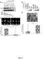
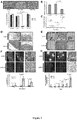
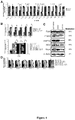
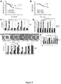
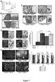
- miR-31 an anti-metastatic human miRNA, miR-31, that acts at multiple steps of the invasion-metastasis cascade via repression of a cohort of pro-metastatic targets.
- miR-31 downregulation or deletion of the miR-31 genomic locus in human breast cancers was detected in genome-wide studies (Calin et al., 2004; Zhang et al., 2006; Yan et al., 2008).
- WO2008/036776 describes microarray gene expression analyses that were performed to identify genes that are mis-regulated by inhibition of hsa-miR-31 expression. Based on the analysis it was concluded that hsa-miR-31 directly or indirectly affects primarily cellular development-related genes and thus primarily affects functional pathways related to cellular development.
- the present invention is based at least in part on the discovery that miR-31 is a pleiotropically acting miRNA that is necessary and sufficient to inhibit breast cancer metastasis.
- miR-31 expression in primary breast tumors correlated inversely with metastatic recurrence.
- the inventors showed that miR-31 normally acts to inhibit expression of certain target genes, e.g., Fzd3, ITGA5, M-RIP, MMP16, RDX, and RhoA. Accordingly, loss of miR-31 results in upregulation of miR-31 target genes, and such upregulation is associated with increased metastasis.
- the invention provides compositions and methods for diagnosis and prognosis, e.g., to assess or predict likelihood that a tumor has or will metastasize and/or to assess the tumor aggressiveness or likelihood of recurrence.
- the invention further provides compositions and methods for research or therapy (e.g., to reduce likelihood of metastasis and/or to inhibit growth of metastases and/or to inhibit one or more aspects considered characteristic of a metastatic phenotype such as invasiveness or motility).
- the compositions and methods mimic the effects of miR-31, e.g., they inhibit one or more miR-31 target gene(s).
- Such agents could be, e.g., oligonucleotides (e.g., RNAs) identical or sufficiently similar to miR-31 to inhibit one or more miR-31 target genes or siRNA targeted to a miR-31 target gene.
- the compositions are pharmaceutical compositions.
- the compositions or agents contained in compositions are isolated (e.g., separated from at least some other components with which they are found in nature or synthetically produced).
- Agents also termed "compounds” herein
- compounds can be, e.g., peptides, polypeptides, nucleic acids, oligonucleotides, small molecules (e.g., organic compounds having a molecular weight equal to or less than 1.5 kD, e.g., less than 1 kD, and usually multiple carbon-carbon bonds), antibodies, sugars, lipids, etc.
- an agent may comprise two or more of the foregoing types of materials, which may be covalently linked.
- a "small molecule” refers to an organic compound having multiple carbon-carbon bonds and a molecular weight of less than 2000 daltons, e.g., between 50 and 1,500 daltons.
- Such compounds comprise one or more functional groups that mediate structural interactions with proteins, e.g., hydrogen bonding, and typically include at least an amine, carbonyl, hydroxyl or carboxyl group, and in some embodiments at least two of the functional chemical groups.
- a small molecule may comprise cyclic carbon or heterocyclic structures and/or aromatic or polyaromatic structures substituted with one or more chemical functional groups and/or heteroatoms.
- nucleic acids e.g., oligonucleotides (which typically refers to short nucleic acids, e.g., 50 nucleotides in length or less)
- oligonucleotides which typically refers to short nucleic acids, e.g., 50 nucleotides in length or less
- ds double-stranded
- blunt-ended double-stranded with overhangs
- the full spectrum of modifications e.g., nucleoside and/or backbone modifications
- non-standard nucleotides, delivery vehicles and systems, etc. known in the art as being useful in the context of siRNA or antisense-based molecules for research or therapeutic purposes is contemplated for use in various embodiments of the instant invention.
- Polypeptides of use herein may contain amino acids such as those that are naturally found in proteins (e.g., the 20 "standard” amino acids), amino acids that are not naturally found in proteins, and/or amino acid analogs that are not amino acids.
- amino acids in a polypeptide may be modified, for example, by the addition of a chemical entity such as a carbohydrate group, a phosphate group, a fatty acid group, etc.
- Nucleic acids and polypeptides can be produced using any suitable method known in the art. In some embodiments a nucleic acid or polypeptide is chemically synthesized.
- a nucleic acid is produced using enzymatic synthesis, e.g., by in vitro transcription or amplification reaction such as PCR.
- a polypeptide is produced using enzymatic synthesis, e.g., by in vitro translation.
- a nucleic acid or polypeptide is synthesized by a cell or multicellular organism and isolated therefrom. The cell or organism may be genetically modified to express the nucleic acid.
- antibody encompasses immunoglobulins and derivatives thereof containing an immunoglobulin domain capable of binding to an antigen.
- An antibody can originate from any mammalian or avian species, e.g., human, rodent (e.g., mouse, rabbit) goat, chicken, etc., or can be generated using, e.g., phage display.
- the antibody may be a member of any immunoglobulin class, e.g., IgG , IgM, IgA, IgD, IgE, or subclasses thereof such as IgG1, IgG2, etc.
- antibody refers to an antibody fragment such as an Fab', F(ab')2, scFv (single-chain variable) or other fragment that retains an antigen binding site, or a recombinantly produced scFv fragment, including recombinantly produced fragments.
- An antibody can be monovalent, bivalent or multivalent in various embodiments.
- the antibody may be a chimeric or "humanized” antibody, which can be generated using methods known in the art.
- An antibody may be polyclonal or monoclonal, though for purposes of the present invention monoclonal antibodies are generally preferred. Methods for producing antibodies that specifically bind to virtually any molecule of interest are known in the art.
- monoclonal or polyclonal antibodies can be purified from blood or ascites fluid of an animal that produces the antibody (e.g., following natural exposure to or immunization with the molecule or an antigenic fragment thereof), can be produced using recombinant techniques in cell culture or transgenic organisms, or can be made at least in part by chemical synthesis.
- a compound or agent of use in the present invention e.g., for research, testing, prognosis, and/or therapy
- the composition may, but need not contain, e.g., an ion, salt, aqueous or non-aqueous diluent or carrier, buffer, enzyme inhibitor, preservative, etc.
- the invention relates to or makes use of genetically modified cells e.g., cells that have been genetically modified to have reduced miR-31 expression or activity.
- a "genetically modified” or “engineered” cell refers to a cell into which a nucleic acid has been introduced by a process involving the hand of man (or a descendant of such a cell that has inherited at least a portion of the nucleic acid).
- the nucleic acid may for example contain a sequence that is not naturally found in the cell, it may contain native sequences (i.e., sequences naturally found in the cell) but in a non-naturally occurring arrangement (e.g., a coding region linked to a promoter from a different gene), or altered versions of native sequences, etc.
- the process of transferring the nucleic acid into the cell can be achieved by any suitable technique and will often involve use of a vector. Suitable techniques include calcium phosphate or lipid-mediated transfection, electroporation, and transduction or infection using a viral vector.
- the nucleic acid or a portion thereof is integrated into the genome of the cell and/or is otherwise stably heritable.
- the nucleic acid may have subsequently been removed or excised from the genome, provided that such removal or excision results in a detectable alteration in the cell relative to an unmodified but otherwise equivalent cell.
- the invention relates to or makes use of genetically modified multi-cellular organisms.
- a "vector" may comprise any of a variety of nucleic acid molecules into which a desired nucleic acid may be inserted, e.g., by restriction digestion followed by ligation.
- a vector can be used for transport of such nucleic acid between different environments, e.g., to introduce the nucleic acid into a cell of interest and, optionally, to direct expression in such cell.
- Vectors are often composed of DNA although RNA vectors are also known. Vectors include, but are not limited to, plasmids and virus genomes or portions thereof.
- Vectors may contain one or more nucleic acids encoding a marker suitable for use in the identifying and/or selecting cells that have or have not been transformed or transfected with the vector.
- Markers include, for example, proteins that increase or decrease either resistance or sensitivity to antibiotics or other compounds, enzymes whose activities are detectable by standard assays known in the art (e.g., ⁇ -galactosidase or alkaline phosphatase), and proteins or RNAs that detectably affect the phenotype of transformed or transfected cells (e.g., fluorescent proteins).
- An expression vector is one into which a desired nucleic acid may be inserted such that it is operably linked to regulatory elements (also termed “regulatory sequences”, “expression control elements”, or “expression control sequences”) and may be expressed as an RNA transcript (e.g., an mRNA that can be translated into protein or a noncoding RNA such as an shRNA or miRNA precursor). Regulatory elements may be contained in the vector or may be part of the inserted nucleic acid or inserted prior to or following insertion of the nucleic acid whose expression is desired.
- a nucleic acid and regulatory element(s) are said to be "operably linked” when they are covalently linked so as to place the expression or transcription of the nucleic acid under the influence or control of the regulatory element(s).
- a promoter region would be operably linked to a nucleic acid if the promoter region were capable of effecting transcription of that nucleic acid.
- the precise nature of the regulatory sequences needed for gene expression may vary between species or cell types, but can in general include, as necessary, 5' non-transcribed and/or 5' untranslated sequences that may be involved with the initiation of transcription and translation respectively, such as a TATA box, cap sequence, CAAT sequence, and the like.
- regulatory elements include IRES sequences.
- Such 5' non-transcribed regulatory sequences will include a promoter region that includes a promoter sequence for transcriptional control of the operably linked gene. Regulatory sequences may also include enhancer sequences or upstream activator sequences.
- Vectors may optionally include 5' leader or signal sequences. Vectors may optionally include cleavage and/or polyadenylations signals and/or a 3' untranslated regions. The choice and design of an appropriate vector and regulatory element(s) is within the ability and discretion of one of ordinary skill in the art. For example, one of skill in the art will select an appropriate promoter (or other expression control sequences) for expression in a desired species (e.g., a mammalian species) or cell type.
- a virus vector is selected from the group consisting of adenoviruses, adeno-associated viruses, poxviruses including vaccinia viruses and attenuated poxviruses, retroviruses (e.g., lentiviruses), Semliki Forest virus, Sindbis virus, etc.
- the virus is replication-defective.
- a replication-deficient retrovirus i.e., a virus capable of directing synthesis of one or more desired transcripts, but incapable of manufacturing an infectious particle
- Various techniques may be employed for introducing nucleic acid molecules into cells. Such techniques include transfection of nucleic acid molecule-calcium phosphate precipitates, transfection of nucleic acid molecules associated with DEAE, transfection or infection with a virus that contains the nucleic acid molecule of interest, liposome-mediated transfection, nanoparticle-mediated transfection, and the like.
- methods of the invention involve a subject.
- a "subject” may be a human or a non-human animal.
- the subject may be suffering from a disease (e.g., cancer) warranting medical and/or surgical attention or in need of prognostic information or may be at increased risk of developing a disease relative to an average member of the population.
- a subject is a female.
- a non-human subject may be a non-human mammal, e.g., a rodent such as a mouse, rat, hamster, rabbit, or guinea pig; a dog, a cat, a bovine or ovine, a non-human primate, etc.
- the subject may serve as an animal model for cancer, e.g., breast cancer.
- the subject may be a genetically engineered non-human mammal, e.g., a mouse, that has a predisposition to develop tumors.
- the mammal may overexpress an oncogene (e.g., as a transgene) or underexpress a tumor suppressor gene (e.g., the animal may have a mutation or deletion in the tumor suppressor gene).
- the invention provides compositions and methods for diagnosis and prognosis, e.g., for assessing or predicting the likelihood of tumor metastasis.
- the methods comprise assessing expression of a miR-31 gene product (e.g., assessing the level of mature miR-31 RNA or a miR-31 precursor RNA that is transcribed from the miR-31 gene, e.g., the pri-miRNA or pre-miRNA).
- the methods comprise assessing the level or activity of an expression product of a gene that is a target of regulation by miR-31.
- “Expression products” can refer to RNA transcribed from a gene and/or polypeptides obtained by translation of mRNA transcribed from a gene (including post-translationally processed forms thereof).
- the afore-mentioned methods may involve assessing level or activity in a biological sample obtained from a tumor.
- the sample may comprise cells obtained, for example, from a tissue biopsy, fine needle aspiration biopsy, surgical specimen, brushing, etc.
- the cells may be expanded in culture after being obtained from the tumor.
- the biological sample may be further processed (e.g., appropriately treated, stained, etc.) to render it suitable for use in subsequent assay procedures.
- the methods are used in conjunction with measuring expression of one or more other microRNAs (e.g., one or more miRNAs with pro- or anti-metastatic properties) or with other diagnostic/prognostic approaches, e.g., assessing estrogen or progesterone receptor status, presence of mutations in oncogenes or tumor suppressor genes, etc.
- the methods may be used, e.g., to recommend or make treatment decisions, e.g., to select particular approaches (e.g., surgery, chemotherapy, radiation, etc.) to treating the tumor or preventing recurrence.
- the invention involves comparison with an appropriate control.
- An appropriate control level may be, e.g., a level in nonmetastatic tumor, a level in normal tissues or cells (e.g., cells or tissues of the same type or from the same organ as that from which the tumor originated), etc.
- a control or reference level may be determined concurrently with assessing the biological sample of interest, or may be a historical control or reference level (e.g. an average measurement obtained from a plurality of samples collected prior to performing the method).
- the invention also relates to kits comprising agents and reagents suitable for use in the diagnostic, prognostic and therapeutic methods of the invention, optionally including instructions for use. Any of the kits can comprise instructions for using the contents of the kit to perform a method of the invention and, optionally, reagents useful to perform the method (e.g., enzymes, buffers) or suitable controls.
- inventive methods relate to a miR-31 target signature. Such a signature may be used in the diagnostic and prognostic methods of the present invention.
- inventive methods relate to one or more miR-31 target genes.
- Embodiments of the invention relate to assessing expression or activity of one or more such genes in cells in culture.
- Embodiments of the invention relate to assessing expression or activity of one or more such genes in cells in vivo.
- Embodiments of the invention relate to modulating (e.g., increasing or inhibiting) expression or activity of one or more such genes in cells in culture.
- modulating e.g., increasing or inhibiting
- the cells are in an animal subject, e.g., a rodent, non-human primate, or human being.
- the rodent or non-human primate, or other non-human animal is an animal model useful for assessing tumor formation, development, and/or efficacy of therapeutic agents.
- the animal is immunocompromised.
- the cells are tumorigenic cells.
- the cells are non-tumorigenic cells.
- the cells are immortalized.
- the cells are genetically modified.
- the tumor is a carcinoma, e.g., a breast carcinoma.
- the tumor is an adenocarcinoma.
- the tumor is a sarcoma.
- the tumor affects an organ or organ system selected from breast, lymph node, prostate, kidney, bladder, lung, liver, gastrointestinal tract, colon, testis, stomach, pancreas, thyroid, skin, ovary, uterus, cervix, skin, nerve, bone, and nervous system (e.g., brain).
- a variety of different tumor types may originate in certain of these organs, and the term "tumor" is intended to encompass all such tumor types.
- Cancer as used herein should be understood to encompass any tumor type that has the potential to metastasize, e.g., any malignant tumor (e.g., a carcinoma or sarcoma in various embodiments).
- the tumor originates from an epithelial cell, e.g., a breast epithelial cell.
- inventions, techniques, methods, and materials described in USSN 10/990,993 US Pub. No. 20050193432
- PCT/US2008/009639 filed August 11, 2008 and/or PCT/US08/85040 filed November 26, 2008
- such inventions, techniques, methods, and/or materials may be useful in further analysis relating to role of miR-31 and/or miR-31 target genes in tumor metastasis and/or for screening and assessing therapeutic agents and drug candidates that relate to miR-31 and/or miR-31 target genes, etc.
- the ability of an agent to inhibit a miR-31 target gene and/or the ability of agent to reduce the likelihood or magnitude of tumor growth or metastasis can be evaluated.
- miR-31 e.g., human miR-31
- miRBase www.mirbase.org
- miRBase tools for microRNA genomics
- Griffiths-Jones S, et al., NAR 2006 34(Database Issue):D140-D144
- Griffiths-Jones S. NAR 2004 32(Database Issue):D109-D111) and GenBank.
- the mature human miR-31 most commonly observed in cloning studies reportedly has the following sequence: AGGCAAGAUGCUGGCAUAGCU (SEQ ID NO: 1; miRBase accession number MIMAT0000089) and is located within a stem-loop predicted to have the following sequence:
- sequence differences may exist among different individuals. For example, a sequence may differ at between 1 and 5 positions as compared with SEQ ID NO: 2.
- a mature miR-31 may have or lack additional nucleotide(s), e.g., 1 or 2 nts, at the 5' and/or 3' end.
- the invention encompasses use of miR-31 or miR-31 precursor from non-human animals in certain embodiments.
- mouse miR-31 can be used, e.g., in embodiments that relate to models for tumor metastasis and drug discovery.
- Mature mouse miR-31 have the following sequence: AGGCAAGAUGCUGGCAUAGCUG (SEQ ID NO: 3; miRBase accession number MIMAT0000538) and is located in a stem-loop of the following sequence:
- the invention provides a variety of methods that provide information, e.g., prognostic information relating to tumors, e.g., breast tumors.
- the methods are, in some embodiments, based at least in part on assessing (e.g., detecting, quantifying, etc.) the expression level or activity of miR-31 or a miR-31 precursor (or, in some embodiments, assessing the expression level or activity of one or more miR-31 target genes).
- miR-31-based prognostic methods or assays Such methods are referred to herein as "miR-31-based prognostic methods or assays”.
- the level of miR-31 is used to discriminate between tumors at high risk of metastasis and those with a low risk.
- a tumor has a significantly reduced level of miR-31 relative to a reference level the tumor is classified as having a high risk of metastasizing, while if the tumor does not have a significantly reduced level of miR-31 relative to a reference level, the tumor is classified as having a low risk.
- a reference level may be determined in a variety of ways. In some embodiments a reference level is an absolute level, while in other embodiments a reference level is a relative level. In some embodiments, tumors are classified as miR-31-positive or -negative based on comparison with a tumor cohort.
- tumors may be considered miR-31-positive or -negative if the normalized expression of miR-31 resides in the top or bottom 30% of tumors in a cohort, respectively.
- Tumors that are miR-31 positive are at low risk of metastasis while tumors that are miR-31 negative are at high risk of metastasis.
- tumors may be considered miR-31-positive or -negative if the normalized expression of miR-31 resides in the top or bottom 25% of tumors in a cohort, respectively.
- tumors may be considered miR-31-positive or - negative if the normalized expression of miR-31 resides in the top or bottom 35% of tumors in a cohort, respectively.
- one or more samples are obtained from a tumor, and one or more samples are obtained from nearby patient-matched normal (non-tumor) tissue composed of similar cell types.
- the relative level of miR-31 in the patient-matched non-tumor samples versus tumor samples is determined. In some embodiments, if the relative level (ratio) of miR-31 in the non-tumor samples versus the tumor samples is greater than a predetermined value (indicating that cells of the tumor have reduced or absent miR-31), the tumor is classified as high risk.
- the predetermined value is, e.g., at least 1.5, 2.0, 2.5, 3.0, etc.
- the predetermined value is between about 1.5 and about 10.
- Various risk categories may be defined. For example, tumors may be classified as at low, intermediate, or high risk of metastasis. A variety of statistical methods may be used to correlate the risk of metastasis with the relative or absolute level of miR-31 expression or loss of expression.
- the invention provides a method assessing the likelihood of development of a tumor metastasis in a subject, comprising the steps of: (a) determining the level of a miR-31 gene product in a biological sample obtained from the subject; (b) comparing the level determined in (a) with an appropriate control level, wherein if the level determined in (a) is lower than the control level of the miR-31 gene product, then the subject has an increased likelihood of developing a metastasis (e.g., as compared with the likelihood if the level determined in (a) is not lower than the control level).
- a miR-31 gene product can be any transcript transcribed from the miR-31 gene or a processed form thereof.
- a miR-31 gene product can be a mature miR-31 RNA or a miR-31 precursor RNA.
- the biological sample is obtained from a tumor.
- the control level is determined using a sample of non-tumor tissue obtained from the subject.
- the invention further provides a method for assessing the likelihood that a tumor in a subject will metastasize or has metastasized, the method comprising (a) determining the level of an expression product of one or more miR-31 target gene(s) in a biological sample obtained from the tumor; and (b) comparing the level with a control level, wherein if the level determined in (a) is greater than the control level, the tumor is considered to have increased likelihood of metastasizing or having metastasized (e.g., as compared with the likelihood if the level determined in (a) is not greater than the control level).
- expression levels of at least two miR-31 target genes are assessed.
- a "miR-31 target gene” is a gene whose expression is directly regulated by miR-31, i.e., miR-31 directs cleavage and/or translational repression of the mRNA of the gene so that the expression level of the gene is higher in the absence of miR-31 than if miR-31 is present.
- the miR-31 target genes are selected from RhoA, RDX, and ITGA5. It will be appreciated that the prognostic methods could be expressed in terms of identifying a tumor having decreased risk of metastasizing or having metastasized.
- a tumor that does not have reduced or absent levels of a miR-31 gene product as compared with a control has decreased likelihood of metastasizing as compared with the likelihood that would exist if the tumor does have reduced or absent levels of a miR-31 gene product.
- a tumor that does not have increased levels of expression of miR-31 target gene(s) as compared with a control has decreased likelihood of metastasizing as compared with the likelihood that would exist if the tumor does have increased levels of expression of miR-31 target gene(s)
- the invention further provides a method for providing prognostic information relating to the likelihood that a tumor in a subject will metastasize, the method comprising (a) determining the level of a miR-31 gene product in a biological sample obtained from the tumor; and (b) comparing the level with a control level, wherein (i) if the level determined in (a) is less than the control level, the tumor is considered to have a poor prognosis; or (ii) if the level determined in (a) is greater than the control level, the tumor is considered to have a good prognosis.
- a prognostic method of the invention comprises providing a subject in need of treatment for cancer, e.g., breast cancer.
- a prognostic method of the invention comprises diagnosing a subject in need of treatment for cancer, e.g., breast cancer.
- a biological sample used in the miR-31 based prognostic methods will typically comprise or be derived from cells isolated from a subject.
- the cells will typically comprise tumor cells, e.g., breast tumor cells.
- Samples can be, e.g., surgical samples, tissue biopsy samples, fine needle aspiration biopsy samples, core needle samples. The sample may be obtained using methods known in the art.
- a sample can be subjected to one or more processing steps. In some embodiments the sample is frozen and/or fixed. In some embodiments the sample is sectioned and/or embedded, e.g., in paraffin.
- tumor cells e.g., epithelial tumor cells
- stromal tissue e.g., stromal cells and/or extracellular matrix.
- Cells of interest can be isolated using, e.g., tissue microdissection, e.g., laser capture microdissection.
- cells of the sample are lysed.
- Nucleic acids or polypeptides may be isolated.
- RNA optionally isolated from a sample, is reverse transcribed and/or amplified.
- short RNA e.g., less than about 200 nucleotides
- a wide variety of solution phase or solid phase methods are available for detection of RNA, e.g., miRNA such as miR-31.
- Suitable methods include e.g., hybridization-based approaches (e.g., nuclease protection assays, Northern blots, microarrays, in situ hybridization), amplification-based approaches (e.g., reverse transcription polymerase chain reaction (which can be a real-time PCR reaction), or sequencing (e.g., RNA-Seq, which uses high throughput sequencing techniques to quantify RNA transcripts (see, e.g., Wang, Z., et al. Nature Reviews Genetics 10, 57-63, 2009 )).
- qPCR quantitative PCR
- Other methods include electrochemical detection, bioluminescence-based methods, fluorescence-correlation spectroscopy, etc.
- Probes composed at least in part of locked nucleic acids (LNA) are of use.
- miR-31 is detected in a formalin-fixed sample using in situ hybridization. Prior to incubation with the probe the tissue is treated with EDC, a water-soluble fixative that irreversibly links the RNA's 5' phosphate to protein side chains. See, e.g., Pena, J, et al., Nature Methods 6, 139-141, 2009 . Kits for isolating, detecting, and/or quantitating miRNAs are commercially available, such as the miRvanaTM kits from Ambion/Applied Biosystems; the miRtect-ITTM miRNA Labeling and Detection Kit from USB Corp.
- the splinted-ligation technology is a nucleic acid hybridization assay that uses a miRNA-specific Bridge Oligonucleotide to form base pairs with the miRNA and a Detection Oligonucleotide. The captured miRNA is subsequently ligated to the Detection Oligonucleotide. See, e.g., Maroney, PA, et al, Nature Protocols, 13 (1) 279-287, 2008 . It will be understood that suitable controls and normalization procedures can be used to accurately quantify miR-31 expression.
- values can be normalized based on the expression of a small RNA such as the 5S RNA or a microRNA whose expression is not correlated with cancer and/or with metastasis can be used. Values can also be normalized to account for the fact that different samples may contain different proportions of a cell type of interest, e.g., cancer cells, versus non-cancer cells. For example, the percentage of stromal cells, e.g., fibroblasts, may be assessed by measuring expression of a stromal cell-specific marker, and the overall results adjusted to accurately reflect miR-31 expression specifically in the tumor cells.
- the level of miR-31 expression is not measured as part of a gene expression profile in which expression of at least 10 different genes (e.g., at least 20, 30, 50, or 100 genes) is measured (e.g., using a microarray).
- the level of miR-31 expression is used as an indicator of the likelihood of metastasis or as a prognostic indicator that is distinct from its contribution to the overall gene expression profile.
- the inventive methods involve using miR-31 expression level as an indicator of tumor aggressiveness, likelihood of metastasis, or prognosis in a manner that, at least in part, does not depend on the contribution of miR-31 to a gene expression profile.
- miR-31 level is assessed as part of a gene expression profile, but the result obtained for miR-31 is analyzed or used in a manner distinct from its use as a contributor to a classification based on the overall gene expression profile.
- Expression levels miR-31 target genes may be assessed using any suitable method. Either mRNA or protein level may be measured. Exemplary methods for measuring mRNA include hybridization based assay, polymerase chain reaction assay, sequencing, in situ hybridization, etc. Exemplary methods for measuring protein level include ELISA assays, Western blot, mass spectrometry, or immunohistochemistry.
- results of a miR-31-based prognostic assay may be useful for selecting a therapeutic regimen for a subject. For example, such results may be useful in determining whether a subject should receive, e.g., would likely benefit from, administration of one or more chemotherapeutic agents (chemotherapy), hormonal therapy, or anti-HER2 antibody such as trastuzumab.
- chemotherapeutic agent refers to an anti-tumor agent (also called anti-neoplastic agent) that has cytotoxic or cytostatic properties and does not act primarily by interacting with (e.g., interfering with) a hormonal pathway that is specific or relatively specific to particular cell type(s).
- Exemplary chemotherapeutic agents include anti-metabolites, alkylating agents, microtubule stabilizers or inhibitors (e.g., taxanes), topoisomerase inhibitors, and DNA intercalators (e.g., anthracycline antibiotics). Such agents are usually administered systemically. Often, multiple agents are administered. Exemplary regimens for breast cancer include CMF (cyclophosphamide, methotrexate, and 5-FU), AC (doxorubicin and cyclophosphamide), and anthracycline-based regimens.
- CMF cyclophosphamide, methotrexate, and 5-FU
- AC doxorubicin and cyclophosphamide
- anthracycline-based regimens include CMF (cyclophosphamide, methotrexate, and 5-FU), AC (doxorubicin and cyclophosphamide), and anthracycline-based regimens.
- “Hormonal therapy” refers to administration of an anti-tumor agent that acts primarily by interacting with (e.g., interfering with) a hormonal pathway that is specific or relatively specific to particular cell type(s).
- “Adjuvant therapy” refers to administration of one or more anti-tumor agents in connection with, e.g., following, local therapy such as surgery and/or radiation. Adjuvant therapy may be used when the cancer appears to be largely or completely eradicated, but there is risk of recurrence. Such therapy may help eliminate residual cells at the site of the primary tumor and/or eliminate any cells that have disseminated.
- “Neoadjuvant therapy” refers to adjuvant therapy administered prior to local therapy, e.g., to shrink a primary tumor.
- a prognostic method of the invention may be used to identify cancer patients that do not require adjuvant therapy, e.g., adjuvant hormonal therapy and/or adjuvant chemotherapy (e.g., patients that would very likely not experience clinically evident metastasis in the absence of such treatment). Since such treatment can cause significant side effects, it would be beneficial to avoid administering it to individuals who would not benefit from it.
- adjuvant therapy e.g., adjuvant hormonal therapy and/or adjuvant chemotherapy
- a prognostic method of the invention may be used to identify cancer patients that are at high risk of recurrence and/or metastasis and may therefore benefit from adjuvant therapy.
- a prognostic method of the invention may be used to identify cancer patients that might not be considered high-risk based on other prognostic indicators (and may therefore not receive adjuvant therapy) but that are in fact at high risk of recurrence and/or metastasis.
- Results of an inventive prognostic assay may be useful for selecting a particular type of chemotherapy regimen and/or for selecting the type of procedures to be used to monitor the subject for metastatic recurrence after therapy and/or the frequency with which such procedures are performed. For example, subjects classified as being at high risk of metastasis may be assessed more frequently than those classified as being at low risk.
- any of the prognostic methods can further comprise using the information obtained from the assay to help in selecting a treatment or monitoring regimen for a subject suffering from or at risk of cancer or in providing an estimate of the risk of poor outcome (e.g., cancer related mortality or recurrence) without adjuvant therapy.
- any of the foregoing methods can comprise making a treatment recommendation or administering a treatment based at least in part on the result of the assay, e.g., based in part on the level of the miR-31 gene product.
- any of the foregoing methods can comprise (i) recommending that the subject not receive chemotherapy (e.g., adjuvant chemotherapy) if the tumor is considered to have a good prognosis; or (ii) recommending that the subject receive chemotherapy (e.g., adjuvant chemotherapy), or administering such chemotherapy, if the tumor is considered to have a poor prognosis.
- chemotherapy e.g., adjuvant chemotherapy
- the miR-31-based methods may be used to provide prognostic information for subjects with tumors that have one or more recognized clinicopathologic features and/or fall into a particular class based on gene expression profiles.
- breast cancers can be classified based on a number of different clinicopathologic features such as histologic subtype (e.g., ductal; lobular; mixed), histologic grade (grade 1, 2, 3); estrogen receptor (ER) and/or progesterone receptor (PR) status (positive (+) or negative (-)), HER2 (ERBB2) expression status, and lymph node involvement.
- breast cancer subtypes can be defined based on expression of estrogen receptor (ER) or progesterone receptor (PR) and human epidermal growth factor receptor 2 (Her2), e.g., as assessed by immunohistochemistry (IHC): ER+, Her2+; ER+, Her2-; ER-, Her2+; and ER-, Her2-.
- IHC immunohistochemistry
- ER+, Her2+; ER+, Her2-; ER-, Her2+; and ER-, Her2- The level of expression can be used to further divide these subtypes.
- Amplification of the HER2 locus can be assessed, e.g., using fluorescent in situ hybridization (FISH).
- FISH fluorescent in situ hybridization
- Breast cancers can also be classified into molecular subtypes based on gene expression profiles, e.g., luminal A, luminal B, ERBB2-associated, basal-like and normal-like (see, e.g., S ⁇ rlie, T., et al., 2001 and Desmedt, C., et al., 2008).
- gene expression profiles e.g., luminal A, luminal B, ERBB2-associated, basal-like and normal-like (see, e.g., S ⁇ rlie, T., et al., 2001 and Desmedt, C., et al., 2008).
- an inventive method is applied to a tumor classified as histologic grade 2, e.g., to classify histologic grade 2 tumors into high and low recurrence risk groups.
- an inventive method is applied to a tumor classified as histologic grade 2, e.g., to classify histologic grade 2 tumors into high and low recurrence risk groups.
- an inventive method is applied to a tumor that is ER+, while in other embodiments an inventive method is applied to a tumor that is ER-.
- an inventive method is applied to a tumor that is HER2 positive, while in other embodiments an inventive method is applied to a tumor that is HER2 negative.
- an inventive method is applied to a tumor that is ER-/HER2-.
- an inventive method is applied to a tumor that is negative for expression of the estrogen and progesterone receptors and HER2 ("triple-negative").
- a subject does not have lymph node involvement, i.e., the subject is "lymph node-negative" (LNN).
- LNN lymph node-negative
- a subject has Stage I or Stage II breast cancer.
- a subject has been treated with tamoxifen or another anti-estrogen agent, e.g., another selective estrogen receptor modulator such as raloxifene or toremifene.
- a subject falls within a predefined age group or range, e.g., 50 years old or less, 60 years old or less, between 40 and 60 years of age, etc. Any age group or range may be selected, even if not specifically mentioned here.
- a miR-31-based prognostic assay is used together with additional information, such as results of a second assay (or multiple assays) and/or clinicopathological information.
- additional information comprises, e.g., subject age, tumor size, nodal involvement, tumor histologic grade, ER/PR status, and/or HER2 status, etc.
- information from a miR-31-based assay is used together with a decision making or risk assessment tool such as the computer program Adjuvant! Online (https://www.adjuvantonline.com/index.jsp). The basic format of an early version of Adjuvant! was described in the article Ravdin, Siminoff, Davis, et al.
- the second assay is a gene expression profiling assay such as the MammaPrint® (Agendia BV, Amsterdam, the Netherlands), Oncotype DXTM (Genomic Health, Redwood City, CA), Celera Metastasis ScoreTM (Celera, Inc., Rockville, MD), Breast BioClassifier (ARUP, Salt Lake City, UT), Rotterdam signature 76-gene panel (Erasmus University Cancer Center, Rotterdam, The Netherlands), or Invasiveness Gene Signature (OncoMed Pharmaceuticals, Redwood City, CA), or NuvoSelectTM assay (Nuvera Biosciences, Woburn, MA), that classifies tumors (e.g., into high or low risk groups) based on expression level of multiple genes using, e.g., a microarray or multiplex RT-PCR assay.
- a gene expression profiling assay such as the MammaPrint® (Agendia BV, Amsterdam, the Netherlands), Oncotype DXTM (Genomic Health, Redwood City
- a miR-31-based assay may be used together with a gene expression profiling assay in which expression level of at least 10 different genes ("classifier genes") is used to classify a tumor. It will be understood that such assays typically measure expression of control genes as well as classifier genes.
- a miR-31-based assay is used together with an H:ITM test (AvariaDX, Carlsbad, CA), in which the ratio of expression of HOXB13 and IL-17B genes is used to classify a tumor.
- a miR-31-based prognostic assay is used together with an antibody-based assay, e.g., the ProExTM Br (TriPath Oncology, Durham, NC), Mammostrat® (Applied Genomics, Inc., Huntsville, AL), or a FISH-based test such as the eXaagenBCTM (eXagen Diagnostics, Inc., Albuquerque, NM).
- a miR-31-based assay may be used together with a second assay in a number of ways. For example, if results of the two tests are discordant (i.e., one test predicts that the subject is at high risk while the other predicts that the subject is at low risk), the subject may be treated. In some embodiments, one can have increased confidence if results are in agreement. For example, if both tests indicate that the subject is at low risk, there can be increased confidence in a decision not to administer adjuvant chemotherapy.
- kits comprising reagents suitable for performing a miR-31-based prognostic assay of the present invention.
- Such kits may contain, e.g., (i) a probe or primer (optionally labeled and/or attached to a support) for detecting, reverse transcribing, and/or amplifying miR-31 or a miR-31 precursor; (ii) a probe or primer for detecting, reverse transcribing, and/or amplifying an mRNA transcribed from a miR-31 target gene; (iii) an antibody for detecting a protein encoded by a miR-31 target gene; (iv) one or more controls (e.g., a probe or primer that does not detect or amplify miR-31).
- the kit comprises written material, e.g., instructions, e.g., in a paper or electronic format (e.g., on a computer-readable medium). Instructions may comprise directions for performing the assay and/or for interpreting results, e.g., in regard to tumor classification, likelihood of metastasis, and/or prognosis. Such material could also be provided online.
- the invention provides a system which is adapted or programmed to carry out a miR-31-based assay.
- the system may include one or more instruments (e.g., a PCR machine) and/or computer processors.
- the system may be programmed with parameters that have been selected or optimized for detection and/or quantification of miR-31, e.g., in tumor samples.
- the system may be adapted to perform the assay on multiple samples in parallel and/or may have appropriate software to provide an interpretation of the result.
- the system can comprise appropriate input and output devices, e.g., keyboard, display, etc.
- a miR-31 based assay is performed at one or more central testing facilities, which may be specially qualified to perform the assay and, optionally, provide an interpretation of the results. In the latter case, samples are sent to the laboratory and a result of the assay is provided.
- the disclosure provides a method comprising: providing to a testing facility (a) a sample obtained from a subject to a testing facility; and (b) instructions to perform a miR-31-based assay of the invention (and, optionally, instructions to perform one or more additional assays such as an ICH, FISH, gene expression profile, or other assay described herein.
- the disclosure also provides a method comprising: (a) providing a sample obtained from a subject to a testing facility; and (b) receiving results of a prognostic assay of the invention.
- the invention further provides a method comprising providing, e.g., electronically, a result of a prognostic assay of the invention.
- the result provided comprises a measurement made on the sample, with or without associated prognostic information.
- the assay may be performed at a testing facility which is remote from the site where the sample is obtained from a subject. It is also contemplated that samples and/or results may be transmitted through one or more different entities, which may carry out one or more steps of an inventive method or transmit or receive results thereof. All such activities are within the scope of the invention.
- the invention provides a variety of methods that, in certain embodiments, involve modulating the level or activity of miR-31 and/or one or more miR-31 target genes. Modulating can comprise causing or facilitating a qualitative or quantitative change, alteration, or modification (e.g., an increase or decrease) in the amount or activity of miR-31 and/or one or more miR-31 target genes in a cell or organism.
- a variety of methods for modulating miRNA level or activity are of use in the present invention. Cells can be contacted in vitro with molecules that are taken up and modulate miRNA expression or activity, or such molecules can be administered to a subject.
- miRNA, miRNA precursors, or synthetic oligonucleotides that resemble miRNA or miRNA precursors can be introduced into cells and result in increased inhibition of miRNA target gene expression.
- Pre-miRTM miRNA Precursor Molecules are small, chemically modified double-stranded RNA molecules designed to mimic endogenous mature miRNAs.
- Nucleic acids that provide a template for transcription of miRNA or miRNA precursors can be introduced into cells and stably or transiently expressed therein.
- Vectors for expressing miRNA precursors are well known in the art and commercially available, e.g., from Open Biosystems.
- Such vectors may contain a site for insertion of a miRNA sequence of interest to create a template for transcription of a miRNA precursor, miRNA flanking sequences, and a promoter to direct transcription of the miRNA precursor sequence. See, e.g., Dickens NG et al., Nature Genetics, 37(11):1289-1295, 2005 .
- miRNA can be inhibited by a variety of approaches.
- miR-31 is inhibited by introducing an oligonucleotide that is complementary to miR-31 or to a miR-31 precursor into a cell (e.g., in vitro) or administering such an oligonucleotide to a subject, e.g., a human.
- the oligonucleotide sometimes termed an "antagomir" need not be perfectly complementary to miR-31 or miR-31 precursor, e.g., it may have 1, 2, 3, 4, 5, or more mismatches and/or be at least 70%, at least 80%, or at least 90% complementary thereto.
- the oligonucleotide is between about 17 nt and about 50 nt in length, e.g., about 19-25 nt long. See, e.g., Krutzfeldt J, et al. Nature 438: 685-9, 2005 . In some embodiments, such oligonucleotides are expressed in cells ( Scherr, M., et al., Nucleic Acids Res., 35(22): e149, 2007 ).
- a miRNA is inhibited using a miRNA sponge approach, which comprises expressing an RNA comprising multiplebinding sites for the particular miRNA in a cell, or introducing a nucleic acid comprising a plurality of binding sites for the particular miRNA into a cell (see, e.g., Ebert, M.S., et al. MicroRNA sponges: competitive inhibitors of small RNAs in mammalian cells. Nat Methods 4, 721-726 , and Examples 4 and 5).
- An oligonucleotide may contain one or more non-standard nucleotides, modified nucleotides (e.g., having modified bases and/or sugars) or nucleotide analogs, and/or have a modified backbone.
- an oligonucleotide is attached to one or more non-nucleic acid moieties.
- the oligonucleotide has one or more modifications as compared with standard RNA or DNA, e.g., to provide at least partial protection from RNase and/or pharmacologic properties such as enhanced tissue and/or cellular uptake, increased half-life, etc.
- the oligonucleotide differs from standard RNA or DNA by having partial or complete 2'-O-methylation or 2'-O-methoxyethyl modification of sugar, phosphorothioate backbone, and/or a cholesterol-moiety at the 3'-end.
- a nucleic acid comprising a morpholino oligonucleotide or a locked nucleic acid is used.
- a miRNA modulating agent is physically associated with a moiety that increases cell uptake, such as a cell-penetrating peptide.
- a miR-31 modulating agent is physically associated with a targeting moiety, which moiety targets the agent to a cell type of interest.
- the targeting moiety binds to a molecule or portion thereof that is exposed at the surface of an epithelial cell, e.g., a breast epithelial cell.
- the targeting moiety binds to a molecule or portion thereof that is exposed at the surface of a cancer cell, e.g., a breast cancer cell.
- the targeting moiety binds to a tumor antigen.
- the targeting moiety binds to a receptor, e.g., a hormone receptor.
- a targeting moiety can be, e.g., an antibody, aptamer, peptide, sugar, small molecule, etc.
- Two or more moieties that are "physically associated" may be covalently or non-covalently bound to each other, either directly or via a third moiety.
- compositions and methods can be used to deliver agents to cells in vitro or in vivo.
- agents can be incorporated into or attached to various types of particles such as liposomes, lipoplexes, or polymer-based particles, e.g., microparticles or nanoparticles composed at least in part of one or more biocompatible polymers or co-polymers comprising poly(lactide-glycolide), copolyoxalates, polycaprolactones, polyesteramides, polyorthoesters, polyhydroxybutyric acid, and/or polyanhydrides.
- biocompatible polymers or co-polymers comprising poly(lactide-glycolide), copolyoxalates, polycaprolactones, polyesteramides, polyorthoesters, polyhydroxybutyric acid, and/or polyanhydrides.
- RNA silencing also termed RNA interference (RNAi)
- RISC RNA-induced silencing complex
- RISC RNA-induced silencing complex
- the complementarity between the short RNA and mRNA need not be perfect (100%) but need only be sufficient to result in inhibition of gene expression.
- the degree of complementarity and/or the characteristics of the structure formed by hybridization of the mRNA and the short RNA strand can be such that the strand can (i) guide cleavage of the mRNA in the RNA-induced silencing complex (RISC) and/or (ii) cause translational repression of the mRNA by RISC.
- RISC RNA-induced silencing complex
- the short RNA is often incorporated into RISC as part of a short double-stranded RNA (dsRNA).
- dsRNA short double-stranded RNA
- RNAi may be achieved by introducing an appropriate short double-stranded nucleic acid into the cells or expressing in the cells a nucleic acid that is processed intracellularly to yield such short dsRNA.
- RNAi agent encompasses nucleic acids that can be used to achieve RNA silencing in mammalian cells.
- exemplary RNAi agents are a short hairpin RNA (shRNA), a short interfering RNA (siRNA), and a microRNA precursor.
- siRNAs typically comprise two separate nucleic acid strands that are hybridized to each other to form a duplex. They can be synthesized in vitro, e.g., using standard nucleic acid synthesis techniques.
- siRNA or shRNA comprises a duplex about 19 nucleotides in length, wherein one or both strands has a 3' overhang of 1-5 nucleotides in length (e.g., 2 nucleotides), which may be composed of deoxyribonucleotides.
- shRNA comprise a single nucleic acid strand that contains two complementary portions separated by a predominantly non-self-complementary region.
- RNAi agent also encompasses vectors, e.g., expression vectors, that comprise templates for transcription of an siRNA (e.g., as two separate strands that can hybridize), shRNA, or microRNA precursor, and can be used to introduce such template into mammalian cells and result in transient or stable expression thereof.
- vectors e.g., expression vectors, that comprise templates for transcription of an siRNA (e.g., as two separate strands that can hybridize), shRNA, or microRNA precursor, and can be used to introduce such template into mammalian cells and result in transient or stable expression thereof.
- Other methods of inhibiting expression or activity of miR-31 target genes include, e.g., use of small molecules, antibodies, aptamers, dominant negative approaches, etc.
- the invention provides methods for identification, characterization, and/or evaluation of compounds, e.g., for treatment of cancer.
- Certain aspects of the invention relate to identifying compounds that selectively kill or inhibit proliferation of cells that have reduced or absent expression of miR-31 as compared, e.g., with their effect on cells that express miR-31, e.g., cells that do not have reduced or absent expression of miR-31.
- Such compounds may be of use as anti-tumor agents, e.g., to reduce the likelihood of metastasis and/or to inhibit the growth of metastases arising from cells having reduced or absent miR-31 expression.
- aberrantly reduced or absent expression or activity of a miRNA may result in increased vulnerability to certain compounds as a result of alterations in expression of one or more gene(s) that are normally regulated by the miRNA.
- aberrantly increased expression or activity of a miRNA may result in increased vulnerability to certain compounds as a result of alterations in expression of one or more gene(s) that are normally regulated by the miRNA.
- Such vulnerabilities can be exploited to identify compounds that have selective activity towards cells that have altered miRNA expression.
- the invention encompasses applying this principle to discover compounds that have selective activity towards a cell that has altered, e.g., reduced, expression of a miRNA of interest, wherein the reduced expression confers on a cell an increased ability to cause or contribute to a disease or undesirable condition, e.g., cancer.
- reduced or absent expression of miR-31 in addition to conferring increased metastatic ability on a cell, may result in increased vulnerability to certain compounds as a result of alterations in expression of one or more gene(s) that are normally regulated by miR-31.
- Such vulnerabilities can be exploited to identify compounds that have selective activity towards cells that have reduced or absent miR-31 expression.
- the invention also encompasses applying this principle to discover compounds that have selective activity towards a cell that has increased expression of a miRNA of interest, wherein the increased expression confers on the cell an increased ability to cause or contribute to a disease or undesirable condition, e.g., cancer.
- the invention provides a method of identifying a potential anti-tumor compound comprising (a) providing a first cell that has reduced or absent miR-31 expression or activity as compared with a second cell; (b) contacting the first cell with a test compound; (c) identifying the test compound as a potential anti-tumor compound if the survival or proliferation of the first cell is reduced relative to a reference value.
- the first cell is often provided as a member of a population of cells, which may be substantially genetically identical.
- a first cell that has reduced or absent expression or activity of miR-31 as compared with a second cell may be referred to herein as a "test cell", and the second cell may be referred to as a "control cell".
- expression or activity of miR-31 in control cells may be greater than that in the test cells by at least 1.2-fold, e.g., between about 1.5-fold and about 100-fold, 500-fold, 1000-fold or more.
- miR-31 expression or activity in the test cell is reduced by at least 20%, 30%, 40%, 50%, 60%, 70%, 80%, 90%, 95%, 99% or more, as compared with a control cell.
- the reference value is a value obtained using control cells that are exposed to the same compound (typically at the same or about the same concentration).
- the cells that have reduced or absent miR-31 expression or activity are cancer cells (e.g., breast cancer cells), while the reference value is obtained using cells that are not cancer cells, e.g., normal breast epithelial cells).
- test and/or control cells can be primary cells, non-immortalized cell lines, immortalized cell lines, transformed immortalized cell lines, benign tumor-derived cells or cell lines, malignant tumor derived cells or cell lines (which may be derived from a primary tumor or from a metastasis), transgenic cell lines, etc.
- the method further comprises comparing the level of inhibition of the survival and/or proliferation of the one or more test cells by the compound with the level of inhibition of the survival and/or proliferation of one or more control cells by the compound, and may further comprise identifying a compound that has greater inhibitory effect on the test cell(s) than the control cell(s). Determining the level of inhibition of the survival and/or proliferation of one or more control cells by the compound can be done prior to, in parallel, or after determining the level of inhibition of the survival and/or proliferation of the one or more test cells.
- test compounds may be conducted in vitro or in vivo using cells identified or generated using any suitable method and in any suitable system for testing compound effect.
- non-tumorigenic or tumorigenic cells that have been genetically modified or treated so that they have reduced or absent miR-31 expression or activity are used as test cells.
- test cells in which other methods of reducing or eliminating miR-31 activity are used may be employed in the invention.
- test cells are non-tumorigenic or tumorigenic cells that have been genetically modified to stably express a nucleic acid comprising multiple miR-31 binding sites.
- expression of such nucleic acid is under control of regulatable expression control element(s), e.g., an inducible promoter.
- regulatable expression control element(s) e.g., an inducible promoter.
- cancer cells primary cells or cell lines
- the cancer cells have been experimentally transformed, e.g., rendered tumorigenic by engineering them to express one or more oncogenes and/or to have reduced or absent expression of one or more tumor suppressor genes are used.
- Cells that do not have reduced or absent miR-31 expression or activity may be used as control cells.
- the control cells may or may not be cancer cells.
- a control cell is derived from a cell, e.g., a cancer cell, that naturally has reduced or absent expression of miR-31, wherein the control cell is generated by introducing a vector that encodes a miR-31 precursor into the cell or otherwise genetically modifying the cell to express miR-31.
- test cells and control cells are genetically matched.
- Genetically matched includes cells or populations of cells that have largely identical genomes, e.g., their genomes are at least 95%, 98%, 99%, 99.5%, 99.9%, 99.99% identical, or more. If desired, a precise or approximate measurement of the level of identity could be established, e.g., by sequencing and comparing all or a significant portion (e.g., at least 1%, 5%, or 10%) of the genomes of the test and control cells.
- genetically matched cells are derived from the same subject or, in the case of certain species such as mice or rats, from different subjects belonging to a particular inbred strain.
- genetically matched cells are derived from the same tissue sample.
- test cells and control cells will have been derived from the same cell or the same initial population of genetically matched cells and will have undergone no more than 2, 5, 10, 15, 20, 25, 30, 35, 40, 50, 60, 70, 80, 90, or 100 rounds of cell division before being used in an inventive method.
- the invention provides genetically matched pairs of cell lines suitable for performing the inventive methods, wherein one cell line of the pair has reduced or absent miR-31 expression or activity (or can be induced to have reduced or absent miR-31 expression or activity) as compared with the other cell line.
- test cells or control cells have a specific genetic modification such as an engineered mutation in the miR-31 gene.
- the invention allows identification of compounds that differentially affect the test cells versus the control cells (e.g., compounds that inhibit survival and/or proliferation of the test cells to a significantly greater extent than the extent to which they inhibit growth of the control cells) as a result of differences in the test cells and control cells that arise as a consequence of the difference in miR-31 expression level rather than because of other, possibly unknown, genetic or epigenetic differences in the test and control cells.
- test and control cells are breast cells, e.g., breast epithelial cells.
- breast cells e.g., breast epithelial cells.
- Numerous breast cell lines are known and can be used in embodiments of the invention. Examples include 184A1, BT20, BT474, BT483, BT549, Hs578T, hTERT-HME1, MCF7, MCF10A, MDA-MB134, MDA-MB157, MDA-MB175, MDA-MB231, MDA-MB361, MDA-MB436, MDA-MB453, MDA-MB468, SKBR3, T47D, UACC812, UACC893, ZR75-1 and ZR75-30 (available from ATCC, Manassas, VA, USA).
- EFM19 and EFM192A (available from DSMZ, Braunschweig, Germany).
- HCC38, HCC70, HCC202, HCC712, HCC1007, HCC1143, HCC1395, HCC1419, HCC1428, HCC1500, HCC1569, HCC1599, HCC1806, HCC1937, HCC1954, HCC2157, HCC2185, HCC2218, HCC2688 and HCC3153 available from the cell repository of the Hamon Center for Therapeutic Oncology Research, UT Soiled Medical Center and/or from ATCC).
- SUM44PE, SUM52PE, SUM102PT, SUM149PT and SUM190PT (available from Asterand, Detroit, MI). Yet other cell lines are discussed in the Examples.
- a cell line is experimentally generated, e.g., by introducing vectors expressing one or more immortalizing genes and/or oncogenes.
- cells can be rendered tumorigenic by genetically engineering them to express hTERT, SV40 LT, and H-Ras.
- test and control cells are non-breast cancer cells. Any type of cancer cell may be employed in various embodiments. In still other embodiments, test and control cells are not cancer cells.
- test cells and control cells are maintained in separate vessels (e.g., separate wells of a microwell plate) under substantially identical conditions to perform an assay.
- Assay systems comprising test cells, control cells, and one or more test compounds, e.g., 10, 100, 1000, 10,000, or more test compounds, wherein the cells and test compound(s) are arranged in one or more vessels in a manner suitable for assessing effect of the test compound(s) on the cells, are aspects of the invention.
- the vessels contain a suitable tissue culture medium, and the test compounds are present in the tissue culture medium.
- One of skill in the art can select a medium appropriate for culturing a particular cell type.
- a medium does not contain serum or tissue extracts, e.g., the medium is chemically defined, while in other embodiments such a component is present. In some embodiments a medium contains less than or equal to 0.01%, 0.05%, or 0.1% by weight or volume of serum or tissue extract.
- cells are cultured on a plastic or glass surface. In some embodiments cells are cultured on or in a material comprising collagen, laminin, Matrigel®, or a synthetic material, e.g., a synthetic hydrogel, intended to provide an environment that resembles in at least some respects the extracellular environment found in many tissues. In some embodiments test and/or control cells are cultured with non-cancerous stromal cells. In some embodiments test and/or control cells are cultured with fibroblasts.
- an assay is performed ex vivo (e.g., in culture) is used to assess the effect of a test compound on cells.
- Such assays can measure, e.g., cell survival, proliferation, invasion, migration, apoptosis, anoikis, colony formation, etc.
- Suitable assays are known in the art.
- cells are cultured in a three-dimensional culture matrix or in soft agar.
- the test compound is typically added to the culture medium.
- compounds are contacted with test cells (and optionally control cells) at a predetermined dose (e.g., predetermined concentration).
- the dose may be about up to 1 nM. In another embodiment the dose may be between about 1 nM and about 100 nM.
- the dose may be between about 100 nM and about 1 ⁇ M. In another embodiment the dose may be at or above 1 ⁇ M, e.g., up to about 10 ⁇ M or 100 ⁇ M.
- the effect of compounds or composition on the growth and/or survival of the test cell is determined by an appropriate method known to one of ordinary skill in the art.
- Cells can be contacted with compounds for various periods of time. In certain embodiments cells are contacted for between 12 hours and 20 days, e.g., for between 1 and 10 days, for between 2 and 5 days, or any intervening range or particular value. Cells can be contacted transiently or continuously.
- a compound can be removed prior to assessing survival and/or proliferation (or other characteristics).
- “suppress”, “inhibit”, or “reduce” and similar terms may, or may not, be complete.
- cell proliferation may, or may not, be decreased to a state of complete arrest for an effect to be considered one of suppression, inhibition, or reduction of cell proliferation.
- “suppress”, “inhibit”, or “reduce” comprises inhibiting the proliferation of a proliferating cell.
- “suppress”, “inhibit”, or “reduce” may comprise maintaining an existing state.
- inhibiting cell proliferation may refer to preventing proliferation of a non- proliferating cell (maintaining a non-proliferating state).
- Inhibiting cell survival may refer to killing a cell, or cells, such as by necrosis or apoptosis (e.g., anoikis), and the process of rendering a cell susceptible to death.
- the suppression, inhibition, or reduction may be at least about 5%, 10%, 15%, 20%, 25%, 30%, 35%, 40%, 45%, 50%, 55%, 60%, 65%, 70%, 75%, 80%, 85%, 90%, 95%, 99% of a reference level (e.g., a control level).
- the level of modulation e.g., suppression, inhibition, or reduction
- a control level is statistically significant.
- statically significant refers to a p-value of less than 0.05, e.g., a p-value of less than 0.025 or a p-value of less than 0.01, using an appropriate statistical test (e.g., an ANOVA, t-test, etc.).
- the survival and/or proliferation of the test cell and/or control cell is determined by an assay selected from: a cell counting assay, a cell membrane integrity assay, a cellular ATP-based viability assay, a mitochondrial reductase activity assay, a caspase activity assay, an Annexin V staining assay, a DNA content assay, a DNA degradation assay, and a nuclear fragmentation assay.
- an assay selected from: a cell counting assay, a cell membrane integrity assay, a cellular ATP-based viability assay, a mitochondrial reductase activity assay, a caspase activity assay, an Annexin V staining assay, a DNA content assay, a DNA degradation assay, and a nuclear fragmentation assay.
- Exemplary assays include BrdU, EdU, or H3-thymidine incorporation assays; DNA content assays using a nucleic acid dye, such as Hoechst Dye, DAPI, actinomycin D, 7- aminoactinomycin D or propidium iodide; cellular metabolism assays such as AlamarBlue, MTT, XTT, and CellTitre Glo®; cytoplasmic histone-associated DNA fragmentation assay; PARP cleavage assay; and a TUNEL assay.
- a nucleic acid dye such as Hoechst Dye, DAPI, actinomycin D, 7- aminoactinomycin D or propidium iodide
- cellular metabolism assays such as AlamarBlue, MTT, XTT, and CellTitre Glo®
- cytoplasmic histone-associated DNA fragmentation assay PARP cleavage assay
- TUNEL assay a TUNEL assay.
- a mixture of cells that comprises at least two distinct populations of cells is used, wherein the populations differ with respect to expression or activity of miR-31.
- cells of a first population have reduced or absent expression of miR-31 while cells of a second population do not.
- a mixture of cells that comprises at least two distinct populations of cells is used, wherein the populations differ with respect to expression of one or more miR-31 target genes.
- cells of a first population have increased expression of at least one (e.g., 2, 3, or more) miR-31 target gene relative to normal cells of the same cell type, while cells of a second population do not.
- each population of cells will have an identifying characteristic that is detectable and may be used to distinguish the cells from the other population(s) of cells in the mixture.
- the identifying characteristic comprises a level of expression of a readily detectable protein.
- a readily detectable protein can be, e.g., a fluorescent protein (e.g., GFP) or an enzyme (e.g., luciferase, beta-glucuronidase) that acts on a substrate to render it detectable.
- Other identifying characteristics that enable detection and, optionally, quantification (e.g., by FACS or other suitable method) of each of the two or more populations of cells in the mixture can be used.
- compositions can comprise, e.g., between 1% and 99% cells of a first population and at least some cells of a second population (e.g., between 1% and 99%), are an aspect of the invention.
- the percentage of cells of the first population is between 10% and 90%, between 20% and 80%, between 30% and 70%, between 40% and 60%, e.g., about 50%.
- the two populations are cultured together in the presence of a test compound.
- the culture is assessed at various time points to determine whether the proportion of cells that have reduced or absent expression of miR-31 (or one or more miR-31 target genes) changes over time. If the proportion of cells that have reduced or absent expression of miR-31 decreases over time to a greater extent than in the absence of the test compound, the test compound may be selectively toxic to cells that have reduced or absent expression of miR-31.
- populations of test and/or control cells are contacted with different doses of a compound.
- a dose response curve can be generated, wherein the test dose response curve indicates the level of inhibition of the survival, proliferation (or any other relevant characteristic, e.g., metastasis-relevant characteristic) of test cells by the compound at a plurality of doses, and the control dose response curve indicates the level of inhibition of the survival, proliferation (or any other relevant characteristic) of control cells by the compound at a plurality of doses.
- the method comprises determining an IC50 or EC50 value for a compound on test cells and/or control cells.
- the compound on test cells if the IC50 or EC50 value for the compound on test cells is statistically significantly less than the IC50 or EC50 value for the compound on control cells, the compound is identified as being selective for cells that have reduced or absent expression of miR-31. In some embodiments, if the IC50 or EC50 value for the compound on test cells is statistically significantly less than the IC50 or EC50 value for the compound on control cells, the compound is identified as being selective for metastatic cells and/or for cells that have a propensity to metastasize.
- Cells that have reduced or absent expression or activity of miR-31 can also be used in methods that do not involve comparison of the effect of the compound on control cells that do not have reduced or absent expression of miR-31.
- the invention provides a method of identifying a potential anti-tumor compound comprising (a) providing a first cell that has reduced or absent expression of miR-31 as compared with a normal cell; (b) contacting the first cell with a test compound; (c) identifying the test compound as a potential anti-tumor compound if the survival or proliferation of the first cell is reduced relative to a reference value.
- the reference value is a value obtained using cells that are not contacted with the compound.
- a test compound is tested in vivo, by administering the compound to a subject, e.g., a non-human subject.
- the test compound may have first been identified using an in vitro assay such as those described above.
- the test compound is administered to a nonhuman subject to which a plurality of cancer cells has been administered (e.g., by implantation or injection), e.g., a number of cancer cells sufficient to induce the formation of one or more tumors, a tumor xenograft, etc.
- the cancer cells are of human origin, e.g., human breast cancer cells.
- cancer cells are introduced into the circulation, e.g., by injection, and the size and/or number of tumor growths, e.g., in the lungs, liver, bone, etc. is monitored.
- cancer cells are introduced locally, e.g., into an organ, e.g., orthotopically.
- cancer cells are introduced subcutanoeusly.
- cancer cells are introduced into a mammary gland, e.g., a mammary fat pad.
- the cells can be introduced together with substances such as MatrigelTM, collagen, or other materials, e.g., that mimic the stromal environment or extracellular matrix.
- cancer cells are introduced together with non-cancerous cells, e.g., non-cancerous fibroblasts, e.g., non-cancerous mammary fibroblasts.
- the cell lines can be genetically modified to express a readily detectable protein, e.g., to facilitate detection and/or measurement of metastases.
- the nonhuman subject can be, e.g., a rodent such as a mouse or rat).
- the nonhuman subject is immunocompromised, e.g., a Nude, SCID, NOD-SCID, Ragl-/-, Rag2-/-, and/or athymic mouse or one that has been immunosuppressed through administration of an immunosuppressive drug.
- the test subject is a cancer-prone animal, e.g., an animal model harboring an activated oncogene and/or lacking a tumor suppressor gene, or an animal that has been exposed to a condition, compound, or stimulus that renders the animal prone to develop cancer, i.e., the animal has an increased likelihood of developing cancer relative to animals that are otherwise substantially identical but do not harbor the activated oncogene, do not lack the tumor suppressor gene, or have not been exposed to the condition, compound, or stimulus, respectively.
- a hormone e.g., estrogen or an estrogen receptor agonist
- Any animal model for cancer known in the art can be used. Breast cancer models are of particular interest.
- Such models include, e.g., rodents, e.g., mice or rats, engineered to express mutant or wild type ERBB-2, TGF alpha, Wnt-1, c-Myc, IGF-I, IGF-II, mutant p53, SV-40 T antigen, and/or cyclin D, optionally under control of tissue-specific regulatory element(s).
- rodents e.g., mice or rats
- an animal model such as those described in USSN 10/990,993 or in PCT/US2008/085040 (Pub. No. WO/2009/070767 ) is used.
- an animal model expresses a readily detectable protein in cells of the type from which the tumor is expected to arise, e.g., breast epithelial cells. Such protein may facilitate detection and/or measurement of metastases, e.g., using suitable imaging methods.
- test compound can be administered to the subject by any regimen known in the art.
- the test compound can be administered prior to, concomitant with, and/or following the administration of cancer cells or exposure to a condition that promotes development of cancer.
- a test compound can also be administered regularly for example, 1, 2, 3, or more times a day, weekly, bi-weekly, or monthly, beginning before or after cancer cells have been administered.
- the test compound is administered continuously to the subject (e.g., by release from an implant, pump, sustained release formulation, etc.).
- the dose of the test compound to be administered can depend on multiple factors, including the identity of the compound, weight of the subject, frequency of administration, etc. Determination of dosages is routine for one of ordinary skill in the art.
- Exemplary doses in certain embodiments are 0.1 ⁇ g to 10,000 mg, e.g., about 1 ⁇ g to 5000 mg, e.g., from about 10 ⁇ g to 100 mg once or more per day, week, month, or other time interval.
- exemplary dosages in certain embodiments are 0.01-200 mg/kg/day (e.g., 0.1-20 or 1-10 mg/kg/day, e.g., from about 1 to 5 mg/kg/day).
- the size and/or number of primary tumors and/or metastases in the subject, and/or cell properties or processes such as invasiveness, motility, extravasation, survival at a distant site (relative to the site of introduction) etc., can be determined following administration of the tumor cells and the test compound.
- Such features can be determined non-invasively by any means known in the art.
- tumor cells that are labeled e.g., by expressing a fluorescent protein
- Useful imaging methods include nuclear imaging, optical imaging (e.g., near-infrared (NIR) optical imaging), positron emission tomography (PET) (optionally using (18)F-fluorodeoxyglucose (FDG)), and magnetic resonance imaging (MRI).
- Labeled imaging agents can be administered, which may be physically associated with a targeting moiety that targets them selectively to introduced cells or their progeny.
- a targeting moiety that targets them selectively to introduced cells or their progeny.
- the size and/or number of tumors in the subject can be compared to a reference standard (e.g., a control value).
- a reference standard can be obtained using a control subject that has been given the same regimen of administration of cancer cells and, optionally, a mock compound administration.
- the mock administration can lack a test compound or contain a compound that is considered inactive.
- a reference standard can be a numerical value, values, or range of values representing the size and/or number of tumors or metastases (or any cell property or process of interest) expected in an untreated subject, which can be determined by observation of a representative sample of untreated subjects.
- nucleic acids or polypeptides to be evaluated are expressed in a cell using recombinant nucleic acid technology, e.g., by introducing a vector encoding the nucleic acid or polypeptide into the cell.
- cDNA libraries encoding a variety of proteins (which may have a naturally occurring or artificial sequence) may be evaluated.
- Compounds can be members of, e.g., chemical libraries, natural product libraries, combinatorial libraries, etc.
- Chemical libraries can comprise diverse chemical structures, some of which may be known compounds, analogs of known compounds, or analogs or compounds that have been identified as “hits” or “leads” in other drug discovery screens, while others are derived from natural products, and still others arise from non-directed synthetic organic chemistry.
- Compounds from such libraries are often arrayed in multi-well plates (e.g., 96, 384, or 1536 -well plates). They may be dissolved in a suitable solvent, e.g., DMSO.
- Natural product libraries can be prepared, e.g., from collections of microorganisms, animals, plants, or marine organisms which are used to create mixtures for screening by, e.g., (1) fermentation and extraction of broths from microorganisms, or (2) extraction of plants or marine organisms.
- Compound libraries are commercially available from a number of companies such as Sigma-Aldrich, TimTec (Newark, DE), and ChemBridge Corporation (San Diego, CA).
- Various government and non-profit research institutions have compound libraries that are available to the scientific community.
- MLSMR Molecular Libraries Small Molecule Repository
- NIH National Institutes of Health
- NCC NIH Clinical Collection
- the drug is one that has been approved by a government regulatory agency such as the US Food and Drug Administration or a compound that is in preclinical or clinical development, or under consideration for development, as a therapeutic agent.
- the drug has been approved for use in cancer, e.g., breast cancer, while in other embodiments it has not.
- the compound is typically one that is not present in standard cell culture medium, or if present is present in lower amounts than when used in the present invention.
- test compound is not a compound found in, or known in the art as an ingredient of, tissue culture medium, e.g., a compound provided for purposes of culturing the cells.
- test compound may be one found in, or known in the art as an ingredient of, tissue culture medium, but is used as a test compound at concentrations differing from those at which it is typically used as an ingredient of tissue culture medium.
- the compound is not a compound known in the art as being useful for treating cancer and/or for reducing side effects associated with chemotherapy.
- Certain results of the compound identification and characterization methods disclosed herein may be of therapeutic interest, such as if the compound is a suppressor of metastasis (e.g., a suppressor of one or more steps in the invasion-metastasis cascade) and/or if the compound inhibits survival or proliferation of cells that have a propensity to metastasize.
- Other beneficial results may include: (a) ability, through administration of an agent identified according to the invention, to reduce the dose or duration of chemotherapy (or to avoid chemotherapy all together) without compromising outcome; (b) ability, through administration of an agent identified according to the invention, to use a chemotherapeutic agent that has reduced toxicity as compared with other agents, without compromising outcome; and (c) extension of survival of a test subject.
- Compounds with clinically beneficial results are potential chemotherapeutics, and may be formulated as such.
- Compounds identified as having a useful effect can be selected and systematically altered, e.g., using rational design, to optimize binding affinity, avidity, specificity, or other parameters. For example, one can screen a first library of compounds using the methods described herein, identify one or more compounds that are "hits” or “leads” (by virtue of, for example, their ability to inhibit metastasis), and subject those hits to systematic structural alteration to create a second library of compounds structurally related to the hit or lead. The second library can then be screened using the methods described herein or other methods known in the art.
- a compound can be modified or selected to achieve (i) improved potency, (ii) decreased toxicity and/or decreased side effects; (iii) modified onset of therapeutic action and/or duration of effect; and/or (iv) modified pharmacokinetic parameters (absorption, distribution, metabolism and/or excretion).
- a compound identified or developed using the inventive methods displays selective activity (e.g., selective inhibition of survival and/or proliferation, selective toxicity) against test cells relative to its activity against control cells.
- the IC50 of a compound may be about 2, 5, 10, 20, 50, 100, 250, 500, or 1000-fold lower for test cells versus control cells.
- the IC50 of a compound may be about 2, 5, 10, 20, 50, 100, 250, 500, or 1000-fold lower for cells that have reduced or absent expression of miR-31 than for genetically matched cells that express miR-31 (e.g., at normal levels for cells of that type).
- the IC50 of a compound may be about 2, 5, 10, 20, 50, 100, 250, 500, or 1000-fold lower for cancer cells than for non-cancer cells. In some embodiments, the IC50 of a compound may be about 2, 5, 10, 20, 50, 100, 250, 500, or 1000-fold lower for cancer cells that lack miR-31 expression than for non-cancer cells that lack miR-31 expression.
- the invention encompasses identifying biological target(s) of compounds that are identified using the inventive methods, e.g., the compounds that selectively inhibit survival or proliferation of cancer cells that lack expression of miR-31.
- biological target is used herein consistent with its meaning in the art to refer to a biomolecule (e.g., a biomolecule present in the body of a subject) on which a biologically active agent exerts an effect, e.g., an effect leading to a therapeutic benefit such as inhibiting survival, proliferation, and/or metastasis of cancer cells.
- a compound may act directly (by physical interaction) or indirectly on one or more cellular gene products.
- a biological target may be, e.g., cell surface or intracellular receptor, enzyme, channel, secreted protein, etc.
- Biological targets of compounds that are identified using the methods of the invention are candidates for further drug discovery efforts (such candidates are sometimes referred to as "drug discovery targets").
- the invention thus provides methods for identifying candidate biomolecules towards which drug discovery efforts may be usefully directed.
- Compounds that are already known in the art to act on such biomolecules may be useful as agents that have selective activity against cancer cells that have reduced or absent miR-31 expression. Such agents may be tested using the methods described herein.
- RNAi library e.g., a shRNA or siRNA library
- GSE genetic suppressor element
- a cDNA library is used to overexpress each gene in cells, e.g., cancer cells that have reduced or absent expression of miR-31 and are susceptible to inhibition by the compound. If inhibition or overexpression of a particular gene modulates (e.g., abrogates, reduces, or increases, in various embodiments of the invention) sensitivity of the cell to the compound, the gene or product of such gene is a candidate for being a target of the compound and/or a target for drug discovery.
- a particular gene modulates (e.g., abrogates, reduces, or increases, in various embodiments of the invention) sensitivity of the cell to the compound, the gene or product of such gene is a candidate for being a target of the compound and/or a target for drug discovery.
- the compound is used to identify its molecular target. Any suitable method can be used for target identification.
- the compound may be labeled or modified to include a reactive group or tag.
- the reactive group or tag may be incorporated, e.g., at a site where such incorporation does not substantially affect activity of the compound.
- Cells are contacted with the compound and maintained under conditions wherein the compound interacts with its target(s).
- the cells may, but need not be, test cells, e.g., cells that have reduced or absent miR-31 expression or activity. They may, but need not be, of the same type as those used to initially identify the compound.
- the compound is crosslinked to the biomolecule(s) with which it physically interacts.
- the compound is then isolated (e.g., removed from some or all other cellular constituents; partially purified) together with the biomolecule(s) to which it is bound. If the compound is tagged, the tag may be used to isolate the compound.
- affinity chromatography is used to identify the biological target(s) of a compound.
- the compound is attached to a support, e.g., chromatography resin or other support, thereby forming an affinity matrix.
- the affinity matrix is incubated with cell lysates (e.g., test cell lysate, control cell lysate) and then washed, and bound biomolecules, e.g., proteins, eluted, e.g., by boiling in SDS-containing buffer.
- the proteins retained by the affinity matrices may be separated, e.g., by SDS/PAGE and visualized, e.g., by silver staining or any appropriate method. They may then be isolated from the gel. The biomolecules are then identified, e.g., using peptide sequencing, mass spectrometry, etc. In some embodiments, biomolecules that are present selectively in test cells and/or that selectively bind to the compound in test cells versus control cells are of interest. In other embodiments, microarray analysis, serial analysis of gene expression (SAGE), or similar techniques is/are used to assess the effect of a compound identified using the inventive methods on gene expression in cells of a selected type, e.g., test cells, control cells, cancer cells, etc.
- SAGE serial analysis of gene expression
- Genes whose expression is modulated as a result of exposure to the compound are candidates for being targets of the compound and/or targets for drug discovery.
- methods known in the art are used to determine the effect of a compound on protein levels, localization, and/or modification state (e.g., phosphorylation state), etc.
- Proteins whose levels, localization, and/or modification state are modulated as a result of exposure to the compound are candidates for being targets of the compound and/or targets for drug discovery.
- Compounds identified using the inventive methods are therefore useful as probes to identify genes and gene products that play significant roles in cancer biology, e.g., in metastasis and/or in the survival or proliferation of cells that have a propensity to metastasize.
- Such genes and gene products are useful, without limitation, as targets for drug discovery for purposes of identifying additional agents that selectively inhibit growth and/or proliferation of metastatic cells and/or cells with a propensity to metastasize.
- a screen is performed to identify genes whose inhibition results in altered, e.g., decreased, survival or proliferation of cells, e.g., cancer cells, that have reduced or absent miR-31 expression (or results in alteration in any other relevant characteristic, e.g., metastasis-relevant characteristic such as invasion ability), e.g., as compared with cells (e.g., genetically matched cells) that do not have reduced or absent miR-31 expression.
- the screen may be performed using, e.g., an siRNA or shRNA library.
- genes that exhibit significant reduction in viability, e.g., "synthetic lethality", with miR-31 (i.e., loss of both miR-31 and the identified gene results in greatly reduced viability) are also targets for drug discovery.
- the methods are applicable to other miRNAs whose aberrant expression contributes to disease, e.g., cancer.
- the invention provides a method of identifying a potential anti-tumor compound comprising (a) providing a first cell that has reduced or absent expression or activity of a miRNA as compared with a second cell, wherein reduced or absent expression or activity of the miRNA contributes to tumor development, progression, or metastasis; (b) contacting the first cell with a test compound; (c) identifying the test compound as a potential anti-tumor compound if the survival or proliferation of the first cell is reduced relative to a reference value.
- the reference value may be, e.g., a value obtained using cells that do not have reduced or absent expression or activity of the miRNA, e.g., non-tumor cells.
- the invention provides a method of identifying a potential anti-tumor compound comprising (a) providing a first cell that has increased expression of a miRNA as compared with a second cell, wherein increased expression or activity of the miRNA contributes to tumor development, progression, or metastasis; (b) contacting the first cell with a test compound; (c) identifying the test compound as a potential anti-tumor compound if the survival or proliferation of the first cell is reduced relative to a reference value.
- the reference value may be, e.g., a value obtained using cells that do not have increased expression or activity of the miRNA, e.g., non-tumor cells.
- the invention provides cells, e.g., cancer cells, that are genetically modified to have altered (e.g., increased or decreased) miR-31 expression and/or activity.
- Such cells e.g., any of the cells described in Section IV and/or in the Examples
- such cells can be used in in vitro or in vivo preclinical toxicology studies, e.g., to test whether a potential therapeutic agent (e.g., for a disease other than cancer) promotes metastases.
- In vivo studies will typically involve introducing the cells into a non-human animal.
- Such cells can be used in in vitro or in vivo toxicology studies of agents such as food ingredients or additives, pesticides, or any agent with which humans may come into contact, e.g., to test whether such agent promotes (or inhibits) metastases. Such cells can also be used to assess the effect of conditions or treatments such as radiation, diet, or any parameter of interest, on metastasis. In some embodiments, such cells are used to assess the effect of immune system status and/or immune system modulating agents (e.g., "cancer vaccines") on metastasis. In other aspects, such cells can be used in research aimed at gaining further understanding of the events involved in metastasis. For example, such cells can be used in in vitro or in vivo models to study cell invasion, motility, etc.
- agents such as food ingredients or additives, pesticides, or any agent with which humans may come into contact, e.g., to test whether such agent promotes (or inhibits) metastases.
- Such cells can also be used to assess the
- the invention further provides genetically engineered non-human animals in which at least one allele of the miR-31 gene is disabled or deleted, or in which miR-31 activity is stably inhibited by at least 20%, in at least some cells of the animal, e.g., in all or substantially all cells of the animal.
- miR-31-deficient animals such animals are termed "miR-31-deficient animals".
- a miR-31 gene is considered “disabled” if it is altered in such a way as to reduce miR-31 activity by at least 20%, e.g., by reducing expression of miR-31 and/or by altering the miR-31 sequence.
- a disabled miR-31 gene results in reduction of miR-31 activity by at least 30%, 40%, 50%, 60%, 70%, 80%, 90%, 95%, or more. In some embodiments, miR-31 activity is reduced by a factor of between about 1.5 and about 20. In some embodiments, both alleles of the miR-31 gene are disabled or deleted in at least some cells of the animal, e.g., in all or substantially all cells of the animal. In some embodiments, miR-31 activity is stably inhibited by at least 30%, 40%, 50%, 60%, 70%, 80%, 90%, 95%, or more. In some embodiments, miR-31 activity is reduced by a factor of between about 1.5 and about 20.
- the miR-31 gene is disabled or deleted, or miR-31 activity is stably inhibited, in breast cells, e.g., breast epithelial cells.
- at least some (e.g., all or substantially all) cells of the animal contain a transgene that encodes a nucleic acid comprising one or more miR-31 binding sites, resulting in reduced miR-31 inhibition of miR-31 target genes.
- the transgene comprises regulatable expression control element(s), e.g., a regulatable promoter, e.g., an inducible promoter.
- the transgene comprises tissue-specific expression control element(s), e.g., a promoter that is active primarily or exclusively in a subset of tissues, e.g., breast epithelial cells.
- the promoter is an MMTV promoter or whey acidic protein (WAP) promoter.
- WAP whey acidic protein
- the deletion or alteration can be induced at a post-embryonic or post-natal stage of development and/or in certain tissues or cell types.
- a miR-31 gene could be replaced by a version in which at least part of the gene is flanked by sites for a recombinase, which may be expressed or induced in specific cells or tissues and/or at specific stages of development.
- a miR-31-deficient animal has one or more naturally occurring or genetically engineered mutations or modifications that increase the likelihood that the animal will develop a tumor, e.g., breast cancer.
- the invention contemplates deleting or disabling miR-31 in any animal model for breast cancer known in the art. Such animals are an aspect of the invention.
- a miR-31 deficient animal e.g., a miR-31 deficient animal that is not known to be cancer-prone, to a cancer-prone animal such as those mentioned above, or by genetic modification of such animal.
- the animal is heterozygous for the disabled or deleted miR-31 gene and/or for the genetic alteration that renders the animal cancer prone.
- the animal is homozygous for the disabled or deleted miR-31 gene and/or for the genetic alteration that renders the animal cancer prone.
- the invention contemplates deleting or disabling miR-31 in any animal model for breast cancer known in the art.
- the non-human animal exhibits an increased propensity for tumor metastasis as compared with control animals, e.g., otherwise genetically identical animals that are not genetically engineered to have reduced or absent miR-31 expression or activity.
- control animals e.g., otherwise genetically identical animals that are not genetically engineered to have reduced or absent miR-31 expression or activity.
- the animal develops more metastases, on average, and/or develops them with a decreased latency, as compared with control animals.
- the non-human animals may be used, e.g., as animal models to test or characterize anti-neoplastic compounds, tumor diagnostic modalities, e.g., imaging techniques and reagents, etc.
- the non-human animals may be used for in vivo preclinical toxicology studies, e.g., to test whether a potential therapeutic agent (e.g., for a disease other than cancer) promotes metastases.
- Such animal can be used in in vivo toxicology studies of agents such as food ingredients or additives, pesticides, or any agent with which humans may come into contact, e.g., to test whether such agent promotes (or inhibits) metastases and/or to assess the effect of conditions or treatments such as radiation, diet, or any parameter of interest, on metastasis.
- agents such as food ingredients or additives, pesticides, or any agent with which humans may come into contact, e.g., to test whether such agent promotes (or inhibits) metastases and/or to assess the effect of conditions or treatments such as radiation, diet, or any parameter of interest, on metastasis.
- such animal are used to assess the effect of immune system status and/or immune system modulating agents (e.g., "cancer vaccines") on metastasis.
- cancer vaccines immune system modulating agents
- such animals can be used in research aimed at gaining further understanding of the events involved in metastasis.
- compounds are tested to determine whether they promote metastases.
- test compounds may be administered to the animals and the incidence of metastasis determined, e.g., using in vivo imaging, e.g., bioluminescent imaging.
- in vivo imaging e.g., bioluminescent imaging.
- Methods of testing or characterizing compounds, e.g. with regard to their effect on metastasis, using miR-31 deficient non-human animals are aspects of the invention.
- Methods of testing or characterizing tumor diagnostic modalities e.g., with respect to their ability to detect and/or quantify metastases, are aspects of the invention.
- the invention provides methods of treating a subject suffering from cancer or at risk of cancer or cancer recurrence.
- the cancer is breast cancer.
- a subject at risk of cancer may be, e.g., a subject who has not been diagnosed with cancer but has an increased risk of developing cancer as compared with an age-matched control of the same sex.
- the subject may have a risk at least 1.2 times that of an age and sex matched control. Determining whether a subject is considered "at risk" of cancer may be within the discretion of the skilled practitioner caring for the subject. Any suitable diagnostic test(s) and/or criteria can be used.
- a subject may be considered "at risk" of developing cancer if (i) the subject has a mutation, genetic polymorphism, gene or protein expression profile, and/or presence of particular substances in the blood, associated with increased risk of developing or having cancer relative to other members of the general population not having such mutation or genetic polymorphism; (ii) the subject has one or more risk factors such as having a family history of cancer, having been exposed to a carcinogen or tumor-promoting agent or condition, e.g., asbestos, tobacco smoke, aflatoxin, radiation, chronic infection/inflammation, etc., advanced age; (iii) the subject has one or more symptoms of cancer, etc.
- risk factors such as having a family history of cancer, having been exposed to a carcinogen or tumor-promoting agent or condition, e.g., asbestos, tobacco smoke, aflatoxin, radiation, chronic infection/inflammation, etc., advanced age; (iii) the subject has one or more symptoms of cancer, etc.
- treatment or treating can include amelioration, cure, and/or maintenance of a cure (i.e., the prevention or delay of recurrence) of cancer.
- Treatment after a disorder has started aims to reduce, ameliorate or altogether eliminate the disorder, and/or at least some of its associated symptoms, to prevent it from becoming more severe, to slow the rate of progression, or to prevent the disorder from recurring once it has been initially eliminated.
- Treatment can be prophylactic, e.g., administered to a subject that has not been diagnosed with cancer, e.g., a subject with a significant risk of developing cancer.
- a subject at risk of cancer recurrence has been diagnosed with cancer and has been treated such that the cancer appears to be largely or completely eradicated.
- a therapeutic method of the invention comprises providing a subject in need of treatment for cancer, e.g., breast cancer. In some embodiments, a therapeutic method of the invention comprises diagnosing a subject in need of treatment for cancer, e.g., breast cancer.
- the invention provides methods of inhibiting metastasis (e.g., reducing the likelihood that a subject will develop evident metastasis (e.g., within a defined time period such as 5 or 10 years following diagnosis) and/or slowing the growth and/or reducing the average number of metastases).
- the breast cancer may have any of the clinicopathological and/or molecular characteristics described above in various embodiments.
- the methods comprise administering a compound identified as described in Section IV hereof to a subject suffering from or at risk of cancer, e.g., breast cancer.
- the methods comprise functionally increasing miR-31 activity or miR-31-like activity in at least some cells of interest.
- the cells of interest comprise cells, e.g., cancer cells, that have reduced or absent expression of miR-31 as compared with normal cells of that cell type (e.g., as compared with cells from which the cancer cells arose).
- cells that have reduced or absent expression of miR-31 comprise breast cancer epithelial cells.
- miR-31-like activity refers to ability to suppress (inhibit, reduce) expression or activity of at least some miR-31 target genes, or proteins encoded thereby, e.g., at least 2, 3, 4, or more miR-31 target genes or proteins encoded thereby.
- the miR-31 target genes include at least two of the following genes: integrin- ⁇ 5 (ITGA5), radixin (RDX), and RhoA.
- the miR-31 target genes include all three of these genes.
- the methods comprise increasing the level of miR-31 in at least some cells of interest.
- a nucleic acid having the sequence of miR-31 or a miR-31 precursor e.g., pri-miR-31 or pre-miR-31 can be administered.
- a vector that encodes a miR-31 precursor is administered systemically or locally.
- expression of miR-31 may be re-established using a viral vector such as an AAV vector (see, e.g., Kota, J., et al. Cell., 137(6):1005-17, 2009 ).
- One aspect of the invention is the recognition that suppression of three genes, i.e., ITGA5, RDX, and RhoA can account for the anti-metastatic activity of miR-31 in breast cancer cells.
- WO2005/113770 suggests the use of siRNAs for inhibiting RhoA for use in treating tumor metastasis. Without wishing to be bound by theory, concurrent inhibition/antagonism of at least two of these genes or their encoded proteins may be of particular benefit to inhibit metastasis.
- the disclosure thus describes methods of treating a subject suffering from or at risk of cancer, e.g., breast cancer, comprising inhibiting expression or activity of least two miR-31 target genes or their encoded proteins, wherein the genes are selected from ITGA5, RDX, and RhoA.
- the method comprises inhibiting expression or activity of all three of these genes or their encoded proteins.
- Any suitable method may be used to inhibit ITGA5, RDX, and/or RhoA, e.g., RNA silencing-based approaches, small molecules, antibodies, dominant negative approaches, etc.
- RhoA may be inhibited using a small molecule (e.g., a statin)
- RDX may be inhibited using an siRNA
- ITGA5 may be inhibited using a monoclonal antibody or small molecule such as JSM6427.
- the invention provides pharmaceutical compositions, comprising a compound identified as described in Section IV hereof.
- the invention further provides pharmaceutical compositions comprising a compound that inhibits expression or activity of a miR-31 target gene.
- a pharmaceutical composition comprises a first compound that inhibits expression or activity of a first miR-31 target gene or a protein encoded thereby and a second compound that inhibits expression or activity of a second miR-31 target gene or a protein encoded thereby.
- a pharmaceutical composition may comprise a variety of pharmaceutically acceptable carriers.
- the compound may be delivered in an effective amount, by which is meant an amount sufficient to achieve a biological response of interest, e.g., reducing gene expression or protein activity by a certain amount, reducing one or more symptoms or manifestations of a disease or condition, e.g., reducing the likelihood of metastasis.
- Pharmaceutically acceptable carriers include, for example, aqueous solutions such as water, 5% dextrose, or physiologically buffered saline or other solvents or vehicles such as glycols, glycerol, oils such as olive oil or injectable organic esters that are suitable for administration to a human or non-human subject.
- a pharmaceutically acceptable carrier or composition is sterile.
- a pharmaceutical composition can comprise, in addition to the active agent, physiologically acceptable compounds that act, for example, as bulking agents, fillers, solubilizers, stabilizers, osmotic agents, uptake enhancers, etc.
- physiologically acceptable compounds include, for example, carbohydrates, such as glucose, sucrose, lactose; dextrans; polyols such as mannitol; antioxidants, such as ascorbic acid or glutathione; preservatives; chelating agents; buffers; or other stabilizers or excipients.
- a pharmaceutically acceptable carrier(s) and/or physiologically acceptable compound(s) can depend for example, on the nature of the active agent, e.g., solubility, compatibility (meaning that the substances can be present together in the composition without interacting in a manner that would substantially reduce the pharmaceutical efficacy of the pharmaceutical composition under ordinary use situations) and/or route of administration of the composition.
- the pharmaceutical composition could be in the form of a liquid, gel, lotion, tablet, capsule, ointment, transdermal patch, etc.
- a pharmaceutical composition can be administered to a subject by various routes including, for example, parenteral administration.
- Exemplary routes of administration include intravenous administration; respiratory administration (e.g., by inhalation), nasal administration, intraperitoneal administration, oral administration, subcutaneous administration and topical administration.
- the compounds can be formulated with pharmaceutically acceptable carriers as tablets, pills, dragees, capsules, liquids, gels, syrups, slurries, suspensions, etc.
- a compound may be administered directly to a tissue e.g., a tissue in which cancer cells are or may be present or in which the cancer is likely to arise.
- Direct administration could be accomplished, e.g., by injection or by implanting a sustained release implant within the tissue.
- a sustained release implant could be implanted at any suitable site.
- a sustained release implant may be particularly suitable for prophylactic treatment of subjects at risk of developing a recurrent cancer.
- a sustained release implant delivers therapeutic levels of the active agent for at least 30 days, e.g., at least 60 days, e.g., up to 3 months, 6 months, or more.
- an effective dose and administration regimen taking into consideration factors such as the patient's weight and general health, the particular condition being treated, etc. Exemplary doses may be selected using in vitro studies, tested in animal models, and/or in human clinical trials as standard in the art.
- the pharmaceutical composition is delivered by means of a microparticle or nanoparticle or a liposome or other delivery vehicle or matrix.
- a number of biocompatible synthetic or naturally occurring polymeric materials are known in the art to be of use for drug delivery purposes. Examples include polylactide-co-glycolide, polycaprolactone, polyanhydride, cellulose derivatives, and copolymers or blends thereof.
- Liposomes for example, which consist of phospholipids or other lipids, are nontoxic, physiologically acceptable and metabolizable carriers that are relatively simple to make and administer.
- a compound of the invention is co-administered with one or more other anti-tumor agents.
- agents may be chemotherapeutic agents, hormonal agents, anti-HER2 antibodies, etc.
- the agents may be administered in the same composition or in separate compositions.
- the invention provides compositions and methods relating to inhibiting microRNA expression in cells or subjects, e.g., animals. In certain embodiments the invention provides improvements in microRNA sponge technology.
- the invention provides a method of inhibiting activity of a miRNA in a mammalian cell such as a human or rodent cell, the method comprising stably expressing a nucleic acid comprising or encoding a plurality of binding sites for the miRNA in the cell.
- the nucleic acid is integrated into the genome.
- the nucleic acid is expressed using a viral vector, e.g., a retroviral vector, e.g., a lentiviral vector.
- the vector is a pBABE vector.
- binding sites for the miRNA are provided, e.g., between 5 and 10 sites, up to, e.g., 20, 50, or more sites.
- the binding sites may have the same sequence or can include sites with different sequences.
- the cell is in culture.
- the cell is part of a multicellular organism, e.g., a human or rodent.
- a miRNA sponge of the invention stably reduces function of a miRNA (e.g., as assessed by ability of the miRNA to inhibit expression of a target gene) by between 2 and 10-fold, e.g., by about 2.5-fold or about 5-fold.
- the miRNA is one whose expression or over-expression has been implicated as a causative or contributing factor in a human disease, e.g., cancer, wherein inhibition of the miRNA may be of use to treat the disease.
- the invention further provides vectors, e.g., retroviral vectors, into which a plurality of binding sites for a miRNA of interest can be inserted at a position whereby they will be operably linked to appropriate expression control elements for stable expression in mammalian cells.
- the vectors of the invention can be used for research or therapeutic purposes.
- the vectors are provided as kits.
- Example 1 miR-31 Expression is Specifically Attenuated in Metastatic Breast Cancer Cell Lines
- HMEC Human Primary mammary epithelial cells No No No No No HMLE Human; Experimentally transformed No No No No HMLER Human; Experimentally transformed Yes No No No MCF7-Ras Human; Pleural effusion Yes No No No SUM-149 Human; Primary ductal carcinoma Yes No No No SUM-159 Human; Primary carcinoma Yes Yes Yes Yes No MDA-MB-231 Human; Pleural effusion Yes Yes Yes Yes Yes NMuMG Murine; Primary mammary epithelial cells No No No No No No No No No D2.OR Murine; Spontaneously arising carcinoma Yes Yes No No No D2.1 Murine; Spontaneously arising carcinoma Yes Yes Yes Yes Yes No D2A1 Murine; Spontaneously arising carcinoma Yes Yes Yes Yes Yes Yes Yes Yes Yes Yes Yes Yes Yes Yes Yes Yes Yes Yes Yes Yes Yes Yes Yes Yes No 67NR Murine; Spontaneously arising carcinoma Yes No No No No No 168FARN Murine; Spontaneously arising carcinoma Yes Yes Yes No No 4TO7 Murine; Sp
- miR-31 Relative to its expression in normal murine mammary gland (NMuMG) cells, miR-31 levels in sub-lines derived from a single murine mammary tumor reflected their capacities to metastasize: miR-31 was reduced by two-fold in metastatic D2.1 and D2A1 cells, but not in non-aggressive D2.OR cells ( Figure 1B ).
- miR-31 levels were also inversely proportional to metastatic ability in four mouse mammary carcinoma sub-lines derived from a single spontaneously arising tumor: while miR-31 levels in non-aggressive 67NR cells were similar to those in NMuMG, miR-31 expression was progressively diminished upon acquisition of the capacity to invade locally (168FARN), to form micrometastases (4TO7), and to yield macroscopic metastases (4T1) ( Figure 1B ). Thus, miR-31 levels are specifically attenuated in aggressive breast cancer cells.
- miR-31 expression was heterogeneous in 4T1 cell primary mammary tumors; of note, the proportion of cells expressing miR-31 was 10-fold reduced in lung metastases relative to the fraction of miR-31-positive cells in the primary tumors from which they were derived ( Figure 1C ). Also, five-fold fewer cells located near the invasive front of 4T1 cell mammary tumors expressed miR-31, compared to cells in the interior of these tumors ( Figure 1D ). These data raise the possibility that selective pressures diminish the prevalence of miR-31-expressing cells within the pool of successfully metastasizing cells during the course of metastatic progression.
- Ectopic miR-31 did not affect proliferation in vitro, but did reduce invasion by 20-fold and motility by 10-fold ( Figures 2A and 8B-8C ). These effects were specifically attributable to the biological activities of miR-31, as equivalent overexpression of a control miRNA, miR-145, failed to influence invasion or motility ( Figure 2A and data not shown). Also, miR-31-expressing cells exhibited 60% diminished resistance to anoikis-mediated cell death ( Figure 2B ).
- miR-31 Despite their ability to generate larger primary tumors, 231 cells expressing miR-31 were strikingly impaired in their capacity to seed lung metastases. miR-31-expressing cells formed 95% fewer lesions than did controls 62 days post-implantation ( Figure 2F ). Thus, miR-31 suppresses metastasis from an orthotopic site, ostensibly due, at least in part, to its ability to impede local invasion.
- miR-31 deprives metastatic cells of attributes associated with high-grade malignancy.
- the latter are expression constructs that carry miRNA recognition motifs in their 3' UTR that bind and thus titer miRNAs (Ebert et al., 2007). Both approaches inhibited miR-31 function by >4.5-fold (Figure S7A). Suppression of miR-31 enhanced invasion by 20-fold and motility by five-fold, but cell viability was unaffected by either inhibitor ( Figures 3A and S7B).
- Example 5 Inhibition of miR-31 Promotes Metastasis in vivo
- miR-31 sponge-expressing MCF7-Ras cells metastasized to the lungs in significant numbers, while control tumor-bearing host lungs were largely devoid of tumor cells; cells with impaired miR-31 activity formed 10-fold more lesions than did controls ( Figure 3F ). Hence, continuous miR-31 function is required to prevent metastasis from an orthotopic site.
- miR-31's ability to impede multiple steps of the invasion-metastasis cascade might derive from its ability to pleiotropically regulate genes involved in diverse aspects of metastatic dissemination.
- To identify effectors of miR-31 we used two algorithms that predict the mRNA targets of a miRNA - PicTar (Krek et al., 2005) and TargetScan (Grimson et al., 2007). Based on the representation of miR-31 sites in their 3' UTRs, >200 mRNAs were predicted to be regulated by miR-31.
- Endogenous Fzd3, ITGA5, MMP16, RDX, and RhoA protein levels were assayed in miR-31-expressing 231 cells.
- miR-31 repressed the levels of these proteins by 40-60% ( Figure 4C ).
- miR-31's effects on levels of the M-RIP protein could not be evaluated due to the lack of appropriate antibodies.
- miR-31 reduced the endogenous mRNA levels of these six targets by two-fold in SUM-159 cells, as well as Fzd3, ITGA5, MMP16, RDX, and RhoA mRNA levels in 231 cells ( Figure 4D ).
- miR-31 did not affect CXCL12 mRNA levels - a computationally predicted miR-31 target found not to be regulated by this miRNA - in either cell type ( Figures 4A and 4D ). These data indicated that miR-31 directly regulates endogenous Fzd3, ITGA5, M-RIP, MMP16, RDX and RhoA expression in human breast cancer cells.
- siRNAs against ITGA5, RDX, or RhoA sensitized 231 cells to anoikis in contrast, siRNAs targeting Fzd3, M-RIP, or MMP16 had no effect on anoikis resistance ( Figure 5D ).
- suppression of Fzd3, ITGA5, RDX, or RhoA impaired metastasis-relevant traits in vitro.
- Example 7 Re-Expression of Fzd3, ITGA5, RDX, and RhoA Reverses miR-31-Dependent Metastasis-Relevant Phenotypes in vitro
- RhoA afforded the most pronounced reversal of miR-31-mediated phenotypes.
- RhoA afforded the most pronounced reversal of miR-31-mediated phenotypes.
- RhoA did not affect proliferation in vitro, but did abrogate miR-31-imposed invasion, motility, and anoikis resistance defects ( Figures S14C-S14F).
- RhoA Ascribable to miR-31, we orthotopically injected mice with 231 cells expressing combinations of miR-31, RhoA, and control vectors. As observed previously, miR-31 enhanced primary tumor growth ( Figure 6A ). RhoA initially augmented primary tumor growth in the presence of ectopic miR-31, but failed to do so in control 231 cells ( Figure 6A ). In consonance with our earlier findings, control 231 primary tumors were locally invasive, while miR-31-expressing tumors were non-invasive ( Figures 6B and 6C ).
- RhoA failed to exacerbate the extent of local invasion; in contrast, RhoA abolished the previously encapsulated appearance of miR-31-expressing tumors and enabled invasion into surrounding normal tissue ( Figures 6B and 6C ).
- RhoA restored lung metastasis in miR-31-expressing 231 cells to 75% of control cell levels, while RhoA failed to enhance metastasis in control 231 cells ( Figure 6D ).
- RhoA partially, yet robustly, reverses metastasis-suppression imposed by miR-31.
- the observed magnitude of rescue is surprising, as RhoA is only one member of a larger cohort of metastasis-relevant genes repressed by miR-31.
- RhoA While expression of RhoA in control 231 cells failed to enhance metastasis, RhoA restored the number of lung metastases to 60% of control cell levels in miR-31-expressing cells; however, RhoA did not facilitate the formation of macroscopic metastases in cells with ectopic miR-31 ( Figure 6E ).
- miR-31's ability to inhibit metastasis is attributable, in significant part, to its capacity to inhibit RhoA.
- miR-31-mediated repression of RhoA affects both local invasion and early post-intravasation events.
- these data also implied that the full spectrum of miR-31's effects on metastasis are elicited only via the coordinate repression of multiple targets, as suppression of RhoA alone could not explain the complete impact of miR-31 on the number of metastases formed or its effects on metastatic colonization.
- ER Estrogen receptor
- PR Progesterone receptor
- LN lymph node
- U Unknown
- D Ductal
- L Lobular
- M Mixed type
- ERLG ER-positive low-grade
- ERHG ER-positive high-grade
- miR-31 appears to represent a marker for metastasis in a variety of breast cancer subtypes based on the data obtained from this set of human breast tumor samples. Further assessment of its utility as a prognostic indicator may involve extension of these initial observations such as analysis of larger cohorts.
- miRNAs can modulate a wide variety of biological processes.
- miR-31 a single human miRNA, miR-31, is endowed with the ability to concomitantly repress multiple pro-metastatic targets and to thereby inhibit several distinct steps of the invasion-metastasis cascade.
- miR-31 levels correlate inversely with metastatic recurrence in a cohort of human breast tumors, an association that appears to persist independent of both tumor grade and subtype.
- miR-31 In contrast to various other miRNA that have been reported to have pro- or anti-metastatic functions, miR-31 obstructs metastasis without exerting confounding influences on primary tumor development. As such, mir-31 might aptly be categorized as a "metastasis suppressor gene" (Steeg, 2002). This unique aspect of miR-31 function, among others, raises questions regarding the still-uncharacterized role of this miRNA in normal cell and organismic physiology. Of significance, loss of miR-31 activity enhances invasiveness, motility, and anoikis resistance in untransformed human mammary epithelial cells. Hence, inactivation of miR-31 in normal epithelium may facilitate dissemination prior to transformation to a fully neoplastic state. This suggests one putative mechanism by which the invasion-metastasis cascade could be initiated very early during the course of tumor progression, a phenomenon that has recently been observed in clinical breast tumors (Hüsemann et al., 2008).
- miRNAs can also influence later steps of metastasis and that an individual miRNA can intervene at multiple distinct stages of the invasion-metastasis cascade.
- miR-31 regulates the local invasion of primary mammary tumors, as well as intraluminal survival, extravasation, and/or initial viability in a foreign microenvironment. miR-31 also suppresses colonization - the final and rate-limiting step of metastasis (Fidler, 2003).
- miR-31-imposed suppression of RhoA partially explains the effects of this miRNA on local invasion and early post-intravasation events; however, the mechanisms by which miR-31 suppresses metastatic colonization at least in part depend on its effects on other genes.
- MDA-MB-231 and MCF7-Ras cells were obtained from the ATCC and cultured under standard conditions. HMEC and HME cells have been described (Ma et al., 2007). SCP3 cells were obtained from J. Massagué (Minn et al., 2005). SUM-149 and -159 cells were provided by S. Ethier (Ma et al., 2007). D2 cells have been described (Morris et al., 1993). 67NR, 168FARN, 4TO7, and 4T1 cells were obtained from F. Miller (Aslakson and Miller, 1992).
- RNA Detection Total RNA, inclusive of the small RNA fraction, was extracted from cultured cells with a mir Vana miRNA Isolation Kit (Ambion). RT-PCR-based detection of mature miR-31 and 5S rRNA was achieved with a mir Vana miRNA Detection Kit and gene-specific primers (Ambion).
- miRNA in situ Hybridization miRNA expression was assessed from paraffin sections using a protocol adapted from Silahtaroglu et al. (2007). Briefly, after a four hour pre-hybridization, a 5' FITC-labeled miRCURY LNA probe targeting miR-31 (Exiqon) was hybridized to proteinase K-treated 10 ⁇ m sections at 55°C for 12 hours. Slides were then incubated with anti-FITC-HRP (PerkinElmer), and the resulting signal was intensified with the TSA Plus Fluorescein System (PerkinElmer).
- Tumors were considered miR-31-positive or -negative if the normalized expression of miR-31 resided in the top or bottom 30% of tumors in this cohort, respectively. Similarly, tumors were classified as RhoA-high or -low if their RhoA levels were in the top or bottom 30% of tumors examined.
- Invasion and Motility Assays For invasion assays, 1.0 x 10 5 cells were seeded in a Matrigel-coated chamber with 8.0 ⁇ m pores (BD Biosciences); for motility assays, 5.0 x 10 4 cells were plated atop uncoated membranes with 8.0 ⁇ m pores (BD Biosciences). Cells were seeded in serum-free media, and translocated toward complete growth media for 20 hours. Fugene6 (Roche) was used to transfect cells 24 hours prior to plating. 200 nM miRIDIAN miRNA Inhibitors (Dharmacon) were employed to transiently inhibit miR-31.
- SMARTpool siRNAs against Fzd3, ITGA5, M-RIP, MMP16, RDX, or RhoA were provided at 100 nM.
- Antisense oligonucleotides and siRNAs were transfected 48 hours prior to seeding using Oligofectamine (Invitrogen).
- Anoikis Assays Anoikis resistance was evaluated by seeding 7.5 x 10 4 cells in ultra-low attachment plates (Corning). After 24 hours of anchorage-independent culture, cells were resuspended in 0.4% trypan blue (Sigma) and cell viability was assessed.
- Luciferase Assays 5.0 x 10 4 cells were co-transfected with 50 ng of the indicated pIS1 Renilla luciferase construct and 50 ng of a pIS0 firefly luciferase normalization control. Lysates were collected 24 hours post-transfection, and Renilla and firefly luciferase activities were measured with a Dual-Luciferase Reporter System (Promega).
- Lysates were resolved by electrophoresis, transferred to a PVDF membrane, and probed with antibodies against ⁇ -actin (Santa Cruz), Fzd3 (Abcam), ITGA5 (Santa Cruz), MMP16 (Abcam), RDX (Cell Signaling), or RhoA (Santa Cruz).
- miR-31 Target Signature Expression profiling of 295 human breast tumors (van de Vijver et al., 2002) was used to categorize tumors as miR-31 target signature-positive or -negative. Tumors were considered target signature-positive or -negative if the normalized expression of multiple of the six miR-31 targets herein identified resided in the top or bottom 15% of tumors in this cohort, respectively.
- the human miR-31 gene was PCR-amplified from genomic DNA and cloned into pcDNA3.1 (Invitrogen), then subcloned into the BamHI and SalI sites of the pBABE-puro retroviral vector (Morgenstern and Land, 1990).
- Synthetic miR-31, miR-126, miR-206, and miR-335 binding site-containing Renilla luciferase reporter genes were constructed by annealing, purifying, and cloning corresponding short oligonucleotides into the SacI and AgeI sites of the 3' UTR of a pIS1 vector backbone provided by D. Bartel (Farh et al., 2005).
- Transient CMV-driven miRNA sponge backbones were provided by P. Sharp; a control sponge containing tandem non-targeting binding sites was identical to that utilized previously (Ebert et al., 2007), while the miR-31 sponge was constructed by annealing, purifying, and cloning oligonucleotides containing seven tandem "bulged" (at positions 9-12) miR-31 binding motifs into the XhoI and ApaI sites of the CMV sponge backbone.
- the BamHI-ApaI fragments of the miR-31 and control CMV sponge constructs were subcloned into the pBABE-puro retroviral vector.
- Luciferase reporter genes driven by the 3' UTRs of the indicated computationally predicted miR-31 targets were created via PCR-based amplification from human genomic DNA, and then cloned into AgeI, SacI, SpeI, and/or XbaI sites within the 3' UTR of the pIS1 Renilla luciferase reporter backbone.
- the indicated mutagenized miR-31 recognition site-containing luciferase reporter genes were generated with a QuikChange II XL Site-Directed Mutagenesis Kit (Stratagene), in accordance with the manufacturer's instructions.
- RhoA Subauste et al., 2000 was provided by P. Klein, S. Kuwada, H. Surks, S. Weiss, S. Crouch, and G. Bokoch, respectively.
- RhoA was directly subcloned from pcDNA3 (Invitrogen) into the retroviral vector pBABE-zeo (Morgenstern and Land, 1990).
- cDNA Synthesis and Real Time RT-PCR were prepared from 200 ng of total RNA using the SuperScript III First-Strand Synthesis System (Invitrogen), and target levels were assessed via SYBR Green-based real time RT-PCR (Applied Biosystems).
- Oligonucleotide Sequences Oligonucleotides employed in this study were:
- Example 9 is repeated using a larger cohort of breast tumors.
- the 5-year and 10-year metastasis-free survival and overall survival rates among patients with high or low miR-31 expression are compared using appropriate statistical tests.
- miR-31 regulates six mRNAs that encode proteins with roles in cell motility and tumor progression: frizzled3 (Fzd3), integrin- ⁇ 5 (ITGA5), matrix metallopeptidase 16 (MMP16), myosin phosphatase-Rho interacting protein (M-RIP), radixin (RDX), and RhoA (Valastyan et al. 2009).
- frizzled3 Fzd3
- IGA5 integrin- ⁇ 5
- MMP16 matrix metallopeptidase 16
- M-RIP myosin phosphatase-Rho interacting protein
- RDX radixin
- RhoA RhoA
- ITGA5, RDX, and RhoA did not enhance metastasis in control 231 cells; however, concomitant re-expression of ITGA5, RDX, and RhoA in 231 cells containing miR-31 completely abrogated miR-31-imposed metastasis suppression ( Fig. 2B ).
- miR-31's capacity to inhibit a cohort of three downstream effectors This was quite surprising, as computational algorithms predict that miR-31 regulates >200 mRNAs, many of which encode proteins that function in metastasis-relevant processes (Krek et al. 2005; Grimson et al. 2007).
- ITGA5, RDX, or RhoA When individually re-expressed in miR-31-containing cells, ITGA5, RDX, or RhoA increased metastasis to 40%, 45%, and 65% of control levels, respectively; re-expression of any two of these targets in miR-31-positive cells yielded 85% as many metastases as controls ( Fig. 3B ). As before, concomitant re-expression of ITGA5, RDX, and RhoA in cells containing miR-31 restored the number of lung metastases to 100% of that observed in controls ( Fig. 3B ).
- miR-31 affects three steps of the invasion-metastasis cascade in vivo : local invasion, early post-intravasation events, and colonization (Valastyan et al. 2009).
- ITGA5 RDX
- RhoA when overexpressed - could synergize to reverse miR-31's effects on local invasion.
- control 231 cell tumors displayed clear evidence of invasion, miR-31-expressing tumors were well-confined ( Fig. 3C ), as we previously documented (Valastyan et al. 2009).
- control 231 cells generated large macroscopic metastases while miR-31-expressing cells yielded only small micrometastases ( Fig. 4B ).
- miR-31 prevented disseminated tumor cells from re-initiating their proliferative program at the site of metastasis, in consonance with miR-31's reported influence on colonization (Valastyan et al. 2009).
- Concomitant re-expression of ITGA5, RDX, and RhoA in miR-31-containing cells abrogated miR-31-imposed suppression of colonization, yet overexpression of these three targets in control 231 cells failed to increase lesion size ( Fig. 4B ).
- miR-31-expressing 231 cells formed 20-fold fewer lung metastases than controls ( Fig. 4C ).
- ITGA5, RDX, or RhoA increased the number of metastases formed to 60%, 60%, and 50% of control levels, respectively ( Fig. 4C ).
- Restored levels of pairwise combinations of these three targets in miR-31-expressing cells enhanced lesion number to >70% of controls; importantly, simultaneous re-expression of ITGA5, RDX, and RhoA in miR-31-containing cells completely abolished miR-31-mediated metastasis suppression ( Fig. 4C ).
- SUM-159 human breast cancer cells Like 231 cells, SUM-159 cells lack endogenous miR-31, are highly aggressive in vitro, and display impaired invasion, motility, and anoikis resistance upon ectopic miR-31 (Valastyan et al. 2009).
- RNA was extracted from the indicated 231 cells with a mir Vana MicroRNA Isolation Kit (Ambion). RT-PCR-based detection of mature microRNAs and the 5S rRNA was achieved via use of a mir Vana MicroRNA Detection Kit and gene-specific primer sets (Ambion). For detection of Fzd3, GAPDH, ITGA5, MMP16, M-RIP, RDX, and RhoA transcript levels, cDNA was prepared from 200 ng of total RNA using the SuperScript III First-Strand Synthesis System (Invitrogen), and quantitated by SYBR Green real time RT-PCR (Applied Biosystems).
- Invasion and Motility Assays For invasion assays, 1.0 x 10 5 cells were plated in Matrigel-coated chambers with 8.0 ⁇ m pores (BD Biosciences); for motility assays, 5.0 x 10 4 cells were seeded atop uncoated membranes with 8.0 ⁇ m pores (BD Biosciences). Cells were seeded in serum-free media, and translocated toward complete growth media for 20 hours. Non-invaded or non-migrated cells were then physically removed; successfully translocated cells were visualized using a Diff-Quick Staining Set (Dade) and counted.
- Dade Diff-Quick Staining Set
- Anoikis Assays Anoikis resistance was evaluated by seeding 7.5 x 10 4 cells in ultra-low attachment plates (Corning). After 24 hours, cells were resuspended in 0.4% trypan blue (Sigma) and the proportion of viable cells was quantified using a hemocytometer.
- proliferative kinetics were assayed by seeding 1.0 x 10 5 cells per well in 6-well plates. Total cell number was assessed every two to three days by trypsinization and manual counting with a hemocytometer. Alternatively, cell proliferation was measured by seeding 5.0 x 10 2 or 1.0 x 10 3 cells per well in 96-well plates and utilizing a CellTiter96 AQ ueous One Solution MTS Cell Proliferation Assay (Promega); cells were incubated with the MTS reagent for 1.5 hours, then total cell number was quantitated by measuring absorbance at 492 nm on a 96-well plate reader.
- Articles such as “a” and “an”, and the like, may mean one or more than one unless indicated to the contrary or otherwise evident from the context.
- the invention also provides embodiments in which more than one, or all of the group members are present, employed in, or otherwise relevant to a given product or process. It is to be understood that the invention encompasses embodiments in which one or more limitations, elements, clauses, descriptive terms, etc., of a claim is introduced into another claim. For example, a claim that is dependent on another claim can be modified to include one or more elements or limitations found in any other claim that is dependent on the same base claim.
- “About” in reference to a numerical value generally refers to a range of values that fall within ⁇ 10%, in some embodiments ⁇ 5%, in some embodiments ⁇ 1%, in some embodiments ⁇ 0.5% of the value unless otherwise stated or otherwise evident from the context.
- a numerical value is prefaced by “about”
- the invention provides an embodiment in which the exact value is recited.
- the invention provides an embodiment in which the value is prefaced by "about”.
Claims (11)
- Inhibitor der Expression oder Aktivität von mindestens einem Ziel von miR-31, ausgewählt aus miR-31, einem Nukleinsäurekonstrukt kodierend einen Vorläufer von miR-31, reifem miR-31 oder einem Vorläufer von miR-31 zur Verwendung bei der Behandlung von Tumormetastase in einem Patienten oder Metastase einer Tumorzelle in einem Patienten.
- Inhibitor zur Verwendung nach Anspruch 1, wobei die Verwendung Exprimieren von miR31 in der Tumorzelle umfasst.
- Zusammensetzung zur Behandlung von Tumormetastase, umfassend miR-31, ein Nukleinsäurekonstrukt kodierend einen Vorläufer von miR-31, reifes miR-31 oder einen Vorläufer von miR-31.
- Verfahren zur Bewertung der Wahrscheinlichkeit, dass ein Tumor in einem Patienten metastasieren wird, das Verfahren umfassend:(a) Bestimmen der Höhe eines miR-31-Genprodukts, bevorzugt reife miR-31-RNA oder eine miR-31-Vorläufer-RNA, in einer biologischen Probe, erhalten von dem Tumor, bevorzugt umfassend Tumorgewebe; und(b) Vergleichen der Höhe mit einer Kontrollhöhe, wobei, falls die in (a) bestimmte Höhe geringer als die Kontrollhöhe ist, der Tumor erachtet wird, eine erhöhte Wahrscheinlichkeit der Metastasierung zu haben, bevorzugt wobei Bestimmen der Höhe des miR-31-Genprodukts in der biologischen Probe Durchführen eines Hybridisierung-basierten Assays, Polymerase-Kettenreaktion-Assay oder Sequenzieren umfasst.
- Verfahren zur Bewertung der Wahrscheinlichkeit der Entwicklung einer Tumormetastase in einem Patienten, umfassend die Schritte von:(a) Bestimmen der Höhe eines miR-31-Genprodukts, bevorzugt einer reifen miR-31-RNA oder einer miR-31-Vorläufer-RNA in einer biologischen Probe, erhalten von einem Patienten, bevorzugt umfassend Tumorgewebe; und(b) Vergleichen der in (a) bestimmten Höhe mit einer geeigneten Kontrolle, wobei, falls die in (a) bestimmte Höhe geringer ist als die Kontrollhöhe des miR-31-Genprodukts, der Patient dann eine erhöhte Wahrscheinlichkeit hat, eine Metastase zu entwickeln, bevorzugt wobei Bestimmen der Höhe des miR-31-Genprodukts in der biologischen Probe Durchführen eines Hybridisierung-basierten Assays, Polymerase-Kettenreaktion-Assay oder Sequenzieren umfasst.
- Verfahren zur Bereitstellung von prognostischer Information bezogen auf die Wahrscheinlichkeit, dass ein Tumor in einem Patienten metastasieren wird, das Verfahren umfassend:(a) Bestimmen der Höhe eines miR-31-Genprodukts, bevorzugt miR-31-RNA oder einer miR-31-Vorläufer-RNA, in einer biologischen Probe, erhalten von dem Tumor, bevorzugt umfassend Tumorgewebe; und(b) Vergleichen der Höhe mit einer Kontrollhöhe, wobei, (i) falls die in (a) bestimmte Höhe geringer als die Kontrollhöhe ist, der Tumor erachtet wird, eine schlechte Prognose zu haben; oder (ii) falls die in (a) bestimmte Höhe größer als die Kontrollhöhe ist, der Tumor erachtet wird, eine gute Prognose zu haben, bevorzugt wobei Bestimmen der Höhe des miR-31-Genprodukts in der biologischen Probe Durchführen eines Hybridisierung-basierten Assays, Polymerase-Kettenreaktion-Assay oder Sequenzieren umfasst.
- Verfahren nach einem der Ansprüche 4-6, wobei der Tumor ein Brustkrebs ist.
- Verfahren zum Testen der Fähigkeit einer Verbindung, das Überleben und/oder Proliferation einer Zelle, bevorzugt einer Krebszelle, insbesondere einer Brustkrebszelle, zu verhindern, umfassend (a) Kontaktieren einer oder mehrerer Testzelle(n) mit einer Verbindung, wobei, die eine oder mehreren Testzelle(n) reduzierte oder fehlende miR-31-Expression oder -Aktivität hat/haben verglichen mit einer Kontrollzelle, bevorzugt wobei die Testzellen eine Mutation oder Deletion in dem miR-31-Gen haben oder einen miRNA-Schwamm exprimieren, der miR-31-Aktivität unterdrückt, und (b) Detektieren der Höhe der Inhibition des Überlebens und/oder Proliferation der einen oder mehreren Testzelle(n) durch die Verbindung, bevorzugt wobei die Testzellen und die Kontrollzellen genetisch passend sind.
- Verfahren nach Anspruch 8, umfassend: (a) Kontaktieren einer oder mehrerer Testzelle(n) und einer oder mehrerer Kontrollzelle(n) mit der Verbindung, wobei die eine oder mehreren Testzelle(n) reduzierte oder fehlende miR-31-Expression oder -Aktivität hat/haben verglichen mit der einen oder den mehreren Kontrollzelle(n); und (b) Detektieren der Höhe der Inhibition des Überlebens und/oder Proliferation der einen oder den mehreren Testzelle(n) und Kontrollzelle(n) durch die Verbindung; und (c) Identifizieren der Verbindung als einen Antitumormittel-Kandidaten, falls die Verbindung einen größeren inhibitorischen Effekt auf das Überleben und/oder Proliferation der Testzellen als die Kontrollzellen hat.
- Verfahren nach Anspruch 8, ferner umfassend Verabreichen der Verbindung an einen Patienten, bevorzugt wobei der Patient Krebs hat, wobei der Patient ein nichtmenschliches Tier ist und das Verfahren optional ferner Einführen einer oder mehrerer Krebszelle(n) in den Patienten oder Induzieren von Krebs in dem Patienten umfasst.
- Verfahren nach Anspruch 10, ferner umfassend Bewerten der Fähigkeit der Verbindung, Tumorentwicklung oder -metastase in dem Patienten zu inhibieren.
Applications Claiming Priority (2)
| Application Number | Priority Date | Filing Date | Title |
|---|---|---|---|
| US12032208P | 2008-12-05 | 2008-12-05 | |
| PCT/US2009/067015 WO2010065961A2 (en) | 2008-12-05 | 2009-12-07 | Compositions and methods relating to mir-31 |
Publications (3)
| Publication Number | Publication Date |
|---|---|
| EP2370091A2 EP2370091A2 (de) | 2011-10-05 |
| EP2370091A4 EP2370091A4 (de) | 2013-03-06 |
| EP2370091B1 true EP2370091B1 (de) | 2017-04-12 |
Family
ID=42233912
Family Applications (1)
| Application Number | Title | Priority Date | Filing Date |
|---|---|---|---|
| EP09831269.7A Not-in-force EP2370091B1 (de) | 2008-12-05 | 2009-12-07 | Zusammensetzungen und verfahren im zusammenhang mit mir-31 |
Country Status (6)
| Country | Link |
|---|---|
| US (2) | US20120041048A1 (de) |
| EP (1) | EP2370091B1 (de) |
| JP (2) | JP2012511031A (de) |
| AU (1) | AU2009322137B2 (de) |
| CA (1) | CA2745919A1 (de) |
| WO (1) | WO2010065961A2 (de) |
Families Citing this family (9)
| Publication number | Priority date | Publication date | Assignee | Title |
|---|---|---|---|---|
| EP2723349A4 (de) * | 2011-06-27 | 2015-01-14 | Medimmune Llc | Mikro-rna-31-zusammensetzungen und verfahren zu ihrer verwendung bei autoimmunkrankheiten |
| CN102776191A (zh) * | 2012-07-20 | 2012-11-14 | 苏州大学 | 一种微小rna用于调控cd86的基因表达 |
| EP4335920A2 (de) | 2013-09-06 | 2024-03-13 | The University of Tokyo | Verwendung von rhoa in der krebsdiagnose und beim screening von inhibitoren |
| US10975391B2 (en) * | 2014-04-25 | 2021-04-13 | University Of Massachusetts | Recombinant AAV vectors useful for reducing immunity against transgene products |
| EP3444362B1 (de) * | 2014-05-02 | 2020-12-09 | Ruprecht-Karls-Universität Heidelberg | Zirkulierende mirnas als früherkennungsmarker und prognostischer marker |
| US9623040B2 (en) * | 2014-07-14 | 2017-04-18 | The Board Of Trustees Of The Leland Stanford Junior University | Immunomodulation by controlling expression levels of microRNAs in dendritic cells |
| US10370432B2 (en) | 2014-10-03 | 2019-08-06 | University Of Massachusetts | Heterologous targeting peptide grafted AAVS |
| EP3256170B1 (de) | 2015-02-13 | 2020-09-23 | University of Massachusetts | Zusammensetzungen und verfahren zur transitorischen freisetzung von polypeptiden |
| WO2018183594A1 (en) * | 2017-03-29 | 2018-10-04 | Children's Hospital Medical Center | Nucleic acid molecules and their methods of use |
Citations (5)
| Publication number | Priority date | Publication date | Assignee | Title |
|---|---|---|---|---|
| WO1999034004A2 (en) * | 1997-12-31 | 1999-07-08 | Chiron Corporation | Metastatic breast and colon cancer regulated genes |
| WO2000022130A2 (en) * | 1998-10-15 | 2000-04-20 | Chiron Corporation | Metastatic breast and colon cancer regulated genes |
| WO2006061430A2 (en) * | 2004-12-10 | 2006-06-15 | Proskelia | Bone metastasis markers as a target regulating bone metastasis and bone development |
| WO2006091776A2 (en) * | 2005-02-25 | 2006-08-31 | The Brigham And Women's Hospital, Inc. | Biomarkers for predicting prostate cancer progression |
| US20080014579A1 (en) * | 2003-02-11 | 2008-01-17 | Affymetrix, Inc. | Gene expression profiling in colon cancers |
Family Cites Families (13)
| Publication number | Priority date | Publication date | Assignee | Title |
|---|---|---|---|---|
| CN1768139A (zh) * | 2003-02-10 | 2006-05-03 | 独立行政法人产业技术总合研究所 | 通过dna干扰调控基因的表达 |
| US20050193432A1 (en) | 2003-11-18 | 2005-09-01 | Whitehead Institute For Biomedical Research | Xenograft model of functional normal and malignant human breast tissues in rodents and methods thereof |
| WO2005113770A1 (en) * | 2004-05-13 | 2005-12-01 | Institut Gustave Roussy | Anti-rhoa and -rhoc sirnas and therapeutic compositions comprising them. |
| ES2503765T3 (es) * | 2004-11-12 | 2014-10-07 | Asuragen, Inc. | Procedimientos y composiciones que implican miARN y moléculas inhibidoras de miARN |
| US20070213292A1 (en) | 2005-08-10 | 2007-09-13 | The Rockefeller University | Chemically modified oligonucleotides for use in modulating micro RNA and uses thereof |
| US8445198B2 (en) * | 2005-12-01 | 2013-05-21 | Medical Prognosis Institute | Methods, kits and devices for identifying biomarkers of treatment response and use thereof to predict treatment efficacy |
| ES2545118T3 (es) * | 2006-01-05 | 2015-09-08 | The Ohio State University Research Foundation | Métodos basados en microARN y composiciones para el diagnóstico y tratamiento de cánceres sólidos |
| US20080199961A1 (en) | 2006-08-25 | 2008-08-21 | Avi Biopharma, Inc. | ANTISENSE COMPOSITION AND METHOD FOR INHIBITION OF miRNA BIOGENESIS |
| EP2145001A2 (de) * | 2006-09-19 | 2010-01-20 | Asuragen, Inc. | Von mir-15, mir-26, mir -31,mir -145, mir-147, mir-188, mir-215, mir-216 mir-331, mmu-mir-292-3p regulierte gene und stoffwechselwege als ziele für einen therapeutischen eingriff |
| WO2009070767A2 (en) | 2007-11-28 | 2009-06-04 | Whitehead Institute For Biomedical Research | Systemic instigation systems to study tumor growth or metastasis |
| WO2009075787A1 (en) * | 2007-12-05 | 2009-06-18 | The Johns Hopkins University | Compositions and methods of treating neoplasia |
| WO2011047147A1 (en) * | 2009-10-14 | 2011-04-21 | Baylor College Of Medicine | Microrna mirna-31 as a therapeutic approach for the treatment of cancer |
| US20140322705A1 (en) * | 2010-08-20 | 2014-10-30 | Thomas Jefferson University | Cancer diagnostic and cancer therapeutic |
-
2009
- 2009-12-07 US US13/132,917 patent/US20120041048A1/en not_active Abandoned
- 2009-12-07 EP EP09831269.7A patent/EP2370091B1/de not_active Not-in-force
- 2009-12-07 CA CA2745919A patent/CA2745919A1/en not_active Abandoned
- 2009-12-07 WO PCT/US2009/067015 patent/WO2010065961A2/en active Application Filing
- 2009-12-07 JP JP2011539777A patent/JP2012511031A/ja active Pending
- 2009-12-07 AU AU2009322137A patent/AU2009322137B2/en not_active Ceased
-
2014
- 2014-01-08 US US14/150,518 patent/US20140343121A1/en not_active Abandoned
- 2014-08-20 JP JP2014167336A patent/JP6110346B2/ja not_active Expired - Fee Related
Patent Citations (5)
| Publication number | Priority date | Publication date | Assignee | Title |
|---|---|---|---|---|
| WO1999034004A2 (en) * | 1997-12-31 | 1999-07-08 | Chiron Corporation | Metastatic breast and colon cancer regulated genes |
| WO2000022130A2 (en) * | 1998-10-15 | 2000-04-20 | Chiron Corporation | Metastatic breast and colon cancer regulated genes |
| US20080014579A1 (en) * | 2003-02-11 | 2008-01-17 | Affymetrix, Inc. | Gene expression profiling in colon cancers |
| WO2006061430A2 (en) * | 2004-12-10 | 2006-06-15 | Proskelia | Bone metastasis markers as a target regulating bone metastasis and bone development |
| WO2006091776A2 (en) * | 2005-02-25 | 2006-08-31 | The Brigham And Women's Hospital, Inc. | Biomarkers for predicting prostate cancer progression |
Also Published As
| Publication number | Publication date |
|---|---|
| US20120041048A1 (en) | 2012-02-16 |
| EP2370091A2 (de) | 2011-10-05 |
| CA2745919A1 (en) | 2010-06-10 |
| AU2009322137B2 (en) | 2015-09-10 |
| EP2370091A4 (de) | 2013-03-06 |
| AU2009322137A1 (en) | 2011-07-07 |
| JP2014221834A (ja) | 2014-11-27 |
| JP6110346B2 (ja) | 2017-04-05 |
| WO2010065961A3 (en) | 2011-02-03 |
| WO2010065961A2 (en) | 2010-06-10 |
| JP2012511031A (ja) | 2012-05-17 |
| US20140343121A1 (en) | 2014-11-20 |
Similar Documents
| Publication | Publication Date | Title |
|---|---|---|
| EP2370091B1 (de) | Zusammensetzungen und verfahren im zusammenhang mit mir-31 | |
| Valastyan et al. | RETRACTED: a pleiotropically acting microRNA, miR-31, inhibits breast cancer metastasis | |
| Jiang et al. | MicroRNA-30e* promotes human glioma cell invasiveness in an orthotopic xenotransplantation model by disrupting the NF-κB/IκBα negative feedback loop | |
| Li et al. | Ratio of miR-196s to HOXC8 messenger RNA correlates with breast cancer cell migration and metastasis | |
| Zaharie et al. | Exosome-carried microRNA-375 inhibits cell progression and dissemination via Bcl-2 blocking in colon cancer | |
| Jiao et al. | MicroRNAs targeting oncogenes are down-regulated in pancreatic malignant transformation from benign tumors | |
| RU2644247C2 (ru) | Использование биогенеза микрорнк в экзосомах для диагностики и лечения | |
| Sempere et al. | Altered MicroRNA expression confined to specific epithelial cell subpopulations in breast cancer | |
| Yu et al. | Overexpression of sirt7 exhibits oncogenic property and serves as a prognostic factor in colorectal cancer | |
| Patel et al. | A novel double-negative feedback loop between miR-489 and the HER2-SHP2-MAPK signaling axis regulates breast cancer cell proliferation and tumor growth | |
| Mirnezami et al. | MicroRNAs: key players in carcinogenesis and novel therapeutic targets | |
| Christodoulatos et al. | Micro-RNAs as clinical biomarkers and therapeutic targets in breast cancer: Quo vadis? | |
| Cheng et al. | Increased cellular levels of microRNA-9 and microRNA-221 correlate with cancer stemness and predict poor outcome in human breast cancer | |
| CN104031984A (zh) | 用于前列腺相关病症的诊断、预后和治疗的基于微rna的方法和组合物 | |
| CN101809169A (zh) | 通过靶向dnmt3a和dnmt3b恢复甲基化的方法 | |
| Mirahmadi et al. | MicroRNAs as biomarkers for early diagnosis, prognosis, and therapeutic targeting of ovarian cancer | |
| Yan et al. | MicroRNAs in colorectal cancer: potential biomarkers and therapeutic targets | |
| Joshi et al. | Noncoding RNA landscape and their emerging roles as biomarkers and therapeutic targets in meningioma | |
| McGarty | MIRNAS: REDUX | |
| Valastyan et al. | Identification of Putative Metastasis Suppressor MicroRNA in Human Breast Cancer | |
| Wu et al. | Downregulation of circ_PLXND1 inhibits tumorigenesis of non‐small cell lung carcinoma via miR‐1287‐5p/ERBB3 axis | |
| Vosgha | Modulatory function of miRNA in human cancer angiogenesis | |
| Kong | The Role of MicroRNA-155 in Human Breast Cancer | |
| Jiao et al. | MicroRNAs Targeting Oncogenes Are Down-Regulated in Pancreatic Malignant | |
| Shah et al. | A novel double-negative feedback loop between miR-489 and the HER2-SHP2-MAPK signaling axis regulates breast cancer... |
Legal Events
| Date | Code | Title | Description |
|---|---|---|---|
| PUAI | Public reference made under article 153(3) epc to a published international application that has entered the european phase |
Free format text: ORIGINAL CODE: 0009012 |
|
| 17P | Request for examination filed |
Effective date: 20110701 |
|
| AK | Designated contracting states |
Kind code of ref document: A2 Designated state(s): AT BE BG CH CY CZ DE DK EE ES FI FR GB GR HR HU IE IS IT LI LT LU LV MC MK MT NL NO PL PT RO SE SI SK SM TR |
|
| DAX | Request for extension of the european patent (deleted) | ||
| RIC1 | Information provided on ipc code assigned before grant |
Ipc: A61K 31/7105 20060101ALI20121221BHEP Ipc: A61P 35/00 20060101ALI20121221BHEP Ipc: C12N 15/113 20100101ALI20121221BHEP Ipc: A61K 38/16 20060101AFI20121221BHEP Ipc: A61K 38/17 20060101ALI20121221BHEP |
|
| RIC1 | Information provided on ipc code assigned before grant |
Ipc: A61K 38/17 20060101ALI20130107BHEP Ipc: A61K 31/7105 20060101ALI20130107BHEP Ipc: A61P 35/00 20060101ALI20130107BHEP Ipc: C12N 15/113 20100101ALI20130107BHEP Ipc: A61K 38/16 20060101AFI20130107BHEP |
|
| RIC1 | Information provided on ipc code assigned before grant |
Ipc: A61K 38/16 20060101AFI20130118BHEP Ipc: A61K 38/17 20060101ALI20130118BHEP Ipc: A61K 31/7105 20060101ALI20130118BHEP Ipc: C12N 15/113 20100101ALI20130118BHEP Ipc: A61P 35/00 20060101ALI20130118BHEP |
|
| A4 | Supplementary search report drawn up and despatched |
Effective date: 20130204 |
|
| 17Q | First examination report despatched |
Effective date: 20150416 |
|
| GRAP | Despatch of communication of intention to grant a patent |
Free format text: ORIGINAL CODE: EPIDOSNIGR1 |
|
| INTG | Intention to grant announced |
Effective date: 20161104 |
|
| GRAS | Grant fee paid |
Free format text: ORIGINAL CODE: EPIDOSNIGR3 |
|
| GRAA | (expected) grant |
Free format text: ORIGINAL CODE: 0009210 |
|
| RIN1 | Information on inventor provided before grant (corrected) |
Inventor name: VALASTYAN, SCOTT, A. Inventor name: WEINBERG, ROBERT, A. |
|
| RAP1 | Party data changed (applicant data changed or rights of an application transferred) |
Owner name: WHITEHEAD INSTITUTE FOR BIOMEDICAL RESEARCH |
|
| AK | Designated contracting states |
Kind code of ref document: B1 Designated state(s): AT BE BG CH CY CZ DE DK EE ES FI FR GB GR HR HU IE IS IT LI LT LU LV MC MK MT NL NO PL PT RO SE SI SK SM TR |
|
| REG | Reference to a national code |
Ref country code: GB Ref legal event code: FG4D |
|
| REG | Reference to a national code |
Ref country code: CH Ref legal event code: EP |
|
| REG | Reference to a national code |
Ref country code: IE Ref legal event code: FG4D |
|
| REG | Reference to a national code |
Ref country code: AT Ref legal event code: REF Ref document number: 883183 Country of ref document: AT Kind code of ref document: T Effective date: 20170515 |
|
| REG | Reference to a national code |
Ref country code: DE Ref legal event code: R096 Ref document number: 602009045459 Country of ref document: DE |
|
| REG | Reference to a national code |
Ref country code: NL Ref legal event code: MP Effective date: 20170412 |
|
| REG | Reference to a national code |
Ref country code: LT Ref legal event code: MG4D |
|
| REG | Reference to a national code |
Ref country code: AT Ref legal event code: MK05 Ref document number: 883183 Country of ref document: AT Kind code of ref document: T Effective date: 20170412 |
|
| PG25 | Lapsed in a contracting state [announced via postgrant information from national office to epo] |
Ref country code: NL Free format text: LAPSE BECAUSE OF FAILURE TO SUBMIT A TRANSLATION OF THE DESCRIPTION OR TO PAY THE FEE WITHIN THE PRESCRIBED TIME-LIMIT Effective date: 20170412 |
|
| PG25 | Lapsed in a contracting state [announced via postgrant information from national office to epo] |
Ref country code: ES Free format text: LAPSE BECAUSE OF FAILURE TO SUBMIT A TRANSLATION OF THE DESCRIPTION OR TO PAY THE FEE WITHIN THE PRESCRIBED TIME-LIMIT Effective date: 20170412 Ref country code: LT Free format text: LAPSE BECAUSE OF FAILURE TO SUBMIT A TRANSLATION OF THE DESCRIPTION OR TO PAY THE FEE WITHIN THE PRESCRIBED TIME-LIMIT Effective date: 20170412 Ref country code: AT Free format text: LAPSE BECAUSE OF FAILURE TO SUBMIT A TRANSLATION OF THE DESCRIPTION OR TO PAY THE FEE WITHIN THE PRESCRIBED TIME-LIMIT Effective date: 20170412 Ref country code: HR Free format text: LAPSE BECAUSE OF FAILURE TO SUBMIT A TRANSLATION OF THE DESCRIPTION OR TO PAY THE FEE WITHIN THE PRESCRIBED TIME-LIMIT Effective date: 20170412 Ref country code: FI Free format text: LAPSE BECAUSE OF FAILURE TO SUBMIT A TRANSLATION OF THE DESCRIPTION OR TO PAY THE FEE WITHIN THE PRESCRIBED TIME-LIMIT Effective date: 20170412 Ref country code: NO Free format text: LAPSE BECAUSE OF FAILURE TO SUBMIT A TRANSLATION OF THE DESCRIPTION OR TO PAY THE FEE WITHIN THE PRESCRIBED TIME-LIMIT Effective date: 20170712 Ref country code: GR Free format text: LAPSE BECAUSE OF FAILURE TO SUBMIT A TRANSLATION OF THE DESCRIPTION OR TO PAY THE FEE WITHIN THE PRESCRIBED TIME-LIMIT Effective date: 20170713 |
|
| PG25 | Lapsed in a contracting state [announced via postgrant information from national office to epo] |
Ref country code: BG Free format text: LAPSE BECAUSE OF FAILURE TO SUBMIT A TRANSLATION OF THE DESCRIPTION OR TO PAY THE FEE WITHIN THE PRESCRIBED TIME-LIMIT Effective date: 20170712 Ref country code: LV Free format text: LAPSE BECAUSE OF FAILURE TO SUBMIT A TRANSLATION OF THE DESCRIPTION OR TO PAY THE FEE WITHIN THE PRESCRIBED TIME-LIMIT Effective date: 20170412 Ref country code: PL Free format text: LAPSE BECAUSE OF FAILURE TO SUBMIT A TRANSLATION OF THE DESCRIPTION OR TO PAY THE FEE WITHIN THE PRESCRIBED TIME-LIMIT Effective date: 20170412 Ref country code: SE Free format text: LAPSE BECAUSE OF FAILURE TO SUBMIT A TRANSLATION OF THE DESCRIPTION OR TO PAY THE FEE WITHIN THE PRESCRIBED TIME-LIMIT Effective date: 20170412 Ref country code: IS Free format text: LAPSE BECAUSE OF FAILURE TO SUBMIT A TRANSLATION OF THE DESCRIPTION OR TO PAY THE FEE WITHIN THE PRESCRIBED TIME-LIMIT Effective date: 20170812 |
|
| REG | Reference to a national code |
Ref country code: FR Ref legal event code: PLFP Year of fee payment: 9 |
|
| REG | Reference to a national code |
Ref country code: DE Ref legal event code: R097 Ref document number: 602009045459 Country of ref document: DE |
|
| PG25 | Lapsed in a contracting state [announced via postgrant information from national office to epo] |
Ref country code: EE Free format text: LAPSE BECAUSE OF FAILURE TO SUBMIT A TRANSLATION OF THE DESCRIPTION OR TO PAY THE FEE WITHIN THE PRESCRIBED TIME-LIMIT Effective date: 20170412 Ref country code: CZ Free format text: LAPSE BECAUSE OF FAILURE TO SUBMIT A TRANSLATION OF THE DESCRIPTION OR TO PAY THE FEE WITHIN THE PRESCRIBED TIME-LIMIT Effective date: 20170412 Ref country code: DK Free format text: LAPSE BECAUSE OF FAILURE TO SUBMIT A TRANSLATION OF THE DESCRIPTION OR TO PAY THE FEE WITHIN THE PRESCRIBED TIME-LIMIT Effective date: 20170412 Ref country code: SK Free format text: LAPSE BECAUSE OF FAILURE TO SUBMIT A TRANSLATION OF THE DESCRIPTION OR TO PAY THE FEE WITHIN THE PRESCRIBED TIME-LIMIT Effective date: 20170412 Ref country code: RO Free format text: LAPSE BECAUSE OF FAILURE TO SUBMIT A TRANSLATION OF THE DESCRIPTION OR TO PAY THE FEE WITHIN THE PRESCRIBED TIME-LIMIT Effective date: 20170412 |
|
| PGFP | Annual fee paid to national office [announced via postgrant information from national office to epo] |
Ref country code: FR Payment date: 20171227 Year of fee payment: 9 |
|
| PLBE | No opposition filed within time limit |
Free format text: ORIGINAL CODE: 0009261 |
|
| STAA | Information on the status of an ep patent application or granted ep patent |
Free format text: STATUS: NO OPPOSITION FILED WITHIN TIME LIMIT |
|
| PG25 | Lapsed in a contracting state [announced via postgrant information from national office to epo] |
Ref country code: IT Free format text: LAPSE BECAUSE OF FAILURE TO SUBMIT A TRANSLATION OF THE DESCRIPTION OR TO PAY THE FEE WITHIN THE PRESCRIBED TIME-LIMIT Effective date: 20170412 Ref country code: SM Free format text: LAPSE BECAUSE OF FAILURE TO SUBMIT A TRANSLATION OF THE DESCRIPTION OR TO PAY THE FEE WITHIN THE PRESCRIBED TIME-LIMIT Effective date: 20170412 |
|
| PGFP | Annual fee paid to national office [announced via postgrant information from national office to epo] |
Ref country code: GB Payment date: 20171227 Year of fee payment: 9 |
|
| 26N | No opposition filed |
Effective date: 20180115 |
|
| PGFP | Annual fee paid to national office [announced via postgrant information from national office to epo] |
Ref country code: DE Payment date: 20171229 Year of fee payment: 9 |
|
| PG25 | Lapsed in a contracting state [announced via postgrant information from national office to epo] |
Ref country code: SI Free format text: LAPSE BECAUSE OF FAILURE TO SUBMIT A TRANSLATION OF THE DESCRIPTION OR TO PAY THE FEE WITHIN THE PRESCRIBED TIME-LIMIT Effective date: 20170412 |
|
| REG | Reference to a national code |
Ref country code: CH Ref legal event code: PL |
|
| REG | Reference to a national code |
Ref country code: IE Ref legal event code: MM4A |
|
| PG25 | Lapsed in a contracting state [announced via postgrant information from national office to epo] |
Ref country code: MT Free format text: LAPSE BECAUSE OF NON-PAYMENT OF DUE FEES Effective date: 20171207 Ref country code: LU Free format text: LAPSE BECAUSE OF NON-PAYMENT OF DUE FEES Effective date: 20171207 |
|
| REG | Reference to a national code |
Ref country code: BE Ref legal event code: MM Effective date: 20171231 |
|
| PG25 | Lapsed in a contracting state [announced via postgrant information from national office to epo] |
Ref country code: IE Free format text: LAPSE BECAUSE OF NON-PAYMENT OF DUE FEES Effective date: 20171207 |
|
| PG25 | Lapsed in a contracting state [announced via postgrant information from national office to epo] |
Ref country code: LI Free format text: LAPSE BECAUSE OF NON-PAYMENT OF DUE FEES Effective date: 20171231 Ref country code: BE Free format text: LAPSE BECAUSE OF NON-PAYMENT OF DUE FEES Effective date: 20171231 Ref country code: CH Free format text: LAPSE BECAUSE OF NON-PAYMENT OF DUE FEES Effective date: 20171231 |
|
| PG25 | Lapsed in a contracting state [announced via postgrant information from national office to epo] |
Ref country code: MC Free format text: LAPSE BECAUSE OF FAILURE TO SUBMIT A TRANSLATION OF THE DESCRIPTION OR TO PAY THE FEE WITHIN THE PRESCRIBED TIME-LIMIT Effective date: 20170412 Ref country code: HU Free format text: LAPSE BECAUSE OF FAILURE TO SUBMIT A TRANSLATION OF THE DESCRIPTION OR TO PAY THE FEE WITHIN THE PRESCRIBED TIME-LIMIT; INVALID AB INITIO Effective date: 20091207 |
|
| REG | Reference to a national code |
Ref country code: DE Ref legal event code: R119 Ref document number: 602009045459 Country of ref document: DE |
|
| GBPC | Gb: european patent ceased through non-payment of renewal fee |
Effective date: 20181207 |
|
| PG25 | Lapsed in a contracting state [announced via postgrant information from national office to epo] |
Ref country code: CY Free format text: LAPSE BECAUSE OF NON-PAYMENT OF DUE FEES Effective date: 20170412 Ref country code: FR Free format text: LAPSE BECAUSE OF NON-PAYMENT OF DUE FEES Effective date: 20181231 Ref country code: DE Free format text: LAPSE BECAUSE OF NON-PAYMENT OF DUE FEES Effective date: 20190702 |
|
| PG25 | Lapsed in a contracting state [announced via postgrant information from national office to epo] |
Ref country code: MK Free format text: LAPSE BECAUSE OF FAILURE TO SUBMIT A TRANSLATION OF THE DESCRIPTION OR TO PAY THE FEE WITHIN THE PRESCRIBED TIME-LIMIT Effective date: 20170412 |
|
| PG25 | Lapsed in a contracting state [announced via postgrant information from national office to epo] |
Ref country code: GB Free format text: LAPSE BECAUSE OF NON-PAYMENT OF DUE FEES Effective date: 20181207 |
|
| PG25 | Lapsed in a contracting state [announced via postgrant information from national office to epo] |
Ref country code: TR Free format text: LAPSE BECAUSE OF FAILURE TO SUBMIT A TRANSLATION OF THE DESCRIPTION OR TO PAY THE FEE WITHIN THE PRESCRIBED TIME-LIMIT Effective date: 20170412 |
|
| PG25 | Lapsed in a contracting state [announced via postgrant information from national office to epo] |
Ref country code: PT Free format text: LAPSE BECAUSE OF FAILURE TO SUBMIT A TRANSLATION OF THE DESCRIPTION OR TO PAY THE FEE WITHIN THE PRESCRIBED TIME-LIMIT Effective date: 20170412 |