EP2357257B1 - Methods and reagents for treatment and diagnosis of age-related macular degeneration - Google Patents
Methods and reagents for treatment and diagnosis of age-related macular degeneration Download PDFInfo
- Publication number
- EP2357257B1 EP2357257B1 EP10185982.5A EP10185982A EP2357257B1 EP 2357257 B1 EP2357257 B1 EP 2357257B1 EP 10185982 A EP10185982 A EP 10185982A EP 2357257 B1 EP2357257 B1 EP 2357257B1
- Authority
- EP
- European Patent Office
- Prior art keywords
- factor
- cfhr5
- amd
- polypeptide
- gene
- Prior art date
- Legal status (The legal status is an assumption and is not a legal conclusion. Google has not performed a legal analysis and makes no representation as to the accuracy of the status listed.)
- Active
Links
Images


Classifications
-
- C—CHEMISTRY; METALLURGY
- C07—ORGANIC CHEMISTRY
- C07K—PEPTIDES
- C07K14/00—Peptides having more than 20 amino acids; Gastrins; Somatostatins; Melanotropins; Derivatives thereof
- C07K14/435—Peptides having more than 20 amino acids; Gastrins; Somatostatins; Melanotropins; Derivatives thereof from animals; from humans
- C07K14/46—Peptides having more than 20 amino acids; Gastrins; Somatostatins; Melanotropins; Derivatives thereof from animals; from humans from vertebrates
- C07K14/47—Peptides having more than 20 amino acids; Gastrins; Somatostatins; Melanotropins; Derivatives thereof from animals; from humans from vertebrates from mammals
-
- C—CHEMISTRY; METALLURGY
- C12—BIOCHEMISTRY; BEER; SPIRITS; WINE; VINEGAR; MICROBIOLOGY; ENZYMOLOGY; MUTATION OR GENETIC ENGINEERING
- C12Q—MEASURING OR TESTING PROCESSES INVOLVING ENZYMES, NUCLEIC ACIDS OR MICROORGANISMS; COMPOSITIONS OR TEST PAPERS THEREFOR; PROCESSES OF PREPARING SUCH COMPOSITIONS; CONDITION-RESPONSIVE CONTROL IN MICROBIOLOGICAL OR ENZYMOLOGICAL PROCESSES
- C12Q1/00—Measuring or testing processes involving enzymes, nucleic acids or microorganisms; Compositions therefor; Processes of preparing such compositions
- C12Q1/68—Measuring or testing processes involving enzymes, nucleic acids or microorganisms; Compositions therefor; Processes of preparing such compositions involving nucleic acids
- C12Q1/6876—Nucleic acid products used in the analysis of nucleic acids, e.g. primers or probes
- C12Q1/6883—Nucleic acid products used in the analysis of nucleic acids, e.g. primers or probes for diseases caused by alterations of genetic material
-
- A—HUMAN NECESSITIES
- A61—MEDICAL OR VETERINARY SCIENCE; HYGIENE
- A61K—PREPARATIONS FOR MEDICAL, DENTAL OR TOILETRY PURPOSES
- A61K38/00—Medicinal preparations containing peptides
- A61K38/16—Peptides having more than 20 amino acids; Gastrins; Somatostatins; Melanotropins; Derivatives thereof
- A61K38/17—Peptides having more than 20 amino acids; Gastrins; Somatostatins; Melanotropins; Derivatives thereof from animals; from humans
- A61K38/1703—Peptides having more than 20 amino acids; Gastrins; Somatostatins; Melanotropins; Derivatives thereof from animals; from humans from vertebrates
-
- A—HUMAN NECESSITIES
- A61—MEDICAL OR VETERINARY SCIENCE; HYGIENE
- A61K—PREPARATIONS FOR MEDICAL, DENTAL OR TOILETRY PURPOSES
- A61K48/00—Medicinal preparations containing genetic material which is inserted into cells of the living body to treat genetic diseases; Gene therapy
-
- A—HUMAN NECESSITIES
- A61—MEDICAL OR VETERINARY SCIENCE; HYGIENE
- A61K—PREPARATIONS FOR MEDICAL, DENTAL OR TOILETRY PURPOSES
- A61K48/00—Medicinal preparations containing genetic material which is inserted into cells of the living body to treat genetic diseases; Gene therapy
- A61K48/005—Medicinal preparations containing genetic material which is inserted into cells of the living body to treat genetic diseases; Gene therapy characterised by an aspect of the 'active' part of the composition delivered, i.e. the nucleic acid delivered
- A61K48/0058—Nucleic acids adapted for tissue specific expression, e.g. having tissue specific promoters as part of a contruct
-
- A—HUMAN NECESSITIES
- A61—MEDICAL OR VETERINARY SCIENCE; HYGIENE
- A61K—PREPARATIONS FOR MEDICAL, DENTAL OR TOILETRY PURPOSES
- A61K9/00—Medicinal preparations characterised by special physical form
- A61K9/0012—Galenical forms characterised by the site of application
- A61K9/0048—Eye, e.g. artificial tears
-
- A—HUMAN NECESSITIES
- A61—MEDICAL OR VETERINARY SCIENCE; HYGIENE
- A61P—SPECIFIC THERAPEUTIC ACTIVITY OF CHEMICAL COMPOUNDS OR MEDICINAL PREPARATIONS
- A61P13/00—Drugs for disorders of the urinary system
- A61P13/12—Drugs for disorders of the urinary system of the kidneys
-
- A—HUMAN NECESSITIES
- A61—MEDICAL OR VETERINARY SCIENCE; HYGIENE
- A61P—SPECIFIC THERAPEUTIC ACTIVITY OF CHEMICAL COMPOUNDS OR MEDICINAL PREPARATIONS
- A61P17/00—Drugs for dermatological disorders
-
- A—HUMAN NECESSITIES
- A61—MEDICAL OR VETERINARY SCIENCE; HYGIENE
- A61P—SPECIFIC THERAPEUTIC ACTIVITY OF CHEMICAL COMPOUNDS OR MEDICINAL PREPARATIONS
- A61P27/00—Drugs for disorders of the senses
-
- A—HUMAN NECESSITIES
- A61—MEDICAL OR VETERINARY SCIENCE; HYGIENE
- A61P—SPECIFIC THERAPEUTIC ACTIVITY OF CHEMICAL COMPOUNDS OR MEDICINAL PREPARATIONS
- A61P27/00—Drugs for disorders of the senses
- A61P27/02—Ophthalmic agents
-
- A—HUMAN NECESSITIES
- A61—MEDICAL OR VETERINARY SCIENCE; HYGIENE
- A61P—SPECIFIC THERAPEUTIC ACTIVITY OF CHEMICAL COMPOUNDS OR MEDICINAL PREPARATIONS
- A61P43/00—Drugs for specific purposes, not provided for in groups A61P1/00-A61P41/00
-
- C—CHEMISTRY; METALLURGY
- C07—ORGANIC CHEMISTRY
- C07K—PEPTIDES
- C07K14/00—Peptides having more than 20 amino acids; Gastrins; Somatostatins; Melanotropins; Derivatives thereof
- C07K14/435—Peptides having more than 20 amino acids; Gastrins; Somatostatins; Melanotropins; Derivatives thereof from animals; from humans
- C07K14/46—Peptides having more than 20 amino acids; Gastrins; Somatostatins; Melanotropins; Derivatives thereof from animals; from humans from vertebrates
- C07K14/47—Peptides having more than 20 amino acids; Gastrins; Somatostatins; Melanotropins; Derivatives thereof from animals; from humans from vertebrates from mammals
- C07K14/4701—Peptides having more than 20 amino acids; Gastrins; Somatostatins; Melanotropins; Derivatives thereof from animals; from humans from vertebrates from mammals not used
- C07K14/472—Complement proteins, e.g. anaphylatoxin, C3a, C5a
-
- C—CHEMISTRY; METALLURGY
- C12—BIOCHEMISTRY; BEER; SPIRITS; WINE; VINEGAR; MICROBIOLOGY; ENZYMOLOGY; MUTATION OR GENETIC ENGINEERING
- C12N—MICROORGANISMS OR ENZYMES; COMPOSITIONS THEREOF; PROPAGATING, PRESERVING, OR MAINTAINING MICROORGANISMS; MUTATION OR GENETIC ENGINEERING; CULTURE MEDIA
- C12N15/00—Mutation or genetic engineering; DNA or RNA concerning genetic engineering, vectors, e.g. plasmids, or their isolation, preparation or purification; Use of hosts therefor
- C12N15/09—Recombinant DNA-technology
- C12N15/63—Introduction of foreign genetic material using vectors; Vectors; Use of hosts therefor; Regulation of expression
-
- C—CHEMISTRY; METALLURGY
- C12—BIOCHEMISTRY; BEER; SPIRITS; WINE; VINEGAR; MICROBIOLOGY; ENZYMOLOGY; MUTATION OR GENETIC ENGINEERING
- C12Q—MEASURING OR TESTING PROCESSES INVOLVING ENZYMES, NUCLEIC ACIDS OR MICROORGANISMS; COMPOSITIONS OR TEST PAPERS THEREFOR; PROCESSES OF PREPARING SUCH COMPOSITIONS; CONDITION-RESPONSIVE CONTROL IN MICROBIOLOGICAL OR ENZYMOLOGICAL PROCESSES
- C12Q1/00—Measuring or testing processes involving enzymes, nucleic acids or microorganisms; Compositions therefor; Processes of preparing such compositions
- C12Q1/68—Measuring or testing processes involving enzymes, nucleic acids or microorganisms; Compositions therefor; Processes of preparing such compositions involving nucleic acids
- C12Q1/6813—Hybridisation assays
- C12Q1/6827—Hybridisation assays for detection of mutation or polymorphism
-
- A—HUMAN NECESSITIES
- A61—MEDICAL OR VETERINARY SCIENCE; HYGIENE
- A61K—PREPARATIONS FOR MEDICAL, DENTAL OR TOILETRY PURPOSES
- A61K48/00—Medicinal preparations containing genetic material which is inserted into cells of the living body to treat genetic diseases; Gene therapy
- A61K48/005—Medicinal preparations containing genetic material which is inserted into cells of the living body to treat genetic diseases; Gene therapy characterised by an aspect of the 'active' part of the composition delivered, i.e. the nucleic acid delivered
-
- A—HUMAN NECESSITIES
- A61—MEDICAL OR VETERINARY SCIENCE; HYGIENE
- A61P—SPECIFIC THERAPEUTIC ACTIVITY OF CHEMICAL COMPOUNDS OR MEDICINAL PREPARATIONS
- A61P25/00—Drugs for disorders of the nervous system
-
- C—CHEMISTRY; METALLURGY
- C12—BIOCHEMISTRY; BEER; SPIRITS; WINE; VINEGAR; MICROBIOLOGY; ENZYMOLOGY; MUTATION OR GENETIC ENGINEERING
- C12Q—MEASURING OR TESTING PROCESSES INVOLVING ENZYMES, NUCLEIC ACIDS OR MICROORGANISMS; COMPOSITIONS OR TEST PAPERS THEREFOR; PROCESSES OF PREPARING SUCH COMPOSITIONS; CONDITION-RESPONSIVE CONTROL IN MICROBIOLOGICAL OR ENZYMOLOGICAL PROCESSES
- C12Q2600/00—Oligonucleotides characterized by their use
- C12Q2600/136—Screening for pharmacological compounds
-
- C—CHEMISTRY; METALLURGY
- C12—BIOCHEMISTRY; BEER; SPIRITS; WINE; VINEGAR; MICROBIOLOGY; ENZYMOLOGY; MUTATION OR GENETIC ENGINEERING
- C12Q—MEASURING OR TESTING PROCESSES INVOLVING ENZYMES, NUCLEIC ACIDS OR MICROORGANISMS; COMPOSITIONS OR TEST PAPERS THEREFOR; PROCESSES OF PREPARING SUCH COMPOSITIONS; CONDITION-RESPONSIVE CONTROL IN MICROBIOLOGICAL OR ENZYMOLOGICAL PROCESSES
- C12Q2600/00—Oligonucleotides characterized by their use
- C12Q2600/156—Polymorphic or mutational markers
-
- C—CHEMISTRY; METALLURGY
- C12—BIOCHEMISTRY; BEER; SPIRITS; WINE; VINEGAR; MICROBIOLOGY; ENZYMOLOGY; MUTATION OR GENETIC ENGINEERING
- C12Q—MEASURING OR TESTING PROCESSES INVOLVING ENZYMES, NUCLEIC ACIDS OR MICROORGANISMS; COMPOSITIONS OR TEST PAPERS THEREFOR; PROCESSES OF PREPARING SUCH COMPOSITIONS; CONDITION-RESPONSIVE CONTROL IN MICROBIOLOGICAL OR ENZYMOLOGICAL PROCESSES
- C12Q2600/00—Oligonucleotides characterized by their use
- C12Q2600/158—Expression markers
-
- C—CHEMISTRY; METALLURGY
- C12—BIOCHEMISTRY; BEER; SPIRITS; WINE; VINEGAR; MICROBIOLOGY; ENZYMOLOGY; MUTATION OR GENETIC ENGINEERING
- C12Q—MEASURING OR TESTING PROCESSES INVOLVING ENZYMES, NUCLEIC ACIDS OR MICROORGANISMS; COMPOSITIONS OR TEST PAPERS THEREFOR; PROCESSES OF PREPARING SUCH COMPOSITIONS; CONDITION-RESPONSIVE CONTROL IN MICROBIOLOGICAL OR ENZYMOLOGICAL PROCESSES
- C12Q2600/00—Oligonucleotides characterized by their use
- C12Q2600/172—Haplotypes
Definitions
- Age-related macular degeneration is the leading cause of irreversible vision loss in the developed world (for reviews see Zarbin, 1998, 2004; Klein et al., 2004; Ambati et al., 2003; de Jong, 2004; van Leeuwen et al., 2003) affecting approximately 15% of individuals over the age of 60. An estimated 600 million individuals are in this age demographic. The prevalence of AMD increases with age; mild, or early forms occur in nearly 30%, and advanced forms in about 7%, of the population that is 75 years and older (Klein et al., 1992; Vingerling et al., 1995a, 1995b).
- AMD a late-onset complex disorder
- Familial aggregation studies have estimated the genetic component to be primarily involved in as much as 25% of the disorder (Klaver et al., 1998a).
- the majority of AMD cases is not a collection of multiple single-gene disorders, but instead represents a quantitative phenotype, an expression of interaction of multiple susceptibility loci. The number of loci involved, the attributable risk conferred, and the interactions between various loci remain obscure.
- An inhibitory nucleic acid e.g., an RNA complementary to at least a portion of the nucleotide sequence of the variant Factor H polypeptide
- An inhibitory nucleic acid may be administered.
- Purified anti-sense RNA complementary to RNA encoding a variant Factor H polypeptide may be administered.
- the vector may include a promoter that drives expression of the Factor H gene in multiple cell types.
- the vector may include a promoter that drives expression of the Factor H gene only in specific cell types, for example, in cells of the retina or in cells of the kidney.
- parmaceutical compositions containing a gene therapy vector encoding a Factor H protein and a pharmaceutically acceptable excipient.
- the presence or absence of a variation at a polymorphic site of the CFHR5 gene may be determined by analysis of a gene product, such as an RNA or a CFHR5 protein (e.g., protein isoform) encoded by the gene.
- a gene product such as an RNA or a CFHR5 protein (e.g., protein isoform) encoded by the gene.
- Expression of a variant protein is an indication of a variation in the CFHR5 gene and can indicate an increased or reduced propensity to develop AMD or MPGNII. Proteins can be detected using immunoassays and other methods.
- immunoassay formats can be used to assay CFH or CFHR5 polypeptide or protein in a sample. These include sandwich ELISA, radioimmunoassay, fluoroimmunoassay, inmunohistochemistry assay, dot-blot, dip-stick and Western Blot.
- a therapeutic amount of an inhibitor (e.g., inactivator) of the variant CFHR5 polypeptide in the individual may be administered.
- an inhibitor e.g., inactivator
- the invention provides a pharmaceutical composition as defined in the claims.
- antibodies that specifically interact with a variant CFHR5 polypeptide but not with a wild-type CFHR5 polypeptide. These antibodies may be polyclonal or monoclonal and may be obtained by subtractive techniques. These antibodies may be sufficient to inactivate a variant CFHR5 polypeptide.
- the invention provides pharmaceutical compositions containing an anti-CFHR5 antibody as defined in the claims.
- the cells may be bacterial or yeast, or any other cell useful for research and drug development.
- the 1q32 particular locus contains a number of complement pathway-associated genes.
- One group of these genes referred to as the regulators of complement activation (RCA) gene cluster, contains the genes that encode Factor H, five Factor H-related genes (FHR-1, FHR-2, FHR-3, FHR-4 and FHR-5 or CFHR1, CFHR2, CFHR3, CFHR4 and CFHR5, respectively), and the gene encoding the beta subunit of coagulation factor XIII.
- the Factor H and Factor H related genes is composed almost entirely of short consensus repeats (SCRs).
- SCRs short consensus repeats
- Factor H and FHL1 are composed of SCRs 1-20 and 1-7, respectively.
- the FHR-2 gene is also known as CHFR2, CFHL2, FHR2 and HFL3.
- the reference form of human HFR-2 cDNA (see Strausberg et al., Proc. Natl. Acad. Sci USA 99:16899-16903 ) and genomic sequences have been determined.
- the FHR-2 cDNA encodes a polypeptide 270 amino acids in length having a predicted molecular weight of 31 kDa.
- cDNA and amino acid sequence data for human FHR-2 are found in the EMBL/GenBank Data Libraries under accession number BC022283.
- the FHR-2 gene sequence is found under GenBank accession number AL139418.
- the FHR-4 gene is also known as CFHR4, CFHL4 and FHR4.
- the reference form of human HFR-4 cDNA (see Skerka et al., 1991, J. Biol. Chem. 272:5627-5634 ) and genomic sequences have been determined.
- the FHR-4 cDNA encodes a polypeptide 331 amino acids in length having a predicted molecular weight of 38 kDa.
- cDNA and amino acid sequence data for human FHR-4 are found in the EMBL/GenBank Data Libraries under accession number X98337.
- the FHR-4 gene sequence is found under GenBank accession numbers AF190816 (5' end), AL139418 (3' end) and BX248415.
- the segments are usually between 5 and 100 contiguous bases, and often range from a lower limit of 5, 10, 12, 15, 20, or 25 nucleotides to an upper limit of 10, 15, 20, 25, 30, 50 or 100 nucleotides (where the upper limit is greater than the lower limit).
- Nucleic acids between 5-10, 5-20, 10-20, 12-30, 15-30, 10-50, 20-50 or 20-100 bases are common.
- the polymorphic site can occur within any position of the segment.
- a reference to the sequence of one strand of a double-stranded nucleic acid defines the complementary sequence and except where otherwise clear from context, a reference to one strand of a nucleic acid also refers to its complement.
- probe includes primers. Probes and primers are sometimes referred to as "oligonucleotides.”
- primer refers to a single-stranded oligonucleotide capable of acting as a point of initiation of template-directed DNA synthesis under appropriate conditions, in an appropriate buffer and at a suitable temperature.
- the appropriate length of a primer depends on the intended use of the primer but typically ranges from 15 to 30 nucleotides.
- a primer sequence need not be exactly complementary to a template but must be sufficiently complementary to hybridize with a template.
- primer site refers to the area of the target DNA to which a primer hybridizes.
- primer pair means a set of primers including a 5' upstream primer, which hybridizes to the 5' end of the DNA sequence to be amplified and a 3' downstream primer, which hybridizes to the complement of the 3' end of the sequence to be amplified.
- Exemplary hybridization conditions for short probes and primers is about 5 to 12 degrees C below the calculated Tm.
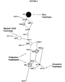
- risk haplotype is an allelic form of a gene, herein Factor H or a Factor H-related gene, comprising at least one variant polymorphism, and preferably a set of variant polymorphisms, associated with increased risk for developing AMD.
- variant when used in reference to a Factor H or Factor H-related gene, refers to a nucleotide sequence in which the sequence differs from the sequence most prevalent in a population, herein humans of European-American descent.
- the variant polymorphisms can be in the coding or non-coding portions of the gene.
- An example of a risk Factor H haplotype is the allele of the Factor H gene encoding histidine at amino acid 402 and/or cysteine at amino acid 1210.
- the risk haplotype can be naturally occurring or can be synthesized by recombinant techniques.
- a protective haplotype is an allelic form of a gene, herein Factor H or a Factor H-related gene, comprising at least one variant polymorphism, and preferably a set of variant polymorphisms, associated with decreased risk of developing AMD.
- one protective Factor H haplotype has an allele of the Factor H gene encoding isoleucine at amino acid 62.
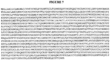
- wild-type Factor H protein has the sequence of SEQ ID NO:2 ( FIGURE 7 ), except that the amino acid at position 402 is tyrosine (Y; [SEQ ID NO:337]).
- variant when used in reference to a Factor H or Factor H-related polypeptide, refers to a polypeptide in which the sequence differs from the normal or wild-type sequence at a position that changes the amino acid sequence of the encoded polypeptide.
- some variations or substitutions in the nucleotide sequence of Factor H gene alter a codon so that a different amino acid is encoded (for example and not for limitation, having an alternative allele at one or more of 162V, Y402H, D936E) resulting in a variant polypeptide.
- Variant polypeptides can be associated with risk (e.g., having histidine at position 402), associated with protection (e.g., having isoleucine at position 62), or can be encoded by a neutral haplotype (e.g., having aspartic acid at position 936).
- Variant CFHR5 polypeptides can be associated with risk (e.g., having serine at position 46), associated with protection, or can be neutral.
- polymorphism refers to the occurrence of two or more genetically determined alternative sequences or alleles in a population.
- a "polymorphic site” is the locus at which sequence divergence occurs. Polymorphic sites have at least two alleles. A diallelic polymorphism has two alleles. A triallelic polymorphism has three alleles. Diploid organisms may be homozygous or heterozygous for allelic forms. A polymorphic site may be as small as one base pair.
- polymorphic sites include: restriction fragment length polymorphisms (RFLPs); variable number of tandem repeats (VNTRs); hypervariable regions; minisatellites; dinucleotide repeats; trinucleotide repeats; tetranucleotide repeats; and simple sequence repeats.
- RFLPs restriction fragment length polymorphisms
- VNTRs variable number of tandem repeats
- minisatellites dinucleotide repeats
- trinucleotide repeats trinucleotide repeats
- tetranucleotide repeats and simple sequence repeats.
- reference to a "polymorphism" can encompass a set of polymorphisms (i.e., a haplotype).
- a "single nucleotide polymorphism (SNP)" occurs at a polymorphic site occupied by a single nucleotide, which is the site of variation between allelic sequences. The site is usually preceded by and followed by highly conserved sequences of the allele.
- a SNP usually arises due to substitution of one nucleotide for another at the polymorphic site. Replacement of one purine by another purine or one pyrimidine by another pyrimidine is called a transition. Replacement of a purine by a pyrimidine or vice versa is called a transversion.
- a synonymous SNP refers to a substitution of one nucleotide for another in the coding region that does not change the amino acid sequence of the encoded polypeptide.
- a non-synonymous SNP refers to a substitution of one nucleotide for another in the coding region that changes the amino acid sequence of the encoded polypeptide.
- a SNP may also arise from a deletion or an insertion of a nucleotide or nucleotides relative to a reference allele.
- a "set" of polymorphisms means more than one polymorphism, e.g. , at least 2, at least 3, at least 4, at least 5, at least 6, or more than 6 of the polymorphisms shown in TABLE 1A, TABLE 1B and/or TABLE 1C or otherwise known in the Factor H gene or other gene.
- nucleic acid means a nucleic acid species that is the predominant species present in a composition. Isolated means the nucleic acid is separated from at least one compound with which it is associated in nature. A purified nucleic acid comprises (on a molar basis) at least about 50, 80 or 90 percent of all macromolecular species present.
- Two amino acid sequences are considered to have "substantial identity" when they are at least about 80% identical, preferably at least about 90% identical, more preferably at least about 95%, at least about 98% identical or at least about 99% identical. Percentage sequence identity is typically calculated by determining the optimal alignment between two sequences and comparing the two sequences. Optimal alignment of sequences may be conducted by inspection, or using the local homology algorithm of Smith and Waterman, 1981, Adv. Appl. Math. 2: 482 , using the homology alignment algorithm of Needleman and Wunsch, 1970, J. Mol. Biol. 48: 443 , using the search for similarity method of Pearson and Lipman, 1988, Proc. Natl. Acad. Sci. U.S.A.
- Linkage describes the tendency of genes, alleles, loci or genetic markers to be inherited together as a result of their location on the same chromosome. Linkage can be measured by percent recombination between the two genes, alleles, loci or genetic markers. Typically, loci occurring within a 50 centimorgan (cM) distance of each other are linked. Linked markers may occur within the same gene or gene cluster. “Linkage disequilibrium” or “allelic association” means the preferential association of a particular allele or genetic marker with a specific allele or genetic marker at a nearby chromosomal location more frequently than expected by chance for any particular allele frequency in the population. A marker in linkage disequilibrium can be particularly useful in detecting susceptibility to disease, even if the marker itself does not cause the disease.
- diagnosis refers to the ability to determine whether an individual has the propensity to develop disease (including with or without signs or symptoms). Diagnosis of propensity to develop disease can also be called “screening” and, as used herein, the terms diagnosis and screening are used interchangeably. It will be appreciated that having an increased or decreased propensity to developing a condition refers to the likelihood of developing the condition relative to individuals in the population without the condition.
- HF1 Complement Factor H
- Factor H polymorphisms associated with AMD were identified as described in Example 1, by examining the coding and adjacent intronic regions of Factor H (including exon 10A, which is transcribed for the Factor H isoform FHL1) for variants using SSCP analysis, DHPLC analysis, and direct sequencing, according to standard protocols. Remaining polymorphisms were typed by the 5' nuclease (Taqman, ABI) methodology. Taqman genotyping and association analysis were performed as described (Gold et al., 2004). Primers for SSCP and DNA sequencing analyses were designed to amplify each exon and its adjacent intronic regions using MacVector software. PCR-derived amplicons were screened for sequence variation by SSCP and DHPLC according to standard protocols. All changes detected by SSCP and DHPLC were confirmed by bidirectional sequencing according to standard protocols. Statistical analyses were performed using chi-square ( ⁇ 2 ) and Fisher's exact tests (P values).
- TABLE 1A a highly significant association of polymorphic sites in the Factor H gene with AMD was found in an examination of two independent cohorts that together included approximately 900 AMD cases and 400 matched controls.
- Sixteen (16) polymorphisms in the Factor H gene are listed in TABLES 1A-1B. Of these twelve (12) are found in the SNP database (dbSNP) which may be found in the National Center for Biotechnology Information (NCBI).
- the dbSNP is a collection of SNPs in the human Factor H gene which are dispersed among the 22 coding exons of the Factor H gene and among the promoter, the 5' untranslated region, the introns, and the 3' untranslated region of the Factor H gene.
- TABLES 1A-1B Three additional polymorphisms in TABLES 1A-1B are not found in the SNP database: a polymorphism in the promoter (promoter 1 in TABLE 1A); a polymorphism in intron 2 in which two T nucleotides are inserted; and a polymorphism in Exon 10A.
- the first column in TABLE 1A lists the dbSNP number for polymorphisms in the Factor H gene.
- rs800292 is the dbSNP designation for a polymorphism in the Factor H gene.
- the second column lists the location of the polymorphism.
- the rs800292 polymorphism is located in exon 2 of the Factor H gene. Polymorphisms not identified by a database number can be referred to by location (e.g., "intron 2").
- the third column lists the nucleic acid sequence of the coding (top, 5' to 3' direction) and non-coding (bottom) strands of DNA spanning the polymorphisms.
- the rs800292 polymorphism, G or A as indicated in the brackets for the coding strand is flanked by the 20 nucleotides shown 5' and 3' to the polymorphism.
- N in the sequence spanning the Exon 10A polymorphism indicates the insertion of a single nucleotide, either A, C, G or T, in the variant allele.
- the fourth column lists the SEQ ID NO: for the sequences.
- the fifth column lists the amino acid change, if any, associated with the polymorphism. For example, the rs800292 polymorphism results in a change in the amino acid sequence from valine (V) to isoleucine (I) at position 62 of the Factor H polypeptide.
- the sixth column lists the allele frequency of the polymorphism in a control population.
- the numbers 1 and 2 refer to the alleles that correspond to the first and second nucleotide, respectively, at the polymorphic site in the third column.
- the second and fourth columns list the forward and reverse primers or AOD numbers for amplifying the polymorphisms.
- the third and fifth columns list the SEQ ID NOs: for the primers.
- the second and fourth columns list probes used for detecting the polymorphisms.
- the third and fifth columns list the SEQ ID NOs: for the probes.
- TABLES 1A-1B additional polymorphic sites in the Factor H gene, which are not listed in TABLES 1A-1B maybe associated with AMD. Exemplary polymorphic sites in the Factor H gene are listed, for example and not limitation, above.
- TABLE 1C lists an additional 14 polymorphic sites in the Factor H gene, which are not found in the dbSNP database, that may be associated with AMD or other diseases. The first column lists the location of the SNP. The second column lists the nucleic acid sequence spanning the polymorphisms. "notG" in the sequence spanning the Exon 5 polymorphism indicates the presence of an A, C or T nucleotide in the variant allele.
- TABLE 2 shows a haplotype analysis of eight SNPs in the human Factor H gene in AMD cases and controls.
- the at-risk haplotypes are shown in stippled boxes, with the haplotype determining SNPs (Y402H and IVS10) shown in denser stippling.
- the protective haplotypes are shown in diagonal-lined boxes, with the haplotype determining SNPs (IVS1, I62V and IVS6) shown indenser diagonal lines.
- the first column lists the allele of the polymorphism in the promoter (Prom).
- the second column lists the allele of the non-coding strand of the polymorphism in intron 1 (IVS1).
- the ninth column lists the Odds Ratio (OR) for the haplotype.
- the tenth column lists the P value for the at-risk and two protective haplotypes.
- the eleventh and twelfth columns list the frequencies of the haploptype in AMD cases and controls.
- the numbers 1 and 2 in columns 1 to 6 refer to the alleles that correspond to the first and second nucleotide, respectively, at each of the polymorphic sites (see TABLE 1A). Thus, columns 1 to 6 list the alleles of polymorphisms from 5' to 3' in the Factor H gene.
- the seventh column lists the Factor H haplotype based on the polymorphisms listed in columns 1 to 6.
- the eighth column lists the frequency of the indicated Factor H haplotype in a control population.
- the ninth column lists the frequency of the indicated Factor H haplotype in the AMD population. As shown in TABLE 3, the haplotype analysis suggests that multiple variants contribute to the association and may confer either elevated or reduced risk of AMD.
- TABLE 8 shows a diplotype analysis of seven Factor H polymorphisms. The first column indicates whether the diplotype is associated with increased (risk diplotype) or decreased (protective diplotype) risk of developing AMD. Common risk and protective diplotypes are indicated. The second column lists the alleles of the polymorpohism in exon 2
- haplotypes associated with increased risk for AMD are shown in TABLES 2 and 6 and FIGURE 5 .
- one common at-risk haplotype is the H1 haplotype, which includes the variant allele at position 402 (encoding histidine) and the variant allele at IVS10 (intron 10, rs203674) and is found in 49% of AMD cases, but only in 26% of controls. Homozygotes for the risk diplotype (H1/H1) are significantly at risk.
- H1/H1 Homozygotes for the risk diplotype
- Other at-risk haplotypes and diplotypes are shown in TABLES 2 and 8. Similar data are presented in TABLE 3, which shows an at-risk haplotype (111211) found in 48% of AMD cases, but only in 28% of controls.
- the non-synonymous polymorphism at amino acid position 1210 in exon 22 of the Factor H gene is strongly associated with AMD (see TABLE 1A).
- the variant allele which encodes a cysteine instead of an arginine, is found in the heterozygous state in 5% of AMD cases, and no controls in a cohort comprised of 919 individuals ascertained at the University of Iowa. No 1210C homozygotes have been identified to date.
- the presence of cysteine at amino acid position 1210 of Factor H therefore, provides a strong indication that the individual has AMD or is likely to develop AMD.
- 1210C is indicative of propensity to develop AMD or other complement mediated conditions even when detected on allele that is otherwise protective (e.g., Y402).
- Variation at CFH position 1210 is known to cause atypical hemolytic uremic syndrome (aHUS), a complement related disease with renal manifestations.
- aHUS atypical hemolytic uremic syndrome
- other CFH variations or mutations known to cause aHUS may be associated with an increased risk for developing AMD.
- aHUS-causing variations include, but are not limited to, T956M, Q1076E, D1119G, W1183L, T1184R, L1189R, L1189F, S1191W, S1191L, V1197A, and R1215G (Esparza-Gordillo et al 2005; Perez-Caballero et al 2001; Richards et al 2001; Sanchez-Corral et al 2002); additional aHUS-causing mutations are described in Saunders (Saunders et al 2006).
- a biological sample from a subject e.g., protein or nucleic acid
- haplotype analysis using non-synonymous polymorphisms in the Factor H gene is useful to identify variant Factor H polypeptides.
- Other haplotypes associated with risk may encode a protein with the same sequence as a protein encoded by a neutral or protective haplotype, but contain an allele in a promoter or intron, for example, that changes the level or site of Factor H expression.
- a polymorphism in the Factor H gene, or in a Factor H-related gene may be linked to a variation in a neighboring gene. The variation in the neighboring gene may result in a change in expression or form of an encoded protein and have detrimental or protective effects in the carrier.
- the protective H2 haplotype including a variant allele in IVS6 (intron 6, rs3766404) occurs in 12% of controls, but only in 6% of AMD cases.
- the protective H4 haplotype includes the variant allele in IVS1 (intron 1, rs529825) and the variant allele (162) (exon 2, rs800292) and occurs in 18% of controls, but only in 12% of AMD cases.
- the protein encoded by a gene characterized by a protective haplotype has a sequence different from risk haplotype proteins (e.g., due to the presence of a nonsynomous SNP).
- a protective form of Factor H protein generally does not have histidine at position 402.
- a protective form may have isoleucine at position 62. Additional protective forms can be identified by (1) identifying an individual or individuals with a protective haplotype and (2) determining the sequence(s) of Factor H cDNA or protein from the individuals. Other protective forms are identified as described below in Section VIII.
- haplotypes are associated in a population with neither increased risk nor decreased risk of developing AMD and are referred to as "neutral.” Examples of neutral haplotypes identified in a Caucasian population are shown in FIGURE 5 (H3 and H5). Additional or different neutral haplotypes may be identified in racially/ethnically different populations. Proteins encoded by a gene characterized by a neutral haplotype are "neutral" Factor H proteins. As explained supra, "neutral" Factor H proteins could provide therapeutic benefit when administered to patients having a risk haplotype or diagnosed with AMD. For example, exemplary proteins encoded by genes characterized by a neutral haplotype include proteins not having histidine at position 402 and/or not having isoleucine at position 62.
- dbSNP SNP database
- NCBI National Center for Biotechnology Information
- the dbSNP is a collection of SNPs in the human genome.
- the SNPs in the Factor H gene are dispersed among the 22 coding exons of the Factor H gene and among the promoter, the 5' untranslated region, the introns, and the 3' untranslated region of the Factor H gene.
- accession numbers for 379 SNPs in the human Factor H gene that are found in the dbSNP database are listed above. These SNPs can be used in carrying out methods disclosed herein.
- the sixth and seventh rows of TABLE 11 list the frequency that a particular haplotype is present among the 22 MPGNII patients. For example, for the exon 2 SNP, G is present in 95%, and A is present in 5%, of the alleles of the 22 MPGNII patients.
- the eighth row lists the nucleotide of the common haplotype in the Factor H gene for the 22 MPGNII patients. For example, G is the more frequent nucleotide in the exon 2 SNP, and 9 T nucleotides is more frequently observed than 11 T nucleotides in the intron 2 SNP, in the Factor H gene for the 22 MPGNII patients.
- the remaining rows list the diplotype for the 11 SNPs in the Factor H gene for each of the 22 MPGNII patients.
- TABLE 15 shows a comparision of the SNP frequencies in patients with MPGNII versus AMD-negative, ethnically-matched control individuals.
- the first column of TABLE 15 lists the SNP in the CFHR5 gene.
- the second and third columns of TABLE 15 list the frequencies that a particular haplotype is present among the 22 MPGNII patients.
- the fourth and fifth columns of TABLE 15 list the frequencies that a particular haplotype is present among the 103 control individuals.
- the sixth column of TABLE 15 lists the P-value calculated for each data set.
- Polymorphisms particularly associated with increased risk in Factor H and CFHR5 include a variant allele at rs1061170 (Y420H in exon 9) and rs12097550 (P46S in exon 2), respectively.
- haplotype analysis using non-synonymous polymorphisms in the Factor H or CFHR5 gene is useful to identify variant Factor H or CFHR5 polypeptides.
- Other haplotypes associated with risk may encode a protein with the same sequence as a protein encoded by a neutral or protective haplotype, but contain an allele in a promoter or intron, for example, that changes the level or site of Factor H or CFHR5 expression.
- haplotypes were also discovered.
- the haplotype with the variant allele in exon 2 (rs800292, I62V) occurs in 23% of controls, but only in ⁇ 3% of MPGNII cases and the haplotype with the variant allele in IVS2 (intron 2, -18insTT) occurs in 26% of controls, but only in ⁇ 3% of MPGNII cases.
- the haplotype with the variant allele in exon 10 (rs2274700, A473A) occurs at higher frequency in controls than in MPGNII cases.
- Example 2 it has been discovered that the same CFH polymorphisms associated with propensity to develop AMD are also associated with development of membranoproliferative glomerulonephritis type 2 (MPGN II). Indeed, the risk haplotypes originally found in AMD patients (Y402 H and IVS10) are also found in 70% of patients tested having membranoproliferative glomerulonephritis type 2 (MPGN II), indicating that the diagnostic methods disclosed herein are useful to detect this condition. In addition, variations and haplotypes in the CFHR5 gene were strongly associated with increased risk of having MPGNII. One conclusion that emerges from these data is that MPGNII and AMD are alternative manifestations of the same genetic lesion.
- ATD age-related macular degeneration
- Diaz-Guillen MA et al., A radiation hybrid map of complement factor H and factor H-related genes, Immunogenetics, 1999 Jun;49(6):549-52 ; Skerka C, et al., A novel short consensus repeat-containing molecule is related to human complement factor H, J Biol Chem.
- Certain polymorphisms in the Factor H gene may predispose an individual to a distinct mutation that is causally related to a particular AMD phenotype.
- a polymorphism in the CFH gene, or in a CFHR5 may be linked to a variation in a neighboring gene (including but not limited to CFHR-1, 2, 3, or 4).
- the variation in the neighboring gene may result in a change in expression or form of an encoded protein and have detrimental or protective effects in the carrier.
- the identity of bases occupying the polymorphic sites in the Factor H gene and the Factor H-Related 5 gene shown in TABLES 1A, 1B, 1C, 11, 14 and 15, as well as others in the dbSNP collection that are located in or adjacent to the Factor H or CFHR5 genes can be determined in an individual, e.g., in a patient being analyzed, using any of several methods known in the art. Examples include: use of allele-specific probes; use of allele-specific primers; direct sequence analysis; denaturing gradient gel electropohoresis (DGGE) analysis; single-strand conformation polymorphism (SSCP) analysis; and denaturing high performance liquid chromatography (DHPLC) analysis.
- DGGE denaturing gradient gel electropohoresis
- SSCP single-strand conformation polymorphism
- DPLC denaturing high performance liquid chromatography
- allele-specific probes for analyzing polymorphisms are described by e.g., Saiki et al., 1986; Dattagupta, EP 235,726 , Saiki, WO 89/11548 . Briefly, allele-specific probes are designed to hybridize to a segment of target DNA from one individual but not to the corresponding segment from another individual, if the two segments represent different polymorphic forms. Hybridization conditions are chosen that are sufficiently stringent so that a given probe essentially hybridizes to only one of two alleles. Typically, allele-specific probes are designed to hybridize to a segment of target DNA such that the polymorphic site aligns with a central position of the probe.
- allele-specific probes for analyzing Factor H polymorphisms are shown in TABLE 16A.
- examples of allele-specific probes include: 5'-TTTCTTCCATAATTTTG-3' [SEQ ID NO:234] (reference allele probe) and 5'-TTTCTTCCATGATTTTG-3' [SEQ ID NO:235] (variant allele probe); and 5'-TAATCAAAATTATGGAA-3' [SEQ ID NO:232] (reference allele probe) and 5-TAATCAAAATCATOGAA-3' [SEQ ID NO:233] (variant allele probe).
- the first set of allele-specific probes hybridize to the non-coding strand of the Factor H gene spanning the exon 9 polymorphism.
- the second set of allele-specific probes hybridize to the coding strand of the Factor H spanning the exon 9 polymorphism.
- These probes are 17 bases in length. The optimum lengths of allele-specific probes can be readily determined using methods known in the art.
- Allele-specific probes are often used in pairs, one member of a pair designed to hybridize to the reference allele of a target sequence and the other member designed to hybridize to the variant allele.
- Several pairs of probes can be immobilized on the same support for simultaneous analysis of multiple polymorphisms within the same target gene sequence.
- allele-specific primers for analyzing polymorphisms are described by, e.g., WO 93/22456 and Gibbs, 1989. Briefly, allele-specific primers are designed to hybridize to a site on target DNA overlapping a polymorphism and to prime DNA amplification according to standard PCR protocols only when the primer exhibits perfect complementarity to the particular allelic form. A single-base mismatch prevents DNA amplification and no detectable PCR product is formed. The method works best when the polymorphic site is at the extreme 3'-end of the primer, because this position is most destabilizing to elongation from the primer.
- allele-specific primers for analyzing Factor H polymorphisms are shown in TABLE 16B.
- examples of allele-specific primers include: 5'-CAAACTTTCTTCCATA-3' [SEQ ID NO:294] (reference allele primer) and 5'-CAAACTTTCTTCCATG-3' [SEQ ID NO:295] (variant allele primer); and 5'-GGATATAATCAAAATT-3' [SEQ ID NO:292] (reference allele primer) and 5'-GGATATAATCAAAATC-3' [SEQ ID NO:293] (variant allele primer).
- primers are used in standard PCR protocols in conjunction with another common primer that hybridizes to the non-coding strand of the Factor H gene at a specified location upstream from the polymorphism.
- the common primers are chosen such that the resulting PCR products can vary from about 100 to about 300 bases in length, or about 150 to about 250 bases in length, although smaller (about 50 to about 100 bases in length) or larger (about 300 to about 500 bases in length) PCR products are possible.
- the length of the primers can vary from about 10 to 30 bases in length, or about 15 to 25 bases in length.
- the sequences of the common primers can be determined by inspection of the Factor H genomic sequence, which is found under GenBank accession number AL049744.
- RNA samples e.g., amplifying the segments of the Factor H gene of an individual using Factor H-specific primers
- amplifying This can be accomplished by standard polymerase chain reaction (PCR & RT-PCR) protocols or other methods known in the art.
- the amplifying may result in the generation of Factor H allele-specific oligonucleotides, which span the single nucleotide polymorphic sites in the Factor H gene.
- the Factor H-specific primer sequences and Factor H allele-specific oligonucleotides may be derived from the coding (exons) or non-coding (promoter, 5' untranslated, introns or 3' untranslated) regions of the Factor H gene.
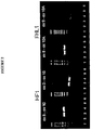
- Amplification products generated using PCR can be analyzed by the use of denaturing gradient gel electrophoresis (DGGE). Different alleles can be identified based on sequence-dependent melting properties and electrophoretic migration in solution. See Erlich, ed., PCR Technology, Principles and Applications for DNA Amplification, Chapter 7 (W.H. Freeman and Co, New York, 1992 ).
- DGGE denaturing gradient gel electrophoresis
- Alleles of target sequences can be differentiated using single-strand conformation polymorphism (SSCP) analysis. Different alleles can be identified based on sequence- and structure-dependent electrophoretic migration of single stranded PCR products (Orita et al., 1989). Amplified PCR products can be generated according to standard protocols, and heated or otherwise denatured to form single stranded products, which may refold or form secondary structures that are partially dependent on base sequence.
- SSCP single-strand conformation polymorphism
- Alleles of target sequences can be differentiated using denaturing high performance liquid chromatography (DHPLC) analysis. Different alleles can be identified based on base differences by alteration in chromatographic migration of single stranded PCR products (Frueh and Noyer-Weidner, 2003). Amplified PCR products can be generated according to standard protocols, and heated or otherwise denatured to form single stranded products, which may refold or form secondary structures that are partially dependent on the base sequence.
- DPLC denaturing high performance liquid chromatography
- the allele of the patient at one of more of the following polymorphic sites in the Factor H gene may be determined: rs529825; rs800292; intron 2 (IVS2 or insTT); rs3766404; rs1061147; rs1061170; exon 10A; rs203674; rs375046; and exon 22 (1210).
- One, two, three, four five, or more than five of the following polymorphic sites in the Factor H gene may be determined: rs529825; rs800292; intron 2 (IVS2 or insTT); rs3766404; rs1061147; rs1061170; rs2274700; exon 10A; rs203674; rs375046; and exon 22 (1210).
- the aforementioned polymorphisms and combinations of polymorphisms are provided for illustration and are not intended to limit the invention in any way.
- the non-synonymous polymorphism at amino acid position 1210 in exon 22 of the Factor H gene is strongly associated with AMD, and the presence of cysteine at amino acid position 1210 of Factor H, therefore, provides a strong indication that the individual has AMD or is likely to develop AMD.
- 1210C is indicative of propensity to develop AMD or other complement mediated conditions even when detected on allele that is otherwise protective (e.g., Y402).
- the allele of the patient at exon 22 (1210) is highly informative with respect to risk of developing AMD or other Factor H-associated diseases.
- the allele of an individual at one of more of the following polymorphic sites in the CFHR5 gene may be determined: rs9427661 (-249T>C); rs9427662 (-20T>C); and rs12097550 (P46S).
- the aforementioned polymorphisms and combinations of polymorphisms are provided for illustration and are not intended to limit the invention in any way. That is, other polymorphisms and haplotypes useful in practicing the invention will be apparent from this disclosure.
- a protein assay may be carried out to characterize polymorphisms in a subject's CFH or CFHR5 genes. Methods that can be adapted for detection of variant CFH, HFL1 and CFHR5 are well known.
- These methods include analytical biochemical methods such as electrophoresis (including capillary electrophoresis and two-dimensional electrophoresis), chromatographic methods such as high performance liquid chromatography (HPLC), thin layer chromatography (TLC), hyperdiffusion chromatography, mass spectrometry, and various immunological methods such as fluid or gel precipitin reactions, immunodiffusion (single or double), immunoelectrophoresis, radioimmnunoassay (RIA), enzyme-linked immunosorbent assays (ELISAs), immunofluorescent assays, western blotting and others.
- analytical biochemical methods such as electrophoresis (including capillary electrophoresis and two-dimensional electrophoresis), chromatographic methods such as high performance liquid chromatography (HPLC), thin layer chromatography (TLC), hyperdiffusion chromatography, mass spectrometry, and various immunological methods such as fluid or gel precipitin reactions, immunodiffusion (single or double), immunoelectrophoresis, radioimmn
- immunological binding assay formats suitable for the practice of the methods disclosed herein are known (see, e.g., Harlow, E.; Lane, D. Antibodies: a laboratory manual. Cold Spring Harbor, N.Y: Cold Spring Harbor Laboratory; 1988 ; and Ausubel et al., (2004) Current Protocols in Molecular Biology, John Wiley & Sons, New York NY .
- the assay may be, for example, competitive or non-conpetitive.
- immunological binding assays or immunoassays
- the capture agent may be a moiety that specifically binds to a variant CFH or CFHR5 polypeptide or subsequence.
- the bound protein may be detected using, for example, a detectably labeled anti-CFH/CFHR5 antibody.
- At least one of the antibodies may be specific for the variant form (e.g., does not bind to the wild-type CFH or CFHR5 polypeptide.
- the variant polypeptide may be detected using an immunoblot (Western blot) format.
- Polymorphisms in the Factor H gene such as those shown in TABLE 1A, TABLE 1B, TABLE 1C or identified as described herein, which correlate with AMD or with particular subtypes of AMD, are useful in diagnosing AMD or specific subtypes of AMD, or susceptibility thereto.
- Polymorphisms in the CFHR5 gene such as those shown in TABLES 14 and 15 or identified as described herein, which correlate with AMD or with particular subtypes of AMD, are useful in diagnosing AMD or specific subtypes of AMD, or susceptibility thereto. These polymorphisms are also useful for screening for MPGNII and other Factor H-associated diseases.
- Combined detection of several such polymorphic forms i.e., the presence or absence of polymorphisms at specified sited
- polymorphisms at specified sited for example, 1, 2, 3, 4, 5, 6, 7, 8, 9, 10, or all of the polymorphisms in the Factor H gene listed in TABLE 1A, TABLE 1B and/or TABLE 1C, alone or in combination with additional Factor H gene polymorphisms not included in TABLES 1A-1C, may increase the probability of an accurate diagnosis.
- the polymorphisms in the Factor H and CFHR5 genes are useful in diagnosing AMD or specific subtypes of AMD, or susceptibility thereto, in family members of patients with AMD, as well as in the general population.
- Therapeutic and prophylactic approaches in subjects identified as being at high risk for AMD include, but are not limited to, (1) increasing the amount or expression of neutral or protective forms of Factor H and/or neutral or protective forms of CHFR5; (2) decreasing the amount or expression of risk-associated forms of Factor H and/or risk-associated form of CHFR5; and (3) reducing activation of the complement alternative pathway.
- Examples of such therapeutic and prophylactic approaches include: (1) administration of neutral or protective forms of Factor H protein or therapeutically active fragments and/or neutral or protective forms of CHFR5 or therapeutically active fragments; (2) other wise increasing expression or activity of neutral and protective forms of Factor H; (3) interfering with expression of variant Factor H and/or variant CFHR5 proteins encoded by individuals with a risk haplotype by (e.g., by administration of antisense RNA); (4) reducing the amount of activity of a detrimental variant form.
- Therapeutic agents can be administered systemically (e.g., by i.v. injection or infusion) or locally (e.g., to the vicinity of the ocular RPE).
- exemplary methods include intraocular injection (e.g., retrobulbar, subretinal, intravitreal and intrachoridal), iontophoresis, eye drops, and intraocular implantation (e.g., intravitreal, sub-Tenons and subconjunctival).
- recombinant Factor H polypeptide is administered to the patient.
- the recombinant Factor H may be encoded by a neutral haplotype sequence, which maybe full-length (CFH/HF1), truncated (FHL1), or alternatively spliced form, or a biologically active fragment thereof.
- the recombinant Factor H may have the sequence of a protective allele, either full-length or truncated form, or a protective biologically active fragment thereof.
- Methods for production of therapeutic recombinant proteins are well known and include methods described hereinbelow.
- the therapeutic polypeptide can be administered systemically (e.g., intravenously or by infusion) or locally (e.g., directly to an organ or tissue, such as the eye or the liver).
- Protective forms of Factor H are as defined in the claims.
- fragments of neutral or protective forms of Factor H may be administered for treatment or prevention of MPGNII.
- Polypeptides encoded by CFH splice variants expressed in individuals with a protected phenotype may be administered. These proteins can be identified by screening expression of CFH-related RNA in individuals homozygous for a protective or neutral haplotype.
- the protective protein may have a sequence corresponding to one or more exons of the CFH gene sequence.
- the protective protein may have the sequence of full-length or truncated CFH protein, except that the amino acid residues encoded by 1, 2, 3 or more exons (which may or may not be contiguous) are deleted.
- a protective Factor H protein may have an amino acid sequence substantially identical to SEQ ID NO:2, with the proviso that the residue at position 402 is not histidine and the residue at position 1210 is not cysteine.
- Disclosed herein is a Factor H protein in which the residue at position 62 is not valine.
- the residue at position 62 is isoleucine.
- the residue at position 62 is isoleucine, the residue at position 402 is tyrosine and the residue at position 1210 is arginine.
- a protective Factor H protein may have an amino acid sequence substantially identical to SEQ ID NO:4 (FHL1). Disclosed herein is a Factor H protein in which the residue at position 62 is not valine. The residue at position 62 is isoleucine. Preferably the protective Factor H protein has 95% amino acid identity to SEQ ID NO:4 or a fragment thereof; sometimes at least 95% amino acid identity, sometimes at least 98% amino acid identity, and sometimes at least 99% identity to the reference Factor H polypeptide of SEQ ID NO:4.
- a protective Factor H protein may have one or more activities of the reference Factor H polypeptide.
- the activity may be binding to heparin.
- the activity may be binding to CRP.
- the activity may be binding to C3b.
- the activity may be binding to endothelial cell surfaces.
- the activity may be C3b co-factor activity.
- the protective Factor H protein has activity that is higher with respect to its normal function than the protein of SEQ ID NO:2.
- the protective Factor H protein may have activity with respect to its normal function that is higher than the protein of SEQ ID NO:4.
- C3b and CFH proteins can be analyzed by surface resonance using a Biacore 3000 system (Biacore AB, Uppsala, Sweden), as described previously ( Manuelian et al., 2003, Mutations in factor H reduce binding affinity to C3b and heparin and surface attachment to endothelial cells in hemolytic uremic syndrome. J Clin Invest 111, 1181-90 ).
- C3b CalBiochem, Inc
- C3b are coupled using standard amine-coupling to flow cells of a sensor chip (Carboxylated Dextran Chip CM5, Biacore AB, Uppsala, Sweden).
- C3b 50 ⁇ g/ml, dialyzed against 10 mM acetate buffer, pH 5.0
- C3b 50 ⁇ g/ml, dialyzed against 10 mM acetate buffer, pH 5.0
- Unreacted groups are inactivated using ethanolamine-HCl.
- the other cell is prepared as a reference cell by injecting the coupling buffer without C3b.
- flow cells will be washed thoroughly by two injections of 2 M NaCl in 10 mM acetate buffer, pH 4.6 and running buffer (PBS, pH 7.4).
- the Factor H protein is injected into the flow cell coupled with C3b or into the control cell at a flow rate of 5 ul/min at 25°C. Binding of Factor H to C3b is quantified by measuring resonance units over time, as described in Manuelian et al., 2003, supra.
- Polyclonal goat anti-human FH antiserum is used as a primary antibody (CalBiochem) (diluted 1:100), incubating cells at 4°C for 15 minutes. Alexa-fluor 488-conjugated goat antiserum diluted 1:100 in blocking buffer is used as the secondary antibody.
- Cells are examined by flow cytometry (FACScalibur, Becton-Dickinson Immunocytometry, Mountain View, California, USA). Typicallly, 10,000 events are counted.
- C3b biotin 100 ng/reaction
- Factor I 200 ng/reaction
- 100 ng of purified factor H 100 ng
- Samples taken before and after addition of Factor I are separated by SDS-PAGE under reducing conditions and analyzed by Western blotting, detecting and quantitating C3b degradation products by Strepavidin-POD-conjugation (1:10000).
- C3b (40 ⁇ g) (CalBiochem) is biotinylated using the Biotin Labeling Kit (Roche Diagnostics, Mannheim, Germany), according to the manufacturer's instructions.
- C3b (CalBiochem) is labeled with D-biotinyl-epsilon-aminocaproic acid-N-hydroxysuccinimide ester for 2 hours at 25°C. Excess biotin is removed by gel filtration using a PBS equilibrated PD10 column (Amersham Biosciences). Also see Sanchez-Corral et al., 2002, Am J. Hum. Genet. 71:1285-95 .
- CFH402Y and CFH402H Binding of purified CFH proteins (CFH402Y and CFH402H) to heparin is analyzed using heparin affinity chromatography in a high-performance liquid chromatograph (HPLC) system.
- 10 ⁇ g of CFH protein is diluted in 1/2xPBS and applied to a heparin-Sepharose affinity column (HiTrap, Amersham Biosciences) at a flow rate of 0.5 ml/min.
- the column is extensively washed with 1/2xPBS, and the bound CFH protein eluted using a linear salt gradient ranging from 75 to 500 mM NaCl, in a total volume of 10 ml and at a flow rate of 0.5 ml/min.
- Eluted fractions are assayed by SDS-PAGE and Western blot analysis. Elution of isoforms in different fractions is indicative that specific amino acid variations in the CFH protein can modulate binding of the protein to heparin. Also see, e.g., Pangburn et al., 1991, Localization of the heparin-binding site on complement Factor H, J Biol Chem. 266:16847-53 .
- recombinant CFHR5 polypeptide is administered to the patient.
- the recombinant CFHR5 may have a neutral-type sequence, or a biologically active fragment thereof.
- the recombinant CFHR5 may have the sequence of a protective allele, or a protective biologically active fragment thereof.
- Methods for production of therapeutic recombinant proteins are well known and include methods described hereinbelow.
- the therapeutic polypeptide can be administered systemically (e.g., intravenously or by infusion) or locally (e.g., directly to an organ or tissue, such as the eye or the liver).
- compositions Containing CFH or CFHR5 Polypeptides Containing CFH or CFHR5 Polypeptides
- Factor H polypeptides which may be wild-type or variants (e.g., neutral or protective variants), and may be full length forms, truncated forms, or biologically active fragments of the variant Factor H polypeptides.
- protective Factor H proteins and genes encoding them
- Biologically active fragments may include any portion of the full-length Factor H polypeptide which confers a biological function on the variant protein.
- a protective haplotype will be associated with expression of a less-than full length form of Factor H (i.e., in addition to FHL-1) due, for example, to the presense of a premature stop codon in the gene.
- Therapeutically active fragments can also be identified by testing the effect of the protein on expression of AMD biomarkers.
- AMD biomarkers include complement pathway components (for example, Factor I, Factor H, C1r, C3, C3a), C reactive protein, haptoglobin, apolipoprotein E, immunoglobulin heavy or light chain(s), alpha 1 antitrypsin, alpha 2 macroglobulin, transthyretin, creatinine, and others described in copending provisional application No. 60/715,503 entitled "Biomarkers Associated With Age-Related Macular Degeneration.”
- protective CFHR5 proteins can be identified by identifying an individual as having a protective haplotype and determining the amino acid sequence(s) of CFHR5 encoded in the genome of the individual, where a protective CFHR5 protein is encoded by an allele having a protective haplotype.
- Biologically active fragments may include any portion of the full-length CFHR5 polypeptide which confers a biological function on the variant protein.
- Therapeutically active fragments can also be identified by testing the effect of the protein on expression of AMD biomarkers as described above for Factor H.
- Factor H and CFHR5 can be isolated from the blood of genotyped donors, from cultured or transformed RPE cells derived from genotyped ocular donors, or from cell lines (e.g., glial or hepatic) that express endogenous Factor H.
- therapeutic proteins can be recombinantly produced (e.g., in cultured bacterial or eukaryotic cells) and purified using methods well known in the art and described herein.
- some forms of Factor H and CFHR5 have been recombinantly expressed for research purposes. However, such research preparations are not suitable for therapeutic use.
- recombinant polypeptides suitable for administration to patients including polypeptides that are produced and tested in compliance with the Good Manufacturing Practice (GMP) requirements.
- GMP Good Manufacturing Practice
- recombinant polypeptides subject to FDA approval must be tested for potency and identity, be sterile, be free of extraneous material, and all ingredients in a product (i.e., preservatives, diluents, adjuvants, and the like) must meet standards of purity, quality, and not be deleterious to the patient.
- the invention provides a pharmaceutical composition comprising a pharmaceutically acceptable excipient or carrier as defined in the claims.
- pharmaceutically acceptable excipient or carrier refers to a medium that is used to prepare a desired dosage form of a compound.
- a pharmaceutically acceptable excipient or carrier can include one or more solvents, diluents, or other liquid vehicles; dispersion or suspension aids; surface active agents; isotonic agents; thickening or emulsifying agents; preservatives; solid binders; lubricants; and the like.
- Remington's Pharmaceutical Sciences Fifteenth Edition, E.W. Martin (Mack Publishing Co., Easton, PA, 1975 ) and Handbook of Pharmaceutical Excipients, Third Edition, A.H.
- Kibbe ed. disclose various carriers used in formulating pharmaceutical compositions and known techniques for the preparation thereof.
- the pharmaceutically acceptable excipient is not deleterious to a mammal (e.g., human patient) if administered to the eye (e.g., by intraocular injection).
- the therapeutic agent can be administered in a Balanced Salt Solution (BSS) or Balanced Salt Solution Plus (BSS Plus) (Alcon Laboratories, Fort Worth, Texas, USA).
- BSS Balanced Salt Solution
- BSS Plus Balanced Salt Solution Plus
- a sterile container e.g. vial, containing a therapeutically acceptable Factor H protein, optionally a lyophilized preparation.
- Factor H proteins or CFHR5 polypeptides can be made recombinantly, as described above.
- Factor H protein or CFHR5 polypeptide can be isolated from cultured RPE cells (e.g., primary cultures) or other cells that express Factor H or CFHR5 endogenously.
- the amount of neutral or protective forms of Factor H or truncated Factor H, or biologically active fragments thereof, or neutral or protective forms of CFHR5, or biologically active fragments thereof, to be administered to an individual can be determined.
- the normal plasma concentration of Factor H varies between 116 and 562 micrograms/ml and the half-life of Factor H in plasma is about 6 1/2 days (for a recent review, see Esparza-Gordillo et al., 2004 "Genetic and environmental factors influencing the human factor H plasma levels" Immunogenetics 56:77-82 ).
- Exogenous Factor H can be administered to an individual in an amount sufficient to achieve a level similar to the plasma concentration of Factor H in a healthy individual, i.e., an amount sufficient to achieve a plasma level of from 50 to 600 mg/ml, such as from 100 to 560 mg/ml.
- the amount of Factor H to be administered to an individual can be, for example and not for limitation, from 10 milligrams to 5000 milligrams per dose, from 50 milligrams to 2000 milligrams per dose, from 100 milligrams to 1500 milligrams per dose, from 200 milligrams to 1000 milligrams per dose, or from 250 milligrams to 750 milligrams per dose.
- the frequency with which Factor H can be administered to an individual can be, for example and not for limitation, twice per day, once per day, twice per week, once per week, once every two weeks, once per month, once every two months, once every six months, or once per year.
- the amount and frequency of administration of Factor H to an individual can be readily determined by a physician by monitoring the course of treatment.
- Factor H protein or CFHR5 polypeptide is administered by in vivo expression of protein encoded by exogenous polynucleotide (i.e., via gene therapy).
- gene therapy involves introducing into a cell a vector that expresses Factor H polypeptide or biologically active fragment or CFHR5 polypeptide or biologically active fragment.
- Vectors can be viral or nonviral.
- a number of vectors derived from animal viruses are available, including those derived from adenovirus, adeno-associated virus, retroviruses, pox viruses, alpha viruses, rhadboviruses, and papillomaviruses. Usually the viruses have been attenuated to no longer replicate (see, e.g., Kay et al. 2001, Nature Medicine 7:33-40 ).
- Factor H or CFHR5 expression vectors may be introduced systemically (e.g., intravenously or by infusion). Factor H or CFHR5 expression vectors may be introduced locally (i.e., directey to a particular tissue or organ, e.g., liver). Factor H or CFHR5 expression vectors may be introduced directly into the eye (e.g., by ocular injection). For recent reviews see, e.g., Dinculescu et al., 2005, "Adeno-associated virus-vectored gene therapy for retinal disease" Hum Gene Ther.
- Nonviral methods for introduction of Factor H or CFHR5 sequences such as encapsulation in biodegradable polymers (e.g., polylactic acid (PLA); polyglycolic acid (PGA); and co-polymers (PLGA) can also be used (for recent reviews see, e.g., Bejjani et al., 2005, "Nanoparticles for gene delivery to retinal pigment epithelial cells” Mol Vis. 11:124-32 ; Mannermaa et al., 2005, "Long-lasting secretion of transgene product from differentiated and filter-grown retinal pigment epithelial cells after nonviral gene transfer" Curr Eye Res. 2005 30:345-53 ; and references cited therein).
- the nucleic acid encoding a Factor H polypeptide or CFHR5 polypeptide may be packaged into liposomes, or the nucleic acid can be delivered to an individual without packaging without using a vector.
- cells expressing neutral or protective forms of Factor H or Factor H-Related proteins are administered to a patient.
- the recipient may be heterozygous or, more often, homozygous for a risk haplotype.
- hepatocyte transplantation has been used as an alternative to whole-organ transplantation to support many forms of hepatic insufficiency (see, e.g., Ohashi et al., Hepatocyte transplantation: clinical and experimental application, J Mol Med. 2001 79:617-30 ).
- hepatocytes or other CFH or CFHR5-expressing cells are administered (e.g., infused) to a patient in need of treatment.
- any method of reducing levels of the risk forms of Factor H or CFHR5 in the eye or systemically may be used for treatment including, for example, inhibiting transcription of a Factor H or CFHR5 gene, inhibiting translation of Factor H or CFHR5 RNA, increasing the amount or activity of a neutral or protective form of Factor H or truncated Factor H, or biologically active fragment thereof, increasing the amount or activity of a neutral or protective form of CFHR5 polypeptide, or biolgocially active fragment thereof, or decreasing the amount or activity of Factor H protein or CFHR5 polypeptides (e.g., by plasmaphoresis, antibody-directed plasmaphoresis, or complexing with a Factor H or CFHR5 binding moiety, e.g., heparin or variant specific antibody).
- levels of Factor H or CFHR5 are preferentially reduced in the eye (e.g., RPE) relative to other tissues.
- siRNA can be is synthesized in vitro and administered to a patient.
- RNAi strategies can be successfully combined with vector-based approaches to achieve synthesis in transfected cells of small RNAs from a DNA template (see, e.g., Sui et al., 2002, "A DNA vector-based RNAi technology to suppress gene expression in mammalian cells” Proc Natl Acad Sci USA 99:5515-20 ; and Kasahara and Aoki, 2005, “Gene silencing using adenoviral RNAi vector in vascular smooth muscle cells and cardiomyocytes” Methods Mol Med.112:155-72 ; and references cited therein).
- a ribozyme that recognizes the sequence spanning the polymorphism and cleaving adjacent to GUA recognizes the risk form but not the neutral or protective form, allowing selective cleavage ( Dawson et al., 2000, "Hammerhead ribozymes selectively suppress mutant type I collagen mRNA in osteogenesis imperfecta fibroblasts" Nucleic Acids Res. 28:4013-20 ; Blalock et al., 2004 "Hammerhead ribozyme targeting connective tissue growth factor mRNA blocks transforming growth factor-beta mediated cell proliferation” Exp Eye Res. 78:1127-36 ).
- Triplex-Forming Oligonucleotides bind to the major groove of duplex DNA in a sequence-specific manner and provoke DNA repair, resulting in the targeted modification of the genome (for a recent review see Kuan et al., 2004, "Targeted gene modification using triplex-forming oligonucleotides” Methods Mol Biol. 262:173-94 ). Oligonucleotides can be designed to specifically bind to polymorphic sites in the Factor H gene associated with a risk haplotype.
- the antibody can be administered systemically or locally (see, e.g., Gaudreault et al., 2005, "Preclinical pharmacokinetics of Ranibizumab (rhuFabV2) after a single intravitreal administration” Invest Ophthalmol Vis Sci. 46:726-33 ).
- Methods for making anti-HFl and anti-CFHR5 antibodies are known in the art, and include methods described below.
- An agent that preferentially interacts with and reduces the activity of a variant Factor H polypeptide and/or CFHR5 polypeptide may be administered to an individual with or at risk for AMD.
- the factor H variant may have valine at amino acid 62 and/or has histidine at amino acid 402 and/or has cysteine at amino acid 1210.
- the CFHR5 variant may have a serine at amino acid 46.
- WO 95/12608 WO 93/06121 ; WO 94/08051 ; WO 95/35503 ; WO 95/30642 and WO 91/18980 .
- Libraries of compounds are initially screened for specific binding to the variant Factor H polypeptide.
- Compounds with in vitro binding activity can also be assayed for their ability to interfere with a biological activity of the variant Factor H polypeptide, for example, binding to C3b or to heparin.
- Antagonist activity can be assayed in either a cell-based system or in a transgenic animal model in which exogenous variant Factor H polypeptide is expressed.
- the Factor H polypeptide may be wild-type or a variant (e.g., a protective variant) and may be a full-length form (e.g., HF1) or a truncated form.
- the nucleic acid may be DNA or RNA and may be single-stranded or double-stranded.
- variant Factor H polypeptides may differ from normal or wild-type Factor H at an amino acid encoded by a codon including one of any non-synonymous polymorphic position known in the Factor H gene.
- the variant Factor H polypeptides may differ from normal or wild-type Factor H polypeptides at an amino acid encoded by a codon including one of the non-synonymous polymorphic positions shown in TABLE 1A, TABLE 1B and/or.TABLE 1C, that position being occupied by the amino acid shown in TABLE 1A, TABLE 1B and/or TABLE 1C. It is understood that variant Factor H genes may be generated that encode variant Factor H polypeptides that have alternate amino acids at multiple polymorphic sites in the Factor H gene.
- vectors comprising nucleic acid encoding the CFHR5 polypeptide.
- the CFHR5 polypeptide may be wild-type or a variant (e.g., a protective variant).
- the nucleic acid may be DNA or RNA and may be single-stranded or double-stranded.
- Suitable host cells include bacteria such as E. coli, yeast, filamentous fungi, insect cells, and mammalian cells, which are typically immortalized, including mouse, hamster, human, and monkey cell lines, and derivatives thereof.
- Host cells may be able to process the variant Factor H or CFHR5 gene product to produce an appropriately processed, mature polypeptide. Such processing may include glycosylation, ubiquitination, disulfide bond formation, and the like.
- Expression constructs containing a variant Factor H or CFHR5 gene are introduced into a host cell, depending upon the particular construction and the target host. Appropriate methods and host cells, both procarytic and eukaryotic, are well-known in the art. Recombinant full-length human Factor H has been expressed for research purposes in Sf 9 insect cells (see Sharma and Pangburn, 1994, Biologically active recombinant human complement factor H: synthesis and secretion by the baculovirus system, Gene 143:301-2 ).
- Recombinant fragments of human Factor H have been expressed for research purposes in a variety of cell types (see, e.g., Cheng et al., 2005, "Complement factor H as a marker for detection of bladder cancer” Clin Chem. 5:856-63 ; Vaziri-Sani et al., 2005, "Factor H binds to washed human platelets” J Thromb Haemost. 3:154-62 ; Gordon et al., 1995, "Identification of complement regulatory domains in human factor H” J Immunol. 155:348-56 ).
- a variant Factor H or CFHR5 polypeptide may be isolated by conventional means of protein biochemistry and purification to obtain a substantially pure product. For general methods see Jacoby, Methods in Enzymology Volume 104, Academic Press, New York
- Secreted proteins can be isolated from the medium in which the host cell is cultured. If the variant Factor H or CFHR5 polypeptide is not secreted, it can be isolated from a cell lysate.
- the vector may be an expression vector for production of a variant Factor H protein having a sequence having non-wildtype sequence at one or more of the polymorphic sites shown in TABLES 1A, 1B and/or 1C.
- the vector may be an expression vector for production of a variant Factor H protein having a sequence of a protective variant of Factor H.
- the vector may be an expression vector for production of a variant CFHR5 protein having a sequence of a protective variant of Factor H.
- CFHR5-specific antibodies that may recognize the normal or wild-type CFHR5 polypeptide or a variant CFHR5 polypeptide in which one or more non-synonymous single nucleotide polymorphisms (SNPs) are present in the CFHR5 coding region.
- SNPs single nucleotide polymorphisms
- the antibodies can be polyclonal or monoclonal, and are made according to standard protocols. Antibodies can be made by injecting a suitable animal with a variant Factor H or variant CFHR5 polypeptide, or fragment thereof, or synthetic peptide fragments thereof. Monoclonal antibodies are screened according to standard protocols ( Koehler and Milstein 1975, Nature 256:495 ; Dower et al., WO 91/17271 and McCafferty et al., WO 92/01047 ; and Vaughan et al., 1996, Nature Biotechnology, 14: 309 ; and references provided below).
- Monoclonal antibodies may be assayed for specific immunoreactivity with the variant Factor H or CFHR5 polypeptide, but not the corresponding wild-type Factor H or CFHR5 polypeptide, respectively.
- Methods to identify antibodies that specifically bind to a variant polypeptide, but not to the corresponding wild-type polypeptide are well-known in the art. For methods, including antibody screening and subtraction methods; see Harlow & Lane, Antibodies, A Laboratory Manual, Cold Spring Harbor Press, New York (1988 ); Current Protocols in Immunology (J.E.
- Immunization or screening can be directed against a full-length variant protein or, alternatively (and often more conveniently), against a peptide or polypeptide fragment comprising an epitope known to differ between the variant and wild-type forms.
- Particular variants include the Y402H or I62V variants of CFH and HFL1, tha R1210C variant of CFH, the P46S variant of CFHR5, and truncated forms of CFH. HFI may be measured.
- Monoclonal antibodies specific for variant Factor H or CFHR5 polypeptides are useful in diagnostic assays for detection of the variant forms of Factor H or CFHR5, or as an active ingredient in a pharmaceutical composition.
- recombinant polypeptides suitable for administration to patients including antibodies that are produced and tested in compliance with the Good Manufacturing Practice (GMP) requirements.
- GMP Good Manufacturing Practice
- recombinant antibodies subject to FDA approval must be tested for potency and identity, be sterile, be free of extraneous material, and all ingredients in a product (i.e., preservatives, diluents, adjuvants, and the like) must meet standards of purity, quality, and not be deleterious to the patient.
- composition as defined in the claims comprising an antibody that specifically recognizes CFHR5 polypeptide and a pharmaceutically acceptable excipient or carrier.
- a sterile container e.g. vial, containing a therapeutically acceptable Factor H-specific or CFHR5- specific antibody.
- a therapeutically acceptable Factor H-specific or CFHR5- specific antibody In one embodiment it is a lyophilized preparation.
- the invention provides pharmaceutical preparations as defined in the claims of human or humanized anti-CFHR5 antibodies for administration to patients.
- Humanized antibodies have variable region framework residues substantially from a human antibody (termed an acceptor antibody) and complementarity determining regions substantially from a mouse-antibody, (referred to as the donor immunoglobulin). See, Peterson, 2005, Advances in monoclonal antibody technology: genetic engineering of mice, cells, and immunoglobulins, ILAR J. 46:314-9 , Kashmiri et al., 2005, SDR grafting - a new approach to antibody humanization, Methods 356:25-34 , Queen et al., Proc. Natl: Acad. Sci.
- the constant region(s), if present, are also substantially or entirely from a human immunoglobulin.
- the human variable domains are usually chosen from human antibodies whose framework sequences exhibit a high degree of sequence identity with the murine variable region domains from which the CDRs were derived.
- the heavy and light chain variable region framework residues can be derived from the same or different human antibody sequences.
- the human antibody sequences can be the sequences of naturally occurring human antibodies or can be consensus sequences of several human antibodies. See Carter et al., WO 92/22653 . Certain amino acids from the human variable region framework residues are selected for substitution based on their possible influence on CDR conformation and/or binding to antigen. Investigation of such possible influences is by modeling, examination of the characteristics of the amino acids at particular locations, or empirical observation of the effects of substitution or mutagenesis of particular amino acids.
- the human framework amino acid when an amino acid differs between a murine variable region framework residue and a selected human variable region framework residue, the human framework amino acid should usually be substituted by the equivalent framework amino acid from the mouse antibody when it is reasonably expected that the amino acid: (1) noncovalently binds antigen directly, (2) is adjacent to a CDR region, (3) otherwise interacts with a CDR region (e.g. is within about 6 A of a CDR region), or (4) participates in the VL-VH interface.
- variable region frameworks of humanized immunoglobulins usually show at least 85% sequence identity to a human variable region framework sequence or consensus of such sequences.
- Age-related macular degeneration is the most frequent cause of irreversible blindness in the elderly in developed countries, affecting more than 50 million individuals worldwide.
- Our previous studies implicated activation of the alternative complement pathway in the formation of ocular drusen, the hallmark lesion of AMD.
- macular drusen in AMD patients are indistinguishable from those that form at an early age in individuals with membranoproliferative glomerulonephritis type 2 (MPGNII), a disease characterized by uncontrolled activation of the alternative pathway of the complement cascade.
- MPGNII membranoproliferative glomerulonephritis type 2
- HF1 Factor H protein
- chromosome 1q25-32 which harbors the Factor H gene ( HF1 / CFH ), as a major AMD susceptibility locus.
- HF1 Factor H gene
- two independent cohorts comprised of approximately 900 AMD cases and 400 matched controls.
- Haplotype analysis suggests that multiple HF1 variants confer either an elevated or a reduced risk of AMD.
- Age-related macular degeneration is the leading cause of irreversible vision loss in the developed world (Klein et al., 2004; van Leeuwen et al., 2003), affecting 15% of individuals over the age of 60 or an estimated 600 million individuals.
- AMD is characterized by a progressive loss of central vision attributable to degenerative and neovascular changes which occur at the interface between the neural retina and the underlying choroid. At this location lie the photoreceptors, the adjacent retinal pigmented epithelium (RPE), a basement membrane complex known as Bruch's membrane BM), and a network of choroidal capillaries.
- AMD is a complex disorder attributable to the interaction of multiple genetic and environmental risk factors (Klein et al., 2003; Tuo et al., 2004). Familial aggregation studies indicate that a genetic component can be identified in up to 25% of the cases (Klaver et al., 1998). As such, AMD appears to be a product of the interaction between multiple susceptibility loci rather than a collection of single-gene disorders. The number of loci involved, the attributable risk conferred, and the interactions between various loci remain obscure.
- HEMICENTIN-1 also known as Fibl 6
- Fibl 6 has been tentatively identified as the causal gene (Schultz et al., 2003), although it does not account for a significant disease load (Abecasis et al., 2004; Hayashi et al., 2004).
- the identification of overlapping loci on chromosome 1q by several groups indicates that this locus is likely to harbor a major AMD-associated gene.
- Drusen are the hallmark deposits associated with early AMD, and recent studies have implicated local inflammation and activation of the complement cascade in their formation as well (Hageman et al., 1999; Espinosa-Heidmann et al., 2003). Drusen contain a variety of complement activators, inhibitors, activation-specific complement fragments, and terminal pathway components including the membrane attack complex (MAC), the lytic complex formed as a consequence of complement activation. The MAC is potentially lethal to host cells and tissues as well as foreign pathogens.
- MAC membrane attack complex
- MPGNII membranoproliferative glomerulonephritis type II
- HF1 a major inhibitor of the alternative pathway of complement activation
- Dr. Caroline Klaver and later individuals trained by Dr. Klaver, graded fundus photographs at both institutions according to a standardized, international classification system (Bird et al., 1995). Control patients were selected and included if they did not exhibit any distinguishing signs of macular disease or have a known family history of AMD. The AMD patients were subdivided into phenotypic categories -- early AMD (eAMD), geographic atrophy (GA) and exudative (CNV) AMD - based on the classification of their most severe eye at the time of their entry into the study.
- eAMD early AMD
- GA geographic atrophy
- CNV exudative
- Genomic DNA was generated from peripheral blood leukocytes collected from case and control subjects using QIAamp DNA Blood Maxi kits (Qiagen, Valencia, CA).
- Rapanui Following an informed consent process approved by the Unidad de Bioetica, Ministerio de Salud (Santiago, Chile), 447 (66% female; 34% male) Easter Island inhabitants were provided a complete eye examination that included a dilated funduscopic examination. Medical, family and ophthalmic histories were taken and records and assistance from local physicians and community leaders were used to classify the ethnicity of the subjects. 49% of those patients examined were pure Rapanui, 9% were admixed (mixture of Rapanui and European, Chilean, Mapuchi and/or recent Polynesian), and 42% were continental (largely Chilean European).
- Human Donor Eyes Human Donor Eyes - Human donor eyes were obtained from the Iowa Lions Eye Bank (Iowa City, IA), the Oregon Lions Eye Bank (Portland, OR) and the Central Florida Lions Eye and Tissue Bank (Tampa, FL) within five hours of death. Gross pathologic features of these eyes, as well as fundus photographs and angiograms, when available, were read and classified by retinal specialists. Fundi were graded according to a modified version of the International AMD grading system (Bird et al., 1995) by at least two individuals.
- RNA integrity was assessed using an Agilent BioAnalyzer.
- PCR Polymerase Chain Reaction
- PCR primers (IDT, Coralville, IA) were designed using MacVector software (San Diego, CA). Reaction parameters were one cycle at 94°C for 3 minutes, 40 cycles at 94°C for 45 sec, 51.4°C (FH1)/55°C (HFL1) for 1 min, 72°C for 1 min, and one cycle at 72°C for 3 min.
- the PCR products were run on 2% agarose gels and recorded using a Gel Doc 2000TM Documentation System accompanied by Quantity One® software (Bio-Rad, Hercules, CA).
- RNA microarray analyses were performed using total RNA extracted from native human RPE or the RPE-Choroid complex (RNeasy minikit, Qiagen, Valencia, CA) collected within ⁇ 5 hours of death. Three different platforms were used: an 18,380 non-redundant DNA microarray (Incyte Pharmaceuticals; St. Louis, MO); the Affymetrix gene chip system; and a Whole Human Genome or Human 1A V2 oligo-microarray (Agilent Inc., Palo Alto, CA). The individual protocols followed each of the manufacturer's instructions.
- cDNA derived from 6mm punches of macular and mid-peripheral regions was labeled with 33-P in a random-primed reaction, purified and hybridized to the Nylon-based arrays containing 18,380 non-redundant cDNAs.
- the membranes were phosphoimaged, signals were normalized and data were analyzed using the Genome Discovery Software package.
- Affymetrix analyses RPE and RPE/choroid (from 6-8mm macular and peripheral punches) cRNA was hybridized directly to Affymetrix GeneChips (HG-U133A) using standardized protocols. These procedures were conducted in the University of Iowa DNA core facility, which is equipped with a fluidics station and a GeneArray scanner.
- the Agilent data was obtained from punches of the macula and mid-periphery. CY3 and CY5 labeled amplified cRNA derived from macula and peripheral RPE/choroid was generated using an Agilent Low Input RNA Amplification Kit using the macular and peripheral RNA from the same donor.
- the Agilent array data were collected from 3 normal young donors, 3 AMD donors, and 3 age-matched non-AMD controls using a VersArray Scanner; data were quantified using the VersArray Analyzer Software (BioRad). The median net intensity of each spot was calculated using global background subtraction and the data was normalized using the local regression method.
- Mutation Screening and Analysis - Coding and adjacent intronic regions of HF1 were examined for variants using single-strand conformation polymorphism (SSCP) analyses, denaturing high performance liquid chromatography (DHPLC) and direct sequencing. The remaining SNPs were typed by the 5' nuclease (Taqman, ABI) methodology. Taqman genotyping and association analyses were performed as described (Gold et al., 2004). Primers for SSCP, DHPLC and DNA sequencing analyses (TABLE 5) were designed to amplify each exon and its adjacent intronic regions using MacVector software (San Diego, CA).
- PCR-derived amplicons were screened for sequence variation by SSCP and DHPLC, as described previously (Allikmets et al., 1997; Hayashi et al., 2004). All changes detected by SSCP and DHPLC were confirmed by bidirectional sequencing according to standard protocols. Statistical analyses were performed using chi-square and Fisher's exact tests.
- FIGURES 1A and 1B One antibody also labeled substructural elements within drusen. Such structures are also labeled using antibodies to the activated complement component C3b/iC3b that binds HF1 (Anderson et al., 2004; Johnson et al., 2001). Factor H immunoreactivity is more robust in donors with AMD compared to age-matched controls; it is also more pronounced in the maculas of AMD donors than in the periphery ( FIGURES 1G and 1H ).
- the anti-HFl pattern in the macula FIGURE 1G
- FIGURES 1I and 1K the anti-C5b-9 pattern
- labeling typically includes the choroidal capillaries. Extramacular locations show much less anti-C5b-9 immunoreactivity ( FIGURE 1J ). Little or no C5b-9 immunoreactivity in the RPE-choroid is observed in donors under the age of 50 and without AMD ( FIGURE 1L ).
- the Retinal Pigment Epithelium is a Local Source of Factor H
- HF1 and FHL1 in the RPE, RPE/choroid and retina were assessed by RT-PCR and DNA microarray analysis. Appropriately sized PCR products for both gene products are present in freshly isolated RPE and the RPE/choroid complex, but not neural retina, in human eyes derived from donors with and without AMD ( FIGURE 2 ). Primers were chosen within exons 8, 9, 10A (the exon employed to generate the truncated isoform FHL1 ) and 10 of the HF1 coding sequence.
- PCR reactions were performed with cDNA prepared from RNA extracted from human neurosensory retina (lanes 2), RPE and choroid (lanes 3), and freshly isolated RPE cells (lanes 4) derived from a donor with a clinically documented history of AMD. Genomic DNA was employed as a template for amplification (lanes 5); no template was added to the mixtures depicted in lanes 6. Lanes 1 contains the 100 bp ladder.
- FIGURE 3 shows the approximate locations of 11 SNPs used in the analysis, the 20 short consensus repeats (SCRs), and the linkage disequilibrium (LD) blocks, and the approximate binding sites for pathogens and other substrates are depicted below the diagram based on previously published data (Zipfel et al., 2002; Rodriguez de Cordoba et al., 2004).
- cSNPs included previously described common non-synonymous variants, such as I62V in exon 2, Y402H in exon 9, and D936E in exon 18 ( FIGURE 3 ).
- An example of a common intronic SNP with a potentially functional effect is the IVS2-18insTT variant.
- the Q672Q and D936E alleles in exon 13 and 18 show no statistically significant association, suggesting that variation in the N-terminal half of HF1, which includes domains involved in pathogen and substrate molecule recognition ( FIGURE 3 , also see below), are associated with AMD.
- the two sets of data are strikingly similar, in that the genotyped SNPs are not only associated with AMD in a highly significant fashion, but the frequencies and extent of association are very similar in the two cohorts (TABLES 4 and 6).
- the association is highly significant when the entire AMD patient cohorts are compared to controls (TABLE 4).
- the major sub-phenotypes of AMD such as the early AMD (eAMD, characterized by macular drusen and/or pigmentary abnormalities), CNV (neovascular membranes and/or disciform scars), and GA (geographic atrophy) are analyzed separately, the association is especially prominent in cases of eAMD and CNV.
- the GA group shows some deviation from the general trend in some cases, especially with the haplotype defined by exon 13 (Q672Q) and 18 (D936E) alleles (data not shown). While this deviation may be significant in terms of varying etiology, it did not reach statistical significance, most likely due to relatively small numbers of patients with GA.
- SNPs IVS2-18insTT and Y402H were also genotyped in 20 unrelated MPGNII patients, 52 Rapanui natives and small cohorts of Hispanic Americans, African Americans and European Americans (TABLE 7).
- the frequency of the at-risk haplotype was estimated in samples from different populations from genotypes of the Y402H variant and/or the IVS10 locus.
- Rapanui natives over the age of 65 AMD is extremely rare, and most likely absent, in this Easter Island population
- controls >65 years of age
- Columbia University Hispanics general population
- controls >65 years of age
- AMD African Americans general population
- N number of individuals. In the MPGNII cohort, the frequency of the risk haplotype is approximately 70%. In addition, the risk haplotype appears to be lower in frequency in Hispanic Americans and African Americans (35-45%), groups with lower incidences of AMD. However, the number of samples typed in these populations is small. The Rapa Nui population on Easter Island has a remarkably low level of AMD. From the analysis of 52 AMD-free Rapanui natives over the age of 65, we estimate the frequency of the risk haplotype to be only 19%.
- HF1 deficiency arises from mutations leading to protein truncations or amino acid substitutions that result in protein retention in the endoplasmic reticulum. Reduced levels of plasma HF1 ensue, leading to uncontrolled activation of the alternative complement pathway with concomitant consumption of C3 and other complement components.
- the HF1 mutations that lead to aHUS are typically missense mutations that limit the complement-inhibitory functions of FH1 at the cell surface.
- Recent studies have revealed an association between three common SNPs and aHUS in individuals both with and without FH1 mutations (Caprioli et al., 2003). Furthermore, insults such as infection have been documented to trigger the manifestation of aHUS.
- SNPs of HF1 are located within important functional domains of the encoded protein ( FIGURE 3 ), which consists of 20 short consensus repeats (SCR) of 60 amino acids.
- the SCRs contain binding sites for C3b (SCR1-4, SCR12-14 and SCR19-20), heparin, sialic acid (SCR7, SCR13 and SCR19-20) and C-reactive protein (CRP) (SCR7). Therefore, SNPs located within the functional domains, although common, presumably affect protein function through variability in expression levels, binding efficiency, and other molecular properties.
- the exon 2 I62V variant is located in SCR2, which is included in the first C3b binding site, and the exon 9 Y402H variant is within SCR7 domain, which binds both heparin and CRP.
- Intronic SNPs such as the IVS2-18insTT variant, can affect splicing.
- the analysis of the effect of the TT insertion (on the https://splice.cmh.edu/ server) suggested a creation of a new cryptic splice acceptor 6 bp upstream of the natural acceptor site (data not shown). It is also possible that some of the studied SNPs affect the expression of HF1 isoforms.
- I62V is present in a predicted exon splice enhancer (Wang et al., 2004) (data not shown).
- HF1 variants with more substantial effects are implicated in early-onset, severe (recessive) diseases, such as HF1 deficiency and aHUS (Zipfel et al., 2002; Rodriguez de Cordoba et al., 2004; Caprioli at al., 2003; Zipfel, 2001).
- HF1 deficiency a severe (recessive) diseases
- aHUS Zipfel et al., 2002; Rodriguez de Cordoba et al., 2004; Caprioli at al., 2003; Zipfel, 2001.
- true disease-causing mutations have been identified in only about 25% to 35% of HUS patients after complete screening of the HF1 gene (Caprioli et al., 2003).
- FIGURE 4 depicts a haplotype network diagram of HF1 SNPs which shows the relationship between the haplotypes and the size of the circles is proportional to the frequency of the haplotype.
- the largefilled-in circle represents the major risk haplotype
- the vertically-lined circles are the two significant protective haplotypes
- the large open circle is the haplotype that contains the three SNPs associated with atypical hemolytic uremic syndrome (HUS), which is neutral for AMD risk.
- HUS hemolytic uremic syndrome
- the putative ancestral haplotype is also indicated. It is possible that different forms of the HF1 gene emerged in response to pathogens that activate the alternative complement pathway.
- the other 6 members of the Factor H family include FHL1, which is a splice isoform of Factor H, and five CFH-related proteins encoded by distinct genes ( CFHR1-5 ). There is little known about the latter five proteins, although they do show varying degrees of structural similarity to Factor H (Appel et al., 2005). Most interesting in this group with respect to MPGNII/DDD is CFHR5, because it shows the highest similarity to Factor H and has been demonstrated in renal biopsies of patients with other types of glomerulonephritis (Appel et al., 2005; Murphy et al., 2002). In vitro studies have also shown that V is present on surfaces exposed to complement attack, suggesting a possible role in the complement cascade (Murphy et al., 2002).
- CFHR5 SNPs Five CFHR5 SNPs were genotyped in the MPGNII/DDD patient group and control population, including one non-synonymous SNP (exon 2 P46S), two promoter SNPs (-249T>C, -20 T>C) and two intronic SNPs (IVS1+75T>A, IVS2+58C>T). Allele frequencies of three SNPs - exon 2 P46S, -249T>C and -20 T>C - were significantly different between groups at p ⁇ 0.05 (TABLES 14 and 15).
- Rapanui natives over the age of 65 AMD is extremely rare, and most likely absent, in this Easter Island population
- controls >65 years of age
- Columbia University Hispanics general population
- controls >65 years of age
- African Americans general population AMD cases from Columbia University, European Americans general population
- AMD cases from the University of Iowa
Abstract
Description
- Age-related macular degeneration (AMD) is the leading cause of irreversible vision loss in the developed world (for reviews see Zarbin, 1998, 2004; Klein et al., 2004; Ambati et al., 2003; de Jong, 2004; van Leeuwen et al., 2003) affecting approximately 15% of individuals over the age of 60. An estimated 600 million individuals are in this age demographic. The prevalence of AMD increases with age; mild, or early forms occur in nearly 30%, and advanced forms in about 7%, of the population that is 75 years and older (Klein et al., 1992; Vingerling et al., 1995a, 1995b). Clinically, AMD is characterized by a progressive loss of central vision attributable to degenerative changes that occur in the macula, a specialized region of the neural retina and underlying tissues. In the most severe, or exudative, form of the disease neovascular fronds derived from the choroidal vasculature breach Bruch's membrane and the retinal pigment epithelium (RPE) typically leading to detachment and subsequent degeneration of the retina.
- AMD, a late-onset complex disorder, appears to be caused and/or modulated by a combination of genetic and environmental factors (Seddon and Chen, 2004; Tuo et al., 2004; Klein and Francis, 2003). Familial aggregation studies have estimated the genetic component to be primarily involved in as much as 25% of the disorder (Klaver et al., 1998a). According to the prevailing hypothesis, the majority of AMD cases is not a collection of multiple single-gene disorders, but instead represents a quantitative phenotype, an expression of interaction of multiple susceptibility loci. The number of loci involved, the attributable risk conferred, and the interactions between various loci remain obscure.
- Linkage and candidate gene screening analyses have provided limited insight into the genetics of AMD. Reliable association of one gene with increased risk, ABCA4 (Allikmets et al., 1997) and one gene with decreased risk, ApoE4 (Klaver et al., 1998b, Souied et al., 1998) for AMD have been reported. In addition, several groups have reported results of genome-wide linkage analyses (reviewed in Tuo et al., 2004; Weeks et al., 2004). Linkage of one family with AMD phenotype to a specific chromosomal region, 1q25-q31 (ARMD1) has been documented (Klein et al., 1998). HEMICENTIN-1 has been suggested to be the causal gene (Schultz et al., 2003) although its role has not been reliably confirmed. The identification of overlapping loci on chromosome 1q in several studies (Weeks et al., 2001; Iyengar et al., 2003; Weeks et al., 2004) suggests that this locus may harbor AMD-associated gene(s).
- Recent studies of drusen, the hallmark ocular lesions associated with the onset of AMD, have implicated a role for inflammation and other immune-mediated processes, in particular complement activation, in the etiology of early and late forms of AMD (Hageman et al., 1999,2001; Mullins et al., 2000, 2001; Russell et al., 2000; Anderson et al., 2002, 2004; Johnson et al., 2000, 2001; Crabb et al., 2002; Ambati et al., 2003; Penfold et al., 2001; Espinosa-Heidman et al., 2003). These studies have revealed the terminal pathway complement components (C5, C6, C7, C8 and C9) and activation-specific complement protein fragments of the terminal pathway (C3b, iC3b, C3dg and C5b-9) as well as various complement pathway regulators and inhibitors (including Factor H, Factor I, Factor D, CD55 and CD59) within drusen, along Bruch's membrane (an extracellular layer comprised of elastin and collagen that separates the RPE and the choroid) and within RPE cells overlying drusen (Johnson et al., 2000, 2001; Mullins et al. 2000, 2001; Crabb et al., 2002). Many of these drusen-associated molecules are circulating plasma proteins previously thought to be synthesized primarily by the liver. Interestingly, many also appear to be synthesized locally by RPE and/or choroidal cells.
- Activation of the complement system plays a key role in normal host defense and in the response to injury (Kinoshita, 1991). Inappropriate activation and/or control of this system, often caused by mutations in specific complement-associated genes, can contribute to autoimmune sequelae and local tissue destruction (Holers, 2003; Liszewski and Atkinson, 1991; Morgan and Walport, 1991; Shen and Meri, 2003), as has been shown in atherosclerosis (Torzewski et al., 1997; Niculescu et al., 1999), Alzheimer's disease (Akiyama et al., 2000) and glomerulonephritis (Schwertz et al., 2001).
- Membranoproliferative glomerulonephritis type 2 (MPGN II) is a rare disease that is associated with uncontrolled systemic activation of the alternative pathway of the complement cascade. The disease is characterized by the deposition of abnormal electron-dense material comprised of C3 and C3c, proteins involved in the alternative pathway of complement, within the renal glomerular basement membrane, which eventually leads to renal failure. Interestingly, many patients with MPGNII develop macular drusen, RPE detachments and choroidal neovascular membranes that are clinically and compositionally indistinguishable from those that form in AMD, although they are often detected in the second decade of life (Mullins et al., 2001; O'Brien et al., 1993; Huang et al., 2003; Colville et al., 2003; Duvall-Young et al., 1989a, 1989b; Raines et al., 1989; Leys et al., 1990; McAvoy and Silvestri, 2004; Bennett et al., 1989; Orth and Ritz, 1998; Habib et al., 1975).
- In most patients with MPGNII, the inability to regulate the complement cascade is mediated by an autoantibody directed against C3bBb. Other MPGN II patients, however, harbor mutations in Factor H (Ault et al., 1997; Dragon-Durey et al., 2004) a major inhibitor of the alternative complement pathway. A point mutation in Factor H (I1166R) causes MPGNII in the Yorkshire pig (Jansen et al., 1998) and Factor H deficient mice develop severe glomerulonephritis (Pickering et al., 2002). Moreover, affected individuals within some extended families with MPGNIII, a related disorder, show linkage to chromosome 1q31-32 (Neary et al., 2002) a region that overlaps a locus that has been identified in genome-wide linkage studies for AMD (see above). This particular locus contains a number of complement pathway-associated genes. One group of these genes, referred to as the regulators of complement activation (RCA) gene cluster, contains the genes that encode Factor H, five Factor H-related genes (CFHR1, CFHR2, CFHR3, CFHR4 and CFHR5), and the beta subunit of coagulation factor XIII. A second cluster of complement pathway-associated genes, including C4BPA, C4BPB, C4BPAL2, DAF (CD55) CR1, CR2, CR1L and MCP (CD46) lies immediately adjacent to the 1q25-31 locus.
-
WO 2006/062716 discusses the identification of variations in CFH gene.WO 01/84149 - The invention relates to polymorphisms and haplotypes in the complement Factor H gene that are associated with development of membranoproliferative glomerulonephritis type 2 (MPGNII). The invention also relates to polymorphisms and haplotypes in the complement Factor H-related 5 (CPHR5) genes that are associated with development of MPGNII. Disclosed herein are methods of diagnosing, monitoring, and treating these and other diseases.
- Disclosed herein are methods of treating an individual with AMD (e.g., an individual in whom a polymorphism or haplotype indicative of elevated risk of developing symptomatic AMD is detected) or other disease involving a variant Factor H gene by modulating the type and/or amount of systemic and/or ocular levels of Factor H. The Factor H polypeptide may be a wild-type Factor H polypeptide or a variant Factor H polypeptide. The Factor H polypeptide may be a Factor H polypeptide with a sequence encoded by neutral or protective alleles rather than alleles associated with a risk haplotype. The method may include administering to the individual a Factor H polypeptide in an amount effective to reduce a symptom of the disease. The method may include administering to an individual a Factor H polypeptide in an amount effective to reduce the propensity to develop symptoms of the disease and delay development or progression of the disease. The methods may include administering a nucleic acid (e.g., transgene) including a nucleotide sequence encoding a Factor H polypeptide.
- The method may include administering to the patient an agent that decreases the amount of a variant Factor H or expression of a gene encoding Factor H in an amount effective to reduce a symptom of the disease in the patient.
- An inhibitory nucleic acid (e.g., an RNA complementary to at least a portion of the nucleotide sequence of the variant Factor H polypeptide) may be administered. Purified anti-sense RNA complementary to RNA encoding a variant Factor H polypeptide may be administered.
- In one aspect, the invention provides a polynucleotide comprising a sequence encoding a Complement Factor H polypeptide for use as defined in the claims.
- Disclosed herein are gene therapy vectors comprising nucleic acid encoding the Factor H polypeptide. The vector may include a promoter that drives expression of the Factor H gene in multiple cell types. Alternatively, the vector may include a promoter that drives expression of the Factor H gene only in specific cell types, for example, in cells of the retina or in cells of the kidney. Disclosed herein are parmaceutical compositions containing a gene therapy vector encoding a Factor H protein and a pharmaceutically acceptable excipient.
- Disclosed herein are cells containing recombinant or purified nucleic acid encoding a Factor H protein or fragment thereof, e.g., a nucleic acid derived from the Factor H gene. The cells may be bacterial or yeast, or any other cell useful for research and drug development. The variant may be a risk variant and has a histidine at amino acid position 402.
- Disclosed herein are diagnostic, therapeutic and screening methods for MPGNII, carried out as described above for AMD.
- The presence or absence of a variation at a polymorphic site of the CFHR5 gene may be determined by analysis of a gene product, such as an RNA or a CFHR5 protein (e.g., protein isoform) encoded by the gene. Expression of a variant protein is an indication of a variation in the CFHR5 gene and can indicate an increased or reduced propensity to develop AMD or MPGNII. Proteins can be detected using immunoassays and other methods.
- Various types of immunoassay formats can be used to assay CFH or CFHR5 polypeptide or protein in a sample. These include sandwich ELISA, radioimmunoassay, fluoroimmunoassay, inmunohistochemistry assay, dot-blot, dip-stick and Western Blot.
- Disclosed herein are methods of treating an individual with or at risk for AMD or MPGNII (e.g., an individual in whom a polymorphism or haplotype indicative of elevated risk of developing symptomatic AMD or MPGNII is detected) or other disease involving a variant CFHR5 gene by modulating the type and/or amount of systemic and/or renal levels of CFHR5. The CFHR5 polypeptide may be a CFHR5 polypeptide encoded by neutral or protective alleles rather than alleles associated with a risk haplotype. The method may include administering to the individual a CFHR5 polypeptide in an amount effective to reduce a symptom of the disease. The method may include administering a nucleic acid (e.g., transgene) including a nucleotide sequence encoding a CFHR5 polypeptide.
- Disclosed herein are methods of treating an individual with AMD or MPGNII (e.g., an individual in whom a polymorphism or haplotype indicative of elevated risk of developing symptomatic AMD or MPGNII is detected) or other disease involving a variant CFHR5 gene. The method may include administering to the patient an agent that decreases the amount of a variant CFHR5 or expression of a gene encoding CFHR5 in an amount effective to reduce a symptom of the disease in the patient.
- An inhibitory nucleic acid (e.g., an RNA complementary to at least a portion of the nucleotide sequence of the variant CFHR5 polypeptide) in the individual may be administered. Purified anti-sense RNA complementary to RNA encoding a variant CFHR5 polypeptide may be administered.
- A therapeutic amount of an anti-CFHR5 antibody sufficient to partially inactivate the variant CFHR5 polypeptide in the individual may be administered.
- A therapeutic amount of an inhibitor (e.g., inactivator) of the variant CFHR5 polypeptide in the individual may be administered.
- In one aspect, the invention provides a polynucleotide comprising a sequence encoding CFHR5 polypeptide, for use as defined in the claims.
- Disclosed herein are gene therapy vectors. The vector may include a promoter that drives expression of the CFHR5 gene in multiple cell types. Alternatively, the vector may include a promoter that drives expression of the CFHR5 gene only in specific cell types, for example, in cells of the retina or cells of the kidney (e.g., endothelial cells, mesangial cells, podocytes). In an aspect, pharmaceutical compositions are provided containing a gene therapy vector encoding a CFHR5 protein and a pharmaceutically acceptable excipient, as defined in the claims. The encoded CFHR5 polypeptide may be a protective variant.
- In one aspect, the invention provides a pharmaceutical composition as defined in the claims.
- Disclosed herein are antibodies that specifically interact with a variant CFHR5 polypeptide but not with a wild-type CFHR5 polypeptide. These antibodies may be polyclonal or monoclonal and may be obtained by subtractive techniques. These antibodies may be sufficient to inactivate a variant CFHR5 polypeptide. In one aspect, the invention provides pharmaceutical compositions containing an anti-CFHR5 antibody as defined in the claims.
- Disclosed herein are cells containing recombinant or purified nucleic acid derived from the CFHR5 gene. The cells may be bacterial or yeast, or any other cell useful for research and drug development.
- The invention is as defined in the claims.
-
-
FIGURES 1A-1L show the immunolocalization of Factor H (Figs. 1A-1H ) and the terminal complement complex (C5b-9) (Figs. 1I-1L ) in the human retinal pigmented epithelium. Abbreviations: (RPE)-choroid (Chor) complex; Bruch's membrane (BM); Retina (Ret); Drusen (Dr). -
FIGURE 2 shows RT-PCR analysis of Factor H gene expression (CFH and the truncated form HFL1) using RNA extracted from the human eye. -
FIGURE 3 is a diagram of the human Factor H gene showing the approximate locations of 12 SNPs used in the analysis, the 22 exons of the Factor H gene, the 20 short consensus repeats (SCRs), the binding sites for pathogens and other substrates, and the linkage disequilibrium (LD) blocks. The diagram, showing all 22 exons of CFH (but not introns) is not drawn to scale. -
FIGURE 4 is a haplotype network diagram of human Factor H gene SNPs showing the relationship between the risk (filled-in circles), protective (lined circles), neutral (open circles) and ancestral (indicated) haplotypes and the relative frequency of the haplotypes, as indicated by the sizes and positions of the circles. -
FIGURE 5 shows an association analysis of human Factor H gene haplotypes and diplotypes. Eight informative SNPs were analyzed for pairwise linkage disequilibrium in AMD cases and controls. The nucleotide on the coding strand at the indicated polymorphic sites is shown, except for IVS1, where the nucleotide on the non-coding strand is shown. -
FIGURE 6 shows the 3926 base nucleotide sequence of the reference form of human Factor H cDNA (GenBank accession number Y00716 [SEQ ID NO:1]). The ATG initiation codon begins at nucleotide position 74 and the TAG termination codon ends at nucleotide position 3769. -
FIGURE 7 shows the polypeptide sequence encoded by SEQ ID NO:1 (GenBank accession number Y00716 [SEQ ID NO:2]). The 1231 amino acid Factor H polypeptide includes an 18 amino acid N-terminal signal peptide. -
FIGURE 8 shows the 1658 base nucleotide sequence of the reference form of HFL1, the truncated form of the human Factor H (GenBank accession number X07523 [SEQ ID NO:3]). The ATG initiation codon begins at nucleotide position 74 and the TGA termination codon ends at nucleotide position 1423. -
FIGURE 9 shows the polypeptide sequence of the reference form of HFL1 encoded by SEQ ID NO:3 (GenBank accession number X07523 [SEQ ID NO:4]). The 449 amino acid HFL1 polypeptide includes an 18 amino acid N-terminal signal peptide. -
FIGURE 10 shows the polypeptide sequence of an exemplary protective variant of human Factor H [SEQ ID NO:5]. This protective variant Factor H polypeptide has a isoleucine at amino acid position 62 and a tyrosine at amino acid position 402 (indicated in bold). -
FIGURE 11 shows the polypeptide sequence of an exemplary protective variant of HFL1, the truncated form of human Factor H (SEQ ID NO:6). This protective variant truncated Factor H polypeptide has a isoleucine at amino acid position 62 and tyrosine at amino acid position 402 (indicated in bold). -
FIGURE 12 shows marked glomerular hypercellularity with dense intramembranous deposits that cause capillary wall thickening in a patient with MPGNII, as viewed by (A) light microscopy and (B) electron microscopy. The deposits can form a segmental, discontinuous or diffuse pattern in the lamina densa of the glomerular basement membrane (GBM). By light microscopy, they are eosinophilic and refractile, stain brightly with periodic acid-Schiff and are highly osmophilic, which explains their electron-dense appearance (A). Even by electron microscopy the deposits lack substructure and appear as very dark homogeneous smudges (B). The exact composition of dense deposits remains unknown (bar, 5 µm). -
FIGURE 13 is a diagram showing the activation and regulation of the alternative pathway of the complement cascade, which is systematically activated at a high level in patients with AMD and MPGNII. The alternative pathway of the complement cascade is systematically activated at a high level in patients with MPGN II/DDD. Normally, continuous low-level activation of C3 occurs by a process of spontaneous hydrolysis known as tick-over. C3 hydrolysis is associated with a large conformational protein change shown at the top of the diagram. The conformational change makes C3(H20) similar to C3b, a C3 cleavage product. The initial convertase, C3(H2O)Bb, activates C3 to form C3b. Although C3b has a fleeting half-life, if it binds to IgG, cells or basement membranes, it is protected from immediate inactivation. (C3b)2-IgG complexes form in the fluid phase and bind properdin (P), which facilitates factor B binding and the generation of C3bBb, the convertase of the alternative pathway, shown here as a Bb(C3b)2-IgG-properdin complex. The amplification loop is depicted by the arrows. C3NeF prolongs the half-life of C3 convertase and is shown in the inset. One mechanism to degrade C3 convertase is through its interaction with complement Factor H (CFH), shown at the bottom right as fH. Deficiency of and mutations in Factor H are associated with MPGN II/DDD. -
FIGURE 14 is a diagram showing the organization of the regulators-of-complement-activation (RCA) gene cluster on chromosome 1q32 and the arrangement of approximately 60-amino acid domains known as short consensus repeats (SCRs) in complement Factor H (CFH), Factor H-Like 1 (CFHL1) and Factor H-Related SCRs -
FIGURE 15 shows a linkage disequilibrium plot indicating that A307A and Y402H are in linkage disequilibrium in Factor H and -249T>C and -20T>C are in linkage disequilibrium in CFHR5. -
FIGURE 16 shows the 2821 base nucleotide sequence of the reference form of human CFHR5 (GenBank accession number AF295327 [SEQ ID NO:7]. The ATG initiation codon begins at nucleotide position 94 and the TGA termination codon ends at nucleotide position 1803. -
FIGURE 17 shows the polypeptide sequence encoded by SEQ ID NO:7 (GenBank accession number AAK15619 [SEQ ID NO:8]. The 569 amino acid CFHR5 polypeptide includes an 18 amino acid N-terminal signal peptide. -
FIGURE 18 shows genomic duplications in the genes for CFH and the Factor H-related proteins. Exons are indicated as vertical lines. Regions labeled with the same letter (e.g., A, A', and A") have substantially identical sequences. - Disclosed herein are a collection of polymorphisms and haplotypes comprised of multiple variations in the Factor H gene, and in Factor H-related genes such as Factor H-Related 5 gene. These polymorphisms and haplotypes are associated with age related macular degeneration (AMD) and other Factor H-related conditions. Certain of these polymorphisms and haplotypes result in variant Factor H polypeptides. Detection of these and other polymorphisms and sets of polymorphisms (e.g., haplotypes) is useful in designing and performing diagnostic assays for AMD. Polymorphisms and sets of polymorphisms can be detected by analysis of nucleic acids, by analysis of polypeptides encoded by Factor H coding sequences (including polypeptides encoded by splice variants), or by other means known in the art. Analysis of such polymorphisms and haplotypes is also useful in designing prophylactic and therapeutic regimes for AMD.
- Factor H is a multifunctional protein that functions as a key regulator of the complement system. See Zipfel, 2001, "Factor H and disease: a complement regulator affects vital body functions" Semin Thromb Hemost. 27:191-9. The Factor H protein activities include: (1) binding to C-reactive protein (CRP), (2) binding to C3b, (3) binding to heparin, (4) binding to sialic acid; (5) binding to endothelial cell surfaces, (6) binding to cellular integrin receptors (7) binding to pathogens, including microbes (see
FIGURE 3 ), and (8 ) C3b co-factor activity. The Factor H gene, known as HF1, CFH and HF, is located onhuman chromosome 1, at position 1q32. The 1q32 particular locus contains a number of complement pathway-associated genes. One group of these genes, referred to as the regulators of complement activation (RCA) gene cluster, contains the genes that encode Factor H, five Factor H-related genes (FHR-1, FHR-2, FHR-3, FHR-4 and FHR-5 or CFHR1, CFHR2, CFHR3, CFHR4 and CFHR5, respectively), and the gene encoding the beta subunit of coagulation factor XIII. The Factor H and Factor H related genes is composed almost entirely of short consensus repeats (SCRs). Factor H and FHL1 are composed of SCRs 1-20 and 1-7, respectively. FHR-1, FHR-2, FHR-3, FHR-4 and FHR-5 are composed of 5, 4, 5, 5 and 8 SCRs, respectively (seeFIGURE 14 ). The order of genes, from centromere to telomere is FH/FHL1, FHR-3, FHR-1, FHR-4, FHR-2 and FHR-5. - . The reference form of human Factor H cDNA (SEQ ID NO:1) (see Ripoche et al., 1988, Biochem J 249:593-602) and genomic sequences have been determined. The Factor H cDNA encodes a polypeptide 1231 amino acids in length (SEQ ID NO:2) having an apparent molecular weight of 155 kDa. There is an alternatively spliced form of Factor H is known as FHL-1 (and also has been referred to as HFL1 or CFHT). FHL-1 (SEQ ID NO:3) corresponds essentially to
exons 1 through 9 of Factor H (see Ripoche et al., 1988, Biochem J 249:593-602). The FHL1 cDNA encodes a polypeptide 449 amino acids in length (SEQ ID NO:4) having an apparent molecular weight of 45-50 kDA. The first 445 amino acids of FH1 and FHL1 are identical, with FHL1 having a unique C-terminal 4 amino acids (exon 10A). Thealternative exon 10A is located in the intron betweenexon 9 andexon 10. cDNA and amino acid sequence data for human Factor H and FHL1 are found in the EMBL/GenBank Data Libraries under accession numbers Y00716 and X07523, respectively. The 3926 base nucleotide sequence of the reference form of human Factor H cDNA (GenBank accession number Y00716 [SEQ ID NO:1]) is shown inFIGURE 6 , and the polypeptide sequence encoded by SEQ ID NO:1 (GenBank accession number Y00716 [SEQ ID NO:2]) is shown inFIGURE 7 . The 1658 base nucleotide sequence of the reference form of HFL1, the truncated form of the human Factor H (GenBank accession number X07523 [SEQ ID NO:3]) is shown inFIGURE 8 , and the polypeptide sequence encoded by SEQ ID NO:3 (GenBank accession number X07523 [SEQ ID NO:4]) is shown inFIGURE 9 . The Factor H gene sequence (150626 bases in length) is found under GenBank accession number AL049744. The Factor H promoter is located 5' to the coding region of the Factor H gene. - The FHR-1 gene is also known as CHFR1, CFHL1, CFHL, FHR1 and HFL1. The reference form of human HFR-1 cDNA (see Estaller et al., 1991, J. Immunol, 146:3190-3196) and genomic sequences have been determined. The FHR-1 cDNA encodes a polypeptide 330 amino acids in length having an predicted molecular weight of 39 kDa. cDNA and amino acid sequence data for human FHR-1 are found in the EMBL/GenBank Data Libraries under accession number M65292. The FHR-1 gene sequence is found under GenBank accession number AL049741.
- The FHR-2 gene is also known as CHFR2, CFHL2, FHR2 and HFL3. The reference form of human HFR-2 cDNA (see Strausberg et al., Proc. Natl. Acad. Sci USA 99:16899-16903) and genomic sequences have been determined. The FHR-2 cDNA encodes a polypeptide 270 amino acids in length having a predicted molecular weight of 31 kDa. cDNA and amino acid sequence data for human FHR-2 are found in the EMBL/GenBank Data Libraries under accession number BC022283. The FHR-2 gene sequence is found under GenBank accession number AL139418.
- The FHR-3 gene is also known as CFHR3, CFHL3, FHR3 and HLF4. The reference form of human HFR-3 cDNA (see Strausberg et al., Proc. Natl. Acad. Sci USA 99:16899-16903) and genomic sequences have been determined. The FHR-3 cDNA encodes a polypeptide 330 amino acids in length having a predicted molecular weight of 38 kDa. cDNA and amino acid sequence data for human FHR-3 are found in the EMBL/GenBank Data Libraries under accession number BC058009. The FHR-3 gene sequence is found under GenBank accession number AL049741.
- The FHR-4 gene is also known as CFHR4, CFHL4 and FHR4. The reference form of human HFR-4 cDNA (see Skerka et al., 1991, J. Biol. Chem. 272:5627-5634) and genomic sequences have been determined. The FHR-4 cDNA encodes a polypeptide 331 amino acids in length having a predicted molecular weight of 38 kDa. cDNA and amino acid sequence data for human FHR-4 are found in the EMBL/GenBank Data Libraries under accession number X98337. The FHR-4 gene sequence is found under GenBank accession numbers AF190816 (5' end), AL139418 (3' end) and BX248415.
- The FHR-5 gene is also known as CFHR5, CFHL5 and FHR5. The reference form of human CFHR5 cDNA (SEQ ID NO:83) (see McRae et al., 2001, J Biol.Chem. 276:6747-6754) and genomic sequences have been determined. The CFHR5 cDNA encodes a polypeptide 569 amino acids in length (SEQ ID NO:8) having an apparent molecular weight of 65 kDa. cDNA and amino acid sequence data for human CFHR5 are found in the EMBL/GenBank Data Libraries under accession number AF295327. The 2821 base nucleotide sequence of the reference form of human CFHR5 (GenBank accession number AF295327 [SEQ ID NO:7] is shown in
FIGURE 16 , and the polypeptide sequence encoded by SEQ ID NO:7 (GenBank accession number AAK15619 [SEQ ID NO:8] is shown inFIGURE 17 . The CFHR5 gene sequence is found under GenBank accession numbers AL139418 (5' end) and AL353809 (3' end). The FHR-5 promoter is located 5' to the coding region of the CFHR5 gene. - The following definitions are provided to aid in understanding the invention. Unless otherwise defined, all terms of art, notations and other scientific or medical terms or terminology used herein are intended to have the meanings commonly understood by those of skill in the arts of medicine and molecular biology. In some cases, terms with commonly understood meanings are defined herein for clarity and/or for ready reference, and the inclusion of such definitions herein should not be assumed to represent a substantial difference over what is generally understood in the art.
- A "nucleic acid", "polynucleotide" or "oligonucleotide" is a polymeric form of nucleotides of any length, may be DNA or RNA, and may be single- or double-stranded. Nucleic acids may include promoters or other regulatory sequences. Oligonucleotides are usually prepared by synthetic means. Nucleic acids include segments of DNA, or their complements spanning or flanking any one of the polymorphic sites shown in TABLE 1A, TABLE 1B and/or TABLE 1C or otherwise known in the Factor H gene. The segments are usually between 5 and 100 contiguous bases, and often range from a lower limit of 5, 10, 12, 15, 20, or 25 nucleotides to an upper limit of 10, 15, 20, 25, 30, 50 or 100 nucleotides (where the upper limit is greater than the lower limit). Nucleic acids between 5-10, 5-20, 10-20, 12-30, 15-30, 10-50, 20-50 or 20-100 bases are common. The polymorphic site can occur within any position of the segment. A reference to the sequence of one strand of a double-stranded nucleic acid defines the complementary sequence and except where otherwise clear from context, a reference to one strand of a nucleic acid also refers to its complement. For certain applications, nucleic acid (e.g., RNA) molecules may be modified to increase intracellular stability and half-life. Possible modifications include, but are not limited to, the use of phosphorothioate or 2'-O-methyl rather than phosphodiesterase linkages within the backbone of the molecule. Modified nucleic acids include peptide nucleic acids (PNAs) and nucleic acids with nontraditional bases such as inosine, queosine and wybutosine and acetyl-, methyl-, thio- and similarly modified forms of adenine, cytidine, guanine, thymine, and uridine which are not as easily recognized by endogenous endonucleases.
- "Hybridization probes" are nucleic acids capable of binding in a base-specific manner to a complementary strand of nucleic acid. Such probes include nucleic acids and peptide nucleic acids (Nielsen et al., 1991). Hybridization may be performed under stringent conditions which are known in the art. For example, see, e.g., Berger and Kimmel (1987) Methods In Enzymology, Vol. 152: Guide To Molecular Cloning Techniques, San Diego: Academic Press, Inc.; Sambrook et al. (1989) Molecular Cloning: A Laboratory Manual, 2nd Ed., Vols. 1-3, Cold Spring Harbor Laboratory; Sambook (2001) 3rd Edition; Rychlik, W. and Rhoads, R.E., 1989, Nucl. Acids Res. 17, 8543; Mueller, P.R. et al. (1993) In: Current Protocols in Molecular Biology 15.5, Greene Publishing Associates, Inc. and John Wiley and Sons, New York; and Anderson and Young, Quantitative Filter Hybridization in Nucleic Acid Hybridization (1985)). As used herein, the term "probe" includes primers. Probes and primers are sometimes referred to as "oligonucleotides."
- The term "primer" refers to a single-stranded oligonucleotide capable of acting as a point of initiation of template-directed DNA synthesis under appropriate conditions, in an appropriate buffer and at a suitable temperature. The appropriate length of a primer depends on the intended use of the primer but typically ranges from 15 to 30 nucleotides. A primer sequence need not be exactly complementary to a template but must be sufficiently complementary to hybridize with a template. The term "primer site" refers to the area of the target DNA to which a primer hybridizes. The term "primer pair" means a set of primers including a 5' upstream primer, which hybridizes to the 5' end of the DNA sequence to be amplified and a 3' downstream primer, which hybridizes to the complement of the 3' end of the sequence to be amplified.
- Exemplary hybridization conditions for short probes and primers is about 5 to 12 degrees C below the calculated Tm. Formulas for calculating Tm are known and include: Tm = 4°C x (number of G's and C's in the primer) + 2°C x (number of A's and T's in the primer) for oligos <14 bases and assumes a reaction is carried out in the presence of 50mM monovalent cations. For longer oligos, the following formula can be used: Tm = 64.9°C + 41°C x (number of G's and C's in the primer - 16.4)/N, where N is the length of the primer. Another commonly used formula takes into account the salt concentration of the reaction (Rychlik, supra, Sambrook, supra, Mueller, supra.): Tm = 81.5°C + 16.6°C x (log10[Na+] + [K+]) + 0.41°C x (%GC) - 675/N, where N is the number of nucleotides in the oligo. The aforementioned formulae provide a starting point for certain applications; however, the design of particular probes and primers may take into account additional or different factors. Methods for design of probes and primers for use in the methods of the disclosure are well known in the art.
- The terms "risk," "protective," and "neutral" are used to describe variations, SNPS, haplotypes, diplotypes, and proteins in a population encoded by genes characterized by such patterns of variations. A risk haplotype is an allelic form of a gene, herein Factor H or a Factor H-related gene, comprising at least one variant polymorphism, and preferably a set of variant polymorphisms, associated with increased risk for developing AMD. The term "variant" when used in reference to a Factor H or Factor H-related gene, refers to a nucleotide sequence in which the sequence differs from the sequence most prevalent in a population, herein humans of European-American descent. The variant polymorphisms can be in the coding or non-coding portions of the gene. An example of a risk Factor H haplotype is the allele of the Factor H gene encoding histidine at amino acid 402 and/or cysteine at amino acid 1210. The risk haplotype can be naturally occurring or can be synthesized by recombinant techniques. A protective haplotype is an allelic form of a gene, herein Factor H or a Factor H-related gene, comprising at least one variant polymorphism, and preferably a set of variant polymorphisms, associated with decreased risk of developing AMD. For example, one protective Factor H haplotype has an allele of the Factor H gene encoding isoleucine at amino acid 62. The protective haplotype can be naturally occurring or synthesized by recombinant techniques. A neutral haplotype is an allelic form of a gene, herein Factor H or a Factor H-related gene, that does not contain a variant polymorphism associated in a population or ethnic group with either increased or decreased risk of developing AMD. It will be clear from the following discussion that a protein encoded in a "neutral" haplotype may be protective when administered to a patient in need of treatment or prophylaxis for AMD or other conditions. That is, both "neutral" and "protective" forms of CFH or CFHR5 can provide therapeutic benefit when admininstered to, for example, a subject with AMD or risk for developing AMD, and thus can "protect" the subject from disease.
- The term "wild-type" refers to a nucleic acid or polypeptide in which the sequence is a form prevalent in a population, herein humans of European-American descent (approximately 40% prevalence; see
FIGURE 5 ). For purposes of this disclosure, a "wild-type" Factor H protein has the sequence of SEQ ID NO:2 (FIGURE 7 ), except that the amino acid at position 402 is tyrosine (Y; [SEQ ID NO:337]). For purposes of this disclosure, a Factor H gene encoding a wild-type Factor H protein has the sequence of SEQ ID NO:1 (FIGURE 6 ), except that the codon beginning at base 1277, corresponding to the amino acid at position 402 encodes tyrosine (TAT [SEQ ID NO:336]). - The term "variant" when used in reference to a Factor H or Factor H-related polypeptide, refers to a polypeptide in which the sequence differs from the normal or wild-type sequence at a position that changes the amino acid sequence of the encoded polypeptide. For example, some variations or substitutions in the nucleotide sequence of Factor H gene alter a codon so that a different amino acid is encoded (for example and not for limitation, having an alternative allele at one or more of 162V, Y402H, D936E) resulting in a variant polypeptide. Variant polypeptides can be associated with risk (e.g., having histidine at position 402), associated with protection (e.g., having isoleucine at position 62), or can be encoded by a neutral haplotype (e.g., having aspartic acid at position 936). Variant CFHR5 polypeptides can be associated with risk (e.g., having serine at position 46), associated with protection, or can be neutral.
- The term "reference" when referring to a Factor H polypeptide means a polypeptide in which the amino acid sequence is identical to the sequence described by Ripoche et al., 1988, Biochem J. 249:593-602) for full-length (FH1, SEQ ID NO:2) or truncated (FHL1, SEQ ID NO:4) human Factor H. The term "reference" when referring to a CFHR5 polypeptide means a polypeptide in which the amino acid sequence is identical to the sequence described by McRae et al., 2001, J. Biol. Chem. 276:6747-6754) for full-length human CFHR5 (SEQ ID NO:8). The first identified allelic form is arbitrarily designated the reference form or allele; other allelic forms are designated as alternative or variant alleles. Wild-type and variant forms may have substantial sequence identity with the reference form (e.g., the wild-type or variant form may be identical to the reference form at at least 90% of the amino acid positions of the wild-type or variant, sometimes at least 95% of the positions and sometimes at least 98% or 99% of the positions). A variant may differ from a reference form in certain regions of the protein due to a frameshift mutation or splice variation.
- The term "polymorphism" refers to the occurrence of two or more genetically determined alternative sequences or alleles in a population. A "polymorphic site" is the locus at which sequence divergence occurs. Polymorphic sites have at least two alleles. A diallelic polymorphism has two alleles. A triallelic polymorphism has three alleles. Diploid organisms may be homozygous or heterozygous for allelic forms. A polymorphic site may be as small as one base pair. Examples of polymorphic sites include: restriction fragment length polymorphisms (RFLPs); variable number of tandem repeats (VNTRs); hypervariable regions; minisatellites; dinucleotide repeats; trinucleotide repeats; tetranucleotide repeats; and simple sequence repeats. As used herein, reference to a "polymorphism" can encompass a set of polymorphisms (i.e., a haplotype).
- A "single nucleotide polymorphism (SNP)" occurs at a polymorphic site occupied by a single nucleotide, which is the site of variation between allelic sequences. The site is usually preceded by and followed by highly conserved sequences of the allele. A SNP usually arises due to substitution of one nucleotide for another at the polymorphic site. Replacement of one purine by another purine or one pyrimidine by another pyrimidine is called a transition. Replacement of a purine by a pyrimidine or vice versa is called a transversion. A synonymous SNP refers to a substitution of one nucleotide for another in the coding region that does not change the amino acid sequence of the encoded polypeptide. A non-synonymous SNP refers to a substitution of one nucleotide for another in the coding region that changes the amino acid sequence of the encoded polypeptide. A SNP may also arise from a deletion or an insertion of a nucleotide or nucleotides relative to a reference allele.
- A "set" of polymorphisms means more than one polymorphism, e.g., at least 2, at least 3, at least 4, at least 5, at least 6, or more than 6 of the polymorphisms shown in TABLE 1A, TABLE 1B and/or TABLE 1C or otherwise known in the Factor H gene or other gene.
- The term "haplotype" refers to the designation of a set of polymorphisms or alleles of polymorphic sites within a gene of an individual. For example, a "112" Factor H haplotype refers to the Factor H
gene comprising allele 1 at each of the first two polymorphic sites andallele 2 at the third polymorphic site. A "diplotype" is a haplotype pair. - An "isolated" nucleic acid means a nucleic acid species that is the predominant species present in a composition. Isolated means the nucleic acid is separated from at least one compound with which it is associated in nature. A purified nucleic acid comprises (on a molar basis) at least about 50, 80 or 90 percent of all macromolecular species present.
- Two amino acid sequences are considered to have "substantial identity" when they are at least about 80% identical, preferably at least about 90% identical, more preferably at least about 95%, at least about 98% identical or at least about 99% identical. Percentage sequence identity is typically calculated by determining the optimal alignment between two sequences and comparing the two sequences. Optimal alignment of sequences may be conducted by inspection, or using the local homology algorithm of Smith and Waterman, 1981, Adv. Appl. Math. 2: 482, using the homology alignment algorithm of Needleman and Wunsch, 1970, J. Mol. Biol. 48: 443, using the search for similarity method of Pearson and Lipman, 1988, Proc. Natl. Acad. Sci. U.S.A. 85: 2444, by computerized implementations of these algorithms (e.g., in the Wisconsin Genetics Software Package, Genetics Computer Group, 575 Science Dr., Madison, Wis.) using default parameters for amino acid comparisons (e.g., for gap-scoring, etc.). It is sometimes desirable to describe sequence identity between two sequences in reference to a particular length or region (e.g., two sequences may be described as having at least 95% identity over a length of at least 500 basepairs). Usually the length will be at least about 50, 100, 200, 300, 400, 500, 600, 700, 800, 900 or 1000 amino acids, or the full length of the reference protein. Two amino acid sequences can also be considered to have substantial identity if they differ by 1, 2, or 3 residues, or by from 2-20 residues, 2-10 residues, 3-20 residues, or 3-10 residues.
- "Linkage" describes the tendency of genes, alleles, loci or genetic markers to be inherited together as a result of their location on the same chromosome. Linkage can be measured by percent recombination between the two genes, alleles, loci or genetic markers. Typically, loci occurring within a 50 centimorgan (cM) distance of each other are linked. Linked markers may occur within the same gene or gene cluster. "Linkage disequilibrium" or "allelic association" means the preferential association of a particular allele or genetic marker with a specific allele or genetic marker at a nearby chromosomal location more frequently than expected by chance for any particular allele frequency in the population. A marker in linkage disequilibrium can be particularly useful in detecting susceptibility to disease, even if the marker itself does not cause the disease.
- The terms "diagnose" and "diagnosis" refer to the ability to determine whether an individual has the propensity to develop disease (including with or without signs or symptoms). Diagnosis of propensity to develop disease can also be called "screening" and, as used herein, the terms diagnosis and screening are used interchangeably. It will be appreciated that having an increased or decreased propensity to developing a condition refers to the likelihood of developing the condition relative to individuals in the population without the condition.
- Certain tables referred to herein are provided following the Examples, infra. The following descriptions are provided to assist the reader:
- TABLES 1A-1C show human Factor H gene polymorphisms and their association with age-related macular degeneration. (1A) The dbSNP no., location, sequences of the coding (top, 5' to 3' direction) and non-coding (bottom) strands spanning the polymorphisms, amino acid changes, allele frequencies for the control and AMD cases, and χ2 and P-values for 1 SNPs in the human Factor H gene are shown. (1B) The dbSNP no., interrogated sequences, corresponding nucleotide in the chimp Factor H gene, location, amino acid changes, and sets of primers and probes for 11 SNPs in the human Factor H gene are shown. (1C) The location, sequences spanning the polymorphisms, amino acid position and amino acid change, if any, for 14 SNPs in the human Factor H gene that are not found in the dbSNP database are shown.
- TABLE 2 shows a haplotype analysis of eight SNPs in the human Factor H gene in a cohort of AMD cases and controls.
- TABLE 3 shows a haplotype analysis of six SNPs in the human Factor H gene and their association with AMD.
- TABLE 4 shows the association of 11 human Factor H gene SNPs with age-related macular degeneration.
- TABLE 5 shows the primers used for SSCP, DHPLC and DNA sequencing analysis for human Factor H.
- TABLE 6 shows genotyping data of AMD patients and controls.
- TABLE 7 shows the frequency of an at-risk haplotype in various ethnic groups.
- TABLE 8 shows several Factor H diplotypes. Common risk and protective diplotypes are indicated.
- TABLE 9 shows the sequences of primers used to amplify the Factor H coding sequence.
- TABLE 10 shows the sequences of primers used to amplify the CFHR5 coding sequence.
- TABLE 11 shows an analysis of Factor H SNPs in 22 MPGNII patients.
- TABLE 12 shows a comparison of Factor H SNP frequencies in 22 MPGNII patients and AMD-negative, ethnically matched controls.
- TABLE 13 lists Factor H SNPs associated with MPGNII and their related SCR.
- TABLE 14 shows an analysis of CFHR5 SNPs in 22 MPGNII patients.
- TABLE 15 shows a comparison of CFHR5 SNP frequencies in 22 MPGNII patients and AMD-negative, ethnically matched controls.
- TABLE 16 shows exemplary allele-specific probes (16A) and primers (16B) useful for detecting polymorphisms in the Factor H gene.
- Disclosed herein are new diagnostic, treatment and drug screening methods related to the discovery that polymorphic sites in the Complement Factor H (HF1) gene are associated with susceptibility to and development of AMD.
- Factor H polymorphisms associated with AMD were identified as described in Example 1, by examining the coding and adjacent intronic regions of Factor H (including
exon 10A, which is transcribed for the Factor H isoform FHL1) for variants using SSCP analysis, DHPLC analysis, and direct sequencing, according to standard protocols. Remaining polymorphisms were typed by the 5' nuclease (Taqman, ABI) methodology. Taqman genotyping and association analysis were performed as described (Gold et al., 2004). Primers for SSCP and DNA sequencing analyses were designed to amplify each exon and its adjacent intronic regions using MacVector software. PCR-derived amplicons were screened for sequence variation by SSCP and DHPLC according to standard protocols. All changes detected by SSCP and DHPLC were confirmed by bidirectional sequencing according to standard protocols. Statistical analyses were performed using chi-square (χ2) and Fisher's exact tests (P values). - Two independent groups of AMD cases and age-matched controls were used. All participating individuals were of European-American descent, over the age of 60 and enrolled under IRB-approved protocols following informed consent. These groups were comprised of 352 unrelated patients with clinically documented AMD (mean age 79.5 ± 7.8 years) and 113 unrelated, control patients (mean age 78.4 ± 7.4 years; matched by age and ethnicity) from the University of Iowa, and 550 unrelated patients with clinically documented AMD (mean age 71.32 ± 8.9 years) and 275 unrelated, matched by age and ethnicity, controls (mean age 68.84 ± 8.6 years) from Columbia University. Patients were examined by indirect ophthalmoscopy and slit-lamp microscopy by retina fellowship-trained ophthalmologists.
- Fundas photographs were graded according to a standardized, international classification system (Bird et al., 1995). Control patients were selected and included if they did not exhibit any distinguishing signs of macular disease or have a known family history of AMD. The AMD patients were subdivided into phenotypic categories: early AMD (ARM), geographic atrophy (GA), and exudative (CNV) AMD, based upon the classification of their most severe eye at the time of their entry into the study. The University of Iowa ARM and GA cases were further subdivided into distinct phenotypes (RPE changes alone, >10 macular hard drusen, macular soft drusen, BB (cuticular) drusen, PED, "Cherokee" atrophy, peninsular geographic atrophy and pattern geographic atrophy). The earliest documentable phenotype for all cases was also recorded and employed in the analyses.
- As shown in TABLE 1A, a highly significant association of polymorphic sites in the Factor H gene with AMD was found in an examination of two independent cohorts that together included approximately 900 AMD cases and 400 matched controls. Sixteen (16) polymorphisms in the Factor H gene are listed in TABLES 1A-1B. Of these twelve (12) are found in the SNP database (dbSNP) which may be found in the National Center for Biotechnology Information (NCBI). The dbSNP is a collection of SNPs in the human Factor H gene which are dispersed among the 22 coding exons of the Factor H gene and among the promoter, the 5' untranslated region, the introns, and the 3' untranslated region of the Factor H gene. Listed below are the accession numbers for 379 SNPs in the human Factor H gene that are found in the dbSNP database. These SNPs can be used in carrying out methods disclosed herein.
TABLE A rs17575212 rs11582939 rs7551203 rs5014736 rs2019724 rs534479 rs395963 rs17573867 rs11580821 rs7546015 rs5014735 rs1984894 rs534399 rs395544 rs16840522 rs11579439 rs7540032 rs5014734 rs1928433 rs529825 rs395129 rs16840465 rs11539862 rs7539005 rs5014733 rs1928432 rs528298 rs393955 rs16840462 rs11398897 rs7537967 rs5003626 rs1887973 rs521605 rs386258 rs16840422 rs11390840 rs7535653 rs5003625 rs1831282 rs520992 rs385892 rs16840419 rs11340441 rs7535263 rs5003624 rs1831281 rs519839 rs385543 rs16840410 rs11339120 rs7529589 rs5002880 rs1831280r rs518957 rs383191 rs16840401 rs11318544 rs7526622 rs5002876 rs1410997 rs551397 rs405306 rs16840397 rs11285593 rs7524776 rs5002875 rs1410996 rs544889 rs403846 rs16840394 rs10922109 rs7522681 rs5002874 rs1329429 rs543879 rs402991 rs16840381 rs10922108 rs7519439 rs4658046 rs1329428 rs536564 rs402056 rs16840379 rs10922107 rs7514261 rs4657826 rs1329427 rs536539 rs399469 rs12756364 rs10922106 rs7513157 rs4350148 rs1329424 rs515299 rs398248 rs12746361 rs10922105 rs7415913 rs4044888 rs1329423 rsS14756 rs381974 rs12740961 rs10922104 rs7413999 rs4044884 rs1329422 rs514591 rs380390 rs12726401 rs10922103 rs7413137 rs4044882 rs1329421 rs513699 rs380060 rs12566629 rs10922102 rs6695321 rs3834020 rs1299282 rs512900 rs379489 rs12565418 rs10922101 rs6691749 rs3766405 rs1292487 rs508505 rs375046 rs12406047 rs10922100 rs6690982 rs3766404 rs1292477 rs499807 rs374896 rs12405238 rs10922099 rs6689826 rs3766403 rs1292476 rs495968 rs374231 rs12402808 rs10922098 rs6689009 rs3753397 rs1292475 rs495222 rs371647 rs12238983 rs10922097 rs6688272 rs3753396 rs1292474 rs493367 rs368465 rs12144939 rs10922096 rs6685249 rs3753395 rs1292473 rs491480 rs364947 rs12136675 rs10922095 rs6682138 rs3753394 rs1292472 rs490864 rs203688 rs12134975 rs10922094 rs6680396 rs3043115 rs1292471 rs488738 rs203687 rs12134598 rs10922093 rs6677604 rs3043113 rs1292466 rs487114 rs203686 rs12127759 rs10922092 rs6677460 rs3043112 rs1156679 rs482934 rs203685 rs12124794 rs10801561 rs6677089 rs3043111 rs1156678 rs480266 rs203684 rs12116702 rs10801560 rs6675088 rs2878649 rs1089031 rs466287 rs203683r rs12096637 rs10801559 rs6674960 rs2878648 rs1065489 rs464798 s203682 rs12085209 rs10801558 rs6673106 rs2878647 rs1061171 rs463726 rs203681 rs12081550 rs10801557 rs6664877 rs2860102 rs1061170 rs460897 rs203680 rs12069060 rs10801556 rs6664705 rs2746965 rs1061147 rs460787 rs203679 rs12047565 rs10801555 rs6660100 rs2336225 rs1061111 rs460184 rs203678 rs12047106 rs10801554 rs6428357 rs2336224 rs1060821 rs459598 rs203677 rs12047103 rs10801553 rs6428356 rs2336223 rs1048663 rs454652 rs203676 rs12045503 rs10754200 rs5779848 rs2336222 rs1040597 rs436337 rs203675 rs12042805 rs10754199 rs5779847 rs2336221 rs800295 rs435628 rs203674 rs12041668 rs10737680 rs5779846 rs2300430 rs800293 rs434536 rs203673 rs12040718 rs10737679 rs5779845 rs2300429 rs800292 rs430173 rs203672 rs12039905 rs10733086 rs5779844 rs2284664 rs800291 rs428060 rs203671 rs12038674 rs10688557 rs5022901 rs2284663 rs800290 rs424535 rs203670 rs12038333 rs10685027 rs5022900 rs2274700 rs800280 rs422851 rs203669 rs12033127 rs10664537 rs5022899 rs2268343 rs800271 rs422404 rs70621 rs12032372 rs10616982 rs5022898 rs2173383 rs800269 rs420922 rs70620 rs12030500 rs10545544 rs5022897 rs2143912 rs766001 rs420921 rs15809 rs12029785 rs10540668 rs5016801 rs2104714 rs765774 rs419137 rs14473 rs12025861 rs10536523 rs5014740 rs2064456 rs742855 rs414539 rs3645 rs11809183 rs10489456 rs5014739 rs2020130 rs731557 rs412852 rs11801630 rs10465603 rs5014738 rs2019727 rs572515 rs410232 rs11799956 rs10465586 rs5014737 rs1803696 rs570618 rs409953 rs11799595 rs9970784 rs5002879 rs1587325 rs569219 rs409319 rs11799380 rs9970075 rs5002878 rs1576340 rs564657 rs409308 rs11584505 rs9427909 rs5002877 rs1474792 rs559350 rs407361 - Two frequent non-synonymous variants, I62V in
exon 2 and Y420H inexon 9, and a less frequent variant, R1210C inexon 22, exhibited the most significant association with AMD. - Three additional polymorphisms in TABLES 1A-1B are not found in the SNP database: a polymorphism in the promoter (
promoter 1 in TABLE 1A); a polymorphism inintron 2 in which two T nucleotides are inserted; and a polymorphism inExon 10A. - The first column in TABLE 1A lists the dbSNP number for polymorphisms in the Factor H gene. For example, rs800292 is the dbSNP designation for a polymorphism in the Factor H gene. A description of this polymorphism, as well as the other Factor H gene polymorphisms in dbSNP, is available at http://www.ncbi.nlm.nih.gov (http://www.ncbi.nlm.nih.gov/entrez/query.fcgi?db=snp&cmd=search&term=). The second column lists the location of the polymorphism. For example, the rs800292 polymorphism is located in
exon 2 of the Factor H gene. Polymorphisms not identified by a database number can be referred to by location (e.g., "intron 2"). The third column lists the nucleic acid sequence of the coding (top, 5' to 3' direction) and non-coding (bottom) strands of DNA spanning the polymorphisms. For example, the rs800292 polymorphism, G or A as indicated in the brackets for the coding strand, is flanked by the 20 nucleotides shown 5' and 3' to the polymorphism. "N" in the sequence spanning theExon 10A polymorphism indicates the insertion of a single nucleotide, either A, C, G or T, in the variant allele. The fourth column lists the SEQ ID NO: for the sequences. The fifth column lists the amino acid change, if any, associated with the polymorphism. For example, the rs800292 polymorphism results in a change in the amino acid sequence from valine (V) to isoleucine (I) at position 62 of the Factor H polypeptide. The sixth column lists the allele frequency of the polymorphism in a control population. Thenumbers - The first column in TABLE 1B parts (1), (2) and (3) lists the dbSNP number for polymorphisms in the Factor H gene. For part (1), the second column lists the nucleic acid sequence spanning the polymorphisms (interrogated sequence). For the rs529825 (intron 1), rs800292 (exon 2), and rs203674 (intron 10) polymorphisms, the sequences of the non-coding strand of the human Factor H gene are shown. The third column lists the SEQ ID NOs: for the sequences. The fourth column lists the allele present in the chimp Factor H gene. The fifth column lists the location of the SNP. The sixth column lists the amino acid change, if any, associated with the polymorphism. For part (2), the second and fourth columns list the forward and reverse primers or AOD numbers for amplifying the polymorphisms. The third and fifth columns list the SEQ ID NOs: for the primers. For part (3), the second and fourth columns list probes used for detecting the polymorphisms. The third and fifth columns list the SEQ ID NOs: for the probes.
- It should be understood that additional polymorphic sites in the Factor H gene, which are not listed in TABLES 1A-1B maybe associated with AMD. Exemplary polymorphic sites in the Factor H gene are listed, for example and not limitation, above. TABLE 1C lists an additional 14 polymorphic sites in the Factor H gene, which are not found in the dbSNP database, that may be associated with AMD or other diseases. The first column lists the location of the SNP. The second column lists the nucleic acid sequence spanning the polymorphisms. "notG" in the sequence spanning the
Exon 5 polymorphism indicates the presence of an A, C or T nucleotide in the variant allele. "not C" in the sequences spanning theExon 6 polymorphisms indicates the presence of an A, G or T nucleotide in the variant allele. "N" in the sequences spanning theExon 21 polymorphism indicates the insertion of a single nucleotide, either A, C, G or T, in the variant allele. The third columm lists the amino acid change, if anyassociated with the polymorphism. The fourth column lists the SEQ ID NOs: for the sequences. These SNPs can also be used in carrying out methods disclosed herein. Moreover, it will be appreciated that these CFH polymorphisms are useful for linkage and association studies, genotyping clinical populations, correlation of genotype information to phenotype information, loss of heterozygosity analysis, and identification of the source of a cell sample. - TABLE 2 shows a haplotype analysis of eight SNPs in the human Factor H gene in AMD cases and controls. The at-risk haplotypes are shown in stippled boxes, with the haplotype determining SNPs (Y402H and IVS10) shown in denser stippling. The protective haplotypes are shown in diagonal-lined boxes, with the haplotype determining SNPs (IVS1, I62V and IVS6) shown indenser diagonal lines. The first column lists the allele of the polymorphism in the promoter (Prom). The second column lists the allele of the non-coding strand of the polymorphism in intron 1 (IVS1). The third column lists the allele of the non-coding strand of the polymorphism in exon 2 (I62V). The fourth column lists the allele of the polymorphism in intron 6 (IVS6). The fifth column lists the allele of the polymorphism in Exon 9 (Y402H). The sixth column lists the allele of the non-coding strand of the polymorphism in intron 10 (IVS10). The seventh column lists the allele of the polymorphism in Exon 13 (Q672Q). The eighth column lists the allele of the polymorphism in Exon 18 (D936E). The dbSNP designations for these eight SNPs are listed in TABLES 1A-1B. The ninth column lists the Odds Ratio (OR) for the haplotype. The tenth column lists the P value for the at-risk and two protective haplotypes. The eleventh and twelfth columns list the frequencies of the haploptype in AMD cases and controls.
- TABLE 3 shows a haplotype analysis of six Factor H polymorphisms with AMD. The first column lists certain alleles of the polymorphism in the promoter (rs3753394). The second column lists the allele of the polymorphism in intron 1 (rs529825). The third column lists the allele of the polymorphism in intron 6 (rs3766404). The fourth column lists the polymorphism in intron 10 (rs203674). The fifth column lists the allele of the polymorphism in exon 13 (rs3753396). The sixth column lists the allele of the polymorphism in exon 18 (rs1065489). The
numbers columns 1 to 6 refer to the alleles that correspond to the first and second nucleotide, respectively, at each of the polymorphic sites (see TABLE 1A). Thus,columns 1 to 6 list the alleles of polymorphisms from 5' to 3' in the Factor H gene. The seventh column lists the Factor H haplotype based on the polymorphisms listed incolumns 1 to 6. The eighth column lists the frequency of the indicated Factor H haplotype in a control population. The ninth column lists the frequency of the indicated Factor H haplotype in the AMD population. As shown in TABLE 3, the haplotype analysis suggests that multiple variants contribute to the association and may confer either elevated or reduced risk of AMD. - TABLE 8 shows a diplotype analysis of seven Factor H polymorphisms. The first column indicates whether the diplotype is associated with increased (risk diplotype) or decreased (protective diplotype) risk of developing AMD. Common risk and protective diplotypes are indicated. The second column lists the alleles of the polymorpohism in
exon 2 - (I62V). The third column lists the alleles of the polymorphism in intron 2 (IVS2-18). The fourth column lists the alleles of the polymorphism in exon 9 (Y402H). The fifth column lists the alleles of the polymorphism in exon 18 (D936E). The sixth column lists the alleles of the polymorphism in intron 20 (IVS20).
- Sites comprising polymorphisms associated with increased risk for AMD are shown in TABLE 1A and TABLE 2. Polymorphisms particularly associated with increased risk include a variant allele at: rs1061170 (402H; exon 9); rs203674 (intron 10) and the polymorphism at residue 1210 (R1210C; exon 22).
- Certain haplotypes associated with increased risk for AMD are shown in TABLES 2 and 6 and
FIGURE 5 . As shown in TABLE 2 andFIGURE 5 , one common at-risk haplotype is the H1 haplotype, which includes the variant allele at position 402 (encoding histidine) and the variant allele at IVS10 (intron 10, rs203674) and is found in 49% of AMD cases, but only in 26% of controls. Homozygotes for the risk diplotype (H1/H1) are significantly at risk. Other at-risk haplotypes and diplotypes are shown in TABLES 2 and 8. Similar data are presented in TABLE 3, which shows an at-risk haplotype (111211) found in 48% of AMD cases, but only in 28% of controls. - Notably, seventy percent of MPGN II (membranoproliferative glomerulonephritis type II) patients harbor this at-risk haplotype (see TABLE 7), indicating that propensity to develop MPGNII can be detected and treated as described herein for AMD.
- Significant associations of these polymorphic sites were also found with various AMD subtypes, as disclosed in Example 1.
- The non-synonymous polymorphism at amino acid position 1210 in
exon 22 of the Factor H gene is strongly associated with AMD (see TABLE 1A). The variant allele, which encodes a cysteine instead of an arginine, is found in the heterozygous state in 5% of AMD cases, and no controls in a cohort comprised of 919 individuals ascertained at the University of Iowa. No 1210C homozygotes have been identified to date. The presence of cysteine at amino acid position 1210 of Factor H, therefore, provides a strong indication that the individual has AMD or is likely to develop AMD. Remarkably, 1210C is indicative of propensity to develop AMD or other complement mediated conditions even when detected on allele that is otherwise protective (e.g., Y402). Variation at CFH position 1210 (R1210C) is known to cause atypical hemolytic uremic syndrome (aHUS), a complement related disease with renal manifestations. By extension, other CFH variations or mutations known to cause aHUS may be associated with an increased risk for developing AMD. The most common established aHUS-causing variations include, but are not limited to, T956M, Q1076E, D1119G, W1183L, T1184R, L1189R, L1189F, S1191W, S1191L, V1197A, and R1215G (Esparza-Gordillo et al 2005; Perez-Caballero et al 2001; Richards et al 2001; Sanchez-Corral et al 2002); additional aHUS-causing mutations are described in Saunders (Saunders et al 2006). A biological sample from a subject (e.g., protein or nucleic acid) may be assayed for the presence of one or more aHUS-associated variations or mutations, the presence of which is indicative of a propensity to develop AMD. - It will be appreciated that additional polymorphic sites in the Factor H gene, which are not listed in TABLES 1A-1C, may further refine this haplotype analysis. A haplotype analysis using non-synonymous polymorphisms in the Factor H gene is useful to identify variant Factor H polypeptides. Other haplotypes associated with risk may encode a protein with the same sequence as a protein encoded by a neutral or protective haplotype, but contain an allele in a promoter or intron, for example, that changes the level or site of Factor H expression. It will also be appreciated that a polymorphism in the Factor H gene, or in a Factor H-related gene, may be linked to a variation in a neighboring gene. The variation in the neighboring gene may result in a change in expression or form of an encoded protein and have detrimental or protective effects in the carrier.
- Unexpectedly, protective polymorphisms and haplotypes were also discovered. For example, as shown in TABLE 2 and
FIGURE 5 , the protective H2 haplotype, including a variant allele in IVS6 (intron 6, rs3766404) occurs in 12% of controls, but only in 6% of AMD cases. The protective H4 haplotype includes the variant allele in IVS1 (intron 1, rs529825) and the variant allele (162) (exon 2, rs800292) and occurs in 18% of controls, but only in 12% of AMD cases. Similar data is presented in TABLE 3, where the haplotype 121111 occurs in 21% of controls, but only in 13% of AMD cases and the haploptype 112111 occurs in 13% of controls, but only in 6% of AMD cases. As shown inFIGURE 5 , homozygotes with a protective haplotype are significantly protected. - In some cases the protein encoded by a gene characterized by a protective haplotype has a sequence different from risk haplotype proteins (e.g., due to the presence of a nonsynomous SNP). For example, a protective form of Factor H protein generally does not have histidine at position 402. A protective form may have isoleucine at position 62. Additional protective forms can be identified by (1) identifying an individual or individuals with a protective haplotype and (2) determining the sequence(s) of Factor H cDNA or protein from the individuals. Other protective forms are identified as described below in Section VIII.
- Certain haplotypes are associated in a population with neither increased risk nor decreased risk of developing AMD and are referred to as "neutral." Examples of neutral haplotypes identified in a Caucasian population are shown in
FIGURE 5 (H3 and H5). Additional or different neutral haplotypes may be identified in racially/ethnically different populations. Proteins encoded by a gene characterized by a neutral haplotype are "neutral" Factor H proteins. As explained supra, "neutral" Factor H proteins could provide therapeutic benefit when administered to patients having a risk haplotype or diagnosed with AMD. For example, exemplary proteins encoded by genes characterized by a neutral haplotype include proteins not having histidine at position 402 and/or not having isoleucine at position 62. A protein not having histidine at position 402 may have tyrosine at that position, or may have an amino acid other than histidine or tyrosine. A protein not having isoleucine at position 62 may have valine at that position, or may have an amino acid other than valine or isoleucine. A neutral form of Factor H protein generally does not have cysteine at position 1210. - Disclosed herein are new diagnostic, treatment and drug screening methods related to the discovery that polymorphic sites in the Factor H and CFHR5 genes are associated with susceptibility to and development of MPGNII.
- Factor H and CFHR5 polymorphisms associated with MPGNII were identified as described in Example 2, by examining the coding and adjacent intronic regions of Factor H or CFHR5 for variants using PCR amplification, followed by agarose gel electrophoresis and bidirectional sequencing according to standard protocols to verify PCR products. Novel and reported SNPs were typed in the control population by denaturing high performance liquid chromatography (DHPLC). Primers used to amplify the Factor H and CFHR5 coding sequences are shown in TABLES 9 and 10, respectively.
- The test group consisted of patients with biopsy-proven MPGNII were ascertained in nephrology divisions and enrolled in this study under IRB-approved guidelines. The control group consisted of ethnically-matched, but not age-matched, unrelated persons in whom AMD had been excluded by ophthalmologic examination.
- As shown in TABLES 11 and 12, a significant association of polymorphic sites in the Factor H gene with MPGNII was found in an examination of 22 MPGNII cases and 131 ethnically-matched controls. Eleven (11) polymorphisms in the Factor H gene are listed in TABLES 11 and 12. Of these, six (6) are found in the SNP database (dbSNP) which may be found in the National Center for Biotechnology Information (NCBI). The dbSNP is a collection of SNPs in the human genome. The SNPs in the Factor H gene are dispersed among the 22 coding exons of the Factor H gene and among the promoter, the 5' untranslated region, the introns, and the 3' untranslated region of the Factor H gene. The accession numbers for 379 SNPs in the human Factor H gene that are found in the dbSNP database are listed above. These SNPs can be used in carrying out methods disclosed herein.
- Five additional polymorphisms in TABLES 11 and 12 are not found in the SNP database: a polymorphism in
intron 2 in which two T nucleotides are inserted (IVS2 - 18insTT); a polymorphism in intron 7 (IVS7 -53G>T); a polymorphism in intron 15 (IVS15 - 30C>A); a polymorphism in intron 18 (IVS18 -89T>C; and a polymorphism in Exon 20 (N1050Y). These polymorphisms are useful in the methods disclosed herein. Moreover, it will be appreciated that these CFHR5 polymorphisms are useful for linkage and association studies, genotyping clinical populations, correlation of genotype information to phenotype information, loss of heterozygosity analysis, and identification of the source of a cell sample. - The first row of TABLE 11 lists the exon or intron position of the SNP in the Factor H gene. For exon SNPs, the amino acid position and change, if any, is listed. For example the
exon 2 SNP is at position 62 of the Factor H polypeptide and a change from valine (V) to isoleucine (I). For intron SNPs, the nature the SNP is indicated. For example, theintron 2 SNP is an insertion of two nucleotides, TT. The second row of TABLE 11 lists dbSNP number, if any, for the polymorphism. For example, rs800292 is the dbSNP designation for a polymorphism inexon 2 in the Factor H gene. A description of this polymorphism, as well as the other Factor H (CFH) gene polymorphisms in dbSNP, is available at http://www.ncbi.nlm.nih.gov (http://www.ncbi.nlm.nih.gov/entrez/ query.fcgi?db=snp&cmd=search&term=). The third to fifth rows of TABLE 11 lists number of times a particular diplotype is present among the 22 MPGNII patients. For example, for theexon 2 SNP, GG is present in 20 patients, GA is present in 2 patient, and AA is present in no patients, with MPGNII. The sixth and seventh rows of TABLE 11 list the frequency that a particular haplotype is present among the 22 MPGNII patients. For example, for theexon 2 SNP, G is present in 95%, and A is present in 5%, of the alleles of the 22 MPGNII patients. The eighth row lists the nucleotide of the common haplotype in the Factor H gene for the 22 MPGNII patients. For example, G is the more frequent nucleotide in theexon 2 SNP, and 9 T nucleotides is more frequently observed than 11 T nucleotides in theintron 2 SNP, in the Factor H gene for the 22 MPGNII patients. The remaining rows list the diplotype for the 11 SNPs in the Factor H gene for each of the 22 MPGNII patients. - It should be understood that additional polymorphic sites in the Factor H gene, which are not listed in TABLE 11 may be associated with MPGNII. Exemplary polymorphic sites in the Factor H gene are listed, for example and not limitation, above.
- TABLE 12 shows a comparision of the SNP frequencies in patients with MPGNII versus AMD-negative, ethnically-matched control individuals. The first column of TABLE 12 lists the SNP in the Factor H gene. The second and third columns of TABLE 12 list the frequencies that a particular haplotype is present among the 22 MPGNII patients. The fourth and fifth columns of TABLE 12 list the frequencies that a particular haplotype is present among the 131 control individuals. The sixth column of TABLE 12 lists the P-value calculated for each data set.
- As shown in TABLES 11 and 12, two frequent non-synonymous variants, I62V in
exon 2 and Y420H inexon 9, a synonymous variant, A307A inexon 10, and a polymorphism inintron 2 exhibited significant association with MPGNII. - As shown in TABLES 14 and 15, a significant association of polymorphic sites in the CHFR5 gene with MPGNII was found in an examination of 22 MPGNII cases and 103 ethnically-matched controls. Five (5) polymorphisms in the CFHR5 gene are listed in TABLES 14 and 15; these are found in dbSNP in the NCBI. The SNPs in the CFHR5 gene are dispersed among the 10 coding exons of the CFHR5 gene and among the promoter, the 5' untranslated region, the introns, and the 3' untranslated region of the CFHR5 gene. Listed below are the accession numbers for 82 SNPs in the human CFHR5 gene that are found in the dbSNP database. These SNPs can be used in carrying out methods disclosed herein.
TABLE B rs16840956 rs12116643 rs10922151 rs9427662 rs7535993 rs6694672 rs1332664 rs16840946 rs12097879rs rs10801584 rs9427661 rs7532441 rs6692162 rs1325926 rs16840943 12097550 rs10801583 rs9427660 rs7532068 rs6657256 rs1170883 rs12755054 rs12092294 rs10622350 rs9427659 rs7528757 rs6657171 rs1170882 rs12750576 rs12091602 rs10614978 rs7555407 rs7527910 rs5779855 rs1170881 rs12745733 rs12064805 rs10613146 rs7555391 rs7522952 rs3748557 rs1170880 rs12736097 rs12049041 rs10588279 rs7554757 rs7522197 rs2151137 rs1170879 rs12736087 rs12039272 rs9727516 rs7550970 rs7419075 rs2151136 rs1170878 rs12735776 rs11583363 rs9427942 rs7550735 rs7366339 rs1855116 rs928440 rs12731848 rs11306823 rs9427941 rs7550650 rs6702632 rs1759016 rs928439 rs12731209 rs10922153 rs9427664 rs7547265 rs6702340 rs1750311 rs12142971 rs10922152 rs9427663 rs7537588 rs6674853 rs1412636 - The first row of TABLE 14 lists the exon, promoter or intron position of the SNP in the CFHR5 gene. For exon SNPs, the amino acid position and change, if any, is listed. For example, the
exon 2 SNP is at position 46 of the CFHR5 polypeptide and a change from proline (P) to serine (S). For promoter and intron SNPs, the nature of the SNP is indicated. For example, the promoter SNP at position -249 replaces T with C. The second row of TABLE 14 lists dbSNP number, if any, for the polymorphism. For example, rs9427661 is the dbSNP designation for a polymorphism in the promoter region of the CFHR5 gene. A description of this polymorphism, as well as the other CFHR5 gene polymorphisms in dbSNP, is available at http://www.ncbi.nlm.nih.gov (http://www.ncbi.nlm.nih.gov/entrez/ query.fcgi?db=snp&cmd=search&term=). The third to fifth rows of TABLE 14 lists number of times a particular diplotype is present among the 22 MPGNII patients. For example, for theexon 2 SNP, CC is present in 19 patients, CT is present in 3 patients, and TT is present in no patients, with MPGNII. The sixth and seventh rows of TABLE 14 list the frequency that a particular haplotype is present among the 22 MPGNII patients. For example, for theexon 2 SNP, C (encoding proline) is present in 93%, and T (encoding serine) is present in 7%, of the alleles of the 22 MPGNII patients. The eighth row lists the nucleotide of the common haplotype in the CFHR5 gene for the 22 MPGNII patients. For example, C is the more frequent nucleotide in theexon 2 SNP in the CFHR5 gene for the 22 MPGNII patients. The remaining rows list the diplotype for the 5 SNPs in the CFHR5 gene for each of the 22 MPGNII patients. - It should be understood that additional polymorphic sites in the CFHR5 gene, which are not listed in TABLE 14 may be associated with MPGNII. Exemplary polymorphic sites in the CFHR5 gene are listed, for example and not limitation, above.
- TABLE 15 shows a comparision of the SNP frequencies in patients with MPGNII versus AMD-negative, ethnically-matched control individuals. The first column of TABLE 15 lists the SNP in the CFHR5 gene. The second and third columns of TABLE 15 list the frequencies that a particular haplotype is present among the 22 MPGNII patients. The fourth and fifth columns of TABLE 15 list the frequencies that a particular haplotype is present among the 103 control individuals. The sixth column of TABLE 15 lists the P-value calculated for each data set.
- As shown in TABLES 14 and 15, one non-synonymous variant, P46S in
exon 2, and two promoter polymorphisms, -249T>C and -2-T>C, exhibited significant association with MPGNII. - Sites comprising polymorphisms in Factor H and CFHR5 associated with increased risk for MPGNII are shown in TABLES 11 and 12 and TABLES 14 and 15, respectively. Polymorphisms particularly associated with increased risk in Factor H and CFHR5 include a variant allele at rs1061170 (Y420H in exon 9) and rs12097550 (P46S in exon 2), respectively.
- Certain haplotypes associated with increased risk for MPGNII are shown in TABLES 12 and 15. As shown in TABLE 12, one at-risk haplotype in the Factor H gene includes the variant allele (encoding histidine) at position 402 and is found in 64% of MPGNII cases, but only in 33% of controls. As shown in TABLE 15, one at-risk haplotype in the CFHR5 gene includes the variant allele (encoding serine) at position 46 and is found in 7% of MPGNII cases, but only in <1% of controls.
- It will be appreciated that additional polymorphic sites in the Factor H and CFHR5 genes, which are not listed in TABLES 11-12 and 14-15, may further refine these haplotype analyses. A haplotype analysis using non-synonymous polymorphisms in the Factor H or CFHR5 gene is useful to identify variant Factor H or CFHR5 polypeptides. Other haplotypes associated with risk may encode a protein with the same sequence as a protein encoded by a neutral or protective haplotype, but contain an allele in a promoter or intron, for example, that changes the level or site of Factor H or CFHR5 expression.
- Unexpectedly, protective polymorphisms and haplotypes were also discovered. For example, as shown in TABLE 12, the haplotype with the variant allele in exon 2 (rs800292, I62V) occurs in 23% of controls, but only in <3% of MPGNII cases and the haplotype with the variant allele in IVS2 (
intron 2, -18insTT) occurs in 26% of controls, but only in <3% of MPGNII cases. The haplotype with the variant allele in exon 10 (rs2274700, A473A) occurs at higher frequency in controls than in MPGNII cases. - In some cases the protein encoded by a gene characterized by a protective haplotype has a sequence different from risk haplotype proteins. For example, a protective form of Factor H protein generally does not have histidine at position 402. In some embodiments a protective form has isoleucine at position 62. Additional protective forms can be identified by (1) identifying an individual or individuals with a protective haplotype and (2) determining the sequence(s) of Factor H cDNA or protein from the individuals. Some protective forms are less than full-length. Protective forms of CFHR5 protein may be similarly identified.
- Certain haplotypes are associated with neither increased risk or decreased risk of developing MPGNII and are referred to as "neutral." Proteins encoded by a gene characterized by a neutral haplotype are "neutral" Factor H or CFHR5 proteins. For example, exemplary proteins encoded by genes characterized by a neutral haplotype include Factor H proteins not having histidine at position 402 or isoleucine at position 62, and CFHR5 proteins not having serine at position 46.
- As shown in Example 2, it has been discovered that the same CFH polymorphisms associated with propensity to develop AMD are also associated with development of membranoproliferative glomerulonephritis type 2 (MPGN II). Indeed, the risk haplotypes originally found in AMD patients (Y402 H and IVS10) are also found in 70% of patients tested having membranoproliferative glomerulonephritis type 2 (MPGN II), indicating that the diagnostic methods disclosed herein are useful to detect this condition. In addition, variations and haplotypes in the CFHR5 gene were strongly associated with increased risk of having MPGNII. One conclusion that emerges from these data is that MPGNII and AMD are alternative manifestations of the same genetic lesion. Notably, patients with MPGNII develop drusen that are clinically and compositionally indistinguishable from drusen that form in AMD. The single feature that distinguishes these two fundus phenotypes is age of onset - drusen in MPGNII develop early, often in the second decade of life, while drusen in AMD develop later in life. We conclude that polymorphisms in the Factor H gene and CFHR5 gene identified in either population (AMD or MPGNII) are predictive of susceptibility to both diseases. There are likely other factors that contribute to MPGNII and account for the early manifestation. Because AMD is very common and MPGNII is rare, the haplotype analysis of both CFH and CFHR5 genes and other methods described herein will be useful for screening and treatment of patients with AMD, or with an increased likelihood of developing AMD.
- Loss of the normal or wild-type function of Factor H or CFHR5 may be associated with AMD. Non-synonymous polymorphisms in the Factor H gene, such as those shown in TABLES 1A, 1B, 1C, 11, 14 and 15, showing the strongest correlation with AMD and resulting in a variant Factor H polypeptide or variant CFHR5 polypeptide, are likely to have a causative role in AMD. Such a role can be confirmed by producing a transgenic non-human animal expressing human Factor H or CFHR5 bearing such a non-synonymous polymorphism(s) and determining whether the animal develops AMD. Polymorphisms in Factor H or CFHR5 coding regions that introduce stop codons may cause AMD by reducing or eliminating functional Factor H or CFHR5 protein. Stop codons may also cause production of a truncated Factor H or CFHR5 peptide with aberrant activities relative to the full-length protein. Polymorphisms in regulatory regions, such as promoters and introns, may cause AMD by decreasing Factor H or CFHR5 gene expression. Polymorphisms in introns (e.g.,
intron 2 of CFH) may also cause AMD by altering gene splicing patterns resulting in an altered Factor H or CFHR5 protein. CFH RNA or proteins can be assayed to detect changes in expression of splice variants, where said changes are indicative of a propensity to develop AMD. Alternative splice patterns have been reported for the Factor H gene itself. - The effect of polymorphisms in the Factor H gene or CFHR5 gene on AMD can be determined by several means. Alterations in expression levels of a variant Factor H or CFHR5 polypeptide can be determined by measuring protein levels in samples from groups of individuals having or not having AMD or various subtypes of AMD. Alterations in biological activity of variant Factor H or CFHR5 polypeptides can be detected by assaying for in vitro activities of Factor H or CFHR5, for example, binding to C3b or to heparin, in samples from the above groups of individuals.
- As illustrated in
FIGURE 18 , the genes for CFH and the factor H related (CFHR) 1-5 genes have regions of shared, highly conserved, sequence which likely arose from genomic duplications. Certain SNPs and variations found in CFH or CFHR5, such as those described herein, are also expected in the corresponding sequences of CFHR1, CFHR2, CFHR3, and CFHR4. For example, sequences corresponding toCFH exon 22 are found in CFH, CFHR1 and CFHR2, and it is possible that polymorphisms identified inexon 22 of CFH (e.g., R1210C) are also found in CFHR1 and/or CFHR2 and these variants might be linked to propensity to development of AMD, MPGNII, and other complement related conditions. Homologous blocks of sequence flanking the polymorphic sites identified in CFH and CFHR5 can be identifed by alignment of the cDNA or genomic sequences in those regions. The conserved sequences flanking the polymorphic site usually comprise at least 10 bp (on either side of the polymorphic site) and more often at least 20 bp, or at least 50 bp, or at least 100 bp with at least 95% identity at the nucleotide level, and sometimes with at least 98% identity, at least 99% identity, or even 100% identity). Identity can be determined by inspection or using well know algorithms (Smith and Waterman, 1981 or Needleman and Wunsch, 1970, both supra). Disclosed herein are methods of determining a subject's propensity to develop age-related macular degeneration (AMD) or other conditions by detecting the presence or absence of a variation at a polymorphic site of a Factor H-related gene that corresponds to a homologous polymorphic site in the CFH or CFHR5 gene. - Sequences for CFH and the factor H related genes are known in the art (see sequences and accession numbers provided elsewhere herein). Also see Rodriquez de Cordoba, S., et al, 2004, Mol Immunol 41:355-67; Zipfel et al, 1999, Immunopharmocology 42:53-60; Zipfel et al., Factor H family proteins: on complement, microbes and human diseases, Biochem Soc Trans. 2002 Nov;30(Pt 6):971-8; Diaz-Guillen MA, et al., A radiation hybrid map of complement factor H and factor H-related genes, Immunogenetics, 1999 Jun;49(6):549-52; Skerka C, et al., A novel short consensus repeat-containing molecule is related to human complement factor H, J Biol Chem. 1993 ; Skerka C, et al., The human factor H-related gene 2 (FHR2): structure and linkage to the coagulation factor XIIIb gene, Immunogenetics, 1995;42(4):268-74; Male DA, et al., Complement factor H: sequence analysis of 221 kb of human genomic DNA containing the entire fH, fHR-1 and fHR-3 genes, Mol Immunol. 2000 Jan-Feb;37(1-2):41-52; Hellwage J, et al., Biochemical and functional characterization of the factor-H-related protein 4 (FHR-4), Immunopharmacology. 1997 Dec;38(1-2):149-57; Skerka C, et al., The human factor H-related protein 4 (FHR-4). A novel short consensus repeat-containing protein is associated with human triglyceride-rich lipoproteins, J Biol Chem. 1997 Feb 28;272(9):5627-34; Hellwage J, et al., Functional properties of complement factor H-related proteins FHR-3 and FHR-4: binding to the C3d region of C3b and differential regulation by heparin, FEBS Lett. 1999 ; Jozsi M, et al., FHR-4A: a new factor H-related protein is encoded by the human FHR-4 gene, Eur J Hum Genet. 2005 Mar;13(3):321-9; McRae JL, et al., Location and structure of the human FHR-5 gene, Genetica. 2002 Mar;114(2):157-61; McRae JL, et al., Human factor H-related ; Murphy B, et al., Factor H-related protein-5: a novel component of human glomerular immune deposits, Am J Kidney Dis. 2002 Jan;39(1):24-7.
- The discovery that polymorphic sites and haplotypes in the Factor H gene and CFHR5 gene are associated with AMD (and MPGNII) has a number of specific applications, including screening individuals to ascertain risk of developing AMD and identification of new and optimal therapeutic approaches for individuals afflicted with, or at increased risk of developing, AMD. Without intending to be limited to a specific mechanism, polymorphisms in the Factor H gene may contribute to the phenotype of an individual in different ways. Polymorphisms that occur within the protein coding region of Factor H may contribute to phenotype by affecting the protein structure and/or function. Polymorphisms that occur in the non-coding regions of Factor H may exert phenotypic effects indirectly via their influence on replication, transcription and/or translation. Certain polymorphisms in the Factor H gene may predispose an individual to a distinct mutation that is causally related to a particular AMD phenotype. Alternatively, as noted above, a polymorphism in the CFH gene, or in a CFHR5, may be linked to a variation in a neighboring gene (including but not limited to CFHR-1, 2, 3, or 4). The variation in the neighboring gene may result in a change in expression or form of an encoded protein and have detrimental or protective effects in the carrier.
- Polymorphisms are detected in a target nucleic acid isolated from an individual being assessed. Typically genomic DNA is analyzed. For assay of genomic DNA, virtually any biological sample containing genomic DNA or RNA, e.g., nucleated cells, is suitable. For example, in the experiments described in Example 1, genomic DNA was obtained from peripheral blood leukocytes collected from case and control subjects (QIAamp DNA Blood Maxi kit, Qiagen, Valencia, CA). Other suitable samples include saliva, cheek scrapings, biopsies of retina, kidney or liver or other organs or tissues; skin biopsies; amniotic fluid or CVS samples; and the like. Alternatively RNA or cDNA can be assayed. Alternatively, as discussed below, the assay can detect variant Factor H proteins. Methods for purification or partial purification of nucleic acids or proteins from patient samples for use in diagnostic or other assays are well known.
- The identity of bases occupying the polymorphic sites in the Factor H gene and the Factor H-Related 5 gene shown in TABLES 1A, 1B, 1C, 11, 14 and 15, as well as others in the dbSNP collection that are located in or adjacent to the Factor H or CFHR5 genes (see lists above), can be determined in an individual, e.g., in a patient being analyzed, using any of several methods known in the art. Examples include: use of allele-specific probes; use of allele-specific primers; direct sequence analysis; denaturing gradient gel electropohoresis (DGGE) analysis; single-strand conformation polymorphism (SSCP) analysis; and denaturing high performance liquid chromatography (DHPLC) analysis. Other well known methods to detect polymorphisms in DNA include use of: Molecular Beacons technology (see, e.g., Piatek et al., 1998; Nat. Biotechnol. 16:359-63; Tyagi, and Kramer, 1996, Nat. Biotechnology 14:303-308; and Tyagi, et al., 1998, Nat. Biotechnol. 16:49-53), Invader technology (see, e.g., Neri et al., 2000, Advances in Nucleic Acid and Protein Analysis 3826:117-125 and
U.S. Patent No. 6,706,471 ), nucleic acid sequence based amplification (Nasba) (Compton, 1991), Scorpion technology (Thelwell et al., 2000, Nuc. Acids Res, 28:3752-3761 and Solinas et al., 2001, "Duplex Scorpion primers in SNP analysis and FRET applications" Nuc. Acids Res, 29:20.), restriction fragment length polymorphism (RFLP) analysis, and the like. Additional methods will be apparent to the one of skill. - The design and use of allele-specific probes for analyzing polymorphisms are described by e.g., Saiki et al., 1986; Dattagupta,
EP 235,726 WO 89/11548 - Exemplary allele-specific probes for analyzing Factor H polymorphisms are shown in TABLE 16A. Using the polymorphism dbSNP No. rs1061170 as an illustration, examples of allele-specific probes include: 5'-TTTCTTCCATAATTTTG-3' [SEQ ID NO:234] (reference allele probe) and 5'-TTTCTTCCATGATTTTG-3' [SEQ ID NO:235] (variant allele probe); and 5'-TAATCAAAATTATGGAA-3' [SEQ ID NO:232] (reference allele probe) and 5-TAATCAAAATCATOGAA-3' [SEQ ID NO:233] (variant allele probe). In this example, the first set of allele-specific probes hybridize to the non-coding strand of the Factor H gene spanning the
exon 9 polymorphism. The second set of allele-specific probes hybridize to the coding strand of the Factor H spanning theexon 9 polymorphism. These probes are 17 bases in length. The optimum lengths of allele-specific probes can be readily determined using methods known in the art. - Allele-specific probes are often used in pairs, one member of a pair designed to hybridize to the reference allele of a target sequence and the other member designed to hybridize to the variant allele. Several pairs of probes can be immobilized on the same support for simultaneous analysis of multiple polymorphisms within the same target gene sequence.
- The design and use of allele-specific primers for analyzing polymorphisms are described by, e.g.,
WO 93/22456 - Exemplary allele-specific primers for analyzing Factor H polymorphisms are shown in TABLE 16B. Using the polymorphism dbSNP No. rs1061170 as an illustration, examples of allele-specific primers include: 5'-CAAACTTTCTTCCATA-3' [SEQ ID NO:294] (reference allele primer) and 5'-CAAACTTTCTTCCATG-3' [SEQ ID NO:295] (variant allele primer); and 5'-GGATATAATCAAAATT-3' [SEQ ID NO:292] (reference allele primer) and 5'-GGATATAATCAAAATC-3' [SEQ ID NO:293] (variant allele primer). In this example, the first set of allele-specific primers hybridize to the non-coding strand of the Factor H gene directly adjacent to the polymorphism in
exon 9, with the last nucleotide complementary to the reference or variant polymorphic allele as indicated. These primers are used in standard PCR protocols in conjunction with another common primer that hybridizes to the coding strand of the Factor H gene at a specified location downstream from the polymorphism. The second set of allele-specific primers hybridize to the coding strand of the Factor H gene directly adjacent to the polymorphic site inexon 9, with the last nucleotide complementary to the reference or variant polymorphic allele as indicated. These primers are used in standard PCR protocols in conjunction with another common primer that hybridizes to the non-coding strand of the Factor H gene at a specified location upstream from the polymorphism. The common primers are chosen such that the resulting PCR products can vary from about 100 to about 300 bases in length, or about 150 to about 250 bases in length, although smaller (about 50 to about 100 bases in length) or larger (about 300 to about 500 bases in length) PCR products are possible. The length of the primers can vary from about 10 to 30 bases in length, or about 15 to 25 bases in length. The sequences of the common primers can be determined by inspection of the Factor H genomic sequence, which is found under GenBank accession number AL049744. - Many of the methods for detecting polymorphisms involve amplifying DNA or RNA from target samples (e.g., amplifying the segments of the Factor H gene of an individual using Factor H-specific primers) and analyzing the amplified gene. This can be accomplished by standard polymerase chain reaction (PCR & RT-PCR) protocols or other methods known in the art. The amplifying may result in the generation of Factor H allele-specific oligonucleotides, which span the single nucleotide polymorphic sites in the Factor H gene. The Factor H-specific primer sequences and Factor H allele-specific oligonucleotides may be derived from the coding (exons) or non-coding (promoter, 5' untranslated, introns or 3' untranslated) regions of the Factor H gene.
- Amplification products generated using PCR can be analyzed by the use of denaturing gradient gel electrophoresis (DGGE). Different alleles can be identified based on sequence-dependent melting properties and electrophoretic migration in solution. See Erlich, ed., PCR Technology, Principles and Applications for DNA Amplification, Chapter 7 (W.H. Freeman and Co, New York, 1992).
- Alleles of target sequences can be differentiated using single-strand conformation polymorphism (SSCP) analysis. Different alleles can be identified based on sequence- and structure-dependent electrophoretic migration of single stranded PCR products (Orita et al., 1989). Amplified PCR products can be generated according to standard protocols, and heated or otherwise denatured to form single stranded products, which may refold or form secondary structures that are partially dependent on base sequence.
- Alleles of target sequences can be differentiated using denaturing high performance liquid chromatography (DHPLC) analysis. Different alleles can be identified based on base differences by alteration in chromatographic migration of single stranded PCR products (Frueh and Noyer-Weidner, 2003). Amplified PCR products can be generated according to standard protocols, and heated or otherwise denatured to form single stranded products, which may refold or form secondary structures that are partially dependent on the base sequence.
- Direct sequence analysis of polymorphisms can be accomplished using DNA sequencing procedures that are well-known in the art. See Sambrook et al., Molecular Cloning, A Laboratory Manual (2nd Ed., CSHP, New York 1989) and Zyskind et al., Recombinant DNA Laboratory Manual (Acad. Press, 1988).
- A wide variety of other methods are known in the art for detecting polymorphisms in a biological sample. See, e.g., Ullman et al. "Methods for single nucleotide polymorphism detection"
U.S. Pat. No. 6,632,606 ; Shi, 2002, "Technologies for individual genotyping: detection of genetic polymorphisms in drug targets and disease genes" Am J Pharmacogenomics 2:197-205; Kwok et al., 2003, "Detection of single nucleotide polymorphisms" Curr Issues Biol. 5:43-60). - It will be apparent to the skilled practitioner guided by this disclosure than various polymorphisms and haplotypes can be detected to assess the propensity of an individual to develop a Factor H related condition. The following examples and combinations, and others provided herein, are provided for illustration and not limitation. The allele of the patient at one of more of the following polymorphic sites in the Factor H gene may be determined: rs529825; rs800292; rs3766404; rs1061147; rs1061170; and rs203674. The aforementioned polymorphisms and combinations of polymorphisms are provided herein for illustration and are not intended to limit the invention in any way. That is, other polymorphisms and haplotypes useful in practicing the methods described herein will be apparent from this disclosure.
- The allele of the patient at one of more of the following polymorphic sites in the Factor H gene may be determined: rs529825; rs800292; intron 2 (IVS2 or insTT); rs3766404; rs1061147; rs1061170;
exon 10A; rs203674; rs375046; and exon 22 (1210). One, two, three, four five, or more than five of the following polymorphic sites in the Factor H gene may be determined: rs529825; rs800292; intron 2 (IVS2 or insTT); rs3766404; rs1061147; rs1061170; rs2274700;exon 10A; rs203674; rs375046; and exon 22 (1210). The aforementioned polymorphisms and combinations of polymorphisms are provided for illustration and are not intended to limit the invention in any way. - As discussed above, the non-synonymous polymorphism at amino acid position 1210 in
exon 22 of the Factor H gene is strongly associated with AMD, and the presence of cysteine at amino acid position 1210 of Factor H, therefore, provides a strong indication that the individual has AMD or is likely to develop AMD. Remarkably, 1210C is indicative of propensity to develop AMD or other complement mediated conditions even when detected on allele that is otherwise protective (e.g., Y402). Thus, the allele of the patient at exon 22 (1210) is highly informative with respect to risk of developing AMD or other Factor H-associated diseases. - The allele of an individual at one of more of the following polymorphic sites in the CFHR5 gene may be determined: rs9427661 (-249T>C); rs9427662 (-20T>C); and rs12097550 (P46S). The aforementioned polymorphisms and combinations of polymorphisms are provided for illustration and are not intended to limit the invention in any way. That is, other polymorphisms and haplotypes useful in practicing the invention will be apparent from this disclosure.
- As disclosed herein a protein assay may be carried out to characterize polymorphisms in a subject's CFH or CFHR5 genes. Methods that can be adapted for detection of variant CFH, HFL1 and CFHR5 are well known. These methods include analytical biochemical methods such as electrophoresis (including capillary electrophoresis and two-dimensional electrophoresis), chromatographic methods such as high performance liquid chromatography (HPLC), thin layer chromatography (TLC), hyperdiffusion chromatography, mass spectrometry, and various immunological methods such as fluid or gel precipitin reactions, immunodiffusion (single or double), immunoelectrophoresis, radioimmnunoassay (RIA), enzyme-linked immunosorbent assays (ELISAs), immunofluorescent assays, western blotting and others.
- For example, a number of well established immunological binding assay formats suitable for the practice of the methods disclosed herein are known (see, e.g., Harlow, E.; Lane, D. Antibodies: a laboratory manual. Cold Spring Harbor, N.Y: Cold Spring Harbor Laboratory; 1988; and Ausubel et al., (2004) Current Protocols in Molecular Biology, John Wiley & Sons, New York NY. The assay may be, for example, competitive or non-conpetitive. Typically, immunological binding assays (or immunoassays) utilize a "capture agent" to specifically bind to and, often, immobilize the analyte. The capture agent may be a moiety that specifically binds to a variant CFH or CFHR5 polypeptide or subsequence. The bound protein may be detected using, for example, a detectably labeled anti-CFH/CFHR5 antibody. At least one of the antibodies may be specific for the variant form (e.g., does not bind to the wild-type CFH or CFHR5 polypeptide. The variant polypeptide may be detected using an immunoblot (Western blot) format.
- Polymorphisms in the Factor H gene, such as those shown in TABLE 1A, TABLE 1B, TABLE 1C or identified as described herein, which correlate with AMD or with particular subtypes of AMD, are useful in diagnosing AMD or specific subtypes of AMD, or susceptibility thereto. Polymorphisms in the CFHR5 gene, such as those shown in TABLES 14 and 15 or identified as described herein, which correlate with AMD or with particular subtypes of AMD, are useful in diagnosing AMD or specific subtypes of AMD, or susceptibility thereto. These polymorphisms are also useful for screening for MPGNII and other Factor H-associated diseases.
- Combined detection of several such polymorphic forms (i.e., the presence or absence of polymorphisms at specified sited), for example, 1, 2, 3, 4, 5, 6, 7, 8, 9, 10, or all of the polymorphisms in the Factor H gene listed in TABLE 1A, TABLE 1B and/or TABLE 1C, alone or in combination with additional Factor H gene polymorphisms not included in TABLES 1A-1C, may increase the probability of an accurate diagnosis. Similarly, combined detection of several polymorphic forms in the CFHR5 gene, for example, 1, 2, 3, 4, 5, 6, 7, 8, 9, 10, or all of the polymorphisms in the CHFR5 gene listed in TABLES 14 and 15, alone or in combination with additional CFHR5 gene polymorphisms not included in TABLES 14 and 15, may increase the probability of an accurate diagnosis. Screening may involves determining the presense or absense of at least one polymorphism in the Factor H gene and at least one polymorphism in the CFHR5 gene. Screening may involve determining the presense or absense of at least 2, 3, or 4 polymorphisms in the Factor H gene in combination with and at least 2, 3, or 4 polymorphisms in the CFHR5 gene.
- The polymorphisms in the Factor H and CFHR5 genes are useful in diagnosing AMD or specific subtypes of AMD, or susceptibility thereto, in family members of patients with AMD, as well as in the general population.
- In diagnostic methods, analysis of Factor H polymorphisms and/or CFHR5 polymorphisms can be combined with analysis of polymorphisms in other genes associated with AMD, detection of protein markers of AMD (see, e.g.,
Hageman et al., patent publications US20030017501 ;US20020102581 ;WO0184149 WO0106262 - Therapeutic and prophylactic approaches in subjects identified as being at high risk for AMD include, but are not limited to, (1) increasing the amount or expression of neutral or protective forms of Factor H and/or neutral or protective forms of CHFR5; (2) decreasing the amount or expression of risk-associated forms of Factor H and/or risk-associated form of CHFR5; and (3) reducing activation of the complement alternative pathway. Examples of such therapeutic and prophylactic approaches include: (1) administration of neutral or protective forms of Factor H protein or therapeutically active fragments and/or neutral or protective forms of CHFR5 or therapeutically active fragments; (2) other wise increasing expression or activity of neutral and protective forms of Factor H; (3) interfering with expression of variant Factor H and/or variant CFHR5 proteins encoded by individuals with a risk haplotype by (e.g., by administration of antisense RNA); (4) reducing the amount of activity of a detrimental variant form.
- Therapeutic agents (e.g., agents that increase or decrease levels of wild-type or variant Factor H or modulate its activity and/or agents that increase or decrease levels of wild-type or variant CFHR5 or modulate its activity) can be administered systemically (e.g., by i.v. injection or infusion) or locally (e.g., to the vicinity of the ocular RPE). Exemplary methods include intraocular injection (e.g., retrobulbar, subretinal, intravitreal and intrachoridal), iontophoresis, eye drops, and intraocular implantation (e.g., intravitreal, sub-Tenons and subconjunctival). For examples, anti-VEGF antibody has been introduced into cynomolgus monkeys by intravitreal injection (see, e.g., Gaudreault et al., 2005, "Preclinical pharmacokinetics of Ranibizumab (rhuFabV2) after a single intravitreal administration" Invest Ophthalmol Vis Sci. 46:726-33), and bioactive VEGF and bFGF have been expressed in the eye via intravitreal implantation of sustained release pellets (Wong et al., 2001, "Intravitreal VEGF and bFGF produce florid retinal neovascularization and hemorrhage in the rabbit" Curr Eye Res. 22:140-7). Importantly, it has been discovered that Factor H is synthesized locally by the retinal pigment epithelium (see Example 1), indicating that local administration of agents has therapeutic benefit.
- Administration of neutral or protective forms of Factor H polypeptides and/or neutral or protective forms of CFHR5 polypeptides to subjects at risk for developing AMD (and/or with early stage disease) can be used to ameliorate the progression of the disease.
- In one approach, recombinant Factor H polypeptide is administered to the patient. The recombinant Factor H may be encoded by a neutral haplotype sequence, which maybe full-length (CFH/HF1), truncated (FHL1), or alternatively spliced form, or a biologically active fragment thereof. The recombinant Factor H may have the sequence of a protective allele, either full-length or truncated form, or a protective biologically active fragment thereof. Methods for production of therapeutic recombinant proteins are well known and include methods described hereinbelow. The therapeutic polypeptide can be administered systemically (e.g., intravenously or by infusion) or locally (e.g., directly to an organ or tissue, such as the eye or the liver).
- Protective forms of Factor H are as defined in the claims. For example, fragments of neutral or protective forms of Factor H may be administered for treatment or prevention of MPGNII. Polypeptides encoded by CFH splice variants expressed in individuals with a protected phenotype may be administered. These proteins can be identified by screening expression of CFH-related RNA in individuals homozygous for a protective or neutral haplotype.
- The protective protein may have a sequence corresponding to one or more exons of the CFH gene sequence. For example, the protective protein may have the sequence of full-length or truncated CFH protein, except that the amino acid residues encoded by 1, 2, 3 or more exons (which may or may not be contiguous) are deleted.
- A protective Factor H protein may have an amino acid sequence substantially identical to SEQ ID NO:2, with the proviso that the residue at position 402 is not histidine and the residue at position 1210 is not cysteine. Disclosed herein is a Factor H protein in which the residue at position 62 is not valine. Preferably, the residue at position 62 is isoleucine. Preferably the residue at position 62 is isoleucine, the residue at position 402 is tyrosine and the residue at position 1210 is arginine. Preferably the protective Factor H protein has 95% amino acid identity to SEQ ID NO:2 or a fragment thereof; sometimes at least 95% amino acid identity, sometimes at least 98% amino acid identity, and sometimes at least 99% identity to the reference Factor H polypeptide of SEQ ID NO:2. The polypeptide sequence of an exemplary protective variant of human Factor H [SEQ ID NO:5] is shown in
FIGURE 10 . This protective variant Factor H polypeptide has an isoleucine at amino acid position 62 and a tyrosine at amino acid position 402 (indicated in bold). The polypeptide sequence of an exemplary protective variant of HFL1, the truncated form of human Factor H (SEQ ID NO:6) is shown inFIGURE 11 . This protective variant truncated Factor H polypeptide has a isoleucine at amino acid position 62 and tyrosine at amino acid position 402 (indicated in bold). - A protective Factor H protein may have an amino acid sequence substantially identical to SEQ ID NO:4 (FHL1). Disclosed herein is a Factor H protein in which the residue at position 62 is not valine. The residue at position 62 is isoleucine. Preferably the protective Factor H protein has 95% amino acid identity to SEQ ID NO:4 or a fragment thereof; sometimes at least 95% amino acid identity, sometimes at least 98% amino acid identity, and sometimes at least 99% identity to the reference Factor H polypeptide of SEQ ID NO:4.
- A protective Factor H protein may have one or more activities of the reference Factor H polypeptide. The activity may be binding to heparin. the activity may be binding to CRP. The activity may be binding to C3b. The activity may be binding to endothelial cell surfaces. The activity may be C3b co-factor activity. The protective Factor H protein has activity that is higher with respect to its normal function than the protein of SEQ ID NO:2. The protective Factor H protein may have activity with respect to its normal function that is higher than the protein of SEQ ID NO:4.
- Assays for Factor H activities are well known and described in the scientific literature. For illustration and not limitation, examples of assays will be described briefly.
- Interactions between C3b and CFH proteins can be analyzed by surface resonance using a Biacore 3000 system (Biacore AB, Uppsala, Sweden), as described previously (Manuelian et al., 2003, Mutations in factor H reduce binding affinity to C3b and heparin and surface attachment to endothelial cells in hemolytic uremic syndrome. J Clin Invest 111, 1181-90). In brief, C3b (CalBiochem, Inc), are coupled using standard amine-coupling to flow cells of a sensor chip (Carboxylated Dextran Chip CM5, Biacore AB, Uppsala, Sweden). Two cells are activated and C3b (50 µg/ml, dialyzed against 10 mM acetate buffer, pH 5.0) is injected into one flow cell until a level of coupling corresponding to 4000 resonance units is reached. Unreacted groups are inactivated using ethanolamine-HCl. The other cell is prepared as a reference cell by injecting the coupling buffer without C3b. Before each binding assay, flow cells will be washed thoroughly by two injections of 2 M NaCl in 10 mM acetate buffer, pH 4.6 and running buffer (PBS, pH 7.4). The Factor H protein is injected into the flow cell coupled with C3b or into the control cell at a flow rate of 5 ul/min at 25°C. Binding of Factor H to C3b is quantified by measuring resonance units over time, as described in Manuelian et al., 2003, supra.
- Interactions between CRP and CHF proteins can be analyzed by surface resonance in an identical manner by substituting CRP for C3b in flow cells of a sensor chip
- Binding of CHF proteins to endothelial cell surfaces is assayed by immunofluorescence staining of HUVECs and FACS analysis. HUVEC cells are kept in serum free DMEM (BioWhittaker) for 24 hrs prior to the assay. Cells are detached from the surface with DPBS/EDTA and washed twice with DPBS; 5 x 10⁵ cells will be transferred into plastic tubes and unspecific binding sites will be blocked with 1 % BSA/DPBS for 15 min prior to incubation with purified allele variants of factor H (5 µg). Controls are performed in the absence of the factor H isoform. Following binding of factor H, cells are thoroughly washed with DPBS. Polyclonal goat anti-human FH antiserum is used as a primary antibody (CalBiochem) (diluted 1:100), incubating cells at 4°C for 15 minutes. Alexa-fluor 488-conjugated goat antiserum diluted 1:100 in blocking buffer is used as the secondary antibody. Cells are examined by flow cytometry (FACScalibur, Becton-Dickinson Immunocytometry, Mountain View, California, USA). Typicallly, 10,000 events are counted.
- For the fluid phase cofactor assay, C3b biotin (100 ng/reaction), Factor I (200 ng/reaction) and 100 ng of purified factor H are used in a total volume of 30 µl. Samples taken before and after addition of Factor I are separated by SDS-PAGE under reducing conditions and analyzed by Western blotting, detecting and quantitating C3b degradation products by Strepavidin-POD-conjugation (1:10000). C3b (40 µg) (CalBiochem) is biotinylated using the Biotin Labeling Kit (Roche Diagnostics, Mannheim, Germany), according to the manufacturer's instructions. In brief, 30 µg of C3b (CalBiochem) is labeled with D-biotinyl-epsilon-aminocaproic acid-N-hydroxysuccinimide ester for 2 hours at 25°C. Excess biotin is removed by gel filtration using a PBS equilibrated PD10 column (Amersham Biosciences). Also see Sanchez-Corral et al., 2002, Am J. Hum. Genet. 71:1285-95.
- Binding of purified CFH proteins (CFH402Y and CFH402H) to heparin is analyzed using heparin affinity chromatography in a high-performance liquid chromatograph (HPLC) system. 10 µg of CFH protein is diluted in 1/2xPBS and applied to a heparin-Sepharose affinity column (HiTrap, Amersham Biosciences) at a flow rate of 0.5 ml/min. The column is extensively washed with 1/2xPBS, and the bound CFH protein eluted using a linear salt gradient ranging from 75 to 500 mM NaCl, in a total volume of 10 ml and at a flow rate of 0.5 ml/min. Eluted fractions are assayed by SDS-PAGE and Western blot analysis. Elution of isoforms in different fractions is indicative that specific amino acid variations in the CFH protein can modulate binding of the protein to heparin. Also see, e.g., Pangburn et al., 1991, Localization of the heparin-binding site on complement Factor H, J Biol Chem. 266:16847-53.
- In another approach, recombinant CFHR5 polypeptide is administered to the patient. The recombinant CFHR5 may have a neutral-type sequence, or a biologically active fragment thereof. The recombinant CFHR5 may have the sequence of a protective allele, or a protective biologically active fragment thereof. Methods for production of therapeutic recombinant proteins are well known and include methods described hereinbelow. The therapeutic polypeptide can be administered systemically (e.g., intravenously or by infusion) or locally (e.g., directly to an organ or tissue, such as the eye or the liver).
- Disclosed herein are therapeutic preparations of Factor H polypeptides, which may be wild-type or variants (e.g., neutral or protective variants), and may be full length forms, truncated forms, or biologically active fragments of the variant Factor H polypeptides. As described herein, protective Factor H proteins (and genes encoding them) can be identified by identifying an individual as having a protective haplotype and determining the amino acid sequence(s) of Factor H encoded in the genome of the individual, where a protective Factor H protein is encoded by an allele having a protective haplotype. Biologically active fragments may include any portion of the full-length Factor H polypeptide which confers a biological function on the variant protein. In some cases, a protective haplotype will be associated with expression of a less-than full length form of Factor H (i.e., in addition to FHL-1) due, for example, to the presense of a premature stop codon in the gene.
- Therapeutically active fragments can also be identified by testing the effect of the protein on expression of AMD biomarkers. Exemplary AMD biomarkers include complement pathway components (for example, Factor I, Factor H, C1r, C3, C3a), C reactive protein, haptoglobin, apolipoprotein E, immunoglobulin heavy or light chain(s),
alpha 1 antitrypsin,alpha 2 macroglobulin, transthyretin, creatinine, and others described in copending provisional application No.60/715,503 - The invention provides therapeutic preparations of CFHR5 polypeptides as defined in the claims. As described herein, protective CFHR5 proteins (and genes encoding them) can be identified by identifying an individual as having a protective haplotype and determining the amino acid sequence(s) of CFHR5 encoded in the genome of the individual, where a protective CFHR5 protein is encoded by an allele having a protective haplotype. Biologically active fragments may include any portion of the full-length CFHR5 polypeptide which confers a biological function on the variant protein. Therapeutically active fragments can also be identified by testing the effect of the protein on expression of AMD biomarkers as described above for Factor H.
- Some forms of Factor H and CFHR5 can be isolated from the blood of genotyped donors, from cultured or transformed RPE cells derived from genotyped ocular donors, or from cell lines (e.g., glial or hepatic) that express endogenous Factor H. Alternatively, therapeutic proteins can be recombinantly produced (e.g., in cultured bacterial or eukaryotic cells) and purified using methods well known in the art and described herein. As noted above, some forms of Factor H and CFHR5 have been recombinantly expressed for research purposes. However, such research preparations are not suitable for therapeutic use. Disclosed herein are recombinant polypeptides suitable for administration to patients including polypeptides that are produced and tested in compliance with the Good Manufacturing Practice (GMP) requirements. For example, recombinant polypeptides subject to FDA approval must be tested for potency and identity, be sterile, be free of extraneous material, and all ingredients in a product (i.e., preservatives, diluents, adjuvants, and the like) must meet standards of purity, quality, and not be deleterious to the patient.
- The invention provides a pharmaceutical composition comprising a pharmaceutically acceptable excipient or carrier as defined in the claims. The term "pharmaceutically acceptable excipient or carrier" refers to a medium that is used to prepare a desired dosage form of a compound. A pharmaceutically acceptable excipient or carrier can include one or more solvents, diluents, or other liquid vehicles; dispersion or suspension aids; surface active agents; isotonic agents; thickening or emulsifying agents; preservatives; solid binders; lubricants; and the like. Remington's Pharmaceutical Sciences, Fifteenth Edition, E.W. Martin (Mack Publishing Co., Easton, PA, 1975) and Handbook of Pharmaceutical Excipients, Third Edition, A.H. Kibbe ed. (American Pharmaceutical Assoc. 2000), disclose various carriers used in formulating pharmaceutical compositions and known techniques for the preparation thereof. In one embodiment, the pharmaceutically acceptable excipient is not deleterious to a mammal (e.g., human patient) if administered to the eye (e.g., by intraocular injection). For intraocular administration, for example and not limitation, the therapeutic agent can be administered in a Balanced Salt Solution (BSS) or Balanced Salt Solution Plus (BSS Plus) (Alcon Laboratories, Fort Worth, Texas, USA). Disclosed herein is a sterile container, e.g. vial, containing a therapeutically acceptable Factor H protein, optionally a lyophilized preparation. Therapeutic Factor H proteins or CFHR5 polypeptides can be made recombinantly, as described above. Alternatively, Factor H protein or CFHR5 polypeptide can be isolated from cultured RPE cells (e.g., primary cultures) or other cells that express Factor H or CFHR5 endogenously.
- The amount of neutral or protective forms of Factor H or truncated Factor H, or biologically active fragments thereof, or neutral or protective forms of CFHR5, or biologically active fragments thereof, to be administered to an individual can be determined. The normal plasma concentration of Factor H varies between 116 and 562 micrograms/ml and the half-life of Factor H in plasma is about 6 1/2 days (for a recent review, see Esparza-Gordillo et al., 2004 "Genetic and environmental factors influencing the human factor H plasma levels" Immunogenetics 56:77-82). Exogenous Factor H can be administered to an individual in an amount sufficient to achieve a level similar to the plasma concentration of Factor H in a healthy individual, i.e., an amount sufficient to achieve a plasma level of from 50 to 600 mg/ml, such as from 100 to 560 mg/ml. The amount of Factor H to be administered to an individual (e.g., a 160 pound subject) can be, for example and not for limitation, from 10 milligrams to 5000 milligrams per dose, from 50 milligrams to 2000 milligrams per dose, from 100 milligrams to 1500 milligrams per dose, from 200 milligrams to 1000 milligrams per dose, or from 250 milligrams to 750 milligrams per dose. The frequency with which Factor H can be administered to an individual can be, for example and not for limitation, twice per day, once per day, twice per week, once per week, once every two weeks, once per month, once every two months, once every six months, or once per year. The amount and frequency of administration of Factor H to an individual can be readily determined by a physician by monitoring the course of treatment.
- In another approach, Factor H protein or CFHR5 polypeptide is administered by in vivo expression of protein encoded by exogenous polynucleotide (i.e., via gene therapy). In one example, gene therapy involves introducing into a cell a vector that expresses Factor H polypeptide or biologically active fragment or CFHR5 polypeptide or biologically active fragment.
- Vectors can be viral or nonviral. A number of vectors derived from animal viruses are available, including those derived from adenovirus, adeno-associated virus, retroviruses, pox viruses, alpha viruses, rhadboviruses, and papillomaviruses. Usually the viruses have been attenuated to no longer replicate (see, e.g., Kay et al. 2001, Nature Medicine 7:33-40).
- The nucleic acid encoding the Factor H polypeptide or CFHR5 polypeptide is typically linked to regulatory elements, such as a promoters and an enhancers, which drive transcription of the DNA in the target cells of an individual. The promoter may drive expression of the Factor H gene or CFHR5 gene in all cell types. Alternatively, the promoter may drive expression of the Factor H gene or CFHR5 gene only in specific cell types, for example, in cells of the retina or the kidney. The regulatory elements, operably linked to the nucleic acid encoding the Factor H polypeptide or CFHR5 polypeptide, are often cloned into a vector.
- As will be understood by those of skill in the art, gene therapy vectors contain the necessary elements for the transcription and translation of the inserted coding sequence (and may include, for example, a promoter, an enhancer, other regulatory elements). Promoters can be constitutive or inducible. Promoters can be selected to target preferential gene expression in a target tissue, such as the RPE (for recent reviews see Sutanto et al., 2005, "Development and evaluation of the specificity of a cathepsin D proximal promoter in the eye" Curr Eye Res. 30:53-61; Zhang et al., 2004, "Concurrent enhancement of transcriptional activity and specificity of a retinal pigment epithelial cell-preferential promoter" Mol Vis. 10:208-14; Esumi et al., 2004, "Analysis of the VMD2 promoter and implication of E-box binding factors in its regulation" J Biol Chem 279:19064-73; Camacho-Hubner et al., 2000, "The Fugu rubripes tyrosinase gene promoter targets transgene expression to pigment cells in the mouse" Genesis. 28:99-105; and references therein).
- Suitable viral vectors include DNA virus vectors (such as adenoviral vectors, adeno-associated virus vectors, lentivirus vectors, and vaccinia virus vectors), and RNA virus vectors (such as retroviral vectors). An adeno-associated viral (AAV) vector may be used. For recent reviews see Auricchio et al., 2005, "Adeno-associated viral vectors for retinal gene transfer and treatment of retinal diseases" Curr Gene Ther. 5:339-48; Martin et al., 2004, Gene therapy for optic nerve disease, Eye 18:1049-55; Ali, 2004, "Prospects for gene therapy" Novartis Found Symp. 255:165-72; Hennig et al., 2004, "AAV-mediated intravitreal gene therapy reduces lysosomal storage in the retinal pigmented epithelium and improves retinal function in adult MPS VII mice" Mol Ther. 10:106-16; Smith et al., 2003, "AAV-Mediated gene transfer slows photoreceptor loss in the RCS rat model of retinitis pigmentosa" Mol Ther. 8:188-95; Broderick et al., 2005, "Local administration of an adeno-associated viral vector expressing IL-10 reduces monocyte infiltration and subsequent photoreceptor damage during experimental autoimmune uveitis" Mol Ther. 12:369-73; Cheng et al., 2005, "Efficient gene transfer to retinal pigment epithelium cells with long-term expression. Retina 25:193-201; Rex et al., "Adenovirus-mediated delivery of catalase to retinal pigment epithelial cells protects neighboring photoreceptors from photo-oxidative stress. Hum Gene Ther. 15:960-7; and references cited therein).
- Gene therapy vectors must be produced in compliance with the Good Manufacturing Practice (GMP) requirements rendering the product suitable for administration to patients. Disclosed herein are gene therapy vectors suitable for administration to patients including gene therapy vectors that are produced and tested in compliance with the GMP requirements. Gene therapy vectors subject to FDA approval must be tested for potency and identity, be sterile, be free of extraneous material, and all ingredients in a product (i.e., preservatives, diluents, adjuvants, and the like) must meet standards of purity, quality, and not be deleterious to the patient. For example, the nucleic acid preparation is demonstrated to be mycoplasma-free. See, e.g, Islam et al., 1997, An academic centre for gene therapy research and clinical grade manufacturing capability, Ann Med 29, 579-583.
- Methods for administering gene therapy vectors are known. Factor H or CFHR5 expression vectors may be introduced systemically (e.g., intravenously or by infusion). Factor H or CFHR5 expression vectors may be introduced locally (i.e., directey to a particular tissue or organ, e.g., liver). Factor H or CFHR5 expression vectors may be introduced directly into the eye (e.g., by ocular injection). For recent reviews see, e.g., Dinculescu et al., 2005, "Adeno-associated virus-vectored gene therapy for retinal disease" Hum Gene Ther. 16:649-63; Rex et al., 2004, "Adenovirus-mediated delivery of catalase to retinal pigment epithelial cells protects neighboring photoreceptors from photo-oxidative stress" Hum Gene Ther. 15:960-7; Bennett, 2004, "Gene therapy for Leber congenital amaurosis" Novartis Found Symp. 255:195-202; Hauswirth et al., "Range of retinal diseases potentially treatable by AAV-vectored gene therapy" Novartis Found Symp. 255:179-188, and references cited therein).
- Disclosed herein is a preparation comprising a gene therapy vector encoding a Factor H protein or CFHR5 polypeptide, optionally a viral vector, where the gene therapy vector is suitable for administration to a human subject and in an excipient suitable for administration to a human subject (e.g., produced using GLP techniques). Optionally the gene therapy vector comprising a promoter that is expressed preferentially or specifically in retinal pigmented epithelium cells.
- Nonviral methods for introduction of Factor H or CFHR5 sequences, such as encapsulation in biodegradable polymers (e.g., polylactic acid (PLA); polyglycolic acid (PGA); and co-polymers (PLGA) can also be used (for recent reviews see, e.g., Bejjani et al., 2005, "Nanoparticles for gene delivery to retinal pigment epithelial cells" Mol Vis. 11:124-32; Mannermaa et al., 2005, "Long-lasting secretion of transgene product from differentiated and filter-grown retinal pigment epithelial cells after nonviral gene transfer" Curr Eye Res. 2005 30:345-53; and references cited therein). Alternatively, the nucleic acid encoding a Factor H polypeptide or CFHR5 polypeptide may be packaged into liposomes, or the nucleic acid can be delivered to an individual without packaging without using a vector.
- In another approach, subjects at risk for developing AMD (and/or with early stage disease) can have a risk form of Factor H or CFHR5 replaced by a neutral or protective form of Factor H or CFHR5 by DNA repair. Triplex forming oligonucleotides designed to specifically bind to polymorphic sites in the Factor H or CFHR5 gene associated with a risk haplotype can be administered to an individual by viral or nonviral methods. Triplex-forming oligonucleotides bind to the major groove of duplex DNA in a sequence-specific manner and provoke DNA repair, resulting in the targeted modification of the genome (for a recent review see Kuan et al., 2004, "Targeted gene modification using triplex-forming oligonucleotides" Methods Mol Biol. 262:173-94). A triplex-forming oligonucleotide that binds to a sequence spanning a polymorphism associated with a risk haplotype provokes DNA repair, resulting in the modification of the sequence from a risk allele to a neutral or protective allele and can ameliorate the development or progression of disease.
- In another approach, cells expressing neutral or protective forms of Factor H or Factor H-Related proteins (e.g., CFHR5) are administered to a patient. The recipient may be heterozygous or, more often, homozygous for a risk haplotype. For example, hepatocyte transplantation has been used as an alternative to whole-organ transplantation to support many forms of hepatic insufficiency (see, e.g., Ohashi et al., Hepatocyte transplantation: clinical and experimental application, J Mol Med. 2001 79:617-30). According to this method, hepatocytes or other CFH or CFHR5-expressing cells are administered (e.g., infused) to a patient in need of treatment. These cells migrate to the liver or other organ, and produce the therapeutic protein. Also see, e.g., Alexandrova et al., 2005, "Large-scale isolation of human hepatocytes for therapeutic application" Cell Transplant. 14(10):845-53; Cheong et al., 2004, "Attempted treatment of factor H deficiency by liver transplantation" Pediatr Nephrol. 19:454-8; Ohashi et al., 2001, "Hepatocyte transplantation: clinical and experimental application" J Mol Med. 79:617-30; Serralta et al., 2005, "Influence of preservation solution on the isolation and culture of human hepatocytes from liver grafts" Cell Transplant. 14(10):837-43; Yokoyama et al., 2006, "In vivo engineering of metabolically active hepatic tissues in a neovascularized subcutaneous cavity" Am. J. Transplant. 6(1):50-9; Dhawan et al., 2005, "Hepatocyte transplantation for metabolic disorders, experience at King's College hospital and review of literature." Acta Grastroenterol. Belg. 68(4):457-60; Bruns et al., 2005, "Injectable liver: a novel approach using fibrin gel as a matrix for culture and intrahepatic transplantation of hepatocytes" Tissue Eng. 11(11-12):1718-26. Other cell types that may be used include, for illustration and not limitation, kidney and pancreatic cells. The administered cells may be engineered to express a recombinant form of the protein.
- In another, related approach, therapeutic organ transplantation is used. Most of the body's systemic Factor H is produced by the liver, making transplation of liver tissue the prefered method. See, Gerber et al., 2003, "Successful (?) therapy of hemolytic-uremic syndrome with factor H abnormality" Pediatr Nephrol. 18:952-5.
- In another approach, a protective form of CFH protein is delivered to the back of the eye by injection into the eye (e.g. intravitreal) or via encapsulated cells. Neurotech's Encapsulated Cell Technology (ECT), as an example, is a unique technology that allows for the sustained, longterm delivery of therapeutic factors to the back of the eye. See (http://www.neurotech.fr). ECT implants consist of cells that have been genetically modified to produce a specific therapeutic protein that are encapsulated in a semi-permeable hollow fiber membrane. The cells continuously produce the therapeutic protein that diffuses out of the implant and into the eye (Bush et al 2004). CNTF delivered to the human eye by ECT devices was recently shown to be completely successful and associated with minimal complications in 10 patients enrolled in a Phase I clinical trial (Sieving et al 2005). Also see Song et al., 2003; Tao 2002., and
Hammang et al., U.S. Pat. No. 6,649,184 . A protective form of Factor H (including the so-called neutral form) may be expressed in cells and administered in an encapsulated form. In one embodiment, the cells used may be the NTC-201 human RPE line (ATCC # CRL-2302) available from the American Type Culture Collection P.O. Box 1549, Manassas, VA 20108. - Loss of the normal or protective function of Factor H or CFHR5 may be associated with AMD. Non-synonymous polymorphisms in the Factor H and CFHR5 genes, such as those shown in TABLES 1A, 1B, 1C, 11, 14 and 15, showing the strongest correlation with AMD and resulting in a variant Factor H polypeptide or CFHR5 polypeptide, are likely to have a causative role in AMD. For example, the variant Factor H or CFHR5 may act as a so-called "dominant-negative" mutant interfering with normal Factor H or CFHR5 function.
- Any method of reducing levels of the risk forms of Factor H or CFHR5 in the eye or systemically may be used for treatment including, for example, inhibiting transcription of a Factor H or CFHR5 gene, inhibiting translation of Factor H or CFHR5 RNA, increasing the amount or activity of a neutral or protective form of Factor H or truncated Factor H, or biologically active fragment thereof, increasing the amount or activity of a neutral or protective form of CFHR5 polypeptide, or biolgocially active fragment thereof, or decreasing the amount or activity of Factor H protein or CFHR5 polypeptides (e.g., by plasmaphoresis, antibody-directed plasmaphoresis, or complexing with a Factor H or CFHR5 binding moiety, e.g., heparin or variant specific antibody). In some embodiments levels of Factor H or CFHR5 are preferentially reduced in the eye (e.g., RPE) relative to other tissues.
- Antisense nucleic acids -Antisense nucleic acids, such as purified anti-sense RNA complementary to the RNA encoding a variant Factor H polypeptide can be used to inhibit expression of a Factor H gene associated with a risk haplotype. For recent reviews see, e.g., Gomes et al., 2005, "Intraocular delivery of oligonucleotides" Curr Pharm Biotechnol. 6:7-15; and Henry et al., 2004, "Setting sights on the treatment of ocular angiogenesis using antisense oligonucleotides" Trends Pharmacol Sci 25:523-7; and references cited therein.
- RNA Interference- Double stranded RNA (dsRNA) inhibition methods can also be used to inhibit expression of HF1. The RNA used in such methods is designed such that at least a region of the dsRNA is substantially identical to a region of the HF1 gene; in some instances, the region is 100% identical to the HF1 gene. For use in mammals, the dsRNA is typically about 19-30 nucleotides in length (i.e., short interfering RNAs are used (siRNA or RNAi)), and most often about 21 nucleotides in length. Methods and compositions useful for performing dsRNAi and siRNA are discussed, for example, in
PCT Publications WO 98/53083 WO 99/32619 WO 99/53050 WO 00/44914 WO 01/36646 WO 01/75164 WO 02/44321 U.S. Patent No. 6,107,094 . siRNA can be is synthesized in vitro and administered to a patient. Alternatively, RNAi strategies can be successfully combined with vector-based approaches to achieve synthesis in transfected cells of small RNAs from a DNA template (see, e.g., Sui et al., 2002, "A DNA vector-based RNAi technology to suppress gene expression in mammalian cells" Proc Natl Acad Sci USA 99:5515-20; and Kasahara and Aoki, 2005, "Gene silencing using adenoviral RNAi vector in vascular smooth muscle cells and cardiomyocytes" Methods Mol Med.112:155-72; and references cited therein). - Ribozymes - Ribozymes are enzymatic RNA molecules capable of catalyzing the specific cleavage of RNA. The mechanism of ribozyme action involves sequence-specific hybridization of the ribozyme molecule to complementary target RNA, followed by endonucleolytic cleavage. Disclosed herein are engineered hammerhead motif ribozyme molecules that can specifically and efficiently catalyze endonucleolytic cleavage of the sequence encoding human Factor H. Specific ribozyme cleavage sites within any potential RNA target are initially identified by scanning the target molecule for ribozyme cleavage sites which include the sequences such as, GUA, GUU and GUC. Once identified, short RNA sequences of between 15 and 20 ribonucleotides corresponding to the region of the target gene containing the cleavage site may be evaluated for secondary structural features which may render the oligonucleotide inoperable. The suitability of candidate targets may also be evaluated by testing accessibility to hybridization with complementary oligonucleotides using ribonuclease protection assays. Properties of ribozymes are well known in the art; for a general description see patents by Cech (
US6180399 ;US5869254 ;US6025167 ;US5854038 ;US5591610 ;US5667969 ;US5354855 ;US5093246 ;US5180818 ;US5116742 ;US5037746 ; andUS4987071 ). Ribozymes and other inhibitory nucleic acids can be designed to preferentially inhibit expression of a gene having a sequence associated with a risk haplotype. Thus, a ribozyme that recognizes the sequence spanning the polymorphism and cleaving adjacent to GUA recognizes the risk form but not the neutral or protective form, allowing selective cleavage (Dawson et al., 2000, "Hammerhead ribozymes selectively suppress mutant type I collagen mRNA in osteogenesis imperfecta fibroblasts" Nucleic Acids Res. 28:4013-20; Blalock et al., 2004 "Hammerhead ribozyme targeting connective tissue growth factor mRNA blocks transforming growth factor-beta mediated cell proliferation" Exp Eye Res. 78:1127-36). - Triplex-Forming Oligonucleotides - Triplex-forming oligonucleotides bind to the major groove of duplex DNA in a sequence-specific manner and provoke DNA repair, resulting in the targeted modification of the genome (for a recent review see Kuan et al., 2004, "Targeted gene modification using triplex-forming oligonucleotides" Methods Mol Biol. 262:173-94). Oligonucleotides can be designed to specifically bind to polymorphic sites in the Factor H gene associated with a risk haplotype. A triplex-forming oligonucleotide that binds to a sequence spanning a polymorphism associated with a risk haplotype provokes DNA repair, resulting in the modification of the sequence from a risk allele to a neutral or protective allele.
- Similar antisense nucleic acid, RNA interference, ribozyme and triplex-forming pliognucleotide methodologies as described above may be used to reduce levels of risk forms of CFHR5 in the eye or systemically for treatment of AMD.
- It will be understood that inhibitory nucleic acids can be administered as a pharmaceutical composition or using gene therapy methods.
- An anti-HF1 antibody that specifically interacts with and neutralizes the activity of a variant Factor H polypeptide may be administered to an individual with or at risk for AMD. The antibody may recognize both wild-type and variant Factor H protein. The antibody may recognize the variant but not the wild-type Factor H protein. An anti-CFHR5 antibody that specifically interacts with and neutralizes the activity of a variant CFHR5 polypeptide may be administered to an individual with or at risk for AMD. The antibody may recognize both wild-type and variant CFHR5 protein. The antibody may recognize the variant but not the wild-type CFHR5 protein. The antibody can be administered systemically or locally (see, e.g., Gaudreault et al., 2005, "Preclinical pharmacokinetics of Ranibizumab (rhuFabV2) after a single intravitreal administration" Invest Ophthalmol Vis Sci. 46:726-33). Methods for making anti-HFl and anti-CFHR5 antibodies are known in the art, and include methods described below. An agent that preferentially interacts with and reduces the activity of a variant Factor H polypeptide and/or CFHR5 polypeptide may be administered to an individual with or at risk for AMD.
- Disclosed herein is a method of screening for an agent effective for treatment of AMD by contacting a variant protein, host cell or transgenic animal expressing a Factor H or CFHR5 variant, and monitoring binding, expression, processing or activity of the variant. The factor H variant may have valine at amino acid 62 and/or has histidine at amino acid 402 and/or has cysteine at amino acid 1210. The CFHR5 variant may have a serine at amino acid 46.
- Antagonists of variant Factor H polypeptides (e.g., variants associated with risk haplotypes) can be used to treat AMD. Antagonists may suppress expression of variant Factor H, suppress activity, or reduce RNA or protein stability. Antagonists can be obtained by producing and screening large combinatorial libraries, which can be produced for many types of compounds in a step-wise and high throughput fashion. Such compounds include peptides, polypeptides, beta-turn mimetics, polysaccharides, phospholipids, hormones, prostaglandins, steroids, aromatic compounds, heterocyclic compounds, benzodiazepines, oligomeric N-substituted glycines and oligocarbamates, and the like. Large combinatorial libraries of the compounds can be constructed by methods known in the art. See e.g.,
WO 95/12608 WO 93/06121 WO 94/08051 WO 95/35503 WO 95/30642 WO 91/18980 - Antagonists of variant CFHR5 polypeptides (e.g., variants associated with risk haplotypes) can be used treat AMD and can be obtained as described above for variant Factor H antagonists.
- As noted above, therapeutic efficacy of particular fragments of CFH or CFHR proteins can also be determined by testing the effect of the protein on expression of AMD biomarkers. Exemplary AMD biomarkers include those described hereinabove. These AMD-associated proteins (biomarkers) are present in individuals with AMD at different (elevated or reduced) levels compared to healthy individuals. Disclosed herein are methods of assessing the efficacy of treatment of AMD and monitoring the progression of AMD by determining a level of a biomarker(s) in an individual with AMD being treated for the disease and comparing the level of the biomarker(s) to an earlier determined level or a reference level of the biomarker. As described in copending provisional application No.
60/715,503 - Disclosed herein are vectors comprising nucleic acid encoding the Factor H polypeptide. The Factor H polypeptide may be wild-type or a variant (e.g., a protective variant) and may be a full-length form (e.g., HF1) or a truncated form. The nucleic acid may be DNA or RNA and may be single-stranded or double-stranded.
- Some nucleic acids encode full-length, variant forms of Factor H polypeptides. The variant Factor H polypeptide may differ from normal or wild-type Factor H at an amino acid encoded by a codon including one of any non-synonymous polymorphic position known in the Factor H gene. The variant Factor H polypeptides may differ from normal or wild-type Factor H polypeptides at an amino acid encoded by a codon including one of the non-synonymous polymorphic positions shown in TABLE 1A, TABLE 1B and/or.TABLE 1C, that position being occupied by the amino acid shown in TABLE 1A, TABLE 1B and/or TABLE 1C. It is understood that variant Factor H genes may be generated that encode variant Factor H polypeptides that have alternate amino acids at multiple polymorphic sites in the Factor H gene.
- Disclosed herein are vectors comprising nucleic acid encoding the CFHR5 polypeptide. The CFHR5 polypeptide may be wild-type or a variant (e.g., a protective variant). The nucleic acid may be DNA or RNA and may be single-stranded or double-stranded.
- Some nucleic acids encode full-length, variant forms of CFHR5 polypeptides. The variant CFHR5 polypeptide may differ from normal or wild-type CFHR5 at an amino acid encoded by a codon including one of any non-synonymous polymorphic position known in the CFHR5 gene. The variant CFHR5 polypeptides may differ from normal or wild-type CFHR5 polypeptides at an amino acid encoded by a codon including one of the non-synonymous polymorphic positions shown in TABLES 14 and 15, that position being occupied by the amino acid shown in TABLES 14 and 15. It is understood that variant CFHR5 genes may be generated that encode variant CFHR5 polypeptides that have alternate amino acids at multiple polymorphic sites in the CFHR5 gene.
- Expression vectors for production of recombinant proteins and peptides are well known (see Ausubel et al., 2004, Current Protocols In Molecular Biology, Greene Publishing and Wiley-Interscience, New York). Such expression vectors include the nucleic acid sequence encoding the Factor H polypeptide linked to regulatory elements, such a promoter, which drive transcription of the DNA and are adapted for expression in prokaryotic (e.g., E. coli) and eukaryotic (e.g., yeast, insect or mammalian cells) hosts. A variant Factor H or CFHR5 polypeptide can be expressed in an expression vector in which a variant Factor H or CFHR5 gene is operably linked to a promoter. Usually, the promoter is a eukaryotic promoter for expression in a mammalian cell. Usually, transcription regulatory sequences comprise a heterologous promoter and optionally an enhancer, which is recognized by the host cell. Commercially available expression vectors can be used. Expression vectors can include host-recognized replication systems, amplifiable genes, selectable markers, host sequences useful for insertion into the host genome, and the like.
- Suitable host cells include bacteria such as E. coli, yeast, filamentous fungi, insect cells, and mammalian cells, which are typically immortalized, including mouse, hamster, human, and monkey cell lines, and derivatives thereof. Host cells may be able to process the variant Factor H or CFHR5 gene product to produce an appropriately processed, mature polypeptide. Such processing may include glycosylation, ubiquitination, disulfide bond formation, and the like.
- Expression constructs containing a variant Factor H or CFHR5 gene are introduced into a host cell, depending upon the particular construction and the target host. Appropriate methods and host cells, both procarytic and eukaryotic, are well-known in the art. Recombinant full-length human Factor H has been expressed for research purposes in Sf9 insect cells (see Sharma and Pangburn, 1994, Biologically active recombinant human complement factor H: synthesis and secretion by the baculovirus system, Gene 143:301-2). Recombinant fragments of human Factor H have been expressed for research purposes in a variety of cell types (see, e.g., Cheng et al., 2005, "Complement factor H as a marker for detection of bladder cancer" Clin Chem. 5:856-63; Vaziri-Sani et al., 2005, "Factor H binds to washed human platelets" J Thromb Haemost. 3:154-62; Gordon et al., 1995, "Identification of complement regulatory domains in human factor H" J Immunol. 155:348-56). Recombinant full-length human CFHR5 has been expressed for research purposes in Sf9 insect cells (see McRae et al., 2001, Human Factor H-related Protein 5 (FHR-5), J. Biol. Chem. 276:6747-6754).
- A variant Factor H or CFHR5 polypeptide may be isolated by conventional means of protein biochemistry and purification to obtain a substantially pure product. For general methods see Jacoby, Methods in
- (1984); Scopes, Protein Purification, Principles and Practice, 2nd Edition, Springer-Verlag, New York (1987); and Deutscher (ed) Guide to Protein Purification, Methods in Enzymology, Vol. 182 (1990). Secreted proteins, like Factor H or CFHR5, can be isolated from the medium in which the host cell is cultured. If the variant Factor H or CFHR5 polypeptide is not secreted, it can be isolated from a cell lysate.
- The vector may be an expression vector for production of a variant Factor H protein having a sequence having non-wildtype sequence at one or more of the polymorphic sites shown in TABLES 1A, 1B and/or 1C.
- The vector may be an expression vector for production of a variant Factor H protein having a sequence of a protective variant of Factor H.
- The vector may be an expression vector for production of a variant CFHR5 protein having a sequence having non-wildtype sequence at one or more of the polymorphic sites shown in TABLES 14 and 15.
- The vector may be an expression vector for production of a variant CFHR5 protein having a sequence of a protective variant of Factor H.
- Methods for expression of Factor H polypeptides or CFHR5 polypeptides for gene therapy are known and are described in Section IV(A) above.
- Disclosed herein are CFHR5-specific antibodies that may recognize the normal or wild-type CFHR5 polypeptide or a variant CFHR5 polypeptide in which one or more non-synonymous single nucleotide polymorphisms (SNPs) are present in the CFHR5 coding region. Disclosed herein are antibodies that specifically recognize variant CFHR5 polypeptides or fragments thereof, but not CFHR5 polypeptides not having a variation at the polymorphic site.
- The antibodies can be polyclonal or monoclonal, and are made according to standard protocols. Antibodies can be made by injecting a suitable animal with a variant Factor H or variant CFHR5 polypeptide, or fragment thereof, or synthetic peptide fragments thereof. Monoclonal antibodies are screened according to standard protocols (Koehler and Milstein 1975, Nature 256:495;
Dower et al., WO 91/17271 McCafferty et al., WO 92/01047 - Disclosed herein are recombinant polypeptides suitable for administration to patients including antibodies that are produced and tested in compliance with the Good Manufacturing Practice (GMP) requirements. For example, recombinant antibodies subject to FDA approval must be tested for potency and identity, be sterile, be free of extraneous material, and all ingredients in a product (i.e., preservatives, diluents, adjuvants, and the like) must meet standards of purity, quality, and not be deleterious to the patient.
- The invention provides a composition as defined in the claims comprising an antibody that specifically recognizes CFHR5 polypeptide and a pharmaceutically acceptable excipient or carrier.
- Disclosed herein is a sterile container, e.g. vial, containing a therapeutically acceptable Factor H-specific or CFHR5- specific antibody. In one embodiment it is a lyophilized preparation.
- In a related aspect, the invention provides pharmaceutical preparations as defined in the claims of human or humanized anti-CFHR5 antibodies for administration to patients. Humanized antibodies have variable region framework residues substantially from a human antibody (termed an acceptor antibody) and complementarity determining regions substantially from a mouse-antibody, (referred to as the donor immunoglobulin). See, Peterson, 2005, Advances in monoclonal antibody technology: genetic engineering of mice, cells, and immunoglobulins, ILAR J. 46:314-9, Kashmiri et al., 2005, SDR grafting - a new approach to antibody humanization, Methods 356:25-34, Queen et al., Proc. Natl: Acad. Sci. USA 86:10029-10033 (1989),
WO 90/07861 U.S. Pat. No. 5,693,762 ,U.S. Pat. No. 5,693,761 ,U.S. Pat. No. 5,585,089 ,U.S. Pat. No. 5,530,101 , andWinter, U.S. Pat. No. 5,225,539 . The constant region(s), if present, are also substantially or entirely from a human immunoglobulin. The human variable domains are usually chosen from human antibodies whose framework sequences exhibit a high degree of sequence identity with the murine variable region domains from which the CDRs were derived. The heavy and light chain variable region framework residues can be derived from the same or different human antibody sequences. The human antibody sequences can be the sequences of naturally occurring human antibodies or can be consensus sequences of several human antibodies. SeeCarter et al., WO 92/22653 - For example, when an amino acid differs between a murine variable region framework residue and a selected human variable region framework residue, the human framework amino acid should usually be substituted by the equivalent framework amino acid from the mouse antibody when it is reasonably expected that the amino acid: (1) noncovalently binds antigen directly, (2) is adjacent to a CDR region, (3) otherwise interacts with a CDR region (e.g. is within about 6 A of a CDR region), or (4) participates in the VL-VH interface.
- Other candidates for substitution are acceptor human framework amino acids that are unusual for a human immunoglobulin at that position. These amino acids can be substituted with amino acids from the equivalent position of the mouse donor antibody or from the equivalent positions of more typical human immunoglobulins. Other candidates for substitution are acceptor human framework amino acids that are unusual for a human immunoglobulin at that position. The variable region frameworks of humanized immunoglobulins usually show at least 85% sequence identity to a human variable region framework sequence or consensus of such sequences.
- Age-related macular degeneration (AMD) is the most frequent cause of irreversible blindness in the elderly in developed countries, affecting more than 50 million individuals worldwide. Our previous studies implicated activation of the alternative complement pathway in the formation of ocular drusen, the hallmark lesion of AMD. We have also shown that macular drusen in AMD patients are indistinguishable from those that form at an early age in individuals with membranoproliferative glomerulonephritis type 2 (MPGNII), a disease characterized by uncontrolled activation of the alternative pathway of the complement cascade. Here we show that Factor H protein (HF1), the major inhibitor of the alternative complement pathway, accumulates within drusen, and is synthesized locally by the retinal pigment epithelium. Previous linkage analyses identified chromosome 1q25-32, which harbors the Factor H gene (HF1/CFH), as a major AMD susceptibility locus. We analyzed HF1 for genetic variation in two independent cohorts comprised of approximately 900 AMD cases and 400 matched controls. We find a highly significant association of 8 common HF1 SNPs with AMD in these cohorts; two common missense variants exhibit highly significant associations (I62V; χ2=36.1, p=3.2x10⁻⁷ and Y402H; χ2=54.4, p=1.6x10⁻¹³). Haplotype analysis suggests that multiple HF1 variants confer either an elevated or a reduced risk of AMD. One common at-risk haplotype is present at a frequency of 49% in AMD cases and 26% in controls (OR=2.67,95%CI [1.80-2.85]). Homozygotes for this haplotype account for 22.1% of cases and 5.1% of controls (OR=5.26, 95%CI [2.84-9.76]). Several protective haplotypes are also identified (OR=0.44-0.55). Further strengthening these data is the finding of the risk haplotype in 70% of MPGNII patients. We propose that genetically pre-determined variation in regulators of the complement system, when combined with triggering events such as infection, underlie a major proportion of AMD in the human population.
- Age-related macular degeneration (AMD) is the leading cause of irreversible vision loss in the developed world (Klein et al., 2004; van Leeuwen et al., 2003), affecting 15% of individuals over the age of 60 or an estimated 600 million individuals. AMD is characterized by a progressive loss of central vision attributable to degenerative and neovascular changes which occur at the interface between the neural retina and the underlying choroid. At this location lie the photoreceptors, the adjacent retinal pigmented epithelium (RPE), a basement membrane complex known as Bruch's membrane BM), and a network of choroidal capillaries.
- The prevailing view is that AMD is a complex disorder attributable to the interaction of multiple genetic and environmental risk factors (Klein et al., 2003; Tuo et al., 2004). Familial aggregation studies indicate that a genetic component can be identified in up to 25% of the cases (Klaver et al., 1998). As such, AMD appears to be a product of the interaction between multiple susceptibility loci rather than a collection of single-gene disorders. The number of loci involved, the attributable risk conferred, and the interactions between various loci remain obscure.
- Linkage analyses and candidate gene screening have provided limited insight into the genetics of AMD. Reliable associations of one gene with increased risk, ABCA4 (Allikmets et al., 1997; Allikmets, 2000), and one gene with decreased risk, ApoE4 (Klaver et al., 1998; Souied et al., 1998), have been reported. Several groups have reported results from genome-wide linkage analyses (Tuo et al., 2004; Weeks et al., 2001). To date, linkage of one AMD phenotype (ARMD1; MIM 603075) to a specific chromosomal region, 1q25-q31, has been documented (Klein et al., 1998). HEMICENTIN-1, also known as Fibl6, has been tentatively identified as the causal gene (Schultz et al., 2003), although it does not account for a significant disease load (Abecasis et al., 2004; Hayashi et al., 2004). The identification of overlapping loci on chromosome 1q by several groups (Weeks et al., 2001; Iyengar et al., 2003) indicates that this locus is likely to harbor a major AMD-associated gene.
- In AMD, as well as many other diseases such as Alzheimer's disease (Akiyama et al., 2000), atherosclerosis (Torzewski et al., 1997), and glomerulonephritis (Schwertz et al., 2001), characteristic lesions and deposits contribute to the pathogenesis and progression of the disease. Although the molecular pathogenesis of these diseases may be diverse, the deposits contain many shared molecular constituents that are attributable, in part, to local inflammation and activation of the complement cascade, a key element of host defense in the innate immune system. Drusen are the hallmark deposits associated with early AMD, and recent studies have implicated local inflammation and activation of the complement cascade in their formation as well (Hageman et al., 1999; Espinosa-Heidmann et al., 2003). Drusen contain a variety of complement activators, inhibitors, activation-specific complement fragments, and terminal pathway components including the membrane attack complex (MAC), the lytic complex formed as a consequence of complement activation. The MAC is potentially lethal to host cells and tissues as well as foreign pathogens.
- Many individuals with membranoproliferative glomerulonephritis type II (MPGNII), a rare kidney disease characterized by uncontrolled systemic activation of the alternative activation pathway of complement, also develop ocular drusen in the macula that are indistinguishable in composition and appearance from those in AMD (Mullins et al., 2001; O'brien et al., 1993; McAvoy et al., 2004). Furthermore, one patient diagnosed with MPGNII harbors a mutation in HF1 (HF1), a major inhibitor of the alternative pathway of complement activation (Zipfel, personal communication). Additionally, individuals in a few extended families with MPGNIII, a related disorder, show linkage to a region of chromosome mapped in 1q31-32 (Neary et al., 2002) that overlaps the locus identified in genome-wide linkage studies for AMD. Collectively, these findings provided the impetus for examining whether HF1 is involved in the development of AMD and MPGNII.
- In this investigation, we determined the frequencies of HF1 sequence variants in AMD and MPGNII patients and matched controls, and analyzed their association with disease phenotype. We also examined HF1 transcription and the distribution of HF1 protein in the macular RPE-choroid complex from normal and AMD donors.
- Patients, Phenotyping and DNA - Two independent groups of AMD cases and age-matched controls were used for this study. All participating individuals were of European-American descent, over the age of 60, and enrolled under IRB approved protocols following informed consent. These groups were comprised of 404 unrelated patients with clinically documented AMD (mean age 79.5 ± 7.8) and 131 unrelated, control individuals (mean age 78.4 ± 7.4; matched by age and ethnicity) from the University of Iowa, and 550 unrelated patients with clinically documented AMD (mean age 71.32 ± 8.9 years), and 275 unrelated, matched by age and ethnicity, controls (mean age 68.84 ± 8.6 years) from the Columbia University. Patients were examined by indirect ophthalmoscopy and slit-lamp microscopy by retina fellowship-trained ophthalmologists.
- Dr. Caroline Klaver, and later individuals trained by Dr. Klaver, graded fundus photographs at both institutions according to a standardized, international classification system (Bird et al., 1995). Control patients were selected and included if they did not exhibit any distinguishing signs of macular disease or have a known family history of AMD. The AMD patients were subdivided into phenotypic categories -- early AMD (eAMD), geographic atrophy (GA) and exudative (CNV) AMD - based on the classification of their most severe eye at the time of their entry into the study. The University of Iowa eAMD and GA cases were further subdivided into distinct phenotypes (RPE changes alone, >10 macular hard drusen, macular soft drusen, BB (cuticular) drusen, PED, "Cherokee" atrophy, peninsular geographic atrophy and pattern geographic atrophy). The earliest documented phenotype for all cases was also recorded and employed in the analyses.
- Genomic DNA was generated from peripheral blood leukocytes collected from case and control subjects using QIAamp DNA Blood Maxi kits (Qiagen, Valencia, CA).
- Rapanui - Following an informed consent process approved by the Unidad de Bioetica, Ministerio de Salud (Santiago, Chile), 447 (66% female; 34% male) Easter Island inhabitants were provided a complete eye examination that included a dilated funduscopic examination. Medical, family and ophthalmic histories were taken and records and assistance from local physicians and community leaders were used to classify the ethnicity of the subjects. 49% of those patients examined were pure Rapanui, 9% were admixed (mixture of Rapanui and European, Chilean, Mapuchi and/or recent Polynesian), and 42% were continental (largely Chilean European). Peripheral venous blood and sera were collected from 201 of the older individuals; 114 of these individuals were pure Rapanui (108 were >50 years old; 89 were >60 years old). DNA from 60 of the pure Rapanui inhabitants and 13 of the Chilean residents over the age of 65 was used in this study.
- Human Donor Eyes - Human donor eyes were obtained from the Iowa Lions Eye Bank (Iowa City, IA), the Oregon Lions Eye Bank (Portland, OR) and the Central Florida Lions Eye and Tissue Bank (Tampa, FL) within five hours of death. Gross pathologic features of these eyes, as well as fundus photographs and angiograms, when available, were read and classified by retinal specialists. Fundi were graded according to a modified version of the International AMD grading system (Bird et al., 1995) by at least two individuals.
- Total RNA was prepared from retina, RPE/choroid, and RPE cells derived from eyes using an RNeasy Mini Kit (Qiagen, Valencia, CA). Genomic DNA was sheared using a QiaShredder (Qiagen, Valencia, CA) and residual genomic DNA digested with DNAse (Promega). RNA integrity was assessed using an Agilent BioAnalyzer.
- DNA derived from 38 unrelated donors with clinically documented AMD (mean age 81.5 ± 8.6) and 19 unrelated, control donors (mean age 80.5 ± 8.8; matched by age and ethnicity) were employed for SSCP analyses and to assess potential genotype-phenotype correlations.
- Immunohistochemistry - Wedges of posterior poles, including the ora serrata and macula were fixed and processed as described previously (Hageman et al., 1999). Some posterior poles were embedded directly in OCT without prior fixation. Tissues were sectioned to a thickness of 6-8µm on a cryostat and immunolabeling was performed as described previously (Hageman et al., 1999. Adjacent sections were incubated with secondary antibody alone, to serve as controls. Some immunolabeled specimens were prepared and viewed by confocal laser scanning microscopy, as described previously (Anderson et al., 2002).
- Polymerase Chain Reaction (PCR) - First strand cDNA was synthesized from total RNA using Superscript reverse transcriptase (Gibco BRL) and random hexamers. PCR reactions were carried out using the following primer sets: FH1 (
exon 8 to exon 10) 5'-GAACATTTTGAGACTCCGTC-3' [SEQ ID NO:324] and 5'-ACCATCCATCTTTCCCAC-3' [SEQ ID NO:325]; FH1 (exon 9 to exon 10) 5'-TCCTGGCTACGCTCTTC-3' [SEQ ID NO:326] and 5'-ACCATCCATCTTTCCCAC-3' [SEQ ID NO:325]; HFL1 (exon 8 to exon 10) 5'-TCCGTCAGGAAGTTACTGG-3' [SEQ ID NO:327] and 5'-AGTCACCATACTCAGGACCC-3' [SEQ ID NO:328]; HFL1 (exon 9 to exon 10), 5'-GGCTACGCTCTTCCAAAAG-3' [SEQ ID NO:329] and 5'-AGTCACCATACTCAGGACCC-3' [SEQ ID NO:330]. PCR primers (IDT, Coralville, IA) were designed using MacVector software (San Diego, CA). Reaction parameters were one cycle at 94°C for 3 minutes, 40 cycles at 94°C for 45 sec, 51.4°C (FH1)/55°C (HFL1) for 1 min, 72°C for 1 min, and one cycle at 72°C for 3 min. The PCR products were run on 2% agarose gels and recorded using a Gel Doc 2000™ Documentation System accompanied by Quantity One® software (Bio-Rad, Hercules, CA). - Microarray Analyses: DNA microarray analyses were performed using total RNA extracted from native human RPE or the RPE-Choroid complex (RNeasy minikit, Qiagen, Valencia, CA) collected within <5 hours of death. Three different platforms were used: an 18,380 non-redundant DNA microarray (Incyte Pharmaceuticals; St. Louis, MO); the Affymetrix gene chip system; and a Whole Human Genome or Human 1A V2 oligo-microarray (Agilent Inc., Palo Alto, CA). The individual protocols followed each of the manufacturer's instructions. For the Incyte analyses, cDNA derived from 6mm punches of macular and mid-peripheral regions was labeled with 33-P in a random-primed reaction, purified and hybridized to the Nylon-based arrays containing 18,380 non-redundant cDNAs. The membranes were phosphoimaged, signals were normalized and data were analyzed using the Genome Discovery Software package. For the Affymetrix analyses, RPE and RPE/choroid (from 6-8mm macular and peripheral punches) cRNA was hybridized directly to Affymetrix GeneChips (HG-U133A) using standardized protocols. These procedures were conducted in the University of Iowa DNA core facility, which is equipped with a fluidics station and a GeneArray scanner. The Agilent data was obtained from punches of the macula and mid-periphery. CY3 and CY5 labeled amplified cRNA derived from macula and peripheral RPE/choroid was generated using an Agilent Low Input RNA Amplification Kit using the macular and peripheral RNA from the same donor. The Agilent array data were collected from 3 normal young donors, 3 AMD donors, and 3 age-matched non-AMD controls using a VersArray Scanner; data were quantified using the VersArray Analyzer Software (BioRad). The median net intensity of each spot was calculated using global background subtraction and the data was normalized using the local regression method.
- Mutation Screening and Analysis - Coding and adjacent intronic regions of HF1 (including
exon 10A that is transcribed to generate the truncated FHL1 isoform) were examined for variants using single-strand conformation polymorphism (SSCP) analyses, denaturing high performance liquid chromatography (DHPLC) and direct sequencing. The remaining SNPs were typed by the 5' nuclease (Taqman, ABI) methodology. Taqman genotyping and association analyses were performed as described (Gold et al., 2004). Primers for SSCP, DHPLC and DNA sequencing analyses (TABLE 5) were designed to amplify each exon and its adjacent intronic regions using MacVector software (San Diego, CA). PCR-derived amplicons were screened for sequence variation by SSCP and DHPLC, as described previously (Allikmets et al., 1997; Hayashi et al., 2004). All changes detected by SSCP and DHPLC were confirmed by bidirectional sequencing according to standard protocols. Statistical analyses were performed using chi-square and Fisher's exact tests. - The distribution of Factor H protein in the RPE/choroid complex from the macula and extramacula was assessed in eyes obtained from six donors with a history of early AMD and three donors of similar age without AMD or drusen (
FIGURES 1A-1L ). In donors with AMD, intense HF1 immunoreactivity (IR) is present in drusen, beneath the RPE (i.e. the sub-RPE space), and around the choroidal capillaries (FIGURES 1A-1D , IE, 1G). In the absence of the primary antibody, labeling in the RPE/choroid is absent (FIGURES IF). All of the Factor H antibodies labeled drusen to some degree, in a homogeneous fashion (FIGURES 1C and IE). One antibody also labeled substructural elements within drusen (FIGURES 1A and 1B ). Such structures are also labeled using antibodies to the activated complement component C3b/iC3b that binds HF1 (Anderson et al., 2004; Johnson et al., 2001). Factor H immunoreactivity is more robust in donors with AMD compared to age-matched controls; it is also more pronounced in the maculas of AMD donors than in the periphery (FIGURES 1G and 1H ). The anti-HFl pattern in the macula (FIGURE 1G ) is highly similar to the anti-C5b-9 pattern (FIGURES 1I and 1K ); in both cases, labeling typically includes the choroidal capillaries. Extramacular locations show much less anti-C5b-9 immunoreactivity (FIGURE 1J ). Little or no C5b-9 immunoreactivity in the RPE-choroid is observed in donors under the age of 50 and without AMD (FIGURE 1L ). -
- (A-B) High magnification confocal immunofluorescence images from an 84 year old male donor diagnosed with atrophic AMD. Anti-HF1 (Advanced Research Technologies) labeling of substructural elements (white arrows) in drusen and the sub-RPE space is imaged on the Cy2/fluorescein channel. The sub-RPE space is the extracellular compartment between the basal RPE surface and the inner collagenous layer of Bruch's membrane. Such elements also display immunoreactivity (IR) using monoclonal antibodies directed against C3 fragments (iC3b, C3d, C3dg) that bind covalently to complement activating surfaces (Johnson et al, 2001; 2003). The intense anti-Factor H labeling in the lumens of choroidal capillaries (asterisks) from this donor most likely reflects the high circulating levels of HF1 in the bloodstream. Autofluorescent lipofuscin granules in the RPE cytoplasm are labeled on the Cy3/Texas Red channel. Magnification bars. A) 5µm; B) 3µm.
- (C-D) Confocal immunofluorescence localization of HF1 in drusen and the sub-RPE space in an 83 year old male with AMD using a different HF1 polyclonal antibody (Quidel) (Cy2/fluorescein channel; green). C) In this donor eye, the drusen (Dr) labeling pattern is homogeneous. D) Low magnification image of the RPE-choroid. Anti-HF1 IR is present throughout the choroid and in the sub-RPE space (arrows), the anatomical compartment where drusen and other deposits associated with aging and AMD form. Lipofuscin autofluorescence (Cy3 channel; red). Magnification bars. C) 10µm; D) 20µm.
- (E-F) Immunohistochemical localization of HF1 in drusen. E) Anti-HF1 monoclonal antibody (Quidel) labeling, signified by the purple alkaline phosphatase reaction product, is apparent in drusen, along Bruch's membrane, and on the choroidal capillary walls (arrows). F) Control section from the same eye. In the absence of the primary antibody, no labeling is present. Brown pigmentation in the RPE cytoplasm and choroid is melanin. Magnification bars = 10µm.
- (G-H) Immunolocalization of HF1 in the macula. G) Extensive labeling is present along BM, the choroidal capillary walls, and the intercapillary pillars (arrows) in a donor with AMD. H) Control section from the macula of a donor without AMD; much less labeling is apparent in same structures. Magnification bars = 20µm.
- (I-J) Immunohistochemical localization of the complement membrane attack complex (C5b-9) in the RPE-choroid underlying the macula (
FIGURE 1I ) and extramacula (FIGURE 1J ) from the same AMD donor eye. In the macula, intense anti-C5b-9 labeling is associated with drusen, Bruch's membrane, and the choroidal capillary endothelium. AntiC5b-9 labeling outside the macula is restricted to a narrow zone in the vicinity of Bruchs membrane. Brown pigment in the RPE cytoplasm and choroid represents melanin pigmentation. Magnification bars = 20µm. - (K-L) Immunohistochemical location of C5b-9 in the macula from a donor with AMD (
FIGURE 1K ) and from a second donor without AMD (FIGURE 1L ). Brown pigmentation in the RPE cytoplasm and choroid represents melanin. The anti-C5b-9 labeling is associated primarily with the choroidal capillary walls (black arrows) and the intercapillary pillars (white arrows). Labeling is much more intense in the AMD eye. Note the strong similarity to the anti-HF1 labeling pattern in the macula from the same donor, as shown in Figure G. Magnification bars. K = 15µm; L = 20µm. - Expression of HF1 and FHL1 in the RPE, RPE/choroid and retina was assessed by RT-PCR and DNA microarray analysis. Appropriately sized PCR products for both gene products are present in freshly isolated RPE and the RPE/choroid complex, but not neural retina, in human eyes derived from donors with and without AMD (
FIGURE 2 ). Primers were chosen withinexons lanes 6.Lanes 1 contains the 100 bp ladder. Amplimers spanning fromexon 8 to exon 10 (left panel), and fromexon 9 to exon 10 (right panel), of HF1 and fromexon 8 toexon 10A (left panel), andexon 8 toexon 10A (right panel), of FHL1 were of the expected sizes (376, 210, 424 and 248 bp, respectively). Transcripts for FHRs 1-5 are not detected in RPE or RPE/choroid, but FHRs 1-4 are detected in neural retina by RT-PCR (data not shown). - Gene expression array data derived from three platforms confirm that HF1 and FHL1 transcripts, but few if any of the HF1-related protein transcripts (FHR1 being the possible exception), are expressed locally by RPE and choroid cells. Data derived from Incyte arrays probed with RPE/choroid cDNA derived from nine donors with AMD and three age-matched controls show elevated levels that average 2-3 times that of HF1 mRNA in the donors with AMD. There is also a trend toward slightly higher levels in the macula regions as compared to the extramacular regions, although the difference is not statistically significant. The data generated from the examination of isolated RPE and adjacent RPE/choroid preparations from two donors with AMD and two age-matched control donors using Affymetrix arrays confirm the presence of HF1 transcripts in these tissues and shows that a significant proportion of the HF1 message is present within the RPE layer (data not shown).
- To test whether allele variants of HF1 gene are associated with AMD, all 22 coding exons and 50-100bp flanking intronic sequences were screened, in a cohort of 404 AMD patients and 131 matched controls at the University of Iowa. A total of 26 sequence variants are detected; 17 SNPs in the coding region (cSNPs), including 5 synonymous and 12 non-synonymous substitutions, and 9 intronic SNPs (some of the variants shown in
FIGURE 3). FIGURE 3 shows the approximate locations of 11 SNPs used in the analysis, the 20 short consensus repeats (SCRs), and the linkage disequilibrium (LD) blocks, and the approximate binding sites for pathogens and other substrates are depicted below the diagram based on previously published data (Zipfel et al., 2002; Rodriguez de Cordoba et al., 2004). cSNPs included previously described common non-synonymous variants, such as I62V inexon 2, Y402H inexon 9, and D936E in exon 18 (FIGURE 3 ). An example of a common intronic SNP with a potentially functional effect is the IVS2-18insTT variant. Five rare (<0.5%) variants are also detected (data not shown) in both AMD patients and controls, excluding the possibility of the disease phenotype being caused by rare HF1 alleles (i.e., disease-causing mutations). Detailed genotyping data were obtained for 6 SNPs in some or all of the 404 patients and 131 controls (TABLES 4 and 6A-6C) and association analyses were performed using a case-control study design. Highly significant associations are found with several individual variants, including I62V (χ2=15.0, p=1.1x10⁻⁴) and Y402H (χ2=49.4, p=2.1x10⁻¹²). The strongest association with AMD in this cohort is observed with a synonymous A473A variant inexon 10, resulting in an odds ratio (OR) of 3.42 (95% confidence interval (CI) [2.27-5.15]). - These results were confirmed in an independent cohort of AMD patients (n=550) and matched controls (n=275) obtained at Columbia University, New York (TABLE 4). The same two non-synonymous SNPs are also highly associated with AMD in this second cohort (162V; χ2=36.1, p=3.2x10⁻⁷ and Y402H; χ2=54.4, p=1.6x10⁻¹³). In addition, several other intronic SNPs were selected based on frequency and the availability of commercial assays (for a total of 11 SNPs). The strongest association in this cohort is observed with SNP rs203674 in intron 10 (χ2=66.1, p=4.29x10⁻¹⁶). This variant shows an OR of 2.44 with AMD (95% CI=1.97-3.03). Although the OR is modest, the variant is very common; 30.5% of the cases are homozygous for allele B, but only 12.9% of the controls. The Q672Q and D936E alleles in
exon FIGURE 3 , also see below), are associated with AMD. The two sets of data are strikingly similar, in that the genotyped SNPs are not only associated with AMD in a highly significant fashion, but the frequencies and extent of association are very similar in the two cohorts (TABLES 4 and 6). - The association is highly significant when the entire AMD patient cohorts are compared to controls (TABLE 4). When the major sub-phenotypes of AMD, such as the early AMD (eAMD, characterized by macular drusen and/or pigmentary abnormalities), CNV (neovascular membranes and/or disciform scars), and GA (geographic atrophy) are analyzed separately, the association is especially prominent in cases of eAMD and CNV. The GA group shows some deviation from the general trend in some cases, especially with the haplotype defined by exon 13 (Q672Q) and 18 (D936E) alleles (data not shown). While this deviation may be significant in terms of varying etiology, it did not reach statistical significance, most likely due to relatively small numbers of patients with GA.
- Linkage disequilibrium (LD) analysis showed extensive LD across the entire HF1 gene (TABLE 2 and
FIGURE 3 ). Three SNPs in the exon 2-3 region are in virtually complete LD as are the A307A and Y402H variants inexons exon 13 and 18 (TABLE 6 andFIGURE 5 ). Haplotype estimation in cases and controls identified the most frequent at-risk haplotype in 49% of cases versus only in 26% of controls (OR=2.93 95%CI [2.29-3.74]). Homozygotes for this haplotype are present in 22.1% of the Columbia cases and 5.1% of the controls (OR=5.26, 95% CI [2.84-9.76]). Two common protective haplotypes are found in 30% of controls and 18% of cases (OR=0.476 95%CI [0.349-0.650] and OR=0.472, 95%CI [0.320-0.698]). These haplotypes differ only in the exon 2-3 locus SNPs and theintron 10 SNP. As shown inFIGURE 4 and TABLE 2, these protective haplotypes are closely related to each other and are both at least five steps away from the risk haplotype. Interestingly, the 3 SNPs, (promoter -257C>T, A473A, and D936E) previously shown to be associated with HUS are all on one relatively common haplotype (12%) that is neutral for AMD risk (see the discussion below). For each SNP we identified the base present in the consensus chimpanzee genome. The haplotype generated represents the likely ancestral human haplotype and is closely related to the protective haplotypes (data not shown). - SNPs IVS2-18insTT and Y402H were also genotyped in 20 unrelated MPGNII patients, 52 Rapanui natives and small cohorts of Hispanic Americans, African Americans and European Americans (TABLE 7). The frequency of the at-risk haplotype was estimated in samples from different populations from genotypes of the Y402H variant and/or the IVS10 locus. These include Rapanui natives over the age of 65 (AMD is extremely rare, and most likely absent, in this Easter Island population), controls (>65 years of age) from Columbia University, Hispanics general population, controls (>65 years of age) from the University of Iowa, African Americans general population, AMD cases from Columbia University, European Americans general population, AMD cases from the University of Iowa and individuals with MPGNII. N=number of individuals. In the MPGNII cohort, the frequency of the risk haplotype is approximately 70%. In addition, the risk haplotype appears to be lower in frequency in Hispanic Americans and African Americans (35-45%), groups with lower incidences of AMD. However, the number of samples typed in these populations is small. The Rapa Nui population on Easter Island has a remarkably low level of AMD. From the analysis of 52 AMD-free Rapanui natives over the age of 65, we estimate the frequency of the risk haplotype to be only 19%.
- The data presented here links a major proportion of AMD cases in two independent cohorts to specific polymorphisms in the complement regulatory gene, Factor H (HF1/CFH) (Zipfel, 2001; Rodriguez de Cordoba et al., 2004). Haplotype analysis shows the most frequent at-risk haplotype to be present in almost half of individuals with AMD, compared to approximately 25% of controls. The frequencies and extent of SNP associations are very similar in the two cohorts and, genotyped SNPs show highly significant association with AMD in each. The associations are especially prominent in cases of early AMD or choroidal neovascularization, less so for geographic atrophy. The magnitude of the observed association of specific HF1 haplotypes with AMD is striking when compared to those genetic abnormalities previously linked to AMD.
- Further support for the conclusion that specific HF1 haplotypes confer increased risk for an AMD disease phenotype was obtained by genotyping of SNPs IVS2-18insTT and Y402H in 20 unrelated patients with MPGNII, a renal disease associated with HF1 mutations in which patients develop early onset macular drusen, and in 52 Rapanui natives, a race with a remarkably low incidence of AMD, if any. Approximately 70% of MPGN II patients and 19% of Rapanui were found to harbor the HF1 at-risk haplotype in this study. Analysis of larger sample sets will be required to confirm these results, but the data do suggest that the HF1 association with AMD may not be restricted to European-derived populations. Protective haplotypes we also identified, further implicating HF1 function in the pathogenetic mechanisms underlying AMD.
- Factor H deficiencies in humans are associated with MPGN II and atypical hemolytic uremic syndrome (aHUS) (Zipfel et al., 2001). HF1 deficiency arises from mutations leading to protein truncations or amino acid substitutions that result in protein retention in the endoplasmic reticulum. Reduced levels of plasma HF1 ensue, leading to uncontrolled activation of the alternative complement pathway with concomitant consumption of C3 and other complement components. The HF1 mutations that lead to aHUS, in contrast, are typically missense mutations that limit the complement-inhibitory functions of FH1 at the cell surface. Recent studies have revealed an association between three common SNPs and aHUS in individuals both with and without FH1 mutations (Caprioli et al., 2003). Furthermore, insults such as infection have been documented to trigger the manifestation of aHUS.
- Most of the genotyped SNPs of HF1 are located within important functional domains of the encoded protein (
FIGURE 3 ), which consists of 20 short consensus repeats (SCR) of 60 amino acids. The SCRs contain binding sites for C3b (SCR1-4, SCR12-14 and SCR19-20), heparin, sialic acid (SCR7, SCR13 and SCR19-20) and C-reactive protein (CRP) (SCR7). Therefore, SNPs located within the functional domains, although common, presumably affect protein function through variability in expression levels, binding efficiency, and other molecular properties. For example, theexon 2 I62V variant is located in SCR2, which is included in the first C3b binding site, and theexon 9 Y402H variant is within SCR7 domain, which binds both heparin and CRP. Intronic SNPs, such as the IVS2-18insTT variant, can affect splicing. For example, the analysis of the effect of the TT insertion (on the https://splice.cmh.edu/ server) suggested a creation of a newcryptic splice acceptor 6 bp upstream of the natural acceptor site (data not shown). It is also possible that some of the studied SNPs affect the expression of HF1 isoforms. For example, I62V is present in a predicted exon splice enhancer (Wang et al., 2004) (data not shown). - The functional consequences of common SNPs may be modest, since they are involved in late-onset phenotypes and not subjected to (rigorous) evolutionary constraint. HF1 variants with more substantial effects, i.e., disease-causing mutations, are implicated in early-onset, severe (recessive) diseases, such as HF1 deficiency and aHUS (Zipfel et al., 2002; Rodriguez de Cordoba et al., 2004; Caprioli at al., 2003; Zipfel, 2001). Of interest is the fact that true disease-causing mutations have been identified in only about 25% to 35% of HUS patients after complete screening of the HF1 gene (Caprioli et al., 2003). At the same time, a disease-associated haplotype defined by variants -257C>T (promoter), A473A (exon 13) and D936E (exon 18) has been identified in HUS patients, predominantly in those persons with no disease-causing mutations (Caprioli et al., 2003). Moreover, the same study identified several families where affected probands had inherited a mutated allele from one parent and a susceptibility allele, from another. By contrast, healthy siblings of affected probands, carriers of the disease-causing mutation, had inherited a protective allele. In affected individuals from these families a disease-triggering effect, specified as bacterial or viral infection in >60% of cases, was identified in >80% of all cases and in >90% of cases with no apparent disease-associated mutation (Caprioli et al., 2003).
- Together, these data strongly suggest that an at-risk HF1 haplotype in combination with an infectious triggering event is sufficient for disease manifestation. Interestingly, the at-risk HF1 haplotype in HUS (mainly C-terminal) does not overlap with that in AMD and/or MPGNII (TABLE 2), suggesting different triggers in HUS as opposed to MPGNII and AMD. Observation of early-onset drusen in MPGNII and those of the same composition in AMD, but not in HUS, support a different etiology for these diseases.
- Disease-causing mutations in HF1 are rare in MPGN II and have not been reported in AMD, nor did we find them after extensive screening in this study. However, we observed the same at-risk haplotype at a frequency of 70% of patients with MPGN II and approximately 50% in patients with AMD. These data are consistent with an at-risk HF1 haplotype that, if triggered by an infectious agent, substantially increase one's susceptibility to disease. The combined effect of these factors determines the severity the resulting phenotype, ranging from AMD to MPGNII.
- Evolutionary analysis of the HF1 haplotype indicates that the risk haplotype has evolved significantly from the ancestral haplotype found in the chimpanzee.
FIGURE 4 depicts a haplotype network diagram of HF1 SNPs which shows the relationship between the haplotypes and the size of the circles is proportional to the frequency of the haplotype. The largefilled-in circle represents the major risk haplotype, the vertically-lined circles are the two significant protective haplotypes, and the large open circle is the haplotype that contains the three SNPs associated with atypical hemolytic uremic syndrome (HUS), which is neutral for AMD risk. The putative ancestral haplotype is also indicated. It is possible that different forms of the HF1 gene emerged in response to pathogens that activate the alternative complement pathway. Weakly acting HF1 haplotypes could provide reduced complement inhibition and stronger protection against bacterial infection. However, such weak alleles could have the consequence of predisposing individuals to the consequences of complement system dysfunction. It is interesting that the AMD risk haplotype is principally different in the 5' end of the gene that produces both the full length HF1 and the FHL1 protein. In contract the HUS mutations cluster in the 3' portion of the gene that is found only in HF1. Therefore, it will be important to determine the role of these two forms of the protein in disease. - One of the primary functions of the complement system is to provide defense against such infectious agents. It mediates the opsonization and lysis of microorganisms, removal of foreign particles, recruitment of inflammatory cells, regulation of antibody production, and elimination of immune complexes (Morgan et al., 1991; Kinoshita, 1991). Activation of the system triggers a sequential, amplifying, proteolytic cascade that gives rise to modifications of activating surfaces, to the release of soluble anaphylatoxins that stimulate inflammatory cells, and ultimately, to formation of the membrane attack complex (MAC), a macromolecular complex that promotes cell lysis through the formation of transmembrane pores. Uncontrolled activation of complement can lead to bystander damage to host cells and tissues. As a result HF1, as well as other circulating and membrane-associated proteins, have evolved to modulate the system (Morgan, 1999).
- A spectrum of complement components have been identified either within drusen (and/or the RPE cells that flank or overlie them), along Bruch's membrane, and/or on the choroidal endothelial cell membrane (Hageman et al., 2001; Mullins et al., 2000; Mullins et al., 2001; Anderson et al., 2002; Johnson, et al., 2000; Johnson et al., 2001; Crabb et al., 2002; Johnson et al., 2002; Mullins et al., 1997). These include terminal pathway complement components, activation-specific fragments of the terminal pathway, as well as various complement modulators. There is evidence that cell-mediated events may also contribute to this process (Penfold et al., 2001; Seddon et al., 2004; Miller et al., 2004).
- We now show that HF1 is also a constituent of drusen in human donors with a prior history of AMD. Secondly, we show that HF1 co-localizes with its ligand C3b in amyloid-containing substructural elements within drusen, further implicating these structures as candidate complement activators (Anderson et al., 2004; Johnson et al., 2002). We also demonstrate that HF1 and the MAC, as shown by C5b-9 immunoreactivity, co-distribute at the RPE-choroid interface and that these deposits are more robust in eyes from donors with prior histories of AMD. Finally, HF1 and C5b-9 immunoreactivities are more intense in the macula compared to more peripheral locations from the same eye. All of these findings are consistent with the fact that AMD pathology is manifested primarily in the macula and with the conclusion that complement activation at the level of Bruch's membrane is a key element in the process of drusen formation as well as a contributing factor in the pathogenesis of AMD Hageman et al., 2001; Anderson et al., 2002).
- The distribution of HF1 at the RPE-choroid interface is strikingly similar to that of C5b-9, implying that significant amounts of the MAC are generated and deposited at the RPE-choroid interface. This suggests that the protein associated with the at-risk HF1 haplotype(s) may undergo a reduction in its normal ability to attenuate complement activation. Thus, the HF1 variants associated with AMD may put RPE and choroidal cells at sustained risk for alternative pathway-mediated complement attack, drusen formation and the disruption of Bruch's membrane integrity that is associated with late-stage neovascular AMD. Since Bruch's membrane is significantly thinner in the macula than elsewhere (Chong et al., 2005), it may be more susceptible to subsequent neovascular invasion. Because Bruch's membrane is significantly thinner in the macula than in the periphery, it may be more likely to become degraded to an extent that it is susceptible to neovascular invasion.
- In summary, the results of this investigation provide strong evidence that common haplotypes in HF1 predispose individuals to AMD. We propose that alterations in genes that regulate the alternative pathway of the complement cascade, in combination with events that activate the system, underlie a major proportion of AMD in the human population.
- The membranoproliferative glomerulonephritides are diseases of diverse and often obscure etiology that account for 4% and 7% of primary renal causes of nephrotic syndrome in children and adults, respectively (Orth et al., 1998). Based on renal immunopathology and ultrastructural studies, three subtypes are recognized. Membranoproliferative glomerulonephritis (MPGN) types I and III are variants of immune complex-mediated disease; MPGNII, in contrast, has no known association with immune complexes (Appel et al., 2005).
- MPGNII accounts for less than 20% of cases of MPGN in children and only a fractional percentage of cases in adults (Orth et al., 1998; Habib et al., 1975; Habib et al., 1987). Both sexes are affected equally, with the diagnosis usually made in children between the ages of 5-15 years who present with non-specific findings like hematuria, proteinuria, acute nephritic syndrome or nephrotic syndrome (Appel et al., 2005). More than 80% of patients with MPGNII are also positive for serum C3 nephritic factor (C3NeF), an autoantibody directed against C3bBb, the convertase of the alternative pathway of the complement cascade (Schwertz et al., 2001). C3NeF is found in up to one-half of persons with MPGN types I and III and also in healthy individuals, making the electron microscopic demonstration of dense deposits in the glomerular basement membrane (GBM) necessary for a definitive diagnosis of MPGN II (Appel et al., 2005). This morphological hallmark is so characteristic of MPGN II that the disease is more accurately referred to as Dense Deposit Disease (MPGNII/DDD) (
FIGURE 12 ). - Spontaneous remissions of MPGNII/DDD are uncommon (Habib et al., 1975; Habib et al., 1987; Cameron et al., 1983; Barbiano di Belgiojoso et al., 1977). The more common outcome is chronic deterioration of renal function leading to end-stage renal disease (ESRD) in about half of patients within 10 years of diagnosis (Barbiano di Belgiojoso et al., 1977; Swainson et al., 1983). In some patients, rapid fluctuations in proteinuria occur with episodes of acute renal deterioration in the absence of obvious triggering events; in other patients, the disease remains stable for years despite persistent proteinuria.
- C3NeF persists throughout the disease course in more than 50% of patients with MPGNII/DDD (Schwertz et al., 2001). Its presence is typically associated with evidence of complement activation, such as a reduction in CH50, decrease in C3, increase in C3dg/C3d and persistently high levels of activation of the alternative pathway of the complement cascade. C3, the most abundant complement protein in serum (∼1.2 mg/ml), normally undergoes low levels of continuous autoactivation by hydrolysis of its thioester in a process known as tick-over. C3 hydrolysis induces a large conformational protein change, making C3(H20) similar to C3b, a cleavage product of C3. C3(H20) associates with factor B to form C3(H20)Bb, which cleaves C3 to C3b in an amplification loop that consumes C3 and produces C3bBb2 (
FIGURE 13 ). - In MPGNII/DDD, C3NeF binds to C3bBb (or to the assembled convertase) to prolong the half-life of this enzyme, resulting in persistent C3 consumption that overwhelms the normal regulatory mechanisms to control levels of C3bBb and complement activation (Appel et al., 2005). Normal control involves at least seven proteins, four of which are present in serum (complement Factor H (CFH), complement factor H-like protein 1 (CFHL1), complement factor I (CFI) and C4 binding protein (C4BP)) and three of which are cell membrane-associated (membrane co-factor protein (MCP, CD46), decay accelerating factor (DAF, CD55) and complement receptor 1 (CR1, CD35)) (Appel et al., 2005; Meri et al., 1994; Pascual et al., 1994).
- Of particular relevance to MPGNII/DDD is Factor H, one of 7 proteins in the Factor H family. In pigs and mice, its deficiency is associated with the development of renal disease that is similar at the light and electron microscopic level to MPGNII/DDD, and in humans its deficiency as well as mutations in the Factor H gene have been reported in patients with MPGNII/DDD (Meri et al., 1994; Dragen-Durey et al., 2004; Zipfel et al., 2005) (
FIGURE 14 ). - The other 6 members of the Factor H family include FHL1, which is a splice isoform of Factor H, and five CFH-related proteins encoded by distinct genes (CFHR1-5). There is little known about the latter five proteins, although they do show varying degrees of structural similarity to Factor H (Appel et al., 2005). Most interesting in this group with respect to MPGNII/DDD is CFHR5, because it shows the highest similarity to Factor H and has been demonstrated in renal biopsies of patients with other types of glomerulonephritis (Appel et al., 2005; Murphy et al., 2002). In vitro studies have also shown that V is present on surfaces exposed to complement attack, suggesting a possible role in the complement cascade (Murphy et al., 2002).
- A possible relationship between Factor H/ CFHR5 and MPGNII/DDD is further strengthened by the observation that patients with MPGNII/DDD develop an ocular phenotype called drusen. Drusen result from the deposition of abnormal extracellular deposits in the retina within the ocular Bruch's membrane beneath the retinal pigment epithelium. The drusen of MPGNII/DDD are clinically and compositionally indistinguishable from drusen that form in age-related macular degeneration (AMD) (Mullins et al., 2001; Anderson et al., 2002), which is the most common form of visual impairment in the elderly (Klein et al., 2004; van Leeuwen et al., 2003). The single feature that distinguishes these two types of drusen is age of onset - drusen in MPGNII/DDD develop early, often in the second decade of life, while drusen in AMD are found in the elderly.
- Four recent studies have implicated specific allele variants of Factor with AMD, suggesting that subtle differences in Factor H-mediated regulation of the alternative pathway of complement may play a role in a substantial proportion of AMD cases (Hageman et al., 2005; Edwards et al., 2005; Haines et al., 2005; Klein et al., 2005). One of these studies also showed that MPGNII/DDD and AMD patients segregate several of the same Factor H risk alleles (Hageman et al., 2005). In this study, we sought to refine the association of allele variations of Factor H and CFHR5 with MPGNII/DDD.
- Patients and Controls. Patients with biopsy-proven MPGNII/DDD were ascertained in nephrology divisions and enrolled in this study under IRB-approved guidelines. The control group was ascertained from ethnically-matched but not age-matched unrelated persons in whom AMD had been excluded by ophthalmologic examination.
- Mutation Screening and Analysis. Coding and adjacent intronic regions of Factor H and CFHR5 were PCR amplified for 35 cycles of 30 seconds each at 94°C denaturing, 61°C annealing and 70°C extension. The sequences of primers used to amplify the Factor H and CFHR5 coding sequences are shown in TABLES 10 and 11, respectively. Product generation was verified by agarose gel electrophoresis and amplicons were then bi-directionally sequenced in patients with MPGNII/DDD. All novel and reported SNPs identified through data mining (Ensemble database, dbSNP, Applied Biosystems) were typed in the control population by denaturing high performance liquid chromatography (DHPLC) (Tables 9 and 10). In brief, DHPLC analysis of each amplicon was performed at three different temperatures. Amplicons were analyzed using Wavemaker software to estimate optimal temperature, run time and acetonitrile gradient. Predicted temperatures were bracketed by +/-2°C to optimize sensitivity and maximize the likelihood that novel mutations would be detected (Prasad et al., 2004).
- Haplotype Analysis. Construction of block structures with distribution of haplotypes was completed using Haploview, a publicly available software program developed at the Whitehead Institute (http://www.broad.mit.edu/mpg/haploview/) (see Barrett et al.). Two datasets, one consisting of each control's sex and genotype, and the other describing marker information including SNP identification and chromosomal location, were assimilated in Excel files, which were up-loaded into the Haploview program. The output consisted of linkage disequilibrium (LD) plots and the corresponding population frequencies with crossover percentages.
- Statistical Analysis. The chi-square test of independence was used to detect differences in SNP frequencies between patients with MPGNII/DDD and controls. P-values≤ 0.05 were considered significant. The LD plots for Factor H and CFHR5 were created using the control population.
- Patients and Controls. Twenty-two patients with biopsy-proven MPGNII/DDD and 131 persons without AMD participated in this study. Mean age of the control group was 78.4 years, reflecting our ascertainment criterion to exclude AMD.
- Factor H, CFHR5 and MPGN II/DDD. Allele frequencies of four of seven Factor H SNPs genotyped in the MPGNII/DDD patient group and the control population showed a significant association with the MPGNII/DDD disease phenotype at p<0.05. These SNPs included
exon 2 162V, IVS 2-18insTT,exon 9 Y402H andexon 10 A473A. Allele frequencies forexon 7 A307A,exon 13 Q672Q andexon 18 D936E were not significantly different between groups (TABLES 11 to 13). - Five CFHR5 SNPs were genotyped in the MPGNII/DDD patient group and control population, including one non-synonymous SNP (
exon 2 P46S), two promoter SNPs (-249T>C, -20 T>C) and two intronic SNPs (IVS1+75T>A, IVS2+58C>T). Allele frequencies of three SNPs -exon 2 P46S, -249T>C and -20 T>C - were significantly different between groups at p<0.05 (TABLES 14 and 15). - Haploblocks. Haplotype blocks showed that A307A and Y402H are in linkage disequilibrium in Factor H while -249T>C and -20T>C are in linkage disequilibrium in CFHR5 (
FIGURE 15 ). - The alternative pathway of complement represents an elegant system to protect humans from pathogens. Its central component, C3, circulates at a high concentration in plasma and is distributed throughout body fluids (Walport, 2001). Its activation creates a toxic local environment that damages foreign surfaces and results in the elimination of microbes. To prevent unrestricted complement activation, host cells and tissue surfaces down-regulate the amplification loop using a combination of surface-attached and membrane-bound regulators of complement. Some host cells express a single membrane-bound regulator of complement in high copy number, while other cells express several membrane-bound regulators and also attach soluble fluid-phase regulators. A few tissues lack membrane-bound regulators and depend exclusively on the attachment of soluble regulators (Appel et al., 2005).
- In the kidney, endothelial and mesangial cells express two membrane-bound regulators of complement, MCP and DAF (van den Dobbelsteen et al., 1994; Timmerman et al., 1996). Podocytes express four: MCP, DAF, CR1 and CD59. Both mesangial cells and podocytes also secrete the soluble regulator, Factor H, which is up-regulated in membranous nephropathy in response to complement activation and inflammation (Angaku et al., 1998; Bao et al., 2002). Factor H acts in an autocrine fashion by binding directly to the secreting mesangial cells and podocytes.
- The GBM, in contrast, is unique. It lacks endogenous membrane-bound regulators to protect it from complement-mediated injury, however its highly negatively charged surface binds and absorbs Factor H (Zipfel et al., 2005). The dependency of the GBM on Factor H for local complement control is consistent with the finding of pathologic mutations in Factor H in a few persons with MPGNII/DDD (Ault et al., 1997; Dragen-Durey et al., 2004).
- Our data identifying several allele variants of Factor H and CFHR5 associated with MPGNII/DDD is consistent with the hypothesis that complement control plays a role in the pathogenesis of this disease. A comparison of our data with reported AMD data adds additional support, as the allele frequency for each of the identified at-risk SNP variants we observed in Factor H is higher in the MPGNII/DDD patient cohort than in the AMD patient cohort, and strong evidence implicates Factor H in AMD (Hageman et al., 2005; Edwards et al., 2005; Haines et al., 2005; Klein et al., 2005). Although it is not known whether the amino acid changes in
exons - With the exception of Factor H, the function of other members of the Factor H - related family is largely unknown and their expression patterns have not been explored, however studies of CFHR5 have shown that it has properties similar to Factor H, including heparin, CRP and C3b binding (Murphy et al., 2002) (
FIGURE 14 ). This similarity suggests that like Factor H, CFHR5 plays a role in MPGNII/DDD. Consistent with this is our finding of CFHR5 expression in renal biopsies from two patients with MPGNII/DDD (data not shown). - Our genotyping data show that some allele variants of CFHR5 preferentially associate with the MPGNII/DDD disease phenotype. Included are two SNPs in the promoter region of CFHR5 which could affect transcription, one by removing a binding site for C/EBPbeta and the other by adding a GATA-1 binding site. The other significant association changes a proline to serine in
exon 2. Sinceexons - An exemplary protective form of human complement factor H (CFH) was prepared based on haplotype H2 (
FIGURE 5 ). Briefly, RNA was isolated from ocular tissues (RPE/choroid complexes) of four donors. The RNA was amplified by reverse transcription-polymerase chain reaction using the following primers: - 5'-GAAGATTGCAATGAACTTCCTCCAAG-3' [SEQ ID NO:331]
- 5'-AAGTTCTGAATAAAGGTGTGC-3'[SEQ ID NO:332].
- The PCR products were cloned using the TOPO-TA cloning system, as recommended by the manufacturer (Invitrogen) in the vector pCR2.1-TOPO. The complete genetic sequences of the clones derived from each of the four patients were determined by direct sequencing. The DNA derived from one patient (patient #498-01) had the fewest number of nucleotide polymorphisms relative to that of an exemplary protective reference sequence (H2), although this DNA encoded the risk sequence at amino acid position 402 (histidine) and encoded valine at amino acid position 62. To prepare a gene encoding a protective form of CFH we changed the bases encoding amino acid 62 such that it coded for isoleucine and those at position 402 such that they encoded a tyrosine, using the QuikChange Mutagenesis system (Stratagene, La Jolla, CA), resulting in SEQ ID NO:335. The amino acid at position 1210 of this protein is arginine. The oligonucleotides employed were as follows (plus the appropriate antisense version):
- 62: 5'-TATAGATCTCTTGGAAATATAATAATGGTATGCAGG-3' [SEQ ID NO:333]
- 402: 5'-ATGGATATAATCAAAATTATGGAAGAAAGTTTGTAC-3' [SEQ ID NO:334] The fidelity of the introduced mutations were confirmed by direct sequencing of the entire gene. The resulting protective gene was cloned into the eukaryotic expression vector pcDNA3.1 (Invitrogen) under control of the cytomegalovirus promoter. This expression vector was transfected into the human lung carcinoma cell line A549 (ATCC, Manassas, VA) using the Exgen 500 transfection reagent (Fermentas, Hanover, MD). Subsequent to transfection, cells were grown in serum-free media (Hybridoma-SFM, Invitrogen).
- Supernatants were collected 48 hours after transfection and subjected to Western blot analyses. The presence of the appropriate-sized product (approximately 150kDa) was confirmed using monoclonal and polyclonal antibodies directed against human CFH (Quidel, San Diego, CA).
- Patient #498-01 (62I, 402Y) CFH Gene [SEQ ID NO:335]


TABLE 1A dbSNP No. Location Sequence Spanning Polymorphism SEQ ID No: AA Change Allele Freq. CTL Allele Freq. AMD Chi2 & P Value Promoter 1 
9 1-0.944:2-0.056 1-0.96:2-0.04 rs3753394 Promoter 4 
10 1-0.31:2-0.69 1 - 0.25:2-0.75 6.485 : 0.039 rs529825 Intron 1 
11 1-0.74:2-0.26 1-0.84:2-0.16 26.07:2.18E-06 re899292 Exon 2 
12 I62V 1-0.78:2-0.22 1-0.91:2-0.09 16.19:5.74E-05 Intron 2 
13 1-0.77:2-0.23 1 -0.89: 2-0.11 22.19 :2.47E-06 rs3766404 Intron 6 
14 1-0.83:2-0.17 1-0.91:2-0.09 23.62:6.71E-06 rs1061147 Exon 7 
15 A307A 1-0.34:2-0.66 1-0.59:2-0.41 50.39:1.26E-12 rs1061170 Exon 9 
16 Y402H 1-0.66:2-0.34 1-0.46:2-0.54 55.20:1.03E-12 rs2274700 Exon 10 
17 A473A 1-0.54:2-0.46 1-0.80:2-0.20 36.48:1.55E-09 Exon 10A 
18 1-1.00:2-0.00 1 -0.933:2-0.067 rs203674 Intron 10 
19 1-0.66:2:0.34 1 -0.44:2-0.56 66.97:2.86E-15 rs203674 Intron 10* 
63 1-0.66:2:0.34 1-0.44: 2-0.56 66.97:2.86E-15 rs3753396 Exon 13 
20 Q672Q 1-0.84:2-0.16 1-0.86:2-0.14 0.308:0.579 rs375046 Intron 15 
21 1-0.67:2-0.31 1-0.85:2-0.14 rs1065489 Exon 18 
22 D936E 1-0.87:2-0.13 1-0.85:2-0.15 0.155:0.694 Exon 22 
23 R1210C 1-1.00:2-0.00 1-0.95:2-0.05 (*)Shows the non-coding strand of the genomic sequence. TABLE 1B (1) SNP name Interrogated Sequence SEQ ID NO: Chimp Location AA rs3753394 AAATCCAGAGGATAT[C/T]ACCAGCTGCTGATTT 24 C Promoter rs529825 AATGGGTAAGTCTAT[C/T]GTACTGTGTAAACTT 25 T Intron 1 rs800292 TGCATACCATTATTA[C/T]ATTTCCAAGAGATCT 26 T Exon 2 I 62V Intron 2 insTT ACATACTAATTCATAAC[-/TT]TTTTTTTTTCGTTTTAG 27 Intron 2 rs3766404 AATACATTTAGGACT[T/C]ATTTGAAGTTAGTGT 28 C Intron 6 rs1061147 CCGGGGAAATACAGC[C/A]AAATGCACAAGTACT 29 A Exon 7 A307A rs1061170 GGATATAATCAAAAT[T/C]ATGGAAGAAAGTTTG 30 T Exon 9 Y402H rs2274700 CTTAAAAGAAAAAGC[G/A]AAATATCAATGCAAA 31 G Exon 10 A473A rs203674 CTTTATTGATTAGAT[A/C]TACTAATAAATAGGT 32 A Intron10 rs3753396 ACCTAATAAAATTCA[A/G]TGTGTTGATGGAGAG 33 A Exon 13 Q672Q rs1065489 TAAATCTCCACCTGA[G/T]ATTTCTCATGGTGTT 34 G Exon 18 D936E (2) SNP name Forward Primer or AOD number SEQ ID NO: Reverse Primer SEQ ID NO: rs3753394 C__2530387_10 rs529825 C__2250476_10 rs800292 C__2530382_10 Intron 2 insTT ACTTGTTCCCCCACTCCTAC 35 CCTCTTTTCGTATGGACTAC 36 rs3766404 C__11890065_10 rs1061147 TGAAATCACGTACCAGTGTAGAAATGG 37 CAGGTATCCAGCCAGTACTTGT 38 rs1061170 CTTTATTTATTTATCATTGTTATGGTCCTTAGGAAAATGTTATTT 39 GGCAGGCAACGTCTATAGATTTACC 40 rs2274700 TCACCATCTGCTGTTACATATCCTAGT 41 TGGGTTTATTTCTGAATCTCAGTATACATATGC 42 rs203674 C__2530311_10 rs3753396 C__2530296_10 rs1065439 C__2530274_10 (3) SNP name VIC Probe SEQ ID NO: FAM Probe SEQ ID NO: rs1061147 AATACAGCAAAATGC 43 ATACAGCCAAATGC 46 rs1061170 TTTCTTCCATGATTTTG 44 TTCTTCCATAATTTTG 47 rs2274700 AAGAAAAAGCGAAATAT 45 AAGAAAAAGCAAAATAT 48 TABLE 1C Location Sequence Spanning Polymorphism AA Position SEQ ID NO: Exon 2 CCAGGCTATCTATAAATGCC [G/A] CCCTGGATATAGATCTCTTG R53H 49 Exon 3 TTGGTACTTTTACCCTTACA [G/T] GAGGAAATGTGTTTGAATAT G100R 50 Exon 5 ACGATGGTTTTTGGAGTAAA [G/notG] AGAAACCAAAGTGTGTGGGT 201 51 Exon 6 TTATTTATAAGGAGAATGAA [C/notC] GATTTCAATATAAATGTAAC R232X 52 Exon 6 CACTGAATCTGGATGGCGTC [C/notC] GTTGCCTTCATGTGAAG (end Exon 6) 258 53 Exon 8 AAGATGGATGGTCGCCAGCA [G/C] [-/C] TACCATGCCTCA (end Exon 8) V383L 54 Exon 16 ACAATTATGCCCACCTCCAC [C/G] TCAGATTCCCAATTCTCACA 815 55 Exon 17 CAACCACCTCAGATAGAACA [C/T] GGAACCATTAATTCATCCAG H878H 56 Exon 17 GTCTTCACAAGAAAGTTATG [C/T] ACATGGGACTAAATTGAGTT A892V 57 Exon 18 CACATGTCAGACAGTTATCA [G/T] TATGGAGAAGAAGTTACGTA Q950H 58 Exon 18 TCAGTATGGAGAAGAAGTTA [C/T] GTACAAATGTTTTGAAGGTT S956L 59 Intron 18 (begin IVS18) GTATGG [G/T] GCATTGAATTTTATTATATG 60 Exon 20 (begin Exon 20) ACACCTCCTGTGTG [A/T] ATCCGCCCACAGTACAAAAT N1050Y 61 Exon 21 CTTGTATCAACTTGAGGGTA [-/N] A [-/N] CAAGCGAATAACATGTAGAAA 1147 62 
TABLE 3 rs3753394 Promoter rs529825 Intron 1 rs3766404 Intron 6 rs203674 Intron 10 rs3753396 Exon 13 rs1065489 Exon 18 Haplotype Freq. CTL Freq. AMD 1 1 1 2 1 1 111211 0.28436 0.478059 1 2 1 1 1 1 121111 0.210856 0.131313 2 1 1 1 2 2 211122 0.149247 0.126697 1 1 2 1 1 1 112111 0.129893 0.061917 2 1 1 1 1 1 211111 0.094861 0.07125 2 1 1 2 1 1 211211 0.046438 0.06834 1 2 2 1 1 1 122111 0.026677 0.014859 2 1 2 1 1 1 212111 0.012731 0.01473 1 1 1 1 1 1 111111 0.012686 0.007305 1 2 1 2 1 1 121211 0.012684 0.004723 1 2 1 1 2 2 121122 0.009249 0.000945 1 1 1 1 2 2 111122 0.007487 0.008705 2 1 2 1 2 1 212121 0.002049 1 2 2 2 1 1 122211 0.00078 0.000488 2 1 2 1 2 2 212122 0.000001 2 1 1 2 1 2 211212 0.002175 2 1 1 1 1 2 211112 0.001869 2 2 1 1 1 1 221111 0.00169 2 1 2 2 1 1 212211 0.001061 1 2 1 2 2 2 121222 0.00096 1 1 2 1 1 2 112112 0.00095 2 1 1 1 2 1 211121 0.00095 2 1 2 2 1 2 212212 0.00069 2 2 1 2 1 1 221211 0.000322 1 2 2 1 1 2 122112 0.000001 TABLE 4 HF1 SNP Association with Age-related Macular Degeneration Promoter rs3753394 IVS1 rs529825 Exon 2 I62V IVS2 insTT IVS6 rs3766404 Exon 7/9 A307A/Y402H Exon 10 A473A IVS10 rs203674 Exon 13 Q672Q Exon 18 D936E Iowa # Controls 68 126 131 68 129 67 # Cases 228 390 404 221 404 223 X2 15 22.21 49.4 35.14 0.21 0.64 P 0.000108 2.44X10-06 2.09X10⁻¹² 3.07X10-09 0.65 0.8 OR 2.79 2.38 2.82 3.42 1.12 0.89 95%CI 1.67-4.65 1.65-3.44 2.11-3.78 2.27-5.15 0.76-1.64 0.51-1.56 Columbia # Controls 126 266 261 273 271 272 264 264 265 264 # Cases 329 547 546 549 546 549 542 545 545 536 X2 8.61 25.4 36.12 28.4 23.04 54.4 66.1 66.1 2.05 0.53 P 0.00334 4.66X10-07 3.21X10-07 9.87X10-08 1.59X10-06 1.64X10⁻¹³ 1.60X10⁻¹¹ 4.29X10⁻¹⁶ 0.15 0.46 OR 0.70 1.92 1.95 2.042 2.105 2.25 2.10 2.44 1.24 1.12 95%CI 0.56-0.89 1.49-2.48 1.51-2.52 1.57-2.63 1.56-2.85 1.79-2.75 1.69-2.61 1.97-3.03 0.937-1.65 0.846-1.49 TABLE 5 SSCP, DHPLC and Sequencing Primers Exon Region Forward Primer (5'-3') SEQ ID NO: Reverse Primer (5'-3') SEQ ID NO: 1 5'upstream-int1 GCAAAAGTTTCTGATAGGC 64 AATCTTACCTTCTGCTACAC 65 2 int-int2 TTAGATAGACCTGTGACTG 66 TCAGGCATAATTGCTAC 67 3 int2-int3 ACTTGTTCCCCCACTC 68 CCTCTTTTCGTATGGACTAC 69 int2-ex3 TTGTTCCCCCACTCCTAC 70 ACACATTTCCTCCTGTAAGG 71 ex3-int3 CCCTGTGGACATCCTGG 72 AACCTCTTTTCGTATGGACTAC 73 4 int3-ex4 ATGCTGTTCATTTTCC 74 CCATCCATCTGTGTCAC 75 ex4-int4 ATTACCGTGAATGTGAC 76 TTGTATGAGAAAAAAAAAC 77 5 int4-int5 TCCAATCTTATCCTGAGG 78 TCTTACCCACACACTTTG 79 6 int5-ex6 GTCCTGGTCACAGTCC 80 GCATACAGCATCTCCTC 81 ex6-int6 GCACTGAATCTGGATG 82 ATGAACCTTGAACACAG 83 7 int6-ex7 CGGATACTTATTTCTGC 84 CGTGATTTCATCTCCAG 85 ex7-int7 AGAACTGGAGATGAAATC 86 TGAATGGAACTTACAGG 87 8 int7-ex8 GTGAAACCTTGTGATTATC 88 TCCCAGTAACTTCCTG 89 ex8-int8 CTGTGATGAACATTTTGAG 90 TGCTCTCCTTTCTTCG 91 9 int8-int9 CATTGTTATGGTCCTTAGG 92 ACATGCTAGGATTTCAGAG 93 10 int9-ex9 CTTTTTCTTATTCTCTTCCC 94 TCACCATCTGCTGTTAC 95 ex9-int10 TGTAACAGCAGATGGTG 96 CCCACAAAAAGACTAAAG 97 11 int10-ex11 GGGAAATACTCAGATTG 98 ATGGCATTCATAGTCC 99 ex11-ex11 CCAGAACTAAAAATGACTTC 100 GGTAAATCAGACCAACC 101 ex11-int11 ATAGTGTGTGGTTACAATG 102 GTTTATGTCAAATCAGGAG 103 12 int11-int12 CAAGAAAGAGAATGCGAAC 104 AGATTACAGGCAATGGG 105 13 int12-ex13 TTGATTGTTTAGGATGC 106 TTGAGGAGTTCAGGAGGTGG 107 ex13-int13 CTGAACTCCTCAATGG 108 ATTACCAATACACACTGG 109 t4 int13-int14 TTACATAGTGGAGGAGAG 110 TGGAAATGTTGAGGC 111 15 int14-int15 AGTTGGTTTGATTCCTATC 112 TTGAGCAGTTCACTTCTG 113 16 int15-ex16 TTATGCCCACCTCCAC 114 ATACACTACTGACCAACAC 115 ex16-int16 GTCTATGAGAATACAAGCC 116 GAATCTGAGGTGGAGG 117 17 int16-ex17 CCCTTTGATTTTCATTC 118 AGAACTCCATTTTCCC 119 ex17-int17 CACAACCACCTCAGATAG 120 GCCTAACCTTCACACTG 121 18 int17-ex18 GTCATAGTAGCTCCTGTATTG 122 ACGTAACTTCTTCTCCATAC 123 ex18-int18 CTTCCTTGTAAATCTCCAC 124 CAATGCACCATACTTATGC 125 19 int18-ex19 TAAAGATTTGCGGAAC 126 GGCTCCATCCATTTTG 127 ex19-int19 TTACAAAATGGATGGAG 128 AAGTGCTGGGATTACAGGCG 129 20 int19-ex20 CTACTCAAAATGAACACTAGG 130 TTTAACCCTGCTATACTCC 131 ex20-int20 TAAATGGAAACTGGACG 132 ACCCTATTACTTGTGTTCTG 133 21 int20-int21 GTGTTTGCGTTTGCC 134 GAGATTTTTCCAGCCAC 135 22 int21-ex22 TCTCACACATTGCGAAC 136 ACCGTTAGTTTTCCAGG 137 ex22-3'downstream GGTTTGGATAGTGTTTTGAG 138 ATGTTGTTCGCAATGTG 139 TABLE 6 Promoter Promoter rs3153394 Intron 1 IVS1 rs529825 Exon 2 I62V rs800292 Intron 2 IVS2 insTT Cohort 1 CON AMD CON AMD GG 44 190 SS 78 310 GA 18 34 SL 38 73 AA 6 4 LL 10 7 Sum 68 228 126 390 freq allele 1 G 0.78 0.91 S 0.77 0.89 freq allele 2 A 0.22 0.09 L 0.23 0.11 AMD association X2 P X2 P Chi square 16.19 5.73703E-05 22.19 2.4667E-06 2.79 2.38 Count Allele 1 Allele 1 106 414 194 693 Count Allele 2 Allele 2 30 42 58 87 Yates χ2/P value 15 0.000107511 22.21 2.44398E-06 OR/95%CI 2.79 1.67-4.65 2.38 1.65-3.44 Cohort 2 CON All AMD CON All AMD CON All AMD CON All AMD 11 CC 126 291 11 GG 149 392 GG 148 395 SS 160 409 12 CT 114 225 12 GA 95 140 GA 90 135 SL 95 133 22 TT 24 33 22 AA 22 15 AA 23 16 LL 18 7 Sum 264 549 266 547 261 546 273 549 freq allele 1 T 0.31 0.25 G 0.74 0.84 G 0.74 0.85 S 0.76 0.87 freq allele 2 C 0.69 0.75 A 0.26 0.16 A 0.26 0.15 L 0.24 0.13 0 0.095 26.1 2.18E-06 29.18756 4.59201E-07 Count allele 1 366 807 393 924 386 925 415 951 Count allele 2 162 291 139 170 136 167 131 147 Yates χ2/P value 2.84 0.089 25.4 4.65918E-07 26.12 3.20843E-07 28.4 9.86653E-08 OR/95%CI 1.23 0.977-1.52 1.922 1.49-2.48 1.95 1.51-2.52 2.042 1.57-2.63 Intron 6 IVS6 rs376644 Exon 7/9 A307A/Y402H rs1061147/rs1061170 Exon 7 A307A rs1061147 Exon 9 Y402H rs1061170 Cohort 1 CON AMD CON AMD CON AMD AA/CC 16 146 AA 16 146 CC 16 146 AC/CT 56 183 AC 56 183 CT 56 183 CC/TT 59 75 CC 59 75 TT 59 74 Sum 131 404 131 404 131 403 freq allele 1 A/C 0.34 0.59 A 0.34 0.59 C 0.34 0.59 freq allele 2 C/T 0.66 0.41 C 0.66 0.41 T 0.66 0.41 AMD association X2 P X2 P X2 P Chisquare 50.4 1.2593E-12 50.4 1.2593E-12 50.4 1.2593E-12 2.82 2.82 2.82 Count Allele 1 88 475 88 475 88 475 Count Allele 2 174 333 174 333 174 333 Yates χ2/P value 49.4 2.08746E-12 49.4 2.08746E-12 49.4 2.08746E-12 OR/95%CI 2.82 2.11-3.78 2.82 2.11-3.78 2.82 2.11-3.78 Cohort 2 CON All AMD CON All AMD CON All AMD 11 TT 186 452 CC 120 114 TT 122 118 12 CT 76 89 AC 109 275 CT 113 271 22 CC 9 5 AA 33 158 CC 37 160 Sum 271 546 262 547 272 549 freq allele 1 T 0.83 0.91 C 0.67 0.46 T 0.66 0.46 freq allele 2 C 0.17 0.09 A 0.33 0.54 C 0.34 0.54 23.32449569 6.70774E-06 55.19638 1.03234E-12 Count allele 1 448 993 349 503 357 507 Count allele 2 94 99 175 591 187 591 1.63563E-13 Yates χ2/P value 23.04 1.6866BE-06 59.6 1.16235E-14 54.4 OR/95%CI 2.105 1.56-2.85 2.34 1.892.91 2.25 1.79-2.75 Exon 10 A473A rs2274700 Intron 10 IVS10 rs203674 Exon 13 Q672Q rs3753396 Exon 18 D936E rs1065489 Cohort 1 CON AMD CON AMD CON AMD GG 22 145 AA 92 295 GG 51 162 GA 30 65 GA 33 101 GT 14 56 AA 16 11 GG 4 8 TT 2 5 Sum 68 221 129 404 67 223 freq allele 1 G 0.54 0.80 A 0.84 0.86 G 0.87 0.85 freq allele 2 A 0.46 0.20 G 0.16 0.14 T 0.13 0.15 AMD association X2 P X2 P X2 P Chi square 36.5 1.54526E-09 0.309 0.579 0.155 0.694 3.42 1.12 0.893 Count Allele 1 74 355 217 691 116 380 Count Allele 2 62 87 41 117 18 66 Yates χ2/P value 35.14 3.06833E-09 0.21 0.65 0.64 0.8 OR/95%C 3.42 2.27-5.15 1.12 0.76-1.64 0.89 0.51-1.56 Cohort 2 CON All AMD CON All AMD CON All AMD CON All AMD GG 77 269 11 TT 118 103 GG 9 8 TT 9 10 GA 131 233 12 GT 112 276 GA 72 138 TG 69 140 AA 56 40 22 GG 34 166 AA 184 399 GG 106 386 Sum 264 542 264 545 265 545 264 536 freq allele 1 G 0.54 0.71 T 0.66 0.44 G 0.17 0.14 T 0.16 0.15 freq allele 2 A 0.46 0.29 G 0.34 0.56 A 0.83 0.86 G 0.84 0.85 46.2 9.24391E-11 66.97458 2.86191E-15 2.27 0.322 0.653 0.722 Count allele 1 285 771 348 482 90 154 87 160 Count allele 2 243 313 180 608 440 936 441 912 Yates χ2/P value 45.4 1.60634E-11 66.1 4.28616E-16 2.05 0.15 0.53 0.46 OR/95%CI 2.10 1.69-2.61 2.44 1.97-3.03 1.24 0.937-1.65 1.12 0.846-1.49 TABLE 7 Frequency of the At-risk Allele in Various Ethnic Groups with or without AMD Rapanui Columbia Controls Hispanic Iowa Controls African American Columbia Cases European American Iowa Cases MPGN II Risk Haplotype 0.20 0.35 0.35 0.36 0.47 0.55 0.57 0.60 0.69 N 52 272 24 131 49 549 56 404 20 TABLE 8 Factor H Diplotypes I62V IVS2-18 Y402H D936E IVS20 Risk GG SS CC GG TT Protective AA LL TT GG CC AA LL CT GG CC Protective AA LL TT GG CC AA LL TT GT TT AA LL TT GT CC AA LS CT GG CC AA SS CC GG TT GA LS CT GG TT GA LS CT GG CT GA LS CT GG CC Protective GA LS CT GG CC GA LS CT GT CT GA LS TT GG CT GA LS TT GG CC GA LS TT GG TT GA LS TT GT CC GA LS TT TT CC GA SS CT GG CC GG SS CT GT CC GG SS TT GT TT GG SS TT TT CT GG LL TT GG TT Risk GG SS CC GG TT Risk GG SS CT GG CT GG SS CT GG CC GG SS CT GG CC GG SS CT GT TT GG SS CT GT CT GG SS CT GT CC GG SS CT GT CC GG SS CT GT CC GG SS CT TT CT GG SS TT GG TT GG SS TT GG CT GG SS TT GT TT GG SS TT GT CT GG SS TT GT GT GG SS TT GT CC GG SS TT TT CT GG SS TT TT CC GG SS CC GG TT GG SS CT GG TT GG SS CT GT CC G, A, T, C refer to nucleotides at the indicated polymorphisms, S, L refer to the short and long (insertion of 2 T nucleotides) alleles of the intron 2 polymorphism. TABLE 9 Primers Used to Amplify the Factor H Coding Sequence Exon Forward SEQ ID NO: Reverse SEQ ID NO: 1 TGCGAGTGCAGTGAGAATTG 140 GCTAATGATGCTTTTCACAGGA 141 2 CCTGTGACTGTCTAGGCATTTT 142 TATGCCTGAATTATATCACTATTGCC 143 3 GCTTTGCTATGTTTAATTTTCCTT 144 AACTATGATGGAAATAATTAAATCTGG 145 4 TGCATATGCTGTTCATTTTC 146 GTCTTACATTAAAATATCTTAAAGTCTC 147 5 TTTCCTCCAATCTTATCCTGAG 148 CGTTCATTCTAAGGAATATCAGCA 149 6 CCTGATGGAAACAACATTTCTG 150 AACAGGGCCAGAAAAGTTCA 151 7 TGTTCATTTTAATGCCATTTTG 152 AGTTTTCGAAGTTGCCGAAA 153 8 CCTAGAAACCCTAATGGAATGTG 154 TGTTCAAGCAAAGTGACCAAA 155 9 TGAGCAAATTTATGTTTCTCATTT 156 ATGTCACCTTGTTTTACCAATGG 157 10 TGAATGCTTATGGTTATCCAGGT 158 AAAACCTGCAGGAACAAAGC 159 11 TCTTAGAATGGGAAATACTCAGATTG 160 TGGTTTTTCCAGAAATTCATTTTCA 161 12 ATGTAAAATTAACTTTGGCAATGA 162 TTGCTGAAATAAGAATTAGAACTTTG 163 13 TGAATAAAAGAAGAAAATCTTTCCA 164 ATCTAAAACACATACATCATGTTTTCA 165 14 AAAACACATACATCATGTTTTCACAA 166 GATATGCCTCAACATTTCCAGTC 167 15 GTTGGTTTGATTCCTATCATTTG 168 TTGGAAAAGTAATAGGTATGTGTGTC 169 16 CTATGAGAATACAAGCCAAAAGTTC 170 TCTCTTGTGCTTCGTGTAAACAA 171 17 AACCCTTTGATTTTCATTCTTCA 172 TCAAAGTGAGGGGAATAATTGA 173 18 AATTTATGAGTTAGTGAAACCTGAAT 174 TCTTCATTCAAAGTGTAAGTGGTACC 175 19 ACAAAATGGCTAATATATTTTCTCAAG 176 TAATGTGTGGGCCCAGCC 177 20 CAAAATGAACACTAGGTGGAACC 178 ATTTTGGGGGAGTATAGCAGG 179 21 CTGTGTTTGCGTTTGCCTTA 180 TTCACGTGGCTGGAAAAATC 181 22 TTGAAAACCTGAAAGTCTATGAAGA 182 TCAATCATAAAGTGCACACCTTT 183 TABLE 10 Primers Used to Amplify the CFHR5 Coding Sequence Exon Forward SEQ ID NO: Reverse SEQ ID NO: 1 CAGTCCCATTTCTGATTGTTCCA 184 GCTGAGGATAATTTGAAGGGG 185 2 GTGATTCATCGATGTAGCTCTTT 186 AATGACCAGAGGAGCCTGGAA 187 3 TGATGTCAGTTTTCAAAGTTTTCC 188 ACCACTCTCTCAGTTTTGCTAATTAT 189 4 CACATTAAATTTGTTTCTGCAATGA 190 AGAAGTGATGAAACAAGAATTTGA 191 5 CCATTTAAGCATTATTTATGGTTTC 192 AAACAGGACAGTTACTATTACTTTGCA 193 6 AAATATTTTCAGAGTAAGCACTCATTT 194 TTTATCATTTTGATTGGGATTGT 195 7 TGCAGATATTTTATTGACATAATTGTT 196 GTTGATCTTGTTGCTTCTTTACAAGA 197 8 CCATTTTCCTGAAACACTACCC 198 TCTGTTGCACTGTACCCCAA 199 9 AATTATTTGAATTTCCAGACACCTT 200 TTTTGGACTAATTTCATAGAATAACCC 201 10 CTTAAATGCAATTTCACTATTCTATGA 202 TAGCCATTATGTAGCC 203 
TABLE 12 Comparison of Factor H SNP Frequencies in 22 MPGNII Patients Versus Controls (Allele Frequencies Given as f1 and f2) SNP f1 MPGNII f2 MPGNII f1 Controls f2 Controls P-value Exon 2 I62V 42 (G) 2 (A) 202 (G) 60 (A) 0.0051 IVS2 -18insTT 42 (short) 2 (long) 194 (short) 68 (long) 0.0018 Exon 7 A307A 16 (C) 28 (A) 88 (A) 174 (C) 0.72 Exon 9 Y402H 28 (H) 16 (Y) 88 (H) 174 (Y) 0.00014 Exon 10 A473A 40 (G) 4 (A) 74 (G) 62 (A) 0.000013 Exon 13 Q672Q 35 (A) 9 (G) 217 (A) 41 (G) 0.45 Exon 18 D936E 35 (D) 9 (E) 115 (D) 19 (E) 0.32 TABLE 13 Coding SNPs Associated with MPGNII and the Related Short Consensus Repeat (SCR) of Factor H SNP SCR Function of SCR Exon 2 I62V 1 Interaction with C3b Exon 9 Y402H 7 Heparin binding Interaction with C reactive protein Exon 10 A473A 8 Interaction with C reactive protein TABLE 14 CFHR5 SNPs in 22 Patients Segregating with MPGNII (Allele Frequencies (f1 and f2) and Number of Patients by Genotype are Shown) 1 Promoter -249T>C Promoter -20T>C IVS1 75T>A Exon2 P46S IVS2 58C>T 2 rs9427661 rs9427662 rs3748557 rs12097550 rs12097550 3 TT 21 TT 21 TT 16 CC 19 CC 16 4 TC 1 TC 1 TA 5 CT 3 CT 5 5 CC 0 CC 0 AA 1 TT 0 TT 1 6 f1 .98T .98T .84T .93P .84C 7 f2 .02C .02C .16A .075 .16T 8 At-Risk Haplotype T T T C C MPGN2-02 T,T T,T T,T C,C C,C MPGN2-03 T,T T,T T,T C,C C,C MPGN2-07 T,T T,T T,T C,C C,C MPGN2-09 T,T T,T A,T C,C C,T MPGM2-10 T,T T,T T,T C,C C,C MPGN2-11 T,T T,T A,T C,C C,T MPGN2-12 T,T T,T T,T C,C C,C MPGN2-13 T,T T,T A,T C,T C,T MPGN2-14 T,T T,T A,A C,C T,T MPGN2-15 T,T T,T T,T C,T C,C MPGN2-16 C,T C,T A,T C,C C,T MPGN2-17 T,T T,T T,T C,C C,C MPGN2-18 T,T T,T A,T C,C C,T MPGN2-19 T,T T,T T,T C,C C,C MPGN2-20 T,T T,T T,T C,T C,C MPGN2-21 T,T T,T T,T C,C C,C MPGN2-22 T,T T,T T,T C,C C,C MPGN2-23 T,T T,T T,T C,C C,C MPGN2-24 T,T T,T T,T C,C C,C MPGN2-27-2 T,T T,T T,T C,C C,C MPGN2-29 T,T T,T T,T C,C C,C MPGN2-30 T,T T,T T,T C,C C,C TABLE 15 Comparison of CFHR5 SNP Frequencies in 22 MPGNII Patients Versus Controls (Allele Frequencies Given as f1 and f2) SNP f1 MPGN II f2 MPGN II f1 Controls f2 Controls P-value Promoter-249T>C 43 (T) 1 (C) 178 (G) 28(A) 0.033 Promoter -20T>C 43 (T) 1 (C) 178 (G) 28(A) 0.033 IVS1 +75T>A 37(T) 7 (A) 161 (A) 41 (C) 0.38 Exon 2 P46S 41 (P) 3(S) 205 (P) 1 (S) 0.00023 IVS2 +58C>T 37 (C) 7 (T) 158 (C) 28 (T) 0.28 TABLE 16A SNP Name Location Probes Reference Allele SEQ ID NO: Variant Allele SEQ ID NO: Promoter 1 5-TCTGGGATGTAATAATG-3' 204 5-TCTGGGATGTAATGATG-3' 205 5-GAACATTATTACATCCC-3' 206 5'-GAACATCATTACATCCC-3' 207 rs3753394 Promoter 4 3'-CAGAGGATATCACCAGC-3' 208 5'-CAGAGGATATTACCAGC-3' 209 5'-AGCAGCTGGTGATATCC-3' 210 5'-AGCAGCTGGTAATATCC-3' 211 rs529825 Intron 1 5'-TACACAGTACGATAGAC-3' 212 5'-TACACAGTACAATAGAC-3' 213 5'-TAAGTCTATCGTACTGT-3' 214 5'-TAAGTCTATTGTACTGT-3' 215 rs800292 Exon 2 5'-TCTTGGAAATGTAATAA-3' 216 5'-TCTTGGAAATATAATAA-3' 217 5'-ACCATTATTACATTTCC-3' 218 5'-ACCATTATTATATTTCC-3' 219 Intron 2 5'-TTTTTTTTTCGTTTTAG-3' 220 5'-TTTTTTTTTTTCGTTTT-3' 221 5'-CTTTCTAAAACGAAAAA-3' 222 5'-TTCTAAAACGAAAAAAA-3' 223 rs3766404 Intron 6 5'-TTTAGGACTCATTTGAA-3' 224 5'-TITAGGACTTATTTGAA-3' 225 5'-TAACTTCAAATGAGTCC-3' 226 5'-TAACTTCAAATAAGTCC-3' 227 rs1061147 Exon 7 5'-GAAATACAGCAAAATGC-3' 228 5'-GAAATACAGCCAAATGC-3' 229 5'-ACTTGTGCATTTTGCTG-3' 230 5'-ACTTGTGCATTTGGCTG-3' 231 rs1061170 Exon 9 5-TAATCAAAATTATGGAA-3' 232 5'-TAATCAAAATCATGGAA-3' 233 5'-TTTCTTCCATAATTTTG-3' 234 5'-TTTCTTCCATGATTTTG-3' 235 rs2274700 Exon 10 5'-AAGAAAAAGCGAAATAT-3' 236 5'-AAGAAAAAGCAAAATAT-3' 237 5'-TTGATATTTCGCTTTTT-3' 238 5'-TTGATATTTTGCTTTTT-3' 239 Exon 10A 5'-GGATCAAAGA[-]TGACAA-3' 240 5'-GGATCAAAGA[N]TGACAA-3' 241 5'-GCCCTTGTCA[-]TCTTTG-3' 242 5'-GCCCTTGTCA[N]TCTTTG-3' 243 rs203674 Intron 10 5'-TTTATTAGTAGATCTAA-3' 244 5'-TTTATTAGTATATCTAA-3' 245 5'-TTGATTAGATCTACTAA-3' 246 5'-TTGATTAGATATACTAA-3' 247 rs3753396 Exon 13 5'-ATAAAATTCAATGTGTT-3' 248 5'-ATAAAATTCAGTGTGTT-3' 249 5'-CCATCAACACATTGAAT-3' 250 5'-CCATCAACACACTGAAT-3' 251 rs375046 Intron 15 5'-TTTATTATAACATTAAT-3' 252 5'-TTTATTATAAAATTAAT-3' 253 5'-TATAATTAATGTTATAA-3' 254 5-TATAATTAATTTTATAA-3' 255 rs1065489 Exon 18 5-CTCCACCTGAGATTTCT-3' 256 5'-CTCCACCTGATATTTCT-3' 257 5-CATGAGAAATCTCAGGT-3' 258 5'-CATGAGAAATATCAGGT-3' 259 Exon 22 5-TCTTTCATCACGTTCTC-3' 260 5'-TCTTTCATCAtGTTCTC-3' 261 5'-GTGTGAGAACGTGATGA-3' 262 5'-GTGTGAGAACATGATGA-3' 263 TABLE 16B SNP Name Location Primers Reference Allele SEQ ID NO: Variant Allele SEQ ID NO: Promoter 1 5'-TTTCTGGGATGTAATA-3' (forward) 264 5'-TTTCTGGGATGTAATG-3' (forward) 265 5'-CAAAACACTGAACATT-3' (reverse) 266 5'-CAAAACACTGAACATC-3' (reverse) 267 rs3753394 Promoter 4 5-AAATCCAGAGGATATC-3' (forward) 268 5'-AAATCCAGAGGATATT-3' (forward) 269 5'-AAATCAGCAGCTGGTG-3' (reverse) 270 5'-AAATCAGCAGCTGGTA-3' (reverse) 271 rs529825 Intron 1 5'-AAGTTTACACAGTACG-3' (forward) 272 (forward) 273 5-AATGGGTAAGTCTATC-3' (reverse) 274 5'-AATGGGTAAGTCTATT-3' (reverse) 275 rs800292 Exon 2 5'-AGATCTCTTGGAAATG-3' (forward) 276 5'-AGATCTCTTGGAAATA-3' (forward) 277 5'-TGCATACCATTATTAC-3' (reverse) 278 5'-TGCATACCATTATTAT-3' (revere) 279 Intron 2 5-TCATAACTTTTTTTTT-3' (forward) 280 5'-ATAACTTTTTTTTTTT-3' (forward) 281 5'-GGGCCTTTCTAAAACG-3' (reverse) 282 5'-GCCTTTCTAAAACGAA-3' (reverse) 283 rs3766404 Intron 6 5'-AATACATTTAGGACTC-3' (forward) 284 5'-AATACATTTAGGACTT-3' (forward) 285 5'-ACACTAACTTCAAATG-3' (reverse) 286 5'-ACACTAACTTCAAATA-3' (reverse) 287 rs1061147 Exon 7 5-CCGGGGAAATACAGCA-3' (forward) 288 5'-CCGGGGAAATACAGCC-3' (forward) 289 5'-AGTACTTGTGCATTTT-3' (reverse) 290 5'-AGTACTTGTGCATTTG-3' (reverse) 291 rs1061170 Exon 9 5'-GGATATAATCAAAATT-3' (forward) 292 5-GGATATAATCAAAATC-3' (forward) 293 5'-CAAACTTTCTTCCATA-3' (reverse) 294 5'-CAAACTTTCTTCCATG-3' (reverse) 295 rs2274700 Exon 10 5'-CTTAAAAGAAAAAGCG-3' (forward) 296 5'-CTTAAAAGAAAAAGCA-3' (forward) 297 5'-TTTGCATTGATATTTC-3' (reverse) 298 5'-TTTGCATTGATATTTT-3' (reverse) 299 Exon 10A 5'-TGAGTGGATCAAAGA[-]-3' (forward) 300 5'-TGAGTGGATCAAAGA[N]-3' (forward) 301 5'-CATTGGCCCTTGTCA[-]-3' (reverse) 302 5'-CATTGGCCCTTGTCA[N]-3' (reverse) 303 rs203674 Intron 10 5'-ACCTATTTATTAGTAG-3' (forward) 304 5'-ACCTATTTATTAGTAT-3' (forward) 305 5'-CTTTATTGATTAGATC-3' (reverse) 306 5'-CTTTATTGATTAGATA-3' (reverse) 307 rs3753396 Exon 13 5'-ACCTAATAAAATTCAA-3' (forward) 308 5'-ACCTAATAAAATTCAG-3' (forward) 309 5'-CTCTCCATCAACACAT-3' (reverse) 310 5'-CTCTCCATCAACACAC-3' (reverse) 311 rs375046 Intron 15 5'-TATTTTTTTATTATAAC-3' (forward) 312 5'-TATTTTTTATATTATAAA-3' (forward) 313 5'-AAAAATATAATTAATG-3' (reverse) 314 5'-AAAAATATAATTAATT-3' (reverse) 315 rs1065489 Exon 18 5'-TAAATCTCCACCTGAG-3' (forward) 316 5'-TAAATCTCCACCTGAT-3' (forward) 317 5-AACACCATGAGAAATC-3' (reverse) 318 5'-AACACCATGAGAAATA-3' (reverse) 319 Exon 22 5'-TATCGTCTTTCATCAC-3' (forward) 320 5'-TATCGTCTTTCATCAT-3' (forward) 321 5'-GCAATGTGTGAGAACG-3' (reverse) 322 5'-GCAATGTGTGAGAACG-3' (reverse) 323 - Full citations are provided below for references cited by by author and date above:
- Abecasis et al. "Age-related macular degeneration: a high-resolution genome scan for susceptibility loci in a population enriched for late-stage disease." American Journal of Human Genetics 74, 482-94 (2004).
- Akiyama et al. "Inflammation and Alzheimer's disease." Neurobiol. Aging 2000; 21:383-421.
- Allikmets et al. "Mutation of the Stargardt disease gene (ABCR) in age-related macular degeneration." Science 1997; 277:1805-1807.
- Allikmets. "Further evidence for an association of ABCR alleles with age-related macular degeneration. The International ABCR Screening Consortium." Am J Hum Genet 67, 487-91 (2000).
- Ambati et al. "Age-related macular degeneration: etiology, pathogenesis, and therapeutic strategies." Surv Ophthalmol 2003; 48(3):257-293.
- Anderson et al. "A role for local inflammation in the formation of drusen in the aging eye." Am J Ophthalmol 2002, 134:411-431.
- Anderson et al. "Characterization of βeta-amyloid assemblies in drusen: the deposits associated with aging and age-related macular degeneration." Exp. Eye Res. 2004; 78:243-256.
- Angaku-. "Complement regulatory proteins in glomerular diseases." Kidney Int 1998; 54:1419-1428.
- Appel et al. "Membranoproliferative glomerulonephritis type II (Dense Deposit Disease): an update." Jam Soc Nephrol 2005; 16:1392-1403.
- Ault et al. "Human factor H deficiency. Mutations in framework cysteine residues and block in H protein secretion and intracellular catabolism." J Biol Chem 1997; 272:25168-25175.
- Bao et al. "Decay-accelerating factor expression in the rat kidney is restricted to the apical surface of podocytes." Kidney Int 2002;62:2010-2021.
- Barbiano di Belgiojosoet al. "The prognostic value of some clinical and histological parameters in membranoproliferative glomerulonephritis." Nephron 1977;19:250-258.
- Barrett et al. "Haploview: analysis and visualization of LD and haplotype maps." Bioinformatics 2004;21:263-5.
- Bennett et al. "Mesangiocapillary glomerulonephritis type 2 (dense deposit disease): Clinical features of progressive disease." Am J Kidney Dis 1989; 13:469-476.
- Bird et al. "An international classification and grading system for age-related maculopathy and age-related macular degeneration. The International ARM Epidemiological Study Group." Surv Ophthalmol 1995, 39: 367-374.
- Bush RA, Lei B, Tao W, Raz D, Chan CC, Cox TA, Santos-Muffley M, Sieving PA. 2004. Encapsulated cell-based intraocular delivery of ciliary neurotrophic factor in normal rabbit: dose-dependent effects on ERG and retinal histology. Invest Ophthalmol Vis Sci. 45:2420-30.
- Cade JR, DeQuesada AM, Shires DL, Levin DM, Hackett RL, Spooner GR, Schlein EM, Pickering MJ, Holcomb A. 1971. The Effect of Long Term High Dose Heparin Treatment on the Course of Chronic Proliferative Glomerulonephritis. Nephron. 8:67-80.
- Cameron et al. "Idiopathic mesangiocapillary glomerulonephritis. Comparison of types I and II in children and adults and long-term prognosis." Am J Med 1983;74:175-192.
- Capecchi. Science 1989; 244:1288-1292.
- Caprioli et al. "Complement factor H mutations and gene polymorphisms in haemolytic uraemic syndrome: the C-257T, the A2089G and the G2881T polymorphisms are strongly associated with the disease." ).
- Chong et al. "Decreased thickness and integrity of the macular elastic layer of Bruch's membrane correspond to the distribution of lesions associated with age-related macular degeneration" Am J Pathol 166, 241-51 (2005).
- Colville et al. "Visual impairment caused by retinal abnormalities in mesangiocapillary (membranoprolifeative) glomerulonephritis type II ("dense deposit disease")." Am J Kidney Dis 2003; 42:E2-5.
- Compton. Nature 1991; 350:91-91.
- Cousins et al. "Monocyte activation in patients with age-related macular degeneration: a biomarker of risk for choroidal neovascularization?" Arch Ophthalmol 122, 1013-8 (2004).
- Crabb et al. "Drusen proteome analysis: An approach to the etiology of age-related macular degeneration." Proc. Natl. Acad. Sci. USA. 2002; 99:14682-14687.
de Jong. "Risk profiles for ageing macular disease." Ophthalmologia 2004; 218 Suppl 1:5-16. - Diamond JR, Karnovsky MJ. 1986. Nonanticoagulant Protective Effect of Heparin in Chronic Aminonucleoside Nephrosis. Renal Physiol. Basel 9:366-374.
- Dragon-Durey et al. "Heterozygous and homozygous factor H deficiencies associated with hemolytic uremic syndrome or membranoproliferative glomerulonephritis: report and genetic analysis of 16 cases." J Am Soc Nephrol 2004; 15:787-795.
- Droz et al. "Evolution a long terme des glomérulonéphrites membranoproliferative de l'adulte: remissionspontanée .
- Duvall-Young et al. "Fundus changes in (type II) mesangiocapillary glomerulonephritis stimulating drusen: a histopathologiocal report." Br J Ophthalmol 1989a; 73(4):297-302.
- Duvall-Young et al. "Fundus changes in mesangiocapillary glomerulonephritis type II: clinical and fluorescein angiographic findings." Br J Ophthalmol 1989b; 73(11):900-906.
- Edwards et al. "Complement factor H Polymorphism and age-related macular degeneration." Science 2005;308:421-424.
- Esparza-Gordillo J, Goicoechea de Jorge E, Buil A, Carreras Berges L, Lopez-Trascasa M, Sanchez-Corral P, Rodriguez de Cordoba S. 2005. Predisposition to atypical hemolytic uremic syndrome involves the concurrence of different susceptibility alleles in the regulators of complement activation gene cluster in 1q32. Human Mol. Genetics 14:703-712.
- Espinosa-Heidman et al. "Macrophage depletion diminishes lesion size and severity in experimental choroidal neovascularization." Invest. Ophthalmol. Vis. Sci. 2003; 44:3586-3592.
- Estaller et al. "Cloning of the 1.4-kb mRNA species of human complement factor H reveals a novel member of the short consensus repeat family related to the carboxy terminal of the classical 150-kDa molecule." J Immunol 1991; 146(9):3190-3196.
- Floege J, Eng E, Young BA, Couser WG, Johnson RJ. 1993. Heparin suppresses mesangial cell proliferation and matrix expansion in experimental mesangioproliferative glomerulonephritis. Kidney International 43:369-380.
- Frueh et al. Clin Chem Lab Med 2003; 41(4):452-461.
- Gibbs. Nucl Acids Res 1989; 17:2427-2448.
- Girardi G, Redecha P, Salmon JE. 2004. Heparin prevents antiphospholipid antibody-induced fetal loss by inhibiting complement activation. Nature Medicine 10:1222-1226.
- Girardi G. 2005. Heparin treatment in pregnancy loss: Potential therapeutic benefits beyond anticoagulation. J. Reproduc. Immunol. 66:45-51.
- Gold et al. "Estrogen receptor genotypes and haplotypes associated with breast cancer risk." Cancer Res 2004; 64:8891-8900.
- Habib et al. "Dense deposit disease. A variant of membranoproliferative glomerulonephritis." Kidney Int 1975; 7:204-15.
- Habib et al. "Glomerular lesions in the transplanted kidney in children." Am J Kidney Diseas 1987;10:198-207.
- Hageman et al. "An integrated hypothesis that considers drusen as biomarkers of immune-mediated processes at the RPE-Bruch's membrane interface in aging and age-related macular degeneration." Prog Retin Eye Res 2001; 20: 705-732.
- Hageman et al. "Common haplotype in the complement regulatory gene, factor H (HF1/CFH), predisposes individuals to age-related macular degeneration." Proc Nat Acad Sci 2005; 102: 7227-32.
- Hageman et al. "Vitronectin is a constituent of ocular drusen and the vitronectin gene is expressed in human retinal pigmented epithelial cells." FASEB Journal 1999; 13:477-484.
- Haines et al. "Complement factor H variant increases the risk of age-related macular degeneration." Science 2005;308:419-421.
- Hayashi et al. "Evaluation of the ARMD1 locus on 1q25-31 in patients with age-related maculopathy: genetic variation in laminin genes and in ).
- Houdebine, 2000, Transgenic animal bioreactors, Transgenic Res. 9:305-20
- Holers. "The complement system as a therapeutic target in autoimmunity." Clin Immunol 2003; 107:140.
- Holz. et al. "Pathogenesis of lesions in late age-related macular disease." Am J Ophthalmol 2004;137:504-510.
- Huang et al. "Peripheral drusen in membranoproliferative glomerulonephritis." Retina 2003; 23(3):429-431.
- Iyengar et al. "Model Free Linkage Analysis in Extended Families Confirms a Susceptibility Locus for Age Related Macular Degeneration (ARMD) on 1q31 [ARVO Abstract]." Invest Ophthalmol Vis Sci 2003; 44:2113.
- Jansen et al. "In situ complement activation in porcine membranoproliferative glomerulonephritis type II." Kidney Int 1998; 53(2):331-349.
- Johnson et al. "A potential role for immune complex pathogenesis in drusen formation." Exp Eye Res 2000; 70:441-449.
- Johnson et al. "Complement activation and inflammatory processes in drusen formation and age-related macular degeneration." Exp. Eye Res. 2001; 73:887-896.
- Johnson et al. "The Alzheimer's A beta -peptide is deposited at sites of complement activation in pathologic deposits associated with aging and age-related macular degeneration." Proc Natl Acad Sci USA 99, 11830-5 (2002).
- Kinoshita. "Biology of complement: the overture." Immunol.Today 1991; 12:291.
- Klaver et al. "Genetic risk of age-related maculopathy. Population-based familial aggregation study." Arch Ophthalmol 1998a; 116:1646-1651.
- Klaver et al. Genetic association of apolipoprotein E with age-related macular degeneration. Am J Hum Genet 1998b; 63:200-206.
- Klein et al. "Age-related macular degeneration. Clinical features in a large family and linkage to chromosome 1q." Arch Ophthalmol 1998; 116:1082-1088.
- Klein et al. "Complement factor H polymorphism in age-related macular degeneration." Science 2005; 308:385-389.
- Klein et al. "Genetics of age-related macular degeneration." Ophthalmol Clin North Am 2003; 16(4):575-582.
- Klein et al. "Prevalence of age-related maculopathy." Opthalmol 1992; 99(6):933-943.
- Klein et al. "The epidemiology of age-related macular degeneration." Am J Ophthalmol 2004; 137(3):504-510.
- Leys et al. "Subretinal neovascular membranes associated with chronic membranoproliferative glomerulonephritis type II." Graefe's Arch Clin Exper Ophthalmol 1990; 228:499-504.
- Lillico et al., 2005, Transgenic chickens as bioreactors for protein-based drugs. Drug Discov Today. 10:191-6
- Liszewski et al. "The role of complement in autoimmunity." Immunol Ser 1991; 54:13.
- Manuelian et al. "Mutations in factor H reduce binding affinity to C3b and heparin and surface attachment to endothelial cells in hemolytic uremic syndrome." J Clin Invest 2003;111:1181-1190.
- McAvoy et al. "Retinal changes associated with
- McEnery. "Membranoproliferative glomerulonephritis: The Cincinnati experience cumulative renal survival from 1957 to 1989." J Pediatr 1990;116:S109-S114.
- McRae et al. "Human factor H-related protein 5 (FHR-5). A new complement-associated protein." J Biol Chem 2001; 276 (9):6747-6754.
- Meri et al. "Regulation of alternative pathway complement activation by glycosaminoglycans: specificity of the polyanion binding site on factor H." Biochem Biophys Res Commun 1994;198:52-59.
- Miller et al. "The association of prior cytomegalovirus infection with neovascular age-related macular degeneration." Am J Ophthalmol 138, 323-8 (2004).
- Morgan et al. "Complement deficiency and disease." Immunol Today 1991; 12:301.
- Morgan. "Regulation of the complement membrane attack pathway." ).
- Mullins et al. "Characterization of drusen-associated glycoconjugates." ).
- Mullins et al. "Drusen associated with aging and age-related macular degeneration contain molecular constituents common to extracellular deposits associated with atherosclerosis, elastosis, amyloidosis and dense deposit disease." FASEB J. 2000; 14:835-846.
- Mullins et al. "Structure and composition of drusen associated with glomerulonephritis: Implications for the role of complement activation in drusen biogenesis." Eye 2001; 15:390-395.
- Murphy et al. "Factor H-related protein-5: a novel component of human glomerular immune deposits." Am J Kid Dis 2002;39:24-27.
- Neary et al. "Linkage of a gene causing familial membranoproliferative glomerulonephritis type III to .
- Neri et al. Adv. Nucl Acid Prot Analysis 2000; 3826:117-125.
- Niculescu et al. "Complement activation and atherosclerosis." Mol. Immunol. 1999; 36:949-955.
- Nielsen et al. Science 1991; 254:1497-1500.
- O'Brien et al. "Electrophysiology of type II mesangiocapillary glomerulonephritis with associated fundus abnormalities." Br J Ophthalmol 1993; 77:778-80.
- Orita et al. Proc Natl Acad Sci 1989; 86:2766-2770.
- Orth et al. The nephrotic syndrome. New Engl J Med 1998; 338:1202-1211.
- Pascual et al. "Identification of membrane-bound CR1 (CD35) in human urine: evidence for its release by glomerular podocytes." J Exp Med 1994;79:889-899.
- Penfold et al. "Immunological and aetiological aspects of macular degeneration." Progress in Retinal and Eye Research 2001; 20:385-414.
- Perez-Caballero D, Gonzalez-Rubio C, Gallardo ME, Vera M, Lopez-Trascasa M, Rodriguez de Cordoba S, Sanchez-Corral P. 2001. Clustering of Missense Mutations in the C-Terminal Region of Factor H in Atypical Hemolytic Uremic Syndrome. Am. J. Hum. Genet. 68:478-484.
- Piatek et al. Nat Biotechnol 1998; 16:359-363.
- Pickering et al. "Uncontrolled C3 activation causes membranoproliferative glomerulonephritis in mice deficient in complement factor H." Nat Genet 2002; 31:424-428.
- Prasad et al. "Pendred syndrome and DFNB4 - Mutation screening of SLC26A4 by denaturing high-performance liquid chromatography and the identification of seven novel mutations." Am J Med Genet 2004;124A:1-9.
- Raines et al. "Fundus changes in mesangiocapillary glomerulonephritis type II: vitreous fluorophotometry." Br J Ophthalmol 1989; 73:907-910.
- Richards A, Buddles MR, Donne RL, Kaplan BS, Kirk E, Venning MC, Tielemans CL, Goodship JA, Goodship THJ. 2001. Factor H Mutations in Hemolytic Uremic Syndrome Cluster in Exons 18-20, a Domain Important for Host Cell Recognition. Am. J. Hum. Genet. 68:485-490.
- Ripoche et al. "The complete amino acid sequence of human complement factor H." Biochem J 1988,249:593-602.
- Rodriguez de Cordoba et al. "The human complement factor H: functional roles, genetic variations and disease associations." Mol Immunol 41, 355-67 (2004).
- Rops Angelique L.W.M.M., Van Der Vlag J, Lensen Joost F.M., Wijnhoven Tessa J.M., Van Den Heuvel Lambert P.W.J., van Kuppevelt TH, Berden Jo H.M. 2004. Heparan sulfate proteoglycans in glomerular inflammation. Kidney International 65:768-785.
- Russell et al. "Location, substructure and composition of basal laminar drusen compared with drusen associated with aging and age-related macular degeneration." Am. J. Ophthalmol. 2000; 129:205-214.
- Saiki et al. Nature 1986; 324:163-166.
- Sanchez-Corral P, Perez-Caballero D, Huarte O, Simckes AM, Goicoechea E, Lopez-Trascasa M, Rodriguez de Cordoba S. 2002. Structural and Functional Characterization of Factor H Mutations Associated with Atypical Hemolytic Uremic Syndrome. Am. J. Genet. 71:1285-1295.
- Saunders RE, Goodship THJ, Zipfel PF, Perkins SJ. An Interactive Web Database of Factor H-Associated Hemolytic Uremic Syndrome Mutations: Insights Into the Structural Consequences of Disease-Associated Mutations. Human Mutation 2006. 27:21-30.
- Schultz et al. "Analysis of the ARMD1 locus: evidence that a mutation in HEMICENTIN-1 is associated with age-related macular degeneration in a large family." Hum Mol Genet 2003; 12(24):3315-3323.
- Schwertz et al. "Complement analysis in children with idiopathic membranoproliferative glomerulonephritis: A long-term follow-up." Pediatr Allergy Immunol 2001; 12:166-172.
- Seddon et al. "Association between C-reactive protein and age-related macular degeneration." Jama 291, 704-10 (2004).
- Seddon et al. "The epidemiology of age-related macular degeneration." Ophthalmol Clin 2004; 44:17-39.
- Sharma et al. "Biologically active recombinant human complement factor H: synthesis and secretion by the baculovirus system." Gene 143:301-302.
- Sharma et al. "Identification of three physically and functionally distinct binding sites in human complement factor H by deletion mutagenesis." Proc Natl Acad Sci USA 1996, 93:10996-11001.
- Shen et al. "Ying and Yang: complement activation and regulation of Alzheimer's disease." Prog Neurobiol 2003; 70(6):463-472.
- Sieving, P.A., R.C. Caruso, W. Tao, DJ.S. Thompson, K.R. Fullmer, H. Rodriquez Coleman and R.A. Bush. 2005. Phase I study of ciliary neurotrophic factor (CNF) delivered by intravitreal implant of encapsulated cell technology (ECT) device in patients with retinitis pigmentosa.
- Skerka et al. "The human factor H-related protein 4 (FHR-4). A novel short consensus repeat-containing protein is associated with human triglyceride-rich lipoproteins." J Biol Cjhem 1997; 272(9):5627-5634.
- Song Y, Zhao L,Tao W,Laties AM, Luo Z, and Wen R. Photoreceptor protection by cardiotrophin-1 in transgenic rats with the rhodopsin mutation s334ter. IOVS, 44(9):4069-75. 2003.
- Souied et al. "The epsilon4 allele of the apolipoprotein E gene as a potential protective factor for exudative age-related macular degeneration." Am J Ophthalmol 1998; 125:353-359.
- Strausberg et al. "Generation and initial analysis of more than 15,000 full-length human and mouse cDNA sequences." Proc Natl Acad Sci USA 2002; 99(26):16899-16903.
- Striker GE. 1999. Therapeutic uses of heparinoids in renal disease patients. Nephrol. Dial. Transplant. 14:540-543.
- Swainson et al. "Mesangiocapillary glomerulonephritis: A long-term study of 40 cases." J Pathol 1983;141:449-468.
- Tao W, Wen R, Goddard MB, Sherman S, O'Rourke PJ, Stabila PF, Bell WJ, Dean BJ, Kauper KA, Budz VA, Tsiaras WG, Acland GM, Pearce-Kelling S, Laties AM, and Aguirre GD Encapsulated Cell-Based Delivery of CNTF Reduces Photoreceptor Degeneration in Animal Models of Retinitis Pigmentosa. IOVS, Vol. 43 (10) 3292-3298. 2002.
- Thelwell et al. Nucleic Acids Res 2000; 28:3752-3761.
- Timmerman et al. "Differential expression of complement components in human fetal and adult kidneys." Kidney Int 1996;49:730-740.
- Torzerski et al. "Processes in atherogenesis complement activation." Atherosclerosis. 1997; 132:131-138.
- Tuo et al. "Genetic factors in age-related macular degeneration." Prog Retin Eye Res 2004; 23(2):229-249.
- van den Dobbelsteen et al. "Regulation of C3 and factor H synthesis of human glomerular mesangial cells by IL-1 and interferon-gamma." Clin Exp Immunol 1994;95:173-180.
- Van Leeuwen et al. "Epidemiology of age-related macular degeneration." Eur J Epidemiol 2003; 18(9):845-854.
- Vingerling et al. "Epidemiology of age-related maculopathy." Epidemiol Rev. 1995;17(2):347-360.
- Vingerling et al. "The prevalence of age-related maculopathy in the Rotterdam Study." Ophthalmol 1995 Feb;102(2):205-210.
- Walport. "Complement. First of two parts." N Engl J Med 2001 ;344:1058-1066.
- Wang et al. "Systematic identification and analysis of exonic splicing silencers." Cell 119, 831-45 (2004).
- Weeks et al. "Age-related maculopathy: a genomewide scan with continued evidence of susceptibility loci within the 1q31, 10q26, and 17q25 regions." Am J Hum Genet 2004; 75:174.
- Weeks et al. "Age-related maculopathy: an expanded genome-wide scan with evidence of susceptibility loci within the 1q31 and 17q25 regions." Am J Ophthalmol 2001; 132:682-692.
- Weiler JM, Daha MR, Austen KF, Fearon DT. 1976. Control of the amplification convertase of complement by the plasma protein β1H. Proc. Natl. Acad. Sci. USA 73:3268-3272.
- Zarbin. "Age Related Macular Degeneration: a review of pathogenesis." Eur J Ophthalmol 1998, 8:199-206.
- Zarbin. "Current concepts in the pathogenesis of age-related macular degeneration." Arch Ophthalmol 2004; 122(4):598-614.
- Zipfel et al. "Complement factor H and hemolytic uremic syndrome." ).
- Zipfel et al. "Factor H family proteins: on complement, microbes and human diseases." ).
- Zipfel et al. "The role of complement in membranoproliferative glomerulonephritis." In Complement and Kidney Disease 2005
- Zipfel. "Complement factor H: physiology and pathophysiology." Semin Thromb Hemost 27, 191-9 (2001).
- Zipfel. "Hemolytic uremic syndrome: how do factor H mutants mediate endothelial damage?" ).
-
- <110> University of Iowa Research Foundation Hageman, Gregory S.
- <120> Methods and Reagents for the Treatment of Age-Related Macular Degeneration
- <130> 020618-001220PC
- <140>
WO 00/000,000
<141> 2006-02-14 - <150>
US 60/650,078
<151> 2005-02-14 - <150>
US 60/717,861
<151> 2005-09-16 - <150>
US 60/715,503
<151> 2005-09-09 - <150>
US 60/735,697
<151> 2005-11-09 - <160> 337
- <170> PatentIn version 3.3
- <210> 1
<211> 3926
<212> DNA
<213> Homo sapiens - <400> 1


- <210> 2
<211> 1231
<212> PRT
<213> Homo sapiens - <400> 2


- <210> 3
<211> 1658
<212> DNA
<213> Homo sapiens - <400> 3


- <210> 4
<211> 449
<212> PRT
<213> Homo sapiens - <400> 4


- <210> 5
<211> 1231
<212> PRT
<213> Artificial - <220>
<223> Synthetic Factor H variant - <400> 5


- <210> 6
<211> 449
<212> PRT
<213> Artificial - <220>
<223> Synthetic Truncated Factor H variant - <400> 6


- <210> 7
<211> 2823
<212> DNA
<213> Homo sapiens - <400> 7


- <210> 8
<211> 569
<212> PRT
<213> Homo sapiens - <400> 8


- <210> 9
<211> 41
<212> DNA
<213> Artificial - <220>
<223> Factor H gene polymorphism - <400> 9
ggggttttct gggatgtaat ratgttcagt gttttgacct t 41 - <210> 10
<211> 41
<212> DNA
<213> Artificial - <220>
<223> Polymorphism - <400> 10
ttatgaaatc cagaggatat yaccagctgc tgatttgcac a 41 - <210> 11
<211> 41
<212> DNA
<213> Artificial - <220>
<223> Polymorphism - <400> 11
agtccaagtt tacacagtac ratagactta cccattgcca a 41 - <210> 12
<211> 41
<212> DNA
<213> Artificial - <220>
<223> Factor H gene polymorphism - <400> 12
gatatagatc tcttggaaat rtaataatgg tatgcaggaa g 41 - <210> 13
<211> 42
<212> DNA
<213> Artificial - <220>
<223> Factor H gene polymorphism - <220>
<221> misc feature
<222> (21)..(22)
<223> Residues 21-22 may be absent - <400> 13
taattcataa cttttttttt ttcgttttag aaaggecctg tg 42 - <210> 14
<211> 41
<212> DNA
<213> Artificial - <220>
<223> Factor H gene polymorphism - <400> 14
aaaggaatac atttaggact yatttgaagt tagtgtcaac a 41 - <210> 15
<211> 40
<212> DNA
<213> Artificial - <220>
<223> Factor H gene polymorphism - <400> 15
caacccgggg aaatacagcm aaatgcacaa gtactggctg 40 - <210> 16
<211> 41
<212> DNA
<213> Artificial - <220>
<223> Factor H gene polymorphism - <400> 16
aaaatggata taatcaaaat yatggaagaa agtttgtaca g 41 - <210> 17
<211> 41
<212> DNA
<213> Artificial - <220>
<223> Factor H gene polymorphism - <400> 17
tatgccttaa aagaaaaagc raaatatcaa tgcaaactag g 41 - <210> 18
<211> 41
<212> DNA
<213> Artificial - <220>
<2.2.3> Factor H gene polymorphism - <220>
<221> misc_feature
<222> (21)..(21)
<223>Residue 21 is optionally absent - <400> 18
cagcttgagt ggatcaaaga ntgacaaggg ccaatggaac c 41 - <210> 19
<211> 41
<212> DNA
<213> Artificial - <220>
<223> Factor H gene polymorphism - <400> 19
acggtaccta tttattagta katctaatca ataaagcttt t 41 - <210> 20
<211> 41
<212> DNA
<213> Artificial - <220>
<223> Factor H gene polymorphism - <400> 20
aagggaccta ataaaattca rtgtgttgat ggagagtgga c 41 - <210> 21
<211> 41
<212> DNA
<213> Artificial - <220>
<223> Factor H gene polymorphism - <400> 21
ttttttattt tttattataa mattaattat atttttaata t 41 - <210> 22
<211> 41
<212> DNA
<213> Artificial - <220>
<223> Factor H gene polymorphism - <400> 22
ccttgtaaat ctccacctga katttctcat ggtgttgtag c 41 - <210> 23
<211> 41
<212> DNA
<213> Artificial - <220>
<223> Factor H gene polymorphism - <400> 23
ggggatatcg tctttcatca ygttctcaca cattgcgaac a 41 - <210> 24
<211> 31
<212> DNA
<213> Artificial - <220>
<223> Factor H gene polymorphism - <400> 24
aaatccagag gatatyacca gctgctgatt t 31 - <210> 25
<211> 31
<212> DNA
<213> Artificial - <220>
<223> Factor H gene polymorphism - <400> 25
aatgggtaag tctatygtac tgtgtaaact t 31 - <210> 26
<211> 31
<212> DNA
<213> Artificial - <220>
<223> Factor H gene polymorphism - <400> 26
tgcataccat tattayattt ccaagagatc t 31 - <210> 27
<211> 36
<212> DNA
<213> Artificial - <220>
<223> Factor H gene polymorphism - <220>
<221> misc_feature
<222> (18)..(19)
<223> Residues 18-19 may be optionally absent - <400> 27
acatactaat tcataacttt ttttttttcg ttttag 36 - <210> 28
<211> 31
<212> DNA
<213> Artificial - <220>
<223> Factor H gene polymorphism - <400> 28
aatacattta ggactyattt gaagttagtg t 31 - <210> 29
<211> 31
<212> DNA
<213> Artificial - <220>
<223> Factor H gene polymorphism - <400> 29
ccggggaaat acagcmaaat gcacaagtac t 31 - <210> 30
<211> 31
<212> DNA
<213> Artificial - <220>
<223> Factor H gene polymorphism - <400> 30
ggatataatc aaaatyatgg aagaaagttt g 31 - <210> 31
<211> 31
<212> DNA
<213> Artificial - <220>
<223> Factor H gene polymorphism - <400> 31
cttaaaagaa aaagcraaat atcaatgcaa a 31 - <210> 32
<211> 31
<212> DNA
<213> Artificial - <220>
<223> Factor H gene polymorphism - <400> 32
ctttattgat tagatmtact aataaatagg t 31 - <210> 33
<211> 31
<212> DNA
<213> Artificial - <220>
<223> Factor H gene polymorphism - <400> 33
acctaataaa attcartgtg ttgatggaga g 31 - <210> 34
<211> 31
<212> DNA
<213> Artificial - <220>
<223> Factor H gene polymorphism - <400> 34
taaatctcca cctgakattt ctcatggtgt t 31 - <210> 35
<211> 20
<212> DNA
<213> Artificial - <220>
<223> Primer - <400> 35
acttgttccc ccactcctac 20 - <210> 36
<211> 20
<212> DNA
<213> Artificial - <220>
<223> Primer - <400> 36
cctcttttcg tatggactac 20 - <210> 37
<211> 27
<212> DNA
<213> Artificial - <220>
<223> Primer - <400> 37
tgaaatcacg taccagtgta gaaatgg 27 - <210> 38
<211> 22
<212> DNA
<213> Artificial - <220>
<223> Primer - <400> 38
caggtatcca gccagtactt gt 22 - <210> 39
<211> 45
<212> DNA
<213> Artificial - <220>
<223> Primer - <400> 39
ctttatttat ttatcattgt tatggtcctt aggaaaatgt tattt 45 - <210> 40
<211> 25
<212> DNA
<213> Artificial - <220>
<223> Primer - <400> 40
ggcaggcaac gtctatagafc ttacc 25 - <210> 41
<211> 27
<212> DNA
<213> Artificial - <220>
<223> Primer - <400> 41
tcaccatctg ctgttacata tcctagt 27 - <210> 42
<211> 33
<212> DNA
<213> Artificial - <220>
<223> Primer - <400> 42
tgggtttatt tctgaatctc agtatacata tgc 33 - <210> 43
<211> 15
<212> DNA
<213> Artificial - <220>
<223> Probe - <400> 43
aatacagcaa aatgc 15 - <210> 44
<211> 17
<212> DNA
<213> Artificial - <220>
<223> Probe - <400> 44
tttcttccat gattttg 17 - <210> 45
<211> 17
<212> DNA
<213> Artificial - <220>
<223> Probe - <400> 45
aagaaaaagc gaaatat 17 - <210> 46
<211> 14
<212> DNA
<213> Artificial - <220>
<223> Probe - <400> 46
atacagccaa atgc 14 - <210> 47
<211> 16
<212> DNA
<213> Artificial - <220>
<223> Probe - <400> 47
ttcttccata attttg 16 - <210> 48
<211> 17
<212> DNA
<213> Artificial - <220>
<223> Probe - <400> 48
aagaaaaagcaaaatat 17 - <210> 49
<211> 41
<212> DNA
<213> Artificial - <220>
<223> Polymorphism - <400> 49
ccaggctatc tataaatgcc rccctggata tagatctctt g 41 - <210> 50
<211> 41
<212> DNA
<213> Artificial - <220>
<223> Polymorphism - <400> 50
ttggtacttt tacccttaca kgaggaaatg tgtttgaata t 41 - <210> 51
<211> 41
<212> DNA
<213> Artificial - <220>
<223> Polymorphism - <220>
<221> misc_feature
<222> (21)..(21)
<223>Residue 21 is G or H - <400> 51
acgatggttt ttggagtaaa nagaaaccaa agtgtgtggg t 41 - <210> 52
<211> 41
<212> DNA
<213> Artificial - <220>
<223> Polymorphism - <220>
<221> misc_feature
<222> (21)..(21)
<223> Residue is C or D - <400> 52
ttatttataa ggagaatgaa ngatttcaat ataaatgtaa c 41 - <210> 53
<211> 38
<212> DNA
<213> Artificial - <220>
<223> Polymorphism - <220>
<221> misc_feature
<222> (21)..(21)
<223> Residue is C or D - <400> 53
cactgaatct ggatggcgtc ngttgccttc atgtgaag 33 - <210> 54
<211> 33
<212> DNA
<213> Artificial - <220>
<223> Polymorphism - <220>
<221> misc_feature
<222> (21)..(21)
<223>Residue 21 is optionally absent - <400> 5.1
aagatggatg gtcgccagca staccatgcc tca 33 - <210> 55
<211> 41
<212> DNA
<213> Artificial - <220>
<223> Polymorphism - <400> 55
acaattatgc ccacctccac stcagattcc caattctcac a 41 - <210> 56
<211> 41
<212> DNA
<213> Artificial - <220>
<223> Polymorphism - <400> 56
caaccacctc agatagaaca yggaaccatt aattcatcca g 41 - <210> 57
<211> 41
<212> DNA
<213> Artificial - <220>
<223> Polymorphism - <400> 57
gtcttcacaa gaaagttatg yacatgggac taaattgagt t 41 - <210> 58
<211> 41
<212> DNA
<213> Artificial - <220>
<223> Polymorphism - <400> 58
cacatgtcag acagttatca ktatggagaa gaagttacgt a 41 - <210> 59
<211> 41
<212> DNA
<213> Artificial - <220>
<223> Polymorphisms - <400> 59
tcagtatgga gaagaagtta ygtacaaatg ttttgaaggt t 41 - <210> 60
<211> 27
<212> DNA
<213> Artificial - <220>
<223> Polymorphism - <400> 60
gtafcggkgca ttgaatttta ttatatg 27 - <210> 61
<211> 35
<212> DNA
<213> Artificial - <220>
<223> Polymorphism - <400> 61
acacctcctg tgtgwatccg cccacagtac aaaat 35 - <210> 62
<211> 44
<212> DNA
<213> Artificial - <220>
<223> Polymorphism - <220>
<221> misc_feature
<222> (21)..(21)
<223>Residue 21 may be optionally absent - <220>
<221> misc_feature
<222> (23)..(23)
<223> Residue 23 may be optionally absent - <400> 62
cttgtatcaa cttgagggta nancaagcga ataacatgta gaaa 44 - <210> 63
<211> 41
<212> DNA
<213> Artificial - <220>
<223> Polymorphism - <400> 63
aaaagcttta ttgattagat mtactaataa ataggbaccg t 41 - <210> 64
<211> 19
<212> DNA
<213> Artificial - <220>
<223> Primer - <400> 64
gcaaaagttt ctgataggc 13 - <210> 65
<211> 20
<212> DNA
<213> Artificial - <220>
<223> Primer - <400> 65
aatcttacct tctgctacac 20 - <210> 66
<211> 19
<212> DNA
<213> Artificial - <220>
<223> Primer - <400> 66
ttagatagac ctgtgactg 19 - <210> 67
<211> 17
<212> DNA
<213> Artificial - <220>
<223> Primer - <400> 67
tcaggcataa ttgctac 17 - <210> 68
<211> 16
<212> DNA
<213> Artificial - <220>
<223> Primer - <400> 68
acttgttccc ccactc 16 - <210> 69
<211> 20
<212> DNA
<213> Artificial - <220>
<223> Primer - <400> 69
cctcttttcg tatggactac 20 - <210> 70
<211> 18
<212> DNA
<213> Artificial - <220>
<223> Primer - <400> 70
ttgttccccc actcctac 18 - <210> 71
<211> 20
<212> DNA
<213> Artificial - <220>
<223> Primer - <400> 71
acacatttcc tcctgtaagg 20 - <210> 72
<211> 17
<212> DNA
<213> Artificial - <220>
<223> Primer - <400> 72
ccctgtggac atcctgg 17 - <210> 73
<211> 22
<212> DNA
<213> Artificial - <220>
<223> Primer - <400> 73
aaectctttt cgtatggactac 22 - <210> 74
<211> 16
<212> DNA
<213> Artificial - <220>
<223> Primer - <400> 74
atgctgttca ttttcc 16 - <210> 75
<211> 17
<212> DNA
<213> Artificial - <220>
<223> Primer - <400> 75
ccatccatct gtgtcac 17 - <210> 76
<211> 17
<212> DNA
<213> Artificial - <220>
<223> Primer - <400> 76
attaccgtga atgtgac 17 - <210> 77
<211> 19
<212> DNA
<213> Artificial - <220>
<223> Primer - <400> 77
ttgtatgaga aaaaaaaac 19 - <210> 78
<211> 18
<212> DNA
<213> Artificial - <220>
<223> Primer - <400> 78
tccaatctta tcctgagg 18 - <210> 79
<211> 18
<212> DNA
<213> Artificial - <220>
<223> Primer - <400> 79
tcttacccac acactttg 18 - <210> 80
<211> 16
<212> DNA
<213> Artificial - <220>
<223> Primer - <400> 80
gtcctggtca cagtec 16 - <210> 81
<211> 17
<212> DNA
<213> Artificial - <220>
<223> Primer - <400> 81
gcatacagca tctcctc 17 - <210> 82
<211> 16
<212> DNA
<213> Artificial - <220>
<223> Primer - <400> 82
gcactgaatc tggatg 16 - <210> 83
<211> 17
<212> DNA
<213> Artificial - <220>
<223> Primer - <400> 83
atgaaccttg aacacag 17 - <210> 84
<211> 17
<212> DNA
<213> Artificial - <220>
<223> Primer - <400> 84
cggatactta tttctgc 17 - <210> 85
<211> 17
<212> DNA
<213> Artificial - <220>
<223> Primer - <400> 85
cgtgatttca tctccag 17 - <210> 86
<211> 18
<212> DNA
<213> Artificial - <220>
<223> Primer - <400> 86
agaactggag atgaaatc 18 - <210> 87
<211> 17
<212> DNA
<213> Artificial - <220>
<223> Primer - <400> 87
tgaatggaac ttacagg 17 - <210> 88
<211> 19
<212> DNA
<213> Artificial - <220>
<223> Printer - <400> 88
gtgaaacctt gtgattatc 19 - <210> 89
<211> 16
<212> DNA
<213> Artificial - <220>
<223> Primer - <400> 89
tcccagtaac ttcctg 16 - <210> 90
<211> 19
<212> DNA
<213> Artificial - <220>
<223> Primer - <400> 90
ctgtgatgaa cattttgag 19 - <210> 91
<211> 16
<212> DNA
<213> Artificial - <220>
<223> Primer - <400> 91
tgctctcctt tcttcg 16 - <210> 92
<211> 19
<212> DNA
<213> Artificial - <220>
<223> Primer - <400> 92
cattgttatg gtccttagg 19 - <210> 93
<211> 19
<212> DNA
<213> Artificial - <220>
<223> Primer - <400> 93
acatgctagg atttcagag 19 - <210> 94
<211> 20
<212> DNA
<213> Artificial - <220>
<223> Primer - <400> 94
ctttttctta ttctcttccc 20 - <210> 95
<211> 17
<212> DNA
<213> Artificial - <220>
<223> Primer - <400> 95
tcaccatctg ctgttac 17 - <210> 96
<211> 17
<212> DNA
<213> Artificial - <220>
<223> Primer - <400> 96
tgtaacagca gatggtg 17 - <210> 97
<211> 18
<212> DNA
<213> Artificial - <220>
<223> Primer - <400> 97
cccacaaaaa gactaaag 18 - <210> 98
<211> 17
<212> DNA
<213> Artificial - <220>
<223> Primer - <400> 98
gggaaatact cagattg 17 - <210> 99
<211> 16
<212> DNA
<213> Artificial - <220>
<223> Primer - <400> 99
atggcattca tagtcc 16 - <210> 100
<211> 20
<212> DNA
<213> Artificial - <220>
<223> Primer - <400> 100
ccagaactaa aaatgacttc 20 - <210> 101
<211> 17
<212> DNA
<213> Artificial - <220>
<223> Primer - <400> 101
ggtaaatcag accaacc 17 - <210> 102
<211> 19
<212> DNA
<213> Artificial - <220>
<223> Primer - <400> 102
atagtgtgtg gttacaatg 19 - <210> 103
<211> 19
<212> DNA
<213> Artificial - <220>
<223> Primer - <400> 103
gtttatgtca aatcaggag 19 - <210> 104
<211> 19
<212> DNA
<213> Artificial - <220>
<223> Primer - <400> 104
caagaaagag aatgcgaac 19 - <210> 105
<211> 17
<212> DNA
<213> Artificial - <220>
<223> Primer - <400> 105
agattacagg caatggg 17 - <210> 106
<211> 17
<212> DNA
<213> Artificial - <220>
<223> Primer - <400> 106
ttgattgttt aggatgc 17 - <210> 107
<211> 20
<212> DNA
<213> Artificial - <220>
<223> Primer - <400> 107
ttgaggagtt caggaggtgg 20 - <210> 108
<211> 16
<212> DNA
<213> Artificial - <220>
<223> Primer - <400> 108
ctgaactcct caatgg 16 - <210> 109
<211> 18
<212> DNA
<213> Artificial - <220>
<223> Primer - <400> 109
attaccaata cacactgg 18 - <210> 110
<211> 18
<212> DNA
<213> Artificial - <220>
<223> Primer - <400> 110
ttacatagtg gaggagag 18 - <210> 111
<211> 15
<212> DNA
<213> Artificial - <220>
<223> Primer - <400> 111
tggaaatgtt gaggc 15 - <210> 112
<211> 19
<212> DNA
<213> Artificial - <220>
<223> Primer - <400> 112
agttggtttg attcctatc 19 - <210> 113
<211> 18
<212> DNA
<213> Artificial - <220>
<223> Primer - <400> 113
ttgagcagtt cacttctg 18 - <210> 114
<211> 16
<212> DNA
<213> Artificial - <220>
<223> Primer - <400> 114
ttatgcccac ctccac 16 - <210> 115
<211> 19
<212> DNA
<213> Artificial - <220>
<223> Primer - <400> 115
atacactact gaccaacac 19 - <210> 116
<211> 19
<212> DNA
<213> Artificial - <220>
<223> Primer - <400> 116
gtctatgaga atacaagcc 13 - <210> 117
<211> 16
<212> DNA
<213> Artificial - <220>
<223> Primer - <400> 117
gaatctgagg tggagg 16 - <210> 118
<211> 17
<212> DNA
<213> Artificial - <220>
<223> Primer - <400> 118
ccctttgatt ttcattc 17 - <210> 119
<211> 16
<212> DNA
<213> Artificial - <220>
<223> Primer - <400> 119
agaactccat tttccc 16 - <210> 120
<211> 18
<212> DNA
<213> Artificial - <220>
<223> Primer - <400> 120
cacaaccacc tcagstag 18 - <210> 121
<211> 17
<212> DNA
<213> Artificial - <220>
<223> Primer - <400> 121
gcctaacctt cacactg 17 - <210> 122
<211> 21
<212> DNA
<213> Artificial - <220>
<223> Primer - <400> 122
gtcatagtag ctcctgtattg 21 - <210> 123
<211> 20
<212> DNA
<213> Artificial - <220>
<223> Primer - <400> 123
acgtaacttc ttctccatac 20 - <210> 124
<211> 19
<212> DNA
<213> Artificial - <220>
<223> Primer - <400> 124
cttccttgta aatctccac 19 - <210> 125
<211> 19
<212> DNA
<213> Artificial - <220>
<223> Primer - <400> 125
caatgcacca tacttatgc 19 - <210> 126
<211> 16
<212> DNA
<213> Artificial - <220>
<223> Printer - <400> 126
taaagatttg cggaac 16 - <210> 127
<211> 16
<212> DNA
<213> Artificial - <220>
<223> Primer - <400> 127
ggctccatcc attttg 16 - <210> 128
<211> 17
<212> DNA
<213> Artificial - <220>
<223> Primer - <400> 128
ttacaaaatg gatggag 17 - <210> 129
<211> 20
<212> DNA
<213> Artificial - <220>
<223> Primer - <400> 129
aagtgctggg attacaggcg 20 - <210> 130
<211> 21
<212> DNA
<213> Artificial - <220>
<223> Primer - <400> 130
ctactcaaaa tgaacactagg 21 - <210> 131
<211> 19
<212> DNA
<213> Artificial - <220>
<223> Primer - <400> 131
tttaaccatg ctatactcc 19 - <210> 132
<211> 17
<212> DNA
<213> Artificial - <220>
<223> Primer - <400> 132
taaatggaaa ctggacg 17 - <210> 133
<211> 20
<212> DNA
<213> Artificial - <220>
<223> Primer - <400> 133
accctattac ttgtgttctg 20 - <210> 134
<211> 15
<212> DNA
<213> Artificial - <220>
<223> Primer - <400> 134
gtgtttgcgt ttgcc 15 - <210> 135
<211> 17
<212> DNA
<213> Artificial - <220>
<223> Printer - <400> 135
gagatttttc cagccac 17 - <210> 136
<211> 17
<212> DNA
<213> Artificial - <220>
<223> Primer - <400> 136
tctcacacat tgcgaac 17 - <210> 137
<211> 17
<212> DNA
<213> Artificial - <220>
<223> Primer - <400> 137
accgttagtt ttccagg 17 - <210> 138
<211> 20
<212> DNA
<213> Artificial - <220>
<223> Primer - <400> 138
ggtttggata gtgttttgag 20 - <210> 139
<211> 17
<212> DNA
<213> Artificial - <220>
<223> Primer - <400> 139
atgttgttcg caatgtg 17 - <210> 140
<211> 20
<212> DNA
<213> Artificial - <220>
<223> Primer - <400> 140
tgggagtgca gtgagaattg 20 - <210> 141
<211> 22
<212> DNA
<213> Artificial - <220>
<223> Primer - <400> 141
gctaatgatg cttttcacagga 22 - <210> 142
<211> 22
<212> DNA
<213> Artificial - <220>
<223> Primer - <400> 142
cctgtgactg tctaggcatt tt 22 - <210> 143
<211> 26
<212> DNA
<213> Artificial - <220>
<223> Primer - <400> 143
tatgcctgaa ttatatcact attgcc 26 - <210> 144
<211> 24
<212> DNA
<213> Artificial - <220>
<223> Primer - <400> 144
gctttgctat gtttaatttt cctt 24 - <210> 145
<211> 27
<212> DNA
<213> Artificial - <220>
<223> Primer - <400> 145
aactatgatg gaaataatta aatctgg 27 - <210> 146
<211> 20
<212> DNA
<213> Artificial - <220>
<223> Primer - <400> 146
tgcatatgct gttcattttc 20 - <210> 147
<211> 28
<212> DNA
<213> Artificial - <220>
<223> Primer - <400> 147
gtcttacatt aaaatatctt aaagtctc 28 - <210> 148
<211> 22
<212> DNA
<213> Artificial - <220>
<223> Primer - <400> 148
tttcctccaa tcttatcctgag 22 - <210> 149
<211> 24
<212> DNA
<213> Artificial - <220>
<223> Primer - <400> 149
cgttcattct aaggaatatc agca 24 - <210> 150
<211> 22
<212> DNA
<213> Artificial - <220>
<223> Primer - <400> 150
cctgatggaa acaacatttc tg 22 - <210> 151
<211> 20
<212> DNA
<213> Artificial - <220>
<223> Primer - <400> 151
aacagggcca gaaaagttca 20 - <210> 152
<211> 22
<212> DNA
<213> Artificial - <220>
<223> Primer - <400> 152
tgttcatttt aatgccattt tg 22 - <210> 153
<211> 20
<212> DNA
<213> Artificial - <220>
<223> Primer - <400> 153
agtfcttcgaa gttgccgaaa 20 - <210> 154
<211> 23
<212> DNA
<213> Artificial - <220>
<223> Primer - <400> 154
cctagaaacc ctaatggaat gtg 23 - <210> 155
<211> 21
<212> DNA
<213> Artificial - <220>
<223> Primer - <400> 155
tgttcaagca aagtgaccaa a 21 - <210> 156
<211> 24
<212> DNA
<213> Artificial - <220>
<223> Primer - <400> 156
tgagcaaatt tatgtttctc attt 24 - <210> 157
<211> 23
<212> DNA
<213> Artificial - <220>
<223> Primer - <400> 157
atgtcacctt gttttaccaa tgg 23 - <210> 158
<211> 23
<212> DNA
<213> Artificial - <220>
<223> Primer - <400> 158
tgaatgctta tggttatcca ggt 23 - <210> 159
<211> 20
<212> DNA
<213> Artificial - <220>
<223> Primer - <400> 159
aaaacctgca ggaacaaagc 20 - <210> 160
<211> 26
<212> DNA
<213> Artificial - <220>
<223> Primer - <400> 160
tcttagaatg ggaaatactc agattg 26 - <210> 161
<211> 24
<212> DNA
<213> Artificial - <220>
<223> Primer - <400> 161
tggtttttca gaaattcatt ttca 24 - <210> 162
<211> 24
<212> DNA
<213> Artificial - <220>
<223> Primer - <400> 162
atgtaaaatt aactttggca atga 24 - <210> 163
<211> 26
<212> DNA
<213> Artificial - <220>
<223> Primer - <400> 163
ttgctgaaat aagaattaga actttg 26 - <210> 164
<211> 25
<212> DNA
<213> Artificial - <220>
<223> Primer - <400> 164
tgaataaaag aagaaaatct ttcca 25 - <210> 165
<211> 27
<212> DNA
<213> Artificial - <220>
<223> Primer - <400> 165
atctaaaaca catacatcat gttttca 27 - <210> 166
<211> 26
<212> DNA
<213> Artificial - <220>
<223> Primer - <400> 166
aaaacacata catcatgttt tcacaa 26 - <210> 167
<211> 23
<212> DNA
<213> Artificial - <220>
<223> Primer - <400> 167
gatatgcctc aacatttcca gtc 23 - <210> 168
<211> 23
<212> DNA
<213> Artificial - <220>
<223> Primer - <400> 168
gttggtttga ttcctatcat ttg 23 - <210> 169
<211> 26
<212> DNA
<213> Artificial - <220>
<223> Primer - <400> 169
ttggaaaagt aataggtatg tgtgtc 26 - <210> 170
<211> 25
<212> DNA
<213> Artificial - <220>
<223> Primer - <400> 170
ctatgagaat acaagccaaa agttc 25 - <210> 171
<211> 23
<212> DNA
<213> Artificial - <220>
<223> Primer - <400> 171
tctcttgtgc ttccgtgtaaa caa 23 - <210> 172
<211> 23
<212> DNA
<213> Artificial - <220>
<223> Primer - <400> 172
aaccctttga ttttcattct tca 23 - <210> 173
<211> 22
<212> DNA
<213> Artificial - <220>
<223> Primer - <400> 173
tcaaagtgag gggaataattga 22 - <210> 174
<211> 26
<212> DNA
<213> Artificial - <220>
<223> Primer - <400> 174
aatttatgag ttagtgaaac ctgaat 26 - <210> 175
<211> 26
<212> DNA
<213> Artificial - <220>
<223> Primer - <400> 175
tcttcattca aagtgtaagt ggtacc 26 - <210> 176
<211> 27
<212> DNA
<213> Artificial - <220>
<223> Primer - <400> 176
acaaaatggc taatatattt tctcaag 27 - <210> 177
<211> 18
<212> DNA
<213> Artificial - <220>
<223> Primer - <400> 177
taatgtgtgg gcccagcc 18 - <210> 178
<211> 23
<212> DNA
<213> Artificial - <220>
<223> Primer - <400> 178
caaaatgaac actaggtgga acc 23 - <210> 179
<211> 21
<212> DNA
<213> Artificial - <220>
<223> Primer - <400> 179
attttggggg agtatagcagg 21 - <210> 180
<211> 20
<212> DNA
<213> Artificial - <220>
<223> Primer - <400> 180
ctgtgtttgc gtttgcctta 20 - <210> 181
<211> 20
<212> DNA
<213> Artificial - <220>
<223> Primer - <400> 181
ttcacgtggc tggaaaaatc 20 - <210> 182
<211> 25
<212> DNA
<213> Artificial - <220>
<223> Primer - <400> 182
ttgaaaacct gaaagtctat gaaga 25 - <210> 183
<211> 23
<212> DNA
<213> Artificial - <220>
<223> Primer - <400> 183
tcaatcataa agtgcacacc ttt 23 - <210> 184
<211> 23
<212> DNA
<213> Artificial - <220>
<223> Primer - <400> 184
cagtcccatt tctgattgtt cca 23 - <210> 185
<211> 21
<212> DNA
<213> Artificial - <220>
<223> Primer - <400> 185
gctgaggata atttgaagggg 21 - <210> 186
<211> 23
<212> DNA
<213> Artificial - <220>
<223> Primer - <400> 186
gtgattcatc gatgtagctc ttt 23 - <210> 187
<211> 21
<212> DNA
<213> Artificial - <220>
<223> Primer - <400> 187
aatgaccaga ggagcctgga a 21 - <210> 188
<211> 24
<212> DNA
<213> Artificial - <220>
<223> Primer - <400> 188
tgatgtcagt tttcaaagtt ttcc 24 - <210> 189
<211> 26
<212> DNA
<213> Artificial - <220>
<223> Primer - <400> 189
accactctct cagttttgct aattat 26 - <210> 190
<211> 25
<212> DNA
<213> Artificial - <220>
<223> Primer - <400> 190
cacattaaat ttgtttctgc aatga 25 - <210> 191
<211> 24
<212> DNA
<213> Artificial - <220>
<223> Primer - <400> 191
agaagtgatg aaacaagaat ttga 24 - <210> 192
<211> 25
<212> DNA
<213> Artificial - <220>
<223> Primer - <400> 192
ccatttaagc attatttatg gtttc 25 - <210> 193
<211> 27
<212> DNA
<213> Artificial - <220>
<223> Primer - <400> 193
aaacaggaca gttactatta ctttgca 27 - <210> 194
<211> 27
<212> DNA
<213> Artificial - <220>
<223> Primer - <400> 194
aaatattttc agagtaagca ctcattt 27 - <210> 195
<211> 23
<212> DNA
<213> Artificial - <220>
<223> Primer - <400> 195
tttatcattt tgattgggat tgt 23 - <210> 196
<211> 27
<212> DNA
<213> Artificial - <220>
<223> Primer - <400> 196
tgcagatatt ttattgacat aattgtt 27 - <210> 197
<211> 26
<212> DNA
<213> Artificial - <220>
<223> Primer - <400> 197
gttgatcttg ttgcttcttt acaaga 26 - <210> 198
<211> 22
<212> DNA
<213> Artificial - <220>
<223> Primer - <400> 198
ccattttcct gaaacactaccc 22 - <210> 199
<211> 25
<212> DNA
<213> Artificial - <220>
<223> Primer - <400> 199
aattatttga atttccagac acctt 25 - <210> 200
<211> 25
<212> DNA
<213> Artificial - <220>
<223> Primer - <400> 200
aattatttga atttccagac acctt 25 - <210> 201
<211> 27
<212> DNA
<213> artificial - <220>
<223> Primer - <400> 201
ttttggacta atttcataga ataaccc 27 - <210> 202
<211> 27
<212> DNA
<213> Artificial - <220>
<223> Primer - <400> 202
cttaaatgca atttcactat tctatga 27 - <210> 203
<211> 16
<212> DNA
<213> Artificial - <220>
<223> Primer - <400> 203
tagccattat gtagcc 16 - <210> 204
<211> 17
<212> DNA
<213> Artificial - <220>
<223> Probe - <400> 204
tctgggatgt aataatg 17 - <210> 205
<211> 17
<212> DNA
<213> Artificial - <220>
<223> Probe - <400> 205
tctgggatgt aatgatg 17 - <210> 206
<211> 17
<212> DNA
<213> Artificial - <220>
<223> Probe - <400> 206
gaacattatt acatccc 17 - <210> 207
<211> 17
<212> DNA
<213> Artificial - <220>
<223> Probe - <400> 207
gaacatcatt acatccc 17 - <210> 208
<211> 17
<212> DNA
<213> Artificial - <220>
<223> Probe - <400> 208
cagaggatat caccagc 17 - <210> 209
<211> 17
<212> DMA
<213> Artificial - <220>
<223> Probe - <400> 209
cagaggatat taccagc 17 - <210> 210
<211> 17
<212> DNA
<213> Artificial - <220>
<223> Probe - <400> 210
agcagctggt gatatcc 17 - <210> 211
<211> 17
<212> DNA
<213> Artificial - <220>
<223> Probe - <400> 211
agcagctggt aatatce 17 - <210> 212
<211> 17
<212> DNA
<213> Artificial - <220>
<223> Probe - <400> 212
tacacagtac gatagac 17 - <210> 213
<211> 17
<212> DNA
<213> Artificial - <220>
<223> Probe - <400> 213
tacacagtac aatagac 17 - <210> 214
<211> 17
<212> DNA
<213> Artificial - <220>
<223> Proton
taagtctatc gtactgt 17 - <210> 215
<211> 17
<212> DNA
<213> Artificial - <220>
<223> Probe - <400> 215
taagtctatt gtactgt 17 - <210> 216
<211> 17
<212> DNA
<213> Artificial - <220>
<223> Probe
tcttggaaat gtaataa 17 - <210> 217
<211> 17
<212> DNA
<213> Artificial - <220>
<223> Probe - <400> 217
tcttggaaat ataataa 17 - <210> 218
<211> 17
<212> DNA
<213> Artificial - <220>
<223> Probe - <400> 218
accattatta catttcc 17 - <210> 219
<211> 17
<212> DNA
<213> Artificial - <220>
<223> Probe - <400> 219
aeoattatta tatttcc 17 - <210> 220
<211> 17
<212> DNA
<213> Artificial - <220>
<223> Probe - <400> 220
tttttttttc gttttag 17 - <210> 221
<211> 17
<212> DNA
<213> Artificial - <220>
<223> Probe - <400> 221
tttttttttt tcgtttt 17 - <210> 222
<211> 17
<212> DNA
<213> Artificial - <220>
<223> Probe - <400> 222
ctttctaaaa cgaaaaa 17 - <210> 223
<211> 17
<212> DNA
<213> Artificial - <220>
<223> Probe - <400> 223
ttctcaaaacg aaaaaaa 17 - <210> 224
<211> 17
<212> DNA
<213> Artificial - <220>
<223> Probe - <400> 224
tttaggactc atttgaa 17 - <210> 225
<211> 17
<212> DNA
<213> Artificial - <220>
<223> Probe - <400> 225
tttaggactt atttgaa 17 - <210> 226
<211> 17
<212> DNA
<213> Artificial - <220>
<223> Probe - <400> 226
taacttcaaa tgagtcc 17 - <210> 227
<211> 17
<212> DNA
<213> Artificial - <220> Probe
- <400> 227
taacttcaaa taagtcc 17 - <210> 228
<211> 17
<212> DNA
<213> Artificial - <220>
<223> Probe - <400> 228
gaaatacagc aaaatgc 17 - <210> 229
<211> 17
<212> DNA
<213> Artificial - <220>
<223> Probe - <400> 229
gaaatacagc caaatgc 17 - <210> 230
<211> 17
<212> DNA
<213> Artificial - <220>
<223> Probe - <400> 230
acttgtgcat tttgctg 17 - <210> 231
<211> 17
<212> DNA
<213> Artificial - <220>
<223> Probe - <400> 231
acttgtgcat ttggctg 17 - <210> 232
<211> 17
<212> DNA
<213> Artificial - <220>
<223> Probe - <400> 232
taatcaaaat tatggaa 17 - <210> 233
<211> 17
<212> DNA
<213> Artificial - <220>
<223> Probe - <400> 233
taatcaaaat catggaa 17 - <210> 234
<211> 17
<212> DNA
<213> Artificial - <220>
<223> Probe - <400> 234
tttcttccat aattttg 17 - <210> 235
<211> 17
<212> DNA
<213> Artificial - <220>
<223> Probe - <400> 235
tttcttccat gattttg 17 - <210> 236
<211> 17
<212> DNA
<213> Artificial - <220>
<223> Probe - <400> 236
aagaaaaagc gaaatat 17 - <210> 237
<211> 17
<212> DNA
<213> Artificial - <220>
<223> Probe - <400> 237
aagaaaaagc aaaatat 17 - <210> 238
<211> 17
<212> DNA
<213> Artificial - <220>
<223> Probe - <400> 238
ttgatatttc gcttttt 17 - <210> 239
<211> 17
<212> DNA
<213> Artificial - <220>
<223> Probe - <400> 239
ttgatatttt gcttttt 17 - <210> 240
<211> 16
<212> DNA
<213> Artificial - <220>
<223> Probe - <400> 240
ggatcaaaga tgacaa 16 - <210> 241
<211> 17
<212> DNA
<213> Artificial - <220>
<223> Probe - <220>
<221> misc_feature
<222> (11)..(11)
<223> n is a, c, g, or t - <400> 241
ggatcaaaga ntgacaa 17 - <210> 242
<211> 16
<212> DNA
<213> Artificial - <220>
<223> Probe - <400> 242
gcccttgtca tctttg 16 - <210> 243
<211> 17
<212> DNA
<213> Artificial - <220>
<223> Probe - <220>
<221> miso_feature
<222> (11)..(11)
<223> n is a, c, g, or t - <400> 243
gcccttgtca ntctttg 17 - <210> 244
<211> 17
<212> DNA
<213> Artificial - <220>
<223> Probe - <400> 244
tttattagta gatctaa 17 - <210> 245
<211> 17
<212> DNA
<213> Artificial - <220>
<223> Probe - <400> 245
tttattagta tatctaa 17 - <210> 246
<211> 17
<212> DNA
<213> Artificial - <220>
<223> Probe - <400> 246
ttgattagat ctactaa 17 - <210> 247
<211> 17
<212> DNA
<213> Artificial - <220>
<223> Probe - <400> 247
ttgattagat atactaa 17 - <210> 248
<211> 17
<212> DNA
<213> Artificial - <220>
<223> Probe - <400> 248
ataaaattca atgtgtt 17 - <210> 249
<211> 17
<212> DNA
<213> Artificial - <220>
<223> Probe - <400> 249
ataaaattca gtgtgtt 17 - <210> 250
<211> 17
<212> DNA
<213> Artificial - <220>
<223> Probe - <400> 250
ccatcaacac attgaat 17 - <210> 251
<211> 17
<212> DNA
<213> Artificial - <220>
<223> Probe - <400> 251
ccatcaacac actgaat 17 - <210> 252
<211> 17
<212> DNA
<213> Artificial - <220>
<223> Probe - <400> 252
tttattataa cattaat 17 - <210> 253
<211> 17
<212> DNA
<213> Artificial - <220>
<223> Probe - <400> 253
tttattataa aattaat 17 - <210> 254
<211> 17
<212> DNA
<213> Artificial - <220>
<223> Probe - <400> 254
tataattaat gttataa 17 - <210> 255
<211> 17
<212> DNA
<213> Artificial - <220>
<223> Probe - <400> 255
tataattaat tttataa 17 - <210> 256
<211> 17
<212> DNA
<213> Artificial - <220>
<223> Probe - <400> 256
ctccacctga gatttct 17 - <210> 257
<211> 17
<212> DNA
<213> Artificial - <220>
<223> Probe - <400> 257
ctccacctga tatttct 17 - <210> 258
<211> 17
<212> DNA
<213> Artificial - <220>
<223> Probe - <400> 258
catgagaaat ctcaggt 17 - <210> 259
<211> 17
<212> DNA
<213> Artificial - <220>
<223> Probe - <400> 259
catgagaaat atcaggt 17 - <210> 260
<211> 17
<212> DNA
<213> Artificial - <220>
<223> Probe - <400> 260
tctttcatca cgttctc 17 - <210> 261
<211> 17
<212> DNA
<213> Artificial - <220>
<223> Probe - <400> 261
tctttcatca tgttctc 17 - <210> 262
<211> 17
<212> DNA
<213> Artificial - <220>
<223> Probe - <400> 262
gtgtgagaac gtgatga 17 - <210> 263
<211> 17
<212> DNA
<213> Artificial - <220>
<223> Probe - <400> 263
gtgtgagaac atgatga 17 - <210> 264
<211> 16
<212> DNA
<213> Artificial - <220>
<223> Primer - <400> 264
tttctgggat gtaata 16 - <210> 265
<211> 16
<212> DNA
<213> Artificial - <220>
<223> Primer - <400> 265
tttctgggat gtaatg 16 - <210> 266
<211> 16
<212> DNA
<213> Artificial - <220>
<223> Primer - <400> 266
caaaacactg aacatt 16 - <210> 267
<211> 16
<212> DNA
<213> Artificial - <220>
<223> Primer - <400> 267
caaaacactg aacatc 16 - <210> 268
<211> 16
<212> DNA
<213> Artificial - <220>
<223> Primer - <400> 268
aaatccagag gatatc 16 - <210> 269
<211> 16
<212> DNA
<213> Artificial - <220>
<223> Primer - <400> 269
aaatccagag gatatt 16 - <210> 270
<211> 16
<212> DNA
<213> Artificial - <220>
<223> Primer - <400> 270
aaatcagcag ctggtg 28 - <210> 271
<211> 16
<212> DNA
<213> Artificial - <220>
<223> Primer - <400> 271
aaatcagcag ctggta 16 - <210> 272
<211> 16
<212> DNA
<213> Artificial - <220>
<223> Primer - <400> 272
aagtttacac agtacg 16 - <210> 273
<211> 16
<212> DNA
<213> Artificial - <220>
<223> Primer - <400> 273
aagtttacac agtaca 16 - <210> 274
<211> 16
<212> DNA
<213> Artificial - <220>
<223> Primer - <400> 274
aatgggtaag tctatc 16 - <210> 275
<211> 16
<212> DNA
<213> Artificial - <220>
<223> Primer - <400> 275
aatgggtaag tctatt 16 - <210> 276
<211> 16
<212> DNA
<213> Artificial - <220>
<223> Primer - <400> 276
agatctcttg gaaatg 16 - <210> 277
<211> 16
<212> DNA
<213> Artificial - <220>
<223> Primer - <400> 277
agatctcttg gaaata 16 - <210> 278
<211> 16
<212> DNA
<213> Artificial - <220>
<223> Primer - <400> 278
tgcataccat tattac 16 - <210> 279
<211> 16
<212> DNA
<213> Artificial - <220>
<223> Primer - <400> 279
tgcataccat tattat 16 - <210> 280
<211> 16
<212> DNA
<213> Artificial - <220>
<223> Primer - <400> 280
tcataacttt tttttt 16 - <210> 281
<211> 16
<212> DNA
<213> Artificial - <220>
<223> Primer - <400> 281
ataacttttt tttttt 16 - <210> 282
<211> 16
<212> DNA
<213> Artificial - <220>
<223> Primer - <400> 282
gggcctttct aaaacg 16 - <210> 283
<211> 16
<212> DNA
<213> Artificial - <220>
<223> Primer - <400> 283
gcctttctaa aacgaa 16 - <210> 284
<211> 16
<212> DNA
<213> Artificial - <220>
<223> Primer - <400> 284
aatacattta ggactc 16 - <210> 285
<211> 16
<212> DNA
<213> Artificial - <220>
<223> Primer - <400> 285
aatacattta ggactt 16 - <210> 286
<211> 16
<212> DNA
<213> Artificial - <220>
<223> Primer - <400> 286
acactaactt caaatg 16 - <210> 287
<211> 16
<212> DNA
<213> Artificial. - <220>
<223> Primer - <400> 287
acactaactt caaata 16 - <210> 288
<211> 16
<222> DNA
<213> Artificial - <220>
<223> Primer - <400> 288
ccggggaaat acagca 16 - <210> 289
<211> 16
<212> DNA
<213> Artificial - <220>
<223> Primer - <400> 289
ccggggaaat acagcc 16 - <210> 290
<211> 16
<212> DNA
<213> Artificial - <220>
<223> Primer - <400> 290
agtacttgtg catttt 16 - <210> 291
<211> 16
<212> DNA
<213> Artificial - <220>
<223> Primer - <400> 291
agtacttgtg catttg 16 - <210> 292
<211> 16
<212> DNA
<213> Artificial - <220>
<223> Primer - <400> 292
ggatataatc aaaatt 16 - <210> 293
<211> 16
<212> DNA
<213> Artificial - <220>
<223> Primer - <400> 293
ggatataatc aaaatc 16 - <210> 294
<211> 16
<212> DNA
<213> Artificial - <220>
<223> Primer - <400> 294
caaactttct tccata 16 - <210> 295
<211> 16
<212> DNA
<213> Artificial - <220>
<223> Primer - <400> 295
caaactttct tccatg 16 - <210> 296
<211> 16
<212> DNA
<213> Artificial - <220>
<223> Primer - <400> 296
cttaaaagaa aaagcg 16 - <210> 297
<211> 16
<212> DNA
<213> Artificial - <220>
<223> Primer - <400> 297
cttaaaagaa aaagca 16 - <210> 298
<211> 16
<212> DNA
<213> Artificial - <220>
<223> Primer - <400> 298
tttgcattga tatttc 16 - <210> 299
<211> 16
<212> DNA
<213> Artificial - <220>
<223> Primer - <400> 299
tttgcattga tatttt 16 - <210> 300
<211> 15
<212> DNA
<213> Artificial - <220>
<223> Primer - <400> 300
tgagtggatc aaaga 15 - <210> 301
<211> 16
<212> DNA
<213> Artificial - <220>
<223> Primer - <220>
<221> misc_feature
<222> (16)..(16)
<223> n is a, c, g, or t - <400> 301
tgagtggatc aaagan 16 - <210> 302
<211> 15
<212> DNA
<213> Artificial - <220>
<223> Primer - <400> 302
cattggccct tgtca 15 - <210> 303
<211> 16
<212> DNA
<213> Artificial - <220>
<223> Primer - <220>
<221> misc_feature
<222> (16)..(16)
<223> n is a, c, g, or t - <400> 303
cattggccct fcgtcan 16 - <210> 304
<211> 16
<212> DNA
<213> Artificial - <220>
<223> Primer - <400> 304
acctatttat tagtag 16 - <210> 305
<211> 16
<212> DNA
<213> Artificial - <220>
<223> Primer - <400> 305
acctatttat tagtat 16 - <210> 306
<211> 16
<212> DNA
<213> Artificial - <220>
<223> Primer - <400> 306
ctttattgat tagatc 16 - <210> 307
<211> 16
<212> DNA
<213> Artificial - <220>
<223> Primer - <400> 307
ctttattgat tagata 16 - <210> 308
<211> 16
<212> DNA
<213> Artificial - <220>
<223> Primer - <400> 308
acctaataaa attcaa 16 - <210> 309
<211> 16
<212> DNA
<213> Artificial - <220>
<223> Primer - <400> 309
acctaataaa attcag 16 - <210> 310
<211> 16
<212> DNA
<213> Artificial - <220>
<223> Primer - <400> 310
ctcfcccatca acacat 16 - <210> 311
<211> 16
<212> DNA
<213> Artificial - <220>
<223> Primer - <400> 311
ctctccatca acacac 16 - <210> 312
<211> 16
<212> DNA
<213> Artificial - <220>
<223> Primer - <400> 312
tattttttat tataac 16 - <210> 313
<211> 16
<212> DNA
<213> Artificial - <220>
<223> Primer - <400> 313
tattttttat tataaa 16 - <210> 314
<211> 16
<212> DNA
<213> Artificial - <220>
<223> Primer - <400> 314
aaaaatataa ttaatg 16 - <210> 315
<211> 16
<212> DNA
<213> Artificial - <220>
<223> Primer - <400> 315
aaaaatataa ttaatt 16 - <210> 316
<211> 16
<212> DNA
<213> Artificial - <220>
<223> Primer - <400> 316
taaatctcca cctgag 16 - <210> 317
<211> 16
<212> DNA
<213> Artificial - <220>
<223> Primer - <400> 317
taaatctcca cctgat 16 - <210> 318
<211> 16
<212> DNA
<213> Artificial - <220>
<223> Primer - <400> 318
aacaccatga gaaatc 16 - <210> 319
<211> 16
<212> DNA
<213> Artificial - <220>
<223> Primer - <400> 319
aacaccatga gaaata 16 - <210> 320
<211> 16
<212> DNA
<213> Artificial - <220>
<223> Primer - <400> 320
tatcgtcttt catcac 16 - <210> 321
<211> 16
<212> DNA
<213> Artificial - <220>
<223> Primer - <400> 321
tatcgtcttt catcat 16 - <210> 322
<211> 16
<212> DNA
<213> Artificial - <220>
<223> Primer - <400> 322
gcaatgtgtg agaacg 16 - <210> 323
<211> 16
<212> DNA
<213> Artificial - <220>
<223> Primer - <400> 323
gcaatgtgtg agaacg 16 - <210> 324
<211> 20
<212> DNA
<213> Artificial - <220>
<223> Printer - <400> 324
gaacattttg agactccgtc 20 - <210> 325
<211> 18
<212> DNA
<213> Artificial - <220>
<223> Primer - <400> 325
accatccatc tttcccac 18 - <210> 326
<211> 17
<212> DNA
<213> Artificial - <220>
<223> Primer - <400> 326
tcctggctac gctcttc 17 - <210> 327
<211> 19
<212> DNA
<213> Artificial - <220>
<223> Primer - <400> 327
tccgtcagga agttactgg 19 - <210> 328
<211> 20
<212> DNA
<213> Artificial - <220>
<223> Primer - <400> 328
agtcaccata ctcaggaccc 20 - <210> 329
<211> 19
<212> DNA
<213> Artificial - <220>
<223> Primer - <400> 329
ggctacgctc ttccaaaag 19 - <210> 330
<211> 20
<212> DNA
<213> Artificial - <220>
<223> Primer - <400> 330
agtcaccata ctcaggaccc 20 - <210> 331
<211> 26
<212> DNA
<213> Artificial - <220>
<223> Primer - <400> 331
gaagattgca atgaacttcc tccaag 26 - <210> 332
<211> 21
<212> DNA
<213> Artificial - <220>
<223> Primer - <400> 332
aagttctgaa taaaggtgtgc 21 - <210> 333
<211> 36
<212> DNA
<213> Artificial - <220>
<223> Primer - <400> 333
tatagatctc ttggaaatat aataatggta tgcagg 36 - <210> 334
<211> 36
<212> DNA
<213> Artificial - <220>
<223> Primer - <400> 334
atggatataa tcaaaattat ggaagaaagt ttgtac 36 - <210> 335
<211> 3769
<212> DNA
<213> Artificial - <220>
<223> Synthetic human CFH variant - <400> 335


- <210> 3926
<212> DNA
<213> Homo sapiens - <400> 336


- <210> 337
<211> 1231
<212> PRT
<213> Homo sapiens - <400> 337


Claims (7)
- The use of:(a) a complement Factor H (CFH) polypeptide or an isolated cell expressing a complement Factor H (CFH) polypeptide in the preparation of a medicament for prevention or treatment of membranoproliferative glomerulonephritis type II (MPGNII), wherein the CFH polypeptide comprises isoleucine at a position corresponding to position 62 of SEQ ID NO:2 and tyrosine at a position corresponding to position 402 of SEQ ID NO:2;(b) a polynucleotide comprising a sequence encoding a CFH polypeptide, said sequence operably linked to a promoter in the preparation of a medicament for prevention or treatment of membranoproliferative glomerulonephritis type II (MPGNII), wherein the CFH polypeptide comprises isoleucine at a position corresponding to position 62 of SEQ ID NO:2 and tyrosine at a position corresponding to position 402 of SEQ ID NO:2;(c) a complement Factor H-related 5 (CFHR5) polypeptide or an isolated cell expressing a complement Factor H-related 5 (CFHR5) polypeptide in the preparation of a medicament for prevention or treatment of membranoproliferative glomerulonephritis type II (MPGNII), wherein the CFHR5 polypeptide comprises proline at a position corresponding to position 46 of SEQ ID NO:8;(d) a polynucleotide comprising a sequence encoding a CFHR5 polypeptide, said sequence operably linked to a promoter in the preparation of a medicament for prevention or treatment of membranoproliferative glomerulonephritis type II (MPGNII), wherein the CFHR5 polypeptide comprises proline at a position corresponding to position 46 of SEQ ID NO:8;(e) an agent that reduces expression of a CFH variant polypeptide associated with increased risk of developing membranoproliferative glomerulonephritis type II (MPGNII) in the preparation of a medicament for prevention or treatment of membranoproliferative glomerulonephritis type II (MPGNII), wherein the CFH variant polypeptide comprises histidine at a position corresponding to position 402 of SEQ ID NO:2; or(f) an agent that reduces expression of a CFHR5 variant polypeptide associated with increased risk of developing MPGNII in the preparation of a medicament for prevention or treatment of membranoproliferative glomerulonephritis type II (MPGNII), wherein the CFHR5 variant polypeptide comprises serine at a position corresponding to position 46 of SEQ ID NO:8.
- The use according to claim 1(b) or 1(d), wherein the promoter is specific for the retinal pigmented epithelium (RPE).
- The use according to claim 1(e) or 1(f), wherein the agent is an RNA complementary to at least a portion of the nucleotide sequence of the variant CFH polypeptide or variant CFHR5 polypeptide.
- A pharmaceutical composition comprising:(a) a gene therapy vector encoding a CFHR5 polypeptide, wherein the CFHR5 polypeptide comprises proline at a position corresponding to position 46 of SEQ ID NO:8; or(b) an antibody that specifically binds to a CFHR5 protein having serine at position 46;and a pharmaceutically acceptable excipient.
- Any of:(a) a complement Factor H (CFH) polypeptide or an isolated cell expressing a complement Factor H (CFH) polypeptide , wherein the polypeptide comprises isoleucine at a position corresponding to position 62 of SEQ ID NO:2 and tyrosine at a position corresponding to position 402 of SEQ ID NO:2;(b) a polynucleotide comprising a sequence encoding a CFH polypeptide comprising isoleucine at a position corresponding to position 62 of SEQ ID NO:2 and tyrosine at a position corresponding to position 402 of SEQ ID NO:2, said sequence operably linked to a promoter;(c) a complement Factor H-related 5 (CFHR5) polypeptide or an isolated cell expressing a complement Factor H-related 5 (CFHR5) polypeptide;(d) a polynucleotide comprising a sequence encoding a CFHR5 variant polypeptide, said sequence operably linked to a promoter;(e) an agent that reduces expression of a CFH variant polypeptide associated with increased risk of developing MPGNII, wherein the CFH variant polypeptide comprises histidine at a position corresponding to position 402 of SEQ ID NO:2; or(f) an agent that reduces expression of a CFHR5 variant polypeptide associated with increased risk of developing MPGNII;for use in prevention or treatment of membranoproliferative glomerulonephritis type II (MPGNII).
- The polynucleotide for use according to claim 5(b) or 5(d), wherein the promoter is specific for the RPE.
- The agent for use according to claim 5(e) or 5(f), wherein the agent is an RNA complementary to at least a portion of the nucleotide sequence of the variant CFH polypeptide or variant CFHR5 polypeptide.
Applications Claiming Priority (7)
| Application Number | Priority Date | Filing Date | Title |
|---|---|---|---|
| US65307805P | 2005-02-14 | 2005-02-14 | |
| US71550305P | 2005-09-09 | 2005-09-09 | |
| US71786105P | 2005-09-16 | 2005-09-16 | |
| US73569705P | 2005-11-09 | 2005-11-09 | |
| EP06735123A EP1856287B1 (en) | 2005-02-14 | 2006-02-14 | Methods and reagents for treatment and diagnosis of age-related macular degeneration |
| EP10179501.1A EP2302076B1 (en) | 2005-02-14 | 2006-02-14 | Methods and reagents for treatment and diagnosis of age-related macular degeneration |
| PCT/US2006/005313 WO2006088950A2 (en) | 2005-02-14 | 2006-02-14 | Methods and reagents for treatment and diagnosis of age-related macular degeneration |
Related Parent Applications (7)
| Application Number | Title | Priority Date | Filing Date |
|---|---|---|---|
| EP06735123A Division EP1856287B1 (en) | 2005-02-14 | 2006-02-14 | Methods and reagents for treatment and diagnosis of age-related macular degeneration |
| EP06735123.9 Division | 2006-02-14 | ||
| EP10179501.1A Division EP2302076B1 (en) | 2005-02-14 | 2006-02-14 | Methods and reagents for treatment and diagnosis of age-related macular degeneration |
| EP10179501.1A Previously-Filed-Application EP2302076B1 (en) | 2005-02-14 | 2006-02-14 | Methods and reagents for treatment and diagnosis of age-related macular degeneration |
| EP10179501.1A Division-Into EP2302076B1 (en) | 2005-02-14 | 2006-02-14 | Methods and reagents for treatment and diagnosis of age-related macular degeneration |
| EP10179501 Previously-Filed-Application | 2006-02-14 | ||
| EP10179501.1 Division | 2010-09-24 |
Publications (2)
| Publication Number | Publication Date |
|---|---|
| EP2357257A1 EP2357257A1 (en) | 2011-08-17 |
| EP2357257B1 true EP2357257B1 (en) | 2019-07-31 |
Family
ID=36917017
Family Applications (5)
| Application Number | Title | Priority Date | Filing Date |
|---|---|---|---|
| EP06735123A Active EP1856287B1 (en) | 2005-02-14 | 2006-02-14 | Methods and reagents for treatment and diagnosis of age-related macular degeneration |
| EP10185982.5A Active EP2357257B1 (en) | 2005-02-14 | 2006-02-14 | Methods and reagents for treatment and diagnosis of age-related macular degeneration |
| EP10179501.1A Active EP2302076B1 (en) | 2005-02-14 | 2006-02-14 | Methods and reagents for treatment and diagnosis of age-related macular degeneration |
| EP10185958A Withdrawn EP2377951A1 (en) | 2005-02-14 | 2006-02-14 | Methods and reagents for treatment and diagnosis of age-related macular degeneration |
| EP10179403A Withdrawn EP2357254A1 (en) | 2005-02-14 | 2006-02-14 | Methods and reagents for treatment and diagnosis of age-related macular degeneration |
Family Applications Before (1)
| Application Number | Title | Priority Date | Filing Date |
|---|---|---|---|
| EP06735123A Active EP1856287B1 (en) | 2005-02-14 | 2006-02-14 | Methods and reagents for treatment and diagnosis of age-related macular degeneration |
Family Applications After (3)
| Application Number | Title | Priority Date | Filing Date |
|---|---|---|---|
| EP10179501.1A Active EP2302076B1 (en) | 2005-02-14 | 2006-02-14 | Methods and reagents for treatment and diagnosis of age-related macular degeneration |
| EP10185958A Withdrawn EP2377951A1 (en) | 2005-02-14 | 2006-02-14 | Methods and reagents for treatment and diagnosis of age-related macular degeneration |
| EP10179403A Withdrawn EP2357254A1 (en) | 2005-02-14 | 2006-02-14 | Methods and reagents for treatment and diagnosis of age-related macular degeneration |
Country Status (13)
| Country | Link |
|---|---|
| US (8) | US8088579B2 (en) |
| EP (5) | EP1856287B1 (en) |
| JP (4) | JP2008529536A (en) |
| KR (2) | KR20130100207A (en) |
| CN (1) | CN103920142A (en) |
| AT (1) | ATE526421T1 (en) |
| AU (1) | AU2006214320C1 (en) |
| CA (1) | CA2597411C (en) |
| HK (2) | HK1113176A1 (en) |
| IL (2) | IL185219A (en) |
| MX (1) | MX2007009565A (en) |
| NZ (2) | NZ595305A (en) |
| WO (1) | WO2006088950A2 (en) |
Families Citing this family (95)
| Publication number | Priority date | Publication date | Assignee | Title |
|---|---|---|---|---|
| PT1713503E (en) * | 2004-02-10 | 2013-11-14 | Nat Jewish Health | Inhibition of factor b, the alternative complement pathway and methods related thereto |
| BRPI0518289A2 (en) * | 2004-11-18 | 2008-11-11 | Univ Yale | Methods and compositions for the treatment of eye disorders |
| CN103920142A (en) | 2005-02-14 | 2014-07-16 | 爱荷华大学研究基金会 | Methods And Reagents For Treatment Of Age-related Macular Degeneration |
| US8088587B2 (en) | 2005-03-04 | 2012-01-03 | Vanderbilt University | Genetic variants increase the risk of age-related macular degeneration |
| WO2006096737A2 (en) * | 2005-03-07 | 2006-09-14 | The Trustees Of Boston University | Diagnostic and therapeutic target for macular degeneration |
| US8911733B2 (en) * | 2005-05-26 | 2014-12-16 | Musc Foundation For Research Development | Inhibition of the alternative complement pathway for treatment of traumatic brain injury, spinal cord injury and related conditions |
| EP2189541A3 (en) * | 2005-09-09 | 2010-09-08 | University of Iowa Research Foundation | Methods and reagents for treatment and diagnosis of age-related macular degeneration |
| US8168584B2 (en) * | 2005-10-08 | 2012-05-01 | Potentia Pharmaceuticals, Inc. | Methods of treating age-related macular degeneration by compstatin and analogs thereof |
| PL2500030T5 (en) * | 2005-11-04 | 2019-02-28 | Genentech, Inc. | Use of complement pathway inhibitors to treat ocular diseases |
| TW200731984A (en) * | 2005-12-22 | 2007-09-01 | Alcon Mfg Ltd | C3-convertase inhibitors for the prevention and treatment of age-related macular degeneration in patients with at risk variants of complement factor H |
| US20080146501A1 (en) * | 2006-02-13 | 2008-06-19 | University Of Iowa Research Foundation | Protective complement proteins and age-related macular degeneration |
| WO2007120975A2 (en) | 2006-02-13 | 2007-10-25 | The Government Of The United States Of America As Represented By The Secretary Of The Department Of Health And Human Services | Variants in complement regulatory genes predict age-related macular degeneration |
| US8012683B2 (en) | 2006-02-13 | 2011-09-06 | University Of Iowa Research Foundation | Variants in complement regulatory genes predict age-related macular degeneration |
| GB0611606D0 (en) * | 2006-06-13 | 2006-07-19 | Univ Belfast | Protection against and treatment of age related macular degeneration |
| EP2044111B1 (en) | 2006-06-21 | 2014-08-13 | MUSC Foundation For Research Development | Targeting complement factor h for treatment of diseases |
| DK2447275T3 (en) | 2006-07-13 | 2015-06-29 | Univ Iowa Res Found | Methods and reagents for the treatment of age-related macular degeneration |
| DK2044223T3 (en) * | 2006-07-26 | 2012-10-08 | Univ Yale Inc | Diagnosis and treatment of age-related macular degeneration |
| US20100248263A1 (en) * | 2006-08-23 | 2010-09-30 | University Of Iowa Research Foundation | Biomarkers associated with age-related macular degeneration |
| CN106084055A (en) | 2006-11-02 | 2016-11-09 | 健泰科生物技术公司 | Humanized anti-D factor antibody |
| GB0701213D0 (en) * | 2007-01-23 | 2007-02-28 | Univ Cardiff | Factor H polymorphisms in the diagnosis and therapy of inflammatory diseases such as age-related macular degeneration |
| US7972787B2 (en) | 2007-02-16 | 2011-07-05 | Massachusetts Eye And Ear Infirmary | Methods for detecting age-related macular degeneration |
| WO2008110828A1 (en) * | 2007-03-14 | 2008-09-18 | Dsm Ip Assets B.V. | Method for prevention of age-related macular degeneration (amd) |
| EP2236518B1 (en) * | 2007-03-14 | 2014-08-06 | Alexion Cambridge Corporation | Humaneered anti-factor B antibody |
| WO2008110283A2 (en) * | 2007-03-14 | 2008-09-18 | Dsm Ip Assets B.V. | Method for prevention of age-related macular degeneration (amd) |
| KR20100014486A (en) * | 2007-04-30 | 2010-02-10 | 알콘 리서치, 리미티드 | Treatment of age-related macular degeneration using inhibitors of complement factor d |
| CA2705500A1 (en) * | 2007-05-11 | 2008-11-20 | Tufts Medical Center | Polynucleotides associated with age-related macular degeneration and methods for evaluating patient risk |
| US20090203001A1 (en) * | 2007-08-24 | 2009-08-13 | The Regents Of The University Of Michigan | Compositions and methods for diagnosing and treating macular degeneration |
| EP2851432B1 (en) * | 2007-11-01 | 2019-01-09 | University of Iowa Research Foundation | RCA locus analysis to assess susceptibility to AMD |
| AU2009251499B2 (en) * | 2008-04-18 | 2015-07-16 | The General Hospital Corporation | Polymorphisms associated with age-related macular degeneration and methods for evaluating patient risk |
| CR20170001A (en) | 2008-04-28 | 2017-08-10 | Genentech Inc | ANTI FACTOR D HUMANIZED ANTIBODIES |
| US20110294682A1 (en) * | 2008-04-28 | 2011-12-01 | The General Hospital Corporation | Polynucleotides Associated With Age-Related Macular Degeneration and Methods for Evaluating Patient Risks |
| DE102008049136B4 (en) * | 2008-09-26 | 2012-10-25 | Leibniz-Institut für Naturstoff-Forschung und Infektionsbiologie e.V. | New regulators of the innate immune system |
| US20100145223A1 (en) * | 2008-11-26 | 2010-06-10 | Duke University | Peripheral Reticular Pigmentary Change and Age-Related Macular Degeneration |
| GB0904427D0 (en) * | 2009-03-13 | 2009-04-29 | Lachmann Peter | Treatment of diseases related to hyperactivity of the complement system |
| JP5871798B2 (en) | 2009-07-02 | 2016-03-01 | エムユーエスシー ファウンデーション フォー リサーチ ディベロップメント | How to stimulate liver regeneration |
| GB0911717D0 (en) * | 2009-07-06 | 2009-08-19 | Ucl Business Plc | Method |
| CA2781595C (en) | 2009-07-10 | 2016-07-05 | The Regents Of The University Of Michigan | Compositions and methods for diagnosing and treating macular degeneration |
| WO2011009016A1 (en) * | 2009-07-16 | 2011-01-20 | Glaxo Wellcome Manufacturing Pte Ltd | Treatment method |
| AT514675B1 (en) | 2009-07-23 | 2019-05-15 | Baxalta Inc | PREPARATION OF FACTOR H (FH) AND FH DERIVATIVES FROM PLASMA |
| US8236570B2 (en) * | 2009-11-03 | 2012-08-07 | Infoscitex | Methods for identifying nucleic acid ligands |
| KR20120130748A (en) | 2009-11-05 | 2012-12-03 | 알렉시온 캠브리지 코포레이션 | Treatment of paroxysmal nocturnal hemoglobinuria, hemolytic anemias and disease states involving intravascular and extravascular hemolysis |
| FR2952539B1 (en) * | 2009-11-16 | 2012-01-13 | Lab Francais Du Fractionnement | PREPARATION OF H-FACTOR CONCENTRATE |
| GB0922659D0 (en) | 2009-12-24 | 2010-02-10 | Univ Edinburgh | Factor H |
| EP2533797A1 (en) | 2010-02-12 | 2012-12-19 | CeMM Forschungszentrum für Molekulare Medizin GmbH | Complement factor h for oxidative stress disease conditions |
| WO2011142827A2 (en) * | 2010-05-12 | 2011-11-17 | Schutzer Steven E | Diagnostic markers for neuropsychiatric disease |
| US9650447B2 (en) | 2010-05-14 | 2017-05-16 | The Regents Of The University Of Colorado, A Body Corporate | Complement receptor 2 (CR2) targeting groups |
| US8772462B2 (en) | 2010-05-26 | 2014-07-08 | Baxter International Inc. | Removal of serine proteases by treatment with finely divided silicon dioxide |
| AU2010202125B1 (en) | 2010-05-26 | 2010-09-02 | Takeda Pharmaceutical Company Limited | A method to produce an immunoglobulin preparation with improved yield |
| US8796430B2 (en) | 2010-05-26 | 2014-08-05 | Baxter International Inc. | Method to produce an immunoglobulin preparation with improved yield |
| MX2012014975A (en) | 2010-06-22 | 2013-03-12 | Univ Colorado Regents | Antibodies to the c3d fragment of complement component 3. |
| WO2011163394A2 (en) | 2010-06-22 | 2011-12-29 | Apellis Pharmaceuticals, Inc. | Compstatin analogs for treatment of neuropathic pain |
| AU2011315977A1 (en) * | 2010-10-14 | 2013-05-02 | Sequenom, Inc. | Complement factor H copy number variants found in the RCA locus |
| AU2011323487B2 (en) * | 2010-11-01 | 2016-08-11 | Genentech, Inc. | Predicting progression to advanced age-related macular degeneration using a polygenic score |
| WO2012112955A2 (en) * | 2011-02-17 | 2012-08-23 | The Trustees Of Columbia University In The City Of New York | Methods for identifying subjects with a genetic risk for developing iga nephropathy |
| CN103874526A (en) * | 2011-04-29 | 2014-06-18 | 犹他大学研究基金会 | Methods of predicting the development of complement-mediated disease |
| FR2988298A1 (en) * | 2012-03-23 | 2013-09-27 | Lfb Biotechnologies | FACTOR H FOR ITS USE AS ANTIOXIDIZING MOLECULE |
| EP2855529A4 (en) | 2012-05-24 | 2015-12-09 | Alexion Pharma Inc | Humaneered anti-factor b antibody |
| US20140004105A1 (en) * | 2012-06-29 | 2014-01-02 | Sequenom, Inc. | Age-related macular degeneration diagnostics |
| US10413620B2 (en) | 2012-08-17 | 2019-09-17 | The Regents Of The University Of Colorado, A Body Corporate | Light-emitting versions of the monoclonal antibody to C3D (MAB 3D29) for imaging |
| WO2014028865A1 (en) | 2012-08-17 | 2014-02-20 | The Regents Of The University Of Colorado, A Body Corporate | Compositions and methods for detecting complement activation |
| WO2014043558A1 (en) * | 2012-09-14 | 2014-03-20 | University Of Utah Research Foundation | Methods of predicting the development of amd based on chromosome 1 and chromosome 10 |
| DK2920201T3 (en) | 2012-11-15 | 2020-04-14 | Apellis Pharmaceuticals Inc | Long-acting compstatin analogs and related compositions and methods |
| GB201301632D0 (en) * | 2013-01-30 | 2013-03-13 | Imp Innovations | Complement System |
| EP2968444A1 (en) * | 2013-03-14 | 2016-01-20 | Baxalta Incorporated | Factor h for treatment of rheumatoid arthritis |
| JP6373963B2 (en) | 2013-03-14 | 2018-08-15 | バクスアルタ インコーポレイテッド | Factor H for transplantation |
| AU2013202965B2 (en) | 2013-03-15 | 2016-07-21 | Takeda Pharmaceutical Company Limited | Improved method for producing factor h from a plasma precipitation fraction |
| US10308687B2 (en) | 2013-03-15 | 2019-06-04 | Apellis Pharmaceuticals, Inc. | Cell-penetrating compstatin analogs and uses thereof |
| US10202647B2 (en) | 2013-04-12 | 2019-02-12 | The Trustees Of Columbia University In The City Of New York | Mutations in DSTYK cause dominant urinary tract malformations |
| ES2753419T3 (en) | 2013-06-07 | 2020-04-08 | Univ Duke | Complement factor H inhibitors |
| US10093978B2 (en) | 2013-08-12 | 2018-10-09 | Genentech, Inc. | Compositions for detecting complement factor H (CFH) and complement factor I (CFI) polymorphisms |
| US9977036B2 (en) | 2013-08-15 | 2018-05-22 | Steven E. Schutzer | Diagnostic markers for multiple sclerosis |
| US20160184391A1 (en) | 2013-08-16 | 2016-06-30 | Alexion Pharmaceuticals, Inc. | Treatment of graft rejection by administering a complement inhibitor to an organ prior to transplant |
| US10155983B2 (en) | 2014-03-31 | 2018-12-18 | Machaon Diagnostics, Inc. | Method of diagnosis of complement-mediated thrombotic microangiopathies |
| BR112016025312A2 (en) | 2014-05-01 | 2017-10-17 | Genentech Inc | antibody variants, anti-d-factor antibody, pharmaceutical formulation, dispensing device, use of the formulation and a composition, composition and method of treating a disorder |
| US9607375B2 (en) * | 2015-06-03 | 2017-03-28 | Eileen B. Gallagher | Biological data annotation and visualization |
| US10672505B2 (en) | 2015-06-03 | 2020-06-02 | General Electric Company | Biological data annotation and visualization |
| US9953133B2 (en) | 2015-06-03 | 2018-04-24 | General Electric Company | Biological data annotation and visualization |
| IL296929A (en) | 2015-09-24 | 2022-12-01 | Univ Pennsylvania | Composition and method for treating complement-mediated disease |
| CN117503905A (en) | 2015-10-07 | 2024-02-06 | 阿佩利斯制药有限公司 | dosing regimen |
| GB201519086D0 (en) * | 2015-10-28 | 2015-12-09 | Syncona Partners Llp | Gene Therapy |
| EP3368090A1 (en) | 2015-10-30 | 2018-09-05 | H. Hoffnabb-La Roche Ag | Anti-factor d antibody variant conjugates and uses thereof |
| EP3368074A2 (en) | 2015-10-30 | 2018-09-05 | Hoffmann-La Roche AG | Anti-factor d antibodies and conjugates |
| EP3394100A1 (en) | 2015-12-23 | 2018-10-31 | Sorbonne Université | AGENTS THAT INHIBIT THE BINDING OF CFH TO CD11b/CD18 AND USES THEREOF |
| FR3046355A1 (en) * | 2015-12-30 | 2017-07-07 | Lab Francais Du Fractionnement | FACTOR H FRAGMENTS FOR ITS USE AS ANTI-ANGIOGENIC AGENT |
| WO2017222915A1 (en) * | 2016-06-21 | 2017-12-28 | Inception 4, Inc. | Heterocyclic prolinamide derivatives |
| AU2017384348B2 (en) * | 2016-12-22 | 2024-03-07 | Daiichi Sankyo Company, Limited | Peptide for treating age-related macular degeneration |
| MX2019012033A (en) | 2017-04-07 | 2019-12-05 | Apellis Pharmaceuticals Inc | Dosing regimens and related compositions and methods. |
| US10865238B1 (en) | 2017-05-05 | 2020-12-15 | Duke University | Complement factor H antibodies |
| EP3697920A4 (en) * | 2017-10-20 | 2022-03-02 | Gemini Therapeutics, Inc. | Compositions and methods for treating age-related macular degeneration |
| EP3791180A1 (en) * | 2018-05-10 | 2021-03-17 | The University Of Manchester | Methods for assessing macular degeneration |
| CA3106838A1 (en) * | 2018-07-20 | 2020-01-23 | University Of Utah Research Foundation | Gene therapy for macular degeneration |
| AU2019339404B2 (en) * | 2018-09-13 | 2022-11-03 | Regeneron Pharmaceuticals, Inc. | Complement factor h gene knockout rat as a model of C3 glomerulopathy |
| JP2022553309A (en) * | 2019-10-22 | 2022-12-22 | アプライド ジェネティック テクノロジーズ コーポレイション | Adeno-associated virus (AAV) vectors for the treatment of age-related macular degeneration and other eye diseases and disorders |
| US11541039B2 (en) | 2020-10-08 | 2023-01-03 | Endogena Therapeutics, Inc. | Compounds and their use as therapeutically active substances in the treatment and/or reducing signs or symptoms of diseases involving the retinal pigment epithelium |
| GB202107586D0 (en) | 2021-05-27 | 2021-07-14 | Complement Therapeutics Ltd | Inhibitory nucleic acids for Factor H family proteins |
Family Cites Families (73)
| Publication number | Priority date | Publication date | Assignee | Title |
|---|---|---|---|---|
| JPH0742235B2 (en) | 1985-11-08 | 1995-05-10 | 三共株式会社 | Prophylactic / therapeutic agent for autoimmune diseases |
| NO870613L (en) | 1986-03-05 | 1987-09-07 | Molecular Diagnostics Inc | DETECTION OF MICROORGANISMS IN A SAMPLE CONTAINING NUCLEIC ACID. |
| US5225539A (en) | 1986-03-27 | 1993-07-06 | Medical Research Council | Recombinant altered antibodies and methods of making altered antibodies |
| US5037746A (en) | 1986-12-03 | 1991-08-06 | University Patents, Inc. | RNA ribozyme polymerases, and methods |
| US4987071A (en) | 1986-12-03 | 1991-01-22 | University Patents, Inc. | RNA ribozyme polymerases, dephosphorylases, restriction endoribonucleases and methods |
| US5116742A (en) | 1986-12-03 | 1992-05-26 | University Patents, Inc. | RNA ribozyme restriction endoribonucleases and methods |
| EP0832981A1 (en) | 1987-02-17 | 1998-04-01 | Pharming B.V. | DNA sequences to target proteins to the mammary gland for efficient secretion |
| WO1989011548A1 (en) | 1988-05-20 | 1989-11-30 | Cetus Corporation | Immobilized sequence-specific probes |
| US5530101A (en) | 1988-12-28 | 1996-06-25 | Protein Design Labs, Inc. | Humanized immunoglobulins |
| IL162181A (en) | 1988-12-28 | 2006-04-10 | Pdl Biopharma Inc | A method of producing humanized immunoglubulin, and polynucleotides encoding the same |
| US5143854A (en) | 1989-06-07 | 1992-09-01 | Affymax Technologies N.V. | Large scale photolithographic solid phase synthesis of polypeptides and receptor binding screening thereof |
| US5633076A (en) | 1989-12-01 | 1997-05-27 | Pharming Bv | Method of producing a transgenic bovine or transgenic bovine embryo |
| US5180818A (en) | 1990-03-21 | 1993-01-19 | The University Of Colorado Foundation, Inc. | Site specific cleavage of single-stranded dna |
| US5427908A (en) | 1990-05-01 | 1995-06-27 | Affymax Technologies N.V. | Recombinant library screening methods |
| DE69128362T2 (en) | 1990-06-01 | 1998-05-20 | Chiron Corp | COMPOSITIONS AND METHOD FOR IDENTIFYING MOLECULES WITH BIOLOGICAL EFFECTIVENESS |
| GB9015198D0 (en) | 1990-07-10 | 1990-08-29 | Brien Caroline J O | Binding substance |
| ES2206447T3 (en) | 1991-06-14 | 2004-05-16 | Genentech, Inc. | HUMANIZED ANTIBODY FOR HEREGULINE. |
| AU669489B2 (en) | 1991-09-18 | 1996-06-13 | Affymax Technologies N.V. | Method of synthesizing diverse collections of oligomers |
| ATE198358T1 (en) | 1992-04-27 | 2001-01-15 | Dartmouth College | DETECTION OF GENE SEQUENCES IN BIOLOGICAL LIQUIDS |
| DE4222534A1 (en) | 1992-07-09 | 1994-01-13 | Behringwerke Ag | Use of complement inhibitors for the manufacture of a medicament for the prophylaxis and therapy of inflammatory bowel and skin diseases and purpura |
| DE69333087T2 (en) | 1992-10-01 | 2004-05-06 | The Trustees Of Columbia University In The City Of New York | COMPLEX COMBINATORY CHEMICAL BANKS WITH MARKINGS |
| CA2154363A1 (en) | 1993-01-22 | 1994-08-04 | Bruce A. Sullenger | Localization of therapeutic agents |
| CN1134156A (en) | 1993-11-02 | 1996-10-23 | 阿菲马克斯技术公司 | Synthesizing and screening molecular diversity |
| US5667969A (en) | 1993-11-12 | 1997-09-16 | University Research Corporation | Alteration of sequence of a deleterious target molecule by ribozyme catalyzed trans-splicing |
| US5578832A (en) | 1994-09-02 | 1996-11-26 | Affymetrix, Inc. | Method and apparatus for imaging a sample on a device |
| EP0758313A4 (en) | 1994-05-06 | 1999-09-15 | Pharmacopeia Inc | Combinatorial dihydrobenzopyran library |
| US5663046A (en) | 1994-06-22 | 1997-09-02 | Pharmacopeia, Inc. | Synthesis of combinatorial libraries |
| US5959171A (en) | 1994-08-17 | 1999-09-28 | Pharming B.V. | Method for the production of biologically active polypeptides in a mammal's |
| US5556752A (en) | 1994-10-24 | 1996-09-17 | Affymetrix, Inc. | Surface-bound, unimolecular, double-stranded DNA |
| US6706471B1 (en) | 1996-01-24 | 2004-03-16 | Third Wave Technologies, Inc. | Detection of nucleic acid sequences by invader-directed cleavage |
| US6299895B1 (en) | 1997-03-24 | 2001-10-09 | Neurotech S.A. | Device and method for treating ophthalmic diseases |
| US5898031A (en) | 1996-06-06 | 1999-04-27 | Isis Pharmaceuticals, Inc. | Oligoribonucleotides for cleaving RNA |
| US7083786B2 (en) * | 1997-04-03 | 2006-08-01 | Jensenius Jens Chr | MASP-2, a complement-fixing enzyme, and uses for it |
| GB9710475D0 (en) | 1997-05-21 | 1997-07-16 | Zeneca Ltd | Gene silencing |
| US8007798B2 (en) * | 1997-11-21 | 2011-08-30 | Genentech, Inc. | Treatment of complement-associated disorders |
| US6506559B1 (en) | 1997-12-23 | 2003-01-14 | Carnegie Institute Of Washington | Genetic inhibition by double-stranded RNA |
| ATE507299T1 (en) | 1998-04-08 | 2011-05-15 | Commw Scient Ind Res Org | METHOD AND MEANS FOR OBTAINING ALTERED PHENOTYPES |
| US7312050B2 (en) | 1998-10-29 | 2007-12-25 | University Of Iowa Research Foundation | Nucleic acids encoding interphotoreceptor matrix proteins |
| AU776150B2 (en) | 1999-01-28 | 2004-08-26 | Medical College Of Georgia Research Institute, Inc. | Composition and method for (in vivo) and (in vitro) attenuation of gene expression using double stranded RNA |
| US20020102581A1 (en) | 1999-02-19 | 2002-08-01 | Hageman Gregory S. | Diagnostics and therapeutics for ocular disorders |
| AU3704500A (en) | 1999-02-19 | 2001-01-22 | University Of Iowa Research Foundation, The | Diagnostics and therapeutics for arterial wall disruptive disorders |
| US7108982B1 (en) | 1999-02-19 | 2006-09-19 | University Of Iowa Research Foundation | Diagnostics and the therapeutics for macular degeneration |
| WO2000052479A2 (en) * | 1999-03-05 | 2000-09-08 | University Of Iowa Research Foundation | Diagnostics and therapeutics for drusen associated ocular disorders |
| GB9927444D0 (en) | 1999-11-19 | 2000-01-19 | Cancer Res Campaign Tech | Inhibiting gene expression |
| US7011952B2 (en) | 2000-02-22 | 2006-03-14 | University Of Iowa Research Foundation | Diagnostics and therapeutics for macular degeneration-related disorders |
| WO2001075164A2 (en) | 2000-03-30 | 2001-10-11 | Whitehead Institute For Biomedical Research | Rna sequence-specific mediators of rna interference |
| ES2316446T3 (en) * | 2000-04-29 | 2009-04-16 | University Of Iowa Research Foundation | DIAGNOSIS AND THERAPEUTICS FOR DISORDERS RELATED TO MACULAR DEGENERATION. |
| US6632606B1 (en) | 2000-06-12 | 2003-10-14 | Aclara Biosciences, Inc. | Methods for single nucleotide polymorphism detection |
| TR200401292T3 (en) | 2000-12-01 | 2004-07-21 | Max@Planck@Gesellschaft�Zur�F�Rderung�Der�Wissenschaften | the rnaágirişimineáyoláaçanáküçükárnaámolekül |
| CN1364780A (en) * | 2001-01-10 | 2002-08-21 | 上海博德基因开发有限公司 | New polypeptide-complement factor H12.32 and polynucleotide for encoding such polypeptide |
| WO2002081638A2 (en) * | 2001-04-06 | 2002-10-17 | Origene Technologies, Inc | Prostate cancer expression profiles |
| US20060241074A1 (en) | 2001-08-14 | 2006-10-26 | The General Hospital Corporation | Methods for treatment of pain |
| DE602004020334D1 (en) | 2003-06-02 | 2009-05-14 | Varleigh Ltd | COMPLEMENT INHIBITORS FROM TICKS |
| WO2005014849A2 (en) | 2003-07-03 | 2005-02-17 | Euro-Celtique, S.A. | Genes associated with responses to neuropathic pain |
| WO2005003159A1 (en) | 2003-07-03 | 2005-01-13 | Koeln Johanna | Complement depletion using recombinant human c3-derivatives |
| US7300471B2 (en) | 2004-02-27 | 2007-11-27 | L'oreal S.A. | Composition comprising at least one mixed dye based on at least one chromophore of azo or tri(hetero) arylmethane type, dyeing process and mixed dyes. |
| US20060018896A1 (en) * | 2004-06-10 | 2006-01-26 | University Of Leicester | Methods for treating conditions associated with lectin-dependent complement activation |
| US7919094B2 (en) * | 2004-06-10 | 2011-04-05 | Omeros Corporation | Methods for treating conditions associated with MASP-2 dependent complement activation |
| PL1753456T3 (en) * | 2004-06-10 | 2017-01-31 | Omeros Corporation | Methods for treating conditions associated with masp-2 dependent complement activation |
| BRPI0518289A2 (en) * | 2004-11-18 | 2008-11-11 | Univ Yale | Methods and compositions for the treatment of eye disorders |
| CN103920142A (en) | 2005-02-14 | 2014-07-16 | 爱荷华大学研究基金会 | Methods And Reagents For Treatment Of Age-related Macular Degeneration |
| US8101169B2 (en) | 2005-02-23 | 2012-01-24 | The Board Of Trustees Of The Leland Stanford Junior University | Ocular gene therapy using avalanche-mediated transfection |
| US8088587B2 (en) * | 2005-03-04 | 2012-01-03 | Vanderbilt University | Genetic variants increase the risk of age-related macular degeneration |
| WO2006096737A2 (en) | 2005-03-07 | 2006-09-14 | The Trustees Of Boston University | Diagnostic and therapeutic target for macular degeneration |
| CN101495651A (en) | 2005-06-08 | 2009-07-29 | 匹兹堡大学 | Susceptibility genes for age-related maculopathy (ARM) on chromosome 10q26 |
| EP1929037A4 (en) | 2005-08-24 | 2009-07-22 | Cy O Connor Erade Village Foun | Identification of ancestral haplotypes and uses thereof |
| EP2189541A3 (en) * | 2005-09-09 | 2010-09-08 | University of Iowa Research Foundation | Methods and reagents for treatment and diagnosis of age-related macular degeneration |
| US8012683B2 (en) * | 2006-02-13 | 2011-09-06 | University Of Iowa Research Foundation | Variants in complement regulatory genes predict age-related macular degeneration |
| US20080146501A1 (en) | 2006-02-13 | 2008-06-19 | University Of Iowa Research Foundation | Protective complement proteins and age-related macular degeneration |
| DK2447275T3 (en) * | 2006-07-13 | 2015-06-29 | Univ Iowa Res Found | Methods and reagents for the treatment of age-related macular degeneration |
| US20100248263A1 (en) * | 2006-08-23 | 2010-09-30 | University Of Iowa Research Foundation | Biomarkers associated with age-related macular degeneration |
| EP2851432B1 (en) * | 2007-11-01 | 2019-01-09 | University of Iowa Research Foundation | RCA locus analysis to assess susceptibility to AMD |
| US7952830B2 (en) * | 2009-06-04 | 2011-05-31 | Samsung Electronics Co., Ltd. | Method and apparatus for estimating flying height in a hard disk drive |
-
2006
- 2006-02-14 CN CN201410180974.2A patent/CN103920142A/en active Pending
- 2006-02-14 US US11/816,167 patent/US8088579B2/en not_active Expired - Fee Related
- 2006-02-14 EP EP06735123A patent/EP1856287B1/en active Active
- 2006-02-14 JP JP2007555366A patent/JP2008529536A/en not_active Withdrawn
- 2006-02-14 EP EP10185982.5A patent/EP2357257B1/en active Active
- 2006-02-14 NZ NZ595305A patent/NZ595305A/en not_active IP Right Cessation
- 2006-02-14 AT AT06735123T patent/ATE526421T1/en not_active IP Right Cessation
- 2006-02-14 AU AU2006214320A patent/AU2006214320C1/en not_active Ceased
- 2006-02-14 NZ NZ608860A patent/NZ608860A/en not_active IP Right Cessation
- 2006-02-14 EP EP10179501.1A patent/EP2302076B1/en active Active
- 2006-02-14 WO PCT/US2006/005313 patent/WO2006088950A2/en active Application Filing
- 2006-02-14 US US11/354,559 patent/US7745389B2/en active Active
- 2006-02-14 KR KR1020137019809A patent/KR20130100207A/en active Search and Examination
- 2006-02-14 EP EP10185958A patent/EP2377951A1/en not_active Withdrawn
- 2006-02-14 KR KR1020077021213A patent/KR20070112801A/en not_active Application Discontinuation
- 2006-02-14 MX MX2007009565A patent/MX2007009565A/en active IP Right Grant
- 2006-02-14 EP EP10179403A patent/EP2357254A1/en not_active Withdrawn
- 2006-02-14 CA CA2597411A patent/CA2597411C/en active Active
-
2007
- 2007-08-13 IL IL185219A patent/IL185219A/en not_active IP Right Cessation
-
2008
- 2008-03-19 HK HK08103245.9A patent/HK1113176A1/en not_active IP Right Cessation
-
2009
- 2009-06-05 US US12/479,716 patent/US8497350B2/en active Active
- 2009-06-08 US US12/480,588 patent/US20090258822A1/en not_active Abandoned
-
2011
- 2011-08-17 HK HK11108671.6A patent/HK1154634A1/en not_active IP Right Cessation
-
2012
- 2012-03-06 JP JP2012049810A patent/JP5100901B2/en not_active Expired - Fee Related
- 2012-08-17 JP JP2012180904A patent/JP2012254089A/en not_active Withdrawn
-
2013
- 2013-03-14 IL IL225203A patent/IL225203A/en active IP Right Grant
- 2013-07-17 US US13/944,845 patent/US20130296255A1/en not_active Abandoned
-
2014
- 2014-06-16 JP JP2014123102A patent/JP2014204729A/en active Pending
-
2015
- 2015-08-31 US US14/841,456 patent/US20160096871A1/en not_active Abandoned
-
2017
- 2017-07-17 US US15/652,064 patent/US20170369543A1/en not_active Abandoned
-
2019
- 2019-11-06 US US16/676,145 patent/US11168120B2/en active Active
Non-Patent Citations (3)
| Title |
|---|
| DRAGON-DUREY MARIE-AGNÈS ET AL: "HETEROZYGOUS AND HOMOZYGOUS FACTOR H DEFICIENCIES ASSOCIATED WITH HEMOLYTIC UREMIC SYNDROME OR MEMBRANOPROLIFERATIVE GLOMERULONEPHRITIS: REPORT AND GENETIC ANALYSIS OF 16 CASES", JOURNAL OF THE AMERICAN SOCIETY OF NEPHROLOGY, WILLIAMS AND WILKINS, BALTIMORE, MD, US, vol. 15, no. 3, 1 March 2004 (2004-03-01), pages 787 - 795, XP008677007, ISSN: 1046-6673, DOI: 10.1097/01.ASN.0000115702.28859.A7 * |
| HOGASEN K ET AL: "PORCINE MEMBRANOPROLIFERATIVE GLOMERULONEPHRITIS (MPGN) TYPE II IS PREVENTED AND REVERSED BY FACTOR H SUBSTITUTION THERAPY", MOLECULAR IMMUNOLOGY, PERGAMON, GB, vol. 36, no. 4/05, 1 March 1999 (1999-03-01), pages 305, XP008067515, ISSN: 0161-5890 * |
| MANUELIAN T ET AL: "Mutations in factor H reduce binding affinity to C3b and heparin and surface attachment to endothelial cells in hemolytic uremic syndrome", JOURNAL OF CLINICAL INVESTIGATION, AMERICAN SOCIETY FOR CLINICAL INVESTIGATION, US, vol. 111, no. 8, 1 April 2003 (2003-04-01), pages 1181 - 1190, XP002397734, ISSN: 0021-9738, DOI: 10.1172/JCI200316651 * |
Also Published As
Similar Documents
| Publication | Publication Date | Title |
|---|---|---|
| US11168120B2 (en) | Methods and reagents for treatment of age-related macular degeneration | |
| US7867727B2 (en) | Methods and reagents for treatment and diagnosis of vascular disorders and age-related macular degeneration | |
| US20070031846A1 (en) | Genetic polymorphisms associated with rheumatoid arthritis, methods of detection and uses thereof | |
| CN101160412A (en) | Methods and reagents for treatment and diagnosis of age-related macular degeneration | |
| CA2519476A1 (en) | Genetic polymorphisms associated with rheumatoid arthritis, methods of detection and uses thereof | |
| AU2016204271B2 (en) | Methods and reagents for the treatment and diagnosis of vascular disorders and age-related macular degeneration | |
| AU2013202401A1 (en) | Methods and reagents for the treatment and diagnosis of vascular disorders and age-related macular degeneration |
Legal Events
| Date | Code | Title | Description |
|---|---|---|---|
| PUAI | Public reference made under article 153(3) epc to a published international application that has entered the european phase |
Free format text: ORIGINAL CODE: 0009012 |
|
| AC | Divisional application: reference to earlier application |
Ref document number: 2302076 Country of ref document: EP Kind code of ref document: P Ref document number: 1856287 Country of ref document: EP Kind code of ref document: P |
|
| AK | Designated contracting states |
Kind code of ref document: A1 Designated state(s): AT BE BG CH CY CZ DE DK EE ES FI FR GB GR HU IE IS IT LI LT LU LV MC NL PL PT RO SE SI SK TR |
|
| AX | Request for extension of the european patent |
Extension state: AL BA HR MK YU |
|
| 17P | Request for examination filed |
Effective date: 20120217 |
|
| REG | Reference to a national code |
Ref country code: HK Ref legal event code: DE Ref document number: 1160492 Country of ref document: HK |
|
| 17Q | First examination report despatched |
Effective date: 20140625 |
|
| RAP1 | Party data changed (applicant data changed or rights of an application transferred) |
Owner name: UNIVERSITY OF IOWA RESEARCH FOUNDATION |
|
| STAA | Information on the status of an ep patent application or granted ep patent |
Free format text: STATUS: EXAMINATION IS IN PROGRESS |
|
| GRAJ | Information related to disapproval of communication of intention to grant by the applicant or resumption of examination proceedings by the epo deleted |
Free format text: ORIGINAL CODE: EPIDOSDIGR1 |
|
| STAA | Information on the status of an ep patent application or granted ep patent |
Free format text: STATUS: GRANT OF PATENT IS INTENDED |
|
| GRAP | Despatch of communication of intention to grant a patent |
Free format text: ORIGINAL CODE: EPIDOSNIGR1 |
|
| INTG | Intention to grant announced |
Effective date: 20180723 |
|
| GRAJ | Information related to disapproval of communication of intention to grant by the applicant or resumption of examination proceedings by the epo deleted |
Free format text: ORIGINAL CODE: EPIDOSDIGR1 |
|
| STAA | Information on the status of an ep patent application or granted ep patent |
Free format text: STATUS: EXAMINATION IS IN PROGRESS |
|
| INTC | Intention to grant announced (deleted) | ||
| GRAP | Despatch of communication of intention to grant a patent |
Free format text: ORIGINAL CODE: EPIDOSNIGR1 |
|
| STAA | Information on the status of an ep patent application or granted ep patent |
Free format text: STATUS: GRANT OF PATENT IS INTENDED |
|
| INTG | Intention to grant announced |
Effective date: 20190212 |
|
| GRAS | Grant fee paid |
Free format text: ORIGINAL CODE: EPIDOSNIGR3 |
|
| GRAA | (expected) grant |
Free format text: ORIGINAL CODE: 0009210 |
|
| STAA | Information on the status of an ep patent application or granted ep patent |
Free format text: STATUS: THE PATENT HAS BEEN GRANTED |
|
| AC | Divisional application: reference to earlier application |
Ref document number: 1856287 Country of ref document: EP Kind code of ref document: P Ref document number: 2302076 Country of ref document: EP Kind code of ref document: P |
|
| AK | Designated contracting states |
Kind code of ref document: B1 Designated state(s): AT BE BG CH CY CZ DE DK EE ES FI FR GB GR HU IE IS IT LI LT LU LV MC NL PL PT RO SE SI SK TR |
|
| AX | Request for extension of the european patent |
Extension state: AL BA HR MK YU |
|
| REG | Reference to a national code |
Ref country code: CH Ref legal event code: EP Ref country code: GB Ref legal event code: FG4D |
|
| REG | Reference to a national code |
Ref country code: AT Ref legal event code: REF Ref document number: 1160928 Country of ref document: AT Kind code of ref document: T Effective date: 20190815 |
|
| REG | Reference to a national code |
Ref country code: IE Ref legal event code: FG4D |
|
| REG | Reference to a national code |
Ref country code: DE Ref legal event code: R096 Ref document number: 602006058409 Country of ref document: DE |
|
| REG | Reference to a national code |
Ref country code: NL Ref legal event code: MP Effective date: 20190731 |
|
| REG | Reference to a national code |
Ref country code: LT Ref legal event code: MG4D |
|
| REG | Reference to a national code |
Ref country code: CH Ref legal event code: NV Representative=s name: SERVOPATENT GMBH, CH |
|
| REG | Reference to a national code |
Ref country code: AT Ref legal event code: MK05 Ref document number: 1160928 Country of ref document: AT Kind code of ref document: T Effective date: 20190731 |
|
| PG25 | Lapsed in a contracting state [announced via postgrant information from national office to epo] |
Ref country code: PT Free format text: LAPSE BECAUSE OF FAILURE TO SUBMIT A TRANSLATION OF THE DESCRIPTION OR TO PAY THE FEE WITHIN THE PRESCRIBED TIME-LIMIT Effective date: 20191202 Ref country code: LT Free format text: LAPSE BECAUSE OF FAILURE TO SUBMIT A TRANSLATION OF THE DESCRIPTION OR TO PAY THE FEE WITHIN THE PRESCRIBED TIME-LIMIT Effective date: 20190731 Ref country code: SE Free format text: LAPSE BECAUSE OF FAILURE TO SUBMIT A TRANSLATION OF THE DESCRIPTION OR TO PAY THE FEE WITHIN THE PRESCRIBED TIME-LIMIT Effective date: 20190731 Ref country code: NL Free format text: LAPSE BECAUSE OF FAILURE TO SUBMIT A TRANSLATION OF THE DESCRIPTION OR TO PAY THE FEE WITHIN THE PRESCRIBED TIME-LIMIT Effective date: 20190731 Ref country code: BG Free format text: LAPSE BECAUSE OF FAILURE TO SUBMIT A TRANSLATION OF THE DESCRIPTION OR TO PAY THE FEE WITHIN THE PRESCRIBED TIME-LIMIT Effective date: 20191031 Ref country code: FI Free format text: LAPSE BECAUSE OF FAILURE TO SUBMIT A TRANSLATION OF THE DESCRIPTION OR TO PAY THE FEE WITHIN THE PRESCRIBED TIME-LIMIT Effective date: 20190731 Ref country code: AT Free format text: LAPSE BECAUSE OF FAILURE TO SUBMIT A TRANSLATION OF THE DESCRIPTION OR TO PAY THE FEE WITHIN THE PRESCRIBED TIME-LIMIT Effective date: 20190731 |
|
| PG25 | Lapsed in a contracting state [announced via postgrant information from national office to epo] |
Ref country code: ES Free format text: LAPSE BECAUSE OF FAILURE TO SUBMIT A TRANSLATION OF THE DESCRIPTION OR TO PAY THE FEE WITHIN THE PRESCRIBED TIME-LIMIT Effective date: 20190731 Ref country code: GR Free format text: LAPSE BECAUSE OF FAILURE TO SUBMIT A TRANSLATION OF THE DESCRIPTION OR TO PAY THE FEE WITHIN THE PRESCRIBED TIME-LIMIT Effective date: 20191101 Ref country code: LV Free format text: LAPSE BECAUSE OF FAILURE TO SUBMIT A TRANSLATION OF THE DESCRIPTION OR TO PAY THE FEE WITHIN THE PRESCRIBED TIME-LIMIT Effective date: 20190731 Ref country code: IS Free format text: LAPSE BECAUSE OF FAILURE TO SUBMIT A TRANSLATION OF THE DESCRIPTION OR TO PAY THE FEE WITHIN THE PRESCRIBED TIME-LIMIT Effective date: 20191130 |
|
| PG25 | Lapsed in a contracting state [announced via postgrant information from national office to epo] |
Ref country code: TR Free format text: LAPSE BECAUSE OF FAILURE TO SUBMIT A TRANSLATION OF THE DESCRIPTION OR TO PAY THE FEE WITHIN THE PRESCRIBED TIME-LIMIT Effective date: 20190731 |
|
| PG25 | Lapsed in a contracting state [announced via postgrant information from national office to epo] |
Ref country code: RO Free format text: LAPSE BECAUSE OF FAILURE TO SUBMIT A TRANSLATION OF THE DESCRIPTION OR TO PAY THE FEE WITHIN THE PRESCRIBED TIME-LIMIT Effective date: 20190731 Ref country code: IT Free format text: LAPSE BECAUSE OF FAILURE TO SUBMIT A TRANSLATION OF THE DESCRIPTION OR TO PAY THE FEE WITHIN THE PRESCRIBED TIME-LIMIT Effective date: 20190731 Ref country code: EE Free format text: LAPSE BECAUSE OF FAILURE TO SUBMIT A TRANSLATION OF THE DESCRIPTION OR TO PAY THE FEE WITHIN THE PRESCRIBED TIME-LIMIT Effective date: 20190731 Ref country code: DK Free format text: LAPSE BECAUSE OF FAILURE TO SUBMIT A TRANSLATION OF THE DESCRIPTION OR TO PAY THE FEE WITHIN THE PRESCRIBED TIME-LIMIT Effective date: 20190731 Ref country code: PL Free format text: LAPSE BECAUSE OF FAILURE TO SUBMIT A TRANSLATION OF THE DESCRIPTION OR TO PAY THE FEE WITHIN THE PRESCRIBED TIME-LIMIT Effective date: 20190731 |
|
| PGFP | Annual fee paid to national office [announced via postgrant information from national office to epo] |
Ref country code: DE Payment date: 20200227 Year of fee payment: 15 Ref country code: GB Payment date: 20200227 Year of fee payment: 15 |
|
| PG25 | Lapsed in a contracting state [announced via postgrant information from national office to epo] |
Ref country code: CZ Free format text: LAPSE BECAUSE OF FAILURE TO SUBMIT A TRANSLATION OF THE DESCRIPTION OR TO PAY THE FEE WITHIN THE PRESCRIBED TIME-LIMIT Effective date: 20190731 Ref country code: SK Free format text: LAPSE BECAUSE OF FAILURE TO SUBMIT A TRANSLATION OF THE DESCRIPTION OR TO PAY THE FEE WITHIN THE PRESCRIBED TIME-LIMIT Effective date: 20190731 Ref country code: IS Free format text: LAPSE BECAUSE OF FAILURE TO SUBMIT A TRANSLATION OF THE DESCRIPTION OR TO PAY THE FEE WITHIN THE PRESCRIBED TIME-LIMIT Effective date: 20200224 |
|
| PGFP | Annual fee paid to national office [announced via postgrant information from national office to epo] |
Ref country code: CH Payment date: 20200304 Year of fee payment: 15 |
|
| REG | Reference to a national code |
Ref country code: DE Ref legal event code: R097 Ref document number: 602006058409 Country of ref document: DE |
|
| REG | Reference to a national code |
Ref country code: CH Ref legal event code: PCAR Free format text: NEW ADDRESS: WANNERSTRASSE 9/1, 8045 ZUERICH (CH) |
|
| PGFP | Annual fee paid to national office [announced via postgrant information from national office to epo] |
Ref country code: FR Payment date: 20200225 Year of fee payment: 15 |
|
| PLBE | No opposition filed within time limit |
Free format text: ORIGINAL CODE: 0009261 |
|
| STAA | Information on the status of an ep patent application or granted ep patent |
Free format text: STATUS: NO OPPOSITION FILED WITHIN TIME LIMIT |
|
| PG2D | Information on lapse in contracting state deleted |
Ref country code: IS |
|
| PG25 | Lapsed in a contracting state [announced via postgrant information from national office to epo] |
Ref country code: IS Free format text: LAPSE BECAUSE OF FAILURE TO SUBMIT A TRANSLATION OF THE DESCRIPTION OR TO PAY THE FEE WITHIN THE PRESCRIBED TIME-LIMIT Effective date: 20191030 |
|
| 26N | No opposition filed |
Effective date: 20200603 |
|
| PG25 | Lapsed in a contracting state [announced via postgrant information from national office to epo] |
Ref country code: SI Free format text: LAPSE BECAUSE OF FAILURE TO SUBMIT A TRANSLATION OF THE DESCRIPTION OR TO PAY THE FEE WITHIN THE PRESCRIBED TIME-LIMIT Effective date: 20190731 |
|
| REG | Reference to a national code |
Ref country code: HK Ref legal event code: WD Ref document number: 1160492 Country of ref document: HK |
|
| REG | Reference to a national code |
Ref country code: BE Ref legal event code: MM Effective date: 20200229 |
|
| PG25 | Lapsed in a contracting state [announced via postgrant information from national office to epo] |
Ref country code: MC Free format text: LAPSE BECAUSE OF FAILURE TO SUBMIT A TRANSLATION OF THE DESCRIPTION OR TO PAY THE FEE WITHIN THE PRESCRIBED TIME-LIMIT Effective date: 20190731 Ref country code: LU Free format text: LAPSE BECAUSE OF NON-PAYMENT OF DUE FEES Effective date: 20200214 |
|
| PG25 | Lapsed in a contracting state [announced via postgrant information from national office to epo] |
Ref country code: IE Free format text: LAPSE BECAUSE OF NON-PAYMENT OF DUE FEES Effective date: 20200214 |
|
| PG25 | Lapsed in a contracting state [announced via postgrant information from national office to epo] |
Ref country code: BE Free format text: LAPSE BECAUSE OF NON-PAYMENT OF DUE FEES Effective date: 20200229 |
|
| REG | Reference to a national code |
Ref country code: DE Ref legal event code: R119 Ref document number: 602006058409 Country of ref document: DE |
|
| GBPC | Gb: european patent ceased through non-payment of renewal fee |
Effective date: 20210214 |
|
| PG25 | Lapsed in a contracting state [announced via postgrant information from national office to epo] |
Ref country code: LI Free format text: LAPSE BECAUSE OF NON-PAYMENT OF DUE FEES Effective date: 20210228 Ref country code: CH Free format text: LAPSE BECAUSE OF NON-PAYMENT OF DUE FEES Effective date: 20210228 |
|
| PG25 | Lapsed in a contracting state [announced via postgrant information from national office to epo] |
Ref country code: GB Free format text: LAPSE BECAUSE OF NON-PAYMENT OF DUE FEES Effective date: 20210214 Ref country code: FR Free format text: LAPSE BECAUSE OF NON-PAYMENT OF DUE FEES Effective date: 20210228 Ref country code: DE Free format text: LAPSE BECAUSE OF NON-PAYMENT OF DUE FEES Effective date: 20210901 |
|
| PG25 | Lapsed in a contracting state [announced via postgrant information from national office to epo] |
Ref country code: CY Free format text: LAPSE BECAUSE OF FAILURE TO SUBMIT A TRANSLATION OF THE DESCRIPTION OR TO PAY THE FEE WITHIN THE PRESCRIBED TIME-LIMIT Effective date: 20190731 |