EP2318039B1 - Goodpasture antigen binding protein and its detection - Google Patents
Goodpasture antigen binding protein and its detection Download PDFInfo
- Publication number
- EP2318039B1 EP2318039B1 EP09777309.7A EP09777309A EP2318039B1 EP 2318039 B1 EP2318039 B1 EP 2318039B1 EP 09777309 A EP09777309 A EP 09777309A EP 2318039 B1 EP2318039 B1 EP 2318039B1
- Authority
- EP
- European Patent Office
- Prior art keywords
- gpbp
- kda
- antibodies
- native
- protein
- Prior art date
- Legal status (The legal status is an assumption and is not a legal conclusion. Google has not performed a legal analysis and makes no representation as to the accuracy of the status listed.)
- Active
Links
- 102100035437 Ceramide transfer protein Human genes 0.000 title claims description 611
- 101710119334 Ceramide transfer protein Proteins 0.000 title claims description 574
- 238000001514 detection method Methods 0.000 title description 22
- 108090000623 proteins and genes Proteins 0.000 claims description 91
- 102000004169 proteins and genes Human genes 0.000 claims description 82
- 238000000034 method Methods 0.000 claims description 78
- 241000282414 Homo sapiens Species 0.000 claims description 75
- 108010029485 Protein Isoforms Proteins 0.000 claims description 72
- 102000001708 Protein Isoforms Human genes 0.000 claims description 72
- 230000027455 binding Effects 0.000 claims description 66
- 230000001404 mediated effect Effects 0.000 claims description 33
- 238000004255 ion exchange chromatography Methods 0.000 claims description 30
- 206010018364 Glomerulonephritis Diseases 0.000 claims description 25
- 210000002966 serum Anatomy 0.000 claims description 23
- 208000024869 Goodpasture syndrome Diseases 0.000 claims description 16
- BFNBIHQBYMNNAN-UHFFFAOYSA-N ammonium sulfate Chemical compound N.N.OS(O)(=O)=O BFNBIHQBYMNNAN-UHFFFAOYSA-N 0.000 claims description 14
- 229910052921 ammonium sulfate Inorganic materials 0.000 claims description 14
- 238000001641 gel filtration chromatography Methods 0.000 claims description 10
- 239000000758 substrate Substances 0.000 claims description 8
- 108091023037 Aptamer Proteins 0.000 claims description 5
- 230000001363 autoimmune Effects 0.000 claims description 5
- 230000009918 complex formation Effects 0.000 claims description 5
- 238000005406 washing Methods 0.000 claims description 4
- 238000012870 ammonium sulfate precipitation Methods 0.000 claims description 3
- 235000011130 ammonium sulphate Nutrition 0.000 claims description 3
- 239000003114 blood coagulation factor Substances 0.000 claims description 2
- 108090000765 processed proteins & peptides Proteins 0.000 description 212
- 102000004196 processed proteins & peptides Human genes 0.000 description 193
- 229920001184 polypeptide Polymers 0.000 description 185
- 210000004027 cell Anatomy 0.000 description 133
- 235000018102 proteins Nutrition 0.000 description 72
- 230000004481 post-translational protein modification Effects 0.000 description 68
- 210000002700 urine Anatomy 0.000 description 62
- 238000001262 western blot Methods 0.000 description 60
- FAPWRFPIFSIZLT-UHFFFAOYSA-M Sodium chloride Chemical compound [Na+].[Cl-] FAPWRFPIFSIZLT-UHFFFAOYSA-M 0.000 description 50
- 210000002381 plasma Anatomy 0.000 description 50
- 239000000523 sample Substances 0.000 description 48
- 230000014509 gene expression Effects 0.000 description 40
- 239000000463 material Substances 0.000 description 31
- 210000002472 endoplasmic reticulum Anatomy 0.000 description 30
- 150000001413 amino acids Chemical group 0.000 description 29
- 108020004999 messenger RNA Proteins 0.000 description 29
- 230000001413 cellular effect Effects 0.000 description 26
- 239000012634 fragment Substances 0.000 description 25
- 239000011780 sodium chloride Substances 0.000 description 25
- 238000002965 ELISA Methods 0.000 description 23
- 239000000047 product Substances 0.000 description 23
- 230000014621 translational initiation Effects 0.000 description 23
- 239000000284 extract Substances 0.000 description 22
- JVIPLYCGEZUBIO-UHFFFAOYSA-N 2-(4-fluorophenyl)-1,3-dioxoisoindole-5-carboxylic acid Chemical compound O=C1C2=CC(C(=O)O)=CC=C2C(=O)N1C1=CC=C(F)C=C1 JVIPLYCGEZUBIO-UHFFFAOYSA-N 0.000 description 19
- 229920001425 Diethylaminoethyl cellulose Polymers 0.000 description 19
- 238000012217 deletion Methods 0.000 description 19
- 208000037265 diseases, disorders, signs and symptoms Diseases 0.000 description 19
- 230000037430 deletion Effects 0.000 description 18
- 210000004379 membrane Anatomy 0.000 description 18
- 239000012528 membrane Substances 0.000 description 18
- 108010042086 Collagen Type IV Proteins 0.000 description 17
- 102000004266 Collagen Type IV Human genes 0.000 description 17
- 238000005119 centrifugation Methods 0.000 description 17
- 208000035475 disorder Diseases 0.000 description 17
- 238000001990 intravenous administration Methods 0.000 description 17
- 239000013612 plasmid Substances 0.000 description 17
- 230000028327 secretion Effects 0.000 description 17
- 241000287828 Gallus gallus Species 0.000 description 16
- 235000013330 chicken meat Nutrition 0.000 description 16
- LOKCTEFSRHRXRJ-UHFFFAOYSA-I dipotassium trisodium dihydrogen phosphate hydrogen phosphate dichloride Chemical compound P(=O)(O)(O)[O-].[K+].P(=O)(O)([O-])[O-].[Na+].[Na+].[Cl-].[K+].[Cl-].[Na+] LOKCTEFSRHRXRJ-UHFFFAOYSA-I 0.000 description 16
- 150000007523 nucleic acids Chemical class 0.000 description 16
- 239000002953 phosphate buffered saline Substances 0.000 description 16
- 230000003248 secreting effect Effects 0.000 description 16
- 210000000585 glomerular basement membrane Anatomy 0.000 description 15
- 230000009257 reactivity Effects 0.000 description 15
- 238000004458 analytical method Methods 0.000 description 14
- 210000000170 cell membrane Anatomy 0.000 description 14
- 210000000172 cytosol Anatomy 0.000 description 14
- 230000003834 intracellular effect Effects 0.000 description 14
- 230000037361 pathway Effects 0.000 description 14
- 238000000746 purification Methods 0.000 description 14
- 108020004459 Small interfering RNA Proteins 0.000 description 13
- 239000007983 Tris buffer Substances 0.000 description 13
- 238000009826 distribution Methods 0.000 description 13
- 238000010166 immunofluorescence Methods 0.000 description 13
- LENZDBCJOHFCAS-UHFFFAOYSA-N tris Chemical compound OCC(N)(CO)CO LENZDBCJOHFCAS-UHFFFAOYSA-N 0.000 description 13
- 239000003656 tris buffered saline Substances 0.000 description 13
- CSCPPACGZOOCGX-UHFFFAOYSA-N Acetone Chemical compound CC(C)=O CSCPPACGZOOCGX-UHFFFAOYSA-N 0.000 description 12
- OKKJLVBELUTLKV-UHFFFAOYSA-N Methanol Chemical compound OC OKKJLVBELUTLKV-UHFFFAOYSA-N 0.000 description 12
- 230000000903 blocking effect Effects 0.000 description 12
- 230000000875 corresponding effect Effects 0.000 description 12
- 239000001963 growth medium Substances 0.000 description 12
- 238000004519 manufacturing process Methods 0.000 description 12
- 108020004707 nucleic acids Proteins 0.000 description 12
- 102000039446 nucleic acids Human genes 0.000 description 12
- 239000000243 solution Substances 0.000 description 12
- 239000006228 supernatant Substances 0.000 description 12
- 238000011282 treatment Methods 0.000 description 12
- 239000013604 expression vector Substances 0.000 description 11
- 230000002485 urinary effect Effects 0.000 description 11
- 208000010159 IgA glomerulonephritis Diseases 0.000 description 10
- 102000011971 Sphingomyelin Phosphodiesterase Human genes 0.000 description 10
- 108010061312 Sphingomyelin Phosphodiesterase Proteins 0.000 description 10
- 239000003814 drug Substances 0.000 description 10
- 210000004408 hybridoma Anatomy 0.000 description 10
- 238000001556 precipitation Methods 0.000 description 10
- 238000003259 recombinant expression Methods 0.000 description 10
- 230000014616 translation Effects 0.000 description 10
- 108091003079 Bovine Serum Albumin Proteins 0.000 description 9
- YDNKGFDKKRUKPY-JHOUSYSJSA-N C16 ceramide Natural products CCCCCCCCCCCCCCCC(=O)N[C@@H](CO)[C@H](O)C=CCCCCCCCCCCCCC YDNKGFDKKRUKPY-JHOUSYSJSA-N 0.000 description 9
- 206010021263 IgA nephropathy Diseases 0.000 description 9
- 241000699670 Mus sp. Species 0.000 description 9
- CRJGESKKUOMBCT-VQTJNVASSA-N N-acetylsphinganine Chemical compound CCCCCCCCCCCCCCC[C@@H](O)[C@H](CO)NC(C)=O CRJGESKKUOMBCT-VQTJNVASSA-N 0.000 description 9
- 235000001014 amino acid Nutrition 0.000 description 9
- 238000003556 assay Methods 0.000 description 9
- 239000000872 buffer Substances 0.000 description 9
- 229940106189 ceramide Drugs 0.000 description 9
- ZVEQCJWYRWKARO-UHFFFAOYSA-N ceramide Natural products CCCCCCCCCCCCCCC(O)C(=O)NC(CO)C(O)C=CCCC=C(C)CCCCCCCCC ZVEQCJWYRWKARO-UHFFFAOYSA-N 0.000 description 9
- 239000002299 complementary DNA Substances 0.000 description 9
- 230000001086 cytosolic effect Effects 0.000 description 9
- 239000012091 fetal bovine serum Substances 0.000 description 9
- 239000000499 gel Substances 0.000 description 9
- 210000002288 golgi apparatus Anatomy 0.000 description 9
- 238000001727 in vivo Methods 0.000 description 9
- 239000000203 mixture Substances 0.000 description 9
- VVGIYYKRAMHVLU-UHFFFAOYSA-N newbouldiamide Natural products CCCCCCCCCCCCCCCCCCCC(O)C(O)C(O)C(CO)NC(=O)CCCCCCCCCCCCCCCCC VVGIYYKRAMHVLU-UHFFFAOYSA-N 0.000 description 9
- 150000003839 salts Chemical class 0.000 description 9
- 241000894007 species Species 0.000 description 9
- 238000010561 standard procedure Methods 0.000 description 9
- QKNYBSVHEMOAJP-UHFFFAOYSA-N 2-amino-2-(hydroxymethyl)propane-1,3-diol;hydron;chloride Chemical compound Cl.OCC(N)(CO)CO QKNYBSVHEMOAJP-UHFFFAOYSA-N 0.000 description 8
- 108020004705 Codon Proteins 0.000 description 8
- 108020004414 DNA Proteins 0.000 description 8
- 108010001336 Horseradish Peroxidase Proteins 0.000 description 8
- 108700026244 Open Reading Frames Proteins 0.000 description 8
- 239000011324 bead Substances 0.000 description 8
- 238000001114 immunoprecipitation Methods 0.000 description 8
- 230000001105 regulatory effect Effects 0.000 description 8
- 239000011347 resin Substances 0.000 description 8
- 229920005989 resin Polymers 0.000 description 8
- 238000002415 sodium dodecyl sulfate polyacrylamide gel electrophoresis Methods 0.000 description 8
- 239000000126 substance Substances 0.000 description 8
- 238000013519 translation Methods 0.000 description 8
- 230000032258 transport Effects 0.000 description 8
- 108091026890 Coding region Proteins 0.000 description 7
- 102000008186 Collagen Human genes 0.000 description 7
- 108010035532 Collagen Proteins 0.000 description 7
- IAZDPXIOMUYVGZ-UHFFFAOYSA-N Dimethylsulphoxide Chemical compound CS(C)=O IAZDPXIOMUYVGZ-UHFFFAOYSA-N 0.000 description 7
- 206010028980 Neoplasm Diseases 0.000 description 7
- 238000012512 characterization method Methods 0.000 description 7
- 229920001436 collagen Polymers 0.000 description 7
- 150000001875 compounds Chemical class 0.000 description 7
- 239000012133 immunoprecipitate Substances 0.000 description 7
- 238000000338 in vitro Methods 0.000 description 7
- 239000006166 lysate Substances 0.000 description 7
- 238000010186 staining Methods 0.000 description 7
- 238000012360 testing method Methods 0.000 description 7
- 238000001890 transfection Methods 0.000 description 7
- 108010008281 Recombinant Fusion Proteins Proteins 0.000 description 6
- 102000007056 Recombinant Fusion Proteins Human genes 0.000 description 6
- 210000002469 basement membrane Anatomy 0.000 description 6
- 239000003153 chemical reaction reagent Substances 0.000 description 6
- 238000004587 chromatography analysis Methods 0.000 description 6
- 239000005090 green fluorescent protein Substances 0.000 description 6
- 238000002955 isolation Methods 0.000 description 6
- 230000008520 organization Effects 0.000 description 6
- 230000026731 phosphorylation Effects 0.000 description 6
- 238000006366 phosphorylation reaction Methods 0.000 description 6
- 239000002244 precipitate Substances 0.000 description 6
- 238000003118 sandwich ELISA Methods 0.000 description 6
- 230000000405 serological effect Effects 0.000 description 6
- 210000001519 tissue Anatomy 0.000 description 6
- 238000012546 transfer Methods 0.000 description 6
- 108020003589 5' Untranslated Regions Proteins 0.000 description 5
- 102100034452 Alternative prion protein Human genes 0.000 description 5
- KCXVZYZYPLLWCC-UHFFFAOYSA-N EDTA Chemical compound OC(=O)CN(CC(O)=O)CCN(CC(O)=O)CC(O)=O KCXVZYZYPLLWCC-UHFFFAOYSA-N 0.000 description 5
- 108010021625 Immunoglobulin Fragments Proteins 0.000 description 5
- 102000008394 Immunoglobulin Fragments Human genes 0.000 description 5
- 241000699666 Mus <mouse, genus> Species 0.000 description 5
- 241000283973 Oryctolagus cuniculus Species 0.000 description 5
- 108091000054 Prion Proteins 0.000 description 5
- 230000035508 accumulation Effects 0.000 description 5
- 238000009825 accumulation Methods 0.000 description 5
- 238000010171 animal model Methods 0.000 description 5
- 210000004899 c-terminal region Anatomy 0.000 description 5
- 238000004132 cross linking Methods 0.000 description 5
- 210000004748 cultured cell Anatomy 0.000 description 5
- 229940079593 drug Drugs 0.000 description 5
- 230000003993 interaction Effects 0.000 description 5
- 239000011159 matrix material Substances 0.000 description 5
- 125000001360 methionine group Chemical group N[C@@H](CCSC)C(=O)* 0.000 description 5
- 230000004048 modification Effects 0.000 description 5
- 238000012986 modification Methods 0.000 description 5
- YBYRMVIVWMBXKQ-UHFFFAOYSA-N phenylmethanesulfonyl fluoride Chemical compound FS(=O)(=O)CC1=CC=CC=C1 YBYRMVIVWMBXKQ-UHFFFAOYSA-N 0.000 description 5
- 230000002829 reductive effect Effects 0.000 description 5
- 201000000596 systemic lupus erythematosus Diseases 0.000 description 5
- YBJHBAHKTGYVGT-ZKWXMUAHSA-N (+)-Biotin Chemical compound N1C(=O)N[C@@H]2[C@H](CCCCC(=O)O)SC[C@@H]21 YBJHBAHKTGYVGT-ZKWXMUAHSA-N 0.000 description 4
- GDBQQVLCIARPGH-UHFFFAOYSA-N Leupeptin Natural products CC(C)CC(NC(C)=O)C(=O)NC(CC(C)C)C(=O)NC(C=O)CCCN=C(N)N GDBQQVLCIARPGH-UHFFFAOYSA-N 0.000 description 4
- 201000011510 cancer Diseases 0.000 description 4
- ZAIPMKNFIOOWCQ-UEKVPHQBSA-N cephalexin Chemical compound C1([C@@H](N)C(=O)N[C@H]2[C@@H]3N(C2=O)C(=C(CS3)C)C(O)=O)=CC=CC=C1 ZAIPMKNFIOOWCQ-UEKVPHQBSA-N 0.000 description 4
- 239000003795 chemical substances by application Substances 0.000 description 4
- 239000002131 composite material Substances 0.000 description 4
- 230000000694 effects Effects 0.000 description 4
- 238000002474 experimental method Methods 0.000 description 4
- 238000000684 flow cytometry Methods 0.000 description 4
- 238000005194 fractionation Methods 0.000 description 4
- GDBQQVLCIARPGH-ULQDDVLXSA-N leupeptin Chemical compound CC(C)C[C@H](NC(C)=O)C(=O)N[C@@H](CC(C)C)C(=O)N[C@H](C=O)CCCN=C(N)N GDBQQVLCIARPGH-ULQDDVLXSA-N 0.000 description 4
- 108010052968 leupeptin Proteins 0.000 description 4
- 238000013507 mapping Methods 0.000 description 4
- 230000007246 mechanism Effects 0.000 description 4
- 210000003470 mitochondria Anatomy 0.000 description 4
- 230000008506 pathogenesis Effects 0.000 description 4
- 239000008188 pellet Substances 0.000 description 4
- 238000002616 plasmapheresis Methods 0.000 description 4
- 230000012846 protein folding Effects 0.000 description 4
- 238000005185 salting out Methods 0.000 description 4
- 230000005945 translocation Effects 0.000 description 4
- 238000002054 transplantation Methods 0.000 description 4
- FWBHETKCLVMNFS-UHFFFAOYSA-N 4',6-Diamino-2-phenylindol Chemical compound C1=CC(C(=N)N)=CC=C1C1=CC2=CC=C(C(N)=N)C=C2N1 FWBHETKCLVMNFS-UHFFFAOYSA-N 0.000 description 3
- FWMNVWWHGCHHJJ-SKKKGAJSSA-N 4-amino-1-[(2r)-6-amino-2-[[(2r)-2-[[(2r)-2-[[(2r)-2-amino-3-phenylpropanoyl]amino]-3-phenylpropanoyl]amino]-4-methylpentanoyl]amino]hexanoyl]piperidine-4-carboxylic acid Chemical compound C([C@H](C(=O)N[C@H](CC(C)C)C(=O)N[C@H](CCCCN)C(=O)N1CCC(N)(CC1)C(O)=O)NC(=O)[C@H](N)CC=1C=CC=CC=1)C1=CC=CC=C1 FWMNVWWHGCHHJJ-SKKKGAJSSA-N 0.000 description 3
- QTBSBXVTEAMEQO-UHFFFAOYSA-N Acetic acid Chemical compound CC(O)=O QTBSBXVTEAMEQO-UHFFFAOYSA-N 0.000 description 3
- 239000012114 Alexa Fluor 647 Substances 0.000 description 3
- 102000004506 Blood Proteins Human genes 0.000 description 3
- 108010017384 Blood Proteins Proteins 0.000 description 3
- LFQSCWFLJHTTHZ-UHFFFAOYSA-N Ethanol Chemical compound CCO LFQSCWFLJHTTHZ-UHFFFAOYSA-N 0.000 description 3
- WSFSSNUMVMOOMR-UHFFFAOYSA-N Formaldehyde Chemical compound O=C WSFSSNUMVMOOMR-UHFFFAOYSA-N 0.000 description 3
- 108010054477 Immunoglobulin Fab Fragments Proteins 0.000 description 3
- 102000001706 Immunoglobulin Fab Fragments Human genes 0.000 description 3
- 206010061218 Inflammation Diseases 0.000 description 3
- 229920002684 Sepharose Polymers 0.000 description 3
- 239000013504 Triton X-100 Substances 0.000 description 3
- 229920004890 Triton X-100 Polymers 0.000 description 3
- 108091023045 Untranslated Region Proteins 0.000 description 3
- 125000000539 amino acid group Chemical group 0.000 description 3
- 239000012491 analyte Substances 0.000 description 3
- 208000037908 antibody-mediated disorder Diseases 0.000 description 3
- 239000000427 antigen Substances 0.000 description 3
- 230000033228 biological regulation Effects 0.000 description 3
- 230000015572 biosynthetic process Effects 0.000 description 3
- 210000004369 blood Anatomy 0.000 description 3
- 239000008280 blood Substances 0.000 description 3
- 230000001419 dependent effect Effects 0.000 description 3
- 230000030609 dephosphorylation Effects 0.000 description 3
- 238000006209 dephosphorylation reaction Methods 0.000 description 3
- 238000001962 electrophoresis Methods 0.000 description 3
- 239000012149 elution buffer Substances 0.000 description 3
- 230000005284 excitation Effects 0.000 description 3
- 239000012530 fluid Substances 0.000 description 3
- MHMNJMPURVTYEJ-UHFFFAOYSA-N fluorescein-5-isothiocyanate Chemical compound O1C(=O)C2=CC(N=C=S)=CC=C2C21C1=CC=C(O)C=C1OC1=CC(O)=CC=C21 MHMNJMPURVTYEJ-UHFFFAOYSA-N 0.000 description 3
- 108091006047 fluorescent proteins Proteins 0.000 description 3
- 102000034287 fluorescent proteins Human genes 0.000 description 3
- 230000004927 fusion Effects 0.000 description 3
- 238000002523 gelfiltration Methods 0.000 description 3
- 102000006602 glyceraldehyde-3-phosphate dehydrogenase Human genes 0.000 description 3
- 108020004445 glyceraldehyde-3-phosphate dehydrogenase Proteins 0.000 description 3
- 230000013632 homeostatic process Effects 0.000 description 3
- 230000001900 immune effect Effects 0.000 description 3
- 230000003053 immunization Effects 0.000 description 3
- 238000010185 immunofluorescence analysis Methods 0.000 description 3
- 230000002163 immunogen Effects 0.000 description 3
- 230000002055 immunohistochemical effect Effects 0.000 description 3
- 238000003364 immunohistochemistry Methods 0.000 description 3
- 230000004054 inflammatory process Effects 0.000 description 3
- 150000002632 lipids Chemical class 0.000 description 3
- 210000004698 lymphocyte Anatomy 0.000 description 3
- 210000003712 lysosome Anatomy 0.000 description 3
- 230000001868 lysosomic effect Effects 0.000 description 3
- 238000005259 measurement Methods 0.000 description 3
- 230000003228 microsomal effect Effects 0.000 description 3
- 230000002438 mitochondrial effect Effects 0.000 description 3
- 238000010369 molecular cloning Methods 0.000 description 3
- 239000003068 molecular probe Substances 0.000 description 3
- 229940126619 mouse monoclonal antibody Drugs 0.000 description 3
- 239000013642 negative control Substances 0.000 description 3
- 230000001575 pathological effect Effects 0.000 description 3
- 230000008569 process Effects 0.000 description 3
- 238000011002 quantification Methods 0.000 description 3
- 208000016691 refractory malignant neoplasm Diseases 0.000 description 3
- 230000008093 supporting effect Effects 0.000 description 3
- 230000008685 targeting Effects 0.000 description 3
- 238000013518 transcription Methods 0.000 description 3
- 230000035897 transcription Effects 0.000 description 3
- 108010012374 type IV collagen alpha3 chain Proteins 0.000 description 3
- 108010082775 97-kDa Golgi complex autoantigen Proteins 0.000 description 2
- 241000242764 Aequorea victoria Species 0.000 description 2
- 229920001817 Agar Polymers 0.000 description 2
- 229920000936 Agarose Polymers 0.000 description 2
- 239000012099 Alexa Fluor family Substances 0.000 description 2
- 108010032595 Antibody Binding Sites Proteins 0.000 description 2
- IJGRMHOSHXDMSA-UHFFFAOYSA-N Atomic nitrogen Chemical compound N#N IJGRMHOSHXDMSA-UHFFFAOYSA-N 0.000 description 2
- 241000193755 Bacillus cereus Species 0.000 description 2
- 241000283690 Bos taurus Species 0.000 description 2
- 102000004082 Calreticulin Human genes 0.000 description 2
- 108090000549 Calreticulin Proteins 0.000 description 2
- 108020004635 Complementary DNA Proteins 0.000 description 2
- 238000011537 Coomassie blue staining Methods 0.000 description 2
- 101710088194 Dehydrogenase Proteins 0.000 description 2
- 206010061819 Disease recurrence Diseases 0.000 description 2
- 239000006144 Dulbecco’s modified Eagle's medium Substances 0.000 description 2
- 108700041152 Endoplasmic Reticulum Chaperone BiP Proteins 0.000 description 2
- 102000004190 Enzymes Human genes 0.000 description 2
- 108090000790 Enzymes Proteins 0.000 description 2
- 108050001049 Extracellular proteins Proteins 0.000 description 2
- 102000005720 Glutathione transferase Human genes 0.000 description 2
- 108010070675 Glutathione transferase Proteins 0.000 description 2
- 102100032563 Golgin subfamily A member 1 Human genes 0.000 description 2
- 108010043121 Green Fluorescent Proteins Proteins 0.000 description 2
- 102000004144 Green Fluorescent Proteins Human genes 0.000 description 2
- 101150112743 HSPA5 gene Proteins 0.000 description 2
- 101000737563 Homo sapiens Ceramide transfer protein Proteins 0.000 description 2
- 101150065069 Hsp90b1 gene Proteins 0.000 description 2
- 125000002842 L-seryl group Chemical group O=C([*])[C@](N([H])[H])([H])C([H])([H])O[H] 0.000 description 2
- 241001465754 Metazoa Species 0.000 description 2
- 108010006519 Molecular Chaperones Proteins 0.000 description 2
- 102000005431 Molecular Chaperones Human genes 0.000 description 2
- 108010077850 Nuclear Localization Signals Proteins 0.000 description 2
- 108091028043 Nucleic acid sequence Proteins 0.000 description 2
- 102000057297 Pepsin A Human genes 0.000 description 2
- 108090000284 Pepsin A Proteins 0.000 description 2
- 102000004160 Phosphoric Monoester Hydrolases Human genes 0.000 description 2
- 108090000608 Phosphoric Monoester Hydrolases Proteins 0.000 description 2
- 108091000080 Phosphotransferase Proteins 0.000 description 2
- 108010004729 Phycoerythrin Proteins 0.000 description 2
- 239000004365 Protease Substances 0.000 description 2
- 102000001253 Protein Kinase Human genes 0.000 description 2
- LCTONWCANYUPML-UHFFFAOYSA-M Pyruvate Chemical compound CC(=O)C([O-])=O LCTONWCANYUPML-UHFFFAOYSA-M 0.000 description 2
- 102000005917 R-SNARE Proteins Human genes 0.000 description 2
- 108010005730 R-SNARE Proteins Proteins 0.000 description 2
- 241000242743 Renilla reniformis Species 0.000 description 2
- 102000009147 START domains Human genes 0.000 description 2
- 108050000027 START domains Proteins 0.000 description 2
- 102100037310 Serine/threonine-protein kinase D1 Human genes 0.000 description 2
- PXIPVTKHYLBLMZ-UHFFFAOYSA-N Sodium azide Chemical compound [Na+].[N-]=[N+]=[N-] PXIPVTKHYLBLMZ-UHFFFAOYSA-N 0.000 description 2
- 108010018411 Steroidogenic acute regulatory protein Proteins 0.000 description 2
- 102000049867 Steroidogenic acute regulatory protein Human genes 0.000 description 2
- IQFYYKKMVGJFEH-XLPZGREQSA-N Thymidine Chemical compound O=C1NC(=O)C(C)=CN1[C@@H]1O[C@H](CO)[C@@H](O)C1 IQFYYKKMVGJFEH-XLPZGREQSA-N 0.000 description 2
- XSQUKJJJFZCRTK-UHFFFAOYSA-N Urea Chemical compound NC(N)=O XSQUKJJJFZCRTK-UHFFFAOYSA-N 0.000 description 2
- 239000000370 acceptor Substances 0.000 description 2
- 230000002378 acidificating effect Effects 0.000 description 2
- 238000001042 affinity chromatography Methods 0.000 description 2
- 239000008272 agar Substances 0.000 description 2
- 210000004102 animal cell Anatomy 0.000 description 2
- 230000005875 antibody response Effects 0.000 description 2
- 108091007433 antigens Proteins 0.000 description 2
- 102000036639 antigens Human genes 0.000 description 2
- 239000002246 antineoplastic agent Substances 0.000 description 2
- 230000004888 barrier function Effects 0.000 description 2
- 230000008827 biological function Effects 0.000 description 2
- 229960002685 biotin Drugs 0.000 description 2
- 235000020958 biotin Nutrition 0.000 description 2
- 239000011616 biotin Substances 0.000 description 2
- 239000001506 calcium phosphate Substances 0.000 description 2
- 229910000389 calcium phosphate Inorganic materials 0.000 description 2
- 230000030833 cell death Effects 0.000 description 2
- 210000004671 cell-free system Anatomy 0.000 description 2
- 230000008859 change Effects 0.000 description 2
- 238000010276 construction Methods 0.000 description 2
- 210000000805 cytoplasm Anatomy 0.000 description 2
- 229940127089 cytotoxic agent Drugs 0.000 description 2
- 238000003748 differential diagnosis Methods 0.000 description 2
- 201000010099 disease Diseases 0.000 description 2
- 238000005516 engineering process Methods 0.000 description 2
- 229940088598 enzyme Drugs 0.000 description 2
- 230000028023 exocytosis Effects 0.000 description 2
- 238000000605 extraction Methods 0.000 description 2
- 239000011536 extraction buffer Substances 0.000 description 2
- GNBHRKFJIUUOQI-UHFFFAOYSA-N fluorescein Chemical compound O1C(=O)C2=CC=CC=C2C21C1=CC=C(O)C=C1OC1=CC(O)=CC=C21 GNBHRKFJIUUOQI-UHFFFAOYSA-N 0.000 description 2
- 238000002866 fluorescence resonance energy transfer Methods 0.000 description 2
- 239000012737 fresh medium Substances 0.000 description 2
- 230000006870 function Effects 0.000 description 2
- 230000001434 glomerular Effects 0.000 description 2
- 230000024924 glomerular filtration Effects 0.000 description 2
- 230000012010 growth Effects 0.000 description 2
- 210000005260 human cell Anatomy 0.000 description 2
- 230000002209 hydrophobic effect Effects 0.000 description 2
- FDGQSTZJBFJUBT-UHFFFAOYSA-N hypoxanthine Chemical compound O=C1NC=NC2=C1NC=N2 FDGQSTZJBFJUBT-UHFFFAOYSA-N 0.000 description 2
- 238000002649 immunization Methods 0.000 description 2
- 230000000984 immunochemical effect Effects 0.000 description 2
- 238000002991 immunohistochemical analysis Methods 0.000 description 2
- 238000011534 incubation Methods 0.000 description 2
- 230000001939 inductive effect Effects 0.000 description 2
- 230000000977 initiatory effect Effects 0.000 description 2
- 238000006317 isomerization reaction Methods 0.000 description 2
- 210000003734 kidney Anatomy 0.000 description 2
- 230000000670 limiting effect Effects 0.000 description 2
- 238000012417 linear regression Methods 0.000 description 2
- 239000007788 liquid Substances 0.000 description 2
- 210000004185 liver Anatomy 0.000 description 2
- 230000004807 localization Effects 0.000 description 2
- 238000004949 mass spectrometry Methods 0.000 description 2
- 238000000816 matrix-assisted laser desorption--ionisation Methods 0.000 description 2
- 239000002609 medium Substances 0.000 description 2
- 210000001589 microsome Anatomy 0.000 description 2
- 238000012544 monitoring process Methods 0.000 description 2
- 238000002703 mutagenesis Methods 0.000 description 2
- 231100000350 mutagenesis Toxicity 0.000 description 2
- 230000009871 nonspecific binding Effects 0.000 description 2
- 239000002773 nucleotide Substances 0.000 description 2
- 125000003729 nucleotide group Chemical group 0.000 description 2
- 210000003463 organelle Anatomy 0.000 description 2
- 238000012261 overproduction Methods 0.000 description 2
- 230000007170 pathology Effects 0.000 description 2
- 229940111202 pepsin Drugs 0.000 description 2
- 102000020233 phosphotransferase Human genes 0.000 description 2
- 229920002401 polyacrylamide Polymers 0.000 description 2
- 238000012545 processing Methods 0.000 description 2
- 238000004393 prognosis Methods 0.000 description 2
- 210000001236 prokaryotic cell Anatomy 0.000 description 2
- 108060006633 protein kinase Proteins 0.000 description 2
- 108010061269 protein kinase D Proteins 0.000 description 2
- 239000012521 purified sample Substances 0.000 description 2
- 239000012857 radioactive material Substances 0.000 description 2
- 230000007115 recruitment Effects 0.000 description 2
- 230000009467 reduction Effects 0.000 description 2
- 230000002787 reinforcement Effects 0.000 description 2
- 230000004044 response Effects 0.000 description 2
- MYFATKRONKHHQL-UHFFFAOYSA-N rhodamine 123 Chemical compound [Cl-].COC(=O)C1=CC=CC=C1C1=C2C=CC(=[NH2+])C=C2OC2=CC(N)=CC=C21 MYFATKRONKHHQL-UHFFFAOYSA-N 0.000 description 2
- 230000009919 sequestration Effects 0.000 description 2
- 238000006467 substitution reaction Methods 0.000 description 2
- 230000001052 transient effect Effects 0.000 description 2
- 238000011144 upstream manufacturing Methods 0.000 description 2
- 108091032973 (ribonucleotides)n+m Proteins 0.000 description 1
- WEEMDRWIKYCTQM-UHFFFAOYSA-N 2,6-dimethoxybenzenecarbothioamide Chemical compound COC1=CC=CC(OC)=C1C(N)=S WEEMDRWIKYCTQM-UHFFFAOYSA-N 0.000 description 1
- KISWVXRQTGLFGD-UHFFFAOYSA-N 2-[[2-[[6-amino-2-[[2-[[2-[[5-amino-2-[[2-[[1-[2-[[6-amino-2-[(2,5-diamino-5-oxopentanoyl)amino]hexanoyl]amino]-5-(diaminomethylideneamino)pentanoyl]pyrrolidine-2-carbonyl]amino]-3-hydroxypropanoyl]amino]-5-oxopentanoyl]amino]-5-(diaminomethylideneamino)p Chemical compound C1CCN(C(=O)C(CCCN=C(N)N)NC(=O)C(CCCCN)NC(=O)C(N)CCC(N)=O)C1C(=O)NC(CO)C(=O)NC(CCC(N)=O)C(=O)NC(CCCN=C(N)N)C(=O)NC(CO)C(=O)NC(CCCCN)C(=O)NC(C(=O)NC(CC(C)C)C(O)=O)CC1=CC=C(O)C=C1 KISWVXRQTGLFGD-UHFFFAOYSA-N 0.000 description 1
- QFVHZQCOUORWEI-UHFFFAOYSA-N 4-[(4-anilino-5-sulfonaphthalen-1-yl)diazenyl]-5-hydroxynaphthalene-2,7-disulfonic acid Chemical compound C=12C(O)=CC(S(O)(=O)=O)=CC2=CC(S(O)(=O)=O)=CC=1N=NC(C1=CC=CC(=C11)S(O)(=O)=O)=CC=C1NC1=CC=CC=C1 QFVHZQCOUORWEI-UHFFFAOYSA-N 0.000 description 1
- TVZGACDUOSZQKY-LBPRGKRZSA-N 4-aminofolic acid Chemical compound C1=NC2=NC(N)=NC(N)=C2N=C1CNC1=CC=C(C(=O)N[C@@H](CCC(O)=O)C(O)=O)C=C1 TVZGACDUOSZQKY-LBPRGKRZSA-N 0.000 description 1
- CJIJXIFQYOPWTF-UHFFFAOYSA-N 7-hydroxycoumarin Natural products O1C(=O)C=CC2=CC(O)=CC=C21 CJIJXIFQYOPWTF-UHFFFAOYSA-N 0.000 description 1
- 102000012440 Acetylcholinesterase Human genes 0.000 description 1
- 108010022752 Acetylcholinesterase Proteins 0.000 description 1
- 102000007469 Actins Human genes 0.000 description 1
- 108010085238 Actins Proteins 0.000 description 1
- 241000243290 Aequorea Species 0.000 description 1
- 102000002260 Alkaline Phosphatase Human genes 0.000 description 1
- 108020004774 Alkaline Phosphatase Proteins 0.000 description 1
- 208000024827 Alzheimer disease Diseases 0.000 description 1
- 206010003445 Ascites Diseases 0.000 description 1
- 208000023275 Autoimmune disease Diseases 0.000 description 1
- 108090001008 Avidin Proteins 0.000 description 1
- DWRXFEITVBNRMK-UHFFFAOYSA-N Beta-D-1-Arabinofuranosylthymine Natural products O=C1NC(=O)C(C)=CN1C1C(O)C(O)C(CO)O1 DWRXFEITVBNRMK-UHFFFAOYSA-N 0.000 description 1
- 102100026189 Beta-galactosidase Human genes 0.000 description 1
- 235000014698 Brassica juncea var multisecta Nutrition 0.000 description 1
- 241000283707 Capra Species 0.000 description 1
- 229920002134 Carboxymethyl cellulose Polymers 0.000 description 1
- 102000014914 Carrier Proteins Human genes 0.000 description 1
- 102000003908 Cathepsin D Human genes 0.000 description 1
- 108090000258 Cathepsin D Proteins 0.000 description 1
- 241000243321 Cnidaria Species 0.000 description 1
- 102100038385 Coiled-coil domain-containing protein R3HCC1L Human genes 0.000 description 1
- 102100033780 Collagen alpha-3(IV) chain Human genes 0.000 description 1
- 108010047041 Complementarity Determining Regions Proteins 0.000 description 1
- 241000186216 Corynebacterium Species 0.000 description 1
- 241000252212 Danio rerio Species 0.000 description 1
- XPDXVDYUQZHFPV-UHFFFAOYSA-N Dansyl Chloride Chemical compound C1=CC=C2C(N(C)C)=CC=CC2=C1S(Cl)(=O)=O XPDXVDYUQZHFPV-UHFFFAOYSA-N 0.000 description 1
- 229920002307 Dextran Polymers 0.000 description 1
- 206010061818 Disease progression Diseases 0.000 description 1
- 206010059866 Drug resistance Diseases 0.000 description 1
- 239000012983 Dulbecco’s minimal essential medium Substances 0.000 description 1
- UPEZCKBFRMILAV-JNEQICEOSA-N Ecdysone Natural products O=C1[C@H]2[C@@](C)([C@@H]3C([C@@]4(O)[C@@](C)([C@H]([C@H]([C@@H](O)CCC(O)(C)C)C)CC4)CC3)=C1)C[C@H](O)[C@H](O)C2 UPEZCKBFRMILAV-JNEQICEOSA-N 0.000 description 1
- 241000283073 Equus caballus Species 0.000 description 1
- 241000588724 Escherichia coli Species 0.000 description 1
- 241000206602 Eukaryota Species 0.000 description 1
- 241001141491 Eumorpha elisa Species 0.000 description 1
- 108010020195 FLAG peptide Proteins 0.000 description 1
- XZWYTXMRWQJBGX-VXBMVYAYSA-N FLAG peptide Chemical compound NCCCC[C@@H](C(O)=O)NC(=O)[C@H](CC(O)=O)NC(=O)[C@H](CC(O)=O)NC(=O)[C@H](CC(O)=O)NC(=O)[C@H](CC(O)=O)NC(=O)[C@H](CCCCN)NC(=O)[C@@H](NC(=O)[C@@H](N)CC(O)=O)CC1=CC=C(O)C=C1 XZWYTXMRWQJBGX-VXBMVYAYSA-N 0.000 description 1
- 108091006010 FLAG-tagged proteins Proteins 0.000 description 1
- 241000282326 Felis catus Species 0.000 description 1
- 102220566469 GDNF family receptor alpha-1_S65T_mutation Human genes 0.000 description 1
- 102220566451 GDNF family receptor alpha-1_Y66H_mutation Human genes 0.000 description 1
- 102000003658 Group IV Phospholipases A2 Human genes 0.000 description 1
- 108010082107 Group IV Phospholipases A2 Proteins 0.000 description 1
- 241000282412 Homo Species 0.000 description 1
- 101000743767 Homo sapiens Coiled-coil domain-containing protein R3HCC1L Proteins 0.000 description 1
- 101000727462 Homo sapiens Reticulon-3 Proteins 0.000 description 1
- 101000727472 Homo sapiens Reticulon-4 Proteins 0.000 description 1
- 101000666295 Homo sapiens X-box-binding protein 1 Proteins 0.000 description 1
- UGQMRVRMYYASKQ-UHFFFAOYSA-N Hypoxanthine nucleoside Natural products OC1C(O)C(CO)OC1N1C(NC=NC2=O)=C2N=C1 UGQMRVRMYYASKQ-UHFFFAOYSA-N 0.000 description 1
- 108010067060 Immunoglobulin Variable Region Proteins 0.000 description 1
- 102000017727 Immunoglobulin Variable Region Human genes 0.000 description 1
- 241000235058 Komagataella pastoris Species 0.000 description 1
- QNAYBMKLOCPYGJ-REOHCLBHSA-N L-alanine Chemical compound C[C@H](N)C(O)=O QNAYBMKLOCPYGJ-REOHCLBHSA-N 0.000 description 1
- ZDXPYRJPNDTMRX-VKHMYHEASA-N L-glutamine Chemical compound OC(=O)[C@@H](N)CCC(N)=O ZDXPYRJPNDTMRX-VKHMYHEASA-N 0.000 description 1
- 229930182816 L-glutamine Natural products 0.000 description 1
- FFEARJCKVFRZRR-BYPYZUCNSA-N L-methionine Chemical compound CSCC[C@H](N)C(O)=O FFEARJCKVFRZRR-BYPYZUCNSA-N 0.000 description 1
- 108091026898 Leader sequence (mRNA) Proteins 0.000 description 1
- 239000012097 Lipofectamine 2000 Substances 0.000 description 1
- 208000005777 Lupus Nephritis Diseases 0.000 description 1
- 241000124008 Mammalia Species 0.000 description 1
- 241001529936 Murinae Species 0.000 description 1
- 108010057466 NF-kappa B Proteins 0.000 description 1
- 102000003945 NF-kappa B Human genes 0.000 description 1
- 208000009869 Neu-Laxova syndrome Diseases 0.000 description 1
- 229910019142 PO4 Inorganic materials 0.000 description 1
- 108090000526 Papain Proteins 0.000 description 1
- 241001494479 Pecora Species 0.000 description 1
- 229930182555 Penicillin Natural products 0.000 description 1
- JGSARLDLIJGVTE-MBNYWOFBSA-N Penicillin G Chemical compound N([C@H]1[C@H]2SC([C@@H](N2C1=O)C(O)=O)(C)C)C(=O)CC1=CC=CC=C1 JGSARLDLIJGVTE-MBNYWOFBSA-N 0.000 description 1
- 108091005804 Peptidases Proteins 0.000 description 1
- 102000003992 Peroxidases Human genes 0.000 description 1
- 241000009328 Perro Species 0.000 description 1
- 241001417958 Phialidium Species 0.000 description 1
- 102100030264 Pleckstrin Human genes 0.000 description 1
- 102000010995 Pleckstrin homology domains Human genes 0.000 description 1
- 108050001185 Pleckstrin homology domains Proteins 0.000 description 1
- 241000276498 Pollachius virens Species 0.000 description 1
- 239000002202 Polyethylene glycol Substances 0.000 description 1
- 229920001213 Polysorbate 20 Polymers 0.000 description 1
- 206010037394 Pulmonary haemorrhage Diseases 0.000 description 1
- 239000012614 Q-Sepharose Substances 0.000 description 1
- 108020004511 Recombinant DNA Proteins 0.000 description 1
- 102100029832 Reticulon-3 Human genes 0.000 description 1
- 102100029831 Reticulon-4 Human genes 0.000 description 1
- 102100037486 Reverse transcriptase/ribonuclease H Human genes 0.000 description 1
- 240000004808 Saccharomyces cerevisiae Species 0.000 description 1
- 241000242583 Scyphozoa Species 0.000 description 1
- 102100029064 Serine/threonine-protein kinase WNK1 Human genes 0.000 description 1
- 229930182558 Sterol Natural products 0.000 description 1
- 108010090804 Streptavidin Proteins 0.000 description 1
- 229930006000 Sucrose Natural products 0.000 description 1
- CZMRCDWAGMRECN-UGDNZRGBSA-N Sucrose Chemical compound O[C@H]1[C@H](O)[C@@H](CO)O[C@@]1(CO)O[C@@H]1[C@H](O)[C@@H](O)[C@H](O)[C@@H](CO)O1 CZMRCDWAGMRECN-UGDNZRGBSA-N 0.000 description 1
- 101710124574 Synaptotagmin-1 Proteins 0.000 description 1
- 239000004098 Tetracycline Substances 0.000 description 1
- AYFVYJQAPQTCCC-UHFFFAOYSA-N Threonine Natural products CC(O)C(N)C(O)=O AYFVYJQAPQTCCC-UHFFFAOYSA-N 0.000 description 1
- 239000004473 Threonine Substances 0.000 description 1
- 102000004142 Trypsin Human genes 0.000 description 1
- 108090000631 Trypsin Proteins 0.000 description 1
- 102100038151 X-box-binding protein 1 Human genes 0.000 description 1
- 230000001594 aberrant effect Effects 0.000 description 1
- 230000002159 abnormal effect Effects 0.000 description 1
- 229940022698 acetylcholinesterase Drugs 0.000 description 1
- 230000004913 activation Effects 0.000 description 1
- 239000002671 adjuvant Substances 0.000 description 1
- 238000001261 affinity purification Methods 0.000 description 1
- 235000004279 alanine Nutrition 0.000 description 1
- UPEZCKBFRMILAV-UHFFFAOYSA-N alpha-Ecdysone Natural products C1C(O)C(O)CC2(C)C(CCC3(C(C(C(O)CCC(C)(C)O)C)CCC33O)C)C3=CC(=O)C21 UPEZCKBFRMILAV-UHFFFAOYSA-N 0.000 description 1
- AFVLVVWMAFSXCK-VMPITWQZSA-N alpha-cyano-4-hydroxycinnamic acid Chemical compound OC(=O)C(\C#N)=C\C1=CC=C(O)C=C1 AFVLVVWMAFSXCK-VMPITWQZSA-N 0.000 description 1
- 229960003896 aminopterin Drugs 0.000 description 1
- 230000003321 amplification Effects 0.000 description 1
- 238000005349 anion exchange Methods 0.000 description 1
- 210000000628 antibody-producing cell Anatomy 0.000 description 1
- 230000000890 antigenic effect Effects 0.000 description 1
- 230000006907 apoptotic process Effects 0.000 description 1
- 230000006472 autoimmune response Effects 0.000 description 1
- 230000005784 autoimmunity Effects 0.000 description 1
- 230000010310 bacterial transformation Effects 0.000 description 1
- 230000008901 benefit Effects 0.000 description 1
- 108010005774 beta-Galactosidase Proteins 0.000 description 1
- IQFYYKKMVGJFEH-UHFFFAOYSA-N beta-L-thymidine Natural products O=C1NC(=O)C(C)=CN1C1OC(CO)C(O)C1 IQFYYKKMVGJFEH-UHFFFAOYSA-N 0.000 description 1
- 125000003164 beta-aspartyl group Chemical group 0.000 description 1
- 108091008324 binding proteins Proteins 0.000 description 1
- 230000004071 biological effect Effects 0.000 description 1
- 239000013060 biological fluid Substances 0.000 description 1
- 230000029918 bioluminescence Effects 0.000 description 1
- 238000005415 bioluminescence Methods 0.000 description 1
- 210000000601 blood cell Anatomy 0.000 description 1
- 108091005948 blue fluorescent proteins Proteins 0.000 description 1
- 210000004556 brain Anatomy 0.000 description 1
- 244000275904 brauner Senf Species 0.000 description 1
- 235000011010 calcium phosphates Nutrition 0.000 description 1
- 238000004364 calculation method Methods 0.000 description 1
- 239000004202 carbamide Substances 0.000 description 1
- 239000001768 carboxy methyl cellulose Substances 0.000 description 1
- 235000010948 carboxy methyl cellulose Nutrition 0.000 description 1
- 230000021523 carboxylation Effects 0.000 description 1
- 238000006473 carboxylation reaction Methods 0.000 description 1
- 239000008112 carboxymethyl-cellulose Substances 0.000 description 1
- 229940105329 carboxymethylcellulose Drugs 0.000 description 1
- 238000004113 cell culture Methods 0.000 description 1
- 239000013592 cell lysate Substances 0.000 description 1
- 230000036755 cellular response Effects 0.000 description 1
- 238000001311 chemical methods and process Methods 0.000 description 1
- 238000006243 chemical reaction Methods 0.000 description 1
- 239000003638 chemical reducing agent Substances 0.000 description 1
- 238000009104 chemotherapy regimen Methods 0.000 description 1
- 239000013611 chromosomal DNA Substances 0.000 description 1
- 230000006329 citrullination Effects 0.000 description 1
- 238000010367 cloning Methods 0.000 description 1
- 238000000975 co-precipitation Methods 0.000 description 1
- 239000011248 coating agent Substances 0.000 description 1
- 238000000576 coating method Methods 0.000 description 1
- 238000004624 confocal microscopy Methods 0.000 description 1
- 238000007796 conventional method Methods 0.000 description 1
- 230000002596 correlated effect Effects 0.000 description 1
- 238000005138 cryopreservation Methods 0.000 description 1
- 239000013078 crystal Substances 0.000 description 1
- 238000012136 culture method Methods 0.000 description 1
- 238000005520 cutting process Methods 0.000 description 1
- 230000007711 cytoplasmic localization Effects 0.000 description 1
- 210000004292 cytoskeleton Anatomy 0.000 description 1
- 238000005034 decoration Methods 0.000 description 1
- 238000000326 densiometry Methods 0.000 description 1
- 230000008021 deposition Effects 0.000 description 1
- 238000013461 design Methods 0.000 description 1
- 238000011161 development Methods 0.000 description 1
- 230000018109 developmental process Effects 0.000 description 1
- 238000003745 diagnosis Methods 0.000 description 1
- 239000000032 diagnostic agent Substances 0.000 description 1
- 229940039227 diagnostic agent Drugs 0.000 description 1
- 238000000502 dialysis Methods 0.000 description 1
- 230000029087 digestion Effects 0.000 description 1
- 238000010790 dilution Methods 0.000 description 1
- 239000012895 dilution Substances 0.000 description 1
- 230000003292 diminished effect Effects 0.000 description 1
- 230000005750 disease progression Effects 0.000 description 1
- 238000010494 dissociation reaction Methods 0.000 description 1
- 230000005593 dissociations Effects 0.000 description 1
- VHJLVAABSRFDPM-QWWZWVQMSA-N dithiothreitol Chemical compound SC[C@@H](O)[C@H](O)CS VHJLVAABSRFDPM-QWWZWVQMSA-N 0.000 description 1
- 239000003937 drug carrier Substances 0.000 description 1
- UPEZCKBFRMILAV-JMZLNJERSA-N ecdysone Chemical compound C1[C@@H](O)[C@@H](O)C[C@]2(C)[C@@H](CC[C@@]3([C@@H]([C@@H]([C@H](O)CCC(C)(C)O)C)CC[C@]33O)C)C3=CC(=O)[C@@H]21 UPEZCKBFRMILAV-JMZLNJERSA-N 0.000 description 1
- 238000004520 electroporation Methods 0.000 description 1
- 230000013020 embryo development Effects 0.000 description 1
- 238000000295 emission spectrum Methods 0.000 description 1
- 210000002889 endothelial cell Anatomy 0.000 description 1
- 210000003038 endothelium Anatomy 0.000 description 1
- 210000003527 eukaryotic cell Anatomy 0.000 description 1
- 238000000695 excitation spectrum Methods 0.000 description 1
- 210000002950 fibroblast Anatomy 0.000 description 1
- 239000003269 fluorescent indicator Substances 0.000 description 1
- 238000005755 formation reaction Methods 0.000 description 1
- 238000009472 formulation Methods 0.000 description 1
- 230000008014 freezing Effects 0.000 description 1
- 238000007710 freezing Methods 0.000 description 1
- 125000000524 functional group Chemical group 0.000 description 1
- 108020001507 fusion proteins Proteins 0.000 description 1
- 102000037865 fusion proteins Human genes 0.000 description 1
- 238000003633 gene expression assay Methods 0.000 description 1
- 238000003197 gene knockdown Methods 0.000 description 1
- 239000011521 glass Substances 0.000 description 1
- 230000013595 glycosylation Effects 0.000 description 1
- 238000006206 glycosylation reaction Methods 0.000 description 1
- 239000011544 gradient gel Substances 0.000 description 1
- 238000000265 homogenisation Methods 0.000 description 1
- 102000045297 human CERT1 Human genes 0.000 description 1
- 102000053391 human F Human genes 0.000 description 1
- 108700031895 human F Proteins 0.000 description 1
- 230000005934 immune activation Effects 0.000 description 1
- 230000028993 immune response Effects 0.000 description 1
- 238000003018 immunoassay Methods 0.000 description 1
- 230000005847 immunogenicity Effects 0.000 description 1
- 230000016784 immunoglobulin production Effects 0.000 description 1
- 230000002621 immunoprecipitating effect Effects 0.000 description 1
- 238000012744 immunostaining Methods 0.000 description 1
- 230000001771 impaired effect Effects 0.000 description 1
- 230000001976 improved effect Effects 0.000 description 1
- 230000006698 induction Effects 0.000 description 1
- 239000003112 inhibitor Substances 0.000 description 1
- 230000002401 inhibitory effect Effects 0.000 description 1
- 230000010354 integration Effects 0.000 description 1
- 230000002452 interceptive effect Effects 0.000 description 1
- 238000011835 investigation Methods 0.000 description 1
- 210000002510 keratinocyte Anatomy 0.000 description 1
- 208000017169 kidney disease Diseases 0.000 description 1
- 239000002502 liposome Substances 0.000 description 1
- 244000144972 livestock Species 0.000 description 1
- 238000011068 loading method Methods 0.000 description 1
- HWYHZTIRURJOHG-UHFFFAOYSA-N luminol Chemical compound O=C1NNC(=O)C2=C1C(N)=CC=C2 HWYHZTIRURJOHG-UHFFFAOYSA-N 0.000 description 1
- 206010025135 lupus erythematosus Diseases 0.000 description 1
- 230000002132 lysosomal effect Effects 0.000 description 1
- 238000012423 maintenance Methods 0.000 description 1
- 239000003550 marker Substances 0.000 description 1
- 210000003584 mesangial cell Anatomy 0.000 description 1
- MYWUZJCMWCOHBA-VIFPVBQESA-N methamphetamine Chemical compound CN[C@@H](C)CC1=CC=CC=C1 MYWUZJCMWCOHBA-VIFPVBQESA-N 0.000 description 1
- NIQQIJXGUZVEBB-UHFFFAOYSA-N methanol;propan-2-one Chemical compound OC.CC(C)=O NIQQIJXGUZVEBB-UHFFFAOYSA-N 0.000 description 1
- 229930182817 methionine Natural products 0.000 description 1
- 230000011987 methylation Effects 0.000 description 1
- 238000007069 methylation reaction Methods 0.000 description 1
- 238000002156 mixing Methods 0.000 description 1
- 230000009456 molecular mechanism Effects 0.000 description 1
- 238000010172 mouse model Methods 0.000 description 1
- 210000003205 muscle Anatomy 0.000 description 1
- ZTLGJPIZUOVDMT-UHFFFAOYSA-N n,n-dichlorotriazin-4-amine Chemical compound ClN(Cl)C1=CC=NN=N1 ZTLGJPIZUOVDMT-UHFFFAOYSA-N 0.000 description 1
- 210000004897 n-terminal region Anatomy 0.000 description 1
- 229910052757 nitrogen Inorganic materials 0.000 description 1
- 230000004942 nuclear accumulation Effects 0.000 description 1
- 238000003199 nucleic acid amplification method Methods 0.000 description 1
- 210000000496 pancreas Anatomy 0.000 description 1
- 229940055729 papain Drugs 0.000 description 1
- 235000019834 papain Nutrition 0.000 description 1
- 238000007911 parenteral administration Methods 0.000 description 1
- 230000001717 pathogenic effect Effects 0.000 description 1
- 229940049954 penicillin Drugs 0.000 description 1
- 230000000737 periodic effect Effects 0.000 description 1
- 108040007629 peroxidase activity proteins Proteins 0.000 description 1
- NBIIXXVUZAFLBC-UHFFFAOYSA-K phosphate Chemical compound [O-]P([O-])([O-])=O NBIIXXVUZAFLBC-UHFFFAOYSA-K 0.000 description 1
- 239000010452 phosphate Substances 0.000 description 1
- 150000003906 phosphoinositides Chemical class 0.000 description 1
- 150000003904 phospholipids Chemical class 0.000 description 1
- 108010026735 platelet protein P47 Proteins 0.000 description 1
- 210000000557 podocyte Anatomy 0.000 description 1
- 229920000729 poly(L-lysine) polymer Polymers 0.000 description 1
- 229920001223 polyethylene glycol Polymers 0.000 description 1
- 238000003752 polymerase chain reaction Methods 0.000 description 1
- 239000000256 polyoxyethylene sorbitan monolaurate Substances 0.000 description 1
- 235000010486 polyoxyethylene sorbitan monolaurate Nutrition 0.000 description 1
- 230000001323 posttranslational effect Effects 0.000 description 1
- 239000000843 powder Substances 0.000 description 1
- 239000002243 precursor Substances 0.000 description 1
- 208000037920 primary disease Diseases 0.000 description 1
- 230000001737 promoting effect Effects 0.000 description 1
- 150000003180 prostaglandins Chemical class 0.000 description 1
- 235000019419 proteases Nutrition 0.000 description 1
- 238000002731 protein assay Methods 0.000 description 1
- 230000004853 protein function Effects 0.000 description 1
- 238000001742 protein purification Methods 0.000 description 1
- 238000001243 protein synthesis Methods 0.000 description 1
- 230000018883 protein targeting Effects 0.000 description 1
- 210000001147 pulmonary artery Anatomy 0.000 description 1
- 230000002285 radioactive effect Effects 0.000 description 1
- 201000008158 rapidly progressive glomerulonephritis Diseases 0.000 description 1
- 210000005084 renal tissue Anatomy 0.000 description 1
- 238000009877 rendering Methods 0.000 description 1
- 230000016914 response to endoplasmic reticulum stress Effects 0.000 description 1
- 210000001995 reticulocyte Anatomy 0.000 description 1
- 230000002441 reversible effect Effects 0.000 description 1
- PYWVYCXTNDRMGF-UHFFFAOYSA-N rhodamine B Chemical compound [Cl-].C=12C=CC(=[N+](CC)CC)C=C2OC2=CC(N(CC)CC)=CC=C2C=1C1=CC=CC=C1C(O)=O PYWVYCXTNDRMGF-UHFFFAOYSA-N 0.000 description 1
- 239000012723 sample buffer Substances 0.000 description 1
- 210000004739 secretory vesicle Anatomy 0.000 description 1
- 230000035945 sensitivity Effects 0.000 description 1
- 238000012163 sequencing technique Methods 0.000 description 1
- 238000000822 sequential centrifugation Methods 0.000 description 1
- 239000002002 slurry Substances 0.000 description 1
- 239000007790 solid phase Substances 0.000 description 1
- 239000002195 soluble material Substances 0.000 description 1
- 210000004336 spermatogonium Anatomy 0.000 description 1
- 210000000952 spleen Anatomy 0.000 description 1
- 210000004989 spleen cell Anatomy 0.000 description 1
- 230000003393 splenic effect Effects 0.000 description 1
- 230000006641 stabilisation Effects 0.000 description 1
- 238000011105 stabilization Methods 0.000 description 1
- 238000007619 statistical method Methods 0.000 description 1
- 150000003431 steroids Chemical class 0.000 description 1
- 150000003432 sterols Chemical class 0.000 description 1
- 235000003702 sterols Nutrition 0.000 description 1
- 238000003860 storage Methods 0.000 description 1
- 229960002385 streptomycin sulfate Drugs 0.000 description 1
- 239000005720 sucrose Substances 0.000 description 1
- 208000011580 syndromic disease Diseases 0.000 description 1
- 229930101283 tetracycline Natural products 0.000 description 1
- 229960002180 tetracycline Drugs 0.000 description 1
- 235000019364 tetracycline Nutrition 0.000 description 1
- 150000003522 tetracyclines Chemical class 0.000 description 1
- JGVWCANSWKRBCS-UHFFFAOYSA-N tetramethylrhodamine thiocyanate Chemical compound [Cl-].C=12C=CC(N(C)C)=CC2=[O+]C2=CC(N(C)C)=CC=C2C=1C1=CC=C(SC#N)C=C1C(O)=O JGVWCANSWKRBCS-UHFFFAOYSA-N 0.000 description 1
- 238000002560 therapeutic procedure Methods 0.000 description 1
- 229940104230 thymidine Drugs 0.000 description 1
- 210000003412 trans-golgi network Anatomy 0.000 description 1
- 230000010474 transient expression Effects 0.000 description 1
- QORWJWZARLRLPR-UHFFFAOYSA-H tricalcium bis(phosphate) Chemical compound [Ca+2].[Ca+2].[Ca+2].[O-]P([O-])([O-])=O.[O-]P([O-])([O-])=O QORWJWZARLRLPR-UHFFFAOYSA-H 0.000 description 1
- IHIXIJGXTJIKRB-UHFFFAOYSA-N trisodium vanadate Chemical compound [Na+].[Na+].[Na+].[O-][V]([O-])([O-])=O IHIXIJGXTJIKRB-UHFFFAOYSA-N 0.000 description 1
- 239000012588 trypsin Substances 0.000 description 1
- 210000005239 tubule Anatomy 0.000 description 1
- ORHBXUUXSCNDEV-UHFFFAOYSA-N umbelliferone Chemical compound C1=CC(=O)OC2=CC(O)=CC=C21 ORHBXUUXSCNDEV-UHFFFAOYSA-N 0.000 description 1
- HFTAFOQKODTIJY-UHFFFAOYSA-N umbelliferone Natural products Cc1cc2C=CC(=O)Oc2cc1OCC=CC(C)(C)O HFTAFOQKODTIJY-UHFFFAOYSA-N 0.000 description 1
- 230000003827 upregulation Effects 0.000 description 1
- 239000013598 vector Substances 0.000 description 1
- 239000003981 vehicle Substances 0.000 description 1
- 239000013603 viral vector Substances 0.000 description 1
- 230000003612 virological effect Effects 0.000 description 1
- 238000012800 visualization Methods 0.000 description 1
- 239000007762 w/o emulsion Substances 0.000 description 1
- XLYOFNOQVPJJNP-UHFFFAOYSA-N water Substances O XLYOFNOQVPJJNP-UHFFFAOYSA-N 0.000 description 1
Images


Classifications
-
- C—CHEMISTRY; METALLURGY
- C07—ORGANIC CHEMISTRY
- C07K—PEPTIDES
- C07K16/00—Immunoglobulins [IGs], e.g. monoclonal or polyclonal antibodies
- C07K16/40—Immunoglobulins [IGs], e.g. monoclonal or polyclonal antibodies against enzymes
-
- C—CHEMISTRY; METALLURGY
- C07—ORGANIC CHEMISTRY
- C07K—PEPTIDES
- C07K2317/00—Immunoglobulins specific features
- C07K2317/20—Immunoglobulins specific features characterized by taxonomic origin
- C07K2317/21—Immunoglobulins specific features characterized by taxonomic origin from primates, e.g. man
-
- C—CHEMISTRY; METALLURGY
- C07—ORGANIC CHEMISTRY
- C07K—PEPTIDES
- C07K2317/00—Immunoglobulins specific features
- C07K2317/20—Immunoglobulins specific features characterized by taxonomic origin
- C07K2317/23—Immunoglobulins specific features characterized by taxonomic origin from birds
-
- C—CHEMISTRY; METALLURGY
- C07—ORGANIC CHEMISTRY
- C07K—PEPTIDES
- C07K2317/00—Immunoglobulins specific features
- C07K2317/50—Immunoglobulins specific features characterized by immunoglobulin fragments
- C07K2317/54—F(ab')2
Definitions
- NC1 non-collagenous domain of the ⁇ 3 chain of the basement membrane collagen IV [ ⁇ 3(IV)NC1] depends in part on phosphorylation.
- Goodpasture Antigen Binding Protein GPBP
- GPBP Goodpasture Antigen Binding Protein
- the identification of GPBP provided methods for identification of compounds for the treatment of autoimmune disorders, cancer, protein misfolding-mediated disorders and aberrant apoptosis, and also provided potential therapeutics for these disorders.
- the identification of novel GPBP isoforms would be advantageous in at least these fields.
- the present invention provides the use of a GPBP-binding molecule according to claims 1-6, methods for detecting circulating GPBP according to claims 7-8 and a method for isolating native GPBP isoforms according to claim 9.
- Other aspects of the application are disclosed:
- the present application discloses isolated polypeptides of 90% or greater purity consisting of the amino acid sequence of SEQ ID NO: 2 (91 kD GPBP).
- the present application discloses substantially purified recombinant polypeptides comprising the general formula X-SEQ ID NO:2, wherein X is a detectable polypeptide.
- the detectable polypeptide may be selected from the group consisting of fluorescent polypeptides and polypeptide members of a binding pair.
- the application discloses provides substantially purified nucleic acids encoding the polypeptides of this second aspect of the application.
- the present application discloses substantially purified nucleic acids encoding a polypeptide consisting of the amino acid sequence of SEQ ID NO:2 (91 kD GPBP).
- the substantially purified nucleic acids may consist of the nucleic acid of SEQ ID NO:1, or a mRNA product thereof.
- the present application discloses recombinant expression vectors comprising the substantially purified nucleic acid of any aspect of the application.
- the present application discloses host cells transfected with a recombinant expression vector of the application.
- the present application discloses a substantially purified polypeptide comprising the amino acid sequence of SEQ ID NO:2 (91 kD GPBP) or SEQ ID NO:4 (77 kD GPBP), wherein the polypeptide of SEQ ID NO:2 or SEQ ID NO:4 comprises one or more post-translational modifications (PTMs) directly and/or indirectly involving amino acids residues 305-344 GGPDYEEGPNSLINEEEFFDAVEAALDRQDKIEEQSQSEK (SEQ ID NO: 10) (numbering based on position within 77 kD GPBP).
- PTMs post-translational modifications
- the one or more may PTMs comprise covalent PTMs.
- the one or more PTMs may comprise covalent PTMs within amino acids 305-344 (SEQ ID NO: 10).
- the one or more PTMs may directly or indirectly involve residues 320-327 (EEFFDAVE, SEQ ID NO:5).
- the one or more PTMs may comprise one or more covalent PTMs within residues 320-327 (EEFFDAVE, SEQ ID NO:5).
- the substantially purified polypeptide may comprise or consist of the amino acid sequence of SEQ ID NO:2 (91 kD GPBP) or SEQ ID NO:4 (77 kD GPBP).
- the present application discloses substantially purified polypeptides comprising the amino acid sequence of SEQ ID NO:2 (91 kD GPBP) or SEQ ID NO:4 (77 kD GPBP), wherein the polypeptide of SEQ ID NO:2 or SEQ ID NO:4 comprises one or more PTMs directly and/or indirectly involving residues 371-396 , PYSRSSSMSSIDLVSASDDVHRFSSQ (SEQ ID NO:9) (numbering based on positions within 77 kD GPBP).
- the one or more PTMs may comprise covalent PTMs.
- the one or more PTMs may comprise covalent PTMs within amino acids 371-396 (SEQ ID NO:9).
- the one or more PTMs may directly or indirectly involve residues 388-392 (DDVHR, SEQ ID NO:6).
- the one or more PTMs may comprise one or more covalent PTMs within residues 388-392 (SEQ ID NO:6).
- the polypeptide may further comprise one or more PTMs directly and/or indirectly involving amino acids residues 305-344 GGPDYEEGPNSLINEEEFFDAVEAALDRQDKIEEQSQSEK (SEQ ID NO: 10) (numbering based on position within 77 kD GPBP); preferably the one or more PTMs comprise covalent PTMs, and even more preferably the one or more PTMs comprise covalent PTMs within amino acids 305-344 (SEQ ID NO: 10).
- the one or more PTMs may directly or indirectly involve residues 320-327 (EEFFDAVE, SEQ ID NO:5).
- the one or more PTMs may comprise one or more covalent PTMs within residues 320-327 (EEFFDAVE, SEQ ID NO:5).
- the substantially purified polypeptide may comprise or consist of the amino acid sequence of SEQ ID NO:2 (91 kD GPBP) or SEQ ID NO:4 (77 kD GPBP).
- the present application discloses substantially purified monoclonal antibodies that selectively bind to a polypeptide of the sixth or seventh aspect of the application.
- the present application discloses substantially purified monoclonal antibodies that specifically binds to the polypeptide of SEQ ID NO:2 and not to the polypeptide of SEQ ID NO:4.
- the monoclonal antibody may bind to an epitope within the amino acid sequence DGWKGRLPSPLVLLPRSARC (SEQ ID NO:7)
- the present application discloses methods for detecting circulating Goodpasture antigen binding protein (GPBP), comprising
- the present application discloses methods for detecting urinary Goodpasture antigen binding protein (GPBP), comprising
- the present application discloses methods for isolating native 77 kD GPBP, comprising:
- the present application discloses methods for isolating native 91 kD GPBP, comprising:
- the present application discloses methods for isolating native GPBP isoforms, comprising:
- the GPBP-binding molecules may comprise GPBP antibodies.
- the antibodies may comprise the novel monoclonal antibodies of the present application.
- the eluting step may comprise use of a denaturing eluting buffer.
- the present invention provides the use of a GPBP-binding molecule according to claims 1-6, methods for detecting circulating GPBP according to claims 7-8 and a method for isolating native GPBP isoforms according to claim 9.
- Aspects, fatures and definitions described below refer, in as far as applicable, to the claimed use and methods.
- native protein means the protein naturally produced by the cell, including any post-translational modifications (PTMs), and includes non-denatured protein, or denatured protein (as, for example, naturally produced protein substantially purified and subjected to one or more denaturing agents to, for example, run on a SDS-PAGE gel).
- PTMs post-translational modifications
- substantially purified polypeptide means that the polypeptide has been separated from its in vivo cellular environments. It is further preferred that the isolated polypeptides are also substantially free of gel agents, such as polyacrylamide, agarose, and chromatography reagents.
- the present application discloses isolated polypeptides of 90% or greater purity consisting of the amino acid sequence of SEQ ID NO: 2 (91 kD GPBP).
- the inventors have determined that the hypothesized sequence of 91 kD GPBP previously proposed in WO 2004/070025 is incorrect, and have now isolated native 91 kD protein and determined its correct amino acid sequence, which is shown in SEQ ID NO:2.
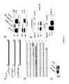
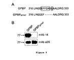
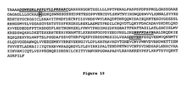
- Figure 19 shows the sequence of 91 kD GPBP, and in bold cursive underlined form, and from N to the C terminus, the amino acid residues comprising the epitopes of Ab 24, mAb 14 and mAb e26 respectively.
- the first residue (Met) of canonical 77-kDa GPBP (SEQ ID NO:4) is highlighted in bold and boxed in the figure.
- 91-kDa and 77-kDa GPBP are identical in amino acid sequence from the highlighted "Met” residue through the end of the protein.
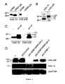
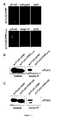
- the inventors have obtained compelling evidence that the mRNA of GPBP undergoes canonical (AUG) and noncanonical (ACG) translation initiation to generate two primary polypeptides of 77- and 91-kDa, respectively.
- the results from this study also support that both products enter the secretory pathway.
- the 91-kDa reaches the extracellular compartment and exists in a soluble immunoprecipitable form
- the 91-kDa remains mainly insoluble, associated with cellular membranes and likely reaches the external side of plasma membrane.
- the evidence supports that the 120-kDa GPBP isoform is a covalently-derived product of the 91-kDa GPBP (ie: the only differences are post-translational modifications) and thus shares the amino acid sequence of 91-kDa polypeptide.
- the term "91-kDa GPBP" includes the 91-kDa and post translational modifications thereof, including but not limited to 120-kDa GPBP and aggregates of 91-kDa and 120-kDa GPBP .
- the present application discloses additional evidence for the 91-kDa GPBP to exist in a soluble form in the plasma and urine revealing that the 91-kDa GPBP can be released from the cellular membranes.
- the polypeptides of this aspect of the application can be used, for example, to produce antibodies against 91-kDa GPBP, and as targets for identification of compounds that interfere with GPBP activity, making them useful therapeutics for various disorders, including Goodpasture Syndrome.
- mRNA alternative translation initiation is a strategy to direct GPBP to multiple locations including secretory pathway, plasma membrane and extracellular compartment.
- the polypeptides can be used, for example, to generate specific antibodies for detection of different isoforms of native GPBP present in serum or in urine, which can thus be used as, for example, diagnostic agents for autoimmune and other disorders.
- the polypeptides can also be used, for example, as tools to identify candidate compounds for inhibiting various specific types of native GPBP isoforms and also to identify candidate compounds for treating, for example, autoimmunity and protein misfolding-mediated disorders, as discussed in more detail below.
- fraction or greater purity means that contaminating proteins make up no more than 10% of the isolated polypeptide; in various preferred meanings, no more than 9%, 8%, 7%, 6%, 5%, 4%, 3%, 2%, 1 %, or 0.5% of the isolated polypeptide (e.g., isolated polypeptides of 91%, 92%, 93%, 94%, 95%, 96%, 97%, 98%, 99%, or 99.5% or greater purity consisting of the amino acid sequence of SEQ ID NO: 2).
- the isolated polypeptides may also substantially free of gel agents, such as polyacrylamide and agarose.
- the isolated polypeptides may be present in solution, frozen, or as a dried powder.
- the isolated polypeptides of this first aspect may be optionally labeled with a detectable, non-polypeptide label, including but not limited to fluorescent labels or radioactive labels.
- the present application discloses substantially purified recombinant polypeptides comprising or consisting of the general formula X-SEQ ID NO:2, wherein X is a detectable polypeptide.
- X is a detectable polypeptide.
- the correct amino acid sequence for 91 kD GPBP (SEQ ID NO:2) is expressed as a fusion protein with a detectable polypeptide.
- the polypeptides of this aspect of the application can be used, for example, to track 91 kD GPBP in cells, and as a detectable target for identification of compounds that interfere with GPBP activity, making them useful therapeutics for various disorders, including Goodpasture Syndrome.
- a "recombinant polypeptide” means that the detectable polypeptide is not derived from GPBP or expressed from a GPBP mRNA, and thus fuses a heterologous detectable peptide with the correct 91 kD GPBP polypeptide.
- a "detectable polypeptide” is any heterologous peptide that can be detected, thus permitting detection of the recombinant polypeptide.
- the detectable polypeptide may comprise a fluorescent protein. Any fluorescent protein known in the art can be used. For example, green fluorescent proteins of cnidarians, which act as their energy-transfer acceptors in bioluminescence, are suitable fluorescent proteins for use in the fluorescent indicators.
- GFP green fluorescent protein
- BFP blue fluorescent protein
- GFPs have been isolated from the Pacific Northwest jellyfish, Aequorea victoria, the sea pansy, Renilla reniformis, and Phialidium gregarium. See, Ward, W. W., et al., Photochem. Photobiol., 35:803 808 (1982 ); and Levine, L. D., et al., Comp. Biochem. Physiol., 72B:77 85 (1982 ).
- a variety of Aequorea-related GFPs having useful excitation and emission spectra have been engineered by modifying the amino acid sequence of a naturally occurring GFP from Aequorea victoria. See, Prasher, D. C., et al., Gene, 111:229 233 (1992 ); Heim, R., et al., Proc. Natl. Acad. Sci., USA, 91:12501 04 (1994 ); U.S. Ser. No. 08/337,915, filed Nov. 10, 1994 ; International application PCT/US95/14692, filed Nov. 10, 1995 ; and U.S. Ser. No. 08/706,408, filed Aug. 30, 1996 .
- the cDNA of GFP can be concatenated with those encoding many other proteins; the resulting fusions generally are fluorescent and retain the biochemical features of the partner proteins. See, Cubitt, A. B., et al., Trends Biochem. Sci. 20:448 455 (1995 ). Mutagenesis studies have produced GFP mutants with shifted wavelengths of excitation or emission. See, Heim, R. & Tsien, R. Y. Current Biol. 6:178 182 (1996 ). Suitable pairs, for example a blue-shifted GFP mutant P4-3 (Y66H/Y145F) and an improved green mutant S65T can respectively serve as a donor and an acceptor for fluorescence resonance energy transfer (FRET). See, Tsien, R. Y., et al., Trends Cell Biol. 3:242 245 (1993 ).
- the detectable polypeptide may comprise a non-GPBP epitope for which antibodies are commercially available, including but not limited to the FLAG (Sigma Chemical, St. Louis, MO), myc (9E10) (Invitrogen, Carlsbad, CA), 6-His (Invitrogen; Novagen, Madison, WI), glutathione S-transferase (GST) (Santa Cruz Biotechnology, Santa Cruz, California), and HA (hemaglutunin) (Boehringer Manheim Biochemicals).
- FLAG Sigma Chemical, St. Louis, MO
- myc (9E10) Invitrogen, Carlsbad, CA
- 6-His Invitrogen; Novagen, Madison, WI
- GST glutathione S-transferase
- HA hemaglutunin
- the isolated polypeptide may preferably further comprise a linker sequence between the detectable polypeptide and the polypeptide of SEQ ID NO:2.
- the linker is not a portion of GPBP or encoded by a GPBP mRNA.
- Such a linker can be of any desirable length, and preferably is between 1 and 20 amino acids, if present; more preferably between 1 and 15, 1-10, 1-5, 1-4, 1-3, or 1-2 amino acids, if present.
- the linker can be used, for example, to optimally position the detectable polypeptide and the 91 kD GPBP sequence and to include specific sequence for protease recognition site to allow removal of detectable polypeptide.
- the isolated polypeptide may further comprise any additional residues necessary for expression, such as an N-terminal methionine residue or peptide sequences to deliver the polypeptide to different cellular and extracellular compartments.
- substantially purified polypeptides of the application can be made by any method known to those of skill in the art, but are preferably made by recombinant means based on the teachings provided herein.
- a coding region of interest as disclosed herein can be cloned into a recombinant expression vector, which can then be used to transfect a host cell for recombinant protein production by the host cells.
- the present application discloses substantially purified nucleic acids encoding a polypeptide consisting of the amino acid sequence of SEQ ID NO:2 (91 kD GPBP).
- the substantially purified nucleic acid sequence may comprise RNA or DNA.
- substantially purified nucleic acids are those that have been removed from their normal surrounding nucleic acid sequences in the genome or in cDNA sequences.
- Such substantially purified nucleic acid sequences may comprise additional sequences useful for promoting expression and/or purification of the encoded protein, including but not limited to polyA sequences, modified Kozak sequences, and sequences encoding epitope tags, export signals, and secretory signals, nuclear localization signals, and plasma membrane localization signals.
- the substantially purified nucleic acid coding region may consist of the nucleic acid of SEQ ID NO:1, or a mRNA product thereof.
- the present application discloses substantially purified nucleic acids encoding the polypeptide of any embodiment of the substantially purified recombinant polypeptides comprising or consisting of the general formula X-SEQ ID NO:2, as discussed in the second aspect of the application.
- the present application discloses recombinant expression vectors comprising the substantially purified nucleic acid of any aspect of the application operatively linked to a promoter.
- "Recombinant expression vector” includes vectors that operatively link a nucleic acid coding region or gene to any promoter capable of effecting expression of the gene product.
- the promoter sequence used to drive expression of the disclosed nucleic acid sequences in a mammalian system may be constitutive (driven by any of a variety of promoters, including but not limited to, CMV, SV40, RSV, actin, EF) or inducible (driven by any of a number of inducible promoters including, but not limited to, tetracycline, ecdysone, steroid-responsive).
- the construction of expression vectors for use in transfecting prokaryotic cells is also well known in the art, and thus can be accomplished via standard techniques.
- the expression vector must be replicable in the host organisms either as an episome or by integration into host chromosomal DNA.
- the expression vector may comprise a plasmid. However, other expression vectors that serve equivalent functions, such as viral vectors, may be used.
- the present application discloses host cells that have been transfected with the recombinant expression vectors disclosed herein, wherein the host cells can be either prokaryotic or eukaryotic.
- the cells can be transiently or stably transfected.
- Such transfection of expression vectors into prokaryotic and eukaryotic cells can be accomplished via any technique known in the art, including but not limited to standard bacterial transformations, calcium phosphate co-precipitation, electroporation, or liposome mediated-, DEAE dextran mediated-, polycationic mediated-, or viral mediated transfection.
- the present application discloses a substantially purified polypeptide comprising the amino acid sequence of SEQ ID NO:2 (91 kD GPBP) or SEQ ID NO:4 (77 kD GPBP), wherein the polypeptide of SEQ ID NO:2 or SEQ ID NO:4 comprises one or more post-translational modifications (PTMs) directly and/or indirectly involving amino acids residues 305-344 GGPDYEEGPNSLINEEEFFDAVEAALDRQDKIEEQSQSEK (SEQ ID NO: 10) (numbering based on position within 77 kD GPBP).
- PTMs post-translational modifications
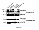
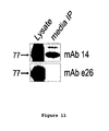
- the inventors provide the first purification of native 77 and 91 kD GPBP and have determined that existing monoclonal antibodies that bind to recombinant versions of 77 kD- and 91 kD-GPBP do not bind to purified native versions, verifying that structural differences exist between recombinant and native forms of the 77 kD GPBP and between recombinant and native forms of the 91 kD GPBP.
- the polypeptides of this aspect of the application can be used, for example, to produce antibodies against native GPBP forms, and as targets for identification of compounds that interfere with native GPBP activity, making them useful therapeutics for various disorders, including Goodpasture Syndrome.
- the one or more PTMs may comprise covalent PTMs.
- the one or more PTMs may comprise covalent PTMs within amino acids 305-344 (SEQ ID NO: 10).
- the one or more PTMs may directly or indirectly involve residues 320-327 (EEFFDAVE, SEQ ID NO:5).
- the one or more PTMs may comprise covalent PTMs within residues 320-327 (EEFFDAVE, SEQ ID NO:5) (numbering based on position within 77 kD GPBP).
- the one or more PTMs may comprise one more PTMs present in residue 320, 321, and/or 327; most preferably, the one or more PTMs present at these residues comprise covalent PTMs.
- the substantially purified polypeptide preferably possesse an amino acid sequence consisting of SEQ ID NO:2 (91 kD GPBP) or SEQ ID NO:4 (77 kD GPBP).
- post-translational modification means a modification in the structure of a protein after its translation.
- the PTM may comprise addition of a functional group, including but not limited to carboxylation, methylation, citrullination, phosphorylation, glycosylation, and formation of atypical isoaspartyl.
- the PTM may comprise an isomerization, leading to a conformational change.
- Any suitable method for making the covalently modified polypeptide of SEQ ID NO:2 or SEQ ID NO:4 based on the teachings of the present disclosure can be used, including isolating from natural sources of GPBP as disclosed herein, and recombinant production of GPBP followed by suitable covalent modification within the relevant region of amino acid residues, using standard methods known to those of skill in the art.
- the present application discloses substantially purified polypeptides comprising the amino acid sequence of SEQ ID NO:2 (91 kD GPBP) or SEQ ID NO:4 (77 kD GPBP), wherein the polypeptide of SEQ ID NO:2 or SEQ ID NO:4 comprises one or more PTMs directly and/or indirectly involving residues 371-396 PYSRSSSMSSIDLVSASDDVHRFSSQ (SEQ ID NO:9) (numbering based on positions within 77 kD GPBP).
- the inventors provide the first purification of native 77 kD and 91 kD GPBP and have determined that existing monoclonal antibodies that bind to recombinant version of 77 kD and 91 kD GPBP do not bind to the purified native 77 kD and 91 kD GPBP versions, verifying that structural differences exist between recombinant and native forms of the 77 and 91 kD GPBP.
- the polypeptides of this aspect of the application can be used, for example, to produce antibodies against native GPBP, and as targets for identification of compounds that interfere with native GPBP activity, making them useful therapeutics for various disorders, including Goodpasture Syndrome.
- the one or more PTMs may comprise covalent PTMs.
- the one or more PTMs may comprise covalent PTMs within amino acids 371-396 (SEQ ID NO:9).
- the one or more PTMs may directly or indirectly involve residues 388-392 (DDVHR, SEQ ID NO:6).
- the one or more PTMs may comprise one or more covalent PTMs within residues 388-392 (SEQ ID NO:6)
- the polypeptide may further comprise one or more PTMs directly or indirectly involving residues 320-327 (EEFFDAVE, SEQ ID NO:5)
- the one or more PTMs within residues 320-327 may be covalent PTMs.
- the substantially purified polypeptide may possess an amino acid sequence consisting of SEQ ID NO:2 (91 kD GPBP) or SEQ ID NO:4 (77 kD GPBP). Any suitable method for making the covalently modified polypeptide of SEQ ID NO:2 or SEQ ID NO:4 can be used, including isolating from natural sources of GPBP as disclosed herein, and recombinant production of GPBP followed by suitable covalent modification within the relevant region of amino acid residues, using standard methods known to those of skill in the art.
- the present application discloses substantially purified monoclonal antibodies that selectively bind to the substantially purified polypeptides of the sixth or seventh aspect of the application.
- the inventors have for the first time isolated native 77- and 91 kD GPBP species that when substantially purified do not bind to existing GPBP-specific monoclonal antibodies.
- existing monoclonal antibodies do not detect GPBP in plasma or urine samples in ELISAs, nor are they capable of use for purification of plasma or urine GPBP.
- the monoclonal antibodies of the application are useful, for example, in ELISA-based assays for GPBP detection in urine or plasma, and for purification of GPBP from plasma or serum.
- the inventors further demonstrate herein that these native 77 kD GPBP and native 91 kD GPBP species are pos-translationally modified, and that at least some of these PTMs render substantially purified, native GPBP non-reactive to existing monoclonal GPBP antibodies.
- Exemplary monoclonal antibodies according to this aspect of the application are provided in the examples that follow.
- the "monoclonal antibodies” of the application can be any type of monoclonal antibody, including but not limited to standard monoclonal antibodies, humanized monoclonals, chimeric monoclonals, and fragments thereof.
- substantially purified means that the recited monoclonal antibodies make up at least 80% of the antibodies in a substantially purified sample; more preferable at least 85%, 90%, 91%, 92%, 93%, 94%, 95%, 96%, 97%, 98%, 99%, or more.
- selective bind means preferential binding of the GPBP monoclonal antibody to native GPBP epitope, as opposed to one or more other biological molecules, structures, cells, tissues, etc., as is well understood by those of skill in the art.
- Monoclonal antibodies can be produced by obtaining spleen cells from the animal [See Kohler and Milstein, Nature 256, 495-497 (1975 )].
- monoclonal antibodies (mAb) of interest are prepared by immunizing inbred mice with native 77 kD GPBP, native 91 kD GBPB, or an antigenic fragment thereof, including, but not limited to, one or more epitopes comprising or consisting of the PTM-containing peptides EEFFDAVE (SEQ ID NO:5), DDVHR (SEQ ID NO:6), LINEEEFFDAVEAALDRQ (SEQ ID NO:8), PYSRSSSMSSIDLVSASDDVHRFSSQ (SEQ ID NO:9), and GGPDYEEGPNSLINEEEFFDAVEAALDRQDKIEEQSQSEK (SEQ ID NO: 10).
- the monoclonal antibodies may bind one or more epitopes comprising one or more PTMs, selected from the group consisting of PTM-containing EEFFDAVE (SEQ ID NO:5), DDVHR (SEQ ID NO:6), LINEEEFFDAVEAALDRQ (SEQ ID NO:8), PYSRSSSMSSIDLVSASDDVHRFSSQ (SEQ ID NO:9), and GGPDYEEGPNSLINEEEFFDAVEAALDRQDKIEEQSQSEK (SEQ ID NO: 10).
- the one or more PTMs may be covalent PTMs.
- the monoclonal antibodies may bind to an epitope that comprises one or more PTMs (preferably covalent PTMs) present in residue 320, 321, and/or 327 (numbering based on 77 kD GPBP).
- the mice may be immunized by the IP or SC route in an amount and at intervals sufficient to elicit an immune response.
- the mice receive an initial immunization on day 0 and are rested for about 3 to about 30 weeks.
- Immunized mice are given one or more booster immunizations of by the intravenous (IV) route.
- Lymphocytes from antibody positive mice are obtained by removing spleens from immunized mice by standard procedures known in the art.
- Hybridoma cells are produced by mixing the splenic lymphocytes with an appropriate fusion partner under conditions which will allow the formation of stable hybridomas.
- the antibody producing cells and fusion partner cells are fused in polyethylene glycol at concentrations from about 30% to about 50%.
- Fused hybridoma cells are selected by growth in hypoxanthine, thymidine and aminopterin supplemented Dulbecco's Modified Eagles Medium (DMEM) by procedures known in the art. Supernatant fluids are collected from growth positive wells and are screened for antibody production by an immunoassay such as solid phase immunoradioassay. Hybridoma cells from antibody positive wells are cloned by a technique such as the soft agar technique of MacPherson, Soft Agar Techniques, in Tissue Culture Methods and Applications, Kruse and Paterson, Eds., Academic Press, 1973 .
- DMEM Dulbecco's Modified Eagles Medium
- Humanized monoclonal antibodies refers to monoclonal antibodies derived from a non-human monoclonal antibody, such as a mouse monoclonal antibody.
- humanized monoclonal antibodies can be derived from chimeric antibodies that retains, or substantially retains, the antigen-binding properties of the parental, non-human, monoclonal antibodies but which exhibits diminished immunogenicity as compared to the parental monoclonal antibody when administered to humans.
- chimeric monoclonal antibodies can comprise human and murine antibody fragments, generally human constant and mouse variable regions.
- Humanized monoclonal antibodies can be prepared using a variety of methods known in the art, including but not limited to (1) grafting complementarity determining regions from a non-human monoclonal antibody onto a human framework and constant region ("humanizing"), and (2) transplanting the non-human monoclonal antibody variable domains, but "cloaking" them with a human-like surface by replacement of surface residues ("veneering").
- humanizing grafting complementarity determining regions from a non-human monoclonal antibody onto a human framework and constant region
- transplanting the non-human monoclonal antibody variable domains but "cloaking" them with a human-like surface by replacement of surface residues ("veneering").
- These methods are disclosed, for example, in, e.g., Jones et al., Nature 321:522-525 (1986 ); Morrison et al., Proc. Natl. Acad. Sci., U.S.A., 81:6851-6855 (1984
- Monoclonal antibodies can be fragmented using conventional techniques, and the fragments screened for utility in the same manner as for whole antibodies.
- F(ab') 2 fragments can be generated by treating antibody with pepsin.
- the resulting F(ab') 2 fragment can be treated to reduce disulfide bridges to produce Fab' fragments.
- Fab fragments can be obtained by treating an IgG antibody with papain;
- F(ab') fragments can be obtained with pepsin digestion of IgG antibody.
- a F(ab') fragment also can be produced by binding Fab' described below via a thioether bond or a disulfide bond.
- a Fab' fragment is an antibody fragment obtained by cutting a disulfide bond of the hinge region of the F(ab')2.
- a Fab' fragment can be obtained by treating a F(ab') 2 fragment with a reducing agent, such as dithiothreitol.
- Antibody fragment peptides can also be generated by expression of nucleic acids encoding such peptides in recombinant cells (see, e.g., Evans et al., J. Immunol. Meth. 184: 123-38 (1995 )).
- a chimeric gene encoding a portion of a F(ab')2 fragment can include DNA sequences encoding the CH1 domain and hinge region of the H chain, followed by a translational stop codon to yield such a truncated antibody fragment molecule.
- monoclonal antibody fragments include (i) a Fab fragment, a monovalent fragment consisting essentially of the VL, VH, CL and CH I domains; (ii) F(ab)2 and F(ab')2 fragments, bivalent fragments comprising two Fab fragments linked by a disulfide bridge at the hinge region; (iii) a Fd fragment consisting essentially of the VH and CH1 domains; (iv) a Fv fragment consisting essentially of the VL and VH domains of a single arm of an antibody, (v) a dAb fragment ( Ward et al., (1989) Nature 341:544-546 ), which consists essentially of a VH domain; and (vi) one or more isolated CDRs or a functional paratope.
- a Fab fragment a monovalent fragment consisting essentially of the VL, VH, CL and CH I domains
- F(ab)2 and F(ab')2 fragments bivalent fragments
- the immunogens are typically formulated with a pharmaceutically acceptable carrier for parenteral administration.
- acceptable adjuvants include, but are not limited to, Freund's complete, Freund's incomplete, alum-precipitate, water in oil emulsion containing Corynebacterium parvum and tRNA.
- the formulation of such compositions, including the concentration of the polypeptide and the selection of the vehicle and other components, is within the skill of the art.
- the present application discloses substantially purified monoclonal antibodies that specifically binds to the polypeptide of SEQ ID NO:2 and not to the polypeptide of SEQ ID NO:4.
- Such monoclonal antibodies of the application are useful, for example, in distinguishing 91 kD GPBP from 77 kD GPBP in assays including, but not limited to, ELISA-based assays for GPBP detection in urine or plasma.
- Such monoclonal antibodies can be generated using methods disclosed above and the use of peptide immunogens present in the polypeptide of SEQ ID NO:2 but not present in SEQ ID NO:4.
- Such immunogens may be of any suitable length to generate an antibody response.
- the monoclonal antibodies may be generated against an immunogen comprising or consisting of DGWKGRLPSPLVLLPRSARC (SEQ ID NO:7).
- the monoclonal antibody binds to an epitope within the amino acid sequence DGWKGRLPSPLVLLPRSARC (SEQ ID NO:7).
- An exemplary such antibody, Ab24, is disclosed below.
- the present application discloses isolated hybridoma cells expressing the monoclonal antibodies of the eighth or ninth aspects of the application.
- the application also discloses methods for making the antibodies of the application, as disclosed above and below.
- the present application discloses methods for detecting circulating Goodpasture antigen-binding protein (GPBP), comprising
- a “plasma sample” means blood plasma, the liquid component of blood, and is prepared, for example, by centrifugation of whole blood to remove blood cells.
- a plasma sample also includes a blood serum sample, in which blood clotting factors have been removed.
- the present application discloses methods for detecting urinary Goodpasture antigen-binding protein (GPBP), comprising
- Urine samples are easily obtained, and analyte determination in urine is well known in the art.
- a "GPBP-binding molecule” is a peptide or nucleic acid molecule that binds selectively to GPBP, as opposed to one or more other biological molecules, structures, cells, tissues, etc. Exemplary embodiments of such GPBP-binding molecules include but are not limited to antibodies, aptamers or substrates.
- a "GPBP substrate” is a target of GPBP biological activity that binds to GPBP, or a fragment thereof that retains GPBP-binding activity.
- Such GPBP substrates include, but are not limited to, I-20 (SEQ ID NO:16), GPBP-interacting proteins (GIPs) (SEQ ID NOS:17-21), myelin basic protein (MBP) and derivatives thereof (SEQ ID NOS:22-25), prion protein (PrP) (SEQ ID NO:26), type IV collagen ⁇ 3 chain NC1 domain ( ⁇ 3(IV)NC1) (SEQ ID NO:27), and Alzheimer's disease beta peptide (A ⁇ 1-42 ) (SEQ ID NO:28).
- GIPs GPBP-interacting proteins
- MBP myelin basic protein
- PrP prion protein
- SEQ ID NO:26 type IV collagen ⁇ 3 chain NC1 domain
- ⁇ 3(IV)NC1 type IV collagen ⁇ 3 chain NC1 domain
- a ⁇ 1-42 Alzheimer's disease beta peptide
- GPBP refers to all GPBP isoforms reactive with GPBP-selective antibodies, including but not limited to 77 kD GPBP and 91 kD GPBP, as well lower and higher molecular weight GPBP isoforms of 160-, 60-, 58-, 56- 53- 50-46- 35 and 34-kD, and aggregates thereof.
- the "plasma sample” or “urine sample” may be obtained from any suitable subject, preferably from a mammal, including but not limited to a human, dog, cat, horse, or livestock (cow, sheep, etc.).
- the plasma sample or urine sample may be obtained from a human subject, such as a human subject suspected of having an autoimmune condition including but not limited to Goodpasture Syndrome and/or immune-complex mediated glomerulonephritis.
- the inventors have isolated native circulating 77 kD GPBP from human plasma and have observed increased levels in Goodpasture patients and in animal models for immune complex-mediated glomerulonephritis, demonstrating that GPBP secretion occurs in vivo and revealing the clinical utility of serological and urinary determination of GPBP.
- the antibody can be any selective GPBP antibody, whether polyclonal, monoclonal, or humanized monoclonal as described above, although monoclonal antibodies are preferred.
- Antibodies according to the eighth or ninth aspects of the application may be used.
- the methods of the tenth and eleventh aspect of the application may comprise analyzing a specific GPBP isoform, such as 77 kD GPBP or 91 kD GPBP; in these cases, antibodies selective for 77 kD GPBP or selective for 91 kD GPBP can be used, including but not limited to those selective antibodies disclosed herein.
- the antibodies for use in the methods of the tenth and eleventh aspects of the application are preferably those that bind to native GPBP isoforms, such as those disclosed herein.
- GPBP-binding molecules such as antibodies, aptamers or substrates
- Conditions suitable to promote binding of GPBP-binding molecules, such as antibodies, aptamers or substrates, to GPBP in the plasma or urine samples can be determined by those of skill in the art based on the teachings herein and the examples provided below.
- antibody-antigen binding often depends on hydrophobic interactions (the so called hydrophobic bonds); thus, high salt concentrations, such as in the molar range can be used to reduce nonspecific binding and increase specific antigen-antibody binding.
- further steps may be included to promote selectivity and specificity, including but not limited to one or more wash steps to remove unbound or weakly bound serum proteins; inhibitors of non-specific binding to reduce binding of high concentration serum proteins, control samples known to contain GPBP isoforms and/or negative controls known not to bind to GPBP isoforms, and/or inclusion of serum or urine samples known to not possess GPBP (ex: deleted for GPBP).
- tenth and eleventh aspects of the present application may be used to test for the presence of GPBP in the plasma or urine sample by standard techniques including, but not limited to ELISA, immunoflourescence, and chromatography (for example, lateral flow assays where the antibody is immobilized on a surface and plasma or urinary proteins are labeled and allowed to flow over the surface under conditions suitable to permit binding of the antibody to GPBP in the plasma or urine).
- Functional beads Becton Dickinson technology coupled to flow cytometry may be used; this technique is an emerging method to measure the levels of proteins in biological fluid or cell/tissue extracts.
- beads made of a fluorescence matrix are coated with one or more specific GPBP antibodies, mixed with the plasma sample and further incubated with a detecting antibody labeled with a phycoerythrins. Finally, beads are analyzed by a flow cytometry program which selects the beads according matrix fluorescence emission and measurement of the level of the analyte through phycoerythrin emission. There are up to thirty different types of beads that can be simultaneously detected and discriminated by the cytometer. This method couples high sensitivity and performance with versatility since a specific bead type coated with GPBP antibody can be mixed with a distinct bead type coated with binding peptides for other analyte (i.e. autoantibodies) and simultaneously measured.
- analyte i.e. autoantibodies
- the measurement of various analytes could enhance the potential of GPBP determination.
- the techniques may determine only the presence or absence of the GPBP isoform(s).
- the techniques may be quantitative, and provide information about the relative amount of the protein or peptide of interest in the sample. For quantitative purposes, ELISAs are preferred.
- Detection of immunocomplex formation can be accomplished by standard detection techniques. For example, detection of immunocomplexes can be accomplished by using labeled antibodies or secondary antibodies. Such methods, including the choice of label are known to those ordinarily skilled in the art. (Harlow and Lane, Supra).
- the antibodies can be coupled to a detectable substance.
- the term "coupled” is used to mean that the detectable substance is physically linked to the antibody.
- Suitable detectable substances include various enzymes, prosthetic groups, fluorescent materials, luminescent materials and radioactive materials. Examples of suitable enzymes include horseradish peroxidase, alkaline phosphatase, ⁇ -galactosidase, or acetylcholinesterase.
- prosthetic-group complexes include streptavidin/biotin and avidin/biotin.
- suitable fluorescent materials include umbelliferone, fluorescein, fluorescein isothiocyanate, rhodamine, dichlorotriazinylamine fluorescein, dansyl chloride or phycoerythrin.
- An example of a luminescent material includes luminol.
- suitable radioactive material include 125 I, 131 I, 35 S or 3 H.
- the methods of this aspect of the application can be used, for example, to detect GPBP-mediated disorder in a subject, including but not limited to an antibody-mediated disorder (including but not limited to a glomerulonephritis selected from the group consisting of IgA nephropathy, systemic lupus erythematosus and Goodpasture disease), inflammation, an ER-stress mediated disorder, and drug-resistant cancer.
- an antibody-mediated disorder including but not limited to a glomerulonephritis selected from the group consisting of IgA nephropathy, systemic lupus erythematosus and Goodpasture disease
- inflammation an ER-stress mediated disorder
- drug-resistant cancer drug-resistant cancer
- the methods would comprise comparison of GPBP levels detected in a test serum or urine sample with a control, such as a control from a serum or urine sample known to have "normal" levels of GPBP or previously determined normal values for GPBP in sera or urine from the subject from whom the serum is obtained.
- the control may provide a standard curve using recombinant GPBP or a reference value.
- an increase preferably a statistically significant increase using standard statistical analysis techniques
- GPBP in the serum or urine sample relative to the control indicates the presence of one or more of the disorders noted above, or an increased risk of developing one or more of the disorders, all of which are correlated with increased GPBP expression.
- the methods identify individuals either having or at risk of being stricken with one or more of an antibody-mediated disorder (including but not limited to a glomerulonephritis selected from the group consisting of IgA nephropathy, systemic lupus erythematosus and Goodpasture disease), inflammation, an ER-stress mediated disorder, and drug-resistant cancer.
- an antibody-mediated disorder including but not limited to a glomerulonephritis selected from the group consisting of IgA nephropathy, systemic lupus erythematosus and Goodpasture disease
- inflammation inflammation
- an ER-stress mediated disorder an ER-stress mediated disorder
- drug-resistant cancer drug-resistant cancer.
- Such patients may undergo periodic testing for serum or urine levels of GPBP to monitor potential risk of developing a drug-resistant tumor.
- patients thought to be at risk for developing, or suspected of already having developed a glomerulonephritis selected from the group consisting of IgA nephropathy, systemic lupus erythematosus and Goodpasture disease can be tested for serum or urine levels of GPBP.
- GPBP is a circulating molecule and GBM (glomerular basement membrane) a principal component of the glomerular filtration barrier; therefore, GPBP accumulation in the glomerulus could result from local production but also from the sequestration of circulating GPBP produced elsewhere, and could also be reflected in increased GPBP in the urine.
- the local overproduction could account for primary antibody-mediated glomerulonephritis whereas increased circulating levels may induce secondary forms of this pathology and perhaps are responsible for disease recurrence upon renal transplantation. Consequently, quantification of the levels of circulating or urinary GPBP is useful in discriminating primary from secondary antibody-mediated glomerulonephritis and for the clinical monitoring of renal transplantation.
- the methods permit one to perform differential diagnosis or prognosis in the above disorders.
- IgA nephropathy patients produce anti-basement membrane autoantibodies. These circulating autoantibodies recognize the NC1 domain of type IV collagen. Determination of the titer of these antibodies could help to monitor disease progression or also to distinguish different IgA nephropathy patients or to perform prognosis in these patients.
- anti-ssDNA, anti-nucleosome autoantibodies and GPBP levels one can diagnose systemic lupus erythematosus but also distinguish between primary IgA nephropathy and IgA nephropathy secondary to systemic lupus erythematosus. Any determination used to diagnosis of primary diseases listed in Donadio and Grande (2002) N Engl J Med 347, 738-748 associated with glomerular deposition of IgA , can be used in conjunction with the methods of the invention for plasma or urinary detection of GPBP for differential diagnosis in secondary IgA nephropathy patients.
- a normal value of GPBP as a reference for an standard curve is between ⁇ 1 ng/ml-10 ng/ml in plasma and approximately 0.2 ng/ml to 1.5 ng/ml in urine, while Goodpasture patients exceed the normal by at least 2-fold; or by at least 3-fold, 4-fold, 5-fold, 6-fold, 7-fold, 8-fold, or more the normal values.
- the present application discloses methods for isolating native GPBP isoforms, comprising:
- the present application discloses methods for isolating native GPBP isoforms, comprising:
- double ion-exchange chromatography means carrying out two successive and distinct ion-exchange chromatography steps prior to step (c).
- IEC techniques are well known in the art, and include those disclosed in the examples that follow.
- GPBP from urine as disclosed in more detail in the examples that follow.
- the present application discloses methods for isolating native GPBP isoforms, comprising:
- the GPBP-binding molecule may comprise GPBP antibodies.
- the antibodies may comprise the novel monoclonal antibodies of the present application.
- the eluting step may comprise use of a denaturing eluting buffer.
- GPBP Goodpasture-antigen binding protein
- GPBP is a nonconventional Ser/Thr kinase for the type IV collagen of basement membrane. More recently, we have shown that GPBP is an extracellular protein that when overexpressed induces type IV collagen disorganization and deposit of immune complexes in glomerular basement membrane (Ref. 4). Here we show that cells expressed at least two GPBP isoforms resulting from canonical (77-kDa) and noncanonical (91-kDa) mRNA translation initiation. The 77-kDa polypeptide interacted with type IV collagen and localized as a soluble form in the extracellular compartment.
- the FFAT motif and the 26-residue Ser-rich region were required for the exportation of the 77-kDa polypeptide.
- removal of the 26-residue Ser-rich region yielded the previously recognized GPBP isoform (GPBP ⁇ 26/CERT) that was cytosolic and in contrast to GPBP, sensitive to sphingomyelinase cell treatment.
- type IV collagen which includes: 1) phosphorylation and regulation of protein molecular/supramolecular organization (GPBP); and 2) inter-organelle ceramide trafficking and regulation of protein cargo transport to the plasma membrane (GPBP ⁇ 26/CERT).
- GPBP protein molecular/supramolecular organization
- GPBP ⁇ 26/CERT inter-organelle ceramide trafficking and regulation of protein cargo transport to the plasma membrane
- GBM glomerular basement membrane
- COL4A3BP also encodes for GPBP ⁇ 26, a more-abundant less-active alternatively spliced GPBP variant lacking a 26-residue Ser-rich region, which is apparently not regulated under these pathological conditions (3).
- GPBP contains multiple structural elements including N terminal pleckstrin homology (PH) domain, Ser-Xaa-Yaa region, bipartite nuclear localization signal, coiled-coil domain, two phenylalalines in an acidic track (FFAT) motif and C terminal steroidogenic acute regulatory related lipid transfer (START) domain. Additional structural features include motifs for self-interaction and phosphorylation (1, 3, 5, 6).
- the PH domains comprise a variety of poorly conserved structures present only in eukaryotes which have been proposed to mediate protein targeting to cellular membranes through interaction with phosphoinositides (7).
- a variety of proteins including several protein kinases contain PH domains (8).
- the FFAT motifs target proteins to the ER through interaction with the transmembrane cytosolic domain of the vesicle associated membrane protein-associated proteins (VAPs) (9), which have been proposed to play a role in maintaining homeostasis for protein folding in the endoplasmic reticulum (ER) and in regulating protein cargo transport to the plasma membrane (10, 11).
- VAPs vesicle associated membrane protein-associated proteins
- the START domains bind lipids including ceramide, phospholipids and sterols, and are modules present in a variety of proteins with distinct physiological and pathological functions (12, 13).
- Human monoclonal F(ab) 2 fragments were isolated from a recombinant F(ab) 2 expression library using a synthetic peptide representing the a lternatively t ranslated r egion (ATR) of GPBP ( Fig. 2C ) (Antibodies by Design, MorphoSys AG). Reactive F(ab) 2 fragments were further characterized using Western blot and recombinant proteins expressing the predicted ATR (not shown). The most reactive F(ab) 2 fragment (Ab 24) was used to characterize native GPBP polypeptides and the least reactive F(ab) 2 fragment (Ab 20) was used as negative control in these studies.
- GPBP-specific antibodies were biotinylated for use in flow cytometry or labeled with Alexa Fluor 647 (Invitrogen) for direct immunofluorescence.
- Polyclonal antibodies specific for GPBP and GPBP ⁇ 26/CERT were produced either in rabbits immunized with GST-FLAG-GPBP (1) following standard procedures ( ⁇ GPBPr) or in chickens immunized with a specific synthetic peptide and purchased from Abcam ( ⁇ GPBPab).
- Specific antibodies in ⁇ GPBPr were affinity-purified using recombinant FLAG-GPBP (see below) bound to Sepharose-CNBr (Sigma).
- glyceraldehyde-3-phosphate dehydrogenase detection we used a mouse monoclonal antibody provided by Erwin Knecht.
- Polyclonal antibodies specific for calregulin, p65 or cathepsin D were from Santa Cruz Biotechnology Inc and those specific for pyruvate dehydrogenase (PDH) were from Molecular Probes.
- Monoclonal antibodies specific for PrP (clone 3F4) or for golgin-97 were from Clontech and Molecular Probes, respectively.
- FLAG/M2 or FLAG/M2-horseradish peroxidase (HRP) Sigma
- HRP horseradish peroxidase
- Alexa Fluor® 488-streptavidin was from Molecular Probes and secondary antibodies were from Promega (anti-mouse and anti-rabbit HRP conjugates), Jackson Immunoresearch (anti-human F(ab) 2 -HRP) and Sigma (anti-chicken HRP and other FITC and TRITC conjugates).
- Recombinant FLAG-GPBP and FLAG-GPBP ⁇ 26 were expressed in Pichia pastoris and affinity-purified as previously described (1, 3).
- Plasmid constructs The production of pc-n4', a pcDNA3 (Invitrogen)-derived construct expressing a cDNA which contained the 5' untranslated region (UTR) and coding sequence of GPBP mRNA has been reported (1). Plasmids derived from pc-n4' included pc-GPBP-Met, a deletion mutant devoid of 5'UTR, and pc-n4'-Mmut, a construct where the canonical AUG (Met) translation initiation was substituted with GGA (Gly).
- pc-FLAG-GPBP which expresses the FLAG sequence fused to the coding region of GPBP, was previously reported (1) and used to obtain pc-FLAG-GPBP ⁇ FFAT , , bearing a deletion in the FFAT motif ( Fig. 9 ).
- the pc-FLAG-GPBP ⁇ 26 expresses the FLAG sequence fused to the coding region of GPBP ⁇ 26 and has been produced similarly to pc-FLAG-GPBP.
- pc-n4'and pc-n4'-Mmut mutants by introducing stop codons at various positions in the open reading frame (ORF) upstream of iMet position.
- the p Silencer TM 2.1-U6 hygro (Ambion) was employed for transient expression of small interfering mRNAs (siRNAs) specific for GPBP or for GPBP/GPBP ⁇ 26.
- the corresponding derived constructs and cDNA target sequences were: pSi-GPBP/GPBP ⁇ 26-2, ACAGAGTATGGCTGCAGAG (SEQ ID NO: 11); pSi-GPBP/GPBP ⁇ 26-3, GTACTTTGATGCCTGTGCT (SEQ ID NO: 12); pSi-GPBP-1, GCCCTATAGTCGCTCTTCC (SEQ ID NO: 13). Selection of the target sequence and plasmid construction were based on manufacturer's recommendations.
- siRNA expressing-plasmids were assessed in a cell recombinant expression system (not shown).
- the control plasmid in these studies (pSi-Control) was designed for targeting the mRNA of green fluorescence protein, a protein not expressed in human cells. All mutants were produced by standard PCR-based mutagenesis and the fidelity of all the cDNAs cloned was confirmed by nucleotide sequencing.
- Cell culture and transfection -HEK-293 or HeLa cells were grown with Dulbecco's modified Eagle's medium or Minimal Essential Medium Eagle respectively, supplemented with 2 mM L-glutamine, 10% (v/v) fetal bovine serum and penicillin (100 U/ml)/streptomycin sulfate (0.1 mg/ml), at 37 °C in a humidified 5% CO 2 environment. Unless otherwise indicated the cells used in the studies were HEK 293 cells.
- Transfections were performed for 16-24 h using ProFection Mammalian Transfection System-Calcium Phosphate (Promega) or Lipofectamine 2000 (Invitrogen), following manufacturer's recommendations.
- ProFection Mammalian Transfection System-Calcium Phosphate Promega
- Lipofectamine 2000 Invitrogen
- cells were seeded on poly-L-lysine-coated cover slips in 24-well plates.
- HEK 293 cells were transfected with pc-n4'-Mmut and selected with G418 (Invitrogen) for 15 days. Resistant cells were further cloned by limiting dilution and the expression of 91-kDa GPBP in a number of individual clones was determined by Western blot analysis of cell extracts (see below). Clones expressing elevated (c8, c14) or reduced (c19) levels of 91-kDa were used in functional studies.
- microsomal fraction which contains fragments of cellular membranes i.e. endoplasmic reticulum, plasma membrane and secretory vesicles (pellet) and the cytosolic fraction (supernatant). All steps were performed at 0-4 °C and protein concentrations determined using Protein Assay reagent (Bio-Rad).
- the supernatant of 500 x g was loaded on a resource-Q FPLC column, and the bound material eluted in 0 to 1 M NaCl gradient in 10 mM Tris-HCl pH 8.0.
- the 0.55-0.6 M NaCl fractions containing the bulk of cellular GPBP were precipitated with ethanol and used as partially purified GPBP for Western blot analysis.
- cells were brought to RT by rinsing with PBS and incubated for 10 min with culture medium containing 1% formaldehyde.
- the crosslink reaction was quenched with 125 mM Gly-HCl in PBS (pH 7.4) for 10 min at RT.
- Cells were brought to 4 °C by rinsing with ice-chilled PBS and procedure continued at 4 °C.
- the beads were collected by centrifugation and washed twice with 1 ml of extraction buffer and once with Tris-buffered saline (TBS, 50mM Tris-HCl pH 7.5, 150 mM NaCl). Proteins were eluted twice with 25 or 125 ⁇ l (six-well or 150-mm plate) of a 100 ⁇ g/ml solution of FLAG peptide in TBS at RT. Eluted samples were boiled with electrophoresis sample buffer (2X) for 15 min to reverse cross-linking and further analyzed by SDS-PAGE and either Coomassie blue staining or Western blot.
- TBS Tris-buffered saline
- HeLa cells transfected with pc-FLAG-GPBP or pc-FLAG-GPBP ⁇ 26 were treated or not with Bacillus cereus sphingomyelinase (Sigma) as previously described (5) and cells were either fixed with methanol/acetone and analysed by direct immunofluorescence (see below) or lysed in 10 mM Tris-HCl pH 7.5, 150 mM NaCl, 0.5% Triton X-100, 1 mM EDTA, 50 mM NaF, 1 mM sodium orthovanadate, 10 ⁇ g/ml leupeptin, 1 mM PMSF, cleared by centrifugation (500 x g for 10 min) and used for FLAG-immunoprecipitation (see above).
- the immunopurified materials from untreated cells were divided and one-half was treated with 5 U/ ⁇ l of ⁇ PPase (New England Biolabs) at 30 °C for 30 min following manufacturer's recommendations. All the samples were further analysed by Western blot using anti-FLAG antibodies.
- cells were grown in 150-mm plates, transfected with 20 ⁇ g of plasmid constructs encoding FLAG-tagged proteins and cultured for two additional days in fresh media. Twenty milliliters of media were used for FLAG-immunoprecipitation essentially as above indicated.
- Flow cytometry -Cells were gently detached and dispersed in culture media. Non-specific antibody binding sites on cell surface were blocked with mouse ascites fluid containing non-relevant mAb (blocking solution). Cells were subsequently incubated in blocking solution in the presence or absence of biotinylated ⁇ GPBP with or without blocking peptide (GPBPpep1) or a non-relevant synthetic peptide. Cells were incubated with Alexa Fluor® 488-streptavidin in blocking solution and further subjected to analysis in a Cytomics FC500 flow cytometer (Beckman Coulter) to measure fluorescence emission. Cell integrity was assessed measuring forward and side scattering, using untreated fresh cells as reference. All incubations were at RT for 1 h.
- Direct and indirect immunofluorescence with fixed cell -Cells were transfected and fixed with methanol-acetone (1/1) chilled at -20 °C for 10 min. Subsequently, cells were incubated with blocking solution (rabbit serum diluted 1:2 in PBS) for 30 min at RT, incubated with the primary antibodies (20 ⁇ g/ml in blocking solution) for 2 h at 37 °C in a humidified chamber, followed by incubation with the secondary antibody (1:200 in blocking solution) for 1 h at RT.
- blocking solution rabbit serum diluted 1:2 in PBS
- Direct immunofluorescence of living cells -Cells were cultured on glass-bottom microwell dishes (MatTek Corp) and when they reached ⁇ 50% confluence, the media were discarded and replaced by fresh media containing 10 ⁇ g/ml ⁇ GPBP-Alexa Fluor 647 with an excess of GPBPpep1 or equimolecular amounts of an unrelated synthetic peptide along with Rhodamine 123 (Invitrogen) for mitochondrial staining of living cells.
- Live cell analysis of fluorescence was performed with a Leica TCS SP2 inverted confocal microscope. Cells were maintained at 37 °C in a humidified 5% CO 2 environment in all the steps.
- Mass spectrometry -Individual protein bands were excised from Coomassie blue-stained gel, distained, in-gel trypsin digested, and centrifuged. One microliter of the supernatant was dried and resuspended with 1 ⁇ l of matrix solution (a-Cyano-4-hydroxycinnamic acid, from Sigma), applied to the sample plate, dried and introduced into the mass spectrometer. Tryptic digests peptides were analyzed by MALDI/TOF/TOF mass spectrometry (4700 Proteomics Analyzer, Applied Biosystems). Collected data were analyzed with GPS software (Applied Biosystems) and protein identification was carried out using the search engine MASCOT v 2.0 (Matrix Science).
- COL4A3BP encodes for polypeptides of 77-, 91- and 120-kDa-
- mAb 14 previously reported to recognize GPBP and GPBP ⁇ 26 (1)
- mAb e26 a novel monoclonal antibody raised against the 26-residue Ser-rich region exclusive for GPBP ( Fig 1A ).
- GPBP deletion mutants and synthetic peptides we have mapped mAb 14 epitope to the FFAT motif and thus, this antibody did not react with a GPBP mutant lacking the FFAT motif (GPBP ⁇ FFAT ) ( Fig. 9 ).
- pc-n4' a construct bearing the 5'UTR and coding sequence of COL4A3BP (1, 17), was used in transient gene expression assays in cultured cells.
- the expression of pc-n4' yielded three polypeptides of ⁇ 77-, 91- and 120-kDa which were detected by mAb e26.
- only the -77-and 91-kDa polypeptides were significantly reactive with mAb 14.
- siRNAs small interfering RNAs specific for COL4A3BP
- Fig 1D small interfering RNAs
- siRNAs specific for both GPBP and GPBP ⁇ 26/CERT were more efficient at reducing the expression of 77-kDa polypeptide
- GPBP-specific siRNA reduced more effectively the expression of 91- and 120-kDa polypeptides (compare pSi-GPBP/GPBP ⁇ 26-3 and psiGPBP-1).
- GPBP-n4' Major cellular GPBP isoforms result from noncanonical mRNA translation initiation -To further define the origin of cellular GPBP isoforms, we produced (pc-n4')-derived constructs expressing mRNA mutants consisting of 5'UTR-deletion or iMet to Gly substitution ( Fig. 2A ) and these were used in protein expression assays ( Fig. 2B ). In cells, the construct representing 5'UTR-deleted mRNA (pc-GPBP-Met) produced only the 77-kDa polypeptide and the constructs representing the iMet to Gly substitution (pc-n4'-Mmut) expressed only the 91- and 120-kDa polypeptides ( Fig.
- a human F(ab) 2 fragment specifically reacting with a synthetic peptide representing the predicted ATR (shaded sequence in Fig. 2C ) was used for Western blot analysis of partially purified GPBP polypeptides ( Fig. 2E ).
- Ab 24 specifically reacted with two polypeptides of 91- and 120-kDa which were also recognized by mAb e26, suggesting that native GPBP polypeptides contained the ATR characteristic of noncanonical translation products.
- the 91- and 120-kDa GPBP isoforms are insoluble membrane-bound polypeptides- GPBP isoform of 91-kDa was predicted to be non-classical secreted proteins when analyzed with SecretomeP 2.0 Server (18, http://www.cbs.dtu.dk/services/SecretomeP/) and to localize in mitochondria (60.9 %), nucleus (26.1 %), cytoskeleton (8.7 %) and vesicles of secretory system (4.3 %) when analyzed with PSORT II Prediction (http://psort.ims.u-tokyo.ac.jp/form2.html).
- these theoretical considerations suggested that GPBP isoforms resulting from noncanonical translation initiation were noncytosolic polypeptides that entered into cellular organelles including the secretory pathway.
- ⁇ GPBP bound to living cells in a specific manner since binding of the antibodies was efficiently abolished by a synthetic peptide representing GPBP (GPBPpep1) but not by an unrelated polypeptide (Contpep).
- GPBPpep1 a synthetic peptide representing GPBP
- Contpep an unrelated polypeptide
- GPBP GPBP isoforms of 91- and 120-kDa were not detected as soluble materials but rather they were found mainly associated with mitochondrial-lysosomal and microsomal fractions. It remained to be determined whether the presence of GPBP in the nuclear fraction indeed reflected nuclear expression of these proteins or rather unbroken cells and/or mitochondria contaminating this fraction.
- the 77-kDa GPBP is a soluble extracellular protein which interacts with type IV collagen- Previous reports suggested that 77-kDa GPBP interacts with type IV collagen (1, 3, 4). This was further assessed by ex vivo cross-linking and FLAG-immunoprecipitation of cells expressing or not expressing BM40-FLAG- ⁇ 3(IV)NC1, a recombinant exportable form of the human ⁇ 3(IV)NC1 (16), followed by SDS-PAGE analysis of immunoprecipitates ( Fig. 4A ).
- FLAG-specific antibodies efficiently precipitated FLAG- ⁇ 3(IV)NC1 and a 77-kDa polypeptide representing either GPBP or GPBP ⁇ 26/CERT (2) (Western) along with Grp78 and Grp94 (Coomassie), two ER resident chaperones implicated in protein folding and ER homeostasis maintenance (19, 20).
- GPBP indeed interacted with FLAG- ⁇ 3(IV) in the ER
- cells expressing or not expressing BM40-FLAG- ⁇ 3(IV)NC1 were transfected with pc-n4' and similarly analyzed ( Fig. 4B ).
- FLAG antibodies efficiently precipitated 77-kDa GPBP from cells expressing FLAG- ⁇ 3(IV)NC1 but not from control cells, suggesting that 77-kDa GPBP isoform enters into the secretory pathway and interacts with FLAG- ⁇ 3(IV)NC1.
- FLAG-GPBP co-localized extensively with calregulin, an ER resident protein, suggesting that, as described for GPBP ⁇ 26/CERT (21, 22), FLAG-GPBP bound to the ER through FFAT-VAP interaction. Consequently, we expressed and similarly analyzed FLAG-GPBP ⁇ FFAT , a FLAG-GPBP variant devoid of FFAT motif.
- FLAG-specific antibodies efficiently immunoprecipitated recombinant protein from the media of cultures expressing FLAG-GPBP but not from the media of cells expressing FLAG-GPBP ⁇ FFAT , revealing that FFAT-mediated binding to the ER is essential for 77-kDa GPBP secretion.
- GPBP ⁇ 26/CERT also binds to the ER in a FFAT-dependent manner (21, 22); however, we found GPBP ⁇ 26/CERT in the cytosol and 77-kDa GPBP in the extracellular compartment, supporting that the Ser-rich 26-residue region exclusive to GPBP is also critical for GPBP secretion. This was similarly explored in cultures expressing FLAG-tagged 77-kDa GPBP or GPBP ⁇ 26/CERT ( Fig. 5C ). As expected, the presence of the 26-residue Ser rich region was critical for protein secretion given that FLAG-GPBP ⁇ 26 was not significantly expressed in the culture media.
- the 91-kDa GPBP regulates the levels of 77-kDa GPBP in the extracellular compartment-
- the evidence supports that both the 77- and 91-kDa GPBP isoforms enter into the secretory pathway but whereas the 91-kDa remains associated to membranes, the 77-kDa GPBP is soluble in the extracellular compartment.
- 91-kDa GPBP regulates the extracellular levels of 77-kDa GPBP. This was accomplished by recombinant expression of FLAG-GPBP in individual cell lines expressing recombinant 91-kDa GPBP to a different levels ( Fig.
- 77-kDa GPBP is not sensitive to cell treatment with sphingomyelinase- Recombinant expression studies also showed that 77-kDa GPBP was a cytosolic polypeptide associated with ER that underwent translocation to the Golgi apparatus when FFAT motif was mutated ( Fig. 5A ). Consequently, we asked whether 77-kDa GPBP underwent dephosphorylation and translocation to the Golgi apparatus in response to sphingomyelinase cell treatment as previously reported for GPBP ⁇ 26/CERT (5).
- the ELISA displayed a linear range between 0.5 ng and 10 ng/ml when measuring recombinant GPBP ( Fig. 8B ) and detected comparatively more circulating 77-kDa GPBP in Goodpasture patients than in control individuals ( Fig. 8C ).
- Fig. 8B young (4-month) and aged (7-month) NZW mice
- Fig. 8D a mouse strain that develops GPBP-dependent IgA immune complex-mediated glomerulonephritis and lupus-prone autoantibody production commencing at 7 months of age (4).
- GPBP cDNA from pulmonary artery endothelial cell has been reported (GenBank accession number AK096854).
- AK096854 bears an alternative canonical translation initiation site (iMet) that extends the ORF of the 91-kDa polypeptide upstream by 45 residues.
- iMet canonical translation initiation site
- noncanonical GPBP isoforms are molecular species associated with cellular membranes ( Fig. 3 ); 2) noncanonical GPBP isoforms are the predominant GPBP species in the cell ( Fig. 1 ) and GPBP-specific antibodies bound to the external surface of intact living cells ( Fig. 3 ); 3) 120-kDa polypeptide is not expressed from the mRNA when translation occurs in a cell-free system devoid of cellular membranes ( Fig.
- 91-kDa GPBP isoform regulates the levels of the 77-kDa GPBP at the extracellular compartment ( Fig. 6 ).
- 91-kDa polypeptide is the primary product of noncanonical translation initiation. This isoform enters into the secretory pathway where undergoes covalent modification to yield the 120-kDa polypeptide and remains bound to membranes reaching the external surface of the plasma membrane.
- the mechanism by which 91-kDa GPBP regulates the extracellular levels of 77-kDa GPBP remains unknown.
- GPBP polypeptides accumulate in the cytosol ( Fig. 10 ), revealing that GPBP transportation into the ER is a saturable process.
- mAb e26 displayed more reactivity for the cytosolic 77-kDa polypeptide than for this isoform when residing in the extracellular compartment ( Fig. 10 and 11 ).
- mAb 14 reacted comparatively more with recombinant than with native 91-kDa GPBP and did not react significantly with native or recombinant 120-kDa product ( Fig. 1 ).
- cytosolic 77-kDa polypeptide representing GPBP ⁇ 26/CERT were significantly reduced in cells expressing GPBP-specific siRNA ( Fig. 1D ). This suggests that either siRNA is also targeting the pre-mRNA or that the mRNA of GPBP is to some extent a precursor of GPBP ⁇ 26 mRNA.
- siRNA is also targeting the pre-mRNA or that the mRNA of GPBP is to some extent a precursor of GPBP ⁇ 26 mRNA.
- cells expressing recombinant GPBP also expressed limited amounts of recombinant GPBP ⁇ 26/CERT (unpublished observations). This reveals that mature GPBP mRNA is subjected to a nonclassical processing, similarly to that reported for XBP1 in response to ER stress signals (25).
- GPBP species bearing covalently modified 26-residue Ser-rich region which co-migrate with GPBP ⁇ 26/CERT could also account for this observation.
- the 77-kDa GPBP is a nonconventional Ser/Thr kinase that binds and phosphorylates the ⁇ 3(IV)NC1 domain at sites (1) that are also phosphorylated in vivo (26); 2)
- the 77-kDa GPBP is mainly found in the extracellular compartment both soluble ( Fig. 5 and Fig. 8 ) or associated with GBM collagen (4), and is not expressed at significant levels in the cytosol of cultured cells ( Figs.
- the ⁇ 3(IV)NC1 domain undergoes unique structural diversification and at least two distinct conformational isoforms (conformers) assemble in basement membranes (27); 6) An increased expression of the 77-kDa GPBP perturbs the quaternary structure of type IV collagen, suggesting that the elevated GPBP levels interferes with the conformational diversification program (tertiary structure) of the ⁇ 3(IV)NC1 domain (4); 7) Increased serum levels of GPBP correlates with type IV-collagen based glomerulonephritis ( Fig. 8 ); 8) The FFAT motif is a structural requirement for 77-kDa GPBP secretion ( Fig.
- VAP is critical for maintaining the homeostasis for adequate protein folding in the ER (10); 9) Grp78 and Grp94, chaperones which reside in the ER and regulate cellular response to protein misfolding (18, 19), are associated with FLAG- ⁇ 3(IV) and 77-kDa GPBP ( Fig. 4 ); 10) Increased COL4A3BP expression has been found to mediate resistance of cancer cells to chemotherapeutic agents that induce protein misfolding and ER stress-mediated cell death (28); 11) Treatment of cells with sphingomyelinase does not induce dephosphorylation nor does it alter intracellular distribution of 77-kDa GPBP ( Fig.
- GPBP lacking the 26-residue Ser-rich region also binds to VAP (21, 22); however, ceramide uptake follows binding to VAP and subsequently, the protein departs to the Golgi apparatus where ceramide is released and protein exocytosis induced (6, 14). Therefore, phosphate transfer and ceramide trafficking may be molecular strategies through which COL4A3BP regulates protein secretion (i.e. type IV collagen). Consistent with this, it has been shown that VAP is also critical for regulating protein cargo transport to the plasma membrane (11).
- GPBP is a circulating molecule and GBM a principal component of the glomerular filtration barrier; therefore, pathogenic GPBP accumulation in the glomerulus could result from local production but also from the sequestration of circulating GPBP produced elsewhere.
- the local overproduction could account for primary antibody-mediated glomerulonephritis whereas increased circulating levels may induce secondary forms of this pathology and perhaps are responsible for disease recurrence upon renal transplantation. Consequently, quantification of the levels of circulating GPBP might be useful in discriminating primary from secondary antibody-mediated glomerulonephritis and for the clinical monitoring of renal transplantation.
- ⁇ 3(IV)NC1 the NC1 domain of the ⁇ 3 chain of type IV collagen
- ATR alternative translated region
- CERT and CERT L short and large isoforms of the ceramide transfer protein
- COL4A3BP the gene encoding for GPBP (CERT L ) and GPBP ⁇ 26 (CERT) which was named collagen IV ⁇ 3-binding protein
- EDTA ethylenediaminetetraacetic acid
- ER endoplasmic reticulum
- FFAT two phenylalalines in an acidic track
- GBM glomerular basement membrane
- GPBP and GPBP ⁇ 26 large and short alternatively spliced variants of the Goodpasture antigen-binding protein
- HRP horseradish peroxidase
- mAb monoclonal antibody
- NC1 noncollagenous-1 domain
- ORF open reading frame
- NZW new Zealand white
- PBS phosphate buffered-saline
- PH phosphate buffered-
- GPBP was purified from 50 ml of frozen control plasma using a combination of salting-out precipitation, ion exchange chromatography and gel filtration.
- Proteins precipitated by freezing were first removed by plasma centrifugation at 8200 x g for 10 min at 4 °C. Since the specific properties for purification of plasma GPBP are not known, proteins were sequentially precipitated from the original sample with growing (NH 4 ) 2 SO 4 saturations (20%, 40%, 60% and 80%). Sequential precipitations were performed by centrifugation at 8200 x g for 10 min at 4 °C and precipitates were dissolved in 5 ml of 50 mM Tris-HCl, pH 7.5. Protein mixtures were desalted by dialysis against 50 mM Tris-HCl, pH 7.5 using membrane bags with 3.5-kDa cut-off. The final supernatant of (NH 4 ) 2 SO 4 precipitations was similarly dialyzed and further used for purification as the final fraction yielded by the precipitation process.
- the fractions rendered by the salting-out were subsequently analyzed by ion exchange chromatography (IEC) using a HiTrap Q-sepharose anion exchange column.
- the column was first equilibrated with buffer A (50 mM Tris/HGl, pH 7.5, 20mM NaCl), further loaded with each individual sample and washed with 10 volumes of buffer A.
- Bound proteins were eluted with a gradient from buffer A to buffer B (50 mM Tris/HCl, pH 7.5, 1 M NaCl) and collected in 0.6 ml fractions.
- IEC fractions containing GPBP material were detected by Western blot with GPBP-specific biotinylated N27 monoclonal antibody.
- GPBP-containing IEC fractions were pooled, concentrated to 0.5 ml, and subsequently subjected to gel filtration chromatography with SuperdexTM 200 10/300 CL.
- the column was first equilibrated with TBS (50 mM Tris/HCl pH 7.5, 150 mM NaCl), the sample was injected into the column and proteins separated by size.
- the gel filtration fractions were analysed by Western blot for detection of GPBP material with biotinylated N27 monoclonal antibody.
- the fractions containing GPBP were pooled, precipitated with 80% acetone and resuspended in 50 mM Tris-HCl pH 7.5, 8M urea.
- GPBP is a normal component of the urine which can be both measured by simple immunological-based procedures (i.e. ELISA) and isolated by chemical and immunochemical procedures.
- ELISA simple immunological-based procedures
- chemical and immunochemical procedures The evidence indicates that 91-kDa polypeptide and derived polypeptides are the major urinary GPBP products.
- GPBP was extracted from urine of a control donor using Sepharose 4B loaded with GPBP-specific rabbit polyclonal antibodies.
- the column-bound material was eluted and analyzed by Western blot using GPBP-specific chicken polyclonal antibodies ( Fig. 13 ).
- a number of polypeptides displaying a broad range of MW were detected with GPBP-specific antibodies in the immunoaffinity purified sample.
- Antibody specificity was confirmed by staining a control lane loaded with the same material with secondary antibody (Cont). Bars and numbers or arrows and numbers indicate the position and size (kDa) of MW standards (left) or GPBP polypeptides (right), respectively
- C is represented raw data obtained analyzing donor samples with: secondary antibody (Cont), nonspecific chicken IgY and secondary antibody (IgY), or with ⁇ GPBPch and secondary antibody ( ⁇ GPBPch).
- D the table shows corresponding transformed data using the curve obtained in B.
- CM resin was eluted with 1M NaCl, 50 mM Tris pH 7.5 (CM, 1M NaCl), and DEAE resin was subsequently eluted with 0.35M NaCl, 50 mM Tris pH 7.5 (DEAE, 0.35M NaCl) and 1M NaCl, 50 mM Tris pH 7.5 (DEAE, 1M NaCl).
- Equivalent amounts of each sample including the supernatant of the DEAE extraction (Spt CM/DEAE) were analyzed by Western blot with GPBP-specific chicken polyclonal antibodies and HRP-labelled anti-chicken IgY ( ⁇ GPBPch).
- Nonspecific reactive polypeptides were identified by staining an in-parallel analysis using only HRP-labelled anti-chicken IgY (Cont). Bars and numbers or arrows and numbers indicate the position and size (kDa) of MW standards (left) or polypeptides specifically reacting with anti-GPBP antibodies and that were detected only in SptCM/DEAE (right), respectively. ( Fig. 15 ). Western blot analysis using GPBP-specific chicken polyclonal antibodies revealed that most of GPBP material was precipitated by salt and did not bound to either CM or DEAE. A major GPBP polypeptide of 91-kDa was detected along with significant amounts of GPBP polypeptide of 77-kDa and only traces of GPBP-related polypeptides of 60- and 50-kDa.
- deletion mutant 8 Since the majority of the antibodies reacted with deletion mutant 8 and failed to react with deletion mutant 7, we further attempted individual epitope mapping using synthetic peptides representing the sequence comprised by between C terminal ends of deletion mutant 7 and 8. Strikingly, we failed confirming reactivity of the antibodies versus these 40-residues and also these peptides could not compete GPBP antibody binding. Data suggested that existed a region that was highly immunogenic that required GPBP N terminal region for adequate epitope assembly. This was investigated by producing FLAG-GPBP internal deletion mutants ( ⁇ 1- ⁇ 4), in which only the indicated individual 20-residue sequences were removed ( Fig. 18B ).
- Deletion mutants ⁇ 1- ⁇ 4 were obtained by standard procedures using two consecutive PCRs and specific primers to introduce the corresponding deletions ( Fig. 18B ). Interestingly, all the antibodies failed to react with ⁇ 2 and ⁇ 3 internal deletion FLAG-GPBP mutants but reacted with ⁇ 1 and ⁇ 4 mutants ( Fig. 18C ). Data indicate that the sequence represented by residues 305-344 of GPBP (GGPDYEEGPNSLINEEEFFDAVEAALDRQDKIEEQ SQSEK, SEQ ID NO:10) conforms a highly immunogenic epitope cluster. Consistently, previously characterized mAb 14 was found to react with this region at the FATT motif ( Revert et al. 2008 J. Biol. Chem. 283:30246-55 ).
- N1-N28 monoclonal antibodies by indirect immunofluorescence analysis of HeLa cells expressing recombinant GPBP.
- HeLa cells were transfected with pcDNA3-FLAG-GPBP, cultured for 24 additional hours, and fixed with methanol/acetone (50%-50%). After fixation, cells were blocked with 3% BSA in PBS (blocking solution) and incubated with the indicated antibodies (cultured media) diluted 1:2 in blocking solution. Subsequently, cells were washed with PBS and incubated with FITC-labeled anti-mouse IgG, washed again, mounted and observed with an inverted fluorescence microscope. Images were acquired with a 40 x objective using identical exposition times and gains.
- N1-N28 monoclonal antibodies Characterization of N1-N28 monoclonal antibodies by indirect immunofluorescence analysis of HeLa cells.
- HeLa cells were seeded and cultured on crystal slides, processed as above, and analyzed with an inverted fluorescence microscope using a 40 x objective and an image amplification of 1.63.
- All antibodies showed endoplasmic reticulum distribution similar to that yielded by antibody N27 shown at the left of the composite.
- Some cells showed also a peri-nuclear and focal reinforcements typical of the Golgi apparatus (white arrow).
- the pattern unveiled by N26 mixes the previously described reticular distribution with nuclear and peri-nuclear punctuate clusters, and linear decoration of plasma membrane.
- N1-N28 monoclonal antibodies Characterization of N1-N28 monoclonal antibodies by immunohistochemical analysis of paraffin-embedded human kidney tissue. Individual monoclonal antibodies were used for standard immunohistochemical analysis of paraffin-embedded human kidney samples. All the reactive antibodies stained mainly convoluted and collecting tubules with significant staining also within glomeruli at mesangial cells, podocytes, mesangial matrix and capillary walls. In the later case with a linear pattern at the endothelium surface and with a granular-like distribution within the capillary wall. In capillary walls, immunostaining was less frequent, being N5, N6, N7, N8, N10 and N26 the best antibodies for these purposes. The antibodies rendering better GPBP detection using immunohistochemical techniques were N5, N6, N7, N8, N9, N10, N12, N26 and N27 (see Table 2).
Description
- This application claims the benefit of
U.S. Provisional Patent Application Serial Nos. 61/082741 filed July 22, 2008 61/085211 filed July 31, 2008 - The conformation of the non-collagenous (NC1) domain of the α3 chain of the basement membrane collagen IV [α3(IV)NC1] depends in part on phosphorylation. Goodpasture Antigen Binding Protein (GPBP) (
WO 00/50607 WO 02/061430 - The identification of GPBP provided methods for identification of compounds for the treatment of autoimmune disorders, cancer, protein misfolding-mediated disorders and aberrant apoptosis, and also provided potential therapeutics for these disorders. Thus, the identification of novel GPBP isoforms would be advantageous in at least these fields.
- The present invention provides the use of a GPBP-binding molecule according to claims 1-6, methods for detecting circulating GPBP according to claims 7-8 and a method for isolating native GPBP isoforms according to
claim 9. Other aspects of the application are disclosed: - In a first aspect, the present application discloses isolated polypeptides of 90% or greater purity consisting of the amino acid sequence of SEQ ID NO: 2 (91 kD GPBP).
- In a second aspect, the present application discloses substantially purified recombinant polypeptides comprising the general formula X-SEQ ID NO:2, wherein X is a detectable polypeptide. The detectable polypeptide may be selected from the group consisting of fluorescent polypeptides and polypeptide members of a binding pair. In another aspect, the application discloses provides substantially purified nucleic acids encoding the polypeptides of this second aspect of the application.
- In a third aspect, the present application discloses substantially purified nucleic acids encoding a polypeptide consisting of the amino acid sequence of SEQ ID NO:2 (91 kD GPBP). The substantially purified nucleic acids may consist of the nucleic acid of SEQ ID NO:1, or a mRNA product thereof.
- In a fourth aspect, the present application discloses recombinant expression vectors comprising the substantially purified nucleic acid of any aspect of the application.
- In a fifth aspect, the present application discloses host cells transfected with a recombinant expression vector of the application.
- In a sixth aspect, the present application discloses a substantially purified polypeptide comprising the amino acid sequence of SEQ ID NO:2 (91 kD GPBP) or SEQ ID NO:4 (77 kD GPBP), wherein the polypeptide of SEQ ID NO:2 or SEQ ID NO:4 comprises one or more post-translational modifications (PTMs) directly and/or indirectly involving amino acids residues 305-344 GGPDYEEGPNSLINEEEFFDAVEAALDRQDKIEEQSQSEK (SEQ ID NO: 10) (numbering based on position within 77 kD GPBP). The one or more may PTMs comprise covalent PTMs. The one or more PTMs may comprise covalent PTMs within amino acids 305-344 (SEQ ID NO: 10). The one or more PTMs may directly or indirectly involve residues 320-327 (EEFFDAVE, SEQ ID NO:5). The one or more PTMs may comprise one or more covalent PTMs within residues 320-327 (EEFFDAVE, SEQ ID NO:5). The substantially purified polypeptide may comprise or consist of the amino acid sequence of SEQ ID NO:2 (91 kD GPBP) or SEQ ID NO:4 (77 kD GPBP).
- In a seventh aspect, the present application discloses substantially purified polypeptides comprising the amino acid sequence of SEQ ID NO:2 (91 kD GPBP) or SEQ ID NO:4 (77 kD GPBP), wherein the polypeptide of SEQ ID NO:2 or SEQ ID NO:4 comprises one or more PTMs directly and/or indirectly involving residues 371-396 , PYSRSSSMSSIDLVSASDDVHRFSSQ (SEQ ID NO:9) (numbering based on positions within 77 kD GPBP). The one or more PTMs may comprise covalent PTMs. The one or more PTMs may comprise covalent PTMs within amino acids 371-396 (SEQ ID NO:9). The one or more PTMs may directly or indirectly involve residues 388-392 (DDVHR, SEQ ID NO:6). The one or more PTMs may comprise one or more covalent PTMs within residues 388-392 (SEQ ID NO:6). The polypeptide may further comprise one or more PTMs directly and/or indirectly involving amino acids residues 305-344 GGPDYEEGPNSLINEEEFFDAVEAALDRQDKIEEQSQSEK (SEQ ID NO: 10) (numbering based on position within 77 kD GPBP); preferably the one or more PTMs comprise covalent PTMs, and even more preferably the one or more PTMs comprise covalent PTMs within amino acids 305-344 (SEQ ID NO: 10). The one or more PTMs may directly or indirectly involve residues 320-327 (EEFFDAVE, SEQ ID NO:5). The one or more PTMs may comprise one or more covalent PTMs within residues 320-327 (EEFFDAVE, SEQ ID NO:5). The substantially purified polypeptide may comprise or consist of the amino acid sequence of SEQ ID NO:2 (91 kD GPBP) or SEQ ID NO:4 (77 kD GPBP).
- In an eighth aspect, the present application discloses substantially purified monoclonal antibodies that selectively bind to a polypeptide of the sixth or seventh aspect of the application.
- In a ninth aspect, the present application discloses substantially purified monoclonal antibodies that specifically binds to the polypeptide of SEQ ID NO:2 and not to the polypeptide of SEQ ID NO:4. The monoclonal antibody may bind to an epitope within the amino acid sequence DGWKGRLPSPLVLLPRSARC (SEQ ID NO:7)
- In a tenth aspect, the present application discloses methods for detecting circulating Goodpasture antigen binding protein (GPBP), comprising
- (a) contacting a plasma sample with a GPBP-binding molecule under conditions to promote selective binding of the GPBP-binding molecule to the GPBP;
- (b) removing unbound GPBP-binding molecules; and
- (c) detecting complex formation between GPBP-binding molecule and the GPBP in the plasma sample.
- In an eleventh aspect, the present application discloses methods for detecting urinary Goodpasture antigen binding protein (GPBP), comprising
- (a) contacting a urine sample with a GPBP-binding molecule under conditions to promote selective binding of the GPBP-binding molecule to the GPBP;
- (b) removing unbound GPBP-binding molecules; and
- (c) detecting complex formation between GPBP-binding molecule and the GPBP in the urine sample.
- In a twelfth aspect, the present application discloses methods for isolating native 77 kD GPBP, comprising:
- (a) subjecting a plasma sample to ammonium sulfate precipitation;
- (b) conducting ion-exchange chromatography (IEC) on the ammonium sulfate precipitated serum sample;
- (c) identifying IEC fractions containing native 77 kD GPBP;
- (d) subjecting IEC fractions containing native 77-GPBP to gel filtration chromatography (GFC); and
- (e) identifying GFC fractions containing native 77 kD GPBP.
- In a thirteenth aspect, the present application discloses methods for isolating native 91 kD GPBP, comprising:
- (a) subjecting a urine sample to salt precipitation;
- (b) conducting double ion exchange chromatography (IEC) on the salt precipitated protein sample; and
- (c) identifying IEC fractions containing native 91 kD GPBP.
- In a fourteenth aspect, the present application discloses methods for isolating native GPBP isoforms, comprising:
- (a) passing a plasma sample or urine sample through an immunoaffinity column containing GPBP-binding molecules that selectively bind to native GPBP;
- (b) washing unbound protein from the plasma or urine sample from the immunoaffinity column; and
- (c) eluting native GPBP isoforms from the column.
- These methods can be used, for example, to substantially purify native 77 kD GPBP and native 91 kD GPBP from plasma and urine, respectively, as disclosed in more detail in the examples that follow. The GPBP-binding molecules may comprise GPBP antibodies. The antibodies may comprise the novel monoclonal antibodies of the present application. The eluting step may comprise use of a denaturing eluting buffer.
-
-
Figure 1 . COL4A3BP encodes for polypeptides of 77-, 91- and 120-kDa. In A, FLAG-tagged GPBP or GPBPΔ26/CERT (10-20 ng) were analyzed by Western blot with the indicated antibodies. In B, cell extracts (50 µg) were analyzed as in A. In C, extracts (10 µg) from control cells (-) or cells expressing pc-n4'were analyzed as in A. In D, extracts (50 µg) from untransfected cells (-) or from cells transfected with the indicated siRNA-expressing plasmid were analyzed as in A. Glyceraldehyde-3-phosphate dehydrogenase (GAPDH) was used as a loading control and siRNA specificity. The reactivity of mAb e26 with native or recombinant polypeptides was fully abolished when using GPBPpep1 (20 µM) as antibody blocking peptide (not shown). In this and following Figures, numbers and bars or arrows indicate the size in kDa and the positions of the MW standards or the reactive polypeptides, respectively. The results shown in this and following Figures are representative of at least two independent experiments. -
Figure 2 . GPBP polypeptides of 91- and 120-kDa are the products of mRNA noncanonical translation initiation. In A, schematic representation of the cDNAs used to construct the indicated plasmids. In B, cell extracts (10 µg) (ex vivo) or individual transcription/translation mixtures (in vitro) expressing the indicated plasmid construct were analyzed by Western blot using mAb e26 (ex vivo) or by fluorography (in vitro). Lysates from untransfected cells (ex vivo) or mixtures without plasmid (in vitro) were used as Control. In C, indicated are the sequence of the N terminal open reading frame (ORF) of GPBP in one-letter code (SEQ ID NO:15) and the corresponding mRNA nucleotide sequence (SEQ ID NO:14) in capital letters. The gray and black letters indicate the 5'-UTR and ATR, respectively. Boxed are the codons and residues for canonical and noncanonical translation initiation. The peptide sequence targeted byAb 24 is highlighted in gray. The negative numbers at right denote the position of the codon or residue from canonical translation initiation site (AUG or Met, +1). In D, extracts (10 µg) from cells not expressing (Control) or expressing the indicated plasmid constructs without (-) or with a stop codon at the indicated positions were analyzed by Western blot using the indicated antibodies. In E, partially purified cell extracts (50 µg) were analyzed by Western blot using the indicated reactive species and a non-reactive F(ab)2 Ab 20 (Cont). -
Figure 3 . The 91- and 120-kDa GPBP isoforms are insoluble membrane-bound polypeptides. In A, intact cells were incubated with αGPBP-Alexa Fluor 647 antibodies (αGPBP-AF647) in the presence of GPBPpep1 or equimolecular amount of a nonrelevant peptide (Contpep) and Rhodamine 123 for mitochondrial staining of living cells, and analyzed by confocal microscopy. Scale bar, 21 µM. In B, cells were detached and incubated with blocking solution in the absence (control) or presence of biotinylated αGPBP antibodies (αGPBP). The cell surface-bound antibody was detected with streptavidin-FITC and flow cytometry. As a control, parallel cultures were incubated with the same antibodies in the presence of GPBPpep1 (αGPBP + GPBPpep1) or equimolecular amount of a non-relevant peptide (αGPBP + Contpep) and similarly analyzed. In C, similar amounts (10 µg) of the indicated cellular fractions were analyzed by Western blot using antibodies for the indicated proteins. We used as cellular compartment markers: pyruvate dehydrogenase (PDH) for mitochondria; cathepsine D for lysosome; prion protein (PrP) for microsome; and nuclear factor kappa B (p65) for nucleus and cytosol. For GPBP and GPBPΔ26/CERT detection, we used mAb e26 andmAb 14, respectively. Since we did not detect expression of 77-kDa GPBP in the cytosol,mAb 14 reactivity in this compartment can be attributed to GPBPΔ26/CERT. -
Figure 4 . The 77-kDa GPBP isoform interacts with type IV collagen in cultured cells. In A, HEK 293 or HEK 293-FLAG-α3(IV) cells were cross-linked, lysed and αFLAG extracted. Fifty micrograms of cell lysate (Input) or the corresponding FLAG-immunoprecipitated materials (IP αFLAG) were reversed cross-linked and analyzed by Coomassie blue staining or Western blot with αGPBPr. The major specific polypeptides in FLAG-immunoprecipitates (arrows) were excised and identified by MALDI/TOF/TOF mass spectrometry. In B, HEK 293 (-) or HEK 293-FLAG-α3(IV) (+) cells were transfected with pcDNA3 (-) or with pc-n4' (+), cross-linked, processed and analyzed as in A by Western blot using the indicated antibodies. -
Figure 5 . Export of 77-kDa GPBP to the extracellular compartment. In A, HeLa cells were transfected with the indicated plasmid constructs, and the indicated proteins visualized by standard indirect immunofluorescence. DNA was stained with 4'-6'-diamino-2-phenylindole (DAPI) for nuclear visualization. Original magnification x 400. In B and C, extracts (10 µg) from cells expressing the indicated plasmid constructs (lysates) or FLAG-immunoprecipitates from the corresponding culture media (media IP) were analyzed by Western blot using the indicated antibodies. -
Figure 6 . The 91-kDa GPBP regulates 77-kDa GPBP secretion in cultured cells. In A, extracts (10 µg) from two independent clones expressing (c8) or not expressing (c19) recombinant 91-kDa GPBP were analyzed by Western blot withmAb 14 antibodies, which react poorly with native 91-kDa counterpart (Fig.1B ). In B, the same clones were transfected with pc-FLAG-GPBP, and cell extracts (lysates) or FLAG-immunoprecipitates from the corresponding culture media (media IP) were analyzed by Western blot using the indicated antibodies. Similar conclusions were obtained when assaying c14, an independent HEK 293 clone expressing levels of recombinant 91-kDa GPBP similar to c8 (not shown). -
Figure 7 . GPBPΔ26/CERT but not GPBP is sensitive to sphingomyelinase cell treatment. In A, HeLa cells were transfected with the indicated plasmid contracts and treated (+) or not (-) with spingomyelinase, lysed, FLAG-immunoprecipitated, and analyzed by Western blot with αFLAG antibodies (bSMase). Immunoprecipitates from untreated cells were incubated (+) or not (-) with phosphatase and similarly analyzed (λPPase). We have used a 8-12% gradient gel and extensive electrophoresis to separate phosphorylated and dephosphorylated versions of GPBPΔ26/CERT and estimated their relative abundance by Western blot and densitometry. In B, the same cells as in A were fixed by methanol/acetone, double-labeled with anti-FLAG-FITC antibody (green) and DAPI (blue) and analyzed by direct immunofluorescence. Original magnification x 400. -
Figure 8 . The levels of circulating 77-kDa GPBP are up-regulated in Goodpasture patients and in animal models of immune complex-mediated glomerulonephritis. In A, material isolated by immunoaffinity chromatography from a Goodpasture patient plasmapheresis was analyzed by Western blot in the presence (αGPBP) or absence (Control) of GPBP-specific antibodies. In B, is the plot representing the standard curve obtained from an ELISA performed as in Material and Methods indicated using recombinant GPBP. In C and D are scatter plots of intensity of fluorescence (I.F.) in arbitrary units (A.U.) measured by similar ELISA. Sera from healthy donors (Control), Goodpasture patients (GP), and from NZW mice of the indicated ages were diluted 1:10. The fluorescence in the absence of sera was considered background and subtracted from each individual value. In both series, P < 0.001. Bars indicate the mean of each series and a circle represents the mean value of individual samples. In A-D, αGPBPr was the capturing and αGPBPab the detecting antibodies. -
Fig. 9 . The binding site ofmAb 14 maps to the FFAT motif of GPBP. In A, indicated in one-letter code is the primary structure of the FFAT motif and flanking region in GPBP (residues 316-333) (SEQ ID NO:8) and the homologous region in GPBPΔFFAT (SEQ ID NO:29) where dashes indicate the deleted residues within FFAT motif (boxed). In B, cell extracts (10 µg) expressing the indicated proteins were analyzed by Western blot using the indicated antibodies. -
Fig. 10 . Recombinant GPBP expression induces accumulation of GPBP polypeptides in the cytosol. Cells were transfected with the indicated plasmid constructs, collected one day after transfection, subjected to fractionation as indicated in Material and Methods in the Example 1 and analyzed by Western blot as inFig. 3C using the indicated antibodies. Arrows and numbers indicate the position and Mr in kDa of the different GPBP polypeptides. The 120-kDa polypeptide was mainly found in lysosomal fraction and in a more limited amounts in microsomal fraction, further suggesting that it represents a covalently modified-derived version of the 91-kDa generated in the secretory pathway. Additional observations include the comparatively lower reactivity that mAb e26 displays towards the 91-kDa polypeptide that resides in the cytosol (compare mAb e26 withmAb 14 reactivity when the polypeptide resides in cytosol or microsomes -150,000 x g). -
Figure 11 . Extracellular 77-kDa GPBP does not react significantly with mAb e26. Cells transfected with pc-FLAG-GPBP were lysed and the corresponding cultured media subjected to FLAG-immunoprecipitation. Similar amounts of cell extracts (lysate) or immunoprecipitates (media IP) were analyzed by Western blot using the indicated antibodies. -
Figure 12 : Western blot analysis of GPBP isolated from plasma samples using chemical techniques. The GPBP partially purified from approximately 1.25 mL of human plasma (see Example 2) was analyzed by Western blot under reducing conditions using HRP-labeledmAb N 27. Arrows and numbers indicated the position and the estimated M r for reactive polypeptides. -
Figure 13 . GPBP isolated from urine of a control donor using immunoaffinity chromatography. Two hundred and fifty milliliters of urine from a control donor (previously cleared by centrifugation and neutralized with Tris), were loaded onto a 1 mL column of Sepharose 4B-conjugated with 200 µg of rabbit polyclonal anti-GPBP antibodies. The column was washed with 30 mL of TBS and the bound material was eluted with Gentle Immunopure™ Elution Buffer (Pierce). The material eluted was dialyzed against TBS and further analyzed by Western blot using GPBP-specific chicken polyclonal antibodies (αGPBPch) and HRP-labelled anti-chicken IgY (secondary antibody). Antibody specificity was confirmed by staining a control lane loaded with the same material with secondary antibody (Cont). Bars and numbers or arrows and numbers indicate the position and size (kDa) of MW standards (left) or GPBP polypeptides (right), respectively. -
Figure 14 . Indirect ELISA to detect GPBP in urine samples. Recombinant GPBP diluted in urine and urine samples from seven donors (1-7) were coated onto ELISA plates overnight at 4° C. Plates were blocked with 3% BSA in PBS and immunodetection performed with GPBP-specific chicken polyclonal antibodies (αGPBPch) and HRP-labelled anti-chicken IgY (secondary antibody). Amplex UltraRed reagent (Invitrogen) was used for developing the plate. In A, is represented a scatter plot on a log-log scale of the indicated concentrations of GPBP versus fluorescence intensity (F.I.) expressed in arbitrary units (A.U.). In B, is represented the linear regression line calculated with the indicated concentrations and their respective F.I. values plotted on linear scale, that was used to determine GPBP sample concentration in D. In C, is represented raw data obtained analyzing donor samples with: secondary antibody (Cont), nonspecific chicken IgY and secondary antibody (IgY), or with αGPBPch and secondary antibody (αGPBPch). In D, the table shows corresponding transformed data using the curve obtained in B. -
Figure 15 . Salting-out and ion exchange chromatography of urine samples. Four hundred milliliters of urine cleared by centrifugation was brought to 0.85 M NaCl overnight at 4 °C, and subjected to centrifugation at 10.000 x g for 30 min at 4 °C. A sample of the supernatant (Spt 0.85 M NaCl) was stored at 4° C to be included in the subsequent analysis. The resulting pellet was dissolved in 50 mM Tris pH 7.5, dialyzed against the same buffer, extracted with 0.7 mL of CM resin and unbound material further extracted with 0.5 mL of DEAE resin. CM resin was eluted with 1M NaCl, 50 mM Tris pH 7.5 (CM, 1M NaCl), and DEAE resin was subsequently eluted with 0.35M NaCl, 50 mM Tris pH 7.5 (DEAE, 0.35M NaCl) and 1M NaCl, 50 mM Tris pH 7.5 (DEAE, 1M NaCl). Equivalent amounts of each sample including the supernatant of the DEAE extraction (Spt CM/DEAE) were analyzed by Western blot with GPBP-specific chicken polyclonal antibodies and HRP-labelled anti-chicken IgY (αGPBPch). Nonspecific reactive polypeptides were identified by staining an in-parallel analysis using only HRP-labelled anti-chicken IgY (Cont). Bars and numbers or arrows and numbers indicate the position and size (kDa) of MW standards (left) or polypeptides specifically reacting with anti-GPBP antibodies and that were detected only in SptCM/DEAE (right), respectively. -
Figure 16 . Western blot analysis of intracellular and extracellular FLAG-GPBP produced in HEK 293 cells using individual N1-N28 monoclonal antibodies. At the upper composite, 10 µg of total protein extract from HEK 293 cells expressing recombinant FLAG-GPBP were subjected to Western blot analysis using N1-N28 antibodies (1-28). A major polypeptide of ∼77-kDa representing the full length recombinant GPBP polypeptide and variable presence of derived polypeptides of lower M r (45-77 kDa) were observed. At the lower composite, the same antibodies were assayed against extracellular recombinant GPBP (77-kDa polypeptide) purified by anti-FLAG immunoprecipitation from the culture media of FLAG-GPBP expressing HEK293 cells (Revert et al. 2008 J. Biol. Chem. 283:30246-55). A major polypeptide ∼ 77-kDa representing the full length FLAG-GPBP polypeptide was detected along with a minor nonspecific polypeptide of lower M r (Conj), which reacted with the secondary antibody (anti-mouse IgG) and is suspected to represent derived products from the immunoprecipitating antibody (mouse anti-FLAG IgG) (not shown). Unless otherwise indicated, in this and subsequent Western blots, 1-28 is N1-N28, and anti-mouse-HRP and chemiluminescence were used for developing purposes. -
Figure 17 . Western blot analysis of HEK 293 cell extracts using N1-N27 monoclonal antibodies. Fifty µg of HEK 293 cell extract were analysed by Western blot using the indicated antibodies. The antibodies recognized four distinct GPBP-related polypeptides: the 77-kDa canonical polypeptide, a 45-kDa fragment, an 88-kDa band, and a 91-kDa polypeptide also targeted by mAb e26. The polypeptide pinpointed by the arrow was recognized by the secondary antibody (anti-mouse IgG HRP-labelled) and therefore does not represents a GPBP product. The origin of 88-kDa polypeptide is unknown although its M r suggest that it represents a phosphorylated version of the 77-kDa canonical polypeptide. -
Figure 18 . Cloning of GPBP deletion mutants. In A, on the primary structure of GPBP (SEQ ID NO:4) we indicate the C terminus (bent arrows) of the thirteen 3' terminal FLAG-GPBP cDNA deletion mutants (1-13), obtained by standard PCR and recombinant DNA procedures. In B, is shown the sequence of GPBP encompassing the C-terminal regions of deletion mutants 7 (upper box) and 8 (lower box). In each lane, the number of the last residue is indicated. Δ1 is a FLAG-GPBP deletion mutant lacking residues 285-304 and similarly Δ2-Δ4 mutants lack residues 305-324, 325-344 and 345-364, respectively (SEQ ID NOS: 30-33). A peptide representing the bold sequence (SEQ ID NO:8) efficiently competedmAb 14 binding to GPBP and a GPBP mutant containing the sequence Ala Ala Val instead of the underlined residues failed to react withmAb 14. In C, protein extracts of HEK 293 cells transfected with individual pCDNA3-FLAG-GPBPΔ1 (Δ1)-pCDNA3-FLAG-GPBPA4 (Δ4), were analyzed by SDS-PAGE and Western blot with the indicated antibodies. Similar results were obtained for remaining antibodies included in the Table 1 under region 7-8: N4, N7, N9, N11, N14, N25, N27, N28 (similar to N22); and N2, N3, N5, N10, N12, N13 (similar to N8). The N16 antibody was not mapped. -
Figure 19 shows the sequence of 91 kD GPBP (SEQ ID NO:2). - The present invention provides the use of a GPBP-binding molecule according to claims 1-6, methods for detecting circulating GPBP according to claims 7-8 and a method for isolating native GPBP isoforms according to
claim 9. Aspects, fatures and definitions described below refer, in as far as applicable, to the claimed use and methods. - Within this application, unless otherwise stated, the techniques utilized may be found in any of several well-known references such as: Molecular Cloning: A Laboratory Manual (Sambrook, et al., 1989, Cold Spring Harbor Laboratory Press), Gene Expression Technology (Methods in Enzymology, Vol. 185, edited by D. Goeddel, 1991. Academic Press, San Diego, CA), "Guide to Protein Purification" in Methods in Enzymology (M.P. Deutshcer, ed., (1990) Academic Press, Inc.); PCR Protocols: A Guide to Methods and Applications (Innis, et al. 1990. Academic Press, San Diego, CA), Culture of Animal Cells: A Manual of Basic Technique, 2nd Ed. (R.I. Freshney. 1987. Liss, Inc. New York, NY), Gene Transfer and Expression Protocols, pp. 109-128, ed. E.J. Murray, The Humana Press Inc., Clifton, N.J.), and the Ambion 1998 Catalog (Ambion, Austin, TX).
- As used herein, the singular forms "a", "an" and "the" include plural referents unless the context clearly dictates otherwise. "And" as used herein is interchangeably used with "or" unless expressly stated otherwise.
- As used in this application, the term "native protein" means the protein naturally produced by the cell, including any post-translational modifications (PTMs), and includes non-denatured protein, or denatured protein (as, for example, naturally produced protein substantially purified and subjected to one or more denaturing agents to, for example, run on a SDS-PAGE gel).
- As used in this application, "substantially purified polypeptide" means that the polypeptide has been separated from its in vivo cellular environments. It is further preferred that the isolated polypeptides are also substantially free of gel agents, such as polyacrylamide, agarose, and chromatography reagents.
- Unless clearly indicated otherwise by the context, embodiments disclosed for one aspect of the invention can be used in other aspects of the invention as well, and in combination with embodiments disclosed in other aspects of the invention.
- In a first aspect, the present application discloses isolated polypeptides of 90% or greater purity consisting of the amino acid sequence of SEQ ID NO: 2 (91 kD GPBP). The inventors have determined that the hypothesized sequence of 91 kD GPBP previously proposed in
WO 2004/070025 is incorrect, and have now isolated native 91 kD protein and determined its correct amino acid sequence, which is shown in SEQ ID NO:2.Figure 19 shows the sequence of 91 kD GPBP, and in bold cursive underlined form, and from N to the C terminus, the amino acid residues comprising the epitopes ofAb 24,mAb 14 and mAb e26 respectively. The first residue (Met) of canonical 77-kDa GPBP (SEQ ID NO:4) is highlighted in bold and boxed in the figure. Thus, 91-kDa and 77-kDa GPBP are identical in amino acid sequence from the highlighted "Met" residue through the end of the protein. As noted below, the inventors have obtained compelling evidence that the mRNA of GPBP undergoes canonical (AUG) and noncanonical (ACG) translation initiation to generate two primary polypeptides of 77- and 91-kDa, respectively. The results from this study also support that both products enter the secretory pathway. However, whereas the 77-kDa reaches the extracellular compartment and exists in a soluble immunoprecipitable form, the 91-kDa remains mainly insoluble, associated with cellular membranes and likely reaches the external side of plasma membrane. The evidence supports that the 120-kDa GPBP isoform is a covalently-derived product of the 91-kDa GPBP (ie: the only differences are post-translational modifications) and thus shares the amino acid sequence of 91-kDa polypeptide. Therefore, as used herein, the term "91-kDa GPBP" includes the 91-kDa and post translational modifications thereof, including but not limited to 120-kDa GPBP and aggregates of 91-kDa and 120-kDa GPBP . The present application discloses additional evidence for the 91-kDa GPBP to exist in a soluble form in the plasma and urine revealing that the 91-kDa GPBP can be released from the cellular membranes. The polypeptides of this aspect of the application can be used, for example, to produce antibodies against 91-kDa GPBP, and as targets for identification of compounds that interfere with GPBP activity, making them useful therapeutics for various disorders, including Goodpasture Syndrome. - Thus, our data support the notion that mRNA alternative translation initiation is a strategy to direct GPBP to multiple locations including secretory pathway, plasma membrane and extracellular compartment.
- The polypeptides can be used, for example, to generate specific antibodies for detection of different isoforms of native GPBP present in serum or in urine, which can thus be used as, for example, diagnostic agents for autoimmune and other disorders. The polypeptides can also be used, for example, as tools to identify candidate compounds for inhibiting various specific types of native GPBP isoforms and also to identify candidate compounds for treating, for example, autoimmunity and protein misfolding-mediated disorders, as discussed in more detail below.
- As used herein, "90% or greater purity" means that contaminating proteins make up no more than 10% of the isolated polypeptide; in various preferred meanings, no more than 9%, 8%, 7%, 6%, 5%, 4%, 3%, 2%, 1 %, or 0.5% of the isolated polypeptide (e.g., isolated polypeptides of 91%, 92%, 93%, 94%, 95%, 96%, 97%, 98%, 99%, or 99.5% or greater purity consisting of the amino acid sequence of SEQ ID NO: 2). The isolated polypeptides may also substantially free of gel agents, such as polyacrylamide and agarose. The isolated polypeptides may be present in solution, frozen, or as a dried powder. The isolated polypeptides of this first aspect may be optionally labeled with a detectable, non-polypeptide label, including but not limited to fluorescent labels or radioactive labels.
- In a second aspect, the present application discloses substantially purified recombinant polypeptides comprising or consisting of the general formula X-SEQ ID NO:2, wherein X is a detectable polypeptide. In this aspect, the correct amino acid sequence for 91 kD GPBP (SEQ ID NO:2) is expressed as a fusion protein with a detectable polypeptide. The polypeptides of this aspect of the application can be used, for example, to track 91 kD GPBP in cells, and as a detectable target for identification of compounds that interfere with GPBP activity, making them useful therapeutics for various disorders, including Goodpasture Syndrome. As used in this aspect, a "recombinant polypeptide" means that the detectable polypeptide is not derived from GPBP or expressed from a GPBP mRNA, and thus fuses a heterologous detectable peptide with the correct 91 kD GPBP polypeptide. As used herein, a "detectable polypeptide" is any heterologous peptide that can be detected, thus permitting detection of the recombinant polypeptide. The detectable polypeptide may comprise a fluorescent protein. Any fluorescent protein known in the art can be used. For example, green fluorescent proteins of cnidarians, which act as their energy-transfer acceptors in bioluminescence, are suitable fluorescent proteins for use in the fluorescent indicators. A green fluorescent protein ("GFP") is a protein that emits green light, and a blue fluorescent protein ("BFP") is a protein that emits blue light. GFPs have been isolated from the Pacific Northwest jellyfish, Aequorea victoria, the sea pansy, Renilla reniformis, and Phialidium gregarium. See, Ward, W. W., et al., Photochem. Photobiol., 35:803 808 (1982); and Levine, L. D., et al., Comp. Biochem. Physiol., 72B:77 85 (1982). A variety of Aequorea-related GFPs having useful excitation and emission spectra have been engineered by modifying the amino acid sequence of a naturally occurring GFP from Aequorea victoria. See, Prasher, D. C., et al., Gene, 111:229 233 (1992); Heim, R., et al., Proc. Natl. Acad. Sci., USA, 91:12501 04 (1994);
U.S. Ser. No. 08/337,915, filed Nov. 10, 1994 ; International applicationPCT/US95/14692, filed Nov. 10, 1995 U.S. Ser. No. 08/706,408, filed Aug. 30, 1996 . The cDNA of GFP can be concatenated with those encoding many other proteins; the resulting fusions generally are fluorescent and retain the biochemical features of the partner proteins. See, Cubitt, A. B., et al., Trends Biochem. Sci. 20:448 455 (1995). Mutagenesis studies have produced GFP mutants with shifted wavelengths of excitation or emission. See, Heim, R. & Tsien, R. Y. Current Biol. 6:178 182 (1996). Suitable pairs, for example a blue-shifted GFP mutant P4-3 (Y66H/Y145F) and an improved green mutant S65T can respectively serve as a donor and an acceptor for fluorescence resonance energy transfer (FRET). See, Tsien, R. Y., et al., Trends Cell Biol. 3:242 245 (1993). - The detectable polypeptide may comprise a non-GPBP epitope for which antibodies are commercially available, including but not limited to the FLAG (Sigma Chemical, St. Louis, MO), myc (9E10) (Invitrogen, Carlsbad, CA), 6-His (Invitrogen; Novagen, Madison, WI), glutathione S-transferase (GST) (Santa Cruz Biotechnology, Santa Cruz, California), and HA (hemaglutunin) (Boehringer Manheim Biochemicals).
- The isolated polypeptide may preferably further comprise a linker sequence between the detectable polypeptide and the polypeptide of SEQ ID NO:2. The linker is not a portion of GPBP or encoded by a GPBP mRNA. Such a linker can be of any desirable length, and preferably is between 1 and 20 amino acids, if present; more preferably between 1 and 15, 1-10, 1-5, 1-4, 1-3, or 1-2 amino acids, if present. The linker can be used, for example, to optimally position the detectable polypeptide and the 91 kD GPBP sequence and to include specific sequence for protease recognition site to allow removal of detectable polypeptide. The isolated polypeptide may further comprise any additional residues necessary for expression, such as an N-terminal methionine residue or peptide sequences to deliver the polypeptide to different cellular and extracellular compartments.
- The substantially purified polypeptides of the application can be made by any method known to those of skill in the art, but are preferably made by recombinant means based on the teachings provided herein. For example, a coding region of interest as disclosed herein can be cloned into a recombinant expression vector, which can then be used to transfect a host cell for recombinant protein production by the host cells.
- In a third aspect, the present application discloses substantially purified nucleic acids encoding a polypeptide consisting of the amino acid sequence of SEQ ID NO:2 (91 kD GPBP). The substantially purified nucleic acid sequence may comprise RNA or DNA. As used herein, "substantially purified nucleic acids" are those that have been removed from their normal surrounding nucleic acid sequences in the genome or in cDNA sequences. Such substantially purified nucleic acid sequences may comprise additional sequences useful for promoting expression and/or purification of the encoded protein, including but not limited to polyA sequences, modified Kozak sequences, and sequences encoding epitope tags, export signals, and secretory signals, nuclear localization signals, and plasma membrane localization signals. The substantially purified nucleic acid coding region may consist of the nucleic acid of SEQ ID NO:1, or a mRNA product thereof. The present application discloses substantially purified nucleic acids encoding the polypeptide of any embodiment of the substantially purified recombinant polypeptides comprising or consisting of the general formula X-SEQ ID NO:2, as discussed in the second aspect of the application.
- In a fourth aspect, the present application discloses recombinant expression vectors comprising the substantially purified nucleic acid of any aspect of the application operatively linked to a promoter. "Recombinant expression vector" includes vectors that operatively link a nucleic acid coding region or gene to any promoter capable of effecting expression of the gene product. The promoter sequence used to drive expression of the disclosed nucleic acid sequences in a mammalian system may be constitutive (driven by any of a variety of promoters, including but not limited to, CMV, SV40, RSV, actin, EF) or inducible (driven by any of a number of inducible promoters including, but not limited to, tetracycline, ecdysone, steroid-responsive). The construction of expression vectors for use in transfecting prokaryotic cells is also well known in the art, and thus can be accomplished via standard techniques. (See, for example, Sambrook, Fritsch, and Maniatis, in: Molecular Cloning, A Laboratory Manual, Cold Spring Harbor Laboratory Press, 1989; Gene Transfer and Expression Protocols, pp. 109-128, ed. E.J. Murray, The Humana Press Inc., Clifton, N.J.), and the Ambion 1998 Catalog (Ambion, Austin, TX). The expression vector must be replicable in the host organisms either as an episome or by integration into host chromosomal DNA. The expression vector may comprise a plasmid. However, other expression vectors that serve equivalent functions, such as viral vectors, may be used.
- In a fifth aspect, the present application discloses host cells that have been transfected with the recombinant expression vectors disclosed herein, wherein the host cells can be either prokaryotic or eukaryotic. The cells can be transiently or stably transfected. Such transfection of expression vectors into prokaryotic and eukaryotic cells can be accomplished via any technique known in the art, including but not limited to standard bacterial transformations, calcium phosphate co-precipitation, electroporation, or liposome mediated-, DEAE dextran mediated-, polycationic mediated-, or viral mediated transfection. (See, for example, Molecular Cloning: A Laboratory Manual (Sambrook, et al., 1989, Cold Spring Harbor Laboratory Press; Culture of Animal Cells: A Manual of Basic Technique, 2nd Ed. (R.I. Freshney. 1987. Liss, Inc. New York, NY).
- In a sixth aspect, the present application discloses a substantially purified polypeptide comprising the amino acid sequence of SEQ ID NO:2 (91 kD GPBP) or SEQ ID NO:4 (77 kD GPBP), wherein the polypeptide of SEQ ID NO:2 or SEQ ID NO:4 comprises one or more post-translational modifications (PTMs) directly and/or indirectly involving amino acids residues 305-344 GGPDYEEGPNSLINEEEFFDAVEAALDRQDKIEEQSQSEK (SEQ ID NO: 10) (numbering based on position within 77 kD GPBP). As disclosed in the examples that follow, the inventors provide the first purification of native 77 and 91 kD GPBP and have determined that existing monoclonal antibodies that bind to recombinant versions of 77 kD- and 91 kD-GPBP do not bind to purified native versions, verifying that structural differences exist between recombinant and native forms of the 77 kD GPBP and between recombinant and native forms of the 91 kD GPBP. The polypeptides of this aspect of the application can be used, for example, to produce antibodies against native GPBP forms, and as targets for identification of compounds that interfere with native GPBP activity, making them useful therapeutics for various disorders, including Goodpasture Syndrome. The one or more PTMs may comprise covalent PTMs. The one or more PTMs may comprise covalent PTMs within amino acids 305-344 (SEQ ID NO: 10). The one or more PTMs may directly or indirectly involve residues 320-327 (EEFFDAVE, SEQ ID NO:5). The one or more PTMs may comprise covalent PTMs within residues 320-327 (EEFFDAVE, SEQ ID NO:5) (numbering based on position within 77 kD GPBP). The one or more PTMs may comprise one more PTMs present in residue 320, 321, and/or 327; most preferably, the one or more PTMs present at these residues comprise covalent PTMs. The substantially purified polypeptide preferably possesse an amino acid sequence consisting of SEQ ID NO:2 (91 kD GPBP) or SEQ ID NO:4 (77 kD GPBP).
- As used herein, the term "post-translational modification" (PTM) means a modification in the structure of a protein after its translation. The PTM may comprise addition of a functional group, including but not limited to carboxylation, methylation, citrullination, phosphorylation, glycosylation, and formation of atypical isoaspartyl. The PTM may comprise an isomerization, leading to a conformational change.
- As used herein, "directly" means that the PTM occurs within the specified residues, while "indirectly" means that the PTM occurs outside the specified residues, but results in a structural change within the cited residues.
- Any suitable method for making the covalently modified polypeptide of SEQ ID NO:2 or SEQ ID NO:4 based on the teachings of the present disclosure can be used, including isolating from natural sources of GPBP as disclosed herein, and recombinant production of GPBP followed by suitable covalent modification within the relevant region of amino acid residues, using standard methods known to those of skill in the art.
- In a seventh aspect, the present application discloses substantially purified polypeptides comprising the amino acid sequence of SEQ ID NO:2 (91 kD GPBP) or SEQ ID NO:4 (77 kD GPBP), wherein the polypeptide of SEQ ID NO:2 or SEQ ID NO:4 comprises one or more PTMs directly and/or indirectly involving residues 371-396 PYSRSSSMSSIDLVSASDDVHRFSSQ (SEQ ID NO:9) (numbering based on positions within 77 kD GPBP). As disclosed in the examples that follow, the inventors provide the first purification of native 77 kD and 91 kD GPBP and have determined that existing monoclonal antibodies that bind to recombinant version of 77 kD and 91 kD GPBP do not bind to the purified native 77 kD and 91 kD GPBP versions, verifying that structural differences exist between recombinant and native forms of the 77 and 91 kD GPBP. The polypeptides of this aspect of the application can be used, for example, to produce antibodies against native GPBP, and as targets for identification of compounds that interfere with native GPBP activity, making them useful therapeutics for various disorders, including Goodpasture Syndrome. The one or more PTMs may comprise covalent PTMs. The one or more PTMs may comprise covalent PTMs within amino acids 371-396 (SEQ ID NO:9). The one or more PTMs may directly or indirectly involve residues 388-392 (DDVHR, SEQ ID NO:6). The one or more PTMs may comprise one or more covalent PTMs within residues 388-392 (SEQ ID NO:6) The polypeptide may further comprise one or more PTMs directly or indirectly involving residues 320-327 (EEFFDAVE, SEQ ID NO:5) The one or more PTMs within residues 320-327 may be covalent PTMs. The substantially purified polypeptide may possess an amino acid sequence consisting of SEQ ID NO:2 (91 kD GPBP) or SEQ ID NO:4 (77 kD GPBP). Any suitable method for making the covalently modified polypeptide of SEQ ID NO:2 or SEQ ID NO:4 can be used, including isolating from natural sources of GPBP as disclosed herein, and recombinant production of GPBP followed by suitable covalent modification within the relevant region of amino acid residues, using standard methods known to those of skill in the art.
- In an eighth aspect, the present application discloses substantially purified monoclonal antibodies that selectively bind to the substantially purified polypeptides of the sixth or seventh aspect of the application. As disclosed above, the inventors have for the first time isolated native 77- and 91 kD GPBP species that when substantially purified do not bind to existing GPBP-specific monoclonal antibodies. For example, existing monoclonal antibodies do not detect GPBP in plasma or urine samples in ELISAs, nor are they capable of use for purification of plasma or urine GPBP. Thus, the monoclonal antibodies of the application are useful, for example, in ELISA-based assays for GPBP detection in urine or plasma, and for purification of GPBP from plasma or serum. The inventors further demonstrate herein that these native 77 kD GPBP and native 91 kD GPBP species are pos-translationally modified, and that at least some of these PTMs render substantially purified, native GPBP non-reactive to existing monoclonal GPBP antibodies. Exemplary monoclonal antibodies according to this aspect of the application are provided in the examples that follow.
- The "monoclonal antibodies" of the application can be any type of monoclonal antibody, including but not limited to standard monoclonal antibodies, humanized monoclonals, chimeric monoclonals, and fragments thereof.
- As used herein, "substantially purified" means that the recited monoclonal antibodies make up at least 80% of the antibodies in a substantially purified sample; more preferable at least 85%, 90%, 91%, 92%, 93%, 94%, 95%, 96%, 97%, 98%, 99%, or more.
- As used herein, "selectively bind" means preferential binding of the GPBP monoclonal antibody to native GPBP epitope, as opposed to one or more other biological molecules, structures, cells, tissues, etc., as is well understood by those of skill in the art.
- Monoclonal antibodies can be produced by obtaining spleen cells from the animal [See Kohler and Milstein, Nature 256, 495-497 (1975)]. In one example, monoclonal antibodies (mAb) of interest are prepared by immunizing inbred mice with native 77 kD GPBP, native 91 kD GBPB, or an antigenic fragment thereof, including, but not limited to, one or more epitopes comprising or consisting of the PTM-containing peptides EEFFDAVE (SEQ ID NO:5), DDVHR (SEQ ID NO:6), LINEEEFFDAVEAALDRQ (SEQ ID NO:8), PYSRSSSMSSIDLVSASDDVHRFSSQ (SEQ ID NO:9), and GGPDYEEGPNSLINEEEFFDAVEAALDRQDKIEEQSQSEK (SEQ ID NO: 10). Thus, the monoclonal antibodies may bind one or more epitopes comprising one or more PTMs, selected from the group consisting of PTM-containing EEFFDAVE (SEQ ID NO:5), DDVHR (SEQ ID NO:6), LINEEEFFDAVEAALDRQ (SEQ ID NO:8), PYSRSSSMSSIDLVSASDDVHRFSSQ (SEQ ID NO:9), and GGPDYEEGPNSLINEEEFFDAVEAALDRQDKIEEQSQSEK (SEQ ID NO: 10). The one or more PTMs may be covalent PTMs. The monoclonal antibodies may bind to an epitope that comprises one or more PTMs (preferably covalent PTMs) present in residue 320, 321, and/or 327 (numbering based on 77 kD GPBP).
- For example, the mice may be immunized by the IP or SC route in an amount and at intervals sufficient to elicit an immune response. The mice receive an initial immunization on
day 0 and are rested for about 3 to about 30 weeks. Immunized mice are given one or more booster immunizations of by the intravenous (IV) route. Lymphocytes, from antibody positive mice are obtained by removing spleens from immunized mice by standard procedures known in the art. Hybridoma cells are produced by mixing the splenic lymphocytes with an appropriate fusion partner under conditions which will allow the formation of stable hybridomas. The antibody producing cells and fusion partner cells are fused in polyethylene glycol at concentrations from about 30% to about 50%. Fused hybridoma cells are selected by growth in hypoxanthine, thymidine and aminopterin supplemented Dulbecco's Modified Eagles Medium (DMEM) by procedures known in the art. Supernatant fluids are collected from growth positive wells and are screened for antibody production by an immunoassay such as solid phase immunoradioassay. Hybridoma cells from antibody positive wells are cloned by a technique such as the soft agar technique of MacPherson, Soft Agar Techniques, in Tissue Culture Methods and Applications, Kruse and Paterson, Eds., Academic Press, 1973. - "Humanized monoclonal antibodies" refers to monoclonal antibodies derived from a non-human monoclonal antibody, such as a mouse monoclonal antibody. Alternatively, humanized monoclonal antibodies can be derived from chimeric antibodies that retains, or substantially retains, the antigen-binding properties of the parental, non-human, monoclonal antibodies but which exhibits diminished immunogenicity as compared to the parental monoclonal antibody when administered to humans. For example, chimeric monoclonal antibodies can comprise human and murine antibody fragments, generally human constant and mouse variable regions. Humanized monoclonal antibodies can be prepared using a variety of methods known in the art, including but not limited to (1) grafting complementarity determining regions from a non-human monoclonal antibody onto a human framework and constant region ("humanizing"), and (2) transplanting the non-human monoclonal antibody variable domains, but "cloaking" them with a human-like surface by replacement of surface residues ("veneering"). These methods are disclosed, for example, in, e.g., Jones et al., Nature 321:522-525 (1986); Morrison et al., Proc. Natl. Acad. Sci., U.S.A., 81:6851-6855 (1984); Morrison and Oi, Adv. Immunol., 44:65-92 (1988); Verhoeyer et al., Science 239:1534-1536 (1988); Padlan, Molec. Immun. 28:489-498 (1991); Padlan, Molec. Immunol. 31(3):169-217 (1994); and Kettleborough, C. A. et al., Protein Eng. 4(7):773-83 (1991).
- Monoclonal antibodies can be fragmented using conventional techniques, and the fragments screened for utility in the same manner as for whole antibodies. For example, F(ab')2 fragments can be generated by treating antibody with pepsin. The resulting F(ab')2 fragment can be treated to reduce disulfide bridges to produce Fab' fragments. Fab fragments can be obtained by treating an IgG antibody with papain; F(ab') fragments can be obtained with pepsin digestion of IgG antibody. A F(ab') fragment also can be produced by binding Fab' described below via a thioether bond or a disulfide bond. A Fab' fragment is an antibody fragment obtained by cutting a disulfide bond of the hinge region of the F(ab')2. A Fab' fragment can be obtained by treating a F(ab')2 fragment with a reducing agent, such as dithiothreitol. Antibody fragment peptides can also be generated by expression of nucleic acids encoding such peptides in recombinant cells (see, e.g., Evans et al., J. Immunol. Meth. 184: 123-38 (1995)). For example, a chimeric gene encoding a portion of a F(ab')2 fragment can include DNA sequences encoding the CH1 domain and hinge region of the H chain, followed by a translational stop codon to yield such a truncated antibody fragment molecule.
- Examples of monoclonal antibody fragments include (i) a Fab fragment, a monovalent fragment consisting essentially of the VL, VH, CL and CH I domains; (ii) F(ab)2 and F(ab')2 fragments, bivalent fragments comprising two Fab fragments linked by a disulfide bridge at the hinge region; (iii) a Fd fragment consisting essentially of the VH and CH1 domains; (iv) a Fv fragment consisting essentially of the VL and VH domains of a single arm of an antibody, (v) a dAb fragment (Ward et al., (1989) Nature 341:544-546), which consists essentially of a VH domain; and (vi) one or more isolated CDRs or a functional paratope.
- To generate an antibody response, the immunogens are typically formulated with a pharmaceutically acceptable carrier for parenteral administration. Such acceptable adjuvants include, but are not limited to, Freund's complete, Freund's incomplete, alum-precipitate, water in oil emulsion containing Corynebacterium parvum and tRNA. The formulation of such compositions, including the concentration of the polypeptide and the selection of the vehicle and other components, is within the skill of the art.
- In a ninth aspect, the present application discloses substantially purified monoclonal antibodies that specifically binds to the polypeptide of SEQ ID NO:2 and not to the polypeptide of SEQ ID NO:4. Such monoclonal antibodies of the application are useful, for example, in distinguishing 91 kD GPBP from 77 kD GPBP in assays including, but not limited to, ELISA-based assays for GPBP detection in urine or plasma. Such monoclonal antibodies can be generated using methods disclosed above and the use of peptide immunogens present in the polypeptide of SEQ ID NO:2 but not present in SEQ ID NO:4. Such immunogens may be of any suitable length to generate an antibody response. The monoclonal antibodies may be generated against an immunogen comprising or consisting of DGWKGRLPSPLVLLPRSARC (SEQ ID NO:7). Thus, in this case, the monoclonal antibody binds to an epitope within the amino acid sequence DGWKGRLPSPLVLLPRSARC (SEQ ID NO:7). An exemplary such antibody, Ab24, is disclosed below.
- In a further aspect, the present application discloses isolated hybridoma cells expressing the monoclonal antibodies of the eighth or ninth aspects of the application.
- The application also discloses methods for making the antibodies of the application, as disclosed above and below.
- In a tenth aspect, the present application discloses methods for detecting circulating Goodpasture antigen-binding protein (GPBP), comprising
- (a) contacting a plasma sample with a GPBP-binding molecule that binds to GPBP under conditions to promote selective binding of the GPBP-binding molecule to the GPBP;
- (b) removing unbound GPBP-binding molecules; and
- (c) detecting complex formation between the GPBP-binding molecule and the GPBP in the plasma sample.
- A "plasma sample" means blood plasma, the liquid component of blood, and is prepared, for example, by centrifugation of whole blood to remove blood cells. As used herein, a plasma sample also includes a blood serum sample, in which blood clotting factors have been removed.
- In an eleventh aspect, the present application discloses methods for detecting urinary Goodpasture antigen-binding protein (GPBP), comprising
- (a) contacting a urine sample with a GPBP-binding molecule that binds to GPBP under conditions to promote selective binding of the GPBP-binding molecule to GPBP;
- (b) removing unbound GPBP-binding molecule; and
- (c) detecting complex formation between the GPBP-binding molecule and the GPBP in the urine sample.
- Urine samples are easily obtained, and analyte determination in urine is well known in the art.
- A "GPBP-binding molecule" is a peptide or nucleic acid molecule that binds selectively to GPBP, as opposed to one or more other biological molecules, structures, cells, tissues, etc. Exemplary embodiments of such GPBP-binding molecules include but are not limited to antibodies, aptamers or substrates. As used herein, a "GPBP substrate" is a target of GPBP biological activity that binds to GPBP, or a fragment thereof that retains GPBP-binding activity. Such GPBP substrates include, but are not limited to, I-20 (SEQ ID NO:16), GPBP-interacting proteins (GIPs) (SEQ ID NOS:17-21), myelin basic protein (MBP) and derivatives thereof (SEQ ID NOS:22-25), prion protein (PrP) (SEQ ID NO:26), type IV collagen α3 chain NC1 domain (α3(IV)NC1) (SEQ ID NO:27), and Alzheimer's disease beta peptide (Aβ 1-42) (SEQ ID NO:28). Exemplary references demonstrating GPBP binding of these substrates can be found in
US Patent Nos. 6,579,969 ;7,147,855 ; and7,326,768 . - As disclosed in the examples that follow, the inventors have discovered circulating and urinary forms of GPBP, including GPBP isoforms of 160-, 91-, 77-, 70-, 66-, 60-, 58-, 56- 53- 50- 46- 35 and 34-kD, and various aggregates thereof Thus, in the tenth and eleventh aspects, the term "GPBP" refers to all GPBP isoforms reactive with GPBP-selective antibodies, including but not limited to 77 kD GPBP and 91 kD GPBP, as well lower and higher molecular weight GPBP isoforms of 160-, 60-, 58-, 56- 53- 50-46- 35 and 34-kD, and aggregates thereof.
- The "plasma sample" or "urine sample" may be obtained from any suitable subject, preferably from a mammal, including but not limited to a human, dog, cat, horse, or livestock (cow, sheep, etc.). The plasma sample or urine sample may be obtained from a human subject, such as a human subject suspected of having an autoimmune condition including but not limited to Goodpasture Syndrome and/or immune-complex mediated glomerulonephritis. As disclosed herein, the inventors have isolated native circulating 77 kD GPBP from human plasma and have observed increased levels in Goodpasture patients and in animal models for immune complex-mediated glomerulonephritis, demonstrating that GPBP secretion occurs in vivo and revealing the clinical utility of serological and urinary determination of GPBP.
- The antibody can be any selective GPBP antibody, whether polyclonal, monoclonal, or humanized monoclonal as described above, although monoclonal antibodies are preferred. Antibodies according to the eighth or ninth aspects of the application may be used. The methods of the tenth and eleventh aspect of the application may comprise analyzing a specific GPBP isoform, such as 77 kD GPBP or 91 kD GPBP; in these cases, antibodies selective for 77 kD GPBP or selective for 91 kD GPBP can be used, including but not limited to those selective antibodies disclosed herein. The antibodies for use in the methods of the tenth and eleventh aspects of the application are preferably those that bind to native GPBP isoforms, such as those disclosed herein.
- Conditions suitable to promote binding of GPBP-binding molecules, such as antibodies, aptamers or substrates, to GPBP in the plasma or urine samples can be determined by those of skill in the art based on the teachings herein and the examples provided below. For example, antibody-antigen binding often depends on hydrophobic interactions (the so called hydrophobic bonds); thus, high salt concentrations, such as in the molar range can be used to reduce nonspecific binding and increase specific antigen-antibody binding. Optionally, further steps may be included to promote selectivity and specificity, including but not limited to one or more wash steps to remove unbound or weakly bound serum proteins; inhibitors of non-specific binding to reduce binding of high concentration serum proteins, control samples known to contain GPBP isoforms and/or negative controls known not to bind to GPBP isoforms, and/or inclusion of serum or urine samples known to not possess GPBP (ex: deleted for GPBP).
- These tenth and eleventh aspects of the present application may be used to test for the presence of GPBP in the plasma or urine sample by standard techniques including, but not limited to ELISA, immunoflourescence, and chromatography (for example, lateral flow assays where the antibody is immobilized on a surface and plasma or urinary proteins are labeled and allowed to flow over the surface under conditions suitable to permit binding of the antibody to GPBP in the plasma or urine). Functional beads (Becton Dickinson technology) coupled to flow cytometry may be used; this technique is an emerging method to measure the levels of proteins in biological fluid or cell/tissue extracts. Specifically, beads made of a fluorescence matrix are coated with one or more specific GPBP antibodies, mixed with the plasma sample and further incubated with a detecting antibody labeled with a phycoerythrins. Finally, beads are analyzed by a flow cytometry program which selects the beads according matrix fluorescence emission and measurement of the level of the analyte through phycoerythrin emission. There are up to thirty different types of beads that can be simultaneously detected and discriminated by the cytometer. This method couples high sensitivity and performance with versatility since a specific bead type coated with GPBP antibody can be mixed with a distinct bead type coated with binding peptides for other analyte (i.e. autoantibodies) and simultaneously measured. The measurement of various analytes could enhance the potential of GPBP determination. The techniques may determine only the presence or absence of the GPBP isoform(s). Alternatively, the techniques may be quantitative, and provide information about the relative amount of the protein or peptide of interest in the sample. For quantitative purposes, ELISAs are preferred.
- Detection of immunocomplex formation can be accomplished by standard detection techniques. For example, detection of immunocomplexes can be accomplished by using labeled antibodies or secondary antibodies. Such methods, including the choice of label are known to those ordinarily skilled in the art. (Harlow and Lane, Supra). Alternatively, the antibodies can be coupled to a detectable substance. The term "coupled" is used to mean that the detectable substance is physically linked to the antibody. Suitable detectable substances include various enzymes, prosthetic groups, fluorescent materials, luminescent materials and radioactive materials. Examples of suitable enzymes include horseradish peroxidase, alkaline phosphatase, β-galactosidase, or acetylcholinesterase. Examples of suitable prosthetic-group complexes include streptavidin/biotin and avidin/biotin. Examples of suitable fluorescent materials include umbelliferone, fluorescein, fluorescein isothiocyanate, rhodamine, dichlorotriazinylamine fluorescein, dansyl chloride or phycoerythrin. An example of a luminescent material includes luminol. Examples of suitable radioactive material include 125I, 131I, 35S or 3H.
- As noted above, the inventors have observed increased levels in Goodpasture patients and in animal models for immune complex-mediated glomerulonephritis, demonstrating that GPBP secretion occurs in vivo and revealing the clinical utility of serological determination of GPBP. Thus, the methods of this aspect of the application can be used, for example, to detect GPBP-mediated disorder in a subject, including but not limited to an antibody-mediated disorder (including but not limited to a glomerulonephritis selected from the group consisting of IgA nephropathy, systemic lupus erythematosus and Goodpasture disease), inflammation, an ER-stress mediated disorder, and drug-resistant cancer. In these cases, the methods would comprise comparison of GPBP levels detected in a test serum or urine sample with a control, such as a control from a serum or urine sample known to have "normal" levels of GPBP or previously determined normal values for GPBP in sera or urine from the subject from whom the serum is obtained. The control may provide a standard curve using recombinant GPBP or a reference value. In comparing the amount of GPBP in the serum or urine sample to a control, an increase (preferably a statistically significant increase using standard statistical analysis techniques) in GPBP in the serum or urine sample relative to the control indicates the presence of one or more of the disorders noted above, or an increased risk of developing one or more of the disorders, all of which are correlated with increased GPBP expression.
- It has previously been disclosed that increased GPBP expression induces IgA nephropathy, immune complex-related glomerulonephritis; that increased GPBP expression is intimately involved in Goodpasture Syndrome pathogenesis; and that increased GPBP expression mediates resistance of cancer cells to chemotherapeutic agents that induce protein misfolding and ER stress-mediated cell death. The methods of the present application thus provide methods for diagnosing these disorders by serological or urine testing for the presence of GPBP. Thus, the methods identify individuals either having or at risk of being stricken with one or more of an antibody-mediated disorder (including but not limited to a glomerulonephritis selected from the group consisting of IgA nephropathy, systemic lupus erythematosus and Goodpasture disease), inflammation, an ER-stress mediated disorder, and drug-resistant cancer. The methods can be used to test cancer patients either prior to or after initiation of a chemotherapy regimen; those patients that test positive for increased serum levels of GPBP are at increased risk of having a drug-resistant tumor or of their tumor is developing drug-resistance, and an attending physician can assess appropriate treatment options in light thereof. Furthermore, such patients may undergo periodic testing for serum or urine levels of GPBP to monitor potential risk of developing a drug-resistant tumor. Similarly, patients thought to be at risk for developing, or suspected of already having developed a glomerulonephritis selected from the group consisting of IgA nephropathy, systemic lupus erythematosus and Goodpasture disease, can be tested for serum or urine levels of GPBP.
- GPBP is a circulating molecule and GBM (glomerular basement membrane) a principal component of the glomerular filtration barrier; therefore, GPBP accumulation in the glomerulus could result from local production but also from the sequestration of circulating GPBP produced elsewhere, and could also be reflected in increased GPBP in the urine. The local overproduction could account for primary antibody-mediated glomerulonephritis whereas increased circulating levels may induce secondary forms of this pathology and perhaps are responsible for disease recurrence upon renal transplantation. Consequently, quantification of the levels of circulating or urinary GPBP is useful in discriminating primary from secondary antibody-mediated glomerulonephritis and for the clinical monitoring of renal transplantation.
- Combining GPBP determination with analysis of other analytes the methods permit one to perform differential diagnosis or prognosis in the above disorders. In one non-limiting example, we have found that some IgA nephropathy patients produce anti-basement membrane autoantibodies. These circulating autoantibodies recognize the NC1 domain of type IV collagen. Determination of the titer of these antibodies could help to monitor disease progression or also to distinguish different IgA nephropathy patients or to perform prognosis in these patients. By measuring anti-ssDNA, anti-nucleosome autoantibodies and GPBP levels one can diagnose systemic lupus erythematosus but also distinguish between primary IgA nephropathy and IgA nephropathy secondary to systemic lupus erythematosus. Any determination used to diagnosis of primary diseases listed in Donadio and Grande (2002) N Engl J Med 347, 738-748 associated with glomerular deposition of IgA , can be used in conjunction with the methods of the invention for plasma or urinary detection of GPBP for differential diagnosis in secondary IgA nephropathy patients.
- A normal value of GPBP as a reference for an standard curve is between ∼ 1 ng/ml-10 ng/ml in plasma and approximately 0.2 ng/ml to 1.5 ng/ml in urine, while Goodpasture patients exceed the normal by at least 2-fold; or by at least 3-fold, 4-fold, 5-fold, 6-fold, 7-fold, 8-fold, or more the normal values.
- In a twelfth aspect, the present application discloses methods for isolating native GPBP isoforms, comprising:
- (a) subjecting a plasma sample to ammonium sulfate precipitation;
- (b) conducting ion-exchange chromatography (IEC) on the ammonium sulfate precipitated serum sample;
- (c) identifying IEC fractions containing native GPBP isoforms;
- (d) subjecting IEC fractions containing native GPBP isoforms to gel filtration chromatography (GFC); and
- (e) identifying GFC fractions containing native GPBP isoforms.
- These methods can be used, for example, to substantially purify native 77 kD GPBP from plasma, as disclosed in more detail in the examples that follow.
- In a thirteenth aspect, the present application discloses methods for isolating native GPBP isoforms, comprising:
- (a) subjecting a urine sample to salt precipitation;
- (b) conducting double ion-exchange chromatography (IEC) on the salt precipitated protein sample; and
- (c) identifying IEC fractions containing native GPBP isoforms.
- As used herein, "double ion-exchange chromatography" means carrying out two successive and distinct ion-exchange chromatography steps prior to step (c). Exemplary embodiments of IEC techniques are well known in the art, and include those disclosed in the examples that follow.
GPBP from urine, as disclosed in more detail in the examples that follow. - In a fourteenth aspect, the present application discloses methods for isolating native GPBP isoforms, comprising:
- (a) passing a plasma sample or urine sample through an affinity column comprising a GPBP-binding molecule that selectively bind to native GPBP;
- (b) washing unbound protein from the plasma or urine sample from the affinity column; and
- (c) eluting native GPBP isoforms from the column.
- These methods can be used, for example, to substantially purify native 77 kD GPBP and native 91 kD GPBP from plasma and urine, as disclosed in more detail in the examples that follow. The GPBP-binding molecule may comprise GPBP antibodies. The antibodies may comprise the novel monoclonal antibodies of the present application. The eluting step may comprise use of a denaturing eluting buffer.
- Details of the purification methods of the twelfth, thirteenth, and fourteenth aspects of the application are provided in the examples below.
- Goodpasture-antigen binding protein (GPBP) is a nonconventional Ser/Thr kinase for the type IV collagen of basement membrane. More recently, we have shown that GPBP is an extracellular protein that when overexpressed induces type IV collagen disorganization and deposit of immune complexes in glomerular basement membrane (Ref. 4). Here we show that cells expressed at least two GPBP isoforms resulting from canonical (77-kDa) and noncanonical (91-kDa) mRNA translation initiation. The 77-kDa polypeptide interacted with type IV collagen and localized as a soluble form in the extracellular compartment. The 91- and derived 120-kDa polypeptides associated with cellular membranes and regulated the levels of the 77-kDa polypeptide in the extracellular compartment. The FFAT motif and the 26-residue Ser-rich region were required for the exportation of the 77-kDa polypeptide. And removal of the 26-residue Ser-rich region yielded the previously recognized GPBP isoform (GPBPΔ26/CERT) that was cytosolic and in contrast to GPBP, sensitive to sphingomyelinase cell treatment. These and previous data implicate COL4A3BP in a multi-compartmental program for protein secretion (i.e. type IV collagen) which includes: 1) phosphorylation and regulation of protein molecular/supramolecular organization (GPBP); and 2) inter-organelle ceramide trafficking and regulation of protein cargo transport to the plasma membrane (GPBPΔ26/CERT). Finally, we have isolated circulating 77-kDa GPBP from human plasma and have observed increased levels in Goodpasture patients and in animal models for immune complex-mediated glomerulonephritis, demonstrating that GPBP secretion occurs in vivo and revealing the clinical utility of serological determination of GPBP.
- Goodpasture antigen-binding protein (GPBP) phosphorylates the noncollagenous-1 (NC1) domain of the α3 chain of type IV collagen [α3(IV)NC1] (1). This domain is a pivotal structure in the molecular and supramolecular organization of the glomerular basement membrane (GBM) collagen and also the target of autoantibodies mediating glomerulonephritis in Goodpasture disease (2). Increased GPBP expression has been associated with autoimmune pathogenesis including Goodpasture disease (3) and with the induction of GBM collagen disorganization and deposit of IgA antibodies (4). These observations suggest that GPBP regulates GBM collagen organization and induces type IV collagen-based antibody-mediated glomerulonephritis when its expression is abnormally elevated (3, 4). COL4A3BP also encodes for GPBPΔ26, a more-abundant less-active alternatively spliced GPBP variant lacking a 26-residue Ser-rich region, which is apparently not regulated under these pathological conditions (3).
- GPBP contains multiple structural elements including N terminal pleckstrin homology (PH) domain, Ser-Xaa-Yaa region, bipartite nuclear localization signal, coiled-coil domain, two phenylalalines in an acidic track (FFAT) motif and C terminal steroidogenic acute regulatory related lipid transfer (START) domain. Additional structural features include motifs for self-interaction and phosphorylation (1, 3, 5, 6). The PH domains comprise a variety of poorly conserved structures present only in eukaryotes which have been proposed to mediate protein targeting to cellular membranes through interaction with phosphoinositides (7). A variety of proteins including several protein kinases contain PH domains (8). The FFAT motifs target proteins to the ER through interaction with the transmembrane cytosolic domain of the vesicle associated membrane protein-associated proteins (VAPs) (9), which have been proposed to play a role in maintaining homeostasis for protein folding in the endoplasmic reticulum (ER) and in regulating protein cargo transport to the plasma membrane (10, 11). The START domains bind lipids including ceramide, phospholipids and sterols, and are modules present in a variety of proteins with distinct physiological and pathological functions (12, 13).
- Recent reports have implicated the FFAT motif and PH domain in the binding of GPBP polypeptides to the ER and Golgi apparatus, respectively. The binding to these organelles has been postulated to enable the START domain to capture ceramide from the ER and to deliver it to the Golgi apparatus. Based on these observations, GPBP polypeptides have been described as non-vesicular cytosolic ceramide transporters and renamed CERTL (GPBP) and CERT (GPBPΔ26) (5, 14). However, the conclusions of these authors were made in the absence of precise data related to the intracellular distribution of the native proteins and in complete disregard of immunochemical evidence demonstrating predominant expression of GPBP in association with basement membranes (3). More recent reports have shown that CERT-dependent ceramide transport is critical for recruitment of phospholipase A2α as well as for the recruitment and activation of protein kinase D at the trans Golgi network, thereby ultimately regulating prostaglandin production and protein exocytosis, respectively (6, 15).
- Immunohistochemical evidence suggests that GPBP is primarily extracellular, although with the potential to localize to various intracellular sites (3, 4). Protein distribution is highly informative with respect to protein function; therefore, additional studies were needed to understand the biological function of GPBP. Here we demonstrate that the translation of the mRNA for GPBP generated several polypeptides, none of which were significantly expressed in the cytosol. On the contrary, the current study provides evidence that GPBP enters into the secretory pathway and interacts with type IV collagen. Furthermore, we show that removal of 26-residue Ser-rich region by alternative exon splicing localizes the protein to the cytosol, revealing that GPBPΔ26/CERT represents a soluble, intracellular version of GPBP. The present data suggest that alternative exon splicing and translation initiation are strategies to direct the products of COL4A3BP to different locations where they are expected to coordinate a multi-compartmental biological program. Various lines of evidence support that the later includes phosphorylation and regulation of basement membrane collagen organization (GPBP) (1, 3, 4) and inter-organelle ceramide transport which regulates vesicular protein cargo transport to the plasma membrane (GPBPΔ26/CERT) (6, 14). Finally, we show that 77-kDa GPBP is a serological component that may be used as a clinical marker of antibody-mediated glomerulonephritis (i.e. Goodpasture disease and immune complex-mediated glomerulonephritis).
- Processing of serum samples-Mice and human blood samples were obtained according to institutional guidelines for human studies and animal experimentation. We used sera from New Zealand white (NZW) mice that were previously characterized (4) and which represent healthy young (4-month) and old undergoing IgA immune complex-mediated (7-month). Human plasmapheresis and sera from control or Goodpasture patients were obtained following standard procedures.
- Antibodies and recombinant proteins- Using truncated recombinant GPBP isoforms and synthetic peptides, we have mapped the epitope of GPBP/GPBPΔ26-specific mouse monoclonal antibody 14 (mAb 14) (1) to the FFAT motif (
Fig. 9 ). Mouse mAb e26 was raised against the 26-residues characteristic of GPBP (GPBPpep1) and therefore, was not reactive with GPBPΔ26/CERT (Fig. 1A ). Human monoclonal F(ab)2 fragments were isolated from a recombinant F(ab)2 expression library using a synthetic peptide representing the alternatively translated region (ATR) of GPBP (Fig. 2C ) (Antibodies by Design, MorphoSys AG). Reactive F(ab)2 fragments were further characterized using Western blot and recombinant proteins expressing the predicted ATR (not shown). The most reactive F(ab)2 fragment (Ab 24) was used to characterize native GPBP polypeptides and the least reactive F(ab)2 fragment (Ab 20) was used as negative control in these studies. The previously reported (4) immunopurified chicken polyclonal GPBP-specific antibodies (αGPBP) were biotinylated for use in flow cytometry or labeled with Alexa Fluor 647 (Invitrogen) for direct immunofluorescence. Polyclonal antibodies specific for GPBP and GPBPΔ26/CERT were produced either in rabbits immunized with GST-FLAG-GPBP (1) following standard procedures (αGPBPr) or in chickens immunized with a specific synthetic peptide and purchased from Abcam (αGPBPab). Specific antibodies in αGPBPr were affinity-purified using recombinant FLAG-GPBP (see below) bound to Sepharose-CNBr (Sigma). For glyceraldehyde-3-phosphate dehydrogenase detection, we used a mouse monoclonal antibody provided by Erwin Knecht. Polyclonal antibodies specific for calregulin, p65 or cathepsin D were from Santa Cruz Biotechnology Inc and those specific for pyruvate dehydrogenase (PDH) were from Molecular Probes. Monoclonal antibodies specific for PrP (clone 3F4) or for golgin-97 were from Clontech and Molecular Probes, respectively. To detect FLAG, we used FLAG/M2 or FLAG/M2-horseradish peroxidase (HRP) (Sigma) for Western blot analysis and chicken antibodies (αFLAG) or goat antibodies (αFLAG-FITC) for immunofluorescences (Abcam). Alexa Fluor® 488-streptavidin was from Molecular Probes and secondary antibodies were from Promega (anti-mouse and anti-rabbit HRP conjugates), Jackson Immunoresearch (anti-human F(ab)2-HRP) and Sigma (anti-chicken HRP and other FITC and TRITC conjugates). Recombinant FLAG-GPBP and FLAG-GPBPΔ26 were expressed in Pichia pastoris and affinity-purified as previously described (1, 3). - Plasmid constructs-The production of pc-n4', a pcDNA3 (Invitrogen)-derived construct expressing a cDNA which contained the 5' untranslated region (UTR) and coding sequence of GPBP mRNA has been reported (1). Plasmids derived from pc-n4' included pc-GPBP-Met, a deletion mutant devoid of 5'UTR, and pc-n4'-Mmut, a construct where the canonical AUG (Met) translation initiation was substituted with GGA (Gly). The production of pc-FLAG-GPBP, which expresses the FLAG sequence fused to the coding region of GPBP, was previously reported (1) and used to obtain pc-FLAG-GPBPΔFFAT,, bearing a deletion in the FFAT motif (
Fig. 9 ). The pc-FLAG-GPBPΔ26 expresses the FLAG sequence fused to the coding region of GPBPΔ26 and has been produced similarly to pc-FLAG-GPBP. To determine the initiation site that accounted for the ATR, we produced pc-n4'and pc-n4'-Mmut mutants by introducing stop codons at various positions in the open reading frame (ORF) upstream of iMet position. The pSilencer™ 2.1-U6 hygro (Ambion) was employed for transient expression of small interfering mRNAs (siRNAs) specific for GPBP or for GPBP/GPBPΔ26. The corresponding derived constructs and cDNA target sequences were: pSi-GPBP/GPBPΔ26-2, ACAGAGTATGGCTGCAGAG (SEQ ID NO: 11); pSi-GPBP/GPBPΔ26-3, GTACTTTGATGCCTGTGCT (SEQ ID NO: 12); pSi-GPBP-1, GCCCTATAGTCGCTCTTCC (SEQ ID NO: 13). Selection of the target sequence and plasmid construction were based on manufacturer's recommendations. The efficiency of siRNA expressing-plasmids was assessed in a cell recombinant expression system (not shown). The control plasmid in these studies (pSi-Control) was designed for targeting the mRNA of green fluorescence protein, a protein not expressed in human cells. All mutants were produced by standard PCR-based mutagenesis and the fidelity of all the cDNAs cloned was confirmed by nucleotide sequencing. - Cell culture and transfection-HEK-293 or HeLa cells were grown with Dulbecco's modified Eagle's medium or Minimal Essential Medium Eagle respectively, supplemented with 2 mM L-glutamine, 10% (v/v) fetal bovine serum and penicillin (100 U/ml)/streptomycin sulfate (0.1 mg/ml), at 37 °C in a humidified 5% CO2 environment. Unless otherwise indicated the cells used in the studies were HEK 293 cells.
- Transfections were performed for 16-24 h using ProFection Mammalian Transfection System-Calcium Phosphate (Promega) or Lipofectamine 2000 (Invitrogen), following manufacturer's recommendations. For immunofluorescence studies, cells were seeded on poly-L-lysine-coated cover slips in 24-well plates. When indicated HEK 293 cells were transfected with pc-n4'-Mmut and selected with G418 (Invitrogen) for 15 days. Resistant cells were further cloned by limiting dilution and the expression of 91-kDa GPBP in a number of individual clones was determined by Western blot analysis of cell extracts (see below). Clones expressing elevated (c8, c14) or reduced (c19) levels of 91-kDa were used in functional studies.
- In vitro transcription and translation-We used TNT® T7 Coupled Reticulocyte Lysate System (Promega) to perform in vitro transcription/translation of ∼1 µg of plasmid, following the manufacturer's recommendations. For assessing protein synthesis, [S35]methionine was added to the mixtures and labeled polypeptides were identified by SDS-PAGE and fluorography. Briefly, after electrophoresis gels were fixed 1 h with 45% methanol and 7.5% acetic acid. Subsequently, gels were treated twice with dimethylsulfoxide for 30 min and with 22.5% of 2,5-dipheniloxazol in dimethylsulfoxide for additional 30 min. Finally, gels were equilibrated with water, dried and exposed at -70 °C.
- Cell extracts and cell fractioning-To obtain cell extracts, growing cultures were rinsed with ice-cold phosphate buffered-saline (PBS) and homogenized on ice bed with 25 mM Tris-HCl pH 7.5,150 mM NaCl, 0.5% Triton X-100, 1 mM phenylmethylsulphonyl fluoride (PMSF) and 10 µg/ml leupeptin. Mixtures were cleared by centrifugation at 500 x g for 10 min, protein concentration determined and stored at-70 °C.
- For subcellular fractionation, cultures at 90% confluence were collected in PBS and subjected to centrifugation (500 x g for 10 min). Cellular pellets were dispersed in 250 mM sucrose, 10 mM PBS pH 7.5 containing 10 µg/ml leupeptin, 1 mM PMSF and disrupted with Dounce homogenization (20 strokes) using a glass pestle. Cell homogenates were cleared progressively by sequential centrifugation to obtain the different cell fractions. Nuclei and unbroken cells were collected by centrifugation at 500 x g for 10 min. The supernatant was further cleared by centrifugation at 7,000 x g for 10 min to obtain mitochondrial/lysosome fraction. Finally, the supernatant was cleared by centrifugation at 150,000 x g for 1 h to obtain microsomal fraction which contains fragments of cellular membranes i.e. endoplasmic reticulum, plasma membrane and secretory vesicles (pellet) and the cytosolic fraction (supernatant). All steps were performed at 0-4 °C and protein concentrations determined using Protein Assay reagent (Bio-Rad).
- For some purposes, the supernatant of 500 x g was loaded on a resource-Q FPLC column, and the bound material eluted in 0 to 1 M NaCl gradient in 10 mM Tris-HCl pH 8.0. The 0.55-0.6 M NaCl fractions containing the bulk of cellular GPBP were precipitated with ethanol and used as partially purified GPBP for Western blot analysis.
- Ex vivo cross-linking, sphingomyelinase treatment and FLAG-immunoprecipitation- For ex vivo cross-linking, we used HEK 293-FLAG-α3(IV) cells expressing an exportable human α3(IV)NCl domain (BM40-FLAG-α3(IV)NC1) which was obtained essentially as previously reported (1, 16). Cells were grown up to 70-90% of confluence in either 150-mm plates (native GPBP) or six-well plates (recombinant GPBP). Cross-linking was performed 48 h after transfection or when cells reached the indicated confluence. Briefly, cells were brought to RT by rinsing with PBS and incubated for 10 min with culture medium containing 1% formaldehyde. The crosslink reaction was quenched with 125 mM Gly-HCl in PBS (pH 7.4) for 10 min at RT. Cells were brought to 4 °C by rinsing with ice-chilled PBS and procedure continued at 4 °C. Cells were lysed with 1 or 5 ml (six-well or 150-mm plate) of extraction buffer [16 mM Tris-HCl pH 7.5, 160 mM NaCl, 2 mM ethylenediaminetetraacetic acid (EDTA), 1.1% Triton X-100, 0.01% SDS, 10 µg/ml leupeptin, 1 mM PMSF] for 30 min, centrifuged at 500 x g for 10 min to remove cell debris and the supernatants were overnight extracted with 50 or 250 αl (six-well or 150-mm plate) of a 50% slurry of αFLAG-affinity gel using gentle rocking. The beads were collected by centrifugation and washed twice with 1 ml of extraction buffer and once with Tris-buffered saline (TBS, 50mM Tris-HCl pH 7.5, 150 mM NaCl). Proteins were eluted twice with 25 or 125 µl (six-well or 150-mm plate) of a 100 µg/ml solution of FLAG peptide in TBS at RT. Eluted samples were boiled with electrophoresis sample buffer (2X) for 15 min to reverse cross-linking and further analyzed by SDS-PAGE and either Coomassie blue staining or Western blot.
- When indicated, HeLa cells transfected with pc-FLAG-GPBP or pc-FLAG-GPBPΔ26 were treated or not with Bacillus cereus sphingomyelinase (Sigma) as previously described (5) and cells were either fixed with methanol/acetone and analysed by direct immunofluorescence (see below) or lysed in 10 mM Tris-HCl pH 7.5, 150 mM NaCl, 0.5% Triton X-100, 1 mM EDTA, 50 mM NaF, 1 mM sodium orthovanadate, 10 µg/ml leupeptin, 1 mM PMSF, cleared by centrifugation (500 x g for 10 min) and used for FLAG-immunoprecipitation (see above). The immunopurified materials from untreated cells were divided and one-half was treated with 5 U/µl of λPPase (New England Biolabs) at 30 °C for 30 min following manufacturer's recommendations. All the samples were further analysed by Western blot using anti-FLAG antibodies.
- For some experiments, cells were grown in 150-mm plates, transfected with 20 µg of plasmid constructs encoding FLAG-tagged proteins and cultured for two additional days in fresh media. Twenty milliliters of media were used for FLAG-immunoprecipitation essentially as above indicated.
- Flow cytometry-Cells were gently detached and dispersed in culture media. Non-specific antibody binding sites on cell surface were blocked with mouse ascites fluid containing non-relevant mAb (blocking solution). Cells were subsequently incubated in blocking solution in the presence or absence of biotinylated αGPBP with or without blocking peptide (GPBPpep1) or a non-relevant synthetic peptide. Cells were incubated with Alexa Fluor® 488-streptavidin in blocking solution and further subjected to analysis in a Cytomics FC500 flow cytometer (Beckman Coulter) to measure fluorescence emission. Cell integrity was assessed measuring forward and side scattering, using untreated fresh cells as reference. All incubations were at RT for 1 h.
- Direct and indirect immunofluorescence with fixed cell-Cells were transfected and fixed with methanol-acetone (1/1) chilled at -20 °C for 10 min. Subsequently, cells were incubated with blocking solution (rabbit serum diluted 1:2 in PBS) for 30 min at RT, incubated with the primary antibodies (20 µg/ml in blocking solution) for 2 h at 37 °C in a humidified chamber, followed by incubation with the secondary antibody (1:200 in blocking solution) for 1 h at RT. Cells were stained with DAPI (1.25 µg/ml) in mounting fluid (DAKO) and visualized in an Axioskop-2 plus microscope (Carl Zeiss) combined with a Spot camera and software v2.2 (Diagnostic Instruments). For some experiments, cells were transfected, fixed, incubated with αFLAG-FITC and visualized as above indicated. Non-transfected cells were used as negative controls.
- Direct immunofluorescence of living cells -Cells were cultured on glass-bottom microwell dishes (MatTek Corp) and when they reached ∼50% confluence, the media were discarded and replaced by fresh media containing 10 µg/ml αGPBP-Alexa Fluor 647 with an excess of GPBPpep1 or equimolecular amounts of an unrelated synthetic peptide along with Rhodamine 123 (Invitrogen) for mitochondrial staining of living cells. Live cell analysis of fluorescence was performed with a Leica TCS SP2 inverted confocal microscope. Cells were maintained at 37 °C in a humidified 5% CO2 environment in all the steps.
- Mass spectrometry-Individual protein bands were excised from Coomassie blue-stained gel, distained, in-gel trypsin digested, and centrifuged. One microliter of the supernatant was dried and resuspended with 1 µl of matrix solution (a-Cyano-4-hydroxycinnamic acid, from Sigma), applied to the sample plate, dried and introduced into the mass spectrometer. Tryptic digests peptides were analyzed by MALDI/TOF/TOF mass spectrometry (4700 Proteomics Analyzer, Applied Biosystems). Collected data were analyzed with GPS software (Applied Biosystems) and protein identification was carried out using the search engine MASCOT v 2.0 (Matrix Science).
- Isolation of circulating GPBP from human plasma- Ten milliliters of plasmapheresis from Goodpasture patients were applied to a Sepharose-CNBr (Sigma) column (1 ml bed) containing 5 mg of covalently bound αGPBPr. The column was washed with 20 ml of TBS containing 0.05% Tween 20 (TBST) and eluted with Gentle-Immunopure elution buffer (Pierce). Eluted material was dialyzed against TBS, concentrated with a Microcon YM-3 (Millipore) and further analyzed by Western blot using αGPBPab.
- Estimation of circulating GPBP levels-Individual wells of microtiter plates were coated overnight with αGPBPr (2 µg/ml in TBS) and further incubated with blocking buffer (3% BSA in PBS) for 2 h. Recombinant GPBP and serum samples were diluted in bovine foetal serum and incubated in duplicate for 2 h. Plates were then incubated for 1 h each with αGPBPab (1:5,000 in TBS) and with anti-chicken HRP-conjugated (1:20,000 in TBS). All the steps except coating (4 °C) were at RT and wells were washed extensively with TBST between steps. Finally, detection was done using Amplex UltraRed reagent (Invitrogen) with an excitation/emission maxima ∼568/581 nm in a
Victor 2 microtiter plate reader (PerkinElmer). A linear range of the standard curve was found between 0,5 and 10 ng/ml of recombinant GPBP. We used Mann-Whitney test to assess differences between series. A P value <0.05 was considered significant. Prism 4.0 software (GraphPad Software, San Diego, CA) was used for calculations. - SDS-PAGE and Western blot analysis-Were performed under reducing conditions following standard procedures and using chemiluminescence (Amersham Pharmacia Biotech) for antibody detection.
- COL4A3BP encodes for polypeptides of 77-, 91- and 120-kDa-To identify GPBP and GPBPΔ26, we have used two different monoclonal antibodies:
mAb 14 previously reported to recognize GPBP and GPBPΔ26 (1), and mAb e26, a novel monoclonal antibody raised against the 26-residue Ser-rich region exclusive for GPBP (Fig 1A ). Using GPBP deletion mutants and synthetic peptides, we have mappedmAb 14 epitope to the FFAT motif and thus, this antibody did not react with a GPBP mutant lacking the FFAT motif (GPBPΔFFAT) (Fig. 9 ). - Western blot analysis of cell extracts revealed that
mAb 14 mainly recognized a single polypeptide with an apparent molecular weight (Mr ) of ~77-kDa (1) whereas mAb e26 reacted with two polypeptides of ~91- and 120-kDa Mr (Fig. 1B ). Minor and variable reactivity was also observed towards polypeptides of ~77-, 60-, 50- and 32-kDa with mAb e26 and against polypeptides of ~91- and 120-kDa with mAb 14 (not shown). We found similar reactive molecular species in a number of cultured human cells including HEK 293 (Fig. 1B ), human fibroblasts, HeLa, hTERT-RPE and hTERT-BJ1 cells (not shown). - To further characterize COL4A3BP products, we compared expression of native and recombinant mRNAs (
Fig. 1C ). For these purposes, pc-n4', a construct bearing the 5'UTR and coding sequence of COL4A3BP (1, 17), was used in transient gene expression assays in cultured cells. The expression of pc-n4' yielded three polypeptides of ~77-, 91- and 120-kDa which were detected by mAb e26. In contrast, only the -77-and 91-kDa polypeptides were significantly reactive withmAb 14. Strikingly, the most prominent mAb e26-reactive polypeptide in the recombinant lysates (77-kDa), representing the previously reported mRNA product (1), did not have a significant native counterpart. We also observed thatmAb 14 reacted comparatively stronger with the 91- than with 120-kDa recombinant polypeptides. - To further determine the origin of native polypeptides, we used small interfering RNAs (siRNAs) specific for COL4A3BP (
Fig 1D ). The expression of all three native polypeptides was reduced when expressing these siRNAs; however, siRNA specific for both GPBP and GPBPΔ26/CERT were more efficient at reducing the expression of 77-kDa polypeptide whereas GPBP-specific siRNA reduced more effectively the expression of 91- and 120-kDa polypeptides (compare pSi-GPBP/GPBPΔ26-3 and psiGPBP-1). Collectively, our data suggested that major cellular products of COL4A3BP included GPBPΔ26/CERT (77-kDa) and the previously unrecognized GPBP isoforms of 91- and 120-kDa, the later likely bearing a modified FFAT motif that preventedconsistent mAb 14 binding. The reduction in the cellular levels of 77-kDa polypeptide when using GPBP-specific siRNAs requires further investigation since this polypeptide displayed no significant reactivity with mAb e26 (Fig. 1B ). - Major cellular GPBP isoforms result from noncanonical mRNA translation initiation -To further define the origin of cellular GPBP isoforms, we produced (pc-n4')-derived constructs expressing mRNA mutants consisting of 5'UTR-deletion or iMet to Gly substitution (
Fig. 2A ) and these were used in protein expression assays (Fig. 2B ). In cells, the construct representing 5'UTR-deleted mRNA (pc-GPBP-Met) produced only the 77-kDa polypeptide and the constructs representing the iMet to Gly substitution (pc-n4'-Mmut) expressed only the 91- and 120-kDa polypeptides (Fig. 2B , ex vivo). However, in a cell-free translation system, pc-GPBP-Met also expressed 77-kDa GPBP polypeptide but pc-n4'Mmut yielded only the 91-kDa polypeptide and no significant expression of 120-kDa polypeptide was observed (Fig. 2B , in vitro). These data indicated that GPBP mRNA contained a noncanonical translation initiation site(s) in the 5'UTR that accounted for polypeptides of 91- and 120-kDa whereas the 77-kDa polypeptide was the product of canonical translation initiation. Moreover, our data also suggested that the 91-kDa was the primary product of noncanonical translation initiation and the 120-kDa polypeptide represented a posttranslational derived product that could not be expressed in a cell-free system devoid of cellular membranes. - To characterize further noncanonical translation initiation, the previously recognized (1) ORF present in the 5'UTR of the GPBP mRNA (
Fig 2C ) was interrupted by introducing a stop codon at individual positions in pc-n4'Mmut and cellular protein expression assessed by Western blot (Fig 2D ). The construct bearing a stop codon at -83 (originally ACG, threonine) did not express the 91- and 120-kDa polypeptides, but the construct with the stop codon at -84 (originally GCG, alanine) expressed the two polypeptides mapping the alternative translation start site to codon-83 (boxed Thr inFig 2C ). The same conclusion was obtained when we assayed the -83 stop-mutant of pc-n4' (Fig. 2D ). - To confirm that noncanonical translation initiation also accounted for endogenous GPBP polypeptides of 91- and 120-kDa, a human F(ab)2 fragment (Ab 24) specifically reacting with a synthetic peptide representing the predicted ATR (shaded sequence in
Fig. 2C ) was used for Western blot analysis of partially purified GPBP polypeptides (Fig. 2E ). As expected,Ab 24 specifically reacted with two polypeptides of 91- and 120-kDa which were also recognized by mAb e26, suggesting that native GPBP polypeptides contained the ATR characteristic of noncanonical translation products. - The 91- and 120-kDa GPBP isoforms are insoluble membrane-bound polypeptides- GPBP isoform of 91-kDa was predicted to be non-classical secreted proteins when analyzed with SecretomeP 2.0 Server (18, http://www.cbs.dtu.dk/services/SecretomeP/) and to localize in mitochondria (60.9 %), nucleus (26.1 %), cytoskeleton (8.7 %) and vesicles of secretory system (4.3 %) when analyzed with PSORT II Prediction (http://psort.ims.u-tokyo.ac.jp/form2.html). Thus, these theoretical considerations suggested that GPBP isoforms resulting from noncanonical translation initiation were noncytosolic polypeptides that entered into cellular organelles including the secretory pathway.
- To assess these predictions, intact living cells were incubated with αGPBP and analyzed by direct immunofluorescence and flow cytometry for antibody binding detection (
Fig. 3A and 3B ). Interestingly, αGPBP bound to living cells in a specific manner since binding of the antibodies was efficiently abolished by a synthetic peptide representing GPBP (GPBPpep1) but not by an unrelated polypeptide (Contpep). These data suggested that cellular GPBP isoforms were present in the external surface of the plasma membrane. - To further characterize the intracellular distribution of GPBP, cells were disrupted and subjected to subcellular fractionation and Western blot analysis (
Fig. 3C ). Consistent with predictions, GPBP isoforms of 91- and 120-kDa were not detected as soluble materials but rather they were found mainly associated with mitochondrial-lysosomal and microsomal fractions. It remained to be determined whether the presence of GPBP in the nuclear fraction indeed reflected nuclear expression of these proteins or rather unbroken cells and/or mitochondria contaminating this fraction. In contrast, a polypeptide of ~77-kDa which reacted withmAb 14 and showed no significant reactivity with mAb e26 was exclusively detected as soluble after sample centrifugation at 150,000 x g for 1 h (cytosol). - These data suggested that native GPBP polypeptides of 91- and 120-kDa were expressed insoluble associated with cellular membranes whereas native GPBPΔ26/CERT polypeptide of 77-kDa was expressed soluble in the cytoplasm.
- The 77-kDa GPBP is a soluble extracellular protein which interacts with type IV collagen- Previous reports suggested that 77-kDa GPBP interacts with type IV collagen (1, 3, 4). This was further assessed by ex vivo cross-linking and FLAG-immunoprecipitation of cells expressing or not expressing BM40-FLAG-α3(IV)NC1, a recombinant exportable form of the human α3(IV)NC1 (16), followed by SDS-PAGE analysis of immunoprecipitates (
Fig. 4A ). FLAG-specific antibodies efficiently precipitated FLAG-α3(IV)NC1 and a 77-kDa polypeptide representing either GPBP or GPBPΔ26/CERT (2) (Western) along with Grp78 and Grp94 (Coomassie), two ER resident chaperones implicated in protein folding and ER homeostasis maintenance (19, 20). To further determine that GPBP indeed interacted with FLAG-α3(IV) in the ER, cells expressing or not expressing BM40-FLAG-α3(IV)NC1 were transfected with pc-n4' and similarly analyzed (Fig. 4B ). FLAG antibodies efficiently precipitated 77-kDa GPBP from cells expressing FLAG-α3(IV)NC1 but not from control cells, suggesting that 77-kDa GPBP isoform enters into the secretory pathway and interacts with FLAG-α3(IV)NC1. - Primary structure analysis predicted a cytoplasmic localization for 77-kDa GPBP polypeptide (unpublished observations). However, in vitro (1, 3), ex vivo (
Fig. 4 ) and in vivo (4) studies suggested that 77-kDa GPBP isoform binds and phosphorylates type IV collagen. Furthermore, although recombinant expression studies revealed that the 77-kDa GPBP polypeptide was the most prominent polypeptide, no significant levels of the native counterpart were detected within the cells (Fig. 1 ). Collectively, these observations suggested that canonical GPBP was a cytosolic polypeptide subjected to a nonclassical secretion. - To explore whether GPBP is secreted, we first expressed FLAG-tagged GPBP in HeLa cells and used FLAG-specific antibodies to analyze intracellular recombinant protein distribution (
Fig. 5A ). FLAG-GPBP co-localized extensively with calregulin, an ER resident protein, suggesting that, as described for GPBPΔ26/CERT (21, 22), FLAG-GPBP bound to the ER through FFAT-VAP interaction. Consequently, we expressed and similarly analyzed FLAG-GPBPΔFFAT, a FLAG-GPBP variant devoid of FFAT motif. Deletion of FFAT motif prevented distribution of GPBP to the ER as the protein was found extensively co-localizing with golgin-97, a Golgi apparatus resident protein (Fig. 5A ). Identical conclusions were obtained when the studies were conducted in HEK 293 cells (not shown). Our data were consistent with the notion that recombinant GPBP was a cytosolic protein bound to VAP through the FFAT motif for its exportation and only when FFAT-interaction was impaired, the protein had the potential to associate with Golgi apparatus. This was explored by expressing FLAG-GPBP or FLAG-GPBPΔFFAT in cultured cells and the subsequent analysis of culture media by immunoprecipitation and Western blot analysis (Fig. 5B ). Interestingly, FLAG-specific antibodies efficiently immunoprecipitated recombinant protein from the media of cultures expressing FLAG-GPBP but not from the media of cells expressing FLAG-GPBPΔFFAT, revealing that FFAT-mediated binding to the ER is essential for 77-kDa GPBP secretion. - GPBPΔ26/CERT also binds to the ER in a FFAT-dependent manner (21, 22); however, we found GPBPΔ26/CERT in the cytosol and 77-kDa GPBP in the extracellular compartment, supporting that the Ser-rich 26-residue region exclusive to GPBP is also critical for GPBP secretion. This was similarly explored in cultures expressing FLAG-tagged 77-kDa GPBP or GPBPΔ26/CERT (
Fig. 5C ). As expected, the presence of the 26-residue Ser rich region was critical for protein secretion given that FLAG-GPBPΔ26 was not significantly expressed in the culture media. - The 91-kDa GPBP regulates the levels of 77-kDa GPBP in the extracellular compartment-The evidence supports that both the 77- and 91-kDa GPBP isoforms enter into the secretory pathway but whereas the 91-kDa remains associated to membranes, the 77-kDa GPBP is soluble in the extracellular compartment. We have explored whether 91-kDa GPBP regulates the extracellular levels of 77-kDa GPBP. This was accomplished by recombinant expression of FLAG-GPBP in individual cell lines expressing recombinant 91-kDa GPBP to a different levels (
Fig. 6A ) followed by FLAG-immunoprecipitation of the corresponding cultured media and analysis of immunoprecipitates by Western blot (Fig. 6B ). Interestingly, increased expression of recombinant 91-kDa GPBP associated with increased levels of FLAG-GPBP in the culture media, suggesting that 91-kDa GPBP induced the secretion of 77-kDa GPBP to the extracellular compartment. - The 77-kDa GPBP is not sensitive to cell treatment with sphingomyelinase-Recombinant expression studies also showed that 77-kDa GPBP was a cytosolic polypeptide associated with ER that underwent translocation to the Golgi apparatus when FFAT motif was mutated (
Fig. 5A ). Consequently, we asked whether 77-kDa GPBP underwent dephosphorylation and translocation to the Golgi apparatus in response to sphingomyelinase cell treatment as previously reported for GPBPΔ26/CERT (5). For these studies, cells expressing FLAG-tagged GPBP or GPBPΔ26/CERT were treated with Bacillus cereus sphingomyelinase (bSMase) and intracellular proteins of interest were analyzed by FLAG-immunoprecipitation and Western blot (Fig. 7A ). As previously noted (1,5), both recombinant proteins were phosphorylated and treatment with a general phosphatase (λPPase) reduced their Mr to a similar extent (top and bottom arrows). However, sphingomyelinase cell treatment had different consequences for each recombinant protein; whereas FLAG-GPBPΔ26/CERT shifted to a lower Mr (top and middle arrows), no significant Mr shift was observed for FLAG-GPBP. This suggested that the reduction in the cellular levels of sphingomyelin caused by sphingomyelinase treatment induced the dephosphorylation of FLAG-GPBPΔ26/CERT but did not affect significantly the phosphorylation state of FLAG-GPBP. As expected, immunofluorescence analysis of the cells revealed that sphingomyelinase treatment promoted translocation of FLAG-GPBPΔ26/CERT to the Golgi apparatus without altering significantly the intracellular distribution of FLAG-GPBP (Fig. 7B ). - Circulating levels of 77-kDa GPBP are upregulated in Goodpasture patients and in animal models of immune complex-mediated glomerulonephritis -Evidence suggested that 77-kDa GPBP was secreted as a soluble protein in vivo was first investigated by immunoaffinity chromatography to isolate circulating human 77-kDa GPBP (
Fig. 8A ). We used plasmapheresis obtained by standard therapeutic procedures from Goodpasture patients, which were predicted to express higher levels of GPBP (3). As expected, we identified a single polypeptide of 77-kDa in the material eluted from the affinity column which reacted with the GPBP-specific antibodies, suggesting that 77-kDa GPBP is secreted in vivo and is a component of the human plasma. To both validate affinity purification and determine the levels of 77-kDa GPBP in a more precise manner, we developed an ELISA employing the same antibodies which were used in affinity chromatography to capture and detect human recombinant GPBP (Fig. 8B ). We used this ELISA to estimate circulating 77-kDa GPBP levels in samples representing control and antibody-mediated glomerulonephritis (Fig. 8 C, D). The ELISA displayed a linear range between 0.5 ng and 10 ng/ml when measuring recombinant GPBP (Fig. 8B ) and detected comparatively more circulating 77-kDa GPBP in Goodpasture patients than in control individuals (Fig. 8C ). We obtained similar results when comparing young (4-month) and aged (7-month) NZW mice (Fig. 8D ), a mouse strain that develops GPBP-dependent IgA immune complex-mediated glomerulonephritis and lupus-prone autoantibody production commencing at 7 months of age (4). - Here we have obtained compelling evidence that the mRNA of GPBP undergoes canonical (AUG) and noncanonical (ACG) translation initiation to generate two primary polypeptides of 77- and 91-kDa, respectively. The results from the present study also support that both products enter the secretory pathway. However, whereas the 77-kDa reaches the extracellular compartment and exists in a soluble immunoprecipitable form, the 91- and its derived 120-kDa polypeptides remain mainly insoluble, associated with cellular membranes. The use of translation initiation at ACG and noncanonical translation initiation to direct proteins to alternative cell compartments has been described for other human genes (23, 24). Based on previous evidence (21, 22), it is expected that FFAT-mediated GPBP binding to the ER (
Fig. 5 ) occurs through VAP and therefore that FFAT-VAP interaction mediates molecular mechanisms underlying GPBP translocation into the ER. Furthermore, we also show that the previously reported alternatively-spliced GPBPΔ26/CERT is a GPBP variant that remains mainly soluble in the cytoplasm. Thus, our data support the notion that mRNA alternative translation initiation and exon splicing are strategies to direct GPBP to multiple locations including the cytosol, secretory pathway, plasma membrane and extracellular compartment. Moreover, previous observations have localized GPBP to the nucleus in human spermatogonium (1) and in the mitochondria and lysosome of rat liver (unpublished observations), suggesting that the distribution of GPBP is virtually ubiquitous and therefore, its biological program is expected to be exerted in several compartments. - A human GPBP cDNA from pulmonary artery endothelial cell has been reported (GenBank accession number AK096854). Interestingly, AK096854 bears an alternative canonical translation initiation site (iMet) that extends the ORF of the 91-kDa polypeptide upstream by 45 residues. We have not found evidence for AK096854 mRNA expression in HEK 293 cells, nor in a number of other human tissues including liver, kidney, brain, muscle, pancreas, keratinocytes, lymphocytes and HeLa cells (not shown). Nevertheless, the existence of GPBP isoforms produced by canonical mRNA translation initiation (i.e. AK096854) with a Mr similar to that of the noncanonical translation initiation products reported here cannot be excluded.
- Primary structure analysis predicts that noncanonically translated GPBP products enter into the secretory pathway. Several observations support these predictions, namely: 1) noncanonical GPBP isoforms are molecular species associated with cellular membranes (
Fig. 3 ); 2) noncanonical GPBP isoforms are the predominant GPBP species in the cell (Fig. 1 ) and GPBP-specific antibodies bound to the external surface of intact living cells (Fig. 3 ); 3) 120-kDa polypeptide is not expressed from the mRNA when translation occurs in a cell-free system devoid of cellular membranes (Fig. 2 ); and4 ) 91-kDa GPBP isoform regulates the levels of the 77-kDa GPBP at the extracellular compartment (Fig. 6 ). Taken together, these observations support the notion that the 91-kDa polypeptide is the primary product of noncanonical translation initiation. This isoform enters into the secretory pathway where undergoes covalent modification to yield the 120-kDa polypeptide and remains bound to membranes reaching the external surface of the plasma membrane. The mechanism by which 91-kDa GPBP regulates the extracellular levels of 77-kDa GPBP remains unknown. - We have observed that when expression is abnormally elevated (i.e. transient gene expression), GPBP polypeptides accumulate in the cytosol (
Fig. 10 ), revealing that GPBP transportation into the ER is a saturable process. Interestingly, under these expression conditions, mAb e26 displayed more reactivity for the cytosolic 77-kDa polypeptide than for this isoform when residing in the extracellular compartment (Fig. 10 and11 ). Moreover,mAb 14 reacted comparatively more with recombinant than with native 91-kDa GPBP and did not react significantly with native or recombinant 120-kDa product (Fig. 1 ). All these observations suggest that the 26-residue Ser-rich region (mAb e26) and the FFAT motif (mAb 14) are subjected to covalent modifications in the secretory pathway. These data also imply that under specific regulatory (physiological or pathological) circumstances GPBP can be expressed as soluble polypeptides in the cytosol. Finally, it remains to be determined whether 91-kDa GPBPΔ26/CERT is expressed endogenously and whether GPBPΔ26/CERT can be transported into the ER without undergoing secretion. - The expression levels of cytosolic 77-kDa polypeptide representing GPBPΔ26/CERT were significantly reduced in cells expressing GPBP-specific siRNA (
Fig. 1D ). This suggests that either siRNA is also targeting the pre-mRNA or that the mRNA of GPBP is to some extent a precursor of GPBPΔ26 mRNA. We have found that cells expressing recombinant GPBP also expressed limited amounts of recombinant GPBPΔ26/CERT (unpublished observations). This reveals that mature GPBP mRNA is subjected to a nonclassical processing, similarly to that reported for XBP1 in response to ER stress signals (25). Alternatively, GPBP species bearing covalently modified 26-residue Ser-rich region which co-migrate with GPBPΔ26/CERT could also account for this observation. - Several lines of evidence support that GPBP regulates protein folding in the ER and supramolecular organization in the extracellular compartment rather than inter-organelle ceramide traffic in the cytosol: 1) The 77-kDa GPBP is a nonconventional Ser/Thr kinase that binds and phosphorylates the α3(IV)NC1 domain at sites (1) that are also phosphorylated in vivo (26); 2) The 77-kDa GPBP is mainly found in the extracellular compartment both soluble (
Fig. 5 andFig. 8 ) or associated with GBM collagen (4), and is not expressed at significant levels in the cytosol of cultured cells (Figs. 1 and3 ); 3) Cellular GPBP isoforms localize at the external surface of the plasma membrane (Fig. 3 ); 4) The 91-kDa GPBP isoform is associated with cellular membranes (Fig. 3 ) and regulates the extracellular levels of the 77-kDa GPBP isoform (Fig. 6 ); 5) The α3(IV)NC1 domain undergoes unique structural diversification and at least two distinct conformational isoforms (conformers) assemble in basement membranes (27); 6) An increased expression of the 77-kDa GPBP perturbs the quaternary structure of type IV collagen, suggesting that the elevated GPBP levels interferes with the conformational diversification program (tertiary structure) of the α3(IV)NC1 domain (4); 7) Increased serum levels of GPBP correlates with type IV-collagen based glomerulonephritis (Fig. 8 ); 8) The FFAT motif is a structural requirement for 77-kDa GPBP secretion (Fig. 5 ) and VAP is critical for maintaining the homeostasis for adequate protein folding in the ER (10); 9) Grp78 and Grp94, chaperones which reside in the ER and regulate cellular response to protein misfolding (18, 19), are associated with FLAG-α3(IV) and 77-kDa GPBP (Fig. 4 ); 10) Increased COL4A3BP expression has been found to mediate resistance of cancer cells to chemotherapeutic agents that induce protein misfolding and ER stress-mediated cell death (28); 11) Treatment of cells with sphingomyelinase does not induce dephosphorylation nor does it alter intracellular distribution of 77-kDa GPBP (Fig. 7 ); 12) Protein kinase D phosphorylates GPBP but not to the same extent as GPBPΔ26/CERT (6); 13) Knock-down and rescue experiments reveal that GPBP and GPBPΔ26/CERT exert different biological functions during embryogenesis in Zebra fish (29); and 14) GPBP interacts with proteins RTN3 and RTN4 which are anchored from the luminal/extracellular side to the membranes in the secretory pathway (30). - GPBP lacking the 26-residue Ser-rich region also binds to VAP (21, 22); however, ceramide uptake follows binding to VAP and subsequently, the protein departs to the Golgi apparatus where ceramide is released and protein exocytosis induced (6, 14). Therefore, phosphate transfer and ceramide trafficking may be molecular strategies through which COL4A3BP regulates protein secretion (i.e. type IV collagen). Consistent with this, it has been shown that VAP is also critical for regulating protein cargo transport to the plasma membrane (11).
- Various lines of evidence support that COL4A3BP is an attractive target for strategies to diagnose and treat antibody-mediated disorders (3, 4), inflammation (15), ER stress-mediated diseases (10) and drug resistant cancer (28). However, observations supporting these conclusions may now need to be re-interpreted since many have been obtained using tools (i.e. siRNA or antibodies) which failed to discriminate between different gene products (i.e. GPBP and GPBPΔ26/CERT), that are expressed at distinct cell compartments, and are differentially regulated in response to stimuli (3). Therefore, the present study makes an important contribution to this understanding by clarifying the mechanisms by which various isoforms of GPBP are generated within the cells.
- Furthermore, by identifying circulating human 77-kDa GPBP, we provide compelling evidence that GPBP secretion is also biologically relevant in vivo. The finding that the levels of circulating 77-kDa GPBP correlate with GPBP glomerular expression and pathogenesis in mouse models of immune complex-mediated glomerulonephritis suggests that serological determination of GPBP is relevant in a clinical setting. Consistent with this, present studies demonstrating upregulation of circulating GPBP in Goodpasture patients support these conclusions and substantiate previous observations that GPBP is overexpressed in these patients (3, 31).
- These and previous findings support that GPBP promotes type IV collagen secretion and supramolecular organization. Accordingly, GPBP is critical for adequate GBM assembly and abnormal GPBP accumulation induces GBM disruption and deposits of IgA immune complexes (4). To our knowledge, increased GPBP expression, GBM dissociation and deposits of immune complexes are novel mechanisms underlying renal disease. Whether similar mechanisms operate in human pathogenesis remains to be determined; however, ultrastructural evidence for GBM disruption and accumulation of electron-dense material has been reported in patients undergoing IgA nephropathy and lupus nephritis (32, 33). Moreover, increased GPBP expression could reduce the reinforcement of the quaternary structure of type IV collagen, thereby facilitating epitope exposure, immune system activation and autoantibody binding in Goodpasture disease (34). Consistent with the later hypothesis, Goodpasture patients present increased levels of circulating GPBP supporting previous observations that GPBP expression is upregulated in Goodpasture tissues (3, 31). GPBP is a circulating molecule and GBM a principal component of the glomerular filtration barrier; therefore, pathogenic GPBP accumulation in the glomerulus could result from local production but also from the sequestration of circulating GPBP produced elsewhere. The local overproduction could account for primary antibody-mediated glomerulonephritis whereas increased circulating levels may induce secondary forms of this pathology and perhaps are responsible for disease recurrence upon renal transplantation. Consequently, quantification of the levels of circulating GPBP might be useful in discriminating primary from secondary antibody-mediated glomerulonephritis and for the clinical monitoring of renal transplantation.
-
- 1. Raya, A., Revert, F., Navarro, S., and Saus, J. (1999) J. Biol. Chem. 274, 12642-12649
- 2. Hudson, B. G., Tryggvason, K., Sundaramoorthy, M., and Neilson, E. G. (2003) N. Engl. J. Med. 348, 2543-2556
- 3. Raya, A., Revert-Ros, F., Martínez-Martínez, P., Navarro, S., Roselló, E., Vieites, B., Granero, F., Forteza, J., and Saus, J. (2000) J. Biol. Chem. 275, 40392-40399
- 4. Revert, F., Merino, R., Monteagudo, C., Macías, J., Peydró, A., Alcácer, J., Muniesa, P., Marquina, R., Blanco, M., Iglesias, M., Revert-Ros, F., Merino, J., and Saus, J. (2007) Am. J. Pathol. 171, 1419-1430
- 5. Kumagai, K., Kawano, M., Shinkai-Ouchi, F., Nishijima, M., and Hanada, K. (2007) J. Biol. Chem. 282, 17758-17766
- 6. Fugmann, T., Hausser, A., Schöffler, P., Schmid, S., Pfizenmaier, K., and Olayioye, M. A. (2007) J. Cell Biol. 178, 15-22
- 7. Lemmon, M. A., and Ferguson, K. M. (2000) Biochem J. 350, 1-18
- 8. Dowler, S., Currie, R. A., Campbell, D. G., Deak, M., Kular, G., Downes, C. P., and Alessi, D. R. (2000) Biochem. J. 351, 19-31
- 9. Loewen, C. J. R., Roy, A., and Levine, T. P. (2003) EMBO J. 22,2025-2035
- 10. Kanekura, K., Nishimoto, I., Aiso, S., and Matsuoka, M. (2006) J. Biol. Chem. 281, 30223-30233
- 11. Wyles, J. P., McMaster, C. R., and Ridgway, N. D. (2002) J. Biol. Chem. 277, 29908-29918
- 12. Soccio, R. E. and Breslow, J. L. (2003) J. Biol. Chem. 278, 22183-22186
- 13. Alpy, F., and Tomasetto, C. (2005) J. Cell Sci. 118, 2791-2801
- 14. Hanada, K., Kumagai, K., Yasuda, S., Miura, Y., Kawano, M., Fukasawa, M., and Nishijima, M. (2003) Nature 426, 803-809
- 15. Lamour, N. F., Stahelin, R. V., Wijesinghe, D. S., Maceyka, M., Wang, E., Allegood, J. C., Merrill, A. H. Jr., Cho, W., Chalfant, C. E. (2007) J. Lipid Res. 48, 1293-1304
- 16. Netzer, K. O., Leinonen, A., Boutaud, A., Borza, D. B., Todd, P., Gunwar, S., Langeveld, J. P., and Hudson, B. G. (1999) J. Biol. Chem. 274, 11267-11274
- 17. Granero, F., Revert, F., Revert-Ros, F., Lainez, S., Martínez-Martínez, P., and Saus, J. (2005) FEBS J. 272, 5291-5305
- 18. Bendtsen, D. J., Jensen, J. L., Blom, N., von Heijne, G., and Brunak, S. (2004) Protein Eng. Des. Sel. 17, 349-356
- 19. Yang, Y., and Li, Z. (2005) Mol. Cells. 20, 173-182
- 20. Ni, M., and Lee, A. S. (2007) FEBS Lett. 581, 3641-3651
- 21. Perry, R. J., and Ridgway, N. D. (2006) Mol. Biol. Cell. 17, 2604-2616
- 22. Kawano, M., Kumagai, K., Nishijima, M., and Hanada, K. (2006) J. Biol. Chem. 281, 30279-30288
- 23. Peabody, D. S. (1989) J. Biol. Chem. 264, 5031-5035
- 24. Touriol, C., Bornes, S., Bonnal, S., Audigier, S., Prats, H., Prats, A.C., and Vagner, S. (2003) Biol. Cell. 95, 169-178
- 25. Yoshida, H., Matsui, T., Yamamoto, A., Okada, T., and Mori, K. (2001) Cell 107, 881-891
- 26. Revert, F., Penadés, J. R., Plana, M., Bernal, D., Johansson, C., Itarte, E., Cervera, J., Wieslander, J., Quinones, S., and Saus, J. (1995) J. Biol. Chem. 270, 13254-13261
- 27. Calvete, J. J., Revert, F., Blanco, M., Cervera, J., Tárrega, C., Sanz, L., Revert-Ros, F., Granero, F., Pérez-Payá, E., Hudson, B. G., and Saus, J. (2006)
- 28. Swanton, C., Marani, M., Pardo, O., Warne, P. H., Kelly, G., Sahai, E., Elustondo, F., Chang, J., Temple, J., Ahmed, A. A., Brenton, J. D., Downward, J., and Nicke, B. (2007)
- 29. Granero-Moltó, F., Sarmah, S., O'Rear, L., Spagnoli, A., Abrahamson, D., Saus, J., Hudson, B.G., and Knapik, E.W. (2008) J. ]
- 30. Rual, J. F., Venkatesan, K., Hao, T., Hirozane-Kishikawa, T., Dricot, A., Li, N., Berriz, G. F., Gibbons, F. D., Dreze, M., Ayivi-Guedehoussou, N., Klitgord, N., Simon, C., Boxem, M., Milstein, S., Rosenberg, J., Goldberg, D. S., Zhang, L. V., Wong, S. L., Franklin, G., Li, S., Albala, J. S., Lim, J., Fraughton, C., Llamosas, E., Cevik, S., Bex, C., Lamesch, P., Sikorski, R. S., Vandenhaute, J., Zoghbi, H. Y., Smolyar, A., Bosak, S., Sequerra, R., Doucette-Stamm, L., Cusick, M. E., Hill, D. E., Roth, F. P., and Vidal, M. (2005) Nature 437, 1173-1178
- 31. Saus, J (2002) "Methods and reagents for treating autoimmune disorders" Utility Patent Application serial n
PCT/EP02/01010 . Publication n°WO 02/061430 - 32. Haas M. "IgA Nephropathy and Henoch-Schönlein Purpura Nephritis". Heptinstall's Pathology of the Kidney. Edited by Jennette JC, Olson JL, Schwartz MM, Silva FG. Philadelphia, Lippincott Williams & Wilkins Publishers 2007, pp. 423-486
- 33. Balow, J.E., Boumpas, D. T., and Austin III, H.A. "Systemic Lupus Erythematosus and the kidney". Systemic Lupus Erythematosus. Edited by Lahita RG. San Diego, Academic Press, 1999, pp. 657-685
- 34. Borza, D. B., Bondar, O., Colon, S., Todd, P., Sado, Y., Neilson, E. G., and Hudson, B. G. (2005) J. Biol. Chem. 280, 27147-27154
-
- (1) The 77-kDa polypeptide can be resolved as a doublet representing phosphorylated (higher) and dephosphorylated (lower) versions of GPBPΔ26/CERT (5)
- (2) Secretion of 77-kDa GPBP associated with loss of reactivity with mAb e26 (
Fig. 11 ), excluding the use of this antibody to estimate the levels of native 77-kDa GPBP in the secretory pathway. - The abbreviations used are: α3(IV)NC1, the NC1 domain of the α3 chain of type IV collagen; ATR, alternative translated region; CERT and CERTL, short and large isoforms of the ceramide transfer protein; COL4A3BP, the gene encoding for GPBP (CERTL) and GPBPΔ26 (CERT) which was named collagen IV α3-binding protein; EDTA, ethylenediaminetetraacetic acid; ER, endoplasmic reticulum; FFAT, two phenylalalines in an acidic track; GBM, glomerular basement membrane; GPBP and GPBPΔ26, large and short alternatively spliced variants of the Goodpasture antigen-binding protein; HRP, horseradish peroxidase; mAb, monoclonal antibody; NC1, noncollagenous-1 domain; ORF, open reading frame; NZW, new Zealand white; PBS, phosphate buffered-saline; PH, pleckstrin homology; RT, room temperature; SDS-PAGE, sodium dodecyl sulfate-polyacrylamide gel electrophoresis; START, steroidogenic acute regulatory related lipid transfer; UTR, untranslated region; VAP, vesicle associated membrane protein-associated protein.
- Here we used classical chemical procedures for protein fractionation of human plasma and identified multiple tertiary and quaternary GPBP structures circulating in human plasma. The data also show that 77-kDa GPBP and derived species of lower MW are the major GPBP circulating isoform(s) as determined by reconstitution of plasma conditions from isolated partially purified-GPBP quaternary structures.
- GPBP was purified from 50 ml of frozen control plasma using a combination of salting-out precipitation, ion exchange chromatography and gel filtration.
- Proteins precipitated by freezing were first removed by plasma centrifugation at 8200 x g for 10 min at 4 °C. Since the specific properties for purification of plasma GPBP are not known, proteins were sequentially precipitated from the original sample with growing (NH4)2SO4 saturations (20%, 40%, 60% and 80%). Sequential precipitations were performed by centrifugation at 8200 x g for 10 min at 4 °C and precipitates were dissolved in 5 ml of 50 mM Tris-HCl, pH 7.5. Protein mixtures were desalted by dialysis against 50 mM Tris-HCl, pH 7.5 using membrane bags with 3.5-kDa cut-off. The final supernatant of (NH4)2SO4 precipitations was similarly dialyzed and further used for purification as the final fraction yielded by the precipitation process.
- The fractions rendered by the salting-out were subsequently analyzed by ion exchange chromatography (IEC) using a HiTrap Q-sepharose anion exchange column. The column was first equilibrated with buffer A (50 mM Tris/HGl, pH 7.5, 20mM NaCl), further loaded with each individual sample and washed with 10 volumes of buffer A. Bound proteins were eluted with a gradient from buffer A to buffer B (50 mM Tris/HCl, pH 7.5, 1 M NaCl) and collected in 0.6 ml fractions. IEC fractions containing GPBP material were detected by Western blot with GPBP-specific biotinylated N27 monoclonal antibody. GPBP-containing IEC fractions were pooled, concentrated to 0.5 ml, and subsequently subjected to gel filtration chromatography with Superdex™ 200 10/300 CL. In this process, the column was first equilibrated with TBS (50 mM Tris/HCl pH 7.5, 150 mM NaCl), the sample was injected into the column and proteins separated by size. The gel filtration fractions were analysed by Western blot for detection of GPBP material with biotinylated N27 monoclonal antibody. The fractions containing GPBP were pooled, precipitated with 80% acetone and resuspended in 50 mM Tris-HCl pH 7.5, 8M urea. The resulting mixtures, each one corresponding to a different initial fraction rendered by sequential (NH4)2SO4 precipitation were pooled in equal proportions in order to faithfully reconstitute the native plasma protein composition. A sample of the final pool was subjected to Western blot with HRP-labelled N27 monoclonal antibody.
- In the resulting Western blot we observed major GPBP isoforms of 77-, 70-, 66-, 58-, 56- and 53-kDa. There exist additional polypeptides not represented in significant amounts in the Western blot in
Figure 12 that were identified in Western blot analysis performed during the purification process. These included polypeptides of approximately: 368-kDa [20% (NH4)2SO4], 40-, 110-, 120- and 311-kDa [40% (NH4)2SO4], and 91-, 146-, 171- and 300-kDa polypeptides [60% (NH4)2SO4] (data not shown). Finally, the size of each chromatographic peak in gel filtration analysis, which represented individual GPBP quaternary structures was also estimated. Specifically, we found GPBP aggregates of: 1400- and 920-kDa in 20% (NH4)2SO4 precipitate; 310- and 145-kDa in 40% (NH4)2SO4 precipitate; 920-, 420-, 270-, and 125-kDa in 60% (NH4)2SO4 precipitate; 66-kDa in the 80% (NH4)2SO4 precipitate; and, 91-kDa in a soluble form at 80% (NH4)2SO4 saturation. -
- 1. There exist multiple circulating GPBP isoforms which are assembled in a number of different quaternary structures.
- 2. The major circulating GPBP isoform includes the previously recognized 77-kDa and derived polypeptides of lower M r.
- Here we demonstrate that GPBP is a normal component of the urine which can be both measured by simple immunological-based procedures (i.e. ELISA) and isolated by chemical and immunochemical procedures. The evidence indicates that 91-kDa polypeptide and derived polypeptides are the major urinary GPBP products.
- GPBP was extracted from urine of a control donor using Sepharose 4B loaded with GPBP-specific rabbit polyclonal antibodies. The column-bound material was eluted and analyzed by Western blot using GPBP-specific chicken polyclonal antibodies (
Fig. 13 ). A number of polypeptides displaying a broad range of MW were detected with GPBP-specific antibodies in the immunoaffinity purified sample. A 91-kDa polypeptide, along with other derived polypeptides of lower MW (46- and 50-kDa) [Juan Saus, Fernando Revert and Francisco Revert-Ros "Novel Goodpasture antigen-binding protein isoforms and protein misfolded-mediated disorders"PCT/EP04/01074 yWO 2004/070025 ], was found to be the most abundant GPBP material in the human urine. - Specifically, two hundred and fifty milliliters of urine from a control donor (previously cleared by centrifugation and neutralized with Tris), were loaded onto a 1 mL column of Sepharose 4B-conjugated with 200 µg of rabbit polyclonal anti-GPBP antibodies. The column was washed with 30 mL of TBS and the bound material was eluted with Gentle Immunopure Elution Buffer (Pierce). The material eluted was dialyzed against TBS and further analyzed by Western blot using GPBP-specific chicken polyclonal antibodies (αGPBPch) and HRP-labelled anti-chicken IgY (secondary antibody) (
Figure 13 ). Antibody specificity was confirmed by staining a control lane loaded with the same material with secondary antibody (Cont). Bars and numbers or arrows and numbers indicate the position and size (kDa) of MW standards (left) or GPBP polypeptides (right), respectively - Since the concentration of protein in urine is low (normally lower than 80 ug/mL), indirect ELISA was attempted with samples from seven donors. For these purposes, plates were coated with urine samples and immunodetection performed using GPBP-specific chicken polyclonal antibodies and HRP-labelled anti-chicken IgY (secondary antibody). A standard curve was similarly obtained using human recombinant GPBP diluted in human urine. GPBP was detected in all donors and individual concentrations were determined by subtracting the background (F.I. measured using unspecific IgY) in each case (
Fig. 14 ). All donors showed detectable levels of GPBP anddonor 3 displayed an abnormally elevated GPBP concentration in urine. - Specifically, recombinant GPBP diluted in urine and urine samples from seven donors (1-7) were coated onto ELISA plates overnight at 4° C. Plates were blocked with 3% BSA in PBS and immunodetection performed with GPBP-specific chicken polyclonal antibodies (αGPBPch) and HRP-labelled anti-chicken IgY (secondary antibody). Amplex UltraRed reagent (Invitrogen) was used for developing the plate. (
Figure 14 ) In A, is represented a scatter plot on a log-log scale of the indicated concentrations of GPBP versus fluorescence intensity (F.I.) expressed in arbitrary units (A.U.). In B, is represented the linear regression line calculated with the indicated concentrations and their respective F.I. values plotted on linear scale, that was used to determine GPBP sample concentration in D. In C, is represented raw data obtained analyzing donor samples with: secondary antibody (Cont), nonspecific chicken IgY and secondary antibody (IgY), or with αGPBPch and secondary antibody (αGPBPch). In D, the table shows corresponding transformed data using the curve obtained in B. - We obtained similar concentration values when GPBP was determined on TBS-diluted urine using the sandwich ELISA procedure used for serum/plasma samples (data not shown).
- To validate immunoaffinity and ELISA procedures and to determine which GPBP species increased in
donor 3, we attempt GPBP purification from this urine using classical chemical purification procedures. These included, salt precipitation and double ion-exchange chromatography [carboxymethyl-cellulose (CM) and diethylaminoethylcellulose (DEAE)], and Western blot analysis of the different materials representing each purification step (Fig. 15 ). Western blot analysis using GPBP-specific chicken polyclonal antibodies revealed that most of GPBP material was precipitated by salt and did not bound to either CM or DEAE. A major GPBP polypeptide of 91-kDa was detected along with significant amounts of GPBP polypeptide of 77-kDa and only traces of GPBP-related polypeptides of 60- and 50-kDa. - To validate immunoaffinity and ELISA procedures and to determine which GPBP species increased in
donor 3, we attempted GPBP purification from this urine using classical chemical purification procedures. Four hundred milliliters of urine cleared by centrifugation was brought to 0.85 M NaCl overnight at 4 °C, and subjected to centrifugation at 10.000 x g for 30 min at 4 °C. A sample of the supernatant (Spt 0.85 M NaCl) was stored at 4° C to be included in the subsequent analysis. The resulting pellet was dissolved in 50 mM Tris pH 7.5, dialyzed against the same buffer, extracted with 0.7 mL of CM resin and unbound material further extracted with 0.5 mL of DEAE resin. CM resin was eluted with 1M NaCl, 50 mM Tris pH 7.5 (CM, 1M NaCl), and DEAE resin was subsequently eluted with 0.35M NaCl, 50 mM Tris pH 7.5 (DEAE, 0.35M NaCl) and 1M NaCl, 50 mM Tris pH 7.5 (DEAE, 1M NaCl). Equivalent amounts of each sample including the supernatant of the DEAE extraction (Spt CM/DEAE) were analyzed by Western blot with GPBP-specific chicken polyclonal antibodies and HRP-labelled anti-chicken IgY (αGPBPch). Nonspecific reactive polypeptides were identified by staining an in-parallel analysis using only HRP-labelled anti-chicken IgY (Cont). Bars and numbers or arrows and numbers indicate the position and size (kDa) of MW standards (left) or polypeptides specifically reacting with anti-GPBP antibodies and that were detected only in SptCM/DEAE (right), respectively. (Fig. 15 ). Western blot analysis using GPBP-specific chicken polyclonal antibodies revealed that most of GPBP material was precipitated by salt and did not bound to either CM or DEAE. A major GPBP polypeptide of 91-kDa was detected along with significant amounts of GPBP polypeptide of 77-kDa and only traces of GPBP-related polypeptides of 60- and 50-kDa. -
- 1) GPBP polypeptides can be isolated from urine either by affinity chromatography or by salting-out precipitation followed by ion-exchange chromatography.
- 2) GPBP levels in urine can be assessed either by indirect ELISA or sandwich ELISA using specific anti-GPBP antibodies.
- 3) The major GPBP polypeptide found in urine displays 91-kDa.
- Previously reported mAb14 and mAb e26 epitopes in GPBP are subjected to posttranslational modifications during secretion (Revert et al. 2008 J. Biol. Chem. 283:30246-55). Accordingly, these monoclonal antibodies did not significantly react with circulating GPBP isoforms present in human plasma. This recommended the use of polyclonal antibody-based immunological procedures for the isolation and estimation of GPBP circulating levels in human plasma (see Example 1). Here we report the production and characterization of novel GPBP-specific monoclonal antibodies for immunological detection of GPBP in plasma which are more reliable than the polyclonal antibody-based strategy.
- Propagation and cryopreservation of hybridomas producing new monoclonal antibodies against GPBP. Using indirect ELISA and recombinant GPBP made in yeast, we have obtained and isolated 28 independent hybridoma clones (N1-N28) which produced anti-GPBP monoclonal antibodies. The clones were expanded in Dulbecco's Modified Eagle's Medium (DMEM) supplemented with 20% fetal bovine serum (FBS), frozen in 10% DMSO in FBS and stored in liquid nitrogen. Before storage, 10 mL of culture medium from each clone were collected stored at 4 °C with 0.01% sodium azide and used for further antibody characterization (see below).
- Western blot characterization of new monoclonal antibodies using recombinant and native GPBP isoforms expressed in HEK 293 cells. The antibodies from each of the 28 hybridomas reacted with recombinant GPBP (25 ng) produced in E. coli (data not shown). Except for N20 and N21, all the rest of antibodies also reacted with intracellular recombinant GPBP (
Fig. 16 ). Eleven monoclonal antibodies (N4, N5, N7, N11, N12, N13, N14, N22, N25, N27 and N28) recognized in a similar fashion both, intracellular and extracellular recombinant GPBP. Seven antibodies (N1, N6, N17, N18, N19, N24 and N26) target intracellular but not extracellular GPBP, while the remaining antibodies (N2, N3, N8, N9, N10, N15, N16 and N23) displayed relatively low reactivity with extracellular recombinant GPBP (Fig. 16 ). - Using protein extracts from HEK 293 cells, we have determined that 18 monoclonal antibodies [N2, N3, N4, N5, N7, N8, N9, N10, N11, N12, N13, N14, N15, N16, N22, N25, N27 (shown) and N28 (not shown)] recognized native intracellular 77-kDa GPBP isoforms. Eleven out of these 18 antibodies [N4, N5, N10, N11, N12, N13, N14, N16, N25, N27(shown) not N28 (not shown)] also targeted a 45-kDa GPBP isoform previously reported to exist in the cells [Juan Saus, Fernando Revert and Francisco Revert-Ros "Novel Goodpasture antigen-binding protein isoforms and protein misfolded-mediated disorders"
WO 2004/070025 ]. The antibodies N4, N7, N11, N14 and N27 also recognized an additional GPBP-related polypeptide of ~88-kDa, which may represent a phosphorylated version of the 77-kDa canonical polypeptide ( Raya et al 1999 J. Biol Chem. 274, 12642-12649). The N26 antibody recognizes a 91-kDa polypeptide which co-migrated with the recently characterized 91 -kDa GPBP isoform (Revert et al. 2008 J. Biol. Chem. 283:30246-55) targeted by mAb e26 (Fig. 17 ).The relative efficiencies of the new monoclonal antibodies for detection of GPBP isoforms (native or recombinant) have been estimated and summarized in Table 2. - Epitope mapping for N1-N28 monoclonal antibodies. For these purposes, we produced thirteen different cDNA constructs representing individual C-terminal deletion mutants of GPBP (
Fig. 18A ). The individual constructs were used for HEK 293 cell transfection and the corresponding cell extracts analyzed by Western blot to assess individual antibody binding. Seventeen out of the 28 new monoclonal antibodies recognizeddeletion mutant 8 but failed to recognize mutant 7 (Table 1); the rest of the antibodies either target the N terminal end, or the epitope was not determined because of lack of reactivity in Western blot assays. Since the majority of the antibodies reacted withdeletion mutant 8 and failed to react withdeletion mutant 7, we further attempted individual epitope mapping using synthetic peptides representing the sequence comprised by between C terminal ends ofdeletion mutant Fig. 18B ). Deletion mutants Δ1-Δ4 were obtained by standard procedures using two consecutive PCRs and specific primers to introduce the corresponding deletions (Fig. 18B ). Interestingly, all the antibodies failed to react with Δ2 and Δ3 internal deletion FLAG-GPBP mutants but reacted with Δ1 and Δ4 mutants (Fig. 18C ). Data indicate that the sequence represented by residues 305-344 of GPBP (GGPDYEEGPNSLINEEEFFDAVEAALDRQDKIEEQ SQSEK, SEQ ID NO:10) conforms a highly immunogenic epitope cluster. Consistently, previously characterizedmAb 14 was found to react with this region at the FATT motif (Revert et al. 2008 J. Biol. Chem. 283:30246-55). - Classification of the monoclonal antibodies. This has been performed taking into consideration epitope mapping and reactivity with either native or recombinant intracellular or extracellular GPBP isoforms in Western blot analysis (Table 1).
Table 1. Classification of the 28 monoclonal antibodies. The numbers in the "Region" field refer to the different deletion mutants used in the analysis (see Fig. 18, upper composite). For example, Region 7-8 indicates that the antibodies recognize mutant 8 but not mutant 7, and Region <7 means the epitope is N terminal respect to the C terminus of mutant 7.Region Group Monoclonal No. Western blot reactivity with GPBP Recombinant (77-kDa) Native (kDa) lysate medium 91 88 77 45 7-8 1a 4,11, 14,27 +++ +++ - + +++ ++/+ 1b 7,22 (~mAb14) +++ +++ - +(7)/- (22) +++ - 2 5, 10, 12,13, 16, 25, 28 +++ ++/ ±(10,15, 16) - - ++(5,10,12) /+ ++ 3 2, 3, 8, 9 ++ ± - - +/± ±/- <7 4 15, 23 ++ + - - ± - <4 5 1, 18, 19,24, 26 ++ - +(26) - - - ? 6 6,17 +/± - - - - ±/- ? 7 20, 21 ±(b)/- - Several polypeptides are targeted - Characterization of N1-N28 monoclonal antibodies by indirect immunofluorescence analysis of HeLa cells expressing recombinant GPBP. HeLa cells were transfected with pcDNA3-FLAG-GPBP, cultured for 24 additional hours, and fixed with methanol/acetone (50%-50%). After fixation, cells were blocked with 3% BSA in PBS (blocking solution) and incubated with the indicated antibodies (cultured media) diluted 1:2 in blocking solution. Subsequently, cells were washed with PBS and incubated with FITC-labeled anti-mouse IgG, washed again, mounted and observed with an inverted fluorescence microscope. Images were acquired with a 40 x objective using identical exposition times and gains. Except N6, all antibodies recognized FLAG-GPBP expressed in HeLa cells with different reactivity, being the most reactive antibodies for this purposes N13, N14, N15, N16, N21, N22 and N26 (see Table 2 for relative detection efficiencies). Among reactive antibodies, all except N28 unveiled the GPBP-characteristic reticular distribution pattern, consequence of the localization of GPBP at the endoplasmic reticulum (Revert et al. 2008 J. Biol. Chem. 283:30246-55).
- Characterization of N1-N28 monoclonal antibodies by indirect immunofluorescence analysis of HeLa cells. HeLa cells were seeded and cultured on crystal slides, processed as above, and analyzed with an inverted fluorescence microscope using a 40 x objective and an image amplification of 1.63. Except N26, all antibodies showed endoplasmic reticulum distribution similar to that yielded by antibody N27 shown at the left of the composite. Some cells showed also a peri-nuclear and focal reinforcements typical of the Golgi apparatus (white arrow). The pattern unveiled by N26 mixes the previously described reticular distribution with nuclear and peri-nuclear punctuate clusters, and linear decoration of plasma membrane. Except N26, all antibodies exclusively unveiled the endoplasmic reticulum distribution described for recombinant GPBP polypeptide. Antibody N26, apart from yielding a reticular pattern, decorated the plasma membrane and evidenced punctuate peri-nuclear and nuclear accumulations. The best antibodies for detecting endogenous GPBP materials in HeLa cells were N5, N12, N16, N21, N26 and N27 (see Table 2).
- Characterization of N1-N28 monoclonal antibodies by immunohistochemical analysis of paraffin-embedded human kidney tissue. Individual monoclonal antibodies were used for standard immunohistochemical analysis of paraffin-embedded human kidney samples. All the reactive antibodies stained mainly convoluted and collecting tubules with significant staining also within glomeruli at mesangial cells, podocytes, mesangial matrix and capillary walls. In the later case with a linear pattern at the endothelium surface and with a granular-like distribution within the capillary wall. In capillary walls, immunostaining was less frequent, being N5, N6, N7, N8, N10 and N26 the best antibodies for these purposes. The antibodies rendering better GPBP detection using immunohistochemical techniques were N5, N6, N7, N8, N9, N10, N12, N26 and N27 (see Table 2).
- Assessment of the ability of N1-N28 monoclonal antibodies to capture GPBP in a sandwich ELISA assay. In order to select individual antibodies for sandwich ELISA assays, an ELISA plate previously coated with anti-mouse antibody was used to bind monoclonal antibodies from culture media and their ability to capture recombinant and native GPBP assessed. Anti-mouse-coated ELISA plates were loaded with the culture medium from the hybridomas of the indicated antibodies or with the culture medium from an anti-GAPDH hybridoma (cont). Subsequently, the plate was blocked with 3% BSA in PBS and incubated with recombinant GPBP diluted in FBS at the indicated concentrations, or with FBS (blank). Bound GPBP was detected with chicken polyclonal anti-GPBP and HRP-labelled anti-chicken IgY. Development was performed with a fluorescent reagent (Amplex).
- a) Capture assays for human recombinant GPBP. All antibodies efficiently captured FLAG-GPBP, with N5, N6, N8, N10, N11, N12, N15, N16, N20, N23, N26, N27 and N28, displaying the best efficiency capturing FLAG-GPBP from FBS containing 10 ng/ml FLAG-GPBP (Table 2)
- b) Capture assays for human circulating GPBP (plasma). Anti-mouse-coated ELISA plates were loaded and blocked as above indicated and further incubated with a Goodpasture patient human plasma (register no. M049) diluted 1:10 in FBS or with FBS alone (blank). Nine out of the 28 antibodies (N3, N5, N9, N10, N11, N12, N13, N26 and N27) efficiently captured efficiently GPBP from human plasma (Table 2).
- We provide new monoclonal antibodies for native GPBP detection by ELISA, immunofluorescence and immunohistochemical procedures.
Table 2. Summary of the relative efficiency of the 28 monoclonal antibodies as detection antibodies in Western blot, immunofluorescence (IF) and immunohistochemistry (IHC), and as capture antibodies in sandwich ELISA Western blot IF IHC capture antibody (sandwich ELISA) recGPBP natGPBP (293) intracel extracel 45 77 88 91 rec nat rec nat (serum) N1 ++ - - - - - + +/- +/- ++ - N2 +++ + - +/- - - +++ +/- +/- +/- - N3 +++ + - + - - ++ +/- + ++ + N4 ++++ ++++ + ++++ + - ++ +/- + + - N5 ++++ +++ ++ ++ - - ++ ++ ++ +++ ++ N6 + - - - - - +/- +/- ++ +++ - N7 ++++ ++++ - ++++ + - ++ +/- ++ + - N8 +++ ++ - +/- - - +++ +/- ++ ++ +/- N9 +++ ++ - +/- - - +++ +/- ++ ++ + N10 +++ + ++ +++ - - ++ +/- ++ ++ + N11 ++++ ++++ ++ ++++ ++ - ++ +/- - +++ ++ N12 +++ +++ +++ +++ - - ++ ++ +++ +++ +++ N13 +++ +++ + + - - ++++ +/- + ++ ++ N14 ++++ ++++ + ++++ ++ - +++ +/- + ++ - N15 +++ ++ - +/- - - +++ +/- +/- +++ - N16 ++++ ++ + ++ - - +++ ++ + +++ - N17 + - - - - - + +/- +/- +/- - N18 ++ - - - - - ++ +/- +/- +/- +/- N19 +++ - - - - - + +/- +/- +/- - N20 - - smear ++ +/- + ++ +/- N21 - - smear +++ ++ + + - N22 ++++ ++++ - ++++ - - ++++ +/- +/- + - N23 ++++ + - - - - ++ +/- +/- ++ +/- N24 ++ - smear ++ + + + +/- N25 +++ ++ ++ + - - ++ +/- +/- + - N26 ++ - - - - ++ +++ ++ ++ ++ ++ N27 ++++ ++++ ++ ++++ ++ - ++ ++ ++ +++ ++ N28 ++++ ++++ ++ + - - + +/- + ++ +/- -
- <110> saus, Juan
- <120> Goodpasture Antigen Binding Protein and its Detection
- <130> 150-270
- <140>
<141> 2009-07-20 - <150>
61/082,741
<151> 2008-07-22 - <150>
61/085,211
<151> 2008-07-31 - <160> 33
- <170> PatentIn version 3.5
- <210> 1
<211> 2124
<212> DNA
<213> Homo sapiens - <400> 1


- <210> 2
<211> 707
<212> PRT
<213> Homo sapiens - <400> 2


- <210> 3
<211> 1875
<212> DNA
<213> Homo sapiens - <400> 3


- <210> 4
<211> 624
<212> PRT
<213> Homo sapiens - <400> 4


- <210> 5
<211> 8
<212> PRT
<213> Homo sapiens - <400> 5

- <210> 6
<211> 5
<212> PRT
<213> Homo sapiens - <400> 6

- <210> 7
<211> 20
<212> PRT
<213> Homo sapiens - <400> 7

- <210> 8
<211> 18
<212> PRT
<213> Homo sapiens - <400> 8

- <210> 9
<211> 26
<212> PRT
<213> Homo sapiens - <400> 9

- <210> 10
<211> 40
<212> PRT
<213> Homo sapiens - <400> 10

- <210> 11
<211> 19
<212> DNA
<213> Artificial sequence - <220>
<223> Synthetic; pSi-GPBP/GPBP_Delta_26-2 cDNA target sequence - <400> 11
acagagtatg gctgcagag 19 - <210> 12
<211> 19
<212> DNA
<213> Artificial sequence - <220>
<223> Synthetic; pSi-GPBP/GPBP_Delta_26-3 CDNA target sequence - <400> 12
gtactttgat gcctgtgct 19 - <210> 13
<211> 19
<212> DNA
<213> Artificial Sequence - <220>
<223> Synthetic; pSi-GPBP-1 cDNA target sequence - <400> 13
gccctatagt cgctcttcc 19 - <210> 14
<211> 411
<212> DNA
<213> Homo sapiens - <400> 14

- <210> 15
<211> 130
<212> PRT
<213> Homo sapiens - <400> 15


- <210> 16
<211> 265
<212> PRT
<213> Homo sapiens - <400> 16


- <210> 17
<211> 764
<212> PRT
<213> Homo sapiens - <400> 17


- <210> 18
<211> 1135
<212> PRT
<213> Homo sapiens - <400> 18


- <210> 19
<211> 1133
<212> PRT
<213> Homo sapiens - <400> 19


- <210> 20
<211> 1133
<212> PRT
<213> Homo sapiens - <400> 20


- <210> 21
<211> 1135
<212> PRT
<213> Homo sapiens - <400> 21


- <210> 22
<211> 197
<212> PRT
<213> Homo sapiens - <400> 22


- <210> 23
<211> 186
<212> PRT
<213> Homo sapiens - <400> 23


- <210> 24
<211> 171
<212> PRT
<213> Homo sapiens - <400> 24


- <210> 25
<211> 160
<212> PRT
<213> Homo sapiens - <400> 25

- <210> 26
<211> 253
<212> PRT
<213> Homo sapiens - <400> 26

- <210> 27
<211> 244
<212> PRT
<213> Homo sapiens - <400> 27

- <210> 28
<211> 42
<212> PRT
<213> Homo sapiens - <400> 28

- <210> 29
<211> 13
<212> PRT
<213> Artificial sequence - <220>
<223> Artificial sequence - GPBP_DeltaFFAT - <400> 29

- <210> 30
<211> 20
<212> PRT
<213> Homo sapiens - <400> 30

- <210> 31
<211> 20
<212> PRT
<213> Homo sapiens - <400> 31

- <210> 32
<211> 20
<212> PRT
<213> Homo sapiens - <400> 32

- <210> 33
<211> 20
<212> PRT
<213> Homo sapiens - <400> 33

Claims (9)
- Use of a Goodpasture antigen binding protein (GPBP)-binding molecule for detecting 77 kDa GPBP in a plasma sample, wherein the GPBP-binding molecule is an antibody, an aptamer or a GPBP substrate.
- The use of claim 1, wherein the plasma is a blood serum in which blood clotting factors have been removed.
- The use of any of claims 1-2, wherein said GPBP-binding molecule is a monoclonal antibody.
- The use of any one of claims 1-3, wherein said 77 kDa GPBP isoform is the native 77 kDa GPBP isoform.
- The use of any of claims 1-4, wherein said plasma or serum sample is obtained from a human subject suspected of having an autoimmune condition selected from the group consisting of Goodpasture Syndrome and immune-complex mediated glomerulonephritis.
- The use of claim 5, for diagnosing the human subject as having an autoimmune condition selected from the group consisting of Goodpasture Syndrome and immune-complex mediated glomerulonephritis, wherein an increase in 77 kD GPBP relative to control is indicative of Goodpasture Syndrome or immune-complex mediated glomerulonephritis in the subject.
- A method for detecting circulating Goodpasture antigen binding protein (GPBP), comprising(a) contacting a plasma sample with a GPBP-binding molecule that binds to GPBP under conditions to promote selective binding of the GPBP-binding molecule to the GPBP, wherein the GPBP-binding molecule is an antibody, an aptamer or a GPBP substrate;(b) removing unbound GPBP-binding molecules; and(c) detecting complex formation between the GPBP-binding molecule and the GPBP in the plasma sample.
- The method of claim 7 wherein the GPBP-binding molecule comprises an antibody.
- A method for isolating native GPBP isoforms, comprising a method selected from the group consisting of:(I)(a) subjecting a plasma sample to ammonium sulfate precipitation;(b) conducting ion-exchange chromatography (IEC) on the ammonium sulfate precipitated plasma sample;(c) identifying IEC fractions containing native GPBP isoforms;(d) subjecting IEC fractions containing native GPBP isoforms to gel filtration chromatography (GFC); and(e) identifying GFC fractions containing native GPBP isoforms; and(II)(a) passing a plasma sample through an affinity column comprising a GPBP-binding molecule that selectively bind to native GPBP, wherein the GPBP-binding molecule is an antibody, an aptamer or a GPBP substrate;(b) washing unbound protein from the plasma sample from the affinity column; and(c) eluting native GPBP isoforms from the column.
Applications Claiming Priority (3)
| Application Number | Priority Date | Filing Date | Title |
|---|---|---|---|
| US8274108P | 2008-07-22 | 2008-07-22 | |
| US8521108P | 2008-07-31 | 2008-07-31 | |
| PCT/EP2009/005258 WO2010009856A2 (en) | 2008-07-22 | 2009-07-20 | Goodpasture antigen binding protein and its detection |
Publications (2)
| Publication Number | Publication Date |
|---|---|
| EP2318039A2 EP2318039A2 (en) | 2011-05-11 |
| EP2318039B1 true EP2318039B1 (en) | 2015-04-08 |
Family
ID=41202276
Family Applications (1)
| Application Number | Title | Priority Date | Filing Date |
|---|---|---|---|
| EP09777309.7A Active EP2318039B1 (en) | 2008-07-22 | 2009-07-20 | Goodpasture antigen binding protein and its detection |
Country Status (6)
| Country | Link |
|---|---|
| US (1) | US7935492B2 (en) |
| EP (1) | EP2318039B1 (en) |
| JP (1) | JP5654989B2 (en) |
| CN (1) | CN102300583A (en) |
| ES (1) | ES2535258T3 (en) |
| WO (1) | WO2010009856A2 (en) |
Families Citing this family (6)
| Publication number | Priority date | Publication date | Assignee | Title |
|---|---|---|---|---|
| US20100017232A1 (en) * | 2008-07-18 | 2010-01-21 | StevenDale Software, LLC | Information Transmittal And Notification System |
| AU2012219582A1 (en) | 2011-02-21 | 2013-08-22 | Fibrostatin S.L. | Methods for treating and diagnosing disease |
| CN104684584A (en) | 2012-07-02 | 2015-06-03 | 费卜卢斯坦丁公司 | Gpbp-1 inhibition and its therapeutic use |
| WO2015044352A1 (en) * | 2013-09-27 | 2015-04-02 | Fibrostatin, S.L. | Goodpasture antigen binding protein detection and inhibition and its use in diabetes |
| WO2016107906A1 (en) | 2014-12-31 | 2016-07-07 | Fibrostatin, S.L. | Methods for inhibiting mesenchymal phenotype after epithelial-to-mesenchymal transition |
| CN107966569A (en) * | 2017-12-27 | 2018-04-27 | 湖北普罗金科技有限公司 | A kind of identification of proteins workbench with cooling channel |
Family Cites Families (7)
| Publication number | Priority date | Publication date | Assignee | Title |
|---|---|---|---|---|
| US5776427A (en) | 1992-03-05 | 1998-07-07 | Board Of Regents, The University Of Texas System | Methods for targeting the vasculature of solid tumors |
| US5625048A (en) | 1994-11-10 | 1997-04-29 | The Regents Of The University Of California | Modified green fluorescent proteins |
| US6124128A (en) | 1996-08-16 | 2000-09-26 | The Regents Of The University Of California | Long wavelength engineered fluorescent proteins |
| JP3761784B2 (en) | 1999-02-24 | 2006-03-29 | フアン サウス、 | Goodpasture antigen binding protein |
| US6881547B1 (en) | 2001-01-31 | 2005-04-19 | Juan Saus | Methods and reagents for treating autoimmune disorders |
| US7147855B2 (en) | 2001-12-07 | 2006-12-12 | Juan Saus | GIPs, a family of polypeptides with transcription factor activity that interact with goodpasture antigen binding protein |
| US7326768B2 (en) | 2003-02-05 | 2008-02-05 | Juan Saus | Goodpasture antigen-binding protein isoforms and protein misfolded-mediated disorders |
-
2009
- 2009-07-20 JP JP2011519076A patent/JP5654989B2/en not_active Expired - Fee Related
- 2009-07-20 CN CN2009801322315A patent/CN102300583A/en active Pending
- 2009-07-20 US US12/506,064 patent/US7935492B2/en not_active Expired - Fee Related
- 2009-07-20 EP EP09777309.7A patent/EP2318039B1/en active Active
- 2009-07-20 WO PCT/EP2009/005258 patent/WO2010009856A2/en active Application Filing
- 2009-07-20 ES ES09777309.7T patent/ES2535258T3/en active Active
Also Published As
| Publication number | Publication date |
|---|---|
| US7935492B2 (en) | 2011-05-03 |
| ES2535258T3 (en) | 2015-05-07 |
| WO2010009856A3 (en) | 2010-05-20 |
| JP5654989B2 (en) | 2015-01-14 |
| EP2318039A2 (en) | 2011-05-11 |
| JP2011528559A (en) | 2011-11-24 |
| WO2010009856A2 (en) | 2010-01-28 |
| CN102300583A (en) | 2011-12-28 |
| US20100021935A1 (en) | 2010-01-28 |
Similar Documents
| Publication | Publication Date | Title |
|---|---|---|
| EP2318039B1 (en) | Goodpasture antigen binding protein and its detection | |
| US20070224186A1 (en) | Diagnostic methods for diseases by screening for hepcidin in human or animal tissues, blood or body fluids; monoclonal antibodies specific to human hepcidin and associated uses therefor | |
| CN106574258B (en) | Anti-transthyretin humanized antibodies | |
| US11866487B2 (en) | Filamin A binding proteins and uses thereof | |
| JP2023509390A (en) | Apolipoprotein L1-specific antibody and method of use thereof | |
| EP1725870A1 (en) | Anti-lipid rafts antibodies | |
| Revert et al. | Goodpasture antigen-binding protein is a soluble exportable protein that interacts with Type IV collagen: identification of novel membrane-bound isoforms | |
| US20150064716A1 (en) | Goodpasture Antigen Binding Protein and Its Detection | |
| WO2021058548A1 (en) | Antibodies for the diagnosis and/or treatment of atherosclerosis | |
| EP3917571A1 (en) | Cnx/erp57 inhibitor for use in the treatment or prevention of cancer | |
| CN113508300A (en) | Novel biomarkers for human alzheimer's disease | |
| JP7316592B2 (en) | Anti-NINJ-1 antibody and use thereof | |
| US20220363738A1 (en) | Method | |
| US11629183B2 (en) | Monoclonal antibodies targeting microtubule-binding domain of tau protein and methods of detecting tau protein in vivo | |
| Rodríguez et al. | Production and characterization of murine monoclonal antibodies against human podocalyxin | |
| US20240052023A1 (en) | Method | |
| WO2020100779A1 (en) | Monoclonal antibody specifically binding to pser46-marcks | |
| RU2798399C2 (en) | Antibodies | |
| US20240076361A1 (en) | Anti-pt217 tau antibody | |
| EP4210751A1 (en) | Anti-ceruloplasmin antibodies and uses thereof | |
| WO2021222181A2 (en) | Isoform-independent antibodies to lipoprotein(a) | |
| CN102617710B (en) | New immune epitope for specific antibody of human new gene FAMLF, and preparation and application of antibody |
Legal Events
| Date | Code | Title | Description |
|---|---|---|---|
| PUAI | Public reference made under article 153(3) epc to a published international application that has entered the european phase |
Free format text: ORIGINAL CODE: 0009012 |
|
| 17P | Request for examination filed |
Effective date: 20110218 |
|
| AK | Designated contracting states |
Kind code of ref document: A2 Designated state(s): AT BE BG CH CY CZ DE DK EE ES FI FR GB GR HR HU IE IS IT LI LT LU LV MC MK MT NL NO PL PT RO SE SI SK SM TR |
|
| AX | Request for extension of the european patent |
Extension state: AL BA RS |
|
| DAX | Request for extension of the european patent (deleted) | ||
| 17Q | First examination report despatched |
Effective date: 20130716 |
|
| GRAP | Despatch of communication of intention to grant a patent |
Free format text: ORIGINAL CODE: EPIDOSNIGR1 |
|
| INTG | Intention to grant announced |
Effective date: 20141119 |
|
| RIN1 | Information on inventor provided before grant (corrected) |
Inventor name: SAUS, JUAN Inventor name: REVERT, FERNANDO |
|
| GRAS | Grant fee paid |
Free format text: ORIGINAL CODE: EPIDOSNIGR3 |
|
| GRAA | (expected) grant |
Free format text: ORIGINAL CODE: 0009210 |
|
| AK | Designated contracting states |
Kind code of ref document: B1 Designated state(s): AT BE BG CH CY CZ DE DK EE ES FI FR GB GR HR HU IE IS IT LI LT LU LV MC MK MT NL NO PL PT RO SE SI SK SM TR |
|
| REG | Reference to a national code |
Ref country code: GB Ref legal event code: FG4D |
|
| REG | Reference to a national code |
Ref country code: CH Ref legal event code: EP |
|
| REG | Reference to a national code |
Ref country code: SE Ref legal event code: TRGR |
|
| REG | Reference to a national code |
Ref country code: CH Ref legal event code: NV Representative=s name: MICHELI AND CIE SA, CH |
|
| REG | Reference to a national code |
Ref country code: IE Ref legal event code: FG4D |
|
| REG | Reference to a national code |
Ref country code: ES Ref legal event code: FG2A Ref document number: 2535258 Country of ref document: ES Kind code of ref document: T3 Effective date: 20150507 |
|
| REG | Reference to a national code |
Ref country code: AT Ref legal event code: REF Ref document number: 720014 Country of ref document: AT Kind code of ref document: T Effective date: 20150515 |
|
| REG | Reference to a national code |
Ref country code: DE Ref legal event code: R096 Ref document number: 602009030534 Country of ref document: DE Effective date: 20150521 |
|
| REG | Reference to a national code |
Ref country code: AT Ref legal event code: MK05 Ref document number: 720014 Country of ref document: AT Kind code of ref document: T Effective date: 20150408 |
|
| REG | Reference to a national code |
Ref country code: NL Ref legal event code: VDEP Effective date: 20150408 |
|
| REG | Reference to a national code |
Ref country code: LT Ref legal event code: MG4D |
|
| PG25 | Lapsed in a contracting state [announced via postgrant information from national office to epo] |
Ref country code: NL Free format text: LAPSE BECAUSE OF FAILURE TO SUBMIT A TRANSLATION OF THE DESCRIPTION OR TO PAY THE FEE WITHIN THE PRESCRIBED TIME-LIMIT Effective date: 20150408 |
|
| PG25 | Lapsed in a contracting state [announced via postgrant information from national office to epo] |
Ref country code: PT Free format text: LAPSE BECAUSE OF FAILURE TO SUBMIT A TRANSLATION OF THE DESCRIPTION OR TO PAY THE FEE WITHIN THE PRESCRIBED TIME-LIMIT Effective date: 20150810 Ref country code: FI Free format text: LAPSE BECAUSE OF FAILURE TO SUBMIT A TRANSLATION OF THE DESCRIPTION OR TO PAY THE FEE WITHIN THE PRESCRIBED TIME-LIMIT Effective date: 20150408 Ref country code: HR Free format text: LAPSE BECAUSE OF FAILURE TO SUBMIT A TRANSLATION OF THE DESCRIPTION OR TO PAY THE FEE WITHIN THE PRESCRIBED TIME-LIMIT Effective date: 20150408 Ref country code: NO Free format text: LAPSE BECAUSE OF FAILURE TO SUBMIT A TRANSLATION OF THE DESCRIPTION OR TO PAY THE FEE WITHIN THE PRESCRIBED TIME-LIMIT Effective date: 20150708 Ref country code: LT Free format text: LAPSE BECAUSE OF FAILURE TO SUBMIT A TRANSLATION OF THE DESCRIPTION OR TO PAY THE FEE WITHIN THE PRESCRIBED TIME-LIMIT Effective date: 20150408 |
|
| PG25 | Lapsed in a contracting state [announced via postgrant information from national office to epo] |
Ref country code: LV Free format text: LAPSE BECAUSE OF FAILURE TO SUBMIT A TRANSLATION OF THE DESCRIPTION OR TO PAY THE FEE WITHIN THE PRESCRIBED TIME-LIMIT Effective date: 20150408 Ref country code: IS Free format text: LAPSE BECAUSE OF FAILURE TO SUBMIT A TRANSLATION OF THE DESCRIPTION OR TO PAY THE FEE WITHIN THE PRESCRIBED TIME-LIMIT Effective date: 20150808 Ref country code: AT Free format text: LAPSE BECAUSE OF FAILURE TO SUBMIT A TRANSLATION OF THE DESCRIPTION OR TO PAY THE FEE WITHIN THE PRESCRIBED TIME-LIMIT Effective date: 20150408 |
|
| REG | Reference to a national code |
Ref country code: DE Ref legal event code: R097 Ref document number: 602009030534 Country of ref document: DE |
|
| PG25 | Lapsed in a contracting state [announced via postgrant information from national office to epo] |
Ref country code: DK Free format text: LAPSE BECAUSE OF FAILURE TO SUBMIT A TRANSLATION OF THE DESCRIPTION OR TO PAY THE FEE WITHIN THE PRESCRIBED TIME-LIMIT Effective date: 20150408 Ref country code: EE Free format text: LAPSE BECAUSE OF FAILURE TO SUBMIT A TRANSLATION OF THE DESCRIPTION OR TO PAY THE FEE WITHIN THE PRESCRIBED TIME-LIMIT Effective date: 20150408 |
|
| PLBE | No opposition filed within time limit |
Free format text: ORIGINAL CODE: 0009261 |
|
| STAA | Information on the status of an ep patent application or granted ep patent |
Free format text: STATUS: NO OPPOSITION FILED WITHIN TIME LIMIT |
|
| PG25 | Lapsed in a contracting state [announced via postgrant information from national office to epo] |
Ref country code: PL Free format text: LAPSE BECAUSE OF FAILURE TO SUBMIT A TRANSLATION OF THE DESCRIPTION OR TO PAY THE FEE WITHIN THE PRESCRIBED TIME-LIMIT Effective date: 20150408 Ref country code: SK Free format text: LAPSE BECAUSE OF FAILURE TO SUBMIT A TRANSLATION OF THE DESCRIPTION OR TO PAY THE FEE WITHIN THE PRESCRIBED TIME-LIMIT Effective date: 20150408 Ref country code: CZ Free format text: LAPSE BECAUSE OF FAILURE TO SUBMIT A TRANSLATION OF THE DESCRIPTION OR TO PAY THE FEE WITHIN THE PRESCRIBED TIME-LIMIT Effective date: 20150408 Ref country code: RO Free format text: LAPSE BECAUSE OF NON-PAYMENT OF DUE FEES Effective date: 20150408 Ref country code: MC Free format text: LAPSE BECAUSE OF FAILURE TO SUBMIT A TRANSLATION OF THE DESCRIPTION OR TO PAY THE FEE WITHIN THE PRESCRIBED TIME-LIMIT Effective date: 20150408 |
|
| 26N | No opposition filed |
Effective date: 20160111 |
|
| PG25 | Lapsed in a contracting state [announced via postgrant information from national office to epo] |
Ref country code: LU Free format text: LAPSE BECAUSE OF FAILURE TO SUBMIT A TRANSLATION OF THE DESCRIPTION OR TO PAY THE FEE WITHIN THE PRESCRIBED TIME-LIMIT Effective date: 20150720 |
|
| PG25 | Lapsed in a contracting state [announced via postgrant information from national office to epo] |
Ref country code: SI Free format text: LAPSE BECAUSE OF FAILURE TO SUBMIT A TRANSLATION OF THE DESCRIPTION OR TO PAY THE FEE WITHIN THE PRESCRIBED TIME-LIMIT Effective date: 20150408 |
|
| REG | Reference to a national code |
Ref country code: FR Ref legal event code: PLFP Year of fee payment: 8 |
|
| PG25 | Lapsed in a contracting state [announced via postgrant information from national office to epo] |
Ref country code: BE Free format text: LAPSE BECAUSE OF FAILURE TO SUBMIT A TRANSLATION OF THE DESCRIPTION OR TO PAY THE FEE WITHIN THE PRESCRIBED TIME-LIMIT Effective date: 20150408 |
|
| PG25 | Lapsed in a contracting state [announced via postgrant information from national office to epo] |
Ref country code: MT Free format text: LAPSE BECAUSE OF FAILURE TO SUBMIT A TRANSLATION OF THE DESCRIPTION OR TO PAY THE FEE WITHIN THE PRESCRIBED TIME-LIMIT Effective date: 20150408 |
|
| PG25 | Lapsed in a contracting state [announced via postgrant information from national office to epo] |
Ref country code: SM Free format text: LAPSE BECAUSE OF FAILURE TO SUBMIT A TRANSLATION OF THE DESCRIPTION OR TO PAY THE FEE WITHIN THE PRESCRIBED TIME-LIMIT Effective date: 20150408 Ref country code: BG Free format text: LAPSE BECAUSE OF FAILURE TO SUBMIT A TRANSLATION OF THE DESCRIPTION OR TO PAY THE FEE WITHIN THE PRESCRIBED TIME-LIMIT Effective date: 20150408 Ref country code: HU Free format text: LAPSE BECAUSE OF FAILURE TO SUBMIT A TRANSLATION OF THE DESCRIPTION OR TO PAY THE FEE WITHIN THE PRESCRIBED TIME-LIMIT; INVALID AB INITIO Effective date: 20090720 |
|
| PG25 | Lapsed in a contracting state [announced via postgrant information from national office to epo] |
Ref country code: CY Free format text: LAPSE BECAUSE OF FAILURE TO SUBMIT A TRANSLATION OF THE DESCRIPTION OR TO PAY THE FEE WITHIN THE PRESCRIBED TIME-LIMIT Effective date: 20150408 Ref country code: GR Free format text: LAPSE BECAUSE OF FAILURE TO SUBMIT A TRANSLATION OF THE DESCRIPTION OR TO PAY THE FEE WITHIN THE PRESCRIBED TIME-LIMIT Effective date: 20150408 |
|
| REG | Reference to a national code |
Ref country code: FR Ref legal event code: PLFP Year of fee payment: 9 |
|
| PG25 | Lapsed in a contracting state [announced via postgrant information from national office to epo] |
Ref country code: TR Free format text: LAPSE BECAUSE OF FAILURE TO SUBMIT A TRANSLATION OF THE DESCRIPTION OR TO PAY THE FEE WITHIN THE PRESCRIBED TIME-LIMIT Effective date: 20150408 |
|
| PG25 | Lapsed in a contracting state [announced via postgrant information from national office to epo] |
Ref country code: MK Free format text: LAPSE BECAUSE OF FAILURE TO SUBMIT A TRANSLATION OF THE DESCRIPTION OR TO PAY THE FEE WITHIN THE PRESCRIBED TIME-LIMIT Effective date: 20150408 |
|
| REG | Reference to a national code |
Ref country code: FR Ref legal event code: PLFP Year of fee payment: 10 |
|
| REG | Reference to a national code |
Ref country code: DE Ref legal event code: R082 Ref document number: 602009030534 Country of ref document: DE Representative=s name: ZWICKER SCHNAPPAUF & PARTNER PATENTANWAELTE PA, DE |
|
| PGFP | Annual fee paid to national office [announced via postgrant information from national office to epo] |
Ref country code: SE Payment date: 20220727 Year of fee payment: 14 Ref country code: IT Payment date: 20220721 Year of fee payment: 14 Ref country code: IE Payment date: 20220727 Year of fee payment: 14 Ref country code: GB Payment date: 20220727 Year of fee payment: 14 Ref country code: DE Payment date: 20220727 Year of fee payment: 14 |
|
| PGFP | Annual fee paid to national office [announced via postgrant information from national office to epo] |
Ref country code: FR Payment date: 20220725 Year of fee payment: 14 |
|
| PGFP | Annual fee paid to national office [announced via postgrant information from national office to epo] |
Ref country code: CH Payment date: 20220808 Year of fee payment: 14 |
|
| PGFP | Annual fee paid to national office [announced via postgrant information from national office to epo] |
Ref country code: ES Payment date: 20230804 Year of fee payment: 15 |
|
| REG | Reference to a national code |
Ref country code: DE Ref legal event code: R119 Ref document number: 602009030534 Country of ref document: DE |
|
| REG | Reference to a national code |
Ref country code: CH Ref legal event code: PL |
|
| REG | Reference to a national code |
Ref country code: SE Ref legal event code: EUG |
|
| GBPC | Gb: european patent ceased through non-payment of renewal fee |
Effective date: 20230720 |