EP2311499B1 - Telomelysin-GFP gene-containing recombinant virus - Google Patents
Telomelysin-GFP gene-containing recombinant virus Download PDFInfo
- Publication number
- EP2311499B1 EP2311499B1 EP10014555A EP10014555A EP2311499B1 EP 2311499 B1 EP2311499 B1 EP 2311499B1 EP 10014555 A EP10014555 A EP 10014555A EP 10014555 A EP10014555 A EP 10014555A EP 2311499 B1 EP2311499 B1 EP 2311499B1
- Authority
- EP
- European Patent Office
- Prior art keywords
- gfp
- gene
- promoter
- cancer
- cells
- Prior art date
- Legal status (The legal status is an assumption and is not a legal conclusion. Google has not performed a legal analysis and makes no representation as to the accuracy of the status listed.)
- Expired - Lifetime
Links
Images








Classifications
-
- C—CHEMISTRY; METALLURGY
- C12—BIOCHEMISTRY; BEER; SPIRITS; WINE; VINEGAR; MICROBIOLOGY; ENZYMOLOGY; MUTATION OR GENETIC ENGINEERING
- C12Q—MEASURING OR TESTING PROCESSES INVOLVING ENZYMES, NUCLEIC ACIDS OR MICROORGANISMS; COMPOSITIONS OR TEST PAPERS THEREFOR; PROCESSES OF PREPARING SUCH COMPOSITIONS; CONDITION-RESPONSIVE CONTROL IN MICROBIOLOGICAL OR ENZYMOLOGICAL PROCESSES
- C12Q1/00—Measuring or testing processes involving enzymes, nucleic acids or microorganisms; Compositions therefor; Processes of preparing such compositions
- C12Q1/02—Measuring or testing processes involving enzymes, nucleic acids or microorganisms; Compositions therefor; Processes of preparing such compositions involving viable microorganisms
- C12Q1/04—Determining presence or kind of microorganism; Use of selective media for testing antibiotics or bacteriocides; Compositions containing a chemical indicator therefor
-
- C—CHEMISTRY; METALLURGY
- C12—BIOCHEMISTRY; BEER; SPIRITS; WINE; VINEGAR; MICROBIOLOGY; ENZYMOLOGY; MUTATION OR GENETIC ENGINEERING
- C12N—MICROORGANISMS OR ENZYMES; COMPOSITIONS THEREOF; PROPAGATING, PRESERVING, OR MAINTAINING MICROORGANISMS; MUTATION OR GENETIC ENGINEERING; CULTURE MEDIA
- C12N15/00—Mutation or genetic engineering; DNA or RNA concerning genetic engineering, vectors, e.g. plasmids, or their isolation, preparation or purification; Use of hosts therefor
- C12N15/09—Recombinant DNA-technology
- C12N15/63—Introduction of foreign genetic material using vectors; Vectors; Use of hosts therefor; Regulation of expression
- C12N15/79—Vectors or expression systems specially adapted for eukaryotic hosts
- C12N15/85—Vectors or expression systems specially adapted for eukaryotic hosts for animal cells
- C12N15/86—Viral vectors
-
- A—HUMAN NECESSITIES
- A61—MEDICAL OR VETERINARY SCIENCE; HYGIENE
- A61K—PREPARATIONS FOR MEDICAL, DENTAL OR TOILETRY PURPOSES
- A61K48/00—Medicinal preparations containing genetic material which is inserted into cells of the living body to treat genetic diseases; Gene therapy
-
- A—HUMAN NECESSITIES
- A61—MEDICAL OR VETERINARY SCIENCE; HYGIENE
- A61K—PREPARATIONS FOR MEDICAL, DENTAL OR TOILETRY PURPOSES
- A61K49/00—Preparations for testing in vivo
- A61K49/001—Preparation for luminescence or biological staining
- A61K49/0013—Luminescence
- A61K49/0017—Fluorescence in vivo
- A61K49/0019—Fluorescence in vivo characterised by the fluorescent group, e.g. oligomeric, polymeric or dendritic molecules
- A61K49/0045—Fluorescence in vivo characterised by the fluorescent group, e.g. oligomeric, polymeric or dendritic molecules the fluorescent agent being a peptide or protein used for imaging or diagnosis in vivo
- A61K49/0047—Green fluorescent protein [GFP]
-
- A—HUMAN NECESSITIES
- A61—MEDICAL OR VETERINARY SCIENCE; HYGIENE
- A61P—SPECIFIC THERAPEUTIC ACTIVITY OF CHEMICAL COMPOUNDS OR MEDICINAL PREPARATIONS
- A61P35/00—Antineoplastic agents
-
- A—HUMAN NECESSITIES
- A61—MEDICAL OR VETERINARY SCIENCE; HYGIENE
- A61P—SPECIFIC THERAPEUTIC ACTIVITY OF CHEMICAL COMPOUNDS OR MEDICINAL PREPARATIONS
- A61P35/00—Antineoplastic agents
- A61P35/02—Antineoplastic agents specific for leukemia
-
- A—HUMAN NECESSITIES
- A61—MEDICAL OR VETERINARY SCIENCE; HYGIENE
- A61P—SPECIFIC THERAPEUTIC ACTIVITY OF CHEMICAL COMPOUNDS OR MEDICINAL PREPARATIONS
- A61P35/00—Antineoplastic agents
- A61P35/04—Antineoplastic agents specific for metastasis
-
- A—HUMAN NECESSITIES
- A61—MEDICAL OR VETERINARY SCIENCE; HYGIENE
- A61P—SPECIFIC THERAPEUTIC ACTIVITY OF CHEMICAL COMPOUNDS OR MEDICINAL PREPARATIONS
- A61P37/00—Drugs for immunological or allergic disorders
-
- A—HUMAN NECESSITIES
- A61—MEDICAL OR VETERINARY SCIENCE; HYGIENE
- A61P—SPECIFIC THERAPEUTIC ACTIVITY OF CHEMICAL COMPOUNDS OR MEDICINAL PREPARATIONS
- A61P43/00—Drugs for specific purposes, not provided for in groups A61P1/00-A61P41/00
-
- C—CHEMISTRY; METALLURGY
- C07—ORGANIC CHEMISTRY
- C07K—PEPTIDES
- C07K14/00—Peptides having more than 20 amino acids; Gastrins; Somatostatins; Melanotropins; Derivatives thereof
- C07K14/435—Peptides having more than 20 amino acids; Gastrins; Somatostatins; Melanotropins; Derivatives thereof from animals; from humans
- C07K14/46—Peptides having more than 20 amino acids; Gastrins; Somatostatins; Melanotropins; Derivatives thereof from animals; from humans from vertebrates
- C07K14/47—Peptides having more than 20 amino acids; Gastrins; Somatostatins; Melanotropins; Derivatives thereof from animals; from humans from vertebrates from mammals
- C07K14/4701—Peptides having more than 20 amino acids; Gastrins; Somatostatins; Melanotropins; Derivatives thereof from animals; from humans from vertebrates from mammals not used
- C07K14/4702—Regulators; Modulating activity
-
- C—CHEMISTRY; METALLURGY
- C07—ORGANIC CHEMISTRY
- C07K—PEPTIDES
- C07K14/00—Peptides having more than 20 amino acids; Gastrins; Somatostatins; Melanotropins; Derivatives thereof
- C07K14/435—Peptides having more than 20 amino acids; Gastrins; Somatostatins; Melanotropins; Derivatives thereof from animals; from humans
- C07K14/705—Receptors; Cell surface antigens; Cell surface determinants
- C07K14/70575—NGF/TNF-superfamily, e.g. CD70, CD95L, CD153, CD154
-
- C—CHEMISTRY; METALLURGY
- C12—BIOCHEMISTRY; BEER; SPIRITS; WINE; VINEGAR; MICROBIOLOGY; ENZYMOLOGY; MUTATION OR GENETIC ENGINEERING
- C12N—MICROORGANISMS OR ENZYMES; COMPOSITIONS THEREOF; PROPAGATING, PRESERVING, OR MAINTAINING MICROORGANISMS; MUTATION OR GENETIC ENGINEERING; CULTURE MEDIA
- C12N15/00—Mutation or genetic engineering; DNA or RNA concerning genetic engineering, vectors, e.g. plasmids, or their isolation, preparation or purification; Use of hosts therefor
- C12N15/09—Recombinant DNA-technology
- C12N15/63—Introduction of foreign genetic material using vectors; Vectors; Use of hosts therefor; Regulation of expression
- C12N15/79—Vectors or expression systems specially adapted for eukaryotic hosts
- C12N15/85—Vectors or expression systems specially adapted for eukaryotic hosts for animal cells
- C12N15/86—Viral vectors
- C12N15/861—Adenoviral vectors
-
- C—CHEMISTRY; METALLURGY
- C12—BIOCHEMISTRY; BEER; SPIRITS; WINE; VINEGAR; MICROBIOLOGY; ENZYMOLOGY; MUTATION OR GENETIC ENGINEERING
- C12N—MICROORGANISMS OR ENZYMES; COMPOSITIONS THEREOF; PROPAGATING, PRESERVING, OR MAINTAINING MICROORGANISMS; MUTATION OR GENETIC ENGINEERING; CULTURE MEDIA
- C12N7/00—Viruses; Bacteriophages; Compositions thereof; Preparation or purification thereof
-
- G—PHYSICS
- G01—MEASURING; TESTING
- G01N—INVESTIGATING OR ANALYSING MATERIALS BY DETERMINING THEIR CHEMICAL OR PHYSICAL PROPERTIES
- G01N33/00—Investigating or analysing materials by specific methods not covered by groups G01N1/00 - G01N31/00
- G01N33/48—Biological material, e.g. blood, urine; Haemocytometers
- G01N33/50—Chemical analysis of biological material, e.g. blood, urine; Testing involving biospecific ligand binding methods; Immunological testing
- G01N33/53—Immunoassay; Biospecific binding assay; Materials therefor
- G01N33/575—Immunoassay; Biospecific binding assay; Materials therefor for cancer
-
- C—CHEMISTRY; METALLURGY
- C12—BIOCHEMISTRY; BEER; SPIRITS; WINE; VINEGAR; MICROBIOLOGY; ENZYMOLOGY; MUTATION OR GENETIC ENGINEERING
- C12N—MICROORGANISMS OR ENZYMES; COMPOSITIONS THEREOF; PROPAGATING, PRESERVING, OR MAINTAINING MICROORGANISMS; MUTATION OR GENETIC ENGINEERING; CULTURE MEDIA
- C12N2710/00—MICROORGANISMS OR ENZYMES; COMPOSITIONS THEREOF; PROPAGATING, PRESERVING, OR MAINTAINING MICROORGANISMS; MUTATION OR GENETIC ENGINEERING; CULTURE MEDIA dsDNA viruses
- C12N2710/00011—Details
- C12N2710/10011—Adenoviridae
- C12N2710/10311—Mastadenovirus, e.g. human or simian adenoviruses
- C12N2710/10341—Use of virus, viral particle or viral elements as a vector
- C12N2710/10343—Use of virus, viral particle or viral elements as a vector viral genome or elements thereof as genetic vector
-
- C—CHEMISTRY; METALLURGY
- C12—BIOCHEMISTRY; BEER; SPIRITS; WINE; VINEGAR; MICROBIOLOGY; ENZYMOLOGY; MUTATION OR GENETIC ENGINEERING
- C12N—MICROORGANISMS OR ENZYMES; COMPOSITIONS THEREOF; PROPAGATING, PRESERVING, OR MAINTAINING MICROORGANISMS; MUTATION OR GENETIC ENGINEERING; CULTURE MEDIA
- C12N2830/00—Vector systems having a special element relevant for transcription
- C12N2830/008—Vector systems having a special element relevant for transcription cell type or tissue specific enhancer/promoter combination
-
- C—CHEMISTRY; METALLURGY
- C12—BIOCHEMISTRY; BEER; SPIRITS; WINE; VINEGAR; MICROBIOLOGY; ENZYMOLOGY; MUTATION OR GENETIC ENGINEERING
- C12N—MICROORGANISMS OR ENZYMES; COMPOSITIONS THEREOF; PROPAGATING, PRESERVING, OR MAINTAINING MICROORGANISMS; MUTATION OR GENETIC ENGINEERING; CULTURE MEDIA
- C12N2840/00—Vectors comprising a special translation-regulating system
- C12N2840/20—Vectors comprising a special translation-regulating system translation of more than one cistron
- C12N2840/203—Vectors comprising a special translation-regulating system translation of more than one cistron having an IRES
Definitions
- the present invention relates to a reagent for detecting cancer cells or diagnosing cancers.
- telomerase activity is often enhanced in malignantly transformed cells or immortalized cell strains, whereas telomerase activity is hardly detected in normal somatic cells excluding such as germ line cells, blood lineage cells and epithelial cells. Therefore, attempts to detect cancer using telomerase activity as an indicator have been made ( Shay JW, Zou Y, Hiyama E, Wright WE. Telomerase and Cancer. Hum Mol Genet 10 (7): 677-85, 2001 ).
- the present inventors have found that it is possible to detect cancer cells with extremely high sensitivity and even in vivo, by integrating a gene encoding a fluorescence labeling protein in E3 region of a viral genome and integrating a replication cassette comprising a human telomerase promoter, an E1A gene, an IRES sequence and an E1B gene in this order in E1 region, and then expressing both cassettes.
- the ADP-encoding E3 region of the viral genome is deleted.
- the present invention provides:
- the present invention relates to a reagent for detecting cancer cells, comprising a recombinant virus where a replication cassette comprising a promoter from human telomerase, an E1A gene, an IRES sequence and an E1B gene in this order is integrated in E1 region of the viral genome and a labeling cassette comprising a gene encoding a labeling protein and a promoter capable of regulating the expression of the gene encoding the labeling protein is integrated in E3 region of the viral genome. Further, the present invention relates to a method of detecting cancer cells, comprising infecting cancer cells with the reagent and detecting the fluorescence emitted by the cancer cells.
- the term recombinant virus means a virus in which the replication cassette and the labeling cassette described later are integrated in the genome.
- the virus used in the present invention is not particularly limited, but an adenovirus is preferable from the viewpoint of safety.
- adenovirus species type 5 adenovirus is especially preferable mainly because it is easy to handle.
- the recombinant virus used in the present invention has a replication cassette integrated in a region corresponding to E1 region of adenovirus genome and a labeling cassette integrated in a region corresponding to E3 region of adenovirus genome.
- the replication cassette comprises a promoter from human telomerase, an E1A gene, an IRES sequence and an E1B gene in this order.
- the E1A gene, IRES sequence and E1B gene are driven by the human telomerase promoter, which results in cancer cell-specific and telomerase-specific proliferation/replication of the virus.
- the labeling cassette comprises a promoter and a gene encoding a labeling protein.
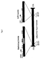
- the gene encoding the labeling protein is driven by a CMV (cytomegalovirus) promoter or hTERT promoter ( Fig. 1 ).
- telomerase promoter determines the transcription initiation site for telomerase and directly regulates the frequency of transcription. Telomerase is an enzyme that maintains the length of telomeres, standing against the shortening of telomeres at the time of replication of eukaryotic chromosomes.
- the type of such telomerase promoter is not particularly limited, and any suitable telomerase promoter compatible with the virus to be used for the expression of the gene of interest may be used.
- the promoter for human telomerase reverse transcriptase (hTERT) is preferable. A number of transcription factor-binding sequences are confirmed in a 1.4 kbp region upstream of the 5' end of hTERT gene.
- This region is believed to be hTERT promoter.
- a 181 bp sequence located upstream of the translation initiation site is a core region important for the expression of the downstream gene.
- any sequence comprising this core region may be used.
- an upstream sequence of approximately 378 bp containing the entire core region is used as hTERT promoter. It has been confirmed that this sequence of approximately 378 bp is equivalent to the 181 bp core region alone in gene expression efficiency.
- the nucleotide sequence of an hTERT promoter of 455 bp is shown in SEQ ID NO: 1.
- nucleotide sequence of hTERT promoter is not limited to the sequence as shown in SEQ ID NO: 1. Nucleotide sequences of nucleotides which hybridize under stringent conditions to a DNA consisting of a nucleotide sequence complementary to the DNA consisting of SEQ ID NO: 1 and have hTERT promoter activity may also be included as a sequence for hTERT promoter. Such nucleotides may be obtained from cDNA libraries or genomic libraries by known hybridization methods such as colony hybridization, plaque hybridization, Southern blotting, etc. using the polynucleotide consisting of the nucleotide sequence of SEQ ID NO:1 or a part thereof as a probe. cDNA libraries may be prepared according to the method described in Molecular Cloning: A Laboratory Manual 2nd ed. (Cold Spring Harbor Press (1989 )). Alternatively, commercial cDNA libraries or genomic libraries may be used.
- examples of stringent conditions include 1xSSC to 2xSSC, 0.1% to 0.5% SDS and 42°C to 68°C. More specifically, an example may be given where prehybridization is performed at 60-68°C for more than 30 minutes, and then washing is performed in 2xSSC, 0.1% SDS at room temperature for 5-15 minutes 4 to 6 times.
- E1A gene, an IRES sequence and an E1B gene are located in this order in the present invention.
- E1A gene and E1B gene are genes included in E1 gene. This is one of early genes of viruses, which have early (E) genes and late (L) genes involved in their DNA replication, and encodes a protein involved in the regulation of transcription of viral genome.
- E1A protein encoded by E1A gene activates the transcription of a group of genes (E1B, E2, E4, etc.) necessary for the production of infectious virus.
- E1B protein encoded by E1B gene assists the accumulation of late gene (L gene) mRNA in the cytoplasm of the infected host cell to thereby inhibit the protein synthesis in the host cell.
- E1B protein promotes viral replication.
- the nucleotide sequences of E1A gene and E1B gene are shown in SEQ ID NO: 2 and SEQ ID NO: 3, respectively.
- E1A and E1B may have, other than those nucleotide sequences shown in SEQ ID NO: 2 and SEQ ID NO: 3, respectively, nucleotide sequences which hybridize under stringent conditions to a DNA consisting of a nucleotide sequence complementary to the DNA consisting of SEQ ID NO: 2 or SEQ ID NO: 3 and encode a protein having E1A or E1B activity.
- nucleotide sequences may be obtained from cDNA libraries or genomic libraries by known hybridization methods such as colony hybridization, plaque hybridization, Southern blotting, etc. using the polynucleotide, or a part thereof, consisting of the nucleotide sequence of SEQ ID NO: 2 or SEQ ID NO: 3 as a probe.
- cDNA libraries may be prepared according to the method described in Molecular Cloning: A Laboratory Manual 2nd ed. (Cold Spring Harbor Press (1989 )). Alternatively, commercial cDNA libraries or genomic libraries may be used. In the above hybridization, examples of stringent conditions include 1xSSC to 2xSSC, 0.1% to 0.5% SDS and 42°C to 68°C. More specifically, an example may be given where prehybridization is performed at 60-68°C for more than 30 minutes, and then washing is performed in 2xSSC, 0.1% SDS at room temperature for 5-15 minutes 4 to 6 times.. For detailed procedures of hybridization, see, for example, Molecular Cloning: A Laboratory Manual 2nd ed. (Cold Spring Harbor Press (1989)), in particular, Section 9.47-9.58 .
- IRES Internal Ribosome Entry Site
- mRNA derived from a virus which belongs to picornaviridae is translated via this sequence. Translation efficiency from IRES sequence is high. Even from the middle of mRNA, protein synthesis is performed in a cap structure non-dependent manner. Therefore, in the virus of the present invention, both E1A gene and E1B gene (which is located downstream of the IRES sequence) are translated independently by a human telomerase promoter.
- IRES the control of expression by a telomerase promoter is exerted on E1A gene and E1B gene independently. Therefore, compared to cases where either E1A gene or E1B gene is controlled by a telomerase promoter, viral replication can be more strictly limited to those cells having telomerase activity.
- An IRES sequence is shown in SEQ ID NO: 4.
- IRES may have, other than the nucleotide sequence as shown in SEQ ID NO: 4, nucleotide sequences which hybridize under stringent conditions to a DNA consisting of a nucleotide sequence complementary to the DNA consisting of SEQ ID NO: 4 and encode a protein having IRES activity
- nucleotide sequences may be obtained from cDNA libraries or genomic libraries by known hybridization methods such as colony hybridization, plaque hybridization, Southern blotting, etc. using the polynucleotide, or a part thereof, consisting of the nucleotide sequence of SEQ ID NO: 4 as a probe.
- cDNA libraries may be prepared according to the method described in Molecular Cloning: A Laboratory Manual 2nd ed.
- examples of stringent conditions include 1xSSC to 2xSSC, 0.1% to 0.5% SDS and 42°C to 68°C. More specifically, an example may be given where prehybridization is performed at 60-68°C for more than 30 minutes, and then washing is performed in 2xSSC, 0.1% SDS at room temperature for 5-15 minutes 4 to 6 times.
- prehybridization is performed at 60-68°C for more than 30 minutes, and then washing is performed in 2xSSC, 0.1% SDS at room temperature for 5-15 minutes 4 to 6 times.
- a promoter from human telomerase is located upstream of the E1 gene because such a promoter is capable of promoting the replication in cells having telomerase activity.
- the genes contained in the replication cassette of the present invention may be obtained by conventional genetic engineering techniques.
- a genetic engineering technique a method of nucleic acid synthesis with a commonly used DNA synthesizer may be used.
- primers specific to each gene may be designed and the genetic sequence may be amplified with a PCR apparatus (PCR method; Current Protocols in Molecular Biology, John Wiley & Sons (1987 ), Section 6.1-6.4); or a gene amplification method using a cloning vector may be used.
- PCR method Current Protocols in Molecular Biology, John Wiley & Sons (1987 ), Section 6.1-6.4
- a gene amplification method using a cloning vector may be used.
- Purification of resultant PCR products may be performed by known methods such as a method using ethidium bromide, a method using SYBR Green I (Molecular Probes), a method with GENECLEAN (Funakoshi), QIAGEN (QIAGEN), etc. using agarose gel, a method using DEAE-cellulose filter, freeze & squeeze method or a method using a dialysis tube.
- agarose gel When agarose gel is used, PCT products are electrophoresed on agarose gel and resultant DNA fragments are cut out from the gel and purified. If necessary, it is possible to confirm by conventional sequencing methods that an expected gene has been obtained. For example, dideoxynucleotide chain termination method ( Sanger et al. (1977) Proc. Natl. Acad. Sci. USA 74: 5463 ) or the like may be used for this purpose. Alternatively, it is also possible to analyze the sequence with an appropriate DNA sequencer (e.g., ABI PRISM; Applied Biosystem
- pIRES vector which comprises the IRES (internal ribosome entry site in mRNA) of encephalomyocarditis virus (ECMV) and is capable of translating two open reading frames (ORFs) from one mRNA; Escherichia coli -derived plasmids (such as pCR4, pCR2, pCR2.1, pBR322, pBR325, pUC12 and pUC13); Bacillus subtilis-derived plasmids (such as pUB110, pTP5 and pC194); yeast-derived plasmids (such as pSH19 and pSH15); bacteriophages such as ⁇ phage; animal viruses such as retrovirus, vaccinia virus
- telomere vector use of pIRES vector.
- this vector it is possible to prepare a replication cassette containing "a promoter from human telomerase, an E1A gene, an IRES sequence and an E1B gene" in this order by inserting necessary genes into the multi-cloning site in this order.
- DNA ligase may be used for ligation of DNAs.
- hTERT is used as human telomerase in the present invention will be described specifically.
- E1A gene and E1B gene may be amplified in E1 gene-expressing cells, such as 293 cells, by carrying out RT-PCR and/or DNA-PCR using primers such as E1A-S, E1A-AS, E1B-S and E1B-AS. If necessary, their sequences are confirmed using a known method such as TA cloning. Then, DNA fragments of E1A and E1B may be cut out using a known restriction enzyme.
- a replication cassette consisting of hTERT-E1A-IRES-E1B to be used in the present invention may be prepared by inserting individual genes into a multi-cloning site or the like of a known vector (such as pIRES vector) to give the following order. "hTERT promoter sequence-E1A-IRES-E1B".
- a known vector such as pIRES vector
- CMV cytomegalovirus
- the adenovirus in which only the replication cassette consisting of hTERT-E1A-IRES-E1B to be used in the present invention has been integrated is designated "Telomelysin".
- Telomelysin By expressing E1 gene necessary for proliferation of adenovirus under the control of hTERT promoter, it is possible to proliferate the virus in a cancer cell-specific manner.
- a labeling cassette is also included together with the replication cassette.
- the "labeling cassette” is integrated in E3 region of the viral genome.
- the primary function of the virus vector used in the present invention is cytotoxicity by viral replication. Therefore, in order to use the reagent of the present invention for the purpose of diagnosing microcancer tissues, occurrence of the cytotoxicity is preferably as late as possible. This is because the emission of fluorescence caused by the replication of the recombinant virus of the present invention disappears when cells are destroyed, and it becomes difficult to identify the site of the microcancer tissue.
- E3A and E3B exist in E3 region of adenovirus, and 11.6 kDa ADP (adenovirus death protein) in E3A region has a function to promote cytotoxicity and viral dispersion.
- viral genomic regions encoding proteins with a function to promote cytotoxicity and viral dispersion are deleted to thereby delay the timing of cell death and facilitate identification of cancer cells by emission of GFP fluorescence or the like.
- the labeling protein which constitutes the labeling cassette is a protein which emits light in those cells where the above-described virus has replicated, and is visualized.
- a substance which emits fluorescence is used.
- examples of such substances include, but are not limited to, green fluorescent protein (GFP) derived from luminescent jellyfish such as Aequorea victoria, enhanced-humanized GFP (EGFP) or red-shift GFP (rsGFP) which are modified variants of GFP (GFP variants). It is also possible to use yellow fluorescent protein (YFP), cyan fluorescent protein (CFP), blue fluorescent protein (BFP), or Renilla reniformis-derived GFP. A gene encoding any of these proteins may be used in the present invention.
- a promoter capable of regulating the expression of the above-described gene may be any promoter as long as it is compatible with the virus to be used for the expression of the above gene of interest.
- promoters include, but are not limited to, cytomegalovirus (CMV) promoter, hTERT promoter, SV40 late promoter, MMTV LTR promoter, RSV LTR promoter and SRa promoter.
- CMV promoter or hTERT promoter may be used.
- the recombinant gene contained in the labeling cassette of the present invention may be obtained by conventional genetic engineering technique.
- a genetic engineering technique a method of nucleic acid synthesis with a commonly used DNA synthesizer may be used.
- primers specific to each gene may be designed and the genetic sequence may be amplified with a PCR apparatus (PCR method; Current Protocols in Molecular Biology, John Wiley & Sons (1987 ), Section 6.1-6.4); or a gene amplification method using a cloning vector may be used.
- PCR product Purification of the resultant PCR product may be performed by known methods such as a method using ethidium bromide, a method using SYBR Green I (Molecular Probes), a method with GENECLEAN (Funakoshi), QIAGEN (QIAGEN), etc. using agarose gel, a method using DEAE-cellulose filter, freeze & squeeze method or a method using a dialysis tube.
- agarose gel the PCT production is electrophoresed on agarose gel and resultant DNA fragments are cut out from the gel and purified.
- shuttle plasmid pHM11 or the like may be used as a plasmid.
- the two genes (for the labeling protein and the promoter) are ligated with DNA ligase and inserted into a vector to thereby prepare a recombinant gene for the labeling cassette.
- pShuttle vector Escherichia coli -derived plasmid (such as pCR4, pCR2, pCR2.1, pBR322, pBR325, pUC12 or pUC13); Bacillus subtilis-derived plasmid (such as pUB110, pTP5 or pC194); yeast-derived plasmid (such as pSH19 or pSH15); bacteriophage such as ⁇ , phage; animal viruse such as renovirus, vaccinia virus or baculovirus; pA1-11, pXT1, pRc/CMV, pRc/RSV, pcDNAI/Neo or the like may be used.
- Escherichia coli -derived plasmid such as pCR4, pCR2, pCR2.1, pBR322, pBR325, pUC12 or pUC13
- Bacillus subtilis-derived plasmid such as pUB110
- a recombinant gene comprising the above described replication cassette and labeling cassette is cut out with appropriate restriction enzymes and inserted into an appropriate virus expression vector, to thereby prepare a recombinant virus.
- virus expression vectors include adenovirus, retrovirus, vaccinia virus and baculovirus.
- adenovirus in particular, type 5 adenovirus
- methods such as electroporation, the liposome method, the spheroplast method or the lithium acetate method may be used.
- the recombinant gene may be prepared by inserting CMV-EGFP-SV40P (A) from pEGFP-N1 (CLONTECH) into shuttle plasmid pHM11, and inserting Csp45I fragment of this plasmid into ClaI site of pShuttle vector in which phTERT-E1A-IRES-E1B has been integrated.
- a sequence of a necessary region may be cut out with restriction enzymes from the recombinant gene as prepared above, and inserted into a viral DNA such as Adeno-X Viral DNA using a commercial kit such as Adeno-X Expression System (CLONTECH) (the resultant product is designated "AdenoX-hAlB").
- This AdenoX-hAIB is linearized with a known restriction enzyme and then transfected into cultured cells, such as 293 cells, to thereby prepare an infectious recombinant virus.
- the target cancer cells to be detected in the present invention are not limited. Cancer cells of any kind may be used.
- solid cancers in the head and neck, stomach, large colon, lung, liver, prostate, pancreas, esophagus, bladder, gallbladder/bile duct, breast, uterus, thyroid, ovary, etc.; or leukemia, lymphoma, sarcoma, mesenchymal tumor or the like may be used.
- Most of cancer cells derived from human tissues show increase in telomerase activity.
- the present invention is capable of detecting those cancer cells in general where proliferation has been activated by such telomerase activity.
- telomerase expression is extremely high in cancer cells compared to normal cells
- hTERT is expressed in telomerase-containing cancer cells and the replication cassette functions therein.
- the virus replicates, which in turn increases the replication of the labeling protein.
- the labeling protein is expressed and visualized.
- the reagent of the present invention does not emit fluorescence in normal cells, but whereas emits fluorescence in cancer cells. Thus, it is possible to observe cancer cells visually.
- the following method may be used, for example.
- cells such as human large colon cancer cell SW620, human lung cancer cells A549 and H1299 are plated in culture plates containing an appropriate culture broth and cultured in the presence of CO 2 gas at 37°C.
- As the culture broth one which is conventionally used for culturing animal cells may be used, e.g. DMEM, MEM, or RPMI-1640. If necessary, serum, antibiotics, vitamins, or the like may be added thereto.
- MOI means a ratio between the viral quantity (infective unit) and the number of cells when a specific amount of cultured cells are infected with a specific amount of viral particles. MOI is used as an indicator when cells are infected with virus.
- cancer cells can be visualized because cells where viral replication is observed emit a specific fluorescence (e.g. green fluorescence when GFP is used) by exposing to excitation light.
- a specific fluorescence e.g. green fluorescence when GFP is used
- emission of GFP fluorescence in the cells can be observed.
- emission of GFP fluorescence may be observed with a CCD camera.
- the recombinant virus of the present invention may be administered into the living body.
- the reagent of the present invention may be applied to the diseased site as it is.
- the reagent of the present invention may be introduced into the living body (target cell or organ) by any known method, e.g. intravenous, intramuscular, intra-abdominal or subcutaneous injection; inhalation through the nasal cavity, oral cavity or lung; oral administration; intravascular administration using catheter or the like. Dose levels are selected appropriately depending on the kind of active ingredient, the administration route, the target of administration, and the age, body weight, sex, symptoms and other conditions of the patient.
- dose levels may be selected so that the virus of the present invention (the active ingredient) is administered at a daily dose of about 10 6 -10 11 PFU (plaque forming units), preferably about 10 9 -10 11 PFU. This amount may be administered once a day, or may be divided into several portions and administered at several times a day.
- PFU plaque forming units
- the reagent of the present invention makes it possible to observe the label in vivo in real time.
- the reagent of the present invention is advantageous for use as an in vivo diagnosis agent. This is useful in the so-called navigation surgery.
- SN sentinel node
- SN is the lymph node which first receives the lymph flow from tumors, and there is a hypothesis that the first micro-metastasis occurs in this lymph node. This hypothesis is called the SN theory.
- In vivo cancer diagnosis system using the reagent of the present invention is capable of establishing the technology of allowing direct expression of a fluorescent protein in cancer cells and identifying tumor tissues or metastasis-positive lymph nodes by a highly sensitive, fluorescence detection system during surgical operation.
- the technology of "navigation surgery" can be established as a method that is more effective than SN.
- the recombinant virus of the present invention replicates in a great number of cancer cells having telomerase activity, and those cells can emit, for example, a strong green fluorescence of GFP.
- the in vivo cancer diagnosis system using the reagent of the present invention identifies tumor tissues or metastasis-positive lymph nodes directly during the surgical operation in real time and the excision range is navigated. This system is original and epoch-making, and further it is extremely practical for smooth progress of surgical operation.
- the reagent of the present invention is endoscopically injected into the site of tumor (e.g., gastric or large colon mucosa around gastric cancer or large colon cancer; internal region of tumors such as gastric cancer, large colon cancer, lung cancer, pancreatic cancer) several days prior to the surgical operation with the same manual technique used in SN navigation. Then, sufficient time is provided so that the virus is distributed into tumor-infiltrated tissues, metastatic tumor tissues or attending lymph nodes to replicate in tumor sites or metastasis-positive sites.
- tumor e.g., gastric or large colon mucosa around gastric cancer or large colon cancer; internal region of tumors such as gastric cancer, large colon cancer, lung cancer, pancreatic cancer
- excitation light for GFP fluorescence is projected from the light source onto the surgery field after ventrotomy, and images from a special 3CCD camera are projected on a face mount display.
- a transmissible lens By using a transmissible lens, the visual field of the actual surgical field can also be secured, and it is possible to detect metastasis-positive lymph nodes from overlapped GFP images. Further, by mounting a special filter, it becomes possible to recognize fluorescence with the eyes without using the camera.
- the reagent of the present invention is also applicable to an ex vivo diagnosis agent for the purpose of screening.
- quantitative determination of tumor markers is the most common method to know the presence of cancer which cannot be detected with the eyes or its primary focus cannot be identified.
- tumor markers are not necessarily satisfactory in their cancer specificity. Besides, it is extremely difficult to detect every cancer species with a single marker.
- telomerase activity increases in 85% or more of human malignant tumors, and thus its cancer specificity is believed to be extremely high.
- Ex vivo cancer diagnosis using the reagent of the present invention may be performed, for example, as described below.
- Erythrocytes are removed from a total blood sample taken from a subject.
- the reagent of the present invention is added at a specific ratio (0.1-10 MOI, preferably 1 MOI) and mixed in a test tube.
- the mixture is left for a specific period of time (e.g., 12-48 hr) to promote infection of cancer cells with the virus and the resultant viral replication.
- the GFP expression in the cell fraction is analyzed quantitatively by flow cytometry. With the use of this system, it becomes possible to detect free cancer cells present in the peripheral blood with high sensitivity. This method can be used for detecting free cancer cells present in the peripheral blood only in an extremely small quantity.
- This Example preliminary examined whether or not fluorescence will be emitted in vitro when cancer cells are co-infected with virus Telomelysin comprising the replication cassette and non-replicating virus Ad-GFP comprising the labeling cassette.
- This Example preliminary examined whether or not fluorescence will be emitted in vivo when cancer tissues are co-infected with virus Telomelysin comprising the replication cassette and non-replicating virus Ad-GFP comprising the labeling cassette.
- Ad-GFP 8 x 10 5 PFU
- TRAD 8 x 10 6 PFU
- Telomelysin-GFP proliferates/replicates cancer cell-specifically and telomerase-specifically because E1A/IRES/E1B is operated by the hTERT promoter. Further, Telomelysin-GFP also has an Aequorea victoria-derived GFP gene integrated in its E3 region which is operated by a promoter. Therefore, cells where viral replication is observed emit green fluorescence when excitation light is applied, which enables visualization of cancer cells.
- Such a replication-incompetent virus was prepared as described below.
- E1A gene of 897 bp was amplified by RT-PCR using the specific primers (E1A-S and E1A-AS) and PCR conditions described below.
- E1B gene of 1822 bp was amplified by DNA-PCR using the following primers E1B-S and E1B-AS.
- composition of the PCR solution and the reaction conditions (cycles, temperature) used were the same as used for the amplification of E1A gene.
- Each PCR product was subjected to TA cloning (TA Cloning Kit Dual Promoter, Invitrogen) to thereby confirm their sequences. Then, DNA fragments of 911 bp (E1A) and 1836 bp (E1B) were cut out, respectively, with restriction enzyme EcoRI.
- E1A and E1B were inserted into the MluI site and the SalI site, respectively, of pIRES vector (CLONTECH) in the normal orientation (E1A-IRES-E1B).
- CMV cytomegalovirus
- pEGFP-N1 (CLONTECH) was digested with AgeI/NheI, blunt-ended with Klenow fragment and self-ligated (pEGFP-N2).
- This pEGFP-N2 was digested with NsiI/AflII and blunt-ended with T4 DNA polymerase, followed by preparation of a BglII site using BglII linker. This BglII fragment was inserted at the BamHI site of pHM11 (pHM11-EGFP-N2).
- Csp45I fragment from pHM11-EGFP-N2 was inserted at the ClaI site of pShuttle vector in which phTERT-E1A IRES-E1B had been integrated (pSh-hAIB).
- a 4381 bp sequence was cut out from the thus prepared recombinant gene (pSh-hAIB) using restriction enzymes I-CeuI and PI-SceI, and inserted into the Adeno-X Viral DNA of Adeno-X Expression System (CLONTECH) (AdenoX-hAIB).
- This AdenoX-hAIB was treated with restriction enzyme PacI for linearization and then transfected into 293 cells to thereby prepare an infectious recombinant adenovirus (hereinafter, referred to as "Telomelysin-GFP").
- H1299 cells cultured in vitro were infected with Telomelysin-GFP at 1 MOI or 10 MOL Specifically, H1299 cells were plated in 24-well plates at 5 x 10 4 cells/well. After 24 hours, cells were counted and the virus was added to the culture broth to give a concentration of 1 MOI or 10 MOL Subsequently, the morphology of cells was observed under inversed microscope with the passage of time to examine the cytotoxic activity of the virus.
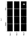
- Fig. 6 The inverted microscopic images shown in Fig. 6 were observed under fluorescent microscope.
- SW620 human large colon cancer cells were infected with Telomelysin-GFP at 10 MOI.
- Cell samples were harvested at 2, 26, 50, 74 and 98 hours after the infection and DNA was extracted therefrom.
- viral replication was quantitatively analyzed by real time PCR.
- Real time PCR was performed using the following primers targeting the E1A gene of Telomelysin-GFP, to thereby quantitatively analyze the viral replication.
- Conditions of the real time PCR composition of the reaction solution, cycle, time period, etc. were the same as in H1299 cells.
- NHLF human lung fibroblast cells
- H1299 human lung cancer cell, SW620 human large colon cancer cell, and normal human lung fibroblast cell (NHLF) were infected with Telomelysin-GFP at 10 MOI as described above. Cell samples were harvested with passage of time, and DNA was extracted therefrom. Then, viral replication was quantitatively analyzed by real time PCR.
- HT29 human large colon cancer cell was transplanted into the rectal wall of nude mouse. When gross tumor was formed, the tumor was removed and analyzed after hematoxylin-eosin (HE) staining.
- HE hematoxylin-eosin
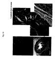
- tumor was formed around the rectum, and tumor cell mass could be confirmed in lymph vessels in the rectal wall ( Fig. 16 ).
- HT29 human large colon cancer cell was transplanted into the rectal wall, and ventrotomy was performed when gross tumor was formed. As a result, swelling was recognized in three lymph nodes (LN) around the aorta ( Fig. 17 )
- Emission of GFP fluorescence was recognized by fluorescence imaging with a CCD camera, in the transplanted HT29 rectal tumor and one of the three para-aortic lymph nodes ( Fig.18 ).
- a reagent for detecting cancer cells or diagnosing cancers is provided. Since the reagent of the present invention is capable of detecting cancer cells with extremely high sensitivity even in the living body, the reagent is useful for the so-called navigation surgery or the like.
Landscapes
- Health & Medical Sciences (AREA)
- Life Sciences & Earth Sciences (AREA)
- Chemical & Material Sciences (AREA)
- Engineering & Computer Science (AREA)
- Organic Chemistry (AREA)
- Genetics & Genomics (AREA)
- General Health & Medical Sciences (AREA)
- Zoology (AREA)
- Molecular Biology (AREA)
- Biomedical Technology (AREA)
- Bioinformatics & Cheminformatics (AREA)
- Medicinal Chemistry (AREA)
- Wood Science & Technology (AREA)
- Biotechnology (AREA)
- Biochemistry (AREA)
- Immunology (AREA)
- General Engineering & Computer Science (AREA)
- Biophysics (AREA)
- Microbiology (AREA)
- Veterinary Medicine (AREA)
- Public Health (AREA)
- Animal Behavior & Ethology (AREA)
- Physics & Mathematics (AREA)
- Nuclear Medicine, Radiotherapy & Molecular Imaging (AREA)
- Proteomics, Peptides & Aminoacids (AREA)
- Pharmacology & Pharmacy (AREA)
- Chemical Kinetics & Catalysis (AREA)
- Virology (AREA)
- Toxicology (AREA)
- Hematology (AREA)
- General Chemical & Material Sciences (AREA)
- Plant Pathology (AREA)
- Urology & Nephrology (AREA)
- Cell Biology (AREA)
- Gastroenterology & Hepatology (AREA)
- Pathology (AREA)
- Epidemiology (AREA)
- Analytical Chemistry (AREA)
- Oncology (AREA)
- Food Science & Technology (AREA)
Abstract
Description
- The present invention relates to a reagent for detecting cancer cells or diagnosing cancers.
- Telomerase activity is often enhanced in malignantly transformed cells or immortalized cell strains, whereas telomerase activity is hardly detected in normal somatic cells excluding such as germ line cells, blood lineage cells and epithelial cells. Therefore, attempts to detect cancer using telomerase activity as an indicator have been made (Shay JW, Zou Y, Hiyama E, Wright WE. Telomerase and Cancer. Hum Mol Genet 10 (7): 677-85, 2001).
- On the other hand, detection of cancer tissues and metastatic lymph nodes in vivo has been studied eagerly in the field of diagnostic imaging. For example, biological diagnosis with PET and image analysis fully utilizing neural network have been reported. Further, investigations into the anti-tumor activity and safety of replication-selective viruses have been reported (DeWeese TL, van der Poel H, Li S, Mikhak B, Drew R, Goemann M, Hamper U, DeJong R, Detorie N, Rodriguez R, Haulk T, DeMarzo AM, Piantadosi S, Yu DC, Chen Y, Henderson DR, Carducci MA, Nelson WG, Simons JW. A phase I trial of CV706, "A replication-competent, PSA selective oncolytic adenovirus, for the treatment of locally recurrent prostate cancer following radiation therapy", Cancer Res 61(20):7464-72, 2001). The present inventors have also found that infecting cancer cells with a virus having a telomerase promoter and replication ability can kill the cancer cells by viral replication (Kawashima T, Kagawa S, Kobayashi N, Shirakiya Y, Umeoka T, Teraishi F, Taki M, Kyo S, Tanaka N, and Fujiwara T., Related Articles, Links Abstract "Telomerase-specific replication-selective virotherapy for human cancer", Clin Cancer Res 10(1):285-92, 2004).
- However, in situ cancer detection system during surgical operation has not yet been developed because of the difficulty in targeting cancer cells. Further, no research has been known to date in which the living body is infected with a virus and the viral kinetics within cancer cells is actually applied to visualization of cancer tissues.
- As described above, development of a reagent for detecting cancer cells or for diagnosing cancers, and capable of visualizing cancer cells even in vivo, has been desired.
- As a result of intensive and extensive researches toward the solution of the above problems, the present inventors have found that it is possible to detect cancer cells with extremely high sensitivity and even in vivo, by integrating a gene encoding a fluorescence labeling protein in E3 region of a viral genome and integrating a replication cassette comprising a human telomerase promoter, an E1A gene, an IRES sequence and an E1B gene in this order in E1 region, and then expressing both cassettes. The ADP-encoding E3 region of the viral genome is deleted. Thus, the present invention has been achieved.
- Accordingly, the present invention provides:
- (1) A reagent for cancer cell detection, or cancer diagnosis, comprising a recombinant adenovirus where:
- a replication cassette comprising a promoter from human telomerase, an adenoviral E1A gene, an IRES sequence and an adenoviral E1B gene in this order is integrated in E1 region of the adenoviral genome; and
- a labeling cassette comprising a gene encoding a labeling protein and a promoter capable of regulating the expression of the gene encoding the labeling protein is integrated in E3 region of the adenoviral genome; and
- the ADP-encoding E3 region of the viral genome is deleted.
In (1) above, these reagents may be used in in vivo detection, diagnosis or navigation surgery. As a specific example of the promoter from human telomerase, hTERT promoter may be given. As a specific example of the labeling protein, GFP may be given. As the promoter capable of regulating the expression of a gene encoding this labeling protein, a cytomegalovirus promoter or hTERT promoter may be used, for example. As the virus, an adenovirus may be used, for example. - (2) An ex vivo method of detecting cancer cells or cancer diagnosis, comprising infecting cancer cells with the reagent of (1) above and detecting the fluorescence-emitted by the cancer cells.
- (3) A use of a recombinant adenovirus as defined in (1) above in the manufacture of a reagent for detecting cancer cells or for cancer diagnosis.
-
-
Fig. 1 is a diagram showing the structure of Telomelysin-GFP. -
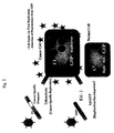
Fig. 2 is a diagram showing the replication of the non-replicating virus. -
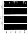
Fig. 3 is a diagram showing the results of detection of cancer cells by in vitro co-infection with Ad-GFP. -
Fig. 4 is a diagram showing the results of detection of human cancer tissues by in vivo co-infection with Ad-GFP -
Fig. 5 is a diagram showing the morphological changes in human lung cancer cells infected with Telomelysin-GFP. -
Fig. 6 is a diagram showing the emission of GFP fluorescence in human lung cancer cells infected with Telomelysin-GFP. -
Fig. 7 is a diagram showing the replication of Telomelysin-GFP determined by quantitative real time-PCR. -
Fig. 8 is a diagram showing the emission of GFP fluorescence in human large colon cancer cells infected with Telomelysin-GFP. -
Fig. 9 is a diagram showing the replication of Telomelysin-GFP determined by quantitative real time-PCR. -
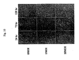
Fig. 10 is a diagram showing the morphological changes in normal human lung fibroblast cells (NHLF) infected with Telomelysin-GFP. -
Fig. 11 is a diagram showing the emission of GFP fluorescence in normal human lung fibroblast cells (NHLF) infected with Telomelysin-GFP. -
Fig. 12 is a diagram showing comparison of Telomelysin-GFP replications determined by quantitative real time-PCR. -
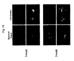
Fig. 13 is a diagram showing the intratumoral proliferation/replication of Telomelysin-GFP observed by fluorescence imaging. -
Fig. 14 is a diagram showing the intratumoral proliferation/replication of Telomelysin-GFP observed by fluorescence imaging. -
Fig. 15 is a diagram showing the intratumoral proliferation/replication of Telomelysin-GFP in a lymph node metastasis model observed by fluorescence imaging. -
Fig. 16 is a diagram showing histological analysis in an orthotopic rectal cancer model using nude mouse and HT29 human large colon cancer cells. -
Fig. 17 is a diagram showing ventrotomy findings in an orthotopic rectal cancer model using nude mouse and HT29 human large colon cancer cells. -
Fig. 18 is a diagram showing the intratumoral proliferation/replication of Telomelysin-GFP in HT29 rectal tumor and para-aortic lymph nodes observed by fluorescence imaging. -
Fig. 19 is a diagram showing the intratumoral proliferation/replication of Telomelysin-GFP in para-aortic lymph nodes observed by fluorescence imaging. -
Fig. 20 is a diagram showing the intratumoral proliferation/replication of Telomelysin-GFP in para-aortic lymph nodes observed by fluorescence imaging. - Hereinbelow, the present invention will be described in detail.
- The present invention relates to a reagent for detecting cancer cells, comprising a recombinant virus where a replication cassette comprising a promoter from human telomerase, an E1A gene, an IRES sequence and an E1B gene in this order is integrated in E1 region of the viral genome and a labeling cassette comprising a gene encoding a labeling protein and a promoter capable of regulating the expression of the gene encoding the labeling protein is integrated in E3 region of the viral genome. Further, the present invention relates to a method of detecting cancer cells, comprising infecting cancer cells with the reagent and detecting the fluorescence emitted by the cancer cells. In the present invention, the term recombinant virus" means a virus in which the replication cassette and the labeling cassette described later are integrated in the genome. The virus used in the present invention is not particularly limited, but an adenovirus is preferable from the viewpoint of safety. Among adenovirus species,
type 5 adenovirus is especially preferable mainly because it is easy to handle. - The recombinant virus used in the present invention has a replication cassette integrated in a region corresponding to E1 region of adenovirus genome and a labeling cassette integrated in a region corresponding to E3 region of adenovirus genome. The replication cassette comprises a promoter from human telomerase, an E1A gene, an IRES sequence and an E1B gene in this order. The E1A gene, IRES sequence and E1B gene are driven by the human telomerase promoter, which results in cancer cell-specific and telomerase-specific proliferation/replication of the virus. The labeling cassette comprises a promoter and a gene encoding a labeling protein. For example, the gene encoding the labeling protein is driven by a CMV (cytomegalovirus) promoter or hTERT promoter (
Fig. 1 ). - The "telomerase promoter" determines the transcription initiation site for telomerase and directly regulates the frequency of transcription. Telomerase is an enzyme that maintains the length of telomeres, standing against the shortening of telomeres at the time of replication of eukaryotic chromosomes. The type of such telomerase promoter is not particularly limited, and any suitable telomerase promoter compatible with the virus to be used for the expression of the gene of interest may be used. For example, the promoter for human telomerase reverse transcriptase (hTERT) is preferable. A number of transcription factor-binding sequences are confirmed in a 1.4 kbp region upstream of the 5' end of hTERT gene. This region is believed to be hTERT promoter. In particular, a 181 bp sequence located upstream of the translation initiation site is a core region important for the expression of the downstream gene. In the present invention, any sequence comprising this core region may be used. Preferably, an upstream sequence of approximately 378 bp containing the entire core region is used as hTERT promoter. It has been confirmed that this sequence of approximately 378 bp is equivalent to the 181 bp core region alone in gene expression efficiency. The nucleotide sequence of an hTERT promoter of 455 bp is shown in SEQ ID NO: 1.
- The nucleotide sequence of hTERT promoter is not limited to the sequence as shown in SEQ ID NO: 1. Nucleotide sequences of nucleotides which hybridize under stringent conditions to a DNA consisting of a nucleotide sequence complementary to the DNA consisting of SEQ ID NO: 1 and have hTERT promoter activity may also be included as a sequence for hTERT promoter. Such nucleotides may be obtained from cDNA libraries or genomic libraries by known hybridization methods such as colony hybridization, plaque hybridization, Southern blotting, etc. using the polynucleotide consisting of the nucleotide sequence of SEQ ID NO:1 or a part thereof as a probe. cDNA libraries may be prepared according to the method described in Molecular Cloning: A Laboratory Manual 2nd ed. (Cold Spring Harbor Press (1989)). Alternatively, commercial cDNA libraries or genomic libraries may be used.
- In the above hybridization, examples of stringent conditions include 1xSSC to 2xSSC, 0.1% to 0.5% SDS and 42°C to 68°C. More specifically, an example may be given where prehybridization is performed at 60-68°C for more than 30 minutes, and then washing is performed in 2xSSC, 0.1% SDS at room temperature for 5-15 minutes 4 to 6 times.
- For detailed procedures of hybridization, see, for example, Molecular Cloning: A Laboratory Manual 2nd ed. (Cold Spring Harbor Press (1989)), in particular, Section 9.47-958.
- The reason why an E1A gene, an IRES sequence and an E1B gene are located in this order in the present invention is that insertion of the IRES sequence between the E1A gene and E1B gene will results in higher replication ability of the virus when a host cell has been infected with it. E1A gene and E1B gene are genes included in E1 gene. This is one of early genes of viruses, which have early (E) genes and late (L) genes involved in their DNA replication, and encodes a protein involved in the regulation of transcription of viral genome. E1A protein encoded by E1A gene activates the transcription of a group of genes (E1B, E2, E4, etc.) necessary for the production of infectious virus. E1B protein encoded by E1B gene assists the accumulation of late gene (L gene) mRNA in the cytoplasm of the infected host cell to thereby inhibit the protein synthesis in the host cell. Thus, E1B protein promotes viral replication. The nucleotide sequences of E1A gene and E1B gene are shown in SEQ ID NO: 2 and SEQ ID NO: 3, respectively.
- E1A and E1B may have, other than those nucleotide sequences shown in SEQ ID NO: 2 and SEQ ID NO: 3, respectively, nucleotide sequences which hybridize under stringent conditions to a DNA consisting of a nucleotide sequence complementary to the DNA consisting of SEQ ID NO: 2 or SEQ ID NO: 3 and encode a protein having E1A or E1B activity. Such nucleotide sequences may be obtained from cDNA libraries or genomic libraries by known hybridization methods such as colony hybridization, plaque hybridization, Southern blotting, etc. using the polynucleotide, or a part thereof, consisting of the nucleotide sequence of SEQ ID NO: 2 or SEQ ID NO: 3 as a probe. cDNA libraries may be prepared according to the method described in Molecular Cloning: A Laboratory Manual 2nd ed. (Cold Spring Harbor Press (1989)). Alternatively, commercial cDNA libraries or genomic libraries may be used. In the above hybridization, examples of stringent conditions include 1xSSC to 2xSSC, 0.1% to 0.5% SDS and 42°C to 68°C. More specifically, an example may be given where prehybridization is performed at 60-68°C for more than 30 minutes, and then washing is performed in 2xSSC, 0.1% SDS at room temperature for 5-15 minutes 4 to 6 times.. For detailed procedures of hybridization, see, for example, Molecular Cloning: A Laboratory Manual 2nd ed. (Cold Spring Harbor Press (1989)), in particular, Section 9.47-9.58.
- IRES (Internal Ribosome Entry Site) is a protein synthesis initiation signal specific to picomavirus. It is believed that this site serves as a ribosome-binding site because it has a complementary sequence to the 3' terminal sequence of 18S ribosomal RNA. It is known that mRNA derived from a virus which belongs to picornaviridae is translated via this sequence. Translation efficiency from IRES sequence is high. Even from the middle of mRNA, protein synthesis is performed in a cap structure non-dependent manner. Therefore, in the virus of the present invention, both E1A gene and E1B gene (which is located downstream of the IRES sequence) are translated independently by a human telomerase promoter. By using IRES, the control of expression by a telomerase promoter is exerted on E1A gene and E1B gene independently. Therefore, compared to cases where either E1A gene or E1B gene is controlled by a telomerase promoter, viral replication can be more strictly limited to those cells having telomerase activity. An IRES sequence is shown in SEQ ID NO: 4.
- IRES may have, other than the nucleotide sequence as shown in SEQ ID NO: 4, nucleotide sequences which hybridize under stringent conditions to a DNA consisting of a nucleotide sequence complementary to the DNA consisting of SEQ ID NO: 4 and encode a protein having IRES activity Such nucleotide sequences may be obtained from cDNA libraries or genomic libraries by known hybridization methods such as colony hybridization, plaque hybridization, Southern blotting, etc. using the polynucleotide, or a part thereof, consisting of the nucleotide sequence of SEQ ID NO: 4 as a probe. cDNA libraries may be prepared according to the method described in Molecular Cloning: A Laboratory Manual 2nd ed. (Cold Spring Harbor Press (1989)). Alternatively, commercial cDNA libraries or genomic libraries may be used. In the above hybridization, examples of stringent conditions include 1xSSC to 2xSSC, 0.1% to 0.5% SDS and 42°C to 68°C. More specifically, an example may be given where prehybridization is performed at 60-68°C for more than 30 minutes, and then washing is performed in 2xSSC, 0.1% SDS at room temperature for 5-15 minutes 4 to 6 times. For detailed procedures of hybridization, see, for example, Molecular Cloning: A Laboratory Manual 2nd ed. (Cold Spring Harbor Press (1989)), in particular, Section 9.47-9.58.
- In the present invention, a promoter from human telomerase is located upstream of the E1 gene because such a promoter is capable of promoting the replication in cells having telomerase activity.
- The genes contained in the replication cassette of the present invention may be obtained by conventional genetic engineering techniques. For example, as a genetic engineering technique, a method of nucleic acid synthesis with a commonly used DNA synthesizer may be used. Alternatively, after a genetic sequence to be used as a template is isolated or synthesized, primers specific to each gene may be designed and the genetic sequence may be amplified with a PCR apparatus (PCR method; Current Protocols in Molecular Biology, John Wiley & Sons (1987), Section 6.1-6.4); or a gene amplification method using a cloning vector may be used One of ordinary skill in the art can readily carry out the above methods according to, for example, Moleculer Cloning 2nd Ed., Cold Spring Harbor Laboratory Press (1989). Purification of resultant PCR products may be performed by known methods such as a method using ethidium bromide, a method using SYBR Green I (Molecular Probes), a method with GENECLEAN (Funakoshi), QIAGEN (QIAGEN), etc. using agarose gel, a method using DEAE-cellulose filter, freeze & squeeze method or a method using a dialysis tube. When agarose gel is used, PCT products are electrophoresed on agarose gel and resultant DNA fragments are cut out from the gel and purified. If necessary, it is possible to confirm by conventional sequencing methods that an expected gene has been obtained. For example, dideoxynucleotide chain termination method (Sanger et al. (1977) Proc. Natl. Acad. Sci. USA 74: 5463) or the like may be used for this purpose. Alternatively, it is also possible to analyze the sequence with an appropriate DNA sequencer (e.g., ABI PRISM; Applied Biosystems).
- Subsequently, individual genes thus obtained are ligated in a specific order. First, above genes are digested with known restriction enzymes, and the resultant DNA fragments are inserted into a known vector according to a known method for ligation. Specific examples of known vectors include pIRES vector which comprises the IRES (internal ribosome entry site in mRNA) of encephalomyocarditis virus (ECMV) and is capable of translating two open reading frames (ORFs) from one mRNA; Escherichia coli-derived plasmids (such as pCR4, pCR2, pCR2.1, pBR322, pBR325, pUC12 and pUC13); Bacillus subtilis-derived plasmids (such as pUB110, pTP5 and pC194); yeast-derived plasmids (such as pSH19 and pSH15); bacteriophages such as λ phage; animal viruses such as retrovirus, vaccinia virus and baculovirus; pA1-11, pXT1, pRc/CMV, pRc/RSV, pcDNAI/Neo and so forth. In the present invention, use of pIRES vector is preferable. With this vector, it is possible to prepare a replication cassette containing "a promoter from human telomerase, an E1A gene, an IRES sequence and an E1B gene" in this order by inserting necessary genes into the multi-cloning site in this order. DNA ligase may be used for ligation of DNAs. Hereinbelow, an example where hTERT is used as human telomerase in the present invention will be described specifically.
- E1A gene and E1B gene may be amplified in E1 gene-expressing cells, such as 293 cells, by carrying out RT-PCR and/or DNA-PCR using primers such as E1A-S, E1A-AS, E1B-S and E1B-AS. If necessary, their sequences are confirmed using a known method such as TA cloning. Then, DNA fragments of E1A and E1B may be cut out using a known restriction enzyme.
- Subsequently, a replication cassette consisting of hTERT-E1A-IRES-E1B to be used in the present invention may be prepared by inserting individual genes into a multi-cloning site or the like of a known vector (such as pIRES vector) to give the following order. "hTERT promoter sequence-E1A-IRES-E1B". Alternatively, if necessary, it is also possible to remove a cytomegalovirus (CMV) promoter from a known vector such as pShuttle with known restriction enzymes, and to insert into that site a sequence cut out from phTERT-E1A-IRES-E1B with appropriate restriction enzymes. The adenovirus in which only the replication cassette consisting of hTERT-E1A-IRES-E1B to be used in the present invention has been integrated is designated "Telomelysin". By expressing E1 gene necessary for proliferation of adenovirus under the control of hTERT promoter, it is possible to proliferate the virus in a cancer cell-specific manner.
- In the recombinant virus used as a reagent of the present invention, a labeling cassette is also included together with the replication cassette. The "labeling cassette" is integrated in E3 region of the viral genome.
- Here, it should be noted that the primary function of the virus vector used in the present invention is cytotoxicity by viral replication. Therefore, in order to use the reagent of the present invention for the purpose of diagnosing microcancer tissues, occurrence of the cytotoxicity is preferably as late as possible. This is because the emission of fluorescence caused by the replication of the recombinant virus of the present invention disappears when cells are destroyed, and it becomes difficult to identify the site of the microcancer tissue.
- On the other hand, E3A and E3B exist in E3 region of adenovirus, and 11.6 kDa ADP (adenovirus death protein) in E3A region has a function to promote cytotoxicity and viral dispersion.
- Therefore, in the recombinant virus used in the present invention, viral genomic regions encoding proteins with a function to promote cytotoxicity and viral dispersion (such ADP-encoding E3 region) are deleted to thereby delay the timing of cell death and facilitate identification of cancer cells by emission of GFP fluorescence or the like.
- The labeling protein which constitutes the labeling cassette is a protein which emits light in those cells where the above-described virus has replicated, and is visualized. Preferably, a substance which emits fluorescence is used. Examples of such substances include, but are not limited to, green fluorescent protein (GFP) derived from luminescent jellyfish such as Aequorea victoria, enhanced-humanized GFP (EGFP) or red-shift GFP (rsGFP) which are modified variants of GFP (GFP variants). It is also possible to use yellow fluorescent protein (YFP), cyan fluorescent protein (CFP), blue fluorescent protein (BFP), or Renilla reniformis-derived GFP. A gene encoding any of these proteins may be used in the present invention.
- A promoter capable of regulating the expression of the above-described gene may be any promoter as long as it is compatible with the virus to be used for the expression of the above gene of interest. Specific examples of such promoters include, but are not limited to, cytomegalovirus (CMV) promoter, hTERT promoter, SV40 late promoter, MMTV LTR promoter, RSV LTR promoter and SRa promoter. Preferably, CMV promoter or hTERT promoter may be used.
- The recombinant gene contained in the labeling cassette of the present invention may be obtained by conventional genetic engineering technique. For example, as a genetic engineering technique, a method of nucleic acid synthesis with a commonly used DNA synthesizer may be used. Alternatively, after a genetic sequence to be used as a template is isolated or synthesized, primers specific to each gene may be designed and the genetic sequence may be amplified with a PCR apparatus (PCR method; Current Protocols in Molecular Biology, John Wiley & Sons (1987), Section 6.1-6.4); or a gene amplification method using a cloning vector may be used. One of ordinary skill in the art can readily carry out the above methods according to, for example, Moleculer Closing 2nd Ed., Cold Spring Harbor Laboratory Press (1989). Purification of the resultant PCR product may be performed by known methods such as a method using ethidium bromide, a method using SYBR Green I (Molecular Probes), a method with GENECLEAN (Funakoshi), QIAGEN (QIAGEN), etc. using agarose gel, a method using DEAE-cellulose filter, freeze & squeeze method or a method using a dialysis tube. When agarose gel is used, the PCT production is electrophoresed on agarose gel and resultant DNA fragments are cut out from the gel and purified. If necessary, it is possible to confirm by conventional sequencing methods that an expected gene has been obtained. For example, dideoxynucleotide chain termination method (Sanger et al. (1977) Proc. Natl. Acad. Sci. USA 74: 5463) or the like may be used for this purpose. Alternatively, it is also possible to analyze the sequence with an appropriate DNA sequencer (e.g., ABI PRISM; Applied Biosystems). Subsequently, the gene thus obtained is digested with known restriction enzymes. The recombinant gene is so designed that the resultant, digested DNA fragment (encoding the labeling protein) is located downstream of a gene fragment encoding the above-described promoter. Here, shuttle plasmid pHM11 or the like may be used as a plasmid. The two genes (for the labeling protein and the promoter) are ligated with DNA ligase and inserted into a vector to thereby prepare a recombinant gene for the labeling cassette.
- As a known vector, pShuttle vector, Escherichia coli-derived plasmid (such as pCR4, pCR2, pCR2.1, pBR322, pBR325, pUC12 or pUC13); Bacillus subtilis-derived plasmid (such as pUB110, pTP5 or pC194); yeast-derived plasmid (such as pSH19 or pSH15); bacteriophage such as λ, phage; animal viruse such as renovirus, vaccinia virus or baculovirus; pA1-11, pXT1, pRc/CMV, pRc/RSV, pcDNAI/Neo or the like may be used.
- Subsequently, a recombinant gene comprising the above described replication cassette and labeling cassette is cut out with appropriate restriction enzymes and inserted into an appropriate virus expression vector, to thereby prepare a recombinant virus. Examples of virus expression vectors include adenovirus, retrovirus, vaccinia virus and baculovirus. As described above, adenovirus (in particular,
type 5 adenovirus) is preferably used. For integration of the cassettes into virus, methods such as electroporation, the liposome method, the spheroplast method or the lithium acetate method may be used. - In the present invention, specifically, the recombinant gene may be prepared by inserting CMV-EGFP-SV40P (A) from pEGFP-N1 (CLONTECH) into shuttle plasmid pHM11, and inserting Csp45I fragment of this plasmid into ClaI site of pShuttle vector in which phTERT-E1A-IRES-E1B has been integrated.
- In the present invention, specifically, a sequence of a necessary region may be cut out with restriction enzymes from the recombinant gene as prepared above, and inserted into a viral DNA such as Adeno-X Viral DNA using a commercial kit such as Adeno-X Expression System (CLONTECH) (the resultant product is designated "AdenoX-hAlB").
- This AdenoX-hAIB is linearized with a known restriction enzyme and then transfected into cultured cells, such as 293 cells, to thereby prepare an infectious recombinant virus.
- The target cancer cells to be detected in the present invention are not limited. Cancer cells of any kind may be used. For example, solid cancers in the head and neck, stomach, large colon, lung, liver, prostate, pancreas, esophagus, bladder, gallbladder/bile duct, breast, uterus, thyroid, ovary, etc.; or leukemia, lymphoma, sarcoma, mesenchymal tumor or the like may be used. Most of cancer cells derived from human tissues show increase in telomerase activity. The present invention is capable of detecting those cancer cells in general where proliferation has been activated by such telomerase activity.
- Since telomerase expression is extremely high in cancer cells compared to normal cells, hTERT is expressed in telomerase-containing cancer cells and the replication cassette functions therein. As a result, the virus replicates, which in turn increases the replication of the labeling protein. Thus, the labeling protein is expressed and visualized.
- Therefore, when the reagent of the present invention does not emit fluorescence in normal cells, but whereas emits fluorescence in cancer cells. Thus, it is possible to observe cancer cells visually.
- For infecting cells with a recombinant virus, the following method may be used, for example. First, cells such as human large colon cancer cell SW620, human lung cancer cells A549 and H1299 are plated in culture plates containing an appropriate culture broth and cultured in the presence of CO2 gas at 37°C. As the culture broth, one which is conventionally used for culturing animal cells may be used, e.g. DMEM, MEM, or RPMI-1640. If necessary, serum, antibiotics, vitamins, or the like may be added thereto. By inoculating a specific amount (0.1-10 MOI (multiplicity of infection), preferably 1 MOI) of the recombinant virus of the present invention, the cultured cells are infected. MOI means a ratio between the viral quantity (infective unit) and the number of cells when a specific amount of cultured cells are infected with a specific amount of viral particles. MOI is used as an indicator when cells are infected with virus.
- In order to confirm viral replication, cells infected with virus are recovered and DNA is extracted therefrom. Then, the DNA is subjected to real time-PCR using primers targeting an appropriate gene contained by the virus of the present invention. Thus, quantitative analysis is possible.
- With respect to the detection of labeled cells, cancer cells can be visualized because cells where viral replication is observed emit a specific fluorescence (e.g. green fluorescence when GFP is used) by exposing to excitation light. For example, when cells infected with virus are observed under fluorescence microscope, emission of GFP fluorescence in the cells can be observed. For the observation of infected cells with the passage of time, emission of GFP fluorescence may be observed with a CCD camera.
- For the real time labeling and detection of cells of interest in vivo, the recombinant virus of the present invention may be administered into the living body.
- The reagent of the present invention may be applied to the diseased site as it is. Alternatively, the reagent of the present invention may be introduced into the living body (target cell or organ) by any known method, e.g. intravenous, intramuscular, intra-abdominal or subcutaneous injection; inhalation through the nasal cavity, oral cavity or lung; oral administration; intravascular administration using catheter or the like. Dose levels are selected appropriately depending on the kind of active ingredient, the administration route, the target of administration, and the age, body weight, sex, symptoms and other conditions of the patient. Usually, dose levels may be selected so that the virus of the present invention (the active ingredient) is administered at a daily dose of about 106-1011 PFU (plaque forming units), preferably about 109-1011 PFU. This amount may be administered once a day, or may be divided into several portions and administered at several times a day.
- The reagent of the present invention makes it possible to observe the label in vivo in real time. Thus, the reagent of the present invention is advantageous for use as an in vivo diagnosis agent. This is useful in the so-called navigation surgery.
- If excision is performed in a wide range including the diseased organ in a surgical operation, the patient who survived this surgical operation can enjoy a long survival However, the rate of occurrence of complications caused by the surgical operation itself becomes high. Further, loss of the function of the excised organ inevitably influences on the daily life after the surgical operation. It is important in cancer treatment to introduce a low-invasive treatment to reduce the burden of patients while maintaining the remote result of long survival.
- When a low-invasive operation is pursued by minimizing the area of excision, one of the information pieces wanted is the presence or absence of metastatic lymph nodes. As a method for obtaining that information, sentinel node (SN) is attracting attention. SN is the lymph node which first receives the lymph flow from tumors, and there is a hypothesis that the first micro-metastasis occurs in this lymph node. This hypothesis is called the SN theory. Although large scale clinical tests in breast cancer have already been started in primarily Europe and the United States, whether or not this theory is applicable to other solid tumors is still unknown. Examination has just been started.
- In vivo cancer diagnosis system using the reagent of the present invention is capable of establishing the technology of allowing direct expression of a fluorescent protein in cancer cells and identifying tumor tissues or metastasis-positive lymph nodes by a highly sensitive, fluorescence detection system during surgical operation. In other words, the technology of "navigation surgery" can be established as a method that is more effective than SN. The recombinant virus of the present invention replicates in a great number of cancer cells having telomerase activity, and those cells can emit, for example, a strong green fluorescence of GFP.
- From the analysis of mono-lymph node metastasis sites, about 10% of skip metastasis, i.e., incipient metastasis to the second group or more remote lymph nodes skipping over the first group lymph nodes has been reported. Based on this report, there are a large number of researchers who have pointed out the danger of SN navigation. However, the in vivo cancer diagnosis system using the reagent of the present invention identifies tumor tissues or metastasis-positive lymph nodes directly during the surgical operation in real time and the excision range is navigated. This system is original and epoch-making, and further it is extremely practical for smooth progress of surgical operation. Specifically, the reagent of the present invention is endoscopically injected into the site of tumor (e.g., gastric or large colon mucosa around gastric cancer or large colon cancer; internal region of tumors such as gastric cancer, large colon cancer, lung cancer, pancreatic cancer) several days prior to the surgical operation with the same manual technique used in SN navigation. Then, sufficient time is provided so that the virus is distributed into tumor-infiltrated tissues, metastatic tumor tissues or attending lymph nodes to replicate in tumor sites or metastasis-positive sites.
- At the time of surgery, excitation light for GFP fluorescence is projected from the light source onto the surgery field after ventrotomy, and images from a special 3CCD camera are projected on a face mount display. By using a transmissible lens, the visual field of the actual surgical field can also be secured, and it is possible to detect metastasis-positive lymph nodes from overlapped GFP images. Further, by mounting a special filter, it becomes possible to recognize fluorescence with the eyes without using the camera.
- The reagent of the present invention is also applicable to an ex vivo diagnosis agent for the purpose of screening. Currently, quantitative determination of tumor markers is the most common method to know the presence of cancer which cannot be detected with the eyes or its primary focus cannot be identified. However, tumor markers are not necessarily satisfactory in their cancer specificity. Besides, it is extremely difficult to detect every cancer species with a single marker.
- It has been confirmed that telomerase activity increases in 85% or more of human malignant tumors, and thus its cancer specificity is believed to be extremely high.
- Ex vivo cancer diagnosis using the reagent of the present invention may be performed, for example, as described below.
- Erythrocytes are removed from a total blood sample taken from a subject. To the remaining cell suspension liquid, the reagent of the present invention is added at a specific ratio (0.1-10 MOI, preferably 1 MOI) and mixed in a test tube. The mixture is left for a specific period of time (e.g., 12-48 hr) to promote infection of cancer cells with the virus and the resultant viral replication. Then, the GFP expression in the cell fraction is analyzed quantitatively by flow cytometry. With the use of this system, it becomes possible to detect free cancer cells present in the peripheral blood with high sensitivity. This method can be used for detecting free cancer cells present in the peripheral blood only in an extremely small quantity.
- Hereinbelow, the present invention will be described in more detail with reference to the following Examples. However, the present invention is not limited to these Examples.
- This Example preliminary examined whether or not fluorescence will be emitted in vitro when cancer cells are co-infected with virus Telomelysin comprising the replication cassette and non-replicating virus Ad-GFP comprising the labeling cassette.
- Human large colon Cancer cell SW620 and human lung cancer cells A549 and H1299 were infected with 0.1 MOI (multiplicity of infection) of Ad-GFP (
Fig. 2 ). - As a result, tendency to green color was hardly recognized when human large colon cancer cell SW620 and human lung cancer cells A549 and H1299 were infected with 0.1 MOI of Ad-GFP. However, when 1 MOI of TRAD was used jointly, fluorescence could be detected only in cancer cells, and no fluorescence was detected in normal cells such as human fibroblast cells WI38 and NHLF and human umbilical vascular endothelial cell (HUVEC) (
Fig. 3 ). - This Example preliminary examined whether or not fluorescence will be emitted in vivo when cancer tissues are co-infected with virus Telomelysin comprising the replication cassette and non-replicating virus Ad-GFP comprising the labeling cassette.
- Ad-GFP (8 x 10⁵ PFU) and TRAD (8 x 10⁶ PFU) were intratumorally administered to human large colon cancer SW5620 and human lung cancer A549 tumors transplanted subcutaneously into the dorsal of nude mice. Then, fluorescence was observed with the passage of time.
- In any of the tumors, spot-like fluorescence had begun to be detected from
day 2 after the administration and disappeared by day 14 (Fig. 4 ). - The outline of Telomelysin-GFP is shown in
Fig. 1 . Telomelysin-GFP proliferates/replicates cancer cell-specifically and telomerase-specifically because E1A/IRES/E1B is operated by the hTERT promoter. Further, Telomelysin-GFP also has an Aequorea victoria-derived GFP gene integrated in its E3 region which is operated by a promoter. Therefore, cells where viral replication is observed emit green fluorescence when excitation light is applied, which enables visualization of cancer cells. - Such a replication-incompetent virus was prepared as described below.
- From RNA extracted from 293 cells, E1A gene of 897 bp was amplified by RT-PCR using the specific primers (E1A-S and E1A-AS) and PCR conditions described below.
- E1A-S: 5'-ACA CCG GGA CTG AAAATG AG-3' (SEQ ID NO: 5)
- E1A-AS: 5'-CACAGGTIT ACA CCT TAT GGC-3' (SEQ ID NO: 6)
- 1 × PCR buffer
- 0.2 mM each dNTPs
- 5 mM MgCl2
- 2.5 U AmpliTaq Gold
- 0.2 µM each Primers
- 95°C, 10 min
- (95°C, 1 min; 56°C, 1 min; 72°C, 1.5 min) x 32 cycles
- 72°C, 7 min
- 4°C, 5 min
- From DNA extracted from 293 cells, E1B gene of 1822 bp was amplified by DNA-PCR using the following primers E1B-S and E1B-AS.
- E1B-S: 5'-CTG ACC TCATGG AGG CIT GG-3' (SEQ ID NO: 7)
- E1B-AS: 5'-GCC CACACATTT CAGTAC CTC-3' (SEQ ID NO: 8)
- The composition of the PCR solution and the reaction conditions (cycles, temperature) used were the same as used for the amplification of E1A gene.
- Each PCR product was subjected to TA cloning (TA Cloning Kit Dual Promoter, Invitrogen) to thereby confirm their sequences. Then, DNA fragments of 911 bp (E1A) and 1836 bp (E1B) were cut out, respectively, with restriction enzyme EcoRI.
- E1A and E1B were inserted into the MluI site and the SalI site, respectively, of pIRES vector (CLONTECH) in the normal orientation (E1A-IRES-E1B).
- A 455 bp hTERT promoter sequence which had been cut out with restriction enzymes MluI and BgIII was inserted into the XhoI site located upstream of the E1A of E1A-IRES-E1B in the normal orientation (phTERT-E1A-IRES-E1B).
- The cytomegalovirus (CMV) promoter contained in pShuttle vector was removed by treatment with restriction enzymes MfeI and NheL Then, a 3828 bp sequence cut out from phTERT-E1A-IRES-E1B using restriction enzymes NheI and NotI was inserted into that site (pSh-hAIB).
- pEGFP-N1 (CLONTECH) was digested with AgeI/NheI, blunt-ended with Klenow fragment and self-ligated (pEGFP-N2).
- This pEGFP-N2 was digested with NsiI/AflII and blunt-ended with T4 DNA polymerase, followed by preparation of a BglII site using BglII linker. This BglII fragment was inserted at the BamHI site of pHM11 (pHM11-EGFP-N2).
- Further, Csp45I fragment from pHM11-EGFP-N2 was inserted at the ClaI site of pShuttle vector in which phTERT-E1A IRES-E1B had been integrated (pSh-hAIB).
- A 4381 bp sequence was cut out from the thus prepared recombinant gene (pSh-hAIB) using restriction enzymes I-CeuI and PI-SceI, and inserted into the Adeno-X Viral DNA of Adeno-X Expression System (CLONTECH) (AdenoX-hAIB). This AdenoX-hAIB was treated with restriction enzyme PacI for linearization and then transfected into 293 cells to thereby prepare an infectious recombinant adenovirus (hereinafter, referred to as "Telomelysin-GFP").
- Human non-small-cell lung cancer-derived H1299 cells cultured in vitro were infected with Telomelysin-GFP at 1 MOI or 10 MOL Specifically, H1299 cells were plated in 24-well plates at 5 x 10⁴ cells/well. After 24 hours, cells were counted and the virus was added to the culture broth to give a concentration of 1 MOI or 10 MOL Subsequently, the morphology of cells was observed under inversed microscope with the passage of time to examine the cytotoxic activity of the virus.
- As a result, cell death was induced by viral replication in a concentration dependent manner and also a time dependent manner. 120 hours after 10 MOI infection, most of the cells became circular and were suspending under inverted microscope (
Fig. 5 ). - The inverted microscopic images shown in
Fig. 6 were observed under fluorescent microscope. The green fluorescence of GFP indicating viral replication in a concentration dependent manner and also a time dependent manner was observed (Fig. 6 ). 72 hours after 10 MOI infection, GFP expression was observed in the maximum number of cells. Then, the number of GFP-positive cells decreased as cell death was induced (Fig. 6 ). - Human lung cancer cell H1299 was infected with Telomelysin-GFP at 10 MOL Cell samples were harvested at 2, 26, 50 and 74 hours after the infection and DNA was extracted therefrom. Real time PCR was performed using the following primers targeting the E1A gene of Telomelysin-GFP, to thereby quantitatively analyze the viral proliferation/replication. The primers and PCR conditions used are as described below.
- E1A-S: 5'-CCT GTG TCT AGA GAATGCAA-3' (SEQ ID NO: 9)
- E1A-AS: 5'-ACA GCT CAA GTC CAAAGG TT-3' (SEQ ID NO: 10)
- 1 × LC FastStart DNAMaster SYBR Green I
- 3 m MgCl2
- 0.5 µM each Primer
- 95°C, 10 min
- (95°C, 10 sec; 60°C, 15 sec; 72°C, 8 sec) x 40 cycles
- 70°C, 15 sec
- 40°C, 30 sec
- The results revealed that Telomelysin-GFP had already replicated 1,000,000-fold at 26 hours after the infection (
Fig. 7 ). Thereafter, the replication reached plateau, but GFP fluorescence was also enhanced gradually slightly after the replication (Fig. 7 ). - Human large colon cancer-derived SW620 cells were infected with Telomelysin-GFP at 10 MOL Changes in the cells were observed with the passage of time-under inverted microscope and fluorescent microscope.
- As a result, GFP green fluorescence indicating viral replication in a time dependent manner was recognized as in the case of H1299 cells (
Fig. 8 ). - In the same manner as in H1299 cells, SW620 human large colon cancer cells were infected with Telomelysin-GFP at 10 MOI. Cell samples were harvested at 2, 26, 50, 74 and 98 hours after the infection and DNA was extracted therefrom. Then, viral replication was quantitatively analyzed by real time PCR. Real time PCR was performed using the following primers targeting the E1A gene of Telomelysin-GFP, to thereby quantitatively analyze the viral replication. Conditions of the real time PCR (composition of the reaction solution, cycle, time period, etc.) were the same as in H1299 cells.
- The results revealed that Telomelysin-GFP had already replicated 1,000,000-fold at 26 hours after the infection and was almost plateau up to 98 hours after the infection (
Fig. 9 ). - Normal human lung fibroblast cells (NHLF) cultured in vitro were infected with Telomelysin-GFP at 1 MOI or 10 MOL Changes were observed under inverted microscope up to 120 hours after the infection.
- As a result, no morphological changes were observed, and cell death was not induced (
Fig. 10 ). - When the inverted microscopic images shown in
Fig. 10 are observed under fluorescent microscope, emission of GFP fluorescence was observed in some cells. However, considering the cell density, the emission was extremely rare compared to that in cancer cells. Therefore, it was believed that Telomelysin-GFP hardly proliferates/replicates in normal cells (Fig.11 ). - H1299 human lung cancer cell, SW620 human large colon cancer cell, and normal human lung fibroblast cell (NHLF) were infected with Telomelysin-GFP at 10 MOI as described above. Cell samples were harvested with passage of time, and DNA was extracted therefrom. Then, viral replication was quantitatively analyzed by real time PCR.
- The results revealed that Telomelysin-GFP had already replicated about 1,000,000-fold in cancer cells at 24 hours after the infection, and emitted remarkable GFP fluorescence at 72 hours after the infection- (
Fig. 12 ). On the other hand, replication was only about 1000-fold in NHLF cells even at 72 hours after the infection, and little GFP fluorescence could be detected (Fig.12 ). -
- 1. Telomelysin-GFP (107 PFU) was administered into the tumor of H1299 human lung cancer transplanted into nude mice. Then, emission of GFP fluorescence was observed with a CCD camera With the passage of time.
The results revealed that emission of GFP fluorescence caused by Telomelysin-GFP replication began to be recognized within 24 hours after the infection, and that the range and luminance were gradually enhanced 3 days and 5 days after the infection (Fig. 13 ). - 2. In the same manner as described above, Telomelysin-GFP (107 PFU) was administered into the tumor of H1299 human lung cancer transplanted into nude mice. One week and three weeks after the infection, subcutaneous tumor was removed. Emission of GFP fluorescence was observed on the entire tumor and on a cut surface using a CCD camera.
As a result, even when fluorescence emission was weak on the surface of the removed tumor, replication of Telomelysin-GFP could be confirmed in a wide range on the cut surface (Fig.14 ). In tissues three weeks after the infection fluorescence was recognized on almost all over the tumor (Fig. 14 ). - 3. In the same manner as described above, HT29 human large colon cancer cell was transplanted into the rectal wall of nude mice as an orthotopic model, and Telomelysin-GFP (107 PFU) was administered at the time when gross tumor was formed. Emission of GFP fluorescence caused by Telomelysin-GFP replication began to be recognized one week after the infection with a CCD camera (
Fig. 15 ). The fluorescence emission was maintained even three weeks after the infection (Fig. 15 ). - HT29 human large colon cancer cell was transplanted into the rectal wall of nude mouse. When gross tumor was formed, the tumor was removed and analyzed after hematoxylin-eosin (HE) staining.
- As a result, tumor was formed around the rectum, and tumor cell mass could be confirmed in lymph vessels in the rectal wall (
Fig. 16 ). - HT29 human large colon cancer cell was transplanted into the rectal wall, and ventrotomy was performed when gross tumor was formed. As a result, swelling was recognized in three lymph nodes (LN) around the aorta (
Fig. 17 ) - Emission of GFP fluorescence was recognized by fluorescence imaging with a CCD camera, in the transplanted HT29 rectal tumor and one of the three para-aortic lymph nodes (
Fig.18 ). - Emission of GFP fluorescence was recognized by fluorescence imaging with a CCD camera, in only one of the three para-aortic lymph nodes (
Fig. 19 ). - Histological analysis of para-aortic lymph nodes detected metastatic tumor tissue in the only one lymph node which was found GFP fluorescence-positive by fluorescence imaging with a CCD camera. Thus, it was confirmed that Telomelysin-GFP replicates only in metastasis-positive lymph nodes (
Fig. 20 ). - Reid T, Galanis E, Abbruzzese J, Sze D, Wein LM, Andrews J, Randlev B, Heise C, Uprichard M, Hatfield M, Rome L, Rubin J, Kim D. Hepatic arterial infusion of a replication-selective oncolytic adenovirus (d11520): phase II viral, immunologic, and clinical endpoints. Cancer Res 62 (21): 6070-9, 2002.
- According to the present invention, a reagent for detecting cancer cells or diagnosing cancers is provided. Since the reagent of the present invention is capable of detecting cancer cells with extremely high sensitivity even in the living body, the reagent is useful for the so-called navigation surgery or the like.
-
- SEQ ID NO: 5: Primer
- SEQ ID NO: 6: Primer
- SEQ ID NO: 7: Primer
- SEQ ID NO: 8: Primer
- SEQ ID NO: 9: Primer
- SEQ ID NO: 10: Primer
-
- <110> ONCOLYS BIOPHARMA, INC.
- <120> TELOMELYSIN-GFP GENE-CONTAINING RECOMBINANT VIRUS
- <130> N.100848B
- <150>
PCT/JP2005/018401
<151> 2005-09-28 - <150>
JP2004-285383
<151> 2004-09-29 - <160> 24
- <170> PatentIn version 3.3
- <210> 1
<211> 455
<212> DNA
<213> homo sapiens - <400> 1

- <210> 2
<211> 899
<212> DNA
<213> adenovirus - <400> 2


- <210> 3
<211> 1823
<212> DNA
<213> adenovirus - <400> 3


- <210> 4
<211> 605
<212> DNA
<213> adenovirus - <400> 4

- <210> 5
<211> 20
<212> DNA
<213> Artificial - <220>
<223> primer - <400> 5
acaccgggac tgaaaatgag 20 - <210> 6
<211> 21
<212> DNA
<213> Artificial - <220>
<223> primer - <400> 6
cacaggttta caccttatgg c 21 - <210> 7
<211> 20
<212> DNA
<213> Artificial - <220>
<223> primer - <400> 7
ctgacctcat ggaggcttgg 20 - <210> 8
<211> 21
<212> DNA
<213> Artificial - <220>
<223> primer - <400> 8
gcccacacat ttcagtacctc 21 - <210> 9
<211> 20
<212> DNA
<213> Artificial - <220>
<223> primer - <400> 9
cctgtgtcta gagaatgcaa 20 - <210> 10
<211> 20
<212> DNA
<213> Artificial - <220>
<223> primer - <400> 10
acagctcaag tccaaaggtt 20 - <210> 11
<211> 1307
<212> DNA
<213> Homo sapiens - <400> 11

- <210> 12
<211> 837
<212> DNA
<213> Homo sapiens - <400> 12

- <210> 13
<211> 987
<212> DNA
<213> Homo sapiens - <400> 13
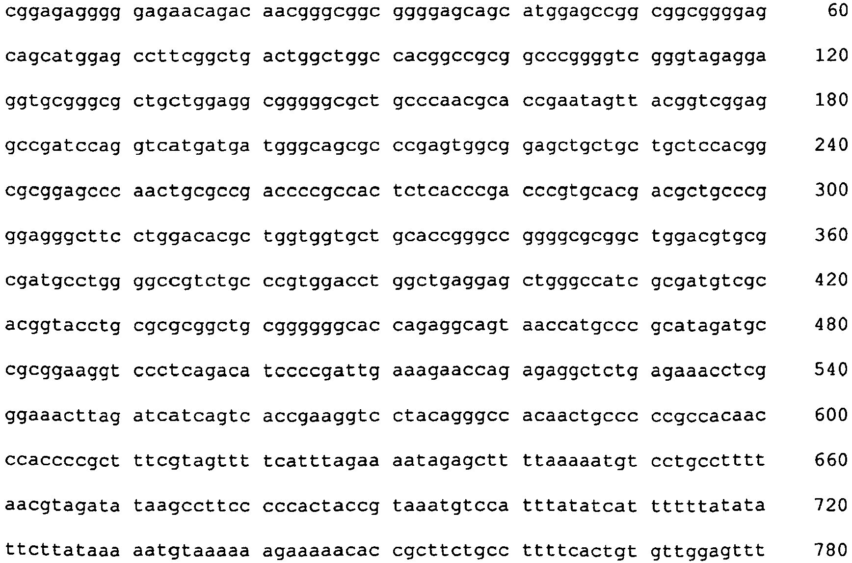

- <210> 14
<211> 8972
<212> DNA
<213> Homo sapiens - <400> 14


- <210> 15
<211> 5711
<212> DNA
<213> Homo sapiens - <400> 15


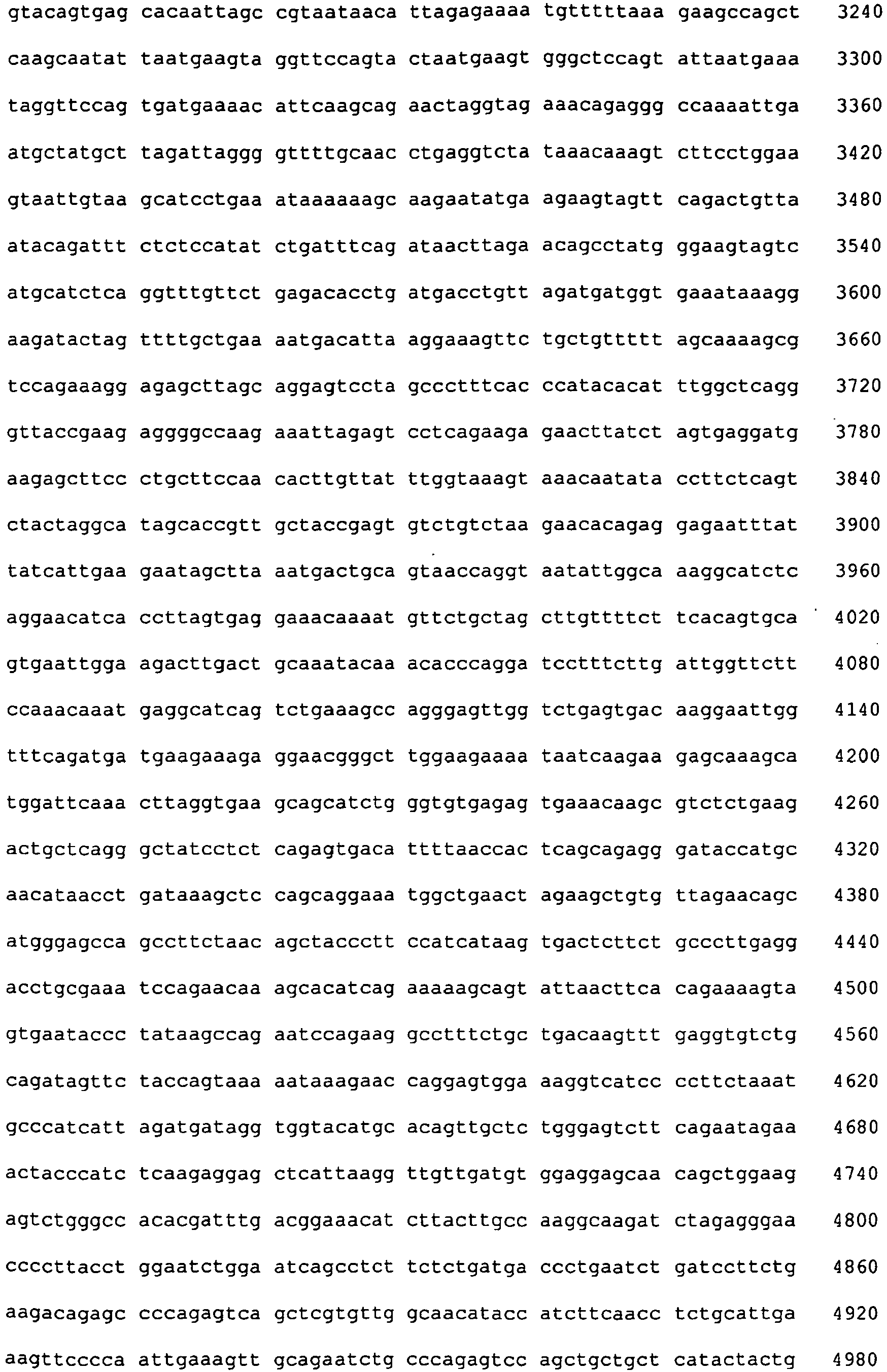

- <210> 16
<211> 2680
<212> DNA
<213> Homo sapiens - <400> 16


- <210> 17
<211> 1095
<212> DNA
<213> Homo sapiens - <400> 17

- <210> 18
<211> 2234
<212> DNA
<213> Homo sapiens - <400> 18


- <210> 19
<211> 4344
<212> DNA
<213> Homo sapiens - <400> 19


- <210> 20
<211> 4740
<212> DNA
<213> Homo sapiens - <400> 20
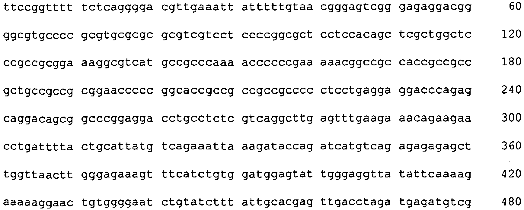


- <210> 21
<211> 4608
<212> DNA
<213> Homo sapiens - <400> 21

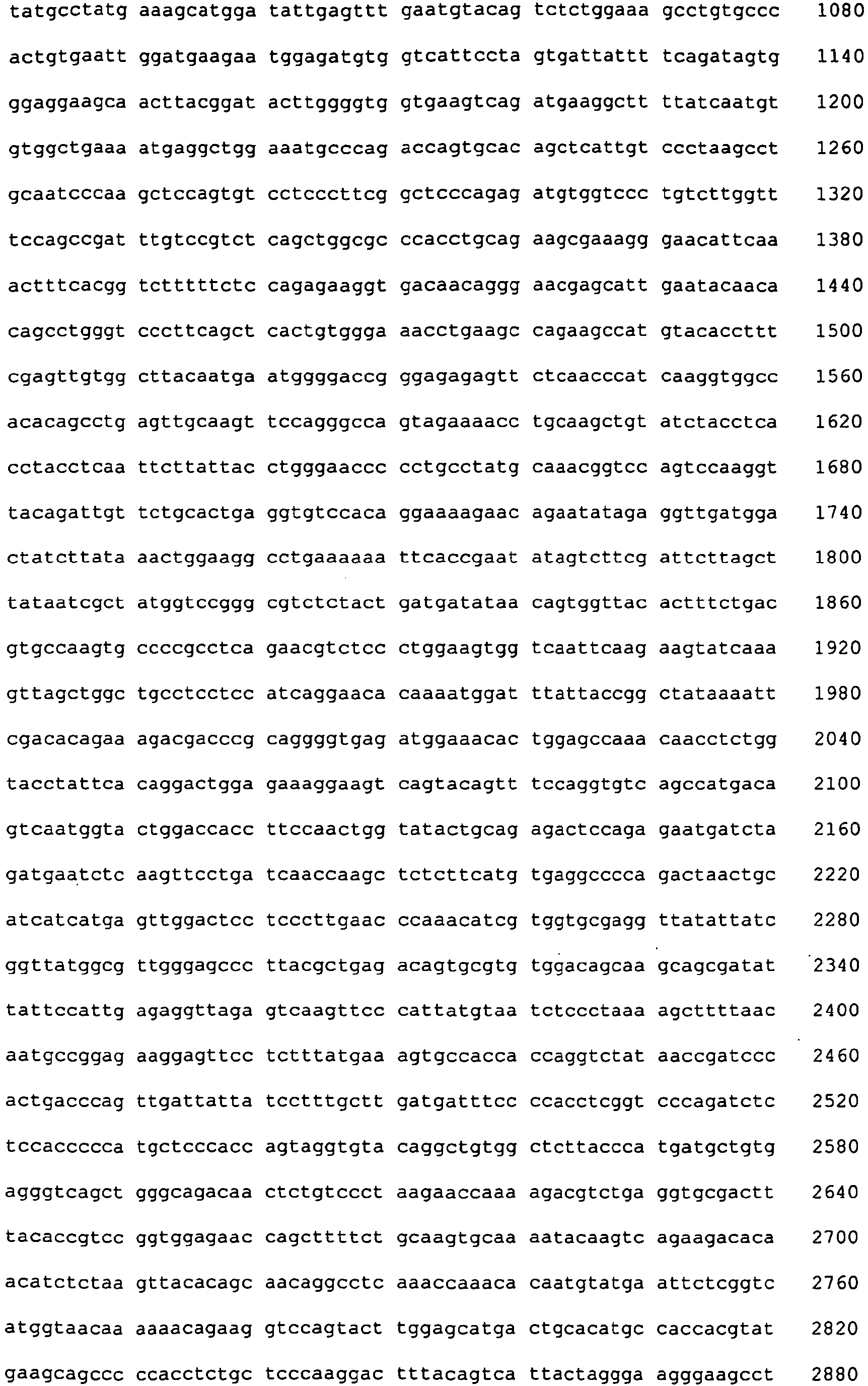

- <210> 22
<211> 8959
<212> DNA
<213> Homo sapiens - <400> 22


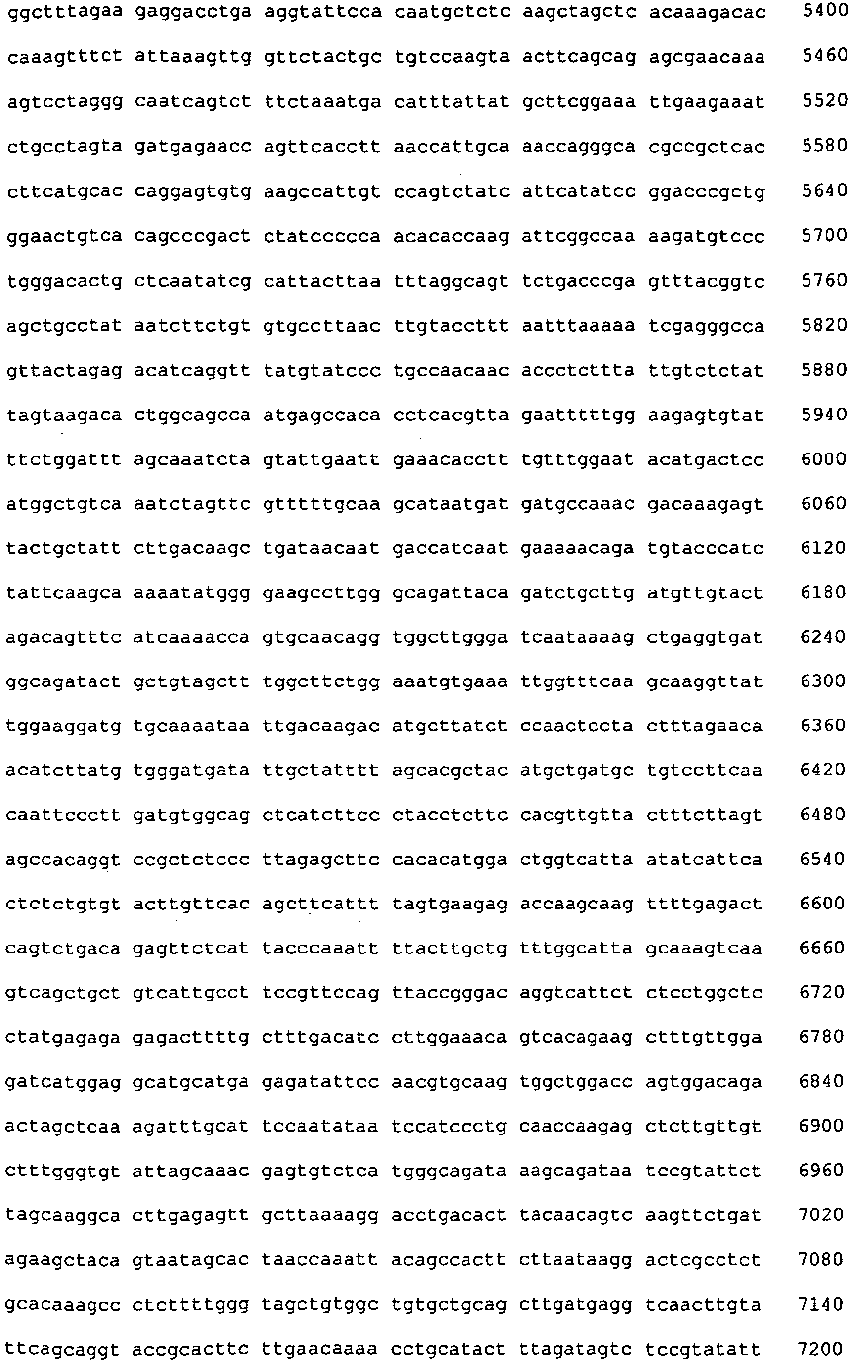

- <210> 23
<211> 2257
<212> DNA
<213> Homo sapiens - <400> 23
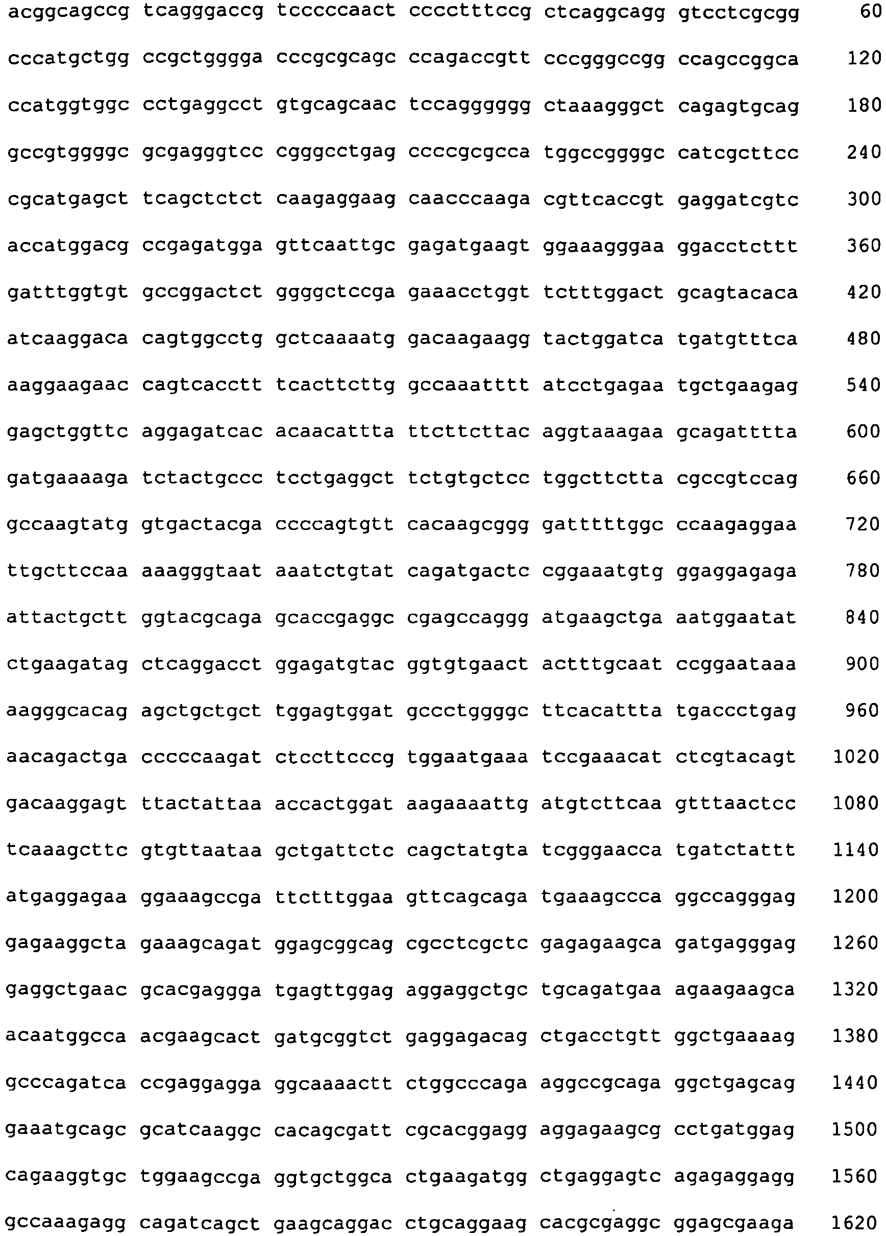

- <210> 24
<211> 2969
<212> DNA
<213> Homo sapiens - <400> 24

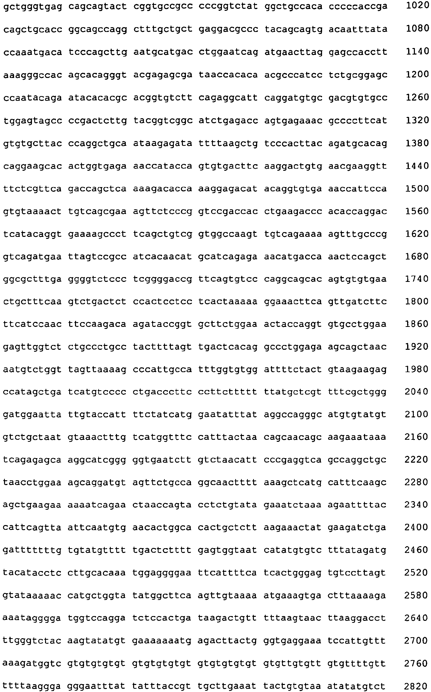

Claims (9)
- A reagent for cancer cell detection or cancer diagnosis, comprising a recombinant adenovirus where:- a replication cassette comprising a promoter from human telomerase, an adenoviral E1A gene, an IRES sequence and an adenoviral E1B gene in this order is integrated into the E1 region of the adenoviral genome; and- a labeling cassette comprising a gene encoding a labeling protein and a promoter capable of regulating the expression of the gene encoding the labeling protein is integrated into the E3 region of the adenoviral genome; and- the ADP-encoding E3 region of the viral genome is deleted.
- The reagent according to claim 1, wherein the reagent is for in vivo cancer detection or diagnosis, or for navigation surgery.
- The reagent according to claim 1 or 2, wherein the promoter from human telomerase is hTERT promoter.
- The reagent according to any one of the preceding claims, wherein the labeling protein is a fluorescent protein.
- The reagent according to claim 4, wherein the fluorescent protein is selected from the group consisting of green fluorescent protein (GFP), enhanced-humanized GFP (EGFP), red-shift GFP (rsGFP), yellow fluorescent protein (YFP), cyan fluorescent protein (CFP), blue fluorescent protein (BFP) and Renilla reniformis-derived GFP.
- The reagent according to claim 4, wherein the fluorescent protein is GFP or EGFP.
- The reagent according to any one of the preceding claims , wherein the promoter capable of regulating the expression of the gene encoding a labeling protein is a cytomegalovirus promoter or hTERT promoter.
- An ex vivo method of detecting cancer cells or cancer diagnosis, comprising infecting cancer cells with the reagent according to any one of claims 1 and 3 to 7, and detecting the fluorescence emitted by the cancer cells.
- Use of a recombinant adenovirus as defined in any one of claims 1 and 3 to 7 in the manufacture of a reagent for detecting cancer cells or for cancer diagnosis.
Applications Claiming Priority (2)
| Application Number | Priority Date | Filing Date | Title |
|---|---|---|---|
| JP2004285383 | 2004-09-29 | ||
| EP05790164A EP1795604B1 (en) | 2004-09-29 | 2005-09-28 | Telomelysin-gfp gene-containing recombinant virus |
Related Parent Applications (2)
| Application Number | Title | Priority Date | Filing Date |
|---|---|---|---|
| EP05790164A Division EP1795604B1 (en) | 2004-09-29 | 2005-09-28 | Telomelysin-gfp gene-containing recombinant virus |
| EP05790164.7 Division | 2005-09-28 |
Publications (2)
| Publication Number | Publication Date |
|---|---|
| EP2311499A1 EP2311499A1 (en) | 2011-04-20 |
| EP2311499B1 true EP2311499B1 (en) | 2012-08-29 |
Family
ID=36099363
Family Applications (2)
| Application Number | Title | Priority Date | Filing Date |
|---|---|---|---|
| EP05790164A Expired - Lifetime EP1795604B1 (en) | 2004-09-29 | 2005-09-28 | Telomelysin-gfp gene-containing recombinant virus |
| EP10014555A Expired - Lifetime EP2311499B1 (en) | 2004-09-29 | 2005-09-28 | Telomelysin-GFP gene-containing recombinant virus |
Family Applications Before (1)
| Application Number | Title | Priority Date | Filing Date |
|---|---|---|---|
| EP05790164A Expired - Lifetime EP1795604B1 (en) | 2004-09-29 | 2005-09-28 | Telomelysin-gfp gene-containing recombinant virus |
Country Status (15)
| Country | Link |
|---|---|
| US (3) | US7943373B2 (en) |
| EP (2) | EP1795604B1 (en) |
| JP (1) | JP5006045B2 (en) |
| KR (1) | KR20070059191A (en) |
| CN (1) | CN101035906A (en) |
| AT (1) | ATE520420T1 (en) |
| AU (1) | AU2005288034A1 (en) |
| BR (1) | BRPI0516191A (en) |
| CA (1) | CA2581969A1 (en) |
| EA (1) | EA011880B1 (en) |
| IL (1) | IL181876A0 (en) |
| MX (1) | MX2007002999A (en) |
| NZ (1) | NZ554799A (en) |
| WO (1) | WO2006036004A1 (en) |
| ZA (1) | ZA200703294B (en) |
Families Citing this family (28)
| Publication number | Priority date | Publication date | Assignee | Title |
|---|---|---|---|---|
| US7192208B2 (en) | 2003-09-02 | 2007-03-20 | Futurelogic, Inc. | Rewritable card printer |
| US8057296B2 (en) * | 2003-09-12 | 2011-11-15 | Igt | Gaming device including a card processing assembly having vertically-stacked card holders operable with thermally-printable data cards and portable card changeover machines |
| US20080153581A1 (en) * | 2003-09-12 | 2008-06-26 | Igt | Card loading system for a data card unit |
| US7494414B2 (en) | 2003-09-12 | 2009-02-24 | Igt | Gaming device having a card management system for the management of circulating data cards |
| US20080153580A1 (en) * | 2003-09-12 | 2008-06-26 | Igt | Bezel interface for a card loading system |
| JP5069871B2 (en) * | 2006-05-30 | 2012-11-07 | シスメックス株式会社 | Novel cancer cell detection sample preparation kit and cancer cell detection kit using the same |
| JP5010890B2 (en) * | 2006-10-11 | 2012-08-29 | オリンパス株式会社 | Biological sample analysis method |
| US8197334B2 (en) | 2007-10-29 | 2012-06-12 | Igt | Circulating data card apparatus and management system |
| US20090181931A1 (en) * | 2008-01-16 | 2009-07-16 | Oncolys Biopharma, Inc. | Antiviral activity of cidofovir against oncolytic viruses |
| US20110065117A1 (en) * | 2008-04-11 | 2011-03-17 | National University Corporation Okayama University | Mesothelioma Specific Transferred Promoter And Use Thereof |
| JPWO2010071114A1 (en) * | 2008-12-18 | 2012-05-31 | シスメックス株式会社 | Method for detecting cancer cells in a blood sample |
| CN101538556B (en) * | 2009-01-07 | 2011-09-21 | 四川大学华西第二医院 | Method for replicating adenovirus by way of selection and mutual compensation |
| US9624476B2 (en) | 2011-08-23 | 2017-04-18 | National Institute Of Biomedical Innovation | Conditionally replicating adenovirus |
| EP2749647B1 (en) * | 2011-08-23 | 2018-07-04 | National Institute of Biomedical Innovation | Conditionally replication-competent adenovirus |
| CA2865642C (en) * | 2012-03-14 | 2021-10-26 | Salk Institute For Biological Studies | Adenoviral tumor diagnostics |
| CN103088061B (en) * | 2013-01-15 | 2016-08-03 | 中国科学院微生物研究所 | A kind of RNA and carrier application in preparation prevention and/or treatment hepatocarcinoma product |
| SG11201507393TA (en) | 2013-03-14 | 2015-10-29 | Salk Inst For Biological Studi | Oncolytic adenovirus compositions |
| JP5812361B2 (en) * | 2013-07-11 | 2015-11-11 | 国立研究開発法人医薬基盤・健康・栄養研究所 | Novel Ad vector containing gene expression control mechanism |
| ES2899913T3 (en) | 2014-09-24 | 2022-03-15 | Salk Inst For Biological Studi | Oncolytic tumor viruses and methods of use |
| WO2016057387A1 (en) * | 2014-10-06 | 2016-04-14 | The Trustees Of The University Of Pennsylvania | Compositions and methods for isolation of circulating tumor cells (ctc) |
| DE102015111756A1 (en) | 2015-07-20 | 2017-01-26 | Eberhard Karls Universität Tübingen Medizinische Fakultät | Recombinant Orf virus vector |
| WO2017147265A1 (en) | 2016-02-23 | 2017-08-31 | Salk Institute For Biological Studies | High throughput assay for measuring adenovirus replication kinetics |
| EP3390645B1 (en) | 2016-02-23 | 2022-09-14 | Salk Institute for Biological Studies | Exogenous gene expression in therapeutic adenovirus for minimal impact on viral kinetics |
| AU2017375633C1 (en) | 2016-12-12 | 2023-04-27 | Salk Institute For Biological Studies | Tumor-targeting synthetic adenoviruses and uses thereof |
| BR112020019942A2 (en) | 2018-04-09 | 2021-01-26 | Salk Institute For Biological Studies | oncolytic adenovirus compositions with enhanced replication properties |
| KR102080996B1 (en) * | 2018-07-23 | 2020-02-25 | 주식회사 코어파마 | Cancer cell specific adenovirus |
| JPWO2021010369A1 (en) * | 2019-07-12 | 2021-01-21 | ||
| CN113444730A (en) * | 2021-03-17 | 2021-09-28 | 昆明市延安医院 | Screening and constructing method of primary hepatocyte klotho gene transduction stem cells |
Family Cites Families (5)
| Publication number | Priority date | Publication date | Assignee | Title |
|---|---|---|---|---|
| WO2001036650A2 (en) * | 1999-11-15 | 2001-05-25 | Onyx Pharmaceuticals, Inc. | An oncolytic adenovirus |
| WO2002012524A2 (en) * | 2000-08-03 | 2002-02-14 | Onyx Pharmaceuticals, Inc. | Adenovirus e1b-55k single amino acid mutants and methods of use |
| JP2004536607A (en) * | 2001-07-23 | 2004-12-09 | オニックス ファーマシューティカルズ,インコーポレイティド | Viral variants that selectively replicate in targeted human cancer cells |
| JP3867968B2 (en) | 2002-07-08 | 2007-01-17 | 関西ティー・エル・オー株式会社 | Oncolytic virus that selectively grows in tumor cells |
| JP3752544B2 (en) | 2003-02-03 | 2006-03-08 | 国立大学法人 長崎大学 | Vector and pharmaceutical composition containing cell death-inducing gene |
-
2005
- 2005-06-21 US US11/158,479 patent/US7943373B2/en not_active Expired - Lifetime
- 2005-09-28 BR BRPI0516191-6A patent/BRPI0516191A/en not_active Application Discontinuation
- 2005-09-28 EP EP05790164A patent/EP1795604B1/en not_active Expired - Lifetime
- 2005-09-28 EP EP10014555A patent/EP2311499B1/en not_active Expired - Lifetime
- 2005-09-28 NZ NZ554799A patent/NZ554799A/en not_active IP Right Cessation
- 2005-09-28 AU AU2005288034A patent/AU2005288034A1/en not_active Abandoned
- 2005-09-28 CA CA002581969A patent/CA2581969A1/en not_active Abandoned
- 2005-09-28 WO PCT/JP2005/018401 patent/WO2006036004A1/en not_active Ceased
- 2005-09-28 MX MX2007002999A patent/MX2007002999A/en not_active Application Discontinuation
- 2005-09-28 EA EA200700694A patent/EA011880B1/en active IP Right Revival
- 2005-09-28 AT AT05790164T patent/ATE520420T1/en not_active IP Right Cessation
- 2005-09-28 CN CNA2005800327883A patent/CN101035906A/en active Pending
- 2005-09-28 KR KR1020077009538A patent/KR20070059191A/en not_active Withdrawn
- 2005-09-28 JP JP2006537857A patent/JP5006045B2/en not_active Expired - Lifetime
-
2007
- 2007-03-12 IL IL181876A patent/IL181876A0/en unknown
- 2007-04-05 US US11/696,965 patent/US20080032283A1/en not_active Abandoned
- 2007-04-23 ZA ZA200703294A patent/ZA200703294B/en unknown
-
2010
- 2010-09-30 US US12/895,120 patent/US20110111480A1/en not_active Abandoned
Also Published As
| Publication number | Publication date |
|---|---|
| IL181876A0 (en) | 2007-07-04 |
| WO2006036004A1 (en) | 2006-04-06 |
| US20080032283A1 (en) | 2008-02-07 |
| NZ554799A (en) | 2009-03-31 |
| US20060067890A1 (en) | 2006-03-30 |
| KR20070059191A (en) | 2007-06-11 |
| EP1795604A1 (en) | 2007-06-13 |
| EP1795604B1 (en) | 2011-08-17 |
| ZA200703294B (en) | 2008-08-27 |
| US20110111480A1 (en) | 2011-05-12 |
| AU2005288034A1 (en) | 2006-04-06 |
| JPWO2006036004A1 (en) | 2008-05-15 |
| ATE520420T1 (en) | 2011-09-15 |
| MX2007002999A (en) | 2007-08-07 |
| BRPI0516191A (en) | 2008-08-26 |
| EP2311499A1 (en) | 2011-04-20 |
| US7943373B2 (en) | 2011-05-17 |
| CA2581969A1 (en) | 2006-04-06 |
| EP1795604A4 (en) | 2008-08-27 |
| EA200700694A1 (en) | 2007-08-31 |
| CN101035906A (en) | 2007-09-12 |
| EA011880B1 (en) | 2009-06-30 |
| JP5006045B2 (en) | 2012-08-22 |
Similar Documents
| Publication | Publication Date | Title |
|---|---|---|
| EP1795604B1 (en) | Telomelysin-gfp gene-containing recombinant virus | |
| Peng et al. | The use of the L-plastin promoter for adenoviral-mediated, tumor-specific gene expression in ovarian and bladder cancer cell lines | |
| RU2642293C2 (en) | Conditionally replicating adenovirus | |
| Chung et al. | Use of L-plastin promoter to develop an adenoviral system that confers transgene expression in ovarian cancer cells but not in normal mesothelial cells | |
| US6296845B1 (en) | Selective killing and diagnosis of p53+ neoplastic cells | |
| JP2010539923A (en) | Recombinant adenovirus comprising a tissue-specific promoter and a tumor-targeting trans-splicing ribozyme and uses thereof | |
| WO2016197592A1 (en) | Application of long noncoding rna, hnf1a-as1, in preparing drug for treating human malignant solid tumor | |
| JP6014029B2 (en) | REIC expression adenovirus vector | |
| KR101478869B1 (en) | Cancer gene therapeutic agent through regulation using microRNA | |
| CN102154369B (en) | Recombinant slow virus vector, recombinant slow virus and stem cell containing recombinant slow virus | |
| CA2464394A1 (en) | Methods and compositions for inducing tumor-specific cytotoxicity | |
| AU712816B2 (en) | Methods and compositions for determining the tumor suppressor status of cells | |
| JP5733705B2 (en) | System for increasing the activity of a specific promoter and vector carrying the system | |
| CN118562799A (en) | A GALC truncated promoter, viral vector and application thereof | |
| US9624476B2 (en) | Conditionally replicating adenovirus | |
| HK1104323A (en) | Telomelysin-gfp gene-containing recombinant virus | |
| EP2111235A1 (en) | A method for diagnosing disease using adenovirus harboring trans-splicing ribozyme by molecular imaging | |
| JP6153205B2 (en) | VA gene disrupted adenovirus vector and precursor vector for preparing the same | |
| KR20010075338A (en) | Use of specific hybrid promoters for controlling tissue expression | |
| CN115651932A (en) | Construction method and application of targeted digestive tract tumor dual-targeted oncolytic adenovirus | |
| Chinnasamy | Developing a Quantitative PCR Assay for Detecting Viral Vector Shedding from Animals | |
| NZ622585B2 (en) | Conditionally replicating adenovirus | |
| JPWO2009125626A1 (en) | Mesothelioma-specific promoter and uses thereof |
Legal Events
| Date | Code | Title | Description |
|---|---|---|---|
| PUAI | Public reference made under article 153(3) epc to a published international application that has entered the european phase |
Free format text: ORIGINAL CODE: 0009012 |
|
| AC | Divisional application: reference to earlier application |
Ref document number: 1795604 Country of ref document: EP Kind code of ref document: P |
|
| AK | Designated contracting states |
Kind code of ref document: A1 Designated state(s): AT BE BG CH CY CZ DE DK EE ES FI FR GB GR HU IE IS IT LI LT LU LV MC NL PL PT RO SE SI SK TR |
|
| 17P | Request for examination filed |
Effective date: 20111020 |
|
| REG | Reference to a national code |
Ref country code: DE Ref legal event code: R079 Ref document number: 602005035952 Country of ref document: DE Free format text: PREVIOUS MAIN CLASS: A61K0049000000 Ipc: A61K0035760000 |
|
| RIC1 | Information provided on ipc code assigned before grant |
Ipc: A61K 35/76 20060101AFI20120112BHEP Ipc: C12N 15/861 20060101ALI20120112BHEP Ipc: G01N 33/574 20060101ALI20120112BHEP |
|
| GRAP | Despatch of communication of intention to grant a patent |
Free format text: ORIGINAL CODE: EPIDOSNIGR1 |
|
| RIN1 | Information on inventor provided before grant (corrected) |
Inventor name: KYO, SATORU Inventor name: TANAKA, NORIAKI Inventor name: MIZUGUCHI, HIROYUKI Inventor name: FUJIWARA, TOSHIYOSHI Inventor name: HAYAKAWA, TAKAO |
|
| GRAS | Grant fee paid |
Free format text: ORIGINAL CODE: EPIDOSNIGR3 |
|
| GRAA | (expected) grant |
Free format text: ORIGINAL CODE: 0009210 |
|
| AC | Divisional application: reference to earlier application |
Ref document number: 1795604 Country of ref document: EP Kind code of ref document: P |
|
| AK | Designated contracting states |
Kind code of ref document: B1 Designated state(s): AT BE BG CH CY CZ DE DK EE ES FI FR GB GR HU IE IS IT LI LT LU LV MC NL PL PT RO SE SI SK TR |
|
| REG | Reference to a national code |
Ref country code: GB Ref legal event code: FG4D |
|
| REG | Reference to a national code |
Ref country code: CH Ref legal event code: EP |
|
| REG | Reference to a national code |
Ref country code: AT Ref legal event code: REF Ref document number: 572683 Country of ref document: AT Kind code of ref document: T Effective date: 20120915 |
|
| REG | Reference to a national code |
Ref country code: NL Ref legal event code: T3 |
|
| REG | Reference to a national code |
Ref country code: IE Ref legal event code: FG4D |
|
| REG | Reference to a national code |
Ref country code: DE Ref legal event code: R096 Ref document number: 602005035952 Country of ref document: DE Effective date: 20121025 |
|
| REG | Reference to a national code |
Ref country code: AT Ref legal event code: MK05 Ref document number: 572683 Country of ref document: AT Kind code of ref document: T Effective date: 20120829 |
|
| REG | Reference to a national code |
Ref country code: LT Ref legal event code: MG4D Effective date: 20120829 |
|
| PG25 | Lapsed in a contracting state [announced via postgrant information from national office to epo] |
Ref country code: LT Free format text: LAPSE BECAUSE OF FAILURE TO SUBMIT A TRANSLATION OF THE DESCRIPTION OR TO PAY THE FEE WITHIN THE PRESCRIBED TIME-LIMIT Effective date: 20120829 Ref country code: IS Free format text: LAPSE BECAUSE OF FAILURE TO SUBMIT A TRANSLATION OF THE DESCRIPTION OR TO PAY THE FEE WITHIN THE PRESCRIBED TIME-LIMIT Effective date: 20121229 Ref country code: AT Free format text: LAPSE BECAUSE OF FAILURE TO SUBMIT A TRANSLATION OF THE DESCRIPTION OR TO PAY THE FEE WITHIN THE PRESCRIBED TIME-LIMIT Effective date: 20120829 Ref country code: FI Free format text: LAPSE BECAUSE OF FAILURE TO SUBMIT A TRANSLATION OF THE DESCRIPTION OR TO PAY THE FEE WITHIN THE PRESCRIBED TIME-LIMIT Effective date: 20120829 |
|
| PG25 | Lapsed in a contracting state [announced via postgrant information from national office to epo] |
Ref country code: SE Free format text: LAPSE BECAUSE OF FAILURE TO SUBMIT A TRANSLATION OF THE DESCRIPTION OR TO PAY THE FEE WITHIN THE PRESCRIBED TIME-LIMIT Effective date: 20120829 Ref country code: SI Free format text: LAPSE BECAUSE OF FAILURE TO SUBMIT A TRANSLATION OF THE DESCRIPTION OR TO PAY THE FEE WITHIN THE PRESCRIBED TIME-LIMIT Effective date: 20120829 Ref country code: LV Free format text: LAPSE BECAUSE OF FAILURE TO SUBMIT A TRANSLATION OF THE DESCRIPTION OR TO PAY THE FEE WITHIN THE PRESCRIBED TIME-LIMIT Effective date: 20120829 Ref country code: GR Free format text: LAPSE BECAUSE OF FAILURE TO SUBMIT A TRANSLATION OF THE DESCRIPTION OR TO PAY THE FEE WITHIN THE PRESCRIBED TIME-LIMIT Effective date: 20121130 Ref country code: BE Free format text: LAPSE BECAUSE OF FAILURE TO SUBMIT A TRANSLATION OF THE DESCRIPTION OR TO PAY THE FEE WITHIN THE PRESCRIBED TIME-LIMIT Effective date: 20120829 Ref country code: PT Free format text: LAPSE BECAUSE OF FAILURE TO SUBMIT A TRANSLATION OF THE DESCRIPTION OR TO PAY THE FEE WITHIN THE PRESCRIBED TIME-LIMIT Effective date: 20121231 |
|
| PG25 | Lapsed in a contracting state [announced via postgrant information from national office to epo] |
Ref country code: DK Free format text: LAPSE BECAUSE OF FAILURE TO SUBMIT A TRANSLATION OF THE DESCRIPTION OR TO PAY THE FEE WITHIN THE PRESCRIBED TIME-LIMIT Effective date: 20120829 Ref country code: RO Free format text: LAPSE BECAUSE OF FAILURE TO SUBMIT A TRANSLATION OF THE DESCRIPTION OR TO PAY THE FEE WITHIN THE PRESCRIBED TIME-LIMIT Effective date: 20120829 Ref country code: EE Free format text: LAPSE BECAUSE OF FAILURE TO SUBMIT A TRANSLATION OF THE DESCRIPTION OR TO PAY THE FEE WITHIN THE PRESCRIBED TIME-LIMIT Effective date: 20120829 Ref country code: CZ Free format text: LAPSE BECAUSE OF FAILURE TO SUBMIT A TRANSLATION OF THE DESCRIPTION OR TO PAY THE FEE WITHIN THE PRESCRIBED TIME-LIMIT Effective date: 20120829 Ref country code: MC Free format text: LAPSE BECAUSE OF NON-PAYMENT OF DUE FEES Effective date: 20120930 Ref country code: ES Free format text: LAPSE BECAUSE OF FAILURE TO SUBMIT A TRANSLATION OF THE DESCRIPTION OR TO PAY THE FEE WITHIN THE PRESCRIBED TIME-LIMIT Effective date: 20121210 |
|
| REG | Reference to a national code |
Ref country code: CH Ref legal event code: PL |
|
| PG25 | Lapsed in a contracting state [announced via postgrant information from national office to epo] |
Ref country code: SK Free format text: LAPSE BECAUSE OF FAILURE TO SUBMIT A TRANSLATION OF THE DESCRIPTION OR TO PAY THE FEE WITHIN THE PRESCRIBED TIME-LIMIT Effective date: 20120829 Ref country code: PL Free format text: LAPSE BECAUSE OF FAILURE TO SUBMIT A TRANSLATION OF THE DESCRIPTION OR TO PAY THE FEE WITHIN THE PRESCRIBED TIME-LIMIT Effective date: 20120829 |
|
| REG | Reference to a national code |
Ref country code: IE Ref legal event code: MM4A |
|
| PLBE | No opposition filed within time limit |
Free format text: ORIGINAL CODE: 0009261 |
|
| STAA | Information on the status of an ep patent application or granted ep patent |
Free format text: STATUS: NO OPPOSITION FILED WITHIN TIME LIMIT |
|
| PG25 | Lapsed in a contracting state [announced via postgrant information from national office to epo] |
Ref country code: CH Free format text: LAPSE BECAUSE OF NON-PAYMENT OF DUE FEES Effective date: 20120930 Ref country code: BG Free format text: LAPSE BECAUSE OF FAILURE TO SUBMIT A TRANSLATION OF THE DESCRIPTION OR TO PAY THE FEE WITHIN THE PRESCRIBED TIME-LIMIT Effective date: 20121129 Ref country code: IE Free format text: LAPSE BECAUSE OF NON-PAYMENT OF DUE FEES Effective date: 20120928 Ref country code: LI Free format text: LAPSE BECAUSE OF NON-PAYMENT OF DUE FEES Effective date: 20120930 |
|
| 26N | No opposition filed |
Effective date: 20130530 |
|
| REG | Reference to a national code |
Ref country code: DE Ref legal event code: R097 Ref document number: 602005035952 Country of ref document: DE Effective date: 20130530 |
|
| PG25 | Lapsed in a contracting state [announced via postgrant information from national office to epo] |
Ref country code: CY Free format text: LAPSE BECAUSE OF FAILURE TO SUBMIT A TRANSLATION OF THE DESCRIPTION OR TO PAY THE FEE WITHIN THE PRESCRIBED TIME-LIMIT Effective date: 20120829 |
|
| PG25 | Lapsed in a contracting state [announced via postgrant information from national office to epo] |
Ref country code: TR Free format text: LAPSE BECAUSE OF FAILURE TO SUBMIT A TRANSLATION OF THE DESCRIPTION OR TO PAY THE FEE WITHIN THE PRESCRIBED TIME-LIMIT Effective date: 20120829 |
|
| PG25 | Lapsed in a contracting state [announced via postgrant information from national office to epo] |
Ref country code: LU Free format text: LAPSE BECAUSE OF NON-PAYMENT OF DUE FEES Effective date: 20120928 |
|
| PG25 | Lapsed in a contracting state [announced via postgrant information from national office to epo] |
Ref country code: HU Free format text: LAPSE BECAUSE OF FAILURE TO SUBMIT A TRANSLATION OF THE DESCRIPTION OR TO PAY THE FEE WITHIN THE PRESCRIBED TIME-LIMIT Effective date: 20050928 |
|
| REG | Reference to a national code |
Ref country code: FR Ref legal event code: PLFP Year of fee payment: 12 |
|
| REG | Reference to a national code |
Ref country code: FR Ref legal event code: PLFP Year of fee payment: 13 |
|
| REG | Reference to a national code |
Ref country code: FR Ref legal event code: PLFP Year of fee payment: 14 |
|
| PGFP | Annual fee paid to national office [announced via postgrant information from national office to epo] |
Ref country code: DE Payment date: 20240918 Year of fee payment: 20 |
|
| PGFP | Annual fee paid to national office [announced via postgrant information from national office to epo] |
Ref country code: GB Payment date: 20240919 Year of fee payment: 20 |
|
| PGFP | Annual fee paid to national office [announced via postgrant information from national office to epo] |
Ref country code: FR Payment date: 20240925 Year of fee payment: 20 |
|
| PGFP | Annual fee paid to national office [announced via postgrant information from national office to epo] |
Ref country code: NL Payment date: 20240918 Year of fee payment: 20 |
|
| PGFP | Annual fee paid to national office [announced via postgrant information from national office to epo] |
Ref country code: IT Payment date: 20240924 Year of fee payment: 20 |
|
| REG | Reference to a national code |
Ref country code: DE Ref legal event code: R071 Ref document number: 602005035952 Country of ref document: DE |
|
| REG | Reference to a national code |
Ref country code: NL Ref legal event code: MK Effective date: 20250927 |
|
| REG | Reference to a national code |
Ref country code: GB Ref legal event code: PE20 Expiry date: 20250927 |