EP2302587A1 - Ultrasound system and method of measuring fetal rib - Google Patents
Ultrasound system and method of measuring fetal rib Download PDFInfo
- Publication number
- EP2302587A1 EP2302587A1 EP10171357A EP10171357A EP2302587A1 EP 2302587 A1 EP2302587 A1 EP 2302587A1 EP 10171357 A EP10171357 A EP 10171357A EP 10171357 A EP10171357 A EP 10171357A EP 2302587 A1 EP2302587 A1 EP 2302587A1
- Authority
- EP
- European Patent Office
- Prior art keywords
- ribs
- rib
- image
- roi
- measuring unit
- Prior art date
- Legal status (The legal status is an assumption and is not a legal conclusion. Google has not performed a legal analysis and makes no representation as to the accuracy of the status listed.)
- Withdrawn
Links
Images




Classifications
-
- A—HUMAN NECESSITIES
- A61—MEDICAL OR VETERINARY SCIENCE; HYGIENE
- A61B—DIAGNOSIS; SURGERY; IDENTIFICATION
- A61B8/00—Diagnosis using ultrasonic, sonic or infrasonic waves
-
- G—PHYSICS
- G06—COMPUTING OR CALCULATING; COUNTING
- G06T—IMAGE DATA PROCESSING OR GENERATION, IN GENERAL
- G06T7/00—Image analysis
- G06T7/0002—Inspection of images, e.g. flaw detection
- G06T7/0004—Industrial image inspection
-
- G—PHYSICS
- G06—COMPUTING OR CALCULATING; COUNTING
- G06T—IMAGE DATA PROCESSING OR GENERATION, IN GENERAL
- G06T2207/00—Indexing scheme for image analysis or image enhancement
- G06T2207/10—Image acquisition modality
- G06T2207/10132—Ultrasound image
-
- G—PHYSICS
- G06—COMPUTING OR CALCULATING; COUNTING
- G06T—IMAGE DATA PROCESSING OR GENERATION, IN GENERAL
- G06T2207/00—Indexing scheme for image analysis or image enhancement
- G06T2207/30—Subject of image; Context of image processing
- G06T2207/30004—Biomedical image processing
- G06T2207/30044—Fetus; Embryo
Definitions
- the present invention relates to an ultrasound system and method of measuring a number of ribs of a fetus, and more particularly, to an ultrasound system and method of setting a portion where the number of ribs is measured as a region of interest (ROI), measuring the number of ribs in the set ROI, and displaying the measured number of ribs.
- ROI region of interest
- an ultrasound system may transmit an ultrasound signal toward a predetermined portion inside a body from a surface of the body, and may obtain an image about a section of a soft tissue or a bloodstream by using information of the ultrasound signal having been reflected from a tissue inside the body.
- the ultrasound system has an advantage of being small, inexpensive, reliable, and not exposing a subject to an X-ray and the like, and thus, the ultrasound system is commonly used together with other image diagnostic devices, such as a computerized tomography (CT) scanner, a magnetic resonance image (MRI) device, a nuclear medicine device, and the like. Particularly, the ultrasound system may display an image of inside the body in real time, thereby being applicable for various uses.
- CT computerized tomography
- MRI magnetic resonance image
- nuclear medicine device a nuclear medicine device
- the ultrasound system may need to obtain a three-dimensional (3D) ultrasound image.
- a number of ribs of a fetus is generally twelve in a 3D ultrasound image as illustrated in FIG. 1 . However, when the number of ribs is eleven, there is a higher probability that the fetus has Down's syndrome. Accordingly, the number of ribs of the fetus is an important index for checking a probability of a disorder in the fetus.
- a conventional system may have inconvenience in that a user calculates the number of ribs of the fetus by hand from a rendered 3D image as illustrated in FIG. 1 .
- An aspect of the present invention provides an ultrasound system and method that accurately measures a number of ribs of a fetus in a three-dimensional (3D) ultrasound image.
- Another aspect of the present invention provides an ultrasound system and method that displays a rib area and a number of ribs extracted from a 3D ultrasound image.
- an ultrasound system of measuring a number ribs of a fetus includes a setting unit to set, by a user, a portion where the number of ribs is measured as a region of interest (ROI) in an ultrasound image, a measuring unit to measure the number of ribs in the set ROI, by using an auto rib extracting algorithm, and a display unit to display the measured number of ribs.
- ROI region of interest
- the measuring unit may enhance a rib image in the ROI, may generate a binary image by extracting an area having a brightness greater than or equal to a reference value from the enhanced rib image, may classify discrete areas from the binary image, may connect the classified discrete areas along a principle axis to generate rib areas, and may count a number of the rib areas to measure the number of ribs.
- the measuring unit may enhance a picture quality or a definition of the rib image through a rib enhancement filtering process for segmentation of a ROI image.
- the display unit may display the measured rib area and the number of ribs.
- a method of measuring a number of ribs in an ultrasound system including a setting unit, a measuring unit, and a display unit, the method including setting, by a user, an ROI to measure, through the setting unit, the number of ribs in an ultrasound area, measuring, by the measuring unit, the number of ribs in the ROI, based on an auto rib extracting algorithm, and displaying, by the display unit, information about the measured number of ribs.
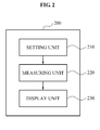
- FIG. 2 illustrates a format of an ultrasound system 200 of measuring a number of ribs of a fetus according to an exemplary embodiment of the present invention.
- the ultrasound system 200 that measures the number of ribs of the fetus may include a setting unit 210, a measuring unit 220, and a display unit 230.
- a user sets, through the setting unit 210, a portion where the number of ribs is measured in an ultrasound image, as a region of interest (ROI).
- ROI region of interest
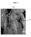
- FIG. 3 illustrates an example where a RIO 310 is set to measure a number of ribs according to an exemplary embodiment of the present invention
- a user may set a portion where the number of ribs is measured in a 3D ultrasound image 300 as an ROI 310, through the setting unit 210.
- the measuring unit 220 may measure the number of ribs in the set ROI 310 based on an auto rib extracting algorithm.
- the measuring unit 220 may enhance a rib image in the ROI 310, may generate a binary image by extracting an area having a brightness greater than or equal to a reference value from the enhanced rib image, may classify discrete areas from the binary image, may connect the classified discrete areas along a principle axis to generate rib areas, and may count a number of the rib areas to measure the number of ribs.
- FIG. 4 illustrates an example of a result from each operation of an auto rib extracting algorithm according to an exemplary embodiment of the present invention.
- the measuring unit 220 may generate an enhanced image 420 by performing a rib enhancement filtering process for segmentation of an ROI image 410, as a preprocessing with respect to the ROI image 410.
- the enhanced image 420 is a 3D ultrasound image of which a picture quality or a definition is enhanced to make a rib to be easily observed in the ROI image 410. That is, the measuring unit 220 may enhance a picture quality or a definition of a rib image through the rib enhancement filtering process for segmentation of the ROI image 410.
- the measuring unit 220 may perform the rib enhancement filtering process with respect to the ROI image 410 through an image enhancement process using various schemes, such as a smoothing scheme, a sharpening scheme, and the like.
- the measuring unit 220 may generate a binary image by extracting an area having a brightness greater than or equal to a reference value from the enhanced rib image 420. Also, the measuring unit 220 may generate a discrete image 430 by classifying discrete images from the binary image. Also, the measuring unit 220 may generate a rib area image 440 by connecting the classified discrete areas along a principle axis and generating rib areas. Also, the measuring unit 220 may count a number of the rib areas, thereby generating a result image 450 indicating a result of the number of ribs.
- the display unit 230 may display the measured number of ribs.
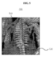
- FIG. 5 illustrates an example of displaying a measured rib area and a number of ribs according to an exemplary embodiment of the present invention.
- the display unit 230 may display a measured rib area 510 and a number of ribs 520 in a 3D ultrasound image 500.
- the measured rib area 510 may be classified into true ribs 511 and false ribs 521.
- the ultrasound system 200 may set a portion where a number of ribs is measured in a 3D ultrasound image, as an ROI, may measure a number ribs in the set ROI, and may display the measured number of ribs.
- the ultrasound system 200 may display the extracted rib area and the number of ribs, and may alleviate the inconvenience of counting the number of ribs, which is one of important indexes, by hand, thereby increasing a user's convenience.
- FIG. 6 is a flowchart illustrating a method of measuring a number of ribs of a fetus according to an exemplary embodiment of the present invention.
- a user sets an ROI where a number of ribs of a fetus is measured in an ultrasound image, through the setting unit 210.
- the user may set a portion where the number of ribs is measured in the 3D ultrasound image 300 as the ROI 310.
- the measuring unit 220 measures the number of ribs in the ROI based on an auto rib extracting algorithm.
- an auto rib extracting algorithm the measuring of the number of ribs of the fetus based on the auto rib extracting algorithm, will be described in detail.
- FIG. 7 illustrates a detailed procedure of measuring a number of ribs of a fetus according to an exemplary embodiment of the present invention.
- the measuring unit 220 performs a preprocessing to enhance a rib image in the ROI. That is, in operation S710, the measuring unit 220 may enhance a picture quality or a definition of a 3D ultrasound image to make the rib image included in the ROI to be clearly shown, through the rib enhancement filtering process for segmentation of the ROI image. As an example, in operation S720, the measuring unit 220 generates an enhanced image 420 that is a 3D ultrasound image of which a picture quality or a definition of is enhanced, as a result of performing the preprocessing on the ROI image 410, the preprocessing being the image enhancement process using various schemes, such as a smoothing scheme, a sharpening scheme, and the like.
- the measuring unit 220 In operation S720, the measuring unit 220 generates a binary image by extracting an area having a brightness greater than or equal to a reference value in the enhanced rib image.
- the measuring unit 220 classifies discrete areas based on levels of areas in the binary images. That is, in operation S730, the measuring unit 220 generates the discrete area image 430 by classifying the discrete areas from the binary image.
- the measuring unit 220 connects the classified discrete areas along a principle axis to generate rib areas. That is, in operation S740, the measuring unit 220 generates a rib area image 440 by linking discontinuous areas of the classified areas along the principle axis.
- the measuring unit 220 measures information about the number of ribs by counting a number of the rib areas. As an example, in operation S750, the measuring unit 220 measures the number of ribs by counting the rib areas in the result image 450.
- the display unit 230 displays information about the measured number of ribs.
- the display unit 230 displays the extracted rib area 510 and the number of ribs 520 extracted from the 3D ultrasound image 300, as the information about the measured number of ribs.
- a method of measuring a number of ribs may set a portion where the number of ribs is measured in a 3D ultrasound image as an ROI, may automatically measure the number of ribs in the set ROI, and may display the measured the number of ribs, thereby removing the inconvenience of counting the number of ribs, which is one of important indexes, by hand. Therefore, user's convenience may increase.
- a method of measuring a number of ribs of a fetus may be recorded in computer-readable media including program instructions to implement various operations embodied by a computer.
- the media may also include, alone or in combination with the program instructions, data files, data structures, and the like.
- Examples of computer-readable media include magnetic media such as hard disks, floppy disks, and magnetic tape; optical media such as CD ROM disks and DVDs; magneto-optical media such as optical disks; and hardware devices that are specially configured to store and perform program instructions, such as read-only memory (ROM), random access memory (RAM), flash memory, and the like.
- Examples of program instructions include both machine code, such as produced by a compiler, and files containing higher level code that may be executed by the computer using an interpreter.
- the described hardware devices may be configured to act as one or more software modules in order to perform the operations of the above-described exemplary embodiments of the present invention, or vice versa.
Landscapes
- Engineering & Computer Science (AREA)
- Physics & Mathematics (AREA)
- Health & Medical Sciences (AREA)
- Life Sciences & Earth Sciences (AREA)
- Quality & Reliability (AREA)
- Computer Vision & Pattern Recognition (AREA)
- General Physics & Mathematics (AREA)
- Theoretical Computer Science (AREA)
- Pathology (AREA)
- Molecular Biology (AREA)
- Biophysics (AREA)
- Radiology & Medical Imaging (AREA)
- Biomedical Technology (AREA)
- Heart & Thoracic Surgery (AREA)
- Medical Informatics (AREA)
- Nuclear Medicine, Radiotherapy & Molecular Imaging (AREA)
- Surgery (AREA)
- Animal Behavior & Ethology (AREA)
- General Health & Medical Sciences (AREA)
- Public Health (AREA)
- Veterinary Medicine (AREA)
- Ultra Sonic Daignosis Equipment (AREA)
Abstract
Disclosed are an ultrasound system of measuring a number of ribs of a fetus and a method thereof. The ultrasound system of measuring the number of ribs of the fetus in three dimensions may include a setting unit that is used by a user, to set a portion where the number of ribs is measured in an ultrasound image, as a region of interest (ROI), a measuring unit to measure the number of ribs in the set ROI by using an auto rib extracting algorithm, and a display unit to display the measured number of ribs.
Description
- This application claims the benefit of Korean Patent Application No.
10-2009-0077096, filed on August 20, 2009 - The present invention relates to an ultrasound system and method of measuring a number of ribs of a fetus, and more particularly, to an ultrasound system and method of setting a portion where the number of ribs is measured as a region of interest (ROI), measuring the number of ribs in the set ROI, and displaying the measured number of ribs.
- Generally, an ultrasound system may transmit an ultrasound signal toward a predetermined portion inside a body from a surface of the body, and may obtain an image about a section of a soft tissue or a bloodstream by using information of the ultrasound signal having been reflected from a tissue inside the body.
- The ultrasound system has an advantage of being small, inexpensive, reliable, and not exposing a subject to an X-ray and the like, and thus, the ultrasound system is commonly used together with other image diagnostic devices, such as a computerized tomography (CT) scanner, a magnetic resonance image (MRI) device, a nuclear medicine device, and the like. Particularly, the ultrasound system may display an image of inside the body in real time, thereby being applicable for various uses.
- As a utilization field of the ultrasound system is expanded, there are various demands on an ultrasound image of the ultrasound system. In particular, a lesion or a tissue of a patient may need to be precisely observed when a procedure, such as a medical checkup, a biopsy, an operation, and the like, is performed. Accordingly, the ultrasound system may need to obtain a three-dimensional (3D) ultrasound image.
- A number of ribs of a fetus is generally twelve in a 3D ultrasound image as illustrated in
FIG. 1 . However, when the number of ribs is eleven, there is a higher probability that the fetus has Down's syndrome. Accordingly, the number of ribs of the fetus is an important index for checking a probability of a disorder in the fetus. - A conventional system may have inconvenience in that a user calculates the number of ribs of the fetus by hand from a rendered 3D image as illustrated in
FIG. 1 . - An aspect of the present invention provides an ultrasound system and method that accurately measures a number of ribs of a fetus in a three-dimensional (3D) ultrasound image.
- Another aspect of the present invention provides an ultrasound system and method that displays a rib area and a number of ribs extracted from a 3D ultrasound image.
- According to an aspect of the present invention, there is provided an ultrasound system of measuring a number ribs of a fetus, the ultrasound system includes a setting unit to set, by a user, a portion where the number of ribs is measured as a region of interest (ROI) in an ultrasound image, a measuring unit to measure the number of ribs in the set ROI, by using an auto rib extracting algorithm, and a display unit to display the measured number of ribs.
- The measuring unit may enhance a rib image in the ROI, may generate a binary image by extracting an area having a brightness greater than or equal to a reference value from the enhanced rib image, may classify discrete areas from the binary image, may connect the classified discrete areas along a principle axis to generate rib areas, and may count a number of the rib areas to measure the number of ribs.
- The measuring unit may enhance a picture quality or a definition of the rib image through a rib enhancement filtering process for segmentation of a ROI image.
- The display unit may display the measured rib area and the number of ribs.
- According to another aspect of the present invention, there is provided a method of measuring a number of ribs in an ultrasound system including a setting unit, a measuring unit, and a display unit, the method including setting, by a user, an ROI to measure, through the setting unit, the number of ribs in an ultrasound area, measuring, by the measuring unit, the number of ribs in the ROI, based on an auto rib extracting algorithm, and displaying, by the display unit, information about the measured number of ribs.
- Additional aspects and/or advantages will be set forth in part in the description which follows and, in part, will be apparent from the description, or may be learned by practice of the embodiments.
- According to the present invention, there is no need for counting a number of ribs by hand, since the number of ribs is automatically counted from a 3D ultrasound image, and thus, a user's convenience increases.
- According to the present invention, there is provided an effect of easily obtaining a rib area and a number of ribs extracted from a 3D ultrasound image.
- These and/or other aspects and advantages will become apparent and more readily appreciated from the following description of the embodiments, taken in conjunction with the accompanying drawings of which:
- FIG. 1
- is a diagram illustrating a number of ribs of a normal fetus in a three dimensional (3D) ultrasound image, according to a conventional art;
- FIG. 2
- is a diagram illustrating a format of an ultrasound system of measuring a number of ribs of a fetus according to an exemplary embodiment of the present invention;
- FIG. 3
- is a diagram illustrating an example where a region of interest (RIO) is set to measure a number of ribs according to an exemplary embodiment of the present invention;
- FIG. 4
- is a diagram illustrating an example of a result from each operation of an auto rib extracting algorithm according to an exemplary embodiment of the present invention;
- FIG. 5
- is a diagram illustrating an example of displaying a measured rib area and a number of ribs according to an exemplary embodiment of the present invention;
- FIG. 6
- is a flowchart illustrating a method of measuring a number of ribs of a fetus according to an exemplary embodiment of the present invention; and
- FIG. 7
- is a diagram illustrating a detailed procedure of measuring a number of ribs of a fetus according to an exemplary embodiment of the present invention.
- Reference will now be made in detail to example embodiments, examples of which are illustrated in the accompanying drawings, wherein like reference numerals refer to the like elements throughout. An ultrasound diagnosis apparatus utilizing a touch interaction is described below to explain the present disclosure by referring to the figures.
-
FIG. 2 illustrates a format of anultrasound system 200 of measuring a number of ribs of a fetus according to an exemplary embodiment of the present invention. - Referring to
FIG. 2 , theultrasound system 200 that measures the number of ribs of the fetus according to an embodiment of the present invention may include asetting unit 210, ameasuring unit 220, and adisplay unit 230. A user sets, through thesetting unit 210, a portion where the number of ribs is measured in an ultrasound image, as a region of interest (ROI). -
FIG. 3 illustrates an example where a RIO 310 is set to measure a number of ribs according to an exemplary embodiment of the present invention; - Referring to
FIG. 3 , a user may set a portion where the number of ribs is measured in a3D ultrasound image 300 as anROI 310, through thesetting unit 210. - The
measuring unit 220 may measure the number of ribs in theset ROI 310 based on an auto rib extracting algorithm. In other words, themeasuring unit 220 may enhance a rib image in theROI 310, may generate a binary image by extracting an area having a brightness greater than or equal to a reference value from the enhanced rib image, may classify discrete areas from the binary image, may connect the classified discrete areas along a principle axis to generate rib areas, and may count a number of the rib areas to measure the number of ribs. -
FIG. 4 illustrates an example of a result from each operation of an auto rib extracting algorithm according to an exemplary embodiment of the present invention. - Referring to
FIG. 4 , themeasuring unit 220 may generate an enhancedimage 420 by performing a rib enhancement filtering process for segmentation of anROI image 410, as a preprocessing with respect to theROI image 410. The enhancedimage 420 is a 3D ultrasound image of which a picture quality or a definition is enhanced to make a rib to be easily observed in theROI image 410. That is, themeasuring unit 220 may enhance a picture quality or a definition of a rib image through the rib enhancement filtering process for segmentation of theROI image 410. As an example, themeasuring unit 220 may perform the rib enhancement filtering process with respect to theROI image 410 through an image enhancement process using various schemes, such as a smoothing scheme, a sharpening scheme, and the like. Also, themeasuring unit 220 may generate a binary image by extracting an area having a brightness greater than or equal to a reference value from the enhancedrib image 420. Also, themeasuring unit 220 may generate adiscrete image 430 by classifying discrete images from the binary image. Also, themeasuring unit 220 may generate arib area image 440 by connecting the classified discrete areas along a principle axis and generating rib areas. Also, themeasuring unit 220 may count a number of the rib areas, thereby generating aresult image 450 indicating a result of the number of ribs. - The
display unit 230 may display the measured number of ribs. -
FIG. 5 illustrates an example of displaying a measured rib area and a number of ribs according to an exemplary embodiment of the present invention. - Referring to
FIG. 5 , thedisplay unit 230 may display a measuredrib area 510 and a number ofribs 520 in a3D ultrasound image 500. As an example, the measuredrib area 510 may be classified into true ribs 511 and false ribs 521. - As described above, the
ultrasound system 200 may set a portion where a number of ribs is measured in a 3D ultrasound image, as an ROI, may measure a number ribs in the set ROI, and may display the measured number of ribs. - Accordingly, the
ultrasound system 200 may display the extracted rib area and the number of ribs, and may alleviate the inconvenience of counting the number of ribs, which is one of important indexes, by hand, thereby increasing a user's convenience. -
FIG. 6 is a flowchart illustrating a method of measuring a number of ribs of a fetus according to an exemplary embodiment of the present invention. - Referring to
FIGS. 2 to 6 , in operation S610, a user sets an ROI where a number of ribs of a fetus is measured in an ultrasound image, through thesetting unit 210. As an example, the user may set a portion where the number of ribs is measured in the3D ultrasound image 300 as theROI 310. - In operation S620, the measuring
unit 220 measures the number of ribs in the ROI based on an auto rib extracting algorithm. Hereinafter, referring toFIG. 7 , the measuring of the number of ribs of the fetus based on the auto rib extracting algorithm, will be described in detail. -
FIG. 7 illustrates a detailed procedure of measuring a number of ribs of a fetus according to an exemplary embodiment of the present invention. - Referring to
FIG. 7 , in operation S710, the measuringunit 220 performs a preprocessing to enhance a rib image in the ROI. That is, in operation S710, the measuringunit 220 may enhance a picture quality or a definition of a 3D ultrasound image to make the rib image included in the ROI to be clearly shown, through the rib enhancement filtering process for segmentation of the ROI image. As an example, in operation S720, the measuringunit 220 generates anenhanced image 420 that is a 3D ultrasound image of which a picture quality or a definition of is enhanced, as a result of performing the preprocessing on theROI image 410, the preprocessing being the image enhancement process using various schemes, such as a smoothing scheme, a sharpening scheme, and the like. - In operation S720, the measuring
unit 220 generates a binary image by extracting an area having a brightness greater than or equal to a reference value in the enhanced rib image. - In operation S730, the measuring
unit 220 classifies discrete areas based on levels of areas in the binary images. That is, in operation S730, the measuringunit 220 generates thediscrete area image 430 by classifying the discrete areas from the binary image. - In operation S740, the measuring
unit 220 connects the classified discrete areas along a principle axis to generate rib areas. That is, in operation S740, the measuringunit 220 generates arib area image 440 by linking discontinuous areas of the classified areas along the principle axis. - In operation S750, the measuring
unit 220 measures information about the number of ribs by counting a number of the rib areas. As an example, in operation S750, the measuringunit 220 measures the number of ribs by counting the rib areas in theresult image 450. - In operation S630, the
display unit 230 displays information about the measured number of ribs. As an example, in operation S630, thedisplay unit 230 displays the extractedrib area 510 and the number ofribs 520 extracted from the3D ultrasound image 300, as the information about the measured number of ribs. - Accordingly, in the ultrasound system according to an embodiment of the present invention, a method of measuring a number of ribs may set a portion where the number of ribs is measured in a 3D ultrasound image as an ROI, may automatically measure the number of ribs in the set ROI, and may display the measured the number of ribs, thereby removing the inconvenience of counting the number of ribs, which is one of important indexes, by hand. Therefore, user's convenience may increase.
- A method of measuring a number of ribs of a fetus according to the above-described exemplary embodiments of the present invention may be recorded in computer-readable media including program instructions to implement various operations embodied by a computer. The media may also include, alone or in combination with the program instructions, data files, data structures, and the like. Examples of computer-readable media include magnetic media such as hard disks, floppy disks, and magnetic tape; optical media such as CD ROM disks and DVDs; magneto-optical media such as optical disks; and hardware devices that are specially configured to store and perform program instructions, such as read-only memory (ROM), random access memory (RAM), flash memory, and the like. Examples of program instructions include both machine code, such as produced by a compiler, and files containing higher level code that may be executed by the computer using an interpreter. The described hardware devices may be configured to act as one or more software modules in order to perform the operations of the above-described exemplary embodiments of the present invention, or vice versa.
- Although a few example embodiments have been shown and described, it would be appreciated by those skilled in the art that changes may be made in these example embodiments without departing from the principles and spirit of the invention, the scope of which is defined in the claims and their equivalents.
Claims (9)
- An ultrasound system of measuring a number ribs of a fetus, the ultrasound system comprising:a setting unit to set, by a user, a portion where the number of ribs is measured as a region of interest (ROI) in an ultrasound image;a measuring unit to measure the number of ribs in the set ROI, by using an auto rib extracting algorithm; anda display unit to display the measured number of ribs.
- The ultrasound system of claim 1, wherein the measuring unit enhances a rib image in the ROI, generates a binary image by extracting an area having a brightness greater than or equal to a reference value from the enhanced rib image, classifies discrete areas from the binary image, connects the classified discrete areas along a principle axis to generate rib areas, and counts a number of the rib areas to measure the number of ribs.
- The ultrasound system of claim 2, wherein the measuring unit enhances a picture quality or a definition of the rib image through a rib enhancement filtering process for segmentation of a ROI image.
- The ultrasound system of claim 1, wherein the display unit displays the measured rib area and the number of ribs.
- A method of measuring a number of ribs in an ultrasound system including a setting unit, a measuring unit, and a display unit, the method comprising:setting, by a user, an ROI to measure, through the setting unit, the number of ribs in an ultrasound area;measuring, by the measuring unit, the number of ribs in the ROI, based on an auto rib extracting algorithm; anddisplaying, by the display unit, information about the measured number of ribs.
- The method of claim 5, wherein the measuring comprises:enhancing, by the measuring unit, a rib image in the ROI;generating, by the measuring unit, a binary image by extracting an area having a brightness greater than or equal to a reference value from the enhanced rib image;classifying, by the measuring unit, discrete areas from the binary image based on a level of an area;connecting, by the measuring unit, the classified discrete areas along a principle axis to generate rib areas; andcounting, by the measuring unit, a number of the rib areas to measure the information about the number of ribs.
- The method of claim 6, wherein the enhancing comprises:enhancing, by the measuring unit, a picture quality or a definition of the rib image through a rib enhancement filtering process for segmentation of an ROI image.
- The method of claim 5, wherein the displaying comprises:displaying, by the display unit, the measured rib area and the information about the number of ribs?.
- A computer readable recoding medium storing a program implementing the method of claim 5.
Applications Claiming Priority (1)
| Application Number | Priority Date | Filing Date | Title |
|---|---|---|---|
| KR1020090077096A KR101113217B1 (en) | 2009-08-20 | 2009-08-20 | Ultrasound system and method for measuring fetal rib |
Publications (1)
| Publication Number | Publication Date |
|---|---|
| EP2302587A1 true EP2302587A1 (en) | 2011-03-30 |
Family
ID=43027746
Family Applications (1)
| Application Number | Title | Priority Date | Filing Date |
|---|---|---|---|
| EP10171357A Withdrawn EP2302587A1 (en) | 2009-08-20 | 2010-07-30 | Ultrasound system and method of measuring fetal rib |
Country Status (4)
| Country | Link |
|---|---|
| US (1) | US20120046549A1 (en) |
| EP (1) | EP2302587A1 (en) |
| JP (1) | JP5579535B2 (en) |
| KR (1) | KR101113217B1 (en) |
Cited By (1)
| Publication number | Priority date | Publication date | Assignee | Title |
|---|---|---|---|---|
| US10157463B2 (en) | 2015-07-10 | 2018-12-18 | General Electric Company | Method and apparatus for measuring an ultrasonic image |
Families Citing this family (4)
| Publication number | Priority date | Publication date | Assignee | Title |
|---|---|---|---|---|
| KR101492254B1 (en) | 2013-05-14 | 2015-02-10 | 사회복지법인 삼성생명공익재단 | Ultrasound diagnostic apparatus and method for quality control |
| WO2020109519A1 (en) * | 2018-11-30 | 2020-06-04 | Koninklijke Philips N.V. | Apparatus and method for detecting bone fracture |
| CN111374712B (en) * | 2018-12-28 | 2023-04-21 | 深圳迈瑞生物医疗电子股份有限公司 | A kind of ultrasonic imaging method and ultrasonic imaging equipment |
| US11457891B2 (en) | 2020-08-17 | 2022-10-04 | Clarius Mobile Health Corp. | Method and system for defining cut lines to generate a 3D fetal representation |
Citations (1)
| Publication number | Priority date | Publication date | Assignee | Title |
|---|---|---|---|---|
| US20070110295A1 (en) * | 2005-10-17 | 2007-05-17 | Siemens Corporate Research Inc | System and method for enhanced viewing of rib metastasis |
Family Cites Families (4)
| Publication number | Priority date | Publication date | Assignee | Title |
|---|---|---|---|---|
| KR100686289B1 (en) * | 2004-04-01 | 2007-02-23 | 주식회사 메디슨 | 3D ultrasound image forming apparatus and method using intra-contour volume data of object image |
| KR100863745B1 (en) * | 2005-08-18 | 2008-10-16 | 주식회사 메디슨 | Ultrasound Image Processing System and Method in Ultrasound Image Diagnosis System |
| KR100803328B1 (en) * | 2006-07-06 | 2008-02-14 | 이화여자대학교 산학협력단 | Automated Measurement of Nape Stripes on Fetal Ultrasound Images |
| JP5364290B2 (en) * | 2008-04-17 | 2013-12-11 | 富士フイルム株式会社 | Image display apparatus, image display control method, and program |
-
2009
- 2009-08-20 KR KR1020090077096A patent/KR101113217B1/en active Active
-
2010
- 2010-07-30 EP EP10171357A patent/EP2302587A1/en not_active Withdrawn
- 2010-08-18 JP JP2010183346A patent/JP5579535B2/en not_active Expired - Fee Related
- 2010-08-20 US US12/860,675 patent/US20120046549A1/en not_active Abandoned
Patent Citations (1)
| Publication number | Priority date | Publication date | Assignee | Title |
|---|---|---|---|---|
| US20070110295A1 (en) * | 2005-10-17 | 2007-05-17 | Siemens Corporate Research Inc | System and method for enhanced viewing of rib metastasis |
Non-Patent Citations (1)
| Title |
|---|
| GINDES L., BENOIT B., PRETORIUS D. H., ACHIRON R.: "Abnormal Number of Fetal Ribs on 3-Dimensional Ultrasonography", J. ULTRASOUND MED., 2008, pages 1263 - 1271, XP002608641, ISSN: 0278-4297 * |
Cited By (1)
| Publication number | Priority date | Publication date | Assignee | Title |
|---|---|---|---|---|
| US10157463B2 (en) | 2015-07-10 | 2018-12-18 | General Electric Company | Method and apparatus for measuring an ultrasonic image |
Also Published As
| Publication number | Publication date |
|---|---|
| KR101113217B1 (en) | 2012-02-20 |
| JP5579535B2 (en) | 2014-08-27 |
| US20120046549A1 (en) | 2012-02-23 |
| JP2011041804A (en) | 2011-03-03 |
| KR20110019531A (en) | 2011-02-28 |
Similar Documents
| Publication | Publication Date | Title |
|---|---|---|
| US7840046B2 (en) | System and method for detection of breast masses and calcifications using the tomosynthesis projection and reconstructed images | |
| Joo et al. | Computer-aided diagnosis of solid breast nodules: use of an artificial neural network based on multiple sonographic features | |
| EP2769676B1 (en) | Method and apparatus for performing registration of medical images | |
| JP4709604B2 (en) | Image display device, image display method, storage medium, and program | |
| KR102154733B1 (en) | Apparatus and method for estimating whether malignant tumor is in object by using medical image | |
| US20100135562A1 (en) | Computer-aided detection with enhanced workflow | |
| Catalano et al. | Pixel distribution analysis: can it be used to distinguish clear cell carcinomas from angiomyolipomas with minimal fat? | |
| EP2365356A2 (en) | Three-dimensional (3D) ultrasound system for scanning object inside human body and method for operating 3D ultrasound system | |
| US8331641B2 (en) | System and method for automatically classifying regions-of-interest | |
| US20120078102A1 (en) | 3-dimensional (3d) ultrasound system using image filtering and method for operating 3d ultrasound system | |
| CN101373479A (en) | A computerized image retrieval method and system for mammography | |
| JPH10503961A (en) | Automated method and apparatus for computerized detection of masses and parenchymal tissue deformation in medical images | |
| EP2493381B1 (en) | Three-dimensional analysis of lesions represented by image data | |
| JP2001346786A (en) | Questionable image detecting method, reproducing method, and detecting system | |
| EP3164079B1 (en) | Lesion signature to characterize pathology for specific subject | |
| US20100034439A1 (en) | Medical image processing apparatus and medical image processing method | |
| US20120046549A1 (en) | Ultrasound system and method of measuring fetal rib | |
| JP2008515466A (en) | Method and system for identifying an image representation of a class of objects | |
| JP2004046594A (en) | Image diagnosis support device | |
| JP2009531129A (en) | A method for detecting vertebral fractures on radiographic images of the chest side | |
| US20150282782A1 (en) | System and method for detection of lesions | |
| CN116563256A (en) | Method, device and storage medium for determining vascular stenosis rate and embolism level | |
| WO2021083847A1 (en) | Workload reducer for quality auditors in radiology | |
| JP2006181025A (en) | Abnormal shadow detecting method, device and program | |
| KR101378675B1 (en) | Hydrocephalus diagnosis method and apparatus using imaging diagnostic equipment |
Legal Events
| Date | Code | Title | Description |
|---|---|---|---|
| PUAI | Public reference made under article 153(3) epc to a published international application that has entered the european phase |
Free format text: ORIGINAL CODE: 0009012 |
|
| AK | Designated contracting states |
Kind code of ref document: A1 Designated state(s): AL AT BE BG CH CY CZ DE DK EE ES FI FR GB GR HR HU IE IS IT LI LT LU LV MC MK MT NL NO PL PT RO SE SI SK SM TR |
|
| AX | Request for extension of the european patent |
Extension state: BA ME RS |
|
| RAP1 | Party data changed (applicant data changed or rights of an application transferred) |
Owner name: SAMSUNG MEDISON CO., LTD. |
|
| 17P | Request for examination filed |
Effective date: 20110922 |
|
| STAA | Information on the status of an ep patent application or granted ep patent |
Free format text: STATUS: THE APPLICATION IS DEEMED TO BE WITHDRAWN |
|
| 18D | Application deemed to be withdrawn |
Effective date: 20160202 |