EP2284260A1 - Anti-(influenza a virus subtype h5 hemagglutinin) monoclonal antibody - Google Patents
Anti-(influenza a virus subtype h5 hemagglutinin) monoclonal antibody Download PDFInfo
- Publication number
- EP2284260A1 EP2284260A1 EP09726058A EP09726058A EP2284260A1 EP 2284260 A1 EP2284260 A1 EP 2284260A1 EP 09726058 A EP09726058 A EP 09726058A EP 09726058 A EP09726058 A EP 09726058A EP 2284260 A1 EP2284260 A1 EP 2284260A1
- Authority
- EP
- European Patent Office
- Prior art keywords
- virus
- influenza
- antibody
- antigen
- monoclonal antibody
- Prior art date
- Legal status (The legal status is an assumption and is not a legal conclusion. Google has not performed a legal analysis and makes no representation as to the accuracy of the status listed.)
- Withdrawn
Links
Images



Classifications
-
- C—CHEMISTRY; METALLURGY
- C07—ORGANIC CHEMISTRY
- C07K—PEPTIDES
- C07K16/00—Immunoglobulins [IG], e.g. monoclonal or polyclonal antibodies
- C07K16/08—Immunoglobulins [IG], e.g. monoclonal or polyclonal antibodies against material from viruses
- C07K16/10—RNA viruses
- C07K16/108—Orthomyxoviridae (F), e.g. influenza virus
-
- G—PHYSICS
- G01—MEASURING; TESTING
- G01N—INVESTIGATING OR ANALYSING MATERIALS BY DETERMINING THEIR CHEMICAL OR PHYSICAL PROPERTIES
- G01N33/00—Investigating or analysing materials by specific methods not covered by groups G01N1/00 - G01N31/00
- G01N33/48—Biological material, e.g. blood, urine; Haemocytometers
- G01N33/50—Chemical analysis of biological material, e.g. blood, urine; Testing involving biospecific ligand binding methods; Immunological testing
- G01N33/53—Immunoassay; Biospecific binding assay; Materials therefor
- G01N33/543—Immunoassay; Biospecific binding assay; Materials therefor with an insoluble carrier for immobilising immunochemicals
- G01N33/54366—Apparatus specially adapted for solid-phase testing
- G01N33/54386—Analytical elements
- G01N33/54387—Immunochromatographic test strips
-
- G—PHYSICS
- G01—MEASURING; TESTING
- G01N—INVESTIGATING OR ANALYSING MATERIALS BY DETERMINING THEIR CHEMICAL OR PHYSICAL PROPERTIES
- G01N33/00—Investigating or analysing materials by specific methods not covered by groups G01N1/00 - G01N31/00
- G01N33/48—Biological material, e.g. blood, urine; Haemocytometers
- G01N33/50—Chemical analysis of biological material, e.g. blood, urine; Testing involving biospecific ligand binding methods; Immunological testing
- G01N33/53—Immunoassay; Biospecific binding assay; Materials therefor
- G01N33/569—Immunoassay; Biospecific binding assay; Materials therefor for microorganisms, e.g. protozoa, bacteria, viruses
- G01N33/56983—Viruses
-
- G—PHYSICS
- G01—MEASURING; TESTING
- G01N—INVESTIGATING OR ANALYSING MATERIALS BY DETERMINING THEIR CHEMICAL OR PHYSICAL PROPERTIES
- G01N33/00—Investigating or analysing materials by specific methods not covered by groups G01N1/00 - G01N31/00
- G01N33/48—Biological material, e.g. blood, urine; Haemocytometers
- G01N33/50—Chemical analysis of biological material, e.g. blood, urine; Testing involving biospecific ligand binding methods; Immunological testing
- G01N33/53—Immunoassay; Biospecific binding assay; Materials therefor
- G01N33/577—Immunoassay; Biospecific binding assay; Materials therefor involving monoclonal antibodies binding reaction mechanisms characterised by the use of monoclonal antibodies
-
- C—CHEMISTRY; METALLURGY
- C07—ORGANIC CHEMISTRY
- C07K—PEPTIDES
- C07K2317/00—Immunoglobulins specific features
- C07K2317/30—Immunoglobulins specific features characterized by aspects of specificity or valency
- C07K2317/34—Identification of a linear epitope shorter than 20 amino acid residues or of a conformational epitope defined by amino acid residues
-
- G—PHYSICS
- G01—MEASURING; TESTING
- G01N—INVESTIGATING OR ANALYSING MATERIALS BY DETERMINING THEIR CHEMICAL OR PHYSICAL PROPERTIES
- G01N2333/00—Assays involving biological materials from specific organisms or of a specific nature
- G01N2333/005—Assays involving biological materials from specific organisms or of a specific nature from viruses
- G01N2333/08—RNA viruses
- G01N2333/11—Orthomyxoviridae, e.g. influenza virus
Definitions
- the present invention relates to an anti-H5 subtype influenza A virus hemagglutinin monoclonal antibody, and a method of immunoassay of anti-H5 subtype influenza A virus and an immunoassay kit, which method and kit use the monoclonal antibody.
- Influenza viruses of H5 and H7 types have caused heavy damage to the poultry industry mainly in Asia, and, in recent years, avian influenza virus was confirmed to be infectious also to human.
- avian influenza viruses belonging to H5 subtype there are many reports as from 2003 on their infection to human and high lethality mainly in Asia, so that there is a worldwide concern of their pandemic due to absence of immunity against the viruses in human. Therefore, prophylaxis of, and countermeasures against them are being studied.
- immunoassay In order to prevent infection of avian influenza virus to human, or in order to prevent at least its outbreak in human, it is important to specifically and rapidly detect avian influenza virus that is infectious also to human.
- immunoassay As a method for rapid detection of a virus in a sample, immunoassay has been conventionally widely used, and immunochromatography carried out by a sandwich immunoassay, wherein a solid phase in which an anti-virus antibody is immobilized on a part of a porous substrate through which a liquid is allowed to flow is used, has been especially widely used at clinical sites such as hospitals since viruses can be very simply detected in a short time thereby.
- Immunochromatography kits with which human influenza viruses can be detected are also commercially available.
- influenza virus In order to detect by immunoassay avian influenza virus that is also infectious to human, an antibody against the avian influenza virus is of course necessary.
- the surface antigens of influenza viruses are known to contain proteins called hemagglutinin and neuraminidase.
- hemagglutinin As the hemagglutinin (HA), 16 types having different antigenicities, H1 to H16, are known, and, as the neuraminidase, 9 types having different antigenicities, N1 to N9, are known.
- the subtype of each influenza virus is represented by description of the types of these hemagglutinin and neuraminidase, such as "H5N1".
- avian influenza viruses that have been so far confirmed to have been infected from bird to human are those having hemagglutinin of H5 type. Therefore, as an antibody to detect a highly pathogenic influenza virus that is infectious from bird to human, an antibody specific to H5 subtype influenza virus is demanded.
- Non-patent Document 1 Hemagglutinin of H5 subtype influenza A virus has been studied well, and not only the amino acid sequence but also the spatial structure thereof are known (Non-patent Document 1). Further, various monoclonal antibodies against hemagglutinin of H5 subtype influenza A virus have been known (Non-patent Documents 2 to 4). Further, measurement of H5 subtype influenza A virus by a sandwich immunoassay using 2 types of hemagglutinin monoclonal antibodies has been known (Non-patent Documents 5 and 6).
- an immunoassay system for H5 subtype influenza virus can of course be constructed.
- an immunoassay system was constructed using a neutralizing antibody, it may be impossible to keep up with mutation of the virus. That is, the present inventors noticed that, since influenza virus is known to cause mutation in the epitope recognized by a neutralizing antibody to escape from immunological surveillance of the host, construction of an immunoassay system using a known neutralizing antibody may lead to loss of reactivity with the mutant virus or decrease in the binding affinity, resulting in inability to detect the virus or drastic decrease in the measured value relative to the correct value. In view of the fact that viruses frequently mutate, it is preferred to be able to accurately carry out immunoassay of a virus even in cases where a certain level of mutation has occurred.
- an object of the present invention is to provide a method of immunoassay of H5 subtype influenza A virus by which the virus can be accurately assayed even in cases where a certain level of mutation has occurred in the H5 subtype influenza A virus, and a kit therefor, and a novel anti-H5 subtype influenza A virus monoclonal antibody which can be used for the immunoassay.
- Influenza virus is known to be first bound to a receptor on the cell surface and then incorporated into the inside of the cell, upon its infection. Therefore, for growth of influenza virus in a host cell, the virus needs to be bound to the receptor on the cell surface, so that, if this process is inhibited, growth of the virus does not occur.
- the corresponding epitopes of known neutralizing antibodies are located in the receptor binding region of influenza virus, and the neutralizing antibodies are bound to the virus to prevent the virus from being bound to the receptor.
- viruses are known to be prone to mutation in circumstances where a neutralizing antibody produced by the natural immunity of the host or by administration of a vaccine exists, in order to escape from growth inhibition by the neutralizing antibody.
- the mutation itself may occur at random, only viruses mutated such that their growth is not inhibited by the neutralizing antibody can grow well, so that, as a result, viruses having such a mutation(s) increase among the growing viruses.
- This phenomenon is called escape mutation, and the fact that a neutralization antibody makes such mutation more easily occur is called antibody pressure or immune pressure.
- a method of immunoassay by which a virus can be assayed even in cases where a certain level of mutation has occurred in the virus can be constructed by using a monoclonal antibody whose corresponding epitope is located in a region other than the region where the corresponding epitopes of neutralizing antibodies exist, and determined the region where the corresponding epitope of such a monoclonal antibody exists, which monoclonal antibody was then actually provided, thereby completing the present invention.
- the present invention provides an anti-H5 subtype influenza A virus hemagglutinin monoclonal antibody or an antigen-binding fragment thereof which undergoes antigen-antibody reaction with hemagglutinin of H5 subtype influenza A virus, whose corresponding epitope does not exist in the receptor subdomain (excluding C-terminal region thereof consisting of 11 amino acids) and which does not have neutralizing activity against the influenza A virus.
- the present invention provides a method of immunoassay of H5 subtype influenza A virus, comprising measurement of H5 subtype influenza A virus in a sample by immunoassay using antigen-antibody reaction of the above-described monoclonal antibody or an antigen-binding fragment thereof of the present invention with hemagglutinin of the H5 subtype influenza A virus.
- the present invention provides an immunoassay kit for H5 subtype influenza A virus, comprising 2 types of the above-described monoclonal antibody and/or an antigen-binding fragment thereof of the present invention, the 2 types of the antibody and/or the antigen-binding fragment thereof being capable of binding at the same time to hemagglutinin of H5 subtype influenza A virus.
- anti-H5 subtype influenza A virus hemagglutinin monoclonal antibodies whose binding affinities do not change even in cases where H5 subtype influenza A virus caused escape mutation were first provided. Therefore, by immunoassay using a monoclonal antibody of the present invention, it is possible to assay H5 subtype influenza A virus in which escape mutation occurred, and it is also possible to rapidly assay avian influenza virus which is pathogenic also to human. Thus, the present invention is thought to largely contribute to prevention of prevalence of avian influenza virus which is pathogenic also to human.
- the monoclonal antibody of the present invention undergoes antigen-antibody reaction with hemagglutinin of H5 subtype influenza A virus, and the corresponding epitope of the monoclonal antibody is located in a region other than the receptor subdomain (excluding C-terminal region thereof consisting of 11 amino acids).
- H5 HA hemagglutinin of H5 subtype influenza A virus
- SEQ ID NOs: 1 and 2 The sequences shown in SEQ ID NOs: 1 and 2 are known and registered in GenBank under the Accession No. AB263192.
- a monoclonal antibody which undergoes antigen-antibody reaction with one of the known H5 subtype HAs is included within the scope of the monoclonal antibody of the present invention, and, since the homologies among the H5 subtype HAs are high, it usually undergoes antigen-antibody reaction with any of the H5 subtype HAs.
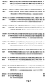
- FIG. 1 An alignment of the amino acid sequence shown in SEQ ID NO:2 and the amino acid sequence shown in SEQ ID NO:4 is shown in Fig. 1 .
- Fig. 1 each domain in the amino acid sequences is also shown (Non-patent Document 1).
- Fig. 2 shows only the alignment of the amino acid sequences. Since, as shown well in Fig.2 , the homology between the both amino acid sequences is very high (about 96.8%) and the numbers of amino acids are the same, the distribution of the various domains in the amino acid sequence shown in SEQ ID NO:2 can be easily seen from the distribution of the various domains (Non-patent Document 1) known for SEQ ID NO:4 ( Fig. 1 ).
- positions in the amino acid sequence of H5 HA are specified based on the amino acid sequence shown in SEQ ID NO:2. Since, as described above, the homologies among the amino acid sequences of HAs of H5 influenza A are very high, the amino acid sequence of another H5 HA can be easily aligned with the amino acid sequence shown in SEQ ID NO:2, so that the corresponding position in the amino acid sequence of the another H5 HA can be easily specified based on the amino acid sequence shown in SEQ ID NO:2.
- the term "based on the amino acid sequence shown in SEQ ID NO:2" means usage of the amino acid sequence shown in SEQ ID NO:2 as the basis for specifying the position, and does not only mean a monoclonal antibody prepared using HA having the amino acid sequence shown in SEQ ID NO:2 as an immunogen.
- the receptor subdomain corresponds to 278 aa from the 126th amino acid from the N-terminus (hereinafter referred to as "126 aa”).
- the corresponding epitope of the monoclonal antibody of the present invention does not exist in the receptor subdomain (excluding C-terminal region thereof consisting of 11 amino acids).
- influenza virus cannot be bound to the receptor on the cell surface, and hence cannot enter the cell, so that the virus cannot grow in the host. That is, the antibody has neutralizing activity against influenza virus.
- the corresponding epitope of one (IFH5-115) of the monoclonal antibodies actually prepared in the Examples below is located in the region of 11 amino acids (268-278 aa) in the C-terminus of the receptor subdomain, but the antibody does not have neutralizing activity, so that the C-terminus region of the receptor subdomain is not indispensable for binding of the virus to the receptor. Therefore, from the region defined in the present invention where the corresponding epitope does not exist (which is basically the receptor subdomain), the 11 amino acids in the C-terminus of the receptor subdomain are excluded.
- the corresponding epitope of the monoclonal antibody of the present invention is not restricted as long as it is located in a region other than the above-described receptor subdomain (excluding C-terminal region thereof consisting of 16 amino acids), and the corresponding epitope preferably exists in:
- the corresponding epitope "exists in the region” means that the binding activity to a deletion mutant in which the region is deleted is lost or at least significantly decreases, and its meaning includes not only cases where the corresponding epitope exists in the region, but also cases where the corresponding epitope exists in a region overlapping with this region and the binding activity is lost or at least significantly decreases if the region is deleted.
- the corresponding epitope regions defined in each of the above (1) to (3) consist of two regions which are largely distant from each other in the amino acid sequence, but these two regions are adjacent to each other in the spatial structure of HA (see Fig. 3 ).
- existence of the corresponding epitope in the region of 41 to 60 aa and 290 to 300 aa described in the above (1) means existence of the corresponding region over the entire portion consisting of these regions adjacent to each other in the spatial structure, and in cases where either 36 to 65 aa or 307 to 327 aa is deleted, the monoclonal antibody cannot be bound to the partially deleted HA.
- the region in which the corresponding epitope exists can be known by investigation of the binding ability to deletion mutants as concretely described in the Examples below, which investigation is based on a principle used in conventional methods. That is, it can be investigated by preparing various mutants in which various regions are deleted using the genetic engineering technique, followed by assaying whether or not the binding ability to the monoclonal antibody is lost or significantly decreased in each of the deletion mutants due to the deletion.
- the monoclonal antibody of the present invention does not have neutralizing activity against the influenza A virus to which it is bound by antigen-antibody reaction.
- the corresponding epitope of the monoclonal antibody of the present invention is located in a region other than the receptor subdomain (excluding C-terminus region thereof consisting of 11 amino acids), and by this constitutional requirement, the neutralizing activity is mostly lost, but monoclonal antibodies whose corresponding epitopes are located in regions other than the receptor subdomain (excluding C-terminal region thereof consisting of 11 amino acids) are also included, which monoclonal antibodies exert neutralizing activity by, for example, inhibition of binding of the receptor subdomain to the receptor due to steric hindrance.
- Whether or not a monoclonal antibody has neutralizing activity can be investigated by allowing a mixture of H5 subtype influenza A virus and the monoclonal antibody to act on cells and culturing the cells, followed by confirming whether or not the cells are injured, as concretely described in the Examples below.
- the monoclonal antibody of the present invention is preferably substantially non-cross-reactive with the other HA subtypes (i.e., H1 to H4 and H6 to H15) of influenza A virus. Because of the fact that the monoclonal antibody is substantially non-cross-reactive with the other HA subtypes, H5 subtype influenza A virus which has been confirmed to be infectious from bird to human can be distinguished from other influenza viruses and assayed.
- the term "substantially non-cross-reactive" means that the cross-reaction does not occur at a detectable level or, even though detection of the cross-reaction is possible, the cross-reaction is very weak and therefore can be clearly distinguished from the antigen-antibody reaction with H5 HA.
- cross-reaction with various subtypes of HAs was investigated by immunochromatography using 2 types of anti-H5 HA monoclonal antibodies of the present invention as an immobilized antibody and a labeled antibody.
- the antibodies can be judged to be substantially non-cross-reactive.
- the base sequences of the genes, as well as the amino acid sequences, of H1 to H4 and H6 to H15 used in the Examples below are shown in SEQ ID NOs:5 to 12 and 15 to 34, respectively. Occurrence of cross-reaction with the other HA subtypes can be investigated using these.
- the present invention also provides an antigen-binding fragment of the monoclonal antibody of the present invention.
- the term "antigen-binding fragment” means an antibody fragment maintaining binding ability (antigen-antibody reactivity) to the corresponding antigen of the antibody, such as a Fab fragment or a F(ab') 2 fragment of immunoglobulin. It is well-known that such an antigen-binding fragment can also be used for immunoassay. As is well-known, such fragments can be obtained by treating a monoclonal antibody with a protease such as papain or pepsin.
- the antigen-binding fragment is not restricted to a Fab fragment and a F(ab') 2 fragment, and may be any fragment which maintains binding ability to the corresponding antigen. It may further be one prepared by the genetic engineering technique. Further, for example, a single chain fragment of the variable region (scFv) of an antibody may be expressed in E . coli by the genetic engineering technique and used.
- scFv variable region
- scFv can be prepared by a process wherein mRNA of a hybridoma prepared as described above is extracted to prepare single-stranded cDNAs, and PCR is carried out using primers specific to the heavy chain and the light chain of immunoglobulin to amplify the heavy chain gene and the light chain gene, which genes are then linked to each other with a linker and given an appropriate restriction site, followed by introduction of the resultant to a plasmid vector, transformation of E. coli with the resulting plasmid vector, and recovery of scFv from the E . coli.
- Such scFv is also included in the "antigen-binding fragment" defined in the present invention.
- the monoclonal antibody of the present invention can be obtained by a process wherein anti-H5 subtype influenza A virus monoclonal antibodies are prepared by a conventional method using H5 subtype influenza A virus as an immunogen, and monoclonal antibodies having binding ability to HA are screened, followed by screening of those which do not have neutralizing activity against H5 subtype influenza A virus and analysis of the corresponding epitopes of the screened monoclonal antibodies, to select an antibody that satisfies the above-described requirement for the epitope.
- the monoclonal antibodies themselves can be prepared by the well-known hybridoma method, and the method is also concretely described in the Examples below.
- the neutralizing activity against influenza A virus can be investigated by the above method wherein a mixture of the virus and a monoclonal antibody is allowed to act on cells to see if the cells are injured, which method is also concretely described in the Examples below.
- the analysis of the corresponding epitopes can be carried out by the above method wherein various deletion mutants of H5 HA are prepared and their binding ability to each monoclonal antibody is investigated, which method is also concretely described in the Examples below.
- the present invention also provides a method of immunoassay of H5 subtype influenza A virus, comprising measurement of H5 subtype influenza A virus in a sample by immunoassay using antigen-antibody reaction of the above-described monoclonal antibody or an antigen-binding fragment thereof of the present invention with hemagglutinin of the H5 subtype influenza A virus.
- the term "measurement” include detection, quantification and semi-quantification.
- immunoassays themselves are well-known in the art and the monoclonal antibody or an antigen-binding fragment thereof of the present invention can be used in any of the well-known methods of immunoassay. That is, based on the reaction type, known immunoassays include sandwich immunoassays, competition immunoassays, agglutination immunoassays and the like. Based on the label employed, known immunoassays include enzyme immunoassays, radio immunoassays, fluorescence immunoassays, chemiluminescence immunoassays and the like. Any of these immunoassays are included in the "immunoassay" defined in the present invention and can be used as the method of immunoassay of the present invention.
- the reagents necessary for each type of immunoassay are also well-known in the art. Except for the monoclonal antibody or an antigen-binding fragment thereof used which is characteristic, the immunoassay can be carried out using an ordinary immunoassay kit. That is, the present invention also provides an immunoassay kit for carrying out the method of measurement of the present invention, which kit contains the above-described monoclonal antibody or an antigen-binding fragment thereof of the present invention.
- a sandwich immunoassay is advantageous for highly sensitive and rapid measurement of H5 subtype influenza A virus, which immunoassay uses 2 types of the monoclonal antibody and/or an antigen-binding fragment thereof of the present invention which can be bound to hemagglutinin of the virus at the same time.
- one of the 2 types of monoclonal antibodies used is immobilized on beads or wells of a microplate, or a solid phase such as a porous matrix for immunochromatography, and the other is labeled by enzyme labeling, fluorescence labeling, chemiluminescence labeling or the like.
- the virus antigen to be measured is sandwiched between the immobilized antibody and the labeled antibody, and the label bound to the solid phase is measured.
- detection of the label bound to the solid phase allows detection of the virus.
- the label is measured for standard samples of various different known amounts by the immunoassay system and the correlation between the amount of the label and the amount of the virus is plotted to prepare a calibration curve (standard curve). By the same operation, the amount of the label of an unknown sample is then measured, and the measured value is applied to the calibration curve to measure the virus quantitatively.
- the sandwich immunoassay of the present invention 2 types of anti-H5 HA monoclonal antibodies of the present invention which can be bound to a single H5 HA molecule at the same time are used.
- the immunoassay is preferably carried out using 2 types out of the 3 types of monoclonal antibodies whose corresponding epitopes exist in:
- immunochromatography In cases where H5 subtype influenza A virus in a sample is to be quantified, a sandwich immunoassay using wells of a microplate or beads as a solid phase, such as ELISA, can be preferably used. On the other hand, in cases where H5 subtype influenza A virus is to be detected rapidly and simply at clinical sites, immunochromatography (often referred to as “immunochromato” for short) is preferably used. Immunochromatography itself and devices (this may be hereinafter referred to as “immunochromatographic device”) used therefor are well-known, and also concretely described in the Examples below.
- lateral flow-based immunochromatography is carried out using a device having, usually on a belt-like matrix comprising a porous material such as nitrocellulose membrane, a detection zone on which a first anti-H5 HA monoclonal antibody was immobilized, and a labeled reagent zone on which a labeled second anti-H5 HA monoclonal antibody was spotted positioned in the upstream (upstream with respect to the direction of the flow of the later-mentioned developer) of the detection zone.
- a device having, usually on a belt-like matrix comprising a porous material such as nitrocellulose membrane, a detection zone on which a first anti-H5 HA monoclonal antibody was immobilized, and a labeled reagent zone on which a labeled second anti-H5 HA monoclonal antibody was spotted positioned in the upstream (upstream with respect to the direction of the flow of the later-mentioned developer) of the detection zone.
- the labeled reagent zone is usually constituted by a porous pad to which the labeled antibody was spotted.
- a developer tank storing a developer is provided at the upstream end of the matrix.
- the device usually further has: a development confirmation section on which an anti-labeled antibody was immobilized, for confirmation of whether or not development of the labeled antibody occurred, in the downstream of the above detection zone; and a developer absorption zone on which a porous absorption pad to absorb the developer flown to this zone is provided, in the further downstream of the development confirmation section.
- a substrate zone on which the substrate of the label enzyme was spotted is provided in the upstream of the labeled reagent zone.
- the sample is added to the labeled reagent zone, and the developer is applied to the upper end of the matrix by breaking the developer tank.
- the developer flows to the downstream by capillary action of the matrix.
- the substrate is eluted into the developer, and the resulting developer containing the substrate continues to flow.
- the labeled antibody and the sample are eluted into the developer, and the resulting developer containing the substrate, the labeled antibody and the sample continues to flow.
- H5 subtype influenza A virus is contained in the sample, HA of the virus is bound to the labeled antibody by antigen-antibody reaction.
- the immobilized antibody is bound to HA of the virus by antigen-antibody reaction.
- the labeled antibody is immobilized on the detection zone via HA of the virus. Therefore, by measuring the label immobilized on the detection zone, the virus can be detected.
- H5 subtype influenza A virus is not contained in the sample, nothing is bound to the immobilized antibody, so that the labeled antibody is not immobilized to the detection zone, and continues to flow to the downstream. Therefore, the label is not detected on the detection zone.
- the anti-labeled antibody is immobilized on the developer confirmation section in the downstream of the detection zone, the labeled antibody is immobilized on the developer confirmation section. In cases where the label is detected on the developer confirmation section, it can be confirmed that the developer has reached this section properly. The developer is absorbed to the absorption pad in the further downstream of the developer confirmation section.
- the sample to be applied to the method of immunoassay of the present invention is not restricted as long as whether or not H5 subtype influenza A virus is contained in the sample is to be detected, and examples of the sample include, but are not limited to, body fluids such as blood (including whole blood, plasma and serum), saliva and sputum; mucosal swabs; and swabs of tools and equipments.
- body fluids such as blood (including whole blood, plasma and serum), saliva and sputum; mucosal swabs; and swabs of tools and equipments.
- H5N1 H5 Subtype Influenza A Virus
- WB ELISA/Western Blotting
- Chorioallantoic fluid derived from an embryonated egg infected with an attenuated influenza virus strain A/duck/Hokkaido/Vac-1/04(HSN1) (GenBank accession No.: AB259712; SEQ ID NOs:49 and 50) was subjected to ultracentrifugation at 27000 rpm at 4°C for 1.5 hours, and the virus was recovered as a pellet. The pellet was dissolved into PBS, followed by recovery of the virus-containing fraction by sucrose gradient centrifugation. Ultracentrifugation was carried out again to remove sucrose, thereby obtaining a purified virus solution.
- the purified virus was inactivated with formalin or Triton X-100 (trademark) to be used as an antigen for immunization and ELISA/WB.
- the formalin treatment was carried out by adding formalin to the purified virus solution to a final concentration of 0.2% and leaving the resulting mixture to stand at 4°C for 1 week, and the Triton X-100 (trademark) treatment was carried out by adding 3 volumes of cell extraction buffer (0.05M Tris-HCl (pH8.0), 0.6 M KCI, 0.5% Triton X-100 (trademark)) to the purified virus solution.
- Inactivation of the virus was confirmed by inoculating the virus subjected to the inactivation treatment to an embryonated egg at 10 days of age and culturing the egg for 2 days, followed by confirming that no HA titer was detected by a hemagglutination (HA) test on chorioallantoic fluid derived from an embryonated egg infected with the virus.
- HA hemagglutination
- H5N1 H5 Subtype Influenza A Virus
- IF, WB Antigen for Confirmation of Specificity
- MDCK cells source of supply: Hokkaido University
- cell extract thereof were prepared as follows.
- IF immunofluorescence
- MDCK cells were cultured on a chamber slide to obtain a full sheet of the cells, and each influenza virus was optimally diluted with MEM supplemented with trypsin (serum-free) and inoculated to the cells.
- the cells were then cultured in a CO 2 incubator at 35°C for 16 hours, and the cultured cells were fixed with cold acetone to provide a slide for IF for confirmation of the specificity.
- MCDK cells were cultured in a petri dish to obtain a full sheet of the cells, and serum-free MEM supplemented with trypsin was added to a virus solution optimized in the same manner as in the preparation of the antibody for IF, followed by culturing the cells at 35°C for 16 hours. Thereafter, cell lysis buffer was added to the petri dish to lyse the cells, and the resulting lysate was centrifuged at 14000 rpm for 5 minutes, to obtain the supernatant as the antigen for WB for confirmation of the specificity.
- the monoclonal antibodies were prepared by immunizing mice with the purified inactivated influenza virus A/duck/Hokkaido/Vac-1/04(H5N1) prepared in Reference Example 1, followed by fusing lymphocytes derived from hind limbs of the mice and myeloma cells.
- mice were first immunized in their footpads with the purified inactivated influenza virus A/duck/Hokkaido/Vac-1/04(H5N1) emulsified with Freund's complete adjuvant in an amount of 50 to 100 ⁇ g/mouse, and 2 to 3 weeks later, the mice were subjected to second immunization with the purified inactivated influenza virus A/duck/Hokkaido/Vac-1/04(H5N1) antigen which is adjuvant-free in an amount of 50 to 100 ⁇ g/mouse.
- the antibody titer was confirmed by solid-phase ELISA using a 96-well ELISA plate coated with Triton X100-treated purified influenza virus A/duck/Hokkaido/Vac-1/04(H5N1), and by WB with the same antigen. Mice in which increase in the antibody titer was confirmed were immunized in their footpads with the purified inactivated influenza virus A/duck/HokkaidoNac-1/04(H5N1) in an amount of 25 to 50 ⁇ g/mouse, and 3 to 4 days later, lymphocytes were prepared from lymph nodes of hinder limbs of the mice.
- Mouse myeloma cells preliminarily cultured in RPMI-1640 medium were mixed with spleen cells at ratios of 1:2 to 1:5, and cell fusion was carried out using polyethylene glycol (PEG; manufactured by Boehringer). The fused cells were suspended in HAT medium and aliquoted to a 96-well culture plate, followed by culturing the cells in a CO 2 incubator at 37°C.
- PEG polyethylene glycol
- Triton X-100 (trademark)-treated purified influenza virus A/duck/Hokkaido/Vac-1/04(H5N1) antigen was aliquoted to a 96-well ELISA plate (manufactured by Nunc) at a concentration of 1 ⁇ g/ml in a volume of 50 ⁇ l/well, and left to stand at 4°C overnight to allow its adsorption.
- the wells were blocked with 1% skim milk and washed 3 times with washing buffer (PBS supplemented with 0.05% Tween 20), followed by adding 50 ⁇ l of the culture supernatant from the plate in which the cell fusion was carried out, and allowing the reaction to proceed at 37°C for 1 hour. After washing the wells 3 times with washing buffer in the same manner, a POD-labeled anti-mouse immunoglobulin antibody (manufactured by DACO) was added, followed by allowing the reaction to proceed at 37°C for 1 hour. After washing the wells 4 times with washing buffer, the substrate ABTS was added, followed by selecting the wells in which coloring was observed.
- washing buffer PBS supplemented with 0.05% Tween 20
- the cells in the selected wells were transferred to a 48-well or 24-well culture plate and cultured in a CO2 incubator at 37°C, followed by confirming their reactivity with hemagglutinin by WB.
- the WB was carried out according to a conventional method using a membrane prepared by subjecting the Triton X-100 (trademark)-treated purified influenza virus A/duck/Hokkaido/Vac-1/04(H5N1) antigen to SDS-PAGE, followed by transferring it to a nitrocellulose membrane.
- the cells in the wells in which reactivity to hemagglutinin was confirmed by WB were made into single clones by the limiting dilution method to establish the various hybridomas described below which produce monoclonal antibodies reactive with influenza (H5N1) hemagglutinin.
- influenza A virus in a sample was actually detected, and 3 types of monoclonal antibodies IFH5-115, IFH5-136 and IFH5-26 showing excellent sensitivities and specificities (absence of cross-reaction with other HA subtypes, as mentioned later), and 3 types of hybridomas IFH5-115, IFH5-136 and IFH5-26 producing them were established (the monoclonal antibodies IFH5-115, IFH5-136 and IFH5-26 may be hereinafter referred to as the monoclonal antibody 115 (or Mab 115), monoclonal antibody 136 (or Mab 136) and monoclonal antibody 26 (or Mab 26), respectively). These monoclonal antibodies did not have neutralizing activity against influenza A virus, as mentioned later.
- the IF was carried out as follows. That is, to the slide for IF for confirmation of the specificity which was prepared in Reference Example 2, the culture supernatant for each monoclonal antibody or ascites (diluted with 1% BSA in PBST) was added, and the reaction was allowed to proceed for 1 hour at room temperature. The resultant was washed with PBS and allowed to react with a fluorescent dye-labeled anti-mouse IgG (1000-fold diluted with 1% BSA in PBST) at room temperature for 30 minutes. This was followed by washing with PBS, mounting in glycerol and observation under a fluorescence microscope. WB was carried out using the antigen prepared in Reference Example 2 according to a conventional method.
- the monoclonal antibodies IFH5-115, IFH5-136 and IFH5-26 were confirmed in either assay to be reactive with attenuated HSN 1 influenza HA, but the results on the reactivities against HAs of other substrains and virulent H5N1 were different between the assays. Therefore, the actual assay system (EL) was used for confirmation of these.
- HA of the influenza H5N1 strain A/duck/HokkaidoNac-1/04(H5N1) (SEQ ID NO:2) consisting of 564 amino acids was subjected to PCR to amplify 6 types of fragments: 1 to 564 aa (A fragment), 1 to 514 aa (B fragment), 21 to 514 aa (C fragment), 101 to 500 aa (D fragment), 151 to 500 aa (E fragment), and 201 to 500 aa (F fragment).
- a Bam HI or Xba I site had been preliminarily added to each primer.
- Each amplified fragment was purified with PCR Purification Kit manufactured by QIAGEN and inserted into the Bam HI -Xba I site in an expression plasmid pWG6A which was prepared by incorporating GST into the Nde I -Eco RI site in an expression plasmid pW6A, to prepare the plasmids pWGInf.H5N1-N-A, pWGInf.H5N1-N-B, pWGInf.H5N1-N-C, pWGInf.H5N1-N-D, pWGInf.H5N1-N-E and pWGInf.H5N1-N-F. Using these plasmids, E .
- coli BL21(DE3) (supplied from Brookhaven National Laboratory) was transformed to obtain ampicillin-resistant E . coli transformants BL21(DE3)pWGInf.H5N1-N-A, BL21(DE3)pWGInf.H5N1-N-B, BL21(DE3)pWGInf.H5N1-N-C, BL21(DE3)pWGInf.H5N1-N-D, BL21(DE3)pWGInf.H5N1-N-E and BL21(DE3)pWGInf.H5N1-N-F.
- Each of the transformants prepared in 3-1 was cultured in 2 ml of LB medium supplemented with 50 ⁇ g/ml ampicillin at 37°C. Preculture was carried out until an OD of 0.6 to 0.8 at 600 nm was attained, and 1 mM IPTG was then added to the culture to induce expression, followed by culturing for additional 3 hours.
- the bacterial cells were collected by centrifuging 1.5 ml of the bacterial cell culture at 5,000 rpm for 2 minutes, and the collected bacterial cells were suspended in 100 ⁇ l of a buffer (10 mM Tris-HCl (pH 8.0), 0.1 M sodium chloride, 1 mM EDTA), followed by completely disrupting the bacterial cells by sonication for 15 minutes. The resultant was used as a bacterial cell sample.
- SDS polyacrylamide buffer (0.15 M Tris-HCl (pH 6.8), 6% SDS, 24% glycerol, 6 mM EDTA, 2% 2-mercaptoethanol, 0.03% bromophenol blue) was added, and the resulting mixture was stirred sufficiently, followed by heat treatment at 100°C before being subjected to electrophoresis.
- Sodium dodecyl sulfate-polyacrylamide gel electrophoresis SDS-PAGE
- e-PAGEL 12.5% gel concentration (trademark; manufactured by ATTO).
- the sample was transferred to a PVDF membrane (manufactured by ATTO) and blocked with 1 % BSA at 4°C overnight.
- the blocking solution was removed, and the membrane was washed with PBS-Tween (trademark), followed by adding the monoclonal antibodies 26, 115 and 136 whose concentrations were adjusted to 2 ⁇ g/ml and allowing the reaction to proceed at room temperature for 60 minutes.
- the membrane was washed sufficiently with PBS-Tween (trademark) and subjected to a reaction with a peroxidase-labeled anti-mouse immunoglobulin rabbit polyclonal antibody (manufactured by DAKO) 5000-fold diluted with a reaction buffer.
- the membrane was exposed to instant film (manufactured by FUJIFILM) for 1 to 15 minutes using ECL Western Blotting Detection System (manufactured by GE Healthcare Bio-Sciences KK) to detect signals.
- the results are shown in Fig. 4 .
- the monoclonal antibodies 26 and 115 reacted with the A, B and C fragments, but not reacted with the D fragment.
- the monoclonal antibody 136 reacted with the A, B, C and D fragments, but not reacted with the E fragment. Accordingly, the monoclonal antibodies 26 and 115, and the monoclonal antibody 136 were revealed to recognize the region of 21 to 100 aa and the region of 101 to 150 aa, respectively.
- N 1 (21 to 400 aa), N2 (41 to 400 aa), N3 (61 to 400 aa), N4 (81 to 400 aa), N5 (101 to 400 aa), N6 (114 to 400 aa), N7 (127 to 400 aa) and N8 (139 to 400 aa) were amplified by PCR.
- Each amplified fragment was purified with PCR Purification Kit manufactured by QIAGEN and inserted into the Bam HI -Xba I site in an insect cell expression plasmid pBac-EGTs-6A (manufactured by Invitrogen; modified from pFastBac1 vector), to prepare plasmids pBacInf.H5N1-N1, pBacInf.H5N1-N2, pBacInf.H5N1-N3 , pBacInf.H5N1-N4, pBacInf.H5N1-N5, pBacInf.HSN1-N6, pBacInfH5N1-N7 and pBacInf.H5N1-N8.
- E . coli DH10Bac for Bacmid preparation was transformed, to prepare Bacmid-Inf.H5N1-N1, Bacmid-Inf.H5N1-N2, Bacmid-Inf.H5N1-N3, Bacmid-Inf.H5N1-N4, Bacmid-Inf.H5N1-N5, Bacmid-Inf.H5N1-N6, Bacmid-Inf.H5N1-N7 and Bacmid-Inf.H5N1-N8.
- the monoclonal antibody 254 which reacts with the amino acid region of 139 to 400 was immobilized and used for ELISA. That is, the monoclonal antibody 254 was diluted to a concentration of 2 ⁇ g/mL, and 50 ⁇ l of the diluted monoclonal antibody was placed in each well of a microplate module (manufactured by Nunc), followed by incubation at 4°C overnight to immobilize the monoclonal antibody.
- each well was washed with PBS supplemented with 0.1% Tween 20 (trademark) (PBS-Tween (trademark)), and 100 ⁇ L of 1% bovine serum albumin (BSA) prepared by dilution with PBS was added to the well, followed by blocking at 37°C for 1 hour. After removing the blocking solution, 50 ⁇ l of a biotinylated monoclonal antibody 26, 115 or 126 was added, and the reaction was allowed to proceed at 37°C for 1 hour.
- PBS-Tween trademark
- BSA bovine serum albumin
- the monoclonal antibody 26 has its epitope in the region of amino acids 61 to 80; the monoclonal antibody 115 has its epitope in the region of amino acids 41 to 60; and the monoclonal antibody 136 has its epitope in the region of amino acids 101 to 113.
- HA of the influenza H5N1 strain A/duck/Hokkaido/Vac-1/04(H5N1) (SEQ ID NO:2) consisting of 564 amino acids was subjected to PCR to amplify 5 types of fragments: 21 to 400 aa (A fragment), 21 to 333 aa (B fragment), 21 to 266 aa (C fragment), 21 to 200 aa (D fragment), and 21 to 133 aa (E fragment).
- a Bam HI or Xba I site had been preliminarily added to each primer.
- Each amplified fragment was purified with PCR Purification Kit manufactured by QIAGEN and inserted into the Bam HI -Xba I site in an expression plasmid pWG6A which was prepared by incorporating GST into the Nde I -Eco RI site in an expression plasmid pW6A, to prepare the plasmids pWGInf.H5N1-C-A, pWGInf.H5N1-C-B, pWGInf.HSN1-C-C, pWGInf.H5N1-C-D and pWGInf.H5N1-C-E. Using these, E .
- coli BL21(DE3) (supplied from Brookhaven National Laboratory) was transformed to obtain ampicillin-resistant E. coli transformants BL21(DE3)pWGInf.H5N1-C-A, BL21(DE3)pWGInf.H5N1-C-B, BL21(DE3)pWGInf.H5N1-C-C, BL21(DE3)pWGInf.HSNI-C-D and BL21(DE3)pWGInf.H5N1-C-E.
- Each of the transformants prepared in 3-5 was cultured in 2 ml of LB medium supplemented with 50 ⁇ g/ml ampicillin at 37°C. Preculture was carried out until an OD of 0.6 to 0.8 at 600 nm was attained, and 1 mM IPTG was then added to the culture to induce expression, followed by culturing for additional 3 hours.
- the bacterial cells were collected by centrifuging 1.5 ml of the bacterial cell culture at 5,000 rpm for 2 minutes, and the collected bacterial cells were suspended in 100 ⁇ l of a buffer (10 mM Tris-HCl (pH 8.0), 0.1 M sodium chloride, 1 mM EDTA), followed by completely disrupting the bacterial cells by sonication for 15 minutes. The resultant was used as a bacterial cell sample.
- SDS polyacrylamide buffer (0.15 M Tris-HCl (pH 6.8), 6% SDS, 24% glycerol, 6 mM EDTA, 2% 2-mercaptoethanol, 0.03% bromophenol blue) was added, and the resulting mixture was stirred sufficiently, followed by heat treatment at 100°C before being subjected to electrophoresis.
- Sodium dodecyl sulfate-polyacrylamide gel electrophoresis SDS-PAGE
- e-PAGEL 12.5% gel concentration (trademark; manufactured by ATTO).
- the sample was transferred to a PVDF membrane (manufactured by ATTO) and blocked with 1% BSA at 4°C overnight.
- the blocking solution was removed, and the membrane was washed with PBS-Tween (trademark), followed by adding the monoclonal antibodies 26, 115 and 136 whose concentrations were adjusted to 2 ⁇ g/ml and allowing the reaction to proceed at room temperature for 60 minutes.
- the membrane was washed sufficiently with PBS-Tween (trademark) and subjected to a reaction with a peroxidase-labeled anti-mouse immunoglobulin rabbit polyclonal antibody (manufactured by DAKO) 5000-fold diluted with a reaction buffer.
- shorter fragments of the amino acid fragment of 267 to 333 aa of influenza H5N1 were amplified by PCR.
- Each amplified fragment was purified with PCR Purification Kit manufactured by QIAGEN and inserted into the Bam HI- Xba I site in an insect cell expression plasmid pBac-EGTs-6A (manufactured by Invitrogen; modified from pFastBac1 vector ), to prepare plasmids pBacInf.H5N1-C1, pBacInf.H5N1-C2, pBacInf.H5N1-C3, pBacInf.H5N1-C4, pBacInf.H5N1-C5, pBacInf.H5N1-C6 and pBacInf.H5N1-C7.
- E . coli DH10Bac for Bacmid preparation was transformed, to prepare Bacmid-Inf.H5N1-C1, Bacmid-Inf.H5N1-C2, Bacmid-Inf.H5N1-C3, Bacmid-Inf.H5N1-C4, Bacmid-Inf.H5N1-C5, Bacmid-Inf.H5N1-C6 and Bacmid-Inf.H5N1-C7
- the monoclonal antibody 270 which reacts with the amino acid region of 41 to 333 aa was immobilized and used for ELISA. That is, the monoclonal antibody 270 was diluted to a concentration of 2 ⁇ g/mL, and 50 ⁇ l of the diluted monoclonal antibody was placed in each well of a microplate module (manufactured by Nunc), followed by incubation at 4°C overnight to immobilize the monoclonal antibody.
- each well was washed with PBS supplemented with 0.1% Tween 20 (trademark) (PBS-Tween (trademark)), and 100 ⁇ L of 1% bovine serum albumin (BSA) prepared by dilution with PBS was added to the well, followed by blocking at 37°C for 1 hour. After removing the blocking solution, 50 ⁇ l of a biotinylated monoclonal antibody 26, 115 or 126 was added, and the reaction was allowed to proceed at 37°C for 1 hour.
- PBS-Tween trademark
- BSA bovine serum albumin
- the monoclonal antibody 26 has its epitope in the region of amino acids 290 to 300; the monoclonal antibody 115 has its epitope in the region of amino acids 312 to 322; and the monoclonal antibody 136 has its epitope in the region of amino acids 268 to 278.
- the monoclonal antibody 26 recognizes the regions of 61 to 88 aa and 290 to 300 aa; the monoclonal antibody 115 recognizes the regions of 41 to 60 aa and 312 to 322 aa; and the monoclonal antibody 136 recognizes the regions of 101 to 113 aa and 268 to 278 aa. That is, it is assumed that these monoclonal antibodies recognize the spatial structure (see Fig. 3 ).
- the monoclonal antibodies IFH5-115, IFH5-136 and IFH5-26 have neutralizing activity against H5 subtype influenza A viruses was tested.
- influenza viruses A/dk/Hokkaido/84/02(HSN3) and A/PR/8/34(H1N1) were used.
- a 2-fold dilution series of each monoclonal antibody was prepared with Eagle's MEM, and 50 ⁇ l of each antibody dilution was placed in a 96-well plate.
- Each of the A/dk/Hokkaido/84/02(H5N3) and A/PR/8/34(H1N1) was diluted such that 200 TCID 50 /25 ⁇ L was attained, and 25 ⁇ l of the virus dilution was added to each well containing the antibody dilution (protein concentration: 0 to 160 ⁇ g/25 ⁇ L). The resulting mixture was incubated at room temperature for 1 hour (virus-antibody mixture). On the other hand, in a 48-well plate, MDCK cells were preliminarily cultured in Eagle's MEM supplemented with 10% fetal calf serum to confluence, and the culture supernatant was removed.
- the plate was washed once with Eagle's MEM, and 75 ⁇ l of the above-described virus-antibody mixture was added thereto, followed by incubation at 35°C for 1 hour. Subsequently, 150 ⁇ l of Eagle's MEM supplemented with 0.0005% trypsin was added to each well, and culture was started at 35°C in the presence of 5% carbon dioxide gas. On day 3 of the culture, whether or not cell injury by the virus was inhibited by neutralizing antibody activity was investigated under the microscope to confirm whether or not the neutralizing activity was exerted. In cases where cell injury has occurred, cell rounding due to cell fusion by virus infection is observed. In cases where cell injury has not occurred, it can be judged that the monoclonal antibody has neutralizing activity against the virus.
- the chicken antiserum (obtained by immunization with A/duck/Hokkaido/Vac-1/04 (SEQ ID NO:2)) used as the neutralizing antibody control showed neutralizing antibody activity against A/dk/Hokkaido/84/02(H5N3) and A/PR/8/34(H1N1) up to 64-fold dilution, but the monoclonal antibodies IFH5-115, IFH5-136 and IFH5-26 showed no neutralizing activity.
- Virus A/dk/Hokkaido/84/02(H5N3) 10 ⁇ 2 TCID50/25 ⁇ L(well) CPE Antibody Antibody concentration ( ⁇ g/mL) 160 80 40 20 10 0 IFH5-115 + + + + + + + + + + + + + + + + + + + + + + + + + + + + + + + + + + + + + + + + + + + + + + + + + + + IFH5-26 + + + + + + + + + + + + + + + + + + + + + + + + + + + + + + + + + + + + + + + + + + + + + + + + + + + + + + + + + + + + + + + + + + + + + + + + + + + + + + + + + + + + + + + + + + + + + + + + + + + + + + + + + + + + + + + + + + + + + + + + + + + + + + + + + + + + + + + + +
- Virus A/PR/8/34(H1N1) 10 ⁇ 2 TCID50/25 ⁇ L(well) CPE Antibody Antibogy concentration ( ⁇ g/mL) 160 80 40 20 10 0 IFH5-115 + + + + + + + + + + + + + + + + + + + + + + + + + + + + + + + + + + + + + + + + + + + + + + + + + + + + + + + + + IFH5-26 + + + + + + + + + + + + + + + + + + + + + + + + + + + + + + + + + + + + + + + + + + + + + + + + + + + + + + + + + + + + + + + + + + + + + + + + + + + + + + + + + + + + + + + + + + + + + + + + + + + + + + + + + + + + + + + + + + + + + + + + + + + + + + + + + + + + + + + + +
- a matrix 2 which is a nitrocellulose membrane having a width of 5 mm and a length of 50 mm (manufactured by Millipore)
- aqueous solution containing an anti-influenza A H5-specific antibody INF5-26
- anti-alkaline phosphatase antibody rabbit
- a developer pad 3 100 ⁇ g of 5-bromo-4-chloro-3-indolyl phosphate (BCIP) was spotted as a substrate in the form of a line having a width of 6.0 mm on a filter paper having a width of 6 mm and length of 20 mm (manufactured by Millipore), and the spot was dried, to prepare a substrate zone 7.
- the matrix 2, the developer pad 3, the enzyme labeling reagent pad 4 and an absorption pad 5 filter paper of 10 mm width, 15 mm length and 1 mm thickness (manufactured by Whatman)) were immobilized in a plastic case having a developer tank 11, to produce the influenza A H5 immunoassay device 1 shown in Figs. 7 and 8 .
- Example 5 the substrate was spotted and dried to prepare a developer pad 3, which was then immobilized, together with the matrix 2, the enzyme labeling reagent pad 4 and the absorption pad 5, in a plastic case having a developer tank 11, to produce the influenza A H5 immunoassay device 2 similar to the device shown in Figs. 6 and 7 .
- alkaline phosphatase-labeled anti-influenza A H5-specific antibody which was produced by labeling IFH5-26 antibody with alkaline phosphatase (ALP- IFH5-26, 10 ⁇ g/ml) was spotted on the matrix, and the spot was dried, thereby preparing a labeled reagent zone comprising an enzyme labeling reagent pad 4.
- Example 5 the substrate was spotted and dried to prepare a developer pad 3, which was then immobilized, together with the matrix 2, the enzyme labeling reagent pad 4 and the absorption pad 5, in a plastic case having a developer tank 11, to produce the influenza A H5 immunoassay device 3 similar to the device shown in Figs. 6 and 7 .
- influenza A H5 immunoassay (immunochromatography) devices 1, 2 and 3 measurement devices of the present invention
- the reaction specificities of the immunoassay devices against the influenza A virus subtype H1-15
- chorioallantoic fluid whose hemagglutination titer (HA titer) was known was used, which chorioallantoic fluid was obtained by culturing each virus in an embryonated egg and recovering the chorioallantoic fluid (see Table 4).
- Each chorioallantoic fluid was 10-fold diluted with a sample diluent (Tris buffer (pH 8.0) containing a surfactant), and 30 ⁇ L of the dilution was added to a sample spotting zone 8.
- a pushing area 12 provided in a deformable member was then pushed down to be deformed, thereby inserting, with a projection 13 provided in the deformable member, the developer pad 3 into the developer tank 11 to supply the developer to the developer pad 3 to start the measurement.
- Fifteen minutes after the start of the measurement development of the developer was confirmed by coloring of the subject reagent zone 10, and coloring of the detection zone 6 was visually observed.
- Table 7 It was confirmed that all of the immunoassay devices specifically detect the influenza A virus H5 subtype and are not reactive with the other subtypes (H 1 to 4 and H6 to 15) of influenza A virus.
- chorioallantoic fluid whose hemagglutination titer (HA titer) was known was used, which chorioallantoic fluid was obtained by culturing each virus in an embryonated egg and recovering the chorioallantoic fluid (see Table 7).
- Each chorioallantoic fluid was serially diluted with a sample diluent (Tris buffer (pH 8.0) containing a surfactant), and 30 ⁇ l of each resulting dilution was added to the sample addition zone 8.
- the pushing area 12 provided in the deformable member was then pushed down to be deformed, thereby inserting, with a projection 13 provided in the deformable member, the developer pad 3 into the developer tank 11 to supply the developer to the developer pad 3 to start the measurement. Fifteen minutes after the start of the measurement, development of the developer was confirmed by coloring of the development confirmation section 10, and coloring of the detection zone 6 was visually observed. The results are shown in Table 8. Each detection sensitivity was represented as the maximum dilution rate of each chorioallantoic fluid at which the virus was detectable, which chorioallantoic fluid had been serially diluted. All of the immunoassay devices detected the influenza A virus H5 substrains that were tested.
- aqueous solutions containing the anti-H5 subtype influenza A virus hemagglutinin H5-specific antibody (IFH5-26) produced in Reference Example 2 and an anti-influenza A virus antibody (FVA2-11) (Literature: Gui-Rong BAI et.al.: Improvement of a Rapid Diagnosis Kit to Detect Either Influenza A or B Virus Infection.
- a filter paper having a width of 6 mm and length of 20 mm 100 ⁇ g of 5-bromo-4-chloro-3-indolyl phosphate (BCIP) was spotted as a substrate in the form of a line having a width of 6.0 mm, and the spot was dried, to prepare a developer pad 3.
- the matrix 2, the developer pad 3, the enzyme labeling reagent pad 4 and an absorption pad 5 were immobilized in a plastic case having a developer tank 11, to produce the simultaneous immunoassay device 4 for influenza A virus and influenza A H5 virus shown in Fig. 10 and Fig. 8 .
Landscapes
- Health & Medical Sciences (AREA)
- Life Sciences & Earth Sciences (AREA)
- Immunology (AREA)
- Chemical & Material Sciences (AREA)
- Engineering & Computer Science (AREA)
- Molecular Biology (AREA)
- Virology (AREA)
- Urology & Nephrology (AREA)
- Hematology (AREA)
- Biomedical Technology (AREA)
- General Health & Medical Sciences (AREA)
- Biochemistry (AREA)
- Medicinal Chemistry (AREA)
- Organic Chemistry (AREA)
- Biotechnology (AREA)
- Physics & Mathematics (AREA)
- Cell Biology (AREA)
- Pathology (AREA)
- Microbiology (AREA)
- General Physics & Mathematics (AREA)
- Food Science & Technology (AREA)
- Analytical Chemistry (AREA)
- Proteomics, Peptides & Aminoacids (AREA)
- Genetics & Genomics (AREA)
- Biophysics (AREA)
- Tropical Medicine & Parasitology (AREA)
- Chemical Kinetics & Catalysis (AREA)
- Peptides Or Proteins (AREA)
- Preparation Of Compounds By Using Micro-Organisms (AREA)
Abstract
Description
- The present invention relates to an anti-H5 subtype influenza A virus hemagglutinin monoclonal antibody, and a method of immunoassay of anti-H5 subtype influenza A virus and an immunoassay kit, which method and kit use the monoclonal antibody.
- Influenza viruses of H5 and H7 types have caused heavy damage to the poultry industry mainly in Asia, and, in recent years, avian influenza virus was confirmed to be infectious also to human. In particular, in terms of avian influenza viruses belonging to H5 subtype, there are many reports as from 2003 on their infection to human and high lethality mainly in Asia, so that there is a worldwide concern of their pandemic due to absence of immunity against the viruses in human. Therefore, prophylaxis of, and countermeasures against them are being studied.
- In order to prevent infection of avian influenza virus to human, or in order to prevent at least its outbreak in human, it is important to specifically and rapidly detect avian influenza virus that is infectious also to human. As a method for rapid detection of a virus in a sample, immunoassay has been conventionally widely used, and immunochromatography carried out by a sandwich immunoassay, wherein a solid phase in which an anti-virus antibody is immobilized on a part of a porous substrate through which a liquid is allowed to flow is used, has been especially widely used at clinical sites such as hospitals since viruses can be very simply detected in a short time thereby. Immunochromatography kits with which human influenza viruses can be detected are also commercially available.
- In order to detect by immunoassay avian influenza virus that is also infectious to human, an antibody against the avian influenza virus is of course necessary. The surface antigens of influenza viruses are known to contain proteins called hemagglutinin and neuraminidase. As the hemagglutinin (HA), 16 types having different antigenicities, H1 to H16, are known, and, as the neuraminidase, 9 types having different antigenicities, N1 to N9, are known. The subtype of each influenza virus is represented by description of the types of these hemagglutinin and neuraminidase, such as "H5N1". Most of the avian influenza viruses that have been so far confirmed to have been infected from bird to human are those having hemagglutinin of H5 type. Therefore, as an antibody to detect a highly pathogenic influenza virus that is infectious from bird to human, an antibody specific to H5 subtype influenza virus is demanded.
- Hemagglutinin of H5 subtype influenza A virus has been studied well, and not only the amino acid sequence but also the spatial structure thereof are known (Non-patent Document 1). Further, various monoclonal antibodies against hemagglutinin of H5 subtype influenza A virus have been known (Non-patent
Documents 2 to 4). Further, measurement of H5 subtype influenza A virus by a sandwich immunoassay using 2 types of hemagglutinin monoclonal antibodies has been known (Non-patentDocuments 5 and 6). -
- [Non-patent Document 1] Ya Ha et al; The EMBO Journal, 21(5), 865-875, 2002
- [Non-patent Document 2] Nikolai V.K. et al., J. Gen. Virol. 83, 2497, 2002
- [Non-patent Document 3] James Stevens et al.,
- [Non-patent Document 4] Zhi-Yong Yang et al.,
- [Non-patent Document 5] Tsuda Y. et al., Microbiol. Immunol. 51(9), 903, 2007
- [Non-patent Document 6] Qigai He et al., Clin. Vacc. Immnol. 617, 2007
- As monoclonal antibodies against hemagglutinin of H5 subtype influenza A virus, neutralizing antibodies having neutralizing activity as antibodies that suppress and inhibit the infectivity of the virus have been mainly studied so far. In order to develop a vaccine against the virus, a neutralizing antibody is necessary, and the monoclonal antibodies described in the above
Non-Patent Documents 2 to 4 are also neutralizing antibodies. Further, in terms of the monoclonal antibodies used for the immunoassays described inNon-patent Documents - Using such a known neutralizing antibody, an immunoassay system for H5 subtype influenza virus can of course be constructed. However, the present inventors noticed that, in cases where an immunoassay system was constructed using a neutralizing antibody, it may be impossible to keep up with mutation of the virus. That is, the present inventors noticed that, since influenza virus is known to cause mutation in the epitope recognized by a neutralizing antibody to escape from immunological surveillance of the host, construction of an immunoassay system using a known neutralizing antibody may lead to loss of reactivity with the mutant virus or decrease in the binding affinity, resulting in inability to detect the virus or drastic decrease in the measured value relative to the correct value. In view of the fact that viruses frequently mutate, it is preferred to be able to accurately carry out immunoassay of a virus even in cases where a certain level of mutation has occurred.
- Accordingly, an object of the present invention is to provide a method of immunoassay of H5 subtype influenza A virus by which the virus can be accurately assayed even in cases where a certain level of mutation has occurred in the H5 subtype influenza A virus, and a kit therefor, and a novel anti-H5 subtype influenza A virus monoclonal antibody which can be used for the immunoassay.
- Influenza virus is known to be first bound to a receptor on the cell surface and then incorporated into the inside of the cell, upon its infection. Therefore, for growth of influenza virus in a host cell, the virus needs to be bound to the receptor on the cell surface, so that, if this process is inhibited, growth of the virus does not occur. The corresponding epitopes of known neutralizing antibodies are located in the receptor binding region of influenza virus, and the neutralizing antibodies are bound to the virus to prevent the virus from being bound to the receptor. On the other hand, viruses are known to be prone to mutation in circumstances where a neutralizing antibody produced by the natural immunity of the host or by administration of a vaccine exists, in order to escape from growth inhibition by the neutralizing antibody. Although the mutation itself may occur at random, only viruses mutated such that their growth is not inhibited by the neutralizing antibody can grow well, so that, as a result, viruses having such a mutation(s) increase among the growing viruses. This phenomenon is called escape mutation, and the fact that a neutralization antibody makes such mutation more easily occur is called antibody pressure or immune pressure. The present inventors inferred that a method of immunoassay by which a virus can be assayed even in cases where a certain level of mutation has occurred in the virus can be constructed by using a monoclonal antibody whose corresponding epitope is located in a region other than the region where the corresponding epitopes of neutralizing antibodies exist, and determined the region where the corresponding epitope of such a monoclonal antibody exists, which monoclonal antibody was then actually provided, thereby completing the present invention.
- That is, the present invention provides an anti-H5 subtype influenza A virus hemagglutinin monoclonal antibody or an antigen-binding fragment thereof which undergoes antigen-antibody reaction with hemagglutinin of H5 subtype influenza A virus, whose corresponding epitope does not exist in the receptor subdomain (excluding C-terminal region thereof consisting of 11 amino acids) and which does not have neutralizing activity against the influenza A virus. Further, the present invention provides a method of immunoassay of H5 subtype influenza A virus, comprising measurement of H5 subtype influenza A virus in a sample by immunoassay using antigen-antibody reaction of the above-described monoclonal antibody or an antigen-binding fragment thereof of the present invention with hemagglutinin of the H5 subtype influenza A virus. Further, the present invention provides an immunoassay kit for H5 subtype influenza A virus, comprising 2 types of the above-described monoclonal antibody and/or an antigen-binding fragment thereof of the present invention, the 2 types of the antibody and/or the antigen-binding fragment thereof being capable of binding at the same time to hemagglutinin of H5 subtype influenza A virus.
- By the present invention, anti-H5 subtype influenza A virus hemagglutinin monoclonal antibodies whose binding affinities do not change even in cases where H5 subtype influenza A virus caused escape mutation were first provided. Therefore, by immunoassay using a monoclonal antibody of the present invention, it is possible to assay H5 subtype influenza A virus in which escape mutation occurred, and it is also possible to rapidly assay avian influenza virus which is pathogenic also to human. Thus, the present invention is thought to largely contribute to prevention of prevalence of avian influenza virus which is pathogenic also to human.
-
-
Fig. 1 is a diagram showing an alignment of the amino acid sequences of hemagglutinin in 2 types of strains of H5 subtype influenza A virus, wherein the distribution of each subdomain and the corresponding epitopes of the 3 types of monoclonal antibodies prepared in Examples are shown. -
Fig. 2 is a diagram showing alignment of the amino acid sequences of hemagglutinin in 2 types of strains of H5 subtype influenza A virus. -
Fig. 3 is a diagram showing the spatial structure of hemagglutinin of the H5 subtype influenza A virus strain, wherein the corresponding epitope regions of the 3 types of monoclonal antibodies prepared in Examples are shown. -
Fig. 4 is a diagram showing the results obtained during the epitope analysis of the 3 types of monoclonal antibodies, which was carried out in Examples. -
Fig. 5 is a diagram showing results obtained during the epitope analysis of the 3 types of monoclonal antibodies, which was carried out in Examples. -
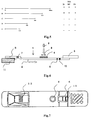
Fig. 6 is a schematic cross-sectional view of the main part of the immunoassay device (immunochromatographic device) prepared in Examples. -
Fig. 7 is a schematic plan view of the immunoassay device (immunochromatographic device) prepared in Examples. -
Fig. 8 is a schematic cross-sectional view of the immunoassay device (immunochromatographic device) prepared in Examples. -
Fig. 9 is a schematic cross-sectional view of the main part of another immunoassay device (immunochromatographic device) prepared in Examples. -
Fig. 10 is a schematic plan view of another immunoassay device (immunochromatographic device) prepared in Examples. - As described above, the monoclonal antibody of the present invention undergoes antigen-antibody reaction with hemagglutinin of H5 subtype influenza A virus, and the corresponding epitope of the monoclonal antibody is located in a region other than the receptor subdomain (excluding C-terminal region thereof consisting of 11 amino acids).
- Examples of the base sequence of the gene of hemagglutinin of H5 subtype influenza A virus (this may be hereinafter referred to as "H5 HA" for short) and the amino acid sequence encoded thereby are shown in SEQ ID NOs: 1 and 2. The sequences shown in SEQ ID NOs: 1 and 2 are known and registered in GenBank under the Accession No. AB263192.
- Several base sequences of genes and amino acid sequences of H5 HA other than those described above are also known, and examples thereof include the base sequence of the gene and the amino acid sequence of HA of H5N3 described in the above-described
Non-patent Document 1, registered in GenBank under Accession No. of AF303057 (the amino acid sequence is registered also under Accession No. Q9DLP3) (SEQ ID NOs. 3 and 4). A monoclonal antibody which undergoes antigen-antibody reaction with one of the known H5 subtype HAs is included within the scope of the monoclonal antibody of the present invention, and, since the homologies among the H5 subtype HAs are high, it usually undergoes antigen-antibody reaction with any of the H5 subtype HAs. - An alignment of the amino acid sequence shown in SEQ ID NO:2 and the amino acid sequence shown in SEQ ID NO:4 is shown in
Fig. 1 . InFig. 1 , each domain in the amino acid sequences is also shown (Non-patent Document 1).Fig. 2 shows only the alignment of the amino acid sequences. Since, as shown well inFig.2 , the homology between the both amino acid sequences is very high (about 96.8%) and the numbers of amino acids are the same, the distribution of the various domains in the amino acid sequence shown in SEQ ID NO:2 can be easily seen from the distribution of the various domains (Non-patent Document 1) known for SEQ ID NO:4 (Fig. 1 ). In the present specification and claims, positions in the amino acid sequence of H5 HA are specified based on the amino acid sequence shown in SEQ ID NO:2. Since, as described above, the homologies among the amino acid sequences of HAs of H5 influenza A are very high, the amino acid sequence of another H5 HA can be easily aligned with the amino acid sequence shown in SEQ ID NO:2, so that the corresponding position in the amino acid sequence of the another H5 HA can be easily specified based on the amino acid sequence shown in SEQ ID NO:2. Further, the term "based on the amino acid sequence shown in SEQ ID NO:2" means usage of the amino acid sequence shown in SEQ ID NO:2 as the basis for specifying the position, and does not only mean a monoclonal antibody prepared using HA having the amino acid sequence shown in SEQ ID NO:2 as an immunogen. - As can be seen from
Fig. 1 , based on the amino acid sequence shown in SEQ ID NO:2, the receptor subdomain corresponds to 278 aa from the 126th amino acid from the N-terminus (hereinafter referred to as "126 aa"). The corresponding epitope of the monoclonal antibody of the present invention does not exist in the receptor subdomain (excluding C-terminal region thereof consisting of 11 amino acids). When the antibody is bound to the receptor subdomain, influenza virus cannot be bound to the receptor on the cell surface, and hence cannot enter the cell, so that the virus cannot grow in the host. That is, the antibody has neutralizing activity against influenza virus. However, it should be noted that a part of the corresponding epitope of one (IFH5-115) of the monoclonal antibodies actually prepared in the Examples below is located in the region of 11 amino acids (268-278 aa) in the C-terminus of the receptor subdomain, but the antibody does not have neutralizing activity, so that the C-terminus region of the receptor subdomain is not indispensable for binding of the virus to the receptor. Therefore, from the region defined in the present invention where the corresponding epitope does not exist (which is basically the receptor subdomain), the 11 amino acids in the C-terminus of the receptor subdomain are excluded. - The corresponding epitope of the monoclonal antibody of the present invention is not restricted as long as it is located in a region other than the above-described receptor subdomain (excluding C-terminal region thereof consisting of 16 amino acids), and the corresponding epitope preferably exists in:
- (1) the region of 41 to 60 aa and 312 to 322 aa;
- (2) the region of 61 to 80 aa and 290 to 300 aa; or
- (3) the region of 101 to 113 aa and 268 to 278 aa.
- Here, the corresponding epitope "exists in the region" means that the binding activity to a deletion mutant in which the region is deleted is lost or at least significantly decreases, and its meaning includes not only cases where the corresponding epitope exists in the region, but also cases where the corresponding epitope exists in a region overlapping with this region and the binding activity is lost or at least significantly decreases if the region is deleted. The corresponding epitope regions defined in each of the above (1) to (3) consist of two regions which are largely distant from each other in the amino acid sequence, but these two regions are adjacent to each other in the spatial structure of HA (see
Fig. 3 ). Therefore, for example, existence of the corresponding epitope in the region of 41 to 60 aa and 290 to 300 aa described in the above (1) means existence of the corresponding region over the entire portion consisting of these regions adjacent to each other in the spatial structure, and in cases where either 36 to 65 aa or 307 to 327 aa is deleted, the monoclonal antibody cannot be bound to the partially deleted HA. - The region in which the corresponding epitope exists can be known by investigation of the binding ability to deletion mutants as concretely described in the Examples below, which investigation is based on a principle used in conventional methods. That is, it can be investigated by preparing various mutants in which various regions are deleted using the genetic engineering technique, followed by assaying whether or not the binding ability to the monoclonal antibody is lost or significantly decreased in each of the deletion mutants due to the deletion.
- The monoclonal antibody of the present invention does not have neutralizing activity against the influenza A virus to which it is bound by antigen-antibody reaction. As described above, the corresponding epitope of the monoclonal antibody of the present invention is located in a region other than the receptor subdomain (excluding C-terminus region thereof consisting of 11 amino acids), and by this constitutional requirement, the neutralizing activity is mostly lost, but monoclonal antibodies whose corresponding epitopes are located in regions other than the receptor subdomain (excluding C-terminal region thereof consisting of 11 amino acids) are also included, which monoclonal antibodies exert neutralizing activity by, for example, inhibition of binding of the receptor subdomain to the receptor due to steric hindrance. Whether or not a monoclonal antibody has neutralizing activity can be investigated by allowing a mixture of H5 subtype influenza A virus and the monoclonal antibody to act on cells and culturing the cells, followed by confirming whether or not the cells are injured, as concretely described in the Examples below.
- Further, the monoclonal antibody of the present invention is preferably substantially non-cross-reactive with the other HA subtypes (i.e., H1 to H4 and H6 to H15) of influenza A virus. Because of the fact that the monoclonal antibody is substantially non-cross-reactive with the other HA subtypes, H5 subtype influenza A virus which has been confirmed to be infectious from bird to human can be distinguished from other influenza viruses and assayed. Here, the term "substantially non-cross-reactive" means that the cross-reaction does not occur at a detectable level or, even though detection of the cross-reaction is possible, the cross-reaction is very weak and therefore can be clearly distinguished from the antigen-antibody reaction with H5 HA. For example, in the Examples below, cross-reaction with various subtypes of HAs was investigated by immunochromatography using 2 types of anti-H5 HA monoclonal antibodies of the present invention as an immobilized antibody and a labeled antibody. In such a method, if the label is not detected in the detection zone, the antibodies can be judged to be substantially non-cross-reactive. The base sequences of the genes, as well as the amino acid sequences, of H1 to H4 and H6 to H15 used in the Examples below are shown in SEQ ID NOs:5 to 12 and 15 to 34, respectively. Occurrence of cross-reaction with the other HA subtypes can be investigated using these.
- The present invention also provides an antigen-binding fragment of the monoclonal antibody of the present invention. Here, the term "antigen-binding fragment" means an antibody fragment maintaining binding ability (antigen-antibody reactivity) to the corresponding antigen of the antibody, such as a Fab fragment or a F(ab')2 fragment of immunoglobulin. It is well-known that such an antigen-binding fragment can also be used for immunoassay. As is well-known, such fragments can be obtained by treating a monoclonal antibody with a protease such as papain or pepsin. The antigen-binding fragment is not restricted to a Fab fragment and a F(ab')2 fragment, and may be any fragment which maintains binding ability to the corresponding antigen. It may further be one prepared by the genetic engineering technique. Further, for example, a single chain fragment of the variable region (scFv) of an antibody may be expressed in E. coli by the genetic engineering technique and used. The method for preparing scFv is well-known, and scFv can be prepared by a process wherein mRNA of a hybridoma prepared as described above is extracted to prepare single-stranded cDNAs, and PCR is carried out using primers specific to the heavy chain and the light chain of immunoglobulin to amplify the heavy chain gene and the light chain gene, which genes are then linked to each other with a linker and given an appropriate restriction site, followed by introduction of the resultant to a plasmid vector, transformation of E. coli with the resulting plasmid vector, and recovery of scFv from the E. coli. Such scFv is also included in the "antigen-binding fragment" defined in the present invention.
- The monoclonal antibody of the present invention can be obtained by a process wherein anti-H5 subtype influenza A virus monoclonal antibodies are prepared by a conventional method using H5 subtype influenza A virus as an immunogen, and monoclonal antibodies having binding ability to HA are screened, followed by screening of those which do not have neutralizing activity against H5 subtype influenza A virus and analysis of the corresponding epitopes of the screened monoclonal antibodies, to select an antibody that satisfies the above-described requirement for the epitope. The monoclonal antibodies themselves can be prepared by the well-known hybridoma method, and the method is also concretely described in the Examples below. Further, the neutralizing activity against influenza A virus can be investigated by the above method wherein a mixture of the virus and a monoclonal antibody is allowed to act on cells to see if the cells are injured, which method is also concretely described in the Examples below. Further, the analysis of the corresponding epitopes can be carried out by the above method wherein various deletion mutants of H5 HA are prepared and their binding ability to each monoclonal antibody is investigated, which method is also concretely described in the Examples below.
- The present invention also provides a method of immunoassay of H5 subtype influenza A virus, comprising measurement of H5 subtype influenza A virus in a sample by immunoassay using antigen-antibody reaction of the above-described monoclonal antibody or an antigen-binding fragment thereof of the present invention with hemagglutinin of the H5 subtype influenza A virus. In the present invention, the term "measurement" include detection, quantification and semi-quantification.
- Methods of immunoassay themselves are well-known in the art and the monoclonal antibody or an antigen-binding fragment thereof of the present invention can be used in any of the well-known methods of immunoassay. That is, based on the reaction type, known immunoassays include sandwich immunoassays, competition immunoassays, agglutination immunoassays and the like. Based on the label employed, known immunoassays include enzyme immunoassays, radio immunoassays, fluorescence immunoassays, chemiluminescence immunoassays and the like. Any of these immunoassays are included in the "immunoassay" defined in the present invention and can be used as the method of immunoassay of the present invention. The reagents necessary for each type of immunoassay are also well-known in the art. Except for the monoclonal antibody or an antigen-binding fragment thereof used which is characteristic, the immunoassay can be carried out using an ordinary immunoassay kit. That is, the present invention also provides an immunoassay kit for carrying out the method of measurement of the present invention, which kit contains the above-described monoclonal antibody or an antigen-binding fragment thereof of the present invention.
- Among the methods of immunoassay, a sandwich immunoassay is advantageous for highly sensitive and rapid measurement of H5 subtype influenza A virus, which immunoassay uses 2 types of the monoclonal antibody and/or an antigen-binding fragment thereof of the present invention which can be bound to hemagglutinin of the virus at the same time. Usually, in a sandwich immunoassay, one of the 2 types of monoclonal antibodies used is immobilized on beads or wells of a microplate, or a solid phase such as a porous matrix for immunochromatography, and the other is labeled by enzyme labeling, fluorescence labeling, chemiluminescence labeling or the like. The virus antigen to be measured is sandwiched between the immobilized antibody and the labeled antibody, and the label bound to the solid phase is measured. In cases where H5 subtype influenza A virus is to be detected, detection of the label bound to the solid phase allows detection of the virus. In cases where the virus is to be quantified, the label is measured for standard samples of various different known amounts by the immunoassay system and the correlation between the amount of the label and the amount of the virus is plotted to prepare a calibration curve (standard curve). By the same operation, the amount of the label of an unknown sample is then measured, and the measured value is applied to the calibration curve to measure the virus quantitatively. These sandwich immunoassays themselves are well-known.
- In the sandwich immunoassay of the present invention, 2 types of anti-H5 HA monoclonal antibodies of the present invention which can be bound to a single H5 HA molecule at the same time are used. In this case, the immunoassay is preferably carried out using 2 types out of the 3 types of monoclonal antibodies whose corresponding epitopes exist in:
- (1) the region of 41 to 60 aa and 312 to 322 aa;
- (2) the region of 61 to 80 aa and 290 to 300 aa; and
- (3) the region of 101 to 113 aa and 268 to 278 aa,
- In cases where H5 subtype influenza A virus in a sample is to be quantified, a sandwich immunoassay using wells of a microplate or beads as a solid phase, such as ELISA, can be preferably used. On the other hand, in cases where H5 subtype influenza A virus is to be detected rapidly and simply at clinical sites, immunochromatography (often referred to as "immunochromato" for short) is preferably used. Immunochromatography itself and devices (this may be hereinafter referred to as "immunochromatographic device") used therefor are well-known, and also concretely described in the Examples below.
- Briefly, lateral flow-based immunochromatography is carried out using a device having, usually on a belt-like matrix comprising a porous material such as nitrocellulose membrane, a detection zone on which a first anti-H5 HA monoclonal antibody was immobilized, and a labeled reagent zone on which a labeled second anti-H5 HA monoclonal antibody was spotted positioned in the upstream (upstream with respect to the direction of the flow of the later-mentioned developer) of the detection zone. Since the sample is added to the labeled reagent zone, and since it is necessary for the labeled antibody to be discharged from the labeled reagent zone and flow in the matrix, the labeled reagent zone is usually constituted by a porous pad to which the labeled antibody was spotted. At the upstream end of the matrix, a developer tank storing a developer is provided. The device usually further has: a development confirmation section on which an anti-labeled antibody was immobilized, for confirmation of whether or not development of the labeled antibody occurred, in the downstream of the above detection zone; and a developer absorption zone on which a porous absorption pad to absorb the developer flown to this zone is provided, in the further downstream of the development confirmation section. Further, in cases where the label is an enzyme label, a substrate zone on which the substrate of the label enzyme was spotted is provided in the upstream of the labeled reagent zone.
- In operation, the sample is added to the labeled reagent zone, and the developer is applied to the upper end of the matrix by breaking the developer tank. The developer flows to the downstream by capillary action of the matrix. When the developer flows through the substrate zone, the substrate is eluted into the developer, and the resulting developer containing the substrate continues to flow. When the developer passes through the labeled reagent zone, the labeled antibody and the sample are eluted into the developer, and the resulting developer containing the substrate, the labeled antibody and the sample continues to flow. In cases where H5 subtype influenza A virus is contained in the sample, HA of the virus is bound to the labeled antibody by antigen-antibody reaction. When the mixture of these has reached the detection zone, the immobilized antibody is bound to HA of the virus by antigen-antibody reaction. As a result, the labeled antibody is immobilized on the detection zone via HA of the virus. Therefore, by measuring the label immobilized on the detection zone, the virus can be detected. In cases where H5 subtype influenza A virus is not contained in the sample, nothing is bound to the immobilized antibody, so that the labeled antibody is not immobilized to the detection zone, and continues to flow to the downstream. Therefore, the label is not detected on the detection zone. Since the anti-labeled antibody is immobilized on the developer confirmation section in the downstream of the detection zone, the labeled antibody is immobilized on the developer confirmation section. In cases where the label is detected on the developer confirmation section, it can be confirmed that the developer has reached this section properly. The developer is absorbed to the absorption pad in the further downstream of the developer confirmation section.
- The sample to be applied to the method of immunoassay of the present invention is not restricted as long as whether or not H5 subtype influenza A virus is contained in the sample is to be detected, and examples of the sample include, but are not limited to, body fluids such as blood (including whole blood, plasma and serum), saliva and sputum; mucosal swabs; and swabs of tools and equipments.
- The present invention will now be described more concretely by way of Examples. However, the present invention is not restricted to the Examples below.
- Chorioallantoic fluid derived from an embryonated egg infected with an attenuated influenza virus strain A/duck/Hokkaido/Vac-1/04(HSN1) (GenBank accession No.: AB259712; SEQ ID NOs:49 and 50) was subjected to ultracentrifugation at 27000 rpm at 4°C for 1.5 hours, and the virus was recovered as a pellet. The pellet was dissolved into PBS, followed by recovery of the virus-containing fraction by sucrose gradient centrifugation. Ultracentrifugation was carried out again to remove sucrose, thereby obtaining a purified virus solution. The purified virus was inactivated with formalin or Triton X-100 (trademark) to be used as an antigen for immunization and ELISA/WB. The formalin treatment was carried out by adding formalin to the purified virus solution to a final concentration of 0.2% and leaving the resulting mixture to stand at 4°C for 1 week, and the Triton X-100 (trademark) treatment was carried out by adding 3 volumes of cell extraction buffer (0.05M Tris-HCl (pH8.0), 0.6 M KCI, 0.5% Triton X-100 (trademark)) to the purified virus solution. Inactivation of the virus was confirmed by inoculating the virus subjected to the inactivation treatment to an embryonated egg at 10 days of age and culturing the egg for 2 days, followed by confirming that no HA titer was detected by a hemagglutination (HA) test on chorioallantoic fluid derived from an embryonated egg infected with the virus.
- The virus-infected Madin-Darby canine kidney (MDCK) cells (source of supply: Hokkaido University) to be used for confirmation of the specificity of the established antibody, and cell extract thereof were prepared as follows. For preparation of the antibody for immunofluorescence (IF), MDCK cells were cultured on a chamber slide to obtain a full sheet of the cells, and each influenza virus was optimally diluted with MEM supplemented with trypsin (serum-free) and inoculated to the cells. The cells were then cultured in a CO2 incubator at 35°C for 16 hours, and the cultured cells were fixed with cold acetone to provide a slide for IF for confirmation of the specificity.
- For preparation of the antigen for WB, MCDK cells were cultured in a petri dish to obtain a full sheet of the cells, and serum-free MEM supplemented with trypsin was added to a virus solution optimized in the same manner as in the preparation of the antibody for IF, followed by culturing the cells at 35°C for 16 hours. Thereafter, cell lysis buffer was added to the petri dish to lyse the cells, and the resulting lysate was centrifuged at 14000 rpm for 5 minutes, to obtain the supernatant as the antigen for WB for confirmation of the specificity.
- The monoclonal antibodies were prepared by immunizing mice with the purified inactivated influenza virus A/duck/Hokkaido/Vac-1/04(H5N1) prepared in Reference Example 1, followed by fusing lymphocytes derived from hind limbs of the mice and myeloma cells. That is, BALB/C mice were first immunized in their footpads with the purified inactivated influenza virus A/duck/Hokkaido/Vac-1/04(H5N1) emulsified with Freund's complete adjuvant in an amount of 50 to 100 µg/mouse, and 2 to 3 weeks later, the mice were subjected to second immunization with the purified inactivated influenza virus A/duck/Hokkaido/Vac-1/04(H5N1) antigen which is adjuvant-free in an amount of 50 to 100 µg/mouse. The antibody titer was confirmed by solid-phase ELISA using a 96-well ELISA plate coated with Triton X100-treated purified influenza virus A/duck/Hokkaido/Vac-1/04(H5N1), and by WB with the same antigen. Mice in which increase in the antibody titer was confirmed were immunized in their footpads with the purified inactivated influenza virus A/duck/HokkaidoNac-1/04(H5N1) in an amount of 25 to 50 µg/mouse, and 3 to 4 days later, lymphocytes were prepared from lymph nodes of hinder limbs of the mice. Mouse myeloma cells (P3U1) preliminarily cultured in RPMI-1640 medium were mixed with spleen cells at ratios of 1:2 to 1:5, and cell fusion was carried out using polyethylene glycol (PEG; manufactured by Boehringer). The fused cells were suspended in HAT medium and aliquoted to a 96-well culture plate, followed by culturing the cells in a CO2 incubator at 37°C.
- Screening of the produced antibodies was carried out by the above-described solid-phase ELISA. That is, the Triton X-100 (trademark)-treated purified influenza virus A/duck/Hokkaido/Vac-1/04(H5N1) antigen was aliquoted to a 96-well ELISA plate (manufactured by Nunc) at a concentration of 1 µg/ml in a volume of 50 µl/well, and left to stand at 4°C overnight to allow its adsorption. The wells were blocked with 1% skim milk and washed 3 times with washing buffer (PBS supplemented with 0.05% Tween 20), followed by adding 50 µl of the culture supernatant from the plate in which the cell fusion was carried out, and allowing the reaction to proceed at 37°C for 1 hour. After washing the
wells 3 times with washing buffer in the same manner, a POD-labeled anti-mouse immunoglobulin antibody (manufactured by DACO) was added, followed by allowing the reaction to proceed at 37°C for 1 hour. After washing thewells 4 times with washing buffer, the substrate ABTS was added, followed by selecting the wells in which coloring was observed. Subsequently, the cells in the selected wells were transferred to a 48-well or 24-well culture plate and cultured in a CO2 incubator at 37°C, followed by confirming their reactivity with hemagglutinin by WB. The WB was carried out according to a conventional method using a membrane prepared by subjecting the Triton X-100 (trademark)-treated purified influenza virus A/duck/Hokkaido/Vac-1/04(H5N1) antigen to SDS-PAGE, followed by transferring it to a nitrocellulose membrane. The cells in the wells in which reactivity to hemagglutinin was confirmed by WB were made into single clones by the limiting dilution method to establish the various hybridomas described below which produce monoclonal antibodies reactive with influenza (H5N1) hemagglutinin. - In order to exclude clones having cross-reactivity with epitopes, competing monoclonal antibodies were first excluded by an inhibition test, and only monoclonal antibodies that are not competitive with one another were selected. More particularly, this inhibition test was carried out as follows. A 96-well ELISA plate to which purified influenza virus was adsorbed was prepared in the same manner as in the screening of the Mabs, and a Mab for inhibition at a several-fold higher concentration and an equal amount of an alkaline phosphatase-labeled Mab were added to each well at the same time, followed by allowing the reaction to proceed at 37°C for 1 hour. After washing, the substrate pNPP was added, and whether the reaction of the labeled Mab with the solid phase was inhibited by each Mab was confirmed. Selected 14 types of monoclonal antibodies were used in various combinations as the solid phase antibody and the labeled antibody for the later-mentioned immunochromatographic device which was actually prepared by the late-mentioned method. Using the prepared immunochromatographic device, influenza A virus in a sample was actually detected, and 3 types of monoclonal antibodies IFH5-115, IFH5-136 and IFH5-26 showing excellent sensitivities and specificities (absence of cross-reaction with other HA subtypes, as mentioned later), and 3 types of hybridomas IFH5-115, IFH5-136 and IFH5-26 producing them were established (the monoclonal antibodies IFH5-115, IFH5-136 and IFH5-26 may be hereinafter referred to as the monoclonal antibody 115 (or Mab 115), monoclonal antibody 136 (or Mab 136) and monoclonal antibody 26 (or Mab 26), respectively). These monoclonal antibodies did not have neutralizing activity against influenza A virus, as mentioned later.
- The IF was carried out as follows. That is, to the slide for IF for confirmation of the specificity which was prepared in Reference Example 2, the culture supernatant for each monoclonal antibody or ascites (diluted with 1% BSA in PBST) was added, and the reaction was allowed to proceed for 1 hour at room temperature. The resultant was washed with PBS and allowed to react with a fluorescent dye-labeled anti-mouse IgG (1000-fold diluted with 1% BSA in PBST) at room temperature for 30 minutes. This was followed by washing with PBS, mounting in glycerol and observation under a fluorescence microscope. WB was carried out using the antigen prepared in Reference Example 2 according to a conventional method.
- The monoclonal antibodies IFH5-115, IFH5-136 and IFH5-26 were confirmed in either assay to be reactive with
attenuated HSN 1 influenza HA, but the results on the reactivities against HAs of other substrains and virulent H5N1 were different between the assays. Therefore, the actual assay system (EL) was used for confirmation of these. - In order to determine the recognition sites of
Mab - Each of the transformants prepared in 3-1 was cultured in 2 ml of LB medium supplemented with 50 µg/ml ampicillin at 37°C. Preculture was carried out until an OD of 0.6 to 0.8 at 600 nm was attained, and 1 mM IPTG was then added to the culture to induce expression, followed by culturing for additional 3 hours. The bacterial cells were collected by centrifuging 1.5 ml of the bacterial cell culture at 5,000 rpm for 2 minutes, and the collected bacterial cells were suspended in 100 µl of a buffer (10 mM Tris-HCl (pH 8.0), 0.1 M sodium chloride, 1 mM EDTA), followed by completely disrupting the bacterial cells by sonication for 15 minutes. The resultant was used as a bacterial cell sample.
- To 8 µl of the bacterial sample, 4 µl of 3 × SDS polyacrylamide buffer (0.15 M Tris-HCl (pH 6.8), 6% SDS, 24% glycerol, 6 mM EDTA, 2% 2-mercaptoethanol, 0.03% bromophenol blue) was added, and the resulting mixture was stirred sufficiently, followed by heat treatment at 100°C before being subjected to electrophoresis. Sodium dodecyl sulfate-polyacrylamide gel electrophoresis (SDS-PAGE) was carried according to a conventional method using e-PAGEL, 12.5% gel concentration (trademark; manufactured by ATTO). Thereafter, the sample was transferred to a PVDF membrane (manufactured by ATTO) and blocked with 1 % BSA at 4°C overnight. The blocking solution was removed, and the membrane was washed with PBS-Tween (trademark), followed by adding the
monoclonal antibodies Fig. 4 . Themonoclonal antibodies monoclonal antibody 136 reacted with the A, B, C and D fragments, but not reacted with the E fragment. Accordingly, themonoclonal antibodies monoclonal antibody 136 were revealed to recognize the region of 21 to 100 aa and the region of 101 to 150 aa, respectively. - Subsequently, shorter fragments of the amino acid fragment of 21 to 400 of influenza H5N1: N 1 (21 to 400 aa), N2 (41 to 400 aa), N3 (61 to 400 aa), N4 (81 to 400 aa), N5 (101 to 400 aa), N6 (114 to 400 aa), N7 (127 to 400 aa) and N8 (139 to 400 aa) were amplified by PCR. Each amplified fragment was purified with PCR Purification Kit manufactured by QIAGEN and inserted into the BamHI-XbaI site in an insect cell expression plasmid pBac-EGTs-6A (manufactured by Invitrogen; modified from pFastBac1 vector), to prepare plasmids pBacInf.H5N1-N1, pBacInf.H5N1-N2, pBacInf.H5N1-N3 , pBacInf.H5N1-N4, pBacInf.H5N1-N5, pBacInf.HSN1-N6, pBacInfH5N1-N7 and pBacInf.H5N1-N8. Using these plasmids, E. coli DH10Bac for Bacmid preparation was transformed, to prepare Bacmid-Inf.H5N1-N1, Bacmid-Inf.H5N1-N2, Bacmid-Inf.H5N1-N3, Bacmid-Inf.H5N1-N4, Bacmid-Inf.H5N1-N5, Bacmid-Inf.H5N1-N6, Bacmid-Inf.H5N1-N7 and Bacmid-Inf.H5N1-N8.
- Transfection of insect cells, Sf-21 (manufactured by Invitrogen), with each of the 8 types of Bacmid DNAs prepared in 3-3 was carried out to prepare recombinant viruses P1-N1, P1-N2, P1-N3, P1-N4, P1-N5, P1-N6, P1-N7 and P1-N8. Sf21 cells were infected with a part of each of these P1s and cultured at 27°C for 4 days, followed by collecting the culture supernatant by centrifugation at 3000 rpm for 20 minutes. This was used as the sample for ELISA.
- The monoclonal antibody 254 which reacts with the amino acid region of 139 to 400 was immobilized and used for ELISA. That is, the monoclonal antibody 254 was diluted to a concentration of 2 µg/mL, and 50 µl of the diluted monoclonal antibody was placed in each well of a microplate module (manufactured by Nunc), followed by incubation at 4°C overnight to immobilize the monoclonal antibody. Subsequently, each well was washed with PBS supplemented with 0.1% Tween 20 (trademark) (PBS-Tween (trademark)), and 100 µL of 1% bovine serum albumin (BSA) prepared by dilution with PBS was added to the well, followed by blocking at 37°C for 1 hour. After removing the blocking solution, 50 µl of a biotinylated
monoclonal antibody monoclonal antibody 26 has its epitope in the region ofamino acids 61 to 80; themonoclonal antibody 115 has its epitope in the region of amino acids 41 to 60; and themonoclonal antibody 136 has its epitope in the region ofamino acids 101 to 113. -
[Table 1] 26 115 136 21-400 1.98 1.89 0.68 0.67 1.48 1.39 41-100 1.94 1.88 0.84 0.64 1.54 1.50 61-400 1.92 1.93 0.29 0.28 1.21 1.20 81-400 0.15 0.15 0.30 0.27 0.78 0.74 101-400 0.15 0.16 0.28 0.27 0.57 0.50 114-400 0.16 0.16 0.30 0.29 0.16 0.17 127-400 0.16 0.16 0.28 0.29 0.17 0.17 139-400 0.15 0.15 0.28 0.29 0.16 0.16 1-514 1.00 0.70 1.10 0.87 1.38 1.44 HBs wt 0.14 0.13 0.24 0.26 0.14 0.14 control 0.13 0.13 0.26 0.26 0.14 0.15 - In order to determine the recognition sites of
Mab - Each of the transformants prepared in 3-5 was cultured in 2 ml of LB medium supplemented with 50 µg/ml ampicillin at 37°C. Preculture was carried out until an OD of 0.6 to 0.8 at 600 nm was attained, and 1 mM IPTG was then added to the culture to induce expression, followed by culturing for additional 3 hours. The bacterial cells were collected by centrifuging 1.5 ml of the bacterial cell culture at 5,000 rpm for 2 minutes, and the collected bacterial cells were suspended in 100 µl of a buffer (10 mM Tris-HCl (pH 8.0), 0.1 M sodium chloride, 1 mM EDTA), followed by completely disrupting the bacterial cells by sonication for 15 minutes. The resultant was used as a bacterial cell sample.
- To 8 µl of the bacterial sample, 4 µl of 3 × SDS polyacrylamide buffer (0.15 M Tris-HCl (pH 6.8), 6% SDS, 24% glycerol, 6 mM EDTA, 2% 2-mercaptoethanol, 0.03% bromophenol blue) was added, and the resulting mixture was stirred sufficiently, followed by heat treatment at 100°C before being subjected to electrophoresis. Sodium dodecyl sulfate-polyacrylamide gel electrophoresis (SDS-PAGE) was carried according to a conventional method using e-PAGEL, 12.5% gel concentration (trademark; manufactured by ATTO). Thereafter, the sample was transferred to a PVDF membrane (manufactured by ATTO) and blocked with 1% BSA at 4°C overnight. The blocking solution was removed, and the membrane was washed with PBS-Tween (trademark), followed by adding the
monoclonal antibodies Fig. 5 . Themonoclonal antibodies monoclonal antibodies - Subsequently, shorter fragments of the amino acid fragment of 267 to 333 aa of influenza H5N1: C1 (41 to 333 aa), C2 (41 to 322 aa), C3 (41 to 311 aa), C4 (41 to 300 aa), C5 (41 to 289 aa), C6 (41 to 278 aa) and C7 (41 to 267 aa) were amplified by PCR. Each amplified fragment was purified with PCR Purification Kit manufactured by QIAGEN and inserted into the BamHI-XbaI site in an insect cell expression plasmid pBac-EGTs-6A (manufactured by Invitrogen; modified from pFastBac1 vector ), to prepare plasmids pBacInf.H5N1-C1, pBacInf.H5N1-C2, pBacInf.H5N1-C3, pBacInf.H5N1-C4, pBacInf.H5N1-C5, pBacInf.H5N1-C6 and pBacInf.H5N1-C7. Using these plasmids, E. coli DH10Bac for Bacmid preparation was transformed, to prepare Bacmid-Inf.H5N1-C1, Bacmid-Inf.H5N1-C2, Bacmid-Inf.H5N1-C3, Bacmid-Inf.H5N1-C4, Bacmid-Inf.H5N1-C5, Bacmid-Inf.H5N1-C6 and Bacmid-Inf.H5N1-C7
- Transfection of insect cells, Sf-21 (manufactured by Invitrogen) with each of the 7 types of Bacmid DNAs prepared in 3-7 was carried out to prepare recombinant viruses P1-C1, P1-C2, P1-C3, P1-C4, P1-C5, P1-C6 and P1-C7. Sf21 cells were infected with a part of each of these P1s and cultured at 27°C for 4 days, followed by collecting the culture supernatant by centrifugation at 3000 rpm for 20 minutes. This was used as the sample for Enzyme Linked Immunosorbent Assay (ELISA).
- The monoclonal antibody 270 which reacts with the amino acid region of 41 to 333 aa was immobilized and used for ELISA. That is, the monoclonal antibody 270 was diluted to a concentration of 2 µg/mL, and 50 µl of the diluted monoclonal antibody was placed in each well of a microplate module (manufactured by Nunc), followed by incubation at 4°C overnight to immobilize the monoclonal antibody. Subsequently, each well was washed with PBS supplemented with 0.1% Tween 20 (trademark) (PBS-Tween (trademark)), and 100 µL of 1% bovine serum albumin (BSA) prepared by dilution with PBS was added to the well, followed by blocking at 37°C for 1 hour. After removing the blocking solution, 50 µl of a biotinylated
monoclonal antibody monoclonal antibody 26 has its epitope in the region of amino acids 290 to 300; themonoclonal antibody 115 has its epitope in the region of amino acids 312 to 322; and themonoclonal antibody 136 has its epitope in the region of amino acids 268 to 278. -
[Table 2] Mutant 26 115 136 41-333 0.22 0.23 0.94 0.96 0.95 0.96 41-322 0.55 0.52 0.87 0.78 1.10 1.12 41-311 0.53 0.50 0.15 0.14 1.15 1.18 41-300 0.57 0.61 0.14 0.14 1.13 1.21 41-289 0.17 0.17 0.15 0.14 1.01 1.07 41-278 0.17 0.16 0.13 0.14 0.99 1.01 41-267 0.14 0.13 0.14 0.14 0.19 0.15 1-514 1.60 1.47 1.55 1.34 1.10 1.11 HBs wt 0.13 0.13 0.17 0.13 0.12 0.14 Sf21 0.13 0.13 0.13 0.15 0.09 0.16 - In summary, from 3-1 to 3-8, it is assumed that the
monoclonal antibody 26 recognizes the regions of 61 to 88 aa and 290 to 300 aa; themonoclonal antibody 115 recognizes the regions of 41 to 60 aa and 312 to 322 aa; and themonoclonal antibody 136 recognizes the regions of 101 to 113 aa and 268 to 278 aa. That is, it is assumed that these monoclonal antibodies recognize the spatial structure (seeFig. 3 ). - Whether or not the monoclonal antibodies IFH5-115, IFH5-136 and IFH5-26 have neutralizing activity against H5 subtype influenza A viruses was tested. As the influenza viruses, A/dk/Hokkaido/84/02(HSN3) and A/PR/8/34(H1N1) were used. A 2-fold dilution series of each monoclonal antibody was prepared with Eagle's MEM, and 50 µl of each antibody dilution was placed in a 96-well plate. Each of the A/dk/Hokkaido/84/02(H5N3) and A/PR/8/34(H1N1) was diluted such that 200 TCID50/25µL was attained, and 25 µl of the virus dilution was added to each well containing the antibody dilution (protein concentration: 0 to 160 µg/25 µL). The resulting mixture was incubated at room temperature for 1 hour (virus-antibody mixture). On the other hand, in a 48-well plate, MDCK cells were preliminarily cultured in Eagle's MEM supplemented with 10% fetal calf serum to confluence, and the culture supernatant was removed. The plate was washed once with Eagle's MEM, and 75 µl of the above-described virus-antibody mixture was added thereto, followed by incubation at 35°C for 1 hour. Subsequently, 150 µl of Eagle's MEM supplemented with 0.0005% trypsin was added to each well, and culture was started at 35°C in the presence of 5% carbon dioxide gas. On
day 3 of the culture, whether or not cell injury by the virus was inhibited by neutralizing antibody activity was investigated under the microscope to confirm whether or not the neutralizing activity was exerted. In cases where cell injury has occurred, cell rounding due to cell fusion by virus infection is observed. In cases where cell injury has not occurred, it can be judged that the monoclonal antibody has neutralizing activity against the virus. - In terms of controls for this measurement system, a well to which no antibody was added was used as the virus activity control, and chicken antiserum of A/duck/Hokkaido/Vac-1/04 (SEQ ID NO:2) having neutralizing activity was used as the neutralizing antibody control. The test results are shown in Table 3 to Table 6.
- The chicken antiserum (obtained by immunization with A/duck/Hokkaido/Vac-1/04 (SEQ ID NO:2)) used as the neutralizing antibody control showed neutralizing antibody activity against A/dk/Hokkaido/84/02(H5N3) and A/PR/8/34(H1N1) up to 64-fold dilution, but the monoclonal antibodies IFH5-115, IFH5-136 and IFH5-26 showed no neutralizing activity.
-
[Table 3] Virus: A/dk/Hokkaido/84/02(H5N3) 10^2 TCID50/25µL(well) CPE Antibody Antibody concentration (µg/mL) 160 80 40 20 10 0 IFH5-115 + + + + + + + + + + + + + + + + + + + + + + + + IFH5-136 + + + + + + + + + + + + + + + + + + + + + + + + IFH5-26 + + + + + + + + + + + + + + + + + + + + + + + + -
[Table 4] Virus: A/PR/8/34(H1N1) 10^2 TCID50/25µL(well) CPE Antibody Antibogy concentration (µg/mL) 160 80 40 20 10 0 IFH5-115 + + + + + + + + + + + + + + + + + + + + + + + + IFH5-136 + + + + + + + + + + + + + + + + + + + + + + + + IFH5-26 + + + + + + + + + + + + + + + + + + + + + + + + -
[Table 5] CPE Immune serum Chicken immune serum dilution rate 2 4 6 8 16 32 64 128 256 A/duck/Hokkaido/V ac-1/04 (HSN1) chicken immune serum - - - - - - - + + - - - - - - - + + - - - - - - - + + - - - - - - - + + -
[Table 6] CPE Immune serum Chicken immune serum dilution rate 2 4 6 8 16 32 64 128 256 A/duck/Hokkaido/V ac-1/04 (H5N1) chicken immune serum - - - - - - - + + - - - - - - - + + - - - - - - - + + - - - - - - - + + - As shown in
Fig. 6 , at the position 15 mm distant from the end of the developer-absorption zone 5-side of amatrix 2 which is a nitrocellulose membrane having a width of 5 mm and a length of 50 mm (manufactured by Millipore), 0.7 µl of an aqueous solution containing an anti-influenza A H5-specific antibody (IFH5-26) was spotted, and the spot was dried, thereby preparing adetection zone 6. Further, at theposition 12 mm distant from the end of the developer-absorption zone 5-side of thematrix 2, anti-alkaline phosphatase antibody (rabbit) was spotted, and the spot was dried, thereby preparing adevelopment confirmation section 10. subsequently, 5 µl of an alkaline phosphatase-labeled anti-influenza A H5-specific antibody which was produced by labeling IFH5-115 antibody with alkaline phosphatase (ALP- IFH5-115, 10 µg/ml) was spotted on the matrix, and the spot was dried, thereby preparing a labeled reagent zone comprising an enzymelabeling reagent pad 4. - In a
developer pad substrate zone 7. Thematrix 2, thedeveloper pad 3, the enzymelabeling reagent pad 4 and an absorption pad 5 (filter paper of 10 mm width, 15 mm length and 1 mm thickness (manufactured by Whatman)) were immobilized in a plastic case having adeveloper tank 11, to produce the influenza AH5 immunoassay device 1 shown inFigs. 7 and8 . - In the same manner as shown in
Fig. 6 in Example 5, at the position 15 mm distant from the end of the developer-absorption zone 5-side of amatrix 2 which is a nitrocellulose membrane having a width of 5 mm and a length of 50 mm (manufactured by Millipore), 0.7 µl of an aqueous solution containing an anti-influenza A H5-specific antibody (IFH5-136) was spotted, and the spot was dried, thereby preparing adetection zone 6. Further, at theposition 12 mm distant from the end of the developer-absorption zone 5-side of thematrix 2, anti-alkaline phosphatase antibody (rabbit) was spotted, and the spot was dried, thereby preparing adevelopment confirmation section 10. Subsequently, 5 µl of an alkaline phosphatase-labeled anti-influenza A H5-specific antibody which was produced by labeling IFH5-115 antibody with alkaline phosphatase (ALP- IFH5-115, 10 µg/ml) was spotted on the matrix, and the spot was dried, thereby preparing a labeled reagent zone comprising an enzymelabeling reagent pad 4. - Further, in the same manner as in Example 5, the substrate was spotted and dried to prepare a
developer pad 3, which was then immobilized, together with thematrix 2, the enzymelabeling reagent pad 4 and theabsorption pad 5, in a plastic case having adeveloper tank 11, to produce the influenza AH5 immunoassay device 2 similar to the device shown inFigs. 6 and 7 . - In the same manner as shown in
Fig. 6 in Example 5, at the position 15 mm distant from the end of the developer-absorption zone 5-side of amatrix 2 which is a nitrocellulose membrane having a width of 5 mm and a length of 50 mm (manufactured by Millipore), 0.7 µl of an aqueous solution containing an anti-influenza A H5-specific antibody (IFH5-136) was spotted, and the spot was dried, thereby preparing adetection zone 6. Further, at theposition 12 mm distant from the end of the developer-absorption zone 5-side of thematrix 2, an anti-alkaline phosphatase antibody (rabbit) was spotted, and the spot was dried, thereby preparing adevelopment confirmation section 10. Subsequently, 5 µl of an alkaline phosphatase-labeled anti-influenza A H5-specific antibody which was produced by labeling IFH5-26 antibody with alkaline phosphatase (ALP- IFH5-26, 10 µg/ml) was spotted on the matrix, and the spot was dried, thereby preparing a labeled reagent zone comprising an enzymelabeling reagent pad 4. - Further, in the same manner as in Example 5, the substrate was spotted and dried to prepare a
developer pad 3, which was then immobilized, together with thematrix 2, the enzymelabeling reagent pad 4 and theabsorption pad 5, in a plastic case having adeveloper tank 11, to produce the influenza AH5 immunoassay device 3 similar to the device shown inFigs. 6 and 7 . - Using the influenza A H5 immunoassay (immunochromatography)
devices sample spotting zone 8. A pushingarea 12 provided in a deformable member was then pushed down to be deformed, thereby inserting, with aprojection 13 provided in the deformable member, thedeveloper pad 3 into thedeveloper tank 11 to supply the developer to thedeveloper pad 3 to start the measurement. Fifteen minutes after the start of the measurement, development of the developer was confirmed by coloring of thesubject reagent zone 10, and coloring of thedetection zone 6 was visually observed. The results are shown in Table 7. It was confirmed that all of the immunoassay devices specifically detect the influenza A virus H5 subtype and are not reactive with the other subtypes (H 1 to 4 and H6 to 15) of influenza A virus. -
[Table 7] Reactivity of the immunoassay devices against influenza A virus substrains Influenza A virus substrain (H1-15) HA titer Immunoassay device 1 Immunoassay device 2Immunoassay device 3Solid phase antibody IFH5-26 IFH5-136 IFH5-136 GenBank Accession No. SEQ ID NO: base/amino acid Labeled antibody IFH5-115 IFH5-115 IFH5-26 A/PR/8/34 (H1N1) 512 - - - J02144 5/6 A/Singapore/1/57 (H2N2) 64 - - - L11142 7/8 A/duck/Ukraine/63 512 V01087, 9/10 (H3N8) - - - J02109 A/duck/Czech/56 (H4N6) 256 - - - AB295611 11/12 A/swan/Hokkaido/51/96 (H5N3) 64 + + + AB241617 13/14 A/turkey/Massachusetts/3740/65 (H6N2) 64 - - - AB296072 15/16 A/seal/Massachusetts/l/80 (H7N7) 256 - - - AB269696 17/18 A/turkey/Ontario/6118/68 (H8N4) 128 - - - AB289343 19/20 A/turkey/Wisconsin/66 (H9N2) 128 - - - AB295601 21/22 A/chicken/Germany/N/49 (H10N7) 512 - - - AB292666 23/24 A/duck/England/1/56 (H11N6) 128 - - - J02107 25/26 A/duck/Alberta/60/76 (H12N5) 256 - - - J02104 27/28 A/gull/Maryland/704/77 (H13N6) 128 - - - AB292664 29/30 A/mallard/Astrakhan/263/82 (H14N5) 256 - - - AB289335 31/32 A/duck/Australian/341/83 (H15N8) 128 - - - AB295613 33/34 - Further, the specificities and the detection sensitivities against influenza A virus H5 substrains were tested. As each influenza A virus H5 substrain, chorioallantoic fluid whose hemagglutination titer (HA titer) was known was used, which chorioallantoic fluid was obtained by culturing each virus in an embryonated egg and recovering the chorioallantoic fluid (see Table 7). Each chorioallantoic fluid was serially diluted with a sample diluent (Tris buffer (pH 8.0) containing a surfactant), and 30 µl of each resulting dilution was added to the
sample addition zone 8. The pushingarea 12 provided in the deformable member was then pushed down to be deformed, thereby inserting, with aprojection 13 provided in the deformable member, thedeveloper pad 3 into thedeveloper tank 11 to supply the developer to thedeveloper pad 3 to start the measurement. Fifteen minutes after the start of the measurement, development of the developer was confirmed by coloring of thedevelopment confirmation section 10, and coloring of thedetection zone 6 was visually observed. The results are shown in Table 8. Each detection sensitivity was represented as the maximum dilution rate of each chorioallantoic fluid at which the virus was detectable, which chorioallantoic fluid had been serially diluted. All of the immunoassay devices detected the influenza A virus H5 substrains that were tested. -
[Table 8] Reactivities and detection sensitivities of the immunoassay devices against the influenza A virus H5 subtype Influenza A virus substrain (H1-15) HA titer Immunoassay device 1 Immunoassay device 2Immunoassay device 3Solid phase antibody IFH5-26 IFH5-136 IFH5-136 GenBank Accession No. SEQ ID NO: base/amino acid Labeled antibody IFH5-115 IFHS-115 IFH5-26 A/swan/Hokkaido/51/96 (H5N3) 256 10000 10000 1000 AB241617 13/14 A/Tern/South Africa/61 (H5N3) 2048 4000 4000 10 U20460 35/36 A/Hong Kong/483/97 (H5N1) 2048 40000 40000 1000 AF084280 37/38 A/Duck/-Yokohama/ aq 10/03 (H5N1)128 1000 1000 1000 AB212280 39/40 A/VietNam/1184/04 (H5N1) 512 8000 8000 1000 EF541402 41/42 A/Chicken/Yamaguchi/7/04 (H5N1) 1024 2000 2000 1000 AB166862 43/44 A/Whooper Swan/Mongolia/3/05 (H5N1) 16 2000 100 100 AB233320 45/46 A/Whooper Swan/Mongolia/2/06 (H5N1) 256 2000 2000 1000 AB263752 47/48 - As shown in
Fig. 9 , at the positions 16 mm and 13.5 mm distant from the end of the developer-absorption zone 5-side of amatrix 2 which is a nitrocellulose membrane having a width of 5 mm and a length of 50 mm (manufactured by Millipore), 0.7 µl each of aqueous solutions containing the anti-H5 subtype influenza A virus hemagglutinin H5-specific antibody (IFH5-26) produced in Reference Example 2 and an anti-influenza A virus antibody (FVA2-11) (Literature: Gui-Rong BAI et.al.: Improvement of a Rapid Diagnosis Kit to Detect Either Influenza A or B Virus Infection. J.Vet.Med.Sci.68(1);1-6,2006), respectively, was spotted, and the spots were dried, thereby preparing detection zones 6a and 6b. Further, at theposition 11 mm distant from the end of the developer-absorption zone 5-side of thematrix 2, an anti-alkaline phosphatase antibody (rabbit) was spotted, and the spot was dried, thereby preparing adevelopment confirmation section 10. Subsequently, 5 µl of a mixed aqueous solution of the alkaline phosphatase-labeled anti-influenza A virus antibody (5 µg/mL) produced in Reference Example 1 and an alkaline phosphatase-labeled anti-influenza A H5-specific antibody (ALP-IFH-115 (10 µg/mL)) was spotted on the matrix, and the spot was dried, thereby preparing a labeled reagent zone comprising an enzymelabeling reagent pad 4. - On a filter paper having a width of 6 mm and length of 20 mm (manufactured by Millipore), 100 µg of 5-bromo-4-chloro-3-indolyl phosphate (BCIP) was spotted as a substrate in the form of a line having a width of 6.0 mm, and the spot was dried, to prepare a
developer pad 3. Thematrix 2, thedeveloper pad 3, the enzymelabeling reagent pad 4 and an absorption pad 5 (filter paper of 10 mm width, 15 mm length and 1 mm thickness (manufactured by Whatman)) were immobilized in a plastic case having adeveloper tank 11, to produce thesimultaneous immunoassay device 4 for influenza A virus and influenza A H5 virus shown inFig. 10 and Fig. 8 . - To the
sample addition zone 8 of the simultaneous immunoassay device 4 (immunoassay device according to the present invention) for influenza A virus and A H5 virus prepared in Example 9, 30 µL each of the samples of influenza A virus subtypes described in Tables 7 and 8 (Tris buffer (pH 8.0) containing a surfactant was used as the sample diluent) was added, and the pushingarea 12 provided in the deformable member was then pushed down to be deformed, thereby inserting, with theprojection 13 provided in the deformable member, thedeveloper pad 3 into thedeveloper tank 11 to supply the developer to thedeveloper pad 3 to start the measurement. Fifteen minutes after the start of the measurement, development of the developer was confirmed by coloring of thedevelopment confirmation section 10, and coloring of the detection zones 6a and 6b was visually observed. - As a result of testing of the influenza
A subtypes H 1 to H 15 using chorioallantoic fluid obtained by culture with embryonated eggs, coloring of the both 6a and 6b lines in the detection zone was observed for the H5 strain, and the strains other than H5, that is, the H1 to H4 and the H6 to H15 strains, showed coloring of only the 6b line (line for detection of the entire A type) in the detection zone. With uninfected chorioallantoic fluid, no coloration was detected in the 6a and 6b lines in the detection zone.


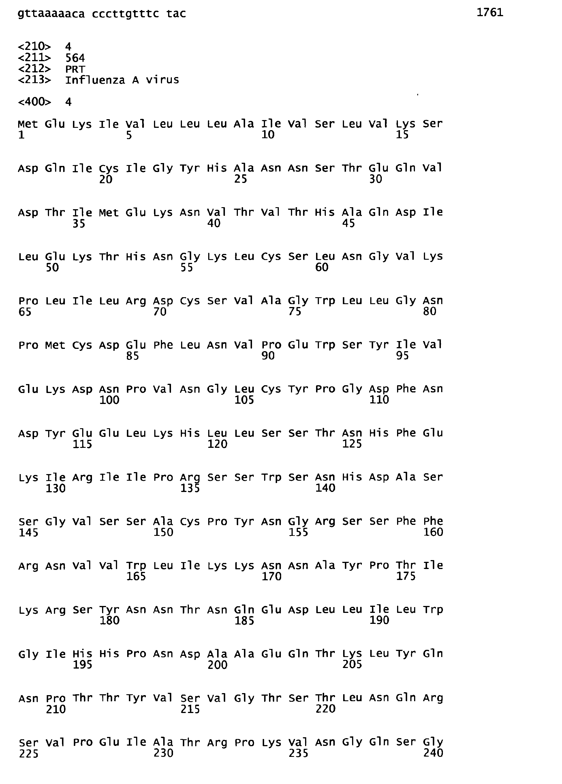


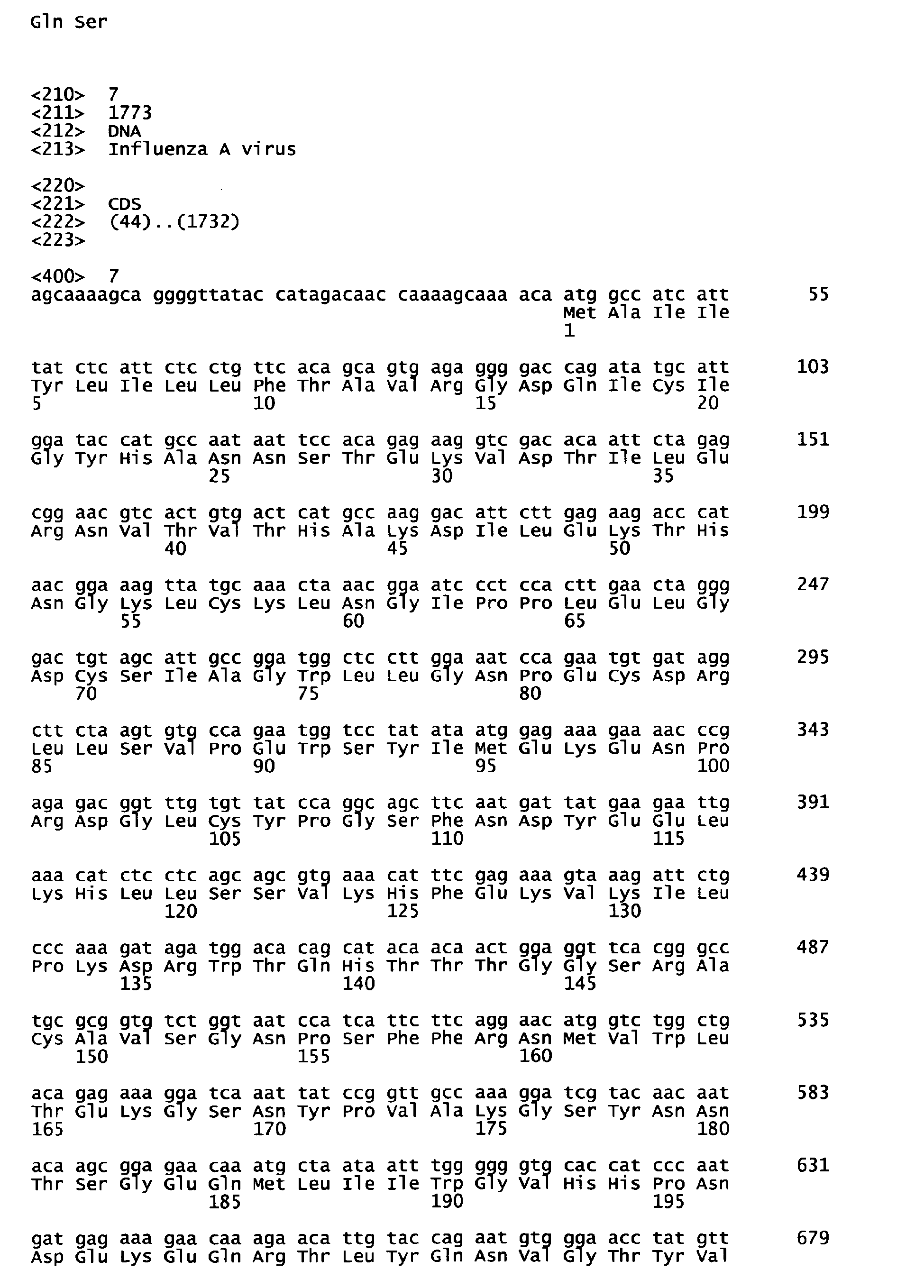








Claims (10)
- An anti-H5 subtype influenza A virus hemagglutinin monoclonal antibody or an antigen-binding fragment thereof which undergoes antigen-antibody reaction with hemagglutinin of H5 subtype influenza A virus, whose corresponding epitope does not exist in the receptor subdomain (excluding C-terminal region thereof consisting of 11 amino acids) and which does not have neutralizing activity against said influenza A virus.
- The monoclonal antibody or an antigen-binding fragment thereof according to claim 1, which is substantially non-cross-reactive with hemagglutinins of H1 to H4 subtypes and H6 to H 15 subtypes.
- The monoclonal antibody or an antigen-binding fragment thereof according to claim 1 or 2, whose corresponding epitope exists in:(1) the regions of 41 to 60 aa and 312 to 322 aa;(2) the regions of 61 to 80 aa and 290 to 300 aa; or(3) the regions of 101 to 113 aa and 268 to 278 aa,based on the amino acid sequence shown in SEQ ID NO:2.
- A method of immunoassay of H5 subtype influenza A virus, comprising measurement of H5 subtype influenza A virus in a sample by immunoassay using antigen-antibody reaction of the monoclonal antibody or an antigen-binding fragment thereof according to any one of claims 1 to 3 with hemagglutinin of the H5 subtype influenza A virus.
- The method according to claim 3, carried out by a sandwich immunoassay using 2 types of the monoclonal antibody and/or an antigen-binding fragment thereof according to any one of claims 1 to 4, said 2 types of the monoclonal antibody and/or the antigen-binding fragment thereof being capable of binding at the same time to a single hemagglutinin molecule of H5 subtype influenza A virus.
- The method according to claim 5, carried out using 2 types out of the 3 types of monoclonal antibodies whose corresponding epitopes exist in:(1) the regions of 41 to 60 aa and 312 to 322 aa;(2) the regions of 61 to 80 aa and 290 to 300 aa; and(3) the regions of 101 to 113 aa and 268 to 278 aa,based on the amino acid sequence shown in SEQ ID NO:2.
- The method according to claim 6, carried out by an immunochromatography.
- An immunoassay kit for H5 subtype influenza A virus, comprising 2 types of the monoclonal antibody and/or an antigen-binding fragment thereof according to any one of claims 1 to 3, said 2 types of the antibody and/or the antigen-binding fragment thereof being capable of binding at the same time to hemagglutinin of H5 subtype influenza A virus.
- The kit according to claim 8, wherein one of said 2 types of the monoclonal antibody and/or the antigen-binding fragment thereof is immobilized on a solid phase and the other is labeled.
- The kit according to claim 9, wherein said solid phase is a solid phase for immunochromatography.
Applications Claiming Priority (2)
| Application Number | Priority Date | Filing Date | Title |
|---|---|---|---|
| JP2008088499 | 2008-03-28 | ||
| PCT/JP2009/056083 WO2009119722A1 (en) | 2008-03-28 | 2009-03-26 | Anti-(influenza a virus subtype h5 hemagglutinin) monoclonal antibody |
Publications (2)
| Publication Number | Publication Date |
|---|---|
| EP2284260A1 true EP2284260A1 (en) | 2011-02-16 |
| EP2284260A4 EP2284260A4 (en) | 2011-08-10 |
Family
ID=41113910
Family Applications (1)
| Application Number | Title | Priority Date | Filing Date |
|---|---|---|---|
| EP09726058A Withdrawn EP2284260A4 (en) | 2008-03-28 | 2009-03-26 | MONOCLONAL ANTI- (HEMAGGLUTININ ANTIBODY OF H5 TYPE A INFLUENZA VIRUS SUB TYPE) |
Country Status (6)
| Country | Link |
|---|---|
| US (1) | US8658354B2 (en) |
| EP (1) | EP2284260A4 (en) |
| JP (1) | JP5586064B2 (en) |
| KR (1) | KR101625979B1 (en) |
| CN (1) | CN101990575B (en) |
| WO (1) | WO2009119722A1 (en) |
Cited By (4)
| Publication number | Priority date | Publication date | Assignee | Title |
|---|---|---|---|---|
| WO2012072788A1 (en) * | 2010-12-02 | 2012-06-07 | Mab-Factory Gmbh | Vaccine against influenza h5n1 viruses, medicament and treatment of h5n1 viral infections |
| WO2013043067A3 (en) * | 2011-09-23 | 2013-11-14 | Instytut Biochemii I Biofizyki Pan | Dna vaccine, method of inducing the immune response, method of immunisation, antibodies specifically recognising the h5 haemagglutinin of an influenza virus and use of the dna vaccine |
| RU2502745C2 (en) * | 2011-12-30 | 2013-12-27 | Общество с ограниченной ответственностью "Технофарма" | Anti-flu nanoantibody, recombinant viral vectors and pharmaceutical compositions for prophylaxis and therapy of a-type flu |
| CN114075278A (en) * | 2020-08-18 | 2022-02-22 | 东莞市朋志生物科技有限公司 | anti-Flu-A antibody, preparation method thereof and detection kit |
Families Citing this family (25)
| Publication number | Priority date | Publication date | Assignee | Title |
|---|---|---|---|---|
| ES2678143T3 (en) | 2010-01-19 | 2018-08-09 | President And Fellows Of Harvard College | Opsonin obtained by genetic engineering for the detection and treatment of pathogenic microorganisms |
| WO2011111108A1 (en) * | 2010-03-11 | 2011-09-15 | Dicプラスチック株式会社 | Instrument for enzyme-linked immunoassay, and enzyme-linked immunoassay |
| JP5630153B2 (en) * | 2010-09-01 | 2014-11-26 | 富士レビオ株式会社 | Method for determining whether the 100th amino acid of influenza A virus nucleoprotein is arginine |
| EP2734843A2 (en) * | 2011-07-18 | 2014-05-28 | President and Fellows of Harvard College | Engineered microbe-targeting molecules and uses thereof |
| JP5884100B2 (en) * | 2011-08-31 | 2016-03-15 | 公益財団法人東京都医学総合研究所 | Recombinant vaccinia virus derived from a DIs strain having a hemagglutinin protein gene derived from a new influenza virus |
| CN103030693B (en) * | 2011-09-30 | 2015-03-11 | 中国科学院上海巴斯德研究所 | Neutralization molecule of high-pathogenicity avian influenza and preparation method thereof |
| JP2013087069A (en) * | 2011-10-17 | 2013-05-13 | Toyobo Co Ltd | Monoclonal antibody specifically recognizing h5 subtype influenza virus |
| US9969794B2 (en) | 2012-05-10 | 2018-05-15 | Visterra, Inc. | HA binding agents |
| CN102928605B (en) * | 2012-11-06 | 2014-06-25 | 江苏省农业科学院 | Indirect enzyme-linked immuno-sorbent assay (ELISA) kit for detecting similar porcine circovirus virus P1 antibody |
| EP2976642A4 (en) | 2013-03-15 | 2016-09-21 | Harvard College | METHODS AND COMPOSITIONS FOR IMPROVING THE DETECTION AND / OR CAPTURE OF A TARGET ENTITY |
| US9988617B2 (en) | 2013-05-21 | 2018-06-05 | President And Fellows Of Harvard College | Engineered heme-binding compositions and uses thereof |
| PT3052192T (en) * | 2013-10-02 | 2020-09-24 | Medimmune Llc | Neutralizing anti-influenza a antibodies and uses thereof |
| EP3546474B1 (en) | 2013-12-18 | 2021-07-07 | President and Fellows of Harvard College | Crp capture/detection of gram positive bacteria |
| CN103869067B (en) * | 2014-04-01 | 2015-12-02 | 开封市疾病预防控制中心 | A kind of influenza A virus colloidal gold diagnosis Test paper and preparation method thereof |
| DK3134131T3 (en) * | 2014-04-23 | 2022-02-07 | Modernatx Inc | Nucleic acid vaccines |
| WO2015186721A1 (en) * | 2014-06-03 | 2015-12-10 | 国立研究開発法人農業・食品産業技術総合研究機構 | Antibody or antibody fragment including variable region thereof, antigen polypeptide, and uses thereof |
| TWI702229B (en) | 2014-12-19 | 2020-08-21 | 美商再生元醫藥公司 | Human antibodies to influenza hemagglutinin |
| RU2618947C2 (en) * | 2015-06-30 | 2017-05-11 | Закрытое акционерное общество "Лаборатория Касперского" | Method of preventing program operation comprising functional undesirable for user |
| CN108289928B (en) | 2015-08-06 | 2024-09-17 | 哈佛大学校长及研究员协会 | Improved microbe-binding molecules and uses thereof |
| US11564893B2 (en) | 2015-08-17 | 2023-01-31 | Modernatx, Inc. | Methods for preparing particles and related compositions |
| EP3374390A1 (en) | 2015-11-13 | 2018-09-19 | Visterra, Inc. | Compositions and methods for treating and preventing influenza |
| EP3419663A1 (en) | 2016-02-24 | 2019-01-02 | Visterra, Inc. | Formulations of antibody molecules to influenza virus |
| CN110167587A (en) | 2016-11-11 | 2019-08-23 | 摩登纳特斯有限公司 | Influenza vaccines |
| WO2018170245A1 (en) | 2017-03-15 | 2018-09-20 | Modernatx, Inc. | Broad spectrum influenza virus vaccine |
| CA3132521A1 (en) | 2019-03-25 | 2020-10-01 | Visterra, Inc. | Compositions and methods for treating and preventing influenza |
Family Cites Families (7)
| Publication number | Priority date | Publication date | Assignee | Title |
|---|---|---|---|---|
| CN100473665C (en) * | 2005-02-06 | 2009-04-01 | 厦门大学 | H5 subtype avian flu virus hemagglutinin protein monoclonal antibody, and preparing method and use thereof |
| JP5525688B2 (en) * | 2005-08-19 | 2014-06-18 | 株式会社ビーエル | Immunodetection method for influenza virus H5 subtype |
| JP5351418B2 (en) * | 2005-12-26 | 2013-11-27 | 株式会社ビーエル | Detection method of influenza A virus H5 subtype |
| JP4936428B2 (en) * | 2006-03-28 | 2012-05-23 | 大阪府 | Specific detection of H5 subtype influenza virus |
| JP5300200B2 (en) | 2006-08-31 | 2013-09-25 | 大阪府 | Rapid diagnostic method specific to avian influenza virus |
| JP5435844B2 (en) | 2007-02-13 | 2014-03-05 | 株式会社ビーエル | Immunodetection method for influenza virus H5 subtype |
| JP5490725B2 (en) * | 2008-02-05 | 2014-05-14 | テマセック・ライフ・サイエンシズ・ラボラトリー・リミテッド | Binding protein and epitope blocking ELISA for universal detection of H5 subtype influenza virus |
-
2009
- 2009-03-26 EP EP09726058A patent/EP2284260A4/en not_active Withdrawn
- 2009-03-26 KR KR1020107018151A patent/KR101625979B1/en not_active Expired - Fee Related
- 2009-03-26 WO PCT/JP2009/056083 patent/WO2009119722A1/en not_active Ceased
- 2009-03-26 CN CN200980112548.2A patent/CN101990575B/en not_active Expired - Fee Related
- 2009-03-26 US US12/934,908 patent/US8658354B2/en not_active Expired - Fee Related
- 2009-03-26 JP JP2010505761A patent/JP5586064B2/en active Active
Cited By (5)
| Publication number | Priority date | Publication date | Assignee | Title |
|---|---|---|---|---|
| WO2012072788A1 (en) * | 2010-12-02 | 2012-06-07 | Mab-Factory Gmbh | Vaccine against influenza h5n1 viruses, medicament and treatment of h5n1 viral infections |
| WO2013043067A3 (en) * | 2011-09-23 | 2013-11-14 | Instytut Biochemii I Biofizyki Pan | Dna vaccine, method of inducing the immune response, method of immunisation, antibodies specifically recognising the h5 haemagglutinin of an influenza virus and use of the dna vaccine |
| US9505806B2 (en) | 2011-09-23 | 2016-11-29 | Instytut Biochemii I Biofizyki Pan | DNA vaccine, method of inducing the immune response, method of immunisation, antibodies specifically recognising the H5 haemagglutinin of an influenza virus and use of the DNA vaccine |
| RU2502745C2 (en) * | 2011-12-30 | 2013-12-27 | Общество с ограниченной ответственностью "Технофарма" | Anti-flu nanoantibody, recombinant viral vectors and pharmaceutical compositions for prophylaxis and therapy of a-type flu |
| CN114075278A (en) * | 2020-08-18 | 2022-02-22 | 东莞市朋志生物科技有限公司 | anti-Flu-A antibody, preparation method thereof and detection kit |
Also Published As
| Publication number | Publication date |
|---|---|
| JPWO2009119722A1 (en) | 2011-07-28 |
| US8658354B2 (en) | 2014-02-25 |
| KR20100124720A (en) | 2010-11-29 |
| CN101990575A (en) | 2011-03-23 |
| CN101990575B (en) | 2015-06-17 |
| EP2284260A4 (en) | 2011-08-10 |
| KR101625979B1 (en) | 2016-05-31 |
| JP5586064B2 (en) | 2014-09-10 |
| US20110065095A1 (en) | 2011-03-17 |
| WO2009119722A1 (en) | 2009-10-01 |
Similar Documents
| Publication | Publication Date | Title |
|---|---|---|
| US8658354B2 (en) | Anti-(influenza a virus subtype H5 hemagglutinin) monoclonal antibody | |
| CN102015765B (en) | Binding protein and epitope-blocking ELISA for the universal detection of H5-subtype influenza viruses | |
| US8540994B2 (en) | H5 subtype-specific binding proteins useful for H5 avian influenza diagnosis and surveillance | |
| EP2063270B1 (en) | Rapid diagnosis method specific to avian influenza virus | |
| EP2207804B1 (en) | Monoclonal antibodies specific to hemagglutinin from influenza virus h5-subtype and uses thereof | |
| SG183032A1 (en) | Monoclonal antibodies specific to hemagglutininand neuraminidase from influenza virus h5-subtypeor n1-subtype and uses thereof | |
| US10696737B2 (en) | Monoclonal antibodies against hemagglutinin of h5-serotype influenza viruses and their uses, hybridomas producing said antibodies, compositions and diagnostic kits | |
| WO2007074812A1 (en) | Method for detection of influenza type-a virus subtype h5 | |
| JP5432453B2 (en) | Detection method of influenza A virus virulent strain | |
| JP5630153B2 (en) | Method for determining whether the 100th amino acid of influenza A virus nucleoprotein is arginine | |
| JP5782690B2 (en) | Method for producing anti-NP-H289 antibody | |
| CN112175912A (en) | Hybridoma cell line 3G4 1D6, anti-GII.4 type norovirus P protein monoclonal antibody and application | |
| CN102718863A (en) | H5 subtype specific binding protein capable of diagnosing and monitoring H5 avian influenza | |
| HK1161725B (en) | H5 subtype-specific binding proteins useful for h5 avian influenza diagnosis and surveillance |
Legal Events
| Date | Code | Title | Description |
|---|---|---|---|
| PUAI | Public reference made under article 153(3) epc to a published international application that has entered the european phase |
Free format text: ORIGINAL CODE: 0009012 |
|
| 17P | Request for examination filed |
Effective date: 20101020 |
|
| AK | Designated contracting states |
Kind code of ref document: A1 Designated state(s): AT BE BG CH CY CZ DE DK EE ES FI FR GB GR HR HU IE IS IT LI LT LU LV MC MK MT NL NO PL PT RO SE SI SK TR |
|
| AX | Request for extension of the european patent |
Extension state: AL BA RS |
|
| DAX | Request for extension of the european patent (deleted) | ||
| RIC1 | Information provided on ipc code assigned before grant |
Ipc: G01N 33/577 20060101ALI20110630BHEP Ipc: G01N 33/569 20060101ALI20110630BHEP Ipc: G01N 33/543 20060101ALI20110630BHEP Ipc: C12N 15/09 20060101ALI20110630BHEP Ipc: C07K 16/10 20060101ALI20110630BHEP Ipc: C12N 15/02 20060101AFI20110630BHEP |
|
| A4 | Supplementary search report drawn up and despatched |
Effective date: 20110713 |
|
| 17Q | First examination report despatched |
Effective date: 20120810 |
|
| RAP1 | Party data changed (applicant data changed or rights of an application transferred) |
Owner name: NATIONAL UNIVERSITY CORPORATION HOKKAIDO UNIVERSIT Owner name: FUJIREBIO INC. |
|
| GRAP | Despatch of communication of intention to grant a patent |
Free format text: ORIGINAL CODE: EPIDOSNIGR1 |
|
| RIC1 | Information provided on ipc code assigned before grant |
Ipc: G01N 33/569 20060101ALI20140721BHEP Ipc: C07K 16/10 20060101AFI20140721BHEP |
|
| INTG | Intention to grant announced |
Effective date: 20140822 |
|
| STAA | Information on the status of an ep patent application or granted ep patent |
Free format text: STATUS: THE APPLICATION IS DEEMED TO BE WITHDRAWN |
|
| 18D | Application deemed to be withdrawn |
Effective date: 20150106 |