EP2267125A1 - Cancer cell motility and cancer cell infiltration inhibitor - Google Patents
Cancer cell motility and cancer cell infiltration inhibitor Download PDFInfo
- Publication number
- EP2267125A1 EP2267125A1 EP09726307A EP09726307A EP2267125A1 EP 2267125 A1 EP2267125 A1 EP 2267125A1 EP 09726307 A EP09726307 A EP 09726307A EP 09726307 A EP09726307 A EP 09726307A EP 2267125 A1 EP2267125 A1 EP 2267125A1
- Authority
- EP
- European Patent Office
- Prior art keywords
- antibody
- par1
- fragment
- cells
- seq
- Prior art date
- Legal status (The legal status is an assumption and is not a legal conclusion. Google has not performed a legal analysis and makes no representation as to the accuracy of the status listed.)
- Granted
Links
Images


Classifications
-
- C—CHEMISTRY; METALLURGY
- C07—ORGANIC CHEMISTRY
- C07K—PEPTIDES
- C07K16/00—Immunoglobulins [IG], e.g. monoclonal or polyclonal antibodies
- C07K16/18—Immunoglobulins [IG], e.g. monoclonal or polyclonal antibodies against material from animals or humans
- C07K16/28—Immunoglobulins [IG], e.g. monoclonal or polyclonal antibodies against material from animals or humans against receptors, cell surface antigens or cell surface determinants
-
- A—HUMAN NECESSITIES
- A61—MEDICAL OR VETERINARY SCIENCE; HYGIENE
- A61P—SPECIFIC THERAPEUTIC ACTIVITY OF CHEMICAL COMPOUNDS OR MEDICINAL PREPARATIONS
- A61P35/00—Antineoplastic agents
-
- C—CHEMISTRY; METALLURGY
- C07—ORGANIC CHEMISTRY
- C07K—PEPTIDES
- C07K7/00—Peptides having 5 to 20 amino acids in a fully defined sequence; Derivatives thereof
- C07K7/04—Linear peptides containing only normal peptide links
- C07K7/06—Linear peptides containing only normal peptide links having 5 to 11 amino acids
-
- C—CHEMISTRY; METALLURGY
- C07—ORGANIC CHEMISTRY
- C07K—PEPTIDES
- C07K7/00—Peptides having 5 to 20 amino acids in a fully defined sequence; Derivatives thereof
- C07K7/04—Linear peptides containing only normal peptide links
- C07K7/08—Linear peptides containing only normal peptide links having 12 to 20 amino acids
-
- G—PHYSICS
- G01—MEASURING; TESTING
- G01N—INVESTIGATING OR ANALYSING MATERIALS BY DETERMINING THEIR CHEMICAL OR PHYSICAL PROPERTIES
- G01N33/00—Investigating or analysing materials by specific methods not covered by groups G01N1/00 - G01N31/00
- G01N33/48—Biological material, e.g. blood, urine; Haemocytometers
- G01N33/50—Chemical analysis of biological material, e.g. blood, urine; Testing involving biospecific ligand binding methods; Immunological testing
- G01N33/5005—Chemical analysis of biological material, e.g. blood, urine; Testing involving biospecific ligand binding methods; Immunological testing involving human or animal cells
- G01N33/5008—Chemical analysis of biological material, e.g. blood, urine; Testing involving biospecific ligand binding methods; Immunological testing involving human or animal cells for testing or evaluating the effect of chemical or biological compounds, e.g. drugs, cosmetics
- G01N33/502—Chemical analysis of biological material, e.g. blood, urine; Testing involving biospecific ligand binding methods; Immunological testing involving human or animal cells for testing or evaluating the effect of chemical or biological compounds, e.g. drugs, cosmetics for testing non-proliferative effects
- G01N33/5029—Chemical analysis of biological material, e.g. blood, urine; Testing involving biospecific ligand binding methods; Immunological testing involving human or animal cells for testing or evaluating the effect of chemical or biological compounds, e.g. drugs, cosmetics for testing non-proliferative effects on cell motility
-
- G—PHYSICS
- G01—MEASURING; TESTING
- G01N—INVESTIGATING OR ANALYSING MATERIALS BY DETERMINING THEIR CHEMICAL OR PHYSICAL PROPERTIES
- G01N33/00—Investigating or analysing materials by specific methods not covered by groups G01N1/00 - G01N31/00
- G01N33/48—Biological material, e.g. blood, urine; Haemocytometers
- G01N33/50—Chemical analysis of biological material, e.g. blood, urine; Testing involving biospecific ligand binding methods; Immunological testing
- G01N33/53—Immunoassay; Biospecific binding assay; Materials therefor
- G01N33/575—Immunoassay; Biospecific binding assay; Materials therefor for cancer
-
- A—HUMAN NECESSITIES
- A61—MEDICAL OR VETERINARY SCIENCE; HYGIENE
- A61K—PREPARATIONS FOR MEDICAL, DENTAL OR TOILETRY PURPOSES
- A61K39/00—Medicinal preparations containing antigens or antibodies
- A61K2039/505—Medicinal preparations containing antigens or antibodies comprising antibodies
-
- C—CHEMISTRY; METALLURGY
- C07—ORGANIC CHEMISTRY
- C07K—PEPTIDES
- C07K2317/00—Immunoglobulins specific features
- C07K2317/30—Immunoglobulins specific features characterized by aspects of specificity or valency
- C07K2317/34—Identification of a linear epitope shorter than 20 amino acid residues or of a conformational epitope defined by amino acid residues
-
- C—CHEMISTRY; METALLURGY
- C07—ORGANIC CHEMISTRY
- C07K—PEPTIDES
- C07K2317/00—Immunoglobulins specific features
- C07K2317/70—Immunoglobulins specific features characterized by effect upon binding to a cell or to an antigen
- C07K2317/76—Antagonist effect on antigen, e.g. neutralization or inhibition of binding
-
- G—PHYSICS
- G01—MEASURING; TESTING
- G01N—INVESTIGATING OR ANALYSING MATERIALS BY DETERMINING THEIR CHEMICAL OR PHYSICAL PROPERTIES
- G01N2500/00—Screening for compounds of potential therapeutic value
- G01N2500/02—Screening involving studying the effect of compounds C on the interaction between interacting molecules A and B (e.g. A = enzyme and B = substrate for A, or A = receptor and B = ligand for the receptor)
-
- G—PHYSICS
- G01—MEASURING; TESTING
- G01N—INVESTIGATING OR ANALYSING MATERIALS BY DETERMINING THEIR CHEMICAL OR PHYSICAL PROPERTIES
- G01N2500/00—Screening for compounds of potential therapeutic value
- G01N2500/10—Screening for compounds of potential therapeutic value involving cells
-
- G—PHYSICS
- G01—MEASURING; TESTING
- G01N—INVESTIGATING OR ANALYSING MATERIALS BY DETERMINING THEIR CHEMICAL OR PHYSICAL PROPERTIES
- G01N2800/00—Detection or diagnosis of diseases
Definitions
- the present invention relates to cancer cell migration and cancer cell invasion inhibitors. More specifically, the present invention relates to a PAR1 antibody inhibiting a cell migration activity and a cell invasion activity induced by MMP1 as well as a medicinal composition containing the PAR1 antibody and the like.
- the present application claims priority based on Japanese Patent Application No. 2008-83588 , the disclosure of which is incorporated by reference herein.
- a typical disease resulting from abnormally elevated migration ability and invasion ability of cells is cancers.
- Metastasis is mentioned as one of the largest threat of cancers.
- Metastatic cancer cells migrate from a primary tumor through a blood vessel by cell migration, invade within the blood vessel, and then metastasize to other tissues through the blood stream.
- PAR1 prote activated receptor 1
- PAR1 is expressed in most of cultured cancer cell lines having a metastatic ability, and that PAR1 is activated when the N-terminal extracellular domain (between R 41 and S 42 ) of the PAR1 is cleaved by a protease inherent to cancers (MMP1: matrix metalloprotease 1).
- MMP1 matrix metalloprotease 1
- the stimulation of PAR1 activation activates G protein coupled to an intracellular domain of the PAR1. It is thought that an intracellular Ca 2+ concentration is locally elevated as a result of the activation of the G protein, and the cell migration ability and cell invasion ability are enhanced by the Ca 2+ signal (see Non-Patent Document 1).
- PAR1 was originally discovered as a thrombin-dependent receptor (thrombin receptor) necessary for activating platelets. Thrombin recognizes and cleaves the same site in the PAR1 as in the case of the MMP1. Also, activation of the thrombin receptor in platelets activates the Ca 2+ signal in a manner similar to that in the case of cancer cells.
- thrombin receptor thrombin-dependent receptor
- some monoclonal antibodies to an N-terminal cell domain of the PAR1 are marketed, which antibodies inhibit the activation of platelets.
- Pepducin was prepared by synthesizing a peptide corresponding to a G protein binding site of the PAR1 and modifying the peptide so as to have membrane permeability (see Non-Patent Document 2).
- Covic et al. succeeded in inhibiting the activity of the PAR1 by utilizing Pepducin as a G protein antagonist (inhibiting effect of Pepducin is 3 to 4 ⁇ M).
- a method applying an antibody as an anti-cancer drug is expected to show not only an effect inhibiting an activity of a target molecule but also an effect exerting an antibody-dependent cell impairing activity by immune system cells.
- An antibody in which the epitope is a sequence from fifty-second Tyr to fifty-sixth Trp in the PAR1 was prepared by a phage display method, which antibody inhibits an activity of thrombin cleaving the PAR1.
- the inhibition of the cleaving activity of the PAR1 by the antibody (binding to PAR1) in particular, it is thought that the inclusion of fifty-sixth Trp in the epitope is important (see Patent Document 1).
- Patent Document 1 Currently, there is no report describing that the migration and invasion of cancer cells were inhibited by a PAR1 antibody, or that other peptide sequence was found as an epitope for the PAR1 antibody effective for an inhibitory effect.
- the object of the present invention is to provide an agent effectively inhibiting a function relating to cell migration and cell invasion, and more specifically an agent effectively inhibiting a function relating to cell migration and cell invasion by suppressing an activity of PAR1 in cells.
- the present inventors have intensively studied so as to solve the above problems and found antibodies which bind specifically to a region containing a cleavage site in PAR1 (between Arg 41 and Ser 42 ) cleaved by MMP1. They have also found that the antibodies inhibit a cell migration activity and a cell invasion activity induced by MMP1, and thus the present invention has been completed.
- the present invention provides:
- the antibodies of the present invention which bind specifically to PAR1 to inhibit cleavage by MMP1 and also inhibit a migration activity and an invasion activity of cancer cells (hereinafter referred to as "PAR1 antibody” in the present description) can effectively inhibit the migration ability and invasion ability of cancer cells, and therefore, an excellent anti-cancer drug can be obtained. Also, it is possible to carry out detection and imaging of a tumor using the PAR1 antibodies of the present invention. Furthermore, it is possible to obtain or purify an effective PAR1 antibody using the antigenic peptide of the present invention.
- the present invention provides a PAR1 antibody and a fragment of the antibody which retains similar characteristics thereto.
- the PAR1 antibodies of the present invention may be those which bind specifically to a particular region of the PAR1 containing a site cleaved by MMP1, and consequently inhibit a migration activity and an invasion activity of cancer cells.
- the PAR1 antibodies of the present invention may be those which inhibit access of MMP1 to a MMP1 cleavage site by binding specifically to the PAR1 and producing a steric hindrance.
- the PAR1 antibodies of the present invention suppress the migration activity and invasion activity of cancer cells by inhibiting the cleavage of the PAR1 by MMP1 in this manner. Accordingly, the PAR1 antibodies of the present invention are useful for treating cancers.
- the PAR1 antibodies of the present invention may be a monoclonal antibody or a polyclonal antibody.
- the "fragment of the antibody which retains similar characteristics thereto” means a fragment in which an amino acid sequence of an epitope is identical or similar to an amino acid sequence of an epitope for the PAR1 antibodies of the present invention, and which has a migration inhibitory activity and an invasion inhibitory activity against cancer cells identical or similar to those of the PAR1 antibodies of the present invention.
- the "fragment” of the PAR1 antibodies of the present invention means a portion of the PAR1 antibodies of the present invention, and includes, for example, a Fab fragment and the like.
- the "PAR1 antibody” includes the above fragment, unless otherwise specified.
- the "epitope” means an amino acid sequence which constitutes the peptides and antigenic peptides of the present invention or is contained in these peptides, and which is recognized and bound by the antibodies or antibody fragments of the present invention.
- the epitope used in the present invention is an amino acid sequence including the cleavage site (between Arg 41 and Ser 42 ) cleaved by MMP1 and having at least several, and preferably at least 10 amino acids, but is not limited thereto.
- the “epitope” consists of about 5 to about 1000 amino acids.
- the “epitope” of the present invention includes variants thereof.
- the variants may have an amino acid sequence accompanied with deletion, addition and substitution of one or several amino acids in the "epitope” having the above amino acid sequence, or have modification of constitutive amino acids.
- a varied or modified "epitope” should retain inherent characteristics of the "epitope", i.e. those of being recognized and bound by the antibodies or antibody fragments of the present invention.
- the "particular region" of the PAR1 is a region containing the cleavage site (between Arg 41 and Ser 42 ) of the PAR1 cleaved by MMP1.
- the particular region is preferably an amino acid sequence containing about 10 amino acids of both sides of the above cleavage site, and most preferably an amino acid sequence NATLDPRSFLL (SEQ ID NO: 1) containing from thirty-fifth to forty-fifth amino acids of the PAR1.
- the antibodies or antibody fragments of the present invention recognize the amino acid sequence as shown in SEQ ID NO: 1 as an antigen recognition site (epitope).
- amino acids are noted using one letter code and three letter code known in the art.
- the right lower number of an amino acid represents the position of the amino acid from the N-terminus of the amino acid sequence of the PAR1.
- Arg 41 or R 41 represents the forty-first arginine from the N-terminus of the amino acid sequence of the PAR1.
- a preferable antibody which inhibits strongly the migration activity and invasion activity of cancer cells is a monoclonal antibody (hereinafter referred to as "N2-11 antibody” in the present description) produced by a hybridoma which was deposited with International Patent Organism Depository, National Institute of Advanced Industrial Science and Technology of Central sixth, 1-1-1 Higashi, Tsukuba, Ibaraki, JAPAN on March 18, 2008 under the Budapest Treaty and which has an accession number of FERM BP-11105.
- the antibodies or antibody fragments of the present invention are characterized in that they recognize amino acid sequence NATLDPRSFLL (SEQ ID NO: 1) and bind specifically thereto. Such an amino acid sequence was newly found in the present invention.
- An advantage of recognizing the above amino acid sequence is to inhibit effectively the cleavage by MMP1 by the fact that the antibodies or antibody fragments of the present invention recognize an amino acid sequence of both sides of the cleavage site of PAR1 cleaved by MMP1.
- the present invention is greatly significant in respect that SEQ ID NO: 1 was selected from a number of peptides constituting candidates of the recognition site.
- the PAR1 antibodies of the present invention can be prepared as described below, for example.
- An amino acid sequence which contains the cleavage site (between R 41 and S 42 ) of animal, preferably human PAR1 cleaved by MMP1 and has at least several, preferably at least 10 amino acids is used as a target peptide.
- Preferable target peptide has an amino acid sequence as shown in SEQ ID NO: 1.
- the target peptide may be bound to a carrier protein such as, for example, keyhole limpet hemocyanin (KLH) by a known method.
- KLH keyhole limpet hemocyanin
- An animal such as mouse can be immunized using such a conjugate as an antigen to obtain hybridomas.
- the resulting hybridomas are cultured, and a migration activity and an invasion activity of cancer cells are evaluated using the culture supernatants, for example, by a known method such as a matrigel assay, whereby hybridomas having a strong activity can be selected.
- Antibodies produced by the hybridomas may be purified and isolated by a conventional method such as, for example, affinity purification.
- the resulting antibodies can be analyzed for their antibody specificity and antibody effect.
- the PAR1 antibodies of the present invention can be obtained by a combination of the above method and a chemical synthetic method of peptides known in the art. Obviously, methods for preparing the PAR1 antibodies of the present invention are not limited to the above method.
- the PAR1 antibodies of the present invention may be chimerized and humanized by conventional methods. Also, the antibodies of the present invention may be single-chain antibodies. The PAR1 antibodies of the present invention include these chimeric antibodies, humanized antibodies, and single-chain antibodies.
- the PAR1 antibodies of the present invention may also be polyclonal antibodies.
- the polyclonal PAR1 antibodies of the present invention can be obtained by a technique known to those skilled in the art. One example is shown in Example 9.
- the present antibodies in particular, antibodies recognizing the amino acid sequence as shown in SEQ ID NO: 1 as an epitope (preferably N2-11 antibody) or antibody fragments (preferably fragments of N2-11 antibody) have a high invasion suppressing activity and a high migration suppressing activity against cancer cells. Accordingly, the antibodies or antibody fragments of the present invention perform excellent effects for treating cancers.
- the present invention provides a composition for inhibiting a migration activity and an invasion activity of cancer cells, comprising a PAR1 antibody, preferably N2-11 antibody.
- a composition for inhibiting a migration activity and an invasion activity of cancer cells comprising a PAR1 antibody, preferably N2-11 antibody.
- the composition of the present invention can effectively act both in vitro and in vivo.
- a composition containing a chimerized or humanized PAR1 antibody of the present invention can effectively inhibit a migration activity and an invasion activity of human cancer cells.
- the present invention provides a medicinal composition for treating cancers, comprising a PAR1 antibody, preferably N2-11 antibody.
- a PAR1 antibody When used for human, the PAR1 antibody contained in the medicinal composition of the present invention is preferably chimerized, and more preferably humanized.
- the medicinal composition of the present invention can be prepared in any dosage forms.
- the dosage forms can be selected suitably according to a site, a type and a size of a cancer in a subject, characteristics of the PAR1 antibody, body situations and health conditions of a subject and the like, and may be, for example, liquid formulations such as injection and infusion solutions, semisolid to solid formulations such as transdermal patches, ointments, creams and powders.
- the medicinal composition of the present invention can be administered to a subject in any administration routes.
- the administration routes can be selected suitably according to a site, a type and a size of a cancer in a subject, characteristics of the PAR1 antibody, body situations and health conditions of a subject and the like.
- the medicinal composition of the present invention may be administered to a subject by injection such as intradermal injection, subcutaneous injection, muscle injection and intravenous injection, by infusion, or by topical application using transdermal patches, ointments, creams and the like.
- the amount of the PAR1 antibody administered by the medicinal composition of the present invention is usually 5 ⁇ g/kg to 125 mg/kg, preferably 10 ⁇ g/kg to 100 mg/kg, and more preferably 50 ⁇ g/kg to 50 mg/kg per dose, and the composition may be administered once to several times per day. The administration may be made daily, every few days, weekly to every few weeks, or monthly to every few months.
- the medicinal composition of the present invention may contain one or more types of PAR1 antibodies.
- the medicinal composition of the present invention contains N2-11 antibody or a fragment thereof.
- the medicinal composition of the present invention may contain other effective ingredients than the PAR1 antibody, for example, known anti-cancer drugs or other drugs.
- the present invention provides a method for treating cancers, which comprises administering an effective amount of a medicinal composition containing a PAR1 antibody or its fragment of the present invention to a subject in need of such treatment. Also, the present invention provides use of a PAR1 antibody or its fragment of the present invention in the production of a medicine for treating cancers.
- a preferable PAR1 antibody in the above method and use is N2-11 antibody.
- the subject is preferably mammals, and most preferably human.
- Tumor cells express a PAR1 protein.
- the present invention provides a method for imaging tumor cells or a method for detecting a tumor in a sample, wherein said method comprises bringing an antibody or antigen fragment of the present invention which binds to the above protein into contact with a sample obtained from a subject to bind it to tumor cells in the sample.
- the present invention provides an imaging agent for tumor cells, comprising an antibody or antigen fragment of the present invention.
- the present invention provides a method for diagnosing a tumor, which comprises administering an antibody or antigen fragment of the present invention to a subject to investigate the binding of the antibody or antigen fragment of the present invention to a tumor tissue in the body.
- detectable label or tag In these methods and in the imaging agent, it is preferable to attach a detectable label or tag to an antibody or antigen fragment of the present invention.
- the detectable labels or tags include, for example, enzymes such as fluorescent materials, luminescent materials, or horseradish peroxidase, specific binding substances such as biotin or avidin, or positron-emitting radionuclides used for PET, SPECT or the like, but are not limited thereto.
- a preferable antibody is N2-11 antibody
- a preferable antibody fragment is a fragment of the N2-11 antibody.
- the present invention provides an antigenic peptide comprising a peptide consisting of the amino acid sequence as shown in SEQ ID NO: 1 and a carrier protein.
- an antigenic peptide comprising a peptide consisting of the amino acid sequence as shown in SEQ ID NO: 1 and a carrier protein.
- antibodies or antibody fragments of the present invention can be obtained or prepared using such an antigenic peptide.
- the carrier protein means relatively large proteins which bind to peptides having a low molecular weight but are not recognized as an antigen.
- the carrier proteins include, for example, bovine serum albumin (BSA), ovalbumin (OVA), and keyhole limpet hemocyanin (KLH), but are not limited thereto.
- BSA bovine serum albumin
- OVA ovalbumin
- KLH keyhole limpet hemocyanin
- the carrier proteins can be bound to the N-terminus or C-terminus of a peptide, or to a suitable amino acid in a peptide by a known method.
- the present invention provides a method for preparing and a method for purifying an antibody or antibody fragment of the present invention, wherein said method comprises utilizing the specific binding of the above epitope peptide or antigenic peptide to an antibody or antibody fragment of the present invention.
- the antibody or antibody fragment obtained or purified by such a method is that binding specifically to PAR1 and inhibiting cleavage by MMP1.
- the antibody or antibody fragment of the present invention can be purified by binding the above epitope peptide or antigenic peptide to an insoluble carrier, bringing a sample containing the antibody or antibody fragment of the present invention into contact with the carrier to generate a specific binding, and then applying a suitable elution condition.
- Cancers can be treated or prevented by administering the above epitope peptide or antigenic peptide, preferably the peptide consisting of the amino acid sequence as shown in SEQ ID NO: 1, to a subject in need of cancer treatment or cancer prevention, thereby generating an antibody or antibody fragment of the present invention in the subject to inhibit a migration activity and an invasion activity of cancer cells in the subject.
- the present invention provides a composition for inhibiting a migration activity and an invasion activity of cancer cells, comprising the above epitope peptide or antigenic peptide.
- a sequence of both sides of the cleavage site (between R 41 and S 42 ) of PAR1 (N 35 ATLDPRSFLL 45 ) (SEQ ID NO: 1), and (B) a sequence located inside the cleavage site (between R 41 and S 42 ) of PAR1 ( 45 LRNPNDKYEPFWEDEEKKNES 64 ) (SEQ ID NO: 22) were selected as targets, and C-N 35 ATLDPRSFLL 45 (SEQ ID NO: 2) peptide and L 45 RNPNDKYEPFWEDEEKKNES 64 -C (SEQ ID NO: 23) peptide were synthesized by adding C to the N-terminus of sequence (A) and to the C-terminus of sequence (B).
- the synthetic peptides were bound to KLH using the SH group in C, and mice were immunized using the resulting conjugates as an antigen to obtain eleven (11) hybridomas.
- These hybridomas were named PAR1 N2-1 to PAR1 N2-11 and PAR1 N3-1 to PAR1 N3-11, respectively.
- PAR1 N2-11 which showed the highest effect in a matrigel assay using culture supernatants of the hybridomas and PAR1 N3-1 which showed a very strong binding activity to SEQ ID NO: 23 were selected and analyzed for their antibody specificity and antibody effect.
- the antibody produced by PAR1 N2-11 was named N2-11, and the antibody produced by PAR1 N3-1 was named N3-1.
- the hybridoma producing the N2-11 antibody was named "PAR1 N2-11" and deposited with International Patent Organism Depository, National Institute of Advanced Industrial Science and Technology of Central sixth, 1-1-1 Higashi, Tsukuba, Ibaraki Japan on March 18, 2008 under the Budapest Treaty, and obtained an accession number of FERM BP-11105.
- N2-11 antibody and N3-1 antibody it was found that they belong to IgG 1 class.
- the antigen recognition site of N2-11 antibody of the present invention was determined in the following manner. On the basis of partial amino acid sequence A 26 PRPESKATNATLDPRSFLLRNPNDKYEP 54 (SEQ ID NO: 3) of PAR1, eighteen (18) peptides having 12 amino acids (hereinafter referred to as "peptides (1) to (18)" in the present description) were prepared, and an epitope mapping was carried out using an ELISA method.
- the amino acid sequences of the peptides (1) to (18) were as follows:
- the antigen recognition site of the N2-11 antibody is an amino acid sequence consisting of from thirty-fifth to forty-fifth amino acids of PAR1: NATLDPRSFLL (SEQ ID NO: 1) (see Fig. 1 ).
- the N2-11 antibody showed reactivity with peptide (9) but not with peptide (10), although peptides (9) and (10) contain an identical NATLDPRSFLL sequence (O.D. 492 value: about 0.12 for peptide (9), about 0 for peptide (10); see Fig. 1 ). It is thought that this is attributed to the fact that the amino group exposed at the N-terminus of NATLDPRSFLL did not show reactivity with the N2-11 antibody due to the use of sequence CN 35 ATLDPRSFLL 45 as an antigen peptide.
- the antibody concentration in a culture supernatant of a hybridoma was generally 0.1 to 10 ⁇ g/ml
- the culture supernatant of a hybridoma contained a bovine IgG from a serum (FBS) in a medium in a concentration of 2 mg/ml in 10% FBS.
- FBS serum
- the antibody was purified by a method using ascites of SCID mice.
- the "SCID mouse” is an immunodeficient mouse in which very little IgG is present in the body due to the lack of functional T-cells and B-cells. Accordingly, a high purity of N2-11 antibody can be obtained by purifying the antibody using ascites of SCID mice.
- the antibody of the present invention was purified as follows.
- the antigen recognition site of N3-1 antibody of the present invention was determined in the following manner. On the basis of partial amino acid sequence A 36 TLDPRSFLLRNPWEDEEKNESGLTEYRLVS 73 (SEQ ID NO: 24) of PAR1, Twenty-seven (27) peptides having 12 amino acids (hereinafter referred to as "PAR1-N3 (1) to (27)" in the present description) were prepared, and an epitope mapping was carried out using an ELISA method.
- the amino acid sequences of the PAR1-N3 (1) to (27) were as follows:
- N3-1 antibody to PAR1-N3 (1) to (27) was investigated using a method similar to that in Example 2, excepting that two solutions of 4 ⁇ g/ml and 400 ⁇ g/ml per well were used. Since an S/N ratio is somewhat poor in a 4 ⁇ g/ml peptide solution, a 400 ⁇ g/ml peptide solution was used in order to raise the S/N ratio.
- the antigen recognition site of the N3-1 antibody is an amino acid sequence consisting of from forty-sixth to fifty-seventh amino acids of PAR1: RNPNDKYEPFWE (SEQ ID NO: 35) (see Fig. 2 ).
- the OD values vary depending on reaction times of the reaction using OPD as a substrate, and therefore, they show relative bindings to antigens but are not indicators showing substantial binding forces of an antibody.
- the N3-1 antibody was purified using a method similar to that in Example 2. By subjecting the purified antibody to 10% SDS-PAGE, it was confirmed that transferrin and albumin contained in the ascites solution were not included in the N3-1 antibody. The yield of the antibody was about 400 ⁇ g per ml of ascites solution.
- PAR1-expressing KPL-4 a cell line stably expressing PAR1-GFP (hereinafter referred to as "PAR1-expressing KPL-4" in the present description) was prepared from low invasive breast cancer cultured cells KPL-4 (low PAR1-expressing cell line). 1.5 ⁇ 10 5 PAR1-expressing KPL-4 cells were suspended by a trypsin treatment, and washed with PBS.
- Fig. 3 For counting the invasive cell number, random spots on the filter were scanned in a size of 0.66 mm ⁇ 0.66 mm to calculate the mean cell number. Then, the percentages of the invasive cell number when treated with the N2-11 antibody and N3-1 antibody were shown in Fig. 3 with setting the invasive cell number when treated with the mouse IgG as 100%. On this occasion, the invasive cell number when treated with 10 ⁇ M mouse IgG was about 2.4 x 10 3 cells. In Fig.
- N2-11 antibody has a 40% inhibitory effect in a 10 ⁇ M antibody concentration but the N3-1 antibody does not have such an activity (see Fig. 3 (b) and Fig. 3(c) ).
- an antibody recognizing a sequence containing amino acids from fifty-second Tyr to fifty-sixth Trp of PAR1 as an epitope does not inhibit the cell invasion ability.
- the N2-11 antibody is more effective than the N3-1 antibody for preparing an antibody medicine having an inhibitory effect against the cell invasion. For this reason, effects of the N2-11 antibody were investigated hereafter.
- binding properties of the antibody were quantitatively analyzed using the N2-11 antibody-QD probe.
- the particle number of the above fluorescent nanoparticles and the fluorescent intensity are in a proportional relation.
- Fluorescence images at an exposure time of 100 ms were converted to TIFF format images in a contrast in a range from 1,150 to 1,600 of SOLIS software (Andor).
- a total gray value per 1 cell derived from the N2-11 antibody-QD fluorescence was calculated by subtracting a background gray value from a total gray value (1-256 tones of gray value) of one cell with Image J software.
- the amount of the N2-11 antibody bound to the PAR1-expressing KPL-4 was about 6 times the amount bound to the KPL-4, that, when the PAR1-expressing KPL-4 was treated with 50 nM MMP1, the amount of the N2-11 antibody bound was the same level as that in KPL-4, and that, when the cells were treated with MMP1 after labeled with the N2-11 antibody, the N2-11 antibody was bound to the cells to the extent similar to that in the case not treated with MMP1.
- the results show that the N2-11 antibody recognizes a sequence of PAR1 on the cell membrane, in which the cleavage site is not cleaved, and binds specifically to the sequence, and inhibits the cleavage of the PAR1 by MMP1.
- the PAR1 is activated by MMP1 treatment, and the cell invasion ability is enhanced. Accordingly, observation of flame shootings over time (time-laps) of cell migration of the PAR1-expressing KPL-4 cells and analysis of the migration trajectory were carried out to investigate whether the N2-11 antibody inhibits the MMP1-dependent cell migration ability.
- 7 ⁇ 10 4 PAR1-expressing KPL-4 cells were suspended by a trypsin treatment, and washed with PBS. To the cells, 10 ⁇ M N2-11 antibody or mouse IgG which is a control antibody was added, and the mixture was incubated at 37°C for 60 minutes.
- PAR1-expressing KPL-4 cells treated with the mouse IgG showed a great elevation of the migration speed in the presence of 2.5 nM MMP1 as compared with that in the absence of MMP1, and the treatment with 10 ⁇ M N2-11 antibody showed suppression of the elevation of the migration speed in the presence of MMP1.
- the degree of the elevation of the migration speed of the PAR1-expressing KPL-4 cells treated with the mouse IgG brought about by MMP1 (7.0 ⁇ m/h)
- the degree of the elevation of the migration speed in the presence of the N2-11 antibody brought about by MMP1 was 54%.
- the results show that the migration speed of the PAR1-expressing cells is elevated by MMP1 treatment, but the action of MMP1 cleaving PAR1 is inhibited by treating with the N2-11 antibody and the activation of the migration ability is inhibited by about 50%.
- PAR1-expressing KPL-4 cells the PAR1 is activated by MMP1 treatment, and the cell invasion ability is enhanced. Accordingly, a matrigel assay of PAR1-expressing KPL-4 was carried out in the presence of the N2-11 antibody to investigate whether the N2-11 antibody inhibits the MMP1-dependent cell invasion ability.
- 1.5 ⁇ 10 5 PAR1-expressing KPL-4 cells were suspended by a trypsin treatment, and washed with PBS. To the cells, 3 or 10 ⁇ M N2-11 antibody or mouse IgG which is a control antibody was added, and the mixture was incubated at 37°C for 90 minutes. The cells were then seeded on Cell Culture Insert, and cultured at 37°C for 45 hours.
- a 10% FBS containing 2.5 nM MMP1 solution was used, and after 45 hours, invasive cells migrated to the bottom of a membrane filter was HE-stained to count the number of invasive cells.
- random spots in the filter were scanned in a size of 0.66 mm ⁇ 0.66 mm to calculate the mean cell number. Then, the percentage of the invasive cell number when treated with the N2-11 antibody was determined with setting the invasive cell number when treated with the mouse IgG as 100%.
- the results show that the invasion ability of the PAR1-expressing cells is elevated by MMP1 treatment, but the action of MMP1 cleaving PAR1 is inhibited in a dose-dependent manner by treating with the N2-11 antibody and the activation of the invasion ability is inhibited by about 60%.
- the N2-11 antibody recognizes the cleavage site of PAR1, binds specifically thereto, and inhibits the activity of MMP1 cleaving PAR1, thereby inhibiting the MMP1-dependent cell migration activity and cell invasion activity of the PAR1-expressing cells.
- the N2-11 antibody can be an antibody medicine effective for cancer treatment.
- Example 5 By an original in vivo imaging method using N2-11 antibody-QD similar to that in Example 5 as a probe, a vital staining of a tumor tissue was carried out to investigate whether the antibody prepared as described above is specific to PAR1 in a tumor tissue.
- 1 ⁇ 10 6 PAR1-expressing KPL-4 cells were injected subcutaneously. The mice were then fed for 4 weeks to prepare a tumor derived from the PAR1-expressing KPL-4 cells. The tumor was exposed to prepare a small window for observation, and the N2-11 antibody-QD probe was then injected via the tail vein of the mice in a blood concentration of 10 nM. Subsequently, the tumor tissue in the living mice was observed using an original imaging apparatus.
- FIG. 7 An image in which the N2-11 antibody-QD specifically reacts with tumor cells is shown in Fig. 7 . From Fig. 7 , it is evident that the N2-11 antibody is specific to the tumor cells in the living body of mice. Thus, the results suggest that the N2-11 antibody can inhibit the PAR1 activity of living tumor cells by increasing the concentration or binding ability (binding constant).
- a sequence of both sides of the cleavage site (between R 41 a n d S 42 ) of PAR1 (N 35 ATLDPRSFLL 45 ) (SEQ ID NO: 1) was selected as a target, and CN 35 ATLDPRSFLL 45 (SEQ ID NO: 2) peptide was synthesized by adding C to the N-terminus of the above sequence.
- the synthetic peptide was bound to KLH using the SH group in C, and rabbits were immunized using the resulting conjugate as an antigen to prepare an antiserum to PAR1.
- a column was then prepared using the peptide as an antigen and SulfoLink coupling gel (PIERCE).
- the antiserum was applied to the column, and the column was then washed with PBS (pH 7.2) of 5 times or more the column volume. Subsequently, the PAR1 polyclonal antibody was eluted using 3M MgCl 2 . After the elution, the solution was dialyzed against PBS to obtain a purified PAR1 antibody (about 0.4 mg of a purified antibody was obtained from 10 ml of the antiserum).
- the cell invasion activity of the polyclonal antibody was investigated using a matrigel in a manner similar to that in the assay of the monoclonal antibody. 300 nM Rabbit IgG was used as a control.
- Fig. 8 The percentages of the invasive cell number when treated with the PAR1 polyclonal antibody are shown in Fig. 8 with setting the mean invasive cell number under condition (a) as 100%, and the calculated values are shown in Table 3 below.
- the results show that 300 nM polyclonal PAR1 antibody has an effect suppressing the invasion ability of cancer cells in the same level as 10 ⁇ M monoclonal PAR1 antibody (inhibitory effect: about 60%).
- a gene of the monoclonal PAR1 antibody can be modified by a genetic engineering technique to obtain 10 to 100-fold enhancement of the inhibitory effect, and shows that the antibody of the present invention can be used as an effective anti-cancer drug.
- the antibody of the present invention can be used not only as an effective anti-cancer drug, but also in detection of a tumor.
- the PAR1 antibody can be an antibody medicine effective for cancer treatment because the PAR1 antibody inhibits the MMP1-dependent cell migration ability and cell invasion ability in PAR1-expressing cells and specifically reacts with tumor cells in the living body of mice. Also, the antibody of the present invention can be used for detection of a tumor and the like.
Landscapes
- Health & Medical Sciences (AREA)
- Chemical & Material Sciences (AREA)
- Life Sciences & Earth Sciences (AREA)
- Immunology (AREA)
- Engineering & Computer Science (AREA)
- Molecular Biology (AREA)
- Organic Chemistry (AREA)
- General Health & Medical Sciences (AREA)
- Medicinal Chemistry (AREA)
- Biochemistry (AREA)
- Biomedical Technology (AREA)
- Hematology (AREA)
- Urology & Nephrology (AREA)
- Cell Biology (AREA)
- Biophysics (AREA)
- Genetics & Genomics (AREA)
- Proteomics, Peptides & Aminoacids (AREA)
- Biotechnology (AREA)
- Pathology (AREA)
- General Physics & Mathematics (AREA)
- Analytical Chemistry (AREA)
- Physics & Mathematics (AREA)
- Food Science & Technology (AREA)
- Microbiology (AREA)
- Tropical Medicine & Parasitology (AREA)
- Toxicology (AREA)
- Bioinformatics & Cheminformatics (AREA)
- Pharmacology & Pharmacy (AREA)
- Chemical Kinetics & Catalysis (AREA)
- General Chemical & Material Sciences (AREA)
- Nuclear Medicine, Radiotherapy & Molecular Imaging (AREA)
- Veterinary Medicine (AREA)
- Public Health (AREA)
- Animal Behavior & Ethology (AREA)
- Peptides Or Proteins (AREA)
- Medicines Containing Antibodies Or Antigens For Use As Internal Diagnostic Agents (AREA)
- Preparation Of Compounds By Using Micro-Organisms (AREA)
Abstract
Description
- The present invention relates to cancer cell migration and cancer cell invasion inhibitors. More specifically, the present invention relates to a PAR1 antibody inhibiting a cell migration activity and a cell invasion activity induced by MMP1 as well as a medicinal composition containing the PAR1 antibody and the like. The present application claims priority based on Japanese Patent Application No.
2008-83588 - A typical disease resulting from abnormally elevated migration ability and invasion ability of cells is cancers. Metastasis is mentioned as one of the largest threat of cancers. Metastatic cancer cells migrate from a primary tumor through a blood vessel by cell migration, invade within the blood vessel, and then metastasize to other tissues through the blood stream. PAR1 (protease activated receptor 1) is a seven-transmembrane receptor which activates the migration ability and invasion ability of cancer cells, and is involved in the metastasis of a great variety of cancers (breast cancer, lung cancer, pancreas cancer, prostate cancer, etc.). It is known that, in the breast cancer in particular, PAR1 is expressed in most of cultured cancer cell lines having a metastatic ability, and that PAR1 is activated when the N-terminal extracellular domain (between R41 and S42) of the PAR1 is cleaved by a protease inherent to cancers (MMP1: matrix metalloprotease 1). The stimulation of PAR1 activation activates G protein coupled to an intracellular domain of the PAR1. It is thought that an intracellular Ca2+ concentration is locally elevated as a result of the activation of the G protein, and the cell migration ability and cell invasion ability are enhanced by the Ca2+ signal (see Non-Patent Document 1).
- PAR1 was originally discovered as a thrombin-dependent receptor (thrombin receptor) necessary for activating platelets. Thrombin recognizes and cleaves the same site in the PAR1 as in the case of the MMP1. Also, activation of the thrombin receptor in platelets activates the Ca2+ signal in a manner similar to that in the case of cancer cells. Currently, some monoclonal antibodies to an N-terminal cell domain of the PAR1 are marketed, which antibodies inhibit the activation of platelets. However, there is no report as to the relationship between these antibodies and the migration and invasion of cancer cells. Even more, there is no report describing that the migration and invasion of cancer cells were inhibited by a PAR1 antibody, or that a PAR1 antibody was used for a cancer treatment.
- Also, a substance named Pepducin was prepared by synthesizing a peptide corresponding to a G protein binding site of the PAR1 and modifying the peptide so as to have membrane permeability (see Non-Patent Document 2). Covic et al. succeeded in inhibiting the activity of the PAR1 by utilizing Pepducin as a G protein antagonist (inhibiting effect of Pepducin is 3 to 4 µM). On the other hand, a method applying an antibody as an anti-cancer drug is expected to show not only an effect inhibiting an activity of a target molecule but also an effect exerting an antibody-dependent cell impairing activity by immune system cells. In addition, a technique for preparing antibodies having a high affinity by genetic engineering has been established, and the safety of antibodies to a living body has been ensured. For the reasons as described above, there is a very large merit in applying antibodies as an anti-cancer drug. Accordingly, it is thought that the application of a PAR1 antibody as an anti-cancer drug has a possibility that the antibody becomes a more effective anti-cancer drug than Pepducin.
- An antibody in which the epitope is a sequence from fifty-second Tyr to fifty-sixth Trp in the PAR1 was prepared by a phage display method, which antibody inhibits an activity of thrombin cleaving the PAR1. With respect to the inhibition of the cleaving activity of the PAR1 by the antibody (binding to PAR1), in particular, it is thought that the inclusion of fifty-sixth Trp in the epitope is important (see Patent Document 1). Currently, there is no report describing that the migration and invasion of cancer cells were inhibited by a PAR1 antibody, or that other peptide sequence was found as an epitope for the PAR1 antibody effective for an inhibitory effect.
- Patent Document 1:
JP-2002-10784 A - Non-Patent Document 1: Boire A, Covic L, Agarwal A, Jacques S, Sherifi S, Kuliopulos A, PAR1 is a matrix matalloprotease-1 receptor that promotes invasion and tumorigenesis of breast cancer cells. Cell 120: 303-313 (2005)
- Non-Patent Document 2: Covic L, Misra M, Bader J, Singh C, Kuliopulus A, Pepducin-based intervention of thrombin-receptor signaling and systematic platelet activation. Nat. Med. 8; 1161-1165 (2002)
- The object of the present invention is to provide an agent effectively inhibiting a function relating to cell migration and cell invasion, and more specifically an agent effectively inhibiting a function relating to cell migration and cell invasion by suppressing an activity of PAR1 in cells.
- The present inventors have intensively studied so as to solve the above problems and found antibodies which bind specifically to a region containing a cleavage site in PAR1 (between Arg41 and Ser42) cleaved by MMP1. They have also found that the antibodies inhibit a cell migration activity and a cell invasion activity induced by MMP1, and thus the present invention has been completed.
Thus, the present invention provides: - (1) An antibody which binds specifically to PAR1 (protease activated receptor 1) to inhibit cleavage by MMP1 (matrix metalloprotease 1) and also inhibit a migration activity and an invasion activity of cancer cells, or a fragment of the antibody which retains similar characteristics thereto;
- (2) The antibody according to (1), which binds specifically to a region containing a cleavage site in PAR1 (protease activated receptor 1) (between Arg41 and Ser42) cleaved by MMP1 (matrix metalloprotease 1), as an epitope, and inhibits the migration activity and invasion activity of cancer cells, or a fragment of the antibody which retains similar characteristics thereto;
- (3) The antibody according to (2), wherein the amino acid sequence of the epitope is shown in SEQ ID NO: 1, or a fragment of the antibody which retains similar characteristics thereto;
- (4) The antibody according to (3), which is a monoclonal antibody produced by a hybridoma having an accession number of FERM BP-11105 assigned by International Patent Organism Depository, National Institute of Advanced Industrial Science and Technology, or a fragment of the antibody which retains similar characteristics thereto;
- (5) The antibody according to any one of (1) to (4), which is chimeric or humanized, or a fragment of the antibody which retains similar characteristics thereto;
- (6) The antibody according to any one of (1) to (5), which is a monoclonal antibody;
- (7) The antibody according to any one of (1) to (3) or (5), which is a polyclonal antibody;
- (8) A composition for inhibiting a migration activity and an invasion activity of cancer cells, comprising the antibody or fragment according to any one of (1) to (7);
- (9) A medicinal composition for treating cancers, comprising the antibody or fragment according to any one of (1) to (7) ;
- (10) A method for treating cancers, which comprises administering an effective amount of a medicinal composition containing the antibody or fragment according to any one of (1) to (7) to a subject in need of such treatment;
- (11) Use of the antibody or fragment according to any one of (1) to (7) in the production of a medicine for treating cancers;
- (12) A method for imaging tumor cells or a method for detecting a tumor in a sample, which comprises bringing the antibody or fragment according to any one of (1) to (7) into contact with a sample obtained from a subject to bind it to tumor cells in the sample;
- (13) An imaging agent for tumor cells, comprising the antibody or fragment according to any one of (1) to (7);
- (14) A method for diagnosing a tumor, which comprises administering the antibody or fragment according to any one of (1) to (7) to a subject to investigate the binding of the antibody or antibody fragment of the present invention to a tumor tissue in the body;
- (15) A peptide consisting of the amino acid sequence as shown in SEQ ID NO: 1;
- (16) An antigenic peptide comprising a peptide consisting of the amino acid sequence as shown in SEQ ID NO: 1 and a carrier protein;
- (17) A method for preparing an antibody which binds specifically to PAR1 to inhibit cleavage by MMP1, or a fragment of the antibody which retains similar characteristics thereto, wherein said method comprises using the peptide according to (15) or the antigenic peptide according to (16);
- (18) A method for purifying an antibody which binds specifically to PAR1 to inhibit cleavage by MMP1, or a fragment of the antibody which retains similar characteristics thereto, wherein said method comprises using the peptide according to (15) or the antigenic peptide according to (16); and
- (19) A composition for inhibiting a migration activity and an invasion activity of cancer cells, comprising the peptide according to (15) or the antigenic peptide according to (16).
- The antibodies of the present invention which bind specifically to PAR1 to inhibit cleavage by MMP1 and also inhibit a migration activity and an invasion activity of cancer cells (hereinafter referred to as "PAR1 antibody" in the present description) can effectively inhibit the migration ability and invasion ability of cancer cells, and therefore, an excellent anti-cancer drug can be obtained. Also, it is possible to carry out detection and imaging of a tumor using the PAR1 antibodies of the present invention. Furthermore, it is possible to obtain or purify an effective PAR1 antibody using the antigenic peptide of the present invention.
-
-
Fig. 1 is a graph showing a partial amino acid sequence of PAR1 in Example 2 (SEQ ID NO: 3) and amino acid sequences of synthetic peptides (1) to (18) in Example 2 (SEQ ID NO: 4 to 21) as well as a graph indicating the binding of N2-11 antibody to each sequence. The concentration of the peptide solution is 4 µg/ml. -
Fig. 2 is a graph showing a partial amino acid sequence of PAR1 in Example 3 (SEQ ID NO: 24) and amino acid sequences of synthetic peptides PAR1-N3 (1) to (27) in Example 3 (SEQ ID NOs: 25 to 51) as well as graphs indicating the binding of N3-1 antibody to each sequence. The concentration of the peptide solution is 4 µg/ml in the upper graph, and 400 µg/ml in the lower graph. -
Fig. 3 is a graph showing the percentages of the invasive cell number under conditions (a) to (c) with setting the invasive cell number under 3 µM or 10 µM mouse IgG (control) as 100%. Condition (a) shows a 3 µM N2-11 antibody (n = 30) treatment, condition (b) a 10 µM N2-11 antibody (n = 80) treatment, and condition (c) a 10 µM N3-1 antibody (n = 40) treatment. Cells were applied to matrigel chambers at 1.5 × 05 cells/well. Under 10 µM mouse IgG, about 2.4 × 10³ cells show the invasion ability. The error bar shows the standard error. -
Fig. 4 is a graph showing cells obtained by (A) to (D) in Example 5 (columns 1 to 4, respectively) and quantitative analysis results of data ofcolumns 1 to 4. The error bar shows the standard error, in which n = 16 in (column 1), n = 11 in (column 2), n = 11 in (column 3), and n = 10 in (column 4). -
Fig. 5 is a graph showing mean migration speeds per hour between 2 hours and 13 hours after starting observation under conditions (a) to (d) in Table 1. The error bar shows the standard error. -
Fig. 6 is a graph showing the percentages of the invasive cell number under conditions (a), (c) and (d) in Table 2 with setting the invasive cell number under condition (b) in Table 2 as 100%. Cells were applied to matrigel chambers at 1.5 × 10⁵ cells/well. Under condition (b), about 2.2 × 10³ cells show the invasion ability. The error bar shows the standard error. -
Fig. 7 is a graph showing images of living tumor tissues imaged using the N2-11 antibody-QD.Fig. 7(B) shows an image of two tumor cells inFig. 7(A) (the outline of the cells is shown with a white line). The black portions around the center of the cells show nuclei. -
Fig. 8 is a graph showing the percentages of the invasive cell number under conditions (a), (b) and (c) in Table 3 with setting the invasive cell number under condition (a) in Table 3 as 100%. Cells were applied to matrigel chambers at 1.5 × 10⁵ cells/well. Under condition (a), about 1.5 × 10³ cells show the invasion ability. The error bar shows the standard error. - In one aspect, the present invention provides a PAR1 antibody and a fragment of the antibody which retains similar characteristics thereto. In particular, the PAR1 antibodies of the present invention may be those which bind specifically to a particular region of the PAR1 containing a site cleaved by MMP1, and consequently inhibit a migration activity and an invasion activity of cancer cells. Alternatively, the PAR1 antibodies of the present invention may be those which inhibit access of MMP1 to a MMP1 cleavage site by binding specifically to the PAR1 and producing a steric hindrance. The PAR1 antibodies of the present invention suppress the migration activity and invasion activity of cancer cells by inhibiting the cleavage of the PAR1 by MMP1 in this manner. Accordingly, the PAR1 antibodies of the present invention are useful for treating cancers. The PAR1 antibodies of the present invention may be a monoclonal antibody or a polyclonal antibody.
- In the present description, the "fragment of the antibody which retains similar characteristics thereto" means a fragment in which an amino acid sequence of an epitope is identical or similar to an amino acid sequence of an epitope for the PAR1 antibodies of the present invention, and which has a migration inhibitory activity and an invasion inhibitory activity against cancer cells identical or similar to those of the PAR1 antibodies of the present invention. The "fragment" of the PAR1 antibodies of the present invention means a portion of the PAR1 antibodies of the present invention, and includes, for example, a Fab fragment and the like. In the present description, the "PAR1 antibody" includes the above fragment, unless otherwise specified.
- In the present description, the "epitope" means an amino acid sequence which constitutes the peptides and antigenic peptides of the present invention or is contained in these peptides, and which is recognized and bound by the antibodies or antibody fragments of the present invention. In particular, the epitope used in the present invention is an amino acid sequence including the cleavage site (between Arg41 and Ser42) cleaved by MMP1 and having at least several, and preferably at least 10 amino acids, but is not limited thereto. Usually, the "epitope" consists of about 5 to about 1000 amino acids. The "epitope" of the present invention includes variants thereof. For example, the variants may have an amino acid sequence accompanied with deletion, addition and substitution of one or several amino acids in the "epitope" having the above amino acid sequence, or have modification of constitutive amino acids. However, such a varied or modified "epitope" should retain inherent characteristics of the "epitope", i.e. those of being recognized and bound by the antibodies or antibody fragments of the present invention.
- The "particular region" of the PAR1 is a region containing the cleavage site (between Arg41 and Ser42) of the PAR1 cleaved by MMP1. The particular region is preferably an amino acid sequence containing about 10 amino acids of both sides of the above cleavage site, and most preferably an amino acid sequence NATLDPRSFLL (SEQ ID NO: 1) containing from thirty-fifth to forty-fifth amino acids of the PAR1. Preferable the antibodies or antibody fragments of the present invention recognize the amino acid sequence as shown in SEQ ID NO: 1 as an antigen recognition site (epitope).
- In the present description, amino acids are noted using one letter code and three letter code known in the art. The right lower number of an amino acid represents the position of the amino acid from the N-terminus of the amino acid sequence of the PAR1. For example, Arg41 or R41 represents the forty-first arginine from the N-terminus of the amino acid sequence of the PAR1.
- Among the PAR1 antibodies of the present invention which recognize the amino acid sequence as shown in SEQ ID NO: 1 as an epitope, a preferable antibody which inhibits strongly the migration activity and invasion activity of cancer cells is a monoclonal antibody (hereinafter referred to as "N2-11 antibody" in the present description) produced by a hybridoma which was deposited with International Patent Organism Depository, National Institute of Advanced Industrial Science and Technology of Central sixth, 1-1-1 Higashi, Tsukuba, Ibaraki, JAPAN on March 18, 2008 under the Budapest Treaty and which has an accession number of FERM BP-11105.
- As described in the Examples, the antibodies or antibody fragments of the present invention are characterized in that they recognize amino acid sequence NATLDPRSFLL (SEQ ID NO: 1) and bind specifically thereto. Such an amino acid sequence was newly found in the present invention. An advantage of recognizing the above amino acid sequence is to inhibit effectively the cleavage by MMP1 by the fact that the antibodies or antibody fragments of the present invention recognize an amino acid sequence of both sides of the cleavage site of PAR1 cleaved by MMP1. The present invention is greatly significant in respect that SEQ ID NO: 1 was selected from a number of peptides constituting candidates of the recognition site.
- The PAR1 antibodies of the present invention can be prepared as described below, for example. An amino acid sequence which contains the cleavage site (between R41 and S42) of animal, preferably human PAR1 cleaved by MMP1 and has at least several, preferably at least 10 amino acids is used as a target peptide. Preferable target peptide has an amino acid sequence as shown in SEQ ID NO: 1. The target peptide may be bound to a carrier protein such as, for example, keyhole limpet hemocyanin (KLH) by a known method. An animal such as mouse can be immunized using such a conjugate as an antigen to obtain hybridomas. The resulting hybridomas are cultured, and a migration activity and an invasion activity of cancer cells are evaluated using the culture supernatants, for example, by a known method such as a matrigel assay, whereby hybridomas having a strong activity can be selected. Antibodies produced by the hybridomas may be purified and isolated by a conventional method such as, for example, affinity purification. The resulting antibodies can be analyzed for their antibody specificity and antibody effect. Also, the PAR1 antibodies of the present invention can be obtained by a combination of the above method and a chemical synthetic method of peptides known in the art. Obviously, methods for preparing the PAR1 antibodies of the present invention are not limited to the above method.
- Moreover, the PAR1 antibodies of the present invention may be chimerized and humanized by conventional methods. Also, the antibodies of the present invention may be single-chain antibodies. The PAR1 antibodies of the present invention include these chimeric antibodies, humanized antibodies, and single-chain antibodies.
- As described above, the PAR1 antibodies of the present invention may also be polyclonal antibodies. The polyclonal PAR1 antibodies of the present invention can be obtained by a technique known to those skilled in the art. One example is shown in Example 9.
- There exist antibodies recognizing a region of PAR1 different from the above amino acid sequence, for example, an antibody described in
JP-2002-10784 A - Accordingly, in another aspect, the present invention provides a composition for inhibiting a migration activity and an invasion activity of cancer cells, comprising a PAR1 antibody, preferably N2-11 antibody. The composition of the present invention can effectively act both in vitro and in vivo. In particular, a composition containing a chimerized or humanized PAR1 antibody of the present invention can effectively inhibit a migration activity and an invasion activity of human cancer cells.
- In still another aspect, the present invention provides a medicinal composition for treating cancers, comprising a PAR1 antibody, preferably N2-11 antibody.
When used for human, the PAR1 antibody contained in the medicinal composition of the present invention is preferably chimerized, and more preferably humanized. The medicinal composition of the present invention can be prepared in any dosage forms. The dosage forms can be selected suitably according to a site, a type and a size of a cancer in a subject, characteristics of the PAR1 antibody, body situations and health conditions of a subject and the like, and may be, for example, liquid formulations such as injection and infusion solutions, semisolid to solid formulations such as transdermal patches, ointments, creams and powders. The medicinal composition of the present invention can be administered to a subject in any administration routes. The administration routes can be selected suitably according to a site, a type and a size of a cancer in a subject, characteristics of the PAR1 antibody, body situations and health conditions of a subject and the like. For example, the medicinal composition of the present invention may be administered to a subject by injection such as intradermal injection, subcutaneous injection, muscle injection and intravenous injection, by infusion, or by topical application using transdermal patches, ointments, creams and the like. The amount of the PAR1 antibody administered by the medicinal composition of the present invention is usually 5 µg/kg to 125 mg/kg, preferably 10 µg/kg to 100 mg/kg, and more preferably 50 µg/kg to 50 mg/kg per dose, and the composition may be administered once to several times per day. The administration may be made daily, every few days, weekly to every few weeks, or monthly to every few months. The medicinal composition of the present invention may contain one or more types of PAR1 antibodies. Preferably, the medicinal composition of the present invention contains N2-11 antibody or a fragment thereof. Also, the medicinal composition of the present invention may contain other effective ingredients than the PAR1 antibody, for example, known anti-cancer drugs or other drugs. - In other aspect, the present invention provides a method for treating cancers, which comprises administering an effective amount of a medicinal composition containing a PAR1 antibody or its fragment of the present invention to a subject in need of such treatment. Also, the present invention provides use of a PAR1 antibody or its fragment of the present invention in the production of a medicine for treating cancers. A preferable PAR1 antibody in the above method and use is N2-11 antibody. The subject is preferably mammals, and most preferably human.
- Tumor cells express a PAR1 protein. Accordingly, in a further aspect, the present invention provides a method for imaging tumor cells or a method for detecting a tumor in a sample, wherein said method comprises bringing an antibody or antigen fragment of the present invention which binds to the above protein into contact with a sample obtained from a subject to bind it to tumor cells in the sample. Also, the present invention provides an imaging agent for tumor cells, comprising an antibody or antigen fragment of the present invention.
- Moreover, the present invention provides a method for diagnosing a tumor, which comprises administering an antibody or antigen fragment of the present invention to a subject to investigate the binding of the antibody or antigen fragment of the present invention to a tumor tissue in the body.
- In these methods and in the imaging agent, it is preferable to attach a detectable label or tag to an antibody or antigen fragment of the present invention. The detectable labels or tags include, for example, enzymes such as fluorescent materials, luminescent materials, or horseradish peroxidase, specific binding substances such as biotin or avidin, or positron-emitting radionuclides used for PET, SPECT or the like, but are not limited thereto.
In these methods and in the imaging agent, a preferable antibody is N2-11 antibody, and a preferable antibody fragment is a fragment of the N2-11 antibody. - In a further aspect, the present invention provides an antigenic peptide comprising a peptide consisting of the amino acid sequence as shown in SEQ ID NO: 1 and a carrier protein. As described above, antibodies or antibody fragments of the present invention can be obtained or prepared using such an antigenic peptide.
- In the present description, the "carrier protein" means relatively large proteins which bind to peptides having a low molecular weight but are not recognized as an antigen. Examples of the carrier proteins include, for example, bovine serum albumin (BSA), ovalbumin (OVA), and keyhole limpet hemocyanin (KLH), but are not limited thereto. The carrier proteins can be bound to the N-terminus or C-terminus of a peptide, or to a suitable amino acid in a peptide by a known method.
- In a further aspect, the present invention provides a method for preparing and a method for purifying an antibody or antibody fragment of the present invention, wherein said method comprises utilizing the specific binding of the above epitope peptide or antigenic peptide to an antibody or antibody fragment of the present invention. Preferably, the antibody or antibody fragment obtained or purified by such a method is that binding specifically to PAR1 and inhibiting cleavage by MMP1. The antibody or antibody fragment of the present invention can be purified by binding the above epitope peptide or antigenic peptide to an insoluble carrier, bringing a sample containing the antibody or antibody fragment of the present invention into contact with the carrier to generate a specific binding, and then applying a suitable elution condition.
- Cancers can be treated or prevented by administering the above epitope peptide or antigenic peptide, preferably the peptide consisting of the amino acid sequence as shown in SEQ ID NO: 1, to a subject in need of cancer treatment or cancer prevention, thereby generating an antibody or antibody fragment of the present invention in the subject to inhibit a migration activity and an invasion activity of cancer cells in the subject. Accordingly, in a further aspect, the present invention provides a composition for inhibiting a migration activity and an invasion activity of cancer cells, comprising the above epitope peptide or antigenic peptide.
- The present invention will be described in more detail by way of the following Examples, but is not limited thereto.
- (A) A sequence of both sides of the cleavage site (between R41 and S42) of PAR1 (N35ATLDPRSFLL45) (SEQ ID NO: 1), and (B) a sequence located inside the cleavage site (between R41 and S42) of PAR1 (45LRNPNDKYEPFWEDEEKKNES64) (SEQ ID NO: 22) were selected as targets, and C-N35ATLDPRSFLL45 (SEQ ID NO: 2) peptide and L45RNPNDKYEPFWEDEEKKNES64-C (SEQ ID NO: 23) peptide were synthesized by adding C to the N-terminus of sequence (A) and to the C-terminus of sequence (B). The synthetic peptides were bound to KLH using the SH group in C, and mice were immunized using the resulting conjugates as an antigen to obtain eleven (11) hybridomas. These hybridomas were named PAR1 N2-1 to PAR1 N2-11 and PAR1 N3-1 to PAR1 N3-11, respectively. Among these hybridomas, PAR1 N2-11 which showed the highest effect in a matrigel assay using culture supernatants of the hybridomas and PAR1 N3-1 which showed a very strong binding activity to SEQ ID NO: 23 were selected and analyzed for their antibody specificity and antibody effect. The antibody produced by PAR1 N2-11 was named N2-11, and the antibody produced by PAR1 N3-1 was named N3-1. Among them, the hybridoma producing the N2-11 antibody was named "PAR1 N2-11" and deposited with International Patent Organism Depository, National Institute of Advanced Industrial Science and Technology of Central sixth, 1-1-1 Higashi, Tsukuba, Ibaraki Japan on March 18, 2008 under the Budapest Treaty, and obtained an accession number of FERM BP-11105. As a result of carrying out a class determination of N2-11 antibody and N3-1 antibody, it was found that they belong to
IgG 1 class. - The antigen recognition site of N2-11 antibody of the present invention was determined in the following manner. On the basis of partial amino acid sequence A26PRPESKATNATLDPRSFLLRNPNDKYEP54 (SEQ ID NO: 3) of PAR1, eighteen (18) peptides having 12 amino acids (hereinafter referred to as "peptides (1) to (18)" in the present description) were prepared, and an epitope mapping was carried out using an ELISA method. The amino acid sequences of the peptides (1) to (18) were as follows:
- Peptide (1) ARRPESKATNAT (SEQ ID NO: 4)
- Peptide (2) RRPESKATNATL (SEQ ID NO: 5)
- Peptide (3) RPESKATNATLD (SEQ ID NO: 6)
- Peptide (4) PESKATNATLDP (SEQ ID NO: 7)
- Peptide (5) ESKATNATLDPR (SEQ ID NO: 8)
- Peptide (6) SKATNATLDPRS (SEQ ID NO: 9)
- Peptide (7) KATNATLDPRSF (SEQ ID NO: 10)
- Peptide (8) ATNATLDPRSFL (SEQ ID NO: 11)
- Peptide (9) TNATLDPRSFLL (SEQ ID NO: 12)
- Peptide (10) NATLDPRSFLLR (SEQ ID NO: 13)
- Peptide (11) ATLDPRSFLLRN (SEQ ID NO: 14)
- Peptide (12) TLDPRSFLLRNP (SEQ ID NO: 15)
- Peptide (13) LDPRSFLLRNPN (SEQ ID NO: 16)
- Peptide (14) DPRSFLLRNPND (SEQ ID NO: 17)
- Peptide (15) PRSFLLPNPNDK (SEQ ID NO: 18)
- Peptide (16) RSFLLRNPNDKY (SEQ ID NO: 19)
- Peptide (17) SFLLRNPNDKYE (SEQ ID NO: 20)
- Peptide (18) FLLRNPNDLYEP (SEQ ID NO: 21)
- To a 96 well plate for ELISA, 75 µl of 4 µg/ml solution was added per well, and the plate was allowed to stand at 4°C for 12 hours to fix peptides (1) to (18). The wells were blocked at room temperature for 1 hour using 125 µl of 1% BSA containing PBS. Subsequently, a chromogenic reaction by HRP using OPD as a substrate was carried out at room temperature for 1 hour using 100 µl of a neat culture supernatant of a hybridoma as a primary antibody, and then using 100 µl of 5000-fold diluted HRP-conjugated anti-mouse antibody (Upstate) as a secondary antibody. For the chromogenic reaction, an absorbance at 492 nm (O.D. 492 nm) was detected by a plate reader, and the reactivity of N2-11 antibody to peptides (1) to (18) was investigated. As a control, 100 µl of a medium used for cultivation of a hybridoma (10% FBS-containing RPM 1640 (GIBCO)) was used. Values obtained by subtracting an O.D. 492 nm value (about 0.05) in a control experiment from the chromogenic values obtained using the N2-11 antibody were analyzed. As a result, it was found that the antigen recognition site of the N2-11 antibody is an amino acid sequence consisting of from thirty-fifth to forty-fifth amino acids of PAR1: NATLDPRSFLL (SEQ ID NO: 1) (see
Fig. 1 ). - The N2-11 antibody showed reactivity with peptide (9) but not with peptide (10), although peptides (9) and (10) contain an identical NATLDPRSFLL sequence (O.D. 492 value: about 0.12 for peptide (9), about 0 for peptide (10); see
Fig. 1 ). It is thought that this is attributed to the fact that the amino group exposed at the N-terminus of NATLDPRSFLL did not show reactivity with the N2-11 antibody due to the use of sequence CN35ATLDPRSFLL45 as an antigen peptide. - Although the antibody concentration in a culture supernatant of a hybridoma was generally 0.1 to 10 µg/ml, the culture supernatant of a hybridoma contained a bovine IgG from a serum (FBS) in a medium in a concentration of 2 mg/ml in 10% FBS. Accordingly, the antibody was purified by a method using ascites of SCID mice. The "SCID mouse" is an immunodeficient mouse in which very little IgG is present in the body due to the lack of functional T-cells and B-cells. Accordingly, a high purity of N2-11 antibody can be obtained by purifying the antibody using ascites of SCID mice. The antibody of the present invention was purified as follows. Into the abdominal cavity of 8 to 10 weeks old SCID mice, 500 µl of pristine (SIGMA) was injected to cause inflammation and make an environment suitable for proliferation of a hybridoma in the abdominal cavity. After 2 weeks from the injection of pristane, 107 hybridomas producing N2-11 antibody were injected into the abdominal cavity of the mice. Ascites was taken between 1-3 weeks after the injection of cells and centrifuged (20,000 xg), and the resulting supernatant was frozen and stored as an ascites solution. The antibody was purified from the ascites solution using Protein G Sepharose FF (GE Healthcare). By subjecting the purified antibody to 10% SDS-PAGE, it was confirmed that transferrin and albumin contained in the ascites solution were not included in the N2-11 antibody. The yield of the antibody was about 200 µg per 1 ml of ascites solution.
- The antigen recognition site of N3-1 antibody of the present invention was determined in the following manner. On the basis of partial amino acid sequence A36TLDPRSFLLRNPWEDEEKNESGLTEYRLVS73 (SEQ ID NO: 24) of PAR1, Twenty-seven (27) peptides having 12 amino acids (hereinafter referred to as "PAR1-N3 (1) to (27)" in the present description) were prepared, and an epitope mapping was carried out using an ELISA method. The amino acid sequences of the PAR1-N3 (1) to (27) were as follows:
- PAR1-N3 (1) ATLDPRSFLLRN (SEQ ID NO: 25)
- PAR1-N3 (2) TLDPRSFLLRNP (SEQ ID NO: 26)
- PAR1-N3 (3) LDPRSFLLRNPN (SEQ ID NO: 27)
- PAR1-N3 (4) DPRSFLLRNPND (SEQ ID NO: 28)
- PAR1-N3 (5) PRSFLLRNPNDK (SEQ ID NO: 29)
- PAR1-N3 (6) RSFLLRNPNDKY (SEQ ID NO: 30)
- PAR1-N3 (7) SFLLRNPNDKYE (SEQ ID NO: 31)
- PAR1-N3 (8) FLLRNPNDKYEP (SEQ ID NO: 32)
- PAR1-N3 (9) LLRNPNDKYEPF (SEQ ID NO: 33)
- PAR1-N3 (10) LRNPNDKYEPFW (SEQ ID NO: 34)
- PAR1-N3 (11) RNPNDKYEPFWE (SEQ ID NO: 35)
- PAR1 -N3 (12) NPNDKYEPFWED (SEQ ID NO: 36)
- PAR1-N3 (13) PNDKYEPFWEDE (SEQ ID NO: 37)
- PAR1-N3 (14) NDKYEPFWEDEE (SEQ ID NO: 38)
- PAR1-N3 (15) DKYEPFWEDEEK (SEQ ID NO: 39)
- PAR1-N3 (16) KYEPFWEDEEKN (SEQ ID NO: 40)
- PAR1-N3 (17) YEPFWEDEEKNE (SEQ ID NO: 41)
- PAR1-N3 (18) EPFWEDEEKNES (SEQ ID NO: 42)
- PAR1-N3 (19) PFWEDEEKNESG (SEQ ID NO: 43)
- PAR1-N3 (20) FWEDEEKNESGL (SEQ ID NO: 44)
- PAR1-N3 (21) WEDEEKNESGLT (SEQ ID NO: 45)
- PAR1-N3 (22) EDEEKNESGLTE (SEQ ID NO: 46)
- PAR1-N3 (23) DEEKNESGLTEY (SEQ ID NO: 47)
- PAR1-N3 (24) EEKNESGLTEYR (SEQ ID NO: 48)
- PAR1-N3 (25) EKNESGLTEYRL (SEQ ID NO: 49)
- PAR1-N3 (26) KNESGLTEYRLV (SEQ ID NO: 50)
- PAR1-N3 (27) NESGLTEYRLVS (SEQ ID NO: 51)
- The reactivity of N3-1 antibody to PAR1-N3 (1) to (27) was investigated using a method similar to that in Example 2, excepting that two solutions of 4 µg/ml and 400 µg/ml per well were used. Since an S/N ratio is somewhat poor in a 4 µg/ml peptide solution, a 400 µg/ml peptide solution was used in order to raise the S/N ratio. As a result, it was found that the antigen recognition site of the N3-1 antibody is an amino acid sequence consisting of from forty-sixth to fifty-seventh amino acids of PAR1: RNPNDKYEPFWE (SEQ ID NO: 35) (see
Fig. 2 ). In this connection, the OD values vary depending on reaction times of the reaction using OPD as a substrate, and therefore, they show relative bindings to antigens but are not indicators showing substantial binding forces of an antibody. - The N3-1 antibody was purified using a method similar to that in Example 2. By subjecting the purified antibody to 10% SDS-PAGE, it was confirmed that transferrin and albumin contained in the ascites solution were not included in the N3-1 antibody. The yield of the antibody was about 400 µg per ml of ascites solution.
- In order to investigate to what extent the N2-11 antibody and N3-1 antibody can inhibit a thrombin-dependent cell invasion activity, invasion-inhibiting effects of the antibodies were tested using a matrigel assay. In order to prepare cells used for the assay, firstly, a cell line stably expressing PAR1-GFP (hereinafter referred to as "PAR1-expressing KPL-4" in the present description) was prepared from low invasive breast cancer cultured cells KPL-4 (low PAR1-expressing cell line). 1.5 × 10⁵ PAR1-expressing KPL-4 cells were suspended by a trypsin treatment, and washed with PBS. To the cells, 3 or 10 µM N2-11 antibody or 10 µM N3-1 antibody or 3 or 10 µM mouse IgG which is a control antibody was added, and the mixture was incubated at 37°C for 90 minutes. The cells were then seeded on Cell Culture Insert (BD Biosciences), and cultured at 37°C for 45 hours. As a solution of an inducing substance immerging the Insert, a 10% FBS containing 2.5 U/ml thrombin (WAKO) solution was used, and after 45 hours, invasive cells migrated to the bottom of a membrane filter was HE-stained to count the number of invasive cells. For counting the invasive cell number, random spots on the filter were scanned in a size of 0.66 mm × 0.66 mm to calculate the mean cell number. Then, the percentages of the invasive cell number when treated with the N2-11 antibody and N3-1 antibody were shown in
Fig. 3 with setting the invasive cell number when treated with the mouse IgG as 100%. On this occasion, the invasive cell number when treated with 10 µM mouse IgG was about 2.4 x 10³ cells. InFig. 3, (a) shows the percentage of the cell number when treated with 3 µM N2-11 antibody (n = 30), (b) shows the percentage when treated with 10 µM N2-11 antibody (n = 80), and (c) shows the percentage when treated with 10 µM N3-1 antibody (n = 40). - It was found that the N2-11 antibody has a 40% inhibitory effect in a 10 µM antibody concentration but the N3-1 antibody does not have such an activity (see
Fig. 3 (b) and Fig. 3(c) ). Thus, it is evident that an antibody recognizing a sequence containing amino acids from fifty-second Tyr to fifty-sixth Trp of PAR1 as an epitope does not inhibit the cell invasion ability. - Thus, it is believed that the N2-11 antibody is more effective than the N3-1 antibody for preparing an antibody medicine having an inhibitory effect against the cell invasion. For this reason, effects of the N2-11 antibody were investigated hereafter.
- In order to evaluate whether the N2-11 antibody can specifically recognize PAR1 on a cell surface, PAR1-expressing KPL-4 was prepared and characteristics of the N2-11 antibody to KPL-4 and PAR1-expressing KPL-4 were then investigated by the following (A) to (D):
- (A) KPL-4 cells were suspended by a trypsin treatment, and washed with an FBS-free L-15 solution (GIBCO). 2 µM mouse IgG (SIGMA) containing L-15 medium was added, incubation was carried out at 37°C for 15 minutes, blocking was carried out, 40 nM N2-11 antibody-QD probe was further added, and incubation was carried out at 37°C for 30 minutes. Then, the N2-11 antibody-QD probe was washed with L-15 medium, 0.5% FBS containing L-15 medium was added, cells were seeded on glass bottom dishes, and observation and analysis of the cells were carried out. In this connection, the N2-11 antibody-QD probe is a probe in which the N2-11 antibody was bound to high-intensity fluorescent nanoparticles QD705 (Invitrogen)
- (B) PAR1-expressing KPL-4 cells were suspended by a trypsin treatment, and washed with an FBS-free L-15 solution (GIBCO). 2 µM mouse IgG (SIGMA) containing L-15 medium was added, incubation was carried out at 37°C for 15 minutes, blocking was carried out, 40 nM N2-11 antibody-QD probe was further added, and incubation was carried out at 37°C for 30 minutes. Then, the N2-11 antibody-QD probe was washed with L-15 medium, 0.5% FBS containing L-15 medium was added, cells were seeded on glass bottom dishes, and observation and analysis of the cells were carried out.
- (C) PAR1-expressing KPL-4 cells were suspended by a trypsin treatment, and washed with an FBS-free L-15 solution (GIBCO). 50 nM MMP1 (SIGMA) was added, and incubation was carried out at 37°C for 60 minutes. Then, 2 µM mouse IgG (SIGMA) containing L-15 medium was added, incubation was carried out at 37°C for 15 minutes, blocking was carried out, 40 nM N2-11 antibody-QD probe was further added, and incubation was carried out at 37°C for 30 minutes. Then, the N2-11 antibody-QD probe was washed with L-15 medium, 0.5% FBS containing L-15 medium was added, cells were seeded on glass bottom dishes, and observation and analysis of the cells were carried out.
- (D) PAR1-expressing KPL-4 cells were suspended by a trypsin treatment, and washed with an FBS-free L-15 solution (GIBCO). 2 µM mouse IgG (SIGMA) containing L-15 was added, incubation was carried out at 37°C for 15 minutes, blocking was carried out, 40 nM N2-11 antibody-QD probe was further added, and incubation was carried out at 37°C for 30 minutes. Then, the N2-11 antibody-QD probe was washed with L-15 medium, 50 nM MMP1 was added, and incubation was carried out at 37°C for 60 minutes. Then, washing with L-15 medium was carried out, 0.5% FBS containing L-15 medium was added, cells were seeded on glass bottom dishes, and observation and analysis of the cells were carried out.
- In addition, binding properties of the antibody were quantitatively analyzed using the N2-11 antibody-QD probe. The particle number of the above fluorescent nanoparticles and the fluorescent intensity are in a proportional relation. Fluorescence images at an exposure time of 100 ms were converted to TIFF format images in a contrast in a range from 1,150 to 1,600 of SOLIS software (Andor). Subsequently, a total gray value per 1 cell derived from the N2-11 antibody-QD fluorescence was calculated by subtracting a background gray value from a total gray value (1-256 tones of gray value) of one cell with Image J software.
- The cells obtained in the above (A) to (D) and the results of the quantitative analysis based on the data obtained are shown in
Fig. 4 . The results from (A) to (D) correspond tocolumns 1 to 4 in the figure, respectively. As a result of observation of the cells, it was found that the expression level of PAR1 was small in the KPL-4 cells, and therefore, the N2-11 antibody-QD only slightly reacted therewith (column 1), that "PAR1 on the cell membrane" and "PAR1 taken up in the cells" were strongly labeled in dots in the PAR1-expressing KPL-4 cells by labeling with the N2-11 antibody-QD (column 2), that PAR1 on the cell membrane was cleaved by MMP1, and therefore, the N2-11 antibody could not recognize the PAR1 and it was not labeled with the N2-11 antibody-QD (column 3), and that, when the PAR1-expressing KPL-4 cells were treated with MMP1 after labeled with the N2-11 antibody-QD, the N2-11 antibody was bound to the cells to the extent similar to that in column 2 (column 4). Also, it was found from the right panel of the quantitative analysis that the amount of the N2-11 antibody bound to the PAR1-expressing KPL-4 was about 6 times the amount bound to the KPL-4, that, when the PAR1-expressing KPL-4 was treated with 50 nM MMP1, the amount of the N2-11 antibody bound was the same level as that in KPL-4, and that, when the cells were treated with MMP1 after labeled with the N2-11 antibody, the N2-11 antibody was bound to the cells to the extent similar to that in the case not treated with MMP1. - Thus, the results show that the N2-11 antibody recognizes a sequence of PAR1 on the cell membrane, in which the cleavage site is not cleaved, and binds specifically to the sequence, and inhibits the cleavage of the PAR1 by MMP1.
- In PAR1-expressing KPL-4 cells, the PAR1 is activated by MMP1 treatment, and the cell invasion ability is enhanced. Accordingly, observation of flame shootings over time (time-laps) of cell migration of the PAR1-expressing KPL-4 cells and analysis of the migration trajectory were carried out to investigate whether the N2-11 antibody inhibits the MMP1-dependent cell migration ability. 7 × 10⁴ PAR1-expressing KPL-4 cells were suspended by a trypsin treatment, and washed with PBS. To the cells, 10 µM N2-11 antibody or mouse IgG which is a control antibody was added, and the mixture was incubated at 37°C for 60 minutes. Then, 10% FBS containing L-15 medium which contains 2.5 nM MMP1 was added, and the cells were seeded on glass bottom dishes. The images of the flame shootings over time of the cell migration were taken every two minutes, and the trajectory of the cell migration was traced. Mean migration speed per hour between 2 hours and 13 hours after starting the observation of the flame shootings over time was calculated. In the trajectory of the cell migration, the barycentric position of the nucleus was used as an indicator, and all cells capable of tracing were analyzed so that data were not biased. As a control experiment, KPL-4 cells and PAR1-expressing KPL-4 cells untreated with MMP1 were also analyzed in a similar manner.
- The mean migration speeds calculated using the N2-11 antibody are shown in
Fig. 5 , and the calculated values are shown in Table 1.Table 1 Cells Conditions Calculated values KPL-4 in the absence of MMP1 2.8±0.2 µm/h
(n = 195)PAR1-expressing KPL-4 10 µM mouse IgG
(in the absence of MMP1)10.2±0.5 µm/h
(n = 355)PAR1-expressing KPL-4 10 µM mouse IgG
(in the presence of 2.5 nM MMP1)17.2±0.7 µm/h
(n = 357)PAR1-expressing KPL-4 10 µM N2-11 antibody
(in the presence of 2.5 nM MMP1)14.0±0.7 µm/h
(n = 272) - Thus, the results show that the migration speed of the PAR1-expressing cells is elevated by MMP1 treatment, but the action of MMP1 cleaving PAR1 is inhibited by treating with the N2-11 antibody and the activation of the migration ability is inhibited by about 50%.
- In PAR1-expressing KPL-4 cells, the PAR1 is activated by MMP1 treatment, and the cell invasion ability is enhanced. Accordingly, a matrigel assay of PAR1-expressing KPL-4 was carried out in the presence of the N2-11 antibody to investigate whether the N2-11 antibody inhibits the MMP1-dependent cell invasion ability. 1.5 × 10⁵ PAR1-expressing KPL-4 cells were suspended by a trypsin treatment, and washed with PBS. To the cells, 3 or 10 µM N2-11 antibody or mouse IgG which is a control antibody was added, and the mixture was incubated at 37°C for 90 minutes. The cells were then seeded on Cell Culture Insert, and cultured at 37°C for 45 hours. As a solution of an inducing substance immerging the Insert, a 10% FBS containing 2.5 nM MMP1 solution was used, and after 45 hours, invasive cells migrated to the bottom of a membrane filter was HE-stained to count the number of invasive cells. For counting the invasive cell number, random spots in the filter were scanned in a size of 0.66 mm × 0.66 mm to calculate the mean cell number. Then, the percentage of the invasive cell number when treated with the N2-11 antibody was determined with setting the invasive cell number when treated with the mouse IgG as 100%.
- The percentages of the invasive cell number when treated with the N2-11 antibody are shown in
Fig. 6 with setting the mean invasive cell number under condition (b) as 100%, and the calculated values are shown in the following Table 2.Table 2 Conditions Calculated values (a) 10 µM mouse IgG (in the absence of MMP1) 24.3±8.5%
(n = 20)(b) 10 µM mouse IgG (in the presence of 2.5 nM MMP1) 100±18.9%
(n = 20)(c) 3 µM N2-11 antibody (in the presence of 2.5 nM MMP1) 76.2±17.4%
(n = 30)(d) 10 µM N2-11 antibody (in the presence of 2.5 nM MMP1) 38.9±12.5%
(n = 20) - Thus, the results show that the invasion ability of the PAR1-expressing cells is elevated by MMP1 treatment, but the action of MMP1 cleaving PAR1 is inhibited in a dose-dependent manner by treating with the N2-11 antibody and the activation of the invasion ability is inhibited by about 60%.
- From the results of the above Examples 5 to 7, it is evident that the N2-11 antibody recognizes the cleavage site of PAR1, binds specifically thereto, and inhibits the activity of MMP1 cleaving PAR1, thereby inhibiting the MMP1-dependent cell migration activity and cell invasion activity of the PAR1-expressing cells. Thus, it is evident that the N2-11 antibody can be an antibody medicine effective for cancer treatment.
- By an original in vivo imaging method using N2-11 antibody-QD similar to that in Example 5 as a probe, a vital staining of a tumor tissue was carried out to investigate whether the antibody prepared as described above is specific to PAR1 in a tumor tissue. To SCID mice, 1 × 10⁶ PAR1-expressing KPL-4 cells were injected subcutaneously. The mice were then fed for 4 weeks to prepare a tumor derived from the PAR1-expressing KPL-4 cells. The tumor was exposed to prepare a small window for observation, and the N2-11 antibody-QD probe was then injected via the tail vein of the mice in a blood concentration of 10 nM. Subsequently, the tumor tissue in the living mice was observed using an original imaging apparatus.
- An image in which the N2-11 antibody-QD specifically reacts with tumor cells is shown in
Fig. 7 . FromFig. 7 , it is evident that the N2-11 antibody is specific to the tumor cells in the living body of mice. Thus, the results suggest that the N2-11 antibody can inhibit the PAR1 activity of living tumor cells by increasing the concentration or binding ability (binding constant). - A sequence of both sides of the cleavage site (between R41 a n d S42) of PAR1 (N35 ATLDPRSFLL45) (SEQ ID NO: 1) was selected as a target, and CN35ATLDPRSFLL45 (SEQ ID NO: 2) peptide was synthesized by adding C to the N-terminus of the above sequence. The synthetic peptide was bound to KLH using the SH group in C, and rabbits were immunized using the resulting conjugate as an antigen to prepare an antiserum to PAR1. In order to carry out affinity purification of the antiserum, a column was then prepared using the peptide as an antigen and SulfoLink coupling gel (PIERCE). The antiserum was applied to the column, and the column was then washed with PBS (pH 7.2) of 5 times or more the column volume. Subsequently, the PAR1 polyclonal antibody was eluted using 3M MgCl2. After the elution, the solution was dialyzed against PBS to obtain a purified PAR1 antibody (about 0.4 mg of a purified antibody was obtained from 10 ml of the antiserum). The cell invasion activity of the polyclonal antibody was investigated using a matrigel in a manner similar to that in the assay of the monoclonal antibody. 300 nM Rabbit IgG was used as a control.
- The percentages of the invasive cell number when treated with the PAR1 polyclonal antibody are shown in
Fig. 8 with setting the mean invasive cell number under condition (a) as 100%, and the calculated values are shown in Table 3 below.Table 3 Conditions Calculated values (a) 300 nM rabbit IgG (in the presence of 2.5 nM MMP1) 100%±22.2%
(n = 40)(b) 100 nM polyclonal PAR1 antibody (in the presence of 2.5 nM MMP1) 82.6%±14.3%
(n = 40)(c) 300 nM polyclonal PAR1 antibody (in the presence of 2.5 nM MMP1) 46.5%±13.6%
(n = 40) - Thus, the results show that 300 nM polyclonal PAR1 antibody has an effect suppressing the invasion ability of cancer cells in the same level as 10 µM monoclonal PAR1 antibody (inhibitory effect: about 60%). This suggests that a gene of the monoclonal PAR1 antibody can be modified by a genetic engineering technique to obtain 10 to 100-fold enhancement of the inhibitory effect, and shows that the antibody of the present invention can be used as an effective anti-cancer drug.
- In addition, it is also evident from these experimental results that the antibody of the present invention can be used not only as an effective anti-cancer drug, but also in detection of a tumor.
- According to the present invention, it is suggested that the PAR1 antibody can be an antibody medicine effective for cancer treatment because the PAR1 antibody inhibits the MMP1-dependent cell migration ability and cell invasion ability in PAR1-expressing cells and specifically reacts with tumor cells in the living body of mice. Also, the antibody of the present invention can be used for detection of a tumor and the like.


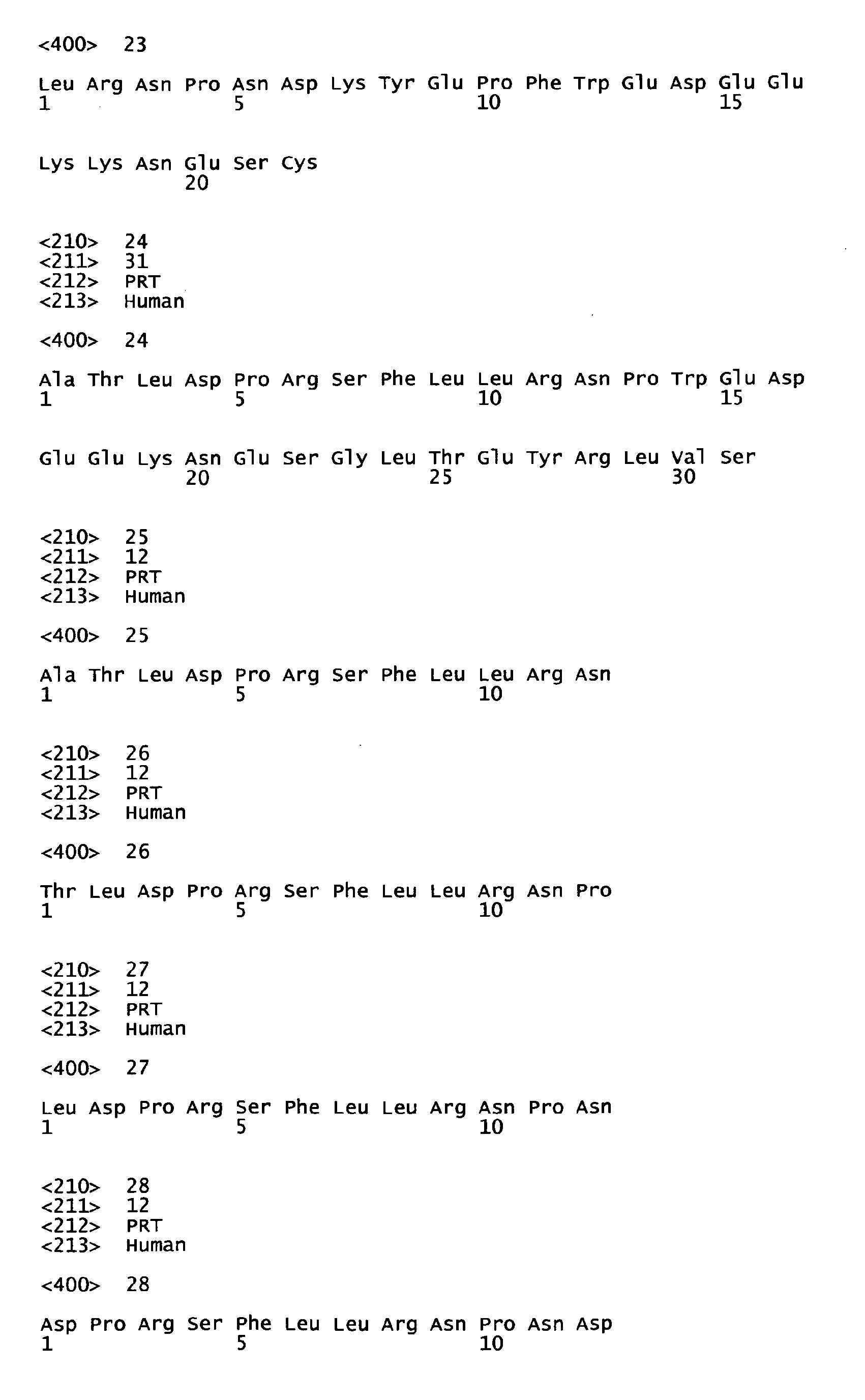


Claims (19)
- An antibody which binds specifically to PAR1 (protease activated receptor 1) to inhibit cleavage by MMP1 (matrix metalloprotease 1) and also inhibit a migration activity and an invasion activity of cancer cells, or a fragment of the antibody which retains similar characteristics thereto.
- The antibody according to claim 1, which binds specifically to a region containing a cleavage site in PAR1 (protease activated receptor 1) (between Arg41 and Ser42) cleaved by MMP1 (matrix metalloprotease 1), as an epitope, and inhibits the migration activity and invasion activity of cancer cells, or a fragment of the antibody which retains similar characteristics thereto.
- The antibody according to claim 2, wherein the amino acid sequence of the epitope is shown in SEQ ID NO: 1, or a fragment of the antibody which retains similar characteristics thereto.
- The antibody according to claim 3, which is a monoclonal antibody produced by a hybridoma having an accession number of FERM BP-11105 assigned by International Patent Organism Depository, National Institute of Advanced Industrial Science and Technology, or a fragment of the antibody which retains similar characteristics thereto.
- The antibody according to any one of claims 1 to 4, which is chimeric or humanized, or a fragment of the antibody which retains similar characteristics thereto.
- The antibody according to any one claims 1 to 5, which is a monoclonal antibody.
- The antibody according to any one of claims 1 to 3 or 5, which is a polyclonal antibody.
- A composition for inhibiting a migration activity and an invasion activity of cancer cells, comprising the antibody or fragment according to any one of claims 1 to 7.
- A medicinal composition for treating cancers, comprising the antibody or fragment according to any one of claims 1 to 7.
- A method for treating cancers, which comprises administering an effective amount of a medicinal composition containing the antibody or fragment according to any one of claims 1 to 7 to a subject in need of such treatment.
- Use of the antibody or fragment according to any one of claims 1 to 7 in the production of a medicine for treating cancers.
- A method for imaging tumor cells or a method for detecting a tumor in a sample, which comprises bringing the antibody or fragment according to any one of claims 1 to 7 into contact with a sample obtained from a subject to bind it to tumor cells in the sample.
- An imaging agent for tumor cells, comprising the antibody or fragment according to any one of claims 1 to 7.
- A method for diagnosing a tumor, which comprises administering the antibody or fragment according to any one of claims 1 to 7 to a subject to investigate the binding of the antibody or antibody fragment of the present invention to a tumor tissue in the body.
- A peptide consisting of the amino acid sequence as shown in SEQ ID NO: 1.
- An antigenic peptide comprising a peptide consisting of the amino acid sequence as shown in SEQ ID NO: 1 and a carrier protein.
- A method for preparing an antibody which binds specifically to PAR1 to inhibit cleavage by MMP1, or a fragment of the antibody which retains similar characteristics thereto, wherein said method comprises using the peptide according to claim 15 or the antigenic peptide according to claim 16.
- A method for purifying an antibody which binds specifically to PAR1 to inhibit cleavage by MMP1, or a fragment of the antibody which retains similar characteristics thereto, wherein said method comprises using the peptide according to claim 15 or the antigenic peptide according to claim 16.
- A composition for inhibiting a migration activity and an invasion activity of cancer cells, comprising the peptide according to claim 15 or the antigenic peptide according to claim 16.
Applications Claiming Priority (2)
| Application Number | Priority Date | Filing Date | Title |
|---|---|---|---|
| JP2008083588 | 2008-03-27 | ||
| PCT/JP2009/055479 WO2009119455A1 (en) | 2008-03-27 | 2009-03-19 | Cancer cell motility and cancer cell infiltration inhibitor |
Publications (3)
| Publication Number | Publication Date |
|---|---|
| EP2267125A1 true EP2267125A1 (en) | 2010-12-29 |
| EP2267125A4 EP2267125A4 (en) | 2011-09-07 |
| EP2267125B1 EP2267125B1 (en) | 2015-09-02 |
Family
ID=41113650
Family Applications (1)
| Application Number | Title | Priority Date | Filing Date |
|---|---|---|---|
| EP09726307.3A Not-in-force EP2267125B1 (en) | 2008-03-27 | 2009-03-19 | Cancer cell migration and cancer cell invasion inhibitor |
Country Status (4)
| Country | Link |
|---|---|
| US (1) | US8674079B2 (en) |
| EP (1) | EP2267125B1 (en) |
| JP (1) | JP5483023B2 (en) |
| WO (1) | WO2009119455A1 (en) |
Cited By (2)
| Publication number | Priority date | Publication date | Assignee | Title |
|---|---|---|---|---|
| US20120121706A1 (en) * | 2009-04-10 | 2012-05-17 | Tufts Medical Center, Inc. | PAR-1 Activation by Metalloproteinase-1 (MMP-1) |
| WO2012090207A3 (en) * | 2010-12-30 | 2012-10-04 | Hadasit Medical Research Services & Development Limited | Par1 and par2 c-tail peptides and peptide mimetics |
Families Citing this family (6)
| Publication number | Priority date | Publication date | Assignee | Title |
|---|---|---|---|---|
| WO2011102483A1 (en) * | 2010-02-19 | 2011-08-25 | 独立行政法人理化学研究所 | HUMAN LAP TGF-β BINDING ANTIBODY |
| EP2613151B1 (en) * | 2010-09-02 | 2016-06-29 | Tohoku University | Method for determining cancer onset or cancer onset risk |
| TWI549997B (en) | 2014-10-14 | 2016-09-21 | 財團法人工業技術研究院 | Method for forming lignin raw epoxy resin and lignin raw epoxy resin composition |
| SG11201705721WA (en) | 2015-01-14 | 2017-08-30 | Brigham & Womens Hospital Inc | Treatment of cancer with anti-lap monoclonal antibodies |
| WO2019075090A1 (en) | 2017-10-10 | 2019-04-18 | Tilos Therapeutics, Inc. | Anti-lap antibodies and uses thereof |
| EP3863722A2 (en) | 2018-10-10 | 2021-08-18 | Tilos Theapeutics, Inc. | Anti-lap antibody variants and uses thereof |
Family Cites Families (6)
| Publication number | Priority date | Publication date | Assignee | Title |
|---|---|---|---|---|
| US5776700A (en) * | 1996-09-05 | 1998-07-07 | Trustees Of The University Of Pennsylvania | Thrombine receptor activation assay for use in identifying thrombin receptor antagonists |
| IL125698A (en) | 1998-08-07 | 2007-03-08 | Hadasit Med Res Service | Use of molecules associated with protease activated receptors for the preparation of pharmaceutical compositions and pharmaceutical compositions comprising them |
| JP4469467B2 (en) | 2000-06-29 | 2010-05-26 | 帝人株式会社 | Cell growth inhibitor |
| US7074765B2 (en) * | 2003-05-01 | 2006-07-11 | The Regents Of The University Of Michigan | Synthetic peptide analogs of Arg-Pro-Pro-Gly-Phe as selective inhibitors of thrombin and thrombin activation of protease activated receptors 1 and 4 |
| US7888483B2 (en) | 2006-07-18 | 2011-02-15 | Irm Llc | Antagonists of protease activated receptor-1 (PAR1) |
| JP4861115B2 (en) | 2006-09-28 | 2012-01-25 | サントリーホールディングス株式会社 | Label, method for producing label, method for stabilizing line peeling strength of label over time, and container |
-
2009
- 2009-03-19 US US12/934,133 patent/US8674079B2/en active Active
- 2009-03-19 JP JP2010505602A patent/JP5483023B2/en active Active
- 2009-03-19 EP EP09726307.3A patent/EP2267125B1/en not_active Not-in-force
- 2009-03-19 WO PCT/JP2009/055479 patent/WO2009119455A1/en not_active Ceased
Cited By (4)
| Publication number | Priority date | Publication date | Assignee | Title |
|---|---|---|---|---|
| US20120121706A1 (en) * | 2009-04-10 | 2012-05-17 | Tufts Medical Center, Inc. | PAR-1 Activation by Metalloproteinase-1 (MMP-1) |
| WO2012090207A3 (en) * | 2010-12-30 | 2012-10-04 | Hadasit Medical Research Services & Development Limited | Par1 and par2 c-tail peptides and peptide mimetics |
| US9745347B2 (en) | 2010-12-30 | 2017-08-29 | Hadasit Medical Research Services And Development Ltd. | PAR1 and PAR2 c-tail peptides and peptide mimetics |
| US10611798B2 (en) | 2010-12-30 | 2020-04-07 | Hadasit Medical Research Services And Development Ltd. | PAR1 and PAR2 c-tail peptides and peptide mimetics |
Also Published As
| Publication number | Publication date |
|---|---|
| EP2267125B1 (en) | 2015-09-02 |
| US8674079B2 (en) | 2014-03-18 |
| EP2267125A4 (en) | 2011-09-07 |
| WO2009119455A1 (en) | 2009-10-01 |
| JP5483023B2 (en) | 2014-05-07 |
| JPWO2009119455A1 (en) | 2011-07-21 |
| US20110070163A1 (en) | 2011-03-24 |
Similar Documents
| Publication | Publication Date | Title |
|---|---|---|
| EP2267125B1 (en) | Cancer cell migration and cancer cell invasion inhibitor | |
| EP2723377B1 (en) | Anti-axl antibodies and uses thereof | |
| EP1856153B1 (en) | Ykl-40 monoclonal antibodies | |
| EP1554309A2 (en) | Antibodies and cyclic peptides which bind cea (carcinoembryonic antigen) and their use as cancer therapeutics | |
| KR20140104944A (en) | Anti-axl antibodies and uses thereof | |
| JP2015516813A5 (en) | ||
| CN105518028A (en) | Novel dual-targeting protein specifically binding to DLL4 and VEGF and use thereof | |
| CA2486252A1 (en) | Methods for screening for agents that modulate hepatocellular carcinoma development | |
| JP2022023215A (en) | DPEP-1 binding composition and method of use | |
| EP1956032B1 (en) | Monoclonal antibody directed against cd166 and method for production thereof | |
| CN101864398B (en) | Monoclonal antibody of anti-gonadotropin-releasing hormone receptor and application thereof | |
| Dallatomasina et al. | Spatiotemporal regulation of tumor angiogenesis by circulating chromogranin A cleavage and neuropilin-1 engagement | |
| CN111201244A (en) | Anti-AQP3 monoclonal antibodies that specifically bind to the extracellular domain of aquaporin 3 (AQP3) and uses thereof | |
| EP2511371A1 (en) | Anticancer anti-mortalin peptide antibodies | |
| EP4532549A1 (en) | Methods, compositions and uses for sema7a monoclonal antibodies | |
| EP4549572A1 (en) | Fusion protein | |
| US12540193B2 (en) | Anti-ADAM8 antibodies and uses of the same | |
| CN111840544A (en) | Application of defucose anti-HER 2 antibody in cancer patient carrying Fc gamma R2A131R genotype | |
| CN104001174B (en) | Applications of the SSX2IP in preventing and treating metastases | |
| US20060024314A1 (en) | Antibodies and cyclic peptides which bind CEA (carcinoembryonic antigens) and their use as cancer therapeutics | |
| WO2022056329A1 (en) | Snx9 subfamily-targeting antibodies | |
| CN120865404A (en) | Anti-CD 209 antibodies, products, nucleic acid molecules, vectors, cells and uses thereof | |
| KR20240139004A (en) | Epitope of regulatory T cell surface antigen and an antibody specifically binding to the epitope thereof | |
| EP3715848A1 (en) | Substance interacting with c terminal fragment of the stim1 fraction localized to the plasma membrane of the cells, for its use in the treatment of cancers, and in screening and diagnostic methods | |
| CA2714880C (en) | A novel tumor biomarker |
Legal Events
| Date | Code | Title | Description |
|---|---|---|---|
| PUAI | Public reference made under article 153(3) epc to a published international application that has entered the european phase |
Free format text: ORIGINAL CODE: 0009012 |
|
| 17P | Request for examination filed |
Effective date: 20100927 |
|
| AK | Designated contracting states |
Kind code of ref document: A1 Designated state(s): AT BE BG CH CY CZ DE DK EE ES FI FR GB GR HR HU IE IS IT LI LT LU LV MC MK MT NL NO PL PT RO SE SI SK TR |
|
| AX | Request for extension of the european patent |
Extension state: AL BA RS |
|
| DAX | Request for extension of the european patent (deleted) | ||
| A4 | Supplementary search report drawn up and despatched |
Effective date: 20110810 |
|
| RIC1 | Information provided on ipc code assigned before grant |
Ipc: C07K 16/46 20060101ALI20110729BHEP Ipc: G01N 33/574 20060101ALI20110729BHEP Ipc: C07K 19/00 20060101ALI20110729BHEP Ipc: C07K 1/22 20060101ALI20110729BHEP Ipc: A61K 39/395 20060101ALI20110729BHEP Ipc: C12P 21/08 20060101ALI20110729BHEP Ipc: C07K 7/06 20060101ALI20110729BHEP Ipc: A61P 35/00 20060101ALI20110729BHEP Ipc: C12N 15/09 20060101AFI20110729BHEP Ipc: G01N 33/577 20060101ALI20110729BHEP Ipc: C07K 14/705 20060101ALI20110729BHEP |
|
| RTI1 | Title (correction) |
Free format text: CANCER CELL MIGRATION AND CANCER CELL INVASION INHIBITOR |
|
| 17Q | First examination report despatched |
Effective date: 20130213 |
|
| GRAP | Despatch of communication of intention to grant a patent |
Free format text: ORIGINAL CODE: EPIDOSNIGR1 |
|
| INTG | Intention to grant announced |
Effective date: 20150313 |
|
| GRAS | Grant fee paid |
Free format text: ORIGINAL CODE: EPIDOSNIGR3 |
|
| GRAA | (expected) grant |
Free format text: ORIGINAL CODE: 0009210 |
|
| AK | Designated contracting states |
Kind code of ref document: B1 Designated state(s): AT BE BG CH CY CZ DE DK EE ES FI FR GB GR HR HU IE IS IT LI LT LU LV MC MK MT NL NO PL PT RO SE SI SK TR |
|
| REG | Reference to a national code |
Ref country code: GB Ref legal event code: FG4D |
|
| REG | Reference to a national code |
Ref country code: AT Ref legal event code: REF Ref document number: 746647 Country of ref document: AT Kind code of ref document: T Effective date: 20150915 Ref country code: CH Ref legal event code: EP |
|
| REG | Reference to a national code |
Ref country code: IE Ref legal event code: FG4D |
|
| REG | Reference to a national code |
Ref country code: DE Ref legal event code: R096 Ref document number: 602009033330 Country of ref document: DE |
|
| REG | Reference to a national code |
Ref country code: AT Ref legal event code: MK05 Ref document number: 746647 Country of ref document: AT Kind code of ref document: T Effective date: 20150902 |
|
| PG25 | Lapsed in a contracting state [announced via postgrant information from national office to epo] |
Ref country code: FI Free format text: LAPSE BECAUSE OF FAILURE TO SUBMIT A TRANSLATION OF THE DESCRIPTION OR TO PAY THE FEE WITHIN THE PRESCRIBED TIME-LIMIT Effective date: 20150902 Ref country code: LV Free format text: LAPSE BECAUSE OF FAILURE TO SUBMIT A TRANSLATION OF THE DESCRIPTION OR TO PAY THE FEE WITHIN THE PRESCRIBED TIME-LIMIT Effective date: 20150902 Ref country code: GR Free format text: LAPSE BECAUSE OF FAILURE TO SUBMIT A TRANSLATION OF THE DESCRIPTION OR TO PAY THE FEE WITHIN THE PRESCRIBED TIME-LIMIT Effective date: 20151203 Ref country code: LT Free format text: LAPSE BECAUSE OF FAILURE TO SUBMIT A TRANSLATION OF THE DESCRIPTION OR TO PAY THE FEE WITHIN THE PRESCRIBED TIME-LIMIT Effective date: 20150902 Ref country code: NO Free format text: LAPSE BECAUSE OF FAILURE TO SUBMIT A TRANSLATION OF THE DESCRIPTION OR TO PAY THE FEE WITHIN THE PRESCRIBED TIME-LIMIT Effective date: 20151202 |
|
| REG | Reference to a national code |
Ref country code: LT Ref legal event code: MG4D Ref country code: NL Ref legal event code: MP Effective date: 20150902 |
|
| PG25 | Lapsed in a contracting state [announced via postgrant information from national office to epo] |
Ref country code: AT Free format text: LAPSE BECAUSE OF FAILURE TO SUBMIT A TRANSLATION OF THE DESCRIPTION OR TO PAY THE FEE WITHIN THE PRESCRIBED TIME-LIMIT Effective date: 20150902 Ref country code: ES Free format text: LAPSE BECAUSE OF FAILURE TO SUBMIT A TRANSLATION OF THE DESCRIPTION OR TO PAY THE FEE WITHIN THE PRESCRIBED TIME-LIMIT Effective date: 20150902 Ref country code: SE Free format text: LAPSE BECAUSE OF FAILURE TO SUBMIT A TRANSLATION OF THE DESCRIPTION OR TO PAY THE FEE WITHIN THE PRESCRIBED TIME-LIMIT Effective date: 20150902 Ref country code: PL Free format text: LAPSE BECAUSE OF FAILURE TO SUBMIT A TRANSLATION OF THE DESCRIPTION OR TO PAY THE FEE WITHIN THE PRESCRIBED TIME-LIMIT Effective date: 20150902 |
|
| REG | Reference to a national code |
Ref country code: FR Ref legal event code: PLFP Year of fee payment: 8 |
|
| PG25 | Lapsed in a contracting state [announced via postgrant information from national office to epo] |
Ref country code: NL Free format text: LAPSE BECAUSE OF FAILURE TO SUBMIT A TRANSLATION OF THE DESCRIPTION OR TO PAY THE FEE WITHIN THE PRESCRIBED TIME-LIMIT Effective date: 20150902 Ref country code: IS Free format text: LAPSE BECAUSE OF FAILURE TO SUBMIT A TRANSLATION OF THE DESCRIPTION OR TO PAY THE FEE WITHIN THE PRESCRIBED TIME-LIMIT Effective date: 20160102 Ref country code: EE Free format text: LAPSE BECAUSE OF FAILURE TO SUBMIT A TRANSLATION OF THE DESCRIPTION OR TO PAY THE FEE WITHIN THE PRESCRIBED TIME-LIMIT Effective date: 20150902 Ref country code: SK Free format text: LAPSE BECAUSE OF FAILURE TO SUBMIT A TRANSLATION OF THE DESCRIPTION OR TO PAY THE FEE WITHIN THE PRESCRIBED TIME-LIMIT Effective date: 20150902 Ref country code: CZ Free format text: LAPSE BECAUSE OF FAILURE TO SUBMIT A TRANSLATION OF THE DESCRIPTION OR TO PAY THE FEE WITHIN THE PRESCRIBED TIME-LIMIT Effective date: 20150902 Ref country code: IT Free format text: LAPSE BECAUSE OF FAILURE TO SUBMIT A TRANSLATION OF THE DESCRIPTION OR TO PAY THE FEE WITHIN THE PRESCRIBED TIME-LIMIT Effective date: 20150902 |
|
| PG25 | Lapsed in a contracting state [announced via postgrant information from national office to epo] |
Ref country code: PT Free format text: LAPSE BECAUSE OF FAILURE TO SUBMIT A TRANSLATION OF THE DESCRIPTION OR TO PAY THE FEE WITHIN THE PRESCRIBED TIME-LIMIT Effective date: 20160104 Ref country code: RO Free format text: LAPSE BECAUSE OF FAILURE TO SUBMIT A TRANSLATION OF THE DESCRIPTION OR TO PAY THE FEE WITHIN THE PRESCRIBED TIME-LIMIT Effective date: 20150902 |
|
| PGFP | Annual fee paid to national office [announced via postgrant information from national office to epo] |
Ref country code: GB Payment date: 20160321 Year of fee payment: 8 Ref country code: FR Payment date: 20160321 Year of fee payment: 8 |
|
| REG | Reference to a national code |
Ref country code: DE Ref legal event code: R097 Ref document number: 602009033330 Country of ref document: DE |
|
| PLBE | No opposition filed within time limit |
Free format text: ORIGINAL CODE: 0009261 |
|
| STAA | Information on the status of an ep patent application or granted ep patent |
Free format text: STATUS: NO OPPOSITION FILED WITHIN TIME LIMIT |
|
| PGFP | Annual fee paid to national office [announced via postgrant information from national office to epo] |
Ref country code: DE Payment date: 20160330 Year of fee payment: 8 |
|
| 26N | No opposition filed |
Effective date: 20160603 |
|
| PG25 | Lapsed in a contracting state [announced via postgrant information from national office to epo] |
Ref country code: DK Free format text: LAPSE BECAUSE OF FAILURE TO SUBMIT A TRANSLATION OF THE DESCRIPTION OR TO PAY THE FEE WITHIN THE PRESCRIBED TIME-LIMIT Effective date: 20150902 Ref country code: SI Free format text: LAPSE BECAUSE OF FAILURE TO SUBMIT A TRANSLATION OF THE DESCRIPTION OR TO PAY THE FEE WITHIN THE PRESCRIBED TIME-LIMIT Effective date: 20150902 Ref country code: BE Free format text: LAPSE BECAUSE OF NON-PAYMENT OF DUE FEES Effective date: 20160331 |
|
| PG25 | Lapsed in a contracting state [announced via postgrant information from national office to epo] |
Ref country code: MC Free format text: LAPSE BECAUSE OF FAILURE TO SUBMIT A TRANSLATION OF THE DESCRIPTION OR TO PAY THE FEE WITHIN THE PRESCRIBED TIME-LIMIT Effective date: 20150902 Ref country code: LU Free format text: LAPSE BECAUSE OF FAILURE TO SUBMIT A TRANSLATION OF THE DESCRIPTION OR TO PAY THE FEE WITHIN THE PRESCRIBED TIME-LIMIT Effective date: 20160319 |
|
| REG | Reference to a national code |
Ref country code: CH Ref legal event code: PL |
|
| REG | Reference to a national code |
Ref country code: IE Ref legal event code: MM4A |
|
| PG25 | Lapsed in a contracting state [announced via postgrant information from national office to epo] |
Ref country code: BE Free format text: LAPSE BECAUSE OF FAILURE TO SUBMIT A TRANSLATION OF THE DESCRIPTION OR TO PAY THE FEE WITHIN THE PRESCRIBED TIME-LIMIT Effective date: 20150902 |
|
| PG25 | Lapsed in a contracting state [announced via postgrant information from national office to epo] |
Ref country code: CH Free format text: LAPSE BECAUSE OF NON-PAYMENT OF DUE FEES Effective date: 20160331 Ref country code: LI Free format text: LAPSE BECAUSE OF NON-PAYMENT OF DUE FEES Effective date: 20160331 Ref country code: IE Free format text: LAPSE BECAUSE OF NON-PAYMENT OF DUE FEES Effective date: 20160319 |
|
| PG25 | Lapsed in a contracting state [announced via postgrant information from national office to epo] |
Ref country code: MT Free format text: LAPSE BECAUSE OF FAILURE TO SUBMIT A TRANSLATION OF THE DESCRIPTION OR TO PAY THE FEE WITHIN THE PRESCRIBED TIME-LIMIT Effective date: 20150902 |
|
| REG | Reference to a national code |
Ref country code: DE Ref legal event code: R119 Ref document number: 602009033330 Country of ref document: DE |
|
| GBPC | Gb: european patent ceased through non-payment of renewal fee |
Effective date: 20170319 |
|
| REG | Reference to a national code |
Ref country code: FR Ref legal event code: ST Effective date: 20171130 |
|
| PG25 | Lapsed in a contracting state [announced via postgrant information from national office to epo] |
Ref country code: FR Free format text: LAPSE BECAUSE OF NON-PAYMENT OF DUE FEES Effective date: 20170331 Ref country code: DE Free format text: LAPSE BECAUSE OF NON-PAYMENT OF DUE FEES Effective date: 20171003 |
|
| PG25 | Lapsed in a contracting state [announced via postgrant information from national office to epo] |
Ref country code: GB Free format text: LAPSE BECAUSE OF NON-PAYMENT OF DUE FEES Effective date: 20170319 |
|
| PG25 | Lapsed in a contracting state [announced via postgrant information from national office to epo] |
Ref country code: HU Free format text: LAPSE BECAUSE OF FAILURE TO SUBMIT A TRANSLATION OF THE DESCRIPTION OR TO PAY THE FEE WITHIN THE PRESCRIBED TIME-LIMIT; INVALID AB INITIO Effective date: 20090319 Ref country code: CY Free format text: LAPSE BECAUSE OF FAILURE TO SUBMIT A TRANSLATION OF THE DESCRIPTION OR TO PAY THE FEE WITHIN THE PRESCRIBED TIME-LIMIT Effective date: 20150902 |
|
| PG25 | Lapsed in a contracting state [announced via postgrant information from national office to epo] |
Ref country code: MK Free format text: LAPSE BECAUSE OF FAILURE TO SUBMIT A TRANSLATION OF THE DESCRIPTION OR TO PAY THE FEE WITHIN THE PRESCRIBED TIME-LIMIT Effective date: 20150902 Ref country code: HR Free format text: LAPSE BECAUSE OF FAILURE TO SUBMIT A TRANSLATION OF THE DESCRIPTION OR TO PAY THE FEE WITHIN THE PRESCRIBED TIME-LIMIT Effective date: 20150902 Ref country code: TR Free format text: LAPSE BECAUSE OF FAILURE TO SUBMIT A TRANSLATION OF THE DESCRIPTION OR TO PAY THE FEE WITHIN THE PRESCRIBED TIME-LIMIT Effective date: 20150902 Ref country code: MT Free format text: LAPSE BECAUSE OF FAILURE TO SUBMIT A TRANSLATION OF THE DESCRIPTION OR TO PAY THE FEE WITHIN THE PRESCRIBED TIME-LIMIT Effective date: 20160331 |
|
| PG25 | Lapsed in a contracting state [announced via postgrant information from national office to epo] |
Ref country code: BG Free format text: LAPSE BECAUSE OF FAILURE TO SUBMIT A TRANSLATION OF THE DESCRIPTION OR TO PAY THE FEE WITHIN THE PRESCRIBED TIME-LIMIT Effective date: 20150902 |