EP2264068A1 - Nicht-endogene, konstitutiv aktivierte, menschliche, mit G-Proteinen gekoppelte Rezeptoren - Google Patents
Nicht-endogene, konstitutiv aktivierte, menschliche, mit G-Proteinen gekoppelte Rezeptoren Download PDFInfo
- Publication number
- EP2264068A1 EP2264068A1 EP10007972A EP10007972A EP2264068A1 EP 2264068 A1 EP2264068 A1 EP 2264068A1 EP 10007972 A EP10007972 A EP 10007972A EP 10007972 A EP10007972 A EP 10007972A EP 2264068 A1 EP2264068 A1 EP 2264068A1
- Authority
- EP
- European Patent Office
- Prior art keywords
- seq
- endogenous
- receptor
- amino acid
- protein
- Prior art date
- Legal status (The legal status is an assumption and is not a legal conclusion. Google has not performed a legal analysis and makes no representation as to the accuracy of the status listed.)
- Withdrawn
Links
Images
Classifications
-
- C—CHEMISTRY; METALLURGY
- C07—ORGANIC CHEMISTRY
- C07K—PEPTIDES
- C07K14/00—Peptides having more than 20 amino acids; Gastrins; Somatostatins; Melanotropins; Derivatives thereof
- C07K14/435—Peptides having more than 20 amino acids; Gastrins; Somatostatins; Melanotropins; Derivatives thereof from animals; from humans
- C07K14/705—Receptors; Cell surface antigens; Cell surface determinants
- C07K14/72—Receptors; Cell surface antigens; Cell surface determinants for hormones
- C07K14/723—G protein coupled receptor, e.g. TSHR-thyrotropin-receptor, LH/hCG receptor, FSH receptor
-
- G—PHYSICS
- G01—MEASURING; TESTING
- G01N—INVESTIGATING OR ANALYSING MATERIALS BY DETERMINING THEIR CHEMICAL OR PHYSICAL PROPERTIES
- G01N33/00—Investigating or analysing materials by specific methods not covered by groups G01N1/00 - G01N31/00
- G01N33/48—Biological material, e.g. blood, urine; Haemocytometers
- G01N33/50—Chemical analysis of biological material, e.g. blood, urine; Testing involving biospecific ligand binding methods; Immunological testing
- G01N33/53—Immunoassay; Biospecific binding assay; Materials therefor
- G01N33/566—Immunoassay; Biospecific binding assay; Materials therefor using specific carrier or receptor proteins as ligand binding reagents where possible specific carrier or receptor proteins are classified with their target compounds
-
- G—PHYSICS
- G01—MEASURING; TESTING
- G01N—INVESTIGATING OR ANALYSING MATERIALS BY DETERMINING THEIR CHEMICAL OR PHYSICAL PROPERTIES
- G01N2333/00—Assays involving biological materials from specific organisms or of a specific nature
- G01N2333/435—Assays involving biological materials from specific organisms or of a specific nature from animals; from humans
- G01N2333/46—Assays involving biological materials from specific organisms or of a specific nature from animals; from humans from vertebrates
- G01N2333/47—Assays involving proteins of known structure or function as defined in the subgroups
- G01N2333/4701—Details
- G01N2333/4719—G-proteins
-
- G—PHYSICS
- G01—MEASURING; TESTING
- G01N—INVESTIGATING OR ANALYSING MATERIALS BY DETERMINING THEIR CHEMICAL OR PHYSICAL PROPERTIES
- G01N2333/00—Assays involving biological materials from specific organisms or of a specific nature
- G01N2333/435—Assays involving biological materials from specific organisms or of a specific nature from animals; from humans
- G01N2333/705—Assays involving receptors, cell surface antigens or cell surface determinants
- G01N2333/72—Assays involving receptors, cell surface antigens or cell surface determinants for hormones
- G01N2333/726—G protein coupled receptor, e.g. TSHR-thyrotropin-receptor, LH/hCG receptor, FSH
Definitions
- the invention disclosed in this patent document relates to transmembrane receptors, and more particularly to human G protein-coupled receptors, and specifically to GPCRs that have been altered to establish or enhance constitutive activity of the receptor.
- the altered GPCRs are used for the direct identification of candidate compounds as receptor agonists, inverse agonists or partial agonists having potential applicability as therapeutic agents.
- GPCR G protein-coupled receptor
- Receptors including GPCRs, for which the endogenous ligand has been identified are referred to as "known" receptors, while receptors for which the endogenous ligand has not been identified are referred to as "orphan" receptors.
- GPCRs represent an important area for the development of pharmaceutical products: from approximately 20 of the 100 known GPCRs, 60% of all prescription pharmaceuticals have been developed.
- GPCRs share a common structural motif. All these receptors have seven sequences of between 22 to 24 hydrophobic amino acids that form seven alpha helices, each of which spans the membrane (each span is identified by number, i . e ., transmembrane-1 (TM-1), transmebrane-2 (TM-2), etc.).
- the transmembrane helices are joined by strands of amino acids between transmembrane-2 and transmembrane-3, transmembrane-4 and transmembrane-5, and transmembrane-6 and transmembrane-7 on the exterior, or "extracellular" side, of the cell membrane (these are referred to as "extracellular" regions 1, 2 and 3 (EC-1, EC-2 and EC-3), respectively).
- transmembrane helices are also joined by strands of amino acids between transmembrane-1 and transmembrane-2, transmembrane-3 and transmembrane-4, and transmembrane-5 and transmembrane-6 on the interior, or "intracellular” side, of the cell membrane (these are referred to as "intracellular” regions 1, 2 and 3 (IC-1, IC-2 and IC-3), respectively).
- the "carboxy" (“C”) terminus of the receptor lies in the intracellular space within the cell, and the "amino" (“N”) terminus of the receptor lies in the extracellular space outside of the cell.
- GPCRs are "promiscuous" with respect to G proteins, i . e ., that a GPCR can interact with more than one G protein. See, Kenakin, T., 43 Life Sciences 1095 (1988). Although other G proteins exist, currently, Gq, Gs, Gi, Gz and Go are G proteins that have been identified. Endogenous ligand-activated GPCR coupling with the G-protein begins a signaling cascade process (referred to as "signal transduction"). Under normal conditions, signal transduction ultimately results in cellular activation or cellular inhibition. It is thought that the IC-3 loop as well as the carboxy terminus of the receptor interact with the G protein.
- GPCRs exist in the cell membrane in equilibrium between two different conformations: an "inactive" state and an “active” state.
- a receptor in an inactive state is unable to link to the intracellular signaling transduction pathway to produce a biological response.
- Changing the receptor conformation to the active state allows linkage to the transduction pathway (via the G-protein) and produces a biological response.
- a receptor may be stabilized in an active state by an endogenous ligand or a compound such as a drug.
- Recent discoveries including but not exclusively limited to modifications to the amino acid sequence of the receptor, provide means other than endogenous ligands or drugs to promote and stabilize the receptor in the active state conformation. These means effectively stabilize the receptor in an active state by simulating the effect of an endogenous ligand binding to the receptor. Stabilization by such ligand-independent means is termed "constitutive receptor activation.”
- AGONISTS shall mean materials (e.g. , ligands, candidate compounds) that activate the intracellular response when they bind to the receptor, or enhance GTP binding to membranes.
- AMINO ACID ABBREVIATIONS used herein are set out in Table A: TABLE A ALANINE ALA A ARGININE ARG R ASPARAGINE ASN N ASPARTIC ACID ASP D CYSTEINE CYS C GLUTAMIC ACID GLU E GLUTAMINE GLN Q GLYCINE GLY G HISTIDINE HIS H ISOLEUCINE ILE I LEUCINE LEU L LYSINE LYS K METHIONINE MET M PHENYLALANINE PHE F PROLINE PRO P SERINE SER S THREONINE THR T TRYPTOPHAN TRP W TYROSINE TYR Y VALINE VAL V
- PARTIAL AGONISTS shall mean materials (e.g. , ligands, candidate compounds) that activate the intracellular response when they bind to the receptor to a lesser degree/extent than do agonists, or enhance GTP binding to membranes to a lesser degree/extent than do agonists.
- ANTAGONIST shall mean materials (e.g. , ligands, candidate compounds) that competitively bind to the receptor at the same site as the agonists but which do not activate the intracellular response initiated by the active form of the receptor, and can thereby inhibit the intracellular responses by agonists or partial agonists.
- ANTAGONISTS do not diminish the baseline intracellular response in the absence of an agonist or partial agonist.
- CANDIDATE COMPOUND shall mean a molecule (for example, and not limitation, a chemical compound) that is amenable to a screening technique.
- the phrase "candidate compound” does not include compounds which were publicly known to be compounds selected from the group consisting of inverse agonist, agonist or antagonist to a receptor, as previously determined by an indirect identification process ("indirectly identified compound”); more preferably, not including an indirectly identified compound which has previously been determined to have therapeutic efficacy in at least one mammal; and, most preferably, not including an indirectly identified compound which has previously been determined to have therapeutic utility in humans.
- COMPOSITION means a material comprising at least one component; a "pharmaceutical composition” is an example of a composition.
- COMPOUND EFFICACY shall mean a measurement of the ability of a compound to inhibit or stimulate receptor functionality, as opposed to receptor binding affinity. Exemplary means of detecting compound efficacy are disclosed in the Example section of this patent document.
- CODON shall mean a grouping of three nucleotides (or equivalents to nucleotides) which generally comprise a nucleoside (adenosine (A), guanosine (G), cytidine (C), uridine (U) and thymidine (T)) coupled to a phosphate group and which, when translated, encodes an amino acid.
- A adenosine
- G guanosine
- C cytidine
- U uridine
- T thymidine
- CONSTITUTIVELY ACTIVATED RECEPTOR shall mean a receptor subject to constitutive receptor activation.
- a constitutively activated receptor can be endogenous or non-endogenous.
- CONSTITUTIVE RECEPTOR ACTIVATION shall mean stabilization of a receptor in the active state by means other than binding of the receptor with its endogenous ligand or a chemical equivalent thereof.
- CONTACT or CONTACTING shall mean bringing at least two moieties together, whether in an in vitro system or an in vivo system.
- DIRECTLY IDENTIFYING or DIRECTLY IDENTIFIED in relationship to the phrase "candidate compound”, shall mean the screening of a candidate compound against a constitutively activated receptor, preferably a constitutively activated orphan receptor, and most preferably against a constitutively activated G protein-coupled cell surface orphan receptor, and assessing the compound efficacy of such compound.
- This phrase is, under no circumstances, to be interpreted or understood to be encompassed by or to encompass the phrase "indirectly identifying" or "indirectly identified.”
- ENDOGENOUS shall mean a material that a mammal naturally produces.
- ENDOGENOUS in reference to, for example and not limitation, the term "receptor,” shall mean that which is naturally produced by a mammal (for example, and not limitation, a human) or a virus.
- the term NON-ENDOGENOUS in this context shall mean that which is not naturally produced by a mammal (for example, and not limitation, a human) or a virus.
- a receptor which is not constitutively active in its endogenous form, but when manipulated becomes constitutively active is most preferably referred to herein as a "non-endogenous, constitutively activated receptor.”
- Both terms can be utilized to describe both "in vivo" and “in vitro” systems.
- the endogenous or non-endogenous receptor may be in reference to an in vitro screening system.
- screening of a candidate compound by means of an in vivo system is viable.
- G PROTEIN COUPLED RECEPTOR FUSION PROTEIN and GPCR FUSION PROTEIN in the context of the invention disclosed herein, each mean a non-endogenous protein comprising an endogenous, constitutively activate GPCR or a non-endogenous, constitutively activated GPCR fused to at least one G protein, most preferably the alpha (a) subunit of such G protein (this being the subunit that binds GTP), with the G protein preferably being of the same type as the G protein that naturally couples with endogenous orphan GPCR.
- Gs ⁇ is the predominate G protein that couples with the GPCR
- a GPCR Fusion Protein based upon the specific GPCR would be a non-endogenous protein comprising the GPCR fused to Gsa; in some circumstances, as will be set forth below, a non-predominant G protein can be fused to the GPCR.
- the G protein can be fused directly to the c-terminus of the constitutively active GPCR or there may be spacers between the two.
- HOST CELL shall mean a cell capable of having a Plasmid and/or Vector incorporated therein.
- a Plasmid is typically replicated as a autonomous molecule as the Host Cell replicates (generally, the Plasmid is thereafter isolated for introduction into a eukaryotic Host Cell); in the case of a eukaryotic Host Cell, a Plasmid is integrated into the cellular DNA of the Host Cell such that when the eukaryotic Host Cell replicates, the Plasmid replicates.
- the Host Cell is eukaryotic, more preferably, mammalian, and most preferably selected from the group consisting of 293, 293T and COS-7 cells.
- INDIRECTLY IDENTIFYING or INDIRECTLY IDENTIFIED means the traditional approach to the drug discovery process involving identification of an endogenous ligand specific for an endogenous receptor, screening of candidate compounds against the receptor for determination of those which interfere and/or compete with the ligand-receptor interaction, and assessing the efficacy of the compound for affecting at least one second messenger pathway associate with the activated receptor.
- INHIBIT or INHIBITING in relationship to the term "response” shall mean that a response is decreased or prevented in the presence of a compound as opposed to in the absence of the compound.
- INVERSE AGONISTS shall mean materials (e.g. , ligand, candidate compound) which bind to either the endogenous form of the receptor or to the constitutively activated form of the receptor, and which inhibit the baseline intracellular response initiated by the active form of the receptor below the normal base level of activity which is observed in the absence of agonists or partial agonists, or decrease GTP binding to membranes.
- the baseline intracellular response is inhibited in the presence of the inverse agonist by at least 30%, more preferably by at least 50%, and most preferably by at least 75%, as compared with the baseline response in the absence of the inverse agonist.
- KNOWN RECEPTOR shall mean an endogenous receptor for which the endogenous ligand specific for that receptor has been identified.
- LIGAND shall mean an endogenous, naturally occurring molecule specific for an endogenous, naturally occurring receptor.
- MUTANT or MUTATION in reference to an endogenous receptor's nucleic acid and/or amino acid sequence shall mean a specified change or changes to such endogenous sequences such that a mutated form of an endogenous, non-constitutively activated receptor evidences constitutive activation of the receptor.
- a subsequent mutated form of a human receptor is considered to be equivalent to a first mutation of the human receptor if (a) the level of constitutive activation of the subsequent mutated form of a human receptor is substantially the same as that evidenced by the first mutation of the receptor; and (b) the percent sequence (amino acid and/or nucleic acid) homology between the subsequent mutated form of the receptor and the first mutation of the receptor is at least about 80%, more preferably at least about 90% and most preferably at least 95%.
- the percent sequence homology should be at least 98%.
- NON-ORPHAN RECEPTOR shall mean an endogenous naturally occurring molecule specific for an endogenous naturally occurring ligand wherein the binding of a ligand to a receptor activates an intracellular signaling pathway.
- ORPHAN RECEPTOR shall mean an endogenous receptor for which the endogenous ligand specific for that receptor has not been identified or is not known.
- PHARMACEUTICAL COMPOSITION shall mean a composition comprising at least one active ingredient, whereby the composition is amenable to investigation for a specified, efficacious outcome in a mammal (for example, and not limitation, a human).
- a mammal for example, and not limitation, a human.
- PLASMID shall mean the combination of a Vector and cDNA.
- a Plasmid is introduced into a Host Cell for the purposes of replication and/or expression of the cDNA as a protein.
- STIMULATE or STIMULATING in relationship to the term "response” shall mean that a response is increased in the presence of a compound as opposed to in the absence of the compound.
- VECTOR in reference to cDNA shall mean a circular DNA capable of incorporating at least one cDNA and capable of incorporation into a Host Cell.
- any search for therapeutic compounds should start by screening compounds against the ligand-independent active state.
- Receptor homology is useful in terms of gaining an appreciation of a role of the receptors within the human body. As the patent document progresses, we will disclose techniques for mutating these receptors to establish non-endogenous, constitutively activated versions of these receptors.
- Screening candidate compounds against a non-endogenous, constitutively activated version of the human GPCRs disclosed herein allows for the direct identification of candidate compounds which act at this cell surface receptor, without requiring use of the receptor's endogenous ligand.
- By determining areas within the body where the endogenous version of human GPCRs disclosed herein is expressed and/or over-expressed it is possible to determine related disease/disorder states which are associated with the expression and/or over-expression of the receptor; such an approach is disclosed in this patent document.
- inverse agonists to the non-endogenous, constitutively activated GPCR can be identified by the methodologies ofthis invention.
- Such inverse agonists are ideal candidates as lead compounds in drug discovery programs for treating diseases related to this receptor.
- a search for diseases and disorders associated with the GPCR is relevant. For example, scanning both diseased and normal tissue samples for the presence of the GPCR now becomes more than an academic exercise or one which might be pursued along the path of identifying an endogenous ligand to the specific GPCR. Tissue scans can be conducted across a broad range of healthy and diseased tissues.
- tissue scans provide a preferred first step in associating a specific receptor with a disease and/or disorder. See , for example , co-pending application (docket number ARE-0050) for exemplary dot-blot and RT-PCR results of several of the GPCRs disclosed herein.
- the DNA sequence of the human GPCR is used to make a probe for (a) dot-blot analysis against tissue-mRNA, and/or (b) RT-PCR identification of the expression of the receptor in tissue samples.
- the presence of a receptor in a tissue source, or a diseased tissue, or the presence of the receptor at elevated concentrations in diseased tissue compared to a normal tissue can be preferably utilized to identify a correlation with a treatment regimen, including but not limited to, a disease associated with that disease.
- Receptors can equally well be localized to regions of organs by this technique. Based on the known functions of the specific tissues to which the receptor is localized, the putative functional role of the receptor can be deduced.
- G protein receptor When a G protein receptor becomes constitutively active, it binds to a G protein (e.g., Gq, Gs, Gi, Gz, Go) and stimulates the binding of GTP to the G protein. The G protein then acts as a GTPase and slowly hydrolyzes the GTP to GDP, whereby the receptor, under normal conditions, becomes deactivated. However, constitutively activated receptors continue to exchange GDP to GTP.
- a non-hydrolyzable analog of GTP [ 35 S]GTP ⁇ S, can be used to monitor enhanced binding to membranes which express constitutively activated receptors. It is reported that [ 35 S]GTP ⁇ S can be used to monitor G protein coupling to membranes in the absence and presence of ligand.
- candidate compounds are identified using the "generic" G protein-coupled receptor assay (i.e., an assay to select compounds that are agonists, partial agonists, or inverse agonists), further screening to confirm that the compounds have interacted at the receptor site is preferred.
- a compound identified by the "generic” assay may not bind to the receptor, but may instead merely "uncouple" the G protein from the intracellular domain.
- Gs stimulates the enzyme adenylyl cyclase.
- Gi and Gz and Go
- Adenylyl cyclase catalyzes the conversion of ATP to cAMP; thus, constitutively activated GPCRs that couple the Gs protein are associated with increased cellular levels of cAMP.
- constitutively activated GPCRs that couple Gi (or Gz, Go) protein are associated with decreased cellular levels of cAMP. See, generally , " Indirect Mechanisms of Synaptic Transmission," Chpt. 8, From Neuron To Brain (3rd Ed.) Nichols, J.G. et al eds. Sinauer Associates, Inc. (1992 ).
- assays that detect cAMP can be utilized to determine if a candidate compound is, e.g. , an inverse agonist to the receptor (i.e ., such a compound would decrease the levels of cAMP).
- a candidate compound e.g. an inverse agonist to the receptor
- a variety of approaches known in the art for measuring cAMP can be utilized; a most preferred approach relies upon the use of anti-cAMP antibodies in an ELISA-based format.
- Another type of assay that can be utilized is a whole cell second messenger reporter system assay. Promoters on genes drive the expression of the proteins that a particular gene encodes.
- Cyclic AMP drives gene expression by promoting the binding of a cAMP-responsive DNA binding protein or transcription factor (CREB) that then binds to the promoter at specific sites called cAMP response elements and drives the expression of the gene.
- Reporter systems can be constructed which have a promoter containing multiple cAMP response elements before the reporter gene, e.g., ⁇ -galactosidase or luciferase.
- a constitutively activated Gs-linked receptor causes the accumulation of cAMP that then activates the gene and expression of the reporter protein.
- the reporter protein such as ⁇ -galactosidase or luciferase can then be detected using standard biochemical assays (Chen et al. 1995).
- Gq and Go are associated with activation of the enzyme phospholipase C, which in turn hydrolyzes the phospholipid PIP 2 , releasing two intracellular messengers: diacycloglycerol (DAG) and inistol 1,4,5-triphoisphate (IP 3 ).
- DAG diacycloglycerol
- IP 3 inistol 1,4,5-triphoisphate
- Increased accumulation of IP 3 is associated with activation of Gq- and Go-associated receptors. See, generally , " Indirect Mechanisms of Synaptic Transmission," Chpt. 8, From Neuron To Brain (3rd Ed.) Nichols, J.G. et al eds. Sinauer Associates, Inc. (1992 ).
- Assays that detect IP 3 accumulation can be utilized to determine if a candidate compound is, e.g.
- Gq-associated receptors can also been examined using an AP1 reporter assay in that Gq-dependent phospholipase C causes activation of genes containing AP1 elements; thus, activated Gq-associated receptors will evidence an increase in the expression of such genes, whereby inverse agonists thereto will evidence a decrease in such expression, and agonists will evidence an increase in such expression.
- Gq-dependent phospholipase C causes activation of genes containing AP1 elements; thus, activated Gq-associated receptors will evidence an increase in the expression of such genes, whereby inverse agonists thereto will evidence a decrease in such expression, and agonists will evidence an increase in such expression.
- Commercially available assays for such detection are available.
- an endogenous, constitutively activate orphan GPCR or a non-endogenous, constitutively activated orphan GPCR for use in screening of candidate compounds for the direct identification of inverse agonists, agonists and partial agonists provide an interesting screening challenge in that, by definition, the receptor is active even in the absence of an endogenous ligand bound thereto.
- the receptor is active even in the absence of an endogenous ligand bound thereto.
- the non-endogenous receptor in the presence of a candidate compound and the non-endogenous receptor in the absence of that compound with an aim of such a differentiation to allow for an understanding as to whether such compound may be an inverse agonist, agonist, partial agonist or have no affect on such a receptor, it is preferred that an approach be utilized that can enhance such differentiation.
- a preferred approach is the use of a GPCR Fusion Protein.
- the non-endogenous, constitutively activated orphan GPCR will continuously signal.
- this signal be enhanced such that in the presence of, e.g. , an inverse agonist to the receptor, it is more likely that it will be able to more readily differentiate, particularly in the context of screening, between the receptor when it is contacted with the inverse agonist.
- the GPCR Fusion Protein is intended to enhance the efficacy of G protein coupling with the non-endogenous GPCR.
- the GPCR Fusion Protein is preferred for screening with a non-endogenous, constitutively activated GPCR because such an approach increases the signal that is most preferably utilized in such screening techniques. This is important in facilitating a significant "signal to noise" ratio; such a significant ratio is import preferred for the screening of candidate compounds as disclosed herein.
- GPCR Fusion Protein The construction of a construct useful for expression of a GPCR Fusion Protein is within the purview of those having ordinary skill in the art. Commercially available expression vectors and systems offer a variety of approaches that can fit the particular needs of an investigator.
- the criteria of importance for such a GPCR Fusion Protein construct is that the endogenous GPCR sequence and the G protein sequence both be in-frame (preferably, the sequence for the endogenous GPCR is upstream of the G protein sequence) and that the "stop" codon of the GPCR must be deleted or replaced such that upon expression of the GPCR, the G protein can also be expressed.
- the GPCR can be linked directly to the G protein, or there can be spacer residues between the two (preferably, no more than about 12, although this number can be readily ascertained by one of ordinary skill in the art).
- G protein that couples to the non-endogenous GPCR will have been identified prior to the creation of the GPCR Fusion Protein construct. Because there are only a few G proteins that have been identified, it is preferred that a construct comprising the sequence of the G protein (i . e ., a universal G protein construct) be available for insertion of an endogenous GPCR sequence therein; this provides for efficiency in the context of large-scale screening of a variety of different endogenous GPCRs having different sequences.
- a Gz coupled receptor such as H9
- a GPCR Fusion Protein can be established that utilizes a Gs fusion protein - we believe that such a fusion construct, upon expression, "drives” or “forces” the non-endogenous GPCR to couple with, e.g., Gs rather than the "natural" Gz protein, such that a cyclase-based assay can be established.
- Gi, Gz and Go coupled receptors we prefer that that when a GPCR Fusion Protein is used and the assay is based upon detection of adenyl cyclase activity, that the fusion construct be established with Gs (or an equivalent G protein that stimulates the formation of the enzyme adenylyl cyclase).
- Candidate compounds selected for further development can be fomulated into pharmaceutical compositions using techniques well known to those in the art. Suitable pharmaceutically-acceptable carriers are available to those in the art; for example, see Remington's Pharmaceutical Sciences, 16th Edition, 1980, Mack Publishing Co., (Oslo et al., eds .)
- non-endogenous versions the human GPCRs disclosed herein may be for the direct identification of candidate compounds as inverse agonists, agonists or partial agonists (preferably for use as pharmaceutical agents), these versions of human GPCRs can also be utilized in research settings.
- in vitro and in vivo systems incorporating GPCRs can be utilized to further elucidate and understand the roles these receptors play in the human condition, both normal and diseased, as well as understanding the role of constitutive activation as it applies to understanding the signaling cascade.
- non-endogenous human GPCRs are useful as a research tool in that, because of their unique features, non-endogenous human GPCRs can be used to understand the role of these receptors in the human body before the endogenous ligand therefor is identified.
- Other uses of the disclosed receptors will become apparent to those in the art based upon, inter alia , a review of this patent document.
- Mouse EST clone 179426 was used to obtain a human genomic clone containing all but three amino acid G2A coding sequences.
- the 5'of this coding sequence was obtained by using 5'RACE, and the template for PCR was Clontech's Human Spleen Marathon-ReadyTM cDNA.
- the disclosed human G2A was amplified by PCR using the G2A cDNA specific primers for the first and second round PCR as shown in SEQ.ID.NO.: 41 and SEQ.ID.NO.:42 as follows:
- PCR was performed using primers based upon the 5' sequence flanking the initiation codon found in CHN9 and the 3' sequence around the termination codon found in the LTB4R 5' untranslated region.
- the 5' primer sequence utilized was as follows:
- RUP4 The full length RUP4 was cloned by RT-PCR with human brain cDNA (Clontech) as templates:
- PCR products were separated on a 1% agarose gel and a 500 bp PCR fragment was isolated and cloned into the pCRII-TOPOTM vector (Invitrogen) and sequenced using the T7 DNA SequenaseTM kit (Amsham) and the SP6/T7 primers (Stratagene). Sequence analysis revealed that the PCR fragment was indeed an alternatively spliced form of A1307658 having a continuous open reading frame with similarity to other GPCRs. The completed sequence of this PCR fragment was as follows: Based on the above sequence, two sense oligonucleotide primer sets:
- oligo 3 and the following primer: 5'-GCAATGCAGGTCATAGTGAGC -3' (SEQ.ID.NO.: 52; oligo 5) were used for the second round of 5' race PCR and the PCR products were analyzed as above.
- a third round of 5' race PCR was carried out utilizing antisense primers:
- RUP5 The full length RUP5 was cloned by RT-PCR using a sense primer upstream from ATG, the initiation codon (SEQ.ID.NO.:57), and an antisense primer containing TCA as the stop codon (SEQ.ID.NO.:58), which had the following sequences:
- Angiotensin II Type 1 Receptor (AT1)
- AT1 human angiotensin II type 1 receptor
- genomic DNA as template and rTth polymerase (Perkin Elmer) with the buffer system provided by the manufacturer, 0.25 ⁇ M of each primer, and 0.2 mM of each 4 nucleotides.
- the cycle condition was 30 cycles of 94°C for 1 min, 55°C for 1min and 72°C for 1.5 min.
- the 5' PCR primer contains a HindIII site with the sequence:
- PCR was performed by combining two PCR fragments, using human genomic cDNA as template and rTth poymerase (Perkin Elmer) with the buffer system provided by the manufacturer, 0.25uM of each primer, and 0.2 mM of each 4 nucleotides.
- the cycle condition for each PCR reaction was 30 cycles of 94°C for 1 min, 62°C for 1min and 72 ° C for 2 min.
- the first fragment was amplified with the 5' PCR primer that contained an end site with the following sequence:
- PCR was performed using human genomic cDNA as template and rTth poymerase (Perkin Elmer) with the buffer system provided by the manufacturer, 0.25uM of each primer, and 0.2 mM of each 4 nucleotides.
- the cycle condition for each PCR reaction was 30 cycles of 94°C for 1 min, 54°C for 1min and 72°C for 1.5 min.
- the 5' PCR contained an EcoRI site with the sequence:
- PCR was performed using human stomach cDNA as template and rTth poymerase (Perkin Elmer) with the buffer system provided by the manufacturer, 0.25uM of each primer, and 0.2 mM of each 4 nucleotides.
- the cycle condition for each PCR reaction was 30 cycles of 94°C for 1 min, 65°C for 1min and 72°C for 1 min and 30 sec.
- the 5' PCR contained a HindIII site with the sequence:
- PCR was performed using genomic DNA as template and rTth polymerase (Perkin Elmer) with the buffer system provided by the manufacturer, 0.25 ⁇ M of each primer, and 0.2 mM of each 4 nucleotides.
- the cycle condition was 30 cycles of 94°C for 1 min, 56°C for 1min and 72 °C for 1 min and 20 sec.
- the 5' PCR primer contained a HindIII site with the following sequence:
- PCR was performed using pituitary cDNA as template and rTth polymerase (Perkin Elmer) with the buffer system provided by the manufacturer, 0.25 ⁇ M of each primer, and 0.2 mM of each 4 nucleotides.
- the cycle condition was 30 cycles of 94°C for 1 min, 62°C for 1 min and 72°C for 2 min.
- the 5' PCR primer contained a HindIII site with the following sequence:
- Preparation of non-endogenous human GPCRs may be accomplished on human GPCRs using Transformer Site-DirectedTM Mutagenesis Kit (Clontech) according to the manufacturer instructions.
- Two mutagenesis primers are utilized, most preferably a lysine mutagenesis oligonucleotide that creates the lysine mutation, and a selection marker oligonucleotide.
- codon mutation to be incorporated into the human GPCR is also noted, in standard form (Table E): TABLE E Receptor Identifier Codon Mutation hARE-3 F313K hARE-4 V233K hARE-5 A240K hGPCR14 L257K hGPCR27 C283K hARE-1 E232K hARE-2 G285K hPPR1 L239K hG2A K232A hRUP3 L224K hRUP5 A236K hRUP6 N267K hRUP7 A302K hCHN4 V236K hMC4 A244K hCHN3 S284K hCHN6 L352K hCHN8 N235K hCHN9 G223K hCHN10 L231K hH9 F236K
- Preparation of a non-endogenous, constitutively activated human AT1 receptor was accomplished by creating an F239K mutation (see, SEQ.ID.NO.: 89 for nucleic acid sequence, and SEQ.ID.NO.: 90 for amino acid sequence). Mutagenesis was performed using Transformer Site-Directed MutagenesisTM Kit (Clontech) according to the to manufacturer's instructions. The two mutagenesis primers were used, a lysine mutagenesis oligonucleotide (SEQ.ID.NO.: 91) and a selection marker oligonucleotide (SEQ.ID.NO.: 92), which had the following sequences:
- Preparation of a non-endogenous human AT1 receptor was also accomplished by creating an N111A mutation (see, SEQ.ID.NO.:93 for nucleic acid sequence, and SEQ.ID.NO.: 94 for amino acid sequence).
- Two PCR reactions were performed using pfu polymerase (Stratagene) with the buffer system provided by the manufacturer, supplemented with 10% DMSO, 0.25 ⁇ M of each primer, and 0.5 mM of each 4 nucleotides.
- the 5' PCR sense primer used had the following sequence:
- Preparation of a non-endogenous, constitutively activated human AT1 was accomplished by creating an AT2K255IC3 "domain swap" mutation (see, SEQ.ID.NO.:99 for nucleic acid sequence, and SEQ.ID.NO.: 100 for amino acid sequence). Restriction sites flanking IC3 of AT1 were generated to facilitate replacement of the IC3 with corresponding IC3 from angiotensin II type 2 receptor (AT2). This was accomplished by performing two PCR reactions. A 5' PCR fragment (Fragment A) encoded from the 5' untranslated region to the beginning of IC3 was generated by utilizing SEQ.ID.NO.: 63 as sense primer and the following sequence:
- Fragment C was inserted in front of Fragment B through EcoRI and AfIII site. The resulting clone was then ligated with the Fragment A through the EcoRI site to generate AT1 with AT2K255IC3.
- Preparation of a non-endogenous human AT1 receptor was also accomplished by creating an A243+ mutation (see, SEQ.ID.NO.: 105 for nucleic acid sequence, and SEQ.ID.NO.: 106 for amino acid sequence).
- An A243+ mutation was constructed using the following PCR based strategy: Two PCR reactions was performed using pfu polymerase (Stratagene) with the buffer system provided by the manufacturer supplemented with 10% DMSO, 0.25 ⁇ M of each primer, and 0.5 mM of each 4 nucleotides. The 5' PCR sense primer utilized had the following sequence:
- Preparation of the non-endogenous, constitutively activated human CCKB receptor was accomplished by creating a V322K mutation (see, SEQ.ID.NO.: 111 for nucleic acid sequence and SEQ.ID.NO.: 112 for amino acid sequence). Mutagenesis was performed by PCR via amplification using the wildtype CCKB from Example 1.
- the first PCR fragment (1kb) was amplified by using SEQ.ID.NO.: 75 and an antisense primer comprising a V322K mutation:
- Preparation of non-endogenous human GPCRs can also be accomplished by using QuikChangeTM Site-DirectedTM Mutagenesis Kit (Stratagene, according to manufacturer's instructions). Endogenous GPCR is preferably used as a template and two mutagenesis primers utilized, as well as, most preferably, a lysine mutagenesis oligonucleotide and a selection marker oligonucleotide (included in kit).
- codon mutation incorporated into the human GPCR and the respective oligonucleotides are noted, in standard form (Table H): TABLE H Receptor Identifier Codon Mutation Lysine Mutagenesis (SEQ.ID.NO.) 5'-3' orientation, mutation underlined Selection Marker (SEQ.ID.NO.) 5'-3' orientation hCHN3 S284K hCHN6 L352K hCHN8 N235K hCHN9 G223K hCHN10 L231K
- mammalian cells Although a variety of cells are available to the art for the expression of proteins, it is most preferred that mammalian cells be utilized. The primary reason for this is predicated upon practicalities, i.e. , utilization of, e.g., yeast cells for the expression of a GPCR, while possible, introduces into the protocol a non-mammalian cell which may not (indeed, in the case of yeast, does not) include the receptor-coupling, genetic-mechanism and secretary pathways that have evolved for mammalian systems - thus, results obtained in non-mammalian cells, while of potential use, are not as preferred as that obtained from mammalian cells.
- COS-7, 293 and 293T cells are particularly preferred, although the specific mammalian cell utilized can be predicated upon the particular needs of the artisan.
- tube A was prepared by mixing 20 ⁇ g DNA (e.g., pCMV vector; pCMV vector with receptor cDNA, etc.) in 1.2ml serum free DMEM (Irvine Scientific, Irvine, CA); tube B was prepared by mixing 120 ⁇ l lipofectamine (Gibco BRL) in 1.2ml serum free DMEM. Tubes A and B were admixed by inversions (several times), followed by incubation at room temperature for 30-45min. The admixture is referred to as the "transfection mixture”.
- Plated 293T cells were washed with 1XPBS, followed by addition of 10ml serum free DMEM. 2.4ml of the transfection mixture were added to the cells, followed by incubation for 4hrs at 37°C/5% CO 2 . The transfection mixture was removed by aspiration, followed by the addition of 25ml of DMEM/10% Fetal Bovine Serum. Cells were incubated at 37°C/5% CO 2 . After 72hr incubation, cells were harvested and utilized for analysis.
- a G protein-coupled receptor When a G protein-coupled receptor is in its active state, either as a result of ligand binding or constitutive activation, the receptor couples to a G protein and stimulates the release of GDP and subsequent binding of GTP to the G protein.
- the alpha subunit of the G protein-receptor complex acts as a GTPase and slowly hydrolyzes the GTP to GDP, at which point the receptor normally is deactivated. Constitutively activated receptors continue to exchange GDP for GTP.
- the non-hydrolyzable GTP analog, [ 35 S]GTP ⁇ S can be utilized to demonstrate enhanced binding of [ 35 S]GTP ⁇ S to membranes expressing constitutively activated receptors.
- the assay utilizes the ability of G protein coupled receptors to stimulate [ 35 S]GTP ⁇ S binding to membranes expressing the relevant receptors.
- the assay can, therefore, be used in the direct identification method to screen candidate compounds to known, orphan and constitutively activated G protein-coupled receptors.
- the assay is generic and has application to drug discovery at all G protein-coupled receptors.
- the [ 35 S]GTP ⁇ S assay can be incubated in 20 mM HEPES and between 1 and about 20mM MgCl 2 (this amount can be adjusted for optimization of results, although 20mM is preferred) pH 7.4, binding buffer with between about 0.3 and about 1.2 nM [ 35 S]GTP ⁇ S (this amount can be adjusted for optimization of results, although 1.2 is preferred ) and 12.5 to 75 ⁇ g membrane protein (e.g, COS-7 cells expressing the receptor; this amount can be adjusted for optimization, although 75 ⁇ g is preferred) and 1 ⁇ M GDP (this amount can be changed for optimization) for 1 hour.
- membrane protein e.g, COS-7 cells expressing the receptor; this amount can be adjusted for optimization, although 75 ⁇ g is preferred
- 1 ⁇ M GDP this amount can be changed for optimization
- Wheatgerm agglutinin beads (25 ⁇ l; Amersham) should then be added and the mixture incubated for another 30 minutes at room temperature. The tubes are then centrifuged at 1500 x g for 5 minutes at room temperature and then counted in a scintillation counter.
- Flash platesTM and WallacTM scintistrips may be utilized to format a high throughput [ 35 S]GTP ⁇ S binding assay.
- the assay can be utilized for known GPCRs to simultaneously monitor tritiated ligand binding to the receptor at the same time as monitoring the efficacy via [ 35 S]GTP ⁇ S binding. This is possible because the Wallac beta counter can switch energy windows to look at both tritium and 35 S-labeled probes.
- This assay may also be used to detect other types of membrane activation events resulting in receptor activation.
- the assay may be used to monitor 32 P phosphorylation of a variety of receptors (both G protein coupled and tyrosine kinase receptors).
- receptors both G protein coupled and tyrosine kinase receptors.
- the assay also has utility for measuring ligand binding to receptors using radioactively labeled ligands.
- the scintistrip label comes into proximity with the radiolabeled ligand resulting in activation and detection.
- a Flash PlateTM Adenylyl Cyclase kit (New England Nuclear; Cat. No. SMP004A) designed for cell-based assays can be modified for use with crude plasma membranes.
- the Flash Plate wells contain a scintillant coating which also contains a specific antibody recognizing cAMP.
- the cAMP generated in the wells was quantitated by a direct competition for binding of radioactive cAMP tracer to the cAMP antibody. The following serves as a brief protocol for the measurement of changes in cAMP levels in membranes that express the receptors.
- Transfected cells are harvested approximately three days after transfection.
- Membranes were prepared by homogenization of suspended cells in buffer containing 20mM HEPES, pH 7.4 and 10mM MgCl 2 . Homogenization is performed on ice using a Brinkman PolytronTM for approximately 10 seconds. The resulting homogenate is centrifuged at 49,000 X g for 15 minutes at 4°C. The resulting pellet is then resuspended in buffer containing 20mM HEPES, pH 7.4 and 0.1 mM EDTA, homogenized for 10 seconds, followed by centrifugation at 49,000 X g for 15 minutes at 4°C. The resulting pellet can be stored at - 80°C until utilized.
- the membrane pellet On the day of measurement, the membrane pellet is slowly thawed at room temperature, resuspended in buffer containing 20mM HEPES, pH 7.4 and 10mM MgCL 2 (these amounts can be optimized, although the values listed herein are preferred), to yield a final protein concentration of 0.60mg/ml (the resuspended membranes were placed on ice until use).
- cAMP standards and Detection Buffer comprising 2 ⁇ Ci of tracer [ 125 I cAMP (100 ⁇ l] to 11 ml Detection Buffer) are prepared and maintained in accordance with the manufacturer's instructions.
- Assay Buffer is prepared fresh for screening and contained 20mM HEPES, pH 7.4, 10mM MgCl 2 , 20mM (Sigma), 0.1 units/ml creatine phosphokinase (Sigma), 50 ⁇ M GTP (Sigma), and 0.2 mM ATP (Sigma); Assay Buffer can be stored on ice until utilized. The assay is initiated by addition of 50ul of assay buffer followed by addition of 50ul of membrane suspension to the NEN Flash Plate.
- the resultant assay mixture is incubated for 60 minutes at room temperature followed by addition of 100ul of detection buffer. Plates are then incubated an additional 2-4 hours followed by counting in a Wallac MicroBetaTM scintillation counter. Values of cAMP/well are extrapolated from a standard cAMP curve that is contained within each assay plate.
- a method to detect Gs stimulation depends on the known property of the transcription factor CREB, which is activated in a cAMP-dependent manner.
- a PathDetectTM CREB trans-Reporting System (Stratagene, Catalogue # 219010) can utilized to assay for Gs coupled activity in 293 or 293T cells. Cells are transfected with the plasmids components of this above system and the indicated expression plasmid encoding endogenous or mutant receptor using a Mammalian Transfection Kit (Stratagene, Catalogue #200285) according to the manufacturer's instructions.
- pFR-Luc luciferase reporter plasmid containing Gal4 recognition sequences
- 40 ng pFA2-CREB Gal4-CREB fusion protein containing the Gal4 DNA-binding domain
- 80 ng pCMV-receptor expression plasmid comprising the receptor
- 20 ng CMV-SEAP secreted alkaline phosphatase expression plasmid; alkaline phosphatase activity is measured in the media of transfected cells to control for variations in transfection efficiency between samples
- Half of the precipitate is equally distributed over 3 wells in a 96-well plate, kept on the cells overnight, and replaced with fresh medium the following morning. Forty-eight (48) hr after the start of the transfection, cells are treated and assayed for, e.g., luciferase activity
- a method to detect Gq stimulation depends on the known property of Gq-dependent phospholipase C to cause the activation of genes containing AP1 elements in their promoter.
- a PathdetectTM AP-1 cis-Reporting System (Stratagene, Catalogue # 219073) can be utilized following the protocol set forth above with respect to the CREB reporter assay. except that the components of the calcium phosphate precipitate were 410 ng pAP1-Luc. 80 ng pCMV-receptor expression plasmid, and 20 ng CMV-SEAP.
- 293 and 293T cells are plated-out on 96 well plates at a density of 2 x 10 4 cells per well and were transfected using Lipofectamine Reagent (BRL) the following day according to manufacturer instructions.
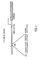
- a DNA/lipid mixture is prepared for each 6-well transfection as follows: 260ng of plasmid DNA in 100 ⁇ l of DMEM were gently mixed with 2 ⁇ l of lipid in 100 ⁇ l of DMEM (the 260ng of plasmid DNA consisted of 200ng of a 8xCRE-Luc reporter plasmid ( see below and Figure 1 for a representation of a portion of the plasmid), 50ng of pCMV comprising endogenous receptor or non-endogenous receptor or pCMV alone, and 10ng of a GPRS expression plasmid (GPRS in pcDNA3 (Invitrogen)).
- the 8XCRE-Luc reporter plasmid was prepared as follows: vector SRIF- ⁇ -gal was obtained by cloning the rat somatostatin promoter (-71/+51) at BglV-HindIII site in the p ⁇ gal-Basic Vector (Clontech). Eight (8) copies of cAMP response element were obtained by PCR from an adenovirus template AdpCF 126CCRE8 ( see, 7 Human Gene Therapy 1883 (1996 )) and cloned into the SRIF- ⁇ -gal vector at the Kpn-BglV site, resulting in the 8xCRE- ⁇ -gal reporter vector.
- the 8xCRE-Luc reporter plasmid was generated by replacing the beta-galactosidase gene in the 8xCRE- ⁇ -gal reporter vector with the luciferase gene obtained from the pGL3-basic vector (Promega) at the HindIII-BamHI site. Following 30 min. incubation at room temperature, the DNA/lipid mixture was diluted with 400 ⁇ l of DMEM and 100 ⁇ l of the diluted mixture was added to each well. 100 ⁇ l of DMEM with 10% FCS were added to each well after a 4hr incubation in a cell culture incubator. The following day the transfected cells were changed with 200 ⁇ l/well of DMEM with 10% FCS.
- Gq-dependent phospholipase C One method to detect Gq stimulation depends on the known property of Gq-dependent phospholipase C to cause the activation of genes containing serum response factors in their promoter.
- a PathdetectTM SRF-Luc-Reporting System (Stratagene) can be utilized to assay for Gq coupled activity in, e.g., COS7 cells. Cells are transfected with the plasmid components of the system and the indicated expression plasmid encoding endogenous or non-endogenous GPCR using a Mammalian TransfectionTM Kit (Stratagene, Catalogue #200285) according to the manufacturer's instructions.
- 410 ng SRF-Luc, 80 ng pCMV-receptor expression plasmid and 20 ng CMV-SEAP secreted alkaline phosphatase expression plasmid; alkaline phosphatase activity is measured in the media of transfected cells to control for variations in transfection efficiency between samples
- CMV-SEAP secreted alkaline phosphatase expression plasmid; alkaline phosphatase activity is measured in the media of transfected cells to control for variations in transfection efficiency between samples
- cells comprising the receptors can be plated onto 24 well plates, usually 1x10 5 cells/well (although his umber can be optimized.
- cells can be transfected by firstly mixing 0.25ug DNA in 50 ul serum free DMEM/well and 2 ul lipofectamine in 50 ⁇ l serumfree DMEM/well. The solutions are gently mixed and incubated for 15-30 min at room temperature. Cells are washed with 0.5 ml PBS and 400 ⁇ l of serum free media is mixed with the transfection media and added to the cells. The cells are then incubated for 3-4 hrs at 37°C/5%CO 2 and then the transfection media is removed and replaced with 1ml/well of regular growth media.
- the cells are labeled with 3 H-myo-inositol, Briefly, the media is removed and the cells are washed with 0.5 ml PBS. Then 0.5 ml inositol-free/serum free media (GIBCO BRL) is added/well with 0.25 ⁇ Ci of 3 H-myo-inositol / well and the cells are incubated for 16-18 hrs o/n at 37°C/5%CO 2 .
- GEBCO BRL inositol-free/serum free media
- the cells are washed with 0.5 ml PBS and 0.45 ml of assay medium is added containing inositol-free/serum free media 10 ⁇ M pargyline 10 mM lithium chloride or 0.4 ml of assay medium and 50 ul of 10x ketanserin (ket) to final concentration of 10 ⁇ M.
- the cells are then incubated for 30 min at 37°C.
- the cells are then washed with 0.5 ml PBSand 200 ul of fresh/icecold stop solution (1M KOH; 18 mM Na-borate; 3.8 mM EDTA) is added/well.
- the solution is kept on ice for 5-10 min or until cells were lysed and then neutralized by 200 ⁇ l of fresh/ice cold neutralization sol. (7.5 % HCL).
- the lysate is then transferred into 1.5 ml eppendorf tubes and 1 ml of chloroform/methanol (1:2) is added/tube.
- the solution is vortexed for 15 sec and the upper phase is applied to a Biorad AG1-X8TM anion exchange resin (100-200 mesh). Firstly, the resin is washed with water at 1:1.25 W/V and 0.9 ml of upper phase is loaded onto the column.
- the column is washed with 10 mls of 5 mM myo-inositol and 10 ml of 5 mM Na-borate/60mM Na-formate.
- the inositol tris phosphates are eluted into scintillation vials containing 10 ml of scintillation cocktail with 2 ml of 0.1 M formic acid/ 1 M ammonium formate.
- the columns are regenerated by washing with 10 ml of 0.1 M formic acid/3M ammonium formate and rinsed twice with dd H 2 O and stored at 4°C in water.
- 293 cells were plated-out on 150mm plates at a density of 1.3 x 10 7 cells per plate, and were transfected using 12ug of the respective DNA and 60ul of Lipofectamine Reagent (BRL) per plate.
- the transfected cells were grown in media containing serum for an assay performed 24 hours post-transfection.
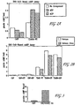
- For detection assay performed 48 hours post-transfection (assay comparing serum and serum-free media; see Figure 3 ), the initial media was changed to either serum or serum-free media.
- the serum-free media was comprised solely of Dulbecco's Modified Eagle's (DME) High Glucose Medium (Irvine Scientific #9024).
- the media with serum contained the following: 10% Fetal Bovine Serum (Hyclone #SH30071.03), 1% of 100mM Sodium Pyruvate (Irvine Scientific #9334), 1% of 20mM L-Glutamine (Irvine Scientific #9317), and 1% of Penicillin-Streptomycin solution (Irvine Scientific #9366).
- a 96-well Adenylyl Cyclase Activation FlashplateTM was used (NEN: #SMP004A).
- 50ul of the standards for the assay were added to the plate, in duplicate, ranging from concentrations of 50pmol to zero pmol cAMP per well.
- the standard cAMP (NEN: #SMP004A) was reconstituted in water, and serial dilutions were made using 1xPBS (Irvine Scientific: #9240).
- 50ul of the stimulation buffer (NEN: #SMP004A) was added to all wells.
- 10ul of each compound, diluted in water was added to its respective well, in triplicate.
- Adenosine 5'-triphosphate, ATP, (Research Biochemicals International: #A-141) and Adenosine 5'-diphosphate, ADP, (Sigma: #A2754) were used in the assay.
- the 293 cells transfected with the respective cDNA were harvested 24 (assay detection in serum media) or 48 hours post-transfection (assay detection comparing serum and serum-free media). The media was aspirated and the cells washed once with 1xPBS. Then 5ml of 1xPBS was added to the cells along with 3ml of cell dissociation buffer (Sigma: #C-1544).
- the detached cells were transferred to a centrifuge tube and centrifuged at room temperature for five minutes. The supernatant was removed and the cell pellet was resuspended in an appropriate amount of 1xPBS to obtain a final concentration of 2x10 6 cells per milliliter.
- 50ul of the cells in 1xPBS (1x10 5 cells/well) were added. The plate was incubated on a shaker for 15 minutes at room temperature.
- the detection buffer containing the tracer cAMP was prepared. In 11ml of detection buffer (NEN: #SMP004A), 50ul (equal to 1uCi) of [ 125 I]cAMP (NEN: #SMP004A) was added.
- ATP and ADP bind to endogenous TDAG8 resulting in an increase of cAMP of about 59% and about 55% respectively.
- Figure 2B evidences ATP and ADP binding to endogenous TDAG8 where endogenous TDAG8 was transfected and grown in serum and serum-free medium. ATP binding to endogenous TDAG8 grown in serum media evidences an increase in cAMP of about 65%, compared to the endogenous TDAG8 with no compounds; in serum-free media there was an increase of about 68%. ADP binding to endogenous TDAG8 in serum evidences about a 61% increase, while in serum-free ADP binding evidences an increase of about 62% increase. ATP and ADP bind to endogenous TDAG8 with an EC50 value of 139.8uM and 120.5uM, respectively (data not shown).
- the design of the constitutively activated GPCR-G protein fusion construct was accomplished as follows: both the 5' and 3' ends of the rat G protein Gsa (long form; Itoh, H. et al., 83 PNAS 3776 (1986 )) were engineered to include a HindIII (5'-AAGCTT-3') sequence thereon. Following confirmation of the correct sequence (including the flanking HindIII sequences), the entire sequence was shuttled into pcDNA3.1(-) (Invitrogen, cat. no. V795-20) by subcloning using the HindIII restriction site of that vector. The correct orientation for the Gsa sequence was determined after subcloning into pcDNA3.1(-).
- the modified pcDNA3.1(-) containing the rat Gsa gene at HindIII sequence was then verified; this vector was now available as a "universal" Gs ⁇ protein vector.
- the pcDNA3.1(-) vector contains a variety of well-known restriction sites upstream of the HindIII site, thus beneficially providing the ability to insert, upstream of the Gs protein, the coding sequence of an endogenous, constitutively active GPCR.
- This same approach can be utilized to create other "universal" G protein vectors, and, of course, other commercially available or proprietary vectors known to the artisan can be utilized - the important criteria is that the sequence for the GPCR be upstream and in-frame with that of the G protein.
- TDAG8 couples via Gs
- H9 couples via Gz.
- fusion to Gs ⁇ was accomplished.
- TDAG8(I225K)-Gs ⁇ Fusion Protein construct was made as follows: primers were designed as follows:
- Nucleotides in lower caps are included as spacers in the restriction sites between the G protein and TDAG8.
- PCR was then utilized to secure the respective receptor sequences for fusion within the Gsa universal vector disclosed above, using the following protocol for each: 100ng cDNA for TDAG8 was added to separate tubes containing 2ul of each primer (sense and anti-sense), 3uL of 10mM dNTPs, 10uL of 10XTaqPlusTM Precision buffer, 1uL of TaqPlusTM Precision polymerase (Stratagene: #600211), and 80uL of water. Reaction temperatures and cycle times for TDAG8 were as follows: the initial denaturing step was done it 94 °C for five minutes, and a cycle of 94°C for 30 seconds; 55°C for 30 seconds; 72°C for two minutes.
- GPCR Fusion Proteins comprising non-endogenous, constitutively activated TDAG8(I225K) were analyzed as above and verified for constitutive activation.
- Nucleotides in lower caps are included as spacers in the restriction sites between the G protein and H9.
- the sense and anti-sense primers included the restriction sites for EcoRV and Kpnl, respectively such that spacers (attributed to the restriction sites) exists between the G protein and H9.
- PCR was then utilized to secure the respective receptor sequences for fusion within the Gsa universal vector disclosed above, using the following protocol for each: 80ng cDNA for H9 was added to separate tubes containing 100ng of each primer (sense and anti-sense), and 45uL of PCR SupermixTM (Gibco-Brl, LifeTech) (50ul total reaction volume). Reaction temperatures and cycle times for H9 were as follows: the initial denaturing step was done it 94°C for one, and a cycle of 94°C for 30 seconds; 55°C for 30 seconds; 72°C for two minutes. A final extension time was done at 72°C for seven minutes. PCR product for was run on a 1% agarose gel and then purified (data not shown).
- the purified product was cloned into pCRII-TOPOTM System followed by identification of positive clones. Positive clones were isolated, digested with EcoRV and KpnI (New England Biolabs) and the desired inserts were isolated, purified and ligated into the Gs universal vector at the respective restriction site. The positive clones was isolated following transformation and determined by restriction enzyme digest; expression using 293 cells was accomplished following the protocol set forth infra. Each positive clone for H9(F236K):Gs - Fusion Protein was sequenced to verify correctness. Membranes were frozen (-80°C) until utilized.
- Binding Buffer consisted of 10mM HEPES, 100mM NaCl and 10mM MgCl (ph 7.4).
- Regeneration Buffer was prepared in Binding Buffer and consisted of 20mM phosphocreatine, 20U creatine phosphokinase, 20uM GTP, 0.2mM ATP, and 0.6mM IBMX.
- cAMP Standards were prepared in Binding Buffer as follows: cAMP Stock (5,000 pmol/ml in 2ml H 2 O) in ul Added to indicted amount of Binding Buffer Final Assay Concentration (50ul into 100ul) to achieve indicated pmol/well A 250 1ml 50 B 500 of A 500ul 25 C 500 of B 500ul 12.5 D 500 of C 750ul 5.0 E 500 of D 500ul 2.5 F 500 of E 500ul 1.25 G 500 of F 750ul 0.5
- Frozen membranes (both pCMV as control and the non-endogenous H(-Gs Fusion Protein) were thawed (on ice at room temperature until in solution). Membranes were homogenized with a polytron until in suspension (2 x 15 seconds). Membrane protein concentration was determined using the Bradford Assay Protocol ( see infra ). Membrane concentration was diluted to 0.5mg/ml in Regeneration Buffer (final assay concentration - 25ug/well). Thereafter, 50ul of Binding Buffer was added to each well. For control, 50ul/well of cAMP standard was added to wells 11 and 12 A-G, with Binding Buffer alone to 12H (on the 96-well format).
- a GPCR Fusion Protein as disclosed above, is also utilized with a non-endogenous, constitutively activated GPCR.
- intra-assay variation appears to be substantially stabilized, whereby an effective signal-to-noise ratio is obtained. This has the beneficial result of allowing for a more robust identification of candidate compounds.
- a GPCR Fusion Protein be used and that when utilized, the following assay protocols be utilized.
- Membranes comprising the non-endogenous, constitutively active orphan GPCR Fusion Protein of interest and for use in the direct identification of candidate compounds as inverse agonists, agonists or partial agonists are preferably prepared as follows:
- “Membrane Scrape Buffer” is comprised of 20mM HEPES and 10mM EDTA, pH 7.4;
- “Membrane Wash Buffer” is comprised of 20 mM HEPES and 0.1 mM EDTA, pH 7.4;
- “Binding Buffer” is comprised of 20mM HEPES, 100 mM NaCl, and 10 mM MgCl 2 , pH 7.4
- the media is aspirated from a confluent monolayer of cells, followed by rinse with 10ml cold PBS, followed by aspiration. Thereafter, 5ml of Membrane Scrape Buffer is added to scrape cells; this is followed by transfer of cellular extract into 50ml centrifuge tubes (centrifuged at 20,000 rpm for 17 minutes at 4°C). Thereafter, the supernatant is aspirated and the pellet is resuspended in 30ml Membrane Wash Buffer followed by centrifuge at 20,000 rpm for 17 minutes at 4°C. The supernatant is then aspirated and the pellet resuspended in Binding Buffer. This is then homogenized using a Brinkman polytronTM homogenizer (15-20 second bursts until the all material is in suspension). This is referred to herein as "Membrane Protein".
- protein concentration of the membranes is determined using the Bradford Protein Assay (protein can be diluted to about 1.5mg/ml, aliquoted and frozen (-80°C) for later use; when frozen, protocol for use is as follows: on the day of the assay, frozen Membrane Protein is thawed at room temperature, followed by vortex and then homogenized with a polytron at about 12 x 1,000 rpm for about 5-10 seconds; it is noted that for multiple preparations, the homogenizor should be thoroughly cleaned between homoginezation of different preparations).
- Binding Buffer (as per above); Bradford Dye Reagent; Bradford Protein Standard are utilized, following manufacturer instructions (Biorad, cat. no. 500-0006).
- Duplicate tubes are prepared, one including the membrane, and one as a control "blank". Each contained 800ul Binding Buffer. Thereafter, 10ul of Bradford Protein Standard (1mg/ml) is added to each tube, and 10ul of membrane Protein is then added to just one tube (not the blank). Thereafter, 200ul of Bradford Dye Reagent is added to each tube, followed by vortex of each. After five (5) minutes, the tubes were re-vortexed and the material therein is transferred to cuvettes. The cuvettes are then read using a CECIL 3041 spectrophotometer, at wavelength 595.
- GDP Buffer consists of 37.5 ml Binding Buffer and 2mg GDP (Sigma, cat. no. G-7127), followed by a series of dilutions in Binding Buffer to obtain 0.2 uM GDP (final concentration of GDP in each well was 0.1 uM GDP); each well comprising a candidate compound, has a final volume of 200ul consisting of 100ul GDP Buffer (final concentration, 0.1uM GDP), 50ul Membrane Protein in Binding Buffer, and 50ul [ 35 S]GTP ⁇ S (0.6 nM) in Binding Buffer (2.5 ul [ 35 S]GTP ⁇ S per 10ml Binding Buffer).
- Candidate compounds are preferably screened using a 96-well plate format (these can be frozen at -80°C).
- Membrane Protein or membranes with expression vector excluding the GPCR Fusion Protein, as control), are homogenized briefly until in suspension. Protein concentration is then determined using the Bradford Protein Assay set forth above. Membrane Protein (and control) is then diluted to 0.25mg/ml in Binding Buffer (final assay concentration, 12.5ug/well). Thereafter, 100 ul GDP Buffer is added to each well of a Wallac ScintistripTM (Wallac).
- a 5ul pin-tool is then used to transfer 5 ul of a candidate compound into such well (i.e ., 5ul in total assay volume of 200 ul is a 1:40 ratio such that the final screening concentration of the candidate compound is 10uM).
- the pin tool should be rinsed in three reservoirs comprising water (1X), ethanol (1X) and water (2X) - excess liquid should be shaken from the tool after each rinse and dried with paper and kimwipes.
- 50 ul of Membrane Protein is added to each well (a control well comprising membranes without the GPCR Fusion Protein is also util ized), and pre-incubated for 5-10 minutes at room temperature.
- Binding Buffer 50 ul of [ 35 S]GTP ⁇ S (0.6 nM) in Binding Buffer is added to each well, followed by incubation on a shaker for 60 minutes at room temperature (again, in this example, plates were covered with foil). The assay is then stopped by spinning of the plates at 4000 RPM for 15 minutes at 22°C. The plates are then aspirated with an 8 channel manifold and sealed with plate covers. The plates are then read on a Wallacc 1450 using setting "Prot. #37" (as per manufacturer instructions).
- the preferred confirmation assay is a cyclase-based assay.
- a modified Flash PlateTM Adenylyl Cyclase kit (New England Nuclear; Cat. No. SMP004A) is preferably utilized for confirmation of candidate compounds directly identified as inverse agonists and agonists to non-endogenous, constitutively activated orphan GPCRs in accordance with the following protocol.
- Transfected cells are harvested approximately three days after transfection.
- Membranes are prepared by homogenization of suspended cells in buffer containing 20mM HEPES, pH 7.4 and 10mM MgCl 2 . Homogenization is performed on ice using a Brinkman PolytronTM for approximately 10 seconds. The resulting homogenate is centrifuged at 49,000 X g for 15 minutes at 4°C. The resulting pellet is then resuspended in buffer containing 20mM HEPES, pH 7.4 and 0.1 mM EDTA, homogenized for 10 seconds, followed by centrifugation at 49,000 X g for 15 minutes at 4°C. The resulting pellet can be stored at - 80°C until utilized.
- the membrane pellet On the day of direct identification screening, the membrane pellet is slowly thawed at room temperature, resuspended in buffer containing 20mM HEPES, pH 7.4 and 10mM MgCL2, to yield a final protein concentration of 0.60mg/ml (the resuspended membranes are placed on ice until use).
- cAMP standards and Detection Buffer comprising 2 ⁇ Ci of tracer [ 125 I cAMP (100 ⁇ l] to 11 ml Detection Buffer) are prepared and maintained in accordance with the manufacturer's instructions.
- Assay Buffer is prepared fresh for screening and contained 20mM HEPES, pH 7.4, 10mM MgCl 2 , 20mM phospocreatine (Sigma), 0.1 units/ml creatine phosphokinase (Sigma), 50 ⁇ M GTP (Sigma), and 0.2 mM ATP (Sigma); Assay Buffer can be stored on ice until utilized.
- Candidate compounds identified as per above are added, preferably, to 96-well plate wells (3 ⁇ l/well; 12 ⁇ M final assay concentration), together with 40 ⁇ l Membrane Protein (30 ⁇ g/well) and 50 ⁇ l of Assay Buffer. This admixture is then incubated for 30 minutes at room temperature, with gentle shaking.
- the vector utilized be pCMV.
- This vector was deposited with the American Type Culture Collection (ATCC) on October 13, 1998 (10801 University Boulevard., Manassas, VA 20110-2209 USA) under the provisions of the Budapest Treaty for the International Recognition of the Deposit of Microorganisms for the Purpose of Patent Procedure. The DNA was tested by the ATCC and determined to be. The ATCC has assigned the following deposit number to pCMV: ATCC #203351.
Landscapes
- Health & Medical Sciences (AREA)
- Life Sciences & Earth Sciences (AREA)
- Chemical & Material Sciences (AREA)
- Immunology (AREA)
- Organic Chemistry (AREA)
- Molecular Biology (AREA)
- General Health & Medical Sciences (AREA)
- Engineering & Computer Science (AREA)
- Cell Biology (AREA)
- Biochemistry (AREA)
- Medicinal Chemistry (AREA)
- Gastroenterology & Hepatology (AREA)
- Hematology (AREA)
- Biophysics (AREA)
- Zoology (AREA)
- Proteomics, Peptides & Aminoacids (AREA)
- Toxicology (AREA)
- Urology & Nephrology (AREA)
- Biomedical Technology (AREA)
- Genetics & Genomics (AREA)
- Biotechnology (AREA)
- Microbiology (AREA)
- Endocrinology (AREA)
- Food Science & Technology (AREA)
- Physics & Mathematics (AREA)
- Analytical Chemistry (AREA)
- General Physics & Mathematics (AREA)
- Pathology (AREA)
- Peptides Or Proteins (AREA)
- Preparation Of Compounds By Using Micro-Organisms (AREA)
- Investigating Or Analysing Biological Materials (AREA)
Applications Claiming Priority (34)
| Application Number | Priority Date | Filing Date | Title |
|---|---|---|---|
| US09/170,496 US6555339B1 (en) | 1997-04-14 | 1998-10-13 | Non-endogenous, constitutively activated human protein-coupled receptors |
| US10802998P | 1998-11-12 | 1998-11-12 | |
| US10921398P | 1998-11-20 | 1998-11-20 | |
| US11006098P | 1998-11-27 | 1998-11-27 | |
| US12041699P | 1999-02-16 | 1999-02-16 | |
| US12185299P | 1999-02-26 | 1999-02-26 | |
| US12394899P | 1999-03-12 | 1999-03-12 | |
| US12394499P | 1999-03-12 | 1999-03-12 | |
| US12394599P | 1999-03-12 | 1999-03-12 | |
| US12394699P | 1999-03-12 | 1999-03-12 | |
| US12394999P | 1999-03-12 | 1999-03-12 | |
| US12395199P | 1999-03-12 | 1999-03-12 | |
| US13656799P | 1999-05-28 | 1999-05-28 | |
| US13643699P | 1999-05-28 | 1999-05-28 | |
| US13643999P | 1999-05-28 | 1999-05-28 | |
| US13643799P | 1999-05-28 | 1999-05-28 | |
| US13713199P | 1999-05-28 | 1999-05-28 | |
| US13712799P | 1999-05-28 | 1999-05-28 | |
| US14144899P | 1999-06-29 | 1999-06-29 | |
| US15111499P | 1999-08-27 | 1999-08-27 | |
| US15252499P | 1999-09-03 | 1999-09-03 | |
| US15663399P | 1999-09-29 | 1999-09-29 | |
| US15663499P | 1999-09-29 | 1999-09-29 | |
| US15665399P | 1999-09-29 | 1999-09-29 | |
| US15655599P | 1999-09-29 | 1999-09-29 | |
| US15728199P | 1999-10-01 | 1999-10-01 | |
| US15728299P | 1999-10-01 | 1999-10-01 | |
| US15729499P | 1999-10-01 | 1999-10-01 | |
| US15729399P | 1999-10-01 | 1999-10-01 | |
| US15728099P | 1999-10-01 | 1999-10-01 | |
| US41676099P | 1999-10-12 | 1999-10-12 | |
| US41704499A | 1999-10-12 | 1999-10-12 | |
| EP99950301A EP1137776A2 (de) | 1998-10-13 | 1999-10-13 | Nicht-endogene, konstitutiv aktivierte, menschliche, mit g-proteinen gekoppelte rezeptoren |
| EP05009877A EP1676861A3 (de) | 1998-10-13 | 1999-10-13 | Nicht-endogene, konstitutiv aktivierte, menschliche, mit G-Proteinen gekoppelte Rezeptoren |
Related Parent Applications (2)
| Application Number | Title | Priority Date | Filing Date |
|---|---|---|---|
| EP99950301.4 Division | 1999-10-13 | ||
| EP05009877.1 Division | 2005-05-06 |
Publications (1)
| Publication Number | Publication Date |
|---|---|
| EP2264068A1 true EP2264068A1 (de) | 2010-12-22 |
Family
ID=42752897
Family Applications (2)
| Application Number | Title | Priority Date | Filing Date |
|---|---|---|---|
| EP10007972A Withdrawn EP2264068A1 (de) | 1998-10-13 | 1999-10-13 | Nicht-endogene, konstitutiv aktivierte, menschliche, mit G-Proteinen gekoppelte Rezeptoren |
| EP05009877A Withdrawn EP1676861A3 (de) | 1998-10-13 | 1999-10-13 | Nicht-endogene, konstitutiv aktivierte, menschliche, mit G-Proteinen gekoppelte Rezeptoren |
Family Applications After (1)
| Application Number | Title | Priority Date | Filing Date |
|---|---|---|---|
| EP05009877A Withdrawn EP1676861A3 (de) | 1998-10-13 | 1999-10-13 | Nicht-endogene, konstitutiv aktivierte, menschliche, mit G-Proteinen gekoppelte Rezeptoren |
Country Status (1)
| Country | Link |
|---|---|
| EP (2) | EP2264068A1 (de) |
Citations (3)
| Publication number | Priority date | Publication date | Assignee | Title |
|---|---|---|---|---|
| WO1993011257A2 (de) * | 1991-11-25 | 1993-06-10 | Boehringer Ingelheim International Gmbh | Verfahren zum screenen von substanzen mit modulierender wirkung auf einen rezeptorabhängigen zellulären signalübertragungsweg |
| US5753516A (en) * | 1995-02-03 | 1998-05-19 | Heagy; Wyrta E. | Screening method for ligands of the EBI-1 receptor |
| WO1998034948A1 (en) * | 1997-02-06 | 1998-08-13 | Cornell Research Foundation, Inc. | Library screening as a strategy to clone drugs for g protein coupled receptors |
Family Cites Families (2)
| Publication number | Priority date | Publication date | Assignee | Title |
|---|---|---|---|---|
| JPH11506341A (ja) * | 1995-06-06 | 1999-06-08 | ヒューマン ジノーム サイエンシーズ,インコーポレイテッド | Gタンパク質レセプターhtnad29 |
| WO1998032858A2 (en) * | 1997-01-23 | 1998-07-30 | Schering Corporation | Mammalian chemokines; receptors; reagents; uses |
-
1999
- 1999-10-13 EP EP10007972A patent/EP2264068A1/de not_active Withdrawn
- 1999-10-13 EP EP05009877A patent/EP1676861A3/de not_active Withdrawn
Patent Citations (3)
| Publication number | Priority date | Publication date | Assignee | Title |
|---|---|---|---|---|
| WO1993011257A2 (de) * | 1991-11-25 | 1993-06-10 | Boehringer Ingelheim International Gmbh | Verfahren zum screenen von substanzen mit modulierender wirkung auf einen rezeptorabhängigen zellulären signalübertragungsweg |
| US5753516A (en) * | 1995-02-03 | 1998-05-19 | Heagy; Wyrta E. | Screening method for ligands of the EBI-1 receptor |
| WO1998034948A1 (en) * | 1997-02-06 | 1998-08-13 | Cornell Research Foundation, Inc. | Library screening as a strategy to clone drugs for g protein coupled receptors |
Non-Patent Citations (8)
| Title |
|---|
| 7 HUMAN GENE THERAPY, 1996, pages 1883 |
| CHOI JANG-WON ET AL: "Identification of a putative G protein-coupled receptor induced during activation-induced apoptosis of T cells", CELLULAR IMMUNOLOGY, vol. 168, no. 1, 1996, pages 78 - 84, ISSN: 0008-8749 * |
| KENAKIN, T., LIFE SCIENCES, vol. 43, 1988, pages 1095 |
| KJELSBERG M A ET AL: "Constitutive activation of the alpha1B-adrenergic receptor by all amino acid substitutions at a single site", JOURNAL OF BIOLOGICAL CHEMISTRY, AMERICAN SOCIETY OF BIOLOCHEMICAL BIOLOGISTS, BIRMINGHAM,, US, vol. 267, no. 3, 25 January 1992 (1992-01-25), pages 1430 - 1433, XP002135768, ISSN: 0021-9258 * |
| KYAW H L A ET AL: "Cloning, characterization, and mapping of human homolog of mouse T-cell death-associated gene", DNA AND CELL BIOLOGY, vol. 17, no. 6, 1 June 1998 (1998-06-01), pages 493 - 500, XP002145168 * |
| LEFKOWITZ R J ET AL: "Constitutive activity of receptors coupled to guanine nucleotide regulatory proteins", TRENDS IN PHARMACOLOGICAL SCIENCES, ELSEVIER, HAYWARTH, GB, vol. 14, no. 8, 1 August 1993 (1993-08-01), pages 303 - 307, XP023843266, ISSN: 0165-6147, [retrieved on 19930801], DOI: 10.1016/0165-6147(93)90048-O * |
| LTOH, H. ET AL., PNAS, vol. 83, 1986, pages 3776 |
| SCHEER A ET AL: "CONSTITUTIVELY ACTIVE G PROTEIN-COUPLED RECEPTORS: POTENTIAL MECHANISMS OF RECEPTOR ACTIVATION", JOURNAL OF RECEPTOR AND SIGNAL TRANSDUCTION RESEARCH, MARCEL DEKKER, NEW YORK, NY, US, vol. 17, no. 1/3, 1997, pages 57 - 73, XP000867531, ISSN: 1079-9893 * |
Also Published As
| Publication number | Publication date |
|---|---|
| EP1676861A2 (de) | 2006-07-05 |
| EP1676861A3 (de) | 2006-07-19 |
Similar Documents
| Publication | Publication Date | Title |
|---|---|---|
| AU2004203102B2 (en) | Human G Protein-Coupled Receptors | |
| WO2000022131A2 (en) | Non-endogenous, constitutively activated human g protein-coupled receptors | |
| AU2005244540B2 (en) | Endogenous and non-endogenous versions of human G protein-coupled receptors | |
| NZ521158A (en) | Non-endogenous, constitutively activated known G protein-coupled receptors | |
| EP2295574A1 (de) | Menschliche, an ein G-Protein gekoppelte Rezeptoren ohne bekannte Liganden | |
| US20130165633A1 (en) | Endogenous and Non-Endogenous Versions of Human G Protein-Coupled Receptors | |
| US20130309766A1 (en) | Endogenous and Non-Endogenous Versions of Human G Protein-Coupled Receptors | |
| AU2002219890B2 (en) | Endogenous and non-endogenous versions of human G protein-coupled receptors | |
| US8440391B2 (en) | Constitutively activated human G protein coupled receptors | |
| EP2264068A1 (de) | Nicht-endogene, konstitutiv aktivierte, menschliche, mit G-Proteinen gekoppelte Rezeptoren | |
| AU6299199A (en) | Non-endogenous, constitutively activated human g protein-coupled receptors | |
| US20070072248A1 (en) | Constitutively activated human G protein coupled receptors | |
| EP1137776A2 (de) | Nicht-endogene, konstitutiv aktivierte, menschliche, mit g-proteinen gekoppelte rezeptoren | |
| US20150153327A1 (en) | Endogenous and Non-Endogenous Versions of Human G Protein-Coupled Receptors | |
| JP2003525018A6 (ja) | 非内在性の構成的に活性化されるヒトgタンパク質共役型受容体 | |
| AU2007216752A1 (en) | Endogenous and non-endogenous versions of human G protein-coupled receptor hRUP35 | |
| CA2732120A1 (en) | Endogenous and non-endogenous versions of human g protein-coupled receptors |
Legal Events
| Date | Code | Title | Description |
|---|---|---|---|
| PUAI | Public reference made under article 153(3) epc to a published international application that has entered the european phase |
Free format text: ORIGINAL CODE: 0009012 |
|
| AC | Divisional application: reference to earlier application |
Ref document number: 1676861 Country of ref document: EP Kind code of ref document: P Ref document number: 1137776 Country of ref document: EP Kind code of ref document: P |
|
| AK | Designated contracting states |
Kind code of ref document: A1 Designated state(s): AT BE CH CY DE DK ES FI FR GB GR IE IT LI LU MC NL PT SE |
|
| RIN1 | Information on inventor provided before grant (corrected) |
Inventor name: CHEN, RUOPING Inventor name: LOWITZ, KEVIN Inventor name: BEHAN, DOMINIC P. Inventor name: LIAW, CHEN W. Inventor name: CHALMERS, DEREK T. |
|
| RIN1 | Information on inventor provided before grant (corrected) |
Inventor name: LOWITZ, KEVIN Inventor name: CHALMERS, DEREK T. Inventor name: LIAW, CHEN W. Inventor name: BEHAN, DOMINIC P. Inventor name: CHEN, RUOPING |
|
| RIN1 | Information on inventor provided before grant (corrected) |
Inventor name: CHALMERS, DEREK T. Inventor name: LOWITZ, KEVIN Inventor name: CHEN, RUOPING Inventor name: LIAW, CHEN W. Inventor name: BEHAN, DOMINIC P. |
|
| 17P | Request for examination filed |
Effective date: 20110527 |
|
| 17Q | First examination report despatched |
Effective date: 20120626 |
|
| STAA | Information on the status of an ep patent application or granted ep patent |
Free format text: STATUS: THE APPLICATION IS DEEMED TO BE WITHDRAWN |
|
| 18D | Application deemed to be withdrawn |
Effective date: 20121107 |