EP2241618A1 - Prostata-Krebszelllinien und ihre Verwendung beim Screening-Verfahren - Google Patents
Prostata-Krebszelllinien und ihre Verwendung beim Screening-Verfahren Download PDFInfo
- Publication number
- EP2241618A1 EP2241618A1 EP09305315A EP09305315A EP2241618A1 EP 2241618 A1 EP2241618 A1 EP 2241618A1 EP 09305315 A EP09305315 A EP 09305315A EP 09305315 A EP09305315 A EP 09305315A EP 2241618 A1 EP2241618 A1 EP 2241618A1
- Authority
- EP
- European Patent Office
- Prior art keywords
- prostate cancer
- cell line
- cancer cell
- cells
- proliferation
- Prior art date
- Legal status (The legal status is an assumption and is not a legal conclusion. Google has not performed a legal analysis and makes no representation as to the accuracy of the status listed.)
- Ceased
Links
- 206010060862 Prostate cancer Diseases 0.000 title claims abstract description 147
- 208000000236 Prostatic Neoplasms Diseases 0.000 title claims abstract description 147
- 238000012216 screening Methods 0.000 title claims abstract description 14
- 238000000034 method Methods 0.000 title claims description 52
- LWGJTAZLEJHCPA-UHFFFAOYSA-N n-(2-chloroethyl)-n-nitrosomorpholine-4-carboxamide Chemical compound ClCCN(N=O)C(=O)N1CCOCC1 LWGJTAZLEJHCPA-UHFFFAOYSA-N 0.000 claims abstract description 7
- 206010028980 Neoplasm Diseases 0.000 claims description 84
- 229940127089 cytotoxic agent Drugs 0.000 claims description 77
- 239000002254 cytotoxic agent Substances 0.000 claims description 77
- 239000003795 chemical substances by application Substances 0.000 claims description 61
- 230000035755 proliferation Effects 0.000 claims description 50
- 238000010171 animal model Methods 0.000 claims description 39
- ZDZOTLJHXYCWBA-VCVYQWHSSA-N N-debenzoyl-N-(tert-butoxycarbonyl)-10-deacetyltaxol Chemical compound O([C@H]1[C@H]2[C@@](C([C@H](O)C3=C(C)[C@@H](OC(=O)[C@H](O)[C@@H](NC(=O)OC(C)(C)C)C=4C=CC=CC=4)C[C@]1(O)C3(C)C)=O)(C)[C@@H](O)C[C@H]1OC[C@]12OC(=O)C)C(=O)C1=CC=CC=C1 ZDZOTLJHXYCWBA-VCVYQWHSSA-N 0.000 claims description 31
- 229960003668 docetaxel Drugs 0.000 claims description 30
- 230000035945 sensitivity Effects 0.000 claims description 15
- 230000009467 reduction Effects 0.000 claims description 14
- 229940124606 potential therapeutic agent Drugs 0.000 claims description 13
- 238000001516 cell proliferation assay Methods 0.000 claims description 8
- 102000039446 nucleic acids Human genes 0.000 claims description 5
- 108020004707 nucleic acids Proteins 0.000 claims description 5
- 150000007523 nucleic acids Chemical class 0.000 claims description 5
- 230000022131 cell cycle Effects 0.000 claims description 4
- 229910052751 metal Inorganic materials 0.000 claims description 4
- 239000002184 metal Substances 0.000 claims description 4
- 229920001184 polypeptide Polymers 0.000 claims description 4
- 102000004196 processed proteins & peptides Human genes 0.000 claims description 4
- 108090000765 processed proteins & peptides Proteins 0.000 claims description 4
- 238000001959 radiotherapy Methods 0.000 claims description 4
- 239000000126 substance Substances 0.000 claims description 4
- 230000005757 colony formation Effects 0.000 claims description 3
- 238000010600 3H thymidine incorporation assay Methods 0.000 claims description 2
- 150000001875 compounds Chemical class 0.000 abstract description 4
- 210000004027 cell Anatomy 0.000 description 364
- 230000014509 gene expression Effects 0.000 description 54
- 210000000130 stem cell Anatomy 0.000 description 38
- 210000002307 prostate Anatomy 0.000 description 29
- 101000610551 Homo sapiens Prominin-1 Proteins 0.000 description 23
- 102100040120 Prominin-1 Human genes 0.000 description 23
- 238000004458 analytical method Methods 0.000 description 19
- 102000007066 Prostate-Specific Antigen Human genes 0.000 description 17
- 108010072866 Prostate-Specific Antigen Proteins 0.000 description 17
- 239000003814 drug Substances 0.000 description 17
- 241000282414 Homo sapiens Species 0.000 description 16
- 108010017842 Telomerase Proteins 0.000 description 16
- 201000011510 cancer Diseases 0.000 description 16
- 229940079593 drug Drugs 0.000 description 16
- 108020004414 DNA Proteins 0.000 description 14
- 238000010186 staining Methods 0.000 description 14
- 238000011282 treatment Methods 0.000 description 14
- 102100031650 C-X-C chemokine receptor type 4 Human genes 0.000 description 13
- 101000922348 Homo sapiens C-X-C chemokine receptor type 4 Proteins 0.000 description 13
- 230000001413 cellular effect Effects 0.000 description 13
- 241000699670 Mus sp. Species 0.000 description 11
- 230000000694 effects Effects 0.000 description 11
- 102100032912 CD44 antigen Human genes 0.000 description 10
- 101000868273 Homo sapiens CD44 antigen Proteins 0.000 description 10
- 102000011782 Keratins Human genes 0.000 description 10
- 108010076876 Keratins Proteins 0.000 description 10
- 230000004069 differentiation Effects 0.000 description 9
- 108010080146 androgen receptors Proteins 0.000 description 8
- 210000000349 chromosome Anatomy 0.000 description 8
- 239000003550 marker Substances 0.000 description 8
- 102100032187 Androgen receptor Human genes 0.000 description 7
- 230000010261 cell growth Effects 0.000 description 7
- 238000000684 flow cytometry Methods 0.000 description 7
- 210000002966 serum Anatomy 0.000 description 7
- 230000000381 tumorigenic effect Effects 0.000 description 7
- 238000001262 western blot Methods 0.000 description 7
- 102000003693 Hedgehog Proteins Human genes 0.000 description 6
- 108090000031 Hedgehog Proteins Proteins 0.000 description 6
- 241001465754 Metazoa Species 0.000 description 6
- 101150080074 TP53 gene Proteins 0.000 description 6
- 208000009956 adenocarcinoma Diseases 0.000 description 6
- 238000003556 assay Methods 0.000 description 6
- 210000000270 basal cell Anatomy 0.000 description 6
- 239000000090 biomarker Substances 0.000 description 6
- 108020004999 messenger RNA Proteins 0.000 description 6
- 230000035772 mutation Effects 0.000 description 6
- 108090000623 proteins and genes Proteins 0.000 description 6
- 238000003753 real-time PCR Methods 0.000 description 6
- 231100000588 tumorigenic Toxicity 0.000 description 6
- 108700028369 Alleles Proteins 0.000 description 5
- 108010037362 Extracellular Matrix Proteins Proteins 0.000 description 5
- 102000010834 Extracellular Matrix Proteins Human genes 0.000 description 5
- 241000027355 Ferocactus setispinus Species 0.000 description 5
- 206010027476 Metastases Diseases 0.000 description 5
- 102100035703 Prostatic acid phosphatase Human genes 0.000 description 5
- 239000003098 androgen Substances 0.000 description 5
- 231100000433 cytotoxic Toxicity 0.000 description 5
- 230000001472 cytotoxic effect Effects 0.000 description 5
- 210000002744 extracellular matrix Anatomy 0.000 description 5
- 239000001963 growth medium Substances 0.000 description 5
- 239000002609 medium Substances 0.000 description 5
- 239000013641 positive control Substances 0.000 description 5
- 210000005267 prostate cell Anatomy 0.000 description 5
- 108010043671 prostatic acid phosphatase Proteins 0.000 description 5
- 238000003757 reverse transcription PCR Methods 0.000 description 5
- 210000001519 tissue Anatomy 0.000 description 5
- 102100025064 Cellular tumor antigen p53 Human genes 0.000 description 4
- 241000699660 Mus musculus Species 0.000 description 4
- 239000012979 RPMI medium Substances 0.000 description 4
- 230000003321 amplification Effects 0.000 description 4
- 230000008901 benefit Effects 0.000 description 4
- 231100000504 carcinogenesis Toxicity 0.000 description 4
- 238000002512 chemotherapy Methods 0.000 description 4
- 210000002919 epithelial cell Anatomy 0.000 description 4
- 239000000284 extract Substances 0.000 description 4
- 238000002372 labelling Methods 0.000 description 4
- 238000003199 nucleic acid amplification method Methods 0.000 description 4
- 238000011580 nude mouse model Methods 0.000 description 4
- 230000037361 pathway Effects 0.000 description 4
- 238000011472 radical prostatectomy Methods 0.000 description 4
- 230000003248 secreting effect Effects 0.000 description 4
- 230000004083 survival effect Effects 0.000 description 4
- 108091032973 (ribonucleotides)n+m Proteins 0.000 description 3
- 102000007325 Amelogenin Human genes 0.000 description 3
- 108010007570 Amelogenin Proteins 0.000 description 3
- 241000283707 Capra Species 0.000 description 3
- 208000005623 Carcinogenesis Diseases 0.000 description 3
- 239000006144 Dulbecco’s modified Eagle's medium Substances 0.000 description 3
- 108010017642 Integrin alpha2beta1 Proteins 0.000 description 3
- 108010070047 Notch Receptors Proteins 0.000 description 3
- 102000005650 Notch Receptors Human genes 0.000 description 3
- 241000283973 Oryctolagus cuniculus Species 0.000 description 3
- 208000035199 Tetraploidy Diseases 0.000 description 3
- 108010078814 Tumor Suppressor Protein p53 Proteins 0.000 description 3
- 102000013127 Vimentin Human genes 0.000 description 3
- 108010065472 Vimentin Proteins 0.000 description 3
- 230000036952 cancer formation Effects 0.000 description 3
- 238000004113 cell culture Methods 0.000 description 3
- 239000002771 cell marker Substances 0.000 description 3
- 230000001085 cytostatic effect Effects 0.000 description 3
- 238000011161 development Methods 0.000 description 3
- 230000018109 developmental process Effects 0.000 description 3
- 230000012010 growth Effects 0.000 description 3
- 238000003364 immunohistochemistry Methods 0.000 description 3
- 238000001727 in vivo Methods 0.000 description 3
- 238000002347 injection Methods 0.000 description 3
- 239000007924 injection Substances 0.000 description 3
- 108010044426 integrins Proteins 0.000 description 3
- 102000006495 integrins Human genes 0.000 description 3
- 230000009401 metastasis Effects 0.000 description 3
- 239000013642 negative control Substances 0.000 description 3
- 208000023958 prostate neoplasm Diseases 0.000 description 3
- 239000000523 sample Substances 0.000 description 3
- 238000007920 subcutaneous administration Methods 0.000 description 3
- 239000013598 vector Substances 0.000 description 3
- 210000005048 vimentin Anatomy 0.000 description 3
- IAKHMKGGTNLKSZ-INIZCTEOSA-N (S)-colchicine Chemical compound C1([C@@H](NC(C)=O)CC2)=CC(=O)C(OC)=CC=C1C1=C2C=C(OC)C(OC)=C1OC IAKHMKGGTNLKSZ-INIZCTEOSA-N 0.000 description 2
- FWBHETKCLVMNFS-UHFFFAOYSA-N 4',6-Diamino-2-phenylindol Chemical compound C1=CC(C(=N)N)=CC=C1C1=CC2=CC=C(C(N)=N)C=C2N1 FWBHETKCLVMNFS-UHFFFAOYSA-N 0.000 description 2
- 206010003445 Ascites Diseases 0.000 description 2
- 102100022595 Broad substrate specificity ATP-binding cassette transporter ABCG2 Human genes 0.000 description 2
- AOJJSUZBOXZQNB-TZSSRYMLSA-N Doxorubicin Chemical compound O([C@H]1C[C@@](O)(CC=2C(O)=C3C(=O)C=4C=CC=C(C=4C(=O)C3=C(O)C=21)OC)C(=O)CO)[C@H]1C[C@H](N)[C@H](O)[C@H](C)O1 AOJJSUZBOXZQNB-TZSSRYMLSA-N 0.000 description 2
- 206010059866 Drug resistance Diseases 0.000 description 2
- 102000018651 Epithelial Cell Adhesion Molecule Human genes 0.000 description 2
- 108010066687 Epithelial Cell Adhesion Molecule Proteins 0.000 description 2
- 101000655352 Homo sapiens Telomerase reverse transcriptase Proteins 0.000 description 2
- 101710128836 Large T antigen Proteins 0.000 description 2
- 241000699666 Mus <mouse, genus> Species 0.000 description 2
- 102000002584 Octamer Transcription Factor-3 Human genes 0.000 description 2
- 108010068425 Octamer Transcription Factor-3 Proteins 0.000 description 2
- 102100035423 POU domain, class 5, transcription factor 1 Human genes 0.000 description 2
- 101710126211 POU domain, class 5, transcription factor 1 Proteins 0.000 description 2
- 229930012538 Paclitaxel Natural products 0.000 description 2
- 102000000017 Patched Receptors Human genes 0.000 description 2
- 108010069873 Patched Receptors Proteins 0.000 description 2
- 229920004890 Triton X-100 Polymers 0.000 description 2
- 239000013504 Triton X-100 Substances 0.000 description 2
- 102100027881 Tumor protein 63 Human genes 0.000 description 2
- 210000002593 Y chromosome Anatomy 0.000 description 2
- 108010016200 Zinc Finger Protein GLI1 Proteins 0.000 description 2
- 102100035535 Zinc finger protein GLI1 Human genes 0.000 description 2
- 230000004913 activation Effects 0.000 description 2
- 239000003242 anti bacterial agent Substances 0.000 description 2
- 229940088710 antibiotic agent Drugs 0.000 description 2
- 238000013459 approach Methods 0.000 description 2
- 230000015572 biosynthetic process Effects 0.000 description 2
- DQLATGHUWYMOKM-UHFFFAOYSA-L cisplatin Chemical compound N[Pt](N)(Cl)Cl DQLATGHUWYMOKM-UHFFFAOYSA-L 0.000 description 2
- 229960004316 cisplatin Drugs 0.000 description 2
- 238000001514 detection method Methods 0.000 description 2
- 239000012895 dilution Substances 0.000 description 2
- 238000010790 dilution Methods 0.000 description 2
- 201000010099 disease Diseases 0.000 description 2
- 208000037265 diseases, disorders, signs and symptoms Diseases 0.000 description 2
- 238000001962 electrophoresis Methods 0.000 description 2
- 238000005516 engineering process Methods 0.000 description 2
- 210000000981 epithelium Anatomy 0.000 description 2
- 230000001747 exhibiting effect Effects 0.000 description 2
- 238000002474 experimental method Methods 0.000 description 2
- 238000001943 fluorescence-activated cell sorting Methods 0.000 description 2
- 230000002068 genetic effect Effects 0.000 description 2
- 238000003205 genotyping method Methods 0.000 description 2
- 230000000762 glandular Effects 0.000 description 2
- 238000007490 hematoxylin and eosin (H&E) staining Methods 0.000 description 2
- 239000005556 hormone Substances 0.000 description 2
- 229940088597 hormone Drugs 0.000 description 2
- 230000002055 immunohistochemical effect Effects 0.000 description 2
- 238000000338 in vitro Methods 0.000 description 2
- 238000010874 in vitro model Methods 0.000 description 2
- 238000010348 incorporation Methods 0.000 description 2
- 230000000977 initiatory effect Effects 0.000 description 2
- 210000003734 kidney Anatomy 0.000 description 2
- 210000000265 leukocyte Anatomy 0.000 description 2
- 210000004185 liver Anatomy 0.000 description 2
- 230000004807 localization Effects 0.000 description 2
- 210000004072 lung Anatomy 0.000 description 2
- 210000004698 lymphocyte Anatomy 0.000 description 2
- 239000000463 material Substances 0.000 description 2
- 108010082117 matrigel Proteins 0.000 description 2
- 238000005259 measurement Methods 0.000 description 2
- 230000007246 mechanism Effects 0.000 description 2
- 230000001394 metastastic effect Effects 0.000 description 2
- 238000012986 modification Methods 0.000 description 2
- 230000004048 modification Effects 0.000 description 2
- 239000002547 new drug Substances 0.000 description 2
- 210000004940 nucleus Anatomy 0.000 description 2
- 210000000056 organ Anatomy 0.000 description 2
- 229960001592 paclitaxel Drugs 0.000 description 2
- 229920003023 plastic Polymers 0.000 description 2
- 238000002360 preparation method Methods 0.000 description 2
- 102000004169 proteins and genes Human genes 0.000 description 2
- 102000005962 receptors Human genes 0.000 description 2
- 108020003175 receptors Proteins 0.000 description 2
- 238000011160 research Methods 0.000 description 2
- 238000010254 subcutaneous injection Methods 0.000 description 2
- 239000007929 subcutaneous injection Substances 0.000 description 2
- RCINICONZNJXQF-MZXODVADSA-N taxol Chemical compound O([C@@H]1[C@@]2(C[C@@H](C(C)=C(C2(C)C)[C@H](C([C@]2(C)[C@@H](O)C[C@H]3OC[C@]3([C@H]21)OC(C)=O)=O)OC(=O)C)OC(=O)[C@H](O)[C@@H](NC(=O)C=1C=CC=CC=1)C=1C=CC=CC=1)O)C(=O)C1=CC=CC=C1 RCINICONZNJXQF-MZXODVADSA-N 0.000 description 2
- 238000012360 testing method Methods 0.000 description 2
- 210000004881 tumor cell Anatomy 0.000 description 2
- 102100022464 5'-nucleotidase Human genes 0.000 description 1
- 101150069931 Abcg2 gene Proteins 0.000 description 1
- 239000012103 Alexa Fluor 488 Substances 0.000 description 1
- 102000027791 CD44 antigen Human genes 0.000 description 1
- 108091016585 CD44 antigen Proteins 0.000 description 1
- 201000009030 Carcinoma Diseases 0.000 description 1
- 102000053642 Catalytic RNA Human genes 0.000 description 1
- 108090000994 Catalytic RNA Proteins 0.000 description 1
- 102000010792 Chromogranin A Human genes 0.000 description 1
- 108010038447 Chromogranin A Proteins 0.000 description 1
- 208000037051 Chromosomal Instability Diseases 0.000 description 1
- 208000031404 Chromosome Aberrations Diseases 0.000 description 1
- 108020004705 Codon Proteins 0.000 description 1
- IGXWBGJHJZYPQS-SSDOTTSWSA-N D-Luciferin Chemical compound OC(=O)[C@H]1CSC(C=2SC3=CC=C(O)C=C3N=2)=N1 IGXWBGJHJZYPQS-SSDOTTSWSA-N 0.000 description 1
- 238000001712 DNA sequencing Methods 0.000 description 1
- CYCGRDQQIOGCKX-UHFFFAOYSA-N Dehydro-luciferin Natural products OC(=O)C1=CSC(C=2SC3=CC(O)=CC=C3N=2)=N1 CYCGRDQQIOGCKX-UHFFFAOYSA-N 0.000 description 1
- 206010061818 Disease progression Diseases 0.000 description 1
- 206010061819 Disease recurrence Diseases 0.000 description 1
- 101710181478 Envelope glycoprotein GP350 Proteins 0.000 description 1
- 102000004190 Enzymes Human genes 0.000 description 1
- 108090000790 Enzymes Proteins 0.000 description 1
- BJGNCJDXODQBOB-UHFFFAOYSA-N Fivefly Luciferin Natural products OC(=O)C1CSC(C=2SC3=CC(O)=CC=C3N=2)=N1 BJGNCJDXODQBOB-UHFFFAOYSA-N 0.000 description 1
- 229930182566 Gentamicin Natural products 0.000 description 1
- CEAZRRDELHUEMR-URQXQFDESA-N Gentamicin Chemical compound O1[C@H](C(C)NC)CC[C@@H](N)[C@H]1O[C@H]1[C@H](O)[C@@H](O[C@@H]2[C@@H]([C@@H](NC)[C@@](C)(O)CO2)O)[C@H](N)C[C@@H]1N CEAZRRDELHUEMR-URQXQFDESA-N 0.000 description 1
- 101000678236 Homo sapiens 5'-nucleotidase Proteins 0.000 description 1
- 101000823298 Homo sapiens Broad substrate specificity ATP-binding cassette transporter ABCG2 Proteins 0.000 description 1
- 101000998011 Homo sapiens Keratin, type I cytoskeletal 19 Proteins 0.000 description 1
- 102100033420 Keratin, type I cytoskeletal 19 Human genes 0.000 description 1
- 108060001084 Luciferase Proteins 0.000 description 1
- 239000005089 Luciferase Substances 0.000 description 1
- DDWFXDSYGUXRAY-UHFFFAOYSA-N Luciferin Natural products CCc1c(C)c(CC2NC(=O)C(=C2C=C)C)[nH]c1Cc3[nH]c4C(=C5/NC(CC(=O)O)C(C)C5CC(=O)O)CC(=O)c4c3C DDWFXDSYGUXRAY-UHFFFAOYSA-N 0.000 description 1
- 108010090306 Member 2 Subfamily G ATP Binding Cassette Transporter Proteins 0.000 description 1
- CBCKQZAAMUWICA-UHFFFAOYSA-N Nc(cc1)ccc1N Chemical compound Nc(cc1)ccc1N CBCKQZAAMUWICA-UHFFFAOYSA-N 0.000 description 1
- 208000003788 Neoplasm Micrometastasis Diseases 0.000 description 1
- 206010061309 Neoplasm progression Diseases 0.000 description 1
- 230000005913 Notch signaling pathway Effects 0.000 description 1
- 102000007999 Nuclear Proteins Human genes 0.000 description 1
- 108010089610 Nuclear Proteins Proteins 0.000 description 1
- 108091034117 Oligonucleotide Proteins 0.000 description 1
- 108700020796 Oncogene Proteins 0.000 description 1
- 102000003992 Peroxidases Human genes 0.000 description 1
- BELBBZDIHDAJOR-UHFFFAOYSA-N Phenolsulfonephthalein Chemical compound C1=CC(O)=CC=C1C1(C=2C=CC(O)=CC=2)C2=CC=CC=C2S(=O)(=O)O1 BELBBZDIHDAJOR-UHFFFAOYSA-N 0.000 description 1
- ONIBWKKTOPOVIA-UHFFFAOYSA-N Proline Natural products OC(=O)C1CCCN1 ONIBWKKTOPOVIA-UHFFFAOYSA-N 0.000 description 1
- 108010007100 Pulmonary Surfactant-Associated Protein A Proteins 0.000 description 1
- 102100027773 Pulmonary surfactant-associated protein A2 Human genes 0.000 description 1
- 238000001190 Q-PCR Methods 0.000 description 1
- 238000011529 RT qPCR Methods 0.000 description 1
- 241000700159 Rattus Species 0.000 description 1
- 101100247004 Rattus norvegicus Qsox1 gene Proteins 0.000 description 1
- 108020004459 Small interfering RNA Proteins 0.000 description 1
- 108091035286 Strbase Proteins 0.000 description 1
- 108700025695 Suppressor Genes Proteins 0.000 description 1
- 229940123237 Taxane Drugs 0.000 description 1
- GLNADSQYFUSGOU-GPTZEZBUSA-J Trypan blue Chemical compound [Na+].[Na+].[Na+].[Na+].C1=C(S([O-])(=O)=O)C=C2C=C(S([O-])(=O)=O)C(/N=N/C3=CC=C(C=C3C)C=3C=C(C(=CC=3)\N=N\C=3C(=CC4=CC(=CC(N)=C4C=3O)S([O-])(=O)=O)S([O-])(=O)=O)C)=C(O)C2=C1N GLNADSQYFUSGOU-GPTZEZBUSA-J 0.000 description 1
- JXLYSJRDGCGARV-WWYNWVTFSA-N Vinblastine Natural products O=C(O[C@H]1[C@](O)(C(=O)OC)[C@@H]2N(C)c3c(cc(c(OC)c3)[C@]3(C(=O)OC)c4[nH]c5c(c4CCN4C[C@](O)(CC)C[C@H](C3)C4)cccc5)[C@@]32[C@H]2[C@@]1(CC)C=CCN2CC3)C JXLYSJRDGCGARV-WWYNWVTFSA-N 0.000 description 1
- 108010088665 Zinc Finger Protein Gli2 Proteins 0.000 description 1
- 102100035558 Zinc finger protein GLI2 Human genes 0.000 description 1
- 230000001464 adherent effect Effects 0.000 description 1
- 230000004075 alteration Effects 0.000 description 1
- APKFDSVGJQXUKY-INPOYWNPSA-N amphotericin B Chemical compound O[C@H]1[C@@H](N)[C@H](O)[C@@H](C)O[C@H]1O[C@H]1/C=C/C=C/C=C/C=C/C=C/C=C/C=C/[C@H](C)[C@@H](O)[C@@H](C)[C@H](C)OC(=O)C[C@H](O)C[C@H](O)CC[C@@H](O)[C@H](O)C[C@H](O)C[C@](O)(C[C@H](O)[C@H]2C(O)=O)O[C@H]2C1 APKFDSVGJQXUKY-INPOYWNPSA-N 0.000 description 1
- 238000012197 amplification kit Methods 0.000 description 1
- 230000002280 anti-androgenic effect Effects 0.000 description 1
- 230000001093 anti-cancer Effects 0.000 description 1
- 230000000692 anti-sense effect Effects 0.000 description 1
- 230000000259 anti-tumor effect Effects 0.000 description 1
- 239000000051 antiandrogen Substances 0.000 description 1
- 239000000427 antigen Substances 0.000 description 1
- 108091007433 antigens Proteins 0.000 description 1
- 102000036639 antigens Human genes 0.000 description 1
- 239000002246 antineoplastic agent Substances 0.000 description 1
- 230000006907 apoptotic process Effects 0.000 description 1
- 238000011717 athymic nude mouse Methods 0.000 description 1
- 230000002146 bilateral effect Effects 0.000 description 1
- 230000033228 biological regulation Effects 0.000 description 1
- DEGAKNSWVGKMLS-UHFFFAOYSA-N calcein Chemical compound O1C(=O)C2=CC=CC=C2C21C1=CC(CN(CC(O)=O)CC(O)=O)=C(O)C=C1OC1=C2C=C(CN(CC(O)=O)CC(=O)O)C(O)=C1 DEGAKNSWVGKMLS-UHFFFAOYSA-N 0.000 description 1
- 230000005907 cancer growth Effects 0.000 description 1
- 230000005773 cancer-related death Effects 0.000 description 1
- 239000002775 capsule Substances 0.000 description 1
- 230000030833 cell death Effects 0.000 description 1
- 230000004663 cell proliferation Effects 0.000 description 1
- 230000005754 cellular signaling Effects 0.000 description 1
- 230000008859 change Effects 0.000 description 1
- 238000012512 characterization method Methods 0.000 description 1
- 239000003153 chemical reaction reagent Substances 0.000 description 1
- 230000035572 chemosensitivity Effects 0.000 description 1
- 231100000005 chromosome aberration Toxicity 0.000 description 1
- 229960001338 colchicine Drugs 0.000 description 1
- 230000000052 comparative effect Effects 0.000 description 1
- 239000002299 complementary DNA Substances 0.000 description 1
- 238000011109 contamination Methods 0.000 description 1
- 210000004748 cultured cell Anatomy 0.000 description 1
- 230000002559 cytogenic effect Effects 0.000 description 1
- 238000004163 cytometry Methods 0.000 description 1
- 230000001086 cytosolic effect Effects 0.000 description 1
- 239000000824 cytostatic agent Substances 0.000 description 1
- 230000002950 deficient Effects 0.000 description 1
- 230000001934 delay Effects 0.000 description 1
- 230000001419 dependent effect Effects 0.000 description 1
- 230000005750 disease progression Effects 0.000 description 1
- BFMYDTVEBKDAKJ-UHFFFAOYSA-L disodium;(2',7'-dibromo-3',6'-dioxido-3-oxospiro[2-benzofuran-1,9'-xanthene]-4'-yl)mercury;hydrate Chemical compound O.[Na+].[Na+].O1C(=O)C2=CC=CC=C2C21C1=CC(Br)=C([O-])C([Hg])=C1OC1=C2C=C(Br)C([O-])=C1 BFMYDTVEBKDAKJ-UHFFFAOYSA-L 0.000 description 1
- 238000010494 dissociation reaction Methods 0.000 description 1
- 230000005593 dissociations Effects 0.000 description 1
- 229960004679 doxorubicin Drugs 0.000 description 1
- 238000007876 drug discovery Methods 0.000 description 1
- 210000001671 embryonic stem cell Anatomy 0.000 description 1
- 230000007613 environmental effect Effects 0.000 description 1
- FRPJXPJMRWBBIH-RBRWEJTLSA-N estramustine Chemical compound ClCCN(CCCl)C(=O)OC1=CC=C2[C@H]3CC[C@](C)([C@H](CC4)O)[C@@H]4[C@@H]3CCC2=C1 FRPJXPJMRWBBIH-RBRWEJTLSA-N 0.000 description 1
- 229960001842 estramustine Drugs 0.000 description 1
- VJJPUSNTGOMMGY-MRVIYFEKSA-N etoposide Chemical compound COC1=C(O)C(OC)=CC([C@@H]2C3=CC=4OCOC=4C=C3[C@@H](O[C@H]3[C@@H]([C@@H](O)[C@@H]4O[C@H](C)OC[C@H]4O3)O)[C@@H]3[C@@H]2C(OC3)=O)=C1 VJJPUSNTGOMMGY-MRVIYFEKSA-N 0.000 description 1
- 229960005420 etoposide Drugs 0.000 description 1
- 210000002950 fibroblast Anatomy 0.000 description 1
- 239000012634 fragment Substances 0.000 description 1
- 230000006870 function Effects 0.000 description 1
- 239000000499 gel Substances 0.000 description 1
- 210000004602 germ cell Anatomy 0.000 description 1
- 239000011521 glass Substances 0.000 description 1
- 238000001794 hormone therapy Methods 0.000 description 1
- 230000007062 hydrolysis Effects 0.000 description 1
- 238000006460 hydrolysis reaction Methods 0.000 description 1
- 238000003119 immunoblot Methods 0.000 description 1
- 238000010166 immunofluorescence Methods 0.000 description 1
- 238000010820 immunofluorescence microscopy Methods 0.000 description 1
- 238000002991 immunohistochemical analysis Methods 0.000 description 1
- 238000011532 immunohistochemical staining Methods 0.000 description 1
- 238000000099 in vitro assay Methods 0.000 description 1
- 230000002401 inhibitory effect Effects 0.000 description 1
- 210000003963 intermediate filament Anatomy 0.000 description 1
- 238000007912 intraperitoneal administration Methods 0.000 description 1
- 238000002350 laparotomy Methods 0.000 description 1
- 230000000670 limiting effect Effects 0.000 description 1
- 230000007774 longterm Effects 0.000 description 1
- 239000006166 lysate Substances 0.000 description 1
- 238000007403 mPCR Methods 0.000 description 1
- 238000012423 maintenance Methods 0.000 description 1
- 230000036210 malignancy Effects 0.000 description 1
- 239000011159 matrix material Substances 0.000 description 1
- 230000031864 metaphase Effects 0.000 description 1
- 206010061289 metastatic neoplasm Diseases 0.000 description 1
- 208000010658 metastatic prostate carcinoma Diseases 0.000 description 1
- 238000002493 microarray Methods 0.000 description 1
- 238000010208 microarray analysis Methods 0.000 description 1
- 244000005700 microbiome Species 0.000 description 1
- 230000001617 migratory effect Effects 0.000 description 1
- 229960001156 mitoxantrone Drugs 0.000 description 1
- KKZJGLLVHKMTCM-UHFFFAOYSA-N mitoxantrone Chemical compound O=C1C2=C(O)C=CC(O)=C2C(=O)C2=C1C(NCCNCCO)=CC=C2NCCNCCO KKZJGLLVHKMTCM-UHFFFAOYSA-N 0.000 description 1
- 239000003068 molecular probe Substances 0.000 description 1
- 230000000877 morphologic effect Effects 0.000 description 1
- 238000002703 mutagenesis Methods 0.000 description 1
- 231100000350 mutagenesis Toxicity 0.000 description 1
- 230000017074 necrotic cell death Effects 0.000 description 1
- 210000004412 neuroendocrine cell Anatomy 0.000 description 1
- 239000002773 nucleotide Substances 0.000 description 1
- 125000003729 nucleotide group Chemical group 0.000 description 1
- 229960002378 oftasceine Drugs 0.000 description 1
- 108700025694 p53 Genes Proteins 0.000 description 1
- 210000000496 pancreas Anatomy 0.000 description 1
- 239000012188 paraffin wax Substances 0.000 description 1
- 238000012335 pathological evaluation Methods 0.000 description 1
- 210000003200 peritoneal cavity Anatomy 0.000 description 1
- 230000008823 permeabilization Effects 0.000 description 1
- 108040007629 peroxidase activity proteins Proteins 0.000 description 1
- 229960003531 phenolsulfonphthalein Drugs 0.000 description 1
- BASFCYQUMIYNBI-UHFFFAOYSA-N platinum Chemical class [Pt] BASFCYQUMIYNBI-UHFFFAOYSA-N 0.000 description 1
- 229920001481 poly(stearyl methacrylate) Polymers 0.000 description 1
- 230000003389 potentiating effect Effects 0.000 description 1
- 230000002062 proliferating effect Effects 0.000 description 1
- -1 proline amino acid Chemical class 0.000 description 1
- XJMOSONTPMZWPB-UHFFFAOYSA-M propidium iodide Chemical compound [I-].[I-].C12=CC(N)=CC=C2C2=CC=C(N)C=C2[N+](CCC[N+](C)(CC)CC)=C1C1=CC=CC=C1 XJMOSONTPMZWPB-UHFFFAOYSA-M 0.000 description 1
- 201000005825 prostate adenocarcinoma Diseases 0.000 description 1
- 210000000064 prostate epithelial cell Anatomy 0.000 description 1
- 238000011471 prostatectomy Methods 0.000 description 1
- 238000011002 quantification Methods 0.000 description 1
- 230000003362 replicative effect Effects 0.000 description 1
- 238000012827 research and development Methods 0.000 description 1
- 230000000717 retained effect Effects 0.000 description 1
- 230000001177 retroviral effect Effects 0.000 description 1
- 108091092562 ribozyme Proteins 0.000 description 1
- HOZOZZFCZRXYEK-HNHWXVNLSA-M scopolamine butylbromide Chemical compound [Br-].C1([C@@H](CO)C(=O)OC2C[C@@H]3[N+]([C@H](C2)[C@@H]2[C@H]3O2)(C)CCCC)=CC=CC=C1 HOZOZZFCZRXYEK-HNHWXVNLSA-M 0.000 description 1
- 238000012163 sequencing technique Methods 0.000 description 1
- 230000019491 signal transduction Effects 0.000 description 1
- 230000011664 signaling Effects 0.000 description 1
- JUJBNYBVVQSIOU-UHFFFAOYSA-M sodium;4-[2-(4-iodophenyl)-3-(4-nitrophenyl)tetrazol-2-ium-5-yl]benzene-1,3-disulfonate Chemical compound [Na+].C1=CC([N+](=O)[O-])=CC=C1N1[N+](C=2C=CC(I)=CC=2)=NC(C=2C(=CC(=CC=2)S([O-])(=O)=O)S([O-])(=O)=O)=N1 JUJBNYBVVQSIOU-UHFFFAOYSA-M 0.000 description 1
- 239000000243 solution Substances 0.000 description 1
- 210000000952 spleen Anatomy 0.000 description 1
- 229960005322 streptomycin Drugs 0.000 description 1
- 239000000758 substrate Substances 0.000 description 1
- 230000008685 targeting Effects 0.000 description 1
- 229940063683 taxotere Drugs 0.000 description 1
- 108091035539 telomere Proteins 0.000 description 1
- 102000055501 telomere Human genes 0.000 description 1
- 210000003411 telomere Anatomy 0.000 description 1
- 229940124597 therapeutic agent Drugs 0.000 description 1
- 230000001225 therapeutic effect Effects 0.000 description 1
- 229940104230 thymidine Drugs 0.000 description 1
- 231100000331 toxic Toxicity 0.000 description 1
- 230000002588 toxic effect Effects 0.000 description 1
- 230000009466 transformation Effects 0.000 description 1
- 230000005751 tumor progression Effects 0.000 description 1
- 230000001173 tumoral effect Effects 0.000 description 1
- 229960003048 vinblastine Drugs 0.000 description 1
- JXLYSJRDGCGARV-XQKSVPLYSA-N vincaleukoblastine Chemical compound C([C@@H](C[C@]1(C(=O)OC)C=2C(=CC3=C([C@]45[C@H]([C@@]([C@H](OC(C)=O)[C@]6(CC)C=CCN([C@H]56)CC4)(O)C(=O)OC)N3C)C=2)OC)C[C@@](C2)(O)CC)N2CCC2=C1NC1=CC=CC=C21 JXLYSJRDGCGARV-XQKSVPLYSA-N 0.000 description 1
- XLYOFNOQVPJJNP-UHFFFAOYSA-N water Substances O XLYOFNOQVPJJNP-UHFFFAOYSA-N 0.000 description 1
Images




Classifications
-
- G—PHYSICS
- G01—MEASURING; TESTING
- G01N—INVESTIGATING OR ANALYSING MATERIALS BY DETERMINING THEIR CHEMICAL OR PHYSICAL PROPERTIES
- G01N33/00—Investigating or analysing materials by specific methods not covered by groups G01N1/00 - G01N31/00
- G01N33/48—Biological material, e.g. blood, urine; Haemocytometers
- G01N33/50—Chemical analysis of biological material, e.g. blood, urine; Testing involving biospecific ligand binding methods; Immunological testing
- G01N33/5005—Chemical analysis of biological material, e.g. blood, urine; Testing involving biospecific ligand binding methods; Immunological testing involving human or animal cells
- G01N33/5008—Chemical analysis of biological material, e.g. blood, urine; Testing involving biospecific ligand binding methods; Immunological testing involving human or animal cells for testing or evaluating the effect of chemical or biological compounds, e.g. drugs, cosmetics
- G01N33/5011—Chemical analysis of biological material, e.g. blood, urine; Testing involving biospecific ligand binding methods; Immunological testing involving human or animal cells for testing or evaluating the effect of chemical or biological compounds, e.g. drugs, cosmetics for testing antineoplastic activity
-
- G—PHYSICS
- G01—MEASURING; TESTING
- G01N—INVESTIGATING OR ANALYSING MATERIALS BY DETERMINING THEIR CHEMICAL OR PHYSICAL PROPERTIES
- G01N2333/00—Assays involving biological materials from specific organisms or of a specific nature
- G01N2333/435—Assays involving biological materials from specific organisms or of a specific nature from animals; from humans
- G01N2333/46—Assays involving biological materials from specific organisms or of a specific nature from animals; from humans from vertebrates
Definitions
- the present invention relates to the oncology and the identification of new drugs for the treatment of prostate cancer.
- Prostate cancer is the leading cause of cancer-related deaths in North America and Europe. Localized prostate tumours are commonly diagnosed and conventionally treated by radical prostatectomy. After an initially effective hormone therapy, the advanced disease led to the development of hormone-refractory, drug-resistant malignancy. To date, there is still no cure available for patients with advanced disease.
- Human prostate cells are known to be one of the most difficult cell types to develop into continuously growing culture and considerable research efforts have been directed toward the establishment of new prostate cancer models from the primary tumours.
- telomerase catalytic subunit HPE
- HPET human prostate epithelial
- docetaxel (Taxotere®) is become the reference treatment of hormone-resistant metastatic prostate cancer. However, despite the survival benefit of this drug, a high number of patients treated with docetaxel develop a chemotherapy resistance. This is why cellular models of prostate cancer resistant to docetaxel are so interesting and useful.
- prostate cancer cell line either sensitive or resistant to a cytotoxic drug, is of great interest for pharmaceutical companies.
- the inventors established a new prostate cancer cell line, called IGR-CaP1, from a localized prostate tumor and a derived cell line called IGR-CaP1-R100 resistant to docetaxel.
- the present invention provides a prostate cancer cell line designated IGR-CaP1 as described in CNCM deposit number 1-4126 on February 10, 2009, or a progeny thereof.
- the present invention concerns a method for preparing a prostate cancer cell line resistant to a dose of a cytotoxic drug comprising submitting the prostate cancer cell line IGR-CaP1 to increasing doses of the cytotoxic drug and selecting the resulting prostate cancer cells resistant to the dose of the cytotoxic drug.
- the present invention also concerns the use of the prostate cancer cell line IGR-CaP1 for preparing a prostate cancer cell line resistant to a dose of a cytotoxic drug and any prostate cancer cell line resistant to a dose of a cytotoxic drug prepared from the prostate cancer cell line IGR-CaP1.
- the present invention concerns the prostate cancer cell line resistant to 100 nM of docetaxel and designated IGR-CaP1-R100 as described in CNCM deposit number I-4127 on February 10, 2009, or a progeny thereof.
- the present invention further concerns a kit for screening potential therapeutic agents comprising a prostate cancer cell line according to the present invention.
- the present invention concerns an animal model comprising a prostate cancer cell according to the present invention.
- the animal model comprises a prostate cancer cell resistant to a cytotoxic drug according to the present invention.
- the present invention concerns the use of a prostate cancer cell line or an animal model according to the present invention for screening potential therapeutic agents.
- the present invention concerns a method for determining whether a candidate agent inhibits proliferation of a prostate cancer cell line according to the present invention comprising: a) contacting the prostate cancer cell line of the present invention with the candidate agent; and b) measuring the proliferation of the prostate cancer cell line so contacted, a reduction in proliferation indicating that the candidate agent inhibits proliferation of the prostate cancer cell line.
- a method for determining whether a candidate agent increases the sensitivity to a cytotoxic drug of a prostate cancer cell line according to the present invention resistant to said cytotoxic drug comprising: a) contacting the prostate cancer cell line resistant to said cytotoxic drug of the present invention with the candidate agent in presence of the cytotoxic drug; and b) measuring the proliferation of the cell line so contacted, a reduction in proliferation indicating that the candidate agent increases the sensitivity of the prostate cancer cell line to the cytotoxic drug.
- the proliferation of the cells in step b) is measured using a method selected from a group consisting of DNA cell cycle method, 3H-thymidine incorporation method, cell count method, colorimetric cell proliferation assay or efficiency of colony formation method.
- the candidate agent can be a chemical molecule, a polypeptide, a nucleic acid molecule, an antibody, a metal, a radiotherapy or a combination thereof.
- the present invention also concerns a method for determining whether a candidate agent inhibits proliferation of a prostate cancer cell line according to the present invention which comprises: a) administering the candidate agent to an animal model comprising a prostate cancer cell according to the present invention; and b) measuring the proliferation of the prostate cancer cells in the animal model, a reduction in proliferation indicating that the candidate agent inhibits proliferation of the prostate cancer cells.
- a method for determining whether a candidate agent increases the sensitivity to a cytotoxic drug of a prostate cancer cell line according to the present invention resistant to said cytotoxic drug comprising: a) administering the candidate agent to an animal model comprising a prostate cancer cell resistant to the cytotoxic drug according to the present invention in combination with the cytotoxic drug; and b) measuring the proliferation of the prostate cancer cells in the animal model, a reduction in proliferation indicating that the agent increases the sensitivity of the prostate cancer cell line to the cytotoxic drug.
- the animal model presents a tumor formed by the prostate cancer cell line and wherein the proliferation of the prostate cancer cells in the animal model is measured by the determination of the tumor size.
- IGR-CaP1 prostate cancer cell line
- This cell line has the advantage to be a cancer prostate cell line obtained from a localized tumor (contrary to almost all other prostate cancer cell lines obtained from metastasis).
- the molecular characterization of the cell line suggests that these cells correspond to prostate epithelial basal cells. These cells were spontaneously immortalized; exhibit a tetraploid karyotype and a high telomerase activity.
- IGR-CaP1 cells do not express androgen receptor protein (AR) or secretory prostate-specific antigen (PSA).
- AR androgen receptor protein
- PSA secretory prostate-specific antigen
- cancer stem cells have the property of self-renewal and the capacity of differentiation reflecting the tumor from which they were derived. So, prostate cancer stem cells have the capacity to differentiate into AR-expressing luminal cells. According to this new concept, it is assumed today that cancer stem cells could be those who may be responsible for the development of drug resistance and disease progression and thus constitute the most appropriate cells to kill. In mice, the IGR-CaP1 cell line recapitulates the characteristics of an undifferentiated prostate tumor observed in patients with a high Gleason score.
- cancer stem cells are tumorigenic in nude mice where it forms tumors reconstituting adenocarcinoma expressing prostate markers PSMA and PAP and capable of metastasizing.
- These cells express markers described as cancer stem cells in some solid tumors (CD44, CD133) and in prostate cancer cellular models (CD44, CD133, ⁇ 2 ⁇ 1 integrin, CXCR4).
- CD44, CD133 solid tumors
- CD44, CD133, ⁇ 2 ⁇ 1 integrin, CXCR4 prostate cancer cellular models
- the inventors obtained cellular derived-clones exhibiting the original features of prostate tissue and conserving cancer stem cell properties.
- cancer stem cells may be a major cause of drug resistance and contribute to disease recurrence in prostate cancer patients, these new models are useful for developing new therapeutic approaches targeting prostate cancer stem cells.
- the present invention concerns the new prostate cancer cell line, called IGR-CaP1, which has been deposited at the CNCM (Collection Nationale de Culture de Microorganismes at the Pasteur Institute, 25 rue du Dr. Roux, F-75724 Paris Cedex 15, France) on February 10, 2009 under number I-4126 and any progeny thereof.
- CNCM Collection Nationale de Culture de Microorganismes at the Pasteur Institute, 25 rue du Dr. Roux, F-75724 Paris Cedex 15, France
- the expressions "cell,” “cell line,” and “cell culture” are used interchangeably and all such designations include progeny.
- progeny is also intended cell clones obtained by limit dilution of the cell lines of the invention. Because certain modifications may occur in succeeding generations due to either mutation or environmental influences, or clonal selection, such progeny may not be identical to the parent cell, but are still included within the scope of the term as used herein.
- the STR profile can be used to determine if a cell is a progeny of the IGR-CaP1 cell line.
- the IGR-CaP1 cell line and its progeny should have the same alleles for D7S820, D13S317, TPOX, and amelogenin loci or at least 10 identical loci among the 16-locus STR multiplex comprising D8S1179, D21S11, D7S820, CSF1PO, D3S1358, TH01, D13S317, D16S539, D2S1338, D19S433, vWA, TPOX, D18S51, amelogenine, D5S818 and FGA loci.
- the present invention also contemplates a method for isolating a clonal cell by a limit dilution of a cell line according to the present invention.
- the present invention also concerns any cell derived from the IGR-CaP1 cell line, for instance by mutagenesis or by genetic transformation.
- the present invention concerns a method for preparing a prostate cancer cell line resistant to a dose of a cytotoxic drug comprising submitting the prostate cancer cell line IGR-CaP1 to increasing doses of the cytotoxic drug and selecting the resulting prostate cancer cell line resistant to the dose of the cytotoxic drug.
- the present invention also concerns the use of the prostate cancer cell line IGR-CaP1 for preparing a prostate cancer cell line resistant to a dose of a cytotoxic drug. It further concerns a prostate cancer cell line resistant to a dose of a cytotoxic drug prepared from the prostate cancer cell line IGR-CaP1.
- IGR-CaP1-R refers herein to a cell line prepared from the prostate cancer cell line IGR-CaP1 and being resistant to a cytotoxic drug.
- cytotoxic drug is intended to refer herein to any molecule having a toxic effect on cells. In particular, it refers to any molecule leading to cell death (e.g., by apoptosis or any other mechanism) or inhibiting or suppressing cellular growth and multiplication. Such drugs are commonly used in chemotherapy to inhibit the proliferation of cancerous cells.
- the cytotoxic drug is a drug used or useful for the treatment of prostate cancer.
- the cytotoxic drug can be selected from the group consisting of docetaxel, paclitaxel and others taxanes, mitoxantrone, platin salts, doxorubicin, vinblastine, estramustine, and etoposide.
- a dose escalation can be carried out in order to obtain a resistant cell line from the IGR-CaP1 cell line.
- a protocol is disclosed in Materials and Methods Section of Patterson et al (2006), the disclosure of which being incorporated herein by reference.
- the cells can be contacted with a first dose of the cytotoxic drug. After the clones sensitive to the first dose are no longer present, the surviving cells repopulate the flask and continue to divide through about 4 passages. Then, they are incubated with a higher second dose of the cytotoxic drug. The same methodology is followed with each increase in the concentration of the cytotoxic drug until to reach the dose of the cytotoxic drug at which the cells have to be resistant.
- the cytotoxic drug is docetaxel.
- the inventors prepared, from the IGR-CaP1-R cell line, resistant cell lines to the following docetaxel doses: 0.5 nM, 5 nM, 12 nM, 25 nM, 50 nM, 100 nM, 200 nM and 12 ⁇ M.
- the present invention concerns the prostate cancer cell line resistant to 100 nM of docetaxel and designated IGR-CaP1-R100 as described in CNCM deposit number 1-4127 on February 10, 2009, or a progeny thereof. This cell line is also resistant to cisplatin.
- the IGR-CaP1 cell line and the IGR-CaP1-R cell lines resistant to a cytotoxic drug can be useful for determining or identifying biomarkers for resistance to the cytotoxic drug.
- Biomarkers identification could be performed by expression profile analysis of sensitive cells and/or of different cells resistant to the same drug and/or cells resistant to the different drugs. It can also be carried out by comparative microarray, RT-PCR, Western Blot, Immunohistochemistry and the like.
- the prostate cancer cell lines of the invention are also useful for screening potential therapeutic agents. Therefore, the present invention concerns a kit for screening potential therapeutic agents comprising a prostate cancer cell line of the invention.
- the kit can further comprise a culture medium appropriate for the prostate cancer cell line.
- the kit can comprise the IGR-CaP1 cell line and an IGR-CaP1-R cell line resistant to a cytotoxic drug, preferably docetaxel.
- the present invention also concerns the use of a prostate cancer cell line of the invention or a combination thereof for screening potential therapeutic agents.
- the potential therapeutic agents can be a molecule having a cytotoxic or cytostatic effect of the cells.
- the potential therapeutic agents can be a molecule increasing the efficacy of a drug, increasing or restoring the sensitivity of cells to a drug to which they are resistant, a molecule preventing, reducing, or delaying the appearance in cells of a resistance to a drug.
- the candidate agent is a chemical molecule, a polypeptide, a nucleic acid molecule, an antibody, or a metal.
- a nucleic acid molecule can be a siRNA, an antisense, a ribozyme and the like.
- the therapeutic agent can also be radiotherapy alone or in combination with a chemotherapy.
- the present invention concerns a method for determining whether a candidate agent inhibits proliferation of a prostate cancer cell line according to the present invention comprising: a) contacting the prostate cancer cell line of the present invention with the candidate agent; and b) measuring the proliferation of the cell line so contacted, a reduction in proliferation indicating that the candidate agent inhibits proliferation of the cell line.
- the prostate cancer cell line used in this method can be an IGR-CaP1 cell line or an IGR-CaP1-R cell line resistant to a cytotoxic drug. In the latter case, the method can allow the identification of alternative treatments when a prostate cancer is already resistant to a cytotoxic drug.
- a negative control can be carried out in absence of the candidate agent and/or a positive control can be carried out in absence of a cytotoxic drug known to be efficient in the prostate cancer cell line.
- the present invention concerns a method for determining whether a candidate agent increases the sensitivity to a cytotoxic drug of a prostate cancer cell line according to the present invention resistant to said cytotoxic drug comprising: a) contacting the prostate cancer cell line resistant to said cytotoxic drug of the present invention with the candidate agent in presence of the cytotoxic drug; and b) measuring the proliferation of the prostate cancer cell line so contacted, a reduction in proliferation indicating that the candidate agent increases the sensitivity of the prostate cancer cell line to the cytotoxic drug.
- a control can be carried out in absence of the candidate agent.
- the cell line is contacted simultaneously with both the candidate agent and the cytotoxic drug.
- the cell line is contacted with the cytotoxic drug prior to be contacted with the candidate agent.
- the cell line is contacted with the candidate agent prior to be contacted with the cytotoxic drug.
- the cytotoxic capacity of the candidate agent can be assessed in order to be sure that the reduction in proliferation is due to an increase of sensitivity to the cytotoxic drug.
- the present invention concerns a method for determining whether a candidate agent is able to prevent, reduce, or delay the appearance of resistance to a cytotoxic drug in a prostate cancer cell line of the present invention comprising: a) contacting the prostate cancer cell line of the present invention, sensitive to the cytotoxic drug, with the cytotoxic drug in presence and in absence of the candidate agent: b) measuring the appearance of cells resistant to the cytotoxic drug, a reduction in appearance of cells resistant to the cytotoxic drug indicating that the candidate agent prevents, reduces, or delays the appearance of resistance to a cytotoxic drug in a prostate cancer cell line.
- the cell line is contacted simultaneously with both the candidate agent and the cytotoxic drug.
- the cell line is contacted with the candidate agent prior to be contacted with the cytotoxic drug.
- the appearance of cells resistant to the cytotoxic drug can be measured by any method known by the artisan skilled in the art. For instance, the resistance appearance can be assessed by counting cells after submitting to the cytotoxic drug. Alternatively, the resistance appearance can be assessed through the cell proliferation.
- the proliferation of the cells can be measured using a method selected from a group consisting of DNA cell cycle method, 3D-thymidine incorporation method, cell count method, colorimetric cell proliferation assay, or efficiency of colony formation method.
- a method selected from a group consisting of DNA cell cycle method, 3D-thymidine incorporation method, cell count method, colorimetric cell proliferation assay, or efficiency of colony formation method Several colorimetric cell proliferation assays are currently used and commercially available, such as WST1 cell proliferation assay (Cat No 1 644 807 from Roche), MTT cell proliferation assay (BioPionneer) assay, XTT cell proliferation assay (BioPionneer) assay, or calcein cell proliferation assay (Cat. No. QIA128 from Calbiochem).
- the cell lines of the invention are tumorigenic in nude mice, they can be used to prepare an animal model for cancer, in particular for prostate cancer. Indeed, when the cell lines of the invention are injected into the animal, they reconstitute a tumor, in particular reproducing an adenocarcinoma.
- the animal model includes, without to be limited thereto, mice, rats and rabbits. Indeed, such animal models are necessary to test the efficacy of new potential anti-tumoral drugs for human beings. In particular, the animal models are immuno-deficient. Accordingly, the present invention concerns an animal model comprising a prostate cancer cell according to the present invention.
- the present invention concerns an animal model comprising a tumor formed by a prostate cancer cell according to the present invention.
- the prostate cancer cell is IGR-CaP1 or a progeny thereof.
- the prostate cancer cell is an IGR-CaP1-R cell line, in particular, an IGR-CaP1-R cell line resistant to docetaxel, and more particularly IGR-CaP1-R100 cell line.
- the animal model comprising the prostate cancer cell lines of the invention are also useful for screening potential therapeutic agents. Therefore, the present invention concerns the use of an animal model of the invention or a combination thereof an animal model with sensitive cells and resistant cells for screening potential therapeutic agents.
- the potential therapeutic agents can be a molecule having a cytotoxic or cytostatic effect of the cells or a molecule increasing the efficacy of a drug, increasing or restoring the sensitivity of cells to a drug to which they are resistant, preventing, reducing, or delaying the appearance in cells of a resistance to a drug.
- the candidate agent is a chemical molecule, a polypeptide, a nucleic acid molecule, an antibody, a metal, a radiotherapy or a combination thereof.
- the present invention concerns a method for determining whether a candidate agent inhibits proliferation of the prostate cancer cell line according to the present invention which comprises: a) administering the candidate agent to the animal model of the present invention; and b) measuring the proliferation of the prostate cancer cells in the animal model, a reduction in proliferation indicating that the candidate agent inhibits proliferation of the prostate cancer cell.
- the animal model comprises a tumor formed by the prostate cancer cell line and the proliferation of the prostate cancer cells in the animal model is measured by the determination of the tumor size.
- the size of the tumor is measured at the beginning (before the treatment with a candidate agent) and during the treatment with a candidate agent.
- the size of the tumor can be measured until the recurrence (the effect is not always observed during the treatment but sometimes after).
- the tumor size can be measured directly or indirectly.
- the prostate cancer cells are beforehand transformed with a vector allowing the expression of a fluorescent marker such as GFP or a luminescent marker such luciferase.
- the tumoral cells emit light after injecting luciferin and this emitted light can be measured in asleep animals, the emitted light being proportional to the tumor size.
- a fluorescent marker such as GFP
- a luminescent marker such luciferase.
- the tumoral cells emit light after injecting luciferin and this emitted light can be measured in asleep animals, the emitted light being proportional to the tumor size.
- Several dose of the candidate agent can be used.
- the size of the tumor is preferably compared to the one of a non-treated animal.
- the present invention also concerns a method for determining whether a candidate agent increases the sensitivity to a cytotoxic drug of a prostate cancer cell line according to the present invention resistant to said cytotoxic drug comprising: a) administering the candidate agent to an animal model comprising a prostate cancer cell resistant to the cytotoxic drug according to the present invention in combination with the cytotoxic drug; and b) measuring the proliferation of the prostate cancer cells in the animal model, a reduction in proliferation indicating that the agent increases the sensitivity of the prostate cancer cell line to the cytotoxic drug.
- the animal model comprises a tumor formed by the prostate cancer cell line and the proliferation of the prostate cancer cells in the animal model is measured by the determination of the tumor size.
- the size of the tumor is measured at the beginning (before the treatment with a candidate agent) and during the treatment with a candidate agent.
- Several dose of the candidate agent can be used in combination with the cytotoxic drug.
- the size of the tumor is preferably compared to the one of an animal non-treated with the candidate agent.
- both the candidate agent and the cytotoxic drug are simultaneously administrating to the animal model.
- the cytotoxic drug is administrating to the animal model prior to the candidate agent.
- the candidate agent is administrating to the animal model prior to the cytotoxic drug.
- IGR-CaP1 cell line constitutes a prostate cancer epithelial cell line
- IGR-CaP1 a novel human prostatic cell line, was established from localized prostate tumour of a man treated by radical prostatectomy. From three localized tumour cultured of the natural extracellular matrix, one primoculture grow on the matrix during at least 10 passages. These cells were spontaneously immortalized since these cells are able to grow on plastic dishes in classical culture medium and they retained their proliferative capacity over a continuous long-term culture. They grew as adherent cells with epithelial cell morphology ( Figure 1A ) and expressed the epithelial cell adhesion molecule EpCAM ( Figure 1B ). They also stained positively for cytokeratin ( Figure 1C-D ) as observed in the original tumour, confirming the epithelial origin of this cell line.
- IGR-CaP1 cells spontaneously expressed high telomerase activity and showed tumorigenic features in vitro
- telomerase the enzyme responsible for replicating telomeres, is expressed at low level in most normal tissues and became activated during tumorigenesis. It's now known that telomerase expression can itself induce immortalization. Because of the lack of prostate cancer model derived from a primary tumor, ectopic telomerase expression with retroviral vectors had been used to successfully immortalize human prostate epithelial (HPE) cells to artificially generate prostate cancer cellular models. To measure the telomerase activity in the IGR-CaP1 cell extracts, the inventors used a quantitative system in which extension products of telomerase are measured by quantitative PCR.
- the spontaneously immortalized IGR-CaP1 cells showed a high telomerase activity compared to normal lymphocyte cells (PBL) ( Figure 2A ). This telomerase activity was however lower than telomerase activity measured from the PC3 prostate cell line derived from a metastatic localization.
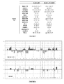
- Karyotypic analysis was performed on the IGR-CaP1 cell line at passage 29 by using a conventional G-banding technique ( Figure 2B ). The analysis showed a complex tetraploid karyotype with 86-91 chromosomes ( Figure 2B ).
- the karyotype was determined to be 86-89 ⁇ 4n>,XX,+der(X),+der(X),-Y,-Y, der(1)t(l;?)(p32;?), add(2)(q3?2),-3,der(3)t(3;?)(p10;?),-4,-4,-5,-5,-6,-6,ins(7;?)(p15;?),+8,+8,+11,+11,-13,-13, add(13)(q31),-14,-15,-15,del(18)(q22),del(18(q22),+20,del(20)(q12),+del(20)(q12),+mar inc[cp15].
- Tp53 Since mutations in the tumour suppressor gene Tp53 are frequently associated with chromosome instability, expression of Tp53 was examined in the IGR-CaP1 cells. Immunohistochemical study showed a high expression of Tp53 protein in the parental tumour and in the derived cells ( Figure 2C-D ). Tp53 expression in the IGR-CaP1 cell line was confirmed by western blot analysis with the anti-Tp53 DO-7 antibody. High expression of Tp53 was assumed to be corresponding to stabilized mutated Tp53 gene product. A single mutation in the nucleotide A377G was indeed detected by sequencing, corresponding to codon Y126C change.
- IGR-CaP1-R100 cell line is an in vitro cellular model of prostate cancer resistant to docetaxel established by the inventors. In order to prepare this resistant cell line, IGR-CaP1 cells have been treated with a dose escalation of docetaxel as disclosed in Patterson et al (2006) ( Figure 3 ).
- IGR-CaP1-R cells The survival and proliferation capacity in presence of docetaxel has been checked for IGR-CaP1-R cells by cellular cycle measurement ( Figure 4 ).
- Sensitive IGR-CaP1 cells (Fig. 4A ) are blocked in G2-M phase when contacted with 100 nM of docetaxel for 48 h ( Fig. 4B ).
- IGR-CaP1-R100 cells which are resistant to 100 nM of docetaxel ( Fig. 4C ) cycle like the sensitive cells in absence of docetaxel. Contrary to the sensitive cells, the resistant cells are not blocked in G2-M phase in presence of docetaxel for 48 h ( Fig. 4D ).
- IGR-CaP1-R cells resistant to other dose of docetaxel e.g., 12 nM.
- STR genotyping of cell lines is become a necessity for indisputably characterizing a cell line. This technique is rapid and can be carried out in any laboratory. For instance, the STR profile for 9 of the 16 analyzed loci is given in an increasing number of cell lines at the American Tissue and Culture Collections (ATCC). The profiles confirmed the complexity of the karyotype and showed the absence of chromosome Y on the amelogenin locus. The loss of the entire Y chromosome had been confirmed by the whole Y chromosome STR profile.
- the alleles 9.1/10.1/10.3/11.2 were shown to be rare at locus D7S820 in the whole population data bases.
- the inventors also found allele 9.1/10.3 for this locus in LNCaP cells on the STR data bank web site ( http://www.cstl.nist.goy/di831/strbase/ ).
- the inventors observed the same STR profile between DNA prepared from IGR-CaP1 cells at passage 10 and DNA prepared from cells at passage 50, showing that chromosome alterations were conserved during the culture.
- 10 loci remained unchanged in the two cell lines and 6 loci were modified.
- Some rare alleles have been found in locus D7S820 (9.1, 10.1, 11.2).
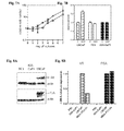
- the IGR-CaP1 cells grew rapidly in classical culture medium conditions with a doubling-time of 1.8 days. Same cellular kinetics was observed in medium containing charcoal-stripped serum ( Figure 7A ). Treatment of cultured IGR-CaP1 cells in the presence of androgen DHT did not affect the cellular growth, as also observed for androgen-independent PC3 cells. As control, cellular growth of LNCaP cells was increased in a treatment of cells with 10-9M DHT for 72 hours ( Figure 7B ).
- IGR-CaP1 cells do not express AR nor PSA
- IGR-CaP1 cells did not express androgen receptor protein (AR) or secretory prostate-specific antigen (PSA) as shown by western analyses on whole cell extracts ( Figure 8A ).
- AR androgen receptor protein
- PSA secretory prostate-specific antigen
- Figure 8A The LNCaP and MDA-PCa-2b cell lines that both expressed these two prostate-specific markers were used as positive controls; the androgen-independent PC3 cell line was used as negative control.
- neither AR nor PSA were detected at mRNA levels in IGR-CaP1 cells by real-time quantitative RT-PCR analyses ( Figure 8B ).
- the expression of androgen receptor and PSA in the IGR-CaP1 cell line contrast with that was observed in the initial tumour.
- IGR-CaP1 cells reconstitute prostate adenocarcinoma in mice that recapitulates the original tumour features
- mice were injected into male nude mice both subcutaneously and intraprostatically.
- Subcutaneous injection of IGR-CAP1 cells resulted in the formation of palpable tumors within a week and 90% of mice (20/22) were bearing 7-9mm tumours after 6-8 weeks.
- One mouse developed intraperitoneal ascites without subcutaneous tumour.
- Intraprostatic injection of IGR-CaP1 cells resulted in the formation of intraprostatic tumors in 38% of mice (5/13).
- the serum samples of tumor-bearing mice contained no measurable PSA.
- H&E-staining section of the tumours revealed a glandular differentiation with ascini attesting the presence of adenocarcinoma ( Figure 9A and 9B ).
- Immunohistochemical staining of the tumours with Ki67 showed a high proliferation index which was higher in intraprostatic tumour.
- Cytokeratin staining proved the epithelial features and prostatic acid phosphatase (PAP) and PSMA staining attested from the prostate origin of the tumour ( Figure 9 ).
- a low AMACR (P504S) staining was also observed, higher staining was observed in intraprostatic tumour.
- vimentin staining was intense in subcutaneous tumours corresponding to that was observed in the parental IGR-CaP1 cells but it was absent in intraprostatic tumours. Vimentin is considered to be the intermediate filament of mesenchymal tissue.
- the aggressive character of the subcutaneous tumours was variable: some tumour-bearing mice showed necrosis and fribosis in involutive forms with no more tumour features. In contrast, some mice showed aggressive tumours that infiltrate the whole peritoneal cavity, forming ascites, and including kidney, spleen and pancreas. Predominant metastases were observed in liver and in lung.
- Intraprostatic tumours were always aggressive and showed metastasis in the diaphragm including kidney and liver and micrometastasis in lung. Collectively, these data showed that intraprostatic tumour initiated by the IGR-CaP1 cell line recapitulates in mice the characteristics of the initial undifferentiated human prostate tumour. Tumour grade and metastasis localization corresponds to that was observed for patient presenting undifferentiated tumour with a high-Gleason score.
- IGR-CaP1 cells express high levels of basal epithelial prostate markers.
- IGR-CaP1 cells show basal, luminal or intermediate cell phenotypes
- cytokeratin markers CK5, CK8, CK14 and CKl8 were first examined by cytometer analysis ( Figure 10 ). Cytokeratin expression profile of luminal epithelial prostate LNCaP cell line was used as control. IGR-CaP1 cells exhibit strong staining for CK5/8 and CK14 but absence of staining for CK8. Although adult prostate basal cells were shown to express p63, the inventors did not detect p63 expression in these cells. They detected a low labelling for the luminal epithelial marker CK18 in IGR-CaP 1 cells compared to that observed in LNCaP cells.
- IGR-CaP1 cells and clonally-derived clones showed features of prostate cancer stem cells.
- the basal cells are particularly important as the basal cell compartment is thought to contain the epithelial stem or progenitor cells.
- Cancer stem cells possess the intrinsic stem cell properties of renewal, proliferation and differentiation. However it is assume that these cells are responsible for initiating cancer growth, it is still difficult to draw up the molecular profile that defines a prostate cancer stem cell.
- the putative stem cell population was enriched using the CD44 and CD133 antigens. Therefore, the inventors first evaluated CD44 and CD133 expression levels within IGR-CaP1 cell line by flow cytometry ( Figure 11A ). Compared to LNCaP cells in which only a minor fraction of cells expressed CD44, almost all the IGR-CaP1 cells expressed CD44 antigen.
- CXCR4 is a key molecule in the regulation of the migratory and metastatic properties of cancer cells. Recent data suggested that the CXCR4 axis may be essential for the progression of the CD133+ prostate cancer stem cells. The inventors found that a large subset of IGR-CaP1 cells express CXCR4 molecule.
- CD133 and CXCR4 were measured by real-time quantitative RT-PCR in the parental IGR-CaP1 cell line and in the clonally-derived clones 3A11 and 3C11 ( Figure 11B ). All the cells showed a high expression of these two stem cell markers, CD133 was two fold less expressed in 3A11 cells while 3C11 cells conserved a high expression of CD133. The inventors next evaluate the combination of the biomarkers expression after triple-labelling and analysis by flow cytometry. In the parental IGR-CaP1 cell line, the CD44+/CD133+/CXCR4+ cells represented 13% expression.
- the fraction of triple-labelled cells was highly increased in derived clones as the inventors observed 23% and 34% of CD44+/CD133+/CXCR4+ cells in 3A11 and 3C11 clones respectively. They next evaluated the ability of CD44 + /CXCR4 + /CD133 + sorted cells to differentiate under 3D differentiating conditions. Among CD133 negative cells, they observed no difference in the differentiation ability between CRCR4+ and CXCR4- cells. However in CD133 positive cells, CXCR4- cells keep their differentiation capacity while CD133+/CXCR4+ lost most of their morphologic differentiation potential. Under stem cells conditions CD133+/CXCR4+ and CD133+/CXCR4- cells produced the same spheroid structures on matrigel.
- IGR-CaP1 cells showed a stem cell marker expression signature.
- the transcription factor Oct4 is expressed in pluripotent embryonic stem (ES) and germ cells and is currently considered a main regulator of human embryonic stem cell pluripotency and self-renewal capacities.
- Oct 4 Nanog and Sox2 have been shown to be expressed in HPET immortalized prostate cancer cells that were shown to reconstitute the original human tumor in vivo and had been suggested to constitute an in vitro model for prostate cancer stem cells.
- Microarray analysis realized on the IGR-CaP1 cells revealed gene expression of Oct4.
- Western blot analysis confirmed the expression of Oct4 in IGR-CaP1 cells, contrasting to the control LNCaP and PC3 cells ( Figure 12B ).
- Hedgehog Hedgehog
- Notch signalling pathways regulate key functions of stem/progenitor cells self-renewal
- the inventors next used real-time RT-PCR to assess their expression ( Figure 12C ). They observed a marked increase of expression of several stemness markers in IGR-CaP1 cells compared to the control LNCaP cells. Sonic Hedgehog (SHH) which has been implicated in signalling in the maintenance and/or proliferation of prostatic progenitor cells was the most expressed gene.
- SHH Sonic Hedgehog
- HH Hedgehog pathway in carcinogenesis.
- the inventors assess the expression of the HH receptor Patched protein (PTCH1) and GLI target gene products.
- Notch As a marker and regulator of normal prostate stem/progenitor cells, however, has only recently been suggested although there is currently still little evidence to support Notch as a marker of prostate cancer stem/initiating cells.
- LNCaP cells were reported to express Notch1 at mRNA and protein levels. In IGR-CaP1 cells, Notch1 mRNA was expressed at roughly 3 fold higher than in LNCaP cells. IGR-CaP1 cells also expressed high level of mRNA coding for the breast cancer resistance protein BCRP/ABCG2 which has been implicated in tumorigenic stem-like cancer cells enriched-side population.
- Prostate tissue was obtained from a 58-year-old caucasian-french patient who underwent a radical prostatectomy for clinically localized prostate cancer.
- the patient had a PSA of 5.6 ng/ml and a free-PSA of 6 %.
- Subsequent pathological evaluation revealed a massive bilateral posterior tumour limited to the prostatic capsule and was diagnosed as a moderately differentiated adenocarcinoma with a Gleason score of 7 (4 (70%) +3 (30%)).
- Clinical staging was assessed as pT2c Nx.
- Prostate tissues were obtained from 3 patients undergoing radical prostatectomy for localized prostate cancer. Fresh prostatectomy specimens were obtained under sterile conditions. After a mechanic dissociation of the tumour tissue, cells were counted and seeded on the natural extracellular matrix in RPMI medium supplemented in 10% FBS (Gibco), penicillin-streptomycin antibiotics and fungizone. This natural extracellular matrix has been developed by J. Bénard in the Gustave Roussy Institute to increase chance of obtaining rare cell lines. The RPMI medium used for primocultures was depleted in proline amino acid to eliminate fibroblast contamination. The cells were incubated at 37°C at 5% CO2 until reaching semi-confluency.
- the culture was pursuing on the extracellular matrix until 10 to 20 passages and then, was continuing in classical RPMI medium containing 10% FBS and antibiotics on plastic dishes.
- Others prostate cancer cell lines were cultured as previously described: LNCaP and PC3 cells were grown in RPMI medium containing 10% FBS and MDA-PCa-2b cells were grown in BRFF-HPC1 medium completed with 20% FBS and 50 ⁇ g/ml gentamycin.
- the cell growth kinetic was determined by counting the viable cell number at regular intervals. Cells were seeded in triplicate at 4000 cells/well in 12 well plates in normal culture medium or in medium containing 10% of charcoal-stripped serum. Each day, cells were colored with trypan blue and counted. The doubling-time was calculated from regression equation of the curve. For the hormone-dependent growth assay, cells were seeded at 10 000 cells/well in 96 well plates. After 24h, the culture medium was replaced with medium without phenol red containing 10% of charcoal-stripped serum and the DHT was added or not at the final concentration of 10 -9 M. The medium was replaced each day. After 72 hours, cell survival was measured with the WST1 test according the manufacturer's procedures.
- telomere activity is determined through its ability to synthesize telomeric repeats onto an oligonucleotide substrate in cellular extracts and resulting extended products were amplified by PCR (35-40 cycles) using the DNA fluorochrome SYBR Green.
- Genomic DNA was extracted from cultured cells and was amplified using the TP53-specific primers 5'-ATTTGATGCTGTCCCCGGACGATATTGAA-3' (exon 3) and 5'-CACTCCAGCCACCTGAAGTCCAAAAAGGGT-3' (exon 10). PCR products amplification was verified by electrophoresis and amplified DNA was sequenced on an automatic sequencer (Applied Biosystems 3730). Two different DNA preparations obtained from different cell aliquots showed the same TP53 mutation.
- Metaphases were harvested after a 2.5-hour colchicine block. Chromosome spreads were obtained according to previously described techniques. Karyotypes were established after R-banding.
- Genomic DNA was prepared from the cells using kit QIAamp DNA Micro kit (Qiagen). Human genomic DNA was quantified using sensitive real time PCR technology, using human specific primers and hydrolysis probes. Q-PCR was performed on LightCycler480 Instrument, (Roche) and analyzed with LightCycler480 software 1.5.
- STR analysis was conducted using the multiplex-PCR-based Identifiler® amplification kit (Applied Biosystems). Fluorescent STR-based amplification was conducted with 1ng of human genomic DNA. 15 autosomal STR Loci were simultaneously co-amplified, including D8S1179, D21S11, D7S820, CSF1P0, D3S1358, TH01, D13S317, D16S539, D2S1338, D19S433, vWA, TP0X, D18S51, D5S818 and FGA plus the Amelogenin gender-determining marker. Automated DNA fragment Analysis was performed by electrophoresis on ABI3130x1 Genetic Analyser (Applied Biosystems). Fluorescent data were collected and analyzed using GeneMapper 3.2 ID specific genotyping software (Applied Biosystems). The resulting profile showed the assigned allele values corresponding to the number of repeat units identified for each locus.
- mice Male athymic nude mice (NC-nu/nu) of 6-7 week old were used to carry out tumorigenicity assay. All of the operative procedures and animal care were in conformity with the Guidelines of the French Government. Subcutaneous injections were realized into the dorsal side with 10 7 viable IGR-CaP1 cells in 100 ⁇ l of PBS (lacking matrigel). Orthotopic injections were realized as previously described (Fizazi et al., 2004). Briefly, the prostate of each anesthetized mouse was exposed through a midline laparotomy incision and direct injection into the prostate was performed with 10 6 cells in 2 ⁇ l PBS.
- Western blot assays were performed on 50 ⁇ g of whole cellular lysates loaded on precasted 4-12% NuPage gels (Invitrogen). Blots were probed with the following antibodies: rabbit polyclonal anti-androgen receptor antibody (AR) (N-20, Santa Cruz), goat polyclonal anti-PSA antibody (C-19, Santa Cruz), rabbit anti-Oct4 antibody (Chemicon) Anti-rabbit (Pierce) or anti-goat (Dako) IgG-horseradish peroxidase antibody served as the secondary antibody. Immunoblot analyses were developed using the enhanced chemoluminescence-based detection kit (Pierce, Rockford, IL, USA).
- Fluorescence-activated cell sorting analysis was used for determination of differentiation marker expression and cell sorting.
- cytoplasmic or nuclear proteins a permeabilization in 0.25% triton X-100 was performed before labelling.
- the following antibodies were used: anti-human EpCAM-PE (clone EBA-1, Becton Dickinson), PSMA-FITC (clone 107-1A4, MBL medical), CK5/8-FITC (clone 5F173, US Biological), CK8-FITC (clone B22.1, GeneTex, Inc.) et CK14-FITC (Clone 2Q1030, US Biological), CD44-FITC (Clone G44-26, BD Biosciences), CD133-APC (AC133, Miltenyi Biotec) et CXCR4-APC (CD 184 clone , BD Pharmingen).
- CD 184-PE CD 184 clone BD Pharmingen
- the corresponding isotype control antibodies were included in each staining condition.
- Samples were analyzed with FACSCalibur cytometer (Becton Dickinson). Triple-color stained cells were sorted by cell sorter (MoFlo-Dako).
- mice studied in 3 experiments were fixed in Finefix (Milestone Medical), and then paraffin sections (4 ⁇ m thick) and hematoxylin-eosin-saffranin (HES) slides were prepared.
- HES hematoxylin-eosin-saffranin
- tumor and organ sections were incubated with anti-pan-CK (Pan Ab-2, LabVision), anti-p53 (Ventana Medical Systems), anti-PSMA (clone 3E6, Diagnostic BioSystems), anti-PAP (PSAP clone PASE/4LJ, Diagnostic BioSystems), anti-AMACR (P504S, Diagnostic biosystems), anti-Ki67(-Zymed), anti-vimentin (Clone V9, Sigma) antibodies. All of the sections were analyzed by using a Zeiss Axiophot microscope and a SensiCam PCO digital camera. Representative views were taken at a magnification of 100 ⁇ .
- Cells were seeded on sterile glass coverslip. When cells reached to 80% confluence, they were fixed at room temperature in 4% formalhedyde in PBS buffer for 10 min and then washed five times in PBS. Cells were permeabilized for 10 min in a 0.25% Triton X-100 and extensively washed, and blocked in a 1:100 normal goat serum solution (Sigma). The coverslips were incubated with primary antibody anti-human ⁇ 2 ⁇ 1 integrin (clone BHA2.1, Chemicon) for 1 hour at room temperature followed by a 30 min with Alexa Fluor 488 goat antimouse secondary antibody (Molecular Probes). Nuclei were stained with Dapi vectashield mounting reagent (Vector Laboratories). Images were acquired on a Zeiss Axioplan 2microscope.
Landscapes
- Health & Medical Sciences (AREA)
- Engineering & Computer Science (AREA)
- Life Sciences & Earth Sciences (AREA)
- Biomedical Technology (AREA)
- Immunology (AREA)
- Hematology (AREA)
- Chemical & Material Sciences (AREA)
- Urology & Nephrology (AREA)
- Molecular Biology (AREA)
- Tropical Medicine & Parasitology (AREA)
- Medicinal Chemistry (AREA)
- Microbiology (AREA)
- Biotechnology (AREA)
- Toxicology (AREA)
- Bioinformatics & Cheminformatics (AREA)
- Food Science & Technology (AREA)
- Cell Biology (AREA)
- Physics & Mathematics (AREA)
- Analytical Chemistry (AREA)
- Biochemistry (AREA)
- General Health & Medical Sciences (AREA)
- General Physics & Mathematics (AREA)
- Pathology (AREA)
- Measuring Or Testing Involving Enzymes Or Micro-Organisms (AREA)
Priority Applications (4)
| Application Number | Priority Date | Filing Date | Title |
|---|---|---|---|
| EP09305315A EP2241618A1 (de) | 2009-04-14 | 2009-04-14 | Prostata-Krebszelllinien und ihre Verwendung beim Screening-Verfahren |
| PCT/EP2010/054739 WO2010119001A1 (en) | 2009-04-14 | 2010-04-12 | Prostate cancer cell lines and their use in screening method |
| EP10713634A EP2419507A1 (de) | 2009-04-14 | 2010-04-12 | Prostatakrebs-zelllinien und deren verwendung bei screening-verfahren |
| US13/264,479 US8674172B2 (en) | 2009-04-14 | 2010-04-12 | Prostate cancer cell lines and their use in screening method |
Applications Claiming Priority (1)
| Application Number | Priority Date | Filing Date | Title |
|---|---|---|---|
| EP09305315A EP2241618A1 (de) | 2009-04-14 | 2009-04-14 | Prostata-Krebszelllinien und ihre Verwendung beim Screening-Verfahren |
Publications (1)
| Publication Number | Publication Date |
|---|---|
| EP2241618A1 true EP2241618A1 (de) | 2010-10-20 |
Family
ID=41467119
Family Applications (1)
| Application Number | Title | Priority Date | Filing Date |
|---|---|---|---|
| EP09305315A Ceased EP2241618A1 (de) | 2009-04-14 | 2009-04-14 | Prostata-Krebszelllinien und ihre Verwendung beim Screening-Verfahren |
Country Status (1)
| Country | Link |
|---|---|
| EP (1) | EP2241618A1 (de) |
Cited By (2)
| Publication number | Priority date | Publication date | Assignee | Title |
|---|---|---|---|---|
| EP2699596A4 (de) * | 2011-04-22 | 2015-01-14 | Emergent Product Dev Seattle | Prostataspezifische membranantigenbindende proteine sowie zusammensetzungen daraus und verfahren dafür |
| CN111019902A (zh) * | 2019-12-25 | 2020-04-17 | 浙江大学 | 一种前列腺癌多西他赛耐药细胞株的构建方法 |
Citations (2)
| Publication number | Priority date | Publication date | Assignee | Title |
|---|---|---|---|---|
| WO1999019479A1 (en) * | 1997-09-26 | 1999-04-22 | Corixa Corporation | Murine model for human carcinoma |
| WO2000044879A1 (en) * | 1999-01-28 | 2000-08-03 | The Regents Of The University Of California | Aggressive prostate cancer sublines selected by androgen deprivation |
-
2009
- 2009-04-14 EP EP09305315A patent/EP2241618A1/de not_active Ceased
Patent Citations (2)
| Publication number | Priority date | Publication date | Assignee | Title |
|---|---|---|---|---|
| WO1999019479A1 (en) * | 1997-09-26 | 1999-04-22 | Corixa Corporation | Murine model for human carcinoma |
| WO2000044879A1 (en) * | 1999-01-28 | 2000-08-03 | The Regents Of The University Of California | Aggressive prostate cancer sublines selected by androgen deprivation |
Non-Patent Citations (23)
Cited By (2)
| Publication number | Priority date | Publication date | Assignee | Title |
|---|---|---|---|---|
| EP2699596A4 (de) * | 2011-04-22 | 2015-01-14 | Emergent Product Dev Seattle | Prostataspezifische membranantigenbindende proteine sowie zusammensetzungen daraus und verfahren dafür |
| CN111019902A (zh) * | 2019-12-25 | 2020-04-17 | 浙江大学 | 一种前列腺癌多西他赛耐药细胞株的构建方法 |
Similar Documents
| Publication | Publication Date | Title |
|---|---|---|
| Li et al. | SREBP-2 promotes stem cell-like properties and metastasis by transcriptional activation of c-Myc in prostate cancer | |
| Jeng et al. | RNA‐binding protein insulin‐like growth factor II mRNA‐binding protein 3 expression promotes tumor invasion and predicts early recurrence and poor prognosis in hepatocellular carcinoma | |
| Bisson et al. | WNT signaling regulates self-renewal and differentiation of prostate cancer cells with stem cell characteristics | |
| Timofeeva et al. | Conditionally reprogrammed normal and primary tumor prostate epithelial cells: a novel patient-derived cell model for studies of human prostate cancer | |
| EP2626414B1 (de) | Krebsstammzellenmasse und verfahren zu ihrer herstellung | |
| US8435746B2 (en) | Aldehyde dehydrogenase 1 (ALDH1) as a cancer stem cell marker | |
| Saw et al. | Characterization of aldehyde dehydrogenase isozymes in ovarian cancer tissues and sphere cultures | |
| Capo-chichi et al. | Anomalous expression of epithelial differentiation-determining GATA factors in ovarian tumorigenesis | |
| Ke et al. | ALG3 contributes to the malignancy of non-small cell lung cancer and is negatively regulated by MiR-98-5p | |
| US8674172B2 (en) | Prostate cancer cell lines and their use in screening method | |
| Lindley et al. | The WNT-controlled transcriptional regulator LBH is required for mammary stem cell expansion and maintenance of the basal lineage | |
| JP2011511634A (ja) | ガン幹細胞に関連する方法および組成物 | |
| Schmitt et al. | Cancer stem cell markers in breast neoplasias: their relevance and distribution in distinct molecular subtypes | |
| Würth et al. | Experimental evidence and clinical implications of pituitary adenoma stem cells | |
| Prasad et al. | Dual TGFβ/BMP pathway inhibition enables expansion and characterization of multiple epithelial cell types of the normal and cancerous breast | |
| KR20210041000A (ko) | 암 조직 또는 암 조직과 유사한 조직의 배양 방법 | |
| Feng et al. | CD133+ subpopulation of the HT1080 human fibrosarcoma cell line exhibits cancer stem-like characteristics | |
| Faraldo et al. | Alpha-6 integrin deletion delays the formation of Brca1/p53-deficient basal-like breast tumors by restricting luminal progenitor cell expansion | |
| Luo et al. | Isolation and genome-wide expression and methylation characterization of CD31+ cells from normal and malignant human prostate tissue | |
| Chauchereau et al. | Stemness markers characterize IGR-CaP1, a new cell line derived from primary epithelial prostate cancer | |
| Tyson et al. | Culture requirements of prostatic epithelial cell lines for acinar morphogenesis and lumen formation in vitro: role of extracellular calcium | |
| EP2241618A1 (de) | Prostata-Krebszelllinien und ihre Verwendung beim Screening-Verfahren | |
| Lin et al. | Prognostic value of pretreatment CD44 mRNA in peripheral blood of patients with locally advanced head and neck cancer | |
| Caria et al. | Thyrospheres from B-CPAP cell line with BRAF and TERT promoter mutations have different functional and molecular features than parental cells | |
| Peculis et al. | Pituispheres contain genetic variants characteristic to pituitary adenoma tumor tissue |
Legal Events
| Date | Code | Title | Description |
|---|---|---|---|
| APBK | Appeal reference recorded |
Free format text: ORIGINAL CODE: EPIDOSNREFNE |
|
| APBN | Date of receipt of notice of appeal recorded |
Free format text: ORIGINAL CODE: EPIDOSNNOA2E |
|
| APBR | Date of receipt of statement of grounds of appeal recorded |
Free format text: ORIGINAL CODE: EPIDOSNNOA3E |
|
| APBV | Interlocutory revision of appeal recorded |
Free format text: ORIGINAL CODE: EPIDOSNIRAPE |
|
| PUAI | Public reference made under article 153(3) epc to a published international application that has entered the european phase |
Free format text: ORIGINAL CODE: 0009012 |
|
| AK | Designated contracting states |
Kind code of ref document: A1 Designated state(s): AT BE BG CH CY CZ DE DK EE ES FI FR GB GR HR HU IE IS IT LI LT LU LV MC MK MT NL NO PL PT RO SE SI SK TR |
|
| AX | Request for extension of the european patent |
Extension state: AL BA RS |
|
| STAA | Information on the status of an ep patent application or granted ep patent |
Free format text: STATUS: THE APPLICATION HAS BEEN REFUSED |
|
| 18R | Application refused |
Effective date: 20101127 |