EP2209889B1 - Compositions for manipulating pim-1 activity in cardiac cells - Google Patents
Compositions for manipulating pim-1 activity in cardiac cells Download PDFInfo
- Publication number
- EP2209889B1 EP2209889B1 EP08850768.6A EP08850768A EP2209889B1 EP 2209889 B1 EP2209889 B1 EP 2209889B1 EP 08850768 A EP08850768 A EP 08850768A EP 2209889 B1 EP2209889 B1 EP 2209889B1
- Authority
- EP
- European Patent Office
- Prior art keywords
- pim
- expression
- cell
- cardiac
- cells
- Prior art date
- Legal status (The legal status is an assumption and is not a legal conclusion. Google has not performed a legal analysis and makes no representation as to the accuracy of the status listed.)
- Active
Links
- 101001001642 Xenopus laevis Serine/threonine-protein kinase pim-3 Proteins 0.000 title claims description 448
- 210000002064 heart cell Anatomy 0.000 title claims description 59
- 239000000203 mixture Substances 0.000 title description 84
- 230000000694 effects Effects 0.000 title description 68
- 230000014509 gene expression Effects 0.000 claims description 183
- 230000000747 cardiac effect Effects 0.000 claims description 93
- 210000000130 stem cell Anatomy 0.000 claims description 77
- 102000039446 nucleic acids Human genes 0.000 claims description 65
- 108020004707 nucleic acids Proteins 0.000 claims description 65
- 150000007523 nucleic acids Chemical class 0.000 claims description 64
- 210000004165 myocardium Anatomy 0.000 claims description 62
- 210000000107 myocyte Anatomy 0.000 claims description 53
- 210000001519 tissue Anatomy 0.000 claims description 52
- 230000006378 damage Effects 0.000 claims description 40
- 206010020880 Hypertrophy Diseases 0.000 claims description 35
- 210000000056 organ Anatomy 0.000 claims description 35
- 239000008194 pharmaceutical composition Substances 0.000 claims description 33
- 239000003981 vehicle Substances 0.000 claims description 28
- 206010021143 Hypoxia Diseases 0.000 claims description 25
- 241000700605 Viruses Species 0.000 claims description 25
- 230000001413 cellular effect Effects 0.000 claims description 22
- 210000005003 heart tissue Anatomy 0.000 claims description 22
- 239000013612 plasmid Substances 0.000 claims description 16
- 206010019280 Heart failures Diseases 0.000 claims description 15
- 230000001969 hypertrophic effect Effects 0.000 claims description 14
- 208000015181 infectious disease Diseases 0.000 claims description 13
- 230000007954 hypoxia Effects 0.000 claims description 10
- 206010002660 Anoxia Diseases 0.000 claims description 7
- 241000976983 Anoxia Species 0.000 claims description 7
- 230000007953 anoxia Effects 0.000 claims description 7
- 230000036044 hypoxaemia Effects 0.000 claims description 7
- 230000000302 ischemic effect Effects 0.000 claims description 7
- 208000007848 Alcoholism Diseases 0.000 claims description 5
- 206010061218 Inflammation Diseases 0.000 claims description 5
- 201000007930 alcohol dependence Diseases 0.000 claims description 5
- 210000004507 artificial chromosome Anatomy 0.000 claims description 5
- 230000037326 chronic stress Effects 0.000 claims description 5
- 206010012601 diabetes mellitus Diseases 0.000 claims description 5
- 239000003937 drug carrier Substances 0.000 claims description 5
- 230000004054 inflammatory process Effects 0.000 claims description 5
- 238000002560 therapeutic procedure Methods 0.000 claims description 4
- 210000004027 cell Anatomy 0.000 description 146
- 108091008611 Protein Kinase B Proteins 0.000 description 90
- 239000013598 vector Substances 0.000 description 90
- 210000004413 cardiac myocyte Anatomy 0.000 description 86
- 101000595531 Homo sapiens Serine/threonine-protein kinase pim-1 Proteins 0.000 description 85
- 102100036077 Serine/threonine-protein kinase pim-1 Human genes 0.000 description 80
- 238000000034 method Methods 0.000 description 70
- 230000001965 increasing effect Effects 0.000 description 67
- 239000013316 polymer of intrinsic microporosity Substances 0.000 description 62
- 241000282414 Homo sapiens Species 0.000 description 43
- 241000699670 Mus sp. Species 0.000 description 42
- 108090000623 proteins and genes Proteins 0.000 description 42
- 102100033810 RAC-alpha serine/threonine-protein kinase Human genes 0.000 description 41
- 230000006907 apoptotic process Effects 0.000 description 41
- 230000002018 overexpression Effects 0.000 description 41
- 238000011282 treatment Methods 0.000 description 39
- 206010061216 Infarction Diseases 0.000 description 38
- 241001465754 Metazoa Species 0.000 description 34
- 208000014674 injury Diseases 0.000 description 33
- 208000010125 myocardial infarction Diseases 0.000 description 33
- 238000003119 immunoblot Methods 0.000 description 32
- 230000007574 infarction Effects 0.000 description 32
- 102000004169 proteins and genes Human genes 0.000 description 32
- 230000011664 signaling Effects 0.000 description 32
- 208000027418 Wounds and injury Diseases 0.000 description 30
- 230000004083 survival effect Effects 0.000 description 27
- AOJJSUZBOXZQNB-TZSSRYMLSA-N Doxorubicin Chemical compound O([C@H]1C[C@@](O)(CC=2C(O)=C3C(=O)C=4C=CC=C(C=4C(=O)C3=C(O)C=21)OC)C(=O)CO)[C@H]1C[C@H](N)[C@H](O)[C@H](C)O1 AOJJSUZBOXZQNB-TZSSRYMLSA-N 0.000 description 26
- 201000010099 disease Diseases 0.000 description 25
- 208000037265 diseases, disorders, signs and symptoms Diseases 0.000 description 25
- 230000002107 myocardial effect Effects 0.000 description 25
- 230000002792 vascular Effects 0.000 description 25
- 230000003612 virological effect Effects 0.000 description 25
- 230000003293 cardioprotective effect Effects 0.000 description 24
- 238000009472 formulation Methods 0.000 description 24
- 230000007423 decrease Effects 0.000 description 22
- -1 e.g. Proteins 0.000 description 22
- 230000006870 function Effects 0.000 description 22
- 238000001727 in vivo Methods 0.000 description 22
- 238000005259 measurement Methods 0.000 description 22
- 210000005167 vascular cell Anatomy 0.000 description 22
- 230000000004 hemodynamic effect Effects 0.000 description 21
- 238000002347 injection Methods 0.000 description 20
- 239000007924 injection Substances 0.000 description 20
- 239000002502 liposome Substances 0.000 description 20
- 102400000686 Endothelin-1 Human genes 0.000 description 19
- 101800004490 Endothelin-1 Proteins 0.000 description 19
- 230000003247 decreasing effect Effects 0.000 description 19
- 239000003814 drug Substances 0.000 description 19
- 230000001404 mediated effect Effects 0.000 description 19
- 230000009261 transgenic effect Effects 0.000 description 19
- 108091000080 Phosphotransferase Proteins 0.000 description 18
- 150000001875 compounds Chemical class 0.000 description 18
- 230000002708 enhancing effect Effects 0.000 description 18
- 102000020233 phosphotransferase Human genes 0.000 description 18
- 239000000243 solution Substances 0.000 description 18
- 241000701161 unidentified adenovirus Species 0.000 description 18
- 102000004433 Proto-Oncogene Proteins c-pim-1 Human genes 0.000 description 17
- 108010017121 Proto-Oncogene Proteins c-pim-1 Proteins 0.000 description 17
- 230000006698 induction Effects 0.000 description 16
- 230000001939 inductive effect Effects 0.000 description 16
- 230000036722 left ventricular developed pressure Effects 0.000 description 16
- 239000003795 chemical substances by application Substances 0.000 description 15
- 230000004217 heart function Effects 0.000 description 15
- 208000028867 ischemia Diseases 0.000 description 15
- 230000026731 phosphorylation Effects 0.000 description 15
- 238000006366 phosphorylation reaction Methods 0.000 description 15
- 230000035755 proliferation Effects 0.000 description 15
- 108010029485 Protein Isoforms Proteins 0.000 description 14
- 102000001708 Protein Isoforms Human genes 0.000 description 14
- 206010063837 Reperfusion injury Diseases 0.000 description 14
- 108700019146 Transgenes Proteins 0.000 description 14
- 230000004913 activation Effects 0.000 description 14
- 238000000338 in vitro Methods 0.000 description 14
- 239000006166 lysate Substances 0.000 description 14
- 108090000765 processed proteins & peptides Proteins 0.000 description 14
- 230000004044 response Effects 0.000 description 14
- 230000007755 survival signaling Effects 0.000 description 14
- 230000001225 therapeutic effect Effects 0.000 description 14
- 241000699666 Mus <mouse, genus> Species 0.000 description 13
- 229960004679 doxorubicin Drugs 0.000 description 13
- 238000002372 labelling Methods 0.000 description 13
- 210000004940 nucleus Anatomy 0.000 description 13
- 239000002245 particle Substances 0.000 description 13
- 230000001105 regulatory effect Effects 0.000 description 13
- 239000013603 viral vector Substances 0.000 description 13
- 108020004414 DNA Proteins 0.000 description 12
- FAPWRFPIFSIZLT-UHFFFAOYSA-M Sodium chloride Chemical compound [Na+].[Cl-] FAPWRFPIFSIZLT-UHFFFAOYSA-M 0.000 description 12
- 230000001640 apoptogenic effect Effects 0.000 description 12
- 108700000711 bcl-X Proteins 0.000 description 12
- 102000055104 bcl-X Human genes 0.000 description 12
- 230000030833 cell death Effects 0.000 description 12
- 102000040430 polynucleotide Human genes 0.000 description 12
- 108091033319 polynucleotide Proteins 0.000 description 12
- 239000002157 polynucleotide Substances 0.000 description 12
- 230000001681 protective effect Effects 0.000 description 12
- 102100021569 Apoptosis regulator Bcl-2 Human genes 0.000 description 11
- 108020004705 Codon Proteins 0.000 description 11
- 101000971171 Homo sapiens Apoptosis regulator Bcl-2 Proteins 0.000 description 11
- 241000725303 Human immunodeficiency virus Species 0.000 description 11
- 108090000723 Insulin-Like Growth Factor I Proteins 0.000 description 11
- 150000002632 lipids Chemical class 0.000 description 11
- 108020004999 messenger RNA Proteins 0.000 description 11
- 235000002639 sodium chloride Nutrition 0.000 description 11
- 239000000758 substrate Substances 0.000 description 11
- 238000010361 transduction Methods 0.000 description 11
- 230000026683 transduction Effects 0.000 description 11
- OHCQJHSOBUTRHG-KGGHGJDLSA-N FORSKOLIN Chemical compound O=C([C@@]12O)C[C@](C)(C=C)O[C@]1(C)[C@@H](OC(=O)C)[C@@H](O)[C@@H]1[C@]2(C)[C@@H](O)CCC1(C)C OHCQJHSOBUTRHG-KGGHGJDLSA-N 0.000 description 10
- 102000014429 Insulin-like growth factor Human genes 0.000 description 10
- 101100297651 Mus musculus Pim2 gene Proteins 0.000 description 10
- KPKZJLCSROULON-QKGLWVMZSA-N Phalloidin Chemical compound N1C(=O)[C@@H]([C@@H](O)C)NC(=O)[C@H](C)NC(=O)[C@H](C[C@@](C)(O)CO)NC(=O)[C@H](C2)NC(=O)[C@H](C)NC(=O)[C@@H]3C[C@H](O)CN3C(=O)[C@@H]1CSC1=C2C2=CC=CC=C2N1 KPKZJLCSROULON-QKGLWVMZSA-N 0.000 description 10
- CZMRCDWAGMRECN-UGDNZRGBSA-N Sucrose Chemical compound O[C@H]1[C@H](O)[C@@H](CO)O[C@@]1(CO)O[C@@H]1[C@H](O)[C@@H](O)[C@H](O)[C@@H](CO)O1 CZMRCDWAGMRECN-UGDNZRGBSA-N 0.000 description 10
- 229930006000 Sucrose Natural products 0.000 description 10
- 230000035508 accumulation Effects 0.000 description 10
- 238000009825 accumulation Methods 0.000 description 10
- 239000013543 active substance Substances 0.000 description 10
- 230000001756 cardiomyopathic effect Effects 0.000 description 10
- 230000004663 cell proliferation Effects 0.000 description 10
- 229940079593 drug Drugs 0.000 description 10
- 230000002401 inhibitory effect Effects 0.000 description 10
- 239000000463 material Substances 0.000 description 10
- 239000002105 nanoparticle Substances 0.000 description 10
- 239000005720 sucrose Substances 0.000 description 10
- 238000011830 transgenic mouse model Methods 0.000 description 10
- 230000002861 ventricular Effects 0.000 description 10
- 102000004127 Cytokines Human genes 0.000 description 9
- 108090000695 Cytokines Proteins 0.000 description 9
- 208000013875 Heart injury Diseases 0.000 description 9
- 101001059454 Homo sapiens Serine/threonine-protein kinase MARK2 Proteins 0.000 description 9
- 241000699660 Mus musculus Species 0.000 description 9
- 102100028904 Serine/threonine-protein kinase MARK2 Human genes 0.000 description 9
- 230000001419 dependent effect Effects 0.000 description 9
- 230000005764 inhibitory process Effects 0.000 description 9
- 230000007774 longterm Effects 0.000 description 9
- 238000004519 manufacturing process Methods 0.000 description 9
- 102000004196 processed proteins & peptides Human genes 0.000 description 9
- 201000008752 progressive muscular atrophy Diseases 0.000 description 9
- 238000011084 recovery Methods 0.000 description 9
- 239000011780 sodium chloride Substances 0.000 description 9
- 206010016654 Fibrosis Diseases 0.000 description 8
- 102000004058 Leukemia inhibitory factor Human genes 0.000 description 8
- 108090000581 Leukemia inhibitory factor Proteins 0.000 description 8
- TWRXJAOTZQYOKJ-UHFFFAOYSA-L Magnesium chloride Chemical compound [Mg+2].[Cl-].[Cl-] TWRXJAOTZQYOKJ-UHFFFAOYSA-L 0.000 description 8
- 230000002424 anti-apoptotic effect Effects 0.000 description 8
- 238000004624 confocal microscopy Methods 0.000 description 8
- 108010048367 enhanced green fluorescent protein Proteins 0.000 description 8
- 230000004761 fibrosis Effects 0.000 description 8
- 239000003112 inhibitor Substances 0.000 description 8
- 239000012528 membrane Substances 0.000 description 8
- 230000004942 nuclear accumulation Effects 0.000 description 8
- YBYRMVIVWMBXKQ-UHFFFAOYSA-N phenylmethanesulfonyl fluoride Chemical compound FS(=O)(=O)CC1=CC=CC=C1 YBYRMVIVWMBXKQ-UHFFFAOYSA-N 0.000 description 8
- 238000007634 remodeling Methods 0.000 description 8
- 230000008439 repair process Effects 0.000 description 8
- 238000011160 research Methods 0.000 description 8
- 238000004904 shortening Methods 0.000 description 8
- 108090000315 Protein Kinase C Proteins 0.000 description 7
- 102000003923 Protein Kinase C Human genes 0.000 description 7
- 230000005754 cellular signaling Effects 0.000 description 7
- 238000005516 engineering process Methods 0.000 description 7
- 238000001415 gene therapy Methods 0.000 description 7
- 230000012010 growth Effects 0.000 description 7
- 230000001976 improved effect Effects 0.000 description 7
- 230000002779 inactivation Effects 0.000 description 7
- 238000011068 loading method Methods 0.000 description 7
- 238000002360 preparation method Methods 0.000 description 7
- 230000002265 prevention Effects 0.000 description 7
- 230000009467 reduction Effects 0.000 description 7
- 230000002829 reductive effect Effects 0.000 description 7
- 230000001172 regenerating effect Effects 0.000 description 7
- 230000010410 reperfusion Effects 0.000 description 7
- 108091032973 (ribonucleotides)n+m Proteins 0.000 description 6
- 101800001288 Atrial natriuretic factor Proteins 0.000 description 6
- 102400001282 Atrial natriuretic peptide Human genes 0.000 description 6
- 101800001890 Atrial natriuretic peptide Proteins 0.000 description 6
- PEDCQBHIVMGVHV-UHFFFAOYSA-N Glycerine Chemical compound OCC(O)CO PEDCQBHIVMGVHV-UHFFFAOYSA-N 0.000 description 6
- 108700020796 Oncogene Proteins 0.000 description 6
- WCUXLLCKKVVCTQ-UHFFFAOYSA-M Potassium chloride Chemical compound [Cl-].[K+] WCUXLLCKKVVCTQ-UHFFFAOYSA-M 0.000 description 6
- 230000008901 benefit Effects 0.000 description 6
- 230000037396 body weight Effects 0.000 description 6
- 230000005961 cardioprotection Effects 0.000 description 6
- NSQLIUXCMFBZME-MPVJKSABSA-N carperitide Chemical compound C([C@H]1C(=O)NCC(=O)NCC(=O)N[C@@H](CCCNC(N)=N)C(=O)N[C@@H](CCSC)C(=O)N[C@@H](CC(O)=O)C(=O)N[C@@H](CCCNC(N)=N)C(=O)N[C@H](C(NCC(=O)N[C@@H](C)C(=O)N[C@@H](CCC(N)=O)C(=O)N[C@@H](CO)C(=O)NCC(=O)N[C@@H](CC(C)C)C(=O)NCC(=O)N[C@@H](CSSC[C@@H](C(=O)N1)NC(=O)[C@H](CO)NC(=O)[C@H](CO)NC(=O)[C@H](CCCNC(N)=N)NC(=O)[C@H](CCCNC(N)=N)NC(=O)[C@H](CC(C)C)NC(=O)[C@@H](N)CO)C(=O)N[C@@H](CC(N)=O)C(=O)N[C@@H](CO)C(=O)N[C@@H](CC=1C=CC=CC=1)C(=O)N[C@@H](CCCNC(N)=N)C(=O)N[C@@H](CC=1C=CC(O)=CC=1)C(O)=O)=O)[C@@H](C)CC)C1=CC=CC=C1 NSQLIUXCMFBZME-MPVJKSABSA-N 0.000 description 6
- 230000005779 cell damage Effects 0.000 description 6
- 230000008859 change Effects 0.000 description 6
- 230000008828 contractile function Effects 0.000 description 6
- 230000032459 dedifferentiation Effects 0.000 description 6
- UREBDLICKHMUKA-CXSFZGCWSA-N dexamethasone Chemical compound C1CC2=CC(=O)C=C[C@]2(C)[C@]2(F)[C@@H]1[C@@H]1C[C@@H](C)[C@@](C(=O)CO)(O)[C@@]1(C)C[C@@H]2O UREBDLICKHMUKA-CXSFZGCWSA-N 0.000 description 6
- 229960003957 dexamethasone Drugs 0.000 description 6
- 235000014113 dietary fatty acids Nutrition 0.000 description 6
- 238000002592 echocardiography Methods 0.000 description 6
- 239000000194 fatty acid Substances 0.000 description 6
- 229930195729 fatty acid Natural products 0.000 description 6
- 150000004665 fatty acids Chemical class 0.000 description 6
- 239000000499 gel Substances 0.000 description 6
- 230000002068 genetic effect Effects 0.000 description 6
- 208000012947 ischemia reperfusion injury Diseases 0.000 description 6
- 238000011813 knockout mouse model Methods 0.000 description 6
- 239000008188 pellet Substances 0.000 description 6
- 230000002062 proliferating effect Effects 0.000 description 6
- ZVOLCUVKHLEPEV-UHFFFAOYSA-N quercetagetin Chemical compound C1=C(O)C(O)=CC=C1C1=C(O)C(=O)C2=C(O)C(O)=C(O)C=C2O1 ZVOLCUVKHLEPEV-UHFFFAOYSA-N 0.000 description 6
- 102000005962 receptors Human genes 0.000 description 6
- 108020003175 receptors Proteins 0.000 description 6
- 239000000725 suspension Substances 0.000 description 6
- 239000002562 thickening agent Substances 0.000 description 6
- 238000013518 transcription Methods 0.000 description 6
- 230000035897 transcription Effects 0.000 description 6
- 238000002054 transplantation Methods 0.000 description 6
- 241001430294 unidentified retrovirus Species 0.000 description 6
- WOVKYSAHUYNSMH-RRKCRQDMSA-N 5-bromodeoxyuridine Chemical compound C1[C@H](O)[C@@H](CO)O[C@H]1N1C(=O)NC(=O)C(Br)=C1 WOVKYSAHUYNSMH-RRKCRQDMSA-N 0.000 description 5
- 102000007469 Actins Human genes 0.000 description 5
- 108010085238 Actins Proteins 0.000 description 5
- 206010007572 Cardiac hypertrophy Diseases 0.000 description 5
- SUZLHDUTVMZSEV-UHFFFAOYSA-N Deoxycoleonol Natural products C12C(=O)CC(C)(C=C)OC2(C)C(OC(=O)C)C(O)C2C1(C)C(O)CCC2(C)C SUZLHDUTVMZSEV-UHFFFAOYSA-N 0.000 description 5
- 102000012199 E3 ubiquitin-protein ligase Mdm2 Human genes 0.000 description 5
- 102100031181 Glyceraldehyde-3-phosphate dehydrogenase Human genes 0.000 description 5
- 101100352234 Homo sapiens PIM1 gene Proteins 0.000 description 5
- 241000713772 Human immunodeficiency virus 1 Species 0.000 description 5
- 108010009711 Phalloidine Proteins 0.000 description 5
- 102000012338 Poly(ADP-ribose) Polymerases Human genes 0.000 description 5
- 108010061844 Poly(ADP-ribose) Polymerases Proteins 0.000 description 5
- 229920000776 Poly(Adenosine diphosphate-ribose) polymerase Polymers 0.000 description 5
- 230000009471 action Effects 0.000 description 5
- 230000033228 biological regulation Effects 0.000 description 5
- OHCQJHSOBUTRHG-UHFFFAOYSA-N colforsin Natural products OC12C(=O)CC(C)(C=C)OC1(C)C(OC(=O)C)C(O)C1C2(C)C(O)CCC1(C)C OHCQJHSOBUTRHG-UHFFFAOYSA-N 0.000 description 5
- 239000003086 colorant Substances 0.000 description 5
- 230000001447 compensatory effect Effects 0.000 description 5
- 239000007859 condensation product Substances 0.000 description 5
- 230000001086 cytosolic effect Effects 0.000 description 5
- 230000000994 depressogenic effect Effects 0.000 description 5
- 238000011161 development Methods 0.000 description 5
- 230000018109 developmental process Effects 0.000 description 5
- 230000003205 diastolic effect Effects 0.000 description 5
- 239000002552 dosage form Substances 0.000 description 5
- 150000002148 esters Chemical class 0.000 description 5
- 238000002474 experimental method Methods 0.000 description 5
- 108020004445 glyceraldehyde-3-phosphate dehydrogenase Proteins 0.000 description 5
- 102000053248 human PIM1 Human genes 0.000 description 5
- 208000037906 ischaemic injury Diseases 0.000 description 5
- 239000007788 liquid Substances 0.000 description 5
- 210000003632 microfilament Anatomy 0.000 description 5
- 239000003921 oil Substances 0.000 description 5
- 235000019198 oils Nutrition 0.000 description 5
- 238000004806 packaging method and process Methods 0.000 description 5
- 239000000137 peptide hydrolase inhibitor Substances 0.000 description 5
- 239000000546 pharmaceutical excipient Substances 0.000 description 5
- 102000005681 phospholamban Human genes 0.000 description 5
- 108010059929 phospholamban Proteins 0.000 description 5
- 229920001184 polypeptide Polymers 0.000 description 5
- 239000000047 product Substances 0.000 description 5
- 230000001177 retroviral effect Effects 0.000 description 5
- 230000019491 signal transduction Effects 0.000 description 5
- 230000003827 upregulation Effects 0.000 description 5
- VRYALKFFQXWPIH-PBXRRBTRSA-N (3r,4s,5r)-3,4,5,6-tetrahydroxyhexanal Chemical compound OC[C@@H](O)[C@@H](O)[C@H](O)CC=O VRYALKFFQXWPIH-PBXRRBTRSA-N 0.000 description 4
- 206010007559 Cardiac failure congestive Diseases 0.000 description 4
- 208000006029 Cardiomegaly Diseases 0.000 description 4
- 206010056370 Congestive cardiomyopathy Diseases 0.000 description 4
- 101100297652 Coturnix japonica PIM3 gene Proteins 0.000 description 4
- FBPFZTCFMRRESA-FSIIMWSLSA-N D-Glucitol Natural products OC[C@H](O)[C@H](O)[C@@H](O)[C@H](O)CO FBPFZTCFMRRESA-FSIIMWSLSA-N 0.000 description 4
- FBPFZTCFMRRESA-JGWLITMVSA-N D-glucitol Chemical compound OC[C@H](O)[C@@H](O)[C@H](O)[C@H](O)CO FBPFZTCFMRRESA-JGWLITMVSA-N 0.000 description 4
- 201000010046 Dilated cardiomyopathy Diseases 0.000 description 4
- 108050002772 E3 ubiquitin-protein ligase Mdm2 Proteins 0.000 description 4
- IAYPIBMASNFSPL-UHFFFAOYSA-N Ethylene oxide Chemical compound C1CO1 IAYPIBMASNFSPL-UHFFFAOYSA-N 0.000 description 4
- 229920000084 Gum arabic Polymers 0.000 description 4
- 206010061598 Immunodeficiency Diseases 0.000 description 4
- 208000029462 Immunodeficiency disease Diseases 0.000 description 4
- 102100020944 Integrin-linked protein kinase Human genes 0.000 description 4
- 241000713666 Lentivirus Species 0.000 description 4
- 108091061960 Naked DNA Proteins 0.000 description 4
- 206010028851 Necrosis Diseases 0.000 description 4
- 229940122907 Phosphatase inhibitor Drugs 0.000 description 4
- 241000288906 Primates Species 0.000 description 4
- 229940124158 Protease/peptidase inhibitor Drugs 0.000 description 4
- 102000016971 Proto-Oncogene Proteins c-kit Human genes 0.000 description 4
- 108010014608 Proto-Oncogene Proteins c-kit Proteins 0.000 description 4
- 108010017324 STAT3 Transcription Factor Proteins 0.000 description 4
- MTCFGRXMJLQNBG-UHFFFAOYSA-N Serine Natural products OCC(N)C(O)=O MTCFGRXMJLQNBG-UHFFFAOYSA-N 0.000 description 4
- 102100024040 Signal transducer and activator of transcription 3 Human genes 0.000 description 4
- 229920002472 Starch Polymers 0.000 description 4
- 108010051583 Ventricular Myosins Proteins 0.000 description 4
- 235000010489 acacia gum Nutrition 0.000 description 4
- 238000004458 analytical method Methods 0.000 description 4
- 239000003963 antioxidant agent Substances 0.000 description 4
- 235000006708 antioxidants Nutrition 0.000 description 4
- 239000007864 aqueous solution Substances 0.000 description 4
- 230000001580 bacterial effect Effects 0.000 description 4
- 239000002775 capsule Substances 0.000 description 4
- 210000000748 cardiovascular system Anatomy 0.000 description 4
- 238000000942 confocal micrograph Methods 0.000 description 4
- 238000012217 deletion Methods 0.000 description 4
- 230000037430 deletion Effects 0.000 description 4
- 230000035487 diastolic blood pressure Effects 0.000 description 4
- 239000008298 dragée Substances 0.000 description 4
- 239000000839 emulsion Substances 0.000 description 4
- 239000003623 enhancer Substances 0.000 description 4
- 239000013604 expression vector Substances 0.000 description 4
- 239000012634 fragment Substances 0.000 description 4
- 208000019622 heart disease Diseases 0.000 description 4
- 230000007813 immunodeficiency Effects 0.000 description 4
- 108010059517 integrin-linked kinase Proteins 0.000 description 4
- 230000003834 intracellular effect Effects 0.000 description 4
- 239000003446 ligand Substances 0.000 description 4
- 230000000670 limiting effect Effects 0.000 description 4
- 229910001629 magnesium chloride Inorganic materials 0.000 description 4
- HQKMJHAJHXVSDF-UHFFFAOYSA-L magnesium stearate Chemical compound [Mg+2].CCCCCCCCCCCCCCCCCC([O-])=O.CCCCCCCCCCCCCCCCCC([O-])=O HQKMJHAJHXVSDF-UHFFFAOYSA-L 0.000 description 4
- 238000001000 micrograph Methods 0.000 description 4
- 238000002156 mixing Methods 0.000 description 4
- 239000003068 molecular probe Substances 0.000 description 4
- 230000017074 necrotic cell death Effects 0.000 description 4
- 238000005457 optimization Methods 0.000 description 4
- 239000012188 paraffin wax Substances 0.000 description 4
- 230000036961 partial effect Effects 0.000 description 4
- 229920001223 polyethylene glycol Polymers 0.000 description 4
- 239000000843 powder Substances 0.000 description 4
- 238000004321 preservation Methods 0.000 description 4
- 239000003755 preservative agent Substances 0.000 description 4
- 230000000069 prophylactic effect Effects 0.000 description 4
- 210000003705 ribosome Anatomy 0.000 description 4
- 239000002904 solvent Substances 0.000 description 4
- 239000000600 sorbitol Substances 0.000 description 4
- 239000003381 stabilizer Substances 0.000 description 4
- 235000019698 starch Nutrition 0.000 description 4
- 235000000346 sugar Nutrition 0.000 description 4
- 239000003765 sweetening agent Substances 0.000 description 4
- 239000003826 tablet Substances 0.000 description 4
- 230000001052 transient effect Effects 0.000 description 4
- 210000005166 vasculature Anatomy 0.000 description 4
- XLYOFNOQVPJJNP-UHFFFAOYSA-N water Substances O XLYOFNOQVPJJNP-UHFFFAOYSA-N 0.000 description 4
- PUPZLCDOIYMWBV-UHFFFAOYSA-N (+/-)-1,3-Butanediol Chemical compound CC(O)CCO PUPZLCDOIYMWBV-UHFFFAOYSA-N 0.000 description 3
- VOXZDWNPVJITMN-ZBRFXRBCSA-N 17β-estradiol Chemical compound OC1=CC=C2[C@H]3CC[C@](C)([C@H](CC4)O)[C@@H]4[C@@H]3CCC2=C1 VOXZDWNPVJITMN-ZBRFXRBCSA-N 0.000 description 3
- 244000215068 Acacia senegal Species 0.000 description 3
- 102000010565 Apoptosis Regulatory Proteins Human genes 0.000 description 3
- 108010063104 Apoptosis Regulatory Proteins Proteins 0.000 description 3
- 241000416162 Astragalus gummifer Species 0.000 description 3
- 101800000407 Brain natriuretic peptide 32 Proteins 0.000 description 3
- 208000020446 Cardiac disease Diseases 0.000 description 3
- 102000003952 Caspase 3 Human genes 0.000 description 3
- 108090000397 Caspase 3 Proteins 0.000 description 3
- 206010010144 Completed suicide Diseases 0.000 description 3
- FBPFZTCFMRRESA-KVTDHHQDSA-N D-Mannitol Chemical compound OC[C@@H](O)[C@@H](O)[C@H](O)[C@H](O)CO FBPFZTCFMRRESA-KVTDHHQDSA-N 0.000 description 3
- 102100031690 Erythroid transcription factor Human genes 0.000 description 3
- 101710100588 Erythroid transcription factor Proteins 0.000 description 3
- LFQSCWFLJHTTHZ-UHFFFAOYSA-N Ethanol Chemical compound CCO LFQSCWFLJHTTHZ-UHFFFAOYSA-N 0.000 description 3
- 108010010803 Gelatin Proteins 0.000 description 3
- 108010017213 Granulocyte-Macrophage Colony-Stimulating Factor Proteins 0.000 description 3
- 102100039620 Granulocyte-macrophage colony-stimulating factor Human genes 0.000 description 3
- 101000599951 Homo sapiens Insulin-like growth factor I Proteins 0.000 description 3
- 102100037852 Insulin-like growth factor I Human genes 0.000 description 3
- 231100000002 MTT assay Toxicity 0.000 description 3
- 238000000134 MTT assay Methods 0.000 description 3
- 229930195725 Mannitol Natural products 0.000 description 3
- 241001529936 Murinae Species 0.000 description 3
- 241000713883 Myeloproliferative sarcoma virus Species 0.000 description 3
- 108091028043 Nucleic acid sequence Proteins 0.000 description 3
- 239000002202 Polyethylene glycol Substances 0.000 description 3
- DNIAPMSPPWPWGF-UHFFFAOYSA-N Propylene glycol Chemical compound CC(O)CO DNIAPMSPPWPWGF-UHFFFAOYSA-N 0.000 description 3
- 102000007568 Proto-Oncogene Proteins c-fos Human genes 0.000 description 3
- 108010071563 Proto-Oncogene Proteins c-fos Proteins 0.000 description 3
- 241000700584 Simplexvirus Species 0.000 description 3
- DBMJMQXJHONAFJ-UHFFFAOYSA-M Sodium laurylsulphate Chemical compound [Na+].CCCCCCCCCCCCOS([O-])(=O)=O DBMJMQXJHONAFJ-UHFFFAOYSA-M 0.000 description 3
- 210000001744 T-lymphocyte Anatomy 0.000 description 3
- 229920001615 Tragacanth Polymers 0.000 description 3
- 108020004566 Transfer RNA Proteins 0.000 description 3
- 241000251539 Vertebrata <Metazoa> Species 0.000 description 3
- 239000004480 active ingredient Substances 0.000 description 3
- 230000009692 acute damage Effects 0.000 description 3
- 239000000443 aerosol Substances 0.000 description 3
- 230000004075 alteration Effects 0.000 description 3
- 150000001413 amino acids Chemical group 0.000 description 3
- 238000000540 analysis of variance Methods 0.000 description 3
- 230000002236 anti-hypertrophic effect Effects 0.000 description 3
- 210000000709 aorta Anatomy 0.000 description 3
- 239000007900 aqueous suspension Substances 0.000 description 3
- 210000001367 artery Anatomy 0.000 description 3
- QVGXLLKOCUKJST-UHFFFAOYSA-N atomic oxygen Chemical compound [O] QVGXLLKOCUKJST-UHFFFAOYSA-N 0.000 description 3
- 108010007734 bcl-Associated Death Protein Proteins 0.000 description 3
- 102000007348 bcl-Associated Death Protein Human genes 0.000 description 3
- 230000009286 beneficial effect Effects 0.000 description 3
- 210000004369 blood Anatomy 0.000 description 3
- 239000008280 blood Substances 0.000 description 3
- 210000004204 blood vessel Anatomy 0.000 description 3
- 239000001768 carboxy methyl cellulose Substances 0.000 description 3
- 230000010261 cell growth Effects 0.000 description 3
- 230000002759 chromosomal effect Effects 0.000 description 3
- 210000000349 chromosome Anatomy 0.000 description 3
- 238000000576 coating method Methods 0.000 description 3
- 210000004351 coronary vessel Anatomy 0.000 description 3
- GVJHHUAWPYXKBD-UHFFFAOYSA-N d-alpha-tocopherol Natural products OC1=C(C)C(C)=C2OC(CCCC(C)CCCC(C)CCCC(C)C)(C)CCC2=C1C GVJHHUAWPYXKBD-UHFFFAOYSA-N 0.000 description 3
- 230000007123 defense Effects 0.000 description 3
- 230000002950 deficient Effects 0.000 description 3
- 239000012636 effector Substances 0.000 description 3
- 239000003995 emulsifying agent Substances 0.000 description 3
- 108700004025 env Genes Proteins 0.000 description 3
- 101150030339 env gene Proteins 0.000 description 3
- 229960005309 estradiol Drugs 0.000 description 3
- 229930182833 estradiol Natural products 0.000 description 3
- 239000000796 flavoring agent Substances 0.000 description 3
- 235000003599 food sweetener Nutrition 0.000 description 3
- 108020001507 fusion proteins Proteins 0.000 description 3
- 102000037865 fusion proteins Human genes 0.000 description 3
- 239000008273 gelatin Substances 0.000 description 3
- 229920000159 gelatin Polymers 0.000 description 3
- 235000019322 gelatine Nutrition 0.000 description 3
- 235000011852 gelatine desserts Nutrition 0.000 description 3
- 238000003364 immunohistochemistry Methods 0.000 description 3
- 230000001771 impaired effect Effects 0.000 description 3
- 238000002513 implantation Methods 0.000 description 3
- 230000006872 improvement Effects 0.000 description 3
- 238000001990 intravenous administration Methods 0.000 description 3
- 239000000594 mannitol Substances 0.000 description 3
- 235000010355 mannitol Nutrition 0.000 description 3
- 230000007246 mechanism Effects 0.000 description 3
- 239000004005 microsphere Substances 0.000 description 3
- 230000002438 mitochondrial effect Effects 0.000 description 3
- 239000013642 negative control Substances 0.000 description 3
- 230000030648 nucleus localization Effects 0.000 description 3
- 231100000590 oncogenic Toxicity 0.000 description 3
- 230000002246 oncogenic effect Effects 0.000 description 3
- 230000004792 oxidative damage Effects 0.000 description 3
- 239000001301 oxygen Substances 0.000 description 3
- 229910052760 oxygen Inorganic materials 0.000 description 3
- 235000013855 polyvinylpyrrolidone Nutrition 0.000 description 3
- 229920000036 polyvinylpyrrolidone Polymers 0.000 description 3
- 239000001267 polyvinylpyrrolidone Substances 0.000 description 3
- 230000008569 process Effects 0.000 description 3
- 230000000750 progressive effect Effects 0.000 description 3
- 230000001737 promoting effect Effects 0.000 description 3
- 230000004224 protection Effects 0.000 description 3
- 108010083755 proto-oncogene proteins pim Proteins 0.000 description 3
- 230000008929 regeneration Effects 0.000 description 3
- 238000011069 regeneration method Methods 0.000 description 3
- 239000012723 sample buffer Substances 0.000 description 3
- 235000019333 sodium laurylsulphate Nutrition 0.000 description 3
- 241000894007 species Species 0.000 description 3
- 238000010186 staining Methods 0.000 description 3
- 239000008107 starch Substances 0.000 description 3
- 230000000638 stimulation Effects 0.000 description 3
- 150000008163 sugars Chemical class 0.000 description 3
- 208000024891 symptom Diseases 0.000 description 3
- 230000008733 trauma Effects 0.000 description 3
- 230000035899 viability Effects 0.000 description 3
- 230000003442 weekly effect Effects 0.000 description 3
- 239000000080 wetting agent Substances 0.000 description 3
- HDTRYLNUVZCQOY-UHFFFAOYSA-N α-D-glucopyranosyl-α-D-glucopyranoside Natural products OC1C(O)C(O)C(CO)OC1OC1C(O)C(O)C(O)C(CO)O1 HDTRYLNUVZCQOY-UHFFFAOYSA-N 0.000 description 2
- LNAZSHAWQACDHT-XIYTZBAFSA-N (2r,3r,4s,5r,6s)-4,5-dimethoxy-2-(methoxymethyl)-3-[(2s,3r,4s,5r,6r)-3,4,5-trimethoxy-6-(methoxymethyl)oxan-2-yl]oxy-6-[(2r,3r,4s,5r,6r)-4,5,6-trimethoxy-2-(methoxymethyl)oxan-3-yl]oxyoxane Chemical compound CO[C@@H]1[C@@H](OC)[C@H](OC)[C@@H](COC)O[C@H]1O[C@H]1[C@H](OC)[C@@H](OC)[C@H](O[C@H]2[C@@H]([C@@H](OC)[C@H](OC)O[C@@H]2COC)OC)O[C@@H]1COC LNAZSHAWQACDHT-XIYTZBAFSA-N 0.000 description 2
- IXPNQXFRVYWDDI-UHFFFAOYSA-N 1-methyl-2,4-dioxo-1,3-diazinane-5-carboximidamide Chemical compound CN1CC(C(N)=N)C(=O)NC1=O IXPNQXFRVYWDDI-UHFFFAOYSA-N 0.000 description 2
- 208000030507 AIDS Diseases 0.000 description 2
- 235000006491 Acacia senegal Nutrition 0.000 description 2
- HRPVXLWXLXDGHG-UHFFFAOYSA-N Acrylamide Chemical compound NC(=O)C=C HRPVXLWXLXDGHG-UHFFFAOYSA-N 0.000 description 2
- CIWBSHSKHKDKBQ-JLAZNSOCSA-N Ascorbic acid Chemical compound OC[C@H](O)[C@H]1OC(=O)C(O)=C1O CIWBSHSKHKDKBQ-JLAZNSOCSA-N 0.000 description 2
- 102000051485 Bcl-2 family Human genes 0.000 description 2
- 108700038897 Bcl-2 family Proteins 0.000 description 2
- 102100026189 Beta-galactosidase Human genes 0.000 description 2
- 241000713704 Bovine immunodeficiency virus Species 0.000 description 2
- 238000009010 Bradford assay Methods 0.000 description 2
- 241000713756 Caprine arthritis encephalitis virus Species 0.000 description 2
- 102000020313 Cell-Penetrating Peptides Human genes 0.000 description 2
- 108010051109 Cell-Penetrating Peptides Proteins 0.000 description 2
- 108700010070 Codon Usage Proteins 0.000 description 2
- 102000008186 Collagen Human genes 0.000 description 2
- 108010035532 Collagen Proteins 0.000 description 2
- 102100033215 DNA nucleotidylexotransferase Human genes 0.000 description 2
- 108010008286 DNA nucleotidylexotransferase Proteins 0.000 description 2
- 230000006820 DNA synthesis Effects 0.000 description 2
- 241000196324 Embryophyta Species 0.000 description 2
- 102000004190 Enzymes Human genes 0.000 description 2
- 108090000790 Enzymes Proteins 0.000 description 2
- 241000713730 Equine infectious anemia virus Species 0.000 description 2
- 241000713800 Feline immunodeficiency virus Species 0.000 description 2
- 241000282412 Homo Species 0.000 description 2
- 101000617830 Homo sapiens Sterol O-acyltransferase 1 Proteins 0.000 description 2
- 102000015696 Interleukins Human genes 0.000 description 2
- 108010063738 Interleukins Proteins 0.000 description 2
- XEEYBQQBJWHFJM-UHFFFAOYSA-N Iron Chemical compound [Fe] XEEYBQQBJWHFJM-UHFFFAOYSA-N 0.000 description 2
- GUBGYTABKSRVRQ-QKKXKWKRSA-N Lactose Natural products OC[C@H]1O[C@@H](O[C@H]2[C@H](O)[C@@H](O)C(O)O[C@@H]2CO)[C@H](O)[C@@H](O)[C@H]1O GUBGYTABKSRVRQ-QKKXKWKRSA-N 0.000 description 2
- 108060001084 Luciferase Proteins 0.000 description 2
- 239000005089 Luciferase Substances 0.000 description 2
- 241000124008 Mammalia Species 0.000 description 2
- 102100038895 Myc proto-oncogene protein Human genes 0.000 description 2
- 101710135898 Myc proto-oncogene protein Proteins 0.000 description 2
- 206010028980 Neoplasm Diseases 0.000 description 2
- 239000012828 PI3K inhibitor Substances 0.000 description 2
- 102000042867 PIM family Human genes 0.000 description 2
- 108091082273 PIM family Proteins 0.000 description 2
- 101800004021 PP2-A Proteins 0.000 description 2
- 102000009516 Protein Serine-Threonine Kinases Human genes 0.000 description 2
- 108010009341 Protein Serine-Threonine Kinases Proteins 0.000 description 2
- 101710149951 Protein Tat Proteins 0.000 description 2
- 241000713311 Simian immunodeficiency virus Species 0.000 description 2
- 229920002125 Sokalan® Polymers 0.000 description 2
- 102100021993 Sterol O-acyltransferase 1 Human genes 0.000 description 2
- 101000697584 Streptomyces lavendulae Streptothricin acetyltransferase Proteins 0.000 description 2
- GWEVSGVZZGPLCZ-UHFFFAOYSA-N Titan oxide Chemical compound O=[Ti]=O GWEVSGVZZGPLCZ-UHFFFAOYSA-N 0.000 description 2
- 101710150448 Transcriptional regulator Myc Proteins 0.000 description 2
- HDTRYLNUVZCQOY-WSWWMNSNSA-N Trehalose Natural products O[C@@H]1[C@@H](O)[C@@H](O)[C@@H](CO)O[C@@H]1O[C@@H]1[C@H](O)[C@@H](O)[C@@H](O)[C@@H](CO)O1 HDTRYLNUVZCQOY-WSWWMNSNSA-N 0.000 description 2
- 102000003672 Tropomodulin Human genes 0.000 description 2
- 108090000089 Tropomodulin Proteins 0.000 description 2
- 241000713325 Visna/maedi virus Species 0.000 description 2
- 238000002679 ablation Methods 0.000 description 2
- 238000010521 absorption reaction Methods 0.000 description 2
- 239000000205 acacia gum Substances 0.000 description 2
- DPXJVFZANSGRMM-UHFFFAOYSA-N acetic acid;2,3,4,5,6-pentahydroxyhexanal;sodium Chemical compound [Na].CC(O)=O.OCC(O)C(O)C(O)C(O)C=O DPXJVFZANSGRMM-UHFFFAOYSA-N 0.000 description 2
- 230000003213 activating effect Effects 0.000 description 2
- 239000012190 activator Substances 0.000 description 2
- 230000001154 acute effect Effects 0.000 description 2
- 230000003044 adaptive effect Effects 0.000 description 2
- 210000004504 adult stem cell Anatomy 0.000 description 2
- 230000032683 aging Effects 0.000 description 2
- HDTRYLNUVZCQOY-LIZSDCNHSA-N alpha,alpha-trehalose Chemical compound O[C@@H]1[C@@H](O)[C@H](O)[C@@H](CO)O[C@@H]1O[C@@H]1[C@H](O)[C@@H](O)[C@H](O)[C@@H](CO)O1 HDTRYLNUVZCQOY-LIZSDCNHSA-N 0.000 description 2
- 230000003321 amplification Effects 0.000 description 2
- 230000002491 angiogenic effect Effects 0.000 description 2
- 238000010171 animal model Methods 0.000 description 2
- 230000002421 anti-septic effect Effects 0.000 description 2
- 230000003078 antioxidant effect Effects 0.000 description 2
- 230000005756 apoptotic signaling Effects 0.000 description 2
- 238000013459 approach Methods 0.000 description 2
- 210000004436 artificial bacterial chromosome Anatomy 0.000 description 2
- 210000001106 artificial yeast chromosome Anatomy 0.000 description 2
- 238000003556 assay Methods 0.000 description 2
- 230000035578 autophosphorylation Effects 0.000 description 2
- 108010005774 beta-Galactosidase Proteins 0.000 description 2
- 230000015572 biosynthetic process Effects 0.000 description 2
- 229960002685 biotin Drugs 0.000 description 2
- 239000011616 biotin Substances 0.000 description 2
- 230000000903 blocking effect Effects 0.000 description 2
- 239000000872 buffer Substances 0.000 description 2
- 201000011510 cancer Diseases 0.000 description 2
- 239000012876 carrier material Substances 0.000 description 2
- 125000002091 cationic group Chemical group 0.000 description 2
- 238000004113 cell culture Methods 0.000 description 2
- 239000002771 cell marker Substances 0.000 description 2
- 230000006364 cellular survival Effects 0.000 description 2
- 229920002678 cellulose Polymers 0.000 description 2
- 239000001913 cellulose Substances 0.000 description 2
- 235000010980 cellulose Nutrition 0.000 description 2
- HVYWMOMLDIMFJA-DPAQBDIFSA-N cholesterol Chemical compound C1C=C2C[C@@H](O)CC[C@]2(C)[C@@H]2[C@@H]1[C@@H]1CC[C@H]([C@H](C)CCCC(C)C)[C@@]1(C)CC2 HVYWMOMLDIMFJA-DPAQBDIFSA-N 0.000 description 2
- 238000003776 cleavage reaction Methods 0.000 description 2
- 238000010367 cloning Methods 0.000 description 2
- 239000011248 coating agent Substances 0.000 description 2
- 229920001436 collagen Polymers 0.000 description 2
- 230000000295 complement effect Effects 0.000 description 2
- 239000002299 complementary DNA Substances 0.000 description 2
- 230000000875 corresponding effect Effects 0.000 description 2
- 239000006071 cream Substances 0.000 description 2
- 238000006731 degradation reaction Methods 0.000 description 2
- 238000001514 detection method Methods 0.000 description 2
- 238000003745 diagnosis Methods 0.000 description 2
- 230000004069 differentiation Effects 0.000 description 2
- 239000003085 diluting agent Substances 0.000 description 2
- 230000003292 diminished effect Effects 0.000 description 2
- 239000002270 dispersing agent Substances 0.000 description 2
- 238000009826 distribution Methods 0.000 description 2
- 230000002526 effect on cardiovascular system Effects 0.000 description 2
- 230000002255 enzymatic effect Effects 0.000 description 2
- 239000000945 filler Substances 0.000 description 2
- 239000012530 fluid Substances 0.000 description 2
- 235000013355 food flavoring agent Nutrition 0.000 description 2
- 108010027225 gag-pol Fusion Proteins Proteins 0.000 description 2
- 239000003102 growth factor Substances 0.000 description 2
- 239000001963 growth medium Substances 0.000 description 2
- BXWNKGSJHAJOGX-UHFFFAOYSA-N hexadecan-1-ol Chemical compound CCCCCCCCCCCCCCCCO BXWNKGSJHAJOGX-UHFFFAOYSA-N 0.000 description 2
- 210000005260 human cell Anatomy 0.000 description 2
- 235000010979 hydroxypropyl methyl cellulose Nutrition 0.000 description 2
- 239000001866 hydroxypropyl methyl cellulose Substances 0.000 description 2
- 229920003088 hydroxypropyl methyl cellulose Polymers 0.000 description 2
- UFVKGYZPFZQRLF-UHFFFAOYSA-N hydroxypropyl methyl cellulose Chemical compound OC1C(O)C(OC)OC(CO)C1OC1C(O)C(O)C(OC2C(C(O)C(OC3C(C(O)C(O)C(CO)O3)O)C(CO)O2)O)C(CO)O1 UFVKGYZPFZQRLF-UHFFFAOYSA-N 0.000 description 2
- 238000011065 in-situ storage Methods 0.000 description 2
- 238000010348 incorporation Methods 0.000 description 2
- 230000028709 inflammatory response Effects 0.000 description 2
- 238000003780 insertion Methods 0.000 description 2
- 230000037431 insertion Effects 0.000 description 2
- NOESYZHRGYRDHS-UHFFFAOYSA-N insulin Chemical compound N1C(=O)C(NC(=O)C(CCC(N)=O)NC(=O)C(CCC(O)=O)NC(=O)C(C(C)C)NC(=O)C(NC(=O)CN)C(C)CC)CSSCC(C(NC(CO)C(=O)NC(CC(C)C)C(=O)NC(CC=2C=CC(O)=CC=2)C(=O)NC(CCC(N)=O)C(=O)NC(CC(C)C)C(=O)NC(CCC(O)=O)C(=O)NC(CC(N)=O)C(=O)NC(CC=2C=CC(O)=CC=2)C(=O)NC(CSSCC(NC(=O)C(C(C)C)NC(=O)C(CC(C)C)NC(=O)C(CC=2C=CC(O)=CC=2)NC(=O)C(CC(C)C)NC(=O)C(C)NC(=O)C(CCC(O)=O)NC(=O)C(C(C)C)NC(=O)C(CC(C)C)NC(=O)C(CC=2NC=NC=2)NC(=O)C(CO)NC(=O)CNC2=O)C(=O)NCC(=O)NC(CCC(O)=O)C(=O)NC(CCCNC(N)=N)C(=O)NCC(=O)NC(CC=3C=CC=CC=3)C(=O)NC(CC=3C=CC=CC=3)C(=O)NC(CC=3C=CC(O)=CC=3)C(=O)NC(C(C)O)C(=O)N3C(CCC3)C(=O)NC(CCCCN)C(=O)NC(C)C(O)=O)C(=O)NC(CC(N)=O)C(O)=O)=O)NC(=O)C(C(C)CC)NC(=O)C(CO)NC(=O)C(C(C)O)NC(=O)C1CSSCC2NC(=O)C(CC(C)C)NC(=O)C(NC(=O)C(CCC(N)=O)NC(=O)C(CC(N)=O)NC(=O)C(NC(=O)C(N)CC=1C=CC=CC=1)C(C)C)CC1=CN=CN1 NOESYZHRGYRDHS-UHFFFAOYSA-N 0.000 description 2
- 238000011835 investigation Methods 0.000 description 2
- 238000000021 kinase assay Methods 0.000 description 2
- 239000008101 lactose Substances 0.000 description 2
- 235000010445 lecithin Nutrition 0.000 description 2
- 239000000787 lecithin Substances 0.000 description 2
- 210000005240 left ventricle Anatomy 0.000 description 2
- 229940057995 liquid paraffin Drugs 0.000 description 2
- 239000000314 lubricant Substances 0.000 description 2
- 210000004698 lymphocyte Anatomy 0.000 description 2
- 239000012931 lyophilized formulation Substances 0.000 description 2
- 230000034701 macropinocytosis Effects 0.000 description 2
- 235000019359 magnesium stearate Nutrition 0.000 description 2
- 230000014759 maintenance of location Effects 0.000 description 2
- 210000000723 mammalian artificial chromosome Anatomy 0.000 description 2
- 229920000609 methyl cellulose Polymers 0.000 description 2
- 230000011987 methylation Effects 0.000 description 2
- 238000007069 methylation reaction Methods 0.000 description 2
- 235000010981 methylcellulose Nutrition 0.000 description 2
- 239000001923 methylcellulose Substances 0.000 description 2
- 239000002480 mineral oil Substances 0.000 description 2
- 235000010446 mineral oil Nutrition 0.000 description 2
- 230000009456 molecular mechanism Effects 0.000 description 2
- 210000002894 multi-fate stem cell Anatomy 0.000 description 2
- 239000000346 nonvolatile oil Substances 0.000 description 2
- 238000003199 nucleic acid amplification method Methods 0.000 description 2
- 230000003076 paracrine Effects 0.000 description 2
- 244000052769 pathogen Species 0.000 description 2
- 230000001575 pathological effect Effects 0.000 description 2
- 230000002093 peripheral effect Effects 0.000 description 2
- 239000000825 pharmaceutical preparation Substances 0.000 description 2
- 229960001802 phenylephrine Drugs 0.000 description 2
- SONNWYBIRXJNDC-VIFPVBQESA-N phenylephrine Chemical compound CNC[C@H](O)C1=CC=CC(O)=C1 SONNWYBIRXJNDC-VIFPVBQESA-N 0.000 description 2
- 229940043441 phosphoinositide 3-kinase inhibitor Drugs 0.000 description 2
- 230000001766 physiological effect Effects 0.000 description 2
- 239000006187 pill Substances 0.000 description 2
- 229920005862 polyol Polymers 0.000 description 2
- 150000003077 polyols Chemical class 0.000 description 2
- 235000010482 polyoxyethylene sorbitan monooleate Nutrition 0.000 description 2
- 239000000244 polyoxyethylene sorbitan monooleate Substances 0.000 description 2
- 229920000136 polysorbate Polymers 0.000 description 2
- 230000006659 positive regulation of apoptotic process Effects 0.000 description 2
- 230000007542 postnatal development Effects 0.000 description 2
- 230000000861 pro-apoptotic effect Effects 0.000 description 2
- 230000001566 pro-viral effect Effects 0.000 description 2
- QELSKZZBTMNZEB-UHFFFAOYSA-N propylparaben Chemical compound CCCOC(=O)C1=CC=C(O)C=C1 QELSKZZBTMNZEB-UHFFFAOYSA-N 0.000 description 2
- 230000017854 proteolysis Effects 0.000 description 2
- 150000003839 salts Chemical class 0.000 description 2
- 239000000523 sample Substances 0.000 description 2
- 210000001908 sarcoplasmic reticulum Anatomy 0.000 description 2
- 230000007017 scission Effects 0.000 description 2
- 210000002966 serum Anatomy 0.000 description 2
- 239000013605 shuttle vector Substances 0.000 description 2
- 235000010413 sodium alginate Nutrition 0.000 description 2
- 239000000661 sodium alginate Substances 0.000 description 2
- 229940005550 sodium alginate Drugs 0.000 description 2
- 235000019812 sodium carboxymethyl cellulose Nutrition 0.000 description 2
- 229920001027 sodium carboxymethylcellulose Polymers 0.000 description 2
- 239000012439 solid excipient Substances 0.000 description 2
- PRAKJMSDJKAYCZ-UHFFFAOYSA-N squalane Chemical compound CC(C)CCCC(C)CCCC(C)CCCCC(C)CCCC(C)CCCC(C)C PRAKJMSDJKAYCZ-UHFFFAOYSA-N 0.000 description 2
- 238000007619 statistical method Methods 0.000 description 2
- 150000003431 steroids Chemical class 0.000 description 2
- 239000000126 substance Substances 0.000 description 2
- 238000006467 substitution reaction Methods 0.000 description 2
- 239000006228 supernatant Substances 0.000 description 2
- 239000000829 suppository Substances 0.000 description 2
- 238000001356 surgical procedure Methods 0.000 description 2
- 239000000375 suspending agent Substances 0.000 description 2
- 239000006188 syrup Substances 0.000 description 2
- 235000020357 syrup Nutrition 0.000 description 2
- 238000007910 systemic administration Methods 0.000 description 2
- 239000000454 talc Substances 0.000 description 2
- 229910052623 talc Inorganic materials 0.000 description 2
- 229940124597 therapeutic agent Drugs 0.000 description 2
- 230000000451 tissue damage Effects 0.000 description 2
- 231100000827 tissue damage Toxicity 0.000 description 2
- 229930003799 tocopherol Natural products 0.000 description 2
- 229960001295 tocopherol Drugs 0.000 description 2
- 235000010384 tocopherol Nutrition 0.000 description 2
- 239000011732 tocopherol Substances 0.000 description 2
- 230000000699 topical effect Effects 0.000 description 2
- 239000003053 toxin Substances 0.000 description 2
- 231100000765 toxin Toxicity 0.000 description 2
- 230000002463 transducing effect Effects 0.000 description 2
- 238000012546 transfer Methods 0.000 description 2
- 230000007704 transition Effects 0.000 description 2
- 230000014616 translation Effects 0.000 description 2
- 230000000472 traumatic effect Effects 0.000 description 2
- 231100000164 trypan blue assay Toxicity 0.000 description 2
- 238000013042 tunel staining Methods 0.000 description 2
- 238000011144 upstream manufacturing Methods 0.000 description 2
- VBEQCZHXXJYVRD-GACYYNSASA-N uroanthelone Chemical compound C([C@@H](C(=O)N[C@H](C(=O)N[C@@H](CS)C(=O)N[C@@H](CC(N)=O)C(=O)N[C@@H](CS)C(=O)N[C@H](C(=O)N[C@@H]([C@@H](C)CC)C(=O)NCC(=O)N[C@@H](CC=1C=CC(O)=CC=1)C(=O)N[C@@H](CO)C(=O)NCC(=O)N[C@@H](CC(O)=O)C(=O)N[C@@H](CCCNC(N)=N)C(=O)N[C@@H](CS)C(=O)N[C@@H](CCC(N)=O)C(=O)N[C@@H]([C@@H](C)O)C(=O)N[C@@H](CCCNC(N)=N)C(=O)N[C@@H](CC(O)=O)C(=O)N[C@@H](CC(C)C)C(=O)N[C@@H](CCCNC(N)=N)C(=O)N[C@@H](CC=1C2=CC=CC=C2NC=1)C(=O)N[C@@H](CC=1C2=CC=CC=C2NC=1)C(=O)N[C@@H](CCC(O)=O)C(=O)N[C@@H](CC(C)C)C(=O)N[C@@H](CCCNC(N)=N)C(O)=O)C(C)C)[C@@H](C)O)NC(=O)[C@H](CO)NC(=O)[C@H](CC(O)=O)NC(=O)[C@H](CC(C)C)NC(=O)[C@H](CO)NC(=O)[C@H](CCC(O)=O)NC(=O)[C@@H](NC(=O)[C@H](CC=1NC=NC=1)NC(=O)[C@H](CCSC)NC(=O)[C@H](CS)NC(=O)[C@@H](NC(=O)CNC(=O)CNC(=O)[C@H](CC(N)=O)NC(=O)[C@H](CC(C)C)NC(=O)[C@H](CS)NC(=O)[C@H](CC=1C=CC(O)=CC=1)NC(=O)CNC(=O)[C@H](CC(O)=O)NC(=O)[C@H](CC=1C=CC(O)=CC=1)NC(=O)[C@H](CO)NC(=O)[C@H](CO)NC(=O)[C@H]1N(CCC1)C(=O)[C@H](CS)NC(=O)CNC(=O)[C@H]1N(CCC1)C(=O)[C@H](CC=1C=CC(O)=CC=1)NC(=O)[C@H](CO)NC(=O)[C@@H](N)CC(N)=O)C(C)C)[C@@H](C)CC)C1=CC=C(O)C=C1 VBEQCZHXXJYVRD-GACYYNSASA-N 0.000 description 2
- 235000015112 vegetable and seed oil Nutrition 0.000 description 2
- 239000008158 vegetable oil Substances 0.000 description 2
- 239000000341 volatile oil Substances 0.000 description 2
- GVJHHUAWPYXKBD-IEOSBIPESA-N α-tocopherol Chemical compound OC1=C(C)C(C)=C2O[C@@](CCC[C@H](C)CCC[C@H](C)CCCC(C)C)(C)CCC2=C1C GVJHHUAWPYXKBD-IEOSBIPESA-N 0.000 description 2
- MTCFGRXMJLQNBG-REOHCLBHSA-N (2S)-2-Amino-3-hydroxypropansäure Chemical compound OC[C@H](N)C(O)=O MTCFGRXMJLQNBG-REOHCLBHSA-N 0.000 description 1
- DFUSDJMZWQVQSF-XLGIIRLISA-N (2r)-2-methyl-2-[(4r,8r)-4,8,12-trimethyltridecyl]-3,4-dihydrochromen-6-ol Chemical compound OC1=CC=C2O[C@@](CCC[C@H](C)CCC[C@H](C)CCCC(C)C)(C)CCC2=C1 DFUSDJMZWQVQSF-XLGIIRLISA-N 0.000 description 1
- WRIDQFICGBMAFQ-UHFFFAOYSA-N (E)-8-Octadecenoic acid Natural products CCCCCCCCCC=CCCCCCCC(O)=O WRIDQFICGBMAFQ-UHFFFAOYSA-N 0.000 description 1
- DNIAPMSPPWPWGF-GSVOUGTGSA-N (R)-(-)-Propylene glycol Chemical compound C[C@@H](O)CO DNIAPMSPPWPWGF-GSVOUGTGSA-N 0.000 description 1
- 102000040650 (ribonucleotides)n+m Human genes 0.000 description 1
- ZORQXIQZAOLNGE-UHFFFAOYSA-N 1,1-difluorocyclohexane Chemical compound FC1(F)CCCCC1 ZORQXIQZAOLNGE-UHFFFAOYSA-N 0.000 description 1
- JLPULHDHAOZNQI-ZTIMHPMXSA-N 1-hexadecanoyl-2-(9Z,12Z-octadecadienoyl)-sn-glycero-3-phosphocholine Chemical compound CCCCCCCCCCCCCCCC(=O)OC[C@H](COP([O-])(=O)OCC[N+](C)(C)C)OC(=O)CCCCCCC\C=C/C\C=C/CCCCC JLPULHDHAOZNQI-ZTIMHPMXSA-N 0.000 description 1
- IIZPXYDJLKNOIY-JXPKJXOSSA-N 1-palmitoyl-2-arachidonoyl-sn-glycero-3-phosphocholine Chemical compound CCCCCCCCCCCCCCCC(=O)OC[C@H](COP([O-])(=O)OCC[N+](C)(C)C)OC(=O)CCC\C=C/C\C=C/C\C=C/C\C=C/CCCCC IIZPXYDJLKNOIY-JXPKJXOSSA-N 0.000 description 1
- GJJVAFUKOBZPCB-UHFFFAOYSA-N 2-methyl-2-(4,8,12-trimethyltrideca-3,7,11-trienyl)-3,4-dihydrochromen-6-ol Chemical compound OC1=CC=C2OC(CCC=C(C)CCC=C(C)CCC=C(C)C)(C)CCC2=C1 GJJVAFUKOBZPCB-UHFFFAOYSA-N 0.000 description 1
- LQJBNNIYVWPHFW-UHFFFAOYSA-N 20:1omega9c fatty acid Natural products CCCCCCCCCCC=CCCCCCCCC(O)=O LQJBNNIYVWPHFW-UHFFFAOYSA-N 0.000 description 1
- CYDQOEWLBCCFJZ-UHFFFAOYSA-N 4-(4-fluorophenyl)oxane-4-carboxylic acid Chemical compound C=1C=C(F)C=CC=1C1(C(=O)O)CCOCC1 CYDQOEWLBCCFJZ-UHFFFAOYSA-N 0.000 description 1
- QSBYPNXLFMSGKH-UHFFFAOYSA-N 9-Heptadecensaeure Natural products CCCCCCCC=CCCCCCCCC(O)=O QSBYPNXLFMSGKH-UHFFFAOYSA-N 0.000 description 1
- 229920001817 Agar Polymers 0.000 description 1
- 230000007730 Akt signaling Effects 0.000 description 1
- 102000002260 Alkaline Phosphatase Human genes 0.000 description 1
- 108020004774 Alkaline Phosphatase Proteins 0.000 description 1
- GUBGYTABKSRVRQ-XLOQQCSPSA-N Alpha-Lactose Chemical compound O[C@@H]1[C@@H](O)[C@@H](O)[C@@H](CO)O[C@H]1O[C@@H]1[C@@H](CO)O[C@H](O)[C@H](O)[C@H]1O GUBGYTABKSRVRQ-XLOQQCSPSA-N 0.000 description 1
- 240000008025 Alternanthera ficoidea Species 0.000 description 1
- 206010002091 Anaesthesia Diseases 0.000 description 1
- 108700031308 Antennapedia Homeodomain Proteins 0.000 description 1
- 108020000948 Antisense Oligonucleotides Proteins 0.000 description 1
- 235000003911 Arachis Nutrition 0.000 description 1
- 244000105624 Arachis hypogaea Species 0.000 description 1
- 108010011485 Aspartame Proteins 0.000 description 1
- JEBFVOLFMLUKLF-IFPLVEIFSA-N Astaxanthin Natural products CC(=C/C=C/C(=C/C=C/C1=C(C)C(=O)C(O)CC1(C)C)/C)C=CC=C(/C)C=CC=C(/C)C=CC2=C(C)C(=O)C(O)CC2(C)C JEBFVOLFMLUKLF-IFPLVEIFSA-N 0.000 description 1
- 241000894006 Bacteria Species 0.000 description 1
- 208000035143 Bacterial infection Diseases 0.000 description 1
- UXVMQQNJUSDDNG-UHFFFAOYSA-L Calcium chloride Chemical compound [Cl-].[Cl-].[Ca+2] UXVMQQNJUSDDNG-UHFFFAOYSA-L 0.000 description 1
- 229920002134 Carboxymethyl cellulose Polymers 0.000 description 1
- 206010007558 Cardiac failure chronic Diseases 0.000 description 1
- 206010054212 Cardiac infection Diseases 0.000 description 1
- 208000031229 Cardiomyopathies Diseases 0.000 description 1
- 229920001661 Chitosan Polymers 0.000 description 1
- KRKNYBCHXYNGOX-UHFFFAOYSA-K Citrate Chemical compound [O-]C(=O)CC(O)(CC([O-])=O)C([O-])=O KRKNYBCHXYNGOX-UHFFFAOYSA-K 0.000 description 1
- 108091026890 Coding region Proteins 0.000 description 1
- 108020004635 Complementary DNA Proteins 0.000 description 1
- 206010010356 Congenital anomaly Diseases 0.000 description 1
- 108091035707 Consensus sequence Proteins 0.000 description 1
- 208000001778 Coronary Occlusion Diseases 0.000 description 1
- 206010011086 Coronary artery occlusion Diseases 0.000 description 1
- 102000008130 Cyclic AMP-Dependent Protein Kinases Human genes 0.000 description 1
- 101710132484 Cytokine-inducible SH2-containing protein Proteins 0.000 description 1
- 241000255581 Drosophila <fruit fly, genus> Species 0.000 description 1
- 102400001368 Epidermal growth factor Human genes 0.000 description 1
- 101800003838 Epidermal growth factor Proteins 0.000 description 1
- 241000283073 Equus caballus Species 0.000 description 1
- 238000011771 FVB mouse Methods 0.000 description 1
- 241000700662 Fowlpox virus Species 0.000 description 1
- 102000016669 GATA1 Transcription Factor Human genes 0.000 description 1
- 108010028165 GATA1 Transcription Factor Proteins 0.000 description 1
- 229920002148 Gellan gum Polymers 0.000 description 1
- WQZGKKKJIJFFOK-GASJEMHNSA-N Glucose Natural products OC[C@H]1OC(O)[C@H](O)[C@@H](O)[C@@H]1O WQZGKKKJIJFFOK-GASJEMHNSA-N 0.000 description 1
- DHMQDGOQFOQNFH-UHFFFAOYSA-N Glycine Natural products NCC(O)=O DHMQDGOQFOQNFH-UHFFFAOYSA-N 0.000 description 1
- 239000004471 Glycine Substances 0.000 description 1
- 108010009202 Growth Factor Receptors Proteins 0.000 description 1
- 102000009465 Growth Factor Receptors Human genes 0.000 description 1
- 241000175212 Herpesvirales Species 0.000 description 1
- 101000738771 Homo sapiens Receptor-type tyrosine-protein phosphatase C Proteins 0.000 description 1
- 101001050288 Homo sapiens Transcription factor Jun Proteins 0.000 description 1
- 239000004354 Hydroxyethyl cellulose Substances 0.000 description 1
- 229920000663 Hydroxyethyl cellulose Polymers 0.000 description 1
- 229920002153 Hydroxypropyl cellulose Polymers 0.000 description 1
- 206010020751 Hypersensitivity Diseases 0.000 description 1
- 102000004877 Insulin Human genes 0.000 description 1
- 108090001061 Insulin Proteins 0.000 description 1
- 102000004218 Insulin-Like Growth Factor I Human genes 0.000 description 1
- 102000004889 Interleukin-6 Human genes 0.000 description 1
- 108090001005 Interleukin-6 Proteins 0.000 description 1
- 108091092195 Intron Proteins 0.000 description 1
- 230000004163 JAK-STAT signaling pathway Effects 0.000 description 1
- 229920000161 Locust bean gum Polymers 0.000 description 1
- 108091054455 MAP kinase family Proteins 0.000 description 1
- 102000043136 MAP kinase family Human genes 0.000 description 1
- 241001508691 Martes zibellina Species 0.000 description 1
- 241000713869 Moloney murine leukemia virus Species 0.000 description 1
- 102100026925 Myosin regulatory light chain 2, ventricular/cardiac muscle isoform Human genes 0.000 description 1
- 101710105127 Myosin regulatory light chain 2, ventricular/cardiac muscle isoform Proteins 0.000 description 1
- 206010029113 Neovascularisation Diseases 0.000 description 1
- 108091005461 Nucleic proteins Proteins 0.000 description 1
- 239000004677 Nylon Substances 0.000 description 1
- 239000005642 Oleic acid Substances 0.000 description 1
- ZQPPMHVWECSIRJ-UHFFFAOYSA-N Oleic acid Natural products CCCCCCCCC=CCCCCCCCC(O)=O ZQPPMHVWECSIRJ-UHFFFAOYSA-N 0.000 description 1
- 102000043276 Oncogene Human genes 0.000 description 1
- 108700026244 Open Reading Frames Proteins 0.000 description 1
- 240000007594 Oryza sativa Species 0.000 description 1
- 235000007164 Oryza sativa Nutrition 0.000 description 1
- 229940116355 PI3 kinase inhibitor Drugs 0.000 description 1
- 239000002033 PVDF binder Substances 0.000 description 1
- 102220489127 Phosphatidylinositol transfer protein alpha isoform_K67M_mutation Human genes 0.000 description 1
- 108700019535 Phosphoprotein Phosphatases Proteins 0.000 description 1
- 102000045595 Phosphoprotein Phosphatases Human genes 0.000 description 1
- 102000004160 Phosphoric Monoester Hydrolases Human genes 0.000 description 1
- 108090000608 Phosphoric Monoester Hydrolases Proteins 0.000 description 1
- 229920003171 Poly (ethylene oxide) Polymers 0.000 description 1
- 239000004372 Polyvinyl alcohol Substances 0.000 description 1
- 102000029797 Prion Human genes 0.000 description 1
- 108091000054 Prion Proteins 0.000 description 1
- 239000004792 Prolene Substances 0.000 description 1
- 206010060862 Prostate cancer Diseases 0.000 description 1
- 208000000236 Prostatic Neoplasms Diseases 0.000 description 1
- 102000001253 Protein Kinase Human genes 0.000 description 1
- 108010058956 Protein Phosphatase 2 Proteins 0.000 description 1
- 102000006478 Protein Phosphatase 2 Human genes 0.000 description 1
- 102000052575 Proto-Oncogene Human genes 0.000 description 1
- 108700020978 Proto-Oncogene Proteins 0.000 description 1
- 108010087776 Proto-Oncogene Proteins c-myb Proteins 0.000 description 1
- 102000009096 Proto-Oncogene Proteins c-myb Human genes 0.000 description 1
- 206010037742 Rabies Diseases 0.000 description 1
- MUPFEKGTMRGPLJ-RMMQSMQOSA-N Raffinose Natural products O(C[C@H]1[C@@H](O)[C@H](O)[C@@H](O)[C@@H](O[C@@]2(CO)[C@H](O)[C@@H](O)[C@@H](CO)O2)O1)[C@@H]1[C@H](O)[C@@H](O)[C@@H](O)[C@@H](CO)O1 MUPFEKGTMRGPLJ-RMMQSMQOSA-N 0.000 description 1
- 102100037422 Receptor-type tyrosine-protein phosphatase C Human genes 0.000 description 1
- 108010029477 STAT5 Transcription Factor Proteins 0.000 description 1
- 102000001712 STAT5 Transcription Factor Human genes 0.000 description 1
- 240000004808 Saccharomyces cerevisiae Species 0.000 description 1
- 206010040047 Sepsis Diseases 0.000 description 1
- 101710185500 Small t antigen Proteins 0.000 description 1
- VMHLLURERBWHNL-UHFFFAOYSA-M Sodium acetate Chemical compound [Na+].CC([O-])=O VMHLLURERBWHNL-UHFFFAOYSA-M 0.000 description 1
- 244000061456 Solanum tuberosum Species 0.000 description 1
- 235000002595 Solanum tuberosum Nutrition 0.000 description 1
- 229930182558 Sterol Natural products 0.000 description 1
- 238000000692 Student's t-test Methods 0.000 description 1
- 101710192266 Tegument protein VP22 Proteins 0.000 description 1
- 102100023132 Transcription factor Jun Human genes 0.000 description 1
- 108700029229 Transcriptional Regulatory Elements Proteins 0.000 description 1
- 239000007983 Tris buffer Substances 0.000 description 1
- 235000021307 Triticum Nutrition 0.000 description 1
- 244000098338 Triticum aestivum Species 0.000 description 1
- 102000005937 Tropomyosin Human genes 0.000 description 1
- 108010030743 Tropomyosin Proteins 0.000 description 1
- 102000004987 Troponin T Human genes 0.000 description 1
- 108090001108 Troponin T Proteins 0.000 description 1
- 238000010162 Tukey test Methods 0.000 description 1
- MUPFEKGTMRGPLJ-UHFFFAOYSA-N UNPD196149 Natural products OC1C(O)C(CO)OC1(CO)OC1C(O)C(O)C(O)C(COC2C(C(O)C(O)C(CO)O2)O)O1 MUPFEKGTMRGPLJ-UHFFFAOYSA-N 0.000 description 1
- 206010046865 Vaccinia virus infection Diseases 0.000 description 1
- 108020005202 Viral DNA Proteins 0.000 description 1
- 108010067390 Viral Proteins Proteins 0.000 description 1
- 108010087302 Viral Structural Proteins Proteins 0.000 description 1
- 208000036142 Viral infection Diseases 0.000 description 1
- 229930003427 Vitamin E Natural products 0.000 description 1
- 240000008042 Zea mays Species 0.000 description 1
- 235000005824 Zea mays ssp. parviglumis Nutrition 0.000 description 1
- 235000002017 Zea mays subsp mays Nutrition 0.000 description 1
- WERKSKAQRVDLDW-ANOHMWSOSA-N [(2s,3r,4r,5r)-2,3,4,5,6-pentahydroxyhexyl] (z)-octadec-9-enoate Chemical compound CCCCCCCC\C=C/CCCCCCCC(=O)OC[C@H](O)[C@@H](O)[C@H](O)[C@H](O)CO WERKSKAQRVDLDW-ANOHMWSOSA-N 0.000 description 1
- HMNZFMSWFCAGGW-XPWSMXQVSA-N [3-[hydroxy(2-hydroxyethoxy)phosphoryl]oxy-2-[(e)-octadec-9-enoyl]oxypropyl] (e)-octadec-9-enoate Chemical compound CCCCCCCC\C=C\CCCCCCCC(=O)OCC(COP(O)(=O)OCCO)OC(=O)CCCCCCC\C=C\CCCCCCCC HMNZFMSWFCAGGW-XPWSMXQVSA-N 0.000 description 1
- 208000037892 acute myocardial injury Diseases 0.000 description 1
- 108060000200 adenylate cyclase Proteins 0.000 description 1
- 102000030621 adenylate cyclase Human genes 0.000 description 1
- 239000008272 agar Substances 0.000 description 1
- 235000010419 agar Nutrition 0.000 description 1
- 206010064930 age-related macular degeneration Diseases 0.000 description 1
- 235000010443 alginic acid Nutrition 0.000 description 1
- 229920000615 alginic acid Polymers 0.000 description 1
- 239000000783 alginic acid Substances 0.000 description 1
- 229960001126 alginic acid Drugs 0.000 description 1
- 150000004781 alginic acids Chemical class 0.000 description 1
- 125000002947 alkylene group Chemical group 0.000 description 1
- 208000026935 allergic disease Diseases 0.000 description 1
- 230000007815 allergy Effects 0.000 description 1
- 230000001668 ameliorated effect Effects 0.000 description 1
- 230000037005 anaesthesia Effects 0.000 description 1
- 239000003242 anti bacterial agent Substances 0.000 description 1
- 230000000844 anti-bacterial effect Effects 0.000 description 1
- 229940121363 anti-inflammatory agent Drugs 0.000 description 1
- 239000002260 anti-inflammatory agent Substances 0.000 description 1
- 230000003064 anti-oxidating effect Effects 0.000 description 1
- 229940088710 antibiotic agent Drugs 0.000 description 1
- 239000000427 antigen Substances 0.000 description 1
- 108091007433 antigens Proteins 0.000 description 1
- 102000036639 antigens Human genes 0.000 description 1
- 239000000074 antisense oligonucleotide Substances 0.000 description 1
- 238000012230 antisense oligonucleotides Methods 0.000 description 1
- 229940064004 antiseptic throat preparations Drugs 0.000 description 1
- 235000010323 ascorbic acid Nutrition 0.000 description 1
- 229960005070 ascorbic acid Drugs 0.000 description 1
- 239000011668 ascorbic acid Substances 0.000 description 1
- 239000000605 aspartame Substances 0.000 description 1
- 235000010357 aspartame Nutrition 0.000 description 1
- IAOZJIPTCAWIRG-QWRGUYRKSA-N aspartame Chemical compound OC(=O)C[C@H](N)C(=O)N[C@H](C(=O)OC)CC1=CC=CC=C1 IAOZJIPTCAWIRG-QWRGUYRKSA-N 0.000 description 1
- 229960003438 aspartame Drugs 0.000 description 1
- 235000013793 astaxanthin Nutrition 0.000 description 1
- 239000001168 astaxanthin Substances 0.000 description 1
- MQZIGYBFDRPAKN-ZWAPEEGVSA-N astaxanthin Chemical compound C([C@H](O)C(=O)C=1C)C(C)(C)C=1/C=C/C(/C)=C/C=C/C(/C)=C/C=C/C=C(C)C=CC=C(C)C=CC1=C(C)C(=O)[C@@H](O)CC1(C)C MQZIGYBFDRPAKN-ZWAPEEGVSA-N 0.000 description 1
- 229940022405 astaxanthin Drugs 0.000 description 1
- 208000006673 asthma Diseases 0.000 description 1
- 230000002238 attenuated effect Effects 0.000 description 1
- 230000004900 autophagic degradation Effects 0.000 description 1
- 238000000376 autoradiography Methods 0.000 description 1
- 208000022362 bacterial infectious disease Diseases 0.000 description 1
- 230000003385 bacteriostatic effect Effects 0.000 description 1
- 235000013871 bee wax Nutrition 0.000 description 1
- 239000012166 beeswax Substances 0.000 description 1
- WQZGKKKJIJFFOK-VFUOTHLCSA-N beta-D-glucose Chemical compound OC[C@H]1O[C@@H](O)[C@H](O)[C@@H](O)[C@@H]1O WQZGKKKJIJFFOK-VFUOTHLCSA-N 0.000 description 1
- 239000011230 binding agent Substances 0.000 description 1
- 230000004071 biological effect Effects 0.000 description 1
- 230000017531 blood circulation Effects 0.000 description 1
- 230000036770 blood supply Effects 0.000 description 1
- 230000036760 body temperature Effects 0.000 description 1
- 210000002798 bone marrow cell Anatomy 0.000 description 1
- 210000004271 bone marrow stromal cell Anatomy 0.000 description 1
- 239000007853 buffer solution Substances 0.000 description 1
- 239000006172 buffering agent Substances 0.000 description 1
- 239000004067 bulking agent Substances 0.000 description 1
- 239000011575 calcium Substances 0.000 description 1
- 239000001110 calcium chloride Substances 0.000 description 1
- 229910001628 calcium chloride Inorganic materials 0.000 description 1
- 235000011148 calcium chloride Nutrition 0.000 description 1
- 238000004364 calculation method Methods 0.000 description 1
- 150000001720 carbohydrates Chemical class 0.000 description 1
- 235000014633 carbohydrates Nutrition 0.000 description 1
- 235000010948 carboxy methyl cellulose Nutrition 0.000 description 1
- 239000008112 carboxymethyl-cellulose Substances 0.000 description 1
- 230000009787 cardiac fibrosis Effects 0.000 description 1
- 210000001196 cardiac muscle myoblast Anatomy 0.000 description 1
- 229940045200 cardioprotective agent Drugs 0.000 description 1
- 239000012659 cardioprotective agent Substances 0.000 description 1
- 239000000969 carrier Substances 0.000 description 1
- 230000015556 catabolic process Effects 0.000 description 1
- 229920006317 cationic polymer Polymers 0.000 description 1
- 230000022131 cell cycle Effects 0.000 description 1
- 230000006369 cell cycle progression Effects 0.000 description 1
- 210000000170 cell membrane Anatomy 0.000 description 1
- 230000006041 cell recruitment Effects 0.000 description 1
- 230000009134 cell regulation Effects 0.000 description 1
- 230000036755 cellular response Effects 0.000 description 1
- 229940106189 ceramide Drugs 0.000 description 1
- 150000001783 ceramides Chemical class 0.000 description 1
- 229960000541 cetyl alcohol Drugs 0.000 description 1
- 238000006243 chemical reaction Methods 0.000 description 1
- 235000012000 cholesterol Nutrition 0.000 description 1
- 150000001841 cholesterols Chemical class 0.000 description 1
- 230000001684 chronic effect Effects 0.000 description 1
- 230000004087 circulation Effects 0.000 description 1
- 238000011281 clinical therapy Methods 0.000 description 1
- 238000011260 co-administration Methods 0.000 description 1
- 229940110456 cocoa butter Drugs 0.000 description 1
- 235000019868 cocoa butter Nutrition 0.000 description 1
- 239000003240 coconut oil Substances 0.000 description 1
- 235000019864 coconut oil Nutrition 0.000 description 1
- 230000001010 compromised effect Effects 0.000 description 1
- 230000002153 concerted effect Effects 0.000 description 1
- 238000012790 confirmation Methods 0.000 description 1
- 238000010226 confocal imaging Methods 0.000 description 1
- 210000002808 connective tissue Anatomy 0.000 description 1
- 238000013270 controlled release Methods 0.000 description 1
- 229920001577 copolymer Polymers 0.000 description 1
- 235000005822 corn Nutrition 0.000 description 1
- 208000029078 coronary artery disease Diseases 0.000 description 1
- 230000002596 correlated effect Effects 0.000 description 1
- 238000004132 cross linking Methods 0.000 description 1
- 239000011243 crosslinked material Substances 0.000 description 1
- 210000004748 cultured cell Anatomy 0.000 description 1
- 229940126513 cyclase activator Drugs 0.000 description 1
- 102000003675 cytokine receptors Human genes 0.000 description 1
- 108010057085 cytokine receptors Proteins 0.000 description 1
- 210000000805 cytoplasm Anatomy 0.000 description 1
- 210000000172 cytosol Anatomy 0.000 description 1
- 230000034994 death Effects 0.000 description 1
- 230000006735 deficit Effects 0.000 description 1
- 230000003111 delayed effect Effects 0.000 description 1
- 230000002939 deleterious effect Effects 0.000 description 1
- 229940124447 delivery agent Drugs 0.000 description 1
- 238000002716 delivery method Methods 0.000 description 1
- 230000006866 deterioration Effects 0.000 description 1
- XXJWXESWEXIICW-UHFFFAOYSA-N diethylene glycol monoethyl ether Chemical compound CCOCCOCCO XXJWXESWEXIICW-UHFFFAOYSA-N 0.000 description 1
- 230000010339 dilation Effects 0.000 description 1
- SZXQTJUDPRGNJN-UHFFFAOYSA-N dipropylene glycol Chemical compound OCCCOCCCO SZXQTJUDPRGNJN-UHFFFAOYSA-N 0.000 description 1
- 229940042399 direct acting antivirals protease inhibitors Drugs 0.000 description 1
- 231100000676 disease causative agent Toxicity 0.000 description 1
- 239000006185 dispersion Substances 0.000 description 1
- 230000003828 downregulation Effects 0.000 description 1
- 238000007876 drug discovery Methods 0.000 description 1
- 230000005670 electromagnetic radiation Effects 0.000 description 1
- 238000004520 electroporation Methods 0.000 description 1
- 239000003974 emollient agent Substances 0.000 description 1
- 230000012202 endocytosis Effects 0.000 description 1
- 210000002472 endoplasmic reticulum Anatomy 0.000 description 1
- 210000003038 endothelium Anatomy 0.000 description 1
- 229960002179 ephedrine Drugs 0.000 description 1
- 229940116977 epidermal growth factor Drugs 0.000 description 1
- 125000001495 ethyl group Chemical group [H]C([H])([H])C([H])([H])* 0.000 description 1
- 238000011156 evaluation Methods 0.000 description 1
- 230000003090 exacerbative effect Effects 0.000 description 1
- 239000013613 expression plasmid Substances 0.000 description 1
- 238000000605 extraction Methods 0.000 description 1
- 230000001815 facial effect Effects 0.000 description 1
- 235000019197 fats Nutrition 0.000 description 1
- 239000010685 fatty oil Substances 0.000 description 1
- 230000008713 feedback mechanism Effects 0.000 description 1
- 230000001605 fetal effect Effects 0.000 description 1
- 108090000062 ficolin Proteins 0.000 description 1
- 230000037406 food intake Effects 0.000 description 1
- WIGCFUFOHFEKBI-UHFFFAOYSA-N gamma-tocopherol Natural products CC(C)CCCC(C)CCCC(C)CCCC1CCC2C(C)C(O)C(C)C(C)C2O1 WIGCFUFOHFEKBI-UHFFFAOYSA-N 0.000 description 1
- 235000010492 gellan gum Nutrition 0.000 description 1
- 239000000216 gellan gum Substances 0.000 description 1
- 238000011773 genetically engineered mouse model Methods 0.000 description 1
- 238000012268 genome sequencing Methods 0.000 description 1
- 239000003862 glucocorticoid Substances 0.000 description 1
- 239000008103 glucose Substances 0.000 description 1
- ZDXPYRJPNDTMRX-UHFFFAOYSA-N glutamine Natural products OC(=O)C(N)CCC(N)=O ZDXPYRJPNDTMRX-UHFFFAOYSA-N 0.000 description 1
- 239000008187 granular material Substances 0.000 description 1
- 238000000227 grinding Methods 0.000 description 1
- 230000003394 haemopoietic effect Effects 0.000 description 1
- 230000036541 health Effects 0.000 description 1
- 210000003958 hematopoietic stem cell Anatomy 0.000 description 1
- 230000011132 hemopoiesis Effects 0.000 description 1
- FBPFZTCFMRRESA-UHFFFAOYSA-N hexane-1,2,3,4,5,6-hexol Chemical compound OCC(O)C(O)C(O)C(O)CO FBPFZTCFMRRESA-UHFFFAOYSA-N 0.000 description 1
- 230000006801 homologous recombination Effects 0.000 description 1
- 238000002744 homologous recombination Methods 0.000 description 1
- 229940088597 hormone Drugs 0.000 description 1
- 239000005556 hormone Substances 0.000 description 1
- 230000002209 hydrophobic effect Effects 0.000 description 1
- 235000019447 hydroxyethyl cellulose Nutrition 0.000 description 1
- 229940071826 hydroxyethyl cellulose Drugs 0.000 description 1
- 235000010977 hydroxypropyl cellulose Nutrition 0.000 description 1
- 239000001863 hydroxypropyl cellulose Substances 0.000 description 1
- 230000001146 hypoxic effect Effects 0.000 description 1
- 230000001900 immune effect Effects 0.000 description 1
- 238000010166 immunofluorescence Methods 0.000 description 1
- 238000010820 immunofluorescence microscopy Methods 0.000 description 1
- 230000004957 immunoregulator effect Effects 0.000 description 1
- 238000012744 immunostaining Methods 0.000 description 1
- 230000003116 impacting effect Effects 0.000 description 1
- 239000007943 implant Substances 0.000 description 1
- 238000012606 in vitro cell culture Methods 0.000 description 1
- 238000011534 incubation Methods 0.000 description 1
- 239000000411 inducer Substances 0.000 description 1
- 230000006882 induction of apoptosis Effects 0.000 description 1
- 210000004969 inflammatory cell Anatomy 0.000 description 1
- 238000001802 infusion Methods 0.000 description 1
- 238000013383 initial experiment Methods 0.000 description 1
- 230000000977 initiatory effect Effects 0.000 description 1
- 239000007972 injectable composition Substances 0.000 description 1
- 229940125396 insulin Drugs 0.000 description 1
- 230000010354 integration Effects 0.000 description 1
- 230000003993 interaction Effects 0.000 description 1
- 102000002467 interleukin receptors Human genes 0.000 description 1
- 108010093036 interleukin receptors Proteins 0.000 description 1
- 229940047122 interleukins Drugs 0.000 description 1
- 210000005061 intracellular organelle Anatomy 0.000 description 1
- 229910052742 iron Inorganic materials 0.000 description 1
- 230000002427 irreversible effect Effects 0.000 description 1
- 238000002955 isolation Methods 0.000 description 1
- QXJSBBXBKPUZAA-UHFFFAOYSA-N isooleic acid Natural products CCCCCCCC=CCCCCCCCCC(O)=O QXJSBBXBKPUZAA-UHFFFAOYSA-N 0.000 description 1
- 235000015110 jellies Nutrition 0.000 description 1
- 230000000366 juvenile effect Effects 0.000 description 1
- 239000004922 lacquer Substances 0.000 description 1
- 229940067606 lecithin Drugs 0.000 description 1
- 230000003902 lesion Effects 0.000 description 1
- 210000004185 liver Anatomy 0.000 description 1
- 230000004807 localization Effects 0.000 description 1
- 235000010420 locust bean gum Nutrition 0.000 description 1
- 239000000711 locust bean gum Substances 0.000 description 1
- 239000006210 lotion Substances 0.000 description 1
- 239000007937 lozenge Substances 0.000 description 1
- 238000003670 luciferase enzyme activity assay Methods 0.000 description 1
- 239000008176 lyophilized powder Substances 0.000 description 1
- 208000002780 macular degeneration Diseases 0.000 description 1
- 210000004962 mammalian cell Anatomy 0.000 description 1
- 101150024228 mdm2 gene Proteins 0.000 description 1
- 239000002609 medium Substances 0.000 description 1
- 210000004779 membrane envelope Anatomy 0.000 description 1
- 230000004060 metabolic process Effects 0.000 description 1
- 230000037323 metabolic rate Effects 0.000 description 1
- 229910052751 metal Inorganic materials 0.000 description 1
- 239000002184 metal Substances 0.000 description 1
- 244000005700 microbiome Species 0.000 description 1
- 231100000324 minimal toxicity Toxicity 0.000 description 1
- 230000000116 mitigating effect Effects 0.000 description 1
- 210000003470 mitochondria Anatomy 0.000 description 1
- 239000003226 mitogen Substances 0.000 description 1
- 230000011278 mitosis Effects 0.000 description 1
- 230000004048 modification Effects 0.000 description 1
- 238000012986 modification Methods 0.000 description 1
- 238000001823 molecular biology technique Methods 0.000 description 1
- 150000002772 monosaccharides Chemical class 0.000 description 1
- 238000007491 morphometric analysis Methods 0.000 description 1
- 239000004570 mortar (masonry) Substances 0.000 description 1
- 210000003205 muscle Anatomy 0.000 description 1
- 210000002464 muscle smooth vascular Anatomy 0.000 description 1
- 239000003471 mutagenic agent Substances 0.000 description 1
- 231100000707 mutagenic chemical Toxicity 0.000 description 1
- 210000000066 myeloid cell Anatomy 0.000 description 1
- 210000003098 myoblast Anatomy 0.000 description 1
- 230000010016 myocardial function Effects 0.000 description 1
- 208000037891 myocardial injury Diseases 0.000 description 1
- ZIUHHBKFKCYYJD-UHFFFAOYSA-N n,n'-methylenebisacrylamide Chemical compound C=CC(=O)NCNC(=O)C=C ZIUHHBKFKCYYJD-UHFFFAOYSA-N 0.000 description 1
- JXTPJDDICSTXJX-UHFFFAOYSA-N n-Triacontane Natural products CCCCCCCCCCCCCCCCCCCCCCCCCCCCCC JXTPJDDICSTXJX-UHFFFAOYSA-N 0.000 description 1
- 229920005615 natural polymer Polymers 0.000 description 1
- 230000007935 neutral effect Effects 0.000 description 1
- 231100000344 non-irritating Toxicity 0.000 description 1
- 231100000252 nontoxic Toxicity 0.000 description 1
- 230000003000 nontoxic effect Effects 0.000 description 1
- 231100000956 nontoxicity Toxicity 0.000 description 1
- 239000002773 nucleotide Substances 0.000 description 1
- 125000003729 nucleotide group Chemical group 0.000 description 1
- 229920001778 nylon Polymers 0.000 description 1
- GYCKQBWUSACYIF-UHFFFAOYSA-N o-hydroxybenzoic acid ethyl ester Natural products CCOC(=O)C1=CC=CC=C1O GYCKQBWUSACYIF-UHFFFAOYSA-N 0.000 description 1
- 239000012053 oil suspension Substances 0.000 description 1
- 239000002674 ointment Substances 0.000 description 1
- QNDVLZJODHBUFM-WFXQOWMNSA-N okadaic acid Chemical compound C([C@H](O1)[C@H](C)/C=C/[C@H]2CC[C@@]3(CC[C@H]4O[C@@H](C([C@@H](O)[C@@H]4O3)=C)[C@@H](O)C[C@H](C)[C@@H]3[C@@H](CC[C@@]4(OCCCC4)O3)C)O2)C(C)=C[C@]21O[C@H](C[C@@](C)(O)C(O)=O)CC[C@H]2O QNDVLZJODHBUFM-WFXQOWMNSA-N 0.000 description 1
- VEFJHAYOIAAXEU-UHFFFAOYSA-N okadaic acid Natural products CC(CC(O)C1OC2CCC3(CCC(O3)C=CC(C)C4CC(=CC5(OC(CC(C)(O)C(=O)O)CCC5O)O4)C)OC2C(O)C1C)C6OC7(CCCCO7)CCC6C VEFJHAYOIAAXEU-UHFFFAOYSA-N 0.000 description 1
- ZQPPMHVWECSIRJ-KTKRTIGZSA-N oleic acid Chemical compound CCCCCCCC\C=C/CCCCCCCC(O)=O ZQPPMHVWECSIRJ-KTKRTIGZSA-N 0.000 description 1
- 229920001542 oligosaccharide Polymers 0.000 description 1
- 150000002482 oligosaccharides Chemical class 0.000 description 1
- 239000004006 olive oil Substances 0.000 description 1
- 235000008390 olive oil Nutrition 0.000 description 1
- 239000003960 organic solvent Substances 0.000 description 1
- 239000003791 organic solvent mixture Substances 0.000 description 1
- 230000036542 oxidative stress Effects 0.000 description 1
- 239000003002 pH adjusting agent Substances 0.000 description 1
- 239000003973 paint Substances 0.000 description 1
- 230000003071 parasitic effect Effects 0.000 description 1
- 239000006072 paste Substances 0.000 description 1
- 230000001717 pathogenic effect Effects 0.000 description 1
- 239000000816 peptidomimetic Substances 0.000 description 1
- 230000008447 perception Effects 0.000 description 1
- 239000002304 perfume Substances 0.000 description 1
- 230000010412 perfusion Effects 0.000 description 1
- 230000035699 permeability Effects 0.000 description 1
- 230000002085 persistent effect Effects 0.000 description 1
- 230000000144 pharmacologic effect Effects 0.000 description 1
- PHEDXBVPIONUQT-RGYGYFBISA-N phorbol 13-acetate 12-myristate Chemical compound C([C@]1(O)C(=O)C(C)=C[C@H]1[C@@]1(O)[C@H](C)[C@H]2OC(=O)CCCCCCCCCCCCC)C(CO)=C[C@H]1[C@H]1[C@]2(OC(C)=O)C1(C)C PHEDXBVPIONUQT-RGYGYFBISA-N 0.000 description 1
- 239000002935 phosphatidylinositol 3 kinase inhibitor Substances 0.000 description 1
- 230000004962 physiological condition Effects 0.000 description 1
- 239000000049 pigment Substances 0.000 description 1
- 239000004584 polyacrylic acid Substances 0.000 description 1
- 229920002451 polyvinyl alcohol Polymers 0.000 description 1
- 235000019422 polyvinyl alcohol Nutrition 0.000 description 1
- 229920002981 polyvinylidene fluoride Polymers 0.000 description 1
- 239000011148 porous material Substances 0.000 description 1
- 230000009596 postnatal growth Effects 0.000 description 1
- 239000001103 potassium chloride Substances 0.000 description 1
- 235000011164 potassium chloride Nutrition 0.000 description 1
- 230000003389 potentiating effect Effects 0.000 description 1
- 239000000955 prescription drug Substances 0.000 description 1
- 230000002335 preservative effect Effects 0.000 description 1
- 238000012545 processing Methods 0.000 description 1
- 238000011321 prophylaxis Methods 0.000 description 1
- 108060006633 protein kinase Proteins 0.000 description 1
- 208000009305 pseudorabies Diseases 0.000 description 1
- MUPFEKGTMRGPLJ-ZQSKZDJDSA-N raffinose Chemical compound O[C@H]1[C@H](O)[C@@H](CO)O[C@@]1(CO)O[C@@H]1[C@H](O)[C@@H](O)[C@H](O)[C@@H](CO[C@@H]2[C@@H]([C@@H](O)[C@@H](O)[C@@H](CO)O2)O)O1 MUPFEKGTMRGPLJ-ZQSKZDJDSA-N 0.000 description 1
- 239000003642 reactive oxygen metabolite Substances 0.000 description 1
- 238000004064 recycling Methods 0.000 description 1
- 230000022983 regulation of cell cycle Effects 0.000 description 1
- 230000021014 regulation of cell growth Effects 0.000 description 1
- 238000009877 rendering Methods 0.000 description 1
- 230000010076 replication Effects 0.000 description 1
- 230000003938 response to stress Effects 0.000 description 1
- 230000000717 retained effect Effects 0.000 description 1
- 238000003757 reverse transcription PCR Methods 0.000 description 1
- 235000009566 rice Nutrition 0.000 description 1
- 108010038196 saccharide-binding proteins Proteins 0.000 description 1
- 235000019204 saccharin Nutrition 0.000 description 1
- CVHZOJJKTDOEJC-UHFFFAOYSA-N saccharin Chemical compound C1=CC=C2C(=O)NS(=O)(=O)C2=C1 CVHZOJJKTDOEJC-UHFFFAOYSA-N 0.000 description 1
- 229940081974 saccharin Drugs 0.000 description 1
- 239000000901 saccharin and its Na,K and Ca salt Substances 0.000 description 1
- 239000008159 sesame oil Substances 0.000 description 1
- 235000011803 sesame oil Nutrition 0.000 description 1
- 230000035939 shock Effects 0.000 description 1
- 239000002002 slurry Substances 0.000 description 1
- 150000003384 small molecules Chemical class 0.000 description 1
- 210000002460 smooth muscle Anatomy 0.000 description 1
- 239000001632 sodium acetate Substances 0.000 description 1
- 235000017281 sodium acetate Nutrition 0.000 description 1
- 239000001509 sodium citrate Substances 0.000 description 1
- NLJMYIDDQXHKNR-UHFFFAOYSA-K sodium citrate Chemical compound O.O.[Na+].[Na+].[Na+].[O-]C(=O)CC(O)(CC([O-])=O)C([O-])=O NLJMYIDDQXHKNR-UHFFFAOYSA-K 0.000 description 1
- 238000002415 sodium dodecyl sulfate polyacrylamide gel electrophoresis Methods 0.000 description 1
- 239000001540 sodium lactate Substances 0.000 description 1
- 229940005581 sodium lactate Drugs 0.000 description 1
- 235000011088 sodium lactate Nutrition 0.000 description 1
- 239000007901 soft capsule Substances 0.000 description 1
- 239000007787 solid Substances 0.000 description 1
- 235000011069 sorbitan monooleate Nutrition 0.000 description 1
- 239000001593 sorbitan monooleate Substances 0.000 description 1
- 229940035049 sorbitan monooleate Drugs 0.000 description 1
- 229940083466 soybean lecithin Drugs 0.000 description 1
- 210000000952 spleen Anatomy 0.000 description 1
- 239000007921 spray Substances 0.000 description 1
- 229940032094 squalane Drugs 0.000 description 1
- 238000000528 statistical test Methods 0.000 description 1
- 238000009168 stem cell therapy Methods 0.000 description 1
- 238000011476 stem cell transplantation Methods 0.000 description 1
- 238000009580 stem-cell therapy Methods 0.000 description 1
- 230000001954 sterilising effect Effects 0.000 description 1
- 238000004659 sterilization and disinfection Methods 0.000 description 1
- 235000003702 sterols Nutrition 0.000 description 1
- 150000003432 sterols Chemical class 0.000 description 1
- 230000035882 stress Effects 0.000 description 1
- 229960004793 sucrose Drugs 0.000 description 1
- 239000013589 supplement Substances 0.000 description 1
- 230000008093 supporting effect Effects 0.000 description 1
- 230000003319 supportive effect Effects 0.000 description 1
- 230000002195 synergetic effect Effects 0.000 description 1
- 229920001059 synthetic polymer Polymers 0.000 description 1
- 235000012222 talc Nutrition 0.000 description 1
- 230000008685 targeting Effects 0.000 description 1
- 238000012360 testing method Methods 0.000 description 1
- MPLHNVLQVRSVEE-UHFFFAOYSA-N texas red Chemical compound [O-]S(=O)(=O)C1=CC(S(Cl)(=O)=O)=CC=C1C(C1=CC=2CCCN3CCCC(C=23)=C1O1)=C2C1=C(CCC1)C3=[N+]1CCCC3=C2 MPLHNVLQVRSVEE-UHFFFAOYSA-N 0.000 description 1
- 230000008719 thickening Effects 0.000 description 1
- 210000000779 thoracic wall Anatomy 0.000 description 1
- 125000000341 threoninyl group Chemical group [H]OC([H])(C([H])([H])[H])C([H])(N([H])[H])C(*)=O 0.000 description 1
- 239000004408 titanium dioxide Substances 0.000 description 1
- 238000004448 titration Methods 0.000 description 1
- 229930003802 tocotrienol Natural products 0.000 description 1
- 239000011731 tocotrienol Substances 0.000 description 1
- 235000019148 tocotrienols Nutrition 0.000 description 1
- 231100000419 toxicity Toxicity 0.000 description 1
- 230000001988 toxicity Effects 0.000 description 1
- 235000010487 tragacanth Nutrition 0.000 description 1
- 239000000196 tragacanth Substances 0.000 description 1
- 229940116362 tragacanth Drugs 0.000 description 1
- 108091006106 transcriptional activators Proteins 0.000 description 1
- 230000002103 transcriptional effect Effects 0.000 description 1
- 238000001890 transfection Methods 0.000 description 1
- 230000009466 transformation Effects 0.000 description 1
- 238000012301 transgenic model Methods 0.000 description 1
- 238000013519 translation Methods 0.000 description 1
- 230000005945 translocation Effects 0.000 description 1
- 241000701447 unidentified baculovirus Species 0.000 description 1
- 241001515965 unidentified phage Species 0.000 description 1
- 208000007089 vaccinia Diseases 0.000 description 1
- 230000006444 vascular growth Effects 0.000 description 1
- 210000004509 vascular smooth muscle cell Anatomy 0.000 description 1
- 235000013311 vegetables Nutrition 0.000 description 1
- 230000005570 vertical transmission Effects 0.000 description 1
- 230000009385 viral infection Effects 0.000 description 1
- 210000000605 viral structure Anatomy 0.000 description 1
- 235000019165 vitamin E Nutrition 0.000 description 1
- 239000011709 vitamin E Substances 0.000 description 1
- 229940046009 vitamin E Drugs 0.000 description 1
- QDLHCMPXEPAAMD-QAIWCSMKSA-N wortmannin Chemical compound C1([C@]2(C)C3=C(C4=O)OC=C3C(=O)O[C@@H]2COC)=C4[C@@H]2CCC(=O)[C@@]2(C)C[C@H]1OC(C)=O QDLHCMPXEPAAMD-QAIWCSMKSA-N 0.000 description 1
- QDLHCMPXEPAAMD-UHFFFAOYSA-N wortmannin Natural products COCC1OC(=O)C2=COC(C3=O)=C2C1(C)C1=C3C2CCC(=O)C2(C)CC1OC(C)=O QDLHCMPXEPAAMD-UHFFFAOYSA-N 0.000 description 1
- 229920001285 xanthan gum Polymers 0.000 description 1
- 235000010493 xanthan gum Nutrition 0.000 description 1
- 239000000230 xanthan gum Substances 0.000 description 1
- 229940082509 xanthan gum Drugs 0.000 description 1
- 239000008096 xylene Substances 0.000 description 1
- 150000003738 xylenes Chemical class 0.000 description 1
Images

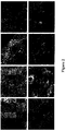

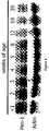

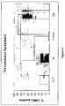

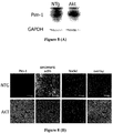

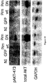


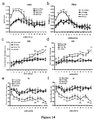
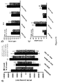
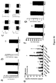

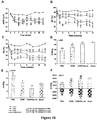
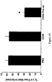

Classifications
-
- A—HUMAN NECESSITIES
- A61—MEDICAL OR VETERINARY SCIENCE; HYGIENE
- A61K—PREPARATIONS FOR MEDICAL, DENTAL OR TOILETRY PURPOSES
- A61K38/00—Medicinal preparations containing peptides
- A61K38/16—Peptides having more than 20 amino acids; Gastrins; Somatostatins; Melanotropins; Derivatives thereof
- A61K38/43—Enzymes; Proenzymes; Derivatives thereof
- A61K38/45—Transferases (2)
-
- A—HUMAN NECESSITIES
- A61—MEDICAL OR VETERINARY SCIENCE; HYGIENE
- A61K—PREPARATIONS FOR MEDICAL, DENTAL OR TOILETRY PURPOSES
- A61K35/00—Medicinal preparations containing materials or reaction products thereof with undetermined constitution
- A61K35/12—Materials from mammals; Compositions comprising non-specified tissues or cells; Compositions comprising non-embryonic stem cells; Genetically modified cells
- A61K35/28—Bone marrow; Haematopoietic stem cells; Mesenchymal stem cells of any origin, e.g. adipose-derived stem cells
-
- A—HUMAN NECESSITIES
- A61—MEDICAL OR VETERINARY SCIENCE; HYGIENE
- A61P—SPECIFIC THERAPEUTIC ACTIVITY OF CHEMICAL COMPOUNDS OR MEDICINAL PREPARATIONS
- A61P31/00—Antiinfectives, i.e. antibiotics, antiseptics, chemotherapeutics
- A61P31/04—Antibacterial agents
-
- A—HUMAN NECESSITIES
- A61—MEDICAL OR VETERINARY SCIENCE; HYGIENE
- A61P—SPECIFIC THERAPEUTIC ACTIVITY OF CHEMICAL COMPOUNDS OR MEDICINAL PREPARATIONS
- A61P31/00—Antiinfectives, i.e. antibiotics, antiseptics, chemotherapeutics
- A61P31/12—Antivirals
-
- A—HUMAN NECESSITIES
- A61—MEDICAL OR VETERINARY SCIENCE; HYGIENE
- A61P—SPECIFIC THERAPEUTIC ACTIVITY OF CHEMICAL COMPOUNDS OR MEDICINAL PREPARATIONS
- A61P43/00—Drugs for specific purposes, not provided for in groups A61P1/00-A61P41/00
-
- A—HUMAN NECESSITIES
- A61—MEDICAL OR VETERINARY SCIENCE; HYGIENE
- A61P—SPECIFIC THERAPEUTIC ACTIVITY OF CHEMICAL COMPOUNDS OR MEDICINAL PREPARATIONS
- A61P9/00—Drugs for disorders of the cardiovascular system
-
- A—HUMAN NECESSITIES
- A61—MEDICAL OR VETERINARY SCIENCE; HYGIENE
- A61P—SPECIFIC THERAPEUTIC ACTIVITY OF CHEMICAL COMPOUNDS OR MEDICINAL PREPARATIONS
- A61P9/00—Drugs for disorders of the cardiovascular system
- A61P9/04—Inotropic agents, i.e. stimulants of cardiac contraction; Drugs for heart failure
-
- A—HUMAN NECESSITIES
- A61—MEDICAL OR VETERINARY SCIENCE; HYGIENE
- A61P—SPECIFIC THERAPEUTIC ACTIVITY OF CHEMICAL COMPOUNDS OR MEDICINAL PREPARATIONS
- A61P9/00—Drugs for disorders of the cardiovascular system
- A61P9/10—Drugs for disorders of the cardiovascular system for treating ischaemic or atherosclerotic diseases, e.g. antianginal drugs, coronary vasodilators, drugs for myocardial infarction, retinopathy, cerebrovascula insufficiency, renal arteriosclerosis
-
- C—CHEMISTRY; METALLURGY
- C12—BIOCHEMISTRY; BEER; SPIRITS; WINE; VINEGAR; MICROBIOLOGY; ENZYMOLOGY; MUTATION OR GENETIC ENGINEERING
- C12N—MICROORGANISMS OR ENZYMES; COMPOSITIONS THEREOF; PROPAGATING, PRESERVING, OR MAINTAINING MICROORGANISMS; MUTATION OR GENETIC ENGINEERING; CULTURE MEDIA
- C12N15/00—Mutation or genetic engineering; DNA or RNA concerning genetic engineering, vectors, e.g. plasmids, or their isolation, preparation or purification; Use of hosts therefor
- C12N15/09—Recombinant DNA-technology
- C12N15/63—Introduction of foreign genetic material using vectors; Vectors; Use of hosts therefor; Regulation of expression
- C12N15/79—Vectors or expression systems specially adapted for eukaryotic hosts
- C12N15/85—Vectors or expression systems specially adapted for eukaryotic hosts for animal cells
- C12N15/86—Viral vectors
-
- C—CHEMISTRY; METALLURGY
- C12—BIOCHEMISTRY; BEER; SPIRITS; WINE; VINEGAR; MICROBIOLOGY; ENZYMOLOGY; MUTATION OR GENETIC ENGINEERING
- C12N—MICROORGANISMS OR ENZYMES; COMPOSITIONS THEREOF; PROPAGATING, PRESERVING, OR MAINTAINING MICROORGANISMS; MUTATION OR GENETIC ENGINEERING; CULTURE MEDIA
- C12N9/00—Enzymes; Proenzymes; Compositions thereof; Processes for preparing, activating, inhibiting, separating or purifying enzymes
- C12N9/10—Transferases (2.)
- C12N9/12—Transferases (2.) transferring phosphorus containing groups, e.g. kinases (2.7)
-
- C—CHEMISTRY; METALLURGY
- C12—BIOCHEMISTRY; BEER; SPIRITS; WINE; VINEGAR; MICROBIOLOGY; ENZYMOLOGY; MUTATION OR GENETIC ENGINEERING
- C12Y—ENZYMES
- C12Y207/00—Transferases transferring phosphorus-containing groups (2.7)
- C12Y207/11—Protein-serine/threonine kinases (2.7.11)
-
- C—CHEMISTRY; METALLURGY
- C12—BIOCHEMISTRY; BEER; SPIRITS; WINE; VINEGAR; MICROBIOLOGY; ENZYMOLOGY; MUTATION OR GENETIC ENGINEERING
- C12Y—ENZYMES
- C12Y207/00—Transferases transferring phosphorus-containing groups (2.7)
- C12Y207/12—Dual-specificity kinases (2.7.12)
-
- A—HUMAN NECESSITIES
- A61—MEDICAL OR VETERINARY SCIENCE; HYGIENE
- A61K—PREPARATIONS FOR MEDICAL, DENTAL OR TOILETRY PURPOSES
- A61K35/00—Medicinal preparations containing materials or reaction products thereof with undetermined constitution
- A61K35/12—Materials from mammals; Compositions comprising non-specified tissues or cells; Compositions comprising non-embryonic stem cells; Genetically modified cells
- A61K2035/124—Materials from mammals; Compositions comprising non-specified tissues or cells; Compositions comprising non-embryonic stem cells; Genetically modified cells the cells being hematopoietic, bone marrow derived or blood cells
Definitions
- This invention generally relates to cardiac cells comprising Pim-1 expression constructs, and to the use thereof in treating cardiac disease or injury.
- Intracellular molecular signaling networks communicate via kinases that phosphorylate target substrates to regulate critical aspects of growth and survival.
- PIM-1PIM- a proto-oncogenic serine/threonine kinase
- PIM-1 is up-regulated in prostate cancer. The gene is highly expressed in the liver and spleen during fetal hematopoiesis and primarily in B-lymphoid and myeloid cell lines.
- PIM-1 exists in two isoforms with molecular weights of 34 and 44kDa.
- the 34kDa isoform is cytosolic and nuclear localized, while the 44kDa isoform was recently found to be membrane bound.
- PIM-1 may be a relatively promiscuous kinase based upon minimal target substrate recognition sequence requirements and capacity for autophosphorylation.
- Two additional family members, PIM-2 and PIM-3, may exhibit functional redundancy with PIM-1.
- Induction of PIM-1 expression is mediated by cytokines and growth factors including LIF, GM-CSF, EGF, and most interleukins, consistent with a role for PIM-1 in proliferation and survival of hematopoeitic cells.
- PIM-1 mediates proliferative actions through phosphorylation of multiple target substrates, resulting in cell cycle transition, as well as protective effects via phosphorylation of multiple targets.
- Induction of PIM-1 expression has been linked to AKT (a serine/threonine kinase) in hematopoeitic cells.
- ILK Integrin Linked Kinase
- the present invention provides a cardiac cell comprising a PIM-1 encoding nucleic acid, wherein the PIM-1 encoding nucleic acid is provided in an expression construct or expression vehicle comprising a plasmid, a recombinant virus or an artificial chromosome, and wherein the cardiac cell is a myocyte or a cardiac stem cell.
- the present invention provides a pharmaceutical composition comprising the cell defined above.
- the present invention provides a cell or pharmaceutical composition as defined above for use in a therapeutic method of preventing or treating damage in a cardiac cell, tissue or organ subsequent to cellular, tissue and/or organ hypoxia, hypoxaemia or anoxia, or subsequent to pressure-overload induced hypertrophy or heart failure; or wherein the damage is a result of a hypertrophic aged, failing or an ischemic myocardium, or a myocardium damaged by inflammation, infection, chronic stress, diabetes or alcoholism.
- the present description includes the discovery of new roles for PIM-1, its isoforms, and other PIM enzymes having equivalent or overlapping targets and substrates. Specifically, these enzymes have a role in cardiac and other circulatory system protection, survival, repair, regeneration, and recovery, and in the implantation, differentiation, function, and survival of stem cells, progenitor cells, or differentiated cells introduced into circulatory system tissues. These discoveries form the basis for new cardiac therapies, including repair of damaged heart tissue and implantation, expansion, and survival of implanted stem cells or progenitor cells that differentiate into functional heart tissue. Prior to this invention, enhancement of PIM activity was not known to have any prophylactic or therapeutic utility in heart tissue, heart cells, or in other circulatory system cells or tissues.
- PIM serine or threonine kinase
- PIM-1 the various PIM enzymes
- PIM-2 the various PIM enzymes
- PIM-3 the various PIM enzymes
- PIM-3 the various PIM enzymes
- the serine/threonine kinase PIM-1 is known to exist in two isoforms, and references to PIM and PIM-1 herein are intended to encompass both isoforms, unless otherwise specified.
- PIM activity and PIM kinase activity refer to the enzymatic or physiological activity of the PIM enzymes, e.g., the activity of a PIM-1, and encompasses use of other materials having similar activity.
- the discoveries set forth herein relate to altering characteristics of living cells by enhancing a particular kinase activity in the cells.
- enzyme variants exist or can be readily constructed, having conservative amino acid substitutions, cross-linking, cross-species domain substitutions, truncations, and the like, while preserving a physiologically-effective level of enzymatic activity (in this case, kinase activity for the PIM-1 target).
- the present discoveries are not focused only on a particular kinase, but include the discovery of an entirely new role for PIM kinase activity in vascular system cells and tissues.
- the results discussed herein flow from alteration of PIM kinase activity, regardless of the exact modality by which that is achieved.
- vascular system refers to the blood vessels and the heart, and all the tissues and cells of which they are comprised, including cardiac smooth muscle, cardiomyocytes, cardiomyoblasts, vascular wall, endothelium, vascular smooth muscle, vascular connective tissue, and other known cells and tissues of the vascular system.
- stem cell is used broadly to include totipotent, pluripotent, and multipotent cells that can differentiate into vascular system cells, including cardiac cells.
- Progenitor cells overlaps somewhat with multipotent stem cells, and includes cells that are at least partially differentiated but that are multipotent or unipotent, in that they have the ability to differentiate into at least one type of vascular system cells.
- treat and “treatment” are used broadly, to include both prophylactic and therapeutic treatments.
- disease or injury of circulatory system tissues those terms are used broadly to include fully developed disease or injury, as well as incipient or threatened disease or injury.
- a patient at risk of or beginning to develop a particular condition is considered to have that condition "treated” when methods as disclosed herein are used to reduce the risk of development or progression of that condition, as well as when an already-developed condition is reversed, inhibited, cured, or ameliorated, and when the rate of development of a condition is halted or slowed.
- Treatments identified as useful for one category are also useful for other categories, and selection of a particular term (other than "human”) is not intended to be limiting, but rather just a use of an alternative expression.
- compositions such as pharmaceutical compositions, as defined in the appended claims.
- the description further provides methods for inducing cardiac or vascular cellular proliferation, and protecting cardiac or vascular cells from hypoxia and cellular apoptosis using the compositions defined herein.
- the compositions of the invention may be used to express PIM-1 to protect cardiomyocytes from hypertrophy and inhibit myocardial apoptosis induced by infarction, reducing infarct size.
- compositions of the invention may be used to express PIM-1 to induce cardiac or vascular cellular dedifferentiation and re-expression of stem cell markers; and in one aspect, to overexpress PIM-1 to enhance the regenerative potential of stem cells, including stem cell ability to engraft in the heart after a myocardial infarction (post-MI).
- compositions of the invention may be used to express PIM-1 to increase BcI-XL expression to induce cardioprotective anti-apoptotic signaling, thus increasing myocardial survival signaling.
- compositions such as pharmaceutical compositions, comprising nucleic acids encoding the serine/threonine kinase PIM-1 for use in preventing or inhibiting cell or tissue damage, e.g., cardiomyocyte cell death or inhibiting an ischemic or reperfusion related injury; including preventing or inhibiting the irreversible cellular and tissue damage and cell death caused by ischemia, e.g., ischemia subsequent to reperfusion (which can exacerbates ischemic damage by activating inflammatory response and oxidative stress).
- the disclosure further provides compositions, such as pharmaceutical compositions, comprising nucleic acids encoding a serine/threonine kinase PIM which are suitable for regulating cardiac or vascular cellular proliferation and survival.
- One aspect of the description relates to a method, comprising identifying a patient in need of enhanced PIM activity in a vascular system tissue, and enhancing levels of PIM in vascular system tissue of the patient to alter a functional characteristic of cells in that tissue.
- the patient has experienced cardiac injury and the enhanced PIM levels facilitate cardiac regeneration to repair that injury.
- the enhancing step may advantageously comprise enhancing endogenous production of PIM in the vascular system tissue.
- it may comprise administering to the patient an exogenous PIM.
- the exogenous PIM may comprise PIM-1, for example, or another material sharing that same function, and may comprise a PIM enzyme in association with a cellular delivery moiety, such as a translocation domain that is attached to the PIM enzyme.
- the enhancing step comprises administering cells to the patient that produce enhanced levels of PIM.
- the administered cells may be cardiac stem cells or vascular system progenitor cells.
- the administered cells comprise a PIM-encoding polynucleotide operatively linked to a non-PIM promoter.
- the enhancing step comprises administering cells to vascular tissue of the patient, and expressing enhanced levels of PIM from the administered cells.
- a different aspect of the description comprises PIM-delivering or enhancing material for treatment of vascular system disease or injury.
- This material can be, for example, a PIM enzyme linked to a cellular delivery agent, or a cell as defined by the claims.
- the cell is a cardiac stem cell, and the alteration comprises a PIM-encoding polynucleotide under control of a non-PIM promoter.
- the promoter may be a cardiac-specific promoter, an inducible promoter, an endogenous promoter, an exogenous promoter, or a constituitive promoter.
- the PIM-enhancer may be an inducer of endogenous PIM expression.
- Yet another aspect of the description is use of a PIM-delivering or enhancing material in the preparation of a medicament for treating vascular system disease or injury.
- compositions comprising a vascular system cell or a cell that is differentiatable into a vascular system cell, where the cell comprises a PIM-encoding polynucleotide sequence operably linked to a non-PIM promoter.
- the cell is a myocyte or a cardiac stem cell.
- the promoter is an inducible promoter or a cardiac-specific promoter.
- the description further provides a method for treating cardiac disease or injury, comprising enhancing levels of PIM within diseased or injured cardiac tissue.
- the cardiac disease or injury may include ischemic injury, hypoxic injury, myocardial infarction, traumatic cardiac injury, cardiac hypertrophy, overpressure injury, congestive heart failure, apoptosis-inducing injury or disease, bacterial infection, viral infection, and conditions that create an enhanced risk of any of the foregoing.
- composition formulated for administration to heart muscle comprising:
- Still other aspects of the description include methods for treating, preventing or ameliorating a disease or condition comprising administering to an individual in need thereof a pharmaceutical compound of the invention, a liposome of the description, or a nanoparticle of the description, or any combination thereof, wherein the treatment, prevention and/or amelioration of the disease or condition comprises:
- the enhanced PIM activity may be provided by the cells and/or compositions of the present invention as defined herein.
- the care provider may wish to perform a patient selection step. This may include, for example, assessing whether a patient is in need of one or more of the various treatments, or identifying a patient in need of such treatment. Two significant categories of need warrant some discussion.
- identification can take place by recognizing an inchoate disease or injury that would otherwise progress, for example, injury or other factors that have or will initiate apoptosis, or conditions or factors that enhance the risk of developing a particular condition. Identification and treatment of those individuals may be desirable to prevent development of a disease or injury or to slow its development.
- Prophylactic treatment is desirable, when high risk of ischemic injury can be identified. However, in many cases, the patient will be treated after the injury has occurred. Treatment should be commenced as soon as is practicable after the injury.
- PIM-activity enhancement can be used to treat a number of other conditions and to create desired physiological effects, by treating a subject to enhance PIM activity in vascular, cardiac, or other circulatory system cells or tissues.
- These include prevention, reduction, or reversal of cardiac hypertrophy, including but not limited to maladaptive hypertrophic remodeling; promoting cardiac cell survival and inhibiting apoptosis of those cells; enhancing cardiac contractility; improving cardiac ejection fraction; enhancing vascular growth and repair; and promoting differentiation of stem cells and progenitor cells toward cardiac or vascular tissue.
- the uses contemplated herein include, but are not limited to, inhibition of cardiac apoptosis; inhibition of cardiac fibrosis; inhibition of cardiac remodeling; inhibition of cardiac hypertrophy; preservation or reduced loss of ejection fraction in damaged hearts; enhanced preservation of contractile function; decrease in cardiac necrosis; reduction in lesion size following ischemic injury; and increasing cardiac cellularity and decreasing myocyte volume. All of these uses can be practiced prophylactically (to prevent or reduce a particular condition that would otherwise be likely to occur) and therapeutically (to treat a condition that is already in existence, including treatment to slow progression of a condition).
- conditions that may lead to treatment by enhancement of PIM activity include (but are not limited to) congenital heart conditions; ischemic injury of any kind to heart tissue; damage from infarction; cardiac reperfusion injury; traumatic cardiac injury; congestive heart failure injury; and injury relating to cardiac infection with a pathogen, including viral, bacterial, and parasitic pathogens.
- methods of treating the individual as provided by the description can include a step of increasing the level of PIM activity in a target tissue, such as vascular tissue or cardiac tissue. This step can be practiced in the various ways disclosed in the description.
- those include administering factors or drugs to the patient, systemically or locally, that upregulate endogenous PIM expression; administering PIM protein, preferably in combination with a delivery modality, such as a linked transduction domain, a liposome, an antibody, or the like; administering PIM- encoding polynucleotide to the patient, including naked DNA administration, administration of the polynucleotide in a viral vector, liposome, or other delivery modality; electroporation of cells of a subject to deliver DNA; and administering an autologous cell to the subject (e.g., into the heart) that has been altered to enhance PIM expression, including cardiomyocytes, cardiac progenitor cells, cardiac stem cells, mesenchymal stem cells, hematopoietic stem cells, adipose- derived stem cells, and the like.
- the present invention specifically encompasses cardiomyocytes and cardiac stem cells which have been altered to enhance PIM expression.
- the number of cells can be any amount effective to enhance cardiac function or structure or treat a target condition.
- Exemplary, non-limiting amounts include 10 5 to 10 10 cells, more typically 10 6 to 10 9 cells.
- Exemplary, non-limiting amounts of PIM protein administered to an adult human heart can be, for example, from about 10 4 g to about 10 -10 g, calculated as the pure PIM protein.
- Exemplary, non-limiting amounts of DNA include about 0.05 to 500 ug/kg, or 0.5 to 50 ug/kg body weight, and in the case of viral particles, formulated at a titer of about at least 10 10 , 10 11 , 10 12 , 10 13 , 10 14 , 10 15 , 10 16 , or 10 17 physical particles per milliliter.
- the PIM-1 encoding nucleic acid is administered in about 10, 20, 30, 40, 50, 60, 70, 80, 90, 100, 110, 120, 130, 140 or 150 or more microliter ( ⁇ l) injections. Doses and dosage regimens can be determined by conventional range-finding techniques known to those of ordinary skill in the art.
- about 10 6 , 10 7 , 10 8 , 10 9 , 10 11 , 10 11 , 10 12 , 10 13 , 10 14 , 10 15 , 10 16 or 10 17 viral (e.g., lentiviral) particles are delivered to the individual (e.g., a human patient) in one or multiple doses, in accordance with the therapeutic compositions for use of the present invention.
- An intracardiac single administration (e.g., a single dose) comprises from about 0.1 ⁇ l to 1.0 ⁇ l, 10 ⁇ l or to about 100 ⁇ l of a pharmaceutical composition of the invention.
- dosage ranges from about 0.5 ng or 1.0 ng to about 10 ⁇ g, 100 ⁇ g to 1000 ⁇ g of PIM-1 expressing nucleic acid is administered (either the amount in an expression construct, or as in one embodiment, naked DNA is injected), in accordance with the therapeutic compositions for use of the present invention.
- PIM-1 is limited to PIM-1.
- Some aspects of the disclosure include nucleic acid constructs comprising a PIM-encoding sequence, e.g., a PIM-1 expressing message or a PIM-1 gene.
- PIM-expressing nucleic acids used to practice this invention include PIM-1 genomic sequences, or fragments thereof, including coding or non-coding sequences, e.g., including introns, 5' or 3' non-coding sequences, and the like. Also encompassed are PIM-encoding mRNA sequences.
- the PIM-1 expressing, or PIM-1 inducing or upregulating, composition is a nucleic acid, including a vector, recombinant virus, and the like; and a recombinant PIM-1 is expressed in a cell in vitro, ex vivo and/or in vivo.
- a PIM-1 expressing nucleic acid encodes a human PIM-1, such as Genbank accession no. AAA36447 (see also, e.g., Domen (1987) Oncogene Res. 1 (1): 103-112 ), SEQ ID NO: 1.
- a PIM-1 expressing nucleic acid encodes a human PIM-1 kinase 44 kDa isoform, see e.g., Genbank accession no. AAY87461 (see also, e.g., Xie (2006) Oncogene 25 (1), 70-78 ), SEQ ID NO:2.
- a PIM-1 expressing nucleic acid comprises a human PIM-1 kinase message (mRNA), see e.g., Genbank accession no. NM_002648 (see also, e.g., Zhang (2007) MoI. Cancer Res. 5 (9), 909-922 ), SEQ ID NO:3.
- mRNA human PIM-1 kinase message
- PIM-2 SEQ ID NO:4
- PIM-3 SEQ ID NO:5
- nucleic acids are operatively linked to a transcriptional regulatory sequence, e.g., a promoter and/or an enhancer, e.g., cardiac-specific, promoters to drive (e.g., regulate) expression of Pim-1.
- a transcriptional regulatory sequence e.g., a promoter and/or an enhancer, e.g., cardiac-specific, promoters to drive (e.g., regulate) expression of Pim-1.
- Promoters and enhancers used to practice this invention can be of any type and/or origin, an in one embodiment promoters specific to the species receiving the Pim-1 construct are used; e.g., humans can receive human promoters, mice receive murine promoters, etc.
- promoters from heterologous species can be used; e.g., mammals or vertebrates receiving promoters that originate from other mammals or vertebrates, or viral or synthetic promoters active in the appropriate specie and/or cell type also can be used.
- These promoters can comprise, for example, a ⁇ -myosin heavy chain promoter; a cardiac troponin-T promoter; a MLC-2v promoter; and any other promoter that drives expression in cardiac tissue but does not drive significant expression in other tissues.
- promoters and enhancers active in primordial cells or stem cells e.g., myocardial stem cells, can be operatively linked to drive expression of Pim-1.
- this disclosure provides constructs or expression vehicles, e.g., expression cassettes, vectors, viruses (e.g., lentiviral expression vectors, e.g., see SEQ ID NO:4), and the like, comprising a PIM- encoding sequence, e.g., a PIM-1 encoding message or a PIM-1 a gene, for use as ex vivo or in vitro gene therapy vehicles, or for expression of PIM-1 in heart tissue, a cardiac or vascular cell, tissue or organ to practice the methods of this invention, and for research, drug discovery or transplantation.
- constructs or expression vehicles e.g., expression cassettes, vectors, viruses (e.g., lentiviral expression vectors, e.g., see SEQ ID NO:4), and the like, comprising a PIM- encoding sequence, e.g., a PIM-1 encoding message or a PIM-1 a gene, for use as ex vivo or in vitro gene therapy vehicles, or for
- an expression vehicle used to practice the invention can comprise a promoter operably linked to a nucleic acid encoding a PIM protein (or functional subsequence thereof).
- the invention provides expression cassettes comprising nucleic acid encoding a PIM-1 protein operably linked to a transcriptional regulatory element, e.g., a promoter.
- an expression vehicle used to practice the invention is designed to deliver a PIM-1 encoding sequence, e.g., a PIM-1 gene or any functional portion thereof to a cardiac tissue or cell of an individual.
- Expression vehicles, e.g., vectors, used to practice the invention can be non-viral or viral vectors or combinations thereof.
- the invention can use any viral vector or viral delivery system known in the art, e.g., adenoviral vectors, adeno-associated viral (AAV) vectors, herpes viral vectors (e.g., herpes simplex virus (HSV)-based vectors), retroviral vectors, lentiviral vectors and baculoviral vectors.
- adenoviral vectors e.g., adeno-associated viral (AAV) vectors
- herpes viral vectors e.g., herpes simplex virus (HSV)-based vectors
- retroviral vectors e.g., lentiviral vectors and
- an expression vehicle e.g., a vector or a virus
- a retroviral e.g., a lentiviral, vector capable of delivering the nucleotide sequence encoding full-length human PIM-1 in vitro, ex vivo and/or in vivo.
- An exemplary lentiviral expression vector backbone (no "payload” included, e.g., no PIM-1 sequence included) that can be used to practice this invention is set forth in SEQ ID NO:4.
- a lentiviral vector used to practice this invention is a "minimal" lentiviral production system lacking one or more viral accessory (or auxiliary) gene.
- Exemplary lentiviral vectors for use in the invention can have enhanced safety profiles in that they are replication defective and self-inactivating (SIN) lentiviral vectors.
- Lentiviral vectors and production systems that can be used to practice this invention include e.g., those described in U.S. Patent Nos.
- non-integrating lentiviral vectors can be employed in the practice of the invention.
- non-integrating lentiviral vectors and production systems that can be employed in the practice of the invention include those described in USPN 6,808,923.
- the expression vehicle can be designed from any vehicle known in the art, e.g., a recombinant adeno-associated viral vector as described, e.g., in U.S. Pat. App. Pub. No. 20020194630, Manning, et al .; or a lentiviral gene therapy vector, e.g., as described by e.g., Dull, et al. (1998) J. Virol. 72:8463-8471 ; or a viral vector particle, e.g., a modified retrovirus having a modified proviral RNA genome, as described, e.g., in U.S. Pat. App. Pub. No.
- adeno-associated viral vector as described e.g., in USPN 6,943,153 , describing recombinant adeno-associated viral vectors for use in the eye; or a retroviral or a lentiviral vector as described in USPNs 7,198,950 ; 7,160,727 ; 7,122,181 (describing using a retrovirus to inhibit intraocular neovascularization in an individual having an age-related macular degeneration); or 6,555,107 .
- Any viral vector can be used to practice this invention, and the concept of using viral vectors for gene therapy is well known; see e.g., Verma and Somia (1997) Nature 389:239-242 ; and Coffin et al ("Retroviruses” 1997 Cold Spring Harbour Laboratory Press Eds: JM Coffin, SM Hughes, HE Varmus pp 758-763 ) having a detailed list of retroviruses.
- Any lentiviruses belonging to the retrovirus family can be used for infecting both dividing and non-dividing cells with a PIM-1 -encoding nucleic acid, see e.g., Lewis et al (1992) EMBO J. 3053-3058 .
- Viruses from lentivirus groups from “primate” and/or “non-primate” can be used; e.g., any primate lentivirus can be used, including the human immunodeficiency virus (HIV), the causative agent of human acquired immunodeficiency syndrome (AIDS), and the simian immunodeficiency virus (SIV); or a non-primate lentiviral group member, e.g., including "slow viruses” such as a visna/maedi virus (VMV), as well as the related caprine arthritis-encephalitis virus (CAEV), equine infectious anemia virus (EIAV) and/or a feline immunodeficiency virus (FIV) or a bovine immunodeficiency virus (BIV).
- VMV visna/maedi virus
- CAEV caprine arthritis-encephalitis virus
- EIAV equine infectious anemia virus
- FV feline immunodeficiency virus
- BIV bo
- lentiviral vectors used to practice this invention are pseudotyped lentiviral vectors.
- pseudotyping used to practice this invention incorporates in at least a part of, or substituting a part of, or replacing all of, an env gene of a viral genome with a heterologous env gene, for example an env gene from another virus.
- the lentiviral vector of the invention is pseudotyped with VSV-G.
- the lentiviral vector of the invention is pseudotyped with Rabies-G.
- Lentiviral vectors used to practice this invention may be codon optimized for enhanced safety purposes. Different cells differ in their usage of particular codons. This codon bias corresponds to a bias in the relative abundance of particular tRNAs in the cell type. By altering the codons in the sequence so that they are tailored to match with the relative abundance of corresponding tRNAs, it is possible to increase expression. By the same token, it is possible to decrease expression by deliberately choosing codons for which the corresponding tRNAs are known to be rare in the particular cell type. Thus, an additional degree of translational control is available.
- the amino acid sequence coding sequence for the packaging components is retained so that the viral components encoded by the sequences remain the same, or at least sufficiently similar that the function of the packaging components is not compromised.
- Codon optimization also overcomes the Rev/RRE requirement for export, rendering optimized sequences Rev independent. Codon optimization also reduces homologous recombination between different constructs within the vector system (for example between the regions of overlap in the gag-pol and env open reading frames). The overall effect of codon optimization is therefore a notable increase in viral titer and improved safety.
- the strategy for codon optimized gag-pol sequences can be used in relation to any retrovirus.
- Vectors, recombinant viruses, and other expression systems used to practice this invention can comprise any nucleic acid which can infect, transfect, transiently or permanently transduce a cell.
- a vector used to practice this invention can be a naked nucleic acid, or a nucleic acid complexed with protein or lipid.
- a vector used to practice this invention comprises viral or bacterial nucleic acids and/or proteins, and/or membranes (e.g., a cell membrane, a viral lipid envelope, etc.).
- expression systems used to practice this invention comprise replicons (e.g., RNA replicons, bacteriophages) to which fragments of DNA may be attached and become replicated.
- expression systems used to practice this invention include, but are not limited to RNA, autonomous self-replicating circular or linear DNA or RNA (e.g., plasmids, viruses, and the like, see, e.g., U.S. Patent No. 5,217,879 ), and include both the expression and non-expression plasmids.
- a recombinant microorganism or cell culture used to practice this invention can comprise "expression vector" including both (or either) extra-chromosomal circular and/or linear nucleic acid (DNA or RNA) that has been incorporated into the host chromosome(s).
- expression vector including both (or either) extra-chromosomal circular and/or linear nucleic acid (DNA or RNA) that has been incorporated into the host chromosome(s).
- DNA or RNA linear nucleic acid
- the vector may either be stably replicated by the cells during mitosis as an autonomous structure, or is incorporated within the host's genome.
- an expression system used to practice this invention can comprise any plasmid, which are commercially available, publicly available on an unrestricted basis, or can be constructed from available plasmids in accord with published procedures. Plasmids that can be used to practice this invention are well known in the art.
- a vector used to make or practice the invention can be chosen from any number of suitable vectors known to those skilled in the art, including cosmids, YACs (Yeast Artificial Chromosomes), megaYACS, BACs (Bacterial Artificial Chromosomes), PACs (Pl Artificial Chromosome), MACs (Mammalian Artificial Chromosomes), a whole chromosome, or a small whole genome.
- the vector also can be in the form of a plasmid, a viral particle, or a phage.
- vectors include chromosomal, non-chromosomal and synthetic DNA sequences, derivatives of SV40; bacterial plasmids, phage DNA, baculovirus, yeast plasmids, vectors derived from combinations of plasmids and phage DNA, viral DNA such as vaccinia, adenovirus, fowl pox virus, and pseudorabies.
- cloning and expression vectors for use with prokaryotic and eukaryotic hosts are described by, e.g., Sambrook.
- Bacterial vectors which can be used include commercially available plasmids comprising genetic elements of known cloning vectors.
- compositions including pharmaceutical compositions, for expressing PIM-1 as defined in the claims.
- compositions are suitable for protecting cardiomyocytes from hypertrophy and inhibiting myocardial apoptosis induced by infarction, and reducing infarct size. (Functional equivalence is considered to exist based on ability to act on the same substrate and produce the same product, and does not require identical kinetics.)
- the pharmaceutical compositions of the invention are used to express PIM-1, and are suitable for inducing cardiac or vascular cellular dedifferentiation and re-expression of stem cell markers; and in one aspect, to overexpress PIM-1 for enhancing the regenerative potential of stem cells, including stem cell ability to engraft in the heart after a myocardial infarction (post-Mi).
- the PIM-1 expressing, composition is a nucleic acid, including a vector, recombinant virus, and the like; and a recombinant PIM-1 is expressed in a cell in vitro, or ex-vivo .
- the cells are limited to myocytes and cardiac stem cells.
- compounds that induce or upregulate PIM nucleic acid or a PIM kinase activity in the heart or a cardiac or vascular cell, tissue or organ are administered.
- compounds that can be administered in practicing use of the pharmaceutical compositions of this invention can comprise: an interleukin, a cytokine and/or a paracrine factor involved in survival and/or proliferative signaling; an up-regulator of AKT serine/threonine kinase; insulin-like growth factor- 1 (IGF-I); insulin; leukemia inhibitory factor (LIF); granulocyte-macrophage colony-stimulating factor (GM-CSF); or epidermal growth factor (EGF).
- an interleukin a cytokine and/or a paracrine factor involved in survival and/or proliferative signaling
- an up-regulator of AKT serine/threonine kinase insulin-like growth
- Okadaic acid and SV40 small T antigen inhibit or block negative regulation of PIM-1 by protein phosphatase 2A, and can thus be used to increase PIM-1 levels. See Maj, et al, Oncogene 26(35):5145-53 (2007 ).
- the PIM-expressing compositions of the invention are formulated with a pharmaceutically acceptable carrier.
- the pharmaceutical compositions of the invention can be administered parenterally, topically, orally or by local administration, such as by aerosol or transdermally.
- compositions can be formulated in any way and can be administered in a variety of unit dosage forms depending upon the condition or disease and the degree of illness, the general medical condition of each patient, the resulting preferred method of administration and the like. Details on techniques for formulation and administration are well described in the scientific and patent literature, see, e.g., the latest edition of Remington's Pharmaceutical Sciences, Maack Publishing Co, Easton PA ("Remington's ").
- Therapeutic agents of the invention can be administered alone or as a component of a pharmaceutical formulation (composition).
- the compounds may be formulated for administration in any convenient way for use in human or veterinary medicine.
- Wetting agents, emulsifiers and lubricants, such as sodium lauryl sulfate and magnesium stearate, as well as coloring agents, release agents, coating agents, sweetening, flavoring and perfuming agents, preservatives and antioxidants can also be present in the compositions.
- Formulations of the PIM-expressing compositions of the invention include those suitable for systemic administration, direct local vascular or cardiac administration, or alternatively oral/ nasal, topical, parenteral, rectal, and/or intravaginal administration.
- the formulations may conveniently be presented in unit dosage form and may be prepared by any methods well known in the art of pharmacy.
- the amount of active ingredient which can be combined with a carrier material to produce a single dosage form will vary depending upon the host being treated, the particular mode of administration.
- the amount of active ingredient which can be combined with a carrier material to produce a single dosage form will generally be that amount of the compound which produces a therapeutic effect.
- compositions of this invention may comprise one or more diluents, emulsifiers, preservatives, buffers, excipients, etc. and may be provided in such forms as liquids, powders, emulsions, lyophilized powders, sprays, creams, lotions, controlled release formulations, tablets, pills, gels, on patches, in implants, etc.
- compositions for oral administration can be formulated using pharmaceutically acceptable carriers well known in the art in appropriate and suitable dosages. Such carriers enable the pharmaceuticals to be formulated in unit dosage forms as tablets, pills, powder, dragees, capsules, liquids, lozenges, gels, syrups, slurries, suspensions, etc., suitable for ingestion by the patient.
- Pharmaceutical preparations for oral use can be formulated as a solid excipient, optionally grinding a resulting mixture, and processing the mixture of granules, after adding suitable additional compounds, if desired, to obtain tablets or dragee cores.
- Suitable solid excipients are carbohydrate or protein fillers include, e.g., sugars, including lactose, sucrose, mannitol, or sorbitol; starch from corn, wheat, rice, potato, or other plants; cellulose such as methyl cellulose, hydroxypropylmethyl-cellulose, or sodium carboxymethylcellulose; and gums including arabic and tragacanth; and proteins, e.g., gelatin and collagen.
- Disintegrating or solubilizing agents may be added, such as the cross-linked polyvinyl pyrrolidone, agar, alginic acid, or a salt thereof, such as sodium alginate.
- Dragee cores are provided with suitable coatings such as concentrated sugar solutions, which may also contain gum arabic, talc, polyvinylpyrrolidone, carbopol gel, polyethylene glycol, and/or titanium dioxide, lacquer solutions, and suitable organic solvents or solvent mixtures.
- Dyestuffs or pigments may be added to the tablets or dragee coatings for product identification or to characterize the quantity of active compound (i.e., dosage).
- Pharmaceutical preparations of the invention can also be used orally using, e.g., push- fit capsules made of gelatin, as well as soft, sealed capsules made of gelatin and a coating such as glycerol or sorbitol.
- Push-fit capsules can contain active agents mixed with a filler or binders such as lactose or starches, lubricants such as talc or magnesium stearate, and, optionally, stabilizers.
- the active agents can be dissolved or suspended in suitable liquids, such as fatty oils, liquid paraffin, or liquid polyethylene glycol with or without stabilizers.
- Aqueous suspensions can contain an active agent (e.g., a chimeric polypeptide or peptidomimetic of the invention) in admixture with excipients suitable for the manufacture of aqueous suspensions.
- excipients include a suspending agent, such as sodium carboxymethylcellulose, methylcellulose, hydroxypropylmethylcellulose, sodium alginate, polyvinylpyrrolidone, gum tragacanth and gum acacia, and dispersing or wetting agents such as a naturally occurring phosphatide (e.g., lecithin), a condensation product of an alkylene oxide with a fatty acid (e.g., polyoxyethylene stearate), a condensation product of ethylene oxide with a long chain aliphatic alcohol (e.g., heptadecaethylene oxycetanol), a condensation product of ethylene oxide with a partial ester derived from a fatty acid and a hexitol (e.g., polyoxyethylene
- the aqueous suspension can also contain one or more preservatives such as ethyl or n-propyl p- hydroxybenzoate, one or more coloring agents, one or more flavoring agents and one or more sweetening agents, such as sucrose, aspartame or saccharin.
- preservatives such as ethyl or n-propyl p- hydroxybenzoate
- coloring agents such as a coloring agent
- flavoring agents such as aqueous suspension
- sweetening agents such as sucrose, aspartame or saccharin.
- Formulations can be adjusted for osmolarity.
- Oil-based pharmaceuticals can be used to deliver PIM-1 expressing, or PIM-1 inducing or upregulating, compositions of the invention.
- Oil-based suspensions can be formulated by suspending an active agent in a vegetable oil, such as arachis oil, olive oil, sesame oil or coconut oil, or in a mineral oil such as liquid paraffin; or a mixture of these. See e.g., U.S. Patent No. 5,716,928 describing using essential oils or essential oil components for increasing bioavailability and reducing inter- and intra-individual variability of orally administered hydrophobic pharmaceutical compounds (see also U.S. Patent No. 5,858,401 ).
- the oil suspensions can contain a thickening agent, such as beeswax, hard paraffin or cetyl alcohol.
- Sweetening agents can be added to provide a palatable oral preparation, such as glycerol, sorbitol or sucrose. These formulations can be preserved by the addition of an antioxidant such as ascorbic acid.
- an injectable oil vehicle see Minto (1997) J. Pharmacol. Exp. Ther. 281:93-102 .
- the pharmaceutical formulations of the invention can also be in the form of oil-in- water emulsions.
- the oily phase can be a vegetable oil or a mineral oil, described above, or a mixture of these.
- Suitable emulsifying agents include naturally-occurring gums, such as gum acacia and gum tragacanth, naturally occurring phosphatides, such as soybean lecithin, esters or partial esters derived from fatty acids and hexitol anhydrides, such as sorbitan mono-oleate, and condensation products of these partial esters with ethylene oxide, such as polyoxyethylene sorbitan mono-oleate.
- the emulsion can also contain sweetening agents and flavoring agents, as in the formulation of syrups and elixirs. Such formulations can also contain a demulcent, a preservative, or a coloring agent.
- the pharmaceutical compounds can also be administered by in intranasal, intraocular and intravaginal routes including suppositories, insufflation, powders and aerosol formulations (for examples of steroid inhalants, see Rohatagi (1995) J. Clin. Pharmacol. 35:1187-1193 ; Tjwa (1995) Ann. Allergy Asthma Immunol. 75:107-111 ).
- Suppositories formulations can be prepared by mixing the drug with a suitable non-irritating excipient which is solid at ordinary temperatures but liquid at body temperatures and will therefore melt in the body to release the drug.
- suitable non-irritating excipient which is solid at ordinary temperatures but liquid at body temperatures and will therefore melt in the body to release the drug.
- Such materials are cocoa butter and polyethylene glycols.
- the pharmaceutical compounds can be delivered by transdermally, by a topical route, formulated as applicator sticks, solutions, suspensions, emulsions, gels, creams, ointments, pastes, jellies, paints, powders, and aerosols.
- the pharmaceutical compounds can also be delivered as microspheres for slow release in the body.
- microspheres can be administered via intradermal injection of drug which slowly release subcutaneously; see Rao (1995) J. Biomater Sci. Polym. Ed. 7:623-645 ; as biodegradable and injectable gel formulations, see, e.g., Gao (1995) Pharm. Res. 12:857-863 (1995 ); or, as microspheres for oral administration, see, e.g., Eyles (1997) J. Pharm. Pharmacol. 49:669-674 .
- the pharmaceutical compounds can be parenterally administered, such as by intravenous (IV) administration or administration into a body cavity or lumen of the heart.
- IV intravenous
- catheters that temporarily block flow of blood from the heart while incubating the stem cells or a viral construct in heart tissue can be used, as well as recirculation systems of well-known type that isolate the circulation in all or a part of the heart to increase the dwell time of an introduced agent (e.g., stem cell, construct, naked DNA, PIM protein, viral or other vector) in the heart.
- These formulations can comprise a solution of active agent dissolved in a pharmaceutically acceptable carrier.
- Acceptable vehicles and solvents that can be employed are water and Ringer's solution, an isotonic sodium chloride.
- sterile fixed oils can be employed as a solvent or suspending medium.
- any bland fixed oil can be employed including synthetic mono- or diglycerides.
- fatty acids such as oleic acid can likewise be used in the preparation of injectables. These solutions are sterile and generally free of undesirable matter.
- These formulations may be sterilized by conventional, well known sterilization techniques.
- the formulations may contain pharmaceutically acceptable auxiliary substances as required to approximate physiological conditions such as pH adjusting and buffering agents, toxicity adjusting agents, e.g., sodium acetate, sodium chloride, potassium chloride, calcium chloride, sodium lactate and the like.
- the concentration of active agent in these formulations can vary widely, and will be selected primarily based on fluid volumes, viscosities, body weight, and the like, in accordance with the particular mode of administration selected and the patient's needs.
- the formulation can be a sterile injectable preparation, such as a sterile injectable aqueous or oleaginous suspension. This suspension can be formulated using those suitable dispersing or wetting agents and suspending agents.
- the sterile injectable preparation can also be a suspension in a nontoxic parenterally-acceptable diluent or solvent, such as a solution of 1,3-butanediol.
- the administration can be by bolus or continuous infusion (e.g., substantially uninterrupted introduction into a blood vessel for a specified period of time).
- the pharmaceutical compounds and formulations of the invention can be lyophilized.
- the invention provides a stable lyophilized formulation comprising a composition of the invention, which can be made by lyophilizing a solution comprising a pharmaceutical of the invention and a bulking agent, e.g., mannitol, trehalose, raffinose, and sucrose or mixtures thereof.
- a process for preparing a stable lyophilized formulation can include lyophilizing a solution about 2.5 mg/mL protein, about 15 mg/mL sucrose, about 19 mg/niL NaCl, and a sodium citrate buffer having a pH greater than 5.5 but less than 6.5. See, e.g., U.S. patent app. no. 20040028670 .
- compositions and formulations of the invention can be delivered by the use of liposomes (see also discussion, below).
- liposomes particularly where the liposome surface carries ligands specific for target cells, or are otherwise preferentially directed to a specific organ, one can focus the delivery of the active agent into target cells of the heart or other part of the circulatory system in vivo. See, e.g., U.S. Patent Nos. 6,063,400 ; 6,007,839 ; Al-Muhammed (1996) J. Microencapsul. 13:293-306 ; Chonn (1995) Curr. Opin. Biotechnol. 6:698-708 ; Ostro (1989) Am. J. Hosp. Pharm. 46:1576-1587 .
- compositions of the invention can be administered for prophylactic and/or therapeutic treatments.
- compositions are administered to a subject already suffering from a condition, infection or disease in an amount sufficient to cure, alleviate or partially arrest the clinical manifestations of the condition, infection or disease and its complications (a "therapeutically effective amount").
- pharmaceutical compositions of the invention are administered in an amount sufficient to treat, prevent and/or ameliorate the deleterious effects on the heart of a myocardial infarction (post-MI); to protect cardiomyocytes from hypertrophy and to inhibit myocardial apoptosis induced by infarction, and to reduce infarct size.
- post-MI myocardial infarction
- the pharmaceutical compositions of the invention are used to express PIM-1 to induce cellular dedifferentiation and re-expression of stem cell markers; and in one aspect, to overexpress PIM-1 to enhance the regenerative potential of stem cells, including stem cell ability to engraft in the heart post-MI.
- the amount of pharmaceutical composition adequate to accomplish this is defined as a "therapeutically effective dose.”
- the dosage schedule and amounts effective for this use, i.e., the "dosing regimen” will depend upon a variety of factors, including the stage of the disease or condition, the severity of the disease or condition, the general state of the patient's health, the patient's physical status, age and the like. In calculating the dosage regimen for a patient, the mode of administration also is taken into consideration.
- the dosage regimen also takes into consideration pharmacokinetics parameters well known in the art, i.e., the active agents' rate of absorption, bioavailability, metabolism, clearance, and the like (see, e.g., Hidalgo-Aragones (1996) J. Steroid Biochem. MoI. Biol. 58:611-617 ; Groning (1996) Pharmazie 51 :337-341 ; Fotherby (1996) Contraception 54:59-69 ; Johnson (1995) J. Pharm. Sci. 84:1144-1146 ; Rohatagi (1995) Pharmazie 50:610-613 ; Brophy (1983) Eur. J. Clin. Pharmacol. 24:103-108 ; the latest Remington's, supra).
- pharmacokinetics parameters well known in the art, i.e., the active agents' rate of absorption, bioavailability, metabolism, clearance, and the like (see, e.g., Hidalgo-Aragones (1996) J. Steroid Biochem. MoI.
- formulations can be given depending on the dosage and frequency as required and tolerated by the patient.
- the formulations should provide a sufficient quantity of active agent to effectively treat, prevent or ameliorate a conditions, diseases or symptoms as described herein.
- Methods for preparing parenterally or non-parenterally administrable formulations are known or apparent to those skilled in the art and are described in more detail in such publications as Remington's.
- the methods of the invention can further comprise co-administration with other drugs or pharmaceuticals, e.g., compositions for treating heart attacks, congestive heart failure and related symptoms or conditions.
- the methods and/or compositions and formulations of the invention can be co-formulated with and/or co-administered with antibiotics (e.g., antibacterial or bacteriostatic peptides or proteins), particularly those effective against gram negative bacteria, fluids, cytokines, immunoregulatory agents, anti-inflammatory agents, complement activating agents, such as peptides or proteins comprising collagen-like domains or fibrinogen-like domains (e.g., a ficolin), carbohydrate-binding domains, and the like and combinations thereof.
- antibiotics e.g., antibacterial or bacteriostatic peptides or proteins
- cytokines cytokines
- immunoregulatory agents e.g., anti-inflammatory agents
- complement activating agents such as peptides or proteins comprising collagen-like domains or fibrin
- the description also provides nanoparticles and liposomal membranes comprising the PIM-1-expressing compounds disclosed herein which target specific molecules, including biologic molecules, such as polypeptide, including cardiac or vascular or stem cell surface polypeptides, including heart cell (e.g., myocyte) cell surface polypeptides.
- biologic molecules such as polypeptide, including cardiac or vascular or stem cell surface polypeptides, including heart cell (e.g., myocyte) cell surface polypeptides.
- the description provides nanoparticles and liposomal membranes targeting diseased and/or injured heart cells, or stem cells, such as any pluripotent cell.
- the description provides nanoparticles and liposomal membranes comprising (in addition to comprising compounds disclosed herein) molecules, e.g., peptides or antibodies, that selectively target diseased and/or injured heart cells, or stem cells.
- the description provides nanoparticles and liposomal membranes using interleukin receptors and/or other receptors to target receptors on cells, e.g., diseased and/or injured heart cells, or stem cells. See, e.g., U.S. patent application publication no. 20060239968 .
- compositions disclosed herein are specifically targeted to stem cells or heart cells, such as myocytes.
- a nanocell is formed by encapsulating a nanocore with a first agent inside a lipid vesicle containing a second agent; see, e.g., Sengupta, et al., U.S. Pat. Pub. No. 20050266067 .
- the agent in the outer lipid compartment is released first and may exert its effect before the agent in the nanocore is released.
- the nanocell delivery system may be formulated in any pharmaceutical composition for delivery to patients suffering from any disease or condition as described herein, e.g., such as congestive heart failure or heart attack (myocardial infarction).
- myocardial infarction an antibody and/or angiogenic agent can be contained in the outer lipid vesicle of the nanocell, and a composition as disclosed herein is loaded into the nanocore. This arrangement allows the antibody and/or angiogenic agent to be released first and delivered to the injured tissue.
- the description also provides multilayered liposomes comprising compounds of this invention, e.g., for transdermal absorption, e.g., as described in Park, et al., U.S. Pat. Pub. No. 20070082042 .
- the multilayered liposomes can be prepared using a mixture of oil-phase components comprising squalane, sterols, ceramides, neutral lipids or oils, fatty acids and lecithins, to about 200 to 5000 nm in particle size, to entrap a composition of this invention.
- a multilayered liposome of the description may further include an antiseptic, an antioxidant, a stabilizer, a thickener, and the like to improve stability.
- Synthetic and natural antiseptics can be used, e.g., in an amount of 0.01% to 20%.
- Antioxidants can be used, e.g., BHT, erysorbate, tocopherol, astaxanthin, vegetable fiavonoid, and derivatives thereof, or a plant-derived antioxidizing substance.
- a stabilizer can be used to stabilize liposome structure, e.g., polyols and sugars.
- Exemplary polyols include butylene glycol, polyethylene glycol, propylene glycol, dipropylene glycol and ethyl carbitol; examples of sugars are trehalose, sucrose, mannitol, sorbitol and chitosan, or a monosaccharide or an oligosaccharide, or a high molecular weight starch.
- a thickener can be used for improving the dispersion stability of constructed liposomes in water, e.g., a natural thickener or an acrylamide, or a synthetic polymeric thickener.
- Exemplary thickeners include natural polymers, such as acacia gum, xanthan gum, gellan gum, locust bean gum and starch, cellulose derivatives, such as hydroxy ethylcellulose, hydroxypropyl cellulose and carboxymethyl cellulose, synthetic polymers, such as polyacrylic acid, poly-acrylamide or polyvinylpyrollidone and polyvinylalcohol, and copolymers thereof or cross-linked materials.
- natural polymers such as acacia gum, xanthan gum, gellan gum, locust bean gum and starch
- cellulose derivatives such as hydroxy ethylcellulose, hydroxypropyl cellulose and carboxymethyl cellulose
- synthetic polymers such as polyacrylic acid, poly-acrylamide or polyvinylpyrollidone and polyvinylalcohol, and copolymers thereof or cross-linked materials.
- Liposomes can be made using any method, e.g., as described in Park, et al., U.S. Pat. Pub. No. 20070042031 , including method of producing a liposome by encapsulating a therapeutic product comprising providing an aqueous solution in a first reservoir; providing an organic lipid solution in a second reservoir, wherein one of the aqueous solution and the organic lipid solution includes a therapeutic product; mixing the aqueous solution with said organic lipid solution in a first mixing region to produce a liposome solution, wherein the organic lipid solution mixes with said aqueous solution so as to substantially instantaneously produce a liposome encapsulating the therapeutic product; and immediately thereafter mixing the liposome solution with a buffer solution to produce a diluted liposome solution.
- the description also provides nanoparticles comprising compounds disclosed herein to deliver a composition disclosed herein as a drug-containing nanoparticles (e.g., a secondary nanoparticle), as described, e.g., in U.S. Pat. Pub. No. 20070077286 .
- the description provides nanoparticles comprising a fat-soluble drug of this invention or a fat-solubilized water-soluble drug to act with a bivalent or trivalent metal salt.
- the PIM-1 expressing nucleic acid compositions of the invention are suitable for delivery for ex vivo or in vivo gene therapy to deliver a PIM-1 encoding nucleic acid.
- PIM-1 expressing nucleic acid compositions including non-reproducing viral constructs expressing high levels of the human PIM-1 protein, are delivered ex vivo or for in vivo gene therapy.
- the PIM-1 expressing nucleic acid compositions are suitable for delivery expression to and in a variety of cardiac or vascular cells to induce cellular proliferation, and/or to protect cardiac or vascular cells from hypoxia and cellular apoptosis.
- PIM-1 so expressed can protect cardiomyocytes from hypertrophy and inhibit cell death induced by myocardial infarction (e.g. heart attack), reducing the amount of muscle affected.
- PIM-1 overexpression by practicing the composition of this invention in cardiac or vascular cells, e.g., in heart cells, can result in cellular reversion; the cardiac or vascular cells become stem cell like; complete with re-expression of stem cell markers (such as cardiac stem cell markers).
- overexpression of PIM-1 is suitable for enhancing the regenerative potential of cardiac stem cells and their ability to repair a damaged or injured heart (e.g., an injured heart after a heart attack).
- the description provides compositions for overexpressing PIM-1 using a controlled system using cultured stem cells prior to reintroduction in the adult human to enhance their ability to repair the heart following injury.
- cardiac cells expressing PIM-1 for use in a clinical therapy for repair of a number of tissues damaged by low oxygen or other means through use of a conditional control element that allows control of PIM-1 expression.
- PIM-1 expressing nucleic acid delivery vehicles e.g., expression constructs, such as vectors or recombinant viruses
- expression constructs such as vectors or recombinant viruses
- expression constructs such as vectors or recombinant viruses
- vectors used to practice this invention are bicistronic.
- a MND (or, myeloproliferative sarcoma virus LTR-negative control region deleted) promoter is used to drive Pim-1 expression.
- a reporter is also used, e.g., an enhanced green florescent protein (eGFP) reporter, which can be driven off a viral internal ribosomal entry site (vIRES).
- eGFP enhanced green florescent protein
- vIRES viral internal ribosomal entry site
- all constructs are third generation self-inactivating (SIN) lentiviral vectors and incorporate several elements to ensure long-term expression of the transgene.
- a MND promoter allows for high expression of the transgene, while the LTR allows for long-term expression after repeated passage.
- the vectors also include (IFN)- ⁇ -scaffold attachment region (SAR) element; SAR elements have been shown to be important in keeping the vector transcriptionally active by inhibiting methylation and protecting the transgene from being silenced.
- PIM-1 expressing nucleic acid delivery vehicles e.g., expression constructs, such as vectors or recombinant viruses
- expression constructs such as vectors or recombinant viruses
- blood, fat, or marrow-derived stem cells can also be used.
- PIM-1 expression can be activated through addition of the drug to culture media. After a number of days in culture, the expression of PIM-1 can be then turned off through removal of the drug; and, in one aspect, the increased number of cells produced in culture are reintroduced into the damaged area contributing to an enhanced repair process.
- non- viral delivery or non- viral vector systems are known in the art, e.g., including lipid mediated transfection, liposomes, immunoliposomes, lipofectin, cationic facial amphiphiles (CFAs) and combinations thereof.
- CFAs cationic facial amphiphiles
- the description provides sterile injectable formulations comprising expression vehicles, e.g., vectors or recombinant viruses, used to practice the invention.
- target cells e.g., myocytes or stem cells
- Multiple applications of the expression construct may also be required to achieve the desired effect.
- One particular embodiment of the invention is the ex vivo modification of cardiac stem cells to enhance PIM-1 expression. According to the present description, this may be followed by administration of the stem cells to a human or other mammalian host, or to any vertebrate.
- the stem cells may be directly or locally administered, for example, into cardiac tissue in the same manner as in existing stem cell therapy for cardiac injury or insufficiency. Alternatively, systemic administration is also contemplated.
- the stem cells may be autologous stem cells or heterologous stem cells. They may be derived from infant or adult organisms.
- the enhancement of PIM-1 expression may for example be the result of upregulation of the expression of existing chromosomal PIM-1 -encoding sequence in the stem cells, or may be the result of insertion of an exogenous polynucleotide operably encoding PIM-1.
- a PIM-1 -encoding insert in such stem cells may advantageously be under inducible expression control.
- a "suicide sequence" of known type may advantageously be under inducible expression control.
- one or more "suicide sequences” are also administered, either separately or in conjunction with a nucleic acid construct of this invention, e.g., incorporated within the same nucleic acid construct (such as a vector, recombinant virus, and the like. See, e.g., Marktel S, et al., Immunologic potential of donor lymphocytes expressing a suicide gene for early immune reconstitution after hematopoietic T-cell-depleted stem cell transplantation. Blood 101 : 1290-1298(2003 ).
- Suicide sequences can be of known type, e.g., sequences to induce apoptosis or otherwise cause cell death, e.g., in one aspect, to induce apoptosis or otherwise cause cell death upon administration of an exogenous trigger compound or exposure to another type of trigger, including but not limited to light or other electromagnetic radiation exposure.
- a PIM-1 encoding nucleic acid-comprising expression construct or vehicle is formulated at an effective amount of ranging from about 0.05 to 500 ug/kg, or 0.5 to 50 ug/kg body weight, and can be administered in a single dose or in divided doses.
- the amount of a PIM-1 encoding nucleic acid of the invention, or other the active ingredient (e.g., a PIM-1 inducing or upregulating agent) actually administered ought to be determined in light of various relevant factors including the condition to be treated, the age and weight of the individual patient, and the severity of the patient's symptom; and, therefore, the above dose should not be intended to limit the scope of the invention in any way.
- a PIM-1 encoding nucleic acid-comprising expression construct or vehicle of the invention is formulated at a titer of about at least 10 10 , 10 11 , 10 12 , 10 13 , 10 14 , 10 15 , 10 16 , or 10 17 physical particles per milliliter.
- the PIM-1 encoding nucleic acid is administered in about 10, 20, 30, 40, 50, 60, 70, 80, 90, 100, 110, 120, 130, 140 or 150 or more microliter ( ⁇ l) injections. Doses and dosage regimens can be determined by conventional range- finding techniques known to those of ordinary skill in the art.
- about 10 6 , 10 7 , 10 8 , 10 9 , 10 10 , 10 11 , 10 12 , 10 13 , 10 14 , 10 15 , 10 16 or 10 17 viral (e.g., lentiviral) particles are delivered to the individual (e.g., a human patient) in one or multiple doses.
- an intracardiac single administration (e.g., a single dose) comprises from about 0.1 ⁇ l to 1.0 ⁇ l, 10 ⁇ l or to about 100 ⁇ l of a pharmaceutical composition of the invention.
- dosage ranges from about 0.5 ng or 1.0 ng to about 10 ⁇ g, 100 ⁇ g to 1000 ⁇ g of PIM-1 expressing nucleic acid is administered (either the amount in an expression construct, or as in one embodiment, naked DNA is injected). Any necessary variations in dosages and routes of administration can be determined by the ordinarily skilled artisan using routine techniques known in the art.
- a PIM-1 expressing nucleic acid is delivered in vivo directly to a heart using a viral stock in the form of an injectable preparation containing pharmaceutically acceptable carrier such as saline.
- the final titer of the vector in the injectable preparation can be in the range of between about 10 8 to 10 14 , or between about 10 10 to 10 12 , viral particles; these ranges can be effective for gene transfer.
- PIM-1 expressing nucleic acids (e.g., vector, transgene) constructs are delivered to the myocardium by direct intracoronary injection, e.g., using a standard percutaneous catheter based methods under fluoroscopic guidance.
- PIM-1 expressing nucleic acids (e.g., vector, transgene) constructs are delivered to organs and tissues, e.g., the heart, directly into both coronary and/or peripheral arteries, e.g., using a lipid-mediated gene transfer.
- the PIM-1 expressing nucleic acids e.g., vector, transgene
- the amount of the PIM-1 expressing nucleic acid (e.g., vector, transgene) injected can be in the range of between about 10 8 to 10 14 , or between about 10 10 to 10 12 , viral particles.
- the injection can be made deeply (such as 1 cm within the arterial lumen) into the lumen of the coronary arteries, and can be made in both coronary arteries, as the growth of collateral blood vessels is highly variable within individual patients.
- a therapeutic PIM-1 nucleic acid is combined with a genetic "sensor" that recognizes and responds to the oxygen deprivation that follows the reduced blood flow, or ischemia, from coronary artery disease and heart attack. As soon as the oxygen declines, the sensor turns on the therapeutic gene, thereby protecting the heart.
- the aspect of this description is useful for any condition in which circulatory system tissues are susceptible to loss of blood supply, including stroke, shock, trauma and sepsis.
- PIM proteins can also be delivered directly to the affected cardiac or other circulatory tissues. Because PIM acts intracellularly, it is preferred to utilize a delivery strategy to facilitate intracellular delivery of PIM.
- PIM in a vehicle that in taken up by or that fuses with a target cell.
- PIM can be encapsulated within a liposome or other vesicle, as described in more detail above in connection with polynucleotide delivery to cells.
- the PIM may be linked to a transduction domain, such as TAT protein.
- PIM enzyme can be operably linked to a transduction moiety, such as a synthetic or non-synthetic peptide transduction domain (PTD), Cell penetrating peptide (CPP), a cationic polymer, an antibody, a cholesterol or cholesterol derivative, a Vitamin E compound, a tocol, a tocotrienol, a tocopherol, glucose, receptor ligand or the like, to further facilitate the uptake of the PIM by cells.
- PTD synthetic or non-synthetic peptide transduction domain
- CPP Cell penetrating peptide
- a cationic polymer an antibody, a cholesterol or cholesterol derivative, a Vitamin E compound, a tocol, a tocotrienol, a tocopherol, glucose, receptor ligand or the like, to further facilitate the uptake of the PIM by cells.
- a number of protein transduction domains/peptides are known in the art and facilitate uptake of heterologous molecules linked to the transduction domains (e.g., cargo molecules). Such peptide transduction domains (PTD's) facilitate uptake through a process referred to as macropinocytosis. Macropinocytosis is a nonselective form of endocytosis that all cells perform.
- Exemplary peptide transduction domains are derived from the Drosophila homeoprotein antennapedia transcription protein (AntHD) ( Joliot et al., New Biol. 3:1121-34, 1991 ; Joliot et al, Proc. Natl. Acad. Sci. USA, 88:1864-8, 1991 ; Le Roux et al., Proc. Natl. Acad. Sci.
- the herpes simplex virus structural protein VP22 ( Elliott and O'Hare, Cell 88:223-33, 1997 ), the HIV-I transcriptional activator TAT protein (Green and Loewenstein, Cell 55:1179-1188, 1988 ; Frankel and Pabo, Cell 55:1189-1193, 1988 ), and more recently the cationic N-terminal domain of prion proteins.
- the peptide transduction domain increases uptake of the bioniolecule to which it is fused in a receptor independent fashion, is capable of transducing a wide range of cell types, and exhibits minimal or no toxicity ( Nagahara et al., Nat. Med. 4:1449-52, 1998 ).
- Peptide transduction domains have been shown to facilitate uptake of DNA (Abu-Amer, supra), antisense oligonucleotides ( Astriab-Fisher et al., Pharm. Res,19:744-54, 2002 ), small molecules ( Polyakov et al., Bioconjug. Chem. 11:762-71, 2000 ) and even inorganic 40 nanometer iron particles ( Dodd et al., J. Immunol. Methods 256:89-105, 2001 ; Wunderbaldinger et al, Bioconjug. Chem. 13:264-8, 2002 ; Lewin et al, Nat. Biotechnol. 18:410-4, 2000 ; Josephson et al, Bioconjug., Chem. 10:186-91, 1999 ).
- Fusion proteins with such trans-cellular delivery proteins can be readily constructed using known molecular biology techniques.
- any of the polynucleotides encoding PIM molecules can be linked to the foregoing domains to facilitate transduction of those polynucleotides into target cells, in vivo or in vitro.
- kits comprising compositions, including PIM-expressing, or PIM-1nducing or upregulating compositions and/or nucleic acids disclosed herein, including vectors, recombinant viruses and the like, transfecting agents, transducing agents, cardiac or vascular cells and/or cell lines, instructions (regarding the methods of the disclosure), or any combination thereof.
- compositions including PIM-expressing, or PIM-1nducing or upregulating compositions and/or nucleic acids disclosed herein, including vectors, recombinant viruses and the like, transfecting agents, transducing agents, cardiac or vascular cells and/or cell lines, instructions (regarding the methods of the disclosure), or any combination thereof.
- kits, cells, vectors and the like are provided herein.
- compositions of the invention comprising nucleic acids encoding the serine/threonine kinase PIM-1, and the methods of this invention, are effective for inducing cellular proliferation, and protecting cells from hypoxia and cellular apoptosis; and to express PIM-1 kinase to protect cardiomyocytes from hypertrophy and/or inhibit myocardial apoptosis induced by infarction, reducing infarct size; and to express PIM-1 to induce cellular dedifferentiation and re-expression of stem cell markers; and to overexpress PIM-1 to enhance the regenerative potential of stem cells, including stem cell ability to engraft in the heart after a myocardial infarction (post-MI).
- post-MI myocardial infarction
- PIM-1 functions as a defense against apoptotic stimuli induced during ischemia/ reperfusion injury resulting from myocardial infarction, pressure-overload induced hypertrophy, and heart failure.
- PIM-1 is expressed in the human myocardium and upregulated in failure.
- PIM-1 expressed in the mouse myocardium is developmentally regulated.
- PIM-1 positive border zone cardiomyocytes are negative for "terminal deoxynucleotidyl transferase-mediated dUTP-biotin nick end labeling" (TUNEL) labeling and exhibit increased Bcl-XL expression indicative of cardioprotective anti-apoptotic signaling, demonstrating a role for PIM-1 in myocardial survival signaling, also demonstrating the compositions and methods of the invention can be effective in myocardial survival signaling by expressing and/or upregulating PIM-1 kinase expression and/or activity.
- TUNEL terminal deoxynucleotidyl transferase-mediated dUTP-biotin nick end labeling
- PIM-1 induces anti-apoptotic protein expression and protects cardiomyocytes in vitro.
- GFP-tagged cDNAs for wild-type 34kDa PIM-1 (PIM-wt) or a kinase dead (K67M) mutant (PIM-DN) as previously described 10 were used to generate recombinant adenoviruses used for infection of neonatal rat cardiomyocyte cultures.
- Immunoblotting of lysates from cultures expressing GFP-PIM-wt or GFP-PIM-DN accumulate 64kDa GFP-PIM-1 fusion proteins recognized by either GFP or PIM-1 antibodies.
- Cardiomyocytes overexpressing GFP-PIM-wt exhibit a statistically significant decrease in TUNEL labeling compared to EGFP infected controls.
- GFP-PIM-DN overexpression induced a 30.8% increase in apoptotic cardiomyocytes (*p ⁇ 0.05).
- Cultured cardiomyocytes were protected from apoptotic challenge with doxorubicin or deoxyglucose by GFP-PIM-wt overexpression (**p ⁇ 0.01 for both groups), whereas GFP-PIM-DN overexpression exacerbated apoptotic effects (**p ⁇ 0.01, and *p ⁇ 0.05 respectively).
- GFP-PIM-DN induced a 3.6-fold increase in caspase3 cleavage and an 80% increase in cleaved poly (ADP-ribose) polymerase (PARP).
- PARP cleaved poly (ADP-ribose) polymerase
- GFP-PIM-wt produced significant increases in bcl-XL and bcl-2 expression (2.2-fold and 25.4-fold, respectively) when compared to control (*p ⁇ 0.01).
- GFP-PIM-wt also increased phosphorylation of Bad at the serine 112 residue (S 112) by 16.7-fold versus uninfected control while levels of total Bad remained unchanged (*p ⁇ 0.01).
- PIM-1 is induced by cardioprotective stimuli.
- compositions and methods of the invention can be used to increase the expression of PIM-1 to provide a cardioprotective effect, e.g., after a myocardial infarction.
- PIM-1 immunoreactivity was not induced by phenylephrine, endothelin-1, forskolin, or estradiol ( Fig. 4A ).
- IGF-1 induction of PIM-1 expression is AKT-dependent.
- PIM-1 expression in response to IGF-1 treatment is significantly reduced in the presence of the PI3 kinase inhibitor wortmannin or inactivated AKT (AKT-DN) by 4.0-fold and 9.1-fold respectively (**p ⁇ 0.001).
- a role for nuclear accumulation of AKT resulting following IGF stimulation 11 was confirmed, as overexpression of nuclear-targeted AKT 12 increased PIM-1 expression in cultured cardiomyocytes by 2.1-fold compared to uninfected controls (*p ⁇ 0.05).
- overexpression of wildtype AKT (AKT-wt) decreased expression of PIM-1 1.4-fold versus uninfected control (*p ⁇ 0.05).
- PIM-1 and AKT exhibit feedback relationships.
- AKT expression and phosphorylation increase in response to overexpression of GFP-PIM-DN in cultured cardiomyocytes ( Fig. 6A ). Comparable findings of increased phospho-AKT 473 were observed with confocal microscopy of immunolabeled myocardial sections from 2-month old PIM-KO mice. Increased phospho-AKT 473 and total AKT in immunoblots of whole heart lysates from PIM-KO mice correlated with the immunostaining, demonstrating that regulation of Akt expression and activity depends, in part, upon PIM-1 levels.
- compositions and methods of the invention can be used to increase the expression of PIM-1 to provide a cardioprotective effect (a myocardium protective effect), e.g., after a myocardial infarction.
- a cardioprotective effect e.g., after a myocardial infarction.
- Molecular regulation of cardioprotection endures as a highly significant research avenue for therapeutic interventional strategies in the treatment of myocardial injury and heart failure.
- this invention's discovery of a central role for PIM-1 in cardioprotection as demonstrated by the data presented herein, a new facet of signaling has been uncovered with profound implications for regulation of cardiomyocyte survival and AKT-mediated effects.
- PIM-1 functions as a defense against apoptotic stimuli induced during ischemia / reperfusion injury
- PIM-1 has not previously been studied in the myocardial context studies. In non-cardiac cells is has been demonstrated that PIM-1 as a critical regulator of proliferation and cell survival signaling, e.g., as reviewed in ref 1 (see below).
- PIM-1 functions as a defense against apoptotic stimuli induced during ischemia/reperfusion injury resulting from myocardial infarction, pressure-overload induced hypertrophy, and heart failure.
- PIM-1 level is developmentally down-regulated, expression reappears in cardiomyocytes following cardiac injury by pressure-overload induced by TAC banding and myocardial infarction.
- PIM-1 is one of several protoncogenes participating in the "immediate early response" gene profile expressed following cardiac injury 13 including c-fos, c-myc 14 , Raf and Ras. PIM-1 has been shown to cooperate with c-myc in activation of c-Myb dependent cellular proliferation in other tissues 15-17 suggesting synergistic effects between oncogenes may help preserve the myocardium in reaction to injury.
- PIM-1 potentiates intracellular anti-apoptotic signaling
- PIM-1 serves as the downstream effector of AKT induced p53 inhibition, since induction of mdm2 expression, phosphorylation, and p53 degradation is mediated by PIM-1 17 and AKT protects against doxorubicin induced apoptosis through a p53-dependent mechanism 20 . Furthermore, inactivation of PIM-1 induced apoptotic signaling in the cardiomyocyte context, exacerbating doxorubicin induced apoptosis. PIM-1 inactivation may also increase apoptotic activity through increasing generation of reactive-oxygen species and mitochondrial pore permeability, as has been found in other cellular
- IGF-1 mediates myocardial survival signaling 40-42 and stem cell proliferation 43 although we were unable to find prior published demonstration of PIM-1 induction by IGF. Results indicate that IGF-1 induced PIM-1 expression is AKT dependent and that nuclear-targeted AKT expression induces significant increases in PIM-1 expression in vitro and in vivo. Conversely, inactivation or ablation of PIM-1 expression induced AKT expression and activation, but increased AKT activation is unable to enhance recovery or reduce apoptosis following ischemia/reperfusion injury in PIM-1 null animals.
- Neonatal Rat Cardiomyocyte Cultures infections and treatments Neonatal rat cardiomyocyte cultures were prepared as described previously 12 . Cells to be subjected to treatments were placed in media with 2% serum overnight and then treated with the appropriate agent and harvested or fixed after the pre-described timepoint. Cardiomyocytes were infected with adenovirus for two hours, washed in PBS and then re-fed M199 with 2% FBS and 50 ⁇ g/ml pen/strep, and 100 ⁇ M glutamine.
- Pellets were resuspended in 1.5 mL of 0.57M STEAKMTM, homogenized in tight fitting pestle and centrifuged at 1000xg for 10 minutes at 4°C. Supernatant was collected for cytosolic fraction.
- Pellets were re-suspended in 750 ⁇ l of 0.57M STEAKMTM with 0.5% TRITON-XTM and centrifuged at 1000xg for 10 minutes at 4° C. Supernatant was collected for membrane fraction.
- Pellets were re-suspended in 300 ⁇ l of 2.3M STEAKMTM (2.3M sucrose, 25mM KCL, 5% MgCl 2 , 1mM DTT, 0.5mM PMSF, 500 ⁇ l of protease inhibitor and 50 ⁇ l of phosphatase inhibitor). 2 volumes of 0.57M STEAKMTM was added and the pellets were gently mixed.
- 2.3M STEAKMTM 2.3M sucrose, 25mM KCL, 5% MgCl 2 , 1mM DTT, 0.5mM PMSF, 500 ⁇ l of protease inhibitor and 50 ⁇ l of phosphatase inhibitor.
- a layer of 2.7M STEAKMTM (2.7M sucrose, 25mM KCL, 5% MgCl 2 , 1mM DTT, 0.5mM PMSF, 500 ⁇ l of protease inhibitor and 50 ⁇ l of phosphatase inhibitor) was made at the bottom of an ultracentrifuge tube followed by a 2.4M STEAKMTM (2.4M sucrose, 25mM KCL, 5% MgCl 2 , 1 mM DTT, 0.5 mM PMSF, 500 ⁇ l of protease inhibitor and 50 ⁇ l of phosphatase inhibitor). The homogenate layer was added. The 3 layers were centrifuged at 112,000xg for 1hour at 4° C.
- Infected cardiomyocytes were harvested 24 hours after infection in SDS denaturing sample buffer, sonicated and boiled for 10 minutes, and quantitated using the Bradford assay.
- Mouse whole heart lysates were generated from flash frozen hearts pulverized in a mortar and pestle then resuspended in SDS denaturing sample buffer, sonicated, boiled for 10 minutes and quantitated using Bradford assay. Approximately 50 ⁇ g of each sample was loaded on a 4-15% gradient Bis-Acrylamide Tris Glycine gel and transferred to PVDF.
- Blots were blocked in 3% BSA for 1hour, and probed with primary antibodies (PIM-1 (Cell Signaling Technology), c-jun (Cell Signaling Technology), HISTONE3TM (Cell Signaling Technology), GFP (Molecular Probes), bcl-2 (Santa Cruz), bcl-XL (Cell Signaling Technology), PHOSPHO-BADS112TM (Biosource) AKT (Cell Signaling Technology), GAPDH (Research Diagnostics Inc.), PHOSPHO-AKTS473TM (Cell Signaling Technology), total PARP (Biosource), cleaved PARP (Biosource), and cleaved caspase3 (Cell Signaling Technologies)) diluted in blocking solution overnight at 4°C.
- PIM-1 Cell Signaling Technology
- c-jun Cell Signaling Technology
- HISTONE3TM Cell Signaling Technology
- GFP Molecular Probes
- bcl-2 Santa Cruz
- bcl-XL Cell Signaling Technology
- Blots were washed in TBS-0.5%Tween three times and probed with fluorescent or alkaline phosphatase conjugated secondary antibodies diluted 1:5000 in blocking solution for 1hour at room temperature followed by three washes in TBS-0.5%Tween. Blots were scanned using a TYPHOON 9410 IMAGERTM (GE Healthcare) and quantitated using IMAGEQUANT 5.2TM software (GE Healthcare). All quantitation is based on standardization to loading controls.
- Adenoviral constructs AKT-nuc and AKT wildtype adenoviruses were prepared as described previously, see reference 12 .
- PIM-wt and PIM-DN adenoviruses were prepared by subcloning of the NheI/SmaI fragments from pEGFP-N1 PIM-1 and pEGFP-N1PIM-DNTM plasmids described previously 10 , into the pDC315ioTM (Microbix) adenoviral shuttle vector.
- 32 Sequence verified shuttle vectors were cotransfected with the genomic pBHG1ox ⁇ E1,3Cre into 293iqTM cells (Microbix) to generate the adenovirus. Purified plaques were isolated and expanded for use in experiments.
- Paraffin embedded samples were cut at 4 ⁇ m and deparaffinized through a standard series of Xylene and graded Ethanol steps to water. Antigen retrieval was performed in 10mM Citrate pH6.0. PIM-1 and phospho-AKT signals were amplified using the TYRAMIDE AMPLIFICATION KITTM (Perkin Elmer) with primary concentrations of 1:500 for both antibodies, and secondaries 1:3000. Slides were washed following the amplification process, and stained with TOPRO-3 (Molecular Probes) to identify nuclei, washed and cover-slipped with VECTRASHIELDTM (Vectra Labs). Confocal imaging of stained slides was performed on a Leica LCS confocal microscope. For comparison purposes, all slides were treated identically and scanned using the same settings in each experiment.
- Doxorubicin and deoxyglucose induction of apoptosis were infected with GFP, PIM-wt, and PIM-DN viruses as described earlier. Twenty-four hours after infection, cells were treated with 1 ⁇ M Doxorubicin or 1mM deoxyglucose for 16 hours then labeled for TUNEL using the IN SITU CELL DEATH DETECTION KITTM, TMR red (Roche Applied Science) per manufacturer instructions. Number of infected TUNEL positive cells was counted for each treatment on a Nikon DIAPHOT 420TM fluorescent scope.
- Ex Vivo Ischemia/Reperfusion Ex vivo ischemia/reperfusion was performed as described previously 34 . Sections from four hearts from each experimental group were cut and analyzed for cell death using TUNEL labeling (IN SITU CELL DEATH DETECTION KITTM, TMR red (Roche Applied Science)) according to the kit directions.
- compositions of the invention comprising nucleic acids encoding the serine/threonine kinase PIM-1, and the methods of this invention, are effective for inducing cellular proliferation, and protecting cells from hypoxia and cellular apoptosis; and to express PIM-1 kinase to protect cardiomyocytes from hypertrophy and/or inhibit myocardial apoptosis induced by infarction, reducing infarct size.
- PKC protein kinase C
- Akt/PKB protein kinase C
- PIM-1 protein kinase C
- PIM-1 was originally identified as a proto-oncogene and subsequently found to be a highly conserved serine / threonine kinase. Unlike other serine / threonine kinases (e.g. Akt, MAPK, PKA or PKC), PIM-1 phosphotransferase activity is not regulated by upstream kinases - it is active in nascent translated form. Thus, PIM-1 activity is regulated by concerted control of gene transcription, mRNA translation, and protein degradation. The target phosphorylation consensus sequence for PIM-1 is found in proteins mediating transcription, cell growth, proliferation, and survival.
- serine / threonine kinases e.g. Akt, MAPK, PKA or PKC
- PIM-1 overexpression alone is not highly oncogenic, it does predispose cells to transformation upon exposure to mutagens. 8,9
- PIM-1 upregulation enhances cell survival whereas loss of PIM-1 increases apoptotic cell death.
- the protective effect of PIM-1 is dependent upon kinase activity as borne out by experiments using a dominant negative kinase dead mutant construct. 10,11 Occasional exceptions wherein PIM-1 activity increases cell death seem to result from differences in the cellular backgrounds where PIM-1 was studied. Increased PIM-1 expression also associated with cellular differentiation 4,12,13 as well as proliferation. 14,15
- Studies with myocardium demonstrate changes in PIM-1 expression during postnatal development and aging that form the basis of studies for this invention.
- PIM-1 expression is stimulated by a variety of hormones, cytokines and mitogens, many of which are associated with cardioprotective signaling. 16,17 These multiple inductive stimuli lead to an accepted survival kinase in the myocardium: Akt/PKB.
- Akt/PKB Akt/PKB
- the connection of Akt-mediated effects to PIM-1 mediated signaling has been overlooked.
- expression of PIM-1 is increased by Akt activation 18 and studies using LY294002 to block PI3-K activity were also inadvertently inhibiting PIM-1 kinase activity as well. 5
- PIM-1 shares homology with two related family members that have largely overlapping functions named PIM-2 and PIM-3. Parallels of signal transduction between PIM family members and Akt are a primary focus of ongoing research in non-myocyte cells.
- PIM-1 is a downstream effector of many cytokines that operate through " S ignal T ransducers and A ctivators of T ranscription" known by the STAT acronym. Both STAT3 or STAT5 bind directly to the PIM-1 promoter and induce expression. 20 PIM-1 expression is inhibited by negative feedback loop regulatory control of the Jak/STAT pathway through interaction with Suppressors Of C ytokine S ignaling (also known as SOCS). 17 PIM-1 protein stability is also decreased through action of serine/threonine phosphatase PP2a. 21 Pivotal roles of STAT, SOCS, and PP2a signaling in the myocardium 22-24 implicate PIM-1 as an attractive candidate effector molecule to mediate biological effects in cardiomyocytes.
- PIM-1 is expressed in cardiomyocytes exposed to cardioprotective stimuli. Unlike Akt, PIM-1 is constitutively active and regulated by protein production / degradation rates. Constitutive low level production of PIM-1 is detectable in cardiomyocytes under basal conditions both in cultured cells as well as normal myocardium.
- IGF-1, PMA, dexamethasone, LIF, phenyl-ephedrine, endothelin-1, estradiol, and forskolin then assayed for PIM-1 protein levels. The first four factors significantly increased PIM-1 expression, whereas little or no increase was seen with the others over a 2 hour period.
- FIG. 1 illustrates immunoblots demonstrating that cardioprotective stimuli induces Pim-1 expression.
- Cultured neonatal rat cardiomyocytes were treated with various factors to increase Pim-1 protein level as indicated above each lane including IGF-1, PMA, dexamethasone, and forskolin. Induction is evident in response to IGF-1, PMA and dexamethasone, whereas forskolin has no discernable effect.
- other stimuli including phenylephrine, endothelin-1, or estradiol did not markedly increase Pim-1 protein expression in this time frame of exposure, which was two hours.
- PIM-1 is induced in cardiomyocytes in response to cardiomyopathic injury.
- Low level PIM-1 expression is markedly increased following cardiomyopathic challenge in hearts of mice subjected to either infarction by coronary occlusion or pressure overload resulting from transverse aortic constriction (TAC) at four days after procedures.
- TAC transverse aortic constriction
- PIM-1 is concentrated within the nuclei of selected cardiomyocytes in chronic heart failure from a genetically engineered mouse model (tropomodulin overexpressing transgenic 34 ).
- PIM-1 protein level is elevated relative to sham-operated control mice. The elevation of PIM-1 under these circumstances is presumably mediated by paracrine cytokine signaling within the challenged myocardium.
- FIG. 2 illustrates confocal micrographs showing that cardiomyopathic stimuli induce Pim-1 expression in surviving myocardium.
- Confocal micrographs show induction of Pim-1 protein expression relative to sham-operated controls (Sham) in response to acute myocardial injury induced four days after infarction in the border zone (MI) or near vasculature after pressure overload after four days (TAC).
- MI border zone
- TAC near vasculature after pressure overload after four days
- Both acute injury models show accumulation of Pim-1 in perinuclear areas
- a genetically engineered transgenic model of chronic dilated cardiomyopathy shows Pim-1 accumulation within select nuclei (TOT). Sections were labeled with antibody to Pim-1 (green), phalloidin to decorate actin filaments (red), and TOPRO dye for nuclei (blue).
- Widefield view upper row
- selected regions shown in higher magnification to reveal cellular detail lower row.
- PIM-1 signaling impairs functional recovery following ischemia-reperfusion injury.
- Genetically engineered mouse lines lacking PIM-1 or a triple knockout lacking PIM-1, 2, and 3 created as described have been established in our colony. Hearts from these mice were subjected to ex vivo treatment 35 leading to ischemia-reperfusion damage. Functional recovery of the PIM-knockout lines was significantly impaired relative to age, strain, and gender-matched control hearts. Hemodynamic recovery of the triple knockout line was comparable to that of the single isoform PIM-1 knockout line, indicating that the PIM-1 isoform is the critical member of the PIM family to mediate protective signaling in response to ischemia-reperfusion challenge.
- FIG. 3 graphically illustrates data showing that Pim-1 preserves hemodynamic function in ischemia-reperfusion injury.
- Hearts harvested from mouse normal (FVB, green or upper line) as well as genetically engineered lines lacking Pim-1 (PIM-1 ko, pink or lower line) or Pim-1, 2, and 3 (PIM 1,2,3 ko; blue or middle line) were subjected to ischemia reperfusion challenge and hemodynamic recovery of function was assessed as previously described.
- 32 Pim knockout mouse lines show significant impairment (p ⁇ 0.01 for time points beyond one hour of reperfusion).
- PIM-1 expression is developmentally regulated in postnatal growth. Elevation of PIM-1 in postnatal development is consistent with our observation that PIM-1 promotes growth and proliferation of cardiomyocytes in the postnatal heart. In addition, we have also observed PIM-1 expression in cardiac progenitor cells of adult hearts coincident with c-kit and Sca-1 stem cell markers. Correlation of PIM-1 expression in stem cell populations would be consistent with observations from hematopoeitic cell biology demonstrating PIM-1 plays a role in proliferation and survival. 6,19
- FIG. 4 illustrates immunoblots demonstrating Pim-1 expression is highest in postnatal hearts and decreases with age. Immunoblot showing Pim-1 protein expression at the indicated weeks after birth: data including samples from less than one week, and 2, 3, 4, 5, 8, 12, 19 and 39 weeks of age. Actin is shown as a loading control to verify comparable protein sample concentration between lanes.
- PIM-1 is expressed by recombinant adenoviral vectors. Overexpression of PIM-1 or dominant-negative PIM-1 lacking kinase activity has been accomplished by creation of adenoviral vectors. We have engineered these constructs with GFP fluorescent tags to track their expression without the need for anti-PIM-1 antibodies, allowing to directly visualize exogenous protein expression. These constructs have been valuable for understanding the effects of PIM-1 accumulation in cardiomyocytes.
- FIG. 5 illustrates immunoblots demonstrating Pim-1 expression in cardiomyocytes from recombinant adenoviral vectors.
- Cardiomyocytes were infected (or not infected, noting the "non-infected” control) with GFP-tagged constructs of Pim-1 in wild-type (GFP-Pimwt) or kinase-dead (GFP-PimDN) forms (and GFP only).
- GFP-Pimwt wild-type
- GFP-PimDN kinase-dead
- PIM-1 overexpression protects against apoptotic challenge with doxorubicin.
- Overexpression of PIM-1 in cultured cardiomyocytes inhibits apoptosis resulting from exposure to doxorubicin as measured by TUNEL labeling.
- Neonatal rat cardiomyocyte cultures were infected with recombinant adenoviruses expressing GFP, PIM-1 wild-type (PIMwt), or PIM-1 dominant negative (DN) overnight prior to apoptotic stimulation with doxorubicin.
- non-infected cells (NI) or GFP-expressing cells show comparable TUNEL labeling following doxorubicin treatment, whereas PIMwt expressing cells show significant reductions of TUNEL positive nuclei (p ⁇ 0.05).
- Cells expressing the DN construct show enhanced TUNEL labeling.
- the mutant kinase-dead PIM-1 construct accumulates to lower protein levels than PIM-1 wild-type, yet significantly increases apoptosis compared to GFP-expressing control cells.
- Figures 6 and 7 show how Pim-1 inhibits apoptosis in cardiomyocytes.
- Neonatal rat cardiomyocyte cultures were infected with recombinant adenoviruses expressing GFP, Pim-1 wild-type (Pimwt), or Pim-1 dominant negative (DN) overnight prior to apoptotic stimulation with doxorubicin.
- NI non-infected cells
- GFP-expressing cells show comparable TUNEL labeling following doxorubicin treatment
- Pimwt expressing cells show significant reductions of TUNEL positive nuclei (p ⁇ 0.05).
- Figure 7 illustrates a micrograph demonstrating that cells expressing the DN construct show enhanced TUNEL labeling; while Figure 6 shows quantitative results, the Figure 7 panels illustrate representative fields of infected cardiomyocytes showing GFP fluorescence (green) overlay with actin filaments revealed by phalloidin (red) in GFP only, GFP-Pim-wt and GFP-Pim-DN samples.
- PIM-1 overexpression promotes anti-apoptotic signaling via cascades involving Bcl-2 family members as well as Mdm2.
- Overexpression of PIM-1 increases accumulation of Bcl-2 and Bcl-XL family members, both of whom antagonize intrinsic apoptotic signaling by preserving mitochondrial integrity, as illustrated in Figure 8A .
- Additional signaling to promote survival induced by PIM-1 includes accumulation of Mdm2 and phosphorylation of Bad.
- Mdm2 antagonizes p53-dependent cell death 36 and Bad phosphorylation inhibits the pro-apoptotic action of this protein. 26
- PIM-1 impacts upon cell survival by promoting the anti-apoptotic action of Bcl-2 family members as well as enhancing Mdm2.
- PIM-1 expression is induced by nuclear accumulation of activated Akt.
- Induction of PIM-1 promoter activity by Akt kinase indicates that PIM-1 expression lies downstream of Akt activation. 18 This observation has now been validated in both transgenic mouse hearts expressing nuclear-targeted Akt (as illustrated in Figure 8B ) as well as cultured cardiomyocytes infected with an adenoviral vector expressing nuclear-targeted Akt.
- Figures 8A and 8B illustrate that nuclear accumulation of Akt induces expression of Pim-1 kinase in the myocardium: Immunoblot ( Figure 8A ) and confocal microscopy ( Figure 8B ) of sections from 6 month old normal (NTG) and transgenic mice expressing cardiac-specific nuclear-targeted Akt. 38 Separated grayscale images in scans correspond to pim-1, actin, and nuclei that correspond to the overlay colors of green, red, and blue respectively.
- Figure 9 illustrates that nuclear accumulation of Akt induces Pim-1 expression.
- Figure 9(A) illustrates a confocal microscopy of cultured cardiomyocytes infected with adenoviruses expressing nuclear-targeted ⁇ -galactosidase (B-gal), Akt wild-type (Akt wt), or nuclear targeted Akt (Akt-nuc) detected with myc-tag antibody (Tag). Adenovirally encoded proteins are green in overlay. Nuclear-targeted Akt promotes accumulation of Pim-1 in the nucleus (shown blue in overlay). Phalloidin shows actin filaments (red in overlay).
- Figure 9(B) illustrates an immunoblot blot showing increased Pim-1 expression in cardiomyocyte cells infected with adenovirus encoding nuclear-targeted Akt (Akt-nuc). GAPDH is shown to show comparable loading between samples.
- Figure 10 illustrates an immunoblot blot showing expression of dominant negative Pim-1 prompts Akt accumulation in cardiomyocytes.
- Immunoblot showing infection of neonatal rat cardiomyocytes with adenoviruses expressing Pim-1 in either wild type (wt) or dominant-negative (DN) forms.
- wt wild type
- DN dominant-negative
- Levels of phospho-Akt 473 as well as total Akt protein levels are elevated in lysates prepared from the cells expressing DN Pim-1.
- Two separate sets of experimental results are shown with controls of uninfected cells (NI), GFP-expressing cells (GFP), and GAPDH loading controls to standardize for variation in protein loading between samples.
- NI uninfected cells
- GFP GFP-expressing cells
- GAPDH GAPDH
- FIG 11 illustrates data characterizing founder lines and protein expression in Pim-1 transgenic mice.
- PCR of genomic DNA samples Figure 11 left
- immunoblot of cardiac lysates Figure 11 right
- Confirmation of vertical transmission of the cardiac-specific Pim-1 wild-type (wt) as well as dominant-negative (DN) transgene is confirmed in samples from the F1 generation.
- An immunoblot of cardiac lysates from the Pim-1 wt transgenic line shows accumulation of Pim-1 and GFP with coincident mobility at approximately 60 kDa (resulting from Pim-1 with a GFP-tag) indicative of substantial transgene expression in these 6 week old mouse hearts.
- GAPDH shown to indicate comparable loading of samples between lanes.
- Pim-1 kinase exerts potent cardioprotective effects in the myocardium downstream of AKT, and that PIM-1 plays a role in cardiac hypertrophy.
- Cardiac-specific expression of Pim-1 (Pim-wt) or the dominant-negative mutant of Pim-1 (Pim-DN) in transgenic mice together with adenoviral-mediated overexpression of these Pim-1 constructs was used to delineate the role of Pim-1 in hypertrophy.
- Transgenic overexpression of Pim-1 protects mice from pressure overload induced hypertrophy relative to wild-type controls as evidenced by improved hemodynamic function, decreased apoptosis, increases in anti-hypertrophic proteins, smaller myocyte size, and inhibition of hypertrophic signaling after challenge.
- Pim-1 overexpression in neonatal rat cardiomyocyte cultures inhibits hypertrophy induced by endothelin-1.
- hearts of Pim-wt mice show enhanced incorporation of BrdU into myocytes as well as a hypercellular phenotype compared to wild-type controls after hypertrophic challenge.
- transgenic overexpression of Pim-DN leads to dilated cardiomyopathy characterized by increased apoptosis, fibrosis, and severely depressed cardiac function.
- overexpression of Pim-DN leads to reduced contractility as evidenced by reduced Ca 2+ transient amplitude and decreased percent cell shortening in isolated myocytes.
- the wildtype form of human Pim-1 (Pim-wt) and a kinase dead mutant that functions as a dominant negative protein (Pim-DN) (16) were fused to GFP under control of the cardiac specific ⁇ -myosin heavy chain promoter. PCR of mouse lines created with these constructs show incorporation of the transgenes into the genome. Immunoblot of whole heart lysates from transgenic samples revealed a 64-kDa GFP-Pim-1 fusion protein that is recognized by both Pim-1 and GFP antibodies. Bona fide inhibitory function of the Pim-DN construct was validated using the ability of Pim-1 to activate GATA-1 transcription (Magnuson, unpublished data).
- Pim-wt phosphorylates the transcription factor GATA-1 and induces GATA-1 luciferase reporter expression in C2C12 myoblasts, with increasing titration of Pim-DN inhibiting GATA-1 activity.
- in-vitro kinase assays confirmed activity of our Pim-wt construct using whole heart lysates that were prepared from GFP-Pim-1-wt, GFP-Pim-1-DN transgenic mice and non-transgenic (NTG) mice.
- GFP-Pim-1 proteins (wt or KD) were immunoprecipitated from whole heart lysates and incubated in the presence of [y-32P] ATP with GST-p21 as substrates. Samples were resolved on SDS-PAGE, and 32 P-labeled proteins were detected by autoradiography. Pim-wt overexpression phosphorylates p21 while this activity was abolished in the Pim-DN construct.
- Pim-1 inactivation increases cardiomyocyte apoptosis and fibrosis
- FIG. 12 Hearts from mice created with genetic deletion of Pim-1 (Pim-1 KO) exhibit increased apoptosis in myocytes relative to NTG (non-transgenic) controls but show no evidence of overt cardiomyopathic remodeling (12).
- Pim-DN overexpressing mice suffer from cardiomyopathy characterized by progressive wall thinning beginning at 3-4 months of age.
- Figure 12 compares the characteristics of the wild-type NTG mice with the mice lacking active PIM-1.
- Fig. 12a echocardiographic measurement of posterior wall dimension over time shows the PIM-DN mice have a progressive thinning. This is also seen in the anterior wall dimension in Figure 12b .
- the heart:body weight ratio at 10 and 22 weeks after birth is also significantly increased in the PIM-DN mice.
- Pim-DN overexpression induces cardiomyocyte apoptosis in vitro (12)
- assessment of apoptotic myocytes in the myocardium of Pim-DN animals was performed by TUNEL staining.
- Pim-DN animals exhibit a two-fold increase in apoptotic cardiomyocytes per mm 2 relative to age-matched controls (1.2/mm 2 and 2.4/mm 2 respectively, Figure 12d **p ⁇ 0.01) resulting in increased fibrosis and collagen deposits in the left ventricle.
- the amount of necrosis was quantified (**p ⁇ .01) and found to be significantly increased at basal levels in Pim-DN animals.
- Figure 12 graphically illustrates data showing that inactivation of Pim-1 in the myocardium increases apoptosis and fibrosis.
- Hearts of Pim-DN mice show progressive dilation from 17 weeks of age (*p ⁇ 0.05) with attendant depression of fractional shortening and ejection fraction (36.6% and 74.2% respectively) by 27 weeks of age (*p ⁇ 0.05, **p ⁇ 0.01) by echocardiographic analyses. Morphometric analysis performed on both NTG and Pim-DN hearts additionally confirmed that Pim-DN hearts were significantly dilated. In vivo hemodynamic assessments verified impaired hemodynamics with diminished ⁇ dP/dt, increased left-ventricular end diastolic pressure (LVEDP), and decreased left-ventricular developed pressure (LVDP). Mechanistically, Pim-DN myocytes displayed reduced Ca 2+ transient amplitude coupled with decreased percent cell shortening in respect to NTG myocytes.
- Induction of Pim-1 in the damaged myocardium is thought to be a protective survival response (12) occurring in cardiomyocytes such as those in the infarct border zone where Pim-1 colocalizes to cells expressing atrial natriuretic peptide.
- ANP is both anti-hypertrophic and cardioprotective (24), so the coincidence of these proteins prompted assessment of the role that Pim-1 accumulation plays in mitigation of hypertrophic signaling.
- FIG. 13 shows individual cell surface area measurements from uninfected control, EGFP, and Pim-wt infected neonatal rat cardiomyocyte cultures treated and untreated with endothelin-1.
- Pim-wt overexpression inhibits ET-1 induced hypertrophy (*p ⁇ 0.05, **p ⁇ 0.01) as assessed by cell surface area measurements relative to the increase in cell size seen in control and EGFP infected myocytes treated with ET-1.
- Molecular profiling of the hypertrophic signature of untreated cultures shows that Pim-wt expression decreases mRNA levels for atrial natriuretic peptide (ANP) by 60.6% and B-type natriuretic peptide (BNP) by 39.8% while increasing ⁇ -skeletal actin levels 89% compared to EGFP infected controls.
- ADP atrial natriuretic peptide
- BNP B-type natriuretic peptide
- Pim-wt cultures exhibit a 2.5-fold increase in ANP levels and 10.2-fold decrease in ⁇ -myosin heavy chain levels versus ET-1 treated EGFP controls.
- cultured cardiomyocytes overexpressing Pim-DN protein show diminished viability after the necessary time course to infect and treat with ET-1 as evidenced by high levels of TUNEL positive cells.
- cardiomyocytes overexpressing Pim-DN protein begin to round up and detach from the plate, their morphology is drastically changed, thereby preventing an accurate assessment of cell size (12).
- the effect of Pim-DN on hypertrophy can be seen using the specific Pim-1 activity inhibitor quercetagetin.
- Pim-1 expressing NRCM cultures were treated for 1 hour with or without 10nM quercetagetin prior to 48 hour incubation with ET-1 and cell size assessment.
- Cells treated with quercetagetin and ET-1 had significantly larger surface compared to ET-1 stimulated cells where Pim-1 activity was not blunted by inhibitor.
- Figure 13 graphically illustrates data showing that Pim-1 overexpression protects cardiomyocytes from endothelin-1 induced hypertrophy.
- Pim-1 overexpression inhibits remodeling induced by pressure overload hypertrophy
- Results show that TAC of control NTG hearts prompts remodeling at two weeks after challenge evidenced by anterior and posterior wall thickening.
- Pim-wt animals do not show significant increases in wall thickness for up to 14 weeks after challenge ( Fig. 14a-14b ).
- NTG controls show left ventricular chamber enlargement measured by end diastolic diameter (EDD) within 8 weeks after banding, and end systolic diameter (ESD) increases significantly within 4 weeks.
- EDD end diastolic diameter
- ESD end systolic diameter
- Figure 14 graphically illustrates data showing Pim-wt transgenic animals are resistant to pressure overload induced hypertrophy.
- Figure 14a-f Line graphs representing weekly echocardiographic assessment of NTG and Pim-wt sham and TAC banded hearts for anterior wall dimension (AWDd, a), posterior wall dimension (PWDd, b), end diastolic dimension (EDD, c), end-systolic dimension (ESD, d), percent fractional shortening (FS, e), and ejection fraction (EF, f)
- ATDd anterior wall dimension
- PWDd posterior wall dimension
- EDD end diastolic dimension
- ESD end-systolic dimension
- FS percent fractional shortening
- EF ejection fraction
- Pim-wt hearts are resistant to TAC induced hypertrophy
- NTG mice exhibit significant increases in heart size and succumb at a significantly faster rate compared to Pim-wt transgenic mice following TAC challenge.
- Molecular mRNA markers of hypertrophy including ANP, BNP, ⁇ -skeletal actin ( ⁇ SKA), ⁇ -myosin heavy chain ( ⁇ -MHC) and c-fos are significantly increased in NTG TAC challenged hearts compared to shams.
- molecular hypertrophic markers are not significantly increased in hearts of Pim-wt mice subjected to TAC challenge, although Pim-wt hearts do express more c-fos mRNA under basal conditions.
- Pim-wt TAC banded hearts have significantly increased levels of anti-apoptotic proteins including Bcl-xl, Bcl-2, and increased phosphorylation of BAD relative to NTG counterparts.
- FIG. 15a-c illustrate an in vivo hemodynamic assessment of NTG and Pim-wt hearts 4 and 10 weeks (black and gray bars respectively) after sham or TAC operation (14 and 20 weeks of age respectively).
- Figure 15a shows ⁇ dP/dt measurements;
- Figure 15b shows left ventricular developed pressure (LVDP), and
- Figure 15c illustrates left ventricular end-diastolic pressure (LVEDP).
- Figure 15 graphically illustrates data showing that Pim-1 enhances cardiac function.
- Figure 15a-c In vivo hemodynamic assessment of NTG and Pim-wt hearts 4 and 10 weeks (black and gray bars respectively) after sham or TAC operation (14 and 20 weeks of age respectively). ⁇ dP/dt measurements ( Figure 15a ), left ventricular developed pressure (LVDP, b), left ventricular end-diastolic pressure (LVEDP, Figure 15c ).
- LVDP left ventricular developed pressure
- LVEDP left ventricular end-diastolic pressure
- the mechanistic basis for preservation of contractile function in Pim-wt hearts may rest with the cellular response in TAC challenged animals. NTG and Pim-wt groups injected with BrdU for 10 days were used to assess stimulation of DNA synthesis and potential cellular proliferation after TAC challenge. Pim-wt hearts possess 67% more BrdU+ myocytes relative to NTG controls after TAC challenge. The majority of BrdU+ cells in Pim-wt hearts post-TAC are diploid, supporting the premise that increases in BrdU+ cells stemmed from new myocyte formation and not enhanced DNA synthesis in pre-existing cells.
- Pim-1 increases myocardial cellularity
- the volume and cellularity of myocytes resulting from myocardial Pim-1 overexpression was assessed by quantitation of myocyte volume distribution. Results show Pim-wt hearts possess an increased percentage of small myocytes relative to NTG controls that is also reflected in decreased average myocyte size in these hearts, resulting in a hypercellular phenotype of approximately 33% more myocytes in Pim-wt compared to NTG. Additionally, isolated Pim-DN myocytes were 11% larger than NTG myocytes, indicating an inverse effect wherein impaired Pim-1 activity prompts formation of larger myocytes in the transgenic heart.
- Pim-1 contributes to Akt-mediated blunting of hypertrophic remodeling.
- Pim-1 is only upregulated in localized regions close to acute injury or damage and is not increased throughout the myocardium until initiation of transit to end stage failure.
- Pim-1 likely serves as a survival and protective response to blunt maladaptive hypertrophic remodeling in early phases of reactive signaling.
- Pim-1 elevation occurring in late stage decompensation probably represents a terminal effort to preserve function, although beneficial effects can be overridden by the sequelae of end stage failure.
- the differential expression of endogenous Pim-1 during transition from adaptive to maladaptive hypertrophy possibly represents a mechanism by which Pim-1 exerts cardioprotection.
- Pim-wt transgenic hearts exhibit persistent blunting of myocardial hypertrophy (see e.g., Figure 14 ) without increases in apoptosis, changes in hypertrophic signaling markers or deterioration of function (see e.g., Figure 15 ).
- Pim-wt transgenic hearts perform significantly better functionally than NTG counterparts at 14 and 20 weeks of age (see e.g., Figure 15 ).
- a potential basis for this remarkable resiliency to pressure overload is in part related to Pim-1-mediated induction of sarco/endoplasmic reticulum Ca +2 ATP-ase 2a (SERCA2a) (12) and phospholamban (PLB) expression.
- SERCA2a sarco/endoplasmic reticulum Ca +2 ATP-ase 2a
- PLB phospholamban
- Pim-1 Overexpression of Pim-1 in the pathologically challenged myocardium results in numerous salutary effects including decreased apoptosis, increased expression of anti-apoptotic proteins, and decreased fibrosis and necrosis. Pim-1 also increases the percentage of small myocytes and an overall increase in the number of myocytes constituting the myocardium. Consequently, PIM-1 overexpression provides an increased capacity to withstand TAC challenge by virtue of increased cell numbers of small cells and decreased cell death.
- Pim-1 increases infarction injury.
- Protective effects of Pim-1 were assessed following MI in Pim-KO animals.
- Left ventricular free wall infarct size is increased 22.7% in Pim-KO hearts compared to wild type controls.
- Fig. 5a Pim-KO mice possess a minor but significant increase in TUNEL positive myocytes in the left ventricle relative to wild type controls (Table S2, $p ⁇ 0.01), and this differential is exacerbated following MI up to a 4.0 fold increase in TUNEL positive myocytes relative to wild-type samples ( Fig. 5b **p ⁇ 0.01).
- Hemodynamic performance is comparable between Pim-KO and wild type controls under normal conditions (Fig.
- Pim-KO mice were noted to possess decreased lymphocyte proliferation and hematopoetic cell differentiation 13-15 that could possibly decrease inflammatory responses following MI, but no significant differences were found in circulating c-kit+ cell number after MI or c-kit+/sca-1+ bone marrow cell number pre or post-MI (Fig. S5). Likewise, inflammatory cell recruitment following MI as indicated by CD45 staining is comparable between Pim-KO hearts versus controls (Fig. S5).
- Pim-1 may be a relatively promiscuous kinase based upon minimal target substrate recognition sequence requirements 16 and capacity for autophosphorylation 17 , so molecular mechanisms responsible for Pim-1-mediated cardioprotection were examined.
- Pim-1-KO heart samples possess increases in phospho-AKT T308 (90.72%), phospho-AKT S473 (2.76-fold), total AKT (2.10-fold), phospho-STAT3 Y705 (2.61-fold), total STAT3 (68.6%) and Pim-2 (4.6-fold) relative to wild type samples.
- phospho-AKT T308 90.72%
- phospho-AKT S473 2.76-fold
- total AKT 2.10-fold
- phospho-STAT3 Y705 2.61-fold
- total STAT3 68.6%
- Pim-2 4.6-fold
- Figure 16 graphically illustrates data demonstrating that Pim-1 protects against infarction injury.
- Figure 16a graphically illustrates a histogram representing infarct size 7 days post-MI as a percent of left-ventricular free wall in Pim-KO hearts (*p ⁇ 0.05 v NTG MI).
- Figure 16b graphically illustrates data showing the number of TUNEL positive myocytes per mm 2 7 days post-MI in Pim-KO hearts (**p ⁇ 0.01).
- Figure 16c graphically illustrates in vivo hemodynamic measurements of NTG and Pim-KO mice 5 days following MI (*p ⁇ 0.05).
- Left ventricular developed pressure (LVDP) left ventricular end-diastolic pressure (LVEDP), and ⁇ change in pressure over change in time.
- Figure 16e graphically illustrates Immunoblot quantitation of survival protein levels 7 days post-infarction in Pim-KO and NTG control hearts (*p ⁇ 0.05 vs. sham, #p ⁇ 0.01 vs. sham, $p ⁇ 0.01 vs. NTG MI).
- Figure 16g graphically illustrates the number of TUNEL-labeled CM/m2 in LV 10 days after MI.
- Pim-wt and Pim-DN cDNA fragments (16) were subcloned into the ⁇ -MHC plasmid for transgenesis.
- Prior publications describe methods for TAC banding and echo (9), as well as HW:BW ratio determination and hemodynamics (12). Further details provided in the online supplement. All animal studies were approved by the Institutional Animal Use and Care Committee.
- GFP-Pim-1 proteins immunoprecipitated from heart lysates were used in an in vitro kinase assay with GST-p21 as substrate.
- C2C12 cells transfected with indicated plasmids and pGATA-Luc reporter construct were analyzed for GATA-dependent luciferase activity.
- Methods for immunofluorescence microscopy were done as described in reference 26, listed below, immunoblotting were done as described in reference 24, listed below, quantitative RT-PCR and TUNEL ("terminal deoxynucleotidyl transferase-mediated dUTP-biotin nick end labeling") staining were done as described in reference 9, listed below.
- Neonatal rat cardiomyocyte cultures were prepared as described previously (10).
- Pim-1 increases the proliferation of cardiac progenitor cells
- CGW-Pim-wt CPCs cardiac progenitor cells
- the number of viable cells was determined by trypan blue exclusion over a six day time course ( Figure 17A ).
- CGW-Pim-wt CPCs continued to expand significantly over that of CGW (p ⁇ .05) and non-treated (p ⁇ .001) CPCs.
- CGW-Pim-wt CPCs also exhibited an increased metabolic rate over CGW CPCs at one (p ⁇ .001) and three days (p ⁇ .001), as determined by MTT assay ( Figure 17B ).
- Figure 17 illustrates data showing increased proliferative rate of Pim-1 engineered CSCs:
- Figure 17A illustrates a cell growth assessment using trypan blue assay of control, CGW, and CGW-Pim-wt transduced CPCs;
- mice were followed for 12 weeks post infarction.
- mice that received CGW-Pim-wt CPCs maintained EF and FS ( Fig. 18B, C ), and were statistically improved over CGW CPC injected mice.
- CGW CPCs failed to maintain cardiac function (FS and EF) and were not statistically different than those animals that received saline injections.
- echocardiography again indicated that CGW-Pim-wt CPC injected animals continued to maintain EF and FS, whereby animals that received control CPCs had a 2-fold and 1.6-fold decrease, respectively ( Fig.
- Figure 18 graphically illustrates data showing that intra-myocardial injection of Pim-1 expressing CPCs improves cardiac function.
- Figure 18A-C graphically illustrates electrocardiographic assessment of AWD ( Figure 18A ), EF ( Figure 18B ), and FS ( Figure 18C ), in sham ( ⁇ ), PBS injected ( ⁇ ), CGW ( ⁇ ), and CGW-Pim-WT ( ⁇ ) cardiac progenitor cells 12 weeks post CPC transplantation. Echocardiography measurements represent n ⁇ 9 animals for each group.
- Figure 18 D-F graphically illustrates in vivo hemodynamic measurements of left ventricular developed pressure (LVDP) (18D), left ventricular end diastolic pressure (LVEDP) (18E), and dP/dT maximum and minimum (18F) were used to assess cardiac function 12 weeks post-intramyocardial injection of PBS, eGFP, and Pim-1 expressing CPCs (n ⁇ 5).
- ANOVA statistical tests were run for echocardiography and in-vivo hemodynamic measurements, using Tukey's post-hoc test. Results are represented as mean ⁇ SEM.
- Figure 20 graphically illustrates that long term cardiac functional recovery is afforded by CGW-Pim-wt expressing CPCs 32 weeks after intra-myocardial injection:
- Figure 20A-C illustrates electrocardiographic assessment of FS ( Figure 20A ), EF ( Figure 20B ), and AWD ( Figure 20C ), in sham ( ⁇ ), PBS injected ( ⁇ ), CGW ( ⁇ ), and CGW-Pim-WT ( ⁇ ) cardiac progenitor cells 32 weeks post CPC transplantation. Echocardiography measurements represent an n ⁇ 7 animals for each group.
- Vectors are bicistronic whereby the MND promoter drives Pim-1 expression and the reporter, enhanced green florescent protein (eGFP), is driven off a viral internal ribosomal entry site (vIRES). All constructs are third generation self-inactivating (SIN) lentiviral vectors and incorporate several elements to ensure long-term expression of the transgene.
- the MND (MND, myeloproliferative sarcoma virus LTR-negative control region deleted) promoter allows for high expression of the transgene, while the LTR allows for long-term expression after repeated passage.
- the vectors also include (IFN)- ⁇ -scaffold attachment region (SAR) element. The SAR element has been shown to be important in keeping the vector transcriptionally active by inhibiting methylation and protecting the transgene from being silenced.
- a bicistronic lentiviral vector was designed to deliver the human Pim-1 gene, CGW-Pim-wt, as well as a control vector, CGW (Fig. S1A).
- CGW a control vector
- Expression of the Pim-1 gene is controlled through a myeloproliferative sarcoma virus LTR-negative control region deleted (MND) promoter, while the eGFP reporter is driven off of a viral internal ribosomal entry site (vIRES).
- MND myeloproliferative sarcoma virus LTR-negative control region deleted
- vIRES viral internal ribosomal entry site
- Figure 21 illustrates an exemplary lentiviral constructs of the invention for e.g., gene expression in cardiac progenitor cells, e.g., gene expression in c-kit+ cardiac progenitor cells (CPCs).
- the figures illustrates a self-inactivating (SIN) lentiviral vectors, termed CGW (GFP control) and CGW-Pim-wt; they were designed such that the Pim-1 gene is driven off an MND promoter while the eGFP reporter is driven off an internal ribosomal entry site.
- CGW self-inactivating
- the following sequence is an exemplary lentiviral vector backbone for practicing the invention, e.g., to express PIM-1 in a cell, including a human cell, e.g., a stem cell or a cardiac or myocyte cell.
- Human PIM-1 protein is used to practice the compositions and methods of this invention;
- an exemplary Human PIM-1 protein that can be used is Genbank accession no. AAA36447 (see also, e.g., Domen (1987) Oncogene Res. 1 (1):103-112 ) (SEQ ID NO:5):
- a Human PIM-1 protein isoform is used to practice the compositions and methods of this invention;
- an exemplary Human PIM-1 protein isoform that can be used is the human pim-1 kinase 44 kDa isoform, see e.g., Genbank accession no. AAY87461 (see also, e.g., Xie (2006) Oncogene 25 (1), 70-78 ) (SEQ ID NO:6):
- a Human PIM-1 message (mRNA) is used to practice the compositions and methods of this invention; an exemplary Human PIM-1 message that can be used is Genbank accession no. NM_002648 (see also, e.g., Zhang (2007) Mol. Cancer Res. 5 (9), 909-922 ) (SEQ ID NO:7):
- a Human PIM-1 gene is used to practice the compositions and methods of this invention; an exemplary Human PIM-1 gene that can be used is (SEQ ID NO:8): LOCUS NC_000023 5826 bp DNA linear CON 03-MAR-2008 DEFINITION Homo sapiens chromosome X, reference assembly, complete sequence. ACCESSION NC_000023 REGION: complement(48655403..48661228) VERSION NC_000023.9 GI:89161218 PROJECT GenomeProject: 168 SOURCE Homo sapiens (human) ORGANISM Homo sapiens REFERENCE 1 (bases 1 to 5826) AUTHORS International Human Genome Sequencing Consortium. TITLE Finishing the euchromatic sequence of the human genome JOURNAL Nature 431 (7011), 931-945 (2004 ) PUBMED 15496913
- exemplary Human PIM-1 polypeptides and message that can be used are:
Description
- This invention generally relates to cardiac cells comprising Pim-1 expression constructs, and to the use thereof in treating cardiac disease or injury.
- Intracellular molecular signaling networks communicate via kinases that phosphorylate target substrates to regulate critical aspects of growth and survival. PIM-1PIM-, a proto-oncogenic serine/threonine kinase, was originally discovered as the proviral integration site for Moloney murine Leukemia virus. PIM-1 is up-regulated in prostate cancer. The gene is highly expressed in the liver and spleen during fetal hematopoiesis and primarily in B-lymphoid and myeloid cell lines.
- PIM-1 exists in two isoforms with molecular weights of 34 and 44kDa. The 34kDa isoform is cytosolic and nuclear localized, while the 44kDa isoform was recently found to be membrane bound. PIM-1 may be a relatively promiscuous kinase based upon minimal target substrate recognition sequence requirements and capacity for autophosphorylation. Two additional family members, PIM-2 and PIM-3, may exhibit functional redundancy with PIM-1.
- Induction of PIM-1 expression is mediated by cytokines and growth factors including LIF, GM-CSF, EGF, and most interleukins, consistent with a role for PIM-1 in proliferation and survival of hematopoeitic cells. PIM-1 mediates proliferative actions through phosphorylation of multiple target substrates, resulting in cell cycle transition, as well as protective effects via phosphorylation of multiple targets. Induction of PIM-1 expression has been linked to AKT (a serine/threonine kinase) in hematopoeitic cells.
- Fox et al, 2003 (Genes Dev. 2003 17:1841-1854) describes PIM-2 as a transcriptionally regulated apoptotic inhibitor.
WO 2006/125321 discloses the benefits of elevated expression of Integrin Linked Kinase (ILK), particularly to the cardioprotective effect evidenced as a result of upregulation of ILK post myocardial infarction, and to use ILK as a means for cardiac stem cell proliferation and self-renewal. - In a first aspect, the present invention provides a cardiac cell comprising a PIM-1 encoding nucleic acid, wherein the PIM-1 encoding nucleic acid is provided in an expression construct or expression vehicle comprising a plasmid, a recombinant virus or an artificial chromosome, and wherein the cardiac cell is a myocyte or a cardiac stem cell.
- In a second aspect, the present invention provides a pharmaceutical composition comprising the cell defined above.
- In a third aspect, the present invention provides a cell or pharmaceutical composition as defined above for use in a therapeutic method of preventing or treating damage in a cardiac cell, tissue or organ subsequent to cellular, tissue and/or organ hypoxia, hypoxaemia or anoxia, or subsequent to pressure-overload induced hypertrophy or heart failure; or wherein the damage is a result of a hypertrophic aged, failing or an ischemic myocardium, or a myocardium damaged by inflammation, infection, chronic stress, diabetes or alcoholism.
- The details of one or more embodiments of the invention are set forth in the accompanying drawings and the description below. Other features, objects, and advantages of the invention will be apparent from the description and drawings, and from the claims.
-
-
Figure 1 illustrates immunoblots demonstrating that cardioprotective stimuli induces Pim-1 expression, as described in detail in Example 2, below. -
Figure 2 illustrates confocal micrographs showing that cardiomyopathic stimuli induce Pim-1 expression in surviving myocardium: a widefield view is shown in the micrographs of the upper row, with selected regions is shown in higher magnification to reveal cellular detail is shown in the micrographs of the lower row, as described in detail in Example 2, below. -
Figure 3 graphically illustrates data showing that Pim-1 preserves hemodynamic function in ischemia-reperfusion injury, as described in detail in Example 2, below. -
Figure 4 illustrates immunoblots demonstrating Pim-1 expression is highest in postnatal hearts and decreases with age, as described in detail in Example 2, below. -
Figure 5 illustrates immunoblots demonstrating Pim-1 expression in cardiomyocytes from recombinant adenoviral vectors, as described in detail in Example 2, below. -
Figures 6 and7 show how Pim-1 inhibits apoptosis in cardiomyocytes:Figure 6 graphically summarizes that non-infected cells (NI) or GFP-expressing cells show comparable TUNEL labeling following doxorubicin treatment, whereas Pim-wt expressing cells show significant reductions of TUNEL positive nuclei (p<0.05); andFigure 7 illustrates a micrograph demonstrating that cells expressing the DN construct show enhanced TUNEL labeling; whileFigure 6 shows quantitative results, theFigure 7 panels illustrate representative fields of infected cardiomyocytes showing GFP fluorescence (green) overlay with actin filaments revealed by phalloidin (red) in GFP only, GFP-Pim-wt and GFP-Pim-DN samples, as described in detail in Example 2, below. -
Figures 8A and 8B illustrate that nuclear accumulation of Akt induces expression of Pim-1 kinase in the myocardium: Immunoblot (Figure 8A ) and confocal microscopy (Figure 8B ) of sections from 6 month old normal (NTG) and transgenic mice expressing cardiac-specific nuclear-targeted Akt; a separated grayscale images in scans correspond to pim-1, actin, and nuclei that correspond to the overlay colors of green, red, and blue respectively, as described in detail in Example 2, below. -
Figure 9 illustrates that nuclear accumulation of Akt induces Pim-1 expression:Figure 9(A) illustrates a confocal microscopy of cultured cardiomyocytes infected with adenoviruses expressing nuclear-targeted β-galactosidase (B-gal), Akt wild-type (Akt wt), or nuclear targeted Akt (Akt-nuc) detected with myc-tag antibody (Tag);Figure 9(B) illustrates an immunoblot blot showing increased Pim-1 expression in cardiomyocyte cells infected with adenovirus encoding nuclear-targeted Akt (Akt-nuc), as described in detail in Example 2, below. -
Figure 10 illustrates an immunoblot blot showing expression of dominant negative Pim-1 prompts Akt accumulation in cardiomyocytes; immunoblot shows infection of neonatal rat cardiomyocytes with adenoviruses expressing Pim-1 in either wild type (wt) or dominantnegative (DN) forms, as described in detail in Example 2, below. -
Figure 11 illustrates data characterizing founder lines and protein expression in Pim-1 transgenic mice:Figure 11 left panel illustrates a PCR of genomic DNA samples from Pim-1 transgenic mice, andFigure 11 right panel illustrates an immunoblot of cardiac lysates, as described in detail in Example 2, below. -
Figure 12 graphically illustrates data showing that inactivation of Pim-1 in the myocardium increases apoptosis and fibrosis:Figure 12a and Figure 12b graphically illustrate echocardio graphic measurement of posterior (12a) and anterior (12b) wall dimension (PWD and AWD respectively) in NTG and Pim-DN animals at two week intervals;Figure 12c graphically illustrates heart weight to body weight ratios in NTG and Pim-DN animals at 10 and 22 weeks of age;Figure 12d graphically illustrates histogram data representing counts of TUNEL positive myocytes per mm2 in 17-22 week old NTG and Pim-DN transgenics, as described in detail in Example 3, below. -
Figure 13 shows individual cell surface area measurements from uninfected control, EGFP, and Pim-wt infected neonatal rat cardiomyocyte cultures treated and untreated with endothelin-1, as described in detail in Example 3, below. -
Figure 14 graphically illustrates data showing Pim-wt transgenic animals are resistant to pressure overload induced hypertrophy:Figures 14a to 14f illustrate line graphs representing weekly echo-cardiographic assessment of NTG and Pim-wt sham and TAC banded hearts for anterior wall dimension (AWD 14d, 14a), posterior wall dimension (PWD 14d, 14b), end diastolic dimension (EDD, 14c), end-systolic dimension (ESD, 14d), percent fractional shortening (FS, 14e), and ejection fraction (EF, 14f), as described in detail in Example 3, below. -
Figure 15 graphically illustrates data showing that Pim-1 enhances cardiac function:Figure 15a, Figure 15b andFigure 1c show in vivo hemodynamic assessment of NTG and Pim-wt hearts -
Figure 16 graphically illustrates data demonstrating that Pim-1 protects against infarction injury:Figure 16a graphically illustrates a histogram representinginfarct size 7 days post-MI as a percent of left-ventricular free wall in Pim-KO hearts;Figure 16b graphically illustrates data showing the number of TUNEL positive myocytes permm 2 7 days post-MI in Pim-KO hearts;Figure 16c graphically illustrates in vivo hemodynamic measurements of NTG and Pim-KO mice 5 days following MI;Figure 16e graphically illustrates immunoblot quantitation ofsurvival protein levels 7 days post-infarction in Pim-KO and NTG control hearts;Figure 16f graphically illustratesinfarct size measurements 10 days post-infarction;Figure 16g graphically illustrates the number of TUNEL-labeled CM/m2 inLV 10 days after MI, as described in detail in Example 3, below. -
Figure 17 illustrates data showing increased proliferative rate of Pim-1 engineered CSCs:Figure 17A illustrates a cell growth assessment using trypan blue assay of control, CGW, and CGW-Pim-wt transduced CPCs;Figure 17B illustrates an MTT assay on control, CGW, CGW- Pim-wt transduced CPCs;Figure 17C illustrates the proliferation rate of Pim-1 expressing CPCs treated with or without 10uM of Quercetagentin, a specific Pim-1 activity inhibitor, as described in detail in Example 4, below. -
Figure 18 graphically illustrates data showing that intra-myocardial injection of Pim-1 expressing CPCs improves cardiac function:Figures 18A-C graphically illustrate electrocardiographic assessment of AWD (Figure 18A ), EF (Figure 18B ), and FS (Figure 18C ), in sham (■), PBS injected (●), CGW (▲), and CGW-Pim-WT (◆) cardiac progenitor cells;Figure 18 D-F graphically illustrates in vivo hemodynamic measurements of left ventricular developed pressure (LVDP) (Figure 18D ), left ventricular end diastolic pressure (LVEDP) (Figure 18E ), and dP/dT maximum and minimum (Figure 18F ) were used to assesscardiac function 12 weeks post-intramyocardial injection of PBS, eGFP, and Pim-1 expressing CPCs, as described in detail in Example 4, below. -
Figure 19 graphically illustrates data showing that CGW-Pim-wt CPCs form myocytes and vasculature in infarcted heart tissue reducing infarction area; and shows a quantitation ofinfarction area 12 weeks post CPC injection, as described in detail in Example 4, below. -
Figure 20 graphically illustrates that long term cardiac functional recovery is afforded by CGW-Pim-wt expressing CPCs 32 weeks after intra-myocardial injection:Figure 20A-C illustrates electrocardiographic assessment of FS (Figure 20A ), EF (Figure 20B ), and AWD (Figure 20C ), in sham (■), PBS injected (●), CGW (▲), and CGW-Pim-WT (◆)cardiac progenitor cells 32 weeks post CPC transplantation, as described in detail in Example 4, below. -
Figure 21 illustrates an exemplary lentiviral constructs of the invention, as described in detail in Example 4, below. - Like reference symbols in the various drawings indicate like elements.
- The present description includes the discovery of new roles for PIM-1, its isoforms, and other PIM enzymes having equivalent or overlapping targets and substrates. Specifically, these enzymes have a role in cardiac and other circulatory system protection, survival, repair, regeneration, and recovery, and in the implantation, differentiation, function, and survival of stem cells, progenitor cells, or differentiated cells introduced into circulatory system tissues. These discoveries form the basis for new cardiac therapies, including repair of damaged heart tissue and implantation, expansion, and survival of implanted stem cells or progenitor cells that differentiate into functional heart tissue. Prior to this invention, enhancement of PIM activity was not known to have any prophylactic or therapeutic utility in heart tissue, heart cells, or in other circulatory system cells or tissues.
- We show that circulatory system disease or injury can be attenuated, halted, prevented, or reversed, and that damaged circulatory system tissue can be replaced, repaired, or regenerated, by enhancement of PIM activity in that tissue. Ways in which PIM activity can be enhanced are described in more detail below, but include upregulation of endogenous PIM production, direct introduction of materials having PIM activity into tissues or cells, introduction of polynucleotide encoding a PIM material into existing cells of a human or animal, removing cells from a subject and altering those cells to express enhanced levels of PIM, then reintroducing the cells into the subject, introducing exogenous cells into the subject that have been engineered to produce enhanced levels of PIM, including cardiac stem cells or progenitor cells that include a PIM-encoding polynucleotide under the control of a non-PIM promoter, including an inducible promoter, a constitutive promoter, or a cardiac-specific promoter.
- The invention is limited to PIM-1 but in the disclosure the term "PIM" is used herein to refer to a serine or threonine kinase, including the various PIM enzymes, e.g., PIM-1, PIM-2, and PIM-3, further including any isoforms thereof. For example, the serine/threonine kinase PIM-1 is known to exist in two isoforms, and references to PIM and PIM-1 herein are intended to encompass both isoforms, unless otherwise specified. In addition, although certain cells, constructs, polynucleotides, techniques, uses, and methods are described in connection with one particular PIM, such as PIM-1, such descriptions are exemplary, and should be taken as also including the other PIM enzymes having similar activity. The term "PIM activity" and "PIM kinase activity" refer to the enzymatic or physiological activity of the PIM enzymes, e.g., the activity of a PIM-1, and encompasses use of other materials having similar activity. The discoveries set forth herein relate to altering characteristics of living cells by enhancing a particular kinase activity in the cells. Of course, as is well known, enzyme variants exist or can be readily constructed, having conservative amino acid substitutions, cross-linking, cross-species domain substitutions, truncations, and the like, while preserving a physiologically-effective level of enzymatic activity (in this case, kinase activity for the PIM-1 target). The present discoveries are not focused only on a particular kinase, but include the discovery of an entirely new role for PIM kinase activity in vascular system cells and tissues. Thus, the results discussed herein flow from alteration of PIM kinase activity, regardless of the exact modality by which that is achieved.
- The term "vascular system" is used herein to refer to the blood vessels and the heart, and all the tissues and cells of which they are comprised, including cardiac smooth muscle, cardiomyocytes, cardiomyoblasts, vascular wall, endothelium, vascular smooth muscle, vascular connective tissue, and other known cells and tissues of the vascular system.
- The invention is limited to cardiac stem cells and myocytes but in the disclosure the term "stem cell" is used broadly to include totipotent, pluripotent, and multipotent cells that can differentiate into vascular system cells, including cardiac cells. "Progenitor cells" overlaps somewhat with multipotent stem cells, and includes cells that are at least partially differentiated but that are multipotent or unipotent, in that they have the ability to differentiate into at least one type of vascular system cells.
- The terms "treat" and "treatment" are used broadly, to include both prophylactic and therapeutic treatments. Similarly, when referring to disease or injury of circulatory system tissues, those terms are used broadly to include fully developed disease or injury, as well as incipient or threatened disease or injury. Thus, a patient at risk of or beginning to develop a particular condition, is considered to have that condition "treated" when methods as disclosed herein are used to reduce the risk of development or progression of that condition, as well as when an already-developed condition is reversed, inhibited, cured, or ameliorated, and when the rate of development of a condition is halted or slowed.
- Those being treated are referred to variously as patients, individuals, subjects, humans, or animals. Treatments identified as useful for one category are also useful for other categories, and selection of a particular term (other than "human") is not intended to be limiting, but rather just a use of an alternative expression.
- The present invention provides compositions, such as pharmaceutical compositions, as defined in the appended claims. The description further provides methods for inducing cardiac or vascular cellular proliferation, and protecting cardiac or vascular cells from hypoxia and cellular apoptosis using the compositions defined herein. In one aspect, the compositions of the invention may be used to express PIM-1 to protect cardiomyocytes from hypertrophy and inhibit myocardial apoptosis induced by infarction, reducing infarct size. In another embodiment, the compositions of the invention may be used to express PIM-1 to induce cardiac or vascular cellular dedifferentiation and re-expression of stem cell markers; and in one aspect, to overexpress PIM-1 to enhance the regenerative potential of stem cells, including stem cell ability to engraft in the heart after a myocardial infarction (post-MI). In another embodiment, the compositions of the invention may be used to express PIM-1 to increase BcI-XL expression to induce cardioprotective anti-apoptotic signaling, thus increasing myocardial survival signaling.
- Also disclosed are compositions, such as pharmaceutical compositions, comprising nucleic acids encoding the serine/threonine kinase PIM-1 for use in preventing or inhibiting cell or tissue damage, e.g., cardiomyocyte cell death or inhibiting an ischemic or reperfusion related injury; including preventing or inhibiting the irreversible cellular and tissue damage and cell death caused by ischemia, e.g., ischemia subsequent to reperfusion (which can exacerbates ischemic damage by activating inflammatory response and oxidative stress). The disclosure further provides compositions, such as pharmaceutical compositions, comprising nucleic acids encoding a serine/threonine kinase PIM which are suitable for regulating cardiac or vascular cellular proliferation and survival.
- Using human and murine myocardial samples, we have demonstrated that both human and murine myocardial cells show elevated PIM-1 expression in failing hearts; where the elevated PIM-1 has predominantly a nuclear localization. We have also shown that acute cardiomyopathic challenge also induces PIM-1 expression with nuclear and perinuclear distribution in mouse myocardium.
- Expression of PIM-1 in postnatal mouse myocardium decreases with aging, and cardioprotective stimuli associated with AKT activation and nuclear-targeted AKT in particular increase PIM-1 expression. We disclose that cardiomyocyte apoptosis is inhibited by PIM-1 via increased expression of bcl-2, bcl-XL, and phosphorylation of Bad <I 12>. Ischemia reperfusion injury is enhanced in PIM-1 knockout mice. Since loss of PIM-1 expression or activity leads to increased AKT expression without associated cardioprotective effects, PIM-1 represents a critical and novel facet of survival signaling downstream of AKT in the myocardium.
- One aspect of the description relates to a method, comprising identifying a patient in need of enhanced PIM activity in a vascular system tissue, and enhancing levels of PIM in vascular system tissue of the patient to alter a functional characteristic of cells in that tissue. In one aspect of the description, the patient has experienced cardiac injury and the enhanced PIM levels facilitate cardiac regeneration to repair that injury. The enhancing step may advantageously comprise enhancing endogenous production of PIM in the vascular system tissue. Alternatively, it may comprise administering to the patient an exogenous PIM. The exogenous PIM may comprise PIM-1, for example, or another material sharing that same function, and may comprise a PIM enzyme in association with a cellular delivery moiety, such as a translocation domain that is attached to the PIM enzyme. In yet another aspect of the description, the enhancing step comprises administering cells to the patient that produce enhanced levels of PIM. As examples, the administered cells may be cardiac stem cells or vascular system progenitor cells. Advantageously, the administered cells comprise a PIM-encoding polynucleotide operatively linked to a non-PIM promoter.
- In one aspect of the description, the enhancing step comprises administering cells to vascular tissue of the patient, and expressing enhanced levels of PIM from the administered cells.
- A different aspect of the description comprises PIM-delivering or enhancing material for treatment of vascular system disease or injury. This material can be, for example, a PIM enzyme linked to a cellular delivery agent, or a cell as defined by the claims. In some cases, the cell is a cardiac stem cell, and the alteration comprises a PIM-encoding polynucleotide under control of a non-PIM promoter. Advantageously, the promoter may be a cardiac-specific promoter, an inducible promoter, an endogenous promoter, an exogenous promoter, or a constituitive promoter. Alternatively, the PIM-enhancer may be an inducer of endogenous PIM expression.
- Yet another aspect of the description is use of a PIM-delivering or enhancing material in the preparation of a medicament for treating vascular system disease or injury.
- Still another aspect of the description is a composition, comprising a vascular system cell or a cell that is differentiatable into a vascular system cell, where the cell comprises a PIM-encoding polynucleotide sequence operably linked to a non-PIM promoter. In accordance with the present invention, the cell is a myocyte or a cardiac stem cell. Advantageously, the promoter is an inducible promoter or a cardiac-specific promoter.
- The description further provides a method for treating cardiac disease or injury, comprising enhancing levels of PIM within diseased or injured cardiac tissue. The cardiac disease or injury may include ischemic injury, hypoxic injury, myocardial infarction, traumatic cardiac injury, cardiac hypertrophy, overpressure injury, congestive heart failure, apoptosis-inducing injury or disease, bacterial infection, viral infection, and conditions that create an enhanced risk of any of the foregoing.
- There is disclosed a pharmaceutical composition formulated for administration to heart muscle comprising:
- (a) a PIM-1 encoding nucleic acid inserted in an expression construct or expression vehicle,
- (b) the pharmaceutical composition of (a), wherein the expression construct or expression vehicle comprises or consists of a vector, a plasmid, a recombinant virus or an artificial chromosome;
- (c) the pharmaceutical composition of (b), wherein the expression construct or expression vehicle comprises or consists of a recombinant adeno-associated viral vector; an adenovirus vector, a retroviral vector; or a lentiviral vector;
- (d) the pharmaceutical composition of (e), wherein the expression construct or expression vehicle comprises or consists of an immunodeficiency virus derived vector;
- (e) the pharmaceutical composition of (f), wherein the immunodeficiency virus derived vector comprises or consists of a human immunodeficiency virus (HIV) derived vector; or
- (f) the pharmaceutical composition of (g), wherein the human immunodeficiency virus (HIV) derived vector comprises or consists of a human immunodeficiency virus- 1 (HIV-I) derived vector;
- (g) the pharmaceutical composition of any of (a) to (f), wherein the PIM-1 encoding nucleic acid is operably linked to a promoter;
- (h) the pharmaceutical composition of (g), wherein the promoter is a constitutive or an inducible promoter; or
- (i) the pharmaceutical composition of (h), wherein the promoter is constitutively or inducibly active in a heart cell (a myocyte); and,
- (ii) a pharmaceutically acceptable excipient. wherein the pharmaceutical composition formulated for administration to heart muscle.
- Also disclosed are the uses of the described compositions for the manufacture of a medicament for:
- (a) the amelioration, treatment or prevention of cellular apoptosis and/or damage in a cardiac or vascular cell, tissue or organ subsequent to cellular, tissue and/or organ hypoxia, hypoxaemia or anoxia, or subsequent to pressure-overload induced hypertrophy or heart failure, by increasing PIM-1 kinase activity in the cardiac or vascular cell, tissue or organ;
- (b) the use of (a), wherein the hypoxia, hypoxaemia or anoxia is caused by an infarction, trauma, surgery, reimplantation, transplantation or a toxin;
- (c) inducing cellular dedifferentiation and/or re-expression of a stem cell marker in a cardiac or vascular cell, tissue or organ;
- (d) enhancing the retention of engrafted or transplanted cells, tissues or organs by overexpressing or expressing PIM-1 in the cells, tissues or organs;
- (e) increasing the expression of bcl-2, bcl-XL and/or phosphorylation of Bad protein in a cardiac or vascular cell, tissue or organ;
- (f) the amelioration, treatment or prevention of ischemia reperfusion injury in a cardiac or vascular cell, tissue or organ;
- (g) the use of any of (a) to (f), wherein the cardiac or vascular cell, tissue or organ is or is contained in: a heart cell (a myocyte), a heart tissue or a heart or other organ;
- (h) overexpressing or expressing PIM-1 in a cardiac stem cell to enhance the regenerative potential and/or induce proliferation of the stem cell;
- (i) overexpressing or expressing PIM-1 in a heart cell (a myocyte) or heart tissue to increase Bcl-XL expression in the heart cell (myocyte) or heart tissue to induce cardioprotective anti-apoptotic signaling and/or to increase myocardial survival signaling;
- (j) the use of any of (a) to (i), wherein the use is for the amelioration, treatment or prevention of cellular apoptosis and/or damage in a cardiac or vascular cell, tissue or organ subsequent to cellular, tissue and/or organ hypoxia, hypoxaemia or anoxia, or subsequent to pressure-overload induced hypertrophy or heart failure; or because of a hypertrophic myocardium, an aged myocardium, a failing myocardium, an ischemic myocardium, a remodeled myocardium, a myocardium damaged by inflammation, infection, chronic stress, disease, diabetes or alcoholism; and/or oxidative damage.
- Also disclosed are methods for inserting a PIM-1 nucleic acid in a cardiac cell, tissue or organ, comprising:
- (a) (i) providing a PIM-1 encoding nucleic acid; and inserting the PIM-1 encoding nucleic acid into the cardiac or cell; or (ii) administering a PIM-1 expressing nucleic acid to the cardiac cell;
- (b) the method of (a), wherein the PIM-1 encoding nucleic acid comprises or consists of a PIM-1 encoding message (a PIM-1 encoding mRNA), or a PIM-1 gene;
- (c) the method of (a) or (b), wherein the PIM-1 encoding nucleic acid comprises or consists of a human PIM-1 encoding nucleic acid, or a human PIM-1 encoding message (mRNA), or a human PIM-1 gene, or a human PIM-1 gene locus;
- (d) the method of any of (a) to (c), wherein the cardiac cell is a human cell, a stem cell, or an adult stem cell;
- (e) the method of any of (a) to (d), wherein the PIM-1 encoding nucleic acid is inserted into a cardiac cell, ex vivo;
- (f) the method of any of (a) to (e), wherein a PIM-1 encoding nucleic acid is inserted in an expression construct or expression vehicle;
- (g) the method of any of (f), wherein the expression construct or expression vehicle comprises or consists of a vector, a plasmid, a recombinant virus or an artificial chromosome;
- (h) the method of any of (g), wherein the expression construct or expression vehicle comprises or consists of a recombinant adeno-associated viral vector; an adenovirus vector, a retroviral vector; or a lentiviral vector;
- (i) the method of any of (h), wherein the expression construct or expression vehicle comprises or consists of an immunodeficiency virus derived vector;
- (j) the method of any of (i), wherein the immunodeficiency virus derived vector comprises or consists of a human immunodeficiency virus (HIV) derived vector;
- (k) the method of any of (j), wherein the human immunodeficiency virus (HIV) derived vector comprises or consists of a human immunodeficiency virus- 1 (HIV-I) derived vector;
- (l) the method of any of (a) to (k), wherein the PIM-1 encoding nucleic acid is inserted into a cell that does not express wild type (normal) levels of PIM-1 protein;
- (m) the method of (1), wherein the PIM-1 encoding nucleic acid is inserted into a cell that does not express wild type (normal) levels of PIM-1 protein-encoding message (mRNA);
- (n) the method of (m), wherein the PIM-1 encoding nucleic acid is inserted into a cell that does not comprise a wild type (normal) PIM-1 gene or genomic PIM-1 encoding nucleic acid;
- (o) the method of any of (a) to (n), wherein the PIM-1 encoding nucleic acid is inserted into a cardiac cell, ex vivo, and the cardiac cell is suitable for implantation into an individual in need thereof;
- (p) the method of any of (a) to (o), wherein the PIM-1 encoding nucleic acid is suitable for insertion into a myocyte cell ex vivo;
- (q) the method of any of (a) to (p), wherein the cardiac cell, is contained in: a heart cell (a myocyte), a heart tissue or a heart or other organ.
- Still other aspects of the description include methods for treating, preventing or ameliorating a disease or condition comprising administering to an individual in need thereof a pharmaceutical compound of the invention, a liposome of the description, or a nanoparticle of the description, or any combination thereof, wherein the treatment, prevention and/or amelioration of the disease or condition comprises:
- (a) the amelioration, treatment or prevention of cellular apoptosis and/or damage in a cardiac or vascular cell, tissue or organ subsequent to cellular, tissue and/or organ hypoxia, hypoxaemia or anoxia, or subsequent to pressure-overload induced hypertrophy or heart failure; or because of a hypertrophic myocardium, an aged myocardium, a failing myocardium, an ischemic myocardium, a remodeled myocardium, a myocardium damaged by inflammation, infection, chronic stress, disease, diabetes or alcoholism; and/or oxidative damage, by increasing or upregulating PIM-1 kinase activity in the cardiac or vascular cell, tissue or organ;
- (b) the method of (a), wherein the cellular apoptosis and/or damage, or the hypoxia, hypoxaemia or anoxia, is caused by an infarction, trauma, surgery, reimplantation, transplantation or a toxin, or by inflammation, infection, chronic stress, diabetes or alcoholism; and/or oxidative damage;
- (c) inducing cellular dedifferentiation and/or re-expression of a stem cell marker in a cardiac or vascular cell, tissue or organ;
- (d) enhancing the retention of engrafted or transplanted cells, tissues or organs by overexpressing or expressing PIM-1 in the cells, tissues or organs;
- (e) increasing the expression of bcl-2, bcl-XL and/or phosphorylation of Bad protein in a cardiac or vascular cell, tissue or organ;
- (f) the amelioration, treatment or prevention of ischemia reperfusion injury in a cardiac or vascular cell, tissue or organ;
- (g) the method of any of (a) to (f), wherein the cardiac or vascular cell, tissue or organ is or is contained in: a heart cell (a myocyte), a heart tissue or a heart or other organ;
- (h) overexpressing or expressing PIM-1 in a cardiac stem cell to enhance the regenerative potential and/or induce proliferation of the stem cell; or
- (i) overexpressing or expressing PIM-1 in a heart cell (a myocyte) to increase Bcl-XL expression in the heart cell (myocyte) or to induce cardioprotective anti-apoptotic signaling and/or to increase myocardial survival signaling;
- (j) the method of any of (a) to (i), wherein the cell is a cardiac stem cell.
- Detailed strategies for enhancing PIM activity within circulatory tissues are provided below. Regardless of the method by which PIM activity is increased in these aspects of the description, we have discovered that enhancement of PIM activity has multiple beneficial effects in cardiac and other circulatory system tissues. The enhanced PIM activity may be provided by the cells and/or compositions of the present invention as defined herein.
- In one aspect of the description, the care provider may wish to perform a patient selection step. This may include, for example, assessing whether a patient is in need of one or more of the various treatments, or identifying a patient in need of such treatment. Two significant categories of need warrant some discussion.
- First, there are individuals with readily-diagno sable existing conditions, including known disease or injury to cardiac or other circulatory tissue that is treatable with the compositions, methods, or techniques contemplated herein. In those cases, diagnosis or identification of the disease or injury would constitute diagnosis, selection, or identification of an individual in need of the specified treatment.
- Second, there are individuals in need of treatment that is more prophylactic, for example, treatment that takes advantage of the powerful cardioprotective properties exerted by PIM. In some cases, identification can take place by recognizing an inchoate disease or injury that would otherwise progress, for example, injury or other factors that have or will initiate apoptosis, or conditions or factors that enhance the risk of developing a particular condition. Identification and treatment of those individuals may be desirable to prevent development of a disease or injury or to slow its development.
- In between these two alternatives are individuals with existing disease or injury, which disease or injury is likely to progress. Identification and treatment of those individuals is also contemplated.
- One significant condition lending itself to treatment through enhancement of PIM activity in cardiac tissue is myocardial infarction or other ischemic injury. Prophylactic treatment is desirable, when high risk of ischemic injury can be identified. However, in many cases, the patient will be treated after the injury has occurred. Treatment should be commenced as soon as is practicable after the injury.
- Similarly, PIM-activity enhancement can be used to treat a number of other conditions and to create desired physiological effects, by treating a subject to enhance PIM activity in vascular, cardiac, or other circulatory system cells or tissues. These include prevention, reduction, or reversal of cardiac hypertrophy, including but not limited to maladaptive hypertrophic remodeling; promoting cardiac cell survival and inhibiting apoptosis of those cells; enhancing cardiac contractility; improving cardiac ejection fraction; enhancing vascular growth and repair; and promoting differentiation of stem cells and progenitor cells toward cardiac or vascular tissue.
- In another aspect, the uses contemplated herein include, but are not limited to, inhibition of cardiac apoptosis; inhibition of cardiac fibrosis; inhibition of cardiac remodeling; inhibition of cardiac hypertrophy; preservation or reduced loss of ejection fraction in damaged hearts; enhanced preservation of contractile function; decrease in cardiac necrosis; reduction in lesion size following ischemic injury; and increasing cardiac cellularity and decreasing myocyte volume. All of these uses can be practiced prophylactically (to prevent or reduce a particular condition that would otherwise be likely to occur) and therapeutically (to treat a condition that is already in existence, including treatment to slow progression of a condition).
- From another perspective, conditions that may lead to treatment by enhancement of PIM activity include (but are not limited to) congenital heart conditions; ischemic injury of any kind to heart tissue; damage from infarction; cardiac reperfusion injury; traumatic cardiac injury; congestive heart failure injury; and injury relating to cardiac infection with a pathogen, including viral, bacterial, and parasitic pathogens.
- In addition to identifying an individual having a condition for which PIM treatment is desirable, methods of treating the individual as provided by the description can include a step of increasing the level of PIM activity in a target tissue, such as vascular tissue or cardiac tissue. This step can be practiced in the various ways disclosed in the description. By way of example, and not limitation, those include administering factors or drugs to the patient, systemically or locally, that upregulate endogenous PIM expression; administering PIM protein, preferably in combination with a delivery modality, such as a linked transduction domain, a liposome, an antibody, or the like; administering PIM- encoding polynucleotide to the patient, including naked DNA administration, administration of the polynucleotide in a viral vector, liposome, or other delivery modality; electroporation of cells of a subject to deliver DNA; and administering an autologous cell to the subject (e.g., into the heart) that has been altered to enhance PIM expression, including cardiomyocytes, cardiac progenitor cells, cardiac stem cells, mesenchymal stem cells, hematopoietic stem cells, adipose- derived stem cells, and the like. The present invention specifically encompasses cardiomyocytes and cardiac stem cells which have been altered to enhance PIM expression.
- When administering cells to a human patient, for example, to treat a cardiac condition in accordance with the uses of the present invention, the number of cells can be any amount effective to enhance cardiac function or structure or treat a target condition. Exemplary, non-limiting amounts include 105 to 1010 cells, more typically 106 to 109 cells. Exemplary, non-limiting amounts of PIM protein administered to an adult human heart can be, for example, from about 104 g to about 10-10 g, calculated as the pure PIM protein. Exemplary, non-limiting amounts of DNA include about 0.05 to 500 ug/kg, or 0.5 to 50 ug/kg body weight, and in the case of viral particles, formulated at a titer of about at least 1010, 1011, 1012, 1013 , 1014, 1015 , 1016 , or 1017 physical particles per milliliter. In one aspect, the PIM-1 encoding nucleic acid is administered in about 10, 20, 30, 40, 50, 60, 70, 80, 90, 100, 110, 120, 130, 140 or 150 or more microliter (µl) injections. Doses and dosage regimens can be determined by conventional range-finding techniques known to those of ordinary skill in the art. For example, in alternative embodiments, about 106, 107, 108, 109, 1011, 1011, 1012, 1013, 1014, 1015, 1016 or 1017 viral (e.g., lentiviral) particles are delivered to the individual (e.g., a human patient) in one or multiple doses, in accordance with the therapeutic compositions for use of the present invention.
- An intracardiac single administration (e.g., a single dose) comprises from about 0.1 µl to 1.0 µl, 10 µl or to about 100 µl of a pharmaceutical composition of the invention. Alternatively, dosage ranges from about 0.5 ng or 1.0 ng to about 10 µg, 100 µg to 1000 µg of PIM-1 expressing nucleic acid is administered (either the amount in an expression construct, or as in one embodiment, naked DNA is injected), in accordance with the therapeutic compositions for use of the present invention.
- The invention is limited to PIM-1. Some aspects of the disclosure include nucleic acid constructs comprising a PIM-encoding sequence, e.g., a PIM-1 expressing message or a PIM-1 gene. In one aspect, PIM-expressing nucleic acids used to practice this invention include PIM-1 genomic sequences, or fragments thereof, including coding or non-coding sequences, e.g., including introns, 5' or 3' non-coding sequences, and the like. Also encompassed are PIM-encoding mRNA sequences.
- In one aspect, the PIM-1 expressing, or PIM-1 inducing or upregulating, composition is a nucleic acid, including a vector, recombinant virus, and the like; and a recombinant PIM-1 is expressed in a cell in vitro, ex vivo and/or in vivo.
- In one aspect, a PIM-1 expressing nucleic acid encodes a human PIM-1, such as Genbank accession no. AAA36447 (see also, e.g., Domen (1987) Oncogene Res. 1 (1): 103-112), SEQ ID NO: 1.
- In another aspect, a PIM-1 expressing nucleic acid encodes a human PIM-1 kinase 44 kDa isoform, see e.g., Genbank accession no. AAY87461 (see also, e.g., Xie (2006) Oncogene 25 (1), 70-78), SEQ ID NO:2.
- In a further aspect, a PIM-1 expressing nucleic acid comprises a human PIM-1 kinase message (mRNA), see e.g., Genbank accession no. NM_002648 (see also, e.g., Zhang (2007) MoI. Cancer Res. 5 (9), 909-922), SEQ ID NO:3.
- Also disclosed are human DNA sequences of PIM-2 (SEQ ID NO:4) and PIM-3 (SEQ ID NO:5).
- Alternatively, nucleic acids are operatively linked to a transcriptional regulatory sequence, e.g., a promoter and/or an enhancer, e.g., cardiac-specific, promoters to drive (e.g., regulate) expression of Pim-1. Promoters and enhancers used to practice this invention can be of any type and/or origin, an in one embodiment promoters specific to the species receiving the Pim-1 construct are used; e.g., humans can receive human promoters, mice receive murine promoters, etc. Otherwise, promoters from heterologous species can be used; e.g., mammals or vertebrates receiving promoters that originate from other mammals or vertebrates, or viral or synthetic promoters active in the appropriate specie and/or cell type also can be used. These promoters can comprise, for example, a α-myosin heavy chain promoter; a cardiac troponin-T promoter; a MLC-2v promoter; and any other promoter that drives expression in cardiac tissue but does not drive significant expression in other tissues. In one embodiment, promoters and enhancers active in primordial cells or stem cells, e.g., myocardial stem cells, can be operatively linked to drive expression of Pim-1.
- In one aspect, this disclosure provides constructs or expression vehicles, e.g., expression cassettes, vectors, viruses (e.g., lentiviral expression vectors, e.g., see SEQ ID NO:4), and the like, comprising a PIM- encoding sequence, e.g., a PIM-1 encoding message or a PIM-1 a gene, for use as ex vivo or in vitro gene therapy vehicles, or for expression of PIM-1 in heart tissue, a cardiac or vascular cell, tissue or organ to practice the methods of this invention, and for research, drug discovery or transplantation.
- In one aspect, an expression vehicle used to practice the invention can comprise a promoter operably linked to a nucleic acid encoding a PIM protein (or functional subsequence thereof). For example, the invention provides expression cassettes comprising nucleic acid encoding a PIM-1 protein operably linked to a transcriptional regulatory element, e.g., a promoter.
- In one aspect, an expression vehicle used to practice the invention is designed to deliver a PIM-1 encoding sequence, e.g., a PIM-1 gene or any functional portion thereof to a cardiac tissue or cell of an individual. Expression vehicles, e.g., vectors, used to practice the invention can be non-viral or viral vectors or combinations thereof. The invention can use any viral vector or viral delivery system known in the art, e.g., adenoviral vectors, adeno-associated viral (AAV) vectors, herpes viral vectors (e.g., herpes simplex virus (HSV)-based vectors), retroviral vectors, lentiviral vectors and baculoviral vectors.
- In one aspect of the invention, an expression vehicle, e.g., a vector or a virus, is capable of accommodating a full-length PIM-1 gene or a message, e.g., a cDNA. In one aspect, the invention provides a retroviral, e.g., a lentiviral, vector capable of delivering the nucleotide sequence encoding full-length human PIM-1 in vitro, ex vivo and/or in vivo. An exemplary lentiviral expression vector backbone (no "payload" included, e.g., no PIM-1 sequence included) that can be used to practice this invention is set forth in SEQ ID NO:4.
- In one embodiment, a lentiviral vector used to practice this invention is a "minimal" lentiviral production system lacking one or more viral accessory (or auxiliary) gene. Exemplary lentiviral vectors for use in the invention can have enhanced safety profiles in that they are replication defective and self-inactivating (SIN) lentiviral vectors. Lentiviral vectors and production systems that can be used to practice this invention include e.g., those described in U.S. Patent Nos. (USPNs)
6,277,633 ;6,312,682 ;6,312,683 ;6,521,457 ;6,669,936 ;6,924,123 ;7,056,699 ; and7,198,784 ; any combination of these are exemplary vectors that can be employed in the practice of the invention. In an alternative embodiment, non-integrating lentiviral vectors can be employed in the practice of the invention. For example, non-integrating lentiviral vectors and production systems that can be employed in the practice of the invention include those described in USPN 6,808,923. - The expression vehicle can be designed from any vehicle known in the art, e.g., a recombinant adeno-associated viral vector as described, e.g., in
U.S. Pat. App. Pub. No. 20020194630, Manning, et al .; or a lentiviral gene therapy vector, e.g., as described by e.g., Dull, et al. (1998) J. Virol. 72:8463-8471; or a viral vector particle, e.g., a modified retrovirus having a modified proviral RNA genome, as described, e.g., inU.S. Pat. App. Pub. No. 20030003582 ; or an adeno-associated viral vector as described e.g., in USPN6,943,153 , describing recombinant adeno-associated viral vectors for use in the eye; or a retroviral or a lentiviral vector as described in USPNs7,198,950 ;7,160,727 ;7,122,181 (describing using a retrovirus to inhibit intraocular neovascularization in an individual having an age-related macular degeneration); or6,555,107 . - Any viral vector can be used to practice this invention, and the concept of using viral vectors for gene therapy is well known; see e.g., Verma and Somia (1997) Nature 389:239-242; and Coffin et al ("Retroviruses" 1997 Cold Spring Harbour Laboratory Press Eds: JM Coffin, SM Hughes, HE Varmus pp 758-763) having a detailed list of retroviruses. Any lentiviruses belonging to the retrovirus family can be used for infecting both dividing and non-dividing cells with a PIM-1 -encoding nucleic acid, see e.g., Lewis et al (1992) EMBO J. 3053-3058.
- Viruses from lentivirus groups from "primate" and/or "non-primate" can be used; e.g., any primate lentivirus can be used, including the human immunodeficiency virus (HIV), the causative agent of human acquired immunodeficiency syndrome (AIDS), and the simian immunodeficiency virus (SIV); or a non-primate lentiviral group member, e.g., including "slow viruses" such as a visna/maedi virus (VMV), as well as the related caprine arthritis-encephalitis virus (CAEV), equine infectious anemia virus (EIAV) and/or a feline immunodeficiency virus (FIV) or a bovine immunodeficiency virus (BIV).
- In alternative embodiments, lentiviral vectors used to practice this invention are pseudotyped lentiviral vectors. In one aspect, pseudotyping used to practice this invention incorporates in at least a part of, or substituting a part of, or replacing all of, an env gene of a viral genome with a heterologous env gene, for example an env gene from another virus. In alternative embodiments, the lentiviral vector of the invention is pseudotyped with VSV-G. In an alternative embodiment, the lentiviral vector of the invention is pseudotyped with Rabies-G.
- Lentiviral vectors used to practice this invention may be codon optimized for enhanced safety purposes. Different cells differ in their usage of particular codons. This codon bias corresponds to a bias in the relative abundance of particular tRNAs in the cell type. By altering the codons in the sequence so that they are tailored to match with the relative abundance of corresponding tRNAs, it is possible to increase expression. By the same token, it is possible to decrease expression by deliberately choosing codons for which the corresponding tRNAs are known to be rare in the particular cell type. Thus, an additional degree of translational control is available. Many viruses, including HIV and other lentiviruses, use a large number of rare codons and by changing these to correspond to commonly used mammalian codons, increased expression of the packaging components in mammalian producer cells can be achieved. Codon usage tables are known in the art for mammalian cells, as well as for a variety of other organisms. Codon optimization has a number of other advantages. By virtue of alterations in their sequences, the nucleotide sequences encoding the packaging components of the viral particles required for assembly of viral particles in the producer cells/packaging cells have RNA instability sequences (INS) eliminated from them. At the same time, the amino acid sequence coding sequence for the packaging components is retained so that the viral components encoded by the sequences remain the same, or at least sufficiently similar that the function of the packaging components is not compromised. Codon optimization also overcomes the Rev/RRE requirement for export, rendering optimized sequences Rev independent. Codon optimization also reduces homologous recombination between different constructs within the vector system (for example between the regions of overlap in the gag-pol and env open reading frames). The overall effect of codon optimization is therefore a notable increase in viral titer and improved safety. The strategy for codon optimized gag-pol sequences can be used in relation to any retrovirus.
- Vectors, recombinant viruses, and other expression systems used to practice this invention can comprise any nucleic acid which can infect, transfect, transiently or permanently transduce a cell. In one aspect, a vector used to practice this invention can be a naked nucleic acid, or a nucleic acid complexed with protein or lipid. In one aspect, a vector used to practice this invention comprises viral or bacterial nucleic acids and/or proteins, and/or membranes (e.g., a cell membrane, a viral lipid envelope, etc.). In one aspect, expression systems used to practice this invention comprise replicons (e.g., RNA replicons, bacteriophages) to which fragments of DNA may be attached and become replicated. In one aspect, expression systems used to practice this invention include, but are not limited to RNA, autonomous self-replicating circular or linear DNA or RNA (e.g., plasmids, viruses, and the like, see, e.g.,
U.S. Patent No. 5,217,879 ), and include both the expression and non-expression plasmids. - In one aspect, a recombinant microorganism or cell culture used to practice this invention can comprise "expression vector" including both (or either) extra-chromosomal circular and/or linear nucleic acid (DNA or RNA) that has been incorporated into the host chromosome(s). In one aspect, where a vector is being maintained by a host cell, the vector may either be stably replicated by the cells during mitosis as an autonomous structure, or is incorporated within the host's genome.
- In one aspect, an expression system used to practice this invention can comprise any plasmid, which are commercially available, publicly available on an unrestricted basis, or can be constructed from available plasmids in accord with published procedures. Plasmids that can be used to practice this invention are well known in the art.
- In alternative aspects, a vector used to make or practice the invention can be chosen from any number of suitable vectors known to those skilled in the art, including cosmids, YACs (Yeast Artificial Chromosomes), megaYACS, BACs (Bacterial Artificial Chromosomes), PACs (Pl Artificial Chromosome), MACs (Mammalian Artificial Chromosomes), a whole chromosome, or a small whole genome. The vector also can be in the form of a plasmid, a viral particle, or a phage. Other vectors include chromosomal, non-chromosomal and synthetic DNA sequences, derivatives of SV40; bacterial plasmids, phage DNA, baculovirus, yeast plasmids, vectors derived from combinations of plasmids and phage DNA, viral DNA such as vaccinia, adenovirus, fowl pox virus, and pseudorabies. A variety of cloning and expression vectors for use with prokaryotic and eukaryotic hosts are described by, e.g., Sambrook. Bacterial vectors which can be used include commercially available plasmids comprising genetic elements of known cloning vectors.
- The invention provides compositions, including pharmaceutical compositions, for expressing PIM-1 as defined in the claims.
- The compositions are suitable for protecting cardiomyocytes from hypertrophy and inhibiting myocardial apoptosis induced by infarction, and reducing infarct size. (Functional equivalence is considered to exist based on ability to act on the same substrate and produce the same product, and does not require identical kinetics.) In another embodiment, the pharmaceutical compositions of the invention are used to express PIM-1, and are suitable for inducing cardiac or vascular cellular dedifferentiation and re-expression of stem cell markers; and in one aspect, to overexpress PIM-1 for enhancing the regenerative potential of stem cells, including stem cell ability to engraft in the heart after a myocardial infarction (post-Mi).
- In one aspect, the PIM-1 expressing, composition is a nucleic acid, including a vector, recombinant virus, and the like; and a recombinant PIM-1 is expressed in a cell in vitro, or ex-vivo .
- In the invention, the cells are limited to myocytes and cardiac stem cells. Also disclosed are compounds that induce or upregulate PIM nucleic acid or a PIM kinase activity in the heart or a cardiac or vascular cell, tissue or organ are administered. For example, compounds that can be administered in practicing use of the pharmaceutical compositions of this invention can comprise: an interleukin, a cytokine and/or a paracrine factor involved in survival and/or proliferative signaling; an up-regulator of AKT serine/threonine kinase; insulin-like growth factor- 1 (IGF-I); insulin; leukemia inhibitory factor (LIF); granulocyte-macrophage colony-stimulating factor (GM-CSF); or epidermal growth factor (EGF). Okadaic acid and SV40 small T antigen inhibit or block negative regulation of PIM-1 by protein phosphatase 2A, and can thus be used to increase PIM-1 levels. See Maj, et al, Oncogene 26(35):5145-53 (2007).
- In alternative embodiments, the PIM-expressing compositions of the invention are formulated with a pharmaceutically acceptable carrier. In alternative embodiments, the pharmaceutical compositions of the invention can be administered parenterally, topically, orally or by local administration, such as by aerosol or transdermally.
- The pharmaceutical compositions can be formulated in any way and can be administered in a variety of unit dosage forms depending upon the condition or disease and the degree of illness, the general medical condition of each patient, the resulting preferred method of administration and the like. Details on techniques for formulation and administration are well described in the scientific and patent literature, see, e.g., the latest edition of Remington's Pharmaceutical Sciences, Maack Publishing Co, Easton PA ("Remington's").
- Therapeutic agents of the invention can be administered alone or as a component of a pharmaceutical formulation (composition). The compounds may be formulated for administration in any convenient way for use in human or veterinary medicine. Wetting agents, emulsifiers and lubricants, such as sodium lauryl sulfate and magnesium stearate, as well as coloring agents, release agents, coating agents, sweetening, flavoring and perfuming agents, preservatives and antioxidants can also be present in the compositions.
- Formulations of the PIM-expressing compositions of the invention include those suitable for systemic administration, direct local vascular or cardiac administration, or alternatively oral/ nasal, topical, parenteral, rectal, and/or intravaginal administration. The formulations may conveniently be presented in unit dosage form and may be prepared by any methods well known in the art of pharmacy. The amount of active ingredient which can be combined with a carrier material to produce a single dosage form will vary depending upon the host being treated, the particular mode of administration. The amount of active ingredient which can be combined with a carrier material to produce a single dosage form will generally be that amount of the compound which produces a therapeutic effect.
- Pharmaceutical formulations of this invention may comprise one or more diluents, emulsifiers, preservatives, buffers, excipients, etc. and may be provided in such forms as liquids, powders, emulsions, lyophilized powders, sprays, creams, lotions, controlled release formulations, tablets, pills, gels, on patches, in implants, etc.
- Pharmaceutical formulations for oral administration can be formulated using pharmaceutically acceptable carriers well known in the art in appropriate and suitable dosages. Such carriers enable the pharmaceuticals to be formulated in unit dosage forms as tablets, pills, powder, dragees, capsules, liquids, lozenges, gels, syrups, slurries, suspensions, etc., suitable for ingestion by the patient. Pharmaceutical preparations for oral use can be formulated as a solid excipient, optionally grinding a resulting mixture, and processing the mixture of granules, after adding suitable additional compounds, if desired, to obtain tablets or dragee cores. Suitable solid excipients are carbohydrate or protein fillers include, e.g., sugars, including lactose, sucrose, mannitol, or sorbitol; starch from corn, wheat, rice, potato, or other plants; cellulose such as methyl cellulose, hydroxypropylmethyl-cellulose, or sodium carboxymethylcellulose; and gums including arabic and tragacanth; and proteins, e.g., gelatin and collagen. Disintegrating or solubilizing agents may be added, such as the cross-linked polyvinyl pyrrolidone, agar, alginic acid, or a salt thereof, such as sodium alginate.
- Dragee cores are provided with suitable coatings such as concentrated sugar solutions, which may also contain gum arabic, talc, polyvinylpyrrolidone, carbopol gel, polyethylene glycol, and/or titanium dioxide, lacquer solutions, and suitable organic solvents or solvent mixtures. Dyestuffs or pigments may be added to the tablets or dragee coatings for product identification or to characterize the quantity of active compound (i.e., dosage). Pharmaceutical preparations of the invention can also be used orally using, e.g., push- fit capsules made of gelatin, as well as soft, sealed capsules made of gelatin and a coating such as glycerol or sorbitol. Push-fit capsules can contain active agents mixed with a filler or binders such as lactose or starches, lubricants such as talc or magnesium stearate, and, optionally, stabilizers. In soft capsules, the active agents can be dissolved or suspended in suitable liquids, such as fatty oils, liquid paraffin, or liquid polyethylene glycol with or without stabilizers.
- Aqueous suspensions can contain an active agent (e.g., a chimeric polypeptide or peptidomimetic of the invention) in admixture with excipients suitable for the manufacture of aqueous suspensions. Such excipients include a suspending agent, such as sodium carboxymethylcellulose, methylcellulose, hydroxypropylmethylcellulose, sodium alginate, polyvinylpyrrolidone, gum tragacanth and gum acacia, and dispersing or wetting agents such as a naturally occurring phosphatide (e.g., lecithin), a condensation product of an alkylene oxide with a fatty acid (e.g., polyoxyethylene stearate), a condensation product of ethylene oxide with a long chain aliphatic alcohol (e.g., heptadecaethylene oxycetanol), a condensation product of ethylene oxide with a partial ester derived from a fatty acid and a hexitol (e.g., polyoxyethylene sorbitol mono-oleate), or a condensation product of ethylene oxide with a partial ester derived from fatty acid and a hexitol anhydride (e.g., polyoxyethylene sorbitan mono-oleate). The aqueous suspension can also contain one or more preservatives such as ethyl or n-propyl p- hydroxybenzoate, one or more coloring agents, one or more flavoring agents and one or more sweetening agents, such as sucrose, aspartame or saccharin. Formulations can be adjusted for osmolarity.
- Oil-based pharmaceuticals can be used to deliver PIM-1 expressing, or PIM-1 inducing or upregulating, compositions of the invention. Oil-based suspensions can be formulated by suspending an active agent in a vegetable oil, such as arachis oil, olive oil, sesame oil or coconut oil, or in a mineral oil such as liquid paraffin; or a mixture of these. See e.g.,
U.S. Patent No. 5,716,928 describing using essential oils or essential oil components for increasing bioavailability and reducing inter- and intra-individual variability of orally administered hydrophobic pharmaceutical compounds (see alsoU.S. Patent No. 5,858,401 ). The oil suspensions can contain a thickening agent, such as beeswax, hard paraffin or cetyl alcohol. Sweetening agents can be added to provide a palatable oral preparation, such as glycerol, sorbitol or sucrose. These formulations can be preserved by the addition of an antioxidant such as ascorbic acid. As an example of an injectable oil vehicle, see Minto (1997) J. Pharmacol. Exp. Ther. 281:93-102. The pharmaceutical formulations of the invention can also be in the form of oil-in- water emulsions. The oily phase can be a vegetable oil or a mineral oil, described above, or a mixture of these. Suitable emulsifying agents include naturally-occurring gums, such as gum acacia and gum tragacanth, naturally occurring phosphatides, such as soybean lecithin, esters or partial esters derived from fatty acids and hexitol anhydrides, such as sorbitan mono-oleate, and condensation products of these partial esters with ethylene oxide, such as polyoxyethylene sorbitan mono-oleate. The emulsion can also contain sweetening agents and flavoring agents, as in the formulation of syrups and elixirs. Such formulations can also contain a demulcent, a preservative, or a coloring agent. - In practicing this invention, the pharmaceutical compounds can also be administered by in intranasal, intraocular and intravaginal routes including suppositories, insufflation, powders and aerosol formulations (for examples of steroid inhalants, see Rohatagi (1995) J. Clin. Pharmacol. 35:1187-1193; Tjwa (1995) Ann. Allergy Asthma Immunol. 75:107-111). Suppositories formulations can be prepared by mixing the drug with a suitable non-irritating excipient which is solid at ordinary temperatures but liquid at body temperatures and will therefore melt in the body to release the drug. Such materials are cocoa butter and polyethylene glycols.
- In practicing this invention, the pharmaceutical compounds can be delivered by transdermally, by a topical route, formulated as applicator sticks, solutions, suspensions, emulsions, gels, creams, ointments, pastes, jellies, paints, powders, and aerosols.
- In practicing this invention, the pharmaceutical compounds can also be delivered as microspheres for slow release in the body. For example, microspheres can be administered via intradermal injection of drug which slowly release subcutaneously; see Rao (1995) J. Biomater Sci. Polym. Ed. 7:623-645; as biodegradable and injectable gel formulations, see, e.g., Gao (1995) Pharm. Res. 12:857-863 (1995); or, as microspheres for oral administration, see, e.g., Eyles (1997) J. Pharm. Pharmacol. 49:669-674.
- In practicing this invention, the pharmaceutical compounds can be parenterally administered, such as by intravenous (IV) administration or administration into a body cavity or lumen of the heart. Use of catheters that temporarily block flow of blood from the heart while incubating the stem cells or a viral construct in heart tissue can be used, as well as recirculation systems of well-known type that isolate the circulation in all or a part of the heart to increase the dwell time of an introduced agent (e.g., stem cell, construct, naked DNA, PIM protein, viral or other vector) in the heart. These formulations can comprise a solution of active agent dissolved in a pharmaceutically acceptable carrier. Acceptable vehicles and solvents that can be employed are water and Ringer's solution, an isotonic sodium chloride. In addition, sterile fixed oils can be employed as a solvent or suspending medium. For this purpose any bland fixed oil can be employed including synthetic mono- or diglycerides. In addition, fatty acids such as oleic acid can likewise be used in the preparation of injectables. These solutions are sterile and generally free of undesirable matter. These formulations may be sterilized by conventional, well known sterilization techniques. The formulations may contain pharmaceutically acceptable auxiliary substances as required to approximate physiological conditions such as pH adjusting and buffering agents, toxicity adjusting agents, e.g., sodium acetate, sodium chloride, potassium chloride, calcium chloride, sodium lactate and the like. The concentration of active agent in these formulations can vary widely, and will be selected primarily based on fluid volumes, viscosities, body weight, and the like, in accordance with the particular mode of administration selected and the patient's needs. For IV administration, the formulation can be a sterile injectable preparation, such as a sterile injectable aqueous or oleaginous suspension. This suspension can be formulated using those suitable dispersing or wetting agents and suspending agents. The sterile injectable preparation can also be a suspension in a nontoxic parenterally-acceptable diluent or solvent, such as a solution of 1,3-butanediol. The administration can be by bolus or continuous infusion (e.g., substantially uninterrupted introduction into a blood vessel for a specified period of time).
- The pharmaceutical compounds and formulations of the invention can be lyophilized. The invention provides a stable lyophilized formulation comprising a composition of the invention, which can be made by lyophilizing a solution comprising a pharmaceutical of the invention and a bulking agent, e.g., mannitol, trehalose, raffinose, and sucrose or mixtures thereof. A process for preparing a stable lyophilized formulation can include lyophilizing a solution about 2.5 mg/mL protein, about 15 mg/mL sucrose, about 19 mg/niL NaCl, and a sodium citrate buffer having a pH greater than 5.5 but less than 6.5. See, e.g.,
U.S. patent app. no. 20040028670 . The compositions and formulations of the invention can be delivered by the use of liposomes (see also discussion, below). By using liposomes, particularly where the liposome surface carries ligands specific for target cells, or are otherwise preferentially directed to a specific organ, one can focus the delivery of the active agent into target cells of the heart or other part of the circulatory system in vivo. See, e.g.,U.S. Patent Nos. 6,063,400 ;6,007,839 ; Al-Muhammed (1996) J. Microencapsul. 13:293-306; Chonn (1995) Curr. Opin. Biotechnol. 6:698-708; Ostro (1989) Am. J. Hosp. Pharm. 46:1576-1587. - The formulations of the invention can be administered for prophylactic and/or therapeutic treatments. In therapeutic applications, compositions are administered to a subject already suffering from a condition, infection or disease in an amount sufficient to cure, alleviate or partially arrest the clinical manifestations of the condition, infection or disease and its complications (a "therapeutically effective amount"). For example, in alternative embodiments, pharmaceutical compositions of the invention are administered in an amount sufficient to treat, prevent and/or ameliorate the deleterious effects on the heart of a myocardial infarction (post-MI); to protect cardiomyocytes from hypertrophy and to inhibit myocardial apoptosis induced by infarction, and to reduce infarct size. In an aspect of the description, the pharmaceutical compositions of the invention are used to express PIM-1 to induce cellular dedifferentiation and re-expression of stem cell markers; and in one aspect, to overexpress PIM-1 to enhance the regenerative potential of stem cells, including stem cell ability to engraft in the heart post-MI. The amount of pharmaceutical composition adequate to accomplish this is defined as a "therapeutically effective dose." The dosage schedule and amounts effective for this use, i.e., the "dosing regimen," will depend upon a variety of factors, including the stage of the disease or condition, the severity of the disease or condition, the general state of the patient's health, the patient's physical status, age and the like. In calculating the dosage regimen for a patient, the mode of administration also is taken into consideration.
- The dosage regimen also takes into consideration pharmacokinetics parameters well known in the art, i.e., the active agents' rate of absorption, bioavailability, metabolism, clearance, and the like (see, e.g., Hidalgo-Aragones (1996) J. Steroid Biochem. MoI. Biol. 58:611-617; Groning (1996) Pharmazie 51 :337-341; Fotherby (1996) Contraception 54:59-69; Johnson (1995) J. Pharm. Sci. 84:1144-1146; Rohatagi (1995) Pharmazie 50:610-613; Brophy (1983) Eur. J. Clin. Pharmacol. 24:103-108; the latest Remington's, supra). The state of the art allows the clinician to determine the dosage regimen for each individual patient, active agent and disease or condition treated. Guidelines provided for similar compositions used as pharmaceuticals can be used as guidance to determine the dosage regiment, i.e., dose schedule and dosage levels, administered practicing the methods of the invention are correct and appropriate.
- Single or multiple administrations of formulations can be given depending on the dosage and frequency as required and tolerated by the patient. The formulations should provide a sufficient quantity of active agent to effectively treat, prevent or ameliorate a conditions, diseases or symptoms as described herein. Methods for preparing parenterally or non-parenterally administrable formulations are known or apparent to those skilled in the art and are described in more detail in such publications as Remington's.
- The methods of the invention can further comprise co-administration with other drugs or pharmaceuticals, e.g., compositions for treating heart attacks, congestive heart failure and related symptoms or conditions. For example, the methods and/or compositions and formulations of the invention can be co-formulated with and/or co-administered with antibiotics (e.g., antibacterial or bacteriostatic peptides or proteins), particularly those effective against gram negative bacteria, fluids, cytokines, immunoregulatory agents, anti-inflammatory agents, complement activating agents, such as peptides or proteins comprising collagen-like domains or fibrinogen-like domains (e.g., a ficolin), carbohydrate-binding domains, and the like and combinations thereof.
- The description also provides nanoparticles and liposomal membranes comprising the PIM-1-expressing compounds disclosed herein which target specific molecules, including biologic molecules, such as polypeptide, including cardiac or vascular or stem cell surface polypeptides, including heart cell (e.g., myocyte) cell surface polypeptides. In alternative aspects, the description provides nanoparticles and liposomal membranes targeting diseased and/or injured heart cells, or stem cells, such as any pluripotent cell.
- In alternative aspects, the description provides nanoparticles and liposomal membranes comprising (in addition to comprising compounds disclosed herein) molecules, e.g., peptides or antibodies, that selectively target diseased and/or injured heart cells, or stem cells. In alternative aspects, the description provides nanoparticles and liposomal membranes using interleukin receptors and/or other receptors to target receptors on cells, e.g., diseased and/or injured heart cells, or stem cells. See, e.g.,
U.S. patent application publication no. 20060239968 . - Thus, in one aspect, the compositions disclosed herein are specifically targeted to stem cells or heart cells, such as myocytes.
- The description also provides nanocells to allow the sequential delivery of two different therapeutic agents with different modes of action or different pharmacokinetics, at least one of which comprises a composition of this invention. A nanocell is formed by encapsulating a nanocore with a first agent inside a lipid vesicle containing a second agent; see, e.g., Sengupta, et al.,
U.S. Pat. Pub. No. 20050266067 . The agent in the outer lipid compartment is released first and may exert its effect before the agent in the nanocore is released. The nanocell delivery system may be formulated in any pharmaceutical composition for delivery to patients suffering from any disease or condition as described herein, e.g., such as congestive heart failure or heart attack (myocardial infarction). For example, in treating myocardial infarction, an antibody and/or angiogenic agent can be contained in the outer lipid vesicle of the nanocell, and a composition as disclosed herein is loaded into the nanocore. This arrangement allows the antibody and/or angiogenic agent to be released first and delivered to the injured tissue. - The description also provides multilayered liposomes comprising compounds of this invention, e.g., for transdermal absorption, e.g., as described in Park, et al.,
U.S. Pat. Pub. No. 20070082042 . The multilayered liposomes can be prepared using a mixture of oil-phase components comprising squalane, sterols, ceramides, neutral lipids or oils, fatty acids and lecithins, to about 200 to 5000 nm in particle size, to entrap a composition of this invention. - A multilayered liposome of the description may further include an antiseptic, an antioxidant, a stabilizer, a thickener, and the like to improve stability. Synthetic and natural antiseptics can be used, e.g., in an amount of 0.01% to 20%. Antioxidants can be used, e.g., BHT, erysorbate, tocopherol, astaxanthin, vegetable fiavonoid, and derivatives thereof, or a plant-derived antioxidizing substance. A stabilizer can be used to stabilize liposome structure, e.g., polyols and sugars. Exemplary polyols include butylene glycol, polyethylene glycol, propylene glycol, dipropylene glycol and ethyl carbitol; examples of sugars are trehalose, sucrose, mannitol, sorbitol and chitosan, or a monosaccharide or an oligosaccharide, or a high molecular weight starch. A thickener can be used for improving the dispersion stability of constructed liposomes in water, e.g., a natural thickener or an acrylamide, or a synthetic polymeric thickener. Exemplary thickeners include natural polymers, such as acacia gum, xanthan gum, gellan gum, locust bean gum and starch, cellulose derivatives, such as hydroxy ethylcellulose, hydroxypropyl cellulose and carboxymethyl cellulose, synthetic polymers, such as polyacrylic acid, poly-acrylamide or polyvinylpyrollidone and polyvinylalcohol, and copolymers thereof or cross-linked materials.
- Liposomes can be made using any method, e.g., as described in
Park, et al., U.S. Pat. Pub. No. 20070042031 , including method of producing a liposome by encapsulating a therapeutic product comprising providing an aqueous solution in a first reservoir; providing an organic lipid solution in a second reservoir, wherein one of the aqueous solution and the organic lipid solution includes a therapeutic product; mixing the aqueous solution with said organic lipid solution in a first mixing region to produce a liposome solution, wherein the organic lipid solution mixes with said aqueous solution so as to substantially instantaneously produce a liposome encapsulating the therapeutic product; and immediately thereafter mixing the liposome solution with a buffer solution to produce a diluted liposome solution. - The description also provides nanoparticles comprising compounds disclosed herein to deliver a composition disclosed herein as a drug-containing nanoparticles (e.g., a secondary nanoparticle), as described, e.g., in
U.S. Pat. Pub. No. 20070077286 . In one aspect, the description provides nanoparticles comprising a fat-soluble drug of this invention or a fat-solubilized water-soluble drug to act with a bivalent or trivalent metal salt. - The PIM-1 expressing nucleic acid compositions of the invention are suitable for delivery for ex vivo or in vivo gene therapy to deliver a PIM-1 encoding nucleic acid. In one aspect, of the description PIM-1 expressing nucleic acid compositions, including non-reproducing viral constructs expressing high levels of the human PIM-1 protein, are delivered ex vivo or for in vivo gene therapy.
- The PIM-1 expressing nucleic acid compositions are suitable for delivery expression to and in a variety of cardiac or vascular cells to induce cellular proliferation, and/or to protect cardiac or vascular cells from hypoxia and cellular apoptosis. PIM-1 so expressed (by practicing the composition and methods of this invention) can protect cardiomyocytes from hypertrophy and inhibit cell death induced by myocardial infarction (e.g. heart attack), reducing the amount of muscle affected. In addition, PIM-1 overexpression (by practicing the composition of this invention) in cardiac or vascular cells, e.g., in heart cells, can result in cellular reversion; the cardiac or vascular cells become stem cell like; complete with re-expression of stem cell markers (such as cardiac stem cell markers).
- In one aspect, overexpression of PIM-1 (by practicing the compositions of this invention) is suitable for enhancing the regenerative potential of cardiac stem cells and their ability to repair a damaged or injured heart (e.g., an injured heart after a heart attack). In one aspect, the description provides compositions for overexpressing PIM-1 using a controlled system using cultured stem cells prior to reintroduction in the adult human to enhance their ability to repair the heart following injury.
- Also disclosed is the use of cardiac cells expressing PIM-1 for use in a clinical therapy for repair of a number of tissues damaged by low oxygen or other means through use of a conditional control element that allows control of PIM-1 expression. For example, PIM-1 expressing nucleic acid delivery vehicles, e.g., expression constructs, such as vectors or recombinant viruses, can be injected directly into the organ (e.g., a heart) to protect it from immediate injury. Expression of the protein can be then activated through an oral prescription drug (formulations for which are discussed above).
- In one embodiment vectors used to practice this invention, e.g., to generate a PIM-expressing cell, are bicistronic. In one embodiment, a MND (or, myeloproliferative sarcoma virus LTR-negative control region deleted) promoter is used to drive Pim-1 expression. In one embodiment, a reporter is also used, e.g., an enhanced green florescent protein (eGFP) reporter, which can be driven off a viral internal ribosomal entry site (vIRES). In alternative embodiments, all constructs are third generation self-inactivating (SIN) lentiviral vectors and incorporate several elements to ensure long-term expression of the transgene. For example, a MND promoter allows for high expression of the transgene, while the LTR allows for long-term expression after repeated passage. In alternative embodiments, the vectors also include (IFN)-β-scaffold attachment region (SAR) element; SAR elements have been shown to be important in keeping the vector transcriptionally active by inhibiting methylation and protecting the transgene from being silenced.
- In alternative embodiments, as a secondary course of therapy, PIM-1 expressing nucleic acid delivery vehicles, e.g., expression constructs, such as vectors or recombinant viruses, can be used to enhance proliferation during culture of adult stem cells extracted from the patient's damaged heart or other organ. In alternative embodiments, blood, fat, or marrow-derived stem cells can also be used. PIM-1 expression can be activated through addition of the drug to culture media. After a number of days in culture, the expression of PIM-1 can be then turned off through removal of the drug; and, in one aspect, the increased number of cells produced in culture are reintroduced into the damaged area contributing to an enhanced repair process.
- The description incorporates the use of any non- viral delivery or non- viral vector systems are known in the art, e.g., including lipid mediated transfection, liposomes, immunoliposomes, lipofectin, cationic facial amphiphiles (CFAs) and combinations thereof.
- In one aspect, of the description expression vehicles, e.g., vectors or recombinant viruses, are injected directly into the heart muscle. In one aspect, the PIM-1 encoding nucleic acid is administered to the individual by direct injection. Thus, in one aspect, the description provides sterile injectable formulations comprising expression vehicles, e.g., vectors or recombinant viruses, used to practice the invention.
- In alternative aspects, it may be appropriate to administer multiple applications and employ multiple routes, e.g., directly into the heart muscle and intravenously, to ensure sufficient exposure of target cells (e.g., myocytes or stem cells) to the expression construct. Multiple applications of the expression construct may also be required to achieve the desired effect.
- One particular embodiment of the invention is the ex vivo modification of cardiac stem cells to enhance PIM-1 expression. According to the present description, this may be followed by administration of the stem cells to a human or other mammalian host, or to any vertebrate. The stem cells may be directly or locally administered, for example, into cardiac tissue in the same manner as in existing stem cell therapy for cardiac injury or insufficiency. Alternatively, systemic administration is also contemplated. The stem cells may be autologous stem cells or heterologous stem cells. They may be derived from infant or adult organisms. The enhancement of PIM-1 expression may for example be the result of upregulation of the expression of existing chromosomal PIM-1 -encoding sequence in the stem cells, or may be the result of insertion of an exogenous polynucleotide operably encoding PIM-1. As discussed in other contexts herein, a PIM-1 -encoding insert in such stem cells may advantageously be under inducible expression control. In addition, the use of a "suicide sequence" of known type.
- In alternative aspects of the description, one or more "suicide sequences" are also administered, either separately or in conjunction with a nucleic acid construct of this invention, e.g., incorporated within the same nucleic acid construct (such as a vector, recombinant virus, and the like. See, e.g., Marktel S, et al., Immunologic potential of donor lymphocytes expressing a suicide gene for early immune reconstitution after hematopoietic T-cell-depleted stem cell transplantation. Blood 101 : 1290-1298(2003). Suicide sequences can be of known type, e.g., sequences to induce apoptosis or otherwise cause cell death, e.g., in one aspect, to induce apoptosis or otherwise cause cell death upon administration of an exogenous trigger compound or exposure to another type of trigger, including but not limited to light or other electromagnetic radiation exposure.
- In one aspect, a PIM-1 encoding nucleic acid-comprising expression construct or vehicle is formulated at an effective amount of ranging from about 0.05 to 500 ug/kg, or 0.5 to 50 ug/kg body weight, and can be administered in a single dose or in divided doses. However, it should be understood that the amount of a PIM-1 encoding nucleic acid of the invention, or other the active ingredient (e.g., a PIM-1 inducing or upregulating agent) actually administered ought to be determined in light of various relevant factors including the condition to be treated, the age and weight of the individual patient, and the severity of the patient's symptom; and, therefore, the above dose should not be intended to limit the scope of the invention in any way. In one aspect, a PIM-1 encoding nucleic acid-comprising expression construct or vehicle of the invention is formulated at a titer of about at least 1010, 1011, 1012, 1013, 1014, 1015, 1016, or 1017 physical particles per milliliter. In one aspect, the PIM-1 encoding nucleic acid is administered in about 10, 20, 30, 40, 50, 60, 70, 80, 90, 100, 110, 120, 130, 140 or 150 or more microliter (µl) injections. Doses and dosage regimens can be determined by conventional range- finding techniques known to those of ordinary skill in the art. For example, in alternative embodiments, about 106, 107, 108, 109, 1010, 1011, 1012, 1013, 1014, 1015, 1016 or 1017 viral (e.g., lentiviral) particles are delivered to the individual (e.g., a human patient) in one or multiple doses.
- In other aspects, an intracardiac single administration (e.g., a single dose) comprises from about 0.1 µl to 1.0 µl, 10 µl or to about 100 µl of a pharmaceutical composition of the invention. Alternatively, dosage ranges from about 0.5 ng or 1.0 ng to about 10 µg, 100 µg to 1000 µg of PIM-1 expressing nucleic acid is administered (either the amount in an expression construct, or as in one embodiment, naked DNA is injected). Any necessary variations in dosages and routes of administration can be determined by the ordinarily skilled artisan using routine techniques known in the art.
- In one aspect of the description, a PIM-1 expressing nucleic acid is delivered in vivo directly to a heart using a viral stock in the form of an injectable preparation containing pharmaceutically acceptable carrier such as saline. The final titer of the vector in the injectable preparation can be in the range of between about 108 to 1014, or between about 1010 to 1012, viral particles; these ranges can be effective for gene transfer.
- In one aspect of the description, PIM-1 expressing nucleic acids (e.g., vector, transgene) constructs are delivered to the myocardium by direct intracoronary injection, e.g., using a standard percutaneous catheter based methods under fluoroscopic guidance. Alternatively, PIM-1 expressing nucleic acids (e.g., vector, transgene) constructs are delivered to organs and tissues, e.g., the heart, directly into both coronary and/or peripheral arteries, e.g., using a lipid-mediated gene transfer.
- In these aspects, including direct intracoronary injection, or directly into both coronary and/or peripheral arteries, can be at an amount sufficient for the PIM-1 expressing nucleic acids (e.g., vector, transgene) to be expressed to a degree which allows for sufficiently effective; e.g., the amount of the PIM-1 expressing nucleic acid (e.g., vector, transgene) injected can be in the range of between about 108 to 1014, or between about 1010 to 1012, viral particles. The injection can be made deeply (such as 1 cm within the arterial lumen) into the lumen of the coronary arteries, and can be made in both coronary arteries, as the growth of collateral blood vessels is highly variable within individual patients. By injecting the material directly into the lumen of the coronary artery by coronary catheters, it is possible to target the PIM-1 expressing nucleic acid (e.g., vector, transgene) rather effectively, and to minimize loss of the recombinant vectors to the proximal aorta during injection. Any variety of coronary catheter, or Stack perfusion catheters, and the like can be used. See, e.g.,
U.S. Patent App. Pub. No. 20040132190 . - In one aspect, of the description a therapeutic PIM-1 nucleic acid is combined with a genetic "sensor" that recognizes and responds to the oxygen deprivation that follows the reduced blood flow, or ischemia, from coronary artery disease and heart attack. As soon as the oxygen declines, the sensor turns on the therapeutic gene, thereby protecting the heart. In addition to its potential for patients with heart disease, the aspect of this description is useful for any condition in which circulatory system tissues are susceptible to loss of blood supply, including stroke, shock, trauma and sepsis.
- In addition to cellular and nucleic acid approaches, in one aspect of the description, PIM proteins can also be delivered directly to the affected cardiac or other circulatory tissues. Because PIM acts intracellularly, it is preferred to utilize a delivery strategy to facilitate intracellular delivery of PIM.
- One technique that can be used is to provide the PIM in a vehicle that in taken up by or that fuses with a target cell. Thus, for example, PIM can be encapsulated within a liposome or other vesicle, as described in more detail above in connection with polynucleotide delivery to cells.
- Alternatively, the PIM may be linked to a transduction domain, such as TAT protein. In some embodiments, PIM enzyme can be operably linked to a transduction moiety, such as a synthetic or non-synthetic peptide transduction domain (PTD), Cell penetrating peptide (CPP), a cationic polymer, an antibody, a cholesterol or cholesterol derivative, a Vitamin E compound, a tocol, a tocotrienol, a tocopherol, glucose, receptor ligand or the like, to further facilitate the uptake of the PIM by cells.
- A number of protein transduction domains/peptides are known in the art and facilitate uptake of heterologous molecules linked to the transduction domains (e.g., cargo molecules). Such peptide transduction domains (PTD's) facilitate uptake through a process referred to as macropinocytosis. Macropinocytosis is a nonselective form of endocytosis that all cells perform.
- Exemplary peptide transduction domains (PTD's) are derived from the Drosophila homeoprotein antennapedia transcription protein (AntHD) (Joliot et al., New Biol. 3:1121-34, 1991 ; Joliot et al, Proc. Natl. Acad. Sci. USA, 88:1864-8, 1991; Le Roux et al., Proc. Natl. Acad. Sci. USA, 90:9120-4, 1993), the herpes simplex virus structural protein VP22 (Elliott and O'Hare, Cell 88:223-33, 1997), the HIV-I transcriptional activator TAT protein (Green and Loewenstein, Cell 55:1179-1188, 1988; Frankel and Pabo, Cell 55:1189-1193, 1988), and more recently the cationic N-terminal domain of prion proteins. Preferably, the peptide transduction domain increases uptake of the bioniolecule to which it is fused in a receptor independent fashion, is capable of transducing a wide range of cell types, and exhibits minimal or no toxicity (Nagahara et al., Nat. Med. 4:1449-52, 1998). Peptide transduction domains have been shown to facilitate uptake of DNA (Abu-Amer, supra), antisense oligonucleotides (Astriab-Fisher et al., Pharm. Res,19:744-54, 2002), small molecules (Polyakov et al., Bioconjug. Chem. 11:762-71, 2000) and even inorganic 40 nanometer iron particles (Dodd et al., J. Immunol. Methods 256:89-105, 2001; Wunderbaldinger et al, Bioconjug. Chem. 13:264-8, 2002; Lewin et al, Nat. Biotechnol. 18:410-4, 2000; Josephson et al, Bioconjug., Chem. 10:186-91, 1999).
- Fusion proteins with such trans-cellular delivery proteins can be readily constructed using known molecular biology techniques. In addition, any of the polynucleotides encoding PIM molecules can be linked to the foregoing domains to facilitate transduction of those polynucleotides into target cells, in vivo or in vitro.
- Further disclosed are kits comprising compositions, including PIM-expressing, or PIM-1nducing or upregulating compositions and/or nucleic acids disclosed herein, including vectors, recombinant viruses and the like, transfecting agents, transducing agents, cardiac or vascular cells and/or cell lines, instructions (regarding the methods of the disclosure), or any combination thereof. As such, kits, cells, vectors and the like are provided herein.
- This example demonstrates that the compositions of the invention comprising nucleic acids encoding the serine/threonine kinase PIM-1, and the methods of this invention, are effective for inducing cellular proliferation, and protecting cells from hypoxia and cellular apoptosis; and to express PIM-1 kinase to protect cardiomyocytes from hypertrophy and/or inhibit myocardial apoptosis induced by infarction, reducing infarct size; and to express PIM-1 to induce cellular dedifferentiation and re-expression of stem cell markers; and to overexpress PIM-1 to enhance the regenerative potential of stem cells, including stem cell ability to engraft in the heart after a myocardial infarction (post-MI). These data demonstrate that in using compositions and methods described herein, PIM-1 functions as a defense against apoptotic stimuli induced during ischemia/ reperfusion injury resulting from myocardial infarction, pressure-overload induced hypertrophy, and heart failure.
- Immunohistochemistry of normal and failing human heart samples indicates that PIM-1 expression is distributed throughout the cytoplasm in normal adult human myocardium. In contrast, in failing human heart samples, PIM-1 becomes mostly nuclear. Immunoblotting of human heart lysates demonstrates that PIM-1 expression increases 2.65-fold in the failing human myocardium when compared to normal controls. A similar pattern is seen in tropomodulin overexpressing transgenic (TOT) mice, a DCM model. Though PIM-1 is expressed at low levels in the 6 month old wildtype (NTG) mouse, expression in the TOT mouse is increased 5.9-fold and was mostly nuclear.
- Immunoblot analysis of myocardial lysates from mice at various time points after birth demonstrates decreasing PIM-1 expression with age. Neonatal heart samples exhibit 6.3-fold more PIM-1 than 30 week old mice. Postnatal expression levels decline, but remain significantly elevated, until 8 weeks of age when they became comparable to 30 week old hearts. Confocal microscopy of mouse hearts at various developmental time points show PIM-1 expression is predominantly nuclear in neonates, becomes increasingly cytosolic in early adulthood, and is virtually absent in the 30 week old adult. This is corroborated by immunoblotting of subcellular fractionated myocardium for PIM-1. PIM-1 expression is 10.5-fold and 5.2-fold more nuclear and 5.0 and 4.6-fold less cytosolic in neonatal hearts and 8 week old hearts respectively when compared to 30 week old mouse myocardium.
- Using an art-accepted animal model, these data demonstrate that expression of PIM-1 in vivo has a cardioprotective effect. PIM-1 localization and expression were examined in hearts from 3-month old normal mice processed four days after sham or cardio-myopathic injury resulting from infarction (MI) or pressure overload (TAC). Four days following TAC banding to induce pressure-overload hypertrophy, a marked peri-nuclear increase in PIM-1 immunoreactivity is observed in cardiomyocytes surrounding major vessels. Similarly, perinuclear PIM-1 immunoreactivity is increased in border zone cardiomyocytes, but is unaffected in healthy regions of remote myocardium. PIM-1 positive border zone cardiomyocytes are negative for "terminal deoxynucleotidyl transferase-mediated dUTP-biotin nick end labeling" (TUNEL) labeling and exhibit increased Bcl-XL expression indicative of cardioprotective anti-apoptotic signaling, demonstrating a role for PIM-1 in myocardial survival signaling, also demonstrating the compositions and methods of the invention can be effective in myocardial survival signaling by expressing and/or upregulating PIM-1 kinase expression and/or activity.
- A protective role for PIM-1 was confirmed using hearts of mice deficient for PIM-1 by genetic deletion subjected to ex vivo ischemia/reperfusion injury together with age and sex matched controls. Hearts of PIM-1 knockout mice exhibited statistically significant decreases in functional recovery following 45 minutes of reperfusion, as measured by left-ventricular developed pressure. TUNEL staining of paraffin embedded sections from hearts subjected to ex vivo ischemia reveals a 2.4-fold increase in the number of TUNEL positive cardiomyocytes in the PIM-1 knockout mice versus wildtype controls.
- PIM-1 induces anti-apoptotic protein expression and protects cardiomyocytes in vitro. GFP-tagged cDNAs for wild-type 34kDa PIM-1 (PIM-wt) or a kinase dead (K67M) mutant (PIM-DN) as previously described10 were used to generate recombinant adenoviruses used for infection of neonatal rat cardiomyocyte cultures. Immunoblotting of lysates from cultures expressing GFP-PIM-wt or GFP-PIM-DN accumulate 64kDa GFP-PIM-1 fusion proteins recognized by either GFP or PIM-1 antibodies. Cardiomyocytes overexpressing GFP-PIM-wt exhibit a statistically significant decrease in TUNEL labeling compared to EGFP infected controls. In comparison, GFP-PIM-DN overexpression induced a 30.8% increase in apoptotic cardiomyocytes (*p<0.05). Cultured cardiomyocytes were protected from apoptotic challenge with doxorubicin or deoxyglucose by GFP-PIM-wt overexpression (**p<0.01 for both groups), whereas GFP-PIM-DN overexpression exacerbated apoptotic effects (**p<0.01, and *p<0.05 respectively). Consistent with these results, GFP-PIM-DN induced a 3.6-fold increase in caspase3 cleavage and an 80% increase in cleaved poly (ADP-ribose) polymerase (PARP). In comparison, GFP-PIM-wt produced significant increases in bcl-XL and bcl-2 expression (2.2-fold and 25.4-fold, respectively) when compared to control (*p<0.01). GFP-PIM-wt also increased phosphorylation of Bad at the serine 112 residue (S 112) by 16.7-fold versus uninfected control while levels of total Bad remained unchanged (*p<0.01).
- These data demonstrate that the compositions and methods of the invention can be used to increase the expression of PIM-1 to provide a cardioprotective effect, e.g., after a myocardial infarction. Treatment of neonatal rat cardiomyocyte cultures with cardioprotective agents including Leukemia Inhibitory Factor (LIF), Insulin-like Growth Factor (IGF-1), dexamethasone, and PMA, for 2 hours prior to assay induced PIM-1 immunoreactivity compared to control cells as visualized by confocal microscopy. PIM-1 immunoreactivity was not induced by phenylephrine, endothelin-1, forskolin, or estradiol (
Fig. 4A ). LIF, IGF, PMA, and dexamethasone induced PIM-1 expression by 2.8, 2.7, 2.3, and 2.0-fold respectively (*p<0.05, **p<0.01). The adenylate cyclase activator, forskolin, reduced PIM-1 expression by 45% versus control cultures (**p<0.01). - PIM-1 expression in response to IGF-1 treatment is significantly reduced in the presence of the PI3 kinase inhibitor wortmannin or inactivated AKT (AKT-DN) by 4.0-fold and 9.1-fold respectively (**p<0.001). A role for nuclear accumulation of AKT resulting following IGF stimulation11 was confirmed, as overexpression of nuclear-targeted AKT 12 increased PIM-1 expression in cultured cardiomyocytes by 2.1-fold compared to uninfected controls (*p<0.05). In contrast, overexpression of wildtype AKT (AKT-wt) decreased expression of PIM-1 1.4-fold versus uninfected control (*p<0.05). Confocal micrographs of cultured cardiomyocytes demonstrate that expression of nuclear-targeted AKT induces increased nuclear localization of PIM-1 (
Fig. 5C ). Consistent with in vitro results, immunohistochemistry of hearts from six-month-old cardiac-specific nuclear-targeted AKT transgenics exhibit increased PIM-1 immunoreactivity and nuclear localization compared to controls and a representative immunoblot corroborates increased PIM-1 expression. - AKT expression and phosphorylation (S473) increase in response to overexpression of GFP-PIM-DN in cultured cardiomyocytes (
Fig. 6A ). Comparable findings of increased phospho-AKT473 were observed with confocal microscopy of immunolabeled myocardial sections from 2-month old PIM-KO mice. Increased phospho-AKT473 and total AKT in immunoblots of whole heart lysates from PIM-KO mice correlated with the immunostaining, demonstrating that regulation of Akt expression and activity depends, in part, upon PIM-1 levels. - Using an art-accepted animal model, these data demonstrate that the compositions and methods of the invention can be used to increase the expression of PIM-1 to provide a cardioprotective effect (a myocardium protective effect), e.g., after a myocardial infarction. Molecular regulation of cardioprotection endures as a highly significant research avenue for therapeutic interventional strategies in the treatment of myocardial injury and heart failure. With this invention's discovery of a central role for PIM-1 in cardioprotection, as demonstrated by the data presented herein, a new facet of signaling has been uncovered with profound implications for regulation of cardiomyocyte survival and AKT-mediated effects. Taken together, data presented here provide the first evidence of PIM-1 expression and protective effects in the myocardium and demonstrate a reciprocal feedback mechanism between PIM-1 and AKT. The codependent interrelationship between AKT and PIM-1 previously documented in the hematopoeitic system7 indicates both molecules work in concert.
- PIM-1 has not previously been studied in the myocardial context studies. In non-cardiac cells is has been demonstrated that PIM-1 as a critical regulator of proliferation and cell survival signaling, e.g., as reviewed in ref1 (see below). Using the compositions and methods of this invention, PIM-1 functions as a defense against apoptotic stimuli induced during ischemia/reperfusion injury resulting from myocardial infarction, pressure-overload induced hypertrophy, and heart failure. Although PIM-1 level is developmentally down-regulated, expression reappears in cardiomyocytes following cardiac injury by pressure-overload induced by TAC banding and myocardial infarction. PIM-1 is one of several protoncogenes participating in the "immediate early response" gene profile expressed following cardiac injury13 including c-fos, c-myc14, Raf and Ras. PIM-1 has been shown to cooperate with c-myc in activation of c-Myb dependent cellular proliferation in other tissues 15-17 suggesting synergistic effects between oncogenes may help preserve the myocardium in reaction to injury.
- In addition to its proliferative effects, these data also demonstrate that PIM-1 potentiates intracellular anti-apoptotic signaling. Consistent with findings in non-cardiac cells,17,18,19 adenoviral overexpression of PIM-1 protects neonatal rat cardiomyocytes from doxorubicin and deoxyglucose induced apoptosis through induction of bcl-2 and Bcl-XL expression as well as phosphorylation of Bad (
Fig. 3 ). It is also possible that PIM-1 serves as the downstream effector of AKT induced p53 inhibition, since induction of mdm2 expression, phosphorylation, and p53 degradation is mediated by PIM-117 and AKT protects against doxorubicin induced apoptosis through a p53-dependent mechanism20. Furthermore, inactivation of PIM-1 induced apoptotic signaling in the cardiomyocyte context, exacerbating doxorubicin induced apoptosis. PIM-1 inactivation may also increase apoptotic activity through increasing generation of reactive-oxygen species and mitochondrial pore permeability, as has been found in other cellular - Several well known cardioprotective factors including LIF, the PKC activator PMA, the glucocorticoid dexamethasone, and IGF-1 significantly increased PIM-1 expression (
Fig. 4 ), consistent with published reports showing induction of PIM-1 by PMA treatment of T cells21 as well as gp130 receptor ligands including IL-6 and LIF(22, reviewed in 1). These ligands and their cognate receptor are increased in the failing heart 23 24 25 26 as well as during development and hypertrophy of the myocardium. With regard to other inductive stimuli, PMA-mediated PKC activation is cardioprotective,28-30 correlative findings show PIM-1 is rapidly induced by PMA treatment in T cells.21 IGF-1 mediates myocardial survival signaling40-42 and stem cell proliferation43 although we were unable to find prior published demonstration of PIM-1 induction by IGF. Results indicate that IGF-1 induced PIM-1 expression is AKT dependent and that nuclear-targeted AKT expression induces significant increases in PIM-1 expression in vitro and in vivo. Conversely, inactivation or ablation of PIM-1 expression induced AKT expression and activation, but increased AKT activation is unable to enhance recovery or reduce apoptosis following ischemia/reperfusion injury in PIM-1 null animals. - These results indicate that PIM-1 is an important mediator of cardioprotection downstream from comparatively well documented AKT signaling in the myocardium responsible for cardioprotection. Following growth factor or cytokine receptor activation, AKT is phosphorylated resulting in a conformational change which releases AKT from the membrane allowing it to transit through the cytosol and eventually to the nucleus where it affects transcription of target genes31 and exerts cardioprotective activity.12 Recent research demonstrates similar substrate specificity shared by PIM-1 and AKT3, and that the widely employed PI3K inhibitor LY294002™ binds to and inhibits PIM-1 activity32. Therefore, previous studies involving the use of LY294002™ require reinterpretation in the context of AKT-dependent PIM-1 signaling in the myocardium.
- Cardioprotective effects together with heightened expression in both postnatal / juvenile myocardium and pathologically challenged hearts implicate PIM-1 in promotion of phenotypic characteristics typically associated with a youthful myocardium. Indeed, the cytokine expression profile of neonatal myocardium share marked similarities with that exhibited by AKT-nuc transgenic hearts. It appears that many of the beneficial effects previously ascribed to Akt-nuc 11,12,33 (see references below) may depend, at least in part, upon induction of PIM-1 expression.
- Neonatal Rat Cardiomyocyte Cultures infections and treatments. Neonatal rat cardiomyocyte cultures were prepared as described previously12. Cells to be subjected to treatments were placed in media with 2% serum overnight and then treated with the appropriate agent and harvested or fixed after the pre-described timepoint. Cardiomyocytes were infected with adenovirus for two hours, washed in PBS and then re-fed M199 with 2% FBS and 50µg/ml pen/strep, and 100 µM glutamine.
- Nuclear and cytosolic extraction. Hearts were washed in PBS, transferred to 2ml of 0.57M STEAKM (0.57 M sucrose, 25 mM KCL, 5% MgCl2, 1mM DTT, 0.5mM PMSF, 500 µl protease inhibitor and 50 µl phosphatase inhibitor), homogenized in ice using polytron and centrifuged for 10 minutes at 1000xg.
- Pellets were resuspended in 1.5 mL of 0.57M STEAKM™, homogenized in tight fitting pestle and centrifuged at 1000xg for 10 minutes at 4°C. Supernatant was collected for cytosolic fraction.
- Pellets were re-suspended in 750µl of 0.57M STEAKM™ with 0.5% TRITON-X™ and centrifuged at 1000xg for 10 minutes at 4° C. Supernatant was collected for membrane fraction.
- Pellets were re-suspended in 300µl of 2.3M STEAKM™ (2.3M sucrose, 25mM KCL, 5% MgCl2, 1mM DTT, 0.5mM PMSF, 500µl of protease inhibitor and 50µl of phosphatase inhibitor). 2 volumes of 0.57M STEAKM™ was added and the pellets were gently mixed.
- A layer of 2.7M STEAKM™ (2.7M sucrose, 25mM KCL, 5% MgCl2, 1mM DTT, 0.5mM PMSF, 500µl of protease inhibitor and 50µl of phosphatase inhibitor) was made at the bottom of an ultracentrifuge tube followed by a 2.4M STEAKM™ (2.4M sucrose, 25mM KCL, 5% MgCl2, 1 mM DTT, 0.5 mM PMSF, 500 µl of protease inhibitor and 50 µl of phosphatase inhibitor). The homogenate layer was added. The 3 layers were centrifuged at 112,000xg for 1hour at 4° C. White interface between 2.7 and 2.4M STEAKM™ was collected for nuclear fraction. 5 volumes of 0.57 STEAKM™ was added and the pellets were centrifuged at 2000xg for 20 minutes at 4°C. The pellet was re-suspended in sample buffer containing phosphatase inhibitors and protease inhibitors.
- Immunoblotting. Infected cardiomyocytes were harvested 24 hours after infection in SDS denaturing sample buffer, sonicated and boiled for 10 minutes, and quantitated using the Bradford assay. Mouse whole heart lysates were generated from flash frozen hearts pulverized in a mortar and pestle then resuspended in SDS denaturing sample buffer, sonicated, boiled for 10 minutes and quantitated using Bradford assay. Approximately 50µg of each sample was loaded on a 4-15% gradient Bis-Acrylamide Tris Glycine gel and transferred to PVDF. Blots were blocked in 3% BSA for 1hour, and probed with primary antibodies (PIM-1 (Cell Signaling Technology), c-jun (Cell Signaling Technology), HISTONE3™ (Cell Signaling Technology), GFP (Molecular Probes), bcl-2 (Santa Cruz), bcl-XL (Cell Signaling Technology), PHOSPHO-BADS112™ (Biosource) AKT (Cell Signaling Technology), GAPDH (Research Diagnostics Inc.), PHOSPHO-AKTS473™ (Cell Signaling Technology), total PARP (Biosource), cleaved PARP (Biosource), and cleaved caspase3 (Cell Signaling Technologies)) diluted in blocking solution overnight at 4°C. Blots were washed in TBS-0.5%Tween three times and probed with fluorescent or alkaline phosphatase conjugated secondary antibodies diluted 1:5000 in blocking solution for 1hour at room temperature followed by three washes in TBS-0.5%Tween. Blots were scanned using a TYPHOON 9410 IMAGER™ (GE Healthcare) and quantitated using IMAGEQUANT 5.2™ software (GE Healthcare). All quantitation is based on standardization to loading controls.
- Adenoviral constructs. AKT-nuc and AKT wildtype adenoviruses were prepared as described previously, see reference12. PIM-wt and PIM-DN adenoviruses were prepared by subcloning of the NheI/SmaI fragments from pEGFP-N1 PIM-1 and pEGFP-N1PIM-DN™ plasmids described previously10, into the pDC315io™ (Microbix) adenoviral shuttle vector. 32Sequence verified shuttle vectors were cotransfected with the genomic pBHG1oxΔE1,3Cre into 293iq™ cells (Microbix) to generate the adenovirus. Purified plaques were isolated and expanded for use in experiments.
- Immunohistochemistry. Hearts were fixed and embedded and cardiomyocytes fixed and permeabilized as described previously12. Staining of cultured neonatal rat cardiomyocytes was performed with antibodies described above diluted 1:25 in PBS containing 10% horse serum overnight at 4°C. Slides were washed in PBS and probed with fluorescent conjugated secondary antibodies (1:100) for one hour at room temperature, and Texas Red phalloidin (1:50 Molecular Probes) to identify actin filaments. Slides were washed three times in PBS, and stained for 30 minutes with TOPRO-3™ (1:5000 Molecular Probes) to identify nuclei, washed once and cover-slipped using VECTRASHIELD™ (Vectra Labs). Paraffin embedded samples were cut at 4µm and deparaffinized through a standard series of Xylene and graded Ethanol steps to water. Antigen retrieval was performed in 10mM Citrate pH6.0. PIM-1 and phospho-AKT signals were amplified using the TYRAMIDE AMPLIFICATION KIT™ (Perkin Elmer) with primary concentrations of 1:500 for both antibodies, and secondaries 1:3000. Slides were washed following the amplification process, and stained with TOPRO-3 (Molecular Probes) to identify nuclei, washed and cover-slipped with VECTRASHIELD™ (Vectra Labs). Confocal imaging of stained slides was performed on a Leica LCS confocal microscope. For comparison purposes, all slides were treated identically and scanned using the same settings in each experiment.
- Doxorubicin and deoxyglucose induction of apoptosis. Cardiomyocytes were infected with GFP, PIM-wt, and PIM-DN viruses as described earlier. Twenty-four hours after infection, cells were treated with 1µM Doxorubicin or 1mM deoxyglucose for 16 hours then labeled for TUNEL using the IN SITU CELL DEATH DETECTION KIT™, TMR red (Roche Applied Science) per manufacturer instructions. Number of infected TUNEL positive cells was counted for each treatment on a Nikon DIAPHOT 420™ fluorescent scope.
- Myocardial infarction and trans-aortic constriction. Mice were placed under anesthesia, and the chest wall surgically opened. To induce an acute ischemic event, the left anterior descending artery (LAD) was located and ligated using 8-0 nylon suture. To induce pressure overload, the aorta was banded with 8-0 prolene using a 27 gauge needle as a guide. Sham animals were treated identically except the LAD or aorta were not ligated. Animal hearts were harvested as described above and embedded into paraffin.
- Ex Vivo Ischemia/Reperfusion. Ex vivo ischemia/reperfusion was performed as described previously34. Sections from four hearts from each experimental group were cut and analyzed for cell death using TUNEL labeling (IN SITU CELL DEATH DETECTION KIT™, TMR red (Roche Applied Science)) according to the kit directions.
- Statistical Analysis. Statistical analysis was performed using student T test, and analysis of variance (ANOVA) as appropriate. P values less than 0.05 were considered significant.
-
- 1. Wang, Z. et al. PIM-1: a serine/threonine kinase with a role in cell survival, proliferation, differentiation and tumorigenesis. ).
- 2. Xie, Y. et al. The 44 kDa PIM-1 kinase directly interacts with tyrosine kinase Etk/BMX and protects human prostate cancer cells from apoptosis induced by chemotherapeutic drugs. Oncogene (2005).
- 3. Bullock, A.N., Debreczeni, J., Amos, A., Knapp, S. & Turk, B.E. Structure and substrate specificity of the PIM-1 kinase. J. Biol. Chem., M510711200 (2005).
- 4. Palaty, C.K. et al. Identification of the autophosphorylation sites of the Xenopus laevis PIM-1 proto-oncogene-encoded protein kinase. J Biol Chem 272, 10514-21 (1997).
- 5. Bachmann, M. & Moroy, T. The serine/threonine kinase PIM-1. Int J Biochem Cell Biol 37, 726-30 (2005).
- 6. Aho, T.L. et al. PIM-1 kinase promotes inactivation of the pro-apoptotic Bad protein by phosphorylating it on the Ser112 gatekeeper site. FEBS Lett 571, 43-9 (2004).
- 7. Hammerman, P.S., Fox, C.J., Birnbaum, M.J. & Thompson, C.B. PIM and Akt oncogenes are independent regulators of hematopoietic cell growth and survival. Blood 105, 4477-83 (2005).
- 8. Krumenacker, J.S., Narang, V.S., Buckley, D.J. & Buckley, A.R. Prolactin signaling to PIM-1 expression: a role for phosphatidylinositol 3-kinase. J Neuroimmunol 113, 249-59 (2001).
- 9. Krishnan, N., Pan, H., Buckley, D.J. & Buckley, A. Prolactin-regulated PIM-1 transcription: identification of critical promoter elements and Akt signaling. ).
- 10. Bhattacharya, N. et al. PIM-1 associates with protein complexes necessary for mitosis. Chromosoma 111, 80-95 (2002).
- 11. Camper-Kirby, D. et al. Myocardial Akt activation and gender: increased nuclear activity in females versus males. Circ Res 88, 1020-7 (2001).
- 12. Shiraishi, I. et al. Nuclear targeting of Akt enhances kinase activity and survival of cardiomyocytes. Circ Res 94, 884-91 (2004).
- 13. Sugden, P.H. & Clerk, A. Cellular mechanisms of cardiac hypertrophy. J Mol Med 76, 725-46 (1998).
- 14. Izumo, S., Nadal-Ginard, B. & Mahdavi, V. Protooncogene Induction and Reprogramming of Cardiac Gene Expression Produced by Pressure Overload. ).
- 15. Katakami, N. et al. Role of PIM-1 in smooth muscle cell proliferation. J Biol Chem 279, 54742-9 (2004).
- 16. Hoefnagel, J.J. et al. Distinct types of primary cutaneous large B-cell lymphoma identified by gene expression profiling. Blood (2004).
- 17. Ionov, Y. et al. PIM-1 protein kinase is nuclear in Burkitt's lymphoma: nuclear localization is necessary for its biologic effects. ).
- 18. Lilly, M., Sandholm, J., Cooper, J.J., Koskinen, P.J. & Kraft, A. The PIM-1 serine kinase prolongs survival and inhibits apoptosis-related mitochondrial dysfunction in part through a bcl-2-dependent pathway. ).
- 19. Macdonald, A. et al. PIM kinases phosphorylate multiple sites on Bad and promote 14-3-3 binding and dissociation from Bcl-XL. ).
- 20. Fujiwara, Y. et al. Inhibition of the PI3 kinase/Akt pathway enhances doxorubicin-induced apoptotic cell death in tumor cells in a p53-dependent manner. Biochem Biophys Res Commun 340, 560-6 (2006).
- 21. Wingett, D., Long, A., Kelleher, D. & Magnuson, N.S. PIM-1 proto-oncogene expression in anti-CD3-mediated T cell activation is associated with protein kinase C activation and is independent of Raf-1. J Immunol 156, 549-57 (1996).
- 22. Rahman, Z., Yoshikawa, H., Nakajima, Y. & Tasaka, K. Down-regulation of PIM-1 and Bcl-2 is accompanied with apoptosis of interleukin-6-depleted mouse B-cell hybridoma 7TD1 cells. Immunol Lett 75, 199-208 (2001).
- 23. Eiken, H.G. et al. Myocardial gene expression of leukaemia inhibitory factor, interleukin-6 and glycoprotein 130 in end-stage human heart failure. Eur J Clin Invest 31, 389-97 (2001).
- 24. Hirota, H. et al. Circulating interleukin-6 family cytokines and their receptors in patients with congestive heart failure. ).
- 25. Jougasaki, M. et al. Leukemia inhibitory factor is augmented in the heart in experimental heart failure. Eur ).
- 26. Sheng, Z. et al. Cardiotrophin 1 (CT-1) inhibition of cardiac myocyte apoptosis via a mitogen-activated protein kinase-dependent pathway. Divergence from downstream CT-1 signals for myocardial cell hypertrophy. J Biol Chem 272, 5783-91 (1997).
- 27. Wollert, K.C. & Chien, K.R. Cardiotrophin-1 and the role of gp130-dependent signaling pathways in cardiac growth and development. J Mol Med 75, 492-501 (1997).
- 28. Sato, T., O'Rourke, B. & Marban, E. Modulation of mitochondrial ATP-dependent K+ channels by protein kinase C. Circ Res 83, 110-4 (1998).
- 29. Sato, T., Saito, T., Saegusa, N. & Nakaya, H. Mitochondrial Ca2+-activated K+ channels in cardiac myocytes: a mechanism of the cardioprotective effect and modulation by protein kinase A. Circulation 111, 198-203 (2005).
- 30. Philipp, S. et al. Postconditioning protects rabbit hearts through a protein kinase C-adenosine A2b receptor cascade. ).
- 31. Pekarsky, Y. et al. Tcl1 enhances Akt kinase activity and mediates its nuclear translocation. Proc Natl Acad Sci U S A 97, 3028-33 (2000).
- 32. Jacobs, M.D. et al. PIM-1 Ligand-bound Structures Reveal the Mechanism of Serine/Threonine Kinase Inhibition by LY294002. J. Biol. Chem. 280, 13728-13734 (2005).
- 33. Rota, M. et al. Nuclear targeting of Akt enhances ventricular function and myocyte contractility. Circ Res 97, 1332-41 (2005).
- 34. Kato, T. et al. Atrial natriuretic peptide promotes cardiomyocyte survival by cGMP-dependent nuclear accumulation of zyxin and Akt. J Clin Invest 115, 2716-2730 (2005).
- This example also demonstrates that the compositions of the invention comprising nucleic acids encoding the serine/threonine kinase PIM-1, and the methods of this invention, are effective for inducing cellular proliferation, and protecting cells from hypoxia and cellular apoptosis; and to express PIM-1 kinase to protect cardiomyocytes from hypertrophy and/or inhibit myocardial apoptosis induced by infarction, reducing infarct size.
- Until relatively recently, dogma held that cardiomyocytes rarely underwent programmed cell death, were impervious to the effects of aging, and incapable of regeneration. The last decade of cardiovascular research has produced major paradigm shifts in the perceptions of cardiomyocyte biology. The emerging picture of the myocardium is quite unlike previous notions of a tenaciously steadfast contracting cell that persists throughout the lifespan of the organism. Instead, cardiomyocytes like many other cell types in the body possess a finite lifespan characterized by ongoing processes of birth, survival, death, and (more controversially) regeneration. Consequently, this new perspective has reinvigorated research into the molecular mechanisms that regulate survival and the cardiomyocyte life cycle.1
- Cellular proliferation and survival are regulated, in part, by the action of signaling cascades that lead to activation of kinases such as protein kinase C (PKC), Akt/PKB, and PIM-1. Voluminous research in the context of the cardiovascular system has established both PKC and Akt/PKB as fundamental pillars upon which cardiomyocyte function is maintained. In contrast, the cardiovascular role of PIM-1 and influences of this kinase upon cardiomyocyte structure and/or function are virtually nonexistent. Despite this dearth of cardiac-related knowledge, published studies of hematopoeitic and oncogenic cells suggest that the effects of PIM-1-mediated signaling are as significant and far reaching as those ascribed to PKC or Akt/PKB. Similarities between these kinases are readily apparent: 1) phosphorylation of serine / threonine residues in target substrates, 2) regulation of cell survival and/or proliferation, and 3) intriguing propensity for nuclear accumulation.2-4 Between PIM-1 and Akt/PKB there are additional connections such as similar target substrate specificities, coordinate regulation of PIM-1 expression by Akt activation, and blunting of activation by treatment with the PI3-K inhibitor LY294002.5-7 Collectively, this evidence implicates PIM-1 for an important role in myocardial signaling, and supportive findings documenting PIM-1 expression and function in cardiomyocytes are presented herein.
- PIM-1 was originally identified as a proto-oncogene and subsequently found to be a highly conserved serine / threonine kinase. Unlike other serine / threonine kinases (e.g. Akt, MAPK, PKA or PKC), PIM-1 phosphotransferase activity is not regulated by upstream kinases - it is active in nascent translated form. Thus, PIM-1 activity is regulated by concerted control of gene transcription, mRNA translation, and protein degradation. The target phosphorylation consensus sequence for PIM-1 is found in proteins mediating transcription, cell growth, proliferation, and survival. While PIM-1 overexpression alone is not highly oncogenic, it does predispose cells to transformation upon exposure to mutagens.8,9 In general, PIM-1 upregulation enhances cell survival whereas loss of PIM-1 increases apoptotic cell death. The protective effect of PIM-1 is dependent upon kinase activity as borne out by experiments using a dominant negative kinase dead mutant construct.10,11 Occasional exceptions wherein PIM-1 activity increases cell death seem to result from differences in the cellular backgrounds where PIM-1 was studied. Increased PIM-1 expression also associated with cellular differentiation4,12,13 as well as proliferation.14,15 Studies with myocardium demonstrate changes in PIM-1 expression during postnatal development and aging that form the basis of studies for this invention.
- PIM-1 expression is stimulated by a variety of hormones, cytokines and mitogens, many of which are associated with cardioprotective signaling.16,17 These multiple inductive stimuli lead to an accepted survival kinase in the myocardium: Akt/PKB. However, the connection of Akt-mediated effects to PIM-1 mediated signaling has been overlooked. In fact, expression of PIM-1 is increased by Akt activation18 and studies using LY294002 to block PI3-K activity were also inadvertently inhibiting PIM-1 kinase activity as well.5 Despite apparent parallels between PIM-1 and Akt, these kinases exhibit distinct effects in regulation of cell growth and survival.6 PIM-1 shares homology with two related family members that have largely overlapping functions named PIM-2 and PIM-3. Parallels of signal transduction between PIM family members and Akt are a primary focus of ongoing research in non-myocyte cells.
- Independent aspects of PIM-1 mediated signaling are waiting to be teased apart from overlaps with Akt using knockout mouse lines in conjunction with overexpression approaches, thereby providing new insight regarding regulation of myocardial survival and proliferation. Such studies with hematopoeitic cells have revealed that PIM and Akt are critical components of overlapping but independent signaling pathways responsible for enhancement of growth and survival.6,7 Mouse lines engineered with deletion of PIM-1 or triple knockouts deficient for all PIM kinases are viable without severe phenotypic effects.19 However, we have found cardiac-specific consequences following ischemia reperfusion damage in the PIM-1 knockout mouse line. This data validates the role of PIM-1 in response to and protection from cardiomyopathic challenge.
- Fundamentals of PIM-1 signal transduction are predominantly based in studies of hematopoeitic and oncogenic cells where the kinase was first identified. PIM-1 is a downstream effector of many cytokines that operate through "Signal Transducers and Activators of Transcription" known by the STAT acronym. Both STAT3 or STAT5 bind directly to the PIM-1 promoter and induce expression.20 PIM-1 expression is inhibited by negative feedback loop regulatory control of the Jak/STAT pathway through interaction with Suppressors Of Cytokine Signaling (also known as SOCS).17 PIM-1 protein stability is also decreased through action of serine/threonine phosphatase PP2a.21 Pivotal roles of STAT, SOCS, and PP2a signaling in the myocardium22-24 implicate PIM-1 as an attractive candidate effector molecule to mediate biological effects in cardiomyocytes.
- The list of target molecules for PIM-1 kinase continues to accumulate new members every year, many of which regulate cell cycle progression and apoptosis. Regulation of cell cycle proliferation by PIM-1 in vascular smooth muscle cells confirms a role of PIM-1 in the cardiovascular system.14 In the context of this proposal, the capacity of PIM-1 to inactivate pro-apoptotic Bad protein via phosphorylation and enhance Bcl-2 activity7,25,26 is reminiscent of prior investigations of cardiomyocyte survival signaling. 27,28 The capacity of PIM-1 to inhibit apoptotic cell death by preserving mitochondrial integrity is a fundamental hypothesis in this proposal studied in
Specific Aim 4. Removal / recycling of mitochondria and other intracellular organelles by autophagy is regulated in part by Akt-dependent signaling (reviewed in reference 29, below). - Recent advances support a central role for PIM kinases in proliferative and survival signaling. Cytokine-responsive gp130 signaling cascades lie directly upstream of PIM kinase activation,30,31 yet extensive studies in the cardiovascular system have yet to explore the contribution of PIM to reported protective effects. Furthermore, intermingling of PIM and Akt-mediated effects are established5-7 along with the pivotal role of Akt in the cardiovascular system, (reviewed in
references Specific Aim 3 is designed to tease apart the relationship of Akt and PIM in the myocardium. Results point to PIM as a pivotal regulator of proliferation and survival in the myocardium.
C. PIM-1 is expressed in cardiomyocytes exposed to cardioprotective stimuli - PIM-1 is expressed in cardiomyocytes exposed to cardioprotective stimuli. Unlike Akt, PIM-1 is constitutively active and regulated by protein production / degradation rates. Constitutive low level production of PIM-1 is detectable in cardiomyocytes under basal conditions both in cultured cells as well as normal myocardium. We cultured neonatal rat cardiomyocytes, which we treated with IGF-1, PMA, dexamethasone, LIF, phenyl-ephedrine, endothelin-1, estradiol, and forskolin and then assayed for PIM-1 protein levels. The first four factors significantly increased PIM-1 expression, whereas little or no increase was seen with the others over a 2 hour period.
-
Figure 1 illustrates immunoblots demonstrating that cardioprotective stimuli induces Pim-1 expression. Cultured neonatal rat cardiomyocytes were treated with various factors to increase Pim-1 protein level as indicated above each lane including IGF-1, PMA, dexamethasone, and forskolin. Induction is evident in response to IGF-1, PMA and dexamethasone, whereas forskolin has no discernable effect. Similarly, other stimuli including phenylephrine, endothelin-1, or estradiol did not markedly increase Pim-1 protein expression in this time frame of exposure, which was two hours. - PIM-1 is induced in cardiomyocytes in response to cardiomyopathic injury. Low level PIM-1 expression is markedly increased following cardiomyopathic challenge in hearts of mice subjected to either infarction by coronary occlusion or pressure overload resulting from transverse aortic constriction (TAC) at four days after procedures. In comparison, PIM-1 is concentrated within the nuclei of selected cardiomyocytes in chronic heart failure from a genetically engineered mouse model (tropomodulin overexpressing transgenic34). In all cases, PIM-1 protein level is elevated relative to sham-operated control mice. The elevation of PIM-1 under these circumstances is presumably mediated by paracrine cytokine signaling within the challenged myocardium.
-
Figure 2 illustrates confocal micrographs showing that cardiomyopathic stimuli induce Pim-1 expression in surviving myocardium. Confocal micrographs show induction of Pim-1 protein expression relative to sham-operated controls (Sham) in response to acute myocardial injury induced four days after infarction in the border zone (MI) or near vasculature after pressure overload after four days (TAC). Both acute injury models show accumulation of Pim-1 in perinuclear areas, whereas a genetically engineered transgenic model of chronic dilated cardiomyopathy shows Pim-1 accumulation within select nuclei (TOT). Sections were labeled with antibody to Pim-1 (green), phalloidin to decorate actin filaments (red), and TOPRO dye for nuclei (blue). Widefield view (upper row) with selected regions shown in higher magnification to reveal cellular detail (lower row). - Loss of PIM-1 signaling impairs functional recovery following ischemia-reperfusion injury. Genetically engineered mouse lines lacking PIM-1 or a triple knockout lacking PIM-1, 2, and 3 created as described have been established in our colony. Hearts from these mice were subjected to ex vivo treatment35 leading to ischemia-reperfusion damage. Functional recovery of the PIM-knockout lines was significantly impaired relative to age, strain, and gender-matched control hearts. Hemodynamic recovery of the triple knockout line was comparable to that of the single isoform PIM-1 knockout line, indicating that the PIM-1 isoform is the critical member of the PIM family to mediate protective signaling in response to ischemia-reperfusion challenge.
-
Figure 3 graphically illustrates data showing that Pim-1 preserves hemodynamic function in ischemia-reperfusion injury. Hearts harvested from mouse normal (FVB, green or upper line) as well as genetically engineered lines lacking Pim-1 (PIM-1 ko, pink or lower line) or Pim-1, 2, and 3 (PIM - PIM-1 expression is developmentally regulated in postnatal growth. Elevation of PIM-1 in postnatal development is consistent with our observation that PIM-1 promotes growth and proliferation of cardiomyocytes in the postnatal heart. In addition, we have also observed PIM-1 expression in cardiac progenitor cells of adult hearts coincident with c-kit and Sca-1 stem cell markers. Correlation of PIM-1 expression in stem cell populations would be consistent with observations from hematopoeitic cell biology demonstrating PIM-1 plays a role in proliferation and survival.6,19
-
Figure 4 illustrates immunoblots demonstrating Pim-1 expression is highest in postnatal hearts and decreases with age. Immunoblot showing Pim-1 protein expression at the indicated weeks after birth: data including samples from less than one week, and 2, 3, 4, 5, 8, 12, 19 and 39 weeks of age. Actin is shown as a loading control to verify comparable protein sample concentration between lanes. - PIM-1 is expressed by recombinant adenoviral vectors. Overexpression of PIM-1 or dominant-negative PIM-1 lacking kinase activity has been accomplished by creation of adenoviral vectors. We have engineered these constructs with GFP fluorescent tags to track their expression without the need for anti-PIM-1 antibodies, allowing to directly visualize exogenous protein expression. These constructs have been valuable for understanding the effects of PIM-1 accumulation in cardiomyocytes.
-
Figure 5 illustrates immunoblots demonstrating Pim-1 expression in cardiomyocytes from recombinant adenoviral vectors. Cardiomyocytes were infected (or not infected, noting the "non-infected" control) with GFP-tagged constructs of Pim-1 in wild-type (GFP-Pimwt) or kinase-dead (GFP-PimDN) forms (and GFP only). Apparent mobility of GFP-fusion constructs (∼65 kDa) differs from native Pim-1 (approximately 33 kDa). Note induction of native Pim-1 resulting from overexpression of GFP-PimDN, presumably as a compensatory mechanism. GAPDH shown to demonstrate comparable loadings between lysates. - PIM-1 overexpression protects against apoptotic challenge with doxorubicin. Overexpression of PIM-1 in cultured cardiomyocytes inhibits apoptosis resulting from exposure to doxorubicin as measured by TUNEL labeling. Neonatal rat cardiomyocyte cultures were infected with recombinant adenoviruses expressing GFP, PIM-1 wild-type (PIMwt), or PIM-1 dominant negative (DN) overnight prior to apoptotic stimulation with doxorubicin. With reference to
Figure 1 , non-infected cells (NI) or GFP-expressing cells show comparable TUNEL labeling following doxorubicin treatment, whereas PIMwt expressing cells show significant reductions of TUNEL positive nuclei (p<0.05). Cells expressing the DN construct show enhanced TUNEL labeling. In contrast, the mutant kinase-dead PIM-1 construct accumulates to lower protein levels than PIM-1 wild-type, yet significantly increases apoptosis compared to GFP-expressing control cells. -
Figures 6 and7 show how Pim-1 inhibits apoptosis in cardiomyocytes. Neonatal rat cardiomyocyte cultures were infected with recombinant adenoviruses expressing GFP, Pim-1 wild-type (Pimwt), or Pim-1 dominant negative (DN) overnight prior to apoptotic stimulation with doxorubicin. As graphically summarized inFigure 6 , non-infected cells (NI) or GFP-expressing cells show comparable TUNEL labeling following doxorubicin treatment, whereas Pimwt expressing cells show significant reductions of TUNEL positive nuclei (p<0.05).Figure 7 illustrates a micrograph demonstrating that cells expressing the DN construct show enhanced TUNEL labeling; whileFigure 6 shows quantitative results, theFigure 7 panels illustrate representative fields of infected cardiomyocytes showing GFP fluorescence (green) overlay with actin filaments revealed by phalloidin (red) in GFP only, GFP-Pim-wt and GFP-Pim-DN samples. - PIM-1 overexpression promotes anti-apoptotic signaling via cascades involving Bcl-2 family members as well as Mdm2. Overexpression of PIM-1 increases accumulation of Bcl-2 and Bcl-XL family members, both of whom antagonize intrinsic apoptotic signaling by preserving mitochondrial integrity, as illustrated in
Figure 8A . Additional signaling to promote survival induced by PIM-1 includes accumulation of Mdm2 and phosphorylation of Bad. Mdm2 antagonizes p53-dependent cell death36 and Bad phosphorylation inhibits the pro-apoptotic action of this protein.26 Thus, PIM-1 impacts upon cell survival by promoting the anti-apoptotic action of Bcl-2 family members as well as enhancing Mdm2. - PIM-1 expression is induced by nuclear accumulation of activated Akt. Induction of PIM-1 promoter activity by Akt kinase indicates that PIM-1 expression lies downstream of Akt activation.18 This observation has now been validated in both transgenic mouse hearts expressing nuclear-targeted Akt (as illustrated in
Figure 8B ) as well as cultured cardiomyocytes infected with an adenoviral vector expressing nuclear-targeted Akt. - In summary,
Figures 8A and 8B illustrate that nuclear accumulation of Akt induces expression of Pim-1 kinase in the myocardium: Immunoblot (Figure 8A ) and confocal microscopy (Figure 8B ) of sections from 6 month old normal (NTG) and transgenic mice expressing cardiac-specific nuclear-targeted Akt.38 Separated grayscale images in scans correspond to pim-1, actin, and nuclei that correspond to the overlay colors of green, red, and blue respectively. - Nuclear accumulation of Akt promotes increased PIM-1 expression detectable by both immunofluorescence as well as immunoblot analyses. The implications of this result are profound for survival signaling in the myocardium, since inhibition of Akt activation would also lead to reduction in PIM-1 levels. Furthermore, pharmacologic treatment with LY294002 that has traditionally been used for inhibition of Akt also inhibits PIM-1 kinase activity.5 These findings provide strong circumstantial evidence that the protective effects previously ascribed to Akt activation may be due, in part, to actions of PIM-1 kinase. Since the role for PIM-1 in myocardial signaling has been overlooked to date, important aspects of cardiac Akt biology related to cell survival and growth need reassessment.
-
Figure 9 illustrates that nuclear accumulation of Akt induces Pim-1 expression.Figure 9(A) illustrates a confocal microscopy of cultured cardiomyocytes infected with adenoviruses expressing nuclear-targeted β-galactosidase (B-gal), Akt wild-type (Akt wt), or nuclear targeted Akt (Akt-nuc) detected with myc-tag antibody (Tag). Adenovirally encoded proteins are green in overlay. Nuclear-targeted Akt promotes accumulation of Pim-1 in the nucleus (shown blue in overlay). Phalloidin shows actin filaments (red in overlay).Figure 9(B) illustrates an immunoblot blot showing increased Pim-1 expression in cardiomyocyte cells infected with adenovirus encoding nuclear-targeted Akt (Akt-nuc). GAPDH is shown to show comparable loading between samples. - Loss of PIM-1 activity results in compensatory elevation of Akt. Previously documented overlaps between PIM-1 and Akt in terms of functional effects and crosstalk warrant further investigation to determine the role of PIM-1 kinase in myocardial biology. Experiments using adenoviruses expressing PIM-1 in either wild-type or dominant negative forms demonstrate that loss of PIM-1 signaling leads to elevation of Akt protein expression and activity. Similarly, immunoblot evaluation of the PIM-1 knockout line shows increased levels of phospho-Akt 473 as well as total Akt protein (data not shown). The basis for this induction of Akt expression may lie with compensatory signaling to counterbalance loss of downstream PIM-1 activity. Since dominant negative PIM-1 is capable of enhancing cell death, as illustrated in
Figure 10 , our data are consistent with the conclusion that PIM-1 mediates certain facet(s) of survival signaling in the Akt cascade. - In summary,
Figure 10 illustrates an immunoblot blot showing expression of dominant negative Pim-1 prompts Akt accumulation in cardiomyocytes. Immunoblot showing infection of neonatal rat cardiomyocytes with adenoviruses expressing Pim-1 in either wild type (wt) or dominant-negative (DN) forms. Levels of phospho-Akt473 as well as total Akt protein levels are elevated in lysates prepared from the cells expressing DN Pim-1. Two separate sets of experimental results are shown with controls of uninfected cells (NI), GFP-expressing cells (GFP), and GAPDH loading controls to standardize for variation in protein loading between samples. -
Figure 11 illustrates data characterizing founder lines and protein expression in Pim-1 transgenic mice. PCR of genomic DNA samples (Figure 11 left) and immunoblot of cardiac lysates (Figure 11 right). Confirmation of vertical transmission of the cardiac-specific Pim-1 wild-type (wt) as well as dominant-negative (DN) transgene is confirmed in samples from the F1 generation. An immunoblot of cardiac lysates from the Pim-1 wt transgenic line shows accumulation of Pim-1 and GFP with coincident mobility at approximately 60 kDa (resulting from Pim-1 with a GFP-tag) indicative of substantial transgene expression in these 6 week old mouse hearts. GAPDH shown to indicate comparable loading of samples between lanes. -
- 1)Sussman MA and Anversa P. Myocardial again and senescence: where have the stem cells gone? Ann. Rev. Physiol. 2004; 66: 29-48.
- 2) Bachmann M and Moroy T. The serine/threonine kinase PIM-1. Int J Biochem Cell Biol. 2005; 37: 726-730.
- 3) Neri LM, Borgatti P, Capitani S, Martelli AM. The nuclear phosphoinositide 3-kinase/AKT pathway: a new second messenger system. Biochim Biophys Acta. 2002; 1584:73-80.
- 4) Wang Z, Bhattacharya N, Weaver M, Petersen K, Meyer M, Gapter L and Magnuson NS. PIM-1: a serine/threonine kinase with a role in cell survival, proliferation, differentiation, and tumorigenesis. J Vet Sci. 2001; 2: 167-179.
- 5) Jacobs MD, Black J, Futer O, Swenson L, Hare B, Fleming M and Saxena K. PIM-1 ligand-bound structures reveal the mechanism of serine/threonine kinase inhibition by LY294002. J Biol Chem. 2005; 280: 13728-13734.
- 6)Hammerman PS, Fox CJ, Birnbaum MJ, Thompson CB. PIM and Akt oncogenes are independent regulators of hematopoietic cell growth and survival. Blood. 2005;105:4477-83.
- 7) Fox CJ, Hammermann, Cinalli RM, Master SR, Chodosh LA and Thompson CB. The serine/threonine kinase PIM-2 is a transcriptionally regulated apoptotic inhibitor. Genes & Dev. 2003; 17: 1841-1854.
- 8)Bachmann M, Hennemann H, Xing PX, Hoffmann I and Moroy T. The oncogenic serine/threonine kinase PIM-1 phosphorylates and inhibits the activity of Cdc25C-associated kinase 1 (C-TAK1). J Biol Chem. 2004; 279: 48319-48328.
- 9)Roh M, Gary B, Song C, Said-Al-Naief N, Tousson A, Kraft, Eltoum I-E and Abdulkadir SA. Overexpression of the oncogenic kinase PIM-1 leads to genomic instability. Cancer Res. 2003; 63: 8079-8084.
- 10) Lilly M, Sandholm J, Cooper JJ, Kosikinen PJ and Kraft A. The PIM-1 serine kinase prolongs survival and inhibits apoptosis-related mitochondrial dysfunction in part through a bcl-2-dependent pathway. Oncogene 1999; 18: 4022-4031.
- 11) Kim KT, Baird K, Ahn JY, Meltzer P, Lilly M, Levis M and Small D. PIM-1 is up-regulated by constitutively activated FLT3 and plays a role in FLT3-mediated survival. Blood 2005; 105: 1759-1767.
- 12) Zippo A, De Robertis A, Bardelli M, Galvagni F, Oliviero S. Identification of Flk-1 target genes in vasculogenesis: PIM-1 is required for endothelial and mural cell differentiation in vitro. Blood. 2004; 103:4536-44.
- 13) Aho TL, Lund RJ, Ylikoski EK, Matikainen S, Lahesmaa R, Koskinen PJ. Expression of human PIM family genes is selectively up-regulated by cytokines promoting .
- 14) Katakami N, Kaneto H, Hao H, Umayahara Y, Fujitani Y, Sakamoto K, Gorogawa, S, Yasuda T, Kawamori D, Kajimoto Y, Matsuhisa M, Yutani C, Hori M and Yamasaki Y. Role of PIM-1 in smooth muscle cell proliferation. J Biol Chem. 2004; 279: 54742-54749.
- 15) Bhattacharya N, Wang Z, Davitt C, McKenzie IFC, Xing P-X and Magnuson NS. PIM-1 associates with protein complexes necessary for mitosis. Chromosoma 2002; 111: 80-95.
- 16) White E. The PIMs and outs of survival signaling: role for the PIM-1 protein kinase in the suppression of apoptosis by cytokines. Gene & Dev. 2003; 17: 1813-1816.
- 17) Chen XP, Losman JA, Cowan S, Donahue E, Fay, S, Vuong BQ, Nawjin MC, Capece D, Cohan VL and Rothman P. PIM serine/threonine kinases regulate the stability of Socs-1 protein. Proc Nat Acad Sci. 2002; 99: 2175-2180.
- 18) Krishnan N, Pan H, Buckley DJ, Buckley A. Prolactin-regulated PIM-1 transcription: identification of critical promoter elements and Akt signaling. Endocrine 2003; 20:123-30.
- 19) Mikkers H, Nawijn M, Allen J, Brouwers C, Verhoeven E, Jonkers J and Berns A. Mice deficient for all PIM kinases display reduced body size and impaired responses to hematopoetic growth factors. Mol Cell Biol. 2004; 24: 6104-6115.
- 20) Losman JA, Chen XP, Vuong BQ, Fay S, Rothman PB. Protein phosphatase 2A regulates the stability of PIM protein kinases. J Biol Chem. 2003;278:4800-4805.
- 21) Peltola KJ, Paukku K, Aho TLT, Ruuska M, Silvennoinen O and Koskinen PJ. PIM-1 kinase inhibits STAT-5-dependent transcription via its interactions with .
- 22) Kunisada K, Negoro S, Tone E, Funamoto M, Osugi T, Yamada S, Okabe M, Kishimoto T and Yamauchi-Takihara K. Signal transducer and activator of .
- 23) Yasukawa H, Yajima T, Duplain H, Iwatate M, Kido M, Hoshijima M, Weitzman MD, Nakamura T, Woodard S, Xiong D, Yoshimura A, Chien KR, Knowlton KU. The suppressor of cytokine signaling-1 (SOCS1) is a novel therapeutic target for enterovirus-induced cardiac injury. J Clin Invest. 2003; 111: 469-78.
- 24) Brewis N, Ohst K, Fields K, Rapacciuolo A, Chou D, Bloor C, Dillmann W, Rockman H, Walter G. Dilated cardiomyopathy in transgenic mice expressing a mutant A subunit of protein phosphatase 2A. Am J Physiol Heart Circ Physiol. 2000; 279: H1307-18.
- 25) Yan B, Zemskova M, Holder S, Chin V, Kraft A, Koskinen PJ and Lilly M. The PIM-2 kinase phosphorylates BAD on Serine 112 and reverses BAD-induced cell death. J Biol Chem. 2003; 278: 45358-45367.
- 26) Aho TLT, Sandholm J, Peltola KJ, Mankonen HP, Lilly M and Koskinen PJ. PIM-1 kinase promotes inactivation of the pro-apoptotic Bad protein by phosphorylating it on the .
- 27) Uchiyama T, Engelman RM, Maulik N, Das DK. Role of Akt signaling in mitochondrial survival pathway triggered by hypoxic preconditioning. Circulation. 2004; 109: 3042-3049.
- 28) Kato K, Yin H, Agata J, Yoshida H, Chao L, Chao J. Adrenomedullin gene delivery attenuates myocardial infarction and apoptosis after ischemia and reperfusion. Am J Physiol Heart Circ Physiol. 2003; 285: H1506-14.
- 29) Lum JJ, DeBerardinis RJ, Thompson CB. Autophagy in metazoans: cell survival in the land of plenty. Nat Rev Mol Cell Biol. 2005; 6: 439-48.
- 30) Shirogane T, Fukada T, Muller JM, Shima DT, Hibi M, Hirano T. Synergistic roles for PIM-1 and c-Myc in STAT3-mediated cell cycle progression and antiapoptosis. Immunity 1999; 11: 709-19.
- 31) Hirano T, Ishihara K, Hibi M. Roles of STAT3 in mediating the cell growth, differentiation and survival signals relayed through the IL-6 family of cytokine receptors. .
- 32) Matsui T and Rosenzweig A. Convergent signal transduction pathways controlling cardiomyocyte survival and function: the role of PI 3-kinase and Akt. J Mol Cell Cardiol. 2005; 38: 63-71.
- 33) Latronico MV, Costinean S, Lavitrano ML, Peschle C, Condorelli G. Regulation of cell size and contractile function by AKT in cardiomyocytes. Ann N Y Acad Sci. 2004; 1015: 250-60.
- 34) Sussman MA, Welch S, Cambon N, Klevitsky R, Hewett T, Price RL, Witt SA and Kimball TR. Myofibril degeneration caused by tropomodulin overexpression in juvenile mice leads to dilated cardiomyopathy. J. Clin. Invest. 1998; 101, 51-61.
- 35) Morrison LE, Whittaker RJ, Klepper RE, Wawrousek EF, Glembotski CC. Roles for alphaB-crystallin and HSPB2 in protecting the myocardium from ischemia-reperfusion-induced damage in a KO mouse model. Am J Physiol Heart Circ Physiol. 2004; 286: H847-55.
- 36) Yang Y, Li CC, Weissman AM. Regulating the p53 system through ubiquitination. Oncogene. 2004; 23: 2096-106.
- 37) Shioi T, McMullen JR, Kang PM, Douglas PS, Obata T, Franke TF, Cantley LC, Izumo S. Akt/protein kinase B promotes organ growth in transgenic mice. Mol Cell Biol. 2002; 22: 2799-2809.
- 38) Matsui T, Li L, Wu JC, Cook SA, Nagoshi T, Picard MH, Liao R, Rosenzweig A. Phenotypic spectrum caused by transgenic overexpression of activated Akt in the heart. J Biol Chem. 2002; 277: 22896-22901.
- 39) Condorelli G, Drusco A, Stassi G, Bellacosa A, Roncarati R, Iaccarino G, Russo MA, Gu Y, Dalton N, Chung C, Latronico MV, Napoli C, Sadoshima J, Croce CM, Ross J, Jr. Akt induces enhanced myocardial contractility and cell size in vivo in transgenic mice. Proc Natl Acad Sci USA. 2002; 99: 12333-12338.
- 40) Shiojima I, Yefremashvili M, Luo Z, Kureishi Y, Takahashi A, Tao J, Rosenzweig A, Kahn CR, Abel ED, Walsh K. Akt signaling mediates postnatal heart growth in response to insulin and nutritional status. J Biol Chem. 2002;277:37670-37677.
- 41) Shiraishi I, Melendez, J, Ahn Y, Welch S, Schaefer E, Walsh K, Rosenzweig A, Kajstura J, Leri A, Anversa P and Sussman MA. Nuclear targeting of Akt enhances kinase activity and survival of cardiomyocytes. Circ Res. 2004; 94: 884-891.
- 42) Camper-Kirby D, Welch S, Walker A, Setchell KDR, Schaefer E, Kajstura J, Anversa P and Sussman MA. Myocardial Akt activation and gender: increased nuclear activity in females versus males. Circ. Res. 2001; 88: 1020-1027.
- 43) Welch S, Plank D, Witt S, Glascock B, Chimenti S, Andreoli AM, LimanaF, Leri A, KajsturaJ, Anversa P and Sussman MA. Cardiac-specific IGF-1 expression attenuates dilated cardiomyopathy in Tropomodulin Overexpressing Transgenic mice. Circ Res. 2002; 90: 649-656.
- 44) Kato T, Muraski J, Chen Y, Tsujita Y, Wall J, Glembotski CC, Schaefer E, Beckerle M and Sussman MA. Atrial natriuretic peptide promotes cardiomyocyte survival by cGMP-dependent nuclear accumulation of zyxin and Akt. J Clin Invest 2005; in press.
- 45) Krishnamurthy J, Torrice C, Ramsey MR, Kovalev GI, Al-Regaiey K, Su L, Sharpless NE. Ink4a/Arf expression is a biomarker of aging. J Clin Invest. 2004; 114:1299-1307.
- 46) Peng XD, Xu PZ, Chen ML, Hahn-Windgassen A, Skeen J, Jacobs J, Sundararajan D, Chen WS, Crawford SE, Coleman KG, Hay N. Dwarfism, impaired skin development, skeletal muscle atrophy, delayed bone development, and impeded adipogenesis in mice lacking Akt1 and Akt2. Genes Dev. 2003;17:1352-1365.
- 47) Cho H, Thorvaldsen JL, Chu Q, Feng F, Birnbaum MJ. Akt1/PKBalpha is required for normal growth but dispensable for maintenance of glucose homeostasis in mice. J Biol Chem. 2001; 276: 38349-52.
- 48) Grunweller A, Gillen C, Erdmann VA and Kurreck J. Cellular uptake and localization of a Cy-3-labeled siRNA specific for the serine/threonine kinase PIM-1. Oligonucleotides. 2003; 13: 345-352.
- 49) Sussman MA, Welch S, Gude N, Khoury PR, Daniels SR, Kirkpatrick D, Walsh RA, Price RL, Lim HW and Molkentin JD. Pathogenesis of dilated cardiomyopathy: molecular, structural, and population analyses in Tropomodulin Overexpressing Transgenics. Am. J. Pathol. 2005; 155: 2101-2113.
- 50) Tsujita Y, Kato T and Sussman MA. Evaluation of Left Ventricular Function in Cardiomyopathic Mice by Tissue Doppler and Color M-mode Doppler Echocardiography. Echocardiography 2005; 22: 245-253.
- 51) Sussman MA, Welch S, Cambon N, Klevitsky R, Hewett T, Price RL, Witt SA and Kimball TR. Myofibril degeneration caused by tropomodulin overexpression in juvenile mice leads to dilated cardiomyopathy. J. Clin. Invest. 1998, 101, 51-61.
- 52) Sanbe A, Gulick J, Hanks MC, Liang Q, Osinska H, and Robbins J. Reengineering inducible cardiac-specific transgenesis with an attenuated myosin heavy chain promoter. Circ Res. 2003; 92: 609-616.
- 53) Sohal DS, Nghiem M, Crackower MA, Witt SA, Kimball TR, Tymitz KM, Penninger JM, and Molkentin JD. Temporally regulated and tissue-specific gene manipulations in the adult and embryonic heart using a tamoxifen-inducible Cre protein. Circ Res. 2001; 89: 20-25.
- 54) Schiekofer S, Shiojima I, Sato K and Walsh K. Microarray cDNA profiles of acute and chronic Akt1 activation in transgenic mouse hearts reveal gene expression profiles associated with compensatory hypertrophy and failure. Circulation 2003; 108 (supplement); IV-77.
- 55) Jin ZQ, Goetzl EJ and Karliner JS. Sphingosine kinase activation mediates ischemic preconditioning in murine heart. Circulation 110 2004; 110: 1980-1989.
- 56) Karliner JS, Honbo N, Summers K, Gray MO and Goetzl EL. The lysophospholipids sphingosine-1-phosphate and lysophosphatidic acid enhance survival dueing hypoxia in neonatal rat cardiac myocytes. J Mol Cell Cardiol. 2001; 33: 1713-1717.
- 57) Karliner JS. Mechanisms of cardioprotection by lysophospholipids. J Cell Biochem 2004; 92: 1095-1103.
- 58) Liang Q, Wiese RJ, Bueno OF, Dai YS, Markham BE, Molkentin JD. The transcription factor GATA4 is activated by extracellular signal-regulated kinase 1- and 2-mediated phosphorylation of serine 105 in cardiomyocytes. Mol Cell Biol. 2001; 21: 7460-7469.
- 59) Gao T, Furnari F, Newton AC. PHLPP: a phosphatase that directly dephosphorylates Akt, promotes apoptosis, and suppresses tumor growth. Mol Cell. 2005; 18:13-24.
- 60) Wang Z, Bhattacharya N, Mixter PF, Wei W, Sedivy J, Magnuson NS. Phosphorylation of the cell cycle inhibitor p21Cip1/WAF1 by PIM-1 kinase. Biochim Biophys Acta. 2002; 1593: 45-55.
- 61) Gottlieb RA, Gruol DL, Zhu JY, Engler RL. Preconditioning rabbit cardiomyocytes: role of pH, vacuolar proton ATPase,and apoptosis.J Clin Invest. 1996; 97:2391-2398.
- 62) Karwatowska-Prokopczuk E, Nordberg JA, Li HL, Engler RL, Gottlieb RA. Effect of vacuolar proton ATPase on pHi, Ca2+, and apoptosis in neonatal cardiomyocytes during metabolic inhibition/recovery. Circ Res. 1998; 82: 1139-44.
- 63) Plank DM, Sussman MA. Impaired intracellular Ca2+ dynamics in live cardiomyocytes revealed by rapid line scan confocal microscopy. Microsc Microanal. 2005; 11: 235-43.
- 64) Plank DM, Yatani A, Ritsu H, Witt S, Glascock B, Lalli MJ, Periasamy M, Fiset C, Benkusky N, Valdivia HH, Sussman MA. Calcium dynamics in the failing heart: restoration by beta-adrenergic receptor blockade. Am J Physiol Heart Circ Physiol. 2003; 285: H305-15.
- 65) Zorov BZ, Filburn CR, Klotz L-O, Zwier JL and Sollott SJ. Reactive oxygen species (ROS)-induced ROS release: a new phenomenon accompanying induction of the mitochondrial permeability transition in cardiac myocytes. J Exp Med. 2000; 192: 1001-1014.
- 66) Sayen MR, Gustafsson AB, Sussman MA, Molkentin JD and Gottlieb RA. Calcineurin transgenic mice have mitochondrial dysfunction and elevated superoxide production. Am J Physiol Cell Physiol 2003; □□□: C□□□̃C□□̃.
- 67) Clarke SJ, McStay GP and Halestrap AP. Sanglifehrin A acts as a potent inhibitor of the mitochondrial permeability transition and reperfusion injury of the heart by binding to cyclophilin-D at a different site from cyclosporin A. J Biol Chem. 2002; 277: 34793-34799.
- 68) Yuan H, Williams SD, Adachi S, Oltersdorf T and Gottlieb RA. Cytochrome c dissociation and release from mitochondria by truncated Bid and ceramide. Mitochondrion 2003; 2: 237-244.
- 69) Gustafsson AB, Tsai JG, Logue SE, Crow MT and Gottlieb RA. Apoptosis repressor with caspase recruitment domain protects against cell death by interfering with Bax activation. J Biol Chem. 2004; 279: 21233-21338.
- The following data demonstrate that Pim-1 kinase exerts potent cardioprotective effects in the myocardium downstream of AKT, and that PIM-1 plays a role in cardiac hypertrophy. Cardiac-specific expression of Pim-1 (Pim-wt) or the dominant-negative mutant of Pim-1 (Pim-DN) in transgenic mice together with adenoviral-mediated overexpression of these Pim-1 constructs was used to delineate the role of Pim-1 in hypertrophy. Transgenic overexpression of Pim-1 protects mice from pressure overload induced hypertrophy relative to wild-type controls as evidenced by improved hemodynamic function, decreased apoptosis, increases in anti-hypertrophic proteins, smaller myocyte size, and inhibition of hypertrophic signaling after challenge. Similarly, Pim-1 overexpression in neonatal rat cardiomyocyte cultures inhibits hypertrophy induced by endothelin-1. On the cellular level, hearts of Pim-wt mice show enhanced incorporation of BrdU into myocytes as well as a hypercellular phenotype compared to wild-type controls after hypertrophic challenge. In comparison, transgenic overexpression of Pim-DN leads to dilated cardiomyopathy characterized by increased apoptosis, fibrosis, and severely depressed cardiac function. Furthermore, overexpression of Pim-DN leads to reduced contractility as evidenced by reduced Ca2+ transient amplitude and decreased percent cell shortening in isolated myocytes. These data support a pivotal role for Pim-1 in modulation of hypertrophy by impacting responses on molecular, cellular, and organ levels.
- The wildtype form of human Pim-1 (Pim-wt) and a kinase dead mutant that functions as a dominant negative protein (Pim-DN) (16) were fused to GFP under control of the cardiac specific α-myosin heavy chain promoter. PCR of mouse lines created with these constructs show incorporation of the transgenes into the genome. Immunoblot of whole heart lysates from transgenic samples revealed a 64-kDa GFP-Pim-1 fusion protein that is recognized by both Pim-1 and GFP antibodies. Bona fide inhibitory function of the Pim-DN construct was validated using the ability of Pim-1 to activate GATA-1 transcription (Magnuson, unpublished data). Pim-wt phosphorylates the transcription factor GATA-1 and induces GATA-1 luciferase reporter expression in C2C12 myoblasts, with increasing titration of Pim-DN inhibiting GATA-1 activity. Based on previous studies that showed Pim-1 phosphorylates p21 (29), in-vitro kinase assays confirmed activity of our Pim-wt construct using whole heart lysates that were prepared from GFP-Pim-1-wt, GFP-Pim-1-DN transgenic mice and non-transgenic (NTG) mice. GFP-Pim-1 proteins (wt or KD) were immunoprecipitated from whole heart lysates and incubated in the presence of [y-32P] ATP with GST-p21 as substrates. Samples were resolved on SDS-PAGE, and 32P-labeled proteins were detected by autoradiography. Pim-wt overexpression phosphorylates p21 while this activity was abolished in the Pim-DN construct.
- Hearts from mice created with genetic deletion of Pim-1 (Pim-1 KO) exhibit increased apoptosis in myocytes relative to NTG (non-transgenic) controls but show no evidence of overt cardiomyopathic remodeling (12). In comparison, Pim-DN overexpressing mice suffer from cardiomyopathy characterized by progressive wall thinning beginning at 3-4 months of age.
Figure 12 compares the characteristics of the wild-type NTG mice with the mice lacking active PIM-1. InFig. 12a , echocardiographic measurement of posterior wall dimension over time shows the PIM-DN mice have a progressive thinning. This is also seen in the anterior wall dimension inFigure 12b . As shown inFigure 12c , the heart:body weight ratio at 10 and 22 weeks after birth is also significantly increased in the PIM-DN mice. Since Pim-DN overexpression induces cardiomyocyte apoptosis in vitro (12), assessment of apoptotic myocytes in the myocardium of Pim-DN animals was performed by TUNEL staining. Pim-DN animals exhibit a two-fold increase in apoptotic cardiomyocytes per mm2 relative to age-matched controls (1.2/mm2 and 2.4/mm2 respectively,Figure 12d **p<0.01) resulting in increased fibrosis and collagen deposits in the left ventricle. In addition, the amount of necrosis was quantified (**p<.01) and found to be significantly increased at basal levels in Pim-DN animals. - In summary,
Figure 12 graphically illustrates data showing that inactivation of Pim-1 in the myocardium increases apoptosis and fibrosis.Figure 12a and Figure 12b graphically illustrate echocardiographic measurement of posterior (12a) and anterior (12b) wall dimension (PWD and AWD respectively) in NTG (n=5) and Pim-DN (n=7) animals at two week intervals (*p<0.05, **p<0.01).Figure 12c graphically illustrates heart weight to body weight ratios in NTG and Pim-DN animals at 10 and 22 weeks of age (n=6, **p<0.01).Figure 12d graphically illustrates histogram data representing counts of TUNEL positive myocytes per mm2 in 17-22 week old NTG and Pim-DN transgenics (n=3, **p<0.01). - Hearts of Pim-DN mice show progressive dilation from 17 weeks of age (*p<0.05) with attendant depression of fractional shortening and ejection fraction (36.6% and 74.2% respectively) by 27 weeks of age (*p<0.05, **p<0.01) by echocardiographic analyses. Morphometric analysis performed on both NTG and Pim-DN hearts additionally confirmed that Pim-DN hearts were significantly dilated. In vivo hemodynamic assessments verified impaired hemodynamics with diminished ±dP/dt, increased left-ventricular end diastolic pressure (LVEDP), and decreased left-ventricular developed pressure (LVDP). Mechanistically, Pim-DN myocytes displayed reduced Ca2+ transient amplitude coupled with decreased percent cell shortening in respect to NTG myocytes. Additionally, the time constant (τ) of the Ca2+ transient decay was larger in Pim-DN myocytes. These results indicate that depressed contractile function of Pim-DN myocytes is mediated, at least in part, by a decline in Ca2+ release from the sarcoplasmic reticulum together with a slower reuptake. Thus inactivation of Pim-1 by Pim-DN in the myocardium has a negative effect on cardiac function.
- Induction of Pim-1 in the damaged myocardium is thought to be a protective survival response (12) occurring in cardiomyocytes such as those in the infarct border zone where Pim-1 colocalizes to cells expressing atrial natriuretic peptide. ANP is both anti-hypertrophic and cardioprotective (24), so the coincidence of these proteins prompted assessment of the role that Pim-1 accumulation plays in mitigation of hypertrophic signaling.
- The impact of Pim-wt overexpression upon cardiomyocyte hypertrophy was initially examined using neonatal rat cardiomyocytes (NRCMs) infected with adenoviruses encoding EGFP-Pim-wt or EGFP protein followed by stimulation with endothelin-1 (ET-1) for 24 hours.
Figure 13 shows individual cell surface area measurements from uninfected control, EGFP, and Pim-wt infected neonatal rat cardiomyocyte cultures treated and untreated with endothelin-1. As illustrated inFigure 13 , Pim-wt overexpression inhibits ET-1 induced hypertrophy (*p<0.05, **p<0.01) as assessed by cell surface area measurements relative to the increase in cell size seen in control and EGFP infected myocytes treated with ET-1. Molecular profiling of the hypertrophic signature of untreated cultures shows that Pim-wt expression decreases mRNA levels for atrial natriuretic peptide (ANP) by 60.6% and B-type natriuretic peptide (BNP) by 39.8% while increasing α-skeletal actin levels 89% compared to EGFP infected controls. However, upon treatment with ET-1, Pim-wt cultures exhibit a 2.5-fold increase in ANP levels and 10.2-fold decrease in β-myosin heavy chain levels versus ET-1 treated EGFP controls. Unfortunately, cultured cardiomyocytes overexpressing Pim-DN protein show diminished viability after the necessary time course to infect and treat with ET-1 as evidenced by high levels of TUNEL positive cells. As cardiomyocytes overexpressing Pim-DN protein begin to round up and detach from the plate, their morphology is drastically changed, thereby preventing an accurate assessment of cell size (12). However, the effect of Pim-DN on hypertrophy can be seen using the specific Pim-1 activity inhibitor quercetagetin. Pim-1 expressing NRCM cultures were treated for 1 hour with or without 10nM quercetagetin prior to 48 hour incubation with ET-1 and cell size assessment. Cells treated with quercetagetin and ET-1 had significantly larger surface compared to ET-1 stimulated cells where Pim-1 activity was not blunted by inhibitor. Collectively these results support an anti-hypertrophic role when Pim-1 is overexpressed, albeit at levels well above normal physiological induction in this cell culture system. - In summary,
Figure 13 graphically illustrates data showing that Pim-1 overexpression protects cardiomyocytes from endothelin-1 induced hypertrophy. Individual cell surface area measurements from uninfected control, EGFP, and Pim-wt infected neonatal rat cardiomyocyte cultures treated and untreated with endothelin-1 (ET-1) (n=4, *p<0.05, **p<0.01). - Consequences of Pim-1 overexpression upon hypertrophy in vivo was assessed with Pim-wt mice subjected to trans-aortic constriction (TAC) to induce pressure overload relative to age and gender matched NTG controls. With reference to
Figures 14a-f , line graphs were generated representing weekly echocardiographic assessment of NTG and Pim-wt sham and TAC banded hearts for anterior wall dimension (AWD 14d, 14a), posterior wall dimension (PWD 14d, 14b), end diastolic dimension (EDD, c), end-systolic dimension (ESD, 14d), percent fractional shortening (FS, 14e), and ejection fraction (EF, 14f) (NTG sham n=6, Pim-wt sham n=6, NTG TAC n=9, Pim-wt TAC n=9; *p<0.05, **p<0.01) (Fig. 14a-14b , *p<0.05, **p<0.01). - Results show that TAC of control NTG hearts prompts remodeling at two weeks after challenge evidenced by anterior and posterior wall thickening. In comparison, Pim-wt animals do not show significant increases in wall thickness for up to 14 weeks after challenge (
Fig. 14a-14b ). Similarly, NTG controls show left ventricular chamber enlargement measured by end diastolic diameter (EDD) within 8 weeks after banding, and end systolic diameter (ESD) increases significantly within 4 weeks. Neither EDD nor ESD parameters show significant changes in Pim-wt transgenics throughout the same time period (Figures. 14c-14d , **p<0.01 vs. sham, $p<0.01 vs. Pim-wt TAC). Furthermore, NTG controls show marked decreases in both fractional shortening (FS) and ejection fraction after challenge, while myocardial function is maintained in Pim-wt hearts (Figures 14e-14f ; **p<0.01 vs. Pim-wt TAC). Interestingly, although decreases in cardiac function are seen in NTG animals, Pim-1 protein is modestly elevated in response to pressure overload during early hypertrophy and progression to heart failure. Endogenous levels of Pim-1 expression increase early during adaptive hypertrophy and decline shortly thereafter, while during late -phase hypertrophy (9 weeks post-TAC), Pim-1 appears localized to nuclei within vasculature. These observations support the conclusion that Pim-1 is induced in response to stress. - In summary,
Figure 14 graphically illustrates data showing Pim-wt transgenic animals are resistant to pressure overload induced hypertrophy.Figure 14a-f ) Line graphs representing weekly echocardiographic assessment of NTG and Pim-wt sham and TAC banded hearts for anterior wall dimension (AWDd, a), posterior wall dimension (PWDd, b), end diastolic dimension (EDD, c), end-systolic dimension (ESD, d), percent fractional shortening (FS, e), and ejection fraction (EF, f) (NTG sham n=6, Pim-wt sham n=6, NTG TAC n=9, Pim-wt TAC n=9; *p<0.05, **p<0.01). - NTG mice exhibit significant increases in heart size and succumb at a significantly faster rate compared to Pim-wt transgenic mice following TAC challenge. Molecular mRNA markers of hypertrophy including ANP, BNP, α-skeletal actin (αSKA), β-myosin heavy chain (β-MHC) and c-fos are significantly increased in NTG TAC challenged hearts compared to shams. In comparison, molecular hypertrophic markers are not significantly increased in hearts of Pim-wt mice subjected to TAC challenge, although Pim-wt hearts do express more c-fos mRNA under basal conditions. Quantitation of apoptotic myocytes by TUNEL labeling in sections reveals a 3.72-fold increase in NTG TAC-challenged hearts compared to shams (3.2/mm2 and 0.86/mm2 respectively), whereas Pim-wt animals exhibit no significant increase in TUNEL positive cells (1.31/mm2 versus 1.05/mm2). Consistent with improved myocardial viability, Pim-wt TAC-challenged hearts show decreased peri-vascular fibrosis as well as decreased necrosis relative to NTG TAC-challenged counterparts. Additionally, Pim-wt TAC banded hearts have significantly increased levels of anti-apoptotic proteins including Bcl-xl, Bcl-2, and increased phosphorylation of BAD relative to NTG counterparts. These data support the idea that protection afforded by Pim-1 overexpression is due in part to increased survival signaling.
- Decreased fibrosis is present in Pim-wt hearts after TAC banding, suggesting Pim-1 overexpression preserves contractile function. Actions of Pim-1 overexpression upon cardiac contractility were examined using Pim-wt and NTG controls assessed by in vivo hemodynamic measurements conducted at 4 weeks and 10 weeks after TAC challenge.
Figures 15a-c illustrate an in vivo hemodynamic assessment of NTG and Pim-wthearts Figure 15a shows ±dP/dt measurements;Figure 15b shows left ventricular developed pressure (LVDP), andFigure 15c illustrates left ventricular end-diastolic pressure (LVEDP). While contractile function is depressed in NTG TAC-challenged hearts at both time points, Pim-wt hearts possess better function after TAC challenge with slight decreases in +dP/dt and no significant change in -dP/dt compared to sham operated NTG controls. Comparison of 4-week and 10-week dP/dt assessments show significant decreases in function for both NTG and Pim-wt TAC challenged hearts, although performance of Pim-wt TAC-challenged hearts is relatively improved. Measurements reveal increases in left ventricular developed pressure and end-diastolic pressure inNTG hearts Figure 15b , Pim-wt 19.75% increase, **p<0.01) and no change in LVEDP (Figure 15c **p<0.01, $$p<0.01 vs. NTG TAC). Hemodynamic function reflected in ±dP/dt and LVDP is improved in Pim-wt hearts compared to NTG at 4 and 10 week time points (Figures 15a-15b , ψp<0.05, ψψp<0.01). - In summary,
Figure 15 graphically illustrates data showing that Pim-1 enhances cardiac function.Figure 15a-c ) In vivo hemodynamic assessment of NTG and Pim-wthearts Figure 15a ), left ventricular developed pressure (LVDP, b), left ventricular end-diastolic pressure (LVEDP,Figure 15c ). For 4-week animals NTG sham n=4, NTG TAC n=3, Pim-wt sham n=4, Pim-wt TAC n=4. For 10-week animals NTG sham n=5, NTG TAC n=10, Pim-wt sham n=14, Pim-wt TAC n=7 (*p<0.05, **p<0.01 vs. sham, ##p<0.01 vs. 4-week TAC, $p<0.05, $$p<0.01 vs. NTG TAC, ψp<0.05, ψψp<0.01 vs. NTG sham). - The mechanistic basis for preservation of contractile function in Pim-wt hearts may rest with the cellular response in TAC challenged animals. NTG and Pim-wt groups injected with BrdU for 10 days were used to assess stimulation of DNA synthesis and potential cellular proliferation after TAC challenge. Pim-wt hearts possess 67% more BrdU+ myocytes relative to NTG controls after TAC challenge. The majority of BrdU+ cells in Pim-wt hearts post-TAC are diploid, supporting the premise that increases in BrdU+ cells stemmed from new myocyte formation and not enhanced DNA synthesis in pre-existing cells. Consistent with improved contractility, we now show that in addition to increased SERCA2a levels (12), Pim-wt hearts also show increased levels of phosphorylated phospholamban (PLB) while Pim-DN animals show significant decreases in phospho-PLB compared to NTG control animals. These results support the conclusion that in the face of decreased cardiac function, overexpression of Pim-1 allows the heart to maintain function through increased contractility through elevation of SERCA2a and phosphorylated PLB.
- The volume and cellularity of myocytes resulting from myocardial Pim-1 overexpression was assessed by quantitation of myocyte volume distribution. Results show Pim-wt hearts possess an increased percentage of small myocytes relative to NTG controls that is also reflected in decreased average myocyte size in these hearts, resulting in a hypercellular phenotype of approximately 33% more myocytes in Pim-wt compared to NTG. Additionally, isolated Pim-DN myocytes were 11% larger than NTG myocytes, indicating an inverse effect wherein impaired Pim-1 activity prompts formation of larger myocytes in the transgenic heart.
- Inhibition of hypertrophy in vivo and in vitro indicates Pim-1 contributes to Akt-mediated blunting of hypertrophic remodeling. Pim-1 is only upregulated in localized regions close to acute injury or damage and is not increased throughout the myocardium until initiation of transit to end stage failure. Thus, Pim-1 likely serves as a survival and protective response to blunt maladaptive hypertrophic remodeling in early phases of reactive signaling. In comparison, Pim-1 elevation occurring in late stage decompensation probably represents a terminal effort to preserve function, although beneficial effects can be overridden by the sequelae of end stage failure. The differential expression of endogenous Pim-1 during transition from adaptive to maladaptive hypertrophy possibly represents a mechanism by which Pim-1 exerts cardioprotection.
- Nuclear AKT delayed but did not overcome compensatory remodeling after TAC challenge (9), but Pim-wt transgenic hearts exhibit persistent blunting of myocardial hypertrophy (see e.g.,
Figure 14 ) without increases in apoptosis, changes in hypertrophic signaling markers or deterioration of function (see e.g.,Figure 15 ). In addition, Pim-wt transgenic hearts perform significantly better functionally than NTG counterparts at 14 and 20 weeks of age (see e.g.,Figure 15 ). A potential basis for this remarkable resiliency to pressure overload is in part related to Pim-1-mediated induction of sarco/endoplasmic reticulum Ca+2ATP-ase 2a (SERCA2a) (12) and phospholamban (PLB) expression. Overexpression of Pim-DN in the myocardium increases myocyte apoptosis (see e.g.,Fig. 12d ) associated with dilated cardiomyopathy, whereas genetic deletion of Pim-1 also results in increased apoptosis although dilated cardiomyopathy is not observed (12). The cardiomyopathic consequences of Pim-DN overexpression suggest this kinase is critical to cardiomyocyte viability, as compensatory up-regulation of Pim-2 occurs in the Pim-1 knockout line could help account for lack of cardiomyopathic changes (12). These findings are also consistent with our previous study that shows the dominant negative form of Pim-1 induces PARP andcaspase 3 cleavage, increasing cardiomyocyte apoptosis in vitro (12). Furthermore for the effect on hypertrophy, Pim-DN seem to be able to mount a hypertrophic response as evidenced by increased anterior wall thickness at one week after TAC challenge (not shown). - Overexpression of Pim-1 in the pathologically challenged myocardium results in numerous salutary effects including decreased apoptosis, increased expression of anti-apoptotic proteins, and decreased fibrosis and necrosis. Pim-1 also increases the percentage of small myocytes and an overall increase in the number of myocytes constituting the myocardium. Consequently, PIM-1 overexpression provides an increased capacity to withstand TAC challenge by virtue of increased cell numbers of small cells and decreased cell death.
- Genetic Ablation of Pim-1 increases infarction injury. Protective effects of Pim-1 were assessed following MI in Pim-KO animals. Left ventricular free wall infarct size is increased 22.7% in Pim-KO hearts compared to wild type controls. (
Fig. 5a ). Pim-KO mice possess a minor but significant increase in TUNEL positive myocytes in the left ventricle relative to wild type controls (Table S2, $p<0.01), and this differential is exacerbated following MI up to a 4.0 fold increase in TUNEL positive myocytes relative to wild-type samples (Fig. 5b **p<0.01). Hemodynamic performance is comparable between Pim-KO and wild type controls under normal conditions (Fig. S5), but developed ventricular pressure in Pim-KO mice is depressed and end diastolic pressure is increased with respect to wild type following infarction (Fig. 5c ). Further, diastolic wall stress is significantly increased in both left ventricular free wall and septum after infarction in Pim-KO hearts (Fig. S5, **p<0.01). Pim-KO mice were noted to possess decreased lymphocyte proliferation and hematopoetic cell differentiation13-15 that could possibly decrease inflammatory responses following MI, but no significant differences were found in circulating c-kit+ cell number after MI or c-kit+/sca-1+ bone marrow cell number pre or post-MI (Fig. S5). Likewise, inflammatory cell recruitment following MI as indicated by CD45 staining is comparable between Pim-KO hearts versus controls (Fig. S5). - Pim-1 may be a relatively promiscuous kinase based upon minimal target substrate recognition sequence requirements16 and capacity for autophosphorylation17, so molecular mechanisms responsible for Pim-1-mediated cardioprotection were examined.
- Pim-1-KO heart samples possess increases in phospho-AKTT308 (90.72%), phospho-AKTS473 (2.76-fold), total AKT (2.10-fold), phospho-STAT3Y705 (2.61-fold), total STAT3 (68.6%) and Pim-2 (4.6-fold) relative to wild type samples. However, no increases were observed for bcl-2, bcl-XL, phospho-BADS112, or Pim-3 expression compared to wild type controls (
Fig. 16d ). These survival-signaling molecules were also examined seven days after MI, when Pim-KO mice exhibit a 2.57-fold increase in Pim-3 expression, but decreases in bcl-XL (2.1-fold), phospho-BADS112 (75.9%), phospho-AKTT308 (92.55%), phospho-AKTS473 (2.24-fold), total AKT (73.66%), phospho-STAT3Y705 (2.72-fold), total STAT3 (2.0-fold), with no significant changes in bcl-2 or Pim-2 expression compared to sham-operated controls (Fig. 5e ). In comparison, wild type heart samples show significant increases in bcl-XL (57.0%), phospho-BADS112 (64.58%), phospho-AKTT308 (98.32%), phospho-AKTS473 (2.81-fold), total AKT (2.26-fold), phospho-STAT3Y705 (3.43-fold) total STAT3 (2.02-fold), with no change in Pim-2 or Pim-3 expression compared to sham (Fig. 16e ). Thus, Pim-KO hearts exhibit significant increases in Pim-2 and Pim-3 compared to NTG hearts post-MI, but profound decreases in the other survival signaling molecules are observed (Fig. 16e ). - In summary,
Figure 16 graphically illustrates data demonstrating that Pim-1 protects against infarction injury.Figure 16a graphically illustrates a histogram representinginfarct size 7 days post-MI as a percent of left-ventricular free wall in Pim-KO hearts (*p<0.05 v NTG MI).Figure 16b graphically illustrates data showing the number of TUNEL positive myocytes permm 2 7 days post-MI in Pim-KO hearts (**p<0.01).Figure 16c graphically illustrates in vivo hemodynamic measurements of NTG and Pim-KO mice 5 days following MI (*p<0.05). Left ventricular developed pressure (LVDP), left ventricular end-diastolic pressure (LVEDP), and ± change in pressure over change in time.Figure 16e graphically illustrates Immunoblot quantitation ofsurvival protein levels 7 days post-infarction in Pim-KO and NTG control hearts (*p<0.05 vs. sham, #p<0.01 vs. sham, $p<0.01 vs. NTG MI).Figure 16f graphically illustratesInfarct size measurements 10 days post-infarction (n=3, **p<0.01).Figure 16g graphically illustrates the number of TUNEL-labeled CM/m2 inLV 10 days after MI. - Pim-wt and Pim-DN cDNA fragments (16) were subcloned into the α-MHC plasmid for transgenesis. Prior publications describe methods for TAC banding and echo (9), as well as HW:BW ratio determination and hemodynamics (12). Further details provided in the online supplement. All animal studies were approved by the Institutional Animal Use and Care Committee.
- GFP-Pim-1 proteins immunoprecipitated from heart lysates were used in an in vitro kinase assay with GST-p21 as substrate. For luciferase assays, C2C12 cells transfected with indicated plasmids and pGATA-Luc reporter construct were analyzed for GATA-dependent luciferase activity. Methods for immunofluorescence microscopy were done as described in
reference 26, listed below, immunoblotting were done as described inreference 24, listed below, quantitative RT-PCR and TUNEL ("terminal deoxynucleotidyl transferase-mediated dUTP-biotin nick end labeling") staining were done as described inreference 9, listed below. - Neonatal rat cardiomyocyte cultures were prepared as described previously (10). Adult myocyte isolation, volume calculations, cell shortening and Ca2+ transient experiments performed as previously described in
references -
- 1. Neri, L. M., Borgatti, P., Capitani, S. & Martelli, A. M. (2002) The nuclear phosphoinositide 3-kinase/AKT pathway: a new second messenger system. Biochim Biophys Acta 1584, 73-80.
- 2. Sugden, P. H. (2003) Ras, Akt, and mechanotransduction in the cardiac myocyte. Circ Res 93, 1179-92.
- 3. McGowan, B. S., Ciccimaro, E. F., Chan, T. O. & Feldman, A. M. (2003) The balance between pro-apoptotic and anti-apoptotic pathways in the failing myocardium. .
- 4. Kumar, D., Lou, H. & Singal, P. K. (2002) Oxidative stress and apoptosis in heart dysfunction. .
- 5. Hardt, S. E. & Sadoshima, J. (2002) Glycogen synthase kinase-3beta: a novel regulator of cardiac hypertrophy and development. .
- 6. Sussman, M. A. & Anversa, P. (2004) Myocardial aging and senescence: where have the stem cells gone? Annu Rev Physiol 66, 29-48.
- 7. Latronico, M. V., Costinean, S., Lavitrano, M. L., Peschle, C. & Condorelli, G. (2004) Regulation of cell size and contractile function by AKT in cardiomyocytes. Ann N Y Acad Sci 1015, 250-60.
- 8. Condorelli, G., et al. (2002) Akt induces enhanced myocardial contractility and cell size in vivo in transgenic mice. Proc Natl .
- 9. Tsujita, Y., et al. (2006) Nuclear targeting of Akt antagonizes aspects of cardiomyocyte hypertrophy. Proc Natl Acad Sci U S A 103, 11946-51.
- 10. Shiraishi, I., et al. Nuclear targeting of Akt enhances kinase activity and survival of cardiomyocytes. (2004) Circ Res 94, 884-91.
- 11. Gude, N., et al. (2006) Akt promotes increased cardiomyocyte cycling and expansion of the cardiac progenitor cell population. .
- 12. Muraski J., et al. (2007) Pim-1 regulates cardiomyocyte survival downstream of .
- 13. Hammerman, P. S., Fox, C. J., Birnbaum, M. J. & Thompson, C. B. (2005) Pim and Akt oncogenes are independent regulators of hematopoietic cell growth and survival. Blood 105, 4477-83.
- 14. Fox, C. J., et al. (2003) The serine/threonine kinase Pim-2 is a transcriptionally regulated apoptotic .
- 15. Wang, Z., et al. (2001) Pim-1: a serine/threonine kinase with a role in cell survival, proliferation, differentiation and tumorigenesis. .
- 16. Bhattacharya, N., et al. (2002) Pim-1 associates with protein complexes necessary for mitosis Chromosoma 111, 80-95.
- 17. Takizawa, T., et al. (1999) Transcription of the SERCA2 gene is decreased in pressure-overloaded hearts: A study using in vivo direct gene transfer into living myocardium. J Mol Cell Cardiol 31, 2167-74.
- 18. Prasad, A. M., et al. (2007) Phenylephrine hypertrophy, Ca2+-ATPase (SERCA2), and Ca2+ signaling in neonatal rat cardiac myocytes. Am J Physiol Cell Physiol 292, C2269-75.
- 19. Asahi, M., et al. (2004) Cardiac-specific overexpression of sarcolipin inhibits sarco(endo)plasmic reticulum Ca2+ ATPase (SERCA2a) activity and impairs cardiac function in mice. Proc Natl Acad Sci USA 101,9199-204.
- 20. Suarez, J., et al. (2004) oxycycline inducible expression of SERCA2a improves calcium handling and reverts cardiac dysfunction in pressure overload-induced cardiac hypertrophy. Am JPhysiol Heart Circ Physiol 287, H2164-72.
- 21. Kim, Y. K., et al. (2003) Mechanism of Enhanced Cardiac Function in Mice with Hypertrophy Induced by Overexpressed Akt. J Biol Chem 278, 47622-8.
- 22. Rota, M., et al. (2005) Nuclear targeting of Akt enhances ventricular function and myocyte contractility. Circ Res 97, 1332-41.
- 23. Seimi, S. K., et al. (2004) Glycogen synthase kinase-3beta is involved in the process of myocardial hypertrophy stimulated by insulin-like growth factor-1. Circ J 68, 247-53.
- 24. Kato, T., et al. (2005) ANP Promotes Cardiomyocyte Survival by cGMP-dependent Nuclear Accumulation of Zyxin and Akt. J Clin Invest 115, 2716-2730.
- 25. Kajstura, J., et al. (1995) The cellular basis of pacing-induced dilated cardiomyopathy. Myocyte cell loss and myocyte cellular reactive hypertrophy. Circulation 92, 2306-17.
- 26. Fransioli, J., et al. (2008) Evolution of The c-kit Positive Cell Response to Pathological Challenge in the Myocardium. Stem Cells 10.1634, 2007-0751.
- 27. Hoshijima, M., et al. (2002) Chronic suppression of heart-failure progression by a pseudophosphorylated mutant of phospholamban via in vivo cardiac rAAV gene delivery Nature Medicine 8,864-871.
- 28. Zhang Y., Wang, Z., Li, X., Magnuson N., (2008) Pim kinase-dependent inhibition of c-Myc degradation. Oncogene, 1-11.
- 29. Zhang Y., Wang, Z., Magnuson N., (2007) Pim-1 kinase-dependent phosphorylation of p21Cip1/WAF1 regulates its stability and cellular localization in H1299 cells. Mol. .
- To evaluate the growth rate of CGW-Pim-wt CPCs (cardiac progenitor cells), the number of viable cells was determined by trypan blue exclusion over a six day time course (
Figure 17A ). At day six CGW-Pim-wt CPCs continued to expand significantly over that of CGW (p<.05) and non-treated (p<.001) CPCs. CGW-Pim-wt CPCs also exhibited an increased metabolic rate over CGW CPCs at one (p<.001) and three days (p<.001), as determined by MTT assay (Figure 17B ). A specific Pim-1 activity inhibitor, Quercetagentin, was used to confirm that the growth advantage acquired by CGW-Pim-wt CPCs was due to overexpression of Pim-1. When added to the culture media, the Pim-1 inhibitor significantly decreased the growth rate of CGW-Pim-wt CPCs at day two and three (p<.001) compared to untreated CGW-Pim-wt CPCs (Figure 17C ). - In summary,
Figure 17 illustrates data showing increased proliferative rate of Pim-1 engineered CSCs:Figure 17A illustrates a cell growth assessment using trypan blue assay of control, CGW, and CGW-Pim-wt transduced CPCs;Figure 17B illustrates an MTT assay on control, CGW, CGW-Pim-wt transduced CPCs (mean ± SEM, n=3);Figure 17C illustrates the proliferation rate of Pim-1 expressing CPC's treated with or without 10uM of Quercetagentin, a specific Pim-1 activity inhibitor (mean ± SEM, n=3) *p<.05, **p<.01, ***p<.001. - Previous studies have shown that Pim-1 transgenic mice elicit a significant resiliency to pathological challenge. To test whether Pim-1 modified CPCs would also confer substantial resistance to infarction damage, twelve week old female FVB mice were given a myocardial infarction and intramyocardially injected with CGW or CGW-Pim-wt CPCs surrounding the border zone (n=15-20/group). Echocardiography measurement at two weeks showed mice that received CGW-Pim-wt CPCs had a thicker anterior wall dimension (AWD) compared to that of saline (p<.001) and CGW CPC (p<.01) injected mice (
Fig. 18A ). In order to determine if intramyocardial injection of Pim-1 modified CPCs provides long term increased functional improvement after pathological challenge; mice were followed for 12 weeks post infarction. At 6 weeks post infarction mice that received CGW-Pim-wt CPCs maintained EF and FS (Fig. 18B, C ), and were statistically improved over CGW CPC injected mice. By 7 weeks, CGW CPCs failed to maintain cardiac function (FS and EF) and were not statistically different than those animals that received saline injections. At 12 weeks, echocardiography again indicated that CGW-Pim-wt CPC injected animals continued to maintain EF and FS, whereby animals that received control CPCs had a 2-fold and 1.6-fold decrease, respectively (Fig. 18B, C ). Hemodynamic measurements of DP (Fig. 18D ), Ped (Fig. 18E ), and changes in ±dp/dt (Fig. 18F ), confirmed significant enhanced cardiac function in CGW-Pim-wt CPC injected mice over CGW CPC and saline injected animals. - In summary,
Figure 18 graphically illustrates data showing that intra-myocardial injection of Pim-1 expressing CPCs improves cardiac function.Figure 18A-C graphically illustrates electrocardiographic assessment of AWD (Figure 18A ), EF (Figure 18B ), and FS (Figure 18C ), in sham (■), PBS injected (●), CGW (▲), and CGW-Pim-WT (◆)cardiac progenitor cells 12 weeks post CPC transplantation. Echocardiography measurements represent n≥9 animals for each group.Figure 18 D-F graphically illustrates in vivo hemodynamic measurements of left ventricular developed pressure (LVDP) (18D), left ventricular end diastolic pressure (LVEDP) (18E), and dP/dT maximum and minimum (18F) were used to assesscardiac function 12 weeks post-intramyocardial injection of PBS, eGFP, and Pim-1 expressing CPCs (n≥ 5). ANOVA statistical tests were run for echocardiography and in-vivo hemodynamic measurements, using Tukey's post-hoc test. Results are represented as mean ± SEM. - Quantitation of tropomyosin over left ventricular free wall area (LVFW) showed mice injected with Pim-1 modified CPCs had a significant 2-fold decrease in infarct area (p=.02) (
Fig. 3B ).Figure 19 graphically illustrates data showing that CGW-Pim-wt CPCs form myocytes and vasculature in infarcted heart tissue reducing infarction area; and shows a quantitation ofinfarction area 12 weeks post CPC injection. Results are represented as mean ± SEM, n=3 animals, *p<.02. - In an effort to extend our previous studies we repeated our initial experiments and monitored injection of PBS, CGW, and CGW-Pim-wt CPC injected mice over 32 weeks by echocardiography and hemodynamic assessment. At 3 days all groups of mice had decreased FS (
Fig. 20A ) and EF (Fig. 20B ), and were not statistically different from saline controls. As was previously seen, mice that received CGW CPCs had an initial early improvement at one week with onset of cardiac failure at six weeks, becoming statistically insignificant to saline controls by eight weeks. However, mice that received Pim-1 modified CPCs had an increase in FS and EF at 1 week that was maintained through 32 weeks (Fig. 20A, B ). - In summary,
Figure 20 graphically illustrates that long term cardiac functional recovery is afforded by CGW-Pim-wt expressing CPCs 32 weeks after intra-myocardial injection:Figure 20A-C illustrates electrocardiographic assessment of FS (Figure 20A ), EF (Figure 20B ), and AWD (Figure 20C ), in sham (■), PBS injected (●), CGW (▲), and CGW-Pim-WT (◆)cardiac progenitor cells 32 weeks post CPC transplantation. Echocardiography measurements represent an n≥7 animals for each group. - Vectors are bicistronic whereby the MND promoter drives Pim-1 expression and the reporter, enhanced green florescent protein (eGFP), is driven off a viral internal ribosomal entry site (vIRES). All constructs are third generation self-inactivating (SIN) lentiviral vectors and incorporate several elements to ensure long-term expression of the transgene. The MND (MND, myeloproliferative sarcoma virus LTR-negative control region deleted) promoter allows for high expression of the transgene, while the LTR allows for long-term expression after repeated passage. The vectors also include (IFN)-□-scaffold attachment region (SAR) element. The SAR element has been shown to be important in keeping the vector transcriptionally active by inhibiting methylation and protecting the transgene from being silenced.
- In order to investigate the potential myocardial benefits of long term overexpression of Pim-1 in CPCs, a bicistronic lentiviral vector was designed to deliver the human Pim-1 gene, CGW-Pim-wt, as well as a control vector, CGW (Fig. S1A). Expression of the Pim-1 gene is controlled through a myeloproliferative sarcoma virus LTR-negative control region deleted (MND) promoter, while the eGFP reporter is driven off of a viral internal ribosomal entry site (vIRES).
-
Figure 21 illustrates an exemplary lentiviral constructs of the invention for e.g., gene expression in cardiac progenitor cells, e.g., gene expression in c-kit+ cardiac progenitor cells (CPCs). The figures illustrates a self-inactivating (SIN) lentiviral vectors, termed CGW (GFP control) and CGW-Pim-wt; they were designed such that the Pim-1 gene is driven off an MND promoter while the eGFP reporter is driven off an internal ribosomal entry site. - The following sequence is an exemplary lentiviral vector backbone for practicing the invention, e.g., to express PIM-1 in a cell, including a human cell, e.g., a stem cell or a cardiac or myocyte cell.


- In one embodiment the Human PIM-1 protein is used to practice the compositions and methods of this invention; an exemplary Human PIM-1 protein that can be used is Genbank accession no. AAA36447 (see also, e.g., Domen (1987) Oncogene Res. 1 (1):103-112) (SEQ ID NO:5):

- In one embodiment, a Human PIM-1 protein isoform is used to practice the compositions and methods of this invention; an exemplary Human PIM-1 protein isoform that can be used is the human pim-1 kinase 44 kDa isoform, see e.g., Genbank accession no. AAY87461 (see also, e.g., Xie (2006) Oncogene 25 (1), 70-78) (SEQ ID NO:6):

- In one embodiment, a Human PIM-1 message (mRNA) is used to practice the compositions and methods of this invention; an exemplary Human PIM-1 message that can be used is Genbank accession no. NM_002648 (see also, e.g., Zhang (2007) Mol. Cancer Res. 5 (9), 909-922) (SEQ ID NO:7):


- In one embodiment, a Human PIM-1 gene is used to practice the compositions and methods of this invention; an exemplary Human PIM-1 gene that can be used is (SEQ ID NO:8): LOCUS NC_000023 5826 bp DNA linear CON 03-MAR-2008 DEFINITION Homo sapiens chromosome X, reference assembly, complete sequence.
ACCESSION NC_000023 REGION: complement(48655403..48661228)
VERSION NC_000023.9 GI:89161218
PROJECT GenomeProject:168
SOURCE Homo sapiens (human)
ORGANISM Homo sapiens
REFERENCE 1 (bases 1 to 5826)
AUTHORS International Human Genome Sequencing Consortium.
TITLE Finishing the euchromatic sequence of the human genome
JOURNAL Nature 431 (7011), 931-945 (2004)
PUBMED 15496913

- In one embodiment, exemplary Human PIM-1 polypeptides and message that can be used are:
- (SEQ ID NO:9) MLLSKFGSLAHLCGPGGVDHLPVKILQPAKADKESFEKAYQVGA (SEQ ID NO:10)


- (SEQ ID NO:11)

- (SEQ ID NO: 12)

- (SEQ ID NO: 13)

- (SEQ ID NO:14)


-
- <110> SAN DIEGO STATE UNIVERSITY RESEARCH FOUNDATION SUSSMAN, Mark A. MURASAKI JR., John A.
- <120> COMPOSITIONS AND METHODS FOR MANIPULATING PIM-1 ACTIVITY IN CIRCULATORY SYSTEM CELLS
- <130> 475442002940
- <140> Not Yet Assigned
<141> Concurrently Herewith - <150>
US 60/988,753
<151> 2007-11-16 - <160> 3
- <170> FastSEQ for Windows Version 4.0
- <210> 1
<211> 313
<212> PRT
<213> Homo sapiens - <220>
<221> DOMAIN
<222> (1)...(313)
<223> human PIM-1 - <400> 1


- <210> 2
<211> 405
<212> PRT
<213> Homo sapiens - <220>
<221> DOMAIN
<222> (1)...(405)
<223> human pim-1 kinase 44 kDa isoform - <220>
<221> SITE
<222> 214
<223> Xaa = any naturally occurring amino acid - <400> 2


- <210> 3
<211> 2684
<212> DNA
<213> Homo sapiens - <220>
<221> misc_feature
<222> (1)...(2684)
<223> human pim-1 kinase message (mRNA) - <400> 3


Claims (3)
- A cardiac cell comprising a PIM-1 encoding nucleic acid,
wherein the PIM-1 encoding nucleic acid is provided in an expression construct or expression vehicle comprising a plasmid, a recombinant virus or an artificial chromosome,
and wherein the cardiac cell is a myocyte or a cardiac stem cell. - A pharmaceutical composition comprising the cardiac cell of claim 1, and a pharmaceutically acceptable carrier.
- The cardiac cell of claim 1, or the pharmaceutical composition of claim 2, for use in a therapeutic method of
preventing or treating damage in a cardiac cell, tissue or organ subsequent to cellular, tissue and/or organ hypoxia, hypoxaemia or anoxia, or subsequent to pressure-overload induced hypertrophy or heart failure; or wherein the damage is a result of a hypertrophic aged, failing or an ischemic myocardium, or a myocardium damaged by inflammation, infection, chronic stress, diabetes or alcoholism.
Applications Claiming Priority (3)
| Application Number | Priority Date | Filing Date | Title |
|---|---|---|---|
| US98875307P | 2007-11-16 | 2007-11-16 | |
| US9169808P | 2008-08-25 | 2008-08-25 | |
| PCT/US2008/083693 WO2009065080A1 (en) | 2007-11-16 | 2008-11-14 | Compositions and method for manipulating pim-1 activity in circulatory system cells |
Publications (3)
| Publication Number | Publication Date |
|---|---|
| EP2209889A1 EP2209889A1 (en) | 2010-07-28 |
| EP2209889A4 EP2209889A4 (en) | 2012-09-05 |
| EP2209889B1 true EP2209889B1 (en) | 2016-07-20 |
Family
ID=40639186
Family Applications (1)
| Application Number | Title | Priority Date | Filing Date |
|---|---|---|---|
| EP08850768.6A Active EP2209889B1 (en) | 2007-11-16 | 2008-11-14 | Compositions for manipulating pim-1 activity in cardiac cells |
Country Status (5)
| Country | Link |
|---|---|
| US (5) | US8617534B2 (en) |
| EP (1) | EP2209889B1 (en) |
| JP (2) | JP2011503207A (en) |
| CA (1) | CA2705862C (en) |
| WO (1) | WO2009065080A1 (en) |
Families Citing this family (13)
| Publication number | Priority date | Publication date | Assignee | Title |
|---|---|---|---|---|
| BR112014020127A2 (en) * | 2012-02-14 | 2020-06-30 | The Regents Of The University Of Calefornia | methods for treating, improving or protecting (preventing) an individual or a patient, and for a dysfunction, disease, infection or condition, and, method for suppressing weight gain, or suppressing apepite, or stimulating or initiating weight loss |
| JP6437452B2 (en) | 2013-01-14 | 2018-12-12 | インサイト・ホールディングス・コーポレイションIncyte Holdings Corporation | Bicyclic aromatic carboxamide compounds useful as Pim kinase inhibitors |
| ME03780B (en) | 2013-01-15 | 2021-04-20 | Incyte Holdings Corp | Thiazolecarboxamides and pyridinecarboxamide compounds useful as pim kinase inhibitors |
| WO2015027124A1 (en) | 2013-08-23 | 2015-02-26 | Incyte Corporation | Furo- and thieno-pyridine carboxamide compounds useful as pim kinase inhibitors |
| US9580418B2 (en) | 2014-07-14 | 2017-02-28 | Incyte Corporation | Bicyclic aromatic carboxamide compounds useful as Pim kinase inhibitors |
| US9822124B2 (en) | 2014-07-14 | 2017-11-21 | Incyte Corporation | Bicyclic heteroaromatic carboxamide compounds useful as Pim kinase inhibitors |
| US9540347B2 (en) | 2015-05-29 | 2017-01-10 | Incyte Corporation | Pyridineamine compounds useful as Pim kinase inhibitors |
| AR105967A1 (en) | 2015-09-09 | 2017-11-29 | Incyte Corp | SALTS OF A PIM QUINASA INHIBITOR |
| TW201718546A (en) | 2015-10-02 | 2017-06-01 | 英塞特公司 | Heterocyclic compounds useful as PIM kinase inhibitors |
| WO2018232323A1 (en) | 2017-06-16 | 2018-12-20 | Avery Therapeutics, Inc. | Three dimensional tissue compositions and methods of use |
| US10596161B2 (en) | 2017-12-08 | 2020-03-24 | Incyte Corporation | Low dose combination therapy for treatment of myeloproliferative neoplasms |
| CN108913655B (en) * | 2018-07-16 | 2022-07-15 | 浙江大学 | Method for establishing human-derived myocardial hypertrophy model based on pluripotent stem cell technology |
| WO2020081604A2 (en) * | 2018-10-15 | 2020-04-23 | Avery Therapeutics, Inc. | Cell-free compositions and methods for restoration or enhancement of tissue function |
Family Cites Families (35)
| Publication number | Priority date | Publication date | Assignee | Title |
|---|---|---|---|---|
| US5217879A (en) | 1989-01-12 | 1993-06-08 | Washington University | Infectious Sindbis virus vectors |
| US20040132190A1 (en) | 1995-02-28 | 2004-07-08 | The Regents Of The University Of California | Gene therapy for myocardial ischemia |
| US5716928A (en) | 1995-06-07 | 1998-02-10 | Avmax, Inc. | Use of essential oils to increase bioavailability of oral pharmaceutical compounds |
| US5858401A (en) | 1996-01-22 | 1999-01-12 | Sidmak Laboratories, Inc. | Pharmaceutical composition for cyclosporines |
| US6007839A (en) | 1996-02-16 | 1999-12-28 | The Liposome Company, Inc. | Preparation of pharmaceutical compositions containing etherlipid-containing multiple lipid liposomes |
| US5869037A (en) * | 1996-06-26 | 1999-02-09 | Cornell Research Foundation, Inc. | Adenoviral-mediated gene transfer to adipocytes |
| EA199900261A1 (en) * | 1996-09-05 | 1999-10-28 | Дзе Риджентс Оф Дзе Юниверсити Оф Калифорния | GENE THERAPY OF STABLE HEART FAILURE |
| WO1998017815A1 (en) | 1996-10-17 | 1998-04-30 | Oxford Biomedica (Uk) Limited | Retroviral vectors |
| US7198784B2 (en) | 1996-10-17 | 2007-04-03 | Oxford Biomedica (Uk) Limited | Retroviral vectors |
| US6924123B2 (en) | 1996-10-29 | 2005-08-02 | Oxford Biomedica (Uk) Limited | Lentiviral LTR-deleted vector |
| US6630137B1 (en) | 1997-04-28 | 2003-10-07 | Eli Lilly And Company | Activated protein C formulations |
| WO1998051810A1 (en) | 1997-05-13 | 1998-11-19 | University Of North Carolina At Chapel Hill | Lentivirus-based gene transfer vectors |
| JP2000516641A (en) | 1997-07-02 | 2000-12-12 | エスディージー インコーポレイテッド | Targeted liposome constructs for diagnostic and therapeutic applications |
| ES2245042T3 (en) | 1997-09-24 | 2005-12-16 | The Regents Of The University Of California | VECTORS OF NON-PRIMATE LENTIVIRUS AND PACKAGING SYSTEMS. |
| ES2373406T3 (en) | 1997-12-22 | 2012-02-03 | Oxford Biomedica (Uk) Limited | VECTORS BASED ON THE VIRUS OF THE EQUINE INFECTIOUS ANEMIA (VAIE). |
| US6943153B1 (en) | 1999-03-15 | 2005-09-13 | The Regents Of The University Of California | Use of recombinant gene delivery vectors for treating or preventing diseases of the eye |
| US6867348B1 (en) * | 1999-12-16 | 2005-03-15 | Xenogen Corporation | Methods and compositions for screening for angiogenesis modulating compounds |
| US7122181B2 (en) | 2000-12-19 | 2006-10-17 | Research Development Foundation | Lentiviral vector-mediated gene transfer and uses thereof |
| JP4115838B2 (en) * | 2001-01-18 | 2008-07-09 | ザ ボード オブ トラスティーズ オブ ザ リーランド スタンフォード ジュニア ユニバーシティ | Peptides for activation and inhibition of δPKC |
| JP2005501802A (en) * | 2001-01-23 | 2005-01-20 | ボストン サイエンティフィック コーポレイション | Localized myocardial injection method for treating ischemic myocardium |
| US20030003582A1 (en) | 2001-05-08 | 2003-01-02 | Tranzyme, Inc. | Trans-viral vector mediated gene transfer to the retina |
| US7732199B2 (en) * | 2001-07-12 | 2010-06-08 | Geron Corporation | Process for making transplantable cardiomyocytes from human embryonic stem cells |
| US7671010B2 (en) | 2002-08-30 | 2010-03-02 | The Board Of Regents Of The University Of Texas System | Compositions and methods of use of targeting peptides for diagnosis and therapy of human cancer |
| IL161229A0 (en) | 2001-10-02 | 2004-09-27 | Inst Clayton De La Rech | Methods and compositions relating to restricted expression lentiviral vectors and their applications. |
| CA2505251A1 (en) * | 2002-11-05 | 2004-05-27 | Brigham And Women's Hospital, Inc. | Mesenchymal stem cells and methods of use thereof |
| EP1624073A4 (en) * | 2003-04-03 | 2006-05-17 | Oncorex Inc | Drug |
| WO2004113367A1 (en) * | 2003-06-24 | 2004-12-29 | Keio University | Peptide having apoptosis-inhibiting activity |
| US7135339B2 (en) | 2003-11-20 | 2006-11-14 | University Of Iowa Research Foundation | Methods for producing and using in vivo pseudotyped retroviruses using envelope glycoproteins from lymphocytic choriomeningitis virus (LCMV) |
| US20070077286A1 (en) | 2003-12-24 | 2007-04-05 | Tsutomu Ishihara | Drug-containing nanoparticle, process for producing the same and parenterally administered preparation from the nanoparticle |
| AU2005219413A1 (en) | 2004-03-02 | 2005-09-15 | Massachusetts Institute Of Technology | Nanocell drug delivery system |
| WO2006014035A1 (en) | 2004-08-06 | 2006-02-09 | Biospectrum, Inc. | Multiple layered liposome and preparation method thereof |
| US20080267921A1 (en) * | 2004-11-08 | 2008-10-30 | Johns Hopkins University | Cardiac Stem Cells |
| US7923218B2 (en) * | 2005-05-27 | 2011-04-12 | John G. Coles | Modulation of the integrin linked kinase signaling pathway to promote cardiac cell proliferation and self-renewal |
| CA2616877C (en) | 2005-07-27 | 2014-01-28 | Protiva Biotherapeutics, Inc. | Systems and methods for manufacturing liposomes |
| US20120128631A1 (en) | 2009-05-19 | 2012-05-24 | San Diego State University (SDSU) Foundation, dba San Diego State University (SDSU) Research | Compositions and methods for kinase-mediated cytoprotection and enhanced cellular engraftment and persistence |
-
2008
- 2008-11-14 JP JP2010534249A patent/JP2011503207A/en active Pending
- 2008-11-14 EP EP08850768.6A patent/EP2209889B1/en active Active
- 2008-11-14 CA CA2705862A patent/CA2705862C/en active Active
- 2008-11-14 WO PCT/US2008/083693 patent/WO2009065080A1/en active Application Filing
- 2008-11-14 US US12/742,871 patent/US8617534B2/en active Active
-
2013
- 2013-11-18 US US14/082,760 patent/US20140234265A1/en not_active Abandoned
-
2014
- 2014-07-30 JP JP2014154915A patent/JP5795670B2/en not_active Expired - Fee Related
-
2016
- 2016-03-23 US US15/078,854 patent/US20160324936A1/en not_active Abandoned
-
2018
- 2018-08-16 US US16/104,047 patent/US20190060417A1/en not_active Abandoned
-
2021
- 2021-03-31 US US17/219,749 patent/US20210322522A1/en not_active Abandoned
Non-Patent Citations (1)
| Title |
|---|
| MANUELA WILLERT ET AL: "Transcriptional regulation of Pim-1 kinase in vascular smooth muscle cells and its role for proliferation", BASIC RESEARCH IN CARDIOLOGY, STEINKOPFF-VERLAG, DA, vol. 105, no. 2, 27 August 2009 (2009-08-27), pages 267 - 277, XP019780552, ISSN: 1435-1803 * |
Also Published As
| Publication number | Publication date |
|---|---|
| JP5795670B2 (en) | 2015-10-14 |
| JP2015007063A (en) | 2015-01-15 |
| US20160324936A1 (en) | 2016-11-10 |
| CA2705862C (en) | 2018-03-27 |
| JP2011503207A (en) | 2011-01-27 |
| EP2209889A4 (en) | 2012-09-05 |
| US20210322522A1 (en) | 2021-10-21 |
| US20140234265A1 (en) | 2014-08-21 |
| CA2705862A1 (en) | 2009-05-22 |
| WO2009065080A1 (en) | 2009-05-22 |
| US20100316701A1 (en) | 2010-12-16 |
| US20190060417A1 (en) | 2019-02-28 |
| US8617534B2 (en) | 2013-12-31 |
| EP2209889A1 (en) | 2010-07-28 |
Similar Documents
| Publication | Publication Date | Title |
|---|---|---|
| US20210322522A1 (en) | Compositions and method for manipulating pim-1 activity in circulatory system cells | |
| US11441124B2 (en) | Mammalian cells enriched with functional mitochondria | |
| Shiojima et al. | Disruption of coordinated cardiac hypertrophy and angiogenesis contributes to the transition to heart failure | |
| KR101220516B1 (en) | Human Adult Stem Cells Secreting Anti-MDM2 and Uses thereof | |
| Li et al. | Integrin β1 increases stem cell survival and cardiac function after myocardial infarction | |
| Zhang et al. | Novel Pgc-1α/Atf5 Axis partly activates Upr mt and mediates Cardioprotective role of Tetrahydrocurcumin in pathological cardiac hypertrophy | |
| Huang et al. | Cardiac-specific Traf2 overexpression enhances cardiac hypertrophy through activating AKT/GSK3β signaling | |
| EP1693451B1 (en) | Method of growing myocardial cells | |
| EP3658157B1 (en) | Treatment of heart disease by inhibition of the action of muscle a-kinase anchoring protein (makap) | |
| Bilbija et al. | Protecting the heart through delivering DNA encoding for heme oxygenase-1 into skeletal muscle | |
| Mascareno et al. | Enhanced hypertrophy in ob/ob mice due to an impairment in expression of atrial natriuretic peptide | |
| CN111821420B (en) | Application of CAND1 in preparation of drugs for inhibiting myocardial cell hypertrophy, heart failure and myocardial fibrosis | |
| EP3689896A1 (en) | Grk2 modulating mirnas | |
| US8496928B2 (en) | Method for preventing and treating cardiovascular diseases with BRCA1 | |
| Molina et al. | Right Ventricular Beneficial Effects of Intracoronary SERCA | |
| CN117122684A (en) | Use of formulations for knockdown or inhibition of METTL4 in the preparation of a medicament for the treatment of heart failure | |
| WO2005044183A2 (en) | Methods and compositions using adiponectin for treatment of cardiac disorders and for stimulation of angiogenesis | |
| Muraski Jr | The cardioprotective effects of Pim-1 kinase | |
| Bae et al. | Gene therapy for heart failure | |
| Gottlieb | Salvatore Pepe, Robert M. Mentzer & | |
| Docherty | Adenovirus Mediated Gene Transfer of Skeletal Troponin C to Myocardium | |
| Szelid | Cardioselective Nitric Oxide Synthase Gene Transfer to Target Myocardial Ischemia | |
| WO2015153357A1 (en) | Compositions and methods for improving cardiac function |
Legal Events
| Date | Code | Title | Description |
|---|---|---|---|
| PUAI | Public reference made under article 153(3) epc to a published international application that has entered the european phase |
Free format text: ORIGINAL CODE: 0009012 |
|
| 17P | Request for examination filed |
Effective date: 20100525 |
|
| AK | Designated contracting states |
Kind code of ref document: A1 Designated state(s): AT BE BG CH CY CZ DE DK EE ES FI FR GB GR HR HU IE IS IT LI LT LU LV MC MT NL NO PL PT RO SE SI SK TR |
|
| AX | Request for extension of the european patent |
Extension state: AL BA MK RS |
|
| RIN1 | Information on inventor provided before grant (corrected) |
Inventor name: SUSSMAN, MARK, A. Inventor name: MURASKI, JOHN, A., JR. |
|
| DAX | Request for extension of the european patent (deleted) | ||
| REG | Reference to a national code |
Ref country code: DE Ref legal event code: R079 Ref document number: 602008045249 Country of ref document: DE Free format text: PREVIOUS MAIN CLASS: C12N0005080000 Ipc: C12N0005077500 |
|
| A4 | Supplementary search report drawn up and despatched |
Effective date: 20120808 |
|
| RIC1 | Information provided on ipc code assigned before grant |
Ipc: C12N 5/0775 20100101AFI20120809BHEP |
|
| 17Q | First examination report despatched |
Effective date: 20130628 |
|
| GRAP | Despatch of communication of intention to grant a patent |
Free format text: ORIGINAL CODE: EPIDOSNIGR1 |
|
| INTG | Intention to grant announced |
Effective date: 20160128 |
|
| GRAS | Grant fee paid |
Free format text: ORIGINAL CODE: EPIDOSNIGR3 |
|
| GRAR | Information related to intention to grant a patent recorded |
Free format text: ORIGINAL CODE: EPIDOSNIGR71 |
|
| GRAA | (expected) grant |
Free format text: ORIGINAL CODE: 0009210 |
|
| INTG | Intention to grant announced |
Effective date: 20160608 |
|
| AK | Designated contracting states |
Kind code of ref document: B1 Designated state(s): AT BE BG CH CY CZ DE DK EE ES FI FR GB GR HR HU IE IS IT LI LT LU LV MC MT NL NO PL PT RO SE SI SK TR |
|
| REG | Reference to a national code |
Ref country code: GB Ref legal event code: FG4D |
|
| REG | Reference to a national code |
Ref country code: CH Ref legal event code: EP |
|
| REG | Reference to a national code |
Ref country code: IE Ref legal event code: FG4D |
|
| REG | Reference to a national code |
Ref country code: AT Ref legal event code: REF Ref document number: 814141 Country of ref document: AT Kind code of ref document: T Effective date: 20160815 |
|
| REG | Reference to a national code |
Ref country code: DE Ref legal event code: R096 Ref document number: 602008045249 Country of ref document: DE |
|
| REG | Reference to a national code |
Ref country code: LT Ref legal event code: MG4D |
|
| REG | Reference to a national code |
Ref country code: FR Ref legal event code: PLFP Year of fee payment: 9 |
|
| REG | Reference to a national code |
Ref country code: NL Ref legal event code: MP Effective date: 20160720 |
|
| REG | Reference to a national code |
Ref country code: AT Ref legal event code: MK05 Ref document number: 814141 Country of ref document: AT Kind code of ref document: T Effective date: 20160720 |
|
| PG25 | Lapsed in a contracting state [announced via postgrant information from national office to epo] |
Ref country code: LT Free format text: LAPSE BECAUSE OF FAILURE TO SUBMIT A TRANSLATION OF THE DESCRIPTION OR TO PAY THE FEE WITHIN THE PRESCRIBED TIME-LIMIT Effective date: 20160720 Ref country code: FI Free format text: LAPSE BECAUSE OF FAILURE TO SUBMIT A TRANSLATION OF THE DESCRIPTION OR TO PAY THE FEE WITHIN THE PRESCRIBED TIME-LIMIT Effective date: 20160720 Ref country code: NO Free format text: LAPSE BECAUSE OF FAILURE TO SUBMIT A TRANSLATION OF THE DESCRIPTION OR TO PAY THE FEE WITHIN THE PRESCRIBED TIME-LIMIT Effective date: 20161020 Ref country code: NL Free format text: LAPSE BECAUSE OF FAILURE TO SUBMIT A TRANSLATION OF THE DESCRIPTION OR TO PAY THE FEE WITHIN THE PRESCRIBED TIME-LIMIT Effective date: 20160720 Ref country code: HR Free format text: LAPSE BECAUSE OF FAILURE TO SUBMIT A TRANSLATION OF THE DESCRIPTION OR TO PAY THE FEE WITHIN THE PRESCRIBED TIME-LIMIT Effective date: 20160720 Ref country code: IS Free format text: LAPSE BECAUSE OF FAILURE TO SUBMIT A TRANSLATION OF THE DESCRIPTION OR TO PAY THE FEE WITHIN THE PRESCRIBED TIME-LIMIT Effective date: 20161120 |
|
| PG25 | Lapsed in a contracting state [announced via postgrant information from national office to epo] |
Ref country code: PT Free format text: LAPSE BECAUSE OF FAILURE TO SUBMIT A TRANSLATION OF THE DESCRIPTION OR TO PAY THE FEE WITHIN THE PRESCRIBED TIME-LIMIT Effective date: 20161121 Ref country code: AT Free format text: LAPSE BECAUSE OF FAILURE TO SUBMIT A TRANSLATION OF THE DESCRIPTION OR TO PAY THE FEE WITHIN THE PRESCRIBED TIME-LIMIT Effective date: 20160720 Ref country code: BE Free format text: LAPSE BECAUSE OF NON-PAYMENT OF DUE FEES Effective date: 20160720 Ref country code: ES Free format text: LAPSE BECAUSE OF FAILURE TO SUBMIT A TRANSLATION OF THE DESCRIPTION OR TO PAY THE FEE WITHIN THE PRESCRIBED TIME-LIMIT Effective date: 20160720 Ref country code: LV Free format text: LAPSE BECAUSE OF FAILURE TO SUBMIT A TRANSLATION OF THE DESCRIPTION OR TO PAY THE FEE WITHIN THE PRESCRIBED TIME-LIMIT Effective date: 20160720 Ref country code: PL Free format text: LAPSE BECAUSE OF FAILURE TO SUBMIT A TRANSLATION OF THE DESCRIPTION OR TO PAY THE FEE WITHIN THE PRESCRIBED TIME-LIMIT Effective date: 20160720 Ref country code: GR Free format text: LAPSE BECAUSE OF FAILURE TO SUBMIT A TRANSLATION OF THE DESCRIPTION OR TO PAY THE FEE WITHIN THE PRESCRIBED TIME-LIMIT Effective date: 20161021 Ref country code: SE Free format text: LAPSE BECAUSE OF FAILURE TO SUBMIT A TRANSLATION OF THE DESCRIPTION OR TO PAY THE FEE WITHIN THE PRESCRIBED TIME-LIMIT Effective date: 20160720 |
|
| REG | Reference to a national code |
Ref country code: DE Ref legal event code: R097 Ref document number: 602008045249 Country of ref document: DE |
|
| PG25 | Lapsed in a contracting state [announced via postgrant information from national office to epo] |
Ref country code: EE Free format text: LAPSE BECAUSE OF FAILURE TO SUBMIT A TRANSLATION OF THE DESCRIPTION OR TO PAY THE FEE WITHIN THE PRESCRIBED TIME-LIMIT Effective date: 20160720 Ref country code: RO Free format text: LAPSE BECAUSE OF FAILURE TO SUBMIT A TRANSLATION OF THE DESCRIPTION OR TO PAY THE FEE WITHIN THE PRESCRIBED TIME-LIMIT Effective date: 20160720 |
|
| PLBE | No opposition filed within time limit |
Free format text: ORIGINAL CODE: 0009261 |
|
| STAA | Information on the status of an ep patent application or granted ep patent |
Free format text: STATUS: NO OPPOSITION FILED WITHIN TIME LIMIT |
|
| PG25 | Lapsed in a contracting state [announced via postgrant information from national office to epo] |
Ref country code: BG Free format text: LAPSE BECAUSE OF FAILURE TO SUBMIT A TRANSLATION OF THE DESCRIPTION OR TO PAY THE FEE WITHIN THE PRESCRIBED TIME-LIMIT Effective date: 20161020 Ref country code: CZ Free format text: LAPSE BECAUSE OF FAILURE TO SUBMIT A TRANSLATION OF THE DESCRIPTION OR TO PAY THE FEE WITHIN THE PRESCRIBED TIME-LIMIT Effective date: 20160720 Ref country code: DK Free format text: LAPSE BECAUSE OF FAILURE TO SUBMIT A TRANSLATION OF THE DESCRIPTION OR TO PAY THE FEE WITHIN THE PRESCRIBED TIME-LIMIT Effective date: 20160720 Ref country code: SK Free format text: LAPSE BECAUSE OF FAILURE TO SUBMIT A TRANSLATION OF THE DESCRIPTION OR TO PAY THE FEE WITHIN THE PRESCRIBED TIME-LIMIT Effective date: 20160720 |
|
| 26N | No opposition filed |
Effective date: 20170421 |
|
| REG | Reference to a national code |
Ref country code: CH Ref legal event code: PL |
|
| PG25 | Lapsed in a contracting state [announced via postgrant information from national office to epo] |
Ref country code: LI Free format text: LAPSE BECAUSE OF NON-PAYMENT OF DUE FEES Effective date: 20161130 Ref country code: CH Free format text: LAPSE BECAUSE OF NON-PAYMENT OF DUE FEES Effective date: 20161130 |
|
| REG | Reference to a national code |
Ref country code: IE Ref legal event code: MM4A |
|
| PG25 | Lapsed in a contracting state [announced via postgrant information from national office to epo] |
Ref country code: SI Free format text: LAPSE BECAUSE OF FAILURE TO SUBMIT A TRANSLATION OF THE DESCRIPTION OR TO PAY THE FEE WITHIN THE PRESCRIBED TIME-LIMIT Effective date: 20160720 |
|
| PG25 | Lapsed in a contracting state [announced via postgrant information from national office to epo] |
Ref country code: LU Free format text: LAPSE BECAUSE OF NON-PAYMENT OF DUE FEES Effective date: 20161130 |
|
| REG | Reference to a national code |
Ref country code: FR Ref legal event code: PLFP Year of fee payment: 10 |
|
| PG25 | Lapsed in a contracting state [announced via postgrant information from national office to epo] |
Ref country code: IE Free format text: LAPSE BECAUSE OF NON-PAYMENT OF DUE FEES Effective date: 20161114 |
|
| PG25 | Lapsed in a contracting state [announced via postgrant information from national office to epo] |
Ref country code: CY Free format text: LAPSE BECAUSE OF FAILURE TO SUBMIT A TRANSLATION OF THE DESCRIPTION OR TO PAY THE FEE WITHIN THE PRESCRIBED TIME-LIMIT Effective date: 20160720 Ref country code: HU Free format text: LAPSE BECAUSE OF FAILURE TO SUBMIT A TRANSLATION OF THE DESCRIPTION OR TO PAY THE FEE WITHIN THE PRESCRIBED TIME-LIMIT; INVALID AB INITIO Effective date: 20081114 |
|
| PG25 | Lapsed in a contracting state [announced via postgrant information from national office to epo] |
Ref country code: TR Free format text: LAPSE BECAUSE OF FAILURE TO SUBMIT A TRANSLATION OF THE DESCRIPTION OR TO PAY THE FEE WITHIN THE PRESCRIBED TIME-LIMIT Effective date: 20160720 Ref country code: MC Free format text: LAPSE BECAUSE OF FAILURE TO SUBMIT A TRANSLATION OF THE DESCRIPTION OR TO PAY THE FEE WITHIN THE PRESCRIBED TIME-LIMIT Effective date: 20160720 |
|
| PG25 | Lapsed in a contracting state [announced via postgrant information from national office to epo] |
Ref country code: MT Free format text: LAPSE BECAUSE OF NON-PAYMENT OF DUE FEES Effective date: 20161114 |
|
| REG | Reference to a national code |
Ref country code: DE Ref legal event code: R082 Ref document number: 602008045249 Country of ref document: DE Representative=s name: PAGE, WHITE & FARRER GERMANY LLP, DE |
|
| PGFP | Annual fee paid to national office [announced via postgrant information from national office to epo] |
Ref country code: GB Payment date: 20231207 Year of fee payment: 16 |
|
| PGFP | Annual fee paid to national office [announced via postgrant information from national office to epo] |
Ref country code: IT Payment date: 20231010 Year of fee payment: 16 Ref country code: FR Payment date: 20231228 Year of fee payment: 16 Ref country code: DE Payment date: 20230919 Year of fee payment: 16 |