JP2005501802A - Localized myocardial injection method for treating ischemic myocardium - Google Patents
Localized myocardial injection method for treating ischemic myocardium Download PDFInfo
- Publication number
- JP2005501802A JP2005501802A JP2002563950A JP2002563950A JP2005501802A JP 2005501802 A JP2005501802 A JP 2005501802A JP 2002563950 A JP2002563950 A JP 2002563950A JP 2002563950 A JP2002563950 A JP 2002563950A JP 2005501802 A JP2005501802 A JP 2005501802A
- Authority
- JP
- Japan
- Prior art keywords
- promoter
- therapeutic agent
- ischemic
- tissue
- fgf
- Prior art date
- Legal status (The legal status is an assumption and is not a legal conclusion. Google has not performed a legal analysis and makes no representation as to the accuracy of the status listed.)
- Pending
Links
Images


Classifications
-
- C—CHEMISTRY; METALLURGY
- C12—BIOCHEMISTRY; BEER; SPIRITS; WINE; VINEGAR; MICROBIOLOGY; ENZYMOLOGY; MUTATION OR GENETIC ENGINEERING
- C12N—MICROORGANISMS OR ENZYMES; COMPOSITIONS THEREOF; PROPAGATING, PRESERVING, OR MAINTAINING MICROORGANISMS; MUTATION OR GENETIC ENGINEERING; CULTURE MEDIA
- C12N15/00—Mutation or genetic engineering; DNA or RNA concerning genetic engineering, vectors, e.g. plasmids, or their isolation, preparation or purification; Use of hosts therefor
- C12N15/09—Recombinant DNA-technology
- C12N15/63—Introduction of foreign genetic material using vectors; Vectors; Use of hosts therefor; Regulation of expression
- C12N15/79—Vectors or expression systems specially adapted for eukaryotic hosts
- C12N15/85—Vectors or expression systems specially adapted for eukaryotic hosts for animal cells
- C12N15/86—Viral vectors
-
- A—HUMAN NECESSITIES
- A61—MEDICAL OR VETERINARY SCIENCE; HYGIENE
- A61K—PREPARATIONS FOR MEDICAL, DENTAL OR TOILETRY PURPOSES
- A61K38/00—Medicinal preparations containing peptides
- A61K38/16—Peptides having more than 20 amino acids; Gastrins; Somatostatins; Melanotropins; Derivatives thereof
- A61K38/17—Peptides having more than 20 amino acids; Gastrins; Somatostatins; Melanotropins; Derivatives thereof from animals; from humans
- A61K38/1703—Peptides having more than 20 amino acids; Gastrins; Somatostatins; Melanotropins; Derivatives thereof from animals; from humans from vertebrates
- A61K38/1709—Peptides having more than 20 amino acids; Gastrins; Somatostatins; Melanotropins; Derivatives thereof from animals; from humans from vertebrates from mammals
-
- A—HUMAN NECESSITIES
- A61—MEDICAL OR VETERINARY SCIENCE; HYGIENE
- A61K—PREPARATIONS FOR MEDICAL, DENTAL OR TOILETRY PURPOSES
- A61K38/00—Medicinal preparations containing peptides
- A61K38/16—Peptides having more than 20 amino acids; Gastrins; Somatostatins; Melanotropins; Derivatives thereof
- A61K38/17—Peptides having more than 20 amino acids; Gastrins; Somatostatins; Melanotropins; Derivatives thereof from animals; from humans
- A61K38/18—Growth factors; Growth regulators
- A61K38/1808—Epidermal growth factor [EGF] urogastrone
-
- A—HUMAN NECESSITIES
- A61—MEDICAL OR VETERINARY SCIENCE; HYGIENE
- A61K—PREPARATIONS FOR MEDICAL, DENTAL OR TOILETRY PURPOSES
- A61K38/00—Medicinal preparations containing peptides
- A61K38/16—Peptides having more than 20 amino acids; Gastrins; Somatostatins; Melanotropins; Derivatives thereof
- A61K38/17—Peptides having more than 20 amino acids; Gastrins; Somatostatins; Melanotropins; Derivatives thereof from animals; from humans
- A61K38/18—Growth factors; Growth regulators
- A61K38/1825—Fibroblast growth factor [FGF]
-
- A—HUMAN NECESSITIES
- A61—MEDICAL OR VETERINARY SCIENCE; HYGIENE
- A61K—PREPARATIONS FOR MEDICAL, DENTAL OR TOILETRY PURPOSES
- A61K38/00—Medicinal preparations containing peptides
- A61K38/16—Peptides having more than 20 amino acids; Gastrins; Somatostatins; Melanotropins; Derivatives thereof
- A61K38/17—Peptides having more than 20 amino acids; Gastrins; Somatostatins; Melanotropins; Derivatives thereof from animals; from humans
- A61K38/18—Growth factors; Growth regulators
- A61K38/1833—Hepatocyte growth factor; Scatter factor; Tumor cytotoxic factor II
-
- A—HUMAN NECESSITIES
- A61—MEDICAL OR VETERINARY SCIENCE; HYGIENE
- A61K—PREPARATIONS FOR MEDICAL, DENTAL OR TOILETRY PURPOSES
- A61K38/00—Medicinal preparations containing peptides
- A61K38/16—Peptides having more than 20 amino acids; Gastrins; Somatostatins; Melanotropins; Derivatives thereof
- A61K38/17—Peptides having more than 20 amino acids; Gastrins; Somatostatins; Melanotropins; Derivatives thereof from animals; from humans
- A61K38/18—Growth factors; Growth regulators
- A61K38/1858—Platelet-derived growth factor [PDGF]
- A61K38/1866—Vascular endothelial growth factor [VEGF]
-
- A—HUMAN NECESSITIES
- A61—MEDICAL OR VETERINARY SCIENCE; HYGIENE
- A61K—PREPARATIONS FOR MEDICAL, DENTAL OR TOILETRY PURPOSES
- A61K38/00—Medicinal preparations containing peptides
- A61K38/16—Peptides having more than 20 amino acids; Gastrins; Somatostatins; Melanotropins; Derivatives thereof
- A61K38/17—Peptides having more than 20 amino acids; Gastrins; Somatostatins; Melanotropins; Derivatives thereof from animals; from humans
- A61K38/18—Growth factors; Growth regulators
- A61K38/1891—Angiogenesic factors; Angiogenin
-
- A—HUMAN NECESSITIES
- A61—MEDICAL OR VETERINARY SCIENCE; HYGIENE
- A61K—PREPARATIONS FOR MEDICAL, DENTAL OR TOILETRY PURPOSES
- A61K38/00—Medicinal preparations containing peptides
- A61K38/16—Peptides having more than 20 amino acids; Gastrins; Somatostatins; Melanotropins; Derivatives thereof
- A61K38/17—Peptides having more than 20 amino acids; Gastrins; Somatostatins; Melanotropins; Derivatives thereof from animals; from humans
- A61K38/19—Cytokines; Lymphokines; Interferons
- A61K38/195—Chemokines, e.g. RANTES
-
- A—HUMAN NECESSITIES
- A61—MEDICAL OR VETERINARY SCIENCE; HYGIENE
- A61K—PREPARATIONS FOR MEDICAL, DENTAL OR TOILETRY PURPOSES
- A61K48/00—Medicinal preparations containing genetic material which is inserted into cells of the living body to treat genetic diseases; Gene therapy
- A61K48/0075—Medicinal preparations containing genetic material which is inserted into cells of the living body to treat genetic diseases; Gene therapy characterised by an aspect of the delivery route, e.g. oral, subcutaneous
-
- A—HUMAN NECESSITIES
- A61—MEDICAL OR VETERINARY SCIENCE; HYGIENE
- A61P—SPECIFIC THERAPEUTIC ACTIVITY OF CHEMICAL COMPOUNDS OR MEDICINAL PREPARATIONS
- A61P9/00—Drugs for disorders of the cardiovascular system
- A61P9/04—Inotropic agents, i.e. stimulants of cardiac contraction; Drugs for heart failure
-
- A—HUMAN NECESSITIES
- A61—MEDICAL OR VETERINARY SCIENCE; HYGIENE
- A61P—SPECIFIC THERAPEUTIC ACTIVITY OF CHEMICAL COMPOUNDS OR MEDICINAL PREPARATIONS
- A61P9/00—Drugs for disorders of the cardiovascular system
- A61P9/06—Antiarrhythmics
-
- A—HUMAN NECESSITIES
- A61—MEDICAL OR VETERINARY SCIENCE; HYGIENE
- A61P—SPECIFIC THERAPEUTIC ACTIVITY OF CHEMICAL COMPOUNDS OR MEDICINAL PREPARATIONS
- A61P9/00—Drugs for disorders of the cardiovascular system
- A61P9/10—Drugs for disorders of the cardiovascular system for treating ischaemic or atherosclerotic diseases, e.g. antianginal drugs, coronary vasodilators, drugs for myocardial infarction, retinopathy, cerebrovascula insufficiency, renal arteriosclerosis
-
- C—CHEMISTRY; METALLURGY
- C12—BIOCHEMISTRY; BEER; SPIRITS; WINE; VINEGAR; MICROBIOLOGY; ENZYMOLOGY; MUTATION OR GENETIC ENGINEERING
- C12N—MICROORGANISMS OR ENZYMES; COMPOSITIONS THEREOF; PROPAGATING, PRESERVING, OR MAINTAINING MICROORGANISMS; MUTATION OR GENETIC ENGINEERING; CULTURE MEDIA
- C12N2710/00—MICROORGANISMS OR ENZYMES; COMPOSITIONS THEREOF; PROPAGATING, PRESERVING, OR MAINTAINING MICROORGANISMS; MUTATION OR GENETIC ENGINEERING; CULTURE MEDIA dsDNA viruses
- C12N2710/00011—Details
- C12N2710/10011—Adenoviridae
- C12N2710/10311—Mastadenovirus, e.g. human or simian adenoviruses
- C12N2710/10341—Use of virus, viral particle or viral elements as a vector
- C12N2710/10343—Use of virus, viral particle or viral elements as a vector viral genome or elements thereof as genetic vector
Abstract
本発明は、治療薬剤(例えば、遺伝子、タンパク質、細胞、または薬物)を正常な心筋内に、好ましくは、被験体の心臓の虚血帯に隣接した心筋内に注入することによって、虚血心筋または病的心筋を処置する方法に関する。この方法は、血管新生および側副血管形成を誘導して、虚血性心疾患を有する被験体での心機能を改善するために有用である。この方法をまた用いて、このような虚血性心疾患を有する被験体での組織再生も促進し得る。この方法は、治療有効量の該治療薬剤を、該心臓の正常組織の心筋内に送達する工程を包含する方法である。The present invention relates to ischemic myocardium by injecting a therapeutic agent (eg, gene, protein, cell or drug) into normal myocardium, preferably into the myocardium adjacent to the ischemic zone of the subject's heart. Or relates to a method of treating pathological myocardium. This method is useful for inducing angiogenesis and collateral angiogenesis to improve cardiac function in subjects with ischemic heart disease. This method can also be used to promote tissue regeneration in subjects with such ischemic heart disease. This method comprises delivering a therapeutically effective amount of the therapeutic agent into the myocardium of normal tissue of the heart.
Description
【技術分野】
【0001】
(発明の分野)
本発明は、治療薬剤(例えば、遺伝子、タンパク質、細胞または薬物)を正常な心筋内に、好ましくは、被験体の心臓の虚血帯に隣接する心筋内に注入することによって、虚血心筋または病的心筋を処置する方法に関する。この方法は、血管新生および側副血管形成を誘導して、虚血性心疾患を有する被験体での心機能を改善するために有用である。この方法をまた用いて、このような被験体での組織再生も促進し得る。
【背景技術】
【0002】
(発明の背景)
心臓血管系疾患(cardiovascular disease)は、一般に、心臓または他の標的器官への血液供給の減少によって特徴付けられる。心臓への血液供給が損なわれる場合、細胞は、新たな血管の増殖を誘導して心臓への血液供給を増大させる化合物を生成することによって応答する。側副血管と称されるこれらの新たな血管が誘導されて既存の血管系から生じるプロセスは、血管新生(angiogenesis)と呼ばれ、そして細胞によって産生されて血管新生を誘導する物質は、血管新生因子(angiogenic factor)と呼ばれる。身体の生まれつきの血管新生応答は、しばしば不適切であるため、現在、外因的に供給される血管新生因子の使用が、心臓血管系疾患を処置する手段として検討されている。
【0003】
心筋(myocardial)の遺伝子治療は、虚血心筋症、うっ血性心不全、および悪性の不整脈を含む多数の心臓血管系疾患の処置に対して使用され得る(Nabel(1995)Circulation 91:541−548)。心疾患を処置するための遺伝子治療は、遺伝子治療薬剤が好ましい応答を生じる様式で心臓に送達されることを必要とする。血管新生増殖因子および遺伝子治療ベクターの冠内送達は、可能であるが、このアプローチは、体循環における治療薬剤の分散に起因して、この薬剤の希釈を生じ得る。さらに、このような送達方法は、このような血管新生薬剤の潜在的な全身分布に起因して、腫瘍の血管新生または網膜症を含む望ましくない副作用を生じ得る。心筋内への注入は、これらの落とし穴を回避する血管新生薬剤を送達する手段を提供する。Kornowskiら(J.Am.Coll.Cardiol.35:1031−9,2000)は、カテーテルベースの技術および外科的技術を用いた、血管新生遺伝子治療ベクターの虚血性組織への直接的な送達を教示している。Postら(Card.and Vasc.Regeneration 2:106−113)は、血管新生遺伝子治療ベクターの経心内膜注入および直接の心外膜注入のトランスフェクション効率を開示している。
【0004】
心筋内への注入の正確な局在が必要かどうかは、現在のところ、未知である。現在、ほとんどの研究は、心筋の虚血部分または病的部分内への標的注入を有している。しかし、注入は、例えば、虚血領域内、虚血領域に隣接する帯内または正常な心筋内に配置され得る。
【発明の開示】
【課題を解決するための手段】
【0005】
(関連出願)
本願は、2001年1月23日に出願された米国仮出願第60/263,468号の利益を主張する。米国仮出願第60/263,468号の内容は全て本明細書中に援用される。
【0006】
本発明によれば、驚いたことに、血管新生薬剤は、正常な心筋に、より詳細には、虚血帯に隣接する正常な心筋内に注入された場合、望ましい機能応答が生じることが見出されている。
【0007】
(発明の要旨)
治療薬剤を正常な心筋あるいは、虚血性心臓または病的心臓の虚血部位に隣接する正常な心筋組織に送達する本方法を使用して、特にヒト患者において、血管新生を誘導し、心臓の収縮性機能を増大させ、心臓内の血流を増大させ、心臓での側副血管の発達を刺激し、組織再生を促進し、そして心筋の虚血を処置し得る。
【0008】
本発明の1つの局面において、本発明は、虚血性心臓または病的心臓の正常組織に治療上有効な量の治療有効薬剤を送達することにより、この虚血性心臓または病的心臓に治療薬剤を送達するための方法を提供する。本発明によれば、その治療薬剤は、導入遺伝子を含む遺伝子治療ベクターの心筋内への注入によって被験体の心筋内に送達される、血管新生タンパク質または血管新生ペプチドをコードする導入遺伝子であり得る。このベクターは、心臓の正常組織内に、そして好ましくは、心臓の非虚血帯または非病的帯に隣接する非虚血心筋または非病的心筋内に、注入される。この遺伝子治療ベクターは、プラスミドまたはウイルスベクター(例えば、アデノウイルスベクターまたは組換えアデノウイルスベクター、あるいはアデノ随伴ベクターまたは組換えアデノ随伴ベクター)であり得る。このプラスミドまたはウイルスベクターは、むき出しでかまたはリポソーム中で送達され得る。あるいは、この治療薬剤は、血管新生を誘導するか、心臓の収縮性機能を増大させるか、心臓内の血流を増大させるか、心臓での側副血管の発達を刺激するか、組織再生を促進するか、運動耐容性を改善するか、または心筋の虚血を処置するために有用な、血管新生タンパク質または血管新生ペプチド、細胞、1つ以上の薬物、アンチセンスDNAまたはアンチセンスRNA、あるいは、任意の他の治療薬剤であり得る。
【0009】
本発明の別の局面において、本発明は、側副血管形成を刺激するために十分な量の血管新生因子を、被験体の虚血性心臓内の正常組織の心筋内に送達することによって、心筋での側副血管形成を刺激する方法を提供する。この血管新生因子は、例えば、心筋細胞でのコード配列の発現を誘導するプロモーターに作動的に連結されているコード配列を含む、アデノウイルスベクターまたはアデノ随伴ウイルスベクターによって、送達され得る。本発明はまた、心筋での側副血管形成を誘導するため、心筋での血管新生を誘導するため、および心臓の収縮性機能を改善するための方法を提供する。これらの方法において、血管新生因子または血管新生因子を産生可能な細胞は、病的心臓または損傷した心臓の正常組織の心筋内に送達される。
【0010】
なお別の局面は、心臓での組織再生を刺激するに十分な量の治療薬剤または治療薬剤を産生可能な細胞を、被験体の虚血性心臓または病的心臓の正常組織に送達することによって、被験体の虚血性心臓または病的心臓での組織再生を促進するための方法を提供する。この治療薬剤は、例えば、幹細胞または前駆細胞に対するリガンド、あるいは組織再生を刺激する任意の他の薬剤をコードする、タンパク質または核酸であり得る。
【0011】
本発明の別の局面において、本発明は、心筋の虚血症状を回復させるに十分な量の治療薬剤を正常な心筋の組織に送達することによって、心筋の虚血を処置するための方法を提供する。本発明のこの局面において、虚血の回復としては、血管新生の誘導、心臓での側副血管の発達の刺激、組織再生、心臓での収縮機能の改善、心臓内の血流増大、運動に対する耐容性の増大、狭心症の減少、ならびに心筋の虚血に関連する他の症状および状態の軽減が挙げられ得る。この治療薬剤は、正常な心筋全体にわたって複数部位に、または虚血帯に隣接する部位に送達され得る。本発明のこの局面での使用に適切な治療薬剤としては、血管新生タンパク質または血管新生ペプチド、血管新生タンパク質または血管新生ペプチドをコードする導入遺伝子、細胞、1つ以上の薬物、アンチセンスRNAまたはアンチセンスDNA、あるいは他の治療薬剤が挙げられる。
【0012】
(発明の詳細な説明)
本発明は、正常な心筋内に、血管新生を誘導するため、側副血管形成を刺激するため、収縮機能を改善するため、または組織再生を促進するために十分な量の治療薬剤を注入することによって、虚血性心疾患を処置する方法を提供する。この治療薬剤は、動物の虚血心筋帯または虚血心筋組織に隣接する正常な心筋内に注入され得るか、あるいはその治療薬剤は、正常な心筋全体にわたって分散した複数部位に注入され得る。好ましい実施形態において、この治療薬剤は、血管新生因子をコードする少なくとも1つの核酸(すなわち、導入遺伝子)をコードし、そして、虚血性心疾患を処置するため、側副血管形成を刺激するため、心臓血管状態を処置するかまたは回復させるため、あるいは組織再生を促進するために効果的な量のその因子を発現する、遺伝子治療ベクターである。本発明はまた、好ましくはヒト患者において、本発明の治療薬剤を用いて、血管新生を誘導するため、心臓での収縮機能を増大させるため、心臓内の血流を増大させるため、心臓での側副血管の発達を刺激するため、組織再生を促進するため、および心筋の虚血を処置するための方法を企図する。
【0013】
いくつかの実施形態において、治療薬剤は、治療有効量の治療薬剤を心臓の正常組織の心筋内に送達することによって虚血性心臓または病的心臓に送達される。本発明はまた、血管新生因子または血管新生因子を産生可能な細胞を虚血性心臓または病的心臓の正常組織に送達することによって、心筋での側副血管形成を刺激するため、心筋での血管新生を誘導するため、および心臓の収縮機能を改善するための方法を提供する。この血管新生因子は、好ましくは、血管新生因子をコードするコード配列を含むアデノウイルスベクターまたはアデノ随伴ベクターによって送達され、ここで、このコード配列は、心臓細胞での血管新生因子の発現を指示し得るプロモーターに作動可能に連結している。本発明での使用のための好ましいベクターとしては、複製欠損アデノウイルス、血清型5のアデノウイルス、および初期遺伝子領域El、初期遺伝子領域E3、またはその両者を欠いているアデノウイルスが挙げられる。
【0014】
本発明はまた、1つ以上の虚血症状を回復させるに十分な量の治療薬剤を正常な心筋組織に送達する工程を包含する、心筋の虚血に関連する症状を回復させるための方法を提供する。症状の回復としては、例えば、運動の耐容量の増大、胸痛の減少、および息切れの減少が挙げられる。
【0015】
この治療薬剤は、遺伝子治療ベクター、タンパク質、ペプチド、アンチセンスDNAまたはアンチセンスRNA、薬物、細胞、治療薬剤を発現する細胞、全骨髄、および/または、血管新生を誘導すること、心臓での収縮機能を増大すること、心臓内の血流を増大すること、心臓での側副血管の発達を刺激すること、心筋の虚血を処置すること、もしくは組織再生を促進することを可能にするか、もしくはそのために有用である任意の他の治療薬剤であり得る。
【0016】
任意の適切な遺伝子治療ベクターは、導入遺伝子を供給するために使用され得る。例えば、遺伝子治療ベクターは、複製欠損アデノウイルス、組換えアデノ随伴ウイルスベクター(rAAV)、レトロウイルスベクター、プラスミド、または心臓での遺伝子治療に有用な任意の他のベクターであり得る。本発明での使用に適した組換えアデノウイルスベクターの限定されない例としては、Grahamら(Virology 163:614−617,1988)に記載の組換えアデノウイルス、ならびにGraham F.ら(Methods in Molecular Biology 7:109−128,Murray,E.編、Humana Press,Clifton,N.J,1991)、Curielら(Proc.Natl.Acad.Sci.USA 88:8850−8854,1991)、Millerら(FASEB J.9:190−199,1995)、およびCuriel(Ann.NY Acad.Sci.886:158−171)に記載の組換えアデノウイルスが挙げられる。本発明での使用に適したアデノウイルスベクターとしてはまた、アデノウイルス血清型5のアデノウイルス、および初期遺伝子E1領域を欠いているか、初期遺伝子E3領域を欠いているかまたはその両者を欠いているアデノウイルスが挙げられる。アデノ随伴ベクターは、例えば、Smith−Aricaら(Curr.Cardiol.Rep.3:43−49,2001)、Philips(Expert Opinion.Biol.Ther.1:655−662,2001)、Rabinowitzら(J.Virol 76:791−801)に記載されている。本発明での使用に適した他のウイルスベクターとしては、レトロウイルスベクター、コロナウイルスベースのベクター、およびワクシニアベースのベクターが挙げられる。プラスミドおよび他の非ウイルス性ベクター(例えば、プラスミド/リポソームベクター、ウイルス/リポソームベクター、オリゴヌクレオチドなど)は、例えば、McKayら(Cariovasc.Drug.Rev.19:245−62,2001)およびRosenzweig(Vectors for Gene Therapy.In:Current Protocols in Human Genetics.Dracopoliら編.New York,NY:John Wiley and Sons,Inc.,1999)に記載されている。
【0017】
本発明において有用な遺伝子治療ベクターは、適切な調節エレメント(例えば、プロモーター、エンハンサー、転写ターミネーターなど)の制御下での導入遺伝子の発現を可能にする様式で1つ以上の導入遺伝子(または目的の核酸)がその中に挿入されている、任意のベクターであり得る。遺伝子治療ベクターは、当該分野で周知であり、そして当業者に公知の標準方法論によって調製され得る。
【0018】
さらに、その核酸は、制御領域(例えば、プロモーター、エンハンサー、終結シグナルなど)に作動可能に連結して、その分子の発現を可能にする。1つより多くの核酸がベクター上に存在する場合、それぞれは、個々の制御領域によって別個に制御され得るか、あるいは、それらの任意の群またはそれらの全ては、オペロン(すなわち、単一の転写産物上に複数の遺伝子の発現を駆動する1つの制御領域を有する)において制御され得る。
【0019】
本明細書中で使用される場合、「導入遺伝子」または「目的の核酸」あるいは「ベクター中にコードされた核酸」は、心臓の虚血領域における血管新生を誘導するため、側副血管形成を刺激するため、または心筋血流を増大するために治療上効果的な分子をコードする任意のヌクレオチド配列をいう。これらの導入遺伝子は、本明細書中に記載の本発明のタンパク質および血管新生因子をコードし得る。この導入遺伝子は、処置される動物に対して外来であり得るかまたは、処置される動物において正常に見出されるが変化した発現が所望される遺伝子であり得る。発現は、発現量を変更することによって、あるいは発現の時間的パターンまたは空間的パターンを変更することによって、変化させられ得る。
【0020】
本明細書中で使用される場合、「制御領域」または「調節エレメント」とは、核酸配列の転写および翻訳を調節する、ポリアデニル化シグナル、上流の調節ドメイン、プロモーター、エンハンサー、転写終止配列などをいう。
【0021】
用語「作動可能に連結している」とは、エレメントの配置をいい、ここで、その構成成分は、それらの通常の機能を果たすように配置されている。従って、コード配列に作動可能に連結している制御領域または調節エレメントは、コード配列の発現をもたらし得る。この制御エレメントは、それらがコード配列の発現を指示するように機能する限り、コード配列と連続している必要はない。従って、例えば、介在している、非翻訳であるが転写される配列は、プロモーター配列とコード配列との間に存在し得、そしてそのプロモーター配列はなおもコード配列に「作動可能に連結している」とみなされ得る。
【0022】
本発明の調節エレメントは、任意の供給源(例えば、ウイルス、哺乳動物、昆虫、または合成物質でさえ)由来であり得る。ただし、それらは心臓内への注入後に機能する。例えば、任意のプロモーターを使用して、導入遺伝子の発現を制御し得る。このようなプロモーター(例えば、SV40初期プロモーター、マウス乳腺癌ウイルスのLTRプロモーター、アデノウイルス主要後期プロモーター(Ad MLP)、単純疱疹プロモーター、CMVプロモーター(例えば、CMV最初期プロモーターまたはラウス肉腫ウイルス(RSV)プロモーター))は、無差別であり得る(すなわち、多くの細胞型において活性であり得る)。あるいは、そのプロモーターは、心臓細胞(例えば、心筋細胞)での発現に対し組織特異的であり得る。当該分野で公知の組織特異的プロモーターの非限定的な例(例えば、Leeら(1992)J.Biol.Chem.267:15875−15885;Jeyaseelanら(1997)Proc.Natl.Acad.Sci.USA 272:22800−22808;Condorelliら(2001)Proc.Natl.Acad.Sci.USA 98:9977−9982を参照のこと)としては、左心室のミオシン軽鎖−2(MLC2γ)プロモーター、ミオシン重鎖(MHC)プロモーター(例えば、α−MHCおよびβ−MHC)、ナトリウム排泄増加性ペプチド前駆体Aプロモーター(NppA)、心臓のアドリアマイシン応答タンパク質(CARP)のプロモーター、cTNC遺伝子のプロモーターなどが挙げられる。
【0023】
(遺伝子治療ベクター中にコードされるか、または直接)投与され得るタンパク質としては、血管新生を誘導する能力のあるタンパク質またはペプチド(例えば、血管新生因子)が挙げられる。血管新生を誘導する能力のあるタンパク質またはペプチド、すなわち「血管新生因子」は、本明細書中で使用される場合、新たな血管の増殖を引き起こすタンパク質または物質であり、そして、線維芽細胞増殖因子、内皮細胞増殖因子またはこのような生物学的活性を有する他のタンパク質を含む。血管新生因子、および血管新生を誘導すると公知の特定のタンパク質としては、以下が挙げられるが、これらに限定されない:FGF−1、FGF−2、FGF−5、VEGFおよびそれらの活性フラグメント(例えば、VEGF165、HIF−1、PDGF−1、PDGF−2、DEL1、アンギオポエチン(angiopoietin)、HGF、MCP−1、eNOSおよびiNOS)。本発明での使用に適した他の血管新生因子は、内皮増殖因子、血管平滑筋増殖因子を含む増殖因子(growth factor)ならびにFGF−1、FGF−2、FGF−5、PDGF−1、およびPDGF−2である。略語は、以下の通りである:FGF、線維芽細胞増殖因子;VEGF、血管内皮増殖因子;HIF、低酸素誘導性因子;PDGF、血小板由来増殖因子;DEL、発達胚性遺伝子座(developmental embryonic locus):HGF、肝細胞増殖因子;MCP、単球走化性タンパク質;eNOS、内皮亜酸化窒素シンターゼ(endothelial nitrous oxide synthase);およびiNOS、誘導性ニトレートオキシドシンターゼ(inducible nitrate oxide synthase)。
【0024】
他のタンパク質または導入遺伝子はまた本発明での使用に適し、例えば、心筋保護または再灌流障害に関係する因子(例えば、ヘムオキシゲナーゼ、hkis、AKT、PR39、およびβarkCT)は、本発明の方法において使用され得る。組織再生因子(前駆細胞または幹細胞に対するリガンド(例えば、c−kitリガンド、CD34リガンドおよび他の因子)が挙げられるがこれらに限定されない)はまた、本発明の方法での使用に適する。
【0025】
本発明の方法によって投与され得る細胞としては、内皮前駆細胞(血管芽細胞)、心筋原細胞、単核細胞、骨髄間質細胞および幹細胞が挙げられるがこれらに限定されない。「幹細胞」は、本明細書中で使用される場合、胎盤または臍帯血由来の単核細胞をいう。本明細書中に記載の細胞は、初代細胞(すなわち形質転換も他のエクスビボ操作もされてない)として投与され得る。あるいは、これらの細胞のいずれかまたは他の適切な細胞型は、当該分野で公知の方法を用いて、血管新生因子を産生するようにエクスビボで操作され得るかまたは拡大され得、あるいは、エクスビボで遺伝的に操作されるかまたは、選択され得る。代表的に、細胞は、血管新生因子を産生するように操作され、所望の血管新生因子を分泌するように操作される。さらに、濾過された(filtered)骨髄の全ては、血管形成性であると公知であり、そしてこのような調製物は、本発明に従って投与され得る。
【0026】
本明細書中に記載の治療薬剤は、単独または組み合わせで投与され得る。1つの非限定的な例において、本発明による治療薬剤は、血管形成細胞と組み合わせて送達されるウイルスベクターを含み得る。別の非限定的な例において、本発明による治療薬剤は、血管形成タンパク質と組み合わせて送達されるウイルスベクターを含み得る。この治療薬剤はまた、他の活性薬剤(例えば、抗アポトーシス剤)と組み合わせて送達され得る。
【0027】
本発明の治療薬剤の薬学処方物は、所望の程度の純度を有するそれらの実体(entity)を、凍結乾燥処方物または水溶液の形態で、任意の生理学的に受容可能な、キャリア、賦形剤または安定剤(Remington’s Pharmaceutical Sciences 第16版,Osol,A.編(1980))と混合することによって、保存のために調製される。受容可能なキャリア、賦形剤または安定剤は、レシピエントに対し、使用される投薬量および濃度で非毒性であり、そして緩衝剤(例えば、リン酸塩、クエン酸塩、および他の有機酸;抗酸化剤(アスコルビン酸およびメチオニンを含む);保存剤(例えば、オクタデシルジメチルベンジル(octadecyldimethylbenzyl)塩化アンモニウム;塩化ヘキサメトニウム;塩化ベンザルコニウム、塩化ベンゼトニウム;フェノール、ブチルアルコールまたはベンジルアルコール;アルキルパラベン(例えば、メチルパラベンまたはプロピルパラベン);カテコール;レゾルシノール;シクロヘキサノール;3−ペンタノール;およびm−クレゾール);低分子量(約10残基未満)ポリペプチド;タンパク質(例えば、血清アルブミン、ゼラチン、または免疫グロブリン);親水性ポリマー(例えば、ポリビニルピロリドン);アミノ酸(例えば、グリシン、グルタミン、アスパラギン、ヒスチジン、アルギニン、またはリジン);単糖類、二糖類、および他の糖質(グルコース、マンノース、またはデキストリンを含む);キレート剤(例えば、EDTA);糖類(例えば、ショ糖、マンニトール、トレハロースまたはソルビトール);塩生成対イオン(例えば、ナトリウム;金属錯体(例えば、亜鉛−タンパク質錯体);および/または非イオン性界面活性剤(例えば、TWEENTM、PLURONICSTMまたはポリエチレングリコール(PEG)))を含む。
【0028】
本明細書中の処方物はまた、処置されている特定の適応症に必要な、1より多くの活性化合物、好ましくは、互いに悪影響を及ぼさない、相補的活性を有する化合物を含み得る。このような分子は、意図される目的に効果的な量で、組み合わされて適切に存在する。
【0029】
その活性成分はまた、例えば、コアセルベーション技術によってか、または界面重合によって調製されたマイクロカプセル(例えば、それぞれ、ヒドロキシメチルセルロースマイクロカプセルまたはゼラチンマイクロカプセルおよびポリ−(メチルメタクリレート)マイクロカプセル(poly−(methylmethacylate)microcapsule))に、コロイド状の薬物送達系(例えば、リポソーム、アルブミンミクロスフェア、マイクロエマルジョン、ナノ粒子およびナノカプセル)に、またはマクロエマルジョンに、それぞれ、取り込まれ得る。このような技術は、Remington’s Pharmaceutical Sciences 第16編,Osol,A.編(1980)に開示されている。
【0030】
インビボ投与のために使用される処方物は、無菌(sterile)でなければならない。これは、滅菌濾過膜を通した濾過によって容易に完成される。
【0031】
持続放出調製物は、調製され得る。持続放出調製物の適切な例としては、ポリペプチド改変体を含む固体の疎水性ポリマーの半透性マトリクスが挙げられる。このマトリクスは、成形品(例えば、フイルムまたはマイクロカプセル)の形で存在する。持続放出性マトリクスの例としては、ポリエステル、ヒドロゲル(例えば、ポリ(2−ヒドロキシエチル−メタクリレート)、またはポリ(ビニルアルコール))、ポリラクチド(米国特許第3,773,919号)、L−グルタミン酸とy エチル−L−グルタメートとのコポリマー、非分解性エチレン酢酸ビニル、分解性の乳酸−グリコール酸コポリマー(例えば、LUPRON DEPOTTM(乳酸−グリコール酸コポリマーおよび酢酸ロイプロリドから構成される注射可能なミクロスフェア)、ならびにポリ−D−(−)−3−ヒドロキシ酪酸が挙げられる。ポリマー(例えば、エチレン−酢酸ビニルおよび乳酸−グリコール酸)は、100日以上の間、分子を放出し得るが、特定のヒドロゲルは、より短期間の間、タンパク質を放出する。カプセル化された抗体が体内に長時間残存する場合、それらは、37℃での水分への暴露の結果、変性または凝集し得、生物学的活性の喪失および免疫原性において起こり得る変化を生じる。合理的なストラテジーは、関連する機構に依存して安定化のために考案され得る。例えば、凝集機構が、チオ−ジスルフィド交換を介する分子間のS−S結合形成であると開示される場合、安定化は、スルフヒドリル残基を修飾すること、酸性溶液から凍結乾燥すること、水分量を調節すること、適切な添加剤を使用すること、および特定のポリマーマトリクス組成物を開発することによって、達成され得る。
【0032】
当業者は、企図される使用のための任意の薬学的組成物および適切な投薬量に含まれるべき治療薬剤の量を容易に決定し得る。
【0033】
本発明の方法は、任意の動物で使用され得る。任意の動物としては、哺乳動物(例えば、げっ歯類動物、イヌ、ネコ、ウシ、霊長類およびヒト)が挙げられるが、これらに限定されない。好ましくは、この方法は、ヒトの虚血性心状態または虚血性心疾患を処置する遺伝子治療のために使用される。
【0034】
動物に注入される治療薬剤の量は、動物の体重に比例し、そしてまた、選択された薬剤に依存する。当業者は、選択された薬剤についての適切な投薬量を容易に決定し得る。一例として、薬剤が遺伝子治療ベクター(例えば、複製欠損のアデノウイルス)である場合、投薬量は、約106〜約1012のプラーク形成単位(pfu)の範囲であり、そして好ましくは、約108〜約1010pfuの間であり得る。rAAVを用いた安定かつ効率的な形質導入に関して、投薬量は、1グラム体重につき約1×10⁵IU(感染単位)のAAV〜1グラム体重につき約1×10⁹IUのAAVであり得、そして好ましくは、1グラム体重につき約1×10⁶IUのAAV〜1グラム体重につき約1×10⁷IUのAAVであり得る。薬剤がタンパク質である場合、投薬量は、約1ピコグラムほどの少なさ〜数百マイクログラムの範囲であり得るが、とにかく、当業者によって容易に決定され得る。
【0035】
心機能を測定する方法は、当該分野で周知である。例えば、Simonsら(2000)Circulation 102:e732−e86,「Clinical Trails in Coronary Angiogenesis:Issues,Problems and Consensus」を参照のこと。例えば、虚血心筋への血流は、シングルフォトンエミッションコンピュータ連動断層撮影(SPECT)、ポジション放射断層撮影(PET)、磁気共鳴画像化法(MRI)、および蛍光ミクロスフェアの注入を含む、種々の非侵襲性の画像化技術を用いて測定され得る。冠動脈造影は、疾患の進行を測定するため、および新たな血管の出現を立証するために使用され得る。心エコー検査を使用して、安静時および負荷(例えば、ドブタミン誘導負荷)下での心臓壁運動を評価し得る。運動耐容性試験(exercise tolerance testing)(例えば、トレッドミルテスト)は、心機能を評価するための別の手段を提供し得る。
【0036】
心筋への送達は、カテーテル(例えば、注入(infusion)カテーテル、診断用カテーテルなど)、スタイレット(stiletto)カテーテル、針、針のない注射器、バルーンカテーテル、チャネリングデバイス、または心筋内への導入のための他の適切な医療用デバイスを使用して、達成され得る。好ましい送達方法において、心内膜注入(injection)カテーテル(例えば、Stiletto カテーテル(Boston Scientific,Natick,Massachusettsから入手可能)を用いて、開胸手術を必要とすることなく治療薬剤を送達する。カテーテル注入は、蛍光透視法、心エコー検査、MRI、または電気機械マッピングによって先導され得る。そのカテーテルを用いて、経心内膜注入によって心筋の非虚血組織に治療薬剤を送達する。カテーテル注入のための適切なデバイスおよび方法は、米国特許第6,238,406号に記載されている。あるいは、経心外膜の外科的アプローチは、開胸を介するかまたは胸腔鏡検査を介するかのいずれかで心筋への送達に必要であり得る。
【0037】
本出願の全体にわたり、種々の刊行物、特許、および特許出願が、言及されている。これらの刊行物、特許、および特許出願の教示および開示は、その全体が、本発明に関する分野の状態をより完全に記載するために、本出願において参考として援用される。
【0038】
代表的な実施形態において本明細書中に開示される本発明の原理におけるバリエーションは、当業者によってなされ得ると理解されかつ期待されるべきである。そして、このような修飾、変化、および置換が、本発明の範囲内に含まれることが意図される。
【実施例1】
【0039】
(慢性心筋虚血の誘導)
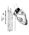
若年の交雑育種(cross bred)ブタ(約20〜25kg)に左側開胸を行った。アメロイドコンストリクター(ameroid constrictor)を、実施例1に記載の左側開胸を介して若年のブタの、左冠動脈の主幹から少し(just)遠位の近位LCX(左冠動脈回旋枝)の周囲に、血管のサイズ(代表的に、1.75、2.00または2.25mmの内径(ID))に適合するアメロイドコンストリクターを用いて配置した。図1は、アメロイドコンストリクターの配置を図示する。
【0040】
(心機能の評価および心筋注入)
心機能のベースライン測定を、アメロイドコンストリクターの配置4週間後に行った。測定は、安静時および1分につき180回の拍動(bpm)の心房ペーシング(atrial pacing)時の、ミクロスフェアの注入による冠動脈造影、ドブタミン負荷心エコー検査、血流測定を含んだ。
【0041】
ベースライン測定を完了した後、Kornowskiら、(2000)J.Am.Coll.Card.35:1031−1039に記載されるように、心筋内注入によって、ベクターまたは生理食塩水を心臓内の示された帯に導入した。この方法は、磁場誘導(magnetic guidance)カテーテルベースのナビゲーションシステムにより、開心手術中に、正常な心筋または虚血心筋内への直接注入を可能にする。この注入は、5×10⁹pfu/mLのAd−VEGF165またはAd−βGalの20μLの10回注入、あるいは20μLのリン酸緩衝生理食塩水(PBS)の10回注入からなっていた。
【0042】
注入4週間後(すなわち、アメロイドコンストリクターの移植8週間後)、ベースラインの定量を繰り返した。さらに、局所の心筋血流測定,組織病理分析、および体型測定分析のために、死後、虚血領域およびそれに隣接する正常領域を回収した。
【0043】
(処置グループ)
動物を4つのグループに分け、そして以下の注入を行った:(1)Ad−VEGF165を虚血帯内に(n=9);(2)Ad−VEGF165を正常帯内に(n=8);(3)Ad−βGalを虚血帯内に(n=8);または(4)PBSを虚血帯内に(n=7)。
【0044】
(結果)
その血流データは、注入が虚血帯を標的とする場合、灌流における適度の改善は安静時に生じることを示す。しかし、注入が心筋の正常帯でなされる場合、有意な改善が、安静時および負荷時の両方で、血液灌流において、観察される。さらに、経壁血流は、負荷下では、0.351(虚血帯注入)に対し、0.815(正常帯注入)というずっと高いレベルに至る。
【実施例2】
【0045】
(慢性心筋虚血の誘導)
アメロイドコンストリクターを、実施例1に記載のとおりに左側開胸を介して若年のブタの近位LCXの周囲に配置した。
【0046】
(心機能の評価および心筋注入)
心機能のベースライン測定を、アメロイドコンストリクターの配置4週間後に行った。測定は、安静時および1分につき180回の拍動(bpm)の心房ペーシング時、蛍光ミクロスフェアの注入による冠動脈造影、ドブタミン負荷心エコー検査、血流測定を含んだ。
【0047】
ベースライン測定を完了した後、ベクターまたは生理食塩水をStiletto注入カテーテルによって心臓内に導入した。各動物は、10回の注入(それぞれ20μLの、5×10⁹pfu/mLのAd−VEGF165またはAd−βGal)を受けた。
【0048】
注入4週間後(すなわち、アメロイドコンストリクターの移植8週間後)、ベースラインの測定を繰り返した。さらに、局所の心筋血流測定,組織病理分析、および体型測定分析のために、死後、虚血領域およびそれに隣接する正常領域を回収した。
【0049】
(処置グループ)
動物を4つのグループに分け、そして以下の注入を行った:(1)Ad−βGalを虚血帯内に(n=7);(2)Ad−VEGF165を虚血帯内に(n=7);(3)AdVEGF165を虚血帯に隣接する正常組織中に(n=7);そして(4)AdVEGF165を正常組織および虚血組織の両方の左心室の自由壁全体にわたり(n=8)。
【0050】
(結果)
安静状態下で、Ad−βGalの注入を虚血帯に受けた動物は、安静時の血流に有意な改善を示さなかったが、ペーシング時に血流に改善を示した。血流の改善に向けた傾向は、AdVEGF165の注入を虚血領域に受けた動物では見られなかった。Ad−VEGF165の注入を正常帯に受けた動物は、安静時およびペーシング時の両方での改善に向けた傾向を示した。虚血帯および正常帯の両方の左心室の自由壁全体にわたり注入を受けた動物はまた、安静時およびペーシング時の両方において改善に向けた傾向を示した。
【0051】
ドブタミン負荷心エコー検査は、Ad−VEGF165構築物を受けた全ての動物における壁運動において改善に向けた傾向を示した。対照的に、Ad−βGal構築物を受けた動物は、壁運動において減少を示した。
【0052】
虚血帯にAd−VEGFl65の注入を受けた動物は、Ad−βGalを虚血帯に受けた動物よりもより低い毛細管密度(capillary density)を有した。Ad−VEGF165の注入を正常帯に受けた動物は、Ad−βGalの注入を虚血帯に受けた動物より高い毛細管密度を有し、そしてAd−VEGF165の注入を正常帯および虚血帯の両方に受けた動物は、Ad−βGalの注入を虚血帯に受けた動物の毛細管密度と同様の毛細管密度を有した。
【図面の簡単な説明】
【0053】
【図1】図1は、慢性心筋虚血を誘導するためのアメロイドコンストリクターを配置したブタ心臓の模式図を提供する。虚血領域および非虚血領域を示す。
【図2】図2は、ブタの心臓の前方(左上)、側方(左下)および後方(右下)の図を表わし、そしてアメロイドコンストリクターによって誘導される虚血危険性領域を示す。
【図3】図3は、虚血帯にAd−βGal構築物を注入されたブタ(実施例1でのグループ3の動物)での心筋血流を示す、棒グラフである。安静時(白四角)およびペーシング時(影付き四角)の血流(ml/分/mg組織):左上のパネル、心内膜の虚血帯;右上のパネル、心外膜の虚血帯;左下のパネル、心内膜の非虚血帯;そして右下のパネル、心外膜の非虚血帯。
【図4】図4は、虚血帯にAd−VEGF165構築物を注入されたブタ(実施例1でのグループ1の動物)での心筋血流を示す、棒グラフである。安静時(白四角)およびペーシング時(影付き四角)の血流(ml/分/mg組織):左上のパネル、心内膜の虚血帯;右上のパネル、心外膜の虚血帯;左下のパネル、心内膜の非虚血帯;そして右下のパネル、心外膜の非虚血帯。
【図5】図5は、非虚血帯にAd−VEGF165構築物を注入されたブタ(実施例1でのグループ2の動物)での心筋血流を示す棒グラフである。安静時(白四角)およびペーシング時(影付き四角)の血流(ml/分/mg組織):左上のパネル、心内膜の虚血帯;右上のパネル、心外膜の虚血帯;左下のパネル、心内膜の非虚血帯;そして右下のパネル、心外膜の非虚血帯。
【図6】図6は、以下を注入されたブタでの心筋の経壁血流を示す棒グラフである:左上のパネル、虚血帯へ、Ad−βGal構築物(実施例1でのグループ3の動物);右上のパネル、虚血帯へ、PBS(実施例1でのグループ4の動物);左下のパネル、虚血帯へ、Ad−VEGF165構築物(実施例1でのグループ1の動物);および非虚血帯へ、Ad−VEGF165構築物((実施例1でのグループ2の動物)。血流(ml/分/mg組織)を、安静時(白四角)およびペーシング時(影付き四角)で示す。
【図7】図7は、ドブタミン負荷心エコー検査による局所の壁運動スコアを示す棒グラフである。壁運動スコアは、以下である:1=正常、2=運動低下、3=運動不能、および4=運動異常(負荷前(白四角)、低用量のドブタミン(明るい影付き四角)、および高用量のドブタミン(暗い影付き四角))。左上のパネル=Ad−βGal構築物(虚血帯へ)(実施例2でのグループ1の動物)、右上のパネルは、VEGF165構築物(虚血帯へ)(実施例2でのグループ2の動物)、左下のパネルは、VEGF165構築物(正常帯へ)(実施例2でのグループ3の動物)、右下のパネルは、VEGF165構築物(正常帯および虚血帯へ)(実施例2でのグループ4の動物)。
【図8】図8は、以下を注入されたブタの虚血帯での心筋血流を示す棒グラフである:Ad−βGalを虚血帯内に(左上)、Ad−VEGF165を虚血帯内に(右上)、Ad−VEGF165を正常帯内に(左下)、またはAd−VEGF165を虚血帯内および正常帯内の両方に(右下)。安静時(白四角)およびペーシング時(影付き四角)の、ベースラインおよび処置後での血流(ml/分/mg組織)。
【図9】図9は、Ad−βGalを虚血帯内に、Ad−VEGF165を虚血帯内に、Ad−VEGF165を正常帯内に、そしてAd−VEGF165を虚血帯内および正常帯内の両方に注入したブタでの毛細管密度(数/mm2)を示す棒グラフである。【Technical field】
[0001]
(Field of Invention)
The present invention relates to ischemic myocardium or by injecting a therapeutic agent (eg, gene, protein, cell or drug) into normal myocardium, preferably into the myocardium adjacent to the ischemic zone of the subject's heart. It relates to a method for treating pathological myocardium. This method is useful for inducing angiogenesis and collateral angiogenesis to improve cardiac function in subjects with ischemic heart disease. This method can also be used to promote tissue regeneration in such subjects.
[Background]
[0002]
(Background of the Invention)
Cardiovascular disease is generally characterized by a decrease in blood supply to the heart or other target organs. If the blood supply to the heart is compromised, the cells respond by producing compounds that induce the growth of new blood vessels and increase the blood supply to the heart. The process by which these new blood vessels, called collateral blood vessels, are induced to arise from the existing vasculature is called angiogenesis, and the substances produced by the cells to induce angiogenesis are angiogenesis It is called an factor (angiogenic factor). Since the body's natural angiogenic response is often inadequate, the use of exogenously supplied angiogenic factors is currently being investigated as a means of treating cardiovascular disease.
[0003]
Myocardial gene therapy can be used for the treatment of a number of cardiovascular diseases, including ischemic cardiomyopathy, congestive heart failure, and malignant arrhythmias (Nabel (1995) Circulation 91: 541-548). . Gene therapy for treating heart disease requires that gene therapy agents be delivered to the heart in a manner that produces a favorable response. Although intracoronary delivery of angiogenic growth factors and gene therapy vectors is possible, this approach can result in dilution of the drug due to dispersion of the therapeutic drug in the systemic circulation. Furthermore, such delivery methods can result in undesirable side effects, including tumor angiogenesis or retinopathy, due to the potential systemic distribution of such angiogenic agents. Intramyocardial injection provides a means of delivering angiogenic drugs that avoid these pitfalls. Kornowski et al. (J. Am. Coll. Cardiol. 35: 1031-9, 2000) teaches the direct delivery of angiogenic gene therapy vectors to ischemic tissue using catheter-based and surgical techniques. doing. Post et al. (Card. And Vasc. Regeneration 2: 106-113) disclose the transfection efficiency of transendocardial and direct epicardial injection of angiogenic gene therapy vectors.
[0004]
Whether precise localization of the injection into the myocardium is necessary is currently unknown. Currently, most studies have targeted injection into the ischemic or pathological part of the myocardium. However, the infusion can be placed, for example, in an ischemic region, in a band adjacent to the ischemic region, or in a normal myocardium.
DISCLOSURE OF THE INVENTION
[Means for Solving the Problems]
[0005]
(Related application)
This application claims the benefit of US Provisional Application No. 60 / 263,468, filed Jan. 23, 2001. The entire contents of US Provisional Application No. 60 / 263,468 are hereby incorporated by reference.
[0006]
In accordance with the present invention, surprisingly, angiogenic agents have been found to produce a desirable functional response when injected into normal myocardium, and more particularly into normal myocardium adjacent to the ischemic zone. Has been issued.
[0007]
(Summary of the Invention)
Using this method of delivering a therapeutic agent to normal myocardium or normal myocardial tissue adjacent to an ischemic heart or ischemic site of a pathological heart, inducing angiogenesis and contracting the heart, particularly in human patients It can increase sexual function, increase blood flow in the heart, stimulate collateral blood vessel development in the heart, promote tissue regeneration, and treat myocardial ischemia.
[0008]
In one aspect of the present invention, the present invention provides a therapeutic agent for an ischemic heart or pathological heart by delivering a therapeutically effective amount of the therapeutic agent to normal tissue of the ischemic heart or pathological heart. A method for delivery is provided. According to the present invention, the therapeutic agent can be a transgene encoding an angiogenic protein or an angiogenic peptide that is delivered into the myocardium of a subject by injection into the myocardium of a gene therapy vector containing the transgene. . The vector is injected into the normal tissue of the heart and preferably into the non-ischemic or non-pathological myocardium adjacent to the non-ischemic or non-pathological zone of the heart. The gene therapy vector can be a plasmid or a viral vector (eg, an adenovirus vector or a recombinant adenovirus vector, or an adeno-associated vector or a recombinant adeno-associated vector). This plasmid or viral vector can be delivered bare or in liposomes. Alternatively, the therapeutic agent induces angiogenesis, increases cardiac contractile function, increases blood flow in the heart, stimulates collateral vessel development in the heart, An angiogenic protein or angiogenic peptide, cell, one or more drugs, antisense DNA or antisense RNA, useful for promoting, improving exercise tolerance, or treating myocardial ischemia, or Can be any other therapeutic agent.
[0009]
In another aspect of the present invention, the present invention provides a method for delivering a sufficient amount of angiogenic factors to stimulate collateral angiogenesis into the myocardium of normal tissue within the ischemic heart of a subject. A method of stimulating collateral angiogenesis in The angiogenic factor can be delivered, for example, by an adenoviral vector or an adeno-associated viral vector that includes a coding sequence operably linked to a promoter that directs expression of the coding sequence in cardiomyocytes. The present invention also provides methods for inducing collateral angiogenesis in the myocardium, inducing angiogenesis in the myocardium, and improving the contractile function of the heart. In these methods, angiogenic factors or cells capable of producing angiogenic factors are delivered into the myocardium of normal tissue in a diseased or damaged heart.
[0010]
Yet another aspect is to deliver a therapeutic agent or cells capable of producing a sufficient amount of a therapeutic agent to stimulate tissue regeneration in the heart to normal tissue of a subject's ischemic heart or pathological heart, Methods are provided for promoting tissue regeneration in an ischemic or pathological heart of a subject. The therapeutic agent can be, for example, a protein or nucleic acid encoding a ligand for stem or progenitor cells, or any other agent that stimulates tissue regeneration.
[0011]
In another aspect of the invention, the invention provides a method for treating myocardial ischemia by delivering a sufficient amount of a therapeutic agent to normal myocardial tissue to ameliorate myocardial ischemic symptoms. provide. In this aspect of the invention, ischemic recovery includes induction of angiogenesis, stimulation of collateral blood vessel development in the heart, tissue regeneration, improvement of cardiac contractile function, increased blood flow in the heart, exercise There may be an increase in tolerability, a decrease in angina, and a reduction in other symptoms and conditions associated with myocardial ischemia. The therapeutic agent can be delivered to multiple sites throughout the normal myocardium or to sites adjacent to the ischemic zone. Suitable therapeutic agents for use in this aspect of the invention include angiogenic proteins or angiogenic peptides, transgenes encoding angiogenic proteins or angiogenic peptides, cells, one or more drugs, antisense RNA or anti-antigen Sense DNA, or other therapeutic agent.
[0012]
(Detailed description of the invention)
The present invention injects a sufficient amount of therapeutic agent into normal myocardium to induce angiogenesis, stimulate collateral angiogenesis, improve contractile function, or promote tissue regeneration Thus, a method of treating ischemic heart disease is provided. The therapeutic agent can be injected into the animal's ischemic myocardium or normal myocardium adjacent to the ischemic myocardial tissue, or the therapeutic agent can be injected at multiple sites dispersed throughout the normal myocardium. In a preferred embodiment, the therapeutic agent encodes at least one nucleic acid encoding an angiogenic factor (ie, a transgene) and stimulates collateral angiogenesis to treat ischemic heart disease, A gene therapy vector that expresses an effective amount of the factor to treat or ameliorate a cardiovascular condition or to promote tissue regeneration. The present invention is also preferably used in human patients to induce angiogenesis, to increase contractile function in the heart, to increase blood flow in the heart, Methods are contemplated for stimulating collateral vessel development, promoting tissue regeneration, and treating myocardial ischemia.
[0013]
In some embodiments, the therapeutic agent is delivered to the ischemic or pathological heart by delivering a therapeutically effective amount of the therapeutic agent into the myocardium of normal tissue of the heart. The present invention also stimulates vascularization in the myocardium by stimulating collateral blood vessel formation in the myocardium by delivering angiogenic factors or cells capable of producing angiogenic factors to normal tissue of the ischemic or pathological heart. Methods are provided for inducing neogenesis and for improving cardiac contractile function. This angiogenic factor is preferably delivered by an adenoviral vector or an adeno-associated vector comprising a coding sequence encoding an angiogenic factor, wherein the coding sequence directs the expression of the angiogenic factor in heart cells. Operably linked to the resulting promoter. Preferred vectors for use in the present invention include replication deficient adenoviruses, serotype 5 adenoviruses, and adenoviruses lacking the early gene region El, early gene region E3, or both.
[0014]
The present invention also provides a method for ameliorating symptoms associated with myocardial ischemia comprising delivering a sufficient amount of a therapeutic agent to normal myocardial tissue to ameliorate one or more ischemic symptoms. provide. Symptom recovery includes, for example, increased exercise capacity, decreased chest pain, and decreased shortness of breath.
[0015]
This therapeutic agent can be a gene therapy vector, protein, peptide, antisense DNA or RNA, drug, cell, cell expressing the therapeutic agent, whole bone marrow, and / or induce angiogenesis, contraction in the heart Can it increase function, increase blood flow in the heart, stimulate collateral blood vessel development in the heart, treat myocardial ischemia, or promote tissue regeneration? Or any other therapeutic agent that is useful therefor.
[0016]
Any suitable gene therapy vector can be used to supply the transgene. For example, the gene therapy vector can be a replication defective adenovirus, a recombinant adeno-associated virus vector (rAAV), a retroviral vector, a plasmid, or any other vector useful for gene therapy in the heart. Non-limiting examples of recombinant adenoviral vectors suitable for use in the present invention include recombinant adenoviruses described in Graham et al. (Virology 163: 614-617, 1988), and Graham F. et al. (Methods in Molecular Biology 7: 109-128, Murray, E. Ed., Humana Press, Clifton, NJ, 1991), Curiel et al. (Proc. Natl. Acad. Sci. USA 88: 8850-8854, 1991). , Miller et al. (FASEB J. 9: 190-199, 1995), and Curiel (Ann. NY Acad. Sci. 886: 158-171). Adenoviral vectors suitable for use in the present invention also include adenovirus serotype 5 adenoviruses and adenos lacking the early gene E1 region, lacking the early gene E3 region, or both. Virus. Adeno-associated vectors are described, for example, by Smith-Arica et al. (Curr. Cardiol. Rep. 3: 43-49, 2001), Philips (Expert Opinion. Biol. Ther. 1: 655-662, 2001), Rabinowitz et al. Virol 76: 791-801). Other viral vectors suitable for use with the present invention include retroviral vectors, coronavirus-based vectors, and vaccinia-based vectors. Plasmids and other non-viral vectors (eg, plasmid / liposome vectors, virus / liposome vectors, oligonucleotides, etc.) are described in, for example, McKay et al. (Cariovasc. Drug. Rev. 19: 245-62, 2001) and Rosenzweig (Vectors for Gene Therapy. In: Current Protocols in Human Genetics. Dracopoli et al., New York, NY: John Wiley and Sons, Inc., 1999).
[0017]
Gene therapy vectors useful in the present invention are those in which one or more transgenes (or of interest) are expressed in a manner that allows expression of the transgene under the control of appropriate regulatory elements (eg, promoters, enhancers, transcription terminators, etc.). Nucleic acid) can be any vector inserted therein. Gene therapy vectors are well known in the art and can be prepared by standard methodologies known to those skilled in the art.
[0018]
In addition, the nucleic acid is operably linked to regulatory regions (eg, promoters, enhancers, termination signals, etc.) to allow expression of the molecule. If more than one nucleic acid is present on a vector, each can be controlled separately by an individual control region, or any group or all of them can be operons (ie, a single transcription With a single control region that drives the expression of multiple genes on the product.
[0019]
As used herein, a “transgene” or “nucleic acid of interest” or “nucleic acid encoded in a vector” refers to collateral blood vessel formation to induce angiogenesis in the ischemic region of the heart. Any nucleotide sequence that encodes a molecule that is therapeutically effective to stimulate or increase myocardial blood flow. These transgenes can encode the proteins and angiogenic factors of the invention described herein. The transgene can be foreign to the animal to be treated or can be a gene that is normally found in the animal to be treated but for which altered expression is desired. Expression can be altered by changing the expression level or by changing the temporal or spatial pattern of expression.
[0020]
As used herein, “control region” or “regulatory element” refers to polyadenylation signals, upstream regulatory domains, promoters, enhancers, transcription termination sequences, and the like that regulate transcription and translation of nucleic acid sequences. Say.
[0021]
The term “operably linked” refers to the arrangement of elements, where the components are arranged to perform their normal functions. Thus, a control region or regulatory element that is operably linked to a coding sequence can result in the expression of the coding sequence. The control elements need not be contiguous with the coding sequence so long as they function to direct the expression of the coding sequence. Thus, for example, an intervening, untranslated but transcribed sequence can be present between a promoter sequence and a coding sequence, and the promoter sequence still “operably linked to the coding sequence. Can be considered.
[0022]
The regulatory elements of the invention can be derived from any source (eg, a virus, mammal, insect, or even synthetic material). However, they function after injection into the heart. For example, any promoter can be used to control transgene expression. Such promoters (eg, SV40 early promoter, mouse mammary tumor virus LTR promoter, adenovirus major late promoter (Ad MLP), herpes simplex promoter, CMV promoter (eg, CMV early promoter or Rous sarcoma virus (RSV) promoter) )) Can be promiscuous (ie, can be active in many cell types). Alternatively, the promoter can be tissue specific for expression in heart cells (eg, cardiomyocytes). Non-limiting examples of tissue specific promoters known in the art (eg, Lee et al. (1992) J. Biol. Chem. 267: 15875-15585; Jayaselan et al. (1997) Proc. Natl. Acad. Sci. USA 272 Condorelli et al. (2001) Proc. Natl. Acad. Sci. USA 98: 9977-9982) as described in left ventricular myosin light chain-2 (MLC2 γ ) Promoter, myosin heavy chain (MHC) promoter (eg, α-MHC and β-MHC), natriuretic peptide precursor A promoter (NppA), heart adriamycin response protein (CARP) promoter, cTNC gene promoter Etc.
[0023]
Proteins that can be administered (encoded in a gene therapy vector or directly) include proteins or peptides capable of inducing angiogenesis (eg, angiogenic factors). A protein or peptide capable of inducing angiogenesis, or “angiogenic factor”, as used herein, is a protein or substance that causes the growth of new blood vessels and fibroblast growth factor , Endothelial cell growth factor or other proteins with such biological activity. Angiogenic factors and specific proteins known to induce angiogenesis include, but are not limited to: FGF-1, FGF-2, FGF-5, VEGF and active fragments thereof (eg, VEGF 165 HIF-1, PDGF-1, PDGF-2, DEL1, angiopoietin, HGF, MCP-1, eNOS and iNOS). Other angiogenic factors suitable for use in the present invention include endothelial growth factor, growth factor including vascular smooth muscle growth factor, and FGF-1, FGF-2, FGF-5, PDGF-1, and PDGF-2. Abbreviations are as follows: FGF, fibroblast growth factor; VEGF, vascular endothelial growth factor; HIF, hypoxia-inducible factor; PDGF, platelet derived growth factor; DEL, developmental embryonic locus ): HGF, hepatocyte growth factor; MCP, monocyte chemotactic protein; eNOS, endothelial nitrous oxide synthase; and iNOS, inducible nitrate oxidase synthase.
[0024]
Other proteins or transgenes are also suitable for use in the present invention, eg, factors associated with myocardial protection or reperfusion injury (eg, heme oxygenase, hkis, AKT, PR39, and βarkCT) are used in the methods of the invention. Can be used. Tissue regeneration factors, including but not limited to ligands for progenitor cells or stem cells (eg, c-kit ligand, CD34 ligand and other factors) are also suitable for use in the methods of the invention.
[0025]
Cells that can be administered by the methods of the present invention include, but are not limited to, endothelial progenitor cells (angioblasts), cardiomyocytes, mononuclear cells, bone marrow stromal cells, and stem cells. “Stem cells” as used herein refers to mononuclear cells derived from placenta or cord blood. The cells described herein can be administered as primary cells (ie, not transformed or otherwise ex vivo). Alternatively, any of these cells or other suitable cell types can be manipulated or expanded ex vivo to produce angiogenic factors using methods known in the art, or ex vivo. It can be genetically manipulated or selected. Typically, the cells are engineered to produce angiogenic factors and engineered to secrete the desired angiogenic factor. Furthermore, all of the filtered bone marrow is known to be angiogenic and such preparations can be administered according to the present invention.
[0026]
The therapeutic agents described herein can be administered alone or in combination. In one non-limiting example, a therapeutic agent according to the present invention may comprise a viral vector that is delivered in combination with angiogenic cells. In another non-limiting example, a therapeutic agent according to the present invention can comprise a viral vector delivered in combination with an angiogenic protein. The therapeutic agent can also be delivered in combination with other active agents (eg, anti-apoptotic agents).
[0027]
The pharmaceutical formulations of the therapeutic agents of the present invention can be used to convert those entities having the desired degree of purity into any physiologically acceptable carrier, excipient in the form of a lyophilized formulation or an aqueous solution. Or prepared for storage by mixing with stabilizers (Remington's Pharmaceutical Sciences 16th edition, Osol, A. Ed. (1980)). Acceptable carriers, excipients or stabilizers are nontoxic to recipients at the dosages and concentrations used and buffers (eg, phosphates, citrates, and other organic acids) Antioxidants (including ascorbic acid and methionine); preservatives (eg octadecyldimethylbenzyl ammonium chloride; hexamethonium chloride; benzalkonium chloride, benzethonium chloride; phenol, butyl alcohol or benzyl alcohol; alkyl parabens; (Eg, methylparaben or propylparaben); catechol; resorcinol; cyclohexanol; 3-pentanol; and m-cresol); low molecular weight (less than about 10 residues) polypeptide; protein (eg, serum Rubmin, gelatin, or immunoglobulin); hydrophilic polymers (eg, polyvinylpyrrolidone); amino acids (eg, glycine, glutamine, asparagine, histidine, arginine, or lysine); monosaccharides, disaccharides, and other carbohydrates (glucose) Chelating agents (eg EDTA); saccharides (eg sucrose, mannitol, trehalose or sorbitol); salt-forming counterions (eg sodium; metal complexes (eg zinc-protein complexes)) And / or nonionic surfactants (eg TWEEN TM , PLURONICS TM Or polyethylene glycol (PEG))).
[0028]
The formulations herein may also contain more than one active compound as necessary for the particular indication being treated, preferably those with complementary activities that do not adversely affect each other. Such molecules are suitably present in combination in amounts that are effective for the purpose intended.
[0029]
The active ingredients can also be produced, for example, by microcapsules prepared by coacervation techniques or by interfacial polymerization (for example, hydroxymethylcellulose microcapsules or gelatin microcapsules and poly- (methyl methacrylate) microcapsules (poly- ( methylmethacrylate)), colloidal drug delivery systems (eg, liposomes, albumin microspheres, microemulsions, nanoparticles and nanocapsules) or macroemulsions, respectively. Such techniques are described in Remington's Pharmaceutical Sciences 16th edition, Osol, A. et al. Ed. (1980).
[0030]
Formulations used for in vivo administration must be sterile. This is easily accomplished by filtration through a sterile filtration membrane.
[0031]
Sustained release preparations can be prepared. Suitable examples of sustained release preparations include solid hydrophobic polymer semipermeable matrices containing polypeptide variants. This matrix is present in the form of a molded article (eg film or microcapsule). Examples of sustained release matrices include polyesters, hydrogels (eg, poly (2-hydroxyethyl-methacrylate) or poly (vinyl alcohol)), polylactide (US Pat. No. 3,773,919), L-glutamic acid and y Copolymer with ethyl-L-glutamate, non-degradable ethylene vinyl acetate, degradable lactic acid-glycolic acid copolymer (eg LUPRON DEPOT TM (Injectable microspheres composed of lactic acid-glycolic acid copolymer and leuprolide acetate), and poly-D-(-)-3-hydroxybutyric acid. While polymers (eg, ethylene-vinyl acetate and lactic acid-glycolic acid) can release molecules for over 100 days, certain hydrogels release proteins for shorter periods of time. If encapsulated antibodies remain in the body for a long time, they can denature or aggregate as a result of exposure to moisture at 37 ° C, resulting in loss of biological activity and possible changes in immunogenicity . Reasonable strategies can be devised for stabilization depending on the mechanism involved. For example, if the aggregation mechanism is disclosed as intermolecular S—S bond formation via thio-disulfide exchange, stabilization can include modifying sulfhydryl residues, lyophilizing from acidic solution, moisture content Can be achieved by adjusting the control, using appropriate additives, and developing specific polymer matrix compositions.
[0032]
One skilled in the art can readily determine the amount of therapeutic agent to be included in any pharmaceutical composition and appropriate dosage for the intended use.
[0033]
The methods of the invention can be used with any animal. Any animal includes, but is not limited to, mammals (eg, rodents, dogs, cats, cows, primates and humans). Preferably, the method is used for gene therapy to treat a human ischemic heart condition or ischemic heart disease.
[0034]
The amount of therapeutic agent injected into the animal is proportional to the animal's body weight and also depends on the drug selected. One skilled in the art can readily determine an appropriate dosage for the selected agent. As an example, if the drug is a gene therapy vector (eg, a replication defective adenovirus), the dosage is about 10 6 ~ About 10 12 Of plaque forming units (pfu), and preferably about 10 8 ~ About 10 10 can be between pfu. For stable and efficient transduction with rAAV, the dosage is about 1 x 10 per gram body weight. 5 IU (infectious unit) AAV ~ 1x10 per gram body weight 9 Can be IU AAV and preferably about 1 × 10 6 per gram body weight 6 About 1 x 10 per gram body weight of IU AAV 7 It can be IU AAV. If the drug is a protein, the dosage can range from as little as about 1 picogram to several hundred micrograms, but can be readily determined by one skilled in the art anyway.
[0035]
Methods for measuring cardiac function are well known in the art. See, for example, Simons et al. (2000) Circulation 102: e732-e86, “Clinical Trails in Coronary Angiogenesis: Issues, Problems and Consensus”. For example, blood flow to the ischemic myocardium can be achieved by various methods including single photon emission computed tomography (SPECT), position emission tomography (PET), magnetic resonance imaging (MRI), and injection of fluorescent microspheres. It can be measured using non-invasive imaging techniques. Coronary angiography can be used to measure disease progression and to establish the appearance of new blood vessels. Echocardiography can be used to assess heart wall motion at rest and under stress (eg, dobutamine-induced stress). Exercise tolerance testing (eg, treadmill testing) may provide another means to assess cardiac function.
[0036]
Delivery to the myocardium is for introduction into catheters (eg, infusion catheters, diagnostic catheters, etc.), stylet catheters, needles, needleless syringes, balloon catheters, channeling devices, or intramyocardial. Other suitable medical devices can be used. In a preferred delivery method, an endocardial injection catheter (eg, a Stiletto catheter (available from Boston Scientific, Natick, Massachusetts) is used to deliver the therapeutic agent without the need for thoracotomy. Can be led by fluoroscopy, echocardiography, MRI, or electromechanical mapping, and the catheter is used to deliver therapeutic agents to non-ischemic tissue of the myocardium by transendocardial injection. Suitable devices and methods are described in US Patent No. 6,238, 406. Alternatively, transepicardial surgical approaches are either through thoracotomy or through thoracoscopy. May be necessary for delivery to the myocardium.
[0037]
Throughout this application, various publications, patents, and patent applications are referred to. The teachings and disclosures of these publications, patents, and patent applications are hereby incorporated by reference in their entirety to more fully describe the state of the art relating to the present invention.
[0038]
It should be understood and expected that variations in the principles of the invention disclosed herein in representative embodiments may be made by those skilled in the art. Such modifications, changes and substitutions are intended to be included within the scope of the present invention.
[Example 1]
[0039]
(Induction of chronic myocardial ischemia)
Young cross-bred pigs (approximately 20-25 kg) were subjected to left thoracotomy. An ameloid constrictor is placed around the proximal LCX (left coronary rotator) just distal to the main trunk of the left coronary artery in young pigs via the left thoracotomy described in Example 1 Were placed using an Ameloid constrictor that fits the size of the vessel (typically 1.75, 2.00, or 2.25 mm ID). FIG. 1 illustrates the placement of an Ameloid constructor.
[0040]
(Evaluation of cardiac function and myocardial injection)
Baseline measurements of cardiac function were made 4 weeks after placement of the Ameloid constructor. Measurements included coronary angiography with microsphere injection, dobutamine stress echocardiography, blood flow measurements at rest and at 180 beats per minute (bpm) atrial pacing.
[0041]
After completing the baseline measurement, Kornowski et al. (2000) J. MoI. Am. Coll. Card. 35: 1031-1039, vector or saline was introduced into the indicated zone in the heart by intramyocardial injection. This method allows for direct injection into normal or ischemic myocardium during open heart surgery with a magnetic guidance catheter-based navigation system. This injection is 5 × 10 9 pfu / mL Ad-VEGF 165 Or consisted of 10 injections of 20 μL of Ad-βGal, or 10 injections of 20 μL phosphate buffered saline (PBS).
[0042]
Baseline quantification was repeated 4 weeks after injection (i.e. 8 weeks after implantation of the Ameloid constructor). Furthermore, the ischemic region and the normal region adjacent thereto were collected after death for local myocardial blood flow measurement, histopathological analysis, and morphometric analysis.
[0043]
(Treatment group)
The animals were divided into 4 groups and the following injections were made: (1) Ad-VEGF 165 In the ischemic zone (n = 9); (2) Ad-VEGF 165 In the normal zone (n = 8); (3) Ad-βGal in the ischemic zone (n = 8); or (4) PBS in the ischemic zone (n = 7).
[0044]
(result)
The blood flow data indicates that when the infusion targets the ischemic zone, a moderate improvement in perfusion occurs at rest. However, when the infusion is made in the normal zone of the myocardium, a significant improvement is observed in blood perfusion both at rest and during stress. Furthermore, transmural blood flow reaches a much higher level of 0.815 (normal zone injection) versus 0.351 (ischemic zone injection) under load.
[Example 2]
[0045]
(Induction of chronic myocardial ischemia)
An Ameloid constrictor was placed around the proximal LCX of young pigs via left thoracotomy as described in Example 1.
[0046]
(Evaluation of cardiac function and myocardial injection)
Baseline measurements of cardiac function were made 4 weeks after placement of the Ameloid constructor. Measurements included coronary angiography with dosing of fluorescent microspheres, dobutamine stress echocardiography, blood flow measurements at rest and at atrial pacing of 180 beats per minute (bpm).
[0047]
After completing the baseline measurement, vector or saline was introduced into the heart via a Stiletto infusion catheter. Each animal received 10 injections (20 μL each, 5 × 10 9 pfu / mL Ad-VEGF 165 Or Ad-βGal).
[0048]
Baseline measurements were repeated 4 weeks after injection (i.e. 8 weeks after implantation of the Ameloid constructor). Furthermore, the ischemic region and the normal region adjacent thereto were collected after death for local myocardial blood flow measurement, histopathological analysis, and morphometric analysis.
[0049]
(Treatment group)
The animals were divided into four groups and the following injections were made: (1) Ad-βGal in the ischemic zone (n = 7); (2) Ad-VEGF 165 In the ischemic zone (n = 7); (3) AdVEGF 165 In normal tissue adjacent to the ischemic zone (n = 7); and (4) AdVEGF 165 Across the free wall of the left ventricle of both normal and ischemic tissue (n = 8).
[0050]
(result)
Under resting conditions, animals that received Ad-βGal infusion in the ischemic zone showed no significant improvement in resting blood flow, but improved blood flow during pacing. The trend towards improved blood flow is AdVEGF 165 Was not seen in animals that received infusions in the ischemic area. Ad-VEGF 165 Animals that received normal injections showed a trend towards improvement at both rest and pacing. Animals that received infusions across the free wall of the left ventricle in both the ischemic and normal zones also tended to improve both at rest and during pacing.
[0051]
Dobutamine stress echocardiography is performed using Ad-VEGF. 165 There was a trend towards improvement in wall motion in all animals that received the construct. In contrast, animals that received the Ad-βGal construct showed a decrease in wall motion.
[0052]
Ad-VEGF in the ischemic zone l65 The animals that received the infusions had lower capillary density than the animals that received Ad-βGal in the ischemic zone. Ad-VEGF 165 Animals that received an infusion in the normal zone had a higher capillary density than animals that received an infusion of Ad-βGal in the ischemic zone, and Ad-VEGF 165 The animals that received both the normal and ischemic zones had a capillary density similar to that of the animals that received the Ad-βGal injections.
[Brief description of the drawings]
[0053]
FIG. 1 provides a schematic diagram of a porcine heart with an Ameloid constructor placed to induce chronic myocardial ischemia. Ischemic and non-ischemic regions are shown.
FIG. 2 depicts an anterior (upper left), lateral (lower left) and posterior (lower right) view of a porcine heart and shows the ischemic risk area induced by the Ameloid Constrictor.
FIG. 3 is a bar graph showing myocardial blood flow in pigs (
FIG. 4 shows Ad-VEGF in the ischemic zone. 165 2 is a bar graph showing myocardial blood flow in pigs injected with the construct (
FIG. 5 shows Ad-VEGF in a non-ischemic zone. 165 FIG. 4 is a bar graph showing myocardial blood flow in pigs injected with the construct (
FIG. 6 is a bar graph showing myocardial transmural blood flow in pigs injected with: upper left panel, to the ischemic zone, Ad-βGal construct (
FIG. 7 is a bar graph showing local wall motion scores from dobutamine stress echocardiography. The wall motion scores are: 1 = normal, 2 = motor deficit, 3 = insufficient, and 4 = motor abnormalities (before stress (white squares), low dose dobutamine (light shaded squares), and high dose Dobutamine (dark shaded square)). Upper left panel = Ad-βGal construct (to ischemic zone) (
FIG. 8 is a bar graph showing myocardial blood flow in the ischemic zone of pigs injected with: Ad-βGal in the ischemic zone (upper left), Ad-VEGF. 165 Into the ischemic zone (upper right), Ad-VEGF 165 Within the normal zone (lower left), or Ad-VEGF 165 In both the ischemic and normal zones (lower right). Baseline and post-treatment blood flow (ml / min / mg tissue) at rest (white squares) and pacing (shaded squares).
FIG. 9 shows Ad-βGal in the ischemic zone, Ad-VEGF. 165 In the ischemic zone, Ad-VEGF 165 Within the normal zone and Ad-VEGF 165 Capillary density (several / mm) in pigs injected with both in the ischemic and normal zones 2 ).
Claims (54)
FGF−1、FGF−2、FGF−5、VEGF、VEGF165、HIF−1、PDGF−1、PDGF−2、DEL1、アンギオポエチン、HGF、MCP−1、eNOS、iNOS、またはそれらの組み合わせ、
からなる群より選択される、方法。21. The method according to any one of claims 15-20, wherein the angiogenic factor is:
FGF-1, FGF-2, FGF-5, VEGF, VEGF 165 , HIF-1, PDGF-1, PDGF-2, DEL1, angiopoietin, HGF, MCP-1, eNOS, iNOS, or combinations thereof,
A method selected from the group consisting of:
Applications Claiming Priority (2)
| Application Number | Priority Date | Filing Date | Title |
|---|---|---|---|
| US26346801P | 2001-01-23 | 2001-01-23 | |
| PCT/US2002/003118 WO2002064157A2 (en) | 2001-01-23 | 2002-01-23 | Localized myocardial injection method for treating ischemic myocardium |
Publications (2)
| Publication Number | Publication Date |
|---|---|
| JP2005501802A true JP2005501802A (en) | 2005-01-20 |
| JP2005501802A5 JP2005501802A5 (en) | 2005-12-22 |
Family
ID=23001908
Family Applications (1)
| Application Number | Title | Priority Date | Filing Date |
|---|---|---|---|
| JP2002563950A Pending JP2005501802A (en) | 2001-01-23 | 2002-01-23 | Localized myocardial injection method for treating ischemic myocardium |
Country Status (5)
| Country | Link |
|---|---|
| US (2) | US20020172663A1 (en) |
| EP (1) | EP1361896A2 (en) |
| JP (1) | JP2005501802A (en) |
| CA (1) | CA2433936A1 (en) |
| WO (1) | WO2002064157A2 (en) |
Cited By (1)
| Publication number | Priority date | Publication date | Assignee | Title |
|---|---|---|---|---|
| JP2011503207A (en) * | 2007-11-16 | 2011-01-27 | サン ディエゴ ステート ユニバーシティ リサーチ ファウンデーション | Compositions and methods for manipulating PIM-1 activity in circulatory cells |
Families Citing this family (43)
| Publication number | Priority date | Publication date | Assignee | Title |
|---|---|---|---|---|
| US7288521B2 (en) | 2000-04-06 | 2007-10-30 | Franco Wayne P | Growth factor therapy mobilization of stem cells into the peripheral blood |
| US7291597B2 (en) * | 2000-04-06 | 2007-11-06 | Franco Wayne P | Growth factor therapy mobilization of stem cells into the peripheral blood |
| US7166280B2 (en) * | 2000-04-06 | 2007-01-23 | Franco Wayne P | Combination growth factor therapy and cell therapy for treatment of acute and chronic heart disease |
| US20100303769A1 (en) * | 2000-04-06 | 2010-12-02 | Franco Wayne P | Combination growth factor therapy and cell therapy for treatment of acute and chronic heart disease |
| CN1630526B (en) | 2001-12-07 | 2010-05-05 | 马克罗珀尔生物外科公司 | Systems and methods for treating patients with processed lipoaspirate cells |
| US7771716B2 (en) | 2001-12-07 | 2010-08-10 | Cytori Therapeutics, Inc. | Methods of using regenerative cells in the treatment of musculoskeletal disorders |
| US7585670B2 (en) | 2001-12-07 | 2009-09-08 | Cytori Therapeutics, Inc. | Automated methods for isolating and using clinically safe adipose derived regenerative cells |
| US9597395B2 (en) | 2001-12-07 | 2017-03-21 | Cytori Therapeutics, Inc. | Methods of using adipose tissue-derived cells in the treatment of cardiovascular conditions |
| US20050048035A1 (en) | 2001-12-07 | 2005-03-03 | Fraser John K. | Methods of using regenerative cells in the treatment of stroke and related diseases and disorders |
| US20050095228A1 (en) | 2001-12-07 | 2005-05-05 | Fraser John K. | Methods of using regenerative cells in the treatment of peripheral vascular disease and related disorders |
| KR100562824B1 (en) | 2002-03-20 | 2006-03-23 | 주식회사 바이로메드 | Hybrid hepatocyte growth factor gene which has a high expression efficiency and expresses two heterotypes of hepatocyte growth factor |
| WO2004044142A2 (en) * | 2002-11-05 | 2004-05-27 | The Brigham And Women's Hospital, Inc. | Mesenchymal stem cells and methods of use thereof |
| US7781415B2 (en) | 2003-02-07 | 2010-08-24 | Roche Madison Inc. | Process for delivering sirna to cardiac muscle tissue |
| DK1599575T3 (en) * | 2003-02-20 | 2012-01-16 | Cytori Therapeutics Inc | Methods of using adipose tissue-derived cells in the treatment of cardiovascular conditions |
| US7840263B2 (en) | 2004-02-27 | 2010-11-23 | Cardiac Pacemakers, Inc. | Method and apparatus for device controlled gene expression |
| US7764995B2 (en) | 2004-06-07 | 2010-07-27 | Cardiac Pacemakers, Inc. | Method and apparatus to modulate cellular regeneration post myocardial infarct |
| US7729761B2 (en) * | 2004-07-14 | 2010-06-01 | Cardiac Pacemakers, Inc. | Method and apparatus for controlled gene or protein delivery |
| WO2006055743A2 (en) * | 2004-11-18 | 2006-05-26 | Franco, Wayne | Compbination growth therapy and cell therapy for treatment of acute and chronic diseases of the organs |
| US8060219B2 (en) | 2004-12-20 | 2011-11-15 | Cardiac Pacemakers, Inc. | Epicardial patch including isolated extracellular matrix with pacing electrodes |
| US7981065B2 (en) | 2004-12-20 | 2011-07-19 | Cardiac Pacemakers, Inc. | Lead electrode incorporating extracellular matrix |
| WO2006102643A2 (en) * | 2005-03-24 | 2006-09-28 | Caritas St. Elizabeth Medical Center Boston, Inc. | Stably transformed bone marrow-derived cells and uses thereof |
| US7774057B2 (en) | 2005-09-06 | 2010-08-10 | Cardiac Pacemakers, Inc. | Method and apparatus for device controlled gene expression for cardiac protection |
| US8637005B2 (en) | 2005-11-07 | 2014-01-28 | Amorcyte, Inc. | Compositions and methods of vascular injury repair |
| US20110076255A1 (en) | 2005-11-07 | 2011-03-31 | Pecora Andrew L | Compositions and methods for treating progressive myocardial injury due to a vascular insufficiency |
| PL1951864T3 (en) | 2005-11-07 | 2014-10-31 | Pecora & Co Llc | Compositions and methods of vascular injury repair |
| US20070190028A1 (en) * | 2006-02-13 | 2007-08-16 | Jihong Qu | Method and apparatus for heat or electromagnetic control of gene expression |
| US20090202606A1 (en) * | 2008-01-25 | 2009-08-13 | Viromed Co., Ltd. | Treatment and Prevention of Cardiac Conditions Using Two or More Isoforms of Hepatocyte Growth Factor |
| WO2010021993A1 (en) | 2008-08-19 | 2010-02-25 | Cytori Therapeutics, Inc. | Methods of using adipose tissue-derived cells in the treatment of the lymphatic system and malignant disease |
| WO2010065601A1 (en) * | 2008-12-03 | 2010-06-10 | Amorcyte, Inc. | Infarct area perfusion-improving compositions and methods of vascular injury repair |
| EP2424463B1 (en) | 2009-05-01 | 2016-12-28 | Bimini Technologies LLC | Systems, methods and compositions for optimizing tissue and cell enriched grafts |
| WO2010138180A2 (en) * | 2009-05-26 | 2010-12-02 | The University Of Vermont And State Agriculture College | Compositions and methods for cardiac tissue repair |
| WO2013138338A2 (en) | 2012-03-12 | 2013-09-19 | Massachusetts Institute Of Technology | Methods for treating tissue damage associated with ischemia with apoliporotein d |
| SG11201602452SA (en) | 2013-10-22 | 2016-05-30 | Viromed Co Ltd | Composition for preventing or treating amyotrophic lateral sclerosis using two or more isoforms of hepatocyte growth factor |
| RU2570285C1 (en) * | 2014-11-25 | 2015-12-10 | Государственное бюджетное образовательное учреждение высшего профессионального образования "Курский государственный медицинский университет" Министерства здравоохранения Российской Федерации | Method of treating subcritical limb ischemia accompanying chronic obliterating diseases of lower-limb arteries |
| AU2016205336A1 (en) | 2015-01-06 | 2017-08-03 | Venturis Therapeutics, Inc. | Angiogenic treatment of ischemic heart disease |
| EP3259012A4 (en) | 2015-02-16 | 2018-09-19 | CardioVascular BioTherapeutics, Inc. | Therapeutic angiogenesis for treating erectile conditions |
| US10813612B2 (en) | 2019-01-25 | 2020-10-27 | Cleerly, Inc. | Systems and method of characterizing high risk plaques |
| US11232564B2 (en) | 2020-01-07 | 2022-01-25 | Cleerly, Inc. | Systems, methods, and devices for medical image analysis, diagnosis, risk stratification, decision making and/or disease tracking |
| AU2021205821A1 (en) | 2020-01-07 | 2022-07-21 | Cleerly, Inc. | Systems, methods, and devices for medical image analysis, diagnosis, risk stratification, decision making and/or disease tracking |
| US20220392065A1 (en) | 2020-01-07 | 2022-12-08 | Cleerly, Inc. | Systems, methods, and devices for medical image analysis, diagnosis, risk stratification, decision making and/or disease tracking |
| KR20220090951A (en) * | 2020-12-23 | 2022-06-30 | 건국대학교 산학협력단 | A Novel Fragment of Fibroblast Growth Factor and Uses Thereof |
| CN114053224A (en) * | 2021-09-07 | 2022-02-18 | 山东第一医科大学附属省立医院(山东省立医院) | Liposome compound containing beta ARKct and preparation method and application thereof |
| US20230289963A1 (en) | 2022-03-10 | 2023-09-14 | Cleerly, Inc. | Systems, devices, and methods for non-invasive image-based plaque analysis and risk determination |
Family Cites Families (16)
| Publication number | Priority date | Publication date | Assignee | Title |
|---|---|---|---|---|
| US5661133B1 (en) * | 1991-11-12 | 1999-06-01 | Univ Michigan | Collateral blood vessel formation in cardiac muscle by injecting a dna sequence encoding an angiogenic protein |
| US5221272A (en) * | 1991-12-03 | 1993-06-22 | Safegrip, Inc. | Unified medical fluid system |
| AU694097B2 (en) * | 1992-11-18 | 1998-07-16 | Arch Development Corporation | Adenovirus-mediated gene transfer to cardiac and vascular smooth muscle |
| US5631237A (en) * | 1992-12-22 | 1997-05-20 | Dzau; Victor J. | Method for producing in vivo delivery of therapeutic agents via liposomes |
| GB9401447D0 (en) * | 1994-01-26 | 1994-03-23 | Ciba Geigy Ag | Pharmaceutical compositions |
| US7186688B1 (en) * | 1994-03-08 | 2007-03-06 | Human Genome Sciences, Inc. | Methods of stimulating angiogenesis in a patient by administering vascular endothelial growth factor 2 |
| US6152141A (en) * | 1994-07-28 | 2000-11-28 | Heartport, Inc. | Method for delivery of therapeutic agents to the heart |
| JP3961019B2 (en) * | 1995-02-28 | 2007-08-15 | ザ リージェンツ オブ ザ ユニバーシティ オブ カリフォルニア | Gene transfer mediated angiogenesis therapy |
| US5800539A (en) * | 1995-11-08 | 1998-09-01 | Emory University | Method of allogeneic hematopoietic stem cell transplantation without graft failure or graft vs. host disease |
| NZ336838A (en) * | 1997-01-29 | 2002-04-26 | Cornell Res Foundation Inc | A method of enhancing the level of perfusion of blood to a target tissue by administering an adenoviral vector encoded with an angiogenic peptide within 0.5-15 cm3 of the tissue |
| US6045565A (en) * | 1997-11-04 | 2000-04-04 | Scimed Life Systems, Inc. | Percutaneous myocardial revascularization growth factor mediums and method |
| EP1007631B2 (en) * | 1997-07-14 | 2009-02-18 | Osiris Therapeutics, Inc. | Cardiac muscle regeneration using mesenchymal stem cells |
| US7078387B1 (en) * | 1998-12-28 | 2006-07-18 | Arch Development Corp. | Efficient and stable in vivo gene transfer to cardiomyocytes using recombinant adeno-associated virus vectors |
| US7097832B1 (en) * | 1999-03-30 | 2006-08-29 | Myocardial Therapeutics, Inc. | Intramyocardial injection of autologous bone marrow |
| DE60035035T2 (en) * | 1999-03-30 | 2008-01-31 | Kornowski, Ran | INJECTION OF AUTOLOGOUS BONE MARROW IN THE HEART MUSCLES |
| WO2002009650A2 (en) * | 2000-07-31 | 2002-02-07 | New York Medical College | Methods and compositions for the repair and/or regeneration of damaged myocardium |
-
2002
- 2002-01-23 JP JP2002563950A patent/JP2005501802A/en active Pending
- 2002-01-23 US US10/057,409 patent/US20020172663A1/en not_active Abandoned
- 2002-01-23 CA CA002433936A patent/CA2433936A1/en not_active Abandoned
- 2002-01-23 WO PCT/US2002/003118 patent/WO2002064157A2/en active Application Filing
- 2002-01-23 EP EP02720895A patent/EP1361896A2/en not_active Withdrawn
-
2008
- 2008-05-16 US US12/122,561 patent/US20080254109A1/en not_active Abandoned
Cited By (2)
| Publication number | Priority date | Publication date | Assignee | Title |
|---|---|---|---|---|
| JP2011503207A (en) * | 2007-11-16 | 2011-01-27 | サン ディエゴ ステート ユニバーシティ リサーチ ファウンデーション | Compositions and methods for manipulating PIM-1 activity in circulatory cells |
| JP2015007063A (en) * | 2007-11-16 | 2015-01-15 | サン ディエゴ ステート ユニバーシティ リサーチ ファウンデーション | Composition and method for manipulating pim-1 activity in circulatory system cell |
Also Published As
| Publication number | Publication date |
|---|---|
| WO2002064157A2 (en) | 2002-08-22 |
| US20020172663A1 (en) | 2002-11-21 |
| WO2002064157A9 (en) | 2003-06-05 |
| US20080254109A1 (en) | 2008-10-16 |
| EP1361896A2 (en) | 2003-11-19 |
| CA2433936A1 (en) | 2002-08-22 |
| WO2002064157A3 (en) | 2003-08-07 |
Similar Documents
| Publication | Publication Date | Title |
|---|---|---|
| JP2005501802A (en) | Localized myocardial injection method for treating ischemic myocardium | |
| JP3961019B2 (en) | Gene transfer mediated angiogenesis therapy | |
| US20090082293A1 (en) | Techniques and compositions for treating cardiovascular disease by in vivo gene delivery | |
| JP2011225611A (en) | Gene transfer-mediated angiogenesis therapy | |
| JP3865632B2 (en) | Cardiomyopathy gene therapy | |
| AU784392B2 (en) | Techniques and compositions for treating cardiovascular disease by in vivo gene delivery | |
| AU772857B2 (en) | Method of inducing angiogenesis | |
| CN100350979C (en) | Combination of a nucleic acid and a vasoactive agent for enhanced gene delivery | |
| JP2002515065A (en) | Techniques and compositions for treating heart failure and ventricular reconstitution by in vivo delivery of an angiogenic transgene | |
| Kastrup, Erik Jørgensen, Viktor Drvota | Vascular growth factor and gene therapy to induce new vessels in the ischemic myocardium. Therapeutic angiogenesis | |
| AU2006200170B2 (en) | Combination of a nucleic acid and a vasoactive agent for enhanced gene delivery | |
| AU706050C (en) | Gene transfer-mediated angiogenesis therapy | |
| US20160296674A1 (en) | Nucleic acid based cardiovascular therapeutics |
Legal Events
| Date | Code | Title | Description |
|---|---|---|---|
| A521 | Written amendment |
Free format text: JAPANESE INTERMEDIATE CODE: A523 Effective date: 20050124 |
|
| A621 | Written request for application examination |
Free format text: JAPANESE INTERMEDIATE CODE: A621 Effective date: 20050124 |
|
| A711 | Notification of change in applicant |
Free format text: JAPANESE INTERMEDIATE CODE: A711 Effective date: 20060713 |
|
| A521 | Written amendment |
Free format text: JAPANESE INTERMEDIATE CODE: A821 Effective date: 20060713 |
|
| A131 | Notification of reasons for refusal |
Free format text: JAPANESE INTERMEDIATE CODE: A131 Effective date: 20080401 |
|
| A521 | Written amendment |
Free format text: JAPANESE INTERMEDIATE CODE: A523 Effective date: 20080618 |
|
| A02 | Decision of refusal |
Free format text: JAPANESE INTERMEDIATE CODE: A02 Effective date: 20080908 |