EP2201026B1 - Novel p2x7 epitopes - Google Patents
Novel p2x7 epitopes Download PDFInfo
- Publication number
- EP2201026B1 EP2201026B1 EP08800000.5A EP08800000A EP2201026B1 EP 2201026 B1 EP2201026 B1 EP 2201026B1 EP 08800000 A EP08800000 A EP 08800000A EP 2201026 B1 EP2201026 B1 EP 2201026B1
- Authority
- EP
- European Patent Office
- Prior art keywords
- antibody
- binding
- fragment
- receptor
- seq
- Prior art date
- Legal status (The legal status is an assumption and is not a legal conclusion. Google has not performed a legal analysis and makes no representation as to the accuracy of the status listed.)
- Active
Links
- 230000027455 binding Effects 0.000 claims description 95
- 239000012634 fragment Substances 0.000 claims description 72
- 101710189965 P2X purinoceptor 7 Proteins 0.000 claims description 68
- 102100037602 P2X purinoceptor 7 Human genes 0.000 claims description 68
- 108090000765 processed proteins & peptides Proteins 0.000 claims description 66
- 206010028980 Neoplasm Diseases 0.000 claims description 56
- 239000000178 monomer Substances 0.000 claims description 37
- 210000001124 body fluid Anatomy 0.000 claims description 30
- 239000010839 body fluid Substances 0.000 claims description 30
- 238000000034 method Methods 0.000 claims description 28
- 201000011510 cancer Diseases 0.000 claims description 21
- 239000000427 antigen Substances 0.000 claims description 19
- 108091007433 antigens Proteins 0.000 claims description 19
- 102000036639 antigens Human genes 0.000 claims description 19
- 238000001514 detection method Methods 0.000 claims description 15
- 238000011282 treatment Methods 0.000 claims description 15
- 239000008194 pharmaceutical composition Substances 0.000 claims description 14
- FWMNVWWHGCHHJJ-SKKKGAJSSA-N 4-amino-1-[(2r)-6-amino-2-[[(2r)-2-[[(2r)-2-[[(2r)-2-amino-3-phenylpropanoyl]amino]-3-phenylpropanoyl]amino]-4-methylpentanoyl]amino]hexanoyl]piperidine-4-carboxylic acid Chemical compound C([C@H](C(=O)N[C@H](CC(C)C)C(=O)N[C@H](CCCCN)C(=O)N1CCC(N)(CC1)C(O)=O)NC(=O)[C@H](N)CC=1C=CC=CC=1)C1=CC=CC=C1 FWMNVWWHGCHHJJ-SKKKGAJSSA-N 0.000 claims description 13
- 230000001413 cellular effect Effects 0.000 claims description 13
- 230000002163 immunogen Effects 0.000 claims description 9
- 239000003937 drug carrier Substances 0.000 claims description 7
- 239000000546 pharmaceutical excipient Substances 0.000 claims description 7
- 239000003085 diluting agent Substances 0.000 claims description 6
- 108010021625 Immunoglobulin Fragments Proteins 0.000 claims description 5
- 102000008394 Immunoglobulin Fragments Human genes 0.000 claims description 5
- 239000003814 drug Substances 0.000 claims description 5
- 238000000338 in vitro Methods 0.000 claims description 5
- 238000004519 manufacturing process Methods 0.000 claims description 4
- 125000003275 alpha amino acid group Chemical group 0.000 claims 1
- 210000004027 cell Anatomy 0.000 description 60
- 102000005962 receptors Human genes 0.000 description 44
- 108020003175 receptors Proteins 0.000 description 44
- 210000001519 tissue Anatomy 0.000 description 39
- 208000037265 diseases, disorders, signs and symptoms Diseases 0.000 description 33
- 239000000203 mixture Substances 0.000 description 32
- 201000010099 disease Diseases 0.000 description 27
- 241000282414 Homo sapiens Species 0.000 description 23
- 230000001225 therapeutic effect Effects 0.000 description 16
- 238000003556 assay Methods 0.000 description 15
- 102000004196 processed proteins & peptides Human genes 0.000 description 11
- 208000000236 Prostatic Neoplasms Diseases 0.000 description 10
- 238000009472 formulation Methods 0.000 description 10
- 239000007790 solid phase Substances 0.000 description 9
- 206010060862 Prostate cancer Diseases 0.000 description 8
- 239000003795 chemical substances by application Substances 0.000 description 8
- 108090000623 proteins and genes Proteins 0.000 description 8
- 239000000243 solution Substances 0.000 description 8
- 230000006870 function Effects 0.000 description 7
- 235000018102 proteins Nutrition 0.000 description 7
- 102000004169 proteins and genes Human genes 0.000 description 7
- 239000000126 substance Substances 0.000 description 7
- 239000004971 Cross linker Substances 0.000 description 6
- LFQSCWFLJHTTHZ-UHFFFAOYSA-N Ethanol Chemical compound CCO LFQSCWFLJHTTHZ-UHFFFAOYSA-N 0.000 description 6
- PEDCQBHIVMGVHV-UHFFFAOYSA-N Glycerine Chemical compound OCC(O)CO PEDCQBHIVMGVHV-UHFFFAOYSA-N 0.000 description 6
- FAPWRFPIFSIZLT-UHFFFAOYSA-M Sodium chloride Chemical compound [Na+].[Cl-] FAPWRFPIFSIZLT-UHFFFAOYSA-M 0.000 description 6
- 150000001413 amino acids Chemical group 0.000 description 6
- 210000000170 cell membrane Anatomy 0.000 description 6
- 239000003153 chemical reaction reagent Substances 0.000 description 6
- 208000035475 disorder Diseases 0.000 description 6
- 150000003839 salts Chemical class 0.000 description 6
- 210000002966 serum Anatomy 0.000 description 6
- 239000004261 Ascorbyl stearate Substances 0.000 description 5
- 201000009030 Carcinoma Diseases 0.000 description 5
- 208000009956 adenocarcinoma Diseases 0.000 description 5
- 230000010056 antibody-dependent cellular cytotoxicity Effects 0.000 description 5
- -1 calcium cations Chemical class 0.000 description 5
- 229940127089 cytotoxic agent Drugs 0.000 description 5
- 210000000981 epithelium Anatomy 0.000 description 5
- 238000002649 immunization Methods 0.000 description 5
- 238000002360 preparation method Methods 0.000 description 5
- PPASLZSBLFJQEF-RKJRWTFHSA-M sodium ascorbate Substances [Na+].OC[C@@H](O)[C@H]1OC(=O)C(O)=C1[O-] PPASLZSBLFJQEF-RKJRWTFHSA-M 0.000 description 5
- 239000011780 sodium chloride Substances 0.000 description 5
- 208000024891 symptom Diseases 0.000 description 5
- 206010006187 Breast cancer Diseases 0.000 description 4
- 208000026310 Breast neoplasm Diseases 0.000 description 4
- 229920002307 Dextran Polymers 0.000 description 4
- 101001098175 Homo sapiens P2X purinoceptor 7 Proteins 0.000 description 4
- 241000699670 Mus sp. Species 0.000 description 4
- 238000009175 antibody therapy Methods 0.000 description 4
- 210000004369 blood Anatomy 0.000 description 4
- 239000008280 blood Substances 0.000 description 4
- 230000037396 body weight Effects 0.000 description 4
- 230000000295 complement effect Effects 0.000 description 4
- 150000001875 compounds Chemical class 0.000 description 4
- 239000000710 homodimer Substances 0.000 description 4
- 238000003018 immunoassay Methods 0.000 description 4
- 238000001727 in vivo Methods 0.000 description 4
- 230000003834 intracellular effect Effects 0.000 description 4
- 210000002307 prostate Anatomy 0.000 description 4
- 239000007787 solid Substances 0.000 description 4
- 238000010186 staining Methods 0.000 description 4
- 238000012360 testing method Methods 0.000 description 4
- 239000013638 trimer Substances 0.000 description 4
- RNAMYOYQYRYFQY-UHFFFAOYSA-N 2-(4,4-difluoropiperidin-1-yl)-6-methoxy-n-(1-propan-2-ylpiperidin-4-yl)-7-(3-pyrrolidin-1-ylpropoxy)quinazolin-4-amine Chemical compound N1=C(N2CCC(F)(F)CC2)N=C2C=C(OCCCN3CCCC3)C(OC)=CC2=C1NC1CCN(C(C)C)CC1 RNAMYOYQYRYFQY-UHFFFAOYSA-N 0.000 description 3
- QTBSBXVTEAMEQO-UHFFFAOYSA-N Acetic acid Chemical compound CC(O)=O QTBSBXVTEAMEQO-UHFFFAOYSA-N 0.000 description 3
- WQZGKKKJIJFFOK-GASJEMHNSA-N Glucose Natural products OC[C@H]1OC(O)[C@H](O)[C@@H](O)[C@@H]1O WQZGKKKJIJFFOK-GASJEMHNSA-N 0.000 description 3
- 241001465754 Metazoa Species 0.000 description 3
- 241000699666 Mus <mouse, genus> Species 0.000 description 3
- 241000283973 Oryctolagus cuniculus Species 0.000 description 3
- DNIAPMSPPWPWGF-UHFFFAOYSA-N Propylene glycol Chemical compound CC(O)CO DNIAPMSPPWPWGF-UHFFFAOYSA-N 0.000 description 3
- 239000002671 adjuvant Substances 0.000 description 3
- 229940024606 amino acid Drugs 0.000 description 3
- 235000001014 amino acid Nutrition 0.000 description 3
- 210000003719 b-lymphocyte Anatomy 0.000 description 3
- 230000008901 benefit Effects 0.000 description 3
- 210000000481 breast Anatomy 0.000 description 3
- 238000004113 cell culture Methods 0.000 description 3
- KRKNYBCHXYNGOX-UHFFFAOYSA-N citric acid Chemical compound OC(=O)CC(O)(C(O)=O)CC(O)=O KRKNYBCHXYNGOX-UHFFFAOYSA-N 0.000 description 3
- 238000004132 cross linking Methods 0.000 description 3
- 239000002254 cytotoxic agent Substances 0.000 description 3
- 231100000599 cytotoxic agent Toxicity 0.000 description 3
- 239000000839 emulsion Substances 0.000 description 3
- 210000002919 epithelial cell Anatomy 0.000 description 3
- 210000003743 erythrocyte Anatomy 0.000 description 3
- 239000000499 gel Substances 0.000 description 3
- 239000011521 glass Substances 0.000 description 3
- 102000053195 human P2RX7 Human genes 0.000 description 3
- 210000004408 hybridoma Anatomy 0.000 description 3
- 230000001976 improved effect Effects 0.000 description 3
- 238000002347 injection Methods 0.000 description 3
- 239000007924 injection Substances 0.000 description 3
- 238000007918 intramuscular administration Methods 0.000 description 3
- 238000007912 intraperitoneal administration Methods 0.000 description 3
- 238000006317 isomerization reaction Methods 0.000 description 3
- 230000007774 longterm Effects 0.000 description 3
- 210000004072 lung Anatomy 0.000 description 3
- 201000001441 melanoma Diseases 0.000 description 3
- 239000013642 negative control Substances 0.000 description 3
- 239000003921 oil Substances 0.000 description 3
- 235000019198 oils Nutrition 0.000 description 3
- 230000002611 ovarian Effects 0.000 description 3
- 230000001696 purinergic effect Effects 0.000 description 3
- 210000003491 skin Anatomy 0.000 description 3
- 238000007920 subcutaneous administration Methods 0.000 description 3
- 239000000725 suspension Substances 0.000 description 3
- 238000013268 sustained release Methods 0.000 description 3
- 239000012730 sustained-release form Substances 0.000 description 3
- 229940124597 therapeutic agent Drugs 0.000 description 3
- 238000011287 therapeutic dose Methods 0.000 description 3
- 230000001988 toxicity Effects 0.000 description 3
- 231100000419 toxicity Toxicity 0.000 description 3
- 239000003053 toxin Substances 0.000 description 3
- 231100000765 toxin Toxicity 0.000 description 3
- 108700012359 toxins Proteins 0.000 description 3
- 210000004881 tumor cell Anatomy 0.000 description 3
- BFSVOASYOCHEOV-UHFFFAOYSA-N 2-diethylaminoethanol Chemical compound CCN(CC)CCO BFSVOASYOCHEOV-UHFFFAOYSA-N 0.000 description 2
- WEVYNIUIFUYDGI-UHFFFAOYSA-N 3-[6-[4-(trifluoromethoxy)anilino]-4-pyrimidinyl]benzamide Chemical compound NC(=O)C1=CC=CC(C=2N=CN=C(NC=3C=CC(OC(F)(F)F)=CC=3)C=2)=C1 WEVYNIUIFUYDGI-UHFFFAOYSA-N 0.000 description 2
- CIWBSHSKHKDKBQ-JLAZNSOCSA-N Ascorbic acid Chemical compound OC[C@H](O)[C@H]1OC(=O)C(O)=C1O CIWBSHSKHKDKBQ-JLAZNSOCSA-N 0.000 description 2
- 206010008263 Cervical dysplasia Diseases 0.000 description 2
- 108091006146 Channels Proteins 0.000 description 2
- 102000004127 Cytokines Human genes 0.000 description 2
- 108090000695 Cytokines Proteins 0.000 description 2
- FBPFZTCFMRRESA-KVTDHHQDSA-N D-Mannitol Chemical compound OC[C@@H](O)[C@@H](O)[C@H](O)[C@H](O)CO FBPFZTCFMRRESA-KVTDHHQDSA-N 0.000 description 2
- 206010061818 Disease progression Diseases 0.000 description 2
- AOJJSUZBOXZQNB-TZSSRYMLSA-N Doxorubicin Chemical compound O([C@H]1C[C@@](O)(CC=2C(O)=C3C(=O)C=4C=CC=C(C=4C(=O)C3=C(O)C=21)OC)C(=O)CO)[C@H]1C[C@H](N)[C@H](O)[C@H](C)O1 AOJJSUZBOXZQNB-TZSSRYMLSA-N 0.000 description 2
- 206010058314 Dysplasia Diseases 0.000 description 2
- 206010014733 Endometrial cancer Diseases 0.000 description 2
- 206010014759 Endometrial neoplasm Diseases 0.000 description 2
- 102000009109 Fc receptors Human genes 0.000 description 2
- 108010087819 Fc receptors Proteins 0.000 description 2
- 206010051066 Gastrointestinal stromal tumour Diseases 0.000 description 2
- DHMQDGOQFOQNFH-UHFFFAOYSA-N Glycine Chemical compound NCC(O)=O DHMQDGOQFOQNFH-UHFFFAOYSA-N 0.000 description 2
- 208000017604 Hodgkin disease Diseases 0.000 description 2
- 208000021519 Hodgkin lymphoma Diseases 0.000 description 2
- 208000010747 Hodgkins lymphoma Diseases 0.000 description 2
- 108060003951 Immunoglobulin Proteins 0.000 description 2
- HNDVDQJCIGZPNO-YFKPBYRVSA-N L-histidine Chemical compound OC(=O)[C@@H](N)CC1=CN=CN1 HNDVDQJCIGZPNO-YFKPBYRVSA-N 0.000 description 2
- 206010025323 Lymphomas Diseases 0.000 description 2
- 229930195725 Mannitol Natural products 0.000 description 2
- 206010027406 Mesothelioma Diseases 0.000 description 2
- 206010033128 Ovarian cancer Diseases 0.000 description 2
- 206010061535 Ovarian neoplasm Diseases 0.000 description 2
- 208000007913 Pituitary Neoplasms Diseases 0.000 description 2
- LCTONWCANYUPML-UHFFFAOYSA-N Pyruvic acid Chemical compound CC(=O)C(O)=O LCTONWCANYUPML-UHFFFAOYSA-N 0.000 description 2
- 208000000453 Skin Neoplasms Diseases 0.000 description 2
- 206010041067 Small cell lung cancer Diseases 0.000 description 2
- 230000002159 abnormal effect Effects 0.000 description 2
- 238000010171 animal model Methods 0.000 description 2
- 230000000259 anti-tumor effect Effects 0.000 description 2
- 239000002246 antineoplastic agent Substances 0.000 description 2
- 238000003782 apoptosis assay Methods 0.000 description 2
- 230000006907 apoptotic process Effects 0.000 description 2
- 239000011324 bead Substances 0.000 description 2
- WPYMKLBDIGXBTP-UHFFFAOYSA-N benzoic acid Chemical compound OC(=O)C1=CC=CC=C1 WPYMKLBDIGXBTP-UHFFFAOYSA-N 0.000 description 2
- WQZGKKKJIJFFOK-VFUOTHLCSA-N beta-D-glucose Chemical compound OC[C@H]1O[C@@H](O)[C@H](O)[C@@H](O)[C@@H]1O WQZGKKKJIJFFOK-VFUOTHLCSA-N 0.000 description 2
- 230000015572 biosynthetic process Effects 0.000 description 2
- 239000011575 calcium Substances 0.000 description 2
- 229910052791 calcium Inorganic materials 0.000 description 2
- 208000007951 cervical intraepithelial neoplasia Diseases 0.000 description 2
- 210000003679 cervix uteri Anatomy 0.000 description 2
- 230000000973 chemotherapeutic effect Effects 0.000 description 2
- 238000002512 chemotherapy Methods 0.000 description 2
- 230000009260 cross reactivity Effects 0.000 description 2
- 230000009089 cytolysis Effects 0.000 description 2
- 210000000805 cytoplasm Anatomy 0.000 description 2
- 231100000433 cytotoxic Toxicity 0.000 description 2
- 230000001472 cytotoxic effect Effects 0.000 description 2
- 231100000135 cytotoxicity Toxicity 0.000 description 2
- 238000003745 diagnosis Methods 0.000 description 2
- XBDQKXXYIPTUBI-UHFFFAOYSA-N dimethylselenoniopropionate Natural products CCC(O)=O XBDQKXXYIPTUBI-UHFFFAOYSA-N 0.000 description 2
- 229960003983 diphtheria toxoid Drugs 0.000 description 2
- LOKCTEFSRHRXRJ-UHFFFAOYSA-I dipotassium trisodium dihydrogen phosphate hydrogen phosphate dichloride Chemical compound P(=O)(O)(O)[O-].[K+].P(=O)(O)([O-])[O-].[Na+].[Na+].[Cl-].[K+].[Cl-].[Na+] LOKCTEFSRHRXRJ-UHFFFAOYSA-I 0.000 description 2
- 230000005750 disease progression Effects 0.000 description 2
- 239000012636 effector Substances 0.000 description 2
- 230000000694 effects Effects 0.000 description 2
- 230000002357 endometrial effect Effects 0.000 description 2
- 210000003722 extracellular fluid Anatomy 0.000 description 2
- 230000005669 field effect Effects 0.000 description 2
- 201000011243 gastrointestinal stromal tumor Diseases 0.000 description 2
- 239000008103 glucose Substances 0.000 description 2
- RWSXRVCMGQZWBV-WDSKDSINSA-N glutathione Chemical compound OC(=O)[C@@H](N)CCC(=O)N[C@@H](CS)C(=O)NCC(O)=O RWSXRVCMGQZWBV-WDSKDSINSA-N 0.000 description 2
- 206010073071 hepatocellular carcinoma Diseases 0.000 description 2
- 231100000844 hepatocellular carcinoma Toxicity 0.000 description 2
- 230000001900 immune effect Effects 0.000 description 2
- 239000012642 immune effector Substances 0.000 description 2
- 102000018358 immunoglobulin Human genes 0.000 description 2
- 229940121354 immunomodulator Drugs 0.000 description 2
- 239000003112 inhibitor Substances 0.000 description 2
- 238000001990 intravenous administration Methods 0.000 description 2
- 238000010253 intravenous injection Methods 0.000 description 2
- 238000002372 labelling Methods 0.000 description 2
- 208000032839 leukemia Diseases 0.000 description 2
- 239000002502 liposome Substances 0.000 description 2
- 239000007788 liquid Substances 0.000 description 2
- 238000011068 loading method Methods 0.000 description 2
- 210000004698 lymphocyte Anatomy 0.000 description 2
- HQKMJHAJHXVSDF-UHFFFAOYSA-L magnesium stearate Chemical compound [Mg+2].CCCCCCCCCCCCCCCCCC([O-])=O.CCCCCCCCCCCCCCCCCC([O-])=O HQKMJHAJHXVSDF-UHFFFAOYSA-L 0.000 description 2
- 239000000594 mannitol Substances 0.000 description 2
- 235000010355 mannitol Nutrition 0.000 description 2
- 238000004949 mass spectrometry Methods 0.000 description 2
- 239000000463 material Substances 0.000 description 2
- 230000007246 mechanism Effects 0.000 description 2
- 230000001404 mediated effect Effects 0.000 description 2
- 239000012528 membrane Substances 0.000 description 2
- GLVAUDGFNGKCSF-UHFFFAOYSA-N mercaptopurine Chemical compound S=C1NC=NC2=C1NC=N2 GLVAUDGFNGKCSF-UHFFFAOYSA-N 0.000 description 2
- 239000011859 microparticle Substances 0.000 description 2
- 230000004048 modification Effects 0.000 description 2
- 238000012986 modification Methods 0.000 description 2
- 210000004877 mucosa Anatomy 0.000 description 2
- 150000007524 organic acids Chemical class 0.000 description 2
- 239000012071 phase Substances 0.000 description 2
- 239000002953 phosphate buffered saline Substances 0.000 description 2
- 239000000843 powder Substances 0.000 description 2
- 230000005522 programmed cell death Effects 0.000 description 2
- 238000011321 prophylaxis Methods 0.000 description 2
- 239000002510 pyrogen Substances 0.000 description 2
- 230000004044 response Effects 0.000 description 2
- OHRURASPPZQGQM-GCCNXGTGSA-N romidepsin Chemical compound O1C(=O)[C@H](C(C)C)NC(=O)C(=C/C)/NC(=O)[C@H]2CSSCC\C=C\[C@@H]1CC(=O)N[C@H](C(C)C)C(=O)N2 OHRURASPPZQGQM-GCCNXGTGSA-N 0.000 description 2
- YGSDEFSMJLZEOE-UHFFFAOYSA-N salicylic acid Chemical compound OC(=O)C1=CC=CC=C1O YGSDEFSMJLZEOE-UHFFFAOYSA-N 0.000 description 2
- 230000019491 signal transduction Effects 0.000 description 2
- 208000000587 small cell lung carcinoma Diseases 0.000 description 2
- 210000002784 stomach Anatomy 0.000 description 2
- 210000001685 thyroid gland Anatomy 0.000 description 2
- WYWHKKSPHMUBEB-UHFFFAOYSA-N tioguanine Chemical compound N1C(N)=NC(=S)C2=C1N=CN2 WYWHKKSPHMUBEB-UHFFFAOYSA-N 0.000 description 2
- 231100000331 toxic Toxicity 0.000 description 2
- 230000002588 toxic effect Effects 0.000 description 2
- 239000003440 toxic substance Substances 0.000 description 2
- 230000009466 transformation Effects 0.000 description 2
- 206010044412 transitional cell carcinoma Diseases 0.000 description 2
- 210000002700 urine Anatomy 0.000 description 2
- 210000004291 uterus Anatomy 0.000 description 2
- 239000003981 vehicle Substances 0.000 description 2
- XLYOFNOQVPJJNP-UHFFFAOYSA-N water Substances O XLYOFNOQVPJJNP-UHFFFAOYSA-N 0.000 description 2
- 238000001262 western blot Methods 0.000 description 2
- WBYWAXJHAXSJNI-VOTSOKGWSA-M .beta-Phenylacrylic acid Natural products [O-]C(=O)\C=C\C1=CC=CC=C1 WBYWAXJHAXSJNI-VOTSOKGWSA-M 0.000 description 1
- HIQIXEFWDLTDED-UHFFFAOYSA-N 4-hydroxy-1-piperidin-4-ylpyrrolidin-2-one Chemical compound O=C1CC(O)CN1C1CCNCC1 HIQIXEFWDLTDED-UHFFFAOYSA-N 0.000 description 1
- HBEDKBRARKFPIC-UHFFFAOYSA-N 6-(2,5-dioxopyrrol-1-yl)hexanoic acid;1-hydroxypyrrolidine-2,5-dione Chemical compound ON1C(=O)CCC1=O.OC(=O)CCCCCN1C(=O)C=CC1=O HBEDKBRARKFPIC-UHFFFAOYSA-N 0.000 description 1
- STQGQHZAVUOBTE-UHFFFAOYSA-N 7-Cyan-hept-2t-en-4,6-diinsaeure Natural products C1=2C(O)=C3C(=O)C=4C(OC)=CC=CC=4C(=O)C3=C(O)C=2CC(O)(C(C)=O)CC1OC1CC(N)C(O)C(C)O1 STQGQHZAVUOBTE-UHFFFAOYSA-N 0.000 description 1
- 101710117290 Aldo-keto reductase family 1 member C4 Proteins 0.000 description 1
- QGZKDVFQNNGYKY-UHFFFAOYSA-O Ammonium Chemical compound [NH4+] QGZKDVFQNNGYKY-UHFFFAOYSA-O 0.000 description 1
- 108010032595 Antibody Binding Sites Proteins 0.000 description 1
- 108020000948 Antisense Oligonucleotides Proteins 0.000 description 1
- 206010003445 Ascites Diseases 0.000 description 1
- 206010003571 Astrocytoma Diseases 0.000 description 1
- 208000032791 BCR-ABL1 positive chronic myelogenous leukemia Diseases 0.000 description 1
- 239000005711 Benzoic acid Substances 0.000 description 1
- 108091003079 Bovine Serum Albumin Proteins 0.000 description 1
- 208000003174 Brain Neoplasms Diseases 0.000 description 1
- COVZYZSDYWQREU-UHFFFAOYSA-N Busulfan Chemical compound CS(=O)(=O)OCCCCOS(C)(=O)=O COVZYZSDYWQREU-UHFFFAOYSA-N 0.000 description 1
- 239000004255 Butylated hydroxyanisole Substances 0.000 description 1
- 239000004322 Butylated hydroxytoluene Substances 0.000 description 1
- NLZUEZXRPGMBCV-UHFFFAOYSA-N Butylhydroxytoluene Chemical compound CC1=CC(C(C)(C)C)=C(O)C(C(C)(C)C)=C1 NLZUEZXRPGMBCV-UHFFFAOYSA-N 0.000 description 1
- 102100024217 CAMPATH-1 antigen Human genes 0.000 description 1
- 108010065524 CD52 Antigen Proteins 0.000 description 1
- OYPRJOBELJOOCE-UHFFFAOYSA-N Calcium Chemical compound [Ca] OYPRJOBELJOOCE-UHFFFAOYSA-N 0.000 description 1
- 241000282472 Canis lupus familiaris Species 0.000 description 1
- 241000283707 Capra Species 0.000 description 1
- 229920002134 Carboxymethyl cellulose Polymers 0.000 description 1
- 102000014914 Carrier Proteins Human genes 0.000 description 1
- 108010078791 Carrier Proteins Proteins 0.000 description 1
- 206010057248 Cell death Diseases 0.000 description 1
- 231100000023 Cell-mediated cytotoxicity Toxicity 0.000 description 1
- 206010057250 Cell-mediated cytotoxicity Diseases 0.000 description 1
- 208000010833 Chronic myeloid leukaemia Diseases 0.000 description 1
- WBYWAXJHAXSJNI-SREVYHEPSA-N Cinnamic acid Chemical compound OC(=O)\C=C/C1=CC=CC=C1 WBYWAXJHAXSJNI-SREVYHEPSA-N 0.000 description 1
- UHDGCWIWMRVCDJ-CCXZUQQUSA-N Cytarabine Chemical compound O=C1N=C(N)C=CN1[C@H]1[C@@H](O)[C@H](O)[C@@H](CO)O1 UHDGCWIWMRVCDJ-CCXZUQQUSA-N 0.000 description 1
- 229940122964 Deacetylase inhibitor Drugs 0.000 description 1
- 108010002156 Depsipeptides Proteins 0.000 description 1
- FEWJPZIEWOKRBE-JCYAYHJZSA-N Dextrotartaric acid Chemical compound OC(=O)[C@H](O)[C@@H](O)C(O)=O FEWJPZIEWOKRBE-JCYAYHJZSA-N 0.000 description 1
- 208000006402 Ductal Carcinoma Diseases 0.000 description 1
- KCXVZYZYPLLWCC-UHFFFAOYSA-N EDTA Chemical compound OC(=O)CN(CC(O)=O)CCN(CC(O)=O)CC(O)=O KCXVZYZYPLLWCC-UHFFFAOYSA-N 0.000 description 1
- 238000002965 ELISA Methods 0.000 description 1
- LVGKNOAMLMIIKO-UHFFFAOYSA-N Elaidinsaeure-aethylester Natural products CCCCCCCCC=CCCCCCCCC(=O)OCC LVGKNOAMLMIIKO-UHFFFAOYSA-N 0.000 description 1
- 208000006168 Ewing Sarcoma Diseases 0.000 description 1
- 241000282326 Felis catus Species 0.000 description 1
- 206010016825 Flushing Diseases 0.000 description 1
- 108010010803 Gelatin Proteins 0.000 description 1
- 108010024636 Glutathione Proteins 0.000 description 1
- 239000004471 Glycine Substances 0.000 description 1
- AEMRFAOFKBGASW-UHFFFAOYSA-N Glycolic acid Polymers OCC(O)=O AEMRFAOFKBGASW-UHFFFAOYSA-N 0.000 description 1
- 241000282412 Homo Species 0.000 description 1
- 101000690301 Homo sapiens Aldo-keto reductase family 1 member C4 Proteins 0.000 description 1
- 101001116548 Homo sapiens Protein CBFA2T1 Proteins 0.000 description 1
- VSNHCAURESNICA-UHFFFAOYSA-N Hydroxyurea Chemical compound NC(=O)NO VSNHCAURESNICA-UHFFFAOYSA-N 0.000 description 1
- XDXDZDZNSLXDNA-TZNDIEGXSA-N Idarubicin Chemical compound C1[C@H](N)[C@H](O)[C@H](C)O[C@H]1O[C@@H]1C2=C(O)C(C(=O)C3=CC=CC=C3C3=O)=C3C(O)=C2C[C@@](O)(C(C)=O)C1 XDXDZDZNSLXDNA-TZNDIEGXSA-N 0.000 description 1
- XDXDZDZNSLXDNA-UHFFFAOYSA-N Idarubicin Natural products C1C(N)C(O)C(C)OC1OC1C2=C(O)C(C(=O)C3=CC=CC=C3C3=O)=C3C(O)=C2CC(O)(C(C)=O)C1 XDXDZDZNSLXDNA-UHFFFAOYSA-N 0.000 description 1
- DGAQECJNVWCQMB-PUAWFVPOSA-M Ilexoside XXIX Chemical compound C[C@@H]1CC[C@@]2(CC[C@@]3(C(=CC[C@H]4[C@]3(CC[C@@H]5[C@@]4(CC[C@@H](C5(C)C)OS(=O)(=O)[O-])C)C)[C@@H]2[C@]1(C)O)C)C(=O)O[C@H]6[C@@H]([C@H]([C@@H]([C@H](O6)CO)O)O)O.[Na+] DGAQECJNVWCQMB-PUAWFVPOSA-M 0.000 description 1
- 102000006992 Interferon-alpha Human genes 0.000 description 1
- 108010047761 Interferon-alpha Proteins 0.000 description 1
- 208000037396 Intraductal Noninfiltrating Carcinoma Diseases 0.000 description 1
- 206010073094 Intraductal proliferative breast lesion Diseases 0.000 description 1
- KDXKERNSBIXSRK-YFKPBYRVSA-N L-lysine Chemical compound NCCCC[C@H](N)C(O)=O KDXKERNSBIXSRK-YFKPBYRVSA-N 0.000 description 1
- 239000005517 L01XE01 - Imatinib Substances 0.000 description 1
- 102100020870 La-related protein 6 Human genes 0.000 description 1
- 108050008265 La-related protein 6 Proteins 0.000 description 1
- WHXSMMKQMYFTQS-UHFFFAOYSA-N Lithium Chemical compound [Li] WHXSMMKQMYFTQS-UHFFFAOYSA-N 0.000 description 1
- 208000000265 Lobular Carcinoma Diseases 0.000 description 1
- 208000025205 Mantle-Cell Lymphoma Diseases 0.000 description 1
- 229920000168 Microcrystalline cellulose Polymers 0.000 description 1
- 241001529936 Murinae Species 0.000 description 1
- 208000033761 Myelogenous Chronic BCR-ABL Positive Leukemia Diseases 0.000 description 1
- 102000007399 Nuclear hormone receptor Human genes 0.000 description 1
- 201000010133 Oligodendroglioma Diseases 0.000 description 1
- 108010058846 Ovalbumin Proteins 0.000 description 1
- 102100040444 P2X purinoceptor 1 Human genes 0.000 description 1
- 101710189973 P2X purinoceptor 1 Proteins 0.000 description 1
- 102100040479 P2X purinoceptor 2 Human genes 0.000 description 1
- 101710189968 P2X purinoceptor 2 Proteins 0.000 description 1
- 102100040460 P2X purinoceptor 3 Human genes 0.000 description 1
- 101710189970 P2X purinoceptor 3 Proteins 0.000 description 1
- 102100037601 P2X purinoceptor 4 Human genes 0.000 description 1
- 101710189967 P2X purinoceptor 4 Proteins 0.000 description 1
- 102100037603 P2X purinoceptor 5 Human genes 0.000 description 1
- 101710189969 P2X purinoceptor 5 Proteins 0.000 description 1
- 102100037606 P2X purinoceptor 6 Human genes 0.000 description 1
- 101710190089 P2X purinoceptor 6 Proteins 0.000 description 1
- 235000019483 Peanut oil Nutrition 0.000 description 1
- 206010057249 Phagocytosis Diseases 0.000 description 1
- 108091000080 Phosphotransferase Proteins 0.000 description 1
- 201000007552 Pituitary carcinoma Diseases 0.000 description 1
- 206010035226 Plasma cell myeloma Diseases 0.000 description 1
- 241000276498 Pollachius virens Species 0.000 description 1
- 239000002202 Polyethylene glycol Substances 0.000 description 1
- 229920000954 Polyglycolide Polymers 0.000 description 1
- ZLMJMSJWJFRBEC-UHFFFAOYSA-N Potassium Chemical compound [K] ZLMJMSJWJFRBEC-UHFFFAOYSA-N 0.000 description 1
- 206010036790 Productive cough Diseases 0.000 description 1
- OFOBLEOULBTSOW-UHFFFAOYSA-N Propanedioic acid Natural products OC(=O)CC(O)=O OFOBLEOULBTSOW-UHFFFAOYSA-N 0.000 description 1
- 102000001253 Protein Kinase Human genes 0.000 description 1
- 108010001267 Protein Subunits Proteins 0.000 description 1
- 102000002067 Protein Subunits Human genes 0.000 description 1
- 206010039491 Sarcoma Diseases 0.000 description 1
- DBMJMQXJHONAFJ-UHFFFAOYSA-M Sodium laurylsulphate Chemical compound [Na+].CCCCCCCCCCCCOS([O-])(=O)=O DBMJMQXJHONAFJ-UHFFFAOYSA-M 0.000 description 1
- 206010041848 Squamous cell carcinoma of the cervix Diseases 0.000 description 1
- KDYFGRWQOYBRFD-UHFFFAOYSA-N Succinic acid Natural products OC(=O)CCC(O)=O KDYFGRWQOYBRFD-UHFFFAOYSA-N 0.000 description 1
- 229930006000 Sucrose Natural products 0.000 description 1
- CZMRCDWAGMRECN-UGDNZRGBSA-N Sucrose Chemical compound O[C@H]1[C@H](O)[C@@H](CO)O[C@@]1(CO)O[C@@H]1[C@H](O)[C@@H](O)[C@H](O)[C@@H](CO)O1 CZMRCDWAGMRECN-UGDNZRGBSA-N 0.000 description 1
- 210000001744 T-lymphocyte Anatomy 0.000 description 1
- FEWJPZIEWOKRBE-UHFFFAOYSA-N Tartaric acid Natural products [H+].[H+].[O-]C(=O)C(O)C(O)C([O-])=O FEWJPZIEWOKRBE-UHFFFAOYSA-N 0.000 description 1
- 208000007097 Urinary Bladder Neoplasms Diseases 0.000 description 1
- KYIKRXIYLAGAKQ-UHFFFAOYSA-N abcn Chemical compound C1CCCCC1(C#N)N=NC1(C#N)CCCCC1 KYIKRXIYLAGAKQ-UHFFFAOYSA-N 0.000 description 1
- 239000004480 active ingredient Substances 0.000 description 1
- 230000001154 acute effect Effects 0.000 description 1
- 239000000443 aerosol Substances 0.000 description 1
- 230000002776 aggregation Effects 0.000 description 1
- 238000004220 aggregation Methods 0.000 description 1
- WYTGDNHDOZPMIW-RCBQFDQVSA-N alstonine Natural products C1=CC2=C3C=CC=CC3=NC2=C2N1C[C@H]1[C@H](C)OC=C(C(=O)OC)[C@H]1C2 WYTGDNHDOZPMIW-RCBQFDQVSA-N 0.000 description 1
- WNROFYMDJYEPJX-UHFFFAOYSA-K aluminium hydroxide Chemical compound [OH-].[OH-].[OH-].[Al+3] WNROFYMDJYEPJX-UHFFFAOYSA-K 0.000 description 1
- 229910021502 aluminium hydroxide Inorganic materials 0.000 description 1
- 230000001668 ameliorated effect Effects 0.000 description 1
- 125000000539 amino acid group Chemical group 0.000 description 1
- 210000004381 amniotic fluid Anatomy 0.000 description 1
- 238000004458 analytical method Methods 0.000 description 1
- 239000002870 angiogenesis inducing agent Substances 0.000 description 1
- 239000010775 animal oil Substances 0.000 description 1
- 230000005875 antibody response Effects 0.000 description 1
- 230000030741 antigen processing and presentation Effects 0.000 description 1
- 239000003963 antioxidant agent Substances 0.000 description 1
- 235000006708 antioxidants Nutrition 0.000 description 1
- 239000000074 antisense oligonucleotide Substances 0.000 description 1
- 238000012230 antisense oligonucleotides Methods 0.000 description 1
- 230000001640 apoptogenic effect Effects 0.000 description 1
- 238000013459 approach Methods 0.000 description 1
- 239000012062 aqueous buffer Substances 0.000 description 1
- 239000008135 aqueous vehicle Substances 0.000 description 1
- 235000010323 ascorbic acid Nutrition 0.000 description 1
- 229960005070 ascorbic acid Drugs 0.000 description 1
- 239000011668 ascorbic acid Substances 0.000 description 1
- 235000010233 benzoic acid Nutrition 0.000 description 1
- 210000000941 bile Anatomy 0.000 description 1
- 229920002988 biodegradable polymer Polymers 0.000 description 1
- 239000004621 biodegradable polymer Substances 0.000 description 1
- 230000008827 biological function Effects 0.000 description 1
- 210000000601 blood cell Anatomy 0.000 description 1
- 238000006664 bond formation reaction Methods 0.000 description 1
- 229940098773 bovine serum albumin Drugs 0.000 description 1
- 239000000872 buffer Substances 0.000 description 1
- 239000007975 buffered saline Substances 0.000 description 1
- 239000008366 buffered solution Substances 0.000 description 1
- 229960002092 busulfan Drugs 0.000 description 1
- KDYFGRWQOYBRFD-NUQCWPJISA-N butanedioic acid Chemical compound O[14C](=O)CC[14C](O)=O KDYFGRWQOYBRFD-NUQCWPJISA-N 0.000 description 1
- 235000019282 butylated hydroxyanisole Nutrition 0.000 description 1
- CZBZUDVBLSSABA-UHFFFAOYSA-N butylated hydroxyanisole Chemical compound COC1=CC=C(O)C(C(C)(C)C)=C1.COC1=CC=C(O)C=C1C(C)(C)C CZBZUDVBLSSABA-UHFFFAOYSA-N 0.000 description 1
- 229940043253 butylated hydroxyanisole Drugs 0.000 description 1
- 235000010354 butylated hydroxytoluene Nutrition 0.000 description 1
- 229940095259 butylated hydroxytoluene Drugs 0.000 description 1
- 210000004899 c-terminal region Anatomy 0.000 description 1
- 239000001506 calcium phosphate Substances 0.000 description 1
- 229910000389 calcium phosphate Inorganic materials 0.000 description 1
- 235000011010 calcium phosphates Nutrition 0.000 description 1
- 239000012830 cancer therapeutic Substances 0.000 description 1
- 239000002775 capsule Substances 0.000 description 1
- 150000001720 carbohydrates Chemical class 0.000 description 1
- 235000014633 carbohydrates Nutrition 0.000 description 1
- 239000001768 carboxy methyl cellulose Substances 0.000 description 1
- 235000010948 carboxy methyl cellulose Nutrition 0.000 description 1
- 239000008112 carboxymethyl-cellulose Substances 0.000 description 1
- 231100000504 carcinogenesis Toxicity 0.000 description 1
- 239000000969 carrier Substances 0.000 description 1
- 150000001768 cations Chemical class 0.000 description 1
- 230000030833 cell death Effects 0.000 description 1
- 230000024245 cell differentiation Effects 0.000 description 1
- 230000022534 cell killing Effects 0.000 description 1
- 230000004663 cell proliferation Effects 0.000 description 1
- 230000005890 cell-mediated cytotoxicity Effects 0.000 description 1
- 210000001175 cerebrospinal fluid Anatomy 0.000 description 1
- 201000006612 cervical squamous cell carcinoma Diseases 0.000 description 1
- 210000003756 cervix mucus Anatomy 0.000 description 1
- 238000012512 characterization method Methods 0.000 description 1
- 239000002738 chelating agent Substances 0.000 description 1
- 238000011098 chromatofocusing Methods 0.000 description 1
- 238000004587 chromatography analysis Methods 0.000 description 1
- 230000001684 chronic effect Effects 0.000 description 1
- 235000013985 cinnamic acid Nutrition 0.000 description 1
- 229930016911 cinnamic acid Natural products 0.000 description 1
- 235000015165 citric acid Nutrition 0.000 description 1
- 206010009887 colitis Diseases 0.000 description 1
- 208000029742 colonic neoplasm Diseases 0.000 description 1
- 238000004624 confocal microscopy Methods 0.000 description 1
- 230000001268 conjugating effect Effects 0.000 description 1
- 229920001577 copolymer Polymers 0.000 description 1
- 235000005687 corn oil Nutrition 0.000 description 1
- 239000002285 corn oil Substances 0.000 description 1
- 235000012343 cottonseed oil Nutrition 0.000 description 1
- 239000002385 cottonseed oil Substances 0.000 description 1
- 230000008878 coupling Effects 0.000 description 1
- 238000010168 coupling process Methods 0.000 description 1
- 238000005859 coupling reaction Methods 0.000 description 1
- 239000006071 cream Substances 0.000 description 1
- 125000000151 cysteine group Chemical group N[C@@H](CS)C(=O)* 0.000 description 1
- 108091007930 cytoplasmic receptors Proteins 0.000 description 1
- 230000001086 cytosolic effect Effects 0.000 description 1
- 230000003013 cytotoxicity Effects 0.000 description 1
- 230000006378 damage Effects 0.000 description 1
- STQGQHZAVUOBTE-VGBVRHCVSA-N daunorubicin Chemical compound O([C@H]1C[C@@](O)(CC=2C(O)=C3C(=O)C=4C=CC=C(C=4C(=O)C3=C(O)C=21)OC)C(C)=O)[C@H]1C[C@H](N)[C@H](O)[C@H](C)O1 STQGQHZAVUOBTE-VGBVRHCVSA-N 0.000 description 1
- 229960000975 daunorubicin Drugs 0.000 description 1
- 238000002716 delivery method Methods 0.000 description 1
- 210000004443 dendritic cell Anatomy 0.000 description 1
- 230000001419 dependent effect Effects 0.000 description 1
- 230000000779 depleting effect Effects 0.000 description 1
- 238000002059 diagnostic imaging Methods 0.000 description 1
- 230000004069 differentiation Effects 0.000 description 1
- 239000000539 dimer Substances 0.000 description 1
- 229960004679 doxorubicin Drugs 0.000 description 1
- 230000009977 dual effect Effects 0.000 description 1
- 208000028715 ductal breast carcinoma in situ Diseases 0.000 description 1
- 201000007273 ductal carcinoma in situ Diseases 0.000 description 1
- 239000000975 dye Substances 0.000 description 1
- 238000001962 electrophoresis Methods 0.000 description 1
- 239000003995 emulsifying agent Substances 0.000 description 1
- 230000012202 endocytosis Effects 0.000 description 1
- 201000003914 endometrial carcinoma Diseases 0.000 description 1
- 238000005516 engineering process Methods 0.000 description 1
- 150000002148 esters Chemical class 0.000 description 1
- 125000001495 ethyl group Chemical group [H]C([H])([H])C([H])([H])* 0.000 description 1
- LVGKNOAMLMIIKO-QXMHVHEDSA-N ethyl oleate Chemical compound CCCCCCCC\C=C/CCCCCCCC(=O)OCC LVGKNOAMLMIIKO-QXMHVHEDSA-N 0.000 description 1
- 229940093471 ethyl oleate Drugs 0.000 description 1
- 229960005420 etoposide Drugs 0.000 description 1
- VJJPUSNTGOMMGY-MRVIYFEKSA-N etoposide Chemical compound COC1=C(O)C(OC)=CC([C@@H]2C3=CC=4OCOC=4C=C3[C@@H](O[C@H]3[C@@H]([C@@H](O)[C@@H]4O[C@H](C)OC[C@H]4O3)O)[C@@H]3[C@@H]2C(OC3)=O)=C1 VJJPUSNTGOMMGY-MRVIYFEKSA-N 0.000 description 1
- 230000001747 exhibiting effect Effects 0.000 description 1
- 239000000284 extract Substances 0.000 description 1
- 210000003608 fece Anatomy 0.000 description 1
- NBVXSUQYWXRMNV-UHFFFAOYSA-N fluoromethane Chemical compound FC NBVXSUQYWXRMNV-UHFFFAOYSA-N 0.000 description 1
- 238000011010 flushing procedure Methods 0.000 description 1
- 230000004927 fusion Effects 0.000 description 1
- 239000008273 gelatin Substances 0.000 description 1
- 229920000159 gelatin Polymers 0.000 description 1
- 235000019322 gelatine Nutrition 0.000 description 1
- 235000011852 gelatine desserts Nutrition 0.000 description 1
- 229960003180 glutathione Drugs 0.000 description 1
- 239000008187 granular material Substances 0.000 description 1
- 230000012010 growth Effects 0.000 description 1
- 239000003102 growth factor Substances 0.000 description 1
- 229940093915 gynecological organic acid Drugs 0.000 description 1
- 230000003394 haemopoietic effect Effects 0.000 description 1
- 201000005787 hematologic cancer Diseases 0.000 description 1
- 208000024200 hematopoietic and lymphoid system neoplasm Diseases 0.000 description 1
- 238000004128 high performance liquid chromatography Methods 0.000 description 1
- 229960002885 histidine Drugs 0.000 description 1
- 230000013632 homeostatic process Effects 0.000 description 1
- 102000054751 human RUNX1T1 Human genes 0.000 description 1
- 229930195733 hydrocarbon Natural products 0.000 description 1
- 150000002430 hydrocarbons Chemical class 0.000 description 1
- 229960001330 hydroxycarbamide Drugs 0.000 description 1
- 229920003088 hydroxypropyl methyl cellulose Polymers 0.000 description 1
- 229960000908 idarubicin Drugs 0.000 description 1
- 239000012216 imaging agent Substances 0.000 description 1
- 238000003384 imaging method Methods 0.000 description 1
- KTUFNOKKBVMGRW-UHFFFAOYSA-N imatinib Chemical compound C1CN(C)CCN1CC1=CC=C(C(=O)NC=2C=C(NC=3N=C(C=CN=3)C=3C=NC=CC=3)C(C)=CC=2)C=C1 KTUFNOKKBVMGRW-UHFFFAOYSA-N 0.000 description 1
- 229960002411 imatinib Drugs 0.000 description 1
- 210000002865 immune cell Anatomy 0.000 description 1
- 230000028993 immune response Effects 0.000 description 1
- 210000000987 immune system Anatomy 0.000 description 1
- 230000036039 immunity Effects 0.000 description 1
- 238000003364 immunohistochemistry Methods 0.000 description 1
- 238000009169 immunotherapy Methods 0.000 description 1
- 239000007943 implant Substances 0.000 description 1
- 230000006872 improvement Effects 0.000 description 1
- 238000000099 in vitro assay Methods 0.000 description 1
- 238000011503 in vivo imaging Methods 0.000 description 1
- 238000011065 in-situ storage Methods 0.000 description 1
- 230000006698 induction Effects 0.000 description 1
- 230000001939 inductive effect Effects 0.000 description 1
- 230000036512 infertility Effects 0.000 description 1
- 230000002757 inflammatory effect Effects 0.000 description 1
- 230000002401 inhibitory effect Effects 0.000 description 1
- 230000003993 interaction Effects 0.000 description 1
- 208000030776 invasive breast carcinoma Diseases 0.000 description 1
- FZWBNHMXJMCXLU-BLAUPYHCSA-N isomaltotriose Chemical compound O[C@@H]1[C@@H](O)[C@H](O)[C@@H](CO)O[C@@H]1OC[C@@H]1[C@@H](O)[C@H](O)[C@@H](O)[C@@H](OC[C@@H](O)[C@@H](O)[C@H](O)[C@@H](O)C=O)O1 FZWBNHMXJMCXLU-BLAUPYHCSA-N 0.000 description 1
- 231100000636 lethal dose Toxicity 0.000 description 1
- 230000000670 limiting effect Effects 0.000 description 1
- 238000004811 liquid chromatography Methods 0.000 description 1
- 239000006193 liquid solution Substances 0.000 description 1
- 239000006194 liquid suspension Substances 0.000 description 1
- 229910052744 lithium Inorganic materials 0.000 description 1
- 210000002751 lymph Anatomy 0.000 description 1
- 210000002540 macrophage Anatomy 0.000 description 1
- 235000019359 magnesium stearate Nutrition 0.000 description 1
- VZCYOOQTPOCHFL-UPHRSURJSA-N maleic acid Chemical compound OC(=O)\C=C/C(O)=O VZCYOOQTPOCHFL-UPHRSURJSA-N 0.000 description 1
- 239000011976 maleic acid Substances 0.000 description 1
- 230000036210 malignancy Effects 0.000 description 1
- 238000005259 measurement Methods 0.000 description 1
- 229960001428 mercaptopurine Drugs 0.000 description 1
- 125000002496 methyl group Chemical group [H]C([H])([H])* 0.000 description 1
- WBYWAXJHAXSJNI-UHFFFAOYSA-N methyl p-hydroxycinnamate Natural products OC(=O)C=CC1=CC=CC=C1 WBYWAXJHAXSJNI-UHFFFAOYSA-N 0.000 description 1
- 239000008108 microcrystalline cellulose Substances 0.000 description 1
- 235000019813 microcrystalline cellulose Nutrition 0.000 description 1
- 229940016286 microcrystalline cellulose Drugs 0.000 description 1
- 239000004005 microsphere Substances 0.000 description 1
- 150000007522 mineralic acids Chemical class 0.000 description 1
- 230000007193 modulation by symbiont of host erythrocyte aggregation Effects 0.000 description 1
- 210000001616 monocyte Anatomy 0.000 description 1
- 229940126619 mouse monoclonal antibody Drugs 0.000 description 1
- 230000035772 mutation Effects 0.000 description 1
- 201000000050 myeloid neoplasm Diseases 0.000 description 1
- 239000002105 nanoparticle Substances 0.000 description 1
- 238000013188 needle biopsy Methods 0.000 description 1
- 210000005170 neoplastic cell Anatomy 0.000 description 1
- 230000009826 neoplastic cell growth Effects 0.000 description 1
- 230000010309 neoplastic transformation Effects 0.000 description 1
- 230000007935 neutral effect Effects 0.000 description 1
- 238000011587 new zealand white rabbit Methods 0.000 description 1
- 239000012457 nonaqueous media Substances 0.000 description 1
- MIMNFCVQODTQDP-NDLVEFNKSA-N oblimersen Chemical compound O=C1NC(=O)C(C)=CN1[C@@H]1O[C@H](COP(S)(=O)O[C@@H]2[C@H](O[C@H](C2)N2C3=NC=NC(N)=C3N=C2)COP(O)(=S)O[C@@H]2[C@H](O[C@H](C2)N2C(N=C(N)C=C2)=O)COP(O)(=S)O[C@@H]2[C@H](O[C@H](C2)N2C(N=C(N)C=C2)=O)COP(O)(=S)O[C@@H]2[C@H](O[C@H](C2)N2C3=C(C(NC(N)=N3)=O)N=C2)COP(O)(=S)O[C@@H]2[C@H](O[C@H](C2)N2C(N=C(N)C=C2)=O)COP(O)(=S)O[C@@H]2[C@H](O[C@H](C2)N2C3=C(C(NC(N)=N3)=O)N=C2)COP(O)(=S)O[C@@H]2[C@H](O[C@H](C2)N2C(NC(=O)C(C)=C2)=O)COP(O)(=S)O[C@@H]2[C@H](O[C@H](C2)N2C3=C(C(NC(N)=N3)=O)N=C2)COP(O)(=S)O[C@@H]2[C@H](O[C@H](C2)N2C(N=C(N)C=C2)=O)COP(O)(=S)O[C@@H]2[C@H](O[C@H](C2)N2C3=C(C(NC(N)=N3)=O)N=C2)COP(O)(=S)O[C@@H]2[C@H](O[C@H](C2)N2C3=NC=NC(N)=C3N=C2)COP(O)(=S)O[C@@H]2[C@H](O[C@H](C2)N2C(N=C(N)C=C2)=O)COP(O)(=S)O[C@@H]2[C@H](O[C@H](C2)N2C(N=C(N)C=C2)=O)COP(O)(=S)O[C@@H]2[C@H](O[C@H](C2)N2C(N=C(N)C=C2)=O)COP(O)(=S)O[C@@H]2[C@H](O[C@H](C2)N2C(NC(=O)C(C)=C2)=O)COP(O)(=S)O[C@@H]2[C@H](O[C@H](C2)N2C(N=C(N)C=C2)=O)COP(O)(=S)O[C@@H]2[C@H](O[C@H](C2)N2C(NC(=O)C(C)=C2)=O)CO)[C@@H](O)C1 MIMNFCVQODTQDP-NDLVEFNKSA-N 0.000 description 1
- 229960000435 oblimersen Drugs 0.000 description 1
- 230000003287 optical effect Effects 0.000 description 1
- 210000000056 organ Anatomy 0.000 description 1
- 235000005985 organic acids Nutrition 0.000 description 1
- 230000003204 osmotic effect Effects 0.000 description 1
- 229940092253 ovalbumin Drugs 0.000 description 1
- 239000006179 pH buffering agent Substances 0.000 description 1
- FJKROLUGYXJWQN-UHFFFAOYSA-N papa-hydroxy-benzoic acid Natural products OC(=O)C1=CC=C(O)C=C1 FJKROLUGYXJWQN-UHFFFAOYSA-N 0.000 description 1
- 238000007911 parenteral administration Methods 0.000 description 1
- 239000000312 peanut oil Substances 0.000 description 1
- 230000002093 peripheral effect Effects 0.000 description 1
- 230000002572 peristaltic effect Effects 0.000 description 1
- 239000003208 petroleum Substances 0.000 description 1
- 230000008782 phagocytosis Effects 0.000 description 1
- 229940124531 pharmaceutical excipient Drugs 0.000 description 1
- 239000008363 phosphate buffer Substances 0.000 description 1
- 102000020233 phosphotransferase Human genes 0.000 description 1
- 230000001766 physiological effect Effects 0.000 description 1
- 239000006187 pill Substances 0.000 description 1
- 208000011866 pituitary adenocarcinoma Diseases 0.000 description 1
- 201000002511 pituitary cancer Diseases 0.000 description 1
- 210000002381 plasma Anatomy 0.000 description 1
- 239000004033 plastic Substances 0.000 description 1
- 229920003023 plastic Polymers 0.000 description 1
- 229920000729 poly(L-lysine) polymer Polymers 0.000 description 1
- 229920000747 poly(lactic acid) Polymers 0.000 description 1
- 229920002401 polyacrylamide Polymers 0.000 description 1
- 229920001223 polyethylene glycol Polymers 0.000 description 1
- 239000000244 polyoxyethylene sorbitan monooleate Substances 0.000 description 1
- 235000010482 polyoxyethylene sorbitan monooleate Nutrition 0.000 description 1
- 229920001184 polypeptide Polymers 0.000 description 1
- 229920000053 polysorbate 80 Polymers 0.000 description 1
- 229940068968 polysorbate 80 Drugs 0.000 description 1
- 239000011148 porous material Substances 0.000 description 1
- 239000013641 positive control Substances 0.000 description 1
- 239000011591 potassium Substances 0.000 description 1
- 229910052700 potassium Inorganic materials 0.000 description 1
- 238000001556 precipitation Methods 0.000 description 1
- 230000001855 preneoplastic effect Effects 0.000 description 1
- 239000003755 preservative agent Substances 0.000 description 1
- 230000002265 prevention Effects 0.000 description 1
- 150000003141 primary amines Chemical class 0.000 description 1
- 230000000750 progressive effect Effects 0.000 description 1
- 230000035755 proliferation Effects 0.000 description 1
- 239000003380 propellant Substances 0.000 description 1
- 235000019260 propionic acid Nutrition 0.000 description 1
- 125000001436 propyl group Chemical group [H]C([*])([H])C([H])([H])C([H])([H])[H] 0.000 description 1
- 108060006633 protein kinase Proteins 0.000 description 1
- 230000009822 protein phosphorylation Effects 0.000 description 1
- 238000000746 purification Methods 0.000 description 1
- 229940107700 pyruvic acid Drugs 0.000 description 1
- IUVKMZGDUIUOCP-BTNSXGMBSA-N quinbolone Chemical compound O([C@H]1CC[C@H]2[C@H]3[C@@H]([C@]4(C=CC(=O)C=C4CC3)C)CC[C@@]21C)C1=CCCC1 IUVKMZGDUIUOCP-BTNSXGMBSA-N 0.000 description 1
- 230000005855 radiation Effects 0.000 description 1
- 239000002287 radioligand Substances 0.000 description 1
- 238000003259 recombinant expression Methods 0.000 description 1
- 230000002829 reductive effect Effects 0.000 description 1
- 230000004043 responsiveness Effects 0.000 description 1
- 229960004889 salicylic acid Drugs 0.000 description 1
- 210000003296 saliva Anatomy 0.000 description 1
- 238000005070 sampling Methods 0.000 description 1
- 238000012216 screening Methods 0.000 description 1
- 150000003335 secondary amines Chemical class 0.000 description 1
- 230000028327 secretion Effects 0.000 description 1
- 210000000582 semen Anatomy 0.000 description 1
- 230000035945 sensitivity Effects 0.000 description 1
- 201000000849 skin cancer Diseases 0.000 description 1
- 239000011734 sodium Substances 0.000 description 1
- 229910052708 sodium Inorganic materials 0.000 description 1
- HRZFUMHJMZEROT-UHFFFAOYSA-L sodium disulfite Chemical compound [Na+].[Na+].[O-]S(=O)S([O-])(=O)=O HRZFUMHJMZEROT-UHFFFAOYSA-L 0.000 description 1
- 229940001584 sodium metabisulfite Drugs 0.000 description 1
- 235000010262 sodium metabisulphite Nutrition 0.000 description 1
- 238000001179 sorption measurement Methods 0.000 description 1
- 239000003549 soybean oil Substances 0.000 description 1
- 235000012424 soybean oil Nutrition 0.000 description 1
- 238000011895 specific detection Methods 0.000 description 1
- 210000000952 spleen Anatomy 0.000 description 1
- 210000004989 spleen cell Anatomy 0.000 description 1
- 210000003802 sputum Anatomy 0.000 description 1
- 208000024794 sputum Diseases 0.000 description 1
- 206010041823 squamous cell carcinoma Diseases 0.000 description 1
- 208000017572 squamous cell neoplasm Diseases 0.000 description 1
- 210000000130 stem cell Anatomy 0.000 description 1
- 230000004936 stimulating effect Effects 0.000 description 1
- 239000005720 sucrose Substances 0.000 description 1
- 239000000829 suppository Substances 0.000 description 1
- 239000004094 surface-active agent Substances 0.000 description 1
- 239000000375 suspending agent Substances 0.000 description 1
- 230000002195 synergetic effect Effects 0.000 description 1
- 239000003826 tablet Substances 0.000 description 1
- 239000011975 tartaric acid Substances 0.000 description 1
- 235000002906 tartaric acid Nutrition 0.000 description 1
- 150000003512 tertiary amines Chemical class 0.000 description 1
- 231100001274 therapeutic index Toxicity 0.000 description 1
- 238000002560 therapeutic procedure Methods 0.000 description 1
- 238000011285 therapeutic regimen Methods 0.000 description 1
- 230000004797 therapeutic response Effects 0.000 description 1
- 239000002562 thickening agent Substances 0.000 description 1
- 229960003087 tioguanine Drugs 0.000 description 1
- 230000000699 topical effect Effects 0.000 description 1
- VZCYOOQTPOCHFL-UHFFFAOYSA-N trans-butenedioic acid Natural products OC(=O)C=CC(O)=O VZCYOOQTPOCHFL-UHFFFAOYSA-N 0.000 description 1
- 238000012546 transfer Methods 0.000 description 1
- 230000032258 transport Effects 0.000 description 1
- QORWJWZARLRLPR-UHFFFAOYSA-H tricalcium bis(phosphate) Chemical compound [Ca+2].[Ca+2].[Ca+2].[O-]P([O-])([O-])=O.[O-]P([O-])([O-])=O QORWJWZARLRLPR-UHFFFAOYSA-H 0.000 description 1
- 230000005747 tumor angiogenesis Effects 0.000 description 1
- 238000002604 ultrasonography Methods 0.000 description 1
- 230000003827 upregulation Effects 0.000 description 1
- 235000015112 vegetable and seed oil Nutrition 0.000 description 1
- 239000008158 vegetable oil Substances 0.000 description 1
- 235000013311 vegetables Nutrition 0.000 description 1
- 239000006226 wash reagent Substances 0.000 description 1
- 238000009736 wetting Methods 0.000 description 1
- 239000000080 wetting agent Substances 0.000 description 1
Images


Classifications
-
- C—CHEMISTRY; METALLURGY
- C07—ORGANIC CHEMISTRY
- C07K—PEPTIDES
- C07K16/00—Immunoglobulins [IGs], e.g. monoclonal or polyclonal antibodies
- C07K16/18—Immunoglobulins [IGs], e.g. monoclonal or polyclonal antibodies against material from animals or humans
- C07K16/28—Immunoglobulins [IGs], e.g. monoclonal or polyclonal antibodies against material from animals or humans against receptors, cell surface antigens or cell surface determinants
-
- A—HUMAN NECESSITIES
- A61—MEDICAL OR VETERINARY SCIENCE; HYGIENE
- A61K—PREPARATIONS FOR MEDICAL, DENTAL OR TOILETRY PURPOSES
- A61K39/00—Medicinal preparations containing antigens or antibodies
-
- A—HUMAN NECESSITIES
- A61—MEDICAL OR VETERINARY SCIENCE; HYGIENE
- A61P—SPECIFIC THERAPEUTIC ACTIVITY OF CHEMICAL COMPOUNDS OR MEDICINAL PREPARATIONS
- A61P35/00—Antineoplastic agents
-
- A—HUMAN NECESSITIES
- A61—MEDICAL OR VETERINARY SCIENCE; HYGIENE
- A61P—SPECIFIC THERAPEUTIC ACTIVITY OF CHEMICAL COMPOUNDS OR MEDICINAL PREPARATIONS
- A61P35/00—Antineoplastic agents
- A61P35/02—Antineoplastic agents specific for leukemia
-
- C—CHEMISTRY; METALLURGY
- C07—ORGANIC CHEMISTRY
- C07K—PEPTIDES
- C07K14/00—Peptides having more than 20 amino acids; Gastrins; Somatostatins; Melanotropins; Derivatives thereof
- C07K14/435—Peptides having more than 20 amino acids; Gastrins; Somatostatins; Melanotropins; Derivatives thereof from animals; from humans
- C07K14/705—Receptors; Cell surface antigens; Cell surface determinants
-
- C—CHEMISTRY; METALLURGY
- C07—ORGANIC CHEMISTRY
- C07K—PEPTIDES
- C07K16/00—Immunoglobulins [IGs], e.g. monoclonal or polyclonal antibodies
- C07K16/18—Immunoglobulins [IGs], e.g. monoclonal or polyclonal antibodies against material from animals or humans
- C07K16/28—Immunoglobulins [IGs], e.g. monoclonal or polyclonal antibodies against material from animals or humans against receptors, cell surface antigens or cell surface determinants
- C07K16/30—Immunoglobulins [IGs], e.g. monoclonal or polyclonal antibodies against material from animals or humans against receptors, cell surface antigens or cell surface determinants from tumour cells
-
- G—PHYSICS
- G01—MEASURING; TESTING
- G01N—INVESTIGATING OR ANALYSING MATERIALS BY DETERMINING THEIR CHEMICAL OR PHYSICAL PROPERTIES
- G01N33/00—Investigating or analysing materials by specific methods not covered by groups G01N1/00 - G01N31/00
- G01N33/48—Biological material, e.g. blood, urine; Haemocytometers
- G01N33/50—Chemical analysis of biological material, e.g. blood, urine; Testing involving biospecific ligand binding methods; Immunological testing
- G01N33/68—Chemical analysis of biological material, e.g. blood, urine; Testing involving biospecific ligand binding methods; Immunological testing involving proteins, peptides or amino acids
- G01N33/6872—Intracellular protein regulatory factors and their receptors, e.g. including ion channels
-
- A—HUMAN NECESSITIES
- A61—MEDICAL OR VETERINARY SCIENCE; HYGIENE
- A61K—PREPARATIONS FOR MEDICAL, DENTAL OR TOILETRY PURPOSES
- A61K39/00—Medicinal preparations containing antigens or antibodies
- A61K2039/505—Medicinal preparations containing antigens or antibodies comprising antibodies
-
- A—HUMAN NECESSITIES
- A61—MEDICAL OR VETERINARY SCIENCE; HYGIENE
- A61K—PREPARATIONS FOR MEDICAL, DENTAL OR TOILETRY PURPOSES
- A61K38/00—Medicinal preparations containing peptides
-
- C—CHEMISTRY; METALLURGY
- C07—ORGANIC CHEMISTRY
- C07K—PEPTIDES
- C07K2317/00—Immunoglobulins specific features
- C07K2317/30—Immunoglobulins specific features characterized by aspects of specificity or valency
- C07K2317/34—Identification of a linear epitope shorter than 20 amino acid residues or of a conformational epitope defined by amino acid residues
-
- C—CHEMISTRY; METALLURGY
- C07—ORGANIC CHEMISTRY
- C07K—PEPTIDES
- C07K2317/00—Immunoglobulins specific features
- C07K2317/50—Immunoglobulins specific features characterized by immunoglobulin fragments
- C07K2317/54—F(ab')2
-
- C—CHEMISTRY; METALLURGY
- C07—ORGANIC CHEMISTRY
- C07K—PEPTIDES
- C07K2317/00—Immunoglobulins specific features
- C07K2317/50—Immunoglobulins specific features characterized by immunoglobulin fragments
- C07K2317/55—Fab or Fab'
-
- C—CHEMISTRY; METALLURGY
- C07—ORGANIC CHEMISTRY
- C07K—PEPTIDES
- C07K2317/00—Immunoglobulins specific features
- C07K2317/60—Immunoglobulins specific features characterized by non-natural combinations of immunoglobulin fragments
- C07K2317/62—Immunoglobulins specific features characterized by non-natural combinations of immunoglobulin fragments comprising only variable region components
- C07K2317/622—Single chain antibody (scFv)
-
- C—CHEMISTRY; METALLURGY
- C07—ORGANIC CHEMISTRY
- C07K—PEPTIDES
- C07K2317/00—Immunoglobulins specific features
- C07K2317/90—Immunoglobulins specific features characterized by (pharmaco)kinetic aspects or by stability of the immunoglobulin
- C07K2317/92—Affinity (KD), association rate (Ka), dissociation rate (Kd) or EC50 value
Landscapes
- Health & Medical Sciences (AREA)
- Life Sciences & Earth Sciences (AREA)
- Chemical & Material Sciences (AREA)
- Immunology (AREA)
- Organic Chemistry (AREA)
- Molecular Biology (AREA)
- General Health & Medical Sciences (AREA)
- Medicinal Chemistry (AREA)
- Proteomics, Peptides & Aminoacids (AREA)
- Cell Biology (AREA)
- Biochemistry (AREA)
- Engineering & Computer Science (AREA)
- Biophysics (AREA)
- Genetics & Genomics (AREA)
- Hematology (AREA)
- Urology & Nephrology (AREA)
- Biomedical Technology (AREA)
- Microbiology (AREA)
- Animal Behavior & Ethology (AREA)
- Pharmacology & Pharmacy (AREA)
- Veterinary Medicine (AREA)
- Public Health (AREA)
- Physics & Mathematics (AREA)
- Pathology (AREA)
- Biotechnology (AREA)
- Gastroenterology & Hepatology (AREA)
- Zoology (AREA)
- Food Science & Technology (AREA)
- Toxicology (AREA)
- Analytical Chemistry (AREA)
- General Physics & Mathematics (AREA)
- Mycology (AREA)
- Epidemiology (AREA)
- General Chemical & Material Sciences (AREA)
- Chemical Kinetics & Catalysis (AREA)
- Nuclear Medicine, Radiotherapy & Molecular Imaging (AREA)
- Oncology (AREA)
- Peptides Or Proteins (AREA)
- Medicines Containing Antibodies Or Antigens For Use As Internal Diagnostic Agents (AREA)
- Medicines That Contain Protein Lipid Enzymes And Other Medicines (AREA)
Description
- The disclosure relates to purinergic (P2X) receptors, to generation of antibodies and to use of antibodies and immunogens for detection and treatment of a disease or condition, especially cancer.
- Purinergic (P2X) receptors are ATP-gated cation -selective channels. Each receptor is made up of three protein subunits or monomers. To date seven separate genes encoding P2X monomers have been identified: P2X1, P2X2, P2X3, P2X4, P2X5, P2X6, P2X7.
- P2X7 receptors are of particular interest as the expression of these receptors is understood to be limited to cells having potential to undergo programmed cell death, such as thymocytes, dendritic cells, lymphocytes, macrophages and monocytes. There is some expression of P2X7 receptors in normal homeostasis, such as on erythrocytes.
- Interestingly, a P2X7 receptor containing one or more monomers having a cis isomerisation at Pro210 (according to SEQ ID NO: 1 in
Figure 1 ) and which is devoid of ATP binding function has been found on cells that are understood to be unable to undergo programmed cell death, such as preneoplastic cells and neoplastic cells. - Antibodies generated from immunisation with a peptide including Pro210 in cis bind to P2X7 receptors that are devoid of ATP binding function. However, they do not bind to P2X7 receptors capable of binding ATP. Accordingly, these antibodies are useful for selectively detecting many forms of carcinoma and haemopoietic cancers and to treatment of some of these conditions.
- The region of the P2X7 receptor containing Pro210 forms part of the extra cellular domain of the receptor. Antibodies raised against other epitopes on this domain, including those that bind regions including from Val71 to Val87 (according to SEQ ID NO: 1 in
Figure 1 ) and from Lys 137 to Cys152 (according to SEQ ID NO:1 inFigure 1 ) have been found not to be capable of selectively binding to the receptor that is devoid of ATP binding function. Hence, other than the antibodies directed to the region including Pro210, no other antibodies have been found to date to be able to discriminate between ATP and non ATP binding receptors. - There is a need for reagents for detection of cancer and in this context, for new antibodies capable of discriminating between ATP and non ATP binding P2X7 receptors. There is also a need for new cancer therapeutics, including antibodies and immunogens for providing an anti tumour response.
- Cheewatrakoolpong et al. (Biochem. & Biophys. Res. Comm. (2005) 332(1): 17-27) describes the identification and characterization of splice variants of the human P2X7 ATP channel. UniProt Accession Nos. Q4VKI1_Human and Q4VK10_Human are human P2X7 receptor sequences. Barden et al. (FEBS Letters (2003) 538: 159-162) describes the specific detection of non-functional human P2X7 receptors in HEK293 cells and B-lymphocytes. Worthington et al. (FEBS Letters (2002) 512: 43-46) describes point mutations which confer the loss of ATP-induced human P2X7 receptor function.
WO 02/057306 WO 2007/027957 describes reagents for the detection of protein phosphorylation in leukaemia signalling pathways - The invention is defined by the claims. Those aspects/instances of the present disclosure which constitute the present invention are defined by the claims.
- In certain instances there is provided a peptide:
- consisting of the sequence of SEQ ID NO: 2;
- consisting of a sequence within the sequence of SEQ ID NO: 2, said peptide being useful as an immunogen to generate an antibody that is capable of selectively binding to a non ATP -binding P2X7 receptor but not to an ATP -binding P2X7 receptor;
- consisting of a sequence of SEQ ID NO: 3, said peptide being useful as an immunogen to generate an antibody that is capable of selectively binding to a non ATP -binding P2X7 receptor but not to an ATP -binding P2X7 receptor; or
- consisting of a sequence within the sequence of SED ID NO: 3, said peptide being useful as an immunogen to generate an antibody that is capable of selectively binding to a non ATP -binding P2X7 receptor but not to an ATP -binding P2X7 receptor.
- In other instances there is provided an antibody or fragment thereof:
- capable of binding to a peptide described above; or
- capable of binding to an epitope that includes one or more residues of a peptide having a sequence of SEQ ID NO: 2.
- In other instances there is provided an immune complex formed from the binding of an antibody or fragment thereof described above to a non ATP -binding P2X7 receptor, monomer or fragment thereof, or to a peptide as described above.
- In certain instances there is provided a method for determining whether a cell, tissue or extra cellular body fluid includes a non ATP -binding P2X7 receptor, monomer or fragment thereof including:
- contacting a cell, tissue or extra cellular body fluid with an antibody or fragment thereof in conditions for forming an immune complex as described above, and
- detecting whether an immune complex has been formed,
- In other instances there is provided a method for determining whether a cell, tissue or extra -cellular body fluid contains an antibody against a non ATP -binding P2X7 receptor, monomer or fragment thereof including:
- contacting a cell, a tissue or an extra -cellular body fluid with a peptide as described above in conditions for forming an immune complex between the peptide and an antibody in the cell, tissue or extra -cellular body fluid, and
- detecting whether an immune complex has been formed,
- In yet further instances there is provided a kit or composition for determining whether a cell, tissue or extra -cellular body fluid contains a non ATP -binding P2X7 receptor, monomer or fragment thereof, or an antibody against same including:
- a peptide as described above; and/or
- an antibody or fragment thereof as described above; and/or
- non ATP -binding P2X7 receptor, monomer or fragment thereof; and optionally
- a further antibody for binding to the peptide, antibody or fragment thereof or the non ATP -binding P2X7 receptor, monomer or fragment thereof;
- written instructions for use of the kit in a method described above.
- In other instances there is provided a pharmaceutical composition including an antibody or fragment thereof as described above, or a peptide as described above together with a pharmaceutically acceptable carrier, diluent or excipient.
- In related instances there is provided a method of treatment of a disease characterised by the expression of a non ATP -binding P2X7 receptor, monomer or fragment thereof including the step of providing an antibody or fragment thereof as described above, or a peptide as described above to an individual requiring said treatment.
- For the following descriptions, the technical and scientific terms used herein will have the meanings commonly understood by one of ordinary skill in the art, unless specifically defined otherwise.
- The inventors have identified an epitope that is exclusively expressed on non ATP - binding P2X7 receptors (otherwise known as "non functional receptors"). The epitope and peptides including the epitope have been found to be useful for generating antibodies and immune complexes that indicate the presence or absence or predisposition to a variety of diseases and conditions including carcinoma.
- Thus in certain instances there is provided a peptide consisting of the sequence:
- SEQ ID NO: 2: KYYKENNVEKRTLIKVF
- The peptide may consist of a sequence within the sequence of SEQ ID NO: 2 (
Figure 2 ), in which case the peptide can be used as an immunogen to generate an antibody that is capable of selectively binding to a non ATP -binding P2X7 ("non-functional") receptor but not to an ATP -binding P2X7 ("functional") receptor. The term "non ATP-binding" and "non-functional" in relation to P2X7 may be used interchangeably in the specification and claims. Similarly, the term "ATP-binding" and "functional" may be used interchangeably in the specification and claims. - Generally a non ATP -binding receptor means a trimer or higher multimer formed from at least one P2X7 monomer, the trimer or higher multimer being at least substantially devoid of ATP binding activity. In certain instances , these trimers or higher multimers are substantially unable to form a pore in a cell membrane for ingress of calcium cations into the cell cytoplasm.
- Methods for screening peptides that can be used as an immunogen to generate an antibody that is capable of selectively binding to a non ATP -binding P2X7 receptor but not to an ATP -binding P2X7 receptor are disclosed herein. One example includes the use of erythrocytes in a rosetting assay. In this assay an antibody that binds to functional receptors is used as a positive control in which rossettes are observed. A test antibody is determined not to bind to functional receptors if it fails to form rossettes. It is determined to bind to non functional receptors if it is observed to bind to a non functional receptor- expressing cell line, including those discussed herein.
- In these instances, the peptide may have a length of 6, 7, 8, 9, 10, 11, 12, 13, 14, 15, 16 or 17 residues.
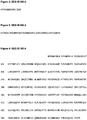
- In other instances the peptide consists of a sequence of SEQ ID NO: 3 (
Figure 3 ), said peptide being useful as an immunogen to generate an antibody that is capable of selectively binding to a non ATP -binding P2X7 receptor but not to an ATP -binding P2X7 receptor. - The peptide may consist of a sequence within the sequence of SEQ ID NO: 3, said peptide being useful as an immunogen to generate an antibody that is capable of selectively binding to a non ATP -binding P2X7 receptor but not to an ATP -binding P2X7 receptor. Examples of these peptides include those having a sequence described in Table 1 (numbering according to SEQ ID NO: 1):
Table 1 K281 to K297 Y298 to G314 T282 to Y298 Y299 to 1315 T283 to Y299 K300 to R316 N284 to K300 E301 to F317 V285 to E301 N302 to D318 S286 to N302 N303 to I319 L287 to N303 V304 to L320 Y288 to V304 E305 to V321 P289 to E305 K306 to F322 G290 to K306 R307 to G323 Y291 to R307 T308 to T324 N292 to T308 L309 to G325 F293 to L309 I310 to G326 R294 to 1310 K311 to K327 Y295 to K311 V312 to F328 A296 to V312 F313 to D329 K297 to F313 (SEQ ID NO: 2) - The peptide shown in Table 1 may have a length of from 6, 7, 8, 9, 10, 11, 12, 13, 14, 15, 16 or 17 residues.
- In other instances there is provided an antibody or fragment thereof capable of binding to a peptide described above.
- The antibody may be produced by immunisation with a peptide as described above. An example of a suitable immunisation is described in Example 1 below.
- The antibody may also be produced by immunisation with a non ATP -binding P2X7 receptor, such as a receptor having an amino acid sequence shown in SEQ ID NO: 1, or a fragment thereof including the amino acid sequences SEQ ID NOS: 3 to 9 (shown in
Figures 3 to 9 ). - In one instance , the non ATP-binding P2X7 receptor, monomer or fragment thereof used for the immunisation has Pro210 in cis isomerisation.
- The antibody may bind to an epitope that includes one or more residues of a peptide having a sequence of SEQ ID NO: 2. In certain instances , the antibody binds to K297 or Y298 or Y299 or K300 or E301 or N302 or N303 or V304 or E305 or K306 or R307 or T308 or L309 or 1310 or K311 or V312 or F313
- The epitope may include a sequence of residues of a peptide having a sequence of SEQ ID NO: 2. In certain instances , the antibody binds to a sequence including K297 and Y298, or Y298 and Y299, or Y299 and K300, or K300 and E301, or E301 and N302, or N302 and N303, or N303 and V304, or V304 and E305, or E305 and K306, or K306 and R307, or R307 and T308, or T308 and L309, or L309 and 1310, or 1310 and K311, or K311 and V312, or V312 and F313.
- The antibody that binds to one or more of the above residues may also bind to one or more residues of the P2X7 receptor extra cellular domain that are located outside of the region defined by SEQ ID NO:3. For example, the one or more residues located outside of the region defined by SEQ ID NO:3 may be located in a sequence of amino acid residues of an ATP -binding P2X7 receptor that defines the ATP binding site of the ATP -binding P2X7 receptor.
- In one instance , the one or more residues located outside the sequence of SEQ ID NO:3 may be located in the sequence of SEQ ID NOS: 10 to 12 (
Figures 10 to 12 respectively). - The antibody may be a whole antibody of any isotype. The antibody may be one obtained from monoclonal or polyclonal antisera. The antibody may be produced by hybridoma, or by recombinant expression.
- The antibody may be chimeric, i.e. one containing human variable domains and non human constant domains. Alternatively, it may be humanized, i.e one formed by grafting non human CDRs onto a human antibody framework. Still further, the antibody may be fully human.
- The antibodies of the disclosure may be modified with respect to effector function, so as to enhance, e.g., the effectiveness of the antibody in treating cancer. For example, cysteine residue(s) can be introduced into the Fc region, thereby allowing interchain disulfide bond formation in this region. The homodimeric antibody thus generated can have improved internalization capability and/or increased complement-mediated cell killing and antibody-dependent cellular cytotoxicity (ADCC). Homodimeric antibodies with enhanced anti-tumor activity can also be prepared using heterobifunctional crosslinkers. Alternatively, an antibody can be engineered that has dual Fc regions and can thereby have enhanced complement lysis and ADCC capabilities.
- Where the antibody is an antibody fragment, the antibody fragment is selected from the group consisting of a dAb, Fab, Fd, Fv, F(ab')2, scFv and CDR.
- The antibody or fragment may be provided on a solid phase such as a bead, surface or tissue culture vessel.
- The antibody or fragment thereof may be provided with a label for detection of binding of the antibody or fragment thereof to antigen.
- The antibodies and fragments may be labelled for use in medical imaging. Such methods involve chemical attachment of a labelling or imaging agent, such as a radioisotope, which include 67 Cu, 90 Y, 124 I, 125 I, 131 I, 186 Re, 188Re, 211 At, 212 Bi, administration of the labelled antibody or fragment to a subject in an acceptable carrier, and imaging the labelled antibody or fragment in vivo at the target site. Radio - labelled antibodies or fragments thereof may be particularly useful in in vivo imaging of cancers described herein.
- The antibodies can be purified by methods known to the skilled artisan. Purification methods include, among other, selective precipitation, liquid chromatography, HPLC, electrophoresis, chromatofocusing, and various affinity techniques.
- In some instances , the antibodies disclosed herein may also include multimeric forms of antibodies. For example, antibodies of the disclosure may take the form of antibody dimers, trimers, or higher-order multimers of monomeric immunoglobulin molecules.
- Crosslinking of antibodies can be done through various methods known in the art. For example, crosslinking of antibodies may be accomplished through natural aggregation of antibodies, through chemical or recombinant linking techniques or other methods known in the art. For example, purified antibody preparations can spontaneously form protein aggregates containing antibody homodimers, and other higher-order antibody multimers. In a specific instance , crosslinking of antibodies by using a second antibody to bind to the antibodies of interest can be used to form a homodimer. The crosslinker antibody can be derived from a different animal compared to the antibody of interest. For example, a goat anti- mouse antibody (Fab specific) may be added to a mouse monoclonal antibody to form a homodimer. This bivalent crosslinker antibody recognizes the Fab or Fc region of the two antibodies of interest forming a homodimer.
- Alternatively, antibody homodimers may be formed through chemical linkage techniques known in the art. Chemical crosslinkers can be homo or heterobifunctional and will covalently bind with two antibodies forming a homodimer. In some instances , it is desirable that the chemical crosslinker not interact with the antigen-binding region of the antibody as this may affect antibody function. As will be appreciated by those skilled in the art, antibodies can be crosslinked at the Fab region.
- In one instance there is provided an immune complex formed from the binding of an antibody or fragment thereof as described above to a non ATP -binding (i.e. non-functional) P2X7 receptor, monomer or fragment thereof. In one instance there is provided an immune complex formed from the binding of an antibody or fragment thereof to a peptide described above.
- Generally an immune complex otherwise known as an antigen-antibody complex is a product that is formed from the binding of an antibody via an antibody binding site to an epitope on an antigen against which the antibody is raised. The complex may or may not consist of more than one antibody.
- The immune complex is particularly important as detection of this in vitro or in vivo is indicative of presence of, or predisposition to a disease or condition including preneoplasia and neoplasia. These detection methods are described in more detail below.
- The non-ATP binding P2X7 receptor, monomer or fragment thereof included in the immune complex may have Pro210 in cis isomerisation.
- The non-ATP binding P2X7 receptor, monomer or fragment thereof included in the immune complex may have an amino acid sequence of any one of SEQ ID NOS: 4 to 9 or fragments thereof.
- The non-ATP binding P2X7 receptor, monomer or fragment thereof included in the immune complex may have a molecular weight in the range of from about 15 to 80 kDa.
- The non-ATP binding P2X7 receptor, monomer or fragment thereof included in the immune complex may lack a transmembrane domain.
- The immune complex may be formed by binding a non-ATP binding P2X7 receptor, monomer or fragment thereof located on a cell surface membrane, in a cytoplasm, in a nucleus or in extra-cellular fluid. The extra -cellular fluid may be blood, plasma, serum, lymph, urine, semen, saliva, sputum, ascites, faeces, uterine and vaginal secretions, bile, amniotic fluid, cerebrospinal fluid and organ and tissue flushings.
- The antibody or antibody fragment included in the immune complex may be attached to a solid phase, such as a bead or a plate, so that the immune complex is attached to a solid phase when formed. Alternatively, the non-ATP binding P2X7 receptor, monomer or fragment thereof included in the immune complex may be attached to a solid phase.
- The antibody may be labelled for detection of formation of the immune complex.
- The immune complex may further include an antibody or fragment thereof, such as a capture antibody for capture of the immune complex. The further antibody or fragment thereof may bind to the anti P2X7 receptor antibody. Also, the further antibody or fragment thereof may bind to the receptor or fragment thereof.
- The further antibody or fragment thereof may be bound to a solid phase such as a phase described above.
- The further antibody may be labelled for detection of formation of the immune complex. Examples of labels include fluorophores, dyes, isotopes etc.
- In certain instances there is provided a method for determining whether a cell, tissue or extra cellular body fluid includes a non ATP -binding P2X7 receptor, monomer or fragment thereof including:
- contacting a cell, tissue or extra cellular body fluid with an antibody or fragment thereof in conditions for forming an immune complex as described above, and
- detecting whether an immune complex has been formed,
- In other instances there is provided a use of an antibody or fragment thereof described above in the manufacture of means for determining whether a cell, tissue or extra-cellular body fluid contains a P2X7 receptor, monomer or fragment thereof.
- In other instances there is provided a method for determining whether a cell, tissue or extra -cellular body fluid contains an antibody against a non ATP -binding P2X7 receptor, monomer or fragment thereof including:
- contacting a cell, a tissue or an extra -cellular body fluid with a peptide as described above in conditions for forming an immune complex between the peptide and an antibody in the cell, tissue or extra -cellular body fluid, and
- detecting whether an immune complex has been formed,
- In other instances there is provided a use of a peptide described above in the manufacture of means for determining whether a cell, tissue or extra-cellular body fluid contains an antibody against a non ATP -binding P2X7 receptor, monomer or fragment thereof.
- The presence of a given protein, or level of expression of a given protein in a host cell, tissue or extra -cellular body fluid can be detected by any number of assays. Examples include immunoassays, chromatography and mass spectrometry.
- Immunoassays, i.e. assays involving an element of the immune system are particularly preferred. These assays may generally be classified into one of:
- (i) assays in which purified antigen is used to detect an antibody in host serum. For example, purified antigen is bound to solid phase by adsorption or indirectly through another molecule and host serum is applied followed by another antibody for detecting presence or absence of host antibody;
- (ii) assays in which purified antigen is used to detect immune cells, such as T and B lymphocytes. For example, peripheral white cells are purified from a host and cultured with purified antigen. The presence or absence of one or factors indicating immunity are then detected. Other examples include assays that measure cell proliferation (lymphocyte proliferation or transformation assays) following exposure to purified antigen, and assays that measure cell death (including apoptosis) following exposure to purified antigen;
- (iii) assays in which purified antibody specific for antigen is used to detect antigen in the host. For example, purified antibody is bound to solid phase, host tissue is then applied followed by another antibody specific for the antigen to be detected. There are many examples of this approach including ELISA, RIA;
- (iv) assays in which a purified anti-idiotypic antibody is used to detect host antibody. For example, anti-idiotypic antibody is adsorbed to solid phase, host serum is added and anti-Fc antibody is added to bind to any host antibodies having been bound by the anti-idiotypic antibody.
- The immunoassays can be applied in vitro or in vivo.
- In one instance , the disease is typically a cancer such as carcinoma, sarcoma, lymphoma, or leukemia. Carcinomas that may be detected include, but not limited to, prostate, breast, skin, lung, cervix, uterus, stomach, oesophagus, bladder, and colon cancers.
- Whilst any body fluid can be used to detect any of these diseases, some body fluids may be more appropriate than others to detect certain diseases, for example urine may be more appropriate to detect prostate cancer and blood for detecting blood cancers such as lymphoma.
- In another instance there is provided a method for determining whether an individual has a cancer including the steps of:
- contacting a cell, a tissue or an extra -cellular body fluid with a peptide as described above in conditions for forming an immune complex between the peptide and an antibody in the cell, tissue or extra -cellular body fluid; or
- contacting a cell, tissue or extra cellular body fluid with an antibody or fragment thereof in conditions for forming an immune complex as described above, and
- detecting whether an immune complex has been formed,
- In a further instance there is provided use of anti purinergic (P2X) receptor antibody or fragment thereof as described above, or a peptide as described above for determining whether an individual has a cancer.
- In certain instances , cancer is selected from the group consisting of prostate cancer, invasive breast cancer, melanoma, adenocarcinoma of the bowel, serous ovarian cancer, squamous cell cancer of the cervix, endometrial cancer, small cell lung cancer, hepatocellular carcinoma, transitional cell carcinoma of the bladder, gastrointestinal stromal tumour, endometrial stromal tumour, pituitary cancer, mesothelioma, Hodgkin's lymphoma and thyroid papillary.
- In yet further instances there is provided a kit or composition for determining whether a cell, tissue or extra -cellular body fluid contains a non ATP -binding P2X7 receptor, monomer or fragment thereof, or an antibody against same including:
- a peptide as described above; and/or
- an antibody or fragment thereof as described above; and/or
- non ATP -binding P2X7 receptor, monomer or fragment thereof; and optionally
- a further antibody for binding to the peptide, antibody or fragment thereof or the non ATP -binding P2X7 receptor, monomer or fragment thereof;
- written instructions for use of the kit in a method described above.
- Kits are provided which contain the necessary reagents to carry out the assays of the present disclosure. The kit may include one or more compartments, each to receive one or more containers such as: (a) a first container comprising one of the components of the present disclosure described above; and (b) one or more other containers comprising one or more of the following: wash reagents, reagents capable of detecting presence of the antibody or peptide.
- The containers allow one to efficiently transfer reagents from one compartment to another compartment such that the samples and reagents are not cross-contaminated, and the agents or solutions of each container can be added in a quantitative fashion from one compartment to another.
- The kit typically contains containers which may be formed from a variety of materials such as glass or plastic, and can include for example, bottles, vials, syringes, and test tubes. A label typically accompanies the kit, and includes any writing or recorded material, which may be electronic or computer readable form (e.g., disk, optical disc, or tape) providing instructions or other information for used of the contents of the kit. The label indicates that the formulation is used for diagnosing or treating the disorder of choice.
- One skilled in the art will readily recognize that the disclosed antibodies and peptides of the present disclosure can be readily incorporated into one of the established kit formats which are well known in the art.
- In other instances there is provided a pharmaceutical composition including an antibody or fragment thereof as described above, or a peptide as described above, together with a pharmaceutically acceptable carrier, diluent or excipient.
- In the preparation of the pharmaceutical compositions comprising the antibodies or peptides described in the teachings herein, a variety of vehicles and excipients and routes of administration may be used, as will be apparent to the skilled artisan. Representative formulation technology is taught in, inter alia, Remington: The Science and Practice of Pharmacy, 19th Ed., Mack Publishing Co., Easton, PA (1995) and Handbook of Pharmaceutical Excipients, 3rd Ed, Kibbe, A.H. ed., Washington DC, American Pharmaceutical Association (2000).
- The pharmaceutical compositions will generally comprise a pharmaceutically acceptable carrier and a pharmacologically effective amount of the antibodies or peptides, or mixture of antibodies or mixture of peptides, or suitable salts thereof.
- The pharmaceutical composition may be formulated as powders, granules, solutions, suspensions, aerosols, solids, pills, tablets, capsules, gels, topical creams, suppositories, transdermal patches, and other formulations known in the art.
- For the purposes described herein, pharmaceutically acceptable salts of the antibodies and peptides is intended to include any art recognized pharmaceutically acceptable salts including organic and inorganic acids and/or bases. Examples of salts include sodium, potassium, lithium, ammonium, calcium, as well as primary, secondary, and tertiary amines, esters of lower hydrocarbons, such as methyl, ethyl, and propyl. Other salts include organic acids, such as acetic acid, propionic acid, pyruvic acid, maleic acid, succinic acid, tartaric acid, citric acid, benzoic acid, cinnamic acid, salicylic acid, etc.
- As used herein, "pharmaceutically acceptable carrier" comprises any standard pharmaceutically accepted carriers known to those of ordinary skill in the art in formulating pharmaceutical compositions. Thus, the antibodies or peptides, by themselves, such as being present as pharmaceutically acceptable salts, or as conjugates, may be prepared as formulations in pharmaceutically acceptable diluents; for example, saline, phosphate buffer saline (PBS), aqueous ethanol, or solutions of glucose, mannitol, dextran, propylene glycol, oils (e.g., vegetable oils, animal oils, synthetic oils, etc.), microcrystalline cellulose, carboxymethyl cellulose, hydroxylpropyl methyl cellulose, magnesium stearate, calcium phosphate, gelatin, polysorbate 80 or as solid formulations in appropriate excipients.
- The pharmaceutical compositions will often further comprise one or more buffers (e.g., neutral buffered saline or phosphate buffered saline), carbohydrates (e.g., glucose, sucrose or dextrans), mannitol, proteins, polypeptides or amino acids such as glycine, antioxidants (e.g., ascorbic acid, sodium metabisulfite, butylated hydroxytoluene, butylated hydroxyanisole, etc.), bacteriostats, chelating agents such as EDTA or glutathione, adjuvants (e.g., aluminium hydroxide), solutes that render the formulation isotonic, hypotonic or weakly hypertonic with the blood of a recipient, suspending agents, thickening agents and/or preservatives. Alternatively, compositions of the present disclosure may be formulated as a lyophilizate.
- While any suitable carrier known to those of ordinary skill in the art may be employed in the compositions of this disclosure, the type of carrier will typically vary depending on the mode of administration. Antibody and peptide compositions may be formulated for any appropriate manner of administration, including for example, oral, nasal, mucosal, intravenous, intraperitoneal, intradermal, subcutaneous, and intramuscular administration.
- For parenteral administration, the compositions can be administered as injectable dosages of a solution or suspension of the substance in a physiologically acceptable diluent with a pharmaceutical carrier that can be a sterile liquid such as sterile pyrogen free water, oils, saline, glycerol, polyethylene glycol or ethanol. Additionally, auxiliary substances, such as wetting or emulsifying agents, surfactants, pH buffering substances and the like can be present in compositions.
- Other components of pharmaceutical compositions are those of petroleum, animal, vegetable, or synthetic origin, for example, non-aqueous solutions of peanut oil, soybean oil, corn oil, cottonseed oil, ethyl oleate, and isopropyl myristate. Antibodies and peptides can be administered in the form of a depot injection or implant preparation which can be formulated in such a manner as to permit a sustained release of the active ingredient. An exemplary composition comprises antibody at 5 mg/ml, formulated in aqueous buffer consisting of 50 mM L-histidine, 150 mM NaCl, adjusted to pH 6.0 with HCl.
- Typically, the compositions are prepared as injectables, either as liquid solutions or suspensions; solid or powder forms suitable for reconstitution with suitable vehicles, including by way example and not limitation, sterile pyrogen free water, saline, buffered solutions, dextrose solution, etc., prior to injection. The preparation also can be emulsified or encapsulated in liposomes or micro particles such as polylactide, polyglycolide, or copolymers.
- The pharmaceutical compositions described herein may be presented in unit-dose or multi- dose containers, such as sealed ampoules or vials. Such containers are typically sealed in such a way to preserve the sterility and stability of the formulation until use. In general, formulations may be stored as suspensions, solutions or emulsions in oily or aqueous vehicles, as indicated above.
- Alternatively, a pharmaceutical composition may be stored in a freeze-dried condition requiring only the addition of a sterile liquid carrier immediately prior to use.
- In related instances there is provided a method of treatment of a disease characterised by the expression of a non ATP -binding P2X7 receptor, monomer or fragment thereof including the step of providing an antibody or fragment thereof as described above, or a peptide as described above to an individual requiring said treatment.
- Methods of immunotargeting cancer cells using antibodies or antibody fragments are well known in the art.
U.S. Pat. No. 6,306,393 describes the use of anti-CD22 antibodies in the immunotherapy of B-cell malignancies, andU.S. Pat. No. 6,329,503 describes immunotargeting of cells that express serpentine transmembrane antigens. Antibodies described herein (including humanized or human monoclonal antibodies or fragments or other modifications thereof, optionally conjugated to cytotoxic agents) can be introduced into a patient such that the antibody binds to cancer cells and mediates the destruction of the cells and the tumor and/or inhibits the growth of the cells or the tumor. - Without intending to limit the disclosure, mechanisms by which such antibodies can exert a therapeutic effect may include complement-mediated cytolysis, antibody-dependent cellular cytotoxicity (ADCC)1 modulating the physiologic function of the tumor antigen, inhibiting binding or signal transduction pathways, modulating tumor cell differentiation, altering tumor angiogenesis factor profiles, modulating the secretion of immune stimulating or tumor suppressing cytokines and growth factors, modulating cellular adhesion, and/or by inducing apoptosis.
- The antibodies can also be conjugated to toxic or therapeutic agents, such as radioligands or cytosolic toxins, and may also be used therapeutically to deliver the toxic or therapeutic agent directly to tumor cells.
- By "treatment" herein is meant therapeutic or prophylactic treatment, or a suppressive measure for the disease, disorder or undesirable condition. Treatment encompasses administration of the subject antibodies in an appropriate form prior to the onset of disease symptoms and/or after clinical manifestations, or other manifestations, of the disease to reduce disease severity, halt disease progression, or eliminate the disease. Prevention of the disease includes prolonging or delaying the onset of symptoms of the disorder or disease, preferably in a subject with increased susceptibility to the disease.
- The therapeutic preparations can use nonmodified antibodies or antibodies conjugated with a therapeutic compound, such as a toxin or cytotoxic molecule, depending on he functionality of the antibody. Generally, when nonmodified antibodies are used, they will typically have a functional Fc region. By "functional Fc region" herein is meant a minimal sequence for effecting the biological function of Fc, such as binding to Fc receptors, particularly FcyR (e.g., FcγR1, ,RIIA, RIIB, RIIIA, RIIIB).
- Without being bound by theory, it is believed that the Fc region may affect the effectiveness of anti¬ tumor monoclonal antibodies by binding to Fc receptors immune effector cells and modulating cell mediated cytotoxicity, endocytosis, phagocytosis, release of inflammatory cytokines, complement mediate cytotoxicity, and antigen presentation. In this regard, polyclonal antibodies, or mixtures of monoclonals will be advantageous because they will bind to different epitopes and thus have a higher density of Fc on the cell surface as compared to when a single monoclonal antibody is used. Of course, to enhance their effectiveness in depleting targeted cells, or where nonmodified antibodies are not therapeutically effective, antibodies conjugated to toxins or cytotoxic agents may be used.
- The antibody compositions may be used either alone or in combination with other therapeutic agents to increase efficacy of traditional treatments or to target abnormal cells not targeted by the antibodies. Combining the antibody therapy method with a chemotherapeutic, radiation or surgical regimen may be preferred in patients that have not received chemotherapeutic treatment, whereas treatment with the antibody therapy may be indicated for patients who have received one or more chemotherapies. Additionally, antibody therapy can also enable the use of reduced dosages of concomitant chemotherapy, particularly in patients that do not tolerate the toxicity of the chemotherapeutic agent very well. Furthermore, treatment of cancer patients with the antibody with tumors resistant to chemotherapeutic agents might induce sensitivity and responsiveness to these agents in combination.
- In one aspect, the antibodies are used adjunctively with therapeutic cytotoxic agents, including, by way of example and not limitation, busulfan, thioguanine, idarubicin, cytosine arabinoside, 6-mercaptopurine, doxorubicin, daunorubicin, etoposide, and hydroxyurea. Other agents useful as adjuncts to antibody therapy are compounds directed specifically to the abnormal cellular molecule found in the disease state. These agents will be disease specific. For example, for treating chronic myeloid leukemia arising from BCR-ABL activity, one class of useful compounds are inhibitors of abl kinase activity, such as Imatinib, an inhibitor of bcr-abl kinase, and antisense oligonucleotides against bcr (e.g., Oblimersen). Other agents include, among others, interferon-alpha, humanized anti- CD52, deacetylase inhibitor FR901228 (depsipeptide), and the like.
- The amount of the compositions needed for achieving a therapeutic effect will be determined empirically in accordance with conventional procedures for the particular purpose. Generally, for administering the compositions ex vivo or in vivo for therapeutic purposes, the compositions are given at a pharmacologically effective dose. By "pharmacologically effective amount" or "pharmacologically effective dose" is an amount sufficient to produce the desired physiological effect or amount capable of achieving the desired result, particularly for treating or retreating the disorder or disease condition, including reducing or eliminating one or more symptoms or manifestations of the disorder or disease.
- As an illustration, administration of antibodies to a patient suffering from prostate cancer provides a therapeutic benefit not only when the underlying disease is eradicated or ameliorated, but also when the patient reports a decrease in the severity or duration of the symptoms associated with the disease. Therapeutic benefit also includes halting or slowing the progression of the underlying disease or disorder, regardless of whether improvement is realized.
- The amount administered to the host will vary depending upon what is being administered, the purpose of the administration, such as prophylaxis or therapy, the state of the host, the manner of administration, the number of administrations, interval between administrations, and the like. These can be determined empirically by those skilled in the art and may be adjusted for the extent of the therapeutic response. Factors to consider in determining an appropriate dose include, but is not limited to, size and weight of the subject, the age and sex of the subject, the severity of the symptom, the stage of the disease, method of delivery of the agent, half-life of the agents, and efficacy of the agents. Stage of the disease to consider includes whether the disease is acute or chronic, relapsing or remitting phase, and the progressiveness of the disease. Determining the dosages and times of administration for a therapeutically effective amount are well within the skill of the ordinary person in the art.
- For any compositions of the present disclosure, the therapeutically effective dose is readily determined by methods well known in the art. For example, an initial effective dose can be estimated from cell culture or other in vitro assays. For example, Sliwkowsky, MX et al., Semin. Oncol. 26.suppl. 12) 60-70 (1999) describes in vitro measurements of antibody dependent cellular cytoxicity. A dose can then be formulated in animal models to generate a circulating concentration or tissue concentration, including that of the IC50 as determined by the cell culture assays.
- In addition, the toxicity and therapeutic efficacy are generally determined by cell culture assays and/or experimental animals, typically by determining a LD50 (lethal dose to 50% of the test population) and ED50 (therapeutically effectiveness in 50% of the test population). The dose ratio of toxicity and therapeutic effectiveness is the therapeutic index. Preferred are compositions, individually or in combination, exhibiting high therapeutic indices. Determination of the effective amount is well within the skill of those in the art, particularly given the detailed disclosure provided herein. Guidance is also found in standard reference works, for example Fingl and Woodbury, General Principles In: The Pharmaceutical Basis of Therapeutics pp. 1-46 (1975), and the references cited therein.
- To achieve an initial tolerizing dose, consideration is given to the possibility that the antibodies may be immunogenic in humans and in non-human primates. The immune response may be biologically significant and may impair the therapeutic efficacy of the antibody even if the antibody is partly or chiefly comprised of human immunoglobulin sequences such as, for example, in the case of a chimeric or humanized antibody. Within certain instances , an initial high dose of antibody is administered such that a degree of immunological tolerance to the therapeutic antibody is established.
- The tolerizing dose is sufficient to prevent or reduce the induction of an antibody response to repeat administration of the committed progenitor cell specific antibody.
- Preferred ranges for the tolerizing dose are between 10 mg/kg body weight to 50 mg/kg body weight, inclusive. More preferred ranges for the tolerizing dose are between 20 and 40 mg/kg, inclusive. Still more preferred ranges for the tolerizing dose are between 20 and 25 mg/kg, inclusive.
- Within these therapeutic regimens, the therapeutically effective dose of antibodies is preferably administered in the range of 0.1 to 10 mg/kg body weight, inclusive. More preferred second therapeutically effective doses are in the range of 0.2 to 5 mg/kg body weight, inclusive. Still more preferred therapeutically effective doses are in the range of 0.5 to 2 mg/kg, inclusive. Within alternative instances , the subsequent therapeutic dose or doses may be in the same or different formulation as the tolerizing dose and/or may be administered by the same or different route as the tolerizing dose.
- For the purposes of this disclosure, the methods of administration are chosen depending on the condition being treated, the form of the subject antibodies, and the pharmaceutical composition.
- Administration of the antibody compositions can be done in a variety of ways, including, but not limited to, continuously, subcutaneously, intravenously, orally, topically, transdermal, intraperitoneal, intramuscularly, and intravesically. For example, microparticle, microsphere, and microencapsulate formulations are useful for oral, intramuscular, or subcutaneous administrations. Liposomes and nanoparticles are additionally suitable for intravenous administrations. Administration of the pharmaceutical compositions may be through a single route or concurrently by several routes. For instance, intraperitoneal administration can be accompanied by intravenous injections. Preferably the therapeutic doses are administered intravenously, intraperitonealy, intramuscularly, or subcutaneously.
- The compositions may be administered once or several times. In some instances, the compositions may be administered once per day, a few or several times per day, or even multiple times per day, depending upon, among other things, the indication being treated and the judgement of the prescribing physician.
- Administration of the compositions may also be achieved through sustained release or long- term delivery methods, which are well known to those skilled in the art. By "sustained release or" "long term release" as used herein is meant that the delivery system administers a pharmaceutically therapeutic amount of subject compounds for more than a day, preferably more than a week, and most preferable at least about 30 days to 60 days, or longer. Long term release systems may comprise implantable solids or gels containing the antibodies, such as biodegradable polymers described above; pumps, including peristaltic pumps and fluorocarbon propellant pumps; osmotic and mini-osmotic pumps; and the like.
- The method of the disclosure contemplates the administration of single monoclonal antibodies and any antibody that recognizes the particular antigens recognized by these antibodies, as well as combinations, of different mAbs. Two or more monoclonal antibodies may provide an improved effect compared to a single antibody. Alternatively, a combination of an antibody with an antibody that binds a different antigen may provide an improved effect compared to a single antibody. Such mAb cocktails may have certain advantages inasmuch as they contain mAbs, which exploit different effector mechanisms or combine directly cytotoxic mAbs with mAbs that rely on immune effector functionality. Such mAbs in combination may exhibit synergistic therapeutic effects.
- It will be understood that the disclosure disclosed and defined in this specification extends to all alternative combinations of two or more of the individual features mentioned or evident from the text or drawings. All of these different combinations constitute various alternative aspects of the disclosure.
- The following protocols are provided as non-limiting examples for the purpose of illustrating the invention.
- Anti-nonfunctional P2X7 antibody was raised against the peptide KYYKENNVEKRTLIKVF (SEQ ID NO: 2) or Lys-Tyr-Tyr-Lys-Glu-Asn-Asn-Val-Glu-Lys-Arg-Thr-Leu-Ile-Lys-Val-Phe, representing amino acids 297-313 of the human P2X7 protein. The peptide was synthesized by solid phase chemistry (Mimotopes Pty Ltd, Melbourne, Australia) to high purity (>95%) as judged by mass spectroscopy. A C-terminal Cys (C) residue was attached to the sequence and to this was attached the cross-linker MCS (6-Maleimido-Caproic Acid N-Hydroxysuccinimide Ester) for conjugating the peptide to carrier proteins including separately Diphtheria toxoid, bovine serum albumin and ovalbumin by Mimotopes.
- Female New Zealand White rabbits, aged between 10-12 weeks, were immunized according to the following schedule.
- On day one, each rabbit received 200 ug of P2X7 epitope conjugated to diphtheria toxoid (total antigen weight ∼ 500 ug of conjugated epitope). This conjugated peptide was supplied by Mimotopes Pty Ltd as a stable solution.
- The epitope conjugate was diluted in sterile PBS to a concentration of 500 ug per 0.8 ml PBS. To this was added 0.1ml of DEAE/Dextran/QUILA™ solution (2.5 mg QUILA™ plus 25 mg DEAE/Dextran per mL of PBS) and 1.2 mL of Montanide 15A50V. This solution was emulsified using glass luerloc syringes and a narrow bore luerloc coupling.
- Animals were each injected with 2 ml of the epitope adjuvant emulsion at multiple subcutaneous and intra-muscular sites.
- The above was repeated at 6-weeks and 9-weeks post initial injections. At week 10, the rabbits were injected intravenously with 1 ml of sterile PBS containing 50 ug of DT-conjugated P2X7 epitope. Four days later, the rabbits were bled out and the serum containing the antibody was stored for future analysis and use in immunoassays.
- The procedure in Example 2 was used except that mice were injected with 20 ug of P2X7 epitope (∼50 ug of DT-conjugate) in 0.2 mL of epitope adjuvant emulsion.
- Four days after the intravenous injection, antibody titers were measured in mouse blood and the highest titer mice were selected as spleen donors for hybridoma fusions.
- Mice selected above were used as spleen cell donors and these cells were fused with mouse SP20 myeloma cells to form hybridoma cell lines according to the 96-well plate format modification of the original protocol described by Kohler and Milstein.
- Cell lines were selected for stability and production of the specific antibody to particular P2X7 epitopes.
- A monoclonal antibody with suitably high affinity for the target epitope was selected for the IHC study. The binding characteristics of the antibody was tested by measuring the interaction with the target epitope on a Biacore instrument. A total of 550 resonance units of binding was achieved in the 60 second loading time showing slow on rate. A very slow off rate was apparent following cessation of loading with no measurable diminution of binding over the subsequent 10 minutes.
- Binding of the monoclonal antibody to fixed and permeabilized C11STH cells expressing the non-functional receptor was performed on a flow cytometer (Becton-Dickenson) using Alexa-488 label. The mean value was 90.8 compared with negative controls of 1.9. Binding of the monoclonal antibody to receptors on the surface of unfixed live cells was also assessed. The mean value was 5.8 compared with 0.34 for the negative control.
- For fixed cells, standard fluorescent antibody staining and confocal microscopy was used as follows. Fixed cells on poly-L-lysine-coated glass coverslips in 48-well plates were incubated with 20% normal horse serum in phosphate buffered saline pH7.5 (PBS) for 20 minutes, washed with PBS for 5 minutes then incubated with primary antibody for 30 min, washed with PBS for 5 minutes and finally labeled with fluorescent labeled secondary antibody (Jackson Immunologics) for 30 minutes. Cells were then washed twice (2 x 5 minutes) with PBS before mounting the coverslips on slides in 50% glycerol in PBS. Cells were visualized with a Leica TCS NT UV laser confocal microscope system with the pinhole set at 1.0. Murine isotype control antibodies were used routinely as negative controls and showed no staining. Western blots of HEK293 cells transfected with P2X7 revealed a single band at approximately 75 kDa that was absent in non-transfected cells and in samples of homogenates pre-treated with the epitope. Cell protein extracts (30 ug) together with molecular weight markers were fractionated on sodium dodecyl sulfate polyacrylamide gel (8-16%) (Novex). Proteins were electro-blotted on Immobilon-P membrane (Millipore). Western blots were developed using the ECL-chemiluminescence system (Amersham).
- A total of 25 different cases of breast cancers, 25 cases of skin cancers and 25 cases of prostate cancers were examined by immunohistochemistry. There was no cross-reactivity between the functional receptors present on the red blood cells and the non-functional receptor antibody (data not shown).
- Examples of cancer tissue including prostate cancer, breast cancer as represented by ductal carcinoma in situ and melanoma all stained for non-functional P2X7. The epithelium in normal tissues with no adjacent tumour was devoid of the receptor while all cancer cases stained for the receptor.
- Cancer cell lines derived from these and other tissues an other were similarly found to express the receptors. Examples tested and found positive include ADDP, PC3, LNCap, MCF7, MDA-MB-235, MDA-MB-431, NCIH460, NCIH69, NCIH596, DU145, ACHN, 786-O, Hep3B2, C11STH and BT474. Similar patterns of expression were seen on tumor xenografts of ovarian ADDP and lung NCIH69 respectively. The non-functional P2X7 receptors were found on the plasma membrane with correspondingly little residual cytoplasmic receptor in the most advanced stages of the cancers. In contrast, lower grade tumours exhibited a preponderance of cells in which a large proportion of the receptors remain intracellular. Such a progressive transport of non-functioning apoptotic receptors to the plasma membrane over the course of the disease progression indicates that cancer cell lines should exhibit non-functional P2X7 receptors on the plasma membrane rather than being largely intracellular.
- Normal epithelial tissue is devoid of the non-functional receptors while cancer tissues are all labelled. Different breast cancers, including both invasive and in situ lobular and ductal carcinomas were examined. All types expressed non-functional receptors. Epithelial cells in these and other normal, non-cancerous tissues such as bowel, bladder, ovarian, uterine, cervical, stomach and lung were found to be devoid of non-functional receptors.
- Areas of morphologically normal tissue surrounding tumours in prostate also expressed the receptors consistent with the field-effect in which tumour cells influence surrounding normal epithelium in connected ducts thereby alerting these cells to the presence of a developing tumour. In response, these cells begin deploying receptor, initially in an entirely intracellular location but eventually on the plasma membrane.
- In addition to the breast, skin and prostate cancer samples, other examples of epithelial cell cancers, all of which expressed non-functional P2X7 included bowel adenocarcinoma, invasive ovarian cancer, squamous cell carcinoma of the cervix, endometrial carcinoma of the uterus, small cell lung cancer, hepatocellular carcinoma, transitional cell carcinoma of the bladder and Barrett's mucosa with adenocarcinoma.
- Human cancers of non-epithelial cell origin were also examined. All were found to express the non-functional P2X7 receptors. Examples of mesenchymal cancers include gastrointestinal stromal tumour and endometrial stromal tumour. Equally, other tumours derived from this cell type such as Ewing's sarcoma express the receptors as do brain tumours such as oligodendrogliomas and astrocytomas, as do pituitary carcinoma. Mesothelioma, a cancer derived from pleural cells, also express non functional receptors, as do solid tumours derived from blood cells such as mantle cell lymphomas. Hodgkin's lymphoma and thyroid papillary are examples.
- Cross-reactivity between human and other mammalian cancers such as prostate, breast and skin from dogs and cats and melanoma in mice was observed using the same antibody to human receptor. These observations add weight to the conclusion that the cancer cell target is ubiquitous.
- There are indications that the receptor expression alters with tumour grade providing the potential to differentiate between latent and aggressive forms of cancer. Certainly, very slow growing low grade prostate cancers exhibit a pattern of receptor expression that is almost entirely intracellular, while cases of invasive prostate cancer exhibit more plasma membrane and myoepithelial cell labelling together with a significantly elevated receptor density.
- Low grade and clinically unimportant prostate cancer can be differentiated from clinically important prostate cancer by the appearance of the surrounding areas of normal epithelium. Clinically important cancers include those that are likely to spread into the prostatic stroma and metastasize. Direct sampling of the tumour with trans rectal ultrasound (TRUS) guided needle biopsies may detect a clinically significant tumour, thus making diagnosis straightforward. In cases in which the extant tumour is sampled in a region that is lower grade (eg Gleason 3+3) with other areas (eg Gleason 3+4) missed, staining for the non-functional P2X7 receptor shows that areas of normal morphology also sampled in the prostate show intense levels of receptor expression and myoepithelial stain, indicating the presence of tissue that is metastasizing. Even cases in which the tumour is completely missed, such as tumours confined to the apical lobe out of reach of the needles, the presence and status of the tumour is readily detected as the field-effect emanating from the tumour to surrounding epithelium alerts the pathologist to the presence of the tumour and the likely grade.
- Dysplastic tissue is difficult to diagnose and conditions such as Barrett's mucosa with dysplasia need to be monitored given the propensity of the tissue to transform to adenocarcinoma. The differentiation between dysplastic tissue that will remain benign and tissue that is at extreme risk of imminent transformation again centres on the presence of associated myoepithelial stain showing a dramatic up-regulation of non-functional P2X7 receptor expression. Similar results were observed for other dysplastic bowel conditions such as colitis. In tissue samples of Barrett's with dysplasia there was no staining indicating a benign condition while samples with intense myoepithelial stain identified a subject that had an associated adenocarcinoma.
- Early cases of neoplastic transformation including cervical intraepithelial neoplasia (CIN) grades 1-3 also showed increased levels of receptor expression and cases such as ovarian serous borderline were categorised as being benign (unstained) and those that were going to transform (heavy epithelial stain).
- The above results indicate that an epitope that includes one or more amino acids of the sequence KYYKENNVEKRTLIKVF has expression on a broad selection of epithelial cancer tissues and cells.
-
- <110> Biosceptre International Limited
- <120> Novel P2X7 epitopes
- <130> M81454722:KAB
- <150>
2007905018
<151> 2007-09-07 - <160> 12
- <170> PatentIn version 3.5
- <210> 1
<211> 595
<212> PRT
<213> Homo sapiens - <400> 1


- <210> 2
<211> 17
<212> PRT
<213> Homo sapiens - <400> 2

- <210> 3
<211> 49
<212> PRT
<213> Homo sapiens - <400> 3


- <210> 4
<211> 505
<212> PRT
<213> Homo sapiens - <400> 4


- <210> 5
<211> 425
<212> PRT
<213> Homo sapiens - <400> 5


- <210> 6
<211> 354
<212> PRT
<213> Homo sapiens - <400> 6


- <210> 7
<211> 306
<212> PRT
<213> Homo sapiens - <400> 7


- <210> 8
<211> 275
<212> PRT
<213> Homo sapiens - <400> 8


- <210> 9
<211> 274
<212> PRT
<213> Homo sapiens - <400> 9


- <210> 10
<211> 258
<212> PRT
<213> Homo sapiens - <400> 10


- <210> 11
<211> 128
<212> PRT
<213> Homo sapiens - <400> 11

- <210> 12
<211> 17
<212> PRT
<213> Homo sapiens - <400> 12

Claims (12)
- A peptide consisting of a sequence within the sequence shown in SEQ ID NO: 3, said peptide being useful as an immunogen to generate an antibody that is capable of selectively binding to a non ATP -binding P2X7 receptor but not to an ATP -binding P2X7 receptor, wherein the peptide includes the sequence shown in SEQ ID NO: 2.
- A peptide according to claim 1, wherein said peptide consists of the sequence shown in SEQ ID NO: 2.
- A peptide according to claim 1, wherein said peptide consists of the sequence shown in SEQ ID NO: 3.
- An isolated or purified antibody or fragment thereof capable of binding to a peptide consisting of the amino acid sequence shown in SEQ ID NO: 2.
- An isolated or purified antibody or fragment thereof according to claim 4, wherein the antibody fragment is selected from the group consisting of a Fab, Fd, Fv, F(ab')2 and scFv.
- An immune complex including the sequence shown in SEQ ID NO: 4, SEQ ID NO: 5, SEQ ID NO: 6 or SEQ ID NO: 9 and an isolated or purified antibody or fragment thereof according to claim 4 or 5, wherein the antibody or fragment thereof binds to SEQ ID NO: 2 in any one of SEQ ID NO: 4, SEQ ID NO: 5, SEQ ID NO: 6 or SEQ ID NO: 9.
- A method for determining whether a cell, tissue or extra cellular body fluid includes a non ATP -binding P2X7 receptor, monomer or fragment thereof including:- contacting a cell, tissue or extra cellular body fluid in vitro with an antibody or antigen-binding fragment thereof of claim 4 or 5 in conditions for forming an immune complex with a non ATP -binding P2X7 receptor, monomer or fragment thereof, and- detecting whether an immune complex has been formed,wherein detection of an immune complex determines that a cell, tissue or extra - cellular body fluid includes a non ATP -binding P2X7 receptor, monomer or fragment thereof.
- A method for determining whether an extra -cellular body fluid contains an antibody against a non ATP -binding P2X7 receptor, monomer or fragment thereof including:- contacting an extra -cellular body fluid in vitro with a peptide according to any one of claims 1 to 3 in conditions for forming an immune complex, and- detecting whether an immune complex has been formed between the peptide according to any one of claims 1 to 3 and an antibody against a non ATP - binding P2X7 receptor, monomer or fragment thereof,wherein detection of an immune complex determines that an extra -cellular body fluid contains an antibody against a non ATP -binding P2X7 receptor, monomer or fragment thereof.
- A pharmaceutical composition including an antibody or antigen-binding fragment thereof according to claim 4 or 5, together with a pharmaceutically acceptable carrier, diluent or excipient.
- A pharmaceutical composition including a peptide according to any one of claims 1 to 3 together with a pharmaceutically acceptable carrier, diluent or excipient.
- Use of an antibody according to claim 4 or 5 in the manufacture of a medicament for the treatment of cancer.
- An antibody according to claim 4 or 5 for use in the treatment of cancer.
Applications Claiming Priority (2)
| Application Number | Priority Date | Filing Date | Title |
|---|---|---|---|
| AU2007905018A AU2007905018A0 (en) | 2007-09-14 | Novel P2x7 epitopes | |
| PCT/AU2008/001364 WO2009033233A1 (en) | 2007-09-14 | 2008-09-12 | Novel p2x7 epitopes |
Publications (3)
| Publication Number | Publication Date |
|---|---|
| EP2201026A1 EP2201026A1 (en) | 2010-06-30 |
| EP2201026A4 EP2201026A4 (en) | 2012-11-28 |
| EP2201026B1 true EP2201026B1 (en) | 2017-01-04 |
Family
ID=40451491
Family Applications (1)
| Application Number | Title | Priority Date | Filing Date |
|---|---|---|---|
| EP08800000.5A Active EP2201026B1 (en) | 2007-09-14 | 2008-09-12 | Novel p2x7 epitopes |
Country Status (8)
| Country | Link |
|---|---|
| US (4) | US8440186B2 (en) |
| EP (1) | EP2201026B1 (en) |
| JP (2) | JP5670192B2 (en) |
| AU (1) | AU2008299593B2 (en) |
| CA (1) | CA2737126C (en) |
| ES (1) | ES2619681T3 (en) |
| NZ (1) | NZ583894A (en) |
| WO (1) | WO2009033233A1 (en) |
Families Citing this family (11)
| Publication number | Priority date | Publication date | Assignee | Title |
|---|---|---|---|---|
| DE60230482D1 (en) | 2001-01-17 | 2009-02-05 | Intreat Pty Ltd | ANTIBODIES TO NON-FUNCTIONAL P 2X7 RECEPTORS, DIAGNOSIS AND TREATMENT OF CANCER AND OTHER SUFFERING |
| JP5624885B2 (en) | 2007-09-14 | 2014-11-12 | バイオセプター・インターナショナル・リミテッド | Purinergic (P2X) receptors in extracellular body fluids |
| AU2008299593B2 (en) | 2007-09-14 | 2013-04-18 | Biosceptre International Limited | Novel P2X7 epitopes |
| WO2010000041A1 (en) | 2008-07-04 | 2010-01-07 | Biosceptre International Limiited | Anti- p2x7 peptides and epitopes |
| ES2704711T3 (en) | 2009-08-20 | 2019-03-19 | Biosceptre Aust Pty Ltd | Anti-P2X7 receptor antibodies and fragments thereof |
| ES2667003T3 (en) * | 2009-12-24 | 2018-05-09 | Biosceptre (Aust) Pty Ltd | Antibodies for non-functional oligomeric P2X7 receptors |
| US9562094B2 (en) | 2010-09-10 | 2017-02-07 | Biosceptre (Aust) Pty Ltd | Companion animal treatments |
| JP6305920B2 (en) | 2011-07-01 | 2018-04-04 | バイオセプター・(オーストラリア)・ピーティーワイ・リミテッド | Combination therapy |
| JP6041297B2 (en) * | 2012-08-24 | 2016-12-07 | 国立大学法人山口大学 | Diagnostic method and diagnostic kit for canine lymphoma |
| ES2848478T3 (en) * | 2015-09-11 | 2021-08-09 | Biosceptre Uk Ltd | Chimeric receptors for antigens and their uses |
| BR112019007895A8 (en) | 2016-10-21 | 2022-12-20 | Biosceptre Uk Ltd | CYTOTOXIC PARTICLES |
Family Cites Families (31)
| Publication number | Priority date | Publication date | Assignee | Title |
|---|---|---|---|---|
| IE920765A1 (en) | 1991-03-12 | 1992-09-23 | Scripps Research Inst | Cell surface receptors homologous to coagulation factors v¹and viii |
| WO1995033048A2 (en) | 1994-05-27 | 1995-12-07 | Glaxo Group Limited | P2x receptors (purinoceptor family) |
| US5981218A (en) | 1995-08-09 | 1999-11-09 | Bristol-Myers Squibb Company | Isolated nucleic acid molecules useful as leukemia markers and in breast cancer prognosis and encoded polypeptides |
| EP0907729A1 (en) | 1996-04-30 | 1999-04-14 | Smithkline Beecham Plc | HUMAN P2x4 RECEPTOR SPLICE-VARIANTS |
| US6303338B1 (en) * | 1996-08-16 | 2001-10-16 | Human Genome Sciences, Inc. | Pancreas-derived plasminogen activator inhibitor |
| US6306393B1 (en) * | 1997-03-24 | 2001-10-23 | Immunomedics, Inc. | Immunotherapy of B-cell malignancies using anti-CD22 antibodies |
| JP3885177B2 (en) | 1997-03-26 | 2007-02-21 | 大塚製薬株式会社 | Human gene |
| US6133434A (en) * | 1997-04-28 | 2000-10-17 | Glaxo Group Limited | Purinergic receptor |
| CA2328989C (en) * | 1998-06-01 | 2012-02-07 | Urogenesys, Inc. | Serpentine transmembrane antigens expressed in human cancers and uses thereof |
| DE19829473C2 (en) * | 1998-07-01 | 2000-08-10 | Magnus Von Knebel Doeberitz Ch | Procedure for early diagnosis of carcinomas |
| EP1157038A4 (en) | 1999-02-26 | 2005-01-19 | Smithkline Beecham Corp | Cloning of a p2y-like 7tm receptor (axor17) |
| AUPP991199A0 (en) * | 1999-04-21 | 1999-05-13 | University Of Sydney, The | Methods for diagnosing pre-cancerous and cancerous conditions |
| NL1012452C2 (en) | 1999-06-28 | 2001-01-02 | Univ Delft Tech | A process for the preparation of protein agglomerates, protein agglomerates, a foodstuff and a pharmaceutical preparation containing them and an apparatus for the preparation of protein agglomerates. |
| AU1339701A (en) | 1999-10-22 | 2001-05-08 | Lifespan Biosciences, Inc. | Anti-cancer nucleic acid and protein targets |
| AUPR201500A0 (en) | 2000-12-11 | 2001-01-11 | Biosceptre Pty Ltd | Methods for identifying pre-neoplastic and neoplastic states in mammals |
| DE60230482D1 (en) * | 2001-01-17 | 2009-02-05 | Intreat Pty Ltd | ANTIBODIES TO NON-FUNCTIONAL P 2X7 RECEPTORS, DIAGNOSIS AND TREATMENT OF CANCER AND OTHER SUFFERING |
| NZ549019A (en) * | 2001-09-03 | 2008-05-30 | Intreat Pty Ltd | Antibody that binds to a P2X7 receptor in which proline at position 210 is in a cis conformation |
| CA2522985A1 (en) * | 2003-04-17 | 2004-10-28 | Affectis Pharmaceuticals Ag | Means and methods for diagnosing and treating affective disorders |
| CA2607541A1 (en) | 2005-05-05 | 2006-12-28 | Medicure International Inc. | Inhibition of atp-mediated, p2x7 dependent pathways by pyridoxal-5-phosphaste and vitamin b6 related compounds |
| US7767789B2 (en) * | 2005-06-02 | 2010-08-03 | University Hopitals of Cleveland | Truncated proteins as cancer markers |
| EP1929305A2 (en) | 2005-08-31 | 2008-06-11 | Cell Signaling Technology, Inc. | Reagents for the detection of protein phosphorylation in leukemia signaling pathways |
| EP2082032A4 (en) * | 2006-10-10 | 2010-06-30 | Biosceptre Int Ltd | Hybridomas producing antibodies against non functional p2x7 receptor |
| US20100036101A1 (en) * | 2006-10-10 | 2010-02-11 | Angus Gidley-Baird | Antibodies against non functional p2x7 receptor |
| AU2008299593B2 (en) | 2007-09-14 | 2013-04-18 | Biosceptre International Limited | Novel P2X7 epitopes |
| JP5624885B2 (en) | 2007-09-14 | 2014-11-12 | バイオセプター・インターナショナル・リミテッド | Purinergic (P2X) receptors in extracellular body fluids |
| WO2010000041A1 (en) | 2008-07-04 | 2010-01-07 | Biosceptre International Limiited | Anti- p2x7 peptides and epitopes |
| ES2704711T3 (en) | 2009-08-20 | 2019-03-19 | Biosceptre Aust Pty Ltd | Anti-P2X7 receptor antibodies and fragments thereof |
| ES2667003T3 (en) | 2009-12-24 | 2018-05-09 | Biosceptre (Aust) Pty Ltd | Antibodies for non-functional oligomeric P2X7 receptors |
| WO2011131472A1 (en) | 2010-04-22 | 2011-10-27 | Institut Gustave Roussy | Compounds and uses thereof to induce an immunogenic cancer cell death in a subject |
| US9562094B2 (en) | 2010-09-10 | 2017-02-07 | Biosceptre (Aust) Pty Ltd | Companion animal treatments |
| JP6305920B2 (en) | 2011-07-01 | 2018-04-04 | バイオセプター・(オーストラリア)・ピーティーワイ・リミテッド | Combination therapy |
-
2008
- 2008-09-12 AU AU2008299593A patent/AU2008299593B2/en active Active
- 2008-09-12 US US12/677,799 patent/US8440186B2/en active Active
- 2008-09-12 WO PCT/AU2008/001364 patent/WO2009033233A1/en active Application Filing
- 2008-09-12 NZ NZ583894A patent/NZ583894A/en unknown
- 2008-09-12 EP EP08800000.5A patent/EP2201026B1/en active Active
- 2008-09-12 ES ES08800000.5T patent/ES2619681T3/en active Active
- 2008-09-12 CA CA2737126A patent/CA2737126C/en active Active
- 2008-09-12 JP JP2010524309A patent/JP5670192B2/en active Active
-
2013
- 2013-03-15 US US13/841,692 patent/US9181320B2/en active Active
-
2014
- 2014-06-24 JP JP2014129036A patent/JP2014196337A/en not_active Withdrawn
-
2015
- 2015-10-07 US US14/877,715 patent/US9944701B2/en active Active
-
2018
- 2018-03-02 US US15/910,987 patent/US10597451B2/en active Active
Non-Patent Citations (2)
| Title |
|---|
| HOLT L J ET AL: "Domain antibodies: proteins for therapy", TRENDS IN BIOTECHNOLOGY, ELSEVIER PUBLICATIONS, CAMBRIDGE, GB, vol. 21, no. 11, 1 November 2003 (2003-11-01), pages 484 - 490, XP004467495, ISSN: 0167-7799, DOI: 10.1016/J.TIBTECH.2003.08.007 * |
| WARD E S ET AL: "BINDING ACTIVITIES OF A REOPERTOIRE OF SINGLE IMMUNOGLOBULIN VARIABLE DOMAINS SECRETED FROM ESCHERICHIA COLI", NATURE, NATURE PUBLISHING GROUP, UNITED KINGDOM, vol. 341, no. 6242, 12 October 1989 (1989-10-12), pages 544 - 546, XP000086104, ISSN: 0028-0836, DOI: 10.1038/341544A0 * |
Also Published As
| Publication number | Publication date |
|---|---|
| US9181320B2 (en) | 2015-11-10 |
| AU2008299593A1 (en) | 2009-03-19 |
| JP2014196337A (en) | 2014-10-16 |
| US20130273085A1 (en) | 2013-10-17 |
| US10597451B2 (en) | 2020-03-24 |
| NZ583894A (en) | 2013-01-25 |
| US20180265580A1 (en) | 2018-09-20 |
| US20110177080A1 (en) | 2011-07-21 |
| AU2008299593B2 (en) | 2013-04-18 |
| WO2009033233A1 (en) | 2009-03-19 |
| JP5670192B2 (en) | 2015-02-18 |
| US8440186B2 (en) | 2013-05-14 |
| EP2201026A4 (en) | 2012-11-28 |
| US9944701B2 (en) | 2018-04-17 |
| AU2008299593A2 (en) | 2010-07-08 |
| JP2010539103A (en) | 2010-12-16 |
| EP2201026A1 (en) | 2010-06-30 |
| CA2737126C (en) | 2018-05-01 |
| CA2737126A1 (en) | 2009-03-19 |
| ES2619681T3 (en) | 2017-06-26 |
| US20160130342A1 (en) | 2016-05-12 |
Similar Documents
| Publication | Publication Date | Title |
|---|---|---|
| US10597451B2 (en) | Methods of treating cancer with a P2X7 peptide | |
| US8067550B2 (en) | Hybridomas producing antibodies against non functional P2X7 receptor | |
| KR101701300B1 (en) | Anti-p2x7 peptides and epitopes | |
| JP2017526654A (en) | Antibodies for therapy and diagnosis | |
| JP2016000713A (en) | Hybridoma clones and monoclonal antibodies to fibroblast growth factor 4 | |
| US9850303B2 (en) | Hybridoma clones and monoclonal antibodies to tetraspanin 8 | |
| JP6159011B2 (en) | Hybridoma clones and monoclonal antibodies against CD9 | |
| US20230295338A1 (en) | A novel antibody binding specifically to human ceacam1/3/5 and use thereof | |
| US20210040174A1 (en) | Composition and Use of Humoral Immune Suppressor Antagonists for the Treatment of Humoral Immune Suppressed Diseases | |
| WO2023235765A1 (en) | Methods, compositions and uses for sema7a monoclonal antibodies | |
| JP2016005449A (en) | Hybridoma clone and monoclonal antibody to cd9 | |
| US9193785B2 (en) | Hybridoma clones and monoclonal antibodies to ING4 |
Legal Events
| Date | Code | Title | Description |
|---|---|---|---|
| PUAI | Public reference made under article 153(3) epc to a published international application that has entered the european phase |
Free format text: ORIGINAL CODE: 0009012 |
|
| 17P | Request for examination filed |
Effective date: 20100414 |
|
| AK | Designated contracting states |
Kind code of ref document: A1 Designated state(s): AT BE BG CH CY CZ DE DK EE ES FI FR GB GR HR HU IE IS IT LI LT LU LV MC MT NL NO PL PT RO SE SI SK TR |
|
| AX | Request for extension of the european patent |
Extension state: AL BA MK RS |
|
| DAX | Request for extension of the european patent (deleted) | ||
| RAP1 | Party data changed (applicant data changed or rights of an application transferred) |
Owner name: BIOSCEPTRE INTERNATIONAL LIMITED |
|
| RIC1 | Information provided on ipc code assigned before grant |
Ipc: C07K 14/435 20060101ALI20121019BHEP Ipc: G01N 33/574 20060101ALI20121019BHEP Ipc: A61K 38/17 20060101ALI20121019BHEP Ipc: A61P 35/00 20060101ALI20121019BHEP Ipc: A61K 39/395 20060101ALI20121019BHEP Ipc: C07K 16/30 20060101ALI20121019BHEP Ipc: G01N 33/577 20060101ALI20121019BHEP Ipc: C07K 14/705 20060101ALI20121019BHEP Ipc: G01N 33/53 20060101ALI20121019BHEP Ipc: G01N 33/563 20060101ALI20121019BHEP Ipc: G01N 33/536 20060101ALI20121019BHEP Ipc: C07K 16/28 20060101ALI20121019BHEP Ipc: C07K 7/08 20060101AFI20121019BHEP Ipc: A61K 38/10 20060101ALI20121019BHEP |
|
| A4 | Supplementary search report drawn up and despatched |
Effective date: 20121029 |
|
| 17Q | First examination report despatched |
Effective date: 20130701 |
|
| GRAP | Despatch of communication of intention to grant a patent |
Free format text: ORIGINAL CODE: EPIDOSNIGR1 |
|
| INTG | Intention to grant announced |
Effective date: 20160817 |
|
| GRAS | Grant fee paid |
Free format text: ORIGINAL CODE: EPIDOSNIGR3 |
|
| GRAA | (expected) grant |
Free format text: ORIGINAL CODE: 0009210 |
|
| AK | Designated contracting states |
Kind code of ref document: B1 Designated state(s): AT BE BG CH CY CZ DE DK EE ES FI FR GB GR HR HU IE IS IT LI LT LU LV MC MT NL NO PL PT RO SE SI SK TR |
|
| RAP1 | Party data changed (applicant data changed or rights of an application transferred) |
Owner name: BIOSCEPTRE (AUST) PTY LTD |
|
| REG | Reference to a national code |
Ref country code: GB Ref legal event code: FG4D |
|
| REG | Reference to a national code |
Ref country code: CH Ref legal event code: EP |
|
| REG | Reference to a national code |
Ref country code: AT Ref legal event code: REF Ref document number: 859152 Country of ref document: AT Kind code of ref document: T Effective date: 20170115 |
|
| REG | Reference to a national code |
Ref country code: IE Ref legal event code: FG4D |
|
| REG | Reference to a national code |
Ref country code: DE Ref legal event code: R096 Ref document number: 602008048256 Country of ref document: DE |
|
| REG | Reference to a national code |
Ref country code: LT Ref legal event code: MG4D Ref country code: NL Ref legal event code: MP Effective date: 20170104 |
|
| REG | Reference to a national code |
Ref country code: AT Ref legal event code: MK05 Ref document number: 859152 Country of ref document: AT Kind code of ref document: T Effective date: 20170104 |
|
| REG | Reference to a national code |
Ref country code: ES Ref legal event code: FG2A Ref document number: 2619681 Country of ref document: ES Kind code of ref document: T3 Effective date: 20170626 |
|
| PG25 | Lapsed in a contracting state [announced via postgrant information from national office to epo] |
Ref country code: NL Free format text: LAPSE BECAUSE OF FAILURE TO SUBMIT A TRANSLATION OF THE DESCRIPTION OR TO PAY THE FEE WITHIN THE PRESCRIBED TIME-LIMIT Effective date: 20170104 |
|
| PG25 | Lapsed in a contracting state [announced via postgrant information from national office to epo] |
Ref country code: HR Free format text: LAPSE BECAUSE OF FAILURE TO SUBMIT A TRANSLATION OF THE DESCRIPTION OR TO PAY THE FEE WITHIN THE PRESCRIBED TIME-LIMIT Effective date: 20170104 Ref country code: LT Free format text: LAPSE BECAUSE OF FAILURE TO SUBMIT A TRANSLATION OF THE DESCRIPTION OR TO PAY THE FEE WITHIN THE PRESCRIBED TIME-LIMIT Effective date: 20170104 Ref country code: NO Free format text: LAPSE BECAUSE OF FAILURE TO SUBMIT A TRANSLATION OF THE DESCRIPTION OR TO PAY THE FEE WITHIN THE PRESCRIBED TIME-LIMIT Effective date: 20170404 Ref country code: FI Free format text: LAPSE BECAUSE OF FAILURE TO SUBMIT A TRANSLATION OF THE DESCRIPTION OR TO PAY THE FEE WITHIN THE PRESCRIBED TIME-LIMIT Effective date: 20170104 Ref country code: IS Free format text: LAPSE BECAUSE OF FAILURE TO SUBMIT A TRANSLATION OF THE DESCRIPTION OR TO PAY THE FEE WITHIN THE PRESCRIBED TIME-LIMIT Effective date: 20170504 Ref country code: GR Free format text: LAPSE BECAUSE OF FAILURE TO SUBMIT A TRANSLATION OF THE DESCRIPTION OR TO PAY THE FEE WITHIN THE PRESCRIBED TIME-LIMIT Effective date: 20170405 |
|
| PG25 | Lapsed in a contracting state [announced via postgrant information from national office to epo] |
Ref country code: PL Free format text: LAPSE BECAUSE OF FAILURE TO SUBMIT A TRANSLATION OF THE DESCRIPTION OR TO PAY THE FEE WITHIN THE PRESCRIBED TIME-LIMIT Effective date: 20170104 Ref country code: PT Free format text: LAPSE BECAUSE OF FAILURE TO SUBMIT A TRANSLATION OF THE DESCRIPTION OR TO PAY THE FEE WITHIN THE PRESCRIBED TIME-LIMIT Effective date: 20170504 Ref country code: LV Free format text: LAPSE BECAUSE OF FAILURE TO SUBMIT A TRANSLATION OF THE DESCRIPTION OR TO PAY THE FEE WITHIN THE PRESCRIBED TIME-LIMIT Effective date: 20170104 Ref country code: AT Free format text: LAPSE BECAUSE OF FAILURE TO SUBMIT A TRANSLATION OF THE DESCRIPTION OR TO PAY THE FEE WITHIN THE PRESCRIBED TIME-LIMIT Effective date: 20170104 Ref country code: BG Free format text: LAPSE BECAUSE OF FAILURE TO SUBMIT A TRANSLATION OF THE DESCRIPTION OR TO PAY THE FEE WITHIN THE PRESCRIBED TIME-LIMIT Effective date: 20170404 Ref country code: SE Free format text: LAPSE BECAUSE OF FAILURE TO SUBMIT A TRANSLATION OF THE DESCRIPTION OR TO PAY THE FEE WITHIN THE PRESCRIBED TIME-LIMIT Effective date: 20170104 |
|
| REG | Reference to a national code |
Ref country code: FR Ref legal event code: PLFP Year of fee payment: 10 |
|
| REG | Reference to a national code |
Ref country code: DE Ref legal event code: R097 Ref document number: 602008048256 Country of ref document: DE |
|
| PG25 | Lapsed in a contracting state [announced via postgrant information from national office to epo] |
Ref country code: SK Free format text: LAPSE BECAUSE OF FAILURE TO SUBMIT A TRANSLATION OF THE DESCRIPTION OR TO PAY THE FEE WITHIN THE PRESCRIBED TIME-LIMIT Effective date: 20170104 Ref country code: RO Free format text: LAPSE BECAUSE OF FAILURE TO SUBMIT A TRANSLATION OF THE DESCRIPTION OR TO PAY THE FEE WITHIN THE PRESCRIBED TIME-LIMIT Effective date: 20170104 Ref country code: CZ Free format text: LAPSE BECAUSE OF FAILURE TO SUBMIT A TRANSLATION OF THE DESCRIPTION OR TO PAY THE FEE WITHIN THE PRESCRIBED TIME-LIMIT Effective date: 20170104 Ref country code: EE Free format text: LAPSE BECAUSE OF FAILURE TO SUBMIT A TRANSLATION OF THE DESCRIPTION OR TO PAY THE FEE WITHIN THE PRESCRIBED TIME-LIMIT Effective date: 20170104 |
|
| PLBE | No opposition filed within time limit |
Free format text: ORIGINAL CODE: 0009261 |
|
| STAA | Information on the status of an ep patent application or granted ep patent |
Free format text: STATUS: NO OPPOSITION FILED WITHIN TIME LIMIT |
|
| PG25 | Lapsed in a contracting state [announced via postgrant information from national office to epo] |
Ref country code: DK Free format text: LAPSE BECAUSE OF FAILURE TO SUBMIT A TRANSLATION OF THE DESCRIPTION OR TO PAY THE FEE WITHIN THE PRESCRIBED TIME-LIMIT Effective date: 20170104 |
|
| 26N | No opposition filed |
Effective date: 20171005 |
|
| PG25 | Lapsed in a contracting state [announced via postgrant information from national office to epo] |
Ref country code: SI Free format text: LAPSE BECAUSE OF FAILURE TO SUBMIT A TRANSLATION OF THE DESCRIPTION OR TO PAY THE FEE WITHIN THE PRESCRIBED TIME-LIMIT Effective date: 20170104 |
|
| PG25 | Lapsed in a contracting state [announced via postgrant information from national office to epo] |
Ref country code: MC Free format text: LAPSE BECAUSE OF FAILURE TO SUBMIT A TRANSLATION OF THE DESCRIPTION OR TO PAY THE FEE WITHIN THE PRESCRIBED TIME-LIMIT Effective date: 20170104 |
|
| REG | Reference to a national code |
Ref country code: IE Ref legal event code: MM4A |
|
| REG | Reference to a national code |
Ref country code: BE Ref legal event code: MM Effective date: 20170930 |
|
| PG25 | Lapsed in a contracting state [announced via postgrant information from national office to epo] |
Ref country code: LU Free format text: LAPSE BECAUSE OF NON-PAYMENT OF DUE FEES Effective date: 20170912 |
|
| PG25 | Lapsed in a contracting state [announced via postgrant information from national office to epo] |
Ref country code: IE Free format text: LAPSE BECAUSE OF NON-PAYMENT OF DUE FEES Effective date: 20170912 |
|
| PG25 | Lapsed in a contracting state [announced via postgrant information from national office to epo] |
Ref country code: BE Free format text: LAPSE BECAUSE OF NON-PAYMENT OF DUE FEES Effective date: 20170930 |
|
| REG | Reference to a national code |
Ref country code: FR Ref legal event code: PLFP Year of fee payment: 11 |
|
| PG25 | Lapsed in a contracting state [announced via postgrant information from national office to epo] |
Ref country code: MT Free format text: LAPSE BECAUSE OF NON-PAYMENT OF DUE FEES Effective date: 20170912 |
|
| PG25 | Lapsed in a contracting state [announced via postgrant information from national office to epo] |
Ref country code: HU Free format text: LAPSE BECAUSE OF FAILURE TO SUBMIT A TRANSLATION OF THE DESCRIPTION OR TO PAY THE FEE WITHIN THE PRESCRIBED TIME-LIMIT; INVALID AB INITIO Effective date: 20080912 |
|
| PG25 | Lapsed in a contracting state [announced via postgrant information from national office to epo] |
Ref country code: CY Free format text: LAPSE BECAUSE OF NON-PAYMENT OF DUE FEES Effective date: 20170104 |
|
| PG25 | Lapsed in a contracting state [announced via postgrant information from national office to epo] |
Ref country code: TR Free format text: LAPSE BECAUSE OF FAILURE TO SUBMIT A TRANSLATION OF THE DESCRIPTION OR TO PAY THE FEE WITHIN THE PRESCRIBED TIME-LIMIT Effective date: 20170104 |
|
| P01 | Opt-out of the competence of the unified patent court (upc) registered |
Effective date: 20230602 |
|
| PGFP | Annual fee paid to national office [announced via postgrant information from national office to epo] |
Ref country code: GB Payment date: 20230920 Year of fee payment: 16 |
|
| PGFP | Annual fee paid to national office [announced via postgrant information from national office to epo] |
Ref country code: FR Payment date: 20230928 Year of fee payment: 16 Ref country code: DE Payment date: 20230920 Year of fee payment: 16 |
|
| PGFP | Annual fee paid to national office [announced via postgrant information from national office to epo] |
Ref country code: ES Payment date: 20231123 Year of fee payment: 16 |
|
| PGFP | Annual fee paid to national office [announced via postgrant information from national office to epo] |
Ref country code: IT Payment date: 20230927 Year of fee payment: 16 Ref country code: CH Payment date: 20231001 Year of fee payment: 16 |