EP2185164B1 - Composition and therapeutic anti-tumour vaccine - Google Patents
Composition and therapeutic anti-tumour vaccine Download PDFInfo
- Publication number
- EP2185164B1 EP2185164B1 EP08787072.1A EP08787072A EP2185164B1 EP 2185164 B1 EP2185164 B1 EP 2185164B1 EP 08787072 A EP08787072 A EP 08787072A EP 2185164 B1 EP2185164 B1 EP 2185164B1
- Authority
- EP
- European Patent Office
- Prior art keywords
- red blood
- blood cells
- cells
- composition
- ovalbumin
- Prior art date
- Legal status (The legal status is an assumption and is not a legal conclusion. Google has not performed a legal analysis and makes no representation as to the accuracy of the status listed.)
- Active
Links
Images
Classifications
-
- A—HUMAN NECESSITIES
- A61—MEDICAL OR VETERINARY SCIENCE; HYGIENE
- A61K—PREPARATIONS FOR MEDICAL, DENTAL OR TOILETRY PURPOSES
- A61K35/00—Medicinal preparations containing materials or reaction products thereof with undetermined constitution
- A61K35/12—Materials from mammals; Compositions comprising non-specified tissues or cells; Compositions comprising non-embryonic stem cells; Genetically modified cells
- A61K35/14—Blood; Artificial blood
- A61K35/18—Erythrocytes
-
- A—HUMAN NECESSITIES
- A61—MEDICAL OR VETERINARY SCIENCE; HYGIENE
- A61K—PREPARATIONS FOR MEDICAL, DENTAL OR TOILETRY PURPOSES
- A61K39/00—Medicinal preparations containing antigens or antibodies
- A61K39/0005—Vertebrate antigens
- A61K39/0011—Cancer antigens
-
- A—HUMAN NECESSITIES
- A61—MEDICAL OR VETERINARY SCIENCE; HYGIENE
- A61K—PREPARATIONS FOR MEDICAL, DENTAL OR TOILETRY PURPOSES
- A61K39/00—Medicinal preparations containing antigens or antibodies
- A61K39/0005—Vertebrate antigens
- A61K39/0011—Cancer antigens
- A61K39/001102—Receptors, cell surface antigens or cell surface determinants
- A61K39/001103—Receptors for growth factors
- A61K39/001106—Her-2/neu/ErbB2, Her-3/ErbB3 or Her 4/ErbB4
-
- A—HUMAN NECESSITIES
- A61—MEDICAL OR VETERINARY SCIENCE; HYGIENE
- A61K—PREPARATIONS FOR MEDICAL, DENTAL OR TOILETRY PURPOSES
- A61K39/00—Medicinal preparations containing antigens or antibodies
- A61K39/0005—Vertebrate antigens
- A61K39/0011—Cancer antigens
- A61K39/001102—Receptors, cell surface antigens or cell surface determinants
- A61K39/001122—Ephrin Receptors [Eph]
-
- A—HUMAN NECESSITIES
- A61—MEDICAL OR VETERINARY SCIENCE; HYGIENE
- A61K—PREPARATIONS FOR MEDICAL, DENTAL OR TOILETRY PURPOSES
- A61K39/00—Medicinal preparations containing antigens or antibodies
- A61K39/0005—Vertebrate antigens
- A61K39/0011—Cancer antigens
- A61K39/001136—Cytokines
- A61K39/001139—Colony stimulating factors [CSF]
-
- A—HUMAN NECESSITIES
- A61—MEDICAL OR VETERINARY SCIENCE; HYGIENE
- A61K—PREPARATIONS FOR MEDICAL, DENTAL OR TOILETRY PURPOSES
- A61K39/00—Medicinal preparations containing antigens or antibodies
- A61K39/0005—Vertebrate antigens
- A61K39/0011—Cancer antigens
- A61K39/001148—Regulators of development
- A61K39/001149—Cell cycle regulated proteins, e.g. cyclin, CDC, CDK or INK-CCR
-
- A—HUMAN NECESSITIES
- A61—MEDICAL OR VETERINARY SCIENCE; HYGIENE
- A61K—PREPARATIONS FOR MEDICAL, DENTAL OR TOILETRY PURPOSES
- A61K39/00—Medicinal preparations containing antigens or antibodies
- A61K39/0005—Vertebrate antigens
- A61K39/0011—Cancer antigens
- A61K39/001148—Regulators of development
- A61K39/00115—Apoptosis related proteins, e.g. survivin or livin
-
- A—HUMAN NECESSITIES
- A61—MEDICAL OR VETERINARY SCIENCE; HYGIENE
- A61K—PREPARATIONS FOR MEDICAL, DENTAL OR TOILETRY PURPOSES
- A61K39/00—Medicinal preparations containing antigens or antibodies
- A61K39/0005—Vertebrate antigens
- A61K39/0011—Cancer antigens
- A61K39/001148—Regulators of development
- A61K39/00115—Apoptosis related proteins, e.g. survivin or livin
- A61K39/001151—Apoptosis related proteins, e.g. survivin or livin p53
-
- A—HUMAN NECESSITIES
- A61—MEDICAL OR VETERINARY SCIENCE; HYGIENE
- A61K—PREPARATIONS FOR MEDICAL, DENTAL OR TOILETRY PURPOSES
- A61K39/00—Medicinal preparations containing antigens or antibodies
- A61K39/0005—Vertebrate antigens
- A61K39/0011—Cancer antigens
- A61K39/001152—Transcription factors, e.g. SOX or c-MYC
- A61K39/001153—Wilms tumor 1 [WT1]
-
- A—HUMAN NECESSITIES
- A61—MEDICAL OR VETERINARY SCIENCE; HYGIENE
- A61K—PREPARATIONS FOR MEDICAL, DENTAL OR TOILETRY PURPOSES
- A61K39/00—Medicinal preparations containing antigens or antibodies
- A61K39/0005—Vertebrate antigens
- A61K39/0011—Cancer antigens
- A61K39/001154—Enzymes
- A61K39/001156—Tyrosinase and tyrosinase related proteinases [TRP-1 or TRP-2]
-
- A—HUMAN NECESSITIES
- A61—MEDICAL OR VETERINARY SCIENCE; HYGIENE
- A61K—PREPARATIONS FOR MEDICAL, DENTAL OR TOILETRY PURPOSES
- A61K39/00—Medicinal preparations containing antigens or antibodies
- A61K39/0005—Vertebrate antigens
- A61K39/0011—Cancer antigens
- A61K39/001154—Enzymes
- A61K39/001157—Telomerase or TERT [telomerase reverse transcriptase]
-
- A—HUMAN NECESSITIES
- A61—MEDICAL OR VETERINARY SCIENCE; HYGIENE
- A61K—PREPARATIONS FOR MEDICAL, DENTAL OR TOILETRY PURPOSES
- A61K39/00—Medicinal preparations containing antigens or antibodies
- A61K39/0005—Vertebrate antigens
- A61K39/0011—Cancer antigens
- A61K39/001154—Enzymes
- A61K39/001158—Proteinases
- A61K39/001159—Matrix metalloproteinases [MMP]
-
- A—HUMAN NECESSITIES
- A61—MEDICAL OR VETERINARY SCIENCE; HYGIENE
- A61K—PREPARATIONS FOR MEDICAL, DENTAL OR TOILETRY PURPOSES
- A61K39/00—Medicinal preparations containing antigens or antibodies
- A61K39/0005—Vertebrate antigens
- A61K39/0011—Cancer antigens
- A61K39/001154—Enzymes
- A61K39/001162—Kinases, e.g. Raf or Src
-
- A—HUMAN NECESSITIES
- A61—MEDICAL OR VETERINARY SCIENCE; HYGIENE
- A61K—PREPARATIONS FOR MEDICAL, DENTAL OR TOILETRY PURPOSES
- A61K39/00—Medicinal preparations containing antigens or antibodies
- A61K39/0005—Vertebrate antigens
- A61K39/0011—Cancer antigens
- A61K39/001154—Enzymes
- A61K39/001164—GTPases, e.g. Ras or Rho
-
- A—HUMAN NECESSITIES
- A61—MEDICAL OR VETERINARY SCIENCE; HYGIENE
- A61K—PREPARATIONS FOR MEDICAL, DENTAL OR TOILETRY PURPOSES
- A61K39/00—Medicinal preparations containing antigens or antibodies
- A61K39/0005—Vertebrate antigens
- A61K39/0011—Cancer antigens
- A61K39/001166—Adhesion molecules, e.g. NRCAM, EpCAM or cadherins
-
- A—HUMAN NECESSITIES
- A61—MEDICAL OR VETERINARY SCIENCE; HYGIENE
- A61K—PREPARATIONS FOR MEDICAL, DENTAL OR TOILETRY PURPOSES
- A61K39/00—Medicinal preparations containing antigens or antibodies
- A61K39/0005—Vertebrate antigens
- A61K39/0011—Cancer antigens
- A61K39/001169—Tumor associated carbohydrates
- A61K39/00117—Mucins, e.g. MUC-1
-
- A—HUMAN NECESSITIES
- A61—MEDICAL OR VETERINARY SCIENCE; HYGIENE
- A61K—PREPARATIONS FOR MEDICAL, DENTAL OR TOILETRY PURPOSES
- A61K39/00—Medicinal preparations containing antigens or antibodies
- A61K39/0005—Vertebrate antigens
- A61K39/0011—Cancer antigens
- A61K39/001184—Cancer testis antigens, e.g. SSX, BAGE, GAGE or SAGE
-
- A—HUMAN NECESSITIES
- A61—MEDICAL OR VETERINARY SCIENCE; HYGIENE
- A61K—PREPARATIONS FOR MEDICAL, DENTAL OR TOILETRY PURPOSES
- A61K39/00—Medicinal preparations containing antigens or antibodies
- A61K39/0005—Vertebrate antigens
- A61K39/0011—Cancer antigens
- A61K39/001184—Cancer testis antigens, e.g. SSX, BAGE, GAGE or SAGE
- A61K39/001186—MAGE
-
- A—HUMAN NECESSITIES
- A61—MEDICAL OR VETERINARY SCIENCE; HYGIENE
- A61K—PREPARATIONS FOR MEDICAL, DENTAL OR TOILETRY PURPOSES
- A61K39/00—Medicinal preparations containing antigens or antibodies
- A61K39/0005—Vertebrate antigens
- A61K39/0011—Cancer antigens
- A61K39/001184—Cancer testis antigens, e.g. SSX, BAGE, GAGE or SAGE
- A61K39/001188—NY-ESO
-
- A—HUMAN NECESSITIES
- A61—MEDICAL OR VETERINARY SCIENCE; HYGIENE
- A61K—PREPARATIONS FOR MEDICAL, DENTAL OR TOILETRY PURPOSES
- A61K39/00—Medicinal preparations containing antigens or antibodies
- A61K39/0005—Vertebrate antigens
- A61K39/0011—Cancer antigens
- A61K39/001184—Cancer testis antigens, e.g. SSX, BAGE, GAGE or SAGE
- A61K39/001189—PRAME
-
- A—HUMAN NECESSITIES
- A61—MEDICAL OR VETERINARY SCIENCE; HYGIENE
- A61K—PREPARATIONS FOR MEDICAL, DENTAL OR TOILETRY PURPOSES
- A61K39/00—Medicinal preparations containing antigens or antibodies
- A61K39/0005—Vertebrate antigens
- A61K39/0011—Cancer antigens
- A61K39/00119—Melanoma antigens
- A61K39/001191—Melan-A/MART
-
- A—HUMAN NECESSITIES
- A61—MEDICAL OR VETERINARY SCIENCE; HYGIENE
- A61K—PREPARATIONS FOR MEDICAL, DENTAL OR TOILETRY PURPOSES
- A61K39/00—Medicinal preparations containing antigens or antibodies
- A61K39/0005—Vertebrate antigens
- A61K39/0011—Cancer antigens
- A61K39/00119—Melanoma antigens
- A61K39/001192—Glycoprotein 100 [Gp100]
-
- A—HUMAN NECESSITIES
- A61—MEDICAL OR VETERINARY SCIENCE; HYGIENE
- A61K—PREPARATIONS FOR MEDICAL, DENTAL OR TOILETRY PURPOSES
- A61K39/00—Medicinal preparations containing antigens or antibodies
- A61K39/0005—Vertebrate antigens
- A61K39/0011—Cancer antigens
- A61K39/001193—Prostate associated antigens e.g. Prostate stem cell antigen [PSCA]; Prostate carcinoma tumor antigen [PCTA]; PAP or PSGR
-
- A—HUMAN NECESSITIES
- A61—MEDICAL OR VETERINARY SCIENCE; HYGIENE
- A61K—PREPARATIONS FOR MEDICAL, DENTAL OR TOILETRY PURPOSES
- A61K39/00—Medicinal preparations containing antigens or antibodies
- A61K39/0005—Vertebrate antigens
- A61K39/0011—Cancer antigens
- A61K39/001196—Fusion proteins originating from gene translocation in cancer cells
- A61K39/001197—Breakpoint cluster region-abelson tyrosine kinase [BCR-ABL]
-
- A—HUMAN NECESSITIES
- A61—MEDICAL OR VETERINARY SCIENCE; HYGIENE
- A61K—PREPARATIONS FOR MEDICAL, DENTAL OR TOILETRY PURPOSES
- A61K39/00—Medicinal preparations containing antigens or antibodies
- A61K39/39—Medicinal preparations containing antigens or antibodies characterised by the immunostimulating additives, e.g. chemical adjuvants
-
- A—HUMAN NECESSITIES
- A61—MEDICAL OR VETERINARY SCIENCE; HYGIENE
- A61K—PREPARATIONS FOR MEDICAL, DENTAL OR TOILETRY PURPOSES
- A61K47/00—Medicinal preparations characterised by the non-active ingredients used, e.g. carriers or inert additives; Targeting or modifying agents chemically bound to the active ingredient
- A61K47/02—Inorganic compounds
-
- A—HUMAN NECESSITIES
- A61—MEDICAL OR VETERINARY SCIENCE; HYGIENE
- A61K—PREPARATIONS FOR MEDICAL, DENTAL OR TOILETRY PURPOSES
- A61K47/00—Medicinal preparations characterised by the non-active ingredients used, e.g. carriers or inert additives; Targeting or modifying agents chemically bound to the active ingredient
- A61K47/06—Organic compounds, e.g. natural or synthetic hydrocarbons, polyolefins, mineral oil, petrolatum or ozokerite
- A61K47/22—Heterocyclic compounds, e.g. ascorbic acid, tocopherol or pyrrolidones
-
- A—HUMAN NECESSITIES
- A61—MEDICAL OR VETERINARY SCIENCE; HYGIENE
- A61K—PREPARATIONS FOR MEDICAL, DENTAL OR TOILETRY PURPOSES
- A61K47/00—Medicinal preparations characterised by the non-active ingredients used, e.g. carriers or inert additives; Targeting or modifying agents chemically bound to the active ingredient
- A61K47/06—Organic compounds, e.g. natural or synthetic hydrocarbons, polyolefins, mineral oil, petrolatum or ozokerite
- A61K47/26—Carbohydrates, e.g. sugar alcohols, amino sugars, nucleic acids, mono-, di- or oligo-saccharides; Derivatives thereof, e.g. polysorbates, sorbitan fatty acid esters or glycyrrhizin
-
- A—HUMAN NECESSITIES
- A61—MEDICAL OR VETERINARY SCIENCE; HYGIENE
- A61P—SPECIFIC THERAPEUTIC ACTIVITY OF CHEMICAL COMPOUNDS OR MEDICINAL PREPARATIONS
- A61P35/00—Antineoplastic agents
-
- A—HUMAN NECESSITIES
- A61—MEDICAL OR VETERINARY SCIENCE; HYGIENE
- A61P—SPECIFIC THERAPEUTIC ACTIVITY OF CHEMICAL COMPOUNDS OR MEDICINAL PREPARATIONS
- A61P37/00—Drugs for immunological or allergic disorders
- A61P37/02—Immunomodulators
- A61P37/04—Immunostimulants
-
- C—CHEMISTRY; METALLURGY
- C07—ORGANIC CHEMISTRY
- C07K—PEPTIDES
- C07K16/00—Immunoglobulins [IG], e.g. monoclonal or polyclonal antibodies
- C07K16/18—Immunoglobulins [IG], e.g. monoclonal or polyclonal antibodies against material from animals or humans
- C07K16/28—Immunoglobulins [IG], e.g. monoclonal or polyclonal antibodies against material from animals or humans against receptors, cell surface antigens or cell surface determinants
- C07K16/2803—Immunoglobulins [IG], e.g. monoclonal or polyclonal antibodies against material from animals or humans against receptors, cell surface antigens or cell surface determinants against the immunoglobulin superfamily
- C07K16/2806—Immunoglobulins [IG], e.g. monoclonal or polyclonal antibodies against material from animals or humans against receptors, cell surface antigens or cell surface determinants against the immunoglobulin superfamily against CD2
-
- A—HUMAN NECESSITIES
- A61—MEDICAL OR VETERINARY SCIENCE; HYGIENE
- A61K—PREPARATIONS FOR MEDICAL, DENTAL OR TOILETRY PURPOSES
- A61K39/00—Medicinal preparations containing antigens or antibodies
- A61K2039/51—Medicinal preparations containing antigens or antibodies comprising whole cells, viruses or DNA/RNA
- A61K2039/515—Animal cells
- A61K2039/5154—Antigen presenting cells [APCs], e.g. dendritic cells or macrophages
-
- A—HUMAN NECESSITIES
- A61—MEDICAL OR VETERINARY SCIENCE; HYGIENE
- A61K—PREPARATIONS FOR MEDICAL, DENTAL OR TOILETRY PURPOSES
- A61K39/00—Medicinal preparations containing antigens or antibodies
- A61K2039/51—Medicinal preparations containing antigens or antibodies comprising whole cells, viruses or DNA/RNA
- A61K2039/515—Animal cells
- A61K2039/5156—Animal cells expressing foreign proteins
-
- A—HUMAN NECESSITIES
- A61—MEDICAL OR VETERINARY SCIENCE; HYGIENE
- A61K—PREPARATIONS FOR MEDICAL, DENTAL OR TOILETRY PURPOSES
- A61K39/00—Medicinal preparations containing antigens or antibodies
- A61K2039/555—Medicinal preparations containing antigens or antibodies characterised by a specific combination antigen/adjuvant
- A61K2039/55511—Organic adjuvants
- A61K2039/55522—Cytokines; Lymphokines; Interferons
-
- A—HUMAN NECESSITIES
- A61—MEDICAL OR VETERINARY SCIENCE; HYGIENE
- A61K—PREPARATIONS FOR MEDICAL, DENTAL OR TOILETRY PURPOSES
- A61K39/00—Medicinal preparations containing antigens or antibodies
- A61K2039/555—Medicinal preparations containing antigens or antibodies characterised by a specific combination antigen/adjuvant
- A61K2039/55511—Organic adjuvants
- A61K2039/55561—CpG containing adjuvants; Oligonucleotide containing adjuvants
-
- A—HUMAN NECESSITIES
- A61—MEDICAL OR VETERINARY SCIENCE; HYGIENE
- A61K—PREPARATIONS FOR MEDICAL, DENTAL OR TOILETRY PURPOSES
- A61K39/00—Medicinal preparations containing antigens or antibodies
- A61K2039/57—Medicinal preparations containing antigens or antibodies characterised by the type of response, e.g. Th1, Th2
- A61K2039/572—Medicinal preparations containing antigens or antibodies characterised by the type of response, e.g. Th1, Th2 cytotoxic response
-
- A—HUMAN NECESSITIES
- A61—MEDICAL OR VETERINARY SCIENCE; HYGIENE
- A61K—PREPARATIONS FOR MEDICAL, DENTAL OR TOILETRY PURPOSES
- A61K39/00—Medicinal preparations containing antigens or antibodies
- A61K2039/60—Medicinal preparations containing antigens or antibodies characteristics by the carrier linked to the antigen
- A61K2039/6031—Proteins
- A61K2039/6056—Antibodies
Definitions
- the present invention relates to a composition which induces, in a host, a cytotoxic response for anti-tumour purposes, and also to a therapeutic anti-tumour vaccine containing this composition.
- tumour antigens The natural immune response against tumour antigens is relatively ineffective and various mechanisms allowing tumours to escape the anti-tumour immune response have been identified.
- Conventional vaccine approaches generate humoral responses which prove to be insufficient.
- Dendritic cells are antigen-presenting cells (APCs) that are very effective in generating cytotoxic effectors specific for tumour cells. They are capable of phagocytizing apoptotic cells or apoptotic bodies originating from tumour cells, and then of presenting the tumour antigens, in association with MHC class I and II molecules, to T lymphocytes. Thus, the dendritic cell is capable of initiating the proliferation and the generation of a clone of specific cytolytic T lymphocytes. At the end of this reaction, the killer lymphocytes thus differentiated leave the lymphoid compartment so as to circulate in the organism and bind in the tumour. Recognition of the antigens expressed by a tumour then induces a lytic signal and brings about the destruction of the tumour cells.
- APCs antigen-presenting cells
- the dendritic cells are differentiated from blood cells taken from the patient: they are cultured and matured, "pulsed", i.e. stimulated, ex vivo with tumour peptides, tumour lysates, apoptotic tumour cells, or heat shock proteins extracted from autologous tumours, and finally reinjected into the patient.
- the stimulation of the dendritic cells is carried out after injection, into the patient, of peptides, proteins, irradiated tumour cells or else viruses containing the antigenic peptide targeting the dendritic cells.
- the cytotoxic response obtained is rarely accompanied by clinical efficacy.
- the activation of the dendritic cell "conditions" its ability to effectively activate the cytolytic T lymphocyte.
- the level of activation of dendritic cells appears to be a delicate point in this vaccine strategy.
- dendritic cells ex vivo raises a certain number of issues on: the state of maturation of the dendritic cells, the number of cells to be injected, the route, the site and the frequency of injection for producing dendritic cells capable of migrating to the secondary lymphoid organs and of inducing an effective cytotoxic T response ( Banchereau J, Schuler-Thurner B, Palucka AK, Schuler G. [Dendritic cells as vectors for therapy] Cell. 2001, 10, 106(3): 271-4 .).
- dendritic cells in vivo is, for its part, limited by the weak immunogenic capacity of tumour antigens and the difficulty in activating dendritic cells at a sufficient level.
- red blood cells as carriers for transporting antigens, encapsulated in the red blood cells or bound to their surface, and delivered to the APCs has been envisaged in several publications.
- the triggered immune responses have been investigated in vitro and in vivo.
- Hamidi et al. have recently described the encapsulation of BSA (bovine serum albumin) as a model of an antigen in human red blood cells ( Hamidi M et al., Drug Deliv., 2007; 14(5):295-300 and Int J Pharm., 2007, 29, 338(1-2): 70-8 ). The authors suggested the use of red blood cells as a vector for the presentation of antigens to APCs of the reticuloendothelial system (RES). In another review published by Hamidi et al. (J. Control.
- a humoral immune response can be obtained in vivo after immunization with antigen-loaded red blood cells.
- the study carried out by Murray et al. made it possible to detect, in mice, IgG immunoglobulins after intravenous injection of murine red blood cells loaded with one of the following four antigens: KLH (Keyhole Limpet Haemocyanin), BSA (Bovine Serum Albumin), CTB (Cholera Toxin b Subunit) and ADA (Bovine Adenosine Deaminase).
- IgG1 and IgG3 which are predominant immunoglobulin isotypes during a Th2 response, and of IgG2, immunoglobulin isotype which is a marker for a Th1 response, would suggest the involvement of both types of immune responses, the humoral response and the cellular response ( Murray AM et al., Vaccine., 2006 28, 24(35-36): 6129-39 ).
- Boberg et al. injected mice intraperitoneally with a vaccine constituted of peptides derived from the HIV-1 protease anchored at the surface of mouse red blood cells by means of the avidin/biotin system. They chemically modified red blood cells with the aim of promoting their recognition by APCs, but a weak immune reaction was obtained. They concluded that the small amount of antigens delivered, due to the limited loading of red blood cells with antigenic peptides and blood volume injected was not compensated for by the chemical modification of the carriers supposed to promote antigen recognition by APCs ( Boberg A. et al., Infect. Agents Cancer. , 2007, 182: 9 ).
- the present invention aims to provide compositions and vaccines that can be used in the treatment of cancers, according to an immunotherapy approach.
- An object of the invention is therefore a composition as defined in claim 1.
- the term "host” refers preferably to humans, but also to animals, in particular pets (especially dogs or cats) and animals for sport (especially horses).
- the red blood cells contain, i.e. encapsulate, the antigen, which means that the antigen is or is essentially inside the red blood cell.
- Said red blood cells are preferably designed, selected or modified so as to promote phagocytosis thereof by APCs, and most particularly by dendritic cells.
- said red blood cells are designed, selected or modified so as to promote phagocytosis thereof in the spleen and the liver, the essential objective being to target APCs of the spleen.
- the compositions according to the invention comprise red blood cells which contain the antigen and target the spleen.
- the composition promotes phagocytosis of these red blood cells by the APCs, in particular the dendritic cells, in the spleen.
- the red blood cells contain a tumour antigen and are in the form of an immune complex with an immunoglobulin which recognizes an epitope at the surface of said red blood cells, so as to promote the phagocytosis of said red blood cells, in particular by dendritic cells.
- the composition also makes it possible to promote phagocytosis by macrophages.
- the immunoglobulin is an immunoglobulin G.
- the formation of the immune complex involves red blood cells and at least one antibody, preferably of IgG subtype.
- Dendritic cells have, at their surface, receptors for the constant Fc region of immunoglobulins G (IgGs). These receptors are capable of triggering phagocytosis or internalization of the antigen-IgG immune complexes thus formed and of promoting antigen presentation by MHC class I and II molecules, resulting in the generation of CD4 + helper lymphocytes and especially CD8 + cytotoxic lymphocytes ( A. Regnault et al., J. Exp. Med., Janvier 1999, 189(2): 371-80 ).
- Anti-glycophorin A antibodies are a preferred modality.
- the red blood cells of human origin that are used to form an immune complex are heterologous red blood cells originating from a donor.
- the red blood cells contain a tumour antigen and are heat or chemically modified, so as to promote phagocytosis of said red blood cells, in particular by dendritic cells.
- the composition also makes it possible to promote phagocytosis by macrophages.
- the heat treatment is in particular carried out under the following conditions: heating of red blood cells for about 15 minutes to about 90 minutes, preferably from about 25 to about 50 minutes, at a temperature of between about 42 and about 55°C, preferably between about 47 and about 51 °C.
- red blood cells are heated for about 30 minutes at between about 48 and about 50°C, for example at about 48°C.
- the chemical treatment is carried out using agents which modify the surface of red blood cells, and in particular bridging or crosslinking agents such as bis(sulphosuccinimidyl) suberate (BS3 or BS 3 ), glutaraldehyde or neuraminidase.
- agents which modify the surface of red blood cells and in particular bridging or crosslinking agents such as bis(sulphosuccinimidyl) suberate (BS3 or BS 3 ), glutaraldehyde or neuraminidase.
- the composition then comprises antigen-containing red blood cells which are in the form of an immune complex and are heat or chemically treated so as to promote their uptake in the spleen and/or the liver, preferably the spleen, and phagocytosis by APCs, in particular by dendritic cells.
- the antigen-contaning red blood cells are xenogenic. Injection of xenogenic red blood cells into humans results in the binding of the patient's natural antibodies to the injected red blood cells.
- the immune complex thus formed promotes phagocytosis by APCs, in particular by dendritic cells.
- said red blood cells are of porcine origin.
- the xenogenic red blood cells are heat or chemically modified so as to promote phagocytosis thereof.
- composition according to the invention may comprise one or more tumour antigens.
- said tumour antigens are preferably selected so as to induce an immune response against one type of tumour or of tumour cells.
- the composition preferably comprises at least two tumour antigens representative of the tumour to be treated.
- the objective is to generate several clones of cytotoxic T lymphocytes which each recognizes a specific antigenic peptide so as to develop a more effective immune response.
- the composition comprises at least two populations of red blood cells, each encapsulating a different antigen.
- Tumour-specific antigens Genes BAGE-1 GAGE-1,2,8 GAGE-3,4,5,6,7 GnTVf HERV-K-MEL KK-LC-1 KM-HN-1 LAGE-1 MAGE-A1 MAGE-A2 MAGE-A3 MAGE-A4 MAGE-A6 MAGE-A9 MAGE-A10 MAGE-A12 MAGE-C2 mucin k NA-88 NY-ESO-1 / LAGE-2 SAGE Sp17 SSX-2 SSX-4 TRAG-3 TRP2-INT2g b) Differentiation antigens Gene / protein Tumour CEA Intestinal carcinoma gp100 / Pmel17 Melanoma Kallikrein 4 Prostate mammaglobin-A Breast cancer Melan-A / MART-1 Melanoma NY-BR-1 Breast cancer OA1 Melanoma PSA Prostate carcinoma RAB38 / NY-MEL-1 Melanoma TRP-1 / gp75
- Van der Bruggen et al. have produced a database referencing all human tumour antigens recognized by T lymphocytes and usable in cancer immunotherapy approaches according to the invention: http://www.cancerimmunity.org/peptidedatabase/Tcellepitopes.htm.
- antigens may be used in the invention such as: Gastrin 17, Human Chorionic Gonadotropin, EGFRvIII, HER2, HER2/neu, P501, Guanylyl Cyclase C, PAP.
- antigen encompasses antigens of natural or synthetic or artificial origin, antigens originating from the patient to be treated, antigenic fragments, derivatives or variants, so long as the antigen is able to inititiate the appropriate immune response.
- the antigens may be for example extracted, chemically synthesized or produced via genetic engeneering.
- dendritic cells In order to generate an effective immune response, dendritic cells have to be active and mature, they have to produce cytokines and chemokines and to express costimulation molecules that are necessary for the recruitment and activation of T lymphocytes.
- adjuvants can be used to stimulate APCs, and in particular dendritic cells.
- Bacterial or viral RNA or DNA, heat shock proteins (HSPs), sugars, immune complexes and cytokines are various factors which induce APC maturation, and in particular dendritic cell maturation mediated by the stimulation of specific receptors (Toll-like receptor TLR, mannose receptor).
- Toll-like receptor TLR, mannose receptor specific receptors
- Macrophages and dendritic cells do not all express the same receptors at their surface.
- the choice of the adjuvant therefore relates to a molecule capable of generating a cytotoxic immune response in humans and for which the receptor is found at the surface of the cells taking up the red blood cells of the invention, and therefore in particular at the surface of dendritic cells.
- the adjuvant is a molecule encapsulated inside red blood cells or attached to the surface of red blood cells. These are preferably the red blood cells containing the antigen.
- the adjuvant is encapsulated in, or attached to the surface of, other red blood cells treated separately.
- red blood cells may also be modified or selected so as to promote phagocytosis thereof by APCs, in particular by dendritic cells. They may thus be, as previously described, in the form of an immune complex, or thermally or chemically modified, or else be xenogenic.
- the adjuvant is a separate adjuvant composition, that can be administered simultaneously with or separately from the red blood cells containing the antigen.
- the adjuvant can be in a separate composition (red blood cell composition or adjuvant composition), the adjuvant can be administered concomitantly with the red blood cells containing the antigen, as a separate administration or in the form of a mixture, or administered separately, for example after the administration of the red blood cells comprising the antigen, in particular a few hours or days apart.
- the primary compartment of a dialysis element (for example, dialysis tubing or a dialysis cartridge) is continuously fed with a suspension of red blood cells, while the secondary compartment contains an aqueous solution which is hypotonic with respect to the suspension of red blood cells, in order to lyse the red blood cells; next, in a resealing unit, the resealing of the red blood cells is induced in the presence of the tumour antigen by increasing the osmotic and/or oncotic pressure, and then a suspension of red blood cells containing the tumour antigen is collected.
- a dialysis element for example, dialysis tubing or a dialysis cartridge
- the “internalization” is intended to mean penetration of the tumour antigen inside the red blood cells.
- the red blood cell pellet is suspended in an isotonic solution at a high haematocrit level, greater than or equal to 65%, and preferably greater than or equal to 70%, and this suspension is cooled to between + 1 and + 8°C, preferably between + 2 and 6°C, typically in the region of + 4°C.
- the haematocrit level is between 65% and 80%, preferably between 70% and 80%.
- the osmotic fragility is advantageously measured on the red blood cells just before the lysis step.
- the red blood cells or the suspension containing them are (is) advantageously at a temperature close to or identical to the temperature selected for the lysis.
- the osmotic fragility measurement is exploited rapidly, i.e. the lysis process is carried out shortly after the sample has been taken.
- this period of time between taking the sample and beginning the lysis is less than or equal to 30 minutes, more preferably still less than or equal to 25, and even less than or equal to 20 minutes.
- the composition according to the invention comprises, at the end, a suspension of red blood cells at a haematocrit level of between about 40% and about 70%, preferably between about 45% and about 55%, better still about 50%. It is preferably packaged in a volume of about 10 to about 250 ml.
- the packaging is preferably in a blood bag of the type suitable for blood transfusion.
- the amount of encapsulated tumour antigen corresponding to the medical prescription is preferably entirely contained in the blood bag.
- An object of the present invention is also a therapeutic anti-tumour vaccine comprising an effective amount of one or more compositions according to the invention.
- the vaccine may comprise a composition according to the invention, itself containing a population of red blood cells encapsulating one or more tumour antigens, in the presence or absence of encapsulated or nonencapsulated adjuvant, or at least two compositions according to the invention with different populations of red blood cells encapsulating one or more tumour antigens, in the presence of encapsulated or nonencapsulated adjuvant.
- compositions and vaccines of the invention may be used in a method for inducing, in a patient, a cytotoxic cellular response against tumour cells or a tumour.
- This method comprises the administration to this patient of an effective amount of a composition according to the invention, in particular intravenously, by injection or infusion, preferably by infusion.
- This method is aimed in particular at inducing activation of the patient's dendritic cells and a CD8 + cytotoxic cellular response.
- a specific CD4 + helper and CD8 + cytotoxic response is obtained.
- compositions and vaccines of the invention may be used in an anticancer treatment method for inducing, in a patient, a cytotoxic cellular response as described above, against tumour cells or a tumour.
- This method comprises the administration to this patient of an effective amount of an anti-tumour vaccine according to the invention, in particular intravenously, by injection or infusion, preferably by infusion.
- About 10 to about 250 ml of a suspension of red blood cells at a haematocrit level of between about 40% and about 70%, preferably between about 45% and about 55%, better still about 50%, may be administered.
- An object of the invention is also the use of a composition according to the invention, for the manufacture of a therapeutic anti-tumour vaccine.
- compositions and vaccines of the invention may be used for inducing, in a host, a cytotoxic cellular response mediated by dendritic cells and directed against tumour cells or a tumour.
- Another object of the invention is a composition according to the inbvention, for use as a therapeutic anti-tumour vaccine.
- Example 1 Method for encapsulating ovalbumin in murine and human red blood cells and in porcine red blood cells
- Ovalbumin protein of 45 kDa, hen egg ovalbumin
- OF1 mice or C57BI/6 mice murine red blood cells
- the red blood cell suspension is washed several times before being brought to a haematocrit of 70% for the dialysis.
- the dialysis is carried out in dialysis tubing in a lysis buffer of low osmolarity for about 1 hour or 30 min when the dialysis occurs after a heat treatment.
- the red blood cells are then resealed by means of a solution of high osmolarity for 30 minutes. After a few washes, the final product is taken up in a buffer, Sag-mannitol, and haematocrit is brought to 50%.
- Ovalbumin is herein encapsulated in the murine red blood cells by the method of hypotonic dialysis in a dialysis column.
- the red blood cell suspension is washed several times before being brought to a haematocrit of 70% for the dialysis.
- the dialysis is carried out in a dialysis column in a lysis buffer of low osmolarity for about 10 min.
- the red blood cells are resealed by means of a solution of high osmolarity for 30 minutes at 37°C.
- the final product is taken up in a NaCl glucose buffer containing glucose SAG mannitol, or decomplemented plasma, and haematocrit is brought back to 50%.
- the red blood cells are washed several times before being brought to a haematocrit of 10%. They are then heated for 30 minutes at 48°C.
- the suspension of red blood cells encapsulating ovalbumin is washed several times before being brought to 10 9 cells/ml for the in vivo test and 10 8 cell/ml for the in vitro test. It is incubated with the anti-TER119 antibody (10 ⁇ g/ml for the in vitro test and 23 ⁇ g/ml or 5 ⁇ g/ml for the in vivo test) for 30 minutes at 4°C. After a few washes, the final product is taken up in a buffer with injectable qualities, and haematocrit is brought to 50%.
- the suspension of red blood cells encapsulating ovalbumin is washed several times before being brought to 1.7 ⁇ 10 6 cell/ ⁇ l with PBS and mixed with one volume of a buffer solution of 2 mM BS3 (the BS3 solution contains glucose and phosphate buffer, pH 7.4), so as to obtain a final BS3 concentration of 1 mM.
- the cells are incubated for 30 minutes at room temperature.
- the reaction is quenched by adding one volume of 20 mM Tris-HCl. After incubation at room temperature for 5 minutes, the mixture is centrifuged at 800 g for 5 min, 4°C.
- the cells are then washed twice with PBS containing glucose (centrifugation at 800 g) and once with SAG-mannitol (centrifugation at 1000 g) for 10 min, before constituting the final products.
- Example 5 Measurement of the phagocytosis of ovalbumin-containing red blood cells by dendritic cells in vitro
- the effect of the various treatments (heat and antibody) on the phagocytosis efficiency of the red blood cells obtained according to variant 1 of example 1, by dendritic cells, is measured in vitro.
- the red blood cells are labelled with a fluorescent label, CFSE (carboxyfluorescein succinimidyl ester), for 20 min at 4°C.
- CFSE is a non-fluorescent dye which diffuses through the cell membrane. Once inside the cell, the molecule becomes fluorescent subsequent to its cleavage by intracellular esterases.
- Dendritic cells are isolated from the spleen of C57BI/6 mice using magnetic beads. These beads carry antibodies which recognize the CD11c marker, thereby making it possible to isolate the CD11c + dendritic cell fraction.
- the CFSE-labelled or unlabelled red blood cells are then incubated with the dendritic cells (10 ⁇ 10 6 cell/ml) at a ratio of 20:1 in a final volume of 200 ⁇ l/well of round-bottomed 96-well culture plates for 4 hours at 37°C and 5% CO 2 . After culturing for 4 hours, the red blood cells not ingested by the dendritic cells are lysed with NH 4 Cl, and several washes are carried out. The capture of the CFSE fluorochrome by the dendritic cells is then measured by flow cytometry ( R. Segura et al., J. Immunol, January 2006, 176(1): 441-50 ).
- Table 1 Percentage of dendritic cells having phagocytosed fluorescent red blood cells: Red blood cell population % of dendritic cells (A) 4% (B) 27% (C) 36%
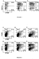
- the murine red blood cells loaded with ovalbumin and treated with the anti-TER 119 antibody were more efficiently phagocytosed by the dendritic cells isolated from the spleen than the untreated red blood cells in vitro, after 4 hours of coculture ( Figure 1 , respectively C and B). 36% of the dendritic cells phagocytosed the red blood cells carrying the antibody, against only 27% in the absence of antibody. Phagocytosis of the antibody-treated red blood cells by a population not expressing the CD11c dendritic cell marker was also observed (10.9%; Figure 1, C ).
- Example 6 Measurement of the phagocytosis of red blood cells containing ovalbumin, by macrophages and dendritic cells of the spleen and liver in vivo on mice
- Each batch is labelled with CFSE and injected intravenously into C57BI/6 mice.
- the blood, the spleen and the liver of the mice are taken.
- the percentage of fluorescent red blood cells circulating in the blood of the mice is measured by flow cytometry.
- the fluorescence incorporated into the spleen macrophages expressing the F4/80 marker ( Figure 2 , A, B, C), into the liver macrophages expressing the F4/80 marker and into the spleen dendritic cells expressing the CD11 c marker ( Figure 2 , D, E, F) is measured by flow cytometry.
- Table 2 Percentage of macrophages or dendritic cells from the spleen, having phagocytosed fluorescent red blood cells 3 hours after injection into the mouse: Batches Macrophages Dendritic cells 1 28% 5% 2 68% 19% 3 81% 22%
- the murine red blood cells loaded with ovalbumin and heat-treated or treated with the anti-TER 119 antibody are almost no longer present in the blood of the mouse (1.6% and 1 %), whereas there are still untreated, ovalbumin-loaded red blood cells in the blood of the mouse (4.6%).
- red blood cells that have been heat-treated or treated with the anti-TER 119 antibody are phagocytosed by the F4/80 macrophages and CD11 c dendritic cells of the spleen ( Figure 2 ).
- the red blood cells treated with the anti-TER 119 antibody were more efficiently phagocytosed by the F4/80 macrophages of the spleen than the heat-treated red blood cells or the untreated red blood cells ( Figure 2 , A, B, C). 81% of the spleen macrophages phagocytosed the antibody-treated red blood cells, against 68% of macrophages having phagocytosed the heat-treated red blood cells and only 28% in the untreated batch (Table 2).
- the heat-treated or antibody-treated red blood cells were also more efficiently phagocytosed by the CD11c dendritic cells from the spleen than the untreated red blood cells ( Figure 2 ). Respectively 22% and 19% of dendritic cells phagocytosed the antibody-treated red blood cells and the heat-treated red blood cells, against only 5% in the case of the untreated red blood cells (Table 2).
- Phagocytosis of the antibody-treated red blood cells by a population from the spleen not expressing the CD11 c dendritic cell marker or the F4/80 macrophage marker was also observed (11.9% and 12.8%, Figure 2 ).
- Table 3 Percentage of liver macrophages having phagocytosed fluorescent red blood cells 3 hours after injection into the mouse. Batches Macrophages 1 24% 2 40% 3 50%
- the heat-treated red blood cells or the red blood cells treated with the anti-TER 119 antibody are phagocytosed by the F4/80 macrophages of the liver.
- the red blood cells treated with the anti-TER 119 antibody were more efficiently phagocytosed by the F4/80 macrophages of the liver than the heat-treated red blood cells or than the untreated red blood cells. 50% of the liver macrophages phagocytosed the antibody-treated red blood cells, against 40% of macrophages having phagocytosed the heat-treated red blood cells and only 24% in the untreated batch (table 3).
- the binding of the antibody to the red blood cells and the heat treatment allowed efficient targeting of the red blood cells in the spleen and the liver, and a significant increase in the percentage of dendritic cells and of macrophages capable of phagocytizing these red blood cells.
- Example 7 Measurement of the phagocytosis of mouse red blood cells containing fluorescent ovalbumin by bone marrow macrophages and dendritic cells in vivo
- This study is an autologous study, since OF1 mouse red blood cells are injected into OF1 mice.
- the fluorescence incorporated into the macrophages expressing the F4/80 marker or the CD11 b marker, the granulocytes expressing the Gr1 marker, the myeloid dendritic cells expressing the CD11c and CD11 b markers and into the plasmacytoid dendritic cells expressing the CD11 c and CD8 markers is measured by flow cytometry.
- Table 4 Percentage of bone marrow macrophages, granulocytes or dendritic cells having phagocytosed fluorescent ovalbumin 1 hour 30 min after the injection: Batches F4/80 macrophages C11b macrophages Granulocytes Myeloid dendritic cells Plasmacytoid dendritic cells 1 2.5 0.05 12.5 2.7 2.4 2 3.3 0 15.5 2.2 3.4 3 17.7 1.75 21 15.3 6 4 2.4 0 13 1.6 2.4 5 0.5 0.1 1.3 0.8 1.5
- the ovalbumin-loaded red blood cells treated with the anti-TER 119 antibody were efficiently phagocytosed by the F4/80 macrophages, the granulocytes and the dendritic cells of the bone marrow.
- the myeloid dendritic cells of the bone marrow are the cells most involved in the phagocytosis of the ovalbumin-loaded red blood cells treated with the anti-TER 119 antibody.
- red blood cells treated with the anti-TER 119 antibody allows the best targeting and phagocytosis of the red blood cells by immune cells in the bone marrow.
- Example 8 Measurement of the ovalbumin-specific CD4 T cell response after a single injection of ovalbumin-loaded red blood cells and of Poly(I:C) adjuvant
- OVA ovalbuproliferin
- the evaluation of the CD4 T cell response is realized using an adaptation of the methods described in Russo V. et al. The Journal of Clinical Investigation, 2007, 117 : 3087-3096 ; Stoitzner P. et al., The Journal of Immunology 2008, 180 : 1991-1998 .
- the CD4 T cells of OT-II transgenic mice (Charles River, ref C57BL/6-Tg(TcraTcrb425Cbn/Crl)) are used to measure the OVA-specific CD4 T cell response.
- the OT-II mice are mice expressing only CD4 T cells which recognize the ovalbumin peptide 323-339 associated with major histocompatibility complex class II molecules.
- the OT-II transgenic CD4 T cells are isolated from the spleen of OT-II mice and labelled with CFSE, before being injected intravenously into Ly5.1 mice.
- OT-II mice and Ly5.1 mice have the same genetic background, but the cells of both types of mice can be identified by means of the CD45 marker. This is because the cells of OT-II mice express the CD45.2 marker, whereas the Ly5.1 mice express the CD45.1 marker.
- the Ly5.1 mice receive an intravenous injection of the following batches. Three mice are injected with the batches containing red blood cells and two mice are injected with the batches containing free OVA or the control. Two batches of 183 ⁇ 10 7 red blood cells of C57BI/6 mice loaded with ovalbumin (Serlabo Technologies, ref WO-LS003054) prepared using variant 2 of example 1 and treated or not with the anti-TER 119 antibody (as described in example 3) are prepared. The adjuvant, Poly (I:C) (Invivogen, ref tlrl-pic), is added to these batches, as well as to the batch containing free ovalbumin. The amount of Poly (I:C) injected per mouse is 25 ⁇ g. The amount of OVA injected into the mice is indicated in Table 5.
- Poly (I:C) Invivogen, ref tlrl-pic
- mice Three days after the injection of the batches, the mice are sacrificed and the spleens of the mice are removed.
- the percentage of OVA-specific CD4 T cells in the spleen of the mice is measured by flow cytometry using the CD4 and CD45.2 markers (Table 5).
- the number of OVA-specific CD4 T cells is calculated from the percentage of OVA-specific CD4 T cells and the number of total lymphocytes counted with trypan blue (Invitrogen, ref 15250061) (Table 5).
- the proliferation and the activation of the OVA-specific CD4 T cells in the spleen of the mice is measured by flow cytometry using the CD4, CD44 and CD45.2 markers and using CFSE (Tables 6 and 7).
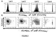
- the amount of CFSE contained in the OVA-specific CD4 T cells is divided by a factor of 2, which makes it possible to determine the number of cell divisions by flow cytometry ( Figure 3 ).
- mice splenocytes The production of IFNg by the mice splenocytes is measured in the culture supernatants after 3 days of in vitro stimulation in the presence of 10 ⁇ g/ml of ovalbumin peptide 323-339 (Neomps, ref) by means of an ELISA assay.
- Table 5 Percentage and number of OVA-specific CD4 T cells three days after injection of the batches (mean ⁇ standard deviation): Batches Amount of OVA injected into the mice ( ⁇ g) % of OVA-specific CD4 T cells Number of OVA-specific CD4 T cells (millions of cells) 1 160 6.6 ⁇ 1.4* 8 ⁇ 2* 2 129 3.6 ⁇ 0.9** 4.1 ⁇ 2* 3 150 0.8 ⁇ 0.3 1.3 ⁇ 0.7 4 0 0.1 ⁇ 0.1 0.2 ⁇ 0.1
- Table 6 Percentage of OVA-specific CD4 T cells which have divided 0, 1, 2, 3, 4, 5, 6 or 7 times three days after injection of the batches: % of OVA-specific CD4 T cells at each division cycle (mean ⁇ standard deviation) Batches 0 1 2 3 4 5 6 7 1 0 ⁇ 0 0 ⁇ 0 0.1 ⁇ 0 0.3 ⁇ 0 1.7 ⁇ 0 9.4 ⁇ 0.1 35.9 ⁇ 0.3 52.5 ⁇ 0.2 2 0.1 ⁇ 0 0.1 ⁇ 0 0.2 ⁇ 0 0.8 ⁇ 0.1 4.2 ⁇ 0.8 19.2 ⁇ 3.2 43.7 ⁇ 2.2 31.5 ⁇ 6.3 3 1.9 ⁇ 1.4 6.9 ⁇ 5.2 15.6 ⁇ 9.5 18.7 ⁇ 5.6 19.7 ⁇ 0.4 16.4 ⁇ 5.3 13.9 ⁇ 10.1 6.8 ⁇ 5.9 4 69.7 ⁇ 4.9 1.3 ⁇ 1 0.1 ⁇ 0.2 0.3 ⁇ 0.4 5.6 ⁇ 3.7 3.7 ⁇
- Table 7 Level of activation of OVA-specific CD4 T cells three days after injection of the batches: Batches Mean of OVA-specific CD4 T cells activation fluorescence (mean ⁇ standard deviation) 1 32.6 ⁇ 2.3 2 37.6 ⁇ 3.5 3 34.6 ⁇ 3.9 4 11.4 ⁇ 0.5
- the results of the cell activation level show that all the dividing OVA-specific CD4 T cells have a high cell activation level (Table 7 and Figure 3 ).
- the activation level of the OVA specific CD4 T cells can be correlated to the number of divisions of these cells: the more the cells have divided and have lost their CFSE marker content, the more they express a high level of activation marker ( Figure 3B ).
- Table 8 Production of IFNg by splenocytes stimulated in vitro with 10 ⁇ g/ml of ovalbumin peptide 323-339 for 3 days: Batches Production of IFNg by splenocytes in ng/ml (mean ⁇ standard deviation) 1 56.3 2 60 ⁇ 26.8 3 9.4 ⁇ 1.6 4 6.7 ⁇ 1
- mice injected with Poly (I:C) and ovalbumin-loaded red blood cells treated or not treated with the anti-TER119 antibody show a strong production of IFNg by the splenocytes of mice injected with Poly (I:C) and ovalbumin-loaded red blood cells treated or not treated with the anti-TER119 antibody.
- Example 9 Measurement of the ovalbumin-specific CD8 T cell response after a single injection of ovalbumin-loaded red blood cells and of Poly (I:C) adjuvant
- the evaluation of the OVA-specific CD8 T cell response consists in measuring the percentage of OVA-specific CD8 T cells, the production of IFNg and the cell lysis, in vivo, of the cells presenting the ovalbumin peptide 257-264 associated with major histocompatibility complex class I molecules. This study is an allogenic study, since ovalbumin-loaded red blood cells from OF1 mice are injected into not consanguineous C57BI/6 mice.
- the batches are injected intravenously into C57BI/6 mice. A minimum of 4 mice are injected with a given batch. In vivo cell lysis is measured using an adaptation of the method described by Hervas-Stubbs S. et al., Blood 2007, 109 : 5318-5326 . Cells presenting the ovalbumin peptide 257-264 associated with major hisotocompatibility complex class I molecules are injected into the immunized mice.
- mice receive an injection of 0.5 ⁇ 10 6 splenocytes presenting the ovalbumin 257-264 peptide (Neomps, ref SC1302) and labelled with a moderate concentration of CFSE, and of 0.5 ⁇ 10 6 splenocytes not presenting the peptide and labelled with high concentrations of CFSE (cell lysis control).
- Latex beads coupled to ovalbumine peptide 257-264 (BOVAp) and poly(I:C) are used as positive control to induce a CD8 T immune response against ovalbumine. It has been shown that the injection of BOVAp and of poly(I:C) induces an incease of the percentage of ovalbumine-specific CD8 T cells and the destruction of cells presenting the OVA257-264 peptide in vivo (Herva-Stubbs).
- mice 16 hours after injection of the splenocytes, the mice are sacrificed by euthanasia and the spleens of the mice are removed.
- the percentage of OVA-specific CD8 T cells in the spleen of the mice is measured by flow cytometry using tetramer and the CD8 marker (Table 9).
- IFNg production and expression of the marker associated with degranulation (CD107) by CD8 T cells was measured by flow cytometry after in vitro stimulation for 4 hours in the presence of 0.1 ⁇ g/ml of ovalbumin peptide 257-264 (Table 10).
- In vivo cell lysis was measured by flow cytometry using CFSE (Table 11).
- Table 9 Percentage of OVA-specific CD8 T cells seven days after injection of batches (mean ⁇ standard deviation): Batch Quantity of OVA injected into mice ( ⁇ g) % of OVA-specific CD8 T cells (mean ⁇ standard deviation) % of OVA-nonspecific T cells (mean ⁇ standard deviation) 1 95 14 ⁇ 3.2* 2.4 ⁇ 0.2 2 91 12.5 ⁇ 5.2** 1.2 ⁇ 0.5 3 0 1.2 ⁇ 0.1 1.2 ⁇ 0.1 4 0 2.5 ⁇ 0.4* 0.9 ⁇ 0.1 5 130 2.4 ⁇ 1.4 1 ⁇ 0.2 6 0 0.7 ⁇ 0.4 1 ⁇ 0.1
- Table 10 Production of IFNg by CD8 T cells stimulated in vitro with 0.1 ⁇ g/ml of ovalbumin peptide 257-264 for 4 hours: Batch % CD8 T cells producing IFNg % of CD8 T cells producing IFNg and expressing CD107 marker 1 8.7 ⁇ 1.8* 71 ⁇ 4* 2 6.6 ⁇ 1.7* 71 ⁇ 4* 3 1.4 ⁇ 0.1 33 ⁇ 4.5 4 2.8 ⁇ 0.5 57 ⁇ 5.5 5 1.8 ⁇ 0.5 46 ⁇ 7 6 0.7 ⁇ 0.1 18 ⁇ 2
- the CD8 T cells generated are effective and cytotoxic since they produce IFNg and express the CD107 marker in response to stimulation with the ovalbumin peptide (Table 10).
- the percentage of CD8 T cells producing IFNg and expressing CD107 is significantly higher for mice injected with Poly(I:C) and ovalbumin-charged red blood cells which had either been treated or had not been treated with anti-TER119 antibody than for mice injected with free OVA and Poly(I:C) or Poly(I:C) and BOVAp (Student's test, p ⁇ 0.008).
- Table 11 Percentage of in vivo anti-OVA-specific cell lysis (mean ⁇ standard deviation): Batch % cell lysis, in vivo (mean ⁇ standard deviation) 1 82 ⁇ 7 2 83 ⁇ 4 5 53 ⁇ 8 6 0
- Example 10 Measuring the maintenance of the ovalbumin-specific CD8 T cell response 30 days after a single injection of ovalbumin-charged red blood cells and adjuvant Poly(I:C)
- Evaluating the maintenance of the OVA-specific CD8 T cell response consisted of measuring the percentage of OVA-specific CD8 T cells and cell lysis in vivo 34 days after a single injection.
- mice were injected intravenously into C57BI/6 mice. Three mice were injected with a given batch.
- cells displaying ovalbumin peptide 257-264 associated with molecules of the major histocompatibility complex of class I were injected into immunized mice. 33 days after injection, the mice received an injection of 0.5 ⁇ 10 6 splenocytes displaying ovalbumin peptide 257-264 (NeoMPS, reference SC1302) and labelled with a moderate concentration of CFSE and of 0.5 ⁇ 10 6 splenocytes not displaying the peptide and labelled with a high concentration of CFSE (cell lysis assay).
- mice 16 hours after injecting the splenocytes, the mice were euthanized and their spleens were removed.
- the percentage of OVA-specific CD8 T cells in the spleens of the mice was measured by flow cytometry using tetramer and CD8 marker (Table 12).
- In vivo cell lysis was measured by flow cytometry using CFSE (Table 13).
- Table 12 Percentage of OVA-specific CD8 T cells 34 days after injection of batches (mean ⁇ standard deviation) Batch Quantity of OVA injected into mice ( ⁇ g) % of OVA-specific CD8 T cells (mean ⁇ standard deviation) 1 170 1.5 ⁇ 0.2 2 138 1.7 ⁇ 0.3 3 0 1.6 ⁇ 0.1 4 0 1.4 ⁇ 0.1 5 150 2.2 ⁇ 0.2 6 0 1.8 ⁇ 0.06
- OVA-specific CD8 T cells were still capable of lysing cells displaying ovalbumin peptide 257-264 (Table 13).
- Injection of Poly(I:C) and ovalbumin-charged red blood cells treated with anti-TER119 antibody induced cell lysis in a manner which was more effective than injection of Poly(I:C) and BOVAp (*Student's test, p ⁇ 0.04).
- ovalbumin-charged red blood cells are superior not only as regards activation and proliferation of cytotoxic CD8 T cells, but also as regards maintenance/survival of these T cells which are capable of lysing cells displaying ovalbumin peptide 257-264.
- Example 11 Measurement of the ovalbumin-specific CD8 T cell response after a single injection of chemically treated, ovalbumin-loaded red blood cells and of Poly (I:C) adjuvant
- This study is an allogenic study, since ovalbumin-loaded red blood cells from OF1 mice are injected into not consanguineous C57BI/6 mice.
- the adjuvant Poly (I:C) is added to this batch, and to the batch containing free ovalbumin.
- the amount of Poly (I:C) injected per mouse is 25 ⁇ g.
- the amount of OVA injected into the mice is indicated in Table 14.
- the batches are injected intravenously into C57BI/6 mice. A minimum of 3 mice are injected with a given batch. Seven days after the injection, the mice are sacrificed and the spleens of the mice are removed. The percentage of OVA-specific CD8 T cells in the spleen of the mice is measured by flow cytometry using tetramer and the CD8 marker (Table 14).
- Table 14 Percentage and number of OVA-specific CD8 T cells seven days after injection of the batches (mean ⁇ standard deviation): Batches Amount of OVA injected into the mice ( ⁇ g) % of OVA-specific CD8 T cells (mean ⁇ standard deviation) 1 38 1.8 ⁇ 0.6 2 150 1.2 ⁇ 0.3 3 0 1 ⁇ 0 4 0 0.8 ⁇ 0.1
- mice injected with Poly (I:C) and ovalbumin-loaded red blood cells treated with BS3 have a higher percentage of OVA-specific CD8 T cells than the mice injected with free OVA and Poly (I:C) or than the mice injected with Poly (I:C). These results are not statistically different, but it should be noted that the amount of free OVA injected is more than three times greater than the amount of OVA injected in the batch containing the OVA-loaded red blood cells.
- Example 12 Measurement of the ovalbumin-specific CD8 T cell response after a single injection of ovalbumin-loaded red blood cells and of CL-097 adjuvant
- This study is an allogenic study, since ovalbumin-loaded red blood cells from OF1 mice are injected into not consanguineous C57BI/6 mice.
- the batches are injected intravenously into C57BI/6 mice. Four days after the injection, the mice are sacrificed and the spleens of the mice are removed. The IFNg production by the splenocytes of mice injected with the various batches is measured in the culture supernatants after three days of in vitro stimulation in the presence of 0.1 ⁇ g/ml of ovalbumin peptide 257-264 by means of ELISA assay.
- Table 15 IFNg production by splenocytes stimulated in vitro with 0.1 ⁇ g/ml of ovalbumin peptide 257-264 for 3 days: Batches Amount of OVA injected into the mice ( ⁇ g) IFNg production by splenocytes in ng/ml 1 83 15.2 2 83 5.7 3 103 11.4 4 103 7.4 5 0 0.4
- the CL097 adjuvant also potentiates the IFNg response induced by the injection of the ovalbumin-loaded red blood cells.
- Example 13 Measure of tumoral growth after one injection of adjuvant Poly (I:C) and treated or untreated ovalbumin-loaded erythrocytes in mice
- EG.7 tumor cells are obtained from ATCC (ATCC-CRL-2113).
- ATCC-CRL-2113 ATCC-CRL-2113
- EG.7 cells originate from the EL.4 cell line, which synthesize and secrete OVA constitutively.
- Two batches of 165 ⁇ 10 7 antibody-treated or untreated ovalbumin-loaded erythrocytes from OF1 mice are prepared according to variant 2 of example 1.
- the adjuvant, Poly (I: C) is added to those batches.
- 25 ⁇ g of Poly (I: C) is injected per mice.
- the negative control is a suspension of unloaded erythrocytes.
- OVA amounts injected to mice are indicated in table 16.
- mice The batches are injected intravenously to C57BI/6 mice (10 mice per group). Seven days after batch injection, 2.2x10 6 EG.7 cells per mice are injected subcutaneously. Mice are followed for tumor growth. Tumor growth is assessed by measuring the diameter of the tumor in centimeters (recorded as the average of two perpendicular diameter measurements). Tumor size is measured 3 times per week at 2 days interval during 14 days. Mice with tumors of more than 2.0 cm of diameter are killed.
- Table 16 OVA amount injected per mice Batches OVA quantity injected to mice ( ⁇ g) 1 134 2 134 3 0
- Table 17 Tumor growth Batches Tumor growth (mm 3 ) the days following tumor cell injection 0 3 5 7 10 12 14 1 0 0 0 0 0 0 0 2 0 0 0 0 0 0 3 0 0 0 363 ⁇ 200 756 ⁇ 462 1009 ⁇ 445 1198 ⁇ 541
- the size of the tumors is measurable on the flank of the mice previously injected with unloaded erythrocytes and the tumors continue to growth until the end of the study. Whereas no tumors are observed on the flank of the mice injected with Poly (I:C) and ovalbumin-loaded red blood cells treated or not with the anti-TER 119 antibody. Observation of the mice is still ongoing.
- Example 14 Measurement of the phagocytosis of pig red blood cells by macrophages and dendritic cells in vivo
- This study is a xenogenic study, since pig red blood cells are injected into C57BI/6 mice.
- the pig red blood cells are washed three times in NaCl containing glucose, before being labelled with CFSE as described above.
- the red blood cells are washed again three times in NaCl containing glucose, and then brought back to a haematocrit of 50%.
- a batch of 142 ⁇ 10 7 pig red blood cells is injected intravenously into three C57BI/6 mice. One hour after the injection, the mice are sacrificed and the spleens, the livers and the femurs of the mice are removed.
- the fluorescence incorporated into the macrophages expressing the F4/80 marker or the CD11 b marker, into the myeloid dendritic cells expressing the CD11c and CD11 b markers and into the plasmacytoid dendritic cells expressing the CD11c and CD8 markers is measured by flow cytometry.
- Table 18 Percentage of spleen macrophages or dendritic cells having phagocytosed fluorescent red blood cells 1 hour after injection: Batches F4/80 macrophages CD11b macrophages Myeloid dendritic cells Plasmacytoid dendritic cells 1 27 ⁇ 6 4 ⁇ 2 21 ⁇ 5.5 45 ⁇ 6 2 0 1 4 3
- Table 19 Percentage of bone marrow macrophages or dendritic cells having phagocytosed fluorescent red blood cells 1 hour after injection: Batches F4/80 macrophages CD11b macrophages Myeloid dendritic cells Plasmacytoid dendritic cells 1 5.3 ⁇ 7.5 0.8 ⁇ 1 5 ⁇ 3 5 ⁇ 7 2 6.3 0 6 12.9
- Table 20 Percentage of liver macrophages or dendritic cells having phagocytosed fluorescent red blood cells 1 hour after injection: Batches CD11b macrophages Myeloid dendritic cells Plasmacytoid dendritic cells 1 1.1 ⁇ 1.1
- the pig red blood cells were efficiently phagocytosed by the F4/80 macrophages and the dendritic cells of the spleen (Table 18).
- the plasmacytoid dendritic cells of the spleen are the cells most involved in the phagocytosis of the pig red blood cells.
- the macrophages and the dendritic cells did not phagocytize fluorescent red blood cells (Tables 19 and 20).
- the mean globular volume, the mean corpuscular haemoglobin and the concentration of red blood cells was measured using an ABX cell counter.
- the osmotic fragility, which corresponds to the concentration of NaCl inducing 50% haemolysis of red blood cells, was measured using an Osmocells instrument (SD Medical).
- the extracellular haemoglobin was measured by spectrophotometry.
- the corpuscular concentration of ovalbumin, the extracellular concentration of ovalbumin and the mean globular quantity of ovalbumin were determined using an ELISA test.
- Table 21 Characteristics of pig red blood cells comprising ovalbumin: Day of production 18 hours after production Characteristics Starting material Batch 1 Batch 2 Starting material Batch 1 Batch 2 Mean globular volume ( ⁇ m 3 ) 65 55 57 65 56.0 58 Mean corpuscular haemoglobin (g/dl) 31.9 27.7 25.9 31.4 28.9 25.8 Osmotic fragility (salinity inducing 50% haemolysis in g/l of NaCl) 3.98 2.51 3.33 Haematocrit of suspension (%) 36.8 48.2 50 36.5 46.3 48 Concentration of red blood cells (10 6 /mm 3 ) 5.64 9.1 8.1 5.9 9 8.4 Extracellular haemoglobulin (g/dl) 0.04 0.27 0.196 0.05 0.79 0.56 Corpuscular ovalbumin concentration (mg/ml of red blood cells) 0.66 0 0.71 0 Extracellular ovalbumin concentration (mg/ml of end product) 0.02 0 0.02 0 Mean globular
- the quantity of extracellular ovalbumin was relatively low compared with the quantity of encapsulated ovalbumin and had not increased 18 hours after production, thereby demonstrating the stability of the red blood cells encapsulating the ovalbumin.
Landscapes
- Health & Medical Sciences (AREA)
- Life Sciences & Earth Sciences (AREA)
- Chemical & Material Sciences (AREA)
- General Health & Medical Sciences (AREA)
- Medicinal Chemistry (AREA)
- Immunology (AREA)
- Veterinary Medicine (AREA)
- Public Health (AREA)
- Animal Behavior & Ethology (AREA)
- Pharmacology & Pharmacy (AREA)
- Epidemiology (AREA)
- Mycology (AREA)
- Microbiology (AREA)
- Oncology (AREA)
- Cell Biology (AREA)
- Molecular Biology (AREA)
- Engineering & Computer Science (AREA)
- Developmental Biology & Embryology (AREA)
- General Chemical & Material Sciences (AREA)
- Chemical Kinetics & Catalysis (AREA)
- Organic Chemistry (AREA)
- Genetics & Genomics (AREA)
- Inorganic Chemistry (AREA)
- Zoology (AREA)
- Biotechnology (AREA)
- Biomedical Technology (AREA)
- Virology (AREA)
- Hematology (AREA)
- Oil, Petroleum & Natural Gas (AREA)
- Biochemistry (AREA)
- Nuclear Medicine, Radiotherapy & Molecular Imaging (AREA)
- Proteomics, Peptides & Aminoacids (AREA)
- Biophysics (AREA)
- Bioinformatics & Cheminformatics (AREA)
- Medicines Containing Antibodies Or Antigens For Use As Internal Diagnostic Agents (AREA)
- Medicines Containing Material From Animals Or Micro-Organisms (AREA)
- Micro-Organisms Or Cultivation Processes Thereof (AREA)
- Peptides Or Proteins (AREA)
- Medicines That Contain Protein Lipid Enzymes And Other Medicines (AREA)
Description
- The present invention relates to a composition which induces, in a host, a cytotoxic response for anti-tumour purposes, and also to a therapeutic anti-tumour vaccine containing this composition.
- The natural immune response against tumour antigens is relatively ineffective and various mechanisms allowing tumours to escape the anti-tumour immune response have been identified. Conventional vaccine approaches generate humoral responses which prove to be insufficient.
- Strategies aimed at developing a cytotoxic response based on antigen-presenting cells (APCs), in particular based on dendritic cells, have been studied. The principle consists in specifically destroying the cancer cells of a patient by stimulating said patient's own immune defences.
- Dendritic cells are antigen-presenting cells (APCs) that are very effective in generating cytotoxic effectors specific for tumour cells. They are capable of phagocytizing apoptotic cells or apoptotic bodies originating from tumour cells, and then of presenting the tumour antigens, in association with MHC class I and II molecules, to T lymphocytes. Thus, the dendritic cell is capable of initiating the proliferation and the generation of a clone of specific cytolytic T lymphocytes. At the end of this reaction, the killer lymphocytes thus differentiated leave the lymphoid compartment so as to circulate in the organism and bind in the tumour. Recognition of the antigens expressed by a tumour then induces a lytic signal and brings about the destruction of the tumour cells.
- Several anti-cancer vaccine strategies targeting dendritic cells have been studied (Eymard JC, Bernard J, Bull Cancer. 2003, 90(8-9):734-43). Some are based on manipulating the dendritic cells in vitro, and others are based on stimulating the dendritic cells in vivo. In the first case, the dendritic cells are differentiated from blood cells taken from the patient: they are cultured and matured, "pulsed", i.e. stimulated, ex vivo with tumour peptides, tumour lysates, apoptotic tumour cells, or heat shock proteins extracted from autologous tumours, and finally reinjected into the patient. In the second case, the stimulation of the dendritic cells is carried out after injection, into the patient, of peptides, proteins, irradiated tumour cells or else viruses containing the antigenic peptide targeting the dendritic cells. However, the cytotoxic response obtained is rarely accompanied by clinical efficacy. The activation of the dendritic cell "conditions" its ability to effectively activate the cytolytic T lymphocyte. The level of activation of dendritic cells appears to be a delicate point in this vaccine strategy.
- The use of dendritic cells ex vivo for obtaining an anti-tumour vaccine raises a certain number of issues on: the state of maturation of the dendritic cells, the number of cells to be injected, the route, the site and the frequency of injection for producing dendritic cells capable of migrating to the secondary lymphoid organs and of inducing an effective cytotoxic T response (Banchereau J, Schuler-Thurner B, Palucka AK, Schuler G. [Dendritic cells as vectors for therapy] Cell. 2001, 10, 106(3): 271-4.).
- The activation of dendritic cells in vivo is, for its part, limited by the weak immunogenic capacity of tumour antigens and the difficulty in activating dendritic cells at a sufficient level.
- The use of red blood cells as carriers for transporting antigens, encapsulated in the red blood cells or bound to their surface, and delivered to the APCs has been envisaged in several publications. The triggered immune responses have been investigated in vitro and in vivo.
- Hamidi et al. have recently described the encapsulation of BSA (bovine serum albumin) as a model of an antigen in human red blood cells (Hamidi M et al., Drug Deliv., 2007; 14(5):295-300 and Int J Pharm., 2007, 29, 338(1-2): 70-8). The authors suggested the use of red blood cells as a vector for the presentation of antigens to APCs of the reticuloendothelial system (RES). In another review published by Hamidi et al. (J. Control. Release, 2007, 118(2): 145-60), the authors indicated that a certain number of strategies have been studied to promote targeting of the RES, said targeting being promoted by ageing of the red blood cells, leading to their uptake for lysis. Other routes were mentioned, such as exposure of red blood cells to stabilizing agents, in particular crosslinking agents, coating of red blood cells with anti-RH antibodies, of IgG type to target the spleen or of IgM type to target the liver, heat shock, or exposure to oxidizing agents, enzymes or antibiotics.
- A humoral immune response can be obtained in vivo after immunization with antigen-loaded red blood cells. The study carried out by Murray et al. made it possible to detect, in mice, IgG immunoglobulins after intravenous injection of murine red blood cells loaded with one of the following four antigens: KLH (Keyhole Limpet Haemocyanin), BSA (Bovine Serum Albumin), CTB (Cholera Toxin b Subunit) and ADA (Bovine Adenosine Deaminase). Detection of IgG1 and IgG3, which are predominant immunoglobulin isotypes during a Th2 response, and of IgG2, immunoglobulin isotype which is a marker for a Th1 response, would suggest the involvement of both types of immune responses, the humoral response and the cellular response (Murray AM et al., Vaccine., 2006 28, 24(35-36): 6129-39).
- Another formulation of antigens used with red blood cells has been tested by Dominici et al. The Tat protein of the HIV-1 virus was anchored to the surface of mouse red blood cells by means of avidin/biotin coupling. The immunization of mice by intraperitoneal injection with this antigen formulation, internalized by dendritic cells, triggered a humorally-mediated immune response in vivo. Isotype characterization of the immunoglobulins detected indicates the induction of Th1 and Th2 responses. The anti-Tat cytotoxic activity was shown in vitro, by the conventional chromium-release technique, for the mice treated with red blood cells coupled to the antigens (Dominici S et al., Vaccine., 2003 16, 21(17-18): 2073-81).
- A cellular response has been demonstrated in vitro by Corinti et al. with the same formulation. Phagocytosis of the red blood cells conjugated with Tat protein, by dendritic cells derived from human monocytes, was shown, as was the induction of CD4+ and CD8+ responses. Moreover, maturation of dendritic cells in the presence of interferon gamma promoted the type I immune response (Corinti S. et al., Leukoc. Biol. 2002, 71(4): 652-8).
- Boberg et al. injected mice intraperitoneally with a vaccine constituted of peptides derived from the HIV-1 protease anchored at the surface of mouse red blood cells by means of the avidin/biotin system. They chemically modified red blood cells with the aim of promoting their recognition by APCs, but a weak immune reaction was obtained. They concluded that the small amount of antigens delivered, due to the limited loading of red blood cells with antigenic peptides and blood volume injected was not compensated for by the chemical modification of the carriers supposed to promote antigen recognition by APCs (Boberg A. et al., Infect. Agents Cancer. , 2007, 182: 9).
- The present invention aims to provide compositions and vaccines that can be used in the treatment of cancers, according to an immunotherapy approach.
- An object of the invention is therefore a composition as defined in
claim 1. - The term "host" refers preferably to humans, but also to animals, in particular pets (especially dogs or cats) and animals for sport (especially horses).
- According to the invention, the red blood cells contain, i.e. encapsulate, the antigen, which means that the antigen is or is essentially inside the red blood cell.
- Said red blood cells are preferably designed, selected or modified so as to promote phagocytosis thereof by APCs, and most particularly by dendritic cells. In particular, said red blood cells are designed, selected or modified so as to promote phagocytosis thereof in the spleen and the liver, the essential objective being to target APCs of the spleen.
- In a preferred embodiment, the compositions according to the invention comprise red blood cells which contain the antigen and target the spleen. The composition promotes phagocytosis of these red blood cells by the APCs, in particular the dendritic cells, in the spleen.
- According to a first embodiment, the red blood cells contain a tumour antigen and are in the form of an immune complex with an immunoglobulin which recognizes an epitope at the surface of said red blood cells, so as to promote the phagocytosis of said red blood cells, in particular by dendritic cells. The composition also makes it possible to promote phagocytosis by macrophages. Preferably, the immunoglobulin is an immunoglobulin G.
- The formation of the immune complex involves red blood cells and at least one antibody, preferably of IgG subtype. Dendritic cells have, at their surface, receptors for the constant Fc region of immunoglobulins G (IgGs). These receptors are capable of triggering phagocytosis or internalization of the antigen-IgG immune complexes thus formed and of promoting antigen presentation by MHC class I and II molecules, resulting in the generation of CD4+ helper lymphocytes and especially CD8+ cytotoxic lymphocytes (A. Regnault et al., J. Exp. Med., Janvier 1999, 189(2): 371-80).
- As antibodies which are suitable, mention may be made of anti-rhesus antibodies, anti-glycophorin A antibodies and anti-CR1 antibodies (CR1 = complement receptor type 1). Anti-glycophorin A antibodies (A. Bigbee et al., Mol. Immunol., December 1983, 20(12): 1353-62) are a preferred modality.
- Preferably, the red blood cells of human origin that are used to form an immune complex are heterologous red blood cells originating from a donor.
- According to a second embodiment, the red blood cells contain a tumour antigen and are heat or chemically modified, so as to promote phagocytosis of said red blood cells, in particular by dendritic cells. The composition also makes it possible to promote phagocytosis by macrophages.
- The heat treatment is in particular carried out under the following conditions: heating of red blood cells for about 15 minutes to about 90 minutes, preferably from about 25 to about 50 minutes, at a temperature of between about 42 and about 55°C, preferably between about 47 and about 51 °C. Typically, red blood cells are heated for about 30 minutes at between about 48 and about 50°C, for example at about 48°C.
- The chemical treatment is carried out using agents which modify the surface of red blood cells, and in particular bridging or crosslinking agents such as bis(sulphosuccinimidyl) suberate (BS3 or BS3), glutaraldehyde or neuraminidase.
- In a particular embodiment, at least two methods of targeting are combined, and, for example, the composition then comprises antigen-containing red blood cells which are in the form of an immune complex and are heat or chemically treated so as to promote their uptake in the spleen and/or the liver, preferably the spleen, and phagocytosis by APCs, in particular by dendritic cells.
- In a third embodiment, the antigen-contaning red blood cells are xenogenic. Injection of xenogenic red blood cells into humans results in the binding of the patient's natural antibodies to the injected red blood cells. The immune complex thus formed promotes phagocytosis by APCs, in particular by dendritic cells. Preferably, said red blood cells are of porcine origin.
- In a particular embodiment, the xenogenic red blood cells are heat or chemically modified so as to promote phagocytosis thereof.
- The composition according to the invention may comprise one or more tumour antigens. When there are several tumour antigens, said tumour antigens are preferably selected so as to induce an immune response against one type of tumour or of tumour cells.
- The composition preferably comprises at least two tumour antigens representative of the tumour to be treated. The objective is to generate several clones of cytotoxic T lymphocytes which each recognizes a specific antigenic peptide so as to develop a more effective immune response.
- In a particular embodiment, the composition comprises at least two populations of red blood cells, each encapsulating a different antigen.
- The most well known antigens that can be used herein are indicated in the tables below, where they are classified by category.
Unique antigens: Gene/protein Tumour alpha-actinin-4 Lung carcinoma ARTC1 Melanoma BCR-ABL fusion protein (b3a2) Chronic myeloid leukaemia B-RAF Melanoma CASP-5 Colorectal, gastric and endometrial carcinoma CASP-8 Head and neck squamous cell carcinoma beta-catenin Melanoma Cdc27 Melanoma CDK4 Melanoma CDKN2A Melanoma COA-1 Colorectal carcinoma dek-can fusion protein Myeloid leukaemia EFTUD2 Melanoma Elongation factor 2 Squamous cell carcinoma of the lung ETV6-AML1 fusion protein Acute lymphoblastic leukaemia FN1 Melanoma GPNMB Melanoma LDLR-fucosyltransferaseAS fusion protein Melanoma HLA-A2d Renal cell carcinoma HLA-A11d Melanoma hsp70-2 Renal cell carcinoma KIAAO205 Bladder tumour MART2 Melanoma ME1 "Non small cell" lung carcinoma MUM-1f Melanoma MUM-2 Melanoma MUM-3 Melanoma neo-PAP Melanoma Myosin class I Melanoma NFYC Squamous cell carcinoma of the lung OGT Colorectal carcinoma OS-9 Melanoma pml-RARalpha-fusion protein Promyelocytic leukaemia PRDX5 Melanoma PTPRK Melanoma K-ras Pancreatic adenocarcinoma N-ras Melanoma RBAF600 Melanoma SIRT2 Melanoma SNRPD1 Melanoma SYT-SSX1 or -SSX2 fusion protein Sarcoma Triosephosphate Isomerase Melanoma FLT3-ITD Acute myeloid leukaemia p53 Head and neck squamous carcinoma - Antigens common to several tumours:
a) Tumour-specific antigens Genes BAGE-1 GAGE-1,2,8 GAGE-3,4,5,6,7 GnTVf HERV-K-MEL KK-LC-1 KM-HN-1 LAGE-1 MAGE-A1 MAGE-A2 MAGE-A3 MAGE-A4 MAGE-A6 MAGE-A9 MAGE-A10 MAGE-A12 MAGE-C2 mucin k NA-88 NY-ESO-1 / LAGE-2 SAGE Sp17 SSX-2 SSX-4 TRAG-3 TRP2-INT2g b) Differentiation antigens Gene / protein Tumour CEA Intestinal carcinoma gp100 / Pmel17 Melanoma Kallikrein 4 Prostate mammaglobin-A Breast cancer Melan-A / MART-1 Melanoma NY-BR-1 Breast cancer OA1 Melanoma PSA Prostate carcinoma RAB38 / NY-MEL-1 Melanoma TRP-1 / gp75 Melanoma TRP-2 Melanoma tyrosinase Melanoma c) Overexpressed antigens Gene Tissue expression adipophilin adipocytes, macrophages AIM-2 Ubiquitous (low level) BING-4 Ubiquitous (low level) CPSF Ubiquitous (low level) cyclin D1 Ubiquitous (low level) Ep-CAM Epithelial cells EphA3 Numerous FGF5 Brain, kidneys G250 / MN / CAIX Stomach, liver, pancreas HER-2 / neu Ubiquitous (low level) IL13Ralpha2 Intestinal carboxyl esterase liver, intestine, kidneys alpha-foetoprotein Liver M-CSF liver, kidneys mdm-2 ubiquitous (brain, muscle, lungs) MMP-2 Ubiquitous MUC1 Glandular epithelium p53 Ubiquitous (low level) PBF ovaries, pancreas, spleen, liver PRAME Testes, ovaries, endometrium, adrenal glands PSMA prostate, CNS, liver RAGE-1 Retina RNF43 RU2AS testes, kidneys, bladder secernin 1 Ubiquitous SOX10 Ubiquitous (low level) STEAP1 Prostate survivin Ubiquitous Telomerase testes, thymus, bone marrow, lymph nodes WT1 testes, ovaries, bone marrow, spleen BCLX (L) Ubiquitous (low level) DKK1 Testes, prostate, mesenchymatous stem cells ENAH (hMena) Breast, prostate, colon-rectum stroma and epithelium, pancreas, endometrium MCSP Endothelial cells, chondrocytes, nonstriated muscle cells RGS5 Heart, skeletal muscle, pericytes - Van der Bruggen et al. have produced a database referencing all human tumour antigens recognized by T lymphocytes and usable in cancer immunotherapy approaches according to the invention: http://www.cancerimmunity.org/peptidedatabase/Tcellepitopes.htm.
- Other antigens may be used in the invention such as: Gastrin 17, Human Chorionic Gonadotropin, EGFRvIII, HER2, HER2/neu, P501, Guanylyl Cyclase C, PAP.
- The word antigen encompasses antigens of natural or synthetic or artificial origin, antigens originating from the patient to be treated, antigenic fragments, derivatives or variants, so long as the antigen is able to inititiate the appropriate immune response. The antigens may be for example extracted, chemically synthesized or produced via genetic engeneering.
- In order to generate an effective immune response, dendritic cells have to be active and mature, they have to produce cytokines and chemokines and to express costimulation molecules that are necessary for the recruitment and activation of T lymphocytes.
- Various types of adjuvants can be used to stimulate APCs, and in particular dendritic cells. Bacterial or viral RNA or DNA, heat shock proteins (HSPs), sugars, immune complexes and cytokines are various factors which induce APC maturation, and in particular dendritic cell maturation mediated by the stimulation of specific receptors (Toll-like receptor TLR, mannose receptor). Macrophages and dendritic cells do not all express the same receptors at their surface. The choice of the adjuvant therefore relates to a molecule capable of generating a cytotoxic immune response in humans and for which the receptor is found at the surface of the cells taking up the red blood cells of the invention, and therefore in particular at the surface of dendritic cells.
- According to one embodiment, the adjuvant is a molecule encapsulated inside red blood cells or attached to the surface of red blood cells. These are preferably the red blood cells containing the antigen.
- Alternatively, the adjuvant is encapsulated in, or attached to the surface of, other red blood cells treated separately. These red blood cells may also be modified or selected so as to promote phagocytosis thereof by APCs, in particular by dendritic cells. They may thus be, as previously described, in the form of an immune complex, or thermally or chemically modified, or else be xenogenic.
- According to another embodiment, the adjuvant is a separate adjuvant composition, that can be administered simultaneously with or separately from the red blood cells containing the antigen.
- It goes without saying that, as long as the adjuvant can be in a separate composition (red blood cell composition or adjuvant composition), the adjuvant can be administered concomitantly with the red blood cells containing the antigen, as a separate administration or in the form of a mixture, or administered separately, for example after the administration of the red blood cells comprising the antigen, in particular a few hours or days apart.
- Among the adjuvants that can be used, mention may first and foremost be made of the preferred adjuvants below
- TLRs (Toll-like receptors) ligands, in particular imidazoquinolones, such as preferably: imidazoquinoline, e.g. imidazoquinoline CL097, imiquimod, resiquimod; CpG oligodeoxynucleotides; LPSs (lipopolysaccharides); poly(inosinic acid)-(polycytidylic acid poly(I:C));
- cytokines, in particular: interferon alpha, IL-2 (interleukin 2), IFNγ (interferon gamma), GM-CSF (Granulocyte Monocyte-Colony Stimulating Factor), IL-12 (interleukin 12), TNFα (Tumour Necrosis Factor alpha).
- Among the other adjuvants that can be used, mention may in particular be made of:
- bacterial constituents, in particular BCG (Bacillus Calmette Guerin), MDP (Muramyl dipeptide), TDM (Trehalose dimycolate), LPS (lipopolysaccharide), MPL (monophosphoryl lipid A);
- mineral adjuvants, in particular: aluminium hydroxide, aluminium phosphate, potassium phosphate and calcium phosphate;
- bacterial toxins, in particular: CT (cholera toxin from Vibrio cholera), CTB (cholera toxin from Vibrio cholera), PT (pertussis toxin from Bordetella pertussis) LT (thermolabile lymphotoxin from Escherichia coli);
- KLH, Keyhole limpet haemocyanin.
- Techniques for encapsulating active ingredients in red blood cells are known and the basic technique by lysis-resealing, which is preferred herein, is described in patents
EP-A-101 341 EP- A-679 101 - Among the variants described up until now, preference is given to the method described in
French Patent Application No. 0408667 - 1 - suspension of a red blood cell pellet in an isotonic solution at a haematocrit level greater than or equal to 65%, cooling between + 1 and + 8°C,
- 2 - measurement of the osmotic fragility using a sample of red blood cells from said red blood cell pellet,
it being possible forsteps - 3 - lysis and internalization process of the tumour antigen, inside the same chamber, at a temperature constantly maintained between + 1 and + 8°C, comprising passing the suspension of red blood cells at a haematocrit level greater than or equal to 65%, and a hypotonic lysis solution cooled to between + 1 and 8°C, through a dialysis cartridge; and the lysis parameters being adjusted according to the osmotic fragility previously measured; and
- 4 - resealing process carried out in a second chamber, inside which the temperature is between + 30 and + 40°C, and in the presence of a hypertonic solution.
- The "internalization" is intended to mean penetration of the tumour antigen inside the red blood cells.
- In particular, for the dialysis, the red blood cell pellet is suspended in an isotonic solution at a high haematocrit level, greater than or equal to 65%, and preferably greater than or equal to 70%, and this suspension is cooled to between + 1 and + 8°C, preferably between + 2 and 6°C, typically in the region of + 4°C. According to a specific embodiment, the haematocrit level is between 65% and 80%, preferably between 70% and 80%.
- The osmotic fragility is advantageously measured on the red blood cells just before the lysis step. The red blood cells or the suspension containing them are (is) advantageously at a temperature close to or identical to the temperature selected for the lysis. According to another advantageous feature of the invention, the osmotic fragility measurement is exploited rapidly, i.e. the lysis process is carried out shortly after the sample has been taken. Preferably, this period of time between taking the sample and beginning the lysis is less than or equal to 30 minutes, more preferably still less than or equal to 25, and even less than or equal to 20 minutes.
- As regards the manner in which the lysis-resealing process is carried out, with the osmotic fragility being measured and taken into account, those skilled in the art may refer to
French Patent Application No. 0408667 - According to one feature of the invention, the composition according to the invention comprises, at the end, a suspension of red blood cells at a haematocrit level of between about 40% and about 70%, preferably between about 45% and about 55%, better still about 50%. It is preferably packaged in a volume of about 10 to about 250 ml. The packaging is preferably in a blood bag of the type suitable for blood transfusion. The amount of encapsulated tumour antigen corresponding to the medical prescription is preferably entirely contained in the blood bag.
- An object of the present invention is also a therapeutic anti-tumour vaccine comprising an effective amount of one or more compositions according to the invention. Thus, the vaccine may comprise a composition according to the invention, itself containing a population of red blood cells encapsulating one or more tumour antigens, in the presence or absence of encapsulated or nonencapsulated adjuvant, or at least two compositions according to the invention with different populations of red blood cells encapsulating one or more tumour antigens, in the presence of encapsulated or nonencapsulated adjuvant.
- The compositions and vaccines of the invention may be used in a method for inducing, in a patient, a cytotoxic cellular response against tumour cells or a tumour. This method comprises the administration to this patient of an effective amount of a composition according to the invention, in particular intravenously, by injection or infusion, preferably by infusion. This method is aimed in particular at inducing activation of the patient's dendritic cells and a CD8+ cytotoxic cellular response. As described above, a specific CD4+ helper and CD8+ cytotoxic response is obtained.
- The compositions and vaccines of the invention may be used in an anticancer treatment method for inducing, in a patient, a cytotoxic cellular response as described above, against tumour cells or a tumour. This method comprises the administration to this patient of an effective amount of an anti-tumour vaccine according to the invention, in particular intravenously, by injection or infusion, preferably by infusion.
- About 10 to about 250 ml of a suspension of red blood cells at a haematocrit level of between about 40% and about 70%, preferably between about 45% and about 55%, better still about 50%, may be administered.
- An object of the invention is also the use of a composition according to the invention, for the manufacture of a therapeutic anti-tumour vaccine.
- The compositions and vaccines of the invention may be used for inducing, in a host, a cytotoxic cellular response mediated by dendritic cells and directed against tumour cells or a tumour.
- Another object of the invention is a composition according to the inbvention, for use as a therapeutic anti-tumour vaccine.
- The present invention will now be described in greater detail by means of embodiments taken by way of nonlimiting examples, and which refer to the attached drawings.
-
Figure 1 : Measurement of the phagocytosis of "antigen-loaded" red blood cells treated with the anti-TER 119 antibody, by spleen dendritic cells in vitro. -
Figure 2 : Measurement of the phagocytosis of "antigen-loaded" red blood cells treated with the anti-TER 119 antibody or heat-treated, by spleen macrophages and dendritic cells in vivo. -
Figure 3 : Graph illustrating the proliferation and activation of ovalbumin-specificCD4 T cells 3 days after injection into mice.- (A) Cell division induces a reduction in the intensity of CFSE fluorescence. OVA-specific CD4 T cells lose half of their fluorescent material on each division. The peak observed for
batch 4 represents undivided cells having a large CFSE content. All of the other peaks represent cells which have undergone 1, 2, 3, 4, 5, 6 or 7 cell divisions. When the cells have divided more than 8 times, the CFSE content of the cells is practically zero. - (B) CD44 cell activation marker expression is represented as a function of the number of divisions occurring.
- (A) Cell division induces a reduction in the intensity of CFSE fluorescence. OVA-specific CD4 T cells lose half of their fluorescent material on each division. The peak observed for
- Ovalbumin (protein of 45 kDa, hen egg ovalbumin) is encapsulated in murine red blood cells (OF1 mice or C57BI/6 mice) by the method of hypotonic dialysis in dialysis tubing. The red blood cell suspension is washed several times before being brought to a haematocrit of 70% for the dialysis. The dialysis is carried out in dialysis tubing in a lysis buffer of low osmolarity for about 1 hour or 30 min when the dialysis occurs after a heat treatment. The red blood cells are then resealed by means of a solution of high osmolarity for 30 minutes. After a few washes, the final product is taken up in a buffer, Sag-mannitol, and haematocrit is brought to 50%.
- Ovalbumin is herein encapsulated in the murine red blood cells by the method of hypotonic dialysis in a dialysis column. The red blood cell suspension is washed several times before being brought to a haematocrit of 70% for the dialysis. The dialysis is carried out in a dialysis column in a lysis buffer of low osmolarity for about 10 min. As soon as they leave the column, the red blood cells are resealed by means of a solution of high osmolarity for 30 minutes at 37°C. After a few washes, the final product is taken up in a NaCl glucose buffer containing glucose SAG mannitol, or decomplemented plasma, and haematocrit is brought back to 50%.
- When the encapsulation is realized according to
variant 1 of example 1, heat treatment is realized before dialysis process. When encapsulation is realized according tovariant 2 of example 1, heat treatment is realized after the dialysis and resealing process and before washing steps and addition of NaCI glucose buffer. - The red blood cells are washed several times before being brought to a haematocrit of 10%. They are then heated for 30 minutes at 48°C.
- The suspension of red blood cells encapsulating ovalbumin is washed several times before being brought to 109 cells/ml for the in vivo test and 108 cell/ml for the in vitro test. It is incubated with the anti-TER119 antibody (10 µg/ml for the in vitro test and 23 µg/ml or 5 µg/ml for the in vivo test) for 30 minutes at 4°C. After a few washes, the final product is taken up in a buffer with injectable qualities, and haematocrit is brought to 50%.
- The suspension of red blood cells encapsulating ovalbumin is washed several times before being brought to 1.7 × 10⁶ cell/µl with PBS and mixed with one volume of a buffer solution of 2 mM BS3 (the BS3 solution contains glucose and phosphate buffer, pH 7.4), so as to obtain a final BS3 concentration of 1 mM. The cells are incubated for 30 minutes at room temperature. The reaction is quenched by adding one volume of 20 mM Tris-HCl. After incubation at room temperature for 5 minutes, the mixture is centrifuged at 800 g for 5 min, 4°C. The cells are then washed twice with PBS containing glucose (centrifugation at 800 g) and once with SAG-mannitol (centrifugation at 1000 g) for 10 min, before constituting the final products.
- The effect of the various treatments (heat and antibody) on the phagocytosis efficiency of the red blood cells obtained according to
variant 1 of example 1, by dendritic cells, is measured in vitro. The red blood cells are labelled with a fluorescent label, CFSE (carboxyfluorescein succinimidyl ester), for 20 min at 4°C. CFSE is a non-fluorescent dye which diffuses through the cell membrane. Once inside the cell, the molecule becomes fluorescent subsequent to its cleavage by intracellular esterases. - Dendritic cells are isolated from the spleen of C57BI/6 mice using magnetic beads. These beads carry antibodies which recognize the CD11c marker, thereby making it possible to isolate the CD11c+ dendritic cell fraction.
- The CFSE-labelled or unlabelled red blood cells are then incubated with the dendritic cells (10×106 cell/ml) at a ratio of 20:1 in a final volume of 200 µl/well of round-bottomed 96-well culture plates for 4 hours at 37°C and 5% CO2. After culturing for 4 hours, the red blood cells not ingested by the dendritic cells are lysed with NH4Cl, and several washes are carried out. The capture of the CFSE fluorochrome by the dendritic cells is then measured by flow cytometry (R. Segura et al., J. Immunol, January 2006, 176(1): 441-50).
- Three populations of red blood cells were tested:
- (A) red blood cells loaded with ovalbumin and not labelled with the CFSE fluorochrome,
- (B) red blood cells loaded with ovalbumin and labelled with CFSE,
- (C) red blood cells loaded with ovalbumin, treated with the anti-TER 119 antibody and labelled with CFSE.
-
Table 1: Percentage of dendritic cells having phagocytosed fluorescent red blood cells: Red blood cell population % of dendritic cells (A) 4% (B) 27% (C) 36% - The murine red blood cells loaded with ovalbumin and treated with the anti-TER 119 antibody were more efficiently phagocytosed by the dendritic cells isolated from the spleen than the untreated red blood cells in vitro, after 4 hours of coculture (
Figure 1 , respectively C and B). 36% of the dendritic cells phagocytosed the red blood cells carrying the antibody, against only 27% in the absence of antibody. Phagocytosis of the antibody-treated red blood cells by a population not expressing the CD11c dendritic cell marker was also observed (10.9%;Figure 1, C ). - This study is an allogenic study since OF1 mice red blood cells containing ovalbumin are injected to not consanguineous C57BI/6 mice.
- Three batches of 74×107 red blood cells, from OF1 mice, loaded with ovalbumin (
variant 1 of example 1) heat treated, treated with the anti-TER 119 antibody (as described in examples 2 and 3, respectively) or not treated are prepared . These batches are divided up in the following way: - Batch 1: no heat or antibody treatment (
Figure 2 , A and D) - Batch 2: heat treated (
Figure 2 , B and E) - Batch 3: treated with the anti-TER 119 antibody (
Figure 2 , C and F). - Each batch is labelled with CFSE and injected intravenously into C57BI/6 mice. Three hours after the injection, the blood, the spleen and the liver of the mice are taken. The percentage of fluorescent red blood cells circulating in the blood of the mice is measured by flow cytometry. The fluorescence incorporated into the spleen macrophages expressing the F4/80 marker (
Figure 2 , A, B, C), into the liver macrophages expressing the F4/80 marker and into the spleen dendritic cells expressing the CD11 c marker (Figure 2 , D, E, F) is measured by flow cytometry. -
Table 2: Percentage of macrophages or dendritic cells from the spleen, having phagocytosed fluorescent red blood cells 3 hours after injection into the mouse:Batches Macrophages Dendritic cells 1 28% 5% 2 68% 19% 3 81% 22% - 3 hours after injection, the murine red blood cells loaded with ovalbumin and heat-treated or treated with the anti-TER 119 antibody are almost no longer present in the blood of the mouse (1.6% and 1 %), whereas there are still untreated, ovalbumin-loaded red blood cells in the blood of the mouse (4.6%).
- The red blood cells that have been heat-treated or treated with the anti-TER 119 antibody are phagocytosed by the F4/80 macrophages and CD11 c dendritic cells of the spleen (
Figure 2 ). - The red blood cells treated with the anti-TER 119 antibody were more efficiently phagocytosed by the F4/80 macrophages of the spleen than the heat-treated red blood cells or the untreated red blood cells (
Figure 2 , A, B, C). 81% of the spleen macrophages phagocytosed the antibody-treated red blood cells, against 68% of macrophages having phagocytosed the heat-treated red blood cells and only 28% in the untreated batch (Table 2). - The heat-treated or antibody-treated red blood cells were also more efficiently phagocytosed by the CD11c dendritic cells from the spleen than the untreated red blood cells (
Figure 2 ). Respectively 22% and 19% of dendritic cells phagocytosed the antibody-treated red blood cells and the heat-treated red blood cells, against only 5% in the case of the untreated red blood cells (Table 2). - Phagocytosis of the antibody-treated red blood cells by a population from the spleen not expressing the CD11 c dendritic cell marker or the F4/80 macrophage marker was also observed (11.9% and 12.8%,
Figure 2 ).Table 3: Percentage of liver macrophages having phagocytosed fluorescent red blood cells 3 hours after injection into the mouse.Batches Macrophages 1 24% 2 40% 3 50% - The heat-treated red blood cells or the red blood cells treated with the anti-TER 119 antibody are phagocytosed by the F4/80 macrophages of the liver.
- The red blood cells treated with the anti-TER 119 antibody were more efficiently phagocytosed by the F4/80 macrophages of the liver than the heat-treated red blood cells or than the untreated red blood cells. 50% of the liver macrophages phagocytosed the antibody-treated red blood cells, against 40% of macrophages having phagocytosed the heat-treated red blood cells and only 24% in the untreated batch (table 3).
- In conclusion, the binding of the antibody to the red blood cells and the heat treatment allowed efficient targeting of the red blood cells in the spleen and the liver, and a significant increase in the percentage of dendritic cells and of macrophages capable of phagocytizing these red blood cells.
- This study is an autologous study, since OF1 mouse red blood cells are injected into OF1 mice.
- Four batches of 132×107 OF1 mouse red blood cells loaded with fluorescent ovalbumin (Serlabo Technologies, ref WO-LS003054) prepared using
variant 2 of example 1 and treated with the anti-TER 119 antibody, with heat or with BS3 (as described in Examples 3, 2 and 4, respectively) or not treated are prepared. Each batch of red blood cells is injected intravenously into two mice. One and a half hours after the injection, the mice are sacrificed and the femurs of the mice are removed. The fluorescence incorporated into the macrophages expressing the F4/80 marker or the CD11 b marker, the granulocytes expressing the Gr1 marker, the myeloid dendritic cells expressing the CD11c and CD11 b markers and into the plasmacytoid dendritic cells expressing the CD11 c and CD8 markers is measured by flow cytometry. - Batch 1: ovalbumin-loaded red blood cells, treated with BS3
- Batch 2: ovalbumin-loaded red blood cells, heat-treated
- Batch 3: ovalbumin-loaded red blood cells, treated with the anti-TER 119 antibody
- Batch 4: ovalbumin-loaded red blood cells
- Batch 5: NaCl glucose
-
Table 4: Percentage of bone marrow macrophages, granulocytes or dendritic cells having phagocytosed fluorescent ovalbumin 1 hour 30 min after the injection:Batches F4/80 macrophages C11b macrophages Granulocytes Myeloid dendritic cells Plasmacytoid dendritic cells 1 2.5 0.05 12.5 2.7 2.4 2 3.3 0 15.5 2.2 3.4 3 17.7 1.75 21 15.3 6 4 2.4 0 13 1.6 2.4 5 0.5 0.1 1.3 0.8 1.5 - One and a half hours after injection, the ovalbumin-loaded red blood cells treated with the anti-TER 119 antibody were efficiently phagocytosed by the F4/80 macrophages, the granulocytes and the dendritic cells of the bone marrow. The myeloid dendritic cells of the bone marrow are the cells most involved in the phagocytosis of the ovalbumin-loaded red blood cells treated with the anti-TER 119 antibody.
- In conclusion, the use of red blood cells treated with the anti-TER 119 antibody allows the best targeting and phagocytosis of the red blood cells by immune cells in the bone marrow.
- The evaluation of the OVA (ovalbumin)-specific CD4 T cell response consists in measuring the percentage of OVA-specific CD4 T cells, the proliferation of these cells, the level of activation of these cells and the production of IFNg (g = gamma).
- The evaluation of the CD4 T cell response is realized using an adaptation of the methods described in Russo V. et al. The Journal of Clinical Investigation, 2007, 117 : 3087-3096 ; Stoitzner P. et al., The Journal of Immunology 2008, 180 : 1991-1998. The CD4 T cells of OT-II transgenic mice (Charles River, ref C57BL/6-Tg(TcraTcrb425Cbn/Crl)) are used to measure the OVA-specific CD4 T cell response. The OT-II mice are mice expressing only CD4 T cells which recognize the ovalbumin peptide 323-339 associated with major histocompatibility complex class II molecules.
- The OT-II transgenic CD4 T cells are isolated from the spleen of OT-II mice and labelled with CFSE, before being injected intravenously into Ly5.1 mice. OT-II mice and Ly5.1 mice have the same genetic background, but the cells of both types of mice can be identified by means of the CD45 marker. This is because the cells of OT-II mice express the CD45.2 marker, whereas the Ly5.1 mice express the CD45.1 marker.
- 20 hours after the injection of the transgenic mouse CD4 T cells, the Ly5.1 mice receive an intravenous injection of the following batches. Three mice are injected with the batches containing red blood cells and two mice are injected with the batches containing free OVA or the control.
Two batches of 183×107 red blood cells of C57BI/6 mice loaded with ovalbumin (Serlabo Technologies, ref WO-LS003054) prepared usingvariant 2 of example 1 and treated or not with the anti-TER 119 antibody (as described in example 3) are prepared. The adjuvant, Poly (I:C) (Invivogen, ref tlrl-pic), is added to these batches, as well as to the batch containing free ovalbumin. The amount of Poly (I:C) injected per mouse is 25 µg. The amount of OVA injected into the mice is indicated in Table 5. - This is an autogenic study since ovalbumine-loaded C57BI/6 mice red blood cells are injected to Ly5.1 mice. C57BI/6, Ly5.1 and OT-II mice have the same genetic background.
- Batch 1: Poly (I:C) and ovalbumin-loaded red blood cells treated with the anti-TER 119 antibody
- Batch 2: Poly (I:C) and ovalbumin-loaded red blood cells
- Batch 3: Poly (I:C) and free ovalbumin
- Batch 4: Plasma
- Three days after the injection of the batches, the mice are sacrificed and the spleens of the mice are removed. The percentage of OVA-specific CD4 T cells in the spleen of the mice is measured by flow cytometry using the CD4 and CD45.2 markers (Table 5). The number of OVA-specific CD4 T cells is calculated from the percentage of OVA-specific CD4 T cells and the number of total lymphocytes counted with trypan blue (Invitrogen, ref 15250061) (Table 5).
- The proliferation and the activation of the OVA-specific CD4 T cells in the spleen of the mice is measured by flow cytometry using the CD4, CD44 and CD45.2 markers and using CFSE (Tables 6 and 7). At each cell division, the amount of CFSE contained in the OVA-specific CD4 T cells is divided by a factor of 2, which makes it possible to determine the number of cell divisions by flow cytometry (
Figure 3 ). - The production of IFNg by the mice splenocytes is measured in the culture supernatants after 3 days of in vitro stimulation in the presence of 10 µg/ml of ovalbumin peptide 323-339 (Neomps, ref) by means of an ELISA assay.
-
Table 5: Percentage and number of OVA-specific CD4 T cells three days after injection of the batches (mean ± standard deviation): Batches Amount of OVA injected into the mice (µg) % of OVA-specific CD4 T cells Number of OVA-specific CD4 T cells (millions of cells) 1 160 6.6 ± 1.4* 8 ± 2* 2 129 3.6 ± 0.9** 4.1 ± 2* 3 150 0.8 ± 0.3 1.3 ± 0.7 4 0 0.1 ± 0.1 0.2 ± 0.1 - Three days after injection of the batches, the mice injected with Poly (I:C) and ovalbumin-loaded red blood cells treated or not with the anti-TER 119 antibody have a significantly higher percentage and number of OVA-specific CD4 T cells than the mice injected with free OVA and Poly (I:C) (Student test *p= 0.01, ** p=0.02).
- Furthermore, the batch containing ovalbumin-loaded red blood cells treated with the anti-TER 119 antibody is more effective in inducing an increase in the number of OVA-specific CD4 T cells than the batch containing untreated, ovalbumin-loaded red blood cells (Student test p=0.04).
Table 6: Percentage of OVA-specific CD4 T cells which have divided 0, 1, 2, 3, 4, 5, 6 or 7 times three days after injection of the batches: % of OVA-specific CD4 T cells at each division cycle (mean ± standard deviation) Batches 0 1 2 3 4 5 6 7 1 0 ± 0 0 ± 0 0.1 ± 0 0.3 ± 0 1.7 ± 0 9.4 ± 0.1 35.9 ± 0.3 52.5 ± 0.2 2 0.1 ± 0 0.1 ± 0 0.2 ± 0 0.8 ± 0.1 4.2 ± 0.8 19.2 ± 3.2 43.7 ± 2.2 31.5 ± 6.3 3 1.9 ± 1.4 6.9 ± 5.2 15.6 ± 9.5 18.7 ± 5.6 19.7 ± 0.4 16.4 ± 5.3 13.9 ± 10.1 6.8 ± 5.9 4 69.7 ± 4.9 1.3 ± 1 0.1 ± 0.2 0.3 ± 0.4 5.6 ± 3.7 3.7 ± 1.1 5.3 ± 2.8 13.6 ± 2.5 - These observations are confirmed by the in vivo cell proliferation results (Table 6 and
Figure 3 ). Cell division induces a decrease of the CFSE fluorescence intensity: disparition of half of the fluorescent material at each division (Figure 3A ). The OVA-specific CD4 T cells divide more rapidly (Table 6 andFigure 3A ) for the mice injected with Poly (I:C) and ovalbumin-loaded red blood cells treated with the anti-TER 119 antibody (6 to 7 divisions) in comparison with the mice injected with free OVA and Poly (I:C) (3 to 4 divisions). Furthermore, the batch containing ovalbumin-loaded red blood cells treated with the anti-TER 119 antibody appears to be more effective in inducing cell proliferation than the batch containing untreated, ovalbumin-loaded red blood cells.Table 7: Level of activation of OVA-specific CD4 T cells three days after injection of the batches: Batches Mean of OVA-specific CD4 T cells activation fluorescence (mean ± standard deviation) 1 32.6 ± 2.3 2 37.6 ± 3.5 3 34.6 ± 3.9 4 11.4 ± 0.5 - The results of the cell activation level (characterized by the level of expression of the CD44 marker) show that all the dividing OVA-specific CD4 T cells have a high cell activation level (Table 7 and
Figure 3 ). In addition, the activation level of the OVA specific CD4 T cells can be correlated to the number of divisions of these cells: the more the cells have divided and have lost their CFSE marker content, the more they express a high level of activation marker (Figure 3B ).Table 8: Production of IFNg by splenocytes stimulated in vitro with 10 µg/ml of ovalbumin peptide 323-339 for 3 days: Batches Production of IFNg by splenocytes in ng/ml (mean ± standard deviation) 1 56.3 2 60 ± 26.8 3 9.4 ± 1.6 4 6.7 ± 1 - The results of IFNg production by splenocytes show a strong production of IFNg by the splenocytes of mice injected with Poly (I:C) and ovalbumin-loaded red blood cells treated or not treated with the anti-TER119 antibody.
- In conclusion, these results demonstrate the superiority of the ovalbumin-loaded red blood cells treated or not with the anti-TER 119 antibody, on the activation and proliferation of OVA-specific CD4 T cells capable of producing IFNg.
- The evaluation of the OVA-specific CD8 T cell response consists in measuring the percentage of OVA-specific CD8 T cells, the production of IFNg and the cell lysis, in vivo, of the cells presenting the ovalbumin peptide 257-264 associated with major histocompatibility complex class I molecules. This study is an allogenic study, since ovalbumin-loaded red blood cells from OF1 mice are injected into not consanguineous C57BI/6 mice.
- Two batches of 167 × 107 ovalbumin-loaded OF1 mouse red blood cells prepared using
variant 2 of example 1 and treated or not with the anti-TER 119 antibody (as described in example 3) are prepared. The adjuvant, Poly (I:C), is added to these batches, and also to the batch containing free ovalbumin. The amount of Poly (I:C) injected per mouse is 25 µg. The amount of OVA injected into the mice is indicated in Table 9. - The batches are injected intravenously into C57BI/6 mice. A minimum of 4 mice are injected with a given batch. In vivo cell lysis is measured using an adaptation of the method described by Hervas-Stubbs S. et al., Blood 2007, 109 : 5318-5326. Cells presenting the ovalbumin peptide 257-264 associated with major hisotocompatibility complex class I molecules are injected into the immunized mice. Briefly, six days after the injection, the mice receive an injection of 0.5 × 10⁶ splenocytes presenting the ovalbumin 257-264 peptide (Neomps, ref SC1302) and labelled with a moderate concentration of CFSE, and of 0.5×10⁶ splenocytes not presenting the peptide and labelled with high concentrations of CFSE (cell lysis control).
- Latex beads coupled to ovalbumine peptide 257-264 (BOVAp) and poly(I:C) are used as positive control to induce a CD8 T immune response against ovalbumine. It has been shown that the injection of BOVAp and of poly(I:C) induces an incease of the percentage of ovalbumine-specific CD8 T cells and the destruction of cells presenting the OVA257-264 peptide in vivo (Herva-Stubbs).
- Batch 1: Poly (I:C) and ovalbumin-loaded red blood cells treated with the anti-TER 119 antibody
- Batch 2: Poly (I:C) and ovalbumin-loaded red blood cells
- Batch 3: Latex beads coupled to the ovalbumin peptide 257-264
- Batch 4: Poly (I:C) and latex beads coupled to the ovalbumin peptide 257-264
- Batch 5: Poly (I:C) and free ovalbumin
- Batch 6: NaCl glucose + plasma
- 16 hours after injection of the splenocytes, the mice are sacrificed by euthanasia and the spleens of the mice are removed. The percentage of OVA-specific CD8 T cells in the spleen of the mice is measured by flow cytometry using tetramer and the CD8 marker (Table 9). IFNg production and expression of the marker associated with degranulation (CD107) by CD8 T cells was measured by flow cytometry after in vitro stimulation for 4 hours in the presence of 0.1 µg/ml of ovalbumin peptide 257-264 (Table 10). In vivo cell lysis was measured by flow cytometry using CFSE (Table 11).
-
Table 9: Percentage of OVA-specific CD8 T cells seven days after injection of batches (mean ± standard deviation): Batch Quantity of OVA injected into mice (µg) % of OVA-specific CD8 T cells (mean ± standard deviation) % of OVA-nonspecific T cells (mean ± standard deviation) 1 95 14 ± 3.2* 2.4 ± 0.2 2 91 12.5 ± 5.2** 1.2 ± 0.5 3 0 1.2 ± 0.1 1.2 ± 0.1 4 0 2.5 ± 0.4* 0.9 ± 0.1 5 130 2.4 ± 1.4 1 ±0.2 6 0 0.7 ± 0.4 1 ± 0.1 - Mice injected with Poly(I:C) and ovalbumin-charged red blood cells which had either been treated or had not been treated with anti-TER119 antibody have a significantly higher percentage of OVA-specific CD8 T cells than mice injected with free OVA and Poly(I:C) or mice injected with Poly(I:C) and BOVAp (Student's test: *p = 0.001 and **p = 0.01).
Table 10: Production of IFNg by CD8 T cells stimulated in vitro with 0.1 µg/ml of ovalbumin peptide 257-264 for 4 hours: Batch % CD8 T cells producing IFNg % of CD8 T cells producing IFNg and expressing CD107 marker 1 8.7 ± 1.8* 71 ± 4* 2 6.6 ± 1.7* 71 ± 4* 3 1.4 ±0.1 33 ± 4.5 4 2.8 ± 0.5 57 ± 5.5 5 1.8 ± 0.5 46 ± 7 6 0.7 ± 0.1 18 ± 2 - The CD8 T cells generated are effective and cytotoxic since they produce IFNg and express the CD107 marker in response to stimulation with the ovalbumin peptide (Table 10). The percentage of CD8 T cells producing IFNg and expressing CD107 is significantly higher for mice injected with Poly(I:C) and ovalbumin-charged red blood cells which had either been treated or had not been treated with anti-TER119 antibody than for mice injected with free OVA and Poly(I:C) or Poly(I:C) and BOVAp (Student's test, p < 0.008).
Table 11: Percentage of in vivo anti-OVA-specific cell lysis (mean ± standard deviation): Batch % cell lysis, in vivo (mean ± standard deviation) 1 82 ±7 2 83 ± 4 5 53 ± 8 6 0 - The cell lysis results correlate with expression of the marker associated with degranulation (Tables 10 and 11). Injection of Poly(I:C) and ovalbumin-charged red blood cells which had either been treated or had not been treated with anti-TER119 antibody induced lysis of cells displaying ovalbumin peptide 257-264 in a manner which was more effective than injection of Poly(I:C) and free ovalbumin (Table 10).
- In conclusion, these results demonstrate the superiority of ovalbumin-charged red blood cells which have either been treated or have not been treated with anti-TER119 antibody in the activation and proliferation of cytotoxic CD8 T cells capable of producing IFNg, degranulating and lysing cells displaying ovalbumin peptide 257-264.
- Evaluating the maintenance of the OVA-specific CD8 T cell response consisted of measuring the percentage of OVA-specific CD8 T cells and cell lysis in vivo 34 days after a single injection.
- This study was an allogenic study, since red blood cells charged with OF1 mouse ovalbumin were injected into non-consanguineous C57BI/6 mice.
- Two batches were prepared of 150 × 107 of ovalbumin-charged OF1 mouse red blood cells obtained in accordance with
variant 2 of Example 1 which had or had not been treated with anti-TER119 antibody (as described in Example 3). The adjuvant, Poly(I:C), was added to these batches, and also to the batch containing free ovalbumin or BOVAps. The quantity of Poly(I:C) injected per mouse was 25 µg. The quantity of OVA injected into the mice is shown in Table 12. - The batches were injected intravenously into C57BI/6 mice. Three mice were injected with a given batch. To measure cell lysis in vivo, cells displaying ovalbumin peptide 257-264 associated with molecules of the major histocompatibility complex of class I were injected into immunized mice. 33 days after injection, the mice received an injection of 0.5 × 10⁶ splenocytes displaying ovalbumin peptide 257-264 (NeoMPS, reference SC1302) and labelled with a moderate concentration of CFSE and of 0.5 × 10⁶ splenocytes not displaying the peptide and labelled with a high concentration of CFSE (cell lysis assay).
- Batch 1:
- Poly(I:C) and ovalbumin-charged red blood cells, treated with anti-TER119 antibody
- Batch 2:
- Poly(I:C) and ovalbumin-charged red blood cells
- Batch 3:
- Latex beads coupled with ovalbumin peptide 257-264: BOVAp
- Batch 4:
- Poly(I:C) and BOVAp
- Batch 5:
- Poly(I:C) and free ovalbumin
- Batch 6:
- NaCl glucosated + plasma
- 16 hours after injecting the splenocytes, the mice were euthanized and their spleens were removed. The percentage of OVA-specific CD8 T cells in the spleens of the mice was measured by flow cytometry using tetramer and CD8 marker (Table 12). In vivo cell lysis was measured by flow cytometry using CFSE (Table 13).
Table 12: Percentage of OVA-specific CD8 T cells 34 days after injection of batches (mean ± standard deviation) Batch Quantity of OVA injected into mice (µg) % of OVA-specific CD8 T cells (mean ± standard deviation) 1 170 1.5 ± 0.2 2 138 1.7 ± 0.3 3 0 1.6 ± 0.1 4 0 1.4 ± 0.1 5 150 2.2 ± 0.2 6 0 1.8 ± 0.06 - 34 days after injection, the percentage of OVA-specific CD8 T cells was equivalent for each group (Table 12). This result is not surprising given that the peak CD8 T cell proliferation was at about day 7 and it was followed by a contraction phase (Hervas-Stubbs S et al, Blood, 2007, 109: 5318-5326).
Table 13: Percentage of anti-OVA-specific cell lysis in vivo (mean ± standard deviation): Batch % cell lysis in vivo (mean ± standard deviation) 1 49 ± 9* 2 54 ± 28 3 12 ± 5.4 4 21.6 ±12 5 20.4 ± 19 6 0.9 ± 0.9 - 34 days after injection, the OVA-specific CD8 T cells were still capable of lysing cells displaying ovalbumin peptide 257-264 (Table 13). Injection of Poly(I:C) and ovalbumin-charged red blood cells treated with anti-TER119 antibody induced cell lysis in a manner which was more effective than injection of Poly(I:C) and BOVAp (*Student's test, p < 0.04).
- In conclusion, these results demonstrate that ovalbumin-charged red blood cells are superior not only as regards activation and proliferation of cytotoxic CD8 T cells, but also as regards maintenance/survival of these T cells which are capable of lysing cells displaying ovalbumin peptide 257-264.
- This study is an allogenic study, since ovalbumin-loaded red blood cells from OF1 mice are injected into not consanguineous C57BI/6 mice.
- A batch of 132 × 107 ovalbumin-loaded OF1 mouse red blood cells prepared using
variant 2 of example 1 and chemically treated with 1 mM BS3 (as described in example 4) is prepared. The adjuvant Poly (I:C), is added to this batch, and to the batch containing free ovalbumin. The amount of Poly (I:C) injected per mouse is 25 µg. The amount of OVA injected into the mice is indicated in Table 14. - Batch 1: Poly (I:C) and ovalbumin-loaded red blood cells treated with 1 mM BS3
- Batch 2: Poly (I:C) and free ovalbumin
- Batch 3: Poly (I:C)
- Batch 4: NaCl containing glucose
- The batches are injected intravenously into C57BI/6 mice. A minimum of 3 mice are injected with a given batch. Seven days after the injection, the mice are sacrificed and the spleens of the mice are removed. The percentage of OVA-specific CD8 T cells in the spleen of the mice is measured by flow cytometry using tetramer and the CD8 marker (Table 14).
-
Table 14: Percentage and number of OVA-specific CD8 T cells seven days after injection of the batches (mean ± standard deviation): Batches Amount of OVA injected into the mice (µg) % of OVA-specific CD8 T cells (mean ± standard deviation) 1 38 1.8 ± 0.6 2 150 1.2 ± 0.3 3 0 1 ± 0 4 0 0.8 ± 0.1 - The mice injected with Poly (I:C) and ovalbumin-loaded red blood cells treated with BS3 have a higher percentage of OVA-specific CD8 T cells than the mice injected with free OVA and Poly (I:C) or than the mice injected with Poly (I:C). These results are not statistically different, but it should be noted that the amount of free OVA injected is more than three times greater than the amount of OVA injected in the batch containing the OVA-loaded red blood cells.
- In conclusion, these results show that the ovalbumin-loaded red blood cells treated with BS3 are also capable of generating OVA-specific CD8 T cells.
- This study is an allogenic study, since ovalbumin-loaded red blood cells from OF1 mice are injected into not consanguineous C57BI/6 mice.
- Batches of 119 × 107 ovalbumin-loaded OF1 mouse red blood cells prepared using
variant 1 of example 1 and treated or not with the anti-TER 119 antibody (as described in example 3) are prepared . The adjuvant, CL097 (Invivogen, ref tlrl-c97), is or is not added to these batches. The amount of CL097 injected per mouse is 0.15 µg. The amount of OVA injected into the mice is indicated in Table 15. - Batch 1: CL097 and ovalbumin-loaded red blood cells treated with the anti-TER 119 antibody
- Batch 2: ovalbumin-loaded red blood cells treated with the anti-TER 119 antibody
- Batch 3: CL097 and ovalbumin-loaded red blood cells
- Batch 4: ovalbumin-loaded red blood cells
- Batch 5: NaCl containing glucose
- The batches are injected intravenously into C57BI/6 mice. Four days after the injection, the mice are sacrificed and the spleens of the mice are removed. The IFNg production by the splenocytes of mice injected with the various batches is measured in the culture supernatants after three days of in vitro stimulation in the presence of 0.1 µg/ml of ovalbumin peptide 257-264 by means of ELISA assay.
-
Table 15: IFNg production by splenocytes stimulated in vitro with 0.1 µg/ml of ovalbumin peptide 257-264 for 3 days: Batches Amount of OVA injected into the mice (µg) IFNg production by splenocytes in ng/ ml 1 83 15.2 2 83 5.7 3 103 11.4 4 103 7.4 5 0 0.4 - The results of IFNg production by the splenocytes show a greater IFNg production when the mice received an injection of both the CL097 adjuvant and ovalbumin-loaded red blood cells treated or not with the anti-TER 119 antibody.
- In conclusion the CL097 adjuvant also potentiates the IFNg response induced by the injection of the ovalbumin-loaded red blood cells.
- The purpose of this study is to measure the tumor growth of EG.7 cell line in C57BI/6 mice after injection of a single dose of Poly (I:C) and treated or untreated ovalbumin-loaded erythrocytes. EG.7 tumor cells are obtained from ATCC (ATCC-CRL-2113). EG.7 cells originate from the EL.4 cell line, which synthesize and secrete OVA constitutively.
- This is an allogenic study since ovalbumin-loaded erythrocytes from OF1 mice are injected into C57BI/6 mice. The model in this experiment is a prophylactic model.
- Two batches of 165×107 antibody-treated or untreated ovalbumin-loaded erythrocytes from OF1 mice are prepared according to
variant 2 of example 1. The adjuvant, Poly (I: C), is added to those batches. 25 µg of Poly (I: C) is injected per mice. The negative control is a suspension of unloaded erythrocytes. OVA amounts injected to mice are indicated in table 16. - Batch 1: Poly (I: C) and antibody-treated ovalbumin-loaded erythrocytes
- Batch 2: Poly (I: C) and ovalbumin-loaded erythrocytes
- Batch 3: unloaded erythrocytes
- The batches are injected intravenously to C57BI/6 mice (10 mice per group). Seven days after batch injection, 2.2x10⁶ EG.7 cells per mice are injected subcutaneously. Mice are followed for tumor growth. Tumor growth is assessed by measuring the diameter of the tumor in centimeters (recorded as the average of two perpendicular diameter measurements). Tumor size is measured 3 times per week at 2 days interval during 14 days. Mice with tumors of more than 2.0 cm of diameter are killed.
Table 16: OVA amount injected per mice Batches OVA quantity injected to mice (µg) 1 134 2 134 3 0 Table 17: Tumor growth Batches Tumor growth (mm3) the days following tumor cell injection 0 3 5 7 10 12 14 1 0 0 0 0 0 0 0 2 0 0 0 0 0 0 0 3 0 0 0 363 ± 200 756 ± 462 1009 ± 445 1198 ± 541 - 7 days after EG.7 injection, the size of the tumors is measurable on the flank of the mice previously injected with unloaded erythrocytes and the tumors continue to growth until the end of the study. Whereas no tumors are observed on the flank of the mice injected with Poly (I:C) and ovalbumin-loaded red blood cells treated or not with the anti-TER 119 antibody. Observation of the mice is still ongoing.
- In conclusion, these results demonstrate that ovalbumin-loaded red blood cells treated or not with the anti-TER 119 antibody are efficient to prevent the growth of OVA expressing tumor cells.
- This study is a xenogenic study, since pig red blood cells are injected into C57BI/6 mice.
- The pig red blood cells are washed three times in NaCl containing glucose, before being labelled with CFSE as described above. The red blood cells are washed again three times in NaCl containing glucose, and then brought back to a haematocrit of 50%. A batch of 142 × 107 pig red blood cells is injected intravenously into three C57BI/6 mice. One hour after the injection, the mice are sacrificed and the spleens, the livers and the femurs of the mice are removed. The fluorescence incorporated into the macrophages expressing the F4/80 marker or the CD11 b marker, into the myeloid dendritic cells expressing the CD11c and CD11 b markers and into the plasmacytoid dendritic cells expressing the CD11c and CD8 markers is measured by flow cytometry.
- Batch 1: pig red blood cells
- Batch 2: NaCl containing glucose
-
Table 18: Percentage of spleen macrophages or dendritic cells having phagocytosed fluorescent red blood cells 1 hour after injection:Batches F4/80 macrophages CD11b macrophages Myeloid dendritic cells Plasmacytoid dendritic cells 1 27 ± 6 4 ± 2 21 ± 5.5 45±6 2 0 1 4 3 Table 19: Percentage of bone marrow macrophages or dendritic cells having phagocytosed fluorescent red blood cells 1 hour after injection:Batches F4/80 macrophages CD11b macrophages Myeloid dendritic cells Plasmacytoid dendritic cells 1 5.3±7.5 0.8± 1 5± 3 5±7 2 6.3 0 6 12.9 Table 20: Percentage of liver macrophages or dendritic cells having phagocytosed fluorescent red blood cells 1 hour after injection:Batches CD11b macrophages Myeloid dendritic cells Plasmacytoid dendritic cells 1 1.1±1.1 3±2.8 1.2±1 2 1.7 3.4 0.7 - One hour after injection, the pig red blood cells were efficiently phagocytosed by the F4/80 macrophages and the dendritic cells of the spleen (Table 18). The plasmacytoid dendritic cells of the spleen are the cells most involved in the phagocytosis of the pig red blood cells. In the bone marrow and the liver, the macrophages and the dendritic cells did not phagocytize fluorescent red blood cells (Tables 19 and 20).
- In conclusion, the use of pig red blood cells allows targeting and phagocytosis of the red blood cells by the spleen cells involved in the generation of immune responses (Lou Y., 2007, J of Immunol, 178: 1534-1541).
- Two batches were prepared of pig red blood cells which had either been charged with ovalbumin (obtained in accordance with
variant 2 of Example 1) or had not been charged therewith. - Batch 1:
- Pig red blood cells charged with OVA
- Batch 2:
- Pig red blood cells not charged with OVA.
- Batches from the starting material were characterized at the end of production and 18 hours after production. The mean globular volume, the mean corpuscular haemoglobin and the concentration of red blood cells was measured using an ABX cell counter. The osmotic fragility, which corresponds to the concentration of NaCl inducing 50% haemolysis of red blood cells, was measured using an Osmocells instrument (SD Medical). The extracellular haemoglobin was measured by spectrophotometry. The corpuscular concentration of ovalbumin, the extracellular concentration of ovalbumin and the mean globular quantity of ovalbumin were determined using an ELISA test.
Table 21: Characteristics of pig red blood cells comprising ovalbumin: Day of production 18 hours after production Characteristics Starting material Batch 1 Batch 2Starting material Batch 1 Batch 2Mean globular volume (µm3) 65 55 57 65 56.0 58 Mean corpuscular haemoglobin (g/dl) 31.9 27.7 25.9 31.4 28.9 25.8 Osmotic fragility (salinity inducing 50% haemolysis in g/l of NaCl) 3.98 2.51 3.33 Haematocrit of suspension (%) 36.8 48.2 50 36.5 46.3 48 Concentration of red blood cells (106/mm3) 5.64 9.1 8.1 5.9 9 8.4 Extracellular haemoglobulin (g/dl) 0.04 0.27 0.196 0.05 0.79 0.56 Corpuscular ovalbumin concentration (mg/ml of red blood cells) 0.66 0 0.71 0 Extracellular ovalbumin concentration (mg/ml of end product) 0.02 0 0.02 0 Mean globular quantity (mg/109 GR) 0.03 0 0.04 0 - As expected, the dialysis procedure caused a reduction in the globular volume and the corpuscular haemoglobin. Furthermore, the osmotic fragility of the batches was lower than that of the red blood cells of the initial blood (Table 21). No major differences were observed between the batch comprising the ovalbumin (batch 1) and that which did not comprise it (batch 2).
- For
batch 1, the quantity of extracellular ovalbumin was relatively low compared with the quantity of encapsulated ovalbumin and had not increased 18 hours after production, thereby demonstrating the stability of the red blood cells encapsulating the ovalbumin. - Thus, it is possible to encapsulate ovalbumin in pig red blood cells.
Claims (19)
- Composition which comprises a suspension of red blood cells encapsulating a tumour antigen and an adjuvant for activating dendritic cell maturation, wherein the adjuvant is present in the red blood cells, at their surface and/or outside the red blood cells, for use as a therapeutic anti-tumour vaccine which induces, in a host, a cytotoxic cellular response against tumour cells.
- Composition for the use according to Claim 1, wherein the red blood cells (1) contain an antigen and (2) are in the form of an immune complex with an immunoglobulin which recognizes an epitope at the surface of the red blood cells, so as to promote phagocytosis of said red blood cells by dendritic cells.
- Composition for the use according to Claim 2, wherein the red blood cells form an immune complex with an anti-rhesus or anti-glycophorin A or anti-CR1 antibody.
- Composition for the use according to Claim 2 or 3, wherein the immunoglobulin is an IgG.
- Composition for the use according to Claim 1, wherein the red blood cells (1) contain an antigen and (2) are heat-treated or chemically treated so as to promote phagocytosis of said red blood cells by dendritic cells.
- Composition for the use according to Claim 2 wherein the red blood cells in the form of an immune complex are heat-treated or chemically treated.
- Composition for the use according to any one of Claims 1, 2, 5 or 6, wherein the red blood cells are xenogenic red blood cells.
- Composition for the use according to any one of Claims 1 to 7, wherein the adjuvant is a TLR (Toll-like receptor) ligand or a cytokine.
- Composition for the use according to Claim 8, wherein the TLR ligand is selected from imidazoquinolines belonging to the group consisting of imidazoquinoline, imiquimod, resiquimod, CpG oligodeoxynucleotides LPSs (lipopolysaccharides) or poly(inosinic acid)-poly(cytidylic acid).
- Composition for the use according to Claim 8, wherein the cytokine is selected from the group consisting of interferon alpha, IL-2 (interleukin 2), IFNγ (interferon gamma), GM-CSF (Granulocyte Monocyte-Colony Stimulating Factor), IL-12 (interleukin 12) or TNFα (Tumor Necrosis Factor alpha).
- Composition for the use according to any one of Claims 1 to 10, comprising at least two tumour antigens representative of the tumour to be treated.
- Composition for the use according to any one of Claims 1 to 11, wherein the tumour antigen is selected from the group consisting of the following antigens: alpha-actinin-4; ARTC1; BCR-ABL fusion protein (b3a2); B-RAF; CASP-5; CASP-8; beta-catenin; Cdc27; CDK4; CDKN2A; COA-1; dek-can fusion protein; EFTUD2; Elongation factor 2; ETV6-AML1 fusion protein; FN1; GPNMB; LDLR-fucosyltransferaseAS fusion protein; HLA-A2d; HLA-A11d; hsp70-2; KIAAO205; MART2; ME1; MUM-1f; MUM-2; MUM-3; neo-PAP; Myosin class I; NFYC; OGT; OS-9; pml-RARalpha fusion protein; PRDX5; PTPRK; K-ras; N-ras; RBAF600; SIRT2; SNRPD1; SYT-SSX1 or -SSX2 fusion protein; Triosephosphate Isomerase; BAGE-1; GAGE-1,2,8; GAGE-3,4,5,6,7; GnTVf; HERV-K-MEL; KK-LC-1; KM-HN-1; LAGE-1; MAGE-A1; MAGE-A2; MAGE-A3; MAGE-A4; MAGE-A6; MAGE-A9; MAGE-A10; MAGE-A12; MAGE-C2; mucin k; NA-88; NY-ESO-1 / LAGE-2; SAGE; Sp17; SSX-2; SSX-4; TRAG-3; TRP2-INT2g; CEA ; gp100 / Pmel17 ; Kallikrein 4 ; mammaglobin-A ; Melan-A / MART-1; NY-BR-1 ; OA1 ; PSA ; RAB38 / NY-MEL-1 ; TRP-1 / gp75 ; TRP-2; tyrosinase ; adipophilin; AIM-2 ; BING-4 ; CPSF; cyclin D1 ; Ep-CAM ; EphA3 ; FGF5; G250 / MN / CAIX; HER-2 / neu; IL13Ralpha2; Intestinal carboxyl esterase; alpha-foetoprotein; M-CSF ; mdm-2 ; MMP-2 ; MUC1 ; p53; PBF ; PRAME ; PSMA ; RAGE-1 ; RNF43 ; RU2AS ; secernin 1; SOX10 ; STEAP1; survivin ; Telomerase ; WT1 ; FLT3-ITD ; BCLX(L) ; DKK1 ; ENAH(hMena) ; MCSP ; RGS5 ; Gastrin-17 ; Human Chorionic Gonadotropin, EGFRvIII, HER2, HER2/neu, P501, Guanylyl Cyclase C, PAP.
- Composition for the use according to any one of claims 1 to 12, wherein the red blood cells encapsulating a tumour antigen have been prepared by lysis-resealing.
- Composition for the use according to any one of claims 1 to 13, comprising the red blood cells at a haematocrit level of between 40 and 70%.
- Composition for the use according to claim 14, comprising the red blood cells at a haematocrit level of between 45 and 55%.
- Composition for the use according to claim 14, comprising the red blood cells at a haematocrit level of 50%.
- Composition for the use according to any one of claims 1 to 16, which is packaged in a volume of 10 to 250 ml.
- Therapeutic anti-tumour vaccine, comprising an effective amount of a composition according to any one of Claims 1 to 17.
- Use of a composition according to any one of Claims 1 to 17, for the manufacture of a therapeutic anti-tumour vaccine.
Priority Applications (2)
| Application Number | Priority Date | Filing Date | Title |
|---|---|---|---|
| PL08787072T PL2185164T3 (en) | 2007-08-08 | 2008-08-08 | Composition and therapeutic anti-tumour vaccine |
| HRP20171566TT HRP20171566T1 (en) | 2007-08-08 | 2008-08-08 | PREPARATION AND THERAPEUTIC ANTI-TUMOR vaccine |
Applications Claiming Priority (3)
| Application Number | Priority Date | Filing Date | Title |
|---|---|---|---|
| FR0705767A FR2919804B1 (en) | 2007-08-08 | 2007-08-08 | COMPOSITION AND ANTI-TUMOR THERAPEUTIC VACCINE |
| US95491707P | 2007-08-09 | 2007-08-09 | |
| PCT/EP2008/060492 WO2009019317A1 (en) | 2007-08-08 | 2008-08-08 | Composition and therapeutic anti-tumour vaccine |
Publications (2)
| Publication Number | Publication Date |
|---|---|
| EP2185164A1 EP2185164A1 (en) | 2010-05-19 |
| EP2185164B1 true EP2185164B1 (en) | 2017-08-30 |
Family
ID=38982501
Family Applications (1)
| Application Number | Title | Priority Date | Filing Date |
|---|---|---|---|
| EP08787072.1A Active EP2185164B1 (en) | 2007-08-08 | 2008-08-08 | Composition and therapeutic anti-tumour vaccine |
Country Status (17)
| Country | Link |
|---|---|
| US (4) | US9364504B2 (en) |
| EP (1) | EP2185164B1 (en) |
| JP (2) | JP6077195B2 (en) |
| KR (1) | KR101666041B1 (en) |
| CN (1) | CN101873862B (en) |
| AU (1) | AU2008285595B2 (en) |
| CA (1) | CA2695478C (en) |
| DK (1) | DK2185164T3 (en) |
| ES (1) | ES2649761T3 (en) |
| FR (1) | FR2919804B1 (en) |
| HR (1) | HRP20171566T1 (en) |
| HU (1) | HUE035983T2 (en) |
| IL (1) | IL203714A (en) |
| NO (1) | NO2185164T3 (en) |
| PL (1) | PL2185164T3 (en) |
| PT (1) | PT2185164T (en) |
| WO (1) | WO2009019317A1 (en) |
Families Citing this family (79)
| Publication number | Priority date | Publication date | Assignee | Title |
|---|---|---|---|---|
| FR2919804B1 (en) | 2007-08-08 | 2010-08-27 | Erytech Pharma | COMPOSITION AND ANTI-TUMOR THERAPEUTIC VACCINE |
| WO2010087335A1 (en) * | 2009-01-27 | 2010-08-05 | 学校法人産業医科大学 | T CELL ANTIGEN RECEPTOR GENE, VECTOR FOR EXPRESSION OF RECEPTOR α-CHAIN AND β-CHAIN, AND CYTOTOXIC T CELL |
| CA2760366A1 (en) | 2009-05-29 | 2010-12-02 | F.Hoffmann-La Roche Ag | Secernin-1 as a marker for cancer |
| WO2011051346A1 (en) * | 2009-10-27 | 2011-05-05 | Erytech Pharma | Composition to induce specific immune tolerance |
| CN107058231A (en) * | 2010-02-11 | 2017-08-18 | 中国人民解放军第二军医大学 | Carry recombined adhenovirus of antigen-4 fusion protein gene and its preparation method and application |
| JP6017422B2 (en) | 2010-08-10 | 2016-11-02 | エコール・ポリテクニーク・フェデラル・ドゥ・ローザンヌ(ウペエフエル)Ecole Polytechnique Federale de Lausanne (EPFL) | Erythrocyte binding therapy |
| US9850296B2 (en) | 2010-08-10 | 2017-12-26 | Ecole Polytechnique Federale De Lausanne (Epfl) | Erythrocyte-binding therapeutics |
| US9517257B2 (en) | 2010-08-10 | 2016-12-13 | Ecole Polytechnique Federale De Lausanne (Epfl) | Erythrocyte-binding therapeutics |
| WO2012059112A1 (en) * | 2010-11-01 | 2012-05-10 | Mahmoud Abdel Wahed Rafea | Development of medical products based on a new discovered function of red blood cells |
| PT3575792T (en) * | 2011-05-31 | 2023-02-28 | Biogen Ma Inc | Method of assessing risk of pml |
| WO2013066438A2 (en) | 2011-07-22 | 2013-05-10 | President And Fellows Of Harvard College | Evaluation and improvement of nuclease cleavage specificity |
| CN102973950B (en) * | 2011-09-06 | 2015-05-27 | 四川百利药业有限责任公司 | PRAME and WT1 bivalent tumor DNA vaccine |
| CN103958679A (en) * | 2011-09-09 | 2014-07-30 | 转基因股份有限公司 | Antibody to variant α-actinin-4 |
| ES2764105T3 (en) | 2011-10-17 | 2020-06-02 | Massachusetts Inst Technology | Intracellular administration |
| US20150044192A1 (en) | 2013-08-09 | 2015-02-12 | President And Fellows Of Harvard College | Methods for identifying a target site of a cas9 nuclease |
| SG10201801188XA (en) | 2013-08-16 | 2018-03-28 | Massachusetts Inst Technology | Selective delivery of material to cells |
| CN104415335A (en) | 2013-09-02 | 2015-03-18 | 北京中康万达医药科技有限公司 | Immunological therapy method and device for in-vivo individual system |
| US9340799B2 (en) | 2013-09-06 | 2016-05-17 | President And Fellows Of Harvard College | MRNA-sensing switchable gRNAs |
| US9388430B2 (en) | 2013-09-06 | 2016-07-12 | President And Fellows Of Harvard College | Cas9-recombinase fusion proteins and uses thereof |
| EP3071515A2 (en) | 2013-11-18 | 2016-09-28 | Rubius Therapeutics, Inc. | Synthetic membrane-receiver complexes |
| US20150166984A1 (en) | 2013-12-12 | 2015-06-18 | President And Fellows Of Harvard College | Methods for correcting alpha-antitrypsin point mutations |
| FR3017299B1 (en) | 2014-02-12 | 2018-05-18 | Erytech Pharma | PHARMACEUTICAL COMPOSITION COMPRISING ERYTHROCYTES ENCAPSULATING A PLP ENZYME AND ITS COFACTOR |
| US10046056B2 (en) | 2014-02-21 | 2018-08-14 | École Polytechnique Fédérale De Lausanne (Epfl) | Glycotargeting therapeutics |
| US10946079B2 (en) | 2014-02-21 | 2021-03-16 | Ecole Polytechnique Federale De Lausanne | Glycotargeting therapeutics |
| US10953101B2 (en) | 2014-02-21 | 2021-03-23 | École Polytechnique Fédérale De Lausanne (Epfl) | Glycotargeting therapeutics |
| KR20240154672A (en) | 2014-02-21 | 2024-10-25 | 에꼴 뽈리떼끄닉 뻬데랄 드 로잔느 (으뻬에프엘) | Glycotargeting therapeutics |
| EP3583946A1 (en) | 2014-04-01 | 2019-12-25 | Rubius Therapeutics, Inc. | Enucleated hematopoietic cells comprising an exogenous antigen |
| WO2016022363A2 (en) | 2014-07-30 | 2016-02-11 | President And Fellows Of Harvard College | Cas9 proteins including ligand-dependent inteins |
| RU2739794C2 (en) | 2014-10-31 | 2020-12-28 | Массачусетс Инститьют Оф Текнолоджи | Delivery of biomolecules into cells of immune system |
| JP6846345B2 (en) | 2014-11-14 | 2021-03-24 | マサチューセッツ インスティテュート オブ テクノロジー | Destruction and field delivery of compounds and compositions to cells |
| AU2016206870B2 (en) | 2015-01-12 | 2022-02-17 | Massachusetts Institute Of Technology | Gene editing through microfluidic delivery |
| AU2016289530B2 (en) | 2015-07-09 | 2021-05-06 | Massachusetts Institute Of Technology | Delivery of materials to anucleate cells |
| WO2017041051A1 (en) | 2015-09-04 | 2017-03-09 | Sqz Biotechnologies Company | Intracellular delivery of biomolecules to cells comprising a cell wall |
| EP3349786A4 (en) * | 2015-09-18 | 2019-04-10 | The General Hospital Corporation d/b/a Massachusetts General Hospital | COMPOSITIONS HAVING ANTICHIMIOREPULSION PROPERTIES FOR THE TREATMENT OF CANCER |
| CN108700565B (en) | 2015-10-07 | 2021-10-29 | 善威生物私人有限公司 | Blood preparation and profiling |
| SG10202104041PA (en) | 2015-10-23 | 2021-06-29 | Harvard College | Nucleobase editors and uses thereof |
| CA3009369A1 (en) | 2015-12-22 | 2017-06-29 | Sangui Bio Pty. Ltd | Therapeutic methods using erythrocytes |
| PT3402491T (en) | 2016-01-11 | 2022-03-03 | Rubius Therapeutics Inc | COMPOSITIONS AND METHODS RELATED TO MULTIMODAL THERAPEUTIC CELL SYSTEMS FOR CANCER INDICATIONS |
| CN105646676A (en) * | 2016-03-17 | 2016-06-08 | 南通大学 | Small-molecule truncated polypeptide Prdx5 (Peroxiredoxin 5) and carrier as well as application thereof |
| RU2770492C2 (en) | 2016-05-03 | 2022-04-18 | ЭсКьюЗед БАЙОТЕКНОЛОДЖИЗ КОМПАНИ | Intracellular delivery of biological molecules for tolerance inducing |
| KR20250103795A (en) | 2016-08-03 | 2025-07-07 | 프레지던트 앤드 펠로우즈 오브 하바드 칼리지 | Adenosine nucleobase editors and uses thereof |
| US11661590B2 (en) | 2016-08-09 | 2023-05-30 | President And Fellows Of Harvard College | Programmable CAS9-recombinase fusion proteins and uses thereof |
| WO2018039438A1 (en) | 2016-08-24 | 2018-03-01 | President And Fellows Of Harvard College | Incorporation of unnatural amino acids into proteins using base editing |
| JP6314201B1 (en) * | 2016-11-21 | 2018-04-18 | テラファーマ株式会社 | Dendritic cell washing solution, dendritic cell washing method using the same, and method for preparing dendritic cell-containing composition |
| WO2018112500A1 (en) | 2016-12-20 | 2018-06-28 | Sangui Bio Pty. Ltd | Blood profiling with protease inhibitors |
| WO2018119359A1 (en) | 2016-12-23 | 2018-06-28 | President And Fellows Of Harvard College | Editing of ccr5 receptor gene to protect against hiv infection |
| EP3592381A1 (en) * | 2017-03-09 | 2020-01-15 | President and Fellows of Harvard College | Cancer vaccine |
| US11898179B2 (en) | 2017-03-09 | 2024-02-13 | President And Fellows Of Harvard College | Suppression of pain by gene editing |
| WO2018165629A1 (en) | 2017-03-10 | 2018-09-13 | President And Fellows Of Harvard College | Cytosine to guanine base editor |
| EP3638296A1 (en) | 2017-06-16 | 2020-04-22 | The University Of Chicago | Compositions and methods for inducing immune tolerance |
| CN111801345A (en) | 2017-07-28 | 2020-10-20 | 哈佛大学的校长及成员们 | Methods and compositions for evolutionary base editors using phage-assisted sequential evolution (PACE) |
| US10960071B2 (en) | 2017-08-07 | 2021-03-30 | The Regents Of The University Of California | Platform for generating safe cell therapeutics |
| WO2019032628A1 (en) | 2017-08-07 | 2019-02-14 | The Regents Of The University Of California | Platform for generating safe cell therapeutics |
| EP3676376B1 (en) | 2017-08-30 | 2025-01-15 | President and Fellows of Harvard College | High efficiency base editors comprising gam |
| US11795443B2 (en) | 2017-10-16 | 2023-10-24 | The Broad Institute, Inc. | Uses of adenosine base editors |
| WO2019118949A1 (en) | 2017-12-15 | 2019-06-20 | The Broad Institute, Inc. | Systems and methods for predicting repair outcomes in genetic engineering |
| CN110078811B (en) * | 2018-01-25 | 2022-02-11 | 中国医学科学院医药生物技术研究所 | Polypeptide IMB-P1 with anti-tumor activity and application thereof |
| JP6385607B2 (en) * | 2018-02-13 | 2018-09-05 | テラファーマ株式会社 | Dendritic cell vaccine |
| MX2020009359A (en) * | 2018-03-08 | 2021-01-29 | Rubius Therapeutics Inc | THERAPEUTIC CELL SYSTEMS AND METHODS FOR THE TREATMENT OF CANCER AND INFECTIOUS DISEASES. |
| MA52003A (en) | 2018-03-12 | 2021-01-20 | Sqz Biotechnologies Co | TREATMENT METHODS FOR DISEASES ASSOCIATED WITH HPV |
| SG11202011078VA (en) | 2018-05-09 | 2020-12-30 | Univ Chicago | Compositions and methods concerning immune tolerance |
| US12157760B2 (en) | 2018-05-23 | 2024-12-03 | The Broad Institute, Inc. | Base editors and uses thereof |
| EP3820495A4 (en) | 2018-07-09 | 2022-07-20 | The Broad Institute Inc. | RNA PROGRAMMABLE EPIGENETIC RNA MODIFIERS AND THEIR USES |
| WO2020092453A1 (en) | 2018-10-29 | 2020-05-07 | The Broad Institute, Inc. | Nucleobase editors comprising geocas9 and uses thereof |
| US12351837B2 (en) | 2019-01-23 | 2025-07-08 | The Broad Institute, Inc. | Supernegatively charged proteins and uses thereof |
| JP7610773B2 (en) | 2019-02-28 | 2025-01-09 | ステムセル テクノロジーズ カナダ インコーポレーテッド | Delivery of biomolecules to PBMCs to modify immune responses |
| EP3942043A2 (en) | 2019-03-19 | 2022-01-26 | The Broad Institute, Inc. | Methods and compositions for editing nucleotide sequences |
| KR20210149791A (en) | 2019-04-08 | 2021-12-09 | 에스큐지 바이오테크놀로지스 컴퍼니 | Cartridge for use in a system for delivering a payload into a cell |
| WO2020214842A1 (en) | 2019-04-17 | 2020-10-22 | The Broad Institute, Inc. | Adenine base editors with reduced off-target effects |
| US12435330B2 (en) | 2019-10-10 | 2025-10-07 | The Broad Institute, Inc. | Methods and compositions for prime editing RNA |
| TWI776276B (en) * | 2019-11-13 | 2022-09-01 | 中國醫藥大學 | Use of xenogeneic tissue cell composition for treating cancer |
| CN114980930A (en) * | 2019-12-27 | 2022-08-30 | 志瑞亚新药工业株式会社 | Cancer treatment method and medicine |
| BR112022022603A2 (en) | 2020-05-08 | 2023-01-17 | Broad Inst Inc | METHODS AND COMPOSITIONS FOR SIMULTANEOUS EDITING OF BOTH DUAL-STRANDED NUCLEOTIDE TARGET SEQUENCE STRAINS |
| EP4149440A1 (en) | 2020-05-11 | 2023-03-22 | Erytech Pharma | Red cell extracellular vesicles (rcevs) containing cargoes and methods of use and production thereof |
| EP4188425B1 (en) * | 2020-07-29 | 2025-05-14 | Stemcell Technologies Canada Inc. | Methods to stimulate immune responses to mutant ras using anucleate cells |
| CN113350499B (en) * | 2021-06-29 | 2022-06-14 | 贵州医科大学 | Application of Chicken Ovalbumin OVA mRNA in Preparation of Nonspecific Immunostimulatory Agent |
| CN114432261B (en) * | 2022-02-24 | 2023-11-28 | 初源汇企业管理(深圳)合伙企业(有限合伙) | Preparation method of targeted delivery vaccine and red blood cell vaccine |
| CN117965442A (en) * | 2022-10-26 | 2024-05-03 | 北京睿脉医药科技有限公司 | A method for coupling therapeutic molecules to the surface of mature red blood cells and its application |
| KR102779044B1 (en) * | 2023-02-17 | 2025-03-12 | 피디씨 라인 파마 | Plasma cell-like dendritic cells for cancer therapy |
Family Cites Families (28)
| Publication number | Priority date | Publication date | Assignee | Title |
|---|---|---|---|---|
| GB1051382A (en) | 1900-01-01 | |||
| DE2740053A1 (en) | 1977-09-06 | 1979-05-03 | Klaus Prof Dr Med Gersonde | USE OF ALLOSTERIC EFFECTORS WITH THE LIPID VESICLES WITH AN IRREVERSIBLE INCORPORATION FOR THE IMPROVED O DEEP 2 DISCHARGE OF THE HAEMOGLOBIN IN ERYTHROCYTES |
| US4327710A (en) | 1980-06-18 | 1982-05-04 | The United States Of America As Represented By The Secretary Of Agriculture | Process for encapsulating additives in resealed erythrocytes for disseminating chemicals via the circulatory system |
| FR2529463B1 (en) | 1982-07-05 | 1986-01-10 | Centre Nat Rech Scient | METHOD AND DEVICE FOR THE ENCAPSULATION IN ERYTHROCYTES OF AT LEAST ONE BIOLOGICALLY ACTIVE SUBSTANCE, IN PARTICULAR ALLOSTERIC EFFECTORS OF HEMOGLOBIN AND ERYTHROCYTES OBTAINED THEREBY |
| US4478824A (en) | 1983-08-08 | 1984-10-23 | Franco Robert S | Method for altering red blood cell function and survival |
| JPH0235081A (en) | 1988-07-26 | 1990-02-05 | Agency Of Ind Science & Technol | Arginine deiminase and use thereof |
| JPH0253490A (en) | 1988-08-16 | 1990-02-22 | Agency Of Ind Science & Technol | Arginine deiminase gene |
| EP0361150B1 (en) | 1988-09-30 | 1994-12-14 | Siemens Aktiengesellschaft | Method for the synchronised keying of the polarisation state of an optical signal in an optical transmission system with heterodyne reception, and device for carrying out the method |
| JP2900279B2 (en) | 1989-08-02 | 1999-06-02 | 株式会社ジャパンエナジー | Novel arginine deiminase, method for producing the same, and anticancer agent containing the enzyme as an active ingredient |
| FR2678512B1 (en) | 1991-07-03 | 1995-06-30 | Novacell | INTERNALIZING MACHINE. |
| JP3004788B2 (en) | 1991-10-28 | 2000-01-31 | 株式会社ジャパンエナジー | Arginine deiminase expression vector, transformed microorganism and method for producing arginine deiminase |
| US5372942A (en) | 1992-02-10 | 1994-12-13 | Coriell Institute For Medical Research | Protease K resistant arginine deiminase, its method of preparation and its use as an anti-neoplastic agent |
| KR960700740A (en) | 1993-03-23 | 1996-02-24 | 라차아드 엘 콘 | METHOD AND APPARATUS FOR ENCAPSULATION OF BIOLOGIC ALLY-ACTIVE SUBSTANCES IN CELLS |
| US6218166B1 (en) * | 1994-12-09 | 2001-04-17 | John Wayne Cancer Institute | Adjuvant incorporation into antigen carrying cells: compositions and methods |
| US5804183A (en) | 1997-01-31 | 1998-09-08 | Enzon, Inc. | Arginine deminase derived from mycoplasma arthritidis and polymer conjugates containing the same |
| AU6542898A (en) * | 1997-03-05 | 1998-09-22 | John Wayne Cancer Institute | Sialyl lewis antigens as targets for immunotherapy |
| DE69732225T2 (en) * | 1997-05-05 | 2005-06-23 | Dideco S.R.L., Mirandola | Process for the encapsulation of biologically active substances in erythrocytes and apparatus therefor |
| US6183738B1 (en) | 1997-05-12 | 2001-02-06 | Phoenix Pharamacologics, Inc. | Modified arginine deiminase |
| DE60026313D1 (en) | 1999-07-23 | 2006-04-27 | Uutech Ltd | SENSITIZATION OF RED BLOOD BODIES AGAINST ULTRASOUND BY IMPACT OF AN ELECTRIC FIELD |
| AU2001275810A1 (en) | 2000-07-27 | 2002-02-13 | Angiolab, Inc. | The pharmaceutical composition comprising arginine deiminase for inhibiting angiogenesis |
| US6610702B2 (en) | 2000-08-01 | 2003-08-26 | Gmp Oxycell, Inc. | Ammonium salts of inositol hexaphosphate, and uses thereof |
| US7037500B1 (en) * | 2000-09-08 | 2006-05-02 | The Trustees Of Columbia University In The City Of New York | Method for using phagocytic particles and ATP receptors to deliver antigens to MHC class I receptors to induce immunity against microbial pathogens or tumors or to suppress immunity |
| DE10335833A1 (en) * | 2003-08-05 | 2005-03-03 | Curevac Gmbh | Transfection of blood cells with mRNA for immune stimulation and gene therapy |
| FR2873925B1 (en) | 2004-08-05 | 2006-10-13 | Erytech Pharma Soc Par Actions | METHOD AND DEVICE FOR LYSE-RESCALING FOR THE INCORPORATION OF ACTIVE PRINCIPLE, IN PARTICULAR ASPARAGINASE OR INOSITOL HEXAPHOSPHATE, IN ERYTHROCYTES |
| FR2884717B1 (en) | 2005-04-25 | 2009-07-03 | Erytech Pharma Soc Par Actions | ERYTHROCYTES CONTAINING ARGININE DEIMINASE |
| FR2919804B1 (en) | 2007-08-08 | 2010-08-27 | Erytech Pharma | COMPOSITION AND ANTI-TUMOR THERAPEUTIC VACCINE |
| WO2011051346A1 (en) | 2009-10-27 | 2011-05-05 | Erytech Pharma | Composition to induce specific immune tolerance |
| US8530011B2 (en) | 2010-12-13 | 2013-09-10 | Southwall Technologies Inc. | Insulating glass unit with crack-resistant low-emissivity suspended film |
-
2007
- 2007-08-08 FR FR0705767A patent/FR2919804B1/en not_active Expired - Fee Related
-
2008
- 2008-08-08 PT PT87870721T patent/PT2185164T/en unknown
- 2008-08-08 KR KR1020107004794A patent/KR101666041B1/en not_active Expired - Fee Related
- 2008-08-08 ES ES08787072.1T patent/ES2649761T3/en active Active
- 2008-08-08 CN CN200880110270.0A patent/CN101873862B/en not_active Expired - Fee Related
- 2008-08-08 DK DK08787072.1T patent/DK2185164T3/en active
- 2008-08-08 PL PL08787072T patent/PL2185164T3/en unknown
- 2008-08-08 WO PCT/EP2008/060492 patent/WO2009019317A1/en not_active Ceased
- 2008-08-08 JP JP2010519476A patent/JP6077195B2/en not_active Expired - Fee Related
- 2008-08-08 HU HUE08787072A patent/HUE035983T2/en unknown
- 2008-08-08 EP EP08787072.1A patent/EP2185164B1/en active Active
- 2008-08-08 HR HRP20171566TT patent/HRP20171566T1/en unknown
- 2008-08-08 AU AU2008285595A patent/AU2008285595B2/en not_active Ceased
- 2008-08-08 NO NO08787072A patent/NO2185164T3/no unknown
- 2008-08-08 CA CA2695478A patent/CA2695478C/en active Active
- 2008-08-08 US US12/672,094 patent/US9364504B2/en active Active
-
2010
- 2010-02-04 IL IL203714A patent/IL203714A/en active IP Right Grant
-
2016
- 2016-05-13 US US15/154,273 patent/US9950049B2/en active Active
- 2016-09-01 JP JP2016170936A patent/JP6352996B2/en not_active Expired - Fee Related
-
2018
- 2018-04-24 US US15/961,226 patent/US10780151B2/en active Active
-
2020
- 2020-08-21 US US17/000,007 patent/US20210077602A1/en not_active Abandoned
Non-Patent Citations (1)
| Title |
|---|
| None * |
Also Published As
| Publication number | Publication date |
|---|---|
| US9950049B2 (en) | 2018-04-24 |
| PL2185164T3 (en) | 2018-01-31 |
| FR2919804B1 (en) | 2010-08-27 |
| HK1148688A1 (en) | 2011-09-16 |
| KR20100075832A (en) | 2010-07-05 |
| US9364504B2 (en) | 2016-06-14 |
| HRP20171566T1 (en) | 2017-11-17 |
| JP6352996B2 (en) | 2018-07-04 |
| AU2008285595A1 (en) | 2009-02-12 |
| FR2919804A1 (en) | 2009-02-13 |
| ES2649761T3 (en) | 2018-01-15 |
| AU2008285595B2 (en) | 2012-08-09 |
| DK2185164T3 (en) | 2017-11-13 |
| KR101666041B1 (en) | 2016-10-13 |
| US20160324946A1 (en) | 2016-11-10 |
| WO2009019317A1 (en) | 2009-02-12 |
| CN101873862B (en) | 2014-10-01 |
| HUE035983T2 (en) | 2018-06-28 |
| JP2016196517A (en) | 2016-11-24 |
| IL203714A (en) | 2014-05-28 |
| CA2695478A1 (en) | 2009-02-12 |
| CN101873862A (en) | 2010-10-27 |
| US20180344822A1 (en) | 2018-12-06 |
| US20210077602A1 (en) | 2021-03-18 |
| US10780151B2 (en) | 2020-09-22 |
| JP2010535744A (en) | 2010-11-25 |
| US20120009140A1 (en) | 2012-01-12 |
| JP6077195B2 (en) | 2017-02-08 |
| PT2185164T (en) | 2017-11-28 |
| CA2695478C (en) | 2018-05-08 |
| NO2185164T3 (en) | 2018-01-27 |
| EP2185164A1 (en) | 2010-05-19 |
Similar Documents
| Publication | Publication Date | Title |
|---|---|---|
| US10780151B2 (en) | Composition and therapeutic anti-tumour vaccine | |
| US12331097B2 (en) | T cell receptor constructs and uses thereof | |
| US20210024649A1 (en) | Methods and Compositions for Antibody and Antibody-loaded Dendritic Cell Mediated Therapy | |
| Singh et al. | An injectable synthetic immune-priming center mediates efficient T-cell class switching and T-helper 1 response against B cell lymphoma | |
| US20200129603A1 (en) | Medical treatment method with administration of dendritic cells | |
| CN105457020A (en) | Methods and compositions for liquidation of tumors | |
| ES2369077T3 (en) | NEW PROCEDURE AND COMPOSITION TO PRODUCE A CELLULAR ALLOGENIC VACCINE. | |
| CN101072582B (en) | Alpha thymosin peptides as cancer vaccine adjuvants | |
| Stegantseva et al. | Conjugation of new DNA vaccine with polyethylenimine induces cellular immune response and tumor regression in neuroblastoma mouse model | |
| HK1148688B (en) | Composition and therapeutic anti-tumour vaccine | |
| Baogang et al. | Fixed-tumor vaccine: a practical formulation with cytokine-microspheres for protective and therapeutic antitumor immunity | |
| WO2026058153A2 (en) | Hpv-16 t cell receptor uses and compositions thereof | |
| Maraveyas et al. | Division of Oncology, St George's Hospital Medical School |
Legal Events
| Date | Code | Title | Description |
|---|---|---|---|
| PUAI | Public reference made under article 153(3) epc to a published international application that has entered the european phase |
Free format text: ORIGINAL CODE: 0009012 |
|
| 17P | Request for examination filed |
Effective date: 20100203 |
|
| AK | Designated contracting states |
Kind code of ref document: A1 Designated state(s): AT BE BG CH CY CZ DE DK EE ES FI FR GB GR HR HU IE IS IT LI LT LU LV MC MT NL NO PL PT RO SE SI SK TR |
|
| AX | Request for extension of the european patent |
Extension state: AL BA MK RS |
|
| RIN1 | Information on inventor provided before grant (corrected) |
Inventor name: BANZ, ALICE Inventor name: GODFRIN, YANN |
|
| DAX | Request for extension of the european patent (deleted) | ||
| RIN1 | Information on inventor provided before grant (corrected) |
Inventor name: BANZ, ALICE Inventor name: GODFRIN, YANN |
|
| 17Q | First examination report despatched |
Effective date: 20130218 |
|
| REG | Reference to a national code |
Ref country code: DE Ref legal event code: R079 Ref document number: 602008051920 Country of ref document: DE Free format text: PREVIOUS MAIN CLASS: A61K0035180000 Ipc: A61K0047020000 |
|
| GRAP | Despatch of communication of intention to grant a patent |
Free format text: ORIGINAL CODE: EPIDOSNIGR1 |
|
| RIC1 | Information provided on ipc code assigned before grant |
Ipc: A61K 47/22 20060101ALI20170110BHEP Ipc: A61K 47/02 20060101AFI20170110BHEP |
|
| INTG | Intention to grant announced |
Effective date: 20170208 |
|
| RIN1 | Information on inventor provided before grant (corrected) |
Inventor name: BANZ, ALICE Inventor name: GODFRIN, YANN |
|
| GRAJ | Information related to disapproval of communication of intention to grant by the applicant or resumption of examination proceedings by the epo deleted |
Free format text: ORIGINAL CODE: EPIDOSDIGR1 |
|
| GRAL | Information related to payment of fee for publishing/printing deleted |
Free format text: ORIGINAL CODE: EPIDOSDIGR3 |
|
| GRAS | Grant fee paid |
Free format text: ORIGINAL CODE: EPIDOSNIGR3 |
|
| GRAR | Information related to intention to grant a patent recorded |
Free format text: ORIGINAL CODE: EPIDOSNIGR71 |
|
| INTC | Intention to grant announced (deleted) | ||
| GRAA | (expected) grant |
Free format text: ORIGINAL CODE: 0009210 |
|
| INTG | Intention to grant announced |
Effective date: 20170714 |
|
| AK | Designated contracting states |
Kind code of ref document: B1 Designated state(s): AT BE BG CH CY CZ DE DK EE ES FI FR GB GR HR HU IE IS IT LI LT LU LV MC MT NL NO PL PT RO SE SI SK TR |
|
| REG | Reference to a national code |
Ref country code: GB Ref legal event code: FG4D |
|
| REG | Reference to a national code |
Ref country code: CH Ref legal event code: EP |
|
| REG | Reference to a national code |
Ref country code: AT Ref legal event code: REF Ref document number: 922919 Country of ref document: AT Kind code of ref document: T Effective date: 20170915 |
|
| REG | Reference to a national code |
Ref country code: IE Ref legal event code: FG4D |
|
| REG | Reference to a national code |
Ref country code: DE Ref legal event code: R096 Ref document number: 602008051920 Country of ref document: DE |
|
| REG | Reference to a national code |
Ref country code: HR Ref legal event code: TUEP Ref document number: P20171566 Country of ref document: HR |
|
| REG | Reference to a national code |
Ref country code: DK Ref legal event code: T3 Effective date: 20171108 |
|
| REG | Reference to a national code |
Ref country code: CH Ref legal event code: NV Representative=s name: ARNOLD AND SIEDSMA AG, CH Ref country code: NL Ref legal event code: FP |
|
| REG | Reference to a national code |
Ref country code: HR Ref legal event code: T1PR Ref document number: P20171566 Country of ref document: HR |
|
| REG | Reference to a national code |
Ref country code: SE Ref legal event code: TRGR |
|
| REG | Reference to a national code |
Ref country code: RO Ref legal event code: EPE |
|
| REG | Reference to a national code |
Ref country code: PT Ref legal event code: SC4A Ref document number: 2185164 Country of ref document: PT Date of ref document: 20171128 Kind code of ref document: T Free format text: AVAILABILITY OF NATIONAL TRANSLATION Effective date: 20171121 |
|
| REG | Reference to a national code |
Ref country code: LT Ref legal event code: MG4D |
|
| REG | Reference to a national code |
Ref country code: ES Ref legal event code: FG2A Ref document number: 2649761 Country of ref document: ES Kind code of ref document: T3 Effective date: 20180115 |
|
| PG25 | Lapsed in a contracting state [announced via postgrant information from national office to epo] |
Ref country code: LT Free format text: LAPSE BECAUSE OF FAILURE TO SUBMIT A TRANSLATION OF THE DESCRIPTION OR TO PAY THE FEE WITHIN THE PRESCRIBED TIME-LIMIT Effective date: 20170830 |
|
| REG | Reference to a national code |
Ref country code: NO Ref legal event code: T2 Effective date: 20170830 |
|
| PG25 | Lapsed in a contracting state [announced via postgrant information from national office to epo] |
Ref country code: IS Free format text: LAPSE BECAUSE OF FAILURE TO SUBMIT A TRANSLATION OF THE DESCRIPTION OR TO PAY THE FEE WITHIN THE PRESCRIBED TIME-LIMIT Effective date: 20171230 Ref country code: LV Free format text: LAPSE BECAUSE OF FAILURE TO SUBMIT A TRANSLATION OF THE DESCRIPTION OR TO PAY THE FEE WITHIN THE PRESCRIBED TIME-LIMIT Effective date: 20170830 Ref country code: BG Free format text: LAPSE BECAUSE OF FAILURE TO SUBMIT A TRANSLATION OF THE DESCRIPTION OR TO PAY THE FEE WITHIN THE PRESCRIBED TIME-LIMIT Effective date: 20171130 Ref country code: GR Free format text: LAPSE BECAUSE OF FAILURE TO SUBMIT A TRANSLATION OF THE DESCRIPTION OR TO PAY THE FEE WITHIN THE PRESCRIBED TIME-LIMIT Effective date: 20171201 |
|
| PG25 | Lapsed in a contracting state [announced via postgrant information from national office to epo] |
Ref country code: SK Free format text: LAPSE BECAUSE OF FAILURE TO SUBMIT A TRANSLATION OF THE DESCRIPTION OR TO PAY THE FEE WITHIN THE PRESCRIBED TIME-LIMIT Effective date: 20170830 Ref country code: EE Free format text: LAPSE BECAUSE OF FAILURE TO SUBMIT A TRANSLATION OF THE DESCRIPTION OR TO PAY THE FEE WITHIN THE PRESCRIBED TIME-LIMIT Effective date: 20170830 |
|
| REG | Reference to a national code |
Ref country code: DE Ref legal event code: R097 Ref document number: 602008051920 Country of ref document: DE |
|
| REG | Reference to a national code |
Ref country code: HU Ref legal event code: AG4A Ref document number: E035983 Country of ref document: HU |
|
| PLBE | No opposition filed within time limit |
Free format text: ORIGINAL CODE: 0009261 |
|
| STAA | Information on the status of an ep patent application or granted ep patent |
Free format text: STATUS: NO OPPOSITION FILED WITHIN TIME LIMIT |
|
| REG | Reference to a national code |
Ref country code: FR Ref legal event code: PLFP Year of fee payment: 11 |
|
| 26N | No opposition filed |
Effective date: 20180531 |
|
| PG25 | Lapsed in a contracting state [announced via postgrant information from national office to epo] |
Ref country code: SI Free format text: LAPSE BECAUSE OF FAILURE TO SUBMIT A TRANSLATION OF THE DESCRIPTION OR TO PAY THE FEE WITHIN THE PRESCRIBED TIME-LIMIT Effective date: 20170830 |
|
| REG | Reference to a national code |
Ref country code: HR Ref legal event code: ODRP Ref document number: P20171566 Country of ref document: HR Payment date: 20190716 Year of fee payment: 12 |
|
| PG25 | Lapsed in a contracting state [announced via postgrant information from national office to epo] |
Ref country code: MT Free format text: LAPSE BECAUSE OF NON-PAYMENT OF DUE FEES Effective date: 20180808 |
|
| PG25 | Lapsed in a contracting state [announced via postgrant information from national office to epo] |
Ref country code: CY Free format text: LAPSE BECAUSE OF FAILURE TO SUBMIT A TRANSLATION OF THE DESCRIPTION OR TO PAY THE FEE WITHIN THE PRESCRIBED TIME-LIMIT Effective date: 20170830 |
|
| REG | Reference to a national code |
Ref country code: HR Ref legal event code: ODRP Ref document number: P20171566 Country of ref document: HR Payment date: 20200731 Year of fee payment: 13 |
|
| PGFP | Annual fee paid to national office [announced via postgrant information from national office to epo] |
Ref country code: LU Payment date: 20200724 Year of fee payment: 13 Ref country code: CZ Payment date: 20200804 Year of fee payment: 13 Ref country code: MC Payment date: 20200729 Year of fee payment: 13 Ref country code: RO Payment date: 20200709 Year of fee payment: 13 Ref country code: PT Payment date: 20200811 Year of fee payment: 13 |
|
| PGFP | Annual fee paid to national office [announced via postgrant information from national office to epo] |
Ref country code: HR Payment date: 20200731 Year of fee payment: 13 |
|
| REG | Reference to a national code |
Ref country code: AT Ref legal event code: UEP Ref document number: 922919 Country of ref document: AT Kind code of ref document: T Effective date: 20170830 |
|
| PGFP | Annual fee paid to national office [announced via postgrant information from national office to epo] |
Ref country code: HU Payment date: 20200629 Year of fee payment: 14 |
|
| REG | Reference to a national code |
Ref country code: HR Ref legal event code: PBON Ref document number: P20171566 Country of ref document: HR Effective date: 20210808 |
|
| PG25 | Lapsed in a contracting state [announced via postgrant information from national office to epo] |
Ref country code: MC Free format text: LAPSE BECAUSE OF NON-PAYMENT OF DUE FEES Effective date: 20210831 |
|
| PG25 | Lapsed in a contracting state [announced via postgrant information from national office to epo] |
Ref country code: RO Free format text: LAPSE BECAUSE OF NON-PAYMENT OF DUE FEES Effective date: 20210808 Ref country code: PT Free format text: LAPSE BECAUSE OF NON-PAYMENT OF DUE FEES Effective date: 20220208 Ref country code: LU Free format text: LAPSE BECAUSE OF NON-PAYMENT OF DUE FEES Effective date: 20210808 Ref country code: HR Free format text: LAPSE BECAUSE OF NON-PAYMENT OF DUE FEES Effective date: 20210808 Ref country code: CZ Free format text: LAPSE BECAUSE OF NON-PAYMENT OF DUE FEES Effective date: 20210808 |
|
| PG25 | Lapsed in a contracting state [announced via postgrant information from national office to epo] |
Ref country code: HU Free format text: LAPSE BECAUSE OF NON-PAYMENT OF DUE FEES Effective date: 20220809 |
|
| PGFP | Annual fee paid to national office [announced via postgrant information from national office to epo] |
Ref country code: NL Payment date: 20230614 Year of fee payment: 16 Ref country code: IE Payment date: 20230606 Year of fee payment: 16 Ref country code: FR Payment date: 20230620 Year of fee payment: 16 Ref country code: DK Payment date: 20230627 Year of fee payment: 16 |
|
| PGFP | Annual fee paid to national office [announced via postgrant information from national office to epo] |
Ref country code: SE Payment date: 20230613 Year of fee payment: 16 Ref country code: PL Payment date: 20230616 Year of fee payment: 16 |
|
| PGFP | Annual fee paid to national office [announced via postgrant information from national office to epo] |
Ref country code: TR Payment date: 20230804 Year of fee payment: 16 Ref country code: NO Payment date: 20230809 Year of fee payment: 16 Ref country code: IT Payment date: 20230711 Year of fee payment: 16 Ref country code: GB Payment date: 20230615 Year of fee payment: 16 Ref country code: FI Payment date: 20230816 Year of fee payment: 16 Ref country code: ES Payment date: 20230907 Year of fee payment: 16 Ref country code: CH Payment date: 20230901 Year of fee payment: 16 Ref country code: AT Payment date: 20230725 Year of fee payment: 16 |
|
| PGFP | Annual fee paid to national office [announced via postgrant information from national office to epo] |
Ref country code: DE Payment date: 20230613 Year of fee payment: 16 Ref country code: BE Payment date: 20230719 Year of fee payment: 16 |
|
| REG | Reference to a national code |
Ref country code: DE Ref legal event code: R119 Ref document number: 602008051920 Country of ref document: DE |
|
| REG | Reference to a national code |
Ref country code: DK Ref legal event code: EBP Effective date: 20240831 |
|
| REG | Reference to a national code |
Ref country code: CH Ref legal event code: PL |
|
| REG | Reference to a national code |
Ref country code: SE Ref legal event code: EUG |
|
| REG | Reference to a national code |
Ref country code: NL Ref legal event code: MM Effective date: 20240901 |
|
| PG25 | Lapsed in a contracting state [announced via postgrant information from national office to epo] |
Ref country code: FI Free format text: LAPSE BECAUSE OF NON-PAYMENT OF DUE FEES Effective date: 20240808 |
|
| REG | Reference to a national code |
Ref country code: AT Ref legal event code: MM01 Ref document number: 922919 Country of ref document: AT Kind code of ref document: T Effective date: 20240808 |
|
| GBPC | Gb: european patent ceased through non-payment of renewal fee |
Effective date: 20240808 |
|
| PG25 | Lapsed in a contracting state [announced via postgrant information from national office to epo] |
Ref country code: NO Free format text: LAPSE BECAUSE OF NON-PAYMENT OF DUE FEES Effective date: 20240831 |
|
| PG25 | Lapsed in a contracting state [announced via postgrant information from national office to epo] |
Ref country code: CH Free format text: LAPSE BECAUSE OF NON-PAYMENT OF DUE FEES Effective date: 20240831 Ref country code: AT Free format text: LAPSE BECAUSE OF NON-PAYMENT OF DUE FEES Effective date: 20240808 |
|
| PG25 | Lapsed in a contracting state [announced via postgrant information from national office to epo] |
Ref country code: NL Free format text: LAPSE BECAUSE OF NON-PAYMENT OF DUE FEES Effective date: 20240901 |
|
| REG | Reference to a national code |
Ref country code: BE Ref legal event code: MM Effective date: 20240831 |
|
| PG25 | Lapsed in a contracting state [announced via postgrant information from national office to epo] |
Ref country code: DE Free format text: LAPSE BECAUSE OF NON-PAYMENT OF DUE FEES Effective date: 20250301 |
|
| PG25 | Lapsed in a contracting state [announced via postgrant information from national office to epo] |
Ref country code: GB Free format text: LAPSE BECAUSE OF NON-PAYMENT OF DUE FEES Effective date: 20240808 Ref country code: DK Free format text: LAPSE BECAUSE OF NON-PAYMENT OF DUE FEES Effective date: 20240831 |
|
| PG25 | Lapsed in a contracting state [announced via postgrant information from national office to epo] |
Ref country code: IT Free format text: LAPSE BECAUSE OF NON-PAYMENT OF DUE FEES Effective date: 20240808 Ref country code: BE Free format text: LAPSE BECAUSE OF NON-PAYMENT OF DUE FEES Effective date: 20240831 |
|
| PG25 | Lapsed in a contracting state [announced via postgrant information from national office to epo] |
Ref country code: FR Free format text: LAPSE BECAUSE OF NON-PAYMENT OF DUE FEES Effective date: 20240831 |
|
| PG25 | Lapsed in a contracting state [announced via postgrant information from national office to epo] |
Ref country code: IE Free format text: LAPSE BECAUSE OF NON-PAYMENT OF DUE FEES Effective date: 20240808 |
|
| REG | Reference to a national code |
Ref country code: ES Ref legal event code: FD2A Effective date: 20250926 |
|
| PG25 | Lapsed in a contracting state [announced via postgrant information from national office to epo] |
Ref country code: ES Free format text: LAPSE BECAUSE OF NON-PAYMENT OF DUE FEES Effective date: 20240809 |
|
| PG25 | Lapsed in a contracting state [announced via postgrant information from national office to epo] |
Ref country code: SE Free format text: LAPSE BECAUSE OF NON-PAYMENT OF DUE FEES Effective date: 20240809 |
|
| PG25 | Lapsed in a contracting state [announced via postgrant information from national office to epo] |
Ref country code: PL Free format text: LAPSE BECAUSE OF NON-PAYMENT OF DUE FEES Effective date: 20240808 |