EP2168979A1 - Method for isolating neural cells with tenascin-R connections - Google Patents
Method for isolating neural cells with tenascin-R connections Download PDFInfo
- Publication number
- EP2168979A1 EP2168979A1 EP08164921A EP08164921A EP2168979A1 EP 2168979 A1 EP2168979 A1 EP 2168979A1 EP 08164921 A EP08164921 A EP 08164921A EP 08164921 A EP08164921 A EP 08164921A EP 2168979 A1 EP2168979 A1 EP 2168979A1
- Authority
- EP
- European Patent Office
- Prior art keywords
- tenascin
- cells
- fragment
- probe
- cell
- Prior art date
- Legal status (The legal status is an assumption and is not a legal conclusion. Google has not performed a legal analysis and makes no representation as to the accuracy of the status listed.)
- Withdrawn
Links
Images


Classifications
-
- C—CHEMISTRY; METALLURGY
- C07—ORGANIC CHEMISTRY
- C07K—PEPTIDES
- C07K14/00—Peptides having more than 20 amino acids; Gastrins; Somatostatins; Melanotropins; Derivatives thereof
- C07K14/435—Peptides having more than 20 amino acids; Gastrins; Somatostatins; Melanotropins; Derivatives thereof from animals; from humans
- C07K14/78—Connective tissue peptides, e.g. collagen, elastin, laminin, fibronectin, vitronectin, cold insoluble globulin [CIG]
-
- A—HUMAN NECESSITIES
- A61—MEDICAL OR VETERINARY SCIENCE; HYGIENE
- A61P—SPECIFIC THERAPEUTIC ACTIVITY OF CHEMICAL COMPOUNDS OR MEDICINAL PREPARATIONS
- A61P25/00—Drugs for disorders of the nervous system
-
- A—HUMAN NECESSITIES
- A61—MEDICAL OR VETERINARY SCIENCE; HYGIENE
- A61P—SPECIFIC THERAPEUTIC ACTIVITY OF CHEMICAL COMPOUNDS OR MEDICINAL PREPARATIONS
- A61P25/00—Drugs for disorders of the nervous system
- A61P25/28—Drugs for disorders of the nervous system for treating neurodegenerative disorders of the central nervous system, e.g. nootropic agents, cognition enhancers, drugs for treating Alzheimer's disease or other forms of dementia
-
- C—CHEMISTRY; METALLURGY
- C07—ORGANIC CHEMISTRY
- C07K—PEPTIDES
- C07K2319/00—Fusion polypeptide
-
- C—CHEMISTRY; METALLURGY
- C07—ORGANIC CHEMISTRY
- C07K—PEPTIDES
- C07K2319/00—Fusion polypeptide
- C07K2319/20—Fusion polypeptide containing a tag with affinity for a non-protein ligand
- C07K2319/21—Fusion polypeptide containing a tag with affinity for a non-protein ligand containing a His-tag
Definitions
- the invention relates to a method for the isolation of neural cells using Tenascin-R compounds, especially suitable for this method Tenascin-R fragments and Tenascin-R fusion proteins, the recombinant production of these Tenascin-R compounds, and a kit for Carrying out this method and the use of the method for producing highly pure neural cell populations.
- the invention further relates to antibodies which are suitable for the detection and isolation of tenascin-R compounds.
- the cell When cells come into contact with the surrounding extracellular milieu, the cell can respond to many different responses, ranging from rejection and avoidance of the environment on the one hand to stable cell adhesion on the other.
- Such cellular behavior leads to pattern formation at the organism level or to regeneration at the tissue or organ level as a consequence of an injury ( Boudreau, NJ, Jones, PL, Biochem. J. 339: 481-488 (1999 ); Sobeih, MM, Corfas, G., Int. J. Dev. Neurosci.
- Tenascin-R (TN-R) (formerly known as J1-160 / 180, Janusin or Restrictin) is a member of the tenascin family of extracellular matrix proteins that occurs exclusively in the vertebrate CNS and is found there by oligodendrocytes and some groups of neurons, ie Motor neurons and interneurons, during later stages of development and in the adult state ( Pesheva, P., Probstmeier, R., Prog. Neurobiol. 61: 465-93 (2000 ); Scherberich, A. et al., J. Cell Sci. 117-571-81 (2004 )).
- TN-R 160 molecular weights of 160 kD
- T-R 180 molecular weights of 180 kD
- TN-R is found in tissue primarily in association with oligodendrocytes, myelinated axons, perineuronal networks of moto- and interneurons, as well as dendritic and synapse-rich regions.
- TN-R consists of four different domain structures.
- the N-terminus whose sequence occurs only in tenascin proteins, contains a cysteine-rich segment (Cys-rich), and is followed by four and a half EGF-like segments (EGF-like) and 9 fibronectin type III (FN III). similar domains (of which the 6th domain can alternatively be spliced).
- the C-terminus of the TN-R protein forms a globular, fibrinogen-like domain (FNG).
- TN-R 180 Individual TN-R polypeptide chains are linked together at their N-termini via disulfide bridges to form homotrimers (TN-R 180) or dimers (TN-R 160), the latter being prepared by proteolytic cleavage of TN-R 180 near the N-terminus arise ( Woodworth, A. et al., J. Biol. Chem. 279: 10413-21 (2004 )).
- TN-R The functional range of TN-R encompasses the molecular control of neural cell adhesion, migration and differentiation (from axon navigation of appendicant neurons to the maturation of myelin-forming oligodendrocytes) during normal developmental processes as well as regenerative processes after injury in the adult brain ( Pesheva, P., Probstmeier, R., Prog. Neurobiol. 61: 465-93 (2000 ); Chiquet-Ehrismann, R., Int. J. Biochem. Cell. Biol. 36: 986-90 (2004 )).
- TN-R acts as an adhesive or antiadhesive molecule, as a differentiation factor for oligodendrocytes, or as a "stop molecule" for growing axons. These properties depend on the respective framework conditions: the particular cell type, the presence of cellular receptors and signal cascades, the spatial distribution and the post-translational modification of the TN-R glycoprotein. Some of the cellular receptors of TN-R that have been identified (F3 / F11, disialogangliosides, sulfatides) induce different cellular mechanisms of action, on the one hand, in patterning during ontogenesis, on the other hand are of importance in regenerative processes ( Angelov, D. et al., J. Neurosci.
- the latter is mediated by sulfatides and ultimately causes oligodendrocyte migration and myelination / remyelination.
- the former can be either substrate-independent (mediated by F3 / F11 and other unknown factors), or substrate / integrin-dependent (mediated by fibronectin and GD2 / GD3).
- the inhibitory effect of TN-R ultimately has an impact on neural cell migration, spatially coordinated axon growth, and synaptogenesis, or it contributes to prevention of axon regeneration and adhesion of activated microglia in a TN-R rich environment under injury conditions.
- ⁇ u> Tab.1 ⁇ / u> Cellular receptors and ligands identified for TN-R of the extracellular matrix.
- CS GAG chondroitin sulfate glycosaminoglycans
- EGF-L EGF-like segments and cysteine-rich segment.
- Tenascin-R binding domain Ligands of the extracellular matrix Tenascin-R binding domain F3 / F11 EGF-L, FN2-3 fibronectin CS GAG, unknown ⁇ 2 subunit of Na channels FN1-2, FN6-8 collagens Unknown neurofascin FN2-5 Tenascin-C CS GAG, Ca 2+ dependent CALEB FNG Tenascin-R Unknown, cation 2+ -dependent LIKE EGF-L, FNG Lecticane FN3-5, Ca 2+ -dependent sulfatides Unknown phosphacan EGF-L, Ca 2+ -dependent gangliosides Unknown
- TN-R As an extracellular matrix protein composed of several domains, a division of the distinct biological functions into distinct domain regions appears and / or distinct glycostructures of TN-R. Similar to the other known TN-R proteins, the human TN-R protein is composed of distinct distinct domains (Carnemolla et al., 1996). Starting with a cysteine-rich region at the N-terminus, followed by 4.5 EGF-like domains and 9 FN III-like domains (of which the 6th domain can alternatively be spliced), the molecule terminates at the C-terminus with a fibrinogen. similar domain. The published human TN-R mRNA sequence comprises 4716 bases with 9 FN III-like domains. Of this, the coding region of the section between bases 82 to 4158, the signal peptide (for secretion of the TN-R protein), the base region 82 to 150 (SEQ ID NO: 2).
- TN-R proteins purified from adult rodent brain are known to promote the adhesion and extension of O4 / sulfatide-positive oligodendrocytes isolated from early postnatal brain ( Pesheva, P. et al., J. Neurosci. 17: 4642-51 (1997 )). These events are mediated by sulfatides, a group of glycolipids found in the cell membrane of oligodendrocytes.
- EP 0759987 . US 5,635,360 . US 5,681,931 and US 5,591,583 describe human TN-R and its immunological detection using antibodies against a protein fragment corresponding to nucleotides 2686-3165 of the cDNA sequence and thus the FN III domains 6 and 7 of human TN-R.
- oligodendrocytes which are characterized by a simple morphology and the expression of the disialoganglioside GD3 and / or 04 antigens, proliferate and subsequently migrate into regions of the later-arising white matter ( Miller, RH Prog. Neurobiol. 67: 451-67 (2002 ); Noble, M. et al., Dev. Biol. 265: 33-52 (2004 ); Liu, Y. Rao, MS, Biol. Cell. 96: 279-90 (2004 )).
- purified TN-R proteins support the stable adhesion of oligodendrocytes of different maturity stages to neuronal ones and microglial cells are antiadhesive. This allows the isolation and purification of defined cell populations from neural primary tissue, particularly for the direct selective purification of oligodendrocytes from mixed neural cell populations.
- a method is provided herein which makes it possible to isolate a highly pure defined cell population, in particular an oligodendrocyte population, from primary tissue of neural origin in a single purification step.
- the purification of the cells takes place via a selection step in which from a single cell suspension by means of a tenascin-R-containing probe, which tenascin-R compounds selected from native tenascin-R (hereinafter abbreviated to "TN-R") and homologues and fragments thereof and fusion proteins of such compounds.
- a preferred tenascin-R fragment contains the C-terminus of native tenascin-R or a substitution, deletion and / or addition mutant thereof, particularly preferably the region which codes for nucleotides 3439-4155 of SEQ ID NO: 1 becomes.
- the fragment H-TNR-Cys having amino acid residues 24 to 189 of SEQ ID NO: 2 is as good as that in WO WO2006 / 067094 described H-TNR-S3 fragment having the amino acid residues 1120 to 1358 of SEQ ID NO: 2 for the isolation of highly purified glial, especially oligodendroglial, cell populations in a single-step method.
- H-TNR-Cys fragment additionally binds sphingomyelin (a phospholipid present in the cell membrane of various cell types and involved in cellular signaling mechanisms) and supports the survival and transdifferentiation of adult human mesenchymal stem cells into neuronal or oligodendroglial cells ,
- the invention relates to a method for isolating high purity cell populations from neural primary tissue in a single step process using native tenascin R proteins or TN-R fragments from vertebrates, preferably from fish, amphibians, reptiles, birds and mammals, most preferably from shark, carp , Chicken, rodents including mouse and rat, bovine, porcine and human, most preferably rodent and human, by selective substrate adhesion.
- Teenascin-R probe in the context of the present invention means an N-terminal fragment of native tenascin-R that is recognized and bound by oligodendrocytes in vivo and in vitro , and / or other tenascin-R compounds including tenascin-R Homologues and tenascin-R fragments which are also bound with high sensitivity and specificity in vivo and in vitro by oligodendrocytes and / or by other neural cells from primary tissue, with "tenascin-R compounds" elsewhere has defined meaning.
- This also includes fusion proteins from several such N-terminal tenascin-R fragments, preferably from two or three tenascin-R compounds.
- the components of these fusion proteins can be linked together directly or by means of a (flexible) linker peptide.
- the tenascin-R probe particularly preferably contains human tenascin-R fragments, very particularly preferably the partial sequence of the human TN-R which comprises amino acid residues 24 to 189 of SEQ ID NO: 2.
- the tenascin-R probe can also have non-TN-R moieties in addition to its TN-R moieties, which ensure the stability of the TN-R moiety and / or maintain or increase the immobilizability and coupling ability to other molecules.
- a tenascin-R probe according to the invention is produced recombinantly.
- Tenascin-R compounds are proteins or peptides which are either N-terminal fragments of native TN-R or their substantially identical homologues.
- Primary tissue is a biological tissue taken directly from an organism and used without further alteration of its genetic material.
- Primary cells are cells derived from primary tissue. Primary cells are advantageous over cell lines because they are structurally and functionally closest to the cells in the intact organism because of their origin.
- Isolated in the case of a protein means that it has been separated or purified from other proteins with which it is normally associated in the organism in which it naturally occurs. This includes biochemically purified proteins, recombinantly produced proteins, and chemically synthesized proteins. The definition also applies to nucleic acids, in particular DNA, and peptides.
- customary methods for the recombinant production of eukaryotic proteins are used, in particular the expression as a fusion protein in prokaryotic and eukaryotic cells, very particularly preferably the expression as fusion protein with polyhistidine tail. Further preferred is the use of DNA encoding the corresponding protein.
- Nucleic acid sequences in particular DNA sequences, which code for proteins or peptides according to the invention are in the context of the present invention either identical or substantially identical to the native or to its underlying artificial sequence according to the invention. If a specific nucleic acid sequence is mentioned in the context of this invention, then this sequence itself and the sequences substantially identical to it are recorded.
- substantially identical here means that only one exchange of bases in the sequence has taken place in the context of the degenerate nucleic acid code, that is to say that the codons within coding sequences of the substantially identical nucleic acid are changed only in relation to the original molecule in a manner which does not result in a change in the amino acid sequence of the translation product (usually replacing the codon with another codon of its codon family).
- sequences named in the sequence listing and their fragments according to the invention are preferred.
- Protein sequences and peptide sequences can be modified in the context of the present invention by substitution of amino acids. Preferred are those substitutions in which the function and / or conformation of the protein or peptide is retained, more preferably those substitutions in which one or more amino acids are replaced by amino acids having similar chemical properties, e.g. B. valine by alanine ("conservative amino acid exchange").
- the proportion of substituted amino acids compared to the native protein or, if it is not a native protein to the starting sequence is preferably 0-30% (based on the number of amino acids in the sequence), particularly preferably 0-15%, completely more preferably 0-5%.
- Nucleic acid sequences and amino acid sequences can be used as full-length sequences or as addition or deletion products of these full-length sequences for carrying out the invention.
- the addition products also comprise fusion proteins, moreover amino acid sequences which are formed by addition of 1-100, preferably 1-30, most preferably 1-10 amino acids.
- the added amino acids can be added or added individually or in contiguous sections of 2 or more linked amino acids. The addition may take place at the N- or C-terminus or within the original sequence. Several additions are allowed in a sequence, with a single addition being preferred, more preferably an addition at the C or N terminus.
- deletion products of the full length amino acid sequences are formed - if not stated otherwise for specific sequences - by deleting 1-100, preferably 1-20, most preferably 1-10 amino acids.
- the deleted amino acids can be removed individually or in contiguous sections of 2 or more linked amino acids.
- the deletion may occur at the N or C terminus or within the original sequence. Several deletions are allowed in a sequence, with a single deletion being preferred, more preferably a deletion at the C or N-terminus.
- the permissible deletions and additions in the nucleic acid sequences according to the invention have the scope and the extent which correspond to the permissible amino acid deletions or additions. In addition to the deletion and addition of whole codons addition or deletion of single or paired bases is possible.
- a fragment of a nucleic acid or a protein is a part of its sequence that is shorter than full length but still contains a minimal sequence segment necessary for hybridization or specific binding.
- this sequence segment is still able to hybridize with the native nucleic acid under stringent conditions and preferably comprises at least 15 nucleotides, more preferably at least 25 nucleotides.
- this sequence portion is sufficient to allow binding of an antibody specific for a portion of the native protein or a cell that binds to TN-R or a TN-R fragment.
- the peptide length is preferably at least 5 amino acids, more preferably at least 10 amino acids, most preferably at least 20 amino acids.
- a "fusion protein" in the context of the present invention comprises at least one tenascin-R compound of the invention linked to at least one second protein or peptide.
- This second protein or peptide is preferably a selection or marker protein, a protein which serves to bind the fusion protein to a surface, or a tenascin-R compound.
- Preferred are fusion proteins of native tenascin-R and other functional proteins and peptides, and fusion proteins of two or more, more preferably two or three tenascin-R fragments.
- the nucleic acid sequences encoding the individual parts of the fusion protein in a vector or a transformed host organism are linked together in a manner that permits expression under the control of a single promoter.
- the amino acid sequences of the individual functional parts of the fusion protein are linked either directly or with a linker.
- the linker is 1-30 amino acids long, preferably 10-20 amino acids.
- Neurodegenerative disorders are disorders of the nervous system associated with the death of neuronal and / or macroglial cells due to impairment of their functional integrity. Such cell damage and loss results in failure or disturbance of the functions of the affected regions of the nervous system and / or the body parts controlled by these regions.
- the cells isolated by the method of embodiments (1) and (2) are preferably glial cells, more preferably oligodendrocytes.
- the tenascin-R probe for use in embodiment (1) is preferably derived from brain tissue of fish (shark, carp, goldfish, trout, etc.), amphibians (salamander, frog, etc.), reptiles (Greek tortoise, grass snake, etc .), Birds (chicken, pigeon, etc.) and mammals (hedgehogs, rabbits, rodents, pigs, cattle, humans), especially of mammals, in particular of rodents, pigs, cattle or humans, which fragments correspond to the above-defined human fragment ,
- the TN-R protein fragment according to the invention is preferably prepared recombinantly and / or is a human TN-R fragment.
- a preferred source of the corresponding DNA sequence in the latter case is the human neuroblastoma cell line SH-SY5Y.
- the expression of the fragment is preferably carried out after transformation of human Flp-In 293 cells (Invitrogen) or in selected E. coli strains with the appropriate DNA sequences.
- the TN-R probe in embodiment (1) may also contain further functional protein or peptide sequences and / or be coupled to a carrier.
- a preferred aspect of embodiments (1) and (2) is the tenascin R probe, a tenascin R fragment (a), which is the human tenascin R fragment encoded by nucleotides 151-648 of SEQ ID NO: 1 corresponds and is preferably a fragment having amino acid residues 51 to 216 of SEQ ID NO: 2.
- the tenascin-R probe (b) is a fragment of (a) wherein up to 10, preferably up to 5 amino acid residues of N- and / or C- Terminus are split off, and / or having at least 150 amino acid residues.
- the tenascin R probe (c) is a substitution, deletion and / or addition mutant of (a) or (b).
- the tenascin-R probe is a tenascin-R fusion protein (d) having a tenascin-R fragment as defined in (a) to (c) above functional component comprising one or more further functional peptides or proteins, or a tenascin-R fusion protein, composed of two or more, preferably two or three, of the functional tenascin-R fragments as defined above under (a) to (c) is.
- TN-R proteins The phylogenetically conserved property of TN-R proteins to act as an adhesive substrate for oligodendrocytes is realized at the molecular level high conservation of amino acid sequence reflected between different vertebrate species: the human TN-R sequence has homologies of 93% (to rat), 75% (to chicken) and 60% (to zebrafish) ( Fig. 4 ). This feature takes advantage of the present application.
- the isolation of the TN-R from natural sources preferably takes place by known chromatographic and / or immunological methods for protein purification, in particular by affinity chromatography on TN-R antibodies (Example 1). Tissue and single-cell suspensions of higher and lower vertebrates may be the sources of TN-R.
- Recombinant methods for the production of the TN-R probe include the known common methods for the transformation of prokaryotes and eukaryotes (described, for example, in US Pat G. Schrimpf (ed.), Genetic Engineering Methods, 3rd Edition, Spektrum Akademischer Verlag (2002 ); Smith, C., The Engineer 12 (3): 18 (1998 ); Unger, T., The Engineer 11 (17): 20 (1997 )).
- Suitable methods of producing recombinant proteins or protein fragments include transfection or transformation methods based on different expression systems / vectors for prokaryotic (especially E. coli ) and eukaryotic cells (yeast, fungi, insect and mammalian cells).
- mammalian cells are preferred as producers.
- the different expression vectors for mammalian cells differ mainly in promoter type (SV40, CMV, human EF1alpha, MMTV-LTR, MSV-LTR, RSV-LTR, etc.), mode of expression (transient, constitutive, inducible), induction mechanism, selection marker (antibiotic or drug resistance and / or coexpression of readily detectable proteins) and elements for subcellular targeting of the gene product (mitochondria, nucleus, secretion).
- promoter type SV40, CMV, human EF1alpha, MMTV-LTR, MSV-LTR, RSV-LTR, etc.
- mode of expression transient, constitutive, inducible
- selection marker antibiotic or drug resistance and / or coexpression of readily detectable proteins
- elements for subcellular targeting of the gene product mitochondria, nucleus, secretion.
- prokaryotic and eukaryotic expression systems that constitutively express the foreign gene
- defined PCR fragments of the human TN-R are cloned with the aid of the TOPO TA cloning system (Invitrogen) into the pcsecTag / FRT / V5-His-TOPO vector (Invitrogen) expressing the constitutive expression and secretion of the desired fragment of protein with a 6xHis peptide at the C-terminus in human Flp-In 293 Cells (Invitrogen) allowed.
- the plasmid pFRT / IacZeo Invitrogen, the plasmid pFRT / IacZeo (Invitrogen) is stably integrated, the FRT region is specifically recognized by the Flp recombinase.
- Simultaneous transfection of the Flp-293 cells with the pOG44 plasmid, which enables the expression of the Flp recombinase, and the pcsecTag / FRT / V5-His-TOPO vector carrying the base sequence of the desired protein fragment occurs at the FRT region to incorporate the portions of the pcsecTag / FRT / V5-His-TOPO vector necessary for the production of a secreted protein fragment.
- Another aspect of embodiment (1) is the preparation of the TN-R fragments by chemical synthesis, by fragmentation of isolated TN-R or recombinant, preferably recombinant according to embodiment (7).
- Suitable recombinant methods include the known common methods for the transformation of prokaryotes and eukaryotes (described, for example, in US Pat G. Schrimpf (ed.), Genetic Engineering Methods, 3rd Edition, Spektrum Akademischer Verlag (2002 ); Smith, C., The Engineer 12 (3): 18 (1998 ); Unger, T., The Engineer 11 (17): 20 (1997 )).
- a host organism according to embodiment (6) transformed or transfected with a vector comprising the above-defined TN-R or TN-R fusion protein-encoding DNA sequences can be used.
- a vector may contain functional sequences such as promoters, leader sequences, etc. adapted to the host organism in addition to said DNA sequences.
- functional sequences such as promoters, leader sequences, etc. adapted to the host organism in addition to said DNA sequences.
- methods known in the art can be used, such as e.g. As solid-phase peptide synthesis or enzymatic or mechanical fragmentation methods.
- a fusion protein comprising a tenascin-R component selected from native tenascin-R or tenascin-R fragments and fusion proteins from two of the more tenascin-R fragments, and a functional component comprising functional peptide or protein sequences.
- the components can be linked together directly or by means of a (flexible) linker peptide.
- a further aspect relates to the combination of two or more of the above-defined tenascin-R components, in particular tenascin-R fragments, in a manner which does not correspond to the native amino acid sequence, to an inventive compound Tenascin-R probe.
- the linkage can also be carried out directly or by means of a linker peptide.
- a linker peptide For the synthesis, the above-mentioned vector systems can be used, wherein those cDNA sequences of parts of the TN-R sequence, which are not spatially adjacent, are linked together via terminal restriction enzyme cleavage sites by customary technical methods.
- the tenascin-R probe according to embodiments (1) and (2) preferably comprises the partial sequence of human TN-R corresponding to amino acid residues 24-189 in SEQ ID NO: 2.
- a preferred aspect of (1) to (4) is that the probe has one of the amino acid sequences of this group or is composed of 2 or more of the amino acid sequences of that group in a fusion protein, also allowing the repetition of one or more of the sequences within the fusion protein is.
- the invention also relates to the nucleic acids which comprise nucleic acid fragments which code for such proteins, preferably DNA sequences and cDNA.
- the tenascin-R probe is a fragment of human tenascin-R and obtainable either by human cell preparation or by recombinant production.
- it is available as native TN-R from cells of neural origin, most preferably from SH-SY5Y neuroblastoma cells.
- Recombinant production specifically of TN-R fragments preferably occurs in appropriately transformed human Flp-In 293 cells or selected E. coli strains.
- a preferred aspect of embodiments (1) and (2) is the performance of the method as a one-step process and / or by selective substrate adhesion to the tenascin-R probe.
- single cell suspensions obtained by enzymatic treatment of the desired central nervous tissue are seeded on plastic surfaces coated with TN-R proteins or protein fragments. After incubation, preferably for 8-20 h and preferably in serum-free medium (which prevents multiplication of astrocytic and microglial cells), pure oligodendrocyte populations are found on the immobilized TN-R substrates. Other cell types are present in the cell culture supernatant and can be completely removed by changing the medium.
- the resulting cell yield is high.
- Reason for this is, among other things, that all Oligodendrocytes (different stages of differentiation) can be selected from a primary tissue on TN-R substrates.
- isolation of oligodendrocytes from mixed glial cultures is associated with high loss of cells (eg shaking off the oligodendrocytes and microglia adhering to an astrocyte monolayer and further selection of microglial cells).
- the isolation method according to (1) and (2) according to the invention allows the isolation of complete oligodendrocyte populations and is thus also advantageous over immunological selection methods such. FACS, biomagnetic cell sorting or antibody panning. These allow only the accumulation of distinct oligodendrocyte populations; Oligodendroglial cells that are not recognized by the antibodies are lost.
- the probe used in the method of (1) and (2) has the property of promoting the survival and transdifferentiation of adult human mesenchymal stem cells into neuronal or oligodendroglial cells, particularly under defined serum-free culture conditions
- the process according to the invention of (1) and (2) thus makes it possible to isolate oligodendrocyte populations, which are often sufficient for further experiments, from a single vertebrate, in particular a single rodent. This is particularly advantageous when certain effects on mice are investigated, the z. B. in transgenic mice, knockout mice or test substances treated mice occur.
- the primary tissues used as the starting material of the embodiment (1) and (2) may be derived from different CNS regions as well as different developmental stages.
- Preferred CNS areas are the brain and individual brain regions (in particular the forebrain, cerebellum, hippocampus, brain stem), the optic nerve and the spinal cord.
- Suitable developmental stages include embryonic, fetal, early / late postnatal and adult tissues, preferably early postnatal and adult tissues.
- this cell contains one or more differentiation stages, preferably a single differentiation stage.
- the neural primary tissues used to perform the method of (1) and (2) are from lower and higher vertebrates, including Fish, amphibians, reptiles, birds and mammals, more preferably from shark, carp, chicken, rodents including mouse and rat, bovine, porcine and human, most preferably rodent and human.
- the TN-R probe for use in (1) and (2) is preferably derived from TN-R higher and lower vertebrates. Preferably, it is the native TN-R or a fragment of the native TN-R.
- the recovery of single cell suspensions of primary tissue for use in the method according to embodiment (1) or (2) is carried out according to methods customary in the art.
- the tissue can be transferred in one or more steps mechanically and / or enzymatically into tissue fragments and / or individual cells. Suitable methods are described in " Cell and Tissue Culture “(T. Lindl, Spektrum Akademischer Verlag, 2002 ) and " Current Protocols in Neuroscience "(John Wiley & Sons, Inc., 2004, eds. J Crawley et al.).
- the cells thus obtained are resuspended in serum-free medium and then contacted with the TN-R probe.
- the non-adherent cells are removed.
- the adhered cells can either remain adherent or enzymatically, preferably by treatment with trypsin or trypsin-EDTA, collagenase, dispase, pronase, Accutase ® or other suitable proteinases, in particular with Accutase ®, are detached from the adsorbent for further use.
- Their further use includes the cultivation - also on other substrates or plastic surfaces or in other defined media - for the recovery of, for example, immature precursor oligodendrocytes or myelin-incompetent oligodendrocytes.
- the method of (1) and (2) can be used regardless of whether the TN-R probe and the primary neural tissue are from organisms of the same species. Thus, isolation of oligodendrocytes across species boundaries is possible.
- TN-R from various vertebrates can be used to select oligodendrocytes from single cell cultures of other species. Among other things, this includes the selection of human, porcine, bovine, chicken, mouse, rat, frog and other higher vertebrate oligodendrocytes on TN-R from another species (including fish, chicken, mouse, rat, bovine, porcine, human, etc .).
- the method according to (1) and (2) can furthermore be used independently of the differentiation stage (eg precursor-immature-mature oligodendrocytes) in which the selected cells are present. All cells of a cell type are selected, regardless of their stage of development.
- the differentiation stage eg precursor-immature-mature oligodendrocytes
- the selection of defined cell populations from single cell suspensions according to (1) and (2) takes place in one aspect in that the cells bound to the TN-R probe are isolated and provided for further use. In another aspect, however, it is the supernatant that is freed from these cells by the specific adsorption of cells to the TN-R probe and intended for further use as a defined cell population.
- the TN-R probe used is a modified TN-R or a TN-R fragment that is selective for cells other than the parent protein (native TN-R).
- the tenascin-R probe is coupled to a support material by suitable methods for immobilization.
- the immobilization can be covalent or noncovalent.
- suitable immobilization methods include adequate coupling techniques that do not alter the specificity of the tenascin-R probe, such as, for example.
- the covalent crosslinking of the protein with the carrier material or the immobilization by interaction with a suitable antibody Preferably, the coupling is noncovalent, via an antibody or by covalent crosslinking.
- carrier materials such as e.g. B. cell culture plates coated with TN-R or with recombinantly produced TN-R fragments by the support material, preferably a plastic surface, incubated with a solution of TN-R probe and then washed. Selective substrate adhesion is then used to isolate the purest cell populations from cell suspensions.
- oligodendrocyte preparations 100% pure oligodendrocyte preparations could be obtained from early postnatal rodent brains: 2x10 6 oligodendrocytes from a P0-P2 rodent forebrain, 4-6x10 6 oligodendrocytes from a P5 rodent brain, 2x10 6 oligodendrocytes from a P7-P8 rodent cerebellum. Similar results are possible with recombinantly produced tenascin R fragments.
- the TN-R probe is immobilized on a plastic surface. This also allows the selective adhesion and isolation of defined cell populations, especially oligodendrocytes, but not other neural cells (such as astrocytes, microglia, or neurons) from the mixed cell populations used as starting material.
- the immobilization is preferably carried out by direct contact of the TN-R probe with the carrier surface. After a sufficient incubation period (1-4 h), the unbound protein is washed off. The unoccupied binding sites on the surface are then blocked, e.g. By incubation with a BSA-containing blocking buffer. The surfaces so coated may be kept moist until used or used after drying.
- an N-terminal fragment of the native TN-R is used. It is also possible to use a mixture of more than one TN-R probe for coating the carrier material.
- the method according to embodiments (1) to (2) is for obtaining neural cells, in particular oligodendrocytes, for the growth of differentiated cells, in particular neural cells, in neurobiological and cell physiological studies, in biological and clinical research and for diagnostic and therapeutic methods in vitro and in vivo, in particular for the manufacture of a medicament for cell therapy and for the therapy of neurodegenerative diseases. Furthermore, it can be used to detect neurodegenerative diseases.
- Embodiment (8) relates to antibodies which specifically bind to the N-terminal end of TN-R, in particular to the N-terminal end of TNR in at least two species, preferably two vertebrates.
- the cross-activity with different species is conditioned by the method of preparation, namely that a suitable host organism is immunized with tenascin-R from at least two different species, preferably two different vertebrates, by conventional methods and isolated following screening and purification steps.
- Suitable host organisms for immunization are, in particular, non-human mammals such as rodents (mice, rats, etc.), rabbits, guinea pigs, goats, etc.
- the antibodies according to embodiment (8) are applicable in various immunochemical methods based on detection and / or binding of TN-R, especially in ELISAs, Western blots, histological and cytological examinations and immunoprecipitations. They are also important in in vitro assays because their presence can neutralize the inhibitory effect of the TN-R protein on neuronal cell adhesion and axon growth.
- the antibodies react specifically with TN-R in brain extracts or purified TN-R and show no cross-reactivity with other TN proteins. The latter can be deduced from the fact that no reaction with heart or kidney (in mouse containing TN-W, Scherbich, A. et al., J. Cell. Sci.
- TN-R fragment (1) and (2) are particularly suitable for selectively blocking the TN-R fragment (1) and (2) according to the invention.
- the monoclonal antibodies according to embodiment (8) can be produced by culturing the cell line according to embodiment (9).
- the cell lines of embodiment (9) are in particular so-called hybridoma cell lines. These are z.
- suitable host organism with TN-R of at least two different species as described above, isolation of spleen cells of the host organism and subsequent fusion with suitable primary cells, e.g. Myeloma cells.
- suitable primary cells e.g. Myeloma cells.
- the former being preferred.
- the antibodies of embodiment (8) of the invention are useful not only for immunochemical detection of TN-R, but also for inhibiting the action of TN-R in vivo and in vitro.
- the antibodies of embodiment (8) are useful for targeting neural development in vivo and in vitro, for the therapy and prophylaxis of traumatic nerve lesions and for the production of drugs for targeted control of neural development and for the therapy and prophylaxis of traumatic nerve lesions.
- Traumatic nerve lesions arise z. B. after mechanical nerve damage. The regeneration of the nerve fibers after such lesions is negatively influenced by TN-R ( Probstmeier, R. et al., J. Neurosci. Res. 60: 21-36 (2000 ); Zhang, Y. et al., Mol. Cell. Neurosci.
- the invention also relates to a method for the therapy and prophylaxis of traumatic nerve lesions and to the targeted influencing of neural development comprising administering a suitable amount of antibody according to embodiment (8) to a patient in need of such treatment.
- the amount of antibody administered and the dosage required will be determined by the treating physician on a case-by-case basis. It depends, inter alia, on the one hand on the age, body weight and the constitution of the patient, on the other hand on the nature and severity of the disease to be treated.
- the kit according to embodiment (10) preferably contains the protein defined in embodiment (3) or a stock culture of the cell line for the production of this protein.
- This kit particularly preferably contains human tenascin-R or its fragments according to the invention and / or a stock culture of cells which are suitable for the recombinant production of these proteins.
- a preferred aspect of embodiment (10) is a kit in which the tenascin-R probe is bound to a support material by an appropriate coupling technique, and / or in which further TN-R antibody (eg, to verify immobilization efficiency the TN-R probe), one or more enzymatic solution (s) for cell dissociation, optionally means for detecting the binding of cells to the tenascin-R probe, buffer and / or culture media.
- the buffers and media include in particular blocking buffer and defined serum-free culture media.
- Antibodies contained in the kit are preferably antibodies of embodiment (8).
- TN-R fragment or TN-R fusion protein as defined in embodiment (3) is preferred.
- a preferred use of the TN-R probe according to the invention is the use of the tenascin-R probe for the production of oligodendrocytes according to embodiment (11).
- An associated aspect of embodiment (11) is the growth of differentiated cells from the cell populations so obtained. The differentiation-promoting effect of the native TN-R has already been described ( Pesheva, P. et al., J. Neurosci. 17: 4642-51 (1997 )).
- the diagnostic methods according to embodiment (11) can be performed in vivo and in vitro , but preferably in vitro.
- a tenascin-R probe according to the embodiment is preferably used (4), in particular human tenascin-R or its fragments according to the invention.
- the tenascin-R probe may have been purified from sources in which it naturally occurs, or may have been produced recombinantly.
- a recombinant tenascin-R probe is used.
- Neurodegenerative diseases associated with loss of oligodendrocytes (by cell death) or myelin such as: As multiple sclerosis (MS), characterized by the fact that in the affected regions remyelination can not take place or only to a small extent. This is largely due to the fact that the existing "traumatic" (ie altered as a result of the action of pathological stimuli) precursor and immature oligodendrocytes are unable to differentiate / remyelinate.
- MS As multiple sclerosis
- the invention offers approaches for new diagnostic and / or therapeutic methods:
- traumatic cells on TN-R probes are suitable, preferably from an animal model or from biopsy samples of patients. This makes it possible to carry out direct investigations of their molecular profile and / or the development of diagnostic markers.
- "traumatic" oligodendrocytes can be treated with various drug candidates in order to determine the influence of the latter on the "recovery of traumatic cells” or the remyelination potency of such cells.
- the method according to the invention is also suitable for the selection of "normal" adult oligodendrocytes, which are selected in vitro (by addition of relevant cytokines or CSF samples from patients / diseased animals) before culturing under traumatic conditions and subsequently treated with drug candidates.
- Such an in vitro system allows investigations into the ongoing traumatic changes in oligodendrocytes that allow conclusions to be drawn about such changes in vivo .
- oligodendrocytes can serve to develop diagnostic markers, and various stages of traumatic changes can also be detected.
- the oligodendrocytes thus obtained are also suitable for screening for drug candidates which inhibit cell death and / or lack of myelinating competence of oligodendrocytes cancel traumatic conditions, or can be used as a drug for cell therapy in vivo .
- a preferred aspect of embodiment (11) is thus the use of the tenascin-R probe for the diagnosis of multiple sclerosis (MS) and for the manufacture of a medicament against MS.
- Another preferred aspect of embodiment (11) is thus the use of the tenascin-R probe for the manufacture of a medicament for cell therapy and therapy of neurodegenerative diseases associated with loss of oligodendrocytes or myelin, in particular multiple sclerosis and periventricular leukomalacia (PVL ).
- Embodiment (13) relates to a method for cell therapy and therapy of neurodegenerative diseases associated with loss of oligodendrocytes or myelin, in particular multiple sclerosis and periventricular leukomalacia (PVL), comprising administering a pharmacologically suitable amount of the TN-R probe a patient in need of such treatment.
- the amount administered and the dosage required will be determined by the treating physician on a case by case basis. It depends, inter alia, on the one hand on the age, body weight and the constitution of the patient, on the other hand on the nature and severity of the disease to be treated.
- the method (12) for producing oligodendrocytes from isolated stem cells can be performed with neural or non-neural stem cells.
- Particularly preferred isolated stem cells are progenitor cells of neural or hematopoietic origin.
- human neural stem cells can be selectively differentiated into mature oligodendrocytes in the presence of TN-R.
- method (12) serves to differentiate immature oligodendrocytes in vitro.
- a TN-R concentration of at least 10 ⁇ g / ml is preferably used for the differentiation of immature oligodendrocytes in vitro on TN-R-coated surfaces. Particularly preferred is a TN-R concentration of at least 20 ⁇ g / ml, more particularly a TN-R concentration of 20 ⁇ g / ml.
- the method (12) with TN-R of higher vertebrates, more preferably with a TN-R probe according to the invention, is particularly preferred with native TN-R or a TN-R fragment or fusion protein of the embodiment (4).
- hybridoma cell lines tn-R4 producer of the antibody R4
- tn-R6 producer of the antibody R6
- the R4 and R6 antibodies are obtainable from the hybridoma cell line tn-R4 and tn-R6, which are deposited on 02.12.05 under the accession numbers DSM ACC2754 and DSM ACC2753 in the DSMZ (German Collection of Microorganisms and Cell Cultures GmbH) (see also WO 2006/067094 ).
- V-5 antibody Sigma, Cat No. V8012
- 04 antibody Sigma, Cat No. 07139
- Example 1 Preparation of a human TNR-Cys (H-TNR-Cys) fragment
- H-TNR-Cys For the production of a recombinant eukaryotic expressed H-TNR-Cys fragment the following sequence range is selected: H-TNR-Cys: bp 151-648 (of SEQ ID NO: 1 cysteine rich region) Product length: 498 bp
- RNA is purified from these cells using Trizol reagent (Invitrogen).
- Trizol reagent Invitrogen
- the cDNA synthesis from these RNA preparations is carried out using "random hexamer" primers using the SuperScript II system (Invitrogen).
- H-TNR-Cys-specific cDNA fragment the following primers are used.
- H-TNR-Cys-UP (SEQ ID NO: 3): bp 151-173: TCCATGATCAAGCCTTCAGAGTG H-TNR-Cys-DO (SEQ ID NO: 4): bp 627-648: GATGCAGCCACAGGACTCAAAG
- the recovered PCR fragment is into the pcsecTag / FRT / V5-His-TOPO vector using the TOPO / TA cloning system (Invitrogen), which utilizes the supernatant A residues of the PCR products using Taq polymerase (Invitrogen) cloned.
- This vector after stable integration into eukaryotic FlpIn cell lines (see below), allows the secretion of the desired fragment provided with a 6xHis peptide at the C-terminus into the cell culture supernatant.
- PolyHis bearing fragments are purified by nickel chelate chromatography from collected cell culture supernatants.
- FlpIn 293 cells As producers of the H-TNR-Cys fragment FlpIn 293 cells (Invitrogen) are derived, which are derived from the human kidney cell line HEK 293. In FlpIn cells, the plasmid pFRT / IacZEo (Invitrogen) is stably integrated. This vector contains an FRT region specifically recognized for Flp recombinase.
- the FRT region When simultaneously transfecting the Flp-293 cells with the pOG44 plasmid, which enables the expression of the Flp recombinase and the pcsecTag / FRT / V5-His-TOPO vector carrying the base sequence of the H-TNR-Cys fragment, the FRT region to incorporate the pcsecTag / FRT / V5-His-TOPO vector.
- Cys fragment can be transformed into a prokaryotic expression vector, e.g. the vector pIN-III-ompA2 (SEQ ID NO: 5, address PubMed Nucleotide: NM_013045) cloned.
- the cell-free synthesized foreign gene (Cys-region + His-tag) has flanking EcoRI resp. BamHI-specific interfaces that facilitate the incorporation of the foreign gene into the EcoRI or. BamHI cloning site of the pIN III ompA2 vector after appropriate digestion with restriction enzymes enable.
- the expression of the protein takes place in E.coli strain JA221 after vector transformation.
- the selection of transformants is done by culturing in ampicillin-containing culture media.
- the protein is secreted into the periplasmic space of the E. coli cell, which is opened by osmotic shock and the protein is purified by affinity chromatography via a C-terminal His tag.
- the H-TNR-Cys fragment produced via the FlpIn expression system has a molecular weight of 34 kD in SDS-PAGE and is recognized in Western blot analyzes of tag-specific (V5) and TN-R-specific (R4) monoclonal antibodies ( Fig. 1A ).
- the starting material used was postnatal mouse brains (either whole brain or isolated forebrain areas or optical nerve preparations) of postnatal day 1 (P1) through 4 (P4) ages. Isolated brain areas were treated with 0.5 to 1% (w / v) trypsin solution after mechanical comminution according to their origin or age (P1 to P2 brains with 0.5% (w / v) trypsin solution for 10 min at room temperature ( RT), P4 brains with 0.5% (w / v) trypsin solution for 15 min at RT The tissue parts were pelleted after adding a larger volume of HBSS at 600 g for 5 minutes at 4 ° C.
- the pelleted pieces of tissue were taken up in DNase solution and repeatedly pipetted up and down in a Pasteur pipette with a constricted tip diameter.
- the resulting crude cell suspension was diluted in a 5-10 fold volume of support medium and incubated on ice for 5 minutes.
- the supernatant containing the single cell suspension was centrifuged at 600 g for 5 min at 4 ° C and the pelleted single cells resuspended in serum-free growth medium.
- the individual cell suspensions thus obtained contained all the cell types present in the corresponding brains / brain areas (neurons, astrocytes, oligodendrocytes, microglia, meningial and endothelial cells).
- single cell suspensions (1-2 x 10 6 cells / ml in serum-free medium) presented in the last section were plated on H-TNR fragment coated cell culture dishes (see below) and in a CO 2 incubator for 8-20 hours (5% CO 2 ) cultured.
- Non-adherent cells were rinsed away with HBSS and cultured adherent cells in serum-free growth medium further.
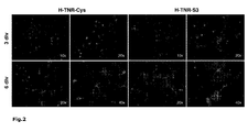
- the cells adhering to H-TNR fragments were labeled with a sulfatide-specific mouse monoclonal antibody (04; Bansal, R. et al., J. Neurosci. Res. 24: 548-557 (1989 )) (30 min at RT).
- the cells obtained through this single step process could for other uses by treatment with accutase ® (Sigma) from the substrate detached and further cultivated on other substrates / plastic surfaces or in other defined media for obtaining, for example, immature or mature, myelin-competing oligodendrocytes.
- plastic surfaces cell culture dishes, plates, etc.
- H-TNR-Cys or H-TNR-S3 fragments (10 ⁇ g / ml in 0.1 M NaHCO 3 ) for 2 h incubated at RT, washed with PBS (2-3 times) and then used as a substrate for oligodendrocyte selection.
- the substrates prepared from H-TNR fragments can also be dried after coating and stored sterile at -20 ° C., without losing the specific adhesive properties of the fragments for oligodendrocytes.
- hMSC human mesenchymal stem cells
- bone marrow Longza, CD105, CD166, CD29 and CD44 positive
- the cells isolated from normal human bone marrow were propagated for 3 to 4 passages in MSCGM.
- the cells growing as monolayers were dissociated with trypsin / EDTA solution, the single cell suspension pelleted after addition of a larger volume of MSCGM at 600 g for 5 minutes at 4 ° C. and the pelleted single cells resuspended in serum-free growth medium.
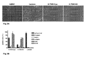
- single cell suspensions (1-2 x 10 6 cells / ml) were plated on uncoated trays and cultured for 6 to 9 days in serum-free growth medium in a CO 2 incubator (5% CO 2 ). The cell culture medium was changed every 2-3 days. Subsequently, adherent cells were trypsinized, single cell suspensions (1 x 10 6 cells / ml) plated on laminin (control substrate) or H-TNR fragments coated dishes (as described above) and cultured for an additional 2 to 5 days in serum-free growth medium ( Fig. 3A ). Thereafter, the growth medium was replaced with glial differentiation medium and the cells were further cultured therein for 3 days.
- adherent cells on the substrates were fixed with 4% (v / v) formaldehyde and 5% (w / v) sucrose in PBS for 30 min at RT and using indirect immunofluorescence with neuron ( ⁇ III tubulin) and glia-specific antibodies
- H-TNR-S3 and H-TNR-Cys substrates were detected 60% (on H-TNR-S3) to 90% (on H-TNR-Cys) sulfatide / GaIC-positive cells, ie predominantly oligodendrocytes, the Survival rate of oligodendroglial differentiated cells on H-TNR-Cys (60% of baseline cell number) was 3-fold higher than that on H-TNR-S3 ( Fig. 3B . 3D ).
- hMSC human mesenchymal stem cells
- bone marrow Longza, CD105, CD166, CD29 and CD44 positive
- the cells isolated from normal human bone marrow were propagated for 3 passages in MSCGM.
- Monolayer cells were dissociated with trypsin / EDTA solution, the single cell suspension pelleted after addition of a larger volume of MSCGM at 600 g for 5 minutes at 4 ° C, and the pelleted single cells resuspended in serum-free Neuromedium.
- single cell suspensions (1-2 x 10 5 cells / cm 2 ) were plated on 0.8% (w / v) agarose type VII (Sigma) coated dishes (5 ml / 95 mm dish) and for 3 to 5 days cultured until the formation of neurospheres in Neuromedium in a CO 2 incubator (5% CO 2 ). The cell culture medium was changed every 2-3 days. Subsequently, neurospheres were pelleted by centrifugation, dissociated with Accutase ® (Sigma) for 15 min at RT and resuspended in neural differentiation medium.
- Accutase ® Sigma
- Single cell suspensions (0.5 x 10 6 cells / ml) were plated on laminin or poly-D-lysine (PDL, control substrates) and H-TNR fragment coated dishes (as described above) and cultured for 5 to 10 days in neuronal differentiation medium.
- the cell culture medium was changed every 2-3 days.
- the adherent cells on the substrates were incorporated with 4% (v / v) formaldehyde in PBS for 30 min RT was fixed and analyzed by indirect immunofluorescence staining with neuron and glia-specific antibodies (as described above).
Abstract
Description
Die Erfindung betrifft ein Verfahren zur Isolierung von neuralen Zellen unter Verwendung von Tenascin-R-Verbindungen, besonders für dieses Verfahren geeignete Tenascin-R-Fragmente und Tenascin-R-Fusionsproteine, die rekombinante Herstellung dieser Tenascin-R-Verbindungen, sowie einen Kit zur Durchführung dieses Verfahrens und die Verwendung des Verfahrens zur Herstellung hochreiner neuraler Zellpopulationen. Die Erfindung betrifft weiterhin Antikörper, die für die Detektion und Isolation von Tenascin-R-Verbindungen geeignet sind.The invention relates to a method for the isolation of neural cells using Tenascin-R compounds, especially suitable for this method Tenascin-R fragments and Tenascin-R fusion proteins, the recombinant production of these Tenascin-R compounds, and a kit for Carrying out this method and the use of the method for producing highly pure neural cell populations. The invention further relates to antibodies which are suitable for the detection and isolation of tenascin-R compounds.
Bei der Kontaktaufnahme von Zellen mit dem sie umgebenden extrazellulären Milieu kann es zu vielfältigen Antworten der Zelle kommen, die von Abstoßung und Vermeidung des Milieus auf der einen bis zur stabilen Zelladhäsion auf der anderen Seite reichen können. Das Wechselspiel zwischen Komponenten der extrazellulären Umgebung, der Extrazellulärmatrix, einerseits, und auf der Zelloberfläche vorhandenen Rezeptoren für solche Komponenten andererseits, bildet die Grundlage für viele entwicklungsbiologische Prozesse, die für eine Zelle unter anderem durch Proliferations-, Migrations- oder Differenzierungsverhalten gekennzeichnet sind. Solch zelluläres Verhalten führt auf Organismenebene zur Musterbildung oder auf Gewebe- oder Organebene zur Neubildung als Folge einer Verletzung (
Tenascin-R (TN-R) (ehemals als J1-160/180, Janusin oder Restrictin bezeichnet) ist ein Mitglied der Tenascin-Familie von Extrazellulärmatrixproteinen, das exklusiv im ZNS von Vertebraten auftritt und dort von Oligodendrozyten und manchen Gruppen von Neuronen, i.e. Motoneurone und Interneurone, während späterer Entwicklungsstadien und im erwachsenen Zustand exprimiert wird (
TN-R ist aus vier unterschiedlichen Domänenstrukturen aufgebaut. Der N-Terminus, dessen Sequenz nur in Tenascin-Proteinen vorkommt, enthält ein Cystein-reiches Segment (Cys-rich), und wird gefolgt von viereinhalb EGF-ähnlichen Segmenten (EGF-like) sowie 9 Fibronektin Typ III (FN III)-ähnlichen Domänen (von denen die 6. Domäne alternativ gespleisst werden kann). Den C-Terminus des TN-R Proteins bildet eine globuläre, Fibrinogen-ähnliche Domäne (FNG). Einzelne TN-R Polypeptidketten sind an ihren N-Termini über Disulfidbrücken miteinander verbunden und bilden so Homotrimere (TN-R 180) oder -dimere (TN-R 160), wobei letztere durch proteolytische Spaltung von TN-R 180 nahe des N-Terminus entstehen (
Die Funktionsweite von TN-R umfasst die molekulare Kontrolle neuraler Zelladhäsion, Migration und Differenzierung (von der Axonnavigation Fortsätze bildender Neuronen bis zur Reifung myelinbildender Oligodendrozyten) während normaler entwicklungsbiologischer Vorgänge sowie regenerativer Prozesse nach Verletzung im erwachsenen Gehirn (
Einige neurale Rezeptoren und intrazelluläre Signalwege, die die Wirkung von TN-R in neuronalen und glialen Zellen vermitteln, sind bekannt, ebenso konnten molekulare Komponenten identifiziert werden, die an der Expression von TN-R durch Oligodendrozyten und Motoneurone beteiligt sind (Tab. 1;
Als aus mehreren Domänen aufgebautes Extrazellulärmatrixprotein erscheint eine Aufteilung der distinkten biologischen Funktionen auf distinkte Domänenbereiche und/oder distinkte Glykostrukturen von TN-R nahe liegend. Ähnlich wie die anderen bekannten TN-R Proteine, setzt sich das humane TN-R Protein aus verschiedenen distinkten Domänen zusammen (Carnemolla et al., 1996). Beginnend mit einer Cystein-reichen Region am N-Terminus, gefolgt von 4.5 EGF-ähnlichen Domänen und 9 FN III-ähnlichen Domänen (von denen die 6. Domäne alternativ gespleisst werden kann), endet das Molekül am C-Terminus mit einer Fibrinogen-ähnlichen Domäne. Die publizierte humane TN-R mRNA-Sequenz umfasst mit 9 FN III-ähnlichen Domänen 4716 Basen. Hiervon entfällt auf den kodierenden Bereich der Abschnitt zwischen den Basen 82 bis 4158, auf das Signalpeptid (zur Sekretion des TN-R Proteins) der Basenbereich 82 bis 150 (SEQ ID NO:2).As an extracellular matrix protein composed of several domains, a division of the distinct biological functions into distinct domain regions appears and / or distinct glycostructures of TN-R. Similar to the other known TN-R proteins, the human TN-R protein is composed of distinct distinct domains (Carnemolla et al., 1996). Starting with a cysteine-rich region at the N-terminus, followed by 4.5 EGF-like domains and 9 FN III-like domains (of which the 6th domain can alternatively be spliced), the molecule terminates at the C-terminus with a fibrinogen. similar domain. The published human TN-R mRNA sequence comprises 4716 bases with 9 FN III-like domains. Of this, the coding region of the section between bases 82 to 4158, the signal peptide (for secretion of the TN-R protein), the base region 82 to 150 (SEQ ID NO: 2).
Es ist bekannt, dass aus adultem Nagergehirn aufgereinigte TN-R Proteine die Adhäsion und Fortsatzbildung von aus früh postnatalem Gehirn isolierten O4/Sulfatid-positiven Oligodendrozyten fördern (
Die frühesten Stadien sich entwickelnder Gliazellen finden sich in Säugern im Rückenmark in ventralen Bereichen des Neuralrohrs, im Gehirn in den ventrikulären Zonen des Vorderhirns. In diesen Regionen proliferieren erste Vorläuferzellen von Oligodendrozyten, die durch eine einfache Morphologie und die Expression des Disialogangliosids GD3 und/oder von 04 Antigenen gekennzeichnet sind, und wandern in der Folgezeit in Bereiche der später entstehenden weißen Substanz ein (
Da insbesondere für gliale Zellen (wie Oligodendrozyten und neurale Stammzellen) keine adäquaten Modellsysteme in Gestalt von Zelllinien vorhanden sind, muss zu ihrer Gewinnung sowohl in der Grundlagenforschung als auch im Rahmen potentieller diagnostischer/therapeutischer Anwendungsbereiche bisher auf relativ zeitaufwendige und niedrig effiziente Anreicherungsverfahren von Primärzellen zurückgegriffen werden. Diese Verfahren ermöglichen außerdem oftmals nur die Herstellung angereicherter Mischzellpopulationen. Die bisherigen Verfahren zur Isolierung definierter Zellpopulationen nutzen unterschiedliche Techniken wie Dichtegradientenzentrifugationen oder immunologische Verfahren (Fluorescenceactivated cell sorting, biomagnetic cell sorting, Antikörper- und Komplementvermittelte Zelltötung, Antikörper-Panning) (
In
Es wurde nunmehr gefunden, dass ein Fragment, das die Aminosäurereste 24 bis 189 der SEQ ID NO:2 des humanen TN-R umfasst, oder ein Homolog oder Fragment desselben wie z. B. das Fragment H-TNR-Cys, das Aminosäurereste 24 bis 189 der SEQ ID NO:2 aufweist, ebenso gut wie das in der WO
Die vorliegende Erfindung betrifft somit
- (1) ein Verfahren zur Isolierung und Reinigung von neuralen Zellen aus neuralem Primärgewebe von Vertebraten, umfassend die Selektion der Zellen aus einer Einzelzellsuspension mittels einer Tenascin-R-haltigen Sonde ("Tenascin-R-Sonde"), welche ein N-terminales Fragment von nativem Tenascin-R (TN-R) sowie Homologen und Fragmenten desselben und Fusionsproteine derartiger Verbindungen umfasst;
- (2) eine bevorzugte Ausführungsform von (1), wobei das Tenascin-R-Fragment ein Peptid mit der Sequenz der Aminosäurereste 51-216 von SEQ ID NO:2 ist und das Überleben und/oder die Transdifferenzierung adulter humaner mesenchymaler Stammzellen in neuronale oder oligodendrogliale Zellen, insbesondere unter definierten serumfreien Kulturbedingungen ermöglicht;
- (3) ein Tenascin-R-Fragment oder ein Tenascin-R-Fusionsprotein wie in (1) oder (2) definiert;
- (4) eine DNA, die ein Tenascin-R-Fragment oder ein Tenascin-R-Fusionsprotein von (3) kodiert;
- (5) einen Vektor, der eine DNA nach (4) umfasst;
- (6) einen Wirtsorganismus, der mit einem Vektor nach (5) transformiert/transfiziert ist und/oder eine DNA nach (4) aufweist;
- (7) ein Verfahren zur Herstellung eines Tenascin-R-Fragments oder Tenascin-R-Fusionsproteins nach (3), umfassend das Kultivieren des Wirtsorganismus nach (6);
- (8) einen Antikörper, insbesondere ein monoklonaler Antikörper, der durch Immunisierung eines geeigneten Wirtsorganismus mit einem Tenascin-R-Fragment nach (3) erhältlich ist;
- (9) eine Zelllinie oder Hybridomzelllinie, die einen monoklonalen Antikörper nach (8) produziert;
- (10) ein Kit zur Isolierung und Reinigung von neuralen Zellen, insbesondere von Oligodendrozyten, nach dem Verfahren von (1) oder (2), insbesondere enthaltend
- (i) eine wie in (1) oder (2) definierte Tenascin-R-Sonde, und/oder
- (ii) einen Vektor, der für die in (i) definierte Tenascin-R-Sonde kodiert, und/oder
- (iii) eine Stammkultur einer Zelllinie, die dazu geeignet ist, die wie in (1) oder (2) definierte Tenascin-R-Sonde zu exprimieren;
- (11) die Verwendung eines Tenascin-R-Fragments oder eines Tenascin-R-Fusionsproteins wie in (3) definiert, zur Gewinnung von neuralen Zellen, insbesondere von Oligodendrozyten, für die Anzucht differenzierter Zellen, insbesondere neuraler Zellen, in neurobiologischen und zellphysiologischen Untersuchungen, in der biologischen und klinischen Forschung und für diagnostische und therapeutische Verfahren in vitro und in vivo, insbesondere für die Herstellung eines Medikaments zur Zelltherapie und zur Therapie von neurodegenerativen Krankheiten, die mit einem Verlust von Oligodendrozyten oder Myelin einhergehen, insbesondere Multipler Sklerose und periventrikulärer Leukomalazie (PVL);
- (12) ein Verfahren zur Herstellung von Oligodendrozyten aus isolierten Stammzellen in vitro durch Inkubation der Stammzellen in Anwesenheit eines Tenascin-R-Fragments oder eines Tenascin-R-Fusionsproteins wie in (3) definiert;
- (13) ein Verfahren zur Zelltherapie oder zur Therapie von neurodegenerativen Krankheiten, die mit einem Verlust von Oligodendrozyten oder Myelin einhergehen, insbesondere von Multipler Sklerose und periventrikulärer Leukomalazie (PVL), umfassend die Gabe eines Tenascin-R-Fragments oder eines Tenascin-R-Fusionsproteins wie in (3) definiert, an einen menschlichen oder tierischen Patienten;
- (14) ein Verfahren zur Therapie und Prophylaxe traumatischer Nervenläsionen oder zur gezielten Beeinflussung der Neuralentwicklung, umfassend die Verabreichung einer pharmakologisch ausreichenden Menge des Antikörpers nach (8) an einen menschlichen oder tierischen Patienten, der einer solchen Behandlung bedarf.
- (1) a method of isolating and purifying neural cells from vertebrate neural primary tissue, comprising selecting the cells from a single cell suspension using a tenascin-R containing probe ("tenascin-R probe") which is an N-terminal fragment of native tenascin-R (TN-R) and homologs and fragments thereof and fusion proteins of such compounds;
- (2) a preferred embodiment of (1), wherein the tenascin-R fragment is a peptide having the sequence of amino acid residues 51-216 of SEQ ID NO: 2 and the survival and / or transdifferentiation of adult human mesenchymal stem cells into neuronal or oligodendroglial cells, in particular under defined serum-free culture conditions allows;
- (3) a tenascin-R fragment or a tenascin-R fusion protein as defined in (1) or (2);
- (4) a DNA encoding a tenascin-R fragment or a tenascin-R fusion protein of (3);
- (5) a vector comprising a DNA according to (4);
- (6) a host organism transformed / transfected with a vector according to (5) and / or having a DNA according to (4);
- (7) a method of producing a tenascin-R fragment or tenascin-R fusion protein according to (3), comprising cultivating the host organism according to (6);
- (8) an antibody, in particular a monoclonal antibody, obtainable by immunizing a suitable host organism with a tenascin R fragment according to (3);
- (9) a cell line or hybridoma cell line producing a monoclonal antibody according to (8);
- (10) a kit for the isolation and purification of neural cells, in particular of oligodendrocytes, according to the method of (1) or (2), in particular containing
- (i) a tenascin-R probe as defined in (1) or (2), and / or
- (ii) a vector coding for the tenascin-R probe defined in (i), and / or
- (iii) a stock culture of a cell line capable of expressing the tenascin-R probe as defined in (1) or (2);
- (11) the use of a tenascin-R fragment or a tenascin-R fusion protein as defined in (3) for obtaining neural cells, in particular oligodendrocytes, for the growth of differentiated cells, especially neural cells, in neurobiological and cell physiological studies in biological and clinical research and for in vitro and in vivo diagnostic and therapeutic procedures, in particular for the manufacture of a medicament for cell therapy and for the therapy of neurodegenerative diseases associated with the loss of oligodendrocytes or myelin, in particular multiple sclerosis and periventricular leukomalacia (PVL);
- (12) a method of producing oligodendrocytes from isolated stem cells in vitro by incubating the stem cells in the presence of a tenascin-R fragment or a tenascin-R fusion protein as defined in (3);
- (13) a method of cell therapy or therapy of neurodegenerative diseases associated with loss of oligodendrocytes or myelin, in particular multiple sclerosis and periventricular leukomalacia (PVL), comprising the administration of a tenascin R fragment or a tenascin R Fusion protein as defined in (3) to a human or animal patient;
- (14) a method for the therapy and prophylaxis of traumatic nerve lesions or for the purpose of influencing neural development, comprising administering to a human or animal patient in need of such treatment a pharmacologically sufficient amount of the antibody according to (8).
- A) Westernblot-Analyse affinitätsgereinigter H-TNR-S3 und H-TNR-Cys Fragmente mit Tag-spezifischen (V5) und TN-R-spezifischen (R4) monoklonalen Antikörpern. Die über Nickel-Agarose gereinigten rekombinanten Fragmente (20 ng/Tasche) H-TNR-S3 (S3) und H-TNR-Cys (Cys) wurden gelelektrophoretisch aufgetrennt, auf Nitrozellulosefilter transferiert und mit V5 oder R4 Antikörper inkubiert. Immunreaktive Proteinbanden wurden durch Inkubation mit Peroxidase-gekoppelten anti-Maus IgG Antikörpern nachgewiesen. Pfeile zeigen das Molekulargewicht von H-TNR-S3 (40 kD), bzw. von H-TNR-Cys (34 kD).
- B) Einfluss von polaren Glykolipiden, Lysenin (Sphingomyelin-bindenden Protein), 04- und TN-R-spezifischen Antikörpern auf die TN-R-vermittelte Zelladhäsion. TN-R Substrate (TNR, H-TNR-Cys, H-TNR-S3) wurden in der Abwesenheit (- GL/Ab) oder Anwesenheit von Monosialogangliosiden (+ GM1), Sulfatiden (+ Sulf) und TN-R-spezifischen Antikörpern (R4, R6) vorinkubiert. Erythrozyten wurden in Ab- oder Anwesenheit von Lysenin (100 µg/ml) und 04 Antikörpern (+ 04 Ab) auf die entsprechende Substrate ausgesät. Die Zahl adhärenter Zellen nach einstündiger Inkubationszeit auf unbehandelten TN-R Substraten (- GL/Ab) wurde
als 100% gesetzt. - C) Interaktion R4 Epitop-tragender TN-R/TN-R Fragmente mit polaren Glykolipiden im Festphasen-Bindungsassay. Mikrotiterplatten wurden mit Sulfatiden (Sulf), Galactocerebrosiden (GalC), Sphingomyelin (SM) oder Sialogangliosiden (GM1, GD1a; alle bei 100 pmol/Vertiefung) beschichtet und mit TN-R oder rekombinanten TN-R Fragmenten (H-TNR-Cys, H-TNR-S3) für 2 h bei RT inkubiert. Die Bindung von TN-R/TN-R Fragmenten wurde mit R6 (für TN-R) oder V5 Antikörper (
nach 1,5 Stunden Inkubation bei RT) mit Peroxidase-gekoppelten anti-Maus IgG Antikörpern nachgewiesen. Die maximale Bindung auf Sulfatide wurde für dasjeweilige Protein als 100% gesetzt.
- A) hMSC (linkes Bild) wurden auf substratgebundenen Laminin und TN-R Fragmenten (H-TNR-Cys, H-TNR-S3) für 5 Tage in serumfreiem Wachstumsmedium kultiviert.
- B) Gliale Differenzierung von hMSC auf substratgebundenen TN-R Fragmenten (H-TNR-Cys, H-TNR-S3). Auf TN-R Fragmenten oder Laminin ausplattierte Zellen wurden für 2 Tage in serumfreiem Wachstumsmedium und für weitere 3 Tage in serumfreiem glialem Differenzierungsmedium kultiviert. Die Zahl der mit Antikörpern markierten Zellen auf dem jeweiligen Substrat wurde zur Gesamtzellzahl (100%) in Relation gesetzt.
Über 80% der Zellen auf H-TNR-Cys Substraten waren Sulfatid-/GaIC-positiv und konnten als Oligodendrozyten identifiziert werden. - C) Neuronale Differenzierung von hMSC auf substratgebundenen TN-R Fragmenten (H-TNR-Cys, H-TNR-S3). Auf TN-R Fragmenten oder poly-D-Lysin (PDL) ausplattierte, aus Neurosphären stammende Zellen wurden für 10 Tage in serumfreiem neuronalem Differenzierungsmedium kultiviert. Die Zahl der mit Antikörpern markierten Zellen auf dem jeweiligen Substrat wurde zur Gesamtzellzahl (100%) in Relation gesetzt. 80% der Zellen auf H-TNR-Cys Substraten waren βIII Tubulin-positiv und konnten als Neurone identifiziert werden. D) Zusammenfassung über den Einfluss von H-TNR-Cys auf die neurale Transdifferenzierung von hMSC unter definierten serumfreien Kulturbedingungen. Überlebensrate und Expression Neuron- (βIII Tubulin) und Oligodendrozytenspezifischer Moleküle (Sulfatid/GaIC) von auf H-TNR-Cys Substraten wachsenden Zellen kultiviert in glialem (glial) oder neuronalem (neuronal) Differenzierungsmedium.
- A) Western blot analysis of affinity-purified H-TNR-S3 and H-TNR-Cys fragments with tag-specific (V5) and TN-R-specific (R4) monoclonal antibodies. The nickel agarose purified recombinant fragments (20 ng / bag) H-TNR-S3 (S3) and H-TNR-Cys (Cys) were gel electrophoresed, transferred to nitrocellulose filters and incubated with V5 or R4 antibody. Immunoreactive protein bands were detected by incubation with peroxidase-linked anti-mouse IgG antibodies. Arrows show the molecular weight of H-TNR-S3 (40 kD), or of H-TNR-Cys (34 kD).
- B) Influence of polar glycolipids, lysine (sphingomyelin-binding protein), 04- and TN-R-specific antibodies on TN-R-mediated cell adhesion. TN-R substrates (TNR, H-TNR-Cys, H-TNR-S3) were in the absence (- GL / Ab) or presence of monosialogangliosides (+ GM1), sulfatides (+ sulf) and TN-R-specific antibodies (R4, R6) pre-incubated. Erythrocytes were seeded in the absence or presence of lysine (100 ug / ml) and 04 antibodies (+ 04 Ab) on the corresponding substrates. The number of adherent cells after one hour of incubation on untreated TN-R substrates (- GL / Ab) was set as 100%.
- C) Interaction R4 Epitope-bearing TN-R / TN-R fragments with polar glycolipids in the solid-phase binding assay. Microtiter plates were coated with sulfatides (Sulf), galactocerebroside (GalC), sphingomyelin (SM) or sialogangliosides (GM1, GD1a, all at 100 pmol / well) and probed with TN-R or recombinant TN-R fragments (H-TNR-Cys, H-TNR-S3) for 2 h at RT. The binding of TN-R / TN-R fragments was detected with R6 (for TN-R) or V5 antibody (after 1.5 hours incubation at RT) with peroxidase-coupled anti-mouse IgG antibodies. The maximum binding to sulfatides was set to 100% for each protein.
- A) hMSC (left panel) were cultured on substrate-bound laminin and TN-R fragments (H-TNR-Cys, H-TNR-S3) for 5 days in serum-free growth medium.
- B) Glial differentiation of hMSC on substrate-bound TN-R fragments (H-TNR-Cys, H-TNR-S3). Cells plated on TN-R fragments or laminin were cultured for 2 days in serum-free growth medium and for a further 3 days in serum-free glial differentiation medium. The number of with Antibody labeled cells on the respective substrate were related to the total cell number (100%). Over 80% of the cells on H-TNR-Cys substrates were sulfatide / GaIC positive and could be identified as oligodendrocytes.
- C) Neuronal differentiation of hMSC on substrate-bound TN-R fragments (H-TNR-Cys, H-TNR-S3). Neurospheric-derived cells plated on TN-R fragments or poly-D-lysine (PDL) were cultured for 10 days in serum-free neuronal differentiation medium. The number of cells labeled with antibodies on the respective substrate was related to the total number of cells (100%). 80% of cells on H-TNR-Cys substrates were βIII tubulin positive and could be identified as neurons. D) Summary of the influence of H-TNR-Cys on the neural transdifferentiation of hMSC under defined serum-free culture conditions. Survival Rate and Expression Neuron (βIII tubulin) and oligodendrocyte-specific molecules (sulfatide / GaIC) of cells growing on H-TNR-Cys substrates cultured in glial (or glial) or neuronal (neuronal) differentiation medium.
Die Erfindung betrifft eine Methode zur Isolierung hochreiner Zellpopulationen aus neuralem Primärgewebe in einem Einstufenverfahren mit Hilfe nativer Tenascin-R-Proteine oder TN-R-Fragmente von Vertebraten, bevorzugt von Fischen, Amphibien, Reptilien, Vögeln und Säugetieren, besonders bevorzugt von Hai, Karpfen, Huhn, Nagern einschließlich Maus und Ratte, Rind, Schwein und Mensch, ganz besonders bevorzugt aus Nagern und Mensch, durch selektive Substratadhäsion.The invention relates to a method for isolating high purity cell populations from neural primary tissue in a single step process using native tenascin R proteins or TN-R fragments from vertebrates, preferably from fish, amphibians, reptiles, birds and mammals, most preferably from shark, carp , Chicken, rodents including mouse and rat, bovine, porcine and human, most preferably rodent and human, by selective substrate adhesion.
Phylogenetische Studien zur Funktion von Tenascin-R im Nervensystem von Vertebraten zeigen als stammesgeschichtlich hoch konservierte Funktion von Tenascin-R-Proteinen die Unterstützung der Adhäsion und Fortsatzbildung von Oligodendrozyten. Die vorliegende Erfindung zeigt, dass diese Eigenschaft des gesamten Tenascin-R-Proteins auch auf distinkten rekombinanten Fragmenten des humanen Tenascin-R Proteins lokalisierbar ist.Phylogenetic studies on the function of tenascin-R in the nervous system of vertebrates show the highly conserved function of tenascin-R proteins as a support for the adhesion and extension of oligodendrocytes. The present invention demonstrates that this property of the entire tenascin-R protein is also localizable on distinct recombinant fragments of the human tenascin-R protein.
"Tenascin-R-Sonde" bedeutet im Kontext der vorliegenden Erfindung ein N-terminales Fragment von nativem Tenascin-R, welches in vivo und in vitro durch Oligodendrozyten erkannt und gebunden wird, und/oder weitere Tenascin-R-Verbindungen einschließlich Tenascin-R-Homologe und Tenascin-R-Fragmente, die ebenfalls mit hoher Sensitivität und Spezifität in vivo und in vitro durch Oligodendrozyten und/oder durch andere neurale Zellen aus Primärgewebe gebunden werden, enthalten kann, wobei "Tenascin-R-Verbindungen" die an anderer Stelle definierte Bedeutung hat. Dies schließt auch Fusionsproteine aus mehreren solcher N-terminalen Tenascin-R-Fragmente ein, bevorzugt aus zwei oder drei Tenascin-R-Verbindungen. Die Bestandteile dieser Fusionsproteine können direkt oder mittels eines (flexiblen) Linkerpeptids miteinander verknüpft sein. Besonders bevorzugt enthält die Tenascin-R-Sonde humane Tenascin-R-Fragmente, ganz besonders bevorzugt die Teilsequenz der humanen TN-R, welche die Aminosäurereste 24 bis 189 der SEQ ID NO:2 umfasst. Die Tenascin-R-Sonde kann neben ihren TN-R-Anteilen auch Nicht-TN-R-Anteile besitzen, welche die Stabilität des TN-R-Anteils gewährleisten und/oder die Immobilisierbarkeit und Kopplungsfähigkeit an andere Moleküle erhalten oder erhöhen. Insbesondere ist eine erfindungsgemäße Tenascin-R-Sonde rekombinant hergestellt."Tenascin-R probe" in the context of the present invention means an N-terminal fragment of native tenascin-R that is recognized and bound by oligodendrocytes in vivo and in vitro , and / or other tenascin-R compounds including tenascin-R Homologues and tenascin-R fragments which are also bound with high sensitivity and specificity in vivo and in vitro by oligodendrocytes and / or by other neural cells from primary tissue, with "tenascin-R compounds" elsewhere has defined meaning. This also includes fusion proteins from several such N-terminal tenascin-R fragments, preferably from two or three tenascin-R compounds. The components of these fusion proteins can be linked together directly or by means of a (flexible) linker peptide. The tenascin-R probe particularly preferably contains human tenascin-R fragments, very particularly preferably the partial sequence of the human TN-R which comprises amino acid residues 24 to 189 of SEQ ID NO: 2. The tenascin-R probe can also have non-TN-R moieties in addition to its TN-R moieties, which ensure the stability of the TN-R moiety and / or maintain or increase the immobilizability and coupling ability to other molecules. In particular, a tenascin-R probe according to the invention is produced recombinantly.
"Tenascin-R-Verbindungen" sind erfindungsgemäß Proteine oder Peptide, welche entweder N-terminale Fragmente von nativen TN-R oder deren substantiell identische Homologe sind."Tenascin-R compounds" according to the invention are proteins or peptides which are either N-terminal fragments of native TN-R or their substantially identical homologues.
"Primärgewebe" ist ein biologisches Gewebe, das direkt aus einem Organismus entnommen und ohne weitere Veränderung seines genetischen Materials verwendet wird. "Primärzellen" sind aus Primärgewebe stammende Zellen. Primärzellen sind gegenüber Zelllinien vorteilhaft, weil sie aufgrund ihrer Herkunft den Zellen im intakten Organismus strukturell und funktionell am nächsten stehen."Primary tissue" is a biological tissue taken directly from an organism and used without further alteration of its genetic material. "Primary cells" are cells derived from primary tissue. Primary cells are advantageous over cell lines because they are structurally and functionally closest to the cells in the intact organism because of their origin.
"Isoliert" bedeutet im Falle eines Proteins, dass es von anderen Proteinen, mit denen es normalerweise in demjenigen Organismus assoziiert ist, in welchem es natürlicherweise vorkommt, getrennt oder gereinigt wurde. Dies umfasst biochemisch gereinigte Proteine, rekombinant hergestellte Proteine und auf chemischem Wege synthetisierte Proteine. Die Definition gilt übertragen auch für Nukleinsäuren, insbesondere DNA, und Peptide."Isolated" in the case of a protein means that it has been separated or purified from other proteins with which it is normally associated in the organism in which it naturally occurs. This includes biochemically purified proteins, recombinantly produced proteins, and chemically synthesized proteins. The definition also applies to nucleic acids, in particular DNA, and peptides.
"Nativ" wird gleichbedeutend mit "natürlich" bzw. "natürlich vorkommend" verwendet."Native" is used synonymously with "natural" or "naturally occurring".
Für die erfindungsgemäße "rekombinante Herstellung" werden in der Fachwelt übliche Methoden zur rekombinanten Herstellung eukaryotischer Proteine verwendet, insbesondere die Expression als Fusionsprotein in pro- und eukaryotischen Zellen, ganz besonders bevorzugt die Expression als Fusionsprotein mit Polyhistidinschwanz. Des Weiteren bevorzugt ist die Verwendung von DNA, die für das entsprechende Protein kodiert.For the "recombinant production" according to the invention, customary methods for the recombinant production of eukaryotic proteins are used, in particular the expression as a fusion protein in prokaryotic and eukaryotic cells, very particularly preferably the expression as fusion protein with polyhistidine tail. Further preferred is the use of DNA encoding the corresponding protein.
Nukleinsäuresequenzen, insbesondere DNA-Sequenzen, die für erfindungsgemäße Proteine oder Peptide kodieren, sind im Rahmen der vorliegenden Erfindung entweder identisch oder substantiell identisch zur nativen oder zu der ihr zugrundeliegenden künstlichen erfindungsgemäßen Sequenz. Wenn im Rahmen dieser Erfindung eine spezielle Nukleinsäuresequenz genannt wird, so sind damit diese Sequenz selbst und die zu ihr substantiell identischen Sequenzen erfasst. "Substantiell identisch" bedeutet hierbei, dass nur ein Austausch von Basen in der Sequenz im Rahmen des degenerierten Nukleinsäure-Codes erfolgt ist, dass also dadurch die Codons innerhalb kodierender Sequenzen der substantiell identischen Nukleinsäure lediglich gegenüber dem ursprünglichen Molekül in einer Weise verändert sind, welche nicht zu einer Veränderung der Aminosäuresequenz des Translationsprodukts führt (in der Regel Austausch des Codons durch ein anderes Codon seiner Codon-Familie). Bevorzugt sind im Rahmen der vorliegenden Erfindung die Sequenzen, welche im Sequenzprotokoll benannt werden, und deren erfindungsgemäße Fragmente.Nucleic acid sequences, in particular DNA sequences, which code for proteins or peptides according to the invention are in the context of the present invention either identical or substantially identical to the native or to its underlying artificial sequence according to the invention. If a specific nucleic acid sequence is mentioned in the context of this invention, then this sequence itself and the sequences substantially identical to it are recorded. "Substantially identical" here means that only one exchange of bases in the sequence has taken place in the context of the degenerate nucleic acid code, that is to say that the codons within coding sequences of the substantially identical nucleic acid are changed only in relation to the original molecule in a manner which does not result in a change in the amino acid sequence of the translation product (usually replacing the codon with another codon of its codon family). In the context of the present invention, the sequences named in the sequence listing and their fragments according to the invention are preferred.
Proteinsequenzen und Peptidsequenzen können im Rahmen der vorliegenden Erfindung durch Substitution von Aminosäuren modifiziert werden. Bevorzugt sind solche Substitutionen, bei denen die Funktion und/oder Konformation des Proteins oder Peptids erhalten bleibt, besonders bevorzugt jene Substitutionen, bei denen eine oder mehrere Aminosäuren durch Aminosäuren ersetzt werden, die ähnliche chemische Eigenschaften besitzen, z. B. Valin durch Alanin ("konservativer Aminosäureaustausch"). Der Anteil der substituierten Aminosäuren im Vergleich zum nativen Protein oder - falls es sich nicht um ein natives Protein handelt - zur Ausgangssequenz beträgt bevorzugt 0-30% (bezogen auf die Zahl der Aminosäuren in der Sequenz), besonders bevorzugt 0-15%, ganz besonders bevorzugt 0-5%.Protein sequences and peptide sequences can be modified in the context of the present invention by substitution of amino acids. Preferred are those substitutions in which the function and / or conformation of the protein or peptide is retained, more preferably those substitutions in which one or more amino acids are replaced by amino acids having similar chemical properties, e.g. B. valine by alanine ("conservative amino acid exchange"). The proportion of substituted amino acids compared to the native protein or, if it is not a native protein to the starting sequence is preferably 0-30% (based on the number of amino acids in the sequence), particularly preferably 0-15%, completely more preferably 0-5%.
Nukleinsäuresequenzen und Aminosäuresequenzen können als Volllänge-Sequenzen oder als Additions- oder Deletionsprodukte dieser Volllängesequenzen zur Durchführung der Erfindung eingesetzt werden. Bei den Aminosäuresequenzen umfassen die Additionsprodukte auch Fusionsproteine, außerdem Aminosäuresequenzen, die durch Addition von 1-100, bevorzugt 1-30, ganz besonders bevorzugt 1-10 Aminosäuren entstehen. Die addierten Aminosäuren können dabei einzeln oder in zusammenhängenden Abschnitten von 2 oder mehr miteinander verknüpften Aminosäuren ein- oder angefügt werden. Die Addition kann am N- oder C-Terminus oder innerhalb der ursprünglichen Sequenz stattfinden. Es sind mehrere Additionen in einer Sequenz zulässig, wobei eine einzelne Addition bevorzugt ist, besonders bevorzugt eine Addition am C- oder N-Terminus.Nucleic acid sequences and amino acid sequences can be used as full-length sequences or as addition or deletion products of these full-length sequences for carrying out the invention. In the case of the amino acid sequences, the addition products also comprise fusion proteins, moreover amino acid sequences which are formed by addition of 1-100, preferably 1-30, most preferably 1-10 amino acids. The added amino acids can be added or added individually or in contiguous sections of 2 or more linked amino acids. The addition may take place at the N- or C-terminus or within the original sequence. Several additions are allowed in a sequence, with a single addition being preferred, more preferably an addition at the C or N terminus.
Die Deletionsprodukte der Volllängen-Aminosäuresequenzen entstehen - falls nicht für spezielle Sequenzen anders angegeben - durch Deletion von 1-100, bevorzugt 1-20, ganz besonders bevorzugt 1-10 Aminosäuren. Die deletierten Aminosäuren können dabei einzeln oder in zusammenhängenden Abschnitten von 2 oder mehr miteinander verknüpften Aminosäuren entfernt werden. Die Deletion kann am N-oder C-Terminus oder innerhalb der ursprünglichen Sequenz stattfinden. Es sind mehrere Deletionen in einer Sequenz zulässig, wobei eine einzelne Deletion bevorzugt ist, besonders bevorzugt eine Deletion am C- oder N-Terminus.The deletion products of the full length amino acid sequences are formed - if not stated otherwise for specific sequences - by deleting 1-100, preferably 1-20, most preferably 1-10 amino acids. The deleted amino acids can be removed individually or in contiguous sections of 2 or more linked amino acids. The deletion may occur at the N or C terminus or within the original sequence. Several deletions are allowed in a sequence, with a single deletion being preferred, more preferably a deletion at the C or N-terminus.
Die zulässigen Deletionen und Additionen in den erfindungsgemäßen Nukleinsäuresequenzen haben den Umfang und die Ausprägung, welche der zulässigen Aminosäuredeletionen bzw.- additionen entsprechen. Über die Deletion und Addition ganzer Codons hinaus ist auch die Addition oder Deletion von einzelnen oder paarweisen Basen möglich.The permissible deletions and additions in the nucleic acid sequences according to the invention have the scope and the extent which correspond to the permissible amino acid deletions or additions. In addition to the deletion and addition of whole codons addition or deletion of single or paired bases is possible.
Ein Fragment einer Nukleinsäure oder eines Proteins ist ein Teil von dessen Sequenz, das kürzer als Volllänge ist, jedoch noch einen minimal zur Hybridisierung bzw. spezifischen Bindung erforderlichen Sequenzabschnitt enthält. Im Falle einer Nukleinsäure ist dieser Sequenzabschnitt noch in der Lage, mit der nativen Nukleinsäure unter stringenten Bedingungen zu hybridisieren und umfasst vorzugsweise mindestens 15 Nukleotide, besonders bevorzugt mindestens 25 Nukleotide. Im Falle eines Peptids ist dieser Sequenzabschnitt ausreichend, um eine Bindung eines für einen Abschnitt des nativen Proteins spezifischen Antikörpers oder einer Zelle, welche an TN-R oder ein TN-R-Fragment bindet, zu ermöglichen. Die Peptidlänge beträgt vorzugsweise wenigstens 5 Aminosäuren, besonders bevorzugt wenigstens 10 Aminosäuren, ganz besonders bevorzugt wenigstens 20 Aminosäuren.A fragment of a nucleic acid or a protein is a part of its sequence that is shorter than full length but still contains a minimal sequence segment necessary for hybridization or specific binding. In the case of a nucleic acid, this sequence segment is still able to hybridize with the native nucleic acid under stringent conditions and preferably comprises at least 15 nucleotides, more preferably at least 25 nucleotides. In the case of a peptide, this sequence portion is sufficient to allow binding of an antibody specific for a portion of the native protein or a cell that binds to TN-R or a TN-R fragment. The peptide length is preferably at least 5 amino acids, more preferably at least 10 amino acids, most preferably at least 20 amino acids.
Ein "Fusionsprotein" im Kontext der vorliegenden Erfindung umfasst mindestens eine erfindungsgemäße Tenascin-R-Verbindung, die mit mindestens einem zweiten Protein oder Peptid verknüpft ist. Dieses zweite Protein oder Peptid ist bevorzugt ein Selektions- oder Markerprotein, ein Protein, welches der Bindung des Fusionsproteins an eine Oberfläche dient, oder eine Tenascin-R-Verbindung. Bevorzugt sind Fusionsproteine aus nativem Tenascin-R und weiteren funktionellen Proteinen und Peptiden, und Fusionsproteine aus zwei oder mehr, besonders bevorzugt zwei oder drei Tenascin-R-Fragmenten. Die Nukleinsäuresequenzen, die in einem Vektor oder einem transformierten Wirtsorganismus für die einzelnen Teile des Fusionsproteins kodieren, sind in einer Weise miteinander verknüpft, welche die Expression unter der Kontrolle eines einzigen Promoters erlaubt. Die Aminosäuresequenzen der einzelnen funktionalen Teile des Fusionsproteins sind dabei entweder direkt oder mit einem Linker verknüpft. Der Linker ist 1-30 Aminosäuren lang, bevorzugt 10-20 Aminosäuren.A "fusion protein" in the context of the present invention comprises at least one tenascin-R compound of the invention linked to at least one second protein or peptide. This second protein or peptide is preferably a selection or marker protein, a protein which serves to bind the fusion protein to a surface, or a tenascin-R compound. Preferred are fusion proteins of native tenascin-R and other functional proteins and peptides, and fusion proteins of two or more, more preferably two or three tenascin-R fragments. The nucleic acid sequences encoding the individual parts of the fusion protein in a vector or a transformed host organism are linked together in a manner that permits expression under the control of a single promoter. The amino acid sequences of the individual functional parts of the fusion protein are linked either directly or with a linker. The linker is 1-30 amino acids long, preferably 10-20 amino acids.
"Neurodegenerative Erkrankungen" sind Erkrankungen des Nervensystems, die mit dem Absterben von neuronalen und/oder makroglialen Zellen aufgrund von Beeinträchtigungen ihrer funktionellen Integrität assoziiert sind. Solche Zellschädigungen und -verluste führen zu Ausfall oder Störung der Funktionen der betroffenen Regionen des Nervensystems und/oder der von diesen Regionen gesteuerten Körperteilen."Neurodegenerative disorders" are disorders of the nervous system associated with the death of neuronal and / or macroglial cells due to impairment of their functional integrity. Such cell damage and loss results in failure or disturbance of the functions of the affected regions of the nervous system and / or the body parts controlled by these regions.
Die durch das Verfahren nach Ausführungsform (1) und (2) isolierten Zellen sind bevorzugt gliale Zellen, besonders bevorzugt Oligodendrozyten.The cells isolated by the method of embodiments (1) and (2) are preferably glial cells, more preferably oligodendrocytes.
Die Tenascin-R-Sonde zur Verwendung in Ausführungsform (1) stammt dabei bevorzugt aus Hirngewebe von Fisch (Hai, Karpfen, Goldfisch, Forelle etc.), Amphibien (Salamander, Frosch, etc.), Reptilien (griechische Landschildkröte, Ringelnatter, etc.), Vögeln (Huhn, Taube etc.) und Säugern (Igel, Kaninchen, Nager, Schwein, Rind, Mensch), besonders von Säugetieren, insbesondere aus Nager, Schwein, Rind oder Mensch, wobei die Fragmente dem vorstehend definierten humanen Fragment entsprechen. Das TN-R-Proteinfragment gemäß der Erfindung wird bevorzugt rekombinant hergestellt und/oder ist ein humanes TN-R-Fragment. Eine bevorzugte Quelle für die entsprechende DNA-Sequenz ist im letztgenannten Fall die humane Neuroblastomzelllinie SH-SY5Y. Die Expression des Fragments erfolgt bevorzugt nach Transformation von humanen Flp-In 293-Zellen (Invitrogen) oder in ausgewählten E. coli Stämmen mit den entsprechenden DNA-Sequenzen.The tenascin-R probe for use in embodiment (1) is preferably derived from brain tissue of fish (shark, carp, goldfish, trout, etc.), amphibians (salamander, frog, etc.), reptiles (Greek tortoise, grass snake, etc .), Birds (chicken, pigeon, etc.) and mammals (hedgehogs, rabbits, rodents, pigs, cattle, humans), especially of mammals, in particular of rodents, pigs, cattle or humans, which fragments correspond to the above-defined human fragment , The TN-R protein fragment according to the invention is preferably prepared recombinantly and / or is a human TN-R fragment. A preferred source of the corresponding DNA sequence in the latter case is the human neuroblastoma cell line SH-SY5Y. The expression of the fragment is preferably carried out after transformation of human Flp-In 293 cells (Invitrogen) or in selected E. coli strains with the appropriate DNA sequences.
Die TN-R-Sonde kann in Ausführungsform (1) zudem weitere funktionelle Protein-oder Peptidsequenzen enthalten und/oder an einen Träger gekoppelt sein.The TN-R probe in embodiment (1) may also contain further functional protein or peptide sequences and / or be coupled to a carrier.
Ein bevorzugter Aspekt der Ausführungsform (1) und (2) ist die Tenascin-R-Sonde, ein Tenascin-R-Fragment (a), das dem von Nukleotiden 151-648 der SEQ ID NO:1 kodierten humanen Tenascin-R-Fragment entspricht und ist vorzugsweise ein Fragment mit Aminosäureresten 51 bis 216 von SEQ ID NO:2.A preferred aspect of embodiments (1) and (2) is the tenascin R probe, a tenascin R fragment (a), which is the human tenascin R fragment encoded by nucleotides 151-648 of SEQ ID NO: 1 corresponds and is preferably a fragment having amino acid residues 51 to 216 of SEQ ID NO: 2.
In einem weiteren bevorzugten Aspekt der Ausführungsform (1) und (2) ist die Tenascin-R-Sonde (b) ein Fragment von (a), bei dem bis zu 10, vorzugsweise bis zu 5 Aminosäurereste von N- und/oder C-Terminus abgespaltet sind, und/oder das wenigstens 150 Aminosäurereste aufweist.In another preferred aspect of embodiments (1) and (2), the tenascin-R probe (b) is a fragment of (a) wherein up to 10, preferably up to 5 amino acid residues of N- and / or C- Terminus are split off, and / or having at least 150 amino acid residues.
In einem weiteren bevorzugten Aspekt der Ausführungsform (1) und (2) ist die Tenascin-R-Sonde (c) eine Substitutions-, Deletions- und/oder Additionsmutante von (a) oder (b).In a further preferred aspect of embodiments (1) and (2), the tenascin R probe (c) is a substitution, deletion and / or addition mutant of (a) or (b).
In einem weiteren bevorzugten Aspekt der Ausführungsform (1) und (2) ist die Tenascin-R-Sonde ein Tenascin-R-Fusionsprotein (d) mit einem wie vorstehend unter (a) bis (c) definiertem Tenascin-R-Fragment und einer funktionelle Komponente, umfassend eines oder mehrere weiterer funktioneller Peptide oder Proteine, oder ein Tenascin-R-Fusionsprotein, das aus zwei oder mehreren, bevorzugt zwei oder drei der wie vorstehend unter (a) bis (c) definierten funktionellen Tenascin-R-Fragmenten zusammengesetzt ist.In a further preferred aspect of the embodiment (1) and (2), the tenascin-R probe is a tenascin-R fusion protein (d) having a tenascin-R fragment as defined in (a) to (c) above functional component comprising one or more further functional peptides or proteins, or a tenascin-R fusion protein, composed of two or more, preferably two or three, of the functional tenascin-R fragments as defined above under (a) to (c) is.
Die phylogenetisch konservierte Eigenschaft von TN-R Proteinen, als adhäsives Substrat für Oligodendrozyten wirken zu können, wird auf molekularer Ebene durch eine hohe Konservierung der Aminosäuresequenz zwischen verschiedenen Vertebratenspezies reflektiert: die humane TN-R Sequenz weist Homologien von 93% (zu Ratte), 75% (zu Huhn) und 60% (zu Zebrafisch) auf (
Die Isolierung des TN-R aus natürlichen Quellen erfolgt bevorzugt durch bekannte chromatographische und/oder immunologische Methoden zur Proteinreinigung, insbesondere durch Affinitätschromatographie an TN-R-Antikörper (Bsp. 1). Als Quelle für das TN-R kommen Gewebe und Einzelzellsuspensionen höherer und niederer Vertebraten infrage.The isolation of the TN-R from natural sources preferably takes place by known chromatographic and / or immunological methods for protein purification, in particular by affinity chromatography on TN-R antibodies (Example 1). Tissue and single-cell suspensions of higher and lower vertebrates may be the sources of TN-R.
Rekombinante Methoden zur Herstellung der TN-R-Sonde umfassen die bekannten gängigen Methoden zur Transformation von Pro- und Eukaryoten (beschrieben z. B. in
In einer bevorzugten Durchführungsform von (2) zur erfindungsgemäßen Expression humaner rekombinanter Proteinfragmente werden definierte PCR-Fragmente des humanen TN-R mit Hilfe des TOPO TA Klonierungssystems (Invitrogen) in den pcsecTag/FRT/V5-His-TOPO-Vektor (Invitrogen) kloniert, welcher die konstitutive Expression und Sekretion des gewünschten, mit einem am C-Terminus mit 6xHis-Peptid versehenen Proteinfragments in humanen Flp-In 293 Zellen (Invitrogen) erlaubt. In Flp-In-Zellen ist das Plasmid pFRT/IacZeo (Invitrogen) stabil integriert, die FRT-Region wird spezifisch von der Flp-Rekombinase erkannt. Bei gleichzeitiger Transfektion der Flp-293-Zellen mit dem pOG44-Plasmid, das die Expression der Flp-Rekombinase ermöglicht, und dem die Basensequenz des gewünschten Proteinfragments tragenden pcsecTag/FRT/V5-His-TOPO-Vektor kommt es an der FRT-Region zu einem Einbau der für die Herstellung eines sekretierten Proteinfragments notwendigen Anteile des pcsecTag/FRT/V5-His-TOPO-Vektors. Dies ermöglicht die Sekretion polyHis-tragender Proteinfragmente in den Zellkulturüberstand und ihre Aufreinigung über Nickel-Chelatchromatographie aus gesammelten Zellkulturüberständen.In a preferred embodiment of (2) for the expression according to the invention of human recombinant protein fragments, defined PCR fragments of the human TN-R are cloned with the aid of the TOPO TA cloning system (Invitrogen) into the pcsecTag / FRT / V5-His-TOPO vector (Invitrogen) expressing the constitutive expression and secretion of the desired fragment of protein with a 6xHis peptide at the C-terminus in human Flp-In 293 Cells (Invitrogen) allowed. In Flp-In cells, the plasmid pFRT / IacZeo (Invitrogen) is stably integrated, the FRT region is specifically recognized by the Flp recombinase. Simultaneous transfection of the Flp-293 cells with the pOG44 plasmid, which enables the expression of the Flp recombinase, and the pcsecTag / FRT / V5-His-TOPO vector carrying the base sequence of the desired protein fragment occurs at the FRT region to incorporate the portions of the pcsecTag / FRT / V5-His-TOPO vector necessary for the production of a secreted protein fragment. This allows the secretion of polyHis-bearing protein fragments into the cell culture supernatant and their purification via nickel chelate chromatography from collected cell culture supernatants.
Ein weiterer Aspekt der Ausführungsform (1) ist die Herstellung der TN-R-Fragmente durch chemische Synthese, durch Fragmentierung von isoliertem TN-R oder rekombinant, bevorzugt rekombinant gemäß Ausführungsform (7). Geeignete rekombinante Methoden umfassen die bekannten gängigen Methoden zur Transformation von Pro- und Eukaryoten (beschrieben z. B. in
Ein weiterer bevorzugter Aspekt der Ausführungsform (1) betrifft ein Fusionsprotein umfassend eine Tenascin-R-Komponente, ausgewählt aus nativem Tenascin-R oder Tenascin-R-Fragmenten und Fusionsproteinen aus zwei der mehr Tenascin-R-Fragmente, und eine funktionelle Komponente, die funktionelle Peptid- oder Proteinsequenzen umfasst. Die Komponenten können direkt oder mittels eines (flexiblen) Linkerpeptides miteinander verknüpft sein. Ein weiterer Aspekt betrifft die Kombination von zwei oder mehreren der vorstehend definierten Tenascin-R-Komponenten, insbesondere Tenascin-R-Fragmenten, in einer Art und Weise, die nicht der nativen Aminosäuresequenz entspricht, zu einer erfindungsgemäßen Tenascin-R-Sonde. Die Verknüpfung kann ebenfalls direkt oder mittels eines Linkerpeptids erfolgen. Zur Synthese können die oben genannten Vektorsysteme verwendet werden, wobei diejenigen cDNA-Sequenzen von Teilen der TN-R-Sequenz, welche räumlich nicht benachbart sind, über endständige Restriktionsenzym-Schnittstellen nach gängigen technischen verfahren miteinander verknüpft werden.Another preferred aspect of embodiment (1) relates to a fusion protein comprising a tenascin-R component selected from native tenascin-R or tenascin-R fragments and fusion proteins from two of the more tenascin-R fragments, and a functional component comprising functional peptide or protein sequences. The components can be linked together directly or by means of a (flexible) linker peptide. A further aspect relates to the combination of two or more of the above-defined tenascin-R components, in particular tenascin-R fragments, in a manner which does not correspond to the native amino acid sequence, to an inventive compound Tenascin-R probe. The linkage can also be carried out directly or by means of a linker peptide. For the synthesis, the above-mentioned vector systems can be used, wherein those cDNA sequences of parts of the TN-R sequence, which are not spatially adjacent, are linked together via terminal restriction enzyme cleavage sites by customary technical methods.
Die Tenascin-R-Sonde gemäß Ausführungsform (1) und (2) umfasst vorzugsweise die Teilsequenz der humanen TN-R, welche die Aminosäurereste 24 - 189 in SEQ ID NO:2 entspricht. Ein bevorzugter Aspekt von (1) bis (4) ist, dass die Sonde eine der Aminosäuresequenzen dieser Gruppe besitzt oder sich aus 2 oder mehreren der Aminosäuresequenzen dieser Gruppe in einem Fusionsprotein zusammensetzt, wobei auch die Wiederholung einer oder mehrerer der Sequenzen innerhalb des Fusionsproteins möglich ist. Gegenstand der Erfindung sind auch die Nukleinsäuren, die Nukleinsäurefragmente umfassen, welche für derartige Proteine kodieren, bevorzugt DNA-Sequenzen und cDNA.The tenascin-R probe according to embodiments (1) and (2) preferably comprises the partial sequence of human TN-R corresponding to amino acid residues 24-189 in SEQ ID NO: 2. A preferred aspect of (1) to (4) is that the probe has one of the amino acid sequences of this group or is composed of 2 or more of the amino acid sequences of that group in a fusion protein, also allowing the repetition of one or more of the sequences within the fusion protein is. The invention also relates to the nucleic acids which comprise nucleic acid fragments which code for such proteins, preferably DNA sequences and cDNA.
In einem bevorzugten Aspekt der Ausführungsform (2) ist die Tenascin-R-Sonde ein Fragment von humanem Tenascin-R und entweder durch Präparation aus Zellen von Menschen oder durch rekombinante Herstellung erhältlich. Insbesondere ist es als natives TN-R aus Zellen neuralen Ursprungs, besonders bevorzugt aus SH-SY5Y Neuroblastomzellen erhältlich. Rekombinante Herstellung speziell der TN-R-Fragmente findet bevorzugt in entsprechend transformierten humanen Flp-In 293-Zellen oder ausgewählten E. coli Stämmen statt.In a preferred aspect of embodiment (2), the tenascin-R probe is a fragment of human tenascin-R and obtainable either by human cell preparation or by recombinant production. In particular, it is available as native TN-R from cells of neural origin, most preferably from SH-SY5Y neuroblastoma cells. Recombinant production specifically of TN-R fragments preferably occurs in appropriately transformed human Flp-In 293 cells or selected E. coli strains.
Ein bevorzugter Aspekt der Ausführungsform (1) und (2) ist die Durchführung des Verfahrens als Einstufenverfahren und/oder durch selektive Substratadhäsion an die Tenascin-R-Sonde. Hierbei werden durch enzymatische Behandlung des gewünschten zentralnervösen Gewebes gewonnene Einzelzellsuspensionen auf mit TN-R Proteinen oder Proteinfragmenten beschichteten Plastikoberflächen ausgesät. Nach Inkubation, bevorzugt für 8-20 h und bevorzugt in serumfreiem Medium (was eine Vermehrung astrozytärer und mikroglialer Zellen verhindert), finden sich auf den immobilisierten TN-R Substraten reine Oligodendrozytenpopulationen. Andere Zelltypen liegen im Zellkulturüberstand vor und können durch Mediumwechsel vollständig entfernt werden. Da nur ein einziger Selektionsschritt erfolgt und die Selektionsphase kurz ist im Vergleich zur Dauer der bislang üblichen mehrwöchigen Kultivierung gemischter glialer Kulturen und weiterer Selektionsverfahren, ist die resultierende Zellausbeute hoch. Grund dafür ist u.a., dass sämtliche Oligodendrozyten (verschiedener Differenzierungsstadien) aus einem Primärgewebe auf TN-R-Substraten selektioniert werden können. Im Gegensatz hierzu ist die Isolierung von Oligodendrozyten aus gemischten glialen Kulturen mit einem hohen Verlust an Zellen verbunden (z. B. beim Abschütteln der auf einer Astrozyten-Monoschicht haftenden Oligodendrozyten und Mikroglia und bei weiterer Selektion mikroglialer Zellen).A preferred aspect of embodiments (1) and (2) is the performance of the method as a one-step process and / or by selective substrate adhesion to the tenascin-R probe. In this case, single cell suspensions obtained by enzymatic treatment of the desired central nervous tissue are seeded on plastic surfaces coated with TN-R proteins or protein fragments. After incubation, preferably for 8-20 h and preferably in serum-free medium (which prevents multiplication of astrocytic and microglial cells), pure oligodendrocyte populations are found on the immobilized TN-R substrates. Other cell types are present in the cell culture supernatant and can be completely removed by changing the medium. Since only a single selection step takes place and the selection phase is short in comparison to the duration of hitherto customary cultivation of mixed glial cultures and further selection processes, the resulting cell yield is high. Reason for this is, among other things, that all Oligodendrocytes (different stages of differentiation) can be selected from a primary tissue on TN-R substrates. In contrast, isolation of oligodendrocytes from mixed glial cultures is associated with high loss of cells (eg shaking off the oligodendrocytes and microglia adhering to an astrocyte monolayer and further selection of microglial cells).
Das erfindungsgemäße Isolierungsverfahren nach (1) und (2) erlaubt die Isolierung vollständiger Oligodendrozytenpopulationen und ist damit auch vorteilhaft gegenüber immunologischen Selektionsverfahren wie z. B. FACS, biomagnetic cell sorting oder Antikörper-Panning. Diese erlauben nur die Anreicherung distinkter Oligodendrozytenpopulationen; oligodendrogliale Zellen, die von den Antikörpern nicht erkannt werden, gehen verloren. Insbesondere hat die in dem Verfahren nach (1) und (2) verwendete Sonde die Eigenschaft, dass sie das Überleben und Transdifferenzierung adulter humaner mesenchymaler Stammzellen in neuronale oder oligodendrogliale Zellen, insbesondere unter definierten serumfreien Kulturbedingungen unterstütztThe isolation method according to (1) and (2) according to the invention allows the isolation of complete oligodendrocyte populations and is thus also advantageous over immunological selection methods such. FACS, biomagnetic cell sorting or antibody panning. These allow only the accumulation of distinct oligodendrocyte populations; Oligodendroglial cells that are not recognized by the antibodies are lost. In particular, the probe used in the method of (1) and (2) has the property of promoting the survival and transdifferentiation of adult human mesenchymal stem cells into neuronal or oligodendroglial cells, particularly under defined serum-free culture conditions
Das erfindungsgemäße Verfahren von (1) und (2) ermöglich somit die Isolierung von oftmals für weitere Experimente ausreichenden Oligodendrozytenpopulationen aus einem einzigen Vertebraten, insbesondere einem einzigen Nager. Dies ist insbesondere von Vorteil, wenn bestimmte Effekte an Mäusen untersucht werden, die z. B. in transgenen Mäusen, Knockout-Mäusen oder mit Testsubstanzen behandelten Mäusen auftreten.The process according to the invention of (1) and (2) thus makes it possible to isolate oligodendrocyte populations, which are often sufficient for further experiments, from a single vertebrate, in particular a single rodent. This is particularly advantageous when certain effects on mice are investigated, the z. B. in transgenic mice, knockout mice or test substances treated mice occur.
Die als Startmaterial der Ausführungsform (1) und (2) verwendeten Primärgewebe können aus unterschiedlichen ZNS-Bereichen sowie aus unterschiedlichen Entwicklungsstadien stammen. Bevorzugte ZNS-Bereiche sind das Gehirn und einzelne Gehirnregionen (insbesondere Vorderhirn, Kleinhirn, Hippocampus, Hirnstamm), der optische Nerv und das Rückenmark. Die geeigneten Entwicklungsstadien umfassen embryonale, fötale, früh-/spätpostnatale und adulte Gewebe, bevorzugt frühpostnatale und adulte Gewebe.The primary tissues used as the starting material of the embodiment (1) and (2) may be derived from different CNS regions as well as different developmental stages. Preferred CNS areas are the brain and individual brain regions (in particular the forebrain, cerebellum, hippocampus, brain stem), the optic nerve and the spinal cord. Suitable developmental stages include embryonic, fetal, early / late postnatal and adult tissues, preferably early postnatal and adult tissues.
Wird im Verfahren nach (1) bis (2) eine Einzelzellsuspension verwendet, so enthält diese Zellen einer oder mehrerer Differenzierungsstufen, bevorzugt einer einzigen Differenzierungsstufe.If a single-cell suspension is used in the method according to (1) to (2), this cell contains one or more differentiation stages, preferably a single differentiation stage.
Die neuralen Primärgewebe, die zur Durchführung des Verfahrens nach (1) und (2) verwendet werden, stammen aus niederen und höheren Vertebraten, einschließlich Fischen, Amphibien, Reptilien, Vögeln und Säugetieren, besonders bevorzugt aus Hai, Karpfen, Huhn, Nagern einschließlich Maus und Ratte, Rind, Schwein und Mensch, ganz besonders bevorzugt aus Nagern und Mensch.The neural primary tissues used to perform the method of (1) and (2) are from lower and higher vertebrates, including Fish, amphibians, reptiles, birds and mammals, more preferably from shark, carp, chicken, rodents including mouse and rat, bovine, porcine and human, most preferably rodent and human.
Die TN-R-Sonde zur Verwendung in (1) und (2) stammt vorzugsweise aus TN-R höherer und niederer Vertebraten. Bevorzugt ist sie das native TN-R oder ein Fragment des nativen TN-R.The TN-R probe for use in (1) and (2) is preferably derived from TN-R higher and lower vertebrates. Preferably, it is the native TN-R or a fragment of the native TN-R.
Die Gewinnung von Einzelzellsuspensionen aus Primärgewebe zum Einsatz im Verfahren gemäß Ausführungsform (1) oder (2) erfolgt nach in der Fachwelt üblichen Methoden. So kann das Gewebe in einem oder mehreren Schritten mechanisch und/oder enzymatisch in Gewebefragmente und/oder einzelne Zellen überführt werden. Geeignete Methoden werden in "
Das Verfahren nach (1) und (2) kann unabhängig davon eingesetzt werden, ob die TN-R-Sonde und das neurale Primärgewebe aus Organismen derselben Spezies stammen. So ist eine Isolierung von Oligodendrozyten über Speziesgrenzen hinweg möglich. TN-R aus verschiedenen Vertebraten können zur Selektion von Oligodendrozyten aus Einzelzellkulturen anderer Spezies verwendet werden. Unter anderem umfasst dies die Selektion von Oligodendrozyten aus Mensch, Schwein, Rind, Huhn, Maus, Ratte, Frosch und anderen höheren Vertebraten auf TN-R aus einer anderen Spezies (umfassend Fisch, Huhn, Maus, Ratte, Rind, Schwein, Mensch etc.).The method of (1) and (2) can be used regardless of whether the TN-R probe and the primary neural tissue are from organisms of the same species. Thus, isolation of oligodendrocytes across species boundaries is possible. TN-R from various vertebrates can be used to select oligodendrocytes from single cell cultures of other species. Among other things, this includes the selection of human, porcine, bovine, chicken, mouse, rat, frog and other higher vertebrate oligodendrocytes on TN-R from another species (including fish, chicken, mouse, rat, bovine, porcine, human, etc .).
Das Verfahren nach (1) und (2) kann des Weiteren unabhängig davon eingesetzt werden, in welcher Differenzierungsstufe (z. B. Vorläufer - unreife - reife Oligodendrozyten) die selektierten Zellen vorliegen. Es werden allen Zellen einer Zellart selektiert, unabhängig von ihrem Entwicklungsstadium.The method according to (1) and (2) can furthermore be used independently of the differentiation stage (eg precursor-immature-mature oligodendrocytes) in which the selected cells are present. All cells of a cell type are selected, regardless of their stage of development.
Die Selektion von definierten Zellpopulationen aus Einzelzellsuspensionen gemäß (1) und (2) erfolgt in einem Aspekt dadurch, dass die an die TN-R-Sonde gebundenen Zellen isoliert werden und zur weiteren Verwendung vorgesehen sind. In einem anderen Aspekt dagegen ist es der Überstand, der durch die spezifische Adsorption von Zellen an die TN-R-Sonde von diesen Zellen befreit wird und zur weiteren Verwendung als definierte Zellpopulation vorgesehen ist. In wieder einem anderen Aspekt wird als TN-R-Sonde ein modifiziertes TN-R oder ein TN-R-Fragment eingesetzt, dass für andere Zellen als das Ausgangsprotein (natives TN-R) selektiv ist.The selection of defined cell populations from single cell suspensions according to (1) and (2) takes place in one aspect in that the cells bound to the TN-R probe are isolated and provided for further use. In another aspect, however, it is the supernatant that is freed from these cells by the specific adsorption of cells to the TN-R probe and intended for further use as a defined cell population. In yet another aspect, the TN-R probe used is a modified TN-R or a TN-R fragment that is selective for cells other than the parent protein (native TN-R).
In einem Aspekt der Ausführungsform (1) oder (2) ist die Tenascin-R-Sonde durch geeignete Methoden zur Immobilisierung an ein Trägermaterial gekoppelt. Die Immobilisierung kann kovalent oder nichtkovalent erfolgen. Derartige geeignete Immobilisierungsmethoden umfassen adäquate Kupplungstechniken, welche die Spezifität der Tenascin-R-Sonde nicht verändern, wie z. B. die kovalente Vernetzung des Proteins mit dem Trägermaterial oder die Immobilisierung durch Wechselwirkung mit einem geeigneten Antikörper. Bevorzugt erfolgt die Kupplung nichtkovalent, über einen Antikörper oder durch kovalente Vernetzung.In one aspect of embodiment (1) or (2), the tenascin-R probe is coupled to a support material by suitable methods for immobilization. The immobilization can be covalent or noncovalent. Such suitable immobilization methods include adequate coupling techniques that do not alter the specificity of the tenascin-R probe, such as, for example. As the covalent crosslinking of the protein with the carrier material or the immobilization by interaction with a suitable antibody. Preferably, the coupling is noncovalent, via an antibody or by covalent crosslinking.
In einer bevorzugten Ausprägung dieses Aspekts der Ausführungsform (1) oder (2) werden zur Isolierung von Zellen aus Primärgewebe neuralen Ursprungs Trägermaterialien wie z. B. Zellkulturplatten mit TN-R bzw. mit rekombinant hergestellten TN-R-Fragmenten beschichtet, indem das Trägermaterial, bevorzugt eine Plastikoberfläche, mit einer Lösung der TN-R-Sonde inkubiert und anschließend gewaschen wird. Durch selektive Substratadhäsion erfolgt dann die Isolierung reinster Zellpopulationen aus Zellsuspensionen. Unter Verwendung von TN-R konnten aus frühpostnatalen Nagerhirnen 100% reine Oligodendrozytenpräparationen gewonnen werden: 2x10⁶ Oligodendrozyten aus einem P0-P2 Nagervorderhirn, 4-6x10⁶ Oligodendrozyten aus einem P5 Nagervorderhirn, 2x10⁶ Oligodendrozyten aus einem P7-P8 Nagerkleinhirn. Ähnliche Ergebnisse sind mit rekombinant hergestellten Tenascin-R-Fragmenten möglich.In a preferred embodiment of this aspect of embodiment (1) or (2), for isolating cells from primary tissue of neural origin, carrier materials such as e.g. B. cell culture plates coated with TN-R or with recombinantly produced TN-R fragments by the support material, preferably a plastic surface, incubated with a solution of TN-R probe and then washed. Selective substrate adhesion is then used to isolate the purest cell populations from cell suspensions. Using TN-R, 100% pure oligodendrocyte preparations could be obtained from early postnatal rodent brains: 2x10 6 oligodendrocytes from a P0-P2 rodent forebrain, 4-6x10 6 oligodendrocytes from a P5 rodent brain, 2x10 6 oligodendrocytes from a P7-P8 rodent cerebellum. Similar results are possible with recombinantly produced tenascin R fragments.
In einer weiteren Ausprägung dieses Aspekts wird die TN-R-Sonde auf einer Plastikoberfläche immobilisiert. Dies ermöglicht ebenfalls die selektive Adhäsion und Isolation von definierten Zellpopulationen, insbesondere von Oligodendrozyten, jedoch nicht von anderen neuralen Zellen (wie Astrozyten, Mikroglia oder Neuronen), aus den als Startmaterial verwendeten gemischten Zellpopulationen.In a further aspect of this aspect, the TN-R probe is immobilized on a plastic surface. This also allows the selective adhesion and isolation of defined cell populations, especially oligodendrocytes, but not other neural cells (such as astrocytes, microglia, or neurons) from the mixed cell populations used as starting material.
Die Immobilisierung erfolgt bevorzugt durch direkten Kontakt der TN-R-Sonde mit der Trägeroberfläche. Nach ausreichender Inkubationszeit (1-4 h) wird das nicht gebundene Protein abgewaschen. Die nicht besetzten Bindungsstellen auf der Oberfläche werden anschließend blockiert, z. B. durch Inkubation mit einem BSAhaltigen Blockierungspuffer. Die so beschichteten Oberflächen können bis zu ihrer Verwendung feucht gehalten oder nach Eintrocknen verwendet werden.The immobilization is preferably carried out by direct contact of the TN-R probe with the carrier surface. After a sufficient incubation period (1-4 h), the unbound protein is washed off. The unoccupied binding sites on the surface are then blocked, e.g. By incubation with a BSA-containing blocking buffer. The surfaces so coated may be kept moist until used or used after drying.
Als bevorzugte TN-R-Sonde werden in Ausführungsform (2) ein N-terminales Fragment des nativen TN-R verwendet. Auch die Verwendung einer Mischung aus mehr als einer TN-R-Sonde zur Beschichtung des Trägermaterials ist möglich.As the preferred TN-R probe, in embodiment (2), an N-terminal fragment of the native TN-R is used. It is also possible to use a mixture of more than one TN-R probe for coating the carrier material.
Das Verfahren nach Ausführungsform (1) bis (2) ist zur Gewinnung von neuralen Zellen, insbesondere von Oligodendrozyten, für die Anzucht differenzierter Zellen, insbesondere neuraler Zellen, in neurobiologischen und zellphysiologischen Untersuchungen, in der biologischen und klinischen Forschung und für diagnostische und therapeutische Verfahren in vitro und in vivo, insbesondere für die Herstellung eines Medikaments zur Zelltherapie und zur Therapie von neurodegenerativen Krankheiten geeignet. Des Weiteren ist es zum Nachweis von neurodegenerativen Krankheiten nutzbar.The method according to embodiments (1) to (2) is for obtaining neural cells, in particular oligodendrocytes, for the growth of differentiated cells, in particular neural cells, in neurobiological and cell physiological studies, in biological and clinical research and for diagnostic and therapeutic methods in vitro and in vivo, in particular for the manufacture of a medicament for cell therapy and for the therapy of neurodegenerative diseases. Furthermore, it can be used to detect neurodegenerative diseases.
Ausführungsform (8) betrifft Antikörper, welche spezifisch an das N-terminale Ende von TN-R, insbesondere an das N-terminale Ende von TNR in wenigstens zwei Spezies, vorzugsweise zwei Vertebraten binden.Embodiment (8) relates to antibodies which specifically bind to the N-terminal end of TN-R, in particular to the N-terminal end of TNR in at least two species, preferably two vertebrates.
Die Kreuzaktivität mit verschiedenen Spezies ist bedingt durch das Herstellverfahren, nämlich dass ein geeigneter Wirtsorganismus mit Tenascin-R von wenigstens zwei unterschiedlichen Spezies, vorzugsweise von zwei unterschiedlichen Vertebraten durch übliche Verfahren immunisiert wird und nachfolgenden Screening- und Reinigungsschritten isoliert wird. Geeignete Wirtsorganismen für die Immunisierung sind insbesondere nicht-humane Säuger wie Nager (Mäuse, Ratten usw.), Kaninchen, Meerschweinchen, Ziege usw.The cross-activity with different species is conditioned by the method of preparation, namely that a suitable host organism is immunized with tenascin-R from at least two different species, preferably two different vertebrates, by conventional methods and isolated following screening and purification steps. Suitable host organisms for immunization are, in particular, non-human mammals such as rodents (mice, rats, etc.), rabbits, guinea pigs, goats, etc.
Die Antikörper nach Ausführungsform (8) sind in verschiedenen auf Nachweis und/oder Bindung von TN-R basierenden immunochemischen Verfahren anwendbar, insbesondere in ELISAs, Westernblots, histologischen und zytologischen Untersuchungen und Immunpräzipitationen. Auch in in vitro-Assays sind sie von Bedeutung, da ihre Anwesenheit die inhibitorische Wirkung des TN-R Proteins auf die neuronale Zelladhäsion und das Axonwachstum neutralisieren kann. Die Antikörper reagieren spezifisch mit TN-R in Hirn-Extrakten oder gereinigtem TN-R und zeigen keine Kreuzreaktivität mit anderen TN-Proteinen. Letzteres läßt sich daraus schließen, dass keine Reaktion mit Herz oder Niere (in Maus enthaltend TN-W,
Die monoklonalen Antikörper gemäß Ausführungsform (8) können dabei durch Kultivieren der Zelllinie gemäß Ausführungsform (9) hergestellt werden. Die Zelllinien Ausführungsform (9) sind insbesondere sogenannte Hybridomzelllinien. Diese sind z. B. durch Immunisierung eines geeigneten Wirtsorganismus mit TN-R von wenigstens zwei verschiedenen Spezies, wie vorstehend beschrieben, Isolation von Milzzellen des Wirtsorganismus und nachfolgendes Verschmelzen mit geeigneten primären Zellen, z. B. Myelomzellen, erhältlich. In Abhängigkeit vom Wirtsorganismus und vom Ursprung der primären Zellen handelt es sich um Homo-oder Heterohybridomzellen, wobei das Erstere bevorzugt ist.The monoclonal antibodies according to embodiment (8) can be produced by culturing the cell line according to embodiment (9). The cell lines of embodiment (9) are in particular so-called hybridoma cell lines. These are z. By immunization of a suitable host organism with TN-R of at least two different species as described above, isolation of spleen cells of the host organism and subsequent fusion with suitable primary cells, e.g. Myeloma cells. Depending on the host organism and the origin of the primary cells are homo- or heterohybridoma cells, the former being preferred.
Die Antikörper der Ausführungsform (8) der Erfindung sind nicht nur zum immunochemischen Nachweis von TN-R geeignet, sondern auch zur Hemmung der Wirkung von TN-R in vivo und in vitro. The antibodies of embodiment (8) of the invention are useful not only for immunochemical detection of TN-R, but also for inhibiting the action of TN-R in vivo and in vitro.
Aufgrund ihrer Wechselwirkung mit TN-R in Bezug auf die neuronale Zelladhäsion und das Axonwachstum sind die Antikörper nach Ausführungsform (8) zur gezielten Beeinflussung der Neuralentwicklung in vivo und in vitro, zur Therapie und Prophylaxe von traumatischen Nervenläsionen und zur Herstellung von Medikamenten zur gezielten Beeinflussung der Neuralentwicklung und zur Therapie und Prophylaxe von traumatischen Nervenläsionen einsetzbar. Traumatische Nervenläsionen entstehen z. B. nach mechanischer Nervenschädigung. Die Regeneration der Nervenfasern nach solchen Läsionen wird durch TN-R negativ beeinflusst (
Die Erfindung betrifft daher in Ausführungsform (14) ebenfalls ein Verfahren zur Therapie und Prophylaxe von traumatischen Nervenläsionen und zur gezielten Beeinflussung der Neuralentwicklung umfassend das Verabreichen einer geeigneten Menge an Antikörper nach Ausführungsform (8) an einen Patienten, der einer solchen Behandlung bedarf. Die verabreichte Antikörpermenge und die notwendige Dosierung wird vom behandelnden Arzt von Fall zu Fall festzulegen sein. Sie hängt dabei unter anderem einerseits vom Alter, Körpergewicht und der Konstitution des Patienten, andererseits von der Art und Schwere der zu behandelnden Krankheit ab.Therefore, in embodiment (14), the invention also relates to a method for the therapy and prophylaxis of traumatic nerve lesions and to the targeted influencing of neural development comprising administering a suitable amount of antibody according to embodiment (8) to a patient in need of such treatment. The amount of antibody administered and the dosage required will be determined by the treating physician on a case-by-case basis. It depends, inter alia, on the one hand on the age, body weight and the constitution of the patient, on the other hand on the nature and severity of the disease to be treated.
Der Kit gemäß Ausführungsform (10) enthält bevorzugt das in Ausführungsform (3) definierten Protein oder eine Stammkultur der Zelllinie zur Produktion dieses Proteins. Besonders bevorzugt enthält dieser Kit humanes Tenascin-R oder dessen erfindungsgemäße Fragmente und/oder eine Stammkultur von Zellen, die zur rekombinanten Produktion dieser Proteine geeignet sind.The kit according to embodiment (10) preferably contains the protein defined in embodiment (3) or a stock culture of the cell line for the production of this protein. This kit particularly preferably contains human tenascin-R or its fragments according to the invention and / or a stock culture of cells which are suitable for the recombinant production of these proteins.
Ein bevorzugter Aspekt der Ausführungsform (10) ist ein Kit, in dem die Tenascin-R-Sonde durch eine adäquate Kupplungstechnik an ein Trägermaterial gebunden ist, und/oder in dem weiterhin TN-R-Antikörper (z. B. zur Überprüfung der Immobilisierungseffizienz der TN-R-Sonde), eine oder mehrere enzymatische Lösung(en) zur Zelldissoziierung, ggf. Mittel zur Detektion der Bindung von Zellen an die Tenascin-R-Sonde, Puffer und/oder Kulturmedien enthalten sind. Zu den Puffern und Medien zählen insbesondere Blockierungspuffer und definierte serumfreie Kulturmedien. Im Kit enthaltene Antikörper sind bevorzugt Antikörper der Ausführungsform (8).A preferred aspect of embodiment (10) is a kit in which the tenascin-R probe is bound to a support material by an appropriate coupling technique, and / or in which further TN-R antibody (eg, to verify immobilization efficiency the TN-R probe), one or more enzymatic solution (s) for cell dissociation, optionally means for detecting the binding of cells to the tenascin-R probe, buffer and / or culture media. The buffers and media include in particular blocking buffer and defined serum-free culture media. Antibodies contained in the kit are preferably antibodies of embodiment (8).
In Ausführungsform (11) ist die Verwendung eines TN-R-Fragments oder eines TN-R-Fusionsproteins wie in Ausführungsform (3) definiert, bevorzugt.In embodiment (11), the use of a TN-R fragment or TN-R fusion protein as defined in embodiment (3) is preferred.
Eine bevorzugte Verwendung der erfindungsgemäßen TN-R-Sonde ist der Einsatz der Tenascin-R-Sonde zur Gewinnung von Oligodendrozyten gemäß Ausführungsform (11). Ein damit verknüpfter Aspekt der Ausführungsform (11) ist die Anzucht differenzierter Zellen aus den so gewonnenen Zellpopulationen. Die differenzierungsfördernde Wirkung des nativen TN-R wurde bereits beschrieben (
Die Diagnoseverfahren gemäß der Ausführungsform (11) können in vivo und in vitro durchgeführt werden, bevorzugt jedoch in vitro. Bevorzugt verwendet wird für die Verwendung gemäß Ausführungsform (11) eine Tenascin-R-Sonde gemäß Ausführungsform (4), insbesondere humanes Tenascin-R oder dessen erfindungsgemäße Fragmente. Die Tenascin-R-Sonde kann dabei aus Quellen gereinigt worden sein, in denen sie natürlich vorkommt, oder rekombinant hergestellt worden sein. Bevorzugt wird eine rekombinante Tenascin-R-Sonde verwendet.The diagnostic methods according to embodiment (11) can be performed in vivo and in vitro , but preferably in vitro. For use according to embodiment (11), a tenascin-R probe according to the embodiment is preferably used (4), in particular human tenascin-R or its fragments according to the invention. The tenascin-R probe may have been purified from sources in which it naturally occurs, or may have been produced recombinantly. Preferably, a recombinant tenascin-R probe is used.
Neurodegenerative Erkrankungen, die mit einem Verlust von Oligodendrozyten (durch Zelltod) oder Myelin assoziiert sind, wie z. B. Multiple Sklerose (MS), zeichnen sich dadurch aus, dass in den betroffenen Regionen Remyelinisierung nicht oder nur im geringen Umfang stattfinden kann. Das liegt zum Großteil daran, dass die vorhandenen "traumatischen" (also als Folge der Wirkung pathologischer Stimuli veränderten) Vorläufer- und unreifen Oligodendrozyten nicht in der Lage sind zu differenzieren/remyelinisieren. Die Erfindung bietet hier Ansätze für neue diagnostische und/oder therapeutische Verfahren:Neurodegenerative diseases associated with loss of oligodendrocytes (by cell death) or myelin such as: As multiple sclerosis (MS), characterized by the fact that in the affected regions remyelination can not take place or only to a small extent. This is largely due to the fact that the existing "traumatic" (ie altered as a result of the action of pathological stimuli) precursor and immature oligodendrocytes are unable to differentiate / remyelinate. The invention offers approaches for new diagnostic and / or therapeutic methods:
Zur Diagnose von MS ist besonders die schnelle (innerhalb von 1-2 Tagen) Selektion "traumatischer" Zellen auf TN-R-Sonden geeignet, bevorzugt aus einem Tiermodell oder aus Biopsie-Proben von Patienten. Dies ermöglicht die Durchführung von direkten Untersuchungen ihres molekularen Profils und/oder die Entwicklung diagnostischer Marker. Für die Entwicklung eines Medikaments zur Zelltherapie können "traumatische" Oligodendrozyten mit verschiedenen Wirkstoffkandidaten behandelt werden, um den Einfluss der letzteren auf die "Genesung traumatischer Zellen", bzw. die Remyelinisierungspotenz solcher Zellen zu bestimmen.For the diagnosis of MS, especially the fast (within 1-2 days) selection of "traumatic" cells on TN-R probes is suitable, preferably from an animal model or from biopsy samples of patients. This makes it possible to carry out direct investigations of their molecular profile and / or the development of diagnostic markers. For the development of a drug for cell therapy, "traumatic" oligodendrocytes can be treated with various drug candidates in order to determine the influence of the latter on the "recovery of traumatic cells" or the remyelination potency of such cells.
Die erfindungsgemäße Methode ist auch geeignet zur Selektion "normaler" adulter Oligodendrozyten, welche vor der Kultivierung unter traumatischen Bedingungen in vitro (durch Zugabe relevanter Cytokine oder Liquorproben von Patienten/erkrankten Tieren) selektioniert und anschließend mit Wirkstoffkandidaten behandelt werden. Solch ein in vitro-System erlaubt Untersuchungen zu den stattfindenden traumatischen Veränderungen in Oligodendrozyten, die Rückschlüsse auf solche Veränderungen in vivo ermöglichen.The method according to the invention is also suitable for the selection of "normal" adult oligodendrocytes, which are selected in vitro (by addition of relevant cytokines or CSF samples from patients / diseased animals) before culturing under traumatic conditions and subsequently treated with drug candidates. Such an in vitro system allows investigations into the ongoing traumatic changes in oligodendrocytes that allow conclusions to be drawn about such changes in vivo .
Des Weiteren können derartig kultivierte Oligodendrozyten zur Entwicklung diagnostischer Marker dienen, wobei auch verschiedene Stadien der traumatischen Veränderungen festgestellt werden können. Die so gewonnenen Oligodendrozyten sind auch geeignet zum Screening auf Wirkstoffkandidaten, die den Zelltod und/oder eine fehlende Myelinisierungskompetenz von Oligodendrozyten unter traumatischen Bedingungen aufheben, bzw. als Medikament zur Zelltherapie in vivo eingesetzt werden können.Furthermore, such cultured oligodendrocytes can serve to develop diagnostic markers, and various stages of traumatic changes can also be detected. The oligodendrocytes thus obtained are also suitable for screening for drug candidates which inhibit cell death and / or lack of myelinating competence of oligodendrocytes cancel traumatic conditions, or can be used as a drug for cell therapy in vivo .
Ein bevorzugter Aspekt der Ausführungsform (11) ist somit die Verwendung der Tenascin-R-Sonde zur Diagnose von Multipler Sklerose (MS) und zur Herstellung eines Medikaments gegen MS.A preferred aspect of embodiment (11) is thus the use of the tenascin-R probe for the diagnosis of multiple sclerosis (MS) and for the manufacture of a medicament against MS.
Ein weiterer bevorzugter Aspekt der Ausführungsform (11) ist somit die Verwendung der Tenascin-R-Sonde zur Herstellung eines Medikaments zur Zelltherapie und zur Therapie von neurodegenerativen Krankheiten, die mit einem Verlust von Oligodendrozyten oder Myelin einhergehen, insbesondere Multipler Sklerose und periventrikulärer Leukomalazie (PVL).Another preferred aspect of embodiment (11) is thus the use of the tenascin-R probe for the manufacture of a medicament for cell therapy and therapy of neurodegenerative diseases associated with loss of oligodendrocytes or myelin, in particular multiple sclerosis and periventricular leukomalacia (PVL ).
Ausführungsform (13) betrifft ein Verfahren zur Zelltherapie und zur Therapie von neurodegenerativen Krankheiten, die mit einem Verlust von Oligodendrozyten oder Myelin einhergehen, insbesondere Multipler Sklerose und periventrikulärer Leukomalazie (PVL), umfassend das Verabreichen einer pharmakologisch geeigneten Menge der TN-R-Sonde an einen Patienten, der einer solchen Behandlung bedarf. Die verabreichte Menge und die notwendige Dosierung wird vom behandelnden Arzt von Fall zu Fall festzulegen sein. Sie hängt dabei unter anderem einerseits vom Alter, Körpergewicht und der Konstitution des Patienten, andererseits von der Art und Schwere der zu behandelnden Krankheit ab.Embodiment (13) relates to a method for cell therapy and therapy of neurodegenerative diseases associated with loss of oligodendrocytes or myelin, in particular multiple sclerosis and periventricular leukomalacia (PVL), comprising administering a pharmacologically suitable amount of the TN-R probe a patient in need of such treatment. The amount administered and the dosage required will be determined by the treating physician on a case by case basis. It depends, inter alia, on the one hand on the age, body weight and the constitution of the patient, on the other hand on the nature and severity of the disease to be treated.
Das Verfahren (12) zur Herstellung von Oligodendrozyten aus isolierten Stammzellen kann mit neuralen oder nicht-neuralen Stammzellen durchgeführt werden. Besonders bevorzugte isolierte Stammzellen sind Vorläuferzellen neuralen oder hämatopoetischen Ursprungs. So können humane neurale Stammzellen in Gegenwart von TN-R selektiv zu reifen Oligodendrozyten differenziert werden.The method (12) for producing oligodendrocytes from isolated stem cells can be performed with neural or non-neural stem cells. Particularly preferred isolated stem cells are progenitor cells of neural or hematopoietic origin. Thus, human neural stem cells can be selectively differentiated into mature oligodendrocytes in the presence of TN-R.
Ganz besonders bevorzugt dient das Verfahren (12) der Differenzierung unreifer Oligodendrozyten in vitro. Most preferably, method (12) serves to differentiate immature oligodendrocytes in vitro.
Bevorzugt wird für die Differenzierung unreifer Oligodendrozyten in vitro auf TN-R-beschichteten Oberflächen eine TN-R-Konzentration von mindestens 10 µg/ml eingesetzt. Besonders bevorzugt ist eine TN-R-Konzentration von mindestens 20 µg/ml, ganz besonders eine TN-R-Konzentration von 20 µg/ml.A TN-R concentration of at least 10 μg / ml is preferably used for the differentiation of immature oligodendrocytes in vitro on TN-R-coated surfaces. Particularly preferred is a TN-R concentration of at least 20 μg / ml, more particularly a TN-R concentration of 20 μg / ml.
Bevorzugt wird das Verfahren (12) mit TN-R höherer Vertebraten, besonders bevorzugt mit einer erfindungsgemäßen TN-R-Sonde, ganz besonders bevorzugt mit nativem TN-R oder einem TN-R-Fragment oder -Fusionsprotein der Ausführungsform (4) durchgeführt.The method (12) with TN-R of higher vertebrates, more preferably with a TN-R probe according to the invention, is particularly preferred with native TN-R or a TN-R fragment or fusion protein of the embodiment (4).
Die Hybridomzelllinien tn-R4 (Produzent des Antikörpers R4) und tn-R6 (Produzent des Antikörpers R6) wurden am 02. Dezember 2005 unter den Hinterlegungsnummern DSM ACC2754 bzw. DSM ACC2753 bei der DSMZ, Deutsche Sammlung von Mikroorganismen und Zellkulturen GmbH, Mascheroder Weg 1b, 38124 Braunschweig, Deutschland, hinterlegt.The hybridoma cell lines tn-R4 (producer of the antibody R4) and tn-R6 (producer of the antibody R6) were on December 2, 2005 under the accession numbers DSM ACC2754 and DSM ACC2753 at the DSMZ, German Collection of Microorganisms and Cell Cultures GmbH, Mascheroder way 1b, 38124 Braunschweig, Germany, deposited.
Die Erfindung wird anhand der nachfolgenden Beispiele näher erläutert. Diese schränken den Schutzbereich der Erfindung jedoch nicht ein.The invention will be explained in more detail with reference to the following examples. However, these do not limit the scope of the invention.
- 1. HBSS (Hank's Balanced Salt Solution) (Sigma)1. HBSS (Hanks Balanced Salt Solution) (Sigma)
-
2. Enzymatische Lösungen für Zelldissoziierung:
- 1% (w/v) Trypsinlösung: 1% (w/v) Trypsin (Zellkultur getestet); 0,1% (w/v) DNase I; 1 mM EDTA; 0,8 mM MgSO4; 10 mM HEPES in HBSS (Ca/Mg-frei). DNaselösung: 0,05% (w/v) DNase I; 10 mM HEPES in BME (Basal Medium Eagle).
- 1% (w / v) trypsin solution: 1% (w / v) trypsin (cell culture tested); 0.1% (w / v) DNase I; 1mM EDTA; 0.8mM MgSO 4 ; 10 mM HEPES in HBSS (Ca / Mg-free). DNase solution: 0.05% (w / v) DNase I; 10 mM HEPES in BME (Basal Medium Eagle).
- 3. Support Medium: DMEM (Sigma) supplementiert mit 10% (v/v) FCS (fötales Kälberserum), Penicillin (100 units/ml) und Streptomycin (0,1 mg/ml).3. Support Medium: DMEM (Sigma) supplemented with 10% (v / v) FCS (fetal calf serum), penicillin (100 units / ml) and streptomycin (0.1 mg / ml).
- 4. Definiertes serumfreies Wachstumsmedium: DMEM (Sigma) supplementiert mit Insulin (10 µg/ml), Progesteron (0,06 µg/ml), Putrescin (16 µg/ml), Natriumselenit (0,22 µM), Transferrin (0,1 mg/ml), HEPES (25 mM), Gentamycin (25 µg/ml), Penicillin (100 units/ml) und Streptomycin (0,1 mg/ml), sowie den Wachstumsfaktoren FGF-B (human recombinant basic FGF, Peprotech: 50 ng/ml) und PDGF-BB (human recombinant PDGF, Peprotech: 10 ng/ml) (unterstützen die Proliferation von Oligodendrozyten-Vorläuferzellen).4. Defined serum-free growth medium: DMEM (Sigma) supplemented with insulin (10 μg / ml), progesterone (0.06 μg / ml), putrescine (16 μg / ml), sodium selenite (0.22 μM), transferrin (0, 1 mg / ml), HEPES (25 mM), gentamycin (25 μg / ml), penicillin (100 units / ml) and streptomycin (0.1 mg / ml), as well as the growth factors FGF-B (human recombinant basic FGF, Peprotech: 50 ng / ml) and PDGF-BB (human recombinant PDGF, Peprotech: 10 ng / ml) (support the proliferation of oligodendrocyte progenitor cells).
- 5. Serumfreies gliales Differenzierungsmedium: serumfreies Wachstumsmedium ohne Wachstumsfaktoren (FGF-B und PDGF-BB).5. Serum-free glial differentiation medium: serum-free growth medium without growth factors (FGF-B and PDGF-BB).
- 6. Trypsin/EDTA Lösung für hMSC (Lonza)6. Trypsin / EDTA Solution for hMSC (Lonza)
- 7. MSCGM (Mesenchymal Stem Cell Growth Medium) für hMSC (Lonza)7. MSCGM (Mesenchymal Stem Cell Growth Medium) for hMSC (Lonza)
- 8. Accutase® Lösung (Sigma)8. accutase ® solution (Sigma)
- 9. Definiertes serumfreies Neuromedium: DMEM/Ham's F-12 (Sigma, mit L-Glutamin und 15 mM HEPES) supplementiert mit EGF (human recombinant epidermal growth factor, 20 ng/ml) und FGF-B (basic fibroblast growth factor, 20 ng/ml).9. Defined serum-free neuromedium: DMEM / Ham's F-12 (Sigma, with L-glutamine and 15 mM HEPES) supplemented with EGF (human recombinant epidermal growth factor, 20 ng / ml) and FGF-B (basic fibroblast growth factor, 20 ng / ml).
- 10. Serumfreies neuronales Differenzierungsmedium: Neurobasal Medium (Invitrogen) supplementiert mit 1% Glutamax (Invitrogen), 1% N2 Supplement (Invitrogen), B-27 Supplement 1:50 (Invitrogen), Gentamycin (25 µg/ml), Penicillin (100 units/ml) und Streptomycin (0,1 mg/ml), sowie dem Wachstumsfaktor BDNF (human recombinant brain-derived neurotrophic factor, 10 ng/ml).10. Serum-free neuronal differentiation medium: Neurobasal medium (Invitrogen) supplemented with 1% glutamax (Invitrogen), 1% N2 supplement (Invitrogen), B-27 supplement 1:50 (Invitrogen), gentamycin (25 μg / ml), penicillin (100 units / ml) and streptomycin (0.1 mg / ml) and the growth factor BDNF (human recombinant brain-derived neurotrophic factor, 10 ng / ml).
Die R4- und R6-Antikörper sind erhältlich aus der Hybridomzelllinie tn-R4 und tn-R6, die am 02.12.05 unter den Hinterlegungsnummern DSM ACC2754 bzw. DSM ACC2753 bei der DSMZ (Deutsche Sammlung von Mikroorganismen und Zellkulturen GmbH) hinterlegt sind (siehe auch
Für die Herstellung eines rekombinanten eukaryotisch exprimierten H-TNR-Cys Fragments wird folgender Sequenzbereich ausgewählt:
Zur Gewinnung humaner TN-R-spezifischer mRNA wird die menschliche Neuroblastomzellinie SH-SY5Y verwendet (Woodworth et al., 2004). Gesamt-RNA wird aus diesen Zellen unter Verwendung von Trizol-Reagenz (Invitrogen) gereinigt. Die cDNA-Synthese aus diesen RNA-Präparationen erfolgt unter Verwendung von "Random hexamer"-Primern mit Hilfe des SuperScript II Systems (Invitrogen). Zur Herstellung von H-TNR-Cys-spezifischem cDNA-Fragment werden folgende Primer verwendet.
Das gewonnene PCR-Fragment wird mit Hilfe des TOPO/TA Klonierungssystems (Invitrogen), das sich die überstehenden A-Reste der PCR-Produkte bei Verwendung von Taq-Polymerase zunutze macht, in den pcsecTag/FRT/V5-His-TOPO-Vektor (Invitrogen) kloniert. Dieser Vektor erlaubt nach stabiler Integration in eukaryontische FlpIn-Zellinien (siehe unten) die Sekretion des gewünschten, mit einem am C-Terminus mit einem 6xHis-Peptid versehenen Fragments in den Zellkulturüberstand. PolyHis-tragende Fragmente werden über Nickel-Chelatchromatographie aus gesammelten Zellkulturüberständen aufgereinigt.The recovered PCR fragment is into the pcsecTag / FRT / V5-His-TOPO vector using the TOPO / TA cloning system (Invitrogen), which utilizes the supernatant A residues of the PCR products using Taq polymerase (Invitrogen) cloned. This vector, after stable integration into eukaryotic FlpIn cell lines (see below), allows the secretion of the desired fragment provided with a 6xHis peptide at the C-terminus into the cell culture supernatant. PolyHis bearing fragments are purified by nickel chelate chromatography from collected cell culture supernatants.
Als Produzenten des H-TNR-Cys Fragments werden FlpIn 293 Zellen (Invitrogen) verwendet, die sich von der menschlichen Nierenzellinie HEK 293 ableiten. In FlpIn-Zellen ist das Plasmid pFRT/IacZEo (Invitrogen) stabil integriert. Dieser Vektor enthält eine für die Flp-Rekombinase spezifisch erkannte FRT-Region. Bei gleichzeitiger Transfektion der Flp-293-Zellen mit dem pOG44-Plasmid, das die Expression der Flp-Rekombinase ermöglicht und dem die Basensequenz des H-TNR-Cys Fragments tragenden pcsecTag/FRT/V5-His-TOPO-Vektor kommt es an der FRT-Region zu einem Einbau des pcsecTag/FRT/V5-His-TOPO-Vektors.As producers of the H-TNR-Cys fragment FlpIn 293 cells (Invitrogen) are derived, which are derived from the human kidney cell line HEK 293. In FlpIn cells, the plasmid pFRT / IacZEo (Invitrogen) is stably integrated. This vector contains an FRT region specifically recognized for Flp recombinase. When simultaneously transfecting the Flp-293 cells with the pOG44 plasmid, which enables the expression of the Flp recombinase and the pcsecTag / FRT / V5-His-TOPO vector carrying the base sequence of the H-TNR-Cys fragment, the FRT region to incorporate the pcsecTag / FRT / V5-His-TOPO vector.
Zur bakteriellen Expression kann Cys-Fragment in einen prokaryotischen Expressionsvektor, z.B. dem Vektor pIN-III-ompA2 (SEQ ID NO:5; Adresse PubMed Nucleotide: NM_013045) kloniert. Das zellfrei synthetisierte Fremdgen (Cys-Region + His-Tag) verfügt über flankierende EcoRI-bzw. BamHI-spezifische Schnittstellen, die den Einbau des Fremdgens in die EcoRI-bzw. BamHI-Klonierungsstelle des pIN-III-ompA2-Vektor nach entsprechendem Verdau mit Restriktionsenzymen ermöglichen.For bacterial expression, Cys fragment can be transformed into a prokaryotic expression vector, e.g. the vector pIN-III-ompA2 (SEQ ID NO: 5, address PubMed Nucleotide: NM_013045) cloned. The cell-free synthesized foreign gene (Cys-region + His-tag) has flanking EcoRI resp. BamHI-specific interfaces that facilitate the incorporation of the foreign gene into the EcoRI or. BamHI cloning site of the pIN III ompA2 vector after appropriate digestion with restriction enzymes enable.
Die Expression des Proteins erfolgt im E.Coli-Stamm JA221 nach Vektortransformation. Die Selection von Transformanten geschieht durch Kultivierung in Ampicillin-haltigen Kulturmedien. Das Protein wird in den periplasmatischen Raum der E.Coli-Zelle sekretiert, dieser durch osmotischen Schock eröffnet und das Protein über einen C-terminalen His-Tag affinitätschromatographisch gereinigt.The expression of the protein takes place in E.coli strain JA221 after vector transformation. The selection of transformants is done by culturing in ampicillin-containing culture media. The protein is secreted into the periplasmic space of the E. coli cell, which is opened by osmotic shock and the protein is purified by affinity chromatography via a C-terminal His tag.
Das über das FlpIn-Expressionssystem hergestellte H-TNR-Cys Fragment weist in SDS-PAGE ein Molekulargewicht von 34 kD auf und wird in Westernblot-Analysen von Tag-spezifischen (V5) und TN-R-spezifischen (R4) monoklonalen Antikörpern erkannt (
Als Ausgangsmaterial wurden postnatale Mausgehirne (entweder das Gesamtgehirn oder isolierte Vorderhirnbereiche bzw. Präparationen des optischen Nervs) der Altersstufen Postnataltag 1 (P1) bis 4 (P4) verwendet. Isolierte Gehirnbereiche wurden nach mechanischer Zerkleinerung entsprechend der Herkunft bzw. des Alters mit 0,5 bis 1%iger (w/v) Trypsinlösung behandelt (P1 bis P2 Gehirne mit 0,5% (w/v) Trypsinlösung für 10 min bei Raumtemperatur (RT), P4 Gehirne mit 0,5% (w/v) Trypsinlösung für 15 min bei RT. Die Gewebeteile wurden nach Zugabe eines größeren Volumens HBSS bei 600 g für 5 Minuten bei 4°C pelletiert.The starting material used was postnatal mouse brains (either whole brain or isolated forebrain areas or optical nerve preparations) of postnatal day 1 (P1) through 4 (P4) ages. Isolated brain areas were treated with 0.5 to 1% (w / v) trypsin solution after mechanical comminution according to their origin or age (P1 to P2 brains with 0.5% (w / v) trypsin solution for 10 min at room temperature ( RT), P4 brains with 0.5% (w / v) trypsin solution for 15 min at RT The tissue parts were pelleted after adding a larger volume of HBSS at 600 g for 5 minutes at 4 ° C.
Zur Gewinnung von Einzelzellen wurden die pelletierten Gewebestücke in DNaseLösung aufgenommen und in einer Pasteurpipette mit verengtem Spitzendurchmesser mehrmals auf- und abpipettiert. Die erhaltene grobe Zellsuspension wurde in einem 5-10fachem Volumen an Support Medium verdünnt und 5 Minuten auf Eis inkubiert. Der die Einzelzellsuspension enthaltende Überstand wurde bei 600 g für 5 min bei 4°C zentrifugiert und die pelletierten Einzelzellen in serumfreiem Wachstumsmedium resuspendiert. Die so gewonnenen Einzelzellsuspensionen enthielten alle in den entsprechenden Gehirnen/Gehirnbereichen vorhandenen Zelltypen (Neurone, Astrozyten, Oligodendrozyten, Mikroglia, meningiale und endotheliale Zellen).To obtain single cells, the pelleted pieces of tissue were taken up in DNase solution and repeatedly pipetted up and down in a Pasteur pipette with a constricted tip diameter. The resulting crude cell suspension was diluted in a 5-10 fold volume of support medium and incubated on ice for 5 minutes. The supernatant containing the single cell suspension was centrifuged at 600 g for 5 min at 4 ° C and the pelleted single cells resuspended in serum-free growth medium. The individual cell suspensions thus obtained contained all the cell types present in the corresponding brains / brain areas (neurons, astrocytes, oligodendrocytes, microglia, meningial and endothelial cells).
Zur Gewinnung reiner Oligodendrozytenpopulationen wurden die im letzten Abschnitt vorgestellten Einzelzellsuspensionen (1-2 x 10⁶ Zellen/ml in serumfreiem Medium) auf mit H-TNR Fragmenten beschichteten Zellkulturschalen (siehe unten) ausplattiert und für 8-20 Stunden in einem CO2-Inkubator (5% CO2) kultiviert. Nicht adhärente Zellen wurden mit HBSS weggespült und adhärente Zellen in serumfreiem Wachstumsmedium weiter kultiviert. Anschließend wurden die auf H-TNR Fragmenten adhärenten Zellen mit einem Sulfatid-spezifischen monoklonalen Maus-Antikörper (04;
Die durch dieses Einstufenverfahren gewonnenen Zellen konnten für weitere Verwendungszwecke durch Behandlung mit Accutase® (Sigma) vom Substrat abgelöst und auf anderen Substraten/Plastikoberflachen bzw. in anderen definierten Medien zur Gewinnung von beispielsweise unreifen oder reifen, myelinkompetenten Oligodendrozyten weiter kultiviert werden. Für die Herstellung von Substraten aus H-TNR Fragmenten wurden Plastikoberflächen (Zellkulturschalen, -platten, etc.) mit H-TNR-Cys oder H-TNR-S3 Fragmenten (10 µg/ml in 0,1 M NaHCO3) für 2 h bei RT inkubiert, mit PBS (2-3 mal) gewaschen und anschließend als Substrat für die Oligodendrozyten-Selektion verwendet. Die aus H-TNR Fragmenten hergestellten Substrate können nach Beschichtung auch eingetrocknet und bei -20°C steril gelagert werden, ohne dass die spezifischen adhäsiven Eigenschaften der Fragmente für Oligodendrozyten hierdurch verloren gingen.The cells obtained through this single step process could for other uses by treatment with accutase ® (Sigma) from the substrate detached and further cultivated on other substrates / plastic surfaces or in other defined media for obtaining, for example, immature or mature, myelin-competing oligodendrocytes. For the preparation of substrates from H-TNR fragments, plastic surfaces (cell culture dishes, plates, etc.) were incubated with H-TNR-Cys or H-TNR-S3 fragments (10 μg / ml in 0.1 M NaHCO 3 ) for 2 h incubated at RT, washed with PBS (2-3 times) and then used as a substrate for oligodendrocyte selection. The substrates prepared from H-TNR fragments can also be dried after coating and stored sterile at -20 ° C., without losing the specific adhesive properties of the fragments for oligodendrocytes.
Als Ausgangsmaterial wurden adulte humane mesenchymale Stammzellen (hMSC) aus dem Knochenmark (Lonza; CD105-, CD166-, CD29- und CD44-positiv) verwendet. Die aus normalem humanem Knochenmark isolierten Zellen wurden für 3 bis 4 Passagen in MSCGM vermehrt. Zur Gewinnung von Einzelzellen wurden die als Monolayer wachsenden Zellen mit Trypsin/EDTA Lösung dissoziiert, die Einzelzellsuspension nach Zugabe eines größeren Volumens MSCGM bei 600 g für 5 Minuten bei 4°C pelletiert und die pelletierten Einzelzellen in serumfreiem Wachstumsmedium resuspendiert. Zur Gewinnung glialer Zellpopulationen wurden Einzelzellsuspensionen (1-2 x 10⁶ Zellen/ml) auf unbeschichtete Schalen ausplattiert und für 6 bis 9 Tage in serumfreiem Wachstumsmedium in einem CO2-Inkubator (5% CO2) kultiviert. Das Zellkulturmedium wurde alle 2-3 Tage gewechselt. Anschließend wurden adhärente Zellen trypsinisiert, Einzelzellsuspensionen (1 x 10⁶ Zellen/ml) auf mit Laminin (Kontrollsubstrat) oder H-TNR Fragmenten beschichteten Schalen (wie oben beschrieben) ausplattiert und für weitere 2 bis 5 Tage in serumfreiem Wachstumsmedium kultiviert (
(gegen Nestin, Sulfatid, Galactocerebrosid (GaIC) und GFAP) für 30 min bei RT (für Sulfatid und GaIC) angefärbt. Für den Nachweis von βIII Tubulin, Nestin und GFAP wurden fixierte Zellen zuerst mit einer 0,25%igen Triton X-100-Lösung in PBS permeabilisiert (20 min bei RT) und anschließend mit Molekül-spezifischen Antikörpern (abcam) über Nacht bei 4°C inkubiert. Die Bindung der entsprechenden Antikörper wurde durch Inkubation (30 min bei RT) mit Cyanin 3- oder ALEXA 488-gekoppelten sekundären Antikörpern fluoreszenzmikroskopisch visualisiert. Auf den H-TNR-S3 und H-TNR-Cys Substraten wurden 60% (auf H-TNR-S3) bis 90% (auf H-TNR-Cys) Sulfatid/GaIC-positive Zellen, also überwiegend Oligodendrozyten nachgewiesen, wobei die Überlebensrate oligodendroglial differenzierter Zellen auf H-TNR-Cys (60% der Ausgangszellzahl) 3fach höher als diese auf H-TNR-S3 war (
Als Ausgangsmaterial wurden adulte humane mesenchymale Stammzellen (hMSC) aus dem Knochenmark (Lonza; CD105-, CD166-, CD29- und CD44-positiv) verwendet. Die aus normalem humanem Knochenmark isolierten Zellen wurden für 3 Passagen in MSCGM vermehrt. Zellen von Monolayern wurden mit Trypsin/EDTA Lösung dissoziiert, die Einzelzellsuspension nach Zugabe eines größeren Volumens MSCGM bei 600 g für 5 Minuten bei 4°C pelletiert und die pelletierten Einzelzellen in serumfreiem Neuromedium resuspendiert. Zur Gewinnung neuronaler Zellpopulationen wurden Einzelzellsuspensionen (1-2 x 10⁵ Zellen/cm2) auf mit 0.8% (w/v) Agarose Typ VII (Sigma) beschichtete Schalen (5 ml/95 mm Schale) ausplattiert und für 3 bis 5 Tage bis zur Ausbildung von Neurosphären in Neuromedium in einem CO2-Inkubator (5% CO2) kultiviert. Das Zellkulturmedium wurde alle 2-3 Tage gewechselt. Anschließend wurden Neurosphären durch Zentrifugation pelletiert, mit Accutase® (Sigma) für 15 min bei RT dissoziiert und in neuronales Differenzierungsmedium resuspendiert. Einzelzellsuspensionen (0.5 x 10⁶ Zellen/ml) wurden auf mit Laminin oder poly-D-Lysin (PDL, Kontrollsubstrate) und H-TNR Fragmenten beschichteten Schalen (wie oben beschrieben) ausplattiert und für 5 bis 10 Tage in neuronalem Differenzierungsmedium kultiviert. Das Zellkulturmedium wurde alle 2-3 Tage gewechselt. Anschließend wurden die auf den Substraten adhärenten Zellen mit 4 % (v/v) Formaldehyd in PBS für 30 min bei RT fixiert und mit Hilfe indirekter Immunfluoreszenzfärbung mit Neuron- und Glia-spezifischen Antikörpern (wie oben beschrieben) analysiert. Während auf H-TNR-S3 Substraten fast keine Stammzellen im neuronalen Differenzierungsmedium überlebten, konnten 80% der Zellen mit langen Axon-ähnlichen Fortsetzten auf H-TNR-Cys Substraten als βIII Tubulin-positiv und somit als Neurone/neuronale Vorläufer identifiziert werden. Hierbei betrug die Überlebensrate transdifferenzierter Zellen über 80% der aus Neurosphären stammender Ausgangszellzahl (


Claims (23)
Priority Applications (4)
| Application Number | Priority Date | Filing Date | Title |
|---|---|---|---|
| EP08164921A EP2168979A1 (en) | 2008-09-23 | 2008-09-23 | Method for isolating neural cells with tenascin-R connections |
| PCT/EP2009/062024 WO2010034662A1 (en) | 2008-09-23 | 2009-09-16 | Method for isolating neural cells with tenascin-r compounds |
| US13/120,474 US20110312893A1 (en) | 2008-09-23 | 2009-09-16 | Method for isolating neural cells with tenascin-r compounds |
| EP09815667A EP2340260A1 (en) | 2008-09-23 | 2009-09-16 | Method for isolating neural cells with tenascin-r compounds |
Applications Claiming Priority (1)
| Application Number | Priority Date | Filing Date | Title |
|---|---|---|---|
| EP08164921A EP2168979A1 (en) | 2008-09-23 | 2008-09-23 | Method for isolating neural cells with tenascin-R connections |
Publications (1)
| Publication Number | Publication Date |
|---|---|
| EP2168979A1 true EP2168979A1 (en) | 2010-03-31 |
Family
ID=40937541
Family Applications (2)
| Application Number | Title | Priority Date | Filing Date |
|---|---|---|---|
| EP08164921A Withdrawn EP2168979A1 (en) | 2008-09-23 | 2008-09-23 | Method for isolating neural cells with tenascin-R connections |
| EP09815667A Withdrawn EP2340260A1 (en) | 2008-09-23 | 2009-09-16 | Method for isolating neural cells with tenascin-r compounds |
Family Applications After (1)
| Application Number | Title | Priority Date | Filing Date |
|---|---|---|---|
| EP09815667A Withdrawn EP2340260A1 (en) | 2008-09-23 | 2009-09-16 | Method for isolating neural cells with tenascin-r compounds |
Country Status (3)
| Country | Link |
|---|---|
| US (1) | US20110312893A1 (en) |
| EP (2) | EP2168979A1 (en) |
| WO (1) | WO2010034662A1 (en) |
Families Citing this family (3)
| Publication number | Priority date | Publication date | Assignee | Title |
|---|---|---|---|---|
| RU2578588C2 (en) | 2010-05-12 | 2016-03-27 | Нестек С.А. | Substrate, capsule for beverage preparation by way of centrifugation, system and method for beverage preparation by way of centrifugation |
| US20190316116A1 (en) * | 2014-12-15 | 2019-10-17 | Monash University | Highly stable polypeptide scaffolds |
| GB202108761D0 (en) * | 2021-06-18 | 2021-08-04 | Ikarovec Ltd | Ocular disorders |
Citations (5)
| Publication number | Priority date | Publication date | Assignee | Title |
|---|---|---|---|---|
| US5591583A (en) | 1995-03-15 | 1997-01-07 | Becton, Dickinson And Company | Human restrictin and nucleic acid sequences |
| US5635360A (en) | 1995-03-15 | 1997-06-03 | Becton, Dickinson And Company | Immunoassay for human restrictin |
| US5681931A (en) | 1995-03-15 | 1997-10-28 | Becton, Dickinson And Company | Human restrictin |
| WO2004052922A2 (en) * | 2002-12-06 | 2004-06-24 | Singapore General Hospital Pte Ltd. | Peptides, antibodies thereto, and their use in the treatment of central nervous system damage |
| WO2006067094A1 (en) | 2004-12-20 | 2006-06-29 | Rheinische-Friedrich- Wilhelms- Universität Bonn | Method for isolating neural cells using tenascin-r compounds |
-
2008
- 2008-09-23 EP EP08164921A patent/EP2168979A1/en not_active Withdrawn
-
2009
- 2009-09-16 US US13/120,474 patent/US20110312893A1/en not_active Abandoned
- 2009-09-16 EP EP09815667A patent/EP2340260A1/en not_active Withdrawn
- 2009-09-16 WO PCT/EP2009/062024 patent/WO2010034662A1/en active Application Filing
Patent Citations (6)
| Publication number | Priority date | Publication date | Assignee | Title |
|---|---|---|---|---|
| US5591583A (en) | 1995-03-15 | 1997-01-07 | Becton, Dickinson And Company | Human restrictin and nucleic acid sequences |
| EP0759987A1 (en) | 1995-03-15 | 1997-03-05 | Becton, Dickinson and Company | Antibody against human restrictin |
| US5635360A (en) | 1995-03-15 | 1997-06-03 | Becton, Dickinson And Company | Immunoassay for human restrictin |
| US5681931A (en) | 1995-03-15 | 1997-10-28 | Becton, Dickinson And Company | Human restrictin |
| WO2004052922A2 (en) * | 2002-12-06 | 2004-06-24 | Singapore General Hospital Pte Ltd. | Peptides, antibodies thereto, and their use in the treatment of central nervous system damage |
| WO2006067094A1 (en) | 2004-12-20 | 2006-06-29 | Rheinische-Friedrich- Wilhelms- Universität Bonn | Method for isolating neural cells using tenascin-r compounds |
Non-Patent Citations (52)
| Title |
|---|
| "Gentechnische Methoden", 2002, SPEKTRUM AKADEMISCHER VERLAG |
| "Zell- und Gewebekultur", 2002, VERLAG |
| ANGELOV, D. ET AL., J. NEUROSCI., vol. 18, 1998, pages 6218 - 29 |
| BANSAL, R. ET AL., J. NEUROSCI. RES., vol. 24, 1989, pages 548 - 557 |
| BECKER, C.C. ET AL., MOL. CELL. NEUROSCI., vol. 26, 2004, pages 376 - 389 |
| BOSMAN, F. T.; STAMENKOVIC, I., J. PATHOL., vol. 200, 2003, pages 423 - 8 |
| BOUDREAU, N. J.; JONES, P. L., BIOCHEM. J., vol. 339, 1999, pages 481 - 488 |
| BRENNEKE, F. ET AL., EPILEPSY RES., vol. 58, 2004, pages 133 - 43 |
| CHIQUET-EHRISMANN, R., INT. J. BIOCHEM. CELL. BIOL., vol. 36, 2004, pages 986 - 90 |
| CRANG, A. J. ET AL., EUR. J. NEUROSCI, vol. 20, 2004, pages 1445 - 60 |
| DIERS-FENGER, M. ET AL., GLIA, vol. 34, 2001, pages 213 - 28 |
| DOW, K. E.; WANG, W., CELL MOL. LIFE SCI., vol. 54, 1998, pages 567 - 81 |
| DUBOIS-DALCQ, M.; MURRAY, K., PATHOL. BIOL. (PARIS, vol. 48, 2000, pages 80 - 6 |
| GRIMPE, B.; SILVER, J., PROG. BRAIN RES., vol. 137, 2002, pages 333 - 49 |
| J CRAWLEY: "Current Protocols in Neuroscience", 2004, JOHN WILEY & SONS, INC. |
| KAGAWA, T. ET AL., MICROSC. RES. TECH., vol. 52, 2001, pages 740 - 5 |
| KRAMER, E.M. ET AL., J. BIOL. CHEM., vol. 274, 1999, pages 29042 - 9 |
| LIAO HONG ET AL: "Tenascin-R plays a role in neuroprotection via its distinct domains that coordinate to modulate the microglia function", JOURNAL OF BIOLOGICAL CHEMISTRY, AMERICAN SOCIETY OF BIOLOCHEMICAL BIOLOGISTS, BIRMINGHAM, US, vol. 280, no. 9, 22 December 2004 (2004-12-22), pages 8316 - 8323, XP002378376, ISSN: 0021-9258 * |
| LIU, Y.; RAO, M. S., BIOL. CELL, vol. 96, 2004, pages 279 - 90 |
| LUXEMBOURG, A. T. ET AL., NAT. BIOTECHNOL., vol. 16, 1998, pages 281 - 5 |
| MCCARTHY, K. D.; DEVELLIS, J., J. CELL BIOL., vol. 85, 1980, pages 890 - 902 |
| MILLER, R. H., PROG. NEUROBIOL., vol. 67, 2002, pages 451 - 67 |
| MONTAG-SALLAZ, M.; MONTAG, D., GENES BRAIN BEHAV., vol. 2, 2003, pages 20 - 31 |
| NISTRI, S. ET AL., BIOL. PROCED. ONLINE, vol. 4, 2002, pages 32 - 37 |
| NOBLE, M. ET AL., DEV. BIOL., vol. 265, 2004, pages 33 - 52 |
| NOBLE, M. ET AL., DEV. NEUROSCI., vol. 25, 2003, pages 217 - 33 |
| NUNES, M. C. ET AL., NAT. MED., vol. 9, 2003, pages 439 - 47 |
| PESHEVA, P. ET AL., J. NEUROSCI, vol. 17, 1997, pages 4642 - 51 |
| PESHEVA, P. ET AL., J. NEUROSCI., vol. 17, 1997, pages 4642 - 51 |
| PESHEVA, P. ET AL., PROG. BRAIN RES., vol. 132, 2001, pages 103 - 14 |
| PESHEVA, P.; PROBSTMEIER, R., PROG. NEUROBIOL., vol. 61, 2000, pages 465 - 93 |
| PROBSTMEIER, R. ET AL., J. NEUROSCI. RES., vol. 60, 2000, pages 21 - 36 |
| SAGHATELYAN, A. ET AL., NAT. NEUROSCI., vol. 7, 2004, pages 347 - 56 |
| SCARLATO, M. ET AL., J. NEUROSCI. RES., vol. 59, 2000, pages 430 - 5 |
| SCHERBERICH, A. ET AL., J. CELL SCI., 2004, pages 117 - 571 |
| SCHMID, R. S.; ANTON, E. S., CEREB. CORTEX., vol. 13, 2003, pages 219 - 24 |
| SMITH, C., THE SCIENTIST, vol. 12, no. 3, 1998, pages 18 |
| SOBEIH, M. M.; CORFAS, G., INT. J. DEV. NEUROSCI., vol. 148, 2002, pages 971 - 84 |
| TANG, D. G. ET AL., J. CELL BIOL., vol. 148, 2000, pages 971 - 84 |
| TESTAI, F. D. ET AL., J. NEUROSCI. RES., vol. 75, 2004, pages 66 - 74 |
| UCHIDA, N. ET AL., PROC. NATL. ACAD. SCI., vol. 97, 2000, pages 14720 - 5 |
| UNGER, T., THE SCIENTIST, vol. 11, no. 17, 1997, pages 20 |
| VROEMEN, M.; WEIDNER, N., J. NEUROSCI. METHODS, vol. 124, 2003, pages 135 - 43 |
| W, SCHERBICH, A. ET AL., J. CELL. SCI., vol. 117, 2004, pages 571 - 581 |
| WOODWORTH, A. ET AL., J. BIOL. CHEM., vol. 279, 2004, pages 10413 - 21 |
| WRIGHT, J. W. ET AL., PEPTIDES, vol. 23, 2002, pages 221 - 46 |
| XIAO ZHI-CHENG ET AL: "Distinct effects of recombinant tenascin-R domains in neuronal cell functions and identification of the domain interacting with the neuronal recognition molecule F3/11", EUROPEAN JOURNAL OF NEUROSCIENCE, OXFORD UNIVERSITY PRESS, GB, vol. 8, no. 4, 1 January 1996 (1996-01-01), pages 766 - 782, XP002378375, ISSN: 0953-816X * |
| XIAO ZHI-CHENG ET AL: "Signaling events following the interaction of the neuronal adhesion molecule F3 with the N-terminal domain of tenascin-R", JOURNAL OF NEUROSCIENCE RESEARCH, vol. 49, no. 6, 1997, pages 698 - 709, XP002541749, ISSN: 0360-4012 * |
| XIAO ZHI-CHENG ET AL: "Tenascin-R is a functional modulator of sodium channel beta subunits", JOURNAL OF BIOLOGICAL CHEMISTRY, vol. 274, no. 37, 10 September 1999 (1999-09-10), pages 26511 - 26517, XP002541748, ISSN: 0021-9258 * |
| XU, G. ET AL., J. NEUROCHEM., vol. 91, 2004, pages 1018 - 1023 |
| ZHANG, Y. ET AL., MOL. CELL. NEUROSCI., vol. 17, 2001, pages 444 - 459 |
| ZWEERS, M.C. ET AL., CELL TISSUE RES., vol. 319, 2005, pages 279 - 287 |
Also Published As
| Publication number | Publication date |
|---|---|
| WO2010034662A1 (en) | 2010-04-01 |
| US20110312893A1 (en) | 2011-12-22 |
| EP2340260A1 (en) | 2011-07-06 |
Similar Documents
| Publication | Publication Date | Title |
|---|---|---|
| Tsui et al. | Narp, a novel member of the pentraxin family, promotes neurite outgrowth and is dynamically regulated by neuronal activity | |
| DE69727558T2 (en) | Human CYR61, a signaling molecule of the extracellular matrix | |
| DE69819167T2 (en) | MODIFIED FACTOR INFLUENCING THE DORSAL TISSUE | |
| EP1928905B1 (en) | Binding domains of proteins of the repulsive guidance molecule (rgm) protein family and functional fragments thereof, and their use | |
| DE69633336T2 (en) | COMPOSITION CONTAINING AN INHIBITOR OF MYELIN-ASSOCIATED GLYCOPROTEIN (MAG), WHICH CONTAINS A CHANGED OR MUTED FORM OF MAG | |
| EP2033971A1 (en) | Bone Morphogenetic Protein (BMP) binding domains of proteins of the Repulsive Guidance Molecule (RGM) protein family and functional fragments thereof and their application | |
| JPH06501617A (en) | Novel neurotrophic factor | |
| DE69726884T2 (en) | HUMAN SDF-5 PROTEIN AND RELATED COMPOSITIONS | |
| EP1600460A1 (en) | Use of an active substance that binds to CD28 for producing a pharmaceutical composition for treatment of B-CLL | |
| JPH10509717A (en) | Use of neuregulin as a modulator of cell communication | |
| US6749847B2 (en) | Hybrid cytokine of IL-7 and β-chain of hepatocyte growth factor | |
| DE69736109T2 (en) | DIFFERENTIATION INHIBITOR | |
| DE60221510T2 (en) | THE NK1 FRAGMENT OF THE HEPATOCYT GROWTH FACTOR / DISPERSION FACTOR (HGF / SF), VARIANTS AND THEIR USE | |
| DE60025830T2 (en) | FUSION POLYPEPTIDES FOR TISSUE HEALING AND WOUND HEALING FOR THE TARGETED MATRIX APPLICATION | |
| WO1998020029A2 (en) | Hnf3-delta compositions | |
| Neuberger et al. | Distribution of fibroblast growth factor in cultured dorsal root ganglion neurons and Schwann cells. I. Localization during maturation in vitro | |
| EP2168979A1 (en) | Method for isolating neural cells with tenascin-R connections | |
| DE69735533T2 (en) | Soluble polypeptides consisting of the first coiled-coil domain of human and mouse epimorphin | |
| DE69535316T2 (en) | AS LERK6 DESIGNATED CYTOKIN | |
| EP1381629B1 (en) | RECEPTOR IN THE EDb FIBRONECTIN DOMAIN | |
| DE69833211T2 (en) | HUMAN PERSEPHIN | |
| WO2006067094A1 (en) | Method for isolating neural cells using tenascin-r compounds | |
| WO1995007092A1 (en) | Blood-brain barrier transporters of neurological agents | |
| DE69534958T2 (en) | Modified epimorphin | |
| DE60222265T2 (en) | CELL TOD INDUCTORS FOR MAST CELLS |
Legal Events
| Date | Code | Title | Description |
|---|---|---|---|
| PUAI | Public reference made under article 153(3) epc to a published international application that has entered the european phase |
Free format text: ORIGINAL CODE: 0009012 |
|
| AK | Designated contracting states |
Kind code of ref document: A1 Designated state(s): AT BE BG CH CY CZ DE DK EE ES FI FR GB GR HR HU IE IS IT LI LT LU LV MC MT NL NO PL PT RO SE SI SK TR |
|
| AX | Request for extension of the european patent |
Extension state: AL BA MK RS |
|
| AKY | No designation fees paid | ||
| STAA | Information on the status of an ep patent application or granted ep patent |
Free format text: STATUS: THE APPLICATION IS DEEMED TO BE WITHDRAWN |
|
| 18D | Application deemed to be withdrawn |
Effective date: 20101001 |
|
| REG | Reference to a national code |
Ref country code: DE Ref legal event code: R108 Effective date: 20110322 Ref country code: DE Ref legal event code: 8566 |