DE69819167T2 - MODIFIED FACTOR INFLUENCING THE DORSAL TISSUE - Google Patents
MODIFIED FACTOR INFLUENCING THE DORSAL TISSUE Download PDFInfo
- Publication number
- DE69819167T2 DE69819167T2 DE69819167T DE69819167T DE69819167T2 DE 69819167 T2 DE69819167 T2 DE 69819167T2 DE 69819167 T DE69819167 T DE 69819167T DE 69819167 T DE69819167 T DE 69819167T DE 69819167 T2 DE69819167 T2 DE 69819167T2
- Authority
- DE
- Germany
- Prior art keywords
- noggin
- modified
- human
- dorsal
- embryos
- Prior art date
- Legal status (The legal status is an assumption and is not a legal conclusion. Google has not performed a legal analysis and makes no representation as to the accuracy of the status listed.)
- Expired - Lifetime
Links
Classifications
-
- C—CHEMISTRY; METALLURGY
- C07—ORGANIC CHEMISTRY
- C07K—PEPTIDES
- C07K14/00—Peptides having more than 20 amino acids; Gastrins; Somatostatins; Melanotropins; Derivatives thereof
- C07K14/435—Peptides having more than 20 amino acids; Gastrins; Somatostatins; Melanotropins; Derivatives thereof from animals; from humans
- C07K14/475—Growth factors; Growth regulators
-
- A—HUMAN NECESSITIES
- A61—MEDICAL OR VETERINARY SCIENCE; HYGIENE
- A61P—SPECIFIC THERAPEUTIC ACTIVITY OF CHEMICAL COMPOUNDS OR MEDICINAL PREPARATIONS
- A61P19/00—Drugs for skeletal disorders
- A61P19/08—Drugs for skeletal disorders for bone diseases, e.g. rachitism, Paget's disease
-
- A—HUMAN NECESSITIES
- A61—MEDICAL OR VETERINARY SCIENCE; HYGIENE
- A61K—PREPARATIONS FOR MEDICAL, DENTAL OR TOILETRY PURPOSES
- A61K38/00—Medicinal preparations containing peptides
Abstract
Description
Die vorliegende Erfindung betrifft allgemein einen Wachstumsfaktor mit einer das dorsale Wachstum induzierenden Aktivität, insbesondere eine modifizierte Form des Spemann-Organisator-Signals Noggin, modifiziertes Noggin umfassende Zusammensetzungen und DNA- oder RNA-Sequenzen, die codierende (Sense) oder Antisense-Sequenzen für modifiziertes Noggin umfassen.The present invention relates to generally a growth factor with one that induces dorsal growth Activity, especially a modified form of the Spemann organizer signal Noggin, modified Noggin-comprising compositions and DNA or RNA sequences, the coding (sense) or antisense sequences for modified Include noggin.
Technischer Hintergrundtechnical background
Wachstumsfaktoren sind Substanzen wie Polypeptid-Hormone, die das Wachstum definierter Populationen tierischer Zellen in vivo oder in vitro beeinflussen, die jedoch keine Nährstoffsubstanzen sind. Am Wachstum oder an der Differenzierung von Geweben beteiligte Proteine können das Wachstum oder die Differenzierung fördern oder inhibieren und daher umfasst der allgemeine Begriff „Wachstumsfaktor" Cytokine und trophische Faktoren.Growth factors are substances like polypeptide hormones that drive the growth of defined populations affect animal cells in vivo or in vitro, however no nutrient substances are. Involved in tissue growth or differentiation Proteins can promote or inhibit growth or differentiation and therefore the general term "growth factor" includes cytokines and trophic Factors.
Wachstumsfaktoren, deren Rezeptoren, codierende oder Antisense-DNA- oder RNA-Sequenzen davon sowie Fragmente davon können bei einer Reihe von therapeutischen, klinischen, Forschungs-, diagnostischen und Arzneimittelentwicklungs-Anwendungen geeignet sein (vgl. beispielsweise das am 15. August 1989 erteilte US-Patent 4,857.637 (Verfahren zur Immunisierung eines Tieres gegen dessen Wachstumshormon-Rezeptor); das am 12. Juni 1990 erteilte US-Patent 4,933,294 (den humanen EGF-Rezeptor umfassende Tests und Therapien); das am 9. Juli 1991 erteilte US-Patent 5,030,576 (Rolle von Rezeptoren und Rezeptor-Hybriden bei der Arzneimittelentwicklung und beim Arzneimittel-Screening der pharmazeutischen Industrie); das am 11. Februar 1992 erteilte US-Patent 5,087,616 (Verfahren zur Zerstörung von Tumorzellen unter Verwendung einer Zusammensetzung, die ein Wachstumsfaktor-Konjugat umfasst); das am 24. März 1992 erteilte US-Patent 5,098,833 (Expressions-Systeme, die in therapeutischen oder diagnostischen Zusammensetzungen eingesetzt werden können); und die am 2. April 1992 veröffentlichte Internationale Anmeldung WO 92/05254 (verschiedene Aspekte der Isolierung, Herstellung und Anwendungen eines neuartigen neurotrophen Faktors), wobei jede Druckschrift durch Bezugnahme in die vorliegende Anmeldung einbezogen wird.Growth factors, their receptors, coding or antisense DNA or RNA sequences thereof and fragments thereof can in a range of therapeutic, clinical, research, diagnostic and drug development applications (see for example U.S. Patent 4,857,637, issued August 15, 1989 (Methods of Immunization of an animal against its growth hormone receptor); U.S. Patent 4,933,294, issued June 12, 1990 (the human EGF receptor comprehensive tests and therapies); U.S. Patent issued July 9, 1991 5,030,576 (role of receptors and receptor hybrids in drug development and drug screening in the pharmaceutical industry); U.S. Patent 5,087,616 issued February 11, 1992 (Procedure for destruction of tumor cells using a composition that a Growth factor conjugate includes); U.S. Patent issued on March 24, 1992 5,098,833 (expression systems used in therapeutic or diagnostic Compositions can be used); and that on April 2nd Published in 1992 International application WO 92/05254 (various aspects of insulation, Production and applications of a novel neurotrophic factor), each document being referenced in the present application is included.
Der Spemann-Organisator induziert Nervengewebe aus dorsalem Ektoderm und bewirkt die Dorsal-Entwicklung von lateralem und ventralem Mesoderm in Xenopus. Das erste Molekül, das die von einem Spemann-Organisator-Signal erwarteten Eigenschaften aufweist, wurde in einem Screening auf Expression von Aktivitäten identifiziert, die dorsale Strukturen in Xenopus-Embryonen induzieren und wurde als Noggin bezeichnet (Smith, W. C. und Harland, R. M., Cell, 70 (1992), 829–840). Vertreter der Klasse der knochenmorphogenetischen Proteine (BMP) der Gen-Überfamilie des transformierenden Wachstumsfaktors Beta (TGF-β) können Antagonisten von Organisator-Signalen wie Noggin sein. Vor kurzem wurde berichtet, dass das Noggin-Protein BMP-4 mit hoher Affinität bindet und die BMP-4-Aktivität durch Blockierung der Bindung an verwandte Zelloberflächen-Rezeptoren aufheben kann (Zimmerman, L. B. et al., Cell, 86 (1996), 599–606).The Spemann organizer induces Nerve tissue from dorsal ectoderm and causes dorsal development of lateral and ventral mesoderm in Xenopus. The first molecule that has properties expected from a Spemann organizer signal, was identified in a screening for expression of activities and has induced the dorsal structures in Xenopus embryos referred to as noggin (Smith, W.C. and Harland, R.M., Cell, 70 (1992), 829-840). Representative of the class of bone morphogenetic proteins (BMP) the gene superfamily of the transforming growth factor beta (TGF-β) can be antagonists of organizer signals like Noggin. Recently it was reported that the noggin protein binds BMP-4 with high affinity and through BMP-4 activity Can unblock binding to related cell surface receptors (Zimmerman, L.B. et al., Cell, 86: 599-606 (1996)).
Neben ihrer Funktionen bei der normalen Knochenbildung scheinen die BMPs an Krankheiten beteiligt zu sein, wobei sie anormales Knochenwachstum fördern. Beispielsweise ist berichtet worden, dass BMPs eine ursächliche Rolle bei der als Fibrodysplasia ossificans progressiva (FOP) bekannte Krankheit spielen, bei der Patienten ein anormales „zweites Skelett" wächst, das jegliche Bewegung verhindert.In addition to their functions in the normal BMPs appear to be involved in disease formation, while promoting abnormal bone growth. For example, it is reported that BMPs have been a causal Role in what is known as Fibrodysplasia ossificans progressiva (FOP) Play disease where the patient has an abnormal “second Skeleton "grows that prevents any movement.
Beschreibung der Figurendescription of the figures
(†) markiert die Position von Cystein-Resten (C) außer den nachstehend angegebenen Ausnahmen.(†) marks the position of Cysteine residues (C) except the exceptions below.
(¥) markiert die Position N-verbundener Glycosylierungsstellen.(¥) marks the position of N-linked glycosylation sites.
(Δ) markiert die Position, an der die Deletion Δ133–144 erzeugt wurde. Die Sequenz der Aminosäuren 133 bis 144 im Wildtyp-hNG ist KKQRLSKKLRRK und kann als „basischer Bereich" von hNG bezeichnet werden.(Δ) marks the position at which the deletion Δ133-144 was generated. The sequence of amino acids 133 up to 144 in wild-type hNG is KKQRLSKKLRRK and can be called “more basic Area "by hNG be designated.
Der CYS-Rest, der an der Disulfid-Brückenbildung zwischen einzelnen Ketten in hNG beteiligt ist, ist in Fettdruck angegeben (...SECKCSC...).The CYS residue that is involved in disulfide bridging between individual chains involved in hNG is in bold specified (... SECKCSC ...).
Die Position der Ser-Gly-Brücke, die die hNGΔ133–144-Sequenz mit dem konstanten Bereich von humanem IgG1 (Fc) verbindet, ist in Fettdruck gezeigt (SG). Die Sequenz der Fc-Domäne ist unterstrichen dargestellt.The location of the Ser-Gly bridge, the the hNGΔ133-144 sequence with the constant region of human IgG1 (Fc) shown in bold (SG). The sequence of the Fc domain is shown underlined.
(♢) markiert die beiden Cystein-Reste (Aminosäure-Nummern 371 und 374) des Fc vorausgehenden IgG-Hinge-Bereiches, der die zwischen den Ketten angeordneten Disulfid-Brücken bildet, die die beiden Fc-Domänen von humanem IgG1 verbinden.(♢) marks the two Cysteine residues (amino acid numbers 371 and 374) of the Fc preceding IgG hinge region, which the disulfide bridges arranged between the chains that forms the two Fc domains connect from human IgG1.
(•) zeigt die Position des STOP-Codons.(•) shows the position of the STOP codon.
ELISA-Platten (CORNING) wurden mit hBMP4 (2 μg/ml) mittels passiver Bindung beschichtet. Nicht gebundenes hBMP4 wurde durch viermaliges Waschen mit PBS entfernt und die Platten wurden mit 1% BSA in PBS blockiert. Eine Standard-Kurve von hNG-Fc wurde erstellt, um die dosisabhängige Bindung von hNG-Fc an hBMP4 zu zeigen, und mit identischen Mengen von hNGΔ133–144Fc verglichen. Nach einer 1-stündigen Inkubation wurde nicht gebundenes hNG-Fc oder hNGΔ133–144Fc durch viermaliges Waschen mit PBS entfernt und in jede Vertiefung wurden 0,5 μg/ml Konjugat eines gegen humanes IgG gerichteten Antikörpers mit alkalischer Phosphatase (anti-Fc·AP) gegeben. Nach einer 1-stündigen Inkubation wurde nicht gebundenes anti-Fc·AP durch viermaliges Waschen mit TBS + 0,1% Tween entfernt und danach wurde ein Substrat der alkalischen Phosphatase (para-Nitrophenylphosphat; Sigma) zugegeben. Alkalische Phosphatase wandelt dieses Substrat in ein Produkt um, dessen Erzeugung durch Bestimmung der Extinktion bei 405 nm verfolgt werden kann. Die Fähigkeit von hNG oder hNGΔ133–144Fc zur Bindung an BMP4 wurde durch den Vergleich der A405-Einheiten sichtbar gemacht. Man beachte, dass keine Bindung von hNG-Fc oder hNGΔ133–144Fc an die Platten erfolgt, wenn hBMP4 weggelassen wird.ELISA plates (CORNING) were used hBMP4 (2 μg / ml) coated with passive binding. Unbound hBMP4 was by washing four times with PBS and the plates were removed blocked with 1% BSA in PBS. A standard curve of hNG-Fc was created created the dose-dependent Show binding of hNG-Fc to hBMP4, and with identical amounts of hNGΔ133-144Fc compared. After a 1 hour Incubation was by unbound hNG-Fc or hNGΔ133-144Fc four washes with PBS were removed and into each well 0.5 μg / ml Conjugate an antibody directed against human IgG with alkaline phosphatase (anti-FcAP). After a 1 hour incubation became unbound anti-Fc · AP removed by washing four times with TBS + 0.1% Tween and then became a substrate of alkaline phosphatase (para-nitrophenyl phosphate; Sigma) added. Alkaline phosphatase converts this substrate into a product, the production of which by determining the absorbance can be tracked at 405 nm. The ability of hNG or hNGΔ133-144Fc to Binding to BMP4 was evident from the comparison of the A405 units made. Note that no binding of hNG-Fc or hNGΔ133-144Fc occurs the disk is done if hBMP4 is omitted.
Etwa 1 μg hNG-Fc, hNGΔ138–144Fc oder hNGΔ133–144Fc wurden jeweils mit 50 μl Heparin-Agarose (Pierce) in 1 ml 1% BSA in PBS inkubiert. Nach einer 1-stündigen Inkubation bei Raumtemperatur wurden die Heparin-Agarose-Perlen mittels Zentrifugation präzipitiert, in PBS resuspendiert und in neue Teströhrchen überführt. Anschließend wurde jedes Pellet nacheinander mit 100 μl 0,1 M, 0,25 M, 0,5 M, 0,75 M und 1,0 M NaCl gewaschen und der bei jedem dieser Schritte erhaltene Überstand wurde aufbewahrt und auf 4%-ige bis 12%-ige NuPAGE/MES-Gele (Novex) unter reduzierenden Bedingungen aufgetragen. Auf die Gele wurde 1/5 jedes Überstandes und des resuspendierten Heparin-Agarose-Pellets aufgetragen. Die mit Fc-Markierungen versehenen hNGs wurden mittels Western-Blotting unter Verwendung eines mit Meerrettich-Peroxidase-konjugierten, gegen humanes IgG gerichteten Antikörpers (Rockland, Inc.), dem sich ein Chemilumineszenz-Nachweis (Pierce) anschloß, sichtbar gemacht.About 1 µg hNG-Fc, hNGΔ138-144Fc or hNGΔ133-144Fc were each with 50 μl Heparin agarose (Pierce) incubated in 1 ml 1% BSA in PBS. After a 1 hour The heparin agarose beads were incubated at room temperature precipitated by centrifugation, resuspended in PBS and transferred to new test tubes. Then was each pellet successively with 100 µl 0.1 M, 0.25 M, 0.5 M, 0.75 M and 1.0 M NaCl and the supernatant obtained in each of these steps was stored and on 4% to 12% NuPAGE / MES gels (Novex) applied under reducing conditions. On the gels 1/5 of each supernatant and the resuspended heparin agarose pellet applied. The hNGs labeled with Fc were analyzed using Western blotting using a horseradish-peroxidase-conjugated anti-human IgG antibody (Rockland, Inc.), the chemiluminescence detection (Pierce) connected, made visible.
Sechs Wochen alten männlichen Balb/c-Mäusen wurden 0,1 mg hNGΔ133–144Fc intraperitoneal (ip) injiziert (2 Mäuse, 0,1 mg/Maus). Vor Injektion wurde aus jeder Maus eine Blutprobe gesammelt („Vorblutung"). Anschließend wurde den Mäusen 1, 3, 6 und 24 Stunden nach Injektion Blut entnommen. Unter Verwendung der folgenden serologischen Standard-Verfahren wurde aus diesen Proben Serum hergestellt und der Titer von biologisch aktivem hNGΔ133-144Fc, das in den Seren von Balb/c-Mäusen nach der ip-Injektion verfügbar war, wurde unter Verwendung eines funktionalen ELISA-Verfahrens bestimmt.Six week old male Balb / c mice were injected intraperitoneally (ip) with 0.1 mg hNGΔ133-144Fc (2 mice, 0.1 mg / mouse). A blood sample was collected from each mouse before injection ("pre-bleeding"). Blood was then drawn from the mice 1, 3, 6 and 24 hours after the injection. Serum was prepared from these samples using the following standard serological procedures and the titer of biologically active hNGΔ133-144Fc available in the sera of Balb / c mice after ip injection was determined using a functional ELISA procedure certainly.
ELISA-Platten (Immulon4 von Dynatech)
wurden mit hBMP4 (2 μg/ml)
mittels passiver Bindung beschichtet. Ungebundenes hBMP4 wurde mittels
viermaligen Waschens mit PBS entfernt und die Platten wurden mit
1% BSA in PBS blockiert. Es wurde eine Standardkurve von hNGΔ133–144Fc erstellt.
Nach einer 1-stündigen
Inkubation wurde ungebundenes hNG-Fc oder hNGΔ133–144Fc durch viermaliges Waschen
mit PBS entfernt und 0,5 μg/ml
Konjugat eines gegen humanes IgG gerichteten Antikörpers mit
alkalischer Phosphatase (anti-Fc·AP) wurde
der Platte zugegeben und wie vorstehend beschrieben (
Man beachte, dass die Menge der in diesen Testsystemen vorhandenen Serumproteine die Bindung und den Nachweis von hNGΔ133–144Fc nicht beeinträchtigte.Note that the amount of in These test systems present the binding and the serum proteins Detection of hNGΔ133–144Fc not impaired.
Einer 250 Gramm schweren adulten
männlichen
Ratte wurden 1,0 mg hNGΔ133–144Fc intravenös (iv) injiziert.
Vor Injektion wurde eine Blutprobe gesammelt („Vorblutung"). Anschließend wurde
der Ratte 10, 30, 60, 120 und 240 Minuten nach Injektion Blut entnommen.
Aus diesen Proben wurde das Serum mittels der folgenden serologischen
Standard-Verfahren hergestellt und die Titer des in den Serumproben
verfügbaren
biologisch aktiven hNGΔ133–144Fc wurden
unter Verwendung des in
Zusammenfassung der ErfindungSummary the invention
Die vorliegende Erfindung betrifft Polypeptide, die das dorsale Wachstum bei Vertebraten induzieren und die für eine Reihe therapeutischer, klinischer und diagnostischer Anwendungen in löslicher, physiologisch aktiver Form hergestellt werden können.The present invention relates to Polypeptides that induce dorsal growth in vertebrates and the for a range of therapeutic, clinical and diagnostic applications in soluble, can be produced in a physiologically active form.
Ein Aspekt der Erfindung betrifft die Bereitstellung eines menschlichen Noggin-Polypeptides (SEQ ID Nr. 2) oder eines im wesentlichen ähnlichen Noggin-Polypeptids, wobei das Polypeptid durch Deletion mindestens der Aminosäuresequenz KKLRRK modifiziert ist und bei Vertebraten die dorsale Entwicklung induziert sowie die Aktivität des knochenmorphogenetischen Proteins (BMP) blockiert. Insbesondere stellt die vorliegende Erfindung ein menschliches Noggin bereit, das mittels Deletion der Aminosäurereste 138–144 oder 133–144 modifiziert ist. Die vorliegende Erfindung stellt auch ein modifiziertes Noggin bereit, das mit Polyethylenglykol (PEG) konjugiert ist.One aspect of the invention relates the provision of a human noggin polypeptide (SEQ ID No. 2) or an essentially similar one Noggin polypeptide, the polypeptide by deletion at least the amino acid sequence KKLRRK is modified and in vertebrates the dorsal development induced as well as the activity of the bone morphogenetic protein (BMP) is blocked. In particular the present invention provides a human noggin by deleting the amino acid residues 138-144 or 133-144 modified is. The present invention also provides a modified noggin ready that is conjugated to polyethylene glycol (PEG).
Ein anderer Aspekt der vorliegenden Erfindung betrifft die Bereitstellung einer isolierten Nucleinsäure, die ein erfindungsgemäßes modifiziertes Polypeptid codiert. Eine solche Nucleinsäure kann eine cDNA sein, die gegenüber SEQ ID Nr. 1 eine weitgehende Ähnlichkeit aufweist. Dieses Oligonucleotid kann einzel- oder doppelsträngig sein, kann aus DNA- oder RNA-Basen gebildet sein und kann gegenüber SEQ ID Nr. 1 in Antisense- Richtung vorliegen. SEQ ID Nr. 1 codiert das funktionelle Polypeptid, das die Erfinder der vorliegenden Erfindung als „Noggin" bezeichnet haben und das bei Expression die dorsale Entwicklung in Vertebraten induzieren kann.Another aspect of the present The invention relates to the provision of an isolated nucleic acid which an inventive modified Encoded polypeptide. Such a nucleic acid can be a cDNA that across from SEQ ID No. 1 a broad similarity having. This oligonucleotide can be single or double stranded, can be formed from DNA or RNA bases and can be compared to SEQ ID No. 1 in the antisense direction available. SEQ ID No. 1 encodes the functional polypeptide that have designated the inventors of the present invention as "noggin", and that when expressed can induce dorsal development in vertebrates.
Die modifiziertes Noggin codierende Nucleinsäure kann eine rekombinante DNA-Sequenz sein, die mit einer Expressions-Kontrollsequenz verbunden ist.The modified noggin encoding nucleic acid can be a recombinant DNA sequence with an expression control sequence connected is.
Noggin oder Fragmente oder Muteine davon (die auch mittels in vitro-Verfahren synthetisiert werden können) können (mittels rekombinanter Expression oder kovalenter in vitro-Verfahren) mit einem immunogenen Polypeptid fusioniert werden, wobei dieses seinerseits zur Immunisierung eines Tieres eingesetzt werden kann, um Antikörper gegen ein Noggin-Epitop zu erzeugen. Aus dem Serum immunisierter Tiere kann ein gegen Noggin gerichteter Antikörper gewonnen werden. In einer anderen Ausführungsform können in herkömmlicher Art und Weise aus Zellen eines immunisierten Tieres monoclonale Antikörper hergestellt werden. Mittels Routine-Screening identifizierte Antikörper binden an Noggin, zeigen jedoch im Wesentlichen keine Kreuzreaktion mit „wnt" oder anderen Wachstumsfaktoren Immobilisierte, gegen Noggin gerichtete Antikörper sind besonders zur (in vitro oder in vivo) Diagnose oder zur Aufreinigung von Noggin geeignet.Noggin or fragments or muteins of which (which can also be synthesized using in vitro methods) can (using recombinant expression or covalent in vitro method) with a immunogenic polypeptide are fused, this in turn can be used to immunize an animal to raise antibodies against a noggin epitope to create. Animals immunized from the serum can be used against noggin targeted antibody be won. In another embodiment, in conventional Monoclonal way from cells of an immunized animal antibody getting produced. Bind antibodies identified by routine screening on noggin, but essentially show no cross-reaction with "wnt" or other growth factors Immobilized antibodies directed against noggin are particularly useful for (in in vitro or in vivo) suitable for diagnosis or purification of noggin.
Noggin oder Fragmente oder Muteine davon können in vitro auch derivatisiert werden, um immobilisiertes Noggin und markiertes Noggin herzustellen, insbesondere zur Diagnose von Noggin oder dessen Antikörpern oder zur Affinitätsreinigung von gegen Noggin gerichteten Antikörpern.Noggin or fragments or muteins of it can can also be derivatized to immobilized noggin and in vitro to produce labeled noggin, especially for the diagnosis of noggin or its antibodies or for affinity purification of antibodies directed against noggin.
Die vorliegende Erfindung betrifft ferner die Bereitstellung von Wirtszellen, die mit einem rekombinanten DNA-Molekül transformiert sind, das ein erfindungsgemäßes modifiziertes Polypeptid codiert. Solche Zellen können zur Expression von biologisch aktiven Noggin-Molekülen eingesetzt werden und können prokaryotischen oder eukaryotischen Ursprungs sein.The present invention relates to also the provision of host cells with a recombinant DNA molecule are transformed, which is a modified polypeptide according to the invention coded. Such cells can can be used for the expression of biologically active noggin molecules and can be prokaryotic or be of eukaryotic origin.
Die vorliegende Erfindung betrifft ferner daher ein Verfahren zur Herstellung eines modifizierten Noggin-Polypeptids, umfassend: (a) Züchten einer erfindungsgemäßen rekombinanten Wirtszelle, so dass das rekombinante DNA-Molekül von der Wirtszelle exprimiert wird, um das modifizierte Noggin-Polypeptid herzustellen, und (b} Isolieren des exprimierten modifizierten Noggin-Polypeptids. Ein solches Verfahren kann zur Herstellung von modifiziertem Noggin in Mengen, die für therapeutische und diagnostische Anwendungen ausreichen, eingesetzt werden. Für die meisten Zwecke werden bei therapeutischer oder diagnostischer Anwendung vorzugsweise Noggin-Gene oder Genprodukte der gleichen Art eingesetzt, obwohl es in spezifischen Ausführungsformen der Erfindung nützlich sein kann, Noggin aus fremden Arten einzusetzen.The present invention relates to furthermore therefore a process for the production of a modified noggin polypeptide, comprising: (a) breeding a recombinant according to the invention Host cell so that the recombinant DNA molecule expresses from the host cell to make the modified noggin polypeptide, and (b} Isolate the expressed modified noggin polypeptide. On Such a process can be used to produce modified noggin in amounts for therapeutic and diagnostic applications are sufficient become. For Most purposes are therapeutic or diagnostic Use preferably noggin genes or gene products of the same Art used, although in specific embodiments of the invention useful can be to use noggin from foreign species.
Weitere Ausführungsformen der vorliegenden Erfindung betreffen sowohl die Bereitstellung von Arzneimitteln, die erfindungsgemäßes modifiziertes Noggin, das gegebenenfalls mit Polyethylenglykol konjugiert ist, und einen pharmazeutisch verträglichen Träger umfassen, als auch die Verwendung von erfindungsgemäßem modifiziertem Noggin zur Herstellung eines Medikamentes zur Behandlung einer Knochen befallenden Erkrankung oder Störung. Insbesondere kann das erfindungsgemäße modifizierte Noggin zur Behandlung sowohl von pathologischem Knochenwachstum als Folge von Hüftersatzoperation, Trauma, Verbrennungen oder Rückenmarksverletzung als auch von Fibrodysplasia ossificans progressiva (FOP) verwendet werden.Other embodiments of the present Invention relate both to the provision of pharmaceuticals, the modified according to the invention Noggin, which may be conjugated to polyethylene glycol, and a pharmaceutically acceptable one carrier include, as well as the use of modified according to the invention Noggin for the manufacture of a medicament for the treatment of a bone infectious disease or disorder. In particular, the modified noggin according to the invention can Treatment of both pathological bone growth as a result of Hip replacement surgery, Trauma, burns or spinal cord injury as well as from Fibrodysplasia ossificans progressiva (FOP) become.
Genaue Beschreibung der ErfindungPrecise description the invention
In der vorliegenden Anmeldung wird durchweg, wenn Polypeptide codierende Nucleinsäuresequenzen angegeben sind, selbstverständlich ebenso auch der komplementäre Strang der codierenden Sequenz gelehrt.In the present application consistently when nucleic acid sequences coding for polypeptides are given, Of course also the complementary one Strand of coding sequence taught.
Die Erfinder der vorliegenden Erfindung haben einen strukturell einzigartigen Wachstumsfaktor entdeckt, der ohne weiteres in einer im Wesentlichen reinen löslichen Form erhältlich ist. Die Erfinder der vorliegende Erfindung haben das erfindungsgemäße Polpeptid als „Noggin" bezeichnet. Dieser Wachstumsfaktor induziert die dorsale Entwicklung und blockiert die BMP-Aktivität bei Vertebraten.The inventors of the present invention have discovered a structurally unique growth factor, which is readily soluble in an essentially pure Shape available is. The inventors of the present invention have the polpeptide of the invention referred to as "Noggin". This Growth factor induces dorsal development and blocks the BMP activity for vertebrates.
Die „wnt"-Proteine gehören zu einer früher beschriebenen Familie von Proteinen, die auch die dorsale Entwicklung induzieren. Im Gegensatz zu Noggin bleiben diese jedoch fest an Zelloberflächen gebunden. Die ursprünglichen Arbeiten der Erfinder der vorliegenden Erfindung mit Noggin sind bei Xenopus-Embryonen durchgeführt worden, wobei jedoch Arbeiten der Erfinder mit Maus-Noggin gezeigt haben, dass Noggin bei Vertebraten hoch konserviert ist. Bekanntlich ist die im Stand der Technik bekannte Familie von FGF-Wachstumsfaktoren an der frühen embryonalen Induzierung beteiligt, wobei sich jedoch sowohl die FGF-Proteine als auch deren Rezeptoren deutlich von Noggin unterscheiden. Noggin modifiziert die Wirkungen von FGF (und auch von Activin), beispielsweise durch Verstärkung des Wachstums, was nahelegt, es insbesondere in therapeutischen Zusammensetzungen in Kombination mit anderen Wachstumsfaktoren (als therapeutische Adjuvantien) zu verwenden, beispielsweise zur Modifizierung oder Verstärkung von deren Wirkungen.The "wnt" proteins belong to one described earlier Family of proteins that also induce dorsal development. In contrast to noggin, however, these remain firmly bound to cell surfaces. The originals Works by the inventors of the present invention with noggin performed on Xenopus embryos has been shown, however, the inventors' work with mouse noggin have that noggin is highly preserved in vertebrates. generally known is the family of FGF growth factors known in the art in the early embryonic induction, but both FGF proteins and their receptors differ clearly from noggin. Noggin modifies the effects of FGF (and also Activin), for example by reinforcement of growth, which suggests it is particularly therapeutic Compositions in combination with other growth factors (as therapeutic adjuvants) to use, for example for modification or reinforcement of their effects.
Die Erfinder der vorliegenden Erfindung haben die Noggin-cDNA cloniert. Die Noggin-cDNA enthält einen einzigen Leserahmen, der ein 26 kDa-Protein mit einer hydrophoben Amino-terminalen Sequenz codiert. Noggin wird sezerniert. Die cDNA von Noggin codiert das Protein als 26 kDa-Protein, wobei die Erfinder der vorliegenden Erfindung jedoch ermittelt haben, dass Noggin in vivo offensichtlich als dimeres Glycoprotein mit einem anfänglichen apparenten Molekulargewicht von etwa 33 kDa (als Wildtyp-Untereinheit) sezerniert wird. Wenn die monomere Einheit nicht glycosyliert ist, weist sie bei SDS-PAGE ein apparentes Molekulargewicht von etwa 25–30 kDa auf.The inventors of the present invention have cloned the noggin cDNA. The noggin cDNA contains a single reading frame, which is a 26 kDa protein with a hydrophobic amino terminal sequence coded. Noggin is secreted. The Noggin cDNA encodes this Protein as 26 kDa protein, the present inventors Invention, however, have found that noggin is evident in vivo as a dimeric glycoprotein with an initial apparent molecular weight of approximately 33 kDa (as a wild-type subunit) is secreted. If the monomeric unit is not glycosylated, it has SDS-PAGE an apparent molecular weight of about 25-30 kDa.
Die Erfinder der vorliegenden Erfindung
haben das Gen für
humanes Noggin cloniert (
Die Erfinder der vorliegenden Erfindung konnten biologisch aktives Noggin sowohl in eukaryotischen als auch prokaryotischen Wirtszellen exprimieren. Bei den beiden Expressionssystemen, die die Erfinder erfolgreich zur Expression von biologisch aktivem Noggin eingesetzt haben, handelt es sich um Säuger-Zellinien (COS und Maus-293). Ein drittes Expressionssystem umfasst die Injektion von synthetischer mRNA in Xenopus-Oocyten. Außerdem haben die Erfinder biologisch aktives humanes Noggin erfolgreich in einem prokaryotischen System, nämlich E. coli, und in Baculovirus exprimiert.The inventors of the present invention were able to use biologically active noggin in both eukaryotic and express prokaryotic host cells. With the two expression systems, which the inventors successfully used to express biologically active Noggin have used, are mammalian cell lines (COS and mouse 293). A third expression system involves the injection of synthetic mRNA in Xenopus oocytes. Moreover the inventors have successfully used biologically active human noggin in a prokaryotic system, namely E. coli, and in baculovirus expressed.
Die Expression in diesen unterschiedlichen Systemen veranschaulicht den hohen Grad der Konservierung von Noggin. Die Erfinder der vorliegenden Erfindung haben festgestellt, dass beispielsweise zwischen Frosch-Noggin und Maus-Noggin eine weitgehende Sequenz-Ähnlichkeit mit einer Reihe von vollständig konservierten Bereichen besteht. Die folgenden Aminosäuresequenzen stellen daher Bereiche dar, die bei Frosch-Noggin und Maus-Noggin vollständig konserviert sind:The expression in these different systems illustrates the high degree of conservation of noggin. The inventors of the present invention have found that, for example, between frog noggin and mouse noggin there is extensive sequence similarity with a number of fully conserved areas. The following amino acid sequences therefore represent areas that are at Frog noggin and mouse noggin are fully preserved:

Insgesamt besteht etwa eine 87%-ige Konservierung zwischen den Maus- und Frosch-Sequenzen, wobei die Erfinder auch eine einzigartige Cystein-Verteilung bei den beiden Sequenzen beobachtet haben.There is a total of about 87% Conservation between the mouse and frog sequences, the inventors also one unique cysteine distribution was observed in the two sequences to have.
Mit „stringenten Bedingungen" sind solche gemeint, bei denen (1) zum Waschen eine geringe Ionen-Stärke und eine hohe Temperatur eingesetzt werden, beispielsweise 0,15 M NaCl/0,015 M Natriumcitrat/0,1% NaDodSo4 bei 50°C, oder (2) bei denen während der Hybridisierung ein Denaturierungsmittel wie Formamid verwendet wird, beispielsweise 50% (Vol./Vol.) Formamid mit 0,1% Rinderserumalbumin/0,1% Ficoll/0,1% Polyvinylpyrrolidon/50 mM Natriumphosphat-Puffer bei einem pH-Wert von 6,5 mit 750 mM NaCl, 75 mM Natriumcitrat bei 42°C.“Stringent conditions” mean those in which (1) a low ionic strength and a high temperature are used for washing, for example 0.15 M NaCl / 0.015 M sodium citrate / 0.1% NaDodSo 4 at 50 ° C. or (2) in which a denaturing agent such as formamide is used during the hybridization, for example 50% (v / v) formamide with 0.1% bovine serum albumin / 0.1% Ficoll / 0.1% polyvinylpyrrolidone / 50 mM sodium phosphate Buffer at pH 6.5 with 750 mM NaCl, 75 mM sodium citrate at 42 ° C.
Bei Bezugnahme auf eine Nucleotidsequenz bedeutet „weitgehende Ähnlichkeit" eine Kreuzhybridisierung von Sequenzen unter Bedingungen mäßiger Stringenz und unter Verwendung einer Sonde mit einer Länge von mehr als 100 Nucleotiden bei 30°C in einem Standard-Puffer (Wahl et al., PNAS 76: 3683) und Waschen bei 37°C in 300 mM NaCl, 30 mM Natriumcitrat, 0,2% SDS bei einem pH-Wert von 7. In einer anderen Ausführungsform lässt sich ein DNA-Fragment, das Noggin oder einen Vertreter der Noggin-Familie codiert, mittels einer Polymerase-Kettenreaktion unter Verwendung von Primern mit degenerierten Sequenzen, die die von SEQ ID Nr. 3–7 codieren, isolieren.When referring to a nucleotide sequence "broad similarity" means cross hybridization sequences under moderate stringency conditions and using a probe with a length of more than 100 nucleotides at 30 ° C in a standard buffer (Wahl et al., PNAS 76: 3683) and washing at 37 ° C. in 300 mM NaCl, 30 mM sodium citrate, 0.2% SDS at pH 7. In another embodiment let yourself a DNA fragment, the noggin or a member of the noggin family encoded using a polymerase chain reaction of primers with degenerate sequences which correspond to those of SEQ ID no. Coding 3–7, isolate.
Bei Bezugnahme auf Proteine ist unter „weitgehender Ähnlichkeit" zu verstehen, dass bei einem Noggin aus unterschiedlichen Spezies oder bei Vertretern der Noggin-Familie innerhalb einer Art die Positionen von Cystein-Resten bei mindestens 80% aller Positionen im gesamten Protein erhalten sind. Wie bei der Neurotrophin-Familie ist die Sequenz der reifen Form von Noggin und Noggin-verwandter Polypeptide bei mindestens 40% der Positionen identisch. Eine weitgehende Ähnlichkeit auf der Proteinebene umfasst die Fähigkeit eines Proteins zur Kompetition mit Noggin um Bindung an Rezeptoren und einige (jedoch nicht alle) monoclonale Antikörper, die gegen Noggin-Epitope erzeugt wurden.When referring to proteins, "broad similarity" means that with a noggin from different species or with representatives of the noggin family the positions of cysteine residues within a species obtained in at least 80% of all positions in the entire protein are. As with the neurotrophin family, the sequence is mature Form of noggin and noggin-related polypeptides in at least 40% of the positions are identical. A broad similarity at the protein level includes the ability of a protein to compete with noggin for binding to receptors and some (but not all) monoclonal antibodies to noggin epitopes were generated.
Die clonierte cDNA voller Sequenz
für Maus-Noggin
ist in den
Bei Verwendung von Nucleotidsequenzen,
die das erfindungsgemäße Noggin
teilweise oder vollständig
codieren, sollte die Sequenz zumindest eine ausreichende Länge aufweisen,
so dass sie mit der endogenen mRNA des Vertebraten-eigenen Noggins
hybridisieren kann. Typischerweise liegt eine ausreichende Sequenz-Größe (beispielsweise
zur Verwendung als diagnostische Sonden) bei etwa 15 aufeinander
folgenden Basen (DNA oder RNA) vor. Bei einigen diagnostischen und
therapeutischen Anwendungen besteht möglicherweise der Wunsch, Noggin
codierende Nucleotidsequenzen (die zur gesamten SEQ ID Nr. 10 oder
SEQ ID Nr. 1 oder
Die Erfinder der vorliegenden Erfindung
schlagen zur Amplifikation von Noggin aus anderen Arten (z. B. Mensch)
einige bevorzugte Primer vor:
5'-Primer 1 (SEQ ID
Nr. 12) ![]()
![]()
![]()
![]()
![]()
![]()
![]()
![]()
![]()
![]()
![]()
![]()
Obwohl das Noggin-Transkript in der Oocyte und im Embryo im Teilungs-Stadium nicht lokalisiert ist, sind zygotische Transkripte anfänglich auf das vermutliche dorsale Mesoderm beschränkt und erreichen ihre höchsten Mengen im Gastrula-Stadium in der dorsalen Lippe des Blastoporus (Spemann-Organisator). In der Neurula wird Noggin im Notochord und prächordalen Mesoderm transkripiert.Although the Noggin transcript in the Oocyte and is not localized in the embryo at the division stage zygotic transcripts initially confined to the presumed dorsal mesoderm and reach their highest levels in the gastrula stage in the dorsal lip of the blastoporus (Spemann organizer). In the neurula, noggin is transcribed in the notochord and prechordal mesoderm.
Ohne an eine Theorie gebunden zu sein, haben die Erfinder der vorliegenden Erfindung Hypothesen über die embryologischen Wirkungen von Noggin formuliert, basierend darauf, wo es exprimiert wird, und basierend auf den Wirkungen der RNA-Injektion in Embryonen. Da Noggin im Spemann-Orginisator exprimiert wird, glauben die Erfinder, dass Noggin ein Mediator der Wirkungen des Spemann-Organisators, nämlich neurale Induzierung und Dorsalentwicklung des Mesoderms, ist. Die Erfinder haben gezeigt, dass Noggin die Nervengewebe-Bildung direkt induzieren kann. Da Noggin im Notochord und Kopf-Mesoderm exprimiert wird, ist anzunehmen, dass Noggin entweder die dorsal-ventralen Muster oder die anterior-posterioren Muster der Medullarplatte beeinflusst. Da Noggin in der Branchialbogen-Medullarwulst exprimiert wird, ist anzunehmen, dass es daher Einfluss darauf nehmen kann, ob die Medullarwulst-Zellen Knorpel einlagern, und auch das spätere Wachstum des Branchialbogens und den Umbau beeinflusst. Noggin wird in der Schwanzflossen-Medullarwulst exprimiert und da die Medullarwulst zum Wachstum der Flosse erforderlich ist, kann Noggin möglicherweise als Wachstumsfaktor für Epidermis oder Mesenchym wirken.Without being tied to a theory , the inventors of the present invention have hypotheses about the formulated embryological effects of noggin based on where it is expressed and based on the effects of RNA injection in embryos. Since noggin is expressed in the Spemann orginalizer, the inventors believe that Noggin is a mediator of the effects of Spemann organizer, namely neural induction and dorsal development of the mesoderm. The Inventors have shown that noggin directly affects nerve tissue formation can induce. Since noggin is expressed in the notochord and head mesoderm, it can be assumed that noggin either the dorsal-ventral pattern or the anterior-posterior Pattern of the medullary plate influenced. Because Noggin in the branchial arch medullar bulge is expressed, it can be assumed that it will therefore influence it can whether the medullary bulge cells store cartilage, and that too later growth of the branch arch and the conversion. Noggin is in the tail fin medullar bead expressed and because the medullary bead required for the growth of the fin noggin may be as a growth factor for Epidermis or mesenchyme act.
Obwohl ein Großteil der experimentellen Arbeit der Erfinder die Rettung der embryonalen Entwicklung umfasst, ist anzunehmen, dass Noggin auch als Zellfunktion des adulten Organismus exprimiert wird, weil in der wachsenden Schwanzknospe die Expression im Notochord bestehen bleibt und eine nicht kontinuierliche Linie gefärbter Zellen (was auf die an neuen Stellen initiierte Noggin-Expression hinweist) über die gesamte Länge der Dachplatte des Medullarrohres verläuft (und auch im Kopf-Mesoderm sichtbar ist).Although much of the experimental work the inventor is to save embryonic development to assume that noggin also functions as a cell of the adult organism is expressed because in the growing tail bud the expression remains in the notochord and a discontinuous line colored Cells (indicating the noggin expression initiated at new sites indicates) about the entire length the roof plate of the medullary tube (and also in the head mesoderm is visible).
Aufgrund seiner Eigenschaften wird für Noggin eine Reihe von Anwendungen vorgeschlagen. Die Noggin-cDNA sollte als diagnostisches Mittel geeignet sein (beispielsweise Verwendung von Oligonucleotiden als Primer in einem PCR-Test, um Sequenzen, die denen der Oligonucleotid-Primer ähnlich sind, zu amplifizieren und festzustellen, welche Menge von Noggin vorhanden ist, beispielsweise Primer wie die 5'-Primer 1–3 und die 3'-Primer 1–3).Because of its properties for Noggin proposed a number of applications. The noggin cDNA should be suitable as a diagnostic agent (e.g. use of oligonucleotides as primers in a PCR test to sequence, which are similar to those of the oligonucleotide primers and determine what amount of noggin is present, for example Primers such as the 5 'primers 1-3 and the 3 'primers 1-3).
Da Noggin ein Expressionsmuster aufweist, das vorschlägt, dass es zur Regulation der Knorpelproduktion im embryonalen Kopf verwendet wird, werden klinische Verwendungen von Noggin zur Regulation des Knorpel- und Knochenwachstums in therapeutischen Zusammensetzungen vorgeschlagen, insbesondere in Kombination mit anderen Wachstumsfaktoren aufgrund der Eigenschaft von Noggin, die Wirkung zumindest einiger Wachstumsfaktoren zu verstärken. Da Medullarwulst-Zellen erforderlich sind, damit die Froschlarven-Flosse wächst, scheint Noggin ein Wachstumsfaktor für die Gewebematrix und Epidermis zu sein und sollte sich für beispielsweise Zusammensetzungen zur Wundheilung als geeignet erweisen.Since noggin has an expression pattern, that suggests that it regulates cartilage production in the embryonic head Clinical uses of noggin for regulation are used of cartilage and bone growth in therapeutic compositions proposed, especially in combination with other growth factors due to the property of Noggin, the effect of at least some Reinforce growth factors. Because medullar bulge cells are required, the frog larva fin grows, Noggin appears to be a growth factor for the tissue matrix and epidermis to be and should be for For example, compositions for wound healing prove to be suitable.
Wenn Noggin als Ligand in Komplexen angesehen wird, dann umfassen erfindungsgemäße Komplexe einen an Noggin gebundenen Antikörper, einen Antikörper, der an von Noggin stammende Peptide gebunden ist, an seinen Rezeptor gebundenes Noggin oder von Noggin stammende Peptide, die an dessen Rezeptor gebunden sind. Es ist davon auszugehen, dass mutierte Formen von Noggin, bei denen es sich entweder um stärkere Agonisten oder Antagonisten handelt oder die verbesserte Eigenschaften wie erhöhte biologische Verfügbarkeit aufweisen, klinisch nützlich sind. Solche Komplexe von Noggin und seinen Bindungsprotein-Partnern können in einer Reihe von Anwendungen eingesetzt werden.When noggin as a ligand in complexes is considered, then complexes according to the invention comprise one of noggin bound antibody, an antibody, that is bound to peptides derived from noggin, to its receptor bound noggin or peptides derived from noggin attached to its Receptor bound. It can be assumed that mutated forms by Noggin, which are either stronger agonists or antagonists acts or the improved properties such as increased bioavailability have clinically useful are. Such complexes from Noggin and its binding protein partners can can be used in a number of applications.
Ein in einem Säuger-, Bakterien- oder viralen Expressionsvektor enthaltenes Oligonucleotid-Konstrukt umfasst eine Noggin codierende Sequenz und eine funktionell mit Noggin verbundende Promotor-Sequenz. Expression- und Clonierungs-Vektoren enthalten eine Nucleotidsequenz, die es dem Vektor ermöglicht, in einer oder mehreren ausgewählten Wirtszellen zu replizieren. Im allgemeinen handelt es sich bei dieser Sequenz in Clonierungs-Vektoren um eine Sequenz, welche es dem Vektor ermöglicht, unabhängig von den Wirts-Chromosomen zu replizieren, und welche Replikationsursprünge oder autonom replizierende Sequenzen enthält. Das wohlbekannte Plasmid pBR322 ist für die meisten gramnegativen Bakterien geeignet, der Ursprung des 2μ-Plasmides für Hefe, und verschiedene virale Ursprünge (SV40, Polyoma, Adenovirus, VSV oder BPV) lassen sich für Clonierungs-Vektoren in Säugerzellen einsetzen.An oligonucleotide construct contained in a mammalian, bacterial or viral expression vector comprises a noggin coding sequence and a promoter sequence functionally linked to noggin. Expression and cloning vectors contain a nucleotide sequence that enables the vector to replicate in one or more selected host cells. In general, this sequence in cloning vectors is a sequence which enables the vector to replicate independently of the host chromosomes and which contains origins of replication or autonomously replicating sequences. The well-known plasmid pBR322 is suitable for most gram-negative bacteria, the origin of the 2μ plasmid for yeast, and various viral origins (SV40, polyoma, adenovirus, VSV or BPV) can be used for cloning vectors in mammalian cells.
Expressions- und Clonierungs-Vektoren sollten ein Selektionsgen enthalten, das auch als selektierbarer Marker bezeichnet wird. Typischerweise handelt es sich dabei um ein Gen, das ein Protein codiert, das für das Überleben oder das Wachstum einer Wirtszelle, die mit dem Vektor transformiert ist, erforderlich ist. Die Gegenwart dieses Genes stellt sicher, dass eine beliebige Wirtszelle, bei der der Vektor entfernt wird, gegenüber transformierten Wirten keinen Vorteil bezüglich Wachstum oder Reproduktion erlangt. Typische Selektionsgene codieren Proteine, die (a) eine Resistenz gegen Antibiotika oder andere Toxine, beispielsweise Ampicillin, Neomycin, Methotrexat oder Tetracyclin vermitteln oder (b) einen auxotrophen Mangel komplementieren.Expression and cloning vectors should contain a selection gene that is also selectable Marker is called. Typically it is a gene that encodes a protein that is essential for survival or growth a host cell transformed with the vector is required is. The presence of this gene ensures that any Host cell from which the vector is removed versus transformed Hosts no advantage regarding Attained growth or reproduction. Coding typical selection genes Proteins that (a) are resistant to antibiotics or other toxins, for example ampicillin, neomycin, methotrexate or tetracycline mediate or (b) complement an auxotrophic deficiency.
Beispiele geeigneter selektierbarer Marker für Säugerzellen sind Dihydrofolat-Reductase (DHFR) oder Thymidinkinase. Solche Marker ermöglichen die Identifizierung von Zellen, die kompetent waren und die Noggin-Nucleinsäure aufnehmen konnten. Die Säugerzell-Transformanten werden unter Selektionsdruck gegeben, bei dem nur die Transformanten eindeutig angepasst sind, so dass sie aufgrund der Tatsache, dass sie den Marker aufgenommen haben, überleben. Durch Kultivierung der Transformanten unter Bedingungen, bei denen die Konzentration eines Selektionsmittels im Medium allmählich geändert wird, wird ein Selektionsdruck erzeugt. Die Amplifikation ist ein Prozess, durch den Gene, für die bezüglich der Herstellung eines für das Wachstum kritischen Proteins ein größerer Bedarf besteht, innerhalb der Chromosomen aufeinander folgender Generationen rekombinanter Zellen in Tandemstellung wiederholt werden. Aus der amplifizierten DNA können daher erhöhte Noggin-Mengen synthetisiert werden.Examples of suitable selectable ones Markers for mammalian cells are dihydrofolate reductase (DHFR) or thymidine kinase. Such markers enable the identification of cells that were competent and take up the noggin nucleic acid could. The mammalian cell transformants will given under selection pressure, in which only the transformants clearly are adjusted so that due to the fact that they are the Markers have survived. By cultivating the transformants under conditions where the concentration of a selection agent in the medium is gradually changed a selection print is generated. Amplification is a process through the genes, for the regarding making one for the growth of critical protein is a greater need within of the chromosomes of successive generations of recombinant Cells are repeated in tandem. From the amplified DNA can therefore increased Noggin quantities are synthesized.
Beispielsweise werden mit dem DHFR-Selektionsgen transformierte Zellen zuerst durch Kultivierung aller Transformanten in einem Kulturmedium, das Methotrexat (Mtx), einen kompetitiven Antagonisten von DHFR, identifiziert. Eine geeignete Wirtszelle in diesem Fall ist die Ovarial-Zellinie vom chinesischen Hamster (CHO), die einen Mangel in der DHFR-Aktivität aufweist, und die hergestellt und vermehrt wird, wie von Urlaub und Chasin, Proc. Nat. Acad. Sci., 77 (1980), 4216, beschrieben. Die transformierten Zellen werden dann erhöhten Mtx-Mengen ausgesetzt. Dies führt zur Synthese mehrfacher Kopien des DHFR-Gens und gleichzeitig mehrfacher Kopien anderer DNA umfassend die Expressions-Vektoren, beispielsweise die Noggin codierende DNA. In einer anderen Ausführungsform können Wirtszellen, die mit einem Expressions-Vektor transformiert sind, der Noggin und das Aminoglycosid-3'-Phosphotransferase (APH)-Protein codierende DNA-Sequenzen umfasst, durch Zellwachstum in einem Medium, das ein Aminoglycosid-Antibiotikum wie Kanamycin oder Neomycin oder G418 enthält, selektiert werden. Da eukaryotische Zellen normalerweise keine endogene APH-Aktivität exprimieren, können das APH-Protein, codierende Gene, die üblicherweise als neo-resistente Gene bezeichnet werden, als dominante selektierbare Marker für ein weites Spektrum eukaryotischer Wirtszellen verwendet werden, wobei mit dem Vektor transformierte Zellen leicht identifiziert werden können.For example, with the DHFR selection gene transformed cells first by culturing all transformants in a culture medium, methotrexate (Mtx), a competitive Antagonists of DHFR. A suitable host cell in this case the Chinese hamster ovary cell line (CHO), which has a lack of DHFR activity, and which is produced and multiplied, as by vacation and chasin, Proc. Nat. Acad. Sci., 77 (1980), 4216. The transformed Cells are then raised Mtx levels exposed. this leads to for the synthesis of multiple copies of the DHFR gene and at the same time multiple copies other DNA comprising the expression vectors, for example the DNA encoding noggin. In another embodiment, host cells, transformed with an expression vector, the noggin and the aminoglycoside 3'-phosphotransferase DNA sequences encoding (APH) protein by cell growth in a medium containing an aminoglycoside antibiotic such as kanamycin or contains neomycin or G418, be selected. Since eukaryotic cells do not normally express endogenous APH activity, can the APH protein, encoding genes commonly known as neo-resistant genes are referred to as dominant selectable markers for a wide range Spectrum of eukaryotic host cells can be used, with cells transformed in the vector can be easily identified.
Anders als Clonierungs-Vektoren sollten Expressions-Vektoren einen Promotor enthalten, der vom Wirtsorganismus erkannt wird und funktionell mit der Noggin-Nucleinsäure verbunden ist. Promotoren sind untranslatierte Sequenzen, die stromaufwärts vom Start-Codon eines Strukturgenes (im allgemeinen innerhalb von etwa 100 bis 1000 bp) lokalisiert sind und die die Transkription und Translation der unter ihrer Kontrolle befindlichen Nucleinsäure kontrollieren. Sie gehören typischerweise zu zwei Klassen, nämlich induzierbaren und konstitutiven Promotoren. Induzierbare Promotoren sind Promotoren, die als Reaktion auf Veränderungen in den Kulturbedingungen, zum Beispiel Gegenwart oder Abwesenheit eines Nährstoffes oder einer Temperatur-Veränderung, eine erhöhte Transkription der unter ihrer Kontrolle befindlichen DNA initiieren. Gegenwärtig ist eine große Anzahl von Promotoren, die von einer Vielzahl potentieller Wirtszellen erkannt werden, gut bekannt. Diese Promotoren können mit der Noggin-codierenden DNA funktionell verbunden werden, indem sie über Restrektionsenzym-Spaltung von ihrem ursprünglichen Gen entfernt und anschließend in 5'-Richtung zum Startcodon für Noggin insertiert werden.Unlike cloning vectors, should Expression vectors contain a promoter derived from the host organism is recognized and functionally linked to the noggin nucleic acid is. Promoters are untranslated sequences that are upstream of the Start codon of a structural gene (generally within about 100 to 1000 bp) are localized and the transcription and Control translation of the nucleic acid under your control. you belong typically two classes, namely inducible and constitutive Promoters. Inducible promoters are promoters that respond on changes in the cultural conditions, for example the presence or absence of one nutrient or a change in temperature, an increased Initiate transcription of the DNA under your control. Is currently a big Number of promoters from a variety of potential host cells be recognized, well known. These promoters can be encoded with the noggin DNA can be linked functionally by cleavage via residual enzyme from their original Gene removed and then in the 5 'direction to Start codon for Noggin can be inserted.
Eine Nucleinsäure ist funktionell verbunden, wenn sie in eine funktionelle Beziehung mit einer anderen Nucleinsäuresequenz gesetzt wird. Beispielsweise ist eine DNA für eine Präsequenz oder einen sekretorischen Leader mit einer DNA für ein Polypeptid funktionell verbunden, wenn dieses als Präprotein exprimiert wird, das an der Sekretion des Polypeptides beteiligt ist; ein Promotor oder Enhancer ist mit einer codierenden Sequenz funktionell verbunden, wenn er die Transkription der Sequenz beeinflusst; oder eine Ribosomen-Bindungsstelle ist mit einer codierenden Sequenz funktionell verbunden, wenn sie so positioniert ist, dass sie die Translation fördert. Im Allgemeinen bedeutet „funktionell verbunden", dass die DNA-Sequenzen, die verbunden werden, benachbart sind und, im Fall eines sekretorischen Leaders, benachbart und im Leseraster sind. Die Verbindung wird durch eine Ligierung an geeigneten Restriktionsstellen erreicht. Wenn solche Stellen nicht existieren, dann werden entsprechend der herkömmlichen Praxis synthetische Oligonucleotid-Adaptoren oder Linker verwendet.A nucleic acid is functionally linked when placed in a functional relationship with another nucleic acid sequence. For example, a DNA for a presequence or a secretory leader is operably linked to a DNA for a polypeptide when it is expressed as a preprotein that is involved in the secretion of the polypeptide; a promoter or enhancer is operably linked to a coding sequence if it affects the transcription of the sequence; or a ribosome binding site is operably linked to a coding sequence when positioned to promote translation. In general, "functionally linked" means that the DNA sequences that are linked are adjacent and, in the case of a secretory leader, are adjacent and in reading frame. The linkage is achieved by ligation at appropriate restriction sites. If such sites are not exist, then synthetic oligonucleotide adapters or linkers are used in accordance with conventional practice.
Die Transkription der Noggin-codierenden DNA in Säuger-Wirtszellen wird durch Promotoren kontrolliert, die aus den Genomen von Viren, wie dem Polyoma-Virus, dem Cytomegalovirus, dem Adenovirus, Retroviren, dem Hepatitis-B-Virus und am bevorzugtesten dem Affen-Virus 40 (SV40) oder von heterologen Säuger-Promotoren, zum Beispiel dem Actin-Promotor, erhalten werden. Für die anmeldungsgemäßen Zwecke lassen sich natürlich Promotoren aus der Wirtszelle oder verwandter Spezies einsetzen.The transcription of the noggin-encoding DNA in mammalian host cells is controlled by promoters derived from the genomes of viruses, such as the polyoma virus, the cytomegalovirus, the adenovirus, retroviruses, the hepatitis B virus and most preferably the monkey virus 40 (SV40) or from heterologous mammalian promoters, for example the actin promoter. Leave for the purposes stated in the application yourself, of course Use promoters from the host cell or related species.
Insbesondere kann Noggin in E. coli exprimiert werden, vorzugsweise unter Verwendung von Vektoren, die folgende Bestandteile umfassen: einen lac UV5-Promotor, der vom Repressor des Lactose-Operon kontrolliert wird; eine starke Ribosomen-Bindungsstelle, beispielsweise die Ribosomen-Bindungsstelle des Bakteriophagen T7; eine Mutation in dem Replikations-Kontrollbereich des Plasmides, das die Kopienzahl erhöhen kann; und eine Mutation, die die Expression des Antibiotikaresistenz-Proteins beschränkt.In particular, Noggin can be found in E. coli are expressed, preferably using vectors that The following components comprise: a lac UV5 promoter, which is from Repressor of the lactose operon is controlled; a strong ribosome binding site, for example the ribosome binding site of bacteriophage T7; a mutation in the replication control area of the plasmid that the Increase the number of copies can; and a mutation that expresses the antibiotic resistance protein limited.
Noggin kann in einem Kanamycin-resistenten, von pBR322 abstammenden Plasmid hoher Kopienzahl unter der Kontrolle eines lac UV5-Promotors exprimiert werden. Außerdem kann Noggin in Insekten-Wirtszellen in einem Baculovirus unter der Kontrolle des Polyhedrin-Promotors des multiplen nucleären Polyhedrosis-Virus von Autographa californica exprimiert werden.Noggin can be used in a kanamycin-resistant, High copy number plasmid derived from pBR322 under control of a lac UV5 promoter can be expressed. Noggin can also be found in insect host cells in a baculovirus under the control of the polyhedrin promoter of the multiple nucleären Polyhedrosis virus can be expressed by Autographa californica.
Eine Aufgabe der vorliegenden Erfindung ist die Bereitstellung neuartiger modifizierter Noggin-Moleküle sowohl zur Behandlung von Krankheiten oder Störungen, zu denen Fibrodysplasia ossificans progressiva (FOP) gehört, ohne jedoch darauf beschränkt zu sein, als auch zur Behandlung anormalen Knochenwachstums als Folge von Hüftersatzoperationen, Trauma, Verbrennungen oder Rückenmarksverletzungen.An object of the present invention is providing novel modified noggin molecules both to treat diseases or disorders that include fibrodysplasia ossificans progressiva (FOP) belongs to without being limited to it to be as well as treating abnormal bone growth Sequence of hip replacement operations, Trauma, burns or spinal cord injuries.
Eine weitere Aufgabe der vorliegenden Erfindung ist die Bereitstellung eines Verfahrens zu Herstellung modifizierter Noggin-Moleküle, bei denen es sich nicht um die in der vorliegenden Erfindung spezifisch beschriebenen handelt und die verbesserte therapeutische Eigenschaften aufweisen.Another task of the present Invention is the provision of a method of manufacture modified noggin molecules, which are not those specifically described in the present invention acts and have improved therapeutic properties.
Diese und andere Aufgaben werden erfindungsgemäß dadurch erreicht, dass Aminosäure-Deletionen im menschlichen Noggin-Protein dessen therapeutische Eigenschaften verbessern.These and other tasks will be according to the invention accomplished amino acid deletions in human Noggin protein improve its therapeutic properties.
Die vorliegende Erfindung stellt sowohl ein humanes Noggin, das durch eine Deletion der Aminosäure-Reste 138–144 modifiziert ist, als auch ein humanes Noggin bereit, das durch eine Deletion der Aminosäure-Reste 133–144 modifiziert ist. Die vorliegende Erfindung stellt auch ein isoliertes Nucleinsäure-Molekül bereit, das ein modifiziertes humanes Noggin codiert. Das isolierte Nucleinsäure-Molekül kann ein rekombinantes DNA-Molekül sein, das funktionell mit einer Expressions-Kontrollsequenz verbunden ist. Die vorliegende Erfindung stellt auch eine Wirtszelle bereit, die mit dem rekombinanten DNA-Molekül transformiert ist.The present invention provides both a human noggin by deletion of the amino acid residues 138-144 modified, as well as a human noggin ready by one Deletion of the amino acid residues 133-144 is modified. The present invention also provides an isolated one Nucleic acid molecule ready which encodes a modified human noggin. The isolated nucleic acid molecule can be a recombinant DNA molecule be functionally linked to an expression control sequence is. The present invention also provides a host cell which is transformed with the recombinant DNA molecule.
Außerdem stellt die vorliegende Erfindung ein Verfahren zur Herstellung eines modifizierten Noggin-Moleküls bereit, umfassend: (a) Züchten einer rekombinanten Wirtszelle, die das DNA-Molekül enthält, so dass das DNA-Molekül von der Wirtszelle exprimiert wird, um das modifizierte Noggin-Molekül zu produzieren, und (b) Isolieren des exprimierten, modifizierten Noggin-Moleküls. Das Verfahren kann in einer eukaryotischen Zelle oder einer prokaryotischen Zelle durchgeführt werden.In addition, the present Invention a method for producing a modified noggin molecule ready comprising: (a) breeding a recombinant host cell containing the DNA molecule so that the DNA molecule is expressed by the host cell to produce the modified noggin molecule, and (b) isolating the expressed, modified noggin molecule. The Procedure can be in a eukaryotic cell or a prokaryotic Cell performed become.
Die vorliegende Erfindung stellt auch eine Zusammensetzung bereit, umfassend ein modifiziertes humanes Noggin und einen Träger. Solche Zusammensetzungen können zur Behandlung von Krankheiten oder Störungen eingesetzt werden, zu denen, ohne jedoch darauf beschränkt zu sein, Fibrodysplasia ossificans progressiva (FOP) und anormales Knochenwachstum, wie das pathologische Knochenwachstum als Folge von Hüftersatzoperation, Trauma, Verbrennungen oder Rückenmarksverletzung, gehören. Da Noggin und modifiziertes Noggin bekanntlich BMPs binden und antagonisieren, können die erfindungsgemäßen Moleküle zur Regulation der Prozesse verwendet werden, die an der Knochenproduktion und dem Knochen-Stoffumsatz beteiligt sind.The present invention provides also a composition comprising a modified human Noggin and a carrier. Such compositions can used to treat diseases or disorders to those without, but not limited to to be Fibrodysplasia ossificans progressiva (FOP) and abnormal Bone growth, such as the pathological bone growth as a result of Hip replacement surgery, Trauma, burns or spinal cord injury, belong. Since noggin and modified noggin are known to bind and antagonize BMPs, can the molecules according to the invention for regulation of the processes used in bone production and the bone metabolism are involved.
Erfindungsgemäß führen daher bestimmte Aminosäure-Deletionen im humanen Noggin-Protein zu einem modifizierten humanen Noggin-Protein, das in tierischen Seren eine verbesserte biologische Verfügbarkeit zeigt, wobei es die Fähigkeit zur Bindung an ein knochenmorphogenetisches Protein beibehält. Von einem solchen Noggin-Protein ist zu erwarten, dass es verbesserte therapeutische Eigenschaften aufweist.According to the invention, therefore, certain amino acid deletions result in human noggin protein to a modified human noggin protein found in animal Sera shows improved bioavailability, being the ability for binding to a bone morphogenetic protein. Of such a noggin protein is expected to improve has therapeutic properties.
Außerdem ist gezeigt worden, dass eine Pegylierung von Proteinen deren in vivo-Wirksamkeit durch Erhöhung der Stabilität und der biologischen Verfügbarkeit erhöht, während die Immunogenität minimiert wird. Es ist bekannt, dass die Eigenschaften bestimmter Proteine durch Anfügung von Polyethylenglykol (PEG)-Polymeren, die das hydrodynamische Volumen des Proteins erhöhen und dadurch dessen Clearance durch die Nieren-Filtration verlangsamen, moduliert werden können. (vgl. zum Beispiel Clark, R. et al., J. Biol. Chem., 271 (1996), 21969–21977). Die Erfinder der vorliegenden Erfindung haben durch kovalente Bindung von Polyethylenglykol (PEG) an modifiziertes menschliches Noggin Moleküle mit verbesserten pharmakokinetischen Eigenschaften erzeugt.It has also been shown that pegylation of proteins increases their in vivo activity stability and bioavailability elevated, while the immunogenicity is minimized. It is known that the properties of certain Proteins by attachment of polyethylene glycol (PEG) polymers that increase the hydrodynamic volume of the protein increase and thereby slow its clearance through kidney filtration, can be modulated. (see, e.g., Clark, R. et al., J. Biol. Chem., 271 (1996), 21969-21977). The inventors of the present invention have through covalent bonding from polyethylene glycol (PEG) to modified human noggin molecules with improved pharmacokinetic properties.
Die vorliegende Erfindung stellt auch Arzneimittel bereit, die ein in der vorliegenden Anmeldung beschriebenes modifiziertes Noggin-Molekül und einen geeigneten pharmazeutischen Träger umfassen.The present invention provides also medicinal products prepared in the present application Modified noggin molecule described and a suitable pharmaceutical carrier include.
Der Wirkstoff, der modifiziertes Noggin umfassen kann, sollte in einem geeigneten pharmazeutischen Träger zur systemischen oder lokalen Verabreichung in vivo mittels eines beliebigen Verabreichungsweges formuliert werden, wozu eine Injektion (zum Beispiel eine intravenöse, intraperitoneale, intramuskuläre, subkutane, endoneurale, perineurale, intraspinale, intraventrikuläre, intravitreale, intrathekale Injektion usw.), die Verabreichnung mittels Absorption durch epitheliale oder Schleimhaut-Auskleidungen (zum Beispiel orale Mukosa, rektale und intestinale Mukosa, usw.) oder die Verabreichung mittels eines Implantats mit verzögerter Freisetzung einschließlich eines zellulären oder Gewebe-Implantats gehören, ohne jedoch darauf beschränkt zu sein.The active ingredient, which may include modified noggin, should be in a suitable pharmaceutical carrier for systemic or local administration in vivo by any route of administration an injection (e.g. an intravenous, intraperitoneal, intramuscular, subcutaneous, endoneural, perineural, intraspinal, intraventricular, intravitreal, intrathecal injection, etc.), administration by absorption through epithelial or mucosal linings (e.g. oral mucosa, rectal and intestinal mucosa, etc.) or administration by means of a sustained release implant including, but not limited to, a cellular or tissue implant.
Je nach Darreichungsform kann der Wirkstoff in einem flüssigen Träger wie Kochsalzlösung formuliert, in Liposomen-Mikrokapseln auf Polymer- oder Wachs-Basis und Präparaten mit kontrollierter Freisetzung eingeschlossen oder in Tabletten-, Pillen- oder Kapsel-Form formuliert werden.Depending on the pharmaceutical form, the Active ingredient in a liquid carrier like saline formulated in polymer or wax-based liposome microcapsules and preparations with controlled release included or in tablet, Pill or capsule form can be formulated.
Die Konzentration des in der Formulierung verwendeten Wirkstoffes hängt von der erforderlichen effektiven Dosis und der eingesetzten Darreichungsform ab. Die Dosis sollte ausreichend sein, um wirksame, im Blutkreislauf zirkulierende Plasma-Konzentrationen des Wirkstoffes zu erreichen. Effektive Dosen können anhand der Dosis-Reaktions-Kurven extrapoliert werden, die aus in vitro- oder tierischen Modell-Testsystemen abgeleitet werden.The concentration of the in the formulation active ingredient depends of the required effective dose and the dosage form used from. The dose should be sufficient to be effective in the bloodstream to achieve circulating plasma concentrations of the active ingredient. Effective doses can can be extrapolated from the dose response curves derived from in vitro or animal model test systems are derived.
Noggin-Präparate eignen sich als Standards in Testsystemen für Noggin und in Rezeptor-Bindungstests vom Kompetitions-Typ, wenn sie mit radioaktivem Iod, Enzymen, Fluorophoren, Spin-Markierungen und dergleichen markiert sind. Zur Aufbewahrung werden therapeutische Noggin-Formulierungen hergestellt, indem Noggin, das den gewünschten Reinheitsgrad aufweist, mit physiologisch verträglichen Trägern, Excipienten oder Stabilisierungsmitteln der Wahl in Form eines lyophilisierten Kuchens oder wässriger Lösungen gemischt werden. Verträgliche Träger, Excipienten oder Stabilisierungsmittel sind in den eingesetzten Dosierungen und Konzentrationen für den Rezipienten nicht toxisch und umfassen Puffer wie Phosphat, Citrat und andere organische Säuren, Antioxidantien einschließlich Ascorbinsäure, Polypeptide mit geringem Molekulargewicht (weniger als etwa 10 Reste) wie Serumalbumin, Gelatine oder Immunoglobuline. Andere Bestandteile können Glycin, Glutamin, Asparagin, Arginin oder Lysin, Monosaccharide, Disaccharide und andere Kohlenhydrate einschließlich Glucose, Mannose oder Dextrine, Chelierungsmittel wie EDTA, Zuckeralkohole wie Mannit oder Sorbit, salzbildende Gegenionen wie Natrium und/oder nicht-ionische oberflächenaktive Mittel wie Tween, Pluronics oder PEG umfassen.Noggin preparations are suitable as standards in test systems for Noggin and in receptor binding tests of the competition type if they contain radioactive iodine, enzymes, fluorophores, Spin marks and the like are marked. For storage therapeutic noggin formulations are produced by using noggin, the desired one Has degree of purity, with physiologically compatible carriers, excipients or stabilizing agents of choice in the form of a lyophilized cake or watery solutions be mixed. compatible Carrier, Excipients or stabilizers are used in the Dosages and concentrations not toxic to the recipient and include buffers such as phosphate, citrate and other organic acids, antioxidants including ascorbic acid, Low molecular weight polypeptides (less than about 10 residues) such as serum albumin, gelatin or immunoglobulins. Other ingredients can include glycine, Glutamine, asparagine, arginine or lysine, monosaccharides, disaccharides and other carbohydrates including glucose, mannose or Dextrins, chelating agents such as EDTA, sugar alcohols such as mannitol or sorbitol, salt-forming counterions such as sodium and / or non-ionic surfactants Agents such as Tween, Pluronics or PEG include.
Erfindungsgemäßes modifiziertes Noggin kann erfindungsgemäß wie a. a. O. beschrieben verwendet werden. Die Konzentration des in der Formulierung verwendeten Wirkstoffes hängt von der erforderlichen effektiven Dosis und der eingesetzten Darreichungsform ab. Die verwendete Dosis sollte ausreichend sein, um wirksame, im Blutkreislauf zirkulierende Plasma-Konzentrationen des Wirkstoffes zu erreichen. Effektive Dosen können aus Dosis-Reaktions-Kurven extrapoliert werden, die aus in vitro oder tierischen Modell-Testsystemen erhältlich sind.Modified noggin according to the invention can according to the invention as a. a. Described above can be used. The concentration of the in the Formulation of active ingredient used depends on the required effective dose and the dosage form used. The one used Dose should be sufficient to keep blood circulating effectively To reach plasma concentrations of the active ingredient. Effective cans can from dose-response curves be extrapolated from in vitro or animal model test systems available are.
Noggin kann in einem beliebigen pharmazeutisch verträglichen Träger verabreicht werden. Der Verabreichungsweg kann eine beliebige im Stand der Technik bekannte Darreichungsform umfassen, wozu, ohne jedoch darauf beschränkt zu sein, die intravenöse, intrathekale oder subcutane Injektion in das betroffene Gewebe, die intraarterielle, intranasale oder orale Verabreichung oder die Verabreichung über eine implantierte Vorrichtung gehören. Die vorliegende Erfindung stellt Arzneimittel bereit, die Noggin in einem pharmazeutisch verträglichen Träger umfassen.Noggin can be used in any pharmaceutical acceptable carrier be administered. The route of administration can be any Dosage forms known in the art include, for what purpose, but without limited to this to be the intravenous, intrathecal or subcutaneous injection into the affected tissue, intraarterial, intranasal or oral administration, or Administration via include an implanted device. The present invention provides drugs that noggin in a pharmaceutically acceptable carrier include.
Die Verabreichung kann zur Verteilung von Noggin im ganzen Körper oder in einem begrenzten Bereich führen. Beispielsweise kann bei einigen Zuständen, die entfernte Bereiche des Nervensystems umfassen, eine intravenöse oder intrathekale Verabreichung von Noggin wünschenswert sein. In einer anderen Ausführungsform, die nicht beschränkend wirken soll, kann eine lokale Verabreichung wünschenswert sein, wenn begrenze Bereiche des Nervensystems involviert sind. In solchen Fällen kann ein Noggin enthaltendes Implantat in den von der Läsion betroffenen Bereich oder in dessen Nähe eingeführt werden. Geeignete Implantate umfassen einen Gelschaum, Wachs oder Implantate auf der Basis von Mikropartikeln, ohne jedoch darauf beschränkt zu sein.Administration can be for distribution of noggin all over the body or run in a limited area. For example, at some conditions which include distant areas of the nervous system, an intravenous or intrathecal administration of noggin may be desirable. In a another embodiment, the not restrictive local administration may be desirable, if limited Areas of the nervous system are involved. In such cases an implant containing noggin in those affected by the lesion Area or near it introduced become. Suitable implants include a gel foam, wax or Implants based on microparticles, but without them limited to be.
Gegen Noggin gerichtete polyclonale Antikörper werden in Tieren durch mehrfache subkultane (sc) oder intraperitoneale (ip) Injektionen von Noggin und einem Adjuvans erzeugt. Es kann nützlich sein, Noggin oder ein die Ziel-Aminosäuresquenz enthaltendes Fragment mit einem Protein zu konjugieren, das in der zu immunisierenden Spezies immunogen wirkt, zum Beispiel Keyhole-Limpet-Hämocyanin (Hämocyanin der Schlüssellochnapfschnecke), Serumalbumin, Rinder-Thyroglobulin oder Sojabohnen-Trypsininhibitor, wobei ein bifunktionelles oder Derivatisierungsmittel, beispielsweise Maleimidobenzoylsulfosuccinimidester (Konjugation durch Cystein-Reste), N-Hydroxysuccinimid (durch Lysin-Reste), Glutaraldehyd, Bernsteinsäureanhydrid, SOCl2 oder R1N = C = NR, verwendet wird.Anti-noggin polyclonal antibodies are raised in animals by multiple subcultaneous (sc) or intraperitoneal (ip) injections of noggin and an adjuvant. It may be useful to conjugate noggin or a fragment containing the target amino acid sequence to a protein that is immunogenic in the species to be immunized, for example keyhole limpet hemocyanin (serpentine hemocyanin), serum albumin, bovine thyroglobulin or soybean Trypsin inhibitor, using a bifunctional or derivatizing agent, for example maleimidobenzoylsulfosuccinimide ester (conjugation by cysteine residues), N-hydroxysuccinimide (by lysine residues), glutaraldehyde, succinic anhydride, SOCl 2 or R 1 N = C = NR.
Tiere können gegen die immunogenen Konjugate oder Derivate immunisiert werden, indem 1 mg oder 1 μg Konjugat (für Kaninchen beziehungsweise Mäuse) mit 3 Volumina komplettem Freund-Adjuvans kombiniert wird und die Lösung intradermal in mehrere Stellen injiziert wird. Einen Monat später erhalten die Tiere Auffrischungsinjektionen mit 1/5 bis 1/10 der ursprünglichen Menge des Konjugats in kompletten Freund-Adjuvans mittels subkutaner Injektion an mehreren Stellen. 7 bis 14 Tage später wird den Tieren Blut abgenommen und das Serum wird im Hinblick auf den Titer der gegen Noggin gerichteten Antikörpers getestet. Die Tiere erhalten solange Auffrischungsinjektionen, bis der Titer ein Plateau erreicht. Vorzugsweise erhält das Tier eine Auffrischungsinjektion mit dem Konjugat des gleichen Noggin-Polypeptides, das jedoch mit einem anderen Protein und/oder über ein anderes Vernetzungsmittel konjugiert ist. Konjugate können auch in Kulturen rekombinanter Zellen als Protein-Fusionen hergestellt werden. Zur Erhöhung der Immunreaktion werden auch Aggregationsmittel wie Alaun verwendet.Animals can be immunized against the immunogenic conjugates or derivatives by combining 1 mg or 1 μg conjugate (for rabbits or mice) with 3 volumes of Freund's complete adjuvant and injecting the solution intradermally into several sites. A month later, the animals are given booster injections at 1/5 to 1/10 of the original amount of conjugate in Freund's complete adjuvant by subcutaneous injection at multiple sites. 7 to 14 days later, blood is drawn from the animals men and the serum is tested for the titer of the anti-noggin antibody. The animals are given booster injections until the titer reaches a plateau. The animal is preferably given a booster injection with the conjugate of the same noggin polypeptide, but which is conjugated to a different protein and / or a different crosslinking agent. Conjugates can also be produced in cultures of recombinant cells as protein fusions. Aggregating agents such as alum are also used to increase the immune response.
Monoclonale Antikörper werden durch Gewinnung von Milzzellen aus immunisierten Tieren und Immortalisierung der Zellen in herkömmlicher Weise, beispielsweise mittels Fusion mit Myelom-Zellen oder mittels EB-Virus-Transfomation, und Screening auf Clone, die den gewünschten Antikörper exprimieren, hergestellt.Monoclonal antibodies are obtained by extraction of spleen cells from immunized animals and immortalization of the Cells in conventional Way, for example by means of fusion with myeloma cells or by means of EB virus transformation, and screening for clones that match the desired antibody express, manufactured.
In einer bevorzugten Ausführungsform reagiert ein monoclonaler Ratten-Antikörper wie RP57-16, der nach Immunisierung einer Ratte mit rekombinantem humanem Noggin hergestellt wurde, spezifisch sowohl mit Xenopus- als auch humanem Noggin, nicht jedoch mit den Neurotrophinen BDNF, NT-3 und NT-4.In a preferred embodiment a rat monoclonal antibody such as RP57-16 responds after Immunization of a rat with recombinant human noggin was made was not, specifically with both Xenopus and human noggin however with the neurotrophins BDNF, NT-3 and NT-4.
Gegen Noggin gerichtete Antikörper lassen sich für diagnostische Tests für Noggin oder dessen Antikörper einsetzen. In einer Ausführungsform eines Rezeptor-Bindungstestes wird eine Antikörper-Zusammensetzung, die an alle von mehreren ausgewählten Vertretern der Noggin-Familie bindet, an einer unlöslichen Matrix immobilisiert, die Testprobe wird mit der immobilisierten Antikörper-Zusammensetzung in Kontakt gebracht, so dass alle Vertreter der Noggin-Familie adsorbiert werden, und danach werden die immobilisierten Vertreter der Familie mit mehreren Antikörpern in Kontakt gebracht, die spezifisch für jeden Vertreter sind, wobei sich die Spezifität jedes der Antikörper für einen vorher bestimmten Familien-Vertreter, beispielsweise durch nur einmal vorkommende Markierungen wie diskrete Fluorophore oder dergleichen, individuell identifizieren lässt. Durch Bestimmung der Gegenwart und/oder der Menge jeder nur einmal vorkommenden Markierung kann der relative Anteil und die Menge jedes Vertreters der Familie bestimmt werden.Leave antibodies directed against noggin for diagnostic tests for Noggin or its antibody deploy. In one embodiment A receptor binding test uses an antibody composition that is attached to all of several selected Representatives of the Noggin family binds to an insoluble matrix immobilized, the test sample is made with the immobilized antibody composition contacted so that all representatives of the Noggin family adsorbed and then the immobilized representatives of the family with multiple antibodies contacted that are specific to each representative, whereby the specificity each of the antibodies for one previously designated family representatives, for example, by using unique markings such as discrete Fluorophores or the like, can be individually identified. By Determination of the presence and / or the amount of each occurring only once Marking can be the relative proportion and quantity of each representative of the family.
Gegen Noggin gerichtete Antikörper lassen sich auch zur Affinitätsreinigung von Noggin aus einer Kultur rekombinanter Zellen oder natürlichen Quellen verwenden. Gegen Noggin gerichtete Antikörper, die nachweisbar nicht mit anderen Wachstumsfaktoren kreuzreagieren, können zur Aufreinigung von Noggin, das frei von diesen anderen Familien-Vertretern ist, eingesetzt werden.Leave antibodies directed against noggin also for affinity purification of noggin from a culture of recombinant cells or natural Use sources. Antibodies directed against noggin, which are not detectable cross-react with other growth factors, can be used to purify noggin, that is free from these other family representatives become.
Erfindungsgemäße Ausführungsformen werden durch die folgenden Beispiele veranschaulicht.Embodiments according to the invention are characterized by the following examples.
BEISPIELEEXAMPLES
Herstellung von Xenopus-EmbrzonenProduction of Xenopus embryos
Xenopus-Embryonen wurden mittels des von Condie und Harland (Development, 101 (1987), 93–105) beschriebenen Protokolls hergestellt. Die Entwicklungsstadien der Embryonen wurden anhand der Tabelle von Nieuwkoop und Faber („Normal Table of Xenopus laevis" (Daubin), Amsterdam: North Holland, 1967) bestimmt. Ventralisierte Embryonen wurden mittels Bestrahlung mit einer Statalinker-Vorrichtung (Stratagene) erzeugt, und dorsalisierte Embryonen wurden durch Behandlung mit LiCl erzeugt, wie von den Erfindern der vorliegenden Erfindung in ihrem Artikel über bestimmte „wnt"-Proteine (bezeichnet als „Xwnt-8") beschrieben (Smith und Harland, Cell, Bd. 67 (1991), S. 753–765); durch Bezugnahme in die vorliegende Anmeldung einbezogen und nachstehend gelegentlich als „S&H, a. a. O." bezeichnet).Xenopus embryos were created using that described by Condie and Harland (Development, 101 (1987), 93-105) Protocol. The stages of development of the embryos were based on the table by Nieuwkoop and Faber ("Normal Table of Xenopus laevis" (Daubin), Amsterdam: North Holland, 1967). Ventralized embryos were created using Radiation with a Statalinker device (Stratagene) generated, and dorsalized embryos were created by treatment with LiCl, as designated by the inventors of the present invention in their article on certain "wnt" proteins ( as "Xwnt-8") (Smith and Harland, Cell, Vol. 67 (1991), pp. 753-765); by reference in included this application and occasionally below as "S&H, a. a. O. "denotes).
BEISPIEL 1EXAMPLE 1
Isolierung und Sequenzierung der Noggin-cDNAIsolation and sequencing the noggin cDNA
Die entsprechend ihrer Größe selektierte Plasmid-cDNA-Genbank aus LiCl-behandelten Embryonen in Stadium 11 wurde wie folgt konstruiert. Sechzig Mikrogramm Poly(A)-RNA aus LiCl-behandelten Embryonen in Stadium 11 wurden auf einem 10- bis 30%-igen Saccharose-Gradienten in Gegenwart von Methylquecksilber (II)-hydroxid entsprechend ihrer Größe fraktioniert. Der erste cDNA-Strang wurde aus 2 μg der entsprechend ihrer Größe fraktionierten Poly(A)-RNAs unter Verwendung eines Oligo(dT)-Oligonucleotids, das die Erkennungsstelle für NotI enthielt, als Sonde synthetisiert. Nach Synthese der zweiten Stranges wurden die cDNAs mit EcoRI-Methylase behandelt, mit EcoRI-Linkern ligiert, mit EcoRI und NotI gespalten und schließlich mit 125 ng modifiziertem pGEM-5Zf(-)(Promega) ligiert. Das verwendete Plasmid pGEM-5Zf(-) wurde durch Einfügen eines Oligonucleotides in die NsiI-Stelle modifiziert, um eine EcoRI-Stelle zu erzeugen. Der Vektor wurde nicht mit alkalischer Phosphatase behandelt, jedoch wurde die ausgeschnittene Polylinker-Sequenz auf einer Sepharose-4BCL-Säule entfernt. Die ligierten Produkte wurden zur Transformation von XL-I-Blue-Zellen (Stratagene) verwendet, wobei nach Plattierung 100.000 Kolonien pro 10 cm-Platte erhalten wurden. Aus den Platten-Kulturen wurden Plasmid-DNAs mittels eines alkalische Lyse und Polyethylenglykol-Präzipitation umfassenden Protokolls isoliert.The one selected according to its size Plasmid cDNA library from stage 11 LiCl-treated embryos was constructed as follows. Sixty micrograms of poly (A) RNA Stage 11 LiCl-treated embryos were grown on a 10- to 30% sucrose gradient in the presence of methylmercury (II) hydroxide fractionated according to their size. The first The cDNA strand was made up of 2 μg which fractionated according to their size Poly (A) RNAs using an oligo (dT) oligonucleotide that the recognition center for NotI contained, synthesized as a probe. After synthesis of the second The cDNAs were treated with EcoRI methylase, with EcoRI linkers ligated, cleaved with EcoRI and NotI and finally modified with 125 ng pGEM-5Zf (-) (Promega) ligated. The plasmid pGEM-5Zf (-) used was by inserting of an oligonucleotide in the NsiI site modified to an EcoRI site to create. The vector was not made with alkaline phosphatase treated, but the cut polylinker sequence was on a Sepharose 4BCL column away. The ligated products were used to transform XL-I blue cells (Stratagene) used, with 100,000 colonies per 10 cm plate after plating were obtained. Plasmid DNAs were removed from the plate cultures using of a protocol comprising alkaline lysis and polyethylene glycol precipitation isolated.
Die Dorsalentwicklung induzierende Aktivität in der Genbank wurde durch Injektion von RNA-Transkripten, die aus vereinigter Plasmid-DNA hergestellt wrden waren, getestet. Einzelkolonien wurden mittels eines Verfahrens der Geschwister-Selektion isoliert. Bei diesem Verfahren wurde die Plasmid-Genbank erneut auf 12 Platten ausplattiert, wobei pro Platte etwa 10-mal weniger Kolonien vorhanden waren. Aus den vereinigten Plasmid-DNAs, die von jeder Platte isoliert worden waren, wurde RNA synthetisiert und mittels Injektion in UV-ventralisierte Embryonen im Hinblick auf Aktivität getestet. Die vereinigten Plasmide mit einer Dorsalentwicklung induzierenden Aktivität wurden erneut ausplattiert und gescreent, wie vorstehend beschrieben. Dieses Verfahren wurde solange wiederholt, bis Einzelclone isoliert wurden.Dorsal development inducing activity in the gene bank was obtained by injection of RNA transcripts pooled plasmid DNA were tested. Single colonies were isolated using a sibling selection procedure. In this procedure, the plasmid library was again on 12 plates plated out, with about 10 times fewer colonies per plate were. From the pooled plasmid DNAs isolated from each plate RNA was synthesized and ventralized by injection into UV Embryos tested for activity. The United Plasmids with activity inducing dorsal development were identified plated again and screened as described above. This The process was repeated until individual clones were isolated.
Die in vitro-RNA-Synthese, der Injektionstest im Hinblick auf Rettung der dorsalen Achse und die Geschwister-Selektionen wurden auch durchgeführt, wie von den Erfindern in S&H, a. a. O. beschrieben.In vitro RNA synthesis, the injection test with regard to saving the dorsal axis and the sibling selections were also carried out like from the inventors in S&H, a. a. Described above.
Die Nucleotidsequenz beider Stränge des isolierten Noggin-cDNA-Clons wurde mittels des Didesoxy-Kettenabbruchverfahrens unter Verwendung von modifizierter T7-DNA-Polymerase (US Biochem) bestimmt. Deletionen wurden in Sequenzierungs-Matrizen durch Spaltung sowohl mit Restrektionsenzymen als auch mit Exonuclease III erzeugt (Henikoff, Meth. Enzymol, 155 (1987), 156–165).The nucleotide sequence of both strands of the Noggin cDNA clones were isolated using the dideoxy chain termination method using modified T7 DNA polymerase (US Biochem). deletions were made in sequencing matrices by cleavage with both residual reaction enzymes as well as with exonuclease III (Henikoff, Meth. Enzymol, 155 (1987) 156-165).
In vitro – TranslationIn vitro translation
Ein halbes μg der in vitro synthetisierten Noggin-, Xwnt-8- und goosecoid-mRNAs wurden in einem Nuclease-behandelten Kaninchen-Reticulocytenlysat (Promega) mit zugegebenem 35S-Methionin entsprechen den Anweisungen des Herstellers translatiert. Die Translationsprodukte wurden mittels SDS-Polyacrylamid-Gelelektrophorese (12%-ige Gele) und anschließender Fluorographie sichtbar gemacht. Das Noggin-Protein wies das Molekulargewicht auf, das aufgrund des offenen Leserahmens vorhergesagt worden war.Half a μg of the noggin, Xwnt-8 and goosecoid mRNAs synthesized in vitro were translated in a nuclease-treated rabbit reticulocyte lysate (Promega) with added 35 S-methionine according to the manufacturer's instructions. The translation products were visualized using SDS-polyacrylamide gel electrophoresis (12% gels) and subsequent fluorography. The noggin protein had the molecular weight that had been predicted based on the open reading frame.
Isolierung und Analyse von RNAinsulation and analysis of RNA
Aus Embryonen und Oocyten wurde Gesamt-RNA mittels eines von Condie und Harland, a. a. O., beschriebenen Verfahrens im kleinen Maßstab isoliert. Dorsale Lippen wurden aus 30 nicht fixierten Embryonen in Stadium 10.5 dissiziiert und zur Präparation von Gesamt-RNA vereinigt. Proben, die Gesamt-RNA enthielten, die entweder 2,5 Embryonen oder etwa 2 μg Poly(A)+-RNA entsprach, wurden mittels Northern-Blot-Analyse analysiert. Nach dem Zufallsprinzip mit Primern versehene DNA-Sonden wurden aus einem 1323 bp-Fragment der Noggin-cDNA von der EcoRI-Stelle bei Nucleotid -83 bis zu einer EcoRV-Stelle, die im Vektor unmittelbar am 3'-Ende der cDNA liegt, hergestellt.Total RNA was created from embryos and oocytes using one of Condie and Harland, a. a. O., described method on a small scale isolated. Dorsal lips were made up of 30 unfixed embryos Stage 10.5 dissected and combined to prepare total RNA. Samples containing total RNA that were either 2.5 embryos or about 2 μg Poly (A) + - RNA corresponded were analyzed by Northern blot analysis. Randomly primed DNA probes were made from a 1323 bp fragment of the noggin cDNA from the EcoRI site at nucleotide -83 to an EcoRV site that is immediately in the vector at the 3 'end of the cDNA lies, manufactured.
RNAse-Schutz-Tests wurden unter Verwendung
eines von Melton et al. (Nuc. Acids Res., 12 (1984), 7035–7056) genau
ausgeführten
Protokolls mit kleineren Modifikationen (C. Kintner, Salk Institute,
La Jolla, Kalifornien) durchgeführt.
Ein mittels Exonuclease III erzeugter Deletions-Clon der Xenopus-Noggin-cDNA (entspricht
In situ-HybridisierungIn situ hybridization
Nach Fixierung und Aufbewahrung wurden die Embryonen überprüft, um sicherzustellen, dass Blastocoel und Archenteron punktiert waren. Es wurde Sorge dafür getragen, dass sowohl das restliche Blastocoel von Neurulae und Larven als auch das Archenteron punktiert wurden. Embryonen wurden bei Raumtemperatur in 100% Ethanol erneut zwei Stunden gewaschen, um restliches Lipid zu entfernen. Nach Hybridisierung ließ man die Färbung die Nacht über entwickeln, und danach wurden die Embryonen in Bouin-Lösung fixiert. Frisch gefärbte Embryonen weisen einen starken Hintergrund mit einer rosa Färbung auf, wobei jedoch das meiste davon ausgewaschen wird und eine spezifische Färbung übrig bleibt. Nach einer Fixierung über Nacht wurden die Embryonen gründlich mit 70%-igem Ethanol, mit PBS gepuffertem 70%-igem Ethanol und Methanol gewaschen. Die Embryonen wurden in Murray-Gemisch gereinigt und mit einem Kodak-Ektar 25-Film unter Verwendung eines Zeiss-Axioplan-Mikroskops (2,5 oder 5 × Objektiv mit 3 × 12B-Teleskop zur Unterstützung der Fokussierung) fotografiert.After fixation and storage checked the embryos to make sure that Blastocoel and Archenteron were dotted. There was concern worn for that both the remaining blastocoel from neurulae and larvae as the Archenteron were also punctured. Embryos were made at room temperature Washed in 100% ethanol again for two hours to remove residual lipid to remove. After hybridization, the stain was allowed to develop overnight, and then the embryos were fixed in Bouin's solution. Freshly colored embryos have a strong background with a pink color, but most of it is washed out and a specific one Coloring remains. After fixation over The embryos became thorough at night with 70% ethanol, 70% ethanol buffered with PBS and methanol washed. The embryos were cleaned in Murray mixture and with a Kodak-Ektar 25 film using a Zeiss Axioplan microscope (2.5 or 5 × lens with 3 × 12B telescope in support of Focusing) photographed.
Nachweis von Abstammungslinienproof of lineages
Der Nachweis der Abstammungslinien mit mRNA, die nucleär lokalisierte B-Galactosidase codiert, erfolgte wie von den Erfindern in S&H, a. a. O., beschrieben. Ventralisierten Embryonen wurden im 32-Zellstadium 0,5 ng B-Galactosidase und 25 pg NogginΔ5'-mRNAs co-injiziert. Etwa in Stadium 22 wurden die Embryonen fixiert und mit X-gal gefärbt.The detection of the lineages with mRNA encoding the nuclear localized B-galactosidase was carried out as described by the inventors in S&H, cited above. Ventralized embryos were found in the 32-cell stage co-injected with 0.5 ng B-galactosidase and 25 pg nogginΔ5 'mRNAs. At about stage 22, the embryos were fixed and stained with X-gal.
ErgebnisseResults
Noggin-cDNA codiert ein neuartiges PolypeptidNoggin cDNA encodes novel polypeptide
Die 1833 Nucleotide umfassende Sequenz
des selektierten Clons ist in
Sense-RNA, die von Clon A3 mit SP6-RNA-Polymerase synthetisiert worden war, wurde in einem Kaninchen-Reticulocytenlysat-System translatiert. Die 3S-markierten Produkte wurden auf einem 12%-igen SDS-Polyacrylamid-Gel fraktioniert und mittels Fluorographie sichtbar gemacht. Das Haupt-Proteinprodukt wies das erwartete Molekulargewicht von etwa 26 kDa auf.Sense RNA generated by clone A3 with SP6 RNA polymerase was synthesized in a rabbit reticulocyte lysate system translated. The 3S-labeled products were on a 12% SDS polyacrylamide gel fractionated and visualized using fluorography. The main protein product showed the expected molecular weight of about 26 kDa.
Bei einem Vergleich der Aminosäuresequenz des vorhergesagten Polypeptides mit dem BLAST-Netzwerk des National Center for Biotechnology Information (nicht redundante Datenbasis) wurde keine ähnliche Sequenz identifiziert. Dieser Clon codiert daher einen neuen Typ eines Proteins, das von den Erfindern als „Noggin" bezeichnet wurde, das sezerniert wird und das in Xenopus eine die Dorsalentwicklung induzierende Aktivität aufweist.When comparing the amino acid sequence of the predicted polypeptide with the National BLAST network Center for Biotechnology Information (non-redundant database) was not a similar sequence identified. So this clone encodes a new type of protein, which the inventors called "noggin" that is secreted and that in Xenopus is a dorsal development inducing activity having.
Noggin-mRNA kann eine vollständige dorsal-ventrale Achse rettenNoggin mRNA can be a complete Save the dorsal-ventral axis
Die Injektion von Noggin-RNA in ein einziges Blastomer eines im Vierzell-Stadium befindlichen UV-ventralisierten Embryos kann das vollständige Spektrum dorsaler Strukturen wiederherstellen. Das Ausmaß der Achsen-Rettung war von der injizierten RNA-Menge abhängig, wobei Embryonen, die geringe Dosen erhalten hatten, nur posterior-dorsale Strukturen aufwiesen, während Embryonen, die höhere Dosen erhalten hatten, im Überschuss vorliegendes dorsal-anteriores Gewebe aufwiesen. Es wurden RNA-Transkripte von 2 Noggin-Plasmiden getestet. Das erste enthielt die vollständige cDNA. Das zweite (pNogginΔ5') wies eine Deletion auf, wobei die ersten 513 Nucleotide des 5'-untranslatierten Bereiches bis zur EcoRI-Stelle entfernt wurden. Die nach Injektion von RNA-Transkripten dieser beiden Plasmide erhaltenen Embryonen wurden ebenso wie die zum Vergleich dienenden Embryonen, die unter Verwendung von Xwnt-8-RNA erhalten worden waren, zur Auswertung entsprechend der Dorsoanterior-Index (DAI)-Skala von Rao und Elinson (Dev. Biol., 127 (1988), 64–77) herangezogen. In dieser Skala wird ein vollständig ventralisierter Embryo mit Null eingestuft, ein normaler Embryo wird mit 5 eingestuft und die Embryonen mit den schwersten Dorsoanterior-Veränderungen, nämlich die, die radiale Dorsoanterior-Strukturen aufweisen, werden mit 10 eingestuft. Von pNogginΔ5' synthetisierte RNA (NogginΔ5'-mRNA) führte wiederholt zu einem höheren DAI-Wert als eine äquivalente mRNA-Menge, die von der vollständigen cDNA synthetisiert worden war. Die Dosis-Abhängigkeit der Achsen-Rettung durch NogginΔ5'-mRNA war der von Xwnt-8-mRNA sehr ähnlich.The injection of noggin RNA into a only blastomer of a UV-ventralized in the four-cell stage Embryos can do the whole thing Restore spectrum of dorsal structures. The extent of the axis rescue was dependent on the amount of RNA injected, with embryos containing the received low doses, only posterior-dorsal structures exhibited while Embryos, the higher Received cans in excess Present dorsal-anterior tissue. There were RNA transcripts tested by 2 noggin plasmids. The first contained the complete cDNA. The second (pNogginΔ5 ') had a deletion with the first 513 nucleotides of the 5'-untranslated region up to EcoRI site was removed. The after injection of RNA transcripts embryos obtained from these two plasmids were made just like the for comparison, embryos using Xwnt-8 RNA had been obtained for evaluation according to the Dorsoanterior index (DAI) scale by Rao and Elinson (Dev. Biol., 127 (1988), 64-77). In this scale, one becomes complete ventralized embryo classified as zero, a normal embryo is rated 5 and the embryos with the most severe dorsoanterior changes, namely the radial dorsoanterior structures have a rating of 10. RNA synthesized from pNogginΔ5 '(NogginΔ5'-mRNA) resulted repeatedly to a higher one DAI value as an equivalent amount of mRNA, that of the full cDNA had been synthesized. The dose dependency Axis rescue by nogginΔ5 'mRNA was that of Xwnt-8 mRNA very similar.
UV-behandelten Embryonen wurde auch eine höhere Dosis (1000 pg) der Noggin-mRNAs injiziert. Die Injektion dieser Dosis von Noggin-mRNA in ein Blastomer im Vierzell-Stadium führte zu Embryonen mit einer sehr schweren HyperDorsalentwicklung (DAI > 7). Die meisten dieser Embryonen starben jedoch im späten Gastrula-/ frühen Neurula-Stadium. Offensichtlich führten übermäßig starke Gastrulationsbewegungen dazu, dass das Blastocoel-Dach dünner wurde und zerriss. Die Erfinder der vorliegenden Erfindung haben diese Wirkung auch bei hohen Dosen injizierter Xwnt-8-mRNA beobachtet.UV-treated embryos was also used a higher one Dose (1000 pg) of noggin mRNAs injected. The injection of this Dose of noggin mRNA into a four cell stage blastomer resulted in Embryos with a very severe hyperdorsal development (DAI> 7). Most of these However, embryos died in late gastrula / early Neurula Stadium. Obviously, excessive gastrulation movements resulted to the blastocoel roof thinner was and torn. The inventors of the present invention have this effect was also observed with high doses of injected Xwnt-8 mRNA.
Die Rettung der dorsalen Entwicklung sowohl durch NogginΔ5'-mRNA als auch durch Xwnt-8-mRNA folgte einem übereinstimmenden Muster, wobei sich erhöhende Mengen der mRNAs dazu führten, dass fortschreitend mehr anteriore Strukturen gerettet wurden. Beispielsweise wurden bei Embryonen, die 1 pg der RNAs erhalten hatten, hauptsächlich die posterioren und Rumpf-Dorsalstrukturen gerettet, wobei ihnen zum größten Teil Kopfstrukturen fehlten. Höhere Dosen (10 oder 100 pg) der beiden RNAs führten zu Embryonen mit einer stärker ausgeprägten anterioren Entwicklung, wobei viele davon entweder nahezu normale oder hyperdorsalisierte Phänotypen aufwiesen.The salvation of dorsal development both by nogginΔ5 'mRNA and by Xwnt-8 mRNA followed a match Pattern, being increasing Amounts of the mRNAs caused that progressively more anterior structures were saved. For example embryos that received 1 pg of the RNAs were mainly the Saved posterior and trunk dorsal structures, using them for the most part Head structures were missing. higher Doses (10 or 100 pg) of the two RNAs resulted in embryos with one stronger pronounced anterior development, many of which are either almost normal or hyperdorsalized phenotypes exhibited.
In Blastomeren injiziertes Noggin wirkt als Nieuwkoop-ZentrumIn blastomeres injected noggin acts as a Nieuwkoop center
Es wurde untersucht, welche Wirkung die Veränderung der Noggin-mRNA-Injektionsstelle hat. UV-behandelten Embryonen im 32-Zellstadium wurden entweder 0,5 ng B-Galactosidase-mRNA allein oder 0,5 ng B-Galactosidase, gemischt mit 25 pg NogginΔ5'-mRNA, injiziert. Eine Injektion von Noggin-mRNA in Blastomeren der vegetalen Schicht ergab Embryonen mit der am stärksten ausgeprägten Dorsoanterior-Entwicklung. In beiden Fällen, wo die Injektion in vegetale Bereiche der Embryonen erfolgte, wurde die nucleäre X-Gal-Färbung fast ausschließlich im Endoderm gefunden (die mRNA codiert eine B-Galactosidase, die in den Nucleus transloziert, so dass eine Unterscheidung zu der diffusen Hintergrundfärbung ermöglicht wird). Einer der gezeigten Embryonen wies infolge der Noggin-mRNA-Injektion eine starke hyperdorsale Entwicklung auf (DAI etwa 7) und hatte einen stark verkürzten Schwanz und vergrößerte Kopfstrukturen. Embryonen wurden auch mittels Noggin-mRNA-Injektionen in die marginale Zone gerettet. Bei diesen Embryonen wurde die B-Galactosidase-Färbung hauptsächlich im axialen und Kopf-Mesoderm beobachtet. Eine Injektion der Noggin-mRNA in den animalen Pol hatte nur eine sehr geringe Wirkung auf die Achsenbildung. Ebenso hatte B-Galactosidase-mRNA allein keine Wirkung.It was examined what effect the change the noggin mRNA injection site. UV-treated embryos in the 32 cell stage were either 0.5 ng B-galactosidase mRNA alone or 0.5 ng B-galactosidase, mixed with 25 pg nogginΔ5'-mRNA, injected. An injection of Noggin mRNA in blastomeres of the vegetal layer resulted in embryos with the strongest pronounced Dorsoanterior development. In both cases, where the injection into vegetale Areas of the embryos, the nuclear X-Gal staining was almost exclusively in the Endoderm found (the mRNA encodes a B-galactosidase that translocates into the nucleus, so that a distinction to the diffuse background coloring is made possible). One of the embryos shown showed due to the noggin mRNA injection had a strong hyperdorsal development (DAI about 7) a much shortened one Tail and enlarged head structures. Embryos were also injected into the marginal using noggin mRNA Zone saved. In these embryos, the B-galactosidase staining was mainly in the axial and head mesoderm observed. An injection of the noggin mRNA in the animal pole had very little effect on the Axis formation. Likewise, B-galactosidase mRNA alone had no effect.
Noggin-mRNA wird sowohl mütterlich als auch zygotisch exprimiertNoggin mRNA becomes both maternal as well as expressed zygotically
Bei einer Northern-Blot-Analyso von RNA aus Xenopus-Embryonen wurden zwei Noggin-mRNA-Arten mit Größen von etwa 1,8 und 1,4 kb beobachtet. Eine relativ geringe Menge der Noggin-mRNA wurde in Oocyten nachgewiesen. In Stadium 11 war die Menge der Noggin-mRNA signifikant höher, was die zygotische Transkription widerspiegelt (im Vergleich zu den mütterlich eingelagerten Transkripten, die in Oocyten zu sehen sind). Die Noggin-mRNA blieb bis zu dem letzten untersuchten Stadium (Stadium 45) erhöht.With a Northern blot analysis of RNA from Xenopus embryos became two types of noggin mRNA with sizes of approximately 1.8 and 1.4 kb observed. A relatively small amount of the noggin mRNA was found in Oocytes detected. In stage 11, the amount of noggin mRNA was significant higher, which reflects the zygotic transcription (compared to the maternal stored transcripts that can be seen in oocytes). The noggin mRNA remained elevated until the last stage examined (stage 45).
Die Erfinder erwarten, dass die primäre dorsalisierende RNA in ihrer Genbank bei LiCl-behandelten Embryonen im Vergleich zu normalen oder UV-behandelten Embryonen erhöht ist. Eine Behandlung mit Lithium-Ionen führte im Vergleich zu unbehandelten Embryonen zu einer starken Erhöhung der exprimierten Noggin-mRNA.The inventors expect the primary to be dorsal RNA in your gene bank for LiCl-treated embryos compared to normal or UV-treated embryos. Treatment with lithium ions led to untreated ones Embryos to a sharp increase of the expressed noggin mRNA.
Eine UV-Behandlung hatte die entgegengesetzte Wirkung. In Gesamt-RNA-Proben aus diesen Embryonen ließ sich eine Noggin-mRNA-Expression im Wesentlichen nicht nachweisen. Die mit großer Häufigkeit vorkommende Noggin-mRNA in manipulierten Embryonen entspricht daher der Rettungs-Aktivität.UV treatment had the opposite Effect. One could be found in total RNA samples from these embryos Essentially, noggin mRNA expression was not detected. With greater Frequency occurring Noggin mRNA in manipulated embryos therefore corresponds to the rescue activity.
Die Erfinder analysierten die Verteilung von Noggin in Oocyten und Embryonen im Furchungsstadium. Da die Menge der mütterlich eingelagerten Noggin-RNA für eine in situ-Hybridisierung zum Nachweis oberhalb des Hintergrund-Niveaus zu gering ist, verwendeten die Erfinder einen RNAse-Schutztest. Oocyten wurden in die animalen und vegetalen Hälften dissiziiert. In keiner der Hemisphären wurde eine Anreicherung von Noggin-mRNA, bezogen auf die Oocyten-Gesamt-RNA, festgestellt. Embryonen im Vierzell-Stadium wurden sowohl in die dorsalen und ventralen Hälften als auch in die animalen und vegetalen Hälften dissiziiert. Es stellte sich heraus, dass Noggin-Trankripte in den dorsalen und ventralen Hälften gleichmäßig verteilt waren, ebenso in den animalen und vegetalen Hälften. Das gleiche Ergebnis wurde bei Embryonen erhalten, die unmittelbar nach Befruchtung in eine Schräglage von 90° gebracht und dann mit einem Vitalfarbstoff an ihrer obersten Seite markiert wurden, um die zukünftige dorsale Seite darzustellen. Ältere Blastula-Embryonen (32 Zellstadium) wurden ebenfalls in die dorsal-ventralen und animal-vegetalen Hälften dissiziiert. In keiner der Hemisphären wurde eine Anreicherung von Noggin-mRNA, bezogen auf den Gesamt-Embryo, festgestellt. Außerdem wurde durch die Behandlung das reichliche Vorkommen mütterlich eingelagerter Noggin-RNA nicht geändert, was darauf hinweist, dass in ventralen Geweben kein bevorzugter Abbau erfolgt. Bei dem RNAase-Schutztest wurden Proben mit bekannten Mengen der in vitro synthetisierten Noggin-mRNA einbezogen. Aufgrund dieser und anderer Daten schätzen die Erfinder, dass pro Embryo im Blastula-Stadium etwa 0,1 pg Noggin-mRNA vorliegen und pro Embryo im Gastrula-Stadium 1 pg.The inventors analyzed the distribution of noggin in oocytes and embryos in the furrow stage. Since the Amount of maternal stored noggin RNA for an in situ hybridization is too low for detection above the background level the inventors carried out an RNAse protection test. Oocytes were in the animal and vegetal halves dissiziiert. There was no accumulation in any of the hemispheres of noggin mRNA, based on total oocyte RNA. Four-cell embryos were found in both the dorsal and ventral halves as well as dissected into the animal and vegetal halves. It posed found that noggin transcripts in the dorsal and ventral halves equally distributed were, also in the animal and vegetal halves. The same result was obtained from embryos immediately after fertilization in an inclined position brought from 90 ° and then marked with a vital dye on its top side have been to the future the dorsal side. elder Blastula embryos (32 cell stage) were also found in the dorsal-ventral and animal-vegetal halves dissiziiert. There was no accumulation in any of the hemispheres of noggin mRNA, based on the total embryo. Besides, was through treatment, the abundance of maternal noggin RNA not changed what indicates that in ventral tissues no preferential degradation he follows. The RNAase protection test used samples with known amounts of the noggin mRNA synthesized in vitro. Because of this and other data the inventors that there are about 0.1 pg of noggin mRNA per embryo at the blastula stage and 1 pg per gastric embryo.
Die Lokalisierung von Noggin-Transkripten wurde bei Embryonen im frühen Gastrula-Stadium untersucht. Dorsale Lippen wurden aus Embryonen in Stadium 10.5 dissiziiert. Bei einer Northern-Blot-Analyse wurden gleiche Mengen von Gesamt-RNA aus intakten Embryonen, dissiziierten dorsalen Lippen und dem nach Dissektion der dorsalen Lippe verbleibenden Rest des Embryo mit einer Noggin-Sonde hybridisiert und dann erneut mit einer EFIα-Sonde als Kontrolle für die pro Probe aufgetragene RNA-Menge hybridisiert. Das Autoradiogramm des Blots zeigte, dass in diesem Stadium Noggin-mRNA in der dorsalen Lippe angereichert ist.The localization of Noggin transcripts was used in early embryos Gastrula stage examined. Dorsal lips were removed from stage 10.5 embryos dissiziiert. In a Northern blot analysis, equal amounts were found of total RNA from intact embryos, dissected dorsal lips and the rest of the remaining after dissection of the dorsal lip Embryo hybridized with a noggin probe and then again with one EFIα probe as a control for the amount of RNA applied per sample hybridizes. The autoradiogram The blot showed that at this stage noggin mRNA in the dorsal Lip is enriched.
In situ-Hybridisierung: Zygotische Expression von Noggin im Spemann-OrganisatorIn situ hybridization: Zygotic expression of noggin in the Spemann organizer
Die Lokalisierung von Noggin-Transkripten bei sich entwickelnden Embryonen wurde unter Verwendung vollständiger Präparate mittels in situ-Hybridisierung genauer untersucht. Ganze fixierte Embryonen wurden mit Digoxigenin enthaltenden RNA-Sonden hybridisiert.The localization of Noggin transcripts in developing embryos was used using complete preparations in situ hybridization examined in more detail. Whole fixed embryos were hybridized with digoxigenin-containing RNA probes.
Hybridisierte RNA-Sonden wurden danach mit einem Antikörper gegen Digoxigenin, der mit alkalischer Phosphatase konjugiert war, sichtbar gemacht. Die Spezifität der mit Antisense-Noggin-Sonden festgestellten Hybridisierung wurde sowohl durch Hybridisierung von Embryonen mit Sense-Noggin-Sonden als auch unter Verwendung von zwei nicht überlappenden Antisense-Sonden getestet. Sowohl aufgrund der geringen Expression als auch der Hintergrundfärbung konnte Noggin-mRNA vor dem späten Blastula-Stadium nicht eindeutig nachgewiesen werden. Die erhöhte Menge von Noggin-mRNA, die nach Aktivierung der zygotischen Transkription mittels Northern-Blot-Analyse nachgewiesen worden war, war bei einer in situ-Hybridisierung in Stadium 9 als Haufen gefärbter Zellen auf der dorsalen Seite des Embryos sichtbar. Vom vegetalen Pol aus betrachtet, beschränkte sich dieser Zellhaufen auf einen Sektor von etwa 600. Eine seitliche Ansicht des gleichen Embryos zeigt, dass die gefärbten Zellen innerhalb der marginalen Zone (d. h. zwischen dem animalen und dem vegetalen Pol und innerhalb des Bereiches, der vermutlich dorsales Mesoderm bildet) befanden. In diesem Stadium sind die Transkripte größtenteils auf den Nucleus beschränkt.Hybridized RNA probes were then treated with an antibody against digoxigenin that with alkali sheared phosphatase was made visible. The specificity of the hybridization detected with antisense-noggin probes was tested both by hybridizing embryos with sense-noggin probes and using two non-overlapping antisense probes. Due to the low expression and the background staining, noggin mRNA could not be clearly detected before the late blastula stage. The increased amount of noggin mRNA, which had been detected after activation of the zygotic transcription by means of Northern blot analysis, was visible in a stage 9 hybridization in situ as a cluster of stained cells on the dorsal side of the embryo. Viewed from the vegetal pole, this cell cluster was limited to a sector of approximately 600. A side view of the same embryo shows that the colored cells are located within the marginal zone (ie between the animal and vegetal poles and within the area that is presumably dorsal) Mesoderm forms). At this stage, the transcripts are largely confined to the nucleus.
Eine seitliche Ansicht eines Embryos im frühen Gastrula-Stadium 30 (etwa Stadium 10.5) zeigt eine spezifische Hybridisierung hauptsächlich in dem sich involutierenden Mesoderm an der dorsalen Lippe. Eine vegetale Ansicht des gleichen Embryos (Blastoporus-Lippe mit einem Pfeil versehen) zeigt, dass Noggin-mRNA am häufigsten auf der dorsalen Seite vorkommt, wobei sich jedoch eine geringere Expression bis zur ventralen Seite des Embryos erstreckt. Mittels dieses Verfahrens der in situ-Hybridisierung werden keine Transkripte in dem dotterreichsten endodermalen Bereich von Embryonen nachgewiesen, so dass die Erfinder eine Expression in vegetaleren Bereichen als denen, die in der Figur zu sehen sind, nicht ausschließen können. Behandlungen, von denen bekannt ist, dass sie die Größe der dorsalen Lippe beeinflussen (LiCl-Behandlung, UV-Bestrahlung), hatten eine starke Wirkung auf das Muster der Noggin-in situ-Hybridisierung. Bei LiCl-behandelten Embryonen ist die Färbung in der ganzen marginalen Zone intensiv. Durch eine UV-Behandlung wird das Hybridisierungs-Signal auf geringe Mengen vermindert. Dieses Ergebnis steht in Übereinstimmung mit den Mengen von Noggin-mRNA, die mittels Northern-Blot-Analyse zu festzustellen sind. Der UV-behandelte Embryo dient auch als Negativkontrolle für die Spezifität der Hybridisierung.A side view of an embryo in the early Gastrula stage 30 (around stage 10.5) shows a specific hybridization mainly in the involved mesoderm on the dorsal lip. A vegetal View of the same embryo (Blastoporus lip with an arrow provided) shows that noggin mRNA is most often on the dorsal side occurs, but there is less expression down to the ventral Side of the embryo extends. Using this method of in situ hybridization will not be transcripts in the yolk-richest endodermal area detected by embryos so that the inventors have an expression in more vegetal areas than those seen in the figure, cannot rule out. treatments which are known to affect the size of the dorsal lip (LiCl treatment, UV radiation) had a strong effect on the pattern of noggin in situ hybridization. For those treated with LiCl Embryos is the coloring intense throughout the marginal zone. Through a UV treatment the hybridization signal is reduced to small amounts. This The result is in agreement with the amounts of noggin mRNA determined by Northern blot analysis to be determined. The UV-treated embryo also serves as a negative control for the specificity of hybridization.
Mit fortschreitender Gastrulation folgt die Noggin-mRNA-Färbung dem involutierenden Mesoderm und ist im vermutlichen Notochord am höchsten. Im späten Neurula-Stadium (etwa 18) sind Noggin-mRNA exprimierende Zellen am deutlichsten in dem am weitesten dorsal gelegenen Mesoderm zu sehen, hauptsächlich im Notochord, wobei sie sich jedoch auch in stärker anterior ausgerichtete Gewebe bis zum prächordalen Mesoderm erstrecken. Die vordere Spitze des Notochord ist mit einem Pfeil versehen. Während der Schwanzknospen-Stadien geht die Expression von Noggin im dorsalen Mesoderm zurück, obwohl die Expression im Notochord in der wachsenden Schwanzknospe bestehen bleibt. Die Expression von Noggin wird an verschiedenen neuen Stellen injiziert, was mit zunehmender Reifung der Froschlarve zunehmend klarer wird. Eine nicht kontinuierliche Linie gefärbter Zellen verläuft über die Länge der Dachplatte des Medullarrohres. Eine Färbung ist auch im Kopf-Mesoderm, hauptsächlich in den Kiefern- und Kiemebögen, sichtbar. Vermutlich entspricht diese Expression den skeletogenen Medullarwulst-Zellen. Außerdem exprimieren Untergruppen dieser Zellen Homeobox-Gene, die in Anterior-Posterior-Bereichen unterschiedliche Mengen der Kopf-Medullarwulst markieren, so ist beispielsweise En-2 im Kiefernbogen mittels Antikörper-Färbung zu sehen. In der Schwanzflosse wurden aufgrund der Noggin-mRNA Zellen mit sternartiger Morphologie gefärbt. Diese sternförmigen Zellen stammen wahrscheinlich auch von der Medullarwulst ab. Mit der Sense-Sonde oder einer Reihe anderer Sonden wurde keines dieser Muster festgestellt.With progressing gastrulation follows the noggin mRNA staining the involved mesoderm and is in the supposed notochord on highest. In the late Neurula stage (about 18) are cells expressing noggin mRNA most clearly in the most dorsal mesoderm see, mainly in Notochord, although they are also oriented in a more anterior direction Tissue up to the prechordal Extend mesoderm. The front tip of the Notochord is one Arrow. While the tail bud stages the expression of noggin goes dorsal Mesoderm back, although expression in the notochord in the growing tail bud remains. The expression of noggin is different injected new places, which with increasing maturation of the frog larva is becoming increasingly clear. A discontinuous line of stained cells runs over the Length of Roof plate of the medullary tube. Coloring is also in the head mesoderm, mainly in the pine and gill arches, visible. This expression presumably corresponds to the skeletogenic Medullary cells. Moreover Subgroups of these cells express homeobox genes found in anterior-posterior areas mark different amounts of the head medullary bead, so is for example En-2 in the pine arch using antibody staining see. Cells were found in the caudal fin due to the noggin mRNA colored with star-like morphology. These star-shaped Cells are probably also derived from the medullary bulge. With the sense probe or a number of other probes became none of these Pattern detected.
Beispiel 2Example 2
In COS-Zellen transfizierte Noggin-cDNA erzeugt aktives konditioniertes MediumTransfected in COS cells Noggin cDNA creates active conditioned medium
Für COS-Zellen wurde die Noggin-cDNA in einen COS-Zell-Expressionsvektor insertiert. COS-Zellen wurden transfiziert und nach Expression der eingeführten Noggin-Gene wurde Medium geerntet. Dieses Medium ist in einem Testsystem unter Verwendung animaler Kappen im Hinblick auf Mesoderm-induzierende oder Dorsalentwicklung induzierende Aktivität getestet worden. Die Erfinder haben zwei Transfektions-Protokolle getestet, nämlich ein Standard-Protokoll, wobei Zellen geerntet und danach in ein serumfreies Medium überführt wurden, und ein alternatives Protokoll, wobei Zellen in ein definiertes Medium überführt werden, dem Serum fehlt, das jedoch Transferrin, Insulin und BSA enthält. In dem angereicherten Medium bleiben die Zellen gesund und ein co-transfiziertes (3-Galactosidase-Gen zeigt eine 100-fach höhere Aktivität als im nicht angereicherten Medium. Die bei der Behandlung von Zellen mit diesen Medien erhaltenen Ergebnisse sind nachstehend in Tabelle 1 gezeigt. Ventralisierien Tieren wurden animale Kappen entnommen, behandelt und am Ende der Neurulation im Hinblick auf eine Verlängerung beurteilt, was üblicherweise ein Zeichen dafür ist, dass Notochord- oder Nerven-Gewebe induziert wurden. Eine Verlängerung ist in Tabelle 1 als „+" angegeben und eine noch größere Verlängerung als „++", Außerdem wurden sie mittels Northern-Blot-Analyse bezüglich eines molekularen Markers beurteilt.For COS cells became the noggin cDNA into a COS cell expression vector inserted. COS cells were transfected and after expression of the introduced Noggin genes were harvested medium. This medium is in a test system using animal caps for mesoderm-inducing or Dorsal development inducing activity has been tested. The inventors have tested two transfection protocols, namely a standard protocol, where cells were harvested and then transferred to a serum-free medium, and an alternative protocol, wherein cells are defined in a Medium are transferred the serum is missing, but contains transferrin, insulin and BSA. By doing Enriched medium, the cells remain healthy and a co-transfected (3-galactosidase gene shows a 100 times higher activity than in the non-enriched medium. Which in the treatment of cells Results obtained with these media are shown in Table below 1 shown. Animal caps were taken from ventralisers, treated and at the end of the neurulation for prolongation judges what is usually a sign of that is that notochord or nerve tissues have been induced. An extension is indicated in Table 1 as "+" and one even bigger extension as "++", also using Northern blot analysis for a molecular marker assessed.
Wie die Daten von Tabelle 1 zeigen, hat die Noggin-cDNA eine große Wirkung auf das durch COS-Zellen konditionierte Medium. Noggin tritt jedoch wahrscheinlich mit irgendeiner Substanz im Medium in Wechselwirkung, da durch COS-Zellen konditioniertes Medium allein eine gewisse Aktivität aufweist. Möglicherweise bewirkt Noggin, dass die Zellen eine Substanz sezernieren, die normalerweise nicht sezernieren würde, wobei die Experimente jedoch darauf hindeuten, dass Noggin sezerniert wird und für einen Teil der Aktivität verantwortlich ist.As the data in Table 1 show, the noggin cDNA has a great effect on the COS cell len conditioned medium. However, noggin is likely to interact with any substance in the medium because medium conditioned by COS cells alone has some activity. Noggin may cause the cells to secrete a substance that would not normally secrete, but the experiments suggest that noggin is secreted and responsible for some of the activity.
Tabelle
1
COS-Zell-konditioniertes Medium: Wirkungen auf animale Kappen 
In Oocyten injizierte Noggin-mRNA erzeugt aktives sezerniertes Noggin-ProteinInjected into oocytes Noggin mRNA produces active secreted noggin protein
Eine zweite Vorgehensweise zur Untersuchung, ob ein Protein in aktiver Form sezerniert werden kann, besteht darin, mRNA in Oocyten zu injizieren und das durch die Oocyte sezernierte Material zu gewinnen. Ein besonderer Vorteil dieses Verfahrens besteht darin, dass injizierte mRNA effizient translatiert wird, wobei die Translation der Oocyte zum größten Teil an der injizierten mRNA erfolgt. Ein neues Protein, dessen Synthese durch die injizierte Noggin-mRNA kontrolliert wird, wird in das Medium sezerniert. Noggin und Activin weisen deutliche synergistische Wirkungen auf, wobei verlängerte Explantate erzeugt werden, die erhöhte Mengen von Muskel-Actin exprimieren.A second approach to investigation, whether a protein can be secreted in active form consists of to inject mRNA into oocytes and that secreted by the oocyte Winning material. This process has a particular advantage in that injected mRNA is translated efficiently, the translation the oocyte for the most part on the injected mRNA. A new protein, whose synthesis by The injected noggin mRNA is checked into the medium secreted. Noggin and Activin have clear synergistic effects on, being elongated Explants are generated that contain increased amounts of muscle actin express.
Biochemische Eigenschaften von NogginBiochemical properties by Noggin
Injizierten Oocyten wird mRNA injiziert und es erfolgt eine Markierung mit 35S-Methionin. Der größte Teil des in das Medium sezernierten radioaktiven Proteins stammt von der injizierten mRNA. Das Noggin-Protein, das fast isotopisch rein ist, kann dann analysiert werden. Anhand dieser Analyse haben die Erfinder der vorliegenden Erfindung ermittelt, dass Noggin ein als Dimer vorliegendes Glycoprotein ist. Bei Auftrennung unter reduzierenden Bedingungen und Behandlung mit N-Glycanase zur Entfernung von Zuckerresten wandert Noggin nur ein wenig langsamer als aufgrund seines vorhergesagten Molekulargewichts von 26 kDa erwartet. Die Entfernung der Zucker-Seitenketten führt zu einem Verlust von etwa 4 kDa, bezogen auf ein apparentes Ausgangs-Molekulargewicht von 33 kDa. Bei Auftrennung unter nicht reduzierenden Bedingungen wandert es bei dem doppelten Wert.Injected oocytes are injected with mRNA and labeled with 35 S-methionine. Most of the radioactive protein secreted into the medium comes from the injected mRNA. The noggin protein, which is almost isotopically pure, can then be analyzed. Based on this analysis, the inventors of the present invention determined that noggin is a glycoprotein present as a dimer. When separated under reducing conditions and treated with N-glycanase to remove sugar residues, noggin only moves a little slower than expected due to its predicted molecular weight of 26 kDa. Removal of the sugar side chains results in a loss of about 4 kDa based on an apparent starting molecular weight of 33 kDa. When separated under non-reducing conditions, it migrates at twice the value.
Die Erfinder wissen noch nicht, ob das Dimer des Proteins die aktive Form ist und ob es eine proteolytisch prozessierte Form gibt, die aktiv ist. Bei einem Kontroll-Experiment mit Activin-mRNA erzeugen Oocyten eine Activin-Aktivität, wobei jedoch der Großteil des radioaktiv markierten Proteins als Präcursor-Form wandert. Nur eine kleine Menge des prozessierten Proteins (15 kDa) wurde nachgewiesen. Es ist möglich, dass Oocyten, denen Noggin injiziert wurde, überwiegend unprozessiertes Protein und ein extrem aktives prozessiertes Protein in Spurenmengen sezernieren, das die Erfinder nicht nachgewiesen haben. Trotz dieser Zweifel besteht das bei der Analyse der injektionsbehandelten Oocyten und transfizierten COS-Zellen erhaltenen Hauptergebnis darin, das aktives Noggin als frei lösliches sezerniertes Polypeptid erhalten werden kann. Dies ist der Unterschied zu der anderen Gen-Gruppe mit einer die Dorsalentwicklung induzierenden Aktivität, nämlich den wnts-Genen. Wnt-Proteine sind nicht in löslicher Form erhältlich und dies hat die Analyse ihrer biologischen Aktivitäten und des Rezeptors, der an sie bindet, stark behindert.The inventors do not yet know whether the dimer of the protein is the active form and whether there is a proteolytically processed form that is active. In a control experiment with activin mRNA, oocytes produce activin activity, but the majority of the radioactively labeled protein migrates as a precursor form. Only a small amount of the processed protein (15 kDa) was detected. It is possible that oocytes injected with noggin predominantly secrete unprocessed protein and an extremely active processed protein in trace amounts that the inventors have not detected. Despite these doubts, the main result obtained when analyzing the injection-treated oocytes and transfected COS cells is that active noggin can be obtained as a freely soluble secreted polypeptide. This is the difference to the other gene group with an activity inducing dorsal development, namely the wnts genes. Wnt proteins are not available in soluble form and this has severely hindered the analysis of their biological activities and the receptor that binds to them.
Beispiel 3Example 3
Clonierung des Maus-Noggin-HomologsCloning the mouse noggin homolog
Es ist derzeit nicht möglich, in sich entwickelnden Xenopus-Embryonen die zygotische Noggin-Transkription auszuschalten. Im Gegenteil dazu sollte es möglich sein, in der Maus homozygote Null-Mutationen zu erzeugen. Die Erfinder der vorliegenden Erfindung haben die Maus-Noggin-cDNA cloniert. Diese eignet sich zur Erzeugung mutierter Mäuse. Neben der Erzeugung der Sonden und Mittel zu Herstellung von Maus-Mutanten sollte ein Vergleich der Noggin-Sequenzen geeignet sein, konservierte Domänen und Funktionen vorherzusagen. Die 80 C-terminalen Aminosäuren sind bei Xenopus- und Maus-Noggin zu 87% identisch.It is currently not possible in developing Xenopus embryos the zygotic noggin transcription off. On the contrary, it should be possible to be homozygous in the mouse To generate zero mutations. The inventors of the present invention have cloned the mouse noggin cDNA. This is suitable for generation mutant mice. In addition to the generation of the probes and means of producing mouse mutants a comparison of the noggin sequences should be suitable, conserved domains and predict functions. The 80 C-terminal amino acids are 87% identical for Xenopus and mouse noggin.
Maus-Noggin wurde aus einer embryonalen
cDNA-Genbank isoliert, indem diese mit einer radioaktiv markierten
Frosch-Noggin-cDNA als Sonde unter Bedingungen mäßiger Stringenz (wie vorstehend
definiert) inkubiert wurde. Anschließend wurde ein genomischer
Clon isoliert, indem eine genomische Genbank mit der Maus-Noggin-cDNA
15 als Sonde unter Bedingungen hoher Stringenz (wie definiert, wobei
jedoch die Hybridisierung bei 42°C
erfolgte und das Waschen bei 50°C
in 15 mM NaCl, 1,5 mM Natriumcitrat) inkubiert wurde. Sowohl die
Nucleotidsequenz der Maus-Noggin-cDNA voller Länge (SEQ ID Nr. 10) als auch
die abgeleitete Aminosäuresequenz
(SEQ ID Nr. 11) sind in den
Beispiel 4-Clonierung des humanen Noggin-HomologsExample 4 cloning of the human noggin homolog
Materialien und VerfahrenMaterials and processes
Sonden-HerstellungProbe Production
Auf der Basis der Maus-Noggin-Sequenz (a. a. O.) wurden zwei Oligonucleotide synthetisiert. Die Oligonucleotidsequenz für Noggin-5'ist: 5'-CAG ATG TGG CTG TGG TCA-3' (SEQ ID Nr. 18), was den Aminosäuren QMWLWS (SEQ ID Nr. 19) entspricht, und für Noggin-3': 5'-GCA GGA ACA CTT ACA CTC-3' (SEQ ID Nr. 20), was den Aminosäuren ECKCSC (SEQ ID Nr. 21) des Maus-Noggin-Proteins entspricht.Based on the mouse noggin sequence (loc. cit.), two oligonucleotides were synthesized. The oligonucleotide sequence for noggin-5 'is: 5'-CAG ATG TGG CTG TGG TCA-3 '(SEQ ID No. 18) what the amino acids QMWLWS (SEQ ID No. 19), and for noggin-3 ': 5'-GCA GGA ACA CTT ACA CTC-3 '(SEQ ID No. 20) what the amino acids ECKCSC (SEQ ID No. 21) of the mouse noggin protein.
Die Oligonucleotide wurden zur PCR-Amplifikation
eines DNA-Abschnittes von 260 Nucleotiden unter Verwendung eines
Maus-cDNA-Clons, der wie in Beispiel 3 dargelegt hergestellt wurde,
als Matrize eingesetzt. Das amplifizierte Fragment wies eine Nucleotidsequenz
auf, die den Nucleotiden 856 durchgehend bis 1116 der in
Außerdem wurde ein als Noggin-D bezeichnetes degeneriertes Oligonucleotid, das den konservierten Maus- und Xenopus-Noggin-Sequenzen entspricht, wie folgt synthetisiert: Noggin D: 5'- GAR GGI ATG GTI TGY AAR CC-3' (SEQ ID Nr. 22). Noggin D (SEQ ID Nr. 22) wurde mittels Kinase-Reaktion unter Verwendung von T4-Polynucleotidkinase und gamma-32P-ATP markiert. Das markierte Oligonucleotid wurde mittels einer NAP5 (Pharmatia)-Säule aufgereinigt und zur Hybridisierung der Genbank verwendet.In addition, a degenerate oligonucleotide designated Noggin-D, which corresponds to the conserved mouse and Xenopus noggin sequences, was synthesized as follows: Noggin D: 5'-GAR GGI ATG GTI TGY AAR CC-3 '(SEQ ID No. 22 ). Noggin D (SEQ ID No. 22) was labeled by kinase reaction using T4 polynucleotide kinase and gamma- 32 P-ATP. The labeled oligonucleotide was purified using a NAP5 (Pharmatia) column and used to hybridize the library.
Durchmustern der Genbankscreening the gene bank
Eine humane genomische Plazenta-Genbank (Clontech Cat#HL1067J, durchschnittliche Insertionsgröße 15 kb) im Vektor EMBL-3 wurde entsprechend den Anweisungen des Herstellers in E. coli NM 538 ausplattiert. Etwa 3 Millionen Plaques wurden in drei Replikaten (bezeichnet als A, B und C) auf Nitrocellulose-Filter (BA-85; Schleicher und Schuell) überführt und gemäß Maniatis et al. (Sambrook et al., Molecular cloning: a laboratory manual, (1989), CSH Lab Press, New York) durchmustert. Die Replikat-Filter A und C wurden in einem Puffer, enthaltend 0,5 M Natriumphosphat, pH-Wert 7,2, 7 Natriumdodecylsulfat, 1% kristallines BSA, 1 mM EDTA, 40 mg/ml denaturierte Lachs-Sperma-DNA und etwa 1 × 10⁶ cpm/ml PCR-Sonde (a. a. O.), hybridisiert. Nach einer 12-stündigen Hybridisierung bei 65°C wurden die Filter zweimal bei Raumtemperatur in 2 × SSC (30 mM Natriumcitrat, 0,3 M NaCl), 0,1% SDS und danach 20 min bei 65°C in 2 × SSC, 0,1 % SDS, gewaschen und auf einen Kodak-X-OMAT-AR-Film aufgelegt. Die Replikat-Filter B wurden mit dem markierten Oligonucleotid Noggin-D 12 h in 6 × SSC, 0,1% SDS bei 51°C hybridisiert, anschließend in 2 × SCC, 0,1% SDS bei Raumtemperatur und in 6 × SSC, 0,1% SDS bei 50°C gewaschen und auf einen Kodak-X-OMAT-AR-Film aufgelegt. Von allen Replikaten wurden positive Plaques isoliert und mittels eines vorstehend beschriebenen erneuten Screenings aufgereinigt. Aufgereinigte positive Plaques wurden in 500 μl SM (100 mM NaCl, 10 mM MgSO4 × 7H2O, 50 mM Tris-HCl, pH 7,5, 0,01% Gelatine) suspendiert. 160 μl Phagen-Suspension wurden mit 0,5 ml gesättigter NM538-Kultur gemischt, 20 min bei 37°C inkubiert und danach in 250 ml LB, enthaltend 10 mM MgSO4 und 0,2% Maltose, überimpft. Die Kulturen wurden bei 37°C bis zur Zell-Lyse (7–8 h) inkubiert. Die Phagen-Lysate wurden zur Phagen-DNA-Aufreinigung mittels des Qiagen-Verfahrens nach den Empfehlungen des Herstellers (Qiagen) verwendet.A human genomic placenta library (Clontech Cat # HL1067J, average insertion size 15 kb) in the vector EMBL-3 was plated in E. coli NM 538 according to the manufacturer's instructions. About 3 million plaques were transferred to nitrocellulose filters (BA-85; Schleicher and Schuell) in three replicates (designated as A, B and C) and according to Maniatis et al. (Sambrook et al., Molecular cloning: a laboratory manual, (1989), CSH Lab Press, New York). The replica filters A and C were in a buffer containing 0.5 M sodium phosphate, pH 7.2, 7 sodium dodecyl sulfate, 1% crystalline BSA, 1 mM EDTA, 40 mg / ml denatured salmon sperm DNA and approximately 1 x 10⁶ cpm / ml PCR probe (op. Cit.), Hybridized. After a 12-hour hybridization at 65 ° C., the filters were washed twice at room temperature in 2 × SSC (30 mM sodium citrate, 0.3 M NaCl), 0.1% SDS and then for 20 minutes at 65 ° C. in 2 × SSC, 0.1% SDS, ge wash and put on a Kodak-X-OMAT-AR film. The replica filters B were hybridized with the labeled oligonucleotide Noggin-D for 12 h in 6 × SSC, 0.1% SDS at 51 ° C., then in 2 × SCC, 0.1% SDS at room temperature and in 6 × SSC, Washed 0.1% SDS at 50 ° C and placed on Kodak-X-OMAT-AR film. Positive plaques were isolated from all replicates and purified by means of a re-screening described above. Purified positive plaques were suspended in 500 ul SM (100mM NaCl, 10mM MgSO 4 x 7H 2 O, 50mM Tris-HCl, pH 7.5, 0.01% gelatin). 160 μl of phage suspension were mixed with 0.5 ml of saturated NM538 culture, incubated for 20 min at 37 ° C. and then inoculated into 250 ml of LB containing 10 mM MgSO 4 and 0.2% maltose. The cultures were incubated at 37 ° C until cell lysis (7-8 h). The phage lysates were used for phage DNA purification using the Qiagen method according to the recommendations of the manufacturer (Qiagen).
Sequenzierungsequencing
Die Sequenzierung wurde unter Verwendung der automatischen Sequenziervorrichtung Modell 373A von Applied Biosystems und des Sequenzierungs-Kits Taq-DyeDeoxyTM-Terminator-Cycle von Applied Biosystems durchgeführt.Sequencing was performed using Applied Biosystems Model 373A automatic sequencer and Applied Biosystems Sequence Kit Taq-DyeDeoxy ™ Terminator Cycle.
ErgebnisseResults
Filter, die mit den PCR-Maus-Noggin-Sonden (SEQ ID Nr. 18 und 20) hybridisierten, zeigten zwei starke Signale, die den als hnogλ-9 und hnog-10 bezeichneten Phagen-Plaques entsprachen. Diese Plaques hybridisierten auch mit der degenerierten Oligonucleotid-Sonde Noggin-D (SEQ ID Nr. 22), was zeigte, dass diese Clone dem humanen Noggin-Gen entsprachen. Außerdem erzeugten zwei andere, als hnogλ-5 und hnogλ-7 bezeichnete Plaques bei Hybridisierung mit den PCR-Sonden etwas schwächere Signale. Diese Clone entsprechen entweder dem humanen Noggin-Gen oder (einem) verwandten Gen(en). Alle humanen DNA-Insertionen können unter Verwendung bekannter Restriktionsstellen, die in der für jede spezifische Genbank betreffenden Literatur beschrieben sind, ausgeschnitten werden.Filters using the PCR mouse noggin probes (SEQ ID No. 18 and 20) hybridized, showed two strong signals, which the as hnogλ-9 and hnog-10 corresponded to phage plaques. These plaques also hybridized with the degenerate oligonucleotide probe Noggin-D (SEQ ID No. 22), which showed that these clones share the human noggin gene corresponded. Moreover generated two others than hnogλ-5 and hnogλ-7 designated plaques somewhat when hybridizing with the PCR probes weaker Signals. These clones correspond to either the human noggin gene or (a) related gene (s). All human DNA insertions can be found under Use known restriction sites that are specific for each Genbank-related literature are described, cut out become.
Ein das humane Noggin-Gen enthaltendes
SacI-Fragment von 1,6 kb des Clons hnogλ-9 wurde subcloniert und die
Nucleotidsequenz wurde wie in den
Beispiel 5 - Gewebe-Lokalisierung der Boten-RNA von humanem NogginExample 5 - Tissue Localization the messenger RNA from human noggin
Materialien und VerfahrenMaterials and processes
Sonden-HerstellungProbe Production
Die Proben wurden wie in Beispiel 4 dargelegt hergestellt. Die verwendeten Oligonucleotide waren wie folgt:The samples were as in Example 4 produced manufactured. The oligonucleotides used were as follows:
![]()
![]()
(Die unterstrichene Sequenz stellt die Maus-Noggin-Sequenz dar; bei der restlichen Sequenz handelt es sich um Schwänze, die Restriktionsstellen zum Clonieren enthalten.) Ein DNA-Fragment von etwa 300 by wurde mittels PCR-Amplifikation eines Maus-cDNA-Clons, der wie in Beispiel 3 beschrieben hergestellt wurde, erhalten.(The underlined sequence represents represents the mouse noggin sequence; the rest of the sequence is cocks, contain the restriction sites for cloning.) A DNA fragment of approximately 300 bp was obtained by PCR amplification of a mouse cDNA clone, which is as in Example 3 was obtained.
RNA-Herstellung und Northern-BlotsRNA Preparation and Northern blots
Aus Sprague-Dawley-Ratten wurden ausgewählte Gewebe seziiert und sofort in flüssigem Stickstoff eingefroren. RNAs wurden mittels Homogenisierung der Gewebe in 3 M LiCl und 6 M Harnstoff isoliert, wie 1990 von Bothwell et al. (Methods of Cloning and Analysis of Eukaryotic Genes, Boston, MS, Jones and Bartlett) beschrieben. RNAs (10 μg) wurden mittels Elektrophorese durch vier 1%-ige Agarose-Formaldehyd-Gele fraktioniert (Bothwell et al., 1990, Methods of Cloning and Analysis of Eukaryotic Genes, Boston, MS, Jones and Bartlett), der sich ein kapillarer Transfer auf Nylon-Membranen (MagnaGraph, Micron Separations Inc.) mit 10 × SSC (pH 7) anschloß. RNAs mit den Membranen wurden durch Behandlung mit ultraviolettem Licht (Stratalinker, Stratagen, Inc.) UV-vernetzt und bei 68°C mit radioaktiv markierten Sonden in Gegenwart von 0,5 M NaPO4 (pH 7), 1% Rinder-Serumalbumin (Fraktion V, Sigma, Inc.), 7% SDS, 1 mM EDTA (Mahoudi et al., Biotechniques, 7 (1989), 331–333), 100 μg/ml ultraschallbehandelte denaturierte Lachs-Sperma-DNA hybridisiert. Die Filter wurden bei 68°C mit 3 × SSC, 0,1% SDS gewaschen und einer 1-tägigen bis 2-wöchigen Autoradiographie mit ein oder zwei Verstärkerfolien (Cronex, DuPont) und einem Röntgen-Film (AR-5, Kodak) bei 70°C unterworfen. Eine Ethidiumbromid-Färbung der Gele zeigte, dass in den unterschiedlichen Proben äquivalente Mengen von Gesamt-RNA getestet wurden (wie in Maisonpierre et al., Science, 247 (1990), 1446–1451, beschrieben). RNA wurde aus den folgenden humanen Zell-Linien präpariert:Selected tissues from Sprague-Dawley rats were dissected and immediately frozen in liquid nitrogen. RNAs were isolated by homogenizing the tissues in 3 M LiCl and 6 M urea, as described in 1990 by Bothwell et al. (Methods of Cloning and Analysis of Eukaryotic Genes, Boston, MS, Jones and Bartlett). RNAs (10 μg) were fractionated by electrophoresis through four 1% agarose formaldehyde gels (Bothwell et al., 1990, Methods of Cloning and Analysis of Eukaryotic Genes, Boston, MS, Jones and Bartlett), which was a capillary Transfer to nylon membranes (MagnaGraph, Micron Separations Inc.) with 10 × SSC (pH 7). RNAs with the membranes were generated by treatment with ultraviolet light (Stratalinker, Stratagen, Inc.) UV cross-linked and at 68 ° C with radioactively labeled probes in the presence of 0.5 M NaPO 4 (pH 7), 1% bovine serum albumin (fraction V, Sigma, Inc.), 7 % SDS, 1mM EDTA (Mahoudi et al., Biotechniques, 7 (1989), 331-333), 100 µg / ml ultrasonically treated denatured salmon sperm DNA hybridized. The filters were washed at 68 ° C with 3 × SSC, 0.1% SDS and a 1-day to 2-week autoradiography with one or two intensifying screens (Cronex, DuPont) and an X-ray film (AR-5, Kodak) Subjected at 70 ° C. Ethidium bromide staining of the gels showed that equivalent amounts of total RNA were tested in the different samples (as described in Maisonpierre et al., Science, 247 (1990), 1446-1451). RNA was prepared from the following human cell lines:

ErgebnisseResults
Die Erfinder der vorliegenden Erfindung haben aus dem Maus-Noggin-Plasmid ein DNA-Fragment amplifiziert, das dem bei Xenopus- und Maus-Noggin konservierten Bereich entspricht.The inventors of the present invention have amplified a DNA fragment from the mouse noggin plasmid which corresponds to that in Xenopus and mouse noggin corresponds to the conserved area.
Das amplifizierte Fragment von etwa 300 by wurde als Sonde zur Hybridisierung mit Northerns-Blots eingesetzt, wobei die RNAs sowohl aus adulten und embryonalen Geweben als auch aus verschiedenen Zell-Linien hergestellt worden waren. Ein Noggin-Transkript mit einer Größe von etwa 2 kb wurde in adultem Ratten-Hirn und in der Zell-Linie SKMES, einem kleinzelligen Lungencarcinom, nachgewiesen.The amplified fragment from about 300 by was used as a probe for hybridization with Northerns blots, the RNAs from both adult and embryonic tissues as well were made from different cell lines. A noggin transcript with a size of about 2 kb was found in adult rat brain and in the SKMES cell line, a small cell lung cancer.
Die Expression der Noggin-Transkripte wurde in verschiedenen Geweben von Ratte und Maus in unterschiedlichen Entwicklungsstadien und in adulten Tieren untersucht. Bei der Maus können Noggin-Transkripte sowohl in Embryonen oder im Kopf von E9 bis E12 als auch im Hirn Neugeborener und im adulten Hirn nachgewiesen werden. Außer im Skelett-Muskel gab es in den untersuchten peripheren Geweben kein nachweisbares Signal. Eine sehr häufige Expression wurde auch in Hippokampus-Astrozyten, die aus der postnatalen Maus isoliert worden waren, festgestellt.The expression of the noggin transcripts was examined in different rat and mouse tissues at different stages of development and in adult animals. In the mouse, noggin transcripts can be found in embryos or in the head from E9 to E12 as well as in the brain of newborns and in the adult brain be directed. Except for the skeletal muscle, there was no detectable signal in the peripheral tissues examined. Very common expression was also found in hippocampal astrocytes isolated from the postnatal mouse.
Bei der Ratte konnten Noggin-Transkripte sowohl in Embryonen oder im Kopf von E9 bis E18 als auch im Hirn von P1, P19 und im adultem Hirn nachgewiesen werden. Im Cerebellum schien die Noggin-Expression in E18 und P1 höher zu sein; im Rückenmark erreichte die Expression der Noggin-mRNA in P1 einen Peak. Die Untersuchung der Noggin-Expression erfolgte in allen Bereichen des ZNS, insbesondere im Bulbus olfactorius, Mittelhirn, Hinterhirn und Cerebellum. Beim adulten Tier konnte Noggin-mRNA in allen ZNS-Bereichen, insbesondere im Bulbus olfactorius und Cerebellum nachgewiesen werden. Auch in der Haut waren geringe Mengen zu sehen.In the rat, noggin transcripts could both in embryos or in the head from E9 to E18 as well as in the brain of P1, P19 and in the adult brain. In the cerebellum Noggin expression appeared to be higher in E18 and P1; in the spinal cord the expression of the noggin mRNA peaked in P1. The investigation of noggin expression took place in all areas of the CNS, especially in the olfactory bulb, Midbrain, hindbrain and cerebellum. In the adult animal could Noggin mRNA in all CNS areas, especially in the olfactory bulb and cerebellum can be detected. There were also small amounts in the skin to see.
Beispiel 6Example 6
Neurale Induzierung durch NogginNeural induction by noggin
Materialien und VerfahrenMaterials and processes
Herstellung von Medium, das durch Xenopus-Noggin-CHO-Zellen konditioniert istProduction of medium, which is conditioned by Xenopus noggin CHO cells
Durch Xenopus-Noggin-CHO-konditioniertes Medium wurde mittels Selektion auf stabil transfizierte CHO-Zellen hergestellt. Elterliche CHO-Zellen, die keine Dihydrofolatreductase (DHFR) aufwiesen (J. Papkoff Syntex Research) wurden mit einem Xenopus-Noggin enthaltenden Expressionsplasmid transfiziert, das Noggin in Tandemstellung mit dem Dihydrofolatreductase-Gen enthielt. Das Züchten in Nucleosid-freien Medium wurde zur Selektion erfolgreich transfizierter Zellen verwendet. Neun Kolonien von Transfektanten wurden abgenommen und einzeln gezüchtet. Das in diesen Zellen vorhandene Noggin-Gen wurde durch eine langsame Erhöhung der Dosis von Methotrexat, einem DHFR-Inhibitor, amplifiziert. Die Gegenwart von Noggin-Transkripten wurde zuerst mittels Northern-Analyse getestet. Anschließend zeigte sich, dass die beiden Clone B3 und C3 Noggin-Protein sezernieren, da konditioniertes Medium dieser Linien die Dorsalentwicklung von ventralmarginalen Zonen induzieren konnte. Durch Markierung zellulärer B3-Proteine mit 35S-Methionin konnte außerdem das Noggin-Protein als Bande von etwa 30 kD auf einer reduzierenden SDS-PAGE und als Bande von 60 kD auf einer nicht reduzierenden SDS-PAGE identifiziert werden, was darauf hinwies, dass es das erwartete Dimer bildet. Diese Eigenschaften stimmten mit denen des Noggin-Proteins überein, das bereits, wie a. a. O. beschrieben, in Xenopus-Oocyten erzeugt worden war (Smith et al., Nature, 361 (1993), 547– 549). B3-konditioniertes Medium wurde in einem Gemisch aus 1 Teil alpha-MEM und 9 Teilen CHO-S-SFMII (Gibco-BRL) gesammelt. Man ließ die Zellen drei Tage das Medium konditionieren. Ein Kontroll-Medium von elterlichen Zellen (CHO dhfr) wurde in identischer Weise gesammelt. Unter Verwendung von Centriprep 10-Konzentrator-Vorrichtungen wurde ein 20-fach konzentriertes Medium hergestellt, wobei die 20-fache Veränderung anhand des Volumens bestimmt wurde.Xenopus-noggin-CHO-conditioned medium was used to produce stably transfected CHO cells. Parental CHO cells that had no dihydrofolate reductase (DHFR) (J. Papkoff Syntex Research) were transfected with an expression plasmid containing Xenopus noggin, which contained noggin in tandem with the dihydrofolate reductase gene. Culture in nucleoside-free medium was used to select successfully transfected cells. Nine colonies of transfectants were picked and grown individually. The noggin gene present in these cells was amplified by slowly increasing the dose of methotrexate, a DHFR inhibitor. The presence of noggin transcripts was first tested using Northern analysis. Subsequently, it was shown that the two clones B3 and C3 secrete noggin protein, since the conditioned medium of these lines could induce the dorsal development of ventral marginal zones. By labeling cellular B3 proteins with 35 S-methionine, the noggin protein could also be identified as a band of approximately 30 kD on a reducing SDS-PAGE and as a band of 60 kD on a non-reducing SDS-PAGE, indicating that it forms the expected dimer. These properties were consistent with those of the noggin protein which had already been produced in Xenopus oocytes as described above (Smith et al., Nature, 361 (1993), 547-549). B3 conditioned medium was collected in a mixture of 1 part alpha-MEM and 9 parts CHO-S-SFMII (Gibco-BRL). The cells were allowed to condition the medium for three days. A control medium from parental cells (CHO dhfr) was collected in an identical manner. A 20-fold medium was prepared using Centriprep 10 concentrator devices, the 20-fold change being determined by volume.
Aufreinigung von humanem Noggin aus COS-ZellenPurification of human Noggin from COS cells
Humanes Noggin-Protein wurde mittels einer Kationenaustausch-Säule aufgereinigt. COS/M5-Zellen wurden kurzzeitig mit dem humanes Noggin enthaltenden Expressionsplasmid pCAE11 transfiziert. Man ließ die Zellen zwei bis drei Tage DMEM-Medium (spezielle Medien) konditionieren, wonach das Medium entfernt wurde. Aus dem Medium wurden kleine Partikel mittels eines Zentrifugationsschrittes und einer anschließenden Passage durch einen 0,2 μm-Celluloseacetat-Filter entfernt. Dieses geklärte Medium wurde auf eine MonoS (Pharmacia)-Säule gepumpt, die mit mehreren Volumina 40 mM Natriumphosphat (pH 7,3), 150 mM NaCl, 1 mM EDTA gewaschen wurde. Danach wurden Proteine mit einem linearen Gradienten von 40 mM Natriumphosphat ( pH 8,5) 1,8 M NaCl, 1 mM EDTA eluiert. Noggin-Protein eluiert bei 0,8 M NaCl und ist bei SDS-PAGE eine Reinheit von > 90%Human noggin protein was analyzed using a cation exchange column purified. COS / M5 cells were briefly treated with human noggin containing expression plasmid pCAE11 transfected. The cells were left two to three days of DMEM medium Condition (special media) after which the media has been removed. Small particles were removed from the medium by means of a centrifugation step and a subsequent one Passage through a 0.2 μm cellulose acetate filter away. This clarified Medium was pumped onto a MonoS (Pharmacia) column using several Volumes of 40 mM sodium phosphate (pH 7.3), 150 mM NaCl, 1 mM EDTA was washed. After that, proteins were made with a linear gradient eluted from 40 mM sodium phosphate (pH 8.5) 1.8 M NaCl, 1 mM EDTA. Noggin protein elutes at 0.8 M NaCl and is one at SDS-PAGE Purity of> 90%
Xenopus-otx-IsolierungXenopus otx insulation
Zur Isolierung von Xenopus-otx-Clonen wurde eine Froschlarven-Kopf-cDNA-Genbank (Hemmati-Brivanlou et al., Development, 106 (1989), 611–617) mit einer Maus-otx-cDNA (S-L Ang und Rossant, Toronto) bei geringer Stringenz durchmustert. Die abgenommenen Clone gehörten zwei Klassen an. Eine Klasse, die die Erfinder als otxA bezeichneten, umfasste pXOT21.2, bei dem es sich um die in der vorliegenden Erfindung verwendete Sonde handelt. Mittels in situ-Hybridisierung werden Transkripte zuerst vor der Gastrulation in der Oberflächen-Schicht auf der dorsalen Seite nachgewiesen. Während der Neurulation wurde das Gen in einer großen vorderständigen Domäne, die sowohl neurale als auch nicht-neurale Gewebe umfasste, exprimiert. Nachdem die Expression in der Schwanzknospen-Froschlarve zurückging, wurde das Gen erneut spezifisch im Gehirn und in den Augen exprimiert.To isolate Xenopus otx clones, a frog larvae head cDNA library (Hemmati-Brivanlou et al., Development, 106 (1989), 611-617) with a mouse otx cDNA (SL Ang and Rossant, Toronto) ) examined with low stringency. The removed clones belonged to two classes. One class that the inventors referred to as otxA included pXOT21.2, which is the probe used in the present invention. Using in situ hybridization, transcripts are first detected in the surface layer on the dorsal side before gastrulation. During neurulation, the gene was expressed in a large anterior domain that included both neural and non-neural tissues. After the expression in the tail bud frog larva declined, the gene was again expressed specifically in the brain and in the eyes.
Test mit ventral-marginalen ZonenVentral-marginal test zones
Embryonen-PräparationEmbryo preparation
Xenopus laevis-Embryonen wurden befruchtet und, wie beschrieben (Condie und Harland, Development, 101 (1987), 93–105) routinemäßig am Abend vor Durchführung der Dissektionen verflüssigt. Die Embryonen werden die Nacht über bei 15°C kultiviert. Am folgenden Morgen wird die Vitellin-Membran, die jeden sich entwickelnden Embryo im späten Blastula-Stadium umgibt, manuell entfernt. Bis zur Dissektion werden die Embryonen in 1/3 × modifizierter Ringer-Lösung in Agarose-beschichteten Schalen gehalten.Xenopus laevis embryos were fertilized and, as described (Condie and Harland, Development, 101 (1987), 93-105) routinely in the evening before implementation the dissections liquefied. The embryos become night at 15 ° C cultured. The following morning the Vitellin membrane, which everyone developing embryo in the late Blastula stage surrounds, manually removed. Until the dissection the embryos modified in 1/3 × Ringer's solution kept in agarose-coated dishes.
Dissektion ventral-marginaler Zonendissection ventral-marginal zones
Die Embryonen werden mit ihrer dotterreichen vegetalen Hemisphäre nach oben gelegt, so dass die dorsale Seite identifiziert werden kann. Die dorsale Seite der frühen Gastrula ist durch die Gegenwart eines kleinen Bogens mit dichtem Pigment, der als „dorsale Lippe" bezeichnet wird und der den Beginn der Involution des dorsalen Mesoderms markiert, gekennzeichnet. Die ventral-marginale Zone (VMZ) findet sich direkt gegenüber der dorsalen Lippe und wird dissiziiert. Da die Vitellin-Membran entfernt worden ist, neigen die Embryonen dazu, flach zu werden. Unter Verwendung eines speziell konstruierten Messers, das aus einer Augenbraue, die mit Wachs auf eine Glaspipette aufgebracht ist, hergestellt wurde, werden zwei Schnitte durch den abgeflachten Embryo vom oberen Teil, der dem vegetalen Pol gegenüberliegt, hindurch bis zum animalen Pol durchgeführt. Die Schnitte erfolgen so, dass dabei etwa 30–60 Grad der ventralen Seite von den mehr lateral gelegenen Geweben isoliert werden. Mittels eines dritten Schnittes, der senkrecht zu den ersten zwei Schnitten verläuft, wird das Gewebe der ventral-marginalen Zone vollständig aus dem restlichen Embryo isoliert. Dieser dritte Schnitt erfasst etwa zwei Drittel des Embryo-Radius beginnend im Zentrum. Vor Behandlung wird die VMZ 1 × in Kulturmedium gewaschen.The embryos are rich in yolk vegetal hemisphere placed upwards so that the dorsal side can be identified can. The dorsal side of the early Gastrula is dense due to the presence of a small arch Pigment, which is called "dorsal Lip " and which marks the beginning of the involution of the dorsal mesoderm. The ventral-marginal zone (VMZ) is located directly opposite the dorsal lip and is dissected. As the Vitellin membrane is removed the embryos tend to become flat. Under use a specially designed knife that comes from an eyebrow, which is applied to a glass pipette with wax , two cuts are made through the flattened embryo from the top Part opposite the vegetal pole through to animal pole performed. The cuts are made so that about 30-60 degrees of the ventral side be isolated from the more laterally located tissues. through a third cut that is perpendicular to the first two cuts runs, the tissue of the ventral-marginal zone is completely removed isolated from the rest of the embryo. This third cut captures approximately two thirds of the embryo radius starting in the center. Before treatment the VMZ becomes 1 × in Culture medium washed.
Testtest
Pro Test werden etwa 5 bis 10 VMZs verwendet. Die gewaschenen VMZs werden sorgfältig abgetropft (wobei versucht wird, die Übertragung von Flüssigkeit zu minimieren) und in Eppendorf-Röhrchen überführt, die das für den Test gewünschte Behandlungs-Proteinmedium, enthalten. Man lässt die VMZs bis zum späten Neurula- oder frühen Schwanzknospen-Stadium entwickeln, wie anhand der Entwicklung eines als Kontrolle dienenden ganzen Embryos bestimmt. Zu diesem Zeitpunkt wird RNA aus den VMZs und ganzen Kontroll-Embryonen isoliert, wie beschrieben (Condie und Harland, ibidem). Die Expression von Muskel-Actin in VMZs deutet auf ein Ereignis hin, das die Dorsalentwicklung induziert (Lettice und Slack, Development, l 17 (1933), 263–72). RNA aus jeder Probe wird auf einem Formaldehyd-Agarose-Gel laufen gelassen und auf einen Gen-Screen geblottet. Der Blot wird danach mit einer Xenopus-Muskel-Actin-Sonde (Dworkin-Rastl et al., J. Embryol. Exp. Morph., 91 (1986), 153–68) hybridisiert. Die quantitive Bestimmung der Dorsalentwicklung kann mittels Normalisierung des Muskel-Actin-Signals auf das von ubiquitär exprimiertem EF-1α durchgeführt werden (Krieg et al., Devl. Biol. 133 (1989), 93–100). Die quantitative Bestimmung erfolgt unter Verwendung von Phophor-Bildgebungsverfahren.There are about 5 to 10 VMZs per test used. The washed VMZs are carefully drained (trying will, the transfer of liquid to minimize) and transferred to Eppendorf tubes, which are used for the test desired Treatment protein medium contain. You leave the VMZs until late neurological or early Develop tail bud stage as evidenced by the development of a whole embryos serving as controls. At this time RNA is isolated from the VMZs and whole control embryos, such as described (Condie and Harland, ibidem). Expression of muscle actin in VMZs indicates an event that induces dorsal development (Lettice and Slack, Development, 17 (1933), 263-72). RNA each sample is run on a formaldehyde agarose gel and blotted onto a gene screen. The blot is then with a Xenopus muscle actin probe (Dworkin-Rastl et al., J. Embryol. Exp. Morph., 91: 91 (1986-153-68) hybridized. The quantitative determination of the dorsal development can be done using Normalization of the muscle actin signal to that of ubiquitously expressed EF-1α can be performed (Krieg et al. 1989 Devl. Biol. 133: 93-100). The quantitative determination is done using phophore imaging techniques.
RNase-Schutz-TestRNase protection assay
Der RNase-Schutz wurde wie beschrieben (D. A. Melton et al., Nucleic Acids Res., 12 (1984), 7035–56) durchgeführt, wobei eine Modifikation dahingehend erfolgte, dass die Spaltung bei Raumtemperatur (22°C) ausschließlich unter Verwendung von RNAse T1 (Calbiochem 556785) in einer Menge von 10 Einheiten/ml durchgeführt wurde. Für jede Laufspur wurden 20–30 animale Kappen geerntet, wobei 80% davon für neurale Marker und 10% für Muskel-Action und Kollagen Typ II verwendet wurden. Für „goosecoid" und „Kurzschwänzigkeit" wurden 20 Kappen verwendet. Die Behandlung erfolgte über einen Bereich von 12 Stunden bis 5 Tagen. In allen Fällen wurden die Filme vorher mit Blitzlicht behandelt. In den Fällen, wo ein Marker nicht exprimiert wurde, wurde das Ergebnis unter Verwendung von Phosphor-Bildgebungsverfahren mit größerer Sensitivität bestätigt.RNase protection was described as described (D.A. Melton et al., Nucleic Acids Res., 12 (1984), 7035-56) a modification was made so that the cleavage at room temperature (22 ° C) exclusively below Use of RNAse T1 (Calbiochem 556785) in an amount of 10 Units / ml has been. For each lane was 20-30 Animal caps harvested, 80% of which for neural markers and 10% for muscle action and type II collagen were used. 20 caps were used for "goosecoid" and "short tail". The treatment was done via a range from 12 hours to 5 days. In all cases previously treated the films with flash. In cases where If a marker was not expressed, the result was used using confirmed by phosphor imaging techniques with greater sensitivity.
ErgebnisseResults
Die Entwicklung von Vertebraten-Embryonen erfordert verschiedene induktive Wechselwirkungen. Mesoderm, das letztendlich Gewebe wie Notochord, Muskel, Herz, Mesenchym und Blut bildet, wird im Äquator-Bereich des Embryos induziert (Nieuwkoop, Wilhelm Roux' Arch. EntwMech. Org., 162 (1969), 341–372). Dieses Induzierungsereignis ist gut untersucht und es gibt mehrere Kandidaten für den/die endogenen Induktor(en), zu denen Vertreter der Familie der Fibroblasten-Wachstumsfaktoren (FGF) und Activin (Jessell und Melton, Cell, 68 (1992), 257–70; Sive, Genes Dev., 7 (1993) 1–12) und der TGFb-Familie (Asashima et al., Roux's Arch. Dev. Biol., 198 (1990), 330–335; Asashima et al., Naturwissenschaften, 77, 8 (1990), 389–91; Green und Smith, Nature 347 (1990), 391–394; Smith et al., Nature, 345, 6277 (1990), 729–31; Thomsen et al., Cell, 63 (1990), 485–493; Van et al., Nature, 345, 6277 (1990), 732–4) gehören. Die Verwendung dominanter negativer Rezeptoren sowohl für FGF (Amaya et al., Cell, 66 (1991), 257–270) als auch für Activin (Hemmati-Brivanlou und Melton, Nature, 359 (1992), 609–614) in Xenopus-Embryonen legt stark nahe, dass die durch diese Moleküle aktivierten Signal-Pathways für eine korrekte Mesoderm-Bildung essentiell sind. Moleküle wie wnts (Christian et al., Development, 111 (1991), 1045–1055; McMahon und Moon, Cell, 58 (1989), 1075–84; Smith und Harland, Cell 67 (1991), 753–765; Sokol et al., Cell, 67 (1991), 741–752) und Noggin (Smith et al., Nature, 361 (1993), 547–49) modifizieren die Mesoderm-Arten, die ohne induzierendes Mesoderm direkt gebildet werden.The development of vertebrate embryos requires various inductive interactions. Mesoderm, which ultimately forms tissues such as notochord, muscle, heart, mesenchyme and blood, is induced in the equatorial region of the embryo (Nieuwkoop, Wilhelm Roux 'Arch. EntwMech. Org., 162 (1969), 341-372). This induction event has been well studied and there are several candidates for the endogenous inducer (s), including members of the fibroblast growth factor (FGF) family and Activin (Jessell and Melton, Cell, 68 (1992), 257-70; Sive, Genes Dev., 7 (1993) 1-12) and the TGFb family (Asashima et al., Roux's Arch. Dev. Biol., 198 (1990), 330-335; Asashima et al., Naturwissenschaften, 77 , 8 (1990), 389-91; Green and Smith, Nature 347 (1990), 391-394; Smith et al., Nature, 345, 6277 (1990), 729-31; Thomsen et al., Cell, 63 (1990), 485-493; Van et al., Nature, 345, 6277 (1990), 732-4). The use of dominant negative receptors both for FGF (Amaya et al., Cell, 66 (1991), 257-270) and for Activin (Hemmati-Brivanlou and Melton, Nature, 359 (1992), 609-614) in Xenopus- Embryos strongly suggests that the signal pathways activated by these molecules are essential for correct mesoderm formation. Molecules such as wnts (Christian et al., Development, 111 (1991), 1045-1055; McMahon and Moon, Cell, 58 (1989), 1075-84; Smith and Harland, Cell 67 (1991), 753-765; Sokol et al., Cell, 67 (1991), 741-752) and Noggin (Smith et al., Nature, 361 (1993), 547-49) modify the mesoderm species that are formed directly without an inducing mesoderm.
Bei einer nachfolgenden Induzierung signalisiert das dorsale Mesoderm des Spemann-Organisators dem nahe gelegenen lateralen Mesoderm. eine stärker dorsal orientierte Entwicklungsrichtung zu nehmen. (Dale und Slack, Development, 100, 2 (1987), 279–95; Lettice und Slack, Development, 117 (1993), 263–271; Spemann und Mangold, Arch. mikrosk. Anat. EntwMech., 100 (1924), 599–638; Stewart und Gerhart, Development, 109 (1990), 363–372). Der einzige bekannte Faktor, der im Organisator exprimiert wird und dessen die Dorsalentwicklung induzierende Aktivität nachahmen kann, ist Noggin.With a subsequent induction signals the Spors organizer's dorsal mesoderm to the nearby lateral one Mesoderm. one stronger to take a dorsally oriented direction of development. (Dale and Slack, Development, 100: 2 (1987) 279-95; Lettice and Slack, Development, 117: 263-271 (1993); Spemann and Swiss chard, Arch. Microscopic Anat. EntwMech., 100 (1924), 599-638; Stewart and Gerhart, Development, 109: 363-372 (1990). The only known factor that is expressed in the organizer and mimic its dorsal development inducing activity can, is noggin.
Das dorsale Mesoderm des Spemann-Organisators gibt auch dem nahe gelegenen Ektoderm das Signal, Nervengewebe zu werden. Eine neurale Induzierung durch neurales Mesoderm ist in Amphibien (Dixon und Kintner, Development, 106 (1989), 749–757; Doniach et al., Science, 257, 5069 (1992), 542–5; Hamburger, The Heritage of Experimental Embryology: Hans Spemann and the Organizer, (1988); Kintner und Melton, Development, 99 (1987), 311– 25; Spemann, Arch. mikrosk. Anat. EntwMech., 100 (1938), 599–638), Vögeln (Kintner und Dodd, Development, 133 (1991), 1495–1506; Tsung et al., Acta Biol. exp. Sinica, 10 (1965), 69–80) und kürzlich in Mäusen (Ang und Rossant, Development, 118 (1993), 139–149) gezeigt worden. Trotz Jahrzehnte langer Anstrengungen ist bezüglich der molekularen Natur der für diese Wechselwirkung verantwortlichen Faktoren wenig bekannt. Von den bekannten Induktoren kann Activin die Bildung von Nervengewebe fördern, wobei dies jedoch auf eine sekundäre Induzierung durch das dorsale Mesoderm, das Activin induziert, zurückzuführen ist (Green et al., Development, 108 (1990), 173–83; Green und Smith, Nature, 347 (1990), 391– 394; Kintner und Dodd, Development, 113 (1991), 1495–1506). Bei Zugabe zu Gastrula-Ektoderm kann Activin daher die Bildung von Nervengewebe nicht fördern; jedoch bleibt ein solches Ektoderm kompetent und kann durch dorsales Mesoderm bis zum Ende der Gastrulation zur Bildung von Nervengewebe angeregt werden (Sharpe und Gurdon, Development, 109 (1990), 765–74).The Spemann organizer's dorsal mesoderm also signals the nearby ectoderm to admit nerve tissue become. Neural induction by neural mesoderm is in Amphibians (Dixon and Kintner, Development, 106 (1989), 749-757; Doniach et al., Science, 257: 5069 (1992), 542-5; Hamburger, The Heritage of Experimental Embryology: Hans Spemann and the Organizer, (1988); Kintner and Melton, Development, 99 (1987), 311-25; Spemann, Arch. Microscopic. Anat. EntwMech., 100 (1938), 599-638), birds (Kintner and Dodd, Development, 133: 1495-1506 (1991); Tsung et al., Acta Biol. Exp. Sinica, 10 (1965), 69-80) and recently in mice (Ang and Rossant, Development, 118 (1993), 139-149). Despite decades long effort is regarding the molecular nature of the for this interaction is not well known to responsible factors. Of The well-known inducers can activate the formation of nerve tissue promote, however, this is due to a secondary induction by the dorsal Mesoderm that induces activin (Green et al., Development, 108: 173-83 (1990); Green and Smith, Nature, 347: 391-394 (1990); Kintner and Dodd, Development, 113: 1495-1506 (1991)). When added to gastrula ectoderm Activin cannot therefore promote the formation of nerve tissue; however such an ectoderm remains competent and can be caused by dorsal mesoderm stimulated to the formation of nerve tissue until the end of gastrulation (Sharpe and Gurdon, Development, 109 (1990), 765-74).
Direkte neurale Induzierung durch NogginDirect neural induction by Noggin
Von den Kandidaten für den endogenen
Induktor wird erwartet, dass sie Nervengewebe in Abwesenheit von
dorsalem Mesoderm induzieren. Kompetentes Ektoderm aus animalen
Kappen von Embryonen im späten
Blastula-Stadium (St9) wurde verwendet, um die Fähigkeit von Noggin zur neuralen
Induzierung zu testen. Durch Xenopus-Noggin-Protein konditioniertes
Medium wurde aus stabil transfizierten CHO-Zellen gesammelt und
20-fach konzentriertes Medium wurde zur Behandlung animaler Kappen
in St 9 verwendet. Bei den in einem RNase-Schutz-Test verwendeten
Marker handelte es sich um N-CAM (Jacobson und Rutishauser, Developmental
Biology, 116 (1986), 524–31;
Kintner und Melton, Development, 99 (1987), 311–25), ein Nervenzell-Adhäsions-Molekül, eine
nervenspezifische Isoform von b-Tubulin (Good et al., Nucleic Acids
Res., 17 (1989), 8000; Good et al., Dev. Biol., 137 (1990), 414–8; Richter
et al., Proc. Natl. Acad. Sci. USA, 85 (1988), 8086–90), die
im Hinterhirn und im Rückenmark
exprimiert wird, und XIF3, ein neural exprimiertes Gen für ein intermediäres Filament
(Sharpe et al., Development, 107 (1989), 701–14), um im Hinblick auf neurale
Induzierung zu testen. Alle diese Marker sind auf Nervengewebe beschränkt, wobei
jedoch nur NCAM im ganzen Nervensystem exprimiert wird. Die Erfinder
der vorliegenden Erfindung stellten fest, dass durch Xenopus-Noggin konditioniertes
Medium eine hohe N-CAM- und XIF3-Expression (
Zur Bestimmung, ob das Noggin-Protein
zur Induzierung von Nervengewebe ausreicht, wurden COS-Zellen mit
pCAE11, einem humanes Noggin enthaltenden Expressionsplasmid, transfiziert,
und das konditionierte Medium wurde mittels Kationenaustausch-Chromatographie aufgereinigt,
wobei Noggin-Präparate erhalten
wurden, die zu 90% rein waren (
Die Erfinder der vorliegenden Erfindung
haben gezeigt, dass Noggin in animalen Kappen im späten Blastula-Stadium
keine Muskeln induziert, wobei es jedoch möglich ist, dass Noggin andere
Typen von dorsalem Mesoderm induziert. Um dieses Problem zu lösen, stellten
sich die Erfinder die Frage, ob Noggin die Expression der frühen mesodermalen
Marker goosecoid (Blumberg et al., Science 253 (1991), 194–6; Cho
et al., Cell, 67 (1991), 1111–20),
einem Marker des Organisator-Gewebes und anschließend des
Kopf-Mesoderms, oder X-brachyury (Kurzschwänzigkeit) (Smith et al., Cell,
67 (1991), 79–87),
das offensichtlich früh
in allen mesodormalen Präcursoren
und anschließend
im posterioren Mesoderm und im Notochord exprimiert wird, induzieren
kann. Animale Kappen wurden in Stadium 9 behandelt und in Stadium
11 gesammelt, wenn im normalen Embryo eine hohe Expression von goosecoid
und brachyury vorliegt. Keiner der Marker wird durch aufgereinigtes
menschliches Noggin (
Da aufgereinigtes humanes Noggin die neurale Induzierung steuern kann, sind keine weiteren Faktoren erforderlich, die möglicherweise im konditionierten Roh-Medium vorhanden sind. Außerdem können sowohl Xenopus- als auch humanes Noggin bei einer Aminosäure-Identität von 80% Nervengewebe in Xenopus induzieren, was eine konservierte Funktion der beiden Proteine nahe legt. Damit Noggin jedoch ein Kandidat für einen endogenen Nerveninduktor sein kann, muss es Nervengewebe in einem Stadium induzieren können, in dem in normalen ganzen Embryonen die neurale Induzierung erfolgt. Es ist unklar, wann bei Embryonen die ersten entscheidenden Signale aus dem dorsalen Mesoderm in das Ektoderm gesendet werden. Bekanntlich ist jedoch in frühen Gastrula-Stadien dorsales Ektoderm bereits so geprägt, dass es Nervengewebe wird (Jones und Woodland, Development, 107 (1989), 785–91). Die Initiierung des neuralen Induzierungs-Signals erfolgt daher wahrscheinlich vor diesem Stadium. Das späteste Stadium, in dem sich gezeigt hat, dass animale Kappen kompetent sind und auf neural induzierendes Mesoderm reagieren, ist die frühe Neurula (ST13–14) (Sharpe und Gurdon, Development, 109 (1990), 765–74). Ein Kandidat für einen endogenen neuralen Induktor muss daher Nervengewebe aus kompetentem Ektoderm des Gastrula-Stadiums induzieren können.Because purified human noggin that can control neural induction are no other factors required that may are present in the conditioned raw medium. In addition, both Xenopus and human noggin with an amino acid identity of 80% Induce nerve tissue in Xenopus, which is a preserved function of the two proteins suggests. However, this made Noggin a candidate for one can be endogenous nerve inducer, there must be nerve tissue in one Stage can induce in which is normally induced in normal whole embryos. It is unclear when embryos will give the first crucial signals are sent from the dorsal mesoderm to the ectoderm. It is well known however in early Gastrula stages of the dorsal ectoderm are already characterized in such a way that it becomes nerve tissue (Jones and Woodland, Development, 107 (1989), 785-91). The neural induction signal is therefore initiated probably before this stage. The latest stage in which has shown that animal caps are competent and responsive to neural Reacting mesoderm is early Neurula (ST13-14) (Sharpe and Gurdon, Development, 109 (1990), 765-74). A candidate for one Endogenous neural inducer must therefore be made from competent nerve tissue Ectoderm of the gastrula stage can induce.
Neurale Induzierung im Gastrula-StadiumNeural induction in the gastrula stage
Zur Abschätzung der Kompetenz des Ektoderms,
auf Noggin zu reagieren, behandelten die Erfinder der vorliegenden
Erfindung animate Kappen, die Embroynen im Blastula (St8)-, dem
späten
Blastula (St9) oder dem frühen
Gastrula (St10)-Stadium entnommen worden waren, und ventrale animale
Kappen aus Embryonen im mittleren Gastrula-Stadium (St10,5) mit
aufgereinigtem humanen Noggin (
Bei einigen Experimenten führte die
Noggin-Zugabe zu animalen Kappen der Gastrula (nicht jedoch der
Blastula) zur Muskel-Induzierung. Dies erfolgte in Stadien, wenn
Activin Muskeln nicht mehr induzieren konnte. Die Erfinder interpretieren
dies als Folge einer die Dorsalentwicklung induzierenden Wirkung
von Noggin auf Gewebe, die ein schwaches Mesoderm-induzierendes
Signal erhalten haben. Das Mesoderm-induzierende Signal, das sich
in die animale Kappe der Gastrula ausbreitet, reicht nicht zur Induzierung
von Mesoderm aus, wobei jedoch in Gegenwart von Xwnt-8 oder Noggin
Muskel gebildet wird. Eine interessante Folge der Muskel-Induzierung
besteht darin, dass die im Explantat nachgewiesenen Nervengewebe-Arten
modifiziert sind. Eine Induzierung in Explantaten, die keinen Muskel
enthalten, führt
zur N-CAM-Expression, wobei jedoch, falls Muskel vorhanden ist,
die Expression sowohl von N-CAM als auch von b-Tubulin festgestellt
wird. Dieses Phänomen
ist in der sekundären
neuralen Induzierung durch Activin in animalen Kappen in St.9 (
Neurale Induzierung nach Injektion einer Noggin-codierenden DNANeural induction after injection of a DNA coding for noggin
Um die Schlussfolgerungen der Erfinder
zu bestätigen,
wurde die Noggin-Expression in animalen Kappen des Gastrula-Stadiums
unter Anwendung einer anderen experimentellen Vorgehensweise eingeleitet,
indem das Plasmid pCSKA-Noggin in den animalen Pol eines Embryos
im Einzell-Stadium injiziert wurde. Dieses Plasmid, bei sich Noggin
unter der Kontrolle des cytoskeletalen Actin-Promotors befindet,
schaltet die Expression der Noggin-mRNA zu Beginn der Gastrulation an (Smith
et al., Nature, 361 (1993), 547–49).
Animale Kappen im Blastula-Stadium werden dissiziiert und dann zur
molekularen Analyse bis zu Schwanzknospen-Stadien ausgereift. Animale
Kappen, denen das Noggin-Plasmid injiziert wurde, zeigen eine Expression von
N-CAM in Abwesenheit von Muskel- oder Notochord-Markern (
Unterschiedliche Kompetenz von dorsalen und ventralen animalen Kappendifferent Competence of dorsal and ventral animal caps
Der dorsalen Seite von Embryonen
im Gastrula-Stadium entnommene animale Kappen zeigen eine größere Kompetenz
zur Bildung von Nervengewebe als ventrale animale Kappen (Otte und
Moon, Cell, 68 (1992), 1021–29;
Sharpe et al., Cell, 50 (1987), 749–58), wenn involutiertes anteriores
Mesoderm als Induktor verwendet wird. Dieser Mesoderm-Typ weist
jedoch eine geringere induzierende Fähigkeit auf als der Rest des involutierten
Mesoderms (Sive et al., Cell, 58 (1989), 171–180). Außerdem kann die ventrale Seite
eines Embryos die Bildung einer vollständigen sekundären Achse
unterstützen,
wenn der Organisator auf dieser Seite platziert wird (Gimlich und
Cooke, Nature, 306 (1983), 471–3;
Smith und Slack, J. Embryol. Exp. Morph., 78 (1983), 299–317; Spemann,
Arch. mikrosk. Anat. EntwMech., 100 (1938), 599–638), was darauf hinweist,
dass es keinen qualitativen Unterschied der Kompetenz gibt. Obwohl
ein schwacher Induktor geringe Unterschiede in der Kompetenz des
Ektoderms enthüllen
kann, ist daher die Vermutung geäußert worden,
dass sich die Wirkungen eines starken neuralen Induktors auf dorsales
und ventrales Ektoderm nur geringfügig unterscheiden (Servetnick
und Grainger, Development, 112 (1991), 177–88). Die Erfinder der vorliegenden
Erfindung haben daher die Wirkungen von Noggin auf dorsales und
ventrales Ektoderm der frühen
Gastrula getestet. Bei der N-CAM-Expression ist kein Unterschied
nachzuweisen (
Die Erfinderschließen daraus, dass bei der Noggin-vermittelten neuralen Induzierung kein dorsal-ventraler Unterschied auftritt, was nahe legt, dass sich Noggin wie das starke neural-induzierende Signal des Spemann-Organisators verhält und nicht wie das schwächere Signal aus dem frühen anterioren Mesoderm.The inventors conclude that noggin-mediated neural induction is not dorsal-ventral Difference occurs, which suggests that Noggin looks like the strong one neural inducing Signal from the Spemann organizer behaves and not like the weaker signal from the early anterior mesoderm.
Dosis-AbhängigkeitDose-dependence
Zur Bestimmung, welche Mengen des
Noggin-Proteins für
die Nerven-induzierende Äktivität erforderlich
sind, wurde ein Dosis-Reaktions-Experiment durchgeführt. Neben
der Bestimmung der Dosen, die in animalen Kappen für die neurale
Induzierung erforderlich sind, wurde auch die Dosis-Reaktion der
Dorsalentwicklung ventral-marginaler Zonen ermittelt, um die für diese
beiden Induzierungstypen erforderlichen Dosen zu vergleichen. Animale
Kappen in Stadium 9 oder VMZ in St. 10.5 wurden mit aufgereinigtem
humanen Noggin behandelt, wobei N-CAM und (3-Tubulin zum Testen
der neuralen Induzierung verwendet wurden, während Muskel-Actin als Marker
für dorsales
Mesoderm verwendet wurde. Dieses Experiment zeigt, dass eine neurale Induzierung
bei einer Dosis von 1 μg/ml
erfolgt, was eine 20-fach höhere
Dosis ist als die, die zur Dorsalentwicklung von VMZ erforderlich
ist (
Muster-BildungPattern formation
Embryonales Nervengewebe entwickelt
ein anteroposteriores (A-P)-Muster mit verschiedenen Gehirn-Strukturen,
Augen und dem Rückenmark.
Es ist davon auszugehen, dass neurale A-P-Muster die Gegenwart von
dorsalem Mesoderm erfordern, unabhängig davon, ob es in einer
Ebene neben dem reagierenden Ektoderm liegt (Dixon und Kintner,
Development, 106 (1989), 749–757;
Doniach et al., Science, 257 (1992), 542–5; Kintner und Melton, Development,
99 (1987), 311–25;
Ruiz i Altaba, Development, 108 (1990), 595–604), oder ob es direkt darunter
liegt und eine vertikale Wechselwirkung vorliegt (Dixon und Kintner,
Development, 106 (1989), 749–757;
(Hemmati-Brivanlou et al., Science, 250 (1990), 800–802; Sharpe
und Gurdon, Development, 109 (1990), 765–74; Sive et al., Cell, 58
(1989), 171–180).
Beide dieser Wechselwirkungs-Typen treten bei der normalen Entwicklung
auf und beide tragen wahrscheinlich zu den resultierenden Mustern
bei. Zur Bestimmung, ob Noggin Muster von Nervengewebe induziert,
und falls dies zutrifft, welche Nervenbereiche dabei auftreten,
verwendeten die Erfinder der vorliegenden Erfindung Xenopus-otx
als Marker für
das Vorderhirn und das Mittelhirn, En-2 (Hemmati-Brivanlou et al.,
Development, 111 (1991), 715–724)
als Marker für
die Mittelhirn-Hinterhirn-Grenze und Krox-20 (Wilkinson et al.,
Nature, 337 (1989), 461–4)
als Marker für
die dritten und fünften
Rhombomeren des Hinterhirns bei einer in situ-Hybridisierung (Harland,
Methods in Cell Biology, 36 (1991), 675–685). Gegen XIHbox 6 gerichtete
Antikörper
(Wright et al., Development, 109 (1990), 225–34) markieren Strukturen des
posterioren Hinterhirns und des Rückenmarks. Vor Verwendung dieser
Marker beobachteten die Erfinder die Bildung von Zement-Drüsen bei
mit Noggin behandelten animalen Kappen. Da Zement-Drüsen induzierte
Organe ektodermalen Ursprungs sind, die anterior zum Medullarrohr zu
finden sind, legt dieses Ergebnis nahe, dass Noggin anteriore Strukturen
induziert. Eine in situ-Hybridisierung bestätigt dies, wobei die Gegenwart
eines Zement-Drüsen-spezifischen
Transkripts, nämlich
XAG-1 (Sive et al., Cell, 58 (1989), 171–180) in Noggin-behandelten
animalen Kappen, nicht jedoch in kontrollbehandelten animalen Kappen
gezeigt wird (
Expression neuraler Antigeneexpression neural antigens
Die Erfinder der vorliegenden Erfindung
haben gezeigt, dass Noggin die Expression nerven-spezifischer Transkripte direkt induziert.
Ein weiterer Nachweis erfolgte unter Verwendung von Antikörpern, die
gegen nervenspezifische Antigene gerichtet sind, um zu zeigen, dass
das Noggin-induzierte Gewebe den Phänotyp von Nervengewebe aufweist.
Zu diesem Zweck haben die Erfinder Gewebe animaler Kappen mit Noggin behandelt
und zum Anfärben
mit Antikörpern
bis zu einem späten
Stadium (St 35) kultiviert. Dabei wurde der gegen N-CAM gerichtete
Antikörper
6F11 verwendet, der das gesamte Medullarrohr eines normalen Embryos anfärbt. Mit
Noggin behandelte animale Kappen exprimieren dieses Antigen (
Beispiel 7Example 7
Herstellung von rekombinantem humanen Noggin in E. coli und BaculovirusProduction of recombinant human noggin in E. coli and baculovirus
Materialien und VerfahrenMaterials and processes
Gentechnische Verfahren und ZellkultivierungGenetic engineering processes and cell cultivation
Ein Lactose-induzierbares Expressionsplasmid wurde konstruiert, indem der SwaI/BsmI-Bereich von pRPN40 (Masiakowski et al., J. Neurochem., 57 (1991), 1003–1012) durch den SwaI/BsmI-Bereich des humanen Noggin-Gens ersetzt wurde, der mittels PCR erhalten wurde und die gleichen Restriktionsstellen überspannt, wobei das Plasmid pRG301 erhalten wurde. pRG301 ist ein Kanamycin-resistentes Plasmid mit hoher Kopiezahl, das von pBR322 abstammt, wobei das humane Noggin-Gen unter der Kontrolle des lacUV5-Promotors steht. Ein Plasmid, das das Kanamycin-Resistenz-Gen in hoher Kopiezahl enthält, wurde bei Agricultural Research Collection (NRRL), Peoria, Illinois hinterlegt und trägt die Hinterlegungsnummer B-18600. Dieses Plasmid wurde in der US-Patentanmeldung Serien-Nr. 07/478,338 beschrieben, die durch Bezugnahme vollständig in den vorliegenden Text einbezogen wird. E. coli W3110 laclq-Zellen, die mit pRG301 transformiert worden waren, wurden bei 37°C gezüchtet, mit Lactose induziert, mittels Zentrifugation geerntet, einmal mit 100 mM Tris-HCl, 50 mM EDTA, pH-Wert 8, gewaschen und in gefrorener Form aufbewahrt, im Wesentlichen wie beschrieben (Masiakowski et al., ibidem).A lactose-inducible expression plasmid was constructed using the SwaI / BsmI region of pRPN40 (Masiakowski et al., J. Neurochem., 57: 1003-1012 (1991)) was replaced by the SwaI / BsmI region of the human noggin gene, which was obtained by means of PCR and spanned the same restriction sites, whereby the plasmid pRG301 was obtained. pRG301 is a kanamycin resistant High copy number plasmid derived from pBR322, the human noggin gene is under the control of the lacUV5 promoter. A plasmid that contains the kanamycin resistance gene in high copy number contains was at Agricultural Research Collection (NRRL), Peoria, Illinois deposited and carries the deposit number B-18600. This plasmid was described in the US patent application Serial no. 07 / 478.338 described by reference fully in the present text is included. E. coli W3110 laclq cells transformed with pRG301 were at 37 ° C bred induced with lactose, harvested by centrifugation, once with 100 mM Tris-HCl, 50 mM EDTA, pH 8, washed and frozen Preserved form, essentially as described (Masiakowski et al., ibidem).
Gewinnung von Einschluß-Körperchen Eine E. coli-Zellpaste (32 g) wurde in zehn Volumina (Vol./Gew.) 50 mM Tris-HCl, pH-Wert 8,0, und 5 mM EDTA suspendiert, in einer French Press-Vorrichtung bei 8000 psi und 8°C lysiert und 30 Minuten bei 8000 x g und bei 4°C zentrifugiert. Das Noggin enthaltende Pellet wurde im Originalvolumen 2 M Harnstoff-20 mM Tris-HCl, pH-Wert 8,0 suspendiert und 30 min gerührt. Die Suspension wurde 30 Minuten bei 8000 × g und bei 4°C zentrifugiert und das hauptsächlich aus Einschluß-Körperchen (IB) bestehende Pellet wurde in 20 Volumina (Vol./Gew.) 6 M Guanidin-HCl, 50 mM Tris-HCl, 1 mM EDTA, 50 mM DTT suspendiert und eine Stunde bei Raumtemperatur gerührt. Nach einer 30-minütigen Zentrifugation bei 8000 × g wurde der 0,45–0,50 g denaturiertes und reduziertes Noggin enthaltende Überstand einer Diafiltration gegen 10 Volumina 6 M Harnstoff-50 mM Natriumactetat, pH-Wert 4,5, 1 mM EDTA, 0,1 mM DTT unter Verwendung von Omega-Membranen mit einem Molekulargewicht-Rückhaltevermögen von 10000 unterworfen. Das 0,4–0,44 g Noggin enthaltende Diafiltrat wurde mit einer Durchflussgeschwindigkeit von 30 ml/min auf eine 2,6 × 10 cm-Säule von S-Sepharose (Pharmacia) geladen, die mit 6 M Harnstoff, 50 mM Natriumactetat, 1 mM EDTA und 0,1 mM DTT, pH-Wert 4,5, äquilibriert worden war. Die Säule wurde zuerst mit dem gleichen Puffer und dann mit einem Ein-Liter-Gradienten (0–1 M NaCl) bei einer Durchflussgeschwindigkeit von 30 ml/min gewaschen. Noggin enthaltende Fraktionen wurden mittels Gel-Elektrophorese identifiziert und vereinigt. Die Ausbeute betrug 0,2–0,25 g Noggin.Obtaining inclusion bodies An E. coli cell paste (32 g) was in ten volumes (v / w) 50 mM Tris-HCl, pH 8.0, and 5 mM EDTA suspended in one French press device lysed at 8000 psi and 8 ° C and at 30 minutes 8000 x g and at 4 ° C centrifuged. The pellet containing noggin was in the original volume 2 M urea-20 mM Tris-HCl, pH 8.0 suspended and 30 min touched. The suspension was centrifuged at 8000 x g for 30 minutes and at 4 ° C and mainly from inclusion bodies (IB) existing pellet was mixed in 20 volumes (v / w) 6 M guanidine-HCl, 50mM Tris-HCl, 1mM EDTA, 50mM DTT suspended and one hour stirred at room temperature. After a 30 minute Centrifugation at 8000 x g became the 0.45-0.50 g supernatant containing denatured and reduced noggin Diafiltration against 10 volumes of 6 M urea-50 mM sodium acetate, pH 4.5, 1mM EDTA, 0.1mM DTT using omega membranes with a molecular weight retention of Subjected to 10,000. The 0.4-0.44 Diafiltrate containing noggin was at a flow rate from 30 ml / min to a 2.6 × 10 cm column from S-Sepharose (Pharmacia) loaded with 6 M urea, 50 mM Sodium acetate, 1 mM EDTA and 0.1 mM DTT, pH 4.5, equilibrated had been. The pillar was first with the same buffer and then with a one liter gradient (0-1 st NaCl) at a flow rate of 30 ml / min. Fractions containing noggin were determined by gel electrophoresis identified and united. The yield was 0.2-0.25 g Noggin.
Rückfaltungrefolding
Die denaturiertes und reduziertes
Noggin enthaltende Lösung
wurde auf eine Proteinkonzentration von 0,05–0,2 mg/ml eingestellt und
auf 1,5–2,5
M Guanidin-HCl, 0,1 M Tris-HCl, pH-Wert 8,0, 0,1 mM EDTA, 0,2–2 mM reduziertes
Glutathion, 0,02–0,2
mM oxidiertes Glutathion (reduziertes zu oxidiertem Glutathion vorzugsweise
in einem Verhältnis
von 10 : 1) gebracht. Nach 24–72
Stunden wurden mittels Umkehrphasen (RP)-HPLC-Chromatographie zwei rückgefaltete
Noggin-Isoformen identifiziert (
Umkehrphasen-HPLC-ChromatographieReverse phase HPLC chromatography
Rückgefaltetes Noggin kann mittels Chromatographie auf einer 12 mm-CB-Dynamax 300 A-Säule mit Abmessungen von 1 × 25 cm, die in Lösungsmittel A (0,1% TFA in Wasser) äquilibriert wurde, aufgereinigt werden. Nach Beladung wurde die Säule mit Lösungsmittel A gewaschen und dann bei einer Durchflussgeschwindigkeit von 4 ml/min entsprechend dem folgenden Protokoll entwickelt: (a) 10 min isokratisch bei 70% Lösungsmittel A und 30% Lösungsmittel B (0,1% TFA in Acetonitril); 30-minütiger linearer Gradient bis 60% Lösungsmittel B und 40% Lösungsmittel A. Korrekt rückgefaltetes Noggin eluiert früher bei 44%–46% Lösungsmittel B. Die Ausbeute betrug 0,07–0,1 g Noggin.refolded Noggin can be chromatographed on a 12mm CB Dynamax 300 A pillar with dimensions of 1 × 25 cm in solvent A (0.1% TFA in water) equilibrated was cleaned up. After loading, the column was added solvent A washed and then at a flow rate of 4 ml / min Developed according to the following protocol: (a) 10 min isocratic at 70% solvent A and 30% solvent B (0.1% TFA in acetonitrile); Linear gradient up to 30 minutes 60% solvent B and 40% solvent A. Correctly refolded Noggin elutes earlier at 44% –46% solvent B. The yield was 0.07-0.1 g noggin.
Herstellung von humanem Noggin in einer Baculovirus-Zellkulturmanufacturing of human noggin in a baculovirus cell culture
Die SF21-Linie von Spodoptera frugiperda wurde routinemäßig als Zell-Monolayer in Grace-Insektenzell-Medium gehalten, das mit Lactalbuminhydrolysat und Yeastolate (Gibco) angereichert worden war. Dieses mit 10% (Vol./Vol.) Hitze-inaktiviertem fötalen Kälberserum (Irvine Scientific) komplettierte Medium wird als TMNFH-10 bezeichnet. Zellen wurden auch nach Anpassung in Serum freiem Medium (SF-900-II; Gibco) kultiviert. Suspensions-Kulturen in einem dieser Medien wurden in Microcarrier-Kulturflaschen (Bellco) angelegt, wobei eine Rührgeschwindigkeit von 80 Upm verwendet wurde. Alle Kulturen wurden bei einer Lebensfähigkeit von > 96% gehalten, wie mittels des Trypan Blau-Ausschlussverfahren beurteilt.The SF21 line from Spodoptera frugiperda was routinely considered Cell monolayer in Grace's insect cell medium kept enriched with lactalbumin hydrolyzate and yeastolate (Gibco) had been. This with 10% (v / v) heat-inactivated fetal calf serum (Irvine Scientific) completed medium is referred to as TMNFH-10. Cells were also grown in serum free medium (SF-900-II; Gibco) cultivated. Suspension cultures were in one of these media placed in microcarrier culture bottles (Bellco), with a stirring speed of 80 rpm was used. All cultures were kept at> 96% viability, such as assessed using the Trypan Blue Elimination Procedure.
Konstruktion eines rekombinanten Baculovirusconstruction of a recombinant baculovirus
Humanem Noggin entsprechende Sequenzen wurden als 5'-BamHI-PstI-3'-Fragment aus einem das humane Noggin-Gen enthaltenden Expressionsplasmid ausgeschnitten. Dieses Fragment wurde in pVL1393 (Invitrogen), das mit BamHI und PstI gespalten worden war, insertiert. Das erhaltene Plasmid pTR 1009 weist die humane Noggin-Sequenz unmittelbar stromabwärts des Polyhedrin-Promotors des multiplen nucleären Polyhedrosis-Virus von Autographa californica (AcMNPV) auf, wobei diese Fusion des Promotors mit dem heterologen Gen ihrerseits von Rekombinations-Zielorten flankiert wird, die aus dem AcMNPV-Polyhedrin-Bereich stammen. Rekombinante Plasmid-DNA wurde mittels alkalischer Lyse und CsCl-Zentrifugation aufgereinigt. SF21-Zellen wurden mit Plasmid- und viraler DNA mittels des folgenden Verfahrens co-transfiziert: Plasmid-DNA (3 mg) wurde mit 0,5 mg linearisierter, deletierter viraler DNA (Baculo GoldTM, Pharminigen) gemischt und mit Ethanol präzipitiert. Getrocknete DNA wurde dann in Wasser (50 ml), das mit 1,5 ml Grace-Medium gemischt worden war, und 30 ml kationischen LipofectinTM-Liposomen (BRL) resuspendiert. Das DNA-Liposomen-Gemisch wurde einer Vortex-Behandlung unterworfen, dann ließ man es 15 Minuten bei Raumtemperatur stehen und gab es tropfenweise auf einen SF21-Zell-Monolayer (2 × 10⁶ Zellen/60 mm-Platte). Nach einer 4-stündigen Inkubation bei 27°C wurden 2 ml TMNFH-10 zugegeben und die Kultur wurde erneut einer 5-tägigen Inkubation zugeführt. Das Gewebe-Kulturmedium wurde geerntet und als Virus-Quelle zur Plaque-Isolierung verwendet.Sequences corresponding to human noggin were excised as a 5'-BamHI-PstI-3 'fragment from an expression plasmid containing the human noggin gene. This fragment was inserted into pVL1393 (Invitrogen) that had been digested with BamHI and PstI. The plasmid pTR 1009 obtained has the human noggin sequence immediately downstream of the polyhedrin promoter of the Autographa californica multiple nuclear polyhedrosis virus (AcMNPV), this fusion of the promoter with the heterologous gene in turn being flanked by recombination targets the AcMNPV polyhedrin range. Recombinant plasmid DNA was purified using alkaline lysis and CsCl centrifugation. SF21 cells were co-transfected with plasmid and viral DNA using the following procedure: Plasmid DNA (3 mg) was mixed with 0.5 mg of linearized, deleted viral DNA (Baculo Gold ™ , Pharminigen) and precipitated with ethanol. Dried DNA was then resuspended in water (50 ml) mixed with 1.5 ml of Grace's medium and 30 ml of Lipofectin ™ cationic liposomes (BRL). The DNA-liposome mixture was vortexed, then allowed to stand at room temperature for 15 minutes and added dropwise to an SF21 cell monolayer (2 x 10⁶ cells / 60 mm plate). After a 4 hour incubation at 27 ° C, 2 ml of TMNFH-10 was added and the culture was returned to a 5 day incubation. The tissue culture medium was harvested and used as a virus source for plaque isolation.
Rekombinanter Virus wurde mittels mehrfacher Plaque-Aufreinigungszyklen auf SF21-Zellen isoliert. Verdünnter Virus (0,5 ml) wurde bei 27°C über einen Zeitraum von einer Stunde an Zell-Monolayern (2 × 10⁶ Zellen/60 mm-Platte) adsorbiert, abgesaugt und nach Überschichtung mit 0,5%-iger Agarose in TMNFH-10-Medium ließ man dann Virus-Plaques über einen Zeitraum von 6 Tagen entwickeln. Nach mikroskopischer Untersuchung wurden Virus-Plaques abgenommen und in 2 ml SF900-II-Medium eluiert. Virus-Stämme wurden mittels Infektion bei einer geringen Multiplizität (0,1 pfu (plaquebildende Einheiten)/Zelle) vermehrt. Noggin exprimierende Virus-Clone wurden durch metabolische Markierung infizierter Kulturen mit 35S-Methionin und 35S-Cystein und Analyse des markierten Gesamt-Proteins mittels Polyacrylamid-Gel-Elektrophorese und Autoradiographie identifiziert. Mittels dieses Verfahrens wurde in Kandidaten-Clonen ein markiertes Protein mit dem erwarteten apparenten MG von 20000–30000 nachgewiesen, nicht jedoch in Kontroll-Kulturen.Recombinant virus was isolated on SF21 cells using multiple plaque purification cycles. Diluted virus (0.5 ml) was adsorbed at 27 ° C. over a period of one hour on cell monolayers (2 × 10 6 cells / 60 mm plate), suctioned off and, after overlaying with 0.5% agarose in TMNFH -10 medium was then allowed to develop virus plaques over a period of 6 days. After microscopic examination, virus plaques were removed and eluted in 2 ml of SF900-II medium. Virus strains were propagated by infection at a low multiplicity (0.1 pfu (plaque-forming units) / cell). Virus clones expressing noggin were identified by metabolic labeling of infected cultures with 35 S-methionine and 35 S-cysteine and analysis of the labeled total protein by means of polyacrylamide gel electrophoresis and autoradiography. Using this method, a labeled protein with the expected apparent MW of 20,000 to 30,000 was detected in candidate clones, but not in control cultures.
Expression und Aufreinigung von aus Baculovirus stammendem Nogginexpression and purification of noggin derived from baculovirus
SF21-Zellen wurden in Suspensions-Flaschen in SF900-II-Medium bis zu einer Dichte von etwa 1,8 × 10⁶/ml kultiviert. Kulturen (500 ml) wurden mittels einer 10-minütigen Zentrifugation bei 1000 x g gesammelt und in 20 ml Wachstumsmedium resuspendiert, das 5– 10 pfu (plaquebildende Einheiten)/Zelle rekombinanten Virus enthielt. Unter vorsichtigem Mischen ließ man den Virus 1 Stunde bei Raumtemperatur adsorbieren. Infizierte Zellen wurden dann mit frischem Wachstumsmedium auf ihr Ausgangsvolumen verdünnt und 3 Tage bei 27°C inkubiert. Das Wachstumsmedium wurde anschließend mittels einer 20-minütigen Zentrifugation bei 1000 × g von Zellen und Zelltrümmern geklärt.SF21 cells were in suspension bottles in SF900-II medium to a density of approximately 1.8 x 10⁶ / ml cultured. Cultures (500 ml) were centrifuged for 10 minutes collected at 1000 x g and resuspended in 20 ml growth medium, the 5-10 pfu (plaque-forming units) / cell contained recombinant virus. The mixture was left under careful mixing adsorb the virus for 1 hour at room temperature. Infected cells were then adjusted to their initial volume with fresh growth medium dilute and 3 days at 27 ° C incubated. The growth medium was then centrifuged for 20 minutes at 1000 × g of cells and cell debris clarified.
Die Zellüberstände wurden auf einen pH-Wert von 8,0 gebracht, durch einen 0,45 mm-Millipack-60-Filter laufen gelassen und auf eine Fast S-Säule aufgetragen, die in 25 mM HEPES, pH-Wert 8,0, äquilibriert worden war. Die Säule wurde mit dem gleichen Puffer gewaschen und mit einem linearen NaCl-Gradienten entwickelt, um andere Bestandteile des Mediums zu entfernen. Noggin eluierte aus dieser Säule bei 1 M NaCl.The cell supernatants were brought to pH 8.0, passed through a 0.45 mm Millipack 60 filter and applied to a Fast S column, which was dissolved in 25 mM HEPES, pH 8.0. equilibrated wor that was. The column was washed with the same buffer and developed with a linear NaCl gradient to remove other components of the medium. Noggin eluted from this column at 1 M NaCl.
ErgebnisseResults
Charakterisierung von humanem Noggin, das in E. coli und Baculovirus hergestellt worden warcharacterization of human noggin produced in E. coli and baculovirus was
Eine Umkehrphasen-HPLC-Chromatographie
zeigt, dass aus E. coli rückgefaltetes
und aufgereinigtes rekombinantes Noggin als ein einzelner scharfer
Peak eluiert, was auf das Vorhandensein einer vorherrschenden Isoform
hinweist (
In Abwesenheit reduzierender Mittel
verhält
sich Noggin, das entweder in E. coli oder in Baculovirus hergestellt
wurde, wie ein Disulfid-verbundenes oligomeres Protein (
Circulardichroismus-Untersuchungen
zeigen, dass sowohl rekombinantes Noggin, das aus E. coli rückgefaltet
und aufgereinigt wurde, als auch Noggin, das aus Insektenzellen
aufgereinigt wurde, sehr ähnliche Konformationen
aufweisen (
Biologische Aktivität von humanem
Noggin, das in E. coli und Baculovirus hergestellt wurde Die biologische
Aktivität
von humanem Noggin, da in E. coli oder in Baculovirus hergestellt
wurde, wurde durch Testen der Muskel-Actin-Expression in dem a.
a. O. beschriebenen ventral-marginalen Zonen-Test bestimmt. Die
in
Beispiel 8Example 8
Herstellung und Charakterisierung des monoclonalen Ratten-Antikörpers RP57-16, der mit humanem Noggign reagiertManufacturing and characterization of the rat monoclonal antibody RP57-16, which reacts with human noggign
Materialien und VerfahrenMaterials and processes
Herstellung des AntikörpersProduction of the antibody
Der monoclonale Ratten-Antikörper RP57-16, der mit rekombinantem humanen und Xenopus-Noggin reagiert, wurde durch Immunisierung einer weiblichen Lewis-Ratte mit vier 35 μg-Injektionen von gereinigtem rekombinantem humanen Noggin (hergestellt in E. coli) über einen 2-monatigen Zeitraum hergestellt. Zur ersten Immunisierung wurde das Protein in komplettem Freund-Adjuvans in die Fußballen-Rückseite injiziert. Nachfolgende Injektionen wurden in den gleichen Fußballen unter Verwendung von inkomplettem Freund-Adjuvans verabreicht. Die Ratte wurde 3 Tage nach der vierten Injektion getötet.The monoclonal rat antibody RP57-16, who reacted with recombinant human and Xenopus noggin by immunizing a female Lewis rat with four 35 μg injections of purified recombinant human noggin (made in E. coli) about established a 2 month period. For the first immunization the protein was in complete Freund's adjuvant in the back of the ball of the foot injected. Subsequent injections were made in the same ball of the foot administered using Freund's incomplete adjuvant. The Rat was sacrificed 3 days after the fourth injection.
Lymphknoten-Zellen der immunisierten Ratte würden im Verhältnis von 2 : 1 mit SP2/0-E.O.-Maus-Myelomzellen gemischt. Nach Zentrifugation wurde das Zellgemisch über einen Zeitraum von insgesamt 3 Minuten in 0,25 ml 42% (Gew./Vol.) PEG 3350 (Baker) in phosphatgepufferter physiologischer Kochsalzlösung mit 10% (Vol./Vol.) Dimethylsulfoxid (Sigma) in einem 37°C-Wasserbad resuspendiert. Die Zellen wurden in einer Dichte von 5 × 10⁴ Lymphocyten pro Vertiefung in Platten mit 96 Vertiefungen (Falcon 3072) in DMEM/F-12 (Mediatech, Inc.) enthaltend 10% FBS (angereichert mit Streptomycin, Penicillin, Pyruvat und Glutamin) und HMGT (1,6 × 10⁻³ M Thymidin, 4,0 × 10⁻⁴ Methotrexat, 1,3 × 10⁻³ Natriumhydrogencarbonat und 1,0 × 10⁻² Hypoxanthin) ausplattiert. Nach einer 10-tägigen Kultur wurden die Überstände geerntet und mittels eines indirekten ELISA-Tests im Hinblick auf Antikörper-Aktivität gegen rekombinantes humanes Noggin getestet. Der Überstand aus COS-MS-Zellen, die mit dem das humane Noggin-Gen enthaltenden Plasmid transfiziert wurden, wurden die Nacht über in Probind-Testplatten mit 96 Vertiefungen (Falcon 3915) luftgetrocknet. Nicht-spezifische Bindung wurde mittels einer 2-stündigen Inkubation mit PBS/1% BSA (Sigma) bei Raumtemperatur beseitigt. Die Platten wurden zweimal mit PBS/0,02% Tween 20 gewaschen. Danach wurden Kulturüberstände zugegeben und bei Raumtemperatur wurde eine 1-stündige Inkubation durchgeführt. Die Platten wurden 4-mal mit PBS/0,02% Tween 20 gewaschen. Ein 1 : 2000 in PBS/1% BSA verdünnter sekundärer Antikörper, bei dem es sich um ein Konjugat eines gegen Ratten-IgG gerichteten Ziegen-Antikörpers (H + L) mit alkalischer Phosphatase (Caltag) handelte, wurde in jede Vertiefung zugegeben und die Platten wurden 1 Stunde bei Raumtemperatur inkubiert. Die Platten wurden erneut 4-mal mit PBS/0,02% Tween 20 gewaschen. Die Antikörper-Bindung wurde durch eine 1-stündige Inkubation bei Raumtemperatur in der Dunkelheit mit 1 mg/ml pNPP (p-Nitrophenylphosphat, Sigma) in Diethanolamin-Puffer, pH-Wert 9,8, sichtbar gemacht. Die Reaktion wurde durch Zugabe eines gleichen Volumens 100 mM EDTA gestoppt. Die Extinktion wurde bei 405 nm auf einer Thermomax-Mikroplatten-Lesevorrichtung (Molecular Devices) bestimmt. Eine Reaktion wurde dann als positiv bewertet, wenn die Extinktion das 2-fache der der Negativ-Kontrolle (nur Lösungsmittel und anschließend Behandlung mit sekundärem Antikörper und Substrat) betrug. Positive Clone wurden vermehrt, und der Kulturüberstand, der den monoclonalen Antikörper enthielt, wurde für die Analyse der Spezifität gesammelt.Lymph node cells from the immunized rat were mixed 2: 1 with SP2 / 0 EO mouse myeloma cells. After centrifugation, the cell mixture was in a total of 3 minutes in 0.25 ml of 42% (w / v) PEG 3350 (Baker) in phosphate-buffered saline with 10% (v / v) dimethyl sulfoxide (Sigma) in resuspended in a 37 ° C water bath. The cells were plated at 5 × 10 4 lymphocytes per well in 96-well plates (Falcon 3072) in DMEM / F-12 (Mediatech, Inc.) containing 10% FBS (enriched with streptomycin, penicillin, pyruvate and glutamine) and HMGT (1.6 x 10 -3 M thymidine, 4.0 x 10 -4 methotrexate, 1.3 x 10 -3 sodium hydrogen carbonate and 1.0 x 10 -2 hypoxanthine). After a 10-day culture, the supernatants were harvested and tested for antibody activity against recombinant human noggin using an indirect ELISA test. The supernatant from COS-MS cells transfected with the plasmid containing the human noggin gene was air dried overnight in 96 well Probind test plates (Falcon 3915). Nonspecific binding was removed by incubation with PBS / 1% BSA (Sigma) at room temperature for 2 hours. The plates were washed twice with PBS / 0.02% Tween 20. Culture supernatants were then added and a 1 hour incubation was carried out at room temperature. The plates were made 4 times washed with PBS / 0.02% Tween 20. A secondary antibody diluted 1: 2000 in PBS / 1% BSA, which was a conjugate of an anti-rat IgG goat antibody (H + L) with alkaline phosphatase (Caltag) was added to each well and the Plates were incubated for 1 hour at room temperature. The plates were washed again 4 times with PBS / 0.02% Tween 20. Antibody binding was visualized by incubation for 1 hour at room temperature in the dark with 1 mg / ml pNPP (p-nitrophenyl phosphate, Sigma) in diethanolamine buffer, pH 9.8. The reaction was stopped by adding an equal volume of 100 mM EDTA. The absorbance was determined at 405 nm on a Thermomax microplate reader (Molecular Devices). A reaction was assessed as positive if the absorbance was twice that of the negative control (only solvent and then treatment with secondary antibody and substrate). Positive clones were grown and the culture supernatant containing the monoclonal antibody was collected for specificity analysis.
RP57-16-Clone wurden in Weichagar erhalten. Clonierte Hybrid-Zellen wurden in DMEM/F-12 (Mediatech, Inc.) enthaltend 10% FBS (angereichert mit Streptomycin, Penicillin, Pyruvat und Glutamin) vermehrt. Der den Antikörper enthaltende Überstand wurde in Aliquotes aufgeteilt und bis zur Verwendung bei –70° C aufbewahrt.RP57-16 clones were found in soft agar receive. Cloned hybrid cells were in DMEM / F-12 (Mediatech, Inc.) containing 10% FBS (enriched with streptomycin, penicillin, pyruvate and glutamine) increased. The supernatant containing the antibody was aliquoted and stored at -70 ° C until use.
Analyse der SpezifitätAnalysis of specificity
ELISAELISA
100 ng aufgereinigtes rekombinantes humanes Noggin-, Xenopus-Noggin-, BDNF-, NT-3- und NT-4-Protein wurden separat einer passiven Adsorption an Probind-Testplatten mit 96 Vertiefungen unterworfen, wobei die Nacht über in 50 mM-Hydrogencarbonat-Puffer, pH-Wert 9,6 bei 4°C inkubiert wurde. BDNF, NT-3 und NT-4 wurden zur Bestimmung der nicht-spezifischen Bindung des monoclonalen Ratten-Antikörpers RP57-16 verwendet. Überstände von COS-MS-Zellen, die entweder mit dem das humane Noggin-Gen enthaltenden Plasmid oder dem Plasmid, das das Xenopus-Noggin-Gen mit der flg-Markierung des C-Terminus enthielt, transfiziert worden waren, wurden die Nacht über in Probind-Platten mit 96 Vertiefungen luftgetrocknet. Die nicht-spezifische Bindung wurde durch eine 2-stündige Inkubation bei Raumtemperatur mit PBS/1% BSA (Sigma) beseitigt. Die Platten wurden 2-mal mit PBS/0,02% Tween 20 gewaschen. Unverdünntes RP57-16 wurde zugegeben und es wurde eine 1-stündige Inkubation bei Raumtemperatur durchgeführt. Die Platten wurden 4-mal mit PBS/0,02% Tween 20 gewaschen. Ein 1 : 2000 in PBS/1% BSA verdünnter sekundäre Antikörper, bei dem es sich um das Konjugat eines gegen Ratten-IgG gerichteten Ziegen-Antikörpers (H+L) mit alkalischer Phosphatase (Caltag) handelte, wurde in jede Vertiefung zugegeben und die Platten wurden bei Raumtemperatur 1 Stunde inkubiert. Die Platten wurden erneut 4-mal mit PBS/0,02% Tween 20 gewaschen. Die Antikörper-Bindung wurde mittels einer 1-stündigen Inkubation mit 1 mg/ml pNPP (p-Nitrophenylphosphat; Sigma) in Diethanolamin-Puffer, pH-Wert 9,8, bei Raumtemperatur in der Dunkelheit sichtbar gemacht. Die Reaktion wurde durch Zugabe eines gleichen Volumens von 100 mM EDTA gestoppt. Die Extinktion wurde bei 405 nm auf einer Thermomax-Mikroplatten-Lesevorrichtung (Molecular Devices) bestimmt. Eine Reaktion wurde positiv bewertet, wenn die Extinktion etwa das 2-fache der der Negativ-Kontrolle (Verdünnungsmittel allein, anschließend Behandlung mit sekundärem Antikörper und Substrat) betrug.100 ng purified recombinant human noggin, xenopus noggin, BDNF, NT-3 and NT-4 proteins were separately one subjected to passive adsorption on 96-well Probind test plates, being the night was incubated in 50 mM bicarbonate buffer, pH 9.6 at 4 ° C. BDNF, NT-3 and NT-4 were used to determine the non-specific binding of the rat monoclonal antibody RP57-16 used. Supernatants from COS-MS cells with either the one containing the human noggin gene Plasmid or the plasmid that contains the Xenopus noggin gene with the flg marker of the C-terminus contained, had been transfected overnight in Probind plates air dried with 96 wells. The non-specific binding was through a 2 hour Incubation at room temperature with PBS / 1% BSA (Sigma) eliminated. The plates were washed twice with PBS / 0.02% Tween 20. Undiluted RP57-16 was made added and it became a 1 hour Incubation performed at room temperature. The plates were made 4 times washed with PBS / 0.02% Tween 20. A secondary antibody diluted 1: 2000 in PBS / 1% BSA, at which is the conjugate of a goat antibody directed against rat IgG (H + L) dealt with alkaline phosphatase (Caltag) was in each well added and the plates were incubated at room temperature for 1 hour. The plates were washed again 4 times with PBS / 0.02% Tween 20. The Antibody Binding was done by means of a 1 hour Incubation with 1 mg / ml pNPP (p-nitrophenyl phosphate; Sigma) in diethanolamine buffer, pH 9.8, at room temperature in the Darkness made visible. The reaction was started by adding a equal volume of 100 mM EDTA stopped. The absorbance was at 405 nm on a Thermomax microplate reader (Molecular Devices). One response was rated positively if the absorbance is about 2 times that of the negative control (diluent alone, then Treatment with secondary antibody and substrate).
Elektrophorese und Western-Blot-AnalyseElectrophoresis and Western blot analysis
Der monoclonale Ratten-Antikörper RP57-16 wurde auch mittels Western-Blot-Analyse analysiert. 50 ng rekombinantes humanes Noggin wurde sowohl einer nicht-reduzierenden als auch einer reduzierenden Elektrophorese auf 12,5 %-igen SDS-Polyacrylamid-Gelen unterworfen und mittels Electroblotting auf Nitrocellulose-Membranen übertragen. Die Membranen wurden mit PBS/1% Casein/0,1% Tween 20 blockiert und danach 2 Stunden mit einem unverdünnten RP57-16-Kulturüberstand inkubiert. Nach 4-maligem Waschen in PBS/0,02% Tween 20 wurden die Membranen mit einer 1 : 5000-Verdünnung eines Konjugates eines gegen Ratten-IgG-gerichteten Ziegen-Antikörpers (H+L) mit Meerrettich-Peroxidase (Pelfreeze) in PBS/1% BSA/0,1% Tween 20 inkubiert. Die Membranen wurden 4-mal mit PBS/0,02% Tween 20 gewaschen. Die Proteine wurden mit ECL-Western Blot-Reagenzien (Amersham) entsprechend den Anweisungen des Herstellers sichtbar gemacht. Danach wurden die Membranen 5 Sekunden auf einen XAR 5-Scientific Imaging-Film (Kodak) aufgelegt.The rat monoclonal antibody RP57-16 was also analyzed by Western blot analysis. 50ng recombinant human noggin became both a non-reducing and one reducing electrophoresis on 12.5% SDS polyacrylamide gels subjected and transferred to nitrocellulose membranes by means of electroblotting. The membranes were blocked with PBS / 1% casein / 0.1% Tween 20 and then 2 hours with an undiluted RP57-16 culture supernatant incubated. After washing 4 times in PBS / 0.02% Tween 20, the Membranes with a 1: 5000 dilution of a conjugate against rat IgG-directed goat antibody (H + L) with horseradish peroxidase (Pelfreeze) incubated in PBS / 1% BSA / 0.1% Tween 20. The membranes were changed 4 times washed with PBS / 0.02% Tween 20. The proteins were labeled with ECL Western Blot reagents (Amersham) visible according to the manufacturer's instructions made. The membranes were then placed on an XAR 5 Scientific for 5 seconds Imaging film (Kodak) launched.
ErgebnisseResults
Der monoclonale Ratten-Antikörper RP57-16 reagiert mit rekombinantem humanen und Xenopus-Noggin, ebenso mit rekombinantem humanen Noggin, das in E. coli, in Insektenzellen und in COS-MS-Zellen hergestellt wurde. Der Antikörper reagiert nicht mit den Neurotrophinen BDNF, NT-3 und NT-4. Eine Western Blot-Analyse zeigte, dass mit dem Antikörper sowohl reduziertes als auch nicht-reduziertes Protein nachgewiesen wird.The RP57-16 monoclonal antibody reacts with recombinant human and Xenopus noggin, as well as with recombinant human noggin, which was produced in E. coli, in insect cells and in COS-MS cells. The antibody does not react with the neurotrophins BDNF, NT-3 and NT-4. A Wes tern blot analysis showed that both reduced and non-reduced protein can be detected with the antibody.
Beispiel 9Example 9
Erzeugung von Noggin-Deletionsmuteinengeneration of noggin deletion muteins
Die vorliegende Erfindung betrifft ferner die Entdeckung, dass natives humanes Noggin modifiziert werden kann, um neue Verbindungen mit hoch erwünschten Merkmalen zu erzeugen.The present invention relates to also the discovery that native human noggin is modified can to create new connections with highly desirable features.
Der basische Bereich von Xenopus-Noggin entspricht den Aminosäuren 123–135 (KKHRLSKKLRRKL) des Moleküls (vgl. Smith, W. C. und Harland, R. M., Cell, 70 (1992), 829–840, hinsichtlich der Sequenz). Auf der Basis dessen, was über die TGF-β-Molekülfamilie bekannt ist, scheint der basische Bereich eine mögliche Prozessierungs-Stelle zu sein. Zum besseren Verständnis der Funktion des basischen Bereiches wurden Konstrukte von Xenopus-Noggin erzeugt, bei denen der hoch konservierte basische Bereich (Δ123–135) oder ein größerer Teil des Peptides einschließlich des basischen Bereiches (Δ91–135) deletiert war. Beide dieser Deletionsmutanten waren aktiv, wobei nach Injektion als RNA Embryonen mit Hyperdorsalentwicklung erhalten wurden. Außerdem zeigten vorläufige Experimente, dass Noggin eine Affinität bezüglich Heparin aufwies, während eine modifizierte Form, bei der der basische Bereich deletiert war, diese Bindungs-Aktivität nicht besaß.The basic range of Xenopus-Noggin corresponds to the amino acids 123-135 (KKHRLSKKLRRKL) of the molecule (See Smith, W.C. and Harland, R.M., Cell, 70 (1992), 829-840, with regard to the sequence). Based on what about the TGF-β family of molecules the basic area seems to be a possible processing point to be. For better understanding Xenopus-Noggin constructs became the function of the basic region generated where the highly conserved basic range (Δ123–135) or a bigger part of the peptide including of the basic range (Δ91–135) deleted was. Both of these deletion mutants were active, being after injection when RNA embryos with hyperdorsal development were obtained. Also showed provisional Experiments that Noggin had an affinity for heparin while one modified form in which the basic region was deleted, this Binding activity didn't own.
Natives humanes Noggin (hNG) zeigt in tierischen Seren eine geringe biologische Verfügbarkeit, was wahrscheinlich auf seine Bindung an die extrazelluläre Matrix zurückzuführen ist. Es hat sich herausgestellt, dass mit Fc-Markierungen versehenes humanes Noggin (hNG-Fc) mit sehr hoher Affinität an BMP4 bindet (Zimmerman, L. B. et al., Cell, 86 (1996), 599–606). Die hNG-Modifikation hat zur Identifizierung von Verbindungen geführt, die eine verbesserte biologische Verfügbarkeit zeigen, wobei die Fähigkeit zur Bindung und Antagonisierung von knochenmorphogenetischen Proteinen (BMPs) beibehalten wird. Die zu veränderten biologischen Eigenschaften führenden spezifischen Modifikationen umfassen die Deletion von Aminosäuren, die nachgewiesenermaßen dafür verantwortlich sind, dass dem nativen Noggin-Molekül die Heparin-Bindungsaktivität verliehen wird. Dem erhaltenen modifizierten Noggin fehlt die Heparin-Bindungsaktivität, wobei es jedoch unerwartet die Fähigkeit zur Bindung von BMP4 und zur Wirkung als dessen Antagonist beibehält.Native human noggin (hNG) shows low biological availability in animal sera, probably due to its binding to the extracellular matrix is due. It has been found that Fc markings human noggin (hNG-Fc) binds to BMP4 with very high affinity (Zimmerman, L.B. et al., Cell, 1996, 599-606 (1996). The hNG modification has led to the identification of compounds that have improved biological Availability show being the ability for binding and antagonizing bone morphogenetic proteins (BMPs) is maintained. The biological properties to be changed leading Specific modifications include the deletion of amino acids that have been shown to be responsible for this are that the native noggin molecule conferred the heparin binding activity becomes. The modified noggin obtained lacks the heparin binding activity, whereby however, the ability unexpectedly to bind BMP4 and act as its antagonist.
Die Anmelder der vorliegenden Erfindung haben insbesondere zwei Moleküle erzeugt, die als hNGΔ138–144Fc und hNGΔ133–144Fc bekannt sind. Bei diesen Molekülen handelt es sich um mit Fc-Markierungen versehene Deletions-Muteine von humanem Noggin, denen entweder die Aminosäuren 138 bis 144 (hNGΔ138–144Fc) oder 133 bis 144 (hNGΔ133-144Fc) fehlen und deren C-Termini mit der Fc-Domäne von humanem IgG 1 markiert sind. Zur Konstruktion der mit Fc-Markierungen versehenen Noggin-Formen kann ein BspEI-NotI-Fragment von humanem IgG1 (vgl. Davis, S. et al., Science, 266 (1994), 816–819; Economides, A. N. et al., Science, 270 (1995), 1351–1353) mit humanem Noggin fusioniert werden, indem ein eine Peptid-Brücken-Sequenz codierendes Oligonucleotides verwendet wird, wie von Zimmerman, L. B. et al., Cell, 86 (1996), 599–606 beschrieben. Wie nachstehend beschrieben, zeigten die Deletions-Muteine hNGΔ138–144Fc und hNGΔ133-144Fc sowohl eine gegenüber hNG-Fc identische Aktivität zur Bindung an BMP4 als auch eine reduzierte Affinität für Heparin sowie im Vergleich zu hNG überlegene pharmacokinetische Eigenschaften in tierischen Seren.The applicants of the present invention have two molecules in particular generated that as hNGΔ138-144Fc and hNGΔ133-144Fc known are. With these molecules are deletion muteins with Fc labels of human noggin, which either have amino acids 138 to 144 (hNGΔ138-144Fc) or 133 to 144 (hNGΔ133-144Fc) are absent and their C-termini with the Fc domain are labeled by human IgG 1. To construct the with Fc markings provided Noggin forms can a BspEI-NotI fragment of human IgG1 (see Davis, S. et al., Science, 266 (1994), 816-819; Economides, A.N. et al., Science, 1995, 270: 1351-1353) be fused with human noggin by using a peptide bridge sequence coding oligonucleotides as used by Zimmerman, L. B. et al., Cell, 86: 599-606 (1996). As follows described, the deletion muteins hNGΔ138-144Fc and hNGΔ133-144Fc both showed one across from hNG-Fc identical activity for binding to BMP4 as well as a reduced affinity for heparin as well as superior to hNG pharmacokinetic properties in animal sera.
Konstruktion von hNGΔ133–144FcConstruction of hNGΔ133-144Fc
hNGΔ133–144Fc besteht aus der Sequenz
von humanem Noggin (hNG) mit einer Deletion der Aminosäuren 133
bis 144 (die Nummerierung beginnt mit dem ersten Methionin-Rest,
der Aminosäure-Nummer
1 ist), die über
eine Ser-Gly-„Brücke" (codiert von einer
gentechnisch hergestellten Bsp EI-Restrictionsenzym-Stelle) mit
dem konstanten Bereich von humanem Immunglobulin G1 (Fc) fusioniert
ist (
Expression von hNGΔ133–144FcExpression of hNGΔ133-144Fc
hNGΔ133–144Fc wurde am Anfang in COS7-Zellen unter Verwendung eines auf Lipofectamin (GIBCO/BRL) basierenden Transfektions-Protokolls exprimiert. hNGΔ133– 144Fc, das wie hNG sezerniert wird, wurde aus den konditionierten Medien aufgereinigt, indem man es durch eine Protein A-Sepharose-Säule (Pharmacia) hindurch laufen ließ und mit 100 mM Essigsäure, die anschließend mit Tris-Puffer bis zu einem pH-Wert von 7 neutralisiert wurde, eluierte. Die Reinheit dieses Präparates wurde mittels SDS-PAGE und anschließendem Färben mit dem Fluoreszenzfarbstoff SYPRO Orange (Molecular Probes, Inc.) überprüft. Das erhaltene Präparat wies anhand dieser Kriterien eine Reinheit von mehr als 90% auf, wobei es hauptsächlich in einer Disulfid-verbundenen Dimer-Form vorlag, was mit früheren Beobachtungen übereinstimmt, die sowohl mit unmarkiertem hNG als auch mit hNG-Fc gemacht wurden, die beide ebenfalls Disulfid-verbundene Dimere bilden.hNGΔ133-144Fc was initially expressed in COS7 cells using a lipofectamine (GIBCO / BRL) based transfection protocol. hNGΔ133-144Fc, which is secreted like hNG, was purified from the conditioned media by passing it through a Protein A Sepharose column (Pharmacia) and eluted with 100 mM acetic acid, which was then neutralized with Tris buffer to pH 7. The purity of this preparation was checked by SDS-PAGE and then stained with the fluorescent dye SYPRO Orange (Molecular Probes, Inc.). The resulting preparation was more than 90% pure based on these criteria, mainly in a disulfide-linked dimer form, which is consistent with previous observations made with both unlabeled hNG and hNG-Fc that both also form disulfide-linked dimers.
Aktivität von hNGΔ133–144FcHNGΔ133-144Fc activity
Bindung an BMP4:Binding to BMP4:
Zur Bestimmung, ob dieses hNG-Mutein
aktiv war, wurde seine Fähigkeit
zur Bindung an hBMP4 mit der von hNG-Fc verglichen, das humanes
knochenmorphogenetisches Protein 4 (hBMP4) mit einer Affinität bindet,
die im Wesentlichen identisch zu der von nicht markiertem hNG ist.
Zum Vergleich der beiden Proteine wurde ein Test im ELISA-Format
verwendet, wobei hBMP4 an der Oberfläche einer ELISA-Platte eingefangen wurde.
Das hNGΔ133–144Fc-Mutein
zeigte die gleich Bindung an BMP4 wie hNG-Fc (
Bindung an Heparin:Binding to heparin:
Die Erfinder der vorliegenden Erfindung
untersuchten das Heparin-Bindungsprofil von hNG-Fc und verglichen es mit dem von hNGΔ133–144Fc und
eines anderen hNG-Deletionsmuteins,
das im basischen Bereich eine kürzere
Deletion trägt,
nämlich
hNGΔ138– 144Fc
(auch als hNGΔKKLRRK-Fc
bezeichnet). Wie in
Unter Verwendung des vorstehend beschriebenen Verfahrens haben die Erfinder auch festgestellt, dass hNG und hNG-Fc eine identische Bindung an Heparin zeigen, während eine chemisch modifizierte hNG-Form (Citraconyl-hNG) nicht an Heparin bindet. Die Erfinder haben auch das Heparin-Bindungsprofil eines anderen hNG-Deletionsmuteins untersucht, das eine längere Deletion über den basischen Bereich hinaus aufweist, nämlich hNGΔ133–179Fc. hNGΔ133–179Fc zeigt das gleiche Bindungsprofil an Heparin wie hNGΔ133–144Fc und hNGΔ138–144Fc, was darauf hinweist, dass eine weitere Deletion der stromabwärts vom basischen Bereich gelegenen Sequenz nicht zu einer weiteren Verminderung der ionischen Komponente der hNG-Bindung an Heparin fuhrt. Daraus ziehen die Erfinder die Schlussfolgerung, dass die ionische Komponente der hNG-Bindung an Heparin innerhalb der Kunitz-artigen hNG-Domäne liegen muss. Außerdem sind das chemisch modifizierte Citraconyl-hNG und die beiden Deletionsmuteine hNGΔ138–144Fc und hNGΔ133–144Fc allesamt biologisch aktiv (d. h., sie binden BMP4 mit der gleichen apparenten Affinität wie hNG-Fc), während das Deletionsmutein hNGΔ133–179Fc inaktiv ist. Zusammen gefasst zeigen diese Ergebnisse, dass die hNG-Bindung an Heparin von der Bindung an BMP4 getrennt werden kann, und dass die Heparin-Bindungsdomäne von hNG nicht für dessen BMP4-Blockierungsaktivität erforderlich ist.Using the one described above The inventors have also found that hNG and hNG-Fc show identical binding to heparin, while a chemically modified hNG form (Citraconyl-hNG) does not bind to heparin. The inventors also have examines the heparin binding profile of another hNG deletion mutein, the longer one Deletion over has the basic range, namely hNGΔ133-179Fc. hNGΔ133-179Fc shows the same binding profile on heparin such as hNGΔ133-144Fc and hNGΔ138-144Fc, indicating that a further deletion of the downstream of the basic region sequence did not lead to further decrease the ionic component of hNG binding to heparin leads. from that the inventors conclude that the ionic component hNG binding to heparin is within the Kunitz-like hNG domain got to. Moreover are the chemically modified citraconyl-hNG and the two deletion muteins hNGΔ138-144Fc and hNGΔ133-144Fc all biologically active (i.e., they bind BMP4 with the same apparents affinity like hNG-Fc), while the deletion mutein hNGΔ133-179Fc inactive is. Taken together, these results show that hNG binding on heparin can be separated from the binding to BMP4, and that the heparin binding domain by hNG not for its BMP4 blocking activity is required.
Pharmacokinetisches Profil von hNGΔ133–144FcPharmacokinetic profile from hNGΔ133-144Fc
Die pharmacokinetischen Eigenschaften von hNGΔ133–144Fc wurden sowohl bei Mäusen als auch bei Ratten getestet. Es ist bereits bestimmt worden, dass nicht modifiziertes hNG, das in E. coli exprimiert und rückgefaltet wurde, nach intravenöser Verabreichung in Rattenserum eine sehr geringe biologische Verfügbarkeit zeigt, wobei die apparente Halbwertzeit weniger als 60 Minuten beträgt. Citraconyl-hNG, das nicht an Heparin bindet, zeigt eine 30-fache Verbesserung der biologischen Verfügbarkeit, verschwindet jedoch ebenfalls etwa 2 Stunden nach Injektion aus dem Blutkreislauf. Die Erfinder schlossen daraus, dass hNGΔ133–144Fc, das nicht an Heparin bindet und das auch eine Fc-Markierung aufweist, eine längere Halbwertzeit in vivo aufweist. Zur Untersuchung dieser Möglichkeit wurde hNGΔ133–144Fc Mäusen und Ratten injiziert, und seine biologische Verfügbarkeit in Seren wurde bestimmt.The pharmacokinetic properties of hNGΔ133-144Fc were tested in both mice and rats. It has already been determined that unmodified hNG expressed and refolded in E. coli exhibits very low bioavailability after intravenous administration in rat serum with an apparent half-life of less than 60 minutes. Citraconyl hNG, which does not bind to heparin, shows a 30-fold improvement in bioavailability, but also disappears from the bloodstream about 2 hours after injection. The inventors concluded that hNGΔ133-144Fc, which does not bind to heparin and which also has an Fc label, has a longer half-life in vivo. To investigate this possibility, hNGΔ133-144Fc was injected into mice and rats, and its bi Ecological availability in sera was determined.
Wenn hNGΔ133–144Fc Mäusen intraperitoneal (ip) injiziert
wurde, war es selbst nach 24 Stunden, dem spätesten Zeitpunkt, wo Proben
entnommen wurden, nachweisbar, wobei Titer von 2 μg/ml erreicht
wurden (
Beispiel 10Example 10
Konstruktion des hNGΔB2-Säuger Expressionsplasmides pSRa.hNGΔB2Construction of the hNGΔB2 mammalian expression plasmid pSRa.hNGΔB2
Ein die cDNA des humanen Noggin Muteins hNGΔ133–144 (ohne Fc-Markierung) enthaltender Säuger-Expressionsvektor wurde wie folgt konstruiert. Nachfolgend wird hNGΔ133–144 auch als hNGΔB2 bezeichnet. Ähnlich wird hNGΔ138–144 als hNGΔB1 bezeichnet.A the human noggin mutein cDNA hNGΔ133-144 (without Fc label) containing mammalian expression vector was constructed as follows. Subsequently, hNGΔ133–144 too as hNGΔB2 designated. Similar hNGΔ138-144 becomes designated hNGΔB1.
Das als pCAE294 bezeichnete Plasmid, das die cDNA für humanes Wildtyp-Noggin (hNG) enthält, die in den Säuger-Expressionsvektor pSRa subcloniert worden war, wurde mit den Restriktionsenzymen EcoRI und Mlu I (New England Biolabs, Beverly, Massachusetts) gespalten. Durch diese Restriktionsspaltung wird ein 4 kb-DNA-Fragment freigesetzt, das sowohl die vollständige pSRa-Vektor-Sequenz als auch das C-terminal gelegene Drittel (223 Nucleotide) von humanem Wildtyp-Noggin (hNG) umfasst. Das Fragment wurde mittels molekularbiologischer Standard-Verfahren Gel-gereinigt (vgl. beispielsweise Sambrook et al., Molecular Cloning, A Laboratory Manual, Cold Spring Harbor Laboratory; Current Protocols in Molecular Biology, Herausgeber: Ausubel et al., Greene Publ. Assoc., Wiley-Interscience, NY).The plasmid designated pCAE294, that the cDNA for contains wild-type human noggin (hNG) in the mammalian expression vector pSRa had been subcloned with the restriction enzymes EcoRI and Mlu I (New England Biolabs, Beverly, Massachusetts) split. By this restriction cleavage releases a 4 kb DNA fragment, that both the complete pSRa vector sequence as well as the C-terminal third (223 Human wild-type noggin (hNG) nucleotides). The fragment was gel-cleaned using standard molecular biological methods (See, e.g., Sambrook et al., Molecular Cloning, A Laboratory Manual, Cold Spring Harbor Laboratory; Current Protocols in Molecular Biology, publisher: Ausubel et al., Greene Publ. Assoc., Wiley-Interscience, NY).
Das als pCAE302 bezeichnete Plasmid, das die cDNA für hNGΔ133–144-Fc (vgl. Beispiel 9) enthält, die in den Expressionsvektor pMT21 (Genetics Institute, Inc.) subcloniert worden war, wurde mit den Restriktionsenzymen EcoRI und Mlu I gespalten, wobei ein 500 bp-DNA-Fragment beigesetzt wurde, das die am N-Terminus gelegenen zwei Drittel (439 Nucleotide) von humanem Noggin-Mutein codiert, das eine Deletion der die Aminosäuren 133 bis 144 codierenden Sequenz aufweist. Dieses DNA-Fragment wurde ebenfalls Gel-gereinigt. Das 500 bp-DNA-Fragment und das 4 kb-DNA-Fragment wurden entsprechend Standard-Verfahren ligiert (vgl. Sambrook et al., Molecular Cloning, A Laboratory Manual, Cold Spring Harbor Laboratory; Current Protocols in Molecular Biology, Herausgeber: Ausubel et al., Greene Publ. Assoc., Wiley-Interscience, NY), wobei der neue Säuger-Expressionsvektor pSRa.hNGΔB2 erzeugt wurde.The plasmid designated pCAE302, that the cDNA for hNGΔ133-144 Fc (see Example 9) contains the subcloned into the expression vector pMT21 (Genetics Institute, Inc.) was cleaved with the restriction enzymes EcoRI and Mlu I, being a 500 bp DNA fragment was buried, the two thirds located at the N-terminus (439 Nucleotides) of human noggin mutein encoding a deletion of the the amino acids 133 to 144 coding sequence. This DNA fragment was also gel-cleaned. The 500 bp DNA fragment and the 4 kb DNA fragment were made according to standard procedures ligated (see Sambrook et al., Molecular Cloning, A Laboratory Manual, Cold Spring Harbor Laboratory; Current Protocols in Molecular Biology, Editor: Ausubel et al., Greene Publ. Assoc., Wiley-Interscience, NY), being the new mammalian expression vector pSRa.hNGΔB2 was generated.
Beispiel 11Example 11
Konstruktion des prokaryotischen hNGΔB2-Expressionsplasmides pRG625Construction of the prokaryotic hNGΔB2 expression plasmid pRG625
Ein prokaryotischer Expressionsvektor, der die cDNA für das humane Noggin-Mutein hNGΔ133–144 (hNGΔB2) enthält, wurde wie folgt konstruiert.A prokaryotic expression vector who the cDNA for containing the human noggin mutein hNGΔ133-144 (hNGΔB2) constructed as follows.
Ein DNA-Fragment, das die cDNA für humanes Noggin (hNG) codiert, wurde aus einem als pUC-hNog#7 bezeichneten Plasmid, das die cDNA für humanes Noggin enthält, die in den Vektor pUC subcloniert worden war, PCR amplifiziert. Die PCR-Reaktion wurde unter Verwendung von Standard-Verfahren durchgeführt (vgl. Sambrook et al., Molecular Cloning, A Laboratory Manual, Cold Spring Harbor Laboratory; Current Protocols in Molecular Biology, Herausgeber: Ausubel et al., Greene Publ. Assoc., Wiley-Interscience, NY), wobei die Amplifikationsprimer:A DNA fragment that contains the cDNA for human Noggin (hNG) was encoded from one designated as pUC-hNog # 7 Plasmid containing the cDNA for contains human noggin, which had been subcloned into the vector pUC was PCR amplified. The PCR reaction was carried out using standard methods (cf. Sambrook et al., Molecular Cloning, A Laboratory Manual, Cold Spring Harbor Laboratory; Current Protocols in Molecular Biology, publisher: Ausubel et al., Greene Publ. Assoc., Wiley-Interscience, NY), wherein the amplification primers:
![]()
![]()
![]()
![]()
Das erhaltene PCR-amplifizierte 1005 bp-DNA-Fragment wurde mit den Restriktionsenzym Sph 1 (New England Biolabs, Beverly, Massachusetts) gespalten und in pRG09 ligiert, ein Plasmid mit niedriger Kopienzahl, das das β-Lactamase-Gen und den lacUV5-Promotor enthält und das zur Ligierung durch Spaltung mit den Restriktionsenzymen Sph I und Nru I präpariert worden war. Nach Ligierung und bakterieller Transformation wurde ein Clon, der das humane Noggin-Gen unter der Transkriptionskontrolle des lacUV5-Promotors enthält, identifiziert und als pRG292 bezeichnet. Das Plasmid pRG292 wurde mit den Restriktionsenzymen Swa I und Bsm I gespalten, wobei ein 1054 bp-DNA-Fragment freigesetzt wurde. Dieses DNA-Fragment wurde mittels Standard-Verfahren Gel-gereinigt und in pCP146 ligiert, eine das kan 1-Gen codierendes Plasmid mit hoher Kopienzahl, das zur Ligierung mittels Spaltung mit den Restriktionsenzymen SwaI und BsmI präpariert worden war. Das erhaltene Plasmid wurde als pRG301 bezeichnet. Zur Konstruktion der ΔB2-Deletion (Deletion der die Aminosäuren 133–144 codierenden Sequenz) wurde pRG301 mit den Restriktionsenzymen Mlu I und Msc I gespalten, wobei ein im humanen Noggin-Gen enthaltenes internes 238 bp-Fragment freigesetzt wurde (Nucleotide 378–615 von SEQ ID Nr.:1). Dieses Fragment wurde dann durch ein 202 bp-Fragment ersetzt, das innerhalb des hNGΔB2-Gens in pVAE302 liegt (vgl. Beispiel 9 – pMT21.hNGΔ133–144 Fc), das durch Spaltung mit Mlu I und Msc I freigesetzt wurde. Das erhaltene prokaryotische Expressionsplasmid wurde als pRG625 bezeichnet.The PCR-amplified 1005 obtained bp-DNA fragment was obtained with the restriction enzyme Sph 1 (New England Biolabs, Beverly, Massachusetts) and ligated into pRG09, a low copy number plasmid containing the β-lactamase gene and the lacUV5 promoter contains and that for ligation by cleavage with the restriction enzymes Sph I and Nru I prepared had been. After ligation and bacterial transformation a clone that controls the human noggin gene under transcription control of the lacUV5 promoter, identified and designated as pRG292. The plasmid pRG292 was with the restriction enzymes Swa I and Bsm I, where a 1054 bp DNA fragment was released. This DNA fragment was analyzed using Standard procedure Gel-purified and ligated into pCP146, one that can 1-gene coding plasmid with high copy number, for ligation prepared by cleavage with the restriction enzymes SwaI and BsmI was. The plasmid obtained was named pRG301. To construction the ΔB2 deletion (deletion of the amino acids 133-144 coding sequence) pRG301 with the restriction enzymes Mlu I and Msc I cleaved, one contained in the human noggin gene internal 238 bp fragment was released (nucleotides 378-615 from SEQ ID No.:1). This fragment was then replaced by a 202 bp fragment replaced that within the hNGΔB2 gene is in pVAE302 (see Example 9 - pMT21.hNGΔ133-144 Fc), which by cleavage was released with Mlu I and Msc I. The prokaryotic obtained Expression plasmid was designated pRG625.
Beispiel 12Example 12
Expression von hNGΔB2 in E. coliExpression of hNGΔB2 in E. coli
Der E. coli-Wirtsstamm RFJ141, der das hNGΔB2-Expressionsplasmid pRG625 enthält, wurde in 40 1 Medium in einem 60 L-Bioreaktor (ABEC, Inc., Allentown, PA) gezüchtet. Das Glukose-Minimalmedium enthielt Dibasisches Kaliumphosphat (5 g/l), monobasisches Kaliumphosphat (1 g/l), Ammoniumsulfat (0,35 g/l), Natriumcitrat (1 g/l), Natriumsulfat (1,3 g/l), das Antischaummittel Dow P2000 (1 ml/l), Calciumchlorid (0.075 g/l), 30 g/l Glucose und 3 ml/l einer Spurenelemente enthaltenden Lösung (3 ml/l), enthaltend geeignete Spurenmengen von Eisensulfat, Zinksulfat, Kupfersulfat, Mangansulfat, Cobaltchlorid und Natriummolybdat.The E. coli host strain RFJ141, the the hNGΔB2 expression plasmid pRG625 contains was in 40 1 medium in a 60 L bioreactor (ABEC, Inc., Allentown, PA) bred. The minimal glucose medium contained dibasic potassium phosphate (5th g / l), monobasic potassium phosphate (1 g / l), ammonium sulfate (0.35 g / l), sodium citrate (1 g / l), sodium sulfate (1.3 g / l), the anti-foaming agent Dow P2000 (1 ml / l), calcium chloride (0.075 g / l), 30 g / l glucose and 3 ml / l of a solution containing trace elements (3 ml / l) containing suitable Trace amounts of iron sulfate, zinc sulfate, copper sulfate, manganese sulfate, Cobalt chloride and sodium molybdate.
Die Fermentation wurde bei einer Temperatur von 37°C durchgeführt. Der pH-Wert der Fermentation wurde unter Verwendung von gasförmigem Ammoniak auf 7,0 eingestellt, und gelöster Sauerstoff bei einer Konzentration von 20% durch einen allmählichen Druckanstieg, Lüftung und Sauerstoff-Fluss zur Aufrechterhaltung des vorgegebenen Wertes. Wenn die optische Dichte einen Wert von 50 erreichte, wurde die Proteinsynthese-Induzierung durch Zugabe des chemischen Induktors Isopropyl-β-D-Thiogalactopyranosid (IPTG) eingeleitet. Man ließ die Fermentation weitere 3 Stunden fortschreiten, so dass sich eine ausreichende Menge des hNGΔB2-Proteins in den Zellen akkumulieren konnte. Die Zellen wurden mittels Tangentialfluß-Filtration unter Verwendung von Koch-500 NMWCO-Hohlfaserfiltern geerntet und in einen Lyse-Puffer, enthaltend 20 mM Tris, 20 mM EDTA und 120 mM NaCl diafiltriert und bei –20°C eingefroren.The fermentation was at one Temperature of 37 ° C carried out. The pH of the fermentation was measured using gaseous ammonia set to 7.0, and solved Oxygen at a concentration of 20% by a gradual Pressure increase, ventilation and oxygen flow to maintain the predetermined value. When the optical density reached 50, the Protein synthesis induction by adding the chemical inductor Isopropyl-β-D-thiogalactopyranoside (IPTG) initiated. They were left Fermentation proceed for another 3 hours so that there is a sufficient amount of the hNGΔB2 protein could accumulate in the cells. The cells were submerged using tangential flow filtration Harvested using Koch-500 NMWCO hollow fiber filters and put into one Lysis buffer containing 20 mM Tris, 20 mM EDTA and 120 mM NaCl diafiltered and frozen at -20 ° C.
Beispiel 13Example 13
Rückfaltung und Aufreinigung von hNGΔB2Refolding and purification from hNGΔB2
Zellen aus der Fermentation wurden aufgetaut, in 20 mM Tris, 20 mM EDTA, 1% Tween 80-Puffer resuspendiert und unter Verwendung einer Manton Gaulin-Homogenisiervorrichtung bei 8000 psig aufgebrochen. Unter Verwendung eines Tangentialfluß-Filtrationssystems mit Microgon-Hohlfasermembranen (Microgon, CA) mit einer nominellen Porengröße von 0,2 Micron wurden Einschlusskörperchen zur Entfernung kontaminierender E. coli-Proteine gewaschen. Der zum Waschen der Einschlusskörperchen verwendete Puffer enthielt 20 mM Tris, 20 mM EDTA und 1% Tween 80. Die gewaschenen Einschlusskörperchen wurden in 6 M Guanidinhydrochlorid und 10 mM DTT solubilisiert. Das Protein wurde teilweise aufgereinigt, indem das Protein durch eine 100 K-Ultrafiltrationsmembran (Millipore, MA) zur Entfernung von Verunreinigungen hohen Molekulargewichts und von Lipiden hindurch gelaufen lassen wurde. Das Protein wurde mit einem 10 K-Filter (Millipore, MA) konzentriert und dann zur Rückfaltung auf eine Endkonzentration von 0,1 mg/ml in einem Puffer, enthaltend 1,5 M Guanidinhydrochlorid, 20% Glycerin, 20 mM Tris, 2 mM EDTA, 0,5 mM oxydiertes Glutathion und 5 mM reduziertes Glutathion, verdünnt. Man ließ das Protein 8 Tage bei 4°C rückfalten. Das rückgefaltete Protein wurde konzentriert und in einen Puffer, enthaltend 4 M Harnstoff, 20 mM Natriumacetat, pH-Wert 4,0, diafiltriert und auf eine Säule mit einem Durchmesser von 10 cm, die 2,8 1 SP-Sepharose FF-Harz (Pharmacia) geladen, die mit 20 mM Natriumacetat, 20% Glycerin, 4 M Harnstoff und 0,02% CHAPS äquilibriert worden war. Die Säule wurde mit drei Säulenvolumina des gleichen Puffers, der 350 mM NaCl enthielt, gewaschen. Das Protein wurde mit einem ansteigenden Salz-Gradienten (400– 750 mM NaCl) mit zehn Säulenvolumina unter Verwendung des gleichen Puffers eluiert. Eine weitere Proteinmenge wurde bei einem pH-Wert von 8,5 unter Verwendung von 20 mM Bicine, 20 mM Acetat, 20% Glycerin, 4 M Harnstoff und 500 mM NaCl eluiert. Das eluierte Protein wurde 4-fach verdünnt, um die Löslichkeit zu erhalten. Das Protein wurde konzentriert und in einen Puffer, enthaltend 4 M Harnstoff, 20 mM Bicine, pH-Wert 8,5, diafiltriert und auf eine Säule mit einem Durchmesser von 10 cm, enthaltend 2,8 1 Q-Sepharose FF-Harz (Pharmacia) aufgetragen, die mit Puffer A (4 M Harnstoff, 20% Glycerin, 50 mM Bicine, 0,02% CHAPS, pH-Wert 8,5) äuilibriert worden war. Die Säule wurde mit Puffer A gewaschen und mit einem linearen Salz-Gradienten von 100 mM bis 300 mM NaCl über 7 Säulenvolumina bei 100 ml/min. eluiert. Die Fraktionen wurden mittels SDS-PAGE analysiert und vereinigt, wobei 1,8 Gramm gereinigtes hNGΔB2-Protein bereitgestellt wurden.Cells from the fermentation were thawed, resuspended in 20mM Tris, 20mM EDTA, 1% Tween 80 buffer and disrupted using a Manton Gaulin homogenizer at 8000 psig. Inclusion bodies were washed to remove contaminating E. coli proteins using a tangential flow filtration system with Microgon hollow fiber membranes (Microgon, CA) with a nominal pore size of 0.2 microns. The buffer used to wash the inclusion bodies contained 20 mM Tris, 20 mM EDTA and 1% Tween 80. The washed inclusion bodies were solubilized in 6 M guanidine hydrochloride and 10 mM DTT. The protein was partially purified by passing the protein through a 100 K ultrafiltration membrane (Millipore, MA) to remove high molecular weight contaminants and lipids. The protein was concentrated with a 10 K filter (Millipore, MA) and then refolded to a final concentration of 0.1 mg / ml in a buffer containing 1.5 M guanidine hydrochloride, 20% glycerol, 20 mM Tris, 2 mM EDTA, 0.5 mM oxidized glutathione and 5 mM reduced glutathione, diluted. The protein was allowed to refold for 8 days at 4 ° C. The refolded protein was concentrated and diafiltered in a buffer containing 4 M urea, 20 mM sodium acetate, pH 4.0 and placed on a 10 cm diameter column containing 2.8 1 SP Sepharose FF resin ( Pharmacia), which had been equilibrated with 20 mM sodium acetate, 20% glycerol, 4 M urea and 0.02% CHAPS. The column was washed with three column volumes of the same buffer containing 350 mM NaCl. The protein was eluted with a rising salt gradient (400-750 mM NaCl) with ten column volumes using the same buffer. Another amount of protein was eluted at pH 8.5 using 20mM bicine, 20mM acetate, 20% glycerol, 4M urea and 500mM NaCl. The eluted protein was diluted 4-fold to maintain solubility. The protein was concentrated and diafiltered in a buffer containing 4 M urea, 20 mM bicine, pH 8.5 and onto a column with a diameter of 10 cm containing 2.8 1 Q-Sepharose FF resin (Pharmacia ) applied, which had been equilibrated with buffer A (4 M urea, 20% glycerol, 50 mM bicine, 0.02% CHAPS, pH 8.5). The column was washed with buffer A and with a linear salt gradient from 100 mM to 300 mM NaCl over 7 column volumes at 100 ml / min. eluted. The fractions were analyzed by SDS-PAGE and pooled to provide 1.8 grams of purified hNGΔB2 protein.
Beispiel 14Example 14
Pegylierung von hNGΔB2Pegylation of hNGΔB2
Die Deletionsmutante hNGΔB2 wurde mittels kovalenter Anlagerung von Polyethylenglycol unter Verwendung des folgenden Verfahrens modifiziert. Methoxy-PEG-propionaldehyd (MG = 20 000) von Shearwater Polymers (Kat. Nr. M-ALD-20000) wurde mit 300 mg hNGΔB2 (MG = 21,495) bei einem stöchiometrischen Verhältnis von 20 : 1 PEG-Reagenz zu Protein umgesetzt. Das Umsetzungsgemisch wurde sterilfiltriert und man ließ die Umsetzung 12 Tage bei Raumtemperatur unter einer Argon-Atmosphäre in einem Puffer, enthaltend 100 mM Bicine, 5 mM Natriumcyanoborhydrid, 20% Glycerin, 4 M aufgereinigten Harnstoff, pH-Wert 8,1, ablaufen. Die hNGΔB2-Konzentration im Umsetzungsgemisch betrug 0,75 mg/ml. Vor Verwendung wurde der Harnstoff vorgereinigt, indem man ihn über eine Kationenaustauschsäule zur Entfernung kontaminierender Amine laufen ließ. Die Verwendung des Cyaonoborhydrids ist erforderlich, um das Schiff-Basen-Zwischenprodukt zu einer sekundären Amin-Bindung zwischen dem mit einer Methoxy-Cap-Struktur versehene PEG und der umgesetzten Aminogruppe des Proteins zu reduzieren. Das Umsetzungsprodukt enthielt ein Gemisch von unPEGylierten, monoPEGylierten und polyPEGylierten Formen.The deletion mutant hNGΔB2 was by means of covalent attachment of polyethylene glycol using modified the following procedure. Methoxy-PEG-propionaldehyde (MW = 20,000) from Shearwater Polymers (Cat. No. M-ALD-20000) with 300 mg hNGΔB2 (MG = 21.495) with a stoichiometric relationship from 20: 1 PEG reagent converted to protein. The implementation mix was sterile filtered and allowed to react for 12 days Room temperature under an argon atmosphere in a buffer containing 100 mM Bicine, 5 mM sodium cyanoborohydride, 20% glycerin, 4 M purified Urea, pH 8.1, run off. The hNGΔB2 concentration in the reaction mixture was 0.75 mg / ml. The urea was pre-cleaned before use, by putting it over a cation exchange column run to remove contaminating amines. The use of the cyaonoborohydride is required to make the Schiff base intermediate a secondary amine bond between the methoxy-capped PEG and the to reduce the reacted amino group of the protein. The implementation product contained a mixture of unPEGylated, monoPEGylated and polyPEGylated To form.
Beispiel 15Example 15
Aufreinigung von PRG-hNGΔB2Purification of PRG-hNGΔB2
Die Aufreinigung von monoPEGyliertem hNGΔB2 wurde unter Verwendung eines zwei Schritte umfassenden Chromatographie-Verfahrens, zu dem ein Kationenaustausch und anschließend ein Größenausschluß gehörten, durchgeführt. Im ersten Schritt wurde das Umsetzungsgemisch um den Faktor 5 mit Puffer A, enthaltend 4 M Harnstoff, 15 mM Natriumcitrat, 20% Glycerin, pH-Wert 5,5, verdünnt und auf eine Chromatographiesäule mit einem Durchmesser von 5 cm und einer Länge von 13 cm (Pharmacia XK50) aufgetragen, die mit SP-Sepharose HP-Kationenaustausch-Harz (Pharmacia) gepackt worden war, wobei eine lineare Geschwindigkeit von 45 cm/HR verwendet wurde. Nach Beladung wurde die Säule mit Puffer A gewaschen und PEGylierten hNGΔB2 wurde unter Verwendung eines linearen Gradienten von 0 bis 500 mM NaCl in Puffer A über 6 Säulenvolumina bei einer linearen Durchflussgeschwindigkeit von 45 cm/h eluiert. Mit den Säulen-Fraktionen wurde eine SDS-PAGE-Gelanalyse durchgeführt und diejenigen, die monoPEGyliertes hNGΔB2 enthielten, wurden vereinigt. Das monoPEGylierte Protein (PEG-hNGΔB2) wurde unter Verwendung eines Amicon-Rühr-Zellkonzentrators mit einem Durchmesser von 7 cm auf 0,76 mg/ml konzentriert. Das Protein wurde auf eine 100 cm-XK26-Säule (Pharmacia) geladen, die 95 cm Superdex 200-Größenausschluß-Harz (Pharmacia) enthielt, die mit 2 M Harnstoff, 150 mM NaCl, 20% Glycerin, 10 mM Natriumcitrat, pH-Wert 5,5, äquilibriert worden war. Das Protein wurde bei einer Durchflussgeschwindigkeit von 3 ml/min. eluiert. Die Fraktionen wurden mittels SDS-PAGE analysiert, und die geeigneten Fraktionen wurden vereinigt. Das vereinigte monoPEGylierte hNGΔB2 wurde in 50 mM Tris, 150 mM NaCl, 10% (Vol./Vol.) Glycerin, pH-Wert 7,5, dialysiert. Das Protein wurde in Aliquotes aufgeteilt und bei –40°C gefroren aufbewahrt.Purification of monoPEGylated hNGΔB2 using a two-step chromatography process, which included a cation exchange and then size exclusion. in the The first step was the implementation mixture by a factor of 5 with buffer A, containing 4 M urea, 15 mM sodium citrate, 20% glycerin, pH 5.5, diluted and on a chromatography column with a diameter of 5 cm and a length of 13 cm (Pharmacia XK50) applied with SP-Sepharose HP cation exchange resin (Pharmacia) had been packed, with a linear speed of 45 cm / HR was used. After loading, the column was washed with buffer A. and PEGylated hNGΔB2 was calculated using a linear gradient from 0 to 500 mM NaCl in buffer A over 6 column volumes eluted at a linear flow rate of 45 cm / h. With the column fractions was an SDS-PAGE gel analysis carried out and those containing monoPEGylated hNGΔB2 were pooled. The monoPEGylated protein (PEG-hNGΔB2) was isolated using a Amicon stir-cell concentrator concentrated to 0.76 mg / ml with a diameter of 7 cm. The Protein was loaded onto a 100 cm XK26 column (Pharmacia) which 95 cm Superdex 200 size exclusion resin (Pharmacia) contained that with 2 M urea, 150 mM NaCl, 20% glycerol, 10 mM Sodium citrate, pH 5.5, have been equilibrated was. The protein was raised at a flow rate of 3 ml / min. eluted. The fractions were analyzed by SDS-PAGE, and the appropriate fractions were pooled. The combined monoPEGylated hNGΔB2 in 50 mM Tris, 150 mM NaCl, 10% (v / v) glycerin, pH 7.5, dialyzed. The protein was aliquoted and frozen at -40 ° C kept.
Beispiel 16Example 16
Vergleich der Heparin-Bindungsprofile von hNG, PEG-hNG, Citraconyl-hNG hNGΔB1 hNGΔB2, PEG-hNGΔB2 und hNGΔB2-FcComparison of heparin binding profiles of hNG, PEG-hNG, citraconyl-hNG hNGΔB1 hNGΔB2, PEG-hNGΔB2 and hNGΔB2-Fc
Proteine, die geringe Affinität für Heparin zeigen, können von Affinitätssäule früher, das heißt, bei einer geringen NaCl-Konzentration, als Proteine eluiert werden, die eine hohe Affinität zu Heparin zeigen (Lobb, R. R. et al., Anal Biochem. Apr., 154 (1986), 1–14). Die Bindung an Heparin bei einer Salzkonzentration von 0,1 bis 0,5 M NaCl wird auf ionische Wechselwirkungen mit den Sulfat-Gruppen, die auf Heparin vorhanden sind, zurückgeführt, währen die Bindung oberhalb von 0,5 M NaCl auf bona fide-Heparin-Bindungsstellen zurückgeführt wird. Die Erfinder der vorliegenden Erfindung haben die Fähigkeit von sowohl hNG und hNG-Muteinen als auch markierter oder chemisch modifizierter hNG- und hNG-Protein-Formen zur Bindung mit Heparin unter Verwendung einer Heparin-Affinitäts-Säule hoher Auflösung verglichen, wobei die Idee darin bestand, dass eine geringe Bindung mit Heparin eine wünschenswerte Eigenschaft für ein Protein-Basierendes Pharmazeutikum ist, das zur systemischen Abgabe bestimmt ist. Eine Affinitätschromatographie hoher Auflösung war das Verfahren der Wahl, da es genauer und stark reproduzierbare Ergebnisse liefert und selbst kleine Unterschiede bei der Heparin-Bindung anzeigt. Die Heparin-Bindungsprofile von hNG, PEG-hNG, Citraconyl-hNG, hNGΔB1, hNGΔB2, PEG-hNGΔB2 und hNGΔB2-Fc wurden verglichen, indem sie schrittweise auf eine TSK-SW-Heparin-Säule (Toso Haas, Montgomeryville, PA) wie folgt injiziert wurden: von hNG, PEG-hNG, hNGΔB2-Fc und hNGΔB2 jeweils 40 μg; 9 μg Citraconyl-hNG und 15 μg PEG-hNGΔB2, jeweils in einem Volumen von 50 μl. Nach Injektion wurde jede Probe einem Salz-Gradienten von 150 mM bis 2 M NaCl in 100 mM Tris, pH-Wert 7,6, unterworfen und das Elutionsprofil der Proteine wurde mittels des A280-Wertes kontrolliert.Proteins that show low affinity for heparin can be eluted from affinity column earlier, i.e. at a low NaCl concentration, than proteins that show high affinity for heparin (Lobb, RR et al., Anal Biochem. Apr., 154: 1-14 (1986). Binding to heparin at a salt concentration of 0.1 to 0.5 M NaCl is attributed to ionic interactions with the sulfate groups present on heparin, while binding above 0.5 M NaCl to bona fide heparin -Binding sites is returned. The inventors of the present invention have the ability of both hNG and hNG muteins as well as labeled or chemically modified forms of hNG and hNG to bind with heparin Using a high resolution heparin affinity column, the idea being that low binding with heparin is a desirable property for a protein-based pharmaceutical intended for systemic delivery. High resolution affinity chromatography was the method of choice because it gives more accurate and highly reproducible results and even shows small differences in heparin binding. The heparin binding profiles of hNG, PEG-hNG, citraconyl-hNG, hNGΔB1, hNGΔB2, PEG-hNGΔB2 and hNGΔB2-Fc were compared by stepwise loading on a TSK-SW heparin column (Toso Haas, Montgomeryville, PA) such as were injected as follows: 40 h of each of hNG, PEG-hNG, hNGΔB2-Fc and hNGΔB2; 9 μg citraconyl-hNG and 15 μg PEG-hNGΔB2, each in a volume of 50 μl. After injection, each sample was subjected to a salt gradient from 150 mM to 2 M NaCl in 100 mM Tris, pH 7.6, and the elution profile of the proteins was checked by means of the A 280 value.
Es stellte sich heraus, dass hNG
bei 1,2 M NaCl eluierte (
Das Deletionsmutein hNGΔB1 (auch
bekannt als hNGΔKKLRRK
oder hNGΔ139–144 a.
a. O.) zeigte eine verminderte Bindung an Heparin und eluierte bei
etwa 0,75 M NaCl (
Hinterlegung von Mikroorganismendeposit of microorganisms
Die folgenden Mikroorganismen wurden bei der American Type Culture Collection, 12301 Parklawn Drive, Rockville, Maryland 20852 unter den Bedingungen des Budapester Vertrages hinterlegt:The following microorganisms were at the American Type Culture Collection, 12301 Parklawn Drive, Rockville, Maryland 20852 under the terms of the Budapest Treaty deposited:

Obwohl die vorliegende Erfindung in Verbindung mit bevorzugten spezifischen Ausführungsformen beschrieben worden ist, sollen die Beschreibung und Beispiele den Schutzumfang der Erfindung nur veranschaulichen und nicht beschränken.Although the present invention in connection with preferred specific embodiments the description and examples are intended to protect the scope of the Only illustrate and not limit the invention.
Sequenzprotokoll 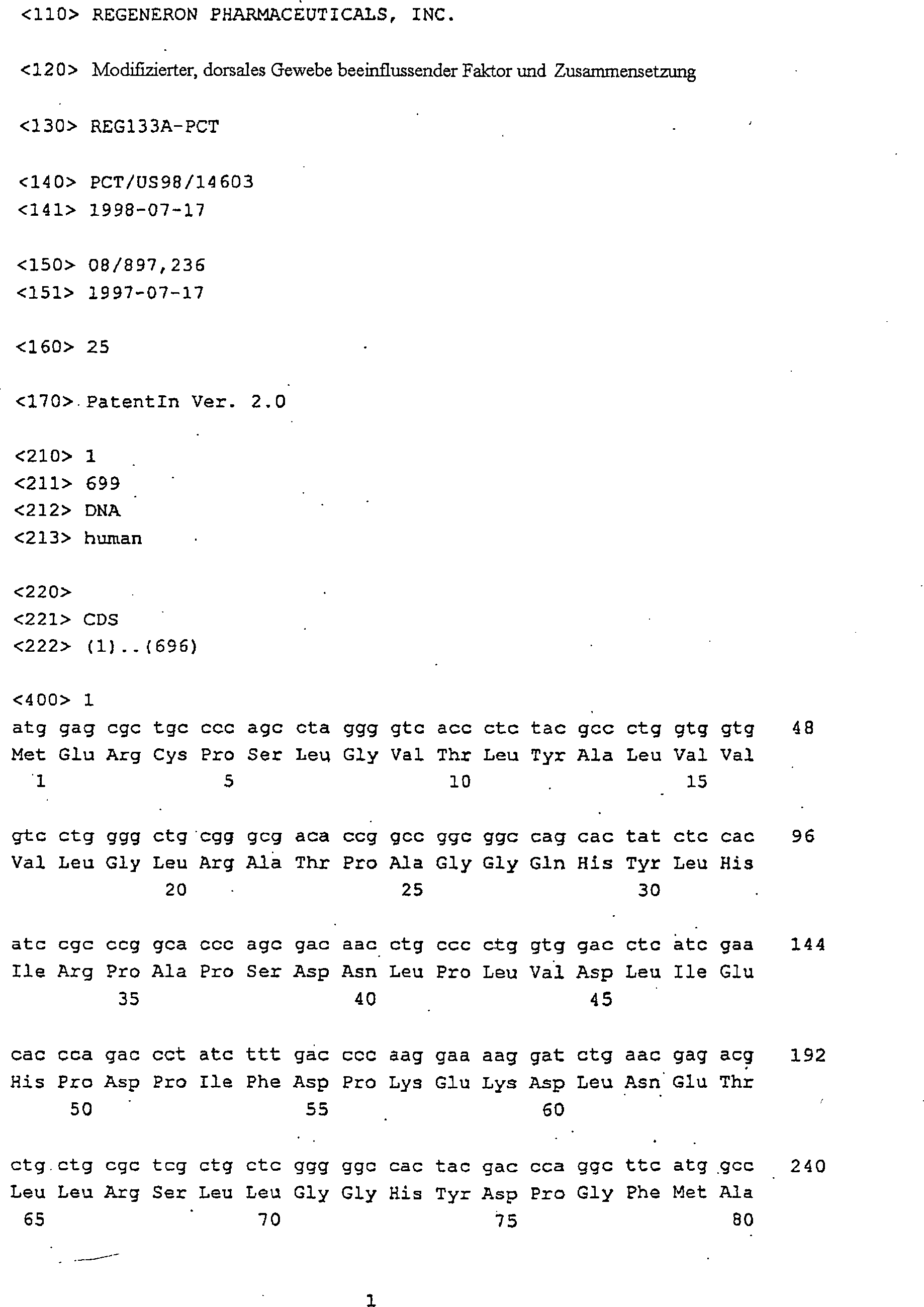


Claims (17)
Applications Claiming Priority (3)
| Application Number | Priority Date | Filing Date | Title |
|---|---|---|---|
| US08/897,236 US6075007A (en) | 1997-07-17 | 1997-07-17 | Modified noggin polypeptide and compositions |
| US897236 | 1997-07-17 | ||
| PCT/US1998/014603 WO1999003996A1 (en) | 1997-07-17 | 1998-07-17 | Modified dorsal tissue affecting factor and compositions |
Publications (2)
| Publication Number | Publication Date |
|---|---|
| DE69819167D1 DE69819167D1 (en) | 2003-11-27 |
| DE69819167T2 true DE69819167T2 (en) | 2004-07-22 |
Family
ID=25407592
Family Applications (1)
| Application Number | Title | Priority Date | Filing Date |
|---|---|---|---|
| DE69819167T Expired - Lifetime DE69819167T2 (en) | 1997-07-17 | 1998-07-17 | MODIFIED FACTOR INFLUENCING THE DORSAL TISSUE |
Country Status (10)
| Country | Link |
|---|---|
| US (3) | US6075007A (en) |
| EP (1) | EP1003860B1 (en) |
| JP (1) | JP4181300B2 (en) |
| AT (1) | ATE252639T1 (en) |
| AU (1) | AU735851B2 (en) |
| CA (1) | CA2296769C (en) |
| DE (1) | DE69819167T2 (en) |
| HK (1) | HK1026229A1 (en) |
| IL (2) | IL134042A0 (en) |
| WO (1) | WO1999003996A1 (en) |
Families Citing this family (38)
| Publication number | Priority date | Publication date | Assignee | Title |
|---|---|---|---|---|
| EP1105477A1 (en) | 1998-08-20 | 2001-06-13 | Regeneron Pharmaceuticals, Inc. | Dcr5, a bmp-binding protein, and applications thereof |
| US20040009535A1 (en) | 1998-11-27 | 2004-01-15 | Celltech R&D, Inc. | Compositions and methods for increasing bone mineralization |
| PT1133558E (en) | 1998-11-27 | 2007-01-31 | Ucb Sa | Compositions and methods for increasing bone mineralization |
| US7306799B2 (en) | 1999-06-08 | 2007-12-11 | Regeneron Pharmaceuticals, Inc. | Use of VEGF inhibitors for treatment of eye disorders |
| US7303746B2 (en) | 1999-06-08 | 2007-12-04 | Regeneron Pharmaceuticals, Inc. | Methods of treating eye disorders with modified chimeric polypeptides |
| US7070959B1 (en) | 1999-06-08 | 2006-07-04 | Regeneron Pharmaceuticals, Inc. | Modified chimeric polypeptides with improved pharmacokinetic properties |
| WO2002054940A2 (en) * | 2001-01-12 | 2002-07-18 | University Of Medicine & Dentistry Of New Jersey | Bone morphogenetic protein-2 in the treatment and diagnosis of cancer |
| US20020197648A1 (en) * | 2001-05-02 | 2002-12-26 | Silva Robin M. | High throughput screening methods using magnetic resonance imaging agents |
| US20030134790A1 (en) * | 2002-01-11 | 2003-07-17 | University Of Medicine And Dentistry Of New Jersey | Bone Morphogenetic Protein-2 And Bone Morphogenetic Protein-4 In The Treatment And Diagnosis Of Cancer |
| WO2003070189A2 (en) * | 2002-02-15 | 2003-08-28 | Cornell Research Foundation, Inc. | Enhancing neurotrophin-induced neurogenesis by endogenous neural progenitor cells by concurrent overexpression of brain derived neurotrophic factor and an inhibitor of a pro-gliogenic bone morphogenetic protein |
| US7799523B2 (en) | 2002-04-03 | 2010-09-21 | Celltech R & D, Inc. | Association of polymorphisms in the SOST gene region with bone mineral density |
| US7226902B2 (en) | 2003-03-14 | 2007-06-05 | Celltech R&D, Inc. | Ligands for TGF-beta binding proteins and uses thereof |
| CN1835974A (en) | 2003-06-16 | 2006-09-20 | 细胞技术研究与发展公司 | Antibodies specific for sclerostin and methods for increasing bone mineralization |
| CA2573354C (en) * | 2004-07-09 | 2015-02-24 | Cythera, Inc. | Preprimitive streak and mesendoderm cells |
| US8003108B2 (en) | 2005-05-03 | 2011-08-23 | Amgen Inc. | Sclerostin epitopes |
| US7592429B2 (en) | 2005-05-03 | 2009-09-22 | Ucb Sa | Sclerostin-binding antibody |
| AU2006289667A1 (en) * | 2005-09-08 | 2007-03-15 | Apollo Life Sciences Limited | Noggin and chimeric molecules thereof |
| CL2008002775A1 (en) | 2007-09-17 | 2008-11-07 | Amgen Inc | Use of a sclerostin binding agent to inhibit bone resorption. |
| KR101163171B1 (en) * | 2009-01-20 | 2012-07-19 | (주)케어젠 | Noggin derived Peptides and Uses Thereof |
| US20100272695A1 (en) * | 2009-04-22 | 2010-10-28 | Alan Agulnick | Cell compositions derived from dedifferentiated reprogrammed cells |
| US9109245B2 (en) | 2009-04-22 | 2015-08-18 | Viacyte, Inc. | Cell compositions derived from dedifferentiated reprogrammed cells |
| CN102946903B (en) | 2010-05-14 | 2014-11-26 | 安进公司 | High concentration antibody formulations |
| JP6043629B2 (en) * | 2011-01-07 | 2016-12-14 | 中外製薬株式会社 | Methods for improving antibody physical properties |
| CA2828000A1 (en) | 2011-03-01 | 2012-09-07 | Amgen Inc. | Bispecific binding agents |
| CA2830538C (en) | 2011-03-25 | 2020-05-12 | Amgen Inc. | Anti - sclerostin antibody crystals and formulations thereof |
| JO3283B1 (en) | 2011-04-26 | 2018-09-16 | Sanofi Sa | Composition comprising aflibercept, folinic acid, 5-fluorouracil (5-fu) and irinocetan (folfiri) |
| EP2739311B9 (en) | 2011-08-04 | 2018-09-19 | Amgen Inc. | Method for treating bone gap defects |
| SG11201403718YA (en) | 2011-12-28 | 2014-07-30 | Amgen Inc | Method of treating alveolar bone loss through the use of anti-sclerostin antibodies |
| CN104619342A (en) | 2012-07-05 | 2015-05-13 | Ucb医药有限公司 | Treatment for bone diseases |
| UY35148A (en) | 2012-11-21 | 2014-05-30 | Amgen Inc | HETERODIMERIC IMMUNOGLOBULINS |
| US8859286B2 (en) | 2013-03-14 | 2014-10-14 | Viacyte, Inc. | In vitro differentiation of pluripotent stem cells to pancreatic endoderm cells (PEC) and endocrine cells |
| US9914769B2 (en) | 2014-07-15 | 2018-03-13 | Kymab Limited | Precision medicine for cholesterol treatment |
| GB201403775D0 (en) | 2014-03-04 | 2014-04-16 | Kymab Ltd | Antibodies, uses & methods |
| ES2732925T3 (en) | 2014-07-18 | 2019-11-26 | Sanofi Sa | Method to predict the result of aflibercept treatment of a patient suspected of suffering from cancer |
| MA41142A (en) | 2014-12-12 | 2017-10-17 | Amgen Inc | ANTI-SCLEROSTINE ANTIBODIES AND THE USE OF THEM TO TREAT BONE CONDITIONS AS PART OF THE TREATMENT PROTOCOL |
| GB201604124D0 (en) | 2016-03-10 | 2016-04-27 | Ucb Biopharma Sprl | Pharmaceutical formulation |
| WO2018083248A1 (en) | 2016-11-03 | 2018-05-11 | Kymab Limited | Antibodies, combinations comprising antibodies, biomarkers, uses & methods |
| AU2019243595A1 (en) | 2018-03-30 | 2020-09-17 | Amgen Inc. | C-terminal antibody variants |
Family Cites Families (4)
| Publication number | Priority date | Publication date | Assignee | Title |
|---|---|---|---|---|
| US5256568A (en) | 1990-02-12 | 1993-10-26 | Regeneron Phamaceuticals, Inc. | Vectors and transformed most cells for recombinant protein production with reduced expression of selectable markers |
| DK0550665T3 (en) | 1990-09-25 | 1996-12-02 | Genentech Inc | New neurotrophic factor |
| WO1994005800A1 (en) * | 1992-09-03 | 1994-03-17 | The Regents Of The University Of California | Dorsal tissue affecting factor and compositions |
| US5843775A (en) * | 1992-09-03 | 1998-12-01 | Regeneron Pharmaceuticals, Inc. | Human dorsal tissue affecting factor (noggin) and nucleic acids encoding same |
-
1997
- 1997-07-17 US US08/897,236 patent/US6075007A/en not_active Expired - Fee Related
-
1998
- 1998-07-17 EP EP98936850A patent/EP1003860B1/en not_active Expired - Lifetime
- 1998-07-17 IL IL13404298A patent/IL134042A0/en active IP Right Grant
- 1998-07-17 CA CA002296769A patent/CA2296769C/en not_active Expired - Fee Related
- 1998-07-17 DE DE69819167T patent/DE69819167T2/en not_active Expired - Lifetime
- 1998-07-17 AU AU85708/98A patent/AU735851B2/en not_active Ceased
- 1998-07-17 JP JP2000503202A patent/JP4181300B2/en not_active Expired - Fee Related
- 1998-07-17 AT AT98936850T patent/ATE252639T1/en not_active IP Right Cessation
- 1998-07-17 WO PCT/US1998/014603 patent/WO1999003996A1/en active IP Right Grant
-
2000
- 2000-01-13 IL IL134042A patent/IL134042A/en not_active IP Right Cessation
- 2000-02-08 US US09/500,253 patent/US6500640B1/en not_active Expired - Fee Related
- 2000-08-24 HK HK00105325A patent/HK1026229A1/en not_active IP Right Cessation
-
2002
- 2002-12-19 US US10/323,268 patent/US20030129703A1/en not_active Abandoned
Also Published As
| Publication number | Publication date |
|---|---|
| EP1003860B1 (en) | 2003-10-22 |
| AU735851B2 (en) | 2001-07-19 |
| WO1999003996A1 (en) | 1999-01-28 |
| HK1026229A1 (en) | 2000-12-08 |
| US6075007A (en) | 2000-06-13 |
| US20030129703A1 (en) | 2003-07-10 |
| JP4181300B2 (en) | 2008-11-12 |
| ATE252639T1 (en) | 2003-11-15 |
| EP1003860A1 (en) | 2000-05-31 |
| JP2001510044A (en) | 2001-07-31 |
| US6500640B1 (en) | 2002-12-31 |
| IL134042A0 (en) | 2001-04-30 |
| IL134042A (en) | 2006-12-10 |
| CA2296769C (en) | 2008-01-15 |
| DE69819167D1 (en) | 2003-11-27 |
| AU8570898A (en) | 1999-02-10 |
| CA2296769A1 (en) | 1999-01-28 |
Similar Documents
| Publication | Publication Date | Title |
|---|---|---|
| DE69819167T2 (en) | MODIFIED FACTOR INFLUENCING THE DORSAL TISSUE | |
| DE69635738T2 (en) | Neurturin and related growth factors | |
| US5670481A (en) | Dorsal tissue affecting factor (noggin) and compositions comprising same | |
| DE69635675T2 (en) | REDUCED NEUROTROPHIC FACTOR OBTAINED FROM A GLIA CELL LINE | |
| EP0769020B1 (en) | NEW hIL-4 MUTANT PROTEINS USED AS ANTAGONISTS OR PARTIAL AGONISTS OF HUMAN INTERLEUKIN 4 | |
| US6277593B1 (en) | Dorsal tissue affecting factor and compositions | |
| CH681080A5 (en) | ||
| DE69732350T2 (en) | PERSEPHIN AND RELATED GROWTH FACTORS | |
| DE69920405T2 (en) | USE OF THE HEREGULIN AS AN EPITHELIAL CELL GROWTH FACTOR | |
| DE69434997T2 (en) | Human chondromodulin I protein | |
| DE69833211T2 (en) | HUMAN PERSEPHIN | |
| US6008339A (en) | Nucleic acids encoding a neural tissue affecting factor | |
| CA2143442C (en) | Dorsal tissue affecting factor and compositions | |
| US20020128440A1 (en) | Endoderm, cardiac and neural inducing factors - oligonucleotides for expressing human frazzled (frzb-1) protein |
Legal Events
| Date | Code | Title | Description |
|---|---|---|---|
| 8364 | No opposition during term of opposition |