EP2164514B1 - Antibodies to il-6 and use thereof - Google Patents
Antibodies to il-6 and use thereof Download PDFInfo
- Publication number
- EP2164514B1 EP2164514B1 EP08756095.9A EP08756095A EP2164514B1 EP 2164514 B1 EP2164514 B1 EP 2164514B1 EP 08756095 A EP08756095 A EP 08756095A EP 2164514 B1 EP2164514 B1 EP 2164514B1
- Authority
- EP
- European Patent Office
- Prior art keywords
- antibody
- cell
- disease
- cells
- seq
- Prior art date
- Legal status (The legal status is an assumption and is not a legal conclusion. Google has not performed a legal analysis and makes no representation as to the accuracy of the status listed.)
- Active
Links
Images


Classifications
-
- A—HUMAN NECESSITIES
- A61—MEDICAL OR VETERINARY SCIENCE; HYGIENE
- A61K—PREPARATIONS FOR MEDICAL, DENTAL OR TOILETRY PURPOSES
- A61K39/00—Medicinal preparations containing antigens or antibodies
- A61K39/395—Antibodies; Immunoglobulins; Immune serum, e.g. antilymphocytic serum
- A61K39/39533—Antibodies; Immunoglobulins; Immune serum, e.g. antilymphocytic serum against materials from animals
- A61K39/3955—Antibodies; Immunoglobulins; Immune serum, e.g. antilymphocytic serum against materials from animals against proteinaceous materials, e.g. enzymes, hormones, lymphokines
-
- C—CHEMISTRY; METALLURGY
- C07—ORGANIC CHEMISTRY
- C07K—PEPTIDES
- C07K16/00—Immunoglobulins [IG], e.g. monoclonal or polyclonal antibodies
- C07K16/18—Immunoglobulins [IG], e.g. monoclonal or polyclonal antibodies against material from animals or humans
- C07K16/24—Immunoglobulins [IG], e.g. monoclonal or polyclonal antibodies against material from animals or humans against cytokines, lymphokines or interferons
-
- A—HUMAN NECESSITIES
- A61—MEDICAL OR VETERINARY SCIENCE; HYGIENE
- A61K—PREPARATIONS FOR MEDICAL, DENTAL OR TOILETRY PURPOSES
- A61K39/00—Medicinal preparations containing antigens or antibodies
- A61K39/395—Antibodies; Immunoglobulins; Immune serum, e.g. antilymphocytic serum
-
- A—HUMAN NECESSITIES
- A61—MEDICAL OR VETERINARY SCIENCE; HYGIENE
- A61K—PREPARATIONS FOR MEDICAL, DENTAL OR TOILETRY PURPOSES
- A61K45/00—Medicinal preparations containing active ingredients not provided for in groups A61K31/00 - A61K41/00
- A61K45/06—Mixtures of active ingredients without chemical characterisation, e.g. antiphlogistics and cardiaca
-
- A—HUMAN NECESSITIES
- A61—MEDICAL OR VETERINARY SCIENCE; HYGIENE
- A61K—PREPARATIONS FOR MEDICAL, DENTAL OR TOILETRY PURPOSES
- A61K49/00—Preparations for testing in vivo
-
- A—HUMAN NECESSITIES
- A61—MEDICAL OR VETERINARY SCIENCE; HYGIENE
- A61P—SPECIFIC THERAPEUTIC ACTIVITY OF CHEMICAL COMPOUNDS OR MEDICINAL PREPARATIONS
- A61P1/00—Drugs for disorders of the alimentary tract or the digestive system
- A61P1/04—Drugs for disorders of the alimentary tract or the digestive system for ulcers, gastritis or reflux esophagitis, e.g. antacids, inhibitors of acid secretion, mucosal protectants
-
- A—HUMAN NECESSITIES
- A61—MEDICAL OR VETERINARY SCIENCE; HYGIENE
- A61P—SPECIFIC THERAPEUTIC ACTIVITY OF CHEMICAL COMPOUNDS OR MEDICINAL PREPARATIONS
- A61P1/00—Drugs for disorders of the alimentary tract or the digestive system
- A61P1/14—Prodigestives, e.g. acids, enzymes, appetite stimulants, antidyspeptics, tonics, antiflatulents
-
- A—HUMAN NECESSITIES
- A61—MEDICAL OR VETERINARY SCIENCE; HYGIENE
- A61P—SPECIFIC THERAPEUTIC ACTIVITY OF CHEMICAL COMPOUNDS OR MEDICINAL PREPARATIONS
- A61P11/00—Drugs for disorders of the respiratory system
-
- A—HUMAN NECESSITIES
- A61—MEDICAL OR VETERINARY SCIENCE; HYGIENE
- A61P—SPECIFIC THERAPEUTIC ACTIVITY OF CHEMICAL COMPOUNDS OR MEDICINAL PREPARATIONS
- A61P11/00—Drugs for disorders of the respiratory system
- A61P11/06—Antiasthmatics
-
- A—HUMAN NECESSITIES
- A61—MEDICAL OR VETERINARY SCIENCE; HYGIENE
- A61P—SPECIFIC THERAPEUTIC ACTIVITY OF CHEMICAL COMPOUNDS OR MEDICINAL PREPARATIONS
- A61P11/00—Drugs for disorders of the respiratory system
- A61P11/16—Central respiratory analeptics
-
- A—HUMAN NECESSITIES
- A61—MEDICAL OR VETERINARY SCIENCE; HYGIENE
- A61P—SPECIFIC THERAPEUTIC ACTIVITY OF CHEMICAL COMPOUNDS OR MEDICINAL PREPARATIONS
- A61P13/00—Drugs for disorders of the urinary system
- A61P13/08—Drugs for disorders of the urinary system of the prostate
-
- A—HUMAN NECESSITIES
- A61—MEDICAL OR VETERINARY SCIENCE; HYGIENE
- A61P—SPECIFIC THERAPEUTIC ACTIVITY OF CHEMICAL COMPOUNDS OR MEDICINAL PREPARATIONS
- A61P13/00—Drugs for disorders of the urinary system
- A61P13/12—Drugs for disorders of the urinary system of the kidneys
-
- A—HUMAN NECESSITIES
- A61—MEDICAL OR VETERINARY SCIENCE; HYGIENE
- A61P—SPECIFIC THERAPEUTIC ACTIVITY OF CHEMICAL COMPOUNDS OR MEDICINAL PREPARATIONS
- A61P15/00—Drugs for genital or sexual disorders; Contraceptives
-
- A—HUMAN NECESSITIES
- A61—MEDICAL OR VETERINARY SCIENCE; HYGIENE
- A61P—SPECIFIC THERAPEUTIC ACTIVITY OF CHEMICAL COMPOUNDS OR MEDICINAL PREPARATIONS
- A61P15/00—Drugs for genital or sexual disorders; Contraceptives
- A61P15/06—Antiabortive agents; Labour repressants
-
- A—HUMAN NECESSITIES
- A61—MEDICAL OR VETERINARY SCIENCE; HYGIENE
- A61P—SPECIFIC THERAPEUTIC ACTIVITY OF CHEMICAL COMPOUNDS OR MEDICINAL PREPARATIONS
- A61P17/00—Drugs for dermatological disorders
-
- A—HUMAN NECESSITIES
- A61—MEDICAL OR VETERINARY SCIENCE; HYGIENE
- A61P—SPECIFIC THERAPEUTIC ACTIVITY OF CHEMICAL COMPOUNDS OR MEDICINAL PREPARATIONS
- A61P17/00—Drugs for dermatological disorders
- A61P17/06—Antipsoriatics
-
- A—HUMAN NECESSITIES
- A61—MEDICAL OR VETERINARY SCIENCE; HYGIENE
- A61P—SPECIFIC THERAPEUTIC ACTIVITY OF CHEMICAL COMPOUNDS OR MEDICINAL PREPARATIONS
- A61P19/00—Drugs for skeletal disorders
- A61P19/02—Drugs for skeletal disorders for joint disorders, e.g. arthritis, arthrosis
-
- A—HUMAN NECESSITIES
- A61—MEDICAL OR VETERINARY SCIENCE; HYGIENE
- A61P—SPECIFIC THERAPEUTIC ACTIVITY OF CHEMICAL COMPOUNDS OR MEDICINAL PREPARATIONS
- A61P19/00—Drugs for skeletal disorders
- A61P19/08—Drugs for skeletal disorders for bone diseases, e.g. rachitism, Paget's disease
-
- A—HUMAN NECESSITIES
- A61—MEDICAL OR VETERINARY SCIENCE; HYGIENE
- A61P—SPECIFIC THERAPEUTIC ACTIVITY OF CHEMICAL COMPOUNDS OR MEDICINAL PREPARATIONS
- A61P19/00—Drugs for skeletal disorders
- A61P19/08—Drugs for skeletal disorders for bone diseases, e.g. rachitism, Paget's disease
- A61P19/10—Drugs for skeletal disorders for bone diseases, e.g. rachitism, Paget's disease for osteoporosis
-
- A—HUMAN NECESSITIES
- A61—MEDICAL OR VETERINARY SCIENCE; HYGIENE
- A61P—SPECIFIC THERAPEUTIC ACTIVITY OF CHEMICAL COMPOUNDS OR MEDICINAL PREPARATIONS
- A61P21/00—Drugs for disorders of the muscular or neuromuscular system
-
- A—HUMAN NECESSITIES
- A61—MEDICAL OR VETERINARY SCIENCE; HYGIENE
- A61P—SPECIFIC THERAPEUTIC ACTIVITY OF CHEMICAL COMPOUNDS OR MEDICINAL PREPARATIONS
- A61P25/00—Drugs for disorders of the nervous system
-
- A—HUMAN NECESSITIES
- A61—MEDICAL OR VETERINARY SCIENCE; HYGIENE
- A61P—SPECIFIC THERAPEUTIC ACTIVITY OF CHEMICAL COMPOUNDS OR MEDICINAL PREPARATIONS
- A61P25/00—Drugs for disorders of the nervous system
- A61P25/24—Antidepressants
-
- A—HUMAN NECESSITIES
- A61—MEDICAL OR VETERINARY SCIENCE; HYGIENE
- A61P—SPECIFIC THERAPEUTIC ACTIVITY OF CHEMICAL COMPOUNDS OR MEDICINAL PREPARATIONS
- A61P25/00—Drugs for disorders of the nervous system
- A61P25/28—Drugs for disorders of the nervous system for treating neurodegenerative disorders of the central nervous system, e.g. nootropic agents, cognition enhancers, drugs for treating Alzheimer's disease or other forms of dementia
-
- A—HUMAN NECESSITIES
- A61—MEDICAL OR VETERINARY SCIENCE; HYGIENE
- A61P—SPECIFIC THERAPEUTIC ACTIVITY OF CHEMICAL COMPOUNDS OR MEDICINAL PREPARATIONS
- A61P29/00—Non-central analgesic, antipyretic or antiinflammatory agents, e.g. antirheumatic agents; Non-steroidal antiinflammatory drugs [NSAID]
-
- A—HUMAN NECESSITIES
- A61—MEDICAL OR VETERINARY SCIENCE; HYGIENE
- A61P—SPECIFIC THERAPEUTIC ACTIVITY OF CHEMICAL COMPOUNDS OR MEDICINAL PREPARATIONS
- A61P3/00—Drugs for disorders of the metabolism
-
- A—HUMAN NECESSITIES
- A61—MEDICAL OR VETERINARY SCIENCE; HYGIENE
- A61P—SPECIFIC THERAPEUTIC ACTIVITY OF CHEMICAL COMPOUNDS OR MEDICINAL PREPARATIONS
- A61P3/00—Drugs for disorders of the metabolism
- A61P3/02—Nutrients, e.g. vitamins, minerals
-
- A—HUMAN NECESSITIES
- A61—MEDICAL OR VETERINARY SCIENCE; HYGIENE
- A61P—SPECIFIC THERAPEUTIC ACTIVITY OF CHEMICAL COMPOUNDS OR MEDICINAL PREPARATIONS
- A61P3/00—Drugs for disorders of the metabolism
- A61P3/04—Anorexiants; Antiobesity agents
-
- A—HUMAN NECESSITIES
- A61—MEDICAL OR VETERINARY SCIENCE; HYGIENE
- A61P—SPECIFIC THERAPEUTIC ACTIVITY OF CHEMICAL COMPOUNDS OR MEDICINAL PREPARATIONS
- A61P3/00—Drugs for disorders of the metabolism
- A61P3/08—Drugs for disorders of the metabolism for glucose homeostasis
- A61P3/10—Drugs for disorders of the metabolism for glucose homeostasis for hyperglycaemia, e.g. antidiabetics
-
- A—HUMAN NECESSITIES
- A61—MEDICAL OR VETERINARY SCIENCE; HYGIENE
- A61P—SPECIFIC THERAPEUTIC ACTIVITY OF CHEMICAL COMPOUNDS OR MEDICINAL PREPARATIONS
- A61P31/00—Antiinfectives, i.e. antibiotics, antiseptics, chemotherapeutics
- A61P31/04—Antibacterial agents
-
- A—HUMAN NECESSITIES
- A61—MEDICAL OR VETERINARY SCIENCE; HYGIENE
- A61P—SPECIFIC THERAPEUTIC ACTIVITY OF CHEMICAL COMPOUNDS OR MEDICINAL PREPARATIONS
- A61P31/00—Antiinfectives, i.e. antibiotics, antiseptics, chemotherapeutics
- A61P31/12—Antivirals
- A61P31/14—Antivirals for RNA viruses
- A61P31/16—Antivirals for RNA viruses for influenza or rhinoviruses
-
- A—HUMAN NECESSITIES
- A61—MEDICAL OR VETERINARY SCIENCE; HYGIENE
- A61P—SPECIFIC THERAPEUTIC ACTIVITY OF CHEMICAL COMPOUNDS OR MEDICINAL PREPARATIONS
- A61P31/00—Antiinfectives, i.e. antibiotics, antiseptics, chemotherapeutics
- A61P31/12—Antivirals
- A61P31/20—Antivirals for DNA viruses
-
- A—HUMAN NECESSITIES
- A61—MEDICAL OR VETERINARY SCIENCE; HYGIENE
- A61P—SPECIFIC THERAPEUTIC ACTIVITY OF CHEMICAL COMPOUNDS OR MEDICINAL PREPARATIONS
- A61P35/00—Antineoplastic agents
-
- A—HUMAN NECESSITIES
- A61—MEDICAL OR VETERINARY SCIENCE; HYGIENE
- A61P—SPECIFIC THERAPEUTIC ACTIVITY OF CHEMICAL COMPOUNDS OR MEDICINAL PREPARATIONS
- A61P35/00—Antineoplastic agents
- A61P35/02—Antineoplastic agents specific for leukemia
-
- A—HUMAN NECESSITIES
- A61—MEDICAL OR VETERINARY SCIENCE; HYGIENE
- A61P—SPECIFIC THERAPEUTIC ACTIVITY OF CHEMICAL COMPOUNDS OR MEDICINAL PREPARATIONS
- A61P35/00—Antineoplastic agents
- A61P35/04—Antineoplastic agents specific for metastasis
-
- A—HUMAN NECESSITIES
- A61—MEDICAL OR VETERINARY SCIENCE; HYGIENE
- A61P—SPECIFIC THERAPEUTIC ACTIVITY OF CHEMICAL COMPOUNDS OR MEDICINAL PREPARATIONS
- A61P37/00—Drugs for immunological or allergic disorders
-
- A—HUMAN NECESSITIES
- A61—MEDICAL OR VETERINARY SCIENCE; HYGIENE
- A61P—SPECIFIC THERAPEUTIC ACTIVITY OF CHEMICAL COMPOUNDS OR MEDICINAL PREPARATIONS
- A61P37/00—Drugs for immunological or allergic disorders
- A61P37/02—Immunomodulators
-
- A—HUMAN NECESSITIES
- A61—MEDICAL OR VETERINARY SCIENCE; HYGIENE
- A61P—SPECIFIC THERAPEUTIC ACTIVITY OF CHEMICAL COMPOUNDS OR MEDICINAL PREPARATIONS
- A61P37/00—Drugs for immunological or allergic disorders
- A61P37/02—Immunomodulators
- A61P37/06—Immunosuppressants, e.g. drugs for graft rejection
-
- A—HUMAN NECESSITIES
- A61—MEDICAL OR VETERINARY SCIENCE; HYGIENE
- A61P—SPECIFIC THERAPEUTIC ACTIVITY OF CHEMICAL COMPOUNDS OR MEDICINAL PREPARATIONS
- A61P37/00—Drugs for immunological or allergic disorders
- A61P37/08—Antiallergic agents
-
- A—HUMAN NECESSITIES
- A61—MEDICAL OR VETERINARY SCIENCE; HYGIENE
- A61P—SPECIFIC THERAPEUTIC ACTIVITY OF CHEMICAL COMPOUNDS OR MEDICINAL PREPARATIONS
- A61P39/00—General protective or antinoxious agents
- A61P39/02—Antidotes
-
- A—HUMAN NECESSITIES
- A61—MEDICAL OR VETERINARY SCIENCE; HYGIENE
- A61P—SPECIFIC THERAPEUTIC ACTIVITY OF CHEMICAL COMPOUNDS OR MEDICINAL PREPARATIONS
- A61P43/00—Drugs for specific purposes, not provided for in groups A61P1/00-A61P41/00
-
- A—HUMAN NECESSITIES
- A61—MEDICAL OR VETERINARY SCIENCE; HYGIENE
- A61P—SPECIFIC THERAPEUTIC ACTIVITY OF CHEMICAL COMPOUNDS OR MEDICINAL PREPARATIONS
- A61P7/00—Drugs for disorders of the blood or the extracellular fluid
-
- A—HUMAN NECESSITIES
- A61—MEDICAL OR VETERINARY SCIENCE; HYGIENE
- A61P—SPECIFIC THERAPEUTIC ACTIVITY OF CHEMICAL COMPOUNDS OR MEDICINAL PREPARATIONS
- A61P7/00—Drugs for disorders of the blood or the extracellular fluid
- A61P7/02—Antithrombotic agents; Anticoagulants; Platelet aggregation inhibitors
-
- A—HUMAN NECESSITIES
- A61—MEDICAL OR VETERINARY SCIENCE; HYGIENE
- A61P—SPECIFIC THERAPEUTIC ACTIVITY OF CHEMICAL COMPOUNDS OR MEDICINAL PREPARATIONS
- A61P7/00—Drugs for disorders of the blood or the extracellular fluid
- A61P7/06—Antianaemics
-
- A—HUMAN NECESSITIES
- A61—MEDICAL OR VETERINARY SCIENCE; HYGIENE
- A61P—SPECIFIC THERAPEUTIC ACTIVITY OF CHEMICAL COMPOUNDS OR MEDICINAL PREPARATIONS
- A61P9/00—Drugs for disorders of the cardiovascular system
-
- A—HUMAN NECESSITIES
- A61—MEDICAL OR VETERINARY SCIENCE; HYGIENE
- A61P—SPECIFIC THERAPEUTIC ACTIVITY OF CHEMICAL COMPOUNDS OR MEDICINAL PREPARATIONS
- A61P9/00—Drugs for disorders of the cardiovascular system
- A61P9/04—Inotropic agents, i.e. stimulants of cardiac contraction; Drugs for heart failure
-
- A—HUMAN NECESSITIES
- A61—MEDICAL OR VETERINARY SCIENCE; HYGIENE
- A61P—SPECIFIC THERAPEUTIC ACTIVITY OF CHEMICAL COMPOUNDS OR MEDICINAL PREPARATIONS
- A61P9/00—Drugs for disorders of the cardiovascular system
- A61P9/10—Drugs for disorders of the cardiovascular system for treating ischaemic or atherosclerotic diseases, e.g. antianginal drugs, coronary vasodilators, drugs for myocardial infarction, retinopathy, cerebrovascula insufficiency, renal arteriosclerosis
-
- A—HUMAN NECESSITIES
- A61—MEDICAL OR VETERINARY SCIENCE; HYGIENE
- A61P—SPECIFIC THERAPEUTIC ACTIVITY OF CHEMICAL COMPOUNDS OR MEDICINAL PREPARATIONS
- A61P9/00—Drugs for disorders of the cardiovascular system
- A61P9/12—Antihypertensives
-
- A—HUMAN NECESSITIES
- A61—MEDICAL OR VETERINARY SCIENCE; HYGIENE
- A61P—SPECIFIC THERAPEUTIC ACTIVITY OF CHEMICAL COMPOUNDS OR MEDICINAL PREPARATIONS
- A61P9/00—Drugs for disorders of the cardiovascular system
- A61P9/14—Vasoprotectives; Antihaemorrhoidals; Drugs for varicose therapy; Capillary stabilisers
-
- C—CHEMISTRY; METALLURGY
- C07—ORGANIC CHEMISTRY
- C07K—PEPTIDES
- C07K16/00—Immunoglobulins [IG], e.g. monoclonal or polyclonal antibodies
- C07K16/18—Immunoglobulins [IG], e.g. monoclonal or polyclonal antibodies against material from animals or humans
- C07K16/24—Immunoglobulins [IG], e.g. monoclonal or polyclonal antibodies against material from animals or humans against cytokines, lymphokines or interferons
- C07K16/244—Interleukins [IL]
- C07K16/248—IL-6
-
- C—CHEMISTRY; METALLURGY
- C12—BIOCHEMISTRY; BEER; SPIRITS; WINE; VINEGAR; MICROBIOLOGY; ENZYMOLOGY; MUTATION OR GENETIC ENGINEERING
- C12N—MICROORGANISMS OR ENZYMES; COMPOSITIONS THEREOF; PROPAGATING, PRESERVING, OR MAINTAINING MICROORGANISMS; MUTATION OR GENETIC ENGINEERING; CULTURE MEDIA
- C12N15/00—Mutation or genetic engineering; DNA or RNA concerning genetic engineering, vectors, e.g. plasmids, or their isolation, preparation or purification; Use of hosts therefor
- C12N15/09—Recombinant DNA-technology
- C12N15/63—Introduction of foreign genetic material using vectors; Vectors; Use of hosts therefor; Regulation of expression
- C12N15/79—Vectors or expression systems specially adapted for eukaryotic hosts
- C12N15/80—Vectors or expression systems specially adapted for eukaryotic hosts for fungi
- C12N15/81—Vectors or expression systems specially adapted for eukaryotic hosts for fungi for yeasts
-
- A—HUMAN NECESSITIES
- A61—MEDICAL OR VETERINARY SCIENCE; HYGIENE
- A61K—PREPARATIONS FOR MEDICAL, DENTAL OR TOILETRY PURPOSES
- A61K39/00—Medicinal preparations containing antigens or antibodies
- A61K2039/505—Medicinal preparations containing antigens or antibodies comprising antibodies
-
- C—CHEMISTRY; METALLURGY
- C07—ORGANIC CHEMISTRY
- C07K—PEPTIDES
- C07K2317/00—Immunoglobulins specific features
- C07K2317/20—Immunoglobulins specific features characterized by taxonomic origin
-
- C—CHEMISTRY; METALLURGY
- C07—ORGANIC CHEMISTRY
- C07K—PEPTIDES
- C07K2317/00—Immunoglobulins specific features
- C07K2317/20—Immunoglobulins specific features characterized by taxonomic origin
- C07K2317/24—Immunoglobulins specific features characterized by taxonomic origin containing regions, domains or residues from different species, e.g. chimeric, humanized or veneered
-
- C—CHEMISTRY; METALLURGY
- C07—ORGANIC CHEMISTRY
- C07K—PEPTIDES
- C07K2317/00—Immunoglobulins specific features
- C07K2317/30—Immunoglobulins specific features characterized by aspects of specificity or valency
- C07K2317/34—Identification of a linear epitope shorter than 20 amino acid residues or of a conformational epitope defined by amino acid residues
-
- C—CHEMISTRY; METALLURGY
- C07—ORGANIC CHEMISTRY
- C07K—PEPTIDES
- C07K2317/00—Immunoglobulins specific features
- C07K2317/40—Immunoglobulins specific features characterized by post-translational modification
- C07K2317/41—Glycosylation, sialylation, or fucosylation
-
- C—CHEMISTRY; METALLURGY
- C07—ORGANIC CHEMISTRY
- C07K—PEPTIDES
- C07K2317/00—Immunoglobulins specific features
- C07K2317/50—Immunoglobulins specific features characterized by immunoglobulin fragments
-
- C—CHEMISTRY; METALLURGY
- C07—ORGANIC CHEMISTRY
- C07K—PEPTIDES
- C07K2317/00—Immunoglobulins specific features
- C07K2317/50—Immunoglobulins specific features characterized by immunoglobulin fragments
- C07K2317/56—Immunoglobulins specific features characterized by immunoglobulin fragments variable (Fv) region, i.e. VH and/or VL
- C07K2317/567—Framework region [FR]
-
- C—CHEMISTRY; METALLURGY
- C07—ORGANIC CHEMISTRY
- C07K—PEPTIDES
- C07K2317/00—Immunoglobulins specific features
- C07K2317/70—Immunoglobulins specific features characterized by effect upon binding to a cell or to an antigen
- C07K2317/76—Antagonist effect on antigen, e.g. neutralization or inhibition of binding
-
- C—CHEMISTRY; METALLURGY
- C07—ORGANIC CHEMISTRY
- C07K—PEPTIDES
- C07K2317/00—Immunoglobulins specific features
- C07K2317/90—Immunoglobulins specific features characterized by (pharmaco)kinetic aspects or by stability of the immunoglobulin
- C07K2317/92—Affinity (KD), association rate (Ka), dissociation rate (Kd) or EC50 value
-
- C—CHEMISTRY; METALLURGY
- C07—ORGANIC CHEMISTRY
- C07K—PEPTIDES
- C07K2317/00—Immunoglobulins specific features
- C07K2317/90—Immunoglobulins specific features characterized by (pharmaco)kinetic aspects or by stability of the immunoglobulin
- C07K2317/94—Stability, e.g. half-life, pH, temperature or enzyme-resistance
Definitions
- This invention pertains to antibodies and fragments thereof having binding specificity to IL-6.
- the invention also pertains to pharmaceutical or diagnostic compositions, and antibodies and fragments thereof having binding specificity to IL-6 for use in methods of treating diseases or disorders associated with IL-6 by administering said antibodies or fragments thereof.
- Interleukin-6 (hereinafter "IL-6") (also known as interferon- ⁇ 2 ; B-cell differentiation factor; B-cell stimulatory factor-2; hepatocyte stimulatory factor; hybridoma growth factor; and plasmacytoma growth factor) is a multifunctional cytokine involved in numerous biological processes such as the regulation of the acute inflammatory response, the modulation of specific immune responses including B- and T-cell differentiation, bone metabolism, thrombopoiesis, epidermal proliferation, menses, neuronal cell differentiation, neuroprotection, aging, cancer, and the inflammatory reaction occurring in Alzheimer's disease. See A. Papassotiropoulos, et al, Neurobiology of Aging, 22:863-871 (2001 ).
- IL-6 is a member of a family of cytokines that promote cellular responses through a receptor complex consisting of at least one subunit of the signal-transducing glycoprotein gp130 and the IL-6 receptor ("IL-6R")(also known as gp80).
- the IL-6R may also be present in a soluble form (“sIL-6R”).
- IL-6 binds to IL-6R, which then dimerizes the signal-transducing receptor gp130. See Jones, SA, J. Immunology, 175:3463-3468 (2005 ).
- IL-6 In humans, the gene encoding for IL-6 is organized in five exons and four introns, and maps to the short arm of chromosome 7 at 7p21. Translation of IL-6 RNA and post-translational processing result in the formation of a 21 to 28 kDa protein with 184 amino acids in its mature form. See A. Papassotiropoulos, et al, Neurobiology of Aging, 22:863-871 (2001 ).
- IL-6 is believed to play a role in the development of a multitude of diseases and disorders, including but not limited to fatigue, cachexia, autoimmune diseases, diseases of the skeletal system, cancer, heart disease, obesity, diabetes, asthma, alzheimer's disease and multiple sclerosis. Due to the perceived involvement of IL-6 in a wide range of diseases and disorders, there remains a need in the art for compositions and methods useful for preventing or treating diseases associated with IL-6, as well as methods of screening to identify patients having diseases or disorders associated with IL-6. Particularly preferred anti-IL-6 compositions are those having minimal or minimizing adverse reactions when administered to the patient. Compositions or methods that reduce or inhibit diseases or disorders associated with IL-6 are beneficial to the patient in need thereof.
- WO2006/119115 describes multiple anti-human IL-6 monoclonal antibodies, both human engineered and chimeric forms, derived from AME-A9.
- the antibodies inhibit IL-6 binding to the IL-6R.
- the antibody may be an scFv, it may be lyophilized, and used for diagnosis or treatment, for example of rheumatoid arthritis.
- the subject-matter of the invention is defined by the claims.
- the present invention provides an anti-IL6 antibody or antibody fragment thereof comprising (i) a variable light (V L ) chain comprising a CDR1 polypeptide consisting of the amino acid sequence of SEQ ID NO:4, a CDR2 polypeptide consisting of the amino acid sequence of SEQ ID NO:5, and a CDR3 polypeptide consisting of the amino acid sequence of SEQ ID NO:6, and (ii) a variable heavy (V H ) chain comprising a CDR1 polypeptide consisting of the amino acid sequence of SEQ ID NO:7, a CDR2 polypeptide consisting of the amino acid sequence of SEQ ID NO:8 or SEQ ID NO:120, and a CDR3 polypeptide consisting of the amino acid sequence of SEQ ID NO:9, wherein the antibody or fragment thereof has a dissociation constant (KD) of less than 50 picomolar as assessed by Biacore.
- KD dissociation constant
- the present invention is directed to specific antibodies and fragments thereof having binding specificity for IL-6, in particular antibodies having specific epitopic specificity and/or functional properties.
- One embodiment of the invention encompasses specific humanized antibodies and fragments thereof capable of binding to IL-6 and/or the IL-6/IL-6R complex. These antibodies may bind soluble IL-6 or cell surface expressed IL-6. Also, these antibodies may inhibit the formation or the biological effects of one or more of IL-6, IL-6/IL-6R complexes, IL-6/IL-6R/gp130 complexes and/or multimers of IL-6/IL-6R/gp130.
- Another embodiment of this invention relates to the antibodies described herein, comprising the sequences of the V H , V L and CDR polypeptides according to the present claims, and the polynucleotides encoding them.
- these antibodies will block gp130 activation and/or possess binding affinities (Kds) less than 50 picomolar and/or K off values less than or equal to 10 -4 S -1 .
- these antibodies and humanized versions will be derived from rabbit immune cells (B lymphocytes) and may be selected based on their homology (sequence identity) to human germ line sequences. These antibodies may require minimal or no sequence modifications, thereby facilitating retention of functional properties after humanization.
- the subject antibodies may be selected based on their activity in functional assays such as IL-6 driven T1165 proliferation assays, IL-6 simulated HepG2 haptoglobin production assays, and the like.
- a further embodiment of the invention is directed to fragments from anti-IL-6 antibodies encompassing V H , V L and CDR polypeptides according to the present claims, e.g., derived from rabbit immune cells and the polynucleotides encoding the same, as well as the use of these antibody fragments and the polynucleotides encoding them in the creation of novel antibodies and polypeptide compositions capable of recognizing IL-6 and/or IL-6/IL-6R complexes or IL-6/IL-6R/gp130 complexes and/or multimers thereof.
- the invention also contemplates conjugates of anti-IL-6 antibodies and binding fragments thereof conjugated to one or more functional or detectable moieties. Also described are methods of making said humanized anti-IL-6 or anti-IL-6/IL-6R complex antibodies and binding fragments thereof.
- binding fragments include, but are not limited to, Fab, Fab', F(ab') 2 , Fv and scFv fragments.
- Embodiments of the invention pertain to anti-IL-6 antibodies according to the present claims for use as a therapeutic or diagnostic agent in the treatment of diseases and disorders associated with IL-6 or aberrant expression thereof.
- the invention also contemplates the use of fragments of anti-IL-6 antibodies according to the present claims in the manufacture of a medicament for use in the treatment of diseases and disorders associated with IL-6 or aberrant expression thereof.
- the subject antibodies may be used in the treatment of cancer associated fatigue, and/or cachexia and rheumatoid arthritis.
- anti-IL-6 antibodies in recombinant host cells, preferably diploid yeast such as diploid Pichia and other yeast strains.
- Interleukin-6 As used herein, interleukin-6 (IL-6) encompasses not only the following 212 amino acid sequence available as GenBank Protein Accession No. NP_000591:
- Mating competent yeast species This is intended to broadly encompass any diploid or tetraploid yeast which can be grown in culture. Such species of yeast may exist in a haploid, diploid, or tetraploid form. The cells of a given ploidy may, under appropriate conditions, proliferate for indefinite number of generations in that form. Diploid cells can also sporulate to form haploid cells. Sequential mating can result in tetraploid strains through further mating or fusion of diploid strains. The diploid or polyploidal yeast cells are preferably produced by mating or spheroplast fusion.
- the mating competent yeast may be a member of the Saccharomycetaceae family, which includes the genera Arxiozyma; Ascobotryozyma; Citeromyces; Debaryomyces; Dekkera; Eremothecium; Issatchenkia; Kazachstania; Kluyveromyces; Kodamaea ; Lodderomyces; Pachysolen ; Pichia ; Saccharomyces; Saturnispora ; Tetrapisispora ; Torulaspora ; Williopsis ; and Zygosaccharomyces.
- Other types of yeast include Yarrowia, Rhodosporidium, Candida, Hansenula, Filobasium, Filobasidellla, Sporidiobolus, Bullera, Leucosporidium and Filobasidella.
- the mating competent yeast may be a member of the genus Pichia.
- the mating competent yeast of the genus Pichia may be one of the following species: Pichia pastoris, Pichia methanolica, and Hansenula polymorpha (Pichia angusta).
- the mating competent yeast of the genus Pichia may be the species Pichia pastoris.
- Haploid Yeast Cell A cell having a single copy of each gene of its normal genomic (chromosomal) complement.
- Polyploid Yeast Cell A cell having more than one copy of its normal genomic (chromosomal) complement.
- Diploid Yeast Cell A cell having two copies (alleles) of essentially every gene of its normal genomic complement, typically formed by the process of fusion (mating) of two haploid cells.
- Tetraploid Yeast Cell A cell having four copies (alleles) of essentially every gene of its normal genomic complement, typically formed by the process of fusion (mating) of two haploid cells. Tetraploids may carry two, three, four, or more different expression cassettes. Such tetraploids might be obtained in S. cerevisiae by selective mating homozygotic heterothallic a/a and alpha/alpha diploids and in Pichia by sequential mating of haploids to obtain auxotrophic diploids.
- a [met his] haploid can be mated with [ade his] haploid to obtain diploid [his]; and a [met arg] haploid can be mated with [ade arg] haploid to obtain diploid [arg]; then the diploid [his] x diploid [arg] to obtain a tetraploid prototroph. It will be understood by those of skill in the art that reference to the benefits and uses of diploid cells may also apply to tetraploid cells.
- Yeast Mating The process by which two haploid yeast cells naturally fuse to form one diploid yeast cell.
- Meiosis The process by which a diploid yeast cell undergoes reductive division to form four haploid spore products. Each spore may then germinate and form a haploid vegetatively growing cell line.
- a selectable marker is a gene or gene fragment that confers a growth phenotype (physical growth characteristic) on a cell receiving that gene as, for example through a transformation event.
- the selectable marker allows that cell to survive and grow in a selective growth medium under conditions in which cells that do not receive that selectable marker gene cannot grow.
- Selectable marker genes generally fall into several types, including positive selectable marker genes such as a gene that confers on a cell resistance to an antibiotic or other drug, temperature when two ts mutants are crossed or a ts mutant is transformed; negative selectable marker genes such as a biosynthetic gene that confers on a cell the ability to grow in a medium without a specific nutrient needed by all cells that do not have that biosynthetic gene, or a mutagenized biosynthetic gene that confers on a cell inability to grow by cells that do not have the wild type gene; and the like. Suitable markers include but are not limited to: ZEO; G418; LYS3; MET1; MET3a; ADE1; ADE3; URA3; and the like.
- Expression Vector These DNA vectors contain elements that facilitate manipulation for the expression of a foreign protein within the target host cell. Conveniently, manipulation of sequences and production of DNA for transformation is first performed in a bacterial host, e.g. E. coli, and usually vectors will include sequences to facilitate such manipulations, including a bacterial origin of replication and appropriate bacterial selection marker. Selection markers encode proteins necessary for the survival or growth of transformed host cells grown in a selective culture medium. Host cells not transformed with the vector containing the selection gene will not survive in the culture medium. Typical selection genes encode proteins that (a) confer resistance to antibiotics or other toxins, (b) complement auxotrophic deficiencies, or (c) supply critical nutrients not available from complex media.
- Exemplary vectors and methods for transformation of yeast are described, for example, in Burke, D., Dawson, D., & Stearns, T. (2000). Methods in yeast genetics: a Cold Spring Harbor Laboratory course manual. Plainview, N.Y.: Cold Spring Harbor Laboratory Press.
- Expression vectors for use in the methods of the invention will further include yeast specific sequences, including a selectable auxotrophic or drug marker for identifying transformed yeast strains.
- a drug marker may further be used to amplify copy number of the vector in a yeast host cell.
- the polypeptide coding sequence of interest is operably linked to transcriptional and translational regulatory sequences that provide for expression of the polypeptide in yeast cells.
- These vector components may include, but are not limited to, one or more of the following: an enhancer element, a promoter, and a transcription termination sequence. Sequences for the secretion of the polypeptide may also be included, e.g. a signal sequence, and the like.
- a yeast origin of replication is optional, as expression vectors are often integrated into the yeast genome.
- the polypeptide of interest may be operably linked, or fused, to sequences providing for optimized secretion of the polypeptide from yeast diploid cells.
- Nucleic acids are "operably linked" when placed into a functional relationship with another nucleic acid sequence.
- DNA for a signal sequence is operably linked to DNA for a polypeptide if it is expressed as a preprotein that participates in the secretion of the polypeptide; a promoter or enhancer is operably linked to a coding sequence if it affects the transcription of the sequence.
- "operably linked” means that the DNA sequences being linked are contiguous, and, in the case of a secretory leader, contiguous and in reading frame. However, enhancers do not have to be contiguous.
- Linking is accomplished by ligation at convenient restriction sites or alternatively via a PCR/recombination method familiar to those skilled in the art (Gateway R Technology; Invitrogen, Carlsbad California). If such sites do not exist, the synthetic oligonucleotide adapters or linkers are used in accordance with conventional practice.
- Promoters are untranslated sequences located upstream (5') to the start codon of a structural gene (generally within about 100 to 1000 bp) that control the transcription and translation of particular nucleic acid sequences to which they are operably linked. Such promoters fall into several classes: inducible, constitutive, and repressible promoters (that increase levels of transcription in response to absence of a repressor). Inducible promoters may initiate increased levels of transcription from DNA under their control in response to some change in culture conditions, e.g., the presence or absence of a nutrient or a change in temperature.
- the yeast promoter fragment may also serve as the site for homologous recombination and integration of the expression vector into the same site in the yeast genome; alternatively a selectable marker is used as the site for homologous recombination. Pichia transformation is described in Cregg et al. (1985) Mol. Cell. Biol. 5:3376-3385 .
- Suitable promoters from Pichia include the AOX1 and promoter ( Cregg et al. (1989) Mol. Cell. Biol. 9:1316-1323 ); ICL1 promoter ( Menendez et al. (2003) Yeast 20(13):1097-108 ); glyceraldehyde-3-phosphate dehydrogenase promoter (GAP) ( Waterham et al. (1997) Gene 186(1):37-44 ); and FLD1 promoter ( Shen et al. (1998) Gene 216(1):93-102 ).
- the GAP promoter is a strong constitutive promoter and the AOX and FLD1 promoters are inducible.
- yeast promoters include ADH1, alcohol dehydrogenase II, GAL4, PHO3, PHO5, Pyk, and chimeric promoters derived therefrom.
- non-yeast promoters may be used such as mammalian, insect, plant, reptile , amphibian, viral, and avian promoters. Most typically the promoter will comprise a mammalian promoter (potentially endogenous to the expressed genes) or will comprise a yeast or viral promoter that provides for efficient transcription in yeast systems.
- the polypeptides of interest may be produced recombinantly not only directly, but also as a fusion polypeptide with a heterologous polypeptide, e.g. a signal sequence or other polypeptide having a specific cleavage site at the N-terminus of the mature protein or polypeptide.
- a heterologous polypeptide e.g. a signal sequence or other polypeptide having a specific cleavage site at the N-terminus of the mature protein or polypeptide.
- the signal sequence may be a component of the vector, or it may be a part of the polypeptide coding sequence that is inserted into the vector.
- the heterologous signal sequence selected preferably is one that is recognized and processed through one of the standard pathways available within the host cell.
- the S. cerevisiae alpha factor pre-pro signal has proven effective in the secretion of a variety of recombinant proteins from P. pastoris.
- yeast signal sequences include the alpha mating factor signal sequence, the invertase signal sequence, and signal sequences derived from other secreted yeast polypeptides. Additionally, these signal peptide sequences may be engineered to provide for enhanced secretion in diploid yeast expression systems. Other secretion signals of interest also include mammalian signal sequences, which may be heterologous to the protein being secreted, or may be a native sequence for the protein being secreted. Signal sequences include pre-peptide sequences, and in some instances may include propeptide sequences.
- signal sequences are known in the art, including the signal sequences found on immunoglobulin chains, e.g., K28 preprotoxin sequence, PHA-E, FACE, human MCP-1, human serum albumin signal sequences, human Ig heavy chain, human Ig light chain, and the like.
- signal sequences found on immunoglobulin chains e.g., K28 preprotoxin sequence, PHA-E, FACE, human MCP-1, human serum albumin signal sequences, human Ig heavy chain, human Ig light chain, and the like.
- Transcription may be increased by inserting a transcriptional activator sequence into the vector.
- These activators are cis-acting elements of DNA, usually about from 10 to 300 bp, which act on a promoter to increase its transcription.
- Transcriptional enhancers are relatively orientation and position independent, having been found 5' and 3' to the transcription unit, within an intron, as well as within the coding sequence itself. The enhancer may be spliced into the expression vector at a position 5' or 3' to the coding sequence, but is preferably located at a site 5' from the promoter.
- Expression vectors used in eukaryotic host cells may also contain sequences necessary for the termination of transcription and for stabilizing the mRNA. Such sequences are commonly available from 3' to the translation termination codon, in untranslated regions of eukaryotic or viral DNAs or cDNAs. These regions contain nucleotide segments transcribed as polyadenylated fragments in the untranslated portion of the mRNA.
- Plasmids from the transformants are prepared, analyzed by restriction endonuclease digestion and/or sequenced.
- recombination methods based on att sites and recombination enzymes may be used to insert DNA sequences into a vector. Such methods are described, for example, by Landy (1989) Ann.Rev.Biochem. 58:913-949 ; and are known to those of skill in the art. Such methods utilize intermolecular DNA recombination that is mediated by a mixture of lambda and E . coli -encoded recombination proteins. Recombination occurs between specific attachment ( att ) sites on the interacting DNA molecules.
- Att sites may be introduced into a sequence of interest by ligating the sequence of interest into an appropriate vector; generating a PCR product containing att B sites through the use of specific primers; generating a cDNA library cloned into an appropriate vector containing att sites; and the like.
- Folding refers to the three-dimensional structure of polypeptides and proteins, where interactions between amino acid residues act to stabilize the structure. While non-covalent interactions are important in determining structure, usually the proteins of interest will have intra- and/or intermolecular covalent disulfide bonds formed by two cysteine residues. For naturally occurring proteins and polypeptides or derivatives and variants thereof, the proper folding is typically the arrangement that results in optimal biological activity, and can conveniently be monitored by assays for activity, e.g. ligand binding, enzymatic activity, etc.
- assays based on biological activity will be less meaningful.
- the proper folding of such molecules may be determined on the basis of physical properties, energetic considerations, modeling studies, and the like.
- the expression host may be further modified by the introduction of sequences encoding one or more enzymes that enhance folding and disulfide bond formation, i.e. foldases, chaperonins, etc.
- sequences may be constitutively or inducibly expressed in the yeast host cell, using vectors, markers, etc. as known in the art.
- sequences, including transcriptional regulatory elements sufficient for the desired pattern of expression are stably integrated in the yeast genome through a targeted methodology.
- the eukaryotic PDI is not only an efficient catalyst of protein cysteine oxidation and disulfide bond isomerization, but also exhibits chaperone activity. Co-expression of PDI can facilitate the production of active proteins having multiple disulfide bonds. Also of interest is the expression of BIP (immunoglobulin heavy chain binding protein); cyclophilin; and the like.
- BIP immunoglobulin heavy chain binding protein
- cyclophilin cyclophilin
- Each of the haploid parental strains may express a distinct folding enzyme, e.g. one strain may express BIP, and the other strain may express PDI or combinations thereof.
- desired protein or "target protein” are used interchangeably and refer generally to a humanized antibody or a binding portion thereof described herein.
- antibody is intended to include any polypeptide chain-containing molecular structure with a specific shape that fits to and recognizes an epitope, where one or more non-covalent binding interactions stabilize the complex between the molecular structure and the epitope.
- the archetypal antibody molecule is the immunoglobulin, and all types of immunoglobulins, IgG, IgM, IgA, IgE, IgD, etc., from all sources, e.g.
- antibodies human, rodent, rabbit, cow, sheep, pig, dog, other mammals, chicken, other avians, etc., are considered to be "antibodies.”
- a preferred source for producing antibodies useful as starting material according to the invention is rabbits. Numerous antibody coding sequences have been described; and others may be raised by methods well-known in the art. Examples thereof include chimeric antibodies, human antibodies and other non-human mammalian antibodies, humanized antibodies, single chain antibodies such as scFvs, camelbodies, nanobodies, IgNAR (single-chain antibodies derived from sharks), small-modular immunopharmaceuticals (SMIPs), and antibody fragments such as Fabs, Fab', F(ab') 2 and the like.
- antibodies or antigen binding fragments may be produced by genetic engineering.
- antibody-producing cells are sensitized to the desired antigen or immunogen.
- the messenger RNA isolated from antibody producing cells is used as a template to make cDNA using PCR amplification.
- a library of vectors, each containing one heavy chain gene and one light chain gene retaining the initial antigen specificity, is produced by insertion of appropriate sections of the amplified immunoglobulin cDNA into the expression vectors.
- a combinatorial library is constructed by combining the heavy chain gene library with the light chain gene library. This results in a library of clones which co-express a heavy and light chain (resembling the Fab fragment or antigen binding fragment of an antibody molecule).
- the vectors that carry these genes are co-transfected into a host cell. When antibody gene synthesis is induced in the transfected host, the heavy and light chain proteins self-assemble to produce active antibodies that can be detected by screening with the antigen or immunogen.
- Antibody coding sequences of interest include those encoded by native sequences, as well as nucleic acids that, by virtue of the degeneracy of the genetic code, are not identical in sequence to the disclosed nucleic acids, and variants thereof.
- Variant polypeptides can include amino acid (aa) substitutions, additions or deletions. The amino acid substitutions can be conservative amino acid substitutions or substitutions to eliminate non-essential amino acids, such as to alter a glycosylation site, or to minimize misfolding by substitution or deletion of one or more cysteine residues that are not necessary for function.
- Variants can be designed so as to retain or have enhanced biological activity of a particular region of the protein (e.g., a functional domain, catalytic amino acid residues, etc ).
- Variants also include fragments of the polypeptides disclosed herein, particularly biologically active fragments and/or fragments corresponding to functional domains. Techniques for in vitro mutagenesis of cloned genes are known. Also described are polypeptides that have been modified using ordinary molecular biological techniques so as to improve their resistance to proteolytic degradation or to optimize solubility properties or to render them more suitable as a therapeutic agent.
- Chimeric antibodies may be made by recombinant means by combining the variable light and heavy chain regions (V L and V H ), obtained from antibody producing cells of one species with the constant light and heavy chain regions from another.
- V L and V H variable light and heavy chain regions
- chimeric antibodies utilize rodent or rabbit variable regions and human constant regions, in order to produce an antibody with predominantly human domains.
- the production of such chimeric antibodies is well known in the art, and may be achieved by standard means (as described, e.g., in U.S. Patent No. 5,624,659 ).
- the human constant regions of chimeric antibodies of the invention may be selected from IgG1, IgG2, IgG3, IgG4, IgG5, IgG6, IgG7, IgG8, IgG9, IgG10, IgG11, IgG12, IgG13, IgG14, IgG15, IgG16, IgG17, IgG18 or IgG19 constant regions.
- Humanized antibodies are engineered to contain even more human-like immunoglobulin domains, and incorporate only the complementarity-determining regions of the animal-derived antibody. This is accomplished by carefully examining the sequence of the hyper-variable loops of the variable regions of the monoclonal antibody, and fitting them to the structure of the human antibody chains. Although facially complex, the process is straightforward in practice. See, e.g., U.S. Patent No. 6,187,287 .
- immunoglobulin fragments comprising the epitope binding site (e.g., Fab', F(ab') 2 , or other fragments) may be synthesized.
- "Fragment,” or minimal immunoglobulins may be designed utilizing recombinant immunoglobulin techniques.
- Fv immunoglobulins for use in the present invention may be produced by synthesizing a fused variable light chain region and a variable heavy chain region. Combinations of antibodies are also of interest, e.g. diabodies, which comprise two distinct Fv specificities.
- SMIPs small molecule immunopharmaceuticals
- camelbodies, nanobodies, and IgNAR are encompassed by immunoglobulin fragments.
- Immunoglobulins and fragments thereof may be modified post-translationally, e.g. to add effector moieties such as chemical linkers, detectable moieties, such as fluorescent dyes, enzymes, toxins, substrates, bioluminescent materials, radioactive materials, chemiluminescent moieties and the like, or specific binding moieties, such as streptavidin, avidin, or biotin, and the like may be utilized in the methods and compositions of the present invention. Examples of additional effector molecules are provided infra.
- polyploid yeast that stably expresses or expresses a desired secreted heterologous polypeptide for prolonged time refers to a yeast culture that secretes said polypeptide for at least several days to a week, more preferably at least a month, still more preferably at least 1-6 months, and even more preferably for more than a year at threshold expression levels, typically at least 10-25 mg/liter and preferably substantially greater.
- polyploidal yeast culture that secretes desired amounts of recombinant polypeptide refers to cultures that stably or for prolonged periods secrete at least 10-25 mg/liter of heterologous polypeptide, more preferably at least 50-500 mg/liter, and most preferably 500-1000 mg/liter or more.
- heterologous region or domain of a DNA construct is an identifiable segment of DNA within a larger DNA molecule that is not found in association with the larger molecule in nature.
- the heterologous region encodes a mammalian gene
- the gene will usually be flanked by DNA that does not flank the mammalian genomic DNA in the genome of the source organism.
- Another example of a heterologous region is a construct where the coding sequence itself is not found in nature (e.g., a cDNA where the genomic coding sequence contains introns, or synthetic sequences having codons different than the native gene). Allelic variations or naturally-occurring mutational events do not give rise to a heterologous region of DNA as defined herein.
- a "coding sequence” is an in-frame sequence of codons that (in view of the genetic code) correspond to or encode a protein or peptide sequence. Two coding sequences correspond to each other if the sequences or their complementary sequences encode the same amino acid sequences. A coding sequence in association with appropriate regulatory sequences may be transcribed and translated into a polypeptide. A polyadenylation signal and transcription termination sequence will usually be located 3' to the coding sequence.
- a “promoter sequence” is a DNA regulatory region capable of binding RNA polymerase in a cell and initiating transcription of a downstream (3' direction) coding sequence. Promoter sequences typically contain additional sites for binding of regulatory molecules (e.g., transcription factors) which affect the transcription of the coding sequence.
- a coding sequence is "under the control" of the promoter sequence or "operatively linked” to the promoter when RNA polymerase binds the promoter sequence in a cell and transcribes the coding sequence into mRNA, which is then in turn translated into the protein encoded by the coding sequence.
- Vectors are used to introduce a foreign substance, such as DNA, RNA or protein, into an organism or host cell.
- Typical vectors include recombinant viruses (for polynucleotides) and liposomes (for polypeptides).
- a "DNA vector” is a replicon, such as plasmid, phage or cosmid, to which another polynucleotide segment may be attached so as to bring about the replication of the attached segment.
- An “expression vector” is a DNA vector which contains regulatory sequences which will direct polypeptide synthesis by an appropriate host cell.
- Amplification of polynucleotide sequences is the in vitro production of multiple copies of a particular nucleic acid sequence.
- the amplified sequence is usually in the form of DNA.
- a variety of techniques for carrying out such amplification are described in a review article by Van Brunt (1990, Bio/Technol., 8(4):291-294 ).
- Polymerase chain reaction or PCR is a prototype of nucleic acid amplification, and use of PCR herein should be considered exemplary of other suitable amplification techniques.
- Antibodies consist of two identical light polypeptide chains of molecular weight approximately 23,000 daltons (the "light chain”), and two identical heavy chains of molecular weight 53,000-70,000 (the “heavy chain”). The four chains are joined by disulfide bonds in a "Y" configuration wherein the light chains bracket the heavy chains starting at the mouth of the "Y” configuration.
- the "branch” portion of the "Y” configuration is designated the F ab region; the stem portion of the "Y” configuration is designated the F C region.
- the amino acid sequence orientation runs from the N-terminal end at the top of the "Y" configuration to the C-terminal end at the bottom of each chain.
- the N-terminal end possesses the variable region having specificity for the antigen that elicited it, and is approximately 100 amino acids in length, there being slight variations between light and heavy chain and from antibody to antibody.
- variable region is linked in each chain to a constant region that extends the remaining length of the chain and that within a particular class of antibody does not vary with the specificity of the antibody (i.e., the antigen eliciting it).
- constant regions There are five known major classes of constant regions that determine the class of the immunoglobulin molecule (IgG, IgM, IgA, IgD, and IgE corresponding to ⁇ , ⁇ , ⁇ , ⁇ , and ⁇ (gamma, mu, alpha, delta, or epsilon) heavy chain constant regions).
- the constant region or class determines subsequent effector function of the antibody, including activation of complement ( Kabat, E. A., Structural Concepts in Immunology and Immunochemistry, 2nd Ed., p.
- Light chains are classified as either ⁇ (kappa) or ⁇ (lambda). Each heavy chain class can be paired with either kappa or lambda light chain. The light and heavy chains are covalently bonded to each other, and the "tail" portions of the two heavy chains are bonded to each other by covalent disulfide linkages when the immunoglobulins are generated either by hybridomas or by B cells.
- variable region refers to the domains within each pair of light and heavy chains in an antibody that are involved directly in binding the antibody to the antigen.
- Each heavy chain has at one end a variable domain (V H ) followed by a number of constant domains.
- Each light chain has a variable domain (V L ) at one end and a constant domain at its other end; the constant domain of the light chain is aligned with the first constant domain of the heavy chain, and the light chain variable domain is aligned with the variable domain of the heavy chain.
- CDR complementarity determining region
- hypervariable region refers to one or more of the hyper-variable or complementarity determining regions (CDRs) found in the variable regions of light or heavy chains of an antibody (See Kabat, E. A. et al., Sequences of Proteins of Immunological Interest, National Institutes of Health, Bethesda, Md., (1987 )). These expressions include the hypervariable regions as defined by Kabat et al. (" Sequences of Proteins of Immunological Interest,” Kabat E., et al., US Dept. of Health and Human Services, 1983 ) or the hypervariable loops in 3-dimensional structures of antibodies ( Chothia and Lesk, J Mol. Biol.
- the CDRs in each chain are held in close proximity by framework regions and, with the CDRs from the other chain, contribute to the formation of the antigen binding site.
- the CDRs there are select amino acids that have been described as the selectivity determining regions (SDRs) which represent the critical contact residues used by the CDR in the anibody-antigen interaction (skyi, S., Methods, 36:25-34 (2005 )).
- framework region refers to one or more of the framework regions within the variable regions of the light and heavy chains of an antibody ( See Kabat, E. A. et al., Sequences of Proteins of Immunological Interest, National Institutes of Health, Bethesda, Md., (1987 )). These expressions include those amino acid sequence regions interposed between the CDRs within the variable regions of the light and heavy chains of an antibody.
- the invention includes antibodies having binding specificity to IL-6 and possessing a variable light chain sequence comprising the sequence set forth below:
- the invention also includes antibodies having binding specificity to IL-6 and possessing a variable heavy chain sequence comprising the sequence set forth below:
- the invention further contemplates antibodies comprising the polypeptide sequences of SEQ ID NO: 4; SEQ ID NO: 5; and SEQ ID NO: 6 which correspond to the complementarity-determining regions (CDRs, or hypervariable regions) of the variable light chain sequence of SEQ ID NO: 2, and the polypeptide sequences of SEQ ID NO: 7; SEQ ID NO: 8; and SEQ ID NO: 9 which correspond to the complementarity-determining regions (CDRs, or hypervariable regions) of the variable heavy chain sequence of SEQ ID NO:3.
- the invention contemplates other antibodies, such as for example chimeric antibodies, comprising the polypeptide sequences of SEQ ID NO: 4; SEQ ID NO: 5; and SEQ ID NO: 6 which correspond to the complementarity-determining regions (CDRs, or hypervariable regions) of the variable light chain sequence of SEQ ID NO: 2, and the polypeptide sequences of SEQ ID NO: 7; SEQ ID NO: 8; and SEQ ID NO: 9 which correspond to the complementarity-determining regions (CDRs, or hypervariable regions) of the variable heavy chain sequence of SEQ ID NO: 3.
- chimeric antibodies comprising the polypeptide sequences of SEQ ID NO: 4; SEQ ID NO: 5; and SEQ ID NO: 6 which correspond to the complementarity-determining regions (CDRs, or hypervariable regions) of the variable light chain sequence of SEQ ID NO: 2, and the polypeptide sequences of SEQ ID NO: 7; SEQ ID NO: 8; and SEQ ID NO: 9 which correspond to the complementarity-determining regions (
- antibody fragments of the invention comprise, or alternatively consist of, the polypeptide sequence of SEQ ID NO: 2, and comprise, or alternatively consist of, the polypeptide sequence of SEQ ID NO: 3.
- the invention also contemplates fragments of the antibodies having binding specificity to IL-6 wherein the fragments comprise, or alternatively consist of, all of the following antibody fragments: the variable light chain region of SEQ ID NO: 2; the variable heavy chain region of SEQ ID NO: 3; the complementarity-determining regions (SEQ ID NO: 4; SEQ ID NO: 5; and SEQ ID NO: 6) of the variable light chain region of SEQ ID NO: 2; and the complementarity-determining regions (SEQ ID NO: 7; SEQ ID NO: 8; and SEQ ID NO: 9) of the variable heavy chain region of SEQ ID NO: 3.
- the invention also contemplates variants wherein either of the heavy chain polypeptide sequences of SEQ ID NO: 18 or SEQ ID NO: 19 is substituted for the heavy chain polypeptide sequence of SEQ ID NO: 3; and the light chain polypeptide sequence of SEQ ID NO: 20 is substituted for the light chain polypeptide sequence of SEQ ID NO: 2.
- the heavy chain CDR sequence of SEQ ID NO: 120 may be substituted for the heavy chain CDR sequence of SEQ ID NO: 8.
- the anti-IL-6 antibody is Ab1, comprising SEQ ID NO: 2 and SEQ ID NO: 3, or the alternative SEQ ID NOs set forth in paragraph [0084] above, and having at least one of the biological activities set forth herein.
- the invention contemplates an isolated anti-IL-6 antibody comprising a V H polypeptide sequence selected from the group consisting of: SEQ ID NO: 3, 18, and 19; and further comprising a V L polypeptide sequence selected from the group consisting of: SEQ ID NO: 2 and 20 or a variant thereof wherein one or more of the framework residues (FR residues) in said V H or V L polypeptide has been substituted with another amino acid residue resulting in an anti-IL-6 antibody that specifically binds IL-6.
- the invention contemplates humanized and chimeric forms of these antibodies.
- the chimeric antibodies may include an Fc derived from IgG1, IgG2, IgG3, IgG4, IgG5, IgG6, IgG7, IgG8, IgG9, IgG10, IgG11, IgG12, IgG13, IgG14, IgG15, IgG16, IgG17, IgG18 or IgG19 constant regions.
- the antibodies or V H or V L polypeptides originate or are selected from one or more rabbit B cell populations prior to initiation of the humanization process referenced herein.
- the anti-IL-6 antibodies and fragments thereof have binding specificity for primate homologs of the human IL-6 protein.
- primate homologs of the human IL-6 protein are IL-6 obtained from Macaca fascicularis (also known as the cynomolgus monkey) and the Rhesus monkey.
- the anti-IL-6 antibodies and fragments thereof inhibits the association of IL-6 with IL-6R, and/or the production of IL-6/IL-6R/gp130 complexes and/or the production of IL-6/IL-6R/gp130 multimers and/or antagonizes the biological effects of one or more of the foregoing.
- antibodies and fragments thereof may be modified post-translationally to add effector moieties such as chemical linkers, detectable moieties such as for example fluorescent dyes, enzymes, substrates, bioluminescent materials, radioactive materials, and chemiluminescent moieties, or functional moieties such as for example streptavidin, avidin, biotin, a cytotoxin, a cytotoxic agent, and radioactive materials.
- effector moieties such as chemical linkers, detectable moieties such as for example fluorescent dyes, enzymes, substrates, bioluminescent materials, radioactive materials, and chemiluminescent moieties, or functional moieties such as for example streptavidin, avidin, biotin, a cytotoxin, a cytotoxic agent, and radioactive materials.
- further exemplary enzymes include, but are not limited to, horseradish peroxidase, acetylcholinesterase, alkaline phosphatase, beta- galactosidase and luciferase.
- Further exemplary fluorescent materials include, but are not limited to, rhodamine, fluorescein, fluorescein isothiocyanate, umbelliferone, dichlorotriazinylamine, phycoerythrin and dansyl chloride.
- Further chemiluminescent moieties include, but are not limited to, luminol.
- Further exemplary bioluminescent materials include, but are not limited to, luciferin and aequorin.
- Further exemplary radioactive materials include, but are not limited to, Iodine 125 ( 125 I), Carbon 14 ( 14 C), Sulfur 35 ( 35 S), Tritium ( 3 H) and Phosphorus 32 ( 32 P).
- exemplary cytotoxic agents include, but are not limited to, methotrexate, aminopterin, 6-mercaptopurine, 6-thioguanine, cytarabine, 5-fluorouracil decarbazine; alkylating agents such as mechlorethamine, thioepa chlorambucil, melphalan, carmustine (BSNU), mitomycin C, lomustine (CCNU), 1-methylnitrosourea, cyclothosphamide, mechlorethamine, busulfan, dibromomannitol, streptozotocin, mitomycin C, cis-dichlorodiamine platinum (II) (DDP) cisplatin and carboplatin (paraplatin); anthracyclines include daunorubicin (formerly daunomycin), doxorubicin (adriamycin), detorubicin, carminomycin, idarubicin, epirubicin, mit
- cytotoxic agents include paclitaxel (taxol), ricin, pseudomonas exotoxin, gemcitabine, cytochalasin B, gramicidin D, ethidium bromide, emetine, etoposide, tenoposide, colchicin, dihydroxy anthracin dione, 1-dehydrotestosterone, glucocorticoids, procaine, tetracaine, lidocaine, propranolol, puromycin, procarbazine, hydroxyurea, asparaginase, corticosteroids, mytotane (O,P'-(DDD)), interferons, and mixtures of these cytotoxic agents.
- taxol taxol
- ricin pseudomonas exotoxin
- gemcitabine cytochalasin B
- gramicidin D ethidium bromide
- emetine emetine
- etoposide tenoposide
- cytotoxic agents include, but are not limited to, chemotherapeutic agents such as carboplatin, cisplatin, paclitaxel, gemcitabine, calicheamicin, doxorubicin, 5-fluorouracil, mitomycin C, actinomycin D, cyclophosphamide, vincristine and bleomycin.
- chemotherapeutic agents such as carboplatin, cisplatin, paclitaxel, gemcitabine, calicheamicin, doxorubicin, 5-fluorouracil, mitomycin C, actinomycin D, cyclophosphamide, vincristine and bleomycin.
- Toxic enzymes from plants and bacteria such as ricin, diphtheria toxin and Pseudomonas toxin may be conjugated to the humanized antibodies, or binding fragments thereof, to generate cell-type-specific-killing reagents ( Youle, et al., Proc. Nat'l
- cytotoxic agents include cytotoxic ribonucleases as described by Goldenberg in U.S. Pat. No. 6,653,104 .
- Embodiments of the invention also relate to radioimmunoconjugates where a radionuclide that emits alpha or beta particles is stably coupled to the antibody, or binding fragments thereof, with or without the use of a complex-forming agent.
- Such radionuclides include beta-emitters such as Phosphorus-32 ( 32 P), Scandium-47 ( 47 Sc), Copper-67 ( 67 Cu), Gallium-67 ( 67 Ga), Yttrium-88 ( 88 Y), Yttrium-90 ( 90 Y), Iodine-125 ( 125 I), Iodine-131 ( 131 I), Samarium-153 ( 153 Sm), Lutetium-177 ( 177 Lu), Rhenium-186 ( 186 Re) or Rhenium-188 ( 188 Re), and alpha-emitters such as Astatine-211 ( 211 At), Lead-212 ( 212 Pb), Bismuth-212 ( 212 Bi) or -213 ( 213 Bi) or Actinium-225 ( 225 Ac).
- beta-emitters such as Phosphorus-32 ( 32 P), Scandium-47 ( 47 Sc), Copper-67 ( 67 Cu), Gallium-67 ( 67 Ga), Yttrium-88 ( 88 Y
- Embodiments described herein further include variants and equivalents that are substantially homologous to the antibodies, antibody fragments, diabodies, SMIPs, camelbodies, nanobodies, IgNAR, polypeptides, variable regions and CDRs set forth herein.
- These may contain, e.g., conservative substitution mutations, (i.e., the substitution of one or more amino acids by similar amino acids).
- conservative substitution refers to the substitution of an amino acid with another within the same general class, e.g., one acidic amino acid with another acidic amino acid, one basic amino acid with another basic amino acid, or one neutral amino acid by another neutral amino acid. What is intended by a conservative amino acid substitution is well known in the art.
- the invention contemplates polypeptide sequences having at least 90% or greater sequence homology to any one or more of the polypeptide sequences of antibody fragments, variable regions and CDRs set forth herein. More preferably, the invention contemplates polypeptide sequences having at least 95% or greater sequence homology, even more preferably at least 98% or greater sequence homology, and still more preferably at least 99% or greater sequence homology to any one or more of the polypeptide sequences of antibody fragments, variable regions and CDRs set forth herein. Methods for determining homology between nucleic acid and amino acid sequences are well known to those of ordinary skill in the art.
- anti-IL-6 activity are set forth herein, for example, in paragraphs [0731] - [0736] infra.
- anti-idiotypic antibodies that bind any of the foregoing sequences.
- such an anti-idiotypic antibody could be administered to a subject who has received an anti-IL-6 antibody to modulate, reduce, or neutralize, the effect of the anti-IL-6 antibody.
- anti-idiotypic antibodies could also be useful for treatment of an autoimmune disease characterized by the presence of anti-IL-6 antibodies.
- a further exemplary use of such anti-idiotypic antibodies is for detection of the anti-IL-6 antibodies of the present invention, for example to monitor the levels of the anti-IL-6 antibodies present in a subject's blood or other bodily fluids.
- anti-IL-6 antibodies comprising any of the polypeptide or polynucleotide sequences described herein substituted for any of the other polynucleotide sequences described herein.
- the present invention contemplates antibodies comprising the combination of any of the variable light chain and variable heavy chain sequences described herein, and further contemplates antibodies resulting from substitution of any of the CDR sequences described herein for any of the other CDR sequences described herein.
- the invention contemplates one or more anti-human IL-6 antibodies or antibody fragment which specifically bind to the same linear or conformational epitope(s) and/or competes for binding to the same linear or conformational epitope(s) on an intact human IL-6 polypeptide or fragment thereof as an anti-human IL-6 antibody that is Ab1.
- the anti-human IL-6 antibody which specifically binds to the same linear or conformational epitopes on an intact IL-6 polypeptide or fragment thereof that is (are) specifically bound by Ab1 binds to a IL-6 epitope(s) ascertained by epitopic mapping using overlapping linear peptide fragments which span the full length of the native human IL-6 polypeptide.
- the IL-6 epitope comprises, or alternatively consists of, one or more residues comprised in IL-6 fragments selected from those respectively encompassing amino acid residues 37-51, amino acid residues 70-84, amino acid residues 169-183, amino acid residues 31-45 and/or amino acid residues 58-72.
- the invention is also directed to an anti-IL-6 antibody that binds with the same IL-6 epitope and/or competes with an anti-IL-6 antibody for binding to IL-6 as an antibody or antibody fragment disclosed herein, including but not limited to an anti-IL-6 antibody that is Ab1.
- the invention is also directed to an isolated anti-IL-6 antibody or antibody fragment comprising one or more of the CDRs contained in the V H polypeptide sequences selected from the group consisting of: SEQ ID NO: 3, 18, and 19, and one or more of the CDRs contained in the V L polypeptide sequence consisting of: 2 and 20.
- the invention further contemplates that the one or more anti-human IL-6 antibodies discussed above are aglycosylated; that contain an Fc region that has been modified to alter effector function, half-life, proteolysis, and/or glycosylation; are human, humanized, single chain or chimeric; and are a humanized antibody derived from a rabbit (parent) anti-human IL-6 antibody.
- the invention further contemplates one or more anti-human IL-6 antibodies wherein the framework regions (FRs) in the variable light region and the variable heavy regions of said antibody respectively are human FRs which are unmodified or which have been modified by the substitution of at most 2 or 3 human FR residues in the variable light or heavy chain region with the corresponding FR residues of the parent rabbit antibody, and wherein said human FRs have been derived from human variable heavy and light chain antibody sequences which have been selected from a library of human germline antibody sequences based on their high level of homology to the corresponding rabbit variable heavy or light chain regions relative to other human germline antibody sequences contained in the library.
- the anti-human IL-6 antibody or fragment specifically binds to IL-6 expressing human cells and/or to circulating soluble IL-6 molecules in vivo, including IL-6 expressed on or by human cells in a patient with a disease associated with cells that express IL-6.
- the disease is selected from general fatigue, exercise-induced fatigue, cancer-related fatigue, inflammatory disease-related fatigue, chronic fatigue syndrome, cancer-related cachexia, cardiac-related cachexia, respiratory-related cachexia, renal-related cachexia, age-related cachexia, rheumatoid arthritis, systemic lupus erythematosis (SLE), systemic juvenile idiopathic arthritis, psoriasis, psoriatic arthropathy, ankylosing spondylitis, inflammatory bowel disease (IBD), polymyalgia rheumatica, giant cell arteritis, autoimmune vasculitis, graft versus host disease (GVHD), Sjogren's syndrome, adult onset Still's disease, rheumatoid arthritis, systemic juvenile idiopathic arthritis, osteoarthritis, osteoporosis, Paget's disease of bone, osteoarthritis, multiple myeloma, Hodgkin's lymphoma, non-
- the invention further contemplates anti-human IL-6 antibodies or fragments directly or indirectly attached to a detectable label or therapeutic agent.
- the invention also contemplates one or more nucleic acid sequences which result in the expression of an anti-human IL-6 antibody or antibody fragment as set forth above, including those comprising, or alternatively consisting of, yeast or human preferred codons.
- the invention also contemplates vectors (including plasmids or recombinant viral vectors) comprising said nucleic acid sequence(s).
- the invention also contemplates host cells or recombinant host cells expressing at least one of the antibodies set forth above, including a mammalian, yeast, bacterial, and insect cells.
- the host cell may be a yeast cell.
- the yeast cell may be a diploidal yeast cell.
- the yeast cell may be a Pichia yeast.
- the invention also contemplates antibodies and antigen binding fragments for use in a method of treatment comprising administering to a patient with a disease or condition associated with IL-6 expressing cells a therapeutically effective amount of at least one anti-human IL-6 antibody or fragment.
- a disease or condition associated with IL-6 expressing cells a therapeutically effective amount of at least one anti-human IL-6 antibody or fragment.
- the diseases that may be treated are presented in the non-limiting list set forth above.
- the disease is selected from a cancer, autoimmune disease, or inflammatory condition.
- the disease is cancer or viral infection.
- the treatment further includes the administration of another therapeutic agent or regimen selected from chemotherapy, radiotherapy, cytokine administration or gene therapy.
- the disclosure further contemplates a method of in vivo imaging which detects the presence of cells which express IL-6 comprising administering a diagnostically effective amount of at least one anti-human IL-6 antibody.
- said administration further includes the administration of a radionuclide or fluorophore that facilitates detection of the antibody at IL-6 expressing disease sites.
- the method of in vivo imaging is used to detect IL-6 expressing tumors or metastases or is used to detect the presence of sites of autoimmune disorders associated with IL-6 expressing cells.
- the results of said in vivo imaging method are used to facilitate design of an appropriate therapeutic regimen, including therapeutic regimens including radiotherapy, chemotherapy or a combination thereof.
- polynucleotides of the invention comprise the following polynucleotide sequence encoding the variable light chain polypeptide sequence of SEQ ID NO: 2:
- polynucleotides of the invention comprise the following polynucleotide sequence encoding the variable heavy chain polypeptide sequence of SEQ ID NO: 3:
- polynucleotides encoding fragments of the antibody having binding specificity to IL-6 comprise one or more of the polynucleotide sequences of SEQ ID NO: 12; SEQ ID NO: 13; and SEQ ID NO: 14 which correspond to polynucleotides encoding the complementarity-determining regions (CDRs, or hypervariable regions) of the light chain variable sequence of SEQ ID NO: 2.
- polynucleotides encoding fragments of the antibody having binding specificity to IL-6 comprise one or more of the polynucleotide sequences of SEQ ID NO: 15; SEQ ID NO: 16; and SEQ ID NO: 17 which correspond to polynucleotides encoding the complementarity-determining regions (CDRs, or hypervariable regions) of the heavy chain variable sequence of SEQ ID NO: 3.
- CDRs complementarity-determining regions
- polynucleotide sequences including one or more of the polynucleotide sequences encoding antibody fragments described herein.
- polynucleotides encoding fragments of the antibody having binding specificity to IL-6 comprise one, two, three or more, including all of the following polynucleotides encoding antibody fragments: the polynucleotide SEQ ID NO: 10 encoding the light chain variable region of SEQ ID NO: 2; the polynucleotide SEQ ID NO: 11 encoding the heavy chain variable region of SEQ ID NO: 3; polynucleotides encoding the complementarity-determining regions (SEQ ID NO: 12; SEQ ID NO: 13; and SEQ ID NO: 14) of the light chain variable region of SEQ ID NO: 10; and polynucleotides encoding the complementarity-determining regions (SEQ ID NO: 15; SEQ ID NO: 16; and SEQ ID NO: 17) of the
- the invention is directed to an isolated polynucleotide comprising a polynucleotide encoding an anti-IL-6 V H antibody amino acid sequence selected from SEQ ID NO: 3, 18, and 19 or encoding a variant thereof wherein at least one framework residue (FR residue) has been substituted with an amino acid present at the corresponding position in a rabbit anti-IL-6 antibody V H polypeptide or a conservative amino acid substitution.
- a polynucleotide encoding an anti-IL-6 V H antibody amino acid sequence selected from SEQ ID NO: 3, 18, and 19 or encoding a variant thereof wherein at least one framework residue (FR residue) has been substituted with an amino acid present at the corresponding position in a rabbit anti-IL-6 antibody V H polypeptide or a conservative amino acid substitution.
- the invention is directed to an isolated polynucleotide comprising the polynucleotide sequence encoding an anti-IL-6 V L antibody amino acid sequence of 2, and 20 or encoding a variant thereof wherein at least one framework residue (FR residue) has been substituted with an amino acid present at the corresponding position in a rabbit anti-IL-6 antibody V L polypeptide or a conservative amino acid substitution.
- FR residue framework residue
- Host cells and vectors comprising said polynucleotides are also contemplated.
- the invention further contemplates vectors comprising the polynucleotide sequences encoding the variable heavy and light chain polypeptide sequences, as well as the individual complementarity determining regions (CDRs, or hypervariable regions) set forth herein, as well as host cells comprising said sequences.
- the host cell may be a yeast cell.
- the yeast host cell may belong to the genus Pichia.
- IL-6 is a member of a family of cytokines that promote cellular responses through a receptor complex consisting of at least one subunit of the signal-transducing glycoprotein gp130 and the IL-6 receptor (IL-6R).
- the IL-6R may also be present in a soluble form (sIL-6R).
- sIL-6R soluble form
- IL-6 binds to IL-6R, which then dimerizes the signal-transducing receptor gp130.
- the anti-IL-6 antibodies of the invention, or IL-6 binding fragments thereof are useful by exhibiting anti-IL-6 activity.
- the anti-IL-6 antibodies of the invention, or IL-6 binding fragments thereof exhibit anti-IL-6 activity by binding to IL-6 which may be soluble IL-6 or cell surface expressed IL-6 and/or may prevent or inhibit the binding of IL-6 to IL-6R and/or activation (dimerization) of the gp130 signal-transducing glycoprotein and the formation of IL-6/IL-6R/gp130 multimers and the biological effects of any of the foregoing.
- the subject IL-6 antibodies may possess different antagonistic activities based on where (i.e., epitope) the particular antibody binds IL-6 and/or how it affects the formation of the foregoing IL-6 complexes and/or multimers and the biological effects thereof. Consequently, different IL-6 antibodies according to the invention e.g., may be better suited for preventing or treating conditions involving the formation and accumulation of substantial soluble IL-6 such as rheumatoid arthritis whereas other antibodies may be favored in treatments wherein the prevention of IL-6/IL-6R/gp130 or IL-6/IL-6R/gp130 multimers is a desired therapeutic outcome. This can be determined in binding and other assays.
- the anti-IL-6 activity of the anti-IL-6 antibody of the present invention, and fragments thereof having binding specificity to IL-6 may also be described by their strength of binding or their affinity for IL-6. This also may affect their therapeutic properties.
- the anti-IL-6 antibodies of the present invention, and fragments thereof having binding specificity to IL-6 bind to IL-6 with a dissociation constant (K D ) of less than or equal to 5x10 -7 , 10 -7 , 5x10 -8 , 10 -8 , 5x10 -9 , 10 -9 , 5x10 -10 , 10 -10 , 5x10 -11 , 10 -11 , 5x10 -12 , 10 -12 , 5x10 -13 , 10 -13 , 5x10 -14 , 10 -14 , 5x10 -15 or 10 -15 .
- the anti-IL-6 antibodies and fragments thereof bind IL-6 with a dissociation constant of less than or equal to 5x10 -10
- the anti-IL-6 activity of the anti-IL-6 antibodies of the present invention, and fragments thereof having binding specificity to IL-6 bind to IL-6 with an off-rate of less than or equal to 10 -4 S -1 , 5x10 -5 S -1 , 10 -5 S -1 , 5x10 -6 S -1 , 10 -6 S -1 , 5x10 -7 S -1 , or 10 -7 S -1 .
- the anti-IL-6 antibodies of the invention, and fragments thereof having binding specificity to IL-6 bind to a linear or conformational IL-6 epitope.
- the anti-IL-6 activity of the anti-IL-6 antibodies of the present invention, and fragments thereof having binding specificity to IL-6 exhibit anti-IL-6 activity by ameliorating or reducing the symptoms of, or alternatively treating, or preventing, diseases and disorders associated with IL-6.
- diseases and disorders associated with IL-6 are set forth infra. As noted cancer-related fatigue, cachexia and rheumatoid arthritis are preferred indications for the subject IL-6 antibodies.
- the anti-IL-6 antibodies described herein, or IL-6 binding fragments thereof do not have binding specificity for IL-6R or the gp-130 signal-transducing glycoprotein.
- Described herein are methods of isolating a clonal population of antigen-specific B cells that may be used for isolating at least one antigen-specific cell.
- these methods contain a series of culture and selection steps that can be used separately, in combination, sequentially, repetitively, or periodically.
- these methods are used for isolating at least one antigen-specific cell, which can be used to produce a monoclonal antibody, which is specific to a desired antigen, or a nucleic acid sequence corresponding to such an antibody.
- Described herein is a method comprising the steps of:
- Also described is an improvement to a method of isolating a single, antibody-producing B cell comprising enriching a B cell population obtained from a host that has been immunized or naturally exposed to an antigen, wherein the enriching step precedes any selection steps, comprises at least one culturing step, and results in a clonal population of B cells that produces a single monoclonal antibody specific to said antigen.
- a "clonal population of B cells” refers to a population of B cells that only secrete a single antibody specific to a desired antigen. That is to say that these cells produce only one type of monoclonal antibody specific to the desired antigen.
- enriched cell population cells means increasing the frequency of desired cells, typically antigen-specific cells, contained in a mixed cell population, e.g., a B cell-containing isolate derived from a host that is immunized against a desired antigen.
- a mixed cell population e.g., a B cell-containing isolate derived from a host that is immunized against a desired antigen.
- an enriched cell population encompasses a cell population having a higher frequency of antigen-specific cells as a result of an enrichment step, but this population of cells may contain and produce different antibodies.
- cell population encompasses pre- and a post-enrichment cell populations, keeping in mind that when multiple enrichment steps are performed, a cell population can be both pre- and post-enrichment.
- a method described herein is a method:
- Each cell population may be used directly in the next step, or it can be partially or wholly frozen for long- or short- term storage or for later steps.
- cells from a cell population can be individually suspended to yield single cell suspensions.
- the single cell suspension can be enriched, such that a single cell suspension serves as the pre-enrichment cell population.
- one or more antigen-specific single cell suspensions together form the enriched cell population; the antigen-specific single cell suspensions can be grouped together, e.g., re-plated for further analysis and/or antibody production.
- Described herein is a method of enriching a cell population to yield an enriched cell population having an antigen-specific cell frequency that is about 50% to about 100%, or increments therein.
- the enriched cell population has an antigen-specific cell frequency greater than or equal to about 50%, 60%, 70%, 75%, 80%, 90%, 95%, 99%, or 100%.
- increment is used to define a numerical value in varying degrees of precision, e.g., to the nearest 10, 1, 0.1, 0.01, etc.
- the increment can be rounded to any measurable degree of precision, and the increment need not be rounded to the same degree of precision on both sides of a range.
- the range 1 to 100 or increments therein includes ranges such as 20 to 80, 5 to 50, and 0.4 to 98.
- increments therein means increments between 100 and the measurable limit.
- less than 100 or increments therein means 0 to 100 or increments therein unless the feature, e.g., temperature, is not limited by 0.
- Antigen-specificity can be measured with respect to any antigen.
- the antigen can be any substance to which an antibody can bind including, but not limited to, peptides, proteins or fragments thereof; carbohydrates; organic and inorganic molecules; receptors produced by animal cells, bacterial cells, and viruses; enzymes; agonists and antagonists of biological pathways; hormones; and cytokines.
- Exemplary antigens include, but are not limited to, IL-2, IL-4, IL-6, IL-10, IL-12, IL-13, IL-18, IFN- ⁇ , IFN- ⁇ , BAFF, CXCL13, IP-10, VEGF, EPO, EGF, HRG, Hepatocyte Growth Factor (HGF) and Hepcidin.
- Preferred antigens include IL-6, IL-13, TNF- ⁇ , VEGF- ⁇ , Hepatocyte Growth Factor (HGF) and Hepcidin.
- the antigen used in each enrichment step can be the same as or different from one another. Multiple enrichment steps with the same antigen may yield a large and/or diverse population of antigen-specific cells; multiple enrichment steps with different antigens may yield an enriched cell population with cross-specificity to the different antigens.
- Enriching a cell population can be performed by any cell-selection means known in the art for isolating antigen-specific cells.
- a cell population can be enriched by chromatographic techniques, e.g., Miltenyi bead or magnetic bead technology.
- the beads can be directly or indirectly attached to the antigen of interest.
- the method of enriching a cell population may include at least one chromatographic enrichment step.
- a cell population can also be enriched by performed by any antigen-specificity assay technique known in the art, e.g., an ELISA assay or a halo assay.
- ELISA assays include, but are not limited to, selective antigen immobilization (e.g., biotinylated antigen capture by streptavidin, avidin, or neutravidin coated plate), non-specific antigen plate coating, and through an antigen build-up strategy (e.g., selective antigen capture followed by binding partner addition to generate a heteromeric protein-antigen complex).
- the antigen can be directly or indirectly attached to a solid matrix or support, e.g., a column.
- a halo assay comprises contacting the cells with antigen-loaded beads and labeled anti-host antibody specific to the host used to harvest the B cells.
- the label can be, e.g., a fluorophore.
- At least one assay enrichment step may be performed on at least one single cell suspension.
- the method of enriching a cell population may include at least one chromatographic enrichment step and at least one assay enrichment step.
- the cell populations may contain at least one cell capable of recognizing an antigen.
- Antigen-recognizing cells include, but are not limited to, B cells, plasma cells, and progeny thereof.
- a clonal cell population may contain a single type of antigen-specific B-cell, i.e., the cell population produces a single monoclonal antibody specific to a desired antigen.
- the clonal antigen-specific population of B cells consists predominantly of antigen-specific, antibody-secreting cells, which are obtained by the novel culture and selection protocol described herein. Accordingly, also described are methods for obtaining an enriched cell population containing at least one antigen-specific, antibody-secreting cell. Described herein is an enriched cell population containing about 50% to about 100%, or increments therein, or greater than or equal to about 60%, 70%, 80%, 90%, or 100% of antigen-specific, antibody-secreting cells.
- the enrichment step can be performed as one, two, three, or more steps.
- a single B cell may be isolated from an enriched cell population before confirming whether the single B cell secretes an antibody with antigen-specificity and/or a desired property.
- a method of enriching a cell population is used in a method for antibody production and/or selection.
- a method comprising enriching a cell population before selecting an antibody.
- the method can include the steps of: preparing a cell population comprising at least one antigen-specific cell, enriching the cell population by isolating at least one antigen-specific cell to form an enriched cell population, and inducing antibody production from at least one antigen-specific cell.
- the enriched cell population may contain more than one antigen-specific cell.
- Each antigen-specific cell of the enriched population may be cultured under conditions that yield a clonal antigen-specific B cell population before isolating an antibody producing cell therefrom and/or producing an antibody using said B cell, or a nucleic acid sequence corresponding to such an antibody.
- the present invention allows antibody selection from among a high frequency of antigen-specific cells. Because an enrichment step is used prior to antibody selection, the majority of the cells, preferably virtually all of the cells, used for antibody production are antigen-specific. By producing antibodies from a population of cells with an increased frequency of antigen specificity, the quantity and variety of antibodies are increased.
- an antibody is preferably selected after an enrichment step and a culture step that results in a clonal population of antigen-specific B cells.
- the methods can further comprise a step of sequencing a selected antibody or portions thereof from one or more isolated, antigen-specific cells. Any method known in the art for sequencing can be employed and can include sequencing the heavy chain, light chain, variable region(s), and/or complementarity determining region(s) (CDR).
- the method for antibody selection can also include one or more steps of screening a cell population for antigen recognition and/or antibody functionality.
- the desired antibodies may have specific structural features, such as binding to a particular epitope or mimicry of a particular structure; antagonist or agonist activity; or neutralizing activity, e.g., inhibiting binding between the antigen and a ligand.
- the antibody functionality screen may be ligand-dependent. Screening for antibody functionality includes, but is not limited to, an in vitro protein-protein interaction assay that recreates the natural interaction of the antigen ligand with recombinant receptor protein; and a cell-based response that is ligand dependent and easily monitored (e.g., proliferation response).
- the method for antibody selection may include a step of screening the cell population for antibody functionality by measuring the inhibitory concentration (IC50). At least one of the isolated, antigen-specific cells may produce an antibody having an IC50 of less than about 100, 50, 30, 25, 10 ⁇ g/mL, or increments therein.
- IC50 inhibitory concentration
- the method for antibody selection can also include one or more steps of screening a cell population for antibody binding strength.
- Antibody binding strength can be measured by any method known in the art (e.g., Biacore).
- At least one of the isolated, antigen-specific cells may produce an antibody having a high antigen affinity, e.g., a dissociation constant (Kd) of less than about 5x10 -10 M-1, preferably about 1x10 -13 to 5x10 -10 , 1x10 -12 to 1x10 -10 , 1x10 -12 to 7.5x10 -11 , 1x10 -11 to 2x10 -11 , about 1.5x10 -11 or less, or increments therein.
- Kd dissociation constant
- the antibodies are said to be affinity mature.
- the affinity of the antibodies may be comparable to or higher than the affinity of any one of Panorex® (edrecolomab), Rituxan® (rituximab), Herceptin® (traztuzumab), Mylotarg® (gentuzumab), Campath® (alemtuzumab), ZevalinTM (ibritumomab), ErbituxTM (cetuximab), AvastinTM (bevicizumab), RaptivaTM (efalizumab), Remicade® (infliximab), HumiraTM (adalimumab), and XolairTM (omalizumab).
- the affinity of the antibodies is comparable to or higher than the affinity of HumiraTM.
- the affinity of an antibody can also be increased by known affinity maturation techniques.
- At least one cell population may be screened for at least one of, preferably both, antibody functionality and antibody binding strength.
- the method for antibody selection can also include one or more steps of screening a cell population for antibody sequence homology, especially human homology.
- At least one of the isolated, antigen-specific cells may produce an antibody that has a homology to a human antibody of about 50% to about 100%, or increments therein, or greater than about 60%, 70%, 80%, 85%, 90%, or 95% homologous.
- the antibodies can be humanized to increase the homology to a human sequence by techniques known in the art such as CDR grafting or selectivity determining residue grafting (SDR).
- Described herein are the antibodies themselves according to any of the embodiments described above in terms of IC50, Kd, and/or homology.
- the B cell selection protocol disclosed herein has a number of intrinsic advantages versus other methods for obtaining antibody-secreting B cells and monoclonal antibodies specific to desired target antigens. These advantages include, but are not restricted to, the following:
- the B cell selection protocol reproducibly yields a clonal B cell culture containing a single B cell, or its progeny, secreting a single monoclonal antibody that generally binds to the desired antigen with a relatively high binding affinity, i.e. picomolar or better antigen binding affinities.
- prior antibody selection methods tend to yield relatively few high affinity antibodies and therefore require extensive screening procedures to isolate an antibody with therapeutic potential.
- the protocol results in both in vivo B cell immunization of the host (primary immunization) followed by a second in vitro B cell stimulation (secondary antigen priming step) that may enhance the ability and propensity of the recovered clonal B cells to secrete a single high affinity monoclonal antibody specific to the antigen target.
- B cell selection protocol reproducibly yields enriched B cells producing IgG's that are, on average, highly selective (antigen specific) to the desired target.
- Antigen-enriched B cells recovered by these methods are believed to contain B cells capable of yielding the desired full complement of epitopic specificities as discussed above.
- the invention can be used to produce therapeutic antibodies to desired peptide targets, e.g., viral, bacterial or autoantigen peptides, thereby allowing for the production of monoclonal antibodies with very discrete binding properties or even the production of a cocktail of monoclonal antibodies to different peptide targets, e.g., different viral strains.
- desired peptide targets e.g., viral, bacterial or autoantigen peptides
- This advantage may especially be useful in the context of the production of a therapeutic or prophylactic vaccine having a desired valency, such as an HPV vaccine that induces protective immunity to different HPV strains.
- the B cell selection protocol tends to reproducibly yield antigen-specific antibody sequences that are very similar to endogenous human immunoglobulins (around 90% similar at the amino acid level) and that contain CDRs that possess a length very analogous to human immunoglobulins and therefore require little or no sequence modification (typically at most only a few CDR residues may be modified in the parent antibody sequence and no framework exogenous residues introduced) in order to eliminate potential immunogenicity concerns.
- the recombinant antibody will contain only the host (rabbit) CDR1 and CDR2 residues required for antigen recognition and the entire CDR3.
- Described herein is a method for identifying a single B cell that secretes an antibody specific to a desired antigen and that optionally possesses at least one desired functional property such as affinity, avidity, cytolytic activity, and the like by a process including the following steps:
- these methods will further comprise an additional step of isolating and sequencing, in whole or in part, the polypeptide and nucleic acid sequences encoding the desired antibody.
- sequences or modified versions or portions thereof can be expressed in desired host cells in order to produce recombinant antibodies to a desired antigen.
- the clonal population of B cells predominantly comprises antibody-secreting B cells producing antibody against the desired antigen. It is also believed based on experimental results obtained with several antigens and with different B cell populations that the clonally produced B cells and the isolated antigen-specific B cells derived therefrom produced according to the invention secrete a monoclonal antibody that is typically of relatively high affinity and moreover is capable of efficiently and reproducibly producing a selection of monoclonal antibodies of greater epitopic variability as compared to other methods of deriving monoclonal antibodies from cultured antigen-specific B cells. In an exemplary embodiment the population of immune cells used in such B cell selection methods will be derived from a rabbit.
- non-human and human hosts can alternatively be used as a source of immune B cells. It is believed that the use of rabbits as a source of B cells may enhance the diversity of monoclonal antibodies that may be derived by the methods. Also, the antibody sequences derived from rabbits according to the invention typically possess sequences having a high degree of sequence identity to human antibody sequences making them favored for use in humans since they should possess little antigenicity. In the course of humanization, the final humanized antibody contains a much lower foreign/host residue content, usually restricted to a subset of the host CDR residues that differ dramatically due to their nature versus the human target sequence used in the grafting. This enhances the probability of complete activity recovery in the humanized antibody protein.
- the methods of antibody selection using an enrichment step disclosed herein include a step of obtaining a immune cell-containing cell population from an immunized host.
- Methods of obtaining an immune cell-containing cell population from an immunized host are known in the art and generally include inducing an immune response in a host and harvesting cells from the host to obtain one or more cell populations. The response can be elicited by immunizing the host against a desired antigen.
- the host used as a source of such immune cells can be naturally exposed to the desired antigen such as an individual who has been infected with a particular pathogen such as a bacterium or virus or alternatively has mounted a specific antibody response to a cancer that the individual is afflicted with.
- Host animals are well-known in the art and include, but are not limited to, guinea pig, rabbit, mouse, rat, non-human primate, human, as well as other mammals and rodents, chicken, cow, pig, goat, and sheep.
- the host is a mammal, more preferably, rabbit, mouse, rat, or human.
- the host produces antibodies as part of the native immune response to the antigen.
- the immune response can occur naturally, as a result of disease, or it can be induced by immunization with the antigen.
- Immunization can be performed by any method known in the art, such as, by one or more injections of the antigen with or without an agent to enhance immune response, such as complete or incomplete Freund's adjuvant. Also described is intrasplenic immunization. As an alternative to immunizing a host animal in vivo, the method can comprise immunizing a host cell culture in vitro.
- host animal cells are harvested to obtain one or more cell populations.
- a harvested cell population is screened for antibody binding strength and/or antibody functionality.
- a harvested cell population is preferably from at least one of the spleen, lymph nodes, bone marrow, and/or peripheral blood mononuclear cells (PBMCs).
- PBMCs peripheral blood mononuclear cells
- the cells can be harvested from more than one source and pooled. Certain sources may be preferred for certain antigens. For example, the spleen, lymph nodes, and PBMCs are preferred for IL-6; and the lymph nodes are preferred for TNF.
- the cell population is harvested about 20 to about 90 days or increments therein after immunization, preferably about 50 to about 60 days.
- a harvested cell population and/or a single cell suspension therefrom can be enriched, screened, and/or cultured for antibody selection.
- the frequency of antigen-specific cells within a harvested cell population is usually about 1% to about 5%, or increments therein.
- a single cell suspension from a harvested cell population may be enriched, preferably by using Miltenyi beads. From the harvested cell population having a frequency of antigen-specific cells of about 1% to about 5%, an enriched cell population is thus derived having a frequency of antigen-specific cells approaching 100%.
- the method of antibody selection using an enrichment step includes a step of producing antibodies from at least one antigen-specific cell from an enriched cell population.
- Methods of producing antibodies in vitro are well known in the art, and any suitable method can be employed.
- An enriched cell population such as an antigen-specific single cell suspension from a harvested cell population, may be plated at various cell densities, such as 50, 100, 250, 500, or other increments between 1 and 1000 cells per well.
- the sub-population comprises no more than about 10,000 antigen-specific, antibody-secreting cells, more preferably about 50-10,000, about 50-5,000, about 50-1,000, about 50-500, about 50-250 antigen-specific, antibody-secreting cells, or increments therein.
- these sub-populations are cultured with suitable medium (e.g., an activated T cell conditioned medium, particularly 1-5% activated rabbit T cell conditioned medium) on a feeder layer, preferably under conditions that favor the survival of a single proliferating antibody-secreting cell per culture well.
- suitable medium e.g., an activated T cell conditioned medium, particularly 1-5% activated rabbit T cell conditioned medium
- the feeder layer generally comprised of irradiated cell matter, e.g., EL4B cells, does not constitute part of the cell population.
- the cells are cultured in a suitable media for a time sufficient for antibody production, for example about 1 day to about 2 weeks, about 1 day to about 10 days, at least about 3 days, about 3 to about 5 days, about 5 days to about 7 days, at least about 7 days, or other increments therein.
- More than one sub-population may be cultured simultaneously.
- a single antibody-producing cell and progeny thereof survives in each well, thereby providing a clonal population of antigen-specific B cells in each well.
- the immunoglobulin G (IgG) produced by the clonal population is highly correlative with antigen specificity.
- the IgGs may exhibit a correlation with antigen specificity that is greater than about 50%, more preferably greater than 70%, 85%, 90%, 95%, 99%, or increments therein. See Fig. 3 , which demonstrates an exemplary correlation for IL-6. The correlations were demonstrated by setting up B cell cultures under limiting conditions to establish single antigen-specific antibody products per well.
- Antigen-specific versus general IgG synthesis was compared. Three populations were observed: IgG that recognized a single format of antigen (biotinylated and direct coating), detectable IgG and antigen recognition irrespective of immobilization, and IgG production alone. IgG production was highly correlated with antigen-specificity.
- a supernatant containing the antibodies is optionally collected, which can be can be enriched, screened, and/or cultured for antibody selection according to the steps described above.
- the supernatant may be enriched (preferably by an antigen-specificity assay, especially an ELISA assay) and/or screened for antibody functionality.
- the enriched, preferably clonal, antigen-specific B cell population from which a supernatant described above is optionally screened in order to detect the presence of the desired secreted monoclonal antibody is used for the isolation of a few B cells, preferably a single B cell, which is then tested in an appropriate assay in order to confirm the presence of a single antibody-producing B cell in the clonal B cell population.
- About 1 to about 20 cells may be isolated from the clonal B cell population, preferably less than about 15, 12, 10, 5, or 3 cells, or increments therein, most preferably a single cell.
- the screen is preferably effected by an antigen-specificity assay, especially a halo assay.
- the halo assay can be performed with the full length protein, or a fragment thereof.
- the antibody-containing supernatant can also be screened for at least one of: antigen binding affinity; agonism or antagonism of antigen-ligand binding, induction or inhibition of the proliferation of a specific target cell type; induction or inhibition of lysis of a target cell, and induction or inhibition of a biological pathway involving the antigen.
- the identified antigen-specific cell can be used to derive the corresponding nucleic acid sequences encoding the desired monoclonal antibody.
- AluI digest can confirm that only a single monoclonal antibody type is produced per well.
- these sequences can be mutated, such as by humanization, in order to render them suitable for use in human medicaments.
- the enriched B cell population used in the process can also be further enriched, screened, and/or cultured for antibody selection according to the steps described above which can be repeated or performed in a different order.
- At least one cell of an enriched, preferably clonal, antigen-specific cell population may be isolated, cultured, and used for antibody selection.
- the method can further include one or more steps of screening the harvested cell population for antibody binding strength (affinity, avidity) and/or antibody functionality. Suitable screening steps include, but are not limited to, assay methods that detect: whether the antibody produced by the identified antigen-specific B cell produces an antibody possessing a minimal antigen binding affinity, whether the antibody agonizes or antagonizes the binding of a desired antigen to a ligand; whether the antibody induces or inhibits the proliferation of a specific cell type; whether the antibody induces or elicits a cytolytic reaction against target cells; whether the antibody binds to a specific epitope; and whether the antibody modulates (inhibits or agonizes) a specific biological pathway or pathways involving the antigen.
- assay methods that detect: whether the antibody produced by the identified antigen-specific B cell produces an antibody possessing a minimal antigen binding affinity, whether the antibody agonizes or antagonizes the binding of a desired antigen to a ligand; whether the antibody induces or inhibit
- the method can include one or more steps of screening the second enriched cell population for antibody binding strength and/or antibody functionality.
- the method can further include a step of sequencing the polypeptide sequence or the corresponding nucleic acid sequence of the selected antibody.
- the method can also include a step of producing a recombinant antibody using the sequence, a fragment thereof, or a genetically modified version of the selected antibody.
- Methods for mutating antibody sequences in order to retain desired properties are well known to those skilled in the art and include humanization, chimerisation, production of single chain antibodies; these mutation methods can yield recombinant antibodies possessing desired effector function, immunogenicity, stability, removal or addition of glycosylation, and the like.
- the recombinant antibody can be produced by any suitable recombinant cell, including, but not limited to mammalian cells such as CHO, COS, BHK, HEK-293, bacterial cells, yeast cells, plant cells, insect cells, and amphibian cells.
- mammalian cells such as CHO, COS, BHK, HEK-293, bacterial cells, yeast cells, plant cells, insect cells, and amphibian cells.
- the antibodies may be expressed in polyploidal yeast cells, i.e., diploid yeast cells, particularly Pichia.
- the method may comprise:
- Described herein is a method for humanizing antibody heavy and light chains. The following method is followed for the humanization of the heavy and light chains:
- the disclosure is also directed to the production of the antibodies described herein or fragments thereof.
- Recombinant polypeptides corresponding to the antibodies described herein or fragments thereof are secreted from polyploidal, preferably diploid or tetraploid strains of mating competent yeast. Described herein are methods for producing these recombinant polypeptides in secreted form for prolonged periods using cultures comprising polyploid yeast, i.e., at least several days to a week, more preferably at least a month or several months, and even more preferably at least 6 months to a year or longer.
- polyploid yeast cultures will express at least 10-25 mg/liter of the polypeptide, more preferably at least 50-250 mg/liter, still more preferably at least 500-1000 mg/liter, and most preferably a gram per liter or more of the recombinant polypeptide(s).
- a pair of genetically marked yeast haploid cells are transformed with expression vectors comprising subunits of a desired heteromultimeric protein.
- One haploid cell comprises a first expression vector
- a second haploid cell comprises a second expression vector.
- Diploid yeast cells may be transformed with one or more expression vectors that provide for the expression and secretion of one or more of the recombinant polypeptides.
- a single haploid cell may be transformed with one or more vectors and used to produce a polyploidal yeast by fusion or mating strategies.
- a diploid yeast culture may be transformed with one or more vectors providing for the expression and secretion of a desired polypeptide or polypeptides.
- vectors may comprise vectors e.g., linearized plasmids or other linear DNA products that integrate into the yeast cell's genome randomly, through homologous recombination, or using a recombinase such as Cre/Lox or Flp/Frt.
- additional expression vectors may be introduced into the haploid or diploid cells; or the first or second expression vectors may comprise additional coding sequences; for the synthesis of heterotrimers; heterotetramers; etc.
- the expression levels of the non-identical polypeptides may be individually calibrated, and adjusted through appropriate selection, vector copy number, promoter strength and/or induction and the like.
- the transformed haploid cells are genetically crossed or fused.
- the resulting diploid or tetraploid strains are utilized to produce and secrete fully assembled and biologically functional proteins, humanized antibodies described herein or fragments thereof.
- diploid or tetraploid cells for protein production provides for unexpected benefits.
- the cells can be grown for production purposes, i.e. scaled up, and for extended periods of time, in conditions that can be deleterious to the growth of haploid cells, which conditions may include high cell density; growth in minimal media; growth at low temperatures; stable growth in the absence of selective pressure; and which may provide for maintenance of heterologous gene sequence integrity and maintenance of high level expression over time.
- haploid strains can comprise numerous minor autotrophic mutations, which mutations are complemented in the diploid or tetraploid, enabling growth and enhanced production under highly selective conditions.
- Transformed mating competent haploid yeast cells provide a genetic method that enables subunit pairing of a desired protein.
- Haploid yeast strains are transformed with each of two expression vectors, a first vector to direct the synthesis of one polypeptide chain and a second vector to direct the synthesis of a second, non-identical polypeptide chain.
- the two haploid strains are mated to provide a diploid host where optimized target protein production can be obtained.
- additional non-identical coding sequence(s) are provided. Such sequences may be present on additional expression vectors or in the first or the second expression vectors. As is known in the art, multiple coding sequences may be independently expressed from individual promoters; or may be coordinately expressed through the inclusion of an "internal ribosome entry site” or "IRES", which is an element that promotes direct internal ribosome entry to the initiation codon, such as ATG, of a cistron (a protein encoding region), thereby leading to the cap-independent translation of the gene. IRES elements functional in yeast are described by Thompson et al. (2001) P.N.A.S. 98:12866-12868 .
- Antibody sequences may be produced in combination with a secretory J chain, which provides for enhanced stability of IgA (see U.S. Patent Nos. 5,959,177 ; and 5,202,422 ).
- the two haploid yeast strains may each be auxotrophic, and require supplementation of media for growth of the haploid cells.
- the pair of auxotrophs are complementary, such that the diploid product will grow in the absence of the supplements required for the haploid cells.
- Many such genetic markers are known in yeast, including requirements for amino acids (e.g. met , lys , his , arg , etc .), nucleosides ( e.g. ura3 , ade1 , etc. ); and the like. Amino acid markers may be preferred for the methods of the invention.
- diploid cells which contain the desired vectors can be selected by other means, e.g., by use of other markers, such as green fluorescent protein, antibiotic resistance genes, various dominant selectable markers, and the like.
- Two transformed haploid cells may be genetically crossed and diploid strains arising from this mating event selected by their hybrid nutritional requirements and/or antibiotic resistance spectra.
- populations of the two transformed haploid strains are spheroplasted and fused, and diploid progeny regenerated and selected.
- diploid strains can be identified and selectively grown based on their ability to grow in different media than their parents.
- the diploid cells may be grown in minimal medium that may include antibiotics.
- the diploid synthesis strategy has certain advantages. Diploid strains have the potential to produce enhanced levels of heterologous protein through broader complementation to underlying mutations, which may impact the production and/or secretion of recombinant protein.
- any antibiotics used to select those strains do not necessarily need to be continuously present in the growth media.
- a haploid yeast may be transformed with a single or multiple vectors and mated or fused with a non-transformed cell to produce a diploid cell containing the vector or vectors.
- a diploid yeast cell may be transformed with one or more vectors that provide for the expression and secretion of a desired heterologous polypeptide by the diploid yeast cell.
- Two haploid strains may be transformed with a library of polypeptides, e . g . a library of antibody heavy or light chains.
- Transformed haploid cells that synthesize the polypeptides are mated with the complementary haploid cells.
- the resulting diploid cells are screened for functional protein.
- the diploid cells provide a means of rapidly, conveniently and inexpensively bringing together a large number of combinations of polypeptides for functional testing. This technology is especially applicable for the generation of heterodimeric protein products, where optimized subunit synthesis levels are critical for functional protein expression and secretion.
- the expression level ratio of the two subunits may be regulated in order to maximize product generation.
- Heterodimer subunit protein levels have been shown previously to impact the final product generation ( Simmons LC, J Immunol Methods. 2002 May 1;263(1-2):133-47 ). Regulation can be achieved prior to the mating step by selection for a marker present on the expression vector. By stably increasing the copy number of the vector, the expression level can be increased. In some cases, it may be desirable to increase the level of one chain relative to the other, so as to reach a balanced proportion between the subunits of the polypeptide.
- Antibiotic resistance markers are useful for this purpose, e.g. Zeocin resistance marker, G418 resistance, etc.
- strains that contain multiple integrated copies of an expression vector in a strain by selecting for transformants that are resistant to higher levels of Zeocin or G418.
- the proper ratio, e.g. 1:1; 1:2; etc. of the subunit genes may be important for efficient protein production. Even when the same promoter is used to transcribe both subunits, many other factors contribute to the final level of protein expressed and therefore, it can be useful to increase the number of copies of one encoded gene relative to the other.
- diploid strains that produce higher levels of a polypeptide, relative to single copy vector strains are created by mating two haploid strains, both of which have multiple copies of the expression vectors.
- Host cells are transformed with the above-described expression vectors, mated to form diploid strains, and cultured in conventional nutrient media modified as appropriate for inducing promoters, selecting transformants or amplifying the genes encoding the desired sequences.
- a number of minimal media suitable for the growth of yeast are known in the art. Any of these media may be supplemented as necessary with salts (such as sodium chloride, calcium, magnesium, and phosphate), buffers (such as phosphate, HEPES), nucleosides (such as adenosine and thymidine), antibiotics, trace elements, and glucose or an equivalent energy source. Any other necessary supplements may also be included at appropriate concentrations that would be known to those skilled in the art.
- the culture conditions such as temperature, pH and the like, are those previously used with the host cell selected for expression, and will be apparent to the ordinarily skilled artisan.
- a protease inhibitor such as phenyl methyl sulfonyl fluoride (PMSF) may be useful to inhibit proteolytic degradation during purification, and antibiotics may be included to prevent the growth of adventitious contaminants.
- PMSF phenyl methyl sulfonyl fluoride
- the composition may be concentrated, filtered, dialyzed, etc., using methods known in the art.
- the diploid cells of the invention are grown for production purposes.
- Such production purposes desirably include growth in minimal media, which media lacks preformed amino acids and other complex biomolecules, e.g., media comprising ammonia as a nitrogen source, and glucose as an energy and carbon source, and salts as a source of phosphate, calcium and the like.
- media comprising ammonia as a nitrogen source, and glucose as an energy and carbon source, and salts as a source of phosphate, calcium and the like.
- Preferably such production media lacks selective agents such as antibiotics, amino acids, purines, pyrimidines, etc.
- the diploid cells can be grown to high cell density, for example at least about 50 g/L; more usually at least about 100 g/L; and may be at least about 300, about 400, about 500 g/L or more.
- the growth of the subject cells for production purposes may be performed at low temperatures, which temperatures may be lowered during log phase, during stationary phase, or both.
- low temperature refers to temperatures of at least about 15°C, more usually at least about 17°C, and may be about 20°C, and is usually not more than about 25°C, more usually not more than about 22°C.
- the low temperature is usually not more than about 28°C.
- Growth temperature can impact the production of full-length secreted proteins in production cultures, and decreasing the culture growth temperature can strongly enhance the intact product yield. The decreased temperature appears to assist intracellular trafficking through the folding and post-translational processing pathways used by the host to generate the target product, along with reduction of cellular protease degradation.
- the methods of the disclosure provide for expression of secreted, active protein, preferably a mammalian protein.
- Secreted, "active antibodies”, as used herein, refers to a correctly folded multimer of at least two properly paired chains, which accurately binds to its cognate antigen.
- Expression levels of active protein are usually at least about 10-50 mg/liter culture, more usually at least about 100 mg/liter, preferably at least about 500 mg/liter, and may be 1000 mg/liter or more.
- the methods of the disclosure can provide for increased stability of the host and heterologous coding sequences during production.
- the stability is evidenced, for example, by maintenance of high levels of expression of time, where the starting level of expression is decreased by not more than about 20%, usually not more than 10%, and may be decreased by not more than about 5% over about 20 doublings, 50 doublings, 100 doublings, or more.
- the strain stability also provides for maintenance of heterologous gene sequence integrity over time, where the sequence of the active coding sequence and requisite transcriptional regulatory elements are maintained in at least about 99% of the diploid cells, usually in at least about 99.9% of the diploid cells, and preferably in at least about 99.99% of the diploid cells over about 20 doublings, 50 doublings, 100 doublings, or more.
- substantially all of the diploid cells maintain the sequence of the active coding sequence and requisite transcriptional regulatory elements.
- Antibody polypeptides of the invention having IL-6 binding specificity may also be produced by constructing, using conventional techniques well known to those of ordinary skill in the art, an expression vector containing an operon and a DNA sequence encoding an antibody heavy chain in which the DNA sequence encoding the CDRs required for antibody specificity is derived from a non-human cell source, preferably a rabbit B-cell source, while the DNA sequence encoding the remaining parts of the antibody chain is derived from a human cell source.
- a second expression vector is produced using the same conventional means well known to those of ordinary skill in the art, said expression vector containing an operon and a DNA sequence encoding an antibody light chain in which the DNA sequence encoding the CDRs required for antibody specificity is derived from a non-human cell source, preferably a rabbit B-cell source, while the DNA sequence encoding the remaining parts of the antibody chain is derived from a human cell source.
- the expression vectors are transfected into a host cell by convention techniques well known to those of ordinary skill in the art to produce a transfected host cell, said transfected host cell cultured by conventional techniques well known to those of ordinary skill in the art to produce said antibody polypeptides.
- the host cell may be co-transfected with the two expression vectors described above, the first expression vector containing DNA encoding an operon and a light chain-derived polypeptide and the second vector containing DNA encoding an operon and a heavy chain-derived polypeptide.
- the two vectors contain different selectable markers, but preferably achieve substantially equal expression of the heavy and light chain polypeptides.
- a single vector may be used, the vector including DNA encoding both the heavy and light chain polypeptides.
- the coding sequences for the heavy and light chains may comprise cDNA.
- the host cells used to express the antibody polypeptides may be either a bacterial cell such as E. coli , or a eukaryotic cell.
- a mammalian cell of a well-defined type for this purpose such as a myeloma cell or a Chinese hamster ovary (CHO) cell line may be used.
- the general methods by which the vectors may be constructed, transfection methods required to produce the host cell and culturing methods required to produce the antibody polypeptides from said host cells all include conventional techniques.
- the cell line used to produce the antibody is a mammalian cell line, any other suitable cell line, such as a bacterial cell line such as an E. coli -derived bacterial strain, or a yeast cell line, may alternatively be used.
- the antibody polypeptides may be purified according to standard procedures in the art, such as for example cross-flow filtration, ammonium sulphate precipitation, affinity column chromatography and the like.
- the antibody polypeptides described herein may also be used for the design and synthesis of either peptide or non-peptide mimetics that would be useful for the same therapeutic applications as the antibody polypeptides of the invention. See, for example, Saragobi et al, Science, 253:792-795 (1991 ).
- the disclosure also includes screening assays designed to assist in the identification of diseases and disorders associated with IL-6 in patients exhibiting symptoms of an IL-6 associated disease or disorder.
- the anti-IL-6 antibodies, or IL-6 binding fragments thereof may be used to detect the presence of IL-6 in a biological sample obtained from a patient exhibiting symptoms of a disease or disorder associated with IL-6.
- the presence of IL-6, or elevated levels thereof when compared to pre-disease levels of IL-6 in a comparable biological sample, may be beneficial in diagnosing a disease or disorder associated with IL-6.
- a diagnostic or screening assay to assist in diagnosis of diseases or disorders associated with IL-6 in patients exhibiting symptoms of an IL-6 associated disease or disorder identified herein, comprising assaying the level of IL-6 expression in a biological sample from said patient using a post-translationally modified anti-IL-6 antibody or binding fragment thereof.
- the anti-IL-6 antibody or binding fragment thereof may be post-translationally modified to include a detectable moiety such as set forth previously in the disclosure.
- the IL-6 level in the biological sample is determined using a modified anti-IL-6 antibody or binding fragment thereof as set forth herein, and comparing the level of IL-6 in the biological sample against a standard level of IL-6 (e.g., the level in normal biological samples).
- a standard level of IL-6 e.g., the level in normal biological samples.
- the above-recited assay may also be useful in monitoring a disease or disorder, where the level of IL-6 obtained in a biological sample from a patient believed to have an IL-6 associated disease or disorder is compared with the level of IL-6 in prior biological samples from the same patient, in order to ascertain whether the IL-6 level in said patient has changed with, for example, a treatment regimen.
- the invention is also directed to a method of in vivo imaging which detects the presence of cells which express IL-6 comprising administering a diagnostically effective amount of a diagnostic composition.
- Said in vivo imaging is useful for the detection and imaging of IL-6 expressing tumors or metastases and IL-6 expressing inflammatory sites, for example, and can be used as part of a planning regimen for design of an effective cancer or arthritis treatment protocol.
- the treatment protocol may include, for example, one or more of radiation, chemotherapy, cytokine therapy, gene therapy, and antibody therapy, as well as an anti-IL-6 antibody or fragment thereof.
- a biological sample includes, but is not limited to, sera, plasma, urine, saliva, mucous, pleural fluid, synovial fluid and spinal fluid.
- anti-IL-6 antibodies described herein, or fragments thereof are useful for ameliorating or reducing the symptoms of, or treating, or preventing, diseases and disorders associated with IL-6.
- Anti-IL-6 antibodies described herein, or fragments thereof can also be administered in a therapeutically effective amount to patients in need of treatment of diseases and disorders associated with IL-6 in the form of a pharmaceutical composition as described in greater detail below.
- anti-IL-6 antibodies according to the present claims, or fragments thereof are useful for ameliorating or reducing the symptoms of, or treating diseases and disorders associated with fatigue.
- Diseases and disorders associated with fatigue include, but are not limited to, general fatigue, exercise-induced fatigue, cancer-related fatigue, inflammatory disease-related fatigue and chronic fatigue syndrome.
- anti-IL-6 antibodies according to the present claims, or fragments thereof are useful for ameliorating or reducing the symptoms of, or treating cachexia.
- Diseases and disorders associated with cachexia include, but are not limited to, cancer-related cachexia, cardiac-related cachexia, respiratory-related cachexia, renal-related cachexia and age-related cachexia.
- anti-IL-6 antibodies are useful for ameliorating or reducing the symptoms of, or treating autoimmune diseases and disorders.
- Diseases and disorders associated with autoimmunity include, but are not limited to, rheumatoid arthritis, systemic lupus erythematosis (SLE), systemic juvenile idiopathic arthritis, psoriasis, psoriatic arthropathy, ankylosing spondylitis, inflammatory bowel disease (IBD), polymyalgia rheumatica, giant cell arteritis, autoimmune vasculitis, graft versus host disease (GVHD), Sjogren's syndrome, adult onset Still's disease.
- SLE systemic lupus erythematosis
- IBD inflammatory bowel disease
- polymyalgia rheumatica giant cell arteritis
- autoimmune vasculitis graft versus host disease (GVHD)
- GVHD graft versus host disease
- humanized anti-IL-6 antibodies are useful for ameliorating or reducing the symptoms of, or treating rheumatoid arthritis and systemic juvenile idiopathic arthritis. See, for example, Nishimoto N., Clinical studies in patients with Castleman's disease, Crohn's disease, and rheumatoid arthritis in Japan, Clin Rev Allergy Immunol., 2005 Jun;28(3):221-30 ; Nishimoto N, et al, Treatment of rheumatoid arthritis with humanized anti-interleukin-6 receptor antibody: a multicenter, double-blind, placebo-controlled trial, Arthritis Rheum., 2004 Jun;50(6):1761-9 ; Choy E., Interleukin 6 receptor as a target for the treatment of rheumatoid arthritis, Ann Rheum Dis., 2003 Nov;62 Suppl 2:ii68-9 ; Nishimoto N, et al, T
- anti-IL-6 antibodies according to the present claims, or fragments thereof are useful for ameliorating or reducing the symptoms of, or treating diseases and disorders associated with the skeletal system.
- Diseases and disorders associated with the skeletal system include, but are not limited to, osteoarthritis, osteoporosis and Paget's disease of bone.
- humanized anti-IL-6 antibodies according to the present claims, or fragments thereof are useful for ameliorating or reducing the symptoms of, or treating osteoarthritis.
- anti-IL-6 antibodies are useful for ameliorating or reducing the symptoms of, or treating diseases and disorders associated with cancer.
- Diseases and disorders associated with cancer include, but are not limited to, multiple myeloma, Hodgkin's lymphoma, non-Hodgkin's lymphoma, prostate cancer, leukemia, renal cell cancer, multicentric Castleman's disease, ovarian cancer, drug resistance in cancer chemotherapy and cancer chemotherapy toxicity.
- anti-IL-6 antibodies are useful for ameliorating or reducing the symptoms of, or treating ischemic heart disease, atherosclerosis, obesity, diabetes, asthma, multiple sclerosis, Alzheimer's disease, cerebrovascular disease, fever, acute phase response, allergies, anemia, anemia of inflammation (anemia of chronic disease), hypertension, depression, depression associated with a chronic illness, thrombosis, thrombocytosis, acute heart failure, metabolic syndrome, miscarriage, obesity, chronic prostatitis, glomerulonephritis, pelvic inflammatory disease, reperfusion injury, and transplant rejection.
- ischemic heart disease atherosclerosis, obesity, diabetes, asthma, multiple sclerosis, Alzheimer's disease, cerebrovascular disease, fever, acute phase response, allergies, anemia, anemia of inflammation (anemia of chronic disease), hypertension, depression, depression associated with a chronic illness, thrombosis, thrombocytosis, acute heart failure, metabolic syndrome, miscarriage, obesity, chronic prostatitis, glomerulonephritis, pelvi
- anti-IL-6 antibodies are useful for ameliorating or reducing the symptoms of, or treating diseases and disorders associated with cytokine storm.
- Diseases and disorders associated with cytokine storm include, but are not limited to, graft versus host disease (GVHD), avian influenza, smallpox, pandemic influenza, adult respiratory distress syndrome (ARDS), severe acute respiratory syndrome (SARS), sepsis, and systemic inflammatory response syndrome (SIRS).
- GVHD graft versus host disease
- ARDS adult respiratory distress syndrome
- SARS severe acute respiratory syndrome
- SIRS systemic inflammatory response syndrome
- anti-IL-6 antibodies described herein, or fragments thereof, may be useful as a wakefulness aid.
- the anti-IL-6 antibodies described herein, or IL-6 binding fragments thereof, as well as combinations of said antibody fragments may be administered to a subject at a concentration of between about 0.1 and 20 mg/kg, such as about 0.4 mg/kg, about 0.8 mg/kg, about 1.6 mg/kg, or about 4 mg/kg, of body weight of recipient subject.
- the anti-IL-6 antibodies described herein, or IL-6 binding fragments thereof, as well as combinations of said antibody fragments may be administered to a subject at a concentration of about 0.4 mg/kg of body weight of recipient subject.
- anti-IL-6 antibodies described herein, or IL-6 binding fragments thereof, as well as combinations of said antibody fragments may be administered to a recipient subject with a frequency of once every twenty-six weeks or less, such as once every sixteen weeks or less, once every eight weeks or less, or once every four weeks, or less.
- the effective dosage may depend on recipient subject attributes, such as, for example, age, gender, pregnancy status, body mass index, lean body mass, condition or conditions for which the composition is given, other health conditions of the recipient subject that may affect metabolism or tolerance of the composition, levels of IL-6 in the recipient subject, and resistance to the composition (for example, arising from the patient developing antibodies against the composition).
- recipient subject attributes such as, for example, age, gender, pregnancy status, body mass index, lean body mass, condition or conditions for which the composition is given, other health conditions of the recipient subject that may affect metabolism or tolerance of the composition, levels of IL-6 in the recipient subject, and resistance to the composition (for example, arising from the patient developing antibodies against the composition).
- a person of skill in the art would be able to determine an effective dosage and frequency of administration through routine experimentation, for example guided by the disclosure herein and the teachings in Goodman, L. S., Gilman, A., Brunton, L. L., Lazo, J. S., & Parker, K. L. (2006).
- anti-IL-6 antibodies described herein, or IL-6 binding fragments thereof, as well as combinations of said antibody fragments may be administered to a subject in a pharmaceutical formulation.
- a "pharmaceutical composition” refers to a chemical or biological composition suitable for administration to a mammal. Such compositions may be specifically formulated for administration via one or more of a number of routes, including but not limited to buccal, epicutaneous, epidural, inhalation, intraarterial, intracardial, intracerebroventricular, intradermal, intramuscular, intranasal, intraocular, intraperitoneal, intraspinal, intrathecal, intravenous, oral, parenteral, rectally via an enema or suppository, subcutaneous, subdermal, sublingual, transdermal, and transmucosal.
- administration can occur by means of injection, powder, liquid, gel, drops, or other means of administration.
- the anti-IL-6 antibodies described herein, or IL-6 binding fragments thereof, as well as combinations of said antibody fragments, may be optionally administered in combination with one or more active agents.
- active agents include analgesic, antipyretic, anti-inflammatory, antibiotic, antiviral, and anti-cytokine agents.
- Active agents include agonists, antagonists, and modulators of TNF- ⁇ , IL-2, IL-4, IL-6, IL-10, IL-12, IL-13, IL-18, IFN- ⁇ , IFN- ⁇ , BAFF, CXCL13, IP-10, VEGF, EPO, EGF, HRG, Hepatocyte Growth Factor (HGF), Hepcidin, including antibodies reactive against any of the foregoing, and antibodies reactive against any of their receptors.
- active agents include analgesic, antipyretic, anti-inflammatory, antibiotic, antiviral, and anti-cytokine agents.
- Active agents include agonists, antagonists, and modulators of TNF- ⁇ , IL-2, IL-4
- Active agents also include 2-Arylpropionic acids, Aceclofenac, Acemetacin, Acetylsalicylic acid (Aspirin), Alclofenac, Alminoprofen, Amoxiprin, Ampyrone, Arylalkanoic acids, Azapropazone, Benorylate/Benorilate, Benoxaprofen, Bromfenac, Carprofen, Celecoxib, Choline magnesium salicylate, Clofezone, COX-2 inhibitors, Dexibuprofen, Dexketoprofen, Diclofenac, Diflunisal, Droxicam, Ethenzamide, Etodolac, Etoricoxib, Faislamine, fenamic acids, Fenbufen, Fenoprofen, Flufenamic acid, Flunoxaprofen, Flurbiprofen, Ibuprofen, Ibuproxam, Indometacin, Indoprofen, Kebuzone, Ketoprofen, Ketorola
- Antibiotics include Amikacin, Aminoglycosides, Amoxicillin, Ampicillin, Ansamycins, Arsphenamine, Azithromycin, Azlocillin, Aztreonam, Bacitracin, Carbacephem, Carbapenems, Carbenicillin, Cefaclor, Cefadroxil, Cefalexin, Cefalothin, Cefalotin, Cefamandole, Cefazolin, Cefdinir, Cefditoren, Cefepime, Cefixime, Cefoperazone, Cefotaxime, Cefoxitin, Cefpodoxime, Cefprozil, Ceftazidime, Ceftibuten, Ceftizoxime, Ceftobiprole, Ceftriaxone, Cefuroxime, Cephalosporins, Chloramphenicol, Cilastatin, Ciprofloxacin, Clarithromycin, Clindamycin, Cloxacillin, Colist
- Active agents also include Aldosterone, Beclometasone, Betamethasone, Corticosteroids, Cortisol, Cortisone acetate, Deoxycorticosterone acetate, Dexamethasone, Fludrocortisone acetate, Glucocorticoids, Hydrocortisone, Methylprednisolone, Prednisolone, Prednisone, Steroids, and Triamcinolone.
- Antiviral agents include abacavir, aciclovir, acyclovir, adefovir, amantadine, amprenavir, an antiretroviral fixed dose combination, an antiretroviral synergistic enhancer, arbidol, atazanavir, atripla, brivudine, cidofovir, combivir, darunavir, delavirdine, didanosine, docosanol, edoxudine, efavirenz, emtricitabine, enfuvirtide, entecavir, entry inhibitors, famciclovir, fomivirsen, fosamprenavir, foscarnet, fosfonet, fusion inhibitor, ganciclovir, gardasil, ibacitabine, idoxuridine, imiquimod, imunovir, indinavir, inosine, integrase inhibitor, interferon, interferon type I, inter
- a “pharmaceutical excipient” or a “pharmaceutically acceptable excipient” is a carrier, usually a liquid, in which an active therapeutic agent is formulated.
- the active therapeutic agent is a humanized antibody described herein, or one or more fragments thereof.
- the excipient generally does not provide any pharmacological activity to the formulation, though it may provide chemical and/or biological stability, and release characteristics. Exemplary formulations can be found, for example, in Remington's Pharmaceutical Sciences, 19th Ed., Grennaro, A., Ed., 1995 .
- pharmaceutically acceptable carrier includes any and all solvents, dispersion media, coatings, antibacterial and antifungal agents, isotonic and absorption delaying agents that are physiologically compatible.
- the carrier may be suitable for parenteral administration.
- the carrier can be suitable for intravenous, intraperitoneal, intramuscular, or sublingual administration.
- Pharmaceutically acceptable carriers include sterile aqueous solutions or dispersions and sterile powders for the extemporaneous preparation of sterile injectable solutions or dispersions.
- the use of such media and agents for pharmaceutically active substances is well known in the art. Except insofar as any conventional media or agent is incompatible with the active compound, use thereof in the pharmaceutical compositions of the invention is contemplated. Supplementary active compounds can also be incorporated into the compositions.
- compositions typically must be sterile and stable under the conditions of manufacture and storage.
- the invention contemplates that the pharmaceutical composition is present in lyophilized form.
- the composition can be formulated as a solution, microemulsion, liposome, or other ordered structure suitable to high drug concentration.
- the carrier can be a solvent or dispersion medium containing, for example, water, ethanol, polyol (for example, glycerol, propylene glycol, and liquid polyethylene glycol), and suitable mixtures thereof.
- the invention further contemplates the inclusion of a stabilizer in the pharmaceutical composition.
- isotonic agents for example, sugars, polyalcohols such as mannitol, sorbitol, or sodium chloride in the composition.
- Prolonged absorption of the injectable compositions can be brought about by including in the composition an agent which delays absorption, for example, monostearate salts and gelatin.
- the alkaline polypeptide can be formulated in a time release formulation, for example in a composition which includes a slow release polymer.
- the active compounds can be prepared with carriers that will protect the compound against rapid release, such as a controlled release formulation, including implants and microencapsulated delivery systems.
- Biodegradable, biocompatible polymers can be used, such as ethylene vinyl acetate, polyanhydrides, polyglycolic acid, collagen, polyorthoesters, polylactic acid and polylactic, polyglycolic copolymers (PLG). Many methods for the preparation of such formulations are known to those skilled in the art.
- the compounds can be administered by a variety of dosage forms. Any biologically-acceptable dosage form known to persons of ordinary skill in the art, and combinations thereof, are contemplated. Examples of such dosage forms include, without limitation, reconstitutable powders, elixirs, liquids, solutions, suspensions, emulsions, powders, granules, particles, microparticles, dispersible granules, cachets, inhalants, aerosol inhalants, patches, particle inhalants, implants, depot implants, injectables (including subcutaneous, intramuscular, intravenous, and intradermal), infusions, and combinations thereof.
- Panels of antibodies are derived by immunizing traditional antibody host animals to exploit the native immune response to a target antigen of interest.
- the host used for immunization is a rabbit or other host that produces antibodies using a similar maturation process and provides for a population of antigen-specific B cells producing antibodies of comparable diversity, e.g., epitopic diversity.
- the initial antigen immunization can be conducted using complete Freund's adjuvant (CFA), and the subsequent boosts effected with incomplete adjuvant.
- CFA complete Freund's adjuvant
- the subsequent boosts effected with incomplete adjuvant At about 50-60 days after immunization, preferably at day 55, antibody titers are tested, and the Antibody Selection (ABS) process is initiated if appropriate titers are established.
- ABS Antibody Selection
- B cell sources include: the spleen, lymph nodes, bone marrow, and peripheral blood mononuclear cells (PBMCs). Single cell suspensions are generated, and the cell suspensions are washed to make them compatible for low temperature long term storage. The cells are then typically frozen.
- PBMCs peripheral blood mononuclear cells
- a small fraction of the frozen cell suspensions are thawed, washed, and placed in tissue culture media. These suspensions are then mixed with a biotinylated form of the antigen that was used to generate the animal immune response, and antigen-specific cells are recovered using the Miltenyi magnetic bead cell selection methodology. Specific enrichment is conducted using streptavidin beads. The enriched population is recovered and progressed in the next phase of specific B cell isolation.
- Enriched B cells produced according to Example 1 are then plated at varying cell densities per well in a 96 well microtiter plate. Generally, this is at 50, 100, 250, or 500 cells per well with 10 plates per group.
- the media is supplemented with 4% activated rabbit T cell conditioned media along with 50K frozen irradiated EL4B feeder cells. These cultures are left undisturbed for 5-7 days at which time supernatant-containing secreted antibody is collected and evaluated for target properties in a separate assay setting. The remaining supernatant is left intact, and the plate is frozen at -70°C.
- the culture process typically results in wells containing a mixed cell population that comprises a clonal population of antigen-specific B cells, i.e., a single well will only contain a single monoclonal antibody specific to the desired antigen.
- Antibody-containing supernatants derived from the well containing a clonal antigen-specific B cell population produced according to Example 2 are initially screened for antigen recognition using ELISA methods. This includes selective antigen immobilization (e.g., biotinylated antigen capture by streptavidin coated plate), nonspecific antigen plate coating, or alternatively, through an antigen build-up strategy (e.g., selective antigen capture followed by binding partner addition to generate a heteromeric protein-antigen complex). Antigen-positive well supernatants are then optionally tested in a function-modifying assay that is strictly dependant on the ligand.
- selective antigen immobilization e.g., biotinylated antigen capture by streptavidin coated plate
- nonspecific antigen plate coating e.g., antigen build-up strategy
- Antigen-positive well supernatants are then optionally tested in a function-modifying assay that is strictly dependant on the ligand.
- One such example is an in vitro protein-protein interaction assay that recreates the natural interaction of the antigen ligand with recombinant receptor protein.
- a cell-based response that is ligand dependent and easily monitored (e.g., proliferation response) is utilized.
- Supernatant that displays significant antigen recognition and potency is deemed a positive well.
- Cells derived from the original positive well are then transitioned to the antibody recovery phase.
- Cells are isolated from a well that contains a clonal population of antigen-specific B cells (produced according to Example 2 or 3), which secrete a single antibody sequence. The isolated cells are then assayed to isolate a single, antibody-secreting cell. Dynal streptavidin beads are coated with biotinylated target antigen under buffered medium to prepare antigen-containing microbeads compatible with cell viability.
- FITC fluorescein isothiocyanate
- the host can be any mammalian host, e.g., rabbit, mouse, rat, etc.
- FITC fluorescein isothiocyanate
- the host can be any mammalian host, e.g., rabbit, mouse, rat, etc.
- This mixture is then re-pipetted in aliquots onto a glass slide such that each aliquot has on average a single, antibody-producing B-cell.
- the antigen-specific, antibody-secreting cells are then detected through fluorescence microscopy. Secreted antibody is locally concentrated onto the adjacent beads due to the bound antigen and provides localization information based on the strong fluorescent signal.
- Antibody-secreting cells are identified via FITC detection of antibody-antigen complexes formed adjacent to the secreting cell.
- the single cell found in the center of this complex is then recovered using a micromanipulator.
- the cell is snap-frozen in an eppendorf PCR tube for storage at -80°C until antibody sequence recovery is initiated.
- Antibody sequences are recovered using a combined RT-PCR based method from a single isolated B-cell produced according to Example 4 or an antigenic specific B cell isolated from the clonal B cell population obtained according to Example 2.
- Primers are designed to anneal in conserved and constant regions of the target immunoglobulin genes (heavy and light), such as rabbit immunoglobulin sequences, and a two-step nested PCR recovery step is used to obtain the antibody sequence. Amplicons from each well are analyzed for recovery and size integrity. The resulting fragments are then digested with AluI to fingerprint the sequence clonality. Identical sequences display a common fragmentation pattern in their electrophoretic analysis.
- the antibodies have high affinity towards IL-6 (single to double digit pM Kd) and demonstrate potent antagonism of IL-6 in multiple cell-based screening systems (T1165 and HepG2). Furthermore, the collection of antibodies display distinct modes of antagonism toward IL-6-driven processes.
- huIL-6 Rabbits were immunized with huIL-6 (R&R). Immunization consisted of a first subcutaneous (sc) injection of 100 ⁇ g in complete Freund's adjuvant (CFA) (Sigma) followed by two boosts, two weeks apart, of 50 ⁇ g each in incomplete Freund's adjuvant (IFA) (Sigma). Animals were bled on day 55, and serum titers were determined by ELISA (antigen recognition) and by non-radioactive proliferation assay (Promega) using the T1165 cell line.
- CFA complete Freund's adjuvant
- IFA incomplete Freund's adjuvant
- Antigen recognition was determined by coating Immulon 4 plates (Thermo) with 1 ⁇ g/ml of huIL-6 (50 ⁇ l/well) in phosphate buffered saline (PBS, Hyclone) overnight at 4°C. On the day of the assay, plates were washed 3 times with PBS /Tween 20 (PBST tablets, Calbiochem). Plates were then blocked with 200 ⁇ l/well of 0.5% fish skin gelatin (FSG, Sigma) in PBS for 30 minutes at 37°C. Blocking solution was removed, and plates were blotted.
- PBS phosphate buffered saline
- Serum samples were made (bleeds and pre-bleeds) at a starting dilution of 1:100 (all dilutions were made in FSG 50 ⁇ l/well) followed by 1:10 dilutions across the plate (column 12 was left blank for background control). Plates were incubated for 30 minutes at 37°C. Plates were washed 3 times with PBS/Tween 20. Goat anti-rabbit FC-HRP (Pierce) diluted 1:5000 was added to all wells (50 ⁇ l/well), and plates were incubated for 30 minutes at 37°C. Plates were washed as described above.
- TMB-Stable stop (Fitzgerald Industries) was added to plates, and color was allowed to develop, generally for 3 to 5 minutes. The development reaction was stopped with 50 ⁇ l/well 0.5 M HCl. Plates were read at 450 nm. Optical density (OD) versus dilution was plotted using Graph Pad Prizm software, and titers were determined.
- T1165 proliferation assay The functional activity of the samples was determined by a T1165 proliferation assay.
- T1165 cells were routinely maintained in modified RPMI medium (Hyclone) supplemented with Hepes, sodium pyruvate, sodium bicarbonate, L-glutamine, high glucose, penicillin/streptomycin, 10% heat inactivated fetal bovine serum (FBS) (all supplements from Hyclone), 2-mercaptoethanol (Sigma), and 10 ng/ml of huIL-6 (R&D).
- FBS heat inactivated fetal bovine serum
- R&D 2-mercaptoethanol
- cell viability was determined by trypan blue (Invitrogen), and cells were seeded at a fixed density of 20,000 cells/well.
- cells Prior to seeding, cells were washed twice in the medium described above without human-IL-6 (by centrifuging at 13000 rpm for 5 minutes and discarding the supernatant). After the last wash, cells were resuspended in the same medium used for washing in a volume equivalent to 50 ⁇ l/well. Cells were set aside at room temperature.
- PBMCs Peripheral blood mononuclear cells
- PBMC, splenocyte, or lymph node vials were thawed for use. Vials were removed from LN2 tank and placed in a 37°C water bath until thawed. Contents of vials were transferred into 15 ml conical centrifuge tube (Corning) and 10 ml of modified RPMI described above was slowly added to the tube. Cells were centrifuged for 5 minutes at 1.5K rpm, and the supernatant was discarded. Cells were resuspended in 10 ml of fresh media. Cell density and viability was determined by trypan blue. Cells were washed again and resuspended at 1E07 cells/80 ul medium.
- Biotinylated huIL-6 (B huIL-6) was added to the cell suspension at the final concentration of 3 ug/mL and incubated for 30 minutes at 4oC. Unbound B huIL-6 was removed with two 10 ml washes of phosphate-buffered (PBF):Ca/Mg free PBS (Hyclone), 2 mM ethylenediamine tetraacetic acid (EDTA), 0.5% bovine serum albumin (BSA) (Sigma-biotin free). After the second wash, cells were resuspended at 1E07 cells/80 ⁇ l PBF. 20 ⁇ l of MACS® streptavidin beads (Milteni)/10E7 cells were added to the cell suspension.
- PPF phosphate-buffered
- EDTA 2 mM ethylenediamine tetraacetic acid
- BSA bovine serum albumin
- a pilot cell screen was established to provide information on seeding levels for the culture.
- Three 10-plate groups (a total of 30 plates) were seeded at 50, 100, and 200 enriched B cells/well.
- each well contained 50K cells/well of irradiated EL-4.B5 cells (5,000 Rads) and an appropriate level of T cell supernatant (ranging from 1-5% depending on preparation) in high glucose modified RPMI medium at a final volume of 250 ⁇ l/well. Cultures were incubated for 5 to 7 days at 37°C in 4% CO2.
- the ELISA format used is as described above except 50 ⁇ l of supernatant from the B cell cultures (BCC) wells (all 30 plates) was used as the source of the antibody. The conditioned medium was transferred to antigen-coated plates. After positive wells were identified, the supernatant was removed and transferred to a 96-well master plate(s). The original culture plates were then frozen by removing all the supernatant except 40 ⁇ l/well and adding 60 ⁇ l/well of 16% DMSO in FBS. Plates were wrapped in paper towels to slow freezing and placed at -70°C.
- Plates containing wells of interest were removed from -70°C, and the cells from each well were recovered with 5-200 ⁇ l washes of medium/well. The washes were pooled in a 1.5 ml sterile centrifuge tube, and cells were pelleted for 2 minutes at 1500 rpm.
- Specific B cells that produce antibody can be identified by the fluorescent ring around them due to antibody secretion, recognition of the bead-associated biotinylated antigen, and subsequent detection by the fluorescent-IgG detection reagent. Once a cell of interest was identified, the cell in the center of the fluorescent ring was recovered via a micromanipulator (Eppendorf). The single cell synthesizing and exporting the antibody was transferred into a 250 ⁇ l microcentrifuge tube and placed in dry ice. After recovering all cells of interest, these were transferred to -70°C for long-term storage.
- Antibody genes Genes were cloned and constructed that directed the synthesis of a chimeric humanized rabbit monoclonal antibody.
- the vector contains the following functional components: 1) a mutant ColE1 origin of replication, which facilitates the replication of the plasmid vector in cells of the bacterium Escherichia coli; 2) a bacterial Sh ble gene, which confers resistance to the antibiotic Zeocin and serves as the selectable marker for transformations of both E. coli and P. pastoris ; 3) an expression cassette composed of the glyceraldehyde dehydrogenase gene ( GAP gene) promoter, fused to sequences encoding the Saccharomyces cerevisiae alpha mating factor pre pro secretion leader sequence, followed by sequences encoding a P. pastoris transcriptional termination signal from the P.
- GAP gene glyceraldehyde dehydrogenase gene
- the Zeocin resistance marker gene provides a means of enrichment for strains that contain multiple integrated copies of an expression vector in a strain by selecting for transformants that are resistant to higher levels of Zeocin.
- P. pastoris strains P. pastoris strains met1 , lys3 , ura3 and ade1 may be used. Although any two complementing sets of auxotrophic strains could be used for the construction and maintenance of diploid strains, these two strains are especially suited for this method for two reasons. First, they grow more slowly than diploid strains that are the result of their mating or fusion. Thus, if a small number of haploid ade1 or ura3 cells remain present in a culture or arise through meiosis or other mechanism, the diploid strain should outgrow them in culture.
- any strains that are haploid ura3 mutants are resistant to the drug 5-fluoro-orotic acid (FOA) and can be sensitively identified by plating samples of a culture on minimal medium + uracil plates with FOA. On these plates, only uracil-requiring ura3 mutant (presumably haploid) strains can grow and form colonies.
- FOA drug 5-fluoro-orotic acid
- pGAPZ-alpha expression vectors for transcription of light and heavy chains antibody genes.
- the humanized light and heavy chain fragments were cloned into the pGAPZ expression vectors through a PCR directed process.
- the recovered humanized constructs were subjected to amplification under standard KOD polymerase (Novagen) kit conditions ((1) 94° C, 2 minutes; (2) 94° C, 30 seconds (3) 55° C, 30 seconds; (4) 72° C, 30 seconds-cycling through steps 2-4 for 35 times; (5) 72° C 2 minutes) employing the following primers (1) light chain forward AGCGCTTATTCC GCTATCCAGATGACCCAGTC-the AfeI site is single underlined. The end of the HSA signal sequence is double underlined, followed by the sequence for the mature variable light chain (not underlined); the reverse CGTACGTTTGATTTCCACCTTG.
- Variable light chain reverse primer BsiWI site is underlined, followed by the reverse complement for the 3' end of the variable light chain.
- restriction enzyme digest with AfeI and BsiWI this enable insertion in-frame with the pGAPZ vector using the human HAS leader sequence in frame with the human kapp light chain constant region for export.
- the forward primer employed is AGCGCTTATTCC GAGGTGCAGCTGGTGGAGTC.
- the AfeI site is single underlined.
- the end of the HSA signal sequence is double underlined, followed by the sequence for the mature variable heavy chain (not underlined).
- the reverse heavy chain primer is CTCGAG ACGGTGACGAGGGT.
- the XhoI site is underlined, followed by the reverse complement for the 3' end of the variable heavy chain. This enables cloning of the heavy chain in-frame with IgG- ⁇ 1 CH1-CH2-CH3 region previous inserted within pGAPZ using a comparable directional cloning strategy.
- Transformation of expressions vectors into haploid ade1 ura3 , met1 and lys3 host strains of P. pastoris All methods used for transformation of haploid P. pastoris strains and genetic manipulation of the P. pastoris sexual cycle are as described in Higgins, D. R., and Cregg, J. M., Eds. 1998. Pichia Protocols. Methods in Molecular Biology. Humana Press, Totowa, NJ .
- each expression vector Prior to transformation, each expression vector is linearized within the GAP promoter sequences with AvrII to direct the integration of the vectors into the GAP promoter locus of the P. pastoris genome. Samples of each vector are then individually transformed into electrocompetent cultures of the ade1 , ura3 , met1 and lys3 strains by electroporation and successful transformants are selected on YPD Zeocin plates by their resistance to this antibiotic.
- Resulting colonies are selected, streaked for single colonies on YPD Zeocin plates and then examined for the presence of the antibody gene insert by a PCR assay on genomic DNA extracted from each strain for the proper antibody gene insert and/or by the ability of each strain to synthesize an antibody chain by a colony lift/immunoblot method ( Wung et al. Biotechniques 21 808-812 (1996 ).
- Haploid ade1 , met1 and lys3 strains expressing one of the three heavy chain constructs are collected for diploid constructions along with haploid ura3 strain expressing light chain gene.
- the haploid expressing heavy chain genes are mated with the appropriate light chain haploid ura3 to generate diploid secreting protein.
- each ade1 (or met1 or lys3) heavy chain producing strain to be crossed is streaked across a rich YPD plate and the ura3 light chain producing strain is streaked across a second YPD plate ( ⁇ 10 streaks per plate).
- cells from one plate containing heavy chain strains and one plate containing ura3 light chain strains are transferred to a sterile velvet cloth on a replica-plating block in a cross hatched pattern so that each heavy chain strain contain a patch of cells mixed with each light chain strain.
- the cross-streaked replica plated cells are then transferred to a mating plate and incubated at 25°C to stimulate the initiation of mating between strains. After two days, the cells on the mating plates are transferred again to a sterile velvet on a replica-plating block and then transferred to minimal medium plates. These plates are incubated at 30°C for three days to allow for the selective growth of colonies of prototrophic diploid strains. Colonies that arose are picked and streaked onto a second minimal medium plate to single colony isolate and purify each diploid strain. The resulting diploid cell lines are then examined for antibody production.
- Putative diploid strains are tested to demonstrate that they are diploid and contain both expression vectors for antibody production.
- samples of a strain are spread on mating plates to stimulate them to go through meiosis and form spores.
- Haploid spore products are collected and tested for phenotype. If a significant percentage of the resulting spore products are single or double auxotrophs it may be concluded that the original strain must have been diploid.
- Diploid strains are examined for the presence of both antibody genes by extracting genomic DNA from each and utilizing this DNA in PCR reactions specific for each gene.
- a diploid strain for the production of full length antibody is derived through the mating of metl light chain and lys3 heavy chain using the methods described above.
- Culture media from shake-flask or fermenter cultures of diploid P. pastoris expression strains are collected and examined for the presence of antibody protein via SDS-PAGE and immunoblotting using antibodies directed against heavy and light chains of human IgG, or specifically against the heavy chain of IgG.
- clarified media from antibody producing cultures are passed through a protein A column and after washing with 20 mM sodium phosphate, pH 7.0, binding buffer, protein A bound protein is eluted using 0.1 M glycine HCl buffer, pH 3.0. Fractions containing the most total protein are examined by Coomasie blue strained SDS-PAGE and immunoblotting for antibody protein.
- Antibody is characterized using the ELISA described above for IL-6 recognition.
- the recombinant yeast-derived humanized antibody is evaluated for functional activity through the IL-6 driven T1165 cell proliferation assay and IL-6 stimulated HepG2 haptoglobin assay described above.
- Example 9 Acute Phase Response Neutralization by Intravenous Administration of Anti-IL-6 Antibody Ab1.
- Human IL-6 can provoke an acute phase response in rats, and one of the major acute phase proteins that is stimulated in the rat is ⁇ -2 macroglobulin (A2M).
- Plasma was recovered and the A2M was quantitated via a commercial sandwich ELISA kit (ICL Inc., Newberg OR; cat. no.- E-25A2M). The endpoint was the difference in the plasma concentration of A2M at the 24 hour time point (post-Ab1). The results are presented in Figure 4 .
- the ID50 for antibody Ab1 was 0.1 mg/kg with complete suppression of the A2M response at the 0.3 mg/kg. This firmly establishes in vivo neutralization of human IL-6 can be accomplished by antibody Ab1.
- the human renal cell cancer cell line, RXF393 produces profound weight loss when transplanted into athymic nude mice. Weight loss begins around day 15 after transplantation with 80% of all animals losing at least 30% of their total body weight by day 18 - 20 after transplantation. RXF393 secretes human IL-6 and the plasma concentration of human IL-6 in these animals is very high at around 10ng/ml. Human IL-6 can bind murine soluble IL-6 receptor and activate IL-6 responses in the mouse. Human IL-6 is approximately 10 times less potent than murine IL-6 at activating IL-6 responses in the mouse. The objectives of this study were to determine the effect of antibody Ab1, on survival, body weight, serum amyloid A protein, and hematology parameters in athymic nude mice transplanted with the human renal cell cancer cell line, RXF393.
- mice Eighty, 6 week old, male athymic nude mice were implanted with RXF393 tumor fragments (30-40mg) subcutaneously in the right flank. Animals were then divided into eight groups of ten mice. Three groups were given either antibody Ab1 at 3mg/kg, 10mg/kg, or 30mg/kg intravenously weekly on day 1, day 8, day 15 and day 22 after transplantation (progression groups). Another three groups were given either antibody Ab1 at 3mg/kg, or 10mg/kg, or 30mg/kg intravenously weekly on day 8, day 15 and day 22 after transplantation (regression groups). Finally, one control group was given polyclonal human IgG 30mg/kg and a second control group was given phosphate buffered saline intravenously weekly on day 1, day 8, day 15 and day 22 after transplantation.
- the mean ( ⁇ SEM) plasma serum amyloid A concentration versus time for the two control groups and the antibody Ab1 progression (dosed from day 1 of the study) and regression (dosed from day 8 of the study) groups are presented in Table 1.
- SAA is up-regulated via the stimulation of hIL-6 and this response is directly correlated with circulating levels of hIL-6 derived from the implanted tumor.
- the surrogate marker provides an indirect readout for active hIL-6.
- mice Eighty, 6 week old, male athymic nude mice were implanted with RXF393 tumor fragments (30-40mg) subcutaneously in the right flank. 20 mice were selected whose tumors had reached between 270 - 320mg in size and divided into two groups. One group received antibody Ab1 at 10mg/kg i.v. every three days and the other group received polyclonal human IgG 10mg/kg every 3 days from that time-point (day 10 after transplantation). Another 20 mice were selected when their tumor size had reached 400 - 527mg in size and divided into two groups. One group received antibody Ab1 at 10mg/kg i.v.
- mice took no further part in the study and were euthanized at either day 49, when the tumor reached 4,000 mm 3 or if they became very debilitated (>30% loss of body weight).
- MPBW Mean Percent Body Weight
- the survival curves for antibody Ab1 at 10mg/kg i.v. every three days (270-320mg tumor size) and for the polyclonal human IgG 10mg/kg i.v. every three days (270-320mg tumor size) are presented in Figure 7 .
- the survival curves for the antibody Ab1 at 10mg/kg i.v. every three days (400-527mg tumor size) and for the polyclonal human IgG at 10mg/kg i.v. every three days (400-527mg tumor size) are presented in Figure 8 .
- Antibody Ab1 was dosed in a single bolus infusion to a single male and single female cynomologus monkey in phosphate buffered saline. Plasma samples were removed at fixed time intervals and the level of antibody Ab1 was quantitated through of the use of an antigen capture ELISA assay. Biotinylated IL-6 (50 ⁇ l of 3 ⁇ g/mL) was captured on Streptavidin coated 96 well microtiter plates. The plates were washed and blocked with 0.5% Fish skin gelatin. Appropriately diluted plasma samples were added and incubated for 1 hour at room temperature. The supernatants removed and an anti-hFc-HRP conjugated secondary antibody applied and left at room temperature.
- This humanized full length aglycosylated antibody expressed and purified Pichia pastoris displays comparable characteristics to mammalian expressed protein.
- multiple doses of this product display reproducible half-lives inferring that this production platform does not generate products that display enhanced immunogenicity.
- IL-6 signaling is dependent upon interactions between IL-6 and two receptors, IL-6R1 (CD126) and GP130 (IL-6 signal transducer).
- IL-6R1 CD126
- GP130 IL-6 signal transducer
- mechanistic studies were performed using bio-layer interferometry with an Octet QK instrument (ForteBio; Menlo Park, CA). Studies were performed in two different configurations. In the first orientation, biotinylated IL-6 (R&D systems part number 206-IL-001MG/CF, biotinylated using Pierce EZ-link sulfo-NHS-LC-LC-biotin product number 21338 according to manufacturer's protocols) was initially bound to a streptavidin coated biosensor (ForteBio part number 18-5006). Binding is monitored as an increase in signal.
- the IL-6 bound to the sensor was then incubated either with the antibody in question or diluent solution alone.
- the sensor was then incubated with soluble IL-6R1 (R&D systems product number 227-SR-025/CF) molecule. If the IL-6R1 molecule failed to bind, the antibody was deemed to block IL-6/IL-6R1 interactions.
- GP130 R&D systems 228-GP-010/CF
- the antibody was bound to a biosensor coated with an anti-human IgG1 Fc-specific reagent (ForteBio part number 18-5001).
- the IL-6 was bound to the immobilized antibody and the sensor was incubated with IL-6R1. If the IL-6R1 did not interact with the IL-6, then it was concluded that the IL-6 binding antibody blocked IL-6/IL-6R1 interactions. In those situations where antibody/IL-6/IL-6R1 was observed, the complex was incubated with GP130 in the presence of IL-6R1. If GP130 did not interact, then it was concluded that the antibody blocked IL-6/GP130 interactions. All studies were performed in a 200 ⁇ L final volume, at 30C and 1000 rpms. For these studies, all proteins were diluted using ForteBio's sample diluent buffer (part number 18-5028).
- human IL-6 In order to determine the epitope recognized by Ab1 on human IL-6, the antibody was employed in a western-blot based assay.
- the form of human IL-6 utilized in this example had a sequence of 183 amino acids in length (shown below).
- a 57-member library of overlapping 15 amino acid peptides encompassing this sequence was commercially synthesized and covalently bound to a PepSpots nitrocellulose membrane (JPT Peptide technologies, Berlin, Germany). The sequences of the overlapping 15 amino acid peptides is shown in Figure 12 . Blots were prepared and probed according to the manufacturer's recommendations.
- blots were pre-wet in methanol, rinsed in PBS, and blocked for over 2 hours in 10% non-fat milk in PBS/0.05% Tween (Blocking Solution).
- the Ab1 antibody was used at 1 mg/ml final dilution, and the HRP-conjugated Mouse Anti-Human-Kappa secondary antibody (Southern BioTech #9220-05) was used at a 1:5000 dilution.
- Antibody dilutions/incubations were performed in blocking solution. Blots were developed using Amersham ECL advance reagents (GE# RPN2135) and chemiluminescent signal documented using a CCD camera (AlphaInnotec). The results of the blots is shown in Figures 13 and 14 .
Landscapes
- Health & Medical Sciences (AREA)
- Chemical & Material Sciences (AREA)
- Life Sciences & Earth Sciences (AREA)
- Medicinal Chemistry (AREA)
- General Health & Medical Sciences (AREA)
- Organic Chemistry (AREA)
- Veterinary Medicine (AREA)
- Public Health (AREA)
- Animal Behavior & Ethology (AREA)
- Pharmacology & Pharmacy (AREA)
- Engineering & Computer Science (AREA)
- Bioinformatics & Cheminformatics (AREA)
- Nuclear Medicine, Radiotherapy & Molecular Imaging (AREA)
- General Chemical & Material Sciences (AREA)
- Chemical Kinetics & Catalysis (AREA)
- Immunology (AREA)
- Genetics & Genomics (AREA)
- Molecular Biology (AREA)
- Diabetes (AREA)
- Epidemiology (AREA)
- Hematology (AREA)
- Biophysics (AREA)
- Biochemistry (AREA)
- Physical Education & Sports Medicine (AREA)
- Mycology (AREA)
- Proteomics, Peptides & Aminoacids (AREA)
- Endocrinology (AREA)
- Biomedical Technology (AREA)
- Cardiology (AREA)
- Pulmonology (AREA)
- Microbiology (AREA)
- Virology (AREA)
- Oncology (AREA)
- Rheumatology (AREA)
- Orthopedic Medicine & Surgery (AREA)
- Heart & Thoracic Surgery (AREA)
- Neurology (AREA)
- Obesity (AREA)
- Neurosurgery (AREA)
- Biotechnology (AREA)
Description
- This invention pertains to antibodies and fragments thereof having binding specificity to IL-6. The invention also pertains to pharmaceutical or diagnostic compositions, and antibodies and fragments thereof having binding specificity to IL-6 for use in methods of treating diseases or disorders associated with IL-6 by administering said antibodies or fragments thereof.
- Interleukin-6 (hereinafter "IL-6") (also known as interferon-β2; B-cell differentiation factor; B-cell stimulatory factor-2; hepatocyte stimulatory factor; hybridoma growth factor; and plasmacytoma growth factor) is a multifunctional cytokine involved in numerous biological processes such as the regulation of the acute inflammatory response, the modulation of specific immune responses including B- and T-cell differentiation, bone metabolism, thrombopoiesis, epidermal proliferation, menses, neuronal cell differentiation, neuroprotection, aging, cancer, and the inflammatory reaction occurring in Alzheimer's disease. See A. Papassotiropoulos, et al, Neurobiology of Aging, 22:863-871 (2001).
- IL-6 is a member of a family of cytokines that promote cellular responses through a receptor complex consisting of at least one subunit of the signal-transducing glycoprotein gp130 and the IL-6 receptor ("IL-6R")(also known as gp80). The IL-6R may also be present in a soluble form ("sIL-6R"). IL-6 binds to IL-6R, which then dimerizes the signal-transducing receptor gp130. See Jones, SA, J. Immunology, 175:3463-3468 (2005).
- In humans, the gene encoding for IL-6 is organized in five exons and four introns, and maps to the short arm of
chromosome 7 at 7p21. Translation of IL-6 RNA and post-translational processing result in the formation of a 21 to 28 kDa protein with 184 amino acids in its mature form. See A. Papassotiropoulos, et al, Neurobiology of Aging, 22:863-871 (2001). - As set forth in greater detail below, IL-6 is believed to play a role in the development of a multitude of diseases and disorders, including but not limited to fatigue, cachexia, autoimmune diseases, diseases of the skeletal system, cancer, heart disease, obesity, diabetes, asthma, alzheimer's disease and multiple sclerosis. Due to the perceived involvement of IL-6 in a wide range of diseases and disorders, there remains a need in the art for compositions and methods useful for preventing or treating diseases associated with IL-6, as well as methods of screening to identify patients having diseases or disorders associated with IL-6. Particularly preferred anti-IL-6 compositions are those having minimal or minimizing adverse reactions when administered to the patient. Compositions or methods that reduce or inhibit diseases or disorders associated with IL-6 are beneficial to the patient in need thereof.
-
WO2006/119115 describes multiple anti-human IL-6 monoclonal antibodies, both human engineered and chimeric forms, derived from AME-A9. The antibodies inhibit IL-6 binding to the IL-6R. The antibody may be an scFv, it may be lyophilized, and used for diagnosis or treatment, for example of rheumatoid arthritis. - The subject-matter of the invention is defined by the claims. The present invention provides an anti-IL6 antibody or antibody fragment thereof comprising (i) a variable light (VL) chain comprising a CDR1 polypeptide consisting of the amino acid sequence of SEQ ID NO:4, a CDR2 polypeptide consisting of the amino acid sequence of SEQ ID NO:5, and a CDR3 polypeptide consisting of the amino acid sequence of SEQ ID NO:6, and (ii) a variable heavy (VH) chain comprising a CDR1 polypeptide consisting of the amino acid sequence of SEQ ID NO:7, a CDR2 polypeptide consisting of the amino acid sequence of SEQ ID NO:8 or SEQ ID NO:120, and a CDR3 polypeptide consisting of the amino acid sequence of SEQ ID NO:9, wherein the antibody or fragment thereof has a dissociation constant (KD) of less than 50 picomolar as assessed by Biacore.
- The present invention is directed to specific antibodies and fragments thereof having binding specificity for IL-6, in particular antibodies having specific epitopic specificity and/or functional properties. One embodiment of the invention encompasses specific humanized antibodies and fragments thereof capable of binding to IL-6 and/or the IL-6/IL-6R complex. These antibodies may bind soluble IL-6 or cell surface expressed IL-6. Also, these antibodies may inhibit the formation or the biological effects of one or more of IL-6, IL-6/IL-6R complexes, IL-6/IL-6R/gp130 complexes and/or multimers of IL-6/IL-6R/gp130.
- Another embodiment of this invention relates to the antibodies described herein, comprising the sequences of the VH, VL and CDR polypeptides according to the present claims, and the polynucleotides encoding them. In more specific embodiments of the invention these antibodies will block gp130 activation and/or possess binding affinities (Kds) less than 50 picomolar and/or Koff values less than or equal to 10-4 S-1.
- In another embodiment of the invention these antibodies and humanized versions will be derived from rabbit immune cells (B lymphocytes) and may be selected based on their homology (sequence identity) to human germ line sequences. These antibodies may require minimal or no sequence modifications, thereby facilitating retention of functional properties after humanization.
- In another embodiment of the invention the subject antibodies may be selected based on their activity in functional assays such as IL-6 driven T1165 proliferation assays, IL-6 simulated HepG2 haptoglobin production assays, and the like. A further embodiment of the invention is directed to fragments from anti-IL-6 antibodies encompassing VH, VL and CDR polypeptides according to the present claims, e.g., derived from rabbit immune cells and the polynucleotides encoding the same, as well as the use of these antibody fragments and the polynucleotides encoding them in the creation of novel antibodies and polypeptide compositions capable of recognizing IL-6 and/or IL-6/IL-6R complexes or IL-6/IL-6R/gp130 complexes and/or multimers thereof.
- The invention also contemplates conjugates of anti-IL-6 antibodies and binding fragments thereof conjugated to one or more functional or detectable moieties. Also described are methods of making said humanized anti-IL-6 or anti-IL-6/IL-6R complex antibodies and binding fragments thereof. In one embodiment, binding fragments include, but are not limited to, Fab, Fab', F(ab')2, Fv and scFv fragments.
- Embodiments of the invention pertain to anti-IL-6 antibodies according to the present claims for use as a therapeutic or diagnostic agent in the treatment of diseases and disorders associated with IL-6 or aberrant expression thereof. The invention also contemplates the use of fragments of anti-IL-6 antibodies according to the present claims in the manufacture of a medicament for use in the treatment of diseases and disorders associated with IL-6 or aberrant expression thereof. The subject antibodies may be used in the treatment of cancer associated fatigue, and/or cachexia and rheumatoid arthritis.
- Also described herein is the production of anti-IL-6 antibodies in recombinant host cells, preferably diploid yeast such as diploid Pichia and other yeast strains.
-
-
Fig. 1 shows that a variety of unique epitopes were recognized by the collection of anti-IL-6 antibodies prepared by the antibody selection protocol. Epitope variability was confirmed by antibody-IL-6 binding competition studies (ForteBio Octet). -
Fig. 2 shows alignments of variable light and variable heavy sequences between a rabbit antibody variable light and variable heavy sequences and homologous human sequences and the final humanized sequences. Framework regions are identified FR1-FR4. Complementarity determining regions are identified as CDR1-CDR3. Amino acid residues are numbered as shown. The initial rabbit sequences are called RbtVL and RbtVH for the variable light and variable heavy sequences respectively. Three of the most similar human germline antibody sequences, spanning from Framework 1 through to the end of Framework 3, are aligned below the rabbit sequences. The human sequence that is considered the most similar to the rabbit sequence is shown first. In this example those most similar sequences are L12A for the light chain and 3-64-04 for the heavy chain. Human CDR3 sequences are not shown. The closesthuman Framework 4 sequence is aligned below the rabbit Framework 4 sequence. The vertical dashes indicate a residue where the rabbit residue is identical with one or more of the human residues at the same position. The bold residues indicate that the human residue at that position is identical to the rabbit residue at the same position. The final humanized sequences are called VLh and VHh for the variable light and variable heavy sequences respectively. The underlined residues indicate that the residue is the same as the rabbit residue at that position but different than the human residues at that position in the three aligned human sequences. -
Fig. 3 demonstrates the high correlation between the IgG produced and antigen specificity for an exemplary IL-6 protocol. 9 of 11 wells showed specific IgG correlation with antigen recognition. -
Fig. 4 provides the α-2-macroglobulin (A2M) dose response curve for antibody Ab1 administered intravenously at different doses one hour after a 100µg/kg s.c. dose of human IL-6. -
Fig. 5 provides survival data for the antibody Ab1 progression groups versus control groups. -
Fig. 6 provides additional survival data for the antibody Ab1 regression groups versus control groups. -
Fig. 7 provides survival data for polyclonal human IgG at 10mg/kg i.v. every three days (270-320mg tumor size) versus antibody Ab1 at 10mg/kg i.v. every three days (270-320mg tumor size). -
Fig. 8 provides survival data for polyclonal human IgG at 10mg/kg i.v. every three days (400-527mg tumor size) versus antibody Ab1 at 10mg/kg i.v. every three days (400-527mg tumor size). -
Fig. 9 provides a pharamcokinetic profile of antibody Ab1. Plasma levels of antibody Ab1 were quantitated through antigen capture ELISA. This protein displays a half life of between 12 and 17 days consistent with other full length humanized antibodies. -
Figs. 10A-D provide binding data for antibodies Ab4, Ab3, Ab8 and Ab2, respectively.Fig. 10E provides binding data for antibodies Ab1, Ab6 and Ab7. -
Fig. 11 summarizes the binding data ofFigures 10A-E in tabular form. -
Fig. 12 presents the sequences of the 15 amino acid peptides used in the peptide mapping experiment of Example 14. -
Fig. 13 presents the results of the blots prepared in Example 14. -
Fig. 14 presents the results of the blots prepared in Example 14. - It is to be understood that this invention is not limited to the particular methodology, protocols, cell lines, animal species or genera, and reagents described, as such may vary. It is also to be understood that the terminology used herein is for the purpose of describing particular embodiments only, and is not intended to limit the scope of the present invention which will be limited only by the appended claims.
- As used herein the singular forms "a", "and", and "the" include plural referents unless the context clearly dictates otherwise. Thus, for example, reference to "a cell" includes a plurality of such cells and reference to "the protein" includes reference to one or more proteins and equivalents thereof known to those skilled in the art, and so forth. All technical and scientific terms used herein have the same meaning as commonly understood to one of ordinary skill in the art to which this invention belongs unless clearly indicated otherwise.
- Interleukin-6 (IL-6): As used herein, interleukin-6 (IL-6) encompasses not only the following 212 amino acid sequence available as GenBank Protein Accession No. NP_000591:
- MNSFSTSAFGPVAFSLGLLLVLPAAFPAPVPPGEDSKDVAAPHRQPLTSSERIDKQI RYILDGISALRKETCNKSNMCESSKEALAENNLNLPKMAEKDGCFQSGFNEETCLV KIITGLLEFEVYLEYLQNRFESSEEQARAVQMSTKVLIQFLQKKAKNLDAITTPDPT TNASLLTKLQAQNQWLQDMTTHLILRSFKEFLQSSLRALRQM (SEQ ID NO: 1), but also any pre-pro, pro- and mature forms of this IL-6 amino acid sequence, as well as mutants and variants including allelic variants of this sequence.
- Mating competent yeast species: This is intended to broadly encompass any diploid or tetraploid yeast which can be grown in culture. Such species of yeast may exist in a haploid, diploid, or tetraploid form. The cells of a given ploidy may, under appropriate conditions, proliferate for indefinite number of generations in that form. Diploid cells can also sporulate to form haploid cells. Sequential mating can result in tetraploid strains through further mating or fusion of diploid strains. The diploid or polyploidal yeast cells are preferably produced by mating or spheroplast fusion.
- The mating competent yeast may be a member of the Saccharomycetaceae family, which includes the genera Arxiozyma; Ascobotryozyma; Citeromyces; Debaryomyces; Dekkera; Eremothecium; Issatchenkia; Kazachstania; Kluyveromyces; Kodamaea; Lodderomyces; Pachysolen; Pichia; Saccharomyces; Saturnispora; Tetrapisispora; Torulaspora; Williopsis; and Zygosaccharomyces. Other types of yeast include Yarrowia, Rhodosporidium, Candida, Hansenula, Filobasium, Filobasidellla, Sporidiobolus, Bullera, Leucosporidium and Filobasidella.
- The mating competent yeast may be a member of the genus Pichia. The mating competent yeast of the genus Pichia may be one of the following species: Pichia pastoris, Pichia methanolica, and Hansenula polymorpha (Pichia angusta). The mating competent yeast of the genus Pichia may be the species Pichia pastoris.
- Haploid Yeast Cell: A cell having a single copy of each gene of its normal genomic (chromosomal) complement.
- Polyploid Yeast Cell: A cell having more than one copy of its normal genomic (chromosomal) complement.
- Diploid Yeast Cell: A cell having two copies (alleles) of essentially every gene of its normal genomic complement, typically formed by the process of fusion (mating) of two haploid cells.
- Tetraploid Yeast Cell: A cell having four copies (alleles) of essentially every gene of its normal genomic complement, typically formed by the process of fusion (mating) of two haploid cells. Tetraploids may carry two, three, four, or more different expression cassettes. Such tetraploids might be obtained in S. cerevisiae by selective mating homozygotic heterothallic a/a and alpha/alpha diploids and in Pichia by sequential mating of haploids to obtain auxotrophic diploids. For example, a [met his] haploid can be mated with [ade his] haploid to obtain diploid [his]; and a [met arg] haploid can be mated with [ade arg] haploid to obtain diploid [arg]; then the diploid [his] x diploid [arg] to obtain a tetraploid prototroph. It will be understood by those of skill in the art that reference to the benefits and uses of diploid cells may also apply to tetraploid cells.
- Yeast Mating: The process by which two haploid yeast cells naturally fuse to form one diploid yeast cell.
- Meiosis: The process by which a diploid yeast cell undergoes reductive division to form four haploid spore products. Each spore may then germinate and form a haploid vegetatively growing cell line.
- Selectable Marker: A selectable marker is a gene or gene fragment that confers a growth phenotype (physical growth characteristic) on a cell receiving that gene as, for example through a transformation event. The selectable marker allows that cell to survive and grow in a selective growth medium under conditions in which cells that do not receive that selectable marker gene cannot grow. Selectable marker genes generally fall into several types, including positive selectable marker genes such as a gene that confers on a cell resistance to an antibiotic or other drug, temperature when two ts mutants are crossed or a ts mutant is transformed; negative selectable marker genes such as a biosynthetic gene that confers on a cell the ability to grow in a medium without a specific nutrient needed by all cells that do not have that biosynthetic gene, or a mutagenized biosynthetic gene that confers on a cell inability to grow by cells that do not have the wild type gene; and the like. Suitable markers include but are not limited to: ZEO; G418; LYS3; MET1; MET3a; ADE1; ADE3; URA3; and the like.
- Expression Vector: These DNA vectors contain elements that facilitate manipulation for the expression of a foreign protein within the target host cell. Conveniently, manipulation of sequences and production of DNA for transformation is first performed in a bacterial host, e.g. E. coli, and usually vectors will include sequences to facilitate such manipulations, including a bacterial origin of replication and appropriate bacterial selection marker. Selection markers encode proteins necessary for the survival or growth of transformed host cells grown in a selective culture medium. Host cells not transformed with the vector containing the selection gene will not survive in the culture medium. Typical selection genes encode proteins that (a) confer resistance to antibiotics or other toxins, (b) complement auxotrophic deficiencies, or (c) supply critical nutrients not available from complex media. Exemplary vectors and methods for transformation of yeast are described, for example, in Burke, D., Dawson, D., & Stearns, T. (2000). Methods in yeast genetics: a Cold Spring Harbor Laboratory course manual. Plainview, N.Y.: Cold Spring Harbor Laboratory Press.
- Expression vectors for use in the methods of the invention will further include yeast specific sequences, including a selectable auxotrophic or drug marker for identifying transformed yeast strains. A drug marker may further be used to amplify copy number of the vector in a yeast host cell.
- The polypeptide coding sequence of interest is operably linked to transcriptional and translational regulatory sequences that provide for expression of the polypeptide in yeast cells. These vector components may include, but are not limited to, one or more of the following: an enhancer element, a promoter, and a transcription termination sequence. Sequences for the secretion of the polypeptide may also be included, e.g. a signal sequence, and the like. A yeast origin of replication is optional, as expression vectors are often integrated into the yeast genome.
- The polypeptide of interest may be operably linked, or fused, to sequences providing for optimized secretion of the polypeptide from yeast diploid cells.
- Nucleic acids are "operably linked" when placed into a functional relationship with another nucleic acid sequence. For example, DNA for a signal sequence is operably linked to DNA for a polypeptide if it is expressed as a preprotein that participates in the secretion of the polypeptide; a promoter or enhancer is operably linked to a coding sequence if it affects the transcription of the sequence. Generally, "operably linked" means that the DNA sequences being linked are contiguous, and, in the case of a secretory leader, contiguous and in reading frame. However, enhancers do not have to be contiguous. Linking is accomplished by ligation at convenient restriction sites or alternatively via a PCR/recombination method familiar to those skilled in the art (GatewayR Technology; Invitrogen, Carlsbad California). If such sites do not exist, the synthetic oligonucleotide adapters or linkers are used in accordance with conventional practice.
- Promoters are untranslated sequences located upstream (5') to the start codon of a structural gene (generally within about 100 to 1000 bp) that control the transcription and translation of particular nucleic acid sequences to which they are operably linked. Such promoters fall into several classes: inducible, constitutive, and repressible promoters (that increase levels of transcription in response to absence of a repressor). Inducible promoters may initiate increased levels of transcription from DNA under their control in response to some change in culture conditions, e.g., the presence or absence of a nutrient or a change in temperature.
- The yeast promoter fragment may also serve as the site for homologous recombination and integration of the expression vector into the same site in the yeast genome; alternatively a selectable marker is used as the site for homologous recombination. Pichia transformation is described in Cregg et al. (1985) Mol. Cell. Biol. 5:3376-3385.
- Examples of suitable promoters from Pichia include the AOX1 and promoter (Cregg et al. (1989) Mol. Cell. Biol. 9:1316-1323); ICL1 promoter (Menendez et al. (2003) Yeast 20(13):1097-108); glyceraldehyde-3-phosphate dehydrogenase promoter (GAP) (Waterham et al. (1997) Gene 186(1):37-44); and FLD1 promoter (Shen et al. (1998) Gene 216(1):93-102). The GAP promoter is a strong constitutive promoter and the AOX and FLD1 promoters are inducible.
- Other yeast promoters include ADH1, alcohol dehydrogenase II, GAL4, PHO3, PHO5, Pyk, and chimeric promoters derived therefrom. Additionally, non-yeast promoters may be used such as mammalian, insect, plant, reptile , amphibian, viral, and avian promoters. Most typically the promoter will comprise a mammalian promoter (potentially endogenous to the expressed genes) or will comprise a yeast or viral promoter that provides for efficient transcription in yeast systems.
- The polypeptides of interest may be produced recombinantly not only directly, but also as a fusion polypeptide with a heterologous polypeptide, e.g. a signal sequence or other polypeptide having a specific cleavage site at the N-terminus of the mature protein or polypeptide. In general, the signal sequence may be a component of the vector, or it may be a part of the polypeptide coding sequence that is inserted into the vector. The heterologous signal sequence selected preferably is one that is recognized and processed through one of the standard pathways available within the host cell. The S. cerevisiae alpha factor pre-pro signal has proven effective in the secretion of a variety of recombinant proteins from P. pastoris. Other yeast signal sequences include the alpha mating factor signal sequence, the invertase signal sequence, and signal sequences derived from other secreted yeast polypeptides. Additionally, these signal peptide sequences may be engineered to provide for enhanced secretion in diploid yeast expression systems. Other secretion signals of interest also include mammalian signal sequences, which may be heterologous to the protein being secreted, or may be a native sequence for the protein being secreted. Signal sequences include pre-peptide sequences, and in some instances may include propeptide sequences. Many such signal sequences are known in the art, including the signal sequences found on immunoglobulin chains, e.g., K28 preprotoxin sequence, PHA-E, FACE, human MCP-1, human serum albumin signal sequences, human Ig heavy chain, human Ig light chain, and the like. For example, see Hashimoto et. al. Protein Eng 11(2) 75 (1998); and Kobayashi et. al. Therapeutic Apheresis 2(4) 257 (1998).
- Transcription may be increased by inserting a transcriptional activator sequence into the vector. These activators are cis-acting elements of DNA, usually about from 10 to 300 bp, which act on a promoter to increase its transcription. Transcriptional enhancers are relatively orientation and position independent, having been found 5' and 3' to the transcription unit, within an intron, as well as within the coding sequence itself. The enhancer may be spliced into the expression vector at a position 5' or 3' to the coding sequence, but is preferably located at a site 5' from the promoter.
- Expression vectors used in eukaryotic host cells may also contain sequences necessary for the termination of transcription and for stabilizing the mRNA. Such sequences are commonly available from 3' to the translation termination codon, in untranslated regions of eukaryotic or viral DNAs or cDNAs. These regions contain nucleotide segments transcribed as polyadenylated fragments in the untranslated portion of the mRNA.
- Construction of suitable vectors containing one or more of the above-listed components employs standard ligation techniques or PCR/recombination methods. Isolated plasmids or DNA fragments are cleaved, tailored, and re-ligated in the form desired to generate the plasmids required or via recombination methods. For analysis to confirm correct sequences in plasmids constructed, the ligation mixtures are used to transform host cells, and successful transformants selected by antibiotic resistance (e.g. ampicillin or Zeocin ) where appropriate. Plasmids from the transformants are prepared, analyzed by restriction endonuclease digestion and/or sequenced.
- As an alternative to restriction and ligation of fragments, recombination methods based on att sites and recombination enzymes may be used to insert DNA sequences into a vector. Such methods are described, for example, by Landy (1989) Ann.Rev.Biochem. 58:913-949; and are known to those of skill in the art. Such methods utilize intermolecular DNA recombination that is mediated by a mixture of lambda and E.coli -encoded recombination proteins. Recombination occurs between specific attachment (att) sites on the interacting DNA molecules. For a description of att sites see Weisberg and Landy (1983) Site-Specific Recombination in Phage Lambda, in Lambda II, Weisberg, ed.(Cold Spring Harbor, NY:Cold Spring Harbor Press), pp.211-250. The DNA segments flanking the recombination sites are switched, such that after recombination, the att sites are hybrid sequences comprised of sequences donated by each parental vector. The recombination can occur between DNAs of any topology.
- Att sites may be introduced into a sequence of interest by ligating the sequence of interest into an appropriate vector; generating a PCR product containing att B sites through the use of specific primers; generating a cDNA library cloned into an appropriate vector containing att sites; and the like.
- Folding, as used herein, refers to the three-dimensional structure of polypeptides and proteins, where interactions between amino acid residues act to stabilize the structure. While non-covalent interactions are important in determining structure, usually the proteins of interest will have intra- and/or intermolecular covalent disulfide bonds formed by two cysteine residues. For naturally occurring proteins and polypeptides or derivatives and variants thereof, the proper folding is typically the arrangement that results in optimal biological activity, and can conveniently be monitored by assays for activity, e.g. ligand binding, enzymatic activity, etc.
- In some instances, for example where the desired product is of synthetic origin, assays based on biological activity will be less meaningful. The proper folding of such molecules may be determined on the basis of physical properties, energetic considerations, modeling studies, and the like.
- The expression host may be further modified by the introduction of sequences encoding one or more enzymes that enhance folding and disulfide bond formation, i.e. foldases, chaperonins, etc. Such sequences may be constitutively or inducibly expressed in the yeast host cell, using vectors, markers, etc. as known in the art. Preferably the sequences, including transcriptional regulatory elements sufficient for the desired pattern of expression, are stably integrated in the yeast genome through a targeted methodology.
- For example, the eukaryotic PDI is not only an efficient catalyst of protein cysteine oxidation and disulfide bond isomerization, but also exhibits chaperone activity. Co-expression of PDI can facilitate the production of active proteins having multiple disulfide bonds. Also of interest is the expression of BIP (immunoglobulin heavy chain binding protein); cyclophilin; and the like. Each of the haploid parental strains may express a distinct folding enzyme, e.g. one strain may express BIP, and the other strain may express PDI or combinations thereof.
- The terms "desired protein" or "target protein" are used interchangeably and refer generally to a humanized antibody or a binding portion thereof described herein. The term "antibody" is intended to include any polypeptide chain-containing molecular structure with a specific shape that fits to and recognizes an epitope, where one or more non-covalent binding interactions stabilize the complex between the molecular structure and the epitope. The archetypal antibody molecule is the immunoglobulin, and all types of immunoglobulins, IgG, IgM, IgA, IgE, IgD, etc., from all sources, e.g. human, rodent, rabbit, cow, sheep, pig, dog, other mammals, chicken, other avians, etc., are considered to be "antibodies." A preferred source for producing antibodies useful as starting material according to the invention is rabbits. Numerous antibody coding sequences have been described; and others may be raised by methods well-known in the art. Examples thereof include chimeric antibodies, human antibodies and other non-human mammalian antibodies, humanized antibodies, single chain antibodies such as scFvs, camelbodies, nanobodies, IgNAR (single-chain antibodies derived from sharks), small-modular immunopharmaceuticals (SMIPs), and antibody fragments such as Fabs, Fab', F(ab')2 and the like. See Streltsov VA, et al., Structure of a shark IgNAR antibody variable domain and modeling of an early-developmental isotype, Protein Sci. 2005 Nov;14(11):2901-9. Epub 2005
Sep 30; Greenberg AS, et al., A new antigen receptor gene family that undergoes rearrangement and extensive somatic diversification in sharks, Nature. 1995 ; Nuttall SD, et al., Isolation of the new antigen receptor from wobbegong sharks, and use as a scaffold for the display of protein loop libraries, Mol Immunol. 2001 Aug;38(4):313-26; Hamers-Casterman C, et al., Naturally occurring antibodies devoid of light chains, Nature. 1993 ; Gill DS, et al., Biopharmaceutical drug discovery using novel protein scaffolds, Curr Opin Biotechnol. 2006 Dec;17(6):653-8. Epub 2006Oct 19. - For example, antibodies or antigen binding fragments may be produced by genetic engineering. In this technique, as with other methods, antibody-producing cells are sensitized to the desired antigen or immunogen. The messenger RNA isolated from antibody producing cells is used as a template to make cDNA using PCR amplification. A library of vectors, each containing one heavy chain gene and one light chain gene retaining the initial antigen specificity, is produced by insertion of appropriate sections of the amplified immunoglobulin cDNA into the expression vectors. A combinatorial library is constructed by combining the heavy chain gene library with the light chain gene library. This results in a library of clones which co-express a heavy and light chain (resembling the Fab fragment or antigen binding fragment of an antibody molecule). The vectors that carry these genes are co-transfected into a host cell. When antibody gene synthesis is induced in the transfected host, the heavy and light chain proteins self-assemble to produce active antibodies that can be detected by screening with the antigen or immunogen.
- Antibody coding sequences of interest include those encoded by native sequences, as well as nucleic acids that, by virtue of the degeneracy of the genetic code, are not identical in sequence to the disclosed nucleic acids, and variants thereof. Variant polypeptides can include amino acid (aa) substitutions, additions or deletions. The amino acid substitutions can be conservative amino acid substitutions or substitutions to eliminate non-essential amino acids, such as to alter a glycosylation site, or to minimize misfolding by substitution or deletion of one or more cysteine residues that are not necessary for function. Variants can be designed so as to retain or have enhanced biological activity of a particular region of the protein (e.g., a functional domain, catalytic amino acid residues, etc). Variants also include fragments of the polypeptides disclosed herein, particularly biologically active fragments and/or fragments corresponding to functional domains. Techniques for in vitro mutagenesis of cloned genes are known. Also described are polypeptides that have been modified using ordinary molecular biological techniques so as to improve their resistance to proteolytic degradation or to optimize solubility properties or to render them more suitable as a therapeutic agent.
- Chimeric antibodies may be made by recombinant means by combining the variable light and heavy chain regions (VL and VH), obtained from antibody producing cells of one species with the constant light and heavy chain regions from another. Typically chimeric antibodies utilize rodent or rabbit variable regions and human constant regions, in order to produce an antibody with predominantly human domains. The production of such chimeric antibodies is well known in the art, and may be achieved by standard means (as described, e.g., in
U.S. Patent No. 5,624,659 ). It is further contemplated that the human constant regions of chimeric antibodies of the invention may be selected from IgG1, IgG2, IgG3, IgG4, IgG5, IgG6, IgG7, IgG8, IgG9, IgG10, IgG11, IgG12, IgG13, IgG14, IgG15, IgG16, IgG17, IgG18 or IgG19 constant regions. - Humanized antibodies are engineered to contain even more human-like immunoglobulin domains, and incorporate only the complementarity-determining regions of the animal-derived antibody. This is accomplished by carefully examining the sequence of the hyper-variable loops of the variable regions of the monoclonal antibody, and fitting them to the structure of the human antibody chains. Although facially complex, the process is straightforward in practice. See, e.g.,
U.S. Patent No. 6,187,287 . - In addition to entire immunoglobulins (or their recombinant counterparts), immunoglobulin fragments comprising the epitope binding site (e.g., Fab', F(ab')2, or other fragments) may be synthesized. "Fragment," or minimal immunoglobulins may be designed utilizing recombinant immunoglobulin techniques. For instance "Fv" immunoglobulins for use in the present invention may be produced by synthesizing a fused variable light chain region and a variable heavy chain region. Combinations of antibodies are also of interest, e.g. diabodies, which comprise two distinct Fv specificities. In another embodiment of the invention, SMIPs (small molecule immunopharmaceuticals), camelbodies, nanobodies, and IgNAR are encompassed by immunoglobulin fragments.
- Immunoglobulins and fragments thereof may be modified post-translationally, e.g. to add effector moieties such as chemical linkers, detectable moieties, such as fluorescent dyes, enzymes, toxins, substrates, bioluminescent materials, radioactive materials, chemiluminescent moieties and the like, or specific binding moieties, such as streptavidin, avidin, or biotin, and the like may be utilized in the methods and compositions of the present invention. Examples of additional effector molecules are provided infra.
- The term "polyploid yeast that stably expresses or expresses a desired secreted heterologous polypeptide for prolonged time" refers to a yeast culture that secretes said polypeptide for at least several days to a week, more preferably at least a month, still more preferably at least 1-6 months, and even more preferably for more than a year at threshold expression levels, typically at least 10-25 mg/liter and preferably substantially greater.
- The term "polyploidal yeast culture that secretes desired amounts of recombinant polypeptide" refers to cultures that stably or for prolonged periods secrete at least 10-25 mg/liter of heterologous polypeptide, more preferably at least 50-500 mg/liter, and most preferably 500-1000 mg/liter or more.
- A polynucleotide sequence "corresponds" to a polypeptide sequence if translation of the polynucleotide sequence in accordance with the genetic code yields the polypeptide sequence (i.e., the polynucleotide sequence "encodes" the polypeptide sequence), one polynucleotide sequence "corresponds" to another polynucleotide sequence if the two sequences encode the same polypeptide sequence.
- A "heterologous" region or domain of a DNA construct is an identifiable segment of DNA within a larger DNA molecule that is not found in association with the larger molecule in nature. Thus, when the heterologous region encodes a mammalian gene, the gene will usually be flanked by DNA that does not flank the mammalian genomic DNA in the genome of the source organism. Another example of a heterologous region is a construct where the coding sequence itself is not found in nature (e.g., a cDNA where the genomic coding sequence contains introns, or synthetic sequences having codons different than the native gene). Allelic variations or naturally-occurring mutational events do not give rise to a heterologous region of DNA as defined herein.
- A "coding sequence" is an in-frame sequence of codons that (in view of the genetic code) correspond to or encode a protein or peptide sequence. Two coding sequences correspond to each other if the sequences or their complementary sequences encode the same amino acid sequences. A coding sequence in association with appropriate regulatory sequences may be transcribed and translated into a polypeptide. A polyadenylation signal and transcription termination sequence will usually be located 3' to the coding sequence. A "promoter sequence" is a DNA regulatory region capable of binding RNA polymerase in a cell and initiating transcription of a downstream (3' direction) coding sequence. Promoter sequences typically contain additional sites for binding of regulatory molecules (e.g., transcription factors) which affect the transcription of the coding sequence. A coding sequence is "under the control" of the promoter sequence or "operatively linked" to the promoter when RNA polymerase binds the promoter sequence in a cell and transcribes the coding sequence into mRNA, which is then in turn translated into the protein encoded by the coding sequence.
- Vectors are used to introduce a foreign substance, such as DNA, RNA or protein, into an organism or host cell. Typical vectors include recombinant viruses (for polynucleotides) and liposomes (for polypeptides). A "DNA vector" is a replicon, such as plasmid, phage or cosmid, to which another polynucleotide segment may be attached so as to bring about the replication of the attached segment. An "expression vector" is a DNA vector which contains regulatory sequences which will direct polypeptide synthesis by an appropriate host cell. This usually means a promoter to bind RNA polymerase and initiate transcription of mRNA, as well as ribosome binding sites and initiation signals to direct translation of the mRNA into a polypeptide(s). Incorporation of a polynucleotide sequence into an expression vector at the proper site and in correct reading frame, followed by transformation of an appropriate host cell by the vector, enables the production of a polypepide encoded by said polynucleotide sequence.
- "Amplification" of polynucleotide sequences is the in vitro production of multiple copies of a particular nucleic acid sequence. The amplified sequence is usually in the form of DNA. A variety of techniques for carrying out such amplification are described in a review article by Van Brunt (1990, Bio/Technol., 8(4):291-294). Polymerase chain reaction or PCR is a prototype of nucleic acid amplification, and use of PCR herein should be considered exemplary of other suitable amplification techniques.
- The general structure of antibodies in vertebrates now is well understood (Edelman, G. M., Ann. N.Y. Acad. Sci., 190: 5 (1971)). Antibodies consist of two identical light polypeptide chains of molecular weight approximately 23,000 daltons (the "light chain"), and two identical heavy chains of molecular weight 53,000-70,000 (the "heavy chain"). The four chains are joined by disulfide bonds in a "Y" configuration wherein the light chains bracket the heavy chains starting at the mouth of the "Y" configuration. The "branch" portion of the "Y" configuration is designated the Fab region; the stem portion of the "Y" configuration is designated the FC region. The amino acid sequence orientation runs from the N-terminal end at the top of the "Y" configuration to the C-terminal end at the bottom of each chain. The N-terminal end possesses the variable region having specificity for the antigen that elicited it, and is approximately 100 amino acids in length, there being slight variations between light and heavy chain and from antibody to antibody.
- The variable region is linked in each chain to a constant region that extends the remaining length of the chain and that within a particular class of antibody does not vary with the specificity of the antibody (i.e., the antigen eliciting it). There are five known major classes of constant regions that determine the class of the immunoglobulin molecule (IgG, IgM, IgA, IgD, and IgE corresponding to γ, µ, α, δ, and ε (gamma, mu, alpha, delta, or epsilon) heavy chain constant regions). The constant region or class determines subsequent effector function of the antibody, including activation of complement (Kabat, E. A., Structural Concepts in Immunology and Immunochemistry, 2nd Ed., p. 413-436, Holt, Rinehart, Winston (1976)), and other cellular responses (Andrews, D. W., et al., Clinical Immunobiology, pp 1-18, W. B. Sanders (1980); Kohl, S., et al., Immunology, 48: 187 (1983)); while the variable region determines the antigen with which it will react. Light chains are classified as either κ (kappa) or λ (lambda). Each heavy chain class can be paired with either kappa or lambda light chain. The light and heavy chains are covalently bonded to each other, and the "tail" portions of the two heavy chains are bonded to each other by covalent disulfide linkages when the immunoglobulins are generated either by hybridomas or by B cells.
- The expression "variable region" or "VR" refers to the domains within each pair of light and heavy chains in an antibody that are involved directly in binding the antibody to the antigen. Each heavy chain has at one end a variable domain (VH) followed by a number of constant domains. Each light chain has a variable domain (VL) at one end and a constant domain at its other end; the constant domain of the light chain is aligned with the first constant domain of the heavy chain, and the light chain variable domain is aligned with the variable domain of the heavy chain.
- The expressions "complementarity determining region," "hypervariable region," or "CDR" refer to one or more of the hyper-variable or complementarity determining regions (CDRs) found in the variable regions of light or heavy chains of an antibody (See Kabat, E. A. et al., Sequences of Proteins of Immunological Interest, National Institutes of Health, Bethesda, Md., (1987)). These expressions include the hypervariable regions as defined by Kabat et al. ("Sequences of Proteins of Immunological Interest," Kabat E., et al., US Dept. of Health and Human Services, 1983) or the hypervariable loops in 3-dimensional structures of antibodies (Chothia and Lesk, J Mol. Biol. 196 901-917 (1987)). The CDRs in each chain are held in close proximity by framework regions and, with the CDRs from the other chain, contribute to the formation of the antigen binding site. Within the CDRs there are select amino acids that have been described as the selectivity determining regions (SDRs) which represent the critical contact residues used by the CDR in the anibody-antigen interaction (Kashmiri, S., Methods, 36:25-34 (2005)).
- The expressions "framework region" or "FR" refer to one or more of the framework regions within the variable regions of the light and heavy chains of an antibody (See Kabat, E. A. et al., Sequences of Proteins of Immunological Interest, National Institutes of Health, Bethesda, Md., (1987)). These expressions include those amino acid sequence regions interposed between the CDRs within the variable regions of the light and heavy chains of an antibody.
- The invention includes antibodies having binding specificity to IL-6 and possessing a variable light chain sequence comprising the sequence set forth below:

- The invention also includes antibodies having binding specificity to IL-6 and possessing a variable heavy chain sequence comprising the sequence set forth below:

- The invention further contemplates antibodies comprising the polypeptide sequences of SEQ ID NO: 4; SEQ ID NO: 5; and SEQ ID NO: 6 which correspond to the complementarity-determining regions (CDRs, or hypervariable regions) of the variable light chain sequence of SEQ ID NO: 2, and the polypeptide sequences of SEQ ID NO: 7; SEQ ID NO: 8; and SEQ ID NO: 9 which correspond to the complementarity-determining regions (CDRs, or hypervariable regions) of the variable heavy chain sequence of SEQ ID NO:3.
- In another embodiment, the invention contemplates other antibodies, such as for example chimeric antibodies, comprising the polypeptide sequences of SEQ ID NO: 4; SEQ ID NO: 5; and SEQ ID NO: 6 which correspond to the complementarity-determining regions (CDRs, or hypervariable regions) of the variable light chain sequence of SEQ ID NO: 2, and the polypeptide sequences of SEQ ID NO: 7; SEQ ID NO: 8; and SEQ ID NO: 9 which correspond to the complementarity-determining regions (CDRs, or hypervariable regions) of the variable heavy chain sequence of SEQ ID NO: 3.
- The invention also contemplates fragments of the antibody having binding specificity to IL-6. In one embodiment of the invention, antibody fragments of the invention comprise, or alternatively consist of, the polypeptide sequence of SEQ ID NO: 2, and comprise, or alternatively consist of, the polypeptide sequence of SEQ ID NO: 3.
- The invention also contemplates fragments of the antibodies having binding specificity to IL-6 wherein the fragments comprise, or alternatively consist of, all of the following antibody fragments: the variable light chain region of SEQ ID NO: 2; the variable heavy chain region of SEQ ID NO: 3; the complementarity-determining regions (SEQ ID NO: 4; SEQ ID NO: 5; and SEQ ID NO: 6) of the variable light chain region of SEQ ID NO: 2; and the complementarity-determining regions (SEQ ID NO: 7; SEQ ID NO: 8; and SEQ ID NO: 9) of the variable heavy chain region of SEQ ID NO: 3.
- The invention also contemplates variants wherein either of the heavy chain polypeptide sequences of SEQ ID NO: 18 or SEQ ID NO: 19 is substituted for the heavy chain polypeptide sequence of SEQ ID NO: 3; and the light chain polypeptide sequence of SEQ ID NO: 20 is substituted for the light chain polypeptide sequence of SEQ ID NO: 2. The heavy chain CDR sequence of SEQ ID NO: 120 may be substituted for the heavy chain CDR sequence of SEQ ID NO: 8.
- In a preferred embodiment of the invention, the anti-IL-6 antibody is Ab1, comprising SEQ ID NO: 2 and SEQ ID NO: 3, or the alternative SEQ ID NOs set forth in paragraph [0084] above, and having at least one of the biological activities set forth herein.
- In another embodiment, the invention contemplates an isolated anti-IL-6 antibody comprising a VH polypeptide sequence selected from the group consisting of: SEQ ID NO: 3, 18, and 19; and further comprising a VL polypeptide sequence selected from the group consisting of: SEQ ID NO: 2 and 20 or a variant thereof wherein one or more of the framework residues (FR residues) in said VH or VL polypeptide has been substituted with another amino acid residue resulting in an anti-IL-6 antibody that specifically binds IL-6. The invention contemplates humanized and chimeric forms of these antibodies. The chimeric antibodies may include an Fc derived from IgG1, IgG2, IgG3, IgG4, IgG5, IgG6, IgG7, IgG8, IgG9, IgG10, IgG11, IgG12, IgG13, IgG14, IgG15, IgG16, IgG17, IgG18 or IgG19 constant regions.
- In one embodiment of the invention, the antibodies or VH or VL polypeptides originate or are selected from one or more rabbit B cell populations prior to initiation of the humanization process referenced herein.
- In another embodiment of the invention, the anti-IL-6 antibodies and fragments thereof have binding specificity for primate homologs of the human IL-6 protein. Non-limiting examples of primate homologs of the human IL-6 protein are IL-6 obtained from Macaca fascicularis (also known as the cynomolgus monkey) and the Rhesus monkey. In another embodiment of the invention, the anti-IL-6 antibodies and fragments thereof inhibits the association of IL-6 with IL-6R, and/or the production of IL-6/IL-6R/gp130 complexes and/or the production of IL-6/IL-6R/gp130 multimers and/or antagonizes the biological effects of one or more of the foregoing.
- As stated in paragraph [0062] herein, antibodies and fragments thereof may be modified post-translationally to add effector moieties such as chemical linkers, detectable moieties such as for example fluorescent dyes, enzymes, substrates, bioluminescent materials, radioactive materials, and chemiluminescent moieties, or functional moieties such as for example streptavidin, avidin, biotin, a cytotoxin, a cytotoxic agent, and radioactive materials.
- Regarding detectable moieties, further exemplary enzymes include, but are not limited to, horseradish peroxidase, acetylcholinesterase, alkaline phosphatase, beta-galactosidase and luciferase. Further exemplary fluorescent materials include, but are not limited to, rhodamine, fluorescein, fluorescein isothiocyanate, umbelliferone, dichlorotriazinylamine, phycoerythrin and dansyl chloride. Further exemplary chemiluminescent moieties include, but are not limited to, luminol. Further exemplary bioluminescent materials include, but are not limited to, luciferin and aequorin. Further exemplary radioactive materials include, but are not limited to, Iodine 125 (125I), Carbon 14 (14C), Sulfur 35 (35S), Tritium (3H) and Phosphorus 32 (32P).
- Regarding functional moieties, exemplary cytotoxic agents include, but are not limited to, methotrexate, aminopterin, 6-mercaptopurine, 6-thioguanine, cytarabine, 5-fluorouracil decarbazine; alkylating agents such as mechlorethamine, thioepa chlorambucil, melphalan, carmustine (BSNU), mitomycin C, lomustine (CCNU), 1-methylnitrosourea, cyclothosphamide, mechlorethamine, busulfan, dibromomannitol, streptozotocin, mitomycin C, cis-dichlorodiamine platinum (II) (DDP) cisplatin and carboplatin (paraplatin); anthracyclines include daunorubicin (formerly daunomycin), doxorubicin (adriamycin), detorubicin, carminomycin, idarubicin, epirubicin, mitoxantrone and bisantrene; antibiotics include dactinomycin (actinomycin D), bleomycin, calicheamicin, mithramycin, and anthramycin (AMC); and antimytotic agents such as the vinca alkaloids, vincristine and vinblastine. Other cytotoxic agents include paclitaxel (taxol), ricin, pseudomonas exotoxin, gemcitabine, cytochalasin B, gramicidin D, ethidium bromide, emetine, etoposide, tenoposide, colchicin, dihydroxy anthracin dione, 1-dehydrotestosterone, glucocorticoids, procaine, tetracaine, lidocaine, propranolol, puromycin, procarbazine, hydroxyurea, asparaginase, corticosteroids, mytotane (O,P'-(DDD)), interferons, and mixtures of these cytotoxic agents.
- Further cytotoxic agents include, but are not limited to, chemotherapeutic agents such as carboplatin, cisplatin, paclitaxel, gemcitabine, calicheamicin, doxorubicin, 5-fluorouracil, mitomycin C, actinomycin D, cyclophosphamide, vincristine and bleomycin. Toxic enzymes from plants and bacteria such as ricin, diphtheria toxin and Pseudomonas toxin may be conjugated to the humanized antibodies, or binding fragments thereof, to generate cell-type-specific-killing reagents (Youle, et al., Proc. Nat'l Acad. Sci. USA 77:5483 (1980); Gilliland, et al., Proc. Nat'l Acad. Sci. USA 77:4539 (1980); Krolick, et al., Proc. Nat'l Acad. Sci. USA 77:5419 (1980)).
- Other cytotoxic agents include cytotoxic ribonucleases as described by
Goldenberg in U.S. Pat. No. 6,653,104 . Embodiments of the invention also relate to radioimmunoconjugates where a radionuclide that emits alpha or beta particles is stably coupled to the antibody, or binding fragments thereof, with or without the use of a complex-forming agent. Such radionuclides include beta-emitters such as Phosphorus-32 (32P), Scandium-47 (47Sc), Copper-67 (67Cu), Gallium-67 (67Ga), Yttrium-88 (88Y), Yttrium-90 (90Y), Iodine-125 (125I), Iodine-131 (131I), Samarium-153 (153Sm), Lutetium-177 (177Lu), Rhenium-186 (186Re) or Rhenium-188 (188Re), and alpha-emitters such as Astatine-211 (211At), Lead-212 (212Pb), Bismuth-212 (212Bi) or -213 (213Bi) or Actinium-225 (225Ac). - Methods are known in the art for conjugating an antibody or binding fragment thereof to a detectable moiety and the like, such as for example those methods described by Hunter et al, Nature 144:945 (1962); David et al, Biochemistry 13:1014 (1974); Pain et al, J. Immunol. Meth. 40:219 (1981); and Nygren, J., Histochem. and Cytochem. 30:407 (1982).
- Embodiments described herein further include variants and equivalents that are substantially homologous to the antibodies, antibody fragments, diabodies, SMIPs, camelbodies, nanobodies, IgNAR, polypeptides, variable regions and CDRs set forth herein. These may contain, e.g., conservative substitution mutations, (i.e., the substitution of one or more amino acids by similar amino acids). For example, conservative substitution refers to the substitution of an amino acid with another within the same general class, e.g., one acidic amino acid with another acidic amino acid, one basic amino acid with another basic amino acid, or one neutral amino acid by another neutral amino acid. What is intended by a conservative amino acid substitution is well known in the art.
- In another embodiment, the invention contemplates polypeptide sequences having at least 90% or greater sequence homology to any one or more of the polypeptide sequences of antibody fragments, variable regions and CDRs set forth herein. More preferably, the invention contemplates polypeptide sequences having at least 95% or greater sequence homology, even more preferably at least 98% or greater sequence homology, and still more preferably at least 99% or greater sequence homology to any one or more of the polypeptide sequences of antibody fragments, variable regions and CDRs set forth herein. Methods for determining homology between nucleic acid and amino acid sequences are well known to those of ordinary skill in the art.
- Also described are the above-recited polypeptide homologs of the antibody fragments, variable regions and CDRs set forth herein further having anti-IL-6 activity. Non-limiting examples of anti-IL-6 activity are set forth herein, for example, in paragraphs [0731] - [0736] infra.
- Described herein is the generation and use of anti-idiotypic antibodies that bind any of the foregoing sequences. In an exemplary embodiment, such an anti-idiotypic antibody could be administered to a subject who has received an anti-IL-6 antibody to modulate, reduce, or neutralize, the effect of the anti-IL-6 antibody. Such anti-idiotypic antibodies could also be useful for treatment of an autoimmune disease characterized by the presence of anti-IL-6 antibodies. A further exemplary use of such anti-idiotypic antibodies is for detection of the anti-IL-6 antibodies of the present invention, for example to monitor the levels of the anti-IL-6 antibodies present in a subject's blood or other bodily fluids.
- Also described are anti-IL-6 antibodies comprising any of the polypeptide or polynucleotide sequences described herein substituted for any of the other polynucleotide sequences described herein. For example, without limitation thereto, the present invention contemplates antibodies comprising the combination of any of the variable light chain and variable heavy chain sequences described herein, and further contemplates antibodies resulting from substitution of any of the CDR sequences described herein for any of the other CDR sequences described herein.
- In another embodiment, the invention contemplates one or more anti-human IL-6 antibodies or antibody fragment which specifically bind to the same linear or conformational epitope(s) and/or competes for binding to the same linear or conformational epitope(s) on an intact human IL-6 polypeptide or fragment thereof as an anti-human IL-6 antibody that is Ab1.
- In another embodiment of the invention, the anti-human IL-6 antibody which specifically binds to the same linear or conformational epitopes on an intact IL-6 polypeptide or fragment thereof that is (are) specifically bound by Ab1 binds to a IL-6 epitope(s) ascertained by epitopic mapping using overlapping linear peptide fragments which span the full length of the native human IL-6 polypeptide. In one embodiment of the invention, the IL-6 epitope comprises, or alternatively consists of, one or more residues comprised in IL-6 fragments selected from those respectively encompassing amino acid residues 37-51, amino acid residues 70-84, amino acid residues 169-183, amino acid residues 31-45 and/or amino acid residues 58-72.
- The invention is also directed to an anti-IL-6 antibody that binds with the same IL-6 epitope and/or competes with an anti-IL-6 antibody for binding to IL-6 as an antibody or antibody fragment disclosed herein, including but not limited to an anti-IL-6 antibody that is Ab1.
- In another embodiment, the invention is also directed to an isolated anti-IL-6 antibody or antibody fragment comprising one or more of the CDRs contained in the VH polypeptide sequences selected from the group consisting of: SEQ ID NO: 3, 18, and 19, and one or more of the CDRs contained in the VL polypeptide sequence consisting of: 2 and 20.
- The invention further contemplates that the one or more anti-human IL-6 antibodies discussed above are aglycosylated; that contain an Fc region that has been modified to alter effector function, half-life, proteolysis, and/or glycosylation; are human, humanized, single chain or chimeric; and are a humanized antibody derived from a rabbit (parent) anti-human IL-6 antibody.
- The invention further contemplates one or more anti-human IL-6 antibodies wherein the framework regions (FRs) in the variable light region and the variable heavy regions of said antibody respectively are human FRs which are unmodified or which have been modified by the substitution of at most 2 or 3 human FR residues in the variable light or heavy chain region with the corresponding FR residues of the parent rabbit antibody, and wherein said human FRs have been derived from human variable heavy and light chain antibody sequences which have been selected from a library of human germline antibody sequences based on their high level of homology to the corresponding rabbit variable heavy or light chain regions relative to other human germline antibody sequences contained in the library.
- In one embodiment of the invention, the anti-human IL-6 antibody or fragment specifically binds to IL-6 expressing human cells and/or to circulating soluble IL-6 molecules in vivo, including IL-6 expressed on or by human cells in a patient with a disease associated with cells that express IL-6.
- In another embodiment, the disease is selected from general fatigue, exercise-induced fatigue, cancer-related fatigue, inflammatory disease-related fatigue, chronic fatigue syndrome, cancer-related cachexia, cardiac-related cachexia, respiratory-related cachexia, renal-related cachexia, age-related cachexia, rheumatoid arthritis, systemic lupus erythematosis (SLE), systemic juvenile idiopathic arthritis, psoriasis, psoriatic arthropathy, ankylosing spondylitis, inflammatory bowel disease (IBD), polymyalgia rheumatica, giant cell arteritis, autoimmune vasculitis, graft versus host disease (GVHD), Sjogren's syndrome, adult onset Still's disease, rheumatoid arthritis, systemic juvenile idiopathic arthritis, osteoarthritis, osteoporosis, Paget's disease of bone, osteoarthritis, multiple myeloma, Hodgkin's lymphoma, non-Hodgkin's lymphoma, prostate cancer, leukemia, renal cell cancer, multicentric Castleman's disease, ovarian cancer, drug resistance in cancer chemotherapy, cancer chemotherapy toxicity, ischemic heart disease, atherosclerosis, obesity, diabetes, asthma, multiple sclerosis, Alzheimer's disease, cerebrovascular disease, fever, acute phase response, allergies, anemia, anemia of inflammation (anemia of chronic disease), hypertension, depression, depression associated with a chronic illness, thrombosis, thrombocytosis, acute heart failure, metabolic syndrome, miscarriage, obesity, chronic prostatitis, glomerulonephritis, pelvic inflammatory disease, reperfusion injury, transplant rejection, graft versus host disease (GVHD), avian influenza, smallpox, pandemic influenza, adult respiratory distress syndrome (ARDS), severe acute respiratory syndrome (SARS), sepsis, and systemic inflammatory response syndrome (SIRS). In a preferred embodiment, the disease is selected from a cancer, inflammatory disorder, viral disorder, or autoimmune disorder. In a particularly preferred embodiment, the disease is arthritis, cachexia, and wasting syndrome
- The invention further contemplates anti-human IL-6 antibodies or fragments directly or indirectly attached to a detectable label or therapeutic agent.
- The invention also contemplates one or more nucleic acid sequences which result in the expression of an anti-human IL-6 antibody or antibody fragment as set forth above, including those comprising, or alternatively consisting of, yeast or human preferred codons. The invention also contemplates vectors (including plasmids or recombinant viral vectors) comprising said nucleic acid sequence(s). The invention also contemplates host cells or recombinant host cells expressing at least one of the antibodies set forth above, including a mammalian, yeast, bacterial, and insect cells. The host cell may be a yeast cell. The yeast cell may be a diploidal yeast cell. The yeast cell may be a Pichia yeast.
- The invention also contemplates antibodies and antigen binding fragments for use in a method of treatment comprising administering to a patient with a disease or condition associated with IL-6 expressing cells a therapeutically effective amount of at least one anti-human IL-6 antibody or fragment. The diseases that may be treated are presented in the non-limiting list set forth above. In a preferred embodiment, the disease is selected from a cancer, autoimmune disease, or inflammatory condition. In a particularly preferred embodiment, the disease is cancer or viral infection. In another embodiment the treatment further includes the administration of another therapeutic agent or regimen selected from chemotherapy, radiotherapy, cytokine administration or gene therapy.
- The disclosure further contemplates a method of in vivo imaging which detects the presence of cells which express IL-6 comprising administering a diagnostically effective amount of at least one anti-human IL-6 antibody. In one embodiment, said administration further includes the administration of a radionuclide or fluorophore that facilitates detection of the antibody at IL-6 expressing disease sites. In another embodiment of the disclosure the method of in vivo imaging is used to detect IL-6 expressing tumors or metastases or is used to detect the presence of sites of autoimmune disorders associated with IL-6 expressing cells. In a further embodiment, the results of said in vivo imaging method are used to facilitate design of an appropriate therapeutic regimen, including therapeutic regimens including radiotherapy, chemotherapy or a combination thereof.
- The invention is further directed to polynucleotides encoding the antibodies having binding specificity to IL-6. In one embodiment of the invention, polynucleotides of the invention comprise the following polynucleotide sequence encoding the variable light chain polypeptide sequence of SEQ ID NO: 2:

- In another embodiment of the invention, polynucleotides of the invention comprise the following polynucleotide sequence encoding the variable heavy chain polypeptide sequence of SEQ ID NO: 3:

- In a further embodiment of the invention, polynucleotides encoding fragments of the antibody having binding specificity to IL-6 comprise one or
more of the polynucleotide sequences of SEQ ID NO: 12; SEQ ID NO: 13; and SEQ ID NO: 14 which correspond to polynucleotides encoding the complementarity-determining regions (CDRs, or hypervariable regions) of the light chain variable sequence of SEQ ID NO: 2. - In a further embodiment of the invention, polynucleotides encoding fragments of the antibody having binding specificity to IL-6 comprise one or more of the polynucleotide sequences of SEQ ID NO: 15; SEQ ID NO: 16; and SEQ ID NO: 17 which correspond to polynucleotides encoding the complementarity-determining regions (CDRs, or hypervariable regions) of the heavy chain variable sequence of SEQ ID NO: 3.
- The invention also contemplates polynucleotide sequences including one or more of the polynucleotide sequences encoding antibody fragments described herein. In one embodiment of the invention, polynucleotides encoding fragments of the antibody having binding specificity to IL-6 comprise one, two, three or
more, including all of the following polynucleotides encoding antibody fragments: the polynucleotide SEQ ID NO: 10 encoding the light chain variable region of SEQ ID NO: 2; the polynucleotide SEQ ID NO: 11 encoding the heavy chain variable region of SEQ ID NO: 3; polynucleotides encoding the complementarity-determining regions (SEQ ID NO: 12; SEQ ID NO: 13; and SEQ ID NO: 14) of the light chain variable region of SEQ ID NO: 10; and polynucleotides encoding the complementarity-determining regions (SEQ ID NO: 15; SEQ ID NO: 16; and SEQ ID NO: 17) of the heavy chain variable region of SEQ ID NO: 11. - In one embodiment, the invention is directed to an isolated polynucleotide comprising a polynucleotide encoding an anti-IL-6 VH antibody amino acid sequence selected from SEQ ID NO: 3, 18, and 19 or encoding a variant thereof wherein at least one framework residue (FR residue) has been substituted with an amino acid present at the corresponding position in a rabbit anti-IL-6 antibody VH polypeptide or a conservative amino acid substitution.
- In another embodiment, the invention is directed to an isolated polynucleotide comprising the polynucleotide sequence encoding an anti-IL-6 VL antibody amino acid sequence of 2, and 20 or encoding a variant thereof wherein at least one framework residue (FR residue) has been substituted with an amino acid present at the corresponding position in a rabbit anti-IL-6 antibody VL polypeptide or a conservative amino acid substitution.
- Host cells and vectors comprising said polynucleotides are also contemplated.
- The invention further contemplates vectors comprising the polynucleotide sequences encoding the variable heavy and light chain polypeptide sequences, as well as the individual complementarity determining regions (CDRs, or hypervariable regions) set forth herein, as well as host cells comprising said sequences. The host cell may be a yeast cell. The yeast host cell may belong to the genus Pichia.
- As stated previously, IL-6 is a member of a family of cytokines that promote cellular responses through a receptor complex consisting of at least one subunit of the signal-transducing glycoprotein gp130 and the IL-6 receptor (IL-6R). The IL-6R may also be present in a soluble form (sIL-6R). IL-6 binds to IL-6R, which then dimerizes the signal-transducing receptor gp130.
- It is believed that the anti-IL-6 antibodies of the invention, or IL-6 binding fragments thereof, are useful by exhibiting anti-IL-6 activity. In one non-limiting embodiment of the invention, the anti-IL-6 antibodies of the invention, or IL-6 binding fragments thereof, exhibit anti-IL-6 activity by binding to IL-6 which may be soluble IL-6 or cell surface expressed IL-6 and/or may prevent or inhibit the binding of IL-6 to IL-6R and/or activation (dimerization) of the gp130 signal-transducing glycoprotein and the formation of IL-6/IL-6R/gp130 multimers and the biological effects of any of the foregoing. The subject IL-6 antibodies may possess different antagonistic activities based on where (i.e., epitope) the particular antibody binds IL-6 and/or how it affects the formation of the foregoing IL-6 complexes and/or multimers and the biological effects thereof. Consequently, different IL-6 antibodies according to the invention e.g., may be better suited for preventing or treating conditions involving the formation and accumulation of substantial soluble IL-6 such as rheumatoid arthritis whereas other antibodies may be favored in treatments wherein the prevention of IL-6/IL-6R/gp130 or IL-6/IL-6R/gp130 multimers is a desired therapeutic outcome. This can be determined in binding and other assays.
- The anti-IL-6 activity of the anti-IL-6 antibody of the present invention, and fragments thereof having binding specificity to IL-6, may also be described by their strength of binding or their affinity for IL-6. This also may affect their therapeutic properties. In one embodiment of the invention, the anti-IL-6 antibodies of the present invention, and fragments thereof having binding specificity to IL-6, bind to IL-6 with a dissociation constant (KD) of less than or equal to 5x10⁻⁷, 10-7, 5x10⁻⁸, 10-8, 5x10⁻⁹, 10-9, 5x10⁻¹⁰, 10-10, 5x10-11, 10-11, 5x10-12, 10-12, 5x10-13, 10-13, 5x10-14, 10-14, 5x10-15 or 10-15. Preferably, the anti-IL-6 antibodies and fragments thereof bind IL-6 with a dissociation constant of less than or equal to 5x10⁻¹⁰.
- In another embodiment of the invention, the anti-IL-6 activity of the anti-IL-6 antibodies of the present invention, and fragments thereof having binding specificity to IL-6, bind to IL-6 with an off-rate of less than or equal to 10-4 S-1, 5x10⁻⁵ S-1, 10-5 S-1, 5x10⁻⁶ S-1, 10-6 S-1, 5x10⁻⁷ S-1, or 10-7 S-1. In one embodiment of the invention, the anti-IL-6 antibodies of the invention, and fragments thereof having binding specificity to IL-6, bind to a linear or conformational IL-6 epitope.
- In a further embodiment of the invention, the anti-IL-6 activity of the anti-IL-6 antibodies of the present invention, and fragments thereof having binding specificity to IL-6, exhibit anti-IL-6 activity by ameliorating or reducing the symptoms of, or alternatively treating, or preventing, diseases and disorders associated with IL-6. Non-limiting examples of diseases and disorders associated with IL-6 are set forth infra. As noted cancer-related fatigue, cachexia and rheumatoid arthritis are preferred indications for the subject IL-6 antibodies.
- In another embodiment of the invention, the anti-IL-6 antibodies described herein, or IL-6 binding fragments thereof, do not have binding specificity for IL-6R or the gp-130 signal-transducing glycoprotein.
- Described herein are methods of isolating a clonal population of antigen-specific B cells that may be used for isolating at least one antigen-specific cell. As described and exemplified infra, these methods contain a series of culture and selection steps that can be used separately, in combination, sequentially, repetitively, or periodically. Preferably, these methods are used for isolating at least one antigen-specific cell, which can be used to produce a monoclonal antibody, which is specific to a desired antigen, or a nucleic acid sequence corresponding to such an antibody.
- Described herein is a method comprising the steps of:
- a. preparing a cell population comprising at least one antigen-specific B cell;
- b. enriching the cell population, e.g., by chromatography, to form an enriched cell population comprising at least one antigen-specific B cell;
- c. isolating a single B cell from the enriched B cell population; and
- d. determining whether the single B cell produces an antibody specific to the antigen.
- Also described is an improvement to a method of isolating a single, antibody-producing B cell, the improvement comprising enriching a B cell population obtained from a host that has been immunized or naturally exposed to an antigen, wherein the enriching step precedes any selection steps, comprises at least one culturing step, and results in a clonal population of B cells that produces a single monoclonal antibody specific to said antigen.
- Throughout this application, a "clonal population of B cells" refers to a population of B cells that only secrete a single antibody specific to a desired antigen. That is to say that these cells produce only one type of monoclonal antibody specific to the desired antigen.
- In the present application, "enriching" a cell population cells means increasing the frequency of desired cells, typically antigen-specific cells, contained in a mixed cell population, e.g., a B cell-containing isolate derived from a host that is immunized against a desired antigen. Thus, an enriched cell population encompasses a cell population having a higher frequency of antigen-specific cells as a result of an enrichment step, but this population of cells may contain and produce different antibodies.
- The general term "cell population" encompasses pre- and a post-enrichment cell populations, keeping in mind that when multiple enrichment steps are performed, a cell population can be both pre- and post-enrichment. For example, described herein is a method:
- a. harvesting a cell population from an immunized host to obtain a harvested cell population;
- b. creating at least one single cell suspension from the harvested cell population;
- c. enriching at least one single cell suspension to form a first enriched cell population;
- d. enriching the first enriched cell population to form a second enriched cell population;
- e. enriching the second enriched cell population to form a third enriched cell population; and
- f. selecting an antibody produced by an antigen-specific cell of the third enriched cell population.
- Each cell population may be used directly in the next step, or it can be partially or wholly frozen for long- or short- term storage or for later steps. Also, cells from a cell population can be individually suspended to yield single cell suspensions. The single cell suspension can be enriched, such that a single cell suspension serves as the pre-enrichment cell population. Then, one or more antigen-specific single cell suspensions together form the enriched cell population; the antigen-specific single cell suspensions can be grouped together, e.g., re-plated for further analysis and/or antibody production.
- Described herein is a method of enriching a cell population to yield an enriched cell population having an antigen-specific cell frequency that is about 50% to about 100%, or increments therein. Preferably, the enriched cell population has an antigen-specific cell frequency greater than or equal to about 50%, 60%, 70%, 75%, 80%, 90%, 95%, 99%, or 100%.
- Also described is a method of enriching a cell population whereby the frequency of antigen-specific cells is increased by at least about 2-fold, 5-fold, 10-fold, 20-fold, 50-fold, 100-fold, or increments therein.
- Throughout this application, the term "increment" is used to define a numerical value in varying degrees of precision, e.g., to the nearest 10, 1, 0.1, 0.01, etc. The increment can be rounded to any measurable degree of precision, and the increment need not be rounded to the same degree of precision on both sides of a range. For example, the
range 1 to 100 or increments therein includes ranges such as 20 to 80, 5 to 50, and 0.4 to 98. When a range is open-ended, e.g., a range of less than 100, increments therein means increments between 100 and the measurable limit. For example, less than 100 or increments therein means 0 to 100 or increments therein unless the feature, e.g., temperature, is not limited by 0. - Antigen-specificity can be measured with respect to any antigen. The antigen can be any substance to which an antibody can bind including, but not limited to, peptides, proteins or fragments thereof; carbohydrates; organic and inorganic molecules; receptors produced by animal cells, bacterial cells, and viruses; enzymes; agonists and antagonists of biological pathways; hormones; and cytokines. Exemplary antigens include, but are not limited to, IL-2, IL-4, IL-6, IL-10, IL-12, IL-13, IL-18, IFN-α, IFN-γ, BAFF, CXCL13, IP-10, VEGF, EPO, EGF, HRG, Hepatocyte Growth Factor (HGF) and Hepcidin. Preferred antigens include IL-6, IL-13, TNF-α, VEGF-α, Hepatocyte Growth Factor (HGF) and Hepcidin. In a method utilizing more than one enrichment step, the antigen used in each enrichment step can be the same as or different from one another. Multiple enrichment steps with the same antigen may yield a large and/or diverse population of antigen-specific cells; multiple enrichment steps with different antigens may yield an enriched cell population with cross-specificity to the different antigens.
- Enriching a cell population can be performed by any cell-selection means known in the art for isolating antigen-specific cells. For example, a cell population can be enriched by chromatographic techniques, e.g., Miltenyi bead or magnetic bead technology. The beads can be directly or indirectly attached to the antigen of interest. The method of enriching a cell population may include at least one chromatographic enrichment step.
- A cell population can also be enriched by performed by any antigen-specificity assay technique known in the art, e.g., an ELISA assay or a halo assay. ELISA assays include, but are not limited to, selective antigen immobilization (e.g., biotinylated antigen capture by streptavidin, avidin, or neutravidin coated plate), non-specific antigen plate coating, and through an antigen build-up strategy (e.g., selective antigen capture followed by binding partner addition to generate a heteromeric protein-antigen complex). The antigen can be directly or indirectly attached to a solid matrix or support, e.g., a column. A halo assay comprises contacting the cells with antigen-loaded beads and labeled anti-host antibody specific to the host used to harvest the B cells. The label can be, e.g., a fluorophore. At least one assay enrichment step may be performed on at least one single cell suspension. The method of enriching a cell population may include at least one chromatographic enrichment step and at least one assay enrichment step.
- Methods of "enriching" a cell population by size or density are known in the art. See, e.g.,
U.S. Patent 5,627,052 . These steps can be used in the present method in addition to enriching the cell population by antigen-specificity. - The cell populations may contain at least one cell capable of recognizing an antigen. Antigen-recognizing cells include, but are not limited to, B cells, plasma cells, and progeny thereof. A clonal cell population may contain a single type of antigen-specific B-cell, i.e., the cell population produces a single monoclonal antibody specific to a desired antigen.
- It is believed that the clonal antigen-specific population of B cells consists predominantly of antigen-specific, antibody-secreting cells, which are obtained by the novel culture and selection protocol described herein. Accordingly, also described are methods for obtaining an enriched cell population containing at least one antigen-specific, antibody-secreting cell. Described herein is an enriched cell population containing about 50% to about 100%, or increments therein, or greater than or equal to about 60%, 70%, 80%, 90%, or 100% of antigen-specific, antibody-secreting cells.
- Also described is a method of isolating a single B cell by enriching a cell population obtained from a host before any selection steps, e.g., selecting a particular B cell from a cell population and/or selecting an antibody produced by a particular cell. The enrichment step can be performed as one, two, three, or more steps. A single B cell may be isolated from an enriched cell population before confirming whether the single B cell secretes an antibody with antigen-specificity and/or a desired property.
- A method of enriching a cell population is used in a method for antibody production and/or selection. Thus, described is a method comprising enriching a cell population before selecting an antibody. The method can include the steps of: preparing a cell population comprising at least one antigen-specific cell, enriching the cell population by isolating at least one antigen-specific cell to form an enriched cell population, and inducing antibody production from at least one antigen-specific cell. The enriched cell population may contain more than one antigen-specific cell. Each antigen-specific cell of the enriched population may be cultured under conditions that yield a clonal antigen-specific B cell population before isolating an antibody producing cell therefrom and/or producing an antibody using said B cell, or a nucleic acid sequence corresponding to such an antibody. In contrast to prior techniques where antibodies are produced from a cell population with a low frequency of antigen-specific cells, the present invention allows antibody selection from among a high frequency of antigen-specific cells. Because an enrichment step is used prior to antibody selection, the majority of the cells, preferably virtually all of the cells, used for antibody production are antigen-specific. By producing antibodies from a population of cells with an increased frequency of antigen specificity, the quantity and variety of antibodies are increased.
- In the antibody selection methods described, an antibody is preferably selected after an enrichment step and a culture step that results in a clonal population of antigen-specific B cells. The methods can further comprise a step of sequencing a selected antibody or portions thereof from one or more isolated, antigen-specific cells. Any method known in the art for sequencing can be employed and can include sequencing the heavy chain, light chain, variable region(s), and/or complementarity determining region(s) (CDR).
- In addition to the enrichment step, the method for antibody selection can also include one or more steps of screening a cell population for antigen recognition and/or antibody functionality. For example, the desired antibodies may have specific structural features, such as binding to a particular epitope or mimicry of a particular structure; antagonist or agonist activity; or neutralizing activity, e.g., inhibiting binding between the antigen and a ligand. The antibody functionality screen may be ligand-dependent. Screening for antibody functionality includes, but is not limited to, an in vitro protein-protein interaction assay that recreates the natural interaction of the antigen ligand with recombinant receptor protein; and a cell-based response that is ligand dependent and easily monitored (e.g., proliferation response). The method for antibody selection may include a step of screening the cell population for antibody functionality by measuring the inhibitory concentration (IC50). At least one of the isolated, antigen-specific cells may produce an antibody having an IC50 of less than about 100, 50, 30, 25, 10 µg/mL, or increments therein.
- In addition to the enrichment step, the method for antibody selection can also include one or more steps of screening a cell population for antibody binding strength. Antibody binding strength can be measured by any method known in the art (e.g., Biacore). At least one of the isolated, antigen-specific cells may produce an antibody having a high antigen affinity, e.g., a dissociation constant (Kd) of less than about 5x10⁻¹⁰ M-1, preferably about 1x10-13 to 5x10⁻¹⁰, 1x10-12 to 1x10⁻¹⁰, 1x10-12 to 7.5x10⁻¹¹, 1x10-11 to 2x10-11, about 1.5x10⁻¹¹ or less, or increments therein. The antibodies are said to be affinity mature. The affinity of the antibodies may be comparable to or higher than the affinity of any one of Panorex® (edrecolomab), Rituxan® (rituximab), Herceptin® (traztuzumab), Mylotarg® (gentuzumab), Campath® (alemtuzumab), Zevalin™ (ibritumomab), Erbitux™ (cetuximab), Avastin™ (bevicizumab), Raptiva™ (efalizumab), Remicade® (infliximab), Humira™ (adalimumab), and Xolair™ (omalizumab). Preferably, the affinity of the antibodies is comparable to or higher than the affinity of Humira™. The affinity of an antibody can also be increased by known affinity maturation techniques. At least one cell population may be screened for at least one of, preferably both, antibody functionality and antibody binding strength.
- In addition to the enrichment step, the method for antibody selection can also include one or more steps of screening a cell population for antibody sequence homology, especially human homology. At least one of the isolated, antigen-specific cells may produce an antibody that has a homology to a human antibody of about 50% to about 100%, or increments therein, or greater than about 60%, 70%, 80%, 85%, 90%, or 95% homologous. The antibodies can be humanized to increase the homology to a human sequence by techniques known in the art such as CDR grafting or selectivity determining residue grafting (SDR).
- Described herein are the antibodies themselves according to any of the embodiments described above in terms of IC50, Kd, and/or homology.
- The B cell selection protocol disclosed herein has a number of intrinsic advantages versus other methods for obtaining antibody-secreting B cells and monoclonal antibodies specific to desired target antigens. These advantages include, but are not restricted to, the following:
- First, it has been found that when these selection procedures are utilized with a desired antigen such as IL-6 or TNF-α, the methods reproducibly result in antigen-specific B cells capable of generating what appears to be a substantially comprehensive complement of antibodies, i.e., antibodies that bind to the various different epitopes of the antigen. Without being bound by theory, it is hypothesized that the comprehensive complement is attributable to the antigen enrichment step that is performed prior to initial B cell recovery. Moreover, this advantage allows for the isolation and selection of antibodies with different properties as these properties may vary depending on the epitopic specificity of the particular antibody.
- Second, it has been found that the B cell selection protocol reproducibly yields a clonal B cell culture containing a single B cell, or its progeny, secreting a single monoclonal antibody that generally binds to the desired antigen with a relatively high binding affinity, i.e. picomolar or better antigen binding affinities. By contrast, prior antibody selection methods tend to yield relatively few high affinity antibodies and therefore require extensive screening procedures to isolate an antibody with therapeutic potential. Without being bound by theory, it is hypothesized that the protocol results in both in vivo B cell immunization of the host (primary immunization) followed by a second in vitro B cell stimulation (secondary antigen priming step) that may enhance the ability and propensity of the recovered clonal B cells to secrete a single high affinity monoclonal antibody specific to the antigen target.
- Third, it has been observed (as shown herein with IL-6 specific B cells) that the B cell selection protocol reproducibly yields enriched B cells producing IgG's that are, on average, highly selective (antigen specific) to the desired target. Antigen-enriched B cells recovered by these methods are believed to contain B cells capable of yielding the desired full complement of epitopic specificities as discussed above.
- Fourth, it has been observed that the B cell selection protocols, even when used with small antigens, i.e., peptides of 100 amino acids or less, e.g., 5-50 amino acids long, reproducibly give rise to a clonal B cell culture that secretes a single high affinity antibody to the small antigen, e.g., a peptide. This is highly surprising as it is generally quite difficult, labor intensive, and sometimes not even feasible to produce high affinity antibodies to small peptides. Accordingly, the invention can be used to produce therapeutic antibodies to desired peptide targets, e.g., viral, bacterial or autoantigen peptides, thereby allowing for the production of monoclonal antibodies with very discrete binding properties or even the production of a cocktail of monoclonal antibodies to different peptide targets, e.g., different viral strains. This advantage may especially be useful in the context of the production of a therapeutic or prophylactic vaccine having a desired valency, such as an HPV vaccine that induces protective immunity to different HPV strains.
- Fifth, the B cell selection protocol, particularly when used with B cells derived from rabbits, tends to reproducibly yield antigen-specific antibody sequences that are very similar to endogenous human immunoglobulins (around 90% similar at the amino acid level) and that contain CDRs that possess a length very analogous to human immunoglobulins and therefore require little or no sequence modification (typically at most only a few CDR residues may be modified in the parent antibody sequence and no framework exogenous residues introduced) in order to eliminate potential immunogenicity concerns. In particular, preferably the recombinant antibody will contain only the host (rabbit) CDR1 and CDR2 residues required for antigen recognition and the entire CDR3. Thereby, the high antigen binding affinity of the recovered antibody sequences produced according to the B cell and antibody selection protocol remains intact or substantially intact even with humanization.
- In sum, these methods can be used to produce antibodies exhibiting higher binding affinities to more distinct epitopes by the use of a more efficient protocol than was previously known.
- Described herein is a method for identifying a single B cell that secretes an antibody specific to a desired antigen and that optionally possesses at least one desired functional property such as affinity, avidity, cytolytic activity, and the like by a process including the following steps:
- a. immunizing a host against an antigen;
- b. harvesting B cells from the host;
- c. enriching the harvested B cells to increase the frequency of antigen-specific cells;
- d. creating at least one single cell suspension;
- e. culturing a sub-population from the single cell suspension under conditions that favor the survival of a single antigen-specific B cell per culture well;
- f. isolating B cells from the sub-population; and
- g. determining whether the single B cell produces an antibody specific to the antigen.
- Typically, these methods will further comprise an additional step of isolating and sequencing, in whole or in part, the polypeptide and nucleic acid sequences encoding the desired antibody. These sequences or modified versions or portions thereof can be expressed in desired host cells in order to produce recombinant antibodies to a desired antigen.
- As noted previously, it is believed that the clonal population of B cells predominantly comprises antibody-secreting B cells producing antibody against the desired antigen. It is also believed based on experimental results obtained with several antigens and with different B cell populations that the clonally produced B cells and the isolated antigen-specific B cells derived therefrom produced according to the invention secrete a monoclonal antibody that is typically of relatively high affinity and moreover is capable of efficiently and reproducibly producing a selection of monoclonal antibodies of greater epitopic variability as compared to other methods of deriving monoclonal antibodies from cultured antigen-specific B cells. In an exemplary embodiment the population of immune cells used in such B cell selection methods will be derived from a rabbit. However, other hosts that produce antibodies, including non-human and human hosts, can alternatively be used as a source of immune B cells. It is believed that the use of rabbits as a source of B cells may enhance the diversity of monoclonal antibodies that may be derived by the methods. Also, the antibody sequences derived from rabbits according to the invention typically possess sequences having a high degree of sequence identity to human antibody sequences making them favored for use in humans since they should possess little antigenicity. In the course of humanization, the final humanized antibody contains a much lower foreign/host residue content, usually restricted to a subset of the host CDR residues that differ dramatically due to their nature versus the human target sequence used in the grafting. This enhances the probability of complete activity recovery in the humanized antibody protein.
- The methods of antibody selection using an enrichment step disclosed herein include a step of obtaining a immune cell-containing cell population from an immunized host. Methods of obtaining an immune cell-containing cell population from an immunized host are known in the art and generally include inducing an immune response in a host and harvesting cells from the host to obtain one or more cell populations. The response can be elicited by immunizing the host against a desired antigen. Alternatively, the host used as a source of such immune cells can be naturally exposed to the desired antigen such as an individual who has been infected with a particular pathogen such as a bacterium or virus or alternatively has mounted a specific antibody response to a cancer that the individual is afflicted with.
- Host animals are well-known in the art and include, but are not limited to, guinea pig, rabbit, mouse, rat, non-human primate, human, as well as other mammals and rodents, chicken, cow, pig, goat, and sheep. Preferably the host is a mammal, more preferably, rabbit, mouse, rat, or human. When exposed to an antigen, the host produces antibodies as part of the native immune response to the antigen. As mentioned, the immune response can occur naturally, as a result of disease, or it can be induced by immunization with the antigen. Immunization can be performed by any method known in the art, such as, by one or more injections of the antigen with or without an agent to enhance immune response, such as complete or incomplete Freund's adjuvant. Also described is intrasplenic immunization. As an alternative to immunizing a host animal in vivo, the method can comprise immunizing a host cell culture in vitro.
- After allowing time for the immune response (e.g., as measured by serum antibody detection), host animal cells are harvested to obtain one or more cell populations. In a preferred embodiment, a harvested cell population is screened for antibody binding strength and/or antibody functionality. A harvested cell population is preferably from at least one of the spleen, lymph nodes, bone marrow, and/or peripheral blood mononuclear cells (PBMCs). The cells can be harvested from more than one source and pooled. Certain sources may be preferred for certain antigens. For example, the spleen, lymph nodes, and PBMCs are preferred for IL-6; and the lymph nodes are preferred for TNF. The cell population is harvested about 20 to about 90 days or increments therein after immunization, preferably about 50 to about 60 days. A harvested cell population and/or a single cell suspension therefrom can be enriched, screened, and/or cultured for antibody selection. The frequency of antigen-specific cells within a harvested cell population is usually about 1% to about 5%, or increments therein.
- A single cell suspension from a harvested cell population may be enriched, preferably by using Miltenyi beads. From the harvested cell population having a frequency of antigen-specific cells of about 1% to about 5%, an enriched cell population is thus derived having a frequency of antigen-specific cells approaching 100%.
- The method of antibody selection using an enrichment step includes a step of producing antibodies from at least one antigen-specific cell from an enriched cell population. Methods of producing antibodies in vitro are well known in the art, and any suitable method can be employed. An enriched cell population, such as an antigen-specific single cell suspension from a harvested cell population, may be plated at various cell densities, such as 50, 100, 250, 500, or other increments between 1 and 1000 cells per well. Preferably, the sub-population comprises no more than about 10,000 antigen-specific, antibody-secreting cells, more preferably about 50-10,000, about 50-5,000, about 50-1,000, about 50-500, about 50-250 antigen-specific, antibody-secreting cells, or increments therein. Then, these sub-populations are cultured with suitable medium (e.g., an activated T cell conditioned medium, particularly 1-5% activated rabbit T cell conditioned medium) on a feeder layer, preferably under conditions that favor the survival of a single proliferating antibody-secreting cell per culture well. The feeder layer, generally comprised of irradiated cell matter, e.g., EL4B cells, does not constitute part of the cell population. The cells are cultured in a suitable media for a time sufficient for antibody production, for example about 1 day to about 2 weeks, about 1 day to about 10 days, at least about 3 days, about 3 to about 5 days, about 5 days to about 7 days, at least about 7 days, or other increments therein. More than one sub-population may be cultured simultaneously. Preferably, a single antibody-producing cell and progeny thereof survives in each well, thereby providing a clonal population of antigen-specific B cells in each well. At this stage, the immunoglobulin G (IgG) produced by the clonal population is highly correlative with antigen specificity. The IgGs may exhibit a correlation with antigen specificity that is greater than about 50%, more preferably greater than 70%, 85%, 90%, 95%, 99%, or increments therein. See
Fig. 3 , which demonstrates an exemplary correlation for IL-6. The correlations were demonstrated by setting up B cell cultures under limiting conditions to establish single antigen-specific antibody products per well. Antigen-specific versus general IgG synthesis was compared. Three populations were observed: IgG that recognized a single format of antigen (biotinylated and direct coating), detectable IgG and antigen recognition irrespective of immobilization, and IgG production alone. IgG production was highly correlated with antigen-specificity. - A supernatant containing the antibodies is optionally collected, which can be can be enriched, screened, and/or cultured for antibody selection according to the steps described above. The supernatant may be enriched (preferably by an antigen-specificity assay, especially an ELISA assay) and/or screened for antibody functionality.
- The enriched, preferably clonal, antigen-specific B cell population from which a supernatant described above is optionally screened in order to detect the presence of the desired secreted monoclonal antibody is used for the isolation of a few B cells, preferably a single B cell, which is then tested in an appropriate assay in order to confirm the presence of a single antibody-producing B cell in the clonal B cell population. About 1 to about 20 cells may be isolated from the clonal B cell population, preferably less than about 15, 12, 10, 5, or 3 cells, or increments therein, most preferably a single cell. The screen is preferably effected by an antigen-specificity assay, especially a halo assay. The halo assay can be performed with the full length protein, or a fragment thereof. The antibody-containing supernatant can also be screened for at least one of: antigen binding affinity; agonism or antagonism of antigen-ligand binding, induction or inhibition of the proliferation of a specific target cell type; induction or inhibition of lysis of a target cell, and induction or inhibition of a biological pathway involving the antigen.
- The identified antigen-specific cell can be used to derive the corresponding nucleic acid sequences encoding the desired monoclonal antibody. (An AluI digest can confirm that only a single monoclonal antibody type is produced per well.) As mentioned above, these sequences can be mutated, such as by humanization, in order to render them suitable for use in human medicaments.
- As mentioned, the enriched B cell population used in the process can also be further enriched, screened, and/or cultured for antibody selection according to the steps described above which can be repeated or performed in a different order. At least one cell of an enriched, preferably clonal, antigen-specific cell population may be isolated, cultured, and used for antibody selection.
- Thus, described is a method comprising:
- a. harvesting a cell population from an immunized host to obtain a harvested cell population;
- b. creating at least one single cell suspension from a harvested cell population;
- c. enriching at least one single cell suspension, preferably by chromatography, to form a first enriched cell population;
- d. enriching the first enriched cell population, preferably by ELISA assay, to form a second enriched cell population which preferably is clonal, i.e., it contains only a single type of antigen-specific B cell;
- e. enriching the second enriched cell population, preferably by halo assay, to form a third enriched cell population containing a single or a few number of B cells that produce an antibody specific to a desired antigen; and
- f. selecting an antibody produced by an antigen-specific cell isolated from the third enriched cell population.
- The method can further include one or more steps of screening the harvested cell population for antibody binding strength (affinity, avidity) and/or antibody functionality. Suitable screening steps include, but are not limited to, assay methods that detect: whether the antibody produced by the identified antigen-specific B cell produces an antibody possessing a minimal antigen binding affinity, whether the antibody agonizes or antagonizes the binding of a desired antigen to a ligand; whether the antibody induces or inhibits the proliferation of a specific cell type; whether the antibody induces or elicits a cytolytic reaction against target cells; whether the antibody binds to a specific epitope; and whether the antibody modulates (inhibits or agonizes) a specific biological pathway or pathways involving the antigen.
- Similarly, the method can include one or more steps of screening the second enriched cell population for antibody binding strength and/or antibody functionality.
- The method can further include a step of sequencing the polypeptide sequence or the corresponding nucleic acid sequence of the selected antibody. The method can also include a step of producing a recombinant antibody using the sequence, a fragment thereof, or a genetically modified version of the selected antibody. Methods for mutating antibody sequences in order to retain desired properties are well known to those skilled in the art and include humanization, chimerisation, production of single chain antibodies; these mutation methods can yield recombinant antibodies possessing desired effector function, immunogenicity, stability, removal or addition of glycosylation, and the like. The recombinant antibody can be produced by any suitable recombinant cell, including, but not limited to mammalian cells such as CHO, COS, BHK, HEK-293, bacterial cells, yeast cells, plant cells, insect cells, and amphibian cells. The antibodies may be expressed in polyploidal yeast cells, i.e., diploid yeast cells, particularly Pichia.
- The method may comprise:
- a. immunizing a host against an antigen to yield host antibodies;
- b. screening the host antibodies for antigen specificity and neutralization;
- c. harvesting B cells from the host;
- d. enriching the harvested B cells to create an enriched cell population having an increased frequency of antigen-specific cells;
- e. culturing one or more sub-populations from the enriched cell population under conditions that favor the survival of a single B cell to produce a clonal population in at least one culture well;
- f. determining whether the clonal population produces an antibody specific to the antigen;
- g. isolating a single B cell; and
- h. sequencing the nucleic acid sequence of the antibody produced by the single B cell.
- Described herein is a method for humanizing antibody heavy and light chains. The following method is followed for the humanization of the heavy and light chains:
-
- 1. Identify the amino acid that is the first one following the signal peptide sequence. This is the start of
Framework 1. The signal peptide starts at the first initiation methionine and is typically, but not necessarily 22 amino acids in length for rabbit light chain protein sequences. The start of the mature polypeptide can also be determined experimentally by N-terminal protein sequencing, or can be predicted using a prediction algorithm. This is also the start ofFramework 1 as classically defined by those in the field.
Example: RbtVLAmino acid residue 1 inFigure 2 , starting 'AYDM...' - 2. Identify the end of
Framework 3. This is typically 86-90 amino acids following the start ofFramework 1 and is typically a cysteine residue preceded by two tyrosine residues. This is the end of theFramework 3 as classically defined by those in the field.
Example: RbtVLamino acid residue 88 inFigure 2 , ending as 'TYYC' - 3. Use the rabbit light chain sequence of the polypeptide starting from the beginning of
Framework 1 to the end ofFramework 3 as defined above and perform a sequence homology search for the most similar human antibody protein sequences. This will typically be a search against human germline sequences prior to antibody maturation in order to reduce the possibility of immunogenicity, however any human sequences can be used. Typically a program like BLAST can be used to search a database of sequences for the most homologous. Databases of human antibody sequences can be found from various sources such as NCBI (National Center for Biotechnology Information).
Example: RbtVL amino acid sequence from residues numbered 1 through 88 inFigure 2 is BLASTed against a human antibody germline database. The top three unique returned sequences are shown inFigure 2 as L12A, V1 and Vx02. - 4. Generally the most homologous human germline variable light chain sequence is then used as the basis for humanization. However those skilled in the art may decide to use another sequence that wasn't the highest homology as determined by the homology algorithm, based on other factors including sequence gaps and framework similarities.
Example: InFigure 2 , L12A was the most homologous human germline variable light chain sequence and is used as the basis for the humanization of RbtVL. - 5. Determine the framework and CDR arrangement (FR1, FR2, FR3, CDR1 & CDR2) for the human homolog being used for the light chain humanization. This is using the traditional layout as described in the field. Align the rabbit variable light chain sequence with the human homolog, while maintaining the layout of the framework and CDR regions.
Example: InFigure 2 , the RbtVL sequence is aligned with the human homologous sequence L12A, and the framework and CDR domains are indicated. - 6. Replace the human homologous light chain sequence CDR1 and CDR2 regions with the CDR1 and CDR2 sequences from the rabbit sequence. If there are differences in length between the rabbit and human CDR sequences then use the entire rabbit CDR sequences and their lengths. It is possible that the specificity, affinity and/or immunogenicity of the resulting humanized antibody may be unaltered if smaller or larger sequence exchanges are performed, or if specific residue(s) are altered, however the exchanges as described have been used successfully, but do not exclude the possibility that other changes may be permitted.
Example: InFigure 2 , the CDR1 and CDR2 amino acid residues of the human homologous variable light chain L12A are replaced with the CDR1 and CDR2 amino acid sequences from the RbtVL rabbit antibody light chain sequence. The human L12Aframeworks - 7. After
framework 3 of the new hybrid sequence created inStep 6, attach the entire CDR3 of the rabbit light chain antibody sequence. The CDR3 sequence can be of various lengths, but is typically 9 to 15 amino acid residues in length. The CDR3 region and the beginning of the followingframework 4 region are defined classically and identifiable by those skilled in the art. Typically the beginning ofFramework 4, and thus after the end of CDR3 consists of the sequence 'FGGG...', however some variation may exist in these residues.
Example: InFigure 2 , the CDR3 of RbtVL (amino acid residues numbered 89-100) is added after the end offramework 3 in the humanized sequence indicated as VLh. - 8. The rabbit
light chain framework 4, which is typically the final 11 amino acid residues of the variable light chain and begins as indicated inStep 7 above and typically ends with the amino acid sequence'... VVKR' is replaced with the nearest humanlight chain framework 4 homolog, usually from germline sequence. Frequently this humanlight chain framework 4 is of the sequence 'FGGGTKVEIKR'. It is possible that other humanlight chain framework 4 sequences that are not the most homologous or otherwise different may be used without affecting the specificity, affinity and/or immunogenicity of the resulting humanized antibody. This humanlight chain framework 4 sequence is added to the end of the variable light chain humanized sequence immediately following the CDR3 sequence fromStep 7 above. This is now the end of the variable light chain humanized amino acid sequence.
Example: InFigure 2 , Framework 4 (FR4) of the RbtVL rabbit light chain sequence is shown above a homologous human FR4 sequence. The human FR4 sequence is added to the humanized variable light chain sequence (VLh) right after the end of the CD3 region added inStep 7 above. -
- 1. Identify the amino acid that is the first one following the signal peptide sequence. This is the start of
Framework 1. The signal peptide starts at the first initiation methionine and is typically 19 amino acids in length for rabbit heavy chain protein sequences. Typically, but not necessarily always, the final 3 amino acid residues of a rabbit heavy chain signal peptide are '...VQC', followed by the start ofFramework 1. The start of the mature polypeptide can also be determined experimentally by N-terminal protein sequencing, or can be predicted using a prediction algorithm. This is also the start ofFramework 1 as classically defined by those in the field.
Example: RbtVHAmino acid residue 1 inFigure 2 , starting 'QEQL...' - 2. Identify the end of
Framework 3. This is typically 95-100 amino acids following the start ofFramework 1 and typically has the final sequence of '...CAR' (although the alanine can also be a valine). This is the end of theFramework 3 as classically defined by those in the field.
Example: RbtVHamino acid residue 98 inFigure 2 , ending as '...FCVR'. - 3. Use the rabbit heavy chain sequence of the polypeptide starting from the beginning of
Framework 1 to the end ofFramework 3 as defined above and perform a sequence homology search for the most similar human antibody protein sequences. This will typically be against a database of human germline sequences prior to antibody maturation in order to reduce the possibility of immunogenicity, however any human sequences can be used. Typically a program like BLAST can be used to search a database of sequences for the most homologous. Databases of human antibody sequences can be found from various sources such as NCBI (National Center for Biotechnology Information).
Example: RbtVH amino acid sequence from residues numbered 1 through 98 inFigure 2 is BLASTed against a human antibody germline database. The top three unique returned sequences are shown inFigure 2 as 3-64-04, 3-66-04, and 3-53-02. - 4. Generally the most homologous human germline variable heavy chain sequence is then used as the basis for humanization. However those skilled in the art may decide to use another sequence that wasn't the most homologous as determined by the homology algorithm, based on other factors including sequence gaps and framework similarities.
Example: 3-64-04 inFigure 2 was the most homologous human germline variable heavy chain sequence and is used as the basis for the humanization of RbtVH. - 5. Determine the framework and CDR arrangement (FR1, FR2, FR3, CDR1 & CDR2) for the human homolog being used for the heavy chain humanization. This is using the traditional layout as described in the field. Align the rabbit variable heavy chain sequence with the human homolog, while maintaining the layout of the framework and CDR regions.
Example: InFigure 2 , the RbtVH sequence is aligned with the human homologous sequence 3-64-04, and the framework and CDR domains are indicated. - 6. Replace the human homologous heavy chain sequence CDR1 and CDR2 regions with the CDR1 and CDR2 sequences from the rabbit sequence. If there are differences in length between the rabbit and human CDR sequences then use the entire rabbit CDR sequences and their lengths. In addition, it may be necessary to replace the final three amino acids of the human
heavy chain Framework 1 region with the final three amino acids of the rabbitheavy chain Framework 1. Typically but not always, in rabbitheavy chain Framework 1 these three residues follow a Glycine residue preceded by a Serine residue. In addition, it may be necessary replace the final amino acid of the humanheavy chain Framework 2 region with the final amino acid of the rabbitheavy chain Framework 2. Typically, but not necessarily always, this is a Glycine residue preceded by an Isoleucine residue in the rabbitheavy chain Framework 2. It is possible that the specificity, affinity and/or immunogenicity of the resulting humanized antibody may be unaltered if smaller or larger sequence exchanges are performed, or if specific residue(s) are altered, however the exchanges as described have been used successfully, but do not exclude the possibility that other changes may be permitted. For example, a tryptophan amino acid residue typically occurs four residues prior to the end of the rabbit heavy chain CDR2 region, whereas in human heavy chain CDR2 this residue is typically a Serine residue. Changing this rabbit tryptophan residue to a the human Serine residue at this position has been demonstrated to have minimal to no effect on the humanized antibody's specificity or affinity, and thus further minimizes the content of rabbit sequence-derived amino acid residues in the humanized sequence.
Example: InFigure 2 , The CDR1 and CDR2 amino acid residues of the human homologous variable heavy chain are replaced with the CDR1 and CDR2 amino acid sequences from the RbtVH rabbit antibody light chain sequence, except for the boxed residue, which is tryptophan in the rabbit sequence (position number 63) and Serine at the same position in the human sequence, and is kept as the human Serine residue. In addition to the CDR1 and CDR2 changes, the final three amino acids of Framework 1 (positions 28-30) as well as the final residue of Framework 2 (position 49) are retained as rabbit amino acid residues instead of human. The resulting humanized sequence is shown below as VHh from residues numbered 1 through 98. Note that the only residues that are different from the 3-64-04 human sequence are underlined, and are thus rabbit-derived amino acid residues. In this example only 15 of the 98 residues are different than the human sequence. - 7. After
framework 3 of the new hybrid sequence created inStep 6, attach the entire CDR3 of the rabbit heavy chain antibody sequence. The CDR3 sequence can be of various lengths, but is typically 5 to 19 amino acid residues in length. The CDR3 region and the beginning of the followingframework 4 region are defined classically and are identifiable by those skilled in the art. Typically the beginning offramework 4, and thus after the end of CDR3 consists of the sequence WGXG...(where X is usually Q or P), however some variation may exist in these residues.
Example: The CDR3 of RbtVH (amino acid residues numbered 99-110) is added after the end offramework 3 in the humanized sequence indicated as VHh. - 8. The rabbit
heavy chain framework 4, which is typically the final 11 amino acid residues of the variable heavy chain and begins as indicated inStep 7 above and typically ends with the amino acid sequence '...TVSS' is replaced with the nearest humanheavy chain framework 4 homolog, usually from germline sequence. Frequently this humanheavy chain framework 4 is of the sequence `WGQGTLVTVSS'. It is possible that other humanheavy chain framework 4 sequences that are not the most homologous or otherwise different may be used without affecting the specificity, affinity and/or immunogenicity of the resulting humanized antibody. This humanheavy chain framework 4 sequence is added to the end of the variable heavy chain humanized sequence immediately following the CDR3 sequence fromStep 7 above. This is now the end of the variable heavy chain humanized amino acid sequence.
Example: InFigure 2 , framework 4 (FR4) of the RbtVH rabbit heavy chain sequence is shown above a homologous human heavy FR4 sequence. The human FR4 sequence is added to the humanized variable heavy chain sequence (VHh) right after the end of the CD3 region added inStep 7 above. - The disclosure is also directed to the production of the antibodies described herein or fragments thereof. Recombinant polypeptides corresponding to the antibodies described herein or fragments thereof are secreted from polyploidal, preferably diploid or tetraploid strains of mating competent yeast. Described herein are methods for producing these recombinant polypeptides in secreted form for prolonged periods using cultures comprising polyploid yeast, i.e., at least several days to a week, more preferably at least a month or several months, and even more preferably at least 6 months to a year or longer. These polyploid yeast cultures will express at least 10-25 mg/liter of the polypeptide, more preferably at least 50-250 mg/liter, still more preferably at least 500-1000 mg/liter, and most preferably a gram per liter or more of the recombinant polypeptide(s).
- Also described is a pair of genetically marked yeast haploid cells are transformed with expression vectors comprising subunits of a desired heteromultimeric protein. One haploid cell comprises a first expression vector, and a second haploid cell comprises a second expression vector. Diploid yeast cells may be transformed with one or more expression vectors that provide for the expression and secretion of one or more of the recombinant polypeptides. A single haploid cell may be transformed with one or more vectors and used to produce a polyploidal yeast by fusion or mating strategies. A diploid yeast culture may be transformed with one or more vectors providing for the expression and secretion of a desired polypeptide or polypeptides. These vectors may comprise vectors e.g., linearized plasmids or other linear DNA products that integrate into the yeast cell's genome randomly, through homologous recombination, or using a recombinase such as Cre/Lox or Flp/Frt. Optionally, additional expression vectors may be introduced into the haploid or diploid cells; or the first or second expression vectors may comprise additional coding sequences; for the synthesis of heterotrimers; heterotetramers; etc. The expression levels of the non-identical polypeptides may be individually calibrated, and adjusted through appropriate selection, vector copy number, promoter strength and/or induction and the like. The transformed haploid cells are genetically crossed or fused. The resulting diploid or tetraploid strains are utilized to produce and secrete fully assembled and biologically functional proteins, humanized antibodies described herein or fragments thereof.
- The use of diploid or tetraploid cells for protein production provides for unexpected benefits. The cells can be grown for production purposes, i.e. scaled up, and for extended periods of time, in conditions that can be deleterious to the growth of haploid cells, which conditions may include high cell density; growth in minimal media; growth at low temperatures; stable growth in the absence of selective pressure; and which may provide for maintenance of heterologous gene sequence integrity and maintenance of high level expression over time. Without wishing to be bound thereby, the inventors theorize that these benefits may arise, at least in part, from the creation of diploid strains from two distinct parental haploid strains. Such haploid strains can comprise numerous minor autotrophic mutations, which mutations are complemented in the diploid or tetraploid, enabling growth and enhanced production under highly selective conditions.
- Transformed mating competent haploid yeast cells provide a genetic method that enables subunit pairing of a desired protein. Haploid yeast strains are transformed with each of two expression vectors, a first vector to direct the synthesis of one polypeptide chain and a second vector to direct the synthesis of a second, non-identical polypeptide chain. The two haploid strains are mated to provide a diploid host where optimized target protein production can be obtained.
- Optionally, additional non-identical coding sequence(s) are provided. Such sequences may be present on additional expression vectors or in the first or the second expression vectors. As is known in the art, multiple coding sequences may be independently expressed from individual promoters; or may be coordinately expressed through the inclusion of an "internal ribosome entry site" or "IRES", which is an element that promotes direct internal ribosome entry to the initiation codon, such as ATG, of a cistron (a protein encoding region), thereby leading to the cap-independent translation of the gene. IRES elements functional in yeast are described by Thompson et al. (2001) P.N.A.S. 98:12866-12868.
- Antibody sequences may be produced in combination with a secretory J chain, which provides for enhanced stability of IgA (see
U.S. Patent Nos. 5,959,177 ; and5,202,422 ). - The two haploid yeast strains may each be auxotrophic, and require supplementation of media for growth of the haploid cells. The pair of auxotrophs are complementary, such that the diploid product will grow in the absence of the supplements required for the haploid cells. Many such genetic markers are known in yeast, including requirements for amino acids (e.g. met, lys, his, arg, etc.), nucleosides (e.g. ura3, ade1, etc.); and the like. Amino acid markers may be preferred for the methods of the invention. Alternatively diploid cells which contain the desired vectors can be selected by other means, e.g., by use of other markers, such as green fluorescent protein, antibiotic resistance genes, various dominant selectable markers, and the like.
- Two transformed haploid cells may be genetically crossed and diploid strains arising from this mating event selected by their hybrid nutritional requirements and/or antibiotic resistance spectra. Alternatively, populations of the two transformed haploid strains are spheroplasted and fused, and diploid progeny regenerated and selected. By either method, diploid strains can be identified and selectively grown based on their ability to grow in different media than their parents. For example, the diploid cells may be grown in minimal medium that may include antibiotics. The diploid synthesis strategy has certain advantages. Diploid strains have the potential to produce enhanced levels of heterologous protein through broader complementation to underlying mutations, which may impact the production and/or secretion of recombinant protein. Furthermore, once stable strains have been obtained, any antibiotics used to select those strains do not necessarily need to be continuously present in the growth media.
- As noted above, a haploid yeast may be transformed with a single or multiple vectors and mated or fused with a non-transformed cell to produce a diploid cell containing the vector or vectors. In other embodiments, a diploid yeast cell may be transformed with one or more vectors that provide for the expression and secretion of a desired heterologous polypeptide by the diploid yeast cell.
- Two haploid strains may be transformed with a library of polypeptides, e.g. a library of antibody heavy or light chains. Transformed haploid cells that synthesize the polypeptides are mated with the complementary haploid cells. The resulting diploid cells are screened for functional protein. The diploid cells provide a means of rapidly, conveniently and inexpensively bringing together a large number of combinations of polypeptides for functional testing. This technology is especially applicable for the generation of heterodimeric protein products, where optimized subunit synthesis levels are critical for functional protein expression and secretion.
- The expression level ratio of the two subunits may be regulated in order to maximize product generation. Heterodimer subunit protein levels have been shown previously to impact the final product generation (Simmons LC, J Immunol Methods. 2002 May 1;263(1-2):133-47). Regulation can be achieved prior to the mating step by selection for a marker present on the expression vector. By stably increasing the copy number of the vector, the expression level can be increased. In some cases, it may be desirable to increase the level of one chain relative to the other, so as to reach a balanced proportion between the subunits of the polypeptide. Antibiotic resistance markers are useful for this purpose, e.g. Zeocin resistance marker, G418 resistance, etc. and provide a means of enrichment for strains that contain multiple integrated copies of an expression vector in a strain by selecting for transformants that are resistant to higher levels of Zeocin or G418. The proper ratio, e.g. 1:1; 1:2; etc. of the subunit genes may be important for efficient protein production. Even when the same promoter is used to transcribe both subunits, many other factors contribute to the final level of protein expressed and therefore, it can be useful to increase the number of copies of one encoded gene relative to the other. Alternatively, diploid strains that produce higher levels of a polypeptide, relative to single copy vector strains, are created by mating two haploid strains, both of which have multiple copies of the expression vectors.
- Host cells are transformed with the above-described expression vectors, mated to form diploid strains, and cultured in conventional nutrient media modified as appropriate for inducing promoters, selecting transformants or amplifying the genes encoding the desired sequences. A number of minimal media suitable for the growth of yeast are known in the art. Any of these media may be supplemented as necessary with salts (such as sodium chloride, calcium, magnesium, and phosphate), buffers (such as phosphate, HEPES), nucleosides (such as adenosine and thymidine), antibiotics, trace elements, and glucose or an equivalent energy source. Any other necessary supplements may also be included at appropriate concentrations that would be known to those skilled in the art. The culture conditions, such as temperature, pH and the like, are those previously used with the host cell selected for expression, and will be apparent to the ordinarily skilled artisan.
- Secreted proteins are recovered from the culture medium. A protease inhibitor, such as phenyl methyl sulfonyl fluoride (PMSF) may be useful to inhibit proteolytic degradation during purification, and antibiotics may be included to prevent the growth of adventitious contaminants. The composition may be concentrated, filtered, dialyzed, etc., using methods known in the art.
- The diploid cells of the invention are grown for production purposes. Such production purposes desirably include growth in minimal media, which media lacks preformed amino acids and other complex biomolecules, e.g., media comprising ammonia as a nitrogen source, and glucose as an energy and carbon source, and salts as a source of phosphate, calcium and the like. Preferably such production media lacks selective agents such as antibiotics, amino acids, purines, pyrimidines, etc. The diploid cells can be grown to high cell density, for example at least about 50 g/L; more usually at least about 100 g/L; and may be at least about 300, about 400, about 500 g/L or more.
- The growth of the subject cells for production purposes may be performed at low temperatures, which temperatures may be lowered during log phase, during stationary phase, or both. The term "low temperature" refers to temperatures of at least about 15°C, more usually at least about 17°C, and may be about 20°C, and is usually not more than about 25°C, more usually not more than about 22°C. The low temperature is usually not more than about 28°C. Growth temperature can impact the production of full-length secreted proteins in production cultures, and decreasing the culture growth temperature can strongly enhance the intact product yield. The decreased temperature appears to assist intracellular trafficking through the folding and post-translational processing pathways used by the host to generate the target product, along with reduction of cellular protease degradation.
- The methods of the disclosure provide for expression of secreted, active protein, preferably a mammalian protein. Secreted, "active antibodies", as used herein, refers to a correctly folded multimer of at least two properly paired chains, which accurately binds to its cognate antigen. Expression levels of active protein are usually at least about 10-50 mg/liter culture, more usually at least about 100 mg/liter, preferably at least about 500 mg/liter, and may be 1000 mg/liter or more.
- The methods of the disclosure can provide for increased stability of the host and heterologous coding sequences during production. The stability is evidenced, for example, by maintenance of high levels of expression of time, where the starting level of expression is decreased by not more than about 20%, usually not more than 10%, and may be decreased by not more than about 5% over about 20 doublings, 50 doublings, 100 doublings, or more.
- The strain stability also provides for maintenance of heterologous gene sequence integrity over time, where the sequence of the active coding sequence and requisite transcriptional regulatory elements are maintained in at least about 99% of the diploid cells, usually in at least about 99.9% of the diploid cells, and preferably in at least about 99.99% of the diploid cells over about 20 doublings, 50 doublings, 100 doublings, or more. Preferably, substantially all of the diploid cells maintain the sequence of the active coding sequence and requisite transcriptional regulatory elements.
- Other methods of producing antibodies are well known to those of ordinary skill in the art. For example, methods of producing chimeric antibodies are now well known in the art (See, for example,
U.S. Patent No. 4,816,567 to Cabilly et al .; Morrison et al., P.N.A.S. USA, 81:8651-55 (1984); Neuberger, M.S. et al., Nature, 314:268-270 (1985); Boulianne, G.L. et al., Nature, 312:643-46 (1984)). - Likewise, other methods of producing humanized antibodies are now well known in the art (See, for example,
U.S. Patent Nos. 5,530,101 ,5,585,089 ,5,693,762 , and6,180,370 to Queen et al ;U.S. Patent Nos. 5,225,539 and6,548,640 to Winter ;U.S. Patent Nos. 6,054,297 ,6,407,213 and6,639,055 to Carter et al ;U.S. Patent No. 6,632,927 to Adair ; Jones, P.T. et al, Nature, 321:522-525 (1986); Reichmann, L., et al, Nature, 332:323-327 (1988); Verhoeyen, M, et al, Science, 239:1534-36 (1988)). - Antibody polypeptides of the invention having IL-6 binding specificity may also be produced by constructing, using conventional techniques well known to those of ordinary skill in the art, an expression vector containing an operon and a DNA sequence encoding an antibody heavy chain in which the DNA sequence encoding the CDRs required for antibody specificity is derived from a non-human cell source, preferably a rabbit B-cell source, while the DNA sequence encoding the remaining parts of the antibody chain is derived from a human cell source.
- A second expression vector is produced using the same conventional means well known to those of ordinary skill in the art, said expression vector containing an operon and a DNA sequence encoding an antibody light chain in which the DNA sequence encoding the CDRs required for antibody specificity is derived from a non-human cell source, preferably a rabbit B-cell source, while the DNA sequence encoding the remaining parts of the antibody chain is derived from a human cell source.
- The expression vectors are transfected into a host cell by convention techniques well known to those of ordinary skill in the art to produce a transfected host cell, said transfected host cell cultured by conventional techniques well known to those of ordinary skill in the art to produce said antibody polypeptides.
- The host cell may be co-transfected with the two expression vectors described above, the first expression vector containing DNA encoding an operon and a light chain-derived polypeptide and the second vector containing DNA encoding an operon and a heavy chain-derived polypeptide. The two vectors contain different selectable markers, but preferably achieve substantially equal expression of the heavy and light chain polypeptides. Alternatively, a single vector may be used, the vector including DNA encoding both the heavy and light chain polypeptides. The coding sequences for the heavy and light chains may comprise cDNA.
- The host cells used to express the antibody polypeptides may be either a bacterial cell such as E. coli, or a eukaryotic cell. A mammalian cell of a well-defined type for this purpose, such as a myeloma cell or a Chinese hamster ovary (CHO) cell line may be used.
- The general methods by which the vectors may be constructed, transfection methods required to produce the host cell and culturing methods required to produce the antibody polypeptides from said host cells all include conventional techniques. Although preferably the cell line used to produce the antibody is a mammalian cell line, any other suitable cell line, such as a bacterial cell line such as an E. coli-derived bacterial strain, or a yeast cell line, may alternatively be used.
- Similarly, once produced the antibody polypeptides may be purified according to standard procedures in the art, such as for example cross-flow filtration, ammonium sulphate precipitation, affinity column chromatography and the like.
- The antibody polypeptides described herein may also be used for the design and synthesis of either peptide or non-peptide mimetics that would be useful for the same therapeutic applications as the antibody polypeptides of the invention. See, for example, Saragobi et al, Science, 253:792-795 (1991).
- The disclosure also includes screening assays designed to assist in the identification of diseases and disorders associated with IL-6 in patients exhibiting symptoms of an IL-6 associated disease or disorder.
- The anti-IL-6 antibodies, or IL-6 binding fragments thereof, may be used to detect the presence of IL-6 in a biological sample obtained from a patient exhibiting symptoms of a disease or disorder associated with IL-6. The presence of IL-6, or elevated levels thereof when compared to pre-disease levels of IL-6 in a comparable biological sample, may be beneficial in diagnosing a disease or disorder associated with IL-6.
- Also described is a diagnostic or screening assay to assist in diagnosis of diseases or disorders associated with IL-6 in patients exhibiting symptoms of an IL-6 associated disease or disorder identified herein, comprising assaying the level of IL-6 expression in a biological sample from said patient using a post-translationally modified anti-IL-6 antibody or binding fragment thereof. The anti-IL-6 antibody or binding fragment thereof may be post-translationally modified to include a detectable moiety such as set forth previously in the disclosure.
- The IL-6 level in the biological sample is determined using a modified anti-IL-6 antibody or binding fragment thereof as set forth herein, and comparing the level of IL-6 in the biological sample against a standard level of IL-6 (e.g., the level in normal biological samples). The skilled clinician would understand that some variability may exist between normal biological samples, and would take that into consideration when evaluating results.
- The above-recited assay may also be useful in monitoring a disease or disorder, where the level of IL-6 obtained in a biological sample from a patient believed to have an IL-6 associated disease or disorder is compared with the level of IL-6 in prior biological samples from the same patient, in order to ascertain whether the IL-6 level in said patient has changed with, for example, a treatment regimen.
- The invention is also directed to a method of in vivo imaging which detects the presence of cells which express IL-6 comprising administering a diagnostically effective amount of a diagnostic composition. Said in vivo imaging is useful for the detection and imaging of IL-6 expressing tumors or metastases and IL-6 expressing inflammatory sites, for example, and can be used as part of a planning regimen for design of an effective cancer or arthritis treatment protocol. The treatment protocol may include, for example, one or more of radiation, chemotherapy, cytokine therapy, gene therapy, and antibody therapy, as well as an anti-IL-6 antibody or fragment thereof.
- A skilled clinician would understand that a biological sample includes, but is not limited to, sera, plasma, urine, saliva, mucous, pleural fluid, synovial fluid and spinal fluid.
- In another embodiment of the invention, anti-IL-6 antibodies described herein, or fragments thereof, are useful for ameliorating or reducing the symptoms of, or treating, or preventing, diseases and disorders associated with IL-6. Anti-IL-6 antibodies described herein, or fragments thereof, can also be administered in a therapeutically effective amount to patients in need of treatment of diseases and disorders associated with IL-6 in the form of a pharmaceutical composition as described in greater detail below.
- In one embodiment of the invention, anti-IL-6 antibodies according to the present claims, or fragments thereof, are useful for ameliorating or reducing the symptoms of, or treating diseases and disorders associated with fatigue. Diseases and disorders associated with fatigue include, but are not limited to, general fatigue, exercise-induced fatigue, cancer-related fatigue, inflammatory disease-related fatigue and chronic fatigue syndrome. See, for example, Esper DH, et al, The cancer cachexia syndrome: a review of metabolic and clinical manifestations, Nutr Clin Pract., 2005 Aug;20 (4):369-76; Vgontzas AN, et al, IL-6 and its circadian secretion in humans, Neuroimmunomodulation, 2005;12(3):131-40; Robson-Ansley, PJ, et al, Acute interleukin-6 administration impairs athletic performance in healthy, trained male runners, Can J Appl Physiol., 2004 Aug;29(4):411-8; Shephard RJ., Cytokine responses to physical activity, with particular reference to IL-6: sources, actions, and clinical implications, Crit Rev Immunol., 2002;22(3):165-82; Arnold, MC, et al, Using an interleukin-6 challenge to evaluate neuropsychological performance in chronic fatigue syndrome, Psychol Med., 2002 Aug;32(6):1075-89; Kurzrock R., The role of cytokines in cancer-related fatigue, Cancer, 2001 Sep 15;92(6 Suppl):1684-8; Nishimoto N, et al, Improvement in Castleman's disease by humanized anti-interleukin-6 receptor antibody therapy, Blood, 2000 Jan 1; 95 (1):56-61; Vgontzas AN, et al, Circadian interleukin-6 secretion and quantity and depth of sleep, J Clin Endocrinol Metab., 1999 Aug;84(8):2603-7; and Spath-Schwalbe E, et al, Acute effects of recombinant human interleukin 6 on endocrine and central nervous sleep functions in healthy men, J Clin Endocrinol Metab., 1998 May;83(5):1573-9.
- In a preferred embodiment of the invention, anti-IL-6 antibodies according to the present claims, or fragments thereof, are useful for ameliorating or reducing the symptoms of, or treating cachexia. Diseases and disorders associated with cachexia include, but are not limited to, cancer-related cachexia, cardiac-related cachexia, respiratory-related cachexia, renal-related cachexia and age-related cachexia. See, for example, Barton, BE., Interleukin-6 and new strategies for the treatment of cancer, hyperproliferative diseases and paraneoplastic syndromes, Expert Opin Ther Targets, 2005 Aug;9(4):737-52; Zaki MH, et al, CNTO 328, a monoclonal antibody to IL-6, inhibits human tumor-induced cachexia in nude mice, Int J Cancer, 2004 Sep 10;111(4):592-5; Trikha M, et al, Targeted anti-interleukin-6 monoclonal antibody therapy for cancer: a review of the rationale and clinical evidence, Clin Cancer Res., 2003 Oct 15;9(13):4653-65; Lelli G, et al, Treatment of the cancer anorexia-cachexia syndrome: a critical reappraisal, J Chemother., 2003 Jun;15(3):220-5; Argiles JM, et al, Cytokines in the pathogenesis of cancer cachexia, Curr Opin Clin Nutr Metab Care, 2003 Jul;6(4):401-6; Barton BE., IL-6-like cytokines and cancer cachexia: consequences of chronic inflammation, Immunol Res., 2001;23(1):41-58; Yamashita JI, et al, Medroxyprogesterone acetate and cancer cachexia: interleukin-6 involvement, Breast Cancer, 2000;7(2):130-5; Yeh SS, et al, Geriatric cachexia: the role of cytokines, Am J Clin Nutr., 1999 Aug;70(2):183-97; Strassmann G, et al, Inhibition of experimental cancer cachexia by anti-cytokine and anti-cytokine-receptor therapy, Cytokines Mol Ther., 1995 Jun;1(2):107-13; Fujita J, et al, Anti-interleukin-6 receptor antibody prevents muscle atrophy in colon-26 adenocarcinoma-bearing mice with modulation of lysosomal and ATP-ubiquitin-dependent proteolytic pathways, Int J Cancer, 1996 Nov 27;68(5):637-43; Tsujinaka T, et al, Interleukin 6 receptor antibody inhibits muscle atrophy and modulates proteolytic systems in interleukin 6 transgenic mice, J Clin Invest., 1996 Jan 1;97(1):244-9; Emilie D, et al, Administration of an anti-interleukin-6 monoclonal antibody to patients with acquired immunodeficiency syndrome and lymphoma: effect on lymphoma growth and on B clinical Symptoms, Blood, 1994 Oct 15;84 (8):2472-9; and Strassmann G, et al, Evidence for the involvement of interleukin 6 in experimental cancer cachexia, J Clin Invest., 1992 May;89(5):1681-4.
- In another embodiment of the invention, anti-IL-6 antibodies according to the present claims, or fragments thereof, are useful for ameliorating or reducing the symptoms of, or treating autoimmune diseases and disorders. Diseases and disorders associated with autoimmunity include, but are not limited to, rheumatoid arthritis, systemic lupus erythematosis (SLE), systemic juvenile idiopathic arthritis, psoriasis, psoriatic arthropathy, ankylosing spondylitis, inflammatory bowel disease (IBD), polymyalgia rheumatica, giant cell arteritis, autoimmune vasculitis, graft versus host disease (GVHD), Sjogren's syndrome, adult onset Still's disease. In a preferred embodiment of the invention, humanized anti-IL-6 antibodies according to the present claims, or fragments thereof, are useful for ameliorating or reducing the symptoms of, or treating rheumatoid arthritis and systemic juvenile idiopathic arthritis. See, for example, Nishimoto N., Clinical studies in patients with Castleman's disease, Crohn's disease, and rheumatoid arthritis in Japan, Clin Rev Allergy Immunol., 2005 Jun;28(3):221-30; Nishimoto N, et al, Treatment of rheumatoid arthritis with humanized anti-interleukin-6 receptor antibody: a multicenter, double-blind, placebo-controlled trial, Arthritis Rheum., 2004 Jun;50(6):1761-9; Choy E., ; Nishimoto N, et al, Toxicity, pharmacokinetics, and dose-finding study of repetitive treatment with the ; Mihara M, et al, Humanized antibody to human interleukin-6 receptor inhibits the development of collagen arthritis in cynomolgus monkeys, Clin Immunol., 2001 Mar;98(3):319-26; Nishimoto N, et al, Anti-interleukin 6 receptor antibody treatment in rheumatic disease, Ann Rheum Dis., 2000 Nov;59 Suppl 1:i21-7; Tackey E, et al, Rationale for interleukin-6 blockade in systemic lupus erythematosus, Lupus, 2004;13(5):339-43; Finck BK, et al, Interleukin 6 promotes murine lupus in NZB/NZW Fl mice, J Clin Invest., 1994 Aug;94 (2):585-91; Kitani A, et al, Autostimulatory effects of IL-6 on excessive B cell differentiation in patients with systemic lupus erythematosus: analysis of IL-6 production and IL-6R expression, Clin Exp Immunol., 1992 Apr;88(1):75-83; Stuart RA, et al, Elevated serum interleukin-6 levels associated with active disease in systemic connective tissue disorders, Clin Exp Rheumatol., 1995 Jan-Feb;13 (1):17-22; Mihara M, et al, IL-6 receptor blockage inhibits the onset of autoimmune kidney disease in NZB/W Fl mice, Clin Exp Immunol., 1998 Jun;12(3):397-402; Woo P, et al, Open label phase II trial of single, ascending doses of MRA in Caucasian children with severe systemic juvenile idiopathic arthritis: proof of principle of the efficacy of IL-6 receptor blockade in this type of arthritis and demonstration of prolonged clinical improvement, Arthritis Res Ther., 2005;7(6):RI281-8. Epub 2005 Sep 15; Yokota S, et al, Clinical study of tocilizumab in children with systemic-onset juvenile idiopathic arthritis, Clin Rev Allergy Immunol., 2005 Jun;28(3):231-8; Yokota S, et al, Therapeutic efficacy of humanized recombinant anti-interleukin-6 receptor antibody in children with systemic-onset juvenile idiopathic arthritis, Arthritis Rheum., 2005 Mar;52(3):818-25; de Benedetti F, et al, Targeting the interleukin-6 receptor: a new treatment for systemic juvenile idiopathic arthritis?, Arthritis Rheum., 2005 Mar;52(3):687-93; De Benedetti F, et al, Is systemic juvenile rheumatoid arthritis an interleukin 6 mediated disease?, J Rheumatol., 1998 Feb;25(2):203-7; Ishihara K, et al, IL-6 in autoimmune disease and chronic inflammatory proliferative disease, Cytokine Growth Factor Rev., 2002 Aug-Oct;13 (4-5):357- 68; Gilhar A, et al, In vivo effects of cytokines on psoriatic skin grafted on nude mice :involvement of the tumor necrosis factor (TNF) receptor, Clin Exp Immunol., 1996 Oct;106(1):134-42; Spadaro A, et al, Interleukin-6 and soluble interleukin-2 receptor in psoriatic arthritis: correlations with clinical and laboratory parameters, Clin Exp Rheumatol., 1996 Jul-Aug;14 (4):413-6; Ameglio F, et al, Interleukin-6 and tumor necrosis factor levels decrease in the suction blister fluids of psoriatic patients during effective therapy, Dermatology, 1994;189(4):359-63; Wendling D, et al, Combination therapy of anti-CD4 and anti-IL-6 monoclonal antibodies in a case of severe spondylarthropathy, Br J Rheumatol., 1996 Dec;35(12):1330; Gratacos J, et al, Serum cytokines (IL-6, TNF-alpha, IL-1 beta and IFN-gamma) in ankylosing spondylitis: a close correlation between serum IL-6 and disease activity and severity, Br J Rheumatol., 1994 Oct;33(10):927-31; Ito H., Treatment of Crohn's disease with anti-IL-6 receptor antibody, J Gastroenterol., 2005 Mar;40 Suppl 16:32-4; Ito H, et al, A pilot randomized trial of a human anti-interleukin-6 receptor monoclonal antibody in active Crohn's disease, Gastroenterology, 2004 Apr;126(4):989-96; discussion 947; Ito H., IL-6 and Crohn's disease, Curr Drug Targets Inflamm Allergy, 2003 Jun;2(2):12530; Ito H, et al, Anti-IL-6 receptor monoclonal antibody inhibits leukocyte recruitment and promotes T-cell apoptosis in a murine model of Crohn's disease, J Gastroenterol., 2002 Nov;37 Suppl 14:56-61; Ito H., Anti-interleukin-6 therapy for Crohn's disease, Curr Pharm Des., 2003;9(4):295-305; Salvarani C, et al, Acute-phase reactants and the risk of relapse/recurrence in polymyalgia rheumatica: a prospective follow-up study, Arthritis Rheum., 2005 Feb 15;53(1):33-8; Roche NE, et al, Correlation of interleukin-6 production and disease activity in polymyalgia rheumatica and giant cell arteritis, Arthritis Rheum., 1993 Sep;36(9):1286-94; Gupta M, et al, Cytokine modulation with immune gamma-globulin in peripheral blood of normal children and its implications in Kawasaki disease treatment, J Clin Immunol., 2001 May;21(3):193-9; Noris M, et al, Interleukin-6 and RANTES in Takayasu arteritis: a guide for therapeutic decisions?, Circulation, 1999 Jul 6;100(1):55-60; Besbas N, et al, The role of cytokines in Henoch Schonlein purpura, Scand J Rheumatol., 1997;26(6):456-60; Hirohata S, et al, Cerebrospinal fluid interleukin-6 in progressive Neuro-Behcet's syndrome, Clin Immunol Immunopathol., 1997 Jan;82(1):12-7; Yamakawa Y, et al, Interleukin-6 (IL-6) in patients with Behcet's disease, J Dermatol Sci., 1996 Mar;11(3):189-95; Kim DS., Serum interleukin-6 in Kawasaki disease, Yonsei Med J., 1992 Jun;33(2):183-8; Lange, A., et al, Cytokines, adhesion molecules (E-selectin and VCAM-1) and graft-versus-host disease, Arch. Immunol Ther Exp., 1995, 43(2):99-105; Tanaka, J., et al, Cytokine gene expression after allogeneic bone marrow transplantation, Leuk. Lymphoma, 1995 16(5-6):413-418; Dickenson, AM, et al, Predicting outcome in hematological stem cell transplantation, Arch Immunol Ther Exp., 2002 50(6):371-8; Zeiser, R, et al, Immunopathogenesis of acute graft-versus-host disease: implications for novel preventive and therapeutic strategies, Ann Hematol., 2004 83(9):551-65; Dickinson, AM, et al, Genetic polymorphisms predicting the outcome of bone marrow transplants, Br. J Haematol., 2004 127(5):479-90; and Scheinberg MA, et al, Interleukin 6: a possible marker of disease activity in adult onset Still's disease, Clin Exp Rheumatol., 1996 Nov-Dec;14 (6):653-5.
- In another embodiment of the invention, anti-IL-6 antibodies according to the present claims, or fragments thereof, are useful for ameliorating or reducing the symptoms of, or treating diseases and disorders associated with the skeletal system. Diseases and disorders associated with the skeletal system include, but are not limited to, osteoarthritis, osteoporosis and Paget's disease of bone. In a preferred embodiment of the invention, humanized anti-IL-6 antibodies according to the present claims, or fragments thereof, are useful for ameliorating or reducing the symptoms of, or treating osteoarthritis. See, for example, Malemud CJ., Cytokines as therapeutic targets for osteoarthritis, BioDrugs, 2004;18(1):23-35; Westacott CI, et al, Cytokines in osteoarthritis: mediators or markers of joint destruction?, Semin Arthritis Rheum., 1996 Feb;25(4):254-72; Sugiyama T., Involvement of interleukin-6 and prostaglandin E2 in particular osteoporosis of postmenopausal women with rheumatoid arthritis, J Bone Miner Metab., 2001;19(2):89-96; Abrahamsen B, et al, Cytokines and bone loss in a 5-year longitudinal study - hormone replacement therapy suppresses serum soluble interleukin-6 receptor and increases interleukin-1-receptor antagonist: the Danish Osteoporosis Prevention Study, J Bone Miner Res., 2000 Aug;15(8):1545-54; Straub RH, et al, Hormone replacement therapy and interrelation between serum interleukin-6 and body mass index in postmenopausal women: a population-based study, J Clin Endocrinol Metab., 2000 Mar;85(3):1340-4; Manolagas SC, The role of IL-6 type cytokines and their receptors in bone, Ann N Y Acad Sci., 1998 May 1;840:194-204; Ershler WB, et al, Immunologic aspects of osteoporosis, Dev Comp Immunol., 1997 Nov-Dec;21(6):487-99; Jilka RL, et al, Increased osteoclast development after estrogen loss: mediation by interleukin-6, Science, 1992 Jul 3;257(5066):88-91; Kallen KJ, et al, New developments in IL-6 dependent biology and therapy: where do we stand and what are the options?, Expert Opin Investig Drugs, 1999 Sep;8(9): 1327-49; Neale SD, et al, The influence of serum cytokines and growth factors on osteoclast formation in Paget's disease, QJM, 2002 Apr;95 (4):233 - 40; Roodman GD, Osteoclast function In Paget's disease and multiple myeloma, Bone, 1995 Aug;17(2 Suppl):57S-61S; Hoyland JA, et al, Interleukin-6, IL-6 receptor, and IL-6 nuclear factor gene expression in Paget's disease, J Bone Miner Res., 1994 Jan;9(1):75-80; and Roodman GD, et al, Interleukin 6. A potential autocrine/paracrine factor in Paget's disease of bone, J Clin Invest., 1992 Jan;89(1):46-52.
- In another embodiment of the invention, anti-IL-6 antibodies according to the present claims, or fragments thereof, are useful for ameliorating or reducing the symptoms of, or treating diseases and disorders associated with cancer. Diseases and disorders associated with cancer include, but are not limited to, multiple myeloma, Hodgkin's lymphoma, non-Hodgkin's lymphoma, prostate cancer, leukemia, renal cell cancer, multicentric Castleman's disease, ovarian cancer, drug resistance in cancer chemotherapy and cancer chemotherapy toxicity. See, for example, Hirata T, et al, Humanized anti-interleukin-6 receptor monoclonal antibody induced apoptosis of fresh and cloned human myeloma cells in vitro, Leuk Res., 2003 Apr;27(4):343-9, Bataille R, et al, Biologic effects of anti-interleukin-6 murine monoclonal antibody in advanced multiple myeloma, Blood, 1995 Jul 15;86 (2):685-91; Goto H, et al, Mouse anti-human interleukin-6 receptor monoclonal antibody inhibits proliferation of fresh human myeloma cells in vitro, Jpn J Cancer Res., 1994 Sep;85(9):958-65; Klein B, et al, Murine anti-interleukin-6 monoclonal antibody therapy for a patient with plasma cell leukemia, Blood, 1991 Sep 1;78(5):1198-204; Mauray S, et al, Epstein-Barr virus-dependent lymphoproliferative disease: critical role of IL-6, Eur J Immunol., 2000 Jul;30(7):2065-73; Tsunenari T, et al, New xenograft model of multiple myeloma and efficacy of a humanized antibody against human interleukin-6 receptor, Blood, 1997 Sep 15;90(6):2437-44; Emilie D, et al, Interleukin-6 production in high-grade B lymphomas: correlation with the presence of malignant immunoblasts in acquired immunodeficiency syndrome and in human immunodeficiency virus-seronegative patients, Blood, 1992 Jul 15;80(2):498-504; Emilie D, et al, Administration of an anti-interleukin-6 monoclonal antibody to patients with acquired immunodeficiency syndrome and lymphoma: effect on lymphoma growth and on B clinical Symptoms, Blood, 1994 Oct 15; 84(8):2472-9; Smith PC, et al, Anti-interleukin-6 monoclonal antibody induces regression of human prostate cancer xenografts in nude mice, Prostate, 2001 Jun 15;48(1):47-53; Smith PC, et al, Interleukin-6 and prostate cancer progression, Cytokine Growth Factor Rev., 2001 Mar;12(1):33-40; Chung TD, et al, Characterization of the role of IL-6 in the progression of prostate cancer, Prostate, 1999 Feb 15;38(3):199-207; Okamoto M, et al, Interleukin-6 as a paracrine and autocrine growth factor in human prostatic carcinoma cells in vitro, Cancer Res., 1997 Jan 1;57(1):141-6; Reittie JE, et al, Interleukin-6 inhibits apoptosis and tumor necrosis factor induced proliferation of B-chronic lymphocytic leukemia, Leuk Lymphoma, 1996 Jun;22(1-2):83-90, follow 186, color plate VI; Sugiyama H, et al, The expression of IL-6 and its related genes in acute leukemia, Leuk Lymphoma, 1996 Mar;21(1-2):49-52; Bataille R, et al, Effects of an anti-interleukin-6 (IL-6) murine monoclonal antibody in a patient with acute monoblastic leukemia, Med Oncol Tumor Pharmacother., 1993;10(4):185-8; Kedar I, et al, Thalidomide reduces serum C-reactive protein and interleukin-6 and induces response to IL-2 in a fraction of metastatic renal cell cancer patients who failed IL-2-based therapy, Int J Cancer, 2004 Jun 10;110(2):260-5; Angelo LS, Talpaz M, Kurzrock R, Autocrine interleukin-6 production in renal cell carcinoma: evidence for the involvement of p53, Cancer Res., 2002 Feb 1;62(3):932-40; Nishimoto N, Humanized anti-interleukin-6 receptor antibody treatment of multicentric Castleman disease, Blood, 2005 Oct 15;106(8):2627-32, Epub 2005 Jul 5; Katsume A, et al, Anti-interleukin 6 (IL-6) receptor antibody suppresses Castleman's disease like symptoms emerged in IL-6 transgenic mice, Cytokine, 2002 Dec 21;20(6):304-11; Nishimoto N, et al, Improvement in Castleman's disease by humanized anti-interleukin-6 receptor antibody therapy, Blood, 2000 Jan 1;95(1):56-61; Screpanti I, Inactivation of the IL-6 gene prevents development of multicentric Castleman's disease in C/EBP beta-deficient mice, J Exp Med., 1996 Oct 1;184(4):1561-6; Hsu SM, et al, Expression of interleukin-6 in Castleman's disease, Hum Pathol., 1993 Aug;24(8):833-9; Yoshizaki K, et al, Pathogenic significance of interleukin-6 (IL 6/BSF-2) in Castleman's disease, Blood, 1989 Sep;74(4):1360-7; Nilsson MB, et al, Interleukin-6, secreted by human ovarian carcinoma cells, is a potent proangiogenic cytokine, Cancer Res., 2005 Dec 1;65(23):10794-800; Toutirais O, et al, Constitutive expression of TGF-betal, interleukin-6 and interleukin-8 by tumor cells as a major component of immune escape in human ovarian carcinoma, Eur Cytokine Netw., 2003 Oct-Dec;14(4):246-55; Obata NH, et al, Effects of interleukin 6 on in vitro cell attachment, migration and invasion of human ovarian carcinoma, Anticancer Res., 1997 Jan-Feb;17 (1A):337-42; Dedoussis GV, et al, Endogenous interleukin 6 conveys resistance to cis-diamminedichloroplatinum-mediated apoptosis of the K562 human leukemic cell line, Exp Cell Res., 1999 Jun 15;249(2):269-78; Borsellino N, et al, Blocking signaling through the Gp130 receptor chain by interleukin-6 and oncostatin M inhibits PC-3 cell growth and sensitizes the tumor cells to etoposide and cisplatin-mediated cytotoxicity, Cancer, 1999 Jan 1;85(1):134-44; Borsellino N, et al, Endogenous interleukin 6 is a resistance factor for cis-diamminedichloroplatinum and etoposide-mediated cytotoxicity of human prostate carcinoma cell lines, Cancer Res., 1995 Oct 15;55(20):4633-9; Mizutani Y, et al, Sensitization of human renal cell carcinoma cells to cis-diamminedichloroplatinum(II) by anti-interleukin 6 monoclonal antibody or anti-interleukin 6 receptor monoclonal antibody; Cancer Res., 1995 Feb 1;55(3):590-6; Yusuf RZ, et al, Paclitaxel resistance: molecular mechanisms and pharmacologic manipulation, Curr Cancer Drug Targets, 2003 Feb;3(1):1-19; Duan Z, et al, Overexpression of IL-6 but not IL-8 increases paclitaxel resistance of U-20S human osteosarcoma cells, Cytokine, 2002 Mar 7;17(5):234-42; Conze D, et al, Autocrine production of interleukin 6 causes multidrug resistance in breast cancer cells, Cancer Res., 2001 Dec 15;61(24):8851-8; Rossi JF, et al, Optimizing the use of anti-interleukin-6 monoclonal antibody with dexamethasone and 140 mg/m2 of melphalan in multiple myeloma: results of a pilot study including biological aspects, Bone Marrow Transplant, 2005 Nov;36(9):771-9; and Tonini G, et al, Oxaliplatin may induce cytokine-release syndrome in colorectal cancer patients, J Biol Regul Homeost Agents, 2002 Apr-Jun;16(2):105-9.
- In another embodiment of the invention, anti-IL-6 antibodies according to the present claims, or fragments thereof, are useful for ameliorating or reducing the symptoms of, or treating ischemic heart disease, atherosclerosis, obesity, diabetes, asthma, multiple sclerosis, Alzheimer's disease, cerebrovascular disease, fever, acute phase response, allergies, anemia, anemia of inflammation (anemia of chronic disease), hypertension, depression, depression associated with a chronic illness, thrombosis, thrombocytosis, acute heart failure, metabolic syndrome, miscarriage, obesity, chronic prostatitis, glomerulonephritis, pelvic inflammatory disease, reperfusion injury, and transplant rejection. See, for example, Tzoulaki I, et al, C-reactive protein, interleukin-6, and soluble adhesion molecules as predictors of progressive peripheral atherosclerosis in the general population: Edinburgh Artery Study, Circulation, 2005 Aug 16;112(7):976-83, Epub 2005 Aug 8; Rattazzi M, et al, C-reactive protein and interleukin-6 in vascular disease: culprits or passive bystanders?, J Hypertens., 2003 Oct;21(10):1787-803; Ito T, et al, HMG-CoA reductase inhibitors reduce interleukin-6 synthesis in human vascular smooth muscle cells, Cardiovasc Drugs Ther., 2002 Mar;16(2):121-6; Stenvinkel P, et al, Mortality, malnutrition, and atherosclerosis in ESRD: what is the role of interleukin-6?, Kidney Int Suppl., 2002 May;(80): 103-8; Yudkin JS, et al, Inflammation, obesity, stress and coronary heart disease: is interleukin-6 the link?, Atherosclerosis, 2000 Feb;148(2):209-14; Huber SA, et al, Interleukin-6 exacerbates early atherosclerosis in mice, Arterioscler Thromb Vasc Biol., 1999 Oct;19(10):2364-7; Kado S, et al, Circulating levels of interleukin-6, its soluble receptor and interleukin-6/interleukin-6 receptor complexes in patients with type 2 diabetes mellitus, Acta Diabetol.,1999 Jun;36(1-2):67-72; Sukovich DA, et al, Expression of interleukin-6 in atherosclerotic lesions of male ApoE-knockout mice: inhibition by 17beta-estradiol, Arterioscler Thromb Vasc Biol.,1998 Sept;8(9):1498-505; Klover PJ, et al, Interleukin-6 depletion selectively improves hepatic insulin action in obesity, Endocrinology, 2005 Aug;146(8):3417-27, Epub 2005 Apr 21; Lee YH, et al, The evolving role of inflammation in obesity and the metabolic syndrome, Curr Diab Rep., 2005 Feb;5(1):70-5; Diamant M, et al, The association between abdominal visceral fat and carotid stiffness is mediated by circulating inflammatory markers in uncomplicated type 2 diabetes, J Clin Endocrinol Metab., 2005 Mar;90(3):1495-501, Epub 2004 Dec 21; Bray GA, Medical consequences of obesity, J Clin Endocrinol Metab., 2004 Jun;89(6):2583 9; Klover PJ, et al, Chronic exposure to interleukin-6 causes hepatic insulin resistance in mice, Diabetes, 2003 Nov;52 (11):2784-9; Yudkin JS, et al, Inflammation, obesity, stress and coronary heart disease: is interleukin-6 the link?, Atherosclerosis, 2000 Feb;148(2):209-14; Doganci A, et al, Pathological role of IL-6 in the experimental allergic bronchial asthma in mice, Clin Rev Allergy Immunol., 2005 Jun;28(3):257-70; Doganci A, et al, The IL-6R alpha chain controls lung CD4+CD25+ Treg development and function during allergic airway inflammation in vivo, J Clin Invest., 2005 Feb;115(2):313 25, (Erratum in: J Clin Invest., 2005 May;115(5):1388, Lehr, Hans A [added]); Stelmasiak Z, et al, IL 6 and sIL-6R concentration in the cerebrospinal fluid and serum of MS patients, Med Sci Monit., 2001 Sep-Oct;7(5):914-8; Tilgner J, et al, Continuous interleukin-6 application in vivo via macroencapsulation of interleukin-6-expressing COS-7 cells induces massive gliosis, Glia, 2001 Sep;35(3):234-45, Brunello AG, et al, Astrocytic alterations in interleukin-6 Soluble interleukin-6 receptor alpha double-transgenic mice, Am J Pathol., 2000 Nov;157(5):1485-93; Hampel H, et al, Pattern of interleukin-6 receptor complex immunoreactivity between cortical regions of rapid autopsy normal and Alzheimer's disease brain, Eur Arch Psychiatry Clin Neurosci., 2005 Aug;255(4):269-78, Epub 2004 Nov 26; Cacquevel M, et al, Cytokines in neuroinflammation and Alzheimer's disease, Curr Drug Targets, 2004 Aug;5(6):529-34; Quintanilla RA, et al, Interleukin 6 induces Alzheimer-type phosphorylation of tau protein by deregulating the cdk5/p35 pathway, Exp Cell Res., 2004 Apr 15; 295 (1):245-57; Gadient RA, et al, Interleukin-6 (IL-6)--a molecule with both beneficial and destructive potentials, Prog Neurobiol., 1997 Aug;52(5):379-90; Hull M, et al, Occurrence of interleukin-6 in cortical plaques of Alzheimer's disease patients may precede transformation of diffuse into neuritic plaques, Ann N Y Acad Sci., 1996 Jan 17;777:205-12; Rallidis LS, et al, Inflammatory markers and in-hospital mortality in acute ischaemic stroke, Atherosclerosis, 2005 Dec 30; Emsley HC, et al, Interleukin-6 and acute ischaemic stroke, Acta Neurol Scand., 2005 Oct;112(4):273-4; Smith CJ, et al, Peak plasma interleukin-6 and other peripheral markers of inflammation in the first week of ischaemic stroke correlate with brain infarct volume, stroke severity and long-term outcome, BMC Neurol., 2004 Jan 15;4:2; Vila N, et al, Proinflammatory cytokines and early neurological worsening in ischemic stroke, Stroke, 2000 Oct;31(10):2325-9; and Tarkowski E, et al, Early intrathecal production of interleukin-6 predicts the size of brain lesion in stroke, Stroke, 1995 Aug;26(8):1393-8.
- In another embodiment of the invention, anti-IL-6 antibodies according to the present claims, or fragments thereof, are useful for ameliorating or reducing the symptoms of, or treating diseases and disorders associated with cytokine storm. Diseases and disorders associated with cytokine storm include, but are not limited to, graft versus host disease (GVHD), avian influenza, smallpox, pandemic influenza, adult respiratory distress syndrome (ARDS), severe acute respiratory syndrome (SARS), sepsis, and systemic inflammatory response syndrome (SIRS). See, for example, Cecil, R. L., Goldman, L., & Bennett, J. C. (2000). Cecil textbook of medicine. Philadelphia: W.B. Saunders; Ferrara JL, et al., Cytokine storm of graft-versus-host disease: a critical effector role for interleukin-1, Transplant Proc. 1993 Feb;25(1 Pt 2):1216-7; Osterholm MT, Preparing for the Next Pandemic, N Engl J Med. 2005 May 5;352(18):1839-42; Huang KJ, et al., An interferon-gamma-related cytokine storm in SARS patients, J Med Virol. 2005 Feb;75(2):185-94; and Cheung CY, et al., Induction of proinflammatory cytokines in human macrophages by influenza A (H5N1) viruses: a mechanism for the unusual severity of human disease? Lancet. 2002 .
- The anti-IL-6 antibodies described herein, or fragments thereof, may be useful as a wakefulness aid.
- The anti-IL-6 antibodies described herein, or IL-6 binding fragments thereof, as well as combinations of said antibody fragments, may be administered to a subject at a concentration of between about 0.1 and 20 mg/kg, such as about 0.4 mg/kg, about 0.8 mg/kg, about 1.6 mg/kg, or about 4 mg/kg, of body weight of recipient subject. The anti-IL-6 antibodies described herein, or IL-6 binding fragments thereof, as well as combinations of said antibody fragments, may be administered to a subject at a concentration of about 0.4 mg/kg of body weight of recipient subject. The anti-IL-6 antibodies described herein, or IL-6 binding fragments thereof, as well as combinations of said antibody fragments, may be administered to a recipient subject with a frequency of once every twenty-six weeks or less, such as once every sixteen weeks or less, once every eight weeks or less, or once every four weeks, or less.
- It is understood that the effective dosage may depend on recipient subject attributes, such as, for example, age, gender, pregnancy status, body mass index, lean body mass, condition or conditions for which the composition is given, other health conditions of the recipient subject that may affect metabolism or tolerance of the composition, levels of IL-6 in the recipient subject, and resistance to the composition (for example, arising from the patient developing antibodies against the composition). A person of skill in the art would be able to determine an effective dosage and frequency of administration through routine experimentation, for example guided by the disclosure herein and the teachings in Goodman, L. S., Gilman, A., Brunton, L. L., Lazo, J. S., & Parker, K. L. (2006). Goodman & Gilman's the pharmacological basis of therapeutics. New York: McGraw-Hill; Howland, R. D., Mycek, M. J., Harvey, R. A., Champe, P. C., & Mycek, M. J. (2006). Pharmacology. Lippincott's illustrated reviews. Philadelphia: Lippincott Williams & Wilkins; and Golan, D. E. (2008). Principles of pharmacology: the pathophysiologic basis of drug therapy. Philadelphia, Pa., [etc.]: Lippincott Williams & Wilkins.
- The anti-IL-6 antibodies described herein, or IL-6 binding fragments thereof, as well as combinations of said antibody fragments, may be administered to a subject in a pharmaceutical formulation.
- A "pharmaceutical composition" refers to a chemical or biological composition suitable for administration to a mammal. Such compositions may be specifically formulated for administration via one or more of a number of routes, including but not limited to buccal, epicutaneous, epidural, inhalation, intraarterial, intracardial, intracerebroventricular, intradermal, intramuscular, intranasal, intraocular, intraperitoneal, intraspinal, intrathecal, intravenous, oral, parenteral, rectally via an enema or suppository, subcutaneous, subdermal, sublingual, transdermal, and transmucosal. In addition, administration can occur by means of injection, powder, liquid, gel, drops, or other means of administration.
- The anti-IL-6 antibodies described herein, or IL-6 binding fragments thereof, as well as combinations of said antibody fragments, may be optionally administered in combination with one or more active agents. Such active agents include analgesic, antipyretic, anti-inflammatory, antibiotic, antiviral, and anti-cytokine agents. Active agents include agonists, antagonists, and modulators of TNF-α, IL-2, IL-4, IL-6, IL-10, IL-12, IL-13, IL-18, IFN-α, IFN-γ, BAFF, CXCL13, IP-10, VEGF, EPO, EGF, HRG, Hepatocyte Growth Factor (HGF), Hepcidin, including antibodies reactive against any of the foregoing, and antibodies reactive against any of their receptors. Active agents also include 2-Arylpropionic acids, Aceclofenac, Acemetacin, Acetylsalicylic acid (Aspirin), Alclofenac, Alminoprofen, Amoxiprin, Ampyrone, Arylalkanoic acids, Azapropazone, Benorylate/Benorilate, Benoxaprofen, Bromfenac, Carprofen, Celecoxib, Choline magnesium salicylate, Clofezone, COX-2 inhibitors, Dexibuprofen, Dexketoprofen, Diclofenac, Diflunisal, Droxicam, Ethenzamide, Etodolac, Etoricoxib, Faislamine, fenamic acids, Fenbufen, Fenoprofen, Flufenamic acid, Flunoxaprofen, Flurbiprofen, Ibuprofen, Ibuproxam, Indometacin, Indoprofen, Kebuzone, Ketoprofen, Ketorolac, Lornoxicam, Loxoprofen, Lumiracoxib, Magnesium salicylate, Meclofenamic acid, Mefenamic acid, Meloxicam, Metamizole, Methyl salicylate, Mofebutazone, Nabumetone, Naproxen, N-Arylanthranilic acids, Oxametacin, Oxaprozin, Oxicams, Oxyphenbutazone, Parecoxib, Phenazone, Phenylbutazone, Phenylbutazone, Piroxicam, Pirprofen, profens, Proglumetacin, Pyrazolidine derivatives, Rofecoxib, Salicyl salicylate, Salicylamide, Salicylates, Sulfinpyrazone, Sulindac, Suprofen, Tenoxicam, Tiaprofenic acid, Tolfenamic acid, Tolmetin, and Valdecoxib. Antibiotics include Amikacin, Aminoglycosides, Amoxicillin, Ampicillin, Ansamycins, Arsphenamine, Azithromycin, Azlocillin, Aztreonam, Bacitracin, Carbacephem, Carbapenems, Carbenicillin, Cefaclor, Cefadroxil, Cefalexin, Cefalothin, Cefalotin, Cefamandole, Cefazolin, Cefdinir, Cefditoren, Cefepime, Cefixime, Cefoperazone, Cefotaxime, Cefoxitin, Cefpodoxime, Cefprozil, Ceftazidime, Ceftibuten, Ceftizoxime, Ceftobiprole, Ceftriaxone, Cefuroxime, Cephalosporins, Chloramphenicol, Cilastatin, Ciprofloxacin, Clarithromycin, Clindamycin, Cloxacillin, Colistin, Co-trimoxazole, Dalfopristin, Demeclocycline, Dicloxacillin, Dirithromycin, Doripenem, Doxycycline, Enoxacin, Ertapenem, Erythromycin, Ethambutol, Flucloxacillin, Fosfomycin, Furazolidone, Fusidic acid, Gatifloxacin, Geldanamycin, Gentamicin, Glycopeptides, Herbimycin, Imipenem, Isoniazid, Kanamycin, Levofloxacin, Lincomycin, Linezolid, Lomefloxacin, Loracarbef, Macrolides, Mafenide, Meropenem, Meticillin, Metronidazole, Mezlocillin, Minocycline, Monobactams, Moxifloxacin, Mupirocin, Nafcillin, Neomycin, Netilmicin, Nitrofurantoin, Norfloxacin, Ofloxacin, Oxacillin, Oxytetracycline, Paromomycin, Penicillin, Penicillins, Piperacillin, Platensimycin, Polymyxin B, Polypeptides, Prontosil, Pyrazinamide, Quinolones, Quinupristin, Rifampicin, Rifampin, Roxithromycin, Spectinomycin, Streptomycin, Sulfacetamide, Sulfamethizole, Sulfanilimide, Sulfasalazine, Sulfisoxazole, Sulfonamides, Teicoplanin, Telithromycin, Tetracycline, Tetracyclines, Ticarcillin, Tinidazole, Tobramycin, Trimethoprim, Trimethoprim-Sulfamethoxazole, Troleandomycin, Trovafloxacin, and Vancomycin. Active agents also include Aldosterone, Beclometasone, Betamethasone, Corticosteroids, Cortisol, Cortisone acetate, Deoxycorticosterone acetate, Dexamethasone, Fludrocortisone acetate, Glucocorticoids, Hydrocortisone, Methylprednisolone, Prednisolone, Prednisone, Steroids, and Triamcinolone. Antiviral agents include abacavir, aciclovir, acyclovir, adefovir, amantadine, amprenavir, an antiretroviral fixed dose combination, an antiretroviral synergistic enhancer, arbidol, atazanavir, atripla, brivudine, cidofovir, combivir, darunavir, delavirdine, didanosine, docosanol, edoxudine, efavirenz, emtricitabine, enfuvirtide, entecavir, entry inhibitors, famciclovir, fomivirsen, fosamprenavir, foscarnet, fosfonet, fusion inhibitor, ganciclovir, gardasil, ibacitabine, idoxuridine, imiquimod, imunovir, indinavir, inosine, integrase inhibitor, interferon, interferon type I, interferon type II, interferon type III, lamivudine, lopinavir, loviride, maraviroc, MK-0518, moroxydine, nelfinavir, nevirapine, nexavir, nucleoside analogues, oseltamivir, penciclovir, peramivir, pleconaril, podophyllotoxin, protease inhibitor, reverse transcriptase inhibitor, ribavirin, rimantadine, ritonavir, saquinavir, stavudine, tenofovir, tenofovir disoproxil, tipranavir, trifluridine, trizivir, tromantadine, truvada, valaciclovir, valganciclovir, vicriviroc, vidarabine, viramidine, zalcitabine, zanamivir, and zidovudine. Any suitable combination of these active agents is also contemplated.
- A "pharmaceutical excipient" or a "pharmaceutically acceptable excipient" is a carrier, usually a liquid, in which an active therapeutic agent is formulated. In one embodiment of the invention, the active therapeutic agent is a humanized antibody described herein, or one or more fragments thereof. The excipient generally does not provide any pharmacological activity to the formulation, though it may provide chemical and/or biological stability, and release characteristics. Exemplary formulations can be found, for example, in Remington's Pharmaceutical Sciences, 19th Ed., Grennaro, A., Ed., 1995.
- As used herein "pharmaceutically acceptable carrier" or "excipient" includes any and all solvents, dispersion media, coatings, antibacterial and antifungal agents, isotonic and absorption delaying agents that are physiologically compatible. The carrier may be suitable for parenteral administration. Alternatively, the carrier can be suitable for intravenous, intraperitoneal, intramuscular, or sublingual administration. Pharmaceutically acceptable carriers include sterile aqueous solutions or dispersions and sterile powders for the extemporaneous preparation of sterile injectable solutions or dispersions. The use of such media and agents for pharmaceutically active substances is well known in the art. Except insofar as any conventional media or agent is incompatible with the active compound, use thereof in the pharmaceutical compositions of the invention is contemplated. Supplementary active compounds can also be incorporated into the compositions.
- Pharmaceutical compositions typically must be sterile and stable under the conditions of manufacture and storage. The invention contemplates that the pharmaceutical composition is present in lyophilized form. The composition can be formulated as a solution, microemulsion, liposome, or other ordered structure suitable to high drug concentration. The carrier can be a solvent or dispersion medium containing, for example, water, ethanol, polyol (for example, glycerol, propylene glycol, and liquid polyethylene glycol), and suitable mixtures thereof. The invention further contemplates the inclusion of a stabilizer in the pharmaceutical composition.
- In many cases, it will be preferable to include isotonic agents, for example, sugars, polyalcohols such as mannitol, sorbitol, or sodium chloride in the composition. Prolonged absorption of the injectable compositions can be brought about by including in the composition an agent which delays absorption, for example, monostearate salts and gelatin. Moreover, the alkaline polypeptide can be formulated in a time release formulation, for example in a composition which includes a slow release polymer. The active compounds can be prepared with carriers that will protect the compound against rapid release, such as a controlled release formulation, including implants and microencapsulated delivery systems. Biodegradable, biocompatible polymers can be used, such as ethylene vinyl acetate, polyanhydrides, polyglycolic acid, collagen, polyorthoesters, polylactic acid and polylactic, polyglycolic copolymers (PLG). Many methods for the preparation of such formulations are known to those skilled in the art.
- For each of the recited examples, the compounds can be administered by a variety of dosage forms. Any biologically-acceptable dosage form known to persons of ordinary skill in the art, and combinations thereof, are contemplated. Examples of such dosage forms include, without limitation, reconstitutable powders, elixirs, liquids, solutions, suspensions, emulsions, powders, granules, particles, microparticles, dispersible granules, cachets, inhalants, aerosol inhalants, patches, particle inhalants, implants, depot implants, injectables (including subcutaneous, intramuscular, intravenous, and intradermal), infusions, and combinations thereof.
- The above description of various illustrated embodiments of the invention is not intended to be exhaustive or to limit the invention to the precise form disclosed. While specific embodiments of, and examples for, the invention are described herein for illustrative purposes, various equivalent modifications are possible. The teachings provided herein of the invention can be applied to other purposes, other than the examples described above.
- These and other changes can be made to the invention in light of the above detailed description. In general, in the following claims, the terms used should not be construed to limit the invention to the specific embodiments disclosed in the specification. Accordingly, the invention is not limited by the disclosure, but instead the scope of the invention is to be determined entirely by the following claims.
- The invention may be practiced in ways other than those particularly described in the foregoing description and examples. Numerous modifications and variations of the foregoing description and examples are possible in light of the above teachings and, therefore, are within the scope of the appended claims.
- Certain teachings related to methods for obtaining a clonal population of antigen-specific B cells were disclosed in
US20070269868 A1, published November 22, 2007 . - Certain teachings related to producing antibodies or fragments thereof using mating competent yeast and corresponding methods were disclosed in
U.S. Patent application no. 11/429,053, filed May 8, 2006 US2006/0270045 ). - Certain teachings related to IL-6 antibodies, methods of producing antibodies or fragments thereof using mating competent yeast and corresponding methods were disclosed in
US20090238825 A1 published on September 24 2009 . - Certain anti-IL-6 antibody polynucleotides and polypeptides are disclosed in the sequence listing accompanying this patent application filing.
- .
- The following examples are put forth so as to provide those of ordinary skill in the art with a complete disclosure and description of how to make and use the subject invention, and are not intended to limit the scope of what is regarded as the invention. Efforts have been made to ensure accuracy with respect to the numbers used (e.g. amounts, temperature, concentrations, etc.) but some experimental errors and deviations should be allowed for. Unless otherwise indicated, parts are parts by weight, molecular weight is average molecular weight, temperature is in degrees centigrade; and pressure is at or near atmospheric.
- Panels of antibodies are derived by immunizing traditional antibody host animals to exploit the native immune response to a target antigen of interest. Typically, the host used for immunization is a rabbit or other host that produces antibodies using a similar maturation process and provides for a population of antigen-specific B cells producing antibodies of comparable diversity, e.g., epitopic diversity. The initial antigen immunization can be conducted using complete Freund's adjuvant (CFA), and the subsequent boosts effected with incomplete adjuvant. At about 50-60 days after immunization, preferably at
day 55, antibody titers are tested, and the Antibody Selection (ABS) process is initiated if appropriate titers are established. The two key criteria for ABS initiation are potent antigen recognition and function-modifying activity in the polyclonal sera. - At the time positive antibody titers are established, animals are sacrificed and B cell sources isolated. These sources include: the spleen, lymph nodes, bone marrow, and peripheral blood mononuclear cells (PBMCs). Single cell suspensions are generated, and the cell suspensions are washed to make them compatible for low temperature long term storage. The cells are then typically frozen.
- To initiate the antibody identification process, a small fraction of the frozen cell suspensions are thawed, washed, and placed in tissue culture media. These suspensions are then mixed with a biotinylated form of the antigen that was used to generate the animal immune response, and antigen-specific cells are recovered using the Miltenyi magnetic bead cell selection methodology. Specific enrichment is conducted using streptavidin beads. The enriched population is recovered and progressed in the next phase of specific B cell isolation.
- Enriched B cells produced according to Example 1 are then plated at varying cell densities per well in a 96 well microtiter plate. Generally, this is at 50, 100, 250, or 500 cells per well with 10 plates per group. The media is supplemented with 4% activated rabbit T cell conditioned media along with 50K frozen irradiated EL4B feeder cells. These cultures are left undisturbed for 5-7 days at which time supernatant-containing secreted antibody is collected and evaluated for target properties in a separate assay setting. The remaining supernatant is left intact, and the plate is frozen at -70°C. Under these conditions, the culture process typically results in wells containing a mixed cell population that comprises a clonal population of antigen-specific B cells, i.e., a single well will only contain a single monoclonal antibody specific to the desired antigen.
- Antibody-containing supernatants derived from the well containing a clonal antigen-specific B cell population produced according to Example 2 are initially screened for antigen recognition using ELISA methods. This includes selective antigen immobilization (e.g., biotinylated antigen capture by streptavidin coated plate), nonspecific antigen plate coating, or alternatively, through an antigen build-up strategy (e.g., selective antigen capture followed by binding partner addition to generate a heteromeric protein-antigen complex). Antigen-positive well supernatants are then optionally tested in a function-modifying assay that is strictly dependant on the ligand. One such example is an in vitro protein-protein interaction assay that recreates the natural interaction of the antigen ligand with recombinant receptor protein. Alternatively, a cell-based response that is ligand dependent and easily monitored (e.g., proliferation response) is utilized. Supernatant that displays significant antigen recognition and potency is deemed a positive well. Cells derived from the original positive well are then transitioned to the antibody recovery phase.
- Cells are isolated from a well that contains a clonal population of antigen-specific B cells (produced according to Example 2 or 3), which secrete a single antibody sequence. The isolated cells are then assayed to isolate a single, antibody-secreting cell. Dynal streptavidin beads are coated with biotinylated target antigen under buffered medium to prepare antigen-containing microbeads compatible with cell viability. Next antigen-loaded beads, antibody-producing cells from the positive well, and a fluorescein isothiocyanate (FITC)-labeled anti-host H&L IgG antibody (as noted, the host can be any mammalian host, e.g., rabbit, mouse, rat, etc.) are incubated together at 37°C. This mixture is then re-pipetted in aliquots onto a glass slide such that each aliquot has on average a single, antibody-producing B-cell. The antigen-specific, antibody-secreting cells are then detected through fluorescence microscopy. Secreted antibody is locally concentrated onto the adjacent beads due to the bound antigen and provides localization information based on the strong fluorescent signal. Antibody-secreting cells are identified via FITC detection of antibody-antigen complexes formed adjacent to the secreting cell. The single cell found in the center of this complex is then recovered using a micromanipulator. The cell is snap-frozen in an eppendorf PCR tube for storage at -80°C until antibody sequence recovery is initiated.
- Antibody sequences are recovered using a combined RT-PCR based method from a single isolated B-cell produced according to Example 4 or an antigenic specific B cell isolated from the clonal B cell population obtained according to Example 2. Primers are designed to anneal in conserved and constant regions of the target immunoglobulin genes (heavy and light), such as rabbit immunoglobulin sequences, and a two-step nested PCR recovery step is used to obtain the antibody sequence. Amplicons from each well are analyzed for recovery and size integrity. The resulting fragments are then digested with AluI to fingerprint the sequence clonality. Identical sequences display a common fragmentation pattern in their electrophoretic analysis. Significantly, this common fragmentation pattern which proves cell clonality is generally observed even in the wells originally plated up to 1000 cells/well. The original heavy and light chain amplicon fragments are then restriction enzyme digested with HindIII and XhoI or HindIII and BsiWI to prepare the respective pieces of DNA for cloning. The resulting digestions are then ligated into an expression vector and transformed into bacteria for plasmid propagation and production. Colonies are selected for sequence characterization.
- Correct full-length antibody sequences for each well containing a single monoclonal antibody is established and miniprep DNA is prepared using Qiagen solid-phase methodology. This DNA is then used to transfect mammalian cells to produce recombinant full-length antibody. Crude antibody product is tested for antigen recognition and functional properties to confirm the original characteristics are found in the recombinant antibody protein. Where appropriate, large-scale transient mammalian transfections are completed, and antibody is purified through Protein A affinity chromatography. Kd is assessed using standard methods (e.g., Biacore) as well as IC50 in a potency assay.
- By using the antibody selection protocol described herein, one can generate an extensive panel of antibodies. The antibodies have high affinity towards IL-6 (single to double digit pM Kd) and demonstrate potent antagonism of IL-6 in multiple cell-based screening systems (T1165 and HepG2). Furthermore, the collection of antibodies display distinct modes of antagonism toward IL-6-driven processes.
- Rabbits were immunized with huIL-6 (R&R). Immunization consisted of a first subcutaneous (sc) injection of 100 µg in complete Freund's adjuvant (CFA) (Sigma) followed by two boosts, two weeks apart, of 50 µg each in incomplete Freund's adjuvant (IFA) (Sigma). Animals were bled on
day 55, and serum titers were determined by ELISA (antigen recognition) and by non-radioactive proliferation assay (Promega) using the T1165 cell line. - Antigen recognition was determined by coating
Immulon 4 plates (Thermo) with 1 µg/ml of huIL-6 (50 µl/well) in phosphate buffered saline (PBS, Hyclone) overnight at 4°C. On the day of the assay, plates were washed 3 times with PBS /Tween 20 (PBST tablets, Calbiochem). Plates were then blocked with 200 µl/well of 0.5% fish skin gelatin (FSG, Sigma) in PBS for 30 minutes at 37°C. Blocking solution was removed, and plates were blotted. Serum samples were made (bleeds and pre-bleeds) at a starting dilution of 1:100 (all dilutions were made inFSG 50 µl/well) followed by 1:10 dilutions across the plate (column 12 was left blank for background control). Plates were incubated for 30 minutes at 37°C. Plates were washed 3 times with PBS/Tween 20. Goat anti-rabbit FC-HRP (Pierce) diluted 1:5000 was added to all wells (50 µl/well), and plates were incubated for 30 minutes at 37°C. Plates were washed as described above. 50 µl/well of TMB-Stable stop (Fitzgerald Industries) was added to plates, and color was allowed to develop, generally for 3 to 5 minutes. The development reaction was stopped with 50 µl/well 0.5 M HCl. Plates were read at 450 nm. Optical density (OD) versus dilution was plotted using Graph Pad Prizm software, and titers were determined. - The functional activity of the samples was determined by a T1165 proliferation assay. T1165 cells were routinely maintained in modified RPMI medium (Hyclone) supplemented with Hepes, sodium pyruvate, sodium bicarbonate, L-glutamine, high glucose, penicillin/streptomycin, 10% heat inactivated fetal bovine serum (FBS) (all supplements from Hyclone), 2-mercaptoethanol (Sigma), and 10 ng/ml of huIL-6 (R&D). On the day of the assay, cell viability was determined by trypan blue (Invitrogen), and cells were seeded at a fixed density of 20,000 cells/well. Prior to seeding, cells were washed twice in the medium described above without human-IL-6 (by centrifuging at 13000 rpm for 5 minutes and discarding the supernatant). After the last wash, cells were resuspended in the same medium used for washing in a volume equivalent to 50 µl/well. Cells were set aside at room temperature.
- In a round-bottom, 96-well plate (Costar), serum samples were added starting at 1:100, followed by a 1:10 dilution across the plate (
columns 2 to 10) at 30 µl/well in replicates of 5 (rows B to F: dilution made in the medium described above with no huIL-6).Column 11 was medium only for IL-6 control. 30 µl/well of huIL-6 at 4x concentration of the final EC50 (concentration previously determined) were added to all wells (huIL-6 was diluted in the medium described above). Wells were incubated for 1 hour at 37°C to allow antibody binding to occur. After 1 hour, 50 µl/well of antibody-antigen (Ab-Ag) complex were transferred to a flat-bottom, 96-well plate (Costar) following the plate map format laid out in the round-bottom plate. On Row G, 50 µl/well of medium were added to all wells (columns 2 to 11) for background control. 50 µl/well of the cell suspension set aside were added to all wells (columns 2 to 11, rows B to G). OnColumns - Once acceptable titers were established, the rabbit(s) were sacrificed. Spleen, lymph nodes, and whole blood were harvested and processed as follows:
- Spleen and lymph nodes were processed into a single cell suspension by disassociating the tissue and pushing through sterile wire mesh at 70 µm (Fisher) with a plunger of a 20 cc syringe. Cells were collected in the modified RPMI medium described above without huIL-6, but with low glucose. Cells were washed twice by centrifugation. After the last wash, cell density was determined by trypan blue. Cells were centrifuged at 1500 rpm for 10 minutes; the supernatant was discarded. Cells were resuspended in the appropriate volume of 10% dimethyl sulfoxide (DMSO, Sigma) in FBS (Hyclone) and dispensed at 1 ml/vial. Vials were then stored at -70°C for 24 h prior to being placed in a liquid nitrogen (LN2) tank for long-term storage.
- Peripheral blood mononuclear cells (PBMCs) were isolated by mixing whole blood with equal parts of the low glucose medium described above without FBS. 35 ml of the whole blood mixture was carefully layered onto 8 ml of Lympholyte Rabbit (Cedarlane) into a 45 ml conical tube (Corning) and centrifuged 30 minutes at 2500 rpm at room temperature without brakes. After centrifugation, the PBMC layers were carefully removed using a glass Pasteur pipette (VWR), combined, and placed into a clean 50 ml vial. Cells were washed twice with the modified medium described above by centrifugation at 1500 rpm for 10 minutes at room temperature, and cell density was determined by trypan blue staining. After the last wash, cells were resuspended in an appropriate volume of 10% DMSO/FBS medium and frozen as described above.
- On the day of setting up B cell culture, PBMC, splenocyte, or lymph node vials were thawed for use. Vials were removed from LN2 tank and placed in a 37°C water bath until thawed. Contents of vials were transferred into 15 ml conical centrifuge tube (Corning) and 10 ml of modified RPMI described above was slowly added to the tube. Cells were centrifuged for 5 minutes at 1.5K rpm, and the supernatant was discarded. Cells were resuspended in 10 ml of fresh media. Cell density and viability was determined by trypan blue. Cells were washed again and resuspended at 1E07 cells/80 ul medium. Biotinylated huIL-6 (B huIL-6) was added to the cell suspension at the final concentration of 3 ug/mL and incubated for 30 minutes at 4oC. Unbound B huIL-6 was removed with two 10 ml washes of phosphate-buffered (PBF):Ca/Mg free PBS (Hyclone), 2 mM ethylenediamine tetraacetic acid (EDTA), 0.5% bovine serum albumin (BSA) (Sigma-biotin free). After the second wash, cells were resuspended at 1E07 cells/80 µl PBF. 20 µl of MACS® streptavidin beads (Milteni)/10E7 cells were added to the cell suspension. Cells were incubated at 4°C for 15 minutes. Cells were washed once with 2 ml of PBF/10E7 cells. After washing, the cells were resuspended at 1E08 cells/500 µl of PBF and set aside. A MACS® MS column (Milteni) was pre-rinsed with 500 ml of PBF on a magnetic stand (Milteni). Cell suspension was applied to the column through a pre-filter, and unbound fraction was collected. The column was washed with 1.5 ml of PBF buffer. The column was removed from the magnet stand and placed onto a clean, sterile 5 ml Polypropylene Falcon tube. 1 ml of PBF buffer was added to the top of the column, and positive selected cells were collected. The yield and viability of positive and negative cell fraction was determined by trypan blue staining. Positive selection yielded an average of 1 % of the starting cell concentration.
- A pilot cell screen was established to provide information on seeding levels for the culture. Three 10-plate groups (a total of 30 plates) were seeded at 50, 100, and 200 enriched B cells/well. In addition, each well contained 50K cells/well of irradiated EL-4.B5 cells (5,000 Rads) and an appropriate level of T cell supernatant (ranging from 1-5% depending on preparation) in high glucose modified RPMI medium at a final volume of 250 µl/well. Cultures were incubated for 5 to 7 days at 37°C in 4% CO2.
- Cultures were tested for antigen recognition and functional activity between
days - The ELISA format used is as described above except 50 µl of supernatant from the B cell cultures (BCC) wells (all 30 plates) was used as the source of the antibody. The conditioned medium was transferred to antigen-coated plates. After positive wells were identified, the supernatant was removed and transferred to a 96-well master plate(s). The original culture plates were then frozen by removing all the supernatant except 40 µl/well and adding 60 µl/well of 16% DMSO in FBS. Plates were wrapped in paper towels to slow freezing and placed at -70°C.
- Master plates were then screened for functional activity in the T1165 proliferation assay as described before, except row B was media only for background control, row C was media + IL-6 for positive proliferation control, and rows D-G and columns 2-11 were the wells from the BCC (50 µl/well, single points). 40 µl of IL-6 was added to all wells except the media row at 2.5 times the EC50 concentration determined for the assay. After 1 h incubation, the Ab/Ag complex was transferred to a tissue culture (TC) treated, 96-well, flat-bottom plate. 20 µl of cell suspension in modified RPMI medium without huIL-6 (T1165 at 20,000 cells/well) was added to all wells (100 µl final volume per well). Background was subtracted, and observed OD values were transformed into % of inhibition.
- Plates containing wells of interest were removed from -70°C, and the cells from each well were recovered with 5-200 µl washes of medium/well. The washes were pooled in a 1.5 ml sterile centrifuge tube, and cells were pelleted for 2 minutes at 1500 rpm.
- The tube was inverted, the spin repeated, and the supernatant carefully removed. Cells were resuspended in 100 µl/tube of medium. 100 µl biotinylated IL-6 coated streptavidin M280 dynabeads (Invitrogen) and 16 µl of goat anti-rabbit H&L IgG-FITC diluted 1:100 in medium was added to the cell suspension.
- 20 µl of cell/beads/FITC suspension was removed, and 5 µl droplets were prepared on a glass slide (Corning) previously treated with Sigmacote (Sigma), 35 to 40 droplets/slide. An impermeable barrier of parafin oil (JT Baker) was added to submerge the droplets, and the slide was incubated for 90 minutes at 37°C, 4% CO2 in the dark.
- Specific B cells that produce antibody can be identified by the fluorescent ring around them due to antibody secretion, recognition of the bead-associated biotinylated antigen, and subsequent detection by the fluorescent-IgG detection reagent. Once a cell of interest was identified, the cell in the center of the fluorescent ring was recovered via a micromanipulator (Eppendorf). The single cell synthesizing and exporting the antibody was transferred into a 250 µl microcentrifuge tube and placed in dry ice. After recovering all cells of interest, these were transferred to -70°C for long-term storage.
- Antibody genes: Genes were cloned and constructed that directed the synthesis of a chimeric humanized rabbit monoclonal antibody.
- Expression vector: The vector contains the following functional components: 1) a mutant ColE1 origin of replication, which facilitates the replication of the plasmid vector in cells of the bacterium Escherichia coli; 2) a bacterial Sh ble gene, which confers resistance to the antibiotic Zeocin and serves as the selectable marker for transformations of both E. coli and P. pastoris; 3) an expression cassette composed of the glyceraldehyde dehydrogenase gene (GAP gene) promoter, fused to sequences encoding the Saccharomyces cerevisiae alpha mating factor pre pro secretion leader sequence, followed by sequences encoding a P. pastoris transcriptional termination signal from the P. pastoris alcohol oxidase I gene (AOX1). The Zeocin resistance marker gene provides a means of enrichment for strains that contain multiple integrated copies of an expression vector in a strain by selecting for transformants that are resistant to higher levels of Zeocin.
- P. pastoris strains: P. pastoris strains met1, lys3, ura3 and ade1 may be used. Although any two complementing sets of auxotrophic strains could be used for the construction and maintenance of diploid strains, these two strains are especially suited for this method for two reasons. First, they grow more slowly than diploid strains that are the result of their mating or fusion. Thus, if a small number of haploid ade1 or ura3 cells remain present in a culture or arise through meiosis or other mechanism, the diploid strain should outgrow them in culture.
- The second is that it is easy to monitor the sexual state of these strains since diploid Ade+ colonies arising from their mating are a normal white or cream color, whereas cells of any strains that are haploid ade1 mutants will form a colony with a distinct pink color. In addition, any strains that are haploid ura3 mutants are resistant to the drug 5-fluoro-orotic acid (FOA) and can be sensitively identified by plating samples of a culture on minimal medium + uracil plates with FOA. On these plates, only uracil-requiring ura3 mutant (presumably haploid) strains can grow and form colonies. Thus, with haploid parent strains marked with ade1 and ura3, one can readily monitor the sexual state of the resulting antibody-producing diploid strains (haploid versus diploid).
- Construction of pGAPZ-alpha expression vectors for transcription of light and heavy chains antibody genes. The humanized light and heavy chain fragments were cloned into the pGAPZ expression vectors through a PCR directed process. The recovered humanized constructs were subjected to amplification under standard KOD polymerase (Novagen) kit conditions ((1) 94° C, 2 minutes; (2) 94° C, 30 seconds (3) 55° C, 30 seconds; (4) 72° C, 30 seconds-cycling through steps 2-4 for 35 times; (5) 72°
C 2 minutes) employing the following primers (1) light chain forward AGCGCTTATTCCGCTATCCAGATGACCCAGTC-the AfeI site is single underlined. The end of the HSA signal sequence is double underlined, followed by the sequence for the mature variable light chain (not underlined); the reverse CGTACGTTTGATTTCCACCTTG. - Variable light chain reverse primer. BsiWI site is underlined, followed by the reverse complement for the 3' end of the variable light chain. Upon restriction enzyme digest with AfeI and BsiWI this enable insertion in-frame with the pGAPZ vector using the human HAS leader sequence in frame with the human kapp light chain constant region for export. (2) A similar strategy is performed for the heavy chain. The forward primer employed is AGCGCTTATTCCGAGGTGCAGCTGGTGGAGTC. The AfeI site is single underlined. The end of the HSA signal sequence is double underlined, followed by the sequence for the mature variable heavy chain (not underlined). The reverse heavy chain primer is CTCGAGACGGTGACGAGGGT. The XhoI site is underlined, followed by the reverse complement for the 3' end of the variable heavy chain. This enables cloning of the heavy chain in-frame with IgG-γ1 CH1-CH2-CH3 region previous inserted within pGAPZ using a comparable directional cloning strategy.
- Transformation of expressions vectors into haploid ade1 ura3, met1 and lys3 host strains of P. pastoris. All methods used for transformation of haploid P. pastoris strains and genetic manipulation of the P. pastoris sexual cycle are as described in Higgins, D. R., and Cregg, J. M., Eds. 1998. Pichia Protocols. Methods in Molecular Biology. Humana Press, Totowa, NJ.
- Prior to transformation, each expression vector is linearized within the GAP promoter sequences with AvrII to direct the integration of the vectors into the GAP promoter locus of the P. pastoris genome. Samples of each vector are then individually transformed into electrocompetent cultures of the ade1, ura3, met1 and lys3 strains by electroporation and successful transformants are selected on YPD Zeocin plates by their resistance to this antibiotic. Resulting colonies are selected, streaked for single colonies on YPD Zeocin plates and then examined for the presence of the antibody gene insert by a PCR assay on genomic DNA extracted from each strain for the proper antibody gene insert and/or by the ability of each strain to synthesize an antibody chain by a colony lift/immunoblot method (Wung et al. Biotechniques 21 808-812 (1996). Haploid ade1, met1 and lys3 strains expressing one of the three heavy chain constructs are collected for diploid constructions along with haploid ura3 strain expressing light chain gene. The haploid expressing heavy chain genes are mated with the appropriate light chain haploid ura3 to generate diploid secreting protein.
- Mating of haploid strains synthesizing a single antibody chain and selection of diploid derivatives synthesizing tetrameric functional antibodies. To mate P. pastoris haploid strains, each ade1 (or met1 or lys3) heavy chain producing strain to be crossed is streaked across a rich YPD plate and the ura3 light chain producing strain is streaked across a second YPD plate (∼10 streaks per plate). After one or two days incubation at 30°C, cells from one plate containing heavy chain strains and one plate containing ura3 light chain strains are transferred to a sterile velvet cloth on a replica-plating block in a cross hatched pattern so that each heavy chain strain contain a patch of cells mixed with each light chain strain. The cross-streaked replica plated cells are then transferred to a mating plate and incubated at 25°C to stimulate the initiation of mating between strains. After two days, the cells on the mating plates are transferred again to a sterile velvet on a replica-plating block and then transferred to minimal medium plates. These plates are incubated at 30°C for three days to allow for the selective growth of colonies of prototrophic diploid strains. Colonies that arose are picked and streaked onto a second minimal medium plate to single colony isolate and purify each diploid strain. The resulting diploid cell lines are then examined for antibody production.
- Putative diploid strains are tested to demonstrate that they are diploid and contain both expression vectors for antibody production. For diploidy, samples of a strain are spread on mating plates to stimulate them to go through meiosis and form spores. Haploid spore products are collected and tested for phenotype. If a significant percentage of the resulting spore products are single or double auxotrophs it may be concluded that the original strain must have been diploid. Diploid strains are examined for the presence of both antibody genes by extracting genomic DNA from each and utilizing this DNA in PCR reactions specific for each gene.
- Fusion of haploid strains synthesizing a single antibody chain and selection of diploid derivatives synthesizing tetrameric functional antibodies. As an alternative to the mating procedure described above, individual cultures of single-chain antibody producing haploid adel and ura3 strains are spheroplasted and their resulting spheroplasts fused using polyethylene glycol/CaCl2. The fused haploid strains are then embedded in agar containing 1 M sorbitol and minimal medium to allow diploid strains to regenerate their cell wall and grow into visible colonies. Resulting colonies are picked from the agar, streaked onto a minimal medium plate, and the plates are incubated for two days at 30°C to generate colonies from single cells of diploid cell lines. The resulting putative diploid cell lines are then examined for diploidy and antibody production as described above.
- Purification and analysis of antibodies. A diploid strain for the production of full length antibody is derived through the mating of metl light chain and lys3 heavy chain using the methods described above. Culture media from shake-flask or fermenter cultures of diploid P. pastoris expression strains are collected and examined for the presence of antibody protein via SDS-PAGE and immunoblotting using antibodies directed against heavy and light chains of human IgG, or specifically against the heavy chain of IgG.
- To purify the yeast secreted antibodies, clarified media from antibody producing cultures are passed through a protein A column and after washing with 20 mM sodium phosphate, pH 7.0, binding buffer, protein A bound protein is eluted using 0.1 M glycine HCl buffer, pH 3.0. Fractions containing the most total protein are examined by Coomasie blue strained SDS-PAGE and immunoblotting for antibody protein. Antibody is characterized using the ELISA described above for IL-6 recognition.
- Assay for antibody activity. The recombinant yeast-derived humanized antibody is evaluated for functional activity through the IL-6 driven T1165 cell proliferation assay and IL-6 stimulated HepG2 haptoglobin assay described above.
- Human IL-6 can provoke an acute phase response in rats, and one of the major acute phase proteins that is stimulated in the rat is α-2 macroglobulin (A2M). A study was designed to assess the dose of antibody Ab1 required to ablate the A2M response to a single s.c. injection of 100 µg of human IL-6 given one hour after different doses (0.03, 0.1, 0.3, 1, and 3 mg/kg) of antibody Ab1 administered intravenously (n=10 rats/dose level) or polyclonal human IgG1 as the control (n=10 rats). Plasma was recovered and the A2M was quantitated via a commercial sandwich ELISA kit (ICL Inc., Newberg OR; cat. no.- E-25A2M). The endpoint was the difference in the plasma concentration of A2M at the 24 hour time point (post-Ab1). The results are presented in
Figure 4 . - The ID50 for antibody Ab1 was 0.1 mg/kg with complete suppression of the A2M response at the 0.3 mg/kg. This firmly establishes in vivo neutralization of human IL-6 can be accomplished by antibody Ab1.
- The human renal cell cancer cell line, RXF393 produces profound weight loss when transplanted into athymic nude mice. Weight loss begins around
day 15 after transplantation with 80% of all animals losing at least 30% of their total body weight by day 18 - 20 after transplantation. RXF393 secretes human IL-6 and the plasma concentration of human IL-6 in these animals is very high at around 10ng/ml. Human IL-6 can bind murine soluble IL-6 receptor and activate IL-6 responses in the mouse. Human IL-6 is approximately 10 times less potent than murine IL-6 at activating IL-6 responses in the mouse. The objectives of this study were to determine the effect of antibody Ab1, on survival, body weight, serum amyloid A protein, and hematology parameters in athymic nude mice transplanted with the human renal cell cancer cell line, RXF393. - Eighty, 6 week old, male athymic nude mice were implanted with RXF393 tumor fragments (30-40mg) subcutaneously in the right flank. Animals were then divided into eight groups of ten mice. Three groups were given either antibody Ab1 at 3mg/kg, 10mg/kg, or 30mg/kg intravenously weekly on
day 1,day 8,day 15 andday 22 after transplantation (progression groups). Another three groups were given either antibody Ab1 at 3mg/kg, or 10mg/kg, or 30mg/kg intravenously weekly onday 8,day 15 andday 22 after transplantation (regression groups). Finally, one control group was given polyclonal human IgG 30mg/kg and a second control group was given phosphate buffered saline intravenously weekly onday 1,day 8,day 15 andday 22 after transplantation. - Animals were euthanized at either
day 28, when the tumor reached 4,000 mm3 or if they became debilitated (>30% loss of body weight). Animals were weighed ondays days 9 to 28 after transplantation. Mean Percent Body Weight (MPBW) was used as the primary parameter to monitor weight loss during the study. It was calculated as follows: (Body Weight - Tumor Weight)BasefineBody Weight x 100. Tumor weight was measured ondays days day 28 in most cases). Blood was analyzed for hematology and serum amyloid A protein (SAA) concentration. An additional group of 10non-tumor bearing 6 week old, athymic nude male mice had blood samples taken for hematology and SAA concentration estimation to act as a baseline set of values. - No animals were euthanized or died in any of the antibody Ab1 groups prior to the study termination date of
day 28. In the two control groups, 15 animals (7/9 in the polyclonal human IgG group and 8/10 in the phosphate buffered saline group) were found dead or were euthanized because they were very debilitated (>30% loss of body weight). Median survival time in both control groups was 20 days. - The survival curves for the two control groups and the antibody Ab1 progression (dosed from
day 1 of the study) groups are presented inFigure 5 . - The survival curves for the two control groups and the anibody Ab1 regression (dosed from
day 8 of the study) groups are presented inFigure 6 . - There was a statistically significant difference between the survival curves for the polyclonal human IgG (p=0.0038) and phosphate buffered saline (p=0.0003) control groups and the survival curve for the six antibody Ab1 groups. There was no statistically significant difference between the two control groups (p=0.97).
- The mean (± SEM) plasma serum amyloid A concentration versus time for the two control groups and the antibody Ab1 progression (dosed from
day 1 of the study) and regression (dosed fromday 8 of the study) groups are presented in Table 1.Table 1: Mean Plasma SAA - antibody Ab1, all groups versus control groups Mean Plasma SAA±SEM Day 5 (µg/ml) Mean Plasma SAA±SEM Day 13 (µg/ml) Mean Plasma SAA±SEM Terminal Bleed (µg/ml) Polyclonal IgG iv weekly from day 1 675 ± 240 (n=5) 3198 ± 628(n=4) 13371 ± 2413 (n=4) PBS iv weekly from day 1 355 ± 207 (n=5) 4844 ± 1126 (n=5) 15826 ± 802(n=3) Ab1 30mg/kg iv weekly from day 1 246 ± 100 (n=5) 2979 ± 170 (n=5) 841 ± 469 (n=10) Ab1 10mg/kg iv weekly from day 1 3629 ± 624 (n=5) 3096 ± 690(n=5) 996 ± 348 (n=10) Ab1 3mg/kg iv weekly from day 1 106 ± 9 (n=5) 1623 ± 595 (n=4) 435 ± 70 (n=9) Ab1 30mg/kg iv weekly from day 8 375 ± 177 (n=5) 1492 ± 418 (n=4) 498 ± 83 (n=9) Ab1 10mg/kg iv weekly from day 8 487 ± 170 (n=5) 1403 ± 187 (n=5) 396 ± 58 (n=10) Ab1 3mg/kg iv weekly from day 8 1255 ± 516 (n=5) 466 ± 157 (n=5) 685 ± 350 (n=5) - SAA is up-regulated via the stimulation of hIL-6 and this response is directly correlated with circulating levels of hIL-6 derived from the implanted tumor. The surrogate marker provides an indirect readout for active hIL-6. Thus in the two treatment groups described above there are significantly decreased levels of SAA due to the neutralization of tumor-derived hIL-6. This further supports the contention that antibody Ab1 displays in vivo efficacy.
- A second study was performed in the RXF-393 cachexia model where treatment with antibody Ab1 was started at a later stage (
days - Eighty, 6 week old, male athymic nude mice were implanted with RXF393 tumor fragments (30-40mg) subcutaneously in the right flank. 20 mice were selected whose tumors had reached between 270 - 320mg in size and divided into two groups. One group received antibody Ab1 at 10mg/kg i.v. every three days and the other group received polyclonal human IgG 10mg/kg every 3 days from that time-point (
day 10 after transplantation). Another 20 mice were selected when their tumor size had reached 400 - 527mg in size and divided into two groups. One group received antibody Ab1 at 10mg/kg i.v. every three days and the other group received polyclonal human IgG 10mg/kg every 3 days from that time-point (day 13 after transplantation). The remaining 40 mice took no further part in the study and were euthanized at eitherday 49, when the tumor reached 4,000 mm3 or if they became very debilitated (>30% loss of body weight). - Animals were weighed every 3-4 days from
day 1 today 49 after transplantation. Mean Percent Body Weight (MPBW) was used as the primary parameter to monitor weight loss during the study. It was calculated as follows: ((Body Weight - Tumor Weight)/Baseline Body Weight) x 100. Tumor weight was measured every 3-4 days fromday 5 today 49 after transplantation. Food consumption was measured (amount consumed in 24 hours by weight (g) by each treatment group) every day fromday 10 for the 270-320mg tumor groups andday 13 for the 400-527mg tumor groups. - The survival curves for antibody Ab1 at 10mg/kg i.v. every three days (270-320mg tumor size) and for the polyclonal human IgG 10mg/kg i.v. every three days (270-320mg tumor size) are presented in
Figure 7 . - Median survival for the antibody Ab1 at 10mg/kg i.v. every three days (270-320mg tumor size) was 46 days and for the polyclonal human IgG at 10mg/kg i.v. every three days (270-320mg tumor size) was 32.5 days (p=0.0071).
- The survival curves for the antibody Ab1 at 10mg/kg i.v. every three days (400-527mg tumor size) and for the polyclonal human IgG at 10mg/kg i.v. every three days (400-527mg tumor size) are presented in
Figure 8 . Median survival for the antibody Ab1 at 10mg/kg i.v. every three days (400-527mg tumor size) was 46.5 days and for the polyclonal human IgG at 10mg/kg i.v. every three days (400-527mg tumor size) was 27 days (p=0.0481). - Antibody Ab1 was dosed in a single bolus infusion to a single male and single female cynomologus monkey in phosphate buffered saline. Plasma samples were removed at fixed time intervals and the level of antibody Ab1 was quantitated through of the use of an antigen capture ELISA assay. Biotinylated IL-6 (50 µl of 3 µg/mL) was captured on Streptavidin coated 96 well microtiter plates. The plates were washed and blocked with 0.5% Fish skin gelatin. Appropriately diluted plasma samples were added and incubated for 1 hour at room temperature. The supernatants removed and an anti-hFc-HRP conjugated secondary antibody applied and left at room temperature.
- The plates were then aspirated and TMB added to visualize the amount of antibody. The specific levels were then determined through the use of a standard curve. A second dose of antibody Ab1 was administered at
day 35 to the same two cynomologus monkeys and the experiment replicated using an identical sampling plan. The resulting concentrations are then plot vs. time as show inFigure 9 . - This humanized full length aglycosylated antibody expressed and purified Pichia pastoris displays comparable characteristics to mammalian expressed protein. In addition, multiple doses of this product display reproducible half-lives inferring that this production platform does not generate products that display enhanced immunogenicity.
- IL-6 signaling is dependent upon interactions between IL-6 and two receptors, IL-6R1 (CD126) and GP130 (IL-6 signal transducer). To determine the antibody mechanism of action, mechanistic studies were performed using bio-layer interferometry with an Octet QK instrument (ForteBio; Menlo Park, CA). Studies were performed in two different configurations. In the first orientation, biotinylated IL-6 (R&D systems part number 206-IL-001MG/CF, biotinylated using Pierce EZ-link sulfo-NHS-LC-LC-biotin product number 21338 according to manufacturer's protocols) was initially bound to a streptavidin coated biosensor (ForteBio part number 18-5006). Binding is monitored as an increase in signal.
- The IL-6 bound to the sensor was then incubated either with the antibody in question or diluent solution alone. The sensor was then incubated with soluble IL-6R1 (R&D systems product number 227-SR-025/CF) molecule. If the IL-6R1 molecule failed to bind, the antibody was deemed to block IL-6/IL-6R1 interactions. These complexes were incubated with GP130 (R&D systems 228-GP-010/CF) in the presence of IL-6R1 for stability purposes. If GP130 did not bind, it was concluded that the antibody blocked GP130 interactions with IL-6.
- In the second orientation, the antibody was bound to a biosensor coated with an anti-human IgG1 Fc-specific reagent (ForteBio part number 18-5001). The IL-6 was bound to the immobilized antibody and the sensor was incubated with IL-6R1. If the IL-6R1 did not interact with the IL-6, then it was concluded that the IL-6 binding antibody blocked IL-6/IL-6R1 interactions. In those situations where antibody/IL-6/IL-6R1 was observed, the complex was incubated with GP130 in the presence of IL-6R1. If GP130 did not interact, then it was concluded that the antibody blocked IL-6/GP130 interactions. All studies were performed in a 200 µL final volume, at 30C and 1000 rpms. For these studies, all proteins were diluted using ForteBio's sample diluent buffer (part number 18-5028).
- Results are presented in
Figures 10A-E and11 . - In order to determine the epitope recognized by Ab1 on human IL-6, the antibody was employed in a western-blot based assay. The form of human IL-6 utilized in this example had a sequence of 183 amino acids in length (shown below). A 57-member library of overlapping 15 amino acid peptides encompassing this sequence was commercially synthesized and covalently bound to a PepSpots nitrocellulose membrane (JPT Peptide technologies, Berlin, Germany). The sequences of the overlapping 15 amino acid peptides is shown in
Figure 12 . Blots were prepared and probed according to the manufacturer's recommendations. - Briefly, blots were pre-wet in methanol, rinsed in PBS, and blocked for over 2 hours in 10% non-fat milk in PBS/0.05% Tween (Blocking Solution). The Ab1 antibody was used at 1 mg/ml final dilution, and the HRP-conjugated Mouse Anti-Human-Kappa secondary antibody (Southern BioTech #9220-05) was used at a 1:5000 dilution. Antibody dilutions/incubations were performed in blocking solution. Blots were developed using Amersham ECL advance reagents (GE# RPN2135) and chemiluminescent signal documented using a CCD camera (AlphaInnotec). The results of the blots is shown in
Figures 13 and14 . - The sequence of the form of human IL-6 utilized to generate peptide library is set forth:

-
- <110> Garcia, Leon Jensen, Anne E.C. Olson, Katie Dutzar, Ben Ojala, Ethan Kovacevich, Brian Latham, John Smith, Jeffrey T.L.
- <120> ANTIBODIES TO IL-6
- <130> 67858.701802
- <150>
60/924,550
<151> 2007-05-21 - <160> 646
- <170> PatentIn version 3.5
- <210> 1
<211> 183
<212> PRT
<213> Homo sapiens - <400> 1


- <210> 2
<211> 122
<212> PRT
<213> Oryctolagus cuniculus - <400> 2

- <210> 3
<211> 125
<212> PRT
<213> Oryctolagus cuniculus - <400> 3


- <210> 4
<211> 11
<212> PRT
<213> Oryctolagus cuniculus - <400> 4

- <210> 5
<211> 7
<212> PRT
<213> Oryctolagus cuniculus - <400> 5

- <210> 6
<211> 12
<212> PRT
<213> Oryctolagus cuniculus - <400> 6

- <210> 7
<211> 5
<212> PRT
<213> Oryctolagus cuniculus - <400> 7

- <210> 8
<211> 16
<212> PRT
<213> Oryctolagus cuniculus - <400> 8

- <210> 9
<211> 12
<212> PRT
<213> Oryctolagus cuniculus - <400> 9

- <210> 10
<211> 366
<212> DNA
<213> Oryctolagus cuniculus - <400> 10

- <210> 11
<211> 375
<212> DNA
<213> Oryctolagus cuniculus - <400> 11


- <210> 12
<211> 33
<212> DNA
<213> Oryctolagus cuniculus - <400> 12
caggccagtc agagcattaa caatgaatta tcc 33 - <210> 13
<211> 21
<212> DNA
<213> Oryctolagus cuniculus - <400> 13
agggcatcca ctctggcatct 21 - <210> 14
<211> 36
<212> DNA
<213> Oryctolagus cuniculus - <400> 14
caacagggtt atagtctgag gaatattgat aatgct 36 - <210> 15
<211> 15
<212> DNA
<213> Oryctolagus cuniculus - <400> 15
aactactacg tgacc 15 - <210> 16
<211> 48
<212> DNA
<213> Oryctolagus cuniculus - <400> 16
atcatttatg gtagtgatga aacggcctac gcgacctggg cgataggc 48 - <210> 17
<211> 36
<212> DNA
<213> Oryctolagus cuniculus - <400> 17
gatgatagta gtgactggga tgcaaaattt aacttg 36 - <210> 18
<211> 109
<212> PRT
<213> Oryctolagus cuniculus - <400> 18
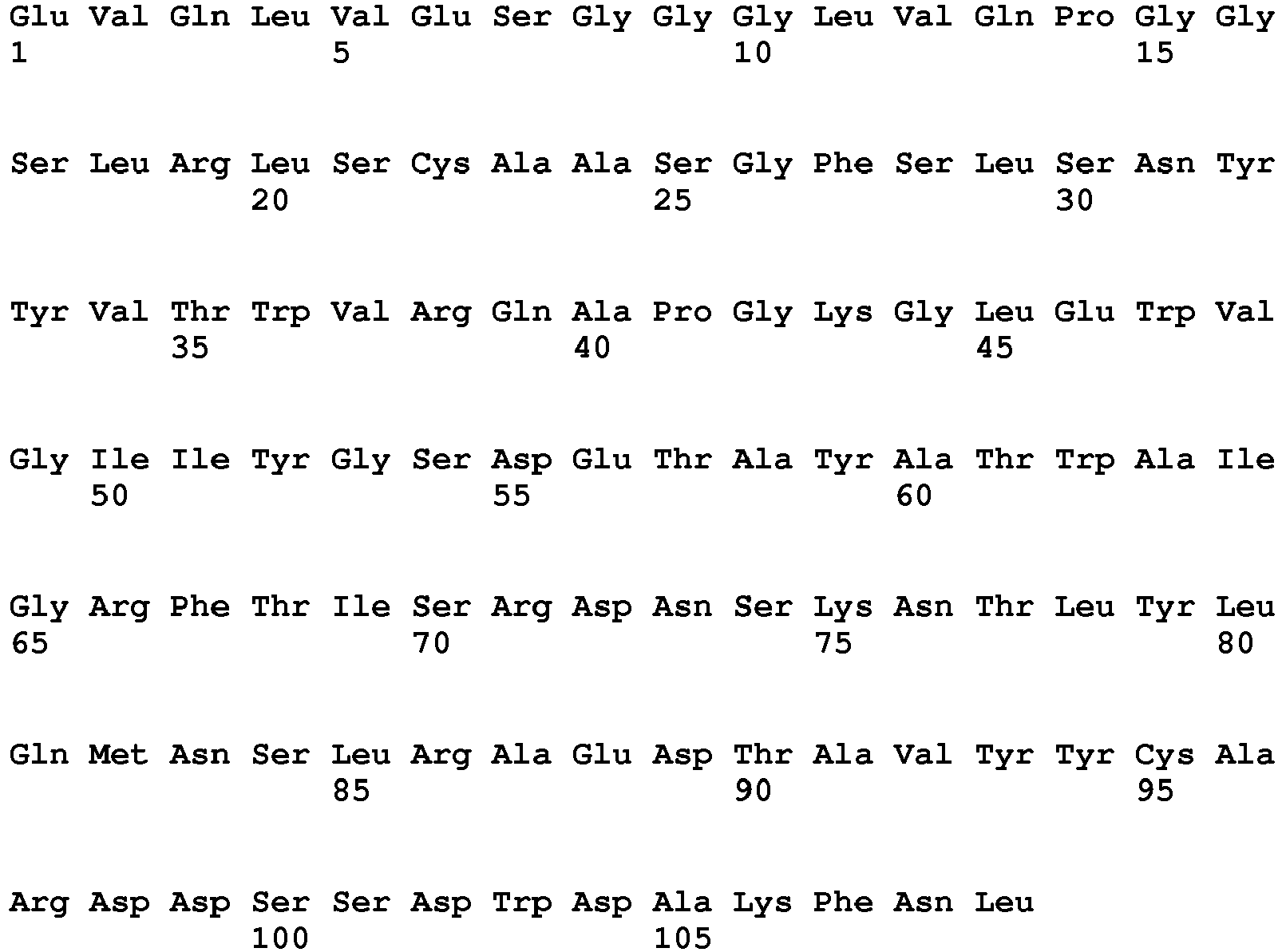
- <210> 19
<211> 109
<212> PRT
<213> Oryctolagus cuniculus - <400> 19


- <210> 20
<211> 99
<212> PRT
<213> Oryctolagus cuniculus - <400> 20

- <210> 21
<211> 122
<212> PRT
<213> Oryctolagus cuniculus - <400> 21

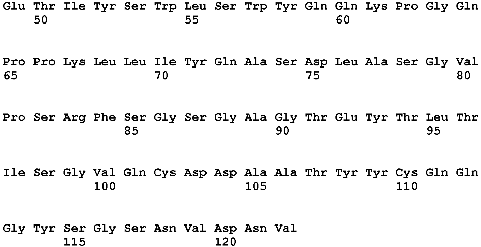
- <210> 22
<211> 126
<212> PRT
<213> Oryctolagus cuniculus - <400> 22

- <210> 23
<211> 11
<212> PRT
<213> Oryctolagus cuniculus - <400> 23

- <210> 24
<211> 7
<212> PRT
<213> Oryctolagus cuniculus - <400> 24

- <210> 25
<211> 12
<212> PRT
<213> Oryctolagus cuniculus - <400> 25

- <210> 26
<211> 5
<212> PRT
<213> Oryctolagus cuniculus - <400> 26

- <210> 27
<211> 16
<212> PRT
<213> Oryctolagus cuniculus - <400> 27

- <210> 28
<211> 12
<212> PRT
<213> Oryctolagus cuniculus - <400> 28

- <210> 29
<211> 366
<212> DNA
<213> Oryctolagus cuniculus - <400> 29

- <210> 30
<211> 378
<212> DNA
<213> Oryctolagus cuniculus - <400> 30
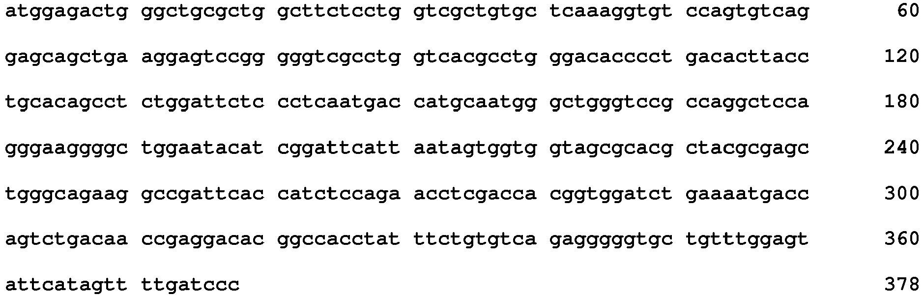
- <210> 31
<211> 33
<212> DNA
<213> Oryctolagus cuniculus - <400> 31
caggccagtg agaccattta cagttggtta tcc 33 - <210> 32
<211> 21
<212> DNA
<213> Oryctolagus cuniculus - <400> 32
caggcatccg atctggcatct 21 - <210> 33
<211> 36
<212> DNA
<213> Oryctolagus cuniculus - <400> 33
caacagggtt atagtggtag taatgttgat aatgtt 36 - <210> 34
<211> 15
<212> DNA
<213> Oryctolagus cuniculus - <400> 34
gaccatgcaa tgggc 15 - <210> 35
<211> 48
<212> DNA
<213> Oryctolagus cuniculus - <400> 35
ttcattaata gtggtggtag cgcacgctac gcgagctggg cagaaggc 48 - <210> 36
<211> 36
<212> DNA
<213> Oryctolagus cuniculus - <400> 36
gggggtgctg tttggagtat tcatagtttt gatccc 36 - <210> 37
<211> 123
<212> PRT
<213> Oryctolagus cuniculus - <400> 37


- <210> 38
<211> 125
<212> PRT
<213> Oryctolagus cuniculus - <400> 38

- <210> 39
<211> 13
<212> PRT
<213> Oryctolagus cuniculus - <400> 39

- <210> 40
<211> 7
<212> PRT
<213> Oryctolagus cuniculus - <400> 40

- <210> 41
<211> 11
<212> PRT
<213> Oryctolagus cuniculus - <400> 41

- <210> 42
<211> 5
<212> PRT
<213> Oryctolagus cuniculus - <400> 42

- <210> 43
<211> 16
<212> PRT
<213> Oryctolagus cuniculus - <400> 43

- <210> 44
<211> 12
<212> PRT
<213> Oryctolagus cuniculus - <400> 44

- <210> 45
<211> 369
<212> DNA
<213> Oryctolagus cuniculus - <400> 45


- <210> 46
<211> 375
<212> DNA
<213> Oryctolagus cuniculus - <400> 46

- <210> 47
<211> 39
<212> DNA
<213> Oryctolagus cuniculus - <400> 47
caggccagtc agagtgttta tgacaacaac tacttatcc 39 - <210> 48
<211> 21
<212> DNA
<213> Oryctolagus cuniculus - <400> 48
ggtgcatcca ctctggcatct 21 - <210> 49
<211> 33
<212> DNA
<213> Oryctolagus cuniculus - <400> 49
gcaggcgttt atgatgatga tagtgataatgcc 33 - <210> 50
<211> 15
<212> DNA
<213> Oryctolagus cuniculus - <400> 50
gtctactaca tgaac 15 - <210> 51
<211> 48
<212> DNA
<213> Oryctolagus cuniculus - <400> 51
ttcattacaa tgagtgataa tataaattac gcgagctggg cgaaaggc 48 - <210> 52
<211> 36
<212> DNA
<213> Oryctolagus cuniculus - <400> 52
agtcgtggct ggggtacaat gggtcggttg gatctc 36 - <210> 53
<211> 123
<212> PRT
<213> Oryctolagus cuniculus - <400> 53

- <210> 54
<211> 126
<212> PRT
<213> Oryctolagus cuniculus - <400> 54

- <210> 55
<211> 13
<212> PRT
<213> Oryctolagus cuniculus - <400> 55

- <210> 56
<211> 7
<212> PRT
<213> Oryctolagus cuniculus - <400> 56

- <210> 57
<211> 11
<212> PRT
<213> Oryctolagus cuniculus - <400> 57

- <210> 58
<211> 5
<212> PRT
<213> Oryctolagus cuniculus - <400> 58

- <210> 59
<211> 16
<212> PRT
<213> Oryctolagus cuniculus - <400> 59

- <210> 60
<211> 12
<212> PRT
<213> Oryctolagus cuniculus - <400> 60

- <210> 61
<211> 369
<212> DNA
<213> Oryctolagus cuniculus - <400> 61
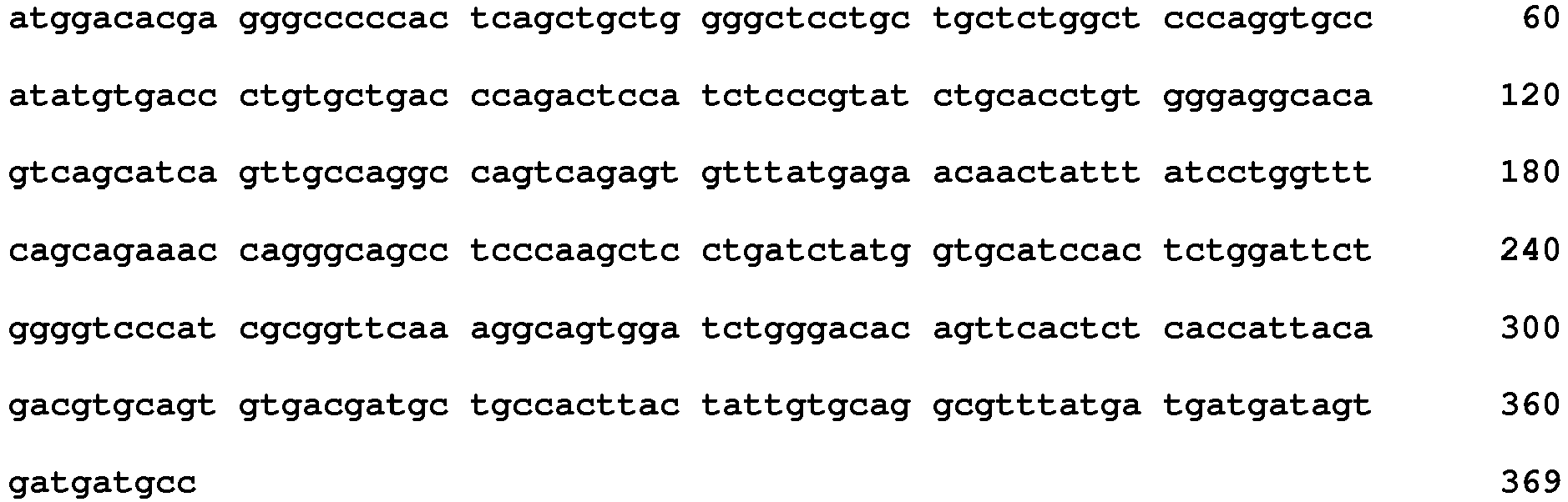
- <210> 62
<211> 378
<212> DNA
<213> Oryctolagus cuniculus - <400> 62

- <210> 63
<211> 39
<212> DNA
<213> Oryctolagus cuniculus - <400> 63
caggccagtc agagtgttta tgagaacaac tatttatcc 39 - <210> 64
<211> 21
<212> DNA
<213> Oryctolagus cuniculus - <400> 64
ggtgcatcca ctctggattct 21 - <210> 65
<211> 33
<212> DNA
<213> Oryctolagus cuniculus - <400> 65
gcaggcgttt atgatgatga tagtgatgatgcc 33 - <210> 66
<211> 15
<212> DNA
<213> Oryctolagus cuniculus - <400> 66
gcctactaca tgaac 15 - <210> 67
<211> 48
<212> DNA
<213> Oryctolagus cuniculus - <400> 67
ttcattactc tgaataataa tgtagcttac gcgaactggg cgaaaggc 48 - <210> 68
<211> 36
<212> DNA
<213> Oryctolagus cuniculus - <400> 68
agtcgtggct ggggtgcaat gggtcggttg gatctc 36 - <210> 69
<211> 122
<212> PRT
<213> Oryctolagus cuniculus - <400> 69
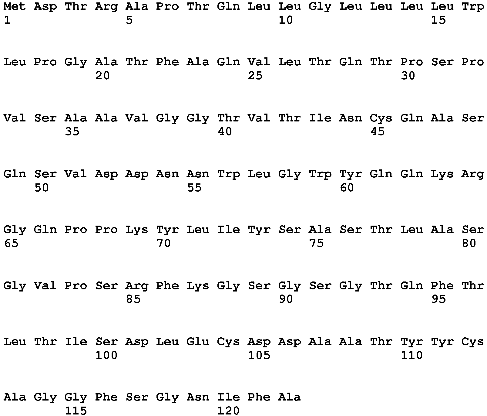
- <210> 70
<211> 122
<212> PRT
<213> Oryctolagus cuniculus - <400> 70


- <210> 71
<211> 13
<212> PRT
<213> Oryctolagus cuniculus - <400> 71

- <210> 72
<211> 7
<212> PRT
<213> Oryctolagus cuniculus - <400> 72

- <210> 73
<211> 10
<212> PRT
<213> Oryctolagus cuniculus - <400> 73

- <210> 74
<211> 5
<212> PRT
<213> Oryctolagus cuniculus - <400> 74

- <210> 75
<211> 16
<212> PRT
<213> Oryctolagus cuniculus - <400> 75

- <210> 76
<211> 9
<212> PRT
<213> Oryctolagus cuniculus - <400> 76

- <210> 77
<211> 366
<212> DNA
<213> Oryctolagus cuniculus - <400> 77

- <210> 78
<211> 366
<212> DNA
<213> Oryctolagus cuniculus - <400> 78


- <210> 79
<211> 39
<212> DNA
<213> Oryctolagus cuniculus - <400> 79
caggccagtc agagtgttga tgataacaac tggttaggc 39 - <210> 80
<211> 21
<212> DNA
<213> Oryctolagus cuniculus - <400> 80
tctgcatcca ctctggcatct 21 - <210> 81
<211> 30
<212> DNA
<213> Oryctolagus cuniculus - <400> 81
gcaggcggtt ttagtggtaa tatctttgct 30 - <210> 82
<211> 15
<212> DNA
<213> Oryctolagus cuniculus - <400> 82
agctatgcaa tgagc 15 - <210> 83
<211> 48
<212> DNA
<213> Oryctolagus cuniculus - <400> 83
atcattggtg gttttggtac cacatactac gcgacctggg cgaaaggc 48 - <210> 84
<211> 27
<212> DNA
<213> Oryctolagus cuniculus - <400> 84
ggtggtcctg gtaatggtgg tgacatc 27 - <210> 85
<211> 122
<212> PRT
<213> Oryctolagus cuniculus - <400> 85

- <210> 86
<211> 128
<212> PRT
<213> Oryctolagus cuniculus - <400> 86


- <210> 87
<211> 12
<212> PRT
<213> Oryctolagus cuniculus - <400> 87

- <210> 88
<211> 7
<212> PRT
<213> Oryctolagus cuniculus - <400> 88

- <210> 89
<211> 11
<212> PRT
<213> Oryctolagus cuniculus - <400> 89

- <210> 90
<211> 5
<212> PRT
<213> Oryctolagus cuniculus - <400> 90

- <210> 91
<211> 17
<212> PRT
<213> Oryctolagus cuniculus - <400> 91


- <210> 92
<211> 14
<212> PRT
<213> Oryctolagus cuniculus - <400> 92

- <210> 93
<211> 366
<212> DNA
<213> Oryctolagus cuniculus - <400> 93

- <210> 94
<211> 384
<212> DNA
<213> Oryctolagus cuniculus - <400> 94

- <210> 95
<211> 36
<212> DNA
<213> Oryctolagus cuniculus - <400> 95
cagtccagtc agagtgttta taataatttc ttatcg 36 - <210> 96
<211> 21
<212> DNA
<213> Oryctolagus cuniculus - <400> 96
caggcatcca aactggcatct 21 - <210> 97
<211> 33
<212> DNA
<213> Oryctolagus cuniculus - <400> 97
ctaggcggtt atgatgatga tgctgataat gct 33 - <210> 98
<211> 15
<212> DNA
<213> Oryctolagus cuniculus - <400> 98
gactatgcaa tgagc 15 - <210> 99
<211> 51
<212> DNA
<213> Oryctolagus cuniculus - <400> 99
atcatttatg ctggtagtgg tagcacatgg tacgcgagct gggcgaaaggc 51 - <210> 100
<211> 42
<212> DNA
<213> Oryctolagus cuniculus - <400> 100
gatggatacg atgactatgg tgatttcgat cgattggatctc 42 - <210> 101
<211> 122
<212> PRT
<213> Oryctolagus cuniculus - <400> 101


- <210> 102
<211> 125
<212> PRT
<213> Oryctolagus cuniculus - <400> 102


- <210> 103
<211> 11
<212> PRT
<213> Oryctolagus cuniculus - <400> 103

- <210> 104
<211> 7
<212> PRT
<213> Oryctolagus cuniculus - <400> 104

- <210> 105
<211> 12
<212> PRT
<213> Oryctolagus cuniculus - <400> 105

- <210> 106
<211> 5
<212> PRT
<213> Oryctolagus cuniculus - <400> 106

- <210> 107
<211> 16
<212> PRT
<213> Oryctolagus cuniculus - <400> 107

- <210> 108
<211> 12
<212> PRT
<213> Oryctolagus cuniculus - <400> 108

- <210> 109
<211> 366
<212> DNA
<213> Oryctolagus cuniculus - <400> 109

- <210> 110
<211> 375
<212> DNA
<213> Oryctolagus cuniculus - <400> 110

- <210> 111
<211> 33
<212> DNA
<213> Oryctolagus cuniculus - <400> 111
caggccagtc agagcattaa caatgaatta tcc 33 - <210> 112
<211> 21
<212> DNA
<213> Oryctolagus cuniculus - <400> 112
agggcatcca ctctggcatct 21 - <210> 113
<211> 36
<212> DNA
<213> Oryctolagus cuniculus - <400> 113
caacagggtt atagtctgag gaatattgat aatgct 36 - <210> 114
<211> 15
<212> DNA
<213> Oryctolagus cuniculus - <400> 114
aactactaca tgacc 15 - <210> 115
<211> 48
<212> DNA
<213> Oryctolagus cuniculus - <400> 115
atgatttatg gtagtgatga aacagcctac gcgaactggg cgataggc 48 - <210> 116
<211> 36
<212> DNA
<213> Oryctolagus cuniculus - <400> 116
gatgatagta gtgactggga tgcaaaattt aacttg 36 - <210> 117
<211> 109
<212> PRT
<213> Oryctolagus cuniculus - <400> 117


- <210> 118
<211> 109
<212> PRT
<213> Oryctolagus cuniculus - <400> 118

- <210> 119
<211> 100
<212> PRT
<213> Oryctolagus cuniculus - <400> 119


- <210> 120
<211> 16
<212> PRT
<213> Oryctolagus cuniculus - <400> 120

- <210> 121
<211> 16
<212> PRT
<213> Oryctolagus cuniculus - <400> 121

- <210> 122
<211> 123
<212> PRT
<213> Oryctolagus cuniculus - <400> 122


- <210> 123
<211> 128
<212> PRT
<213> Oryctolagus cuniculus - <400> 123

- <210> 124
<211> 13
<212> PRT
<213> Oryctolagus cuniculus - <400> 124


- <210> 125
<211> 7
<212> PRT
<213> Oryctolagus cuniculus - <400> 125

- <210> 126
<211> 11
<212> PRT
<213> Oryctolagus cuniculus - <400> 126

- <210> 127
<211> 5
<212> PRT
<213> Oryctolagus cuniculus - <400> 127

- <210> 128
<211> 16
<212> PRT
<213> Oryctolagus cuniculus - <400> 128

- <210> 129
<211> 15
<212> PRT
<213> Oryctolagus cuniculus - <400> 129

- <210> 130
<211> 369
<212> DNA
<213> Oryctolagus cuniculus - <400> 130

- <210> 131
<211> 384
<212> DNA
<213> Oryctolagus cuniculus - <400> 131
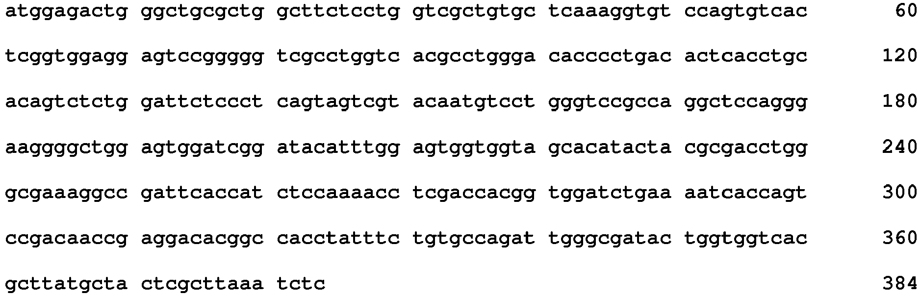
- <210> 132
<211> 39
<212> DNA
<213> Oryctolagus cuniculus - <400> 132
cagtccagtc agagtgttgg taataaccag gacttatcc 39 - <210> 133
<211> 21
<212> DNA
<213> Oryctolagus cuniculus - <400> 133
gaaatatcca aactggaatct 21 - <210> 134
<211> 33
<212> DNA
<213> Oryctolagus cuniculus - <400> 134
ctaggcggtt atgatgatga tgctgataat get 33 - <210> 135
<211> 15
<212> DNA
<213> Oryctolagus cuniculus - <400> 135
agtcgtacaa tgtcc 15 - <210> 136
<211> 48
<212> DNA
<213> Oryctolagus cuniculus - <400> 136
tacatttgga gtggtggtag cacatactac gcgacctggg cgaaaggc 48 - <210> 137
<211> 45
<212> DNA
<213> Oryctolagus cuniculus - <400> 137
ttgggcgata ctggtggtca cgcttatgct actcgcttaa atctc 45 - <210> 138
<211> 123
<212> PRT
<213> Oryctolagus cuniculus - <400> 138


- <210> 139
<211> 126
<212> PRT
<213> Oryctolagus cuniculus - <400> 139

- <210> 140
<211> 13
<212> PRT
<213> Oryctolagus cuniculus - <400> 140

- <210> 141
<211> 7
<212> PRT
<213> Oryctolagus cuniculus - <400> 141

- <210> 142
<211> 11
<212> PRT
<213> Oryctolagus cuniculus - <400> 142

- <210> 143
<211> 5
<212> PRT
<213> Oryctolagus cuniculus - <400> 143

- <210> 144
<211> 16
<212> PRT
<213> Oryctolagus cuniculus - <400> 144

- <210> 145
<211> 12
<212> PRT
<213> Oryctolagus cuniculus - <400> 145

- <210> 146
<211> 369
<212> DNA
<213> Oryctolagus cuniculus - <400> 146


- <210> 147
<211> 378
<212> DNA
<213> Oryctolagus cuniculus - <400> 147

- <210> 148
<211> 39
<212> DNA
<213> Oryctolagus cuniculus - <400> 148
cagtccagtc agagtgttta tagtaataag tacctagcc 39 - <210> 149
<211> 21
<212> DNA
<213> Oryctolagus cuniculus - <400> 149
tggacatcca aactggcatct 21 - <210> 150
<211> 33
<212> DNA
<213> Oryctolagus cuniculus - <400> 150
ctaggcgctt atgatgatga tgctgataat gct 33 - <210> 151
<211> 15
<212> DNA
<213> Oryctolagus cuniculus - <400> 151
ggcggctaca tgacc 15 - <210> 152
<211> 48
<212> DNA
<213> Oryctolagus cuniculus - <400> 152
atcagttatg atagtggtag cacatactac gcgagctggg cgaaaggc 48 - <210> 153
<211> 36
<212> DNA
<213> Oryctolagus cuniculus - <400> 153
tcactaaaat atcctactgt tacttctgat gacttg 36 - <210> 154
<211> 123
<212> PRT
<213> Oryctolagus cuniculus - <400> 154

- <210> 155
<211> 129
<212> PRT
<213> Oryctolagus cuniculus - <400> 155


- <210> 156
<211> 13
<212> PRT
<213> Oryctolagus cuniculus - <400> 156

- <210> 157
<211> 7
<212> PRT
<213> Oryctolagus cuniculus - <400> 157

- <210> 158
<211> 11
<212> PRT
<213> Oryctolagus cuniculus - <400> 158

- <210> 159
<211> 5
<212> PRT
<213> Oryctolagus cuniculus - <400> 159

- <210> 160
<211> 16
<212> PRT
<213> Oryctolagus cuniculus - <400> 160

- <210> 161
<211> 16
<212> PRT
<213> Oryctolagus cuniculus - <400> 161

- <210> 162
<211> 369
<212> DNA
<213> Oryctolagus cuniculus - <400> 162

- <210> 163
<211> 387
<212> DNA
<213> Oryctolagus cuniculus - <400> 163
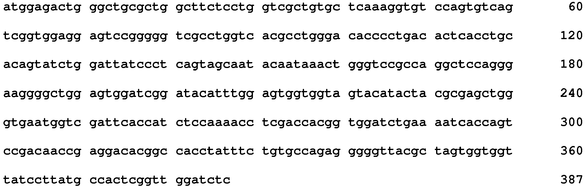
- <210> 164
<211> 39
<212> DNA
<213> Oryctolagus cuniculus - <400> 164
cagtccagtc agagtgttta taataataac gacttagcc 39 - <210> 165
<211> 21
<212> DNA
<213> Oryctolagus cuniculus - <400> 165
tatgcatcca ctctggcatct 21 - <210> 166
<211> 33
<212> DNA
<213> Oryctolagus cuniculus - <400> 166
ctaggcggtt atgatgatga tgctgataat get 33 - <210> 167
<211> 15
<212> DNA
<213> Oryctolagus cuniculus - <400> 167
agcaatacaa taaac 15 - <210> 168
<211> 48
<212> DNA
<213> Oryctolagus cuniculus - <400> 168
tacatttgga gtggtggtag tacatactac gcgagctggg tgaatggt 48 - <210> 169
<211> 48
<212> DNA
<213> Oryctolagus cuniculus - <400> 169
gggggttacg ctagtggtgg ttatccttat gccactcggt tggatctc 48 - <210> 170
<211> 123
<212> PRT
<213> Oryctolagus cuniculus - <400> 170

- <210> 171
<211> 123
<212> PRT
<213> Oryctolagus cuniculus - <400> 171


- <210> 172
<211> 13
<212> PRT
<213> Oryctolagus cuniculus - <400> 172

- <210> 173
<211> 7
<212> PRT
<213> Oryctolagus cuniculus - <400> 173

- <210> 174
<211> 11
<212> PRT
<213> Oryctolagus cuniculus - <400> 174

- <210> 175
<211> 6
<212> PRT
<213> Oryctolagus cuniculus - <400> 175

- <210> 176
<211> 16
<212> PRT
<213> Oryctolagus cuniculus - <400> 176

- <210> 177
<211> 8
<212> PRT
<213> Oryctolagus cuniculus - <400> 177

- <210> 178
<211> 369
<212> DNA
<213> Oryctolagus cuniculus - <400> 178

- <210> 179
<211> 369
<212> DNA
<213> Oryctolagus cuniculus - <400> 179


- <210> 180
<211> 39
<212> DNA
<213> Oryctolagus cuniculus - <400> 180
cagtccagtc agagtgttta taataacgac tacttatcc 39 - <210> 181
<211> 21
<212> DNA
<213> Oryctolagus cuniculus - <400> 181
ggtgcttcca aactggcatct 21 - <210> 182
<211> 33
<212> DNA
<213> Oryctolagus cuniculus - <400> 182
ctgggcgatt atgatgatga tgctgataatact 33 - <210> 183
<211> 18
<212> DNA
<213> Oryctolagus cuniculus - <400> 183
accaactact acctgagc 18 - <210> 184
<211> 48
<212> DNA
<213> Oryctolagus cuniculus - <400> 184
atcatttatc ctagtggtaa cacatattgc gcgaagtggg cgaaaggc 48 - <210> 185
<211> 24
<212> DNA
<213> Oryctolagus cuniculus - <400> 185
aattatggtg gtgatgaaag tttg 24 - <210> 186
<211> 119
<212> PRT
<213> Oryctolagus cuniculus - <400> 186
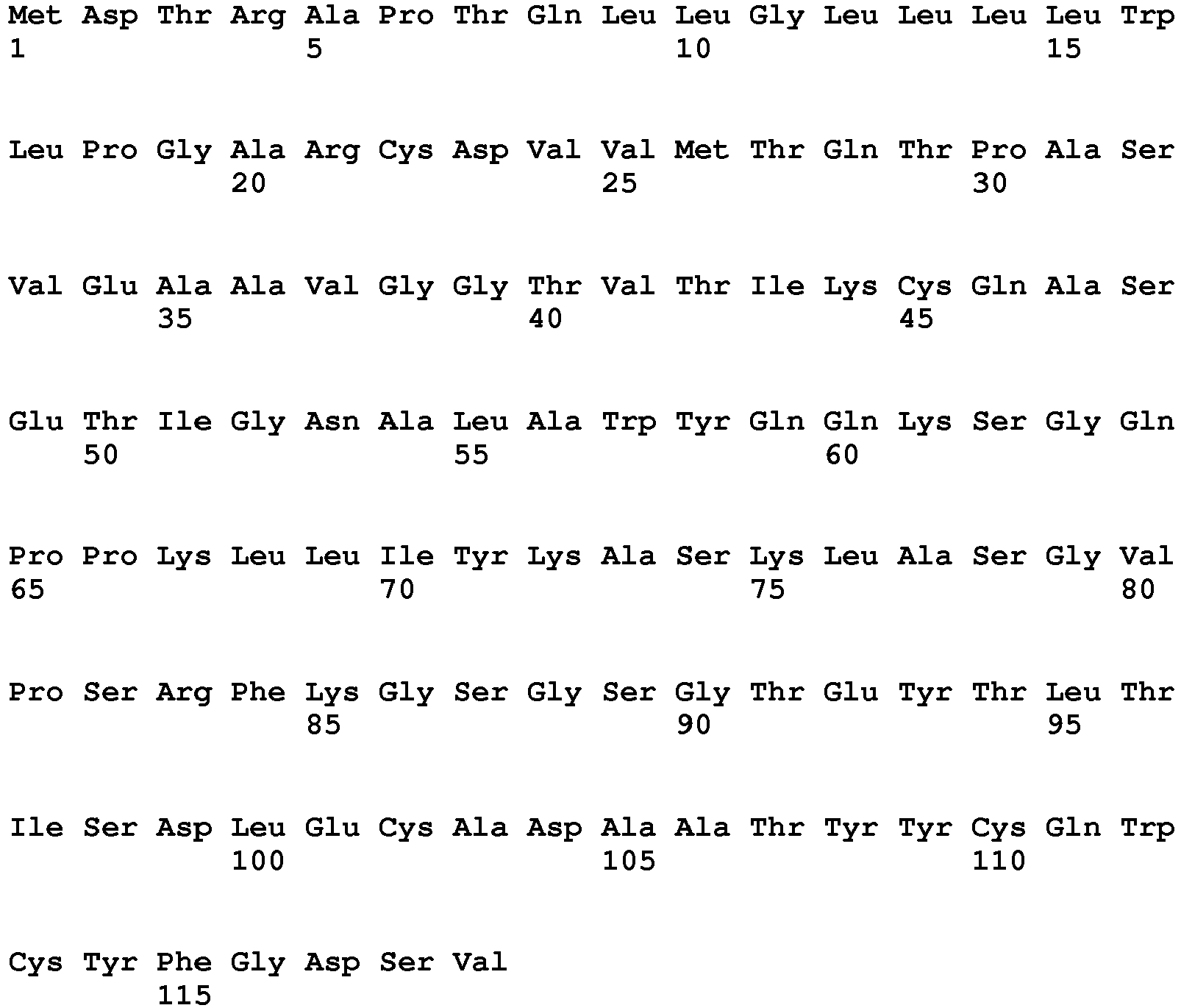
- <210> 187
<211> 128
<212> PRT
<213> Oryctolagus cuniculus - <400> 187


- <210> 188
<211> 11
<212> PRT
<213> Oryctolagus cuniculus - <400> 188

- <210> 189
<211> 7
<212> PRT
<213> Oryctolagus cuniculus - <400> 189

- <210> 190
<211> 9
<212> PRT
<213> Oryctolagus cuniculus - <400> 190

- <210> 191
<211> 6
<212> PRT
<213> Oryctolagus cuniculus - <400> 191

- <210> 192
<211> 17
<212> PRT
<213> Oryctolagus cuniculus - <400> 192


- <210> 193
<211> 11
<212> PRT
<213> Oryctolagus cuniculus - <400> 193

- <210> 194
<211> 357
<212> DNA
<213> Oryctolagus cuniculus - <400> 194

- <210> 195
<211> 384
<212> DNA
<213> Oryctolagus cuniculus - <400> 195
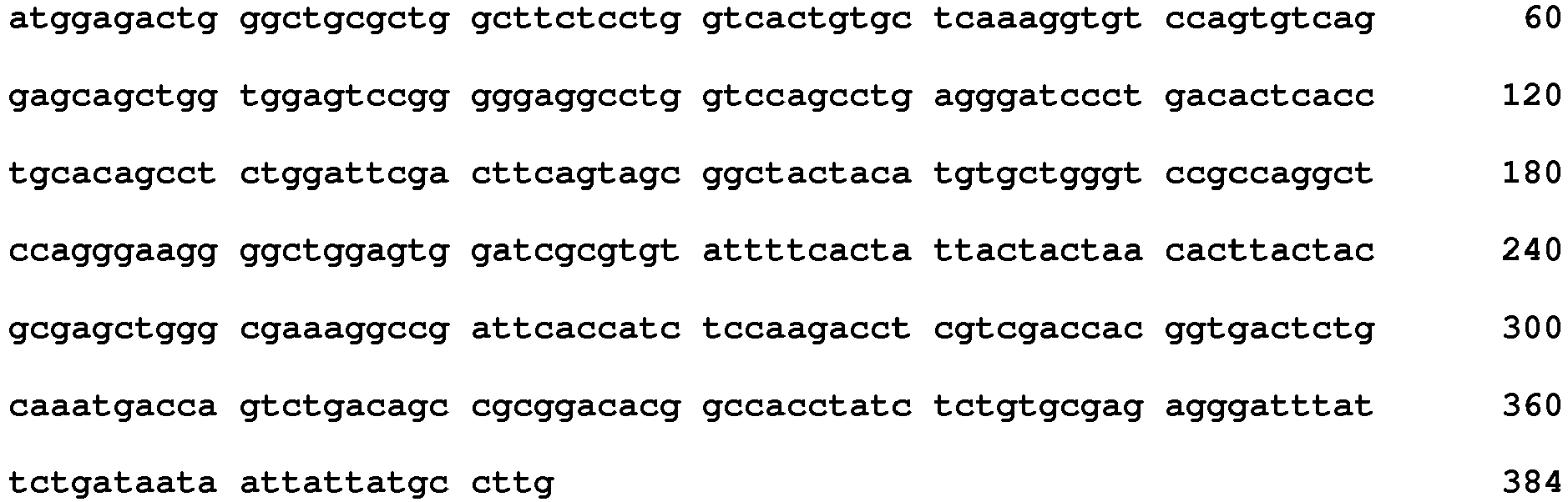
- <210> 196
<211> 33
<212> DNA
<213> Oryctolagus cuniculus - <400> 196
caggccagtg agaccattgg caatgcattagcc 33 - <210> 197
<211> 21
<212> DNA
<213> Oryctolagus cuniculus - <400> 197
aaggcatcca aactggcatct 21 - <210> 198
<211> 27
<212> DNA
<213> Oryctolagus cuniculus - <400> 198
caatggtgtt attttggtga tagtgtt 27 - <210> 199
<211> 18
<212> DNA
<213> Oryctolagus cuniculus - <400> 199
agcggctact acatgtgc 18 - <210> 200
<211> 51
<212> DNA
<213> Oryctolagus cuniculus - <400> 200
tgtattttca ctattactac taacacttac tacgcgagct gggcgaaaggc 51 - <210> 201
<211> 33
<212> DNA
<213> Oryctolagus cuniculus - <400> 201
gggatttatt ctgataataa ttattatgcc ttg 33 - <210> 202
<211> 119
<212> PRT
<213> Oryctolagus cuniculus - <400> 202


- <210> 203
<211> 128
<212> PRT
<213> Oryctolagus cuniculus - <400> 203

- <210> 204
<211> 11
<212> PRT
<213> Oryctolagus cuniculus - <400> 204

- <210> 205
<211> 7
<212> PRT
<213> Oryctolagus cuniculus - <400> 205

- <210> 206
<211> 9
<212> PRT
<213> Oryctolagus cuniculus - <400> 206

- <210> 207
<211> 6
<212> PRT
<213> Oryctolagus cuniculus - <400> 207

- <210> 208
<211> 17
<212> PRT
<213> Oryctolagus cuniculus - <400> 208

- <210> 209
<211> 11
<212> PRT
<213> Oryctolagus cuniculus - <400> 209

- <210> 210
<211> 357
<212> DNA
<213> Oryctolagus cuniculus - <400> 210

- <210> 211
<211> 384
<212> DNA
<213> Oryctolagus cuniculus - <400> 211

- <210> 212
<211> 33
<212> DNA
<213> Oryctolagus cuniculus - <400> 212
caggccagtg agagcattgg caatgcattagcc 33 - <210> 213
<211> 21
<212> DNA
<213> Oryctolagus cuniculus - <400> 213
aaggcatcca ctctggcatct 21 - <210> 214
<211> 27
<212> DNA
<213> Oryctolagus cuniculus - <400> 214
caatggtgtt attttggtga tagtgtt 27 - <210> 215
<211> 18
<212> DNA
<213> Oryctolagus cuniculus - <400> 215
agcggctact acatgtgc 18 - <210> 216
<211> 51
<212> DNA
<213> Oryctolagus cuniculus - <400> 216
tgcattttta ctattactga taacacttac tacgcgaact gggcgaaaggc 51 - <210> 217
<211> 33
<212> DNA
<213> Oryctolagus cuniculus - <400> 217
gggatttatt ctactgataa ttattatgcc ttg 33 - <210> 218
<211> 123
<212> PRT
<213> Oryctolagus cuniculus - <400> 218
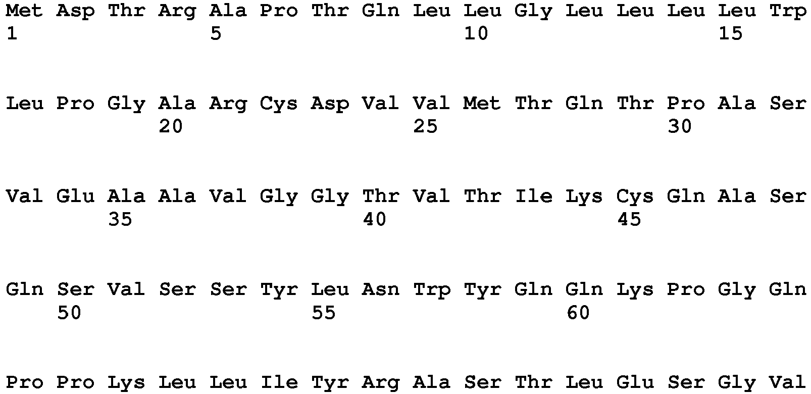

- <210> 219
<211> 133
<212> PRT
<213> Oryctolagus cuniculus - <400> 219

- <210> 220
<211> 11
<212> PRT
<213> Oryctolagus cuniculus - <400> 220

- <210> 221
<211> 7
<212> PRT
<213> Oryctolagus cuniculus - <400> 221

- <210> 222
<211> 13
<212> PRT
<213> Oryctolagus cuniculus - <400> 222

- <210> 223
<211> 5
<212> PRT
<213> Oryctolagus cuniculus - <400> 223

- <210> 224
<211> 16
<212> PRT
<213> Oryctolagus cuniculus - <400> 224

- <210> 225
<211> 19
<212> PRT
<213> Oryctolagus cuniculus - <400> 225

- <210> 226
<211> 369
<212> DNA
<213> Oryctolagus cuniculus - <400> 226

- <210> 227
<211> 399
<212> DNA
<213> Oryctolagus cuniculus - <400> 227

- <210> 228
<211> 33
<212> DNA
<213> Oryctolagus cuniculus - <400> 228
caggccagtc agagcgttag tagctacttaaac 33 - <210> 229
<211> 21
<212> DNA
<213> Oryctolagus cuniculus - <400> 229
agggcatcca ctctggaatct 21 - <210> 230
<211> 39
<212> DNA
<213> Oryctolagus cuniculus - <400> 230
caatgtactt atggtactag tagtagttat ggtgctgct 39 - <210> 231
<211> 15
<212> DNA
<213> Oryctolagus cuniculus - <400> 231
agcaatgcaa taagc 15 - <210> 232
<211> 48
<212> DNA
<213> Oryctolagus cuniculus - <400> 232
atcattagtt atagtggtac cacatactac gcgagctggg cgaaaggc 48 - <210> 233
<211> 57
<212> DNA
<213> Oryctolagus cuniculus - <400> 233
gatgacccta cgacagttat ggttatgttg ataccttttg gagccggcat ggacctc 57 - <210> 234
<211> 125
<212> PRT
<213> Oryctolagus cuniculus - <400> 234


- <210> 235
<211> 119
<212> PRT
<213> Oryctolagus cuniculus - <400> 235

- <210> 236
<211> 13
<212> PRT
<213> Oryctolagus cuniculus - <400> 236

- <210> 237
<211> 7
<212> PRT
<213> Oryctolagus cuniculus - <400> 237

- <210> 238
<211> 13
<212> PRT
<213> Oryctolagus cuniculus - <400> 238

- <210> 239
<211> 5
<212> PRT
<213> Oryctolagus cuniculus - <400> 239

- <210> 240
<211> 17
<212> PRT
<213> Oryctolagus cuniculus - <400> 240

- <210> 241
<211> 4
<212> PRT
<213> Oryctolagus cuniculus - <400> 241

- <210> 242
<211> 375
<212> DNA
<213> Oryctolagus cuniculus - <400> 242

- <210> 243
<211> 357
<212> DNA
<213> Oryctolagus cuniculus - <400> 243

- <210> 244
<211> 39
<212> DNA
<213> Oryctolagus cuniculus - <400> 244
caggccagtc agagtgttta taagaacaac tacttatcc 39 - <210> 245
<211> 21
<212> DNA
<213> Oryctolagus cuniculus - <400> 245
tctgcatcga ctctagattct 21 - <210> 246
<211> 39
<212> DNA
<213> Oryctolagus cuniculus - <400> 246
ctaggcagtt atgattgtag tagtggtgat tgttatgct 39 - <210> 247
<211> 15
<212> DNA
<213> Oryctolagus cuniculus - <400> 247
agctactgga tgtgc 15 - <210> 248
<211> 51
<212> DNA
<213> Oryctolagus cuniculus - <400> 248
tgcattgtta ctggtaatgg taacacttac tacgcgaact gggcgaaaggc 51 - <210> 249
<211> 12
<212> DNA
<213> Oryctolagus cuniculus - <400> 249
gcctatgact tg 12 - <210> 250
<211> 123
<212> PRT
<213> Oryctolagus cuniculus - <400> 250

- <210> 251
<211> 125
<212> PRT
<213> Oryctolagus cuniculus - <400> 251

- <210> 252
<211> 13
<212> PRT
<213> Oryctolagus cuniculus - <400> 252

- <210> 253
<211> 7
<212> PRT
<213> Oryctolagus cuniculus - <400> 253

- <210> 254
<211> 11
<212> PRT
<213> Oryctolagus cuniculus - <400> 254

- <210> 255
<211> 5
<212> PRT
<213> Oryctolagus cuniculus - <400> 255

- <210> 256
<211> 16
<212> PRT
<213> Oryctolagus cuniculus - <400> 256

- <210> 257
<211> 12
<212> PRT
<213> Oryctolagus cuniculus - <400> 257

- <210> 258
<211> 369
<212> DNA
<213> Oryctolagus cuniculus - <400> 258

- <210> 259
<211> 375
<212> DNA
<213> Oryctolagus cuniculus - <400> 259
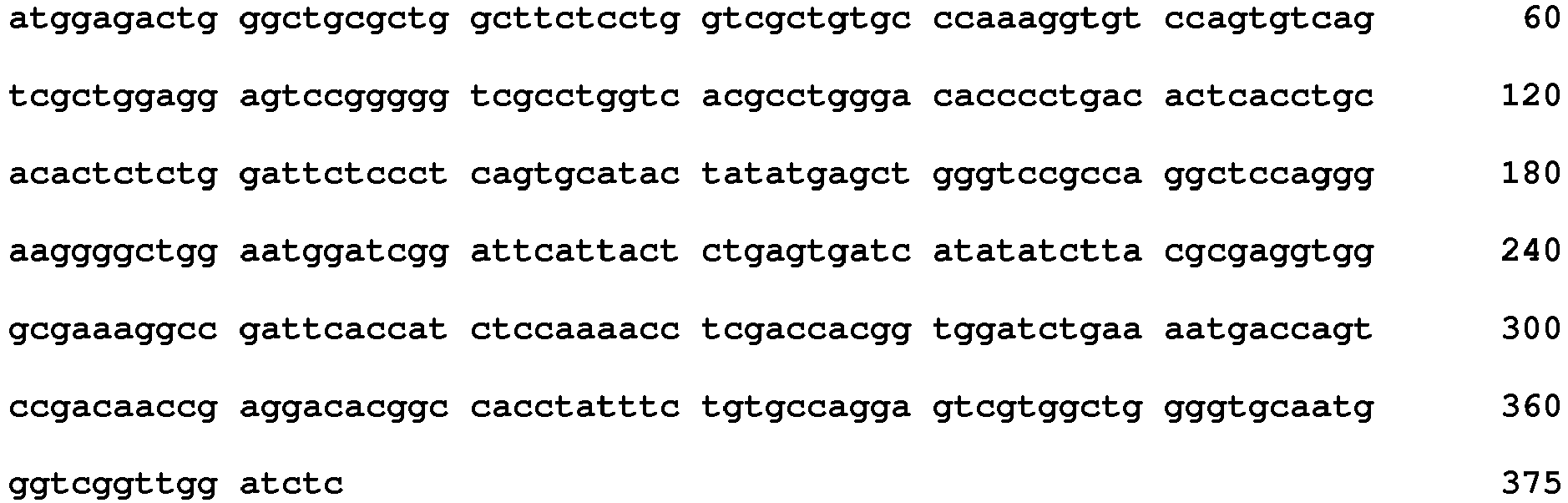
- <210> 260
<211> 39
<212> DNA
<213> Oryctolagus cuniculus - <400> 260
caggccagtc agagtgttta tgacaacaac tatttatcc 39 - <210> 261
<211> 21
<212> DNA
<213> Oryctolagus cuniculus - <400> 261
ggtgcatcca ctctggcatct 21 - <210> 262
<211> 33
<212> DNA
<213> Oryctolagus cuniculus - <400> 262
gcaggcgttt ttaatgatga tagtgatgatgcc 33 - <210> 263
<211> 15
<212> DNA
<213> Oryctolagus cuniculus - <400> 263
gcatactata tgagc 15 - <210> 264
<211> 48
<212> DNA
<213> Oryctolagus cuniculus - <400> 264
ttcattactc tgagtgatca tatatcttac gcgaggtggg cgaaaggc 48 - <210> 265
<211> 36
<212> DNA
<213> Oryctolagus cuniculus - <400> 265
agtcgtggct ggggtgcaat gggtcggttg gatctc 36 - <210> 266
<211> 123
<212> PRT
<213> Oryctolagus cuniculus - <400> 266

- <210> 267
<211> 121
<212> PRT
<213> Oryctolagus cuniculus - <400> 267


- <210> 268
<211> 13
<212> PRT
<213> Oryctolagus cuniculus - <400> 268

- <210> 269
<211> 7
<212> PRT
<213> Oryctolagus cuniculus - <400> 269

- <210> 270
<211> 11
<212> PRT
<213> Oryctolagus cuniculus - <400> 270

- <210> 271
<211> 5
<212> PRT
<213> Oryctolagus cuniculus - <400> 271

- <210> 272
<211> 16
<212> PRT
<213> Oryctolagus cuniculus - <400> 272

- <210> 273
<211> 8
<212> PRT
<213> Oryctolagus cuniculus - <400> 273

- <210> 274
<211> 369
<212> DNA
<213> Oryctolagus cuniculus - <400> 274

- <210> 275
<211> 363
<212> DNA
<213> Oryctolagus cuniculus - <400> 275


- <210> 276
<211> 39
<212> DNA
<213> Oryctolagus cuniculus - <400> 276
caggccagtc agagtgttta taacaacaaa aatttagcc 39 - <210> 277
<211> 21
<212> DNA
<213> Oryctolagus cuniculus - <400> 277
tgggcatcca ctctggcatct 21 - <210> 278
<211> 33
<212> DNA
<213> Oryctolagus cuniculus - <400> 278
ctaggcgttt ttgatgatga tgctgataat get 33 - <210> 279
<211> 15
<212> DNA
<213> Oryctolagus cuniculus - <400> 279
agctactcca tgacc 15 - <210> 280
<211> 48
<212> DNA
<213> Oryctolagus cuniculus - <400> 280
gtcattggta ctagtggtag cacatactac gcgacctggg cgaaaggc 48 - <210> 281
<211> 24
<212> DNA
<213> Oryctolagus cuniculus - <400> 281
agtctttctt ctattacttt cttg 24 - <210> 282
<211> 120
<212> PRT
<213> Oryctolagus cuniculus - <400> 282

- <210> 283
<211> 128
<212> PRT
<213> Oryctolagus cuniculus - <400> 283

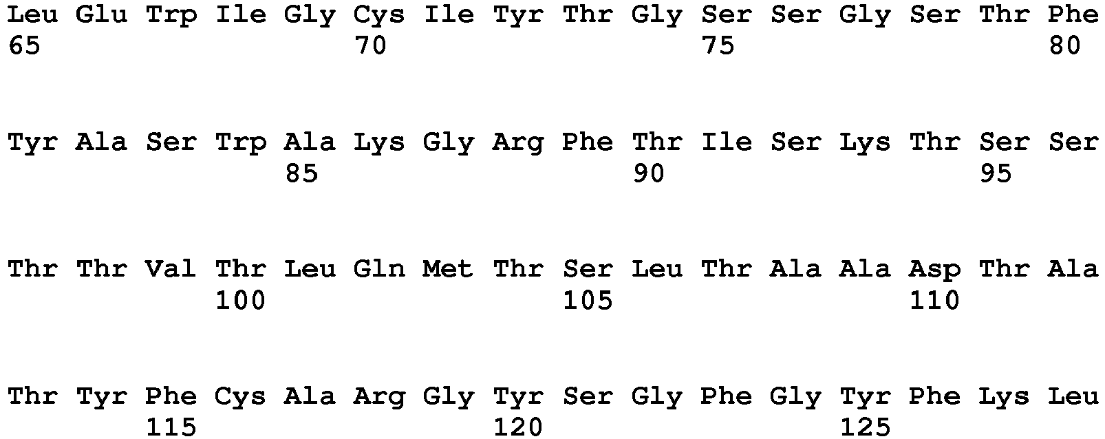
- <210> 284
<211> 11
<212> PRT
<213> Oryctolagus cuniculus - <400> 284

- <210> 285
<211> 7
<212> PRT
<213> Oryctolagus cuniculus - <400> 285

- <210> 286
<211> 10
<212> PRT
<213> Oryctolagus cuniculus - <400> 286

- <210> 287
<211> 6
<212> PRT
<213> Oryctolagus cuniculus - <400> 287

- <210> 288
<211> 18
<212> PRT
<213> Oryctolagus cuniculus - <400> 288

- <210> 289
<211> 10
<212> PRT
<213> Oryctolagus cuniculus - <400> 289

- <210> 290
<211> 360
<212> DNA
<213> Oryctolagus cuniculus - <400> 290

- <210> 291
<211> 384
<212> DNA
<213> Oryctolagus cuniculus - <400> 291
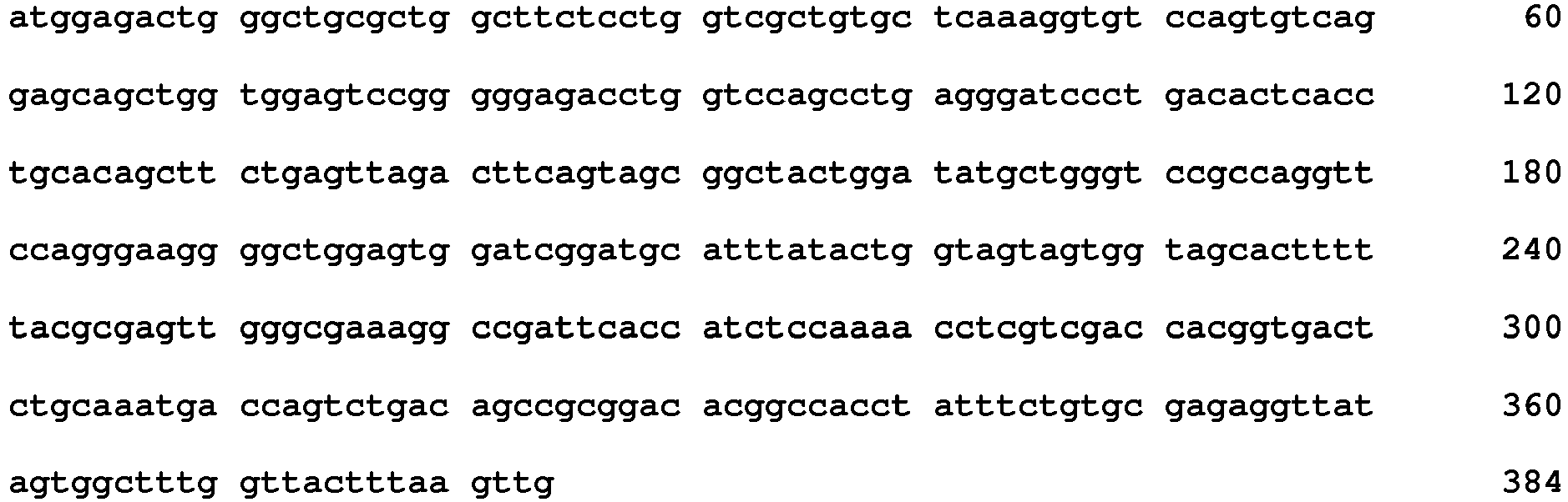
- <210> 292
<211> 33
<212> DNA
<213> Oryctolagus cuniculus - <400> 292
caggccagtc agaacattta tagatacttagcc 33 - <210> 293
<211> 21
<212> DNA
<213> Oryctolagus cuniculus - <400> 293
ctggcatcta ctctggcatct 21 - <210> 294
<211> 30
<212> DNA
<213> Oryctolagus cuniculus - <400> 294
caaagttatt atagtagtaa tagtgtcgct 30 - <210> 295
<211> 18
<212> DNA
<213> Oryctolagus cuniculus - <400> 295
agcggctact ggatatgc 18 - <210> 296
<211> 54
<212> DNA
<213> Oryctolagus cuniculus - <400> 296
tgcatttata ctggtagtag tggtagcact ttttacgcga gttgggcgaa aggc 54 - <210> 297
<211> 30
<212> DNA
<213> Oryctolagus cuniculus - <400> 297
ggttatagtg gctttggtta ctttaagttg 30 - <210> 298
<211> 122
<212> PRT
<213> Oryctolagus cuniculus - <400> 298


- <210> 299
<211> 123
<212> PRT
<213> Oryctolagus cuniculus - <400> 299


- <210> 300
<211> 11
<212> PRT
<213> Oryctolagus cuniculus - <400> 300

- <210> 301
<211> 7
<212> PRT
<213> Oryctolagus cuniculus - <400> 301

- <210> 302
<211> 12
<212> PRT
<213> Oryctolagus cuniculus - <400> 302

- <210> 303
<211> 5
<212> PRT
<213> Oryctolagus cuniculus - <400> 303

- <210> 304
<211> 16
<212> PRT
<213> Oryctolagus cuniculus - <400> 304

- <210> 305
<211> 10
<212> PRT
<213> Oryctolagus cuniculus - <400> 305

- <210> 306
<211> 366
<212> DNA
<213> Oryctolagus cuniculus - <400> 306

- <210> 307
<211> 369
<212> DNA
<213> Oryctolagus cuniculus - <400> 307

- <210> 308
<211> 33
<212> DNA
<213> Oryctolagus cuniculus - <400> 308
caggccagtg aggacattta taggttattggcc 33 - <210> 309
<211> 21
<212> DNA
<213> Oryctolagus cuniculus - <400> 309
gattcatccg atctggcatct 21 - <210> 310
<211> 36
<212> DNA
<213> Oryctolagus cuniculus - <400> 310
caacaggctt ggagttatag tgatattgat aatgct 36 - <210> 311
<211> 15
<212> DNA
<213> Oryctolagus cuniculus - <400> 311
agctactaca tgagc 15 - <210> 312
<211> 48
<212> DNA
<213> Oryctolagus cuniculus - <400> 312
atcattacta ctagtggtaa tacattttac gcgagctggg cgaaaggc 48 - <210> 313
<211> 30
<212> DNA
<213> Oryctolagus cuniculus - <400> 313
acttctgata ttttttatta tcgtaacttg 30 - <210> 314
<211> 123
<212> PRT
<213> Oryctolagus cuniculus - <400> 314


- <210> 315
<211> 129
<212> PRT
<213> Oryctolagus cuniculus - <400> 315

- <210> 316
<211> 13
<212> PRT
<213> Oryctolagus cuniculus - <400> 316

- <210> 317
<211> 7
<212> PRT
<213> Oryctolagus cuniculus - <400> 317

- <210> 318
<211> 11
<212> PRT
<213> Oryctolagus cuniculus - <400> 318

- <210> 319
<211> 5
<212> PRT
<213> Oryctolagus cuniculus - <400> 319

- <210> 320
<211> 16
<212> PRT
<213> Oryctolagus cuniculus - <400> 320

- <210> 321
<211> 16
<212> PRT
<213> Oryctolagus cuniculus - <400> 321

- <210> 322
<211> 369
<212> DNA
<213> Oryctolagus cuniculus - <400> 322

- <210> 323
<211> 387
<212> DNA
<213> Oryctolagus cuniculus - <400> 323

- <210> 324
<211> 39
<212> DNA
<213> Oryctolagus cuniculus - <400> 324
cagtccagtc agagtgttta taatgacatg gacttagcc 39 - <210> 325
<211> 21
<212> DNA
<213> Oryctolagus cuniculus - <400> 325
tctgcatcca ctctggcatct 21 - <210> 326
<211> 33
<212> DNA
<213> Oryctolagus cuniculus - <400> 326
ctaggcgctt ttgatgatga tgctgataatact 33 - <210> 327
<211> 15
<212> DNA
<213> Oryctolagus cuniculus - <400> 327
aggcatgcaa taacc 15 - <210> 328
<211> 48
<212> DNA
<213> Oryctolagus cuniculus - <400> 328
tgcatttgga gtggtggtag cacatactac gcgacctggg cgaaaggc 48 - <210> 329
<211> 48
<212> DNA
<213> Oryctolagus cuniculus - <400> 329
gtcattggcg atactgctgg ttatgcttat tttacggggc ttgacttg 48 - <210> 330
<211> 121
<212> PRT
<213> Oryctolagus cuniculus - <400> 330


- <210> 331
<211> 130
<212> PRT
<213> Oryctolagus cuniculus - <400> 331

- <210> 332
<211> 11
<212> PRT
<213> Oryctolagus cuniculus - <400> 332

- <210> 333
<211> 7
<212> PRT
<213> Oryctolagus cuniculus - <400> 333

- <210> 334
<211> 11
<212> PRT
<213> Oryctolagus cuniculus - <400> 334

- <210> 335
<211> 5
<212> PRT
<213> Oryctolagus cuniculus - <400> 335

- <210> 336
<211> 16
<212> PRT
<213> Oryctolagus cuniculus - <400> 336

- <210> 337
<211> 16
<212> PRT
<213> Oryctolagus cuniculus - <400> 337

- <210> 338
<211> 363
<212> DNA
<213> Oryctolagus cuniculus - <400> 338


- <210> 339
<211> 390
<212> DNA
<213> Oryctolagus cuniculus - <400> 339

- <210> 340
<211> 33
<212> DNA
<213> Oryctolagus cuniculus - <400> 340
caggccagtc agagtgttta taattggtta tcc 33 - <210> 341
<211> 21
<212> DNA
<213> Oryctolagus cuniculus - <400> 341
actgcatcca gtctggcatct 21 - <210> 342
<211> 33
<212> DNA
<213> Oryctolagus cuniculus - <400> 342
caacagggtt atactagtga tgttgataat gtt 33 - <210> 343
<211> 15
<212> DNA
<213> Oryctolagus cuniculus - <400> 343
agctatgcaa tgggc 15 - <210> 344
<211> 48
<212> DNA
<213> Oryctolagus cuniculus - <400> 344
atcattagta gtagtggtag cacatactac gcgacctggg cgaaaggc 48 - <210> 345
<211> 48
<212> DNA
<213> Oryctolagus cuniculus - <400> 345
gggggtgctg gtagtggtgg tgtttggctg cttgatggtt ttgatccc 48 - <210> 346
<211> 123
<212> PRT
<213> Oryctolagus cuniculus - <400> 346

- <210> 347
<211> 130
<212> PRT
<213> Oryctolagus cuniculus - <400> 347

- <210> 348
<211> 11
<212> PRT
<213> Oryctolagus cuniculus - <400> 348

- <210> 349
<211> 7
<212> PRT
<213> Oryctolagus cuniculus - <400> 349

- <210> 350
<211> 12
<212> PRT
<213> Oryctolagus cuniculus - <400> 350

- <210> 351
<211> 5
<212> PRT
<213> Oryctolagus cuniculus - <400> 351

- <210> 352
<211> 16
<212> PRT
<213> Oryctolagus cuniculus - <400> 352

- <210> 353
<211> 16
<212> PRT
<213> Oryctolagus cuniculus - <400> 353

- <210> 354
<211> 369
<212> DNA
<213> Oryctolagus cuniculus - <400> 354


- <210> 355
<211> 390
<212> DNA
<213> Oryctolagus cuniculus - <400> 355

- <210> 356
<211> 33
<212> DNA
<213> Oryctolagus cuniculus - <400> 356
caggccagtg agaacattta taattggttagcc 33 - <210> 357
<211> 21
<212> DNA
<213> Oryctolagus cuniculus - <400> 357
actgtaggcg atctggcatct 21 - <210> 358
<211> 36
<212> DNA
<213> Oryctolagus cuniculus - <400> 358
caacagggtt atagtagtag ttatgttgat aatgtt 36 - <210> 359
<211> 15
<212> DNA
<213> Oryctolagus cuniculus - <400> 359
gactatgcag tgggc 15 - <210> 360
<211> 48
<212> DNA
<213> Oryctolagus cuniculus - <400> 360
tacattcgta gtagtggtac cacagcctac gcgacctggg cgaaaggc 48 - <210> 361
<211> 48
<212> DNA
<213> Oryctolagus cuniculus - <400> 361
gggggtgctg gtagtagtgg tgtgtggatc cttgatggtt ttgctccc 48 - <210> 362
<211> 121
<212> PRT
<213> Oryctolagus cuniculus - <400> 362
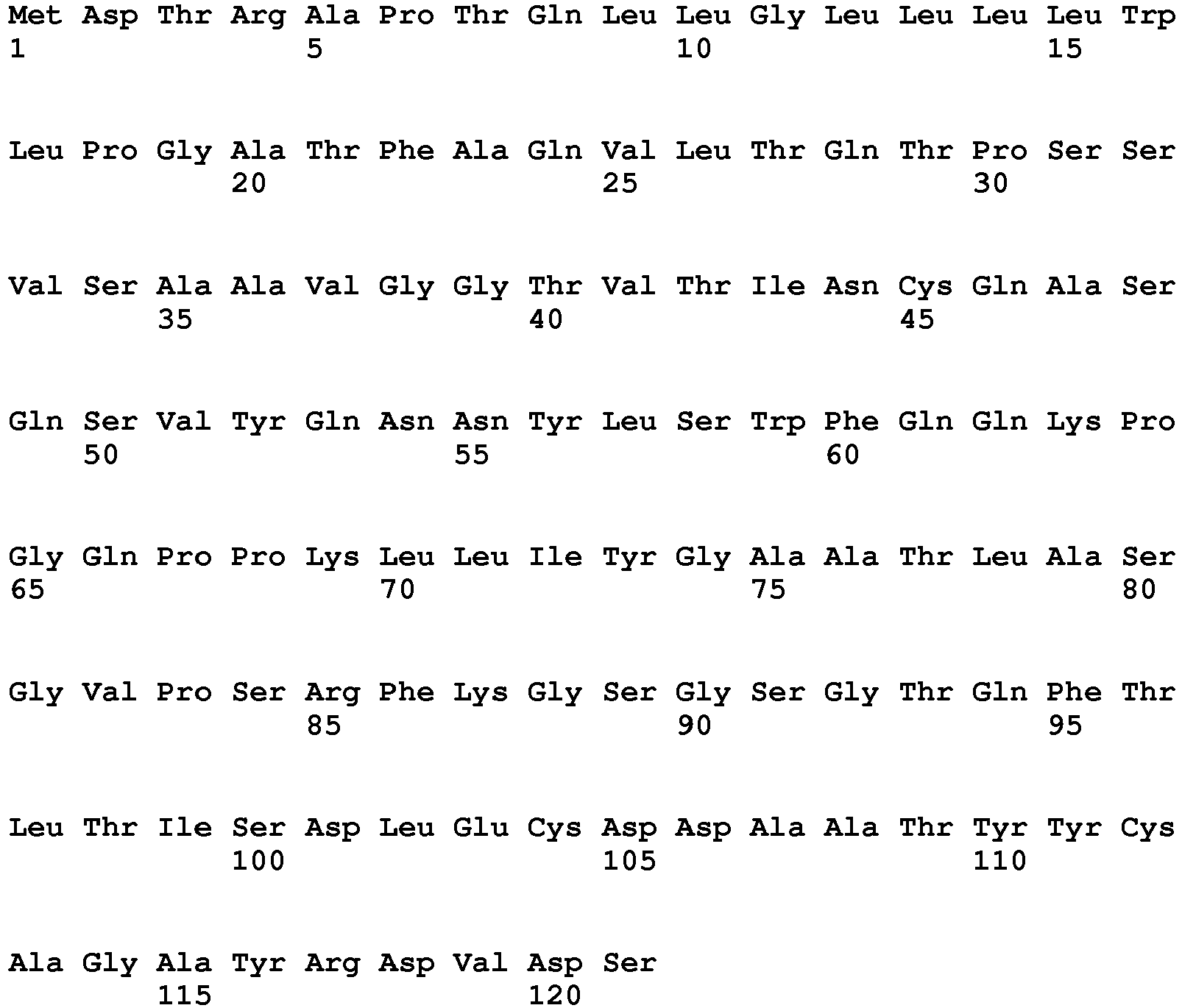
- <210> 363
<211> 130
<212> PRT
<213> Oryctolagus cuniculus - <400> 363

- <210> 364
<211> 13
<212> PRT
<213> Oryctolagus cuniculus - <400> 364

- <210> 365
<211> 7
<212> PRT
<213> Oryctolagus cuniculus - <400> 365

- <210> 366
<211> 9
<212> PRT
<213> Oryctolagus cuniculus - <400> 366

- <210> 367
<211> 6
<212> PRT
<213> Oryctolagus cuniculus - <400> 367

- <210> 368
<211> 18
<212> PRT
<213> Oryctolagus cuniculus - <400> 368

- <210> 369
<211> 13
<212> PRT
<213> Oryctolagus cuniculus - <400> 369

- <210> 370
<211> 363
<212> DNA
<213> Oryctolagus cuniculus - <400> 370

- <210> 371
<211> 390
<212> DNA
<213> Oryctolagus cuniculus - <400> 371

- <210> 372
<211> 39
<212> DNA
<213> Oryctolagus cuniculus - <400> 372
caggccagtc agagtgttta tcagaacaac tacttatcc 39 - <210> 373
<211> 21
<212> DNA
<213> Oryctolagus cuniculus - <400> 373
ggtgcggcca ctctggcatct 21 - <210> 374
<211> 27
<212> DNA
<213> Oryctolagus cuniculus - <400> 374
gcaggcgctt atagggatgt ggattct 27 - <210> 375
<211> 18
<212> DNA
<213> Oryctolagus cuniculus - <400> 375
agtacctact acatctac 18 - <210> 376
<211> 54
<212> DNA
<213> Oryctolagus cuniculus - <400> 376
tgtattgatg ctggtagtag tggtagcact tactacgcga cctgggtgaa tggc 54 - <210> 377
<211> 39
<212> DNA
<213> Oryctolagus cuniculus - <400> 377
tgggattatg gtggtaatgt tggttggggt tatgacttg 39 - <210> 378
<211> 120
<212> PRT
<213> Oryctolagus cuniculus - <400> 378

- <210> 379
<211> 127
<212> PRT
<213> Oryctolagus cuniculus - <400> 379

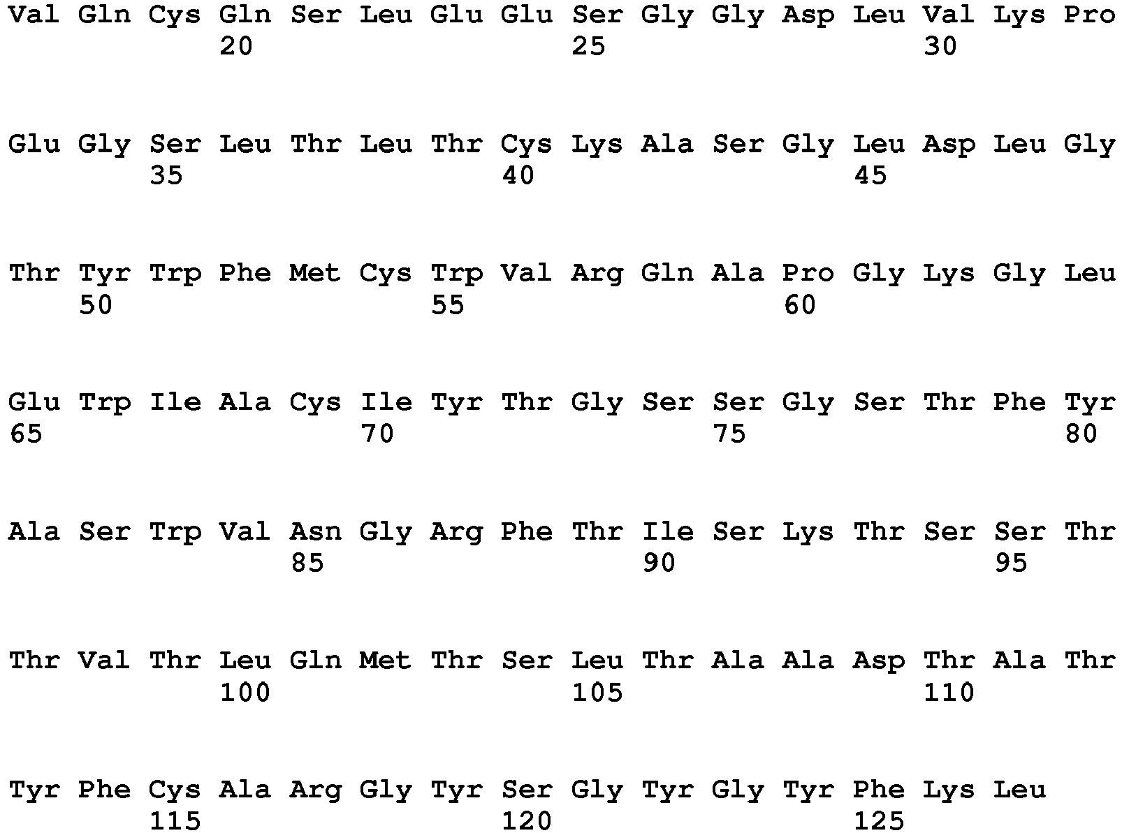
- <210> 380
<211> 11
<212> PRT
<213> Oryctolagus cuniculus - <400> 380

- <210> 381
<211> 7
<212> PRT
<213> Oryctolagus cuniculus - <400> 381

- <210> 382
<211> 10
<212> PRT
<213> Oryctolagus cuniculus - <400> 382

- <210> 383
<211> 6
<212> PRT
<213> Oryctolagus cuniculus - <400> 383

- <210> 384
<211> 18
<212> PRT
<213> Oryctolagus cuniculus - <400> 384

- <210> 385
<211> 10
<212> PRT
<213> Oryctolagus cuniculus - <400> 385

- <210> 386
<211> 360
<212> DNA
<213> Oryctolagus cuniculus - <400> 386

- <210> 387
<211> 381
<212> DNA
<213> Oryctolagus cuniculus - <400> 387

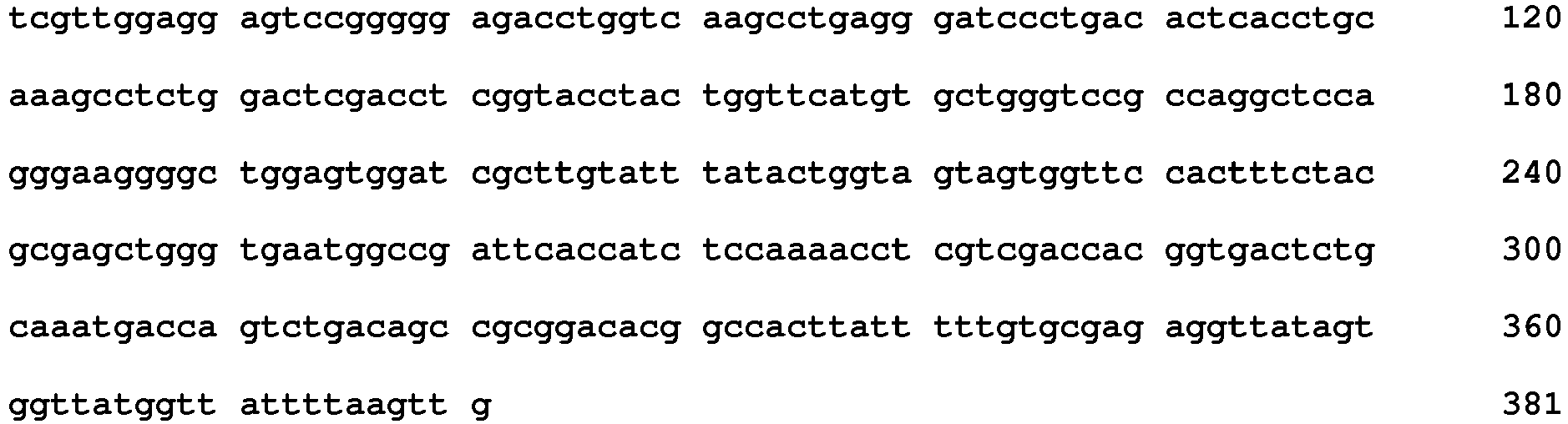
- <210> 388
<211> 33
<212> DNA
<213> Oryctolagus cuniculus - <400> 388
caggccagtc agagcattag tagttacttagcc 33 - <210> 389
<211> 21
<212> DNA
<213> Oryctolagus cuniculus - <400> 389
agggcgtcca ctctggcatct 21 - <210> 390
<211> 30
<212> DNA
<213> Oryctolagus cuniculus - <400> 390
caaagctatt atgatagtgt ttcaaatcct 30 - <210> 391
<211> 18
<212> DNA
<213> Oryctolagus cuniculus - <400> 391
acctactggt tcatgtgc 18 - <210> 392
<211> 54
<212> DNA
<213> Oryctolagus cuniculus - <400> 392
tgtatttata ctggtagtag tggttccact ttctacgcga gctgggtgaa tggc 54 - <210> 393
<211> 30
<212> DNA
<213> Oryctolagus cuniculus - <400> 393
ggttatagtg gttatggtta ttttaagttg 30 - <210> 394
<211> 124
<212> PRT
<213> Oryctolagus cuniculus - <400> 394

- <210> 395
<211> 130
<212> PRT
<213> Oryctolagus cuniculus - <400> 395
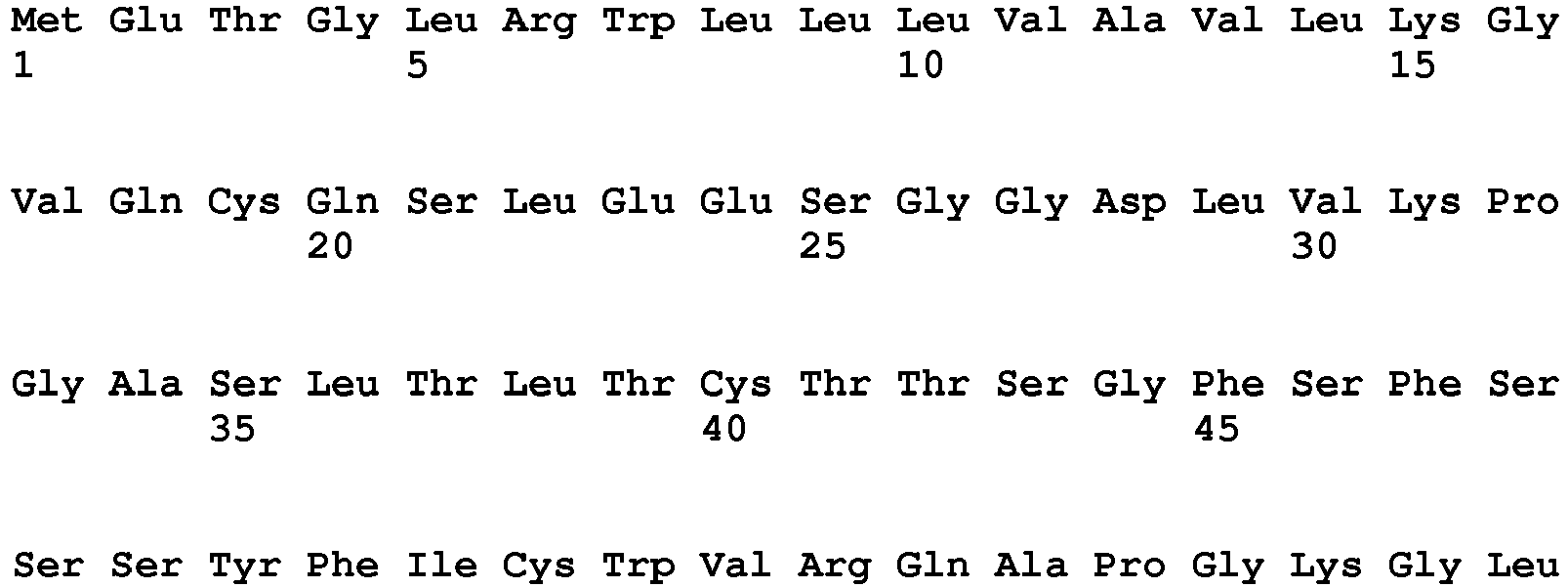
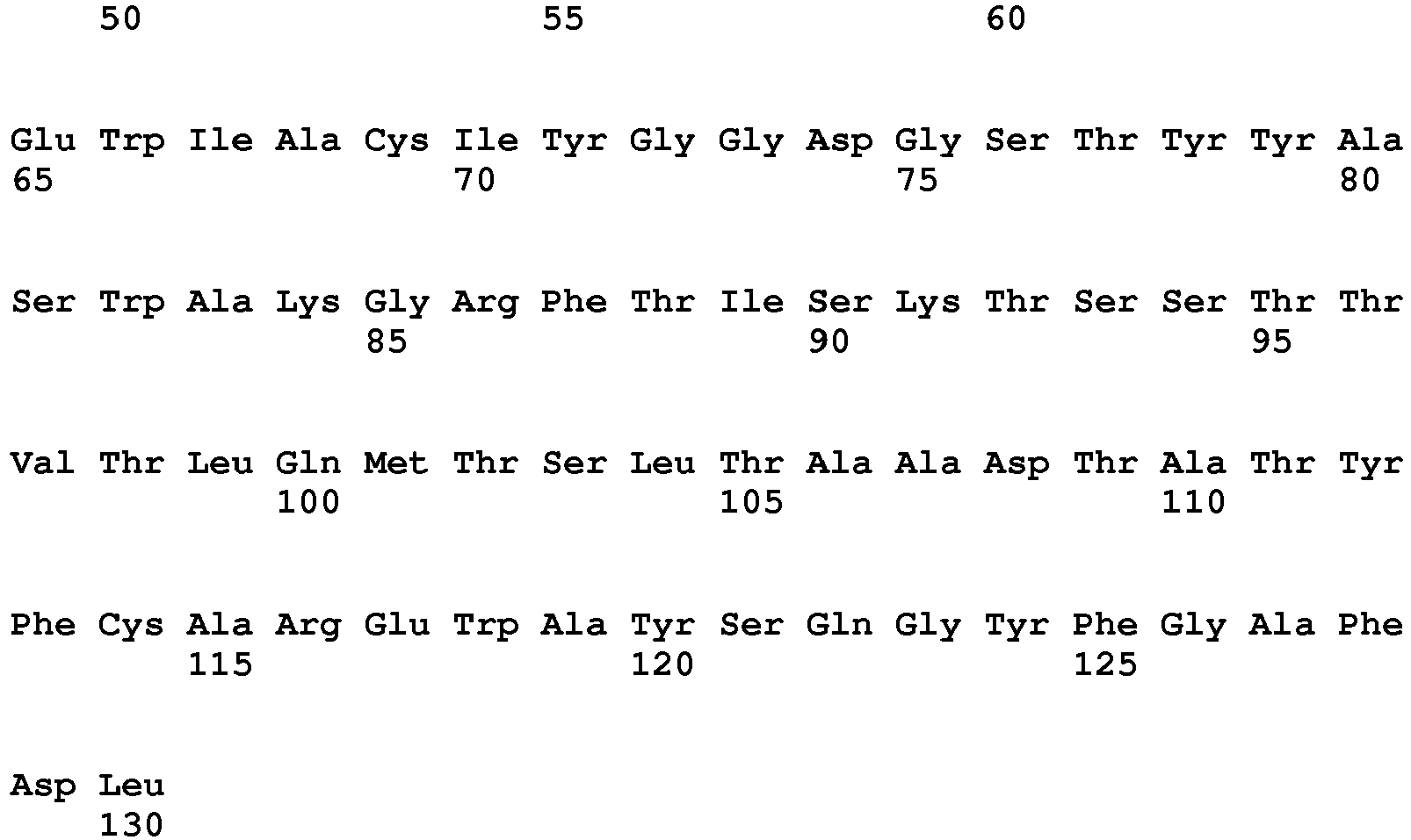
- <210> 396
<211> 13
<212> PRT
<213> Oryctolagus cuniculus - <400> 396

- <210> 397
<211> 7
<212> PRT
<213> Oryctolagus cuniculus - <400> 397

- <210> 398
<211> 12
<212> PRT
<213> Oryctolagus cuniculus - <400> 398

- <210> 399
<211> 6
<212> PRT
<213> Oryctolagus cuniculus - <400> 399

- <210> 400
<211> 17
<212> PRT
<213> Oryctolagus cuniculus - <400> 400

- <210> 401
<211> 14
<212> PRT
<213> Oryctolagus cuniculus - <400> 401

- <210> 402
<211> 372
<212> DNA
<213> Oryctolagus cuniculus - <400> 402

- <210> 403
<211> 390
<212> DNA
<213> Oryctolagus cuniculus - <400> 403


- <210> 404
<211> 39
<212> DNA
<213> Oryctolagus cuniculus - <400> 404
caggccagtc agagtgttta taagaacaac caattatcc 39 - <210> 405
<211> 21
<212> DNA
<213> Oryctolagus cuniculus - <400> 405
ggtgcatcgg ctctggcatct 21 - <210> 406
<211> 36
<212> DNA
<213> Oryctolagus cuniculus - <400> 406
gcaggcgcta ttactggtag tattgatacg gatggt 36 - <210> 407
<211> 18
<212> DNA
<213> Oryctolagus cuniculus - <400> 407
agcagctact tcatttgc 18 - <210> 408
<211> 51
<212> DNA
<213> Oryctolagus cuniculus - <400> 408
tgcatttatg gtggtgatgg cagcacatac tacgcgagct gggcgaaaggc 51 - <210> 409
<211> 42
<212> DNA
<213> Oryctolagus cuniculus - <400> 409
gaatgggcat atagtcaagg ttattttggt gcttttgatctc 42 - <210> 410
<211> 124
<212> PRT
<213> Oryctolagus cuniculus - <400> 410

- <210> 411
<211> 124
<212> PRT
<213> Oryctolagus cuniculus - <400> 411


- <210> 412
<211> 11
<212> PRT
<213> Oryctolagus cuniculus - <400> 412

- <210> 413
<211> 7
<212> PRT
<213> Oryctolagus cuniculus - <400> 413

- <210> 414
<211> 14
<212> PRT
<213> Oryctolagus cuniculus - <400> 414

- <210> 415
<211> 5
<212> PRT
<213> Oryctolagus cuniculus - <400> 415

- <210> 416
<211> 16
<212> PRT
<213> Oryctolagus cuniculus - <400> 416

- <210> 417
<211> 11
<212> PRT
<213> Oryctolagus cuniculus - <400> 417

- <210> 418
<211> 372
<212> DNA
<213> Oryctolagus cuniculus - <400> 418

- <210> 419
<211> 372
<212> DNA
<213> Oryctolagus cuniculus - <400> 419

- <210> 420
<211> 33
<212> DNA
<213> Oryctolagus cuniculus - <400> 420
caggccagtg aggatattag tagctacttagcc 33 - <210> 421
<211> 21
<212> DNA
<213> Oryctolagus cuniculus - <400> 421
gctgcatcca atctggaatct 21 - <210> 422
<211> 42
<212> DNA
<213> Oryctolagus cuniculus - <400> 422
caatgtactt atggtactat ttctattagt gatggtaatgct 42 - <210> 423
<211> 15
<212> DNA
<213> Oryctolagus cuniculus - <400> 423
agctacttca tgacc 15 - <210> 424
<211> 48
<212> DNA
<213> Oryctolagus cuniculus - <400> 424
ttcattaatc ctggtggtag cgcttactac gcgagctggg tgaaaggc 48 - <210> 425
<211> 33
<212> DNA
<213> Oryctolagus cuniculus - <400> 425
gttctgattg tttcttatgg agcctttacc ate 33 - <210> 426
<211> 124
<212> PRT
<213> Oryctolagus cuniculus - <400> 426


- <210> 427
<211> 124
<212> PRT
<213> Oryctolagus cuniculus - <400> 427
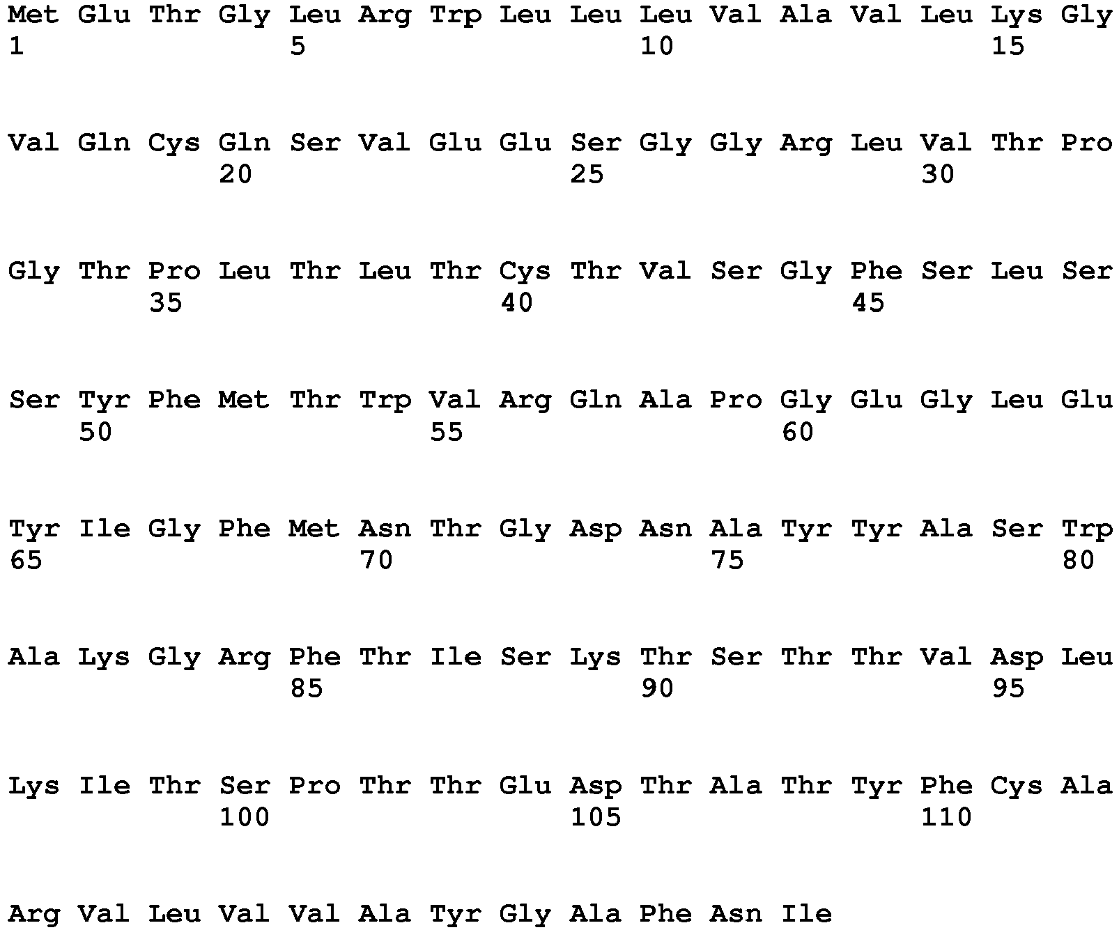

- <210> 428
<211> 11
<212> PRT
<213> Oryctolagus cuniculus - <400> 428

- <210> 429
<211> 7
<212> PRT
<213> Oryctolagus cuniculus - <400> 429

- <210> 430
<211> 14
<212> PRT
<213> Oryctolagus cuniculus - <400> 430

- <210> 431
<211> 5
<212> PRT
<213> Oryctolagus cuniculus - <400> 431

- <210> 432
<211> 16
<212> PRT
<213> Oryctolagus cuniculus - <400> 432

- <210> 433
<211> 11
<212> PRT
<213> Oryctolagus cuniculus - <400> 433

- <210> 434
<211> 372
<212> DNA
<213> Oryctolagus cuniculus - <400> 434

- <210> 435
<211> 372
<212> DNA
<213> Oryctolagus cuniculus - <400> 435

- <210> 436
<211> 33
<212> DNA
<213> Oryctolagus cuniculus - <400> 436
caggccagtg aggacattga aagctatctagcc 33 - <210> 437
<211> 21
<212> DNA
<213> Oryctolagus cuniculus - <400> 437
ggtgcatcca atctggaatct 21 - <210> 438
<211> 42
<212> DNA
<213> Oryctolagus cuniculus - <400> 438
caatgcactt atggtattat tagtattagt gatggtaatgct 42 - <210> 439
<211> 15
<212> DNA
<213> Oryctolagus cuniculus - <400> 439
agctacttca tgacc 15 - <210> 440
<211> 48
<212> DNA
<213> Oryctolagus cuniculus - <400> 440
ttcatgaata ctggtgataa cgcatactac gcgagctggg cgaaaggc 48 - <210> 441
<211> 33
<212> DNA
<213> Oryctolagus cuniculus - <400> 441
gttcttgttg ttgcttatgg agcctttaacatc 33 - <210> 442
<211> 124
<212> PRT
<213> Oryctolagus cuniculus - <400> 442


- <210> 443
<211> 127
<212> PRT
<213> Oryctolagus cuniculus - <400> 443

- <210> 444
<211> 13
<212> PRT
<213> Oryctolagus cuniculus - <400> 444

- <210> 445
<211> 7
<212> PRT
<213> Oryctolagus cuniculus - <400> 445

- <210> 446
<211> 12
<212> PRT
<213> Oryctolagus cuniculus - <400> 446

- <210> 447
<211> 5
<212> PRT
<213> Oryctolagus cuniculus - <400> 447

- <210> 448
<211> 16
<212> PRT
<213> Oryctolagus cuniculus - <400> 448

- <210> 449
<211> 14
<212> PRT
<213> Oryctolagus cuniculus - <400> 449

- <210> 450
<211> 372
<212> DNA
<213> Oryctolagus cuniculus - <400> 450

- <210> 451
<211> 381
<212> DNA
<213> Oryctolagus cuniculus - <400> 451

- <210> 452
<211> 39
<212> DNA
<213> Oryctolagus cuniculus - <400> 452
cagtccagta agagtgttat gaataacaac tacttagcc 39 - <210> 453
<211> 21
<212> DNA
<213> Oryctolagus cuniculus - <400> 453
ggtgcatcca atctggcatct 21 - <210> 454
<211> 36
<212> DNA
<213> Oryctolagus cuniculus - <400> 454
caaggcggtt atactggtta tagtgatcat gggact 36 - <210> 455
<211> 15
<212> DNA
<213> Oryctolagus cuniculus - <400> 455
agctatccaa tgaac 15 - <210> 456
<211> 48
<212> DNA
<213> Oryctolagus cuniculus - <400> 456
ttcattaata ctggtggtac catagtctac gcgagctggg caaaaggc 48 - <210> 457
<211> 42
<212> DNA
<213> Oryctolagus cuniculus - <400> 457
ggcagttatg tttcatctgg ttatgcctac tattttaatgtc 42 - <210> 458
<211> 121
<212> PRT
<213> Oryctolagus cuniculus - <400> 458


- <210> 459
<211> 126
<212> PRT
<213> Oryctolagus cuniculus - <400> 459

- <210> 460
<211> 13
<212> PRT
<213> Oryctolagus cuniculus - <400> 460

- <210> 461
<211> 7
<212> PRT
<213> Oryctolagus cuniculus - <400> 461

- <210> 462
<211> 9
<212> PRT
<213> Oryctolagus cuniculus - <400> 462

- <210> 463
<211> 5
<212> PRT
<213> Oryctolagus cuniculus - <400> 463

- <210> 464
<211> 16
<212> PRT
<213> Oryctolagus cuniculus - <400> 464

- <210> 465
<211> 13
<212> PRT
<213> Oryctolagus cuniculus - <400> 465

- <210> 466
<211> 363
<212> DNA
<213> Oryctolagus cuniculus - <400> 466


- <210> 467
<211> 378
<212> DNA
<213> Oryctolagus cuniculus - <400> 467

- <210> 468
<211> 39
<212> DNA
<213> Oryctolagus cuniculus - <400> 468
cagtccagtc agagtgttta taataacaac tggttatcc 39 - <210> 469
<211> 21
<212> DNA
<213> Oryctolagus cuniculus - <400> 469
aaggcatcca ctctggcatct 21 - <210> 470
<211> 27
<212> DNA
<213> Oryctolagus cuniculus - <400> 470
gcgggcggtt atcttgatag tgttatt 27 - <210> 471
<211> 15
<212> DNA
<213> Oryctolagus cuniculus - <400> 471
acctattcaa taaac 15 - <210> 472
<211> 48
<212> DNA
<213> Oryctolagus cuniculus - <400> 472
atcattgcta atagtggtac cacattctac gcgaactggg cgaaaggc 48 - <210> 473
<211> 39
<212> DNA
<213> Oryctolagus cuniculus - <400> 473
gagagtggaa tgtacaatga atatggtaaa tttaacatc 39 - <210> 474
<211> 122
<212> PRT
<213> Oryctolagus cuniculus - <400> 474

- <210> 475
<211> 128
<212> PRT
<213> Oryctolagus cuniculus - <400> 475


- <210> 476
<211> 11
<212> PRT
<213> Oryctolagus cuniculus - <400> 476

- <210> 477
<211> 7
<212> PRT
<213> Oryctolagus cuniculus - <400> 477

- <210> 478
<211> 12
<212> PRT
<213> Oryctolagus cuniculus - <400> 478

- <210> 479
<211> 5
<212> PRT
<213> Oryctolagus cuniculus - <400> 479

- <210> 480
<211> 16
<212> PRT
<213> Oryctolagus cuniculus - <400> 480

- <210> 481
<211> 15
<212> PRT
<213> Oryctolagus cuniculus - <400> 481

- <210> 482
<211> 366
<212> DNA
<213> Oryctolagus cuniculus - <400> 482

- <210> 483
<211> 384
<212> DNA
<213> Oryctolagus cuniculus - <400> 483


- <210> 484
<211> 33
<212> DNA
<213> Oryctolagus cuniculus - <400> 484
caggccagtg agaacattta tagctttttggcc 33 - <210> 485
<211> 21
<212> DNA
<213> Oryctolagus cuniculus - <400> 485
aaggcttcca ctctggcatct 21 - <210> 486
<211> 36
<212> DNA
<213> Oryctolagus cuniculus - <400> 486
caacagggtg ctactgtgta tgatattgat aataat 36 - <210> 487
<211> 15
<212> DNA
<213> Oryctolagus cuniculus - <400> 487
gcctatgcaa tgatc 15 - <210> 488
<211> 48
<212> DNA
<213> Oryctolagus cuniculus - <400> 488
atcatttatc ctaatggtat cacatactac gcgaactggg cgaaaggc 48 - <210> 489
<211> 45
<212> DNA
<213> Oryctolagus cuniculus - <400> 489
gatgcagaaa gtagtaagaa tgcttattgg ggctacttta acgtc 45 - <210> 490
<211> 122
<212> PRT
<213> Oryctolagus cuniculus - <400> 490

- <210> 491
<211> 128
<212> PRT
<213> Oryctolagus cuniculus - <400> 491


- <210> 492
<211> 11
<212> PRT
<213> Oryctolagus cuniculus - <400> 492

- <210> 493
<211> 7
<212> PRT
<213> Oryctolagus cuniculus - <400> 493

- <210> 494
<211> 12
<212> PRT
<213> Oryctolagus cuniculus - <400> 494

- <210> 495
<211> 5
<212> PRT
<213> Oryctolagus cuniculus - <400> 495

- <210> 496
<211> 16
<212> PRT
<213> Oryctolagus cuniculus - <400> 496

- <210> 497
<211> 15
<212> PRT
<213> Oryctolagus cuniculus - <400> 497

- <210> 498
<211> 366
<212> DNA
<213> Oryctolagus cuniculus - <400> 498

- <210> 499
<211> 384
<212> DNA
<213> Oryctolagus cuniculus - <400> 499

- <210> 500
<211> 33
<212> DNA
<213> Oryctolagus cuniculus - <400> 500
caggccagtg agaacattta tagctttttggcc 33 - <210> 501
<211> 21
<212> DNA
<213> Oryctolagus cuniculus - <400> 501
agggcttcca ctctggcatct 21 - <210> 502
<211> 36
<212> DNA
<213> Oryctolagus cuniculus - <400> 502
caacagggtg ctactgtgta tgatattgat aataat 36 - <210> 503
<211> 15
<212> DNA
<213> Oryctolagus cuniculus - <400> 503
gcctatgcaa tgatc 15 - <210> 504
<211> 48
<212> DNA
<213> Oryctolagus cuniculus - <400> 504
atcatttatc ctaatggtat cacatactac gcgaactggg cgaaaggc 48 - <210> 505
<211> 45
<212> DNA
<213> Oryctolagus cuniculus - <400> 505
gatgcagaaa gtagtaagaa tgcttattgg ggctacttta acgtc 45 - <210> 506
<211> 124
<212> PRT
<213> Oryctolagus cuniculus - <400> 506


- <210> 507
<211> 123
<212> PRT
<213> Oryctolagus cuniculus - <400> 507


- <210> 508
<211> 12
<212> PRT
<213> Oryctolagus cuniculus - <400> 508

- <210> 509
<211> 7
<212> PRT
<213> Oryctolagus cuniculus - <400> 509

- <210> 510
<211> 13
<212> PRT
<213> Oryctolagus cuniculus - <400> 510

- <210> 511
<211> 5
<212> PRT
<213> Oryctolagus cuniculus - <400> 511

- <210> 512
<211> 16
<212> PRT
<213> Oryctolagus cuniculus - <400> 512

- <210> 513
<211> 10
<212> PRT
<213> Oryctolagus cuniculus - <400> 513

- <210> 514
<211> 372
<212> DNA
<213> Oryctolagus cuniculus - <400> 514
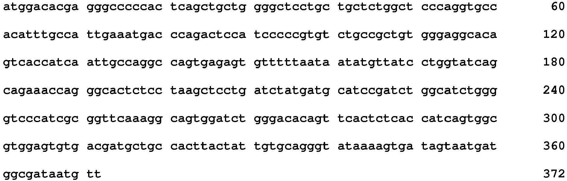
- <210> 515
<211> 369
<212> DNA
<213> Oryctolagus cuniculus - <400> 515

- <210> 516
<211> 36
<212> DNA
<213> Oryctolagus cuniculus - <400> 516
caggccagtg agagtgtttt taataatatg ttatcc 36 - <210> 517
<211> 21
<212> DNA
<213> Oryctolagus cuniculus - <400> 517
gatgcatccg atctggcatct 21 - <210> 518
<211> 39
<212> DNA
<213> Oryctolagus cuniculus - <400> 518
gcagggtata aaagtgatag taatgatggc gataatgtt 39 - <210> 519
<211> 15
<212> DNA
<213> Oryctolagus cuniculus - <400> 519
aggaattcaa taacc 15 - <210> 520
<211> 48
<212> DNA
<213> Oryctolagus cuniculus - <400> 520
atcattactg gtagtggtag aacgtactac gcgaactggg caaaaggc 48 - <210> 521
<211> 30
<212> DNA
<213> Oryctolagus cuniculus - <400> 521
ggccatcctg gtcttggtag tggtaacatc 30 - <210> 522
<211> 121
<212> PRT
<213> Oryctolagus cuniculus - <400> 522

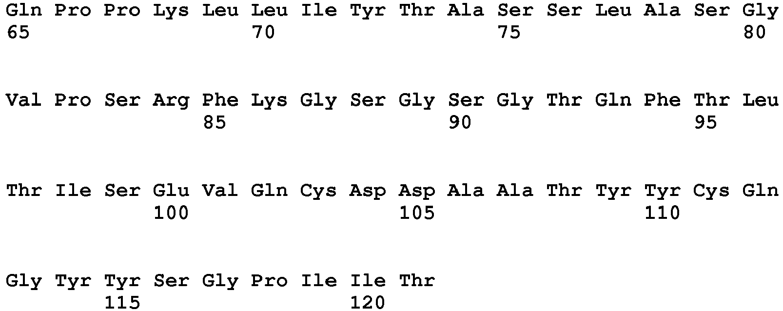
- <210> 523
<211> 122
<212> PRT
<213> Oryctolagus cuniculus - <400> 523

- <210> 524
<211> 12
<212> PRT
<213> Oryctolagus cuniculus - <400> 524

- <210> 525
<211> 7
<212> PRT
<213> Oryctolagus cuniculus - <400> 525

- <210> 526
<211> 10
<212> PRT
<213> Oryctolagus cuniculus - <400> 526

- <210> 527
<211> 5
<212> PRT
<213> Oryctolagus cuniculus - <400> 527

- <210> 528
<211> 16
<212> PRT
<213> Oryctolagus cuniculus - <400> 528

- <210> 529
<211> 8
<212> PRT
<213> Oryctolagus cuniculus - <400> 529

- <210> 530
<211> 363
<212> DNA
<213> Oryctolagus cuniculus - <400> 530

- <210> 531
<211> 366
<212> DNA
<213> Oryctolagus cuniculus - <400> 531

- <210> 532
<211> 36
<212> DNA
<213> Oryctolagus cuniculus - <400> 532
cagtccagtc agagtgttta taataactac ttatcc 36 - <210> 533
<211> 21
<212> DNA
<213> Oryctolagus cuniculus - <400> 533
actgcatcca gcctggcatct 21 - <210> 534
<211> 30
<212> DNA
<213> Oryctolagus cuniculus - <400> 534
caaggctatt atagtggtcc tataattact 30 - <210> 535
<211> 15
<212> DNA
<213> Oryctolagus cuniculus - <400> 535
aactactaca tacaa 15 - <210> 536
<211> 48
<212> DNA
<213> Oryctolagus cuniculus - <400> 536
atcatttatg ctggtggtag cgcatactac gcgacctggg caaacggc 48 - <210> 537
<211> 24
<212> DNA
<213> Oryctolagus cuniculus - <400> 537
gggacatttg atggttatga gttg 24 - <210> 538
<211> 122
<212> PRT
<213> Oryctolagus cuniculus - <400> 538


- <210> 539
<211> 124
<212> PRT
<213> Oryctolagus cuniculus - <400> 539


- <210> 541
<211> 7
<212> PRT
<213> Oryctolagus cuniculus - <400> 541

- <210> 542
<211> 10
<212> PRT
<213> Oryctolagus cuniculus - <400> 542

- <210> 543
<211> 5
<212> PRT
<213> Oryctolagus cuniculus - <400> 543

- <210> 544
<211> 16
<212> PRT
<213> Oryctolagus cuniculus - <400> 544

- <210> 545
<211> 11
<212> PRT
<213> Oryctolagus cuniculus - <400> 545

- <210> 546
<211> 366
<212> DNA
<213> Oryctolagus cuniculus - <400> 546


- <210> 547
<211> 372
<212> DNA
<213> Oryctolagus cuniculus - <400> 547

- <210> 548
<211> 39
<212> DNA
<213> Oryctolagus cuniculus - <400> 548
cagtccagtg agagcgttta tagtaataac ctcttatcc 39 - <210> 549
<211> 21
<212> DNA
<213> Oryctolagus cuniculus - <400> 549
agggcatcca atctggcatct 21 - <210> 550
<211> 30
<212> DNA
<213> Oryctolagus cuniculus - <400> 550
caaggctatt atagtggtgt cattaatagt 30 - <210> 551
<211> 15
<212> DNA
<213> Oryctolagus cuniculus - <400> 551
agctacttca tgagc 15 - <210> 552
<211> 48
<212> DNA
<213> Oryctolagus cuniculus - <400> 552
ttcattaatc ctggtggtag cgcatactac gcgagctggg cgagtggc 48 - <210> 553
<211> 33
<212> DNA
<213> Oryctolagus cuniculus - <400> 553
attcttattg tttcttatgg agcctttaccatc 33 - <210> 554
<211> 122
<212> PRT
<213> Oryctolagus cuniculus - <400> 554

- <210> 555
<211> 128
<212> PRT
<213> Oryctolagus cuniculus - <400> 555

- <210> 556
<211> 11
<212> PRT
<213> Oryctolagus cuniculus - <400> 556

- <210> 557
<211> 7
<212> PRT
<213> Oryctolagus cuniculus - <400> 557

- <210> 558
<211> 12
<212> PRT
<213> Oryctolagus cuniculus - <400> 558


- <210> 559
<211> 5
<212> PRT
<213> Oryctolagus cuniculus - <400> 559

- <210> 560
<211> 17
<212> PRT
<213> Oryctolagus cuniculus - <400> 560

- <210> 561
<211> 14
<212> PRT
<213> Oryctolagus cuniculus - <400> 561

- <210> 562
<211> 366
<212> DNA
<213> Oryctolagus cuniculus - <400> 562

- <210> 563
<211> 384
<212> DNA
<213> Oryctolagus cuniculus - <400> 563

- <210> 564
<211> 33
<212> DNA
<213> Oryctolagus cuniculus - <400> 564
caggccactg agagcattgg caatgagtta tcc 33 - <210> 565
<211> 21
<212> DNA
<213> Oryctolagus cuniculus - <400> 565
tctgcatcca ctctggcatct 21 - <210> 566
<211> 36
<212> DNA
<213> Oryctolagus cuniculus - <400> 566
caacagggtt atagtagtgc taatattgat aatgct 36 - <210> 567
<211> 15
<212> DNA
<213> Oryctolagus cuniculus - <400> 567
aagtactaca tgagc 15 - <210> 568
<211> 51
<212> DNA
<213> Oryctolagus cuniculus - <400> 568
tacattgata gtactactgt taatacatac tacgcgacct gggcgagaggc 51 - <210> 569
<211> 42
<212> DNA
<213> Oryctolagus cuniculus - <400> 569
ggaagtactt attttactga tggaggccat cggttggatctc 42 - <210> 570
<211> 122
<212> PRT
<213> Oryctolagus cuniculus - <400> 570

- <210> 571
<211> 124
<212> PRT
<213> Oryctolagus cuniculus - <400> 571


- <210> 572
<211> 11
<212> PRT
<213> Oryctolagus cuniculus - <400> 572

- <210> 573
<211> 7
<212> PRT
<213> Oryctolagus cuniculus - <400> 573

- <210> 574
<211> 12
<212> PRT
<213> Oryctolagus cuniculus - <400> 574

- <210> 575
<211> 5
<212> PRT
<213> Oryctolagus cuniculus - <400> 575

- <210> 576
<211> 16
<212> PRT
<213> Oryctolagus cuniculus - <400> 576

- <210> 577
<211> 11
<212> PRT
<213> Oryctolagus cuniculus - <400> 577

- <210> 578
<211> 366
<212> DNA
<213> Oryctolagus cuniculus - <400> 578

- <210> 579
<211> 372
<212> DNA
<213> Oryctolagus cuniculus - <400> 579


- <210> 580
<211> 33
<212> DNA
<213> Oryctolagus cuniculus - <400> 580
caggccactg agagcattgg caatgagtta tcc 33 - <210> 581
<211> 21
<212> DNA
<213> Oryctolagus cuniculus - <400> 581
tctgcatcca ctctggcatct 21 - <210> 582
<211> 36
<212> DNA
<213> Oryctolagus cuniculus - <400> 582
caacagggtt atagtagtgc taatattgat aatgct 36 - <210> 583
<211> 15
<212> DNA
<213> Oryctolagus cuniculus - <400> 583
acctacaaca tgggc 15 - <210> 584
<211> 48
<212> DNA
<213> Oryctolagus cuniculus - <400> 584
agtattacta ttgatggtcg cacatactac gcgagctggg cgaaaggc 48 - <210> 585
<211> 33
<212> DNA
<213> Oryctolagus cuniculus - <400> 585
attcttattg tttcttatgg ggcctttaccatc 33 - <210> 586
<211> 105
<212> PRT
<213> Artificial Sequence - <220>
<223> Kappa constant domain of Ab1 - <400> 586
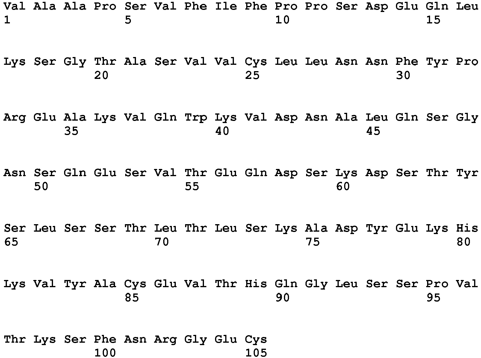
- <210> 587
<211> 315
<212> DNA
<213> Artificial Sequence - <220>
<223> Kappa constant domain of Ab1 - <400> 587
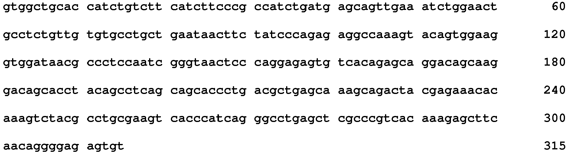
- <210> 588
<211> 330
<212> PRT
<213> Artificial Sequence - <220>
<223> Gamma-1 constant domain of Ab1 - <400> 588


- <210> 589
<211> 990
<212> DNA
<213> Artificial Sequence - <220>
<223> Gamma-1 constant domain of Ab1 - <400> 589


- <210> 590
<211> 15
<212> PRT
<213> Homo sapiens - <400> 590

- <210> 591
<211> 15
<212> PRT
<213> Homo sapiens - <400> 591

- <210> 592
<211> 15
<212> PRT
<213> Homo sapiens - <400> 592

- <210> 593
<211> 15
<212> PRT
<213> Homo sapiens - <400> 593

- <210> 594
<211> 15
<212> PRT
<213> Homo sapiens - <400> 594

- <210> 595
<211> 15
<212> PRT
<213> Homo sapiens - <400> 595

- <210> 596
<211> 15
<212> PRT
<213> Homo sapiens - <400> 596

- <210> 597
<211> 15
<212> PRT
<213> Homo sapiens - <400> 597

- <210> 598
<211> 15
<212> PRT
<213> Homo sapiens - <400> 598

- <210> 599
<211> 15
<212> PRT
<213> Homo sapiens - <400> 599

- <210> 600
<211> 15
<212> PRT
<213> Homo sapiens - <400> 600

- <210> 601
<211> 15
<212> PRT
<213> Homo sapiens - <400> 601

- <210> 602
<211> 15
<212> PRT
<213> Homo sapiens - <400> 602

- <210> 603
<211> 15
<212> PRT
<213> Homo sapiens - <400> 603

- <210> 604
<211> 15
<212> PRT
<213> Homo sapiens - <400> 604

- <210> 605
<211> 15
<212> PRT
<213> Homo sapiens - <400> 605

- <210> 606
<211> 15
<212> PRT
<213> Homo sapiens - <400> 606

- <210> 607
<211> 15
<212> PRT
<213> Homo sapiens - <400> 607

- <210> 608
<211> 15
<212> PRT
<213> Homo sapiens - <400> 608

- <210> 609
<211> 15
<212> PRT
<213> Homo sapiens - <400> 609

- <210> 610
<211> 15
<212> PRT
<213> Homo sapiens - <400> 610

- <210> 611
<211> 15
<212> PRT
<213> Homo sapiens - <400> 611

- <210> 612
<211> 15
<212> PRT
<213> Homo sapiens - <400> 612

- <210> 613
<211> 15
<212> PRT
<213> Homo sapiens - <400> 613

- <210> 614
<211> 15
<212> PRT
<213> Homo sapiens - <400> 614

- <210> 615
<211> 15
<212> PRT
<213> Homo sapiens - <400> 615

- <210> 616
<211> 15
<212> PRT
<213> Homo sapiens - <400> 616

- <210> 617
<211> 15
<212> PRT
<213> Homo sapiens - <400> 617

- <210> 618
<211> 15
<212> PRT
<213> Homo sapiens - <400> 618

- <210> 619
<211> 15
<212> PRT
<213> Homo sapiens - <400> 619

- <210> 620
<211> 15
<212> PRT
<213> Homo sapiens - <400> 620

- <210> 621
<211> 15
<212> PRT
<213> Homo sapiens - <400> 621

- <210> 622
<211> 15
<212> PRT
<213> Homo sapiens - <400> 622

- <210> 623
<211> 15
<212> PRT
<213> Homo sapiens - <400> 623


- <210> 624
<211> 15
<212> PRT
<213> Homo sapiens - <400> 624

- <210> 625
<211> 15
<212> PRT
<213> Homo sapiens - <400> 625

- <210> 626
<211> 15
<212> PRT
<213> Homo sapiens - <400> 626

- <210> 627
<211> 15
<212> PRT
<213> Homo sapiens - <400> 627

- <210> 628
<211> 15
<212> PRT
<213> Homo sapiens - <400> 628

- <210> 629
<211> 15
<212> PRT
<213> Homo sapiens - <400> 629

- <210> 630
<211> 15
<212> PRT
<213> Homo sapiens - <400> 630

- <210> 631
<211> 15
<212> PRT
<213> Homo sapiens - <400> 631

- <210> 632
<211> 15
<212> PRT
<213> Homo sapiens - <400> 632

- <210> 633
<211> 15
<212> PRT
<213> Homo sapiens - <400> 633

- <210> 634
<211> 15
<212> PRT
<213> Homo sapiens - <400> 634

- <210> 635
<211> 15
<212> PRT
<213> Homo sapiens - <400> 635

- <210> 636
<211> 15
<212> PRT
<213> Homo sapiens - <400> 636

- <210> 637
<211> 15
<212> PRT
<213> Homo sapiens - <400> 637

- <210> 638
<211> 15
<212> PRT
<213> Homo sapiens - <400> 638

- <210> 639
<211> 15
<212> PRT
<213> Homo sapiens - <400> 639

- <210> 640
<211> 15
<212> PRT
<213> Homo sapiens - <400> 640

- <210> 641
<211> 15
<212> PRT
<213> Homo sapiens - <400> 641

- <210> 642
<211> 15
<212> PRT
<213> Homo sapiens - <400> 642

- <210> 643
<211> 15
<212> PRT
<213> Homo sapiens - <400> 643

- <210> 644
<211> 15
<212> PRT
<213> Homo sapiens - <400> 644

- <210> 645
<211> 15
<212> PRT
<213> Homo sapiens - <400> 645

- <210> 646
<211> 15
<212> PRT
<213> Homo sapiens - <400> 646

Claims (13)
- An anti-IL6 antibody or antibody fragment thereof comprising (i) a variable light (VL) chain comprising a CDR1 polypeptide consisting of the amino acid sequence of SEQ ID NO:4, a CDR2 polypeptide consisting of the amino acid sequence of SEQ ID NO:5, and a CDR3 polypeptide consisting of the amino acid sequence of SEQ ID NO:6, and (ii) a variable heavy (VH) chain comprising a CDR1 polypeptide consisting of the amino acid sequence of SEQ ID NO:7, a CDR2 polypeptide consisting of the amino acid sequence of SEQ ID NO:8 or SEQ ID NO:120, and a CDR3 polypeptide consisting of the amino acid sequence of SEQ ID NO:9, wherein the antibody or fragment thereof has a dissociation constant (KD) of less than 50 picomolar as assessed by Biacore.
- The antibody or antibody fragment of claim 1,a) which is aglycosylated,b) which contains an Fc region that has been modified to alter effector function, half-life, proteolysis, and/or glycosylation,c) which is a humanized antibody, a single chain antibody or a chimeric antibody,d) which is a humanized antibody derived from a rabbit antihuman IL-6 antibody,e) which specifically binds to IL-6 expressed on human cells and/or to circulating soluble IL-6 molecules in vivo,f) which specifically binds to IL-6 expressed on or by human cells in a patient with a disease wherein IL-6 levels are elevated,
wherein the disease is selected from general fatigue, exercise induced fatigue, cancer-related fatigue, inflammatory disease-related fatigue, chronic fatigue syndrome, cancer-related cachexia, cardiac-related cachexia, respiratory-related cachexia, renal-related cachexia, age-related cachexia, rheumatoid arthritis, systemic lupus erythematosis (SLE), systemic juvenile idiopathic arthritis, psoriasis, psoriatic arthropathy, ankylosing spondylitis, inflammatory bowel disease (IBD), polymyalgia rheumatica, giant cell arteritis, autoimmune vasculitis, graft versus host disease (GVHD), Sjogren's syndrome, adult onset Still's disease, osteoarthritis, osteoporosis, Paget's disease of bone, multiple myeloma, Hodgkin's lymphoma, non-Hodgkin's lymphoma, prostate cancer, leukemia, renal cell cancer, multicentric Castleman's disease, ovarian cancer, drug resistance in cancer chemotherapy, cancer chemotherapy toxicity, ischemic heart disease, atherosclerosis, obesity, diabetes, asthma, multiple sclerosis, Alzheimer's disease, cerebrovascular disease, fever, acute phase response, allergies, anemia, anemia of inflammation (anemia of chronic disease), hypertension, depression, depression associated with a chronic illness, thrombosis, thrombocytosis, acute heart failure, metabolic syndrome, miscarriage, chronic prostatitis, glomerulonephritis, pelvic inflammatory disease, reperfusion injury, transplant rejection, avian influenza, smallpox, pandemic influenza, adult respiratory distress syndrome (ARDS), severe acute respiratory syndrome (SARS), sepsis, systemic inflammatory response syndrome (SIRS), a cancer, an_inflammatory disorder, a_viral disorder, an_autoimmune disorder, arthritis, cachexia, and wasting syndrome,g) which is directly or indirectly attached to a detectable label or a_therapeutic agent,h) wherein said chimeric antibody of claim 2(c) comnrises a human Fc selected from IgG1, IgG2, IgG3, IgG4, IgG5, IgG6, IgG7, IgG8, IgG9, IgG10, IgG11, IgG12, IgG13, IgG14, IgG15, IgG16, IgG17, IgG18, IgG19.i) wherein said antibody binds at least one of soluble IL-6, cell-surface expressed IL-6, IL-6/IL-6R, IL-6/IL-6R/gp130 complexes and or multimers thereof,j) wherein the VH or VL chains contained in said antibody originated from one or more rabbit B cell populations,k) wherein said antibody does not have binding specificity for soluble IL-6R (slL-6R) or gp130,l) wherein said antibody inhibits the association of IL-6 with IL-6R, and/or the production of IL-6/IL-6R/gp130 complexes and/or the production of IL-6/IL-6R/gp130 multimers,m) wherein said fragment is selected from a Fab fragment, a Fab' fragment, or a F(ab')2 fragment,n) wherein said antibody further comprises an effector moiety selected from(i) a detectable moiety selected from a fluorescent dye, an enzyme, a substrate, a bioluminescent material, a radioactive material, or a chemiluminescent material, or(ii) a functional moiety selected from streptavidin, avidin, biotin, a cytotoxin, a cytotoxic agent, or a radioactive material,o) wherein said antibody antagonizes the biological effects of one or more of soluble IL-6, cell surface expressed IL-6, IL-6/IL-6R, IL-6/IL-6R/gp130 complexes and/or IL-6/IL-6R/gp130 complex multimers,p) wherein said antibody binds to a linear or conformational IL-6 epitope,q which is a single chain antibody selected from scFv, camelbody, nanobody, IgNAR SMIP, and combinations thereof. - An anti-IL6 antibody or antibody fragment thereof according to claim 1, comprising a VL chain and a VH chain, respectively, comprising the amino acid sequences of SEQ ID NO:2 and SEQ ID NO:3; SEQ ID NO:2 and SEQ ID NO:18; SEQ ID NO:2 and SEQ ID NO:19; SEQ ID NO:20 and SEQ ID NO:3; SEQ ID NO:20 and SEQ ID NO:18; or SEQ ID NO:20 and SEQ ID NO:19, or an antibody or antibody fragment thereof comprising a VL chain and a VH chain, respectively, comprising amino acid sequences having at least 90% or greater homology thereto.
- A nucleic acid sequence which encodes an anti-human IL-6 antibody or antibody fragment according to any one of claims 1 - 3.
- A vector comprising a nucleic acid sequence according to claim 4.
- A recombinant cell which expresses an antibody or antibody fragment according to any one of claims 1 - 3.
- A pharmaceutical or diagnostic composition containing at least one IL-6 antibody or fragment according to any one of claims 1 - 3 and a pharmaceutically acceptable carrier.
- The pharmaceutical or diagnostic composition of claim 7,a) which further comprises at least one stabilizer,b) which is lyophilized, orc) which comprises one or more anti-IL-6 antibodies comprising (a) a VH chain having the amino acid sequence of SEQ ID NO: 3, 18, or 19; and (b) a VL chain having the amino acid sequence of SEQ ID NO: 2 or 20, or a chimeric antibody, a humanized antibody or a fragment derived therefrom.
- An antibody or antibody fragment according to any one of claims 1 - 3 for use as a therapeutic or diagnostic agent.
- Use of an antibody or antibody fragment according to any one of claims 1 - 3 in the manufacture of a medicament or in the manufacture of an agent for use in a method of treatment.
- An antibody or fragment for use as a therapeutic agent according to claim 9 or use according to claim 10:a) wherein the disease or condition to be treated is selected from general fatigue, exercise-induced fatigue, cancer-related fatigue, inflammatory disease-related fatigue, chronic fatigue syndrome, cancer-related cachexia, cardiac-related cachexia, respiratory related cachexia, renal-related cachexia, age-related cachexia, rheumatoid arthritis, systemic lupus erythematosis (SLE), systemic juvenile idiopathic arthritis, psoriasis, psoriatic arthropathy, ankylosing spondylitis, inflammatory bowel disease (IBD), polymyalgia rheumatica, giant cell arteritis, autoimmune vasculitis, graft versus host disease (GVHD), Sjogren's syndrome, adult onset Still's disease, osteoarthritis, osteoporosis, Paget's disease of bone, multiple myeloma, Hodgkin's lymphoma, non-Hodgkin's lymphoma, prostate cancer, leukemia, renal cell cancer, multicentric Castleman's disease, ovarian cancer, drug resistance in cancer chemotherapy, cancer chemotherapy toxicity, ischemic heart disease, atherosclerosis, obesity, diabetes, asthma, multiple sclerosis, Alzheimer's disease, cerebrovascular disease, fever, acute phase response, allergies, anemia, anemia of inflammation (anemia of chronic disease), hypertension, depression, depression associated with a chronic illness, thrombosis, thrombocytosis, acute heart failure, metabolic syndrome, miscarriage, chronic prostatitis, glomerulonephritis, pelvic inflammatory disease, reperfusion injury, transplant rejection, avian influenza, smallpox, pandemic influenza, adult respiratory distress syndrome (ARDS), severe acute respiratory syndrome (SARS), sepsis, systemic inflammatory response syndrome (SIRS), arthritis, a cancer, an autoimmune disease, or an inflammatory condition,b) wherein the treatment further includes the administration of another therapeutic agent or regimen selected from chemotherapy, radiotherapy, cytokine administration, or gene therapy, orc) which is used to treat a side effect of cancer or viral infection.
- An antibody or antibody fragment for use as a diagnostic agent according to claim 9, wherein the use is for diagnostic in vivo imaginga) wherein said use detects the presence of cells which express IL-6 comprising administering a diagnostically effective amount of at least one anti-human IL-6 antibody according to claim 1,b) wherein said administration further includes the administration of a radionuclide or fluorophore that facilitates detection of the antibody at IL-6 expressing disease sites,c) which is used to detect IL-6 expressing tumors or metastases,d) which is used to detect the presence of sites of inflammation associated with IL-6 expressing cells, ore) wherein the results are used to facilitate design of an appropriate therapeutic regimen.
- An antibody or antibody fragment for use as a diagnostic agent according to claim 9, wherein the use is for diagnostic in vivo imaging wherein the usea) is to detect or image IL-6 expressing tumors or metastases, orb) is to detect or image IL-6 expressing inflammatory sites.
Priority Applications (6)
| Application Number | Priority Date | Filing Date | Title |
|---|---|---|---|
| PL16195797T PL3187506T3 (en) | 2007-05-21 | 2008-05-21 | Antibodies to il-6 and use thereof |
| HRP20170038TT HRP20170038T1 (en) | 2007-05-21 | 2008-05-21 | IL-6 ANTIBODIES AND THEIR USE |
| SI200831772A SI2164514T1 (en) | 2007-05-21 | 2008-05-21 | Antibodies to il-6 and use thereof |
| EP16195797.2A EP3187506B1 (en) | 2007-05-21 | 2008-05-21 | Antibodies to il-6 and use thereof |
| DK16195797.2T DK3187506T3 (en) | 2007-05-21 | 2008-05-21 | Antibodies to IL-6 and their use |
| CY20191100624T CY1121779T1 (en) | 2007-05-21 | 2019-06-18 | ANTIBODIES TO IL-6 AND THEIR USE |
Applications Claiming Priority (2)
| Application Number | Priority Date | Filing Date | Title |
|---|---|---|---|
| US92455007P | 2007-05-21 | 2007-05-21 | |
| PCT/US2008/064432 WO2008144763A2 (en) | 2007-05-21 | 2008-05-21 | Antibodies to il-6 and use thereof |
Related Child Applications (2)
| Application Number | Title | Priority Date | Filing Date |
|---|---|---|---|
| EP16195797.2A Division EP3187506B1 (en) | 2007-05-21 | 2008-05-21 | Antibodies to il-6 and use thereof |
| EP16195797.2A Division-Into EP3187506B1 (en) | 2007-05-21 | 2008-05-21 | Antibodies to il-6 and use thereof |
Publications (3)
| Publication Number | Publication Date |
|---|---|
| EP2164514A2 EP2164514A2 (en) | 2010-03-24 |
| EP2164514A4 EP2164514A4 (en) | 2012-08-29 |
| EP2164514B1 true EP2164514B1 (en) | 2016-12-14 |
Family
ID=40122295
Family Applications (2)
| Application Number | Title | Priority Date | Filing Date |
|---|---|---|---|
| EP08756095.9A Active EP2164514B1 (en) | 2007-05-21 | 2008-05-21 | Antibodies to il-6 and use thereof |
| EP16195797.2A Active EP3187506B1 (en) | 2007-05-21 | 2008-05-21 | Antibodies to il-6 and use thereof |
Family Applications After (1)
| Application Number | Title | Priority Date | Filing Date |
|---|---|---|---|
| EP16195797.2A Active EP3187506B1 (en) | 2007-05-21 | 2008-05-21 | Antibodies to il-6 and use thereof |
Country Status (28)
| Country | Link |
|---|---|
| US (4) | US20100290993A1 (en) |
| EP (2) | EP2164514B1 (en) |
| JP (4) | JP5833823B2 (en) |
| KR (3) | KR101615715B1 (en) |
| CN (2) | CN101754772B (en) |
| AU (1) | AU2008254584B2 (en) |
| BR (1) | BRPI0811782A2 (en) |
| CA (2) | CA2688146C (en) |
| CO (1) | CO6241159A2 (en) |
| CY (2) | CY1119068T1 (en) |
| DK (2) | DK3187506T3 (en) |
| EA (1) | EA200901533A1 (en) |
| ES (2) | ES2721753T3 (en) |
| HR (2) | HRP20170038T1 (en) |
| HU (2) | HUE043782T2 (en) |
| IL (4) | IL202238A (en) |
| LT (2) | LT2164514T (en) |
| MX (3) | MX374978B (en) |
| NO (2) | NO344119B1 (en) |
| NZ (2) | NZ597767A (en) |
| PL (2) | PL2164514T3 (en) |
| PT (2) | PT2164514T (en) |
| SG (1) | SG183742A1 (en) |
| SI (2) | SI2164514T1 (en) |
| TR (1) | TR201905366T4 (en) |
| TW (2) | TWI617576B (en) |
| WO (1) | WO2008144763A2 (en) |
| ZA (1) | ZA200908770B (en) |
Families Citing this family (103)
| Publication number | Priority date | Publication date | Assignee | Title |
|---|---|---|---|---|
| US8945558B2 (en) | 2005-10-21 | 2015-02-03 | Chugai Seiyaku Kabushiki Kaisha | Methods for treating myocardial infarction comprising administering an IL-6 inhibitor |
| EP2025346B1 (en) | 2006-04-07 | 2016-08-10 | Osaka University | Muscle regeneration promoter |
| KR101508019B1 (en) | 2007-01-23 | 2015-04-06 | 고쿠리츠 다이가쿠 호우징 신슈 다이가쿠 | Chronic rejection inhibitor |
| US8404235B2 (en) | 2007-05-21 | 2013-03-26 | Alderbio Holdings Llc | Antagonists of IL-6 to raise albumin and/or lower CRP |
| US8062864B2 (en) | 2007-05-21 | 2011-11-22 | Alderbio Holdings Llc | Nucleic acids encoding antibodies to IL-6, and recombinant production of anti-IL-6 antibodies |
| US8178101B2 (en) | 2007-05-21 | 2012-05-15 | Alderbio Holdings Inc. | Use of anti-IL-6 antibodies having specific binding properties to treat cachexia |
| US9701747B2 (en) | 2007-05-21 | 2017-07-11 | Alderbio Holdings Llc | Method of improving patient survivability and quality of life by anti-IL-6 antibody administration |
| US7906117B2 (en) | 2007-05-21 | 2011-03-15 | Alderbio Holdings Llc | Antagonists of IL-6 to prevent or treat cachexia, weakness, fatigue, and/or fever |
| US8252286B2 (en) | 2007-05-21 | 2012-08-28 | Alderbio Holdings Llc | Antagonists of IL-6 to prevent or treat thrombosis |
| CN101754772B (en) * | 2007-05-21 | 2015-08-19 | 奥尔德生物控股有限责任公司 | Antibodies against IL-6 and uses thereof |
| US20150050288A1 (en) * | 2007-09-06 | 2015-02-19 | The Johns Hopkins University | Treatment of TH17-Mediated Autoimmune Disease Via Inhibition of Stat3 |
| US9550827B2 (en) * | 2007-10-19 | 2017-01-24 | The Regents Of The University Of California | Methods for ameliorating and preventing central nervous system inflammation |
| US8277804B2 (en) | 2008-05-21 | 2012-10-02 | Alderbio Holdings Llc | Antagonists of IL-6 to prevent or treat thrombosis |
| TWI528973B (en) | 2008-06-05 | 2016-04-11 | Chugai Pharmaceutical Co Ltd | Nerve infiltration inhibitor |
| US8188235B2 (en) | 2008-06-18 | 2012-05-29 | Pfizer Inc. | Antibodies to IL-6 and their uses |
| US9452227B2 (en) | 2008-11-25 | 2016-09-27 | Alderbio Holdings Llc | Methods of treating or diagnosing conditions associated with elevated IL-6 using anti-IL-6 antibodies or fragments |
| US8992920B2 (en) | 2008-11-25 | 2015-03-31 | Alderbio Holdings Llc | Anti-IL-6 antibodies for the treatment of arthritis |
| AU2013203804B2 (en) * | 2008-11-25 | 2016-10-13 | Vitaeris Inc. | Antibodies to il-6 and use thereof |
| US8420089B2 (en) | 2008-11-25 | 2013-04-16 | Alderbio Holdings Llc | Antagonists of IL-6 to raise albumin and/or lower CRP |
| AU2016250477B2 (en) * | 2008-11-25 | 2018-07-05 | Vitaeris Inc. | Antagonists of IL-6 to raise albumin and/or lower CRP |
| US8323649B2 (en) | 2008-11-25 | 2012-12-04 | Alderbio Holdings Llc | Antibodies to IL-6 and use thereof |
| US9212223B2 (en) | 2008-11-25 | 2015-12-15 | Alderbio Holdings Llc | Antagonists of IL-6 to prevent or treat thrombosis |
| MX2011005411A (en) * | 2008-11-25 | 2011-06-16 | Alder Biopharmaceuticals Inc | Antibodies to il-6 and use thereof. |
| US8337847B2 (en) * | 2008-11-25 | 2012-12-25 | Alderbio Holdings Llc | Methods of treating anemia using anti-IL-6 antibodies |
| WO2010071610A1 (en) * | 2008-12-19 | 2010-06-24 | Agency For Science, Technology And Research (A*Star) | Severe chikungunya biomarkers |
| CA2749200A1 (en) * | 2009-01-29 | 2010-08-05 | Michael Bowen | Human anti-il-6 antibodies with extended in vivo half-life and their use in treatment of oncology, autoimmune diseases and inflammatory diseases |
| CN104928336B (en) * | 2009-10-26 | 2020-05-08 | 弗·哈夫曼-拉罗切有限公司 | Methods for the production of glycosylated immunoglobulins |
| AU2016250478A1 (en) * | 2009-11-24 | 2016-11-17 | Alder Biopharmaceuticals, Inc. | Antibodies to IL-6 and use thereof |
| US9775921B2 (en) | 2009-11-24 | 2017-10-03 | Alderbio Holdings Llc | Subcutaneously administrable composition containing anti-IL-6 antibody |
| EP2504032A4 (en) | 2009-11-24 | 2013-06-19 | Alderbio Holdings Llc | Antagonists of il-6 to prevent or treat thrombosis |
| ES2434256T3 (en) | 2010-05-28 | 2013-12-16 | F. Hoffmann-La Roche Ag | Method of culturing individual B cells and production of specific antibodies |
| SI2578231T1 (en) | 2010-05-28 | 2023-01-31 | Chugai Seiyaku Kabushiki Kaisha | Antitumor t cell response enhancer |
| CA2818814A1 (en) | 2010-11-23 | 2012-05-31 | Alder Biopharmaceuticals, Inc. | Anti-il-6 antibodies for the treatment of anemia |
| NZ611076A (en) | 2010-12-01 | 2015-09-25 | Alderbio Holdings Llc | Anti-ngf compositions and use thereof |
| CA2818992C (en) * | 2010-12-01 | 2021-05-04 | The United States Of America, As Represented By The Secretary, Department Of Health And Human Services | Chimeric rabbit/human ror1 antibodies |
| US8597883B2 (en) | 2011-02-14 | 2013-12-03 | The United States Of America, As Represented By The Secretary, Department Of Health And Human Services | Biomarkers for cancer-related fatigue and use thereof |
| SG10201604040PA (en) | 2011-05-20 | 2016-07-28 | Alderbio Holdings Llc | Anti-cgrp compositions and use thereof |
| RS66303B1 (en) | 2011-05-20 | 2025-01-31 | H Lundbeck As | Use of anti-cgrp antibodies and antibody fragments to prevent or inhibit photophobia or light aversion in subjects in need thereof, especially migraine sufferers |
| NZ732875A (en) | 2011-05-20 | 2022-08-26 | H Lundbeck As | Use of anti-cgrp or anti-cgrp-r antibodies or antibody fragments to treat or prevent chronic and acute forms of diarrhea |
| EA028913B9 (en) | 2011-05-20 | 2018-03-30 | Олдербайо Холдингз Ллк | Production of high-purity antibodies in pichia pastoris |
| SG10201609752YA (en) | 2011-08-19 | 2017-01-27 | Alderbio Holdings Llc | Multi-copy strategy for high-titer and high-purity production of multi-subunit proteins such as antibodies in transformed microbes such as pichia pastoris |
| US10150968B2 (en) | 2011-08-19 | 2018-12-11 | Alderbio Holdings Llc | Multi-copy strategy for high-titer and high-purity production of multi-subunit proteins such as antibodies in transformed microbes such as Pichia pastoris |
| EP2583980A1 (en) * | 2011-10-19 | 2013-04-24 | Effimune | Antibodies directed against the alpha chain of IL7 receptor - their use for the preparation of drug candidates |
| KR102069394B1 (en) | 2011-11-23 | 2020-01-22 | 에프. 호프만-라 로슈 아게 | Cd40l expressing mammalian cells and their use |
| JP2015500655A (en) | 2011-12-21 | 2015-01-08 | エフ.ホフマン−ラ ロシュ アーゲーF. Hoffmann−La Roche Aktiengesellschaft | A rapid method for cloning and expressing cognate antibody variable region gene segments |
| WO2013175276A1 (en) | 2012-05-23 | 2013-11-28 | Argen-X B.V | Il-6 binding molecules |
| LT3338794T (en) | 2012-07-13 | 2020-05-25 | The Trustees Of The University Of Pennsylvania | ADJUSTMENT OF CAR ANTICODIC TOXICITY |
| EP2917238A1 (en) | 2012-11-08 | 2015-09-16 | Eleven Biotherapeutics, Inc. | Il-6 antagonists and uses thereof |
| US9062104B2 (en) | 2013-03-14 | 2015-06-23 | Alderbio Holdings Llc | Therapeutic use of antibodies to HGF |
| ES2834113T3 (en) | 2013-03-15 | 2021-06-16 | H Lundbeck As | Antibody purification and purity monitoring |
| US10138294B2 (en) | 2013-03-15 | 2018-11-27 | Alderbio Holdings Llc | Temperature shift for high yield expression of polypeptides in yeast and other transformed cells |
| US20170114122A1 (en) | 2015-10-23 | 2017-04-27 | Alderbio Holdings Llc | Regulation of glucose metabolism using anti-cgrp antibodies |
| WO2015003122A2 (en) | 2013-07-03 | 2015-01-08 | Alder Biopharmaceuticals, Inc. | Regulation of glucose metabolism using anti-cgrp antibodies |
| CN105198982B (en) * | 2014-06-18 | 2019-07-26 | 上海交通大学 | IL-6-based epitopes and their applications |
| US11142571B2 (en) * | 2014-11-07 | 2021-10-12 | Sesen Bio, Inc. | IL-6 antibodies |
| US10047157B2 (en) | 2014-12-19 | 2018-08-14 | Alder Biopharmaceuticals, Inc. | Humanized anti-ACTH antibodies and use thereof |
| DE102016001333A1 (en) | 2015-03-06 | 2016-09-08 | Shimano Inc. | ELECTRIC BICYCLE COMPONENT ARRANGEMENT |
| US10029754B2 (en) | 2015-03-06 | 2018-07-24 | Shimano Inc. | Bicycle electrical system |
| TWI869018B (en) | 2015-03-06 | 2025-01-01 | 日商島野股份有限公司 | Bicycle electrical component |
| DE102016016024B4 (en) | 2015-03-06 | 2025-03-20 | Shimano Inc. | Electric bicycle rear derailleur |
| WO2016160923A1 (en) * | 2015-03-31 | 2016-10-06 | Janssen Biotech, Inc | Antigen binding proteins |
| EP3305325B1 (en) | 2015-06-04 | 2025-07-30 | National Center of Neurology and Psychiatry | Mental illness therapeutic agent having il-6 inhibitor as active ingredient |
| EP3419599A4 (en) | 2016-02-23 | 2019-09-11 | Sesen Bio, Inc. | FORMULATIONS OF IL-6 ANTAGONISTS AND USES THEREOF |
| JP7026634B2 (en) | 2016-03-30 | 2022-02-28 | エフ.ホフマン-ラ ロシュ アーゲー | B cell culture method |
| CA3020839A1 (en) | 2016-04-15 | 2017-10-19 | Alder Biopharmaceuticals, Inc. | Anti-pacap antibodies and uses thereof |
| WO2018013563A1 (en) * | 2016-07-12 | 2018-01-18 | Kite Pharma, Inc. | Antigen binding molecules and methods of use thereof |
| JP6916884B2 (en) | 2017-01-02 | 2021-08-11 | エフ.ホフマン−ラ ロシュ アーゲーF. Hoffmann−La Roche Aktiengesellschaft | B cell culture method |
| TWI716668B (en) | 2017-02-01 | 2021-01-21 | 耶魯大學 | Precision treatment of heart failure and cardiorenal syndrome |
| BR112019017853A2 (en) | 2017-02-28 | 2021-04-27 | Vib Vzw | MEANS AND METHODS FOR ORAL RELEASE OF PROTEIN |
| US11851486B2 (en) | 2017-05-02 | 2023-12-26 | National Center Of Neurology And Psychiatry | Method for predicting and evaluating therapeutic effect in diseases related to IL-6 and neutrophils |
| EP3625332B1 (en) | 2017-05-19 | 2022-02-02 | F. Hoffmann-La Roche AG | Method for the production of thymocyte supernatant |
| US11255865B2 (en) | 2017-06-06 | 2022-02-22 | The Regents Of The University Of California | Immunoassay for human erythroferrone |
| MX2020002750A (en) * | 2017-09-13 | 2020-07-20 | Jiangsu Hengrui Medicine Co | Il-6r antibody and antigen binding fragment thereof and medical use. |
| US11692037B2 (en) | 2017-10-20 | 2023-07-04 | Hyogo College Of Medicine | Anti-IL-6 receptor antibody-containing medicinal composition for preventing post-surgical adhesion |
| CN111417719B (en) | 2017-11-30 | 2023-08-29 | 豪夫迈·罗氏有限公司 | B cell culture method |
| AU2019205488A1 (en) * | 2018-01-04 | 2020-07-23 | Cedars-Sinai Medical Center | Use of anti-iL-6 antibody, e.g., Clazakizumab for desensitization of solid organ transplant recipients and/or for preventing, stabilizing or reducing antibody mediated rejection (ABMR) |
| MX2020006882A (en) | 2018-01-05 | 2020-09-07 | Novo Nordisk As | METHODS TO TREAT INFLAMMATION MEDIATED BY INTERLEUKIN-6 WITHOUT IMMUNOSUPPRESSION. |
| WO2019151418A1 (en) | 2018-01-31 | 2019-08-08 | 元一 加藤 | Therapeutic agent for asthma containing il-6 inhibitor |
| WO2020006469A1 (en) * | 2018-06-29 | 2020-01-02 | University Of Pittsburgh - Of The Commonwealth System Of Higher Education | Methods for treating and reducing traumatic brain injury-associated impairments using sgp130 |
| US12331110B2 (en) * | 2018-09-05 | 2025-06-17 | The Board Of Regents Of The University Of Texas System | Monoclonal antibodies against endotrophin and the use thereof |
| CN109517064B (en) * | 2018-10-10 | 2020-05-08 | 北京汇智和源生物技术有限公司 | Humanized monoclonal antibody of interleukin-6, coding gene and application thereof |
| BR112021007633A2 (en) | 2018-11-08 | 2021-10-13 | Cedars-Sinai Medical Center | Use of CLAZAKIZUMAB to desensitize and improve kidney transplantation in patients sensitized by HLA |
| BR112021010615A2 (en) * | 2018-12-20 | 2021-11-03 | Cedars Sinai Medical Center | Clazakizumab in Treating Chronic Antibody-Mediated Organ Transplant Rejection |
| PE20211707A1 (en) | 2019-01-08 | 2021-09-01 | H Lundbeck As | ACUTE TREATMENT AND RAPID TREATMENT OF HEADACHE USING ANTICGRP ANTIBODIES |
| WO2020213665A1 (en) | 2019-04-17 | 2020-10-22 | 国立大学法人広島大学 | Therapeutic agent for urological cancer which is characterized by being administered with il-6 inhibitor and ccr2 inhibitor in combination |
| EP4004044A1 (en) | 2019-07-24 | 2022-06-01 | H. Lundbeck A/S | Anti-mglur5 antibodies and uses thereof |
| EP4070336A4 (en) * | 2019-12-06 | 2024-03-06 | The Henry M. Jackson Foundation for the Advancement of Military Medicine, Inc. | PREDICTION OF VENOUS THROMBOEMBOLIA USING MACHINE LEARNING MODELS |
| WO2021154530A1 (en) * | 2020-01-27 | 2021-08-05 | Vanderbilt University | Human anti-dengue antibodies and methods of use therefor |
| CN111228480A (en) * | 2020-02-24 | 2020-06-05 | 浙江正熙生物医药有限公司 | Use of anti-human TNF- α antibody in preparation of medicine for treating virus infection |
| KR20210124867A (en) | 2020-04-06 | 2021-10-15 | 하. 룬드벡 아크티에셀스카브 | Treatment of most bothersome symptom (mbs) associated with migraine using anti-cgrp antibodies |
| WO2022009211A1 (en) * | 2020-07-09 | 2022-01-13 | Remedy Cell Ltd. | Methods for treating acute respiratory inflammatory conditions and cytokine storm syndrome |
| EP4212163A4 (en) | 2020-09-08 | 2024-09-18 | Dexon Pharmaceuticals Inc. | CYTOKINE STORM INHIBITOR, METHOD OF USING THE CYTOKINE STORM INHIBITOR, AND METHOD OF SCREENING FOR A CYTOKINE INHIBITOR |
| CN116806156A (en) * | 2021-01-29 | 2023-09-26 | 安进公司 | Materials and methods for monitoring cancer by administration of anti-MCL 1 antibodies |
| JP6974892B1 (en) | 2021-05-27 | 2021-12-01 | デクソンファーマシューティカルズ株式会社 | Cancer cachexia improving agent and cancer cachexia improving method |
| US20250215069A1 (en) | 2021-08-27 | 2025-07-03 | H. Lundbeck A/S | Treatment of cluster headache using anti-cgrp antibodies |
| CN115819578B (en) * | 2022-10-29 | 2023-08-18 | 武汉爱博泰克生物科技有限公司 | High affinity Human IL-5 rabbit monoclonal antibody and application thereof |
| CN116715763B (en) * | 2022-11-22 | 2023-11-24 | 武汉爱博泰克生物科技有限公司 | Mouse IL-6 rabbit monoclonal antibody pair and preparation method and application thereof |
| WO2024235871A1 (en) * | 2023-05-12 | 2024-11-21 | Seerave Ventures Limited | Anti-interleukin-6 single-domain antibodies |
| EP4731664A1 (en) | 2023-06-23 | 2026-04-29 | H. Lundbeck A/S | Combinational treatment |
| CN119285767B (en) * | 2023-07-10 | 2025-11-18 | 东莞市朋志生物科技有限公司 | Anti-interleukin-6 antibodies, reagents and kits for detecting interleukin-6 |
| CN117797317A (en) * | 2024-02-29 | 2024-04-02 | 四川大学 | Preparation method of peptide microsphere-chitosan composite material and its products and applications |
| US20260001942A1 (en) | 2024-06-21 | 2026-01-01 | H. Lundbeck A/S | Treatment of headache disorders and/or psychiatric symptoms using anti-cgrp antibodies, and compositions and methods related thereto |
| CN118515763B (en) * | 2024-07-23 | 2024-11-01 | 北京百普赛斯生物科技股份有限公司 | Antibody of IL-6, detection kit and application thereof |
Citations (3)
| Publication number | Priority date | Publication date | Assignee | Title |
|---|---|---|---|---|
| WO2004048552A2 (en) * | 2002-11-21 | 2004-06-10 | Celltech R & D, Inc. | Modulating immune responses |
| WO2006020266A2 (en) * | 2004-07-20 | 2006-02-23 | Alexion Pharmaceuticals, Inc. | Polypeptides and antibodies derived from chronic lymphocytic leukemia cells and uses thereof |
| US20090291077A1 (en) * | 2007-05-21 | 2009-11-26 | Smith Jeffrey T L | Antagonists of IL-6 to prevent or treat Cachexia, weakness, fatigue, and/or fever |
Family Cites Families (165)
| Publication number | Priority date | Publication date | Assignee | Title |
|---|---|---|---|---|
| US5554513A (en) | 1979-11-21 | 1996-09-10 | Yeda Research & Development Co. Ltd. | Production of recombinant human interferon-beta2 |
| IL58765A (en) * | 1979-11-21 | 1986-09-30 | Yeda Res & Dev | Process for the production of essentially pure messenger rna of human fibroblast interferon and process for the production of interferon beta |
| US4816567A (en) | 1983-04-08 | 1989-03-28 | Genentech, Inc. | Recombinant immunoglobin preparations |
| US5225539A (en) | 1986-03-27 | 1993-07-06 | Medical Research Council | Recombinant altered antibodies and methods of making altered antibodies |
| US6548640B1 (en) | 1986-03-27 | 2003-04-15 | Btg International Limited | Altered antibodies |
| EP0307434B2 (en) | 1987-03-18 | 1998-07-29 | Scotgen Biopharmaceuticals, Inc. | Altered antibodies |
| US5670373A (en) | 1988-01-22 | 1997-09-23 | Kishimoto; Tadamitsu | Antibody to human interleukin-6 receptor |
| IL88375A (en) | 1988-11-14 | 1995-07-31 | Yeda Res & Dev | Monoclonal antibodies specifically binding to natural and recombinant interferon-beta2 and method of purification of said interferon-beta2 |
| US5530101A (en) | 1988-12-28 | 1996-06-25 | Protein Design Labs, Inc. | Humanized immunoglobulins |
| US5216128A (en) * | 1989-06-01 | 1993-06-01 | Yeda Research And Development Co., Ltd. | IFN-β2/IL-6 receptor its preparation and pharmaceutical compositions containing it |
| JP3443119B2 (en) | 1989-08-07 | 2003-09-02 | ペプテック リミテッド | Tumor necrosis factor binding ligand |
| US5959177A (en) | 1989-10-27 | 1999-09-28 | The Scripps Research Institute | Transgenic plants expressing assembled secretory antibodies |
| US5202422A (en) | 1989-10-27 | 1993-04-13 | The Scripps Research Institute | Compositions containing plant-produced glycopolypeptide multimers, multimeric proteins and method of their use |
| US5859205A (en) | 1989-12-21 | 1999-01-12 | Celltech Limited | Humanised antibodies |
| GB8928874D0 (en) | 1989-12-21 | 1990-02-28 | Celltech Ltd | Humanised antibodies |
| US6657103B1 (en) * | 1990-01-12 | 2003-12-02 | Abgenix, Inc. | Human antibodies derived from immunized xenomice |
| CA2090126C (en) | 1990-08-02 | 2002-10-22 | John W. Schrader | Methods for the production of proteins with a desired function |
| ZA919299B (en) | 1990-11-26 | 1992-08-26 | Akzo Nv | Method for the production of antibodies |
| JPH06508821A (en) * | 1991-03-07 | 1994-10-06 | セラジュン インク | Use of molecules that target cell surface receptors for the treatment of viral diseases |
| US6277969B1 (en) | 1991-03-18 | 2001-08-21 | New York University | Anti-TNF antibodies and peptides of human tumor necrosis factor |
| ES2134212T3 (en) | 1991-04-25 | 1999-10-01 | Chugai Pharmaceutical Co Ltd | HUMAN ANTIBODY RECONSTITUTED AGAINST THE RECEIVER OF INTERLEUKIN 6 HUMAN. |
| ATE255131T1 (en) | 1991-06-14 | 2003-12-15 | Genentech Inc | HUMANIZED HEREGULIN ANTIBODIES |
| WO1994004679A1 (en) | 1991-06-14 | 1994-03-03 | Genentech, Inc. | Method for making humanized antibodies |
| JPH05304986A (en) * | 1992-04-28 | 1993-11-19 | Tosoh Corp | Monoclonal antibody against gp130 protein |
| JPH08503603A (en) * | 1992-08-21 | 1996-04-23 | ザ ガバメント オブ ザ ユナイテッド ステイツ オブ アメリカ,アズ リプリゼンテッド バイ ザ セクレタリー,デパートメント オブ ヘルス アンド ヒューマン サービシーズ | Novel B lymphoma cell line and antigen |
| US5888511A (en) * | 1993-02-26 | 1999-03-30 | Advanced Biotherapy Concepts, Inc. | Treatment of autoimmune diseases, including AIDS |
| AU6445894A (en) | 1993-03-19 | 1994-10-11 | Duke University | Method of treatment of tumors with an antibody binding to tenascin |
| US5856135A (en) | 1993-05-31 | 1999-01-05 | Chugai Seiyaku Kabushiki Kaisha | Reshaped human antibody to human interleukin-6 |
| US5888510A (en) | 1993-07-21 | 1999-03-30 | Chugai Seiyaku Kabushiki Kaisha | Chronic rheumatoid arthritis therapy containing IL-6 antagonist as effective component |
| FR2707882B1 (en) | 1993-07-23 | 1997-08-01 | Immunotech Sa | New protein anti-mediator therapeutic kits, preparation process and pharmaceutical compositions containing them. |
| NZ278607A (en) * | 1994-02-07 | 1999-05-28 | Knoll Ag | Use of tnf antagonists for treating disorders involving elevated serum levels of il-6 wherein the serum levels are 500pg/ml or above |
| US5646005A (en) * | 1994-04-28 | 1997-07-08 | Kudsk; Kenneth A. | Use of an IL-6 assay for predicting the development of post-trauma complications |
| US5882872A (en) | 1994-04-28 | 1999-03-16 | Kudsk; Kenneth A. | Use of an IL-6 assay for predicting the development of post-trauma complications |
| US5707624A (en) * | 1994-06-03 | 1998-01-13 | The Regents Of The University Of Michigan | Treatment of Kaposi's sarcoma by inhibition of scatter factor |
| US6083501A (en) | 1994-06-07 | 2000-07-04 | Toray Industries, Inc. | Drug for prevention and therapy of diseases caused by fibrinoid formation or thrombus formation in the lung and model animals of the diseases |
| US5545623A (en) | 1994-06-10 | 1996-08-13 | Akira Matsumori | Method of inhibiting secretion of inflammatory cytokines |
| US8017121B2 (en) | 1994-06-30 | 2011-09-13 | Chugai Seiyaku Kabushika Kaisha | Chronic rheumatoid arthritis therapy containing IL-6 antagonist as effective component |
| AU3272695A (en) | 1994-08-12 | 1996-03-07 | Immunomedics Inc. | Immunoconjugates and humanized antibodies specific for b-cell lymphoma and leukemia cells |
| PL182089B1 (en) | 1994-10-21 | 2001-11-30 | Chugai Pharmaceutical Co Ltd | Pharmaceutic compositions for treating diseases caused by production of il-6 |
| AU704723B2 (en) * | 1994-12-29 | 1999-04-29 | Chugai Seiyaku Kabushiki Kaisha | Antitumor agent effect enhancer containing IL-6 antagonist |
| DK0811384T3 (en) * | 1995-02-13 | 2006-10-09 | Chugai Pharmaceutical Co Ltd | Muscle protein degradation inhibitor containing IL-6 receptor antibody |
| US6482927B1 (en) | 1995-11-27 | 2002-11-19 | Millennium Pharmaceuticals, Inc. | Chimeric proteins comprising the extracellular domain of murine Ob receptor |
| CA2250394A1 (en) * | 1996-03-30 | 1997-10-09 | Boehringer Mannheim Gmbh | Method for the production of activated marked tumor-specific t cells and use thereof in treatment of tumors |
| US5854398A (en) | 1996-07-25 | 1998-12-29 | The Trustees Of Columbia University In The City Of New York | Kaposi's sarcoma-associated herpesvirus (KSHV) interleukin 6 (IL-6) and uses thereof |
| EP0826696B1 (en) | 1996-09-03 | 2002-05-29 | GSF-Forschungszentrum für Umwelt und Gesundheit GmbH | Use of bi-and trispecific antibodies for inducing tumor immunity |
| US6653104B2 (en) | 1996-10-17 | 2003-11-25 | Immunomedics, Inc. | Immunotoxins, comprising an internalizing antibody, directed against malignant and normal cells |
| ES2312184T3 (en) | 1997-03-21 | 2009-02-16 | Chugai Seiyaku Kabushiki Kaisha | THERAPEUTIC PREVENTIVE AGENTS FOR THE TREATMENT OF MULTIPLE SCLEROSIS, CONTAINING ANTI-RECEIVING ANTIBODIES OF IL-6 ANTAGONISTS. |
| US6306393B1 (en) | 1997-03-24 | 2001-10-23 | Immunomedics, Inc. | Immunotherapy of B-cell malignancies using anti-CD22 antibodies |
| TR199902800T2 (en) | 1997-05-15 | 2000-04-21 | Chugai Seiyaku Kabushiki Kaisha | A substance that treats cacaxia. |
| DE19725586C2 (en) * | 1997-06-17 | 1999-06-24 | Gsf Forschungszentrum Umwelt | Process for the preparation of cell preparations for immunization by means of heterologous intact bispecific and / or trispecific antibodies |
| US20020187150A1 (en) | 1997-08-15 | 2002-12-12 | Chugai Seiyaku Kabushiki Kaisha | Preventive and/or therapeutic agent for systemic lupus erythematosus comprising anti-IL-6 receptor antibody as an active ingredient |
| US6407218B1 (en) * | 1997-11-10 | 2002-06-18 | Cytimmune Sciences, Inc. | Method and compositions for enhancing immune response and for the production of in vitro mabs |
| WO1999036544A2 (en) * | 1998-01-14 | 1999-07-22 | Chiron S.P.A. | Neisseria meningitidis antigens |
| PT1074268E (en) | 1998-03-17 | 2008-02-28 | Chugai Pharmaceutical Co Ltd | Preventives or remedies for inflammatory intestinal diseases containing il-6 receptor antagonist antibodies |
| EP1073464B1 (en) * | 1998-04-28 | 2004-10-06 | Smithkline Beecham Corporation | Monoclonal antibodies with reduced immunogenicity |
| US6620382B1 (en) | 1998-05-22 | 2003-09-16 | Biopheresis Technologies, Llc. | Method and compositions for treatment of cancers |
| GB9815909D0 (en) * | 1998-07-21 | 1998-09-16 | Btg Int Ltd | Antibody preparation |
| US6838290B2 (en) | 1998-08-21 | 2005-01-04 | Immunex Corporation | Methods for screening compounds that affect IL-1 epsilon activity |
| US7345217B2 (en) | 1998-09-22 | 2008-03-18 | Mendel Biotechnology, Inc. | Polynucleotides and polypeptides in plants |
| ES2212638T3 (en) | 1998-09-25 | 2004-07-16 | Lindhofer, Horst, Dr. | USE OF TUMOR CELLS IN STAGGED TIME IN COMBINATION WITH INTACT BODIES FOR IMMUNIZATION. |
| EP1127944B9 (en) | 1998-10-21 | 2009-08-12 | Chugai Seiyaku Kabushiki Kaisha | Methods for screening compounds inhibiting inflammatory cytokine production |
| US6972125B2 (en) * | 1999-02-12 | 2005-12-06 | Genetics Institute, Llc | Humanized immunoglobulin reactive with B7-2 and methods of treatment therewith |
| US6419944B2 (en) * | 1999-02-24 | 2002-07-16 | Edward L. Tobinick | Cytokine antagonists for the treatment of localized disorders |
| CA2388245C (en) | 1999-10-19 | 2012-01-10 | Kyowa Kirin Co., Ltd. | The use of serum-free adapted rat cells for producing heterologous polypeptides |
| US6342587B1 (en) * | 1999-10-22 | 2002-01-29 | Ludwig Institute For Cancer Research | A33 antigen specific immunoglobulin products and uses thereof |
| ATE273323T1 (en) * | 1999-12-28 | 2004-08-15 | Esbatech Ag | INTRABODY WITH DEFINED STRUCTURE (ßFRAMEWORKß) STABLE IN A REDUCING ENVIRONMENT AND THEIR USES |
| US7094566B2 (en) | 2000-03-16 | 2006-08-22 | Amgen Inc., | IL-17 receptor like molecules and uses thereof |
| WO2002010764A2 (en) | 2000-07-31 | 2002-02-07 | The Government Of The United States Of America, As Represented By The Secretary Of The Department Of Health And Human Services | SPECIFIC BINDING AGENTS FOR KSHV vIL-6 THAT NEUTRALIZE A BIOLOGICAL ACTIVITY |
| JP4261907B2 (en) | 2000-10-20 | 2009-05-13 | 中外製薬株式会社 | Low molecular weight agonist antibody |
| WO2002034292A1 (en) * | 2000-10-25 | 2002-05-02 | Chugai Seiyaku Kabushiki Kaisha | Preventives or remedies for psoriasis containing as the active ingredient il-6 antagonist |
| WO2002036164A1 (en) | 2000-10-27 | 2002-05-10 | Chugai Seiyaku Kabushiki Kaisha | Blooe vegf level-lowering agent containing il-6 antagonist as the active ingredient |
| WO2002036165A1 (en) | 2000-10-27 | 2002-05-10 | Chugai Seiyaku Kabushiki Kaisha | Blood mmp-3 level-lowering agent containing il-6 antgonist as the active ingredient |
| IL139380A0 (en) * | 2000-10-31 | 2001-11-25 | Prochon Biotech Ltd | Active variants of fibroblast growth factor |
| US20040198661A1 (en) | 2000-12-08 | 2004-10-07 | Bowdish Katherine S. | Polypeptides and antibodies derived from chronic lymphocytic leukemia cells and uses thereof |
| US7462694B2 (en) * | 2001-02-02 | 2008-12-09 | Rutgers, The State University | C-terminally truncated non-cytotoxic PAP mutants |
| US7169573B2 (en) | 2001-02-20 | 2007-01-30 | Oklahoma Medical Research Foundation | Method for monitoring coagulability and hypercoagulable states |
| UA80091C2 (en) * | 2001-04-02 | 2007-08-27 | Chugai Pharmaceutical Co Ltd | Remedies for infant chronic arthritis-relating diseases and still's disease which contain an interleukin-6 (il-6) antagonist |
| JP2003066047A (en) * | 2001-06-14 | 2003-03-05 | Matsushita Electric Ind Co Ltd | Immune reaction measurement method and immunoreaction measurement reagent used therefor |
| DE60233414D1 (en) * | 2001-07-09 | 2009-10-01 | Combinatorx Inc | COMBINATIONS FOR THE TREATMENT OF INFLAMMATORY DISEASES |
| US20030219839A1 (en) * | 2001-09-20 | 2003-11-27 | Katherine Bowdish | Anti-PDGF antibodies and methods for producing engineered antibodies |
| RU2318829C2 (en) | 2001-11-14 | 2008-03-10 | Сентокор, Инк. | Anti-il-6 antibodies, methods and using |
| US20040185040A1 (en) | 2001-11-21 | 2004-09-23 | Celltech R & D Limited | Modulating immune responses |
| CA2466845A1 (en) * | 2001-11-21 | 2003-06-05 | Fred Ramsdell | Manipulation of cytokine levels using cd83 gene products |
| US7393648B2 (en) | 2001-12-03 | 2008-07-01 | Alexion Pharmaceuticals, Inc. | Hybrid antibodies |
| US8188231B2 (en) * | 2002-09-27 | 2012-05-29 | Xencor, Inc. | Optimized FC variants |
| GB0209884D0 (en) | 2002-04-30 | 2002-06-05 | Ares Trading Sa | Proteins |
| US20030229030A1 (en) | 2002-06-11 | 2003-12-11 | Theoharides Theoharis C. | Method of treating interleukin-6-mediated inflammatory diseases |
| CN101824090B (en) | 2002-06-14 | 2013-01-02 | 免疫医疗公司 | Monoclonal antibody hpam4 |
| WO2004003544A1 (en) | 2002-06-26 | 2004-01-08 | Millennium Pharmaceuticals, Inc. | Methods and compositions for the diagnosis and treatment of demyelinating inflammatory disorders |
| EP1539947A4 (en) * | 2002-08-15 | 2006-09-06 | Epitomics Inc | Humanized rabbit antibodies |
| US20060275294A1 (en) | 2002-08-22 | 2006-12-07 | Omoigui Osemwota S | Method of prevention and treatment of aging, age-related disorders and/or age-related manifestations including atherosclerosis, peripheral vascular disease, coronary artery disease, osteoporosis, arthritis, type 2 diabetes, dementia, alzheimers disease and cancer |
| US20060078533A1 (en) * | 2004-10-12 | 2006-04-13 | Omoigui Osemwota S | Method of prevention and treatment of aging and age-related disorders including atherosclerosis, peripheral vascular disease, coronary artery disease, osteoporosis, arthritis, type 2 diabetes, dementia, alzheimer's disease and cancer |
| US20060078532A1 (en) | 2004-10-12 | 2006-04-13 | Omoigui Osemwota S | Method of prevention and treatment of Atherosclerosis, Peripheral vascular disease, Coronary artery disease, aging and age-related disorders including osteoporosis, arthritis, type 2 diabetes, dementia and Alzheimer's disease |
| AU2003261787B2 (en) | 2002-08-30 | 2008-11-06 | Juridical Foundation The Chemo-Sero-Therapeutic Research Institute | Human antihuman interleukin-6 antibody and fragment of the antibody |
| EP1572131B1 (en) | 2002-10-08 | 2016-07-06 | Immunomedics, Inc. | Antibody therapy |
| ATE472556T1 (en) * | 2002-12-02 | 2010-07-15 | Amgen Fremont Inc | ANTIBODIES DIRECTED AGAINST THE TUMOR NECROSIS FACTOR AND THEIR USES |
| JP2007524566A (en) | 2002-12-20 | 2007-08-30 | エンカム ファーマシューティカルズ アクティーゼルスカブ | Methods for modulating the interaction between receptors and ligands |
| US7534427B2 (en) | 2002-12-31 | 2009-05-19 | Immunomedics, Inc. | Immunotherapy of B cell malignancies and autoimmune diseases using unconjugated antibodies and conjugated antibodies and antibody combinations and fusion proteins |
| CA2516945A1 (en) | 2003-02-24 | 2004-09-02 | Chugai Seiyaku Kabushiki Kaisha | Therapeutic agent for spinal cord injury comprising interleukin-6 antagonist |
| GB2401040A (en) | 2003-04-28 | 2004-11-03 | Chugai Pharmaceutical Co Ltd | Method for treating interleukin-6 related diseases |
| GB0312481D0 (en) | 2003-05-30 | 2003-07-09 | Celltech R&D Ltd | Antibodies |
| MXPA05013789A (en) * | 2003-06-19 | 2006-06-27 | Tanox Inc | Compositions and methods for treating coagulation related disorders. |
| US20050048578A1 (en) | 2003-06-26 | 2005-03-03 | Epitomics, Inc. | Methods of screening for monoclonal antibodies with desirable activity |
| MXPA06000965A (en) | 2003-07-25 | 2007-05-04 | Silanes Sa De Cv Lab | Administration of anti-cytokine f(ab')2 antibody fragments. |
| US20050033031A1 (en) * | 2003-08-07 | 2005-02-10 | Couto Fernando Jose Rebelo Do | Methods for humanizing rabbit monoclonal antibodies |
| WO2005037314A1 (en) | 2003-08-20 | 2005-04-28 | Centocor, Inc. | Method for generating antibodies |
| CN1871259A (en) | 2003-08-22 | 2006-11-29 | 比奥根艾迪克Ma公司 | Improved antibodies having altered effector function and methods for making the same |
| WO2005020924A2 (en) * | 2003-08-29 | 2005-03-10 | Centocor, Inc. | A rapid way to obtain high expression clones of mammalian cells using a methylcellulose and immunoprecipitation screening method |
| US7634360B2 (en) * | 2003-09-23 | 2009-12-15 | Prediction Sciences, LL | Cellular fibronectin as a diagnostic marker in stroke and methods of use thereof |
| US7392140B2 (en) | 2003-09-23 | 2008-06-24 | Prediction Sciences, Llc | Cellular fibronectin as a diagnostic marker in stroke and methods of use thereof |
| RU2554942C2 (en) | 2003-10-17 | 2015-07-10 | Тугаи Сейяку Кабусики Кайся | Therapeutic agent for mesothelioma |
| ES2393555T3 (en) * | 2003-10-22 | 2012-12-26 | Keck Graduate Institute | Methods for the synthesis of hetero-multimeric polypeptides in yeasts using a haploid mating strategy. |
| AR048210A1 (en) | 2003-12-19 | 2006-04-12 | Chugai Pharmaceutical Co Ltd | A PREVENTIVE AGENT FOR VASCULITIS. |
| CA2572119A1 (en) * | 2004-06-29 | 2006-01-12 | Warner-Lambert Company Llc | Combination therapies utilizing benzamide inhibitors of the p2x7 receptor |
| CA2574496C (en) * | 2004-07-22 | 2014-04-22 | Early Detection, Llc | Methods for diagnosis using anti-cytokine receptor antibodies |
| US20060051348A1 (en) * | 2004-09-09 | 2006-03-09 | Jorn Gorlach | Method of producing a plurality of isolated antibodies to a plurality of cognate antigens |
| US20070036788A1 (en) | 2004-09-22 | 2007-02-15 | Ahmed Sheriff | Use of a compound for reducing the biological effectiveness of il-6 |
| WO2006047639A2 (en) * | 2004-10-27 | 2006-05-04 | Medimmune, Inc. | Modulation of antibody specificity by tailoring the affinity to cognate antigens |
| US7462697B2 (en) | 2004-11-08 | 2008-12-09 | Epitomics, Inc. | Methods for antibody engineering |
| US20060171943A1 (en) | 2005-02-01 | 2006-08-03 | Amgen Inc. | Compositions and methods of treating fibrotic disorders |
| ITRM20050103A1 (en) | 2005-03-10 | 2006-09-11 | Rocco Savino | ASSOCIATION OF ANTAGONISTS OF THE INTERLEUCHINE 6 AND ANTIPROLIFERATIVE DRUGS. |
| US20080033027A1 (en) | 2005-03-21 | 2008-02-07 | Vicus Therapeutics Spe 1, Llc | Drug combination pharmaceutical compositions and methods for using them |
| US7431927B2 (en) * | 2005-03-24 | 2008-10-07 | Epitomics, Inc. | TNFα-neutralizing antibodies |
| PA8672101A1 (en) | 2005-04-29 | 2006-12-07 | Centocor Inc | ANTI-IL-6 ANTIBODIES, COMPOSITIONS, METHODS AND USES |
| WO2007027805A2 (en) | 2005-08-30 | 2007-03-08 | Centocor, Inc. | Method for generating anti-variable region monoclonal antibodies |
| US8080528B2 (en) | 2005-12-22 | 2011-12-20 | The Releef Initiative | Compositions and methods for treatment of cachexia |
| EA200870130A1 (en) | 2005-12-30 | 2009-02-27 | Мерк Патент Гмбх | ANTIBODIES AGAINST IL-6, PREVENTING BONDING TO GP130 COMPLEX IL-6 AND ALPHA RECEPTOR IL-6 |
| US7790463B2 (en) | 2006-02-02 | 2010-09-07 | Yale University | Methods of determining whether a pregnant woman is at risk of developing preeclampsia |
| EP2004690A2 (en) * | 2006-03-13 | 2008-12-24 | Ablynx N.V. | Amino acid sequences directed against il-6 and polypeptides comprising the same for the treatment of diseases and disorders associated with il-6-mediated signalling |
| US7504106B2 (en) | 2006-03-14 | 2009-03-17 | Boris Skurkovich | Method and composition for treatment of renal failure with antibodies and their equivalents as partial or complete replacement for dialysis |
| WO2007120656A2 (en) | 2006-04-10 | 2007-10-25 | Abbott Biotechnology Ltd. | Uses and compositions for treatment of rheumatoid arthritis |
| CN101600793B (en) | 2006-05-19 | 2013-06-26 | 奥尔德生物制药公司 | Culture method for obtaining a clonal population of antigen-specific b cells |
| US7655417B2 (en) | 2006-05-31 | 2010-02-02 | Hanwha Chemical Corporation | VCAM-1 specific monoclonal antibody |
| NZ587107A (en) | 2006-06-02 | 2012-05-25 | Regeneron Pharma | High affinity antibodies to human il-6 receptor |
| AU2007282023B2 (en) | 2006-08-03 | 2012-09-13 | Vaccinex, Inc. | Anti-IL-6 monoclonal antibodies and uses thereof |
| PL3339445T3 (en) * | 2006-09-08 | 2020-12-14 | Abbvie Bahamas Ltd. | Interleukin -13 binding proteins |
| US20080081041A1 (en) * | 2006-09-29 | 2008-04-03 | Jeffrey Nemeth | Method of Using IL6 Antagonists with Mitoxantrone for Prostate Cancer |
| AU2007317333A1 (en) | 2006-11-02 | 2008-05-15 | Seattle Genetics, Inc. | Methods of treating neoplastic, autoimmune and inflammatory diseases |
| TW200831528A (en) | 2006-11-30 | 2008-08-01 | Astrazeneca Ab | Compounds |
| KR101508019B1 (en) | 2007-01-23 | 2015-04-06 | 고쿠리츠 다이가쿠 호우징 신슈 다이가쿠 | Chronic rejection inhibitor |
| US8178101B2 (en) | 2007-05-21 | 2012-05-15 | Alderbio Holdings Inc. | Use of anti-IL-6 antibodies having specific binding properties to treat cachexia |
| US8252286B2 (en) * | 2007-05-21 | 2012-08-28 | Alderbio Holdings Llc | Antagonists of IL-6 to prevent or treat thrombosis |
| US8404235B2 (en) | 2007-05-21 | 2013-03-26 | Alderbio Holdings Llc | Antagonists of IL-6 to raise albumin and/or lower CRP |
| CN101754772B (en) * | 2007-05-21 | 2015-08-19 | 奥尔德生物控股有限责任公司 | Antibodies against IL-6 and uses thereof |
| US8062864B2 (en) | 2007-05-21 | 2011-11-22 | Alderbio Holdings Llc | Nucleic acids encoding antibodies to IL-6, and recombinant production of anti-IL-6 antibodies |
| US9701747B2 (en) | 2007-05-21 | 2017-07-11 | Alderbio Holdings Llc | Method of improving patient survivability and quality of life by anti-IL-6 antibody administration |
| KR20100028571A (en) | 2007-05-21 | 2010-03-12 | 앨더 바이오파마슈티컬즈, 인코포레이티드 | Novel rabbit antibody humanization methods and humanized rabbit antibodies |
| US20090238825A1 (en) | 2007-05-21 | 2009-09-24 | Kovacevich Brian R | Novel rabbit antibody humanization methods and humanized rabbit antibodies |
| WO2008156807A2 (en) * | 2007-06-19 | 2008-12-24 | The Johns Hopkins University | Antithrombotic agents and methods of use thereof |
| US8277804B2 (en) | 2008-05-21 | 2012-10-02 | Alderbio Holdings Llc | Antagonists of IL-6 to prevent or treat thrombosis |
| KR20110094307A (en) * | 2008-11-13 | 2011-08-23 | 펨타 파마슈티컬스, 인크. | Humanized Anti-IL-6 Antibody |
| US9452227B2 (en) | 2008-11-25 | 2016-09-27 | Alderbio Holdings Llc | Methods of treating or diagnosing conditions associated with elevated IL-6 using anti-IL-6 antibodies or fragments |
| US8337847B2 (en) | 2008-11-25 | 2012-12-25 | Alderbio Holdings Llc | Methods of treating anemia using anti-IL-6 antibodies |
| MX2011005411A (en) | 2008-11-25 | 2011-06-16 | Alder Biopharmaceuticals Inc | Antibodies to il-6 and use thereof. |
| US8323649B2 (en) * | 2008-11-25 | 2012-12-04 | Alderbio Holdings Llc | Antibodies to IL-6 and use thereof |
| US9212223B2 (en) * | 2008-11-25 | 2015-12-15 | Alderbio Holdings Llc | Antagonists of IL-6 to prevent or treat thrombosis |
| US8992920B2 (en) | 2008-11-25 | 2015-03-31 | Alderbio Holdings Llc | Anti-IL-6 antibodies for the treatment of arthritis |
| US8420089B2 (en) * | 2008-11-25 | 2013-04-16 | Alderbio Holdings Llc | Antagonists of IL-6 to raise albumin and/or lower CRP |
| US10227404B2 (en) | 2008-11-25 | 2019-03-12 | Alderbio Holdings Llc | Antagonists of IL-6 to raise albumin and/or lower CRP |
| EP2453874A2 (en) | 2009-07-14 | 2012-05-23 | Biogen Idec MA Inc. | Methods for inhibiting yellow color and peroxide formation in a composition |
| EP2504032A4 (en) | 2009-11-24 | 2013-06-19 | Alderbio Holdings Llc | Antagonists of il-6 to prevent or treat thrombosis |
| US9775921B2 (en) | 2009-11-24 | 2017-10-03 | Alderbio Holdings Llc | Subcutaneously administrable composition containing anti-IL-6 antibody |
| EP2787007A3 (en) | 2010-11-08 | 2015-03-11 | F. Hoffmann-La Roche AG | Subcutaneously administered ANTI-IL-6 receptor antibody |
| CA2818814A1 (en) | 2010-11-23 | 2012-05-31 | Alder Biopharmaceuticals, Inc. | Anti-il-6 antibodies for the treatment of anemia |
| WO2015054293A1 (en) | 2013-10-07 | 2015-04-16 | Alder Biopharmaceuticals, Inc. | Anti-il-6 antibodies for the treatment of psoriatic arthritis |
-
2008
- 2008-05-21 CN CN200880025053.1A patent/CN101754772B/en active Active
- 2008-05-21 SG SG2012061594A patent/SG183742A1/en unknown
- 2008-05-21 EP EP08756095.9A patent/EP2164514B1/en active Active
- 2008-05-21 SI SI200831772A patent/SI2164514T1/en unknown
- 2008-05-21 HU HUE16195797A patent/HUE043782T2/en unknown
- 2008-05-21 CA CA2688146A patent/CA2688146C/en active Active
- 2008-05-21 ES ES16195797T patent/ES2721753T3/en active Active
- 2008-05-21 KR KR1020097026636A patent/KR101615715B1/en active Active
- 2008-05-21 NZ NZ597767A patent/NZ597767A/en not_active IP Right Cessation
- 2008-05-21 PL PL08756095T patent/PL2164514T3/en unknown
- 2008-05-21 MX MX2017009458A patent/MX374978B/en unknown
- 2008-05-21 HU HUE08756095A patent/HUE032563T2/en unknown
- 2008-05-21 JP JP2010509538A patent/JP5833823B2/en active Active
- 2008-05-21 HR HRP20170038TT patent/HRP20170038T1/en unknown
- 2008-05-21 ES ES08756095.9T patent/ES2610607T3/en active Active
- 2008-05-21 BR BRPI0811782-9A patent/BRPI0811782A2/en not_active Application Discontinuation
- 2008-05-21 PT PT87560959T patent/PT2164514T/en unknown
- 2008-05-21 CN CN201510430038.7A patent/CN105001332B/en active Active
- 2008-05-21 AU AU2008254584A patent/AU2008254584B2/en active Active
- 2008-05-21 EP EP16195797.2A patent/EP3187506B1/en active Active
- 2008-05-21 US US12/601,295 patent/US20100290993A1/en not_active Abandoned
- 2008-05-21 NZ NZ581596A patent/NZ581596A/en unknown
- 2008-05-21 DK DK16195797.2T patent/DK3187506T3/en active
- 2008-05-21 KR KR1020147029836A patent/KR101741731B1/en active Active
- 2008-05-21 PT PT16195797T patent/PT3187506T/en unknown
- 2008-05-21 LT LTEP08756095.9T patent/LT2164514T/en unknown
- 2008-05-21 EA EA200901533A patent/EA200901533A1/en unknown
- 2008-05-21 TW TW103112322A patent/TWI617576B/en active
- 2008-05-21 TW TW097118660A patent/TWI561530B/en active
- 2008-05-21 MX MX2015001768A patent/MX349322B/en unknown
- 2008-05-21 CA CA2993715A patent/CA2993715A1/en not_active Abandoned
- 2008-05-21 LT LTEP16195797.2T patent/LT3187506T/en unknown
- 2008-05-21 US US12/153,612 patent/US7935340B2/en active Active
- 2008-05-21 KR KR1020177014124A patent/KR101829776B1/en not_active Expired - Fee Related
- 2008-05-21 PL PL16195797T patent/PL3187506T3/en unknown
- 2008-05-21 SI SI200832074T patent/SI3187506T1/en unknown
- 2008-05-21 TR TR2019/05366T patent/TR201905366T4/en unknown
- 2008-05-21 WO PCT/US2008/064432 patent/WO2008144763A2/en not_active Ceased
- 2008-05-21 DK DK08756095.9T patent/DK2164514T3/en active
- 2008-05-21 MX MX2009012492A patent/MX2009012492A/en active IP Right Grant
-
2009
- 2009-11-19 IL IL202238A patent/IL202238A/en active IP Right Grant
- 2009-11-20 NO NO20093387A patent/NO344119B1/en not_active IP Right Cessation
- 2009-12-09 ZA ZA2009/08770A patent/ZA200908770B/en unknown
- 2009-12-11 CO CO09141956A patent/CO6241159A2/en active IP Right Grant
-
2014
- 2014-10-02 JP JP2014204059A patent/JP6189274B2/en not_active Expired - Fee Related
- 2014-11-24 IL IL235877A patent/IL235877B/en active IP Right Grant
-
2015
- 2015-01-23 US US14/604,419 patent/US9771421B2/en not_active Expired - Fee Related
-
2016
- 2016-12-02 JP JP2016235068A patent/JP6412543B2/en not_active Expired - Fee Related
-
2017
- 2017-03-10 CY CY20171100306T patent/CY1119068T1/en unknown
- 2017-09-13 US US15/703,414 patent/US10800841B2/en not_active Expired - Fee Related
-
2018
- 2018-08-07 IL IL261019A patent/IL261019B/en active IP Right Grant
- 2018-09-25 JP JP2018178782A patent/JP6703063B2/en not_active Expired - Fee Related
-
2019
- 2019-06-04 HR HRP20191013TT patent/HRP20191013T1/en unknown
- 2019-06-18 CY CY20191100624T patent/CY1121779T1/en unknown
- 2019-08-15 NO NO20190984A patent/NO20190984A1/en not_active Application Discontinuation
- 2019-12-09 IL IL271270A patent/IL271270A/en unknown
Patent Citations (3)
| Publication number | Priority date | Publication date | Assignee | Title |
|---|---|---|---|---|
| WO2004048552A2 (en) * | 2002-11-21 | 2004-06-10 | Celltech R & D, Inc. | Modulating immune responses |
| WO2006020266A2 (en) * | 2004-07-20 | 2006-02-23 | Alexion Pharmaceuticals, Inc. | Polypeptides and antibodies derived from chronic lymphocytic leukemia cells and uses thereof |
| US20090291077A1 (en) * | 2007-05-21 | 2009-11-26 | Smith Jeffrey T L | Antagonists of IL-6 to prevent or treat Cachexia, weakness, fatigue, and/or fever |
Non-Patent Citations (4)
| Title |
|---|
| "Competitive inhibition", INTERNET CITATION, 13 September 2011 (2011-09-13), pages 1 - 5, XP002660444, Retrieved from the Internet <URL:http://en.wikipedia.org/wiki/Competitive_inhibition> [retrieved on 20111003] * |
| "Phase 2a study of ALD518 for treatment of non small cell lung cancer (NSCLC) symptoms" * |
| 6 November 2010 (2010-11-06), MEASE ET AL: "ALD518 (BMS945429), A High Affinity Monoclonal Antibody Directed Against Interleukin-6, Reduces Disease Activity And Achieves Remission In Patients With Rheumatoid Arthritis And Inadequate Response To Methotrexate" * |
| BRISTOL-MYERS SQUIBB: "Bristol-Myers Squibb and Alder Biopharmaceuticals Enter Global Agreement on Rheumatoid Arthritis Biologic" * |
Also Published As
Similar Documents
| Publication | Publication Date | Title |
|---|---|---|
| US11827700B2 (en) | Treatment or prevention of diseases and disorders associated with cells that express IL-6 with Anti-IL-6 antibodies | |
| US10800841B2 (en) | Methods of treating autoimmunity using specific anti-IL-6 antibodies | |
| US10759853B2 (en) | Antibodies to IL-6 and use thereof | |
| HK1240945B (en) | Antibodies to il-6 and use thereof | |
| HK1240945A1 (en) | Antibodies to il-6 and use thereof | |
| HK1138796B (en) | Antibodies to il-6 and use thereof | |
| AU2014200210A1 (en) | Antibodies to IL-6 and use thereof |
Legal Events
| Date | Code | Title | Description |
|---|---|---|---|
| PUAI | Public reference made under article 153(3) epc to a published international application that has entered the european phase |
Free format text: ORIGINAL CODE: 0009012 |
|
| 17P | Request for examination filed |
Effective date: 20091218 |
|
| AK | Designated contracting states |
Kind code of ref document: A2 Designated state(s): AT BE BG CH CY CZ DE DK EE ES FI FR GB GR HR HU IE IS IT LI LT LU LV MC MT NL NO PL PT RO SE SI SK TR |
|
| AX | Request for extension of the european patent |
Extension state: AL BA MK RS |
|
| RIN1 | Information on inventor provided before grant (corrected) |
Inventor name: SMITH, JEFFREY, T.L. Inventor name: KOVACEVICH, BRIAN Inventor name: LATHAM, JOHN Inventor name: OJALA, ETHAN Inventor name: DUTZAR, BEN Inventor name: OLSON, KATIE Inventor name: JENSEN, ANNE, ELISABETH CARVALHO Inventor name: GARCIA-MARTINEZ, LEON |
|
| RIN1 | Information on inventor provided before grant (corrected) |
Inventor name: SMITH, JEFFREY, T.L. Inventor name: KOVACEVICH, BRIAN Inventor name: LATHAM, JOHN Inventor name: OJALA, ETHAN Inventor name: DUTZAR, BEN Inventor name: OLSON, KATIE Inventor name: JENSEN, ANNE, ELISABETH CARVALHO Inventor name: GARCIA-MARTINEZ, LEON |
|
| REG | Reference to a national code |
Ref country code: HK Ref legal event code: DE Ref document number: 1138796 Country of ref document: HK |
|
| RAP1 | Party data changed (applicant data changed or rights of an application transferred) |
Owner name: ALDERBIO HOLDINGS LLC |
|
| RAP1 | Party data changed (applicant data changed or rights of an application transferred) |
Owner name: BRISTOL-MYERS SQUIBB COMPANY Owner name: ALDERBIO HOLDINGS LLC |
|
| REG | Reference to a national code |
Ref country code: DE Ref legal event code: R079 Ref document number: 602008047872 Country of ref document: DE Free format text: PREVIOUS MAIN CLASS: A61K0039395000 Ipc: C07K0016240000 |
|
| DAX | Request for extension of the european patent (deleted) | ||
| RIC1 | Information provided on ipc code assigned before grant |
Ipc: A61K 39/395 20060101ALI20120719BHEP Ipc: C07K 16/24 20060101AFI20120719BHEP |
|
| A4 | Supplementary search report drawn up and despatched |
Effective date: 20120730 |
|
| 17Q | First examination report despatched |
Effective date: 20130521 |
|
| RAP1 | Party data changed (applicant data changed or rights of an application transferred) |
Owner name: ALDERBIO HOLDINGS LLC |
|
| GRAP | Despatch of communication of intention to grant a patent |
Free format text: ORIGINAL CODE: EPIDOSNIGR1 |
|
| INTG | Intention to grant announced |
Effective date: 20160621 |
|
| RIN1 | Information on inventor provided before grant (corrected) |
Inventor name: JENSEN, ANNE, ELISABETH CARVALHO Inventor name: GARCIA-MARTINEZ, LEON Inventor name: DUTZAR, BEN Inventor name: LATHAM, JOHN Inventor name: KOVACEVICH, BRIAN Inventor name: OLSON, KATIE Inventor name: OJALA, ETHAN Inventor name: SMITH, JEFFREY, T.L. |
|
| GRAS | Grant fee paid |
Free format text: ORIGINAL CODE: EPIDOSNIGR3 |
|
| GRAA | (expected) grant |
Free format text: ORIGINAL CODE: 0009210 |
|
| AK | Designated contracting states |
Kind code of ref document: B1 Designated state(s): AT BE BG CH CY CZ DE DK EE ES FI FR GB GR HR HU IE IS IT LI LT LU LV MC MT NL NO PL PT RO SE SI SK TR |
|
| REG | Reference to a national code |
Ref country code: GB Ref legal event code: FG4D |
|
| REG | Reference to a national code |
Ref country code: CH Ref legal event code: EP |
|
| REG | Reference to a national code |
Ref country code: HR Ref legal event code: TUEP Ref document number: P20170038 Country of ref document: HR |
|
| REG | Reference to a national code |
Ref country code: NL Ref legal event code: FP Ref country code: IE Ref legal event code: FG4D |
|
| REG | Reference to a national code |
Ref country code: AT Ref legal event code: REF Ref document number: 853502 Country of ref document: AT Kind code of ref document: T Effective date: 20170115 |
|
| REG | Reference to a national code |
Ref country code: DE Ref legal event code: R096 Ref document number: 602008047872 Country of ref document: DE |
|
| REG | Reference to a national code |
Ref country code: CH Ref legal event code: NV Representative=s name: KIRKER AND CIE S.A., CH |
|
| REG | Reference to a national code |
Ref country code: PT Ref legal event code: SC4A Ref document number: 2164514 Country of ref document: PT Date of ref document: 20170216 Kind code of ref document: T Free format text: AVAILABILITY OF NATIONAL TRANSLATION Effective date: 20170210 |
|
| REG | Reference to a national code |
Ref country code: DK Ref legal event code: T3 Effective date: 20170220 |
|
| REG | Reference to a national code |
Ref country code: NO Ref legal event code: T2 Effective date: 20161214 |
|
| REG | Reference to a national code |
Ref country code: SE Ref legal event code: TRGR |
|
| REG | Reference to a national code |
Ref country code: RO Ref legal event code: EPE |
|
| REG | Reference to a national code |
Ref country code: FR Ref legal event code: PLFP Year of fee payment: 10 |
|
| REG | Reference to a national code |
Ref country code: ES Ref legal event code: FG2A Ref document number: 2610607 Country of ref document: ES Kind code of ref document: T3 Effective date: 20170428 |
|
| REG | Reference to a national code |
Ref country code: EE Ref legal event code: FG4A Ref document number: E013524 Country of ref document: EE Effective date: 20170217 |
|
| REG | Reference to a national code |
Ref country code: HR Ref legal event code: T1PR Ref document number: P20170038 Country of ref document: HR |
|
| REG | Reference to a national code |
Ref country code: DE Ref legal event code: R097 Ref document number: 602008047872 Country of ref document: DE |
|
| REG | Reference to a national code |
Ref country code: HU Ref legal event code: AG4A Ref document number: E032563 Country of ref document: HU |
|
| REG | Reference to a national code |
Ref country code: SK Ref legal event code: T3 Ref document number: E 24016 Country of ref document: SK |
|
| PLBE | No opposition filed within time limit |
Free format text: ORIGINAL CODE: 0009261 |
|
| STAA | Information on the status of an ep patent application or granted ep patent |
Free format text: STATUS: NO OPPOSITION FILED WITHIN TIME LIMIT |
|
| 26N | No opposition filed |
Effective date: 20170915 |
|
| REG | Reference to a national code |
Ref country code: HK Ref legal event code: GR Ref document number: 1138796 Country of ref document: HK |
|
| REG | Reference to a national code |
Ref country code: FR Ref legal event code: PLFP Year of fee payment: 11 |
|
| REG | Reference to a national code |
Ref country code: HR Ref legal event code: ODRP Ref document number: P20170038 Country of ref document: HR Payment date: 20190329 Year of fee payment: 12 |
|
| REG | Reference to a national code |
Ref country code: AT Ref legal event code: UEP Ref document number: 853502 Country of ref document: AT Kind code of ref document: T Effective date: 20161214 |
|
| REG | Reference to a national code |
Ref country code: HR Ref legal event code: ODRP Ref document number: P20170038 Country of ref document: HR Payment date: 20200427 Year of fee payment: 13 |
|
| PGFP | Annual fee paid to national office [announced via postgrant information from national office to epo] |
Ref country code: LT Payment date: 20200421 Year of fee payment: 13 Ref country code: MC Payment date: 20200430 Year of fee payment: 13 Ref country code: CY Payment date: 20200519 Year of fee payment: 13 Ref country code: RO Payment date: 20200429 Year of fee payment: 13 Ref country code: EE Payment date: 20200406 Year of fee payment: 13 |
|
| PGFP | Annual fee paid to national office [announced via postgrant information from national office to epo] |
Ref country code: SK Payment date: 20200414 Year of fee payment: 13 Ref country code: BG Payment date: 20200410 Year of fee payment: 13 Ref country code: HR Payment date: 20200427 Year of fee payment: 13 Ref country code: SI Payment date: 20200416 Year of fee payment: 13 Ref country code: LV Payment date: 20200420 Year of fee payment: 13 Ref country code: IS Payment date: 20200407 Year of fee payment: 13 |
|
| PGFP | Annual fee paid to national office [announced via postgrant information from national office to epo] |
Ref country code: MT Payment date: 20200514 Year of fee payment: 13 |
|
| REG | Reference to a national code |
Ref country code: CH Ref legal event code: PUE Owner name: H. LUNDBECK A/S., DK Free format text: FORMER OWNER: ALDERBIO HOLDINGS LLC, US Ref country code: CH Ref legal event code: PFA Owner name: H. LUNDBECK A/S., DK Free format text: FORMER OWNER: H. LUNDBECK A/S., DK Ref country code: CH Ref legal event code: NV Representative=s name: KELLER SCHNEIDER PATENT- UND MARKENANWAELTE AG, CH |
|
| REG | Reference to a national code |
Ref country code: DE Ref legal event code: R081 Ref document number: 602008047872 Country of ref document: DE Owner name: VITAERIS INC., VANCOUVER, CA Free format text: FORMER OWNER: ALDERBIO HOLDINGS LLC, LAS VEGAS, NEV., US Ref country code: DE Ref legal event code: R081 Ref document number: 602008047872 Country of ref document: DE Owner name: H. LUNDBECK A/S, DK Free format text: FORMER OWNER: ALDERBIO HOLDINGS LLC, LAS VEGAS, NEV., US |
|
| REG | Reference to a national code |
Ref country code: ES Ref legal event code: PC2A Owner name: H. LUNDBECK A/S. Effective date: 20201015 |
|
| REG | Reference to a national code |
Ref country code: BE Ref legal event code: PD Owner name: H. LUNDBECK A/S; DK Free format text: DETAILS ASSIGNMENT: CHANGE OF OWNER(S), CESSION; FORMER OWNER NAME: ALDERBIO HOLDINGS LLC Effective date: 20201007 |
|
| REG | Reference to a national code |
Ref country code: LU Ref legal event code: PD Owner name: H. LUNDBECK A/S; DK Free format text: FORMER OWNER: ALDERBIO HOLDINGS LLC Effective date: 20201022 |
|
| REG | Reference to a national code |
Ref country code: NL Ref legal event code: PD Owner name: H. LUNDBECK A/S.; DK Free format text: DETAILS ASSIGNMENT: CHANGE OF OWNER(S), ASSIGNMENT; FORMER OWNER NAME: ALDERBIO HOLDINGS LLC Effective date: 20201013 |
|
| REG | Reference to a national code |
Ref country code: NO Ref legal event code: CHAD Owner name: H. LUNDBECK A/S, DK |
|
| REG | Reference to a national code |
Ref country code: FI Ref legal event code: PCE Owner name: H. LUNDBECK A/S |
|
| REG | Reference to a national code |
Ref country code: EE Ref legal event code: GB1A Ref document number: E013524 Country of ref document: EE |
|
| REG | Reference to a national code |
Ref country code: HU Ref legal event code: GB9C Owner name: H. LUNDBECK A/S., DK Free format text: FORMER OWNER(S): ALDERBIO HOLDINGS LLC, US Ref country code: HU Ref legal event code: FH1C Free format text: FORMER REPRESENTATIVE(S): SBGK SZABADALMI UEGYVIVOEI IRODA, HU Representative=s name: DR. KOCSOMBA NELLI UEGYVEDI IRODA, HU |
|
| REG | Reference to a national code |
Ref country code: AT Ref legal event code: PC Ref document number: 853502 Country of ref document: AT Kind code of ref document: T Owner name: H. LUNDBECK A/S, DK Effective date: 20201203 |
|
| REG | Reference to a national code |
Ref country code: SK Ref legal event code: PC4A Ref document number: E 24016 Country of ref document: SK Owner name: H. LUNDBECK A/S., DK VALBY, DK Free format text: FORMER OWNER: ALDERBIO HOLDINGS LLC, LAS VEGAS, NV, US Effective date: 20210118 |
|
| REG | Reference to a national code |
Ref country code: HR Ref legal event code: PPPP Ref document number: P20170038 Country of ref document: HR Owner name: H. LUNDBECK A/S, DK |
|
| REG | Reference to a national code |
Ref country code: GB Ref legal event code: 732E Free format text: REGISTERED BETWEEN 20210225 AND 20210303 |
|
| REG | Reference to a national code |
Ref country code: NL Ref legal event code: PD Owner name: VITAERIS INC.; CA Free format text: DETAILS ASSIGNMENT: CHANGE OF OWNER(S), ASSIGNMENT; FORMER OWNER NAME: H. LUNDBECK A/S. Effective date: 20210825 |
|
| REG | Reference to a national code |
Ref country code: FI Ref legal event code: PCE Owner name: VITAERIS INC. |
|
| REG | Reference to a national code |
Ref country code: DE Ref legal event code: R081 Ref document number: 602008047872 Country of ref document: DE Owner name: VITAERIS INC., VANCOUVER, CA Free format text: FORMER OWNER: H. LUNDBECK A/S, VALBY, DK |
|
| REG | Reference to a national code |
Ref country code: BE Ref legal event code: PD Owner name: VITAERIS INC.; CA Free format text: DETAILS ASSIGNMENT: CHANGE OF OWNER(S), ASSIGNMENT; FORMER OWNER NAME: ALDERBIO HOLDINGS LLC Effective date: 20210903 |
|
| REG | Reference to a national code |
Ref country code: NO Ref legal event code: CHAD Owner name: VITAERIS INC., CA |
|
| REG | Reference to a national code |
Ref country code: AT Ref legal event code: PC Ref document number: 853502 Country of ref document: AT Kind code of ref document: T Owner name: VITAERIS INC., CA Effective date: 20211011 |
|
| REG | Reference to a national code |
Ref country code: LU Ref legal event code: PD Owner name: VITAERIS INC.; CA Free format text: FORMER OWNER: H. LUNDBECK A/S Effective date: 20211112 |
|
| REG | Reference to a national code |
Ref country code: HR Ref legal event code: PBON Ref document number: P20170038 Country of ref document: HR Effective date: 20210521 |
|
| REG | Reference to a national code |
Ref country code: EE Ref legal event code: MM4A Ref document number: E013524 Country of ref document: EE Effective date: 20210531 |
|
| REG | Reference to a national code |
Ref country code: ES Ref legal event code: PC2A Owner name: VITAERIS INC. Effective date: 20211210 |
|
| REG | Reference to a national code |
Ref country code: LT Ref legal event code: MM4D Effective date: 20210521 |
|
| REG | Reference to a national code |
Ref country code: SK Ref legal event code: MM4A Ref document number: E 24016 Country of ref document: SK Effective date: 20210521 |
|
| PG25 | Lapsed in a contracting state [announced via postgrant information from national office to epo] |
Ref country code: SK Free format text: LAPSE BECAUSE OF NON-PAYMENT OF DUE FEES Effective date: 20210521 Ref country code: HR Free format text: LAPSE BECAUSE OF NON-PAYMENT OF DUE FEES Effective date: 20210521 Ref country code: EE Free format text: LAPSE BECAUSE OF NON-PAYMENT OF DUE FEES Effective date: 20210531 Ref country code: CY Free format text: LAPSE BECAUSE OF NON-PAYMENT OF DUE FEES Effective date: 20210521 Ref country code: RO Free format text: LAPSE BECAUSE OF NON-PAYMENT OF DUE FEES Effective date: 20210521 Ref country code: BG Free format text: LAPSE BECAUSE OF NON-PAYMENT OF DUE FEES Effective date: 20211130 Ref country code: MC Free format text: LAPSE BECAUSE OF NON-PAYMENT OF DUE FEES Effective date: 20210531 Ref country code: LT Free format text: LAPSE BECAUSE OF NON-PAYMENT OF DUE FEES Effective date: 20210521 |
|
| PG25 | Lapsed in a contracting state [announced via postgrant information from national office to epo] |
Ref country code: SI Free format text: LAPSE BECAUSE OF NON-PAYMENT OF DUE FEES Effective date: 20210522 Ref country code: LV Free format text: LAPSE BECAUSE OF NON-PAYMENT OF DUE FEES Effective date: 20210521 |
|
| REG | Reference to a national code |
Ref country code: SI Ref legal event code: KO00 Effective date: 20220124 |
|
| REG | Reference to a national code |
Ref country code: HU Ref legal event code: HC9C Owner name: H. LUNDBECK A/S., DK Free format text: FORMER OWNER(S): ALDERBIO HOLDINGS LLC, US |
|
| REG | Reference to a national code |
Ref country code: HU Ref legal event code: GB9C Owner name: VITAERIS INC., CA Free format text: FORMER OWNER(S): ALDERBIO HOLDINGS LLC, US; H. LUNDBECK A/S., DK Ref country code: HU Ref legal event code: FH1C Free format text: FORMER REPRESENTATIVE(S): SBGK SZABADALMI UEGYVIVOEI IRODA, HU Representative=s name: DR. KOCSOMBA NELLI UEGYVEDI IRODA, HU |
|
| REG | Reference to a national code |
Ref country code: FR Ref legal event code: PLFP Year of fee payment: 16 |
|
| P01 | Opt-out of the competence of the unified patent court (upc) registered |
Effective date: 20230526 |
|
| PG25 | Lapsed in a contracting state [announced via postgrant information from national office to epo] |
Ref country code: MT Free format text: LAPSE BECAUSE OF NON-PAYMENT OF DUE FEES Effective date: 20210521 |
|
| PGFP | Annual fee paid to national office [announced via postgrant information from national office to epo] |
Ref country code: SE Payment date: 20250310 Year of fee payment: 18 |
|
| PGFP | Annual fee paid to national office [announced via postgrant information from national office to epo] |
Ref country code: IE Payment date: 20250310 Year of fee payment: 18 |
|
| PGFP | Annual fee paid to national office [announced via postgrant information from national office to epo] |
Ref country code: PL Payment date: 20250314 Year of fee payment: 18 Ref country code: FR Payment date: 20250310 Year of fee payment: 18 |
|
| PGFP | Annual fee paid to national office [announced via postgrant information from national office to epo] |
Ref country code: GB Payment date: 20250327 Year of fee payment: 18 |
|
| PGFP | Annual fee paid to national office [announced via postgrant information from national office to epo] |
Ref country code: NL Payment date: 20250409 Year of fee payment: 18 |
|
| PGFP | Annual fee paid to national office [announced via postgrant information from national office to epo] |
Ref country code: FI Payment date: 20250513 Year of fee payment: 18 |
|
| PGFP | Annual fee paid to national office [announced via postgrant information from national office to epo] |
Ref country code: DE Payment date: 20250325 Year of fee payment: 18 |
|
| PGFP | Annual fee paid to national office [announced via postgrant information from national office to epo] |
Ref country code: DK Payment date: 20250516 Year of fee payment: 18 Ref country code: ES Payment date: 20250605 Year of fee payment: 18 |
|
| PGFP | Annual fee paid to national office [announced via postgrant information from national office to epo] |
Ref country code: HU Payment date: 20250429 Year of fee payment: 18 Ref country code: NO Payment date: 20250509 Year of fee payment: 18 |
|
| PGFP | Annual fee paid to national office [announced via postgrant information from national office to epo] |
Ref country code: LU Payment date: 20250516 Year of fee payment: 18 Ref country code: BE Payment date: 20250410 Year of fee payment: 18 Ref country code: IT Payment date: 20250422 Year of fee payment: 18 |
|
| PGFP | Annual fee paid to national office [announced via postgrant information from national office to epo] |
Ref country code: PT Payment date: 20250520 Year of fee payment: 18 |
|
| PGFP | Annual fee paid to national office [announced via postgrant information from national office to epo] |
Ref country code: GR Payment date: 20250416 Year of fee payment: 18 |
|
| PGFP | Annual fee paid to national office [announced via postgrant information from national office to epo] |
Ref country code: CH Payment date: 20250601 Year of fee payment: 18 |
|
| PGFP | Annual fee paid to national office [announced via postgrant information from national office to epo] |
Ref country code: AT Payment date: 20250425 Year of fee payment: 18 |
|
| PGFP | Annual fee paid to national office [announced via postgrant information from national office to epo] |
Ref country code: TR Payment date: 20250514 Year of fee payment: 18 |
|
| PGFP | Annual fee paid to national office [announced via postgrant information from national office to epo] |
Ref country code: CZ Payment date: 20250430 Year of fee payment: 18 |
|
| PG25 | Lapsed in a contracting state [announced via postgrant information from national office to epo] |
Ref country code: IS Free format text: LAPSE BECAUSE OF NON-PAYMENT OF DUE FEES Effective date: 20211206 |