EP2148609B1 - Sehkrafttest - Google Patents
Sehkrafttest Download PDFInfo
- Publication number
- EP2148609B1 EP2148609B1 EP08750486.6A EP08750486A EP2148609B1 EP 2148609 B1 EP2148609 B1 EP 2148609B1 EP 08750486 A EP08750486 A EP 08750486A EP 2148609 B1 EP2148609 B1 EP 2148609B1
- Authority
- EP
- European Patent Office
- Prior art keywords
- stimulus
- patient
- eye
- visual field
- visual
- Prior art date
- Legal status (The legal status is an assumption and is not a legal conclusion. Google has not performed a legal analysis and makes no representation as to the accuracy of the status listed.)
- Not-in-force
Links
- 238000012360 testing method Methods 0.000 title claims abstract description 78
- 230000004438 eyesight Effects 0.000 title claims description 7
- 230000000007 visual effect Effects 0.000 claims abstract description 87
- 230000004424 eye movement Effects 0.000 claims abstract description 15
- 230000008859 change Effects 0.000 claims description 41
- 238000000034 method Methods 0.000 claims description 27
- 230000004044 response Effects 0.000 claims description 19
- 210000003128 head Anatomy 0.000 claims description 12
- 239000013598 vector Substances 0.000 claims description 12
- 230000035945 sensitivity Effects 0.000 claims description 11
- 230000004304 visual acuity Effects 0.000 claims description 8
- 238000004590 computer program Methods 0.000 claims description 5
- 230000004456 color vision Effects 0.000 claims description 4
- 238000000926 separation method Methods 0.000 claims description 4
- 230000006870 function Effects 0.000 description 11
- 230000008901 benefit Effects 0.000 description 7
- 230000004434 saccadic eye movement Effects 0.000 description 7
- 206010053694 Saccadic eye movement Diseases 0.000 description 5
- 238000004364 calculation method Methods 0.000 description 5
- 238000006243 chemical reaction Methods 0.000 description 5
- 230000004886 head movement Effects 0.000 description 5
- 238000004422 calculation algorithm Methods 0.000 description 4
- 230000002093 peripheral effect Effects 0.000 description 4
- 208000036693 Color-vision disease Diseases 0.000 description 3
- 201000007254 color blindness Diseases 0.000 description 3
- 238000001514 detection method Methods 0.000 description 3
- 238000010586 diagram Methods 0.000 description 3
- 230000000694 effects Effects 0.000 description 3
- 238000005259 measurement Methods 0.000 description 3
- 230000001711 saccadic effect Effects 0.000 description 3
- 208000010412 Glaucoma Diseases 0.000 description 2
- 238000007796 conventional method Methods 0.000 description 2
- 230000001419 dependent effect Effects 0.000 description 2
- 238000011161 development Methods 0.000 description 2
- 201000003723 learning disability Diseases 0.000 description 2
- 230000005043 peripheral vision Effects 0.000 description 2
- 230000008569 process Effects 0.000 description 2
- 230000035484 reaction time Effects 0.000 description 2
- 238000011160 research Methods 0.000 description 2
- 230000003068 static effect Effects 0.000 description 2
- 238000010998 test method Methods 0.000 description 2
- 238000012549 training Methods 0.000 description 2
- 208000017033 Cerebral visual impairment Diseases 0.000 description 1
- 244000124209 Crocus sativus Species 0.000 description 1
- 206010028980 Neoplasm Diseases 0.000 description 1
- 241000238413 Octopus Species 0.000 description 1
- 208000004350 Strabismus Diseases 0.000 description 1
- 206010047555 Visual field defect Diseases 0.000 description 1
- 230000009118 appropriate response Effects 0.000 description 1
- 230000002490 cerebral effect Effects 0.000 description 1
- 239000003086 colorant Substances 0.000 description 1
- 230000001054 cortical effect Effects 0.000 description 1
- 238000013461 design Methods 0.000 description 1
- 201000010099 disease Diseases 0.000 description 1
- 208000037265 diseases, disorders, signs and symptoms Diseases 0.000 description 1
- 206010015037 epilepsy Diseases 0.000 description 1
- 230000000763 evoking effect Effects 0.000 description 1
- 230000010354 integration Effects 0.000 description 1
- 201000009941 intracranial hypertension Diseases 0.000 description 1
- 238000011835 investigation Methods 0.000 description 1
- 238000013507 mapping Methods 0.000 description 1
- 238000012986 modification Methods 0.000 description 1
- 230000004048 modification Effects 0.000 description 1
- 238000012544 monitoring process Methods 0.000 description 1
- 230000003389 potentiating effect Effects 0.000 description 1
- 230000000750 progressive effect Effects 0.000 description 1
- 210000001525 retina Anatomy 0.000 description 1
- 210000004761 scalp Anatomy 0.000 description 1
- 238000012956 testing procedure Methods 0.000 description 1
- 230000001960 triggered effect Effects 0.000 description 1
- 238000010200 validation analysis Methods 0.000 description 1
- PJDFLNIOAUIZSL-UHFFFAOYSA-N vigabatrin Chemical compound C=CC(N)CCC(O)=O PJDFLNIOAUIZSL-UHFFFAOYSA-N 0.000 description 1
- 229960005318 vigabatrin Drugs 0.000 description 1
Images




Classifications
-
- A—HUMAN NECESSITIES
- A61—MEDICAL OR VETERINARY SCIENCE; HYGIENE
- A61B—DIAGNOSIS; SURGERY; IDENTIFICATION
- A61B3/00—Apparatus for testing the eyes; Instruments for examining the eyes
- A61B3/02—Subjective types, i.e. testing apparatus requiring the active assistance of the patient
- A61B3/024—Subjective types, i.e. testing apparatus requiring the active assistance of the patient for determining the visual field, e.g. perimeter types
-
- A—HUMAN NECESSITIES
- A61—MEDICAL OR VETERINARY SCIENCE; HYGIENE
- A61B—DIAGNOSIS; SURGERY; IDENTIFICATION
- A61B3/00—Apparatus for testing the eyes; Instruments for examining the eyes
- A61B3/10—Objective types, i.e. instruments for examining the eyes independent of the patients' perceptions or reactions
- A61B3/113—Objective types, i.e. instruments for examining the eyes independent of the patients' perceptions or reactions for determining or recording eye movement
Definitions
- the present invention relates to a system and method for testing vision, for example visual fields.
- the invention is particularly suited to testing visual fields in children.
- perimetry can be loosely divided into kinetic and static perimetry.
- kinetic and double-arc perimetry as described by Quinn GE, Fea AM, Minguini N. "Visual fields in 4- to 10-year-old children using Goldmann and double-arc perimeters.” J.Pediatr.Ophthalmol.Strabismus 1991 Nov-Dec; 28(6): 314-319 .
- Kinetic perimetry gives some control and freedom to the examiner, thus conferring the advantage of adaptability to the child's age and maturity.
- a disadvantage is the need for the child's cooperation in maintaining a continuous fixation on a central target during the test.
- Another disadvantage is that results of the test are dependent upon the examiner's skills and knowledge, which means that they cannot be used to provide quantitative data for serial comparison studies.
- Automated static perimetry (ASP) using perimeters such as the Humphrey Visual Field Analyzer, on the other hand, allows for measurement of quantitative data that can be used for serial comparison.
- ASP static static perimetry
- Humphrey Visual Field Analyzer allows for measurement of quantitative data that can be used for serial comparison.
- a continuous central target fixation is still required of the child.
- the Rarebit perimetry uses a dynamic and moving fixation target, which enhances fixation. Subjects were asked to respond by single or double mouse-clicks depending upon the number of perceived dots in the moving fixation target. This is a dynamic test as the pace can be adapted to the reaction time of the subject, thus allowing them to feel more in control of the whole process. The test duration can be varied according to the type of information needed and ranges from less than 1 minute to 5 minutes.
- the Rarebit perimetry is popular amongst the children as it mimics a computer game.
- Another advantage is that it does not require the use of a headrest or a bowl, which is appealing to children.
- One of the disadvantages of Rarebit perimetry is the high false positive response rate, which reduces its reliability. Moreover, subjects need to possess fairly sophisticated motor skills to respond appropriately.
- High Pass Resolution (HPR) perimetry uses a personal computer, a monitor and ringshaped high pass spatial frequency filtered targets of different sizes. Like Rarebit perimetry, this has been popular as it appears like a pleasant game to the children. The test duration is five minutes and was found to be suitable for children age five and above. HPR perimetry, like Rarebit perimetry, does not use a headrest and adapts the pace of the test to the current reaction time of the subject. However, a disadvantage of HPR is the need for a continuous fixation on a central target during the test.
- m-VEP multifocal-Visual Evoked Potential
- ASP is the visual field assessment method of choice in adults. However, it is rarely reliable in children under nine years of age. Research efforts to perform ASP in children have concentrated on studying the feasibility of using current adult perimetry techniques with children. Following the development of algorithms designed to provide faster testing time such as SITA Fast and TOP, investigations into using these techniques with children were made. However, the youngest age able to produce reliable results is still only in the region of seven to eight years. Training and familiarisation strategies for particular techniques have been looked at as a route to improving reliability, which has shown some success. However, this still does not address the fundamental problems inherent in performing ASP on children.
- US 5,459,536 describes an automated perimetry system, in which a patient's visual field can be tested. This requires that the patient's head be restrained in a very restricted position.
- US 4,059,348 describes another visual field testing system, but again the patient's head has to be held in a fixed position. In both cases, the requirement for the patient's head movement to be severely restricted makes these systems unsuitable for use with young children.
- GB 2,096,791 describes a system for determining visual field in which the patient has to wear a head-mounted helmet that has an integral screen. Various shapes are shown at different positions on the screen, which is a fixed distance from the patient's eyes, so that the patient's visual field can be tested.
- WO 99/22638 describes a system for testing a patient's visual field by tracking eye movements using goggles that are connected to an eye tracking system. Whilst in these systems, the patient is allowed some degree of head movement the requirement for a patient to wear some form of measurement equipment on their head can be problematic when children or people with learning disabilities are involved.
- US 5920375 describes a perimetric structure and eye testing system, in which individual testing points of varying size and varying contrast are sequentially offered to a patient for viewing. The visual field is tested by registration of eye movements (saccades), which the person being tested carries out at presented testing points, as soon as he/she can recognize them.
- US 2006/0114414 describes a system for testing glaucoma that has a controller for presenting an image at a position in a display, followed by a stimulus spaced from the image. An eye position sensor detects a saccade movement towards the stimulus. The stimulus is then replaced with a fixation image and the cycle repeated.
- a system for testing visual field comprising: a display for presenting a visual stimulus; an eye tracker for tracking movement of at least one of a patient's eyes to determine a gaze point and its position in three dimensions without requiring the patient to wear any tracking equipment; means for changing the position of the stimulus on the display by an amount that is a function of the tracked three dimensional position of the patient's eye and a visual field position that is to be tested, and means for using the change to the position of the stimulus and any tracked eye movement in response to the change of the position to assess the patient's visual field.
- an eye tracker arrangement that can track eye movement over a wide range of head positions without requiring a patient to wear eye-tracking equipment allows a patient freedom to move.
- test accuracy can be ensured.
- patient comfort and so compliance with the test can be significantly improved, whilst ensuring a high level of test reliability.
- This is essential for testing visual field for young children. Testing visual fields is very useful as it can provide an early indication of various conditions, for example glaucoma.
- the system may further comprise means for varying the size of the stimulus presented, as a function of three-dimensional position of the patient's eye and a visual field position that is to be tested, so that stimuli are presented at a constant angular size.
- the system may further comprise means for varying the shape of the stimulus presented, as a function of three-dimensional position of the patient's eye relative to the display screen and a visual field position that is to be tested, so that stimuli are presented at a constant angular shape.
- the eye tracker has a minimum head position tolerance of 10 cm x 10 cm x 10cm, for example 20cm x 10cm x 20cm (Horizontal x Vertical x Distance from the tracker).
- the eye tracker provides data for the calculation of the 3-dimesional position in space of each eye relative to the eye tracker.
- the eye tracker includes or consists of the Integration Eye Tracking Component provided by Tobii Technology.
- the system may be adapted to present a first stimulus; capture gaze data for that first stimulus; present a second stimulus, and test whether the patient has seen that second stimulus.
- the test of determining whether a patient has seen the second stimulus may involve comparing the direction in which the patient's eye(s) moved with the vector direction associated with a line between the first stimulus and the second stimulus. In the event that the direction is substantially the same or the same within pre-determined limits, it is assumed that the patient could be looking at the second stimulus. In this case, the test may further involve comparing the angular distance between the first fixation point and the second fixation point and the angular distance between the first and second stimuli. In the event that these distances are the same or the same within pre-determined limits, it is assumed that the patient is looking at the second stimuli.
- the system may be operable to measure at least one of visual acuity, colour vision, contrast sensitivity and colour-contrast sensitivity.
- means may be provided for varying the separation of two stimuli for testing visual acuity and/or the colour of the stimulus presented for testing colour and/or contrast of the stimulus for testing contrast sensitivity.
- a computer program or computer program product preferably on a data carrier or computer readable medium or a processor comprising code or instructions for presenting a visual stimulus on a display; receiving data from an eye tracker on the three dimensional position of a patient's eye(s); changing the position of the stimulus on the display by an amount that is a function of the tracked three dimensional position of the patient's eye and a visual field position that is to be tested, displayed at a size which is a function of the tracked three dimensional position and predetermined angular size, and using the change to the position of the stimulus and any tracked eye movement in response to the change of the position to assess the patient's visual field.
- the program/product/processor may be adapted to relocate the position of the stimulus on the screen and use eye-tracking information to determine whether the patient is looking at the stimulus at its new position.
- the program/product/processor may be adapted to present a first stimulus; capture gaze data for that first stimulus; present a second stimulus, and test whether the patient has seen that second stimulus.
- Determining whether a patient has seen the second stimulus may involve using eye tracking information to compare the direction in which the patient's eye(s) moved with the vector direction associated with a line between the first stimulus and the second stimulus. In the event that the direction is substantially the same or the same within pre-determined limits, the test further involves comparing the distance between the first fixation point and the second fixation point and the distance between the first and second stimuli. In the event that these distances are the same or the same within pre-determined limits, it is determined that the patient is looking at the second stimuli.
- the program/product/processor may be arranged to determine a time between showing a first stimulus, deleting that stimulus and displaying a second stimulus and the time for a patient to respond to the change in stimulus. This time information can then be used to identify, at least in part, whether the patient is responding to the stimulus.
- a system for testing visual fields comprising: a display for presenting a visual stimulus; an eye tracker for tracking movement of at least one of a patient's eyes to determine its position in three dimensions without requiring the patient to wear any tracking equipment; means for changing the position of the stimulus on the display; means for determining the visual field position associated with the new position of the stimulus using its position on the display and the three dimensional position of the patient's eye(s), and means for using the change to the position of the stimulus, the determined visual field position and any tracked eye movement in response to the change of the position to assess the patient's visual field.
- Figure 1 shows a system 10 for measuring peripheral vision.
- This has a personal computer that has a processor 11 with a screen 12 for presenting a visual stimulus 14, typically a circular stimulus, and an eye tracking system 16 for tracking saccadic eye movement when the stimulus 14 is presented in the periphery of a patient's visual field.
- the processor includes software for controlling the presentation of the stimulus on the screen and using the known position of this and information from the eye tracking module to determine whether a patient is looking at a stimulus or not.
- the only task required of the patient, typically a child is to follow his natural reaction to fixate on the stimulus 14 of interest when he sees it.
- the system is able to detect changes in eye gaze position.
- the vector of the saccadic eye gaze movements indicates whether the patient perceives the stimulus 14 or not.
- the time interval between presentation of the stimulus 14 and the child's response can also be used to confirm that the eye saccadic movements are a reaction to the peripheral stimulus 14 presented.
- any suitable eye tracking system could be used, provided it is able to track eye movement over a wide range of head positions, ideally with a range of 20x10x20cm, and able to provide data for calculation of the 3-dimensional position in space of each eye relative to the eye tracker, whilst not requiring the patient to wear any form of head mounted equipment or to have a fixed head position.
- the eye tracking system provided by Tobii is used. This has a head position tolerance of 30x15x20cm and is able to detect the distance of a child's eyes from the eye tracker and the position of the eyes in the camera field of view, thereby allowing for the calculation of the three dimensional position of the eyes from a current fixation point.
- the device does not need positional calibration, so the child is free to move around within the field of detection. Also, it provides good gaze data accuracy and the ability to detect the very fast movements of the eye in real time and does not require the subject to wear any equipment on their head. This allows freedom of head movements, which is a clear benefit for children. Also, the eye tracker has no moving parts and as such is highly unobtrusive, which is of importance as children can be easily distracted.
- the position of each eye in 3-dimensional space relative to the display screen must be known.
- the Tobii eye tracker provides real time data giving the distance of each eye from the eye tracker camera and the position of each eye in the camera field of view. This allows the real time calculation of the 3D position of each eye relative to any point on the display screen and so provides a way of determining the correct size and position of the stimuli to be presented on the screen at any moment and for any particular visual field point being assessed.
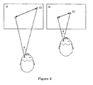
- Figure 2 demonstrates how the screen position for a visual field point being assessed is dependant upon the 3D position of the subject's eyes relative to the display.
- This shows two examples (a) and (b) that allow testing of the patient's visual field at angle ⁇ .
- ⁇ remains unchanged, but the position of the second stimulus (S2) changes as a function of the position of the patient's eyes relative to the screen.
- S2 the second stimulus
- the 3D position in space of each eye has to be carefully monitored, so that the stimulus can be positioned and correctly associated with the particular visual field point being assessed.
- the actual size of the stimuli being displayed on the screen also has to be varied dependant upon the distance the subject's eyes are away from the screen. This is because the angular stimulus size must be kept constant for all visual field points during a test. Hence, the size is also varied depending on the location of the patent's eyes, as shown in Figure 3 .
- the system may also be adapted to take into account the fact that the stimuli displayed to test visual field points may not be directly in front of the patient and so to this end, the system may vary the shape of the stimulus presented, as a function of three-dimensional position of the patient's eye relative to the display screen and the visual field position that is to be tested, so that stimuli are presented at a constant angular shape.
- the shape presented on screen may in fact be elliptical, so that when viewed from an angle, is it perceived as being circular.
- the response of a subject's eye gaze is monitored when stimuli 14 are shown in different positions on the display screen 12 corresponding to different points in a subject's visual field.
- An algorithm is used to decide whether the subject was able to see the new stimulus based on the direction and length of movement of eye gaze and the timing of that movement. If a subject sees something in their peripheral vision the natural response is to gaze at it. If the point is not seen there could be no change in eye gaze position or a searching eye gaze movement.
- the algorithm automatically distinguishes these responses in real time based on the direction and angular length of any fixation change made immediately following the presentation of a new visual field point stimulus, and the timing of any such fixation change.
- the vector change in fixation point is compared directly with the vector change in the screen position of the stimuli to decide if the subject was able to see the stimuli or not.
- Figure 5 shows the steps in a method for testing visual fields.
- this method is implemented using a computer program running on the processor/personal computer.
- each of the patient's eyes is measured separately and the results averaged, although this is not essential.
- a stimulus 14 is displayed in the centre of the screen 12 to cause the patient to fixate on a central point.
- the eye tracker 16 captures eye gaze data in order to verify whether the patient is indeed gazing at the stimulus 14. In the event that the gaze data suggests that the patient is not fixating on the correct point, further gaze data is captured. This is repeated until the system is confident that the patient is looking at the central stimulus 14. Animations to attract attention can be used if required.
- the position and size of the next stimulus are calculated.
- the position of the next stimulus is calculated using the distance from the current fixation point (in this case the central stimulus) to the subject's eyes, as measured in real time using the eye tracker, and the points in the subject's visual field that are to be assessed.
- the on-screen position can vary depending on the measured distance of the patient from the fixation point. For a particular examination there are a number of different visual field points to be assessed. Generally, the order in which the selected points are assessed is random.
- the timer runs in the background and acts as a countdown. If the displayed stimulus is not detected as 'Seen' within this time then the stimulus is categorised as 'Not Seen'. Typically, this time is set to a pre-determined limit, for example 1 second.
- gaze data is captured. For the Tobii tracker this is done every 20ms or so. The captured data is used to determine whether there has been a change in the fixation point. If there has, the following gaze data point is used to determine whether there has been a further change in the fixation point, this continues until no change is found. In this manner the movement of the user's eye towards an end, fixation point can be found. If there is no change in fixation with a gaze data point and the prior gaze data point did indicate a change in fixation then this indicates the end of the subject's fixation change. Once a change is detected the fixation change properties are tested to identify whether the current fixation change corresponds to the location of the new stimulus.
- Figure 6 illustrates steps for testing the fixation change properties. Firstly, the fixation change properties are used to identify whether the direction of the change of the fixation is correct when compared with a vector from the first stimulus to the new stimulus. If not, then the fixation is determined not to relate to the new stimulus and the system returns to the capture gaze data step of Figure 5 . If yes, then the fixation data and three-dimensional eye position data is used to determine the angular distance between the first fixation point and the second fixation point. If this distance differs from the known angular separation between the first stimulus and the second stimulus, then the fixation is determined not to relate to the new stimulus and the system returns to the capture gaze data step of Figure 5 . In contrast, if the angular distance does correspond to the separation of the first stimulus and the second stimulus then it is concluded that the fixation does relate to the displayed stimulus.
- the system could also be used to assess a person's reaction to peripheral stimuli within a recreated "real-world" environment such as street or home scene or driving situation. This can be helpful as their reactions can be affected by different disease states.
- the visual field position to be tested is selected and the position of the stimulus on the display calculated as a function of the patient's three-dimensional eye position.
- the stimulus may be positioned at one of a plurality of selected positions on the display and then the visual field position associated with this position calculated using the three dimensional position of the patient's eye(s).
- the peripheral stimulus presented will generally be of the same type for all subjects, for example a circle or disc.
- the visual field stimulus can be changed to interesting audio-visual animations appropriate to their developmental age once the original peripheral stimulus has been detected as 'seen'.
- the human face is a potent visual fixation stimulus for an infant and cartoon figures can be used for young children. This will motivate the children to submit to the test and increase compliance rate without changing the properties of the required visual field stimulus.
- the test can also be interrupted anytime by sounds or video to draw the attention of a distracted child back to the screen.
- the method in which the invention is embodied does not require any cooperation or understanding from a child. Also, the child is not required to learn any task or to give any subjective response. This removes the need for vigilance, and endurance or understanding of the test method on the part of the child. Moreover, the absence of subjective responses from the child also eliminates false-positive and false-negative errors, which increases the reliability of the test. Also, the child's head movement is not restricted in any way and the system does not require any form of physical contact with the child, which makes it more comfortable. A further advantage is that a continuous fixation on a stationary central fixation target is not needed.
- the system of Figure 1 can be used for numerous other eye tests, for example visual acuity, colour vision, contrast sensitivity and colour-contrast sensitivity, in both children and adults.
- Visual acuity is the ability to discriminate two stimuli separated in space whilst colour vision is the ability to differentiate two different colours.
- Contrast sensitivity is the ability to discriminate the difference between shades of grey or hues of colour.
- Eye tracking has the potential to more accurately and reliably detect change of direction of gaze towards a test target. Using the arrangement of Figure 1 , this could be done by presenting vanishing optotype targets and using the eye tracker to detect if the subject looks at a picture. Alternatively stationary pictures with varying levels of visual acuity and contrast sensitivity could be presented on the screen. Likewise, different colour images could be presented, so as to test colour vision deficiency. In any case, the pictures are presented somewhere away from the subject's gaze point and eye movement is tracked. If the subject looks at the picture then it can be concluded that they were able to see it, and if they did not look at the picture then they were not able to see it. The test proceeds based on the subject's responses, i.e.
- the software for controlling the test is similar to that used for visual field assessment, although it is simpler, as it is only required to recognise that the subject looks at the picture rather than assessing fixation change properties from one point to another.
Landscapes
- Health & Medical Sciences (AREA)
- Life Sciences & Earth Sciences (AREA)
- Engineering & Computer Science (AREA)
- Medical Informatics (AREA)
- Surgery (AREA)
- Biophysics (AREA)
- Biomedical Technology (AREA)
- Heart & Thoracic Surgery (AREA)
- Physics & Mathematics (AREA)
- Molecular Biology (AREA)
- Ophthalmology & Optometry (AREA)
- Animal Behavior & Ethology (AREA)
- General Health & Medical Sciences (AREA)
- Public Health (AREA)
- Veterinary Medicine (AREA)
- Human Computer Interaction (AREA)
- Eye Examination Apparatus (AREA)
Claims (15)
- System zum Testen von Sehfeldern (10), Folgendes umfassend:ein Display (12) zum Präsentieren eines Sehreizes (14);einen Augenverfolger (16) zum Verfolgen von Bewegung von mindestens einem der Augen eines Patienten, um seine Position in drei Dimensionen zu bestimmen, ohne dass der Patient eine Verfolgungsausrüstung tragen muss;Mittel zum Ändern der Position des Reizes (14) auf dem Display (12) um eine Größe, die eine Funktion der verfolgten dreidimensionalen Position des Auges des Patienten ist und einer zu testenden Sehfeldposition ist, undMittel zum Verwenden der Änderung der Position des Reizes (14) und einer verfolgten Augenbewegung, um das Sehfeld des Patienten abzuschätzen.
- System (10) nach Anspruch 1, außerdem Mittel umfassend, um die Größe des präsentierten Reizes (14) als eine Funktion der dreidimensionalen Position des Auges des Patienten und einer zu testenden Sehfeldposition zu variieren, sodass der Reiz (14) während des Tests einen konstanten Winkeldurchmesser hat, und/oder außerdem Mittel umfassend, um die Form des präsentierten Reizes (14) als eine Funktion der dreidimensionalen Position des Auges des Patienten relativ zum Display (12) und der getesteten Sehfeldposition zu variieren, sodass der Reiz (14) mit einer konstanten Winkelform präsentiert wird.
- System (10) nach einem der vorhergehenden Ansprüche, angepasst zum Präsentieren eines ersten Reizes (14); Erfassen von Blickdaten für diesen ersten Reiz (14); Präsentieren eines zweiten Reizes (14) und Testen, ob der Patient auf den zweiten Reiz (14) blickt.
- System (10) nach Anspruch 3, worin das Testen, ob ein Patient den zweiten Reiz (14) gesehen hat, das Bestimmen einer Richtung einschließt, in der sich das oder die Augen des Patienten bewegt haben seit der zweite Reiz (14) präsent ist, und das Vergleichen dieser Richtung mit der Vektorrichtung, die mit einer Linie zwischen dem ersten Reiz (14) und dem zweiten Reiz (14) assoziiert ist.
- System (10) nach Anspruch 3 oder Anspruch 4, worin das Testen, ob ein Patient den zweiten Reiz (14) gesehen hat, das Bestimmen der Winkelentfernung zwischen einem ersten Fixierpunkt und einem zweiten Fixierpunkt und das Vergleichen dieser mit der Winkelentfernung zwischen dem ersten und zweiten Reiz einschließt.
- System (10) nach einem der vorhergehenden Ansprüche, wobei das System außerdem betriebsfähig ist, mindestens eins von Folgenden zu messen: Sehschärfe, Farbsehkraft, Kontrastsensitivität und Farbkontrastsensitivität.
- System nach einem der vorhergehenden Ansprüche, worin das Display (12) gekrümmt ist.
- System (10) nach einem der vorhergehenden Ansprüche, worin der Augenverfolger (16) eine Kopfpositionstoleranz von mehr als 10 cm x 10 cm x 10 cm hat (Horizontale x Vertikale x Entfernung vom Verfolger), worin der Augenverfolger optional eine Kopfpositionstoleranz von mehr als 20 cm x 10 cm x 20 cm hat.
- Verfahren zum Testen von Sehkraft, Folgendes umfassend: Präsentieren eines Sehreizes (14) auf einem Bildschirm; Verfolgen von Bewegung von mindestens einem der Augen des Patienten unter Verwendung eines Augenverfolgers (16), um einen Blickpunkt und seine Position in drei Dimensionen zu bestimmen, ohne dass der Patient eine Verfolgungsausrüstung tragen muss; Ändern der Position des Reizes (14) auf dem Display (12) um eine Größe, die eine Funktion der verfolgten dreidimensionalen Position des Auges des Patienten und einer zu testenden Sehfeldposition ist, und Verwenden der Änderung der Position des Reizes (14) und einer verfolgten Augenbewegung als Reaktion auf die Änderung der Position, um das Sehfeld des Patienten abzuschätzen.
- Computerprogramm oder Computerprogrammprodukt, vorzugsweise auf einem Datenträger oder computerlesbarem Medium oder einem Prozessor, der Code oder Anweisungen umfasst, um beim Ablaufen auf einem Computer einen Sehreiz (14) auf einem Display (12) zu präsentieren; Daten von einem Augenverfolger (16) über die Augenbewegung und über die dreidimensionale Position eines oder beider Augen eines Patienten zu empfangen; die Position des Reizes (14) auf dem Display (12) um eine Größe, die eine Funktion der verfolgten dreidimensionalen Position des Auges des Patienten und einer zu testenden Sehfeldposition ist zu ändern, und die Änderung der Position des Reizes (14) und einer verfolgten Augenbewegung als Reaktion auf die Änderung der Position zu verwenden, um das Sehfeld des Patienten abzuschätzen.
- Programm/Produkt/Prozessor nach Anspruch 10, konfiguriert zum Variieren der Größe des präsentierten Reizes (14) als eine Funktion der dreidimensionalen Position des Auges des Patienten und einer zu testenden Sehfeldposition, sodass der Reiz (14) während des Tests eine konstante Winkelgröße hat, und/oder dazu angepasst, die Position des Reizes (14) auf dem Bildschirm neu zu positionieren und Augenverfolgungsinformation zu verwenden, um zu bestimmen, ob der Patient auf den Reiz (14) an seiner neuen Position blickt.
- Programm/Produkt/Prozessor nach Anspruch 10 oder Anspruch 11, angepasst zum Präsentieren eines ersten Reizes (14); Erfassen von Blickdaten für diesen ersten Reiz (14); Präsentieren eines zweiten Reizes (14) und Testen, ob der Patient den zweiten Reiz (14) gesehen hat, worin das Bestimmen, ob ein Patient den zweiten Reiz (14) gesehen hat, optional einschließt, dass Augenverfolgungsinformation verwendet wird, um eine Richtung zu bestimmen, in der sich das oder die Augen des Patienten bewegt haben, und diese mit der Vektorrichtung verglichen wird, die mit einer Linie zwischen dem ersten Reiz (14) und dem zweiten Reiz (14) assoziiert ist, und/oder worin das Testen, ob ein Patient den zweiten Reiz (14) gesehen hat, das Bestimmen der Winkelentfernung zwischen einem ersten Fixierpunkt und einem zweiten Fixierpunkt und das Vergleichen von ihr mit der Winkelentfernung zwischen dem ersten und zweiten Reiz einschließt.
- Programm/Produkt/Prozessor nach einem der Ansprüche 10 bis 12, dazu angeordnet, eine Zeit zwischen dem Zeigen eines ersten Reizes (14), dem Löschen dieses Reizes (14) und dem Anzeigen eines zweiten Reizes (14) und der Zeit zu bestimmen, die ein Patient benötigt, um auf die Änderung des Reizes (14) zu reagieren, worin der Code oder die Anweisungen optional zum Bestimmen angepasst sind, ob der Patient den Reiz (14) an seiner neuen Position gesehen hat, indem die Richtung der Änderung der Fixierung und die Richtung eines Vektors vom ersten Reiz (14) zum neuen Reiz (14) und die Winkelentfernung zwischen dem ersten Fixierpunkt und dem zweiten Fixierpunkt und die Winkeltrennung zwischen dem ersten Reiz (14) und dem zweiten Reiz (14) verglichen werden.
- Programm/Produkt/Prozessor nach einem der Ansprüche 10 bis 13, dazu angeordnet, eine Zeit zwischen dem Zeigen eines ersten Reizes (14), Löschen dieses Reizes (14) und Anzeigen eines zweiten Reizes (14) und der Zeit, die ein Patient braucht, um auf die Änderung im Reiz (14) zu reagieren, und dies mindestens teilweise zum Identifizieren zu verwenden, ob der Patient auf den Reiz (14) reagiert.
- System zum Testen von Sehfeldern, Folgendes umfassend:ein Display (12) zum Präsentieren eines Sehreizes (14);einen Augenverfolger (16) zum Verfolgen von Bewegung von mindestens einem der Augen eines Patienten, um seine Position in drei Dimensionen zu bestimmen, ohne dass der Patient eine Verfolgungsausrüstung tragen muss;Mittel zum Ändern der Position des Reizes (14) auf dem Display (12);Mittel zum Bestimmen der Sehfeldposition, die mit der neuen Position des Reizes (14) assoziiert ist unter Verwendung seiner Position auf dem Display (12) und der dreidimensionalen Position des oder der Augen des Patienten, undMittel zum Verwenden der Änderung der Position des Reizes (14) und jedweder verfolgten Augenbewegung als Reaktion auf die Änderung der Position, um das Sehfeld des Patienten abzuschätzen.
Applications Claiming Priority (2)
| Application Number | Priority Date | Filing Date | Title |
|---|---|---|---|
| GBGB0709405.5A GB0709405D0 (en) | 2007-05-16 | 2007-05-16 | Testing vision |
| PCT/GB2008/001480 WO2008139137A1 (en) | 2007-05-16 | 2008-04-25 | Testing vision |
Publications (2)
| Publication Number | Publication Date |
|---|---|
| EP2148609A1 EP2148609A1 (de) | 2010-02-03 |
| EP2148609B1 true EP2148609B1 (de) | 2013-12-25 |
Family
ID=38234544
Family Applications (1)
| Application Number | Title | Priority Date | Filing Date |
|---|---|---|---|
| EP08750486.6A Not-in-force EP2148609B1 (de) | 2007-05-16 | 2008-04-25 | Sehkrafttest |
Country Status (9)
| Country | Link |
|---|---|
| US (1) | US8337019B2 (de) |
| EP (1) | EP2148609B1 (de) |
| JP (1) | JP5498375B2 (de) |
| CN (1) | CN101742957B (de) |
| AU (1) | AU2008249842B2 (de) |
| CA (1) | CA2687409C (de) |
| GB (1) | GB0709405D0 (de) |
| IN (1) | IN2009DN07381A (de) |
| WO (1) | WO2008139137A1 (de) |
Cited By (3)
| Publication number | Priority date | Publication date | Assignee | Title |
|---|---|---|---|---|
| RU2725782C2 (ru) * | 2018-11-30 | 2020-07-06 | Общество С Ограниченной Ответственностью "Нейрочат" | Система для коммуникации пользователей без использования мышечных движений и речи |
| WO2023033545A1 (ko) * | 2021-09-06 | 2023-03-09 | 재단법인 아산사회복지재단 | 시야손상 환자의 선호망막부위 탐색 및 훈련 장치, 방법 및 프로그램 |
| WO2025015374A1 (en) * | 2023-07-20 | 2025-01-23 | GLANCE Optical Pty Ltd | A digital display device field of vision testing system |
Families Citing this family (64)
| Publication number | Priority date | Publication date | Assignee | Title |
|---|---|---|---|---|
| GB0915136D0 (en) | 2009-08-28 | 2009-10-07 | Cambridge Entpr Ltd | Visual perimeter measurement system and method |
| FR2960412B1 (fr) * | 2010-05-25 | 2013-03-22 | Essilor Int | Dispositif et procede de mesure d'une distance caracteristique de lecture d'un individu en posture naturelle de vision de pres |
| FR2960413B1 (fr) * | 2010-05-25 | 2013-03-22 | Essilor Int | Dispositif de mesure d'une distance caracteristique de lecture d'un individu |
| WO2012141576A1 (en) | 2011-04-15 | 2012-10-18 | Erasmus University Medical Center Rotterdam | A method for investigating visual field defects |
| KR20140103900A (ko) | 2011-09-08 | 2014-08-27 | 아이체크 헬스 커넥션, 인크. | 유아 및 소아에 있어서 눈의 동공 적색 반사 검사 및 각막 광 반사 선별의 기록 및 문서화를 위한 방법 및 시스템 |
| JP5876704B2 (ja) * | 2011-10-18 | 2016-03-02 | 花王株式会社 | 視野測定方法及び視野測定装置 |
| US9433346B2 (en) | 2011-11-21 | 2016-09-06 | Gobiquity, Inc. | Circular preferential hyperacuity perimetry video game to monitor macular and retinal diseases |
| AU2012340573A1 (en) | 2011-11-21 | 2014-07-17 | Icheck Health Connection, Inc. | Video game to monitor retinal diseases |
| EP2793682A1 (de) * | 2011-12-20 | 2014-10-29 | ICheck Health Connection Inc. | Videospiel zur überwachung des sichtfeldsverlustes bei glaukom |
| KR101245330B1 (ko) | 2011-12-20 | 2013-03-25 | 경희대학교 산학협력단 | 개인용 컴퓨터 기반 시야 자가진단 시스템 및 시선 고정방법 |
| JP6366510B2 (ja) * | 2012-03-09 | 2018-08-01 | オキュスペクト オサケ ユキチュア | 視覚系の機能を評価するためのシステムの作動方法および装置 |
| US10537240B2 (en) | 2012-03-09 | 2020-01-21 | Ocuspecto Oy | Method for assessing function of the visual system and apparatus thereof |
| CN103356158A (zh) * | 2012-03-26 | 2013-10-23 | 傅凌进 | 一种新颖的人眼颜色视觉特性评价系统 |
| JP5900165B2 (ja) * | 2012-05-31 | 2016-04-06 | 株式会社Jvcケンウッド | 視線検出装置および視線検出方法 |
| US9888842B2 (en) * | 2012-05-31 | 2018-02-13 | Nokia Technologies Oy | Medical diagnostic gaze tracker |
| US9265458B2 (en) | 2012-12-04 | 2016-02-23 | Sync-Think, Inc. | Application of smooth pursuit cognitive testing paradigms to clinical drug development |
| US9179833B2 (en) | 2013-02-28 | 2015-11-10 | Carl Zeiss Meditec, Inc. | Systems and methods for improved ease and accuracy of gaze tracking |
| KR102179931B1 (ko) * | 2013-03-11 | 2020-11-18 | 칠드런스 헬스케어 오브 애틀란타, 인크. | 인지 및 발달 조건의 탐지 시스템 및 방법 |
| US9380976B2 (en) | 2013-03-11 | 2016-07-05 | Sync-Think, Inc. | Optical neuroinformatics |
| US9247870B2 (en) * | 2013-03-15 | 2016-02-02 | Neuro Kinetics, Inc. | Method and apparatus for system synchronization in video oculography based neuro-otologic testing and evaluation |
| US9704411B1 (en) | 2013-07-31 | 2017-07-11 | Edward Newill | Method and apparatus for determining reading acuity with automated calibration |
| EP3031388B1 (de) * | 2013-08-08 | 2020-01-15 | Panasonic Intellectual Property Management Co., Ltd. | Sichtfeldberechnungseinheit und sichtfeldberechnungsverfahren |
| CN103557859B (zh) | 2013-10-10 | 2015-12-23 | 北京智谷睿拓技术服务有限公司 | 图像采集定位方法及图像采集定位系统 |
| CN103932674A (zh) * | 2014-01-01 | 2014-07-23 | 林肯 | 调节灵敏度测试训练仪 |
| WO2015143073A1 (en) | 2014-03-19 | 2015-09-24 | Intuitive Surgical Operations, Inc. | Medical devices, systems, and methods integrating eye gaze tracking for stereo viewer |
| CN106456148B (zh) * | 2014-03-19 | 2020-06-12 | 直观外科手术操作公司 | 使用眼球凝视跟踪的医疗装置、系统和方法 |
| WO2016027215A1 (en) * | 2014-08-21 | 2016-02-25 | Cagno Andrea | Apparatus for detecting, diagnosing and exercising an individual's functionalities |
| CN107072523A (zh) * | 2014-08-22 | 2017-08-18 | 华柏恩视觉诊断公司 | 用于监测眼动以检查视场的系统、方法和设备 |
| CN106999036A (zh) * | 2014-11-27 | 2017-08-01 | 皇家飞利浦有限公司 | 用于评估视力敏锐度和听觉能力的系统和方法 |
| US9595126B2 (en) | 2014-12-15 | 2017-03-14 | Konan Medical Usa, Inc. | Visual function targeting using randomized, dynamic, contrasting features |
| WO2016173652A1 (en) * | 2015-04-29 | 2016-11-03 | Metrovision | System for testing visual field of a patient and the corresponding method |
| GB2539250B (en) | 2015-06-12 | 2022-11-02 | Okulo Ltd | Methods and systems for testing aspects of vision |
| FR3042400A1 (fr) * | 2015-10-15 | 2017-04-21 | Essilor Int | Dispositif de test du comportement visuel d'un individu et methode de determination d'au moins un parametre de conception optique d'une lentille ophtalmique utilisant un tel dispositif |
| JP6063549B1 (ja) * | 2015-12-17 | 2017-01-18 | 雅子 吉田 | 定規 |
| WO2017186721A1 (en) * | 2016-04-25 | 2017-11-02 | Koninklijke Philips N.V. | Eye movement monitoring system |
| CN107550452B (zh) * | 2016-06-30 | 2022-04-15 | 中国科学院心理研究所 | 一种个性化自适应式视野缺损治疗系统 |
| WO2018164636A1 (en) * | 2017-03-04 | 2018-09-13 | Gunasekeran Dinesh Visva | Visual performance assessment |
| IL312112A (en) * | 2017-03-05 | 2024-06-01 | Virtuoptica Ltd | Eye examination method and apparatus therefor |
| CN107028584B (zh) * | 2017-06-01 | 2019-05-17 | 赵阳 | 一种视野测量方法 |
| US11291362B2 (en) * | 2017-11-01 | 2022-04-05 | Robert Wendell Arnold | Systems and methods for eye evaluation and treatment |
| JP7043889B2 (ja) * | 2018-03-01 | 2022-03-30 | 株式会社Jvcケンウッド | 視機能検出装置、視機能検出方法及びプログラム |
| JP7043890B2 (ja) * | 2018-03-01 | 2022-03-30 | 株式会社Jvcケンウッド | 視機能検出装置、視機能検出方法及びプログラム |
| EP3747351B1 (de) * | 2018-03-01 | 2022-07-06 | JVCKenwood Corporation | Vorrichtung zur visuellen funktionsdetektion, verfahren zur visuellen funktionsdetektion und programm |
| CN112351727B (zh) * | 2018-03-02 | 2022-05-27 | 俄亥俄州立创新基金会 | 用于测量视觉功能图的系统和方法 |
| CN108937841B (zh) * | 2018-07-17 | 2020-06-26 | 遂宁市中心医院 | 眼科用视野检查装置 |
| CN109875495A (zh) * | 2019-03-05 | 2019-06-14 | 四川大学 | 一种基于显示设备的非接触式全自动反馈视野计 |
| JP7213492B2 (ja) * | 2019-06-24 | 2023-01-27 | 株式会社Qdレーザ | 視覚検査装置及び視覚検査方法 |
| IT201900009996A1 (it) | 2019-06-28 | 2020-12-28 | Centro Rham S R L | Sistema ad alta innovazione e metodo per il miglioramento della consapevolezza dell'aggancio visivo e della risposta monoculare |
| US11966511B2 (en) * | 2019-11-14 | 2024-04-23 | Rijksuniversiteit Groningen | Method, system and computer program product for mapping a visual field |
| US10827918B1 (en) * | 2019-12-25 | 2020-11-10 | Shamir Optical Industry Ltd. | System and method for automatically evaluating a vision of a user |
| WO2021209159A1 (en) | 2020-04-17 | 2021-10-21 | Tilak Healthcare Sas | Method for determining an indication of an acuity level of a user |
| JP2022037273A (ja) * | 2020-08-25 | 2022-03-09 | 株式会社ファインデックス | 可視判定システム及び視野検査装置 |
| JP7574985B2 (ja) * | 2020-11-05 | 2024-10-29 | 株式会社ファインデックス | 特性検査システム |
| SE2050893A1 (en) * | 2020-07-14 | 2022-01-15 | Heads Stockholm Ab | Eye movement evaluation |
| EP4225125A4 (de) * | 2020-10-27 | 2024-11-20 | Occutrack Medical Solutions Pte. Ltd. | Verfahren und system zur durchführung eines sehkraftbewertungstests unter verwendung von authorisierten testprofilen |
| US12446769B2 (en) * | 2020-10-28 | 2025-10-21 | University Of Miami | Active calibration of head-mounted displays |
| SE2051412A1 (en) * | 2020-12-03 | 2022-06-04 | Heads Stockholm Ab | Device for visual field testing |
| EP4056101A1 (de) * | 2021-03-12 | 2022-09-14 | Carl Zeiss Vision International GmbH | Verfahren und vorrichtung zur bestimmung einer visuellen leistung |
| CN112991392B (zh) * | 2021-04-12 | 2021-09-07 | 广东视明科技发展有限公司 | 不同空间知觉场景中眼球运动功能的检查系统 |
| CN113647900A (zh) * | 2021-06-28 | 2021-11-16 | 中山大学中山眼科中心 | 一种基于个人终端的自助视野检测方法 |
| EP4197425A1 (de) * | 2021-12-17 | 2023-06-21 | Carl Zeiss Vision International GmbH | Bestimmung einer visuellen leistung eines auges einer person |
| EP4218542A1 (de) * | 2022-01-26 | 2023-08-02 | Carl Zeiss Vision International GmbH | Verfahren und vorrichtung zur bestimmung von mindestens einem visuellen parameter |
| CN116195966B (zh) * | 2022-12-30 | 2026-05-05 | 北京国科启明教育科技有限公司 | 基于空间任意位置的视力检测方法及系统 |
| WO2026064097A1 (en) * | 2024-09-17 | 2026-03-26 | University Of Rochester | Method and systems for reducing the burden of visual impairment |
Family Cites Families (17)
| Publication number | Priority date | Publication date | Assignee | Title |
|---|---|---|---|---|
| IL62627A (en) | 1981-04-10 | 1984-09-30 | Yissum Res Dev Co | Eye testing system |
| JPH05305057A (ja) * | 1992-05-01 | 1993-11-19 | Topcon Corp | 自覚式検眼装置 |
| US5459536A (en) | 1993-12-27 | 1995-10-17 | Alcon Laboratories, Inc. | Apparatus and method for automated perimetry |
| DE19505399B4 (de) * | 1995-02-17 | 2005-11-10 | Oculus Optikgeräte GmbH | Verfahren zum Ausmessen des Gesichtsfeldes |
| JP3716357B2 (ja) * | 1996-07-14 | 2005-11-16 | 株式会社トプコン | 視野測定装置 |
| GB9722949D0 (en) | 1997-10-30 | 1998-01-07 | Bid Instr Ltd | Ocular testing and related projection apparatus and method |
| US6129722A (en) * | 1999-03-10 | 2000-10-10 | Ruiz; Luis Antonio | Interactive corrective eye surgery system with topography and laser system interface |
| US6290357B1 (en) * | 1999-03-31 | 2001-09-18 | Virtual-Eye.Com, Inc. | Kinetic visual field apparatus and method |
| GB2375821A (en) | 2001-04-27 | 2002-11-27 | Peter Hamilton Galloway | Automatic vision testing system for children |
| GB0220721D0 (en) | 2002-09-06 | 2002-10-16 | Univ Glasgow | Systems and apparatus for assessment of visual field functions |
| SE524003C2 (sv) | 2002-11-21 | 2004-06-15 | Tobii Technology Ab | Förfarande och anläggning för att detektera och följa ett öga samt dess blickvinkel |
| GB0309025D0 (en) * | 2003-04-22 | 2003-05-28 | Mcgrath John A M | Method and apparatus for the early and rapid diagnosis of glaucoma and other human and higher primate visual disorders |
| CN101065766A (zh) | 2004-09-03 | 2007-10-31 | 潘那西卡股份有限公司 | 视觉中心自助式服务机 |
| JP3992045B2 (ja) * | 2005-02-18 | 2007-10-17 | 松下電工株式会社 | 映像信号処理装置及び該方法並びに仮想現実感生成装置 |
| JP4892742B2 (ja) | 2005-03-31 | 2012-03-07 | 国立大学法人 東京大学 | 視野計及び視野計測方法 |
| EP1933694B1 (de) * | 2005-10-10 | 2020-07-22 | Tobii AB | Augen-erfassungsgerät mit erweitertem bedienungsentfernungsbereich |
| CN101730500B (zh) | 2007-03-08 | 2013-12-18 | 北英属哥伦比亚大学 | 用于客观地测试视野的设备和方法 |
-
2007
- 2007-05-16 GB GBGB0709405.5A patent/GB0709405D0/en not_active Ceased
-
2008
- 2008-04-25 WO PCT/GB2008/001480 patent/WO2008139137A1/en not_active Ceased
- 2008-04-25 CA CA2687409A patent/CA2687409C/en active Active
- 2008-04-25 US US12/600,419 patent/US8337019B2/en not_active Expired - Fee Related
- 2008-04-25 AU AU2008249842A patent/AU2008249842B2/en not_active Ceased
- 2008-04-25 IN IN7381DEN2009 patent/IN2009DN07381A/en unknown
- 2008-04-25 CN CN2008800226903A patent/CN101742957B/zh not_active Expired - Fee Related
- 2008-04-25 JP JP2010507964A patent/JP5498375B2/ja not_active Expired - Fee Related
- 2008-04-25 EP EP08750486.6A patent/EP2148609B1/de not_active Not-in-force
Cited By (4)
| Publication number | Priority date | Publication date | Assignee | Title |
|---|---|---|---|---|
| RU2725782C2 (ru) * | 2018-11-30 | 2020-07-06 | Общество С Ограниченной Ответственностью "Нейрочат" | Система для коммуникации пользователей без использования мышечных движений и речи |
| WO2023033545A1 (ko) * | 2021-09-06 | 2023-03-09 | 재단법인 아산사회복지재단 | 시야손상 환자의 선호망막부위 탐색 및 훈련 장치, 방법 및 프로그램 |
| US12446770B2 (en) | 2021-09-06 | 2025-10-21 | The Asan Foundation | Apparatus, method, and program for searching and training preferred retinal locus of patient with visual field damage |
| WO2025015374A1 (en) * | 2023-07-20 | 2025-01-23 | GLANCE Optical Pty Ltd | A digital display device field of vision testing system |
Also Published As
| Publication number | Publication date |
|---|---|
| CN101742957B (zh) | 2012-08-08 |
| JP5498375B2 (ja) | 2014-05-21 |
| CN101742957A (zh) | 2010-06-16 |
| IN2009DN07381A (de) | 2015-07-24 |
| GB0709405D0 (en) | 2007-06-27 |
| EP2148609A1 (de) | 2010-02-03 |
| AU2008249842B2 (en) | 2013-09-26 |
| JP2010526623A (ja) | 2010-08-05 |
| US8337019B2 (en) | 2012-12-25 |
| CA2687409A1 (en) | 2008-11-20 |
| CA2687409C (en) | 2017-08-29 |
| WO2008139137A1 (en) | 2008-11-20 |
| US20100195051A1 (en) | 2010-08-05 |
| AU2008249842A1 (en) | 2008-11-20 |
Similar Documents
| Publication | Publication Date | Title |
|---|---|---|
| CA2687409C (en) | Testing vision | |
| EP3551036B1 (de) | Stimulus, augenverfolgungssystem und methode | |
| US8931905B2 (en) | Binocular measurement method and device | |
| US8500278B2 (en) | Apparatus and method for objective perimetry visual field test | |
| CN109219386B (zh) | 显示系统和方法 | |
| WO2016116933A1 (en) | Method and system for automatic eyesight diagnosis | |
| EP2827758A1 (de) | Verfahren zur beurteilung der funktion des visuellen systems und vorrichtung dafür | |
| WO2004093668A1 (en) | Method and apparatus for the diagnosis of glaucoma and other visual disorders | |
| CN111067474B (zh) | 使用动态速度阈值滤波器的客观视敏度测量的装置和方法 | |
| WO2006106877A1 (ja) | 視野計及び視野計測方法 | |
| US20230404385A1 (en) | Visual field testing | |
| US20240081636A1 (en) | Method for Visual Function Assessment Using Multistable Rivalry Paradigms | |
| EP3727128B1 (de) | Verfahren zur analyse eines sichtfeldes eines individuums | |
| US20250152082A1 (en) | Measuring a patient's visual motion detection threshold | |
| WO2025051943A1 (en) | Device and method for obtaining dynamic measurements of eye optical surfaces | |
| Goliskina et al. | Changes in Horizontal Saccades with Age | |
| HK40014705A (en) | Stimulus and eye tracking system |
Legal Events
| Date | Code | Title | Description |
|---|---|---|---|
| PUAI | Public reference made under article 153(3) epc to a published international application that has entered the european phase |
Free format text: ORIGINAL CODE: 0009012 |
|
| 17P | Request for examination filed |
Effective date: 20091117 |
|
| AK | Designated contracting states |
Kind code of ref document: A1 Designated state(s): AT BE BG CH CY CZ DE DK EE ES FI FR GB GR HR HU IE IS IT LI LT LU LV MC MT NL NO PL PT RO SE SI SK TR |
|
| AX | Request for extension of the european patent |
Extension state: AL BA MK RS |
|
| DAX | Request for extension of the european patent (deleted) | ||
| 17Q | First examination report despatched |
Effective date: 20120903 |
|
| GRAP | Despatch of communication of intention to grant a patent |
Free format text: ORIGINAL CODE: EPIDOSNIGR1 |
|
| INTG | Intention to grant announced |
Effective date: 20130703 |
|
| GRAS | Grant fee paid |
Free format text: ORIGINAL CODE: EPIDOSNIGR3 |
|
| GRAA | (expected) grant |
Free format text: ORIGINAL CODE: 0009210 |
|
| AK | Designated contracting states |
Kind code of ref document: B1 Designated state(s): AT BE BG CH CY CZ DE DK EE ES FI FR GB GR HR HU IE IS IT LI LT LU LV MC MT NL NO PL PT RO SE SI SK TR |
|
| REG | Reference to a national code |
Ref country code: GB Ref legal event code: FG4D |
|
| REG | Reference to a national code |
Ref country code: CH Ref legal event code: EP |
|
| REG | Reference to a national code |
Ref country code: AT Ref legal event code: REF Ref document number: 646116 Country of ref document: AT Kind code of ref document: T Effective date: 20140115 |
|
| REG | Reference to a national code |
Ref country code: IE Ref legal event code: FG4D |
|
| REG | Reference to a national code |
Ref country code: DE Ref legal event code: R096 Ref document number: 602008029520 Country of ref document: DE Effective date: 20140213 |
|
| PG25 | Lapsed in a contracting state [announced via postgrant information from national office to epo] |
Ref country code: LT Free format text: LAPSE BECAUSE OF FAILURE TO SUBMIT A TRANSLATION OF THE DESCRIPTION OR TO PAY THE FEE WITHIN THE PRESCRIBED TIME-LIMIT Effective date: 20131225 Ref country code: SE Free format text: LAPSE BECAUSE OF FAILURE TO SUBMIT A TRANSLATION OF THE DESCRIPTION OR TO PAY THE FEE WITHIN THE PRESCRIBED TIME-LIMIT Effective date: 20131225 Ref country code: HR Free format text: LAPSE BECAUSE OF FAILURE TO SUBMIT A TRANSLATION OF THE DESCRIPTION OR TO PAY THE FEE WITHIN THE PRESCRIBED TIME-LIMIT Effective date: 20131225 Ref country code: NO Free format text: LAPSE BECAUSE OF FAILURE TO SUBMIT A TRANSLATION OF THE DESCRIPTION OR TO PAY THE FEE WITHIN THE PRESCRIBED TIME-LIMIT Effective date: 20140325 Ref country code: FI Free format text: LAPSE BECAUSE OF FAILURE TO SUBMIT A TRANSLATION OF THE DESCRIPTION OR TO PAY THE FEE WITHIN THE PRESCRIBED TIME-LIMIT Effective date: 20131225 |
|
| REG | Reference to a national code |
Ref country code: NL Ref legal event code: VDEP Effective date: 20131225 |
|
| REG | Reference to a national code |
Ref country code: AT Ref legal event code: MK05 Ref document number: 646116 Country of ref document: AT Kind code of ref document: T Effective date: 20131225 |
|
| REG | Reference to a national code |
Ref country code: LT Ref legal event code: MG4D |
|
| PG25 | Lapsed in a contracting state [announced via postgrant information from national office to epo] |
Ref country code: LV Free format text: LAPSE BECAUSE OF FAILURE TO SUBMIT A TRANSLATION OF THE DESCRIPTION OR TO PAY THE FEE WITHIN THE PRESCRIBED TIME-LIMIT Effective date: 20131225 |
|
| REG | Reference to a national code |
Ref country code: CH Ref legal event code: NV Representative=s name: MARKS AND CLERK (LUXEMBOURG) LLP, CH |
|
| PG25 | Lapsed in a contracting state [announced via postgrant information from national office to epo] |
Ref country code: IS Free format text: LAPSE BECAUSE OF FAILURE TO SUBMIT A TRANSLATION OF THE DESCRIPTION OR TO PAY THE FEE WITHIN THE PRESCRIBED TIME-LIMIT Effective date: 20140425 Ref country code: EE Free format text: LAPSE BECAUSE OF FAILURE TO SUBMIT A TRANSLATION OF THE DESCRIPTION OR TO PAY THE FEE WITHIN THE PRESCRIBED TIME-LIMIT Effective date: 20131225 Ref country code: BE Free format text: LAPSE BECAUSE OF FAILURE TO SUBMIT A TRANSLATION OF THE DESCRIPTION OR TO PAY THE FEE WITHIN THE PRESCRIBED TIME-LIMIT Effective date: 20131225 |
|
| PG25 | Lapsed in a contracting state [announced via postgrant information from national office to epo] |
Ref country code: CZ Free format text: LAPSE BECAUSE OF FAILURE TO SUBMIT A TRANSLATION OF THE DESCRIPTION OR TO PAY THE FEE WITHIN THE PRESCRIBED TIME-LIMIT Effective date: 20131225 Ref country code: AT Free format text: LAPSE BECAUSE OF FAILURE TO SUBMIT A TRANSLATION OF THE DESCRIPTION OR TO PAY THE FEE WITHIN THE PRESCRIBED TIME-LIMIT Effective date: 20131225 Ref country code: RO Free format text: LAPSE BECAUSE OF FAILURE TO SUBMIT A TRANSLATION OF THE DESCRIPTION OR TO PAY THE FEE WITHIN THE PRESCRIBED TIME-LIMIT Effective date: 20131225 Ref country code: SK Free format text: LAPSE BECAUSE OF FAILURE TO SUBMIT A TRANSLATION OF THE DESCRIPTION OR TO PAY THE FEE WITHIN THE PRESCRIBED TIME-LIMIT Effective date: 20131225 Ref country code: NL Free format text: LAPSE BECAUSE OF FAILURE TO SUBMIT A TRANSLATION OF THE DESCRIPTION OR TO PAY THE FEE WITHIN THE PRESCRIBED TIME-LIMIT Effective date: 20131225 Ref country code: PT Free format text: LAPSE BECAUSE OF FAILURE TO SUBMIT A TRANSLATION OF THE DESCRIPTION OR TO PAY THE FEE WITHIN THE PRESCRIBED TIME-LIMIT Effective date: 20140428 Ref country code: ES Free format text: LAPSE BECAUSE OF FAILURE TO SUBMIT A TRANSLATION OF THE DESCRIPTION OR TO PAY THE FEE WITHIN THE PRESCRIBED TIME-LIMIT Effective date: 20131225 Ref country code: CY Free format text: LAPSE BECAUSE OF FAILURE TO SUBMIT A TRANSLATION OF THE DESCRIPTION OR TO PAY THE FEE WITHIN THE PRESCRIBED TIME-LIMIT Effective date: 20131225 Ref country code: PL Free format text: LAPSE BECAUSE OF FAILURE TO SUBMIT A TRANSLATION OF THE DESCRIPTION OR TO PAY THE FEE WITHIN THE PRESCRIBED TIME-LIMIT Effective date: 20131225 |
|
| REG | Reference to a national code |
Ref country code: DE Ref legal event code: R097 Ref document number: 602008029520 Country of ref document: DE |
|
| PG25 | Lapsed in a contracting state [announced via postgrant information from national office to epo] |
Ref country code: DK Free format text: LAPSE BECAUSE OF FAILURE TO SUBMIT A TRANSLATION OF THE DESCRIPTION OR TO PAY THE FEE WITHIN THE PRESCRIBED TIME-LIMIT Effective date: 20131225 |
|
| PLBE | No opposition filed within time limit |
Free format text: ORIGINAL CODE: 0009261 |
|
| STAA | Information on the status of an ep patent application or granted ep patent |
Free format text: STATUS: NO OPPOSITION FILED WITHIN TIME LIMIT |
|
| PG25 | Lapsed in a contracting state [announced via postgrant information from national office to epo] |
Ref country code: LU Free format text: LAPSE BECAUSE OF FAILURE TO SUBMIT A TRANSLATION OF THE DESCRIPTION OR TO PAY THE FEE WITHIN THE PRESCRIBED TIME-LIMIT Effective date: 20140425 Ref country code: MC Free format text: LAPSE BECAUSE OF FAILURE TO SUBMIT A TRANSLATION OF THE DESCRIPTION OR TO PAY THE FEE WITHIN THE PRESCRIBED TIME-LIMIT Effective date: 20131225 |
|
| 26N | No opposition filed |
Effective date: 20140926 |
|
| REG | Reference to a national code |
Ref country code: DE Ref legal event code: R097 Ref document number: 602008029520 Country of ref document: DE Effective date: 20140926 |
|
| REG | Reference to a national code |
Ref country code: IE Ref legal event code: MM4A |
|
| PG25 | Lapsed in a contracting state [announced via postgrant information from national office to epo] |
Ref country code: IE Free format text: LAPSE BECAUSE OF NON-PAYMENT OF DUE FEES Effective date: 20140425 |
|
| PG25 | Lapsed in a contracting state [announced via postgrant information from national office to epo] |
Ref country code: SI Free format text: LAPSE BECAUSE OF FAILURE TO SUBMIT A TRANSLATION OF THE DESCRIPTION OR TO PAY THE FEE WITHIN THE PRESCRIBED TIME-LIMIT Effective date: 20131225 |
|
| REG | Reference to a national code |
Ref country code: FR Ref legal event code: PLFP Year of fee payment: 9 |
|
| PG25 | Lapsed in a contracting state [announced via postgrant information from national office to epo] |
Ref country code: MT Free format text: LAPSE BECAUSE OF FAILURE TO SUBMIT A TRANSLATION OF THE DESCRIPTION OR TO PAY THE FEE WITHIN THE PRESCRIBED TIME-LIMIT Effective date: 20131225 |
|
| PG25 | Lapsed in a contracting state [announced via postgrant information from national office to epo] |
Ref country code: BG Free format text: LAPSE BECAUSE OF FAILURE TO SUBMIT A TRANSLATION OF THE DESCRIPTION OR TO PAY THE FEE WITHIN THE PRESCRIBED TIME-LIMIT Effective date: 20131225 |
|
| PG25 | Lapsed in a contracting state [announced via postgrant information from national office to epo] |
Ref country code: IT Free format text: LAPSE BECAUSE OF FAILURE TO SUBMIT A TRANSLATION OF THE DESCRIPTION OR TO PAY THE FEE WITHIN THE PRESCRIBED TIME-LIMIT Effective date: 20131225 Ref country code: GR Free format text: LAPSE BECAUSE OF FAILURE TO SUBMIT A TRANSLATION OF THE DESCRIPTION OR TO PAY THE FEE WITHIN THE PRESCRIBED TIME-LIMIT Effective date: 20140326 |
|
| PG25 | Lapsed in a contracting state [announced via postgrant information from national office to epo] |
Ref country code: HU Free format text: LAPSE BECAUSE OF FAILURE TO SUBMIT A TRANSLATION OF THE DESCRIPTION OR TO PAY THE FEE WITHIN THE PRESCRIBED TIME-LIMIT; INVALID AB INITIO Effective date: 20080425 Ref country code: TR Free format text: LAPSE BECAUSE OF FAILURE TO SUBMIT A TRANSLATION OF THE DESCRIPTION OR TO PAY THE FEE WITHIN THE PRESCRIBED TIME-LIMIT Effective date: 20131225 |
|
| REG | Reference to a national code |
Ref country code: FR Ref legal event code: PLFP Year of fee payment: 10 |
|
| REG | Reference to a national code |
Ref country code: FR Ref legal event code: PLFP Year of fee payment: 11 |
|
| PGFP | Annual fee paid to national office [announced via postgrant information from national office to epo] |
Ref country code: GB Payment date: 20220411 Year of fee payment: 15 Ref country code: FR Payment date: 20220408 Year of fee payment: 15 Ref country code: DE Payment date: 20220407 Year of fee payment: 15 |
|
| PGFP | Annual fee paid to national office [announced via postgrant information from national office to epo] |
Ref country code: CH Payment date: 20220413 Year of fee payment: 15 |
|
| REG | Reference to a national code |
Ref country code: DE Ref legal event code: R119 Ref document number: 602008029520 Country of ref document: DE |
|
| REG | Reference to a national code |
Ref country code: CH Ref legal event code: PL |
|
| GBPC | Gb: european patent ceased through non-payment of renewal fee |
Effective date: 20230425 |
|
| PG25 | Lapsed in a contracting state [announced via postgrant information from national office to epo] |
Ref country code: GB Free format text: LAPSE BECAUSE OF NON-PAYMENT OF DUE FEES Effective date: 20230425 |
|
| PG25 | Lapsed in a contracting state [announced via postgrant information from national office to epo] |
Ref country code: LI Free format text: LAPSE BECAUSE OF NON-PAYMENT OF DUE FEES Effective date: 20230430 Ref country code: GB Free format text: LAPSE BECAUSE OF NON-PAYMENT OF DUE FEES Effective date: 20230425 Ref country code: FR Free format text: LAPSE BECAUSE OF NON-PAYMENT OF DUE FEES Effective date: 20230430 Ref country code: DE Free format text: LAPSE BECAUSE OF NON-PAYMENT OF DUE FEES Effective date: 20231103 Ref country code: CH Free format text: LAPSE BECAUSE OF NON-PAYMENT OF DUE FEES Effective date: 20230430 |