EP2143794A1 - Viral vector for gene therapy - Google Patents
Viral vector for gene therapy Download PDFInfo
- Publication number
- EP2143794A1 EP2143794A1 EP08752166A EP08752166A EP2143794A1 EP 2143794 A1 EP2143794 A1 EP 2143794A1 EP 08752166 A EP08752166 A EP 08752166A EP 08752166 A EP08752166 A EP 08752166A EP 2143794 A1 EP2143794 A1 EP 2143794A1
- Authority
- EP
- European Patent Office
- Prior art keywords
- cells
- gene
- csf
- vector
- sev18
- Prior art date
- Legal status (The legal status is an assumption and is not a legal conclusion. Google has not performed a legal analysis and makes no representation as to the accuracy of the status listed.)
- Withdrawn
Links
Images


Classifications
-
- A—HUMAN NECESSITIES
- A61—MEDICAL OR VETERINARY SCIENCE; HYGIENE
- A61K—PREPARATIONS FOR MEDICAL, DENTAL OR TOILETRY PURPOSES
- A61K38/00—Medicinal preparations containing peptides
- A61K38/16—Peptides having more than 20 amino acids; Gastrins; Somatostatins; Melanotropins; Derivatives thereof
- A61K38/17—Peptides having more than 20 amino acids; Gastrins; Somatostatins; Melanotropins; Derivatives thereof from animals; from humans
- A61K38/19—Cytokines; Lymphokines; Interferons
- A61K38/195—Chemokines, e.g. RANTES
-
- A—HUMAN NECESSITIES
- A61—MEDICAL OR VETERINARY SCIENCE; HYGIENE
- A61K—PREPARATIONS FOR MEDICAL, DENTAL OR TOILETRY PURPOSES
- A61K38/00—Medicinal preparations containing peptides
- A61K38/16—Peptides having more than 20 amino acids; Gastrins; Somatostatins; Melanotropins; Derivatives thereof
- A61K38/17—Peptides having more than 20 amino acids; Gastrins; Somatostatins; Melanotropins; Derivatives thereof from animals; from humans
- A61K38/19—Cytokines; Lymphokines; Interferons
- A61K38/193—Colony stimulating factors [CSF]
-
- A—HUMAN NECESSITIES
- A61—MEDICAL OR VETERINARY SCIENCE; HYGIENE
- A61P—SPECIFIC THERAPEUTIC ACTIVITY OF CHEMICAL COMPOUNDS OR MEDICINAL PREPARATIONS
- A61P35/00—Antineoplastic agents
-
- A—HUMAN NECESSITIES
- A61—MEDICAL OR VETERINARY SCIENCE; HYGIENE
- A61P—SPECIFIC THERAPEUTIC ACTIVITY OF CHEMICAL COMPOUNDS OR MEDICINAL PREPARATIONS
- A61P37/00—Drugs for immunological or allergic disorders
- A61P37/02—Immunomodulators
- A61P37/04—Immunostimulants
-
- A—HUMAN NECESSITIES
- A61—MEDICAL OR VETERINARY SCIENCE; HYGIENE
- A61P—SPECIFIC THERAPEUTIC ACTIVITY OF CHEMICAL COMPOUNDS OR MEDICINAL PREPARATIONS
- A61P43/00—Drugs for specific purposes, not provided for in groups A61P1/00-A61P41/00
-
- C—CHEMISTRY; METALLURGY
- C12—BIOCHEMISTRY; BEER; SPIRITS; WINE; VINEGAR; MICROBIOLOGY; ENZYMOLOGY; MUTATION OR GENETIC ENGINEERING
- C12N—MICROORGANISMS OR ENZYMES; COMPOSITIONS THEREOF; PROPAGATING, PRESERVING, OR MAINTAINING MICROORGANISMS; MUTATION OR GENETIC ENGINEERING; CULTURE MEDIA
- C12N15/00—Mutation or genetic engineering; DNA or RNA concerning genetic engineering, vectors, e.g. plasmids, or their isolation, preparation or purification; Use of hosts therefor
- C12N15/09—Recombinant DNA-technology
- C12N15/63—Introduction of foreign genetic material using vectors; Vectors; Use of hosts therefor; Regulation of expression
- C12N15/79—Vectors or expression systems specially adapted for eukaryotic hosts
- C12N15/85—Vectors or expression systems specially adapted for eukaryotic hosts for animal cells
- C12N15/86—Viral vectors
-
- C—CHEMISTRY; METALLURGY
- C12—BIOCHEMISTRY; BEER; SPIRITS; WINE; VINEGAR; MICROBIOLOGY; ENZYMOLOGY; MUTATION OR GENETIC ENGINEERING
- C12N—MICROORGANISMS OR ENZYMES; COMPOSITIONS THEREOF; PROPAGATING, PRESERVING, OR MAINTAINING MICROORGANISMS; MUTATION OR GENETIC ENGINEERING; CULTURE MEDIA
- C12N7/00—Viruses; Bacteriophages; Compositions thereof; Preparation or purification thereof
-
- A—HUMAN NECESSITIES
- A61—MEDICAL OR VETERINARY SCIENCE; HYGIENE
- A61K—PREPARATIONS FOR MEDICAL, DENTAL OR TOILETRY PURPOSES
- A61K48/00—Medicinal preparations containing genetic material which is inserted into cells of the living body to treat genetic diseases; Gene therapy
-
- C—CHEMISTRY; METALLURGY
- C12—BIOCHEMISTRY; BEER; SPIRITS; WINE; VINEGAR; MICROBIOLOGY; ENZYMOLOGY; MUTATION OR GENETIC ENGINEERING
- C12N—MICROORGANISMS OR ENZYMES; COMPOSITIONS THEREOF; PROPAGATING, PRESERVING, OR MAINTAINING MICROORGANISMS; MUTATION OR GENETIC ENGINEERING; CULTURE MEDIA
- C12N2710/00—MICROORGANISMS OR ENZYMES; COMPOSITIONS THEREOF; PROPAGATING, PRESERVING, OR MAINTAINING MICROORGANISMS; MUTATION OR GENETIC ENGINEERING; CULTURE MEDIA dsDNA viruses
- C12N2710/00011—Details
- C12N2710/10011—Adenoviridae
- C12N2710/10311—Mastadenovirus, e.g. human or simian adenoviruses
- C12N2710/10341—Use of virus, viral particle or viral elements as a vector
- C12N2710/10343—Use of virus, viral particle or viral elements as a vector viral genome or elements thereof as genetic vector
-
- C—CHEMISTRY; METALLURGY
- C12—BIOCHEMISTRY; BEER; SPIRITS; WINE; VINEGAR; MICROBIOLOGY; ENZYMOLOGY; MUTATION OR GENETIC ENGINEERING
- C12N—MICROORGANISMS OR ENZYMES; COMPOSITIONS THEREOF; PROPAGATING, PRESERVING, OR MAINTAINING MICROORGANISMS; MUTATION OR GENETIC ENGINEERING; CULTURE MEDIA
- C12N2760/00—MICROORGANISMS OR ENZYMES; COMPOSITIONS THEREOF; PROPAGATING, PRESERVING, OR MAINTAINING MICROORGANISMS; MUTATION OR GENETIC ENGINEERING; CULTURE MEDIA ssRNA viruses negative-sense
- C12N2760/00011—Details
- C12N2760/18011—Paramyxoviridae
- C12N2760/18811—Sendai virus
- C12N2760/18841—Use of virus, viral particle or viral elements as a vector
- C12N2760/18843—Use of virus, viral particle or viral elements as a vector viral genome or elements thereof as genetic vector
Definitions
- the present invention relates to viral vectors for gene therapy. More specifically, the present invention relates to viral vectors based on negative-strand RNA viruses, which comprise a gene encoding a cytokine such as granulocyte macrophage colony stimulating factor (GM-CSF), and are appropriate for treatment of cancers, in particular, metastatic cancers.
- GM-CSF granulocyte macrophage colony stimulating factor
- the present invention also relates to compositions comprising such viral vectors, and gene therapy using them.
- immunogene therapy is expected to be an effective therapeutic method for cancers, in particular, metastatic cancers. It is conceivable that immunogene therapy has fewer side effects and persistent therapeutic effects, and is cost-beneficial for patients.
- an autologous tumor vaccine for introducing a granulocyte macrophage colony stimulating factor (GM-CSF) gene has a substantial anti-tumor activity in preclinical cancer models and some clinical trials.
- Serotype 5 E1-deleted adenovirus (Ad5)/GM-CSF is one of the most promising vectors.
- adenovirus vector-mediated gene transfer into various tumor cells is limited due to the lack of expression of integrins ⁇ v ⁇ 3 and ⁇ v ⁇ 5, and the Ad5 receptor, which is a coxsackie-adenovirus receptor (CAR).
- CAR coxsackie-adenovirus receptor
- adenovirus vectors which are also double-stranded DNA vectors
- retroviral vectors such as single-stranded RNA (RT) viral vectors
- RT single-stranded RNA
- the clinical application of these vectors is limited by not only the complicated and time-consuming procedure for vector introduction, but also the insufficient GM-CSF production in vivo.
- the major obstacle for successful application of gene therapy strategy is the inability of recombinant viral vectors to spread to the whole tumor mass, and low efficiency of in vivo introduction (Non-patent Document 1).
- To promote gene therapy it is necessary to develop a novel vector system that is capable of safely and efficiently introducing genes into target cells.
- Sendai virus (SeV) vectors are cytoplasmic viral vectors with all steps of their expression carried out within the cytoplasm. Thus, even when the vectors are used in vivo, there is no worry that a carried gene will become integrated into the host chromosome and cause genetic toxicity. Furthermore, these vectors have a number of excellent characteristics, such as high efficiency of gene transfer and expression both in vitro and in vivo, and long-term sustained expression in vitro. Thus, SeV is expected to have many applications and uses as a gene transfer vector in gene therapy, gene vaccination, antibody production, and functional analysis, and such (Non-patent Documents 2 and 3). In recent years, the advanced engineering of the negative-strand RNA viral genome has enabled additional insertion of non-viral genes into the genome, and thus the development of a new class of viral vectors for gene transfer approaches has become possible (Non-patent Document 2).
- immunogene therapy is a promising gene therapy method for cancers, in particular, metastatic cancers.
- an autologous tumor vaccine for introducing a granulocyte macrophage colony stimulating factor (GM-CSF) gene has a substantial anti-tumor activity in preclinical cancer models and some clinical trials.
- GM-CSF granulocyte macrophage colony stimulating factor
- Ad5 E1-deieted adenovirus
- adenovirus vector-mediated gene transfer into various tumor cells is limited due to the lack of expression of integrins ⁇ v ⁇ 3 and ⁇ v ⁇ 5, and the Ad5 receptor, which is a coxsackie-adenovirus receptor (CAR).
- CAR coxsackie-adenovirus receptor
- poxvirus vectors which are also double-stranded DNA vectors
- retroviral vectors such as single-stranded RNA (RT) viral vectors
- RT single-stranded RNA
- RNA viral vector-mediated gene delivery An important advantage of the negative-strand RNA viral vector-mediated gene delivery is that high efficiency gene delivery is achieved by a simple technique.
- gene delivery mediated by a retroviral vector or such has low efficiency, and therefore requires the vector be concentrated by centrifugation for optimal gene delivery.
- centrifugation often reduces the viral titer.
- a toxic agent, polybrene is sometimes needed for high efficiency infection ( Bunnell, B.A. et al., Proc. Natl. Acad. Sci. U S A, 1995, 92: 7739-7743 ; Chuck, A.S., Hum. Gene Ther., 1996, 7: 743-750 ; Chinnasamy, D.
- ex-vivo administration sometimes requires the step of selecting cells retaining the introduced gene after cell infection.
- a negative-strand RNA viral vector can achieve a more superior gene delivery by merely contacting cells with the virus without a special agent.
- the infection efficiency is very high, and it is usually unnecessary to select cells that have been introduced with the gene with agents or such after vector infection.
- the optimal efficiency can be achieved in very short cell exposure time (30 minutes or shorter). In view of clinical situations, these characteristics enable to simplify the procedure for administration ex vivo, in vivo, and such, and to minimize procedure-dependent adverse effects such as cell damage.
- Negative-strand RNA viruses are excellent gene transfer vectors. Since the viruses do not have a DNA phase, and their transcription and replication occur only in the host cytoplasm, the viruses are not integrated into the chromosome ( Lamb, R.A. and Kolakofsky, D., Paramyxoviridae: The viruses and their replication. In: Fields BN, Knipe DM, Howley PM, (eds). Fields of virology. Vol. 2. Lippincott-Raven Publishers: Philadelphia, pp. 1177-1204, 1996 ). Therefore, safety issues such as transformation and immortalization due to chromosomal aberration do not occur. This characteristic of negative-strand RNA viruses greatly contributes to safety when they are used as vectors.
- results on foreign gene expression show that few nucleotide mutations are observed even after multiple consecutive passages of Sendai virus (SeV). This indicates that the viral genome is highly stable and the inserted foreign genes are stably expressed over a long period of time ( Yu, D. et al., Genes Cells 2, 457-466 (1997 )). Furthermore, there are qualitative advantages such as flexibility in the size and packaging of inserted genes due to the absence of a capsid structural protein. Thus, it is suggested that negative-strand RNA viral vectors are a novel class of highly efficient vectors for human anti-tumor gene therapy. Transmissible SeV vectors are capable of carrying an inserted foreign gene of up to at least 4 kb, and simultaneously expressing two or more types of genes by adding transcriptional units.
- the Sendai virus (SeV) vector which is a cytoplasmic viral vector, has come to be recognized as a new promising means of gene transfer, since it can infect almost all mammalian cells including tumor cells and propagate in the cells. Furthermore, SeV poses no risk of fusion caused by host DNA transfer that results in tumor formation.
- the present inventors compared the in vivo anti-tumor activity against mouse renal cell cancer (RENCA (RC)) between RC vaccine cells, RENCA/SeV18+mGM-CSF/TS ⁇ F and RENCA/AdV+mGM-CSF, into which the SeV18/TS ⁇ F (F gene-deficient) vector carrying GM-CSF (SeV18+mGM-CSF/TS ⁇ F) and the Ad5 vector carrying GM-CSF (AdV+mGM-CSF) have been transduced, respectively.
- RENCA mouse renal cell cancer
- IFN- ⁇ production was much higher in RENCA/SeV18+mGM-CSF/TS ⁇ F-treated mice than in RENCA/AdV+mGM-CSF-treated mice (p ⁇ 0.05).
- the result obtained shows that RENCA/SeV18+mGM-CSF/TS ⁇ F has in vivo anti-tumor effect comparable to that of RENCA/AdV+mGM-CSF, although the level of GM-CSF production is low. This suggests that SeV carrying GM-CSF may have a greater application for the production of autologous GM-CSF vaccines.
- the present invention relates to the following:
- RNA virus vectors in particular, Sendai virus-based vectors
- the vector system of the present invention could be, for example, a powerful tool for producing autologous tumor vaccines.
- Fig. 1 shows the viral vectors used in the present invention.
- SeV18+GFP/TS ⁇ F, SeV18+mGM-CSF/TS ⁇ F, and SeV18+hGM-CSF/TS ⁇ F are shown.
- the SeV genome is delimited by the following two promoter regions: the leader (ld) and trailer (tr) regions.
- the respective exogenous genes (GFP, mGM-CSF, and hGM-CSF) were inserted between the leader sequence and the open reading frame of the N gene.
- the SeV genes encode the envelope-related proteins, M, F, and HN, and the negative-strand genomic ribonucleotide-protein complex (RNP) proteins, N, P/N/C, and L.
- RNP negative-strand genomic ribonucleotide-protein complex
- Sendai virus vectors having temperature-sensitive mutations do not express the envelope-related M and HN genes, and have ribonucleotide substitutions in the M, HN, and L genes, as indicated by the arrowheads ( Inoue M, Tokusumi Y, Ban H, et al. Nontransmissible virus-like particle formation by F-deficient sendai virus is temperature sensitive and reduced by mutations in M and HN proteins. Journal of virology. 2003; 77: 3238-46 ). (b) SeV18+GFP/ ⁇ F, SeV18+mGM-CSF/ ⁇ F, and SeV18+mTARC/ ⁇ F, and SeV 18+mRANTES/ ⁇ F are shown.
- the SeV genome is delimited by the following two promoter regions: the leader (ld) and trailer (tr) regions.
- the respective exogenous genes (GFP, mGM-CSF, and hGM-CSF) were inserted between the leader sequence and the open reading frame of the N gene.
- the SeV genes encode the envelope-related proteins, M, F, and HN, and the negative-strand genomic ribonucleotide-protein complex (RNP) proteins, N, P/N/C, and L.
- AdV/GFP and AdV/mGM-CSF The recombinant AdV vectors containing the GFP and mouse GM-CSF cDNA expression cassettes (AdV/GFP and AdV/mGM-CSF) were constructed by homologous recombination between the expression cosmid cassette and the parental virus genome ( Abe J, Wakimoto H, Yoshida Y, Aoyagi M, Hirakawa K, Hamada H. Antitumor effect induced by granulocyte/macrophage-colony-stimulating factor gene-modified tumor vaccination: comparison of adenovirus- and retrovirus-mediated genetic transduction. Journal of cancer research and clinical oncology, 1995; 121: 587-92 ). The expression of these genes was driven by a CAG promoter. These replication-defective adenovirus serotype 5 (Ad5)-based vectors have deletions in the E1A, E1B, and E3 regions.
- Ad5 replication-defective
- Fig. 2 shows the result of comparing the efficiency of gene transfer and expression between SeV and an adenovirus vector (AdV) in a colon cancer cell line.
- AdV adenovirus vector
- Fig. 3 presents photographs and a graph showing the transduction efficiency of various mouse and human cell lines.
- SeV18+GFP/TS ⁇ F was transduced into RENCA (upper row), LLC (middle row), and H1299 cells (lower row) at an MOI of 100 (indicated as "SeV18+GFP/TS ⁇ F" on the panel).
- the "CONTROL” shows RENCA (upper row), LLC (middle row), and H1299 cells (lower row) into which no vector (e.g., Sev18+GFP/TS ⁇ F) was transduced.
- fluorescence microscopy of transduced cells was performed (the GFP phase is displayed in the third column).
- Sev18+GFP/TS ⁇ F was transduced into nine mouse and five human cell lines at an MOI of 0, 1, 10, 50, 100, or 300.
- the percentage of GFP expressing cells was determined by flow cytometry analysis.
- Fig. 4 shows the transduction of SeV18+mGM-CSF/TS ⁇ F and SeV18+hGM-CSF/TS ⁇ F into mouse and human tumor cell lines.
- (a) One million cells of four mouse tumor cell lines were transduced with SeV/ ⁇ F/mGM at an MOI of 0, 1, 10, 50, 100, or 300 for 90 minutes in serum-free RPMI, and incubated in 10% FBS/RPMI in 6-well plates for 24 hours. The level of mouse GM-CSF produced in the supernatant was measured by ELISA.
- Fig. 5(a) shows the amount of mGM-CSF produced in mouse RENCA cells transduced with SeV18+mGM-CSF/TS ⁇ F. The amount was determined by ELISA.
- Fig. 5(b) shows the amount of hGM-CSF produced in human H1299 cells transduced with SeV18+hGM-CSF/TS ⁇ F. The amount was determined by ELISA.
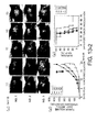
- Fig. 6 shows the effect of RENCA/SeV 18+mGM-CSF/TS ⁇ F on the tumor (kidney cancer) model mice (RENCA model mice).
- the tumor volume (left panel, a) and mouse survival rate (right panel, b) were monitored over time. The significant difference is indicated with an asterisk (*p ⁇ 0.05).
- the abbreviations in this figure are defined below:
- Fig. 7 shows the outline of the experimental procedure of CTL assay using IrRENCA/SeV18+mGM-CSF/TS ⁇ F.
- Fig. 8 presents the result of CTL assay using IrRENCA/SeV18+mGM-CSF/TS ⁇ F. The tumor-specific response in comparison to the negative control is shown.
- Fig. 9 shows the IFN- ⁇ and IL-4 production by splenocytes from mice immunized with IrRENCA/AdV+mGM-CSF or IrRENCA/SeV18+mGM-CSF evaluated using mouse IFN- ⁇ (a) and IL-4 (b) ELISPOT assays.
- Ten thousand splenocytes from RENCA-bearing mice vaccinated with the above-mentioned tumor vaccines were incubated for 20 hours with stimulator cells at the above-mentioned ratios.
- Bound cytokines were visualized by incubation with biotinylated anti-IFN- ⁇ and anti-IL-4 monoclonal antibodies, and then with streptavidin-HRP and premixed peroxidase substrate AEC. The result is expressed as the mean number of spot-forming cells + SD from quadruplicate determinations per 1 x 10 5 splenocytes.
- Figs. 9(c) to (h) present the result of mixed cultures of RENCA (cancer) cells and the above-mentioned splenocytes. Specifically, the cytokine production in the mixed-culture cells was determined. Tumor-specific cytokine production by splenocytes stimulated with GM-CSF-transduced tumor vaccination is shown.
- mice TNF- ⁇ (c), IFN- ⁇ (d), IL-2 (e), IL-4 (f), IL-5 (g), or IL-6 (h) in the culture supernatant was measured by CBA (c to g) or ELISA (h). Significant differences are denoted with asterisks (*p ⁇ 0.05). The abbreviations are defined below.
- Cell stimulation (+) irradiated RENCA (cancer) cells.
- Fig. 10 shows the immunophenotypic analysis of tumor-infiltrating cells in a tumor mass.
- RENCA-bearing mice were either left untreated (HBSS) or treated with the above-mentioned tumor vaccine cells (IrRENCA/AdV+GFP, IrRENCA/AdV+mGM-CSF, IrRENCA/SeV18+GFP/TS ⁇ F, or IrRENCA/SeV 18+mGM-CSF/TS ⁇ F), as described in "Materials and methods".
- Resected RENCA tumors were then subjected to immunohistological evaluation.
- Fig. 11 shows the antitumor effect of IrLLC/SeV 18+mGM-CSF/TS ⁇ F in tumor (lung cancer) model mice (LLC model mice). 2 x 10 5 mouse LLC tumor cells were inoculated subcutaneously into the right flank of C57BL/6 mice (day zero).
- Fig. 12 presents a graph showing the tumor formation-suppressing effect of SeV18+mGM-CSF/ ⁇ F, etc.
- the groups (1) to (11) are as follows:
- Figs. 13-1 and 13-2 show the anti-tumor effect of DC/SeV18+mGM-CSF/TS ⁇ F on a mouse tumor (lung cancer) model (LLC model).
- the present invention provides negative-strand RNA virus vectors comprising cytokine genes.
- a negative-strand RNA virus is an envelope virus that includes a minus-strand (an antisense strand against a sense strand that encodes viral proteins) RNA (also referred to as negative-strand RNA) as the genome.
- the virus exhibits high infectivity and is capable of overexpressing the genes carried by the virus in the cytoplasm.
- Negative-strand RNA viruses used in the present invention particularly include negative single-stranded RNA viruses (also referred to as “non-segmented negative-strand RNA viruses").
- the "negative single-stranded RNA viruses” refers to viruses that include a negative single-stranded (i.e., minus-strand) RNA as the genome.
- Such viruses include those belonging to Paramyxoviridae (including the genera Paramyxovirus, Morbillivirus, Rubulavirus, and Pneumovirus, etc.), Rhabdoviridae (including the genera Vesiculovirus, Lyssavirus, and Ephemerovirus, etc.), Filoviridae, Orthomyxoviridae, (including Influenza viruses A, B, and C, and Thogoto-like viruses, etc.), Bunyaviridae (including the genera Bunyavirus, Hantavirus, Nairovirus, and Phlebovirus, etc.), Arenaviridae, and such.
- Paramyxoviridae including the genera Paramyxovirus, Morbillivirus, Rubulavirus, and Pneumovirus, etc.
- Rhabdoviridae including the genera Vesiculovirus, Lyssavirus, and Ephemerovirus, etc.
- Filoviridae including Influenza viruses A, B, and C, and Thogoto-like viruses
- RNA viruses include the following: (i) there is no risk of integration into the genomic DNA, since the replication cycle occurs exclusively in the cytoplasm; (ii) the introduction efficiency does not depend on the cell cycle of target cells; (iii) homologous recombination does not occur between the virus and a different viral genome or a wild type virus; (iv) virus incorporation into cells requires only a very short contact time; (v) genes encoded by the virus can be strongly and controllably expressed in a broad range of host cells.
- the viruses are expected to be suitable for use in immunogene therapy that utilizes various cytokines due to the above-mentioned advantages and the induction of superior protective effect demonstrated herein.
- Negative-strand RNA viruses that are preferably used in the present invention include viruses of Paramyxoviridae, Orthomyxoviridae, and Rhabdoviridae. Among them, viruses of Paramyxoviridae and derivatives thereof are preferred as the vectors of the present invention.
- paramyxovirus refers to a virus of Paramyxoviridae, or a derivative thereof.
- Paramyxoviridae is a group of viruses that have a non-segmented negative-strand RNA as the genome, and includes Paramyxovirinae (including the genera Respirovirus (also referred to as Paramyxovirus ), Rubulavirus, and Morbillivirus ) and Pneumovirinae (including the genera pneumovirus and metapneumovirus ).
- the viruses that are used in the present invention are more preferably viruses belonging to Paramyxoviridae (including the genera Respirovirus, Rubulavirus, and Morbillivirus ) or derivatives thereof, and still more preferably viruses belonging to the genus Respirovirus (also referred to as the genus Paramyxovirus ) or derivatives thereof.
- the viruses belonging to Paramyxoviridae include the following: Sendai virus (SeV, also referred to as mouse parainfluenza virus-1), Newcastle disease virus (NDV), mumps virus, measles virus, respiratory syncytial virus (RS virus), rinderpest virus, distemper virus, simian parainfluenza virus 5 (SV5), SPIV-10), human parainfluenza viruses 1, 2, 3, 4a, and 4b (HPIV-1, 2, 3, 4a, and 4b), bovine parainfluenza virus-3 (BPIV-3), phocine distemper virus (PDV), canine distemper virus (CDV), dolphin molbillivirus (DMV), peste-des-petits-ruminants virus (PDPR), measles virus (MV), rinderpest virus (RPV), Hendra virus (Hendra), Nipah virus (Nipah), etc. More preferably, the paramyxovirus of the present invention is Sendai virus.
- Sendai virus
- Sendai virus is known to be pathogenic in rodents causing pneumonia, but is not pathogenic for human. This is also supported by a previous report that nasal administration of wild type Sendai virus does not have severely harmful effects on non-human primates ( Hurwitz, J.L. et al., Vaccine 15: 533-540, 1997 ; Bitzer, M. et al., J. Gene Med. 5: 543-553, 2003 ). These characteristics of Sendai virus suggest that when a vector is constructed based on Sendai virus, the vector can be applied to human treatment, and can be a promising choice in gene therapy for human cancer or such.
- derivatives of a virus refers to viruses that have been genetically modified or chemically modified in a manner not to impair the gene-transferring ability of the viral vectors.
- the viral vectors of the present invention may be derived from natural strains, wild type strains, mutant strains, laboratory-passaged strains, artificially-constructed strains, or such.
- gene refers to a genetic substance, i.e., a nucleic acid encoding a transcriptional unit.
- the genes include RNAs and DNAs.
- a nucleic acid encoding a protein is referred to as a gene for the protein.
- a gene may have a naturally-occurring or artificially-designed sequence.
- DNAs include both single-stranded and double-stranded DNAs.
- Encoding a protein means that a polynucleotide comprises an ORF that encodes an amino acid sequence of the protein in the sense or antisense direction, so that the protein can be expressed under appropriate conditions.
- Cytokines to be carried by the vectors of the present invention may be cytokines that induce immune cell differentiation and/or growth, and have anti-tumor activity.
- cytokines include cytokine that are produced by T cells, NK cells, monocytes, macrophages, or such, and induce T cell differentiation and/or growth.
- An immunostimulatory cytokine gene can be isolated, for example, from T-cell-derived cDNAs or such by PCR amplification using primers designed based on the sequence of the gene. Cytokines having anti-tumor activity are well known to those skilled in the art.
- Such cytokine genes can be preferably used in the present invention.
- IL-2 Iwadate, Y. et al., Cancer Res., 61: 8769-8774 (2001 );
- IL-2, IL-4, GM-CSF Sampson, J.H. et al., Proc. Natl. Acad. Sci. USA 93, 10399-10404 (1996 );
- GM-CSF Herrlinger, U. et al., Cancer Gene Ther. 4, 345-352 (1997 );
- IL-4 Seleh, M. et al., J. Natl. Cancer Inst.
- IL-4 Giezeman-Smits, K.M. et al., Cancer Res. 60, 2449-2457 (2000 )).
- IL-23 has been demonstrated to be closely associated with the migration of immune cells in brain autoimmune diseases ( Becher B. et al., J Clin Invest. 112(8), 1186-91 (2000 )).
- Fas-L has been shown to be effective as a chemoattractant ( Silvestris F et al., Br J Haematol. 122(1) 39-52. (2003 )).
- Cytokine genes that are particularly useful in the present invention include those encoding cytokines that have heparin-binding activity, such as GM-CSF. Furthermore, cytokine genes of the present invention also include genes encoding chemokines such as Thymus and activation-regulated chemokine (TARC) and RANTES.
- TARC Thymus and activation-regulated chemokine
- the sequence of GM-CSF is known, and one can obtain it, for example, using the Accession numbers M11220 (protein ID AAA52578), A14305 (protein ID CAA01150), etc. It is also possible to use known sequence information for other cytokines.
- Cytokine genes that are used in the present invention may be derived from human or other mammals, for example, mouse, rat, rabbit, pig, and primates, such as monkey.
- cytokines include variants of naturally occurring cytokines as long as they retain biological activity. Such variants include, for example, polypeptides with a deletion or addition of one to several amino acid residues (for example, two, three, four, five, or six residues) at the N- or C-terminus, and polypeptides with a substitution of one to several amino acid residues (for example, two, three, four, five, or six residues).
- the biological activity of a cytokine can be determined by known methods for assaying cytokine activity.
- the activity can be determined by the method for assaying tumor suppression described herein.
- Genes encoding variants with a biological activity equivalent to that of a naturally occurring cytokine are expected to exhibit an anti-tumor growth effect equivalent to that of the naturally occurring cytokine.
- Variants of a naturally occurring cytokine include fragments, analogues, and derivatives of a naturally occurring cytokine, and fusion proteins with other polypeptides (for example, a cytokine having a heterologous signal peptide and a polypeptide fused with an antibody fragment).
- cytokines that are used in the present invention include polypeptides that comprise a sequence with a substitution, deletion, and/or addition of one or more amino acids in the amino acid sequence of a naturally occurring cytokine or fragment thereof, and have a biological activity equivalent to that of the naturally occurring cytokine.
- the fragment refers to a polypeptide comprising a portion of a naturally occurring cytokine polypeptide, which includes, for example, N- or C-terminal truncated forms.
- Cytokine fragments with the biological activity typically comprise a continuous region of 70% or more, preferably 80% or more, more preferably 90% or more of a naturally occurring polypeptide (in its mature form after secretion).
- Amino acid sequence variants can be prepared, for example, by introducing mutations into DNAs that encode naturally occurring polypeptides ( Walker and Gaastra, eds. Techniques in Molecular Biology (MacMillan Publishing Company, New York, 1983 ); Kunkel Proc. Natl. Acad. Sci. USA 82:488-492, 1985 ; Kunkel et al. Methods Enzymol. 154:367-382, 1987 ; Sambrook et al. Molecular Cloning: A Laboratory Manual (Cold Spring Harbor Laboratory Press, Plainview, N.Y, 1989 ); U.S. Pat. No. 4,873,192 ).
- Guidance for substituting amino acids without affecting the polypeptide's biological activity includes, for example, Dayhoff et al. in Atlas of Protein Sequence and Structure (Natl. Biomed. Res. Found., Washington, D.C.) (1978)).
- the number of amino acids altered is not specifically limited, but is, for example, 30% or less of the total amino acids in the mature form of a naturally occurring polypeptide, preferably 25% or less, more preferably 20% or less, even more preferably 15% or less, still more preferably 10% or less. It is, for example, 15 amino acids or less, preferably ten amino acids or less, even more preferably eight amino acids or less, still more preferably five amino acids or less, yet more preferably three amino acids or less.
- amino acid substitution substituting amino acids with those that have side chains with similar properties is expected to maintain a protein's original activity. This substitution is referred to as "conservative substitution" in the present invention.
- Conservative substitutions include substitutions between amino acids within the same group, such as basic amino acids (for example, lysine, arginine, and histidine), acidic amino acids (for example, aspartic acid, and glutamic acid), non-charged polar amino acids (for example, glycine, asparagine, glutamine, serine, threonine, tyrosine, and cysteine), nonpolar amino acids (for example, alanine, valine, leucine, isoleucine, proline, phenylalanine, methionine, and tryptophan), ⁇ -branched amino acids (for example, threonine, valine, and isoleucine), and aromatic amino acids (for example, tyrosine, phenylalanine, tryptophan, and histidine).
- basic amino acids for example, lysine, arginine, and histidine
- acidic amino acids for example, aspartic acid, and glutamic acid
- Conservative substitutions also include, for example, substitutions between amino acids that give a positive score in the BLOSUM62 substitution matrix ( S. Henikoff and J.G. Henikoff, Proc. Acad. Natl. Sci. USA 89: 10915-10919, 1992 ).
- Cytokine variants also include polypeptides comprising an amino acid sequence with high homology to the amino acid sequence of a naturally occurring polypeptide.
- High homology amino acid sequences include those with an identity of, for example, 70% or higher, more preferably 75% or higher, even more preferably 80% or higher, still more preferably 85% or higher, yet more preferably 90% or higher, even still more preferably 93% or higher, yet still more preferably 95% or higher, yet still even more preferably 96% or higher.
- the amino acid sequence identity can be determined, for example, using the BLASTP program ( Altschul, S. F. et al., J. Mol. Biol. 215: 403-410, 1990 ).
- search is carried out on the BLAST web page of NCBI (National Center for Biotechnology Information) using default parameters, with all the filters including Low complexity turned off ( Altschul, S. F. et al. Nature Genet. 3:266-272, 1993 ; Madden, T. L. et al. Meth. Enzymol. 266:131-141, 1996 ; Altschul, S. F. et al. Nucleic Acids Res. 25:3389-3402, 1997 ; Zhang, J. & Madden, T. L. Genome Res. 7:649-656, 1997 ). Sequence identity can be determined, for example, by comparing two sequences using the Blast 2 sequences program to prepare an alignment of the two sequences ( Tatiana A et al.
- Gaps are treated in the same way as mismatches.
- an identity score is calculated in view of the entire amino acid sequence of a naturally occurring cytokine (its mature form after secretion). Specifically, the ratio of the number of identical amino acids to the total number of amino acids in a naturally occurring cytokine (mature form) is calculated.
- Preferred variants include polypeptides encoded by nucleic acids that hybridize under stringent conditions with the entire or a portion of the coding region of a naturally occurring cytokine gene, and have a biological activity equivalent to that of the naturally occurring cytokine.
- hybridization such a variant can be identified, for example, by preparing a probe either from a nucleic acid that comprises the sequence of the coding region of a naturally occurring cytokine gene or the complementary sequence thereof, or from a nucleic acid to be hybridized, and then detecting whether the probe hybridizes to the other nucleic acid.
- Stringent hybridization conditions are, for example, hybridization at 60°C, preferably at 65°C, more preferably at 68°C in a solution containing 5x SSC, 7%(W/V) SDS, 100 ⁇ g/ml denatured salmon sperm DNA, and 5x Denhardt's solution (1x Denhardt's solution contains 0.2% polyvinylpyrrolidone, 0.2% bovine serum albumin, and 0.2% Ficoll); and washing for two hours at the same temperature as the hybridization while shaking in 2x SSC, preferably 1x SSC, more preferably 0.5x SSC, still more preferably 0.1x SSC.
- Negative-strand RNA virus vector refers to a vehicle that is prepared based on the above-described negative-strand RNA virus and intended for introduction of the above-described cytokine gene into cells.
- infectivity refers to the capability of a negative-strand RNA viral vector to maintain the cell-adhesion ability toward a cell, and introduce a gene carried by the vector into the inside of the cell to which the vector has adhered.
- Negative-strand RNA, viral vectors of the present invention may be transmissible vectors or defective non-transmissible vectors. "Transmissible” means that when a viral vector infects a host cell, the virus can replicate within the cell to produce infectious virions.
- a virus based on a virus means that the components (proteins, RNAs, and such) of the virus are used in a vector in a non-modified or partially modified manner.
- a protein and RNA prepared by modifying a paramyxovirus protein and RNA is a protein and RNA "derived from a paramyxovirus", respectively.
- a paramyxovirus contains within its envelope a complex (ribonucleoprotein (RNP)) consisting of RNA and proteins.
- RNP ribonucleoprotein
- the RNA comprised in the RNP is the genome of paramyxovirus, which is a minus-strand (negative-strand) single-stranded RNA. This single-stranded RNA binds to the NP, P, and L proteins to form an RNP
- the RNA comprised in the RNP serves as the template for transcription and replication of the viral genome ( Lamb, R. A., and D. Kolakofsky, 1996, "Paramyxoviridae: The viruses and their replication" in Fields Virology, 3rd edn. Fields, B. N., D. M. Knipe, and P. M. Howley et al. (ed.), Raven Press, New York.N.Y.pp. 1177-1204, 1996 ).
- NP, P, M, F, HN, and L genes of Paramyxovirus refers to genes encoding the nucleocapsid, phospho, matrix, fusion, hemagglutinin-neuraminidase, and large proteins, respectively.
- the nucleocapsid (NP) protein binds to the genomic RNA and is essential for the template activity of the genomic RNA. In general, the NP gene is occasionally referred to as the "N gene”.
- the phospho (P) protein is a phosphorylated protein, which is a small subunit of RNA polymerase.
- the matrix (M) protein functions to support the viral particle structure from the inside.
- the fusion (F) protein is a membrane fusion protein involved in invasion into host cells.
- the hemagglutinin-neuraminidase (HN) protein is involved in adhesion to host cells.
- the large (L) protein is a large subunit of RNA polymerase.
- Each of the genes described above has an independent transcription regulatory unit. Thus, each gene is transcribed into an individual mRNA, which in turn is translated into a protein.
- the P gene is translated into the P protein, as well as a non-structural protein (C) by using an ORB other than that of the P protein, and another protein (V) produced via RNA editing of the P protein mRNA during translation.
- respective genes of viruses belonging to the Paramyxovirinae family are shown below (starting from the 3' end).
- the genus Respirovirus N P/C/V M F HN - L
- the genus Rubulavirus N P/V M F HN (SH) L
- the genus Morbillivirus N P/C/V M F H - L
- the negative-strand RNA virus vectors of the present invention may lack some of the wild-type viral genes.
- Such viruses can be reconstituted, for example, by exogenously supplying the gene products that are deficient in the viruses. Similar to wild-type viruses, the viruses thus prepared adhere to host cells and cause cell fusion, but cannot form daughter virions that retain the infectivity of the original vectors, since the viral genome introduced into cells is deficient in viral genes. Therefore, such vectors are useful as safe viral vectors that can introduce genes only once.
- viral gene-deficient vectors for example, two or more types of vectors which differ in the deficient viral gene on the viral genome carried by the vector are transduced into same cells. The respective deficient viral proteins are supplied through the expression from the other vector(s).
- the vectors complement each other to form infectious viral particles, resulting in a complete replication cycle and amplification of the viral vectors.
- two or more types of viral vectors of the present invention are inoculated in combination that allows complementation of viral proteins, a mixture of the viral gene-deficient vectors can be produced on a large scale at low cost. Since such viruses lack viral genes, their genome sizes are smaller than those of viral gene-nondeficient viruses and thus can advantageously carry larger foreign genes. Furthermore, these viruses that are non-proliferative due to the lack of viral genes become diluted outside cells, which makes it difficult to maintain their coinfection. The viruses become sterile and thus are advantageous from the viewpoint of controlling their environmental release.
- genes in which the genome may be deficient are the F gene, HN gene, M gene, or any combination thereof.
- recombinant viruses can be reconstituted by transfecting host cells with a plasmid expressing a recombinant negative-strand RNA viral genome deficient in the F gene, along with an F protein expression vector and expression vectors for the NP, P, and L proteins ( WO00/70055 , WO00/70070 , and WO03/025570 ; Li, H.-O. et al., J. Virol. 74(14) 6564-6569 (2000 )).
- Viruses can also be produced, for example, using host cells that have incorporated the F gene into their chromosomes.
- proteins which are expressed in virus-producing cells, do not need to have the same amino acid sequences as the viral sequences, and a mutant or homologous gene from another virus may be used as a substitute, as long as the activity in nucleic acid introduction is the same as, or greater than, that of the natural type.
- viruses of the present invention may be deficient in wild-type virus accessory genes.
- V gene which is one of the accessory genes of SeV
- the pathogenicity of SeV toward hosts such as mice is markedly reduced without hindering gene expression and replication in cultured cells ( Kato, A. et al., J. Virol. 71:7266-7272, 1997 ; Kato, A. et al., EMBO J. 16:578-587, 1997 ; Curran, J. et al., WO 01/04272 , EP 1067179 ).
- Such attenuated vectors are particularly useful as viral vectors for in vivo or ex vivo gene transfer with lower toxicity.
- the NP, P, and L proteins of a Paramyxoviridae virus bind to a negative single-stranded RNA, and play an essential role in genomic RNA replication and protein expression (hereinafter, the NP, P, and L proteins are occasionally referred to as "genomic RNA-binding proteins").
- the NP protein binds very tightly to the genomic RNA, and thereby confers the genomic RNA with template activity.
- the genomic RNA has template activity for RNA synthesis only when it binds to the NP protein.
- the RNA has no template activity when it is free from the NP protein.
- the P and L proteins bind to the genomic RNA as the small and large subunits of RNA polymerase, respectively. Therefore, in Paramyxoviridae viruses, genomic RNA replication cannot occur if any one of the NP, P, and L proteins is defective.
- Sendai virus vectors when used to express foreign genes, these vectors express the viral structural proteins (in particular, the NP, P, and L proteins, which are RNP-constituting proteins essential for transcription and replication) as well as the foreign genes that they carry.
- the viral structural proteins in particular, the NP, P, and L proteins, which are RNP-constituting proteins essential for transcription and replication
- immunogenic reaction against these viral structural proteins may be induced.
- various types of gene-deficient SeV vectors have been developed, considering the immunogenicity of the viral structural proteins.
- there are known vectors that are deficient in one or more of the M, F, and HN proteins, which are not viral RNP-constituting proteins WO 00/70070 ; WO 2003/025570 ).
- an attenuated negative-strand RNA virus in which the M protein-encoding gene has been deleted or inactivated has in addition to the advantage of attenuation, the advantage of no infection with daughter viruses, since no virion is released due to the loss of production of the M protein, which is essential for the particle formation after viral infection.
- a Sendai virus vector becomes a non-transmissible virus, thus securing safety.
- a Sendai virus vector has the advantage of not releasing newly formed viral particles from infected cells, since the activity to cleave cell surface sugar chains at sialic acid residues is inhibited.
- Such gene-deficient viral vectors are also included in the present invention.
- viral vectors in which temperature-sensitive mutations are introduced into the envelope protein genes (the M, F, and HN genes) encoded by the viral genomic RNA ( WO 2003/025570 ).
- Such vectors with introduced temperature-sensitive mutations can also be used as a viral vector of the present invention.
- NP, P, and L which are proteins constituting SeV vector RNP, are necessary for transcription and replication of the Sendai virus genome, and thus are normally essential for SeV vectors.
- viral vector particles can be reconstituted and amplified by externally supplying these proteins from expression plasmids or such.
- target cells are infected with reconstituted SeV vectors deficient in NP, P, and L, a foreign gene in the genome can be expressed by transcribing mRNAs of the gene using the NP, P, and L proteins carried in the particles.
- the SeV vectors deficient in NP, P, and L cannot replicate RNP due to lack of the NP, P, and L genes in the genome, and thus are turned into non-replicating SeV vectors. It was previously reported that vectors deficient in either of the NP or P protein gene were constructed to produce non-replicating SeV vectors ( Molecular Therapy, 13(Suppl.1), S185, May 2006 ; NSV2006 ( 13th International Conference Negative Strand Viruses 2006, Salamance, Spain, June 17-22nd, 2006 ), Abstract).
- the characteristics of a non-replicating vector are expected to be further assured by deleting the L gene or multiple genes including the L gene, rather than by deleting only the P or NP gene from the genome.
- the influence of virus-derived immunogenic proteins is expected to be minimized by deleting multiple genes including the L gene.
- deletion of multiple genes including the L gene for example, all of the NP, P, and L genes, is preferred from the viewpoint of immunogenicity reduction.
- deletion of the L gene which occupies about half of the genome length, is expected to enable the genome to carry a larger gene of interest.
- higher transcription efficiency and expression level of a gene of interest can be expected with a small genome size.
- L gene-deficient SeV vectors are considered to be very useful.
- the applicants have constructed non-replicative Paramyxoviridae viral vectors, and filed a patent application related to the vectors (Japanese Patent Application No. 2006-193433 ).
- the vectors of the present invention may be vectors in which the NP, P, and L genes have been completely or partially deleted.
- the NP, P, and L proteins can be incorporated into the vector by expressing these proteins in cells at the time of intracellular vector reconstitution. In this manner, viral vectors that maintain infectivity can be prepared. Once cells are infected with such vectors, proteins can be expressed from the genomic RNA by intracellular RNPs. However, since the vectors themselves do not have the L gene or such, they cannot reproduce viruses that have the replication ability of the original viruses. Such vectors are very useful for gene therapy and such, in particular, for treatment of target diseases that requires the influence of vectors to be minimized, and for other applications.
- nucleotide sequences of the genes of various viruses including Sendai virus are known. Such known information can be used to produce the vectors of the present invention.
- the database accession numbers for the nucleotide sequences of the Sendai virus genes are shown below: M29343, M30202, M30203, M30204, M51331, M55565, M69046, and X17218 (the N gene); M30202, M30203, M30204, M55565, M69046, X00583, X17007, and X17008 (the P gene); D11446, K02742, M30202, M30203, M30204, M69046, U31956, X00584, and X53056 (the M gene); D00152, D11446, D17334, D17335, M30202, M30203, M30204, M69046, X00152, and X02131 (the F gene); D26475, M12397, M30202, M30203, M30204, M69046, X00586, X02808, andX56131 (the HN gene); and D00053, M30202, M30203, M30204,
- viral genes encoded by other viruses are CDV, AF014953; DMV, X75961; HPIV-1, D01070; HPIV-2, M55320; HPIV-3, D10025; Mapuera, X85128; Mumps, D86172; MV, K01711; NDV, AF064091; PDPR, X74443; PDV, X75717; RPV, X68311; SeV, X00087; SV5, M81442; and Tupaia, AF079780 for the N gene; CDV, X51869; DMV, Z47758; SPIV-1, M74081; HPIV-3, X04721; HPIV-4a, M55975; HPIV-4b, M55976; Mumps, D86173; MV, M89920; NDV, M20302; PDV, X75960; RPV, X68311; SeV, M30202; SV5, AF
- Genes encoded by the viral genome RNA to be used as a vector of the present invention may have the original viral gene sequences, or may be deleted or inactivated as described above.
- a mutation(s) may be introduced into the genes.
- those skilled in the art can use known methods to introduce into genes in the genomic RNA, mild mutations that do not impair the function of the proteins.
- site-specific mutations can be introduced by PCR, cassette mutagenesis, or such.
- random mutations can be introduced by using chemical reagents, random nucleotides, or such.
- the cytotoxicity of a paramyxovirus vector can be reduced by introducing mutations into the P gene.
- mutations may be introduced into the RNA gene of the genome.
- the mutations include, for example, substitution of a different amino acid for Leu at position 511 (L511) of the SeV P protein, or substitution at the homologous position of the P protein in other negative-strand RNA viruses.
- the amino acid mutations may be desirable substitutions by a different amino acid, and preferably by amino acids whose side chains have different chemical properties.
- Amino acids can be categorized, for example, into groups such as basic amino acids (for example, lysine, arginine, and histidine), acidic amino acids (for example, aspartic acid and glutamic acid), non-charged polar amino acids (for example, glycine, asparagine, glutamine, serine, threonine, tyrosine, and cysteine), non-polar amino acids (for example, alanine, valine, leucine, isoleucine, proline, phenylalanine, methionine, and tryptophan), ⁇ -branched amino acids (for example, threonine, valine, and isoleucine), and aromatic amino acids (for example, tyrosine, phenylalanine, tryptophan, and histidine).
- basic amino acids for example, lysine, arginine, and histidine
- acidic amino acids for example, aspartic acid and glutamic acid
- the amino acid substitutions include substitution of an amino acid by another amino acid different those in the group to which the original amino acid belongs.
- the substitutions include substitution of a basic amino acid by an acidic or neutral amino acid; substitution of a polar amino acid by an non-polar amino acid; substitution of an amino acid whose molecular weight is higher than the average molecular weight of the 20 types of natural amino acids by an amino acid whose molecular weight is lower than the average molecular weight; and substitution of an amino acid whose molecular weight is lower than the average molecular weight by an amino acid whose molecular weight is higher than the average molecular weight; however, the substitutions are not limited to those described above.
- the substitutions include, for examples, the substitution of L511 by Phe (L511F).
- viral genome RNA refers to an RNA that has the function to form a ribonucleoprotein (RNP) with the viral proteins of a negative-strand RNA virus.
- RNP ribonucleoprotein
- the genes in the genome are expressed by the ribonucleoprotein, and the genomic RNA is replicated to form daughter RNPs.
- the genome of a negative-strand RNA virus is constituted so that the viral genes are situated in the antisense orientation between the 3'-leader region and 5'-trailer region.
- a transcription termination sequence (E sequence) - intervening sequence (I sequence) - transcription initiation sequence (S sequence) exists between the ORFs of individual genes, and allows the RNA encoding the ORF of each gene to be transcribed as a separate cistron.
- ORFs encoding the viral proteins contained in a vector, and ORFs of other foreign genes are arranged in the antisense direction in the genomic RNA via the above-described E-I-S sequence.
- the ORF clOsest to the 3'-end of the genomic RNA requires only an S sequence between the 3'-leader region and the ORF, and does not require an E or I sequence.
- the ORF closest to the 5'-end of the genomic RNA requires only an E sequence between the 5'-trailer region and the ORF, and does not require an I or S sequence.
- two ORFs can be transcribed as a single cistron, for example, by using an internal ribosome entry site (IRES) sequence.
- IRS internal ribosome entry site
- an E-I-S sequence is not required between these two ORFs.
- a typical RNA genome includes a 3'-leader region, six ORFs encoding the N, P, M, F, HN, and L proteins in the antisense direction in this order, and a 5'-trailer region on the other end.
- the orientation of the viral gene in the genomic RNAs of the present invention is not restricted.
- ORFs encoding the N, P, M, F, HN, and L proteins can be arranged after the 3'-leader region and before the 5'-trailer region.
- viruses have different viral genes, but even in such cases, it is possible to arrange each gene as in the wild type, as described above.
- vectors maintaining the N, P, and L genes can autonomously express genes from the RNA genome in cells and the genomic RNA is replicated, Furthermore, by the action of genes such as the F and HN genes which encode envelope proteins and the M gene, infectious virions are formed and released to the outside of the cells. Thus, such vectors become transmissible viral vectors.
- a cytokine gene to be carried by the vectors may be inserted into a non-protein-coding region in this genome, as described below.
- Viral vectors of the present invention encode cytokine genes in their genomic RNA.
- a recombinant viral vector harboring a cytokine gene is obtained by inserting a cytokine gene into an above-described viral vector genome.
- the cytokine gene can be inserted at any desired position in a non-protein-coding region of the virus genome, for example.
- the above nucleic acid can be inserted, for example, between the 3'-leader region and the viral protein ORF closest to the 3'-end; between each of the viral protein ORFs; and/or between the viral protein ORF closest to the 5'-end and the 5'-trailer region in genomic DNA.
- nucleic acids encoding the cytokine genes can be inserted into those deficient regions.
- An E-I-S sequence should be arranged between the inserted cytokine gene and the viral ORF. Two or more foreign genes can be inserted in tandem via E-I-S sequences.
- Expression levels of a foreign gene carried in a vector can be controlled using the type of transcriptional initiation sequence added upstream (to the 3'-side of the minus strand (negative strand)) of the gene ( WO01/18223 ).
- the expression levels can also be controlled by the position at which the foreign gene is inserted in the genome: the nearer to the 3'-end of the minus strand the insertion position is, the higher the expression level; while the nearer to the 5'-end the insertion position is, the lower the expression level.
- the insertion position of a foreign gene can be appropriately controlled such that the combination with genes encoding the viral proteins before and after the foreign gene is most suitable.
- a foreign gene is inserted between the 3'-leader region and the viral protein ORF closest to the 3'-end.
- a foreign gene may be inserted between the ORFs of the viral protein gene closest to the 3'-end and the second closest viral protein gene, or between the ORFs of the second and third closest viral protein genes.
- the viral protein gene closest to the 3'-end of the genome is the N gene
- the second closest gene is the P gene
- the third closest gene is M gene.
- the gene expression level from the viral vector can be suppressed to obtain an appropriate effect, for example, by inserting the foreign gene at a site as close as possible to the 5'-side of the minus strand genome, or by selecting an inefficient transcriptional initiation sequence.
- a desired S sequence of a negative-strand RNA virus may be used as the S sequence to be attached when inserting a foreign gene-encoding nucleic acid into the genome.
- Particularly preferred sequences are 3'-UCCCAGUUUC-5' (SEQ ID NO: 2), 3'-UCCCACUUAC-5' (SEQ ID NO: 3), and 3'-UCCCACUUUC-5' (SEQ ID NO: 4).
- a preferred E sequence of a Sendai viral vector is, for example, 3'-AUUCUUUU-5' (SEQ ID NO: 8) or 5'-TAAGAAAAA-3' (SEQ ID NO: 9) for the plus strand-encoding DNA.
- An I sequence may be, for example, any three nucleotides, specifically 3'-GAA-5' (5'-CTT-3' in the plus strand DNA).
- Negative-strand RNA viruses of the present invention may be, for example, complexes of negative-strand RNA viral genomic RNAs and viral proteins, that is, ribonucleoproteins (RNPs).
- RNPs can be introduced into cells, for example, in combination with desired transfection reagents.
- RNPs are complexes comprising a negative-strand RNA viral genomic RNA, N protein, P protein, and L protein.
- cistrons encoding the viral proteins are transcribed from the genomic RNA by the action of viral proteins, and, at the same time, the genome itself is replicated to form daughter RNPs. Replication of a genomic RNA can be confirmed by using RT-PCR, Northern blot hybridization, or such to detect an increase in the copy number of the RNA.
- the vectors of the present invention may be of any type as long as they have the above-mentioned characteristics, and include a viral vector that has a viral particle structure, an RNP vector which is itself RNP, and such.
- recombinant viruses that include an envelope protein other than that of the virus from which the viral genome was derived, may be prepared as viral vectors used in the present invention.
- a recombinant virus including a desired envelope protein can be generated by expressing in a cell an envelope protein other than the envelope protein originally encoded by the basic viral genome.
- Such proteins are not particularly limited.
- a desired protein that confers an ability to infect cells may be used.
- examples of such proteins include the envelope proteins of other viruses, for example, the G protein of vesicular stomatitis virus (VSV-G).
- VSV-G vesicular stomatitis virus
- the VSV-G protein may be derived from an arbitrary VSV strain.
- VSV-G proteins derived from Indiana serotype strains may be used, but the present invention is not limited thereto.
- the present vector may include any arbitrary combination of envelope proteins derived from other viruses.
- Preferred examples of such proteins are envelope proteins derived from viruses that infect human cells.
- Such proteins are not particularly limited, and include retroviral amphotropic envelope proteins and such.
- the envelope proteins derived from mouse leukemia virus (MuLV) 4070A strain can be used as the retroviral amphotropic envelope proteins.
- envelope proteins derived from MuMLV 10A1 strain may also be used (for example, pCL-10A1 (Imgenex) ( Naviaux, R. K.
- the proteins of Herpesviridae include, for example, gB, gD, gH, and gp85 proteins of herpes simplex viruses, and gp350 and gp220 proteins of EB virus.
- the proteins of Hepadnaviridae include the S protein of hepatitis B virus. These proteins may be used as fusion proteins in which the extracellular domain is linked to the intracellular domain of the F or HN protein.
- the viral vectors of the present invention include pseudotype viral vectors comprising envelope proteins such as VSV-G, which are derived from viruses other than the virus from which the genome was derived. If the viral genomic RNAs are designed in a way that these envelope proteins are not encoded by the genome, the proteins will not be expressed from the viral vectors after virions infect the cells.
- the viral vectors used in the present invention may be, for example, vectors that comprise on their envelope surface, proteins such as adhesion factors capable of adhering to specific cells, ligands, receptors, antibodies or fragments thereof, or chimeric proteins that have the above proteins in the extracellular domain and polypeptides derived from the viral envelope in the intracellular domain.
- proteins may be encoded in the viral genome, or supplied by expression of genes other than those in the viral genome (for example, genes in other expression vectors, the host chromosome, or such) at the time of viral reconstitution.
- a cloning site may be designed at the position of insertion.
- the cloning site can be made into, for example, a recognition sequence for restriction enzyme.
- Foreign gene fragments can be inserted into the restriction enzyme site in the vector DNA encoding the genome.
- Cloning site may be arranged to be a so-called multi-cloning site comprising a plurality of restriction enzyme recognition sequences.
- Vectors of the present invention may thus harbor additional foreign genes.
- the vectors of the present invention may carry a foreign gene different from the above-mentioned cytokine and other genes.
- a cDNA encoding a genomic RNA of a negative-strand RNA virus is transcribed in mammalian cells, in the presence of viral proteins (i.e., the N, P, and L proteins) essential for reconstitution of an RNP including the genomic RNA of the negative-strand RNA virus.
- viral proteins i.e., the N, P, and L proteins
- Viral RNP can be reconstituted by producing either the negative-strand genome (the same antisense strand as the viral genome) or the plus strand (antigenome, the complementary strand of the genomic RNA). For increasing the efficiency of vector reconstitution, it is more preferable to produce the plus strand.
- the RNA terminals preferably reflect the terminals of the 3'-leader sequence and 5'-trailer sequence as accurately as possible, as in the natural viral genome.
- the recognition sequence of T7 RNA polymerase may be used as a transcription initiation site and the RNA polymerase may be expressed within a cell.
- a self-cleaving ribozyme can be encoded at the 3'-end of the transcript, allowing accurate cleavage of the 3'-end with this ribozyme ( Hasan, M. K. et al., J. Gen. Virol. 78: 2813-2820,1997 ; Kato, A.
- Sendai virus-based viral vectors of the present invention that carry a cytokine gene can be constructed, for example, by the following procedure according to the method described in documents such as Kato, A. et al., EMBO J. 16: 578-587, 1997 ; and Yu, D. et al., Genes Cells 2: 457-466, 1997 .
- a DNA sample comprising the cDNA nucleotide sequence of a desired cytokine gene is prepared. It is preferable that the DNA sample is at a concentration of 25 ng/ ⁇ l or more and can be electrophoretically identified as a single plasmid.
- a cytokine gene is inserted to a DNA encoding a viral genome utilizing the Nat I site will be described as an example.
- the Not I recognition site is included in the cDNA nucleotide sequence of interest, it is preferable to delete the Not I site beforehand by modifying the nucleotide sequence using site-specific mutagenesis and such, so as not to alter the amino acid sequence encoded by the cDNA.
- the desired gene fragment is amplified and recovered by PCR.
- a forward synthetic DNA sequence (sense strand) and reverse synthetic DNA sequence (antisense strand) are prepared as a pair of primers containing the Not I restriction enzyme cleavage site sequence, the transcription termination sequence (E), intervening sequence (I), and transcription initiation sequence (S), and a partial sequence of the gene of interest.
- the forward synthetic DNA sequence is arranged in a form, in which any two or more nucleotides (preferably four nucleotides excluding GCG and GCC, which are sequences originating in the Not I recognition site, more preferably ACTT) are selected on the 5'-side of the synthetic DNA, the Not I recognition site "gcggccgc" is added to its 3'-side, and to the 3'-side thereof, any nine nucleotides or nucleotides of nine plus a multiple of six nucleotides are added as the spacer sequence, and to the 3'-side thereof, a sequence of about 25 nucleotides from the ORF including the initiation codon ATG of the desired cDNA is added. It is preferable to select about 25 nucleotides from the desired cDNA as the forward side synthetic DNA sequence so that it has G or C as the final nucleotide on its 3'-end.
- any two or more nucleotides preferably four nucleotides excluding GCG and GCC
- any two or more nucleotides are selected from the 5'-side of the synthetic DNA, and the Not I recognition site "gcggccgc" is added to its 3'-side. Further to its 3'-side, an oligo DNA is added as an insertion fragment to adjust the length.
- the number of nucleotides in the oligo DNA is determined so that the total nucleotide number of the Sendai virus genome becomes a multiple of six (so-called "rule of six"; Kolakofski, D. et al., J. Virol.
- sequences complementary to the S sequence of Sendai virus preferably 5'-CTTTCACCCT-3' (SEQ ID NO: 10), the I sequence, preferably 5'-AAG-3', and the E sequence, preferably 5'-TTTTTCTTACTACGG-3' (SEQ ID NO: 11), are added.
- a complementary sequence of about 25 nucleotides counted in the reverse direction from the termination codon of the desired cDNA sequence is selected and added as the 3'-end of the reverse synthetic DNA, wherein the length of the complementary sequence is adjusted so that the final nucleotide is G or C.
- PCR can be performed according to conventional methods with, for example, ExTaq polymerase (Takara Shuzo).
- PCR is performed using Vent polymerase (NEB), and the fragments of interest thus amplified are digested with Not I , then inserted to the Nat I site of the plasmid vector pBluescript.
- Nucleotide sequences of the PCR products thus obtained are confirmed with a sequencer to select a plasmid having the correct sequence.
- the inserted fragment is excised from the plasmid using Not I , and cloned to the Not I site of a plasmid carrying the genomic cDNA deficient in envelope genes.
- it is also possible to obtain a recombinant Sendai virus cDNA by directly inserting the fragment to the Not I site without mediation of the plasmid vector pBluescript.
- the viral vector cDNA of the present invention is transcribed in vitro or in cells to produce RNA, which is as necessary allowed to bind to separately expressed L, P, and NP proteins to reconstitute an RNP.
- viral vectors comprising the RNP can be produced.
- known methods WO 97/16539 ; WO 97/16538 ; Durbin, A.P. et al., Virology 235: 323-332, 1997 ; Whelan, S. P. et al., Proc. Natl. Acad. Sci. USA 92: 8388-8392, 1995 ; Schnell. M. J. et al., EMBO J. 13: 4195-4203, 1994 ; Radecke, F.
- viral particles can be reconstituted from a viral vector cDNA encoding a negative single-stranded RNA, which has been modified so as not to express some of the virus-derived proteins, or the complementary strand thereof.
- improved methods are effective ( WO 2005/071092 ).
- Genes that have been deleted from the viral vector cDNA of the present invention can be expressed by introducing them into host cells separately.
- the viral particles When viral particles are reconstituted by forming RNPs and expressing envelope proteins using genes encoded by the viral vector cDNA of the present invention or separate expression vectors, the viral particles can be infectious.
- each gene may be integrated into the host cell chromosome to complement the gene deficiency.
- Genes expressed to form RNPs, such as the NP, P, and L genes are not necessarily completely identical to those encoded in the virus genome on which the vector is based.
- the amino acid sequences of the proteins encoded by these genes are not necessarily identical to those of the proteins encoded by the RNP genome. Mutations may be introduced into the proteins, or homologous genes of other viruses may be used as substitutes, as long as the proteins bind to the genomic RNA and have the activity of transcribing and replicating the genome in cells.
- the viral vectors of the present invention can be amplified by introducing the vectors into cells that allow viral propagation (in the case of viral vectors in which the genes essential for viral expression and propagation have been deleted, cells that express the genes deficient in the viral vectors); culturing the cells; and collecting virions from the culture supernatant.
- the cells into which the vectors are introduced may express proteins that have the function to promote formation and release of virions, for example, the C protein. High viral vector productivity can be achieved by expressing the C protein.
- the Sendai virus C protein includes four types of proteins, C', C, Y1, and Y2, which are translated using different initiation codons in the same reading frame.
- any one, or two or more, or all of the four types of C proteins described above may be expressed in helper cells at the time of viral vector reconstitution.
- Deletion of the P gene from the genomic RNA of the viral vectors of the present invention results in deletion of the C gene which is in a reading frame different from that of the P gene.
- the C gene can be expressed in cells by introducing an expression vector that encodes the C gene into the cells.
- the host cells used in the reconstitution are not particularly limited.
- the host cells used in the reconstitution are not particularly limited.
- cultured cells such as LLC-MK2 cells and CV-1 cells derived from monkey kidney, BHK cells derived from hamster kidney, and cells derived from humans.
- infectious virions including the proteins in the envelope can also be obtained.
- a viral vector obtained from an above-described host can be used to infect embrionated hen eggs to amplify the vector. Methods for manufacturing viral vectors using hen eggs have already been developed ( Nakanishi, et al., ed.
- a fertilized egg is placed in an incubator, and cultured for nine to twelve days at 37 to 38°C to grow an embryo. After the viral vector is inoculated into the allantoic cavity, the egg is cultured for several days (for example, three days) to proliferate the viral vector. Conditions such as the period of culture may vary depending upon the recombinant Sendai virus being used. Then, allantoic fluids including the vector are recovered.
- RNP may be introduced into cells as a complex formed together with, for example, lipofectamine or polycationic liposomes.
- transfection reagents include, for example, DOTMA (Boehringer), Superfect (QIAGEN #301305), DOTAP, DOPE, DOSPER (Boehringer #1811169), and Lipofectamine 2000 (Invitrogen).
- Chloroquine may be added to prevent decomposition in endosomes ( Calos, M.P., 1983, Proc. Natl. Acad. Sci. USA 80: 3015 ).
- Major methods for introducing viral vector cDNAs into cells include those described below. (i) methods of preparing DNA precipitates that can be internalized into the cells of interest; (ii) methods of preparing positively-charged complexes comprising DNAs that are suitable for internalization into the cells of interest, and have low cytotoxicity; and (iii) methods of momentarily creating holes with a size sufficient for DNA molecules to pass through on the membrane of cells of interest using electric pulses.
- the methods of (i) include, for example, transfection methods using calcium phosphate. It is known that while DNA incorporated into cells by these methods is internalized into phagosomes, a sufficient amount of the DNA also enter the nucleus ( Graham, F. L. and Van Der Eb, J., Virology 52: 456, 1973 ; Wigler, M. and Silverstein, S., Cell 11: 223, 1977 ).
- transfection reagents can be used in the methods of (ii).
- Such reagents include, for example, DOTMA (Boehringer), Superfect (QIAGEN #301305), DOTAP, DOPE, DOSPER (Boehringer #1811169), and Lipofectamine 2000 (Invitrogen).
- the methods of (ii) are suitable for transient transfection. Methods for performing transfection by preparing a DEAE-dextran (Sigma #D-9885 M.W 5 x 10 5 ) mixture with a desired DNA concentration ratio have been known for a long time. Since most complexes are degraded in endosomes, chloroquine may be added to enhance the effect ( Calos, M.P., Proc. Natl. Acad. Sci. USA 80: 3015, 1983 ).
- electroporation methods which are not cell-selective, and thus have high versatility compared to the methods of (i) and (ii).
- the efficiency of these methods is assumed to be high under optimal conditions for the duration of pulse electric current, shape of the pulse, potency of electric field (gap between electrodes, voltage), buffer conductivity, DNA concentration, and cell density.
- Transfection reagents that can be suitably used in the present invention include Lipofectamine 2000 (Invitrogen), Superfect Transfection Reagent (QIAGEN, Cat No. 301305), and DOSPER Liposomal Transfection Reagent (Boehringer Mannheim, Cat No. 1811169).
- virus reconstitution from cDNA can be carried out, for example, as follows: In a plastic plate of about 6 to 24 wells, or a 100-mm Petri dish or such, simian kidney-derived LLC-MK2 cells (ATCC CCL-7) are cultured up to about 100% confluency, using minimum essential medium (MEM) containing 10% fetal calf serum (FCS) and antibiotics (100 units/ml penicillin G and 100 ⁇ g/ml streptomycin).
- MEM minimum essential medium
- FCS fetal calf serum
- antibiotics 100 units/ml penicillin G and 100 ⁇ g/ml streptomycin
- PFU plaque forming units
- vTF7-3 which expresses T7 RNA polymerase and has been inactivated by 20-minutes of UV irradiation in the presence of 1 ⁇ g/ml psoralen
- Fuerst, T. R. et al., Proc. Natl. Acad. Sci. USA 83: 8122-8126,1986 ; Kato, A. et al., Genes Cells 1: 569-579, 1996 The amount of psoralen added and the UV irradiation time can be appropriately adjusted.
- One hour after infection, 2 to 60 ⁇ g, and more preferably 3 to 20 ⁇ g, of DNA encoding the genomic RNA of a recombinant Sendai virus is transfected along with the plasmids expressing trans-acting viral proteins essential for viral RNP generation (0.5 to 24 ⁇ g of pGEM-N, 0.25 to 12 ⁇ g of pGEM-P, and 0.5 to 24 ⁇ g of pGEM-L) ( Kato, A. et al., Genes Cells 1: 569-579, 1996 ), using the lipofection method or such with Superfect (QIAGEN).
- the ratio of the amounts of expression vectors encoding the N, P, and L proteins is preferably 2:1:2, and the plasmid amounts are appropriately adjusted in the range of 1 to 4 ⁇ g of pGEM-N, 0.5 to 2 ⁇ g of pGEM-P, and 1 to 4 ⁇ g of pGEM-L.
- the transfected cells are cultured, as desired, in serum-free MEM supplemented with 100 ⁇ g/ml of rifampicin (Sigma) and cytosine arabinoside (AraC), more preferably only 40 ⁇ g/ml of cytosine arabinoside (AraC) (Sigma).
- Optimal drug concentrations are set so as to minimize cytotoxicity due to the vaccinia virus, and to maximize virus recovery rate ( Kato, A. et al., 1996, Genes Cells 1: 569-579 ). After culturing for about 48 to 72 hours after transfection, cells are harvested, and then disrupted by repeating freeze-thawing three times.
- LLC-MK2 cells are re-transfected with the disrupted materials containing RNP, and cultured. Alternatively, the culture supernatant is recovered, added to a culture medium of LLC-MK2 cells to infect them, and the cells are then cultured. Transfection can be conducted by, for example, forming a complex with lipofectamine, polycationic liposome, or such, and transducing the complex into cells. Specifically, various transfection reagents can be used. For example, DOTMA (Roche), Superfect TM (QIAGEN #301305), DOTAP, DOPE, and DOSPER (Roche #1811169) may be cited. In order to prevent degradation in the endosome, chloroquine may also be added ( Calos, M.
- LLC-MK2 cells expressing the envelope protein may be used for transfection, or a plasmid expressing the envelope protein may be cotransfected.
- an envelope-gene defective type virus can be amplified by culturing the transfected cells overlaid with LLK-MK2 cells expressing the envelope protein (see WO00/70055 and WO00/70070 ).
- Titers of collected virus vectors can be determined, for example, by measuring CIU (Cell Infectious Unit) or Hemagglutination Activity (HA) ( WO 00/70070 ; Kato, A. et al., Genes Cells 1: 569-579,1996 ; Yonemitsu, Y. & Kaneda, Y., Hemagglutinating virus of Japan-liposome-mediated gene delivery to vascular cells. Ed. by Baker AH. Molecular Biology of Vascular Diseases. Method in Molecular Medicine: Humana Press: pp. 295-306, 1999 ).
- CIU Cell Infectious Unit
- HA Hemagglutination Activity
- titers of vectors carrying marker genes such as GFP can be quantified by directly counting infected cells, using the marker as an index (for example, as GFP-CIU). Titers thus determined can be treated in the same way as CIU ( WO 00/70070 ).
- the collected virus vectors can be purified to be substantially pure. Purification can be achieved using known purification/separation methods including filtration, centrifugation, column purification, and such, or any combination thereof. "Substantially pure" means that the viral vector is a component that accounts for the major proportion of a sample containing the vector. Typically, a viral vector can be confirmed to be substantially pure when the percentage of the viral vector-derived proteins relative to the total proteins (excluding proteins added as carriers, stabilizers, and such) in a sample is 10% or more, preferably 20% or more, more preferably 50% or more, preferably 70% or more, more preferably 80% or more, and even more preferably 90% or more.
- JP-B Japanese Patent Application Kokoku Publication No.
- JP-B S62-30752 Examined, approved Japanese patent application published for opposition
- JP-B S62-33879 JP-B S62-30753
- methods of adsorbing viruses to fucose sulfate-containing polysaccharides and/or degradation products thereof WO97/32010 .
- a PCR product formed in PCR by using the pair of primers [forward: 5'-gttgagtactgcaagagc/SEQ ID NO: 13, reverse: 5'-tttgccggcatgcatgtttcccaaggggagagttttgcaacc/SEQ ID NO: 14] is connected upstream of F, and a PCR product formed using the pair of primers [forward: 5'-atgcatgccggcagatga/SEQ ID NO: 15, reverse: 5'-tgggtgaatgagagaatcagc/SEQ ID NO: 16] is connected downstream of the F gene with Eco T22I.
- the plasmid thus obtained is digested with Sac I and Sal I to recover a 4931 bp fragment of the region including the F gene-deficient site, which is cloned into pUC18 to form pUC18/dFSS.
- This pUC18/dFSS is digested with Dra III , the fragment is recovered, replaced with the Dra III fragment of the region comprising the F gene of pSeV18 + , and ligated to obtain the plasmid pSeV18 + / ⁇ F.
- a foreign gene is inserted, for example, into the Nsi I and Ngo MIV restriction enzyme sites in the F gene-deficient site of pUC18/dFSS.
- a foreign gene fragment may be, for example, amplified using an Nsi I-tailed primer and an Ngo MIV-tailed primer.
- the SeV-F gene is amplified by PCR, and inserted to the unique Swa I site of the plasmid pCALNdlw ( Arai, T. et al., J. Virology 72, p.1115-1121, 1998 ), which is designed to enable the inducible expression of a gene product by Cre DNA recombinase, thus constructing the plasmid pCALNdLw/F.
- a helper cell line expressing SeV-F protein is established.
- LLC-MK2 cell line which is commonly used for SeV proliferation, can be used as the cells, for example. LLC-MK2 cells are cultured in MEM supplemented with 10% heat-inactivated fetal bovine serum (FBS), penicillin G sodium (50 units/ml), and streptomycin (50 ⁇ g/ml) at 37°C in 5% CO 2 .
- FBS heat-inactivated fetal bovine serum
- penicillin G sodium 50 units/ml
- streptomycin 50 ⁇ g/ml
- the above-described plasmid pCALNdLw/F which was designed to enable inducible expression of the F gene product with Cre DNA recombinase, is transfected to LLC-MK2 cells by the calcium phosphate method (using a mammalian transfection kit (Stratagene)), according to protocols well known in the art.
- the plasmid pCALNdLw/F (10 ⁇ g) is transduced into LLC-MK2 cells grown to 40% confluency using a 10-cm plate, and the cells are then cultured in MEM (10 ml) containing 10% FBS, in a 5% CO 2 incubator at 37°C for 24 hours.
- the cells are detached and suspended in the medium (10 ml).
- the suspension is then seeded into five 10-cm dishes, 5 ml into one dish, 2 ml each into two dishes, and 0.2 ml each into two dishes, and cultured in MEM (10 ml) containing G418 (GIBCO-BRL) (1200 ⁇ g/ml) and 10% FBS.
- MEM 10 ml
- G418 G418
- FBS 10% FBS
- the cells are cultured for 14 days, exchanging the medium every two days, to select cell lines stably transduced with the gene.
- the cells grown in the above medium that show G418 resistance are recovered using cloning rings. Culture of each clone thus recovered is continued in 10-cm plates until confluent.
- LLC-MK2 cells are seeded on 100-mm dishes at 5 x 10 6 cells/dish.
- T7 RNA polymerase To transcribe the genomic RNA using T7 RNA polymerase, the cells are cultured for 24 hours, and then recombinant vaccinia virus, which expresses T7 RNA polymerase (PLWUV-VacT7: Fuerst, T.R. et al., Proc. Natl. Acad. Sci.
- UV Stratalinker 2400 with five 15-watt bulbs (catalog No. 400676 (100V); Stratagene, La Jolla, CA, USA).
- plasmid expressing the genomic RNA and expression plasmids each expressing N, P, L, F, or HN protein of the negative-strand RNA virus are transfected into the cells using an appropriate lipofection reagent.
- the plasmid ratio is preferably, but is not limited to, 6:2:1:2:2:2 in this order.
- the expression plasmid for the genomic RNA, and the expression plasmids each of which expresses N, P, or L protein, or F and HN proteins pGEM/NP, pGEM/P, pGEM/L, and pGEM/F-HN; WO00/70070 , Kato, A.
- the suspension is subjected to three freeze-thaw cycles, and mixed with lipofection reagent DOSPER (Roche #1811169) (10 6 cells/25 ⁇ l DOSPER). After the mixture is allowed to stand at room temperature for 15 minutes, it is transfected to F-expressing helper cells (10 6 cells/well in 12-well-plate) cloned as described above. The cells are cultured in serum-free MEM (containing 40 ⁇ g/ml AraC and 7.5 ⁇ g/ml trypsin), and the supernatant is collected. Viruses deficient in a gene other than F, for example, HN or M gene, can be prepared by a similar method as described above.
- the vectors of the present invention can be prepared as compositions depending on the purpose.
- the vectors may be combined with desired pharmaceutically acceptable carriers or media according to needs.
- pharmaceutically acceptable carriers or medina refers to materials that can be administered together with the vectors and that do not significantly inhibit the gene transfer via the vectors.
- Such carriers and media include, for example, sterile water, sodium chloride solution, dextrose solution, Ringer's solution containing dextrose, sodium chloride, and lactate, culture medium, serum, and phosphate buffered saline (PBS). They may be appropriately combined with the vectors to formulate a composition.
- compositions of the present invention may include carriers or media such as deionized water and aqueous dextrose solution.
- the compositions may also include membrane stabilizers for liposome (for example, sterols such as cholesterol).
- the compositions may also include antioxidants (for example, tocopherol or vitamin E).
- the compositions may also include vegetable oils, suspending agents, detergents, stabilizers, biocidal agents, and such. Furthermore, preservatives and other additives may also be added.
- the formula of the present composition may be aqueous solution, capsule, suspension, syrup, or such.
- the compositions of the present invention may also be in a form of solution, freeze-dried product, or aerosol. When it is a freeze-dried product, it may include sorbitol, sucrose, amino acids, various proteins, and such as a stabilizer.
- the present invention includes anti-tumor agents containing a negative-strand RNA viral vector that encodes a cytokine.
- the present invention includes cells into which a negative-strand RNA viral vector that encodes a cytokine of the present invention has been introduced, and anti-tumor agents containing such cells.
- Compositions containing the vectors of the present invention and cells introduced with the vectors are useful as anti-tumor pharmaceuticals.
- the vector compositions of the present invention and the cells introduced with the vectors are also useful as anti-tumor vaccines.
- the vector compositions and the cells may include immunostimulants such as cytokine, cholera toxin, and Salmonella toxin to improve immunogenicity.
- the vaccines may be combined with adjuvants, such as alum, incomplete Freund's adjuvant, MF59 (oil emulsion), MTP-PE (muramyl tripeptide derived from cell wall of mycobacteria), and QS-21 (derived from soapbark tree Quilaja saponaria ).
- adjuvants such as alum, incomplete Freund's adjuvant, MF59 (oil emulsion), MTP-PE (muramyl tripeptide derived from cell wall of mycobacteria), and QS-21 (derived from soapbark tree Quilaja saponaria ).
- cytokines that improve the adjuvant effect.
- cytokines include, for example, IL-12 ( Proc. Natl. Acad. Sci. USA 96 (15): 8591-8596, 1999 ), interferon- ⁇ ( U.S. Patent No. 5,798,100 ), and IL-4 ( J. Neurosurgery 90 (6), 1115-1124 (1999 )).
- the above-described viral vectors of the present invention, and compositions or cells comprising the vectors can induce in vivo immune responses by cytokine production in the cells into which the vectors have been introduced.
- the vectors are preferably used, for example, in gene therapy against tumors for which cytokines are effective.
- the viral vectors of the present invention encoding GM-CSF are expressed in various cancer cells, and have an in vivo anti-tumor activity, as described in the Examples in this specification.
- anti-tumor activity refers to an activity of suppressing tumor development and/or growth.
- the viral vectors of the present invention, or compositions or cells comprising the vectors can suppress tumor formation (including recurrence) or growth (including metastasis) by induction of anti-tumor immune response at the administration site, when they are locally administered to a tumor tissue, a risky site of tumor development (for example, a site from which tumor has been removed), or such.
- the vector introduction can be achieved in vivo or ex vivo.
- the vectors are injected directly into tumor sites.
- the vectors are introduced into cells outside the body and the cells are injected into tumor sites.
- a tumor site refers to a tumor itself, a tumor excision site, or a region adjacent thereto.
- the adjacent region refers to a region from which cytokines secreted from cells introduced with the vectors can reach the tumor or tumor excision site.
- the region is preferably within 5 mm, for example, within 3 mm, 2 mm, or 1 mm of a tumor or tumor excision site.
- the vector introduction may be conducted by dissolving or suspending a vector or cells introduced with the vector in a desired carrier (desired physiological aqueous solution, for example, culture medium, physiological saline, blood, plasma, serum, body fluid, etc.), and then directly injecting them into a tumor or a region adjacent thereto.
- a desired carrier for example, culture medium, physiological saline, blood, plasma, serum, body fluid, etc.
- MOI multiplicity of infection, the number of infecting viruses per cell
- MOI multipleplicity of infection, the number of infecting viruses per cell
- Contact between the vector and target cells needs only a short period of time, which may be, for example, 1 minute or longer, preferably 3 minutes or longer, 5 minutes or longer, 10 minutes or longer, or 20 minutes or longer, about 1 to about 60 minutes, and more specifically about 5 to 30 minutes. Also, the contact time may be longer, for example, several days or longer.
- Cells derived from a patient to be administered may be used, for example, primary cultured fibroblast cells derived from a patient can be preferably used.
- xenogeneic cells and allogeneic cells can be used ( Iwadate, Y. et al., Cancer Res., 61: 8769-8774, 2001 ).
- Xenogeneic or allogeneic cells are expected to be eliminated through host immune response after ex-vivo injection.
- the cells may be treated by UV, X-ray or gamma-ray irradiation, or such to make their dividing ability defective, and introduced ex vivo into tumors subsequently.
- the vectors of the present invention were demonstrated to have the ability to express genes in various cancer cells, as described in the Examples herein.
- the vectors can be used to treat various tumors.
- metastatic renal cell cancer kidney cancer
- the five-year survival rate after treatment is only about 10% even when the disease is treated with conventional therapy such as surgical therapy, radiotherapy, chemotherapy, or such.
- conventional therapy such as surgical therapy, radiotherapy, chemotherapy, or such.
- the vectors of the present invention are also expected to be effective against metastatic cancers including metastatic kidney cancer, since they are delivered to tumor tissues via blood stream and expected to have fewer adverse effects as compared to anticancer agents used in conventional chemotherapy.
- tumor-specific immunity was demonstrated to be induced in three out of four cases.
- administration of a constant dose of IL-2 after inoculation of the vaccine cells resulted in the survival of two patients out of four for five years or longer (one patient has survived for seven years and eight months).
- the vectors of the present invention which encode the cytokines, GM-CSF and/or IL-2, are effective against metastatic cancers including metastatic kidney cancer.
- the dosage of the vectors of the present invention can be appropriately determined by those skilled in the art, although it varies depending on the disease, patient's weight, age, sex, symptom, objective of administration, form of composition administered, administration method, type of gene to be introduced, and such.
- the route of administration can be appropriately selected, and includes, for example, percutaneous, intranasal, transbronchial, intramuscular, intraperitoneal, intravenous, intraarticular, intraspinal, and subcutaneous administrations, but is not limited thereto. Intranasal administration is a preferred administration route.
- the administration may be local and/or systemic.
- the vectors at a dosage within the range of preferably about 10 5 CIU/ml to about 10 11 CIU/ml, more preferably about 10 7 CIU/ml to about 10 9 CIU/ml, and still more preferably about 1 x 10 8 CIU/ml to about 5 x 10 8 CIU/ml, together with a pharmaceutically acceptable carrier.
- a single dose for human is preferably 2 x 10 5 CIU to 2 x 10 10 CIU.
- the frequency of administration can be once or more, and within the range of clinically acceptable side effects. The same applies to the daily administration frequency.
- the protein dosage may be, for example, within the range of 10 ng/kg to 100 ⁇ g/kg, preferably 100 ng/kg to 50 ⁇ g/kg, and more preferably 1 ⁇ g/kg to 5 ⁇ g/kg.
- the dosage to be administered can be converted from the above-described dosage based on the body weight ratio or volume ratio (e.g., average value) of the target site for administration between the animal of interest and human.
- the subjects to which the vectors of the present invention are administered are not particularly limited, and include humans, and all nonhuman mammals such as monkeys, mice, rats, rabbits, sheep, bovines, and dogs.
- the administration of the vectors of the present invention may be achieved in combination with immunization using a tumor antigen or a vector that expresses the antigen.
- the treatment in combination with tumor antigen immunization is expected to produce an anti-tumor effect that is significantly stronger than that of the administration of the vectors alone.
- the antigens to be used for tumor antigen immunization include, for example, tumor cells that have lost their growth ability, and tumor cell lysates. It is preferable to eliminate the growth ability of tumor cells by heating, radiation, mitomycin C treatment, or such. For example, when X-ray irradiation is used, a total radiation dose of 700 to 3300 Rad can be irradiated.
- the mitomycin C treatment can be carried out, for example, by adding 25 to 50 ⁇ g/ml mitomycin C to cells and incubating the cells at 37°C for 30 to 60 minutes.
- the heat treatment of cells can be achieved, for example, by heating at 50 to 65°C for 20 minutes.
- tumor antigens expressed in target tumor cells may be used. Such tumor antigens may be naturally occurring or recombinant polypeptides.
- a tumor antigen-expressing vector can be administered.
- the tumor antigen-expressing vector is not particularly limited; for example, desired expression vectors that have the ability to express a tumor antigen in administered subjects, such as plasmid, viral vector, and naked DNA, are used.
- Such vectors may be those containing a nucleic acid in which a nucleic acid encoding a tumor antigen is linked downstream of an appropriate promoter (for example, SV40 promoter, CAG promoter, CMV promoter, EFI promoter, or LTR promoter).
- an appropriate promoter for example, SV40 promoter, CAG promoter, CMV promoter, EFI promoter, or LTR promoter.
- the cytokine-encoding negative-strand RNA virus vectors of the present invention may be provided in the form of a kit, in combination with other substances required for anti-tumor treatment (for example, solvents required for vector administration, tumor antigens, cytokines, and adjuvants to be co-administered with the vectors, etc.), apparatuses, and such.
- substances required for anti-tumor treatment for example, solvents required for vector administration, tumor antigens, cytokines, and adjuvants to be co-administered with the vectors, etc.
- mice Six to eight week-old female BALB/c and C57BL/6 mice were purchased from CLEA Japan, Inc (Tokyo, Japan) and housed in the Animal Maintenance Facility at Kyushu University under specific pathogen-free conditions. All animal experiments were approved by the Committee of the Ethics on Animal Experiments in the Faculty of Medicine, Kyushu University, and performed following the Guidelines for Animal Experiments in the Faculty of Medicine, Kyushu University, Fukuoka, Japan and the Law and Notification of the Government. Mouse experiments were performed at least twice to confirm the results.
- PCR was carried out using a mouse GM-CSF gene cDNA (mGM-CSF gene cDNA) (SEQ ID NO: 17) as template, and the following two primers: pmGMCSF-NNotI (SEQ ID NO: 18) and pmGMCSF-CNotI (SEQ ID NO: 19).
- the resulting PCR product was digested with Nat I , and then subcloned into pBluescript TM II KS (Stratagene) to construct a GM-CSF gene Not I fragment to which the transcriptional signal of Sendai virus (italic) was added (SEQ ID NO: 20).
- Virus reconstitution and amplification were carried out according to the report of Li et al. ( Li, H.-O. et al., J. Virology 74: 6564-6569, 2000 ; WO 00/70070 ) and a modified method thereof ( WO 2005/071092 ; Inoue M, et al. J. virology. 2003; 77: 3238-46 ).
- Production of the vector used helper cells for the F protein in which the F protein expression can be induced by the Cre/loxP expression induction system. This system uses the pCALNdLw plasmid ( Arai, T. et al., J. Virol.
- SeV18+mGM-CSF/ ⁇ F An F gene-deficient SeV vector carrying the mGM-CSF gene (abbreviated as SeV18+mGM-CSF/ ⁇ F) was prepared by the methods described above ( Fig. 1b ). SeV18+mGM-CSF/ ⁇ F was used as a sample in the experiments described in Example 10.
- an F gene-deficient SeV vector carrying mouse TARC (PUBMED Accession No. NM_011332) (abbreviated as SeV18+mTARC/ ⁇ F) and an F gene-deficient SeV vector carrying mouse RANTES (PUBMED Accession No. X70675) (abbreviated as SeV18+mRANTES/ ⁇ F) were prepared by the same methods as described above (PUBMED URL: http://www.ncbi.nlm.nih.gov/sites/entrez).
- an F gene-deficient Sendai virus vector expressing mGM-CSF that carries the following mutations (hereinafter abbreviated as SeV18+mGM-CSF/TS ⁇ F) was constructed: the G69E, T116A, and A183S temperature-sensitive mutations in the M protein, the A262T, G264, and K461 G temperature-sensitive mutations in the HN protein, the L511F mutation in the P protein, and the N1197S and K179SE mutations in the L protein (Fig. la, the full-length sequence is shown in SEQ ID NO: 21).
- Ad5 adenovirus vectors that lack the E1A, E1B, and E3 genes, and carry the GFP or mouse GM-CSF gene (AdV/GFP and AdV/GM, respectively) were constructed as previously described ( Abe J, et al. Journal of cancer research and clinical oncology. 1995; 121: 587-92 ), and kindly provided by the RIKEN BioResource Center (BRC).
- the recombinant virus vectors were propagated in 293 cells (ATCC), and the titer was determined by a plaque-forming assay in 293 cells (the TCID 50 method).
- the adenovirus solutions were stored at -80°C until use.
- the recombinant adenovirus vectors were used as a control to compare their antitumor activity with that of recombinant SeV vectors in this study ( Fig. 1c ).
- WEHI-3B cells a mouse myelomonocytic leukemia cell line, were kindly provided by Dr. D. Metcalf (University of Melbourne), and RENCA cells, a mouse renal cell cancer cell line, were kindly provided by Dr. M. Azuma (Tokyo Medical and Dental University). LLC (mouse Lewis lung carcinoma) and EL4 (mouse lymphoma) cell lines were purchased from the American Type Culture Collection (Manassas, VA). The human non-small cell carcinoma cell lines, PC9, H1299, H460, and LK87, were kindly provided by Dr. K. Takayama (Kyushu University).
- the human RCC (renal cell cancer) cell lines, Caki-1, Caki-2, and A498 were purchased from the American Type Culture Collection (ATCC) (Manassas, VA).
- OSRC-2 and VMRC-RCW cells were purchased from the RIKEN BioResource Center (BRC) (Ibaraki, Japan) and the Japanese Collection of Research Bioresources (JCRB) (Osaka, Japan), respectively. All the cell lines were cultured at 37°C in a humidified atmosphere containing 5% CO 2 .
- CM complete medium
- Vaccine cells used in the experiments were prepared by introducing each of the following into RENCA cells: SeV18+mGM-CSF/TS ⁇ F, and AdV+mGM-CSF, an F gene-deficient SeV vector carrying GFP (SeV18+GFP/TS ⁇ F), and AdV carrying GFP (AdV+GFP) as controls.
- the amount of GM-CSF produced by 1.0 x 10 6 vaccine cells in a culture medium for 24 hours was determined by ELISA (BD Pharmingen, NJ).
- the number of viable cells was estimated spectrophotometrically by incorporation of tetrazolium dye using Cell Count Reagent SF (Nacalai Tesque, Inc., Kyoto, Japan). The reagent was added to the cells, and this was incubated for an additional hour. Then, the OD value at 450 nm was determined using a microplate reader. For each sample, all the assays from three independent experiments were performed in triplicate.
- the Sendai virus vector carrying the LacZ gene (SeV18+LacZ) was prepared by the method described in the document ( Shiotani A, Fukumura M, Maeda M, Hou X, Inoue M, Kanamori T, Komaba S, Washizawa K, Fujikawa S, Yamamoto T, Kadono C, Watabe K, Fukuda H, Saito K, Sakai Y, Nagai Y, Kanzaki J, Hasegawa M., Skeletal muscle regeneration after insulin-like growth factor I gene transfer by recombinant Sendai virus vector.
- the adenovirus vector carrying the LacZ gene (AdV+LacZ) was used as a control (http://clontech.takara-bio.co.jp/product/catalog/004003003.shtml).
- AdV infection was carried out at an Moi of 5 or 50
- SeV infection was carried out at an Moi of 1 or 5.
- the expression efficiency of each vector was determined based on the LacZ activity by measuring the fluorescence intensity of cells into which each vector was introduced, using a flow cytometer and the fluorescent substrate, fluorescein di- ⁇ -D-galactopyranoside (FDG). The result showed that the LacZ expression at an MOI of 5 was greater in the SeV of the present invention than in AdV. This demonstrates that the SeV of the present invention is suitable for gene introduction into cancer cells ( Fig. 2 ).
- the cell transduction efficiency of SeV was determined based on the presence/absence of fluorescence emission.
- Example 2 GM-CSF expression in tumor cells into which SeV18+mGM-CSF/TS ⁇ F or SeV18+hGM-CSF/TS ⁇ F was introduced
- the level of GM-CSF expression was quantified in tumor cells into which SeV18+mGM-CSF/TS ⁇ F or SeV18+hGM-CSF/TS ⁇ F was introduced.
- the level of mouse GM-CSF in four histologically different mouse tumor cell lines transduced with SeV18+mGM-CSF/TS ⁇ F was maximized to more than 300 ng/10 6 cells/24 hours, at an MOI of more than 50.
- two NSCLC (human) and two RCC (human) cell lines that were transduced with SeV18+hGM-CSF/TS ⁇ F produced GM-CSF at a high level in an MOI-dependent manner for at least seven days after transduction ( Fig. 4b ).
- SeV18+mGM-CSF/TS ⁇ F and SeV18+hGM-CSF/TS ⁇ F have high gene transduction ability in various tumor cell lines.
- RENCA and LLC tumor cells transduced with either SeV18+GFP/TS ⁇ F or SeV18+mGM-CSF/TS ⁇ F were cultured in vitro under the same conditions, and their cell viability and proliferation were evaluated.
- Sendai virus vector-mediated exogenous gene transduction itself or Sendai virus component genes themselves affect the survival and growth of tumor cells.
- the anti-tumor activity against kidney cancer was compared between IrRENCA/SeV18+mGM-CSF/TS ⁇ F and IrRENCA/AdV+mGM-CSF, based on the tumor size and mouse survival rate.
- SeV18-mGM-CSF/TS ⁇ F-transduced tumor cells SeV18+mGM-CSF/TS ⁇ F-transduced RENCA cells (mouse-derived cancer cells), and SeV18+hGM-CSF/TS ⁇ F-transduced H1299 cells (A549, human-derived cancer cells) were irradiated at 50 Gy and 100 Gy, respectively, and the level of GM-CSF production in these cells was determined.
- RENCA and H1299 cells continuously produced abundant GM-CSF until day five post transduction with SeV18+mGM-CSF/TS ⁇ F and such ( Figs. 5a and b )
- RENCA cells murine renal cell carcinoma
- RENCA cells murine renal cell carcinoma
- the tumor volume was monitored two or three times per week.
- the renal cell cancer model mice were thereby prepared.
- the tumor size (mm 3 ) ( Fig. 6a ) and survival rate (%) of the above-mentioned model mice ( Fig. 6b ) were determined.
- IrRENCA/SeV18+mGM-CSF/TS ⁇ F has an anti-tumor activity comparable to or greater than that of IrRENCA/AdV+mGM-CSF.
- SeV18+mGM-CSF/ ⁇ F has the activity of suppressing tumor growth.
- MHC class I expression in tumor cells is required for CTL recognition in cancer immunotherapy ( Vertuani S, De Geer A, Levitsky V, Kogner P, Kiessling R, Levitskaya J. Retinoids act as multistep modulators of the major histocompatibility class I presentation pathway and sensitize neuroblastomas to cytotoxic lymphocytes. Cancer research. 2003; 63: 8006-13 ). Therefore, before CTL assay, the present inventors assessed RENCA and WEHI-3B tumor cells for the expression of MHC class I (H-2K d ) on their cell surface by flow cytometry. RENCA and WEHI-3B cells showed high and moderate MHC class I expression, respectively (data not shown).
- kidney cancer model mice were prepared by subcutaneously inoculating 1.0 x 10 6 mouse RENCA cells into the right flank region. From day seven, the below-mentioned vaccines and such were administered to the above mice every week for three times in total by subcutaneous injection into the left flank region.
- the vaccines and such are as follows:
- the dosage of the above-mentioned vaccines ((4) and (5) above) administered to the mice in the experiment was 3.0 x 10 6 cells.
- the dosage of the above-mentioned vaccines ((2) to (5) above) administered to the mice in the experiment was 1.0 x 10 6 cells.
- spleen cells from immunized mice were assessed for their in vitro cytokine productivity (cytokine analysis). Specifically, supernatant cytokines (IL-2, IL-4, IL-5, IL-6, TNF- ⁇ , and IFN- ⁇ ) were assayed using spleen cells from mice immunized with HBSS, RENCA/AdV+GFP, RENCA/SeV18+GFP/TS ⁇ F, RENCA/AdV+mGM-CSF, or RENCA/SeV18+mGM-CSF/TS ⁇ F (six days after the second vaccination). The supernatant was prepared by culturing spleen cells in vitro for 20 hours alone or together with RENCA cells re-stimulated with 50-Gy irradiation.
- mice treated with IrRENCA/AdV+mGM-CSF and IrRENCA/SeV18+mGM-CSF/TS ⁇ F which suppressed tumor growth, as compared to the respective control.
- the mice treated with RENCA/SeV18+mGM-CSF/TS ⁇ F produced a significantly higher level of IFN- ⁇ as compared to the mice treated with IrRENCA/AdV+mGM-CSF (p ⁇ 0.05) (data not shown).
- the tumor-specific response against RENCA cells was observed as compared to the negative control.
- RENCA/SeV18+mGM-CSF/TS ⁇ F has an in vivo anti-tumor activity comparable to or greater than that of IrRENCA/AdV+mGM-CSF. This suggests that IrRENCA/SeV18+mGM-CSF/TS ⁇ , which is a vector of the present invention, has a greater application for the production of autologous GM-CSF vaccines.
- the present inventors evaluated the level of cytokine production in splenocytes in the presence or absence of irradiated RENCA cells, using RENCA vaccine cell-immunized immunocompetent mice.
- mice were subjected to in vitro ELISPOT assay for IFN- ⁇ and IL-4.
- mice treated with irRC/SeV/GM cells that secreted IFN- ⁇ were significantly higher than that from mice treated with irRC/AdV/GM cells (p ⁇ 0.05) ( Fig. 9a ).
- These enhanced IFN- ⁇ -producing cells are tumor antigen-specific, since the number of IFN- ⁇ - and IL-4-producing cells in the presence of an irrelevant antigen, irradiated WEHI-3B cells, is as low as that in the absence of antigen ( Fig. 9b ).
- the present inventors measured the various in vitro inflammatory cytokines produced by splenocytes from mice either left untreated (HBSS only) or treated with IrRENCA/AdV+GFP, IrRENCA/SeV18+GFP/TS ⁇ F, IrRENCA/AdV+mGM-CSF or IrRENCA/SeV18+mGM-CSF/TS ⁇ F. After a 20-hour co-culture with or without irradiated RENCA cells for re-stimulation, the supernatant was collected, and the following inflammatory cytokines were measured: IL-2, IL-4, IL-5, IL-6, IFN- ⁇ and TNF- ⁇ .
- the level of IFN- ⁇ , IL-2, and IL-4 produced in splenocytes from mice treated with IrRENCA/AdV+mGM-CSF or IrRENCA/SeV18+mGM-CSF/TS ⁇ F was significantly higher than that in splenocytes treated with the respective GFP control (p ⁇ 0.05) ( Figs. 9d, e, and f ).
- the level of IFN- ⁇ , IL-2, and IL-4 produced in splenocytes from mice treated with IrRENCA/AdV+mGM-CSF or IrRENCA/SeV18+mGM-CSF/TS ⁇ F was significantly higher than that in splenocytes treated with the respective GFP control (p ⁇ 0.05) ( Figs. 9d, e, and f ).
- the ELISPOT assay only IFN- ⁇ production in splenocytes of mice treated with IrRENCA/SeV18+mGM-CSF/TS ⁇ F was greater than
- IL-2 produced from splenocytes of mice treated with IrRENCA/AdV+GFP was significantly higher than that in mice treated with IrRENCA/SeV18+GFP/TS ⁇ F (p ⁇ 0.05) ( Fig. 9e ).
- the level of TNF- ⁇ and IL-6 production in each vaccine group, except for the HBSS-treated group was elevated in the presence of stimulator cells, there was no significant difference between each GFP-treated group and GM-CSF-treated group ( Figs. 9c and h ).
- the level of IL-5 produced by splenocytes from mice treated with IrRENCA/AdV+mGM-CSF or IrRENCA/SeV18+mGM-CSF/TS ⁇ F in the presence of stimulator cells was elevated compared with the respective GFP control (IrRENCA/SeV18+mGM-CSF/TS ⁇ F versus IrRENCA/SeV18+GFP/TS ⁇ F; p ⁇ 0.05) ( Fig. 9g ).
- This result suggests that the vaccination systemically activated eosinophils, which are putatively involved in GM-CSF-induced antitumor responses ( Soiffer R, Lynch T, Mihm M, et al.
- Vaccination with irradiated autologous melanoma cells engineered to secrete human granulocyte-macrophage colony-stimulating factor generates potent antitumor immunity in patients with metastatic melanoma.
- ImmunoSpot TM ELISPOT 96-well plates were coated with 5 ⁇ g/ml of purified anti-mouse IFN- ⁇ or anti-mouse IL-4 monoclonal antibody, and incubated overnight at 4°C. The wells were washed with PBS containing 0.05% Tween 20, and incubated for two hours with a blocking buffer (RPMI 1640 containing 10% FBS) at room temperature.
- a blocking buffer RPMI 1640 containing 10% FBS
- RBC-depleted splenocytes (1 x 10 5 ) were incubated for 20 hours at 37°C in 5 % CO 2 in the presence of irradiated RENCA or WEHI-3B cells at the above-mentioned splenocyte : irradiated tumor cell ratio (100:1 or 50:1) in a total volume of 200 ⁇ l.
- PMA 20 ng/ml of phorbol 12-myristate 13-acetate; Sigma, St.Louis, MO
- a mitogen for T cell stimulation was added to the above-mentioned wells.
- the wells were incubated with 2 ⁇ g/ml of biotinylated anti-mouse IFN- ⁇ or anti-mouse IL-4 monoclonal antibody for two hours at room temperature.
- the plates were washed extensively, incubated with a streptavidin-HRP solution for an hour at room temperature, washed twice, incubated with a final substrate solution (AEC substrate mixed with AEC chromogen, BD Pharmingen, NJ, US), and then monitored for spot development for five minutes at room temperature.
- spots indicating IFN- ⁇ - or IL-4-secreting cells were enumerated manually under a Zeiss Axiovert dissecting microscope (Jena, Germany), and the spot number was expressed as the mean + SD based on quadruplicate determinations per sample.
- RBC-depleted mouse splenocytes (5 x 10 6 ) harvested on day six after the second tumor vaccination were incubated in the presence or absence of irradiated RENCA cells at a 10:1 ratio in a total volume of 1.0 ml at 37°C for 20 hours.
- the cell supernatant was collected, and the concentration of mouse IL-2, IL-4, IL-5, TNF- ⁇ , and IFN- ⁇ was measured using the BD Mouse Th1/Th2 Cytokine CBA (Cytometric Bead Array) Kit (BD Pharmingen) according to the manufacturer's protocol.
- the concentration of IL-6 was measured using a mouse IL-6 immunoassay kit (R&D Systems, MN) according to the manufacturer's procedure.
- TILs tumor infiltrating leukocytes
- IHC analysis revealed more infiltrating CD8 + T cells in tumor masses from mice treated with IrRENCA/AdV+mGM-CSF or IrRENCA/SeV18+mGM-CSF/TS ⁇ F than those treated with the respective control (IrRENCA/AdV+GFP or IrRENCA/SeV18+GFP/TS ⁇ F) (p ⁇ 0.05) ( Fig. 10 ).
- CD11c dendritic cells
- FoxP3 regulatory T cells
- the tumor suppressing effect of SeV18+mGM-CSF/TS ⁇ F was assessed using lung cancer model mice.
- mice of the IrLLC/SeV18+mGM-CSF/TS ⁇ F (MOI 100) administration group ((4) above) as compared to the other administration groups ((1) to (3) above) ( Fig. 11(a) ).
- the present inventors demonstrated that an anti-tumor effect was produced when a cytokine having heparin-binding activity, such as granulocyte macrophage colony stimulating factor (GM-CSF), was expressed in vivo using viral vectors based on a negative-strand RNA virus.
- a cytokine having heparin-binding activity such as granulocyte macrophage colony stimulating factor (GM-CSF)
- GM-CSF granulocyte macrophage colony stimulating factor
- the present inventors also demonstrated that the vectors of the present invention produced a superior protective effect as compared to conventional adenovirus vectors.
- the negative-strand RNA virus vectors of the present invention carrying a cytokine gene effectively suppress tumor growth by a simple technique.
- the vectors of the present invention are expected to be widely applicable to cancer gene therapy.
- the viral vectors of the present invention can be applied to treatment of various cancers. In particular, the vectors are suitable for treatment of metastatic cancers.
Landscapes
- Health & Medical Sciences (AREA)
- Life Sciences & Earth Sciences (AREA)
- Chemical & Material Sciences (AREA)
- Engineering & Computer Science (AREA)
- Bioinformatics & Cheminformatics (AREA)
- Genetics & Genomics (AREA)
- Zoology (AREA)
- Organic Chemistry (AREA)
- General Health & Medical Sciences (AREA)
- Immunology (AREA)
- Medicinal Chemistry (AREA)
- Wood Science & Technology (AREA)
- Public Health (AREA)
- Veterinary Medicine (AREA)
- Pharmacology & Pharmacy (AREA)
- Animal Behavior & Ethology (AREA)
- Biotechnology (AREA)
- Biomedical Technology (AREA)
- General Engineering & Computer Science (AREA)
- Gastroenterology & Hepatology (AREA)
- Biochemistry (AREA)
- Epidemiology (AREA)
- Proteomics, Peptides & Aminoacids (AREA)
- Virology (AREA)
- Microbiology (AREA)
- Chemical Kinetics & Catalysis (AREA)
- General Chemical & Material Sciences (AREA)
- Nuclear Medicine, Radiotherapy & Molecular Imaging (AREA)
- Physics & Mathematics (AREA)
- Plant Pathology (AREA)
- Molecular Biology (AREA)
- Biophysics (AREA)
- Medicines Containing Material From Animals Or Micro-Organisms (AREA)
- Medicines That Contain Protein Lipid Enzymes And Other Medicines (AREA)
Applications Claiming Priority (2)
| Application Number | Priority Date | Filing Date | Title |
|---|---|---|---|
| JP2007119130 | 2007-04-27 | ||
| PCT/JP2008/058132 WO2008136438A1 (ja) | 2007-04-27 | 2008-04-25 | 遺伝子治療用ウイルスベクター |
Publications (1)
| Publication Number | Publication Date |
|---|---|
| EP2143794A1 true EP2143794A1 (en) | 2010-01-13 |
Family
ID=39943545
Family Applications (1)
| Application Number | Title | Priority Date | Filing Date |
|---|---|---|---|
| EP08752166A Withdrawn EP2143794A1 (en) | 2007-04-27 | 2008-04-25 | Viral vector for gene therapy |
Country Status (5)
| Country | Link |
|---|---|
| US (1) | US20100203027A1 (ja) |
| EP (1) | EP2143794A1 (ja) |
| JP (1) | JPWO2008136438A1 (ja) |
| CA (1) | CA2685417A1 (ja) |
| WO (1) | WO2008136438A1 (ja) |
Families Citing this family (4)
| Publication number | Priority date | Publication date | Assignee | Title |
|---|---|---|---|---|
| DK2322611T3 (en) * | 2008-07-16 | 2016-09-05 | Ip Pharma Co Ltd | A process for producing reprogrammed cells using chromosomally non-integrated viral vector |
| US20140030218A1 (en) | 2011-01-05 | 2014-01-30 | Imperial Innovations Limited | Treatment And Screening |
| CN102772791A (zh) * | 2011-05-12 | 2012-11-14 | 天津泽世德生物医药有限公司 | 一种肿瘤疫苗及其制备方法 |
| KR20220129567A (ko) * | 2019-12-31 | 2022-09-23 | 엘릭서젠 쎄라퓨틱스, 인크. | 핵산 및 단백질의 세포 및 조직으로의 온도-기반 일시적 전달 |
Family Cites Families (20)
| Publication number | Priority date | Publication date | Assignee | Title |
|---|---|---|---|---|
| JPS6147186A (ja) | 1984-08-09 | 1986-03-07 | Chemo Sero Therapeut Res Inst | インフルエンザウイルスの精製方法 |
| JPS6147185A (ja) | 1984-08-09 | 1986-03-07 | Chemo Sero Therapeut Res Inst | 日本脳炎ウイルスの精製方法 |
| JPS6147187A (ja) | 1984-08-10 | 1986-03-07 | Chemo Sero Therapeut Res Inst | 狂犬病ウイルスの精製方法 |
| US4873192A (en) | 1987-02-17 | 1989-10-10 | The United States Of America As Represented By The Department Of Health And Human Services | Process for site specific mutagenesis without phenotypic selection |
| US5798100A (en) | 1994-07-06 | 1998-08-25 | Immunomedics, Inc. | Multi-stage cascade boosting vaccine |
| AU7335196A (en) | 1995-10-31 | 1997-05-22 | Dnavec Research Inc. | (-)-strand rna virus vector having autonomously replicating activity |
| DK0863202T3 (da) | 1995-11-01 | 2010-09-27 | Dnavec Research Inc | Rekombinant Sendai-virus |
| US6194192B1 (en) | 1996-02-29 | 2001-02-27 | Takara Shuzo Co., Ltd. | Method of purifying and removing viruses |
| US6828138B1 (en) * | 1998-08-11 | 2004-12-07 | Dnavec Research Inc. | Recombinant sendai virus vector including a gene encoding a chemokine |
| DE69933528T2 (de) * | 1998-11-09 | 2007-08-09 | Nippon Biologicals, Inc. | Verfahren zur Cytokinproduktionn unter Verwendung eines Sendai Virus Expressionssystems |
| EP1179594B1 (en) | 1999-05-18 | 2008-06-25 | Dnavec Research Inc. | Ribonucleoprotein complex in paramyxovirus |
| DE60035589T2 (de) | 1999-05-18 | 2008-04-17 | Dnavec Research Inc., Tsukuba | Viraler paramyxoviridae vektor mit einem defekten hüllprotein-gen |
| EP1067179A1 (en) | 1999-07-09 | 2001-01-10 | Pierre Fabre Medicament | Method to select attenuated paramyxoviridae useful for vaccines and therapeutics |
| KR100776086B1 (ko) | 1999-09-06 | 2007-11-15 | 가부시키가이샤 디나벡크 겐큐쇼 | 개변된 전사개시서열을 갖는 파라믹소바이러스 |
| CN1633599A (zh) | 2001-09-18 | 2005-06-29 | 株式会社载体研究所 | 检查和制造降低了粒子形成能力的(-)链rna载体的方法 |
| KR101279748B1 (ko) * | 2003-11-04 | 2013-07-04 | 가부시키가이샤 디나벡크 겐큐쇼 | 유전자 도입된 수상세포의 제조방법 |
| CN1929867A (zh) * | 2004-01-13 | 2007-03-14 | 株式会社载体研究所 | 肿瘤的基因治疗,即利用编码免疫刺激性细胞因子的单链负链rna病毒载体进行的治疗 |
| JP4999330B2 (ja) | 2004-01-22 | 2012-08-15 | 株式会社ディナベック研究所 | サイトメガロウイルスエンハンサーおよびニワトリβ−アクチンプロモーターを含むハイブリッドプロモーターを利用したマイナス鎖RNAウイルスベクターの製造方法 |
| AU2005257107A1 (en) * | 2004-06-24 | 2006-01-05 | Dnavec Research Inc. | Anticancer agent containing dendritic cell having RNA virus transferred thereinto |
| JP4755829B2 (ja) | 2005-01-11 | 2011-08-24 | 花王株式会社 | ピロリジン誘導体の製造方法 |
-
2008
- 2008-04-25 WO PCT/JP2008/058132 patent/WO2008136438A1/ja active Application Filing
- 2008-04-25 JP JP2009512999A patent/JPWO2008136438A1/ja active Pending
- 2008-04-25 US US12/597,567 patent/US20100203027A1/en not_active Abandoned
- 2008-04-25 EP EP08752166A patent/EP2143794A1/en not_active Withdrawn
- 2008-04-25 CA CA002685417A patent/CA2685417A1/en not_active Abandoned
Non-Patent Citations (1)
| Title |
|---|
| See references of WO2008136438A1 * |
Also Published As
| Publication number | Publication date |
|---|---|
| CA2685417A1 (en) | 2008-11-13 |
| WO2008136438A1 (ja) | 2008-11-13 |
| US20100203027A1 (en) | 2010-08-12 |
| JPWO2008136438A1 (ja) | 2010-07-29 |
Similar Documents
| Publication | Publication Date | Title |
|---|---|---|
| US20080031855A1 (en) | Anticancer Agent Containing Minus-Strand Rna Virus | |
| CA2368948C (en) | Envelope gene-deficient virus vector of paramyxoviridae | |
| US7521043B2 (en) | Gene therapy for tumors using minus-strand RNA viral vectors encoding immunostimulatory cytokines | |
| EP1179594B1 (en) | Ribonucleoprotein complex in paramyxovirus | |
| EP1334174B1 (en) | Paramyxovirus vector for gene transfer to the cardiovascular system | |
| EP1717317A1 (en) | Methods for producing minus-strand rna viral vectors using hybrid promoter comprising cytomegalovirus enhancer and chicken beta-actin promoter | |
| EP1437593A1 (en) | Method of examining (-)-strand rna virus vector having lowered ability to form grains and method of constructing the same | |
| JP2012065652A (ja) | 遺伝子導入された樹状細胞の製造方法 | |
| EP1561819A1 (en) | Method of transferring gene into t cells | |
| EP2048232A1 (en) | Non-replicating paramyxoviridae virus vector | |
| EP2143794A1 (en) | Viral vector for gene therapy | |
| WO2006137517A1 (ja) | 幼少個体への遺伝子導入用ベクター | |
| US10828359B2 (en) | Anti-Mycobacterium tuberculosis vaccine using sendai virus as vector |
Legal Events
| Date | Code | Title | Description |
|---|---|---|---|
| PUAI | Public reference made under article 153(3) epc to a published international application that has entered the european phase |
Free format text: ORIGINAL CODE: 0009012 |
|
| 17P | Request for examination filed |
Effective date: 20091110 |
|
| AK | Designated contracting states |
Kind code of ref document: A1 Designated state(s): AT BE BG CH CY CZ DE DK EE ES FI FR GB GR HR HU IE IS IT LI LT LU LV MC MT NL NO PL PT RO SE SI SK TR |
|
| STAA | Information on the status of an ep patent application or granted ep patent |
Free format text: STATUS: THE APPLICATION HAS BEEN WITHDRAWN |
|
| 18W | Application withdrawn |
Effective date: 20100519 |