EP2133023B1 - Tissue discrimination device and method - Google Patents
Tissue discrimination device and method Download PDFInfo
- Publication number
- EP2133023B1 EP2133023B1 EP20090007675 EP09007675A EP2133023B1 EP 2133023 B1 EP2133023 B1 EP 2133023B1 EP 20090007675 EP20090007675 EP 20090007675 EP 09007675 A EP09007675 A EP 09007675A EP 2133023 B1 EP2133023 B1 EP 2133023B1
- Authority
- EP
- European Patent Office
- Prior art keywords
- tissue
- infrared spectrum
- spectrum information
- infrared
- vital
- Prior art date
- Legal status (The legal status is an assumption and is not a legal conclusion. Google has not performed a legal analysis and makes no representation as to the accuracy of the status listed.)
- Not-in-force
Links
- 238000000034 method Methods 0.000 title claims description 13
- 238000002329 infrared spectrum Methods 0.000 claims abstract description 69
- 230000002159 abnormal effect Effects 0.000 claims abstract description 38
- 230000003595 spectral effect Effects 0.000 claims description 38
- 238000000338 in vitro Methods 0.000 claims description 18
- 238000001228 spectrum Methods 0.000 claims description 16
- 238000006243 chemical reaction Methods 0.000 claims description 13
- 208000002699 Digestive System Neoplasms Diseases 0.000 claims description 4
- 238000012850 discrimination method Methods 0.000 claims description 4
- 230000006870 function Effects 0.000 claims description 3
- 238000001727 in vivo Methods 0.000 claims description 3
- 230000035945 sensitivity Effects 0.000 claims description 2
- 210000001519 tissue Anatomy 0.000 description 107
- 206010028980 Neoplasm Diseases 0.000 description 31
- 238000005286 illumination Methods 0.000 description 9
- 239000006185 dispersion Substances 0.000 description 8
- 239000000835 fiber Substances 0.000 description 7
- 238000007689 inspection Methods 0.000 description 6
- 101700004678 SLIT3 Proteins 0.000 description 5
- 102100027339 Slit homolog 3 protein Human genes 0.000 description 5
- 201000011510 cancer Diseases 0.000 description 5
- 238000012706 support-vector machine Methods 0.000 description 5
- 238000012360 testing method Methods 0.000 description 5
- 102000001554 Hemoglobins Human genes 0.000 description 4
- 108010054147 Hemoglobins Proteins 0.000 description 4
- 238000001514 detection method Methods 0.000 description 4
- 239000000463 material Substances 0.000 description 4
- 210000002784 stomach Anatomy 0.000 description 4
- 208000005718 Stomach Neoplasms Diseases 0.000 description 3
- 206010017758 gastric cancer Diseases 0.000 description 3
- 201000011549 stomach cancer Diseases 0.000 description 3
- 238000010521 absorption reaction Methods 0.000 description 2
- 239000008280 blood Substances 0.000 description 2
- 210000004369 blood Anatomy 0.000 description 2
- 238000000701 chemical imaging Methods 0.000 description 2
- 230000003211 malignant effect Effects 0.000 description 2
- 239000013307 optical fiber Substances 0.000 description 2
- 230000001575 pathological effect Effects 0.000 description 2
- 230000007170 pathology Effects 0.000 description 2
- 150000004032 porphyrins Chemical class 0.000 description 2
- 238000010408 sweeping Methods 0.000 description 2
- 238000012935 Averaging Methods 0.000 description 1
- 241001465754 Metazoa Species 0.000 description 1
- 206010034972 Photosensitivity reaction Diseases 0.000 description 1
- 238000013528 artificial neural network Methods 0.000 description 1
- 238000012512 characterization method Methods 0.000 description 1
- 238000010276 construction Methods 0.000 description 1
- 238000007796 conventional method Methods 0.000 description 1
- 206010012601 diabetes mellitus Diseases 0.000 description 1
- 238000003745 diagnosis Methods 0.000 description 1
- 201000010099 disease Diseases 0.000 description 1
- 208000037265 diseases, disorders, signs and symptoms Diseases 0.000 description 1
- 239000011521 glass Substances 0.000 description 1
- 230000036210 malignancy Effects 0.000 description 1
- 230000004089 microcirculation Effects 0.000 description 1
- 238000002324 minimally invasive surgery Methods 0.000 description 1
- 230000003287 optical effect Effects 0.000 description 1
- 208000007578 phototoxic dermatitis Diseases 0.000 description 1
- 231100000018 phototoxicity Toxicity 0.000 description 1
- 238000000985 reflectance spectrum Methods 0.000 description 1
- 230000001105 regulatory effect Effects 0.000 description 1
- 208000010110 spontaneous platelet aggregation Diseases 0.000 description 1
- 238000012549 training Methods 0.000 description 1
Images


Classifications
-
- A—HUMAN NECESSITIES
- A61—MEDICAL OR VETERINARY SCIENCE; HYGIENE
- A61B—DIAGNOSIS; SURGERY; IDENTIFICATION
- A61B5/00—Measuring for diagnostic purposes; Identification of persons
- A61B5/0059—Measuring for diagnostic purposes; Identification of persons using light, e.g. diagnosis by transillumination, diascopy, fluorescence
- A61B5/0075—Measuring for diagnostic purposes; Identification of persons using light, e.g. diagnosis by transillumination, diascopy, fluorescence by spectroscopy, i.e. measuring spectra, e.g. Raman spectroscopy, infrared absorption spectroscopy
-
- A—HUMAN NECESSITIES
- A61—MEDICAL OR VETERINARY SCIENCE; HYGIENE
- A61B—DIAGNOSIS; SURGERY; IDENTIFICATION
- A61B5/00—Measuring for diagnostic purposes; Identification of persons
- A61B5/0059—Measuring for diagnostic purposes; Identification of persons using light, e.g. diagnosis by transillumination, diascopy, fluorescence
- A61B5/0082—Measuring for diagnostic purposes; Identification of persons using light, e.g. diagnosis by transillumination, diascopy, fluorescence adapted for particular medical purposes
- A61B5/0084—Measuring for diagnostic purposes; Identification of persons using light, e.g. diagnosis by transillumination, diascopy, fluorescence adapted for particular medical purposes for introduction into the body, e.g. by catheters
- A61B5/0086—Measuring for diagnostic purposes; Identification of persons using light, e.g. diagnosis by transillumination, diascopy, fluorescence adapted for particular medical purposes for introduction into the body, e.g. by catheters using infrared radiation
-
- G—PHYSICS
- G01—MEASURING; TESTING
- G01N—INVESTIGATING OR ANALYSING MATERIALS BY DETERMINING THEIR CHEMICAL OR PHYSICAL PROPERTIES
- G01N21/00—Investigating or analysing materials by the use of optical means, i.e. using sub-millimetre waves, infrared, visible or ultraviolet light
- G01N21/17—Systems in which incident light is modified in accordance with the properties of the material investigated
- G01N21/25—Colour; Spectral properties, i.e. comparison of effect of material on the light at two or more different wavelengths or wavelength bands
- G01N21/31—Investigating relative effect of material at wavelengths characteristic of specific elements or molecules, e.g. atomic absorption spectrometry
- G01N21/35—Investigating relative effect of material at wavelengths characteristic of specific elements or molecules, e.g. atomic absorption spectrometry using infrared light
- G01N21/359—Investigating relative effect of material at wavelengths characteristic of specific elements or molecules, e.g. atomic absorption spectrometry using infrared light using near infrared light
-
- A—HUMAN NECESSITIES
- A61—MEDICAL OR VETERINARY SCIENCE; HYGIENE
- A61B—DIAGNOSIS; SURGERY; IDENTIFICATION
- A61B5/00—Measuring for diagnostic purposes; Identification of persons
- A61B5/0059—Measuring for diagnostic purposes; Identification of persons using light, e.g. diagnosis by transillumination, diascopy, fluorescence
- A61B5/0071—Measuring for diagnostic purposes; Identification of persons using light, e.g. diagnosis by transillumination, diascopy, fluorescence by measuring fluorescence emission
-
- A—HUMAN NECESSITIES
- A61—MEDICAL OR VETERINARY SCIENCE; HYGIENE
- A61B—DIAGNOSIS; SURGERY; IDENTIFICATION
- A61B5/00—Measuring for diagnostic purposes; Identification of persons
- A61B5/72—Signal processing specially adapted for physiological signals or for diagnostic purposes
- A61B5/7235—Details of waveform analysis
- A61B5/7253—Details of waveform analysis characterised by using transforms
- A61B5/726—Details of waveform analysis characterised by using transforms using Wavelet transforms
-
- G—PHYSICS
- G01—MEASURING; TESTING
- G01N—INVESTIGATING OR ANALYSING MATERIALS BY DETERMINING THEIR CHEMICAL OR PHYSICAL PROPERTIES
- G01N21/00—Investigating or analysing materials by the use of optical means, i.e. using sub-millimetre waves, infrared, visible or ultraviolet light
- G01N21/17—Systems in which incident light is modified in accordance with the properties of the material investigated
- G01N2021/1765—Method using an image detector and processing of image signal
-
- G—PHYSICS
- G01—MEASURING; TESTING
- G01N—INVESTIGATING OR ANALYSING MATERIALS BY DETERMINING THEIR CHEMICAL OR PHYSICAL PROPERTIES
- G01N21/00—Investigating or analysing materials by the use of optical means, i.e. using sub-millimetre waves, infrared, visible or ultraviolet light
- G01N21/17—Systems in which incident light is modified in accordance with the properties of the material investigated
- G01N2021/178—Methods for obtaining spatial resolution of the property being measured
Definitions
- This invention relates to a vital tissue discrimination device and a method, for identifying whether a vital tissue ( in vivo, in vitro ) is a normal tissue or an abnormal tissue (a tumor or similarpathology), based on infrared spectrum information.
- Non-patent Reference 1 As a conventional technique for discriminating normal from abnormal vital tissues based on spectral information, an attempt to discriminate normal tissue from malignant tumor based on hyperspectral endoscopic images has been made by M. E. Martin et al . (Non-patent Reference 1). However, the method of Non-patent Reference 1 cannot find a significant difference by the spectral change in reflectance alone, because it is based on the observation of visible light region alone and there is no means for introducing illumination light having necessary intensity for hyperspectral observation safely into the body. Thus, an indirect technique for injecting a fluorescent material, porphyrin, which is specifically absorbed by tumors and observing the thereby generated fluorescence has been employed, and its efficiency has been shown by animal tests.
- Non-patent Reference 2 a diagnostic index of a diabetes mellitus-derived disease is calculated from visible region hyperspectral images of the livingbody, but since diagnosis of microcirculation is carried out in this method by actualizing absorption characteristics of different oxidized hemoglobin and reduced hemoglobin at about 600 nm to 800 nm, this cannot evaluate malignancy of the tissue itself.
- visible light spectrum information is used as spectrum information by the conventional tumor discrimination techniques, so that it is apt to undergo influence of the blood (particularly hemoglobin) and it is difficult to obtain spectrum information on vital tissues themselves.
- a fluorescent material is administered to patients, but there is a possibility that the fluorescent material is not desirable for the human body.
- burden on the patients is large.
- WO 03/041481 A2 which discloses the features of the preamble of claim 1, relates to an optical method for diagnosis and staging of premalignant and malignant human colonic tissues.
- Agarwal N. et al. "Wavelet transform of breast tissue fluorescence spectra: A technique for diagnosis of tumors", IEEE Journal of Selected Topics in Quantum Electronics, Vol. 9, No. 2, March 1, 2003, pages 154 to 161 , relates to polarized fluorescence spectra of malignant, benign and normal human breast tissues in the emission range of 500 to 700 nm, with an excitation wavelength of 488 nm, which are analyzed through discrete wavelet transform.
- the invention aims at solving the above-mentioned problems and thereby providing a vital tissue discrimination device and a method, which can discriminate normal from abnormal (tumor) of vital tissues based on spectrum information without using a fluorescent material.
- the invention has the following means.
- a vital tissue discrimination device including:
- a vital tissue is normal or abnormal (a tumor or similar pathology) can be identified based on the micro-cycle fluctuation component of the infrared spectrum information. Since an infrared light, particularly an infrared light of 1,000 nm or more in wavelength, is used, it is hard to undergo influence of absorption of hemoglobin in the blood so that spectrum information on the tissue itself can be obtained.
- the present inventors have actually measured the infrared spectrum information on a normal tissue and an abnormal tissue (tumor, cancer tissue or similar pathology) and compared the results, and found as a result that a micro-cycle fluctuation component of spectrum which is not present in the normal tissue is present in the abnormal tissue. As shown by an example of the graph of Fig.
- micro-cycle fluctuation component there is a minute fluctuation (micro-cycle fluctuation component) at a wavelength of about 1,200 nm to 1,320 nm, which is not present in the normal tissue.
- the invention discriminates an abnormal tissue from a normal tissue based on this minute spectral fluctuation (micro-cycle fluctuation component) .
- a micro-cycle fluctuation component at a wavelength of about 1,200 nm to 1, 320 nm in the example, it goes without saying that a micro-cycle fluctuation component of not only this wavelength region but of other infrared wavelength region may also be employed.
- a similar micro-cycle fluctuation component is present at a wavelength of about 1,500 nm to 1,600 nm.
- the micro-cycle fluctuation component of the infrared spectrum information indicates a spectrum fluctuation in a wavelength period of 30nm or less.
- the infrared spectrum acquiring section includes:
- an infrared light of a specific wavelength is applied to a vital tissue by sweeping the central wavelength at the infrared light source side, only an infrared light having a limited wavelength is applied to the vital tissue so that excess light energy is not applied to the vital tissue.
- thermal influence on the vital tissue by irradiation light is little in comparison with the spectral operation at the side of light detecting section.
- the vital tissue discrimination device according to the first aspect, wherein
- infrared spectrum information can be obtained for each picture element of a two-dimensional image by the image picking-up section, so that abnormal tissues can be identified at further high accuracy. Since spectrum information is obtained by sweeping the wavelength at the side of infrared light source, a spectral section is not necessary for the image picking-up section so that a general infrared image picking-up section (CCD or the like) can be used.
- CCD general infrared image picking-up section
- the vital tissue discrimination device according to the first or the second aspect, wherein the infrared spectrum information is infrared spectrum information within a wavelength range of from 1,200 to 1,320 nm.
- the infrared spectrum information is infrared spectrum information within a wavelength range of from 1,200 to 1,320 nm.
- the vital tissue discrimination device calculates the micro-cycle fluctuation component by wavelet conversion of the infrared spectrum information, and thereby discriminates normal from abnormal of the vital tissue, in particular a Daubedries -3 wavelet function.
- the vital tissue discrimination device according to any one of the first to fourth aspects, wherein the operating section further includes a spectral pattern comparing part which compares spectral pattern of the infrared spectrum information with a standard spectral pattern of a normal tissue memorized in advance, and the operating section discriminates normal from abnormal of the vital tissue based on the result of comparing micro-cycle fluctuation component of the infrared spectrum information with the spectral pattern comparing part.
- it fifth is found based on a test carried out by the present inventors that the spectral pattern of infrared spectrum information is different between normal tissue and abnormal tissue. By comparing the spectral patterns, in addition to the comparison of micro-cycle fluctuation components of the infrared spectrum, it is able to discriminate normal from abnormal vital tissues at further high accuracy.
- the vital tissue discrimination device according to the fifth aspect, wherein the spectral pattern comparing part compares the spectral pattern with the standard spectral pattern based on the correlation coefficient.
- the vital tissue discrimination device according to any one of the first to sixth aspects, wherein the operating section discriminates normal from abnormal of the vital tissue using a learning classification type algorithm.
- the vital tissue discrimination device according to any one of the first to seventh aspects, wherein the vital tissue is a living, that is, in vivo tissue.
- the vital tissue discrimination device according to any one of the first to eighth aspects, wherein the infrared spectrum acquiring section acquires infrared spectrum information on a vital tissue in the living body by an endoscope.
- the vital tissue discrimination device according to any one of the first to ninth aspects, wherein the vital tissue is a tissue cut out from the living body, that is, an in vitro tissue.
- the vital tissue discrimination device according to any one of the first to tenth aspects, wherein the vital tissue identified as abnormal by the operating section is a tumor of digestive system.
- the discrimination of the invention of normal from abnormal vital tissues is particularly effective for the identification of digestive system tumors. It is possible to inspect digestive system tumors, for example, by an endoscope.
- a in vitro tissue discrimination method for discriminating normal from abnormal of the in vitro tissue based on the infrared spectrum information from the in vitro tissue obtained by an infrared spectrum acquiring section, the method including:
- a tissue discrimination method for discriminating normal from abnormal of the in vitro tissue based on the infrared spectrum information from the in vitro tissue obtained by an infrared spectrum acquiring section including:
- normal and abnormal (tumor) of vital tissues can be discriminated based on spectrum information without using a fluorescent material. Since spectrum information of infrared light is used as the spectrum information, it is hard to undergo influence of the blood (particularly hemoglobin) and it is easy to obtain the spectrum information on vital tissues themselves without using a fluorescent material. Since any fluorescent material is not used, influence on the human body is less and the inspection also becomes convenient.
- the invention is based on the knowledge that the reflectance spectral pattern at the infrared region is different between a normal cell anda malignant tumor tissue, and as shown in Fig. 1 , the spectral distribution curve, particularly within the range of from 1,200 to 1,320 nm, is relatively smooth in the normal tissue, while minute fluctuation is significantly found in the tumor tissue.
- dispersion value of high frequency component alone is evaluated on a large number of picture elements and expressed as a histogram in Fig. 2 , showing that the dispersion value in the tumor tissue is larger than the normal range.
- a reflection spectrum curve S possessed by each pixel is subjected to wavelet conversion in accordance with the following formula and developed into a high region component D1, a medium region component D2, a low region component D3 and a remainder A3.
- Daubechies-3 wavelet function as one of the orthogonal wavelets is used as the wavelet.
- Fig. 3 is a set of graphs showing an example of the wavelet conversion of a normal tissue (left side drawing) and a tumor tissue (right side drawing), representing the components at respective irradiation wavelengths of about 17 nm in cycle as the high region component D1, and 35 nm as the medium region component D2 and 70 nm as the low region component D3, when the irradiation is carried out within a wavelength, region of from 900 nm to 1,700 nm.
- the D1 which represents changes at the high region becomes high amplitude of the component D' in the tumor (right side drawing) in comparison with the normal (left side drawing), particularly within the range of from 1,200 to 1,320 nm.
- Fig. 4 is an image of a tissue in the stomach in which dispersion values at 1,200 to 1,320 nm of the wavelet conversion D1 component are made into an image.
- the whitish part is a part having large dispersion and represents a tumor tissue.
- the means for detecting fluctuation of micro-cycle is not limited to the wavelet conversion and other means may also be used.
- a vital tissue discrimination device for detecting a malignant region based on the periodical change in reflection brightness (dispersion value) at the time of perturbation illumination is described using Fig. 5 .
- central wavelength of a narrow band illumination light having a band width of 5 nm is changed within a range of from 1,200 nm to 1,320, and roughness of the reflection spectrum curve of a tissue is evaluated from the second power of the image difference between continuous images obtained at 5 nm intervals and converted into an image.
- a femtowave infrared light source 1 is generally called SC light source.
- SC light source When an infrared monochromatic laser of femtosecond is injected into a highly non-linier optical fiber, the monochromatic laser light is converted into a broad band white light by a non-linear optical effect which is called self phase modulation.
- a white light of 1,100 to 2, 500 nm in wavelength is obtained by carrying out wavelength conversion by the non-linear optical fiber using a femtosecond laser of 1,550 nm in wavelength as the seed light source.
- the light generated by the femtowave infrared light source 1 is developed on the surface of a movable slit 3 by a spectroscope 2.
- the movable slit 3 is driven to a chopping wave form by a piezoelectric actuator 4, and a light within the range of from 1,205 to 1,315 nm in central wavelength, slipping off by a half band width from the continuous spectra 1,200 to 1, 320 nm, is taken out and introduced into an illumination infrared fiber 6 by a condenser 5.
- the image of an object 7 to be observed illuminated by this light is converted into an electric signal by an infrared CCD camera 9 via an infrared fiber scope 8 consisting of an infrared multi-core fiber and an object glass and introduced into an operating unit 11 via an image input board 10.
- the image difference between adjacent frames is treated by the second power for each pixel, and the results are averaged for the past 45 frames and displayed on an image displaying device 12.
- a clock signal is fed into a control circuit 13 of the piezoelectric actuator 4 so that a 5 nm wavelength change is obtained for each image input. That is, a symmetric chopping wave of 0.75 second in cycle is output from the control circuit 13, and its amplitude is regulated by a variable attenuator R (14) in such a manner that the output light central wavelength from the movable slit 3 becomes 1,205 to 1,315 nm.
- the irradiation energy which is given to the object 7 to be observed can be suppressed to a sufficiently low level so that thermal influence on the object to be observed can be sharply reduced in comparison with the hyperspectral endoscope which restricts the light-interception side waveband.
- image buffer and operation mechanism can be simplified in comparison with the generally used hyperspectral image treating devices, so that observation with superior real time ability can be realized.
- the image acquirement is set to 1/60 seconds per 1 frame in this example, but it is possible to further improve the real time ability by using a high speed camera or changing the averaging operation of difference quantities to exponential load average.
- the infrared spectrum acquiring section of the invention corresponds to the femtowave infrared light source 1, spectroscope 2, movable slit 3, piezoelectric actuator 4, condenser 5, illumination infrared fiber 6, infrared fiber scope 8, infrared CCD camera 9, image input board 10, control circuit 13 and variable attenuator 14,
- the operating section corresponds to the operating unit 11
- the infrared light source corresponds to the femtowave infrared light source 1
- the infrared light detecting section corresponds to the infrared fiber scope 8 and infrared CCD camera 9.
- the detection of a normal tissue and an abnormal tissue based on the difference in spectral pattern is described.
- the detection of a normal tissue and an abnormal tissue based on the difference in spectral pattern described in the following, can be improved to further high accuracy by combining it with the detection based on the micro-cycle fluctuation of infrared spectrum information.
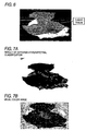
- Fig. 6 is a graph displaying an area (a tumor region) having low correlation value with the spectrum of normal tissue, based on the above-mentioned correlation coefficient R.
- Fig. 7A is an image showing a result in which a hyperspectral image of a gastric cancer inner wall is classified into a tumor region (a gray islet shape part) and a normal tissue by a supervised classification, and in comparison with the image shown in Fig. 7B , the tumor region and normal tissue are distinctively distinguished.
- the above treatment results are compared with pathologic inspection data.
- Fig. 8 is a graph in which a stomach tissue containing a tumor is sliced and a part judged as the tumor based on the situation in the tissue is marked, and contrary to this, a part judged as the tumor region by SVM classification based on a hyperspectral image before carrying out the slicing operation is shown by a bar (the lower step). (The actual tumor region is shown by a bar on the upper step.) Though a slight difference can be seen, it is possible to grasp the region judged as a tumor by a pathologic inspection almost accurately by a spectral inspection from the surface.
Landscapes
- Health & Medical Sciences (AREA)
- Life Sciences & Earth Sciences (AREA)
- Physics & Mathematics (AREA)
- Spectroscopy & Molecular Physics (AREA)
- General Health & Medical Sciences (AREA)
- Pathology (AREA)
- Animal Behavior & Ethology (AREA)
- Veterinary Medicine (AREA)
- Heart & Thoracic Surgery (AREA)
- Medical Informatics (AREA)
- Molecular Biology (AREA)
- Surgery (AREA)
- Engineering & Computer Science (AREA)
- Biophysics (AREA)
- Public Health (AREA)
- Biomedical Technology (AREA)
- Chemical & Material Sciences (AREA)
- Analytical Chemistry (AREA)
- Biochemistry (AREA)
- General Physics & Mathematics (AREA)
- Immunology (AREA)
- Investigating Or Analysing Materials By Optical Means (AREA)
- Endoscopes (AREA)
- Measuring And Recording Apparatus For Diagnosis (AREA)
- Measurement Of The Respiration, Hearing Ability, Form, And Blood Characteristics Of Living Organisms (AREA)
Applications Claiming Priority (1)
| Application Number | Priority Date | Filing Date | Title |
|---|---|---|---|
| JP2008152420A JP4575474B2 (ja) | 2008-06-11 | 2008-06-11 | 生体組織識別装置および方法 |
Publications (2)
| Publication Number | Publication Date |
|---|---|
| EP2133023A1 EP2133023A1 (en) | 2009-12-16 |
| EP2133023B1 true EP2133023B1 (en) | 2011-08-17 |
Family
ID=40934857
Family Applications (1)
| Application Number | Title | Priority Date | Filing Date |
|---|---|---|---|
| EP20090007675 Not-in-force EP2133023B1 (en) | 2008-06-11 | 2009-06-10 | Tissue discrimination device and method |
Country Status (4)
| Country | Link |
|---|---|
| US (1) | US20090312644A1 (ja) |
| EP (1) | EP2133023B1 (ja) |
| JP (1) | JP4575474B2 (ja) |
| AT (1) | ATE520342T1 (ja) |
Families Citing this family (14)
| Publication number | Priority date | Publication date | Assignee | Title |
|---|---|---|---|---|
| JP5737899B2 (ja) * | 2010-10-07 | 2015-06-17 | Hoya株式会社 | 診断システム |
| US20140148726A1 (en) * | 2010-10-21 | 2014-05-29 | Timothy Andrew WAGNER | Methods for detecting a condition |
| JP2012176096A (ja) * | 2011-02-25 | 2012-09-13 | Sumitomo Electric Ind Ltd | 生体検査装置および生体検査方法 |
| JP6281973B2 (ja) * | 2013-12-10 | 2018-02-21 | 国立大学法人京都大学 | 混合物試料の特性を表現する方法、混合物試料の特性を評価する方法、混合物試料の属性を識別する方法、及び混合物試料に由来する電磁波信号を処理する方法 |
| US10806334B2 (en) | 2017-02-28 | 2020-10-20 | Verily Life Sciences Llc | System and method for multiclass classification of images using a programmable light source |
| EP3375352A1 (en) | 2017-03-13 | 2018-09-19 | Koninklijke Philips N.V. | Device, system and method for determining a tissue characteristic of a subject |
| CN107967946B (zh) * | 2017-12-21 | 2021-05-11 | 武汉楚精灵医疗科技有限公司 | 基于深度学习的胃镜操作实时辅助系统及方法 |
| KR102167946B1 (ko) * | 2018-02-15 | 2020-10-20 | 스페클립스 주식회사 | 인-비보 병변 조직 검출을 위한 레이저 분광 기반의 독립 장치 및 방법 |
| US11672425B2 (en) | 2018-02-15 | 2023-06-13 | Speclipse, Inc. | Stand-alone apparatus and methods for in vivo detection of tissue malignancy using laser spectroscopy |
| KR102258181B1 (ko) * | 2018-02-15 | 2021-05-28 | 스페클립스 주식회사 | 레이저 분광 기반의 인-비보 병변 조직 검출을 위한 넌-디스크리트 스펙트럼 분석 알고리즘 및 이를 위한 방법 |
| JP6392476B1 (ja) * | 2018-03-19 | 2018-09-19 | 大輝 中矢 | 生体組織解析装置および生体組織解析プログラム |
| KR102370530B1 (ko) | 2019-03-22 | 2022-03-07 | 스페클립스 주식회사 | 레이저 유도 붕괴 스펙트럼 분석을 이용하는 진단 방법 및 이를 수행하는 진단 장치 |
| US20230218175A1 (en) * | 2020-07-31 | 2023-07-13 | Tokyo University Of Science Foundation | Image Processing Device, Image Processing Method, Image Processing Program, Endoscope Device, and Endoscope Image Processing System |
| CN114419741B (zh) * | 2022-03-15 | 2022-07-19 | 深圳市一心视觉科技有限公司 | 活体检测方法、活体检测装置、电子设备以及存储介质 |
Family Cites Families (21)
| Publication number | Priority date | Publication date | Assignee | Title |
|---|---|---|---|---|
| US4980551A (en) * | 1990-01-05 | 1990-12-25 | National Research Council Canada Conseil National De Recherches Canada | Non-pressure-dependancy infrared absorption spectra recording, sample cell |
| US5168162A (en) * | 1991-02-04 | 1992-12-01 | Cornell Research Foundation, Inc. | Method of detecting the presence of anomalies in exfoliated cells using infrared spectroscopy |
| US5261410A (en) * | 1991-02-07 | 1993-11-16 | Alfano Robert R | Method for determining if a tissue is a malignant tumor tissue, a benign tumor tissue, or a normal or benign tissue using Raman spectroscopy |
| CA2128190A1 (en) * | 1994-07-15 | 1996-01-16 | Patrick T.T. Wong | Method of identifying tissue |
| US5539207A (en) * | 1994-07-19 | 1996-07-23 | National Research Council Of Canada | Method of identifying tissue |
| US5579773A (en) * | 1994-09-30 | 1996-12-03 | Martin Marietta Energy Systems, Inc. | Laser-induced differential normalized fluorescence method for cancer diagnosis |
| CN1200174A (zh) * | 1995-08-24 | 1998-11-25 | 普渡研究基金会 | 基于荧光寿命的人体组织及其它无规则介质成象技术和光谱技术 |
| US5939721A (en) * | 1996-11-06 | 1999-08-17 | Lucent Technologies Inc. | Systems and methods for processing and analyzing terahertz waveforms |
| CA2287296C (en) * | 1997-04-03 | 2009-06-16 | National Research Council Of Canada | Method of assessing tissue viability using near-infrared spectroscopy |
| JP2001521772A (ja) * | 1997-10-30 | 2001-11-13 | ハイパーメッド・イメジング・インコーポレーテッド | マルチスペクトル/ハイパースペクトルの医療用計器 |
| US6119026A (en) * | 1997-12-04 | 2000-09-12 | Hewlett-Packard Company | Radiation apparatus and method for analysis of analytes in sample |
| CN1341209A (zh) * | 1999-01-25 | 2002-03-20 | 牛顿实验室公司 | 使用偏振光对组织成像 |
| CA2443098A1 (en) * | 2001-04-13 | 2002-10-24 | Cargill, Incorporated | Processes for evaluating agricultural and/or food materials; applications; and, products |
| IL146521A0 (en) * | 2001-11-15 | 2002-07-25 | Univ Ben Gurion | Novel optical method for diagnosis and grading of premalignant and malignant human colonic tissues |
| JP2005515473A (ja) * | 2002-01-18 | 2005-05-26 | ニユートン・ラボラトリーズ・インコーポレーテツド | 分光診断方法とシステム |
| JP2006516739A (ja) * | 2003-01-24 | 2006-07-06 | ザ・ジェネラル・ホスピタル・コーポレイション | 低コヒーレンス干渉計を用いて組織を識別するためのシステムおよび方法 |
| US7510849B2 (en) * | 2004-01-29 | 2009-03-31 | Glucolight Corporation | OCT based method for diagnosis and therapy |
| WO2006113476A2 (en) * | 2005-04-15 | 2006-10-26 | Bayer Healthcare Llc | Non-invasive system for measuring glucose in the body |
| KR20080043843A (ko) * | 2005-08-16 | 2008-05-19 | 스킨 캔서 스캐닝 리미티드 | 비침투 피부환부 확인방법 및 피부의 환부를 식별하기위한 디텍터 |
| US20080009748A1 (en) * | 2006-05-16 | 2008-01-10 | The Regents Of The University Of California | method and apparatus for the determination of intrinsic spectroscopic tumor markers by broadband-frequency domain technology |
| US8000774B2 (en) * | 2007-01-03 | 2011-08-16 | Infraredx, Inc. | Method and system for intra luminal thrombus detection |
-
2008
- 2008-06-11 JP JP2008152420A patent/JP4575474B2/ja not_active Expired - Fee Related
-
2009
- 2009-06-10 US US12/481,912 patent/US20090312644A1/en not_active Abandoned
- 2009-06-10 EP EP20090007675 patent/EP2133023B1/en not_active Not-in-force
- 2009-06-10 AT AT09007675T patent/ATE520342T1/de not_active IP Right Cessation
Also Published As
| Publication number | Publication date |
|---|---|
| JP4575474B2 (ja) | 2010-11-04 |
| JP2009300131A (ja) | 2009-12-24 |
| ATE520342T1 (de) | 2011-09-15 |
| US20090312644A1 (en) | 2009-12-17 |
| EP2133023A1 (en) | 2009-12-16 |
Similar Documents
| Publication | Publication Date | Title |
|---|---|---|
| EP2133023B1 (en) | Tissue discrimination device and method | |
| US10117582B2 (en) | Medical hyperspectral imaging for evaluation of tissue and tumor | |
| KR102409070B1 (ko) | 비정상적 성장하는 표본 또는 조직의 유형 또는 특성을 분석하는 라만 분광 시스템, 장치 및 방법 | |
| Akbari et al. | Hyperspectral imaging and quantitative analysis for prostate cancer detection | |
| Kiyotoki et al. | New method for detection of gastric cancer by hyperspectral imaging: a pilot study | |
| US8649849B2 (en) | Optical methods to intraoperatively detect positive prostate and kidney cancer margins | |
| EP2054706B1 (en) | Method for identifying refractive-index fluctuations of a target | |
| CN101500486A (zh) | 结合可见光与被动红外线技术及相关系统用于皮肤癌症先兆、痣和肿瘤的检测和识别及早期诊断 | |
| WO2013160780A1 (en) | Short-wave infrared imaging and spectroscopy technique for inflammation classification and tumor and inflammation differentiation in human tissues inside the body | |
| Shaikh et al. | A comparative evaluation of diffuse reflectance and Raman spectroscopy in the detection of cervical cancer | |
| Fawzy et al. | In vivo assessment and evaluation of lung tissue morphologic and physiological changes from<? xpp qa?> non-contact endoscopic reflectance spectroscopy<? xpp qa?> for improving lung cancer detection | |
| US20060089553A1 (en) | Method and apparatus for investigating histology of epithelial tissue | |
| Wu et al. | Review on the application of hyperspectral imaging technology of the exposed cortex in cerebral surgery | |
| Sato et al. | Raman spectroscopy and its use for live cell and tissue analysis | |
| US20120203115A1 (en) | System and method for noninvasive tissue examination | |
| JP5674683B2 (ja) | 混濁媒体に少なくとも部分的に挿入されるよう構成される光学検査デバイス | |
| JP5341707B2 (ja) | 生体組織識別装置及び方法 | |
| WO2012127378A1 (en) | An apparatus for optical analysis of an associated tissue sample | |
| Haugen et al. | Measurement of rat and human tissue optical properties for improving the optical detection and visualization of peripheral nerves | |
| Giannoni et al. | Optical characterisation and study of ex vivo glioma tissue for hyperspectral imaging during neurosurgery | |
| RU2676647C1 (ru) | Способ определения типа биологической ткани | |
| Anichini et al. | Hyperspectral and multispectral imaging in neurosurgery: A systematic literature and metanalysis | |
| Rocha-Osornio et al. | Chemometric techniques on the analysis of Raman spectra of serum blood samples of breast cancer patients | |
| Waterhouse et al. | Flexible Endoscopy: Multispectral Imaging | |
| Pickard et al. | Diagnosis of dysplasia in Barrett's oesophagus with in-situ elastic-scattering spectroscopy |
Legal Events
| Date | Code | Title | Description |
|---|---|---|---|
| PUAI | Public reference made under article 153(3) epc to a published international application that has entered the european phase |
Free format text: ORIGINAL CODE: 0009012 |
|
| AK | Designated contracting states |
Kind code of ref document: A1 Designated state(s): AT BE BG CH CY CZ DE DK EE ES FI FR GB GR HR HU IE IS IT LI LT LU LV MC MK MT NL NO PL PT RO SE SI SK TR |
|
| 17P | Request for examination filed |
Effective date: 20100527 |
|
| GRAP | Despatch of communication of intention to grant a patent |
Free format text: ORIGINAL CODE: EPIDOSNIGR1 |
|
| RTI1 | Title (correction) |
Free format text: TISSUE DISCRIMINATION DEVICE AND METHOD |
|
| RIN1 | Information on inventor provided before grant (corrected) |
Inventor name: TANAKA, MASAKO Inventor name: KOJIMA, KAZUYUKI Inventor name: KOSUGI, YUKIO Inventor name: AKBARI, HAMED Inventor name: SAITOH, TATSUHIKO |
|
| GRAS | Grant fee paid |
Free format text: ORIGINAL CODE: EPIDOSNIGR3 |
|
| GRAA | (expected) grant |
Free format text: ORIGINAL CODE: 0009210 |
|
| AK | Designated contracting states |
Kind code of ref document: B1 Designated state(s): AT BE BG CH CY CZ DE DK EE ES FI FR GB GR HR HU IE IS IT LI LT LU LV MC MK MT NL NO PL PT RO SE SI SK TR |
|
| REG | Reference to a national code |
Ref country code: GB Ref legal event code: FG4D |
|
| REG | Reference to a national code |
Ref country code: CH Ref legal event code: EP |
|
| REG | Reference to a national code |
Ref country code: IE Ref legal event code: FG4D |
|
| REG | Reference to a national code |
Ref country code: DE Ref legal event code: R096 Ref document number: 602009002130 Country of ref document: DE Effective date: 20111117 |
|
| REG | Reference to a national code |
Ref country code: NL Ref legal event code: VDEP Effective date: 20110817 |
|
| LTIE | Lt: invalidation of european patent or patent extension |
Effective date: 20110817 |
|
| PG25 | Lapsed in a contracting state [announced via postgrant information from national office to epo] |
Ref country code: IS Free format text: LAPSE BECAUSE OF FAILURE TO SUBMIT A TRANSLATION OF THE DESCRIPTION OR TO PAY THE FEE WITHIN THE PRESCRIBED TIME-LIMIT Effective date: 20111217 Ref country code: NL Free format text: LAPSE BECAUSE OF FAILURE TO SUBMIT A TRANSLATION OF THE DESCRIPTION OR TO PAY THE FEE WITHIN THE PRESCRIBED TIME-LIMIT Effective date: 20110817 Ref country code: LT Free format text: LAPSE BECAUSE OF FAILURE TO SUBMIT A TRANSLATION OF THE DESCRIPTION OR TO PAY THE FEE WITHIN THE PRESCRIBED TIME-LIMIT Effective date: 20110817 Ref country code: FI Free format text: LAPSE BECAUSE OF FAILURE TO SUBMIT A TRANSLATION OF THE DESCRIPTION OR TO PAY THE FEE WITHIN THE PRESCRIBED TIME-LIMIT Effective date: 20110817 Ref country code: SE Free format text: LAPSE BECAUSE OF FAILURE TO SUBMIT A TRANSLATION OF THE DESCRIPTION OR TO PAY THE FEE WITHIN THE PRESCRIBED TIME-LIMIT Effective date: 20110817 Ref country code: PT Free format text: LAPSE BECAUSE OF FAILURE TO SUBMIT A TRANSLATION OF THE DESCRIPTION OR TO PAY THE FEE WITHIN THE PRESCRIBED TIME-LIMIT Effective date: 20111219 Ref country code: NO Free format text: LAPSE BECAUSE OF FAILURE TO SUBMIT A TRANSLATION OF THE DESCRIPTION OR TO PAY THE FEE WITHIN THE PRESCRIBED TIME-LIMIT Effective date: 20111117 |
|
| REG | Reference to a national code |
Ref country code: AT Ref legal event code: MK05 Ref document number: 520342 Country of ref document: AT Kind code of ref document: T Effective date: 20110817 |
|
| PG25 | Lapsed in a contracting state [announced via postgrant information from national office to epo] |
Ref country code: CY Free format text: LAPSE BECAUSE OF FAILURE TO SUBMIT A TRANSLATION OF THE DESCRIPTION OR TO PAY THE FEE WITHIN THE PRESCRIBED TIME-LIMIT Effective date: 20110817 Ref country code: SI Free format text: LAPSE BECAUSE OF FAILURE TO SUBMIT A TRANSLATION OF THE DESCRIPTION OR TO PAY THE FEE WITHIN THE PRESCRIBED TIME-LIMIT Effective date: 20110817 Ref country code: AT Free format text: LAPSE BECAUSE OF FAILURE TO SUBMIT A TRANSLATION OF THE DESCRIPTION OR TO PAY THE FEE WITHIN THE PRESCRIBED TIME-LIMIT Effective date: 20110817 Ref country code: PL Free format text: LAPSE BECAUSE OF FAILURE TO SUBMIT A TRANSLATION OF THE DESCRIPTION OR TO PAY THE FEE WITHIN THE PRESCRIBED TIME-LIMIT Effective date: 20110817 Ref country code: GR Free format text: LAPSE BECAUSE OF FAILURE TO SUBMIT A TRANSLATION OF THE DESCRIPTION OR TO PAY THE FEE WITHIN THE PRESCRIBED TIME-LIMIT Effective date: 20111118 Ref country code: LV Free format text: LAPSE BECAUSE OF FAILURE TO SUBMIT A TRANSLATION OF THE DESCRIPTION OR TO PAY THE FEE WITHIN THE PRESCRIBED TIME-LIMIT Effective date: 20110817 |
|
| PG25 | Lapsed in a contracting state [announced via postgrant information from national office to epo] |
Ref country code: BE Free format text: LAPSE BECAUSE OF FAILURE TO SUBMIT A TRANSLATION OF THE DESCRIPTION OR TO PAY THE FEE WITHIN THE PRESCRIBED TIME-LIMIT Effective date: 20110817 |
|
| PG25 | Lapsed in a contracting state [announced via postgrant information from national office to epo] |
Ref country code: SK Free format text: LAPSE BECAUSE OF FAILURE TO SUBMIT A TRANSLATION OF THE DESCRIPTION OR TO PAY THE FEE WITHIN THE PRESCRIBED TIME-LIMIT Effective date: 20110817 Ref country code: CZ Free format text: LAPSE BECAUSE OF FAILURE TO SUBMIT A TRANSLATION OF THE DESCRIPTION OR TO PAY THE FEE WITHIN THE PRESCRIBED TIME-LIMIT Effective date: 20110817 |
|
| PG25 | Lapsed in a contracting state [announced via postgrant information from national office to epo] |
Ref country code: RO Free format text: LAPSE BECAUSE OF FAILURE TO SUBMIT A TRANSLATION OF THE DESCRIPTION OR TO PAY THE FEE WITHIN THE PRESCRIBED TIME-LIMIT Effective date: 20110817 Ref country code: IT Free format text: LAPSE BECAUSE OF FAILURE TO SUBMIT A TRANSLATION OF THE DESCRIPTION OR TO PAY THE FEE WITHIN THE PRESCRIBED TIME-LIMIT Effective date: 20110817 Ref country code: EE Free format text: LAPSE BECAUSE OF FAILURE TO SUBMIT A TRANSLATION OF THE DESCRIPTION OR TO PAY THE FEE WITHIN THE PRESCRIBED TIME-LIMIT Effective date: 20110817 |
|
| PLBE | No opposition filed within time limit |
Free format text: ORIGINAL CODE: 0009261 |
|
| STAA | Information on the status of an ep patent application or granted ep patent |
Free format text: STATUS: NO OPPOSITION FILED WITHIN TIME LIMIT |
|
| PG25 | Lapsed in a contracting state [announced via postgrant information from national office to epo] |
Ref country code: DK Free format text: LAPSE BECAUSE OF FAILURE TO SUBMIT A TRANSLATION OF THE DESCRIPTION OR TO PAY THE FEE WITHIN THE PRESCRIBED TIME-LIMIT Effective date: 20110817 |
|
| 26N | No opposition filed |
Effective date: 20120521 |
|
| PG25 | Lapsed in a contracting state [announced via postgrant information from national office to epo] |
Ref country code: HR Free format text: LAPSE BECAUSE OF FAILURE TO SUBMIT A TRANSLATION OF THE DESCRIPTION OR TO PAY THE FEE WITHIN THE PRESCRIBED TIME-LIMIT Effective date: 20120328 |
|
| REG | Reference to a national code |
Ref country code: DE Ref legal event code: R097 Ref document number: 602009002130 Country of ref document: DE Effective date: 20120521 |
|
| PG25 | Lapsed in a contracting state [announced via postgrant information from national office to epo] |
Ref country code: MC Free format text: LAPSE BECAUSE OF NON-PAYMENT OF DUE FEES Effective date: 20120630 |
|
| PG25 | Lapsed in a contracting state [announced via postgrant information from national office to epo] |
Ref country code: MK Free format text: LAPSE BECAUSE OF FAILURE TO SUBMIT A TRANSLATION OF THE DESCRIPTION OR TO PAY THE FEE WITHIN THE PRESCRIBED TIME-LIMIT Effective date: 20110817 |
|
| REG | Reference to a national code |
Ref country code: IE Ref legal event code: MM4A |
|
| PG25 | Lapsed in a contracting state [announced via postgrant information from national office to epo] |
Ref country code: IE Free format text: LAPSE BECAUSE OF NON-PAYMENT OF DUE FEES Effective date: 20120610 Ref country code: ES Free format text: LAPSE BECAUSE OF FAILURE TO SUBMIT A TRANSLATION OF THE DESCRIPTION OR TO PAY THE FEE WITHIN THE PRESCRIBED TIME-LIMIT Effective date: 20111128 |
|
| PG25 | Lapsed in a contracting state [announced via postgrant information from national office to epo] |
Ref country code: BG Free format text: LAPSE BECAUSE OF FAILURE TO SUBMIT A TRANSLATION OF THE DESCRIPTION OR TO PAY THE FEE WITHIN THE PRESCRIBED TIME-LIMIT Effective date: 20111117 |
|
| PG25 | Lapsed in a contracting state [announced via postgrant information from national office to epo] |
Ref country code: MT Free format text: LAPSE BECAUSE OF FAILURE TO SUBMIT A TRANSLATION OF THE DESCRIPTION OR TO PAY THE FEE WITHIN THE PRESCRIBED TIME-LIMIT Effective date: 20110817 |
|
| PG25 | Lapsed in a contracting state [announced via postgrant information from national office to epo] |
Ref country code: HR Free format text: LAPSE BECAUSE OF FAILURE TO SUBMIT A TRANSLATION OF THE DESCRIPTION OR TO PAY THE FEE WITHIN THE PRESCRIBED TIME-LIMIT Effective date: 20110817 |
|
| REG | Reference to a national code |
Ref country code: CH Ref legal event code: PL |
|
| PG25 | Lapsed in a contracting state [announced via postgrant information from national office to epo] |
Ref country code: CH Free format text: LAPSE BECAUSE OF NON-PAYMENT OF DUE FEES Effective date: 20130630 Ref country code: TR Free format text: LAPSE BECAUSE OF FAILURE TO SUBMIT A TRANSLATION OF THE DESCRIPTION OR TO PAY THE FEE WITHIN THE PRESCRIBED TIME-LIMIT Effective date: 20110817 Ref country code: LI Free format text: LAPSE BECAUSE OF NON-PAYMENT OF DUE FEES Effective date: 20130630 |
|
| PG25 | Lapsed in a contracting state [announced via postgrant information from national office to epo] |
Ref country code: LU Free format text: LAPSE BECAUSE OF NON-PAYMENT OF DUE FEES Effective date: 20120610 |
|
| PG25 | Lapsed in a contracting state [announced via postgrant information from national office to epo] |
Ref country code: HU Free format text: LAPSE BECAUSE OF FAILURE TO SUBMIT A TRANSLATION OF THE DESCRIPTION OR TO PAY THE FEE WITHIN THE PRESCRIBED TIME-LIMIT Effective date: 20090610 |
|
| REG | Reference to a national code |
Ref country code: FR Ref legal event code: PLFP Year of fee payment: 7 |
|
| PGFP | Annual fee paid to national office [announced via postgrant information from national office to epo] |
Ref country code: DE Payment date: 20150602 Year of fee payment: 7 Ref country code: GB Payment date: 20150610 Year of fee payment: 7 |
|
| PGFP | Annual fee paid to national office [announced via postgrant information from national office to epo] |
Ref country code: FR Payment date: 20150608 Year of fee payment: 7 |
|
| REG | Reference to a national code |
Ref country code: DE Ref legal event code: R119 Ref document number: 602009002130 Country of ref document: DE |
|
| GBPC | Gb: european patent ceased through non-payment of renewal fee |
Effective date: 20160610 |
|
| REG | Reference to a national code |
Ref country code: FR Ref legal event code: ST Effective date: 20170228 |
|
| PG25 | Lapsed in a contracting state [announced via postgrant information from national office to epo] |
Ref country code: DE Free format text: LAPSE BECAUSE OF NON-PAYMENT OF DUE FEES Effective date: 20170103 Ref country code: FR Free format text: LAPSE BECAUSE OF NON-PAYMENT OF DUE FEES Effective date: 20160630 |
|
| PG25 | Lapsed in a contracting state [announced via postgrant information from national office to epo] |
Ref country code: GB Free format text: LAPSE BECAUSE OF NON-PAYMENT OF DUE FEES Effective date: 20160610 |