EP2117571B1 - Urokinase-type plasminogen activator receptor epitope - Google Patents
Urokinase-type plasminogen activator receptor epitope Download PDFInfo
- Publication number
- EP2117571B1 EP2117571B1 EP07862653.8A EP07862653A EP2117571B1 EP 2117571 B1 EP2117571 B1 EP 2117571B1 EP 07862653 A EP07862653 A EP 07862653A EP 2117571 B1 EP2117571 B1 EP 2117571B1
- Authority
- EP
- European Patent Office
- Prior art keywords
- upar
- antibody
- human
- cells
- binding
- Prior art date
- Legal status (The legal status is an assumption and is not a legal conclusion. Google has not performed a legal analysis and makes no representation as to the accuracy of the status listed.)
- Active
Links
- 108010042352 Urokinase Plasminogen Activator Receptors Proteins 0.000 title description 182
- 102000004504 Urokinase Plasminogen Activator Receptors Human genes 0.000 title description 182
- 210000004027 cell Anatomy 0.000 claims description 191
- 241000282414 Homo sapiens Species 0.000 claims description 136
- 108090000765 processed proteins & peptides Proteins 0.000 claims description 115
- 238000000034 method Methods 0.000 claims description 109
- 239000012634 fragment Substances 0.000 claims description 100
- 101000760337 Homo sapiens Urokinase plasminogen activator surface receptor Proteins 0.000 claims description 77
- 235000001014 amino acid Nutrition 0.000 claims description 62
- 150000001413 amino acids Chemical class 0.000 claims description 58
- 125000003275 alpha amino acid group Chemical group 0.000 claims description 38
- 210000004408 hybridoma Anatomy 0.000 claims description 31
- 206010035226 Plasma cell myeloma Diseases 0.000 claims description 18
- 201000000050 myeloid neoplasm Diseases 0.000 claims description 18
- 210000004988 splenocyte Anatomy 0.000 claims description 16
- 241000124008 Mammalia Species 0.000 claims description 13
- 230000003053 immunization Effects 0.000 claims description 11
- 230000002163 immunogen Effects 0.000 claims description 7
- 125000002924 primary amino group Chemical group [H]N([H])* 0.000 claims description 7
- BWGNESOTFCXPMA-UHFFFAOYSA-N Dihydrogen disulfide Chemical compound SS BWGNESOTFCXPMA-UHFFFAOYSA-N 0.000 claims description 3
- 125000000151 cysteine group Chemical group N[C@@H](CS)C(=O)* 0.000 claims 1
- 125000002228 disulfide group Chemical group 0.000 claims 1
- 230000027455 binding Effects 0.000 description 154
- 108090000623 proteins and genes Proteins 0.000 description 133
- 206010028980 Neoplasm Diseases 0.000 description 90
- 102000004169 proteins and genes Human genes 0.000 description 64
- 239000000427 antigen Substances 0.000 description 60
- 235000018102 proteins Nutrition 0.000 description 60
- 108091007433 antigens Proteins 0.000 description 59
- 102000036639 antigens Human genes 0.000 description 59
- 229940024606 amino acid Drugs 0.000 description 57
- 102000004196 processed proteins & peptides Human genes 0.000 description 53
- 238000003556 assay Methods 0.000 description 46
- 239000003814 drug Substances 0.000 description 43
- 125000000539 amino acid group Chemical group 0.000 description 40
- 230000014509 gene expression Effects 0.000 description 40
- 229920001184 polypeptide Polymers 0.000 description 37
- -1 125I Chemical compound 0.000 description 36
- 150000001875 compounds Chemical class 0.000 description 35
- 201000011510 cancer Diseases 0.000 description 34
- 239000013598 vector Substances 0.000 description 34
- 229940124597 therapeutic agent Drugs 0.000 description 31
- 239000000203 mixture Substances 0.000 description 30
- 230000001225 therapeutic effect Effects 0.000 description 29
- 238000011282 treatment Methods 0.000 description 28
- 102100024952 Protein CBFA2T1 Human genes 0.000 description 25
- 108020004414 DNA Proteins 0.000 description 23
- 230000009467 reduction Effects 0.000 description 22
- 241001465754 Metazoa Species 0.000 description 21
- 230000001419 dependent effect Effects 0.000 description 21
- 239000007787 solid Substances 0.000 description 21
- 238000006467 substitution reaction Methods 0.000 description 21
- 206010027476 Metastases Diseases 0.000 description 20
- 230000033115 angiogenesis Effects 0.000 description 20
- 239000002299 complementary DNA Substances 0.000 description 20
- LOKCTEFSRHRXRJ-UHFFFAOYSA-I dipotassium trisodium dihydrogen phosphate hydrogen phosphate dichloride Chemical compound P(=O)(O)(O)[O-].[K+].P(=O)(O)([O-])[O-].[Na+].[Na+].[Cl-].[K+].[Cl-].[Na+] LOKCTEFSRHRXRJ-UHFFFAOYSA-I 0.000 description 20
- 230000000694 effects Effects 0.000 description 20
- 239000002953 phosphate buffered saline Substances 0.000 description 20
- 238000001727 in vivo Methods 0.000 description 19
- 230000000069 prophylactic effect Effects 0.000 description 19
- 239000000523 sample Substances 0.000 description 19
- 108060003951 Immunoglobulin Proteins 0.000 description 18
- 102000018358 immunoglobulin Human genes 0.000 description 18
- 241000699666 Mus <mouse, genus> Species 0.000 description 17
- 239000008194 pharmaceutical composition Substances 0.000 description 17
- 238000012360 testing method Methods 0.000 description 16
- 210000001519 tissue Anatomy 0.000 description 16
- 208000037265 diseases, disorders, signs and symptoms Diseases 0.000 description 15
- 125000005647 linker group Chemical group 0.000 description 15
- 239000012528 membrane Substances 0.000 description 15
- 210000004881 tumor cell Anatomy 0.000 description 15
- YZCKVEUIGOORGS-OUBTZVSYSA-N Deuterium Chemical group [2H] YZCKVEUIGOORGS-OUBTZVSYSA-N 0.000 description 14
- 239000003795 chemical substances by application Substances 0.000 description 14
- 229910052805 deuterium Inorganic materials 0.000 description 14
- 238000004519 manufacturing process Methods 0.000 description 14
- 210000004379 membrane Anatomy 0.000 description 14
- 230000009401 metastasis Effects 0.000 description 14
- 230000035772 mutation Effects 0.000 description 14
- 235000002639 sodium chloride Nutrition 0.000 description 14
- 230000004614 tumor growth Effects 0.000 description 14
- 102000004190 Enzymes Human genes 0.000 description 13
- 108090000790 Enzymes Proteins 0.000 description 13
- 229940088598 enzyme Drugs 0.000 description 13
- 238000002347 injection Methods 0.000 description 13
- 239000007924 injection Substances 0.000 description 13
- 240000004808 Saccharomyces cerevisiae Species 0.000 description 12
- 238000013459 approach Methods 0.000 description 12
- 230000015572 biosynthetic process Effects 0.000 description 12
- 238000003018 immunoassay Methods 0.000 description 12
- 239000000047 product Substances 0.000 description 12
- 239000000243 solution Substances 0.000 description 12
- 108010047041 Complementarity Determining Regions Proteins 0.000 description 11
- 102000008394 Immunoglobulin Fragments Human genes 0.000 description 11
- 108010021625 Immunoglobulin Fragments Proteins 0.000 description 11
- 241000699670 Mus sp. Species 0.000 description 11
- 108091028043 Nucleic acid sequence Proteins 0.000 description 11
- 201000010099 disease Diseases 0.000 description 11
- 238000000338 in vitro Methods 0.000 description 11
- 210000004962 mammalian cell Anatomy 0.000 description 11
- 108010082117 matrigel Proteins 0.000 description 11
- 241001529936 Murinae Species 0.000 description 10
- 210000002889 endothelial cell Anatomy 0.000 description 10
- 150000007523 nucleic acids Chemical class 0.000 description 10
- HEMHJVSKTPXQMS-UHFFFAOYSA-M sodium hydroxide Substances [OH-].[Na+] HEMHJVSKTPXQMS-UHFFFAOYSA-M 0.000 description 10
- PEDCQBHIVMGVHV-UHFFFAOYSA-N Glycerine Chemical compound OCC(O)CO PEDCQBHIVMGVHV-UHFFFAOYSA-N 0.000 description 9
- 238000007792 addition Methods 0.000 description 9
- 230000001580 bacterial effect Effects 0.000 description 9
- 230000006870 function Effects 0.000 description 9
- 239000003446 ligand Substances 0.000 description 9
- 239000007788 liquid Substances 0.000 description 9
- 239000002609 medium Substances 0.000 description 9
- 238000002360 preparation method Methods 0.000 description 9
- 150000003839 salts Chemical class 0.000 description 9
- XLYOFNOQVPJJNP-UHFFFAOYSA-N water Substances O XLYOFNOQVPJJNP-UHFFFAOYSA-N 0.000 description 9
- 206010003571 Astrocytoma Diseases 0.000 description 8
- 108091003079 Bovine Serum Albumin Proteins 0.000 description 8
- 108090000379 Fibroblast growth factor 2 Proteins 0.000 description 8
- FAPWRFPIFSIZLT-UHFFFAOYSA-M Sodium chloride Chemical compound [Na+].[Cl-] FAPWRFPIFSIZLT-UHFFFAOYSA-M 0.000 description 8
- 239000003242 anti bacterial agent Substances 0.000 description 8
- 229940098773 bovine serum albumin Drugs 0.000 description 8
- 238000001514 detection method Methods 0.000 description 8
- 238000011534 incubation Methods 0.000 description 8
- 230000002401 inhibitory effect Effects 0.000 description 8
- 230000003993 interaction Effects 0.000 description 8
- 239000000463 material Substances 0.000 description 8
- 238000013508 migration Methods 0.000 description 8
- 239000000126 substance Substances 0.000 description 8
- 238000013268 sustained release Methods 0.000 description 8
- 239000012730 sustained-release form Substances 0.000 description 8
- 238000002560 therapeutic procedure Methods 0.000 description 8
- 239000003981 vehicle Substances 0.000 description 8
- 241000282693 Cercopithecidae Species 0.000 description 7
- DHMQDGOQFOQNFH-UHFFFAOYSA-N Glycine Chemical compound NCC(O)=O DHMQDGOQFOQNFH-UHFFFAOYSA-N 0.000 description 7
- ZDXPYRJPNDTMRX-VKHMYHEASA-N L-glutamine Chemical compound OC(=O)[C@@H](N)CCC(N)=O ZDXPYRJPNDTMRX-VKHMYHEASA-N 0.000 description 7
- 229940088710 antibiotic agent Drugs 0.000 description 7
- 210000002469 basement membrane Anatomy 0.000 description 7
- 239000000872 buffer Substances 0.000 description 7
- 238000006243 chemical reaction Methods 0.000 description 7
- 239000003153 chemical reaction reagent Substances 0.000 description 7
- 238000010276 construction Methods 0.000 description 7
- 238000013270 controlled release Methods 0.000 description 7
- 238000010790 dilution Methods 0.000 description 7
- 239000012895 dilution Substances 0.000 description 7
- 239000013604 expression vector Substances 0.000 description 7
- 239000012530 fluid Substances 0.000 description 7
- 230000004927 fusion Effects 0.000 description 7
- 230000012010 growth Effects 0.000 description 7
- 239000003112 inhibitor Substances 0.000 description 7
- 230000005764 inhibitory process Effects 0.000 description 7
- 239000010410 layer Substances 0.000 description 7
- 108020004999 messenger RNA Proteins 0.000 description 7
- 230000008488 polyadenylation Effects 0.000 description 7
- 238000003752 polymerase chain reaction Methods 0.000 description 7
- 238000000746 purification Methods 0.000 description 7
- 241000894007 species Species 0.000 description 7
- 239000006228 supernatant Substances 0.000 description 7
- 108700012359 toxins Proteins 0.000 description 7
- YBJHBAHKTGYVGT-ZKWXMUAHSA-N (+)-Biotin Chemical compound N1C(=O)N[C@@H]2[C@H](CCCCC(=O)O)SC[C@@H]21 YBJHBAHKTGYVGT-ZKWXMUAHSA-N 0.000 description 6
- 108091026890 Coding region Proteins 0.000 description 6
- AOJJSUZBOXZQNB-TZSSRYMLSA-N Doxorubicin Chemical compound O([C@H]1C[C@@](O)(CC=2C(O)=C3C(=O)C=4C=CC=C(C=4C(=O)C3=C(O)C=21)OC)C(=O)CO)[C@H]1C[C@H](N)[C@H](O)[C@H](C)O1 AOJJSUZBOXZQNB-TZSSRYMLSA-N 0.000 description 6
- 238000002965 ELISA Methods 0.000 description 6
- 241000282412 Homo Species 0.000 description 6
- 108700005091 Immunoglobulin Genes Proteins 0.000 description 6
- 206010058467 Lung neoplasm malignant Diseases 0.000 description 6
- 206010033128 Ovarian cancer Diseases 0.000 description 6
- 241000700159 Rattus Species 0.000 description 6
- 108010039491 Ricin Proteins 0.000 description 6
- UIIMBOGNXHQVGW-UHFFFAOYSA-M Sodium bicarbonate Chemical compound [Na+].OC([O-])=O UIIMBOGNXHQVGW-UHFFFAOYSA-M 0.000 description 6
- 108010073929 Vascular Endothelial Growth Factor A Proteins 0.000 description 6
- 102000005789 Vascular Endothelial Growth Factors Human genes 0.000 description 6
- 108010019530 Vascular Endothelial Growth Factors Proteins 0.000 description 6
- 239000002671 adjuvant Substances 0.000 description 6
- 210000003719 b-lymphocyte Anatomy 0.000 description 6
- 230000009400 cancer invasion Effects 0.000 description 6
- 230000012292 cell migration Effects 0.000 description 6
- DQLATGHUWYMOKM-UHFFFAOYSA-L cisplatin Chemical compound N[Pt](N)(Cl)Cl DQLATGHUWYMOKM-UHFFFAOYSA-L 0.000 description 6
- 238000000749 co-immunoprecipitation Methods 0.000 description 6
- 230000001268 conjugating effect Effects 0.000 description 6
- 238000012217 deletion Methods 0.000 description 6
- 230000037430 deletion Effects 0.000 description 6
- 238000002405 diagnostic procedure Methods 0.000 description 6
- 229940079593 drug Drugs 0.000 description 6
- 239000003937 drug carrier Substances 0.000 description 6
- 238000005516 engineering process Methods 0.000 description 6
- 239000003623 enhancer Substances 0.000 description 6
- 210000003527 eukaryotic cell Anatomy 0.000 description 6
- 238000009472 formulation Methods 0.000 description 6
- 238000001802 infusion Methods 0.000 description 6
- 229910052751 metal Inorganic materials 0.000 description 6
- 239000002184 metal Substances 0.000 description 6
- 108020004707 nucleic acids Proteins 0.000 description 6
- 102000039446 nucleic acids Human genes 0.000 description 6
- PYWVYCXTNDRMGF-UHFFFAOYSA-N rhodamine B Chemical compound [Cl-].C=12C=CC(=[N+](CC)CC)C=C2OC2=CC(N(CC)CC)=CC=C2C=1C1=CC=CC=C1C(O)=O PYWVYCXTNDRMGF-UHFFFAOYSA-N 0.000 description 6
- 239000011780 sodium chloride Substances 0.000 description 6
- 230000000699 topical effect Effects 0.000 description 6
- 230000002588 toxic effect Effects 0.000 description 6
- 239000003053 toxin Substances 0.000 description 6
- 231100000765 toxin Toxicity 0.000 description 6
- 239000011534 wash buffer Substances 0.000 description 6
- 241000894006 Bacteria Species 0.000 description 5
- 206010009944 Colon cancer Diseases 0.000 description 5
- 239000006144 Dulbecco’s modified Eagle's medium Substances 0.000 description 5
- 241000196324 Embryophyta Species 0.000 description 5
- 102100024785 Fibroblast growth factor 2 Human genes 0.000 description 5
- 206010016654 Fibrosis Diseases 0.000 description 5
- 108010054477 Immunoglobulin Fab Fragments Proteins 0.000 description 5
- 102000001706 Immunoglobulin Fab Fragments Human genes 0.000 description 5
- 108091092195 Intron Proteins 0.000 description 5
- 241000699660 Mus musculus Species 0.000 description 5
- NWIBSHFKIJFRCO-WUDYKRTCSA-N Mytomycin Chemical compound C1N2C(C(C(C)=C(N)C3=O)=O)=C3[C@@H](COC(N)=O)[C@@]2(OC)[C@@H]2[C@H]1N2 NWIBSHFKIJFRCO-WUDYKRTCSA-N 0.000 description 5
- 108091034117 Oligonucleotide Proteins 0.000 description 5
- 206010061535 Ovarian neoplasm Diseases 0.000 description 5
- 239000002585 base Substances 0.000 description 5
- 230000008901 benefit Effects 0.000 description 5
- 230000037396 body weight Effects 0.000 description 5
- 230000004709 cell invasion Effects 0.000 description 5
- 230000004663 cell proliferation Effects 0.000 description 5
- 210000004978 chinese hamster ovary cell Anatomy 0.000 description 5
- 230000002759 chromosomal effect Effects 0.000 description 5
- 238000010367 cloning Methods 0.000 description 5
- 230000000295 complement effect Effects 0.000 description 5
- 238000007796 conventional method Methods 0.000 description 5
- 238000003745 diagnosis Methods 0.000 description 5
- 230000004761 fibrosis Effects 0.000 description 5
- GNBHRKFJIUUOQI-UHFFFAOYSA-N fluorescein Chemical compound O1C(=O)C2=CC=CC=C2C21C1=CC=C(O)C=C1OC1=CC(O)=CC=C21 GNBHRKFJIUUOQI-UHFFFAOYSA-N 0.000 description 5
- 108020001507 fusion proteins Proteins 0.000 description 5
- 102000037865 fusion proteins Human genes 0.000 description 5
- ZDXPYRJPNDTMRX-UHFFFAOYSA-N glutamine Natural products OC(=O)C(N)CCC(N)=O ZDXPYRJPNDTMRX-UHFFFAOYSA-N 0.000 description 5
- 230000013595 glycosylation Effects 0.000 description 5
- 238000006206 glycosylation reaction Methods 0.000 description 5
- 210000005260 human cell Anatomy 0.000 description 5
- 239000012133 immunoprecipitate Substances 0.000 description 5
- 230000001965 increasing effect Effects 0.000 description 5
- 238000007912 intraperitoneal administration Methods 0.000 description 5
- 230000005012 migration Effects 0.000 description 5
- 239000002674 ointment Substances 0.000 description 5
- 238000002823 phage display Methods 0.000 description 5
- 230000002265 prevention Effects 0.000 description 5
- 230000008569 process Effects 0.000 description 5
- 230000035755 proliferation Effects 0.000 description 5
- 230000002285 radioactive effect Effects 0.000 description 5
- 102000005962 receptors Human genes 0.000 description 5
- 108020003175 receptors Proteins 0.000 description 5
- 238000012216 screening Methods 0.000 description 5
- 239000000725 suspension Substances 0.000 description 5
- 238000003786 synthesis reaction Methods 0.000 description 5
- 231100000331 toxic Toxicity 0.000 description 5
- 230000003442 weekly effect Effects 0.000 description 5
- VBICKXHEKHSIBG-UHFFFAOYSA-N 1-monostearoylglycerol Chemical compound CCCCCCCCCCCCCCCCCC(=O)OCC(O)CO VBICKXHEKHSIBG-UHFFFAOYSA-N 0.000 description 4
- LRFVTYWOQMYALW-UHFFFAOYSA-N 9H-xanthine Chemical compound O=C1NC(=O)NC2=C1NC=N2 LRFVTYWOQMYALW-UHFFFAOYSA-N 0.000 description 4
- 206010003445 Ascites Diseases 0.000 description 4
- 241000867607 Chlorocebus sabaeus Species 0.000 description 4
- 108010053187 Diphtheria Toxin Proteins 0.000 description 4
- 102000016607 Diphtheria Toxin Human genes 0.000 description 4
- 102000010834 Extracellular Matrix Proteins Human genes 0.000 description 4
- 108010037362 Extracellular Matrix Proteins Proteins 0.000 description 4
- 108010010803 Gelatin Proteins 0.000 description 4
- WQZGKKKJIJFFOK-GASJEMHNSA-N Glucose Natural products OC[C@H]1OC(O)[C@H](O)[C@@H](O)[C@@H]1O WQZGKKKJIJFFOK-GASJEMHNSA-N 0.000 description 4
- 108010043121 Green Fluorescent Proteins Proteins 0.000 description 4
- 102000004144 Green Fluorescent Proteins Human genes 0.000 description 4
- QPCDCPDFJACHGM-UHFFFAOYSA-N N,N-bis{2-[bis(carboxymethyl)amino]ethyl}glycine Chemical compound OC(=O)CN(CC(O)=O)CCN(CC(=O)O)CCN(CC(O)=O)CC(O)=O QPCDCPDFJACHGM-UHFFFAOYSA-N 0.000 description 4
- 241000283973 Oryctolagus cuniculus Species 0.000 description 4
- 206010060862 Prostate cancer Diseases 0.000 description 4
- 102000004887 Transforming Growth Factor beta Human genes 0.000 description 4
- 108090001012 Transforming Growth Factor beta Proteins 0.000 description 4
- JLCPHMBAVCMARE-UHFFFAOYSA-N [3-[[3-[[3-[[3-[[3-[[3-[[3-[[3-[[3-[[3-[[3-[[5-(2-amino-6-oxo-1H-purin-9-yl)-3-[[3-[[3-[[3-[[3-[[3-[[5-(2-amino-6-oxo-1H-purin-9-yl)-3-[[5-(2-amino-6-oxo-1H-purin-9-yl)-3-hydroxyoxolan-2-yl]methoxy-hydroxyphosphoryl]oxyoxolan-2-yl]methoxy-hydroxyphosphoryl]oxy-5-(5-methyl-2,4-dioxopyrimidin-1-yl)oxolan-2-yl]methoxy-hydroxyphosphoryl]oxy-5-(6-aminopurin-9-yl)oxolan-2-yl]methoxy-hydroxyphosphoryl]oxy-5-(6-aminopurin-9-yl)oxolan-2-yl]methoxy-hydroxyphosphoryl]oxy-5-(6-aminopurin-9-yl)oxolan-2-yl]methoxy-hydroxyphosphoryl]oxy-5-(6-aminopurin-9-yl)oxolan-2-yl]methoxy-hydroxyphosphoryl]oxyoxolan-2-yl]methoxy-hydroxyphosphoryl]oxy-5-(5-methyl-2,4-dioxopyrimidin-1-yl)oxolan-2-yl]methoxy-hydroxyphosphoryl]oxy-5-(4-amino-2-oxopyrimidin-1-yl)oxolan-2-yl]methoxy-hydroxyphosphoryl]oxy-5-(5-methyl-2,4-dioxopyrimidin-1-yl)oxolan-2-yl]methoxy-hydroxyphosphoryl]oxy-5-(5-methyl-2,4-dioxopyrimidin-1-yl)oxolan-2-yl]methoxy-hydroxyphosphoryl]oxy-5-(6-aminopurin-9-yl)oxolan-2-yl]methoxy-hydroxyphosphoryl]oxy-5-(6-aminopurin-9-yl)oxolan-2-yl]methoxy-hydroxyphosphoryl]oxy-5-(4-amino-2-oxopyrimidin-1-yl)oxolan-2-yl]methoxy-hydroxyphosphoryl]oxy-5-(4-amino-2-oxopyrimidin-1-yl)oxolan-2-yl]methoxy-hydroxyphosphoryl]oxy-5-(4-amino-2-oxopyrimidin-1-yl)oxolan-2-yl]methoxy-hydroxyphosphoryl]oxy-5-(6-aminopurin-9-yl)oxolan-2-yl]methoxy-hydroxyphosphoryl]oxy-5-(4-amino-2-oxopyrimidin-1-yl)oxolan-2-yl]methyl [5-(6-aminopurin-9-yl)-2-(hydroxymethyl)oxolan-3-yl] hydrogen phosphate Polymers Cc1cn(C2CC(OP(O)(=O)OCC3OC(CC3OP(O)(=O)OCC3OC(CC3O)n3cnc4c3nc(N)[nH]c4=O)n3cnc4c3nc(N)[nH]c4=O)C(COP(O)(=O)OC3CC(OC3COP(O)(=O)OC3CC(OC3COP(O)(=O)OC3CC(OC3COP(O)(=O)OC3CC(OC3COP(O)(=O)OC3CC(OC3COP(O)(=O)OC3CC(OC3COP(O)(=O)OC3CC(OC3COP(O)(=O)OC3CC(OC3COP(O)(=O)OC3CC(OC3COP(O)(=O)OC3CC(OC3COP(O)(=O)OC3CC(OC3COP(O)(=O)OC3CC(OC3COP(O)(=O)OC3CC(OC3COP(O)(=O)OC3CC(OC3COP(O)(=O)OC3CC(OC3COP(O)(=O)OC3CC(OC3COP(O)(=O)OC3CC(OC3CO)n3cnc4c(N)ncnc34)n3ccc(N)nc3=O)n3cnc4c(N)ncnc34)n3ccc(N)nc3=O)n3ccc(N)nc3=O)n3ccc(N)nc3=O)n3cnc4c(N)ncnc34)n3cnc4c(N)ncnc34)n3cc(C)c(=O)[nH]c3=O)n3cc(C)c(=O)[nH]c3=O)n3ccc(N)nc3=O)n3cc(C)c(=O)[nH]c3=O)n3cnc4c3nc(N)[nH]c4=O)n3cnc4c(N)ncnc34)n3cnc4c(N)ncnc34)n3cnc4c(N)ncnc34)n3cnc4c(N)ncnc34)O2)c(=O)[nH]c1=O JLCPHMBAVCMARE-UHFFFAOYSA-N 0.000 description 4
- 239000002253 acid Substances 0.000 description 4
- RJURFGZVJUQBHK-UHFFFAOYSA-N actinomycin D Natural products CC1OC(=O)C(C(C)C)N(C)C(=O)CN(C)C(=O)C2CCCN2C(=O)C(C(C)C)NC(=O)C1NC(=O)C1=C(N)C(=O)C(C)=C2OC(C(C)=CC=C3C(=O)NC4C(=O)NC(C(N5CCCC5C(=O)N(C)CC(=O)N(C)C(C(C)C)C(=O)OC4C)=O)C(C)C)=C3N=C21 RJURFGZVJUQBHK-UHFFFAOYSA-N 0.000 description 4
- 230000002491 angiogenic effect Effects 0.000 description 4
- 230000001028 anti-proliverative effect Effects 0.000 description 4
- 210000004369 blood Anatomy 0.000 description 4
- 239000008280 blood Substances 0.000 description 4
- 239000012830 cancer therapeutic Substances 0.000 description 4
- 239000000969 carrier Substances 0.000 description 4
- 210000000170 cell membrane Anatomy 0.000 description 4
- 239000002738 chelating agent Substances 0.000 description 4
- 229960004316 cisplatin Drugs 0.000 description 4
- 238000003776 cleavage reaction Methods 0.000 description 4
- 239000006071 cream Substances 0.000 description 4
- 231100000433 cytotoxic Toxicity 0.000 description 4
- 231100000599 cytotoxic agent Toxicity 0.000 description 4
- 230000001472 cytotoxic effect Effects 0.000 description 4
- 230000007423 decrease Effects 0.000 description 4
- 238000011161 development Methods 0.000 description 4
- 230000018109 developmental process Effects 0.000 description 4
- 208000035475 disorder Diseases 0.000 description 4
- 239000000839 emulsion Substances 0.000 description 4
- 210000002744 extracellular matrix Anatomy 0.000 description 4
- 210000002950 fibroblast Anatomy 0.000 description 4
- 239000000499 gel Substances 0.000 description 4
- 239000008273 gelatin Substances 0.000 description 4
- 229920000159 gelatin Polymers 0.000 description 4
- 235000019322 gelatine Nutrition 0.000 description 4
- 235000011852 gelatine desserts Nutrition 0.000 description 4
- 229930004094 glycosylphosphatidylinositol Natural products 0.000 description 4
- 239000005090 green fluorescent protein Substances 0.000 description 4
- 239000003102 growth factor Substances 0.000 description 4
- 230000002209 hydrophobic effect Effects 0.000 description 4
- RAXXELZNTBOGNW-UHFFFAOYSA-N imidazole Substances C1=CNC=N1 RAXXELZNTBOGNW-UHFFFAOYSA-N 0.000 description 4
- 239000004615 ingredient Substances 0.000 description 4
- 238000003780 insertion Methods 0.000 description 4
- 230000037431 insertion Effects 0.000 description 4
- 102000006495 integrins Human genes 0.000 description 4
- 108010044426 integrins Proteins 0.000 description 4
- 238000002372 labelling Methods 0.000 description 4
- 210000004072 lung Anatomy 0.000 description 4
- 230000001404 mediated effect Effects 0.000 description 4
- 150000002739 metals Chemical class 0.000 description 4
- 230000007935 neutral effect Effects 0.000 description 4
- 231100000252 nontoxic Toxicity 0.000 description 4
- 230000003000 nontoxic effect Effects 0.000 description 4
- 238000011275 oncology therapy Methods 0.000 description 4
- 229960003330 pentetic acid Drugs 0.000 description 4
- 239000013612 plasmid Substances 0.000 description 4
- 238000011160 research Methods 0.000 description 4
- 230000007017 scission Effects 0.000 description 4
- 208000020431 spinal cord injury Diseases 0.000 description 4
- 238000007920 subcutaneous administration Methods 0.000 description 4
- 238000011830 transgenic mouse model Methods 0.000 description 4
- 238000005406 washing Methods 0.000 description 4
- STQGQHZAVUOBTE-UHFFFAOYSA-N 7-Cyan-hept-2t-en-4,6-diinsaeure Natural products C1=2C(O)=C3C(=O)C=4C(OC)=CC=CC=4C(=O)C3=C(O)C=2CC(O)(C(C)=O)CC1OC1CC(N)C(O)C(C)O1 STQGQHZAVUOBTE-UHFFFAOYSA-N 0.000 description 3
- 108010066676 Abrin Proteins 0.000 description 3
- CSCPPACGZOOCGX-UHFFFAOYSA-N Acetone Chemical compound CC(C)=O CSCPPACGZOOCGX-UHFFFAOYSA-N 0.000 description 3
- 231100000699 Bacterial toxin Toxicity 0.000 description 3
- 208000026310 Breast neoplasm Diseases 0.000 description 3
- 241000283707 Capra Species 0.000 description 3
- 102000003952 Caspase 3 Human genes 0.000 description 3
- 108090000397 Caspase 3 Proteins 0.000 description 3
- 102000012422 Collagen Type I Human genes 0.000 description 3
- 108010022452 Collagen Type I Proteins 0.000 description 3
- CMSMOCZEIVJLDB-UHFFFAOYSA-N Cyclophosphamide Chemical compound ClCCN(CCCl)P1(=O)NCCCO1 CMSMOCZEIVJLDB-UHFFFAOYSA-N 0.000 description 3
- 108010092160 Dactinomycin Proteins 0.000 description 3
- 229920002307 Dextran Polymers 0.000 description 3
- 241000588724 Escherichia coli Species 0.000 description 3
- LFQSCWFLJHTTHZ-UHFFFAOYSA-N Ethanol Chemical compound CCO LFQSCWFLJHTTHZ-UHFFFAOYSA-N 0.000 description 3
- 108010073385 Fibrin Proteins 0.000 description 3
- 102000009123 Fibrin Human genes 0.000 description 3
- BWGVNKXGVNDBDI-UHFFFAOYSA-N Fibrin monomer Chemical compound CNC(=O)CNC(=O)CN BWGVNKXGVNDBDI-UHFFFAOYSA-N 0.000 description 3
- 102000003974 Fibroblast growth factor 2 Human genes 0.000 description 3
- 108010067306 Fibronectins Proteins 0.000 description 3
- 102000016359 Fibronectins Human genes 0.000 description 3
- 239000004471 Glycine Substances 0.000 description 3
- 241000238631 Hexapoda Species 0.000 description 3
- 108010067060 Immunoglobulin Variable Region Proteins 0.000 description 3
- 102000017727 Immunoglobulin Variable Region Human genes 0.000 description 3
- 206010061218 Inflammation Diseases 0.000 description 3
- FBOZXECLQNJBKD-ZDUSSCGKSA-N L-methotrexate Chemical compound C=1N=C2N=C(N)N=C(N)C2=NC=1CN(C)C1=CC=C(C(=O)N[C@@H](CCC(O)=O)C(O)=O)C=C1 FBOZXECLQNJBKD-ZDUSSCGKSA-N 0.000 description 3
- 108090001090 Lectins Proteins 0.000 description 3
- 102000004856 Lectins Human genes 0.000 description 3
- KDXKERNSBIXSRK-UHFFFAOYSA-N Lysine Natural products NCCCCC(N)C(O)=O KDXKERNSBIXSRK-UHFFFAOYSA-N 0.000 description 3
- 239000004472 Lysine Substances 0.000 description 3
- OKKJLVBELUTLKV-UHFFFAOYSA-N Methanol Chemical compound OC OKKJLVBELUTLKV-UHFFFAOYSA-N 0.000 description 3
- 108010058846 Ovalbumin Proteins 0.000 description 3
- 102000035195 Peptidases Human genes 0.000 description 3
- 108091005804 Peptidases Proteins 0.000 description 3
- 229920001213 Polysorbate 20 Polymers 0.000 description 3
- 239000004793 Polystyrene Substances 0.000 description 3
- 241000288906 Primates Species 0.000 description 3
- 239000004365 Protease Substances 0.000 description 3
- 108700033844 Pseudomonas aeruginosa toxA Proteins 0.000 description 3
- 102000003978 Tissue Plasminogen Activator Human genes 0.000 description 3
- 108090000373 Tissue Plasminogen Activator Proteins 0.000 description 3
- ZMANZCXQSJIPKH-UHFFFAOYSA-N Triethylamine Chemical compound CCN(CC)CC ZMANZCXQSJIPKH-UHFFFAOYSA-N 0.000 description 3
- JXLYSJRDGCGARV-WWYNWVTFSA-N Vinblastine Natural products O=C(O[C@H]1[C@](O)(C(=O)OC)[C@@H]2N(C)c3c(cc(c(OC)c3)[C@]3(C(=O)OC)c4[nH]c5c(c4CCN4C[C@](O)(CC)C[C@H](C3)C4)cccc5)[C@@]32[C@H]2[C@@]1(CC)C=CCN2CC3)C JXLYSJRDGCGARV-WWYNWVTFSA-N 0.000 description 3
- 108010031318 Vitronectin Proteins 0.000 description 3
- 102100035140 Vitronectin Human genes 0.000 description 3
- HCHKCACWOHOZIP-UHFFFAOYSA-N Zinc Chemical compound [Zn] HCHKCACWOHOZIP-UHFFFAOYSA-N 0.000 description 3
- 230000002378 acidificating effect Effects 0.000 description 3
- 239000013543 active substance Substances 0.000 description 3
- 230000004075 alteration Effects 0.000 description 3
- 230000003321 amplification Effects 0.000 description 3
- 239000003708 ampul Substances 0.000 description 3
- 238000004458 analytical method Methods 0.000 description 3
- 239000004037 angiogenesis inhibitor Substances 0.000 description 3
- 238000010171 animal model Methods 0.000 description 3
- 230000001772 anti-angiogenic effect Effects 0.000 description 3
- 239000002246 antineoplastic agent Substances 0.000 description 3
- 230000006907 apoptotic process Effects 0.000 description 3
- 239000012298 atmosphere Substances 0.000 description 3
- 239000000688 bacterial toxin Substances 0.000 description 3
- WQZGKKKJIJFFOK-VFUOTHLCSA-N beta-D-glucose Chemical compound OC[C@H]1O[C@@H](O)[C@H](O)[C@@H](O)[C@@H]1O WQZGKKKJIJFFOK-VFUOTHLCSA-N 0.000 description 3
- 229960002685 biotin Drugs 0.000 description 3
- 235000020958 biotin Nutrition 0.000 description 3
- 239000011616 biotin Substances 0.000 description 3
- 230000000903 blocking effect Effects 0.000 description 3
- 210000004899 c-terminal region Anatomy 0.000 description 3
- 230000003197 catalytic effect Effects 0.000 description 3
- 230000001413 cellular effect Effects 0.000 description 3
- 239000001913 cellulose Substances 0.000 description 3
- 229920002678 cellulose Polymers 0.000 description 3
- 238000012512 characterization method Methods 0.000 description 3
- 239000003593 chromogenic compound Substances 0.000 description 3
- 238000011490 co-immunoprecipitation assay Methods 0.000 description 3
- 230000021615 conjugation Effects 0.000 description 3
- 229960004397 cyclophosphamide Drugs 0.000 description 3
- 125000001295 dansyl group Chemical group [H]C1=C([H])C(N(C([H])([H])[H])C([H])([H])[H])=C2C([H])=C([H])C([H])=C(C2=C1[H])S(*)(=O)=O 0.000 description 3
- STQGQHZAVUOBTE-VGBVRHCVSA-N daunorubicin Chemical compound O([C@H]1C[C@@](O)(CC=2C(O)=C3C(=O)C=4C=CC=C(C=4C(=O)C3=C(O)C=21)OC)C(C)=O)[C@H]1C[C@H](N)[C@H](O)[C@H](C)O1 STQGQHZAVUOBTE-VGBVRHCVSA-N 0.000 description 3
- 238000013461 design Methods 0.000 description 3
- 239000012636 effector Substances 0.000 description 3
- 210000001671 embryonic stem cell Anatomy 0.000 description 3
- 229950003499 fibrin Drugs 0.000 description 3
- 239000000834 fixative Substances 0.000 description 3
- ZFKJVJIDPQDDFY-UHFFFAOYSA-N fluorescamine Chemical compound C12=CC=CC=C2C(=O)OC1(C1=O)OC=C1C1=CC=CC=C1 ZFKJVJIDPQDDFY-UHFFFAOYSA-N 0.000 description 3
- 208000005017 glioblastoma Diseases 0.000 description 3
- 239000001963 growth medium Substances 0.000 description 3
- 230000036541 health Effects 0.000 description 3
- 238000003384 imaging method Methods 0.000 description 3
- 238000002649 immunization Methods 0.000 description 3
- 229940072221 immunoglobulins Drugs 0.000 description 3
- 238000011065 in-situ storage Methods 0.000 description 3
- 230000001939 inductive effect Effects 0.000 description 3
- 230000004054 inflammatory process Effects 0.000 description 3
- 238000011081 inoculation Methods 0.000 description 3
- 238000007917 intracranial administration Methods 0.000 description 3
- 238000001990 intravenous administration Methods 0.000 description 3
- 239000002523 lectin Substances 0.000 description 3
- 230000000670 limiting effect Effects 0.000 description 3
- 201000005202 lung cancer Diseases 0.000 description 3
- 201000005296 lung carcinoma Diseases 0.000 description 3
- 208000020816 lung neoplasm Diseases 0.000 description 3
- 210000001165 lymph node Anatomy 0.000 description 3
- 239000003550 marker Substances 0.000 description 3
- 238000004949 mass spectrometry Methods 0.000 description 3
- 239000011159 matrix material Substances 0.000 description 3
- 238000005259 measurement Methods 0.000 description 3
- 206010061289 metastatic neoplasm Diseases 0.000 description 3
- MYWUZJCMWCOHBA-VIFPVBQESA-N methamphetamine Chemical compound CN[C@@H](C)CC1=CC=CC=C1 MYWUZJCMWCOHBA-VIFPVBQESA-N 0.000 description 3
- 229960000485 methotrexate Drugs 0.000 description 3
- 229960004857 mitomycin Drugs 0.000 description 3
- 230000004048 modification Effects 0.000 description 3
- 238000012986 modification Methods 0.000 description 3
- 230000001537 neural effect Effects 0.000 description 3
- 238000003199 nucleic acid amplification method Methods 0.000 description 3
- 229940092253 ovalbumin Drugs 0.000 description 3
- 239000002245 particle Substances 0.000 description 3
- 239000000546 pharmaceutical excipient Substances 0.000 description 3
- ZWLUXSQADUDCSB-UHFFFAOYSA-N phthalaldehyde Chemical compound O=CC1=CC=CC=C1C=O ZWLUXSQADUDCSB-UHFFFAOYSA-N 0.000 description 3
- 239000013600 plasmid vector Substances 0.000 description 3
- 229920002401 polyacrylamide Polymers 0.000 description 3
- 238000002264 polyacrylamide gel electrophoresis Methods 0.000 description 3
- 229920001223 polyethylene glycol Polymers 0.000 description 3
- 108091033319 polynucleotide Proteins 0.000 description 3
- 102000040430 polynucleotide Human genes 0.000 description 3
- 239000002157 polynucleotide Substances 0.000 description 3
- 239000000256 polyoxyethylene sorbitan monolaurate Substances 0.000 description 3
- 235000010486 polyoxyethylene sorbitan monolaurate Nutrition 0.000 description 3
- 235000010482 polyoxyethylene sorbitan monooleate Nutrition 0.000 description 3
- 229920000053 polysorbate 80 Polymers 0.000 description 3
- 229920002223 polystyrene Polymers 0.000 description 3
- 238000000159 protein binding assay Methods 0.000 description 3
- 238000001243 protein synthesis Methods 0.000 description 3
- 230000006337 proteolytic cleavage Effects 0.000 description 3
- 230000009257 reactivity Effects 0.000 description 3
- 230000002441 reversible effect Effects 0.000 description 3
- 238000002741 site-directed mutagenesis Methods 0.000 description 3
- 229910000030 sodium bicarbonate Inorganic materials 0.000 description 3
- 210000000952 spleen Anatomy 0.000 description 3
- 239000000758 substrate Substances 0.000 description 3
- 230000009885 systemic effect Effects 0.000 description 3
- 239000003826 tablet Substances 0.000 description 3
- 230000008685 targeting Effects 0.000 description 3
- 229960000187 tissue plasminogen activator Drugs 0.000 description 3
- 230000001988 toxicity Effects 0.000 description 3
- 231100000419 toxicity Toxicity 0.000 description 3
- 230000002103 transcriptional effect Effects 0.000 description 3
- 238000001890 transfection Methods 0.000 description 3
- 230000014616 translation Effects 0.000 description 3
- 230000000472 traumatic effect Effects 0.000 description 3
- 229960003048 vinblastine Drugs 0.000 description 3
- JXLYSJRDGCGARV-XQKSVPLYSA-N vincaleukoblastine Chemical compound C([C@@H](C[C@]1(C(=O)OC)C=2C(=CC3=C([C@]45[C@H]([C@@]([C@H](OC(C)=O)[C@]6(CC)C=CCN([C@H]56)CC4)(O)C(=O)OC)N3C)C=2)OC)C[C@@](C2)(O)CC)N2CCC2=C1NC1=CC=CC=C21 JXLYSJRDGCGARV-XQKSVPLYSA-N 0.000 description 3
- 239000000080 wetting agent Substances 0.000 description 3
- 229910052725 zinc Inorganic materials 0.000 description 3
- 239000011701 zinc Substances 0.000 description 3
- DIGQNXIGRZPYDK-WKSCXVIASA-N (2R)-6-amino-2-[[2-[[(2S)-2-[[2-[[(2R)-2-[[(2S)-2-[[(2R,3S)-2-[[2-[[(2S)-2-[[2-[[(2S)-2-[[(2S)-2-[[(2R)-2-[[(2S,3S)-2-[[(2R)-2-[[(2S)-2-[[(2S)-2-[[(2S)-2-[[2-[[(2S)-2-[[(2R)-2-[[2-[[2-[[2-[(2-amino-1-hydroxyethylidene)amino]-3-carboxy-1-hydroxypropylidene]amino]-1-hydroxy-3-sulfanylpropylidene]amino]-1-hydroxyethylidene]amino]-1-hydroxy-3-sulfanylpropylidene]amino]-1,3-dihydroxypropylidene]amino]-1-hydroxyethylidene]amino]-1-hydroxypropylidene]amino]-1,3-dihydroxypropylidene]amino]-1,3-dihydroxypropylidene]amino]-1-hydroxy-3-sulfanylpropylidene]amino]-1,3-dihydroxybutylidene]amino]-1-hydroxy-3-sulfanylpropylidene]amino]-1-hydroxypropylidene]amino]-1,3-dihydroxypropylidene]amino]-1-hydroxyethylidene]amino]-1,5-dihydroxy-5-iminopentylidene]amino]-1-hydroxy-3-sulfanylpropylidene]amino]-1,3-dihydroxybutylidene]amino]-1-hydroxy-3-sulfanylpropylidene]amino]-1,3-dihydroxypropylidene]amino]-1-hydroxyethylidene]amino]-1-hydroxy-3-sulfanylpropylidene]amino]-1-hydroxyethylidene]amino]hexanoic acid Chemical compound C[C@@H]([C@@H](C(=N[C@@H](CS)C(=N[C@@H](C)C(=N[C@@H](CO)C(=NCC(=N[C@@H](CCC(=N)O)C(=NC(CS)C(=N[C@H]([C@H](C)O)C(=N[C@H](CS)C(=N[C@H](CO)C(=NCC(=N[C@H](CS)C(=NCC(=N[C@H](CCCCN)C(=O)O)O)O)O)O)O)O)O)O)O)O)O)O)O)N=C([C@H](CS)N=C([C@H](CO)N=C([C@H](CO)N=C([C@H](C)N=C(CN=C([C@H](CO)N=C([C@H](CS)N=C(CN=C(C(CS)N=C(C(CC(=O)O)N=C(CN)O)O)O)O)O)O)O)O)O)O)O)O DIGQNXIGRZPYDK-WKSCXVIASA-N 0.000 description 2
- MTCFGRXMJLQNBG-REOHCLBHSA-N (2S)-2-Amino-3-hydroxypropansäure Chemical compound OC[C@H](N)C(O)=O MTCFGRXMJLQNBG-REOHCLBHSA-N 0.000 description 2
- 108091032973 (ribonucleotides)n+m Proteins 0.000 description 2
- IZHVBANLECCAGF-UHFFFAOYSA-N 2-hydroxy-3-(octadecanoyloxy)propyl octadecanoate Chemical compound CCCCCCCCCCCCCCCCCC(=O)OCC(O)COC(=O)CCCCCCCCCCCCCCCCC IZHVBANLECCAGF-UHFFFAOYSA-N 0.000 description 2
- AWKYSZSJPJUFQS-UHFFFAOYSA-N 3,7-dihydropurine-2,6-dione;phosphoric acid Chemical compound OP(O)(O)=O.O=C1NC(=O)NC2=C1NC=N2 AWKYSZSJPJUFQS-UHFFFAOYSA-N 0.000 description 2
- 229920000936 Agarose Polymers 0.000 description 2
- GUBGYTABKSRVRQ-XLOQQCSPSA-N Alpha-Lactose Chemical compound O[C@@H]1[C@@H](O)[C@@H](O)[C@@H](CO)O[C@H]1O[C@@H]1[C@@H](CO)O[C@H](O)[C@H](O)[C@H]1O GUBGYTABKSRVRQ-XLOQQCSPSA-N 0.000 description 2
- 239000004475 Arginine Substances 0.000 description 2
- DCXYFEDJOCDNAF-UHFFFAOYSA-N Asparagine Natural products OC(=O)C(N)CC(N)=O DCXYFEDJOCDNAF-UHFFFAOYSA-N 0.000 description 2
- 206010065869 Astrocytoma, low grade Diseases 0.000 description 2
- 241000271566 Aves Species 0.000 description 2
- 108010006654 Bleomycin Proteins 0.000 description 2
- 101001011741 Bos taurus Insulin Proteins 0.000 description 2
- 208000003174 Brain Neoplasms Diseases 0.000 description 2
- 206010006187 Breast cancer Diseases 0.000 description 2
- OYPRJOBELJOOCE-UHFFFAOYSA-N Calcium Chemical compound [Ca] OYPRJOBELJOOCE-UHFFFAOYSA-N 0.000 description 2
- 241000282472 Canis lupus familiaris Species 0.000 description 2
- 201000009030 Carcinoma Diseases 0.000 description 2
- DLGOEMSEDOSKAD-UHFFFAOYSA-N Carmustine Chemical compound ClCCNC(=O)N(N=O)CCCl DLGOEMSEDOSKAD-UHFFFAOYSA-N 0.000 description 2
- 102000014914 Carrier Proteins Human genes 0.000 description 2
- 108010078791 Carrier Proteins Proteins 0.000 description 2
- 102000000844 Cell Surface Receptors Human genes 0.000 description 2
- 108010001857 Cell Surface Receptors Proteins 0.000 description 2
- 108010035563 Chloramphenicol O-acetyltransferase Proteins 0.000 description 2
- 108020004705 Codon Proteins 0.000 description 2
- 108010035532 Collagen Proteins 0.000 description 2
- 102000008186 Collagen Human genes 0.000 description 2
- 108020004635 Complementary DNA Proteins 0.000 description 2
- 108091035707 Consensus sequence Proteins 0.000 description 2
- 229920000742 Cotton Polymers 0.000 description 2
- UHDGCWIWMRVCDJ-CCXZUQQUSA-N Cytarabine Chemical compound O=C1N=C(N)C=CN1[C@H]1[C@@H](O)[C@H](O)[C@@H](CO)O1 UHDGCWIWMRVCDJ-CCXZUQQUSA-N 0.000 description 2
- 101710112752 Cytotoxin Proteins 0.000 description 2
- 206010012689 Diabetic retinopathy Diseases 0.000 description 2
- KCXVZYZYPLLWCC-UHFFFAOYSA-N EDTA Chemical compound OC(=O)CN(CC(O)=O)CCN(CC(O)=O)CC(O)=O KCXVZYZYPLLWCC-UHFFFAOYSA-N 0.000 description 2
- 201000009273 Endometriosis Diseases 0.000 description 2
- 206010014967 Ependymoma Diseases 0.000 description 2
- LYCAIKOWRPUZTN-UHFFFAOYSA-N Ethylene glycol Chemical compound OCCO LYCAIKOWRPUZTN-UHFFFAOYSA-N 0.000 description 2
- 241000282326 Felis catus Species 0.000 description 2
- GHASVSINZRGABV-UHFFFAOYSA-N Fluorouracil Chemical compound FC1=CNC(=O)NC1=O GHASVSINZRGABV-UHFFFAOYSA-N 0.000 description 2
- 206010018341 Gliosis Diseases 0.000 description 2
- WHUUTDBJXJRKMK-UHFFFAOYSA-N Glutamic acid Natural products OC(=O)C(N)CCC(O)=O WHUUTDBJXJRKMK-UHFFFAOYSA-N 0.000 description 2
- 102000004269 Granulocyte Colony-Stimulating Factor Human genes 0.000 description 2
- 108010017080 Granulocyte Colony-Stimulating Factor Proteins 0.000 description 2
- 108010017213 Granulocyte-Macrophage Colony-Stimulating Factor Proteins 0.000 description 2
- 102100039620 Granulocyte-macrophage colony-stimulating factor Human genes 0.000 description 2
- 101710154606 Hemagglutinin Proteins 0.000 description 2
- 108090000100 Hepatocyte Growth Factor Proteins 0.000 description 2
- 102100021866 Hepatocyte growth factor Human genes 0.000 description 2
- 108010093488 His-His-His-His-His-His Proteins 0.000 description 2
- 108010009817 Immunoglobulin Constant Regions Proteins 0.000 description 2
- 102000000589 Interleukin-1 Human genes 0.000 description 2
- 108010002352 Interleukin-1 Proteins 0.000 description 2
- 102000000588 Interleukin-2 Human genes 0.000 description 2
- 108010002350 Interleukin-2 Proteins 0.000 description 2
- 102000004889 Interleukin-6 Human genes 0.000 description 2
- 108090001005 Interleukin-6 Proteins 0.000 description 2
- XUJNEKJLAYXESH-REOHCLBHSA-N L-Cysteine Chemical compound SC[C@H](N)C(O)=O XUJNEKJLAYXESH-REOHCLBHSA-N 0.000 description 2
- ONIBWKKTOPOVIA-BYPYZUCNSA-N L-Proline Chemical compound OC(=O)[C@@H]1CCCN1 ONIBWKKTOPOVIA-BYPYZUCNSA-N 0.000 description 2
- QNAYBMKLOCPYGJ-REOHCLBHSA-N L-alanine Chemical compound C[C@H](N)C(O)=O QNAYBMKLOCPYGJ-REOHCLBHSA-N 0.000 description 2
- ODKSFYDXXFIFQN-BYPYZUCNSA-P L-argininium(2+) Chemical compound NC(=[NH2+])NCCC[C@H]([NH3+])C(O)=O ODKSFYDXXFIFQN-BYPYZUCNSA-P 0.000 description 2
- DCXYFEDJOCDNAF-REOHCLBHSA-N L-asparagine Chemical compound OC(=O)[C@@H](N)CC(N)=O DCXYFEDJOCDNAF-REOHCLBHSA-N 0.000 description 2
- CKLJMWTZIZZHCS-REOHCLBHSA-N L-aspartic acid Chemical compound OC(=O)[C@@H](N)CC(O)=O CKLJMWTZIZZHCS-REOHCLBHSA-N 0.000 description 2
- WHUUTDBJXJRKMK-VKHMYHEASA-N L-glutamic acid Chemical compound OC(=O)[C@@H](N)CCC(O)=O WHUUTDBJXJRKMK-VKHMYHEASA-N 0.000 description 2
- 229930182816 L-glutamine Natural products 0.000 description 2
- HNDVDQJCIGZPNO-YFKPBYRVSA-N L-histidine Chemical compound OC(=O)[C@@H](N)CC1=CN=CN1 HNDVDQJCIGZPNO-YFKPBYRVSA-N 0.000 description 2
- AGPKZVBTJJNPAG-WHFBIAKZSA-N L-isoleucine Chemical compound CC[C@H](C)[C@H](N)C(O)=O AGPKZVBTJJNPAG-WHFBIAKZSA-N 0.000 description 2
- ROHFNLRQFUQHCH-YFKPBYRVSA-N L-leucine Chemical compound CC(C)C[C@H](N)C(O)=O ROHFNLRQFUQHCH-YFKPBYRVSA-N 0.000 description 2
- KDXKERNSBIXSRK-YFKPBYRVSA-N L-lysine Chemical compound NCCCC[C@H](N)C(O)=O KDXKERNSBIXSRK-YFKPBYRVSA-N 0.000 description 2
- FFEARJCKVFRZRR-BYPYZUCNSA-N L-methionine Chemical compound CSCC[C@H](N)C(O)=O FFEARJCKVFRZRR-BYPYZUCNSA-N 0.000 description 2
- COLNVLDHVKWLRT-QMMMGPOBSA-N L-phenylalanine Chemical compound OC(=O)[C@@H](N)CC1=CC=CC=C1 COLNVLDHVKWLRT-QMMMGPOBSA-N 0.000 description 2
- AYFVYJQAPQTCCC-GBXIJSLDSA-N L-threonine Chemical compound C[C@@H](O)[C@H](N)C(O)=O AYFVYJQAPQTCCC-GBXIJSLDSA-N 0.000 description 2
- QIVBCDIJIAJPQS-VIFPVBQESA-N L-tryptophane Chemical compound C1=CC=C2C(C[C@H](N)C(O)=O)=CNC2=C1 QIVBCDIJIAJPQS-VIFPVBQESA-N 0.000 description 2
- OUYCCCASQSFEME-QMMMGPOBSA-N L-tyrosine Chemical compound OC(=O)[C@@H](N)CC1=CC=C(O)C=C1 OUYCCCASQSFEME-QMMMGPOBSA-N 0.000 description 2
- KZSNJWFQEVHDMF-BYPYZUCNSA-N L-valine Chemical compound CC(C)[C@H](N)C(O)=O KZSNJWFQEVHDMF-BYPYZUCNSA-N 0.000 description 2
- GUBGYTABKSRVRQ-QKKXKWKRSA-N Lactose Natural products OC[C@H]1O[C@@H](O[C@H]2[C@H](O)[C@@H](O)C(O)O[C@@H]2CO)[C@H](O)[C@@H](O)[C@H]1O GUBGYTABKSRVRQ-QKKXKWKRSA-N 0.000 description 2
- ROHFNLRQFUQHCH-UHFFFAOYSA-N Leucine Natural products CC(C)CC(N)C(O)=O ROHFNLRQFUQHCH-UHFFFAOYSA-N 0.000 description 2
- GQYIWUVLTXOXAJ-UHFFFAOYSA-N Lomustine Chemical compound ClCCN(N=O)C(=O)NC1CCCCC1 GQYIWUVLTXOXAJ-UHFFFAOYSA-N 0.000 description 2
- 108060001084 Luciferase Proteins 0.000 description 2
- 239000005089 Luciferase Substances 0.000 description 2
- 241000282567 Macaca fascicularis Species 0.000 description 2
- 102000003792 Metallothionein Human genes 0.000 description 2
- 108090000157 Metallothionein Proteins 0.000 description 2
- 108091007491 NSP3 Papain-like protease domains Proteins 0.000 description 2
- 206010061309 Neoplasm progression Diseases 0.000 description 2
- 206010029113 Neovascularisation Diseases 0.000 description 2
- 239000000020 Nitrocellulose Substances 0.000 description 2
- 239000004677 Nylon Substances 0.000 description 2
- 201000010133 Oligodendroglioma Diseases 0.000 description 2
- 102000043276 Oncogene Human genes 0.000 description 2
- 108700020796 Oncogene Proteins 0.000 description 2
- 101710093908 Outer capsid protein VP4 Proteins 0.000 description 2
- 101710135467 Outer capsid protein sigma-1 Proteins 0.000 description 2
- 235000019483 Peanut oil Nutrition 0.000 description 2
- 102000011755 Phosphoglycerate Kinase Human genes 0.000 description 2
- 108010053210 Phycocyanin Proteins 0.000 description 2
- 108010004729 Phycoerythrin Proteins 0.000 description 2
- GLUUGHFHXGJENI-UHFFFAOYSA-N Piperazine Chemical compound C1CNCCN1 GLUUGHFHXGJENI-UHFFFAOYSA-N 0.000 description 2
- 231100000742 Plant toxin Toxicity 0.000 description 2
- 102000013566 Plasminogen Human genes 0.000 description 2
- 108010051456 Plasminogen Proteins 0.000 description 2
- 102000010752 Plasminogen Inactivators Human genes 0.000 description 2
- 108010077971 Plasminogen Inactivators Proteins 0.000 description 2
- 239000002202 Polyethylene glycol Substances 0.000 description 2
- 229920000954 Polyglycolide Polymers 0.000 description 2
- 239000004743 Polypropylene Substances 0.000 description 2
- ONIBWKKTOPOVIA-UHFFFAOYSA-N Proline Natural products OC(=O)C1CCCN1 ONIBWKKTOPOVIA-UHFFFAOYSA-N 0.000 description 2
- 101710176177 Protein A56 Proteins 0.000 description 2
- 108010076504 Protein Sorting Signals Proteins 0.000 description 2
- 108020005067 RNA Splice Sites Proteins 0.000 description 2
- 239000012980 RPMI-1640 medium Substances 0.000 description 2
- 108020004511 Recombinant DNA Proteins 0.000 description 2
- 208000006265 Renal cell carcinoma Diseases 0.000 description 2
- 206010038933 Retinopathy of prematurity Diseases 0.000 description 2
- 241000283984 Rodentia Species 0.000 description 2
- MTCFGRXMJLQNBG-UHFFFAOYSA-N Serine Natural products OCC(N)C(O)=O MTCFGRXMJLQNBG-UHFFFAOYSA-N 0.000 description 2
- CDBYLPFSWZWCQE-UHFFFAOYSA-L Sodium Carbonate Chemical compound [Na+].[Na+].[O-]C([O-])=O CDBYLPFSWZWCQE-UHFFFAOYSA-L 0.000 description 2
- 229920002472 Starch Polymers 0.000 description 2
- 108010090804 Streptavidin Proteins 0.000 description 2
- 229930006000 Sucrose Natural products 0.000 description 2
- CZMRCDWAGMRECN-UGDNZRGBSA-N Sucrose Chemical compound O[C@H]1[C@H](O)[C@@H](CO)O[C@@]1(CO)O[C@@H]1[C@H](O)[C@@H](O)[C@H](O)[C@@H](CO)O1 CZMRCDWAGMRECN-UGDNZRGBSA-N 0.000 description 2
- WDLRUFUQRNWCPK-UHFFFAOYSA-N Tetraxetan Chemical compound OC(=O)CN1CCN(CC(O)=O)CCN(CC(O)=O)CCN(CC(O)=O)CC1 WDLRUFUQRNWCPK-UHFFFAOYSA-N 0.000 description 2
- 101001099217 Thermotoga maritima (strain ATCC 43589 / DSM 3109 / JCM 10099 / NBRC 100826 / MSB8) Triosephosphate isomerase Proteins 0.000 description 2
- AYFVYJQAPQTCCC-UHFFFAOYSA-N Threonine Natural products CC(O)C(N)C(O)=O AYFVYJQAPQTCCC-UHFFFAOYSA-N 0.000 description 2
- 239000004473 Threonine Substances 0.000 description 2
- 108090000190 Thrombin Proteins 0.000 description 2
- QIVBCDIJIAJPQS-UHFFFAOYSA-N Tryptophan Natural products C1=CC=C2C(CC(N)C(O)=O)=CNC2=C1 QIVBCDIJIAJPQS-UHFFFAOYSA-N 0.000 description 2
- 108060008682 Tumor Necrosis Factor Proteins 0.000 description 2
- 102000000852 Tumor Necrosis Factor-alpha Human genes 0.000 description 2
- 206010064390 Tumour invasion Diseases 0.000 description 2
- VGQOVCHZGQWAOI-UHFFFAOYSA-N UNPD55612 Natural products N1C(O)C2CC(C=CC(N)=O)=CN2C(=O)C2=CC=C(C)C(O)=C12 VGQOVCHZGQWAOI-UHFFFAOYSA-N 0.000 description 2
- 102000003990 Urokinase-type plasminogen activator Human genes 0.000 description 2
- 108090000435 Urokinase-type plasminogen activator Proteins 0.000 description 2
- KZSNJWFQEVHDMF-UHFFFAOYSA-N Valine Natural products CC(C)C(N)C(O)=O KZSNJWFQEVHDMF-UHFFFAOYSA-N 0.000 description 2
- MZVQCMJNVPIDEA-UHFFFAOYSA-N [CH2]CN(CC)CC Chemical group [CH2]CN(CC)CC MZVQCMJNVPIDEA-UHFFFAOYSA-N 0.000 description 2
- 238000002835 absorbance Methods 0.000 description 2
- 230000021736 acetylation Effects 0.000 description 2
- 238000006640 acetylation reaction Methods 0.000 description 2
- 150000007513 acids Chemical group 0.000 description 2
- RJURFGZVJUQBHK-IIXSONLDSA-N actinomycin D Chemical compound C[C@H]1OC(=O)[C@H](C(C)C)N(C)C(=O)CN(C)C(=O)[C@@H]2CCCN2C(=O)[C@@H](C(C)C)NC(=O)[C@H]1NC(=O)C1=C(N)C(=O)C(C)=C2OC(C(C)=CC=C3C(=O)N[C@@H]4C(=O)N[C@@H](C(N5CCC[C@H]5C(=O)N(C)CC(=O)N(C)[C@@H](C(C)C)C(=O)O[C@@H]4C)=O)C(C)C)=C3N=C21 RJURFGZVJUQBHK-IIXSONLDSA-N 0.000 description 2
- 230000003213 activating effect Effects 0.000 description 2
- 230000004913 activation Effects 0.000 description 2
- 239000011149 active material Substances 0.000 description 2
- 230000001464 adherent effect Effects 0.000 description 2
- 239000000443 aerosol Substances 0.000 description 2
- 238000000246 agarose gel electrophoresis Methods 0.000 description 2
- 235000004279 alanine Nutrition 0.000 description 2
- 238000012867 alanine scanning Methods 0.000 description 2
- 229940100198 alkylating agent Drugs 0.000 description 2
- 239000002168 alkylating agent Substances 0.000 description 2
- 108010004469 allophycocyanin Proteins 0.000 description 2
- 125000003277 amino group Chemical group 0.000 description 2
- VGQOVCHZGQWAOI-HYUHUPJXSA-N anthramycin Chemical compound N1[C@@H](O)[C@@H]2CC(\C=C\C(N)=O)=CN2C(=O)C2=CC=C(C)C(O)=C12 VGQOVCHZGQWAOI-HYUHUPJXSA-N 0.000 description 2
- 230000000844 anti-bacterial effect Effects 0.000 description 2
- 230000005911 anti-cytotoxic effect Effects 0.000 description 2
- 230000000340 anti-metabolite Effects 0.000 description 2
- 230000000259 anti-tumor effect Effects 0.000 description 2
- 230000005875 antibody response Effects 0.000 description 2
- 229940100197 antimetabolite Drugs 0.000 description 2
- 239000002256 antimetabolite Substances 0.000 description 2
- ODKSFYDXXFIFQN-UHFFFAOYSA-N arginine Natural products OC(=O)C(N)CCCNC(N)=N ODKSFYDXXFIFQN-UHFFFAOYSA-N 0.000 description 2
- 235000009582 asparagine Nutrition 0.000 description 2
- 229960001230 asparagine Drugs 0.000 description 2
- 235000003704 aspartic acid Nutrition 0.000 description 2
- 239000011324 bead Substances 0.000 description 2
- OQFSQFPPLPISGP-UHFFFAOYSA-N beta-carboxyaspartic acid Natural products OC(=O)C(N)C(C(O)=O)C(O)=O OQFSQFPPLPISGP-UHFFFAOYSA-N 0.000 description 2
- 238000004166 bioassay Methods 0.000 description 2
- 230000004071 biological effect Effects 0.000 description 2
- 230000008512 biological response Effects 0.000 description 2
- 229960001561 bleomycin Drugs 0.000 description 2
- OYVAGSVQBOHSSS-UAPAGMARSA-O bleomycin A2 Chemical compound N([C@H](C(=O)N[C@H](C)[C@@H](O)[C@H](C)C(=O)N[C@@H]([C@H](O)C)C(=O)NCCC=1SC=C(N=1)C=1SC=C(N=1)C(=O)NCCC[S+](C)C)[C@@H](O[C@H]1[C@H]([C@@H](O)[C@H](O)[C@H](CO)O1)O[C@@H]1[C@H]([C@@H](OC(N)=O)[C@H](O)[C@@H](CO)O1)O)C=1N=CNC=1)C(=O)C1=NC([C@H](CC(N)=O)NC[C@H](N)C(N)=O)=NC(N)=C1C OYVAGSVQBOHSSS-UAPAGMARSA-O 0.000 description 2
- IXIBAKNTJSCKJM-BUBXBXGNSA-N bovine insulin Chemical compound C([C@@H](C(=O)N[C@@H](CC(C)C)C(=O)N[C@H]1CSSC[C@H]2C(=O)N[C@@H](C)C(=O)N[C@@H](CO)C(=O)N[C@H](C(=O)N[C@H](C(N[C@@H](CO)C(=O)N[C@@H](CC(C)C)C(=O)N[C@@H](CC=3C=CC(O)=CC=3)C(=O)N[C@@H](CCC(N)=O)C(=O)N[C@@H](CC(C)C)C(=O)N[C@@H](CCC(O)=O)C(=O)N[C@@H](CC(N)=O)C(=O)N[C@@H](CC=3C=CC(O)=CC=3)C(=O)N[C@@H](CSSC[C@H](NC(=O)[C@H](C(C)C)NC(=O)[C@H](CC(C)C)NC(=O)[C@H](CC=3C=CC(O)=CC=3)NC(=O)[C@H](CC(C)C)NC(=O)[C@H](C)NC(=O)[C@H](CCC(O)=O)NC(=O)[C@H](C(C)C)NC(=O)[C@H](CC(C)C)NC(=O)[C@H](CC=3NC=NC=3)NC(=O)[C@H](CO)NC(=O)CNC1=O)C(=O)NCC(=O)N[C@@H](CCC(O)=O)C(=O)N[C@@H](CCCNC(N)=N)C(=O)NCC(=O)N[C@@H](CC=1C=CC=CC=1)C(=O)N[C@@H](CC=1C=CC=CC=1)C(=O)N[C@@H](CC=1C=CC(O)=CC=1)C(=O)N[C@@H]([C@@H](C)O)C(=O)N1[C@@H](CCC1)C(=O)N[C@@H](CCCCN)C(=O)N[C@@H](C)C(O)=O)C(=O)N[C@@H](CC(N)=O)C(O)=O)=O)CSSC[C@@H](C(N2)=O)NC(=O)[C@H](CCC(N)=O)NC(=O)[C@H](CCC(O)=O)NC(=O)[C@H](C(C)C)NC(=O)[C@@H](NC(=O)CN)[C@@H](C)CC)C(C)C)NC(=O)[C@H](CCC(N)=O)NC(=O)[C@H](CC(N)=O)NC(=O)[C@@H](NC(=O)[C@@H](N)CC=1C=CC=CC=1)C(C)C)C1=CN=CN1 IXIBAKNTJSCKJM-BUBXBXGNSA-N 0.000 description 2
- 210000004556 brain Anatomy 0.000 description 2
- 210000000481 breast Anatomy 0.000 description 2
- HQABUPZFAYXKJW-UHFFFAOYSA-N butan-1-amine Chemical compound CCCCN HQABUPZFAYXKJW-UHFFFAOYSA-N 0.000 description 2
- 229910052791 calcium Inorganic materials 0.000 description 2
- 239000002775 capsule Substances 0.000 description 2
- 150000001720 carbohydrates Chemical class 0.000 description 2
- 235000014633 carbohydrates Nutrition 0.000 description 2
- 239000005018 casein Substances 0.000 description 2
- BECPQYXYKAMYBN-UHFFFAOYSA-N casein, tech. Chemical compound NCCCCC(C(O)=O)N=C(O)C(CC(O)=O)N=C(O)C(CCC(O)=N)N=C(O)C(CC(C)C)N=C(O)C(CCC(O)=O)N=C(O)C(CC(O)=O)N=C(O)C(CCC(O)=O)N=C(O)C(C(C)O)N=C(O)C(CCC(O)=N)N=C(O)C(CCC(O)=N)N=C(O)C(CCC(O)=N)N=C(O)C(CCC(O)=O)N=C(O)C(CCC(O)=O)N=C(O)C(COP(O)(O)=O)N=C(O)C(CCC(O)=N)N=C(O)C(N)CC1=CC=CC=C1 BECPQYXYKAMYBN-UHFFFAOYSA-N 0.000 description 2
- 235000021240 caseins Nutrition 0.000 description 2
- HVYWMOMLDIMFJA-DPAQBDIFSA-N cholesterol Chemical compound C1C=C2C[C@@H](O)CC[C@]2(C)[C@@H]2[C@@H]1[C@@H]1CC[C@H]([C@H](C)CCCC(C)C)[C@@]1(C)CC2 HVYWMOMLDIMFJA-DPAQBDIFSA-N 0.000 description 2
- 238000012411 cloning technique Methods 0.000 description 2
- 229920001436 collagen Polymers 0.000 description 2
- 210000001072 colon Anatomy 0.000 description 2
- 230000002860 competitive effect Effects 0.000 description 2
- 239000000599 controlled substance Substances 0.000 description 2
- XUJNEKJLAYXESH-UHFFFAOYSA-N cysteine Natural products SCC(N)C(O)=O XUJNEKJLAYXESH-UHFFFAOYSA-N 0.000 description 2
- 235000018417 cysteine Nutrition 0.000 description 2
- 229940127089 cytotoxic agent Drugs 0.000 description 2
- 239000002254 cytotoxic agent Substances 0.000 description 2
- 230000003013 cytotoxicity Effects 0.000 description 2
- 231100000135 cytotoxicity Toxicity 0.000 description 2
- 239000002619 cytotoxin Substances 0.000 description 2
- 229960000640 dactinomycin Drugs 0.000 description 2
- 230000006378 damage Effects 0.000 description 2
- 229960000975 daunorubicin Drugs 0.000 description 2
- 230000003247 decreasing effect Effects 0.000 description 2
- CFCUWKMKBJTWLW-UHFFFAOYSA-N deoliosyl-3C-alpha-L-digitoxosyl-MTM Natural products CC=1C(O)=C2C(O)=C3C(=O)C(OC4OC(C)C(O)C(OC5OC(C)C(O)C(OC6OC(C)C(O)C(C)(O)C6)C5)C4)C(C(OC)C(=O)C(O)C(C)O)CC3=CC2=CC=1OC(OC(C)C1O)CC1OC1CC(O)C(O)C(C)O1 CFCUWKMKBJTWLW-UHFFFAOYSA-N 0.000 description 2
- 239000008121 dextrose Substances 0.000 description 2
- 229940039227 diagnostic agent Drugs 0.000 description 2
- 239000000032 diagnostic agent Substances 0.000 description 2
- 230000004069 differentiation Effects 0.000 description 2
- 239000003085 diluting agent Substances 0.000 description 2
- 239000000539 dimer Substances 0.000 description 2
- 239000002552 dosage form Substances 0.000 description 2
- 230000007783 downstream signaling Effects 0.000 description 2
- 229960004679 doxorubicin Drugs 0.000 description 2
- 238000012377 drug delivery Methods 0.000 description 2
- 238000009509 drug development Methods 0.000 description 2
- 229960001484 edetic acid Drugs 0.000 description 2
- 238000004520 electroporation Methods 0.000 description 2
- 239000003995 emulsifying agent Substances 0.000 description 2
- VJJPUSNTGOMMGY-MRVIYFEKSA-N etoposide Chemical compound COC1=C(O)C(OC)=CC([C@@H]2C3=CC=4OCOC=4C=C3[C@@H](O[C@H]3[C@@H]([C@@H](O)[C@@H]4O[C@H](C)OC[C@H]4O3)O)[C@@H]3[C@@H]2C(OC3)=O)=C1 VJJPUSNTGOMMGY-MRVIYFEKSA-N 0.000 description 2
- 229960005420 etoposide Drugs 0.000 description 2
- 230000007717 exclusion Effects 0.000 description 2
- 239000013613 expression plasmid Substances 0.000 description 2
- 238000011049 filling Methods 0.000 description 2
- 238000000799 fluorescence microscopy Methods 0.000 description 2
- 239000007850 fluorescent dye Substances 0.000 description 2
- 229960002949 fluorouracil Drugs 0.000 description 2
- 239000012737 fresh medium Substances 0.000 description 2
- 238000007429 general method Methods 0.000 description 2
- 230000002068 genetic effect Effects 0.000 description 2
- 230000007387 gliosis Effects 0.000 description 2
- 239000008103 glucose Substances 0.000 description 2
- 235000013922 glutamic acid Nutrition 0.000 description 2
- 239000004220 glutamic acid Substances 0.000 description 2
- 230000002414 glycolytic effect Effects 0.000 description 2
- HNDVDQJCIGZPNO-UHFFFAOYSA-N histidine Natural products OC(=O)C(N)CC1=CN=CN1 HNDVDQJCIGZPNO-UHFFFAOYSA-N 0.000 description 2
- 238000002744 homologous recombination Methods 0.000 description 2
- 230000006801 homologous recombination Effects 0.000 description 2
- 230000003463 hyperproliferative effect Effects 0.000 description 2
- 230000028993 immune response Effects 0.000 description 2
- 238000010166 immunofluorescence Methods 0.000 description 2
- 238000011532 immunohistochemical staining Methods 0.000 description 2
- 238000003364 immunohistochemistry Methods 0.000 description 2
- 239000007943 implant Substances 0.000 description 2
- 238000000099 in vitro assay Methods 0.000 description 2
- 238000005462 in vivo assay Methods 0.000 description 2
- 238000011503 in vivo imaging Methods 0.000 description 2
- 230000002757 inflammatory effect Effects 0.000 description 2
- 238000007918 intramuscular administration Methods 0.000 description 2
- 238000002955 isolation Methods 0.000 description 2
- AGPKZVBTJJNPAG-UHFFFAOYSA-N isoleucine Natural products CCC(C)C(N)C(O)=O AGPKZVBTJJNPAG-UHFFFAOYSA-N 0.000 description 2
- 229960000310 isoleucine Drugs 0.000 description 2
- 238000005304 joining Methods 0.000 description 2
- 239000008101 lactose Substances 0.000 description 2
- 239000002502 liposome Substances 0.000 description 2
- 238000004020 luminiscence type Methods 0.000 description 2
- 210000004924 lung microvascular endothelial cell Anatomy 0.000 description 2
- 210000004698 lymphocyte Anatomy 0.000 description 2
- HQKMJHAJHXVSDF-UHFFFAOYSA-L magnesium stearate Chemical compound [Mg+2].CCCCCCCCCCCCCCCCCC([O-])=O.CCCCCCCCCCCCCCCCCC([O-])=O HQKMJHAJHXVSDF-UHFFFAOYSA-L 0.000 description 2
- GLVAUDGFNGKCSF-UHFFFAOYSA-N mercaptopurine Chemical compound S=C1NC=NC2=C1NC=N2 GLVAUDGFNGKCSF-UHFFFAOYSA-N 0.000 description 2
- 230000001394 metastastic effect Effects 0.000 description 2
- 229930182817 methionine Natural products 0.000 description 2
- CFCUWKMKBJTWLW-BKHRDMLASA-N mithramycin Chemical compound O([C@@H]1C[C@@H](O[C@H](C)[C@H]1O)OC=1C=C2C=C3C[C@H]([C@@H](C(=O)C3=C(O)C2=C(O)C=1C)O[C@@H]1O[C@H](C)[C@@H](O)[C@H](O[C@@H]2O[C@H](C)[C@H](O)[C@H](O[C@@H]3O[C@H](C)[C@@H](O)[C@@](C)(O)C3)C2)C1)[C@H](OC)C(=O)[C@@H](O)[C@@H](C)O)[C@H]1C[C@@H](O)[C@H](O)[C@@H](C)O1 CFCUWKMKBJTWLW-BKHRDMLASA-N 0.000 description 2
- 238000002156 mixing Methods 0.000 description 2
- 238000010369 molecular cloning Methods 0.000 description 2
- 238000000302 molecular modelling Methods 0.000 description 2
- 239000003068 molecular probe Substances 0.000 description 2
- 239000001788 mono and diglycerides of fatty acids Substances 0.000 description 2
- 238000010172 mouse model Methods 0.000 description 2
- 230000010807 negative regulation of binding Effects 0.000 description 2
- 229920001220 nitrocellulos Polymers 0.000 description 2
- 230000009871 nonspecific binding Effects 0.000 description 2
- 125000003729 nucleotide group Chemical group 0.000 description 2
- 229920001778 nylon Polymers 0.000 description 2
- 206010073131 oligoastrocytoma Diseases 0.000 description 2
- 210000000056 organ Anatomy 0.000 description 2
- 150000007530 organic bases Chemical class 0.000 description 2
- 239000006179 pH buffering agent Substances 0.000 description 2
- 230000001717 pathogenic effect Effects 0.000 description 2
- 239000000312 peanut oil Substances 0.000 description 2
- 210000003200 peritoneal cavity Anatomy 0.000 description 2
- 239000000825 pharmaceutical preparation Substances 0.000 description 2
- COLNVLDHVKWLRT-UHFFFAOYSA-N phenylalanine Natural products OC(=O)C(N)CC1=CC=CC=C1 COLNVLDHVKWLRT-UHFFFAOYSA-N 0.000 description 2
- 239000003123 plant toxin Substances 0.000 description 2
- 229920003023 plastic Polymers 0.000 description 2
- 239000004033 plastic Substances 0.000 description 2
- 229960003171 plicamycin Drugs 0.000 description 2
- 229920000747 poly(lactic acid) Polymers 0.000 description 2
- 239000004417 polycarbonate Substances 0.000 description 2
- 229920000515 polycarbonate Polymers 0.000 description 2
- 229920002523 polyethylene Glycol 1000 Polymers 0.000 description 2
- 229940113116 polyethylene glycol 1000 Drugs 0.000 description 2
- 229920000642 polymer Polymers 0.000 description 2
- 229920001155 polypropylene Polymers 0.000 description 2
- 230000001323 posttranslational effect Effects 0.000 description 2
- 239000000843 powder Substances 0.000 description 2
- 238000012545 processing Methods 0.000 description 2
- 239000000651 prodrug Substances 0.000 description 2
- 229940002612 prodrug Drugs 0.000 description 2
- 238000004393 prognosis Methods 0.000 description 2
- 210000001236 prokaryotic cell Anatomy 0.000 description 2
- 230000001737 promoting effect Effects 0.000 description 2
- 230000000644 propagated effect Effects 0.000 description 2
- AQHHHDLHHXJYJD-UHFFFAOYSA-N propranolol Chemical compound C1=CC=C2C(OCC(O)CNC(C)C)=CC=CC2=C1 AQHHHDLHHXJYJD-UHFFFAOYSA-N 0.000 description 2
- 235000019419 proteases Nutrition 0.000 description 2
- 230000002797 proteolythic effect Effects 0.000 description 2
- RXWNCPJZOCPEPQ-NVWDDTSBSA-N puromycin Chemical compound C1=CC(OC)=CC=C1C[C@H](N)C(=O)N[C@H]1[C@@H](O)[C@H](N2C3=NC=NC(=C3N=C2)N(C)C)O[C@@H]1CO RXWNCPJZOCPEPQ-NVWDDTSBSA-N 0.000 description 2
- 238000001959 radiotherapy Methods 0.000 description 2
- 238000011084 recovery Methods 0.000 description 2
- 230000002829 reductive effect Effects 0.000 description 2
- 230000001105 regulatory effect Effects 0.000 description 2
- 238000007634 remodeling Methods 0.000 description 2
- 125000006853 reporter group Chemical group 0.000 description 2
- 229920005989 resin Polymers 0.000 description 2
- 239000011347 resin Substances 0.000 description 2
- 208000037803 restenosis Diseases 0.000 description 2
- 108091008146 restriction endonucleases Proteins 0.000 description 2
- 238000012552 review Methods 0.000 description 2
- 230000037390 scarring Effects 0.000 description 2
- 230000003248 secreting effect Effects 0.000 description 2
- 230000028327 secretion Effects 0.000 description 2
- 238000000926 separation method Methods 0.000 description 2
- 210000002966 serum Anatomy 0.000 description 2
- 239000002356 single layer Substances 0.000 description 2
- 239000007790 solid phase Substances 0.000 description 2
- 238000010186 staining Methods 0.000 description 2
- 239000008107 starch Substances 0.000 description 2
- 235000019698 starch Nutrition 0.000 description 2
- 239000005720 sucrose Substances 0.000 description 2
- 230000001629 suppression Effects 0.000 description 2
- 208000024891 symptom Diseases 0.000 description 2
- 239000006188 syrup Substances 0.000 description 2
- 235000020357 syrup Nutrition 0.000 description 2
- 238000007910 systemic administration Methods 0.000 description 2
- 239000000454 talc Substances 0.000 description 2
- 235000012222 talc Nutrition 0.000 description 2
- 229910052623 talc Inorganic materials 0.000 description 2
- ZRKFYGHZFMAOKI-QMGMOQQFSA-N tgfbeta Chemical compound C([C@H](NC(=O)[C@H](C(C)C)NC(=O)CNC(=O)[C@H](CCC(O)=O)NC(=O)[C@H](CCCNC(N)=N)NC(=O)[C@H](CC(N)=O)NC(=O)[C@H](CC(C)C)NC(=O)[C@H]([C@@H](C)O)NC(=O)[C@H](CCC(O)=O)NC(=O)[C@H]([C@@H](C)O)NC(=O)[C@H](CC(C)C)NC(=O)CNC(=O)[C@H](C)NC(=O)[C@H](CO)NC(=O)[C@H](CCC(N)=O)NC(=O)[C@@H](NC(=O)[C@H](C)NC(=O)[C@H](C)NC(=O)[C@@H](NC(=O)[C@H](CC(C)C)NC(=O)[C@@H](N)CCSC)C(C)C)[C@@H](C)CC)C(=O)N[C@@H]([C@@H](C)O)C(=O)N[C@@H](C(C)C)C(=O)N[C@@H](CC=1C=CC=CC=1)C(=O)N[C@@H](C)C(=O)N1[C@@H](CCC1)C(=O)N[C@@H]([C@@H](C)O)C(=O)N[C@@H](CC(N)=O)C(=O)N[C@@H](CCC(O)=O)C(=O)N[C@@H](C)C(=O)N[C@@H](CC=1C=CC=CC=1)C(=O)N[C@@H](CCCNC(N)=N)C(=O)N[C@@H](C)C(=O)N[C@@H](CC(C)C)C(=O)N1[C@@H](CCC1)C(=O)N1[C@@H](CCC1)C(=O)N[C@@H](CCCNC(N)=N)C(=O)N[C@@H](CCC(O)=O)C(=O)N[C@@H](CCCNC(N)=N)C(=O)N[C@@H](CO)C(=O)N[C@@H](CCCNC(N)=N)C(=O)N[C@@H](CC(C)C)C(=O)N[C@@H](CC(C)C)C(O)=O)C1=CC=C(O)C=C1 ZRKFYGHZFMAOKI-QMGMOQQFSA-N 0.000 description 2
- 229940126585 therapeutic drug Drugs 0.000 description 2
- 229960004072 thrombin Drugs 0.000 description 2
- WYWHKKSPHMUBEB-UHFFFAOYSA-N tioguanine Chemical compound N1C(N)=NC(=S)C2=C1N=CN2 WYWHKKSPHMUBEB-UHFFFAOYSA-N 0.000 description 2
- VZCYOOQTPOCHFL-UHFFFAOYSA-N trans-butenedioic acid Natural products OC(=O)C=CC(O)=O VZCYOOQTPOCHFL-UHFFFAOYSA-N 0.000 description 2
- 230000005030 transcription termination Effects 0.000 description 2
- 238000012546 transfer Methods 0.000 description 2
- 230000009466 transformation Effects 0.000 description 2
- 230000001052 transient effect Effects 0.000 description 2
- 230000005751 tumor progression Effects 0.000 description 2
- 210000004981 tumor-associated macrophage Anatomy 0.000 description 2
- OUYCCCASQSFEME-UHFFFAOYSA-N tyrosine Natural products OC(=O)C(N)CC1=CC=C(O)C=C1 OUYCCCASQSFEME-UHFFFAOYSA-N 0.000 description 2
- 210000003606 umbilical vein Anatomy 0.000 description 2
- 241000701447 unidentified baculovirus Species 0.000 description 2
- 239000004474 valine Substances 0.000 description 2
- 230000002792 vascular Effects 0.000 description 2
- 108700026220 vif Genes Proteins 0.000 description 2
- OGWKCGZFUXNPDA-XQKSVPLYSA-N vincristine Chemical compound C([N@]1C[C@@H](C[C@]2(C(=O)OC)C=3C(=CC4=C([C@]56[C@H]([C@@]([C@H](OC(C)=O)[C@]7(CC)C=CCN([C@H]67)CC5)(O)C(=O)OC)N4C=O)C=3)OC)C[C@@](C1)(O)CC)CC1=C2NC2=CC=CC=C12 OGWKCGZFUXNPDA-XQKSVPLYSA-N 0.000 description 2
- 229960004528 vincristine Drugs 0.000 description 2
- OGWKCGZFUXNPDA-UHFFFAOYSA-N vincristine Natural products C1C(CC)(O)CC(CC2(C(=O)OC)C=3C(=CC4=C(C56C(C(C(OC(C)=O)C7(CC)C=CCN(C67)CC5)(O)C(=O)OC)N4C=O)C=3)OC)CN1CCC1=C2NC2=CC=CC=C12 OGWKCGZFUXNPDA-UHFFFAOYSA-N 0.000 description 2
- 230000003612 virological effect Effects 0.000 description 2
- 238000009736 wetting Methods 0.000 description 2
- 229940075420 xanthine Drugs 0.000 description 2
- KYRUKRFVOACELK-UHFFFAOYSA-N (2,5-dioxopyrrolidin-1-yl) 3-(4-hydroxyphenyl)propanoate Chemical compound C1=CC(O)=CC=C1CCC(=O)ON1C(=O)CCC1=O KYRUKRFVOACELK-UHFFFAOYSA-N 0.000 description 1
- BRZYSWJRSDMWLG-DJWUNRQOSA-N (2r,3r,4r,5r)-2-[(1s,2s,3r,4s,6r)-4,6-diamino-3-[(2s,3r,4r,5s,6r)-3-amino-4,5-dihydroxy-6-[(1r)-1-hydroxyethyl]oxan-2-yl]oxy-2-hydroxycyclohexyl]oxy-5-methyl-4-(methylamino)oxane-3,5-diol Chemical compound O1C[C@@](O)(C)[C@H](NC)[C@@H](O)[C@H]1O[C@@H]1[C@@H](O)[C@H](O[C@@H]2[C@@H]([C@@H](O)[C@H](O)[C@@H]([C@@H](C)O)O2)N)[C@@H](N)C[C@H]1N BRZYSWJRSDMWLG-DJWUNRQOSA-N 0.000 description 1
- MZOFCQQQCNRIBI-VMXHOPILSA-N (3s)-4-[[(2s)-1-[[(2s)-1-[[(1s)-1-carboxy-2-hydroxyethyl]amino]-4-methyl-1-oxopentan-2-yl]amino]-5-(diaminomethylideneamino)-1-oxopentan-2-yl]amino]-3-[[2-[[(2s)-2,6-diaminohexanoyl]amino]acetyl]amino]-4-oxobutanoic acid Chemical compound OC[C@@H](C(O)=O)NC(=O)[C@H](CC(C)C)NC(=O)[C@H](CCCN=C(N)N)NC(=O)[C@H](CC(O)=O)NC(=O)CNC(=O)[C@@H](N)CCCCN MZOFCQQQCNRIBI-VMXHOPILSA-N 0.000 description 1
- FDKXTQMXEQVLRF-ZHACJKMWSA-N (E)-dacarbazine Chemical compound CN(C)\N=N\c1[nH]cnc1C(N)=O FDKXTQMXEQVLRF-ZHACJKMWSA-N 0.000 description 1
- IAKHMKGGTNLKSZ-INIZCTEOSA-N (S)-colchicine Chemical compound C1([C@@H](NC(C)=O)CC2)=CC(=O)C(OC)=CC=C1C1=C2C=C(OC)C(OC)=C1OC IAKHMKGGTNLKSZ-INIZCTEOSA-N 0.000 description 1
- VYEWZWBILJHHCU-OMQUDAQFSA-N (e)-n-[(2s,3r,4r,5r,6r)-2-[(2r,3r,4s,5s,6s)-3-acetamido-5-amino-4-hydroxy-6-(hydroxymethyl)oxan-2-yl]oxy-6-[2-[(2r,3s,4r,5r)-5-(2,4-dioxopyrimidin-1-yl)-3,4-dihydroxyoxolan-2-yl]-2-hydroxyethyl]-4,5-dihydroxyoxan-3-yl]-5-methylhex-2-enamide Chemical compound N1([C@@H]2O[C@@H]([C@H]([C@H]2O)O)C(O)C[C@@H]2[C@H](O)[C@H](O)[C@H]([C@@H](O2)O[C@@H]2[C@@H]([C@@H](O)[C@H](N)[C@@H](CO)O2)NC(C)=O)NC(=O)/C=C/CC(C)C)C=CC(=O)NC1=O VYEWZWBILJHHCU-OMQUDAQFSA-N 0.000 description 1
- 125000003088 (fluoren-9-ylmethoxy)carbonyl group Chemical group 0.000 description 1
- PORPENFLTBBHSG-MGBGTMOVSA-N 1,2-dihexadecanoyl-sn-glycerol-3-phosphate Chemical compound CCCCCCCCCCCCCCCC(=O)OC[C@H](COP(O)(O)=O)OC(=O)CCCCCCCCCCCCCCC PORPENFLTBBHSG-MGBGTMOVSA-N 0.000 description 1
- FJQZXCPWAGYPSD-UHFFFAOYSA-N 1,3,4,6-tetrachloro-3a,6a-diphenylimidazo[4,5-d]imidazole-2,5-dione Chemical compound ClN1C(=O)N(Cl)C2(C=3C=CC=CC=3)N(Cl)C(=O)N(Cl)C12C1=CC=CC=C1 FJQZXCPWAGYPSD-UHFFFAOYSA-N 0.000 description 1
- IIZPXYDJLKNOIY-JXPKJXOSSA-N 1-palmitoyl-2-arachidonoyl-sn-glycero-3-phosphocholine Chemical compound CCCCCCCCCCCCCCCC(=O)OC[C@H](COP([O-])(=O)OCC[N+](C)(C)C)OC(=O)CCC\C=C/C\C=C/C\C=C/C\C=C/CCCCC IIZPXYDJLKNOIY-JXPKJXOSSA-N 0.000 description 1
- VGIRNWJSIRVFRT-UHFFFAOYSA-N 2',7'-difluorofluorescein Chemical compound OC(=O)C1=CC=CC=C1C1=C2C=C(F)C(=O)C=C2OC2=CC(O)=C(F)C=C21 VGIRNWJSIRVFRT-UHFFFAOYSA-N 0.000 description 1
- BNBQQYFXBLBYJK-UHFFFAOYSA-N 2-pyridin-2-yl-1,3-oxazole Chemical class C1=COC(C=2N=CC=CC=2)=N1 BNBQQYFXBLBYJK-UHFFFAOYSA-N 0.000 description 1
- OALHHIHQOFIMEF-UHFFFAOYSA-N 3',6'-dihydroxy-2',4',5',7'-tetraiodo-3h-spiro[2-benzofuran-1,9'-xanthene]-3-one Chemical compound O1C(=O)C2=CC=CC=C2C21C1=CC(I)=C(O)C(I)=C1OC1=C(I)C(O)=C(I)C=C21 OALHHIHQOFIMEF-UHFFFAOYSA-N 0.000 description 1
- AOJJSUZBOXZQNB-VTZDEGQISA-N 4'-epidoxorubicin Chemical compound O([C@H]1C[C@@](O)(CC=2C(O)=C3C(=O)C=4C=CC=C(C=4C(=O)C3=C(O)C=21)OC)C(=O)CO)[C@H]1C[C@H](N)[C@@H](O)[C@H](C)O1 AOJJSUZBOXZQNB-VTZDEGQISA-N 0.000 description 1
- APRZHQXAAWPYHS-UHFFFAOYSA-N 4-[5-[3-(carboxymethoxy)phenyl]-3-(4,5-dimethyl-1,3-thiazol-2-yl)tetrazol-3-ium-2-yl]benzenesulfonate Chemical compound S1C(C)=C(C)N=C1[N+]1=NC(C=2C=C(OCC(O)=O)C=CC=2)=NN1C1=CC=C(S([O-])(=O)=O)C=C1 APRZHQXAAWPYHS-UHFFFAOYSA-N 0.000 description 1
- HUDPLKWXRLNSPC-UHFFFAOYSA-N 4-aminophthalhydrazide Chemical compound O=C1NNC(=O)C=2C1=CC(N)=CC=2 HUDPLKWXRLNSPC-UHFFFAOYSA-N 0.000 description 1
- VJXSSYDSOJBUAV-UHFFFAOYSA-N 6-(2,5-dimethoxy-benzyl)-5-methyl-pyrido[2,3-d]pyrimidine-2,4-diamine Chemical compound COC1=CC=C(OC)C(CC=2C(=C3C(N)=NC(N)=NC3=NC=2)C)=C1 VJXSSYDSOJBUAV-UHFFFAOYSA-N 0.000 description 1
- NALREUIWICQLPS-UHFFFAOYSA-N 7-imino-n,n-dimethylphenothiazin-3-amine;hydrochloride Chemical compound [Cl-].C1=C(N)C=C2SC3=CC(=[N+](C)C)C=CC3=NC2=C1 NALREUIWICQLPS-UHFFFAOYSA-N 0.000 description 1
- ZCYVEMRRCGMTRW-UHFFFAOYSA-N 7553-56-2 Chemical compound [I] ZCYVEMRRCGMTRW-UHFFFAOYSA-N 0.000 description 1
- 206010069754 Acquired gene mutation Diseases 0.000 description 1
- 108010000239 Aequorin Proteins 0.000 description 1
- 229920001817 Agar Polymers 0.000 description 1
- 102000002260 Alkaline Phosphatase Human genes 0.000 description 1
- 108020004774 Alkaline Phosphatase Proteins 0.000 description 1
- VHUUQVKOLVNVRT-UHFFFAOYSA-N Ammonium hydroxide Chemical compound [NH4+].[OH-] VHUUQVKOLVNVRT-UHFFFAOYSA-N 0.000 description 1
- 206010073128 Anaplastic oligodendroglioma Diseases 0.000 description 1
- 102400000068 Angiostatin Human genes 0.000 description 1
- 108010079709 Angiostatins Proteins 0.000 description 1
- 102000015790 Asparaginase Human genes 0.000 description 1
- 108010024976 Asparaginase Proteins 0.000 description 1
- 201000001320 Atherosclerosis Diseases 0.000 description 1
- 238000011725 BALB/c mouse Methods 0.000 description 1
- 102100036597 Basement membrane-specific heparan sulfate proteoglycan core protein Human genes 0.000 description 1
- 102100023995 Beta-nerve growth factor Human genes 0.000 description 1
- 208000010392 Bone Fractures Diseases 0.000 description 1
- ZOXJGFHDIHLPTG-UHFFFAOYSA-N Boron Chemical compound [B] ZOXJGFHDIHLPTG-UHFFFAOYSA-N 0.000 description 1
- 241000283690 Bos taurus Species 0.000 description 1
- COVZYZSDYWQREU-UHFFFAOYSA-N Busulfan Chemical compound CS(=O)(=O)OCCCCOS(C)(=O)=O COVZYZSDYWQREU-UHFFFAOYSA-N 0.000 description 1
- 101150111062 C gene Proteins 0.000 description 1
- 125000001433 C-terminal amino-acid group Chemical group 0.000 description 1
- LWFDUMPGOAKHKC-UHFFFAOYSA-N C1=CC=C2C=C(C(=O)C(C(O)=C3O)=O)C3=CC2=C1 Chemical compound C1=CC=C2C=C(C(=O)C(C(O)=C3O)=O)C3=CC2=C1 LWFDUMPGOAKHKC-UHFFFAOYSA-N 0.000 description 1
- UXVMQQNJUSDDNG-UHFFFAOYSA-L Calcium chloride Chemical compound [Cl-].[Cl-].[Ca+2] UXVMQQNJUSDDNG-UHFFFAOYSA-L 0.000 description 1
- 241000282836 Camelus dromedarius Species 0.000 description 1
- 241000700199 Cavia porcellus Species 0.000 description 1
- 241001227713 Chiron Species 0.000 description 1
- VEXZGXHMUGYJMC-UHFFFAOYSA-M Chloride anion Chemical compound [Cl-] VEXZGXHMUGYJMC-UHFFFAOYSA-M 0.000 description 1
- 241000282552 Chlorocebus aethiops Species 0.000 description 1
- 108010049048 Cholera Toxin Proteins 0.000 description 1
- 102000009016 Cholera Toxin Human genes 0.000 description 1
- KRKNYBCHXYNGOX-UHFFFAOYSA-K Citrate Chemical compound [O-]C(=O)CC(O)(CC([O-])=O)C([O-])=O KRKNYBCHXYNGOX-UHFFFAOYSA-K 0.000 description 1
- 108010042086 Collagen Type IV Proteins 0.000 description 1
- 102000004266 Collagen Type IV Human genes 0.000 description 1
- 241000699800 Cricetinae Species 0.000 description 1
- 241000701022 Cytomegalovirus Species 0.000 description 1
- IGXWBGJHJZYPQS-SSDOTTSWSA-N D-Luciferin Chemical compound OC(=O)[C@H]1CSC(C=2SC3=CC=C(O)C=C3N=2)=N1 IGXWBGJHJZYPQS-SSDOTTSWSA-N 0.000 description 1
- 230000007018 DNA scission Effects 0.000 description 1
- 102000016928 DNA-directed DNA polymerase Human genes 0.000 description 1
- 108010014303 DNA-directed DNA polymerase Proteins 0.000 description 1
- XPDXVDYUQZHFPV-UHFFFAOYSA-N Dansyl Chloride Chemical compound C1=CC=C2C(N(C)C)=CC=CC2=C1S(Cl)(=O)=O XPDXVDYUQZHFPV-UHFFFAOYSA-N 0.000 description 1
- WEAHRLBPCANXCN-UHFFFAOYSA-N Daunomycin Natural products CCC1(O)CC(OC2CC(N)C(O)C(C)O2)c3cc4C(=O)c5c(OC)cccc5C(=O)c4c(O)c3C1 WEAHRLBPCANXCN-UHFFFAOYSA-N 0.000 description 1
- 206010051055 Deep vein thrombosis Diseases 0.000 description 1
- CYCGRDQQIOGCKX-UHFFFAOYSA-N Dehydro-luciferin Natural products OC(=O)C1=CSC(C=2SC3=CC(O)=CC=C3N=2)=N1 CYCGRDQQIOGCKX-UHFFFAOYSA-N 0.000 description 1
- FEWJPZIEWOKRBE-JCYAYHJZSA-N Dextrotartaric acid Chemical compound OC(=O)[C@H](O)[C@@H](O)C(O)=O FEWJPZIEWOKRBE-JCYAYHJZSA-N 0.000 description 1
- 241000255581 Drosophila <fruit fly, genus> Species 0.000 description 1
- 241000255601 Drosophila melanogaster Species 0.000 description 1
- 238000012286 ELISA Assay Methods 0.000 description 1
- MBYXEBXZARTUSS-QLWBXOBMSA-N Emetamine Natural products O(C)c1c(OC)cc2c(c(C[C@@H]3[C@H](CC)CN4[C@H](c5c(cc(OC)c(OC)c5)CC4)C3)ncc2)c1 MBYXEBXZARTUSS-QLWBXOBMSA-N 0.000 description 1
- 102400001047 Endostatin Human genes 0.000 description 1
- 108010079505 Endostatins Proteins 0.000 description 1
- 241000305071 Enterobacterales Species 0.000 description 1
- 108010013369 Enteropeptidase Proteins 0.000 description 1
- 102100029727 Enteropeptidase Human genes 0.000 description 1
- 102400001368 Epidermal growth factor Human genes 0.000 description 1
- 101800003838 Epidermal growth factor Proteins 0.000 description 1
- HTIJFSOGRVMCQR-UHFFFAOYSA-N Epirubicin Natural products COc1cccc2C(=O)c3c(O)c4CC(O)(CC(OC5CC(N)C(=O)C(C)O5)c4c(O)c3C(=O)c12)C(=O)CO HTIJFSOGRVMCQR-UHFFFAOYSA-N 0.000 description 1
- 241000283086 Equidae Species 0.000 description 1
- 241000283074 Equus asinus Species 0.000 description 1
- 241000283073 Equus caballus Species 0.000 description 1
- 241001646716 Escherichia coli K-12 Species 0.000 description 1
- 241001302584 Escherichia coli str. K-12 substr. W3110 Species 0.000 description 1
- 108010074860 Factor Xa Proteins 0.000 description 1
- 108010049003 Fibrinogen Proteins 0.000 description 1
- 102000008946 Fibrinogen Human genes 0.000 description 1
- 241000724791 Filamentous phage Species 0.000 description 1
- BJGNCJDXODQBOB-UHFFFAOYSA-N Fivefly Luciferin Natural products OC(=O)C1CSC(C=2SC3=CC(O)=CC=C3N=2)=N1 BJGNCJDXODQBOB-UHFFFAOYSA-N 0.000 description 1
- KRHYYFGTRYWZRS-UHFFFAOYSA-M Fluoride anion Chemical compound [F-] KRHYYFGTRYWZRS-UHFFFAOYSA-M 0.000 description 1
- VZCYOOQTPOCHFL-OWOJBTEDSA-N Fumaric acid Chemical compound OC(=O)\C=C\C(O)=O VZCYOOQTPOCHFL-OWOJBTEDSA-N 0.000 description 1
- 241000287828 Gallus gallus Species 0.000 description 1
- 108700004714 Gelonium multiflorum GEL Proteins 0.000 description 1
- 201000008540 Gemistocytic astrocytoma Diseases 0.000 description 1
- 108700028146 Genetic Enhancer Elements Proteins 0.000 description 1
- 208000010412 Glaucoma Diseases 0.000 description 1
- 108010026389 Gramicidin Proteins 0.000 description 1
- 108010051696 Growth Hormone Proteins 0.000 description 1
- 102000008055 Heparan Sulfate Proteoglycans Human genes 0.000 description 1
- 229920002971 Heparan sulfate Polymers 0.000 description 1
- 101000830596 Homo sapiens Tumor necrosis factor ligand superfamily member 15 Proteins 0.000 description 1
- 241000701024 Human betaherpesvirus 5 Species 0.000 description 1
- 241000701044 Human gammaherpesvirus 4 Species 0.000 description 1
- VEXZGXHMUGYJMC-UHFFFAOYSA-N Hydrochloric acid Chemical compound Cl VEXZGXHMUGYJMC-UHFFFAOYSA-N 0.000 description 1
- CPELXLSAUQHCOX-UHFFFAOYSA-N Hydrogen bromide Chemical compound Br CPELXLSAUQHCOX-UHFFFAOYSA-N 0.000 description 1
- GRSZFWQUAKGDAV-KQYNXXCUSA-N IMP Chemical compound O[C@@H]1[C@H](O)[C@@H](COP(O)(O)=O)O[C@H]1N1C(NC=NC2=O)=C2N=C1 GRSZFWQUAKGDAV-KQYNXXCUSA-N 0.000 description 1
- DGAQECJNVWCQMB-PUAWFVPOSA-M Ilexoside XXIX Chemical compound C[C@@H]1CC[C@@]2(CC[C@@]3(C(=CC[C@H]4[C@]3(CC[C@@H]5[C@@]4(CC[C@@H](C5(C)C)OS(=O)(=O)[O-])C)C)[C@@H]2[C@]1(C)O)C)C(=O)O[C@H]6[C@@H]([C@H]([C@@H]([C@H](O6)CO)O)O)O.[Na+] DGAQECJNVWCQMB-PUAWFVPOSA-M 0.000 description 1
- 102000009786 Immunoglobulin Constant Regions Human genes 0.000 description 1
- 102000018071 Immunoglobulin Fc Fragments Human genes 0.000 description 1
- 108010091135 Immunoglobulin Fc Fragments Proteins 0.000 description 1
- 102000006992 Interferon-alpha Human genes 0.000 description 1
- 108010047761 Interferon-alpha Proteins 0.000 description 1
- 108090000467 Interferon-beta Proteins 0.000 description 1
- 102000003996 Interferon-beta Human genes 0.000 description 1
- 102000014150 Interferons Human genes 0.000 description 1
- 108010050904 Interferons Proteins 0.000 description 1
- 102000015696 Interleukins Human genes 0.000 description 1
- 108010063738 Interleukins Proteins 0.000 description 1
- 208000002260 Keloid Diseases 0.000 description 1
- 201000006165 Kuhnt-Junius degeneration Diseases 0.000 description 1
- JVTAAEKCZFNVCJ-UHFFFAOYSA-M Lactate Chemical compound CC(O)C([O-])=O JVTAAEKCZFNVCJ-UHFFFAOYSA-M 0.000 description 1
- 108010085895 Laminin Proteins 0.000 description 1
- 102000007547 Laminin Human genes 0.000 description 1
- 240000007472 Leucaena leucocephala Species 0.000 description 1
- 235000010643 Leucaena leucocephala Nutrition 0.000 description 1
- NNJVILVZKWQKPM-UHFFFAOYSA-N Lidocaine Chemical compound CCN(CC)CC(=O)NC1=C(C)C=CC=C1C NNJVILVZKWQKPM-UHFFFAOYSA-N 0.000 description 1
- 101710172064 Low-density lipoprotein receptor-related protein Proteins 0.000 description 1
- DDWFXDSYGUXRAY-UHFFFAOYSA-N Luciferin Natural products CCc1c(C)c(CC2NC(=O)C(=C2C=C)C)[nH]c1Cc3[nH]c4C(=C5/NC(CC(=O)O)C(C)C5CC(=O)O)CC(=O)c4c3C DDWFXDSYGUXRAY-UHFFFAOYSA-N 0.000 description 1
- 102000006830 Luminescent Proteins Human genes 0.000 description 1
- 108010047357 Luminescent Proteins Proteins 0.000 description 1
- 206010025323 Lymphomas Diseases 0.000 description 1
- 102000004083 Lymphotoxin-alpha Human genes 0.000 description 1
- 108090000542 Lymphotoxin-alpha Proteins 0.000 description 1
- PEEHTFAAVSWFBL-UHFFFAOYSA-N Maleimide Chemical compound O=C1NC(=O)C=C1 PEEHTFAAVSWFBL-UHFFFAOYSA-N 0.000 description 1
- 102000005741 Metalloproteases Human genes 0.000 description 1
- 108010006035 Metalloproteases Proteins 0.000 description 1
- AFVFQIVMOAPDHO-UHFFFAOYSA-N Methanesulfonic acid Chemical compound CS(O)(=O)=O AFVFQIVMOAPDHO-UHFFFAOYSA-N 0.000 description 1
- VFKZTMPDYBFSTM-KVTDHHQDSA-N Mitobronitol Chemical compound BrC[C@@H](O)[C@@H](O)[C@H](O)[C@H](O)CBr VFKZTMPDYBFSTM-KVTDHHQDSA-N 0.000 description 1
- 229930192392 Mitomycin Natural products 0.000 description 1
- 241000713869 Moloney murine leukemia virus Species 0.000 description 1
- GXCLVBGFBYZDAG-UHFFFAOYSA-N N-[2-(1H-indol-3-yl)ethyl]-N-methylprop-2-en-1-amine Chemical compound CN(CCC1=CNC2=C1C=CC=C2)CC=C GXCLVBGFBYZDAG-UHFFFAOYSA-N 0.000 description 1
- BKAYIFDRRZZKNF-VIFPVBQESA-N N-acetylcarnosine Chemical compound CC(=O)NCCC(=O)N[C@H](C(O)=O)CC1=CN=CN1 BKAYIFDRRZZKNF-VIFPVBQESA-N 0.000 description 1
- 230000004988 N-glycosylation Effects 0.000 description 1
- 241001045988 Neogene Species 0.000 description 1
- 208000034827 Neointima Diseases 0.000 description 1
- 229930193140 Neomycin Natural products 0.000 description 1
- 108010025020 Nerve Growth Factor Proteins 0.000 description 1
- REYJJPSVUYRZGE-UHFFFAOYSA-N Octadecylamine Chemical compound CCCCCCCCCCCCCCCCCCN REYJJPSVUYRZGE-UHFFFAOYSA-N 0.000 description 1
- 240000007594 Oryza sativa Species 0.000 description 1
- 235000007164 Oryza sativa Nutrition 0.000 description 1
- MUBZPKHOEPUJKR-UHFFFAOYSA-N Oxalic acid Chemical compound OC(=O)C(O)=O MUBZPKHOEPUJKR-UHFFFAOYSA-N 0.000 description 1
- 101150038998 PLAUR gene Proteins 0.000 description 1
- 229910019142 PO4 Inorganic materials 0.000 description 1
- 229930012538 Paclitaxel Natural products 0.000 description 1
- 108090000526 Papain Proteins 0.000 description 1
- 206010033799 Paralysis Diseases 0.000 description 1
- 241001494479 Pecora Species 0.000 description 1
- 102000057297 Pepsin A Human genes 0.000 description 1
- 108090000284 Pepsin A Proteins 0.000 description 1
- 208000008469 Peptic Ulcer Diseases 0.000 description 1
- 102000011420 Phospholipase D Human genes 0.000 description 1
- 108090000553 Phospholipase D Proteins 0.000 description 1
- 108091000080 Phosphotransferase Proteins 0.000 description 1
- 201000007286 Pilocytic astrocytoma Diseases 0.000 description 1
- 108010038512 Platelet-Derived Growth Factor Proteins 0.000 description 1
- 102000010780 Platelet-Derived Growth Factor Human genes 0.000 description 1
- 241000276498 Pollachius virens Species 0.000 description 1
- 229920001054 Poly(ethylene‐co‐vinyl acetate) Polymers 0.000 description 1
- 229920002732 Polyanhydride Polymers 0.000 description 1
- 239000004698 Polyethylene Substances 0.000 description 1
- 229920001710 Polyorthoester Polymers 0.000 description 1
- ZLMJMSJWJFRBEC-UHFFFAOYSA-N Potassium Chemical compound [K] ZLMJMSJWJFRBEC-UHFFFAOYSA-N 0.000 description 1
- 208000000236 Prostatic Neoplasms Diseases 0.000 description 1
- 108010001267 Protein Subunits Proteins 0.000 description 1
- 102000002067 Protein Subunits Human genes 0.000 description 1
- 241000589516 Pseudomonas Species 0.000 description 1
- 101000762949 Pseudomonas aeruginosa (strain ATCC 15692 / DSM 22644 / CIP 104116 / JCM 14847 / LMG 12228 / 1C / PRS 101 / PAO1) Exotoxin A Proteins 0.000 description 1
- 206010037649 Pyogenic granuloma Diseases 0.000 description 1
- 101000760333 Rattus norvegicus Urokinase plasminogen activator surface receptor Proteins 0.000 description 1
- 108700008625 Reporter Genes Proteins 0.000 description 1
- 208000017442 Retinal disease Diseases 0.000 description 1
- 206010038923 Retinopathy Diseases 0.000 description 1
- 206010038935 Retinopathy sickle cell Diseases 0.000 description 1
- 241000714474 Rous sarcoma virus Species 0.000 description 1
- AUVVAXYIELKVAI-UHFFFAOYSA-N SJ000285215 Natural products N1CCC2=CC(OC)=C(OC)C=C2C1CC1CC2C3=CC(OC)=C(OC)C=C3CCN2CC1CC AUVVAXYIELKVAI-UHFFFAOYSA-N 0.000 description 1
- 241000293869 Salmonella enterica subsp. enterica serovar Typhimurium Species 0.000 description 1
- 102000012479 Serine Proteases Human genes 0.000 description 1
- 108010022999 Serine Proteases Proteins 0.000 description 1
- 108050000761 Serpin Proteins 0.000 description 1
- 102000008847 Serpin Human genes 0.000 description 1
- 241000607715 Serratia marcescens Species 0.000 description 1
- VYPSYNLAJGMNEJ-UHFFFAOYSA-N Silicium dioxide Chemical compound O=[Si]=O VYPSYNLAJGMNEJ-UHFFFAOYSA-N 0.000 description 1
- 229920002125 Sokalan® Polymers 0.000 description 1
- 102100038803 Somatotropin Human genes 0.000 description 1
- 235000021355 Stearic acid Nutrition 0.000 description 1
- ZSJLQEPLLKMAKR-UHFFFAOYSA-N Streptozotocin Natural products O=NN(C)C(=O)NC1C(O)OC(CO)C(O)C1O ZSJLQEPLLKMAKR-UHFFFAOYSA-N 0.000 description 1
- 208000001662 Subependymal Glioma Diseases 0.000 description 1
- 241000282887 Suidae Species 0.000 description 1
- QAOWNCQODCNURD-UHFFFAOYSA-L Sulfate Chemical compound [O-]S([O-])(=O)=O QAOWNCQODCNURD-UHFFFAOYSA-L 0.000 description 1
- QAOWNCQODCNURD-UHFFFAOYSA-N Sulfuric acid Chemical compound OS(O)(=O)=O QAOWNCQODCNURD-UHFFFAOYSA-N 0.000 description 1
- 108090000054 Syndecan-2 Proteins 0.000 description 1
- 210000001744 T-lymphocyte Anatomy 0.000 description 1
- 206010043189 Telangiectasia Diseases 0.000 description 1
- 108020005038 Terminator Codon Proteins 0.000 description 1
- IQFYYKKMVGJFEH-XLPZGREQSA-N Thymidine Chemical compound O=C1NC(=O)C(C)=CN1[C@@H]1O[C@H](CO)[C@@H](O)C1 IQFYYKKMVGJFEH-XLPZGREQSA-N 0.000 description 1
- 108700019146 Transgenes Proteins 0.000 description 1
- 108010021119 Trichosanthin Proteins 0.000 description 1
- 235000021307 Triticum Nutrition 0.000 description 1
- 244000098338 Triticum aestivum Species 0.000 description 1
- 239000013504 Triton X-100 Substances 0.000 description 1
- 229920004890 Triton X-100 Polymers 0.000 description 1
- 108090000631 Trypsin Proteins 0.000 description 1
- 102000004142 Trypsin Human genes 0.000 description 1
- 102100024587 Tumor necrosis factor ligand superfamily member 15 Human genes 0.000 description 1
- 102100031988 Tumor necrosis factor ligand superfamily member 6 Human genes 0.000 description 1
- 108050002568 Tumor necrosis factor ligand superfamily member 6 Proteins 0.000 description 1
- YJQCOFNZVFGCAF-UHFFFAOYSA-N Tunicamycin II Natural products O1C(CC(O)C2C(C(O)C(O2)N2C(NC(=O)C=C2)=O)O)C(O)C(O)C(NC(=O)C=CCCCCCCCCC(C)C)C1OC1OC(CO)C(O)C(O)C1NC(C)=O YJQCOFNZVFGCAF-UHFFFAOYSA-N 0.000 description 1
- 102000014384 Type C Phospholipases Human genes 0.000 description 1
- 108010079194 Type C Phospholipases Proteins 0.000 description 1
- GBOGMAARMMDZGR-UHFFFAOYSA-N UNPD149280 Natural products N1C(=O)C23OC(=O)C=CC(O)CCCC(C)CC=CC3C(O)C(=C)C(C)C2C1CC1=CC=CC=C1 GBOGMAARMMDZGR-UHFFFAOYSA-N 0.000 description 1
- 208000016599 Uterine disease Diseases 0.000 description 1
- 206010049060 Vascular Graft Occlusion Diseases 0.000 description 1
- 206010047249 Venous thrombosis Diseases 0.000 description 1
- 241000251539 Vertebrata <Metazoa> Species 0.000 description 1
- 241000700605 Viruses Species 0.000 description 1
- 102100025342 Voltage-dependent N-type calcium channel subunit alpha-1B Human genes 0.000 description 1
- 101710088658 Voltage-dependent N-type calcium channel subunit alpha-1B Proteins 0.000 description 1
- 206010073696 Wallerian degeneration Diseases 0.000 description 1
- 208000000208 Wet Macular Degeneration Diseases 0.000 description 1
- 108010027570 Xanthine phosphoribosyltransferase Proteins 0.000 description 1
- 238000010521 absorption reaction Methods 0.000 description 1
- 238000009825 accumulation Methods 0.000 description 1
- DZBUGLKDJFMEHC-UHFFFAOYSA-N acridine Chemical class C1=CC=CC2=CC3=CC=CC=C3N=C21 DZBUGLKDJFMEHC-UHFFFAOYSA-N 0.000 description 1
- 229930183665 actinomycin Natural products 0.000 description 1
- 239000004480 active ingredient Substances 0.000 description 1
- 229940009456 adriamycin Drugs 0.000 description 1
- 239000008272 agar Substances 0.000 description 1
- 235000010419 agar Nutrition 0.000 description 1
- 206010064930 age-related macular degeneration Diseases 0.000 description 1
- 150000001299 aldehydes Chemical class 0.000 description 1
- 229910052783 alkali metal Inorganic materials 0.000 description 1
- 150000001340 alkali metals Chemical class 0.000 description 1
- VREFGVBLTWBCJP-UHFFFAOYSA-N alprazolam Chemical compound C12=CC(Cl)=CC=C2N2C(C)=NN=C2CN=C1C1=CC=CC=C1 VREFGVBLTWBCJP-UHFFFAOYSA-N 0.000 description 1
- 230000009435 amidation Effects 0.000 description 1
- 238000007112 amidation reaction Methods 0.000 description 1
- 150000001412 amines Chemical class 0.000 description 1
- 229940067621 aminobutyrate Drugs 0.000 description 1
- 239000000908 ammonium hydroxide Substances 0.000 description 1
- 229960000723 ampicillin Drugs 0.000 description 1
- AVKUERGKIZMTKX-NJBDSQKTSA-N ampicillin Chemical compound C1([C@@H](N)C(=O)N[C@H]2[C@H]3SC([C@@H](N3C2=O)C(O)=O)(C)C)=CC=CC=C1 AVKUERGKIZMTKX-NJBDSQKTSA-N 0.000 description 1
- 239000002269 analeptic agent Substances 0.000 description 1
- 206010002224 anaplastic astrocytoma Diseases 0.000 description 1
- 208000014534 anaplastic ependymoma Diseases 0.000 description 1
- 229940121369 angiogenesis inhibitor Drugs 0.000 description 1
- 230000006427 angiogenic response Effects 0.000 description 1
- 238000002399 angioplasty Methods 0.000 description 1
- 150000008064 anhydrides Chemical class 0.000 description 1
- GQKVCZAPFYNZHX-UHFFFAOYSA-N anthracene-2,3-dicarboxylic acid Chemical compound C1=CC=C2C=C(C=C(C(C(=O)O)=C3)C(O)=O)C3=CC2=C1 GQKVCZAPFYNZHX-UHFFFAOYSA-N 0.000 description 1
- 229940045799 anthracyclines and related substance Drugs 0.000 description 1
- 229940124650 anti-cancer therapies Drugs 0.000 description 1
- 230000000843 anti-fungal effect Effects 0.000 description 1
- 230000002924 anti-infective effect Effects 0.000 description 1
- 230000003110 anti-inflammatory effect Effects 0.000 description 1
- 230000001740 anti-invasion Effects 0.000 description 1
- 230000002095 anti-migrative effect Effects 0.000 description 1
- 230000002141 anti-parasite Effects 0.000 description 1
- 230000000692 anti-sense effect Effects 0.000 description 1
- 230000000840 anti-viral effect Effects 0.000 description 1
- 210000000628 antibody-producing cell Anatomy 0.000 description 1
- 238000011319 anticancer therapy Methods 0.000 description 1
- 229940124536 anticoccidial agent Drugs 0.000 description 1
- 239000003429 antifungal agent Substances 0.000 description 1
- 102000025171 antigen binding proteins Human genes 0.000 description 1
- 108091000831 antigen binding proteins Proteins 0.000 description 1
- 229960005475 antiinfective agent Drugs 0.000 description 1
- 239000003080 antimitotic agent Substances 0.000 description 1
- 229940041181 antineoplastic drug Drugs 0.000 description 1
- 239000003963 antioxidant agent Substances 0.000 description 1
- 239000003096 antiparasitic agent Substances 0.000 description 1
- 239000003443 antiviral agent Substances 0.000 description 1
- 230000001640 apoptogenic effect Effects 0.000 description 1
- 206010003246 arthritis Diseases 0.000 description 1
- 229960003272 asparaginase Drugs 0.000 description 1
- DCXYFEDJOCDNAF-UHFFFAOYSA-M asparaginate Chemical compound [O-]C(=O)C(N)CC(N)=O DCXYFEDJOCDNAF-UHFFFAOYSA-M 0.000 description 1
- FZCSTZYAHCUGEM-UHFFFAOYSA-N aspergillomarasmine B Natural products OC(=O)CNC(C(O)=O)CNC(C(O)=O)CC(O)=O FZCSTZYAHCUGEM-UHFFFAOYSA-N 0.000 description 1
- 238000003149 assay kit Methods 0.000 description 1
- 238000011717 athymic nude mouse Methods 0.000 description 1
- 238000000376 autoradiography Methods 0.000 description 1
- 239000012752 auxiliary agent Substances 0.000 description 1
- 108010030694 avidin-horseradish peroxidase complex Proteins 0.000 description 1
- 210000003050 axon Anatomy 0.000 description 1
- 230000004888 barrier function Effects 0.000 description 1
- 229940077388 benzenesulfonate Drugs 0.000 description 1
- SRSXLGNVWSONIS-UHFFFAOYSA-M benzenesulfonate Chemical compound [O-]S(=O)(=O)C1=CC=CC=C1 SRSXLGNVWSONIS-UHFFFAOYSA-M 0.000 description 1
- 239000012148 binding buffer Substances 0.000 description 1
- 230000008033 biological extinction Effects 0.000 description 1
- 238000005415 bioluminescence Methods 0.000 description 1
- 230000029918 bioluminescence Effects 0.000 description 1
- 210000002459 blastocyst Anatomy 0.000 description 1
- 210000004204 blood vessel Anatomy 0.000 description 1
- 210000001124 body fluid Anatomy 0.000 description 1
- 239000010839 body fluid Substances 0.000 description 1
- RSIHSRDYCUFFLA-DYKIIFRCSA-N boldenone Chemical compound O=C1C=C[C@]2(C)[C@H]3CC[C@](C)([C@H](CC4)O)[C@@H]4[C@@H]3CCC2=C1 RSIHSRDYCUFFLA-DYKIIFRCSA-N 0.000 description 1
- 229910052796 boron Inorganic materials 0.000 description 1
- 201000008275 breast carcinoma Diseases 0.000 description 1
- 229960002092 busulfan Drugs 0.000 description 1
- QCUOBSQYDGUHHT-UHFFFAOYSA-L cadmium sulfate Chemical compound [Cd+2].[O-]S([O-])(=O)=O QCUOBSQYDGUHHT-UHFFFAOYSA-L 0.000 description 1
- 229910000331 cadmium sulfate Inorganic materials 0.000 description 1
- 239000011575 calcium Substances 0.000 description 1
- 239000001110 calcium chloride Substances 0.000 description 1
- 229910001628 calcium chloride Inorganic materials 0.000 description 1
- 239000000920 calcium hydroxide Substances 0.000 description 1
- 239000001506 calcium phosphate Substances 0.000 description 1
- 229910000389 calcium phosphate Inorganic materials 0.000 description 1
- 235000011010 calcium phosphates Nutrition 0.000 description 1
- OSGAYBCDTDRGGQ-UHFFFAOYSA-L calcium sulfate Chemical compound [Ca+2].[O-]S([O-])(=O)=O OSGAYBCDTDRGGQ-UHFFFAOYSA-L 0.000 description 1
- PASHVRUKOFIRIK-UHFFFAOYSA-L calcium sulfate dihydrate Chemical compound O.O.[Ca+2].[O-]S([O-])(=O)=O PASHVRUKOFIRIK-UHFFFAOYSA-L 0.000 description 1
- 230000009702 cancer cell proliferation Effects 0.000 description 1
- 239000003560 cancer drug Substances 0.000 description 1
- 229960005243 carmustine Drugs 0.000 description 1
- 239000012876 carrier material Substances 0.000 description 1
- CZPLANDPABRVHX-UHFFFAOYSA-N cascade blue Chemical compound C=1C2=CC=CC=C2C(NCC)=CC=1C(C=1C=CC(=CC=1)N(CC)CC)=C1C=CC(=[N+](CC)CC)C=C1 CZPLANDPABRVHX-UHFFFAOYSA-N 0.000 description 1
- 238000000423 cell based assay Methods 0.000 description 1
- 239000006143 cell culture medium Substances 0.000 description 1
- 230000024245 cell differentiation Effects 0.000 description 1
- 230000010261 cell growth Effects 0.000 description 1
- 239000013592 cell lysate Substances 0.000 description 1
- 230000006037 cell lysis Effects 0.000 description 1
- 239000013553 cell monolayer Substances 0.000 description 1
- 230000009087 cell motility Effects 0.000 description 1
- 239000006285 cell suspension Substances 0.000 description 1
- 230000005889 cellular cytotoxicity Effects 0.000 description 1
- 210000003169 central nervous system Anatomy 0.000 description 1
- 238000005119 centrifugation Methods 0.000 description 1
- 239000013522 chelant Substances 0.000 description 1
- NDAYQJDHGXTBJL-MWWSRJDJSA-N chembl557217 Chemical compound C1=CC=C2C(C[C@H](NC(=O)[C@@H](CC(C)C)NC(=O)[C@H](CC=3C4=CC=CC=C4NC=3)NC(=O)[C@@H](CC(C)C)NC(=O)[C@H](CC=3C4=CC=CC=C4NC=3)NC(=O)[C@@H](CC(C)C)NC(=O)[C@H](CC=3C4=CC=CC=C4NC=3)NC(=O)[C@@H](C(C)C)NC(=O)[C@H](C(C)C)NC(=O)[C@@H](C(C)C)NC(=O)[C@H](C)NC(=O)[C@H](NC(=O)CNC(=O)[C@@H](NC=O)C(C)C)CC(C)C)C(=O)NCCO)=CNC2=C1 NDAYQJDHGXTBJL-MWWSRJDJSA-N 0.000 description 1
- 238000001311 chemical methods and process Methods 0.000 description 1
- 238000007385 chemical modification Methods 0.000 description 1
- 239000002975 chemoattractant Substances 0.000 description 1
- 230000003399 chemotactic effect Effects 0.000 description 1
- 230000000973 chemotherapeutic effect Effects 0.000 description 1
- 238000002512 chemotherapy Methods 0.000 description 1
- 229940044683 chemotherapy drug Drugs 0.000 description 1
- JCKYGMPEJWAADB-UHFFFAOYSA-N chlorambucil Chemical compound OC(=O)CCCC1=CC=C(N(CCCl)CCCl)C=C1 JCKYGMPEJWAADB-UHFFFAOYSA-N 0.000 description 1
- 229960004630 chlorambucil Drugs 0.000 description 1
- 229960005091 chloramphenicol Drugs 0.000 description 1
- WIIZWVCIJKGZOK-RKDXNWHRSA-N chloramphenicol Chemical compound ClC(Cl)C(=O)N[C@H](CO)[C@H](O)C1=CC=C([N+]([O-])=O)C=C1 WIIZWVCIJKGZOK-RKDXNWHRSA-N 0.000 description 1
- 235000012000 cholesterol Nutrition 0.000 description 1
- 230000001684 chronic effect Effects 0.000 description 1
- 229940001468 citrate Drugs 0.000 description 1
- 238000000975 co-precipitation Methods 0.000 description 1
- 239000011248 coating agent Substances 0.000 description 1
- 238000000576 coating method Methods 0.000 description 1
- 239000003224 coccidiostatic agent Substances 0.000 description 1
- 238000007398 colorimetric assay Methods 0.000 description 1
- 238000011284 combination treatment Methods 0.000 description 1
- 238000012875 competitive assay Methods 0.000 description 1
- 230000009137 competitive binding Effects 0.000 description 1
- 238000002967 competitive immunoassay Methods 0.000 description 1
- 230000000536 complexating effect Effects 0.000 description 1
- 238000013329 compounding Methods 0.000 description 1
- 239000012141 concentrate Substances 0.000 description 1
- 235000008504 concentrate Nutrition 0.000 description 1
- 238000006482 condensation reaction Methods 0.000 description 1
- 208000021921 corneal disease Diseases 0.000 description 1
- 150000001893 coumarin derivatives Chemical class 0.000 description 1
- 230000008878 coupling Effects 0.000 description 1
- 238000010168 coupling process Methods 0.000 description 1
- 238000005859 coupling reaction Methods 0.000 description 1
- 230000009260 cross reactivity Effects 0.000 description 1
- 239000013078 crystal Substances 0.000 description 1
- 238000012258 culturing Methods 0.000 description 1
- 229960000684 cytarabine Drugs 0.000 description 1
- GBOGMAARMMDZGR-TYHYBEHESA-N cytochalasin B Chemical compound C([C@H]1[C@@H]2[C@@H](C([C@@H](O)[C@@H]3/C=C/C[C@H](C)CCC[C@@H](O)/C=C/C(=O)O[C@@]23C(=O)N1)=C)C)C1=CC=CC=C1 GBOGMAARMMDZGR-TYHYBEHESA-N 0.000 description 1
- GBOGMAARMMDZGR-JREHFAHYSA-N cytochalasin B Natural products C[C@H]1CCC[C@@H](O)C=CC(=O)O[C@@]23[C@H](C=CC1)[C@H](O)C(=C)[C@@H](C)[C@@H]2[C@H](Cc4ccccc4)NC3=O GBOGMAARMMDZGR-JREHFAHYSA-N 0.000 description 1
- 231100000409 cytocidal Toxicity 0.000 description 1
- 230000000445 cytocidal effect Effects 0.000 description 1
- 239000000824 cytostatic agent Substances 0.000 description 1
- 230000001085 cytostatic effect Effects 0.000 description 1
- 238000002784 cytotoxicity assay Methods 0.000 description 1
- 231100000263 cytotoxicity test Toxicity 0.000 description 1
- 238000010908 decantation Methods 0.000 description 1
- RSIHSRDYCUFFLA-UHFFFAOYSA-N dehydrotestosterone Natural products O=C1C=CC2(C)C3CCC(C)(C(CC4)O)C4C3CCC2=C1 RSIHSRDYCUFFLA-UHFFFAOYSA-N 0.000 description 1
- 230000002939 deleterious effect Effects 0.000 description 1
- 239000005547 deoxyribonucleotide Substances 0.000 description 1
- 238000001212 derivatisation Methods 0.000 description 1
- 239000003599 detergent Substances 0.000 description 1
- 230000001627 detrimental effect Effects 0.000 description 1
- 206010012601 diabetes mellitus Diseases 0.000 description 1
- 238000002059 diagnostic imaging Methods 0.000 description 1
- 238000010586 diagram Methods 0.000 description 1
- 229930191339 dianthin Natural products 0.000 description 1
- RNPXCFINMKSQPQ-UHFFFAOYSA-N dicetyl hydrogen phosphate Chemical compound CCCCCCCCCCCCCCCCOP(O)(=O)OCCCCCCCCCCCCCCCC RNPXCFINMKSQPQ-UHFFFAOYSA-N 0.000 description 1
- 229940093541 dicetylphosphate Drugs 0.000 description 1
- 239000005546 dideoxynucleotide Substances 0.000 description 1
- 238000009792 diffusion process Methods 0.000 description 1
- 238000010494 dissociation reaction Methods 0.000 description 1
- 230000005593 dissociations Effects 0.000 description 1
- 239000003534 dna topoisomerase inhibitor Substances 0.000 description 1
- 231100000673 dose–response relationship Toxicity 0.000 description 1
- 229940126534 drug product Drugs 0.000 description 1
- 230000002500 effect on skin Effects 0.000 description 1
- AUVVAXYIELKVAI-CKBKHPSWSA-N emetine Chemical compound N1CCC2=CC(OC)=C(OC)C=C2[C@H]1C[C@H]1C[C@H]2C3=CC(OC)=C(OC)C=C3CCN2C[C@@H]1CC AUVVAXYIELKVAI-CKBKHPSWSA-N 0.000 description 1
- 229960002694 emetine Drugs 0.000 description 1
- AUVVAXYIELKVAI-UWBTVBNJSA-N emetine Natural products N1CCC2=CC(OC)=C(OC)C=C2[C@H]1C[C@H]1C[C@H]2C3=CC(OC)=C(OC)C=C3CCN2C[C@H]1CC AUVVAXYIELKVAI-UWBTVBNJSA-N 0.000 description 1
- 238000005538 encapsulation Methods 0.000 description 1
- 230000003511 endothelial effect Effects 0.000 description 1
- 239000002532 enzyme inhibitor Substances 0.000 description 1
- 229940116977 epidermal growth factor Drugs 0.000 description 1
- 229960001904 epirubicin Drugs 0.000 description 1
- CCIVGXIOQKPBKL-UHFFFAOYSA-M ethanesulfonate Chemical compound CCS([O-])(=O)=O CCIVGXIOQKPBKL-UHFFFAOYSA-M 0.000 description 1
- ZMMJGEGLRURXTF-UHFFFAOYSA-N ethidium bromide Chemical compound [Br-].C12=CC(N)=CC=C2C2=CC=C(N)C=C2[N+](CC)=C1C1=CC=CC=C1 ZMMJGEGLRURXTF-UHFFFAOYSA-N 0.000 description 1
- 229960005542 ethidium bromide Drugs 0.000 description 1
- 238000002474 experimental method Methods 0.000 description 1
- 230000007185 extracellular pathway Effects 0.000 description 1
- 210000003414 extremity Anatomy 0.000 description 1
- 230000001605 fetal effect Effects 0.000 description 1
- 239000000835 fiber Substances 0.000 description 1
- 201000001169 fibrillary astrocytoma Diseases 0.000 description 1
- 229940012952 fibrinogen Drugs 0.000 description 1
- 230000020764 fibrinolysis Effects 0.000 description 1
- 238000001914 filtration Methods 0.000 description 1
- 235000013312 flour Nutrition 0.000 description 1
- 238000001917 fluorescence detection Methods 0.000 description 1
- 238000001215 fluorescent labelling Methods 0.000 description 1
- 229940014144 folate Drugs 0.000 description 1
- OVBPIULPVIDEAO-LBPRGKRZSA-N folic acid Chemical compound C=1N=C2NC(N)=NC(=O)C2=NC=1CNC1=CC=C(C(=O)N[C@@H](CCC(O)=O)C(O)=O)C=C1 OVBPIULPVIDEAO-LBPRGKRZSA-N 0.000 description 1
- 235000019152 folic acid Nutrition 0.000 description 1
- 239000011724 folic acid Substances 0.000 description 1
- 230000022244 formylation Effects 0.000 description 1
- 238000006170 formylation reaction Methods 0.000 description 1
- BTCSSZJGUNDROE-UHFFFAOYSA-N gamma-aminobutyric acid Chemical compound NCCCC(O)=O BTCSSZJGUNDROE-UHFFFAOYSA-N 0.000 description 1
- 239000007903 gelatin capsule Substances 0.000 description 1
- 238000010353 genetic engineering Methods 0.000 description 1
- 238000009650 gentamicin protection assay Methods 0.000 description 1
- 239000011521 glass Substances 0.000 description 1
- 239000003862 glucocorticoid Substances 0.000 description 1
- 102000005396 glutamine synthetase Human genes 0.000 description 1
- 108020002326 glutamine synthetase Proteins 0.000 description 1
- YQEMORVAKMFKLG-UHFFFAOYSA-N glycerine monostearate Natural products CCCCCCCCCCCCCCCCCC(=O)OC(CO)CO YQEMORVAKMFKLG-UHFFFAOYSA-N 0.000 description 1
- SVUQHVRAGMNPLW-UHFFFAOYSA-N glycerol monostearate Natural products CCCCCCCCCCCCCCCCC(=O)OCC(O)CO SVUQHVRAGMNPLW-UHFFFAOYSA-N 0.000 description 1
- 229940074045 glyceryl distearate Drugs 0.000 description 1
- 229940075507 glyceryl monostearate Drugs 0.000 description 1
- 101150106093 gpt gene Proteins 0.000 description 1
- 238000000227 grinding Methods 0.000 description 1
- 239000000122 growth hormone Substances 0.000 description 1
- 238000003306 harvesting Methods 0.000 description 1
- 239000000185 hemagglutinin Substances 0.000 description 1
- 230000011132 hemopoiesis Effects 0.000 description 1
- 206010073071 hepatocellular carcinoma Diseases 0.000 description 1
- 230000001744 histochemical effect Effects 0.000 description 1
- 230000003054 hormonal effect Effects 0.000 description 1
- 150000007857 hydrazones Chemical class 0.000 description 1
- WGCNASOHLSPBMP-UHFFFAOYSA-N hydroxyacetaldehyde Natural products OCC=O WGCNASOHLSPBMP-UHFFFAOYSA-N 0.000 description 1
- 206010020718 hyperplasia Diseases 0.000 description 1
- 210000000987 immune system Anatomy 0.000 description 1
- 229940127121 immunoconjugate Drugs 0.000 description 1
- 238000003125 immunofluorescent labeling Methods 0.000 description 1
- 230000005847 immunogenicity Effects 0.000 description 1
- 230000016784 immunoglobulin production Effects 0.000 description 1
- 230000002055 immunohistochemical effect Effects 0.000 description 1
- 239000000367 immunologic factor Substances 0.000 description 1
- 238000001114 immunoprecipitation Methods 0.000 description 1
- 230000008676 import Effects 0.000 description 1
- 239000012535 impurity Substances 0.000 description 1
- 238000010874 in vitro model Methods 0.000 description 1
- 230000000415 inactivating effect Effects 0.000 description 1
- 230000006882 induction of apoptosis Effects 0.000 description 1
- 208000015181 infectious disease Diseases 0.000 description 1
- 210000004969 inflammatory cell Anatomy 0.000 description 1
- 230000004968 inflammatory condition Effects 0.000 description 1
- 206010022000 influenza Diseases 0.000 description 1
- 230000000977 initiatory effect Effects 0.000 description 1
- 208000014674 injury Diseases 0.000 description 1
- 150000007529 inorganic bases Chemical class 0.000 description 1
- 229910010272 inorganic material Inorganic materials 0.000 description 1
- 239000011147 inorganic material Substances 0.000 description 1
- 235000013902 inosinic acid Nutrition 0.000 description 1
- 239000012444 intercalating antibiotic Substances 0.000 description 1
- 229940047124 interferons Drugs 0.000 description 1
- 229940100601 interleukin-6 Drugs 0.000 description 1
- 229940047122 interleukins Drugs 0.000 description 1
- 239000000543 intermediate Substances 0.000 description 1
- 210000004347 intestinal mucosa Anatomy 0.000 description 1
- 230000006662 intracellular pathway Effects 0.000 description 1
- 238000007913 intrathecal administration Methods 0.000 description 1
- 230000002601 intratumoral effect Effects 0.000 description 1
- 230000009545 invasion Effects 0.000 description 1
- 230000026045 iodination Effects 0.000 description 1
- 238000006192 iodination reaction Methods 0.000 description 1
- PNDPGZBMCMUPRI-UHFFFAOYSA-N iodine Chemical compound II PNDPGZBMCMUPRI-UHFFFAOYSA-N 0.000 description 1
- 239000011630 iodine Substances 0.000 description 1
- 229910052740 iodine Inorganic materials 0.000 description 1
- XMBWDFGMSWQBCA-RNFDNDRNSA-M iodine-131(1-) Chemical compound [131I-] XMBWDFGMSWQBCA-RNFDNDRNSA-M 0.000 description 1
- PGLTVOMIXTUURA-UHFFFAOYSA-N iodoacetamide Chemical compound NC(=O)CI PGLTVOMIXTUURA-UHFFFAOYSA-N 0.000 description 1
- 150000002500 ions Chemical class 0.000 description 1
- 208000028867 ischemia Diseases 0.000 description 1
- 230000000302 ischemic effect Effects 0.000 description 1
- 150000002540 isothiocyanates Chemical class 0.000 description 1
- 210000001117 keloid Anatomy 0.000 description 1
- 229910052747 lanthanoid Inorganic materials 0.000 description 1
- 150000002602 lanthanoids Chemical class 0.000 description 1
- 239000012633 leachable Substances 0.000 description 1
- 239000000787 lecithin Substances 0.000 description 1
- 235000010445 lecithin Nutrition 0.000 description 1
- 229940067606 lecithin Drugs 0.000 description 1
- 208000032839 leukemia Diseases 0.000 description 1
- 210000000265 leukocyte Anatomy 0.000 description 1
- 229960004194 lidocaine Drugs 0.000 description 1
- 238000000670 ligand binding assay Methods 0.000 description 1
- 239000000865 liniment Substances 0.000 description 1
- 238000001638 lipofection Methods 0.000 description 1
- 239000006194 liquid suspension Substances 0.000 description 1
- 210000004185 liver Anatomy 0.000 description 1
- 229960002247 lomustine Drugs 0.000 description 1
- HWYHZTIRURJOHG-UHFFFAOYSA-N luminol Chemical compound O=C1NNC(=O)C2=C1C(N)=CC=C2 HWYHZTIRURJOHG-UHFFFAOYSA-N 0.000 description 1
- 210000003563 lymphoid tissue Anatomy 0.000 description 1
- 239000008176 lyophilized powder Substances 0.000 description 1
- 239000006166 lysate Substances 0.000 description 1
- 210000002540 macrophage Anatomy 0.000 description 1
- 208000002780 macular degeneration Diseases 0.000 description 1
- 235000019359 magnesium stearate Nutrition 0.000 description 1
- 230000005291 magnetic effect Effects 0.000 description 1
- VZCYOOQTPOCHFL-UPHRSURJSA-N maleic acid Chemical compound OC(=O)\C=C/C(O)=O VZCYOOQTPOCHFL-UPHRSURJSA-N 0.000 description 1
- 230000003211 malignant effect Effects 0.000 description 1
- 238000013507 mapping Methods 0.000 description 1
- 230000007246 mechanism Effects 0.000 description 1
- 229960004961 mechlorethamine Drugs 0.000 description 1
- HAWPXGHAZFHHAD-UHFFFAOYSA-N mechlorethamine Chemical compound ClCCN(C)CCCl HAWPXGHAZFHHAD-UHFFFAOYSA-N 0.000 description 1
- 229960001924 melphalan Drugs 0.000 description 1
- SGDBTWWWUNNDEQ-LBPRGKRZSA-N melphalan Chemical compound OC(=O)[C@@H](N)CC1=CC=C(N(CCCl)CCCl)C=C1 SGDBTWWWUNNDEQ-LBPRGKRZSA-N 0.000 description 1
- 230000028161 membrane depolarization Effects 0.000 description 1
- 230000005906 menstruation Effects 0.000 description 1
- 229960001428 mercaptopurine Drugs 0.000 description 1
- 230000002503 metabolic effect Effects 0.000 description 1
- 229910021645 metal ion Inorganic materials 0.000 description 1
- HPNSFSBZBAHARI-UHFFFAOYSA-N micophenolic acid Natural products OC1=C(CC=C(C)CCC(O)=O)C(OC)=C(C)C2=C1C(=O)OC2 HPNSFSBZBAHARI-UHFFFAOYSA-N 0.000 description 1
- 239000003094 microcapsule Substances 0.000 description 1
- 238000000520 microinjection Methods 0.000 description 1
- 239000011859 microparticle Substances 0.000 description 1
- 238000000386 microscopy Methods 0.000 description 1
- 230000001617 migratory effect Effects 0.000 description 1
- 230000003278 mimic effect Effects 0.000 description 1
- 235000010446 mineral oil Nutrition 0.000 description 1
- 239000002480 mineral oil Substances 0.000 description 1
- 150000007522 mineralic acids Chemical class 0.000 description 1
- 229960005485 mitobronitol Drugs 0.000 description 1
- 230000000394 mitotic effect Effects 0.000 description 1
- KKZJGLLVHKMTCM-UHFFFAOYSA-N mitoxantrone Chemical compound O=C1C2=C(O)C=CC(O)=C2C(=O)C2=C1C(NCCNCCO)=CC=C2NCCNCCO KKZJGLLVHKMTCM-UHFFFAOYSA-N 0.000 description 1
- 229960001156 mitoxantrone Drugs 0.000 description 1
- 239000003607 modifier Substances 0.000 description 1
- 238000012544 monitoring process Methods 0.000 description 1
- 210000001616 monocyte Anatomy 0.000 description 1
- 238000004264 monolayer culture Methods 0.000 description 1
- 210000002200 mouth mucosa Anatomy 0.000 description 1
- 210000004400 mucous membrane Anatomy 0.000 description 1
- 229960000951 mycophenolic acid Drugs 0.000 description 1
- HPNSFSBZBAHARI-RUDMXATFSA-N mycophenolic acid Chemical compound OC1=C(C\C=C(/C)CCC(O)=O)C(OC)=C(C)C2=C1C(=O)OC2 HPNSFSBZBAHARI-RUDMXATFSA-N 0.000 description 1
- 230000002107 myocardial effect Effects 0.000 description 1
- 201000004057 myxopapillary ependymoma Diseases 0.000 description 1
- KHARCSTZAGNHOT-UHFFFAOYSA-L naphthalene-2,3-dicarboxylate Chemical compound C1=CC=C2C=C(C([O-])=O)C(C(=O)[O-])=CC2=C1 KHARCSTZAGNHOT-UHFFFAOYSA-L 0.000 description 1
- 150000002790 naphthalenes Chemical class 0.000 description 1
- 239000013642 negative control Substances 0.000 description 1
- 230000035407 negative regulation of cell proliferation Effects 0.000 description 1
- 101150091879 neo gene Proteins 0.000 description 1
- 230000008692 neointimal formation Effects 0.000 description 1
- 229960004927 neomycin Drugs 0.000 description 1
- 201000003142 neovascular glaucoma Diseases 0.000 description 1
- 229940053128 nerve growth factor Drugs 0.000 description 1
- 210000002569 neuron Anatomy 0.000 description 1
- 230000003955 neuronal function Effects 0.000 description 1
- 150000002823 nitrates Chemical class 0.000 description 1
- QJGQUHMNIGDVPM-UHFFFAOYSA-N nitrogen group Chemical group [N] QJGQUHMNIGDVPM-UHFFFAOYSA-N 0.000 description 1
- 239000002773 nucleotide Substances 0.000 description 1
- 238000011580 nude mouse model Methods 0.000 description 1
- QIQXTHQIDYTFRH-UHFFFAOYSA-N octadecanoic acid Chemical compound CCCCCCCCCCCCCCCCCC(O)=O QIQXTHQIDYTFRH-UHFFFAOYSA-N 0.000 description 1
- OQCDKBAXFALNLD-UHFFFAOYSA-N octadecanoic acid Natural products CCCCCCCC(C)CCCCCCCCC(O)=O OQCDKBAXFALNLD-UHFFFAOYSA-N 0.000 description 1
- 239000003921 oil Substances 0.000 description 1
- 235000019198 oils Nutrition 0.000 description 1
- 239000003883 ointment base Substances 0.000 description 1
- 239000004006 olive oil Substances 0.000 description 1
- 235000008390 olive oil Nutrition 0.000 description 1
- 150000007524 organic acids Chemical class 0.000 description 1
- 230000003204 osmotic effect Effects 0.000 description 1
- 230000016087 ovulation Effects 0.000 description 1
- 229960001592 paclitaxel Drugs 0.000 description 1
- 229940055729 papain Drugs 0.000 description 1
- 235000019834 papain Nutrition 0.000 description 1
- 230000005298 paramagnetic effect Effects 0.000 description 1
- 238000007911 parenteral administration Methods 0.000 description 1
- 230000036961 partial effect Effects 0.000 description 1
- 230000007170 pathology Effects 0.000 description 1
- 230000037361 pathway Effects 0.000 description 1
- 239000001814 pectin Substances 0.000 description 1
- 235000010987 pectin Nutrition 0.000 description 1
- 229920001277 pectin Polymers 0.000 description 1
- 230000006320 pegylation Effects 0.000 description 1
- 229940111202 pepsin Drugs 0.000 description 1
- 238000010647 peptide synthesis reaction Methods 0.000 description 1
- 239000000816 peptidomimetic Substances 0.000 description 1
- 239000002304 perfume Substances 0.000 description 1
- 210000005259 peripheral blood Anatomy 0.000 description 1
- 239000011886 peripheral blood Substances 0.000 description 1
- 108010049224 perlecan Proteins 0.000 description 1
- 235000019271 petrolatum Nutrition 0.000 description 1
- 239000003208 petroleum Substances 0.000 description 1
- 229940124531 pharmaceutical excipient Drugs 0.000 description 1
- 230000000144 pharmacologic effect Effects 0.000 description 1
- RLZZZVKAURTHCP-UHFFFAOYSA-N phenanthrene-3,4-diol Chemical compound C1=CC=C2C3=C(O)C(O)=CC=C3C=CC2=C1 RLZZZVKAURTHCP-UHFFFAOYSA-N 0.000 description 1
- RXNXLAHQOVLMIE-UHFFFAOYSA-N phenyl 10-methylacridin-10-ium-9-carboxylate Chemical compound C12=CC=CC=C2[N+](C)=C2C=CC=CC2=C1C(=O)OC1=CC=CC=C1 RXNXLAHQOVLMIE-UHFFFAOYSA-N 0.000 description 1
- NBIIXXVUZAFLBC-UHFFFAOYSA-K phosphate Chemical compound [O-]P([O-])([O-])=O NBIIXXVUZAFLBC-UHFFFAOYSA-K 0.000 description 1
- 239000010452 phosphate Substances 0.000 description 1
- 150000003904 phospholipids Chemical class 0.000 description 1
- 230000026731 phosphorylation Effects 0.000 description 1
- 238000006366 phosphorylation reaction Methods 0.000 description 1
- 239000003504 photosensitizing agent Substances 0.000 description 1
- 239000006187 pill Substances 0.000 description 1
- 229950001030 piritrexim Drugs 0.000 description 1
- 230000028742 placenta development Effects 0.000 description 1
- 239000002797 plasminogen activator inhibitor Substances 0.000 description 1
- 238000007747 plating Methods 0.000 description 1
- 108700028325 pokeweed antiviral Proteins 0.000 description 1
- 229920000191 poly(N-vinyl pyrrolidone) Polymers 0.000 description 1
- 229920001200 poly(ethylene-vinyl acetate) Polymers 0.000 description 1
- 229920003229 poly(methyl methacrylate) Polymers 0.000 description 1
- 229920000573 polyethylene Polymers 0.000 description 1
- 229920002338 polyhydroxyethylmethacrylate Polymers 0.000 description 1
- 239000004926 polymethyl methacrylate Substances 0.000 description 1
- 229920002451 polyvinyl alcohol Polymers 0.000 description 1
- 239000004800 polyvinyl chloride Substances 0.000 description 1
- 229920000915 polyvinyl chloride Polymers 0.000 description 1
- 229920002981 polyvinylidene fluoride Polymers 0.000 description 1
- 239000011148 porous material Substances 0.000 description 1
- 238000002600 positron emission tomography Methods 0.000 description 1
- 230000004481 post-translational protein modification Effects 0.000 description 1
- 229910052700 potassium Inorganic materials 0.000 description 1
- KWYUFKZDYYNOTN-UHFFFAOYSA-M potassium hydroxide Substances [OH-].[K+] KWYUFKZDYYNOTN-UHFFFAOYSA-M 0.000 description 1
- 230000003389 potentiating effect Effects 0.000 description 1
- 238000001556 precipitation Methods 0.000 description 1
- 230000035935 pregnancy Effects 0.000 description 1
- 239000003755 preservative agent Substances 0.000 description 1
- 230000002046 pro-migratory effect Effects 0.000 description 1
- 229960004919 procaine Drugs 0.000 description 1
- MFDFERRIHVXMIY-UHFFFAOYSA-N procaine Chemical compound CCN(CC)CCOC(=O)C1=CC=C(N)C=C1 MFDFERRIHVXMIY-UHFFFAOYSA-N 0.000 description 1
- 230000009465 prokaryotic expression Effects 0.000 description 1
- 201000007914 proliferative diabetic retinopathy Diseases 0.000 description 1
- 230000002035 prolonged effect Effects 0.000 description 1
- 239000003380 propellant Substances 0.000 description 1
- 229960003712 propranolol Drugs 0.000 description 1
- QQONPFPTGQHPMA-UHFFFAOYSA-N propylene Natural products CC=C QQONPFPTGQHPMA-UHFFFAOYSA-N 0.000 description 1
- 125000004805 propylene group Chemical group [H]C([H])([H])C([H])([*:1])C([H])([H])[*:2] 0.000 description 1
- 210000002307 prostate Anatomy 0.000 description 1
- 201000001514 prostate carcinoma Diseases 0.000 description 1
- 235000004252 protein component Nutrition 0.000 description 1
- 238000001742 protein purification Methods 0.000 description 1
- 231100000654 protein toxin Toxicity 0.000 description 1
- 230000004850 protein–protein interaction Effects 0.000 description 1
- 230000017854 proteolysis Effects 0.000 description 1
- 229940024999 proteolytic enzymes for treatment of wounds and ulcers Drugs 0.000 description 1
- 201000008520 protoplasmic astrocytoma Diseases 0.000 description 1
- 210000001938 protoplast Anatomy 0.000 description 1
- 208000005069 pulmonary fibrosis Diseases 0.000 description 1
- 239000002213 purine nucleotide Substances 0.000 description 1
- 150000003212 purines Chemical class 0.000 description 1
- 229950010131 puromycin Drugs 0.000 description 1
- 150000003220 pyrenes Chemical class 0.000 description 1
- 239000002096 quantum dot Substances 0.000 description 1
- 239000012857 radioactive material Substances 0.000 description 1
- 230000003439 radiotherapeutic effect Effects 0.000 description 1
- 238000002708 random mutagenesis Methods 0.000 description 1
- 230000010837 receptor-mediated endocytosis Effects 0.000 description 1
- 238000010188 recombinant method Methods 0.000 description 1
- 230000006798 recombination Effects 0.000 description 1
- 238000005215 recombination Methods 0.000 description 1
- 230000008929 regeneration Effects 0.000 description 1
- 238000011069 regeneration method Methods 0.000 description 1
- 230000022532 regulation of transcription, DNA-dependent Effects 0.000 description 1
- 230000010076 replication Effects 0.000 description 1
- 230000004043 responsiveness Effects 0.000 description 1
- 230000000717 retained effect Effects 0.000 description 1
- 208000004644 retinal vein occlusion Diseases 0.000 description 1
- 230000001177 retroviral effect Effects 0.000 description 1
- 210000003705 ribosome Anatomy 0.000 description 1
- 235000009566 rice Nutrition 0.000 description 1
- 239000012488 sample solution Substances 0.000 description 1
- 238000007790 scraping Methods 0.000 description 1
- 238000010187 selection method Methods 0.000 description 1
- 239000006152 selective media Substances 0.000 description 1
- 150000003346 selenoethers Chemical class 0.000 description 1
- 239000004054 semiconductor nanocrystal Substances 0.000 description 1
- 230000035945 sensitivity Effects 0.000 description 1
- 238000012163 sequencing technique Methods 0.000 description 1
- 239000003001 serine protease inhibitor Substances 0.000 description 1
- 239000012679 serum free medium Substances 0.000 description 1
- 239000004017 serum-free culture medium Substances 0.000 description 1
- 239000008159 sesame oil Substances 0.000 description 1
- 235000011803 sesame oil Nutrition 0.000 description 1
- 230000011664 signaling Effects 0.000 description 1
- 229920000260 silastic Polymers 0.000 description 1
- 239000000741 silica gel Substances 0.000 description 1
- 229910002027 silica gel Inorganic materials 0.000 description 1
- 235000020183 skimmed milk Nutrition 0.000 description 1
- 150000003384 small molecules Chemical class 0.000 description 1
- 239000011734 sodium Substances 0.000 description 1
- 229910052708 sodium Inorganic materials 0.000 description 1
- 229910000029 sodium carbonate Inorganic materials 0.000 description 1
- RYYKJJJTJZKILX-UHFFFAOYSA-M sodium octadecanoate Chemical compound [Na+].CCCCCCCCCCCCCCCCCC([O-])=O RYYKJJJTJZKILX-UHFFFAOYSA-M 0.000 description 1
- JJGWLCLUQNFDIS-GTSONSFRSA-M sodium;1-[6-[5-[(3as,4s,6ar)-2-oxo-1,3,3a,4,6,6a-hexahydrothieno[3,4-d]imidazol-4-yl]pentanoylamino]hexanoyloxy]-2,5-dioxopyrrolidine-3-sulfonate Chemical compound [Na+].O=C1C(S(=O)(=O)[O-])CC(=O)N1OC(=O)CCCCCNC(=O)CCCC[C@H]1[C@H]2NC(=O)N[C@H]2CS1 JJGWLCLUQNFDIS-GTSONSFRSA-M 0.000 description 1
- 239000002904 solvent Substances 0.000 description 1
- 230000037439 somatic mutation Effects 0.000 description 1
- 239000003549 soybean oil Substances 0.000 description 1
- 235000012424 soybean oil Nutrition 0.000 description 1
- 230000009870 specific binding Effects 0.000 description 1
- 238000001228 spectrum Methods 0.000 description 1
- 210000004989 spleen cell Anatomy 0.000 description 1
- 239000003381 stabilizer Substances 0.000 description 1
- 238000010561 standard procedure Methods 0.000 description 1
- 239000008117 stearic acid Substances 0.000 description 1
- 239000008227 sterile water for injection Substances 0.000 description 1
- 150000003431 steroids Chemical class 0.000 description 1
- 230000004936 stimulating effect Effects 0.000 description 1
- 238000003860 storage Methods 0.000 description 1
- 229960001052 streptozocin Drugs 0.000 description 1
- ZSJLQEPLLKMAKR-GKHCUFPYSA-N streptozocin Chemical compound O=NN(C)C(=O)N[C@H]1[C@@H](O)O[C@H](CO)[C@@H](O)[C@@H]1O ZSJLQEPLLKMAKR-GKHCUFPYSA-N 0.000 description 1
- 201000004059 subependymal giant cell astrocytoma Diseases 0.000 description 1
- 208000030819 subependymoma Diseases 0.000 description 1
- 125000001424 substituent group Chemical group 0.000 description 1
- 239000000829 suppository Substances 0.000 description 1
- 239000004094 surface-active agent Substances 0.000 description 1
- 238000001356 surgical procedure Methods 0.000 description 1
- 230000004083 survival effect Effects 0.000 description 1
- 230000002194 synthesizing effect Effects 0.000 description 1
- 230000009897 systematic effect Effects 0.000 description 1
- 229940037128 systemic glucocorticoids Drugs 0.000 description 1
- 229940095064 tartrate Drugs 0.000 description 1
- RCINICONZNJXQF-MZXODVADSA-N taxol Chemical compound O([C@@H]1[C@@]2(C[C@@H](C(C)=C(C2(C)C)[C@H](C([C@]2(C)[C@@H](O)C[C@H]3OC[C@]3([C@H]21)OC(C)=O)=O)OC(=O)C)OC(=O)[C@H](O)[C@@H](NC(=O)C=1C=CC=CC=1)C=1C=CC=CC=1)O)C(=O)C1=CC=CC=C1 RCINICONZNJXQF-MZXODVADSA-N 0.000 description 1
- 208000009056 telangiectasis Diseases 0.000 description 1
- NRUKOCRGYNPUPR-QBPJDGROSA-N teniposide Chemical compound COC1=C(O)C(OC)=CC([C@@H]2C3=CC=4OCOC=4C=C3[C@@H](O[C@H]3[C@@H]([C@@H](O)[C@@H]4O[C@@H](OC[C@H]4O3)C=3SC=CC=3)O)[C@@H]3[C@@H]2C(OC3)=O)=C1 NRUKOCRGYNPUPR-QBPJDGROSA-N 0.000 description 1
- 229960001278 teniposide Drugs 0.000 description 1
- 238000012956 testing procedure Methods 0.000 description 1
- 229960002372 tetracaine Drugs 0.000 description 1
- GKCBAIGFKIBETG-UHFFFAOYSA-N tetracaine Chemical compound CCCCNC1=CC=C(C(=O)OCCN(C)C)C=C1 GKCBAIGFKIBETG-UHFFFAOYSA-N 0.000 description 1
- MPLHNVLQVRSVEE-UHFFFAOYSA-N texas red Chemical compound [O-]S(=O)(=O)C1=CC(S(Cl)(=O)=O)=CC=C1C(C1=CC=2CCCN3CCCC(C=23)=C1O1)=C2C1=C(CCC1)C3=[N+]1CCCC3=C2 MPLHNVLQVRSVEE-UHFFFAOYSA-N 0.000 description 1
- 125000003396 thiol group Chemical group [H]S* 0.000 description 1
- 150000003573 thiols Chemical class 0.000 description 1
- 230000001732 thrombotic effect Effects 0.000 description 1
- 229960003087 tioguanine Drugs 0.000 description 1
- 238000011200 topical administration Methods 0.000 description 1
- 229940044693 topoisomerase inhibitor Drugs 0.000 description 1
- 230000001131 transforming effect Effects 0.000 description 1
- 230000009261 transgenic effect Effects 0.000 description 1
- 230000010474 transient expression Effects 0.000 description 1
- 230000008733 trauma Effects 0.000 description 1
- QORWJWZARLRLPR-UHFFFAOYSA-H tricalcium bis(phosphate) Chemical compound [Ca+2].[Ca+2].[Ca+2].[O-]P([O-])([O-])=O.[O-]P([O-])([O-])=O QORWJWZARLRLPR-UHFFFAOYSA-H 0.000 description 1
- 229960001099 trimetrexate Drugs 0.000 description 1
- NOYPYLRCIDNJJB-UHFFFAOYSA-N trimetrexate Chemical compound COC1=C(OC)C(OC)=CC(NCC=2C(=C3C(N)=NC(N)=NC3=CC=2)C)=C1 NOYPYLRCIDNJJB-UHFFFAOYSA-N 0.000 description 1
- 239000001226 triphosphate Substances 0.000 description 1
- 235000011178 triphosphate Nutrition 0.000 description 1
- LENZDBCJOHFCAS-UHFFFAOYSA-N tris Chemical compound OCC(N)(CO)CO LENZDBCJOHFCAS-UHFFFAOYSA-N 0.000 description 1
- 239000003656 tris buffered saline Substances 0.000 description 1
- 230000010415 tropism Effects 0.000 description 1
- 239000012588 trypsin Substances 0.000 description 1
- 230000005747 tumor angiogenesis Effects 0.000 description 1
- MEYZYGMYMLNUHJ-UHFFFAOYSA-N tunicamycin Natural products CC(C)CCCCCCCCCC=CC(=O)NC1C(O)C(O)C(CC(O)C2OC(C(O)C2O)N3C=CC(=O)NC3=O)OC1OC4OC(CO)C(O)C(O)C4NC(=O)C MEYZYGMYMLNUHJ-UHFFFAOYSA-N 0.000 description 1
- 230000007306 turnover Effects 0.000 description 1
- 210000000689 upper leg Anatomy 0.000 description 1
- 230000003827 upregulation Effects 0.000 description 1
- VBEQCZHXXJYVRD-GACYYNSASA-N uroanthelone Chemical compound C([C@@H](C(=O)N[C@H](C(=O)N[C@@H](CS)C(=O)N[C@@H](CC(N)=O)C(=O)N[C@@H](CS)C(=O)N[C@H](C(=O)N[C@@H]([C@@H](C)CC)C(=O)NCC(=O)N[C@@H](CC=1C=CC(O)=CC=1)C(=O)N[C@@H](CO)C(=O)NCC(=O)N[C@@H](CC(O)=O)C(=O)N[C@@H](CCCNC(N)=N)C(=O)N[C@@H](CS)C(=O)N[C@@H](CCC(N)=O)C(=O)N[C@@H]([C@@H](C)O)C(=O)N[C@@H](CCCNC(N)=N)C(=O)N[C@@H](CC(O)=O)C(=O)N[C@@H](CC(C)C)C(=O)N[C@@H](CCCNC(N)=N)C(=O)N[C@@H](CC=1C2=CC=CC=C2NC=1)C(=O)N[C@@H](CC=1C2=CC=CC=C2NC=1)C(=O)N[C@@H](CCC(O)=O)C(=O)N[C@@H](CC(C)C)C(=O)N[C@@H](CCCNC(N)=N)C(O)=O)C(C)C)[C@@H](C)O)NC(=O)[C@H](CO)NC(=O)[C@H](CC(O)=O)NC(=O)[C@H](CC(C)C)NC(=O)[C@H](CO)NC(=O)[C@H](CCC(O)=O)NC(=O)[C@@H](NC(=O)[C@H](CC=1NC=NC=1)NC(=O)[C@H](CCSC)NC(=O)[C@H](CS)NC(=O)[C@@H](NC(=O)CNC(=O)CNC(=O)[C@H](CC(N)=O)NC(=O)[C@H](CC(C)C)NC(=O)[C@H](CS)NC(=O)[C@H](CC=1C=CC(O)=CC=1)NC(=O)CNC(=O)[C@H](CC(O)=O)NC(=O)[C@H](CC=1C=CC(O)=CC=1)NC(=O)[C@H](CO)NC(=O)[C@H](CO)NC(=O)[C@H]1N(CCC1)C(=O)[C@H](CS)NC(=O)CNC(=O)[C@H]1N(CCC1)C(=O)[C@H](CC=1C=CC(O)=CC=1)NC(=O)[C@H](CO)NC(=O)[C@@H](N)CC(N)=O)C(C)C)[C@@H](C)CC)C1=CC=C(O)C=C1 VBEQCZHXXJYVRD-GACYYNSASA-N 0.000 description 1
- 229910052720 vanadium Inorganic materials 0.000 description 1
- 230000004862 vasculogenesis Effects 0.000 description 1
- 235000013311 vegetables Nutrition 0.000 description 1
- 210000003462 vein Anatomy 0.000 description 1
- 230000006490 viral transcription Effects 0.000 description 1
- 239000013603 viral vector Substances 0.000 description 1
- 235000012431 wafers Nutrition 0.000 description 1
- 230000008734 wallerian degeneration Effects 0.000 description 1
- 230000029663 wound healing Effects 0.000 description 1
- 210000005253 yeast cell Anatomy 0.000 description 1
Images
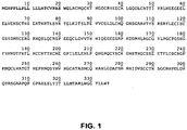
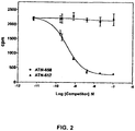

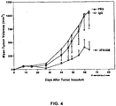

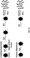

Classifications
-
- C—CHEMISTRY; METALLURGY
- C07—ORGANIC CHEMISTRY
- C07K—PEPTIDES
- C07K14/00—Peptides having more than 20 amino acids; Gastrins; Somatostatins; Melanotropins; Derivatives thereof
- C07K14/435—Peptides having more than 20 amino acids; Gastrins; Somatostatins; Melanotropins; Derivatives thereof from animals; from humans
- C07K14/705—Receptors; Cell surface antigens; Cell surface determinants
-
- A—HUMAN NECESSITIES
- A61—MEDICAL OR VETERINARY SCIENCE; HYGIENE
- A61P—SPECIFIC THERAPEUTIC ACTIVITY OF CHEMICAL COMPOUNDS OR MEDICINAL PREPARATIONS
- A61P35/00—Antineoplastic agents
-
- A—HUMAN NECESSITIES
- A61—MEDICAL OR VETERINARY SCIENCE; HYGIENE
- A61P—SPECIFIC THERAPEUTIC ACTIVITY OF CHEMICAL COMPOUNDS OR MEDICINAL PREPARATIONS
- A61P35/00—Antineoplastic agents
- A61P35/04—Antineoplastic agents specific for metastasis
-
- C—CHEMISTRY; METALLURGY
- C07—ORGANIC CHEMISTRY
- C07K—PEPTIDES
- C07K14/00—Peptides having more than 20 amino acids; Gastrins; Somatostatins; Melanotropins; Derivatives thereof
- C07K14/435—Peptides having more than 20 amino acids; Gastrins; Somatostatins; Melanotropins; Derivatives thereof from animals; from humans
- C07K14/705—Receptors; Cell surface antigens; Cell surface determinants
- C07K14/70596—Molecules with a "CD"-designation not provided for elsewhere
-
- C—CHEMISTRY; METALLURGY
- C07—ORGANIC CHEMISTRY
- C07K—PEPTIDES
- C07K16/00—Immunoglobulins [IGs], e.g. monoclonal or polyclonal antibodies
- C07K16/18—Immunoglobulins [IGs], e.g. monoclonal or polyclonal antibodies against material from animals or humans
- C07K16/28—Immunoglobulins [IGs], e.g. monoclonal or polyclonal antibodies against material from animals or humans against receptors, cell surface antigens or cell surface determinants
- C07K16/2896—Immunoglobulins [IGs], e.g. monoclonal or polyclonal antibodies against material from animals or humans against receptors, cell surface antigens or cell surface determinants against molecules with a "CD"-designation, not provided for elsewhere
-
- A—HUMAN NECESSITIES
- A61—MEDICAL OR VETERINARY SCIENCE; HYGIENE
- A61K—PREPARATIONS FOR MEDICAL, DENTAL OR TOILETRY PURPOSES
- A61K39/00—Medicinal preparations containing antigens or antibodies
- A61K2039/505—Medicinal preparations containing antigens or antibodies comprising antibodies
-
- C—CHEMISTRY; METALLURGY
- C07—ORGANIC CHEMISTRY
- C07K—PEPTIDES
- C07K2317/00—Immunoglobulins specific features
- C07K2317/30—Immunoglobulins specific features characterized by aspects of specificity or valency
- C07K2317/34—Identification of a linear epitope shorter than 20 amino acid residues or of a conformational epitope defined by amino acid residues
-
- C—CHEMISTRY; METALLURGY
- C07—ORGANIC CHEMISTRY
- C07K—PEPTIDES
- C07K2317/00—Immunoglobulins specific features
- C07K2317/70—Immunoglobulins specific features characterized by effect upon binding to a cell or to an antigen
- C07K2317/73—Inducing cell death, e.g. apoptosis, necrosis or inhibition of cell proliferation
Definitions
- the present invention relates to a urokinase-type plasminogen activator receptor (uPAR) epitope.
- uPAR urokinase-type plasminogen activator receptor
- Monoclonal antibodies that immunospecifically bind this epitope, and uses thereof for the treatment or prevention of a disease, e.g ., cancer are also disclosed. These antibodies may inhibit the interaction of uPA/uPAR complexes with additional molecules with which these complexes interact.
- the antibodies can be used in diagnostic and therapeutic methods, particularly for cancer.
- uPA urokinase plasminogen activator
- uPAR cell surface receptor
- Membrane-associated uPAR is a glycosylphosphatidylinositol-anchored (GPI)-anchored protein ( Slound, E. M., Blood 105:1847-1848 (2005 )).
- uPAR is composed of 3 domains; domain 1 (D1) is the N-terminal domain, domain 2 (D2) connects D1 to domain 3 (D3), and D3 is the C-terminal domain that anchors the molecule to the cell membrane through a GPI tail to Gly 283 of D3 ( Montuori et al., J. Biol. Chem. 277:46932-46939 (2002 ); Dano et al., Fibrinolysis 8:189-203 (1994 )).
- GPI glycosylphosphatidylinositol-anchored
- uPAR When uPAR is cleaved at the GPI anchor by phospholipase C ( Ploug et al., J Biol Chem. 266:1926-1933 (1991 )) or phospholipase D, soluble uPAR (suPAR) is released from the cell membrane ( Wilhelm et al., J. Cell Physiol. 180:225-235 (1999 )).
- Metastasis and angiogenesis share many common functional features that characterize invasive and migratory processes of tumor cells and of ECs. These features include (1) the up-regulation of protease and integrin expression, (2) the loss of cell-cell and cell-matrix contacts, (3) increased responsiveness to growth and differentiation factors, and (4) remodeling of extracellular matrix (ECM) and basement membrane (BasM). All of these contribute to tumor progression.
- ECM extracellular matrix
- BasM basement membrane
- the activity of this system is responsible for:
- uPA and uPAR have been demonstrated in numerous tumor types including glioblastoma, prostate, breast, colon, hepatocellular, and renal cell carcinoma ( Mizukami IF et al. (1994) Clin Immunol and Immunopathol 71:96-104 ; Hsu DW et al., (1995) Am J Pathol 147:114-23 ; de Witte JH et al. (1999) Br J Cancer 79:1190-8 ).
- the expression of uPA and uPAR are typically greater in more aggressive forms of disease.
- uPA uPA is chemotactic for monocytes and mediates both adhesion and migration of these cells. Adhesion and migration require only uPAR occupancy but not uPA catalytic activity. Thus, the uPA system is believed to contribute to tumor progression by acting on multiple tumor-associated cell types.
- ATF adenovirus-encoded murine amino-terminal fragment of uPA
- a polyclonal antibody raised against a 100-residue fragment of rat uPAR selectively localized to a rat breast tumor which grew from cells of the Mat BIII cell line ( Rabbani SA et al. (2002) Cancer Res 62: 2390-97 ).
- This polyclonal antibody completely inhibited tumor growth and led to tumor regression.
- the present invention is based, in part, on the characterization of the epitope of the uPAR-specific monoclonal antibody, ATN-658. Accordingly, the present invention provides an isolated or purified peptide consisting of the amino acid sequence CCTKSGCNHPDLDVQYPSG (SEQ ID NO: 9), CCTKSGCNHPDLDVQYPS (SEQ ID NO: 10), CCTKSGCNHPDLDVQYP (SEQ ID NO: 11), CCTKSGCNHPDLDVQY (SEQ ID NO: 12), or CCTKSGCNHPDLDVQ (SEQ ID NO: 13).
- the epitope sequence may also be part of a longer sequence, e.g ., 20, 25, 30, 35, 40 amino acids, where the longer sequence is a fragment of human uPAR and comprises the epitope sequence.
- the present invention provides methods of producing an antibody, which comprises (i) immunizing a non-human mammal with a peptide (optionally, purified) of the invention; (ii) isolating splenocytes from said mammal; (iii) fusing said splenocytes to myeloma cells; and (iv) selecting a hybridoma.
- the present invention provides methods of producing an antibody, which comprises (i) immunizing a non-human mammal with a peptide comprising a conformation-dependent epitope defined by (a) CCTKSGCNHPDLDVQYPSG (SEQ ID NO: 9), CCTKSGCNHPDLDVQYPS (SEQ ID NO: 10), CCTKSGCNHPDLDVQYP (SEQ ID NO: 11), CCTKSGCNHPDLDVQY (SEQ ID NO: 12) or CCTKSGCNHPDLDVQ (SEQ ID NO: 13); and (b) CGSSDMSCERGRHQSL (SEQ ID NO: 14) of a human uPAR (SEQ ID NO: 15); (ii) isolating splenocytes from said mammal; (iii) fusing said splenocytes to myeloma cells; and (iv) selecting a hybridoma that secretes an antibody that binds said peptide.
- the present disclosure provides methods of producing an antibody, which comprises (i) immunizing a mammal with a peptide comprising a conformation-dependent epitope defined by (a) KSGCNHPDLD (SEQ ID NO: 16); and (b) CGSSDMSCERGRHQSL (SEQ ID NO: 14) of a human uPAR (SEQ ID NO: 15); (ii) isolating splenocytes from said mammal; (iii) fusing said splenocytes to myeloma cells; and (iv) selecting a hybridoma that secretes an antibody that binds said peptide.
- the methods of producing an antibody comprise (i) immunizing a non-human mammal with a fragment of human uPAR (optionally, purified) comprising domains 2 and 3 of human uPAR (SEQ ID NO: 15); (ii) isolating splenocytes from said mammal; (iii) fusing said splenocytes to myeloma cells; and (iv) selecting a hybridoma that secretes an antibody that binds said fragment.
- the methods of producing an antibody comprise (i) immunizing a mammal with an isolated fragment of human uPAR (SEQ ID NO: 15), in which the amino terminus of said fragment is at any one of amino acid numbers 93-98 and the carboxy terminus of said fragment is at any one of amino acids 279-283, or a derivative thereof containing only conservative substitutions relative to the sequence of said fragment; (ii) isolating splenocytes from said mammal; (iii) fusing said splenocytes to myeloma cells; and (iv) selecting a hybridoma that secretes an antibody that binds said fragment.
- a non-human mammal may be mouse, rabbit, goat, rat, cat, dog, etc. Methods of producing an antibody using phage display are also encompassed by the invention.
- the present disclosure provides antibodies, or antigen-binding fragments thereof, that immunospecifically bind to an epitope defined by the amino acid sequence CCTKSGCNHPDLDVQYPSG (SEQ ID NO: 9), CCTKSGCNHPDLDVQYPS (SEQ ID NO: 10), CCTKSGCNHPDLDVQYP (SEQ ID NO: 11), CCTKSGCNHPDLDVQY (SEQ ID NO: 12) or CCTKSGCNHPDLDVQ (SEQ ID NO: 13).
- Disclosed are antibodies, or antigen-binding fragments thereof, immunospecifically bind to an epitope defined by the amino acid sequence KSGCNHPDLD (SEQ ID NO: 16).
- mutating the epitope at amino acid residue 268 reduces or abolishes the immunospecific binding affinity of the antibody. It should be noted that all references in this application to amino acid numbers in the human uPAR sequence refer to numbering from the amino terminus of processed uPAR, which lacks the 22 amino terminal acids shown in Figure 1 , unless explicitly indicated otherwise.
- the antibodies, or antigen-binding fragments thereof immunospecifically bind to a conformation-dependent epitope defined by (i) KSGCNHPDLD (SEQ ID NO: 16); and (ii) CGSSDMSCERGRHQSL (SEQ ID NO: 14) within the context of a human uPAR (SEQ ID NO:15).
- binding of an antibody to the epitope is demonstrated by a reduction in said binding by said antibody when the K (Lys) residue at position 268 in human uPAR is mutated to a E (Glu) residue (uPAR K268E) or to an A (Ala) residue (uPAR K268A).
- the amount of binding is demonstrated by co-immunoprecipitation of the antibody and uPAR K268E or uPAR K268A.
- binding of an antibody to the epitope is demonstrated by a reduction in said binding by said antibody when the H (His) residue at position 273 in human uPAR is mutated to an A (Ala) residue (uPAR H273A).
- the amount of binding is demonstrated by co-immunoprecipitation of the antibody and uPAR H273A.
- binding of an antibody to the epitope is demonstrated by a reduction in said binding by said antibody when the D (Asp) residue at position 275 or 277 in human uPAR is mutated to an A (Ala) residue (uPAR D275A or uPAR D277A, respectively).
- the amount of binding is demonstrated by co-immunoprecipitation of the antibody and uPAR D275A or uPAR D277A, respectively.
- a reduction in binding by the antibody is demonstrated by a decreased amount of mutated uPAR (e.g ., uPAR K268E, uPAR K268A, uPAR H273A, uPAR D275A, or uPAR D277A) that co-immunoprecipitates with the antibody relative to the amount of wild-type uPAR (e.g ., membrane-bound uPAR or suPAR) or a fragment of suPAR (e.g ., D2D3 suPAR) that co-immunoprecipitates with the antibody.
- wild-type uPAR e.g ., membrane-bound uPAR or suPAR
- a fragment of suPAR e.g ., D2D3 suPAR
- the binding of the antibody to an epitope of the invention is demonstrated by a deuterium exchange assay, wherein the binding of an antibody (e.g ., ATN-658) to an epitope of a protein (e.g ., human suPAR) decreases the ability of that epitope to exchange deuterium.
- human suPAR is contacted with an antibody, and binding of the antibody to an epitope of human uPAR is demonstrated by a reduction in deuteration level over the epitope when in the presence of the antibody under binding conditions, relative to deuteration over the epitope in the absence of the antibody.
- a region of suPAR that is not contacted by the antibody possesses the same or similar deuteration level when in the presence or absence of the antibody under binding conditions.
- the deuteration assay is performed with a fragment of human uPAR containing, or consisting of, domains 2 and 3 (D2D3).
- binding of the antibody to an epitope of human suPAR is demonstrated by a reduction in deuteration level over the epitope of human suPAR, when in the presence of the antibody under binding conditions, of at least 10%, or at least 20%, or at least 30%, or at least 40%, or at least 50%, relative to deuteration level over the epitope in the absence of the antibody.
- the antibodies or fragments thereof of the disclosure are isolated or purified.
- the antibody, or antigen-binding fragment thereof is a monoclonal antibody (preferably an IgG) or a scFv.
- the antibody of the disclosure can be a human antibody, humanized antibody or chimeric antibody.
- the antibody is a bi-specific antibody. In certain embodiments, the antibody is not ATN-658.
- the antibodies modulate downstream signaling of the urokinase-type plasminogen activator receptor and can interfere with and inhibit uPAR signaling, including, but not limited to, (a) uPAR mediated assembly of fibronectin, (b) binding of fibronectin or a fragment thereof to integrin ⁇ 5v1 and/or (c) the assembly of vitronectin components.
- the antibodies can also downregulate the number of uPAR molecules on the cell membrane.
- the antibodies of the disclosure are conjugated to a detectable label or therapeutic agent.
- a detectable label includes, but is not limited to, a radionuclide, a PET-imageable agent, an MRI-imageable agent, a fluorescer, a fluorogen, a chromophore, a chromogen, a phosphorescer, a chemiluminescer or a bioluminescer.
- Representive radionuclides include, but are not limited to, 3 H, 14 C, 35 S, 67 Ga, 68 Ga, 72 As, 89 Zr, 97 Ru, 99 Tc, 111 In, 123 I, 125 I, 131 I, 169 Yb and 201 Tl.
- fluorescers or fluorogens include, but are not limited to, fluorescein, rhodamine, dansyl, phycoerythrin, phycocyanin, allophycocyanin, o-phthaldehyde, fluorescamine, a fluorescein derivative, Oregon Green, Rhodamine Green, Rhodol Green and Texas Red.
- a therapeutic agent includes, but is not limited to, a chemotherapeutic drug, a toxin or a therapeutic radionuclide.
- compositions comprising an antibody, or antigen-binding fragment thereof; and a pharmaceutically acceptable carrier.
- the pharmaceutical compositions may be used in methods for inhibiting cell migration, cell invasion, cell proliferation or angiogenesis, or for inducing apoptosis, by contacting cells associated with undesired cell migration, invasion, proliferation or angiogenesis with an effective amount of a pharmaceutical composition of the disclosure.
- the pharmaceutical compositions may also be used in methods for treating a subject having a disease, disorder or condition characterized by undesired angiogenesis, tumor growth and/or tumor metastasis by administering to the subject a therapeutically effective amount of the pharmaceutical composition of the disclosure.
- uPAR is an ideal target for antibodies because it is expressed on the cell surface. Expression of uPAR at the tumor-vasculature interface (on invasive tumor cells, angiogenic endothelial cells, or tumor-associated macrophages) suggests that antibodies targeting this protein would not suffer the same barriers to diffusion that have led to the failure of other monoclonal antibodies to enter tumors and serve as diagnostic agents or exert therapeutic effects. Importantly, uPAR is not normally expressed on quiescent tissues, which should minimize the potential for toxicity when employing a therapeutic antibody and minimize non-specific signals (or false positives) when employing a diagnostic antibody.
- the present invention is based, in part, on the inventors' characterization of the epitope targeted by the uPAR-specific monoclonal antibody, ATN-658.
- the hybridoma secreting monoclonal antibody ATN-658 has been deposited under the provisions of the Budapest Treaty with the American Type Culture Collection (10801 University Boulevard., Manassas, VA. 20110-2209) on February 1,2007, and assigned ATCC Accession No. PTA-8191. This antibody does not affect the binding of uPA to its receptor.
- Antibodies that bind to this epitope affect downstream signaling pathways, including such "downstream" ligands as integrins, low-density lipoprotein receptor-related protein (LRP) and other binding partners. These downstream interactions are believed to be important to the processes of cell migration, invasion and proliferation. It is thus desirable to target these processes therapeutically or detect the process or their interacting components diagnostically.
- the present disclosure relates to methods and compositions that provide for the prevention and treatment of cancer.
- a particular aspect of the disclosure relates to methods and compositions containing compounds that inhibit cancer cell proliferation and invasion.
- the disclosure further provides diagnostic methods using the uPAR antibodies of the disclosure.
- the diagnostic methods can also be used to prognose or predict cancer progression.
- the diagnostic methods provide methods of imaging and localizing metastases and methods of diagnosis and prognosis using tissues and fluids distal to the primary tumor site (as well as methods using tissues and fluids of the primary tumor).
- the diagnostic methods provide methods of imaging and localizing metastases and methods of diagnosis and prognosis in vivo.
- the present inventors have mapped an epitope on uPAR that is not involved in binding to uPA. This epitope has been mapped by its interaction with the uPAR monoclonal antibody ATN-658, described in WO 2005/116077 .
- the variable region sequences of ATN-658 are set forth below:
- ATN-658 Variable region sequences
- This antibody recognizes the uPA-uPAR complex.
- This antibody was generated against an epitope in the uPAR D2D3 fragment.
- An epitope in D2D3 has been demonstrated to be critical to the pro-migratory activity of uPA ( Andolfo A et al. (2002) Thromb Haemost 88: 298-306 ).
- antibodies generated against the D2D3 fragment where this epitope is already exposed are expected to have anti-migratory activity.
- uPAR epitope sequences Sequence Amino acids of Human uPAR 1 SEQ ID NO: CCTKSGCNHPDLDVQYPSG 265-283 9 CCTKSGCNHPDLDVQYPS 265-282 10 CCTKSGCNHPDLDVQYP 265-281 11 CCTKSGCNHPDLDVQY 265-280 12 CCTKSGCNHPDLDVQ 265-279 13 CGSSDMSCERGRHQSL 98-114 14 KSGCNHPDLD 268-277 16 1 The amino acid numbering reflects the processed form of uPAR
- the antibody recognizes an epitope defined by the amino acid sequence of SEQ ID NO: 9, 10, 11, 12, 13, or 16.
- the antibody recognizes a conformation-dependent epitope defined by (i) an amino acid sequence of SEQ ID NO: 9, 10, 11, 12, 13, or 16; and (ii) an amino acid sequence of SEQ ID NO: 14 within the context of human uPAR (SEQ ID NO: 15).
- full-length soluble uPAR (suPAR) (residues 1-283, domains 1,2, and 3) can be used as an immunogen.
- suPAR full-length soluble uPAR
- one of skill in the art will recognize that shorter fragment of suPAR can be used as an immunogen as long as the proper conformation is maintained.
- the conformation-dependent epitope retains its native conformation, as shown, e.g., by molecular modeling.
- the conformation-dependent epitope is defined by (i) an amino acid sequence of SEQ ID NO: 9, 10, 11, 12, 13, or 16; and (ii) an amino acid sequence of SEQ ID NO: 14, wherein a linker peptide sequence is placed in between the amino acid sequences of (i) and (ii), such that the native conformation is maintained.
- the linker peptide sequence mimics the native conformation of uPAR.
- the linker peptide sequence is heterologous to human uPAR.
- the linker peptide sequence is the intervening sequence of human uPAR in which conservative amino acid substitutions have been made.
- one or more amino acid residues within the sequence can be substituted by another amino acid of a similar polarity which acts as a functional equivalent, resulting in a silent alteration.
- substitutions for an amino acid within the sequence may be selected from other members of the class to which the amino acid belongs.
- the nonpolar (hydrophobic) amino acids include alanine, leucine, isoleucine, valine, proline, phenylalanine, tryptophan and methionine.
- the polar neutral amino acids include glycine, serine, threonine, cysteine, tyrosine, asparagine, and glutamine.
- the positively charged (basic) amino acids include arginine, lysine and histidine.
- the negatively charged (acidic) amino acids include aspartic acid and glutamic acid. Such substitutions are generally understood to be conservative substitutions. Conservative substitutions of one or more amino acid residues are preferably introduced into the peptide sequence at sites that will not disturb the native conformation or activity.
- the uPAR peptides may comprise up to 20, 30, 40 amino acids.
- the peptides comprise contiguous amino acid residues of uPAR. These longer peptides generally will consist of a fragment of human uPAR and comprise the epitope sequence of uPAR.
- the uPAR is human uPAR.
- the amino acid sequence of the unprocessed form of human uPAR UniProtKB/Swiss-Prot Accession No. Q03405 (SEQ ID NO: 15) is provided in Figure 1 .
- the processed (mature) form of human uPAR removes the first 22 amino acids.
- C-terminal residues 284-313 are also removed by post-translational processing when uPAR is anchored to the plasma membrane by a GPI tail (see Moller et al., Eur. J. Biochem. 208:493-500 (1992 ); Low, M. G., FASEB J. 3:1600-1608 (1989 )).
- Reference to uPAR (including suPAR) in this application is reference to processed (mature) uPAR (containing amino acids 1-283) unless explicitly indicated otherwise.
- mutating one or more amino acid residues of the epitope sequences in Table 2 reduces or abolishes the immunospecific binding affinity of the antibody, e.g ., ATN-658, to the epitope, e.g ., as contained in human uPAR, which can be membrane-associated or suPAR.
- a mutation at amino acid residue 268 of human uPAR or the epitopes in Table 2 reduces or abolishes the immunospecific binding affinity of the antibody or antigen-binding fragments thereof to the epitope.
- binding of an antibody to the epitope is demonstrated by reduction in said binding by said antibody when the K (Lys) residue at position 268 in human uPAR is mutated to a E (Glu) residue (uPAR K268E) or an A (Ala) residue (uPAR K268A).
- a mutation at amino acid residue 273 of human uPAR or the epitopes in Table 2 reduces or abolishes the immunospecific binding affinity of the antibody or antigen-binding fragments thereof to the epitope.
- binding of an antibody to the epitope is demonstrated by reduction in said binding by said antibody when the H (His) residue at position 273 in human uPAR is mutated to an A (Ala) residue (uPAR H273A).
- a mutation at amino acid residue 275 of human uPAR or the epitopes in Table 2 reduces or abolishes the immunospecific binding affinity of the antibody or antigen-binding fragments thereof to the epitope.
- binding of an antibody to the epitope is demonstrated by reduction in said binding by said antibody when the D (Asp) residue at position 275 in human uPAR is mutated to an A (Ala) residue (uPAR D275A).
- a mutation at amino acid residue 277 of human uPAR or the epitopes in Table 2 reduces or abolishes the immunospecific binding affinity of the antibody or antigen-binding fragments thereof to the epitope.
- binding of an antibody to the epitope is demonstrated by reduction in said binding by said antibody when the D (Asp) residue at position 277 in human uPAR is mutated to an A (Ala) residue (uPAR D277A).
- a mutation of one or more residues of human uPAR, e.g ., 268, 273, 275, and/or 277, e.g ., as described herein, or a mutation in any of the epitopes in Table 2 reduces the immunospecific binding affinity of the antibody or an antigen-binding fragment thereof by at least 5%, at least 10%, at least 20%, at least 30%, at least 40% or at least 50% relative to the affinity of the antibody to a wild-type human uPAR (e.g ., membrane-bound uPAR, suPAR) or to a fragment of suPAR (e.g ., D2D3 suPAR).
- a wild-type human uPAR e.g ., membrane-bound uPAR, suPAR
- a fragment of suPAR e.g ., D2D3 suPAR
- the binding affinity of the antibody or antigen-binding fragment thereof to human uPAR or to a fragment ( e . g ., D2D3 suPAR) containing an epitope thereof can be determined by methods well known in the art, e.g ., but not limited to co-immunoprecipitation assays, BIAcore assay, deuterium exchange assay, and ELISA.
- binding of an antibody to the epitope is demonstrated by a reduction of at least 10%, 20%, 30%, 40%, 50%, 60%, or 70% in said binding by said antibody when the K (Lys) residue at position 268 in human uPAR is mutated to a E (Glu) residue (uPAR K268E) or an A (Ala) residue (uPAR K268A).
- the amount of binding is demonstrated by co-immunoprecipitation of the antibody and uPAR K268E or uPAR K268A, respectively.
- binding of an antibody to the epitope is demonstrated by a reduction of at least 10%, 20%, 30%, 40%, 50%, 60%, 70%, 80% or 90% in said binding by said antibody when the H (His) residue at position 273 in human uPAR is mutated to an A (Ala) residue (uPAR H273A).
- the amount of binding is demonstrated by co-immunoprecipitation of the antibody and uPAR H273A.
- binding of an antibody to the epitope is demonstrated by a reduction of at least 10%, 20%, 30%, 40%, 50%, 60%, 70%, 80% or 90% in said binding by said antibody when the D (Asp) residue at position 275 or 277 in human uPAR is mutated to an A (Ala) residue (uPAR D275A or uPAR D277A, respectively).
- the amount of binding is as demonstrated by co-immunoprecipitation of the antibody and uPAR D275A or uPAR D277A, respectively.
- a reduction in binding by the antibody is demonstrated by a decreased amount of mutated uPAR (e.g ., uPAR K268E, uPAR K268A, uPAR H273A, uPAR D275A, or uPAR D277A) that co-immunoprecipitates with the antibody relative to the amount of wild-type uPAR (e.g ., membrane-bound uPAR, suPAR) or a fragment of suPAR (e.g ., D2D3 suPAR) that co-immunoprecipitates with the antibody.
- wild-type uPAR e.g ., membrane-bound uPAR, suPAR
- a fragment of suPAR e.g ., D2D3 suPAR
- the binding of the antibody to an epitope of the invention is demonstrated by a deuterium exchange assay, wherein the binding of an antibody (e.g ., ATN-658) to an epitope of a protein (e.g ., human suPAR) decreases the ability of that epitope to exchange deuterium.
- human suPAR is contacted with an antibody, and binding of the antibody to an epitope of human uPAR is demonstrated by a reduction in deuteration level over the epitope when in the presence of the antibody, under binding conditions, relative to deuteration over the epitope in the absence of the antibody.
- a region of suPAR that is not contacted by the antibody possesses the same or similar deuteration level when in the presence or absence of the antibody under binding conditions.
- the deuteration assay is performed with a fragment of human uPAR containing, or consisting of, domains 2 and 3 (D2D3) (amino acids 88-283).
- binding of the antibody to an epitope of human suPAR is demonstrated by reduction in deuteration level over the epitope of human suPAR, when in the presence of the antibody under binding conditions, of at least 10%, or at least 20%, or at least 30%, or at least 40%, or at least 50%, relative to deuteration level over the epitope in the absence of the antibody.
- reduction in deuteration level of at least 10%, at least 20%, at least 30%, at least 40%, at least 50%, at least 55%, at least 60%, at least 65%, at least 70%, or at least 75% over the epitope of suPAR is demonstrated when in the presence of the antibody under binding conditions, relative to deuteration level over the epitope of suPAR in the absence of the antibody.
- the antibody is immobilized.
- the uPAR epitope and peptides comprising these sequences may have utility in the diagnostic and therapeutic methods described herein.
- This disclosure provides an isolated antibody, or antigen-binding fragment thereof, that immunospecifically binds to an epitope of uPAR defined by the amino acid sequence of SEQ ID NO: 9, 10, 11, 12 or 13.
- the disclosure also provides an isolated antibody, or antigen-binding fragment thereof, that immunospecifically binds to a conformation-dependent epitope defined by (i) CCTKSGCNHPDLDVQYPSG (SEQ ID NO: 9), CCTKSGCNHPDLDVQYPS (SEQ ID NO: 10), CCTKSGCNHPDLDVQYP (SEQ ID NO: 11), CCTKSGCNHPDLDVQY (SEQ ID NO: 12) or CCTKSGCNHPDLDVQ (SEQ ID NO: 13); and (ii) CGSSDMSCERGRHQSL (SEQ ID NO: 14) within the context of a human uPAR (SEQ ID NO: 15).
- This disclosure provides an isolated antibody, or antigen-binding fragment thereof, that immunospecifically binds to an epitope of uPAR defined by the amino acid sequence of SEQ ID NO: 16.
- the disclosure also provides an isolated antibody, or antigen-binding fragment thereof, that immunospecifically binds to a conformation-dependent epitope defined by (i) KSGCNHPDLD (SEQ ID NO: 16); and (ii) CGSSDMSCERGRHQSL (SEQ ID NO: 14) within the context of a human uPAR (SEQ ID NO: 15).
- immunospecifically binds means that the antibody, or an antigen-binding fragment thereof, binds to the antigen via its antigen-recognition region of its variable domain.
- An animal preferably a mouse is primed by immunization with an immunogen as above to elicit the desired antibody response in the primed animal.
- a non-human mammal is primed by immunization with an immunogen as described above to elicit the desired antibody response in the primed non-human mammal.
- mice can be immunized with a uPAR epitope and once an immune response is detected, e.g ., antibodies specific for uPAR are detected in the mouse serum, the mouse spleen is harvested and splenocytes isolated. The splenocytes are then fused by well known techniques to any suitable myeloma cells, for example cells from cell line SP20 available from the ATCC. Hybridomas are selected and cloned by limited dilution. Hybridoma clones are then assayed by methods known in the art for cells that secrete antibodies capable of binding uPAR. Ascites fluid, which generally contains high levels of antibodies, can be generated by immunizing mice with positive hybridoma clones.
- an immune response e.g ., antibodies specific for uPAR are detected in the mouse serum
- the mouse spleen is harvested and splenocytes isolated.
- the splenocytes are then fused by well known techniques to any suitable myel
- the epitope sequences in Table 2 are used as an immunogen for the generation of the antibodies of the disclosure.
- a peptide consisting of the amino acid sequence CCTKSGCNHPDLDVQYPSG (SEQ ID NO: 9), CCTKSGCNHPDLDVQYPS (SEQ ID NO: 10), CCTKSGCNHPDLDVQYP (SEQ ID NO: 11), CCTKSGCNHPDLDVQY (SEQ ID NO: 12), or CCTKSGCNHPDLDVQ (SEQ ID NO: 13) is used.
- a fragment of human uPAR of up to 20, 30, or 40 amino acids, comprising the amino acid sequence CCTKSGCNHPDLDVQYPSG (SEQ ID NO: 9), CCTKSGCNHPDLDVQYPS (SEQ ID NO: 10), CCTKSGCNHPDLDVQYP (SEQ ID NO: 11), CCTKSGCNHPDLDVQY (SEQ ID NO: 12), or CCTKSGCNHPDLDVQ (SEQ ID NO: 13) is used.
- a peptide comprising or alternatively, consisting of, a conformation-dependent epitope defined by (i) KSGCNHPDLD (SEQ ID NO: 16); and (ii) CGSSDMSCERGRHQSL (SEQ ID NO: 14) of a human uPAR (SEQ ID NO: 15).
- such a conformation-dependent epitope comprises or consists of contiguous amino acid sequences of a fragment of human uPAR from position 98 to 279, 280, 281, 282, or 283.
- the conformation-dependent epitope retains its native conformation, as shown, e.g., by molecular modeling.
- the conformation-dependent epitope is defined by (i) an amino acid sequence of SEQ ID NO: 9, 10, 11, 12, 13, or 16; and (ii) an amino acid sequence of SEQ ID NO: 14, wherein a linker peptide sequence is placed in between the amino acid sequences of (i) and (ii), such that the native conformation is maintained.
- the linker peptide sequence mimics the native conformation of uPAR.
- the linker peptide sequence is heterologous to human uPAR.
- the linker peptide sequence is the intervening sequence of human uPAR in which conservative amino acid substitutions have been made. Conservative substitutions of one or more amino acid residues are preferably introduced into the peptide sequence at sites that will not disturb the native conformation or activity. For example, one or more amino acid residues within the sequence can be substituted by another amino acid of a similar polarity which acts as a functional equivalent, resulting in a silent alteration. Substitutions for an amino acid within the sequence may be selected from other members of the class to which the amino acid belongs.
- the nonpolar (hydrophobic) amino acids include alanine, leucine, isoleucine, valine, proline, phenylalanine, tryptophan and methionine.
- the polar neutral amino acids include glycine, serine, threonine, cysteine, tyrosine, asparagine, and glutamine.
- the positively charged (basic) amino acids include arginine, lysine and histidine.
- the negatively charged (acidic) amino acids include aspartic acid and glutamic acid. Such substitutions are generally understood to be conservative substitutions.
- a peptide or fragment of human uPAR comprising or consisting of domains 2 and 3 (D2D3) corresponding to residues 88-283 of soluble uPAR (suPAR) is used as the immunogen.
- Peptides comprising these sequences can be produced in a variety of ways known in the art, e.g . , expression of cloned genes using conventional recombinant methods, isolation from cells of origin, cell populations expressing high levels of e.g ., uPA or uPAR, etc. In the case of shorter fragments, they may be chemically synthesized.
- B lymphocytes from the lymph nodes, spleens or peripheral blood of a primed, animal are fused with myeloma cells, generally in the presence of a fusion promoting agent such as polyethylene glycol (PEG).
- PEG polyethylene glycol
- Any of a number of murine myeloma cell lines are available for such use: the P3-NS1/1-Ag4-1, P3-x63-k0Ag8.653, Sp2/0-Agl4, or HL1-653 myeloma lines (available from the ATCC, Rockville, MD).
- Subsequent steps include growth in selective medium so that unfused parental myeloma cells and donor lymphocyte cells eventually die while only the hybridoma cells survive.
- Hybridomas produced according to these methods can be propagated in vitro or in vivo (in ascites fluid) using techniques known in the art (see generally Fink et al., Prog. Clin. Pathol. 9:121-33 (1984 )).
- the individual cell line is propagated in culture and the culture medium containing high concentrations of a single monoclonal antibody can be harvested by decantation, filtration, or centrifugation.
- a preferred approach for producing a mAb according to the present invention is as follows.
- An epitope in table 2 is preferably conjugated to a carrier protein, e.g ., KLH, is and injected into BALB/c mice intraperitoneally (i.p.) in complete Freund's adjuvant (e.g ., 50 ⁇ g conjugate), followed by two additional injections of the same dose in incomplete Freund's adjuvant at two week intervals. After one month, a final injection is given i. p ( e.g ., 50 ⁇ g in 0.5 ml PBS) and preferably also intravenously (i.v.) ( e.g ., 50 ⁇ g in 0.2 ml) without adjuvant.
- a carrier protein e.g ., KLH
- Spleen cells are harvested three days after the final injection and fused with P3X63AF8/653 or other myeloma cells using standard techniques.
- uPAR immobilized onto plastic is preferred for the primary screening.
- Many tumor cell lines overexpressing uPAR are well-known and publicly available; these may be used for screening.
- cells such as the HeLa line that overexpress uPAR may also be used to demonstrate cell binding of an anti-uPAR mAb.
- Cells are generally plated in 96-well microplates. The cells may be fixed, e.g ., with methanol/acetone (50/50), and the binding detected by immunofluorescence staining.
- the mAbs may be labeled, using radioactive isotopes or other tracers such as biotin, and binding detected by measurement of radioactivity or biotin.
- a hybridoma supernatant e.g ., 50 ⁇ l
- a hybridoma supernatant e.g ., 50 ⁇ l
- plates are washed twice in washing buffer (such as PBS/0.05% Tween-20), and Rhodamine Red-conjugated goat anti-mouse IgG is added ( e.g ., 30 ⁇ l/well) at an appropriate dilution, such as 1:100, for 1.5 h at 37°C.
- washing buffer such as PBS/0.05% Tween-20
- Rhodamine Red-conjugated goat anti-mouse IgG is added (e.g ., 30 ⁇ l/well) at an appropriate dilution, such as 1:100, for 1.5 h at 37°C.
- washing buffer such as PBS/0.05% Tween-20
- Rhodamine Red-conjugated goat anti-mouse IgG is added (e.g ., 30 ⁇ l/well) at
- immunofluorescence is the basis for determining whether a hybridoma supernatant contains an antibody specific for the uPA/uPAR complex (although immunohistochemical staining may also be used). If supernatants show positively staining the hybridoma clones are selected, expanded and the supernatants tested for reactivity to the complex by ELISA.
- the peptide is coupled to ovalbumin (OVA) as a carrier protein and the peptide/OVA conjugate coated onto wells of 96 well EIA plate which receives, for example, 2 ⁇ g/ml of conjugate in 50 ⁇ l coating buffer (0.2 M Na 2 CO 3 /NaHCO 3 , pH 9.6). Plates are incubated overnight at 4°C, blocked with an appropriate blocking buffer, e.g ., PBS containing 1% BSA (200 ⁇ l/well) overnight at 4°C. Hybridoma supernatants (e.g ., 50 ⁇ l) are added to wells for 1.5 hours at room temperature.
- OVA ovalbumin
- Plates are washed twice in washing buffer (e.g ., PBS/ 0.05% Tween-20), and enzyme-coupled secondary Ab, such as alkaline phosphatase-coupled goat-anti-mouse IgG is added (50 ⁇ l/well) at an appropriate dilution, e.g ., 1:2000. Plates are incubated for 1.5 hours at RT. After washing 4X in washing buffer, an appropriate chromogenic substrate for the enzyme, e.g ., CP-nitrophenylphosphate in this embodiment (available from Kirkegaard and Perry Co., Gaithersburg, MD), is added for about 30 min and absorbance measured at wavelength appropriate for the colored product (here 405nm).
- washing buffer e.g ., PBS/ 0.05% Tween-20
- enzyme-coupled secondary Ab such as alkaline phosphatase-coupled goat-anti-mouse IgG is added (50 ⁇ l/well) at an appropriate dilution, e.g
- Hybridoma supernatants that react strong with the epitope-bearing peptide e.g ., A 405 >1.0 when negative controls are ⁇ 0.02 are re-cloned (preferably twice), and the mAb reactivity again confirmed by ELISA as above.
- the anti-uPAR antibodies of the disclosure are preferably tested in xenogeneic tumor models, two preferred examples of which are the A2780 and A549 models (described in more detail below).
- the antibodies are evaluated for direct anti-angiogenic activity in an in vivo Matrigel plug model. Radioiodinated antibodies are used to test antibody internalization using MDA MB 231 cells which express both receptor and ligand. Antibody internalization is also measured in the presence of PAI- 1: uPA complexes.
- Antibodies used in the methods of the disclosure include, but are not limited to, monoclonal antibodies, synthetic antibodies, multispecific antibodies (including bi-specific antibodies), human antibodies, humanized antibodies, chimeric antibodies, single-chain Fvs (scFv) (including bi-specific scFvs), single chain antibodies, Fab fragments, F(ab') fragments, disulfide-linked Fvs (sdFv), and epitope-binding fragments of any of the above.
- antibodies used in the methods of the present disclosure include immunoglobulin molecules and immunologically active portions of immunoglobulin molecules, i.e., molecules that contain an antigen binding site that immunospecifically binds to uPAR.
- the immunoglobulin molecules of the disclosure can be of any type (e.g ., IgG, IgE, IgM, IgD, IgA and IgY), class (e.g ., IgG 1 , IgG 2 , IgG 3 , IgG 4 , IgA 1 and IgA 2 ) or subclass of immunoglobulin molecule.
- Anti-idiotypic antibodies specific for the idiotype of, for example, an anti-uPA/uPAR antibody are also included.
- Antibody derivatives are also encompassed by the disclosure.
- the term "derivative" as used herein refers to a polypeptide that comprises an amino acid sequence of an antibody that immunospecifically binds to an uPAR polypeptide, or an antibody fragment that immunospecifically binds to an uPAR polypeptide, which has been altered by the introduction of amino acid residue substitutions, deletions or additions ( i.e ., mutations).
- an antibody derivative or fragment thereof comprises amino acid residue substitutions, deletions or additions in one or more CDRs.
- the antibody derivative may have substantially the same binding, better binding, or worse binding when compared to a non-derivative antibody.
- one, two, three, four, or five amino acid residues of the CDR have been substituted, deleted or added (i.e ., mutated).
- derivative as used herein also refers to an antibody that immunospecifically binds to uPAR, or an antibody fragment that immunospecifically binds to an uPAR, which has been modified, i.e , by the covalent attachment of any type of molecule to the polypeptide.
- an uPAR antibody, or antibody fragment may be modified, e.g., by glycosylation, acetylation, pegylation, phosphorylation, amidation, derivatization by known protecting/blocking groups, proteolytic cleavage, linkage to a cellular ligand or other protein, etc.
- a derivative of an uPAR antibody, or antibody fragment may be modified by chemical modifications using techniques known to those of skill in the art, including, but not limited to specific chemical cleavage, acetylation, formylation, metabolic synthesis of tunicamycin, etc.
- a derivative of an uPAR antibody, or antibody fragment may contain one or more non-classical amino acids.
- the antibodies used in the methods of the disclosure may be from any animal origin including birds and mammals (e.g ., human, murine, donkey, sheep, rabbit, goat, guinea pig, camel, horse, or chicken).
- the methods of the invention may be from a non-human mammal.
- the antibodies are human or humanized monoclonal antibodies.
- "human" antibodies include antibodies having the amino acid sequence of a human immunoglobulin and include antibodies isolated from human immunoglobulin libraries or from mice or other animal that express antibodies from human genes.
- the antibodies used in the methods of the present disclosure may be monospecific, bispecific, trispecific or of greater multispecificity.
- Multispecific antibodies may immunospecifically bind to different epitopes of uPAR or may immunospecifically bind to both uPAR as well a heterologous epitope, such as a heterologous polypeptide or solid support material. See, e.g ., International Publication Nos. WO 93/17715 , WO 92/08802 , WO 91/00360 , and WO 92/05793 ; Tutt, et al., 1991, J. Immunol. 147:60-69 ; U.S. Patent Nos.
- a bispecific antibody which binds to the epitope of ATN-658 and another uPAR epitope. See Gardsvoll et al., 2006, J. Biol. Chem. 281(28):19260-72 ; Gardsvoll et al., 1999, J. Biol. Chem. 274(53):37995-8003 .
- Antibodies of the present disclosure can also be generated using various phage display methods known in the art.
- phage display methods functional antibody domains are displayed on the surface of phage particles which carry the polynucleotide sequences encoding them.
- DNA sequences encoding V H and V L domains are amplified from animal cDNA libraries (e.g ., human or murine cDNA libraries of lymphoid tissues).
- the DNA encoding the V H and V L domains are recombined together with an scFv linker by PCR and cloned into a phagemid vector (e.g ., p CANTAB 6 or pComb 3 HSS).
- the vector is electroporated in E.
- Phage used in these methods are typically filamentous phage including fd and M13 and the V H and V L domains are usually recombinantly fused to either the phage gene III or gene VIII.
- Phage expressing an antigen binding domain that binds to the uPAR epitope of interest can be selected or identified with antigen, e.g., using labeled antigen or antigen bound or captured to a solid surface or bead. Examples of phage display methods that can be used to make the antibodies of the present disclosure include those disclosed in Brinkman et al., 1995, J. Immunol. Methods 182:41-50 ; Ames et al., 1995, J.
- the antibody coding regions from the phage can be isolated and used to generate whole antibodies, including human antibodies, or any other desired antigen binding fragment, and expressed in any desired host, including mammalian cells, insect cells, plant cells, yeast, and bacteria, e.g., as described below.
- Techniques to recombinantly produce Fab, Fab' and F(ab')2 fragments can also be employed using methods known in the art such as those disclosed in International Publication No.
- PCR primers including V H or V L nucleotide sequences, a restriction site, and a flanking sequence to protect the restriction site can be used to amplify the V H or V L sequences in scFv clones.
- V H constant region e.g ., the human gamma 4 constant region
- V L constant region e.g ., human kappa or lambda constant regions.
- V H and V L domains may also be cloned into one vector expressing the necessary constant regions.
- the heavy chain conversion vectors and light chain conversion vectors are then co-transfected into cell lines to generate stable or transient cell lines that express full-length antibodies, e.g., IgG, using techniques known to those of skill in the art.
- antibody is meant to include both intact immunoglobulin (Ig) molecules as well as fragments and derivative thereof, that may be produced by proteolytic cleavage of Ig molecules or engineered genetically or chemically. Fragments include, for example, Fab, Fab', F(ab')2 and Fv, each of which is capable of binding antigen.
- fragments include a peptide or polypeptide comprising an amino acid sequence of at least 5 contiguous amino acid residues, at least 10 contiguous amino acid residues, at least 15 contiguous amino acid residues, at least 20 contiguous amino acid residues, at least 25 contiguous amino acid residues, at least 40 contiguous amino acid residues, at least 50 contiguous amino acid residues, at least 60 contiguous amino residues, at least 70 contiguous amino acid residues, at least contiguous 80 amino acid residues, at least contiguous 90 amino acid residues, at least contiguous 100 amino acid residues, at least contiguous 125 amino acid residues, at least 150 contiguous amino acid residues, at least contiguous 175 amino acid residues, at least contiguous 200 amino acid residues, or at least contiguous 250 amino acid residues of the amino acid sequence of an antibody that immunospecifically binds to uPAR polypeptide.
- antibody fragments are epitope-binding fragments.
- a Fab fragment is a multimeric protein consisting of the portion of an Ig molecule containing the immunologically active portions of an Ig heavy (H) chain and an Ig light (L) chain covalently coupled together and capable of specifically combining with antigen.
- a (Fab') 2 fragment is a tetramer that includes a fragment of two H and two L chains.
- the Fv fragment is a multimeric protein consisting of the immunologically active portions of an Ig H chain variable (V) region (VH) and an Ig L chain V region(VL) covalently coupled together and capable of specifically combining with antigen.
- fragments lack the Fc fragment of intact Ab and have an additional advantage, if used therapeutically, of clearing more rapidly from the circulation and undergoing less nonspecific tissue binding than intact antibodies.
- various fragments are produced using conventional techniques such as protease cleavage or chemical cleavage (see, e.g., Rousseaux et al., Meth.Enzymol., 121:663-69(1986 )).
- protease cleavage or chemical cleavage
- papain treatment of Ig's produces Fab fragments
- pepsin treatment produces F(ab')2 fragments.
- fragments may also produced by genetic or protein engineering using methods well known in the art.
- a Fab fragment may also be prepared by expressing in a suitable host cell the desired portions of Ig H chain and L chain using methods well known in the art.
- Fv fragments are typically prepared by expressing in suitable host cell the desired portions of Ig VH region and VL region using methods well known in the art.
- the antibodies of the present disclosure may be produced as a single chain antibody or scFv instead of the normal multimeric structure.
- Single-chain antigen-binding protein or single chain Ab also referred to as "scFv," is a polypeptide composed of an Ig VL amino acid sequence tethered to an Ig V H amino acid sequence by a peptide that links the C-terminus of the V L sequence to the N-terminus of the V H sequence.
- scFvs include bispecific scFvs and humanized scFvs.
- One method of producing the single chain antibodies is to link two or more peptides or polypeptides together by protein chemistry techniques.
- peptides or polypeptides can be chemically synthesized using currently available laboratory equipment using either Fmoc (9- fluorenylmethyloxycarbonyl) or tBoc (tert-butyloxycarbonoyl) chemistry. (Applied Biosystems, Inc., Foster City, CA).
- Fmoc 9- fluorenylmethyloxycarbonyl
- tBoc tert-butyloxycarbonoyl
- a peptide or polypeptide can be synthesized but not cleaved from its synthesis resin whereas the other fragment of an Ab can be synthesized and subsequently cleaved from the resin, thereby exposing a terminal group which is functionally blocked on the other fragment.
- peptide condensation reactions these two fragments can be covalently joined via a peptide bond at their C- and N- termini, respectively, to form an Ab, or a fragment thereof.
- Antibodies can be selected for particular desired properties.
- Ab screening procedures can include any of the in vitro or in vivo bioassays that measure binding to e.g., uPA/uPAR or uPAR-integrin complex, to cells expressing the relevant polypeptide or peptide epitope.
- human or chimeric antibodies are particularly desirable for therapeutic treatment of human subjects.
- a chimeric antibody is a molecule in which different portions of the antibody are derived from different immunoglobulin molecules.
- Methods for producing chimeric antibodies are known in the art. See, e.g., Morrison, 1985, Science 229:1202 ; Oi et al., 1986, BioTechniques 4:214 ; Gillies et al., 1989, J. Immunol. Methods 125:191-202 ; and U.S. Patent Nos. 6,311,415 , 5,807,715 , 4,816,567 , and 4,816,397 .
- Chimeric antibodies comprising one or more CDRs from one species and framework regions from a different species can be produced using a variety of techniques known in the art including, for example, CDR-grafting ( EP 239,400 ; International Publication No. WO 91/09967 ; and U.S. Patent Nos. 5,225,539 , 5,530,101 , and 5,585,089 ), veneering or resurfacing ( EP 592,106 ; EP 519,596 ; Padlan, 1991, Molecular Immunology 28(4/5):489-498 ; Studnicka et al., 1994, Protein Engineering 7:805 ; and Roguska et al., 1994, PNAS 91:969 ), and chain shuffling ( U.S. Patent No. 5,565,332 ).
- Antibodies, fragments or derivatives having chimeric H chains and L chains of the same or different V region binding specificity can be prepared by appropriate association of the individual polypeptide chains, as taught, for example by Sears et al., Proc. Natl. Acad. Sci. USA 72: 353-357 (1975 ).
- hosts expressing chimeric H chains (or their derivatives) are separately cultured from hosts expressing chimeric L chains (or their derivatives), and the Ig chains are separately recovered and then associated.
- the hosts can be co-cultured and the chains allowed to associate spontaneously in the culture medium, followed by recovery of the assembled Ig, fragment or derivative.
- chimeric antibody includes monovalent, divalent or polyvalent Igs.
- a monovalent chimeric Ab is an HL dimer formed by a chimeric H chain associated through disulfide bridges with a chimeric L chain.
- a divalent chimeric Ab is tetramer H2L2 formed by two HL dimers associated through at least one disulfide bridge.
- a polyvalent chimeric Ab can also be produced, for example, by employing a CH region that aggregates ( e.g. , from an IgM H chain, termed the ⁇ chain).
- the disclosure also provides for "derivatives" of the chimeric antibodies, which term includes those proteins encoded by truncated or modified genes to yield molecular species functionally resembling the Ig fragments.
- the modifications include, but are not limited to, addition of genetic sequences coding for cytotoxic proteins such as plant and bacterial toxins.
- the fragments and derivatives can be produced from any of the hosts of this disclosure.
- a humanized antibody refers to a non-human (e.g. , murine) antibody that is a chimeric antibody which contain minimal sequence derived from non-human immunoglobulin.
- humanized antibodies are human immunoglobulins (recipient antibody) in which hypervariable region residues of the recipient are replaced by hypervariable region residues from a non-human species (donor antibody) such as mouse, rat, rabbit or non-human primate having the desired specificity, affinity, and capacity.
- a humanized antibody comprises substantially all of at least one, and typically two, variable domains in which all or substantially all of the CDR regions correspond to those of a non-human immunoglobulin (i.e ., donor antibody) and all or substantially all of the framework regions are those of a human immunoglobulin consensus sequence.
- a humanized antibody also comprises at least a portion of an immunoglobulin constant region (Fc), typically that of a human immunoglobulin.
- the antibody will contain both the light chain as well as at least the variable domain of a heavy chain.
- the antibody also may include the CH1, hinge, CH2, CH3, and CH4 regions of the heavy chain.
- the humanized antibody can be selected from any class of immunoglobulins, including IgM, IgG, IgD, IgA and IgE, and any isotype, including IgG 1 , IgG 2 , IgG 3 and IgG 4 .
- the constant domain is a complement fixing constant domain where it is desired that the humanized antibody exhibit cytotoxic activity, and the class is typically IgG 1 . Where such cytotoxic activity is not desirable, the constant domain may be of the IgG 2 class.
- the humanized antibody may comprise sequences from more than one class or isotype, and selecting particular constant domains to optimize desired effector functions is within the ordinary skill in the art.
- the framework and CDR regions of a humanized antibody need not correspond precisely to the parental sequences, e.g. , the donor CDR or the consensus framework may be mutagenized by substitution, insertion or deletion of at least one residue so that the CDR or framework residue at that site does not correspond to either the consensus or the import antibody. Such mutations, however, will not be extensive. Usually, at least 75% of the humanized antibody residues will correspond to those of the parental framework region (FR) and CDR sequences, more often 90%, and most preferably greater than 95%. Humanized antibodies can be produced using variety of techniques known in the art, including but not limited to, CDR-grafting (European Patent No. EP 239,400 ; International Publication No. WO 91/09967 ; and U.S.
- Human antibodies can be made by a variety of methods known in the art including phage display methods described above using antibody libraries derived from human immunoglobulin sequences. See also U.S. Patent Nos. 4,444,887 and 4,716,111 ; and International Publication Nos.
- framework residues in the framework regions will be substituted with the corresponding residue from the CDR donor antibody to alter, preferably improve, antigen binding.
- framework substitutions are identified by methods well known in the art, e.g. , by modeling of the interactions of the CDR and framework residues to identify framework residues important for antigen binding and sequence comparison to identify unusual framework residues at particular positions. ( See, e.g., Queen et al., U.S. Patent No. 5,585,089 ; and Riechmann et al., 1988, Nature 332:323 .)
- a humanized antibody of the disclosure immunospecifically binds uPAR and comprises one, two, or three V L CDRs of a uPAR antibody within human framework regions. In another embodiment, a humanized antibody of the disclosure immunospecifically binds uPAR and comprises one, two, or three VH CDRs within human framework regions. In a preferred embodiment, a humanized antibody of the disclosure immunospecifically binds uPAR and comprises one, two, or three V L CDRs and further comprises one, two, or three V H CDRs within human framework regions. In a more preferred embodiment, a humanized antibody of the disclosure immunospecifically binds uPAR and comprises three V L CDRs and three V H CDRs having within human framework regions.
- framework residues in the framework regions will be substituted with the corresponding residue from the CDR donor antibody to alter, preferably improve, antigen binding.
- These framework substitutions are identified by methods well known in the art, e.g. , by modeling of the interactions of the CDR and framework residues to identify framework residues important for antigen binding and sequence comparison to identify unusual framework residues at particular positions. (See, e.g. , U.S. Patent No. 5,585,089 ; and Riechmann et al., 1988, Nature 332:323 .)
- the chimeric antibodies of the disclosure comprise individual chimeric H and L Ig chains.
- the chimeric H chain comprises an antigen binding region derived from the H chain of a non-human Ab specific for e.g. , uPA/uPAR or uPAR-integrin complex which is linked to at least a portion of a human C H region.
- a chimeric L chain comprises an antigen binding region derived from the L chain of a non-human antibody specific for the target antigen linked to at least a portion of a human C L region.
- the term "'antigen binding region” refers to that portion of an antibody molecule which contains the amino acid residues that interact with an antigen and confer on the antibody its specificity and affinity for the antigen.
- the antibody region includes the "framework" amino acid residues necessary to maintain the proper conformation of the antigen-binding (or "contact") residues.
- Human antibodies are antibodies that are produced using transgenic mice which are incapable of expressing functional endogenous immunoglobulins, but which can express human immunoglobulin genes.
- the human heavy and light chain immunoglobulin gene complexes may be introduced randomly or by homologous recombination into mouse embryonic stem cells.
- the human variable region, constant region, and diversity region may be introduced into mouse embryonic stem cells in addition to the human heavy and light chain genes.
- the mouse heavy and light chain immunoglobulin genes may be rendered non-functional separately or simultaneously with the introduction of human immunoglobulin loci by homologous recombination. In particular, homozygous deletion of the J H region prevents endogenous antibody production.
- the modified embryonic stem cells are expanded and microinjected into blastocysts to produce chimeric mice.
- the chimeric mice are then be bred to produce homozygous offspring which express human antibodies.
- the transgenic mice are immunized in the normal fashion with a selected antigen, e.g. , all or a portion of a polypeptide of the invention.
- Monoclonal antibodies directed against the antigen can be obtained from the immunized, transgenic mice using conventional hybridoma technology.
- the human immunoglobulin transgenes harbored by the transgenic mice rearrange during B cell differentiation, and subsequently undergo class switching and somatic mutation.
- the antigen binding region of the chimeric antibody (or a human monoclonal antibody) of the present disclosure is derived preferably from a non-human antibody specific for e.g. , uPA/uPAR or uPAR-integrin complex.
- Preferred sources for the DNA encoding such a non-human antibody include cell lines which produce antibody, preferably hybridomas, e.g. , the ATN-658 hybridoma.
- the non-human antibody producing cell from which the V region of the Ab of the disclosure is derived may be a B lymphocyte obtained from the blood, spleen, lymph nodes or other tissue of an animal immunized with D2D3 of suPAR.
- the Ab-producing cell contributing the nucleotide sequences encoding the antigen-binding region of the chimeric antibody of the present disclosure may also be produced by transformation of a non-human, such as a primate, or a human cell.
- a B lymphocyte which produces an antibody specific e.g.
- uPA/uPAR or uPAR-integrin complex may be infected and transformed with a virus such as Epstein-Barr virus to yield an immortal Ab producing cell ( Kozbor et al. Immunol. Today 4:72-79 (1983 )).
- the B lymphocyte may be transformed by providing a transforming gene or transforming gene product, as is well-known in the art.
- the antigen binding region will be of murine origin. In other embodiments, the antigen binding region may be derived from other animal species, in particular rodents such as rat or hamster.
- the chimeric monoclonal antibody of the present disclosure may be produced in large quantities by injecting hybridoma or transfectoma cells secreting the antibody into the peritoneal cavity of mice and, after appropriate time, harvesting the ascites fluid which contains a high titer of the monoclonal antibody, and isolating the monoclonal antibody therefrom.
- hybridoma cells are preferably grown in irradiated or athymic nude mice.
- the antibodies may be produced by culturing hybridoma (or transfectoma) cells in vitro and isolating secreted mAb from the cell culture medium.
- Human genes which encode the constant C regions of the chimeric antibodies of the present disclosure may be derived from a human fetal liver library or from any human cell including those which express and produce human Igs.
- the human CH region can be derived from any of the known classes or isotypes of human H chains, including ⁇ , ⁇ , ⁇ , ⁇ or ⁇ , and subtypes thereof, such as G1, G2, G3 and G4.
- the CH region is derived from ⁇ 1 (IgG1), ⁇ 3 (IgG3), ⁇ 4 (IgG4), or ⁇ (IgM).
- the human CL region can be derived from either human L chain isotype, ⁇ or ⁇ .
- Human Ig C regions are obtained from human cells by standard cloning techniques ( Sambrook, J. et al., Molecular Cloning: A Laboratory Manual, 2nd Edition, Cold Spring Harbor Press, Cold Spring Harbor, NY(1989 )).
- Human C region genes are readily available from known clones containing genes representing the two classes of L chains, the five classes of H chains and subclasses thereof.
- Chimeric Ab fragments, such as F(ab')2 and Fab can be prepared by designing a chimeric H chain gene which is appropriately truncated.
- a chimeric gene encoding an H chain portion of anF(ab')2 fragment would include DNA sequences encoding the CH, domain and hinge region of the H chain, followed by a translational stop codon to yield the truncated molecule.
- the chimeric antibodies of the present disclosure are produced by cloning DNA segments encoding the H and L chain antigen-binding regions of a specific antibody of the disclosure preferably non-human, and joining these DNA segments to DNA segments encoding human CH and CL regions, respectively, to produce chimeric Ig-encoding genes.
- a fused gene which comprises a first DNA segment that encodes at least the antigen-binding region of non-human origin, such as a functionally rearranged V region with joining (J) segment, linked to a second DNA segment encoding at least a part of a human C region.
- the DNA encoding the Ab-binding region may be genomic DNA or cDNA.
- a convenient alternative to the use of chromosomal gene fragments as the source of DNA encoding the murine V region antigen-binding segment is the use of cDNA for the construction of chimeric Ig genes, as reported by Liu et al. (Proc. Natl. Acad. Sci. USA 84: 3439 (1987 ); J. Immuno. 139:3521 (1987 ).
- cDNA requires that gene expression elements appropriate for the host cell be combined with the gene in order to achieve synthesis of the desired protein.
- the use of cDNA sequences is advantageous over genomic sequences (which contain introns), in that cDNA sequences can be expressed in bacteria or other hosts which lack appropriate RNA splicing systems.
- the method of producing the chimeric Ab involves several steps, outlined below:
- H and L chain J regions have different sequences, but a high degree of sequence homology exists (greater than 80%) among each group, especially near the C region. This homology is exploited in this method and consensus sequences of H and L chain J regions may be used to design oligonucleotides for use as primers for introducing useful restriction sites into the J region for subsequent linkage of V region segments to human C region segments.
- C region cDNA vectors prepared from human cells can be modified by site-directed mutagenesis to place a restriction site at the analogous position in the human sequence. For example, one can clone the complete human ⁇ chain C (C ⁇ ) region and the complete human ⁇ -1 C region(C ⁇ -1 ). In this case, the alternative method based upon genomic C region clones as the source for C region vectors would not allow these genes to be expressed in bacterial systems where enzymes needed to remove intervening sequences are absent. Cloned V region segments are excised and ligated to L or H chain C region vectors.
- the human C ⁇ -1 region can be modified by introducing a termination codon thereby generating a gene sequence which encodes the H chain portion of a Fab molecule.
- the coding sequences with linked V and C regions are then transferred into appropriate expression vehicles for expression in appropriate hosts, prokaryotic or eukaryotic.
- Two coding DNA sequences are said to be "operably linked” if the linkage results in a continuously translatable sequence without alteration or interruption of the triplet reading frame.
- a DNA coding sequence is operably linked to a gene expression element if the linkage results in the proper function of that gene expression element to result in expression of the coding sequence.
- Expression vehicles include plasmids or other vectors. Preferred among these are vehicles carrying a functionally complete human CH or CL chain sequence having appropriate restriction sites engineered so that any VH or VL chain sequence with appropriate cohesive ends can be easily inserted therein. Human CH or CL chain sequence-containing vehicles thus serve as intermediates for the expression of any desired complete H or L chain in any appropriate host.
- a chimeric mouse-human antibody will typically be synthesized from genes driven by the chromosomal gene promoters native to the mouse H and L chain V regions used in the constructs. Splicing usually occurs between the splice donor site in the mouse J region and the splice acceptor site preceding the human C region and also at the splice regions that occur within the human CH region; polyadenylation and transcription termination occur at native chromosomal sites downstream of the human coding regions.
- Gene expression elements useful for the expression of cDNA genes include: (a) viral transcription promoters and their enhancer elements, such as the SV40 early promoter ( Okayama, H. et al., Mol. Cell. Biol. 3:280 (1983 )), Rous sarcoma virus LTR ( Gorman, C. et al., Proc. Natl. Acad.
- Ig cDNA genes may be expressed as described by Liu et al ., supra, and Weidle, UH et al.., Gene 51:21-29 (1987 ), using as expression elements the SV40 early promoter and its enhancer, the mouse Ig H chain promoter enhancers, SV40 late region mRNA splicing, rabbitB-globin intervening sequence, Ig and rabbit (3-globin polyadenylation sites, and SV40 polyadenylation elements.
- Ig genes comprised of part cDNA, part genomic DNA ( Whittle, N et al., Protein Eng.
- the transcriptional promoter is human cytomegalovirus
- the promoter enhancers are cytomegalovirus and mouse/human Ig
- mRNA splicing and polyadenylation regions are from the native chromosomal Ig sequences.
- the transcriptional promoter is a viral LTR sequence
- the transcriptional promoter enhancers are either or both the mouse Ig H chain enhancer and the viral LTR enhancer
- the splice region contains an intron of greater than 31 bp
- the polyadenylation and transcription termination regions are derived from the native chromosomal sequence corresponding to the Ig chain being synthesized.
- cDNA sequences encoding other proteins are combined with the above-recited expression elements to achieve expression of the proteins in mammalian cells.
- Each fused gene is assembled in, or inserted into, an expression vector.
- Recipient cells capable of expressing the chimeric Ig chain gene product are then transfected singly with a chimeric H or chimeric L chain-encoding gene, or are co-transfected with a chimeric H and a chimeric L chain gene.
- the transfected recipient cells are cultured under conditions that permit expression of the incorporated genes and the expressed Ig chains or intact antibodies or fragments are recovered from the culture.
- the fused genes encoding the chimeric H and L chains, or portions thereof are assembled in separate expression vectors that are then used to co-transfect a recipient cell.
- Each vector may contain two selectable genes, a first selectable gene designed for selection in a bacterial system and a second selectable gene designed for selection in a eukaryotic system, wherein each vector has a different pair of genes.
- This strategy results in vectors which first direct the production, and permit amplification, of the fused genes in a bacterial system.
- the genes so produced and amplified in a bacterial host are subsequently used to co-transfect a eukaryotic cell, and allow selection of a co- transfected cell carrying the desired transfected genes.
- selectable genes for use in a bacterial system are the gene that confers resistance to ampicillin and the gene that confers resistance to chloramphenicol.
- Preferred selectable genes for use in eukaryotic transfectants include the xanthine guanine phosphoribosyl transferase gene (designated gpt) and the phosphotransferase gene from Tn5 (designated neo).
- Selection of cells expressing gpt is based on the fact that the enzyme encoded by this gene utilizes xanthine as a substrate for purine nucleotide synthesis, whereas the analogous endogenous enzyme cannot.
- mycophenolic acid which blocks the conversion of inosine monophosphate to xanthine monophosphate (XMP)
- XMP xanthine monophosphate
- xanthine only cells expressing the gpt gene can survive.
- the product of the neo gene blocks the inhibition of protein synthesis by the antibiotic G418 and other antibiotics of the neomycin class.
- the two selection procedures can be used simultaneously or sequentially to select for the expression of Ig chain genes introduced on two different DNA vectors into a eukaryotic cell. It is not necessary to include different selectable markers for eukaryotic cells; an H and an L chain vector, each containing the same selectable marker can be co-transfected. After selection of the appropriately resistant cells, the majority of the clones will contain integrated copies of both H and L chain vectors.
- the fused genes encoding the chimeric H and L chains can be assembled on the same expression vector.
- the preferred recipient cell line is a myeloma cell.
- Myeloma cells can synthesize, assemble and secrete Igs encoded by transfected Ig genes and possess the mechanism for glycosylation of the Ig.
- a particularly preferred recipient cell is the Ig-non-producing myeloma cell SP2/0 (ATCC No. CRL 8287). SP2/0 cells produce only Ig encoded by the transfected genes.
- Myeloma cells can be grown in culture or in the peritoneal cavity of a mouse, where secreted Ig can be obtained from ascites fluid.
- Other suitable recipient cells include lymphoid cells such as B lymphocytes of human or non-human origin, hybridoma cells of human or non- human origin, or interspecies heterohybridoma cells.
- the expression vector carrying a chimeric antibody construct of the present disclosure may be introduced into an appropriate host cell by any of a variety of suitable means, including such biochemical means as transformation, transfection, conjugation, protoplast fusion, calcium phosphate-precipitation, and application with polycations such as diethylaminoethyl (DEAE) dextran, and such mechanical means as electroporation, direct microinjection, and microprojectile bombardment.
- suitable means including such biochemical means as transformation, transfection, conjugation, protoplast fusion, calcium phosphate-precipitation, and application with polycations such as diethylaminoethyl (DEAE) dextran, and such mechanical means as electroporation, direct microinjection, and microprojectile bombardment.
- suitable means including such biochemical means as transformation, transfection, conjugation, protoplast fusion, calcium phosphate-precipitation, and application with polycations such as diethylaminoethyl (DEAE) dextran, and such mechanical means
- the chimeric Ig coding sequences or genes of the present disclosure can also be expressed in nonlymphoid mammalian cells or in other eukaryotic cells, such as yeast, or in prokaryotic cells, in particular bacteria.
- Yeast provides substantial advantages over bacteria for the production of Ig H and L chains. Yeasts carry out post-translational peptide modifications including glycosylation. A number of recombinant DNA strategies now exist which utilize strong promoter sequences and high copy number plasmids which can be used for production of the desired proteins in yeast. Yeast recognizes leader sequences of cloned mammalian gene products and secretes peptides bearing leader sequences ( i.e., pre-peptides).
- Yeast gene expression systems can be routinely evaluated for the levels of production, secretion and the stability of chimeric H and L chain proteins and assembled chimeric antibodies. Any of a series of yeast gene expression systems incorporating promoter and termination elements from the actively expressed genes coding for glycolytic enzymes produced in large quantities when yeasts are grown in media rich in glucose can be utilized. Known glycolytic genes can also provide very efficient transcription control signals. For example, the promoter and terminator signals of the phosphoglycerate kinase (PGK) gene can be utilized. A number of approaches may be taken for evaluating optimal expression plasmids for the expression of cloned Ig cDNAs in yeast (see Glover, D. M., ed., DNA Cloning, IRL Press, 1985 ).
- PGK phosphoglycerate kinase
- Bacterial strains may also be utilized as hosts for the production of Ab molecules or Ab fragments described by this disclosure
- E. coli K12 strains such as E. coli W3110 (ATCC No. 27325), and other enterobacteria such as Salmonella typhimurium or Serratia marcescens, and various Pseudomonas species may be used.
- Plasmid vectors containing replicon and control sequences which are derived from species compatible with a host cell are used in connection with these bacterial hosts.
- the vector carries a replication site, as well as specific genes which are capable of providing phenotypic selection in transformed cells.
- a number of approaches may be taken for evaluating the expression plasmids for the production of chimeric antibodies or antibody chains encoded by the cloned Ig cDNAs in bacteria (see Glover, supra ).
- Preferred hosts are mammalian cells, grown in vitro or in vivo.
- Mammalian cells provide post-translational modifications to Ig protein molecules including leader peptide removal, folding and assembly of H and L chains, glycosylation of the Ab molecules, and secretion of functional Ab protein.
- Mammalian cells which may be useful as hosts for the production of Ab proteins include cells of fibroblast origin, such as Vero (ATCC CRL 81) or CHO-K (ATCC CRL 61).
- fibroblast origin such as Vero (ATCC CRL 81) or CHO-K (ATCC CRL 61).
- Vero ATCC CRL 81
- CHO-K ATCC CRL 61
- Many vector systems are available for the expression of cloned H and L chain genes in mammalian cells (see Glover, supra ). Different approaches can be followed to obtain complete H2L2 Abs.
- the immunogenicity of the monoclonal antibodies For in vivo use, particularly for injection into humans, it is desirable to decrease the immunogenicity of the monoclonal antibodies by making mouse-human (or rodent-human) chimeric antibodies as above, or by humanizing the antibodies using methods known in the art.
- the humanized antibody may be the product of an animal having transgenic human Ig Constant region genes (see for example WO90/10077 and WO90/04036 ).
- the antibody of interest may be genetically engineered to substitute the CHI, CH2, CH3, hinge domains, and/or the framework domain with the corresponding human sequence (see WO 92/02190 ).
- the present disclosure encompasses the use of antibodies or fragments thereof recombinantly fused or chemically conjugated (including both covalent and non-covalent conjugations) to a heterologous polypeptide (or portion thereof, preferably to a polypeptide of at least 10, at least 20, at least 30, at least 40, at least 50, at least 60, at least 70, at least 80, at least 90 or at least 100 amino acids) to generate fusion proteins.
- the fusion does not necessarily need to be direct, but may occur through linker sequences.
- antibodies may be used to target heterologous polypeptides to particular cell types, either in vitro or in vivo, by fusing or conjugating the antibodies to antibodies specific for particular cell surface receptors.
- Antibodies fused or conjugated to heterologous polypeptides may also be used in in vitro immunoassays and purification methods using methods known in the art. See e.g ., International Publication WO 93/21232 ; EP 439,095 ; Naramura et al., 1994, Immunol. Lett. 39:91-99 ; U.S. Patent 5,474,981 ; Gillies et al., 1992, PNAS 89:1428-1432 ; and Fell et al., 1991, J. Immunol. 146:2446-2452 .
- compositions comprising heterologous polypeptides fused or conjugated to antibody fragments.
- the heterologous polypeptides may be fused or conjugated to a Fab fragment, Fd fragment, Fv fragment, F(ab) 2 fragment, or portion thereof.
- Methods for fusing or conjugating polypeptides to antibody portions are known in the art. See, e.g., U.S. Patent Nos. 5,336,603 , 5,622,929 , 5,359,046 , 5,349,053 , 5,447,851 , and 5,112,946 ; EP 307,434 ; EP 367,166 ; International Publication Nos.
- DNA shuffling may be employed to alter the activities of antibodies of the disclosure or fragments thereof (e.g. , antibodies or fragments thereof with higher affinities and lower dissociation rates). See, generally, U.S. Patent Nos. 5,605,793 ; 5,811,238 ; 5,830,721 ; 5,834,252 ; and 5,837,458 , and Patten et al., 1997, Curr. Opinion Biotechnol.
- Antibodies or fragments thereof, or the encoded antibodies or fragments thereof, may be altered by being subjected to random mutagenesis by error-prone PCR, random nucleotide insertion or other methods prior to recombination.
- One or more portions of a polynucleotide encoding an antibody or antibody fragment, which portions immunospecifically bind to uPAR may be recombined with one or more components, motifs, sections, parts, domains, fragments, etc. of one or more heterologous molecules.
- the antibodies or fragments thereof can be fused to marker sequences, such as a peptide to facilitate purification.
- the marker amino acid sequence is a hexa-histidine peptide, such as the tag provided in a pQE vector (QIAGEN, Inc., 9259 Eton Avenue, Chatsworth, CA, 91311), among others, many of which are commercially available.
- hexa-histidine provides for convenient purification of the fusion protein.
- peptide tags useful for purification include, but are not limited to, the hemagglutinin "HA” tag, which corresponds to an epitope derived from the influenza hemagglutinin protein ( Wilson et al., 1984, Cell 37:767 ) and the "flag" tag.
- antibodies of the present disclosure or fragments or variants thereof are conjugated to a diagnostic or detectable agent.
- Such antibodies can be useful for monitoring or prognosing the development or progression of a cancer as part of a clinical testing procedure, such as determining the efficacy of a particular therapy.
- diagnostically labeled means that the present antibody has attached to it a diagnostically detectable label.
- labels and methods of labeling known to those of ordinary skill in the art, described below.
- General classes of labels which can be used in the present disclosure include radioactive isotopes, paramagnetic isotopes, and compounds which can be imaged by positron emission tomography (PET), fluorescent or colored compounds, etc.
- Suitable detectable labels include radioactive, fluorescent, fluorogenic, chromogenic, or other chemical labels.
- Useful radiolabels (radionuclides) which are detected simply by gamma counter, scintillation counter or autoradiography include 3 H, 125 I, 131 I, 135 S and 14 C. 131 I is also a useful therapeutic isotope (see below).
- U. S. patents disclose methods and compositions for complexing metals to larger molecules, including description of useful chelating agents.
- the metals are preferably detectable metal atoms, including radionuclides, and are complexed to proteins and other molecules.
- These documents include: U. S. Patents 5,627,286 ; 5,618,513 ; 5,567,408 ; 5,443,816 ; and 5,561,220 .
- Common fluorescent labels include fluorescein, rhodamine, dansyl, phycoerythrin, phycocyanin, allophycocyanin, o-phthaldehyde and fluorescamine.
- the fluorophore such as the dansyl group, must be excited by light of a particular wavelength to fluoresce. See, for example, Haugland, Handbook of Fluorescent Probes and Research Chemicals, Sixth Ed., Molecular Probes, Eugene, OR., 1996 ).
- the long wavelength rhodamines which are basically Rhodamine GreenTM derivatives with substituents on the nitrogens, are among the most photostable fluorescent labeling reagents known.
- spectra are not affected by changes in pH between 4 and 10, an important advantage over the fluoresceins for many biological applications.
- This group includes the tetramethylrhodamines, X-rhodamines and Texas RedTM derivatives.
- Other preferred fluorophores for derivatizing the peptide according to this invention are those which are excited by ultraviolet light. Examples include cascade blue, coumarin derivatives, naphthalenes (of which dansyl chloride is a member), pyrenes and pyridyloxazole derivatives.
- semiconductor nanocrystals comprising, for example, cadmium sulfate ( Bruchez, M et al., Science 281:2013-2016 (1998 ), and quantum dots, e.g., zinc-sulfide-capped Cd selenide ( Chan, WC et al., Science 281:2016-2018 (1998 )).
- the amino group of the antibody is allowed to react with reagents that yield fluorescent products, for example, fluorescamine, dialdehydes such as o-phthaldialdehyde, naphthalene- 2,3-dicarboxylate and anthracene-2,3-dicarboxylate.
- reagents that yield fluorescent products for example, fluorescamine, dialdehydes such as o-phthaldialdehyde, naphthalene- 2,3-dicarboxylate and anthracene-2,3-dicarboxylate.
- 7-nitrobenz-2-oxa-1,3-diazole (NBD) derivatives are useful to modify amines to yield fluorescent products.
- the antibody of the disclosure can also be labeled for detection using fluorescence-emitting metals such as 152 Eu, or others of the lanthanide series. These metals can be attached to the peptide using such metal chelating groups as diethylenetriaminepentaacetic acid (DTPA) or ethylene- diaminetetraacetic acid (EDTA). DTPA, for example, is available as the anhydride, which can readily modify the NH 2 -containing peptides of this invention.
- DTPA diethylenetriaminepentaacetic acid
- EDTA ethylene- diaminetetraacetic acid
- radionuclides may be bound to the antibody either directly or indirectly using a chelating agent such as DTPA and DOTA.
- a chelating agent such as DTPA and DOTA.
- radionuclides are 99 Tc, 123 I, 125 I, 131 I, 111 In, 97 Ru, 67 Cu, 67 Ga, 68 Ga, 72 As, 89 Zr, 90 Y and 201 Ti.
- the amount of labeled antibody needed for detectability in diagnostic use will vary depending on considerations such as age, condition, sex, and extent of disease in the patient, contraindications, if any, and other variables, and is to be adjusted by the individual physician or diagnostician. Dosage can vary from 0.001 mg/kg to 100 mg/kg.
- the antibody can also be made detectable by coupling to a phosphorescent or a chemiluminescent compound.
- the presence of the chemiluminescent-tagged peptide is then determined by detecting the presence of luminescence that arises during the course of a chemical reaction.
- chemiluminescers are luminol, isoluminol, theromatic acridinium ester, imidazole, acridinium salt and oxalate ester.
- a bioluminescent compound may be used to label the peptides. Bioluminescence is a type of chemiluminescence found in biological systems in which a catalytic protein increases the efficiency of the chemiluminescent reaction.
- the presence of a bioluminescent protein is determined by detecting the presence of luminescence.
- Important bioluminescent compounds for purposes of labeling are luciferin, luciferase and aequorin.
- colorimetric detection is used, based on chromogenic compounds which have, or result in, chromophores with high extinction coefficients.
- In situ detection of the labeled peptide may be accomplished by removing a histological specimen from a subject and examining it by microscopy under appropriate conditions to detect the label.
- histological methods such as staining procedures
- the type of detection instrument available is a major factor in selecting a radionuclide.
- the radionuclide chosen must have a type of decay which is detectable by a particular instrument.
- any conventional method for visualizing diagnostic imaging can be utilized in accordance with this invention.
- Another factor in selecting a radionuclide for in vivo diagnosis is that its half-life be long enough so that the label is still detectable at the time of maximum uptake by the target tissue, but short enough so that deleterious irradiation of the host is minimized.
- a radionuclide used for in vivo imaging does not emit particles, but produces a large number of photons in a 140-200 keV range, which may be readily detected by conventional gamma cameras.
- Imaging may be used to detect occult metastases which are not observable by other methods. Imaging could be used, for example, to stage tumors non-invasively.
- the present disclosure further encompasses uses of antibodies or fragments thereof conjugated to a therapeutic agent.
- the monoclonal antibodies describe herein are "therapeutically conjugated” or “therapeutically labeled” (terms which are intended to be interchangeable) and used to deliver a therapeutic agent to the site to which the compounds home and bind, such as sites of tumor metastasis or foci of infection/inflammation, restenosis or fibrosis.
- therapeutically conjugated means that the modified monoclonal antibody is conjugated to another therapeutic agent that is directed either to the underlying cause or to a "component" of tumor invasion, angiogenesis, inflammation or other pathology.
- a therapeutically labeled polypeptide carries a suitable therapeutic "label” also referred to herein as a "therapeutic moiety.”
- a therapeutic moiety is an atom, a molecule, a compound or any chemical component added to the peptide that renders it active in treating a target disease or condition, primarily one a associated with undesired angiogenesis.
- the therapeutic moiety may be bound directly or indirectly to the monoclonal antibody.
- the therapeutically labeled monoclonal antibody is administered as pharmaceutical composition which comprises a pharmaceutically acceptable carrier or excipient, and is preferably in a form suitable for injection.
- Examples of useful therapeutic radioisotopes include 47 Sc, 67 Cu, 90 Y, 109 Pd, 125 I, 131 I, 186 Re, 188 Re, 199 Au, 211 At, 212 Pb and 217 Bi. These atoms can be conjugated to the peptide directly, indirectly as part of a chelate, or, in the case of iodine, indirectly as part of an iodinated Bolton-Hunter group. The radioiodine can be introduced either before or after this group is coupled to the peptide compound.
- Preferred doses of the radionuclide conjugates are a function of the specific radioactivity to be delivered to the target site which varies with tumor type, tumor location and vascularization, kinetics and biodistribution of the peptide carrier, energy of radioactive emission by the nuclide, etc.
- Those skilled in the art of radiotherapy can readily adjust the dose of the peptide in conjunction with the dose of the particular nuclide to effect the desired therapeutic benefit without undue experimentation.
- boron neutron capture therapy where a boronated peptide is delivered to a desired target site, such as a tumor, most preferably an intracranial tumor ( Barth, RF, Cancer Invest. 14: 534-550 (1996 ); Mishima, Y (ed.), Cancer Neutron Capture Therapy, New York: Plenum Publishing Corp., 1996 ; Soloway, AH et al., (eds), J. Neuro-Oncol. 33: 1- 188 (1997 ).
- the stable isotope 10 B is irradiated with low energy ( ⁇ 0.025eV) thermal neutrons, and the resulting nuclear capture yields a-particles and 7Li nuclei which have high linear energy transfer and respective path lengths of about 9 and 5 m.
- This method is predicated on 10 B accumulation in the tumor with lower levels in blood, endothelial cells and normal tissue ( e.g. , brain).
- Such delivery has been accomplished using epidermal growth factor ( Yang. W et al., Cancer Res 57:4333-4339 (1997 ).
- therapeutic agents which can be coupled to the monoclonal antibodies according to the method of the disclosure are drugs, prodrugs, enzymes for activating prodrugs, photosensitizing agents, nucleic acid therapeutics, antisense vectors, viral vectors, lectins and other toxins.
- Lectins are proteins, commonly derived from plants, that bind to carbohydrates. Among other activities, some lectins are toxic. Some of the most cytotoxic substances known are protein toxins of bacterial and plant origin ( Frankel, AE et al., Ann. Rev. Med. 37:125-142 (1986 )). These molecules binding the cell surface and inhibit cellular protein synthesis. The most commonly used plant toxins are ricin and abrin; the most commonly used bacterial toxins are diphtheria toxin and Pseudomonas exotoxin A. In ricin and abrin, the binding and toxic functions are contained in two separate protein subunits, the A and B chains.
- the ricin B chain binds to the cell surface carbohydrates and promotes the uptake of the A chain into the cell. Once inside the cell, the ricin A chain inhibits protein synthesis by inactivating the 60S subunit of the eukaryotic ribosome Endo, Y. et al., J. Biol. Chem. 262: 5908-5912 (1987 )).
- Other plant derived toxins which are single chain ribosomal inhibitory proteins, include pokeweed antiviral protein, wheat germ protein, gelonin, dianthins, momorcharins, trichosanthin, and many others ( Strip, F. et al.,FEBS Lett. 195:1-8 (1986 )).
- Diphtheria toxin and Pseudomonas exotoxin A are also single chain proteins, and their binding and toxicity functions reside in separate domains of the same protein Pseudomonas exotoxin A has the same catalytic activity as diphtheria toxin.
- Ricin has been used therapeutically by binding its toxic a-chain, to targeting molecules such as Abs to enable site-specific delivery of the toxic effect.
- Bacterial toxins have also been used as anti-tumor conjugates.
- a toxic peptide chain or domain is conjugated to a compound of this disclosure and delivered in a site-specific manner to a target site where the toxic activity is desired, such as a metastatic focus.
- An antibody or fragment thereof may be conjugated to a therapeutic moiety such as a cytotoxin, e.g., a cytostatic or cytocidal agent, a therapeutic agent or a radioactive metal ion, e.g. , alpha-emitters.
- a cytotoxin or cytotoxic agent includes any agent that is detrimental to cells.
- Examples include paclitaxel, cytochalasin B, gramicidin D, ethidium bromide, emetine, methotrexate, mitomycin, etoposide, tenoposide, vincristine, vinblastine, colchicin, doxorubicin, daunorubicin, dihydroxy anthracene dione, Mitomycin C, mitoxantrone, mithramycin, actinomycin D, 1-dehydrotestosterone, glucocorticoids, procaine, tetracaine, lidocaine, propranolol, puromycin, epirubicin, and cyclophosphamide and analogs or homologs thereof.
- Therapeutic agents include, but are not limited to, antimetabolites (e.g ., methotrexate, 6-mercaptopurine, 6-thioguanine, cytarabine, 5-fluorouracil decarbazine), alkylating agents (e.g., mechlorethamine, thioepa chlorambucil, melphalan, carmustine (BCNU) and lomustine (CCNU), cyclophosphamide, busulfan, dibromomannitol, streptozotocin, mitomycin C, and cisdichlorodiamine platinum (II) (DDP) cisplatin), anthracyclines (e.g., daunorubicin (formerly daunomycin) and doxorubicin), antibiotics ( e.g., dactinomycin (formerly actinomycin), bleomycin, mithramycin, and anthramycin (AMC)), and anti-mitotic agents (
- an antibody or fragment thereof may be conjugated to a therapeutic agent or drug moiety that modifies a given biological response.
- Therapeutic agents or drug moieties are not to be construed as limited to classical chemical therapeutic agents.
- the drug moiety may be a protein or polypeptide possessing a desired biological activity.
- Such proteins may include, for example, a toxin such as abrin, ricin A, pseudomonas exotoxin, cholera toxin, or diphtheria toxin; a protein such as tumor necrosis factor, ⁇ -interferon, ⁇ -interferon, nerve growth factor, platelet derived growth factor, tissue plasminogen activator, an apoptotic agent, e.g ., TNF- ⁇ , TNF- ⁇ , AIM I (see, International Publication No. WO 97/33899 ), AIM II (see, International Publication No. WO 97/34911 ), Fas Ligand ( Takahashi et al., 1994, J.
- a toxin such as abrin, ricin A, pseudomonas exotoxin, cholera toxin, or diphtheria toxin
- a protein such as tumor necrosis factor, ⁇ -interferon, ⁇ -interferon, nerve growth factor, platelet
- a thrombotic agent or an anti-angiogenic agent e.g. , angiostatin or endostatin
- a biological response modifier such as, for example, a lymphokine (e.g. , interleukin-1 ("IL-1"), interleukin-2 ("IL-2”), interleukin-6 (“IL-6”), granulocyte macrophage colony stimulating factor (“GM-CSF”), and granulocyte colony stimulating factor (“G-CSF”)), or a growth factor (e.g ., growth hormone (“GH”)).
- IL-1 interleukin-1
- IL-2 interleukin-2
- IL-6 interleukin-6
- G-CSF granulocyte macrophage colony stimulating factor
- G-CSF granulocyte colony stimulating factor
- GH growth hormone
- an antibody can be conjugated to therapeutic moieties such as a macrocyclic chelators useful for conjugating radiometal ions (see above for examples of radioactive materials).
- the macrocyclic chelator is 1,4,7,10-tetraazacyclododecane-N,N',N",N"-tetraacetic acid (DOTA) which can be attached to the antibody via a linker molecule.
- linker molecules are commonly known in the art and described in Denardo et al., 1998, Clin Cancer Res. 4:2483-90 ; Peterson et al., 1999, Bioconjug. Chem. 10:553 ; and Zimmerman et al., 1999, Nucl. Med. Biol. 26:943-50 .
- Moieties can be conjugated to antibodies by any method known in the art, including, but not limited to aldehyde/Schiff linkage, sulphydryl linkage, acid-labile linkage, cis-aconityl linkage, hydrazone linkage, enzymatically degradable linkage (see generally Garnett, 2002, Adv. Drug Deliv. Rev. 53:171-216 ). Additional techniques for conjugating therapeutic moieties to antibodies are well known, see, e.g., Amon el al., "Monoclonal Antibodies For Immunotargeting Of Drugs In Cancer Therapy," in Monoclonal Antibodies And Cancer Therapy, Reisfeld et al.
- linker molecules are commonly known in the art and described in Denardo et al., 1998, Clin Cancer Res. 4:2483-90 ; Peterson et al., 1999, Bioconjug. Chem. 10:553 ; Zimmerman et al., 1999, Nucl. Med. Biol. 26:943-50 ; Garnett, 2002, Adv. Drug Deliv. Rev. 53:171-216 .
- an antibody can be conjugated to a second antibody to form an antibody heteroconjugate as described by Segal in U.S. Patent No. 4,676,980 .
- Antibodies may also be attached to solid supports, which are particularly useful for immunoassays or purification of the target antigen.
- solid supports include, but are not limited to, glass, cellulose, polyacrylamide, nylon, polystyrene, polyvinyl chloride or polypropylene.
- the compounds that may be employed in the pharmaceutical compositions of the disclosure include all of the polypeptide molecules, preferably monoclonal antibodies, described above, as well as the pharmaceutically acceptable salts of these compounds.
- pharmaceutically acceptable means approved by a regulatory agency of the Federal or a state government or listed in the U.S. Pharmacopeia or other generally recognized pharmacopeia for use in animals, and more particularly in humans.
- carrier refers to a diluent, adjuvant (e.g ., Freund's adjuvant (complete and incomplete) or, more preferably, MF59C.1 adjuvant available from Chiron, Emeryville, CA), excipient, or vehicle with which the therapeutic is administered.
- Such pharmaceutical carriers can be sterile liquids, such as water and oils, including those of petroleum, animal, vegetable or synthetic origin, such as peanut oil, soybean oil, mineral oil, sesame oil and the like. Water is a preferred carrier when the pharmaceutical composition is administered intravenously. Saline solutions and aqueous dextrose and glycerol solutions can also be employed as liquid carriers, particularly for injectable solutions. Suitable pharmaceutical excipients include starch, glucose, lactose, sucrose, gelatin, malt, rice, flour, chalk, silica gel, sodium stearate, glycerol monostearate, talc, sodium chloride, dried skim milk, glycerol, propylene, glycol, water, ethanol and the like.
- compositions can also contain minor amounts of wetting or emulsifying agents, or pH buffering agents.
- These compositions can take the form of solutions, suspensions, emulsion, tablets, pills, capsules, powders, sustained-release formulations and the like.
- compositions of the disclosure can be formulated as neutral or salt forms.
- acid addition salts of the compounds of the invention containing a basic group are formed where appropriate with strong or moderately strong, non-toxic, organic or inorganic acids by methods known to the art.
- Exemplary of the acid addition salts that are included in this invention are maleate, fumarate, lactate, oxalate, methanesulfonate, ethanesulfonate, benzenesulfonate, tartrate, citrate, hydrochloride, hydrobromide, sulfate, phosphate and nitrate salts.
- Pharmaceutically acceptable base addition salts of compounds of the invention containing an acidic group are prepared by known methods from organic and inorganic bases and include, for example, nontoxic alkali metal and alkaline earth bases, such as calcium, sodium, potassium and ammonium hydroxide; and nontoxic organic bases such as triethylamine, butylamine, piperazine, and tri(hydroxymethyl)methylamine.
- nontoxic alkali metal and alkaline earth bases such as calcium, sodium, potassium and ammonium hydroxide
- nontoxic organic bases such as triethylamine, butylamine, piperazine, and tri(hydroxymethyl)methylamine.
- compositions of the disclosure are supplied either separately or mixed together in unit dosage form, for example, as a dry lyophilized powder or water free concentrate in a hermetically sealed container such as an ampoule or sachette indicating the quantity of active agent.
- a hermetically sealed container such as an ampoule or sachette indicating the quantity of active agent.
- the composition is to be administered by infusion, it can be dispensed with an infusion bottle containing sterile pharmaceutical grade water or saline.
- an ampoule of sterile water for injection or saline can be provided so that the ingredients may be mixed prior to administration.
- the compounds of the disclosure may be incorporated into convenient dosage forms, such as capsules, impregnated wafers, tablets or injectable preparations.
- Solid or liquid pharmaceutically acceptable carriers may be employed.
- Solid carriers include starch, lactose, calcium sulfate dihydrate, terra alba, sucrose, talc, gelatin, agar, pectin, acacia, magnesium stearate and stearic acid.
- Liquid carriers include syrup, peanut oil, olive oil, saline, water, dextrose, glycerol and the like.
- the carrier or diluent may include any prolonged release material, such as glyceryl monostearate or glyceryl distearate, alone or with a wax.
- the preparation may be in the form of a syrup, elixir, emulsion, soft gelatin capsule, sterile injectable liquid (e.g ., a solution), such as an ampoule, or an aqueous or nonaqueous liquid suspension.
- the pharmaceutical preparations are made following conventional techniques of pharmaceutical chemistry involving such steps as mixing, granulating and compressing, when necessary for tablet forms, or mixing, filling and dissolving the ingredients, as appropriate, to give the desired products for oral, parenteral, topical, transdermal, intravaginal, intrapenile, intranasal, intrabronchial, intracranial, intraocular, intraaural and rectal administration.
- the pharmaceutical compositions may also contain minor amounts of nontoxic auxiliary substances such as wetting or emulsifying agents, pH buffering agents and so forth.
- the present invention may be used in the diagnosis or treatment of any of a number of animal genera and species, and are equally applicable in the practice of human or veterinary medicine.
- the pharmaceutical compositions can be used to treat domestic and commercial animals, including birds and more preferably mammals, as well as humans.
- systemic administration refers to administration of a composition or agent such as the polypeptide, described herein, in a manner that results in the introduction of the composition into the subject's circulatory system or otherwise permits its spread throughout the body, such as intravenous (i. v.) injection or infusion.
- Regular administration refers to administration into a specific, and somewhat more limited, anatomical space, such as intraperitoneal, intrathecal, subdural, or to a specific organ. Examples include intravaginal, intrapenile, intranasal, intrabronchial(or lung instillation), intracranial, intra-aural or intraocular.
- local administration refers to administration of a composition or drug into a limited, or circumscribed, anatomic space, such as intratumoral injection into a tumor mass, subcutaneous (s. c.) injections, intramuscular (i.m.) injections.
- s. c. subcutaneous
- i.m. intramuscular
- injectables or infusible preparations can be prepared in conventional forms, either as solutions or suspensions, solid forms suitable for solution or suspension in liquid prior to injection or infusion, or as emulsions.
- the pharmaceutical composition may be administered topically or transdermally, e.g., as an ointment, cream or gel; orally; rectally; e.g., as a suppository.
- the compound may be incorporated into topically applied vehicles such as a salve or ointment.
- the carrier for the active ingredient may be either in sprayable or nonsprayable form.
- Non-sprayable forms can be semi-solid or solid forms comprising a carrier indigenous to topical application and having a dynamic viscosity preferably greater than that of water.
- Suitable formulations include, but are not limited to, solution, suspensions, emulsions, creams, ointments, powders, liniments, salves, and the like. If desired, these may be sterilized or mixed with auxiliary agents, e.g., preservatives, stabilizers, wetting agents, buffers, or salts for influencing osmotic pressure and the like.
- Preferred vehicles for non-sprayable topical preparations include ointment bases, e.g., polyethylene glycol-1000 (PEG-1000); conventional creams such as HEB cream; gels; as well as petroleum jelly and the like.
- ointment bases e.g., polyethylene glycol-1000 (PEG-1000); conventional creams such as HEB cream; gels; as well as petroleum jelly and the like.
- sprayable aerosol preparations wherein the compound, preferably in combination with a solid or liquid inert carrier material, is packaged in a squeeze bottle or in admixture with a pressurized volatile, normally gaseous propellant.
- the aerosol preparations can contain solvents, buffers, surfactants, perfumes, and/or antioxidants in addition to the compounds of the invention.
- an effective amount of the compound for the preferred topical applications, especially for humans, it is preferred to administer an effective amount of the compound to an affected area, e.g., skin surface, mucous membrane, eyes, etc.
- an affected area e.g., skin surface, mucous membrane, eyes, etc.
- This amount will generally range from about 0.001 mg to about 1 g per application, depending upon the area to be treated, the severity of the symptoms, and the nature of the topical vehicle employed.
- compositions of the present disclosure are liposomes, pharmaceutical compositions in which the active protein is contained either dispersed or variously present in corpuscles consisting of aqueous concentric layers adherent to lipidic layers.
- the active polypeptide is preferably present in the aqueous layer and in the lipidic layer, inside or outside, or, in any event, in the nonhomogeneous system generally known as a liposomic suspension.
- the hydrophobic layer, or lipidic layer generally, but not exclusively, comprises phospholipids such as lecithin and sphingomyelin, steroids such as cholesterol, more or less ionic surface active substances such as dicetylphosphate, stearylamine or phosphatidic acid, and/or other materials of a hydrophobic nature.
- phospholipids such as lecithin and sphingomyelin
- steroids such as cholesterol
- more or less ionic surface active substances such as dicetylphosphate, stearylamine or phosphatidic acid
- compositions for treating tumors and cancer may comprise, in addition to the peptide, one or more additional anti-tumor agents, such as mitotic inhibitors, e.g., vinblastine; alkylating agents, e.g., cyclophosphamide; folate inhibitors, e.g., methotrexate, piritrexim or trimetrexate; antimetabolites, e.g., 5-fluorouracil and cytosine arabinoside; intercalating antibiotics, e.g., adriamycin and bleomycin; enzymes or enzyme inhibitors, e.g., asparaginase, topoisomerase inhibitors such as etoposide; or biological response modifiers, e.g., interferons or interleukins.
- mitotic inhibitors e.g., vinblastine
- alkylating agents e.g., cyclophosphamide
- folate inhibitors e.g., methot
- compositions comprising any known cancer therapeutic in combination with the peptides disclosed herein are within the scope of this disclosure.
- the pharmaceutical composition may also comprise one or more other medicaments to treat additional symptoms for which the target patients are at risk, for example, anti-infectives including antibacterial, anti-fungal, anti-parasitic, antiviral, and anti-coccidial agents.
- the therapeutic dosage administered is an amount which is therapeutically effective, as is known to or readily ascertainable by those skilled in the art.
- the dose is also dependent upon the age, health, and weight of the recipient, kind of concurrent treatment (s), any, the frequency of treatment, and the nature of the effect desired, such as, for example, anti-inflammatory effects or anti-bacterial effect.
- Various delivery systems are known and can be used to administer a monoclonal antibody of the disclosure or the combination of a monoclonal antibody of the disclosure and a prophylactic agent or therapeutic agent useful for preventing or treating cancer, e.g., encapsulation in liposomes, microparticles, microcapsules, recombinant cells capable of expressing the antibody or antibody fragment, receptor-mediated endocytosis (see, e.g., Wu and Wu, 1987, J. Biol. Chem. 262:4429-4432 ), construction of a nucleic acid as part of a retroviral or other vector, etc.
- a prophylactic agent or therapeutic agent useful for preventing or treating cancer, e.g., encapsulation in liposomes, microparticles, microcapsules, recombinant cells capable of expressing the antibody or antibody fragment, receptor-mediated endocytosis (see, e.g., Wu and Wu, 1987, J. Biol. Chem. 262:
- Methods of administering a prophylactic or therapeutic agent of the disclosure include, but are not limited to, parenteral administration (e.g., intradermal, intramuscular, intraperitoneal, intravenous and subcutaneous), epidural, and mucosal (e.g. , intranasal, inhaled, and oral routes).
- parenteral administration e.g., intradermal, intramuscular, intraperitoneal, intravenous and subcutaneous
- epidural e.g. , epidural, and mucosal
- prophylactic or therapeutic agents of the disclosure are administered intramuscularly, intravenously, or subcutaneously.
- the prophylactic or therapeutic agents may be administered by any convenient route, for example by infusion or bolus injection, by absorption through epithelial or mucocutaneous linings (e.g., oral mucosa, rectal and intestinal mucosa, etc.) and may be administered together with other biologically active agents. Administration can be systemic or local.
- the prophylactic or therapeutic agent can be delivered in a controlled release or sustained release system.
- a pump may be used to achieve controlled or sustained release (see Langer, supra ; Sefton, 1987, CRC Crit. Ref. Biomed. Eng. 14:20 ; Buchwald et al., 1980, Surgery 88:507 ; Saudek et al., 1989, N. Engl. J. Med. 321:574 ).
- polymeric materials can be used to achieve controlled or sustained release of the antibodies of the disclosure or fragments thereof (see e.g., Medical Applications of Controlled Release, Langer and Wise (eds.), CRC Pres., Boca Raton, Florida (1974 ); Controlled Drug Bioavailability, Drug Product Design and Performance, Smolen and Ball (eds.), Wiley, New York (1984 ); Ranger and Peppas, 1983, J. Macromol. Sci. Rev. Macromol. Chem. 23:61 ; see also Levy et al., 1985, Science 228:190 ; During et al., 1989, Ann. Neurol. 25:351 ; Howard et al., 1989, J. Neurosurg.
- polymers used in sustained release formulations include, but are not limited to, poly(2-hydroxy ethyl methacrylate), poly(methyl methacrylate), poly(acrylic acid), poly(ethylene-co-vinyl acetate), poly(methacrylic acid), polyglycolides (PLG), polyanhydrides, poly(N-vinyl pyrrolidone), poly(vinyl alcohol), polyacrylamide, poly(ethylene glycol), polylactides (PLA), poly(lactide-co-glycolides) (PLGA), and polyorthoesters.
- the polymer used in a sustained release formulation is inert, free of leachable impurities, stable on storage, sterile, and biodegradable.
- a controlled or sustained release system can be placed in proximity of the prophylactic or therapeutic target, thus requiring only a fraction of the systemic dose (see, e.g., Goodson, in Medical Applications of Controlled Release, supra, vol. 2, pp. 115-138 (1984 )).
- Controlled release systems are discussed in the review by Langer (1990, Science 249:1527-1533 ). Any technique known to one of skill in the art can be used to produce sustained release formulations comprising one or more therapeutic agents of the disclosure See, e.g ., U.S. Patent No. 4,526,938 ; International Publication Nos. WO 91/05548 and WO 96/20698 ; Ning et al., 1996, Radiotherapy & Oncology 39:179-189 ; Song et al., 1995, PDA Journal of Pharmaceutical Science & Technology 50:372-397 ; Cleek et al., 1997, Pro. Int'l. Symp. Control. Rel. Bioact. Mater. 24:853-854 ; and Lam et al., 1997, Proc. Int'l. Symp. Control Rel. Bioact. Mater. 24:759-760 .
- the methods of this disclosure may be used to inhibit tumor growth and invasion in a subject or to suppress angiogenesis induced by tumors by inhibiting endothelial cell growth and migration.
- the methods result in inhibition of tumor metastasis.
- a vertebrate subject preferably a mammal, such as a non-primate (e.g ., cows, pigs, horses, cats, dogs, rats etc.) and a primate ( e.g., monkey and human), more preferably a human, is administered an amount of the compound effective to inhibit tumor growth, invasion or angiogenesis.
- the compound or pharmaceutically acceptable salt thereof is preferably administered in the form of a pharmaceutical composition as described above.
- the amount of the composition of the disclosure which will be effective in the prevention or treatment of cancer can be determined by standard research techniques. Effective doses may be extrapolated from dose-response curves derived from in vitro or animal model test systems.
- the dosage of the composition which will be effective in the prevention or treatment of cancer can be determined by administering the composition to an animal model such as, e.g., the animal models disclosed herein or known to those skilled in the art.
- in vitro assays may optionally be employed to help identify optimal dosage ranges.
- Doses of the proteins preferably include pharmaceutical dosage units comprising an effective amount of the peptide.
- Dosage unit form refers to physically discrete units suited as unitary dosages for a mammalian subject; each unit contains a predetermined quantity of active material calculated to produce the desired therapeutic effect, in association with the required pharmaceutical carrier.
- the specification for the dosage unit forms of the disclosure are dictated by and directly dependent on (a) the unique characteristics of the active material and the particular therapeutic effect to be achieved, and (b) the limitations inherent in the art of compounding such an active compound for the treatment of, and sensitivity of, individual subjects.
- an effective amount is meant an amount sufficient to achieve a steady state concentration in vivo which results in a measurable reduction in any relevant parameter of disease and may include growth of primary or metastatic tumor, any accepted index of inflammatory reactivity, or a measurable prolongation of disease-free interval or of survival.
- a reduction in tumor growth in 20 % of patients is considered efficacious ( Frei III, E., The Cancer Journal 3:127-136 (1997 )).
- an effect of this magnitude is not considered to be a minimal requirement for the dose to be effective in accordance with this disclosure.
- an effective dose is preferably 10-fold and more preferably 100-fold higher than the 50% effective dose (ED 50 ) of the compound in an in vivo assay as described herein.
- Selection of the preferred effective dose can be determined ( e.g., via clinical trials) by a skilled artisan based upon the consideration of several factors which will be known to one of ordinary skill in the art. Such factors include the precise peptide or derivative selected, the disease or condition, the route of administration, the health and weight of the recipient, the existence of other concurrent treatment, if any, the frequency of treatment, the nature of the effect desired, for example, inhibition of tumor metastasis, and the judgment of the skilled practitioner.
- a preferred dose for treating a subject, preferably mammalian, more preferably human, with a tumor is an amount of up to about 100 milligrams of active polypeptide-based compound per kilogram of body weight.
- a typical single dosage of the peptide or peptidomimetic is between about 1 ng and about 100 mg/kg body weight.
- the dosage administered to a patient is typically 0.1 mg/kg to 100 mg/kg of the patient's body weight.
- the dosage administered to a patient is between 0.1 mg/kg and 20 mg/kg of the patient's body weight, more preferably 1 mg/kg to 10 mg/kg of the patient's body weight.
- dosages in the range of about 0.01-20% concentration (by weight) of the compound, preferably 1-5%, are suggested.
- a total daily dosage in the range of about 0.1 milligrams to about 7 grams is preferred for intravenous administration.
- the foregoing ranges are, however, suggestive, as the number of variables in an individual treatment regime is large, and considerable excursions from these preferred values are expected.
- human and humanized antibodies have a longer half-life within the human body than antibodies from other species due to the immune response to the foreign polypeptides. Thus, lower dosages of human antibodies and less frequent administration is often possible.
- an effective amount or dose of the peptide for inhibiting endothelial cell proliferation or migration in vitro is in the range of about 1 picogram to about 5 nanograms per cell. Effective doses and optimal dose ranges may be determined in vitro using the methods described herein.
- the compounds of the invention may be characterized as producing an inhibitory effect on tumor cell or endothelial cell proliferation, migration, invasion, or on angiogenesis, on tumor metastasis or on inflammatory reactions.
- the compounds are especially useful in producing an anti-tumor effect in a mammalian host, preferably human, harboring a tumor wherein angiogenesis inhibition results in reduction in size or growth rate of the tumor or destruction of the tumor.
- the subject is a human.
- a longer example of a disease or condition against which the above method is effective include primary growth of a solid tumor, leukemia or lymphoma; tumor invasion, metastasis or growth of tumor metastases; benign hyperplasia; atherosclerosis; myocardial angiogenesis; post-balloon angioplasty vascular restenosis; neointima formation following vascular trauma; vascular graft restenosis; coronary collateral formation; deep venous thrombosis; ischemic limb angiogenesis; telangiectasia; pyogenic granuloma; corneal disease; rubeosis; neovascular glaucoma; diabetic and other retinopathy; retrolental fibroplasia; diabetic neovascularization; macular degeneration; endometriosis; arthritis; fibrosis associated with a chronic inflammatory condition, traumatic spinal cord injury including ischemia, scarring or fibrosis; lung fibrosis, chemotherapy-induced fibrosis
- a preferred disease or condition to be treated by the above method is tumor growth, invasion or metastasis.
- This includes brain tumors.
- brain tumors are astrocytoma, anaplastic astrocytoma, glioblastoma, glioblastoma multiformae, pilocytic astrocytoma, pleiomorphic xanthoastrocytoma, subependymal giant cell astrocytoma, fibrillary astrocytoma, gemistocytic astrocytoma, protoplasmic astrocytoma, oligodendroglioma, anaplastic oligodendroglioma, ependymoma, anaplastic ependymoma, myxopapillary ependymoma, subependymoma, mixed oligoastrocytoma and malignant oligoastrocytoma.
- the method is also used to treat a uterine disease such as endometriosis and pathogenic ocular neovascularization such as that associated with, or a cause of, proliferative diabetic retinopathy, neovascular age-related macular degeneration, retinopathy of prematurity, sickle cell retinopathy or retinal vein occlusion.
- a uterine disease such as endometriosis and pathogenic ocular neovascularization such as that associated with, or a cause of, proliferative diabetic retinopathy, neovascular age-related macular degeneration, retinopathy of prematurity, sickle cell retinopathy or retinal vein occlusion.
- Angiogenesis inhibitors may play a role in preventing inflammatory angiogenesis and gliosis following traumatic spinal cord injury, thereby promoting the reestablishment of neuronal connectivity ( Wamil, AW et al., Proc. Nat'l. Acad. Sci. USA 95:13188-13193 (1998 )). Therefore, the compositions of the present disclosure are administered as soon as possible after traumatic spinal cord injury and for several days up to about two weeks thereafter to inhibit the angiogenesis and gliosis that would sterically prevent reestablishment of neuronal connectivity.
- the treatment reduces the area of damage at the site of spinal cord injury and facilitates regeneration of neuronal function and thereby prevents paralysis.
- the compounds of the invention are expected also to protect axons from Wallerian degeneration, reverse aminobutyrate-mediated depolarization (occurring in traumatized neurons), and improve recovery of neuronal conductivity of isolated central nervous system cells and tissue in culture.
- the disclosure provides for any method of administrating lower doses of known prophylactic or therapeutic agents than previously thought to be effective for the prevention or treatment of cancer.
- lower doses of known anti-cancer therapies are administered in combination with lower doses of monoclonal antibodies of the disclosure.
- the term “in combination” refers to the use of more than one prophylactic and/or therapeutic agents.
- the use of the term “in combination” does not restrict the order in which prophylactic and/or therapeutic agents are administered to a subject with a hyperproliferative cell disorder, especially cancer.
- a first prophylactic or therapeutic agent can be administered prior to (e.g., 1 minute, 5 minutes, 15 minutes, 30 minutes, 45 minutes, 1 hour, 2 hours, 4 hours, 6 hours, 12 hours, 24 hours, 48 hours, 72 hours, 96 hours, 1 week, 2 weeks, 3 weeks, 4 weeks, 5 weeks, 6 weeks, 8 weeks, or 12 weeks before), concomitantly with, or subsequent to ( e.g., 1 minute, 5 minutes, 15 minutes, 30 minutes, 45 minutes, 1 hour, 2 hours, 4 hours, 6 hours, 12 hours, 24 hours, 48 hours, 72 hours, 96 hours, 1 week, 2 weeks, 3 weeks, 4 weeks, 5 weeks, 6 weeks, 8 weeks, or 12 weeks after) the administration of a second prophylactic or therapeutic agent to a subject which had, has, or is susceptible to a hyperproliferative cell disorder, especially cancer.
- the prophylactic or therapeutic agents are administered to a subject in a sequence and within a time interval such that the agent of the invention can act together with the other agent to provide an increased benefit than if they were administered otherwise. Any additional prophylactic or therapeutic agent can be administered in any order with the other additional prophylactic or therapeutic agents.
- Antibodies of this disclosure are useful in immunoassays to detect molecules containing these epitopes in tissue sample or a body fluid, such as serum or plasma. Such antibodies would detect the antigen or an epitope-bearing fragment thereof. Thus, if proteolysis in the tumor milieu results in release of the fragments or in tissue.
- ком ⁇ онентs are typically used to detect molecules in a test sample that are ligands for the complex that may mimic the monoclonal antibodies in their binding specificity, affinity, capacity, etc.
- a competitive binding assay the amount of antibody bound to the complex is measured (directly or indirectly using a labeled anti-Ig). Competition ( i . e.,, less binding of antibody to complex) in the presence of the test sample is evidence that one or more components of the sample bind to the complex. It is expected that most compounds being tested will bind with moderate affinities (approximately 1-10 ⁇ M)
- a solid support e . g ., a microplate
- the test sample is added and incubated, e . g .,, for about 30 minutes to allow binding of relevant molecules to the antibody.
- the plates are washed and the complex, in detectably labeled form (e . g ., biotinylated), is added as the competitive ligand, and allowed to compete with the test sample for binding to the antibody.
- a "positive" result for the test sample will be expressed as less binding of labeled complex bound to the solid phase.
- One preferred assay for detecting the antigens in a tissue is by immunohistochemistry, using any conventional assay methods, with which the art is replete.
- a preferred assay is the one described in the Examples below. For a description of such methods, see, for example, Dabbs, DJ, Diagnostic Immunohistochemistry, Churchill Livingstone, 2001 .
- Preferred immunoassays are enzyme immunoassays (EIA's) such as ELISA, which employ antigens or Abs immobilized to solid supports.
- the solid support is preferably any one of polystyrene, polypropylene, polyethylene, dextran, nylon, polyacrylamide, polyvinylidene difluoride, natural cellulose, modified cellulose, nitrocellulose, agarose and magnetic beads.
- the surface of polystyrene or other plastic multiwell plates serves as the solid support.
- a solid support to which the Ab or antigen is affixed to the bottom or placed loosely in the wells of multiwell plates. Multiwell plates in which the bottoms of the wells comprise nitrocellulose or a similar membrane material and through which liquid can be moved under pressure or vacuum may also be used.
- Typical, and preferred, immunoassays include "forward" assays in which the Ab immobilized to a solid support is first contacted with the sample being tested to bind or "extract" the antigen from the sample by formation of a binary immobilized Ab-antigen complex. After suitable incubation, the solid support is washed to remove the residue of the fluid sample including unbound antigen, if any, and then contacted with the solution containing an unknown quantity of labeled antibody (which functions as a "reporter molecule").
- This type of forward sandwich assay may be a simple "yes/no" assay to determine whether antigen is present or may be made quantitative by comparing the amount of immobilized labeled antibody with the amount immobilized when a standard sample containing a known quantity of antigen is used.
- a simultaneous assay involves a single incubation step as the immobilized antibody and labeled antibody are added simultaneously to the sample. After appropriate incubation, the solid support is washed to remove residue of the sample and uncomplexed labeled antibody. The presence or amount of labeled antibody associated with the solid support is then determined as in the above conventional "forward" sandwich assay.
- a solution of labeled antibody is added to the sample after a suitable incubation period followed by addition of immobilized unlabeled antibody. After a second incubation, the solid phase material is washed in conventional fashion to free it of the residue of the sample and unreacted labeled antibody. The determination of immobilized antibody associated with the solid support is then determined as in the "simultaneous" and "forward" assays.
- the uPAR-targeting Ab and/or conjugate thereof is readily tested for binding to uPAR, preferably by measuring inhibition of the binding of [ 125 I]DFP-uPA to uPAR in a competitive ligand-binding assay or by directly labeling the Ab with [ 125 I].
- the assay may employ whole cells that express uPAR, for example cells lines such as A2780 or HeLa.
- a preferred assay is conducted as follows. Cells (about 5 x 10 4 /well) are plated in medium (e.g., MEM with Earle's salts/10% FBS + antibiotics) in 24-well plates, then incubated in a humid 5% CO 2 atmosphere until the cells reach 70% confluence.
- Catalytically inactivated high molecular weight uPA (DFP-uPA) is radioiodinated using Iodo-gen ® (Pierce) to a specific activity of about 250,000 cpm/ ⁇ g.
- the cell-containing plates are then chilled on ice and the cells are washed twice (5 minutes each) with cold PBS/ 0.05% Tween-80.
- Test Abs and/or conjugates thereof are serially diluted in cold PBS/ 0.1 % BSA/ 0.01% Tween-80 and added to each well to a final volume of 0.3mL 10 minutes prior to the addition of the [ 125 I]DFP-uPA.
- Each well then receives 9500 cpm of [ 125 I]DFP-uPA at a final concentration of 0.2 nM).
- the plates are then incubated at 4°C for 2 hrs, after which time the cells are washed 3x (5 minutes each) with cold PBS/ 0.05% Tween-80.
- NaOH (IN) is added to each well in 0.5 mL to lyse the cells, and the plate is incubated for 5 minutes at room temperature or until all the cells in each well are lysed as determined by microscopic examination.
- the contents of each well are then aspirated and the total counts in each well determined using a gamma counter.
- Each compound is tested in triplicate and the results are expressed as a percentage of the total radioactivity measured in wells containing [ 125 I]DFP-uPA alone, which is taken to represent maximum (100 %) binding.
- the inhibition of binding of [ 125 I]DFP-uPA to uPAR is usually dose-related, such that the concentration of the test compound necessary to produce a 50% inhibition of binding (the IC 50 value), which is expected to fall in the linear part of the curve, is easily determined.
- Abs and/or conjugates thereof have IC 50 values of less than about 10 -5 M.
- Abs and/or conjugates thereof have IC 50 values of less than about 10 -6 M, more preferably, less than about 10 -7 M.
- transwells are coated with type I collagen (50 ⁇ g/mL) by adding 200 ⁇ L of the collagen solution per transwell, then incubating overnight at 37°C.
- the transwells are assembled in a 24-well plate and a chemoattractant (e.g., FGF-2) is added tc the bottom chamber in a total volume of 0.8 mL media.
- ECs such as human umbilical vein endothelial cells (HUVEC), which have been detached from monolayer culture using trypsin, are diluted to a final concentration of about 10 6 cells/mL with serum-free media and 0.2 mL of this cell suspension is added to the upper chamber of each transwell.
- a chemoattractant e.g., FGF-2
- Inhibitors to be tested may be added to both the upper and lower chambers and the migration is allowed to proceed for 5 hrs in a humidified atmosphere at 37°C.
- the transwells are removed from the plate stained using DiffQuik®.
- Cells which did not migrate are removed from the upper chamber by scraping with a cotton swab and the membranes are detached, mounted on slides, and counted under a high-power field (400x) to determine the number of cells migrated.
- Matrigel ® is a reconstituted basement membrane containing type IV collagen, laminin, heparan sulfate proteoglycans such as perlecan (which bind to and localize bFGF), vitronectin as well as transforming growth factor- ⁇ (TGF ⁇ ), urokinase-type plasminogen activator (uPA), tissue plasminogen activator (tPA) and the serpin known as plasminogen activator inhibitor type 1 (PAI-1) ( Chambers et al., Canc. Res. 1995, 55:1578-1585 ).
- perlecan which bind to and localize bFGF
- TGF ⁇ transforming growth factor- ⁇
- uPA urokinase-type plasminogen activator
- tPA tissue plasminogen activator
- PAI-1 plasminogen activator inhibitor type 1
- Such assays employ transwell tissue culture inserts.
- Invasive cells are defined as cells which traverse through the Matrigel ® and upper aspect of a polycarbonate membrane and adhere to the bottom of the membrane.
- Transwells e.g., from Costar
- Polycarbonate membranes 8.0 ⁇ m pore size
- Matrigel ® e.g., from Collaborative Research
- the membranes are dried overnight in a biological safety cabinet, then rehydrated by adding 100 ⁇ L of medium, e .
- DMEM fetal calf serum
- the final volume of the upper chamber is adjusted to 200 ⁇ L.
- the assembled plate is then incubated in a humid 5% CO 2 atmosphere for about 72 hours. After incubation, the cells are fixed and stained using DiffQuik ® (Giemsa stain) and the upper chamber is then scraped using a cotton swab to remove the Matrigel ® and any cells which did not invade through the membrane.
- the membranes are detached from the transwell using an X-acto ® blade, mounted on slides using Permount ® and coverslips, then counted under a microscope using high power (e.g., 400x). A mean number of invading cells from 5-10 counted fields is calculated and plotted as a function of inhibitor concentration.
- ECs for example, human umbilical vein endothelial cells (HUVEC) or human microvascular endothelial cells (HMVEC) which can be prepared or obtained commercially, are mixed at a concentration of 2 x 10 5 cells/mL with fibrinogen (5mg/mL in phosphate buffered saline (PBS) in a 1:1 (v/v) ratio.
- fibrinogen 5mg/mL in phosphate buffered saline (PBS) in a 1:1 (v/v) ratio.
- Thrombin is added (5 units/ mL final concentration) and the mixture is immediately transferred to a 24-well plate (0.5 mL per well).
- the fibrin gel is allowed to form and then VEGF and bFGF are added to the wells (each at 5 ng/mL final concentration) along with the test compound.
- the cells are incubated at 37°C in 5% CO 2 for 4 days at which time the cells in each well are counted and classified as either rounded, elongated with no branches, elongated with one branch, or elongated with 2 or more branches. Results are expressed as the average of 5 different wells for each concentration of compound. Typically, in the presence of angiogenic inhibitors, cells remain either rounded or form undifferentiated tubes ( e.g. 0 or 1 branch). This assay is recognized in the art to be predictive of angiogenic (or anti-angiogenic) efficacy in vivo ( Min et al., Cancer Res. 1996, 56: 2428-2433 ).
- EC tube formation is observed when ECs are cultured on Matrigel® ( Schnaper HW et al., J. Cell. Physiol. 1995, 165:107-118 ). 10 4 EC/well are transferred onto Matrigel ® -coated 24-well plates, and tube formation is quantitated after 48 hrs. Inhibitors are tested by adding them either at the time of adding the ECs or at various time points thereafter. Tube formation can also be stimulated by adding (a) an angiogenic growth factor such as bFGF or VEGF, (b) a differentiation stimulating agent (e . g ., PMA) or (c) a combination of these.
- an angiogenic growth factor such as bFGF or VEGF
- a differentiation stimulating agent e . g ., PMA
- this assay models angiogenesis by presenting to the ECs a particular type of basement membrane, namely the layer of matrix which migrating and differentiating ECs would be expected to encounter first.
- the matrix components found in Matrigel ® (and in basement membranes in situ ), or proteolytic products thereof may also be stimulatory for EC tube formation which makes this model complementary to the fibrin gel angiogenesis model previously described ( Blood, CH et al., Biochim. Biophys. Acta 1990, 1032:89-118 ; Odedra, R et al., Pharmac. Ther. 1991, 49:111-124 ).
- the ability of the Abs and/or conjugates of this disclosure to inhibit the proliferation of ECs may be determined in a 96-well format.
- Type I collagen (gelatin) is used to coat the wells of the plate (0.1-1 mg/mL in PBS, 0.1 mL per well for 30 minutes at room temperature). After washing the plate (3x using PBS), 3-6 x 10 3 cells are plated per well and allowed to attach for 4 hrs (37°C/5% CO 2 ) in Endothelial Growth Medium (EGM; Clonetics) or M199 medium supplemented with 0.1-2% FBS.
- EMM Endothelial Growth Medium
- the medium and any unattached cells are removed at the end of 4 hrs and fresh medium supplemented with bFGF (1-10 ng/mL) or VEGF (1-10 ng/mL) is added to each well.
- Antibodies and/or conjugates to be tested are added last, and the plate is allowed to incubate (37°C/5% CO 2 ) for 24-48 hrs.
- the chromogenic compound MTS (Promega) is added to each well and allowed to incubate from 1-4 hrs.
- the color developing in each well is directly proportional to the cell number, thereby serving as a surrogate for counting cells.
- Absorbance read at 490nm is used to determine the differences in cell numbers, i.e., proliferation, between control wells and those containing test Abs and/or conjugates.
- Tumor cells e.g., 3-10 x 10 3 /well
- Serum-free medium is then added, and the cells forced to synchronize for 24 hrs.
- Medium + 10% FBS is then added to each well to stimulate proliferation.
- Antibodies and/or conjugates to be tested are included in some of the wells. After 24 hrs, MTS is added to the plate and the assay developed and read as above.
- the anti-proliferative and cytotoxic effects of Abs and/or conjugates thereof may be determined for various cell types including tumor cells, ECs, fibroblasts and macrophages. This is especially useful when testing a Ab which has been conjugated to a therapeutic moiety such as a radiotherapeutic or a toxin.
- a conjugate of one of the Abs of the disclosure with Bolton-Hunter reagent which has been iodinated with 131 I would be expected to inhibit the proliferation of cells expressing uPAR (most likely by inducing apoptosis).
- Anti-proliferative effects would be expected against tumor cells and stimulated endothelial cells but, under some circumstances not quiescent endothelial cells or normal human dermal fibroblasts. Any anti-proliferative or cytotoxic effects observed in the normal cells may represent non-specific toxicity of the conjugate.
- a typical assay would involve plating cells at a density of 5-10,000 cells per well in a 96-well plate.
- the compound to be tested is added at a concentration 10x the IC 50 measured in a binding assay (this will vary depending on the conjugate) and allowed to incubate with the cells for 30 minutes.
- the cells are washed 3X with media, then fresh media containing [ 3 H]thymidine (1 ⁇ Ci/mL) is added to the cells and they are allowed to incubate at 37°C in 5% CO 2 for 24 and 48 hours.
- Cells are lysed at the various time points using 1 M NaOH and counts per well determined using a ⁇ -counter.
- Proliferation may be measured non-radioactively using MTS reagent or CyQuant ® to measure total cell number.
- MTS reagent or CyQuant ® for cytotoxicity assays (measuring cell lysis), a Promega 96-well cytotoxicity kit is used. If there is evidence of anti-proliferative activity, induction of apoptosis may be measured using TumorTACS (Genzyme).
- the ability of the Abs and/or conjugates to promote apoptosis of EC's may be determined by measuring activation of caspase-3.
- Type I collagen (gelatin) is used to coat a P100 plate and 5x10 5 ECs are seeded in EGM + 10% FBS. After 24 hours (at 37°C/5% CO 2 ) the medium is replaced by EGM + 2% FBS, 10 ng/ml bFGF and the desired test compound. The cells are harvested after 6 hrs, cell lysates prepared in 1% Triton X-100 detergent, and the lysates assayed using the EnzChek ® Caspase-3 Assay Kit #1 (Molecular Probes) according to the manufacturer's instructions.
- A2780 human ovarian cancer line was established from tumor tissue from an untreated patient.
- the A2780 cells are maintained as a monolayer in RPMI 1640 medium supplemented with 2 mM glutamine, 0.01 mg/mL bovine insulin, and 10% FBS. ( Hamilton, TC et al., Sem. Oncol. 1984; 11:285-293 ; Behrens, BC et al., Cancer Res. 1987; 47:414-418 ).
- Two million A2780 are inoculated in the right flank of nude Balb/c female mice.
- the A2780 tumor is staged to 50 to 200 mm 3 range before treatment is.
- the IgG control Ab as well as the anti-D2D3 uPAR mAbs are administered by the intraperitoneal route at 10 mg/kg twice weekly on Monday and Friday.
- the cisplatin treatment group was staged to 1000 mm 3 ; animals received 6 mg/kg once a week. Tumor volumes were measured twice a week. At the time of sacrifice, plasma is obtained and the tumor excised from each animal. Half of the tumor is snap frozen for biochemical assessment and the rest is placed in Zinc fixative for histological assessment.
- A549 human lung carcinoma (ATCC Catalog No. CCL-185) cell line, was established through explant culture of lung carcinomatous tissue from a 58-year-old Caucasian male ( Giard, DJ el al., J. Natl. Cancer Inst. 51:1417-23 (1973 )). A549 cells are maintained in Ham's F12K medium supplemented with 2 mM L-glutamine, 0.15% NaHCO 3 , and 10 % FBS.
- A549 carcinoma cells are inoculated in the right flank of C.B-17/Sys ( scid / scid) Severe Combined Immunodeficient (SCID) female mice. Treatment is preferably initiated the day after tumor inoculation.
- the IgG control Ab (and the PBS control) as well as the anti-D2D3 uPAR mAb ATN-658 are administered intraperitoneally 10 mg/kg twice weekly on Monday and Friday. Initially tumor volumes are measured once a week. When the volume in any treatment group exceeds 300 mm 3 , measurements are obtained twice a week.
- Late metastasis involves the steps wherein tumor cells attach and extravasate, invade locally, seed, proliferate and induce angiogenesis.
- Human prostatic carcinoma cells PC-3) transfected with a reporter gene, preferably the green fluorescent protein (GFP) gene, but as an alternative with a gene encoding the enzymes chloramphenicol acetyl-transferase (CAT), luciferase or LacZ, are inoculated into nude mice.
- a reporter gene preferably the green fluorescent protein (GFP) gene
- This approach permits utilization of either of these markers (fluorescence detection of GFP or histochemical colorimetric detection of the various enzymes) to follow the fate of these cells.
- Cells are injected, preferably iv, and metastases identified after about 14 days, particularly in the lungs but also in regional lymph nodes, femurs and brain. This mimics the organ tropism of naturally occurring metastases in human prostate cancer.
- GFP-expressing PC-3 cells (10 6 cells per mouse) are injected iv into the tail veins of nude ( nu / nu ) mice. Animals are treated with a test composition at 100 ⁇ g/animal/day given q.d. IP. Single metastatic cells and foci are visualized and quantitated by fluorescence microscopy or light microscopic histochemistry or by grinding the tissue and quantitative colorimetric assay of the detectable label.
- the disclosure provides a pharmaceutical pack or kit comprising one or more containers filled with a uPAR antibody of the disclosure. Additionally, one or more other prophylactic or therapeutic agents useful for the treatment of a cancer can also be included in the pharmaceutical pack or kit.
- the disclosure also provides a pharmaceutical pack or kit comprising one or more containers filled with one or more of the ingredients of the pharmaceutical compositions of the disclosure.
- Optionally associated with such container(s) can be a notice in the form prescribed by a governmental agency regulating the manufacture, use or sale of pharmaceuticals or biological products, which notice reflects approval by the agency of manufacture, use or sale for human administration.
- kits that can be used in the above methods.
- a kit comprises one or more uPAR antibodies of the disclosure.
- a kit further comprises one or more other prophylactic or therapeutic agents useful for the treatment of cancer, in one or more containers.
- the other prophylactic or therapeutic agent is a chemotherapeutic.
- the prophylactic or therapeutic agent is a biological or hormonal therapeutic.
- nucleic acid is synonymous with "polynucleotide” and is intended to include a gene, a cDNA molecule, an mRNA molecule, as well as a fragment of any of these such as an oligonucleotide, and further, equivalents thereof (explained more fully below). Sizes of nucleic acids are stated either as kilobases (kb) or base pairs (bp).
- Protein size is stated as molecular mass in kilodaltons (kDa) or as length (number of amino acid residues). Protein size is estimated from PAGE, from sequencing, from presumptive amino acid sequences based on the coding nucleic acid sequence or from published amino acid sequences.
- DNA molecules encoding the amino acid sequence corresponding to the polypeptides of the present invention, or active variants thereof, can be synthesized by the polymerase chain reaction (PCR) (see, for example, U.S. Patent No. 4,683,202 ) using primers derived the sequence of the protein disclosed herein.
- PCR polymerase chain reaction
- These cDNA sequences can then be assembled into a eukaryotic or prokaryotic expression vector and the resulting vector can be used to direct the synthesis of the fusion polypeptide or its fragment or derivative by appropriate host cells, for example COS or CHO cells.
- the nucleic acid sequences of this disclosure can be DNA or RNA.
- Prokaryotic or eukaryotic host cells transformed or transfected to express the present polypeptides are within the scope of the disclosure.
- the polypeptide may be expressed in bacterial cells such as E. coli, insect cells (baculovirus), yeast, or mammalian cells such as Chinese hamster ovary cells (CHO) or human cells (which are preferred for human therapeutic use of the transfected cells).
- suitable host are known to those skilled in the art.
- Expression in eukaryotic cells leads to partial or complete glycosylation and/or formation of relevant inter- or intra-chain disulfide bonds of the recombinant polypeptide. Examples of vectors for expression in yeast S.
- Baculovirus vectors available for expression of proteins in cultured insect cells include the pAc series ( Smith et al., 1983, Mol. Cell Biol. 3:2156-2165 ) and the pVL series ( Lucklow et al., (1989) Virology 170:31-39 ).
- COS cells Gluzman 1981 Cell 23:175-182
- pCDM 8 Aruffo et al., supra
- CHO dhfr-negative CHO
- pMT2PC Kaufman et al., 1987, EMBO J. 6: 187-195
- the NSO myeloma cell line (a glutamine synthetase expression system. ) is available from Celltech Ltd.
- Plasmids containing the desired coding and control sequences employs standard ligation and restriction techniques which are well understood in the art. Isolated plasmids, DNA sequences, or synthesized oligonucleotides are cleaved, tailored, and re-ligated in the form desired. The DNA sequences which form the vectors are available from a number of sources.
- Backbone vectors and control systems are generally found on available "host" vectors which are used for the bulk of the sequences in construction.
- initial construction may be, and usually is, a matter of retrieving the appropriate sequences from cDNA or genomic DNA libraries.
- the entire gene sequence for genes of length in the range of 500-1000 bp may be prepared by synthesizing individual overlapping complementary oligonucleotides and filling in single stranded nonoverlapping portions using DNA polymerase in the presence of the deoxyribonucleotide triphosphates. This approach has been used successfully in the construction of several genes of known sequence.
- the components of the desired vectors can be excised and ligated using standard restriction and ligation procedures.
- Site-specific DNA cleavage is performed by treating with the suitable restriction enzyme (or enzymes) under conditions which are generally understood in the art, and the particulars of which are specified by the manufacturer of these commercially available restriction enzymes. See, e . g ., New England Biolabs, Product Catalog.
- size separation of the cleaved fragments may be performed by standard polyacrylamide gel or agarose gel electrophoresis techniques ( e . g ., Meth. Enzymol. (1980) 65:499-560 ).
- Any of a number of methods are used to introduce mutations into the coding sequence to generate variants if these are to be produced recombinantly. These mutations include simple deletions or insertions, systematic deletions, insertions or substitutions of clusters of bases or substitutions of single bases. Modification of the DNA sequence by site-directed mutagenesis is a well-known technique for which protocols and reagents are commercially available ( Zoller et al., Nucleic Acids Res. 1982,10:6487-6500 ; Adelman et al., DNA 1983, 2:183-193 )). The isolated DNA is analyzed by restriction and/or sequenced by the dideoxy nucleotide method ( Sanger, Proc. Natl. Acad. Sci. USA 1977,74:5463 ; Messing, et al., Nucleic Acids Res. 1981, 9:3091 or Maxam et al., Meth. Enzymol. , supra ) .
- Vector DNA can be introduced into mammalian cells via conventional techniques such as calcium phosphate or calcium chloride co-precipitation, DEAE-dextran-mediated transfection, lipofection, or electroporation. Suitable methods for transforming host cells can be found in Sambrook et al. supra and other standard texts.
- a proteolytic cleavage site is introduced at the junction of the reporter group and the target protein to enable separation of the target protein from the reporter group subsequent to purification of the fusion protein.
- Proteolytic enzymes for such cleavage and their recognition sequences include Factor Xa, thrombin and enterokinase.
- the Drosophila expression system utilizes the Schneider 2 (S2) cell line, derived from Drosophila melanogaster, and plasmid vectors for the expression of heterologous proteins.
- S2 Schneider 2
- the plasmid vectors for expression in S2 cells are very versatile, allowing inducible expression of a protein driven by the metallothionein (MT) promoter.
- MT metallothionein
- the same plasmid also allows the protein to be secreted from the cell into the surrounding media, greatly simplifying protein purification. Multiple copies of the vector can be stably inserted into the genomic DNA of S2 cells, increasing levels of protein expression. Proteins expressed in S2 cells are minimally glycosylated, which is important for the generation of Abs directed against the protein component of uPAR.
- Typical yields of protein following purification are 25-50 mg/L with a purity of approximately 95 percent.
- Cell lines expressing the following proteins have been generated: suPAR, D1, D2D3, scuPA, ATF1-143, ATF1-135, Kringle47-143, and Kringle47-135.
- clones have been generated for suPAR in which the N-linked glycosylation sites have been abolished.
- 125 I was purchased as Na 125 I (480-630 MBq [13-17 mCi] per ⁇ g iodine) from the Amersham Corp.
- A549, HeLa, and A2780 The following cell and tumor lines were used: A549, HeLa, and A2780.
- the A2780 human ovarian cancer line was established from tumor tissue from an untreated patient.
- A2780 cells are maintained as a monolayer in RPMI 1640 medium supplemented with 2 mM glutamine, 0.01 mg/mL bovine insulin, and 10% FBS ( supra ) .
- A549, human lung carcinoma, ATCC Catalog No. CCL-185, described above, are maintained in Ham's F12K medium supplemented with 2 mM L-glutamine, 0.15% NaHCO 3 , and 10 % FBS.
- A2780 cells (2x10 6 ) were inoculated in the right flank of nude Balb/c female mice.
- the tumor was staged to 50 to 200 mm 3 range before treatment was initiated.
- the IgG control antibody as well as the anti-D2D3 uPAR mAbs were administered intraperitoneally at 10 mg/kg twice weekly on Monday and Friday.
- the cisplatin treatment group was staged to 1000 mm 3 ; animals received 6 mg/kg once a week. Tumor volumes were measured twice a week.
- A549 carcinoma cells (10 6 ) were inoculated in the right flank of C.B-17/Sys ( scid / scid ) female mice ( scid: Severe Combined Immunodeficient). Treatment was started the day after tumor inoculation.
- the IgG control Ab (and the PBS control) as well as the anti-D2D3 uPAR mAb ATN-658 were administered intraperitoneally 10 mg/kg twice weekly on Monday and Friday. Initially tumor volumes were measured once a week. When the volume in any treatment group exceeded 300 mm 3 , measurements were obtained twice a week.
- Binding of uPA to uPAR was measured using 125 I-labeled uPA and HeLa cells (see Figure 2 ). HeLa cells express abundant amounts of uPAR but do not express uPA. Briefly, 100 ⁇ g of scuPA was labeled with 100 ⁇ Ci of[ 125 I]Nal using Iodo-GenTM iodination reagent (Pierce Biotechnology Inc.). Unincorporated labeled Nal was removed from the labeled protein using a size exclusion column and the labeled protein eluted in Tris-buffered saline containing 0.1% bovine serum albumin (BSA).
- BSA bovine serum albumin
- HeLa cells were incubated with increasing concentrations of [ 125 I]-scuPA diluted in PBS containing 0.1% BSA for 2 h at 4°C. Cells were washed extensively with PBS/0.1% BSA, the cell monolayers lysed with 1M NaOH and the total number of bound counts determined. Specific binding was determined by incubating cells with [ 125 I]-scuPA in the presence of a large excess of unlabeled scuPA. Binding was also performed with MDA-MB231 cells which express both uPA and uPAR.
- endogenous uPA is first removed from the surface of MDA-MB231 cells by washing with a buffer containing 0.1 M glycine/100 mM NaCl, pH 3 for 5 minutes at 4°C. This protocol was also used to determine binding of [ 125 I]-ATF to HeLa cells. The ability of Abs to inhibit the binding of either [ 125 I]-scuPA or [ 125 I]-ATF binding to HeLa cells was determined by incubating cells with increasing amounts of the unlabeled Ab for 15 minutes at 4°C, prior to the addition of the [ 125 I]-labeled protein.
- ATN-658 does not inhibit the binding of scuPA to uPAR ( Figure 2 ) on the surface of HeLa cells and is able to bind to HeLa cells in the presence of scuPA. Thus, ATN-658 can target both occupied and unoccupied receptors on the cell surface.
- Binding of scuPA to uPAR also was measured using biotinylated scuPA and HeLa cells. Briefly, HeLa cells were plated into 24 well plate at 150,000 cells per well 24 hours prior to carrying out the binding assay. 3 ml of 30 nM biotin-ATN-615, 3 ml of 30 nM biotin-ATN-658 and 3 mL of 100 nM biotin-scuPA were prepared and serial diluted with 2 ml buffer for every 1 ml of reagent.
- the plated HeLa cells were washed 2 times with 1 ml wash buffer (1xHBSS/0.1% BSA) followed by incubation with biotin-scuPA, biotin-ATN-658 or biotin-ATN-615 for 1 hour at room temperature in binding buffer (1xHBSS/0.1% BSA). Unlabeled ATN-658, ATN-617, or scuPA can be added to assay for non-specific and competitive binding.
- the cells were then washed 2 times with 1 ml wash buffer. 250 ⁇ l Avidin-HRP (1 ⁇ l into 20 ml buffer) were added to each well and allowed to incubate for 30 minutes at room temperature, followed by 3 washes.
- OPD substrate 250 ⁇ l was added to each well, and a yellow color was allowed to develop before the reaction was stopped with 50 ⁇ l 1M H 2 SO 4 (for 24-well plate). Readings at OD 490 nm were done to analyze the color of each well of the plate.
- Antibodies were tested for their ability to inhibit tumor growth in vivo in two models: the A549 non-small cell human lung cancer model and the A2780 ovarian cancer model using the protocols and conditions described in Section 6.1. Treatment was started the day after tumor inoculation.
- the IgG control Ab (and the PBS control) as well as the anti-D2D3 UPAR mAb ATN-658 was administered by the intraperitoneal route at 10 mg/kg twice weekly on Monday and Friday.
- ATN-658 significantly inhibited growth in both of these models ( Figure 3 and 4 ).
- the ATN-658 epitope was identified by comparing human and green monkey uPAR protein sequences. ATN-658 does not bind to green monkey uPAR so it was hypothesized that the difference in sequences might be responsible for this lack of cross-reactivity. There are 9 amino acids that are different between green monkey and human uPAR in the D2D3 region (see Figure 5A ).
- each of the non-homologous green monkey residues was changed to the corresponding human residue ( e . g . amino acid 125 was changed from M to V; amino acid 192 was changed from H to R etc.). All amino acid numbers refer to the mature processed form of human uPAR, i . e ., after the 22 amino acid signal peptide is removed. The effects of these changes were evaluated on the ability of ATN-658 to immunoprecipitate the mutated monkey suPAR after transient expression in S2 cells. The only mutation that restored binding was when amino acid 268 was mutated from E to K (see Figure 5B ).
- the epitope appears to be contiguous, it may be a conformationally dependent epitope in that reduction destroys the ability of uPAR to be recognized by ATN-658. This is likely due to the fact that there is a disulfide loop within the epitope (Cys266-Cys271) that appears to stabilize the epitope.
- the epitope sequence is set forth below.
- This sequence represents amino acids 265-283 of the mature human uPAR sequence.
- the epitope may stop at Q279 since amino acids 280-283 appear to be floppy and are not visible in the crystal structure.
- This method measures deuterium exchange of a protein in the presence and absence of an antibody.
- the binding of an antibody to an epitope on a protein decreases the ability of that epitope to exchange deuterium and a comparison of proteolytic digests from a protein that undergoes deuterium exchange in the presence or absence of antibody using mass spectrometry localizes the epitope that is bound, by detecting reduced deuterium exchange at the epitope (see Figure 7A ).
- deuterium exchange of suPAR D2D3 was analyzed by mass spectrometry in the presence and absence of ATN-658.
- Figures 7B and 7C present the results obtained from two independent deuterium exchange experiments.
- FIGs 7B and 7C the level of deuteration detected at each amino acid residue of D2D3 suPAR in the presence and absence of ATN-658 is shown.
- Two epitope sequences that had the highest degree of protection in the presence of ATN-658 were identified: a region encompassing amino acids 268-277 (SEQ ID NO: 16) (which is encompassed in the epitope identified using site directed mutagenesis) and a second region encompassing amino acids 98-114 (SEQ ID NO: 14) (see Figures 7B and 7C ).
- the region encompassing acids 268-277 (SEQ ID NO: 16) possessed the highest differences in the level of deuteration in the presence and absence of ATN-658 relative to all other regions of D2D3 suPAR.
- the anti-D2D3 antibody, ATN-658 is biotinylated using EZ-linkTM sulfo-NHS-LC-biotin (Pierce Biotechnology Inc.) according to the manufacturer's instructions. Typically, a 20-fold molar excess of the biotin-labeling reagent is used to label ATN-658 and unincorporated biotin is removed from the labeled Ab using a size exclusion column. To ensure that the labeled antibody retained its affinity for uPAR, Biotin-ATN-658 is tested in an ELISA assay for binding to suPAR. Bound Biotin-ATN-658 is detected using HRP-conjugated streptavidin.
- a competition assay is performed. Briefly, 96-well EIA/RIA high protein binding plates are coated with 100 ng/well of suPAR overnight at 4°C. After the blocking of non-specific binding with 1% casein, plates are washed with PBS and antibodies to be tested, diluted in PBS/0.1% casein containing 0.2 nM Biotin-ATN-658, added to the appropriate wells. Plates are incubated for a further 1 hr at room-temperature, are washed extensively with PBS/0.05% Tween-20 and the bound Biotin-ATN-658 is detected using HRP-conjugated streptavidin and the appropriate substrate.
Description
- The present invention relates to a urokinase-type plasminogen activator receptor (uPAR) epitope. Monoclonal antibodies that immunospecifically bind this epitope, and uses thereof for the treatment or prevention of a disease, e.g., cancer are also disclosed. These antibodies may inhibit the interaction of uPA/uPAR complexes with additional molecules with which these complexes interact. The antibodies can be used in diagnostic and therapeutic methods, particularly for cancer.
- A significant body of evidence from studies in vitro and in vivo has established that the urokinase plasminogen activator (uPA) system is central to the process of metastasis, making it a promising target for cancer drug development (Mazar, AP et al. (1999) Angiogenesis 3: 15-32). In addition to uPA, its cell surface receptor (uPAR) is a suitable target for the design and development of cancer therapeutic and diagnostic agents (Mazar, AP (2001) Anti-Cancer Drugs 12: 397-400) because:
- (a) uPAR is selectively expressed on some tumor cells, angiogenic endothelial cells ("ECs"), and other tumor associated cells, such as tumor associated inflammatory cells and tumor associated fibroblasts, but not on most quiescent, normal cells;
- (b) uPAR is an important participant in several extracellular and intracellular pathways required for metastasis that are currently the object of intense drug development efforts; and
- (c) it is possible to interfere at several different points along the uPA pathway. Thus, uPA and uPAR are promising targets for the development of diagnostics and therapeutics useful against many different types of tumors/cancers.
- Membrane-associated uPAR is a glycosylphosphatidylinositol-anchored (GPI)-anchored protein (Slound, E. M., Blood 105:1847-1848 (2005)). uPAR is composed of 3 domains; domain 1 (D1) is the N-terminal domain, domain 2 (D2) connects D1 to domain 3 (D3), and D3 is the C-terminal domain that anchors the molecule to the cell membrane through a GPI tail to Gly283 of D3 (Montuori et al., J. Biol. Chem. 277:46932-46939 (2002); Dano et al., Fibrinolysis 8:189-203 (1994)). When uPAR is cleaved at the GPI anchor by phospholipase C (Ploug et al., J Biol Chem. 266:1926-1933 (1991)) or phospholipase D, soluble uPAR (suPAR) is released from the cell membrane (Wilhelm et al., J. Cell Physiol. 180:225-235 (1999)).
- Metastasis and angiogenesis share many common functional features that characterize invasive and migratory processes of tumor cells and of ECs. These features include (1) the up-regulation of protease and integrin expression, (2) the loss of cell-cell and cell-matrix contacts, (3) increased responsiveness to growth and differentiation factors, and (4) remodeling of extracellular matrix (ECM) and basement membrane (BasM). All of these contribute to tumor progression.
- The uPA "system," which comprises the serine protease uPA, its receptor uPAR, and its specific serpin inhibitor, plasminogen activator inhibitor-type 1(PAI-1), plays a central role in many of these activities. The activity of this system is responsible for:
- (1) initiating cascades that result in the activation of plasminogen, activating several pro-metalloproteases (proMMPs),
- (2) release and processing of latent growth factors such as fibroblast growth factor-2 (FGF-2), vascular endothelial growth factor (VEGF), hepatocyte growth factor (HGF), and transforming growth factor-β (TGFβ),
- (3) (a) interactions with components of the ECM such as vitronectin (Vn) and fibronectin (Fn), (b) direct interactions with several integrins including α5β1 and αvβ3, and (c) remodeling the BasM and ECM to promote cell motility.
- The expression of uPA and uPAR has been demonstrated in numerous tumor types including glioblastoma, prostate, breast, colon, hepatocellular, and renal cell carcinoma (Mizukami IF et al. (1994) Clin Immunol and Immunopathol 71:96-104; Hsu DW et al., (1995) Am J Pathol 147:114-23; de Witte JH et al. (1999) Br J Cancer 79:1190-8). The expression of uPA and uPAR are typically greater in more aggressive forms of disease. On tumor cells, this expression is often highest at the invasive front of the tumor (Buo, L et al., (1995) Human Pathol 26:1133-1138; Yamamoto M et al. (1994) Cancer Res 54:5016-5020). Strong immunohistochemical staining for uPAR in blood vessels associated with the invasive front of breast, colon, and renal cell carcinomas has been reported (Bastholm L et al. Appl Immunohistochem Mol Morphol 7: 39-47; Nakata S et al. (1998) Int. J. Cancer 79:179-186). In the colon carcinoma study, uPAR co-localized with VEGF. The expression of uPA and uPAR has also been observed on tumor-associated macrophages in several tumor types (Ohtani H et al. (1995) Int J Cancer 62:691-6; Xu Y et al. (1997) Hum Pathol 28:206-13). uPA is chemotactic for monocytes and mediates both adhesion and migration of these cells. Adhesion and migration require only uPAR occupancy but not uPA catalytic activity. Thus, the uPA system is believed to contribute to tumor progression by acting on multiple tumor-associated cell types.
- Several recent studies have evaluated the therapeutic potential of inhibiting the binding of uPA to uPAR in syngeneic systems. The delivery of an adenovirus-encoded murine amino-terminal fragment of uPA (abbreviated "ATF" -- this is the domain of uPA that contains the uPAR binding region) directly into tumors resulted in (a) suppression of neovascularization and (b) arrest of tumor growth (Li H et al. (1998) Gene Ther 5:1105-1113). Due to species "specificity," murine ATF would be expected to bind only to murine host ECs and leukocytes, not to human tumor cells. This indicates that the tumor inhibition was mediated through the suppression of the host angiogenic response. Finally, a polyclonal antibody raised against a 100-residue fragment of rat uPAR selectively localized to a rat breast tumor which grew from cells of the Mat BIII cell line (Rabbani SA et al. (2002) Cancer Res 62: 2390-97). This polyclonal antibody completely inhibited tumor growth and led to tumor regression.
- Unfortunately, despite the promise of targeting the uPA system for therapeutic and diagnostic purposes, research efforts have not resulted in the development of agents suitable for the clinic. Small molecule approaches have been hampered by (1) the difficulty of potently inhibiting a protein-protein interaction (e.g., uPA-uPAR or uPAR-integrin), and (2) the lack of suitable leads or structural information amenable to medicinal chemistry efforts. Several potent peptide inhibitors of the uPA- uPAR interaction have been identified but these would suffer from the typically poor pharmacological properties of peptides and have not demonstrated the requisite levels of activity even in cell-based assays (Ploug M et al. (2001) Biochemistry 40:12157-68).
- The present invention is based, in part, on the characterization of the epitope of the uPAR-specific monoclonal antibody, ATN-658. Accordingly, the present invention provides an isolated or purified peptide consisting of the amino acid sequence CCTKSGCNHPDLDVQYPSG (SEQ ID NO: 9), CCTKSGCNHPDLDVQYPS (SEQ ID NO: 10), CCTKSGCNHPDLDVQYP (SEQ ID NO: 11), CCTKSGCNHPDLDVQY (SEQ ID NO: 12), or CCTKSGCNHPDLDVQ (SEQ ID NO: 13). The epitope sequence may also be part of a longer sequence, e.g., 20, 25, 30, 35, 40 amino acids, where the longer sequence is a fragment of human uPAR and comprises the epitope sequence.
- In one embodiment, the present invention provides methods of producing an antibody, which comprises (i) immunizing a non-human mammal with a peptide (optionally, purified) of the invention; (ii) isolating splenocytes from said mammal; (iii) fusing said splenocytes to myeloma cells; and (iv) selecting a hybridoma. In another embodiment, the present invention provides methods of producing an antibody, which comprises (i) immunizing a non-human mammal with a peptide comprising a conformation-dependent epitope defined by (a) CCTKSGCNHPDLDVQYPSG (SEQ ID NO: 9), CCTKSGCNHPDLDVQYPS (SEQ ID NO: 10), CCTKSGCNHPDLDVQYP (SEQ ID NO: 11), CCTKSGCNHPDLDVQY (SEQ ID NO: 12) or CCTKSGCNHPDLDVQ (SEQ ID NO: 13); and (b) CGSSDMSCERGRHQSL (SEQ ID NO: 14) of a human uPAR (SEQ ID NO: 15); (ii) isolating splenocytes from said mammal; (iii) fusing said splenocytes to myeloma cells; and (iv) selecting a hybridoma that secretes an antibody that binds said peptide. The present disclosure provides methods of producing an antibody, which comprises (i) immunizing a mammal with a peptide comprising a conformation-dependent epitope defined by (a) KSGCNHPDLD (SEQ ID NO: 16); and (b) CGSSDMSCERGRHQSL (SEQ ID NO: 14) of a human uPAR (SEQ ID NO: 15); (ii) isolating splenocytes from said mammal; (iii) fusing said splenocytes to myeloma cells; and (iv) selecting a hybridoma that secretes an antibody that binds said peptide. Disclosed are methods of producing an antibody that comprise (i) immunizing a mammal with a fragment of human uPAR (optionally, purified) comprising a conformation-dependent epitope defined by (a) KSGCNHPDLD (SEQ ID NO: 16); and (b) CGSSDMSCERGRHQSL (SEQ ID NO: 14) of a human uPAR (SEQ ID NO: 15); (ii) isolating splenocytes from said mammal; (iii) fusing said splenocytes to myeloma cells; and (iv) selecting a hybridoma that secretes an antibody that binds said fragment. In one embodiment, the methods of producing an antibody comprise (i) immunizing a non-human mammal with a fragment of human uPAR (optionally, purified) comprising domains 2 and 3 of human uPAR (SEQ ID NO: 15); (ii) isolating splenocytes from said mammal; (iii) fusing said splenocytes to myeloma cells; and (iv) selecting a hybridoma that secretes an antibody that binds said fragment. In another embodiment, the methods of producing an antibody comprise (i) immunizing a mammal with an isolated fragment of human uPAR (SEQ ID NO: 15), in which the amino terminus of said fragment is at any one of amino acid numbers 93-98 and the carboxy terminus of said fragment is at any one of amino acids 279-283, or a derivative thereof containing only conservative substitutions relative to the sequence of said fragment; (ii) isolating splenocytes from said mammal; (iii) fusing said splenocytes to myeloma cells; and (iv) selecting a hybridoma that secretes an antibody that binds said fragment. A non-human mammal, may be mouse, rabbit, goat, rat, cat, dog, etc. Methods of producing an antibody using phage display are also encompassed by the invention.
- The present disclosure provides antibodies, or antigen-binding fragments thereof, that immunospecifically bind to an epitope defined by the amino acid sequence CCTKSGCNHPDLDVQYPSG (SEQ ID NO: 9), CCTKSGCNHPDLDVQYPS (SEQ ID NO: 10), CCTKSGCNHPDLDVQYP (SEQ ID NO: 11), CCTKSGCNHPDLDVQY (SEQ ID NO: 12) or CCTKSGCNHPDLDVQ (SEQ ID NO: 13). Disclosed are antibodies, or antigen-binding fragments thereof, immunospecifically bind to an epitope defined by the amino acid sequence KSGCNHPDLD (SEQ ID NO: 16). In certain embodiments, mutating the epitope at amino acid residue 268 reduces or abolishes the immunospecific binding affinity of the antibody. It should be noted that all references in this application to amino acid numbers in the human uPAR sequence refer to numbering from the amino terminus of processed uPAR, which lacks the 22 amino terminal acids shown in
Figure 1 , unless explicitly indicated otherwise. Also disclosed are antibodies, or antigen-binding fragments thereof, that immunospecifically bind to a conformation-dependent epitope defined by (i) CCTKSGCNHPDLDVQYPSG (SEQ ID NO: 9), CCTKSGCNHPDLDVQYPS (SEQ ID NO: 10), CCTKSGCNHPDLDVQYP (SEQ ID NO: 11), CCTKSGCNHPDLDVQY (SEQ ID NO: 12) or CCTKSGCNHPDLDVQ (SEQ ID NO: 13); and (ii) CGSSDMSCERGRHQSL (SEQ ID NO: 14) within the context of a human uPAR (SEQ ID NO:15). In some embodiments, the antibodies, or antigen-binding fragments thereof, immunospecifically bind to a conformation-dependent epitope defined by (i) KSGCNHPDLD (SEQ ID NO: 16); and (ii) CGSSDMSCERGRHQSL (SEQ ID NO: 14) within the context of a human uPAR (SEQ ID NO:15). In some embodiments, binding of an antibody to the epitope is demonstrated by a reduction in said binding by said antibody when the K (Lys) residue at position 268 in human uPAR is mutated to a E (Glu) residue (uPAR K268E) or to an A (Ala) residue (uPAR K268A). Preferably, the amount of binding is demonstrated by co-immunoprecipitation of the antibody and uPAR K268E or uPAR K268A. In other embodiments, binding of an antibody to the epitope is demonstrated by a reduction in said binding by said antibody when the H (His) residue at position 273 in human uPAR is mutated to an A (Ala) residue (uPAR H273A). Preferably, the amount of binding is demonstrated by co-immunoprecipitation of the antibody and uPAR H273A. In particular embodiments, binding of an antibody to the epitope is demonstrated by a reduction in said binding by said antibody when the D (Asp) residue at position 275 or 277 in human uPAR is mutated to an A (Ala) residue (uPAR D275A or uPAR D277A, respectively). Preferably, the amount of binding is demonstrated by co-immunoprecipitation of the antibody and uPAR D275A or uPAR D277A, respectively. In the above embodiments, for example, a reduction in binding by the antibody is demonstrated by a decreased amount of mutated uPAR (e.g., uPAR K268E, uPAR K268A, uPAR H273A, uPAR D275A, or uPAR D277A) that co-immunoprecipitates with the antibody relative to the amount of wild-type uPAR (e.g., membrane-bound uPAR or suPAR) or a fragment of suPAR (e.g., D2D3 suPAR) that co-immunoprecipitates with the antibody. - In certain embodiments, the binding of the antibody to an epitope of the invention is demonstrated by a deuterium exchange assay, wherein the binding of an antibody (e.g., ATN-658) to an epitope of a protein (e.g., human suPAR) decreases the ability of that epitope to exchange deuterium. In particular embodiments, human suPAR is contacted with an antibody, and binding of the antibody to an epitope of human uPAR is demonstrated by a reduction in deuteration level over the epitope when in the presence of the antibody under binding conditions, relative to deuteration over the epitope in the absence of the antibody. Conversely, a region of suPAR that is not contacted by the antibody possesses the same or similar deuteration level when in the presence or absence of the antibody under binding conditions. Preferably, the deuteration assay is performed with a fragment of human uPAR containing, or consisting of, domains 2 and 3 (D2D3). In specific embodiments, binding of the antibody to an epitope of human suPAR is demonstrated by a reduction in deuteration level over the epitope of human suPAR, when in the presence of the antibody under binding conditions, of at least 10%, or at least 20%, or at least 30%, or at least 40%, or at least 50%, relative to deuteration level over the epitope in the absence of the antibody.
- Preferably, the antibodies or fragments thereof of the disclosure are isolated or purified. In certain embodiments, the antibody, or antigen-binding fragment thereof, is a monoclonal antibody (preferably an IgG) or a scFv. In certain embodiments, the antibody of the disclosure can be a human antibody, humanized antibody or chimeric antibody. In certain embodiments, the antibody is a bi-specific antibody. In certain embodiments, the antibody is not ATN-658.
- In a preferred embodiment, the antibodies modulate downstream signaling of the urokinase-type plasminogen activator receptor and can interfere with and inhibit uPAR signaling, including, but not limited to, (a) uPAR mediated assembly of fibronectin, (b) binding of fibronectin or a fragment thereof to integrin α5v1 and/or (c) the assembly of vitronectin components. The antibodies can also downregulate the number of uPAR molecules on the cell membrane.
- In a preferred embodiment, the antibodies of the disclosure are conjugated to a detectable label or therapeutic agent. A detectable label includes, but is not limited to, a radionuclide, a PET-imageable agent, an MRI-imageable agent, a fluorescer, a fluorogen, a chromophore, a chromogen, a phosphorescer, a chemiluminescer or a bioluminescer. Representive radionuclides include, but are not limited to, 3H, 14C, 35S, 67Ga, 68Ga, 72As, 89Zr, 97Ru, 99Tc, 111In, 123I, 125I, 131I, 169Yb and 201Tl. Representative fluorescers or fluorogens include, but are not limited to, fluorescein, rhodamine, dansyl, phycoerythrin, phycocyanin, allophycocyanin, o-phthaldehyde, fluorescamine, a fluorescein derivative, Oregon Green, Rhodamine Green, Rhodol Green and Texas Red. A therapeutic agent includes, but is not limited to, a chemotherapeutic drug, a toxin or a therapeutic radionuclide.
- Disclosed is also a pharmaceutical composition comprising an antibody, or antigen-binding fragment thereof; and a pharmaceutically acceptable carrier. The pharmaceutical compositions may be used in methods for inhibiting cell migration, cell invasion, cell proliferation or angiogenesis, or for inducing apoptosis, by contacting cells associated with undesired cell migration, invasion, proliferation or angiogenesis with an effective amount of a pharmaceutical composition of the disclosure. The pharmaceutical compositions may also be used in methods for treating a subject having a disease, disorder or condition characterized by undesired angiogenesis, tumor growth and/or tumor metastasis by administering to the subject a therapeutically effective amount of the pharmaceutical composition of the disclosure.
-
-
Figure 1 provides the amino acid sequence of the unprocessed form of human uPAR (UniProtKB/Swiss-Prot Accession No. Q03405 (SEQ ID NO: 15). The processed (mature) form of human uPAR lacks the first 22 amino acids (indicated in bold). -
Figure 2 shows binding assay data of ATN-658 and ATN-617. HeLa cells were incubated with 5nM 125I-scuPA (single chain uPA, see Holmes et al., Biotechnology 3:923-929 (1985)) in the presence or absence of either 300nM unlabeled scuPA or 300nM ATN-658. ATN-617, an anti-uPAR monoclonal antibody that blocks the binding of uPA to uPAR is shown to compete with scuPA binding as a control. The data show that the monoclonal antibody ATN-658 does not compete with binding of uPA to HeLa cells. Binding of ATN-658 to HeLa cells did not inhibit binding of 125I-scuPA. -
Figure 3 shows the result of a tumor growth assay in a mouse model of ovarian cancer. The data show that ATN-658 inhibits tumor growth in the A2780 ovarian cancer model as effectively as cisplatin. A2780 cells express only uPAR and not uPA. -
Figure 4 shows the result of a tumor growth assay in a mouse model of lung cancer. The data show that ATN-658 inhibits tumor growth in an A549 lung cancer (non-small cell) model in which 106 tumor cells were inoculated. A549 cells express both uPA and uPAR. -
Figure 5A is a table displaying the 9 amino acid residues of uPAR that differ between humans, African Green monkeys, and Crab eating or Long tailed macaques. -
Figure 5B shows the results of a co-immunoprecipitation assay carried out with ATN-658 (or ATN-615 as a control) and monkey uPAR, wild-type or with the indicated mutations. The immunoprecipitation data show that ATN-658 does not bind to monkey uPAR, but a mutation of monkey uPAR at position 268 from E (Glutamine) to K (Lysine), which corresponds to the amino acid at position 268 in human uPAR, confers binding of ATN-658. Mutating the other amino acid residues of monkey uPAR to the corresponding amino acid residues of human uPAR did not confer binding of ATN-658. -
Figure 6 is a table presenting the binding data from an alanine scanning mutagenesis analysis of human uPAR. The data from the table show that mutations of several amino acid residues, i.e., amino acid residues 268, 273, 275, and 277, of human uPAR reduce binding of ATN-658 to the uPAR epitope. The relative binding affinity of ATN-658 to the respective human uPAR (wild-type or mutated) is indicated, with high binding affinity designated as ++++ and lower affinities designated as +++, ++, and +. -
Figure 7A shows a schematic diagram depicting the steps and principle behind the Deuterium Exchange assay used to map the epitope of human uPAR to which ATN-658 binds; andFigures 7B and7C show the epitope mapping data from Deuterium Exchange assays with human uPAR D2D3 in the presence and absence of ATN-658. The approximate percentage of reduction (difference) in the deuteration level is indicated for the respective epitope region. - uPAR is an ideal target for antibodies because it is expressed on the cell surface. Expression of uPAR at the tumor-vasculature interface (on invasive tumor cells, angiogenic endothelial cells, or tumor-associated macrophages) suggests that antibodies targeting this protein would not suffer the same barriers to diffusion that have led to the failure of other monoclonal antibodies to enter tumors and serve as diagnostic agents or exert therapeutic effects. Importantly, uPAR is not normally expressed on quiescent tissues, which should minimize the potential for toxicity when employing a therapeutic antibody and minimize non-specific signals (or false positives) when employing a diagnostic antibody.
- The present invention is based, in part, on the inventors' characterization of the epitope targeted by the uPAR-specific monoclonal antibody, ATN-658. The hybridoma secreting monoclonal antibody ATN-658 has been deposited under the provisions of the Budapest Treaty with the American Type Culture Collection (10801 University Blvd., Manassas, VA. 20110-2209) on February 1,2007, and assigned ATCC Accession No. PTA-8191. This antibody does not affect the binding of uPA to its receptor. Antibodies that bind to this epitope affect downstream signaling pathways, including such "downstream" ligands as integrins, low-density lipoprotein receptor-related protein (LRP) and other binding partners. These downstream interactions are believed to be important to the processes of cell migration, invasion and proliferation. It is thus desirable to target these processes therapeutically or detect the process or their interacting components diagnostically.
- Accordingly, the present disclosure relates to methods and compositions that provide for the prevention and treatment of cancer. A particular aspect of the disclosure relates to methods and compositions containing compounds that inhibit cancer cell proliferation and invasion.
- The disclosure further provides diagnostic methods using the uPAR antibodies of the disclosure. The diagnostic methods can also be used to prognose or predict cancer progression. In particular embodiments, the diagnostic methods provide methods of imaging and localizing metastases and methods of diagnosis and prognosis using tissues and fluids distal to the primary tumor site (as well as methods using tissues and fluids of the primary tumor). In other embodiments, the diagnostic methods provide methods of imaging and localizing metastases and methods of diagnosis and prognosis in vivo.
- The present inventors have mapped an epitope on uPAR that is not involved in binding to uPA. This epitope has been mapped by its interaction with the uPAR monoclonal antibody ATN-658, described in
WO 2005/116077 . The variable region sequences of ATN-658 are set forth below: - The consensus amino acid sequence (single-letter code) of the light chain variable region (VL) and heavy chain variable region (VH) polypeptides of monoclonal antibody ATN-658 are shown below. The complementarity-determining regions (CDRs) for each variable region are highlighted (italic, bold, underscored)
- ATN-658 VL Consensus Protein (SEQ ID NO: 1):

- ATN-658 VH Consensus Protein (SEQ ID NO:2)

- This antibody recognizes the uPA-uPAR complex.
- This antibody was generated against an epitope in the uPAR D2D3 fragment. An epitope in D2D3 has been demonstrated to be critical to the pro-migratory activity of uPA (Andolfo A et al. (2002) Thromb Haemost 88: 298-306). Thus, antibodies generated against the D2D3 fragment where this epitope is already exposed, are expected to have anti-migratory activity.
TABLE 2: uPAR epitope sequences Sequence Amino acids of Human uPAR1 SEQ ID NO: CCTKSGCNHPDLDVQYPSG 265-283 9 CCTKSGCNHPDLDVQYPS 265-282 10 CCTKSGCNHPDLDVQYP 265-281 11 CCTKSGCNHPDLDVQY 265-280 12 CCTKSGCNHPDLDVQ 265-279 13 CGSSDMSCERGRHQSL 98-114 14 KSGCNHPDLD 268-277 16 1 The amino acid numbering reflects the processed form of uPAR - In one embodiment, the antibody recognizes an epitope defined by the amino acid sequence of SEQ ID NO: 9, 10, 11, 12, 13, or 16.
- In an alternative embodiment, the antibody recognizes a conformation-dependent epitope defined by (i) an amino acid sequence of SEQ ID NO: 9, 10, 11, 12, 13, or 16; and (ii) an amino acid sequence of SEQ ID NO: 14 within the context of human uPAR (SEQ ID NO: 15). In this case, full-length soluble uPAR (suPAR) (residues 1-283, domains 1,2, and 3) can be used as an immunogen. However, one of skill in the art will recognize that shorter fragment of suPAR can be used as an immunogen as long as the proper conformation is maintained.
- In some aspects, a conformation-dependent epitope comprises or consists of contiguous amino acid sequences of a fragment of human uPAR from position 98 to 277, 279, 280, 281, 282, or 283. In another aspect, a conformation-dependent epitope comprises or consists of an isolated fragment of human uPAR (SEQ ID NO: 15), in which the amino terminus of said fragment is at any one of amino acid numbers 93-98 and the carboxy terminus of said fragment is at any one of amino acids 277-283, or a derivative thereof containing only conservative substitutions relative to the sequence of said fragment. Fusion proteins comprising such a fragment and a sequence of a different protein are also provided. In a preferred aspect, the conformation-dependent epitope retains its native conformation, as shown, e.g., by molecular modeling. In other aspects, the conformation-dependent epitope is defined by (i) an amino acid sequence of SEQ ID NO: 9, 10, 11, 12, 13, or 16; and (ii) an amino acid sequence of SEQ ID NO: 14, wherein a linker peptide sequence is placed in between the amino acid sequences of (i) and (ii), such that the native conformation is maintained. In a specific embodiment, the linker peptide sequence mimics the native conformation of uPAR. In one embodiment, the linker peptide sequence is heterologous to human uPAR. In another embodiment, the linker peptide sequence is the intervening sequence of human uPAR in which conservative amino acid substitutions have been made. For example, one or more amino acid residues within the sequence can be substituted by another amino acid of a similar polarity which acts as a functional equivalent, resulting in a silent alteration. Substitutions for an amino acid within the sequence may be selected from other members of the class to which the amino acid belongs. For example, the nonpolar (hydrophobic) amino acids include alanine, leucine, isoleucine, valine, proline, phenylalanine, tryptophan and methionine. The polar neutral amino acids include glycine, serine, threonine, cysteine, tyrosine, asparagine, and glutamine. The positively charged (basic) amino acids include arginine, lysine and histidine. The negatively charged (acidic) amino acids include aspartic acid and glutamic acid. Such substitutions are generally understood to be conservative substitutions. Conservative substitutions of one or more amino acid residues are preferably introduced into the peptide sequence at sites that will not disturb the native conformation or activity.
- Longer peptides comprising these uPAR epitopes are also contemplated by the present invention. For example, the uPAR peptides may comprise up to 20, 30, 40 amino acids. In one embodiment, the peptides comprise contiguous amino acid residues of uPAR. These longer peptides generally will consist of a fragment of human uPAR and comprise the epitope sequence of uPAR. In another embodiment, the uPAR is human uPAR.
The amino acid sequence of the unprocessed form of human uPAR UniProtKB/Swiss-Prot Accession No. Q03405 (SEQ ID NO: 15) is provided inFigure 1 . - The processed (mature) form of human uPAR removes the first 22 amino acids. C-terminal residues 284-313 (numbered from the amino terminus of the mature protein) are also removed by post-translational processing when uPAR is anchored to the plasma membrane by a GPI tail (see Moller et al., Eur. J. Biochem. 208:493-500 (1992); Low, M. G., FASEB J. 3:1600-1608 (1989)). Reference to uPAR (including suPAR) in this application is reference to processed (mature) uPAR (containing amino acids 1-283) unless explicitly indicated otherwise.
- In some embodiments, mutating one or more amino acid residues of the epitope sequences in Table 2 reduces or abolishes the immunospecific binding affinity of the antibody, e.g., ATN-658, to the epitope, e.g., as contained in human uPAR, which can be membrane-associated or suPAR. In a particular embodiment, a mutation at amino acid residue 268 of human uPAR or the epitopes in Table 2 reduces or abolishes the immunospecific binding affinity of the antibody or antigen-binding fragments thereof to the epitope. In a specific embodiment, binding of an antibody to the epitope is demonstrated by reduction in said binding by said antibody when the K (Lys) residue at position 268 in human uPAR is mutated to a E (Glu) residue (uPAR K268E) or an A (Ala) residue (uPAR K268A). In another embodiment, a mutation at amino acid residue 273 of human uPAR or the epitopes in Table 2 reduces or abolishes the immunospecific binding affinity of the antibody or antigen-binding fragments thereof to the epitope. In a specific embodiment, binding of an antibody to the epitope is demonstrated by reduction in said binding by said antibody when the H (His) residue at position 273 in human uPAR is mutated to an A (Ala) residue (uPAR H273A). In a certain embodiment, a mutation at amino acid residue 275 of human uPAR or the epitopes in Table 2 reduces or abolishes the immunospecific binding affinity of the antibody or antigen-binding fragments thereof to the epitope. In a specific embodiment, binding of an antibody to the epitope is demonstrated by reduction in said binding by said antibody when the D (Asp) residue at position 275 in human uPAR is mutated to an A (Ala) residue (uPAR D275A). In one embodiment, a mutation at amino acid residue 277 of human uPAR or the epitopes in Table 2 reduces or abolishes the immunospecific binding affinity of the antibody or antigen-binding fragments thereof to the epitope. In a specific embodiment, binding of an antibody to the epitope is demonstrated by reduction in said binding by said antibody when the D (Asp) residue at position 277 in human uPAR is mutated to an A (Ala) residue (uPAR D277A). In specific embodiments, a mutation of one or more residues of human uPAR, e.g., 268, 273, 275, and/or 277, e.g., as described herein, or a mutation in any of the epitopes in Table 2 reduces the immunospecific binding affinity of the antibody or an antigen-binding fragment thereof by at least 5%, at least 10%, at least 20%, at least 30%, at least 40% or at least 50% relative to the affinity of the antibody to a wild-type human uPAR (e.g., membrane-bound uPAR, suPAR) or to a fragment of suPAR (e.g., D2D3 suPAR).
- The binding affinity of the antibody or antigen-binding fragment thereof to human uPAR or to a fragment (e.g., D2D3 suPAR) containing an epitope thereof can be determined by methods well known in the art, e.g., but not limited to co-immunoprecipitation assays, BIAcore assay, deuterium exchange assay, and ELISA. In a particular embodiment, binding of an antibody to the epitope is demonstrated by a reduction of at least 10%, 20%, 30%, 40%, 50%, 60%, or 70% in said binding by said antibody when the K (Lys) residue at position 268 in human uPAR is mutated to a E (Glu) residue (uPAR K268E) or an A (Ala) residue (uPAR K268A). Preferably, the amount of binding is demonstrated by co-immunoprecipitation of the antibody and uPAR K268E or uPAR K268A, respectively. In certain embodiments, binding of an antibody to the epitope is demonstrated by a reduction of at least 10%, 20%, 30%, 40%, 50%, 60%, 70%, 80% or 90% in said binding by said antibody when the H (His) residue at position 273 in human uPAR is mutated to an A (Ala) residue (uPAR H273A). Preferably, the amount of binding is demonstrated by co-immunoprecipitation of the antibody and uPAR H273A. In other embodiments, binding of an antibody to the epitope is demonstrated by a reduction of at least 10%, 20%, 30%, 40%, 50%, 60%, 70%, 80% or 90% in said binding by said antibody when the D (Asp) residue at position 275 or 277 in human uPAR is mutated to an A (Ala) residue (uPAR D275A or uPAR D277A, respectively). Preferably, the amount of binding is as demonstrated by co-immunoprecipitation of the antibody and uPAR D275A or uPAR D277A, respectively. For example, a reduction in binding by the antibody is demonstrated by a decreased amount of mutated uPAR (e.g., uPAR K268E, uPAR K268A, uPAR H273A, uPAR D275A, or uPAR D277A) that co-immunoprecipitates with the antibody relative to the amount of wild-type uPAR (e.g., membrane-bound uPAR, suPAR) or a fragment of suPAR (e.g., D2D3 suPAR) that co-immunoprecipitates with the antibody.
- In other aspects of the invention, the binding of the antibody to an epitope of the invention is demonstrated by a deuterium exchange assay, wherein the binding of an antibody (e.g., ATN-658) to an epitope of a protein (e.g., human suPAR) decreases the ability of that epitope to exchange deuterium. In particular embodiments, human suPAR is contacted with an antibody, and binding of the antibody to an epitope of human uPAR is demonstrated by a reduction in deuteration level over the epitope when in the presence of the antibody, under binding conditions, relative to deuteration over the epitope in the absence of the antibody. Conversely, a region of suPAR that is not contacted by the antibody possesses the same or similar deuteration level when in the presence or absence of the antibody under binding conditions. Preferably, the deuteration assay is performed with a fragment of human uPAR containing, or consisting of, domains 2 and 3 (D2D3) (amino acids 88-283). In specific embodiments, binding of the antibody to an epitope of human suPAR is demonstrated by reduction in deuteration level over the epitope of human suPAR, when in the presence of the antibody under binding conditions, of at least 10%, or at least 20%, or at least 30%, or at least 40%, or at least 50%, relative to deuteration level over the epitope in the absence of the antibody. In particular embodiments, reduction in deuteration level of at least 10%, at least 20%, at least 30%, at least 40%, at least 50%, at least 55%, at least 60%, at least 65%, at least 70%, or at least 75% over the epitope of suPAR is demonstrated when in the presence of the antibody under binding conditions, relative to deuteration level over the epitope of suPAR in the absence of the antibody. In a particular embodiment, the antibody is immobilized.
- The uPAR epitope and peptides comprising these sequences may have utility in the diagnostic and therapeutic methods described herein.
- This disclosure provides an isolated antibody, or antigen-binding fragment thereof, that immunospecifically binds to an epitope of uPAR defined by the amino acid sequence of SEQ ID NO: 9, 10, 11, 12 or 13. The disclosure also provides an isolated antibody, or antigen-binding fragment thereof, that immunospecifically binds to a conformation-dependent epitope defined by (i) CCTKSGCNHPDLDVQYPSG (SEQ ID NO: 9), CCTKSGCNHPDLDVQYPS (SEQ ID NO: 10), CCTKSGCNHPDLDVQYP (SEQ ID NO: 11), CCTKSGCNHPDLDVQY (SEQ ID NO: 12) or CCTKSGCNHPDLDVQ (SEQ ID NO: 13); and (ii) CGSSDMSCERGRHQSL (SEQ ID NO: 14) within the context of a human uPAR (SEQ ID NO: 15). This disclosure provides an isolated antibody, or antigen-binding fragment thereof, that immunospecifically binds to an epitope of uPAR defined by the amino acid sequence of SEQ ID NO: 16. The disclosure also provides an isolated antibody, or antigen-binding fragment thereof, that immunospecifically binds to a conformation-dependent epitope defined by (i) KSGCNHPDLD (SEQ ID NO: 16); and (ii) CGSSDMSCERGRHQSL (SEQ ID NO: 14) within the context of a human uPAR (SEQ ID NO: 15).
- It is believed that these antibodies bind to a binary uPA-uPAR complex, but not substantially to (a) free uPA or (b) the region of uPAR that recognizes and binds to uPA, so that the mAb does not inhibit uPA-uPAR binding. As used herein, "immunospecifically binds" means that the antibody, or an antigen-binding fragment thereof, binds to the antigen via its antigen-recognition region of its variable domain.
- Methods for producing and screening for specific antibodies using hybridoma technology are routine and well known in the art, for example, in Harlow et al., Antibodies: A Laboratory Manual, (Cold Spring Harbor Laboratory Press, 2nd ed. 1988); Hammerling, et al., in: Monoclonal Antibodies and T-Cell Hybridomas 563-681 (Elsevier, N.Y., 1981), Kohler and Milstein, Nature 256: 495-497 (1975);
U.S. Patent No. 4,376,110 ; Monoclonal Antibodies and Hybridomas: A New Dimension in Biological Analyses, Plenum Press, New York, NY (1980); H. Zola et al., in Monoclonal Hybridoma Antibodies: Techniques and Applications, CRC Press, 1982). An animal, preferably a mouse is primed by immunization with an immunogen as above to elicit the desired antibody response in the primed animal. In other embodiments, a non-human mammal is primed by immunization with an immunogen as described above to elicit the desired antibody response in the primed non-human mammal. - Briefly, mice can be immunized with a uPAR epitope and once an immune response is detected, e.g., antibodies specific for uPAR are detected in the mouse serum, the mouse spleen is harvested and splenocytes isolated. The splenocytes are then fused by well known techniques to any suitable myeloma cells, for example cells from cell line SP20 available from the ATCC. Hybridomas are selected and cloned by limited dilution. Hybridoma clones are then assayed by methods known in the art for cells that secrete antibodies capable of binding uPAR. Ascites fluid, which generally contains high levels of antibodies, can be generated by immunizing mice with positive hybridoma clones.
- The epitope sequences in Table 2 are used as an immunogen for the generation of the antibodies of the disclosure. In one embodiment, a peptide consisting of the amino acid sequence CCTKSGCNHPDLDVQYPSG (SEQ ID NO: 9), CCTKSGCNHPDLDVQYPS (SEQ ID NO: 10), CCTKSGCNHPDLDVQYP (SEQ ID NO: 11), CCTKSGCNHPDLDVQY (SEQ ID NO: 12), or CCTKSGCNHPDLDVQ (SEQ ID NO: 13) is used. In another embodiment, a fragment of human uPAR, of up to 20, 30, or 40 amino acids, comprising the amino acid sequence CCTKSGCNHPDLDVQYPSG (SEQ ID NO: 9), CCTKSGCNHPDLDVQYPS (SEQ ID NO: 10), CCTKSGCNHPDLDVQYP (SEQ ID NO: 11), CCTKSGCNHPDLDVQY (SEQ ID NO: 12), or CCTKSGCNHPDLDVQ (SEQ ID NO: 13) is used. In yet another embodiment, a peptide comprising a conformation-dependent epitope defined by (i) CCTKSGCNHPDLDVQYPSG (SEQ ID NO: 9), CCTKSGCNHPDLDVQYPS (SEQ ID NO: 10), CCTKSGCNHPDLDVQYP (SEQ ID NO: 11), CCTKSGCNHPDLDVQY (SEQ ID NO: 12) or CCTKSGCNHPDLDVQ (SEQ ID NO: 13); and (ii) CGSSDMSCERGRHQSL (SEQ ID NO: 14) within the context of a human uPAR (SEQ ID NO: 15) is used. Also disclosed is the use of a peptide comprising or alternatively, consisting of, a conformation-dependent epitope defined by (i) KSGCNHPDLD (SEQ ID NO: 16); and (ii) CGSSDMSCERGRHQSL (SEQ ID NO: 14) of a human uPAR (SEQ ID NO: 15).
- In some aspects, such a conformation-dependent epitope comprises or consists of contiguous amino acid sequences of a fragment of human uPAR from position 98 to 279, 280, 281, 282, or 283. In a preferred aspect, the conformation-dependent epitope retains its native conformation, as shown, e.g., by molecular modeling. In other aspects, the conformation-dependent epitope is defined by (i) an amino acid sequence of SEQ ID NO: 9, 10, 11, 12, 13, or 16; and (ii) an amino acid sequence of SEQ ID NO: 14, wherein a linker peptide sequence is placed in between the amino acid sequences of (i) and (ii), such that the native conformation is maintained. In a specific embodiment, the linker peptide sequence mimics the native conformation of uPAR. In one embodiment, the linker peptide sequence is heterologous to human uPAR. In another embodiment, the linker peptide sequence is the intervening sequence of human uPAR in which conservative amino acid substitutions have been made. Conservative substitutions of one or more amino acid residues are preferably introduced into the peptide sequence at sites that will not disturb the native conformation or activity. For example, one or more amino acid residues within the sequence can be substituted by another amino acid of a similar polarity which acts as a functional equivalent, resulting in a silent alteration. Substitutions for an amino acid within the sequence may be selected from other members of the class to which the amino acid belongs. For example, the nonpolar (hydrophobic) amino acids include alanine, leucine, isoleucine, valine, proline, phenylalanine, tryptophan and methionine. The polar neutral amino acids include glycine, serine, threonine, cysteine, tyrosine, asparagine, and glutamine. The positively charged (basic) amino acids include arginine, lysine and histidine. The negatively charged (acidic) amino acids include aspartic acid and glutamic acid. Such substitutions are generally understood to be conservative substitutions.
- In other embodiments, a peptide or fragment of human uPAR comprising or consisting of domains 2 and 3 (D2D3) corresponding to residues 88-283 of soluble uPAR (suPAR) is used as the immunogen. Peptides comprising these sequences can be produced in a variety of ways known in the art, e.g., expression of cloned genes using conventional recombinant methods, isolation from cells of origin, cell populations expressing high levels of e.g., uPA or uPAR, etc. In the case of shorter fragments, they may be chemically synthesized.
- B lymphocytes from the lymph nodes, spleens or peripheral blood of a primed, animal are fused with myeloma cells, generally in the presence of a fusion promoting agent such as polyethylene glycol (PEG). Any of a number of murine myeloma cell lines are available for such use: the P3-NS1/1-Ag4-1, P3-x63-k0Ag8.653, Sp2/0-Agl4, or HL1-653 myeloma lines (available from the ATCC, Rockville, MD). Subsequent steps include growth in selective medium so that unfused parental myeloma cells and donor lymphocyte cells eventually die while only the hybridoma cells survive. These are cloned and grown and their supernatants screened for the presence of Ab of the desired specificity, e.g., by immunoassay techniques. Positive clones are subcloned, e.g., by limiting dilution, and the monoclonal antibodies are isolated.
- Hybridomas produced according to these methods can be propagated in vitro or in vivo (in ascites fluid) using techniques known in the art (see generally Fink et al., Prog. Clin. Pathol. 9:121-33 (1984)). Generally, the individual cell line is propagated in culture and the culture medium containing high concentrations of a single monoclonal antibody can be harvested by decantation, filtration, or centrifugation.
- A preferred approach for producing a mAb according to the present invention is as follows.
- An epitope in table 2 is preferably conjugated to a carrier protein, e.g., KLH, is and injected into BALB/c mice intraperitoneally (i.p.) in complete Freund's adjuvant (e.g., 50 µg conjugate), followed by two additional injections of the same dose in incomplete Freund's adjuvant at two week intervals. After one month, a final injection is given i. p (e.g., 50 µg in 0.5 ml PBS) and preferably also intravenously (i.v.) (e.g., 50 µg in 0.2 ml) without adjuvant.
- Spleen cells are harvested three days after the final injection and fused with P3X63AF8/653 or other myeloma cells using standard techniques.
- Following immunization using standard protocols, conventional techniques are employed to generate hybridoma cell lines from the immunized animals and to generate monoclonal antibodies having the desired properties.
- Pure suPAR immobilized onto plastic is preferred for the primary screening. Many tumor cell lines overexpressing uPAR are well-known and publicly available; these may be used for screening. For example, cells such as the HeLa line that overexpress uPAR may also be used to demonstrate cell binding of an anti-uPAR mAb. Cells are generally plated in 96-well microplates. The cells may be fixed, e.g., with methanol/acetone (50/50), and the binding detected by immunofluorescence staining. Alternatively, the mAbs may be labeled, using radioactive isotopes or other tracers such as biotin, and binding detected by measurement of radioactivity or biotin.
- In one embodiment, a hybridoma supernatant (e.g., 50 µl) is added to wells containing fixed 293 cells for about 1.5 h at 37°C. Plates are washed twice in washing buffer (such as PBS/0.05% Tween-20), and Rhodamine Red-conjugated goat anti-mouse IgG is added (e.g., 30 µl/well) at an appropriate dilution, such as 1:100, for 1.5 h at 37°C. After washing in a washing buffer, cells are examined for the presence of immunofluorescence; in the embodiment described here, fluorescence microscopy is used.
- In this embodiment, immunofluorescence is the basis for determining whether a hybridoma supernatant contains an antibody specific for the uPA/uPAR complex (although immunohistochemical staining may also be used). If supernatants show positively staining the hybridoma clones are selected, expanded and the supernatants tested for reactivity to the complex by ELISA.
- In a preferred ELISA, the peptide is coupled to ovalbumin (OVA) as a carrier protein and the peptide/OVA conjugate coated onto wells of 96 well EIA plate which receives, for example, 2 µg/ml of conjugate in 50 µl coating buffer (0.2 M Na2CO3/NaHCO3, pH 9.6). Plates are incubated overnight at 4°C, blocked with an appropriate blocking buffer, e.g., PBS containing 1% BSA (200 µl/well) overnight at 4°C. Hybridoma supernatants (e.g., 50 µl) are added to wells for 1.5 hours at room temperature.
- Plates are washed twice in washing buffer (e.g., PBS/ 0.05% Tween-20), and enzyme-coupled secondary Ab, such as alkaline phosphatase-coupled goat-anti-mouse IgG is added (50 µl/well) at an appropriate dilution, e.g., 1:2000. Plates are incubated for 1.5 hours at RT. After washing 4X in washing buffer, an appropriate chromogenic substrate for the enzyme, e.g., CP-nitrophenylphosphate in this embodiment (available from Kirkegaard and Perry Co., Gaithersburg, MD), is added for about 30 min and absorbance measured at wavelength appropriate for the colored product (here 405nm). Hybridoma supernatants that react strong with the epitope-bearing peptide (e.g., A405 >1.0 when negative controls are <0.02) are re-cloned (preferably twice), and the mAb reactivity again confirmed by ELISA as above.
- The anti-uPAR antibodies of the disclosure are preferably tested in xenogeneic tumor models, two preferred examples of which are the A2780 and A549 models (described in more detail below).
- The antibodies are evaluated for direct anti-angiogenic activity in an in vivo Matrigel plug model. Radioiodinated antibodies are used to test antibody internalization using MDA MB 231 cells which express both receptor and ligand. Antibody internalization is also measured in the presence of PAI- 1: uPA complexes.
- Antibodies used in the methods of the disclosure include, but are not limited to, monoclonal antibodies, synthetic antibodies, multispecific antibodies (including bi-specific antibodies), human antibodies, humanized antibodies, chimeric antibodies, single-chain Fvs (scFv) (including bi-specific scFvs), single chain antibodies, Fab fragments, F(ab') fragments, disulfide-linked Fvs (sdFv), and epitope-binding fragments of any of the above. In particular, antibodies used in the methods of the present disclosure include immunoglobulin molecules and immunologically active portions of immunoglobulin molecules, i.e., molecules that contain an antigen binding site that immunospecifically binds to uPAR. The immunoglobulin molecules of the disclosure can be of any type (e.g., IgG, IgE, IgM, IgD, IgA and IgY), class (e.g., IgG1, IgG2, IgG3, IgG4, IgA1 and IgA2) or subclass of immunoglobulin molecule. Anti-idiotypic antibodies specific for the idiotype of, for example, an anti-uPA/uPAR antibody are also included.
- Antibody derivatives are also encompassed by the disclosure. The term "derivative" as used herein refers to a polypeptide that comprises an amino acid sequence of an antibody that immunospecifically binds to an uPAR polypeptide, or an antibody fragment that immunospecifically binds to an uPAR polypeptide, which has been altered by the introduction of amino acid residue substitutions, deletions or additions (i.e., mutations). In some embodiments, an antibody derivative or fragment thereof comprises amino acid residue substitutions, deletions or additions in one or more CDRs. The antibody derivative may have substantially the same binding, better binding, or worse binding when compared to a non-derivative antibody. In specific embodiments, one, two, three, four, or five amino acid residues of the CDR have been substituted, deleted or added (i.e., mutated). The term "derivative" as used herein also refers to an antibody that immunospecifically binds to uPAR, or an antibody fragment that immunospecifically binds to an uPAR, which has been modified, i.e, by the covalent attachment of any type of molecule to the polypeptide. For example, but not by way of limitation, an uPAR antibody, or antibody fragment may be modified, e.g., by glycosylation, acetylation, pegylation, phosphorylation, amidation, derivatization by known protecting/blocking groups, proteolytic cleavage, linkage to a cellular ligand or other protein, etc. A derivative of an uPAR antibody, or antibody fragment, may be modified by chemical modifications using techniques known to those of skill in the art, including, but not limited to specific chemical cleavage, acetylation, formylation, metabolic synthesis of tunicamycin, etc. Further, a derivative of an uPAR antibody, or antibody fragment, may contain one or more non-classical amino acids.
- The antibodies used in the methods of the disclosure may be from any animal origin including birds and mammals (e.g., human, murine, donkey, sheep, rabbit, goat, guinea pig, camel, horse, or chicken). In a specific embodiment, the the methods of the invention may be from a non-human mammal. Preferably, the antibodies are human or humanized monoclonal antibodies. As used herein, "human" antibodies include antibodies having the amino acid sequence of a human immunoglobulin and include antibodies isolated from human immunoglobulin libraries or from mice or other animal that express antibodies from human genes.
- The antibodies used in the methods of the present disclosure may be monospecific, bispecific, trispecific or of greater multispecificity. Multispecific antibodies may immunospecifically bind to different epitopes of uPAR or may immunospecifically bind to both uPAR as well a heterologous epitope, such as a heterologous polypeptide or solid support material. See, e.g., International Publication Nos.
WO 93/17715 WO 92/08802 WO 91/00360 WO 92/05793 U.S. Patent Nos. 4,474,893 ,4,714,681 ,4,925,648 ,5,573,920 , and5,601,819 ; and Kostelny et al., 1992, J. Immunol. 148:1547-1553. In certain embodiments, a bispecific antibody which binds to the epitope of ATN-658 and another uPAR epitope. See Gardsvoll et al., 2006, J. Biol. Chem. 281(28):19260-72; Gardsvoll et al., 1999, J. Biol. Chem. 274(53):37995-8003. - Antibodies of the present disclosure can also be generated using various phage display methods known in the art. In phage display methods, functional antibody domains are displayed on the surface of phage particles which carry the polynucleotide sequences encoding them. In particular, DNA sequences encoding VH and VL domains are amplified from animal cDNA libraries (e.g., human or murine cDNA libraries of lymphoid tissues). The DNA encoding the VH and VL domains are recombined together with an scFv linker by PCR and cloned into a phagemid vector (e.g.,
p CANTAB 6 or pComb 3 HSS). The vector is electroporated in E. coli and the E. coli is infected with helper phage. Phage used in these methods are typically filamentous phage including fd and M13 and the VH and VL domains are usually recombinantly fused to either the phage gene III or gene VIII. Phage expressing an antigen binding domain that binds to the uPAR epitope of interest can be selected or identified with antigen, e.g., using labeled antigen or antigen bound or captured to a solid surface or bead. Examples of phage display methods that can be used to make the antibodies of the present disclosure include those disclosed in Brinkman et al., 1995, J. Immunol. Methods 182:41-50; Ames et al., 1995, J. Immunol. Methods 184:177; Kettleborough et al., 1994, Eur. J. Immunol. 24:952-958; Persic et al., 1997, Gene 187:9; Burton et al., 1994, Advances in Immunology 57:191-280; International Application No.PCT/GB91/01134 WO 90/02809 WO 91/10737 WO 92/01047 WO 92/18619 WO 93/1 1236 WO 95/15982 WO 95/20401 WO97/13844 U.S. Patent Nos. 5,698,426 ,5,223,409 ,5,403,484 ,5,580,717 ,5,427,908 ,5,750,753 ,5,821,047 ,5,571,698 ,5,427,908 ,5,516,637 ,5,780,225 ,5,658,727 ,5,733,743 and5,969,108 . - As described in the above references, after phage selection, the antibody coding regions from the phage can be isolated and used to generate whole antibodies, including human antibodies, or any other desired antigen binding fragment, and expressed in any desired host, including mammalian cells, insect cells, plant cells, yeast, and bacteria, e.g., as described below. Techniques to recombinantly produce Fab, Fab' and F(ab')2 fragments can also be employed using methods known in the art such as those disclosed in International Publication No.
WO 92/22324 - To generate whole antibodies, PCR primers including VH or VL nucleotide sequences, a restriction site, and a flanking sequence to protect the restriction site can be used to amplify the VH or VL sequences in scFv clones. Utilizing cloning techniques known to those of skill in the art, the PCR amplified VH domains can be cloned into vectors expressing a VH constant region, e.g., the human gamma 4 constant region, and the PCR amplified VL domains can be cloned into vectors expressing a VL constant region, e.g., human kappa or lambda constant regions. The VH and VL domains may also be cloned into one vector expressing the necessary constant regions. The heavy chain conversion vectors and light chain conversion vectors are then co-transfected into cell lines to generate stable or transient cell lines that express full-length antibodies, e.g., IgG, using techniques known to those of skill in the art.
- The term "antibody" is meant to include both intact immunoglobulin (Ig) molecules as well as fragments and derivative thereof, that may be produced by proteolytic cleavage of Ig molecules or engineered genetically or chemically. Fragments include, for example, Fab, Fab', F(ab')2 and Fv, each of which is capable of binding antigen. The "fragments" described herein include a peptide or polypeptide comprising an amino acid sequence of at least 5 contiguous amino acid residues, at least 10 contiguous amino acid residues, at least 15 contiguous amino acid residues, at least 20 contiguous amino acid residues, at least 25 contiguous amino acid residues, at least 40 contiguous amino acid residues, at least 50 contiguous amino acid residues, at least 60 contiguous amino residues, at least 70 contiguous amino acid residues, at least contiguous 80 amino acid residues, at least contiguous 90 amino acid residues, at least contiguous 100 amino acid residues, at least contiguous 125 amino acid residues, at least 150 contiguous amino acid residues, at least contiguous 175 amino acid residues, at least contiguous 200 amino acid residues, or at least contiguous 250 amino acid residues of the amino acid sequence of an antibody that immunospecifically binds to uPAR polypeptide. Preferably, antibody fragments are epitope-binding fragments.
- A Fab fragment is a multimeric protein consisting of the portion of an Ig molecule containing the immunologically active portions of an Ig heavy (H) chain and an Ig light (L) chain covalently coupled together and capable of specifically combining with antigen. A (Fab') 2 fragment is a tetramer that includes a fragment of two H and two L chains. The Fv fragment is a multimeric protein consisting of the immunologically active portions of an Ig H chain variable (V) region (VH) and an Ig L chain V region(VL) covalently coupled together and capable of specifically combining with antigen. These fragments lack the Fc fragment of intact Ab and have an additional advantage, if used therapeutically, of clearing more rapidly from the circulation and undergoing less nonspecific tissue binding than intact antibodies. These various fragments are produced using conventional techniques such as protease cleavage or chemical cleavage (see, e.g., Rousseaux et al., Meth.Enzymol., 121:663-69(1986)). For example, papain treatment of Ig's produces Fab fragments; pepsin treatment produces F(ab')2 fragments. These fragments may also produced by genetic or protein engineering using methods well known in the art. For example, a Fab fragment may also be prepared by expressing in a suitable host cell the desired portions of Ig H chain and L chain using methods well known in the art. Fv fragments are typically prepared by expressing in suitable host cell the desired portions of Ig VH region and VL region using methods well known in the art.
- The antibodies of the present disclosure may be produced as a single chain antibody or scFv instead of the normal multimeric structure. Single-chain antigen-binding protein or single chain Ab, also referred to as "scFv," is a polypeptide composed of an Ig VL amino acid sequence tethered to an Ig VH amino acid sequence by a peptide that links the C-terminus of the VL sequence to the N-terminus of the VH sequence. (Skerra, A. et al. (1988) Science, 240: 1038-1041; Pluckthun, A. et al. (1989) Methods Enzymol. 178: 497-515; Winter, G. et al. (1991) Nature, 349: 293-299); Bird et al., (1988) Science 242: 423; Huston et al. (1988) Proc. Natl. Acad. Sci. USA 85: 5879; Jost CR et al., J Biol Chem. 1994 269:26267-26273;
U.S. Patent Nos. 4,704,692 ;4,853,871 ;4,94,6778 ;5,260,203 ;5,455,030 ). DNA sequences encoding the V regions of the H chain and the L chain are ligated to a linker encoding at least about 4 amino acids (typically small neutral amino acids). The protein encoded by this fusion allows assembly of a functional variable region that retains the specificity and affinity of the original antibody. In specific embodiments, scFvs include bispecific scFvs and humanized scFvs. - One method of producing the single chain antibodies is to link two or more peptides or polypeptides together by protein chemistry techniques. For example, peptides or polypeptides can be chemically synthesized using currently available laboratory equipment using either Fmoc (9- fluorenylmethyloxycarbonyl) or tBoc (tert-butyloxycarbonoyl) chemistry. (Applied Biosystems, Inc., Foster City, CA). One skilled in the art can readily appreciate that a peptide or polypeptide corresponding to an antibody chain or antigen-binding fragment thereof can be synthesized by standard chemical reactions. For example, a peptide or polypeptide can be synthesized but not cleaved from its synthesis resin whereas the other fragment of an Ab can be synthesized and subsequently cleaved from the resin, thereby exposing a terminal group which is functionally blocked on the other fragment. By peptide condensation reactions, these two fragments can be covalently joined via a peptide bond at their C- and N- termini, respectively, to form an Ab, or a fragment thereof. (Grunt, GA, Synthetic Peptides: A User Guide, W. H. Freeman and Co., N.Y. (1992); Bodansky, M et al., eds, Principles of Peptide Synthesis, Springer-Verlag Inc., N.Y. (1993))
- Antibodies can be selected for particular desired properties. In the case of an antibody to be used in vivo, Ab screening procedures can include any of the in vitro or in vivo bioassays that measure binding to e.g., uPA/uPAR or uPAR-integrin complex, to cells expressing the relevant polypeptide or peptide epitope.
- For some uses, including in vivo use of antibodies in humans and in vitro detection assays, it may be preferable to use human or chimeric antibodies. Completely human antibodies are particularly desirable for therapeutic treatment of human subjects.
- A chimeric antibody is a molecule in which different portions of the antibody are derived from different immunoglobulin molecules. Methods for producing chimeric antibodies are known in the art. See, e.g., Morrison, 1985, Science 229:1202; Oi et al., 1986, BioTechniques 4:214; Gillies et al., 1989, J. Immunol. Methods 125:191-202; and
U.S. Patent Nos. 6,311,415 ,5,807,715 ,4,816,567 , and4,816,397 . Chimeric antibodies comprising one or more CDRs from one species and framework regions from a different species can be produced using a variety of techniques known in the art including, for example, CDR-grafting (EP 239,400 WO 91/09967 U.S. Patent Nos. 5,225,539 ,5,530,101 , and5,585,089 ), veneering or resurfacing (EP 592,106 EP 519,596 U.S. Patent No. 5,565,332 ). - Antibodies, fragments or derivatives having chimeric H chains and L chains of the same or different V region binding specificity, can be prepared by appropriate association of the individual polypeptide chains, as taught, for example by Sears et al., Proc. Natl. Acad. Sci. USA 72: 353-357 (1975).
- With this approach, hosts expressing chimeric H chains (or their derivatives) are separately cultured from hosts expressing chimeric L chains (or their derivatives), and the Ig chains are separately recovered and then associated. Alternatively, the hosts can be co-cultured and the chains allowed to associate spontaneously in the culture medium, followed by recovery of the assembled Ig, fragment or derivative.
- As used herein, the term "chimeric antibody" includes monovalent, divalent or polyvalent Igs. A monovalent chimeric Ab is an HL dimer formed by a chimeric H chain associated through disulfide bridges with a chimeric L chain. A divalent chimeric Ab is tetramer H2L2 formed by two HL dimers associated through at least one disulfide bridge. A polyvalent chimeric Ab can also be produced, for example, by employing a CH region that aggregates (e.g., from an IgM H chain, termed the µ chain).
- The disclosure also provides for "derivatives" of the chimeric antibodies, which term includes those proteins encoded by truncated or modified genes to yield molecular species functionally resembling the Ig fragments. The modifications include, but are not limited to, addition of genetic sequences coding for cytotoxic proteins such as plant and bacterial toxins. The fragments and derivatives can be produced from any of the hosts of this disclosure.
- A humanized antibody refers to a non-human (e.g., murine) antibody that is a chimeric antibody which contain minimal sequence derived from non-human immunoglobulin. For the most part, humanized antibodies are human immunoglobulins (recipient antibody) in which hypervariable region residues of the recipient are replaced by hypervariable region residues from a non-human species (donor antibody) such as mouse, rat, rabbit or non-human primate having the desired specificity, affinity, and capacity. A humanized antibody comprises substantially all of at least one, and typically two, variable domains in which all or substantially all of the CDR regions correspond to those of a non-human immunoglobulin (i.e., donor antibody) and all or substantially all of the framework regions are those of a human immunoglobulin consensus sequence. Preferably, a humanized antibody also comprises at least a portion of an immunoglobulin constant region (Fc), typically that of a human immunoglobulin. Ordinarily, the antibody will contain both the light chain as well as at least the variable domain of a heavy chain. The antibody also may include the CH1, hinge, CH2, CH3, and CH4 regions of the heavy chain. The humanized antibody can be selected from any class of immunoglobulins, including IgM, IgG, IgD, IgA and IgE, and any isotype, including IgG1, IgG2, IgG3 and IgG4. Usually the constant domain is a complement fixing constant domain where it is desired that the humanized antibody exhibit cytotoxic activity, and the class is typically IgG1. Where such cytotoxic activity is not desirable, the constant domain may be of the IgG2 class. The humanized antibody may comprise sequences from more than one class or isotype, and selecting particular constant domains to optimize desired effector functions is within the ordinary skill in the art. The framework and CDR regions of a humanized antibody need not correspond precisely to the parental sequences, e.g., the donor CDR or the consensus framework may be mutagenized by substitution, insertion or deletion of at least one residue so that the CDR or framework residue at that site does not correspond to either the consensus or the import antibody. Such mutations, however, will not be extensive. Usually, at least 75% of the humanized antibody residues will correspond to those of the parental framework region (FR) and CDR sequences, more often 90%, and most preferably greater than 95%. Humanized antibodies can be produced using variety of techniques known in the art, including but not limited to, CDR-grafting (European Patent No.
EP 239,400 WO 91/09967 U.S. Patent Nos. 5,225,539 ,5,530,101 , and5,585,089 ), veneering or resurfacing (European Patent Nos.EP 592,106 EP 519,596 U.S. Patent No. 5,565,332 ), and techniques disclosed in, e.g.,U.S. Patent Nos. 6,407,213 ,5,766,886 ,5,585,089 , International Publication No.WO 9317105 U.S. Patent Nos. 4,444,887 and4,716,111 ; and International Publication Nos.WO 98/46645 WO 98/50433 WO 98/24893 WO 98/16654 WO 96/34096 WO 96/33735 WO 91/10741 Queen et al., U.S. Patent No. 5,585,089 ; and Riechmann et al., 1988, Nature 332:323.) - In one embodiment, a humanized antibody of the disclosure immunospecifically binds uPAR and comprises one, two, or three VL CDRs of a uPAR antibody within human framework regions. In another embodiment, a humanized antibody of the disclosure immunospecifically binds uPAR and comprises one, two, or three VH CDRs within human framework regions. In a preferred embodiment, a humanized antibody of the disclosure immunospecifically binds uPAR and comprises one, two, or three VL CDRs and further comprises one, two, or three VH CDRs within human framework regions. In a more preferred embodiment, a humanized antibody of the disclosure immunospecifically binds uPAR and comprises three VL CDRs and three VH CDRs having within human framework regions. Often, framework residues in the framework regions will be substituted with the corresponding residue from the CDR donor antibody to alter, preferably improve, antigen binding. These framework substitutions are identified by methods well known in the art, e.g., by modeling of the interactions of the CDR and framework residues to identify framework residues important for antigen binding and sequence comparison to identify unusual framework residues at particular positions. (See, e.g.,
U.S. Patent No. 5,585,089 ; and Riechmann et al., 1988, Nature 332:323 .) - Thus, in one embodiment, the chimeric antibodies of the disclosure comprise individual chimeric H and L Ig chains. The chimeric H chain comprises an antigen binding region derived from the H chain of a non-human Ab specific for e.g., uPA/uPAR or uPAR-integrin complex which is linked to at least a portion of a human CH region. A chimeric L chain comprises an antigen binding region derived from the L chain of a non-human antibody specific for the target antigen linked to at least a portion of a human CL region. As used herein, the term "'antigen binding region" refers to that portion of an antibody molecule which contains the amino acid residues that interact with an antigen and confer on the antibody its specificity and affinity for the antigen. The antibody region includes the "framework" amino acid residues necessary to maintain the proper conformation of the antigen-binding (or "contact") residues.
- Human antibodies are antibodies that are produced using transgenic mice which are incapable of expressing functional endogenous immunoglobulins, but which can express human immunoglobulin genes. For example, the human heavy and light chain immunoglobulin gene complexes may be introduced randomly or by homologous recombination into mouse embryonic stem cells. Alternatively, the human variable region, constant region, and diversity region may be introduced into mouse embryonic stem cells in addition to the human heavy and light chain genes. The mouse heavy and light chain immunoglobulin genes may be rendered non-functional separately or simultaneously with the introduction of human immunoglobulin loci by homologous recombination. In particular, homozygous deletion of the JH region prevents endogenous antibody production. The modified embryonic stem cells are expanded and microinjected into blastocysts to produce chimeric mice. The chimeric mice are then be bred to produce homozygous offspring which express human antibodies. The transgenic mice are immunized in the normal fashion with a selected antigen, e.g., all or a portion of a polypeptide of the invention. Monoclonal antibodies directed against the antigen can be obtained from the immunized, transgenic mice using conventional hybridoma technology. The human immunoglobulin transgenes harbored by the transgenic mice rearrange during B cell differentiation, and subsequently undergo class switching and somatic mutation. Thus, using such a technique, it is possible to produce therapeutically useful IgG, IgA, IgM and IgE antibodies. For an overview of this technology for producing human antibodies, see Lonberg and Huszar (1995, Int. Rev. Immunol. 13:65-93). For a detailed discussion of this technology for producing human antibodies and human monoclonal antibodies and protocols for producing such antibodies, see, e.g., International Publication Nos.
WO 98/24893 WO 96/34096 WO 96/33735 U.S. Patent Nos. 5,413,923 ,5,625,126 ,5,633,425 ,5,569,825 ,5,661,016 ,5,545,806 ,5,814,318 , and5,939,598 . In addition, companies such as Abgenix, Inc. (Fremont, CA) and Medarex (Princeton, NJ) can be engaged to provide human antibodies directed against a selected antigen using technology similar to that described above. - The antigen binding region of the chimeric antibody (or a human monoclonal antibody) of the present disclosure is derived preferably from a non-human antibody specific for e.g., uPA/uPAR or uPAR-integrin complex. Preferred sources for the DNA encoding such a non-human antibody include cell lines which produce antibody, preferably hybridomas, e.g., the ATN-658 hybridoma.
- Alternatively, the non-human antibody producing cell from which the V region of the Ab of the disclosure is derived may be a B lymphocyte obtained from the blood, spleen, lymph nodes or other tissue of an animal immunized with D2D3 of suPAR. The Ab-producing cell contributing the nucleotide sequences encoding the antigen-binding region of the chimeric antibody of the present disclosure may also be produced by transformation of a non-human, such as a primate, or a human cell. For example, a B lymphocyte which produces an antibody specific, e.g., uPA/uPAR or uPAR-integrin complex may be infected and transformed with a virus such as Epstein-Barr virus to yield an immortal Ab producing cell (Kozbor et al. Immunol. Today 4:72-79 (1983)). Alternatively, the B lymphocyte may be transformed by providing a transforming gene or transforming gene product, as is well-known in the art. Preferably, the antigen binding region will be of murine origin. In other embodiments, the antigen binding region may be derived from other animal species, in particular rodents such as rat or hamster.
- The chimeric monoclonal antibody of the present disclosure may be produced in large quantities by injecting hybridoma or transfectoma cells secreting the antibody into the peritoneal cavity of mice and, after appropriate time, harvesting the ascites fluid which contains a high titer of the monoclonal antibody, and isolating the monoclonal antibody therefrom. For such in vivo production of the mAb with a non-murine hybridoma (e.g., rat or human), hybridoma cells are preferably grown in irradiated or athymic nude mice.
- Alternatively, the antibodies may be produced by culturing hybridoma (or transfectoma) cells in vitro and isolating secreted mAb from the cell culture medium.
- Human genes which encode the constant C regions of the chimeric antibodies of the present disclosure may be derived from a human fetal liver library or from any human cell including those which express and produce human Igs. The human CH region can be derived from any of the known classes or isotypes of human H chains, including γ, µ, α, δ or ε, and subtypes thereof, such as G1, G2, G3 and G4.
- Since the H chain isotype is responsible for the various effector functions of an Ab, the choice of CH region will be guided by the desired effector functions, such as complement fixation, or activity in Ab-dependent cellular cytotoxicity (ADCC). Preferably, the CH region is derived from γ1 (IgG1), γ3 (IgG3), γ4 (IgG4), or µ (IgM).
- The human CL region can be derived from either human L chain isotype, κ or λ.
- Genes encoding human Ig C regions are obtained from human cells by standard cloning techniques (Sambrook, J. et al., Molecular Cloning: A Laboratory Manual, 2nd Edition, Cold Spring Harbor Press, Cold Spring Harbor, NY(1989)). Human C region genes are readily available from known clones containing genes representing the two classes of L chains, the five classes of H chains and subclasses thereof. Chimeric Ab fragments, such as F(ab')2 and Fab, can be prepared by designing a chimeric H chain gene which is appropriately truncated. For example, a chimeric gene encoding an H chain portion of anF(ab')2 fragment would include DNA sequences encoding the CH, domain and hinge region of the H chain, followed by a translational stop codon to yield the truncated molecule.
- Generally, the chimeric antibodies of the present disclosure are produced by cloning DNA segments encoding the H and L chain antigen-binding regions of a specific antibody of the disclosure preferably non-human, and joining these DNA segments to DNA segments encoding human CH and CL regions, respectively, to produce chimeric Ig-encoding genes.
- Thus, in a preferred embodiment, a fused gene is created which comprises a first DNA segment that encodes at least the antigen-binding region of non-human origin, such as a functionally rearranged V region with joining (J) segment, linked to a second DNA segment encoding at least a part of a human C region.
- The DNA encoding the Ab-binding region may be genomic DNA or cDNA. A convenient alternative to the use of chromosomal gene fragments as the source of DNA encoding the murine V region antigen-binding segment is the use of cDNA for the construction of chimeric Ig genes, as reported by Liu et al. (Proc. Natl. Acad. Sci. USA 84: 3439 (1987); J. Immuno. 139:3521 (1987).
- The use of cDNA requires that gene expression elements appropriate for the host cell be combined with the gene in order to achieve synthesis of the desired protein. The use of cDNA sequences is advantageous over genomic sequences (which contain introns), in that cDNA sequences can be expressed in bacteria or other hosts which lack appropriate RNA splicing systems.
- Therefore, in an embodiment utilizing cDNA encoding the Ab V region, the method of producing the chimeric Ab involves several steps, outlined below:
- 1. Isolation of messenger RNA (mRNA) from the cell line producing the mAb, cloning and cDNA production therefrom;
- 2. Preparation of a full length cDNA library from purified mRNA from which the appropriate V region gene segments of the L and H chain genes can be: (i) identified with appropriate probes, (ii) sequenced, and (iii) made compatible with a C gene segment;
- 3. Preparation of C region gene segments by cDNA preparation and cloning;
- 4. Construction of complete H or L chain coding sequences by linkage of the cloned specific V region gene segments to cloned human C region gene, as described above;
- 5. Expression and production of chimeric L and H chains in selected hosts, including prokaryotic and eukaryotic cells.
- One common feature of all Ig H and L chain genes and their encoded mRNAs is the J region. H and L chain J regions have different sequences, but a high degree of sequence homology exists (greater than 80%) among each group, especially near the C region. This homology is exploited in this method and consensus sequences of H and L chain J regions may be used to design oligonucleotides for use as primers for introducing useful restriction sites into the J region for subsequent linkage of V region segments to human C region segments.
- C region cDNA vectors prepared from human cells can be modified by site-directed mutagenesis to place a restriction site at the analogous position in the human sequence. For example, one can clone the complete human κ chain C (Cκ) region and the complete human γ-1 C region(Cγ-1). In this case, the alternative method based upon genomic C region clones as the source for C region vectors would not allow these genes to be expressed in bacterial systems where enzymes needed to remove intervening sequences are absent. Cloned V region segments are excised and ligated to L or H chain C region vectors.
- Alternatively, the human Cγ-1 region can be modified by introducing a termination codon thereby generating a gene sequence which encodes the H chain portion of a Fab molecule. The coding sequences with linked V and C regions are then transferred into appropriate expression vehicles for expression in appropriate hosts, prokaryotic or eukaryotic.
- Two coding DNA sequences are said to be "operably linked" if the linkage results in a continuously translatable sequence without alteration or interruption of the triplet reading frame. A DNA coding sequence is operably linked to a gene expression element if the linkage results in the proper function of that gene expression element to result in expression of the coding sequence.
- Expression vehicles include plasmids or other vectors. Preferred among these are vehicles carrying a functionally complete human CH or CL chain sequence having appropriate restriction sites engineered so that any VH or VL chain sequence with appropriate cohesive ends can be easily inserted therein. Human CH or CL chain sequence-containing vehicles thus serve as intermediates for the expression of any desired complete H or L chain in any appropriate host.
- A chimeric mouse-human antibody will typically be synthesized from genes driven by the chromosomal gene promoters native to the mouse H and L chain V regions used in the constructs. Splicing usually occurs between the splice donor site in the mouse J region and the splice acceptor site preceding the human C region and also at the splice regions that occur within the human CH region; polyadenylation and transcription termination occur at native chromosomal sites downstream of the human coding regions.
- Gene expression elements useful for the expression of cDNA genes include: (a) viral transcription promoters and their enhancer elements, such as the SV40 early promoter (Okayama, H. et al., Mol. Cell. Biol. 3:280 (1983)), Rous sarcoma virus LTR (Gorman, C. et al., Proc. Natl. Acad. Sci., USA 79:6777 (1982)), and Moloney murine leukemia virus LTR (Grosschedl, R et al., Cell 41:885 (1985)); (b) splice regions and polyadenylation sites such as those derived from the SV40 late region (Okayama et al., supra); and (c) polyadenylation sites such as in SV40 (Okayama et al., supra).
- Ig cDNA genes may be expressed as described by Liu et al., supra, and Weidle, UH et al.., Gene 51:21-29 (1987), using as expression elements the SV40 early promoter and its enhancer, the mouse Ig H chain promoter enhancers, SV40 late region mRNA splicing, rabbitB-globin intervening sequence, Ig and rabbit (3-globin polyadenylation sites, and SV40 polyadenylation elements. For Ig genes comprised of part cDNA, part genomic DNA (Whittle, N et al., Protein Eng. 1:499-505 (1987)), the transcriptional promoter is human cytomegalovirus, the promoter enhancers are cytomegalovirus and mouse/human Ig, and mRNA splicing and polyadenylation regions are from the native chromosomal Ig sequences. In one embodiment, for expression of cDNA genes in rodent cells, the transcriptional promoter is a viral LTR sequence, the transcriptional promoter enhancers are either or both the mouse Ig H chain enhancer and the viral LTR enhancer, the splice region contains an intron of greater than 31 bp, and the polyadenylation and transcription termination regions are derived from the native chromosomal sequence corresponding to the Ig chain being synthesized. In other embodiments, cDNA sequences encoding other proteins are combined with the above-recited expression elements to achieve expression of the proteins in mammalian cells.
- Each fused gene is assembled in, or inserted into, an expression vector. Recipient cells capable of expressing the chimeric Ig chain gene product are then transfected singly with a chimeric H or chimeric L chain-encoding gene, or are co-transfected with a chimeric H and a chimeric L chain gene. The transfected recipient cells are cultured under conditions that permit expression of the incorporated genes and the expressed Ig chains or intact antibodies or fragments are recovered from the culture. In one embodiment, the fused genes encoding the chimeric H and L chains, or portions thereof, are assembled in separate expression vectors that are then used to co-transfect a recipient cell.
- Each vector may contain two selectable genes, a first selectable gene designed for selection in a bacterial system and a second selectable gene designed for selection in a eukaryotic system, wherein each vector has a different pair of genes. This strategy results in vectors which first direct the production, and permit amplification, of the fused genes in a bacterial system. The genes so produced and amplified in a bacterial host are subsequently used to co-transfect a eukaryotic cell, and allow selection of a co- transfected cell carrying the desired transfected genes. Examples of selectable genes for use in a bacterial system are the gene that confers resistance to ampicillin and the gene that confers resistance to chloramphenicol. Preferred selectable genes for use in eukaryotic transfectants include the xanthine guanine phosphoribosyl transferase gene (designated gpt) and the phosphotransferase gene from Tn5 (designated neo).
- Selection of cells expressing gpt is based on the fact that the enzyme encoded by this gene utilizes xanthine as a substrate for purine nucleotide synthesis, whereas the analogous endogenous enzyme cannot.
- In a medium containing (1) mycophenolic acid, which blocks the conversion of inosine monophosphate to xanthine monophosphate (XMP), and (2) xanthine, only cells expressing the gpt gene can survive. The product of the neo gene blocks the inhibition of protein synthesis by the antibiotic G418 and other antibiotics of the neomycin class.
- The two selection procedures can be used simultaneously or sequentially to select for the expression of Ig chain genes introduced on two different DNA vectors into a eukaryotic cell. It is not necessary to include different selectable markers for eukaryotic cells; an H and an L chain vector, each containing the same selectable marker can be co-transfected. After selection of the appropriately resistant cells, the majority of the clones will contain integrated copies of both H and L chain vectors.
- Alternatively, the fused genes encoding the chimeric H and L chains can be assembled on the same expression vector.
- For transfection of the expression vectors and production of the chimeric antibody, the preferred recipient cell line is a myeloma cell. Myeloma cells can synthesize, assemble and secrete Igs encoded by transfected Ig genes and possess the mechanism for glycosylation of the Ig. A particularly preferred recipient cell is the Ig-non-producing myeloma cell SP2/0 (ATCC No. CRL 8287). SP2/0 cells produce only Ig encoded by the transfected genes. Myeloma cells can be grown in culture or in the peritoneal cavity of a mouse, where secreted Ig can be obtained from ascites fluid. Other suitable recipient cells include lymphoid cells such as B lymphocytes of human or non-human origin, hybridoma cells of human or non- human origin, or interspecies heterohybridoma cells.
- The expression vector carrying a chimeric antibody construct of the present disclosure may be introduced into an appropriate host cell by any of a variety of suitable means, including such biochemical means as transformation, transfection, conjugation, protoplast fusion, calcium phosphate-precipitation, and application with polycations such as diethylaminoethyl (DEAE) dextran, and such mechanical means as electroporation, direct microinjection, and microprojectile bombardment.
- The chimeric Ig coding sequences or genes of the present disclosure can also be expressed in nonlymphoid mammalian cells or in other eukaryotic cells, such as yeast, or in prokaryotic cells, in particular bacteria. Yeast provides substantial advantages over bacteria for the production of Ig H and L chains. Yeasts carry out post-translational peptide modifications including glycosylation. A number of recombinant DNA strategies now exist which utilize strong promoter sequences and high copy number plasmids which can be used for production of the desired proteins in yeast. Yeast recognizes leader sequences of cloned mammalian gene products and secretes peptides bearing leader sequences (i.e., pre-peptides). Yeast gene expression systems can be routinely evaluated for the levels of production, secretion and the stability of chimeric H and L chain proteins and assembled chimeric antibodies. Any of a series of yeast gene expression systems incorporating promoter and termination elements from the actively expressed genes coding for glycolytic enzymes produced in large quantities when yeasts are grown in media rich in glucose can be utilized. Known glycolytic genes can also provide very efficient transcription control signals. For example, the promoter and terminator signals of the phosphoglycerate kinase (PGK) gene can be utilized. A number of approaches may be taken for evaluating optimal expression plasmids for the expression of cloned Ig cDNAs in yeast (see Glover, D. M., ed., DNA Cloning, IRL Press, 1985).
- Bacterial strains may also be utilized as hosts for the production of Ab molecules or Ab fragments described by this disclosure E. coli K12 strains such as E. coli W3110 (ATCC No. 27325), and other enterobacteria such as Salmonella typhimurium or Serratia marcescens, and various Pseudomonas species may be used.
- Plasmid vectors containing replicon and control sequences which are derived from species compatible with a host cell are used in connection with these bacterial hosts. The vector carries a replication site, as well as specific genes which are capable of providing phenotypic selection in transformed cells. A number of approaches may be taken for evaluating the expression plasmids for the production of chimeric antibodies or antibody chains encoded by the cloned Ig cDNAs in bacteria (see Glover, supra).
- Preferred hosts are mammalian cells, grown in vitro or in vivo. Mammalian cells provide post-translational modifications to Ig protein molecules including leader peptide removal, folding and assembly of H and L chains, glycosylation of the Ab molecules, and secretion of functional Ab protein.
- Mammalian cells which may be useful as hosts for the production of Ab proteins, in addition to the cells of lymphoid origin described above, include cells of fibroblast origin, such as Vero (ATCC CRL 81) or CHO-K (ATCC CRL 61). Many vector systems are available for the expression of cloned H and L chain genes in mammalian cells (see Glover, supra). Different approaches can be followed to obtain complete H2L2 Abs.
- For in vivo use, particularly for injection into humans, it is desirable to decrease the immunogenicity of the monoclonal antibodies by making mouse-human (or rodent-human) chimeric antibodies as above, or by humanizing the antibodies using methods known in the art. The humanized antibody may be the product of an animal having transgenic human Ig Constant region genes (see for example
WO90/10077 WO90/04036 WO 92/02190 - The present disclosure encompasses the use of antibodies or fragments thereof recombinantly fused or chemically conjugated (including both covalent and non-covalent conjugations) to a heterologous polypeptide (or portion thereof, preferably to a polypeptide of at least 10, at least 20, at least 30, at least 40, at least 50, at least 60, at least 70, at least 80, at least 90 or at least 100 amino acids) to generate fusion proteins. The fusion does not necessarily need to be direct, but may occur through linker sequences. For example, antibodies may be used to target heterologous polypeptides to particular cell types, either in vitro or in vivo, by fusing or conjugating the antibodies to antibodies specific for particular cell surface receptors. Antibodies fused or conjugated to heterologous polypeptides may also be used in in vitro immunoassays and purification methods using methods known in the art. See e.g., International Publication
WO 93/21232 EP 439,095 U.S. Patent 5,474,981 ; Gillies et al., 1992, PNAS 89:1428-1432; and Fell et al., 1991, J. Immunol. 146:2446-2452. - The present disclosure further includes compositions comprising heterologous polypeptides fused or conjugated to antibody fragments. For example, the heterologous polypeptides may be fused or conjugated to a Fab fragment, Fd fragment, Fv fragment, F(ab)2 fragment, or portion thereof. Methods for fusing or conjugating polypeptides to antibody portions are known in the art. See, e.g.,
U.S. Patent Nos. 5,336,603 ,5,622,929 ,5,359,046 ,5,349,053 ,5,447,851 , and5,112,946 ;EP 307,434 EP 367,166 WO 96/04388 WO 91/06570 - Additional fusion proteins may be generated through the techniques of gene-shuffling, motif-shuffling, exon-shuffling, and/or codon-shuffling (collectively referred to as "DNA shuffling"). DNA shuffling may be employed to alter the activities of antibodies of the disclosure or fragments thereof (e.g., antibodies or fragments thereof with higher affinities and lower dissociation rates). See, generally,
U.S. Patent Nos. 5,605,793 ;5,811,238 ;5,830,721 ;5,834,252 ; and5,837,458 , and Patten et al., 1997, Curr. Opinion Biotechnol. 8:724-33; Harayama, 1998, Trends Biotechnol. 16:76; Hansson, et al., 1999, J. Mol. Biol. 287:265; and Lorenzo and Blasco, 1998, BioTechniques 24:308 . Antibodies or fragments thereof, or the encoded antibodies or fragments thereof, may be altered by being subjected to random mutagenesis by error-prone PCR, random nucleotide insertion or other methods prior to recombination. One or more portions of a polynucleotide encoding an antibody or antibody fragment, which portions immunospecifically bind to uPAR may be recombined with one or more components, motifs, sections, parts, domains, fragments, etc. of one or more heterologous molecules. - Moreover, the antibodies or fragments thereof can be fused to marker sequences, such as a peptide to facilitate purification. In preferred embodiments, the marker amino acid sequence is a hexa-histidine peptide, such as the tag provided in a pQE vector (QIAGEN, Inc., 9259 Eton Avenue, Chatsworth, CA, 91311), among others, many of which are commercially available. As described in Gentz et al., 1989, PNAS 86:821, for instance, hexa-histidine provides for convenient purification of the fusion protein. Other peptide tags useful for purification include, but are not limited to, the hemagglutinin "HA" tag, which corresponds to an epitope derived from the influenza hemagglutinin protein (Wilson et al., 1984, Cell 37:767) and the "flag" tag.
- In other embodiments, antibodies of the present disclosure or fragments or variants thereof are conjugated to a diagnostic or detectable agent. Such antibodies can be useful for monitoring or prognosing the development or progression of a cancer as part of a clinical testing procedure, such as determining the efficacy of a particular therapy.
- The term "diagnostically labeled" means that the present antibody has attached to it a diagnostically detectable label. There are many different labels and methods of labeling known to those of ordinary skill in the art, described below. General classes of labels which can be used in the present disclosure include radioactive isotopes, paramagnetic isotopes, and compounds which can be imaged by positron emission tomography (PET), fluorescent or colored compounds, etc. Suitable detectable labels include radioactive, fluorescent, fluorogenic, chromogenic, or other chemical labels. Useful radiolabels (radionuclides), which are detected simply by gamma counter, scintillation counter or autoradiography include 3H, 125I, 131I, 135S and 14C. 131I is also a useful therapeutic isotope (see below).
- A number of U. S. patents disclose methods and compositions for complexing metals to larger molecules, including description of useful chelating agents. The metals are preferably detectable metal atoms, including radionuclides, and are complexed to proteins and other molecules. These documents include:
U. S. Patents 5,627,286 ;5,618,513 ;5,567,408 ;5,443,816 ; and5,561,220 . - Common fluorescent labels include fluorescein, rhodamine, dansyl, phycoerythrin, phycocyanin, allophycocyanin, o-phthaldehyde and fluorescamine. The fluorophore, such as the dansyl group, must be excited by light of a particular wavelength to fluoresce. See, for example, Haugland, Handbook of Fluorescent Probes and Research Chemicals, Sixth Ed., Molecular Probes, Eugene, OR., 1996). Fluorescein, fluorescein derivatives and fluorescein-like molecules such as Oregon Green™ and its derivatives, Rhodamine Green™ and Rhodol Green™, are coupled to amine groups using the isothiocyanate, succinimidyl ester or dichlorotriazinyl-reactive groups. Similarly, fluorophores may also be coupled to thiols using maleimide, iodoacetamide, and aziridine-reactive groups. The long wavelength rhodamines, which are basically Rhodamine Green™ derivatives with substituents on the nitrogens, are among the most photostable fluorescent labeling reagents known. Their spectra are not affected by changes in pH between 4 and 10, an important advantage over the fluoresceins for many biological applications. This group includes the tetramethylrhodamines, X-rhodamines and Texas Red™ derivatives. Other preferred fluorophores for derivatizing the peptide according to this invention are those which are excited by ultraviolet light. Examples include cascade blue, coumarin derivatives, naphthalenes (of which dansyl chloride is a member), pyrenes and pyridyloxazole derivatives. Also included as labels are two related inorganic materials that have recently been described: semiconductor nanocrystals, comprising, for example, cadmium sulfate (Bruchez, M et al., Science 281:2013-2016 (1998), and quantum dots, e.g., zinc-sulfide-capped Cd selenide (Chan, WC et al., Science 281:2016-2018 (1998)).
- In yet another approach, the amino group of the antibody is allowed to react with reagents that yield fluorescent products, for example, fluorescamine, dialdehydes such as o-phthaldialdehyde, naphthalene- 2,3-dicarboxylate and anthracene-2,3-dicarboxylate. 7-nitrobenz-2-oxa-1,3-diazole (NBD) derivatives, both chloride and fluoride, are useful to modify amines to yield fluorescent products.
- The antibody of the disclosure can also be labeled for detection using fluorescence-emitting metals such as 152Eu, or others of the lanthanide series. These metals can be attached to the peptide using such metal chelating groups as diethylenetriaminepentaacetic acid (DTPA) or ethylene- diaminetetraacetic acid (EDTA). DTPA, for example, is available as the anhydride, which can readily modify the NH2-containing peptides of this invention.
- For in vivo diagnosis or therapy, radionuclides may be bound to the antibody either directly or indirectly using a chelating agent such as DTPA and DOTA. Examples of such radionuclides are 99Tc, 123I, 125I, 131I, 111In, 97Ru, 67Cu, 67Ga, 68Ga, 72As, 89Zr, 90Y and 201Ti. Generally, the amount of labeled antibody needed for detectability in diagnostic use will vary depending on considerations such as age, condition, sex, and extent of disease in the patient, contraindications, if any, and other variables, and is to be adjusted by the individual physician or diagnostician. Dosage can vary from 0.001 mg/kg to 100 mg/kg.
- The antibody can also be made detectable by coupling to a phosphorescent or a chemiluminescent compound. The presence of the chemiluminescent-tagged peptide is then determined by detecting the presence of luminescence that arises during the course of a chemical reaction. Examples of particularly useful chemiluminescers are luminol, isoluminol, theromatic acridinium ester, imidazole, acridinium salt and oxalate ester. Likewise, a bioluminescent compound may be used to label the peptides. Bioluminescence is a type of chemiluminescence found in biological systems in which a catalytic protein increases the efficiency of the chemiluminescent reaction. The presence of a bioluminescent protein is determined by detecting the presence of luminescence. Important bioluminescent compounds for purposes of labeling are luciferin, luciferase and aequorin.
- In yet another embodiment, colorimetric detection is used, based on chromogenic compounds which have, or result in, chromophores with high extinction coefficients.
- In situ detection of the labeled peptide may be accomplished by removing a histological specimen from a subject and examining it by microscopy under appropriate conditions to detect the label. Those of ordinary skill will readily perceive that any of a wide variety of histological methods (such as staining procedures) can be modified in order to achieve such in situ detection.
- For diagnostic in vivo radioimaging, the type of detection instrument available is a major factor in selecting a radionuclide. The radionuclide chosen must have a type of decay which is detectable by a particular instrument. In general, any conventional method for visualizing diagnostic imaging can be utilized in accordance with this invention. Another factor in selecting a radionuclide for in vivo diagnosis is that its half-life be long enough so that the label is still detectable at the time of maximum uptake by the target tissue, but short enough so that deleterious irradiation of the host is minimized. In one preferred embodiment, a radionuclide used for in vivo imaging does not emit particles, but produces a large number of photons in a 140-200 keV range, which may be readily detected by conventional gamma cameras.
- In vivo imaging may be used to detect occult metastases which are not observable by other methods. Imaging could be used, for example, to stage tumors non-invasively.
- The present disclosure further encompasses uses of antibodies or fragments thereof conjugated to a therapeutic agent. In one embodiment, the monoclonal antibodies describe herein are "therapeutically conjugated" or "therapeutically labeled" (terms which are intended to be interchangeable) and used to deliver a therapeutic agent to the site to which the compounds home and bind, such as sites of tumor metastasis or foci of infection/inflammation, restenosis or fibrosis. The term "therapeutically conjugated" means that the modified monoclonal antibody is conjugated to another therapeutic agent that is directed either to the underlying cause or to a "component" of tumor invasion, angiogenesis, inflammation or other pathology. A therapeutically labeled polypeptide carries a suitable therapeutic "label" also referred to herein as a "therapeutic moiety." A therapeutic moiety is an atom, a molecule, a compound or any chemical component added to the peptide that renders it active in treating a target disease or condition, primarily one a associated with undesired angiogenesis. The therapeutic moiety may be bound directly or indirectly to the monoclonal antibody. The therapeutically labeled monoclonal antibody is administered as pharmaceutical composition which comprises a pharmaceutically acceptable carrier or excipient, and is preferably in a form suitable for injection.
- Examples of useful therapeutic radioisotopes (ordered by atomic number) include 47Sc, 67Cu, 90Y, 109Pd, 125I, 131I, 186Re, 188Re, 199Au, 211At, 212Pb and 217Bi. These atoms can be conjugated to the peptide directly, indirectly as part of a chelate, or, in the case of iodine, indirectly as part of an iodinated Bolton-Hunter group. The radioiodine can be introduced either before or after this group is coupled to the peptide compound.
- Preferred doses of the radionuclide conjugates are a function of the specific radioactivity to be delivered to the target site which varies with tumor type, tumor location and vascularization, kinetics and biodistribution of the peptide carrier, energy of radioactive emission by the nuclide, etc. Those skilled in the art of radiotherapy can readily adjust the dose of the peptide in conjunction with the dose of the particular nuclide to effect the desired therapeutic benefit without undue experimentation.
- Another therapeutic approach included here is the use of boron neutron capture therapy, where a boronated peptide is delivered to a desired target site, such as a tumor, most preferably an intracranial tumor (Barth, RF, Cancer Invest. 14: 534-550 (1996); Mishima, Y (ed.), Cancer Neutron Capture Therapy, New York: Plenum Publishing Corp., 1996; Soloway, AH et al., (eds), J. Neuro-Oncol. 33: 1- 188 (1997). The stable isotope 10B is irradiated with low energy (<0.025eV) thermal neutrons, and the resulting nuclear capture yields a-particles and 7Li nuclei which have high linear energy transfer and respective path lengths of about 9 and 5 m. This method is predicated on 10B accumulation in the tumor with lower levels in blood, endothelial cells and normal tissue (e.g., brain). Such delivery has been accomplished using epidermal growth factor (Yang. W et al., Cancer Res 57:4333-4339 (1997).
- Other therapeutic agents which can be coupled to the monoclonal antibodies according to the method of the disclosure are drugs, prodrugs, enzymes for activating prodrugs, photosensitizing agents, nucleic acid therapeutics, antisense vectors, viral vectors, lectins and other toxins.
- Lectins are proteins, commonly derived from plants, that bind to carbohydrates. Among other activities, some lectins are toxic. Some of the most cytotoxic substances known are protein toxins of bacterial and plant origin (Frankel, AE et al., Ann. Rev. Med. 37:125-142 (1986)). These molecules binding the cell surface and inhibit cellular protein synthesis. The most commonly used plant toxins are ricin and abrin; the most commonly used bacterial toxins are diphtheria toxin and Pseudomonas exotoxin A. In ricin and abrin, the binding and toxic functions are contained in two separate protein subunits, the A and B chains. The ricin B chain binds to the cell surface carbohydrates and promotes the uptake of the A chain into the cell. Once inside the cell, the ricin A chain inhibits protein synthesis by inactivating the 60S subunit of the eukaryotic ribosome Endo, Y. et al., J. Biol. Chem. 262: 5908-5912 (1987)). Other plant derived toxins, which are single chain ribosomal inhibitory proteins, include pokeweed antiviral protein, wheat germ protein, gelonin, dianthins, momorcharins, trichosanthin, and many others (Strip, F. et al.,FEBS Lett. 195:1-8 (1986)). Diphtheria toxin and Pseudomonas exotoxin A are also single chain proteins, and their binding and toxicity functions reside in separate domains of the same protein Pseudomonas exotoxin A has the same catalytic activity as diphtheria toxin. Ricin has been used therapeutically by binding its toxic a-chain, to targeting molecules such as Abs to enable site-specific delivery of the toxic effect. Bacterial toxins have also been used as anti-tumor conjugates. As intended herein, a toxic peptide chain or domain is conjugated to a compound of this disclosure and delivered in a site-specific manner to a target site where the toxic activity is desired, such as a metastatic focus.
- Conjugation of toxins to protein such as Abs or other ligands are known in the art (Olsnes, S. et al., Immunol. Today 10:291-295 (1989) ; Vitetta, ES et al., Ann. Rev. Immunol. 3: 197-212 (1985)).
- An antibody or fragment thereof may be conjugated to a therapeutic moiety such as a cytotoxin, e.g., a cytostatic or cytocidal agent, a therapeutic agent or a radioactive metal ion, e.g., alpha-emitters. A cytotoxin or cytotoxic agent includes any agent that is detrimental to cells. Examples include paclitaxel, cytochalasin B, gramicidin D, ethidium bromide, emetine, methotrexate, mitomycin, etoposide, tenoposide, vincristine, vinblastine, colchicin, doxorubicin, daunorubicin, dihydroxy anthracene dione, Mitomycin C, mitoxantrone, mithramycin, actinomycin D, 1-dehydrotestosterone, glucocorticoids, procaine, tetracaine, lidocaine, propranolol, puromycin, epirubicin, and cyclophosphamide and analogs or homologs thereof. Therapeutic agents include, but are not limited to, antimetabolites (e.g., methotrexate, 6-mercaptopurine, 6-thioguanine, cytarabine, 5-fluorouracil decarbazine), alkylating agents (e.g., mechlorethamine, thioepa chlorambucil, melphalan, carmustine (BCNU) and lomustine (CCNU), cyclophosphamide, busulfan, dibromomannitol, streptozotocin, mitomycin C, and cisdichlorodiamine platinum (II) (DDP) cisplatin), anthracyclines (e.g., daunorubicin (formerly daunomycin) and doxorubicin), antibiotics (e.g., dactinomycin (formerly actinomycin), bleomycin, mithramycin, and anthramycin (AMC)), and anti-mitotic agents (e.g., vincristine and vinblastine).
- Further, an antibody or fragment thereof may be conjugated to a therapeutic agent or drug moiety that modifies a given biological response. Therapeutic agents or drug moieties are not to be construed as limited to classical chemical therapeutic agents. For example, the drug moiety may be a protein or polypeptide possessing a desired biological activity. Such proteins may include, for example, a toxin such as abrin, ricin A, pseudomonas exotoxin, cholera toxin, or diphtheria toxin; a protein such as tumor necrosis factor, α-interferon, β-interferon, nerve growth factor, platelet derived growth factor, tissue plasminogen activator, an apoptotic agent, e.g., TNF-α, TNF-β, AIM I (see, International Publication No.
WO 97/33899 WO 97/34911 WO 99/23105 - Moreover, an antibody can be conjugated to therapeutic moieties such as a macrocyclic chelators useful for conjugating radiometal ions (see above for examples of radioactive materials). In certain embodiments, the macrocyclic chelator is 1,4,7,10-tetraazacyclododecane-N,N',N",N"-tetraacetic acid (DOTA) which can be attached to the antibody via a linker molecule. Such linker molecules are commonly known in the art and described in Denardo et al., 1998, Clin Cancer Res. 4:2483-90; Peterson et al., 1999, Bioconjug. Chem. 10:553; and Zimmerman et al., 1999, Nucl. Med. Biol. 26:943-50.
- Techniques for conjugating therapeutic moieties to antibodies are well known. Moieties can be conjugated to antibodies by any method known in the art, including, but not limited to aldehyde/Schiff linkage, sulphydryl linkage, acid-labile linkage, cis-aconityl linkage, hydrazone linkage, enzymatically degradable linkage (see generally Garnett, 2002, Adv. Drug Deliv. Rev. 53:171-216). Additional techniques for conjugating therapeutic moieties to antibodies are well known, see, e.g., Amon el al., "Monoclonal Antibodies For Immunotargeting Of Drugs In Cancer Therapy," in Monoclonal Antibodies And Cancer Therapy, Reisfeld et al. (eds.), pp. 243-56 (Alan R. Liss, Inc. 1985); Hellstrom et al., "Antibodies For Drug Delivery," in Controlled Drug Delivery (2nd Ed.), Robinson et al. (eds.), pp. 623-53 (Marcel Dekker, Inc. 1987); Thorpe, "Antibody Carriers Of Cytotoxic Agents In Cancer Therapy: A Review," in Monoclonal Antibodies '84: Biological And Clinical Applications, Pinchera et al. (eds.), pp. 475-506 (1985 ); "Analysis, Results, And Future Prospective Of The Therapeutic Use Of Radiolabeled Antibody In Cancer Therapy," in Monoclonal Antibodies For Cancer Detection And Therapy, Baldwin et al. (eds.), pp. 303-16 (Academic Press 1985), and Thorpe et al., 1982, Immunol. Rev. 62:119-58. Methods for fusing or conjugating antibodies to polypeptide moieties are known in the art. See, e.g.,
U.S. Patent Nos. 5,336,603 ,5,622,929 ,5,359,046 ,5,349,053 ,5,447,851 , and5,112,946 ;EP 307,434 EP 367,166 WO 96/04388 WO 91/06570 - Alternatively, an antibody can be conjugated to a second antibody to form an antibody heteroconjugate as described by
Segal in U.S. Patent No. 4,676,980 . - Antibodies may also be attached to solid supports, which are particularly useful for immunoassays or purification of the target antigen. Such solid supports include, but are not limited to, glass, cellulose, polyacrylamide, nylon, polystyrene, polyvinyl chloride or polypropylene.
- The compounds that may be employed in the pharmaceutical compositions of the disclosure include all of the polypeptide molecules, preferably monoclonal antibodies, described above, as well as the pharmaceutically acceptable salts of these compounds. In a specific embodiment, the term "pharmaceutically acceptable" means approved by a regulatory agency of the Federal or a state government or listed in the U.S. Pharmacopeia or other generally recognized pharmacopeia for use in animals, and more particularly in humans. The term "carrier" refers to a diluent, adjuvant (e.g., Freund's adjuvant (complete and incomplete) or, more preferably, MF59C.1 adjuvant available from Chiron, Emeryville, CA), excipient, or vehicle with which the therapeutic is administered. Such pharmaceutical carriers can be sterile liquids, such as water and oils, including those of petroleum, animal, vegetable or synthetic origin, such as peanut oil, soybean oil, mineral oil, sesame oil and the like. Water is a preferred carrier when the pharmaceutical composition is administered intravenously. Saline solutions and aqueous dextrose and glycerol solutions can also be employed as liquid carriers, particularly for injectable solutions. Suitable pharmaceutical excipients include starch, glucose, lactose, sucrose, gelatin, malt, rice, flour, chalk, silica gel, sodium stearate, glycerol monostearate, talc, sodium chloride, dried skim milk, glycerol, propylene, glycol, water, ethanol and the like. The composition, if desired, can also contain minor amounts of wetting or emulsifying agents, or pH buffering agents. These compositions can take the form of solutions, suspensions, emulsion, tablets, pills, capsules, powders, sustained-release formulations and the like.
- The compositions of the disclosure can be formulated as neutral or salt forms.
- Pharmaceutically acceptable acid addition salts of the compounds of the invention containing a basic group are formed where appropriate with strong or moderately strong, non-toxic, organic or inorganic acids by methods known to the art. Exemplary of the acid addition salts that are included in this invention are maleate, fumarate, lactate, oxalate, methanesulfonate, ethanesulfonate, benzenesulfonate, tartrate, citrate, hydrochloride, hydrobromide, sulfate, phosphate and nitrate salts.
- Pharmaceutically acceptable base addition salts of compounds of the invention containing an acidic group are prepared by known methods from organic and inorganic bases and include, for example, nontoxic alkali metal and alkaline earth bases, such as calcium, sodium, potassium and ammonium hydroxide; and nontoxic organic bases such as triethylamine, butylamine, piperazine, and tri(hydroxymethyl)methylamine.
- Generally, the ingredients of compositions of the disclosure are supplied either separately or mixed together in unit dosage form, for example, as a dry lyophilized powder or water free concentrate in a hermetically sealed container such as an ampoule or sachette indicating the quantity of active agent. Where the composition is to be administered by infusion, it can be dispensed with an infusion bottle containing sterile pharmaceutical grade water or saline. Where the composition is administered by injection, an ampoule of sterile water for injection or saline can be provided so that the ingredients may be mixed prior to administration.
- The compounds of the disclosure, as well as the pharmaceutically acceptable salts thereof, may be incorporated into convenient dosage forms, such as capsules, impregnated wafers, tablets or injectable preparations. Solid or liquid pharmaceutically acceptable carriers may be employed.
- Solid carriers include starch, lactose, calcium sulfate dihydrate, terra alba, sucrose, talc, gelatin, agar, pectin, acacia, magnesium stearate and stearic acid. Liquid carriers include syrup, peanut oil, olive oil, saline, water, dextrose, glycerol and the like. Similarly, the carrier or diluent may include any prolonged release material, such as glyceryl monostearate or glyceryl distearate, alone or with a wax.
- When a liquid carrier is used, the preparation may be in the form of a syrup, elixir, emulsion, soft gelatin capsule, sterile injectable liquid (e.g., a solution), such as an ampoule, or an aqueous or nonaqueous liquid suspension. A summary of such pharmaceutical compositions may be found, for example, in Remington's Pharmaceutical Sciences, Mack Publishing Company, Easton Pennsylvania (Gennaro18th ed. 1990).
- The pharmaceutical preparations are made following conventional techniques of pharmaceutical chemistry involving such steps as mixing, granulating and compressing, when necessary for tablet forms, or mixing, filling and dissolving the ingredients, as appropriate, to give the desired products for oral, parenteral, topical, transdermal, intravaginal, intrapenile, intranasal, intrabronchial, intracranial, intraocular, intraaural and rectal administration. The pharmaceutical compositions may also contain minor amounts of nontoxic auxiliary substances such as wetting or emulsifying agents, pH buffering agents and so forth.
- The present invention may be used in the diagnosis or treatment of any of a number of animal genera and species, and are equally applicable in the practice of human or veterinary medicine. Thus, the pharmaceutical compositions can be used to treat domestic and commercial animals, including birds and more preferably mammals, as well as humans.
- The term "systemic administration" refers to administration of a composition or agent such as the polypeptide, described herein, in a manner that results in the introduction of the composition into the subject's circulatory system or otherwise permits its spread throughout the body, such as intravenous (i. v.) injection or infusion. "Regional" administration refers to administration into a specific, and somewhat more limited, anatomical space, such as intraperitoneal, intrathecal, subdural, or to a specific organ. Examples include intravaginal, intrapenile, intranasal, intrabronchial(or lung instillation), intracranial, intra-aural or intraocular. The term "local administration" refers to administration of a composition or drug into a limited, or circumscribed, anatomic space, such as intratumoral injection into a tumor mass, subcutaneous (s. c.) injections, intramuscular (i.m.) injections. One of skill in the art would understand that local administration or regional administration often also result in entry of a composition into the circulatory system, i.e., so that s.c. or i.m. are also routes for systemic administration. Injectables or infusible preparations can be prepared in conventional forms, either as solutions or suspensions, solid forms suitable for solution or suspension in liquid prior to injection or infusion, or as emulsions. Though the preferred routes of administration are systemic, such as i.v., the pharmaceutical composition may be administered topically or transdermally, e.g., as an ointment, cream or gel; orally; rectally; e.g., as a suppository.
- For topical application, the compound may be incorporated into topically applied vehicles such as a salve or ointment. The carrier for the active ingredient may be either in sprayable or nonsprayable form. Non-sprayable forms can be semi-solid or solid forms comprising a carrier indigenous to topical application and having a dynamic viscosity preferably greater than that of water. Suitable formulations include, but are not limited to, solution, suspensions, emulsions, creams, ointments, powders, liniments, salves, and the like. If desired, these may be sterilized or mixed with auxiliary agents, e.g., preservatives, stabilizers, wetting agents, buffers, or salts for influencing osmotic pressure and the like.
- Preferred vehicles for non-sprayable topical preparations include ointment bases, e.g., polyethylene glycol-1000 (PEG-1000); conventional creams such as HEB cream; gels; as well as petroleum jelly and the like.
- Also suitable for topic application as well as for lung instillation are sprayable aerosol preparations wherein the compound, preferably in combination with a solid or liquid inert carrier material, is packaged in a squeeze bottle or in admixture with a pressurized volatile, normally gaseous propellant.
- The aerosol preparations can contain solvents, buffers, surfactants, perfumes, and/or antioxidants in addition to the compounds of the invention.
- For the preferred topical applications, especially for humans, it is preferred to administer an effective amount of the compound to an affected area, e.g., skin surface, mucous membrane, eyes, etc.
- This amount will generally range from about 0.001 mg to about 1 g per application, depending upon the area to be treated, the severity of the symptoms, and the nature of the topical vehicle employed.
- Other pharmaceutically acceptable carriers for polypeptide compositions of the present disclosure are liposomes, pharmaceutical compositions in which the active protein is contained either dispersed or variously present in corpuscles consisting of aqueous concentric layers adherent to lipidic layers. The active polypeptide is preferably present in the aqueous layer and in the lipidic layer, inside or outside, or, in any event, in the nonhomogeneous system generally known as a liposomic suspension.
- The hydrophobic layer, or lipidic layer, generally, but not exclusively, comprises phospholipids such as lecithin and sphingomyelin, steroids such as cholesterol, more or less ionic surface active substances such as dicetylphosphate, stearylamine or phosphatidic acid, and/or other materials of a hydrophobic nature. Those skilled in the art will appreciate other suitable embodiments of the present liposomal formulations.
- Therapeutic compositions for treating tumors and cancer may comprise, in addition to the peptide, one or more additional anti-tumor agents, such as mitotic inhibitors, e.g., vinblastine; alkylating agents, e.g., cyclophosphamide; folate inhibitors, e.g., methotrexate, piritrexim or trimetrexate; antimetabolites, e.g., 5-fluorouracil and cytosine arabinoside; intercalating antibiotics, e.g., adriamycin and bleomycin; enzymes or enzyme inhibitors, e.g., asparaginase, topoisomerase inhibitors such as etoposide; or biological response modifiers, e.g., interferons or interleukins. In fact, pharmaceutical compositions comprising any known cancer therapeutic in combination with the peptides disclosed herein are within the scope of this disclosure. The pharmaceutical composition may also comprise one or more other medicaments to treat additional symptoms for which the target patients are at risk, for example, anti-infectives including antibacterial, anti-fungal, anti-parasitic, antiviral, and anti-coccidial agents.
- The therapeutic dosage administered is an amount which is therapeutically effective, as is known to or readily ascertainable by those skilled in the art. The dose is also dependent upon the age, health, and weight of the recipient, kind of concurrent treatment (s), any, the frequency of treatment, and the nature of the effect desired, such as, for example, anti-inflammatory effects or anti-bacterial effect.
- Various delivery systems are known and can be used to administer a monoclonal antibody of the disclosure or the combination of a monoclonal antibody of the disclosure and a prophylactic agent or therapeutic agent useful for preventing or treating cancer, e.g., encapsulation in liposomes, microparticles, microcapsules, recombinant cells capable of expressing the antibody or antibody fragment, receptor-mediated endocytosis (see, e.g., Wu and Wu, 1987, J. Biol. Chem. 262:4429-4432), construction of a nucleic acid as part of a retroviral or other vector, etc. Methods of administering a prophylactic or therapeutic agent of the disclosure include, but are not limited to, parenteral administration (e.g., intradermal, intramuscular, intraperitoneal, intravenous and subcutaneous), epidural, and mucosal (e.g., intranasal, inhaled, and oral routes). In a specific embodiment, prophylactic or therapeutic agents of the disclosure are administered intramuscularly, intravenously, or subcutaneously. The prophylactic or therapeutic agents may be administered by any convenient route, for example by infusion or bolus injection, by absorption through epithelial or mucocutaneous linings (e.g., oral mucosa, rectal and intestinal mucosa, etc.) and may be administered together with other biologically active agents. Administration can be systemic or local.
- In a specific embodiment, it may be desirable to administer the prophylactic or therapeutic agents of the disclosure locally to the area in need of treatment; this may be achieved by, for example, and not by way of limitation, local infusion, by injection, or by means of an implant, said implant being of a porous, non-porous, or gelatinous material, including membranes, such as silastic membranes, or fibers.
- In yet another embodiment, the prophylactic or therapeutic agent can be delivered in a controlled release or sustained release system. In one embodiment, a pump may be used to achieve controlled or sustained release (see Langer, supra; Sefton, 1987, CRC Crit. Ref. Biomed. Eng. 14:20; Buchwald et al., 1980, Surgery 88:507; Saudek et al., 1989, N. Engl. J. Med. 321:574). In another embodiment, polymeric materials can be used to achieve controlled or sustained release of the antibodies of the disclosure or fragments thereof (see e.g., Medical Applications of Controlled Release, Langer and Wise (eds.), CRC Pres., Boca Raton, Florida (1974); Controlled Drug Bioavailability, Drug Product Design and Performance, Smolen and Ball (eds.), Wiley, New York (1984); Ranger and Peppas, 1983, J. Macromol. Sci. Rev. Macromol. Chem. 23:61; see also Levy et al., 1985, Science 228:190; During et al., 1989, Ann. Neurol. 25:351; Howard et al., 1989, J. Neurosurg. 7 1:105);
U.S. Patent Nos. 5,679,377 ;5,916,597 ;5,912,015 ;5,989,463 ;5,128,326 ; International Publication Nos.WO 99/15154 WO 99/20253 - Controlled release systems are discussed in the review by Langer (1990, Science 249:1527-1533). Any technique known to one of skill in the art can be used to produce sustained release formulations comprising one or more therapeutic agents of the disclosure See, e.g.,
U.S. Patent No. 4,526,938 ; International Publication Nos.WO 91/05548 WO 96/20698 - The methods of this disclosure may be used to inhibit tumor growth and invasion in a subject or to suppress angiogenesis induced by tumors by inhibiting endothelial cell growth and migration. By inhibiting the growth or invasion of a tumor or angiogenesis, the methods result in inhibition of tumor metastasis. A vertebrate subject, preferably a mammal, such as a non-primate (e.g., cows, pigs, horses, cats, dogs, rats etc.) and a primate (e.g., monkey and human), more preferably a human, is administered an amount of the compound effective to inhibit tumor growth, invasion or angiogenesis. The compound or pharmaceutically acceptable salt thereof is preferably administered in the form of a pharmaceutical composition as described above.
- The amount of the composition of the disclosure which will be effective in the prevention or treatment of cancer can be determined by standard research techniques. Effective doses may be extrapolated from dose-response curves derived from in vitro or animal model test systems. For example, the dosage of the composition which will be effective in the prevention or treatment of cancer can be determined by administering the composition to an animal model such as, e.g., the animal models disclosed herein or known to those skilled in the art. In addition, in vitro assays may optionally be employed to help identify optimal dosage ranges.
- Doses of the proteins (including antibodies), peptides, peptide multimers, etc., preferably include pharmaceutical dosage units comprising an effective amount of the peptide. Dosage unit form refers to physically discrete units suited as unitary dosages for a mammalian subject; each unit contains a predetermined quantity of active material calculated to produce the desired therapeutic effect, in association with the required pharmaceutical carrier. The specification for the dosage unit forms of the disclosure are dictated by and directly dependent on (a) the unique characteristics of the active material and the particular therapeutic effect to be achieved, and (b) the limitations inherent in the art of compounding such an active compound for the treatment of, and sensitivity of, individual subjects.
- By an effective amount is meant an amount sufficient to achieve a steady state concentration in vivo which results in a measurable reduction in any relevant parameter of disease and may include growth of primary or metastatic tumor, any accepted index of inflammatory reactivity, or a measurable prolongation of disease-free interval or of survival. For example, a reduction in tumor growth in 20 % of patients is considered efficacious (Frei III, E., The Cancer Journal 3:127-136 (1997)). However, an effect of this magnitude is not considered to be a minimal requirement for the dose to be effective in accordance with this disclosure.
- In one embodiment, an effective dose is preferably 10-fold and more preferably 100-fold higher than the 50% effective dose (ED50) of the compound in an in vivo assay as described herein.
- Selection of the preferred effective dose can be determined (e.g., via clinical trials) by a skilled artisan based upon the consideration of several factors which will be known to one of ordinary skill in the art. Such factors include the precise peptide or derivative selected, the disease or condition, the route of administration, the health and weight of the recipient, the existence of other concurrent treatment, if any, the frequency of treatment, the nature of the effect desired, for example, inhibition of tumor metastasis, and the judgment of the skilled practitioner.
- A preferred dose for treating a subject, preferably mammalian, more preferably human, with a tumor is an amount of up to about 100 milligrams of active polypeptide-based compound per kilogram of body weight. A typical single dosage of the peptide or peptidomimetic is between about 1 ng and about 100 mg/kg body weight. For antibodies, the dosage administered to a patient is typically 0.1 mg/kg to 100 mg/kg of the patient's body weight. Preferably, the dosage administered to a patient is between 0.1 mg/kg and 20 mg/kg of the patient's body weight, more preferably 1 mg/kg to 10 mg/kg of the patient's body weight. For topical administration, dosages in the range of about 0.01-20% concentration (by weight) of the compound, preferably 1-5%, are suggested. A total daily dosage in the range of about 0.1 milligrams to about 7 grams is preferred for intravenous administration. The foregoing ranges are, however, suggestive, as the number of variables in an individual treatment regime is large, and considerable excursions from these preferred values are expected. Generally, human and humanized antibodies have a longer half-life within the human body than antibodies from other species due to the immune response to the foreign polypeptides. Thus, lower dosages of human antibodies and less frequent administration is often possible.
- An effective amount or dose of the peptide for inhibiting endothelial cell proliferation or migration in vitro is in the range of about 1 picogram to about 5 nanograms per cell. Effective doses and optimal dose ranges may be determined in vitro using the methods described herein.
- The compounds of the invention may be characterized as producing an inhibitory effect on tumor cell or endothelial cell proliferation, migration, invasion, or on angiogenesis, on tumor metastasis or on inflammatory reactions. The compounds are especially useful in producing an anti-tumor effect in a mammalian host, preferably human, harboring a tumor wherein angiogenesis inhibition results in reduction in size or growth rate of the tumor or destruction of the tumor. Preferably, the subject is a human.
- A longer example of a disease or condition against which the above method is effective include primary growth of a solid tumor, leukemia or lymphoma; tumor invasion, metastasis or growth of tumor metastases; benign hyperplasia; atherosclerosis; myocardial angiogenesis; post-balloon angioplasty vascular restenosis; neointima formation following vascular trauma; vascular graft restenosis; coronary collateral formation; deep venous thrombosis; ischemic limb angiogenesis; telangiectasia; pyogenic granuloma; corneal disease; rubeosis; neovascular glaucoma; diabetic and other retinopathy; retrolental fibroplasia; diabetic neovascularization; macular degeneration; endometriosis; arthritis; fibrosis associated with a chronic inflammatory condition, traumatic spinal cord injury including ischemia, scarring or fibrosis; lung fibrosis, chemotherapy-induced fibrosis; wound healing with scarring and fibrosis; peptic ulcers; a bone fracture; keloids; or a disorder of vasculogenesis, hematopoiesis, ovulation, menstruation, pregnancy or placentation associated with pathogenic cell invasion or with angiogenesis.
- A preferred disease or condition to be treated by the above method is tumor growth, invasion or metastasis. This includes brain tumors. Examples of such brain tumors are astrocytoma, anaplastic astrocytoma, glioblastoma, glioblastoma multiformae, pilocytic astrocytoma, pleiomorphic xanthoastrocytoma, subependymal giant cell astrocytoma, fibrillary astrocytoma, gemistocytic astrocytoma, protoplasmic astrocytoma, oligodendroglioma, anaplastic oligodendroglioma, ependymoma, anaplastic ependymoma, myxopapillary ependymoma, subependymoma, mixed oligoastrocytoma and malignant oligoastrocytoma.
- The method is also used to treat a uterine disease such as endometriosis and pathogenic ocular neovascularization such as that associated with, or a cause of, proliferative diabetic retinopathy, neovascular age-related macular degeneration, retinopathy of prematurity, sickle cell retinopathy or retinal vein occlusion.
- Angiogenesis inhibitors may play a role in preventing inflammatory angiogenesis and gliosis following traumatic spinal cord injury, thereby promoting the reestablishment of neuronal connectivity (Wamil, AW et al., Proc. Nat'l. Acad. Sci. USA 95:13188-13193 (1998)). Therefore, the compositions of the present disclosure are administered as soon as possible after traumatic spinal cord injury and for several days up to about two weeks thereafter to inhibit the angiogenesis and gliosis that would sterically prevent reestablishment of neuronal connectivity. The treatment reduces the area of damage at the site of spinal cord injury and facilitates regeneration of neuronal function and thereby prevents paralysis. The compounds of the invention are expected also to protect axons from Wallerian degeneration, reverse aminobutyrate-mediated depolarization (occurring in traumatized neurons), and improve recovery of neuronal conductivity of isolated central nervous system cells and tissue in culture.
- For other cancer therapeutic agents administered to a patient, the typical doses of various cancer therapeutics are known in the art. Disclosed is the administration of lower dosages in combination treatment regimens than dosages recommended for the administration of single agents.
- The disclosure provides for any method of administrating lower doses of known prophylactic or therapeutic agents than previously thought to be effective for the prevention or treatment of cancer. Preferably, lower doses of known anti-cancer therapies are administered in combination with lower doses of monoclonal antibodies of the disclosure.
- As used herein, the term "in combination" refers to the use of more than one prophylactic and/or therapeutic agents. The use of the term "in combination" does not restrict the order in which prophylactic and/or therapeutic agents are administered to a subject with a hyperproliferative cell disorder, especially cancer. A first prophylactic or therapeutic agent can be administered prior to (e.g., 1 minute, 5 minutes, 15 minutes, 30 minutes, 45 minutes, 1 hour, 2 hours, 4 hours, 6 hours, 12 hours, 24 hours, 48 hours, 72 hours, 96 hours, 1 week, 2 weeks, 3 weeks, 4 weeks, 5 weeks, 6 weeks, 8 weeks, or 12 weeks before), concomitantly with, or subsequent to (e.g., 1 minute, 5 minutes, 15 minutes, 30 minutes, 45 minutes, 1 hour, 2 hours, 4 hours, 6 hours, 12 hours, 24 hours, 48 hours, 72 hours, 96 hours, 1 week, 2 weeks, 3 weeks, 4 weeks, 5 weeks, 6 weeks, 8 weeks, or 12 weeks after) the administration of a second prophylactic or therapeutic agent to a subject which had, has, or is susceptible to a hyperproliferative cell disorder, especially cancer. The prophylactic or therapeutic agents are administered to a subject in a sequence and within a time interval such that the agent of the invention can act together with the other agent to provide an increased benefit than if they were administered otherwise. Any additional prophylactic or therapeutic agent can be administered in any order with the other additional prophylactic or therapeutic agents.
- Antibodies of this disclosure are useful in immunoassays to detect molecules containing these epitopes in tissue sample or a body fluid, such as serum or plasma. Such antibodies would detect the antigen or an epitope-bearing fragment thereof. Thus, if proteolysis in the tumor milieu results in release of the fragments or in tissue.
- Any conventional immunoassay known in the art may be employed for this purpose, though Enzyme Immunoassays such as ELISA are preferred. Immunoassay methods are also described in references cited above.
- Competitive immunoassays are typically used to detect molecules in a test sample that are ligands for the complex that may mimic the monoclonal antibodies in their binding specificity, affinity, capacity, etc. In one embodiment a competitive binding assay, the amount of antibody bound to the complex is measured (directly or indirectly using a labeled anti-Ig). Competition (i.e.,, less binding of antibody to complex) in the presence of the test sample is evidence that one or more components of the sample bind to the complex. It is expected that most compounds being tested will bind with moderate affinities (approximately 1-10 µM)
- In another embodiment, a solid support, e.g., a microplate, is coated with the mAb of interest. The test sample is added and incubated, e. g .,, for about 30 minutes to allow binding of relevant molecules to the antibody. The plates are washed and the complex, in detectably labeled form (e.g., biotinylated), is added as the competitive ligand, and allowed to compete with the test sample for binding to the antibody. A "positive" result for the test sample will be expressed as less binding of labeled complex bound to the solid phase. This approach, in which the complex solution and sample solution are not added simultaneously, avoids the confounding effects of test sample binding directly to the complex, because any test sample present must first be captured by the immobilized mAb. Preferably, to assure that binding is specific, a series of dilutions are run to obtain a dilution curve. This will show if, for example, there is 50% less binding/signal ratio with half the sample. In the absence of such dilution effects, it may be concluded that multiple binding entities are entering into the assay. Results are more rigorous if molecules binding at the mAb binding site have similar affinities.
- One preferred assay for detecting the antigens in a tissue is by immunohistochemistry, using any conventional assay methods, with which the art is replete. A preferred assay is the one described in the Examples below. For a description of such methods, see, for example, Dabbs, DJ, Diagnostic Immunohistochemistry, Churchill Livingstone, 2001.
- Preferred immunoassays are enzyme immunoassays (EIA's) such as ELISA, which employ antigens or Abs immobilized to solid supports. For the present compositions and methods, the solid support is preferably any one of polystyrene, polypropylene, polyethylene, dextran, nylon, polyacrylamide, polyvinylidene difluoride, natural cellulose, modified cellulose, nitrocellulose, agarose and magnetic beads. In a preferred embodiment, the surface of polystyrene or other plastic multiwell plates serves as the solid support. In another embodiment, a solid support to which the Ab or antigen is affixed to the bottom or placed loosely in the wells of multiwell plates. Multiwell plates in which the bottoms of the wells comprise nitrocellulose or a similar membrane material and through which liquid can be moved under pressure or vacuum may also be used.
- Typical, and preferred, immunoassays include "forward" assays in which the Ab immobilized to a solid support is first contacted with the sample being tested to bind or "extract" the antigen from the sample by formation of a binary immobilized Ab-antigen complex. After suitable incubation, the solid support is washed to remove the residue of the fluid sample including unbound antigen, if any, and then contacted with the solution containing an unknown quantity of labeled antibody (which functions as a "reporter molecule"). After a second incubation, that permits the labeled antibody to complex with the immobilized antigen through the unlabeled Ab, the solid support is washed a second time to remove the unreacted labeled Ab and the immobilized label is measured. This type of forward sandwich assay may be a simple "yes/no" assay to determine whether antigen is present or may be made quantitative by comparing the amount of immobilized labeled antibody with the amount immobilized when a standard sample containing a known quantity of antigen is used.
- So called "simultaneous" and "reverse" sandwich assays may also be used. A simultaneous assay involves a single incubation step as the immobilized antibody and labeled antibody are added simultaneously to the sample. After appropriate incubation, the solid support is washed to remove residue of the sample and uncomplexed labeled antibody. The presence or amount of labeled antibody associated with the solid support is then determined as in the above conventional "forward" sandwich assay.
- In a "reverse" assay, a solution of labeled antibody is added to the sample after a suitable incubation period followed by addition of immobilized unlabeled antibody. After a second incubation, the solid phase material is washed in conventional fashion to free it of the residue of the sample and unreacted labeled antibody. The determination of immobilized antibody associated with the solid support is then determined as in the "simultaneous" and "forward" assays.
- The uPAR-targeting Ab and/or conjugate thereof is readily tested for binding to uPAR, preferably by measuring inhibition of the binding of [125I]DFP-uPA to uPAR in a competitive ligand-binding assay or by directly labeling the Ab with [125I]. The assay may employ whole cells that express uPAR, for example cells lines such as A2780 or HeLa. A preferred assay is conducted as follows. Cells (about 5 x 10⁴/well) are plated in medium (e.g., MEM with Earle's salts/10% FBS + antibiotics) in 24-well plates, then incubated in a humid 5% CO2 atmosphere until the cells reach 70% confluence. Catalytically inactivated high molecular weight uPA (DFP-uPA) is radioiodinated using Iodo-gen® (Pierce) to a specific activity of about 250,000 cpm/µg. The cell-containing plates are then chilled on ice and the cells are washed twice (5 minutes each) with cold PBS/ 0.05% Tween-80. Test Abs and/or conjugates thereof are serially diluted in cold PBS/ 0.1 % BSA/ 0.01% Tween-80 and added to each well to a final volume of 0.3
mL 10 minutes prior to the addition of the [125I]DFP-uPA. Each well then receives 9500 cpm of [125I]DFP-uPA at a final concentration of 0.2 nM). The plates are then incubated at 4°C for 2 hrs, after which time the cells are washed 3x (5 minutes each) with cold PBS/ 0.05% Tween-80. NaOH (IN) is added to each well in 0.5 mL to lyse the cells, and the plate is incubated for 5 minutes at room temperature or until all the cells in each well are lysed as determined by microscopic examination. The contents of each well are then aspirated and the total counts in each well determined using a gamma counter. Each compound is tested in triplicate and the results are expressed as a percentage of the total radioactivity measured in wells containing [125I]DFP-uPA alone, which is taken to represent maximum (100 %) binding. - The inhibition of binding of [125I]DFP-uPA to uPAR is usually dose-related, such that the concentration of the test compound necessary to produce a 50% inhibition of binding (the IC50 value), which is expected to fall in the linear part of the curve, is easily determined. In general, Abs and/or conjugates thereof have IC50 values of less than about 10-5 M. Preferably, Abs and/or conjugates thereof have IC50 values of less than about 10-6 M, more preferably, less than about 10-7M.
- Those of skill in the art will appreciate that the in vitro and in vivo assays useful for measuring the activity of the Abs or other uPAR-binding ligands of the disclosure or of conjugates thereof, as described herein, are intended to be illustrative and neither comprehensive nor limiting.
- For EC migration studies, transwells are coated with type I collagen (50 µg/mL) by adding 200 µL of the collagen solution per transwell, then incubating overnight at 37°C. The transwells are assembled in a 24-well plate and a chemoattractant (e.g., FGF-2) is added tc the bottom chamber in a total volume of 0.8 mL media. ECs, such as human umbilical vein endothelial cells (HUVEC), which have been detached from monolayer culture using trypsin, are diluted to a final concentration of about 106 cells/mL with serum-free media and 0.2 mL of this cell suspension is added to the upper chamber of each transwell. Inhibitors to be tested may be added to both the upper and lower chambers and the migration is allowed to proceed for 5 hrs in a humidified atmosphere at 37°C. The transwells are removed from the plate stained using DiffQuik®. Cells which did not migrate are removed from the upper chamber by scraping with a cotton swab and the membranes are detached, mounted on slides, and counted under a high-power field (400x) to determine the number of cells migrated.
- The ability of cells such as ECs or tumor cells (e.g., PC-3 human prostatic carcinoma cells) to invade through a reconstituted basement membrane (Matrigel®) in an assay known as a Matrigel® invasion assay system is well known (Kleinman et al., Biochemistry 1986, 25: 312-318; Parish et al., 1992, Int. J. Cancer 52:378-383). Matrigel® is a reconstituted basement membrane containing type IV collagen, laminin, heparan sulfate proteoglycans such as perlecan (which bind to and localize bFGF), vitronectin as well as transforming growth factor-β (TGFβ), urokinase-type plasminogen activator (uPA), tissue plasminogen activator (tPA) and the serpin known as plasminogen activator inhibitor type 1 (PAI-1) (Chambers et al., Canc. Res. 1995, 55:1578-1585). It is accepted in the art that results obtained in this type of assay for Abs and/or conjugates thereof or other ligands which target extracellular receptors or enzymes are predictive of the efficacy of these Abs and/or conjugates thereof in vivo (Rabbani et al., Int. J. Cancer 1995, 63: 840-845).
- Such assays employ transwell tissue culture inserts. Invasive cells are defined as cells which traverse through the Matrigel® and upper aspect of a polycarbonate membrane and adhere to the bottom of the membrane. Transwells (e.g., from Costar) containing polycarbonate membranes (8.0 µm pore size) are coated with Matrigel® (e.g., from Collaborative Research), which has been diluted in sterile PBS to a final concentration of about 75 µg/mL (e.g., 60 µL of diluted Matrigel® per insert), and placed in the wells of a 24-well plate. The membranes are dried overnight in a biological safety cabinet, then rehydrated by adding 100 µL of medium, e.g., DMEM, supplemented with antibiotics for 1 hour on a shaker table. The DMEM is removed from each insert by aspiration and 0.8 mL of complete DMEM (+/10 % FBS and antibiotics) is added to each well of the 24-well plate such that it surrounds the outside of the transwell ("lower chamber"). Fresh DMEM with antibiotics (100µL), human Glu-plasminogen (5 µg/mL), and any inhibitors to be tested are added to the top, inside of the transwell ("upper chamber"). The cells which are to be tested are trypsinized and resuspended in DMEM+antibiotics and added to the top chamber of the transwell at a final concentration of about 8x10⁵ cells/mL. The final volume of the upper chamber is adjusted to 200 µL. The assembled plate is then incubated in a humid 5% CO2 atmosphere for about 72 hours. After incubation, the cells are fixed and stained using DiffQuik® (Giemsa stain) and the upper chamber is then scraped using a cotton swab to remove the Matrigel® and any cells which did not invade through the membrane. The membranes are detached from the transwell using an X-acto® blade, mounted on slides using Permount® and coverslips, then counted under a microscope using high power (e.g., 400x). A mean number of invading cells from 5-10 counted fields is calculated and plotted as a function of inhibitor concentration.
- ECs, for example, human umbilical vein endothelial cells (HUVEC) or human microvascular endothelial cells (HMVEC) which can be prepared or obtained commercially, are mixed at a concentration of 2 x 10⁵ cells/mL with fibrinogen (5mg/mL in phosphate buffered saline (PBS) in a 1:1 (v/v) ratio. Thrombin is added (5 units/ mL final concentration) and the mixture is immediately transferred to a 24-well plate (0.5 mL per well). The fibrin gel is allowed to form and then VEGF and bFGF are added to the wells (each at 5 ng/mL final concentration) along with the test compound. The cells are incubated at 37°C in 5% CO2 for 4 days at which time the cells in each well are counted and classified as either rounded, elongated with no branches, elongated with one branch, or elongated with 2 or more branches. Results are expressed as the average of 5 different wells for each concentration of compound. Typically, in the presence of angiogenic inhibitors, cells remain either rounded or form undifferentiated tubes (e.g. 0 or 1 branch). This assay is recognized in the art to be predictive of angiogenic (or anti-angiogenic) efficacy in vivo (Min et al., Cancer Res. 1996, 56: 2428-2433).
- In an alternate assay, EC tube formation is observed when ECs are cultured on Matrigel® (Schnaper HW et al., J. Cell. Physiol. 1995, 165:107-118). 104 EC/well are transferred onto Matrigel®-coated 24-well plates, and tube formation is quantitated after 48 hrs. Inhibitors are tested by adding them either at the time of adding the ECs or at various time points thereafter. Tube formation can also be stimulated by adding (a) an angiogenic growth factor such as bFGF or VEGF, (b) a differentiation stimulating agent (e.g., PMA) or (c) a combination of these.
- While not wishing to be bound by theory, this assay models angiogenesis by presenting to the ECs a particular type of basement membrane, namely the layer of matrix which migrating and differentiating ECs would be expected to encounter first. In addition to bound growth factors, the matrix components found in Matrigel® (and in basement membranes in situ), or proteolytic products thereof, may also be stimulatory for EC tube formation which makes this model complementary to the fibrin gel angiogenesis model previously described (Blood, CH et al., Biochim. Biophys. Acta 1990, 1032:89-118; Odedra, R et al., Pharmac. Ther. 1991, 49:111-124).
- The ability of the Abs and/or conjugates of this disclosure to inhibit the proliferation of ECs may be determined in a 96-well format. Type I collagen (gelatin) is used to coat the wells of the plate (0.1-1 mg/mL in PBS, 0.1 mL per well for 30 minutes at room temperature). After washing the plate (3x using PBS), 3-6
x 103 cells are plated per well and allowed to attach for 4 hrs (37°C/5% CO2) in Endothelial Growth Medium (EGM; Clonetics) or M199 medium supplemented with 0.1-2% FBS. The medium and any unattached cells are removed at the end of 4 hrs and fresh medium supplemented with bFGF (1-10 ng/mL) or VEGF (1-10 ng/mL) is added to each well. Antibodies and/or conjugates to be tested are added last, and the plate is allowed to incubate (37°C/5% CO2) for 24-48 hrs. The chromogenic compound MTS (Promega) is added to each well and allowed to incubate from 1-4 hrs. The color developing in each well is directly proportional to the cell number, thereby serving as a surrogate for counting cells. Absorbance read at 490nm is used to determine the differences in cell numbers, i.e., proliferation, between control wells and those containing test Abs and/or conjugates. - A similar assay employing cultured adherent tumor cells may also be used. However, collagen may be omitted in this format. Tumor cells (e.g., 3-10
x 103/well) are plated and allowed to adhere overnight. Serum-free medium is then added, and the cells forced to synchronize for 24 hrs. Medium + 10% FBS is then added to each well to stimulate proliferation. Antibodies and/or conjugates to be tested are included in some of the wells. After 24 hrs, MTS is added to the plate and the assay developed and read as above. - The anti-proliferative and cytotoxic effects of Abs and/or conjugates thereof may be determined for various cell types including tumor cells, ECs, fibroblasts and macrophages. This is especially useful when testing a Ab which has been conjugated to a therapeutic moiety such as a radiotherapeutic or a toxin. For example, a conjugate of one of the Abs of the disclosure with Bolton-Hunter reagent which has been iodinated with 131I would be expected to inhibit the proliferation of cells expressing uPAR (most likely by inducing apoptosis). Anti-proliferative effects would be expected against tumor cells and stimulated endothelial cells but, under some circumstances not quiescent endothelial cells or normal human dermal fibroblasts. Any anti-proliferative or cytotoxic effects observed in the normal cells may represent non-specific toxicity of the conjugate.
- A typical assay would involve plating cells at a density of 5-10,000 cells per well in a 96-well plate. The compound to be tested is added at a concentration 10x the IC50 measured in a binding assay (this will vary depending on the conjugate) and allowed to incubate with the cells for 30 minutes. The cells are washed 3X with media, then fresh media containing [3H]thymidine (1 µCi/mL) is added to the cells and they are allowed to incubate at 37°C in 5% CO2 for 24 and 48 hours. Cells are lysed at the various time points using 1 M NaOH and counts per well determined using a β-counter. Proliferation may be measured non-radioactively using MTS reagent or CyQuant® to measure total cell number. For cytotoxicity assays (measuring cell lysis), a Promega 96-well cytotoxicity kit is used. If there is evidence of anti-proliferative activity, induction of apoptosis may be measured using TumorTACS (Genzyme).
- The ability of the Abs and/or conjugates to promote apoptosis of EC's may be determined by measuring activation of caspase-3. Type I collagen (gelatin) is used to coat a P100 plate and 5x10⁵ ECs are seeded in EGM + 10% FBS. After 24 hours (at 37°C/5% CO2) the medium is replaced by EGM + 2% FBS, 10 ng/ml bFGF and the desired test compound. The cells are harvested after 6 hrs, cell lysates prepared in 1% Triton X-100 detergent, and the lysates assayed using the EnzChek®Caspase-3 Assay Kit #1 (Molecular Probes) according to the manufacturer's instructions.
- A2780 human ovarian cancer line was established from tumor tissue from an untreated patient. The A2780 cells are maintained as a monolayer in RPMI 1640 medium supplemented with 2 mM glutamine, 0.01 mg/mL bovine insulin, and 10% FBS. (Hamilton, TC et al., Sem. Oncol. 1984; 11:285-293; Behrens, BC et al., Cancer Res. 1987; 47:414-418). Two million A2780 are inoculated in the right flank of nude Balb/c female mice. The A2780 tumor is staged to 50 to 200 mm3 range before treatment is. The IgG control Ab as well as the anti-D2D3 uPAR mAbs are administered by the intraperitoneal route at 10 mg/kg twice weekly on Monday and Friday. The cisplatin treatment group was staged to 1000 mm3; animals received 6 mg/kg once a week. Tumor volumes were measured twice a week. At the time of sacrifice, plasma is obtained and the tumor excised from each animal. Half of the tumor is snap frozen for biochemical assessment and the rest is placed in Zinc fixative for histological assessment.
- A549, human lung carcinoma (ATCC Catalog No. CCL-185) cell line, was established through explant culture of lung carcinomatous tissue from a 58-year-old Caucasian male (Giard, DJ el al., J. Natl. Cancer Inst. 51:1417-23 (1973)). A549 cells are maintained in Ham's F12K medium supplemented with 2 mM L-glutamine, 0.15% NaHCO3, and 10 % FBS.
- About 106 A549 carcinoma cells are inoculated in the right flank of C.B-17/Sys (scid/scid) Severe Combined Immunodeficient (SCID) female mice. Treatment is preferably initiated the day after tumor inoculation. The IgG control Ab (and the PBS control) as well as the anti-D2D3 uPAR mAb ATN-658 are administered intraperitoneally 10 mg/kg twice weekly on Monday and Friday. Initially tumor volumes are measured once a week. When the volume in any treatment group exceeds 300 mm3, measurements are obtained twice a week.
- At the time of sacrifice, plasma is obtained and the tumor excised from each animal. Half of the tumor is snap frozen for biochemical assessment and the rest is placed in Zinc fixative for histological assessment.
- The Abs and/or conjugates are tested for inhibition of late metastasis using an experimental metastasis model such as that of Crowley et al., Proc. Natl. ). Late metastasis involves the steps wherein tumor cells attach and extravasate, invade locally, seed, proliferate and induce angiogenesis. Human prostatic carcinoma cells (PC-3) transfected with a reporter gene, preferably the green fluorescent protein (GFP) gene, but as an alternative with a gene encoding the enzymes chloramphenicol acetyl-transferase (CAT), luciferase or LacZ, are inoculated into nude mice. This approach permits utilization of either of these markers (fluorescence detection of GFP or histochemical colorimetric detection of the various enzymes) to follow the fate of these cells. Cells are injected, preferably iv, and metastases identified after about 14 days, particularly in the lungs but also in regional lymph nodes, femurs and brain. This mimics the organ tropism of naturally occurring metastases in human prostate cancer. For example, GFP-expressing PC-3 cells (106 cells per mouse) are injected iv into the tail veins of nude (nu/nu) mice. Animals are treated with a test composition at 100µg/animal/day given q.d. IP. Single metastatic cells and foci are visualized and quantitated by fluorescence microscopy or light microscopic histochemistry or by grinding the tissue and quantitative colorimetric assay of the detectable label.
- The disclosure provides a pharmaceutical pack or kit comprising one or more containers filled with a uPAR antibody of the disclosure. Additionally, one or more other prophylactic or therapeutic agents useful for the treatment of a cancer can also be included in the pharmaceutical pack or kit. The disclosure also provides a pharmaceutical pack or kit comprising one or more containers filled with one or more of the ingredients of the pharmaceutical compositions of the disclosure. Optionally associated with such container(s) can be a notice in the form prescribed by a governmental agency regulating the manufacture, use or sale of pharmaceuticals or biological products, which notice reflects approval by the agency of manufacture, use or sale for human administration.
- The present disclosure provides kits that can be used in the above methods. In one embodiment, a kit comprises one or more uPAR antibodies of the disclosure. In another embodiment, a kit further comprises one or more other prophylactic or therapeutic agents useful for the treatment of cancer, in one or more containers. In certain embodiments, the other prophylactic or therapeutic agent is a chemotherapeutic. In other embodiments, the prophylactic or therapeutic agent is a biological or hormonal therapeutic.
- General methods of molecular biology have been amply described in the art (Sambrook, et al., Molecular Cloning: A Laboratory Manual, 2nd (or later) Edition, Cold Spring Harbor Press, Cold Spring Harbor, NY, 1989; Ausubel, F et al., Current Protocols in Molecular Biology, Vol. 2, Wiley-Interscience, New York, (current edition) ; Kriegler, Gene Transfer and Expression: A Laboratory Manual (1990) ; Glover, DM, ed., DNA Cloning: A Practical Approach, vol. I & II, IRL Press, 1985; Alberts, B. et al., Molecular Biology of the Cell, 4th (or later) Ed., Garland Publishing, Inc., New York, NY (2002) ; Watson, JD et al., Recombinant DNA, 2nd Ed. (or later) Ed., WH Freeman & Co.; 2nd edition (1993) ; and Old, RW et al., Principles of Gene Manipulation: An Introduction to Genetic Engineering, 5th (or later) ed., Univ. of Calif. Press, Berkeley (1994).
- In the description, reference is made to various methodologies known to those of skill in the art of immunology, cell biology, and molecular biology. Standard reference works setting forth the general principles of immunology include Abbas, AK et al., Cellular and Molecular Immunology (Fourth Ed.), W. B. Saunders Co., Philadelphia, 2000; Janeway, CA et al., Immunobiology. The Immune System in Health and Disease, 4th ed., Garland Publishing Co., New York, 1999; Roitt, I et al., Immunology, (current ed.) C. V. Mosby Co., St. Louis, MO (1999) ; Klein, J, Immunology, Blackwell Scientific Publications, Inc., Cambridge, MA, (1990).
- Unless otherwise indicated, a particular nucleic acid sequence is intended to encompasses conservative substitution variants thereof (e.g., degenerate codon substitutions) and a complementary sequence. The term "nucleic acid" is synonymous with "polynucleotide" and is intended to include a gene, a cDNA molecule, an mRNA molecule, as well as a fragment of any of these such as an oligonucleotide, and further, equivalents thereof (explained more fully below). Sizes of nucleic acids are stated either as kilobases (kb) or base pairs (bp). These are estimates derived from agarose or polyacrylamide gel electrophoresis (PAGE), from nucleic acid sequences which are determined by the user or published. Protein size is stated as molecular mass in kilodaltons (kDa) or as length (number of amino acid residues). Protein size is estimated from PAGE, from sequencing, from presumptive amino acid sequences based on the coding nucleic acid sequence or from published amino acid sequences.
- Specifically, DNA molecules encoding the amino acid sequence corresponding to the polypeptides of the present invention, or active variants thereof, can be synthesized by the polymerase chain reaction (PCR) (see, for example,
U.S. Patent No. 4,683,202 ) using primers derived the sequence of the protein disclosed herein. These cDNA sequences can then be assembled into a eukaryotic or prokaryotic expression vector and the resulting vector can be used to direct the synthesis of the fusion polypeptide or its fragment or derivative by appropriate host cells, for example COS or CHO cells. - The nucleic acid sequences of this disclosure can be DNA or RNA.
- Prokaryotic or eukaryotic host cells transformed or transfected to express the present polypeptides are within the scope of the disclosure. For example, the polypeptide may be expressed in bacterial cells such as E. coli, insect cells (baculovirus), yeast, or mammalian cells such as Chinese hamster ovary cells (CHO) or human cells (which are preferred for human therapeutic use of the transfected cells). Other suitable host are known to those skilled in the art. Expression in eukaryotic cells leads to partial or complete glycosylation and/or formation of relevant inter- or intra-chain disulfide bonds of the recombinant polypeptide. Examples of vectors for expression in yeast S. cerevisiae include pYepSec(Baldari et al., 1987, EMBO J. 6: 229-234), pMFa (Kurjan et al. 1982 Cell 30: 933-943), pJRY88 (Schultz et al., 1987, Gene 54: 113-123), and pYES2 (Invitrogen Corporation, San Diego, Calif.). Baculovirus vectors available for expression of proteins in cultured insect cells (SF 9 cells) include the pAc series (Smith et al., 1983, Mol. Cell Biol. 3:2156-2165) and the pVL series (Lucklow et al., (1989) Virology 170:31-39). Generally, COS cells (Gluzman 1981 Cell 23:175-182) are used in conjunction with such vectors as pCDM 8 (Aruffo et al., supra, for transient amplification/expression in mammalian cells, while CHO (dhfr-negative CHO) cells are used with vectors such as pMT2PC (Kaufman et al., 1987, EMBO J. 6: 187-195) for stable amplification/expression in mammalian cells. The NSO myeloma cell line (a glutamine synthetase expression system. ) is available from Celltech Ltd.
- Construction of suitable vectors containing the desired coding and control sequences employs standard ligation and restriction techniques which are well understood in the art. Isolated plasmids, DNA sequences, or synthesized oligonucleotides are cleaved, tailored, and re-ligated in the form desired. The DNA sequences which form the vectors are available from a number of sources.
- Backbone vectors and control systems are generally found on available "host" vectors which are used for the bulk of the sequences in construction. For the pertinent coding sequence, initial construction may be, and usually is, a matter of retrieving the appropriate sequences from cDNA or genomic DNA libraries. However, once the sequence is disclosed it is possible to synthesize the entire gene sequence in vitro starting from the individual nucleotide derivatives. The entire gene sequence for genes of length in the range of 500-1000 bp may be prepared by synthesizing individual overlapping complementary oligonucleotides and filling in single stranded nonoverlapping portions using DNA polymerase in the presence of the deoxyribonucleotide triphosphates. This approach has been used successfully in the construction of several genes of known sequence. See, for example, Edge, Nature 1981, 292:756; Nambair et al., Science 1984, 223:1299; and Jay, J. Biol. Chem. 1984,259:6311. Synthetic oligonucleotides are prepared by methods described in references cited above or by Beaucage et al., Tetrahedron Lett. 1981, 22:1859; and Matteucci et al., J. Am. Chem. Soc. 1981, 103:3185.
- The components of the desired vectors can be excised and ligated using standard restriction and ligation procedures. Site-specific DNA cleavage is performed by treating with the suitable restriction enzyme (or enzymes) under conditions which are generally understood in the art, and the particulars of which are specified by the manufacturer of these commercially available restriction enzymes. See, e.g., New England Biolabs, Product Catalog. If desired, size separation of the cleaved fragments may be performed by standard polyacrylamide gel or agarose gel electrophoresis techniques (e.g., Meth. Enzymol. (1980) 65:499-560).
- Any of a number of methods are used to introduce mutations into the coding sequence to generate variants if these are to be produced recombinantly. These mutations include simple deletions or insertions, systematic deletions, insertions or substitutions of clusters of bases or substitutions of single bases. Modification of the DNA sequence by site-directed mutagenesis is a well-known technique for which protocols and reagents are commercially available (Zoller et al., Nucleic Acids Res. 1982,10:6487-6500; Adelman et al., DNA 1983, 2:183-193)). The isolated DNA is analyzed by restriction and/or sequenced by the dideoxy nucleotide method (Sanger, Proc. Natl. Acad. Sci. USA 1977,74:5463; Messing, et al., Nucleic Acids Res. 1981, 9:3091 or Maxam et al., Meth. Enzymol., supra).
- Vector DNA can be introduced into mammalian cells via conventional techniques such as calcium phosphate or calcium chloride co-precipitation, DEAE-dextran-mediated transfection, lipofection, or electroporation. Suitable methods for transforming host cells can be found in Sambrook et al. supra and other standard texts. In fusion expression vectors, a proteolytic cleavage site is introduced at the junction of the reporter group and the target protein to enable separation of the target protein from the reporter group subsequent to purification of the fusion protein. Proteolytic enzymes for such cleavage and their recognition sequences include Factor Xa, thrombin and enterokinase.
- Having now generally described the invention, the same will be more readily understood through reference to the following examples which are provided by way of illustration, and are not intended to be limiting of the present invention, unless specified.
- The Drosophila expression system (DES™; Invitrogen, Inc.) utilizes the Schneider 2 (S2) cell line, derived from Drosophila melanogaster, and plasmid vectors for the expression of heterologous proteins. The plasmid vectors for expression in S2 cells are very versatile, allowing inducible expression of a protein driven by the metallothionein (MT) promoter. The same plasmid also allows the protein to be secreted from the cell into the surrounding media, greatly simplifying protein purification. Multiple copies of the vector can be stably inserted into the genomic DNA of S2 cells, increasing levels of protein expression. Proteins expressed in S2 cells are minimally glycosylated, which is important for the generation of Abs directed against the protein component of uPAR. Typical yields of protein following purification are 25-50 mg/L with a purity of approximately 95 percent. Cell lines expressing the following proteins have been generated: suPAR, D1, D2D3, scuPA, ATF1-143, ATF1-135, Kringle47-143, and Kringle47-135. In addition, clones have been generated for suPAR in which the N-linked glycosylation sites have been abolished.
- 125I was purchased as Na125I (480-630 MBq [13-17 mCi] per µg iodine) from the Amersham Corp.
- The following cell and tumor lines were used: A549, HeLa, and A2780. The A2780 human ovarian cancer line was established from tumor tissue from an untreated patient. A2780 cells are maintained as a monolayer in RPMI 1640 medium supplemented with 2 mM glutamine, 0.01 mg/mL bovine insulin, and 10% FBS (supra). A549, human lung carcinoma, ATCC Catalog No. CCL-185, described above, are maintained in Ham's F12K medium supplemented with 2 mM L-glutamine, 0.15% NaHCO3, and 10 % FBS.
- A2780 cells (2x10⁶) were inoculated in the right flank of nude Balb/c female mice. The tumor was staged to 50 to 200 mm3 range before treatment was initiated. The IgG control antibody as well as the anti-D2D3 uPAR mAbs were administered intraperitoneally at 10 mg/kg twice weekly on Monday and Friday. The cisplatin treatment group was staged to 1000 mm3; animals received 6 mg/kg once a week. Tumor volumes were measured twice a week.
- A549 carcinoma cells (106) were inoculated in the right flank of C.B-17/Sys (scid/scid) female mice (scid: Severe Combined Immunodeficient). Treatment was started the day after tumor inoculation. The IgG control Ab (and the PBS control) as well as the anti-D2D3 uPAR mAb ATN-658 were administered intraperitoneally 10 mg/kg twice weekly on Monday and Friday. Initially tumor volumes were measured once a week. When the volume in any treatment group exceeded 300 mm3, measurements were obtained twice a week.
- At the time of sacrifice, plasma is obtained and the tumor excised from each animal. Half of the tumor is snap frozen for biochemical assessment and the rest is placed in Zinc fixative for histological assessment.
- Binding of uPA to uPAR was measured using 125I-labeled uPA and HeLa cells (see
Figure 2 ). HeLa cells express abundant amounts of uPAR but do not express uPA. Briefly, 100 µg of scuPA was labeled with 100 µCi of[125I]Nal using Iodo-Gen™ iodination reagent (Pierce Biotechnology Inc.). Unincorporated labeled Nal was removed from the labeled protein using a size exclusion column and the labeled protein eluted in Tris-buffered saline containing 0.1% bovine serum albumin (BSA). HeLa cells were incubated with increasing concentrations of [125I]-scuPA diluted in PBS containing 0.1% BSA for 2 h at 4°C. Cells were washed extensively with PBS/0.1% BSA, the cell monolayers lysed with 1M NaOH and the total number of bound counts determined. Specific binding was determined by incubating cells with [125I]-scuPA in the presence of a large excess of unlabeled scuPA. Binding was also performed with MDA-MB231 cells which express both uPA and uPAR. To determine binding of scuPA, endogenous uPA is first removed from the surface of MDA-MB231 cells by washing with a buffer containing 0.1 M glycine/100 mM NaCl, pH 3 for 5 minutes at 4°C. This protocol was also used to determine binding of [125I]-ATF to HeLa cells. The ability of Abs to inhibit the binding of either [125I]-scuPA or [125I]-ATF binding to HeLa cells was determined by incubating cells with increasing amounts of the unlabeled Ab for 15 minutes at 4°C, prior to the addition of the [125I]-labeled protein. - ATN-658 does not inhibit the binding of scuPA to uPAR (
Figure 2 ) on the surface of HeLa cells and is able to bind to HeLa cells in the presence of scuPA. Thus, ATN-658 can target both occupied and unoccupied receptors on the cell surface. - Binding of scuPA to uPAR also was measured using biotinylated scuPA and HeLa cells. Briefly, HeLa cells were plated into 24 well plate at 150,000 cells per well 24 hours prior to carrying out the binding assay. 3 ml of 30 nM biotin-ATN-615, 3 ml of 30 nM biotin-ATN-658 and 3 mL of 100 nM biotin-scuPA were prepared and serial diluted with 2 ml buffer for every 1 ml of reagent. The plated HeLa cells were washed 2 times with 1 ml wash buffer (1xHBSS/0.1% BSA) followed by incubation with biotin-scuPA, biotin-ATN-658 or biotin-ATN-615 for 1 hour at room temperature in binding buffer (1xHBSS/0.1% BSA). Unlabeled ATN-658, ATN-617, or scuPA can be added to assay for non-specific and competitive binding. The cells were then washed 2 times with 1 ml wash buffer. 250 µl Avidin-HRP (1 µl into 20 ml buffer) were added to each well and allowed to incubate for 30 minutes at room temperature, followed by 3 washes. Subsequently, OPD substrate (250 µl) was added to each well, and a yellow color was allowed to develop before the reaction was stopped with 50 µl 1M H2SO4 (for 24-well plate). Readings at OD 490 nm were done to analyze the color of each well of the plate.
- Antibodies were tested for their ability to inhibit tumor growth in vivo in two models: the A549 non-small cell human lung cancer model and the A2780 ovarian cancer model using the protocols and conditions described in Section 6.1. Treatment was started the day after tumor inoculation. The IgG control Ab (and the PBS control) as well as the anti-D2D3 UPAR mAb ATN-658 was administered by the intraperitoneal route at 10 mg/kg twice weekly on Monday and Friday.
- ATN-658 significantly inhibited growth in both of these models (
Figure 3 and4 ). - The ATN-658 epitope was identified by comparing human and green monkey uPAR protein sequences. ATN-658 does not bind to green monkey uPAR so it was hypothesized that the difference in sequences might be responsible for this lack of cross-reactivity. There are 9 amino acids that are different between green monkey and human uPAR in the D2D3 region (see
Figure 5A ). - Each of the non-homologous green monkey residues was changed to the corresponding human residue (e.g. amino acid 125 was changed from M to V; amino acid 192 was changed from H to R etc.). All amino acid numbers refer to the mature processed form of human uPAR, i.e., after the 22 amino acid signal peptide is removed. The effects of these changes were evaluated on the ability of ATN-658 to immunoprecipitate the mutated monkey suPAR after transient expression in S2 cells. The only mutation that restored binding was when amino acid 268 was mutated from E to K (see
Figure 5B ). Since mutating amino acids 262 and 264 had no effect on binding of ATN-658 to monkey uPAR, the N-terminus of the epitope was defined asamino acid 265. Alanine scanning mutagenesis of human uPAR starting at amino acid 267 identified amino acids 268 (K) through amino acid 277 (D) as being part of the epitope (seeFigure 6 ). See Vajdos et al., 2002, J Mol Biol. 320(2):415-28; Nisihara et al., 2001, J Immunol. 167(6):3266-75; and Zhang et al., 1999, Int Immunol. 11 (12):1935-44. Mutating amino acids 268 (K), 273 (H), 275 (D), or 277 (D) to Ala in human uPAR resulted in reduced binding of ATN-658 to the mutated human uPAR as demonstrated by co-immunoprecipitation assays. Although the epitope appears to be contiguous, it may be a conformationally dependent epitope in that reduction destroys the ability of uPAR to be recognized by ATN-658. This is likely due to the fact that there is a disulfide loop within the epitope (Cys266-Cys271) that appears to stabilize the epitope. The epitope sequence is set forth below. - C C T K S G C N H P D L D V Q Y R S G (SEQ ID NO: 9)
- This sequence represents amino acids 265-283 of the mature human uPAR sequence. The epitope may stop at Q279 since amino acids 280-283 appear to be floppy and are not visible in the crystal structure.
- An additional sequence encompassing amino acids 98-114 of mature uPAR was identified using deuterium exchange mass spectrometry. See Hamuro et al., 2006, Protein Sci. 15(8): 1883-92; Baerga-Ortiz et al., 2002, Protein Sci. 11(6):1300-8. The epitope sequence is set forth below.
- C G S S D M S C E R G R H Q S L Q (SEQ ID NO: 14)
- This method measures deuterium exchange of a protein in the presence and absence of an antibody. The binding of an antibody to an epitope on a protein decreases the ability of that epitope to exchange deuterium and a comparison of proteolytic digests from a protein that undergoes deuterium exchange in the presence or absence of antibody using mass spectrometry localizes the epitope that is bound, by detecting reduced deuterium exchange at the epitope (see
Figure 7A ). Thus, deuterium exchange of suPAR D2D3 was analyzed by mass spectrometry in the presence and absence of ATN-658.Figures 7B and7C present the results obtained from two independent deuterium exchange experiments. InFigures 7B and7C , the level of deuteration detected at each amino acid residue of D2D3 suPAR in the presence and absence of ATN-658 is shown. The difference in the levels of deuteration detected at each amino acid residue of D2D3 suPAR in the presence and absence of ATN-658 is also shown. Two epitope sequences that had the highest degree of protection in the presence of ATN-658 were identified: a region encompassing amino acids 268-277 (SEQ ID NO: 16) (which is encompassed in the epitope identified using site directed mutagenesis) and a second region encompassing amino acids 98-114 (SEQ ID NO: 14) (seeFigures 7B and7C ). In particular, the region encompassing acids 268-277 (SEQ ID NO: 16) possessed the highest differences in the level of deuteration in the presence and absence of ATN-658 relative to all other regions of D2D3 suPAR. - The anti-D2D3 antibody, ATN-658, is biotinylated using EZ-link™ sulfo-NHS-LC-biotin (Pierce Biotechnology Inc.) according to the manufacturer's instructions. Typically, a 20-fold molar excess of the biotin-labeling reagent is used to label ATN-658 and unincorporated biotin is removed from the labeled Ab using a size exclusion column. To ensure that the labeled antibody retained its affinity for uPAR, Biotin-ATN-658 is tested in an ELISA assay for binding to suPAR. Bound Biotin-ATN-658 is detected using HRP-conjugated streptavidin. To identify antibodies that recognize the same epitope as ATN-658 a competition assay is performed. Briefly, 96-well EIA/RIA high protein binding plates are coated with 100 ng/well of suPAR overnight at 4°C. After the blocking of non-specific binding with 1% casein, plates are washed with PBS and antibodies to be tested, diluted in PBS/0.1% casein containing 0.2 nM Biotin-ATN-658, added to the appropriate wells. Plates are incubated for a further 1 hr at room-temperature, are washed extensively with PBS/0.05% Tween-20 and the bound Biotin-ATN-658 is detected using HRP-conjugated streptavidin and the appropriate substrate.
- Those skilled in the art will recognize, or be able to ascertain using no more than routine experimentation, many equivalents to the specific embodiments of the invention described herein. Such equivalents are intended to be encompassed by the following claims.
-
- <110> Attenuon, LLC
- <120> UROKINASE-TYPE PLASMINOGEN ACTIVATOR RECEPTOR
EPITOPE, MONOCLONAL ANTIBODIES DERIVED THEREFROM
AND METHODS OF USE THEREOF - <130> 9715-051-228
- <140>
<141> - <150>
60/873,627 - <150>
60/930,034 - <160> 16
- <170> FastSEQ for Windows Version 4.0
- <210> 1
<211> 141
<212> PRT
<213> mouse - <220>
<223> light chain variable region of antibody ATN-658 - <400> 1

- <210> 2
<211> 141
<212> PRT
<213> mouse - <220>
<223> heavy chain variable region of antibody ATN-658 - <400> 2

- <210> 3
<211> 16
<212> PRT
<213> Artificial Sequence - <220>
<223> CDR L1 of ATN-658 - <400> 3

- <210> 4
<211> 7
<212> PRT
<213> Artificial Sequence - <220>
<223> CDR L2 of ATN-658 - <400> 4

- <210> 5
<211> 9
<212> PRT
<213> Artificial Sequence - <220>
<223> CDR L3 of ATN-658 - <400> 5

- <210> 6
<211> 10
<212> PRT
<213> Artificial Sequence - <220>
<223> CDR H1 of ATN-658 - <400> 6

- <210> 7
<211> 17
<212> PRT
<213> Artificial Sequence - <220>
<223> CDR H2 of ATN-658 - <400> 7

- <210> 8
<211> 10
<212> PRT
<213> Artificial Sequence - <220>
<223> CDR H3 of ATN-658 - <400> 8

- <210> 9
<211> 19
<212> PRT
<213> Homo sapiens - <220>
<223> amino acid 265-283 of human uPAR - <400> 9

- <210> 10
<211> 18
<212> PRT
<213> Homo sapiens - <220>
<223> amino acid 265-282 of human uPAR - <400> 10

- <210> 11
<211> 17
<212> PRT
<213> Homo sapiens - <220>
<223> amino acid 265-281 of human uPAR - <400> 11

- <210> 12
<211> 16
<212> PRT
<213> Homo sapiens - <220>
<223> amino acid 265-280 of human uPAR - <400> 12

- <210> 13
<211> 15
<212> PRT
<213> Homo sapiens - <220>
<223> amino acid 265-279 of human uPAR - <400> 13

- <210> 14
<211> 16
<212> PRT
<213> Homo sapiens - <220>
<223> amino acid 98-114 of human uPAR - <400> 14

- <210> 15
<211> 335
<212> PRT
<213> Homo sapiens - <220>
<223> human uPAR (GenBank accession No. Q03405) - <400> 15

- <210> 16
<211> 10
<212> PRT
<213> Homo sapiens - <220>
<223>amino acid 268-277 of human uPAR - <400> 16

| CDR* | No. of residues | Sequence1 | SEQ ID NO: |
| CDR L1 | 16 | KSSQSLLDSDGKTYLN | 3 |
| | 7 | LVSKLDS | 4 |
| CDR L3 | 9 | WQGTHFPLT | 5 |
| | 10 | | 6 |
| CDR H2 | 17 | | 7 |
| | 10 | SIYGHSVLDY | 8 |
| *CDR-L1: first CDR of L chain; CDR-H2: 2nd CDR of H chain, etc. |
Claims (7)
- An isolated peptide of up to 40 amino acids comprising the amino acid sequence CCTKSGCNHPDLDVQYRSG, CCTKSGCNHPDLDVQYRS, CCTKSGCNHPDLDVQYR, CCTKSGCNHPDLDVQY or CCTKSGCNHPDLDVQ, wherein the second and third cysteine residue within the sequence are disulfide bonded to one another forming a disulfide loop.
- The isolated peptide according to claim 1, consisting of the amino acid sequence CCTKSGCNHPDLDVQYRSG, CCTKSGCNHPDLDVQYRS, CCTKSGCNHPDLDVQYR, CCTKSGCNHPDLDVQY or CCTKSGCNHPDLDVQ.
- An isolated peptide according to claim 1, consisting of a fragment of human uPAR as provided in Figure 1.
- An isolated peptide according to claim 1 or 3, wherein said peptide is up to 30 amino acids in length.
- An isolated peptide according to claim 4, wherein said peptide is up to 20 amino acids in length.
- An isolated peptide according to any one of claims 1 and 3-5, the sequence of which peptide (i) consists of a fragment of human uPAR in which Cys 265 is the amino terminus of the peptide; and (ii) comprises the amino acid sequence CCTKSGCNHPDLDVQYRS, CCTKSGCNHPDLDVQYR, CCTKSGCNHPDLDVQY, or CCTKSGCNHPDLDVQ.
- A method of producing an antibody, which comprises:(i) immunizing a non-human mammal with an immunogen comprising a peptide of anyone of claims 1 to 6;(ii) isolating splenocytes from said mammal;(iii) fusing said splenocytes to myeloma cells; and(iv) selecting a hybridoma that secretes an antibody that binds said peptide.
Applications Claiming Priority (3)
| Application Number | Priority Date | Filing Date | Title |
|---|---|---|---|
| US87362706P | 2006-12-08 | 2006-12-08 | |
| US93003407P | 2007-05-11 | 2007-05-11 | |
| PCT/US2007/025105 WO2008073312A2 (en) | 2006-12-08 | 2007-12-07 | Urokinase-type plasminogen activator receptor epitope, monoclonal antibodies derived therefrom and methods of use thereof |
Publications (3)
| Publication Number | Publication Date |
|---|---|
| EP2117571A2 EP2117571A2 (en) | 2009-11-18 |
| EP2117571A4 EP2117571A4 (en) | 2010-08-04 |
| EP2117571B1 true EP2117571B1 (en) | 2017-03-08 |
Family
ID=39512272
Family Applications (1)
| Application Number | Title | Priority Date | Filing Date |
|---|---|---|---|
| EP07862653.8A Active EP2117571B1 (en) | 2006-12-08 | 2007-12-07 | Urokinase-type plasminogen activator receptor epitope |
Country Status (9)
| Country | Link |
|---|---|
| US (1) | US8105602B2 (en) |
| EP (1) | EP2117571B1 (en) |
| JP (2) | JP2010512324A (en) |
| CA (1) | CA2671971C (en) |
| DK (1) | DK2117571T3 (en) |
| ES (1) | ES2636451T3 (en) |
| PL (1) | PL2117571T3 (en) |
| PT (1) | PT2117571T (en) |
| WO (1) | WO2008073312A2 (en) |
Families Citing this family (18)
| Publication number | Priority date | Publication date | Assignee | Title |
|---|---|---|---|---|
| CA2568428C (en) * | 2004-05-25 | 2020-12-29 | Attenuon, Llc | Ligands binding the complex of urokinase-type plasminogen activator (upa) and its receptor (upar) that inhibit downstream upar interactions: identification and use in diagnosis or therapy |
| US8105602B2 (en) | 2006-12-08 | 2012-01-31 | Tactic Pharma, Llc | Urokinase-type plasminogen activator receptor epitope, monoclonal antibodies derived therefrom and methods of use thereof |
| WO2009126725A1 (en) * | 2008-04-09 | 2009-10-15 | The Regents Of The University Of Michigan | Method of modulating neovascularization |
| SI2510012T1 (en) * | 2009-12-09 | 2017-08-31 | Bayer Pharma Aktiengesellschaft | Anti-c4.4a antibodies and uses thereof |
| EP2534179A4 (en) * | 2010-02-12 | 2013-07-17 | Univ California | Upar binding agents and methods of use thereof |
| BR112013016107A2 (en) * | 2010-12-22 | 2017-04-04 | Ifom Fond Inst Firc Di Oncologia Molecolare | dimeric molecule, cancer treatment method, pharmaceutical composition and kit for the diagnosis of an upar-mediated pathology or tumor |
| WO2013020898A1 (en) | 2011-08-05 | 2013-02-14 | Ifom Fondazione Istituto Firc Di Oncologia Molecolare | Constitutively active upar variants and their use for the generation and isolation of inhibitory antibodies |
| WO2015004148A1 (en) | 2013-07-08 | 2015-01-15 | Ifom Fondazione Istituto Firc Di Oncologia Molecolare | Biomarker of plasminogen activation system and/or of upar/vn interaction and uses thereof |
| CA2919687A1 (en) * | 2013-08-01 | 2015-02-05 | The General Hospital Corporation | Non-glycosylated supar biomarkers and uses thereof |
| US10676723B2 (en) | 2015-05-11 | 2020-06-09 | David Gordon Bermudes | Chimeric protein toxins for expression by therapeutic bacteria |
| AU2016324479B2 (en) | 2015-09-18 | 2022-12-01 | Baylor College Of Medicine | Immunogenic antigen identification from a pathogen and correlation to clinical efficacy |
| US11129906B1 (en) | 2016-12-07 | 2021-09-28 | David Gordon Bermudes | Chimeric protein toxins for expression by therapeutic bacteria |
| JP7140362B2 (en) * | 2018-04-09 | 2022-09-21 | 国立大学法人東海国立大学機構 | Active Recombinant Acrosin |
| US20230338590A1 (en) * | 2020-06-15 | 2023-10-26 | Monopar Therapeutics, Inc. | Materials and methods for treating coronavirus |
| US20220378956A1 (en) * | 2021-05-21 | 2022-12-01 | Northstar Medical Technologies Llc | Trivalent Radioisotope Bio-Targeted Radiopharmaceutical, Methods Of Preparation And Use |
| CN116410312A (en) * | 2021-12-29 | 2023-07-11 | 博生吉医药科技(苏州)有限公司 | Development of novel UPAR single domain antibody |
| WO2023172886A2 (en) * | 2022-03-07 | 2023-09-14 | Rush University Medical Center | Novel treatment of diabetes and kidney disease by inhibition of d2d3 a proteolytic upar protein |
| CN117264061B (en) * | 2023-11-22 | 2024-03-01 | 中山大学附属第八医院(深圳福田) | Nanometer antibody for recognizing uPAR and application thereof |
Family Cites Families (36)
| Publication number | Priority date | Publication date | Assignee | Title |
|---|---|---|---|---|
| GB8422238D0 (en) | 1984-09-03 | 1984-10-10 | Neuberger M S | Chimeric proteins |
| US5057313A (en) | 1986-02-25 | 1991-10-15 | The Center For Molecular Medicine And Immunology | Diagnostic and therapeutic antibody conjugates |
| US4946778A (en) | 1987-09-21 | 1990-08-07 | Genex Corporation | Single polypeptide chain binding molecules |
| GB8721023D0 (en) | 1987-09-07 | 1987-10-14 | Lepetit Spa | Modified plasminogen activators |
| IT1228881B (en) | 1988-08-12 | 1991-07-09 | Lepetit Spa | SPECIFIC MONOCLONAL ANTIBODY FOR THE EPITOPE OF THE UROKINASIS PROURICHINASI CORRESPONDING TO THE REGION DENOMINATED GFD |
| US5066789A (en) | 1988-09-30 | 1991-11-19 | Neorx Corporation | Targeting substance-diagnostic/therapeutic agent conjugates having Schiff base linkages |
| ATE197067T1 (en) * | 1989-04-07 | 2000-11-15 | Cancerforskningsfonden Af 1989 | UROKINASE TYPE PLASMINOGEN ACTIVATOR RECEPTOR |
| US5891664A (en) * | 1989-04-07 | 1999-04-06 | Cancerforskningsfondet Af 1989 | Vectors and methods for recombinant production of uPA-binding fragments of the human urokinase-type plasminogen receptor (uPAR) |
| US5519120A (en) | 1989-04-07 | 1996-05-21 | Cancerforskningsfondet Af 1989 | Urokinase-type plasminogen activator receptor antibodies |
| US5443816A (en) | 1990-08-08 | 1995-08-22 | Rhomed Incorporated | Peptide-metal ion pharmaceutical preparation and method |
| EP0574391B1 (en) | 1990-10-18 | 2002-06-26 | Cancerforskningsfonden af 1989 (fonden til fremme af eksperimentel cancerforskning) | Antibodies against the urokinase receptor and their use |
| US5561220A (en) | 1991-02-08 | 1996-10-01 | Diatech, Inc. | Technetium-99m labeled peptides for imaging inflammation |
| WO1993009808A1 (en) | 1991-11-18 | 1993-05-27 | The Regents Of The University Of Michigan | METHODS FOR DETECTING AND ISOLATING uPA-R AND INHIBITING THE BINDING OF uPA TO uPA-R |
| US5556609A (en) | 1992-02-20 | 1996-09-17 | Rhomed Incorporated | YIGSR peptide radiopharmaceutical applications |
| DE69328092T2 (en) | 1992-05-28 | 2000-07-20 | Univ Toledo | PROVIDING A CYTOTOXIC CONNECTION FOR CANCER CELLS USING THE WAY OF PLASMINOGEN ACTIVATOR MATERIAL |
| US5608110A (en) | 1993-06-15 | 1997-03-04 | Bracco International B.V. | Heteroatom-bearing ligands and metal complexes thereof |
| WO1995004281A1 (en) | 1993-07-27 | 1995-02-09 | THE UNITED STATES GOVERNMENT, represented by THE ADMINISTRATOR OF THE NATIONAL AERONAUTICS AND SPACE ADMINISTRATION | Quantitative method of measuring cancer cell urokinase and metastatic potential |
| US5532132A (en) | 1993-09-28 | 1996-07-02 | President And Fellows Of Harvard University | Method for regulating formation of a complex of plasminogen activator, its receptor and inhibitor |
| JP2852192B2 (en) | 1994-07-08 | 1999-01-27 | カンセアフォースクニングスフォンデン・アフ・1989(フォンデン・チル・フレメ・アフ・エクスペリメンテル・カンセアフォースクニング) | uPAR binding site of domain 2 + 3 of uPAR and antibody |
| US5618513A (en) | 1995-06-07 | 1997-04-08 | Mallinckrodt Medical, Inc. | Method for preparing radiolabeled peptides |
| WO1997033899A1 (en) | 1996-03-14 | 1997-09-18 | Human Genome Sciences, Inc. | Apoptosis inducing molecule i |
| WO1997034911A1 (en) | 1996-03-22 | 1997-09-25 | Human Genome Sciences, Inc. | Apoptosis inducing molecule ii |
| AU753157B2 (en) | 1997-11-03 | 2002-10-10 | Georgetown University Medical Center | VEGI, an inhibitor of angiogenesis and tumor growth |
| US6077508A (en) | 1998-03-23 | 2000-06-20 | American Diagnostica Inc. | Urokinase plasminogen activator receptor as a target for diagnosis of metastases |
| JP2002541482A (en) | 1999-04-13 | 2002-12-03 | ヴィレックス アクチェンゲゼルシャフト | Use of antibodies against the urokinase receptor for diagnosis and therapy |
| AU2002229721A1 (en) | 2001-01-25 | 2002-08-06 | Fondazione Centro San Raffaele Del Monte Tabor | Methods and agents modulating upa/upar activity |
| US6846484B2 (en) | 2001-12-28 | 2005-01-25 | Regents Of The University Of Minnesota | DTAT fusion toxin |
| EP1563302B1 (en) | 2002-02-13 | 2011-09-14 | American Diagnostica, Inc. | Methods for selecting treatment regimens and predicting outcomes in cancer patients |
| US20030219837A1 (en) | 2002-03-18 | 2003-11-27 | Cress Anne E. | Integrin ligand |
| KR100470535B1 (en) | 2002-06-27 | 2005-02-22 | 재단법인서울대학교산학협력재단 | Immunoliposome conjugated with upa antibody |
| WO2004099780A1 (en) | 2003-05-06 | 2004-11-18 | Rigshospitalet | Immunoassays for the detection of urokinase receptor forms |
| KR20060130588A (en) | 2003-11-18 | 2006-12-19 | 아테뉴온, 엘엘씨 | Antibodies and/or conjugates thereof which bind to the amino terminal fragment of urokinase, compositions and uses thereof |
| US7541159B2 (en) | 2004-04-14 | 2009-06-02 | The United States Of America As Represented By The Administrator Of The National Aeronautics And Space Administration | Molecular-specific urokinase antibodies |
| CA2568428C (en) * | 2004-05-25 | 2020-12-29 | Attenuon, Llc | Ligands binding the complex of urokinase-type plasminogen activator (upa) and its receptor (upar) that inhibit downstream upar interactions: identification and use in diagnosis or therapy |
| EP1851543A2 (en) * | 2005-02-24 | 2007-11-07 | Compugen Ltd. | Novel diagnostic markers, especially for in vivo imaging, and assays and methods of use thereof |
| US8105602B2 (en) | 2006-12-08 | 2012-01-31 | Tactic Pharma, Llc | Urokinase-type plasminogen activator receptor epitope, monoclonal antibodies derived therefrom and methods of use thereof |
-
2007
- 2007-12-07 US US12/001,037 patent/US8105602B2/en active Active
- 2007-12-07 PL PL07862653T patent/PL2117571T3/en unknown
- 2007-12-07 WO PCT/US2007/025105 patent/WO2008073312A2/en active Application Filing
- 2007-12-07 PT PT78626538T patent/PT2117571T/en unknown
- 2007-12-07 EP EP07862653.8A patent/EP2117571B1/en active Active
- 2007-12-07 JP JP2009540318A patent/JP2010512324A/en active Pending
- 2007-12-07 ES ES07862653.8T patent/ES2636451T3/en active Active
- 2007-12-07 DK DK07862653.8T patent/DK2117571T3/en active
- 2007-12-07 CA CA2671971A patent/CA2671971C/en active Active
-
2014
- 2014-07-30 JP JP2014154804A patent/JP6019457B2/en active Active
Non-Patent Citations (1)
| Title |
|---|
| XIANG XU ET AL: "Identification of a New Epitope in uPAR as a Target for the Cancer Therapeutic Monoclonal Antibody ATN-658, a Structural Homolog of the uPAR Binding Integrin CD11b ([alpha]M)", PLOS ONE, vol. 9, no. 1, 21 January 2014 (2014-01-21), pages e85349, XP055198467, DOI: 10.1371/journal.pone.0085349 * |
Also Published As
| Publication number | Publication date |
|---|---|
| US8105602B2 (en) | 2012-01-31 |
| JP6019457B2 (en) | 2016-11-02 |
| PT2117571T (en) | 2017-06-14 |
| JP2014224134A (en) | 2014-12-04 |
| DK2117571T3 (en) | 2017-05-08 |
| EP2117571A2 (en) | 2009-11-18 |
| EP2117571A4 (en) | 2010-08-04 |
| JP2010512324A (en) | 2010-04-22 |
| WO2008073312A2 (en) | 2008-06-19 |
| WO2008073312B1 (en) | 2008-12-18 |
| CA2671971A1 (en) | 2008-06-19 |
| PL2117571T3 (en) | 2017-08-31 |
| US20080199476A1 (en) | 2008-08-21 |
| CA2671971C (en) | 2021-03-30 |
| ES2636451T3 (en) | 2017-10-05 |
| WO2008073312A3 (en) | 2008-10-30 |
Similar Documents
| Publication | Publication Date | Title |
|---|---|---|
| EP2117571B1 (en) | Urokinase-type plasminogen activator receptor epitope | |
| JP4890449B2 (en) | Ligands that bind to the complex of urokinase-type plasminogen activator (uPA) and its receptor (uPAR) that inhibit downstream uPAR interactions: identification and use in diagnosis or therapy | |
| US8541565B2 (en) | Polynucleotide encoding anti-CD14 antibody fusion protein | |
| US10584172B2 (en) | Humanized monoclonal antibodies and methods of use | |
| JP2010526066A (en) | Anti-MDL-1 antibody | |
| US20180237503A1 (en) | Monoclonal antibody neutralizing infectivity of all ebola viruses | |
| CA2714657C (en) | Anti-adam-15 antibodies and utilization of the same |
Legal Events
| Date | Code | Title | Description |
|---|---|---|---|
| PUAI | Public reference made under article 153(3) epc to a published international application that has entered the european phase |
Free format text: ORIGINAL CODE: 0009012 |
|
| 17P | Request for examination filed |
Effective date: 20090708 |
|
| AK | Designated contracting states |
Kind code of ref document: A2 Designated state(s): AT BE BG CH CY CZ DE DK EE ES FI FR GB GR HU IE IS IT LI LT LU LV MC MT NL PL PT RO SE SI SK TR |
|
| AX | Request for extension of the european patent |
Extension state: AL BA HR MK RS |
|
| A4 | Supplementary search report drawn up and despatched |
Effective date: 20100706 |
|
| 17Q | First examination report despatched |
Effective date: 20110408 |
|
| RAP1 | Party data changed (applicant data changed or rights of an application transferred) |
Owner name: TACTIC PHARMA, LLC |
|
| REG | Reference to a national code |
Ref country code: DE Ref legal event code: R079 Ref document number: 602007050126 Country of ref document: DE Free format text: PREVIOUS MAIN CLASS: A61K0038000000 Ipc: C07K0007080000 |
|
| GRAP | Despatch of communication of intention to grant a patent |
Free format text: ORIGINAL CODE: EPIDOSNIGR1 |
|
| RIC1 | Information provided on ipc code assigned before grant |
Ipc: C07K 16/28 20060101ALI20160111BHEP Ipc: C07K 7/08 20060101AFI20160111BHEP |
|
| INTG | Intention to grant announced |
Effective date: 20160215 |
|
| GRAP | Despatch of communication of intention to grant a patent |
Free format text: ORIGINAL CODE: EPIDOSNIGR1 |
|
| INTG | Intention to grant announced |
Effective date: 20160708 |
|
| GRAS | Grant fee paid |
Free format text: ORIGINAL CODE: EPIDOSNIGR3 |
|
| GRAA | (expected) grant |
Free format text: ORIGINAL CODE: 0009210 |
|
| RAP1 | Party data changed (applicant data changed or rights of an application transferred) |
Owner name: MONOPAR THERAPEUTICS INC. |
|
| AK | Designated contracting states |
Kind code of ref document: B1 Designated state(s): AT BE BG CH CY CZ DE DK EE ES FI FR GB GR HU IE IS IT LI LT LU LV MC MT NL PL PT RO SE SI SK TR |
|
| AX | Request for extension of the european patent |
Extension state: AL BA HR MK RS |
|
| REG | Reference to a national code |
Ref country code: GB Ref legal event code: FG4D |
|
| REG | Reference to a national code |
Ref country code: CH Ref legal event code: EP Ref country code: AT Ref legal event code: REF Ref document number: 873415 Country of ref document: AT Kind code of ref document: T Effective date: 20170315 |
|
| REG | Reference to a national code |
Ref country code: IE Ref legal event code: FG4D |
|
| REG | Reference to a national code |
Ref country code: CH Ref legal event code: NV Representative=s name: ISLER AND PEDRAZZINI AG, CH |
|
| REG | Reference to a national code |
Ref country code: DE Ref legal event code: R096 Ref document number: 602007050126 Country of ref document: DE |
|
| REG | Reference to a national code |
Ref country code: SE Ref legal event code: TRGR |
|
| REG | Reference to a national code |
Ref country code: DK Ref legal event code: T3 Effective date: 20170504 |
|
| REG | Reference to a national code |
Ref country code: NL Ref legal event code: FP |
|
| REG | Reference to a national code |
Ref country code: PT Ref legal event code: SC4A Ref document number: 2117571 Country of ref document: PT Date of ref document: 20170614 Kind code of ref document: T Free format text: AVAILABILITY OF NATIONAL TRANSLATION Effective date: 20170601 |
|
| REG | Reference to a national code |
Ref country code: LT Ref legal event code: MG4D |
|
| PG25 | Lapsed in a contracting state [announced via postgrant information from national office to epo] |
Ref country code: GR Free format text: LAPSE BECAUSE OF FAILURE TO SUBMIT A TRANSLATION OF THE DESCRIPTION OR TO PAY THE FEE WITHIN THE PRESCRIBED TIME-LIMIT Effective date: 20170609 Ref country code: LT Free format text: LAPSE BECAUSE OF FAILURE TO SUBMIT A TRANSLATION OF THE DESCRIPTION OR TO PAY THE FEE WITHIN THE PRESCRIBED TIME-LIMIT Effective date: 20170308 |
|
| PG25 | Lapsed in a contracting state [announced via postgrant information from national office to epo] |
Ref country code: LV Free format text: LAPSE BECAUSE OF FAILURE TO SUBMIT A TRANSLATION OF THE DESCRIPTION OR TO PAY THE FEE WITHIN THE PRESCRIBED TIME-LIMIT Effective date: 20170308 Ref country code: BG Free format text: LAPSE BECAUSE OF FAILURE TO SUBMIT A TRANSLATION OF THE DESCRIPTION OR TO PAY THE FEE WITHIN THE PRESCRIBED TIME-LIMIT Effective date: 20170608 |
|
| REG | Reference to a national code |
Ref country code: ES Ref legal event code: FG2A Ref document number: 2636451 Country of ref document: ES Kind code of ref document: T3 Effective date: 20171005 |
|
| PG25 | Lapsed in a contracting state [announced via postgrant information from national office to epo] |
Ref country code: EE Free format text: LAPSE BECAUSE OF FAILURE TO SUBMIT A TRANSLATION OF THE DESCRIPTION OR TO PAY THE FEE WITHIN THE PRESCRIBED TIME-LIMIT Effective date: 20170308 Ref country code: RO Free format text: LAPSE BECAUSE OF FAILURE TO SUBMIT A TRANSLATION OF THE DESCRIPTION OR TO PAY THE FEE WITHIN THE PRESCRIBED TIME-LIMIT Effective date: 20170308 Ref country code: SK Free format text: LAPSE BECAUSE OF FAILURE TO SUBMIT A TRANSLATION OF THE DESCRIPTION OR TO PAY THE FEE WITHIN THE PRESCRIBED TIME-LIMIT Effective date: 20170308 |
|
| PG25 | Lapsed in a contracting state [announced via postgrant information from national office to epo] |
Ref country code: IS Free format text: LAPSE BECAUSE OF FAILURE TO SUBMIT A TRANSLATION OF THE DESCRIPTION OR TO PAY THE FEE WITHIN THE PRESCRIBED TIME-LIMIT Effective date: 20170708 |
|
| REG | Reference to a national code |
Ref country code: DE Ref legal event code: R097 Ref document number: 602007050126 Country of ref document: DE |
|
| REG | Reference to a national code |
Ref country code: FR Ref legal event code: PLFP Year of fee payment: 11 |
|
| PLBE | No opposition filed within time limit |
Free format text: ORIGINAL CODE: 0009261 |
|
| STAA | Information on the status of an ep patent application or granted ep patent |
Free format text: STATUS: NO OPPOSITION FILED WITHIN TIME LIMIT |
|
| 26N | No opposition filed |
Effective date: 20171211 |
|
| PG25 | Lapsed in a contracting state [announced via postgrant information from national office to epo] |
Ref country code: SI Free format text: LAPSE BECAUSE OF FAILURE TO SUBMIT A TRANSLATION OF THE DESCRIPTION OR TO PAY THE FEE WITHIN THE PRESCRIBED TIME-LIMIT Effective date: 20170308 |
|
| REG | Reference to a national code |
Ref country code: DE Ref legal event code: R082 Ref document number: 602007050126 Country of ref document: DE Representative=s name: SCHOEN, THILO, DIPL.-PHYS., DE |
|
| PG25 | Lapsed in a contracting state [announced via postgrant information from national office to epo] |
Ref country code: LU Free format text: LAPSE BECAUSE OF NON-PAYMENT OF DUE FEES Effective date: 20171207 Ref country code: MT Free format text: LAPSE BECAUSE OF NON-PAYMENT OF DUE FEES Effective date: 20171207 |
|
| PG25 | Lapsed in a contracting state [announced via postgrant information from national office to epo] |
Ref country code: MC Free format text: LAPSE BECAUSE OF FAILURE TO SUBMIT A TRANSLATION OF THE DESCRIPTION OR TO PAY THE FEE WITHIN THE PRESCRIBED TIME-LIMIT Effective date: 20170308 Ref country code: HU Free format text: LAPSE BECAUSE OF FAILURE TO SUBMIT A TRANSLATION OF THE DESCRIPTION OR TO PAY THE FEE WITHIN THE PRESCRIBED TIME-LIMIT; INVALID AB INITIO Effective date: 20071207 |
|
| REG | Reference to a national code |
Ref country code: AT Ref legal event code: UEP Ref document number: 873415 Country of ref document: AT Kind code of ref document: T Effective date: 20170308 |
|
| PG25 | Lapsed in a contracting state [announced via postgrant information from national office to epo] |
Ref country code: CY Free format text: LAPSE BECAUSE OF NON-PAYMENT OF DUE FEES Effective date: 20170308 |
|
| PGFP | Annual fee paid to national office [announced via postgrant information from national office to epo] |
Ref country code: PL Payment date: 20221118 Year of fee payment: 16 Ref country code: BE Payment date: 20221221 Year of fee payment: 16 |
|
| PGFP | Annual fee paid to national office [announced via postgrant information from national office to epo] |
Ref country code: ES Payment date: 20230227 Year of fee payment: 16 Ref country code: CH Payment date: 20221213 Year of fee payment: 16 |
|
| PGFP | Annual fee paid to national office [announced via postgrant information from national office to epo] |
Ref country code: GB Payment date: 20231220 Year of fee payment: 17 |
|
| PGFP | Annual fee paid to national office [announced via postgrant information from national office to epo] |
Ref country code: TR Payment date: 20231204 Year of fee payment: 17 Ref country code: SE Payment date: 20231220 Year of fee payment: 17 Ref country code: PT Payment date: 20231123 Year of fee payment: 17 Ref country code: NL Payment date: 20231220 Year of fee payment: 17 Ref country code: IT Payment date: 20231228 Year of fee payment: 17 Ref country code: IE Payment date: 20231220 Year of fee payment: 17 Ref country code: FR Payment date: 20231221 Year of fee payment: 17 Ref country code: FI Payment date: 20231220 Year of fee payment: 17 Ref country code: DK Payment date: 20231227 Year of fee payment: 17 Ref country code: DE Payment date: 20231214 Year of fee payment: 17 Ref country code: CZ Payment date: 20231127 Year of fee payment: 17 Ref country code: AT Payment date: 20231221 Year of fee payment: 17 |
|
| PGFP | Annual fee paid to national office [announced via postgrant information from national office to epo] |
Ref country code: PL Payment date: 20231130 Year of fee payment: 17 Ref country code: BE Payment date: 20231220 Year of fee payment: 17 |
|
| PGFP | Annual fee paid to national office [announced via postgrant information from national office to epo] |
Ref country code: ES Payment date: 20240126 Year of fee payment: 17 |