EP2081029B1 - Bio-markers for diagnosing diabetic retinopathy - Google Patents
Bio-markers for diagnosing diabetic retinopathy Download PDFInfo
- Publication number
- EP2081029B1 EP2081029B1 EP09000609A EP09000609A EP2081029B1 EP 2081029 B1 EP2081029 B1 EP 2081029B1 EP 09000609 A EP09000609 A EP 09000609A EP 09000609 A EP09000609 A EP 09000609A EP 2081029 B1 EP2081029 B1 EP 2081029B1
- Authority
- EP
- European Patent Office
- Prior art keywords
- prt
- homo sapiens
- proteins
- diabetic retinopathy
- regulated
- Prior art date
- Legal status (The legal status is an assumption and is not a legal conclusion. Google has not performed a legal analysis and makes no representation as to the accuracy of the status listed.)
- Ceased
Links
Images


Classifications
-
- G—PHYSICS
- G01—MEASURING; TESTING
- G01N—INVESTIGATING OR ANALYSING MATERIALS BY DETERMINING THEIR CHEMICAL OR PHYSICAL PROPERTIES
- G01N33/00—Investigating or analysing materials by specific methods not covered by groups G01N1/00 - G01N31/00
- G01N33/48—Biological material, e.g. blood, urine; Haemocytometers
- G01N33/50—Chemical analysis of biological material, e.g. blood, urine; Testing involving biospecific ligand binding methods; Immunological testing
- G01N33/68—Chemical analysis of biological material, e.g. blood, urine; Testing involving biospecific ligand binding methods; Immunological testing involving proteins, peptides or amino acids
- G01N33/6893—Chemical analysis of biological material, e.g. blood, urine; Testing involving biospecific ligand binding methods; Immunological testing involving proteins, peptides or amino acids related to diseases not provided for elsewhere
-
- G—PHYSICS
- G01—MEASURING; TESTING
- G01N—INVESTIGATING OR ANALYSING MATERIALS BY DETERMINING THEIR CHEMICAL OR PHYSICAL PROPERTIES
- G01N33/00—Investigating or analysing materials by specific methods not covered by groups G01N1/00 - G01N31/00
- G01N33/48—Biological material, e.g. blood, urine; Haemocytometers
- G01N33/50—Chemical analysis of biological material, e.g. blood, urine; Testing involving biospecific ligand binding methods; Immunological testing
- G01N33/68—Chemical analysis of biological material, e.g. blood, urine; Testing involving biospecific ligand binding methods; Immunological testing involving proteins, peptides or amino acids
-
- A—HUMAN NECESSITIES
- A61—MEDICAL OR VETERINARY SCIENCE; HYGIENE
- A61K—PREPARATIONS FOR MEDICAL, DENTAL OR TOILETRY PURPOSES
- A61K38/00—Medicinal preparations containing peptides
- A61K38/04—Peptides having up to 20 amino acids in a fully defined sequence; Derivatives thereof
- A61K38/08—Peptides having 5 to 11 amino acids
- A61K38/095—Oxytocins; Vasopressins; Related peptides
-
- A—HUMAN NECESSITIES
- A61—MEDICAL OR VETERINARY SCIENCE; HYGIENE
- A61P—SPECIFIC THERAPEUTIC ACTIVITY OF CHEMICAL COMPOUNDS OR MEDICINAL PREPARATIONS
- A61P27/00—Drugs for disorders of the senses
- A61P27/02—Ophthalmic agents
-
- A—HUMAN NECESSITIES
- A61—MEDICAL OR VETERINARY SCIENCE; HYGIENE
- A61P—SPECIFIC THERAPEUTIC ACTIVITY OF CHEMICAL COMPOUNDS OR MEDICINAL PREPARATIONS
- A61P3/00—Drugs for disorders of the metabolism
- A61P3/08—Drugs for disorders of the metabolism for glucose homeostasis
- A61P3/10—Drugs for disorders of the metabolism for glucose homeostasis for hyperglycaemia, e.g. antidiabetics
-
- C—CHEMISTRY; METALLURGY
- C07—ORGANIC CHEMISTRY
- C07K—PEPTIDES
- C07K16/00—Immunoglobulins [IG], e.g. monoclonal or polyclonal antibodies
- C07K16/18—Immunoglobulins [IG], e.g. monoclonal or polyclonal antibodies against material from animals or humans
-
- C—CHEMISTRY; METALLURGY
- C07—ORGANIC CHEMISTRY
- C07K—PEPTIDES
- C07K16/00—Immunoglobulins [IG], e.g. monoclonal or polyclonal antibodies
- C07K16/18—Immunoglobulins [IG], e.g. monoclonal or polyclonal antibodies against material from animals or humans
- C07K16/28—Immunoglobulins [IG], e.g. monoclonal or polyclonal antibodies against material from animals or humans against receptors, cell surface antigens or cell surface determinants
- C07K16/2803—Immunoglobulins [IG], e.g. monoclonal or polyclonal antibodies against material from animals or humans against receptors, cell surface antigens or cell surface determinants against the immunoglobulin superfamily
- C07K16/2833—Immunoglobulins [IG], e.g. monoclonal or polyclonal antibodies against material from animals or humans against receptors, cell surface antigens or cell surface determinants against the immunoglobulin superfamily against MHC-molecules, e.g. HLA-molecules
-
- G—PHYSICS
- G01—MEASURING; TESTING
- G01N—INVESTIGATING OR ANALYSING MATERIALS BY DETERMINING THEIR CHEMICAL OR PHYSICAL PROPERTIES
- G01N33/00—Investigating or analysing materials by specific methods not covered by groups G01N1/00 - G01N31/00
- G01N33/48—Biological material, e.g. blood, urine; Haemocytometers
-
- G—PHYSICS
- G01—MEASURING; TESTING
- G01N—INVESTIGATING OR ANALYSING MATERIALS BY DETERMINING THEIR CHEMICAL OR PHYSICAL PROPERTIES
- G01N33/00—Investigating or analysing materials by specific methods not covered by groups G01N1/00 - G01N31/00
- G01N33/48—Biological material, e.g. blood, urine; Haemocytometers
- G01N33/50—Chemical analysis of biological material, e.g. blood, urine; Testing involving biospecific ligand binding methods; Immunological testing
- G01N33/53—Immunoassay; Biospecific binding assay; Materials therefor
-
- A—HUMAN NECESSITIES
- A61—MEDICAL OR VETERINARY SCIENCE; HYGIENE
- A61K—PREPARATIONS FOR MEDICAL, DENTAL OR TOILETRY PURPOSES
- A61K39/00—Medicinal preparations containing antigens or antibodies
- A61K2039/505—Medicinal preparations containing antigens or antibodies comprising antibodies
-
- C—CHEMISTRY; METALLURGY
- C07—ORGANIC CHEMISTRY
- C07K—PEPTIDES
- C07K2317/00—Immunoglobulins specific features
- C07K2317/30—Immunoglobulins specific features characterized by aspects of specificity or valency
- C07K2317/34—Identification of a linear epitope shorter than 20 amino acid residues or of a conformational epitope defined by amino acid residues
-
- G—PHYSICS
- G01—MEASURING; TESTING
- G01N—INVESTIGATING OR ANALYSING MATERIALS BY DETERMINING THEIR CHEMICAL OR PHYSICAL PROPERTIES
- G01N2800/00—Detection or diagnosis of diseases
- G01N2800/04—Endocrine or metabolic disorders
- G01N2800/042—Disorders of carbohydrate metabolism, e.g. diabetes, glucose metabolism
-
- G—PHYSICS
- G01—MEASURING; TESTING
- G01N—INVESTIGATING OR ANALYSING MATERIALS BY DETERMINING THEIR CHEMICAL OR PHYSICAL PROPERTIES
- G01N2800/00—Detection or diagnosis of diseases
- G01N2800/16—Ophthalmology
- G01N2800/164—Retinal disorders, e.g. retinopathy
Definitions
- the present invention relates to an in vitro method of diagnosing diabetic retinopathy.
- DM Diabetes mellitus
- sucrose blood glucose
- Diabetes mellitus develops due to an absolutely or relatively diminished production of insulin induced by autoimmune destruction of insulin-producing beta cells of the pancreas, or decreased effects of insulin in a target organ (insulin resistance), which causes consistent increase of blood glucose level and impairs body metabolism including glucose metabolism.
- Diabetic retinopathy is one of the major microvascular complications of diabetes mellitus, which is caused by inharmonious supply of blood to the retina due to the circulatory disturbance resulting from the gradual deformation and occlusion of the retinal microvessles on account of the consistently high blood glucose level and metabolic abnormality caused thereby. Because the frequency of occurrence of diabetic retinopathy increases as the period suffering from diabetes mellitus is long and there are no warning signs for some time, it is necessary to periodically examine the change of the retina in a diabetic patient.
- vascular adhesion molecules such as endothelin-1(ET-1), protein kinase C(PKC) and E-selectin
- PKC protein kinase C
- E-selectin thrombin-cleaved osteopontin
- phosphoinositides thrombin-stimulated platelet aggregation
- vitreous VEGF circulating soluble ICAM-1 (sICAM-1); advanced glycation end products(AGEs); nitric oxide
- cytokines such as IL-1, IL-6, IL-8 and tumor necrosis factor- ⁇
- HLA alleles such as HLA-DR1, HLA-A9 and HLA-B40
- HLA alleles such as HLA-DR1, HLA-A9 and HLA-B40
- glycosylated haemoglobin(HbA1c) microalbuminurea
- aldose reductase(AR) aldose reductase
- a serum has been typically used as a source of biomarkers for the early diagnosis of diabetic retinopathy.
- biomarkers for the early diagnosis of diabetic retinopathy.
- there have been difficulties in finding out such biomarkers from a serum because of the glycosylation of serum proteins and presence of other abundant proteins such as albumin and immunoglobulin.
- tears consist of mucins, lipids, salts, glycoproteins and other various proteins.
- Representative tear proteins include lysozyme, lactoferrins, secretory immunoglobulin A (sIgA), lipocalins, albumins and lipophilins. Any quantitative or qualitative changes of these proteins may be interpreted as changes in the pathological conditions of our bodies.
- WO 2004/039956 A relates to compositions comprising a polypeptide or an antibody specific for the polypeptide, and methods useful for the diagnoses and treatment of immune related diseases such as diabetes mellitus.
- a kit comprising an antibody specific for the polypeptide is also disclosed.
- the present inventors have endeavored to analyze the tear proteins more accurately and efficiently, and to develop effective biomarkers for early diagnosis of diabetic retinopathy. Finally, the present inventors have succeeded in accurately and efficiently analyzing tear proteins by employing two-dimensional gel electrophoresis (2-DE), thereby finding out many biomarker proteins for the diagnosis of diabetic retinopathy. Further, the present inventors have developed a composition for treating or preventing diabetic retinopathy, which comprises the biomarker proteins, and a composition and a kit for diagnosing diabetic retinopathy, which comprises an antibody specific for any one of the biomarker proteins.
- 2-DE two-dimensional gel electrophoresis
- proteins, or immunogenic fragments thereof for diagnosing diabetic retinopathy.
- composition for diagnosing diabetic retinopathy comprising antibodies specific for the proteins or the immunogenic fragments thereof.
- kits for diagnosing diabetic retinopathy comprising antibodies specific for the proteins or the immunogenic fragments thereof.
- compositions for treating or preventing diabetic retinopathy comprising the proteins or the immunogenic fragments thereof as an active ingredient.
- Healthy means a healthy person; No DMR, a non-retinopathic diabetic patient; and NPDR, a NPDR patient;
- peptide sequence obtained by trypsin digestion means any peptide formed when a protein is digested by trypsin. Further, the molecular weight and isoelectric point (PI) of the proteins are measured by 2-dimentional electrophoresis, which may comprise an allowable experimental error.
- non-retinopathic diabetic patient means a patient suffering from diabetes mellitus (DM), e.g., type 2 DM without retinopathic symptoms
- NPDR patient means a patient suffering from type 2 DM with the symptoms of non-proliferative diabetic retinopathy (NPDR).
- immunogenic fragment means a fragment of a protein, which has at least one epitope being recognized by an antibody specific for the protein.
- the present invention provides an vitro method of diagnosing diabetic retinopathy, which comprises measuring the expression level of a DJ-1 protein having a molecular weight of 24 to 26 kDa and an isoelectric point (PI) of 5.83 to 6.83. which comprises the peptide sequence of SEQ ID NOs: 1 and 2 obtained by trypsin digestion thereof, in the tears of a subject, and comparing the measured expression level to that measured in a healthy subject not suffering from diabetes.
- PI isoelectric point
- the method for diagnosing diabetic retinopathy of the present invention comprises 1) subjecting the tears obtained from a subject to a reaction with an antibody specific for the biomarker protein, to obtain an antigen-antibody complex; 2) adding a secondary antibody conjugated with a marker, which will develop a color upon reaction with a substrate thereof and a solution comprising the substrate to detect the antigen-antibody complex by color reaction; and 3) comparing the detected amount of the protein in the subject with that detected in a healthy subject not suffering from diabetes.
- step 1) the reaction of the tears of the subject with the antibody may be performed in a nitrocellulose membrane, or 96-well plate made of polyvinyl resin or polystyrene resin, or on a slide glass.
- the subject may be diagnosed as having diabetic retinopathy when the expression level of a DJ-1 protein in the subject's tears is higher by at least 100%, preferably at least 200%, and more preferably at least 300%, than that of non-retinopathic diabetic patients.
- NPDR patients Tear fluid samples were obtained from the meniscus of 14 healthy persons (control), 10 patients suffering from type 2 DM without retinopathy (hereinafter, referred to as "non-retinopathic diabetic patients"), and patients suffering from type 2 DM with NPDR (hereinafter, referred to as "NPDR patients”) by using polyester wicks.
- the polyester wicks thus obtained were placed on the top of 1.5 ml of micropipet tips and centrifuged at 10,000 ppm for 5 min (Union-55R centrifuge, Hanil science, Korea). The concentration of the tear proteins was measured by the Bradford method, and then the tear proteins were stored at -70°C until further use.
- each of the tear proteins obtained in step 1 was subjected to an isoelectrofocusing.
- 450 ⁇ l of rehydration buffer solution (8M urea, 2% CHAPS, 13mM DTT, 1% IPG buffer and Bromophenol blue (BPB)) was added to the tear protein sample.
- the mixture was loaded in a 24 cm strip holder, and Drystrips (Amerahsm Pharmacia Biotech, Uppsala, Sweden) having a pH ranging from 4 to 7 was placed in the strip holder.
- the strips were rehydrated for 5 hours without a current, and then, for another 5 hours with a current of 80V Isoelectrofocusing (IEF) was carried out for a total of 100,000 Vhr using an IPGphore IEF system (Amerahsm Pharmacia Biotech, Uppsala, Sweden).
- IPF Isoelectrofocusing
- the gel strips obtained from (2-1) were equilibrated with 10 ml of the first equilibration solution (6M urea, 50 mM Tris-Cl buffer (pH 8.8), 20% glycerol, 2% SDS and 1% DTT) for 15 min, and the first equilibration solution was removed.
- the gel strips were then equilibrated again with 10 ml of the second equilibration solution (6M urea, 50 mM Tris-Cl buffer (pH 8.8), 30% glycerol, 2% SDS and 1.5% IAA) for 10 min.
- the strips were applied onto 11-16% gradient of polyacryl amide gel (25 ⁇ 30 cm), and subjected to a sealing with 0.5% agarose gel.
- SDS-PAGE was conducted by adding a buffer solution (24 mM Tris, 192 mM glycine and 0.1% SDS) to an Ettan DALT system (Amerahsm Pharmacia Biotech, Uppsala, Sweden) at a current of 70 V for 1 hour, 140 V for 2 hours, and 320 V for 5 hours.
- the gels obtained in (2-2) were fixed in a mixture of 50% methanol, 12% acetic acid and 0.05% formaldehyde for at least 2 hours.
- the fixed gels were rinsed with 50% ethanol three times for 20 min each, then sensitized with 0.2% sodium thiosulfate for 1 min and followed by washing with D.W.
- the gels were immersed in a mixture of 0.1% silver nitrate and 0.075% formaldehyde (37%) for 20 min, and washed with D.W.
- the resulting gels were developed with a mixture of 6% sodium carbonate and 0.075% formaldehyde (37%) for 7 min, and 50% methanol and 12% acetic acid were added thereto to fix the resulting gels.
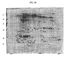
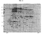
- the gels were scanned by an Image Scanner (Amersham Pharmacia Bio-tech, Uppsala, Sweden) The patterns of the spots were automatically analyzed using Image-Master 2D Platinum version 6.0 (GE Healthcare, UK).
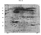
- FIGs. 1A to 1C The images of SDS-PAGE electrophoresis of the proteins expressed in the tears of a healthy person (control), and a non-retinopathic diabetic patient and a NPDR patient are shown in FIGs. 1A to 1C , respectively.
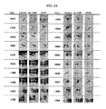
- FIG. 2A the magnified images of the proteins shown in FIGs. 1A to 1C are shown in FIG. 2A
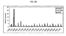
- the relative intensities of the proteins are shown in FIG. 2B .
- Healthy is a healthy person group
- No DMR is a non-retinopathic diabetic patient group
- NPDR is a NPDR patient group.
- FIGs. 1A to 2B newly expressed proteins and up-regulated or down-regulated proteins were observed in the samples of the NPDR patients as compared with those of the healthy persons or the non-retinopathic diabetic patients.
- 18 proteins showing down-regulated expressions spot Nos. of 10699, 10861, 11035, 11038, 11055, 11056, 11057, 11069, 11078, 10530, 10765, 11051, 10825, 10852, 10889, 10894, 10927 and 11058
- 2 proteins showing up-regulated expressions spot Nos. 10688 and 11046
- the expression of 18 proteins were down-regulated by at least 50%, and that of 2 proteins were up-regulated by at least 300%, as compared with the healthy persons or the non-retinopathic diabetic patients.
- the gel spots were excised.
- the excised gel spots were destained with 100 ⁇ L of destaining solution comprising 30 mM of potassium ferricyanide and 100 mM sodium thiosulfate as a ratio of 1:1 (v/v) for 5 min.
- the gel spots were washed three times with 400 ⁇ l of water, the gel spots were incubated with 200 mM ammonium bicarbonate for 10 min, and washed again.
- the resulting gel spots were dehydrated with acetonitrile, until the gel was changed to be a white, and dried with a speed vacuum centrifuge.
- the dried gel spots were the rehydrated with 20 ⁇ l of 50 mM ammonium bicarbonate containing 0.2 ⁇ g of trypsin (promega) for 45 min on ice. After the removal of the solution, 30 ⁇ l of 50 mM ammonium bicarbonate was added thereto, and incubated overnight at 37°C for digestion. A peptide solution obtained in above was desalted using C 18 nano column (homemade).
- Custom-made chromatographic columns were employed for desalting and concentraion of the peptide solution, prior to mass spectrometric analysis.
- a column consisting of 100-300 nL of Poros reverse phase R2 material (20-30 um bead size, PerSeptive Biosystems) was packed into a constricted GELoader tip (Eppendorf, Hamburg, Germany).
- a 10 mL syringe was then utilized to force liquid through the column, by applying a gentle air pressure.
- 30 ⁇ l of the peptide solution from the digestion supernatant was then diluted, loaded onto the column, and washed with 20 ⁇ l of 5% formic acid.
- the peptides were eluted with a mixture of 50% methanol, 49% H 2 O and 1% formic acid directly into a precoated borosilicate nanoelectrospray needle (Micromass, Manchester, U.K.).
- the MS/MS analysis of the peptides was performed by nano-ESI on a Q-TOF mass spectrometer (Micromass, Manchester, U.K.).
- the source temperature was 80°C.
- a 1 kV potential was applied to precoated borosilicate nanoelectrospray needles (EconoTip, New Objective, USA) in the ion source, combined with a nitrogen back-pressure of 0-5 psi, to achieve a stable flow rate (10-30 nL/min).
- the cone voltage was 40 V.
- the quadrupole analyzer was used to select precursor ions for fragmentation in the hexapole collision cell.
- the collision gas used was argon (Ar) at a pressure of 6-7 ⁇ 10 -5 mbar, and a collision energy was 20-30V.
- the product ions were analyzed with an orthogonal TOF analyzer, fitted with a reflector, a microchannel plate detector and a time-to-digital converter. The data was processed using a Mass Lynx Windows NT PC system (Micromass, Manchester, U.K.).
- SDS-PAGE loading buffer 60 mM Tris-Cl, 2% SDS, 25% Glycerol, 14.4 mM 2-mercaptoethanol and 0.1% bromophenol blue; pH 6.8
- the gels were blotted onto mini gel (6 ⁇ 8cm) using a running buffer at a current of 120V for molecular weight separation.
- the running buffer is containing 0.025M Tris-Cl, 0.192M Glycine and 1% SDS (pH 8.3).
- the gels obtained in (3-1) were transferred onto nitrocellulose (NC) membranes to blot using semi-dry transfer kit (Bio-rad) at a current of 50mA for 1 hour 30 min.
- An antibody reaction was conducted by employing LCN-1 (Spot NO. 11038), Hsp27 (Spot NO. 10530) and B2M (Spot NO. 11046) obtained from the NPDR patients.
- the nitrocellulose membranes were incubated with blocking solution of 5% w/v nonfat dry milk for 1 hour at room temperature.
- Anti-LCN1-monoclonal (1:1000), Anti-Hsp27-monoclonal (1:1000) and Anti-B2M-monoclonal (1:500) antibodies were used with the blocking solution, and incubated at 4 °C for 24 hours.
- the monoclonal antibody prepared by a conventional hybridoma technique ( Koeher and Milsteinas (1975) Nature, 256:495 ) using a rabbit was used as a primary antibody.
- the resulting solution was washed with TBS/T (0.1% Tris-buffered saline-tween20), and subsequently incubated with a solution containing 1:10,000 of horseradish peroxidase (HRP)-conjugated anti-rabbit secondary antibody (Santa cruz biotechnology Incs.) at a room temperature for I hour.
- the resulting solution was washed with TBS/T, and detection was performed with Enhanced chemiluminescence (ECL) system (Pierce Biotechnology Inc., USA).
- ECL Enhanced chemiluminescence
- FIG 3A The results of western blot analysis of LCN-1, Hsp27 and B2M are shown in FIG 3A , and relative intensities of the proteins are shown in FIGs. 3B to 3D , respectively (In each bar, *, * and *** represents that significant differences between groups determined by unpaired student's t-test are p ⁇ 0.05, p ⁇ 0.01 and p ⁇ 0.001, reapectively).
- LCN-1, Hsp27 and B2M are useful as biomarkers for diagnosing diabetic retinopathy, and are useful treating or preventing diabetic retinopathy.
Landscapes
- Health & Medical Sciences (AREA)
- Life Sciences & Earth Sciences (AREA)
- Chemical & Material Sciences (AREA)
- Immunology (AREA)
- Engineering & Computer Science (AREA)
- Medicinal Chemistry (AREA)
- Molecular Biology (AREA)
- General Health & Medical Sciences (AREA)
- Hematology (AREA)
- Organic Chemistry (AREA)
- Urology & Nephrology (AREA)
- Biochemistry (AREA)
- Biomedical Technology (AREA)
- Proteomics, Peptides & Aminoacids (AREA)
- Pathology (AREA)
- General Physics & Mathematics (AREA)
- Analytical Chemistry (AREA)
- Physics & Mathematics (AREA)
- Food Science & Technology (AREA)
- Veterinary Medicine (AREA)
- Biotechnology (AREA)
- Pharmacology & Pharmacy (AREA)
- Biophysics (AREA)
- Genetics & Genomics (AREA)
- Animal Behavior & Ethology (AREA)
- Public Health (AREA)
- Microbiology (AREA)
- Cell Biology (AREA)
- Bioinformatics & Cheminformatics (AREA)
- Diabetes (AREA)
- Gastroenterology & Hepatology (AREA)
- General Chemical & Material Sciences (AREA)
- Epidemiology (AREA)
- Nuclear Medicine, Radiotherapy & Molecular Imaging (AREA)
- Chemical Kinetics & Catalysis (AREA)
- Emergency Medicine (AREA)
- Obesity (AREA)
- Ophthalmology & Optometry (AREA)
- Endocrinology (AREA)
- Peptides Or Proteins (AREA)
Description
- The present invention relates to an in vitro method of diagnosing diabetic retinopathy.
- Diabetes mellitus (DM), a representative metabolic disorder caused by long-term maintenance of abnormally high blood glucose (sugar) level, is a systemic, non-contagious, chronic disease.
- Diabetes mellitus develops due to an absolutely or relatively diminished production of insulin induced by autoimmune destruction of insulin-producing beta cells of the pancreas, or decreased effects of insulin in a target organ (insulin resistance), which causes consistent increase of blood glucose level and impairs body metabolism including glucose metabolism.
- Diabetic retinopathy is one of the major microvascular complications of diabetes mellitus, which is caused by inharmonious supply of blood to the retina due to the circulatory disturbance resulting from the gradual deformation and occlusion of the retinal microvessles on account of the consistently high blood glucose level and metabolic abnormality caused thereby. Because the frequency of occurrence of diabetic retinopathy increases as the period suffering from diabetes mellitus is long and there are no warning signs for some time, it is necessary to periodically examine the change of the retina in a diabetic patient.
- Recently, there have been increasing needs for developing diagnostic markers for the early diagnosis of diabetic retinopathy and an effective method for diagnosing diabetic retinopathy. Accordingly, many proteins have been presented as markers for diagnosing diabetic retinopathy, examples of such proteins including vascular adhesion molecules such as endothelin-1(ET-1), protein kinase C(PKC) and E-selectin; thrombin-cleaved osteopontin; phosphoinositides; thrombin-stimulated platelet aggregation; vitreous VEGF; circulating soluble ICAM-1 (sICAM-1); advanced glycation end products(AGEs); nitric oxide; cytokines such as IL-1, IL-6, IL-8 and tumor necrosis factor-α; HLA alleles such as HLA-DR1, HLA-A9 and HLA-B40; glycosylated haemoglobin(HbA1c); microalbuminurea; aldose reductase(AR); Fas; PEDF; and the like proteins. However, none of the above proteins showed specificity for diabetic retinopathy only.
- Hitherto, a serum has been typically used as a source of biomarkers for the early diagnosis of diabetic retinopathy. However, there have been difficulties in finding out such biomarkers from a serum because of the glycosylation of serum proteins and presence of other abundant proteins such as albumin and immunoglobulin.
- Generally, tears consist of mucins, lipids, salts, glycoproteins and other various proteins. Representative tear proteins include lysozyme, lactoferrins, secretory immunoglobulin A (sIgA), lipocalins, albumins and lipophilins. Any quantitative or qualitative changes of these proteins may be interpreted as changes in the pathological conditions of our bodies. Although there have been many attempts to analyze tear proteins in relation with a disease, most of them are limited to discover the relationship between a single protein with a specific disease.
-
WO 2004/039956 A relates to compositions comprising a polypeptide or an antibody specific for the polypeptide, and methods useful for the diagnoses and treatment of immune related diseases such as diabetes mellitus. A kit comprising an antibody specific for the polypeptide is also disclosed. - The present inventors have endeavored to analyze the tear proteins more accurately and efficiently, and to develop effective biomarkers for early diagnosis of diabetic retinopathy. Finally, the present inventors have succeeded in accurately and efficiently analyzing tear proteins by employing two-dimensional gel electrophoresis (2-DE), thereby finding out many biomarker proteins for the diagnosis of diabetic retinopathy. Further, the present inventors have developed a composition for treating or preventing diabetic retinopathy, which comprises the biomarker proteins, and a composition and a kit for diagnosing diabetic retinopathy, which comprises an antibody specific for any one of the biomarker proteins.
- Accordingly, it is an object of the present invention to provide a method of diagnosing diabetic retinopathy.
- Described are proteins, or immunogenic fragments thereof for diagnosing diabetic retinopathy.
- Further described is a composition for diagnosing diabetic retinopathy, comprising antibodies specific for the proteins or the immunogenic fragments thereof.
- Further described is a kit for diagnosing diabetic retinopathy, comprising antibodies specific for the proteins or the immunogenic fragments thereof.
- Also described is a composition for treating or preventing diabetic retinopathy, comprising the proteins or the immunogenic fragments thereof as an active ingredient.
- The above and other objects and features of the present application and the present invention will become apparent from the following description of the invention, when taken in conjunction with the accompanying drawings, which respectively show:
-
Fig. 1A is a 2-DE gel image of the proteins expressed in the tears of healthy person; -
Figs. 1B and1C is 2-DE gel images of the proteins down-regulated or up-regulated in the tears of a non-retinopathic diabetic patient and an NPDR patient, respectively; -
Fig. 2A is magnified images of the proteins shown inFigs. 1A to 1C ; -
Fig. 2B is relative intensities of the proteins shown inFigs. 1A to 1C ; -
Fig. 3A is a western blot analysis result of LCN-1, Hsp27 and B2M identified in the tears of a patient with diabetic retinopathy; and -
Figs. 3B to 3D display relative intensities of LCN-1, Hsp27 and B2M, respectively, in healthy persons, non-retinopathic diabetic patients and NPDR patients. - In the above figures, Healthy means a healthy person; No DMR, a non-retinopathic diabetic patient; and NPDR, a NPDR patient;
- As used herein, the expression "peptide sequence obtained by trypsin digestion" means any peptide formed when a protein is digested by trypsin. Further, the molecular weight and isoelectric point (PI) of the proteins are measured by 2-dimentional electrophoresis, which may comprise an allowable experimental error.
- As used herein, the term "non-retinopathic diabetic patient" means a patient suffering from diabetes mellitus (DM), e.g.,
type 2 DM without retinopathic symptoms, and the term "NPDR patient" means a patient suffering fromtype 2 DM with the symptoms of non-proliferative diabetic retinopathy (NPDR). - As used herein, the term "immunogenic fragment" means a fragment of a protein, which has at least one epitope being recognized by an antibody specific for the protein.
- The present invention provides an vitro method of diagnosing diabetic retinopathy, which comprises measuring the expression level of a DJ-1 protein having a molecular weight of 24 to 26 kDa and an isoelectric point (PI) of 5.83 to 6.83. which comprises the peptide sequence of SEQ ID NOs: 1 and 2 obtained by trypsin digestion thereof, in the tears of a subject, and comparing the measured expression level to that measured in a healthy subject not suffering from diabetes.
- Specifically, the method for diagnosing diabetic retinopathy of the present invention comprises 1) subjecting the tears obtained from a subject to a reaction with an antibody specific for the biomarker protein, to obtain an antigen-antibody complex; 2) adding a secondary antibody conjugated with a marker, which will develop a color upon reaction with a substrate thereof and a solution comprising the substrate to detect the antigen-antibody complex by color reaction; and 3) comparing the detected amount of the protein in the subject with that detected in a healthy subject not suffering from diabetes.
- In step 1), the reaction of the tears of the subject with the antibody may be performed in a nitrocellulose membrane, or 96-well plate made of polyvinyl resin or polystyrene resin, or on a slide glass.
- The subject may be diagnosed as having diabetic retinopathy when the expression level of a DJ-1 protein in the subject's tears is higher by at least 100%, preferably at least 200%, and more preferably at least 300%, than that of non-retinopathic diabetic patients.
- The following Examples are intended to further illustrate the present invention and the present application without limiting its scope.
- Tear fluid samples were obtained from the meniscus of 14 healthy persons (control), 10 patients suffering from
type 2 DM without retinopathy (hereinafter, referred to as "non-retinopathic diabetic patients"), and patients suffering fromtype 2 DM with NPDR (hereinafter, referred to as "NPDR patients") by using polyester wicks. The polyester wicks thus obtained were placed on the top of 1.5 ml of micropipet tips and centrifuged at 10,000 ppm for 5 min (Union-55R centrifuge, Hanil science, Korea). The concentration of the tear proteins was measured by the Bradford method, and then the tear proteins were stored at -70°C until further use. - 60 µg each of the tear proteins obtained in
step 1 was subjected to an isoelectrofocusing. 450 µℓ of rehydration buffer solution (8M urea, 2% CHAPS, 13mM DTT, 1% IPG buffer and Bromophenol blue (BPB)) was added to the tear protein sample. The mixture was loaded in a 24 cm strip holder, and Drystrips (Amerahsm Pharmacia Biotech, Uppsala, Sweden) having a pH ranging from 4 to 7 was placed in the strip holder. The strips were rehydrated for 5 hours without a current, and then, for another 5 hours with a current of 80V Isoelectrofocusing (IEF) was carried out for a total of 100,000 Vhr using an IPGphore IEF system (Amerahsm Pharmacia Biotech, Uppsala, Sweden). - Following IEF separation, the gel strips obtained from (2-1) were equilibrated with 10 mℓ of the first equilibration solution (6M urea, 50 mM Tris-Cl buffer (pH 8.8), 20% glycerol, 2% SDS and 1% DTT) for 15 min, and the first equilibration solution was removed. The gel strips were then equilibrated again with 10 mℓ of the second equilibration solution (6M urea, 50 mM Tris-Cl buffer (pH 8.8), 30% glycerol, 2% SDS and 1.5% IAA) for 10 min. The strips were applied onto 11-16% gradient of polyacryl amide gel (25×30 cm), and subjected to a sealing with 0.5% agarose gel. SDS-PAGE was conducted by adding a buffer solution (24 mM Tris, 192 mM glycine and 0.1% SDS) to an Ettan DALT system (Amerahsm Pharmacia Biotech, Uppsala, Sweden) at a current of 70 V for 1 hour, 140 V for 2 hours, and 320 V for 5 hours.
- In order to visualize the gels obtained in (2-2), a silver staining was conducted. The gels obtained in (2-2) were fixed in a mixture of 50% methanol, 12% acetic acid and 0.05% formaldehyde for at least 2 hours. The fixed gels were rinsed with 50% ethanol three times for 20 min each, then sensitized with 0.2% sodium thiosulfate for 1 min and followed by washing with D.W. The gels were immersed in a mixture of 0.1% silver nitrate and 0.075% formaldehyde (37%) for 20 min, and washed with D.W. The resulting gels were developed with a mixture of 6% sodium carbonate and 0.075% formaldehyde (37%) for 7 min, and 50% methanol and 12% acetic acid were added thereto to fix the resulting gels.
- In order to analyze images of the silver-stained gels, the gels were scanned by an Image Scanner (Amersham Pharmacia Bio-tech, Uppsala, Sweden) The patterns of the spots were automatically analyzed using Image-Master 2D Platinum version 6.0 (GE Healthcare, UK).
- The images of SDS-PAGE electrophoresis of the proteins expressed in the tears of a healthy person (control), and a non-retinopathic diabetic patient and a NPDR patient are shown in
FIGs. 1A to 1C , respectively. - Also, the magnified images of the proteins shown in
FIGs. 1A to 1C are shown inFIG. 2A , the relative intensities of the proteins are shown inFIG. 2B . Wherein Healthy is a healthy person group, No DMR is a non-retinopathic diabetic patient group; and NPDR is a NPDR patient group. - As shown in
FIGs. 1A to 2B , newly expressed proteins and up-regulated or down-regulated proteins were observed in the samples of the NPDR patients as compared with those of the healthy persons or the non-retinopathic diabetic patients. Specifically, 18 proteins showing down-regulated expressions (spot Nos. of 10699, 10861, 11035, 11038, 11055, 11056, 11057, 11069, 11078, 10530, 10765, 11051, 10825, 10852, 10889, 10894, 10927 and 11058), and 2 proteins showing up-regulated expressions (spot Nos. 10688 and 11046) were found in the NPDR patients as compared with the healthy persons or non-retinopathic diabetic patients (see,FIGs. 1A to 2B ). In the NPDR patients, the expression of 18 proteins were down-regulated by at least 50%, and that of 2 proteins were up-regulated by at least 300%, as compared with the healthy persons or the non-retinopathic diabetic patients. - The molecular weight and the isoelectric point (PI) of the spots of these proteins are shown in Table 1.
<Table 1> Spot No. Molecular weight (kDa) pI classification 10688 20/25 6.33 Up-regulated 11046 13/16 5.77 Up-regulated 10699 54/28 7 Down-regulated 10861 14/20 4.74 Down-regulated 11035 18/21 4.93 Down-regulated 11038 19/21 4.8 Down-regulated 11055 45/21 5.14 Down-regulated 11056 21/22 4.63 Down-regulated 11057 10/22 6.51 Down-regulated 11069 19/22 5.39 Drown-regulated 11078 48/28 4.84 Drown-regulated 10530 22/31 7.83 Down-regulated 10765 19/20 5.78 Down-regulated 11051 14/20 5.3 Down-regulated 10825 16/19 6.46 Down-regulated 10852 16/19 7 Down-regulated 10889 13/15 5.71 Down-regulated 10894 13/14 5.71 Down-regulated 10927 12/12 6.78 Down-regulated 11058 14/21 5.2 Down-regulated - To identify the proteins obtained in Example 1, the gel spots were excised. The excised gel spots were destained with 100 µ L of destaining solution comprising 30 mM of potassium ferricyanide and 100 mM sodium thiosulfate as a ratio of 1:1 (v/v) for 5 min. After the gel spots were washed three times with 400 µℓ of water, the gel spots were incubated with 200 mM ammonium bicarbonate for 10 min, and washed again. The resulting gel spots were dehydrated with acetonitrile, until the gel was changed to be a white, and dried with a speed vacuum centrifuge. The dried gel spots were the rehydrated with 20 µℓ of 50 mM ammonium bicarbonate containing 0.2 µg of trypsin (promega) for 45 min on ice. After the removal of the solution, 30 µℓ of 50 mM ammonium bicarbonate was added thereto, and incubated overnight at 37°C for digestion. A peptide solution obtained in above was desalted using C18 nano column (homemade).
- Custom-made chromatographic columns were employed for desalting and concentraion of the peptide solution, prior to mass spectrometric analysis. A column consisting of 100-300 nL of Poros reverse phase R2 material (20-30 um bead size, PerSeptive Biosystems) was packed into a constricted GELoader tip (Eppendorf, Hamburg, Germany). A 10 mL syringe was then utilized to force liquid through the column, by applying a gentle air pressure. 30 µℓ of the peptide solution from the digestion supernatant was then diluted, loaded onto the column, and washed with 20 µℓ of 5% formic acid. For MS/MS analysis, the peptides were eluted with a mixture of 50% methanol, 49% H2O and 1% formic acid directly into a precoated borosilicate nanoelectrospray needle (Micromass, Manchester, U.K.).
- The MS/MS analysis of the peptides was performed by nano-ESI on a Q-TOF mass spectrometer (Micromass, Manchester, U.K.). The source temperature was 80°C. A 1 kV potential was applied to precoated borosilicate nanoelectrospray needles (EconoTip, New Objective, USA) in the ion source, combined with a nitrogen back-pressure of 0-5 psi, to achieve a stable flow rate (10-30 nL/min). The cone voltage was 40 V. The quadrupole analyzer was used to select precursor ions for fragmentation in the hexapole collision cell. The collision gas used was argon (Ar) at a pressure of 6-7×10⁻⁵ mbar, and a collision energy was 20-30V. The product ions were analyzed with an orthogonal TOF analyzer, fitted with a reflector, a microchannel plate detector and a time-to-digital converter. The data was processed using a Mass Lynx Windows NT PC system (Micromass, Manchester, U.K.).
- For identification of the protein, all MS/MS spectra recorded on the tryptic peptides derived from the spots were searched against the protein sequences from the NCBInr databases (www.ncbi.nlm.nih.gov), using the MASCOT search program (www.matrixscience.com). The results of identification of the proteins up-regulated or down-regulated, are shown in Tables 2 and 3, respectively.
<Table 2> Spot NO. Identification of protein MW (Da) Peptide by trypsin digestion NCBI Assession No. SEQ ID NO. Sequence SEQ ID NO. 10688 DJ-I protein 20050 GAEEMETVIPVDVMR 1 NP_009193 33 EGPYDVVVLPGGNLGAQNLSESAAVK 2 11046 Beta-2 microglobulin 12905 SNFLNCYVSGFHPSDIEVDLLK 3 CAA23830 34 DWSFYLLYYTEFTPTEKDEYACR 4 <Table 3> Spot NO. Identification of protein MW (Da) Peptide by trypsin digestion NCBI Assession No. SEQ ID NO. Sequence SEQ ID NO. 10699 Envelope protein 54093 SGLNTEAFYVMTVGSK 5 AAB05390 35 6 10861 Protein SAP1 13491 IIPGGIYDADLNDEWVQR 7 AAB19889 36 11035 Lipocalin 1-like 1 18078 GLSTESILIPR 8 CAH73781 37 11056 Lipocalin 1-like 1 18078 GLSTESILIPR 8 CAH73781 37 11038 Lipocalin (hCG201503) 19488 GLSTESILIPR 8 EAW88053 38 11055 Cytokeratin 445593 FASFIDKVQFLEQQNK 9 CAA30534 39 WNLLQQQTTTTSSK 10 NLEPLFETYLSVLR 11 VDSLNDEINFLK 12 NLDLDSIIAEVR 13 VQQLQISVDQHGDNLK 14 11057 S 100 calcium-binding protein A810885 ELDINTDGAVNFQEFLILVIK 15 NP_002955 40 11069 Lipocalin 1 precursor19409 GLSTESILIPR 8 NP_002288 41 11078 Keratin 448633 QNQEYQVLLDVR 16 NP_002268 42 LNVEVDAAPTVDLNR 17 10530 Heat shock protein 27 22427 LFDQAFGLPR 18 AAA62175 43 VSLDVNHFAPDELTVK 19 LATQSNEITIPVRFESR 20 10765 Adenine phospho-ribosyl-transferase isoform 19766 LQAEVLECVSLVELTSLK 21 NP_000476 44 11051 Phosphohistidine phosphatase 14 kDa ALIPDVDLDSD 22 BC024648 45 11058 Phosphohistidine phosphatase 14 kDa ALIPDVDLDSD 22 BC024648 45 10825 Beta globin 16101 LLVVYPWTQR 23 AAF00488 46 EFTPPVQAAYQK 24 FFESFGDLSTPDAVMGNPK 25 10852 Lysozyme precursor 16885 GISLANWMCLAK 26 1LHM 47 STDYGIFQINSR 27 10889 S 100 calcium binding protein A913291 QLSFEEFIMLMAR 28 NP_002956 DIETIINTFHQYSVK 29 48 VIEHIMEDLDTNADKQLSFEEFIMLMAR 30 10894 Crystal structure of human calprotectin 13086 QLSFEEFIMLMAR 31 NP_002956 49 10927 S100 calcium binding protein A4 11949 ALDVMVSTFHK 32 AAB20971 50 - 40-50 µg of the proteins obtained from the tears of the non-retinopathic diabetic patients and NPDR patients were diluted with SDS-PAGE loading buffer (60 mM Tris-Cl, 2% SDS, 25% Glycerol, 14.4 mM 2-mercaptoethanol and 0.1% bromophenol blue; pH 6.8), and subjected to 12% SDS-PAGE. The gels were blotted onto mini gel (6×8cm) using a running buffer at a current of 120V for molecular weight separation. The running buffer is containing 0.025M Tris-Cl, 0.192M Glycine and 1% SDS (pH 8.3).
- The gels obtained in (3-1) were transferred onto nitrocellulose (NC) membranes to blot using semi-dry transfer kit (Bio-rad) at a current of 50mA for 1
hour 30 min. - An antibody reaction was conducted by employing LCN-1 (Spot NO. 11038), Hsp27 (Spot NO. 10530) and B2M (Spot NO. 11046) obtained from the NPDR patients. The nitrocellulose membranes were incubated with blocking solution of 5% w/v nonfat dry milk for 1 hour at room temperature. Anti-LCN1-monoclonal (1:1000), Anti-Hsp27-monoclonal (1:1000) and Anti-B2M-monoclonal (1:500) antibodies were used with the blocking solution, and incubated at 4 °C for 24 hours. The monoclonal antibody prepared by a conventional hybridoma technique (Koeher and Milsteinas (1975) Nature, 256:495) using a rabbit was used as a primary antibody. The resulting solution was washed with TBS/T (0.1% Tris-buffered saline-tween20), and subsequently incubated with a solution containing 1:10,000 of horseradish peroxidase (HRP)-conjugated anti-rabbit secondary antibody (Santa cruz biotechnology Incs.) at a room temperature for I hour. The resulting solution was washed with TBS/T, and detection was performed with Enhanced chemiluminescence (ECL) system (Pierce Biotechnology Inc., USA).
- The results of western blot analysis of LCN-1, Hsp27 and B2M are shown in
FIG 3A , and relative intensities of the proteins are shown inFIGs. 3B to 3D , respectively (In each bar, *, * and *** represents that significant differences between groups determined by unpaired student's t-test are p<0.05, p<0.01 and p<0.001, reapectively). - As shown in
FIGs. 3A to 3D , the expression levels of LCN-1 and Hsp 27 were significantly down-regulated, and the expression level of B2M was up-regulated according to the progress of diabetic retinopathy. These results corresponds to the results of 2-DE Therefore, it is concluded that LCN-1, Hsp27 and B2M are useful as biomarkers for diagnosing diabetic retinopathy, and are useful treating or preventing diabetic retinopathy. - While the invention has been described with respect to the above specific embodiments, it should be recognized that various modifications and changes may be made to the invention by those skilled in the art which also fall within the scope of the invention as defined by the appended claims.
- <110> Korea University Research and Business Foundation
- <120> Bio-markers for diagnosing diabetic retinopathy
- <130> EP63503HVpau
- <140> not yet assigned
<141> 2008-01-17 - <150>
KR 10-2008-0005162
<151> 2008-01-17 - <160> 50
- <170> KopatentIn 1.71
- <210> 1
<211> 15
<212> PRT
<213> Homo sapiens - <400> 1

- <210> 2
<211> 26
<212> PRT
<213> Homo sapiens - <400> 2

- <210> 3
<211> 22
<212> PRT
<213> Homo sapiens - <400> 3

- <210> 4
<211> 23
<212> PRT
<213> Homo sapiens - <400> 4

- <210> 5
<211> 13
<212> PRT
<213> Homo sapiens - <400> 5

- <210> 6
<211> 16
<212> PRT
<213> Homo sapiens - <400> 6

- <210> 7
<211> 18
<212> PRT
<213> Homo sapiens - <400> 7

- <210> 8
<211> 11
<212> PRT
<213> Homo sapiens - <400> 8

- <210> 9
<211> 16
<212> PRT
<213> Homo sapiens - <400> 9

- <210> 10
<211> 14
<212> PRT
<213> Homo sapiens - <400> 10

- <210> 11
<211> 14
<212> PRT
<213> Homo sapiens - <400> 11

- <210> 12
<211> 12
<212> PRT
<213> Homo sapiens - <400> 12

- <210> 13
<211> 12
<212> PRT
<213> Homo sapiens - <400> 13

- <210> 14
<211> 16
<212> PRT
<213> Homo sapiens - <400> 14

- <210> 15
<211> 21
<212> PRT
<213> Homo sapiens - <400> 15

- <210> 16
<211> 12
<212> PRT
<213> Homo sapiens - <400> 16

- <210> 17
<211> 15
<212> PRT
<213> Homo sapiens - <400> 17

- <210> 18
<211> 10
<212> PRT
<213> Homo sapiens - <400> 18

- <210> 19
<211> 16
<212> PRT
<213> Homo sapiens - <400> 19

- <210> 20
<211> 17
<212> PRT
<213> Homo sapiens - <400> 20

- <210> 21
<211> 18
<212> PRT
<213> Homo sapiens - <400> 21

- <210> 22
<211> 11
<212> PRT
<213> Homo sapiens - <400> 22

- <210> 23
<211> 10
<212> PRT
<213> Homo sapiens - <400> 23

- <210> 24
<211> 12
<212> PRT
<213> Homo sapiens - <400> 24

- <210> 25
<211> 19
<212> PRT
<213> Homo sapiens - <400> 25

- <210> 26
<211> 12
<212> PRT
<213> Homo sapiens - <400> 26

- <210> 27
<211> 12
<212> PRT
<213> Homo sapiens - <400> 27

- <210> 28
<211> 13
<212> PRT
<213> Homo sapiens - <400> 28

- <210> 29
<211> 15
<212> PRT
<213> Homo sapiens - <400> 29

- <210> 30
<211> 28
<212> PRT
<213> Homo sapiens - <400> 30


- <210> 31
<211> 13
<212> PRT
<213> Homo sapiens - <400> 31

- <210> 32
<211> 11
<212> PRT
<213> Homo sapiens - <400> 32

- <210> 33
<211> 188
<212> PRT
<213> Homo sapiens - <400> 33

- <210> 34
<211> 110
<212> PRT
<213> Homo sapiens - <400> 34

- <210> 35
<211> 500
<212> PRT
<213> Homo sapiens - <400> 35


- <210> 36
<211> 113
<212> PRT
<213> Homo sapiens - <400> 36


- <210> 37
<211> 162
<212> PRT
<213> Homo sapiens - <400> 37

- <210> 38
<211> 176
<212> PRT
<213> Homo sapiens - <400> 38

- <210> 39
<211> 408
<212> PRT
<213> Homo sapiens - <400> 39


- <210> 40
<211> 93
<212> PRT
<213> Homo sapiens - <400> 40


- <210> 41
<211> 176
<212> PRT
<213> Homo sapiens - <400> 41

- <210> 42
<211> 416
<212> PRT
<213> Homo sapiens - <400> 42

- <210> 43
<211> 199
<212> PRT
<213> Homo sapiens - <400> 43

- <210> 44
<211> 180
<212> PRT
<213> Homo sapiens - <400> 44


- <210> 45
<211> 125
<212> PRT
<213> Homo sapiens - <400> 45

- <210> 46
<211> 147
<212> PRT
<213> Homo sapiens - <400> 46


- <210> 47
<211> 148
<212> PRT
<213> Homo sapiens - <400> 47

- <210> 48
<211> 114
<212> PRT
<213> Homo sapiens - <400> 48


- <210> 49
<211> 114
<212> PRT
<213> Homo sapiens - <400> 49

- <210> 50
<211> 101
<212> PRT
<213> Homo sapiens - <400> 50


Claims (2)
- An in vitro method of diagnosing diabetic retinopathy, which comprises measuring an expression level of a DJ-1 protein having a molecular weight of 24 to 26 kDa and an isoelectric point (PI) of 5.83 to 6.83, which comprises the peptide sequences of SEQ ID NOs: 1 and 2 obtained by trypsin digestion thereof, in the tears of a subject, and comparing the measured expression level to that measured in a healthy subject not suffering from diabetes.
- The method of claim 1, which comprises1) subjecting the tears obtained from a subject to a reaction with an antibody specific for the biomarker protein to obtain an antigen-antibody complex;2) adding a secondary antibody conjugated with a marker, which will develop a color upon reaction with a substrate thereof, and a solution comprising the substrate to detect the antigen-antibody complex by color reaction; and3) comparing the detected amount of the protein in the subject with that detected in a healthy subject not suffering from diabetes.
Priority Applications (1)
| Application Number | Priority Date | Filing Date | Title |
|---|---|---|---|
| EP10075360.7A EP2267457B1 (en) | 2008-01-17 | 2009-01-16 | Beta 2-microglobulin biomarker for diagnosing diabetic retinopathy |
Applications Claiming Priority (1)
| Application Number | Priority Date | Filing Date | Title |
|---|---|---|---|
| KR1020080005162A KR100983475B1 (en) | 2008-01-17 | 2008-01-17 | Biomarkers for Diagnosing Diabetic Retinopathy |
Related Child Applications (1)
| Application Number | Title | Priority Date | Filing Date |
|---|---|---|---|
| EP10075360.7 Division-Into | 2010-08-23 |
Publications (3)
| Publication Number | Publication Date |
|---|---|
| EP2081029A2 EP2081029A2 (en) | 2009-07-22 |
| EP2081029A3 EP2081029A3 (en) | 2009-08-12 |
| EP2081029B1 true EP2081029B1 (en) | 2012-12-26 |
Family
ID=40380735
Family Applications (2)
| Application Number | Title | Priority Date | Filing Date |
|---|---|---|---|
| EP10075360.7A Ceased EP2267457B1 (en) | 2008-01-17 | 2009-01-16 | Beta 2-microglobulin biomarker for diagnosing diabetic retinopathy |
| EP09000609A Ceased EP2081029B1 (en) | 2008-01-17 | 2009-01-16 | Bio-markers for diagnosing diabetic retinopathy |
Family Applications Before (1)
| Application Number | Title | Priority Date | Filing Date |
|---|---|---|---|
| EP10075360.7A Ceased EP2267457B1 (en) | 2008-01-17 | 2009-01-16 | Beta 2-microglobulin biomarker for diagnosing diabetic retinopathy |
Country Status (4)
| Country | Link |
|---|---|
| US (2) | US20090186032A1 (en) |
| EP (2) | EP2267457B1 (en) |
| JP (1) | JP2009168819A (en) |
| KR (1) | KR100983475B1 (en) |
Families Citing this family (10)
| Publication number | Priority date | Publication date | Assignee | Title |
|---|---|---|---|---|
| CN104508473B (en) | 2012-05-29 | 2017-10-13 | 健康诊断实验室有限公司 | Composition and method for the gel electrophoresis with calibrated in situ |
| KR102073312B1 (en) * | 2012-06-27 | 2020-02-05 | 엘지전자 주식회사 | Marker for diagnosing diabetic retinopathy and use thereof |
| EP2892922A2 (en) * | 2012-09-10 | 2015-07-15 | Westfälische Wilhelms-Universität Münster | Methods and compounds for preventing, treating and diagnosing an inflammatory condition |
| CN104892751B (en) * | 2015-06-27 | 2018-02-16 | 吉林大学 | Self-organization recombinates Vitreoscilla hemoglobin and its gene and application |
| IT201700031721A1 (en) * | 2017-03-22 | 2018-09-22 | Ciro Costagliola | Medical device, method and immunoassay test for the identification of proliferative diabetic retinopathy |
| WO2019169192A1 (en) * | 2018-02-28 | 2019-09-06 | The Regents Of The University Of California | Electrochemical biosensor array devices, systems, and methods for point-of-care detection |
| RU2696880C1 (en) * | 2018-11-22 | 2019-08-07 | Федеральное государственное бюджетное учреждение "Национальный медицинский исследовательский центр глазных болезней имени Гельмгольца" Министерства здравоохранения Российской Федерации (ФГБУ "НМИЦ ГБ им. Гельмгольца" Минздрава России) | Method for prediction of intraoperative haemorrhagic complications in the severe form of proliferative diabetic retinopathy |
| KR102091483B1 (en) | 2019-07-12 | 2020-03-20 | 서울대학교산학협력단 | Biomarker for Predicting Oral Cancer and Use Thereof |
| KR102583808B1 (en) * | 2021-03-05 | 2023-09-26 | 경희대학교 산학협력단 | Method and kit for diagnosing diabetes using tear samples |
| CN114855285B (en) * | 2022-06-02 | 2023-02-17 | 山东省食品药品检验研究院 | A characteristic peptide library for quickly identifying the source of velvet antler species and its application |
Family Cites Families (9)
| Publication number | Priority date | Publication date | Assignee | Title |
|---|---|---|---|---|
| US4208479A (en) * | 1977-07-14 | 1980-06-17 | Syva Company | Label modified immunoassays |
| JP2001133454A (en) * | 1999-11-02 | 2001-05-18 | Iatron Lab Inc | Method for immunological analysis of apolipoprotein AI in tear and method for detecting diabetic retinopathy |
| US20040127445A1 (en) * | 2002-08-28 | 2004-07-01 | Chondrogene Limited | Beta-2 microglobulin (B2M) and B2M related gene products for the regulation of osteoarthritis pathogenesis and chondrocyte proliferation |
| EP1576137A4 (en) * | 2002-10-29 | 2010-06-30 | Genentech Inc | COMPOSITIONS AND METHODS FOR TREATING DISEASES RELATED TO THE IMMUNE SYSTEM |
| CA2532130C (en) * | 2003-09-20 | 2016-07-19 | Electrophoretics Limited | Diagnostic method for brain damage-related disorders |
| JP2007320850A (en) * | 2004-08-24 | 2007-12-13 | Kyowa Hakko Kogyo Co Ltd | Anti-ephrin B2 antibody |
| US20090093010A1 (en) * | 2004-09-21 | 2009-04-09 | University Of Manitoba | Method of Detecting Kidney Dysfunction |
| JP2006325528A (en) * | 2005-05-27 | 2006-12-07 | Institute For Rheumatic Diseases Co Ltd | Diagnosis and prevention of diabetic retinopathy |
| EP1982174A4 (en) * | 2006-01-27 | 2009-05-06 | Univ George Mason | EYE LIQUID MARKERS |
-
2008
- 2008-01-17 KR KR1020080005162A patent/KR100983475B1/en active Active
-
2009
- 2009-01-16 EP EP10075360.7A patent/EP2267457B1/en not_active Ceased
- 2009-01-16 US US12/354,907 patent/US20090186032A1/en not_active Abandoned
- 2009-01-16 JP JP2009008028A patent/JP2009168819A/en active Pending
- 2009-01-16 EP EP09000609A patent/EP2081029B1/en not_active Ceased
-
2011
- 2011-07-14 US US13/182,915 patent/US20110269248A1/en not_active Abandoned
Also Published As
| Publication number | Publication date |
|---|---|
| KR20090079294A (en) | 2009-07-22 |
| KR100983475B1 (en) | 2010-09-24 |
| US20110269248A1 (en) | 2011-11-03 |
| US20090186032A1 (en) | 2009-07-23 |
| EP2081029A2 (en) | 2009-07-22 |
| JP2009168819A (en) | 2009-07-30 |
| EP2267457A1 (en) | 2010-12-29 |
| EP2267457B1 (en) | 2013-04-10 |
| EP2081029A3 (en) | 2009-08-12 |
Similar Documents
| Publication | Publication Date | Title |
|---|---|---|
| EP2267457B1 (en) | Beta 2-microglobulin biomarker for diagnosing diabetic retinopathy | |
| Yan et al. | Systematic comparison of the human saliva and plasma proteomes | |
| Jerkovic et al. | Afamin is a novel human vitamin E-binding glycoprotein characterization and in vitro expression | |
| Kim et al. | Comparison of tear proteins between healthy and early diabetic retinopathy patients | |
| EP3130602B1 (en) | Immunoregulatory structures from normally occurring proteins | |
| EP2455389A1 (en) | Sugar chain marker as measure of disease conditions of hepatic diseases | |
| Landi et al. | A functional proteomics approach to the comprehension of sarcoidosis | |
| WO2006073195A1 (en) | Method for predicting or diagnosing diabetes and kit for predicting or diagnosing diabetes | |
| EP3044594B1 (en) | Identification of novel biomarkers of flares of systemic lupus erythematosus | |
| KR101240208B1 (en) | Proteinic markers for diagnosing type I diabetic nephropathy | |
| Gamberi et al. | A proteomic approach to identify plasma proteins in patients with abdominal aortic aneurysm | |
| KR101040321B1 (en) | Biomarker for Diagnosing Cardiovascular Disease | |
| KR100792630B1 (en) | Biomarker for Diagnosing Diabetic Nephropathy | |
| Mao et al. | MALDI-TOF/TOF-MS reveals elevated serum haptoglobin and amyloid A in Behcet’s disease | |
| EP3290923B1 (en) | Tropomyosin isoforms related to alzheimers disease and mild cognitive impairment | |
| WO2017212463A1 (en) | Methods for diagnosis, differentiation and monitoring using urine proteins as markers in iga nephropathy | |
| KR101274835B1 (en) | Biomarker composition for diagnosing acute exacerbations chronic obstructive pulmonary disease and the method for detecting using the same | |
| KR101142779B1 (en) | Analytical method or kit for diagnosing polycystic ovary syndrome | |
| KR20090012503A (en) | Markers, Compositions, and Kits for Diagnosing Diabetic Retinopathy | |
| US20120214169A1 (en) | Differential levels of haptoglodin isoforms in small cell lung cancer | |
| EP3004885B1 (en) | Aiding assessment of prognosis in inflammatory disease | |
| RU2832391C1 (en) | Method for laboratory diagnosis of chronic heart failure with preserved ejection fraction in patients with diabetes mellitus based on analysis of protein profile of blood serum exosome fraction | |
| US20080108549A1 (en) | Actin proteins as biomarkers for indication and targeting of resistance and sensitivity to an Abl kinase inhibitor in patients with chronic myelogenous leukemia | |
| Xiao et al. | An integrated method for IgG N-glycans enrichment and analysis: Understanding the role of IgG glycosylation in diabetic foot ulcer | |
| Ishida et al. | Identification of nine proteins in whole lung extract derived from bleomycin-induced interstitial pneumonia model mouse by two-dimensional electrophoresis/in-gel digestion/MALDI-TOF MSMS analysis |
Legal Events
| Date | Code | Title | Description |
|---|---|---|---|
| PUAI | Public reference made under article 153(3) epc to a published international application that has entered the european phase |
Free format text: ORIGINAL CODE: 0009012 |
|
| PUAL | Search report despatched |
Free format text: ORIGINAL CODE: 0009013 |
|
| 17P | Request for examination filed |
Effective date: 20090116 |
|
| AK | Designated contracting states |
Kind code of ref document: A2 Designated state(s): AT BE BG CH CY CZ DE DK EE ES FI FR GB GR HR HU IE IS IT LI LT LU LV MC MK MT NL NO PL PT RO SE SI SK TR |
|
| AX | Request for extension of the european patent |
Extension state: AL BA RS |
|
| AK | Designated contracting states |
Kind code of ref document: A3 Designated state(s): AT BE BG CH CY CZ DE DK EE ES FI FR GB GR HR HU IE IS IT LI LT LU LV MC MK MT NL NO PL PT RO SE SI SK TR |
|
| AX | Request for extension of the european patent |
Extension state: AL BA RS |
|
| 17Q | First examination report despatched |
Effective date: 20100312 |
|
| AKX | Designation fees paid |
Designated state(s): GB |
|
| REG | Reference to a national code |
Ref country code: DE Ref legal event code: 8566 |
|
| GRAP | Despatch of communication of intention to grant a patent |
Free format text: ORIGINAL CODE: EPIDOSNIGR1 |
|
| GRAS | Grant fee paid |
Free format text: ORIGINAL CODE: EPIDOSNIGR3 |
|
| GRAA | (expected) grant |
Free format text: ORIGINAL CODE: 0009210 |
|
| AK | Designated contracting states |
Kind code of ref document: B1 Designated state(s): GB |
|
| REG | Reference to a national code |
Ref country code: GB Ref legal event code: FG4D |
|
| PLBE | No opposition filed within time limit |
Free format text: ORIGINAL CODE: 0009261 |
|
| STAA | Information on the status of an ep patent application or granted ep patent |
Free format text: STATUS: NO OPPOSITION FILED WITHIN TIME LIMIT |
|
| 26N | No opposition filed |
Effective date: 20130927 |
|
| PGFP | Annual fee paid to national office [announced via postgrant information from national office to epo] |
Ref country code: GB Payment date: 20210107 Year of fee payment: 13 |
|
| GBPC | Gb: european patent ceased through non-payment of renewal fee |
Effective date: 20220116 |
|
| PG25 | Lapsed in a contracting state [announced via postgrant information from national office to epo] |
Ref country code: GB Free format text: LAPSE BECAUSE OF NON-PAYMENT OF DUE FEES Effective date: 20220116 |