EP2057466B1 - Methods and compositions for treatment and diagnosis of autoimmune encephalitis or epilepsy - Google Patents
Methods and compositions for treatment and diagnosis of autoimmune encephalitis or epilepsy Download PDFInfo
- Publication number
- EP2057466B1 EP2057466B1 EP07836873.5A EP07836873A EP2057466B1 EP 2057466 B1 EP2057466 B1 EP 2057466B1 EP 07836873 A EP07836873 A EP 07836873A EP 2057466 B1 EP2057466 B1 EP 2057466B1
- Authority
- EP
- European Patent Office
- Prior art keywords
- another embodiment
- subject
- encephalitis
- tumor
- subunit
- Prior art date
- Legal status (The legal status is an assumption and is not a legal conclusion. Google has not performed a legal analysis and makes no representation as to the accuracy of the status listed.)
- Active
Links
Images


Classifications
-
- G—PHYSICS
- G01—MEASURING; TESTING
- G01N—INVESTIGATING OR ANALYSING MATERIALS BY DETERMINING THEIR CHEMICAL OR PHYSICAL PROPERTIES
- G01N33/00—Investigating or analysing materials by specific methods not covered by groups G01N1/00 - G01N31/00
- G01N33/48—Biological material, e.g. blood, urine; Haemocytometers
- G01N33/50—Chemical analysis of biological material, e.g. blood, urine; Testing involving biospecific ligand binding methods; Immunological testing
- G01N33/53—Immunoassay; Biospecific binding assay; Materials therefor
- G01N33/564—Immunoassay; Biospecific binding assay; Materials therefor for pre-existing immune complex or autoimmune disease, i.e. systemic lupus erythematosus, rheumatoid arthritis, multiple sclerosis, rheumatoid factors or complement components C1-C9
-
- G—PHYSICS
- G01—MEASURING; TESTING
- G01N—INVESTIGATING OR ANALYSING MATERIALS BY DETERMINING THEIR CHEMICAL OR PHYSICAL PROPERTIES
- G01N33/00—Investigating or analysing materials by specific methods not covered by groups G01N1/00 - G01N31/00
- G01N33/48—Biological material, e.g. blood, urine; Haemocytometers
- G01N33/50—Chemical analysis of biological material, e.g. blood, urine; Testing involving biospecific ligand binding methods; Immunological testing
- G01N33/53—Immunoassay; Biospecific binding assay; Materials therefor
- G01N33/574—Immunoassay; Biospecific binding assay; Materials therefor for cancer
-
- G—PHYSICS
- G01—MEASURING; TESTING
- G01N—INVESTIGATING OR ANALYSING MATERIALS BY DETERMINING THEIR CHEMICAL OR PHYSICAL PROPERTIES
- G01N2333/00—Assays involving biological materials from specific organisms or of a specific nature
- G01N2333/435—Assays involving biological materials from specific organisms or of a specific nature from animals; from humans
- G01N2333/705—Assays involving receptors, cell surface antigens or cell surface determinants
- G01N2333/70571—Assays involving receptors, cell surface antigens or cell surface determinants for neuromediators, e.g. serotonin receptor, dopamine receptor
-
- G—PHYSICS
- G01—MEASURING; TESTING
- G01N—INVESTIGATING OR ANALYSING MATERIALS BY DETERMINING THEIR CHEMICAL OR PHYSICAL PROPERTIES
- G01N2800/00—Detection or diagnosis of diseases
- G01N2800/28—Neurological disorders
-
- G—PHYSICS
- G01—MEASURING; TESTING
- G01N—INVESTIGATING OR ANALYSING MATERIALS BY DETERMINING THEIR CHEMICAL OR PHYSICAL PROPERTIES
- G01N2800/00—Detection or diagnosis of diseases
- G01N2800/28—Neurological disorders
- G01N2800/2857—Seizure disorders; Epilepsy
Definitions
- This invention provides methods of diagnosing or determining a cause of an autoimmune encephalitis or an epilepsy in a subject and of diagnosing a tumor in a subject, comprising the step of testing a body fluid of the subject for an antibody to an NR subunit of the NMDA receptor.
- autoimmune encephalitis Disturbances of memory, behavior, cognition, and seizures can result from immune-mediated encephalitis.
- One cause of autoimmune encephalitis is the paraneoplastic manifestation of a neoplasm.
- puraneoplastic encephalitides have been associated with antibodies to intracellular onconeuronal proteins and cytotoxic T-cells presumably against the same proteins. These disorders usually associate with malignant tumors and are poorly responsive to immunotherapies or treatment of the cancer.
- the affected patients were women who developed prominent psychiatric symptoms, seizures, memory deficits, and decreased level of consciousness often requiring ventilatory support.
- This invention provides methods of diagnosing or determining a cause of an autoimmune encephalitis or an epilepsy in a subject and of diagnosing a tumor in a subject, comprising the step of testing a body fluid of the subject for an antibody to an NR subunit of the NMDA receptor.
- the present invention provides a method of determining a cause of an encephalitis in a subject, comprising the step of testing a body fluid of the subject for an antibody to an NR subunit of the NMDA receptor (NMDAR), thereby determining a cause of an encephalitis in a subject.
- NMDAR NMDA receptor
- the presence of an antibody to an NR subunit in the body fluid indicates that the encephalitis is of autoimmune etiology.
- the present invention provides a method of determining a cause of an autoimmune encephalitis in a subject, comprising the step of testing a body fluid of the subject for an antibody to an NR subunit of the NMDA receptor, thereby determining a cause of an autoimmune encephalitis in a subject.
- the presence of the antibody indicates a presence of a tumor in the subject.
- the tumor is a cause of the autoimmune encephalitis.
- the present invention provides a method of determining a cause of an epilepsy in a subject, comprising the step of testing a body fluid of the subject for an antibody to an antibody to an NR subunit of the NMDA receptor, thereby determining a cause of an epilepsy in a subject.
- the antibody indicates a presence of a tumor in the subject.
- the tumor is a cause of the epilepsy.
- the present invention provides a method of diagnosing a tumor in a subject having an encephalitis, comprising the step of testing a body fluid of the subject for an antibody to an NR subunit of the NMDA receptor, thereby diagnosing a tumor in a subject having an encephalitis.
- the presence of the antibody indicates a presence of a tumor in the subject.
- the present invention provides a method of diagnosing a tumor in a subject having an epilepsy, comprising the step of testing a body fluid of the subject for an antibody to an antibody to an NR subunit of the NMDA receptor, thereby diagnosing a tumor in a subject having an epilepsy.
- the presence of the antibody indicates a presence of a tumor in the subject.
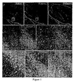
- Figure 1 Hippocampal neuropil antibodies in LE of unclear etiology. Sagiital sections of rat brain reacted with sera of several LE patients. Upper row: 3 cases showing intense reactivity with the neuropil of hippocampus. Cases #2 and 6 competed for the same epitopes (not shown); case #2 had an additional antibody against a subset of neurons in the inner part of the dentate gyrus (shown in middle row). Middle and lower rows at high magnification show the reactivity of cases #1, 2, 5, 7 and a control anti-Hu.
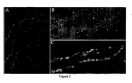
- FIG 2 Immuno-labeling of neurons with novel antibodies of a patient with LE. Cultures of rat hippocampal neurons: (A) incubated with patients' CSF, (B) co-labeled with synaptophysin or (C) with spinophilin. Patient's antibodies (red in all panels in original) show intense reactivity with the cell membrane, with limited colocalization with spinophilin (yellow in C in original) and less with synaptophysin (B). (x800 oil).
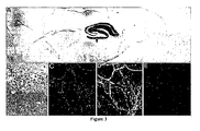
- FIG. 3 Reactivity of patients' antibodies with hippocampus and forebrain, and colocalization with the NR2B subunit of the N-methyl-D-aspartate receptor (NMDAR).
- NMDAR N-methyl-D-aspartate receptor
- FIG. 4 Ovarian teratoma from a patient with LE.
- A Macroscopic view shows presence of fat and hairs (arrow).
- B Microscopic region showing normal-appearing nervous tissue (arrows point to neurons).
- FIG. 5 Brain magnetic resonance imaging (MRI) findings in three patients.
- A, B MRI of Patient I at symptom presentation (A) and after partial clinical improvement and cerebrospinal fluid normalization with immunotherapy (B); note that the clinical and MRI improvement started to occur before tumor resection.
- C, D MRI of Patient 2 at symptom presentation (C) and 4 months later (D); this patient developed rapidly progressive neurological deterioration that did not respond to immunotherapy.
- the autopsy demonstrated that the ovarian cyst was a mature teratoma of the ovary.
- E, F MRI of Patient 3 at symptom presentation; note the mild fluid-attenuated inversion recovery hyperintensity in medial temporal lobes and right frontal cortex. After immunotherapy and tumor resection, the MRI was normal (not shown).
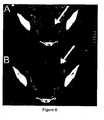
- Figure 6 Interval increase of tumor size during encephalitis.
- A Computed tomography of Patient 1 shows a 5cm cystic ovarian lesion (arrow) that doubled in size over 2 months (B). The lesion was not initially removed because of the poor clinical condition of the patient and benign appearance of the ovarian mass. After partial clinical improvement with immunotherapy, the mass was removed (immature teratoma).
- Figure 7 Patients' antibodies react with heteromers of NR2B and NR2A subunits of the N-methyl-D-aspartate receptor (NMDAR).
- HEK293 cells expressing heteromers (NR1/NR2B or NR1/NR2A) or transfected with single subunits NR1 or NR2B of the NMDAR were incubated with patients' serum or cerebrospinal fluid (CSF).
- CSF cerebrospinal fluid
- Panels demonstrate that the CSF of Patient 7 reacts with cells expressing heteromers (functional receptors) of NR1/NR2B and NR1/NR2A, but not with cells transfected with single subunits (NR1; NR2B).
- FIG 8 Patients' antibodies react with NR2 subunits of the N-methyl-D-aspartate receptors (NMDARs) expressed by nervous tissue present in the tumor.
- A, B Panels correspond to the ovarian teratoma of Patients 3 and 5 immunolabeled with MAP2 (brown staining), a marker specific for neurons and dendritic processes. Note the intense reactivity with neuronal-like cells and a network of cell processes that are better developed in A.
- B, inset Some immature neuronal cells at higher magnification.
- C-E Panels correspond to the tumor of Patient 3 immunolabeled with the patient's antibodies (C, green) and a specific antibody for NR2B (E, red).
- the present invention provides a method of determining a cause of an encephalitis in a subject, comprising the step of testing a body fluid of the subject for an antibody to an NR subunit of the NMDA receptor, thereby determining a cause of an encephalitis in a subject.
- the presence of an antibody to an NR subunit in the body fluid indicates that the encephalitis is of autoimmune etiology.
- the term "subunit" is interchangeable with the term "heteromer”.
- the methods and compositions provided herein facilitate the recognition of a severe form of autoimmune encephalitis that is often responsive to treatment.
- the methods and compositions described herein emphasize the idea that autoimmunity can affect behavior, and particularly that antibodies to heteromers containing the NR2B and NR2A subunits of the NMDAR may alter emotion, in one embodiment, or memory, consciousness or their compbination in other independent embodiments.
- the present invention provides a method of determining a cause of an autoimmune encephalitis in a subject, comprising the step of testing a body fluid of the subject for an antibody to an NR subunit of the NMDA receptor, thereby determining a cause of an autoimmune encephalitis in a subject.
- the presence of the antibody indicates a presence of a tumor in the subject.
- the tumor is a cause of the autoimmune encephalitis.
- the present invention provides a method of diagnosing a paraneoplastic autoimmune encephalitis in a subject, comprising the step of testing a body fluid of the subject for an antibody to an NR subunit of the NMDA receptor, thereby diagnosing a paraneoplastic autoimmune encephalitis in a subject.
- the biological sample used in the methods described herein is a body fluid that is tested by methods of the present invention is, in another embodiment, a cerebro-spinal fluid (CSF).
- the body fluid is plasma.
- the body fluid is any other type of fluid known in the art. Each possibility represents a separate embodiment of the present invention.
- the biological sample is amniotic fluids, blood, sera, saliva, or their combination in another embodiment.
- the autoimmune encephalitis of methods and compositions of the present invention is, in another embodiment, an autoimmune encephalitis.
- the autoimmune encephalitis is a paraneoplastic encephalitis.
- the autoimmune encephalitis is a non-paraneoplastic encephalitis.
- the autoimmune encephalitis is a paraneoplastic autoimmune encephalitis.
- the autoimmune encephalitis is a non-paraneoplastic, autoimmune encephalitis.
- the autoimmune encephalitis is any other type of autoimmune encephalitis known in the art. Each possibility represents a separate embodiment of the present invention.
- the frequency of paraneoplastic anti-NMDAR encephalitis, diagnosed by the methods described herein, is unknown.
- paraneoplastic anti-NMDAR encephalitis is frequently unrecognized. This may be due to several features that make this disorder unique among paraneoplastic encephalitis, including in one embodiment, involvement of relatively young women between the 2 nd and 5 th decades, or, in another embodiment, the unusual presentation with prominent psychiatric manifestations, or in another embodiment, normal or atypical MRI findings, which in 75% of cases consist of mild, transient T2 or FLAIR abnormalities outside the medial temporal lobes, with cortical enhancement in certain embodiments, or in yet another embodiment, the benign appearance of the ovarian tumors.
- any of the subjects presenting the symptoms described hereinabove are diagnosed using the methods described herein.
- Anti-NMDAR encephalitis is different from other types of paraneoplastic encephalitis in several ways: it results in a highly characteristic syndrome; usually affects young women; is treatment-responsive; and associates with tumors that can be benign. Another difference shown here is that despite the presence of the tumor, the immune response is not maintained. This brings into consideration a contributory role of the prodromal "viral-like" disorder, which by itself or in combination with a teratoma sets off or enhances the autoimmune response. In one embodiment, the methods provided herein are used to differentiate anti-NMDAR encephalitis from other types of paraneoplastic encephalitis.
- the autoimmune encephalitis of methods and compositions of the present invention comprises a limbic encephalitis.
- the autoimmune encephalitis is a limbic encephalitis.
- the autoimmune encephalitis is associated with a limbic dysfunction.
- the autoimmune encephalitis is not associated with a limbic dysfunction.
- Limbic encephalitis causes impressive deficits that are characteristically dominated by rapid and severe loss of short-term memory.
- patients show subacute encephalitis of later adult life, mainly affecting the limbic areas with evidence of cancer in one embodiment.
- the term "limbic encephalitis" refers to a subject exhibiting severe short-term memory loss and dementia in association with bronchial carcinoma.
- the autoimmune encephalitis of methods and compositions of the present invention is associated with seizures.
- the autoimmune encephalitis is associated with a dienoephalic syndrome.
- the autoimmune encephalitis is associated with a psychiatric symptom.
- the autoimmune encephalitis is associated with an abnormality in cognition.
- the autoimmune encephalitis is associated with an abnormality in behavior.
- the autoimmune encephalitis is associated with amnesia. In another embodiment, the autoimmune encephalitis is associated with a memory deficit. In another embodiment, the autoimmune encephalitis is associated with memory problems. In another embodiment, the autoimmune encephalitis is associated with a hypokinetic syndrome.
- the autoimmune encephalitis is associated with a movement disorder. In another embodiment, the autoimmune encephalitis is associated with abnormal movements. In another embodiment, the movement disorder is Stiff Man/Person Syndrome. In another embodiment, the movement disorder is any other movement disorder known in the art. Each possibility represents a separate embodiment of the present invention.
- the autoimmune encephalitis is associated with a decreased level ofconsciousness. In another embodiment, the autoimmune encephalitis is associated with hypoventilation.
- the autoimmune encephalitis is associated with, dysfunction of any part of the brain or spinal cord. In another embodiment, the autoimmune encephalitis is associated with a combination of any of the above symptoms or disorders. Each type of encephalitis represents a separate embodiment of the present invention.
- the autoimmune encephalitis is associated with a tumor.
- the tumor is an ovarian teratoma.
- the tumor is a thymic tumor.
- the tumor is a testicular tumor.
- the cancer associated with the encephalitis is a cervical cancer tumor.
- the cancer is a head and neck cancer tumor.
- the cancer is a breast cancer tumor.
- the cancer is an ano-genital cancer tumor.
- the cancer is a melanoma. In another embodiment, the cancer is a sarcoma. In another embodiment, the cancer is a carcinoma. In another embodiment, the cancer is a lymphoma. In another embodiment, the cancer is a leukemia. In another embodiment, the cancer is mesothelioma. In another embodiment, the cancer is a glioma. In another embodiment, the cancer is a germ cell tumor. In another embodiment, the cancer is a choriocarcinoma. Each possibility represents a separate embodiment of the present invention.
- the cancer is pancreatic cancer. In another embodiment, the cancer is ovarian cancer. In another embodiment, the cancer is gastric cancer. In another embodiment, the cancer is a carcinomatous lesion of the pancreas. In another embodiment, the cancer is pulmonary adenocarcinoma. In another embodiment, the cancer is colorectal adenocarcinoma. In another embodiment, the cancer is pulmonary squamous adenocarcinoma. In another embodiment, the cancer is gastric adenocarcinoma. In another embodiment, the cancer is an ovarian surface epithelial neoplasm (e.g. a benign, proliferative or malignant variety thereof). In another embodiment, the cancer is an oral squamous cell carcinoma.
- the cancer is an oral squamous cell carcinoma.
- the cancer is non small-cell lung carcinoma. In another embodiment, the cancer is an endometrial carcinoma. In another embodiment, the cancer is a bladder cancer. In another embodiment, the cancer is a head and neck cancer. In another embodiment, the cancer is a prostate carcinoma.
- the cancer is an acute myelogenous leukemia (AML). In another embodiment, the cancer is a myelodysplastic syndrome (MDS). In another embodiment, the cancer is a non-small cell lung cancer (NSCLC). In another embodiment, the cancer is a Wilms' tumor. In another embodiment, the cancer is a leukemia. In another embodiment, the cancer is a lymphoma. In another embodiment, the cancer is a desmoplastic small round cell tumor. In another embodiment, the cancer is a mesothelioma (e.g. malignant mesothelioma). In another embodiment, the cancer is a gastric cancer. In another embodiment, the cancer is a colon cancer. In another embodiment, the cancer is a lung cancer.
- AML acute myelogenous leukemia
- MDS myelodysplastic syndrome
- NSCLC non-small cell lung cancer
- the cancer is a Wilms' tumor.
- the cancer is a leukemia.
- the cancer is a lymph
- the cancer is a breast cancer. In another embodiment, the cancer is a germ cell tumor. In another embodiment, the cancer is an ovarian cancer. In another embodiment, the cancer is a uterine cancer. In another embodiment, the cancer is a thyroid cancer. In another embodiment, the cancer is a hepatocellular carcinoma. In another embodiment, the cancer is a thyroid cancer. In another embodiment, the cancer is a liver cancer. In another embodiment, the cancer is a renal cancer. In another embodiment, the cancer is a kaposis. In another embodiment, the cancer is a sarcoma. In another embodiment, the cancer is another carcinoma or sarcoma.
- the tumor is any other type of tumor known in the art.
- Each possibility represents a separate embodiment of the present invention.
- the present invention provides a method of determining a cause of an epilepsy in a subject, comprising the step of testing a body fluid of the subject for an antibody to an antibody to an NR subunit of the NMDA receptor, wherein the antibody indicates a presence of a tumor, wherein a tumor is a cause of the epilepsy.
- the epilepsy of methods and compositions of the present invention is, in another embodiment, an idiopathic epilepsy.
- the epilepsy responds to IgG-depleting therapy.
- the epilepsy is associated with partial seizures.
- the epilepsy is associated with simple partial seizures.
- the epilepsy is associated with complex partial seizures.
- the epilepsy is associated with generalized seizures.
- the epilepsy is associated with absence (petit mal) seizures.
- the epilepsy is associated with myoclonic seizures.
- the epilepsy is associated with tonic-clonic (grand mal) seizures.
- the epilepsy is associated with West syndrome. In another embodiment, the epilepsy is associated with Lennox-Gastaut syndrome. In another embodiment, the epilepsy is associated with any other syndrome known in the art.
- the epilepsy is of no known cause. In another embodiment the epilepsy is any other type of epilepsy known in the art. Each type of epilepsy represents a separate embodiment of the present invention.
- Chronic autoimmune encephalitis refers, in another embodiment, to a primary cause of the disorder.
- the term refers to a contributing cause of the disorder.
- the term refers to an indirect causation.
- the term refers to causation via an immune response induced by the tumor.
- the term refers to a significant cause of the disorder.
- the present invention provides a method of diagnosing a tumor in a subject having an encephalitis, comprising the step of testing a body fluid of the subject for an antibody to an NR subunit of the NMDA receptor, thereby diagnosing a tumor in a subject having an encephalitis.
- the presence of the antibody indicates a presence of a tumor in the subject.
- the present invention provides a method of detecting a tumor in a subject having an encephalitis, comprising the step of testing a body fluid of the subject for an antibody to an NR subunit of the NMDA receptor, thereby detecting a tumor in a subject having an encephalitis.
- the presence of the antibody indicates a presence of a tumor in the subject.
- the present invention provides a method of diagnosing a tumor in a subject having an epilepsy, comprising the step of testing a body fluid of the subject for an antibody to an antibody to an NR subunit of the NMDA receptor, thereby diagnosing a tumor in a subject having an epilepsy.
- the presence of the antibody indicates a presence of a tumor in the subject.
- the present invention provides a method of detecting a tumor in a subject having an epilepsy, comprising the step of testing a body fluid of the subject for an antibody to an antibody to an NR subunit of the NMDA (N-methyl D-aspartate) receptor, thereby detecting a tumor in a subject having an epilepsy.
- the presence of the antibody indicates a presence of a tumor in the subject.
- NMDARs are formed from heteromers of NR1 (which bind glycine) and NR2 subunits (which bind glutamate). Both subunits are required to create a functional receptor that likely contains two NR1 and two NR2 subunits.
- NR2A-D There are four NR2 subunits (NR2A-D) which have 50-70% sequence identity in the extracellular domain (NR2B is 70% identical to NR2A, and 55-58% identical to NR2C and NR2D). These NR2 subunits are coded by four different genes and show developmental and regional variability.
- NR2B is expressed at high levels prenatally and declines postnatally. During its decline, expression levels of NR2A and NR2C increase in certain embodiments.
- NR2A is found in most brain regions, NR2B in the hippocampus and forebrain, NR2C in cerebellum, and NR2D in limited subsets of neurons.
- NR1 is ubiquitously distributed in the brain. With maturity many NR1/NR2B receptors become largely extrasynaptic in hippocampal neurons and NR1/NR2A/NR2B becomes the major synaptic receptors in the hippocampus and forebrain.
- the predominant reactivity of the subjects' antibodies with hippocampus and forebrain correlates with the distribution of heteromers containing NR2B.
- the antibodies readily access cells surface epitopes of live neurons, and only react with HEK293 cells expressing functional receptors (heteromers of NR1/NR2B or NR1/NR2A).
- reactivity is identified when each subunit is expressed individually (even though NR1 in particular assembles in stable but inactive homomeric receptors in HEK293 cells) or in anotherembodiment, with immunoblots of cells expressing the functional receptor.
- the NR subunit of methods and compositions of the present invention is, in another embodiment, a NR2B subunit.
- the NR subunit is a GRIN2B (glutamate receptor, ionotropic, N-methyl D-aspartate 2B subunit.
- the NR2B subunit is part of an NMDA heteromer.
- the heteromer is a functional heteromer.
- the NR subunit is a NR2A subunit.
- the NR subunit is a GRIN2A (glutamate receptor, ionotropic, N-methyl D-aspartate 2A) subunit.
- the NR2A subunit is part of an NMDA heteromer.
- the heteromer is a functional heteromer
- the NR subunit is a NR2C subunit.
- the NR subunit is a GRIN2C (glutamate receptor, ionotropic, N-methyl D-aspartate 2C subunit).
- the NR2C subunit is part of an NMDA heteromer.
- the heteromer is a functional heteromer
- the NR subunit is any other NR subunit known in the art. Each possibility represents a separate embodiment of the present invention.
- the NR2B subunit has, in another embodiment, the sequence:
- the NR2B subunit is a homologue of SEQ ID No: 1. In another embodiment, the NR2B subunit is a variant of SEQ ID No: 1. In another embodiment, the NR2B subunit is an isomer of SEQ ID No: 1. In another embodiment, the NR2B subunit is a fragment of SEQ ID No: 1. In another embodiment, the NR2B subunit comprises SEQ ID No: 1. Each possibility represents a separate embodiment of the present invention.
- the NR2B subunit is encoded by a nucleotide sequence having the sequence:
- the NR2B subunit is encoded by a nucleotide molecule that a homologue of SEQ ID No: 2.
- the nucleotide molecule is a variant of SEQ ID No: 2.
- the nucleotide molecule is an isomer of SEQ ID No: 2.
- the nucleotide molecule is a fragment of SEQ ID No: 2.
- the nucleotide molecule comprises SEQ ID No: 2.
- the NR2B subunit has a sequence set forth in 1 of the following GenBank entries: U28861, U28862, U28758, U90278, U11287, U88963, BC113618, BC113620, and AB208850.
- the NR2B subunit has a sequence that is a variant of 1 of the above sequences.
- the NR2B subunit has a sequence that is a homologue of 1 of the above sequences.
- the NR2B subunit has a sequence that is an isomer of 1 of the above sequences.
- the NR2B subunit has a sequence that is a fragment of 1 of the above sequences.
- the NR2B subunit has a sequence that comprises 1 of the above sequences. In another embodiment, the NR2B subunit is encoded by a sequence set forth in 1 of the above entries. In another embodiment, the NR2B subunit is encoded by a homologue of a sequence set forth in 1 of the above entries. In another embodiment, the NR2B subunit is encoded by a variant of a sequence set forth in 1 of the above entries. In another embodiment, the NR2B subunit is encoded by an isomer of a sequence set forth in 1 of the above entries. Each sequence, and each possibility, represents a separate embodiment of the present invention.
- NR2A subunit of methods and compositions of the present invention has the sequence:
- the NR2A subunit is a homologue of SEQ ID No: 3. In another embodiment, the NR2A subunit is a variant of SEQ ID No: 3. In another embodiment, the NR2A subunit is an isomer of SEQ ID No: 3. In another embodiment, the NR2A subunit is a fragment of SEQ ID No: 3. In another embodiment, the NR2A subunit comprises SEQ ID No: 3. Each possibility represents a separate embodiment of the present invention.
- the NR2A subunit is encoded by a nucleotide sequence having the sequence:
- the NR2A subunit is encoded by a nucleotide molecule that a homologue of SEQ ID No: 4.
- the nucleotide molecule is a variant of SEQ ID No: 4.
- the nucleotide molecule is an isomer of SEQ ID No: 4.
- the nucleotide molecule is a fragment of SEQ ID No: 4.
- the nucleotide molecule comprises SEQ ID No: 4.
- the NR2A subunit has a sequence set forth in I of the following GenBank entries: U09002, U90277, BC117131, AB209695, and NP_000824.
- the NR2A subunit has a sequence that is a variant of 1 of the above sequences.
- the NR2A subunit has a sequence that is a homologue of 1 of the above sequences.
- the NR2A subunit has a sequence that is an isomer of 1 of the above sequences.
- the NR2A subunit has a sequence that is a fragment of 1 of the above sequences.
- the NR2A subunit has a sequence that comprises 1 of the above sequences.
- the NR2A subunit is encoded by a sequence set forth in 1 of the above entries.
- the NR2A subunit is encoded by a homologue of a sequence set forth in I of the above entries.
- the NR2A subunit is encoded by a variant of a sequence set forth in 1 of the above entries.
- the NR2A subunit is encoded by an isomer of a sequence set forth in 1 of the above entries.
- NR2C subunit of methods and compositions of the present invention has the sequence:
- the NR2C subunit is a homologue of SEQ ID No: 5. In another embodiment, the NR2C subunit is a variant of SEQ ID No: 5. In another embodiment, the NR2C subunit is an isomer of SEQ ID No: 5. In another embodiment, the NR2C subunit is a fragment of SEQ ID No: 5. In another embodiment, the NR2C subunit comprises SEQ ID No: 5. Each possibility represents a separate embodiment of the present invention.
- the NR2C subunit is encoded by a nucleotide sequence having the sequence:
- the NR2C subunit is encoded by a nucleotide molecule that a homologue of SEQ ID No: 6.
- the nucleotide molecule is a variant of SEQ ID No: 6.
- the nucleotide molecule is an isomer of SEQ ID No: 6.
- the nucleotide molecule is a fragment of SEQ ID No: 6.
- the nucleotide molecule comprises SEQ ID No: 6.
- the NR2C subunit has a sequence set forth in 1 of the following GenBank entries: L76224, U77782, BC031077, BC059384, and AB208799. In other embodiments, the NR2C subunit has a sequence that is a variant of 1 of the above sequences. In other embodiments, the NR2C subunit has a sequence that is a homologue of 1 of the above sequences. In other embodiments, the NR2C subunit has a sequence that is an isomer of 1 of the above sequences. In other embodiments, the NR2C subunit has a sequence that is a fragment of 1 of the above sequences.
- the NR2C subunit has a sequence that comprises 1 of the above sequences. In another embodiment, the NR2C subunit is encoded by a sequence set forth in 1 of the above entries. In another embodiment, the NR2C subunit is encoded by a homologue of a sequence set forth in 1 of the above entries. In another embodiment, the NR2C subunit is encoded by a variant of a sequence set forth in 1 of the above entries. In another embodiment, the NR2C subunit is encoded by an isomer of a sequence set forth in 1 of the above entries. Each sequence, and each possibility, represents a separate embodiment of the present invention.
- the epitope recognized by an antibody detected by a method of the present invention is, in another embodiment, a conformational epitope.
- the epitope is a linear epitope.
- the epitope is any other type of epitope known in the art. Each possibility represents a separate embodiment of the present invention.
- NR2B-related antibodies in the serum and CSF of all patients provides in one embodiment, a diagnostic test for the paraneoplastic anti-NMDAR encephalitis disorder, and provides a novel immune-mediated mechanism of NMDAR dysfunction.
- critical roles of NMDARs include synaptic transmission and remodeling, or dendritic sprouting, and hippocampal long-term potentiation in other embodiments in addition to one paradigm of memory formation and learning.
- NMDARs are also the major mediator of excitotoxicity, and their dysfunction has been associated with schizophrenia, epilepsy, and several types of dementia. Drugs interacting with NMDARs may result in paranoia in one embodiment, or hallucinations and dyskinesias in certain embodiments, all frequent symptoms in subjects diagnosed using the methods described herein.
- ectopic expression of NR2 subunits by nervous tissue contained in the teratomas contributes to break immune tolerance.
- a combination of factors such as an adjuvant effect of the prodromal viral-like illness that occur in most subjects, and a genetic predisposition in certain embodiments, play additional roles in the initiation of the immune response tested for using the diagnosis methods described herein.
- NR2 antibodies in paraneoplastic anti-NMDAR encephalitis is shown by the correlation between patients' symptoms and antibody Liters in one embodiment or, in another embodiment, by the demonstration of deposits of IgG in the hippocampus and amygdala of a patient's autopsy, in a pattern that showed striking resemblance to the rat brain immunolabeling by patients' antibodies (See Figure 6 ).
- the subject has exhibits antibodies that react with SIZN1 (Smad-Interacting Zinc finger protein expressed in the Nervous system).
- SIZN1 Smad-Interacting Zinc finger protein expressed in the Nervous system.
- the subject has exhibits antibodies that react with a VGKC antigen.
- the subject exhibits antibodies that react with an extracellular neuronal antigen. In another embodiment, the subject exhibits antibodies that react with an antigen exposed on the cell surface of a neuron. As provided herein (Example 9), patients with antibodies to extracellular antigens exhibit, under the conditions utilized herein, enhanced responsiveness to immune therapy.
- a method of the present invention utilizes, detects, or tests for a target antigen (other than a NR2B subunit) identified by a method disclosed herein.
- the target antigen is identified by a library screening technique.
- the target antigen is identified by cDNA library screening.
- the target antigen is identified by reactivity with cultured neurons.
- the target antigen is identified by a protocol set forth in Example 8. Each possibility represents a separate embodiment of the present invention.
- Léx limbic encephalitis
- patients with LE develop subacute confusion, irritability, depression, sleep disturbances, seizures, short-term memory loss, and/or dementia.
- the pathological substrate of LE is an inflammatory disorder that predominantly involves the limbic system (hippocampi, amygdala, and cingulate gyrus).
- biopsy and autopsy studies demonstrate interstitial and perivascular infiltrates ofT cells, and less frequently B cells, along with microglial activation, neuronal degeneration, and/or gliosis.
- inflammatory infiltrates are found in areas distant from the limbic system.
- the infiltrates remain mild and clinically silent. In another embodiment, the infiltrates become prominent and develop into a disorder called encephalomyelitis. Additional methods of diagnosing LE are described, for example, in Gultekin SH et al (Paraneoplastic limbic encephalitis: neurological symptoms, immunological findings and tumour association in 50 patients. Brain 2000;123:1481-1494 ). Each possibility represents a separate embodiment of the present invention.
- an antigen of the present invention is homologous to a peptide disclosed herein.
- the terms "homology,” “homologous,” etc, when in reference to any protein or peptide, refer, in one embodiment, to a percentage of amino acid residues in the candidate sequence that are identical with the residues of a corresponding native polypeptide, after aligning the sequences and introducing gaps, if necessary, to achieve the maximum percent homology, and not considering any conservative substitutions as part of the sequence identity. Methods and computer programs for the alignment are well known in the art.
- Homology is, in another embodiment, determined by computer algorithm for sequence alignment, by methods well described in the art.
- computer algorithm analysis of nucleic acid sequence homology can include the utilization of any number of software packages available, such as, for example, the BLAST, DOMAIN, BEAUTY (BLAST Enhanced Alignment Utility), GENPEPT and TREMBL packages.
- “homology” refers to identity to a sequence selected from SEQ ID No: 1-6 of greater than 70%. In another embodiment, “homology” refers to identity to a sequence selected from SEQ ID No: 1-6 of greater than 72%. In another embodiment, “homology” refers to identity to one of SEQ ID No: 1-6 of greater than 75%. In another embodiment, “horology” refers to identity to a sequence selected from SEQ ID No: 1-6 of greater than 78%. In another embodiment, “homology” refers to identity to one of SEQ ID No: 1-6 of greater than 80%. In another embodiment, “homology” refers to identity to one of SEQ ID No: 1-6 of greater than 82%.
- “homology” refers to identity to a sequence selected from SEQ ID No: 1-6 of greater than 83%. In another embodiment, “homology” refers to identity to one of SEQ ID No: 1-6 of greater than 85%. In another embodiment, “homology” refers to identity to one of SEQ ID No: 1-6 of greater than 87%. In another embodiment. “homology” refers to identity to a sequence selected from SEQ ID No: 1-6 of greater than 88%. In another embodiment, “homology” refers to identity to one of SEQ ID No: 1-6 of greater than 90%. In another embodiment, “homology” refers to identity to one of SEQ ID No: 1-6 of greater than 92%.
- “homology” refers to identity to a sequence selected from SEQ ID No: 1-6 of greater than 93%. In another embodiment, “homology” refers to identity to one of SEQ ID No: 1-6 of greater than 95%. In another embodiment, “homology” refers to identity to a sequence selected from SEQ ID No: 1-6 of greater than 96%. In another embodiment, “homology” refers to identity to one of SEQ ID No: 1-6 of greater than 97%. In another embodiment, “homology” refers to identity to one of SEQ ID No: 1-6 of greater than 98%. In another embodiment, “homology” refers to identity to one of SEQ ID No: 1-6 of greater than 99%. In another embodiment, “homology” refers to identity to one of SEQ ID No: 1-6 of 100%. Each possibility represents a separate embodiment of the present invention.
- homology is determined via determination of candidate sequence hybridization, methods of which are well described in the art (See, for example, " Nucleic Acid Hybridization” Hames, B. D., and Higgins S. J., Eds. (1985 ); Sambrook et al., 2001, Molecular Cloning, A Laboratory Manual, Cold Spring Harbor Press, N.Y .; and Ausubel et al., 1989, Current Protocols in Molecular Biology. Green Publishing Associates and Wiley Interscience, N.Y ).
- methods of hybridization are carried out under moderate to stringent conditions, to the complement of a DNA encoding a native caspase peptide.
- Hybridization conditions being, for example, overnight incubation at 42 °C in a solution comprising: 10-20 % formamide, 5 X SSC (150 mM NaCl, 15 mM trisodium citrate), 50 mM sodium phosphate (pH 7. 6), 5 X Denhardt's solution, 10 % dextran sulfate, and 20 ⁇ g/ml denatured, sheared salmon sperm DNA.
- Protein and/or peptide homology for any AA sequence listed herein is determined, in another embodiment, by methods well described in the art, including immunoblot analysis, or via computer algorithm analysis of AA sequences, utilizing any of a number of software packages available, via established methods. Some of these packages include the FASTA, BLAST, MPsrch or Scanps packages, and, in another embodiment, employ the use of the Smith and Waterman algorithm, and/or global/local or BLOCKS alignments for analysis, for example. Each method of determining homology represents a separate embodiment of the present invention.
- nucleic acids or “nucleotide” refers to a string of at least two base-sugar-phosphate combinations.
- the term includes, in one embodiment, DNA and RNA.
- Nucleotides refers, in one embodiment, to the monomeric units of nucleic acid polymers.
- RNA is, in one embodiment, in the form of a tRNA (transfer RNA), snRNA (small nuclear RNA), rRNA (ribosomal RNA), mRNA (messenger RNA), anti-sense RNA, small inhibitory RNA (siRNA), micro RNA (miRNA) and ribozymes.
- DNA can be, in other embodiments, in form of plasmid DNA, viral DNA, linear DNA, or chromosomal DNA or derivatives of these groups.
- these forms of DNA and RNA can be single, double, triple, or quadruple stranded.
- the term also includes, in another embodiment, artificial nucleic acids that contain other types of backbones but the same bases.
- the artificial nucleic acid is a PNA (peptide nucleic acid).
- PNA contain peptide backbones and nucleotide bases and are able to bind, in one embodiment, to both DNA and RNA molecules.
- the nucleotide is oxetane modified.
- the nucleotide is modified by replacement of one or more phosphodiester bonds with a phosphorothioate bond.
- the artificial nucleic acid contains any other variant of the phosphate backbone of native nucleic acids known in the art.
- nucleic acid derivative represents a separate embodiment of the present invention.
- EXAMPLE 1 Paraneoplastic anti-NMDA Receptor Encephalitis Associated with Ovarian Teratoma MATERIALS AND EXPERIMENTAL METHODS
- Patients include 12 women with paraneoplastic encephalitis associated with teratomas. The 6 most recently identified patients and neuropathological findings (2 cases) are described in detail in the supplementary files; the clinical features of the other 6 patients have been previously reported. Frozen serum or cerebrospinal fluid was available from all 12 patients. Tissues for immunological studies included tumors from 5 patients (1 frozen tissue, 4 embedded in paraffin), and brain obtained at autopsy of 1 patient and 2 neurologically normal individuals. Sera or CSF of 200 individuals, including blood donors and patients with diverse paraneoplastic and non-paraneoplastic encephalitis served as controls. Studies were approved by the University of Pennsylvania Institutional Review Board.
- Wistar rats were sacrificed omitting perfusion with saline or fixatives; the brain was removed, immersed in 4% paraformaldehyde (PFA) at 4°C for 24 hours, cryoprotected with 40% sucrose, sagittally sectioned and snap frozen in isopentane chilled with liquid nitrogen.
- PFA paraformaldehyde
- chicken anti-MAP2 (Covance, Princeton, NJ; 1:20,000); rabbit anti-NR 1 (amino acids 1-20) and rabbit anti-NR2A (amino acids 1265-1464) (both from Upstate biotechnology, Lake Placid, NY, both at 1:50); rabbit anti-NR2B (251 amino acid sequence from N-terminal portion of NMDAR; Zymed, San Francisco, CA, 1:50); CD3, CD19 and CD68 (DakoCytomation, Carpinteria, CA, all 1:100).
- Paraffin-embedded tissue was deparaffinized and the antigens retrieved, as reported. 10 Seven micron-thick frozen (or 4 micron-thick paraffin) tissue sections were serially incubated with 0.3% H 2 O 2 for 20 minutes, 10% goat serum for 1 hour, and patient's serum (1:250), CSF (1:10) or biotinylated IgG (0.2 mg/ml), or the indicated purchased antibodies overnight at 4°C. After using the appropriate secondary antibodies (all 1:2000), reactivities were developed with the avidin-biotin-peroxidase method. The secondary antibody was omitted when patients' biotinylated IgG was used. Normal human serum and biotinylated IgG, and normal goat serum served as controls. Double immunolabeling with patients' IgG, MAP2, and NR2 antibodies was performed using the appropriate Alexa Fluor secondary antibodies diluted 1:2000 (Molecular Probes, OR); results were photographed under a fluorescence microscope using Zeiss Axiovision software.
- Rat hippocampal neuronal cultures were prepared as reported. 11 Live neurons grown on coverslips were exposed for 1 hour at 37°C to the patients' or control serum (final dilution 1:1000) orCSF (1:100). After removing the media and extensive washing, neurons were fixed with 4% PFA, and single or double immunolabeling was performed, as indicated above. The reactivity of commercial antibodies to NR1 and NR2 subunits was best seen with cell permeabilization (0.1% Triton) after PFA.
- HEK293 cells were transfected with plasmids containing rodent NR1, NR2A, or NR2B subunits of the NMDAR (>90% homologous to the human subunits in the extracellular domains) or plasmids without insert (control), as reported.
- cells were co-transfected with NR1 and NR2A (or NR1 and NR2B) in equimolar ratios. Cells were grown for 24 hours after transfection before assessment. All cells were routinely grown in the presence of NMDAR antagonists (500 ⁇ M ketamine) to prevent cell death after transfeciion. 15 Transfected cells were then incubated with patients' serum (1:400) or CSF (1:10) overnight at 4°C, and the appropriate Alexa Fluor secondary antibodies, as above.
- the median age of the patients was 27 years (range 14-44 years). In 11 patients the neurologic symptoms preceded the diagnosis of the teratoma by 3 weeks-4 months (median 2 months) and in 1 patient occurred after an ovarian cyst had been radiologically detected one month earlier. Ten patients had a viral-like prodromic syndrome with hyperthermia; (7 cases) and frequent headache (6 cases) (Table 1).
- Table 1 Clinical features Case Sex/ age Teratoma: histology, side, size Time to tumor diagnosis Prodrome Presenting symptoms Other symptoms and findings during the course of the disorder # 1 F/30 immature, right ovary, 10 cm 2 months headache, hyperthermia STMD, decreased level of consciousness, "epilepsia partialis continua" for 2 weeks. Sedation, MV, PEG Restlessness, involuntary movements hyperthermia # 2 F/35 mature, left ovary, 3.5 cm 4 months (autopsy) headache, nausea, no fever; received acetaminop hen STMD, generalized tonic-clonic seizure followed by refractory status epilepticus.
- Psychiatric syndrome generalized seizures, MV, PEG Incomprehensible speech, decreased of level of consciousness, STMD, hyperthermia # 9 4 F/40 mature, left ovary, 6.0 cm 3 weeks NA Secondary generalized seizures, psychiatric syndrome, MV, PEG Decreased of level of consciousness, STMD # 10 4.8 F/14 immature, left ovary, 1.9 cm 2 months hyperthermia, headache, rhinorrhea Psychiatric syndrome (hallucinations, extreme panic), generalized seizures, MV, PEG Incomprehensible speech, choreoathetotic movements, hypersomnia, hyperthermia, autonomic instability # 11 4.7 F/28 1 st : mature, right ovary, 14 cm; 1 month after tumor cough,no fever; received antibiotics 1 st episode : psychiatric syndrome (delusional thinking, personality change
- CSF lymphocytic pleocytosis was identified in all patients (9-219 cells/ ⁇ 1, median 24), and 4 also had elevated protein concentration (56-129 mg/dL, median 67); the glucose concentration was normal in all instances (Table 2). Oligoclonal bands were identified in 3 of 6 patients examined. All patients had extensive serum and CSF diagnostic tests with negative or normal results for viral, bacterial, fungal infections, collagen-vascular autoimmune disorders, thyroid autoimmunity, and comprehensive panels of paraneoplastic and VGKC antibodies.
- Table 2 Diagnostic tests, treatment, and outcome Case MRI at presentation CSF Chronologic list of treatments Initial improvement Outcome (immuno-therapy and tumor) (duration follow-up) # 1 FLAIR and T2 hyperintensity in medial temporal lobes 40 WBC, protein 67 corticosteroids, plasma exchange, IVIg Partial clinical and MRI improvement presurgery. Further improvement 4 weeks after surgery Back to work as an internal medicine resident; normal MRI (12 months).
- MRI fronto-temporal atrophy (16 months). # 8 4 FLAIR/ T2 hyperintensities in cerebral cortex and cerebellum; mild cortical cerebellar enhancement. 49 WBC, corticosteroids ⁇ 7 weeks Full recovery; normal MRI (24 months). protein 67 # 9 4 FLAIR abnormalities involving the cingulum and gray matter of the frontal lobes 9 WBC, tumor removal ⁇ 16 weeks Residual cognitive dysfunction and memory problem, MMSE 28/30 (6 years). protein normal # 10 4.8 Three MRIs normal 115 WBC, tumor removal, plasma exchange, costicosteroids, IVIg transferred to a chronic care facility with MV (no significant improvement) Unexpected death after mild improvement, ⁇ 6 months after symptom presentation.
- CT of the chest, abdomen and pelvis revealed an ovarian mass in 10 patients, a tumor in the anterior mediastinum in 1, and no evidence of tumor in 1 (the tumorwas demonstrated at autopsy).
- the radiological size of the tumors ranged from 1.5 to 22 cm (median largest diameter 6.5 cm) (Table 1).
- Two patients (cases #1 and 7) had significant tumor growth during the encephalitic process ( Figure 6A and B ). Only one patient had elevated levels of CEA, CA125 and ⁇ -fetoprotein.
- the main autoantigens are functional heteromers of NMDARs
- EXAMPLE 2 IDENTIFICATION OF IMMUNE RESPONSES IN PATIENTS WITH AUTOIMMUNE ENCEPHALITIS OF UNKNOWN ETIOLOGY
- Rats were anaesthesized and euthanized by decapitation without tissue perfusion; brains were removed and kept for 10 days in 4% PFA at 4 °C. Subsequently, brains were cryoprotected with 30% sucrose for 48 h, embedded in freezing medium, and snap-frozen in isopentane chilled with liquid nitrogen.
- Brains from rats perfused with 4% PFA were removed and kept in 4% PFA for 1 h, and subsequently cryoprotected and embedded in freezing medium as above. Non-perfused rat brains were removed and directly embedded in freezing medium without fixative.
- Immunoblots included protein extracts (100 microgram (mcg)/ml) from purified human cortical neurons, Purkinje cells and the recombinant proteins, HuD, Cdr2, Nova, Ma1, Ma2, CRMP5 and amphiphysin.
- Immunohistochemistry was performed with cryostat-cut 7 mm thick sections mounted directly on slides. Non-pre-fixed tissue was incubated for 10 min with acetone or methanol-acetone at 4 °C. Subsequently, all tissue sections were serially incubated with 0.25% H 2 O 2 for 20 min, 10% goat serum for 30 min, the patient's serum or CSF at the indicated dilutions in 10% goat serum overnight at 4 °C, biotinylated goat anti-human IgG (1 : 2000) for 2 h and avidin-biotin peroxidase for 1 h, and the reactivity developed with diaminobenzidine.
- polyclonal rabbit antibodies to VGKCs Kv1.1, Kv1.2 and Kv1.6 (dilution 1:50; all from Sigma, St Louis, MO); a monoclonal antibody to Kv1.2 (1:50; Upstate Laboratories, Lake Placid, NY); a polyclonal antibody to synaptophysin (1:1000, Sigma); a monoclonal antibody to spinophilin (1:50; Upstate Laboratories); and human control serum with amphiphysin and Hu antibodies (1:500).
- Intrathecal synthesis of antibodies was determined using scrum and CSF samples normalized with the same concentration of IgG and serially diluted in parallel. Patients were considered to have intrathecal synthesis of antibodies if the end dilution point of CSF showed reactivity that was no longer present in the paired serum containing the same amount of IgG.
- the serum and/or CSF of the 4 patients contained antibodies with two unique properties: 1) the target antigens were highly enriched in the hippocampus ( Figure 7 ), and 2 ) the antibodies were more readily detected in the CSF (with intrathecal synthesis) than in sera.
- Immune-competition assays between patients' antibodies showed that they partially blocked the hippocampal reactivity of each other, indicating that some, but not all epitopes are common targets.
- the dose temporal association between presentation of the encephalitis and the diagnosis of an ovarian teratoma (median, 3 months) and the correlation in one case between three episodes of neurological relapse and tumor recurrence shows that the tumor played a role in triggering the immune response.
- EXAMPLE 3 IDENTIFICATION OF ADDITIONAL MARKERS FOR TREATMENT-RESPONSIVE AUTOIMMUNE ENCEPHALITIS
- Uni-ZAP-XR limbic and cerebellum libraries are screened at a density of 5 x 10 4 pfu/150 mm plate. Nitrocellulose filters with phage plaques are incubated with sera (pooled from 5 patients with similar antibodies, each diluted 1:1,000) or CSF (diluted 1:5) overnight at 4°C, then sequentially incubated with biotinylated goat anti-human IgG (1:2000) for 2 hours and avidin-biotin-peroxidase for 1 hour, and development under direct visual guidance with 0.05% diaminobenzidine. Clones yielding positive results are purified by several rounds of antibody screening until a yield of 100% positive plaques is obtained. Phage clones are subdoned in pBluescript using the in vivo excision phage rescue protocol (Stratagene) and sequenced.
- the pooled (or individual) sera to be screened is preabsorbed with E. coli proteins.
- E. coli XL-1 blue are grown overnight in 25 ml of LB media and pelleted. The pellet is lysed in 1% Triton-X 100, and cell debris removed by centrifugation. The patients' sera are incubated with the supernatant containing the E. coli proteins (ratio 1 to 10) for 1 hour. Prior to screening, the mixture of sera and E. coli proteins is diluted with PBS so that the final dilution of each patient's serum is 1:1000.
- the phage clone of interest is mixed with an irrelevant (negative) phage clone at a ratio of 50/50 and plated onto agar plates yielding a lawn of phage plaques that are 50% plaques of interest and 50% irrelevant plaques. These plaques are then transferred to nitrocellulose filters that are cut into sections and incubated with each individual patient's or control sera (1:1000) or CSF (1:10), followed by the indicated biotinylated goat anti-human IgG and the avidin-biotin-peroxidase method.
- Neuronal cells To label cells, media is removed from neuronal cultures of 3-week-old hippocampal neurons, and cells are washed in PBS and incubated in methionine-free Neurobasal medium (Gibco-Invitrogen) supplemented with B27 serum free su pplement and 50 U/ml penicillin-streptomycin with 1% bovine serum albumin for 60 minutes. Cells are washed and incubated with methionine-free DMEM with 0.5 mCi/ml 35 [S]-L-methionine for 60 minutes at 37°C, placed on ice, and washed 2 times in ice-cold PBS. This is followed by solubilization by either of the following methods. The choice of method is initially based on the presumed antigen location using double immuno-labeling techniques with well characterized proteins (pre-synaptic, post-synaptic, dendritic spines, etc).
- Method A Used for antigens that are neuronal membrane proteins. It employs Triton X-114 which undergoes cloud point precipitation at 30°C, yielding a detergent phase into which membrane and other hydrophobic proteins preferentially partition. Cells are solubilized in 50 mM Tris-HCl pH 7.4,100 mM NaCl, 0.5% Triton-X 114 (w/v), with protease inhibitors (1 mM PMSF and CLAP buffer at 1 to 1000). Cloud point precipitation is carried out by placing the mixture over a bed of 6% (w/v) sucrose and incubating for 3 minutes at 30°C or until the solution becomes cloudy followed by centrifugation. The detergent phase is removed from under the sucrose and resuspended in 50 mM Tris, pH 7.4,100 mM NaCl, 1 mM PMSF to original volume.
- Method B Used for antigens that are freely soluble neuronal proteins and some membrane proteins. Cells are lysed in 0.5 ml of 50 mM Tris-HCl pH 7.4,100 mM NaCl, 05% Triton-X 100 (w/v), and 1 mM PMSF.
- Synaptosomes Used for antigens that are enriched in synaptosomes.
- the region of interest is dissected from rat brain and homogenizing in 0.32M sucrose in 5 mM Tris-HCl, pH 7.4 with 2 mM EDTA. After centrifugation (750 g) the supernatant is retrieved and centrifuged at 17,000 g. The pellet is lysed in Tris-HCl pH 82 with 1 mM EDTA and homogenized, followed by centrifugation at 100,000 g.
- the pellet is resuspended in 0.1 M KCl, 10 mM Tris HCl, pH 7.4, 1% Triton X-100 and 2 mM EDTA and centrifuged again at 100,000 g.
- the synaptosome-containing supernatant is retrieved and stored until use. All buffers contain a mixture of several protease inhibitors.
- 3 ⁇ g of biotin (as a 50 ⁇ g/ ⁇ l solution in DMSO) is added for each 1 ⁇ g of synaptosome protein, and incubated at 4 °C overnight, and dialysed against PBS. Biotinylated synaptosomes are incubated with primary antibody in 0.1% BSA in PBS (ratio of 5 ⁇ l patient's serum, or 50 ⁇ l CSF, to 100 ⁇ g protein).
- Solubilized protein extracts are preabsorbed by incubating with 30 ⁇ l protein A/G Sepharose beads and 4 ⁇ l of normal human sera for 3 hours.
- the pre-absorbed extracts are retrieved and incubated with the patient's serum or CSF at a ratio of 5 ⁇ l sera (50 ⁇ l CSF) to 100 ⁇ l of extract and 25 ⁇ l of protein A/G beads overnight at 4°C, washed, resuspended in Laemmli buffer and run on an SDS-PAGE immunoblot. After protein transfer to nitrocellulose, protein is visualized by XR autoradiography or avidin-biotin-peroxidase and diaminobenzidine.
- each of the cellular fractions is further fractionated.
- dot blots Prior to performing precipitation on subfractions, dot blots are used as a quick and simple screening of small amounts of material, and only positive fractions are then generated.
- Sepharose beads are pelleted after pre-absorption with normal human sera, washed, resuspended in Laemmli and run on SDS-PAGE as a control for non-specific binding.
- Positive controls for localization include synaptic vesicle 2, Kvl.2 VGKC, and amphiphysin; the latter located at the cytoplasmic side of the cell membrane).
- Protein bands of interest are excised and subjected to matrix-assisted laser desorption ionization time of flight mass spectroscopy (MALDI-MS) using the core Protein Chemistry facility of Cancer Center at the University of Pennsylvania.
- MALDI-MS matrix-assisted laser desorption ionization time of flight mass spectroscopy
- cDNA library screening and immunoprecipitation (IP) with rat hippocampal proteins extracted from brain or neuronal cultures are used to identify additional paraneoplastic antigens.
- 3-week-old hippocampal neurons which are enriched with many of the auto-antigens that are isolated herein, are used to purify the hippocampal proteins.
- These antigens are predominantly expressed in rat hippocampus and cerebellum, and in the cell membrane of neurons in culture ( Figures 5-6 ).
- Novel auto-antigens are tested for association with specific sub-phenotypes of LE.
- Tests to identify the novel auto-antigens include immunoblot, IP, and ELISA techniques.
- EXAMPLE 4 NR2B AND NR2A HETEROMERS OF THE NMDA RECEPTOR ARE THE AUTOANTIGENS OF OVARIAN TERATOMA-ASSOCIATED ENCEPHALITIS
- Serum/CSF antibodies were analyzed using neuronal cultures, as described above, and HEK293 cells expressing NR subunits of the NMDA receptor.
- Tumor resection and immunotherapy resulted in recovery of 7/8 patients, paralleled by decreased antibodies titers.
- 2/3 patients without tumor resection died of neurologic deterioration, as evidenced by autopsy findings.
- NMDA receptor antibodies can be used as a diagnostic test for autoimmune encephalitis.
- these findings show that NMDA receptor antibodies are likely to play a causative role in epilepsy and immune-mediated disorders of memory, cognition, and behavior.
- Rasmussen's encephalitis patients are tested for the presence of anti-NMDA antibodies. Patients testing positive for the antibodies are administered treatment for depletion of anti-NMDA antibodies.
- EXAMPLE 7 PATHOLOGICAL ROLE OF NMDA RECEPTOR ANTIBODIES IN REM-ASSOCIATED SLEEP-DISORDERS
- Patients having REM-associated sleep-disorders are tested for the presence of anti-NMDA antibodies. Patients testing positive for the antibodies are administered treatment for depletion of anti-NMDA antibodies.
Landscapes
- Health & Medical Sciences (AREA)
- Life Sciences & Earth Sciences (AREA)
- Immunology (AREA)
- Engineering & Computer Science (AREA)
- Hematology (AREA)
- Urology & Nephrology (AREA)
- Molecular Biology (AREA)
- Biomedical Technology (AREA)
- Chemical & Material Sciences (AREA)
- Physics & Mathematics (AREA)
- Biochemistry (AREA)
- Pathology (AREA)
- General Physics & Mathematics (AREA)
- Cell Biology (AREA)
- Food Science & Technology (AREA)
- Medicinal Chemistry (AREA)
- Biotechnology (AREA)
- Analytical Chemistry (AREA)
- Microbiology (AREA)
- General Health & Medical Sciences (AREA)
- Hospice & Palliative Care (AREA)
- Oncology (AREA)
- Rehabilitation Therapy (AREA)
- Rheumatology (AREA)
- Peptides Or Proteins (AREA)
- Medicines Containing Antibodies Or Antigens For Use As Internal Diagnostic Agents (AREA)
Description
- This invention provides methods of diagnosing or determining a cause of an autoimmune encephalitis or an epilepsy in a subject and of diagnosing a tumor in a subject, comprising the step of testing a body fluid of the subject for an antibody to an NR subunit of the NMDA receptor.
- Disturbances of memory, behavior, cognition, and seizures can result from immune-mediated encephalitis. One cause of autoimmune encephalitis is the paraneoplastic manifestation of a neoplasm. Until now most puraneoplastic encephalitides have been associated with antibodies to intracellular onconeuronal proteins and cytotoxic T-cells presumably against the same proteins. These disorders usually associate with malignant tumors and are poorly responsive to immunotherapies or treatment of the cancer.
- In recent years a severe but often reversible encephalitis of unknown etiology that predominantly affects young women has been increasingly recognized. The disorder has received several names, including acute diffuse lymphocytic meningoencephalitis, acute reversible limbic encephalitis, acute juvenile female non-herpetic encephalitis, or juvenile acute non-herpetic encephalitis. No association has yet been made with infections, cancer, or specific autoantibodies but given that most patients develop a prodromic viral-like illness, a postinfectious immune-mediated etiology has been postulated.
- The affected patients were women who developed prominent psychiatric symptoms, seizures, memory deficits, and decreased level of consciousness often requiring ventilatory support. Three salient features included the young age of the patients, the association with ovarian teratomas, and the detection of antibodies to unknown antigens predominantly expressed in the cell membrane of hippocampal neurons (also referred to as a subgroup of neuropil antigens).
- A better understanding of the function of the paraneoplastic neuronal (or onconeuronal) antigens along with modelling PND in animals results in improved treatment strategies. For the clinician who currently confronts these patients, however, the best chance to affect the neurologic outcome depends on: (1) the prompt diagnosis of the disorder, (2) the early discovery and treatment of the tumor, and (3) the use of immunotherapy. Accordingly, a need Exists for a reliable method of diagnosing the presence of autoimmune encephalitis and the possibility that observed epileptic seizures are caused by (sometimes occult) tumors.
- This invention provides methods of diagnosing or determining a cause of an autoimmune encephalitis or an epilepsy in a subject and of diagnosing a tumor in a subject, comprising the step of testing a body fluid of the subject for an antibody to an NR subunit of the NMDA receptor.
- In one embodiment, the present invention provides a method of determining a cause of an encephalitis in a subject, comprising the step of testing a body fluid of the subject for an antibody to an NR subunit of the NMDA receptor (NMDAR), thereby determining a cause of an encephalitis in a subject. In another embodiment, the presence of an antibody to an NR subunit in the body fluid indicates that the encephalitis is of autoimmune etiology. Each possibility represents a separate embodiment of the present invention.
- In another embodiment, the present invention provides a method of determining a cause of an autoimmune encephalitis in a subject, comprising the step of testing a body fluid of the subject for an antibody to an NR subunit of the NMDA receptor, thereby determining a cause of an autoimmune encephalitis in a subject. In another embodiment, the presence of the antibody indicates a presence of a tumor in the subject. In another embodiment, the tumor is a cause of the autoimmune encephalitis. Each possibility represents a separate embodiment of the present invention.
- In another embodiment, the present invention provides a method of determining a cause of an epilepsy in a subject, comprising the step of testing a body fluid of the subject for an antibody to an antibody to an NR subunit of the NMDA receptor, thereby determining a cause of an epilepsy in a subject. In another embodiment, the antibody indicates a presence of a tumor in the subject. In another embodiment, the tumor is a cause of the epilepsy. Each possibility represents a separate embodiment of the present invention.
- In another embodiment, the present invention provides a method of diagnosing a tumor in a subject having an encephalitis, comprising the step of testing a body fluid of the subject for an antibody to an NR subunit of the NMDA receptor, thereby diagnosing a tumor in a subject having an encephalitis. In another embodiment, the presence of the antibody indicates a presence of a tumor in the subject. Each possibility represents a separate embodiment of the present invention.
- In another embodiment, the present invention provides a method of diagnosing a tumor in a subject having an epilepsy, comprising the step of testing a body fluid of the subject for an antibody to an antibody to an NR subunit of the NMDA receptor, thereby diagnosing a tumor in a subject having an epilepsy. In another embodiment, the presence of the antibody indicates a presence of a tumor in the subject. Each possibility represents a separate embodiment of the present invention.
-
Figure 1 : Hippocampal neuropil antibodies in LE of unclear etiology. Sagiital sections of rat brain reacted with sera of several LE patients. Upper row: 3 cases showing intense reactivity with the neuropil of hippocampus.Cases # 2 and 6 competed for the same epitopes (not shown); case #2 had an additional antibody against a subset of neurons in the inner part of the dentate gyrus (shown in middle row). Middle and lower rows at high magnification show the reactivity ofcases # case # 7 and anti-Hu with the predominant neuropil reactivity ofcases # 1, 2 and 5 that spare the nuclei and cytoplasm of neurons (except for #2 that labels a subset of cells). Onlycase # 1 had VGKC antibodies. Arrows point to the dentate gyrus to allow comparison between panels. (Upper row x5; middle and low rows x200). -
Figure 2 . Immuno-labeling of neurons with novel antibodies of a patient with LE. Cultures of rat hippocampal neurons: (A) incubated with patients' CSF, (B) co-labeled with synaptophysin or (C) with spinophilin. Patient's antibodies (red in all panels in original) show intense reactivity with the cell membrane, with limited colocalization with spinophilin (yellow in C in original) and less with synaptophysin (B). (x800 oil). -
Figure 3 . Reactivity of patients' antibodies with hippocampus and forebrain, and colocalization with the NR2B subunit of the N-methyl-D-aspartate receptor (NMDAR). (A) Sagittal section of rat brain immunolabeled with a patient's cerebrospinal fluid. Note the robust reactivity with the hippocampus and milder reactivity with forebrain. The cerebellum is largely spared. (B) Higher magnification of the molecular layer of the hippocampus (arrow in A); this pattern of reactivity is identical to that previously reported in patients with paraneoplastic encephalitis and ovarian teratoma.4 (C-E) Double immunolabeling of cultures of rat hippocampal neurons using a patient's antibodies (C, green) and an antibody against NR2B of the NMDAR (E, red); note the significant colocalization of reactivities (D, yellow). These findings suggested that patients' antibodies were directed against the NMDAR. Subsequent studies demonstrated that the patient also had antibodies against NR2A (not shown), which explains, in part, the partial colocalization of reactivities. Original magnification X2.5 (A) and X400 (B), both counterstained with hematoxylin; X800 (oil lens), immunofluorescence (C-E). -
Figure 4 . Ovarian teratoma from a patient with LE. (A) Macroscopic view shows presence of fat and hairs (arrow). (B) Microscopic region showing normal-appearing nervous tissue (arrows point to neurons). x200 H& -
Figure 5 : Brain magnetic resonance imaging (MRI) findings in three patients. (A, B) MRI of Patient I at symptom presentation (A) and after partial clinical improvement and cerebrospinal fluid normalization with immunotherapy (B); note that the clinical and MRI improvement started to occur before tumor resection. (C, D) MRI of Patient 2 at symptom presentation (C) and 4 months later (D); this patient developed rapidly progressive neurological deterioration that did not respond to immunotherapy. The autopsy demonstrated that the ovarian cyst was a mature teratoma of the ovary. (E, F) MRI ofPatient 3 at symptom presentation; note the mild fluid-attenuated inversion recovery hyperintensity in medial temporal lobes and right frontal cortex. After immunotherapy and tumor resection, the MRI was normal (not shown). -
Figure 6 : Interval increase of tumor size during encephalitis. (A) Computed tomography ofPatient 1 shows a 5cm cystic ovarian lesion (arrow) that doubled in size over 2 months (B). The lesion was not initially removed because of the poor clinical condition of the patient and benign appearance of the ovarian mass. After partial clinical improvement with immunotherapy, the mass was removed (immature teratoma). -
Figure 7 : Patients' antibodies react with heteromers of NR2B and NR2A subunits of the N-methyl-D-aspartate receptor (NMDAR). HEK293 cells expressing heteromers (NR1/NR2B or NR1/NR2A) or transfected with single subunits NR1 or NR2B of the NMDAR were incubated with patients' serum or cerebrospinal fluid (CSF). (top row) Panels demonstrate that the CSF ofPatient 7 reacts with cells expressing heteromers (functional receptors) of NR1/NR2B and NR1/NR2A, but not with cells transfected with single subunits (NR1; NR2B). Cells transfected with single NR2A or plasmid without insert were not reactive with patient's antibodies (not shown). (bottom row) Panels demonstrate the reactivity of the CSF ofPatients Patient 7 and an antibody specific for NR2B, showing colocalization of reactivities. The fourth panel (C (-)) corresponds to the CSF of an individual without paraneoplastic encephalitis (negative control). Original magnification 0 800, immunofluorescence; nuclei of cells demonstrated with 4 diamidino-2-phenylindole (DAPI), except "#7 + NR2B," in which no DAPI was used. -
Figure 8 : Patients' antibodies react with NR2 subunits of the N-methyl-D-aspartate receptors (NMDARs) expressed by nervous tissue present in the tumor. (A, B) Panels correspond to the ovarian teratoma ofPatients 3 and 5 immunolabeled with MAP2 (brown staining), a marker specific for neurons and dendritic processes. Note the intense reactivity with neuronal-like cells and a network of cell processes that are better developed in A. (B, inset) Some immature neuronal cells at higher magnification. (C-E) Panels correspond to the tumor ofPatient 3 immunolabeled with the patient's antibodies (C, green) and a specific antibody for NR2B (E, red). Note that there is colocalization of reactivities (D, yellow), indicating that the patient's antibodies react with NR2B expressed in the tumor (similar findings were observed with NR2A, not shown). (F-H) Panels correspond to the tumor of Patient 5 immunolabeled with the patient's antibodies (F, green) and a specific antibody for NR2B (H, red). There is also colocalization of reactivities (G, yellow), indicating that the patient's antibodies recognize NR2B expressed in the tumor (similar findings were observed with NR2A, not shown). Original magnification X200, counterstained with hematoxylin (A, B); X400, immunofluorescence (C-H). - In one embodiment, the present invention provides a method of determining a cause of an encephalitis in a subject, comprising the step of testing a body fluid of the subject for an antibody to an NR subunit of the NMDA receptor, thereby determining a cause of an encephalitis in a subject. In another embodiment, the presence of an antibody to an NR subunit in the body fluid indicates that the encephalitis is of autoimmune etiology. Each possibility represents a separate embodiment of the present invention. In one embodiment, the term "subunit" is interchangeable with the term "heteromer".
- In one embodiment, the methods and compositions provided herein facilitate the recognition of a severe form of autoimmune encephalitis that is often responsive to treatment. In another embodiment, the methods and compositions described herein emphasize the idea that autoimmunity can affect behavior, and particularly that antibodies to heteromers containing the NR2B and NR2A subunits of the NMDAR may alter emotion, in one embodiment, or memory, consciousness or their compbination in other independent embodiments.
- In another embodiment, the present invention provides a method of determining a cause of an autoimmune encephalitis in a subject, comprising the step of testing a body fluid of the subject for an antibody to an NR subunit of the NMDA receptor, thereby determining a cause of an autoimmune encephalitis in a subject. In another embodiment, the presence of the antibody indicates a presence of a tumor in the subject. In another embodiment, the tumor is a cause of the autoimmune encephalitis. Each possibility represents a separate embodiment of the present invention.
- In another embodiment, the present invention provides a method of diagnosing a paraneoplastic autoimmune encephalitis in a subject, comprising the step of testing a body fluid of the subject for an antibody to an NR subunit of the NMDA receptor, thereby diagnosing a paraneoplastic autoimmune encephalitis in a subject.
- The biological sample used in the methods described herein is a body fluid that is tested by methods of the present invention is, in another embodiment, a cerebro-spinal fluid (CSF). In another embodiment, the body fluid is plasma. In another embodiment, the body fluid is any other type of fluid known in the art. Each possibility represents a separate embodiment of the present invention. In another embodiment, the biological sample is amniotic fluids, blood, sera, saliva, or their combination in another embodiment.
- The autoimmune encephalitis of methods and compositions of the present invention is, in another embodiment, an autoimmune encephalitis. In another embodiment, the autoimmune encephalitis is a paraneoplastic encephalitis. In another embodiment, the autoimmune encephalitis is a non-paraneoplastic encephalitis. In another embodiment, the autoimmune encephalitis is a paraneoplastic autoimmune encephalitis. In another embodiment, the autoimmune encephalitis is a non-paraneoplastic, autoimmune encephalitis. In another embodiment, the autoimmune encephalitis is any other type of autoimmune encephalitis known in the art. Each possibility represents a separate embodiment of the present invention.
- In one embodiment, the frequency of paraneoplastic anti-NMDAR encephalitis, diagnosed by the methods described herein, is unknown. In another embodiment paraneoplastic anti-NMDAR encephalitis is frequently unrecognized. This may be due to several features that make this disorder unique among paraneoplastic encephalitis, including in one embodiment, involvement of relatively young women between the 2nd and 5th decades, or, in another embodiment, the unusual presentation with prominent psychiatric manifestations, or in another embodiment, normal or atypical MRI findings, which in 75% of cases consist of mild, transient T2 or FLAIR abnormalities outside the medial temporal lobes, with cortical enhancement in certain embodiments, or in yet another embodiment, the benign appearance of the ovarian tumors. In one embodiment, any of the subjects presenting the symptoms described hereinabove are diagnosed using the methods described herein.
- Anti-NMDAR encephalitis is different from other types of paraneoplastic encephalitis in several ways: it results in a highly characteristic syndrome; usually affects young women; is treatment-responsive; and associates with tumors that can be benign. Another difference shown here is that despite the presence of the tumor, the immune response is not maintained. This brings into consideration a contributory role of the prodromal "viral-like" disorder, which by itself or in combination with a teratoma sets off or enhances the autoimmune response. In one embodiment, the methods provided herein are used to differentiate anti-NMDAR encephalitis from other types of paraneoplastic encephalitis.
- In another embodiment, the autoimmune encephalitis of methods and compositions of the present invention comprises a limbic encephalitis. In another embodiment, the autoimmune encephalitis is a limbic encephalitis. In another embodiment, the autoimmune encephalitis is associated with a limbic dysfunction. In another embodiment, the autoimmune encephalitis is not associated with a limbic dysfunction. Each possibility represents a separate embodiment of the present invention.
- In one embodiment, Limbic encephalitis causes impressive deficits that are characteristically dominated by rapid and severe loss of short-term memory. In another embodiment, patients show subacute encephalitis of later adult life, mainly affecting the limbic areas with evidence of cancer in one embodiment. In one embodiment, the term "limbic encephalitis" refers to a subject exhibiting severe short-term memory loss and dementia in association with bronchial carcinoma.
- In another embodiment, the autoimmune encephalitis of methods and compositions of the present invention is associated with seizures. In another embodiment, the autoimmune encephalitis is associated with a dienoephalic syndrome. In another embodiment, the autoimmune encephalitis is associated with a psychiatric symptom. In another embodiment, the autoimmune encephalitis is associated with an abnormality in cognition. In another embodiment, the autoimmune encephalitis is associated with an abnormality in behavior.
- In another embodiment, the autoimmune encephalitis is associated with amnesia. In another embodiment, the autoimmune encephalitis is associated with a memory deficit. In another embodiment, the autoimmune encephalitis is associated with memory problems. In another embodiment, the autoimmune encephalitis is associated with a hypokinetic syndrome.
- In another embodiment, the autoimmune encephalitis is associated with a movement disorder. In another embodiment, the autoimmune encephalitis is associated with abnormal movements. In another embodiment, the movement disorder is Stiff Man/Person Syndrome. In another embodiment, the movement disorder is any other movement disorder known in the art. Each possibility represents a separate embodiment of the present invention.
- In another embodiment, the autoimmune encephalitis is associated with a decreased level ofconsciousness. In another embodiment, the autoimmune encephalitis is associated with hypoventilation.
- In another embodiment, the autoimmune encephalitis is associated with, dysfunction of any part of the brain or spinal cord. In another embodiment, the autoimmune encephalitis is associated with a combination of any of the above symptoms or disorders. Each type of encephalitis represents a separate embodiment of the present invention.
- In another embodiment, the autoimmune encephalitis is associated with a tumor. In another embodiment, the tumor is an ovarian teratoma. In another embodiment, the tumor is a thymic tumor.
- In another embodiment, the tumor is a testicular tumor. In another embodiment, the cancer associated with the encephalitis is a cervical cancer tumor. In another embodiment, the cancer is a head and neck cancer tumor. In another embodiment, the cancer is a breast cancer tumor. In another embodiment, the cancer is an ano-genital cancer tumor.
- In another embodiment, the cancer is a melanoma. In another embodiment, the cancer is a sarcoma. In another embodiment, the cancer is a carcinoma. In another embodiment, the cancer is a lymphoma. In another embodiment, the cancer is a leukemia. In another embodiment, the cancer is mesothelioma. In another embodiment, the cancer is a glioma. In another embodiment, the cancer is a germ cell tumor. In another embodiment, the cancer is a choriocarcinoma. Each possibility represents a separate embodiment of the present invention.
- In another embodiment, the cancer is pancreatic cancer. In another embodiment, the cancer is ovarian cancer. In another embodiment, the cancer is gastric cancer. In another embodiment, the cancer is a carcinomatous lesion of the pancreas. In another embodiment, the cancer is pulmonary adenocarcinoma. In another embodiment, the cancer is colorectal adenocarcinoma. In another embodiment, the cancer is pulmonary squamous adenocarcinoma. In another embodiment, the cancer is gastric adenocarcinoma. In another embodiment, the cancer is an ovarian surface epithelial neoplasm (e.g. a benign, proliferative or malignant variety thereof). In another embodiment, the cancer is an oral squamous cell carcinoma. In another embodiment, the cancer is non small-cell lung carcinoma. In another embodiment, the cancer is an endometrial carcinoma. In another embodiment, the cancer is a bladder cancer. In another embodiment, the cancer is a head and neck cancer. In another embodiment, the cancer is a prostate carcinoma.
- In another embodiment, the cancer is an acute myelogenous leukemia (AML). In another embodiment, the cancer is a myelodysplastic syndrome (MDS). In another embodiment, the cancer is a non-small cell lung cancer (NSCLC). In another embodiment, the cancer is a Wilms' tumor. In another embodiment, the cancer is a leukemia. In another embodiment, the cancer is a lymphoma. In another embodiment, the cancer is a desmoplastic small round cell tumor. In another embodiment, the cancer is a mesothelioma (e.g. malignant mesothelioma). In another embodiment, the cancer is a gastric cancer. In another embodiment, the cancer is a colon cancer. In another embodiment, the cancer is a lung cancer. In another embodiment, the cancer is a breast cancer. In another embodiment, the cancer is a germ cell tumor. In another embodiment, the cancer is an ovarian cancer. In another embodiment, the cancer is a uterine cancer. In another embodiment, the cancer is a thyroid cancer. In another embodiment, the cancer is a hepatocellular carcinoma. In another embodiment, the cancer is a thyroid cancer. In another embodiment, the cancer is a liver cancer. In another embodiment, the cancer is a renal cancer. In another embodiment, the cancer is a kaposis. In another embodiment, the cancer is a sarcoma. In another embodiment, the cancer is another carcinoma or sarcoma.
- In another embodiment, the tumor is any other type of tumor known in the art. Each possibility represents a separate embodiment of the present invention.
- In another embodiment, the present invention provides a method of determining a cause of an epilepsy in a subject, comprising the step of testing a body fluid of the subject for an antibody to an antibody to an NR subunit of the NMDA receptor, wherein the antibody indicates a presence of a tumor, wherein a tumor is a cause of the epilepsy.
- The epilepsy of methods and compositions of the present invention is, in another embodiment, an idiopathic epilepsy. In another embodiment, the epilepsy responds to IgG-depleting therapy. In another embodiment, the epilepsy is associated with partial seizures. In another embodiment, the epilepsy is associated with simple partial seizures. In another embodiment, the epilepsy is associated with complex partial seizures. In another embodiment. the epilepsy is associated with generalized seizures. In another embodiment, the epilepsy is associated with absence (petit mal) seizures. In another embodiment, the epilepsy is associated with myoclonic seizures. In another embodiment, the epilepsy is associated with tonic-clonic (grand mal) seizures.
- In another embodiment, the epilepsy is associated with West syndrome. In another embodiment, the epilepsy is associated with Lennox-Gastaut syndrome. In another embodiment, the epilepsy is associated with any other syndrome known in the art.
- In another embodiment the epilepsy is of no known cause. In another embodiment the epilepsy is any other type of epilepsy known in the art. Each type of epilepsy represents a separate embodiment of the present invention.
- "Cause of" an autoimmune encephalitis, epilepsy, etc, refers, in another embodiment, to a primary cause of the disorder. In another embodiment, the term refers to a contributing cause of the disorder. In another embodiment, the term refers to an indirect causation. In another embodiment, the term refers to causation via an immune response induced by the tumor. In another embodiment, the term refers to a significant cause of the disorder. Each possibility represents a separate embodiment of the present invention.
- In another embodiment, the present invention provides a method of diagnosing a tumor in a subject having an encephalitis, comprising the step of testing a body fluid of the subject for an antibody to an NR subunit of the NMDA receptor, thereby diagnosing a tumor in a subject having an encephalitis. In another embodiment, the presence of the antibody indicates a presence of a tumor in the subject. Each possibility represents a separate embodiment of the present invention.
- In another embodiment, the present invention provides a method of detecting a tumor in a subject having an encephalitis, comprising the step of testing a body fluid of the subject for an antibody to an NR subunit of the NMDA receptor, thereby detecting a tumor in a subject having an encephalitis. In another embodiment, the presence of the antibody indicates a presence of a tumor in the subject. Each possibility represents a separate embodiment of the present invention.
- In one embodiment, the present invention provides a method of diagnosing a tumor in a subject having an epilepsy, comprising the step of testing a body fluid of the subject for an antibody to an antibody to an NR subunit of the NMDA receptor, thereby diagnosing a tumor in a subject having an epilepsy. In another embodiment, the presence of the antibody indicates a presence of a tumor in the subject. Each possibility represents a separate embodiment of the present invention.
- In one embodiment, the present invention provides a method of detecting a tumor in a subject having an epilepsy, comprising the step of testing a body fluid of the subject for an antibody to an antibody to an NR subunit of the NMDA (N-methyl D-aspartate) receptor, thereby detecting a tumor in a subject having an epilepsy. In another embodiment, the presence of the antibody indicates a presence of a tumor in the subject. Each possibility represents a separate embodiment of the present invention.
- In one embodiment, surprisingly, all patients that have antibodies to NMDARs containing the NR2B, and at a lesser degree the NR2A subunit. In another embodiment, NMDARs are formed from heteromers of NR1 (which bind glycine) and NR2 subunits (which bind glutamate). Both subunits are required to create a functional receptor that likely contains two NR1 and two NR2 subunits. There are four NR2 subunits (NR2A-D) which have 50-70% sequence identity in the extracellular domain (NR2B is 70% identical to NR2A, and 55-58% identical to NR2C and NR2D). These NR2 subunits are coded by four different genes and show developmental and regional variability. NR2B is expressed at high levels prenatally and declines postnatally. During its decline, expression levels of NR2A and NR2C increase in certain embodiments.
- In adults, NR2A is found in most brain regions, NR2B in the hippocampus and forebrain, NR2C in cerebellum, and NR2D in limited subsets of neurons. NR1 is ubiquitously distributed in the brain. With maturity many NR1/NR2B receptors become largely extrasynaptic in hippocampal neurons and NR1/NR2A/NR2B becomes the major synaptic receptors in the hippocampus and forebrain. Thus, in one embodiment, the predominant reactivity of the subjects' antibodies with hippocampus and forebrain correlates with the distribution of heteromers containing NR2B. In one embodiment, the antibodies readily access cells surface epitopes of live neurons, and only react with HEK293 cells expressing functional receptors (heteromers of NR1/NR2B or NR1/NR2A).
- In one embodiment, reactivity is identified when each subunit is expressed individually (even though NR1 in particular assembles in stable but inactive homomeric receptors in HEK293 cells) or in anotherembodiment, with immunoblots of cells expressing the functional receptor. These findings suggest that the main epitopes are in the extracellular domain of NR2B and NR2A subunits, and are likely conformational. In another embodiment, subjects harbor antibodies to functional heteromers of NR2C and NR2D (consistent with the 55-58% homology between these subunits and NR2B). In one embodiment, no increase of cerebellar immunolabeling is observed using the methods described herein in sera or CSF from these subjects, indicating that in certain embodiments, these antibodies occur at very low titers.
- The NR subunit of methods and compositions of the present invention is, in another embodiment, a NR2B subunit. In another embodiment, the NR subunit is a GRIN2B (glutamate receptor, ionotropic, N-methyl D-aspartate 2B subunit. In another embodiment, the NR2B subunit is part of an NMDA heteromer. In another embodiment, the heteromer is a functional heteromer. In another embodiment, the NR subunit is a NR2A subunit. In another embodiment, the NR subunit is a GRIN2A (glutamate receptor, ionotropic, N-methyl D-aspartate 2A) subunit. In another embodiment, the NR2A subunit is part of an NMDA heteromer. In another embodiment, the heteromer is a functional heteromer In another embodiment, the NR subunit is a NR2C subunit. In another embodiment, the NR subunit is a GRIN2C (glutamate receptor, ionotropic, N-methyl D-aspartate 2C subunit). In another embodiment, the NR2C subunit is part of an NMDA heteromer.. In another embodiment, the heteromer is a functional heteromer In another embodiment, the NR subunit is any other NR subunit known in the art. Each possibility represents a separate embodiment of the present invention.
- The NR2B subunit has, in another embodiment, the sequence:
-
 In another embodiment, the NR2B subunit is a homologue of SEQ ID No: 1. In another embodiment, the NR2B subunit is a variant of SEQ ID No: 1. In another embodiment, the NR2B subunit is an isomer of SEQ ID No: 1. In another embodiment, the NR2B subunit is a fragment of SEQ ID No: 1. In another embodiment, the NR2B subunit comprises SEQ ID No: 1. Each possibility represents a separate embodiment of the present invention.
- In another embodiment, the NR2B subunit is encoded by a nucleotide sequence having the sequence:
-
 In another embodiment, the NR2B subunit is encoded by a nucleotide molecule that a homologue of SEQ ID No: 2. In another embodiment, the nucleotide molecule is a variant of SEQ ID No: 2. In another embodiment, the nucleotide molecule is an isomer of SEQ ID No: 2. In another embodiment, the nucleotide molecule is a fragment of SEQ ID No: 2. In another embodiment, the nucleotide molecule comprises SEQ ID No: 2. Each possibility represents a separate embodiment of the present invention.
- In other embodiments, the NR2B subunit has a sequence set forth in 1 of the following GenBank entries: U28861, U28862, U28758, U90278, U11287, U88963, BC113618, BC113620, and AB208850. In other embodiments, the NR2B subunit has a sequence that is a variant of 1 of the above sequences. In other embodiments, the NR2B subunit has a sequence that is a homologue of 1 of the above sequences. In other embodiments, the NR2B subunit has a sequence that is an isomer of 1 of the above sequences. In other embodiments, the NR2B subunit has a sequence that is a fragment of 1 of the above sequences. In other embodiments, the NR2B subunit has a sequence that comprises 1 of the above sequences. In another embodiment, the NR2B subunit is encoded by a sequence set forth in 1 of the above entries. In another embodiment, the NR2B subunit is encoded by a homologue of a sequence set forth in 1 of the above entries. In another embodiment, the NR2B subunit is encoded by a variant of a sequence set forth in 1 of the above entries. In another embodiment, the NR2B subunit is encoded by an isomer of a sequence set forth in 1 of the above entries. Each sequence, and each possibility, represents a separate embodiment of the present invention.
- In another embodiment, the NR2A subunit of methods and compositions of the present invention has the sequence:
-
 In another embodiment, the NR2A subunit is a homologue of SEQ ID No: 3. In another embodiment, the NR2A subunit is a variant of SEQ ID No: 3. In another embodiment, the NR2A subunit is an isomer of SEQ ID No: 3. In another embodiment, the NR2A subunit is a fragment of SEQ ID No: 3. In another embodiment, the NR2A subunit comprises SEQ ID No: 3. Each possibility represents a separate embodiment of the present invention.
- In another embodiment, the NR2A subunit is encoded by a nucleotide sequence having the sequence:
-
 In another embodiment, the NR2A subunit is encoded by a nucleotide molecule that a homologue of SEQ ID No: 4. In another embodiment, the nucleotide molecule is a variant of SEQ ID No: 4. In another embodiment, the nucleotide molecule is an isomer of SEQ ID No: 4. In another embodiment, the nucleotide molecule is a fragment of SEQ ID No: 4. In another embodiment, the nucleotide molecule comprises SEQ ID No: 4. Each possibility represents a separate embodiment of the present invention.
- In other embodiments, the NR2A subunit has a sequence set forth in I of the following GenBank entries: U09002, U90277, BC117131, AB209695, and NP_000824. In other embodiments, the NR2A subunit has a sequence that is a variant of 1 of the above sequences. In other embodiments, the NR2A subunit has a sequence that is a homologue of 1 of the above sequences. In other embodiments, the NR2A subunit has a sequence that is an isomer of 1 of the above sequences. In other embodiments, the NR2A subunit has a sequence that is a fragment of 1 of the above sequences. In other embodiments, the NR2A subunit has a sequence that comprises 1 of the above sequences. In another embodiment, the NR2A subunit is encoded by a sequence set forth in 1 of the above entries. In another embodiment, the NR2A subunit is encoded by a homologue of a sequence set forth in I of the above entries. In another embodiment, the NR2A subunit is encoded by a variant of a sequence set forth in 1 of the above entries. In another embodiment, the NR2A subunit is encoded by an isomer of a sequence set forth in 1 of the above entries. Each sequence, and each possibility, represents a separate embodiment of the present invention.
- In another embodiment, the NR2C subunit of methods and compositions of the present invention has the sequence:
- In another embodiment, the NR2C subunit is a homologue of SEQ ID No: 5. In another embodiment, the NR2C subunit is a variant of SEQ ID No: 5. In another embodiment, the NR2C subunit is an isomer of SEQ ID No: 5. In another embodiment, the NR2C subunit is a fragment of SEQ ID No: 5. In another embodiment, the NR2C subunit comprises SEQ ID No: 5. Each possibility represents a separate embodiment of the present invention.

- In another embodiment, the NR2C subunit is encoded by a nucleotide sequence having the sequence:
- In another embodiment, the NR2C subunit is encoded by a nucleotide molecule that a homologue of SEQ ID No: 6. In another embodiment, the nucleotide molecule is a variant of SEQ ID No: 6. In another embodiment, the nucleotide molecule is an isomer of SEQ ID No: 6. In another embodiment, the nucleotide molecule is a fragment of SEQ ID No: 6. In another embodiment, the nucleotide molecule comprises SEQ ID No: 6. Each possibility represents a separate embodiment of the present invention.

- In other embodiments, the NR2C subunit has a sequence set forth in 1 of the following GenBank entries: L76224, U77782, BC031077, BC059384, and AB208799. In other embodiments, the NR2C subunit has a sequence that is a variant of 1 of the above sequences. In other embodiments, the NR2C subunit has a sequence that is a homologue of 1 of the above sequences. In other embodiments, the NR2C subunit has a sequence that is an isomer of 1 of the above sequences. In other embodiments, the NR2C subunit has a sequence that is a fragment of 1 of the above sequences. In other embodiments, the NR2C subunit has a sequence that comprises 1 of the above sequences. In another embodiment, the NR2C subunit is encoded by a sequence set forth in 1 of the above entries. In another embodiment, the NR2C subunit is encoded by a homologue of a sequence set forth in 1 of the above entries. In another embodiment, the NR2C subunit is encoded by a variant of a sequence set forth in 1 of the above entries. In another embodiment, the NR2C subunit is encoded by an isomer of a sequence set forth in 1 of the above entries. Each sequence, and each possibility, represents a separate embodiment of the present invention.
- The epitope recognized by an antibody detected by a method of the present invention is, in another embodiment, a conformational epitope. In another embodiment, the epitope is a linear epitope. In another embodiment, the epitope is any other type of epitope known in the art. Each possibility represents a separate embodiment of the present invention.
- The discovery of NR2B-related antibodies in the serum and CSF of all patients provides in one embodiment, a diagnostic test for the paraneoplastic anti-NMDAR encephalitis disorder, and provides a novel immune-mediated mechanism of NMDAR dysfunction. In one embodiment, critical roles of NMDARs include synaptic transmission and remodeling, or dendritic sprouting, and hippocampal long-term potentiation in other embodiments in addition to one paradigm of memory formation and learning. In another embodiment, NMDARs are also the major mediator of excitotoxicity, and their dysfunction has been associated with schizophrenia, epilepsy, and several types of dementia. Drugs interacting with NMDARs may result in paranoia in one embodiment, or hallucinations and dyskinesias in certain embodiments, all frequent symptoms in subjects diagnosed using the methods described herein.
- In one embodiment, ectopic expression of NR2 subunits by nervous tissue contained in the teratomas contributes to break immune tolerance. In another embodiment, a combination of factors such as an adjuvant effect of the prodromal viral-like illness that occur in most subjects, and a genetic predisposition in certain embodiments, play additional roles in the initiation of the immune response tested for using the diagnosis methods described herein.
- A possible pathogenic role of NR2 antibodies in paraneoplastic anti-NMDAR encephalitis is shown by the correlation between patients' symptoms and antibody Liters in one embodiment or, in another embodiment, by the demonstration of deposits of IgG in the hippocampus and amygdala of a patient's autopsy, in a pattern that showed striking resemblance to the rat brain immunolabeling by patients' antibodies (See
Figure 6 ). While in other paraneoplastic encephalitis (i.e., Hu, Ma2) the perivascular and interstitial infiltrates of T-cells are prominent,34,35 the encephalitis with NR2 antibodies associates with extensive microglial proliferation, variable neuronal degeneration, and rare inflammatory infiltrates (Figure 7 ). In these patients and a previously reported case (#11)8 the abnormalities predominated in the hippocampi and amygdala, and at a lesser degree in other areas of the neuraxis. - In another embodiment, the subject has exhibits antibodies that react with SIZN1 (Smad-Interacting Zinc finger protein expressed in the Nervous system).
- In another embodiment, the subject has exhibits antibodies that react with a VGKC antigen.
- In another embodiment, the subject exhibits antibodies that react with an extracellular neuronal antigen. In another embodiment, the subject exhibits antibodies that react with an antigen exposed on the cell surface of a neuron. As provided herein (Example 9), patients with antibodies to extracellular antigens exhibit, under the conditions utilized herein, enhanced responsiveness to immune therapy.
- In another embodiment, a method of the present invention utilizes, detects, or tests for a target antigen (other than a NR2B subunit) identified by a method disclosed herein. In another embodiment, the target antigen is identified by a library screening technique. In another embodiment, the target antigen is identified by cDNA library screening. In another embodiment, the target antigen is identified by reactivity with cultured neurons. In another embodiment, the target antigen is identified by a protocol set forth in Example 8. Each possibility represents a separate embodiment of the present invention.
- Methods for testing a reactivity of a serum against neuronal antigens are well known in the art. In another embodiment, one of the methods enumerated in the Examples herein is utilized. In another embodiment, neuronal tissue is fixed with PFA. In another embodiment, any other method know in the art is utilized. Each possibility represents a separate embodiment of the present invention.
- Methods and kits for detection of antibodies are well known in the art, and are described, for example, in Ances BM et al (Treatment-responsive limbic encephalitis identified by neuropil antibodies: MRI and PET correlates. Brain 2005;128:1764-1777) and Vitaliani et al (Paraneoplastic encephalitis, psychiatric symptoms, and hypoventilation in ovarian teratoma. Ann Neurol 2005; 58:594-604.).
- Methods for diagnosing limbic encephalitis (LE) are well known in the art. In another embodiment, patients with LE develop subacute confusion, irritability, depression, sleep disturbances, seizures, short-term memory loss, and/or dementia. In another embodiment, the pathological substrate of LE is an inflammatory disorder that predominantly involves the limbic system (hippocampi, amygdala, and cingulate gyrus). In another embodiment, biopsy and autopsy studies demonstrate interstitial and perivascular infiltrates ofT cells, and less frequently B cells, along with microglial activation, neuronal degeneration, and/or gliosis. In another embodiment, inflammatory infiltrates are found in areas distant from the limbic system. In another embodiment, the infiltrates remain mild and clinically silent. In another embodiment, the infiltrates become prominent and develop into a disorder called encephalomyelitis. Additional methods of diagnosing LE are described, for example, in Gultekin SH et al (Paraneoplastic limbic encephalitis: neurological symptoms, immunological findings and tumour association in 50 patients. Brain 2000;123:1481-1494). Each possibility represents a separate embodiment of the present invention.
- In another embodiment, an antigen of the present invention is homologous to a peptide disclosed herein. The terms "homology," "homologous," etc, when in reference to any protein or peptide, refer, in one embodiment, to a percentage of amino acid residues in the candidate sequence that are identical with the residues of a corresponding native polypeptide, after aligning the sequences and introducing gaps, if necessary, to achieve the maximum percent homology, and not considering any conservative substitutions as part of the sequence identity. Methods and computer programs for the alignment are well known in the art.
- Homology is, in another embodiment, determined by computer algorithm for sequence alignment, by methods well described in the art. For example, computer algorithm analysis of nucleic acid sequence homology can include the utilization of any number of software packages available, such as, for example, the BLAST, DOMAIN, BEAUTY (BLAST Enhanced Alignment Utility), GENPEPT and TREMBL packages.
- In another embodiment, "homology" refers to identity to a sequence selected from SEQ ID No: 1-6 of greater than 70%. In another embodiment, "homology" refers to identity to a sequence selected from SEQ ID No: 1-6 of greater than 72%. In another embodiment, "homology" refers to identity to one of SEQ ID No: 1-6 of greater than 75%. In another embodiment, "horology" refers to identity to a sequence selected from SEQ ID No: 1-6 of greater than 78%. In another embodiment, "homology" refers to identity to one of SEQ ID No: 1-6 of greater than 80%. In another embodiment, "homology" refers to identity to one of SEQ ID No: 1-6 of greater than 82%. In another embodiment, "homology" refers to identity to a sequence selected from SEQ ID No: 1-6 of greater than 83%. In another embodiment, "homology" refers to identity to one of SEQ ID No: 1-6 of greater than 85%. In another embodiment, "homology" refers to identity to one of SEQ ID No: 1-6 of greater than 87%. In another embodiment. "homology" refers to identity to a sequence selected from SEQ ID No: 1-6 of greater than 88%. In another embodiment, "homology" refers to identity to one of SEQ ID No: 1-6 of greater than 90%. In another embodiment, "homology" refers to identity to one of SEQ ID No: 1-6 of greater than 92%. In another embodiment, "homology" refers to identity to a sequence selected from SEQ ID No: 1-6 of greater than 93%. In another embodiment, "homology" refers to identity to one of SEQ ID No: 1-6 of greater than 95%. In another embodiment, "homology" refers to identity to a sequence selected from SEQ ID No: 1-6 of greater than 96%. In another embodiment, "homology" refers to identity to one of SEQ ID No: 1-6 of greater than 97%. In another embodiment, "homology" refers to identity to one of SEQ ID No: 1-6 of greater than 98%. In another embodiment, "homology" refers to identity to one of SEQ ID No: 1-6 of greater than 99%. In another embodiment, "homology" refers to identity to one of SEQ ID No: 1-6 of 100%. Each possibility represents a separate embodiment of the present invention.
- In another embodiment, homology is determined via determination of candidate sequence hybridization, methods of which are well described in the art (See, for example, "Nucleic Acid Hybridization" Hames, B. D., and Higgins S. J., Eds. (1985); Sambrook et al., 2001, Molecular Cloning, A Laboratory Manual, Cold Spring Harbor Press, N.Y.; and Ausubel et al., 1989, Current Protocols in Molecular Biology. Green Publishing Associates and Wiley Interscience, N.Y). In other embodiments, methods of hybridization are carried out under moderate to stringent conditions, to the complement of a DNA encoding a native caspase peptide. Hybridization conditions being, for example, overnight incubation at 42 °C in a solution comprising: 10-20 % formamide, 5 X SSC (150 mM NaCl, 15 mM trisodium citrate), 50 mM sodium phosphate (
pH 7. 6), 5 X Denhardt's solution, 10 % dextran sulfate, and 20 µg/ml denatured, sheared salmon sperm DNA. - Protein and/or peptide homology for any AA sequence listed herein is determined, in another embodiment, by methods well described in the art, including immunoblot analysis, or via computer algorithm analysis of AA sequences, utilizing any of a number of software packages available, via established methods. Some of these packages include the FASTA, BLAST, MPsrch or Scanps packages, and, in another embodiment, employ the use of the Smith and Waterman algorithm, and/or global/local or BLOCKS alignments for analysis, for example. Each method of determining homology represents a separate embodiment of the present invention.
- In another embodiment of the present invention, "nucleic acids" or "nucleotide" refers to a string of at least two base-sugar-phosphate combinations. The term includes, in one embodiment, DNA and RNA. "Nucleotides" refers, in one embodiment, to the monomeric units of nucleic acid polymers. RNA is, in one embodiment, in the form of a tRNA (transfer RNA), snRNA (small nuclear RNA), rRNA (ribosomal RNA), mRNA (messenger RNA), anti-sense RNA, small inhibitory RNA (siRNA), micro RNA (miRNA) and ribozymes. The use of siRNA and miRNA has been described (Caudy AA et al, Genes & Devel 16: 2491-96 and references cited therein). DNA can be, in other embodiments, in form of plasmid DNA, viral DNA, linear DNA, or chromosomal DNA or derivatives of these groups. In addition, these forms of DNA and RNA can be single, double, triple, or quadruple stranded. The term also includes, in another embodiment, artificial nucleic acids that contain other types of backbones but the same bases. In one embodiment, the artificial nucleic acid is a PNA (peptide nucleic acid). PNA contain peptide backbones and nucleotide bases and are able to bind, in one embodiment, to both DNA and RNA molecules. In another embodiment, the nucleotide is oxetane modified. In another embodiment, the nucleotide is modified by replacement of one or more phosphodiester bonds with a phosphorothioate bond. In another embodiment, the artificial nucleic acid contains any other variant of the phosphate backbone of native nucleic acids known in the art. The use of phosphothiorate nucleic acids and PNA are known to those skilled in the art, and are described in, for example, Neilsen PE, Curr Opin Struct Biol 9:353-57; and Raz NK et al Biochem Biophys Res Commun. 297:1075-84. The production and use of nucleic acids is known to those skilled in art and is described, for example, in Molecular Cloning, (2001), Sambrook and Russell, eds. and Methods in Enzymology: Methods for molecular cloning in eukaryotic cells (2003) Purchio and G. C. Fareed. Each nucleic acid derivative represents a separate embodiment of the present invention.
- Patients include 12 women with paraneoplastic encephalitis associated with teratomas. The 6 most recently identified patients and neuropathological findings (2 cases) are described in detail in the supplementary files; the clinical features of the other 6 patients have been previously reported. Frozen serum or cerebrospinal fluid was available from all 12 patients. Tissues for immunological studies included tumors from 5 patients (1 frozen tissue, 4 embedded in paraffin), and brain obtained at autopsy of 1 patient and 2 neurologically normal individuals. Sera or CSF of 200 individuals, including blood donors and patients with diverse paraneoplastic and non-paraneoplastic encephalitis served as controls. Studies were approved by the University of Pennsylvania Institutional Review Board.
- Wistar rats were sacrificed omitting perfusion with saline or fixatives; the brain was removed, immersed in 4% paraformaldehyde (PFA) at 4°C for 24 hours, cryoprotected with 40% sucrose, sagittally sectioned and snap frozen in isopentane chilled with liquid nitrogen. The following antibodies were used at the indicated dilutions: chicken anti-MAP2 (Covance, Princeton, NJ; 1:20,000); rabbit anti-NR 1 (amino acids 1-20) and rabbit anti-NR2A (amino acids 1265-1464) (both from Upstate biotechnology, Lake Placid, NY, both at 1:50); rabbit anti-NR2B (251 amino acid sequence from N-terminal portion of NMDAR; Zymed, San Francisco, CA, 1:50); CD3, CD19 and CD68 (DakoCytomation, Carpinteria, CA, all 1:100).
- To avoid reactivity with endogenous IgG, all immunohistochemical studies with tumor tissue utilized IgG purified from patients' sera and labeled with biotin.
- Paraffin-embedded tissue was deparaffinized and the antigens retrieved, as reported.10 Seven micron-thick frozen (or 4 micron-thick paraffin) tissue sections were serially incubated with 0.3% H2O2 for 20 minutes, 10% goat serum for 1 hour, and patient's serum (1:250), CSF (1:10) or biotinylated IgG (0.2 mg/ml), or the indicated purchased antibodies overnight at 4°C. After using the appropriate secondary antibodies (all 1:2000), reactivities were developed with the avidin-biotin-peroxidase method. The secondary antibody was omitted when patients' biotinylated IgG was used. Normal human serum and biotinylated IgG, and normal goat serum served as controls. Double immunolabeling with patients' IgG, MAP2, and NR2 antibodies was performed using the appropriate Alexa Fluor secondary antibodies diluted 1:2000 (Molecular Probes, OR); results were photographed under a fluorescence microscope using Zeiss Axiovision software.
- Rat hippocampal neuronal cultures were prepared as reported.11 Live neurons grown on coverslips were exposed for 1 hour at 37°C to the patients' or control serum (final dilution 1:1000) orCSF (1:100). After removing the media and extensive washing, neurons were fixed with 4% PFA, and single or double immunolabeling was performed, as indicated above. The reactivity of commercial antibodies to NR1 and NR2 subunits was best seen with cell permeabilization (0.1% Triton) after PFA.
- HEK293 cells were transfected with plasmids containing rodent NR1, NR2A, or NR2B subunits of the NMDAR (>90% homologous to the human subunits in the extracellular domains) or plasmids without insert (control), as reported. In other experiments cells were co-transfected with NR1 and NR2A (or NR1 and NR2B) in equimolar ratios. Cells were grown for 24 hours after transfection before assessment. All cells were routinely grown in the presence of NMDAR antagonists (500 µM ketamine) to prevent cell death after transfeciion.15 Transfected cells were then incubated with patients' serum (1:400) or CSF (1:10) overnight at 4°C, and the appropriate Alexa Fluor secondary antibodies, as above.
- The median age of the patients was 27 years (range 14-44 years). In 11 patients the neurologic symptoms preceded the diagnosis of the teratoma by 3 weeks-4 months (median 2 months) and in 1 patient occurred after an ovarian cyst had been radiologically detected one month earlier. Ten patients had a viral-like prodromic syndrome with hyperthermia; (7 cases) and frequent headache (6 cases) (Table 1).
- Three of the 12 patients presented with short-term memory loss, followed by psychiatric symptoms or confusion and decreased level of consciousness (Table 1). The other 9 patients presented with an acute psychiatric syndrome, including personality and behavioral change, agitation or paranoid thoughts. Overall, 6 patients were initially evaluated by psychiatrists and 5 admitted to psychiatric units. Eleven patients developed generalized or partial complex seizures. After controlling the seizures, 10 patients required mechanical ventilation due to the decreased level of consciousness and hypoventilation in 9 cases, and a pulmonary embolism in 1; the median time that patients were on mechanical ventilation was 12 weeks (2-16 weeks). During that time multiple EEGs showed diffuse general slowing in 7 patients, and generalized slowing with occasional epileptic activity in the other 3.
- During the course of the disease, 8 patients developed episodes of hyperthermia alternating in 1 case with hypothermia (Table 1). 7 patients developed abnormal movements that included one or more of the following: choreoathetosis, myoclonic and ballistic movements, dystonic movements, dyskinesias in face and arms, rhythmic contractions of the abdominal wall, opisthotonos-like postures, and catatonic-like episodes. 5 patients had signs of autonomic instability, including episodes of mydriasis, tachycardia, tachypnea, diaphoresis, or hypertension which in 1 case alternated with hypotension. 4 patients developed transient sleep dysfunction while recovering from the encephalitis.
Table 1: Clinical features Case Sex/ age Teratoma: histology, side, size Time to tumor diagnosis Prodrome Presenting symptoms Other symptoms and findings during the course of the disorder # 1 F/30 immature, right ovary, 10 cm 2 months headache, hyperthermia STMD, decreased level of consciousness, "epilepsia partialis continua" for 2 weeks. Sedation, MV, PEG Restlessness, involuntary movements hyperthermia # 2 F/35 mature, left ovary, 3.5 cm 4 months (autopsy) headache, nausea, no fever; received acetaminop hen STMD, generalized tonic-clonic seizure followed by refractory status epilepticus. Sedation, MV, and PEG Partial motor seizures in left lower extremity; dystonic movements; hyperthermia. # 3 F/25 mature, left ovary, 6cm 6 weeks hyperthermia STMD, panic attacks, confusion, hallucinations (admitted to psychiatric center). Subsequently, partial complex seizures, TPN, PEG. Episodes of staring; minimally reactive to stimuli. Catatonic-like episodes. Autonomic instability: tachycardia, high blood pressure. # 4 F/17 immature, left ovary, 7 cm 4 weeks hyperthermia Bizarre behavior, disorganized thinking, restless, wandering aimlessly, hallucinations, catatonic-like episodes (2 admissions in psychiatric centers). Subsequently, status epilepticus. Sedation, MV. Incoherent speech; episodes of staring, catatonia, oculogyric crisis, choreoathetoid movements. Autonomic instability: hyperthermia; mydriasis during episodes of restlessness and agitation. # 5 F/32 mature, right ovary, 6cm 2 months NA Acute development of personality change, abnormal behavior, confused, agitated, wandering off (admitted to psychiatry). Generalized TC seizures, partial motor seizures, severe general encephalopathy, PEG. Unable to speak and follow-commands for 2 months; episodes of "facial twitches". # 6 F/24 mature, right ovary, 1.5 cm 3 months (autopsy) headache, hypenhermia, nausea, vomiting, diarrhea Paranoid thoughts, auditory hallucinations, agitation (admitted to psychiatric). Subsequently, generalized seizure; sedation, MV, PEG. Remained with decreased level of consciousness and ventilator dependant. Myoclonic and dyskinetic movements in arms and face. Autonomic instability: alternating hypo-hyperthermia; hypo-hypertension. During awake periods, tachypnea; during sleep periods, problem triggering ventilator. Previously reported patients # 76 F/19 immature, right ovary, 22 cm 4 months headache, hyperthermia Acute personality change, aggressive behavior (seen at a psychiatric center). Progressive confusion, decreased level of consciousness. General and partial seizures, refractory status epilepticus. Sedation, MV, PEG. Episodes of "chewing, grimacing"; myoclonic and ballistic movements with limbs; rhythmic contractions of abdominal wall. Dystonic movements of the trunk and limbs; opisthotonos-like posture; insomnia-hypersomnia. # 84 F/26 left ovarian dermoid cyst by CT*, 1.6 cm 3 weeks anorexia and insomnia (3 weeks) Psychiatric syndrome, generalized seizures, MV, PEG Incomprehensible speech, decreased of level of consciousness, STMD, hyperthermia # 94 F/40 mature, left ovary, 6.0 cm 3 weeks NA Secondary generalized seizures, psychiatric syndrome, MV, PEG Decreased of level of consciousness, STMD # 104.8 F/14 immature, left ovary, 1.9 cm 2 months hyperthermia, headache, rhinorrhea Psychiatric syndrome (hallucinations, extreme panic), generalized seizures, MV, PEG Incomprehensible speech, choreoathetotic movements, hypersomnia, hyperthermia, autonomic instability # 114.7 F/28 1st: mature, right ovary, 14 cm; 1 month after tumor cough,no fever; received antibiotics 1 st episode: psychiatric syndrome (delusional thinking, personality change), auditory hallucinations, STMD, dysphagia, horizontal nystagmus, vertical gaze paresis, MV 1st episode: hypersomnia, comatose, flaccid paraplegia. 2nd: "benign", left, 2 cm; 2 nd episode: dysarthria; 3 rd episode: diplopia, facial numbness, dysphagia, ataxia. 3rd: "benign", left, 1.7 cm # 125 F/44 mature, mediastinu m, 6.5 cm 3 weeks hyperthermia, headache Admission to psychiatry for acute agitation, personality change, memory loss, generalized tonic-clonic seizures, MV for PE, PEG Word finding difficulty, mild right hemiparesis, episodes of diaphoresis, hypersomnia, headache. - All patients underwent several brain MRIs at the early stage of the disorder (Table 2): 3 had bilateral media temporal lobe FLAIR hyperintensities (1 with right parietal cortex involvement) (
Figure 5 ); 5 had small a punctuate areas of FLAIR or T2 hyperintensity in the frontal or parietal cortex (2 with cerebellar involvement) and subtle enhancement of overlying meninges; 1 had transient T2 hyperintensity in the medulla and spinal cord, and had normal or non-specific findings. - CSF lymphocytic pleocytosis was identified in all patients (9-219 cells/□1, median 24), and 4 also had elevated protein concentration (56-129 mg/dL, median 67); the glucose concentration was normal in all instances (Table 2). Oligoclonal bands were identified in 3 of 6 patients examined. All patients had extensive serum and CSF diagnostic tests with negative or normal results for viral, bacterial, fungal infections, collagen-vascular autoimmune disorders, thyroid autoimmunity, and comprehensive panels of paraneoplastic and VGKC antibodies.
Table 2: Diagnostic tests, treatment, and outcome Case MRI at presentation CSF Chronologic list of treatments Initial improvement Outcome (immuno-therapy and tumor) (duration follow-up) # 1 FLAIR and T2 hyperintensity in medial temporal lobes 40 WBC, protein 67 corticosteroids, plasma exchange, IVIg Partial clinical and MRI improvement presurgery. Further improvement 4 weeks after surgery Back to work as an internal medicine resident; normal MRI (12 months). (tumor removal) # 2 FLAIR and T2 hyperintensity in medial temporal lobes 189 WBC, protein 68, corticosteroids, plasma exchange, IVIg, cyclophosphamide No surgery (did not improve) Follow-up MRIs: severe atrophy, mainly in temporal lobes. Died 4 months after symptom presentation. (+) OGB # 3 Initial MRI normal. Subsequent MRI: FLAIR hyperintensity in medial temporal lobes and right frontal cortex 15 WBC, corticosteroids, Three days post-surgery Normal exam; normal MRI (12 months). protein normal (tumor removal), (-) OGB plasma exchange # 4 Single punctuate FLAIR abnormality in the right frontal lobe. Two subsequent MRIs: normal 26 WBC, plasma exchange, IVIg, 12 days post-surgery, and 3 days postcorticosteroids and bolus of cyclophosphamide: striking improvement, able to communicate and be extubated Back to high school with good grades. Normal exam; normal MRI (7 months). protein normal (tumor removal), corticosteroids and cyclophosphamide # 5 Two MRIs normal 20 WBC, corticosteroids, 2 days after of plasma exchange (1 day before surgery). Three days after surgery: able to sit, talk, eat. Normal exam; normal MRI (7 months). protein 56 plasma exchange (2 days) (tumor removal), plasma exchange (3days) # 6 T2 hyperintensity in sulci of parietal hemispheres; mild enhancement of overlying meninges. 219 WBC, protein 129, corticosteroids No surgery (did not improve) Died 3 months after symptom presentation. (+) OGB Previously reported patients # 76 Small FLAIR abnormality in cerebellum. Mild enhancement of meninges of cerebral sulci. 12 WBC, corticosteroids, IVIg, 4 weeks after surgery Alternating insomnia-hypersomnia for 7 months. protein normal (tumor removal) and chemotherapy Total recovery of motor function, but MMSE 24/30 (mild STMD). MRI: fronto-temporal atrophy (16 months). # 84 FLAIR/ T2 hyperintensities in cerebral cortex and cerebellum; mild cortical cerebellar enhancement. 49 WBC, corticosteroids ∼ 7 weeks Full recovery; normal MRI (24 months). protein 67 # 94 FLAIR abnormalities involving the cingulum and gray matter of the frontal lobes 9 WBC, tumor removal ∼ 16 weeks Residual cognitive dysfunction and memory problem, MMSE 28/30 (6 years). protein normal # 104.8 Three MRIs normal 115 WBC, tumor removal, plasma exchange, costicosteroids, IVIg transferred to a chronic care facility with MV (no significant improvement) Unexpected death after mild improvement, ∼6 months after symptom presentation. protein 92 (+) OGB # 114.7 T2 hyperintensity in the dorsal aspect of the medulla and 3 similar areas in the spinal cord. 1st episode: each episode: tumor removal, IVIg, corticosteroids 1 st episode: ∼8 weeks 1 st episode: recovered memory and cognitive function; residual mild truncal dyscsthesias (6 months) 23 WBC, 2 nd episode: 6 weeks protein 61 3 rd episode: 2 weeks 2 nd , 3 rd episodes: normal. 2 nd , 3 rd episodes: complete recovery (22 months) (-) OGB # 125 Nonspecific, scattered T2 foci of hyperintensity in frontal lobes. No contrast enhancement 15 WBC, protein 18, (-) OGB tumor removal ∼1 week post-surgery Full recovery; MRI unchanged, 4 years. - CT of the chest, abdomen and pelvis revealed an ovarian mass in 10 patients, a tumor in the anterior mediastinum in 1, and no evidence of tumor in 1 (the tumorwas demonstrated at autopsy). The radiological size of the tumors ranged from 1.5 to 22 cm (median largest diameter 6.5 cm) (Table 1). Two patients (
cases # 1 and 7) had significant tumor growth during the encephalitic process (Figure 6A and B ). Only one patient had elevated levels of CEA, CA125 and α-fetoprotein. One patient had a history of a resected contralateral teratoma (without accompanying encephalitis), and another patient had 3 episodes of neurologic symptoms, each heralding a new or recurrent mature teratoma (NR2 antibodies were measured at the last recurrence) - Nine patients had complete tumor resection and 3 did not have surgery (the tumor of 2 of these cases was studied at autopsy). Overall, pathological studies showed that 7 patients had mature teratoma (6 ovary, I mediastinum) and 4 immature teratoma. Review of slides from 8 of the ovarian teratomas demonstrated in all instances nervous tissue intermixed with tissues derived from the other germinal layers; from 5 of these tumors tissue was available for immunological studies (described below).
- Seven patients were treated with tumor resection and immunosuppressants (1 with additional chemotherapy): 6 recovered and 1 died unexpectedly in a chronic care facility after mild improvement (Table 2). Five of the 6 patients who recovered, returned to work and the follow-up MRIs were normal; the sixth patient had partial recovery (MMSE 24/30) and developed mild fronto-temporal atrophy.
- The other 5 patients were treated with surgery alone (2 cases) or immunosuppressants (3). Three patients recovered (2 returning to work; 1 partial recovery MMSE 28/30) and the other 2 died of neurologic progression (both without tumor removal). In one of these patients (case #2), corticosteroids, plasma exchange and IVIg had no effect on the symptoms and CSF pleocytosis; multiple MRIs revealed progressive atrophy, mainly in the temporal lobes and hippocampi. Seven weeks before her death, she received cyclophosphamide that resulted in normalization of the CSF but no clinical recovery. In the other deceased patient (case #6) the ovarian teratoma was discovered at autopsy; among many diagnostic tests this patient had undergone brain biopsy. The neuropathological findings of these 2 patients are described in the supplemental information.
- The CSF and serum of all 12 patients, but not controls, showed a distinctive pattern of reactivity with the neuropil of rat hippocampus; the reactivity with other forebrain regions was less intense, and hardly visible in the cerebellum (
Figure 3 A) . The immunolabeling predominantly occurred with the cell membrane of neurons and was very intense in the molecular layer of the hippocampus (Figure 3 B) . The addition of CSF or serum of patients, but not controls, to cultures of live rat hippocampal neurons, produced striking immunolabeling of the cell surface and dendrites (Figure 3 C) . Control antibodies to intracellular antigens (i.e., HuD) produced no reactivity with live neurons, but showed intracellular reactivity with permeabilized neurons (data not shown). These findings indicate that the patients' antibodies recognize epitopes exposed on the cell surface. - Using rat brain tissue and serial dilutions of normalized total IgG in paired serum and CSF samples, 8 of 11 patients had evidence of intrathecal synthesis of antibodies (in 2 patients the antibodies were barely detectable in serum). Serum antibody titers were followed in 8 patients: all 7 patients with neurologic improvement had a decrease of titers (5 became undetectable), and 1 who died of neurologic progression had an increase of titers (from 1:200 to 1:1,600).
- Double immunolabeling of brain and hippocampal rat neurons using patients' antibodies and diverse markers of candidate autoantigens demonstrated significant co-localization with the NR2B subunit of the NMDAR (
Figure 3C-E ). Because this subunit is preferentially expressed in the hippocampus and forebrain (and absent from the cerebellum) forming heteromers with NR1 or with NR1 and NR2A, we subsequently examined the reactivity of patients' antibodies with HEK293 cells expressing individual subunits (NR1, NR2A or NR2B) or a combination of subunits required for a functional receptor (NR1/NR2B or NR1/NR2A heteromers). These studies showed that all 12 patients' CSF and serum reacted with NR1/NR2 heteromers containing NR2B; sera and CSF from 8 patients also recognized heteromers containing NR1/NR2A (Figure 7 ). Sera did not react with cells transfected with individual subunits (NR1, NR2A or NR2B). These antibodies were not identified in an extensive group of control sera and CSF, including amongothers 6 patients with testicular teratomas and paraneoplastic anti-Ma2 encephalitis. - Immunoblots of membrane proteins isolated from hippocampal neurons or HEK293 cells expressing NR1/NR2B or NR1/NR2A heteromers, showed reactivity with commercially available antibodies (NR1, NR2A, NR2B) but did not react with patients' CSF or serum (data not shown). Overall, these findings indicate that patients' antibodies recognize NR1/NR2 heteromers containing the NR2B (and at a lesser degree NR2A) subunit of the NMDAR, and suggest that the epitopes are conformational.
- Subsequently it was examined whether patients' tumors contained nervous tissue expressing NMDARs and whether these receptors were recognized by patients' antibodies. Five of 5 tumors examined showed matureand immature-appearing neurons along with a variably dense network of fibers expressing MAP2 (a marker of neurons and dendritic processes) (
Figure 8A and B ). This atypical nervous tissue had intense expression of NR2 subunits of NMDAR; co-localization of reactivities was observed when tumors were incubated with commercially available antibodies to NR2B or NR2A and patients' antibodies (Figure 8C-H ). - Rats were anaesthesized and euthanized by decapitation without tissue perfusion; brains were removed and kept for 10 days in 4% PFA at 4 °C. Subsequently, brains were cryoprotected with 30% sucrose for 48 h, embedded in freezing medium, and snap-frozen in isopentane chilled with liquid nitrogen.
- Brains from rats perfused with 4% PFA were removed and kept in 4% PFA for 1 h, and subsequently cryoprotected and embedded in freezing medium as above. Non-perfused rat brains were removed and directly embedded in freezing medium without fixative.
- Sera (diluted 1:500) and CSF (1:10) were examined for antibodies using immunoblot avidin-biotin peroxidase assay, as reported. Immunoblots included protein extracts (100 microgram (mcg)/ml) from purified human cortical neurons, Purkinje cells and the recombinant proteins, HuD, Cdr2, Nova, Ma1, Ma2, CRMP5 and amphiphysin.
- Immunohistochemistry was performed with cryostat-cut 7 mm thick sections mounted directly on slides. Non-pre-fixed tissue was incubated for 10 min with acetone or methanol-acetone at 4 °C. Subsequently, all tissue sections were serially incubated with 0.25% H2O2 for 20 min, 10% goat serum for 30 min, the patient's serum or CSF at the indicated dilutions in 10% goat serum overnight at 4 °C, biotinylated goat anti-human IgG (1 : 2000) for 2 h and avidin-biotin peroxidase for 1 h, and the reactivity developed with diaminobenzidine. Other primary antibodies used in consecutive tissue sections included: polyclonal rabbit antibodies to VGKCs Kv1.1, Kv1.2 and Kv1.6 (dilution 1:50; all from Sigma, St Louis, MO); a monoclonal antibody to Kv1.2 (1:50; Upstate Laboratories, Lake Placid, NY); a polyclonal antibody to synaptophysin (1:1000, Sigma); a monoclonal antibody to spinophilin (1:50; Upstate Laboratories); and human control serum with amphiphysin and Hu antibodies (1:500).
- Intrathecal synthesis of antibodies was determined using scrum and CSF samples normalized with the same concentration of IgG and serially diluted in parallel. Patients were considered to have intrathecal synthesis of antibodies if the end dilution point of CSF showed reactivity that was no longer present in the paired serum containing the same amount of IgG.
- Several patients exhibiting encephalitis of unknown etiology or "possible paraneoplastic LE" were tested for anti-Ma immunity. While some patients were Ma positive, serologic testing in many of the cases was negative for all known paraneoplastic antibodies as well as other antibodies that may associate with encephalitis, memory disturbances or seizures, such as VGKC antibodies, thyroid peroxidase antibodies, glutamic-acid decarboxylase (GAD) antibodies, and antibodies associated with disorders such as lupus and Sjogren's. Since suspicion for a paraneoplastic etiology was high, cancer evaluations were carried out despite the absence of known paraneoplastic antibodies. Body CT combined with FDG-PET in 7 of the first patients studied demonstrated a tumor in 4. These findings, along with the detection of CSF abnormalities similar to those found in immune mediated disorders and the improvement of some patients with immunotherapy, led us to postulation that immune mechanisms were involved to which current screening methods were insensitive.
- Several different immunohistochemical methods and tissue fixation strategies were then designed. For example, in addition to using perfused rat brain non-perfused brain fixed in paraformaldehyde (PFA) and cryoprotected with sucrose was used, and also immunolabeling of live cultures of rat neurons (from hippocampus, cortex, etc). These methods were able to visualize antibodies that were barely detectable or completely missed with other fixatives (i.e., amphiphysin, GAD or VGKC antibodies missed with acetone or methanol-acetone). Examination of the sera or CSF of the 7 patients that were antibody negative by routine screening revealed that 6 had intense reactivity predominantly concentrated in the neuropil of the hippocampus orcerebellum, involving regions enriched in dendrites and synapses and largely sparing the cytoplasm and nuclei of neurons (
Figure 1 ). The immunohistochemical appearance of cell membrane, dendritic and synaptic staining (Figure 2A ) was supported by partial co-localization with known markers, including, among others, spinophilin (dendritic spines), synaptophysin (synapses), psd95 (postsynaptic marker), and less frequently sv2 (presynaptic marker) (Figures 2B-C ). Only 1/7 patients had antibodies against VGKC (confirmed by double labeling with specific Kv1.1 and Kv1.2 antibodies, and dendrotoxin radioimmunoassay). Immunocompetition studies demonstrated that 3 / 5 cases with novel antibodies to cell membrane antigens competed for the same epitopes; for example, preincubation of tissue or cells with serum of a patient abrogated the reactivity of the other patients' biotinylated IgG suggesting that they target the same auto-antigen(s). - Subsequently, additional subjects with LE were recruited. Within the category of patients with LE and novel cell membrane antibodies, a subgroup of young women with ovarian teratoma was identified. The initial cohort was 4 young women, ages 14 to 40 years, who presented with an acute severe psychotic episode that was believed to be secondary to malingering or unsubstantiated drug abuse. These patients had agitation, paranoid and delusional thinking, memory loss, seizures, progressive decrease of level of consciousness and central hypoventilation. All had MRI or FDG-PET evidence of temporal lobe involvement and CSF inflammatory abnormalities, but none had serum or CSF antibodies to Ma proteins or other well characterized antigens. Using the modified immuno-histochemical methods, it was found that the serum and/or CSF of the 4 patients contained antibodies with two unique properties: 1) the target antigens were highly enriched in the hippocampus (
Figure 7 ), and2 ) the antibodies were more readily detected in the CSF (with intrathecal synthesis) than in sera. - Immune-competition assays between patients' antibodies showed that they partially blocked the hippocampal reactivity of each other, indicating that some, but not all epitopes are common targets. The dose temporal association between presentation of the encephalitis and the diagnosis of an ovarian teratoma (median, 3 months) and the correlation in one case between three episodes of neurological relapse and tumor recurrence shows that the tumor played a role in triggering the immune response.
- Uni-ZAP-XR limbic and cerebellum libraries (Stratagene) are screened at a density of 5 x 10⁴ pfu/150 mm plate. Nitrocellulose filters with phage plaques are incubated with sera (pooled from 5 patients with similar antibodies, each diluted 1:1,000) or CSF (diluted 1:5) overnight at 4°C, then sequentially incubated with biotinylated goat anti-human IgG (1:2000) for 2 hours and avidin-biotin-peroxidase for 1 hour, and development under direct visual guidance with 0.05% diaminobenzidine. Clones yielding positive results are purified by several rounds of antibody screening until a yield of 100% positive plaques is obtained. Phage clones are subdoned in pBluescript using the in vivo excision phage rescue protocol (Stratagene) and sequenced.
- To reduce background and non-specific reactivity, the pooled (or individual) sera to be screened is preabsorbed with E. coli proteins. E. coli XL-1 blue are grown overnight in 25 ml of LB media and pelleted. The pellet is lysed in 1% Triton-X 100, and cell debris removed by centrifugation. The patients' sera are incubated with the supernatant containing the E. coli proteins (
ratio 1 to 10) for 1 hour. Prior to screening, the mixture of sera and E. coli proteins is diluted with PBS so that the final dilution of each patient's serum is 1:1000. - After a clone has been retrieved, the ability of each individual serum or CSF in the pool to recognize the done is tested using a rapid screening approach. The phage clone of interest is mixed with an irrelevant (negative) phage clone at a ratio of 50/50 and plated onto agar plates yielding a lawn of phage plaques that are 50% plaques of interest and 50% irrelevant plaques. These plaques are then transferred to nitrocellulose filters that are cut into sections and incubated with each individual patient's or control sera (1:1000) or CSF (1:10), followed by the indicated biotinylated goat anti-human IgG and the avidin-biotin-peroxidase method. Positive sera strongly react with ∼50% of the plaques; negative sera show no or mild background reactivity with 100% of the plaques. The juxtaposition of negative and positive plaques provides a dear visual signal that significantly reduces false positive readings. When a clone is identified by antibodies from several patients, this clone is prioritized for retrieval and further study.
- Neuronal cells: To label cells, media is removed from neuronal cultures of 3-week-old hippocampal neurons, and cells are washed in PBS and incubated in methionine-free Neurobasal medium (Gibco-Invitrogen) supplemented with B27 serum free su pplement and 50 U/ml penicillin-streptomycin with 1% bovine serum albumin for 60 minutes. Cells are washed and incubated with methionine-free DMEM with 0.5 mCi/ml 35[S]-L-methionine for 60 minutes at 37°C, placed on ice, and washed 2 times in ice-cold PBS. This is followed by solubilization by either of the following methods. The choice of method is initially based on the presumed antigen location using double immuno-labeling techniques with well characterized proteins (pre-synaptic, post-synaptic, dendritic spines, etc).
- Method A: Used for antigens that are neuronal membrane proteins. It employs Triton X-114 which undergoes cloud point precipitation at 30°C, yielding a detergent phase into which membrane and other hydrophobic proteins preferentially partition. Cells are solubilized in 50 mM Tris-HCl pH 7.4,100 mM NaCl, 0.5% Triton-X 114 (w/v), with protease inhibitors (1 mM PMSF and CLAP buffer at 1 to 1000). Cloud point precipitation is carried out by placing the mixture over a bed of 6% (w/v) sucrose and incubating for 3 minutes at 30°C or until the solution becomes cloudy followed by centrifugation. The detergent phase is removed from under the sucrose and resuspended in 50 mM Tris, pH 7.4,100 mM NaCl, 1 mM PMSF to original volume.
- Method B: Used for antigens that are freely soluble neuronal proteins and some membrane proteins. Cells are lysed in 0.5 ml of 50 mM Tris-HCl pH 7.4,100 mM NaCl, 05% Triton-X 100 (w/v), and 1 mM PMSF.
- Synaptosomes: Used for antigens that are enriched in synaptosomes. The region of interest is dissected from rat brain and homogenizing in 0.32M sucrose in 5 mM Tris-HCl, pH 7.4 with 2 mM EDTA. After centrifugation (750 g) the supernatant is retrieved and centrifuged at 17,000 g. The pellet is lysed in Tris-HCl pH 82 with 1 mM EDTA and homogenized, followed by centrifugation at 100,000 g. The pellet is resuspended in 0.1 M KCl, 10 mM Tris HCl, pH 7.4, 1% Triton X-100 and 2 mM EDTA and centrifuged again at 100,000 g. The synaptosome-containing supernatant is retrieved and stored until use. All buffers contain a mixture of several protease inhibitors. To biotinylate the synaptosomes, 3 µg of biotin (as a 50 µg/µl solution in DMSO) is added for each 1µg of synaptosome protein, and incubated at 4 °C overnight, and dialysed against PBS. Biotinylated synaptosomes are incubated with primary antibody in 0.1% BSA in PBS (ratio of 5 µl patient's serum, or 50 µl CSF, to 100 µg protein).
- Solubilized protein extracts are preabsorbed by incubating with 30 µl protein A/G Sepharose beads and 4 µl of normal human sera for 3 hours. The pre-absorbed extracts are retrieved and incubated with the patient's serum or CSF at a ratio of 5 µl sera (50 µl CSF) to 100 µl of extract and 25 µl of protein A/G beads overnight at 4°C, washed, resuspended in Laemmli buffer and run on an SDS-PAGE immunoblot. After protein transfer to nitrocellulose, protein is visualized by XR autoradiography or avidin-biotin-peroxidase and diaminobenzidine.
- If necessary to further optimize immunoprecipitation, each of the cellular fractions (cell membrane, cytosolic, synaptosomes) is further fractionated. Prior to performing precipitation on subfractions, dot blots are used as a quick and simple screening of small amounts of material, and only positive fractions are then generated.
- Sepharose beads are pelleted after pre-absorption with normal human sera, washed, resuspended in Laemmli and run on SDS-PAGE as a control for non-specific binding. Positive controls for localization include synaptic vesicle 2, Kvl.2 VGKC, and amphiphysin; the latter located at the cytoplasmic side of the cell membrane).
- Protein bands of interest (common bands precipitated by more than one sera in a group) are excised and subjected to matrix-assisted laser desorption ionization time of flight mass spectroscopy (MALDI-MS) using the core Protein Chemistry facility of Cancer Center at the University of Pennsylvania.
- cDNA library screening and immunoprecipitation (IP) with rat hippocampal proteins extracted from brain or neuronal cultures (Figure 11) are used to identify additional paraneoplastic antigens. 3-week-old hippocampal neurons, which are enriched with many of the auto-antigens that are isolated herein, are used to purify the hippocampal proteins. These antigens are predominantly expressed in rat hippocampus and cerebellum, and in the cell membrane of neurons in culture (
Figures 5-6 ). Studies focus on antigens that are targeted by several patients' antibodies, as determined by immunohistochemical competition studies and/or immunoblot. - Novel auto-antigens are tested for association with specific sub-phenotypes of LE. Tests to identify the novel auto-antigens include immunoblot, IP, and ELISA techniques.
- Serum/CSF antibodies were analyzed using neuronal cultures, as described above, and HEK293 cells expressing NR subunits of the NMDA receptor.
- Eleven female patients (14-40 years) developed sub-acute encephalitis characterized by prominent psychiatric symptoms, seizures, and life-threatening complications (psychiatric symptoms, amnesia, seizures, dyskinesias, and decreased level of consciousness requiring ventilatory support). All the patients exhibited antibodies that reacted with extracellular conformational epitopes of NR2B (11/11) and NR2A (7/11) subunits of the NMDA receptor. NR2B binds glutamate and forms heteromers (NR1/NR2B or NR1/NR2A/NR2B) that are preferentially expressed in adult forebrain/hippocampus. Co-expression of functional heteromers was required for antibody binding. All patients were found to have an ovarian teratoma (7 mature/4 immature); all 5 tumors out of 5 tested contained nervous tissue that strongly expressed NR2B and reacted with patients' antibodies. Thus, the pathological immune response was triggered by the tumor
- Tumor resection and immunotherapy (IgG-depleting strategies) resulted in recovery of 7/8 patients, paralleled by decreased antibodies titers. 2/3 patients without tumor resection died of neurologic deterioration, as evidenced by autopsy findings.
- These findings demonstrate that NMDA receptor antibodies can be used as a diagnostic test for autoimmune encephalitis. In addition, these findings show that NMDA receptor antibodies are likely to play a causative role in epilepsy and immune-mediated disorders of memory, cognition, and behavior.
- In additional experiments, the functional effects of antibodies against NMDA and/or other auto-antigens identified from previous Examples are examined in live neuronal cultures, using electrophysiology with single cell recording and voltage clamp experiments. Pathological effects provide additional information regarding the physiological role of these antibodies in causation of encephalitis, epilepsy, and other disorders.
- Rasmussen's encephalitis patients are tested for the presence of anti-NMDA antibodies. Patients testing positive for the antibodies are administered treatment for depletion of anti-NMDA antibodies.
- Patients having REM-associated sleep-disorders are tested for the presence of anti-NMDA antibodies. Patients testing positive for the antibodies are administered treatment for depletion of anti-NMDA antibodies.
-
- <110> Trustees of the University of Pennsylvania Dalmau, Joseph
- <120> METHODS AND COMPOSITIONS FOR TREATMENT AND
DIAGNOSIS OF AUTOIMMUNE ENCEPHALITIS OR EPILEPSY - <130> P-9055-PC
- <150>
60/837, 624
<151> 2006-08-15 - <160> 6
- <170> PatentIn version 3.4
- <210> 1
<211> 1484
<212> PRT
<213> Human - <400> 1


- <210> 2
<211> 4455
<212> DNA
<213> Human - <400> 2


- <210> 3
<211> 1464
<212> PRT
<213> Human - <400> 3


- <210> 4
<211> 4400
<212> DNA
<213> Human - <400> 4


- <210> 5
<211> 907
<212> PRT
<213> Human - <400> 5


- <210> 6
<211> 2724
<212> DNA
<213> Human - <400> 6


Claims (14)
- A method of diagnosing an anti-NMDA receptor encephalitis in a subject, comprising the step of testing a biological sample obtained from the subject for an antibody to an NR subunit of an NMDA receptor, whereby a presence of said antibody in said biological sample indicates said anti-NMDA receptor encephalitis, and wherein the anti-NMDA receptor encephalitis is associated with dystonic movement, dyskinesias, or autonomic instability, thereby diagnosing said anti-NMDA receptor encephalitis in a said subject.
- The method of claim 1, wherein the NR subunit is NR1 or NR2.
- A method of diagnosing autoimmune encephalitis associated with a tumor in a subject, comprising the step of testing a biological sample obtained from the subject for an antibody to an NR heteromer of an NMDA receptor, whereby a presence of said antibody in said biological sample indicates an autoimmune encephalitis associated with a tumor, thereby determining a cause of said encephalitis in a subject.
- The method of claim 3, whereby said tumor is an ovarian teratoma.
- A method of diagnosing an occult tumor associated with an autoimmune encephalitis in a subject, comprising the step testing a body fluid obtained from the subject for an antibody to an NR heteromer of the NMDA receptor, whereby a presence of said antibody indicates a presence of an occult tumor in said subject and that said tumor is a cause of said autoimmune encephalitis.
- The method of claim 5, whereby said autoimmune encephalitis is(i) a paraneoplastic autoimmune encephalitis; or(ii) an autoimmune encephalitis which comprises a limbic encephalitis; or(iii) an autoimmune encephalitis which is associated with seizures; or(iv) an autoimmune encephalitis which is associated with pathological symptoms.
- The method of claim 6 (iv), whereby said pathological symptoms are psychiatric symptoms; an abnormality in cognition and behavior a movement disorder; a decreased level of consciousness; autonomic instability; hypoventilation; amnesia or a memory deficit; or a combination thereof.
- A method of diagnosing epilepsy in a subject, comprising the step of testing a body fluid obtained from the subject for the presence of an antibody to an NR heteromer of the NMDA receptor, whereby a presence of said antibody indicates a presence of a tumor in said subject and said tumor is a cause of said epilepsy, thereby diagnosing epilepsy in a subject.
- The method of any one of claims 3 to 8, whereby said NR heteromer is NR1 and NR2 heteromer.
- The method of claim 9, whereby said NR heteromer(i)is a NR2A heteromer; or(ii) is a NR2B heteromer; or(iii) is a NR2C heteromer.
- The method of any one of claims 1 to 10, whereby the body fluid is cerebrospinal (CSF) fluid.
- A method of diagnosing a tumor in a subject having an epilepsy, comprising the step of testing a body fluid obtained from the subject for an antibody to an antibody to an NR heteromer of the NMDA receptor, whereby a presence of said antibody indicates a presence of a tumor in said subject, thereby diagnosing a tumor in a subject having an epilepsy.
- The method of claim 12, whereby the NR heteromer of the NMDA receptor is NR1, NR2 heteromer or both.
- The method of claim 13, whereby said NR2 heteromer is a NR2B subunit, a NR2A subunit or a NR2C subunit.
Applications Claiming Priority (2)
| Application Number | Priority Date | Filing Date | Title |
|---|---|---|---|
| US83762406P | 2006-08-15 | 2006-08-15 | |
| PCT/US2007/018092 WO2008021408A2 (en) | 2006-08-15 | 2007-08-15 | Methods and compositions for treatment and diagnosis of autoimmune encephalitis or epilepsy |
Publications (3)
| Publication Number | Publication Date |
|---|---|
| EP2057466A2 EP2057466A2 (en) | 2009-05-13 |
| EP2057466A4 EP2057466A4 (en) | 2009-10-28 |
| EP2057466B1 true EP2057466B1 (en) | 2013-10-23 |
Family
ID=39082719
Family Applications (1)
| Application Number | Title | Priority Date | Filing Date |
|---|---|---|---|
| EP07836873.5A Active EP2057466B1 (en) | 2006-08-15 | 2007-08-15 | Methods and compositions for treatment and diagnosis of autoimmune encephalitis or epilepsy |
Country Status (6)
| Country | Link |
|---|---|
| US (1) | US7972796B2 (en) |
| EP (1) | EP2057466B1 (en) |
| CN (1) | CN101606062B (en) |
| CA (1) | CA2697742C (en) |
| ES (1) | ES2443490T3 (en) |
| WO (1) | WO2008021408A2 (en) |
Cited By (2)
| Publication number | Priority date | Publication date | Assignee | Title |
|---|---|---|---|---|
| EP3505936A1 (en) | 2017-12-29 | 2019-07-03 | Euroimmun Medizinische Labordiagnostika AG | Method for the diagnosis of a borna virus infection |
| EP3872492A1 (en) | 2020-02-28 | 2021-09-01 | Seramun Diagnostica GmbH | Identifying borna disease virus (bodv) infection through detection of antibodies against the bodv glycoprotein |
Families Citing this family (15)
| Publication number | Priority date | Publication date | Assignee | Title |
|---|---|---|---|---|
| US8685656B2 (en) | 2009-09-29 | 2014-04-01 | The Trustees Of The University Of Pennsylvania | Methods for diagnosing and treating encephalitis or epilepsy |
| WO2011143298A1 (en) * | 2010-05-11 | 2011-11-17 | The Trustees Of The University Of Pennsylvania | Methods for diagnosing and treating encephalitis or epilepsy |
| RU2433404C1 (en) * | 2010-06-07 | 2011-11-10 | Государственное образовательное учреждение высшего профессионального образования "Владивостокский государственный медицинский университет Федерального агентства по здравоохранению и социальному развитию" (ГОУ ВПО ВГМУ Росздрава) | Method of differential diagnostics of tick-borne encephalitis, ixodid tick-borne borreliosis and their mixed infections of children |
| DE102010053432A1 (en) * | 2010-12-06 | 2012-06-06 | Euroimmun Medizinische Labordiagnostika Ag | Method for detection of anti-NMDA receptor autoantibodies for use in diagnostic procedures |
| FR2986331B1 (en) * | 2012-02-01 | 2017-11-03 | Centre Nat Rech Scient | PROTEIN CHIPS, PREPARATION AND USES |
| CA2995529A1 (en) * | 2015-08-17 | 2017-02-23 | Deutsches Zentrum Fur Neurodegenerative Erkrankungen E.V. (Dzne) | Antibody or antibody fragment or non-ig scaffold binding to a binding region of an anti-n-methyl-d-aspartate (nmda) receptor antibody |
| WO2018088803A1 (en) * | 2016-11-08 | 2018-05-17 | (주) 어드밴스드 엔티 | Autoimmune encephalitis type determination method using hla genotype testing |
| RU2668534C1 (en) * | 2017-07-18 | 2018-10-01 | Общество с ограниченной ответственностью "ДРД" | Diagnostic kit of reagents for detecting chronic pathologies of the brain of ischemic genesis |
| WO2020204977A1 (en) * | 2019-03-29 | 2020-10-08 | Lankenau Institute For Medical Research | Anti-nmda receptor antibodies and methods of use |
| WO2020245343A1 (en) * | 2019-06-05 | 2020-12-10 | Deutsches Zentrum Für Neurodegenerative Erkrankungen E. V. (Dzne) | Chimeric autoantibody receptor (caar) that binds autoantibodies targeting the central nervous system in neurological autoimmune disease |
| CN114144427A (en) * | 2019-07-16 | 2022-03-04 | 德国神经退行性疾病中心 | NMDA receptor constructs for detection and isolation of NMDAR autoantibodies |
| CA3153217A1 (en) * | 2019-09-13 | 2021-03-18 | Euroimmun Medizinische Labordiagnostika Ag | Improved detection of nmda receptor autoantibodies |
| US20250208139A1 (en) * | 2021-01-21 | 2025-06-26 | Marc Key | Method for rapid immunohistochemical detection of an antigen from a biological sample |
| CN114891091B (en) * | 2022-06-13 | 2024-04-26 | 四川携光生物技术有限公司 | NMDAR encephalitis resistant recombinant antigen and application thereof |
| CN115247211B (en) * | 2022-08-17 | 2023-05-09 | 山东省科学院生物研究所 | A screening method for mind-boosting drugs and application of pyridoside in mind-boosting |
Family Cites Families (2)
| Publication number | Priority date | Publication date | Assignee | Title |
|---|---|---|---|---|
| US7205956B1 (en) * | 2004-12-14 | 2007-04-17 | Nortel Networks Limited | Structural waveguide formed in a leg of an antenna tower and method of use |
| US20060257943A1 (en) * | 2005-01-25 | 2006-11-16 | Cis Biotech, Inc. | Ischemic biomarkers and their use to predict adverse neurological events from surgery |
-
2007
- 2007-08-15 WO PCT/US2007/018092 patent/WO2008021408A2/en not_active Ceased
- 2007-08-15 CN CN200780038351XA patent/CN101606062B/en active Active
- 2007-08-15 CA CA2697742A patent/CA2697742C/en active Active
- 2007-08-15 ES ES07836873.5T patent/ES2443490T3/en active Active
- 2007-08-15 EP EP07836873.5A patent/EP2057466B1/en active Active
-
2008
- 2008-11-24 US US12/277,252 patent/US7972796B2/en active Active
Cited By (2)
| Publication number | Priority date | Publication date | Assignee | Title |
|---|---|---|---|---|
| EP3505936A1 (en) | 2017-12-29 | 2019-07-03 | Euroimmun Medizinische Labordiagnostika AG | Method for the diagnosis of a borna virus infection |
| EP3872492A1 (en) | 2020-02-28 | 2021-09-01 | Seramun Diagnostica GmbH | Identifying borna disease virus (bodv) infection through detection of antibodies against the bodv glycoprotein |
Also Published As
| Publication number | Publication date |
|---|---|
| US20090155261A1 (en) | 2009-06-18 |
| WO2008021408A2 (en) | 2008-02-21 |
| ES2443490T3 (en) | 2014-02-19 |
| CA2697742A1 (en) | 2008-02-21 |
| US7972796B2 (en) | 2011-07-05 |
| CA2697742C (en) | 2017-10-03 |
| EP2057466A2 (en) | 2009-05-13 |
| EP2057466A4 (en) | 2009-10-28 |
| CN101606062A (en) | 2009-12-16 |
| WO2008021408A3 (en) | 2009-01-15 |
| CN101606062B (en) | 2013-11-13 |
Similar Documents
| Publication | Publication Date | Title |
|---|---|---|
| EP2057466B1 (en) | Methods and compositions for treatment and diagnosis of autoimmune encephalitis or epilepsy | |
| DK2483417T3 (en) | PROCEDURES FOR DIAGNOSTICATION AND TREATMENT OF ENCEPHALITIS OR EPILEPSY | |
| US9746481B2 (en) | Biomarkers of brain injury | |
| JP2021192036A (en) | ALS-related diamino acid repeat-containing protein | |
| US9188587B2 (en) | Neurological autoimmune disorders | |
| WO2011143298A1 (en) | Methods for diagnosing and treating encephalitis or epilepsy | |
| WO2022227617A1 (en) | Applications of crmp2 and anti-crmp2 antibodies | |
| US9945843B2 (en) | Methods for identifying compounds that inhibit G protein-coupled receptor (GPR84) agonist-stimulated chemotaxis | |
| KR102410544B1 (en) | Biomarker composition for diagnosis of Mild cognitive impairment using nasal samples and method of diagnosing Mild cognitive impairment using the same | |
| US20190203302A1 (en) | Biomarker composition comprising lrp-1 as active ingredient, for diagnosis of radiation-resistant cancer or prediction of radiation therapy prognosis | |
| EP4024049A1 (en) | Biomarker composition for diagnosing mild cognitive impairment using nasal fluid sample, and method for diagnosing mild cognitive impairment using same | |
| Fominykh et al. | Autoimmune Encephalitis: A Disease of the 21st Century at the Crossroads of Neurology and Psychiatry | |
| US20140377883A1 (en) | Peripherin-specific autoantibodies as a marker for neurological and endocrinological disease | |
| US20220120744A1 (en) | Assessing and treating germ cell tumors and paraneoplastic autoimmunity | |
| Myers et al. | Cardiac autoantibodies promote a fibrotic transcriptome and reduced ventricular recovery in human myocarditis | |
| Kråkenes | Paraneoplastic cerebellar degeneration: Characterizing the Yo antibody antigens | |
| Class et al. | Patent application title: SCREENING METHODS TO IDENTIFY COMPOUNDS USEFUL IN THE PREVENTION AND/OR TREATMENT OF INFLAMMATORY CONDITIONS Inventors: Reginald Christophe Xavier Brys (Mechelen, BE) Reginald Christophe Xavier Brys (Mechelen, BE) Sonia Dupont (Romainville, FR) Assignees: GALAPAGOS NV |
Legal Events
| Date | Code | Title | Description |
|---|---|---|---|
| PUAI | Public reference made under article 153(3) epc to a published international application that has entered the european phase |
Free format text: ORIGINAL CODE: 0009012 |
|
| 17P | Request for examination filed |
Effective date: 20090313 |
|
| AK | Designated contracting states |
Kind code of ref document: A2 Designated state(s): AT BE BG CH CY CZ DE DK EE ES FI FR GB GR HU IE IS IT LI LT LU LV MC MT NL PL PT RO SE SI SK TR |
|
| AX | Request for extension of the european patent |
Extension state: AL BA HR MK RS |
|
| A4 | Supplementary search report drawn up and despatched |
Effective date: 20090930 |
|
| 17Q | First examination report despatched |
Effective date: 20100128 |
|
| GRAP | Despatch of communication of intention to grant a patent |
Free format text: ORIGINAL CODE: EPIDOSNIGR1 |
|
| GRAS | Grant fee paid |
Free format text: ORIGINAL CODE: EPIDOSNIGR3 |
|
| GRAP | Despatch of communication of intention to grant a patent |
Free format text: ORIGINAL CODE: EPIDOSNIGR1 |
|
| INTG | Intention to grant announced |
Effective date: 20130626 |
|
| GRAA | (expected) grant |
Free format text: ORIGINAL CODE: 0009210 |
|
| AK | Designated contracting states |
Kind code of ref document: B1 Designated state(s): AT BE BG CH CY CZ DE DK EE ES FI FR GB GR HU IE IS IT LI LT LU LV MC MT NL PL PT RO SE SI SK TR |
|
| AX | Request for extension of the european patent |
Extension state: AL BA HR MK RS |
|
| REG | Reference to a national code |
Ref country code: GB Ref legal event code: FG4D |
|
| REG | Reference to a national code |
Ref country code: CH Ref legal event code: EP |
|
| REG | Reference to a national code |
Ref country code: AT Ref legal event code: REF Ref document number: 637867 Country of ref document: AT Kind code of ref document: T Effective date: 20131115 |
|
| REG | Reference to a national code |
Ref country code: IE Ref legal event code: FG4D |
|
| REG | Reference to a national code |
Ref country code: DE Ref legal event code: R096 Ref document number: 602007033504 Country of ref document: DE Effective date: 20131219 |
|
| REG | Reference to a national code |
Ref country code: SE Ref legal event code: TRGR |
|
| REG | Reference to a national code |
Ref country code: ES Ref legal event code: FG2A Ref document number: 2443490 Country of ref document: ES Kind code of ref document: T3 Effective date: 20140219 |
|
| REG | Reference to a national code |
Ref country code: NL Ref legal event code: VDEP Effective date: 20131023 |
|
| REG | Reference to a national code |
Ref country code: AT Ref legal event code: MK05 Ref document number: 637867 Country of ref document: AT Kind code of ref document: T Effective date: 20131023 |
|
| REG | Reference to a national code |
Ref country code: DE Ref legal event code: R082 Ref document number: 602007033504 Country of ref document: DE Representative=s name: GULDE HENGELHAUPT ZIEBIG & SCHNEIDER, DE |
|
| REG | Reference to a national code |
Ref country code: LT Ref legal event code: MG4D |
|
| REG | Reference to a national code |
Ref country code: DE Ref legal event code: R081 Ref document number: 602007033504 Country of ref document: DE Owner name: THE TRUSTEES OF THE UNIVERSITY OF PENNSYLVANIA, US Free format text: FORMER OWNER: THE TRUSTEES OF THE UNIVERSITY OF PENNSYLVANIA, PHILADELPHIA, US Effective date: 20140317 Ref country code: DE Ref legal event code: R081 Ref document number: 602007033504 Country of ref document: DE Owner name: THE TRUSTEES OF THE UNIVERSITY OF PENNSYLVANIA, US Free format text: FORMER OWNER: THE TRUSTEES OF THE UNIVERSITY OF PENNSYLVANIA, PHILADELPHIA, PA., US Effective date: 20140317 Ref country code: DE Ref legal event code: R082 Ref document number: 602007033504 Country of ref document: DE Representative=s name: GULDE & PARTNER PATENT- UND RECHTSANWALTSKANZL, DE Effective date: 20140317 Ref country code: DE Ref legal event code: R081 Ref document number: 602007033504 Country of ref document: DE Owner name: CHILDREN'S HOSPITAL OF PHILADELPHIA, US Free format text: FORMER OWNER: THE TRUSTEES OF THE UNIVERSITY OF PENNSYLVANIA, PHILADELPHIA, US Effective date: 20140317 Ref country code: DE Ref legal event code: R081 Ref document number: 602007033504 Country of ref document: DE Owner name: CHILDREN'S HOSPITAL OF PHILADELPHIA, PHILADELP, US Free format text: FORMER OWNER: THE TRUSTEES OF THE UNIVERSITY OF PENNSYLVANIA, PHILADELPHIA, PA., US Effective date: 20140317 |
|
| PG25 | Lapsed in a contracting state [announced via postgrant information from national office to epo] |
Ref country code: FI Free format text: LAPSE BECAUSE OF FAILURE TO SUBMIT A TRANSLATION OF THE DESCRIPTION OR TO PAY THE FEE WITHIN THE PRESCRIBED TIME-LIMIT Effective date: 20131023 Ref country code: BE Free format text: LAPSE BECAUSE OF FAILURE TO SUBMIT A TRANSLATION OF THE DESCRIPTION OR TO PAY THE FEE WITHIN THE PRESCRIBED TIME-LIMIT Effective date: 20131023 Ref country code: NL Free format text: LAPSE BECAUSE OF FAILURE TO SUBMIT A TRANSLATION OF THE DESCRIPTION OR TO PAY THE FEE WITHIN THE PRESCRIBED TIME-LIMIT Effective date: 20131023 Ref country code: LT Free format text: LAPSE BECAUSE OF FAILURE TO SUBMIT A TRANSLATION OF THE DESCRIPTION OR TO PAY THE FEE WITHIN THE PRESCRIBED TIME-LIMIT Effective date: 20131023 Ref country code: IS Free format text: LAPSE BECAUSE OF FAILURE TO SUBMIT A TRANSLATION OF THE DESCRIPTION OR TO PAY THE FEE WITHIN THE PRESCRIBED TIME-LIMIT Effective date: 20140223 |
|
| PG25 | Lapsed in a contracting state [announced via postgrant information from national office to epo] |
Ref country code: CY Free format text: LAPSE BECAUSE OF FAILURE TO SUBMIT A TRANSLATION OF THE DESCRIPTION OR TO PAY THE FEE WITHIN THE PRESCRIBED TIME-LIMIT Effective date: 20131023 Ref country code: AT Free format text: LAPSE BECAUSE OF FAILURE TO SUBMIT A TRANSLATION OF THE DESCRIPTION OR TO PAY THE FEE WITHIN THE PRESCRIBED TIME-LIMIT Effective date: 20131023 Ref country code: LV Free format text: LAPSE BECAUSE OF FAILURE TO SUBMIT A TRANSLATION OF THE DESCRIPTION OR TO PAY THE FEE WITHIN THE PRESCRIBED TIME-LIMIT Effective date: 20131023 |
|
| PG25 | Lapsed in a contracting state [announced via postgrant information from national office to epo] |
Ref country code: PT Free format text: LAPSE BECAUSE OF FAILURE TO SUBMIT A TRANSLATION OF THE DESCRIPTION OR TO PAY THE FEE WITHIN THE PRESCRIBED TIME-LIMIT Effective date: 20140224 |
|
| REG | Reference to a national code |
Ref country code: DE Ref legal event code: R026 Ref document number: 602007033504 Country of ref document: DE |
|
| PLBI | Opposition filed |
Free format text: ORIGINAL CODE: 0009260 |
|
| PG25 | Lapsed in a contracting state [announced via postgrant information from national office to epo] |
Ref country code: EE Free format text: LAPSE BECAUSE OF FAILURE TO SUBMIT A TRANSLATION OF THE DESCRIPTION OR TO PAY THE FEE WITHIN THE PRESCRIBED TIME-LIMIT Effective date: 20131023 |
|
| PLAX | Notice of opposition and request to file observation + time limit sent |
Free format text: ORIGINAL CODE: EPIDOSNOBS2 |
|
| 26 | Opposition filed |
Opponent name: RAVO DIAGNOSTIKA GMBH Effective date: 20140714 |
|
| PG25 | Lapsed in a contracting state [announced via postgrant information from national office to epo] |
Ref country code: SK Free format text: LAPSE BECAUSE OF FAILURE TO SUBMIT A TRANSLATION OF THE DESCRIPTION OR TO PAY THE FEE WITHIN THE PRESCRIBED TIME-LIMIT Effective date: 20131023 Ref country code: RO Free format text: LAPSE BECAUSE OF FAILURE TO SUBMIT A TRANSLATION OF THE DESCRIPTION OR TO PAY THE FEE WITHIN THE PRESCRIBED TIME-LIMIT Effective date: 20131023 Ref country code: PL Free format text: LAPSE BECAUSE OF FAILURE TO SUBMIT A TRANSLATION OF THE DESCRIPTION OR TO PAY THE FEE WITHIN THE PRESCRIBED TIME-LIMIT Effective date: 20131023 Ref country code: CZ Free format text: LAPSE BECAUSE OF FAILURE TO SUBMIT A TRANSLATION OF THE DESCRIPTION OR TO PAY THE FEE WITHIN THE PRESCRIBED TIME-LIMIT Effective date: 20131023 |
|
| PG25 | Lapsed in a contracting state [announced via postgrant information from national office to epo] |
Ref country code: DK Free format text: LAPSE BECAUSE OF FAILURE TO SUBMIT A TRANSLATION OF THE DESCRIPTION OR TO PAY THE FEE WITHIN THE PRESCRIBED TIME-LIMIT Effective date: 20131023 |
|
| REG | Reference to a national code |
Ref country code: DE Ref legal event code: R026 Ref document number: 602007033504 Country of ref document: DE Effective date: 20140714 |
|
| PLBB | Reply of patent proprietor to notice(s) of opposition received |
Free format text: ORIGINAL CODE: EPIDOSNOBS3 |
|
| PG25 | Lapsed in a contracting state [announced via postgrant information from national office to epo] |
Ref country code: SI Free format text: LAPSE BECAUSE OF FAILURE TO SUBMIT A TRANSLATION OF THE DESCRIPTION OR TO PAY THE FEE WITHIN THE PRESCRIBED TIME-LIMIT Effective date: 20131023 |
|
| PG25 | Lapsed in a contracting state [announced via postgrant information from national office to epo] |
Ref country code: MC Free format text: LAPSE BECAUSE OF FAILURE TO SUBMIT A TRANSLATION OF THE DESCRIPTION OR TO PAY THE FEE WITHIN THE PRESCRIBED TIME-LIMIT Effective date: 20131023 Ref country code: LU Free format text: LAPSE BECAUSE OF FAILURE TO SUBMIT A TRANSLATION OF THE DESCRIPTION OR TO PAY THE FEE WITHIN THE PRESCRIBED TIME-LIMIT Effective date: 20140815 |
|
| REG | Reference to a national code |
Ref country code: CH Ref legal event code: PL |
|
| PG25 | Lapsed in a contracting state [announced via postgrant information from national office to epo] |
Ref country code: LI Free format text: LAPSE BECAUSE OF NON-PAYMENT OF DUE FEES Effective date: 20140831 Ref country code: CH Free format text: LAPSE BECAUSE OF NON-PAYMENT OF DUE FEES Effective date: 20140831 |
|
| REG | Reference to a national code |
Ref country code: IE Ref legal event code: MM4A |
|
| PG25 | Lapsed in a contracting state [announced via postgrant information from national office to epo] |
Ref country code: IE Free format text: LAPSE BECAUSE OF NON-PAYMENT OF DUE FEES Effective date: 20140815 |
|
| PG25 | Lapsed in a contracting state [announced via postgrant information from national office to epo] |
Ref country code: BG Free format text: LAPSE BECAUSE OF FAILURE TO SUBMIT A TRANSLATION OF THE DESCRIPTION OR TO PAY THE FEE WITHIN THE PRESCRIBED TIME-LIMIT Effective date: 20131023 |
|
| PG25 | Lapsed in a contracting state [announced via postgrant information from national office to epo] |
Ref country code: GR Free format text: LAPSE BECAUSE OF FAILURE TO SUBMIT A TRANSLATION OF THE DESCRIPTION OR TO PAY THE FEE WITHIN THE PRESCRIBED TIME-LIMIT Effective date: 20140124 Ref country code: MT Free format text: LAPSE BECAUSE OF FAILURE TO SUBMIT A TRANSLATION OF THE DESCRIPTION OR TO PAY THE FEE WITHIN THE PRESCRIBED TIME-LIMIT Effective date: 20131023 |
|
| REG | Reference to a national code |
Ref country code: FR Ref legal event code: PLFP Year of fee payment: 10 |
|
| PG25 | Lapsed in a contracting state [announced via postgrant information from national office to epo] |
Ref country code: TR Free format text: LAPSE BECAUSE OF FAILURE TO SUBMIT A TRANSLATION OF THE DESCRIPTION OR TO PAY THE FEE WITHIN THE PRESCRIBED TIME-LIMIT Effective date: 20131023 Ref country code: HU Free format text: LAPSE BECAUSE OF FAILURE TO SUBMIT A TRANSLATION OF THE DESCRIPTION OR TO PAY THE FEE WITHIN THE PRESCRIBED TIME-LIMIT; INVALID AB INITIO Effective date: 20070815 |
|
| APAH | Appeal reference modified |
Free format text: ORIGINAL CODE: EPIDOSCREFNO |
|
| APBM | Appeal reference recorded |
Free format text: ORIGINAL CODE: EPIDOSNREFNO |
|
| APBP | Date of receipt of notice of appeal recorded |
Free format text: ORIGINAL CODE: EPIDOSNNOA2O |
|
| APBQ | Date of receipt of statement of grounds of appeal recorded |
Free format text: ORIGINAL CODE: EPIDOSNNOA3O |
|
| REG | Reference to a national code |
Ref country code: FR Ref legal event code: PLFP Year of fee payment: 11 |
|
| APAH | Appeal reference modified |
Free format text: ORIGINAL CODE: EPIDOSCREFNO |
|
| REG | Reference to a national code |
Ref country code: FR Ref legal event code: PLFP Year of fee payment: 12 |
|
| REG | Reference to a national code |
Ref country code: DE Ref legal event code: R100 Ref document number: 602007033504 Country of ref document: DE |
|
| APBU | Appeal procedure closed |
Free format text: ORIGINAL CODE: EPIDOSNNOA9O |
|
| PLCK | Communication despatched that opposition was rejected |
Free format text: ORIGINAL CODE: EPIDOSNREJ1 |
|
| STAA | Information on the status of an ep patent application or granted ep patent |
Free format text: STATUS: OPPOSITION REJECTED |
|
| PLBN | Opposition rejected |
Free format text: ORIGINAL CODE: 0009273 |
|
| 27O | Opposition rejected |
Effective date: 20210325 |
|
| PGFP | Annual fee paid to national office [announced via postgrant information from national office to epo] |
Ref country code: ES Payment date: 20220902 Year of fee payment: 16 |
|
| P01 | Opt-out of the competence of the unified patent court (upc) registered |
Effective date: 20230524 |
|
| P04 | Withdrawal of opt-out of the competence of the unified patent court (upc) registered |
Effective date: 20240304 |
|
| REG | Reference to a national code |
Ref country code: ES Ref legal event code: FD2A Effective date: 20240930 |
|
| PG25 | Lapsed in a contracting state [announced via postgrant information from national office to epo] |
Ref country code: ES Free format text: LAPSE BECAUSE OF NON-PAYMENT OF DUE FEES Effective date: 20230816 |
|
| PG25 | Lapsed in a contracting state [announced via postgrant information from national office to epo] |
Ref country code: ES Free format text: LAPSE BECAUSE OF NON-PAYMENT OF DUE FEES Effective date: 20230816 |
|
| PGFP | Annual fee paid to national office [announced via postgrant information from national office to epo] |
Ref country code: DE Payment date: 20250819 Year of fee payment: 19 |
|
| PGFP | Annual fee paid to national office [announced via postgrant information from national office to epo] |
Ref country code: IT Payment date: 20250829 Year of fee payment: 19 |
|
| PGFP | Annual fee paid to national office [announced via postgrant information from national office to epo] |
Ref country code: GB Payment date: 20250822 Year of fee payment: 19 |
|
| PGFP | Annual fee paid to national office [announced via postgrant information from national office to epo] |
Ref country code: FR Payment date: 20250827 Year of fee payment: 19 |
|
| PGFP | Annual fee paid to national office [announced via postgrant information from national office to epo] |
Ref country code: SE Payment date: 20250821 Year of fee payment: 19 |