EP2046834B9 - Monoklonale antikörper gegen stromal derived factor-1 (sdf-1-) - Google Patents
Monoklonale antikörper gegen stromal derived factor-1 (sdf-1-) Download PDFInfo
- Publication number
- EP2046834B9 EP2046834B9 EP07792618.6A EP07792618A EP2046834B9 EP 2046834 B9 EP2046834 B9 EP 2046834B9 EP 07792618 A EP07792618 A EP 07792618A EP 2046834 B9 EP2046834 B9 EP 2046834B9
- Authority
- EP
- European Patent Office
- Prior art keywords
- antibody
- seq
- amino acid
- sdf
- variable region
- Prior art date
- Legal status (The legal status is an assumption and is not a legal conclusion. Google has not performed a legal analysis and makes no representation as to the accuracy of the status listed.)
- Not-in-force
Links
Images


Classifications
-
- C—CHEMISTRY; METALLURGY
- C07—ORGANIC CHEMISTRY
- C07K—PEPTIDES
- C07K16/00—Immunoglobulins [IGs], e.g. monoclonal or polyclonal antibodies
- C07K16/18—Immunoglobulins [IGs], e.g. monoclonal or polyclonal antibodies against material from animals or humans
- C07K16/24—Immunoglobulins [IGs], e.g. monoclonal or polyclonal antibodies against material from animals or humans against cytokines, lymphokines or interferons
-
- A—HUMAN NECESSITIES
- A61—MEDICAL OR VETERINARY SCIENCE; HYGIENE
- A61P—SPECIFIC THERAPEUTIC ACTIVITY OF CHEMICAL COMPOUNDS OR MEDICINAL PREPARATIONS
- A61P1/00—Drugs for disorders of the alimentary tract or the digestive system
-
- A—HUMAN NECESSITIES
- A61—MEDICAL OR VETERINARY SCIENCE; HYGIENE
- A61P—SPECIFIC THERAPEUTIC ACTIVITY OF CHEMICAL COMPOUNDS OR MEDICINAL PREPARATIONS
- A61P1/00—Drugs for disorders of the alimentary tract or the digestive system
- A61P1/04—Drugs for disorders of the alimentary tract or the digestive system for ulcers, gastritis or reflux esophagitis, e.g. antacids, inhibitors of acid secretion, mucosal protectants
-
- A—HUMAN NECESSITIES
- A61—MEDICAL OR VETERINARY SCIENCE; HYGIENE
- A61P—SPECIFIC THERAPEUTIC ACTIVITY OF CHEMICAL COMPOUNDS OR MEDICINAL PREPARATIONS
- A61P13/00—Drugs for disorders of the urinary system
- A61P13/12—Drugs for disorders of the urinary system of the kidneys
-
- A—HUMAN NECESSITIES
- A61—MEDICAL OR VETERINARY SCIENCE; HYGIENE
- A61P—SPECIFIC THERAPEUTIC ACTIVITY OF CHEMICAL COMPOUNDS OR MEDICINAL PREPARATIONS
- A61P17/00—Drugs for dermatological disorders
- A61P17/06—Antipsoriatics
-
- A—HUMAN NECESSITIES
- A61—MEDICAL OR VETERINARY SCIENCE; HYGIENE
- A61P—SPECIFIC THERAPEUTIC ACTIVITY OF CHEMICAL COMPOUNDS OR MEDICINAL PREPARATIONS
- A61P19/00—Drugs for skeletal disorders
- A61P19/02—Drugs for skeletal disorders for joint disorders, e.g. arthritis, arthrosis
-
- A—HUMAN NECESSITIES
- A61—MEDICAL OR VETERINARY SCIENCE; HYGIENE
- A61P—SPECIFIC THERAPEUTIC ACTIVITY OF CHEMICAL COMPOUNDS OR MEDICINAL PREPARATIONS
- A61P25/00—Drugs for disorders of the nervous system
-
- A—HUMAN NECESSITIES
- A61—MEDICAL OR VETERINARY SCIENCE; HYGIENE
- A61P—SPECIFIC THERAPEUTIC ACTIVITY OF CHEMICAL COMPOUNDS OR MEDICINAL PREPARATIONS
- A61P27/00—Drugs for disorders of the senses
- A61P27/02—Ophthalmic agents
-
- A—HUMAN NECESSITIES
- A61—MEDICAL OR VETERINARY SCIENCE; HYGIENE
- A61P—SPECIFIC THERAPEUTIC ACTIVITY OF CHEMICAL COMPOUNDS OR MEDICINAL PREPARATIONS
- A61P29/00—Non-central analgesic, antipyretic or antiinflammatory agents, e.g. antirheumatic agents; Non-steroidal antiinflammatory drugs [NSAID]
-
- A—HUMAN NECESSITIES
- A61—MEDICAL OR VETERINARY SCIENCE; HYGIENE
- A61P—SPECIFIC THERAPEUTIC ACTIVITY OF CHEMICAL COMPOUNDS OR MEDICINAL PREPARATIONS
- A61P3/00—Drugs for disorders of the metabolism
- A61P3/08—Drugs for disorders of the metabolism for glucose homeostasis
- A61P3/10—Drugs for disorders of the metabolism for glucose homeostasis for hyperglycaemia, e.g. antidiabetics
-
- A—HUMAN NECESSITIES
- A61—MEDICAL OR VETERINARY SCIENCE; HYGIENE
- A61P—SPECIFIC THERAPEUTIC ACTIVITY OF CHEMICAL COMPOUNDS OR MEDICINAL PREPARATIONS
- A61P35/00—Antineoplastic agents
-
- A—HUMAN NECESSITIES
- A61—MEDICAL OR VETERINARY SCIENCE; HYGIENE
- A61P—SPECIFIC THERAPEUTIC ACTIVITY OF CHEMICAL COMPOUNDS OR MEDICINAL PREPARATIONS
- A61P37/00—Drugs for immunological or allergic disorders
-
- A—HUMAN NECESSITIES
- A61—MEDICAL OR VETERINARY SCIENCE; HYGIENE
- A61P—SPECIFIC THERAPEUTIC ACTIVITY OF CHEMICAL COMPOUNDS OR MEDICINAL PREPARATIONS
- A61P37/00—Drugs for immunological or allergic disorders
- A61P37/02—Immunomodulators
-
- A—HUMAN NECESSITIES
- A61—MEDICAL OR VETERINARY SCIENCE; HYGIENE
- A61P—SPECIFIC THERAPEUTIC ACTIVITY OF CHEMICAL COMPOUNDS OR MEDICINAL PREPARATIONS
- A61P37/00—Drugs for immunological or allergic disorders
- A61P37/02—Immunomodulators
- A61P37/06—Immunosuppressants, e.g. drugs for graft rejection
-
- A—HUMAN NECESSITIES
- A61—MEDICAL OR VETERINARY SCIENCE; HYGIENE
- A61P—SPECIFIC THERAPEUTIC ACTIVITY OF CHEMICAL COMPOUNDS OR MEDICINAL PREPARATIONS
- A61P43/00—Drugs for specific purposes, not provided for in groups A61P1/00-A61P41/00
-
- A—HUMAN NECESSITIES
- A61—MEDICAL OR VETERINARY SCIENCE; HYGIENE
- A61P—SPECIFIC THERAPEUTIC ACTIVITY OF CHEMICAL COMPOUNDS OR MEDICINAL PREPARATIONS
- A61P5/00—Drugs for disorders of the endocrine system
- A61P5/14—Drugs for disorders of the endocrine system of the thyroid hormones, e.g. T3, T4
-
- A—HUMAN NECESSITIES
- A61—MEDICAL OR VETERINARY SCIENCE; HYGIENE
- A61P—SPECIFIC THERAPEUTIC ACTIVITY OF CHEMICAL COMPOUNDS OR MEDICINAL PREPARATIONS
- A61P9/00—Drugs for disorders of the cardiovascular system
- A61P9/10—Drugs for disorders of the cardiovascular system for treating ischaemic or atherosclerotic diseases, e.g. antianginal drugs, coronary vasodilators, drugs for myocardial infarction, retinopathy, cerebrovascula insufficiency, renal arteriosclerosis
-
- A—HUMAN NECESSITIES
- A61—MEDICAL OR VETERINARY SCIENCE; HYGIENE
- A61K—PREPARATIONS FOR MEDICAL, DENTAL OR TOILETRY PURPOSES
- A61K39/00—Medicinal preparations containing antigens or antibodies
- A61K2039/505—Medicinal preparations containing antigens or antibodies comprising antibodies
-
- C—CHEMISTRY; METALLURGY
- C07—ORGANIC CHEMISTRY
- C07K—PEPTIDES
- C07K2299/00—Coordinates from 3D structures of peptides, e.g. proteins or enzymes
-
- C—CHEMISTRY; METALLURGY
- C07—ORGANIC CHEMISTRY
- C07K—PEPTIDES
- C07K2317/00—Immunoglobulins specific features
- C07K2317/20—Immunoglobulins specific features characterized by taxonomic origin
- C07K2317/21—Immunoglobulins specific features characterized by taxonomic origin from primates, e.g. man
-
- C—CHEMISTRY; METALLURGY
- C07—ORGANIC CHEMISTRY
- C07K—PEPTIDES
- C07K2317/00—Immunoglobulins specific features
- C07K2317/30—Immunoglobulins specific features characterized by aspects of specificity or valency
- C07K2317/34—Identification of a linear epitope shorter than 20 amino acid residues or of a conformational epitope defined by amino acid residues
-
- C—CHEMISTRY; METALLURGY
- C07—ORGANIC CHEMISTRY
- C07K—PEPTIDES
- C07K2317/00—Immunoglobulins specific features
- C07K2317/50—Immunoglobulins specific features characterized by immunoglobulin fragments
- C07K2317/55—Fab or Fab'
-
- C—CHEMISTRY; METALLURGY
- C07—ORGANIC CHEMISTRY
- C07K—PEPTIDES
- C07K2317/00—Immunoglobulins specific features
- C07K2317/50—Immunoglobulins specific features characterized by immunoglobulin fragments
- C07K2317/56—Immunoglobulins specific features characterized by immunoglobulin fragments variable (Fv) region, i.e. VH and/or VL
- C07K2317/565—Complementarity determining region [CDR]
-
- C—CHEMISTRY; METALLURGY
- C07—ORGANIC CHEMISTRY
- C07K—PEPTIDES
- C07K2317/00—Immunoglobulins specific features
- C07K2317/70—Immunoglobulins specific features characterized by effect upon binding to a cell or to an antigen
- C07K2317/76—Antagonist effect on antigen, e.g. neutralization or inhibition of binding
-
- C—CHEMISTRY; METALLURGY
- C07—ORGANIC CHEMISTRY
- C07K—PEPTIDES
- C07K2317/00—Immunoglobulins specific features
- C07K2317/90—Immunoglobulins specific features characterized by (pharmaco)kinetic aspects or by stability of the immunoglobulin
- C07K2317/92—Affinity (KD), association rate (Ka), dissociation rate (Kd) or EC50 value
Definitions
- Chemokines are secreted proteins that are involved in the migration of leukocyte subsets to sites of inflammation, lymphopoiesis, angiogenesis and lymphoid organ development ( Nelson and Krensky (2001) Immunity 14:377-86 ; Campbell et al. (2003) Immunol Rev 195:58-71 ; Moser et al. (2004) Trends Immunol 25:75-84 ; Moriguchi et al. (2005) J Biol Chem 280:17408-14 ). Chemokines, through their action in inducing cellular chemotactic responses, play a role in various inflammatory and infectious diseases.
- the two main subfamilies are distinguished by the position of the first two cysteines, either separated by one amino acid (CXC chemokines) or adjacent (CC chemokines) ( Zlotnik- and Yoshie (2000) Immunity 12:121-7 ; Loetscher and Clark-Lewis (2001) J Leukocyte Biol 69:881-4 ). Chemokines mediate their function by binding to seven transmembrane G protein-coupled receptors ( Murphy et al (2000) Pharmacol Rev 52:145-76 ).
- SDF-1 The chemokine Stromal cell-Derived Factor 1
- CXCR7 The chemokine Stromal cell-Derived Factor 1
- CXCR7 The chemokine Stromal cell-Derived Factor 1 (SDF-1/CXCL12) is the only known natural ligand for the receptor CXCR4.
- SDF-1 may serve as a ligand for a second receptor, RDC1 (CXCR7) ( Balabanian et al. (2005) J Biol Chem 280:35760-35766 ).
- CXCR4 is widely expressed on both hematopoetic and non-hempatopoetic cell and is found to be expressed on certain tumor cells.
- SDF-1 plays a role in directing metastasis of CXCR4 + tumor cells to organs such as lymph node, lung, liver and bone that highly express SDF-1 ( Kucia et al (2005) Stem Cells 23:879-894 ). Additional studies have shown that mesenchymal or marrow-derived stromal cells within the tumor microenvironment constitutively secrete SDF-1 (Burger and Kipps 2005).
- Murine SDF-1 knockout models show that SDF-1 is critical for colonization of bone marrow by fetal liver derived hematopoietic stem cells during embryogenesis, retention of these cells in adult life, blood vessel formation in the gastrointestinal tract, cardiac ventricular septum formation and cerebellar differentiation ( Nagasawa et al. (1996) Nature 382:635-8 ; Ma et al. (1999) Immunity 10:463-71 ; You et al. (1998) Nature 393:595-9 ). SDF-1 has also been suggested to be involved in activation of both Jak and Stat kinases ( Vila-Coro et al. (1999) FASEB J 13:1699-1710 ; Zhang et al.
- the present invention provides isolated monoclonal antibodies, in particular human monoclonal antibodies, that bind to SDF-1 and that exhibit numerous desirable properties. These properties include high affinity binding to human SDF-1,
- the invention pertains to an isolated monoclonal antibody, or an antigen-binding portion thereof, wherein the antibody:
- the antibody is a human antibody, although in alternative embodiments the antibody can be a murine antibody, a chimeric antibody or humanized antibody.
- the antibody binds to human SDF-1 with a K D of 5 x 10 -8 M or less, binds to human SDF-1 with a K D of 2 x 10 -8 M or less, binds to human SDF-1 with a K D of 1 x 10 -8 M or less, binds to human SDF-1 with a K D of 5x10 -9 M or less, binds to human SDF-1 with a K D of 4x10 -9 M or less, binds to human SDF-1 with a K D of 3x10 -9 M or less, or binds to human SDF-1 with a K D of 2 x 10 -9 M or less.
- the invention provides an isolated monoclonal antibody, or antigen binding portion thereof, wherein the antibody cross-competes for binding to SDF-1 with a reference antibody comprising:
- the reference antibody comprises:
- the invention in another aspect, pertains to an isolated monoclonal antibody, or an antigen-binding portion thereof, comprising a heavy chain variable region that is the product of or derived from a human V H 1-24 gene, wherein the antibody specifically binds SDF-1.
- the invention also provides an isolated monoclonal antibody, or an antigen-binding portion thereof, comprising a heavy chain variable region that is the product of or derived from a human V H 3-7 gene, wherein the antibody specifically binds SDF-1.
- the invention still further provides an isolated monoclonal antibody, or an antigen-binding portion thereof, comprising a light chain variable region that is the product of or derived from a human V K L18 gene, wherein the antibody specifically binds SDF-1.
- the invention provides an isolated monoclonal antibody, or an antigen-binding portion thereof, comprising:
- the: antibody comprises a heavy chain variable region of a human V H 1-24 gene and a light chain variable region of a human V K L18 gene.
- the antibody comprises a heavy chain variable region of a human. V H 3-7 gene and a light chain variable region of a human V K L18 gene.
- the invention provides an isolated monoclonal antibody, or antigen binding portion thereof, comprising: ..
- the heavy chain variable region CDR2 sequence comprises an amino acid sequence selected from the group consisting of amino acid sequences of SEQ ID NOs:13, 14, 15 and 16, and conservative modifications thereof; and the light chain variable region CDR2 sequence comprises an amino acid sequence selected from the group consisting of amino acid sequences of SEQ ID NOs:25, 26, 27 and 28, and conservative modifications thereof.
- the heavy chain variable region CDR1 sequence comprises an amino acid sequence selected from the group consisting of amino acid sequences of SEQ ID NOs:9, 10, 11 and 12, and conservative modifications thereof; and the light chain variable region CDR1 sequence comprises an amino acid sequence selected from the group consisting of amino acid sequences of SEQ ID NOs:21, 22, 23 and 24, and conservative modifications thereof.
- a preferred combination comprises:
- Another preferred combination comprises:
- Another preferred combination comprises:
- Another preferred combination comprises:
- a preferred combination comprises:
- Another preferred combination comprises:
- antibodies, or antigen-binding portions thereof are provided that compete for binding to SDF-1 with any of the aforementioned antibodies.
- the antibodies of the invention can be, for example, full-length antibodies, for example of an IgG1, IgG2 or IgG4 isotype.
- the antibodies can be antibody fragments, such as Fab, Fab' or Fab'2 fragments, or single chain antibodies.
- the disclosure also provides an immunoconjugate comprising an antibody of the invention, or antigen-binding portion thereof, linked to a therapeutic agent, such as a cytotoxin or a radioactive isotope.
- a therapeutic agent such as a cytotoxin or a radioactive isotope.
- the disclosure also provides a bispecific molecule comprising an antibody, or antigen-binding portion thereof, of the disclosure linked to a second functional moiety having a different binding specificity than said antibody, or antigen binding portion thereof.
- compositions comprising an antibody, or antigen-binding portion thereof, or immunoconjugate or bispecific molecule of the disclosure and a pharmaceutically acceptable carrier are also provided.
- Nucleic acid molecules encoding the antibodies or antigen-binding portions thereof, of the invention are also encompassed by the invention.
- the present invention relates to isolated monoclonal antibodies, particularly human monoclonal antibodies, that bind specifically to SDF-1 with high affinity.
- the antibodies of the invention are derived from particular heavy and light chain germline sequences and/or comprise particular structural features such as CDR regions comprising particular amino acid sequences.
- the invention provides isolated antibodies, and pharmaceutical compositions containing the antibodies of the invention. Also disclosed are methods of making such antibodies, immunoconjugates and bispecific molecules comprising such antibodies and pharmaceutical compositions containing the immunconjugates or bispecific molecules of the disclosure .
- the disclosure also relates to methods of using the antibodies, such as to detect SDF-1.
- the invention also relates to methods of using the antibody to treat diseases associated with expression of SDF-1, such as malignancies that express CXCR4 and/or SDF-1, including breast cancer, B-cell malignancies, and metastatic tumors.
- SDF-1 diseases associated with expression of SDF-1
- the invention further relates to methods of using the antibodies to treat autoimmune disorders, such as rheumatoid arthritis (RA) and osteoarthritis (OA), or treat transplant rejection.
- RA rheumatoid arthritis
- OA osteoarthritis
- the invention further relates to methods of using the antibodies to treat proliferative diabetic retinopathy.
- human cell-Derived Factor-1 and “SDF-1” are used interchangeably, and include variants, isoforms and species homologs of human SDF-1. Accordingly, human antibodies of this disclosure may cross-react to any of the isoforms of SDF-1. Furthermore, human antibodies of this disclosure may, in certain cases, cross-react with SDF-1 from species other than human. In certain embodiments, the antibodies may be completely specific for one or more human SDF-1 and may not exhibit species or other types of non-human cross-reactivity.
- exemplary human SDF-1 alpha, beta and gamma isoforms has Genbank accession numbers NP_954637 (SEQ ID NO:44), NP_000600 (SEQ ID NO:45) and NP_001029058 (SEQ ID NO:46), respectively.
- immune response refers to the action of, for example, lymphocytes, antigen presenting cells, phagocytic cells, granulocytes, and soluble macromolecules produced by the above cells or the liver (including antibodies, cytokines, and complement) that results in selective damage to, destruction of, or elimination from the human body of invading pathogens, cells or tissues infected with pathogens, cancerous cells, or, in cases of autoimmunity or pathological inflammation, normal human cells or tissues.
- a “signal transduction pathway” refers to the biochemical relationship, between various of signal transduction molecules that play a role in the transmission of a signal from one portion of a cell to another portion of a cell.
- the phrase "cell surface receptor” includes, for example, molecules and complexes of molecules capable of receiving a signal and the transmission of such a signal across the plasma membrane of a cell.
- An example of a “cell surface receptor” of the present invention is the SDF-1 receptor.
- antibody as referred to herein includes whole antibodies and any antigen binding fragment (i.e ., "antigen-binding portion") or single chains thereof.
- An “antibody” refers to a glycoprotein comprising at least two heavy (H) chains and two light (L) chains inter-connected by disulfide bonds, or an antigen binding portion thereof.
- Each heavy chain is comprised of a heavy chain variable region (abbreviated herein as V H ) and a heavy chain constant region.
- the heavy chain constant region is comprised of three domains, C H 1, C H 2 and C H 3.
- Each light chain is comprised of a light chain variable region (abbreviated herein as V L ) and a light chain constant region.
- the light chain constant region is comprised of one domain, C L .
- the V H and V L regions can be further subdivided into regions of hypervariability, termed complementarity determining regions (CDR), interspersed with regions that are more conserved, termed framework regions (FR).
- CDR complementarity determining regions
- FR framework regions
- Each V H and V L is composed of three CDRs and four FRs, arranged from amino-terminus to carboxy-terrninus in the following order: FR1, CDR1, FR2, CDR2, FR3, CDR3, FR4.
- the variable regions of the heavy and light chains contain a binding domain that interacts with an antigen.
- the constant regions of the antibodies may mediate the binding of the immunoglobulin to host tissues or factors, including various cells of the immune system (e.g ., effector cells) and the first component (Clq) of the classical complement system.
- antibody portion refers to one or more fragments of an antibody that retain the ability to specifically bind to an antigen (e.g ., SDF-1). It has been shown that the antigen-binding function of an antibody can be performed by fragments of a full-length antibody.
- binding fragments encompassed within the term "antigen-binding portion" of an antibody include (i) a Fab fragment, a monovalent fragment consisting of the V L , V H , C L and C H 1 domains; (ii) a F(ab') 2 fragment, a bivalent fragment comprising two Fab fragments linked by a disulfide bridge at the hinge region; (iii) a Fab' fragment, which is essentially an Fab with part of the hinge region (see, FUNDAMENTAL IMMUNOLOGY (Paul ed., 3.sup.rd ed.
- the two domains of the Fv fragment, V L and V H are coded for by separate genes, they can be joined, using recombinant methods, by a synthetic linker that enables them to be made as a single protein chain in which the V L and V H regions pair to form monovalent molecules (known as single chain Fv (scFv); see e.g ., Bird et al. (1988) Science 242:423-426 ; and Huston et al. (1988) Proc. Natl. Acad Sci. USA 85:5879-5883 ).
- single chain Fv single chain Fv
- Such single chain antibodies are also intended to be encompassed within the term "antigen-binding portion" of an antibody.
- an "isolated antibody,” as used herein, is intended to refer to an antibody that is substantially free of other antibodies having different antigenic specificities (e.g. , an isolated antibody that specifically binds SDF-1 is substantially free of antibodies that specifically bind antigens other than SDF-1).
- An isolated antibody that specifically binds SDF-1 may, however, have cross-reactivity to other antigens, such as SDF-1 molecules from other species.
- an isolated antibody may be substantially free of other cellular material and/or chemicals.
- monoclonal antibody or “monoclonal antibody composition” as used herein refer to a preparation of antibody molecules of single molecular composition.
- a monoclonal antibody composition displays a single binding specificity and affinity for a particular epitope.
- human antibody is intended to include antibodies having variable regions in which both the framework and CDR regions are derived from human germline immunoglobulin sequences. Furthermore, if the antibody contains a constant region, the constant region also is derived from human germline immunoglobulin sequences.
- the human antibodies of the invention may include amino acid residues not encoded by human germline immunoglobulin sequences ( e.g ., mutations introduced by random or site-specific mutagenesis in vitro or by somatic mutation in vivo ).
- the term "human antibody,” as used herein is not intended to include antibodies in which CDR sequences derived from the germline of another mammalian species, such as a mouse, have been grafted onto human framework sequences.
- human monoclonal antibody refers to antibodies displaying a single binding specificity which have variable regions in which both the framework and CDR regions are derived from human germline immunoglobulin sequences.
- the human monoclonal antibodies are produced by a hybridoma which includes a B cell obtained from a transgenic nonhuman animal, e.g ., a transgenic mouse, having a genome comprising a human heavy chain transgene and a light chain transgene fused to an immortalized cell.
- recombinant human antibody includes all human antibodies that are prepared, expressed, created or isolated by recombinant means, such as (a) antibodies isolated from an animal (e.g. , a mouse) that is transgenic or transchromosomal for human immunoglobulin genes or a hybridoma prepared therefrom (described further below), (b) antibodies isolated from a host cell transformed to express the human antibody, e.g ., from a transfectoma, (c) antibodies isolated from a recombinant, combinatorial human antibody library, and (d) antibodies prepared, expressed, created or isolated by any other means that involve splicing of human immunoglobulin gene sequences to other DNA sequences.
- Such recombinant human antibodies have variable regions in which the framework and CDR regions are derived from human germline immunoglobulin sequences.
- such recombinant human antibodies can be subjected to in vitro mutagenesis (or, when an animal transgenic for human Ig sequences is used, in vivo somatic mutagenesis) and thus the amino acid sequences of the V H and V L regions of the recombinant antibodies are sequences that, while derived from and related to human germline V H and V L sequences, may not naturally exist within the human antibody germline repertoire in vivo.
- isotype refers to the antibody class (e.g ., IgM or IgG1) that is encoded by the heavy chain constant region genes.
- an antibody recognizing an antigen and "an antibody specific for an antigen” are used interchangeably herein with the term “an antibody which binds specifically to an antigen.”
- human antibody derivatives refers to any modified form of the human antibody, e.g ., a conjugate of the antibody and another agent or antibody.
- humanized antibody is intended to refer to antibodies in which CDR sequences derived from the germline of another mammalian species, such as a mouse, have been grafted onto human framework sequences. Additional framework region modifications may be made within the human framework sequences.
- chimeric antibody is intended to refer to antibodies in which the variable region sequences are derived from one species and the constant region sequences are derived from another species, such as an antibody in which the variable region sequences are derived from a mouse antibody and the constant region sequences are derived from a human antibody.
- an antibody that "specifically binds to human SDF-1" is intended to refer to an antibody that binds to human SDF-1 with a K D of 1 x 10 -7 M or less, more preferably 5 x 10 -8 M or less, more preferably 3 x 10 -8 M or less, more preferably 1 x 10 -8 M or less, even more preferably 5 x 10 -9 M or less.
- does not substantially bind to a protein or cells, as used herein, means does not bind or does not bind.with a high affinity to the protein or cells, i.e. binds to the protein or cells with a K D of 1 x 10 -6 M or more, more preferably 1 x 10 -5 M or more, more preferably 1 x 10 -4 M or more, more preferably 1 x 10 -3 M or more, even more preferably 1 x 10 -2 M or more.
- K assoc or "K a ,” as used herein, is intended to refer to the association rate of a particular antibody-antigen interaction
- K dis or "K d ,” as used herein, is intended to refer to the dissociation rate of a particular antibody-antigen interaction
- K D is intended to refer to the dissociation constant, which is obtained from the ratio of K d to K a (i.e,. K d /K a ) and is expressed as a molar concentration (M).
- K D values for antibodies can be determined using methods well established in the art. A preferred method for determining the K D of an antibody is by using surface plasmon resonance, preferably using a biosensor system such as a Biacore ® system.
- the term "high affinity" for an IgG antibody refers to an antibody having a K D of 1 x 10 -7 M or less, more preferably 5 x 10 -8 M or less, even more preferably 1x10 -8 M or less, even more preferably 5 x 10 -9 M or less and even more preferably 1 x 10 -9 M or less for a target antigen.
- “high affinity” binding can vary for other antibody isotypes.
- “high affinity” binding for an IgM isotype refers to an antibody having a K D of 10 -6 M or less, more preferably 10 -7 M or less, even more preferably 10 -8 M or less.
- the term “subject” includes any human or nonhuman animal.
- nonhuman animal includes all vertebrates, e.g ., mammals and non-mammals, such as nonhuman primates, sheep, dogs, cats, horses, cows, chickens, amphibians, reptiles, etc.
- the antibodies of the invention are characterized by particular functional features or properties of the antibodies.
- the antibodies bind specifically to human SDF-1.
- an antibody of the invention binds to SDF-1 with high affinity, for example with a K D of 1 x 10 -7 M or less.
- the anti-SDF-1 antibodies of the invention preferably exhibit one or more of the following characteristics:
- the antibody binds to human SDF-1 with a K D of 5 x 10 -8 M or less, binds to human SDF-1 with a K D of 2 x 10 -8 M or less, binds to human SDF-1 with a K D of 5x10 -9 M or less, binds to human SDF-1 with a K D of 4x10 -9 M or less, binds to human SDF-1 with a K D of 3x10 -9 M or less, binds to human SDF-1 with a K D of 2x10 -9 M or less, or binds to human SDF-1 with a K D of 1x10 -9 M or less.
- the antibody preferably binds to an antigenic epitope present in SDF-1, which epitope is not present in other proteins.
- the antibody typically binds to SDF-1 but does not bind to other proteins, or binds to other proteins with a low affinity, such as with a K D of 1 x 10 -6 M or more, more preferably 1 x 10 -5 M or more, more preferably 1 x 10 -4 M or more, more preferably 1 x 10 -3 M or more, even more preferably 1 x 10 -2 M or more.
- Standard assays to evaluate the binding ability of the antibodies toward SDF-1 are known in the art, including for example, ELISAs, Western blots, RIAs, and flow cytometry analysis. Suitable assays are described in detail in the Examples.
- the binding kinetics (e.g ., binding affinity) of the antibodies also can be assessed by standard assays known in the art, such as by Biacore ® system analysis.
- Preferred antibodies of the invention are the human monoclonal antibodies 1D3, 1H2, 1C6 and 2A5, isolated and structurally characterized as described in Examples 1 and 2.
- the V H amino acid sequences of 1D3, 1H2, 1C6 and 2A5 are shown in SEQ ID NOs:1, 2, 3 and 4, respectively.
- the V L amino acid sequences of 1D3, 1H2, 1C6 and 2A5 are shown in SEQ ID NOs:5, 6, 7 and 8, respectively.
- V H and V L sequences can be "mixed and matched" to create other anti-SDF-1 binding molecules of the invention. SDF-1 binding of such "mixed and matched,” antibodies can be tested using the binding assays described above and in the Examples (e.g ., ELISAs).
- a V H sequence from a particular V H /V L pairing is replaced with a structurally similar V H sequence.
- a V L sequence from a particular V H /V L pairing is replaced with a structurally similar V L sequence.
- the invention provides an isolated monoclonal antibody, or antigen binding portion thereof comprising:
- the antibodies of the present invention further have one or more of the following characteristics:
- Preferred heavy and light chain combinations include:
- the invention provides antibodies that comprise the heavy chain and light chain CDR1s, CDR2s and CDR3s of 1D3, 1H2, 1C6 and 2A5, or combinations thereof.
- the amino acid sequences of the V H CDR1s of 1D3, 1H2, 1C6 and 2A5 are shown in SEQ ID NOs:9, 10, 11 and 12.
- the amino acid sequences of the V H CDR2s of 1D3, 1H2, 1C6 and 2A5 are shown in SEQ ID NOs:13, 14, 15 and 16.
- the amino acid sequences of the V H CDR3s of 1D3, 1H2, 1C6 and 2A5 are shown in SEQ ID NOs:17,18,19 and 20.
- the amino acid sequences of the V k CDR1s of 1D3, 1H2, 1C6 and 2A5 are shown in SEQ ID NOs:21, 22, 23 and 24.
- the amino acid sequences of the V k CDR2s of 1D3, 1H2, 1C6 and 2A5 are shown in SEQ ID NOs:25, 26, 27 and 28.
- the amino acid sequences of the V k CDR3s of 1D3, 1H2, 1C6 and 2A5 are shown in SEQ ID NOs:29, 30, 31 and 32.
- the CDR regions are delineated using the Kabat system ( Kabat, E. A., et al. (1991) Sequences of Proteins of Immunological Interest, Fifth Edition, U.S. Department of Health and Human Services, NIH Publication No. 91-3242 ).
- V H CDR1, CDR2, and CDR3 sequences and V k CDR1, CDR2, and CDR3 sequences can be "mixed and matched" (i.e ., CDRs from different antibodies can be mixed and match, although each antibody must contain a V H CDR1, CDR2, and CDR3 and a V k CDR1, CDR2, and CDR3) to create other anti-SDF-1 binding molecules of the invention.
- SDF-1 binding of such "mixed and matched" antibodies can be tested using the binding assays described above and in the Examples (e.g ., ELISAs, Biacore ® analysis).
- the CDR1, CDR2 and/or CDR3 sequence from a particular V H sequence is replaced with a structurally similar CDR sequence(s).
- V k CDR sequences are mixed and matched, the CDR1, CDR2 and/or CDR3 sequence from a particular V k sequence preferably is replaced with a structurally similar CDR sequence(s).
- V H and V L sequences can be created by substituting one or more V H and/or V L CDR region sequences with structurally similar sequences from the CDR sequences disclosed herein for monoclonal antibodies 1D3, 1H2, 1C6 and 2A5.
- the invention provides an isolated monoclonal antibody, or antigen binding portion thereof comprising:
- the antibody comprises:
- the antibody comprises:
- the antibody comprises:
- the antibody comprises:
- the CDR3 domain independently from the CDR1 and/or CDR2 domain(s), alone can determine the binding specificity of an antibody for a cognate antigen and that multiple antibodies can predictably be generated having the same binding specificity based on a common CDR3 sequence. See, for example, Klimka et al., British J. of Cancer 83(2):252-260 (2000 ) (describing the production of a humanized anti-CD30 antibody using only the heavy chain variable domain CDR3 of murine anti-CD30 antibody Ki-4); Beiboer et al., J. Mol. Biol.
- the present invention provides monoclonal antibodies comprising one or more heavy and/or light chain CDR3 domains from an antibody derived from a human or non-human animal, wherein the monoclonal antibody is capable of specifically binding to SDF-1.
- the present invention provides monoclonal antibodies comprising one or more heavy and/or light chain CDR3 domain from a non-human antibody, such as a mouse or rat antibody, wherein the monoclonal antibody is capable of specifically binding to SDF-1.
- inventive antibodies comprising one or more heavy and/or light chain CDR3 domain from a non-human antibody (a) are capable of competing for binding with; (b) retain the functional characteristics; (c) bind to the same epitope; and/or (d) have a similar binding affinity as the corresponding parental non-human antibody.
- the present invention provides monoclonal antibodies comprising one or more heavy and/or light chain CDR3 domain from a human antibody, such as, for example, a human antibody obtained from a non-human animal, wherein the human antibody is capable of specifically binding to SDF-1.
- a human antibody such as, for example, a human antibody obtained from a non-human animal
- the present invention provides monoclonal antibodies comprising one or more heavy and/or light chain CDR3 domain from a first human antibody, such as, for example, a human antibody obtained from a non-human animal, wherein the first human antibody is capable of specifically binding to SDF-1 and wherein the CDR3 domain from the first human antibody replaces a CDR3 domain in a human antibody that is lacking binding specificity for SDF-1 to generate a second human antibody that is capable of specifically binding to SDF-1.
- inventive antibodies comprising one or more heavy and/or light chain CDR3 domain from the first human antibody (a) are capable of competing for binding with; (b) retain the functional characteristics; (c) bind to the same epitope; and/or (d) have a similar binding affinity as the corresponding parental first human antibody.
- the first human antibody is 1D3, 1H2, 1C6 or 2A5.
- an antibody of the invention comprises a heavy chain variable region from a particular germline heavy chain immunoglobulin gene and/or a light chain variable region from a particular germline light chain immunoglobulin gene.
- the invention provides an isolated monoclonal antibody, or an antigen-binding portion thereof, comprising a heavy chain variable region that is the product of or derived from a human V H 1-24 gene, wherein the antibody specifically binds SDF-1.
- the invention provides an isolated monoclonal antibody, or an antigen-binding portion thereof, comprising a heavy chain variable region that is the product of or derived from a human V H 3-7 gene, wherein the antibody specifically binds SDF-1.
- the invention provides an isolated monoclonal antibody, or an antigen-binding portion thereof, comprising a light chain variable region that is the product of or derived from a human V K L18 gene, wherein the antibody specifically binds SDF-1.
- the invention provides an isolated monoclonal antibody, or antigen-binding portion thereof, wherein the antibody:
- Examples of antibodies having V H and V K of V H 1-24 and V K L18, respectively, are 1D3 and 1H2.
- Examples of antibody having V H and V K of V H 3-7 and V K L18, respectively, are 1 C6 and 2A5.
- the antibodies of the present invention further have one or more of the following characteristics:
- a human antibody comprises heavy or light chain variable regions that is "the product of” or “derived from” a particular germline sequence if the variable regions of the antibody are obtained from a system that uses human germline immunoglobulin genes.
- Such systems include immunizing a transgenic mouse carrying human immunoglobulin genes with the antigen of interest or screening a human immunoglobulin gene library displayed on phage with the antigen of interest.
- a human antibody that is "the product of” or “derived from” a human germline immunoglobulin sequence can be identified as such by comparing the amino acid sequence of the human antibody to the amino acid sequences of human germline immunoglobulins and selecting the human germline immunoglobulin sequence that is closest in sequence ( i.e ., greatest % identity) to the sequence of the human antibody.
- a human antibody that is "the product of” or “derived from” a particular human germline immunoglobulin sequence may contain amino acid differences as compared to the germline sequence, due to, for example, naturally-occurring somatic mutations or intentional introduction of site-directed mutation.
- a selected human antibody typically is at least 90% identical in amino acids sequence to an amino acid sequence encoded by a human germline immunoglobulin gene and contains amino acid residues that identify the human antibody as being human when compared to the germline immunoglobulin amino acid sequences of other species (e.g ., murine germline sequences).
- a human antibody may be at least 95%, or even at least 96%, 97%, 98%, or 99% identical in amino acid sequence to the amino acid sequence encoded by the germline immunoglobulin gene.
- a human antibody derived from a particular human germline sequence will display no more than 10 amino acid differences from the amino acid sequence encoded by the human germline immunoglobulin gene.
- the human antibody may display no more than 5, or even no more than 4, 3, 2, or 1 amino acid difference from the amino acid sequence encoded by the germline immunoglobulin gene.
- an antibody of the disclosure comprises heavy and light chain variable regions comprising amino acid sequences that are homologous to the amino acid sequences of the preferred antibodies described herein, and wherein the antibodies retain the desired functional properties of the anti-SDF-1 antibodies of the invention .
- the disclosure provides an isolated monoclonal antibody, or antigen binding portion thereof, comprising a heavy chain variable region and a light chain variable region, wherein:
- the antibody can be, for example, a human antibody, a humanized antibody or a chimeric antibody.
- the antibodies of the present invention further have one or more of the following characteristics:
- the V H and/or V L amino acid sequences may be 85%, 90%, 95%, 96%, 97%, 98% or 99% homologous to the sequences set forth above.
- An antibody having V H and V L regions having high (i.e., 80% or greater) homology to the V H and V L regions of the sequences set forth above can be obtained by mutagenesis (e.g ., site-directed or PCR-mediated mutagenesis) of nucleic acid molecules encoding SEQ ID NOs:33, 34, 35, 36, 37, 38, 39 and 40, followed by testing of the encoded altered antibody for retained function ( i.e ., the functions set forth in (c) and (d) above, as well as the functions set forth in (i)-(iv) above) using the functional assays described herein.
- mutagenesis e.g ., site-directed or PCR-mediated mutagenesis
- the percent homology between two amino acid sequences is equivalent to the percent identity between the two sequences.
- the comparison of sequences and determination of percent identity between two sequences can be accomplished using a mathematical algorithm, as described in the non-limiting examples below.
- the percent identity between two amino acid sequences can be determined using the algorithm of E. Meyers and W. Miller (Comput. Appl. Biosci., 4:11-17 (1988 )) which has been incorporated into the ALIGN program (version 2.0), using a PAM120 weight residue table, a gap length penalty of 12 and a gap penalty of 4.
- the percent identity between two amino acid sequences can be determined using the Needleman and Wunsch (J. Mol. Biol.
- the protein sequences of the present invention can further be used as a "query sequence" to perform a search against public databases to, for example, identify related sequences.
- Such searches can be performed using the XBLAST program (version 2.0) of Altschul, et al. (1990) J. Mol. Biol. 215:403-10 .
- Gapped BLAST can be utilized as described in Altschul et al., (1997) Nucleic Acids Res. 25(17):3389-3402 .
- the default parameters of the respective programs e.g ., XBLAST and BLAST
- an antibody of the disclosure comprises a heavy chain variable region comprising CDR1, CDR2 and CDR3 sequences and a light chain variable region comprising CDR1, CDR2 and CDR3 sequences, wherein one or more of these CDR sequences comprise specified amino acid sequences based on the preferred antibodies described herein ( e.g ., 1D3, 1H2, 1C6 or 2A5), or conservative modifications thereof, and wherein the antibodies retain the desired functional properties of the anti-SDF-1 antibodies of the invention.
- the invention provides an isolated monoclonal antibody, or antigen binding portion thereof, comprising a heavy chain variable region comprising CDR1, CDR2, and CDR3 sequences and a light chain variable region comprising CDR1, CDR2, and CDR3 sequences, wherein:
- the antibodies of the present invention further have one or more of the following characteristics:
- the heavy chain variable region CDR2 sequence comprises an amino acid sequence selected from the group consisting of amino acid sequences of SEQ ID NOs:13, 14, 15 and 16, and conservative modifications thereof; and the light chain variable region CDR2 sequence comprises an amino acid sequence selected from the group consisting of amino acid sequences of SEQ ID NOs:25, 26, 27 and 28, and conservative modifications thereof.
- the heavy chain variable region CDR1 sequence comprises an amino acid sequence selected from the group consisting of amino acid sequences of SEQ ID NOs:9, 10, 11 and 12, and conservative modifications thereof; and the light chain variable region CDR1 sequence comprises an amino acid sequence selected from the group consisting of amino acid sequences of SEQ ID NOs:21, 22, 23 and 24, and conservative modifications thereof.
- the antibody can be, for example, human antibodies, humanized antibodies or chimeric antibodies.
- conservative sequence modifications is intended to refer to amino acid modifications that do not significantly affect or alter the binding characteristics of the antibody containing the amino acid sequence. Such conservative modifications include amino acid substitutions, additions and deletions. Modifications can be introduced into an antibody of the invention by standard techniques known in the art, such as site-directed mutagenesis and PCR-mediated mutagenesis. Conservative amino acid substitutions are ones in which the amino acid residue is replaced with an amino acid residue having a similar side chain. Families of amino acid residues having similar side chains have been defined in the art.
- amino acids with basic side chains e.g ., lysine, arginine, histidine
- acidic side chains e.g ., aspartic acid, glutamic acid
- uncharged polar side chains e.g ., glycine, asparagine, glutamine, serine, threonine, tyrosine, cysteine, tryptophan
- nonpolar side chains e.g ., alanine, valine, leucine, isoleucine, proline, phenylalanine, methionine
- beta-branched side chains e.g ., threonine, valine, isoleucine
- aromatic side chains e.g ., tyrosine, phenylalanine, tryptophan, histidine
- one or more amino acid residues within the CDR regions of an antibody of the invention can be replaced with other amino acid residues from the same side chain family and the altered antibody can be tested for retained function (i.e ., the functions set forth in (c) and (d) above, as well as the functions set forth in (i)-(iv) above) using the functional assays described herein.
- the heavy chain CDR1 sequence of SEQ ID NO:9, 10, 11 or 12 may comprise one or more conservative sequence modification, such as one, two, three, four, five or more amino acid substitutions, additions or deletions;
- the light chain CDR1 sequence of SEQ ID NO:21, 22, 23 or 24 may comprise one or more conservative sequence modification, such as one, two, three, four, five or more amino acid substitutions, additions or deletions;
- the heavy chain CDR2 sequence shown in SEQ ID NO:13, 14, 15 or 16 may comprise one or more conservative sequence modification, such as one, two, three, four, five or more amino acid substitutions, additions or deletions;
- the light chain CDR2 sequence shown in SEQ ID NO:25, 26, 27 or 28 may comprise one or more conservative sequence modification, such as one, two, three, four, five or more amino acid substitutions, additions or deletions;
- the heavy chain CDR3 sequence shown in SEQ ID NO: 17, 18, 19 or 20 may comprise one or more conservative sequence modification, such as one, two, three,
- the invention provides antibodies that bind to the same epitope on human SDF-1 as any of the SDF-1 monoclonal antibodies of the invention (i.e ., antibodies that have the ability to cross-compete for binding to SDF-1 with any of the monoclonal antibodies of the invention).
- the reference antibody for cross-competition studies can be the monoclonal antibody 1D3 (having V H and V L sequences as shown in SEQ ID NOs:1 and 5, respectively), or the monoclonal antibody 1H2 (having V H and V L sequences as shown in SEQ ID NOs:2 and 6, respectively), or the monoclonal antibody 1C6 (having V H and V L sequences as shown in SEQ ID NOs:3 and 7, respectively), or the monoclonal antibody 2A5 (having V H and V L sequences as shown in SEQ ID NOs:4 and 8, respectively).
- Such cross-competing antibodies can be identified based on their ability to cross-compete with 1D3, 1H2, 1C6 or 2A5 in standard SDF-1 binding assays.
- BIAcore ® analysis, ELISA assays or flow cvtometrv may be used to demonstrate cross-competition with the antibodies of the current invention.
- the ability of a test antibody to inhibit the binding of, for example, 1D3, 1H2, 1C6 or 2A5, to human SDF-1 demonstrates that the test antibody can compete with 1D3, 1H2, 1C6 or 2A5 for binding to human SDF-1 and thus binds to the same epitope on human SDF-1 as 1D3, 1H2, 1C6 or 2A5.
- the antibody that binds to the same epitope on human SDF-1 as 1D3, 1H2, 1C6 or 2A5 is a human monoclonal antibody.
- human monoclonal antibodies can be prepared and isolated as described in the Examples.
- An antibody of the invention further can be prepared using an antibody having one or more of the V H and/or V L sequences disclosed herein can be used as starting material to engineer a modified antibody, which modified antibody may have altered properties as compared to the starting antibody.
- An antibody can be engineered by modifying one or more amino acids within one or both variable regions (i.e ., V H and/or V L ), for example within one or more CDR regions and/or within one or more framework regions. Additionally or alternatively, an antibody can be engineered by modifying residues within the constant region(s), for example to alter the effector function(s) of the antibody.
- CDR grafting can be used to engineer variable regions of antibodies.
- Antibodies interact with target antigens predominantly through amino acid residues that are located in the six heavy and light chain complementarity determining regions (CDRs). For this reason, the amino acid sequences within CDRs are more diverse between individual antibodies than sequences outside of CDRs. Because CDR sequences are responsible for most antibody-antigen interactions, it is possible to express recombinant antibodies that mimic the properties of specific naturally occurring antibodies by constructing expression vectors that include CDR sequences from the specific naturally occurring antibody grafted onto framework sequences from a different antibody with different properties (see, e.g ., Riechmann, L. et al. (1998) Nature 332:323-327 ; Jones, P.
- another embodiment of the invention pertains to an isolated monoclonal antibody, or antigen binding portion thereof, comprising a heavy chain variable region comprising CDR1, CDR2, and CDR3 sequences comprising an amino acid sequence selected from the group consisting of SEQ ID NOs:9, 10, 11 and 12, SEQ ID NOs:13, 14, 15 and 16, and SEQ ID NOs:17, 18, 19 and 20, respectively, and a light chain variable region comprising CDR1, CDR2, and CDR3 sequences comprising an amino acid sequence selected from the group consisting of SEQ ID NOs:21, 22, 23 and 24, SEQ ID NOs:25, 26, 27 and 28, and SEQ ID NOs:29, 30, 31 and 32, respectively.
- such antibodies contain the V H and V L CDR sequences of monoclonal antibodies 1D3, 1H2, 1C6 or 2A5 yet may contain different framework sequences from these antibodies.
- Such framework sequences can be obtained from public DNA databases or published references that include germline antibody gene sequences.
- germline DNA sequences for human heavy and light chain variable region genes can be found in the "VBase" human germline sequence database (available on the Internet at www.mrc-cpe.cam.ac.uk/vbase ), as well as in Kabat, E. A., et al. (1991) Sequences of Proteins of Immunological Interest, Fifth Edition, U.S. Department of Health and Human Services, NIH Publication No. 91-3242 ; Tomlinson, I. M., et al.
- the following heavy chain germline sequences found in the HCo12 HuMAb mouse are available in the accompanying Genbank accession numbers: 1-69 (NG_0010109, NT_024637 and BC070333), 5-51 (NG_0010109 and NT_024637), 4-34 (NG_0010109 and NT_024637), 3-30.3 (CAJ556644) and 3-23 (AJ406678).
- Antibody protein sequences are compared against a compiled protein sequence database using one of the sequence similarity searching methods called the Gapped BLAST ( AltschXul et al. (1997) Nucleic Acids Research 25:3389-3402 ), which is well known to those skilled in the art.
- BLAST is a heuristic algorithm in that a statistically significant alignment between the antibody sequence and the database sequence is likely to contain high-scoring segment pairs (HSP) of aligned words. Segment pairs whose scores cannot be improved by extension or trimming is called a hit .
- HSP high-scoring segment pairs
- nucleotide sequences of VBASE origin (http: // vbase.mrc-cpe.cam.ac.uk / vbase1 / list2.php ) are translated and the region between and including FR1 through FR3 framework region is retained.
- the database sequences have an average length of 98 residues. Duplicate sequences which are exact matches over the entire length of the protein are removed.
- the nucleotide sequences are translated in all six frames and the frame with no stop codons in the matching segment of the database sequence is considered the potential hit. This is in turn confirmed using the BLAST program tblastx, which translates the antibody sequence in all six frames and compares those translations to the VBASE nucleotide sequences dynamically translated in all six frames.
- the identities are exact amino acid matches between the antibody sequence and the protein database over the entire length of the sequence.
- the positives are not identical but amino acid substitutions guided by the BLOSUM62 substitution matrix. If the antibody sequence matches two of the databases sequences with same identity, the hit with most positives would be decided to be the matching sequence hit.
- Preferred framework sequences for use in the antibodies of the invention are those that are structurally similar to the framework sequences used by selected antibodies of the invention, e.g ., similar to the V H 1-24 framework sequences (SEQ ID NO:41) and/or the V H 3-7 framework sequences (SEQ ID NO:42) and/or the V K L18 framework sequences (SEQ ID NO:43) used by preferred monoclonal antibodies of the invention.
- V H CDR1, CDR2, and CDR3 sequences can be grafted onto framework regions that have the identical sequence as that found in the germline immunoglobulin gene from which the framework sequence derive, or the CDR sequences can be grafted onto framework regions that contain one or more mutations as compared to the germline sequences.
- variable region modification is to mutate amino acid residues within the V H and/or V K CDR1, CDR2 and/or CDR3 regions to thereby improve one or more binding properties (e.g., affinity) of the antibody of interest.
- Site-directed mutagenesis or PCR-mediated mutagenesis can be performed to introduce the mutation(s) and the effect on antibody binding, or other functional property of interest, can be evaluated in in vitro or in vivo assays as described herein and provided in the Examples.
- Preferably conservative modifications are introduced.
- the mutations may be amino acid substitutions, additions or deletions, but are preferably substitutions.
- typically no more than one, two, three, four or five residues within a CDR region are altered.
- the instant disclosure provides isolated anti-SDF-1 monoclonal antibodies, or antigen binding portions thereof, comprising a heavy chain variable region comprising: (a) a V H CDR1 region comprising an amino acid sequence selected from the group consisting of SEQ ID NOs:9, 10, 11 and 12, or an amino acid sequence having one, two, three, four or five amino acid substitutions, deletions or additions as compared to SEQ ID NOs:9, 10, 11 and 12; (b) a V H CDR2 region comprising an amino acid sequence selected from the group consisting of SEQ ID NOs:13, 14, 15 and 16, or an amino acid sequence having one, two, three, four or five amino acid substitutions, deletions or additions as compared to SEQ ID NOs:13, 14, 15 and 16; (c) a V H CDR3 region comprising an amino acid sequence selected from the group consisting of SEQ ID NOs:17, 18, 19 and 20, or an amino acid sequence having one, two, three, four or five amino acid substitutions, deletions
- Engineered antibodies of the invention include those in which modifications have been made to framework residues within V H and/or V K , e.g . to improve the properties of the antibody. Typically such framework modifications are made to decrease the immunogenicity of the antibody. For example, one approach is to "backmutate" one or more framework residues to the corresponding germline sequence. More specifically, an antibody that has undergone somatic mutation may contain framework residues that differ from the germline sequence from which the antibody is derived. Such residues can be identified by comparing the antibody framework sequences to the germline sequences from which the antibody is derived.
- amino acid residue #1 (within FR1) of V H is a glutamine (SEQ ID NO:4) whereas this residue in the corresponding V H 3-7 germline sequence is a glutamic acid (SEQ ID NO:42).
- residue #1 within FR1 of the V H of 2A5 can be "backmutated" from glutamine to glutamic acid.
- amino acid residue #6 (within FR1) of V H is a glutamine (SEQ ID NO:4) whereas this residue in the corresponding V H 3-7 germline sequence is a glutamic acid (SEQ ID NO:42).
- residue #6 within FR1 of the V H of 2A5 can be "backmutated" from glutamine to glutamic acid.
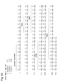
- Table 1 shows a number of amino acid changes in the framework regions of the anti-SDF-1 antibodies 1D3, 1H2, 1C6 and 2A5 that differ from the heavy chain parent germline sequence.
- somatic mutations can be "backmutated" to the germline sesquence by, for example, site-directed mutagenesis or PCR-mediated mutagenesis.
- Table 2 shows a number of amino acid changes in the framework regions of the anti-SDF-1 antibodies 1D3, 1H2, 1C6 and 2A5 that differ from the light chain parent germline sequence.
- the somatic mutations can be "backmutated" to the germline sesquence by, for example, site-directed mutagenesis or PCR-mediated mutagenesis.
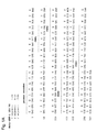
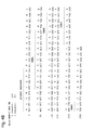
- V H regions for 1D3 and 1H2, against the parent genmline V H 1-24 (SEQ ID NO:41) sequence is shown in Figure 6 .
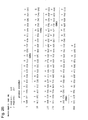
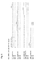
- the alignment of V H regions for 1C6 and 2A5 against the parent germline V H 3-7 sequence (SEQ ID NO:42) is shown in Figure 7 .
- the alignment of V K regions for 1D3 and 1H2, against the parent germline V K L18 (SEQ ID NO:43) sequence is shown in Figure 7 .
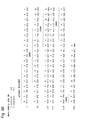
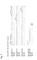
- the alignment of V K regions for 1 C6 and 2A5 against the parent germline V K L18 sequence is shown in Figure 8 .
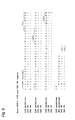
- Anti-SDF-1 Ab Amino acid position Amino acid of antibody Original amino acid of germline configuration 1D3 1 E Q 84 T S 1H2 29 F L 117 M T 1C6 9 R G 2A5 1 Q E 6 Q E Table 2. Modifications to antibodies 1D3, 1H2, 1C6 and 2A5 from the light chain germline configuration. Anti-SDF-1 Ab Amino acid position Amino acid of antibody Original amino acid of germline configuration 1D3 1 E A 3 V Q 1H2 1 E A 3 V Q 1C6 3 R Q 4 M L 11 V L 39 T K 2A5 1 D A 4 M L 5 I T
- Another type of framework modification involves mutating one or more residues within the framework region, or even within one or more CDR regions, to remove T cell epitopes to thereby reduce the potential immunogenicity of the antibody. This approach is also referred to as "deimmunization" and is described in further detail in U.S. Patent Publication No. 20030153043 by Carr et al .
- antibodies of the invention may be engineered to include modifications within the Fc region, typically to alter one or more functional properties of the antibody, such as serum half-life, complement fixation, Fc receptor binding, and/or antigen-dependent cellular cytotoxicity.
- an antibody of the invention may be chemically modified (e.g ., one or more chemical moieties can be attached to the antibody) or be modified to alter its glycosylation, again to alter one or more functional properties of the antibody.
- the hinge region of CH1 is modified such that the number of cysteine residues in the hinge region is altered, e.g ., increased or decreased.
- This approach is described further in U.S. Patent No. 5,677,425 by Bodmer et al .
- the number of cysteine residues in the hinge region of CH1 is altered to, for example, facilitate assembly of the light and heavy chains or to increase or decrease the stability of the antibody.
- the Fc hinge region of an antibody is mutated to decrease the biological half life of the antibody. More specifically, one or more amino acid mutations are introduced into the CH2-CH3 domain interface region of the Fc-hinge fragment such that the antibody has impaired Staphylococcyl protein A (SpA) binding relative to native Fc-hinge domain SpA binding.
- SpA Staphylococcyl protein A
- the antibody is modified to increase its biological half life.
- Various approaches are possible. For example, one or more of the following mutations can be introduced: T252L, T254S, T256F, as described in U.S. Patent No. 6,277,375 to Ward .
- the antibody can be altered within the CH1 or C L region to contain a salvage receptor binding epitope taken from two loops of a CH2 domain of an Fc region of an IgG, as described in U.S. Patent Nos. 5,869,046 and 6,121,022 by Presta et al .
- the Fc region is altered by replacing at least one amino acid residue with a different amino acid residue to alter the effector function(s) of the antibody.
- one or more amino acids selected from amino acid residues 234, 235, 236, 237, 297, 318, 320 and 322 can be replaced with a different amino acid residue such that the antibody has an altered affinity for an effector ligand but retains the antigen-binding ability of the parent antibody.
- the effector ligand to which affinity is altered can be, for example, an Fc receptor or the C1 component of complement. This approach is described in further detail in U.S. Patent Nos. 5,624,821 and 5,648,260, both by Winter et al .
- one or more amino acids selected from amino acid residues 329, 331 and 322 can be replaced with a different amino acid residue such that the antibody has altered C1q binding and/or reduced or abolished complement dependent cytotoxicity (CDC).
- CDC complement dependent cytotoxicity
- one or more amino acid residues within amino acid positions 231 and 239 are altered to thereby alter the ability of the antibody to fix complement. This approach is described further in PCT Publication WO 94/29351 by Bodmer et al .
- the Fc region is modified to increase the ability of the antibody to mediate antibody dependent cellular cytotoxicity (ADCC) and/or to increase the affinity of the antibody for an Fcy receptor by modifying one or more amino acids at the following positions: 238, 239, 248, 249, 252, 254, 255, 256, 258, 265, 267, 268, 269, 270, 272, 276, 278, 280, 283, 285, 286, 289, 290, 292, 293, 294, 295, 296, 298, 301, 303, 305, 307, 309, 312, 315, 320, 322, 324, 326, 327, 329, 330, 331, 333, 334, 335, 337, 338, 340, 360, 373, 376, 378, 382, 388, 389, 398, 414, 416, 419, 430, 434, 435, 437, 438 or 439.
- ADCC antibody dependent cellular cytotoxicity
- the glycosylation of an antibody is modified.
- an aglycoslated antibody can be made (i.e ., the antibody lacks glycosylation).
- Glycosylation can be altered to, for example, increase the affinity of the antibody for antigen.
- carbohydrate modifications can be accomplished by, for example, altering one or more sites of glycosylation within the antibody sequence.
- one or more amino acid substitutions can be made that result in elimination of one or more variable region framework glycosylation sites to thereby eliminate glycosylation at that site.
- Such aglycosylation may increase the affinity of the antibody for antigen.
- Such an approach is described in further detail in U.S. Patent Nos. 5,714,350 and 6,350,861 by Co et al .
- an antibody can be made that has an altered type of glycosylation, such as a hypofucosylated antibody having reduced amounts of fucosyl residues or an antibody having increased bisecting GlcNac structures.
- altered glycosylation patterns have been demonstrated to increase the ADCC ability of antibodies.
- carbohydrate modifications can be accomplished by, for example, expressing the antibody in a host cell with altered glycosylation machinery. Cells with altered glycosylation machinery have been described in the art and can be used as host cells in which to express recombinant antibodies of the invention to thereby produce an antibody with altered glycosylation.
- the cell lines Ms704, Ms705, and Ms709 lack the fucosyltransferase gene, FUT8 (alpha (1,6) fucosyltransferase), such that antibodies expressed in the Ms704, Ms705, and Ms709 cell lines lack fucose on their carbohydrates.
- the Ms704, Ms705, and Ms709 FUT8 -/- cell lines were created by the targeted disruption of the FUT8 gene in CHO/DG44 cells using two replacement vectors (see U.S. Patent Publication No. 20040110704 by Yamane et al . and Yamane-Ohnuki et al. (2004) Biotechnol Bioeng 87:614-22 ).
- EP 1,176,195 by Hanai et al . describes a cell line with a functionally disrupted FUT8 gene, which encodes a fucosyl transferase, such that antibodies expressed in such a cell line exhibit hypofucosylation by reducing or eliminating the alpha 1,6 bond-related enzyme.
- Hanai et al. also describe cell lines which have a low enzyme activity for adding fucose to the N-acetylglucosamine that binds to the Fc region of the antibody or does not have the enzyme activity, for example the rat myeloma cell line YB2/0 (ATCC CRL 1662).
- PCT Publication WO 03/035835 by Presta describes a variant CHO cell line, Lec13 cells, with reduced ability to attach fucose to Asn(297)-linked carbohydrates, also resulting in hypofucosylation of antibodies expressed in that host cell (see also Shields, R.L. et al. (2002) J. Biol. Chem. 277:26733-26740 ).
- PCT Publication WO 99/54342 by Umana et al .
- glycoprotein-modifying glycosyl transferases e.g ., beta(1,4)-N-acetylglucosaminyltransferase III (GnTIII)
- GnTIII glycoprotein-modifying glycosyl transferases
- the fucose residues of the antibody may be cleaved off using a fucosidase enzyme.
- the fucosidase alpha-L-fucosidase removes fucosyl residues from antibodies ( Tarentino, A.L. et al. (1975) Biochem. 14:5516-23 ).
- An antibody can be pegylated to, for example, increase the biological (e.g., serum) half life of the antibody.
- the antibody, or fragment thereof typically is reacted with polyethylene glycol (PEG), such as a reactive ester or aldehyde derivative of PEG, under conditions in which one or more PEG groups become attached to the antibody or antibody fragment.
- PEG polyethylene glycol
- the pegylation is carried out via an acylation reaction or an alkylation reaction with a reactive PEG molecule (or an analogous reactive water-soluble polymer).
- polyethylene glycol is intended to encompass any of the forms of PEG that have been used to derivatize other proteins, such as mono (C1-C10) alkoxy- or aryloxy-polyethylene glycol or polyethylene glycol-maleimide.
- the antibody to be pegylated is an aglycosylated antibody. Methods for pegylating proteins are known in the art and can be applied to the antibodies of the invention. See for example, EP 0 154 316 by Nishimura et al , and EP 0 401 384 by Ishikawa et al .
- the antibodies of the present disclosure may be further characterized by the various physical properties of the anti-SDF-1 antibodies.
- Various assays may be used to detect and/or differentiate different classes of antibodies based on these physical properties.
- antibodies of the present invention may contain one or more glycosylation sites in either the light or heavy chain variable region.
- the presence of one or more glycosylation sites in the variable region may result in increased immunogenicity of the antibody or an alteration of the pK of the antibody due to altered antigen binding ( Marshall et al (1972) Annu Rev Biochem 41:673-702 ; Gala FA and Morrison SL (2004) J Immunol 172:5489-94 ; Wallick et al (1988) J Exp Med 168:1099-109 ; Spiro RG (2002) Glycobiology 12:43R-56R ; Parekh et al (1985) Nature 316:452-7 ; Mimura et al.
- variable region glycosylation may be tested using Dionex light chromatography (Dionex-LC), which cleaves saccharides from a Fab into monosaccharides and analyzes the individual saccharide content
- Dionex-LC Dionex light chromatography
- the antibodies of the present invention -do not contain asparagine isomerism sites.
- a deamidation or isoaspartic acid effect may occur on N-G or D-G sequences, respectively.
- the deamidation or isoaspartic acid effect results in the creation of isoaspartic acid which decreases the stability of an antibody by creating a kinked structure off a side chain carboxy terminus rather than the main chain.
- the creation of isoaspartic acid can be measured using an iso-quant assay, which uses a reverse-phase HPLC to test for isoaspartic acid.
- Each antibody will have a unique isoelectric point (pI), but generally antibodies will fall in the pH range of between 6 and 9.5.
- the pI for an IgG1 antibody typically falls within the pH range of 7-9.5 and the pI for an IgG4 antibody typically falls within the pH range of 6-8.
- Antibodies may have a pI that is outside this range. Although the effects are generally unknown, there is speculation that antibodies with a pI outside the normal range may have some unfolding and instability under in vivo conditions.
- the isoelectric point may be tested using a capillary isoelectric focusing assay, which creates a pH gradient and may utilize laser focusing for increased accuracy ( Janini et al (2002) Electrophoresis 23:1605-11 ; Ma et al.
- an anti-SDF-1 antibody that contains a pI value that falls in the normal range. This can be achieved either by selecting antibodies with a pI in the normal range, or by mutating charged surface residues using standard techniques well known in the art.
- Each antibody will have a melting temperature that is indicative of thermal stability ( Krishnamurthy R and Manning MC (2002) Curr Pharm Biotechnol 3:361-71 ).
- a higher thermal stability indicates greater overall antibody stability in vivo.
- the melting point of an antibody may be measure using techniques such as differential scanning calorimetry ( Chen et al (2003) Pharm Res 20:1952-60 ; Ghirlando et al (1999) Immunol Lett 68:47-52 ).
- T M1 indicates the temperature of the initial unfolding of the antibody.
- T M2 indicates the temperature of complete unfolding of the antibody.
- the T M1 of an antibody of the present invention is greater than 60°C, preferably greater than 65°C, even more preferably greater than 70°C.
- the thermal stability of an antibody may be measure using circular dichroism ( Murray et al. (2002) J. Chromatogr Sci 40:343-9 ).
- antibodies are selected that do not rapidly degrade. Fragmentation of an anti-SDF-1 antibody may be measured using capillary electrophoresis (CE) and MALDI-MS, as is well understood in the art ( Alexander AJ and Hughes DE (1995) Anal Chem 67:3626-32 ).
- CE capillary electrophoresis
- MALDI-MS MALDI-MS
- antibodies are selected that have minimal aggregation effects. Aggregation may lead to triggering of an unwanted immune response and/or altered or unfavorable pharmacokinetic properties. Generally, antibodies are acceptable with aggregation of 25% or less, preferably 20% or less, even more preferably 15% or less, even more preferably 10% or less and even more preferably 5% or less. Aggregation may be measured by several techniques well known in the art, including size-exclusion column (SEC) high performance liquid chromatography (HPLC), and light scattering to identify monomers, dimers, trimers or multimers.
- SEC size-exclusion column
- HPLC high performance liquid chromatography
- the anti-SDF-1 antibodies having V H and V K sequences disclosed herein can be used to create new anti-SDF-1 antibodies by modifying the V H and/or V K sequences, or the constant region(s) attached thereto.
- the structural features of an anti-SDF-1 antibody of the disclosure e.g . 1D3, 1H2, 1C6 or 2A5, are used to create structurally related anti-SDF-1 antibodies that retain at least one functional property of the antibodies of the disclosure, such as binding to human SDF-1.
- one or more CDR regions of 1D3, 1H2, 1C6 or 2A5, or mutations thereof can be combined recombinantly with known framework regions and/or other CDRs to create additional, recombinantly-engineered, anti-SDF-1 antibodies of the invention, as discussed above.
- the starting material for the engineering method is one or more of the V H and/or V K sequences provided herein, or one or more CDR regions thereof.
- To create the engineered antibody it is not necessary to actually prepare ( i.e ., express as a protein) an antibody having one or more of the V H and/or V K sequences provided herein, or one or more CDR regions thereof. Rather, the information contained in the sequence(s) is used as the starting material to create a "second generation" sequence(s) derived from the original sequence(s) and then the "second generation" sequence(s) is prepared and expressed as a protein.
- the disclosure provides a method for preparing an anti-SDF-1 antibody comprising:
- Standard molecular biology techniques can be used to prepare and express the altered antibody sequence.
- the antibody encoded by the altered antibody sequence(s) is one that retains one, some or all of the functional properties of the anti-SDF-1 antibodies described herein, which functional properties include, but are not limited to:
- the functional properties of the altered antibodies can be assessed using standard assays available in the art and/or described herein, such as those set forth in the Examples (e.g ., flow cytometry, binding assays).
- mutations can be introduced randomly or selectively along all or part of an anti-SDF-1 antibody coding sequence and the resulting modified anti-SDF-1 antibodies can be screened for binding activity and/or other functional properties as described herein.
- Mutational methods have been described in the art.
- PCT Publication WO 02/092780 by Short describes methods for creating and screening antibody mutations using saturation mutagenesis, synthetic ligation assembly, or a combination thereof.
- PCT Publication WO 03/074679 by Lazar et al describes methods of using computational screening methods to optimize physiochemical properties of antibodies.
- nucleic acid molecules that encode the antibodies of the invention.
- the nucleic acids may be present in whole cells, in a cell lysate, or in a partially purified or substantially pure form.
- a nucleic acid is "isolated” or “rendered substantially pure” when purified away from other cellular components or other contaminants, e.g ., other cellular nucleic acids or proteins, by standard techniques, including alkaline/SDS treatment, CsCl banding, column chromatography, agarose gel electrophoresis and others well known in the art. See, F. Ausubel, et al., ed. (1987) Current Protocols in Molecular Biology, Greene Publishing and Wiley Interscience, New York .
- a nucleic acid of the invention can be, for example, DNA or RNA and may or may not contain intronic sequences.
- the nucleic acid is a cDNA molecule.
- Nucleic acids of the invention can be obtained using standard molecular biology techniques.
- hybridomas e.g ., hybridomas prepared from transgenic mice carrying human immunoglobulin genes as described further below
- cDNAs encoding the light and heavy chains of the antibody made by the hybridoma can be obtained by standard PCR amplification or cDNA cloning techniques.
- nucleic acid encoding the antibody can be recovered from the library.
- Preferred nucleic acids molecules of the invention are those encoding the V H and V L sequences of the 1D3, 1H2, 1C6 or 2A5 monoclonal antibodies.
- DNA sequences encoding the V H sequences of 1D3, 1H2, 1C6 and 2A5 are shown in SEQ ID NOs:33, 34, 35 and 36, respectively.
- DNA sequences encoding the V L sequences of 1D3, 1H2, 1C6 and 2A5 are shown in SEQ ID NOs:37, 38, 39 and 40, respectively.
- nucleic acids of the disclosure are nucleic acids having at least 80% sequence identity, such as at least 85%, at least 90%, at least 95%, at least 98% or at least 99% sequence identity, with one of the sequences shown in SEQ ID NOs:33, 34, 35, 36, 37, 38, 39 or 40, which nucleic acids encode an antibody of the invention , or an antigen-binding portion thereof.
- the percent identity between two nucleic acid sequences is the number of positions in the sequence in which the nucleotide is identical, taking into account the number of gaps and the length of each gap, which need to be introduced for optimal alignment of the two sequences.
- the comparison of sequences and determination of percent identity between two sequences can be accomplished using a mathematical algorithm, such as the algorithm of Meyers and Miller or the XBLAST program of Altschul described above.
- nucleic acids of the disclosure comprise one or more CDR-encoding portion of the nucleic acid sequences shown in SEQ ID NOs:33, 34, 35, 36, 37, 38, 39 and 40.
- the nucleic acid may encode the heavy chain CDR1, CDR2 and/or CDR3 sequence of 1D3, 1H2, 1C6 or 2A5 or the light chain CDR1, CDR2 and/or CDR3 sequence of 1D3, 1H2, 1C6 or 2A5.
- Nucleic acids which have at least 80%, such as at least 85%, at least 90%, at least 95%, at least 98% or at least 99% sequence identity, with such a CDR-encoding portion of SEQ ID NOs:33, 34, 35, 36, 37, 38, 39 or 40 are also preferred nucleic acids of the disclosure. Such nucleic acids may differ from the corresponding portion of SEQ ID NOs:33, 34, 35, 36, 37, 38, 39 or 40 in a non-CDR coding region and/or in a CDR-coding region.
- the nucleic acid CDR region encoded by the nucleic acid typically comprises one or more conservative sequence modification as defined herein compared to the corresponding CDR sequence of 1D3, 1H2, 1C6 or 2A5.
- V H and V L segments are obtained, these DNA fragments can be further manipulated by standard recombinant DNA techniques, for example to convert the variable region genes to full-length antibody chain genes, to Fab fragment genes or to a scFv gene.
- a V L - or V H -encoding DNA fragment is operatively linked to another DNA fragment encoding another protein, such as an antibody constant region or a flexible linker.
- the term "operatively linked,” as used in this context, is intended to mean that the two DNA fragments are joined such that the amino acid sequences encoded by the two DNA fragments remain in-frame.
- the isolated DNA encoding the V H region can be converted to a full-length heavy chain gene by operatively linking the VH-encoding DNA to another DNA molecule encoding heavy chain constant regions (CH1, CH2 and CH3).
- heavy chain constant regions CH1, CH2 and CH3
- the sequences of human heavy chain constant region genes are known in the art (see e.g ., Kabat, E. A., el al. (1991) Sequences of Proteins of Immunological Interest, Fifth Edition, U.S. Department of Health and Human Services, NIH Publication No. 91-3242 ) and DNA fragments encompassing these regions can be obtained by standard PCR amplification.
- the heavy chain constant region can be an IgGI, IgG2, IgG3, IgG4, IgA, IgE, IgM or IgD constant region, but most preferably is an IgG1 or IgG4 constant region.
- the V H -encoding DNA can be operatively linked to another DNA molecule encoding only the heavy chain CH1 constant region.
- the isolated DNA encoding the V L region can be converted to a full-length light chain gene (as well as a Fab light chain gene) by operatively linking the V L -encoding DNA to another DNA molecule encoding the light chain constant region, CL.
- the sequences of human light chain constant region genes are known in the art (see e.g ., Kabat, E. A., et al. (1991) Sequences of Proteins of Immunological Interest, Fifth Edition, U.S. Department of Health and Human Services, NIH Publication No. 91-3242 ) and DNA fragments encompassing these regions can be obtained by standard PCR amplification.
- the light chain constant region can be a kappa or lambda constant region.
- the V H - and V L -encoding DNA fragments are operatively linked to another fragment encoding a flexible linker, e.g ., encoding the amino acid sequence (Gly 4 -Ser) 3 (SEQ ID NO: 51), such that the V H and V L sequences can be expressed as a contiguous single-chain protein, with the V L and V H regions joined by the flexible linker (see e.g ., Bird et al. (1988) Science 242:423-426 ; Huston et al. (1988) Proc. Natl. Acad Sci. USA 85:5879-5883 ; McCafferty et al., (1990) Nature 348:552-554 ).
- a flexible linker e.g ., encoding the amino acid sequence (Gly 4 -Ser) 3 (SEQ ID NO: 51)
- Monoclonal antibodies (mAbs) of the present invention can be produced by a variety of techniques, including conventional monoclonal antibody methodology e.g ., the standard somatic cell hybridization technique of Kohler and Milstein (1975) Nature 256: 495 . Although somatic cell hybridization procedures are preferred, in principle, other techniques for producing monoclonal antibody can be employed e.g ., viral or oncogenic transformation of B lymphocytes.
- hybridomas The preferred animal system for preparing hybridomas is the murine system.
- Hybridoma production in the mouse is a very well-established procedure. Immunization protocols and techniques for isolation of immunized splenocytes for fusion are known in the art. Fusion partners (e.g ., murine myeloma cells) and fusion procedures are also known.
- Chimeric or humanized antibodies of the present invention can be prepared based on the sequence of a non-human monoclonal antibody prepared as described above.
- DNA encoding the heavy and light chain immunoglobulins can be obtained from the non-human hybridoma of interest and engineered to contain non-murine (e.g ., human) immunoglobulin sequences using standard molecular biology techniques.
- murine variable regions can be linked to human constant regions using methods known in the art (see e.g ., U.S. Patent No. 4,816,567 to Cabilly et al .).
- murine CDR regions can be inserted into a human framework using methods known in the art (see e.g., U.S. Patent No. 5,225,539 to Winter , and U.S. Patent Nos. 5,530,101 ; 5,585,089 ; 5,693,762 and 6,180,370 to Queen et al .).
- the antibodies of the invention are human monoclonal antibodies.
- Such human monoclonal antibodies directed against SDF-1 can be generated using transgenic or transchromosomic mice carrying parts of the human immune system rather than the mouse system.
- transgenic and transchromosomic mice include mice referred to herein as the HuMAb Mouse ® and KM Mouse ® , respectively, and are collectively referred to herein as "human Ig mice.”
- the HuMAb Mouse ® (Medarex ® , Inc.) contains human immunoglobulin gene miniloci that encode unrearranged human heavy ( ⁇ and ⁇ ) and ⁇ light chain immunoglobulin sequences, together with targeted mutations that inactivate the endogenous and ⁇ chain loci (see e.g ., Lonberg, et al. (1994) Nature 368(6474): 856-859 ). Accordingly, the mice exhibit reduced expression of mouse IgM or ⁇ , and in response to immunization, the introduced human heavy and light chain transgenes undergo class switching and somatic mutation to generate high affinity human IgG ⁇ monoclonal antibodies (Lonberg, N. et al. (1994), supra ; reviewed in Lonberg, N.
- human antibodies of the invention can be raised using a mouse that carries human immunoglobulin sequences on transgenes and transchromosomes, such as a mouse that carries a human heavy chain transgene and a human light chain transchromosome.
- This mouse is referred to herein as a "KM mouse ® ,” are described in detail in PCT Publication WO 02/43478 to Ishida et al .
- transgenic animal systems expressing human immunoglobulin genes are available in the art and can be used to raise anti-SDF-1 antibodies of the invention.
- an alternative transgenic system referred to as the Xenomouse can be used; such mice are described in, for example, U.S. Patent Nos. 5,939,598 ; 6,075,181 ; 6,114,598 ; 6,150,584 and 6,162,963 to Kucherlapati et al .
- mice carrying both a human heavy chain transchromosome and a human light chain transchromosome referred to as "TC mice” can be used; such mice are described in Tomizuka et al. (2000) Proc. Natl. Acad Sci. USA 97:722-727 .
- cows carrying human heavy and light chain transchromosomes have been described in the art ( Kuroiwa et al. (2002) Nature Biotechnology 20:889-894 ) and can be used to raise anti-SDF-1 antibodies of the invention.
- Human monoclonal antibodies of the invention can also be prepared using phage display methods for screening libraries of human immunoglobulin genes. Such phage display methods for isolating human antibodies are established in the art. See for example: U.S. Patent Nos. 5,223,409 ; 5,403,484 ; and 5,571,698 to Ladner et al . ; U.S. Patent Nos. 5,427,908 and 5,580,717 to Dower et al . ; U.S. Patent Nos. 5,969,108 and 6,172,197 to McCafferty et al . ; and U.S. Patent Nos. 5,885,793 ; 6;521,404 ; 6,544,731 ; 6,555,313 ; 6,582,915 and 6,593,081 to Griffiths et al .
- Human monoclonal antibodies of the invention can also be prepared using SCID mice into which human immune cells have been reconstituted such that a human antibody response can be generated upon immunization.
- SCID mice into which human immune cells have been reconstituted such that a human antibody response can be generated upon immunization.
- Such mice are described in, for example, U.S. Patent Nos. 5,476,996 and 5,698,767 to Wilson et al .
- mice When human Ig mice are used to raise human antibodies of the invention, such mice can be immunized with a purified or enriched preparation of SDF-1 antigen and/or recombinant SDF-1, or cells expressing SDF-1, or an SDF-1 fusion protein, as described by Lonberg, N. et al. (1994) Nature 368(6474): 856-859 ; Fishwild, D. et al. (1996) Nature Biotechnology 14: 845-851 ; and PCT Publication WO 98/24884 and WO 01/14424 .
- the mice will be 6-16 weeks of age upon the first infusion.
- a purified or recombinant preparation (5-50 ⁇ g) of SDF-1 antigen can be used to immunize the human Ig mice intraperitoneally.
- Example 1 Detailed procedures to generate fully human monoclonal antibodies to SDF-1 are described in Example 1 below. Cumulative experience with various antigens has shown that the transgenic mice respond when initially immunized intraperitoneally (IP) with antigen in complete Freund's adjuvant, followed by every other week IP immunizations (up to a total of 6) with antigen in incomplete Freund's adjuvant. However, adjuvants other than Freund's are also found to be effective. In addition, whole cells in the absence of adjuvant are found to be highly immunogenic. The immune response can be monitored over the course of the immunization protocol with plasma samples being obtained by retroorbital bleeds.
- mice with sufficient titers of anti-SDF-1 human immunoglobulin can be used for fusions.
- Mice can be boosted intravenously with antigen 3 days before sacrifice and removal of the spleen. It is expected that 2-3 fusions for each immunization may need to be performed. Between 6 and 24 mice are typically immunized for each antigen.
- HCo7 and HCo12 strains are used.
- both HCo7 and HCo12 transgene can be bred together into a single mouse having two different human heavy chain transgenes (HCo7/HCo12).
- the KM Mouse ® strain can be used, as described in Example 1.
- splenocytes and/or lymph node cells from immunized mice can be isolated and fused to an appropriate immortalized cell line, such as a mouse myeloma cell line.
- an appropriate immortalized cell line such as a mouse myeloma cell line.
- the resulting hybridomas can be screened for the production of antigen-specific antibodies.
- single cell suspensions of splenic lymphocytes from immunized mice can be fused to one-sixth the number of P3X63-Ag8.653 nonsecreting mouse myeloma cells (ATCC, CRL 1580) with 50% PEG.
- the single cell suspension of splenic lymphocytes from immunized mice can be fused using an electric field based electrofusion method, using a CytoPulse large chamber cell fusion electroporator (CytoPulse Sciences, Inc., Glen Burnie Maryland).
- Cells are plated at approximately 2 x 10 5 in flat bottom microliter plate, followed by a two week incubation in selective medium containing 20% fetal Clone Serum, 18% "653" conditioned media, 5% origen (IGEN), 4 mM L-glutamine, 1 mM sodium pyruvate, 5mM HEPES, 0.055 mM 2-mercaptoethanol, 50 units/ml penicillin, 50 mg/ml streptomycin, 50 mg/ml gentamycin and 1X HAT (Sigma; the HAT is added 24 hours after the fusion). After approximately two weeks, cells can be cultured in medium in which the HAT is replaced with HT.
- selective medium containing 20% fetal Clone Serum, 18% "653" conditioned media, 5% origen (IGEN), 4 mM L-glutamine, 1 mM sodium pyruvate, 5mM HEPES, 0.055 mM 2-mercaptoethanol, 50 units/ml penicillin, 50 mg
- selected hybridomas can be grown in two-liter spinner-flasks for monoclonal antibody purification.
- Supernatants can be filtered and concentrated before affinity chromatography with protein A-sepharose (Pharmacia, Piscataway, N.J.).
- Eluted IgG can be checked by gel electrophoresis and high performance liquid chromatography to ensure purity.
- the buffer solution can be exchanged into PBS, and the concentration can be determined by OD280 using 1.43 extinction coefficient.
- the monoclonal antibodies can be aliquoted and stored at -80° C.
- Antibodies of the invention also can be produced in a host cell transfectoma using, for example, a combination of recombinant DNA techniques and gene transfection methods as is well known in the art (e.g ., Morrison, S. (1985) Science 229:1202 ).
- DNAs encoding partial or full-length light and heavy chains can be obtained by standard molecular biology techniques (e.g ., PCR amplification or cDNA cloning using a hybridoma that expresses the antibody of interest) and the DNAs can be inserted into expression vectors such that the genes are operatively linked to transcriptional and translational control sequences.
- operatively linked is intended to mean that an antibody gene is ligated into a vector such that transcriptional and translational control sequences within the vector serve their intended function of regulating the transcription and translation of the antibody gene.
- the expression vector and expression control sequences are chosen to be compatible with the expression host cell used.
- the antibody light chain gene and the antibody heavy chain gene can be inserted into separate vector or, more typically, both genes are inserted into the same expression vector.
- the antibody genes are inserted into the expression vector by standard methods (e.g ., ligation of complementary restriction sites on the antibody gene fragment and vector, or blunt end ligation if no restriction sites are present).
- the light and heavy chain variable regions of the antibodies described herein can be used to create full-length antibody genes of any antibody isotype by inserting them into expression vectors already encoding heavy chain constant and light chain constant regions of the desired isotype such that the V H segment is operatively linked to the C H segment(s) within the vector and the V K segment is operatively linked to the C L segment within the vector.
- the recombinant expression vector can encode a signal peptide that facilitates secretion of the antibody chain from a host cell.
- the antibody chain gene can be cloned into the vector such that the signal peptide is linked in-frame to the amino terminus of the antibody chain gene.
- the signal peptide can be an immunoglobulin signal peptide or a heterologous signal peptide ( i.e., a signal peptide from a non-immunoglobulin protein).
- the recombinant expression vectors of the invention carry regulatory sequences that control the expression of the antibody chain genes in a host cell
- the term "regulatory sequence” is intended to include promoters, enhancers and other expression control elements (e.g ., polyadenylation signals) that control the transcription or translation of the antibody chain genes.
- Such regulatory sequences are described, for example, in Goeddel (Gene Expression Technology. Methods in Enzymology 185, Academic Press, San Diego, CA (1990 )). It will be appreciated by those skilled in the art that the design of the expression vector, including the selection of regulatory sequences, may depend on such factors as the choice of the host cell to be transformed, the level of expression of protein desired, etc.
- Preferred regulatory sequences for mammalian host cell expression include viral elements that direct high levels of protein expression in mammalian cells, such as promoters and/or enhancers derived from cytomegalovirus (CMV), Simian Virus 40 (SV40), adenovirus, ( e.g ., the adenovirus major late promoter (AdMLP) and polyoma.
- CMV cytomegalovirus
- SV40 Simian Virus 40
- AdMLP adenovirus major late promoter
- nonviral regulatory sequences may be used, such as the ubiquitin promoter or ⁇ -globin promoter.
- regulatory elements composed of sequences from different sources such as the SR ⁇ promoter system, which contains sequences from the SV40 early promoter and the long terminal repeat of human T cell leukemia virus type 1 ( Takebe, Y. et al. (1988) Mol. Cell. Biol. 8:466-472 ).
- the recombinant expression vectors of the invention may carry additional sequences, such as sequences that regulate replication of the vector in host cells (e.g ., origins of replication) and selectable marker genes.
- the selectable marker gene facilitates selection of host cells into which the vector has been introduced (see, e.g ., U.S. Pat. Nos. 4,399,216 , 4,634,665 and 5,179,017, all by Axel et al .).
- the selectable marker gene confers resistance to drugs, such as G418, hygromycin or methotrexate, on a host cell into which the vector has been introduced.
- Preferred selectable marker genes include the dihydrofolate reductase (DHFR) gene (for use in dhfr- host cells with methotrexate selection/amplification) and the neo gene (for G418 selection).
- DHFR dihydrofolate reductase
- the expression vector(s) encoding the heavy and light chains is transfected into a host cell by standard techniques.
- the various forms of the term "transfection" are intended to encompass a wide variety of techniques commonly used for the introduction of exogenous DNA into a prokaryotic or eukaryotic host cell, e.g ., electroporation, calcium-phosphate precipitation, DEAE-dextran transfection and the like.
- Preferred mammalian host cells for expressing the recombinant antibodies of the invention include Chinese Hamster Ovary (CHO cells) (including dhfr - CHO cells, described in Urlaub and Chasin, (1980) Proc. Natl. Acad Sci. USA 77:4216-4220 , used with a DHFR selectable marker, e.g ., as described in R. J. Kaufman and P. A. Sharp (1982) J. Mol. Biol. 159:601-621 ), NSO myeloma cells, COS cells and SP2 cells.
- Chinese Hamster Ovary CHO cells
- dhfr - CHO cells described in Urlaub and Chasin, (1980) Proc. Natl. Acad Sci. USA 77:4216-4220 , used with a DHFR selectable marker, e.g ., as described in R. J. Kaufman and P. A. Sharp (1982) J. Mol. Biol. 159:601-621
- another preferred expression system is the GS gene expression system disclosed in WO 87/04462 (to Wilson ), WO 89/01036 (to Bebbington ) and EP 338,841 (to Bebbington ).
- the antibodies are produced by culturing the host cells for a period of time sufficient to allow for expression of the antibody in the host cells or, more preferably, secretion of the antibody into the culture medium in which the host cells are grown.
- Antibodies can be recovered from the culture medium using standard protein purification methods.
- Antibodies of the invention can be tested for binding to SDF-1 by, for example, standard ELISA. Briefly, microtiter plates are coated with purified SDF-1 at 0.25 ⁇ g/ml in PBS, and then blocked with 5% bovine serum albumin in PBS. Dilutions of antibody (e.g ., dilutions of plasma from SDF-1-immunized mice) are added to each well and incubated for 1-2 hours at 37°C.
- mice which develop the highest titers will be used for fusions.
- An ELISA assay as described above can also be used to screen for hybridomas that show positive reactivity with SDF-1 immunogen.
- Hybridomas that bind with high avidity to SDF-1 are subcloned and further characterized.
- One clone from each hybridoma, which retains the reactivity of the parent cells (by ELISA) can be chosen for making a 5-10 vial cell bank stored at -140°C, and for antibody purification.
- selected hybridomas can be grown in two-liter spinner-flasks for monoclonal antibody purification.
- Supernatants can be filtered and concentrated before affinity chromatography with protein A-sepharose (Pharmacia, Piscataway, NJ).
- Eluted IgG can be checked by gel electrophoresis and high performance liquid chromatography to ensure purity.
- the buffer solution can be exchanged into PBS, and the concentration can be determined by OD280 using 1.43 extinction coefficient.
- the monoclonal antibodies can be aliquoted and stored at -80°C.
- each antibody can be biotinylated using commercially available reagents (Pierce, Rockford, IL). Competition studies using unlabeled monoclonal antibodies and biotinylated monoclonal antibodies can be performed using SDF-1 coated-ELISA plates as described above. Biotinylated mAb binding can be detected with a strep-avidin-alkaline phosphatase probe.
- isotype ELISAs can be performed using reagents specific for antibodies of a particular isotype. For example, to determine the isotype of a human monoclonal antibody, wells of microtiter plates can be coated with 1 ⁇ g/ml of anti-human immunoglobulin overnight at 4°C. After blocking with 1% BSA, the plates are reacted with 1 ⁇ g /ml or less of test monoclonal antibodies or purified isotype controls, at ambient temperature for one to two hours. The wells can then be reacted with either human IgG1 or human IgM-specific alkaline phosphatase-conjugated probes. Plates are developed and analyzed as described above.
- Anti-SDF-1 human IgGs can be further tested for reactivity with SDF-1 antigen by Western blotting. Briefly, SDF-1 can be prepared and subjected to sodium dodecyl sulfate polyacrylamide gel electrophoresis. After electrophoresis, the separated antigens are transferred to nitrocellulose membranes, blocked with 10% fetal calf serum, and probed with the monoclonal antibodies to be tested. Human IgG binding can be detected using anti-human IgG alkaline phosphatase and developed with BCIP/NBT substrate tablets (Sigma Chem. Co., St. Louis, Mo.).
- the binding specificity of an antibody of the invention may also be determined by monitoring binding of the antibody to cells expressing SDF-1, for example by flow cytometry.
- a cell line such as a CHO cell line
- the transfected protein may comprise a tag, such as a myc-tag, preferably at the N-terminus, for detection using an antibody to the tag.
- Binding of an antibody of the invention to SDF-1 may be determined by incubating the transfected cells with the antibody, and detecting bound antibody. Binding of an antibody to the tag on the transfected protein may be used as a positive control.
- the specificity of an antibody of the invention for SDF-1 may be further studied by determining whether or not the antibody binds to other proteins using the same methods by which binding to SDF-1 is determined.
- the present disclosure features an anti-SDF-1 antibody, or a fragment thereof, conjugated to a therapeutic moiety, such as a cytotoxin, a drug (e.g ., an immunosuppressant) or a radiotoxin.
- a therapeutic moiety such as a cytotoxin, a drug (e.g ., an immunosuppressant) or a radiotoxin.
- Examples include taxol, cytochalasin B, gramicidin D, ethidium bromide, emetine, mitomycin, etoposide, tenoposide, vincristine, vinblastine, colchicin, doxorubicin, daunorubicin, dihydroxy anthracin dione, mitoxantrone, mithramycin, actinomycin D, 1-dehydrotestosterone, glucocorticoids, procaine, tetracaine, lidocaine, propranolol, and puromycin and analogs or homologs thereof.
- Therapeutic agents also include, for example, antimetabolites (e.g ., methotrexate, 6-mercaptopurine, 6-thioguanine, cytarabine, 5-fluorouracil decarbazine), alkylating agents (e.g ., mechlorethamine, thioepa chlorambucil, melphalan, carmustine (BSNU) and lomustine (CCNU), cyclothosphamide, busulfan, dibromomannitol, streptozotocin, mitomycin C, and cis-dichlorodiamine platinum (II) (DDP) cisplatin), anthracyclines (e.g ., daunorubicin (formerly daunomycin) and doxorubicin), antibiotics ( e.g ., dactinomycin (formerly actinomycin), bleomycin, mithramycin, and anthramycin (AMC)), and anti-mitotic agents
- An example of a calicheamicin antibody conjugate is commercially available (Mylotarg®; American Home Products).
- Cytotoxins can be conjugated to antibodies of the invention using linker technology available in the art.
- linker types that have been used to conjugate a cytotoxin to an antibody include, but are not limited to, hydrazones, thioethers, esters, disulfides and peptide-containing linkers.
- a linker can be chosen that is, for example, susceptible to cleavage by low pH within the lysosomal compartment or susceptible to cleavage by proteases, such as proteases preferentially expressed in tumor tissue such as cathepsins ( e.g ., cathepsins B, C, D).
- Antibodies of the present invention also can be conjugated to a radioactive isotope to generate cytotoxic radiopharmaceuticals, also referred to as radioimmunoconjugates.
- radioactive isotopes that can be conjugated to antibodies for use diagnostically or therapeutically include, but are not limited to, iodine 131 , indium 111 , yttrium 90 and lutetium 177 .
- Method for preparing radioimmunconjugates are established in the art. Examples of radioimmunoconjugates are commercially available, including Zevalin® (IDEC Pharmaceuticals) and Bexxar® (Corixa Pharmaceuticals), and similar methods can be used to prepare radioimmunoconjugates using the antibodies of the invention.
- the drug moiety can be used to modify a given biological response, and the drug moiety is not to be construed as limited to classical chemical therapeutic agents.
- the drug moiety may be a protein or polypeptide possessing a desired biological activity.
- proteins may include, for example, an enzymatically active toxin, or active fragment thereof; such as abrin, ricin A, pseudomonas exotoxin, or diphtheria toxin; a protein such as tumor necrosis factor or interferon- ⁇ ; or, biological response modifiers such as, for example, lymphokines, interleukin-1 ("IL-1"), interleukin-2 (“IL-2”), interleukin-6 (“IL-6”), granulocyte macrophage colony stimulating factor (“GM-CSF”), granulocyte colony stimulating factor (“G-CSF”), or other growth factors.
- IL-1 interleukin-1
- IL-2 interleukin-2
- IL-6 interleukin-6
- GM-CSF gran
- the present disclosure features bispecific molecules comprising an anti-SDF-1 antibody, or a fragment thereof, of the invention.
- An antibody of the invention, or antigen-binding portions thereof can be derivatized or linked to another functional molecule, e.g ., another peptide or protein (e.g ., another antibody or ligand for a receptor) to generate a bispecific molecule that binds to at least two different binding sites or target molecules.
- the antibody of the invention may in fact be derivatized or linked to more than one other functional molecule to generate multispecific molecules that bind to more than two different binding sites and/or target molecules; such multispecific molecules are also intended to be encompassed by the term "bispecific molecule" as used herein.
- an antibody of the invention can be functionally linked ( e.g ., by chemical coupling, genetic fusion, noncovalent association or otherwise) to one or more other binding molecules, such as another antibody, antibody fragment, peptide or binding mimetic, such that a bispecific molecule results.
- the present disclosure includes bispecific molecules comprising at least one first binding specificity for SDF-1 and a second binding specificity for a second target epitope.
- the second target epitope is an Fc receptor, e.g ., human Fc ⁇ RI (CD64) or a human Fca receptor (CD89). Therefore, the disclosure includes bispecific molecules capable of binding both to Fc ⁇ R or Fc ⁇ R expressing effector cells (e.g ., monocytes, macrophages or polymorphonuclear cells (PMNs)), and to target cells expressing SDF-1.
- bispecific molecules target SDF-1 expressing cells to effector cell and trigger Fc receptor-mediated effector cell activities, such as phagocytosis of an SDF-1 expressing cells, antibody dependent cell-mediated cytotoxicity (ADCC), cytokine release, or generation of superoxide anion.
- ADCC antibody dependent cell-mediated cytotoxicity
- the molecule can further include a third binding specificity, in addition to an anti-Fc binding specificity and an anti-SDF-1 binding specificity.
- the third binding specificity is an anti-enhancement factor (EF) portion, e.g ., a molecule which binds to a surface protein involved in cytotoxic activity and thereby increases the immune response against the target cell.
- EF anti-enhancement factor
- the "anti-enhancement factor portion” can be an antibody, functional antibody fragment or a ligand that binds to a given molecule, e.g ., an antigen or a receptor, and thereby results in an enhancement of the effect of the binding determinants for the Fc receptor or target cell antigen.
- the "anti-enhancement factor portion” can bind an Fc receptor or a target cell antigen.
- the anti-enhancement factor portion can bind to an entity that is different from the entity to which the first and second binding specificities bind.
- the anti-enhancement factor portion can bind a cytotoxic T-cell (e.g . via CD2, CD3, CD8, CD28, CD4, CD40, ICAM-1 or other immune cell that results in an increased immune response against the target cell).
- the bispecific molecules of the disclosure comprise as a binding specificity at least one antibody, or an antibody fragment thereof, including, e.g ., an Fab, Fab', F(ab') 2 , Fv, Fd, dAb or a single chain Fv.
- the antibody may also be a light chain or heavy chain dimer, or any minimal fragment thereof such as a Fv or a single chain construct as described in U.S. Patent No. 4,946,778 to Ladner et al .
- the binding specificity for an Fc ⁇ receptor is provided by a monoclonal antibody, the binding of which is not blocked by human immunoglobulin G (IgG).
- IgG receptor refers to any of the eight ⁇ -chain genes located on chromosome 1. These genes encode a total of twelve transmembrane or soluble receptor isoforms which are grouped into three Fc ⁇ receptor classes: Fc ⁇ RI (CD64), Fc ⁇ RII(CD32), and Fc ⁇ RIII (CD16).
- the Fc ⁇ receptor a human high affinity Fc ⁇ RI.
- the human Fc ⁇ RI is a 72 kDa molecule, which shows high affinity for monomeric IgG (10 8 - 10 9 M -1 ).
- the anti-Fc ⁇ receptor antibody is a humanized form of monoclonal antibody 22 (H22).
- H22 monoclonal antibody 22
- the production and characterization of the H22 antibody is described in Graziano, R.F. et al. (1995) J. Immunol 155 (10): 4996-5002 and PCT Publication WO 94/10332 to Tempest et al..
- the H22 antibody producing cell line was deposited at the American Type Culture Collection under the designation HA022CL1 and has the accession no. CRL 11177.
- the binding specificity for an Fc receptor is provided by an antibody that binds to a human IgA receptor, e.g ., an Fc-alpha receptor (Fc ⁇ RI (CD89)), the binding of which is preferably not blocked by human immunoglobulin A (IgA).
- IgA receptor is intended to include the gene product of one ⁇ -gene (Fc ⁇ RI) located on chromosome 19. This gene is known to encode several alternatively spliced transmembrane isoforms of 55 to 110 kDa.
- Fc ⁇ RI (CD89) is constitutively expressed on monocytes/macrophages, eosinophilic and neutrophilic granulocytes, but not on non-effector cell populations.
- Fc ⁇ RI has medium affinity ( ⁇ 5 ⁇ 10 7 M -1 ) for both IgA1 and IgA2, which is increased upon exposure to cytokines such as G-CSF or GM-CSF ( Morton, H.C. et al. (1996) Critical Reviews in Immunology 16:423-440 ).
- cytokines such as G-CSF or GM-CSF
- Fc ⁇ RI and Fc ⁇ RI are preferred trigger receptors for use in the bispecific molecules of the disclosure because they are (1) expressed primarily on immune effector cells, e.g ., monocytes, PMNs, macrophages and dendritic cells; (2) expressed at high levels ( e.g ., 5,000-100,000 per cell); (3) mediators of cytotoxic activities (e.g ., ADCC, phagocytosis); and (4) mediate enhanced antigen presentation of antigens, including self-antigens, targeted to them.
- immune effector cells e.g ., monocytes, PMNs, macrophages and dendritic cells
- mediators of cytotoxic activities e.g ., ADCC, phagocytosis
- human monoclonal antibodies are preferred, other antibodies which can be employed in the bispecific molecules of the disclosure are murine, chimeric and humanized monoclonal antibodies.
- the bispecific molecules of the present disclosure can be prepared by conjugating the constituent binding specificities, e.g ., the anti-FcR and anti-SDF-1 binding specificities, using methods known in the art. For example, each binding specificity of the bispecific molecule can be generated separately and then conjugated to one another. When the binding specificities are proteins or peptides, a variety of coupling or cross-linking agents can be used for covalent conjugation.
- cross-linking agents examples include protein A, carbodiimide, N-succinimidyl-S-acetyl-thioacetate (SATA), 5,5'-dithiobis(2-nitrobenzoic acid) (DTNB), o-phenylenedimaleimide (oPDM), N-succinimidyl-3-(2-pyridyldithio)propionate (SPDP), and sulfosuccinimidyl 4-(N-maleimidomethyl) cyclohaxane-1-carboxylate (sulfo-SMCC) (see e.g ., Karpovsky et al. (1984) J. Exp. Med.
- the binding specificities are antibodies, they can be conjugated via sulfhydryl bonding of the C-terminus hinge regions of the two heavy chains.
- the hinge region is modified to contain an odd number of sulfhydryl residues, preferably one, prior to conjugation.
- both binding specificities can be encoded in the same vector and expressed and assembled in the same host cell.
- This method is particularly useful where the bispecific molecule is a mAb x mAb, mAb x Fab, Fab x F(ab') 2 or ligand x Fab fusion protein.
- a bispecific molecule of the invention can be a single chain molecule comprising one single chain antibody and a binding determinant, or a single chain bispecific molecule comprising two binding determinants. Bispecific molecules may comprise at least two single chain molecules. Methods for preparing bispecific molecules are described for example in U.S.
- Binding of the bispecific molecules to their specific targets can be confirmed by, for example, enzyme-linked immunosorbent assay (ELISA), radioimmunoassay (RIA), FACS analysis, bioassay (e.g ., growth inhibition), or Western Blot assay.
- ELISA enzyme-linked immunosorbent assay
- RIA radioimmunoassay
- FACS analysis bioassay (e.g ., growth inhibition)
- bioassay e.g ., growth inhibition
- Western Blot assay Western Blot assay.
- Each of these assays generally detects the presence of protein-antibody complexes of particular interest by employing a labeled reagent (e.g ., an antibody) specific for the complex of interest.
- the FcR-antibody complexes can be detected using e.g ., an enzyme-linked antibody or antibody fragment which recognizes and specifically binds to the antibody-FcR complexes.
- the complexes can be detected using any of a variety of other immunoassays.
- the antibody can be radioactively labeled and used in a radioimmunoassay (RIA) (see, for example, Weintraub, B., Principles of Radioimmunoassays, Seventh Training Course on Radioligand Assay Techniques, The Endocrine Society, March, 1986 .
- RIA radioimmunoassay
- the radioactive isotope can be detected by such means as the use of a counter or a scintillation counter or by autoradiography.
- the present invention provides a composition, e.g. , a pharmaceutical composition, containing one or a combination of monoclonal antibodies, or antigen-binding portion(s) thereof, of the present invention, formulated together with a pharmaceutically acceptable carrier.
- a pharmaceutical composition of the invention can comprise a combination of antibodies (or immunoconjugates or bispecifics) that bind to different epitopes on the target antigen or that have complementary activities.
- compositions of the invention also can be administered in combination therapy, i.e ., combined with other agents.
- the combination therapy can include an anti-SDF-1 antibody of the present invention combined with at least one other anti-inflammatory or immunosuppressant agent. Examples of therapeutic agents that can be used in combination therapy are described in greater detail below in the section on uses of the antibodies of the invention .
- pharmaceutically acceptable carrier includes any and all solvents, dispersion media, coatings, antibacterial and antifungal agents, isotonic and absorption delaying agents, and the like that are physiologically compatible.
- the carrier is suitable for intravenous, intramuscular, subcutaneous, parenteral, spinal or epidermal administration ( e.g ., by injection or infusion).
- the active compound i.e ., antibody, immunoconjugate, or bispecific molecule
- the active compound i.e ., antibody, immunoconjugate, or bispecific molecule, may be coated in a material to protect the compound from the action of acids and other natural conditions that may inactivate the compound.
- the pharmaceutical compounds of the invention may include one or more pharmaceutically acceptable salts.
- a "pharmaceutically acceptable salt” refers to a salt that retains the desired biological activity of the parent compound and does not impart any undesired toxicological effects (see e.g ., Berge, S.M., et al. (1977) J. Pharm. Sci. 66:1-19 ). Examples of such salts include acid addition salts and base addition salts.
- Acid addition salts include those derived from nontoxic inorganic acids, such as hydrochloric, nitric, phosphoric, sulfuric, hydrobromic, hydroiodic, phosphorous and the like, as well as from nontoxic organic acids such as aliphatic mono- and dicarboxylic acids, phenyl-substituted alkanoic acids, hydroxy alkanoic acids, aromatic acids, aliphatic and aromatic sulfonic acids and the like.
- nontoxic inorganic acids such as hydrochloric, nitric, phosphoric, sulfuric, hydrobromic, hydroiodic, phosphorous and the like
- nontoxic organic acids such as aliphatic mono- and dicarboxylic acids, phenyl-substituted alkanoic acids, hydroxy alkanoic acids, aromatic acids, aliphatic and aromatic sulfonic acids and the like.
- Base addition salts include those derived from alkaline earth metals, such as sodium, potassium, magnesium, calcium and the like, as well as from nontoxic organic amines, such as N,N'-dibenzylethylenediamine, N-methylglucamine, chloroprocaine, choline, diethanolamine, ethylenediamine, procaine and the like.
- a pharmaceutical composition of the invention also may include a pharmaceutically acceptable anti-oxidant.
- pharmaceutically acceptable antioxidants include: (1) water soluble antioxidants, such as ascorbic acid, cysteine hydrochloride, sodium bisulfate, sodium metabisulfite, sodium sulfite and the like; (2) oil-soluble antioxidants, such as ascorbyl palmitate, butylated hydroxyanisole (BHA), butylated hydroxytoluene (BHT), lecithin, propyl gallate, alpha-tocopherol, and the like; and (3) metal chelating agents, such as citric acid, ethylenediamine tetraacetic acid (EDTA), sorbitol, tartaric acid, phosphoric acid, and the like.
- water soluble antioxidants such as ascorbic acid, cysteine hydrochloride, sodium bisulfate, sodium metabisulfite, sodium sulfite and the like
- oil-soluble antioxidants such as ascorbyl palmitate, butylated
- aqueous and nonaqueous carriers examples include water, ethanol, polyols (such as glycerol, propylene glycol, polyethylene glycol, and the like), and suitable mixtures thereof, vegetable oils, such as olive oil, and injectable organic esters, such as ethyl oleate.
- polyols such as glycerol, propylene glycol, polyethylene glycol, and the like
- vegetable oils such as olive oil
- injectable organic esters such as ethyl oleate.
- Proper fluidity can be maintained, for example, by the use of coating materials, such as lecithin, by the maintenance of the required particle size in the case of dispersions, and by the use of surfactants.
- compositions may also contain adjuvants such as preservatives, wetting agents, emulsifying agents and dispersing agents. Prevention of presence of microorganisms may be ensured both by sterilization procedures, supra, and by the inclusion of various antibacterial and antifungal agents, for example, paraben, chlorobutanol, phenol sorbic acid, and the like. It may also be desirable to include isotonic agents, such as sugars, sodium chloride, and the like into the compositions. In addition, prolonged absorption of the injectable pharmaceutical form may be brought about by the inclusion of agents which delay absorption such as aluminum monostearate and gelatin.
- Pharmaceutically acceptable carriers include sterile aqueous solutions or dispersions and sterile powders for the extemporaneous preparation of sterile injectable solutions or dispersion.
- sterile aqueous solutions or dispersions and sterile powders for the extemporaneous preparation of sterile injectable solutions or dispersion.
- the use of such media and agents for pharmaceutically active substances is known in the art. Except insofar as any conventional media or agent is incompatible with the active compound, use thereof in the pharmaceutical compositions of the invention is contemplated. Supplementary active compounds can also be incorporated into the compositions.
- compositions typically must be sterile and stable under the conditions of manufacture and storage.
- the composition can be formulated as a solution, microemulsion, liposome, or other ordered structure suitable to high drug concentration.
- the carrier can be a solvent or dispersion medium containing, for example, water, ethanol, polyol (for example, glycerol, propylene glycol, and liquid polyethylene glycol, and the like), and suitable mixtures thereof.
- the proper fluidity can be maintained, for example, by the use of a coating such as lecithin, by the maintenance of the required particle size in the case of dispersion and by the use of surfactants.
- isotonic agents for example, sugars, polyalcohols such as mannitol, sorbitol, or sodium chloride in the composition.
- Prolonged absorption of the injectable compositions can be brought about by including in the composition an agent that delays absorption, for example, monostearate salts and gelatin.
- Sterile injectable solutions can be prepared by incorporating the active compound in the required amount in an appropriate solvent with one or a combination of ingredients enumerated above, as required, followed by sterilization microfiltration.
- dispersions are prepared by incorporating the active compound into a sterile vehicle that contains a basic dispersion medium and the required other ingredients from those enumerated above.
- the preferred methods of preparation are vacuum drying and freeze-drying (lyophilization) that yield a powder of the active ingredient plus any additional desired ingredient from a previously sterile-filtered solution thereof.
- the amount of active ingredient which can be combined with a carrier material to produce a single dosage form will vary depending upon the subject being treated, and the particular mode of administration.
- the amount of active ingredient which can be combined with a carrier material to produce a single dosage form will generally be that amount of the composition which produces a therapeutic effect. Generally, out of one hundred per cent, this amount will range from about 0.01 per cent to about ninety-nine percent of active ingredient, preferably from about 0.1 per cent to about 70 per cent, most preferably from about 1 per cent to about 30 per cent of active ingredient in combination with a pharmaceutically acceptable carrier.
- Dosage regimens are adjusted to provide the optimum desired response (e.g ., a therapeutic response). For example, a single bolus may be administered, several divided doses may be administered over time or the dose may be proportionally reduced or increased as indicated by the exigencies of the therapeutic situation. It is especially advantageous to formulate parenteral compositions in dosage unit form for ease of administration and uniformity of dosage.
- Dosage unit form as used herein refers to physically discrete units suited as unitary dosages for the subjects to be treated; each unit contains a predetermined quantity of active compound calculated to produce the desired therapeutic effect in association with the required pharmaceutical carrier.
- the specification for the dosage unit forms of the invention are dictated by and directly dependent on (a) the unique characteristics of the active compound and the particular therapeutic effect to be achieved, and (b) the limitations inherent in the art of compounding such an active compound for the treatment of sensitivity in individuals.
- the dosage ranges from about 0.0001 to 100 mg/kg, and more usually 0.01 to 5 mg/kg, of the host body weight.
- dosages can be 0.3 mg/kg body weight, 1 mg/kg body weight, 3 mg/kg body weight, 5 mg/kg body weight or 10 mg/kg body weight or within the range of 1-10 mg/kg.
- An exemplary treatment regime entails administration once per week, once every two weeks, once every three weeks, once every four weeks, once a month, once every 3 months or once every three to 6 months.
- Preferred dosage regimens for an anti-SDF-1 antibody of the invention include 1 mg/kg body weight or 3 mg/kg body weight via intravenous administration, with the antibody being given using one of the following dosing schedules: (i) every four weeks for six dosages, then every three months; (ii) every three weeks; (iii) 3 mg/kg body weight once followed by 1 mg/kg body weight every three weeks.
- two or more monoclonal antibodies with different binding specificities are administered simultaneously, in which case the dosage of each antibody administered falls within the ranges indicated.
- Antibody is usually administered on multiple occasions. Intervals between single dosages can be, for example, weekly, monthly, every three monthgs or yearly. Intervals can also be irregular as indicated by measuring blood levels of antibody to the target antigen in the patient. In some methods, dosage is adjusted to achieve a plasma antibody concentration of about 1-1000 ⁇ g /ml and in some methods about 25-300 ⁇ g /ml.
- antibody can be administered as a sustained release formulation, in which case less frequent administration is required. Dosage and frequency vary depending on the half-life of the antibody in the patient. In general, human antibodies show the longest half life, followed by humanized antibodies, chimeric antibodies, and nonhuman antibodies. The dosage and frequency of administration can vary depending on whether the treatment is prophylactic or therapeutic. In prophylactic applications, a relatively low dosage is administered at relatively infrequent intervals over a long period of time. Some patients continue to receive treatment for the rest of their lives. In therapeutic applications, a relatively high dosage at relatively short intervals is sometimes required until progression of the disease is reduced or terminated, and preferably until the patient shows partial or complete amelioration of symptoms of disease. Thereafter, the patient can be administered a prophylactic regime.
- Actual dosage levels of the active ingredients in the pharmaceutical compositions of the present invention may be varied so as to obtain an amount of the active ingredient which is effective to achieve the desired therapeutic response for a particular patient, composition, and mode of administration, without being toxic to the patient.
- the selected dosage level will depend upon a variety of pharmacokinetic factors including the activity of the particular compositions of the present invention employed, or the ester, salt or amide thereof, the route of administration, the time of administration, the rate of excretion of the particular compound being employed, the duration of the treatment, other drugs, compounds and/or materials used in combination with the particular compositions employed, the age, sex, weight, condition, general health and prior medical history of the patient being treated, and like factors well known in the medical arts.
- a “therapeutically effective dosage" of an anti-SDF-1 antibody of the invention preferably results in a decrease in severity of disease symptoms, an increase in frequency and duration of disease symptom-free periods, or a prevention of impairment or disability due to the disease affliction.
- a "therapeutically effective dosage” preferably inhibits cell growth or tumor growth by at least about 20%, more preferably by at least about 40%, even more preferably by at least about 60%, and still more preferably by at least about 80% relative to untreated subjects.
- the ability of a compound to inhibit tumor growth can be evaluated in an animal model system predictive of efficacy in human tumors.
- this property of a composition can be evaluated by examining the ability of the compound to inhibit cell growth, such inhibition can be measured in vitro by assays known to the skilled practitioner.
- a therapeutically effective amount of a therapeutic compound can decrease tumor size, or otherwise ameliorate symptoms in a subject.
- One of ordinary skill in the art would be able to determine such amounts based on such factors as the subject's size, the severity of the subject's symptoms, and the particular composition or route of administration selected.
- a composition of the present invention can be administered via one or more routes of administration using one or more of a variety of methods known in the art.
- routes and/or mode of administration will vary depending upon the desired results.
- Preferred routes of administration for antibodies of the invention include intravenous, intramuscular, intradermal, intraperitoneal, subcutaneous, spinal or other parenteral routes of administration, for example by injection or infusion.
- parenteral administration means modes of administration other than enteral and topical administration, usually by injection, and includes, without limitation, intravenous, intramuscular, intraarterial, intrathecal, intracapsular, intraorbital, intracardiac, intradermal, intraperitoneal, transtracheal, subcutaneous, subcuticular, intraarticular, subcapsular, subarachnoid, intraspinal, epidural and intrasternal injection and infusion.
- an antibody of the invention can be administered via a non-parenteral route, such as a topical, epidermal or mucosal route of administration, for example, intranasally, orally, vaginally, rectally, sublingually or topically.
- a non-parenteral route such as a topical, epidermal or mucosal route of administration, for example, intranasally, orally, vaginally, rectally, sublingually or topically.
- the active compounds can be prepared with carriers that will protect the compound against rapid release, such as a controlled release formulation, including implants, transdermal patches, and microencapsulated delivery systems.
- a controlled release formulation including implants, transdermal patches, and microencapsulated delivery systems.
- Biodegradable, biocompatible polymers can be used, such as ethylene vinyl acetate, polyanhydrides, polyglycolic acid, collagen, polyorthoesters, and polylactic acid. Many methods for the preparation of such formulations are patented or generally known to those skilled in the art. See, e.g., Sustained and Controlled Release Drug Delivery Systems, J.R. Robinson, ed., Marcel Dekker, Inc., New York, 1978 .
- compositions can be administered with medical devices known in the art.
- a therapeutic composition of the invention can be administered with a needleless hypodermic injection device, such as the devices disclosed in U.S. Patent Nos. 5,399,163 ; 5,383,851 ; 5,312,335 ; 5,064,413 ; 4,941,880 ; 4,790,824 ; or 4,596,556 .
- a needleless hypodermic injection device such as the devices disclosed in U.S. Patent Nos. 5,399,163 ; 5,383,851 ; 5,312,335 ; 5,064,413 ; 4,941,880 ; 4,790,824 ; or 4,596,556 .
- Examples of well-known implants and modules useful in the present invention include: U.S. Patent No. 4,487,603 , which discloses an implantable micro-infusion pump for dispensing medication at a controlled rate; U.S. Patent No.
- U.S. Patent No. 4,447,233 which discloses a medication infusion pump for delivering medication at a precise infusion rate
- U.S. Patent No. 4,447,224 which discloses a variable flow implantable infusion apparatus for continuous drug delivery
- U.S. Patent No. 4,439,196 which discloses an osmotic drug delivery system having multi-chamber compartments
- U.S. Patent No. 4,475,196 which discloses an osmotic drug delivery system.
- Many other such implants, delivery systems, and modules are known to those skilled in the art.
- the human monoclonal antibodies of the invention can be formulated to ensure proper distribution in vivo.
- the blood-brain barrier excludes many highly hydrophilic compounds.
- the therapeutic compounds of the invention cross the BBB (if desired)
- they can be formulated, for example, in liposomes.
- liposomes For methods of manufacturing liposomes, see, e.g ., U.S. Patents 4,522,811 ; 5,374,548 ; and 5,399,331 .
- the liposomes may comprise one or more moieties which are selectively transported into specific cells or organs, thus enhance targeted drug delivery (see, e.g. , V.V. Ranade (1989) J. Clin. Pharmacol. 29:685 ).
- Exemplary targeting moieties include folate or biotin (see, e.g ., U.S. Patent 5,416,016 to Low et al .); mannosides ( Umezawa et al., (1988) Biochem. Biophys. Res. Commun. 153:1038 ); antibodies ( P.G. Bloeman et al. (1995) FEBS Lett. 357:140 ; M. Owais et al. (1995) Antimicrob. Agents Chemother. 39:180 ); surfactant protein A receptor ( Briscoe et al. (1995) Am. J. Physiol. 1233:134 ); p120 ( Schreier et al. (1994) J. Biol. Chem.
- the antibodies, particularly the human antibodies, antibody compositions and methods of the present disclosure have numerous in vitro and in vivo diagnostic and therapeutic utilities involving the diagnosis and treatment of SDF-1 mediated disorders.
- these molecules can be administered to cells in culture, in vitro or ex vivo , or to human subjects, e.g ., in vivo , to treat, prevent and to diagnose a variety of disorders.
- the term "subject" is intended to include human and non-human animals.
- Non-human animals include all vertebrates, e.g ., mammals and non-mammals, such as non-human primates, sheep, dogs, cats, cows, horses, chickens, amphibians, and reptiles.
- Preferred subjects include human patients having disorders mediated by SDF-1 activity.
- the methods are particularly suitable for treating human patients having a disorder associated with aberrant SDF-1 expression.
- the two can be administered in either order or simultaneously.
- the antibodies of the invention can be used to specifically detect SDF-1 expression on the surface of cells and, moreover, can be used to purify SDF-1 via immunoaffinity purification.
- SDF-1 has also been shown to have a role in Intrahepatic cholangiocarcinoma (ICC) ( Ohira et al. (2006) Am J Pathol 168:1155-68 ).
- ICC Intrahepatic cholangiocarcinoma
- inhibition of the SDF-1 interaction with CXCR4 has been found to be involved in stem cell mobilization and may be a useful therapy for various other cancers, for example, multiple myeloma and non-Hodgkin's lymphoma (Fricker et al. (2006) published ahead of print; Fruehauf and Seeger (2005) Future Oncol 1:375-83 ).
- cancers whose growth may be inhibited using the antibodies of the invention include metastatic tumors and cancers typically responsive to immunotherapy.
- preferred cancers for treatment include breast cancer, multiple myeloma and lymphomas (e.g ., Hodgkin's and non-Hodgkin's lymphoma, lymphocytic lymphoma, primary CNS lymphoma, T-cell lymphoma).
- cancers examples include melanoma (e.g ., metastatic malignant melanoma), renal cancer (e.g ., renal cell carcinoma), brain tumors, chronic or acute leukemias including acute myeloid leukemia, chronic myeloid leukemia, acute lymphoblastic leukemia, chronic lymphocytic leukemia, and nasopharangeal carcinomas, prostate cancer, colon cancer, lung cancer, bone cancer, pancreatic cancer, skin cancer, cancer of the head or neck, cutaneous or intraocular malignant melanoma, uterine cancer, ovarian cancer, rectal cancer, cancer of the anal region, stomach cancer, testicular cancer, uterine cancer, carcinoma of the fallopian tubes, carcinoma of the endometrium, carcinoma of the cervix, carcinoma of the vagina, carcinoma of the vulva, cancer of the esophagus, cancer of the small intestine, cancer of the endocrine system, cancer of the thyroid
- the human antibodies and antibody compositions of the present invention can be used to treat a subject with a tumorigenic disorder, e.g ., a disorder characterized by the presence of tumor cells expressing SDF-1 including, for example, breast cancer, multiple myeloma, non-Hodgkin's lymphoma (NHL), renal cell carcinomas (RCC), such as clear cell RCC, glioblastoma, breast cancer, brain tumors, nasopharangeal carcinomas, acute lymphocytic leukemia (ALL), chronic lymphocytic leukemia (CLL), Burkitt's lymphoma, anaplastic large-cell lymphomas (ALCL), cutaneous T-cell lymphomas, nodular small cleaved-cell lymphomas, lymphocytic lymphomas, peripheral T-cell lymphomas, Lennert's lymphomas, immunoblastic lymphomas, T-cell leukemia/lymphomas (ATLL), adult T-cell leukemia (T
- the disclosure provides a method of inhibiting growth of tumor cells in a subject, comprising administering to the subject a therapeutically effective amount of an anti-SDF-1 antibody or antigen-binding portion thereof.
- the antibody is a human anti-SDF-1 antibody (such as any of the human anti-human SDF-1 antibodies described herein). Additionally or alternatively, the antibody may be a chimeric or humanized anti-SDF-1 antibody.
- SDF-1 has also been detected in the hyperplastic lining and into the extracellular matrix and perivascular linking, including blood vessel endothelium, of rheumatoid arthritis (RA) and osteoarthritis synovial (OA) sections ( Pablos et al. (2003) J Immunol 170:2147-52 ).
- Northern blot analysis of OA and RA fibroblast-like synoviocytes detected SDF-1 expression that was not induced by proinflammatory cytokines, angiogenic factors, or hypoxia. Removal of heparan sulfate molecules from endothelial cells eliminated SDF-1 immunostaining of these cells, suggesting that SDF-1 accumulates on the endothelial cell surface.
- SDF-1 has been shown to induce neovascularization of brochial mucosa in asthmatic subjects and that blocking CXCR-4, the receptor for SDF-1 attenuates allergic lung inflammation, allergic airway disease, airway hyperreactivty and hypersensitivity-type pulmonary granuloma formation ( Hoshino et al. (2003) Eur Respir J 21:804-9 ; Lukacs et al. (2002) Am J Pathol 160:1353-60 ; Gonzalo et al. (2000) J Immunol 165:499-508 ; Hu et al. (2006) Am J Pathol 169:424-32 ).
- the human antibodies and antibody compositions of the present invention can be used to treat a subject with an autoimmune disorder, e.g ., a disorder characterized by the presence of SDF-1 including, for example, rheumatoid arthritis (RA), osteoarthritis (OA), experimental autoimmune encephalomyelitis, asthma, allergic inflammation, such as allergic lung inflammation, allergic airway disease, airway hyperreactivity and hypersensitivity-type pulmonary granuloma.
- an autoimmune disorder e.g ., a disorder characterized by the presence of SDF-1 including, for example, rheumatoid arthritis (RA), osteoarthritis (OA), experimental autoimmune encephalomyelitis, asthma, allergic inflammation, such as allergic lung inflammation, allergic airway disease, airway hyperreactivity and hypersensitivity-type pulmonary granuloma.
- autoimmune disorders in which the antibodies of the invention can be used include, but are not limited to systemic lupus erythematosus (SLE), insulin dependent diabetes mellitus (IDDM), inflammatory bowel disease (IBD) (including Crohn's Disease, ulcerative colitis and Celiac disease), multiple sclerosis (MS), psoriasis, autoimmune thyroiditis and glomerulonephritis.
- SLE systemic lupus erythematosus
- IBD inflammatory bowel disease
- MS multiple sclerosis
- psoriasis autoimmune thyroiditis and glomerulonephritis.
- the antibodies of the present invention can be used to treat transplant rejection.
- the invention is for use in a method of treating a subject with an autoimmune disorder comprising administering to the subject a therapeutically effective amount of an anti-SDF-1 antibody or antigen-binding portion thereof.
- the antibody is a human anti-SDF-1 antibody (such as any of the human anti-human SDF-1 antibodies described herein). Additionally or alternatively, the antibody may be a chimeric or humanized anti-SDF-1 antibody.
- the invention is for use in a method of treating a subject with proliferative diabetic retinopathy, cystoid macular edema or age related macular degeneration comprising administering to the subject a therapeutically effective amount of an anti-SDF-1 antibody or antigen-binding portion thereof.
- the antibody is a human anti-SDF-1 antibody (such as any of the human anti-human SDF-1 antibodies described herein). Additionally or alternatively, the antibody may be a chimeric or humanized anti-SDF-1 antibody.
- Circulating pools of CD45+, collagen I+, CXCR4+ fibrocytes have been shown to traffic to areas of fibrosis, including lung fibrosis.
- Treatment with anti-SDF-1 antibodies has been shown to inhibit intrapulmonary recruitment of CD45+, collagen I+, CXCR4+ fibrocytes and attenuate lung fibrosis ( Phillips et al. (2004) J Clin Invest 114:438-46 ).
- the disclosure provides a method of treating a subject with fibrosis, such as lung fibrosis, comprising administering to the subject a therapeutically effective amount of an anti-SDF-1 antibody or antigen-binding portion thereof.
- the antibody is a human anti-SDF-1 antibody (such as any of the human anti-human SDF-1 antibodies described herein). Additionally or alternatively, the antibody may be a chimeric or humanized anti-SDF-1 antibody.
- the disclosure provides a method of treating a subject with ischemia, ischemia-induced angiogenesis or coronary microvessel contraction, comprising administering to the subject a therapeutically effective amount of an anti-SDF-1 antibody or antigen-binding portion thereof.
- the antibody is a human anti-SDF-1 antibody (such as any of the human anti-human SDF-1 antibodies described herein). Additionally or alternatively, the antibody may be a chimeric or humanized anti-SDF-1 antibody.
- the antibodies (e.g ., human monoclonal antibodies) of the invention can be used to detect levels of SDF-1, or levels of cells which contain SDF-1 on their membrane surface, which levels can then be linked to certain disease symptoms.
- the antibodies can be used to inhibit or block SDF-1 function which, in turn, can be linked to the prevention or amelioration of certain disease symptoms, thereby implicating SDF-1 as a mediator of the disease. This can be achieved by contacting a sample and a control sample with the anti-SDF-1 antibody under conditions that allow for the formation of a complex between the antibody and SDF-1. Any complexes formed between the antibody and SDF-1 are detected and compared in the sample and the control.
- the antibodies (e.g ., human antibodies and compositions) of the invention can be initially tested for binding activity associated with therapeutic or diagnostic use in vitro.
- compositions of the invention can be tested using the flow cytometric assays described in the Examples below.
- the antibodies e.g ., human antibodies, and compositions
- the human monoclonal antibodies, the multi specific or bispecific molecules and the immunoconjugates can be used to elicit in vivo or in vitro one or more of the following biological activities: to inhibit the growth of and/or kill a cell expressing SDF-1; to mediate phagocytosis or ADCC of a cell expressing SDF-1 in the presence of human effector cells, or to block SDF-1 ligand binding to SDF-1.
- the antibodies are used in vivo to treat, prevent or diagnose a variety of SDF-1-related diseases.
- SDF-1-related diseases include, among others, breast cancer, rheumatoid arthritis, osteoarthritis, prolific diabetic retinopathy, autoimmune disorders, cancer, non-Hodgkin's lymphoma, acute lymphocytic leukemia (ALL), chronic lymphocytic leukemia (CLL), Burkitt's lymphoma, anaplastic large-cell lymphomas (ALCL), multiple myeloma, cutaneous T-cell lymphomas, nodular small cleaved-cell lymphomas, lymphocytic lymphomas, peripheral T-cell lymphomas, Lennert's lymphomas, immunoblastic lymphomas, T-cell leukemia/lymphomas (ATLL), adult T-cell leukemia (T-ALL), entroblastic/centrocytic (
- Suitable routes of administering the antibody compositions (e.g ., human monoclonal antibodies) of the invention in vivo and in vitro are well known in the art and can be selected by those of ordinary skill.
- the antibody compositions can be administered by injection (e.g ., intravenous or subcutaneous). Suitable dosages of the molecules used will depend on the age and weight of the subject and the concentration and/or formulation of the antibody composition.
- human anti-SDF-1 antibodies of the invention can be co-administered with one or other more therapeutic agents, e.g ., a cytotoxic agent, a radiotoxic agent or an immunosuppressive agent.
- the antibody can be linked to the agent (as an immunocomplex) or can be administered separate from the agent. In the latter case (separate administration), the antibody can be administered before, after or concurrently with the agent or can be co-administered with other known therapies, e.g ., an anti-cancer therapy, e.g ., radiation.
- Such therapeutic agents include, among others, anti-neoplastic agents such as doxorubicin (adriamycin), cisplatin bleomycin sulfate, carmustine, chlorambucil, and cyclophosphamide hydroxyurea which, by themselves, are only effective at levels which are toxic or subtoxic to a patient.
- anti-neoplastic agents such as doxorubicin (adriamycin), cisplatin bleomycin sulfate, carmustine, chlorambucil, and cyclophosphamide hydroxyurea which, by themselves, are only effective at levels which are toxic or subtoxic to a patient.
- Cisplatin is intravenously administered as a 100 mg/kg dose once every four weeks and adriamycin is intravenously administered as a 60-75 mg/ml dose once every 21 days.
- Co-administration of the human anti-SDF-1 antibodies, or antigen binding fragments thereof, of the present invention with chemotherapeutic agents provides two anti-cancer agents which operate via different mechanisms which yield a cytotoxic effect to human tumor cells. Such co-administration can solve problems due to development of resistance to drugs or a change in the antigenicity of the tumor cells which would render them unreactive with the antibody.
- Target-specific effector cells e.g ., effector cells linked to compositions ( e.g ., human antibodies, multispecific and bispecific molecules) of the invention can also be used as therapeutic agents.
- Effector cells for targeting can be human leukocytes such as macrophages, neutrophils or monocytes. Other cells include eosinophils, natural killer cells and other IgG- or IgA-receptor bearing cells. If desired, effector cells can be obtained from the subject to be treated.
- the target-specific effector cells can be administered as a suspension of cells in a physiologically acceptable solution.
- the number of cells administered can be in the order of 10 8 -10 9 but will vary depending on the therapeutic purpose. In general, the amount will be sufficient to obtain localization at the target cell, e.g ., a tumor cell expressing SDF-1, and to effect cell killing by, e.g ., phagocytosis. Routes of administration can also vary.
- Target-specific effector cells can be performed in conjunction with other techniques for removal of targeted cells.
- anti-tumor therapy using the compositions (e.g ., human antibodies, multispecific and bispecific molecules) of the invention and/or effector cells armed with these compositions can be used in conjunction with chemotherapy.
- combination immunotherapy may be used to direct two distinct cytotoxic effector populations toward tumor cell rejection.
- anti-SDF-1 antibodies linked to anti-Fc-gamma RI or anti-CD3 may be used in conjunction with IgG- or IgA-receptor specific binding agents.
- Bispecific and multispecific molecules of the disclosure can also be used to modulate Fc ⁇ R or Fc ⁇ R levels on effector cells, such as by capping and elimination of receptors on the cell surface. Mixtures of anti-Fc receptors can also be used for this purpose.
- compositions (e.g ., human, humanized, and or chimeric antibodies) of the invention which have complement binding sites, such as portions from IgG1, -2, or -3 or IgM which bind complement, can also be used in the presence of complement.
- ex vivo treatment of a population of cells comprising target cells with a binding agent of the disclosure and appropriate effector cells can be supplemented by the addition of complement or serum containing complement. Phagocytosis of target cells coated with a binding agent of the disclosure can be improved by binding of complement proteins.
- target cells coated with the compositions (e.g ., human antibodies) of the invention can also be lysed by complement.
- the compositions of the invention do not activate complement.
- compositions e.g ., human, humanized or chimeric antibodies
- the instant disclosure provides compositions comprising human antibodies, multispecific or bispecific molecules and serum or complement. These compositions can be advantageous when the complement is located in close proximity to the human antibodies, multispecific or bispecific molecules.
- the human antibodies, multispecific or bispecific molecules of the disclosure and the complement or serum can be administered separately.
- kits comprising the antibody compositions of the disclosure (e.g , human antibodies, bispecific or multispecific molecules, or immunoconjugates) and instructions for use.
- the kit can further contain one ore more additional reagents, such as an immunosuppressive reagent, a cytotoxic agent or a radiotoxic agent, or one or more additional human antibodies of the invention (e.g ., a human antibody having a complementary activity which binds to an epitope in the SDF-1 antigen distinct from the first human antibody).
- patients treated with antibody compositions of the invention can be additionally administered (prior to, simultaneously with, or following administration of a human antibody of the invention) with another therapeutic agent, such as a cytotoxic or radiotoxic agent, which enhances or augments the therapeutic effect of the human antibodies.
- another therapeutic agent such as a cytotoxic or radiotoxic agent, which enhances or augments the therapeutic effect of the human antibodies.
- the subject can be additionally treated with an agent that modulates, e.g ., enhances or inhibits, the expression or activity of Fc ⁇ or Fc ⁇ receptors by, for example, treating the subject with a cytokine.
- an agent that modulates e.g ., enhances or inhibits, the expression or activity of Fc ⁇ or Fc ⁇ receptors by, for example, treating the subject with a cytokine.
- Preferred cytokines for administration during treatment with the multispecific molecule include of granulocyte colony-stimulating factor (G-CSF), granulocyte- macrophage colony-stimulating factor (GM-CSF), interferon- ⁇ (IFN- ⁇ ), and tumor necrosis factor (TNF).
- G-CSF granulocyte colony-stimulating factor
- GM-CSF granulocyte- macrophage colony-stimulating factor
- IFN- ⁇ interferon- ⁇
- TNF tumor necrosis factor
- compositions (e.g ., human antibodies) of the invention can also be used to target cells expressing Fc ⁇ R or SDF-1, for example for labeling such cells.
- the binding agent can be linked to a molecule that can be detected.
- the disclosure provides methods for localizing ex vivo or in vitro cells expressing Fc receptors, such as Fc ⁇ R, or SDF-1.
- the detectable label can be, e.g ., a radioisotope, a fluorescent compound, an enzyme, or an enzyme co-factor.
- the disclosure provides methods for detecting the presence of SDF-1 antigen in a sample, or measuring the amount of SDF-1 antigen, comprising contacting the sample, and a control sample, with a human monoclonal antibody, or an antigen binding portion thereof, which specifically binds to SDF-1, under conditions that allow for formation of a complex between the antibody or portion thereof and SDF-1. The formation of a complex is then detected, wherein a difference complex formation between the sample compared to the control sample is indicative the presence of SDF-1 antigen in the sample.
- immunoconjugates of the disclosure can be used to target compounds (e.g ., therapeutic agents, labels, cytotoxins, radiotoxins immunosuppressants, etc.) to cells which have SDF-1 cell surface receptors by linking such compounds to the antibody.
- compounds e.g ., therapeutic agents, labels, cytotoxins, radiotoxins immunosuppressants, etc.
- an anti-SDF-1 antibody can, be conjugated to any of the toxin compounds described in US Patent Nos. 6,281, 354 and 6,548,530 , US patent publication Nos. 20030050331 , 20030064984 , 20030073852 , and 20040087497 , or published in WO 03/022806 .
- the disclosure also provides methods for localizing ex vivo or in vivo cells expressing SDF-1 (e.g ., with a detectable label, such as a radioisotope, a fluorescent compound, an enzyme, or an enzyme co-factor).
- a detectable label such as a radioisotope, a fluorescent compound, an enzyme, or an enzyme co-factor.
- the immunoconjugates can be used to kill cells which have SDF-1 cell surface receptors by targeting cytotoxins or radiotoxins to SDF-1.
- Antibodies were produced using Trans-Phage SM technology, which combines the UltiMAb Human Antibody Development System ® , which generates fully human antibodies in a stable transgenic mouse system, and the Omniclonal ® phage display technology, which generates custom antibody libraries, and allows for selection from a combinatorial library of antibody heavy and light chains.
- Fully human monoclonal antibodies to SDF-1 were prepared using the HCo7 strain of the transgenic HuMAb Mouse ® and the KM strain of transgenic transchromosomic mice, each of which express human antibody genes.
- the endogenous mouse kappa light chain gene has been homozygously disrupted as described in Chen et al. (1993) EMBO J. 12:811-820 and the endogenous mouse heavy chain gene has been homozygously disrupted as described in Example 1 of PCT Publication WO 01/09187 .
- Each of these mouse strains carriers a human kappa light chain transgene, KCo5, as described in Fishwild et al. (1996) Nature Biotechnology 14:845-851 .
- the HCo7 strain carries the HCo7 human heavy chain transgenes as described in U.S. Patent Nos. 5,545,806 ; 5,625,825 ; and 5,545,807 .
- the KM Mouse ® strain contains the SC20 transchromosome as described in PCT Publication WO 02/43478 .
- mice of the HuMAb Mouse ® and KM Mouse ® were immunized with purified recombinant SDF-1 ⁇ (Peptrotech; Rocky Hill, NJ).
- the human isoforms SDF-1 ⁇ , SDF-1 ⁇ and SDF-1 ⁇ may be used interchangeably, since the extracellular domains are identical.
- General immunization schemes for the HuMAb Mouse ® are described in Lonberg, N. et al (1994) Nature 368(6474): 856-859 ; Fishwild, D. et al. (1996) Nature Biotechnology 14: 845-851 and PCT Publication WO 98/24884 .
- the mice were 6-16 weeks of age upon the first infusion of antigen.
- a purified recombinant preparation (5-50 ⁇ g) of SDF-1 ⁇ was used to immunize each HuMab mouse ® and KM mouse ® .
- Transgenic mice were immunized twice with antigen in complete Freund's adjuvant or Ribi adjuvant either intraperitonealy (IP), subcutaneously (Sc) or via footpad (FP), followed by 3-21 days IP, Sc or FP immunization (up to a total of 11 immunizations) with the antigen in incomplete Freund's or Ribi adjuvant.
- IP intraperitonealy
- Sc subcutaneously
- FP footpad
- the immune response was monitored by retroorbital bleeds.
- the serum was screened by ELISA (as described below), and mice with sufficient titers of anti-SDF-1 human immunogolobulin were used for fusions. Mice were boosted intravenously with antigen 3 and 2 days before sacrifice and removal of the spleen.
- the plates were washed with PBS/Tween and then incubated with a goat-anti-human IgG Fc polyclonal antibody conjugated with horseradish peroxidase (HRP) for 1 hour at room temperature. After washing, the plates were developed with ABTS substrate (Sigma, A-1888, 0.22 mg/ml) and analyzed by spectrophotometer at OD 415-495. Mice that developed the highest titers of anti-SDF-1 antibodies were used for antibody generation..
- HRP horseradish peroxidase
- the mouse splenocytes isolated from a HuMab mouse ® and/or a KM mouse ® , are fused with PEG to a mouse myeloma cell line either using PEG based upon standard protocols or electric field based electrofusion using a Cyto Pulse large chamber cell fusion electroporator (Cyto Pulse Sciences, Inc., Glen Burnie, MD).
- the resulting hybridomas are then screened for the production of antigen-specific antibodies.
- Single cell suspensions of splenic lymphocytes from immunized mice are fused to one-fourth the number of SP2/0 nonsecreting mouse myeloma cells (ATCC, CRL 1581) with 50% PEG (Sigma).
- Cells are plated at approximately 1x10 5 /well in flat bottom microtiter plate, followed by about a two week incubation in selective medium containing 10% fetal bovine serum, 10% P388D1 (ATCC, CRL TIB-63) conditioned medium, 3-5% origen (IGEN) in DMEM (Mediatech, CRL 10013, with high glucose, L-glutamine and sodium pyruvate) plus 5 mM HEPES, 0.055 mM 2-mercaptoethanol, 50 mg/ml gentamycin and 1x HAT (Sigma, CRL P-7185). After 1-2 weeks, cells are cultured in medium in which the HAT is replaced with HT.
- selective medium containing 10% fetal bovine serum, 10% P388D1 (ATCC, CRL TIB-63) conditioned medium, 3-5% origen (IGEN) in DMEM (Mediatech, CRL 10013, with high glucose, L-glutamine and sodium pyruvate) plus 5 mM HEPES,
- the cDNA sequences encoding the heavy and light chain variable regions of the 1D3, 1H2, 1C6 and 2A5 monoclonal antibodies were obtained from the 1D3, 1H2, 1C6 and 2A5 hybridomas, respectively, using standard PCR techniques and were sequenced using standard DNA sequencing techniques.
- nucleotide and amino acid sequences of the heavy chain variable region of 1D3 are shown in Figure 1A and in SEQ ID NO:33 and 1, respectively.
- nucleotide and amino acid sequences of the light chain variable region of 1 D3 are shown in Figure 1B and in SEQ ID NO:37 and 5, respectively.
- nucleotide and amino acid sequences of the heavy chain variable region of 1H2 are shown in Figure 2A and in SEQ ID NO:34 and 2, respectively.
- nucleotide and amino acid sequences of the light chain variable region of 1H2 are shown in Figure 2B and in SEQ ID NO:38 and 6, respectively.
- nucleotide and amino acid sequences of the heavy chain variable region of 1C6 are shown in Figure 3A and in SEQ ID NO:35 and 3, respectively.
- nucleotide and amino acid sequences of the light chain variable region of 1C6 are shown in Figure 3B and in SEQ ID NO:39 and 7, respectively.
- nucleotide and amino acid sequences of the heavy chain variable region of 2A5 are shown in Figure 4A and in SEQ ID NO:36 and 4, respectively.
- nucleotide and amino acid sequences of the light chain variable region of 2A5 are shown in Figure 4B and in SEQ ID NO:40 and 8, respectively.
- binding affinity and binding kinetics of anti-SDF-1 antibodies were examined by Biacore analysis and Western immunoblot analysis.
- Anti-SDF-1 antibodies were characterized for affinities and binding kinetics by Biacore analysis (Biacore AB, Uppsala, Sweden). Purified anti-SDF-1 monoclonal antibodies were covalently linked to a CM5 chip (carboxy methyl dextran coated chip) via primary amines, using standard amine coupling chemistry and kit provided by Biacore. Binding was measured by flowing the SDF-1 (Peprotech, Rocky Hill, NJ) in PBS buffer (pH7.4) at concentrations of 40, 30, 20, 10, and 5 nM at a flow rate of 75 ⁇ l/min. The SDF-1 was injected immediately after dilution to maximize the dimer population.
- Anti-SDF-1 antibodies were characterized for binding to native SDF-1 by immunoprecipitation and subsequent Western blot analysis.
- a preparation of 4.5x10 8 of platelets was made and one T75 flask each of 90-95% confluent cells of CHO-SDF, CHO-S and MCF7 cells was prepared.
- the cells were washed twice with cold PBS.
- the cells were lysed in 1.5ml of lysis buffer (Roche Immunoprecipitation Kit, Cat#1719394) and disrupted by sonicatmg on ice using five 1-second bursts.
- the mixture was centrifuged at 12000 x g for 10 minutes and the supernatant was collected.
- the protein concentration was determined using a Micro BCA Protein Assay kit (PIERCE,Cat#23235).
- the cell lysates were precleared by adding 50 ⁇ l protein G agarose to the mixture and incubated at 2-8°C for 3 hours.
- Five ⁇ g of the anti-SDF-1 antibody 2A5 or an isotype control antibody was added to 500 ⁇ g of platelets and one of the CHO-SDF, CHO-S and MCF7 cell lyses and incubated for 1 hour at 2-8°C on a rocking platform.
- the antibody-SDF-1 complex was immunoprecipitated by adding 50 ⁇ l protein G agarose to the mixture and incubated overnight at 2-8°C on a rocking platform.
- the beads were pelleted by centrifugation at 12,000 x g for 1 minute.
- the oligomerization state of SDF-1 was determined by fluorescence anisotropy and fluorescence resonance energy transfer (FRET) experiments. Both sets of experiments were performed on a Spex Fluorolog 3.2 instrument (Spex, Edison, NJ).
- the fluorescence anisotropy experiments utilized fluorescein labeled SDF-1.
- the experiment was performed with excitation at 494 nm and emission monitored at 514 nm and slits set to a band pass of 5 nm.
- a 12.5 ⁇ M solution of SDF-1 was diluted to 50 nM and the anisotropy measured as a function of time.
- These experiments were performed in PBS, PBS and 1mM CaCl 2 .
- Anisotropy which is a function of total mass and shape of the molecular complex decreases in the absence of CaCl 2 , but stays constant with the addition of 1mM CaCl 2 .
- the FRET experiments used dansyl-labeled SDF-1 and fluorescein-labeled SDF-1. These experiments were performed with 335 nm excitation and 520 nm emission and the band pass on the spectrometer was set to 5 nm. In these experiments a 12.5 ⁇ M solution of SDF-1 was diluted to 50 nM and the FRET signal measured as a function of time. These experiments were performed in PBS and PBS + 1mM CaCl 2 . The results are shown in Figure 9.
- Figure 9A shows the effect of 1mM CaCl 2 on the loss of SDF-1 dimers as a function of time after dilution, based on anisotropy measurements.
- Figure 9B shows the effect of 1mM CaCl 2 on the loss of SDF-1 dimers as a function of time after dilution, based on FRET measurements.
- the fluorescence anisotropy and FRET experiments demonstrate the loss of SDF-1 dimerization as a function of time after dilution in PBS buffer but not in PBS buffer supplemented with 1mM CaCl 2 .
- Example 5 Thermostability of anti-SDF-1 monoclonal antibodies by differential scanning calorimetry
- the thermal stability of the anti-SDF-1 monoclonal antibodies was determined using calorimetric analysis of the melting temperature of the antibody.
- T m melting temperatures
- VP-Capillary DSC differential scanning microcalorimeter platform that is combined with an autosampler (MicroCal LLC, Northampton, MA, USA).
- Sample cell volume is 0.144 mL.
- Denaturation data was obtained by heating the samples, at a concentration of 2.0 ⁇ M, from 30 to 95 °C at a rate of 1 °C/min.
- the protein samples were present in phosphate-buffered saline (PBS) at pH 7.4. The same buffer was used in the reference cell to obtain the molar heat capacity by comparison.
- PBS phosphate-buffered saline
- the same buffer was used in the reference cell to obtain the molar heat capacity by comparison.
- the observed thermograms were baseline corrected and normalized data analyzed based on a non-2-step model, using the software Origin v7.0.
- the data is shown in Table 4.
- the anti-SDF-1 monoclonal antibody 2A5 is more stable by differential scanning calorimetry compared to 1C6, 1H2 and 1D3.
- Table 4 Differential scanning calorimetry data for SDF-1 HuMAbs.
- Example 6 Methods for physical stability under chemical denaturation
- the stability of anti-SDF-1 monoclonal antibodies were compared by measuring the midpoint of chemical denaturation by fluorescence spectroscopy.
- Fluorescence measurements of chemical denaturation were performed on a SPEX Fluorolog 3.22 equipped with a Micromax plate reader (SPEX, Edison, NJ). The measurements were performed on antibody samples that had been left for 24 hours to equilibrate in 16 different concentrations of guanidinium hydrochloride in PBS buffer. The measurements were made in black, low volume, non-binding surface 384-well plates (Corning, Acton, MA) and required 1 ⁇ M of antibody in a well volume of 12 ⁇ L. Fluorescence was excited at 280 nm and the emission spectra were measured between 300 and 400 nm. The scan speed was 1 second per nm and slits were set to 5 nm bandpass.
- Example 7 Methods for epitope mapping based on native and synthetic peptides by mass spectrometry and Biacore
- epitope extraction the chemokine is first subjected to enzymatic digestion, then its peptide fragments are mixed with anti-chemokine antibody-bound POROS resin to check for binders; whereas in epitope excision, the digestion is performed in situ while the chemokine is bound to the antibody.
- the epitope containing peptide(s) that was still bound onto the antibody resin was taken together directly for MALDI-MS analysis, or eluted off separately from the antibody and analyzed by ESI-MS.
- chemokine peptides were generated by using various proteases to cover the entire chemokine sequence. With different overlaps, the chemokine peptides in both reduced and native forms were examined for epitope binding. Multiple enzymes, in combination or in series, may be necessary in order to achieve me tightest cut near the epitope region.
- the peptides that have been identified by mass spectrometry as binders to the antibody were identified on the three-dimensional structure of the chemokine, determined by x-ray crystallography and which is available in public domain (www.rcsb.org ). By this procedure, it was possible to eliminate certain stretches of amino acids identified by mass spectrometry as false positives, for example, if they were too distal to the core epitope to be recognized by the antibody. Based on the epitope thus mapped, amino acid sequences for synthetic peptides were designed. To further identify key residues in the epitope (functional epitope) peptides with alanine substituted on key positions were also designed for synthesis. Antibody binding to these synthetic peptides were tested on Biacore and/or mass spectrometer.
- the antibodies were covalently linked to a Biacore CM5 chip.
- Peptides were freshly prepared in 20mM ammonium acetate buffer, pH 7.0, at a concentration of 10ug/mL. These peptides were flowed over the antibody surfaces at a flow rate of 10 ⁇ l/min for 15 minutes. Binding of peptides to isotype controls were used as blank signals. Based on this approach, peptides, with native antigen sequences as well as with alanine substitutions, which bound to the antibodies were identified.
- mAb Z and mAb B Two molecular epitopes were identified from two different populations of huMAbs produced by hybridoma, designated mAb Z and mAb B. Each of these two antibody groups has selective binding to either the monomer or dimer SDF-1 ⁇ .
- the anti-SDF-1 antibodies 1D3 and 1H2 are mAb B group antibodies.
- the anti-SDF-1 antibodies 1C6 and 2A5 are mAb Z group antibodies.
- the mAb Z group recognizes two epitope peptides, one near the N-terminal region amino acid residues 7-19 which is also the known receptor binding site, and the other one on the third beta strand between residues 37-50.
- the mAb B group blocks the heparin binding site, which has been shown to be the SDF-1 ⁇ dimer interface.
- mAb B binds predominately to the dimer interface, between residues 24-30 of the first and the second monomer, where the heparin also binds.
- One critical residue, Arg8, involved in epitope binding with mAb Z was also identified by this mass spectrometric based assay.
- Example 8 Biacore and fluorescence based methods to monitor the variation of SDF-1 oligomeric states and mAb binding when the following conditions are changed
- the blocking of SDF-1 binding to heparin sulphate was determined by capturing biotinylated heparin sulphate (Sigma) on a Streptavidin chip (Biacore, Uppsala, Sweden) then measuring the SDF-1 binding with and without an excess of monoclonal antibodies (1H2 and 2A5). Experiments were carried out in PBS.
- Example 9 Functional Activity Measured by Blockade of 125 I-SDF-1 ⁇ binding to cells
- a comparison of anti-SDF-1 ⁇ antibodies on blocking SDF-1 binding to CEM cells was performed by a standard radio-labeled ligand binding assay.
- the anti-SDF-1 ⁇ antibodies were serially diluted 1:3 to yield a range of concentrations from 40nM to 2pM.
- the antibodies were added to CEM cells in the presence of 100pM 125 I-SDF-1 with a specific activity of 2000Ci/mmole (from Amersham catalog # IM314-25UCI). An irrelevant antibody with the same isotype was used as a negative control.
- the total possible bound radio-labeled ligand was determined by allowing the 125 I-SDF-1 to bind to CEM cells in the absence of antibodies.
- Non-specific binding of the radio-labeled ligand was determined by allowing the 125 I-SDF-1 to bind in the presence of 1 ⁇ M unlabeled SDF-1 (from Peprotech cataolog # 300-28A). The results are shown in Figure 10 .
- CEM cells were labeled with the fluorescent calcium dye, Calcium 3 (Molecular Devices, Sunnyvale, CA).
- the anti-SDF-1 antibodies were titrated in a 1:3 serial dilution series resulting in a concentration range from 100nM to 1pM and mixed with 50nM SDF-1 ⁇ (Peprotech, Rocky Hill, NJ) prior to loading into the Flexstation machine (Molecular Devices).
- As a negative control an irrelevant antibody with the same isotype was used.
- Cells were then stimulated with the SDF-1 ⁇ /antibody mixtures.
- Cells without antibody were stimulated with SDF-1 ⁇ (made up in Hank's buffered saline with 0.1% BSA or HBS) to achieve a maximum possible calcium flux signal.
- CEM cells were labeled with the BATDA reagent from Perkin Elmer.
- the anti-SDF-1 antibodies were titrated in a 1:3 serial dilution series resulting in a concentration range from 100nM to 1pM and mixed with 5nM SDF-1 ⁇ (Peprotech, Rocky Hill, NJ).
- 5nM SDF-1 ⁇ Peprotech, Rocky Hill, NJ.
- Recombinant human SDF-1 ⁇ with or without antibodies was added at 5nM to the lower chamber of a 96 well Neuroprobe migration plate with 5.7mm diameter filters per well. Each well contains 5 ⁇ m pores. Labeled CEM cells were loaded onto the filters at a concentration of 0.5 million cells per well. The migration plate was incubated at 37°C for 2.5 hours.
- Migrated cells are captured in the lower chamber of the plate, lysed and detected with Europium detection solution by Perkin Elmer. The chemi-luminescent signal is recorded on the Fusion instrument. The resulting inhibition of SDF-1 ⁇ -induced migration by the anti- SDF-1 antibodies are shown in Figure 12 .
- Example 12 Treatment of Antibody- induced arthritis using anti-SDF-1 antibodies
- Collagen-induced arthritis in mice is widely used as an experimental screening model for rheumatoid arthritis in humans.
- mice are immunized with type II collagen that triggers the production of antibodies to particular regions of the type II collagen molecule.
- the onset of the disease is gradual and 6 weeks is the minimum length in the classical CIA model.
- 4 autoantigenic epitopes of the type II collagen were identified and a cocktail of 4 monoclonal antibodies generated against these epitopes was used to induce arthritis.
- An LPS injection 3 days after the anti-collagen antibody administration induced a rapid manifestation of the disease. In this antibody induced model, the onset of disease is rapid and the whole study can be performed in 2 weeks.
- This antibody induced model displays features similar to those found in the classical CIA model, such as extensive infiltration of inflammatory cells into the synovium and joining space, pannus formation and marked destruction of bone and cartilage tissues. Hence, this model was used to monitor the efficacy of anti-inflammatory substances in a similar fashion in a much shorter study period than the classical model.
- mice 6-8 week old Balb/c mice were used. On day 0, mice were injected with 4mg of anti-collagen antibody cocktail (Arthrogen) followed by an injection of 25 ⁇ g LPS on day 2. On days 1, 4, 7, and 10 mice were treated with our anti-SDF-1 antibodies, isotype control antibody, PBS, or the corticosteroid, dexamethasone at 15 mg/kg for antibodies and 0.3mg/kg for dexamethasone.
- the anti-SDF-1 antibodies tested in this study were clones 1H2, 2A5 and 1C6. Development of arthritis was scored on a scale of 0-4 by clinical observation (redness and swelling) on days 3, 5, 8, 10, 12 and 15.
- the severity score are 0 for normal, 1 for clear redness or mild swelling of the ankle or wrist, or swelling of one digit, 2 for redness and swelling of the more than one digit, 3 for redness and swelling of the entire paw, metatarsus, hock and/or carpus, 4 for inflamed, swollen or deformed paw, with involvement of multiple joints. Severity scores were assessed for all 4 paws, with a total possible overall score of 16 for each animal. Swelling of all four paws (paw thickness) was measured using a precision caliper on days 3, 5, 8, 10, 12 and 15.
- Figure 13-1 shows mean score (A) and mean paw width (B) for each mouse.
- Figure 13-2 shows mean score (C) and mean paw (thickness) (D) on day 15.
- Figure 13-3 shows mean disease score (E) and mean disease score on day 24 (F).
- Mice treated with 2A5 black solid upright triangle
- the disease severity and paw inflammation of the isotype control group was similar to the positive control, dexamethasone (large boxes)
- the 1H2 group and 1C6 group did not show any benefit with antibody treatment and were comparable to isotype control.
- Example 13 Efficacy of anti-SDF-1 antibodies in the mouse air pouch model
- mice were anesthetized and an air pouch was created by injecting sterile air. On the day of the study, the air pouch was injected with recombinant human SDF-1 alpha in the absence or presence of anti-SDF-1 antibodies. The efficacy of the antibodies was determined. In this example, anti-SDF-1 antibodies 2A5, 1C6, 1D3 and 1H2 were tested.
- mice Female C57BL/6 mice between 8-12 weeks of age (CRL) were anesthetized with isoflorane and an air pouch was created by injecting 5 ml of sterile air subcutaneously in the dorsum of the mice. Three days later, an additional 3 ml of sterile air was injected. On day 7, one ml of PBS, or SDF-1 ⁇ alone (200 ng, PeproTech) or SDF-1 ⁇ plus human IgG4 isotype control, or SDF-1 ⁇ plus 50 ⁇ g or 100 ⁇ g of anti-SDF-1 antibodies was injected in a 1 ml volume. Five mice were used in each test group.
- mice Four hours later, mice were sacrificed by asphyxiation using CO 2 and pouches were washed with 2 ml of PBS/5mM EDTA followed by two additional washes with 3 ml of PBS/5mM EDTA. The exudates were centrifuged at 500 X g for 5 min at room temperature. Cells were counted with a hemacytometer and differential counts of leukocyte subpopulations were performed on cytospin preparation stained with Kwik-Diff staining kit (Thermo Electron Corporation, Waltham, MA).
- the concentration of SDF-1 used to induce migration was reduced to 200 ng and the concentration of antibodies were tested at both 100 ⁇ g/ml and a reduced level at 50 ⁇ g/ml.
- Both 2A5 and 1C6 at 50 ⁇ g and 100 ⁇ g blocked neutrophil, lymphocyte and monocyte migration ( Figure 15 ).
- the numbers of neutrophils, lymphocytes and monocytes were completely reduced to PBS levels.
- the potency was similar between the two antibodies at this concentration. Isotype control at 100 ⁇ g had no effect.
- anti-SDF-1 antibodies, 2A5, 1C6, 1H2 and 1D3 are potent inhibitors of in vivo cell migration.
- HuVEC cells contain glycosaminoglycans (GAGs) to which SDF-1 binds.
- GAGs glycosaminoglycans
- HUVEC human umbilical vein endothelial cells, Cambrex, P/N CC-2519 cells were plated at 1x10 4 cells per well in 200 ⁇ L per well culture medium (Cambrex, P/N CC-3124, with added extra FBS to 10% final) onto 96-well black, clear bottom, opaque plates and incubated at 37°C with CO 2 overnight. Carefully remove cell supernatant. Dilute antigen and antibody at 4x concentration.
- Example 15 Blockade of 125 I-SDF-1 ⁇ binding to GAGs on non-CXCR4 expressing cells.
- a comparison of anti-SDF-1 antibodies on blocking SDF-1 binding to HuVEC cells from ATCC was performed by a standard radio-labeled ligand binding assay.
- the anti-SDF-1 antibodies were serially diluted 1:3 to yield a range of concentrations from 10nM to 0.5pM.
- the antibodies were added to HuVEC cells in the presence of 100pM 125 I-SDF-1 ⁇ with a specific activity of 2000Ci/mmole (from Amersham catalog # IM314-25UCI). An irrelevant antibody with the same isotype was used as a negative control.
- the total possible bound radio-labeled ligand was determined by allowing the 125 I-SDF-1 ⁇ to bind to HuVEC cells in the absence of antibodies.
- Non-specific binding of the radio-labeled ligand was determined by allowing the 125 I-SDF-1 ⁇ to bind in the presence of 1 ⁇ M unlabeled recombinant human SDF-1 ⁇ (from Peprotech cataolog # 300-28A). HuVEC cells do not express CXCR4 and thus there is only non-specific binding of SDF-1 ⁇ to these cells presumably through GAGs. The results are shown in Figure 17 .
- Matrigel is diluted 1:1 with RPMI and plated onto the wells of a 96 wells plate and allowed to polymerize for 30 minutes at 37°C.
- HuVEC from Cambrex cat.# CC-2519
- Antibodies are well mixed with HuVEC at a final concentration of 3 ⁇ g/ml and allowed to incubate at room temperature for 30 minutes.
- An irrelevant antibody of the same isotype or media was used as a negative control.
- a mouse anti-human ⁇ V ⁇ 3 (CD51/CD61) antibody (from R&D Systems cat# MAB3050) was used. HuVEC with or without antibodies were plated onto the matrigel-coated wells and incubated at 37°C for 18 hours.
- the HuVEC incubated with media or isotype control form capillary tubes resulted in the appearance of connected cells across the plate with 3-5 points of connection or branch points per cell.
- the HuVEC incubated with either anti-SDF-1 Abs or Anti- ⁇ V ⁇ 3 antibody did not form capillary tubes.
- the cells appear isolated and with few or no branch points.
- the anti-SDF-1 antibody 1H2 yielded very few branch points where as 2A5 produced some branch points in a small percentage of cells.
Claims (17)
- Isolierter monoklonaler Antikörper, oder ein Antigen-bindender Teil davon, wobei der Antikörper spezifisch an menschliches SDF-1 bindet und umfasst:(I) die CDR1 der variablen Region einer schweren Kette, welche eine Aminosäuresequenz von SEQ ID NO:9 umfasst, die CDR2 der variablen Region einer schweren Kette, welche eine Aminosäuresequenz von SEQ ID NO:13 umfasst, die CDR3 der variablen Region einer schweren Kette, welche eine Aminosäuresequenz von SEQ ID NO:17 umfasst, die CDR1 der variablen Region einer leichten Kette, welche eine Aminosäuresequenz von SEQ ID NO:21 umfasst, die CDR2 der variablen Region einer leichten Kette, welche eine Aminosäuresequenz von SEQ ID NO:25 umfasst und die CDR3 der variablen Region einer leichten Kette, welche eine Aminosäuresequenz von SEQ ID NO:29 umfasst,(II) die CDR1 der variablen Region einer schweren Kette, welche eine Aminosäuresequenz von SEQ ID NO:10 umfasst, die CDR2 der variablen Region einer schweren Kette, welche eine Aminosäuresequenz von SEQ ID NO:14 umfasst, die CDR3 der variablen Region einer schweren Kette, welche eine Aminosäuresequenz von SEQ ID NO:18 umfasst, die CDR1 der variablen Region einer leichten Kette, welche eine Aminosäuresequenz von SEQ ID NO:22 umfasst, die CDR2 der variablen Region einer leichten Kette, welche eine Aminosäuresequenz von SEQ ID NO:26 umfasst und die CDR3 der variablen Region einer leichten Kette, welche eine Aminosäuresequenz von SEQ ID NO:30 umfasst,(III) die CDR1 der variablen Region einer schweren Kette, welche eine Aminosäuresequenz von SEQ ID NO:11 umfasst, die CDR2 der variablen Region einer schweren Kette, welche eine Aminosäuresequenz von SEQ ID NO:15 umfasst, die CDR3 der variablen Region einer schweren Kette, welche eine Aminosäuresequenz von SEQ ID NO:19 umfasst, die CDR1 der variablen Region einer leichten Kette, welche eine Aminosäuresequenz von SEQ ID NO:23 umfasst, die CDR2 der variablen Region einer leichten Kette, welche eine Aminosäuresequenz von SEQ ID NO:27 umfasst und die CDR3 der variablen Region einer leichten Kette, welche eine Aminosäuresequenz von SEQ ID NO:31 umfasst, oder(IV) die CDR1 der variablen Region einer schweren Kette, welche eine Aminosäuresequenz von SEQ ID NO:12 umfasst, die CDR2 der variablen Region einer schweren Kette, welche eine Aminosäuresequenz von SEQ ID NO:16 umfasst, die CDR3 der variablen Region einer schweren Kette, welche eine Aminosäuresequenz von SEQ ID NO:20 umfasst, die CDR1 der variablen Region einer leichten Kette, welche eine Aminosäuresequenz von SEQ ID NO:24 umfasst, die CDR2 der variablen Region einer leichten Kette, welche eine Aminosäuresequenz von SEQ ID NO:28 umfasst und die CDR3 der variablen Region einer leichten Kette, welche eine Aminosäuresequenz von SEQ ID NO:32 umfasst,wobei das menschliche SDF-1 das Polypeptid ist, welches eine Aminosäuresequenz hat, die in SEQ ID NO: 44 dargelegt ist.
- Der isolierte monoklonale Antikörper, oder der Antigen-bindende Teil davon, nach Anspruch 1, welcher umfasst:(a) eine variable Region einer schweren Kette, welche die Aminosäuresequenz von SEQ ID NO:1 umfasst und eine variable Region einer leichten Kette, welche die Aminosäuresequenz von SEQ ID NO:5 umfasst,(b) eine variable Region einer schweren Kette, welche die Aminosäuresequenz von SEQ ID NO:2 umfasst und eine variable Region einer leichten Kette, welche die Aminosäuresequenz von SEQ ID NO:6 umfasst,(c) eine variable Region einer schweren Kette, welche die Aminosäuresequenz von SEQ ID NO:3 umfasst und eine variable Region einer leichten Kette, welche die Aminosäuresequenz von SEQ ID NO:7 umfasst, oder(d) eine variable Region einer schweren Kette, welche die Aminosäuresequenz von SEQ ID NO:4 umfasst und eine variable Region einer leichten Kette, welche die Aminosäuresequenz von SEQ ID NO:8 umfasst.
- Der isolierte monoklonale Antikörper, oder der Antigen-bindende Teil davon, nach Anspruch 2, dieser umfasst eine variable Region einer schweren Kette, welche die Aminosäuresequenz von SEQ ID NO:1 umfasst, und eine variable Region einer leichten Kette, welche die Aminosäuresequenz von SEQ ID NO:5 umfasst.
- Der isolierte monoklonale Antikörper, oder der Antigen-bindende Teil davon, nach Anspruch 2, dieser umfasst eine variable Region einer schweren Kette, welche die Aminosäuresequenz von SEQ ID NO:2 umfasst, und eine variable Region einer leichten Kette, welche die Aminosäuresequenz von SEQ ID NO:6 umfasst.
- Der isolierte monoklonale Antikörper, oder der Antigen-bindende Teil davon, nach Anspruch 2, dieser umfasst eine variable Region einer schweren Kette, welche die Aminosäuresequenz von SEQ ID NO:3 umfasst, und eine variable Region einer leichten Kette, welche die Aminosäuresequenz von SEQ ID NO:7 umfasst.
- Der isolierte monoklonale Antikörper, oder der Antigen-bindende Teil davon, nach Anspruch 2, dieser umfasst eine variable Region einer schweren Kette, welche die Aminosäuresequenz von SEQ ID NO:4 umfasst, und eine variable Region einer leichten Kette, welche die Aminosäuresequenz von SEQ ID NO:8 umfasst.
- Der Antikörper oder der Antigen-bindende Teil davon nach jedwedem der Ansprüche 1-6, welcher ein menschlicher Antikörper ist.
- Der Antikörper oder der Antigen-bindende Teil davon nach jedwedem der Ansprüche 1-7, welcher an das menschliche SDF-1 mit einem KD von 2×10⁻⁹ M oder weniger bindet.
- Der Antikörper oder der Antigen-bindende Teil davon nach Anspruch 3 oder 4, welcher an die Dimer-Interaktionsfläche zwischen den Aminosäureresten 24-30 auf SDF-1 bindet.
- Der Antikörper oder der Antigen-bindende Teil davon nach Anspruch 5 oder 6, welcher sowohl an die Aminosäurereste 7-19 der N-terminalen Region als auch an den dritten Beta-Strang zwischen den Aminosäureresten 37-50 auf SDF-1 bindet.
- Der Antikörper oder der Antigen-bindende Teil davon nach jedwedem der Ansprüche 1-10 zur Verwendung in der Behandlung von Krebs.
- Der Antikörper oder der Antigen-bindende Teil davon für die Verwendung nach Anspruch 11, wobei der Krebs aus der Gruppe, die aus Brustkrebs, multiplem Myelom, Lymphomen, Melanomen, Nierenkrebs, Hirntumoren, chronischen oder akuten Leukämien, Prostatakrebs, Darmkrebs, Lungenkrebs, Knochenkrebs, Bauchspeicheldrüsenkrebs, Hautkrebs, Kopf- und Halskrebs, kutanen oder intraokularen malignen Melanomen, Gebärmutterkrebs, Eierstockkrebs, Rektumkarzinoma, Analkarzinoma, Magenkrebs, Hodenkrebs, Gebärmutterkrebs, Karzinome des Eileiters, Karzinome des Endometriums, Karzinome des Gebärmutterhalses, Karzinome der Vagina, Karzinome der Vulva, Krebs des Ösophagus, Krebs des Dünndarms, Krebs des endokrinen Systems, Krebs der Schilddrüse, Krebs der Nebenschilddrüse, Krebs der Nebenniere, Weichteilsarkomen, Krebs der Harnröhre, Krebs des Penis, soliden Tumoren im Kindesalter, Krebs der Blase, Krebs der Nieren oder des Ureters, Karzinome des Nierenbeckens, Neoplasma des zentralen Nervensystems (ZNS), Tumorangiogenese, Tumoren der Wirbelsäule, Gliome des Hirnstamms, Hypophysenadenome, Kaposi-Sarkom, epidermoider Krebs, Plattenepithelkrebs, umweltinduzierte Krebse, einschließlich jenen die durch Asbest ausgelöst wurden, und Kombinationen der besagten Krebse, besteht ausgewählt wird.
- Der Antikörper oder der Antigen-bindende Teil davon nach jedwedem der Ansprüche 1-10 für die Verwendung in der Behandlung einer Autoimmunerkrankung.
- Der Antikörper oder der Antigen-bindende Teil davon für die Verwendung nach Anspruch 13, wobei die Autoimmunerkrankung aus der Gruppe, die aus rheumatischer Arthritis (RA), Osteoarthritis (OA), experimenteller autoimmuner Encephalomyelitis, systemischem Lupus erythematodes (SLE), insulinabhängigem Diabetes mellitus (IDDM), chronischentzündlicher Darmerkrankung (IBD), multipler Sklerose (MS), Psoriasis, autoimmuner Thyreoiditis, Glomerulonephritis und Transplantatabstoßung besteht, gewählt wird.
- Der Antikörper oder der Antigen-bindende Teil davon nach jedwedem der Ansprüche 1-10 für die Verwendung in der Behandlung von proliferierender diabetischer Retinopathie.
- Eine Zusammensetzung, welche den Antikörper oder den Antigen-bindenden Teil davon nach jedwedem der Ansprüche 1-10 und einen pharmazeutisch annehmbaren Trägerstoff umfasst.
- Ein isoliertes Nukleinsäuremolekül, welches den Antikörper oder den Antigen-bindende Teil davon nach jedwedem der Ansprüche 1-10 kodiert.
Applications Claiming Priority (2)
| Application Number | Priority Date | Filing Date | Title |
|---|---|---|---|
| US83700406P | 2006-08-11 | 2006-08-11 | |
| PCT/JP2007/065991 WO2008018641A1 (en) | 2006-08-11 | 2007-08-10 | Monoclonal antibodies against stromal derived factor-1 (sdf-1) |
Publications (4)
| Publication Number | Publication Date |
|---|---|
| EP2046834A1 EP2046834A1 (de) | 2009-04-15 |
| EP2046834A4 EP2046834A4 (de) | 2010-05-05 |
| EP2046834B1 EP2046834B1 (de) | 2012-10-10 |
| EP2046834B9 true EP2046834B9 (de) | 2013-04-10 |
Family
ID=39033152
Family Applications (1)
| Application Number | Title | Priority Date | Filing Date |
|---|---|---|---|
| EP07792618.6A Not-in-force EP2046834B9 (de) | 2006-08-11 | 2007-08-10 | Monoklonale antikörper gegen stromal derived factor-1 (sdf-1-) |
Country Status (6)
| Country | Link |
|---|---|
| US (1) | US8496931B2 (de) |
| EP (1) | EP2046834B9 (de) |
| JP (1) | JP5244785B2 (de) |
| ES (1) | ES2396220T3 (de) |
| TW (1) | TW200817437A (de) |
| WO (1) | WO2008018641A1 (de) |
Families Citing this family (21)
| Publication number | Priority date | Publication date | Assignee | Title |
|---|---|---|---|---|
| WO2008021263A2 (en) * | 2006-08-11 | 2008-02-21 | Medarex, Inc. | Methods of indentifying antibodies that bind oligomeric forms of chemokines and uses thereof |
| CA2722909A1 (en) | 2008-04-30 | 2009-11-05 | Genomix Co., Ltd. | Method for collecting functional cells in vivo with high efficiency |
| KR101728379B1 (ko) | 2008-04-30 | 2017-04-19 | 가부시키가이샤 제노믹스 | 말초 순환으로의 골수 유래 다능성 줄기세포 동원약 |
| EP2494977B1 (de) | 2009-10-28 | 2018-06-13 | Genomix Co., Ltd. | Geweberegenerationsförderer mit rekrutierung mesenchymaler knochenmarkstammzellen und/oder pluripotenter stammzellen in blut |
| WO2011106234A1 (en) * | 2010-02-25 | 2011-09-01 | Provasculon, Inc. | Protease-resistant mutants of stromal cell derived factor-1 in the repair of tissue damage |
| US8628554B2 (en) | 2010-06-13 | 2014-01-14 | Virender K. Sharma | Intragastric device for treating obesity |
| US10420665B2 (en) | 2010-06-13 | 2019-09-24 | W. L. Gore & Associates, Inc. | Intragastric device for treating obesity |
| US9526648B2 (en) | 2010-06-13 | 2016-12-27 | Synerz Medical, Inc. | Intragastric device for treating obesity |
| US10010439B2 (en) | 2010-06-13 | 2018-07-03 | Synerz Medical, Inc. | Intragastric device for treating obesity |
| SG10201506725QA (en) * | 2010-09-09 | 2015-10-29 | Noxxon Pharma Ag | Sdf-1 binding nucleic acids and the use thereof in cancer treatment |
| JP5843189B2 (ja) * | 2011-03-15 | 2016-01-13 | 国立大学法人名古屋大学 | Sdf−1阻害剤を含有した軟骨組織再生用組成物 |
| EP2703487B1 (de) | 2011-04-26 | 2018-06-27 | Genomix Co., Ltd. | Peptide zur induktion der regenerierung von gewebe und verwendung davon |
| EP2717894B1 (de) | 2011-06-07 | 2018-01-24 | Mesoblast International Sàrl | Verfahren zur reparatur von gewebeschäden mithilfe proteaseresistenter mutanten des stromazellenfaktors 1 |
| US9623078B2 (en) | 2012-10-25 | 2017-04-18 | Genomix Co., Ltd. | Method for treating cardiac infarction using HMGB1 fragment |
| SG11201503215XA (en) | 2012-10-25 | 2015-06-29 | Genomix Co Ltd | Novel method for treating spinal cord injury using hmgb1 fragment |
| CA2920377A1 (en) * | 2013-08-05 | 2015-02-12 | Cambridge Enterprise Limited | Inhibition of cxcr4 signaling in cancer immunotherapy |
| US10647766B2 (en) | 2014-12-17 | 2020-05-12 | Cancer Research Technology Limited | Anti-CXCL12 antibody molecules and their uses |
| JP2018516969A (ja) | 2015-06-12 | 2018-06-28 | ブリストル−マイヤーズ スクイブ カンパニーBristol−Myers Squibb Company | Pd−1およびcxcr4シグナル伝達経路の組合せ遮断による癌の処置 |
| US10779980B2 (en) | 2016-04-27 | 2020-09-22 | Synerz Medical, Inc. | Intragastric device for treating obesity |
| BR112019023138A2 (pt) * | 2017-05-05 | 2020-07-28 | Vaccinex, Inc. | anticorpo 4d anti-semaforina humano |
| WO2019107530A1 (ja) | 2017-12-01 | 2019-06-06 | 株式会社ステムリム | 炎症性腸疾患の治療薬 |
Family Cites Families (41)
| Publication number | Priority date | Publication date | Assignee | Title |
|---|---|---|---|---|
| US4447233A (en) | 1981-04-10 | 1984-05-08 | Parker-Hannifin Corporation | Medication infusion pump |
| US4487603A (en) | 1982-11-26 | 1984-12-11 | Cordis Corporation | Implantable microinfusion pump system |
| US4486194A (en) | 1983-06-08 | 1984-12-04 | James Ferrara | Therapeutic device for administering medicaments through the skin |
| DE3572982D1 (en) | 1984-03-06 | 1989-10-19 | Takeda Chemical Industries Ltd | Chemically modified lymphokine and production thereof |
| US4596556A (en) | 1985-03-25 | 1986-06-24 | Bioject, Inc. | Hypodermic injection apparatus |
| GB8601597D0 (en) | 1986-01-23 | 1986-02-26 | Wilson R H | Nucleotide sequences |
| US5225539A (en) | 1986-03-27 | 1993-07-06 | Medical Research Council | Recombinant altered antibodies and methods of making altered antibodies |
| US4941880A (en) | 1987-06-19 | 1990-07-17 | Bioject, Inc. | Pre-filled ampule and non-invasive hypodermic injection device assembly |
| US4790824A (en) | 1987-06-19 | 1988-12-13 | Bioject, Inc. | Non-invasive hypodermic injection device |
| GB8717430D0 (en) | 1987-07-23 | 1987-08-26 | Celltech Ltd | Recombinant dna product |
| US5677425A (en) | 1987-09-04 | 1997-10-14 | Celltech Therapeutics Limited | Recombinant antibody |
| GB8809129D0 (en) | 1988-04-18 | 1988-05-18 | Celltech Ltd | Recombinant dna methods vectors and host cells |
| US5476996A (en) | 1988-06-14 | 1995-12-19 | Lidak Pharmaceuticals | Human immune system in non-human animal |
| US5223409A (en) | 1988-09-02 | 1993-06-29 | Protein Engineering Corp. | Directed evolution of novel binding proteins |
| ATE135370T1 (de) | 1988-12-22 | 1996-03-15 | Kirin Amgen Inc | Chemisch modifizierte granulocytenkolonie erregender faktor |
| US5530101A (en) | 1988-12-28 | 1996-06-25 | Protein Design Labs, Inc. | Humanized immunoglobulins |
| US5312335A (en) | 1989-11-09 | 1994-05-17 | Bioject Inc. | Needleless hypodermic injection device |
| US5064413A (en) | 1989-11-09 | 1991-11-12 | Bioject, Inc. | Needleless hypodermic injection device |
| JP3068180B2 (ja) | 1990-01-12 | 2000-07-24 | アブジェニックス インコーポレイテッド | 異種抗体の生成 |
| US6150584A (en) | 1990-01-12 | 2000-11-21 | Abgenix, Inc. | Human antibodies derived from immunized xenomice |
| US6075181A (en) | 1990-01-12 | 2000-06-13 | Abgenix, Inc. | Human antibodies derived from immunized xenomice |
| US6673986B1 (en) | 1990-01-12 | 2004-01-06 | Abgenix, Inc. | Generation of xenogeneic antibodies |
| US5427908A (en) | 1990-05-01 | 1995-06-27 | Affymax Technologies N.V. | Recombinant library screening methods |
| GB9015198D0 (en) | 1990-07-10 | 1990-08-29 | Brien Caroline J O | Binding substance |
| US6172197B1 (en) | 1991-07-10 | 2001-01-09 | Medical Research Council | Methods for producing members of specific binding pairs |
| US6300129B1 (en) | 1990-08-29 | 2001-10-09 | Genpharm International | Transgenic non-human animals for producing heterologous antibodies |
| US5885793A (en) | 1991-12-02 | 1999-03-23 | Medical Research Council | Production of anti-self antibodies from antibody segment repertoires and displayed on phage |
| EP0640094A1 (de) | 1992-04-24 | 1995-03-01 | The Board Of Regents, The University Of Texas System | Rekombinantherstellung von immunoglobuun-änliche domänen in prokaryotischen zellen |
| US5383851A (en) | 1992-07-24 | 1995-01-24 | Bioject Inc. | Needleless hypodermic injection device |
| US6121022A (en) | 1995-04-14 | 2000-09-19 | Genentech, Inc. | Altered polypeptides with increased half-life |
| US5869046A (en) | 1995-04-14 | 1999-02-09 | Genentech, Inc. | Altered polypeptides with increased half-life |
| US6277375B1 (en) | 1997-03-03 | 2001-08-21 | Board Of Regents, The University Of Texas System | Immunoglobulin-like domains with increased half-lives |
| GB2339430A (en) | 1997-05-21 | 2000-01-26 | Biovation Ltd | Method for the production of non-immunogenic proteins |
| CA2323525C (en) | 1998-03-30 | 2011-03-01 | Gerald P. Murphy | Therapeutic and diagnostic applications based on the role of the cxcr-4 gene in tumorigenesis |
| CA2589418A1 (en) | 1999-08-24 | 2001-03-01 | Medarex, Inc. | Human ctla-4 antibodies and their uses |
| US7041870B2 (en) | 2000-11-30 | 2006-05-09 | Medarex, Inc. | Transgenic transchromosomal rodents for making human antibodies |
| JP2004532038A (ja) | 2001-05-17 | 2004-10-21 | ディヴァーサ コーポレイション | 新規抗原結合分子の治療、診断、予防、酵素、産業ならびに農業各分野への応用とそのための新規抗原結合分子の作製とスクリーニングの方法 |
| AU2003217912A1 (en) | 2002-03-01 | 2003-09-16 | Xencor | Antibody optimization |
| AU2003224712A1 (en) * | 2002-03-21 | 2003-10-08 | University Of Florida | Modulating angiogenesis |
| US20050002939A1 (en) * | 2002-12-23 | 2005-01-06 | Albert Zlotnik | Tumor killing/tumor regression using CXCR4 antagonists |
| WO2008021263A2 (en) * | 2006-08-11 | 2008-02-21 | Medarex, Inc. | Methods of indentifying antibodies that bind oligomeric forms of chemokines and uses thereof |
-
2007
- 2007-08-10 TW TW096129514A patent/TW200817437A/zh unknown
- 2007-08-10 WO PCT/JP2007/065991 patent/WO2008018641A1/en active Application Filing
- 2007-08-10 US US12/377,015 patent/US8496931B2/en not_active Expired - Fee Related
- 2007-08-10 JP JP2009506826A patent/JP5244785B2/ja not_active Expired - Fee Related
- 2007-08-10 EP EP07792618.6A patent/EP2046834B9/de not_active Not-in-force
- 2007-08-10 ES ES07792618T patent/ES2396220T3/es active Active
Also Published As
| Publication number | Publication date |
|---|---|
| EP2046834B1 (de) | 2012-10-10 |
| US20100158902A1 (en) | 2010-06-24 |
| US8496931B2 (en) | 2013-07-30 |
| TW200817437A (en) | 2008-04-16 |
| EP2046834A1 (de) | 2009-04-15 |
| ES2396220T3 (es) | 2013-02-20 |
| EP2046834A4 (de) | 2010-05-05 |
| JP5244785B2 (ja) | 2013-07-24 |
| JP2010500005A (ja) | 2010-01-07 |
| WO2008018641A1 (en) | 2008-02-14 |
Similar Documents
| Publication | Publication Date | Title |
|---|---|---|
| US11312777B2 (en) | Human monoclonal antibodies that bind CXCR4 and uses thereof | |
| EP2046834B9 (de) | Monoklonale antikörper gegen stromal derived factor-1 (sdf-1-) | |
| US9217033B2 (en) | Monoclonal antibodies against Glypican-3 | |
| US9505845B2 (en) | Treating lung cancer using human monoclonal antibodies to protein tyrosine kinase 7 (PTK7) | |
| AU2013257387A1 (en) | Monoclonal antibodies against glypican-3 |
Legal Events
| Date | Code | Title | Description |
|---|---|---|---|
| PUAI | Public reference made under article 153(3) epc to a published international application that has entered the european phase |
Free format text: ORIGINAL CODE: 0009012 |
|
| 17P | Request for examination filed |
Effective date: 20090210 |
|
| AK | Designated contracting states |
Kind code of ref document: A1 Designated state(s): AT BE BG CH CY CZ DE DK EE ES FI FR GB GR HU IE IS IT LI LT LU LV MC MT NL PL PT RO SE SI SK TR |
|
| AX | Request for extension of the european patent |
Extension state: AL BA HR MK RS |
|
| A4 | Supplementary search report drawn up and despatched |
Effective date: 20100407 |
|
| RIC1 | Information provided on ipc code assigned before grant |
Ipc: C12N 5/22 20060101ALI20100326BHEP Ipc: A61P 29/00 20060101ALI20100326BHEP Ipc: A61P 3/10 20060101ALI20100326BHEP Ipc: A61P 9/10 20060101ALI20100326BHEP Ipc: A61P 35/00 20060101ALI20100326BHEP Ipc: C07K 16/24 20060101AFI20080326BHEP Ipc: A61P 43/00 20060101ALI20100326BHEP Ipc: A61P 37/00 20060101ALI20100326BHEP Ipc: A61K 39/395 20060101ALI20100326BHEP Ipc: C12P 21/08 20060101ALI20100326BHEP Ipc: C12N 15/13 20060101ALI20100326BHEP Ipc: A61P 37/06 20060101ALI20100326BHEP |
|
| 17Q | First examination report despatched |
Effective date: 20100806 |
|
| GRAP | Despatch of communication of intention to grant a patent |
Free format text: ORIGINAL CODE: EPIDOSNIGR1 |
|
| DAX | Request for extension of the european patent (deleted) | ||
| GRAS | Grant fee paid |
Free format text: ORIGINAL CODE: EPIDOSNIGR3 |
|
| RIN1 | Information on inventor provided before grant (corrected) |
Inventor name: CARDARELLI, JOSEPHINE, M. Inventor name: SRINIVASAN, MOHAN Inventor name: POGUE, SARAH, L. Inventor name: CHEN, BINGLIANG Inventor name: BELL, ALASDAIR, F. Inventor name: KORMAN, ALAN J. |
|
| GRAA | (expected) grant |
Free format text: ORIGINAL CODE: 0009210 |
|
| RAP1 | Party data changed (applicant data changed or rights of an application transferred) |
Owner name: MEDAREX, INC. Owner name: ONO PHARMACEUTICAL CO., LTD. |
|
| AK | Designated contracting states |
Kind code of ref document: B1 Designated state(s): AT BE BG CH CY CZ DE DK EE ES FI FR GB GR HU IE IS IT LI LT LU LV MC MT NL PL PT RO SE SI SK TR |
|
| REG | Reference to a national code |
Ref country code: GB Ref legal event code: FG4D |
|
| REG | Reference to a national code |
Ref country code: AT Ref legal event code: REF Ref document number: 578938 Country of ref document: AT Kind code of ref document: T Effective date: 20121015 Ref country code: CH Ref legal event code: EP |
|
| REG | Reference to a national code |
Ref country code: IE Ref legal event code: FG4D |
|
| REG | Reference to a national code |
Ref country code: DE Ref legal event code: R096 Ref document number: 602007026021 Country of ref document: DE Effective date: 20121206 |
|
| REG | Reference to a national code |
Ref country code: ES Ref legal event code: FG2A Ref document number: 2396220 Country of ref document: ES Kind code of ref document: T3 Effective date: 20130220 |
|
| PG25 | Lapsed in a contracting state [announced via postgrant information from national office to epo] |
Ref country code: SI Free format text: LAPSE BECAUSE OF FAILURE TO SUBMIT A TRANSLATION OF THE DESCRIPTION OR TO PAY THE FEE WITHIN THE PRESCRIBED TIME-LIMIT Effective date: 20121010 |
|
| REG | Reference to a national code |
Ref country code: NL Ref legal event code: VDEP Effective date: 20121010 |
|
| REG | Reference to a national code |
Ref country code: AT Ref legal event code: MK05 Ref document number: 578938 Country of ref document: AT Kind code of ref document: T Effective date: 20121010 |
|
| REG | Reference to a national code |
Ref country code: LT Ref legal event code: MG4D |
|
| PG25 | Lapsed in a contracting state [announced via postgrant information from national office to epo] |
Ref country code: NL Free format text: LAPSE BECAUSE OF FAILURE TO SUBMIT A TRANSLATION OF THE DESCRIPTION OR TO PAY THE FEE WITHIN THE PRESCRIBED TIME-LIMIT Effective date: 20121010 Ref country code: FI Free format text: LAPSE BECAUSE OF FAILURE TO SUBMIT A TRANSLATION OF THE DESCRIPTION OR TO PAY THE FEE WITHIN THE PRESCRIBED TIME-LIMIT Effective date: 20121010 Ref country code: LT Free format text: LAPSE BECAUSE OF FAILURE TO SUBMIT A TRANSLATION OF THE DESCRIPTION OR TO PAY THE FEE WITHIN THE PRESCRIBED TIME-LIMIT Effective date: 20121010 Ref country code: IS Free format text: LAPSE BECAUSE OF FAILURE TO SUBMIT A TRANSLATION OF THE DESCRIPTION OR TO PAY THE FEE WITHIN THE PRESCRIBED TIME-LIMIT Effective date: 20130210 Ref country code: SE Free format text: LAPSE BECAUSE OF FAILURE TO SUBMIT A TRANSLATION OF THE DESCRIPTION OR TO PAY THE FEE WITHIN THE PRESCRIBED TIME-LIMIT Effective date: 20121010 |
|
| PG25 | Lapsed in a contracting state [announced via postgrant information from national office to epo] |
Ref country code: BE Free format text: LAPSE BECAUSE OF FAILURE TO SUBMIT A TRANSLATION OF THE DESCRIPTION OR TO PAY THE FEE WITHIN THE PRESCRIBED TIME-LIMIT Effective date: 20121010 Ref country code: PT Free format text: LAPSE BECAUSE OF FAILURE TO SUBMIT A TRANSLATION OF THE DESCRIPTION OR TO PAY THE FEE WITHIN THE PRESCRIBED TIME-LIMIT Effective date: 20130211 Ref country code: GR Free format text: LAPSE BECAUSE OF FAILURE TO SUBMIT A TRANSLATION OF THE DESCRIPTION OR TO PAY THE FEE WITHIN THE PRESCRIBED TIME-LIMIT Effective date: 20130111 Ref country code: LV Free format text: LAPSE BECAUSE OF FAILURE TO SUBMIT A TRANSLATION OF THE DESCRIPTION OR TO PAY THE FEE WITHIN THE PRESCRIBED TIME-LIMIT Effective date: 20121010 Ref country code: PL Free format text: LAPSE BECAUSE OF FAILURE TO SUBMIT A TRANSLATION OF THE DESCRIPTION OR TO PAY THE FEE WITHIN THE PRESCRIBED TIME-LIMIT Effective date: 20121010 Ref country code: CY Free format text: LAPSE BECAUSE OF FAILURE TO SUBMIT A TRANSLATION OF THE DESCRIPTION OR TO PAY THE FEE WITHIN THE PRESCRIBED TIME-LIMIT Effective date: 20121010 |
|
| PG25 | Lapsed in a contracting state [announced via postgrant information from national office to epo] |
Ref country code: AT Free format text: LAPSE BECAUSE OF FAILURE TO SUBMIT A TRANSLATION OF THE DESCRIPTION OR TO PAY THE FEE WITHIN THE PRESCRIBED TIME-LIMIT Effective date: 20121010 |
|
| PG25 | Lapsed in a contracting state [announced via postgrant information from national office to epo] |
Ref country code: BG Free format text: LAPSE BECAUSE OF FAILURE TO SUBMIT A TRANSLATION OF THE DESCRIPTION OR TO PAY THE FEE WITHIN THE PRESCRIBED TIME-LIMIT Effective date: 20130110 Ref country code: DK Free format text: LAPSE BECAUSE OF FAILURE TO SUBMIT A TRANSLATION OF THE DESCRIPTION OR TO PAY THE FEE WITHIN THE PRESCRIBED TIME-LIMIT Effective date: 20121010 Ref country code: CZ Free format text: LAPSE BECAUSE OF FAILURE TO SUBMIT A TRANSLATION OF THE DESCRIPTION OR TO PAY THE FEE WITHIN THE PRESCRIBED TIME-LIMIT Effective date: 20121010 Ref country code: EE Free format text: LAPSE BECAUSE OF FAILURE TO SUBMIT A TRANSLATION OF THE DESCRIPTION OR TO PAY THE FEE WITHIN THE PRESCRIBED TIME-LIMIT Effective date: 20121010 Ref country code: SK Free format text: LAPSE BECAUSE OF FAILURE TO SUBMIT A TRANSLATION OF THE DESCRIPTION OR TO PAY THE FEE WITHIN THE PRESCRIBED TIME-LIMIT Effective date: 20121010 |
|
| PLBE | No opposition filed within time limit |
Free format text: ORIGINAL CODE: 0009261 |
|
| STAA | Information on the status of an ep patent application or granted ep patent |
Free format text: STATUS: NO OPPOSITION FILED WITHIN TIME LIMIT |
|
| PG25 | Lapsed in a contracting state [announced via postgrant information from national office to epo] |
Ref country code: RO Free format text: LAPSE BECAUSE OF FAILURE TO SUBMIT A TRANSLATION OF THE DESCRIPTION OR TO PAY THE FEE WITHIN THE PRESCRIBED TIME-LIMIT Effective date: 20121010 |
|
| 26N | No opposition filed |
Effective date: 20130711 |
|
| REG | Reference to a national code |
Ref country code: DE Ref legal event code: R097 Ref document number: 602007026021 Country of ref document: DE Effective date: 20130711 |
|
| REG | Reference to a national code |
Ref country code: ES Ref legal event code: PC2A Owner name: MEDAREX, L.L.C. Effective date: 20140303 |
|
| REG | Reference to a national code |
Ref country code: FR Ref legal event code: TQ Owner name: ONO PHARMACEUTICAL CO., LTD., JP Effective date: 20140226 Ref country code: FR Ref legal event code: TQ Owner name: MEDAREX, LLC, US Effective date: 20140226 |
|
| REG | Reference to a national code |
Ref country code: CH Ref legal event code: PL |
|
| REG | Reference to a national code |
Ref country code: DE Ref legal event code: R082 Ref document number: 602007026021 Country of ref document: DE Representative=s name: GRUENECKER, KINKELDEY, STOCKMAIR & SCHWANHAEUS, DE |
|
| PG25 | Lapsed in a contracting state [announced via postgrant information from national office to epo] |
Ref country code: MC Free format text: LAPSE BECAUSE OF FAILURE TO SUBMIT A TRANSLATION OF THE DESCRIPTION OR TO PAY THE FEE WITHIN THE PRESCRIBED TIME-LIMIT Effective date: 20121010 Ref country code: LI Free format text: LAPSE BECAUSE OF NON-PAYMENT OF DUE FEES Effective date: 20130831 Ref country code: CH Free format text: LAPSE BECAUSE OF NON-PAYMENT OF DUE FEES Effective date: 20130831 |
|
| REG | Reference to a national code |
Ref country code: GB Ref legal event code: 732E Free format text: REGISTERED BETWEEN 20140403 AND 20140409 |
|
| REG | Reference to a national code |
Ref country code: DE Ref legal event code: R081 Ref document number: 602007026021 Country of ref document: DE Owner name: ONO PHARMACEUTICAL CO., LTD., JP Free format text: FORMER OWNER: MEDAREX, INC., ONO PHARMACEUTICAL CO., LTD., , JP Effective date: 20140401 Ref country code: DE Ref legal event code: R081 Ref document number: 602007026021 Country of ref document: DE Owner name: MEDAREX, L.L.C., US Free format text: FORMER OWNER: MEDAREX, INC., ONO PHARMACEUTICAL CO., LTD., , JP Effective date: 20140401 Ref country code: DE Ref legal event code: R082 Ref document number: 602007026021 Country of ref document: DE Representative=s name: GRUENECKER, KINKELDEY, STOCKMAIR & SCHWANHAEUS, DE Effective date: 20140401 Ref country code: DE Ref legal event code: R081 Ref document number: 602007026021 Country of ref document: DE Owner name: MEDAREX, L.L.C., PRINCETON, US Free format text: FORMER OWNER: MEDAREX, INC., ONO PHARMACEUTICAL CO., LTD., , JP Effective date: 20140401 Ref country code: DE Ref legal event code: R081 Ref document number: 602007026021 Country of ref document: DE Owner name: E. R. SQUIBB & SONS, L.L.C. (N.D.GES.D. STAATE, US Free format text: FORMER OWNER: MEDAREX, INC., ONO PHARMACEUTICAL CO., LTD., , JP Effective date: 20140401 Ref country code: DE Ref legal event code: R082 Ref document number: 602007026021 Country of ref document: DE Representative=s name: GRUENECKER PATENT- UND RECHTSANWAELTE PARTG MB, DE Effective date: 20140401 Ref country code: DE Ref legal event code: R081 Ref document number: 602007026021 Country of ref document: DE Owner name: E. R. SQUIBB & SONS, L.L.C. (N.D.GES.D. STAATE, US Free format text: FORMER OWNERS: MEDAREX, INC., PRINCETON, N.J., US; ONO PHARMACEUTICAL CO., LTD., OSAKA, JP Effective date: 20140401 Ref country code: DE Ref legal event code: R081 Ref document number: 602007026021 Country of ref document: DE Owner name: ONO PHARMACEUTICAL CO., LTD., JP Free format text: FORMER OWNERS: MEDAREX, INC., PRINCETON, N.J., US; ONO PHARMACEUTICAL CO., LTD., OSAKA, JP Effective date: 20140401 |
|
| REG | Reference to a national code |
Ref country code: IE Ref legal event code: MM4A |
|
| PG25 | Lapsed in a contracting state [announced via postgrant information from national office to epo] |
Ref country code: IE Free format text: LAPSE BECAUSE OF NON-PAYMENT OF DUE FEES Effective date: 20130810 |
|
| REG | Reference to a national code |
Ref country code: DE Ref legal event code: R082 Ref document number: 602007026021 Country of ref document: DE Representative=s name: GRUENECKER, KINKELDEY, STOCKMAIR & SCHWANHAEUS, DE |
|
| REG | Reference to a national code |
Ref country code: GB Ref legal event code: 732E Free format text: REGISTERED BETWEEN 20150212 AND 20150219 |
|
| REG | Reference to a national code |
Ref country code: DE Ref legal event code: R081 Ref document number: 602007026021 Country of ref document: DE Owner name: E. R. SQUIBB & SONS, L.L.C. (N.D.GES.D. STAATE, US Free format text: FORMER OWNER: MEDAREX, L.L.C., ONO PHARMACEUTICAL CO., LTD., , JP Effective date: 20150205 Ref country code: DE Ref legal event code: R081 Ref document number: 602007026021 Country of ref document: DE Owner name: ONO PHARMACEUTICAL CO., LTD., JP Free format text: FORMER OWNER: MEDAREX, L.L.C., ONO PHARMACEUTICAL CO., LTD., , JP Effective date: 20150205 Ref country code: DE Ref legal event code: R082 Ref document number: 602007026021 Country of ref document: DE Representative=s name: GRUENECKER PATENT- UND RECHTSANWAELTE PARTG MB, DE Effective date: 20150205 Ref country code: DE Ref legal event code: R081 Ref document number: 602007026021 Country of ref document: DE Owner name: E. R. SQUIBB & SONS, L.L.C. (N.D.GES.D. STAATE, US Free format text: FORMER OWNERS: MEDAREX, L.L.C., PRINCETON, N.J., US; ONO PHARMACEUTICAL CO., LTD., OSAKA, JP Effective date: 20150205 Ref country code: DE Ref legal event code: R081 Ref document number: 602007026021 Country of ref document: DE Owner name: ONO PHARMACEUTICAL CO., LTD., JP Free format text: FORMER OWNERS: MEDAREX, L.L.C., PRINCETON, N.J., US; ONO PHARMACEUTICAL CO., LTD., OSAKA, JP Effective date: 20150205 |
|
| REG | Reference to a national code |
Ref country code: FR Ref legal event code: PLFP Year of fee payment: 9 |
|
| PG25 | Lapsed in a contracting state [announced via postgrant information from national office to epo] |
Ref country code: TR Free format text: LAPSE BECAUSE OF FAILURE TO SUBMIT A TRANSLATION OF THE DESCRIPTION OR TO PAY THE FEE WITHIN THE PRESCRIBED TIME-LIMIT Effective date: 20121010 Ref country code: MT Free format text: LAPSE BECAUSE OF FAILURE TO SUBMIT A TRANSLATION OF THE DESCRIPTION OR TO PAY THE FEE WITHIN THE PRESCRIBED TIME-LIMIT Effective date: 20121010 |
|
| REG | Reference to a national code |
Ref country code: ES Ref legal event code: PC2A Owner name: E. R. SQUIBB & SONS, L.L.C. Effective date: 20150716 |
|
| PG25 | Lapsed in a contracting state [announced via postgrant information from national office to epo] |
Ref country code: HU Free format text: LAPSE BECAUSE OF FAILURE TO SUBMIT A TRANSLATION OF THE DESCRIPTION OR TO PAY THE FEE WITHIN THE PRESCRIBED TIME-LIMIT; INVALID AB INITIO Effective date: 20070810 Ref country code: LU Free format text: LAPSE BECAUSE OF NON-PAYMENT OF DUE FEES Effective date: 20130810 |
|
| REG | Reference to a national code |
Ref country code: FR Ref legal event code: TQ Owner name: E.R. SQUIBB & SONS, LLC, US Effective date: 20150625 Ref country code: FR Ref legal event code: TQ Owner name: ONO PHARMACEUTICAL CO., LTD., JP Effective date: 20150625 |
|
| REG | Reference to a national code |
Ref country code: FR Ref legal event code: RM Effective date: 20151021 |
|
| REG | Reference to a national code |
Ref country code: FR Ref legal event code: PLFP Year of fee payment: 10 |
|
| PGFP | Annual fee paid to national office [announced via postgrant information from national office to epo] |
Ref country code: GB Payment date: 20160810 Year of fee payment: 10 Ref country code: DE Payment date: 20160802 Year of fee payment: 10 Ref country code: IT Payment date: 20160822 Year of fee payment: 10 |
|
| PGFP | Annual fee paid to national office [announced via postgrant information from national office to epo] |
Ref country code: FR Payment date: 20160712 Year of fee payment: 10 |
|
| PGFP | Annual fee paid to national office [announced via postgrant information from national office to epo] |
Ref country code: ES Payment date: 20160712 Year of fee payment: 10 |
|
| REG | Reference to a national code |
Ref country code: DE Ref legal event code: R119 Ref document number: 602007026021 Country of ref document: DE |
|
| GBPC | Gb: european patent ceased through non-payment of renewal fee |
Effective date: 20170810 |
|
| REG | Reference to a national code |
Ref country code: FR Ref legal event code: ST Effective date: 20180430 |
|
| PG25 | Lapsed in a contracting state [announced via postgrant information from national office to epo] |
Ref country code: DE Free format text: LAPSE BECAUSE OF NON-PAYMENT OF DUE FEES Effective date: 20180301 Ref country code: GB Free format text: LAPSE BECAUSE OF NON-PAYMENT OF DUE FEES Effective date: 20170810 |
|
| PG25 | Lapsed in a contracting state [announced via postgrant information from national office to epo] |
Ref country code: FR Free format text: LAPSE BECAUSE OF NON-PAYMENT OF DUE FEES Effective date: 20170831 Ref country code: IT Free format text: LAPSE BECAUSE OF NON-PAYMENT OF DUE FEES Effective date: 20170810 |
|
| REG | Reference to a national code |
Ref country code: ES Ref legal event code: FD2A Effective date: 20181029 |
|
| PG25 | Lapsed in a contracting state [announced via postgrant information from national office to epo] |
Ref country code: ES Free format text: LAPSE BECAUSE OF NON-PAYMENT OF DUE FEES Effective date: 20170811 |