EP2046366B1 - Copolymer-1 for treatment of age-related macular degeneration - Google Patents
Copolymer-1 for treatment of age-related macular degeneration Download PDFInfo
- Publication number
- EP2046366B1 EP2046366B1 EP07766829.1A EP07766829A EP2046366B1 EP 2046366 B1 EP2046366 B1 EP 2046366B1 EP 07766829 A EP07766829 A EP 07766829A EP 2046366 B1 EP2046366 B1 EP 2046366B1
- Authority
- EP
- European Patent Office
- Prior art keywords
- microglia
- cells
- mice
- cop
- peptide
- Prior art date
- Legal status (The legal status is an assumption and is not a legal conclusion. Google has not performed a legal analysis and makes no representation as to the accuracy of the status listed.)
- Active
Links
- 208000002780 macular degeneration Diseases 0.000 title claims description 52
- 206010064930 age-related macular degeneration Diseases 0.000 title claims description 48
- 238000011282 treatment Methods 0.000 title claims description 26
- 108090000978 Interleukin-4 Proteins 0.000 claims description 87
- 210000001744 T-lymphocyte Anatomy 0.000 claims description 76
- 239000003795 chemical substances by application Substances 0.000 claims description 54
- 208000011325 dry age related macular degeneration Diseases 0.000 claims description 10
- 230000009467 reduction Effects 0.000 claims description 10
- 230000003442 weekly effect Effects 0.000 claims description 9
- 239000003814 drug Substances 0.000 claims description 6
- 238000002360 preparation method Methods 0.000 claims description 5
- 238000010254 subcutaneous injection Methods 0.000 claims description 4
- 239000007929 subcutaneous injection Substances 0.000 claims description 4
- 210000000274 microglia Anatomy 0.000 description 155
- 108090000765 processed proteins & peptides Proteins 0.000 description 127
- MZOFCQQQCNRIBI-VMXHOPILSA-N (3s)-4-[[(2s)-1-[[(2s)-1-[[(1s)-1-carboxy-2-hydroxyethyl]amino]-4-methyl-1-oxopentan-2-yl]amino]-5-(diaminomethylideneamino)-1-oxopentan-2-yl]amino]-3-[[2-[[(2s)-2,6-diaminohexanoyl]amino]acetyl]amino]-4-oxobutanoic acid Chemical compound OC[C@@H](C(O)=O)NC(=O)[C@H](CC(C)C)NC(=O)[C@H](CCCN=C(N)N)NC(=O)[C@H](CC(O)=O)NC(=O)CNC(=O)[C@@H](N)CCCCN MZOFCQQQCNRIBI-VMXHOPILSA-N 0.000 description 90
- 108010090849 Amyloid beta-Peptides Proteins 0.000 description 86
- 102000004388 Interleukin-4 Human genes 0.000 description 84
- 241000699670 Mus sp. Species 0.000 description 84
- 102000013455 Amyloid beta-Peptides Human genes 0.000 description 82
- 210000004027 cell Anatomy 0.000 description 79
- 102100022297 Integrin alpha-X Human genes 0.000 description 51
- 208000024827 Alzheimer disease Diseases 0.000 description 44
- 229920001577 copolymer Polymers 0.000 description 41
- 102000004196 processed proteins & peptides Human genes 0.000 description 39
- 239000000427 antigen Substances 0.000 description 37
- 241000699666 Mus <mouse, genus> Species 0.000 description 36
- 230000000694 effects Effects 0.000 description 34
- 102100037850 Interferon gamma Human genes 0.000 description 31
- 108010074328 Interferon-gamma Proteins 0.000 description 31
- 101001046686 Homo sapiens Integrin alpha-M Proteins 0.000 description 30
- 102100022338 Integrin alpha-M Human genes 0.000 description 30
- 210000004556 brain Anatomy 0.000 description 29
- 230000004766 neurogenesis Effects 0.000 description 29
- 229920001184 polypeptide Polymers 0.000 description 29
- 108700018351 Major Histocompatibility Complex Proteins 0.000 description 28
- 230000020382 suppression by virus of host antigen processing and presentation of peptide antigen via MHC class I Effects 0.000 description 27
- 238000002255 vaccination Methods 0.000 description 27
- 230000014509 gene expression Effects 0.000 description 25
- 108091007433 antigens Proteins 0.000 description 23
- 102000036639 antigens Human genes 0.000 description 23
- 210000005155 neural progenitor cell Anatomy 0.000 description 23
- 241000282414 Homo sapiens Species 0.000 description 22
- WOVKYSAHUYNSMH-RRKCRQDMSA-N 5-bromodeoxyuridine Chemical compound C1[C@H](O)[C@@H](CO)O[C@H]1N1C(=O)NC(=O)C(Br)=C1 WOVKYSAHUYNSMH-RRKCRQDMSA-N 0.000 description 21
- 210000001642 activated microglia Anatomy 0.000 description 21
- 229940024606 amino acid Drugs 0.000 description 21
- 108090000723 Insulin-Like Growth Factor I Proteins 0.000 description 20
- 235000001014 amino acid Nutrition 0.000 description 19
- 230000002025 microglial effect Effects 0.000 description 19
- WOVKYSAHUYNSMH-UHFFFAOYSA-N BROMODEOXYURIDINE Natural products C1C(O)C(CO)OC1N1C(=O)NC(=O)C(Br)=C1 WOVKYSAHUYNSMH-UHFFFAOYSA-N 0.000 description 18
- 238000000034 method Methods 0.000 description 18
- 150000001413 amino acids Chemical class 0.000 description 17
- 210000003169 central nervous system Anatomy 0.000 description 17
- 210000002569 neuron Anatomy 0.000 description 17
- 108010043121 Green Fluorescent Proteins Proteins 0.000 description 16
- 102000004144 Green Fluorescent Proteins Human genes 0.000 description 16
- 108060008682 Tumor Necrosis Factor Proteins 0.000 description 16
- 102000000852 Tumor Necrosis Factor-alpha Human genes 0.000 description 16
- 210000001185 bone marrow Anatomy 0.000 description 16
- 208000037265 diseases, disorders, signs and symptoms Diseases 0.000 description 16
- 239000005090 green fluorescent protein Substances 0.000 description 16
- 108091005601 modified peptides Proteins 0.000 description 16
- 210000000066 myeloid cell Anatomy 0.000 description 16
- 238000010186 staining Methods 0.000 description 16
- 210000001519 tissue Anatomy 0.000 description 16
- 102000004127 Cytokines Human genes 0.000 description 15
- 108090000695 Cytokines Proteins 0.000 description 15
- DHMQDGOQFOQNFH-UHFFFAOYSA-N Glycine Chemical compound NCC(O)=O DHMQDGOQFOQNFH-UHFFFAOYSA-N 0.000 description 15
- 210000001947 dentate gyrus Anatomy 0.000 description 15
- 201000010099 disease Diseases 0.000 description 15
- 230000001717 pathogenic effect Effects 0.000 description 15
- WHUUTDBJXJRKMK-VKHMYHEASA-N L-glutamic acid Chemical compound OC(=O)[C@@H](N)CCC(O)=O WHUUTDBJXJRKMK-VKHMYHEASA-N 0.000 description 14
- 210000000612 antigen-presenting cell Anatomy 0.000 description 14
- 101001092197 Homo sapiens RNA binding protein fox-1 homolog 3 Proteins 0.000 description 13
- KDXKERNSBIXSRK-YFKPBYRVSA-N L-lysine Chemical compound NCCCC[C@H](N)C(O)=O KDXKERNSBIXSRK-YFKPBYRVSA-N 0.000 description 13
- 238000012347 Morris Water Maze Methods 0.000 description 13
- 102100035530 RNA binding protein fox-1 homolog 3 Human genes 0.000 description 13
- 239000003550 marker Substances 0.000 description 13
- 108010072051 Glatiramer Acetate Proteins 0.000 description 12
- QNAYBMKLOCPYGJ-REOHCLBHSA-N L-alanine Chemical compound C[C@H](N)C(O)=O QNAYBMKLOCPYGJ-REOHCLBHSA-N 0.000 description 12
- 239000004472 Lysine Substances 0.000 description 12
- 229960003767 alanine Drugs 0.000 description 12
- 230000009286 beneficial effect Effects 0.000 description 12
- 230000001537 neural effect Effects 0.000 description 12
- OUYCCCASQSFEME-QMMMGPOBSA-N L-tyrosine Chemical compound OC(=O)[C@@H](N)CC1=CC=C(O)C=C1 OUYCCCASQSFEME-QMMMGPOBSA-N 0.000 description 11
- KDXKERNSBIXSRK-UHFFFAOYSA-N Lysine Natural products NCCCCC(N)C(O)=O KDXKERNSBIXSRK-UHFFFAOYSA-N 0.000 description 11
- 235000004279 alanine Nutrition 0.000 description 11
- 210000004443 dendritic cell Anatomy 0.000 description 11
- LOKCTEFSRHRXRJ-UHFFFAOYSA-I dipotassium trisodium dihydrogen phosphate hydrogen phosphate dichloride Chemical compound P(=O)(O)(O)[O-].[K+].P(=O)(O)([O-])[O-].[Na+].[Na+].[Cl-].[K+].[Cl-].[Na+] LOKCTEFSRHRXRJ-UHFFFAOYSA-I 0.000 description 11
- 229960002989 glutamic acid Drugs 0.000 description 11
- 235000018977 lysine Nutrition 0.000 description 11
- 230000004770 neurodegeneration Effects 0.000 description 11
- 239000002953 phosphate buffered saline Substances 0.000 description 11
- WHUUTDBJXJRKMK-UHFFFAOYSA-N Glutamic acid Natural products OC(=O)C(N)CCC(O)=O WHUUTDBJXJRKMK-UHFFFAOYSA-N 0.000 description 10
- 238000002474 experimental method Methods 0.000 description 10
- 235000013922 glutamic acid Nutrition 0.000 description 10
- 239000004220 glutamic acid Substances 0.000 description 10
- 208000015122 neurodegenerative disease Diseases 0.000 description 10
- 230000001681 protective effect Effects 0.000 description 10
- 229960004441 tyrosine Drugs 0.000 description 10
- 102000006386 Myelin Proteins Human genes 0.000 description 9
- 108010083674 Myelin Proteins Proteins 0.000 description 9
- 101710137302 Surface antigen S Proteins 0.000 description 9
- 125000000539 amino acid group Chemical group 0.000 description 9
- 210000001320 hippocampus Anatomy 0.000 description 9
- 238000000338 in vitro Methods 0.000 description 9
- 238000002347 injection Methods 0.000 description 9
- 239000007924 injection Substances 0.000 description 9
- 230000001404 mediated effect Effects 0.000 description 9
- 239000002609 medium Substances 0.000 description 9
- 210000005012 myelin Anatomy 0.000 description 9
- 238000004445 quantitative analysis Methods 0.000 description 9
- 238000012360 testing method Methods 0.000 description 9
- OUYCCCASQSFEME-UHFFFAOYSA-N tyrosine Natural products OC(=O)C(N)CC1=CC=C(O)C=C1 OUYCCCASQSFEME-UHFFFAOYSA-N 0.000 description 9
- 235000002374 tyrosine Nutrition 0.000 description 9
- 208000023275 Autoimmune disease Diseases 0.000 description 8
- 241000283690 Bos taurus Species 0.000 description 8
- 241000700159 Rattus Species 0.000 description 8
- 230000004913 activation Effects 0.000 description 8
- 238000000540 analysis of variance Methods 0.000 description 8
- 230000001149 cognitive effect Effects 0.000 description 8
- 229940038717 copaxone Drugs 0.000 description 8
- 230000006378 damage Effects 0.000 description 8
- 230000004048 modification Effects 0.000 description 8
- 238000012986 modification Methods 0.000 description 8
- 210000001525 retina Anatomy 0.000 description 8
- 210000003994 retinal ganglion cell Anatomy 0.000 description 8
- 239000004471 Glycine Substances 0.000 description 7
- 230000006870 function Effects 0.000 description 7
- 230000000971 hippocampal effect Effects 0.000 description 7
- 230000008569 process Effects 0.000 description 7
- 108090000623 proteins and genes Proteins 0.000 description 7
- 238000011002 quantification Methods 0.000 description 7
- 208000037259 Amyloid Plaque Diseases 0.000 description 6
- 239000004475 Arginine Substances 0.000 description 6
- 241000283707 Capra Species 0.000 description 6
- 241000283074 Equus asinus Species 0.000 description 6
- 102000003816 Interleukin-13 Human genes 0.000 description 6
- 108090000176 Interleukin-13 Proteins 0.000 description 6
- CKLJMWTZIZZHCS-REOHCLBHSA-N L-aspartic acid Chemical compound OC(=O)[C@@H](N)CC(O)=O CKLJMWTZIZZHCS-REOHCLBHSA-N 0.000 description 6
- QIVBCDIJIAJPQS-VIFPVBQESA-N L-tryptophane Chemical compound C1=CC=C2C(C[C@H](N)C(O)=O)=CNC2=C1 QIVBCDIJIAJPQS-VIFPVBQESA-N 0.000 description 6
- 241001465754 Metazoa Species 0.000 description 6
- 238000000692 Student's t-test Methods 0.000 description 6
- QIVBCDIJIAJPQS-UHFFFAOYSA-N Tryptophan Natural products C1=CC=C2C(CC(N)C(O)=O)=CNC2=C1 QIVBCDIJIAJPQS-UHFFFAOYSA-N 0.000 description 6
- 238000009825 accumulation Methods 0.000 description 6
- ODKSFYDXXFIFQN-UHFFFAOYSA-N arginine Natural products OC(=O)C(N)CCCNC(N)=N ODKSFYDXXFIFQN-UHFFFAOYSA-N 0.000 description 6
- 235000003704 aspartic acid Nutrition 0.000 description 6
- OQFSQFPPLPISGP-UHFFFAOYSA-N beta-carboxyaspartic acid Natural products OC(=O)C(N)C(C(O)=O)C(O)=O OQFSQFPPLPISGP-UHFFFAOYSA-N 0.000 description 6
- 230000015572 biosynthetic process Effects 0.000 description 6
- 230000001684 chronic effect Effects 0.000 description 6
- 230000004069 differentiation Effects 0.000 description 6
- 239000003102 growth factor Substances 0.000 description 6
- 230000002757 inflammatory effect Effects 0.000 description 6
- 238000004519 manufacturing process Methods 0.000 description 6
- 239000002773 nucleotide Substances 0.000 description 6
- 125000003729 nucleotide group Chemical group 0.000 description 6
- 235000018102 proteins Nutrition 0.000 description 6
- 102000004169 proteins and genes Human genes 0.000 description 6
- 230000004044 response Effects 0.000 description 6
- 230000004083 survival effect Effects 0.000 description 6
- 102000015696 Interleukins Human genes 0.000 description 5
- 108010063738 Interleukins Proteins 0.000 description 5
- 238000004458 analytical method Methods 0.000 description 5
- 230000000903 blocking effect Effects 0.000 description 5
- 238000006243 chemical reaction Methods 0.000 description 5
- 239000002158 endotoxin Substances 0.000 description 5
- 239000001963 growth medium Substances 0.000 description 5
- 210000000987 immune system Anatomy 0.000 description 5
- 229920006008 lipopolysaccharide Polymers 0.000 description 5
- 230000004112 neuroprotection Effects 0.000 description 5
- 210000001539 phagocyte Anatomy 0.000 description 5
- 230000001737 promoting effect Effects 0.000 description 5
- 238000012827 research and development Methods 0.000 description 5
- 229920001897 terpolymer Polymers 0.000 description 5
- 238000002560 therapeutic procedure Methods 0.000 description 5
- BMYNFMYTOJXKLE-UHFFFAOYSA-N 3-azaniumyl-2-hydroxypropanoate Chemical compound NCC(O)C(O)=O BMYNFMYTOJXKLE-UHFFFAOYSA-N 0.000 description 4
- 108091003079 Bovine Serum Albumin Proteins 0.000 description 4
- ODKSFYDXXFIFQN-BYPYZUCNSA-P L-argininium(2+) Chemical compound NC(=[NH2+])NCCC[C@H]([NH3+])C(O)=O ODKSFYDXXFIFQN-BYPYZUCNSA-P 0.000 description 4
- 108091008874 T cell receptors Proteins 0.000 description 4
- 102000016266 T-Cell Antigen Receptors Human genes 0.000 description 4
- 208000027418 Wounds and injury Diseases 0.000 description 4
- 230000002159 abnormal effect Effects 0.000 description 4
- 230000001154 acute effect Effects 0.000 description 4
- -1 amyloid P Proteins 0.000 description 4
- 230000006999 cognitive decline Effects 0.000 description 4
- 208000010877 cognitive disease Diseases 0.000 description 4
- 238000012258 culturing Methods 0.000 description 4
- 230000001419 dependent effect Effects 0.000 description 4
- 238000009169 immunotherapy Methods 0.000 description 4
- 230000001771 impaired effect Effects 0.000 description 4
- 238000001727 in vivo Methods 0.000 description 4
- 230000004054 inflammatory process Effects 0.000 description 4
- 208000014674 injury Diseases 0.000 description 4
- NOESYZHRGYRDHS-UHFFFAOYSA-N insulin Chemical compound N1C(=O)C(NC(=O)C(CCC(N)=O)NC(=O)C(CCC(O)=O)NC(=O)C(C(C)C)NC(=O)C(NC(=O)CN)C(C)CC)CSSCC(C(NC(CO)C(=O)NC(CC(C)C)C(=O)NC(CC=2C=CC(O)=CC=2)C(=O)NC(CCC(N)=O)C(=O)NC(CC(C)C)C(=O)NC(CCC(O)=O)C(=O)NC(CC(N)=O)C(=O)NC(CC=2C=CC(O)=CC=2)C(=O)NC(CSSCC(NC(=O)C(C(C)C)NC(=O)C(CC(C)C)NC(=O)C(CC=2C=CC(O)=CC=2)NC(=O)C(CC(C)C)NC(=O)C(C)NC(=O)C(CCC(O)=O)NC(=O)C(C(C)C)NC(=O)C(CC(C)C)NC(=O)C(CC=2NC=NC=2)NC(=O)C(CO)NC(=O)CNC2=O)C(=O)NCC(=O)NC(CCC(O)=O)C(=O)NC(CCCNC(N)=N)C(=O)NCC(=O)NC(CC=3C=CC=CC=3)C(=O)NC(CC=3C=CC=CC=3)C(=O)NC(CC=3C=CC(O)=CC=3)C(=O)NC(C(C)O)C(=O)N3C(CCC3)C(=O)NC(CCCCN)C(=O)NC(C)C(O)=O)C(=O)NC(CC(N)=O)C(O)=O)=O)NC(=O)C(C(C)CC)NC(=O)C(CO)NC(=O)C(C(C)O)NC(=O)C1CSSCC2NC(=O)C(CC(C)C)NC(=O)C(NC(=O)C(CCC(N)=O)NC(=O)C(CC(N)=O)NC(=O)C(NC(=O)C(N)CC=1C=CC=CC=1)C(C)C)CC1=CN=CN1 NOESYZHRGYRDHS-UHFFFAOYSA-N 0.000 description 4
- 108010048996 interstitial retinol-binding protein Proteins 0.000 description 4
- 238000007912 intraperitoneal administration Methods 0.000 description 4
- 230000007246 mechanism Effects 0.000 description 4
- 239000000203 mixture Substances 0.000 description 4
- 210000001616 monocyte Anatomy 0.000 description 4
- 210000001178 neural stem cell Anatomy 0.000 description 4
- 230000006576 neuronal survival Effects 0.000 description 4
- 230000000324 neuroprotective effect Effects 0.000 description 4
- 210000005259 peripheral blood Anatomy 0.000 description 4
- 239000011886 peripheral blood Substances 0.000 description 4
- 210000003819 peripheral blood mononuclear cell Anatomy 0.000 description 4
- 229920005604 random copolymer Polymers 0.000 description 4
- 230000002441 reversible effect Effects 0.000 description 4
- DAEPDZWVDSPTHF-UHFFFAOYSA-M sodium pyruvate Chemical compound [Na+].CC(=O)C([O-])=O DAEPDZWVDSPTHF-UHFFFAOYSA-M 0.000 description 4
- 239000000243 solution Substances 0.000 description 4
- 210000000130 stem cell Anatomy 0.000 description 4
- UCSJYZPVAKXKNQ-HZYVHMACSA-N streptomycin Chemical compound CN[C@H]1[C@H](O)[C@@H](O)[C@H](CO)O[C@H]1O[C@@H]1[C@](C=O)(O)[C@H](C)O[C@H]1O[C@@H]1[C@@H](NC(N)=N)[C@H](O)[C@@H](NC(N)=N)[C@H](O)[C@H]1O UCSJYZPVAKXKNQ-HZYVHMACSA-N 0.000 description 4
- 238000003786 synthesis reaction Methods 0.000 description 4
- 230000009885 systemic effect Effects 0.000 description 4
- 230000009261 transgenic effect Effects 0.000 description 4
- 101100059544 Arabidopsis thaliana CDC5 gene Proteins 0.000 description 3
- 101000665878 Bos taurus Retinol-binding protein 3 Proteins 0.000 description 3
- 102000004219 Brain-derived neurotrophic factor Human genes 0.000 description 3
- 108090000715 Brain-derived neurotrophic factor Proteins 0.000 description 3
- 241000699800 Cricetinae Species 0.000 description 3
- 239000006144 Dulbecco’s modified Eagle's medium Substances 0.000 description 3
- 241000219726 Griffonia simplicifolia Species 0.000 description 3
- ZDXPYRJPNDTMRX-VKHMYHEASA-N L-glutamine Chemical compound OC(=O)[C@@H](N)CCC(N)=O ZDXPYRJPNDTMRX-VKHMYHEASA-N 0.000 description 3
- 229930182816 L-glutamine Natural products 0.000 description 3
- 108090001090 Lectins Proteins 0.000 description 3
- 102000004856 Lectins Human genes 0.000 description 3
- 101150115300 MAC1 gene Proteins 0.000 description 3
- 102100038247 Retinol-binding protein 3 Human genes 0.000 description 3
- 241000283984 Rodentia Species 0.000 description 3
- 102000013275 Somatomedins Human genes 0.000 description 3
- 239000013504 Triton X-100 Substances 0.000 description 3
- 229920004890 Triton X-100 Polymers 0.000 description 3
- FHEAIOHRHQGZPC-KIWGSFCNSA-N acetic acid;(2s)-2-amino-3-(4-hydroxyphenyl)propanoic acid;(2s)-2-aminopentanedioic acid;(2s)-2-aminopropanoic acid;(2s)-2,6-diaminohexanoic acid Chemical compound CC(O)=O.C[C@H](N)C(O)=O.NCCCC[C@H](N)C(O)=O.OC(=O)[C@@H](N)CCC(O)=O.OC(=O)[C@@H](N)CC1=CC=C(O)C=C1 FHEAIOHRHQGZPC-KIWGSFCNSA-N 0.000 description 3
- 230000002411 adverse Effects 0.000 description 3
- 230000003110 anti-inflammatory effect Effects 0.000 description 3
- 230000030741 antigen processing and presentation Effects 0.000 description 3
- 230000002238 attenuated effect Effects 0.000 description 3
- 230000005784 autoimmunity Effects 0.000 description 3
- 229940098773 bovine serum albumin Drugs 0.000 description 3
- 229940077737 brain-derived neurotrophic factor Drugs 0.000 description 3
- 239000003153 chemical reaction reagent Substances 0.000 description 3
- 150000001875 compounds Chemical class 0.000 description 3
- 230000000875 corresponding effect Effects 0.000 description 3
- 230000001472 cytotoxic effect Effects 0.000 description 3
- 230000003247 decreasing effect Effects 0.000 description 3
- 230000008021 deposition Effects 0.000 description 3
- 230000003828 downregulation Effects 0.000 description 3
- MHMNJMPURVTYEJ-UHFFFAOYSA-N fluorescein-5-isothiocyanate Chemical compound O1C(=O)C2=CC(N=C=S)=CC=C2C21C1=CC=C(O)C=C1OC1=CC(O)=CC=C21 MHMNJMPURVTYEJ-UHFFFAOYSA-N 0.000 description 3
- 229960003776 glatiramer acetate Drugs 0.000 description 3
- 230000009931 harmful effect Effects 0.000 description 3
- 210000002865 immune cell Anatomy 0.000 description 3
- 230000003053 immunization Effects 0.000 description 3
- 238000002649 immunization Methods 0.000 description 3
- 238000003364 immunohistochemistry Methods 0.000 description 3
- 238000011534 incubation Methods 0.000 description 3
- 238000002372 labelling Methods 0.000 description 3
- 230000013016 learning Effects 0.000 description 3
- 230000003902 lesion Effects 0.000 description 3
- 210000002540 macrophage Anatomy 0.000 description 3
- 239000000463 material Substances 0.000 description 3
- 230000006724 microglial activation Effects 0.000 description 3
- 201000006417 multiple sclerosis Diseases 0.000 description 3
- 210000004498 neuroglial cell Anatomy 0.000 description 3
- 230000037361 pathway Effects 0.000 description 3
- 239000000902 placebo Substances 0.000 description 3
- 229940068196 placebo Drugs 0.000 description 3
- 230000002028 premature Effects 0.000 description 3
- 230000002062 proliferating effect Effects 0.000 description 3
- 210000003583 retinal pigment epithelium Anatomy 0.000 description 3
- 230000006886 spatial memory Effects 0.000 description 3
- 210000000278 spinal cord Anatomy 0.000 description 3
- 208000024891 symptom Diseases 0.000 description 3
- 230000017423 tissue regeneration Effects 0.000 description 3
- 231100000331 toxic Toxicity 0.000 description 3
- 230000002588 toxic effect Effects 0.000 description 3
- 238000011830 transgenic mouse model Methods 0.000 description 3
- 230000003827 upregulation Effects 0.000 description 3
- XLYOFNOQVPJJNP-UHFFFAOYSA-N water Chemical compound O XLYOFNOQVPJJNP-UHFFFAOYSA-N 0.000 description 3
- YBJHBAHKTGYVGT-ZKWXMUAHSA-N (+)-Biotin Chemical compound N1C(=O)N[C@@H]2[C@H](CCCCC(=O)O)SC[C@@H]21 YBJHBAHKTGYVGT-ZKWXMUAHSA-N 0.000 description 2
- KISWVXRQTGLFGD-UHFFFAOYSA-N 2-[[2-[[6-amino-2-[[2-[[2-[[5-amino-2-[[2-[[1-[2-[[6-amino-2-[(2,5-diamino-5-oxopentanoyl)amino]hexanoyl]amino]-5-(diaminomethylideneamino)pentanoyl]pyrrolidine-2-carbonyl]amino]-3-hydroxypropanoyl]amino]-5-oxopentanoyl]amino]-5-(diaminomethylideneamino)p Chemical compound C1CCN(C(=O)C(CCCN=C(N)N)NC(=O)C(CCCCN)NC(=O)C(N)CCC(N)=O)C1C(=O)NC(CO)C(=O)NC(CCC(N)=O)C(=O)NC(CCCN=C(N)N)C(=O)NC(CO)C(=O)NC(CCCCN)C(=O)NC(C(=O)NC(CC(C)C)C(O)=O)CC1=CC=C(O)C=C1 KISWVXRQTGLFGD-UHFFFAOYSA-N 0.000 description 2
- IJGRMHOSHXDMSA-UHFFFAOYSA-N Atomic nitrogen Chemical compound N#N IJGRMHOSHXDMSA-UHFFFAOYSA-N 0.000 description 2
- 150000008574 D-amino acids Chemical class 0.000 description 2
- 102000006354 HLA-DR Antigens Human genes 0.000 description 2
- 108010058597 HLA-DR Antigens Proteins 0.000 description 2
- 206010061218 Inflammation Diseases 0.000 description 2
- 102000004877 Insulin Human genes 0.000 description 2
- 108090001061 Insulin Proteins 0.000 description 2
- XUJNEKJLAYXESH-REOHCLBHSA-N L-Cysteine Chemical compound SC[C@H](N)C(O)=O XUJNEKJLAYXESH-REOHCLBHSA-N 0.000 description 2
- COLNVLDHVKWLRT-QMMMGPOBSA-N L-phenylalanine Chemical compound OC(=O)[C@@H](N)CC1=CC=CC=C1 COLNVLDHVKWLRT-QMMMGPOBSA-N 0.000 description 2
- 102000043136 MAP kinase family Human genes 0.000 description 2
- 108091054455 MAP kinase family Proteins 0.000 description 2
- 241000699660 Mus musculus Species 0.000 description 2
- 102000047918 Myelin Basic Human genes 0.000 description 2
- 101710107068 Myelin basic protein Proteins 0.000 description 2
- 102000007999 Nuclear Proteins Human genes 0.000 description 2
- 108010089610 Nuclear Proteins Proteins 0.000 description 2
- 241000283973 Oryctolagus cuniculus Species 0.000 description 2
- 229930040373 Paraformaldehyde Natural products 0.000 description 2
- 229930182555 Penicillin Natural products 0.000 description 2
- JGSARLDLIJGVTE-MBNYWOFBSA-N Penicillin G Chemical compound N([C@H]1[C@H]2SC([C@@H](N2C1=O)C(O)=O)(C)C)C(=O)CC1=CC=CC=C1 JGSARLDLIJGVTE-MBNYWOFBSA-N 0.000 description 2
- 206010057249 Phagocytosis Diseases 0.000 description 2
- 108010047620 Phytohemagglutinins Proteins 0.000 description 2
- NQRYJNQNLNOLGT-UHFFFAOYSA-N Piperidine Chemical compound C1CCNCC1 NQRYJNQNLNOLGT-UHFFFAOYSA-N 0.000 description 2
- RJKFOVLPORLFTN-LEKSSAKUSA-N Progesterone Chemical compound C1CC2=CC(=O)CC[C@]2(C)[C@@H]2[C@@H]1[C@@H]1CC[C@H](C(=O)C)[C@@]1(C)CC2 RJKFOVLPORLFTN-LEKSSAKUSA-N 0.000 description 2
- 108010029485 Protein Isoforms Proteins 0.000 description 2
- 102000001708 Protein Isoforms Human genes 0.000 description 2
- 230000005867 T cell response Effects 0.000 description 2
- 108700019146 Transgenes Proteins 0.000 description 2
- 208000000208 Wet Macular Degeneration Diseases 0.000 description 2
- 239000002253 acid Substances 0.000 description 2
- 230000001405 anti-neuronal effect Effects 0.000 description 2
- 230000001363 autoimmune Effects 0.000 description 2
- 210000003050 axon Anatomy 0.000 description 2
- 208000036815 beta tubulin Diseases 0.000 description 2
- 230000003139 buffering effect Effects 0.000 description 2
- 238000004113 cell culture Methods 0.000 description 2
- 230000030833 cell death Effects 0.000 description 2
- 239000006285 cell suspension Substances 0.000 description 2
- 238000003501 co-culture Methods 0.000 description 2
- 230000004186 co-expression Effects 0.000 description 2
- 230000037029 cross reaction Effects 0.000 description 2
- 231100000433 cytotoxic Toxicity 0.000 description 2
- 230000007423 decrease Effects 0.000 description 2
- 230000006735 deficit Effects 0.000 description 2
- 230000007850 degeneration Effects 0.000 description 2
- 230000001066 destructive effect Effects 0.000 description 2
- 238000001514 detection method Methods 0.000 description 2
- 230000006866 deterioration Effects 0.000 description 2
- 230000001627 detrimental effect Effects 0.000 description 2
- 230000004406 elevated intraocular pressure Effects 0.000 description 2
- 230000000763 evoking effect Effects 0.000 description 2
- 229940049906 glutamate Drugs 0.000 description 2
- 229930195712 glutamate Natural products 0.000 description 2
- 230000005965 immune activity Effects 0.000 description 2
- 210000000428 immunological synapse Anatomy 0.000 description 2
- 230000001976 improved effect Effects 0.000 description 2
- 208000027866 inflammatory disease Diseases 0.000 description 2
- 229940125396 insulin Drugs 0.000 description 2
- 230000002427 irreversible effect Effects 0.000 description 2
- 230000005923 long-lasting effect Effects 0.000 description 2
- 229960003646 lysine Drugs 0.000 description 2
- 238000012423 maintenance Methods 0.000 description 2
- 244000005700 microbiome Species 0.000 description 2
- 230000000877 morphologic effect Effects 0.000 description 2
- 238000010172 mouse model Methods 0.000 description 2
- 210000003061 neural cell Anatomy 0.000 description 2
- 238000010899 nucleation Methods 0.000 description 2
- 230000000242 pagocytic effect Effects 0.000 description 2
- 229920002866 paraformaldehyde Polymers 0.000 description 2
- 229940049954 penicillin Drugs 0.000 description 2
- 230000008823 permeabilization Effects 0.000 description 2
- 230000008782 phagocytosis Effects 0.000 description 2
- COLNVLDHVKWLRT-UHFFFAOYSA-N phenylalanine Natural products OC(=O)C(N)CC1=CC=CC=C1 COLNVLDHVKWLRT-UHFFFAOYSA-N 0.000 description 2
- 235000008729 phenylalanine Nutrition 0.000 description 2
- 229960005190 phenylalanine Drugs 0.000 description 2
- 230000001885 phytohemagglutinin Effects 0.000 description 2
- 230000000770 proinflammatory effect Effects 0.000 description 2
- KIDHWZJUCRJVML-UHFFFAOYSA-N putrescine Chemical compound NCCCCN KIDHWZJUCRJVML-UHFFFAOYSA-N 0.000 description 2
- 230000007115 recruitment Effects 0.000 description 2
- 230000002829 reductive effect Effects 0.000 description 2
- 230000008439 repair process Effects 0.000 description 2
- 230000002000 scavenging effect Effects 0.000 description 2
- 230000028327 secretion Effects 0.000 description 2
- 229940054269 sodium pyruvate Drugs 0.000 description 2
- 229960005322 streptomycin Drugs 0.000 description 2
- 230000035882 stress Effects 0.000 description 2
- 230000003319 supportive effect Effects 0.000 description 2
- 230000001988 toxicity Effects 0.000 description 2
- 231100000419 toxicity Toxicity 0.000 description 2
- 238000012762 unpaired Student’s t-test Methods 0.000 description 2
- 230000031836 visual learning Effects 0.000 description 2
- NFDXQGNDWIPXQL-UHFFFAOYSA-N 1-cyclooctyldiazocane Chemical compound C1CCCCCCC1N1NCCCCCC1 NFDXQGNDWIPXQL-UHFFFAOYSA-N 0.000 description 1
- NCYCYZXNIZJOKI-IOUUIBBYSA-N 11-cis-retinal Chemical compound O=C/C=C(\C)/C=C\C=C(/C)\C=C\C1=C(C)CCCC1(C)C NCYCYZXNIZJOKI-IOUUIBBYSA-N 0.000 description 1
- 238000010174 APPSwe Methods 0.000 description 1
- 208000000044 Amnesia Diseases 0.000 description 1
- 102000001049 Amyloid Human genes 0.000 description 1
- 108010094108 Amyloid Proteins 0.000 description 1
- 101710137189 Amyloid-beta A4 protein Proteins 0.000 description 1
- 101710151993 Amyloid-beta precursor protein Proteins 0.000 description 1
- 102100022704 Amyloid-beta precursor protein Human genes 0.000 description 1
- 102100029470 Apolipoprotein E Human genes 0.000 description 1
- 101710095339 Apolipoprotein E Proteins 0.000 description 1
- 230000007351 Aβ plaque formation Effects 0.000 description 1
- 230000003844 B-cell-activation Effects 0.000 description 1
- 201000004569 Blindness Diseases 0.000 description 1
- BTBUEUYNUDRHOZ-UHFFFAOYSA-N Borate Chemical compound [O-]B([O-])[O-] BTBUEUYNUDRHOZ-UHFFFAOYSA-N 0.000 description 1
- 238000012756 BrdU staining Methods 0.000 description 1
- 208000017667 Chronic Disease Diseases 0.000 description 1
- 102000000989 Complement System Proteins Human genes 0.000 description 1
- 108010069112 Complement System Proteins Proteins 0.000 description 1
- 108010062580 Concanavalin A Proteins 0.000 description 1
- QNAYBMKLOCPYGJ-UHFFFAOYSA-N D-alpha-Ala Natural products CC([NH3+])C([O-])=O QNAYBMKLOCPYGJ-UHFFFAOYSA-N 0.000 description 1
- KDXKERNSBIXSRK-RXMQYKEDSA-N D-lysine Chemical compound NCCCC[C@@H](N)C(O)=O KDXKERNSBIXSRK-RXMQYKEDSA-N 0.000 description 1
- OUYCCCASQSFEME-MRVPVSSYSA-N D-tyrosine Chemical compound OC(=O)[C@H](N)CC1=CC=C(O)C=C1 OUYCCCASQSFEME-MRVPVSSYSA-N 0.000 description 1
- 206010061818 Disease progression Diseases 0.000 description 1
- KCXVZYZYPLLWCC-UHFFFAOYSA-N EDTA Chemical compound OC(=O)CN(CC(O)=O)CCN(CC(O)=O)CC(O)=O KCXVZYZYPLLWCC-UHFFFAOYSA-N 0.000 description 1
- 239000012594 Earle’s Balanced Salt Solution Substances 0.000 description 1
- 102000004190 Enzymes Human genes 0.000 description 1
- 108090000790 Enzymes Proteins 0.000 description 1
- 102400001368 Epidermal growth factor Human genes 0.000 description 1
- 101800003838 Epidermal growth factor Proteins 0.000 description 1
- 208000010228 Erectile Dysfunction Diseases 0.000 description 1
- 102100024785 Fibroblast growth factor 2 Human genes 0.000 description 1
- 108090000379 Fibroblast growth factor 2 Proteins 0.000 description 1
- 238000010167 Fisher's least significant difference multiple comparison test Methods 0.000 description 1
- 208000010412 Glaucoma Diseases 0.000 description 1
- WQZGKKKJIJFFOK-GASJEMHNSA-N Glucose Natural products OC[C@H]1OC(O)[C@H](O)[C@@H](O)[C@@H]1O WQZGKKKJIJFFOK-GASJEMHNSA-N 0.000 description 1
- 102000003886 Glycoproteins Human genes 0.000 description 1
- 108090000288 Glycoproteins Proteins 0.000 description 1
- 229920002683 Glycosaminoglycan Polymers 0.000 description 1
- HTTJABKRGRZYRN-UHFFFAOYSA-N Heparin Chemical compound OC1C(NC(=O)C)C(O)OC(COS(O)(=O)=O)C1OC1C(OS(O)(=O)=O)C(O)C(OC2C(C(OS(O)(=O)=O)C(OC3C(C(O)C(O)C(O3)C(O)=O)OS(O)(=O)=O)C(CO)O2)NS(O)(=O)=O)C(C(O)=O)O1 HTTJABKRGRZYRN-UHFFFAOYSA-N 0.000 description 1
- 101000823051 Homo sapiens Amyloid-beta precursor protein Proteins 0.000 description 1
- 101000611338 Homo sapiens Rhodopsin Proteins 0.000 description 1
- 108060003951 Immunoglobulin Proteins 0.000 description 1
- QNAYBMKLOCPYGJ-UWTATZPHSA-N L-Alanine Natural products C[C@@H](N)C(O)=O QNAYBMKLOCPYGJ-UWTATZPHSA-N 0.000 description 1
- 235000019766 L-Lysine Nutrition 0.000 description 1
- 150000008575 L-amino acids Chemical class 0.000 description 1
- 239000004201 L-cysteine Substances 0.000 description 1
- 235000013878 L-cysteine Nutrition 0.000 description 1
- 102000043131 MHC class II family Human genes 0.000 description 1
- 108091054438 MHC class II family Proteins 0.000 description 1
- 206010025421 Macule Diseases 0.000 description 1
- 208000026139 Memory disease Diseases 0.000 description 1
- 101000823042 Mus musculus Amyloid-beta precursor protein Proteins 0.000 description 1
- 101001044384 Mus musculus Interferon gamma Proteins 0.000 description 1
- 101001002703 Mus musculus Interleukin-4 Proteins 0.000 description 1
- 108010025020 Nerve Growth Factor Proteins 0.000 description 1
- 102000007072 Nerve Growth Factors Human genes 0.000 description 1
- 208000030768 Optic nerve injury Diseases 0.000 description 1
- 238000012408 PCR amplification Methods 0.000 description 1
- 229910019142 PO4 Inorganic materials 0.000 description 1
- 108090000526 Papain Proteins 0.000 description 1
- 102000045595 Phosphoprotein Phosphatases Human genes 0.000 description 1
- 108700019535 Phosphoprotein Phosphatases Proteins 0.000 description 1
- 239000004372 Polyvinyl alcohol Substances 0.000 description 1
- 102000012412 Presenilin-1 Human genes 0.000 description 1
- 108010036933 Presenilin-1 Proteins 0.000 description 1
- 239000004365 Protease Substances 0.000 description 1
- 239000005700 Putrescine Substances 0.000 description 1
- 239000012980 RPMI-1640 medium Substances 0.000 description 1
- 102100040756 Rhodopsin Human genes 0.000 description 1
- 108090000820 Rhodopsin Proteins 0.000 description 1
- MTCFGRXMJLQNBG-UHFFFAOYSA-N Serine Natural products OCC(N)C(O)=O MTCFGRXMJLQNBG-UHFFFAOYSA-N 0.000 description 1
- 108010090804 Streptavidin Proteins 0.000 description 1
- 229930006000 Sucrose Natural products 0.000 description 1
- CZMRCDWAGMRECN-UGDNZRGBSA-N Sucrose Chemical compound O[C@H]1[C@H](O)[C@@H](CO)O[C@@]1(CO)O[C@@H]1[C@H](O)[C@@H](O)[C@H](O)[C@@H](CO)O1 CZMRCDWAGMRECN-UGDNZRGBSA-N 0.000 description 1
- 102000004338 Transferrin Human genes 0.000 description 1
- 108090000901 Transferrin Proteins 0.000 description 1
- 102000004142 Trypsin Human genes 0.000 description 1
- 108090000631 Trypsin Proteins 0.000 description 1
- 108010031318 Vitronectin Proteins 0.000 description 1
- 102100035140 Vitronectin Human genes 0.000 description 1
- 159000000021 acetate salts Chemical class 0.000 description 1
- 238000005903 acid hydrolysis reaction Methods 0.000 description 1
- 230000009471 action Effects 0.000 description 1
- 239000008186 active pharmaceutical agent Substances 0.000 description 1
- 230000004721 adaptive immunity Effects 0.000 description 1
- 230000001464 adherent effect Effects 0.000 description 1
- 210000000593 adipose tissue white Anatomy 0.000 description 1
- 239000002671 adjuvant Substances 0.000 description 1
- 210000004504 adult stem cell Anatomy 0.000 description 1
- 230000002776 aggregation Effects 0.000 description 1
- 238000004220 aggregation Methods 0.000 description 1
- 230000000735 allogeneic effect Effects 0.000 description 1
- DZHSAHHDTRWUTF-SIQRNXPUSA-N amyloid-beta polypeptide 42 Chemical compound C([C@@H](C(=O)N[C@@H](C)C(=O)N[C@@H](CCC(O)=O)C(=O)N[C@@H](CC(O)=O)C(=O)N[C@H](C(=O)NCC(=O)N[C@@H](CO)C(=O)N[C@@H](CC(N)=O)C(=O)N[C@@H](CCCCN)C(=O)NCC(=O)N[C@@H](C)C(=O)N[C@H](C(=O)N[C@@H]([C@@H](C)CC)C(=O)NCC(=O)N[C@@H](CC(C)C)C(=O)N[C@@H](CCSC)C(=O)N[C@@H](C(C)C)C(=O)NCC(=O)NCC(=O)N[C@@H](C(C)C)C(=O)N[C@@H](C(C)C)C(=O)N[C@@H]([C@@H](C)CC)C(=O)N[C@@H](C)C(O)=O)[C@@H](C)CC)C(C)C)NC(=O)[C@H](CC=1C=CC=CC=1)NC(=O)[C@@H](NC(=O)[C@H](CC(C)C)NC(=O)[C@H](CCCCN)NC(=O)[C@H](CCC(N)=O)NC(=O)[C@H](CC=1N=CNC=1)NC(=O)[C@H](CC=1N=CNC=1)NC(=O)[C@@H](NC(=O)[C@H](CCC(O)=O)NC(=O)[C@H](CC=1C=CC(O)=CC=1)NC(=O)CNC(=O)[C@H](CO)NC(=O)[C@H](CC(O)=O)NC(=O)[C@H](CC=1N=CNC=1)NC(=O)[C@H](CCCNC(N)=N)NC(=O)[C@H](CC=1C=CC=CC=1)NC(=O)[C@H](CCC(O)=O)NC(=O)[C@H](C)NC(=O)[C@@H](N)CC(O)=O)C(C)C)C(C)C)C1=CC=CC=C1 DZHSAHHDTRWUTF-SIQRNXPUSA-N 0.000 description 1
- 238000010171 animal model Methods 0.000 description 1
- QVGXLLKOCUKJST-UHFFFAOYSA-N atomic oxygen Chemical compound [O] QVGXLLKOCUKJST-UHFFFAOYSA-N 0.000 description 1
- 230000006472 autoimmune response Effects 0.000 description 1
- 230000006399 behavior Effects 0.000 description 1
- 230000003542 behavioural effect Effects 0.000 description 1
- 230000008901 benefit Effects 0.000 description 1
- 230000008436 biogenesis Effects 0.000 description 1
- 230000033228 biological regulation Effects 0.000 description 1
- 229960002685 biotin Drugs 0.000 description 1
- 235000020958 biotin Nutrition 0.000 description 1
- 239000011616 biotin Substances 0.000 description 1
- 210000004369 blood Anatomy 0.000 description 1
- 239000008280 blood Substances 0.000 description 1
- 230000008499 blood brain barrier function Effects 0.000 description 1
- 210000004204 blood vessel Anatomy 0.000 description 1
- 210000001218 blood-brain barrier Anatomy 0.000 description 1
- 230000037396 body weight Effects 0.000 description 1
- 229910021538 borax Inorganic materials 0.000 description 1
- KGBXLFKZBHKPEV-UHFFFAOYSA-N boric acid Chemical compound OB(O)O KGBXLFKZBHKPEV-UHFFFAOYSA-N 0.000 description 1
- 239000004327 boric acid Substances 0.000 description 1
- 210000001775 bruch membrane Anatomy 0.000 description 1
- 239000000872 buffer Substances 0.000 description 1
- 239000000969 carrier Substances 0.000 description 1
- 239000002771 cell marker Substances 0.000 description 1
- 230000004663 cell proliferation Effects 0.000 description 1
- 238000005119 centrifugation Methods 0.000 description 1
- 210000003710 cerebral cortex Anatomy 0.000 description 1
- 210000001175 cerebrospinal fluid Anatomy 0.000 description 1
- 230000008859 change Effects 0.000 description 1
- 238000004587 chromatography analysis Methods 0.000 description 1
- 230000012085 chronic inflammatory response Effects 0.000 description 1
- 230000006726 chronic neurodegeneration Effects 0.000 description 1
- 230000003930 cognitive ability Effects 0.000 description 1
- 230000003920 cognitive function Effects 0.000 description 1
- 230000003931 cognitive performance Effects 0.000 description 1
- 239000003086 colorant Substances 0.000 description 1
- 230000000052 comparative effect Effects 0.000 description 1
- 238000000942 confocal micrograph Methods 0.000 description 1
- 238000011109 contamination Methods 0.000 description 1
- 230000008094 contradictory effect Effects 0.000 description 1
- 230000001276 controlling effect Effects 0.000 description 1
- 230000002596 correlated effect Effects 0.000 description 1
- 230000034994 death Effects 0.000 description 1
- 230000002950 deficient Effects 0.000 description 1
- 230000002939 deleterious effect Effects 0.000 description 1
- 230000005016 dendritic process Effects 0.000 description 1
- 238000011161 development Methods 0.000 description 1
- 230000018109 developmental process Effects 0.000 description 1
- 229910003460 diamond Inorganic materials 0.000 description 1
- 239000010432 diamond Substances 0.000 description 1
- 239000012973 diazabicyclooctane Substances 0.000 description 1
- 230000007120 differential activation Effects 0.000 description 1
- 230000029087 digestion Effects 0.000 description 1
- 230000005750 disease progression Effects 0.000 description 1
- BVTBRVFYZUCAKH-UHFFFAOYSA-L disodium selenite Chemical compound [Na+].[Na+].[O-][Se]([O-])=O BVTBRVFYZUCAKH-UHFFFAOYSA-L 0.000 description 1
- 208000035475 disorder Diseases 0.000 description 1
- 239000012153 distilled water Substances 0.000 description 1
- 229940079593 drug Drugs 0.000 description 1
- 208000025688 early-onset autosomal dominant Alzheimer disease Diseases 0.000 description 1
- 230000008030 elimination Effects 0.000 description 1
- 238000003379 elimination reaction Methods 0.000 description 1
- 201000002491 encephalomyelitis Diseases 0.000 description 1
- 230000007071 enzymatic hydrolysis Effects 0.000 description 1
- 238000006047 enzymatic hydrolysis reaction Methods 0.000 description 1
- 229940088598 enzyme Drugs 0.000 description 1
- 229940116977 epidermal growth factor Drugs 0.000 description 1
- 208000033068 episodic angioedema with eosinophilia Diseases 0.000 description 1
- 230000007328 extracellular aggregation Effects 0.000 description 1
- 210000000887 face Anatomy 0.000 description 1
- 238000005562 fading Methods 0.000 description 1
- 208000015756 familial Alzheimer disease Diseases 0.000 description 1
- 230000002349 favourable effect Effects 0.000 description 1
- 239000012894 fetal calf serum Substances 0.000 description 1
- 239000000945 filler Substances 0.000 description 1
- 239000012634 fragment Substances 0.000 description 1
- 230000008014 freezing Effects 0.000 description 1
- 238000007710 freezing Methods 0.000 description 1
- 238000012239 gene modification Methods 0.000 description 1
- 230000005017 genetic modification Effects 0.000 description 1
- 235000013617 genetically modified food Nutrition 0.000 description 1
- 238000003205 genotyping method Methods 0.000 description 1
- 230000002518 glial effect Effects 0.000 description 1
- 239000008103 glucose Substances 0.000 description 1
- 230000002008 hemorrhagic effect Effects 0.000 description 1
- 229920000669 heparin Polymers 0.000 description 1
- 229960002897 heparin Drugs 0.000 description 1
- 239000013628 high molecular weight specie Substances 0.000 description 1
- 210000004295 hippocampal neuron Anatomy 0.000 description 1
- 230000002519 immonomodulatory effect Effects 0.000 description 1
- 230000028993 immune response Effects 0.000 description 1
- 230000036039 immunity Effects 0.000 description 1
- 238000003365 immunocytochemistry Methods 0.000 description 1
- 230000002163 immunogen Effects 0.000 description 1
- 102000018358 immunoglobulin Human genes 0.000 description 1
- 230000004957 immunoregulator effect Effects 0.000 description 1
- 238000012744 immunostaining Methods 0.000 description 1
- 238000011293 immunotherapeutic strategy Methods 0.000 description 1
- 230000006698 induction Effects 0.000 description 1
- 230000028709 inflammatory response Effects 0.000 description 1
- 239000004615 ingredient Substances 0.000 description 1
- 230000002401 inhibitory effect Effects 0.000 description 1
- 230000005764 inhibitory process Effects 0.000 description 1
- 230000000266 injurious effect Effects 0.000 description 1
- 238000010212 intracellular staining Methods 0.000 description 1
- 238000007918 intramuscular administration Methods 0.000 description 1
- 230000004410 intraocular pressure Effects 0.000 description 1
- 238000007913 intrathecal administration Methods 0.000 description 1
- 238000001990 intravenous administration Methods 0.000 description 1
- 230000002147 killing effect Effects 0.000 description 1
- 238000009533 lab test Methods 0.000 description 1
- 208000001517 late-onset retinal degeneration Diseases 0.000 description 1
- 210000003140 lateral ventricle Anatomy 0.000 description 1
- 230000021633 leukocyte mediated immunity Effects 0.000 description 1
- 230000000670 limiting effect Effects 0.000 description 1
- 150000002632 lipids Chemical class 0.000 description 1
- 231100000053 low toxicity Toxicity 0.000 description 1
- 210000004698 lymphocyte Anatomy 0.000 description 1
- 210000003563 lymphoid tissue Anatomy 0.000 description 1
- 230000007087 memory ability Effects 0.000 description 1
- 230000006984 memory degeneration Effects 0.000 description 1
- 230000006386 memory function Effects 0.000 description 1
- 206010027175 memory impairment Diseases 0.000 description 1
- 208000023060 memory loss Diseases 0.000 description 1
- 210000002418 meninge Anatomy 0.000 description 1
- 238000007431 microscopic evaluation Methods 0.000 description 1
- 239000003226 mitogen Substances 0.000 description 1
- 230000009456 molecular mechanism Effects 0.000 description 1
- 239000003068 molecular probe Substances 0.000 description 1
- 238000011201 multiple comparisons test Methods 0.000 description 1
- 210000003643 myeloid progenitor cell Anatomy 0.000 description 1
- 210000005036 nerve Anatomy 0.000 description 1
- 210000000653 nervous system Anatomy 0.000 description 1
- 230000000626 neurodegenerative effect Effects 0.000 description 1
- 230000016273 neuron death Effects 0.000 description 1
- 231100000189 neurotoxic Toxicity 0.000 description 1
- 230000002887 neurotoxic effect Effects 0.000 description 1
- 239000003900 neurotrophic factor Substances 0.000 description 1
- 230000007935 neutral effect Effects 0.000 description 1
- 238000006386 neutralization reaction Methods 0.000 description 1
- 229910052757 nitrogen Inorganic materials 0.000 description 1
- 238000012758 nuclear staining Methods 0.000 description 1
- 210000004248 oligodendroglia Anatomy 0.000 description 1
- 210000003733 optic disk Anatomy 0.000 description 1
- 210000001328 optic nerve Anatomy 0.000 description 1
- 230000003287 optical effect Effects 0.000 description 1
- 239000001301 oxygen Substances 0.000 description 1
- 229910052760 oxygen Inorganic materials 0.000 description 1
- 229940055729 papain Drugs 0.000 description 1
- 235000019834 papain Nutrition 0.000 description 1
- 230000008506 pathogenesis Effects 0.000 description 1
- 230000007918 pathogenicity Effects 0.000 description 1
- 230000001575 pathological effect Effects 0.000 description 1
- 230000007170 pathology Effects 0.000 description 1
- 230000007310 pathophysiology Effects 0.000 description 1
- 230000008447 perception Effects 0.000 description 1
- 239000008194 pharmaceutical composition Substances 0.000 description 1
- 239000000546 pharmaceutical excipient Substances 0.000 description 1
- NBIIXXVUZAFLBC-UHFFFAOYSA-K phosphate Chemical compound [O-]P([O-])([O-])=O NBIIXXVUZAFLBC-UHFFFAOYSA-K 0.000 description 1
- 239000010452 phosphate Substances 0.000 description 1
- 210000000608 photoreceptor cell Anatomy 0.000 description 1
- 108091008695 photoreceptors Proteins 0.000 description 1
- 230000006461 physiological response Effects 0.000 description 1
- 230000019612 pigmentation Effects 0.000 description 1
- 230000007505 plaque formation Effects 0.000 description 1
- 229920002451 polyvinyl alcohol Polymers 0.000 description 1
- 229960003387 progesterone Drugs 0.000 description 1
- 239000000186 progesterone Substances 0.000 description 1
- 230000000750 progressive effect Effects 0.000 description 1
- XJMOSONTPMZWPB-UHFFFAOYSA-M propidium iodide Chemical compound [I-].[I-].C12=CC(N)=CC=C2C2=CC=C(N)C=C2[N+](CCC[N+](C)(CC)CC)=C1C1=CC=CC=C1 XJMOSONTPMZWPB-UHFFFAOYSA-M 0.000 description 1
- 230000006337 proteolytic cleavage Effects 0.000 description 1
- 238000011552 rat model Methods 0.000 description 1
- 102000005962 receptors Human genes 0.000 description 1
- 108020003175 receptors Proteins 0.000 description 1
- 238000011084 recovery Methods 0.000 description 1
- 210000003289 regulatory T cell Anatomy 0.000 description 1
- 230000000284 resting effect Effects 0.000 description 1
- 210000000964 retinal cone photoreceptor cell Anatomy 0.000 description 1
- 230000002207 retinal effect Effects 0.000 description 1
- 239000000523 sample Substances 0.000 description 1
- 238000010187 selection method Methods 0.000 description 1
- 230000009758 senescence Effects 0.000 description 1
- 210000002966 serum Anatomy 0.000 description 1
- 238000004513 sizing Methods 0.000 description 1
- 229960001471 sodium selenite Drugs 0.000 description 1
- 235000015921 sodium selenite Nutrition 0.000 description 1
- 239000011781 sodium selenite Substances 0.000 description 1
- 239000004328 sodium tetraborate Substances 0.000 description 1
- 235000010339 sodium tetraborate Nutrition 0.000 description 1
- 239000007787 solid Substances 0.000 description 1
- 238000000527 sonication Methods 0.000 description 1
- 241000894007 species Species 0.000 description 1
- 230000002269 spontaneous effect Effects 0.000 description 1
- 238000007619 statistical method Methods 0.000 description 1
- 239000008223 sterile water Substances 0.000 description 1
- 150000003431 steroids Chemical class 0.000 description 1
- 238000007920 subcutaneous administration Methods 0.000 description 1
- 239000000126 substance Substances 0.000 description 1
- 238000006467 substitution reaction Methods 0.000 description 1
- 239000005720 sucrose Substances 0.000 description 1
- 230000008093 supporting effect Effects 0.000 description 1
- 230000001629 suppression Effects 0.000 description 1
- 210000000225 synapse Anatomy 0.000 description 1
- 238000009121 systemic therapy Methods 0.000 description 1
- 230000001225 therapeutic effect Effects 0.000 description 1
- 230000000699 topical effect Effects 0.000 description 1
- 238000012546 transfer Methods 0.000 description 1
- 239000012581 transferrin Substances 0.000 description 1
- 230000001052 transient effect Effects 0.000 description 1
- 230000000472 traumatic effect Effects 0.000 description 1
- 238000001665 trituration Methods 0.000 description 1
- 239000012588 trypsin Substances 0.000 description 1
- VBEQCZHXXJYVRD-GACYYNSASA-N uroanthelone Chemical compound C([C@@H](C(=O)N[C@H](C(=O)N[C@@H](CS)C(=O)N[C@@H](CC(N)=O)C(=O)N[C@@H](CS)C(=O)N[C@H](C(=O)N[C@@H]([C@@H](C)CC)C(=O)NCC(=O)N[C@@H](CC=1C=CC(O)=CC=1)C(=O)N[C@@H](CO)C(=O)NCC(=O)N[C@@H](CC(O)=O)C(=O)N[C@@H](CCCNC(N)=N)C(=O)N[C@@H](CS)C(=O)N[C@@H](CCC(N)=O)C(=O)N[C@@H]([C@@H](C)O)C(=O)N[C@@H](CCCNC(N)=N)C(=O)N[C@@H](CC(O)=O)C(=O)N[C@@H](CC(C)C)C(=O)N[C@@H](CCCNC(N)=N)C(=O)N[C@@H](CC=1C2=CC=CC=C2NC=1)C(=O)N[C@@H](CC=1C2=CC=CC=C2NC=1)C(=O)N[C@@H](CCC(O)=O)C(=O)N[C@@H](CC(C)C)C(=O)N[C@@H](CCCNC(N)=N)C(O)=O)C(C)C)[C@@H](C)O)NC(=O)[C@H](CO)NC(=O)[C@H](CC(O)=O)NC(=O)[C@H](CC(C)C)NC(=O)[C@H](CO)NC(=O)[C@H](CCC(O)=O)NC(=O)[C@@H](NC(=O)[C@H](CC=1NC=NC=1)NC(=O)[C@H](CCSC)NC(=O)[C@H](CS)NC(=O)[C@@H](NC(=O)CNC(=O)CNC(=O)[C@H](CC(N)=O)NC(=O)[C@H](CC(C)C)NC(=O)[C@H](CS)NC(=O)[C@H](CC=1C=CC(O)=CC=1)NC(=O)CNC(=O)[C@H](CC(O)=O)NC(=O)[C@H](CC=1C=CC(O)=CC=1)NC(=O)[C@H](CO)NC(=O)[C@H](CO)NC(=O)[C@H]1N(CCC1)C(=O)[C@H](CS)NC(=O)CNC(=O)[C@H]1N(CCC1)C(=O)[C@H](CC=1C=CC(O)=CC=1)NC(=O)[C@H](CO)NC(=O)[C@@H](N)CC(N)=O)C(C)C)[C@@H](C)CC)C1=CC=C(O)C=C1 VBEQCZHXXJYVRD-GACYYNSASA-N 0.000 description 1
- 230000000007 visual effect Effects 0.000 description 1
- 210000004885 white matter Anatomy 0.000 description 1
- DGVVWUTYPXICAM-UHFFFAOYSA-N β‐Mercaptoethanol Chemical compound OCCS DGVVWUTYPXICAM-UHFFFAOYSA-N 0.000 description 1
Images


Classifications
-
- A—HUMAN NECESSITIES
- A61—MEDICAL OR VETERINARY SCIENCE; HYGIENE
- A61K—PREPARATIONS FOR MEDICAL, DENTAL OR TOILETRY PURPOSES
- A61K39/00—Medicinal preparations containing antigens or antibodies
- A61K39/0005—Vertebrate antigens
- A61K39/0007—Nervous system antigens; Prions
-
- A—HUMAN NECESSITIES
- A61—MEDICAL OR VETERINARY SCIENCE; HYGIENE
- A61K—PREPARATIONS FOR MEDICAL, DENTAL OR TOILETRY PURPOSES
- A61K38/00—Medicinal preparations containing peptides
- A61K38/04—Peptides having up to 20 amino acids in a fully defined sequence; Derivatives thereof
- A61K38/10—Peptides having 12 to 20 amino acids
-
- A—HUMAN NECESSITIES
- A61—MEDICAL OR VETERINARY SCIENCE; HYGIENE
- A61K—PREPARATIONS FOR MEDICAL, DENTAL OR TOILETRY PURPOSES
- A61K38/00—Medicinal preparations containing peptides
- A61K38/16—Peptides having more than 20 amino acids; Gastrins; Somatostatins; Melanotropins; Derivatives thereof
-
- A—HUMAN NECESSITIES
- A61—MEDICAL OR VETERINARY SCIENCE; HYGIENE
- A61K—PREPARATIONS FOR MEDICAL, DENTAL OR TOILETRY PURPOSES
- A61K38/00—Medicinal preparations containing peptides
- A61K38/16—Peptides having more than 20 amino acids; Gastrins; Somatostatins; Melanotropins; Derivatives thereof
- A61K38/17—Peptides having more than 20 amino acids; Gastrins; Somatostatins; Melanotropins; Derivatives thereof from animals; from humans
- A61K38/19—Cytokines; Lymphokines; Interferons
- A61K38/20—Interleukins [IL]
- A61K38/2026—IL-4
-
- A—HUMAN NECESSITIES
- A61—MEDICAL OR VETERINARY SCIENCE; HYGIENE
- A61K—PREPARATIONS FOR MEDICAL, DENTAL OR TOILETRY PURPOSES
- A61K40/00—Cellular immunotherapy
- A61K40/10—Cellular immunotherapy characterised by the cell type used
- A61K40/11—T-cells, e.g. tumour infiltrating lymphocytes [TIL] or regulatory T [Treg] cells; Lymphokine-activated killer [LAK] cells
-
- A—HUMAN NECESSITIES
- A61—MEDICAL OR VETERINARY SCIENCE; HYGIENE
- A61K—PREPARATIONS FOR MEDICAL, DENTAL OR TOILETRY PURPOSES
- A61K40/00—Cellular immunotherapy
- A61K40/20—Cellular immunotherapy characterised by the effect or the function of the cells
- A61K40/22—Immunosuppressive or immunotolerising
-
- A—HUMAN NECESSITIES
- A61—MEDICAL OR VETERINARY SCIENCE; HYGIENE
- A61K—PREPARATIONS FOR MEDICAL, DENTAL OR TOILETRY PURPOSES
- A61K40/00—Cellular immunotherapy
- A61K40/40—Cellular immunotherapy characterised by antigens that are targeted or presented by cells of the immune system
- A61K40/41—Vertebrate antigens
- A61K40/414—Nervous system antigens
-
- A—HUMAN NECESSITIES
- A61—MEDICAL OR VETERINARY SCIENCE; HYGIENE
- A61K—PREPARATIONS FOR MEDICAL, DENTAL OR TOILETRY PURPOSES
- A61K40/00—Cellular immunotherapy
- A61K40/40—Cellular immunotherapy characterised by antigens that are targeted or presented by cells of the immune system
- A61K40/41—Vertebrate antigens
- A61K40/416—Antigens related to auto-immune diseases; Preparations to induce self-tolerance
-
- A—HUMAN NECESSITIES
- A61—MEDICAL OR VETERINARY SCIENCE; HYGIENE
- A61P—SPECIFIC THERAPEUTIC ACTIVITY OF CHEMICAL COMPOUNDS OR MEDICINAL PREPARATIONS
- A61P27/00—Drugs for disorders of the senses
- A61P27/02—Ophthalmic agents
-
- A—HUMAN NECESSITIES
- A61—MEDICAL OR VETERINARY SCIENCE; HYGIENE
- A61P—SPECIFIC THERAPEUTIC ACTIVITY OF CHEMICAL COMPOUNDS OR MEDICINAL PREPARATIONS
- A61P37/00—Drugs for immunological or allergic disorders
- A61P37/02—Immunomodulators
- A61P37/04—Immunostimulants
-
- C—CHEMISTRY; METALLURGY
- C07—ORGANIC CHEMISTRY
- C07K—PEPTIDES
- C07K5/00—Peptides containing up to four amino acids in a fully defined sequence; Derivatives thereof
- C07K5/04—Peptides containing up to four amino acids in a fully defined sequence; Derivatives thereof containing only normal peptide links
- C07K5/10—Tetrapeptides
-
- A—HUMAN NECESSITIES
- A61—MEDICAL OR VETERINARY SCIENCE; HYGIENE
- A61K—PREPARATIONS FOR MEDICAL, DENTAL OR TOILETRY PURPOSES
- A61K35/00—Medicinal preparations containing materials or reaction products thereof with undetermined constitution
- A61K35/12—Materials from mammals; Compositions comprising non-specified tissues or cells; Compositions comprising non-embryonic stem cells; Genetically modified cells
-
- A—HUMAN NECESSITIES
- A61—MEDICAL OR VETERINARY SCIENCE; HYGIENE
- A61K—PREPARATIONS FOR MEDICAL, DENTAL OR TOILETRY PURPOSES
- A61K38/00—Medicinal preparations containing peptides
- A61K38/02—Peptides of undefined number of amino acids; Derivatives thereof
-
- A—HUMAN NECESSITIES
- A61—MEDICAL OR VETERINARY SCIENCE; HYGIENE
- A61K—PREPARATIONS FOR MEDICAL, DENTAL OR TOILETRY PURPOSES
- A61K38/00—Medicinal preparations containing peptides
- A61K38/04—Peptides having up to 20 amino acids in a fully defined sequence; Derivatives thereof
- A61K38/07—Tetrapeptides
-
- C—CHEMISTRY; METALLURGY
- C07—ORGANIC CHEMISTRY
- C07K—PEPTIDES
- C07K14/00—Peptides having more than 20 amino acids; Gastrins; Somatostatins; Melanotropins; Derivatives thereof
- C07K14/435—Peptides having more than 20 amino acids; Gastrins; Somatostatins; Melanotropins; Derivatives thereof from animals; from humans
- C07K14/52—Cytokines; Lymphokines; Interferons
- C07K14/54—Interleukins [IL]
- C07K14/5406—IL-4
-
- C—CHEMISTRY; METALLURGY
- C07—ORGANIC CHEMISTRY
- C07K—PEPTIDES
- C07K7/00—Peptides having 5 to 20 amino acids in a fully defined sequence; Derivatives thereof
-
- C—CHEMISTRY; METALLURGY
- C12—BIOCHEMISTRY; BEER; SPIRITS; WINE; VINEGAR; MICROBIOLOGY; ENZYMOLOGY; MUTATION OR GENETIC ENGINEERING
- C12N—MICROORGANISMS OR ENZYMES; COMPOSITIONS THEREOF; PROPAGATING, PRESERVING, OR MAINTAINING MICROORGANISMS; MUTATION OR GENETIC ENGINEERING; CULTURE MEDIA
- C12N5/00—Undifferentiated human, animal or plant cells, e.g. cell lines; Tissues; Cultivation or maintenance thereof; Culture media therefor
- C12N5/06—Animal cells or tissues; Human cells or tissues
- C12N5/0602—Vertebrate cells
- C12N5/0634—Cells from the blood or the immune system
- C12N5/0636—T lymphocytes
Definitions
- the present invention relates to methods and compositions for treatment of age-related macular degeneration.
- a ⁇ amyloid ⁇ -peptide
- AD Alzheimer's disease
- AMD age-related macular degeneration
- APCs antigen-presenting cells
- BrdU 5-bromo-2'-deoxyuridine
- CNS central nervous system
- COP-1 copolymer 1
- GFP green fluorescent protein
- IB-4 Bandeiraea simplicifolia isolectin B4
- IGF insulin-like growth factor
- IL interleukin
- MG microglia
- MHC-II class II major histocompatibility complex
- MWM Morris water maze
- NPCs neural stem/progenitor cells
- RGCs retinal ganglion cells
- Tg transgenic
- Th T-helper
- TNF- ⁇ tumor necrosis factor- ⁇ .
- Age-related macular degeneration is a disease affecting the macular region of the eye, which is the area in the retina where the sharp vision is obtained. Macular degeneration is caused by the deterioration of the central portion of the retina, the inside back layer of the eye that records the images we see and sends them via the optic nerve from the eye to the brain.
- the retina's central portion known as the macula, is responsible for focusing central vision in the eye, and it controls our ability to read, drive a car, recognize faces or colors, and see objects in fine detail.
- AMD is the leading cause of irreversible blindness among the elderly in industrialized nations, and its prevalence increases in the population over the age of 60 (Klein et al., 1992; Mitchell et al., 1995). Numerous attempts have been made to understand the etiology of the disease, its pathophysiology and factors involved in the progression of the disease.
- a common early sign of AMD is the buildup of drusen, tiny yellow or white fat globules and extracellular material in the retina of the eye or on the optic nerve head. Drusen occurs as hard drusen (small, solid deposits that seem harmless) or larger deposits of soft drusen with indistinct borders. Soft drusen accumulating between the retinal pigment epithelium (RPE) and Bruch's membrane force these two structures apart.
- RPE retinal pigment epithelium
- the dry form of AMD which constitutes 80% of all AMD patients, is characterized by the appearance of drusen.
- the presence of drusen is considered to be a pre-existing factor associated with the progression of the disease to either advanced dry AMD or wet AMD.
- AD Alzheimer's disease
- AD is an age-related progressive neurodegenerative disorder characterized by memory loss and severe cognitive decline
- the clinical features are manifested morphologically by excessive accumulation of extracellular aggregations of amyloid ⁇ -peptide (A ⁇ ) in the form of amyloid plaques in the brain parenchyma, particularly in the hippocampus and cerebral cortex, leading to neuronal loss (Selkoe, 1991).
- a ⁇ amyloid ⁇ -peptide
- the neurogenesis that normally occurs throughout life in the hippocampus of the adult brain is disrupted (Haughey et al., 2002).
- PDGF-APPSw, Ind some increase in neurogenesis takes place but is apparently not sufficient to overcome the disease (Jin et al., 2004a, b).
- AMD and Alzheimer's disease are both chronic neurodegenerative disorders that affect a substantial proportion of elderly persons. Characteristic of these disorders is the irreversible loss of function, for which there is no cure.
- the degeneration occurring in AMD and Alzheimer's disease may, to some extent, have a common pathogenesis (Klaver et al., 1999). Although the etiology of both AMD and Alzheimer's disease is largely unknown, the pathogeneses of the two diseases show some striking similarities.
- AMD early histopathological manifestations are extracellular drusen deposits and basal laminar deposits (Hageman & Mullins, 1999).
- amyloid- ⁇ (A ⁇ ) peptides Two peptides that are widely regarded as major contributors to the pathology of Alzheimer's disease are known as amyloid- ⁇ (A ⁇ ) peptides. Shared components of amyloid deposits and drusen include proteins such as vitronectin, amyloid P, apolipoprotein E, and even the A ⁇ peptides and amyloid oligomers that are associated with amyloid plaques in Alzheimer's disease (Luibl et al., 2006; Mullins et al., 2000; Yoshida et al., 2005).
- the A ⁇ peptides present in Alzheimer's disease activate microglial cells to produce potentially neurotoxic substances such as reactive oxygen and nitrogen species, proinflammatory cytokines, complement proteins, and other inflammatory mediators that bring about neurodegenerative changes (Akiyama et al., 2000).
- the inflammatory response that has been associated with Alzheimer's disease often involves CD11b + activated microglia, representing the innate arm of the immune system in the central nervous system (CNS) (Streit, 2004).
- CD11b + microglia were reported to be associated with age-related normal human brain (Streit, 2004), and it is possible that such microglia are the ones that contribute both to age-related cognitive loss and to impaired neurogenesis (Monje et al., 2003).
- CD11b have also been found in patients with Alzheimer's disease (Akiyama & McGeer, 1990). Moreover, inflammatory mediators are present in amyloid deposits as well as in drusen, suggesting a possible common role for the inflammatory pathway in AMD and Alzheimer's disease (Hageman et al., 2001). A role for local inflammation in drusen biogenesis suggests that it is analogous to the process that occurs in Alzheimer's disease, where accumulation of extracellular plaques and deposits elicits a local chronic inflammatory response that exacerbates the effects of primary pathogenic stimuli (Akiyama et al., 2000).
- Microglia are bone marrow-derived glial cells, which are present within all layers of the adult human retina (Penfold et al., 1991). Several types are present which may be associated with neurons or with blood vessels, and some of these are antigen-presenting cells (APCs) (Penfold et al., 1991; Provis, 2001). The nature of microglial activation, either beneficial or harmful, in damaged neural tissue depends on how microglia interpret the threat (Butovsky et al., 2005). Although the presence of microglial cells in normal undamaged neural tissue has been debated for years, it is now an accepted fact (Nimmerjahn et al., 2005), including their presence in the eye. The role of microglia in inflammatory processes is controversial.
- microglia may also be involved in rod cell death in AMD and late-onset retinal degeneration.
- a recent study has proposed that microglia, activated by primary rod cell death, migrate to the outer nuclear layer, remove rod cell debris and may kill adjacent cone photoreceptors (Gupta et al., 2003).
- microglia Like blood-derived macrophages, microglia exhibit scavenging of extracellular deposits, and phagocytosis of abnormal amyloid deposits in Alzheimer's disease. Such microglia, while efficiently acting as phagocytic cells, cause neuronal death by the secretion of mediators like tumor necrosis factor alpha (TNF- ⁇ ) (Butovsky et al., 2005), and thus, while acting as phagocytic cells (Frenkel et al., 2005), they are apparently not efficient enough to fight off the Alzheimer's disease symptoms. In contrast to these resident microglia, microglia derived from the bone marrow of matched wild-type mice can effectively remove plaques (Simard et al., 2006).
- TNF- ⁇ tumor necrosis factor alpha
- AMD like Alzheimer's disease, illustrates a disease in which scavenging of abnormal deposits inevitably induces self-perpetuation of disease progression mediated by the phagocytic cell themselves (Gupta et al., 2003).
- the beneficial effect of the autoreactive T cells was found to be exerted via their ability to induce the CNS-resident microglia to adopt a phenotype capable of presenting antigens (Butovsky et al., 2001; Butovsky et al., 2005; Schwartz et al., 2006; Butovsky et al., 2006a; Shaked et al., 2004), expressing growth factors (Butovsky et al., 2005; Butovsky et al., 2006a;b), and buffering glutamate (Shaked et al., 2005).
- Cop-1 A single injection of Cop-1 is protective in acute models of CNS insults (Kipnis et al., 2000; Avidan et al., 2004; Kipnis. & Schwartz, 2002), while in chronic models occasional boosting is required for a long-lasting protective effect (Angelov et al., 2003).
- vaccination with Cop-1 significantly reduces RGC loss even if the pressure remains high.
- the vaccination does not prevent disease onset, but can slow down its progression by controlling the local extracellular environment of the nerve and retina, making it less hostile to neuronal survival and allowing the RGCs to be better able to withstand the stress (Schori et al., 2001; Benner et al., 2004; Kipnis & Schwartz, 2002; Kipnis et al., 2000).
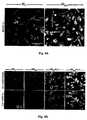
- Aggregated A ⁇ induces toxicity on resident microglia and impairs cell renewal
- Such activities are manifested by increased production of TNF- ⁇ , down-regulation of insulin-like growth factor (IGF-I), inhibition of the ability to express class II major histocompatibility complex (MHC-II) proteins and CD11c (a marker of dendritic cells) and thus to act as antigen-presenting cells (APCs), and failure to support neural tissue survival and renewal ((Butovsky et al., 2006a; Butovsky et al., 2005; Butovsky et al., 2006b). Further, we found that when microglia encounter aggregated ⁇ -amyloid, their ability to remove these aggregates without exerting toxic effects on neighboring neurons or impairing neurogenesis depends upon their undergoing a phenotype switch.
- IGF-I insulin-like growth factor
- MHC-II major histocompatibility complex
- CD11c a marker of dendritic cells
- a switch in microglial phenotype might take place via a local dialog between microglia and T-cells, which is mediated by T cell-derived cytokines such as interleukin (IL)-4.
- T cell-derived cytokines such as interleukin (IL)-4.
- IL-4 interleukin-4
- Th T-helper
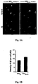
- microglia for in-vivo neural cell renewal was demonstrated by enhanced neurogenesis in the rat dentate gyrus after injection of IL-4-activated microglia intracerebroventricularly and by the presence of IGF-I-expressing microglia in the dentate gyrus of rats kept in an enriched environment (Ziv et al., 2006).
- injection of IL-4-activated microglia into the cerebrospinal fluid resulted in increased oligodendrogenesis in the spinal cord and improved clinical symptoms.
- the newly formed oligodendrocytes were spatially associated with microglia expressing MHC-II and IGF-I (Butovsky et al., 2006c).
- systemic immune cells in the form of T cells directed to certain self-antigens
- T cells can protect injured neurons from death
- studies in rodents showing that passive transfer of T cells specific to myelin basic protein reduces the loss of RGCs after a traumatic optic nerve injury (Moalem et al., 1999).
- T cells are also effective when directed to either cryptic or pathogenic epitopes of myelin basic protein, as well as to other myelin antigens or their epitopes (Mizrahi et al., 2002).
- myelin antigens capable of protecting the nervous system from any type of acute or chronic insult?
- protective T cell response is a physiologically evoked response that might not be sufficient in severe insults or might not always be properly controlled.
- specificity of such protective T cells depends on the site of the insult.
- the protective effect of vaccination with myelin-associated antigens is restricted to injuries of the white matter, i.e., to myelinated axons (Mizrahi et al., 2002; Avidan et al., 2004; Schori et al., 2001). If the insult is to the retina, which contains no myelin, myelin antigens have no effect.
- T cells specific to antigens residing in the site of damage help clean and heal
- the anti-self T cells should home to the site of damage and be locally activated. This is why only those antigens that are being presented at the site of lesion can be used for the vaccination.
- the T cells provide a source of cytokines and growth factors that shape the resident eye sentinels cells - the microglia, so as to make them active defensible cells that the eye can tolerate. Namely, such activated microglia can take up glutamate, remove debris and produce growth factors while refraining from production of agents that are part of their killing mechanism (e.g.
- TNF- ⁇ TNF- ⁇ to which the eye, like the brain, has a low tolerance
- Such T cells are constitutively controlled by physiologically existing regulatory T cells that are themselves amenable to control upon need (Kipnis et al., 2004a; Kipnis et al., 2002).
- the present invention relates to the embodiments characterized in the claims. Thus, it relates to the following items:
- a method for treatment of age-related macular degeneration which comprises causing T cells that produce IL-4 to accumulate in the eye of a patient in need, thereby halting or delaying progress of the macular degeneration.
- T cells are administered to said patient an agent selected from the group consisting of:
- the brain like the retina, is considered to be immune privileged in the sense that the blood-brain barrier resists passive deposition of antibodies and reduces the recruitment of antigen-specific lymphocytes (Streilein et al., 1992).
- antigen-specific immunity might actually function to protect against degenerative diseases.
- immunization with the abnormal amyloid, or passive administration of antibodies against the abnormal protein greatly reduced the quantity of deposition in the brain of the genetically modified mice, and improved their performance in laboratory tests of memory and cognitive function (e.g. Morgan et al., 2000).
- Alzheimer's disease patients may be immunologically tolerant to amyloid, preventing protective autoimmunization to the abnormally processed protein and thus developing autoimmune encephalomyelitis (Furlan et al., 2003).
- myelin-presenting microglia with which myelin-specific T cells can readily hold a dialog, are likely to be present at the damaged sites.
- Myelin-related antigens or antigens that are weakly cross-reactive with myelin (such as Cop-1), are therefore likely to be the antigens of choice for therapeutic vaccination (Avidan et al., 2004).
- T cells activated by these antigens will then home to the CNS and, upon encountering their relevant APCs there, become locally activated to supply the cytokines and growth factors in order to switch the phenotype of the harmful microglia (activated by aggregated A ⁇ ; Butovsky et al., 2005) into microglia with dendritic-like characteristics.
- the resulting immunological synapse between T cells and microglia will then create a supportive niche for cell renewal by promoting neurogenesis from the pool of adult stem cells (Butovsky et al., 2006a).
- T cells constitute the immune-based therapy of choice for AMD. This does not preclude the potential benefit of antibodies as a supplementary therapy as shown in anima; model of Alzheimer disease with antibodies against A ⁇ peptide (Bard et al., 2000). Moreover, the T cells can function as a mini-factory capable of producing a variety of compounds, including cytokines and neurotrophic factors (Ziemssen et al., 2002). Above all, they represent a physiological system of maintenance and repair that might help to counteract the age-related conditions leading to brain senescence.
- a ⁇ aggregated ⁇ -amyloid
- microglia activated by A ⁇ rather than help the suffering tissue further contribute to the chaos.
- aggregated A ⁇ induces microglia to become cytotoxic and block neurogenesis from adult rodent neural progenitor cells (NPCs).
- NPCs rodent neural progenitor cells
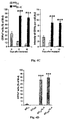
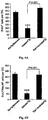
- Addition of IL-4, a cytokine derived from T-helper (Th)2 cells, to microglia activated by A ⁇ can reverse the impediment, the down-regulation of IGF-I, the up-regulation of TNF- ⁇ , and the failure to act as APCs.
- Alzheimer's disease double-transgenic mice expressing mutant human genes encoding presenilin 1 and chimeric mouse/human amyloid precursor protein we show that switching of microglia phenotype into professional APCs producing IGF-I, achieved here by a T cell-based vaccination with Copolymer-1, resulted in reduction of amyloid loads and the induction of neuronal survival and neurogenesis.
- ALD age-related macular degeneration
- T cells that produce IL-4 to accumulate in the eye of an individual in need, thereby halting or delaying progress of the macular degeneration.
- This effect can be affected by several self antigens and cytokine activated cells.
- agents that can cause T cells producing IL-4 to accumulate in the eye include, without being limited to:
- the agent is Copolymer 1, a Copolymer 1-related peptide or a Copolymer 1-reflated polypeptide.
- Copolymer 1 or a Copolymer 1-related peptide or polypeptide is intended to include any peptide or polypeptide; including a random copolymer that cross-reacts functionally with MBP and is able to compete with MBP on the MHC class II in the antigen presentation.
- the Cop 1 or a Cop 1-related peptide or polypeptide is represented by a random copolymer consisting of a suitable ratio of a positively charged amino acid such as lysine or arginine, in combination with a negatively charged amino acid (preferably in a lesser quantity) such as glutamic acid or aspartic acid, optionally in combination with a non-charged neutral amino acid such as alanine or glycine, serving as a filler, and optionally with an amino acid adapted to confer on the copolymer immunogenic properties, such as an aromatic amino acid like tyrosine or tryptophan.
- a positively charged amino acid such as lysine or arginine
- a negatively charged amino acid preferably in a lesser quantity
- a non-charged neutral amino acid such as alanine or glycine
- an amino acid adapted to confer on the copolymer immunogenic properties such as an aromatic amino acid like tyrosine or tryptophan.
- Such copolymers are disclosed
- the Copolymer 1 or a Copolymer 1-related peptide or polypeptide is a copolymer selected from the group consisting of random copolymers comprising one amino acid selected from each of at least three of the following groups: (a) lysine and arginine; (b) glutamic acid and aspartic acid; (c) alanine and glycine; and (d) tyrosine and tryptophan.
- the amino acids may be L- or D-amino acids or mixtures thereof.
- the present invention contemplates the use of copolymers containing both D- and L-amino acids, as well as copolymers consisting essentially of either L- or D-amino acids.
- the copolymer contains four different amino acids, each from a different one of the groups (a) to (d).
- the agent is Copolymer 1, composed of a mixture of random polypeptides consisting essentially of the amino acids L-glutamic acid (E), L-alanine (A), L-tyrosine (Y) and L-lysine (K) in an approximate ratio of 1.5:4.8:1:3.6, having a net overall positive electrical charge and of a molecular weight from about 2 KDa to about 40 KDa.
- E L-glutamic acid
- A L-alanine
- Y L-tyrosine
- K L-lysine
- the Cop 1 has average molecular weight of about 2 KDa to about 20 KDa, more preferably of about 4,7 KDa to about 13 K Da, still more preferably of about 4 KDa to about 8.6 KDa, of about 5 KDa to 9 KDa, or of about 6.25 KDa to 8.4 KDa.
- the Cop 1 has average molecular weight of about 13 KDa to about 20 KDa, more preferably of about 13,5 KDa to about 18 KDa, with an average of about 15 KDa to about 16 KD, preferably of 16kDa.
- Other average molecular weights for Cop 1, lower than 40 KDa, are also encompassed by the present invention.
- Copolymer 1 of said molecular weight ranges can be prepared by methods known in the art, for example by the processes described in U.S. Patent No. 5,800,808 , the entire contents of which are hereby incorporated by reference in the entirety.
- the Copolymer 1 may be a polypeptide comprising from about 15 to about 100, preferably from about 40 to about 80, amino acids in length.
- the agent is Cop 1 in the form of its acetate salt known under the generic name glatiramer acetate or its trade name Copaxone® (a trademark of Teva Pharmaceutical Industries Ltd., Petach Tikva, Israel).
- Copaxone® a trademark of Teva Pharmaceutical Industries Ltd., Petach Tikva, Israel.
- the Copolymer 1-related peptide is a random copolymer of 4 amino acids in which one or more of the following substitutions is made: aspartic acid for glutamic acid, glycine for alanine, arginine for lysine, and tryptophan for tyrosine, which is expected to have the same activity of Copolymer.
- the Cop 1-related peptide or polypeptide is a copolymer of three different amino acids each from a different one of three groups of the groups (a) to (d). These copolymers are herein referred to as terpolyiners.
- the terpolymer contains tyrosine (Y), alanine (A), and lysine (K), hereinafter designated YAK, in which the average molar fraction of the amino acids can vary: Y, A and K can be present in a mole fraction of about 0.05-0.250, 0.3 - 0.6; and 0.1-0.5, respectively, more preferably, the molar ratios of Y, A and K are about 0.10:0.54:0.35, respectively. It is possible to substitute arginine for lysine, glycine for alanine, and/or tryptophan for tyrosine.
- the terpolymer contains tyrosine (Y), glutamic acid (E), and lysine (K), hereinafter designated YEK, in which the average molar fraction of the amino acids can vary: E, Y and K can be present in a mole fraction of about 0.005 - 0.300, 0.005-0.250, and 0.3-0.7, respectively. More preferably, the molar ratios of E, Y and K are about 0.26:0.16:0.58, respectively. It is possible to substitute aspartic acid for glutamic acid, arginine for lysine, and/or tryptophan for tyrosine.
- the terpolymer contains lysine (K), glutamic acid (E), and alanine (A), hereinafter designated KEA, in which the average molar fraction of the amino acids can vary: E, A and K van be present in a mole fraction of about 0.005-0.300, 0.005-0.600, and 0.2 - 0.7, respectively. More preferably, the molar ratios of E, A and K are about 0.15:0.48:0.36, respectively. It is possible to substitute aspartic acid for glutamic acid, glycine for alanine, and/or arginine for lysine.
- the terpolymer contains tyrosine (Y), glutamic acid (E), and alanine (A0, hereinafter designated YEA, in which the average molar fraction of the amino acids can vary: Y, E and A can be present in a mole fraction of about 0.005-0.250, 0.005-0.300, and 0.005-0.800, respectively. More preferably, the molar ratios of E, A, and Y are about 0.21: 0.65:0.14, respectively. It is possible to substitute tryptophan for tyrosine, aspartic acid for glutamic acid, and/or glycine for alanine.
- the average molecular weight of the terpolymers YAK, YEK, KEA and YEA can vary between about 2 KDa to 40 KDa, preferably between about 3 KDa to 35 KDa, more preferably between about 5 KDa to 25 KDa.
- Copolymer 1 and related peptides and polypeptides may be prepared by methods known in the art, for example, by the process disclosed in U.S. Patent 3,849,550 .
- the molecular weight of the copolymers can be adjusted during polypeptide synthesis or after the copolymers have been made. To adjust the molecular weight during polypeptide synthesis, the synthetic conditions or the amounts of amino acids are adjusted so that synthesis stops when the polypeptide reaches the approximate length that is desired. After synthesis, polypeptides with the desired molecular weight can be obtained by any available size selection procedure, such as chromatography of the polypeptides on a molecular weight sizing column or gel, and collection of the molecular weight ranges desired.
- the copolymers can also be partially hydrolyzed to remove high molecular weight species, for example, by acid or enzymatic hydrolysis, and then purified to remove the acid or enzymes.
- the copolymers with a desired molecular weight may be prepared by a process, which includes reacting a protected polypeptide with hydrobromic acid to form a trifluoroacetyl-polypeptide having the desired molecular weight profile.
- the reaction is performed for a time and at a temperature that is predetermined by one or more test reactions.
- the time and temperature are varied and the molecular weight range of a given batch of test polypeptides is determined.
- the test conditions that provide the optimal molecular weight range for that batch of polypeptides are used for the batch.
- a trifluoroacetyl-polypeptide having the desired molecular weight profile can be produced by a process, which includes reacting the protected polypeptide with hydrobromic acid for a time and at a temperature predetermined by test reaction.
- the trifluoroacetyl-polypeptide with the desired molecular weight profile is then further treated with an aqueous piperidine solution to form a low toxicity polypeptide having the desired molecular weight.
- a test sample of protected polypeptide from a given batch is reacted with hydrobromic acid for about 10-50 hours at a temperature of about 20-28°C. The best conditions for that batch are determined by running several test reactions. For example, in one embodiment, the protected polypeptide is reacted with hydrobromic acid for about 17 hours at a temperature of about 26°C.
- Cop 1 As binding motifs of Cop 1 to MS-associated HLA-DR molecules are known (Fridkis-Hareli et al, 1999), polypeptides derived from Cop 1 having a defined sequence can readily be prepared and tested for binding to the peptide binding groove of the HLA-DR molecules as described in the Fridkis-Hareli et al (1999) publication. Examples of such peptides are those disclosed in WO 00/05249 and WO 00/05250 and include the peptides of SEQ ID NOs. 1-32 hereinbelow.
- Such peptides of SEQ ID Nos: 1-32 and other similar peptides derived from Cop 1 would be expected to have similar activity as Cop 1.
- Such peptides, and other similar peptides are also considered to be within the definition of Cop 1-related peptides or polypeptides and their use is encompassed by the present invention as well as other synthetic amino acid copolymers such as the random four-amino acid copolymers described by Fridkis-Hareli et al., 2002 (as candidates for treatment of multiple sclerosis), namely copolymers (14-, 35- and 50-mers) containing the amino acids phenylalanine, glutamic acid, alanine and lysine (poly FEAK), or tyrosine, phenylalanine, alanine and lysine (poly YFAK), and any other similar copolymer to be discovered that can be considered a universal antigen similar to Cop 1.
- the agent that causes T cells that produce IL-4 to accumulate in the eye is IL-4, preferably human recombinant IL-4, that can be administered nasally.
- the agent is IL-4 activated dendritic cells, IL-4 activated monocytes, IL-4 activated bone marrow-derived myeloid cells or IL-4 activated peripheral blood mononuclear cells (PBMCs).
- IL-13 have the same effect as IL-4, because it is well established in the field of cytokines that IL-4 and IL-13 can utilize a common receptor and share many actions such as B-cell activation and suppression of Th-1 cells.
- the agent is bone marrow-derived myeloid cells or peripheral blood-derived myeloid cells activated with IL-13 or with a narrow concentration range of IFN- ⁇ , more preferably up to 20 ng/ml IFN- ⁇ .
- the agent is a mammalian pathogenic self-antigen associated with a T-cell-mediated specific autoimmune disease of the eye such as, but not limited to, a pathogenic uveitogenic antigen selected from mammalian interphotoreceptor retinoid-binding protein (IRBP), S-antigen (S-Ag), or rhodopsin.
- a pathogenic uveitogenic antigen selected from mammalian interphotoreceptor retinoid-binding protein (IRBP), S-antigen (S-Ag), or rhodopsin.
- the mammalian uveitogenic antigen is preferably a human IRBP (SEQ ID NO: 33) or a bovine IRBP (SEQ ID NO: 34), a human S-Ag (SEQ ID NO: 35) or a bovine S-Ag (SEQ ID NO: 36), or human rhodopsin (SEQ ID NO: 37).
- the agent is a peptide which sequence is comprised within the sequence of said pathogenic self-antigen; a peptide obtained by modification of said peptide, which modification consists in the replacement of one or more amino acid residues of the peptide by different amino acid residues (hereinafter "modified peptide"), said modified peptide still being capable of recognizing the T-cell receptor recognized by the parent peptide but with less affinity; or a nucleotide sequence encoding said pathogenic self-antigen, said peptide or said modified peptide.
- modified peptide a nucleotide sequence encoding said pathogenic self-antigen, said peptide or said modified peptide.
- the agent is: (a) a peptide which sequence is comprised within the sequence of bovine IRBP (SEQ ID NO: 34); (b) a modified peptide as defined above obtained by modification of the peptide of (a); or (c) a nucleotide sequence encoding human or bovine IRPB, a peptide of (a), or a modified peptide of (b).
- the peptide which sequence is comprised within the sequence of bovine IRBP may be the peptide R16 (sequence 1177-1191 of IRBP), ADGSSWEGVGVVPDV (SEQ ID NO:38); the peptide PTARSVGAADGSSWEGVGVVPDV (SEQ ID NO:39); or the peptide HVDDTDLYLTIPTARSVGAADGS (SEQ ID NO:40).
- the agent is: (a) a peptide which sequence is comprised within the sequence of bovine S-Ag (SEQ ID NO:36); (b) a modified peptide as defined above obtained by modification of the peptide of (a); or (c) a nucleotide sequence encoding human or bovine S-Ag, a peptide of (a), or a modified peptide of (b).
- the peptide (a) which sequence is comprised within the sequence of bovine SAg may be the peptide G-8 (sequence 347-354 of S-Ag) of the sequence TSSEVATE (SEQ ID NO:41); the peptide M-8 (sequence 307-314 of S-Ag), DTNLASST (SEQ ID NO:42; or the peptides of the sequences:
- the modified peptide (c) derived from bovine S-Ag may be the G-8 analog, TSSEAATE (SEQ ID NO:50) or the M-8 analog, DTALASST (SEQ ID NO:51).
- the agent is a nucleotide sequence encoding a pathogenic self-antigen associated with a T-cell-mediated specific autoimmune disease of the eye, or a peptide or a modified peptide derived therefrom as defined herein.
- the agent is T cells activated by Copolymer 1, or by a pathogenic self-antigen associated with a T-cell-mediated specific autoimmune disease of the eye, a peptide or a modified peptide derived therefrom as defined herein.
- the agent is any combination of the agents defined above.
- an agent selected from the agents defined herein for treatment of age-related macular degeneration or for the manufacture of a medicament for treatment of age-related macular degeneration is also disclosed.
- the cells can be preferably autologous or they can be from a matched donor and are preferably administered intravenously.
- the agent is T cells activated by Copolymer 1, which can be prepared close to the administration step or cell banks can be established to store Copolymer 1-sensitized T cells for treatment of individuals at a later time, as needed.
- Autologous T cells may be obtained from the individual and allogeneic or semi-allogeneic T cells may obtained from a bank of stored T cells of each of the most common MHC-class II types are present.
- the patient is preferably treated with its autologous stored T cells, but if autologous T cells are not available, then cells should be used which share an MHC type II molecule with the patient, and these would be expected to be operable in that patient.
- the T cells are preferably stored in an activated state after exposure to Copolymer 1. However, the T cells may also be stored in a resting state and activated with Copolymer 1 once they are thawed and prepared for use.
- the T cell lines are prepared in any way that is well known in the art.
- the cell lines of the bank are preferably cryopreserved. Once the cells are thawed, they are preferably cultured prior to injection in order to eliminate non-viable cells.
- the T cells can be activated or reactivated using the Copolymer 1 antigen as used in the original activation.
- activation may be achieved by culturing in the presence of a mitogen, such as phytohemagglutinin (PHA) or concanavalin A (preferably the former). This will place the cells into an even higher state of activation.
- a mitogen such as phytohemagglutinin (PHA) or concanavalin A (preferably the former).
- the bone marrow-derived myeloid cells for use in the present invention can be obtained from autologous or donor's peripheral blood or bone marrow and processed by techniques well-known in the art.
- the donor should be a matched, namely HLA-matched, donor.
- the myeloid cells may be cultured until they multiply to the level needed for transplant back into the patient and are then activated with the cytokine (IL-4, IL-13 or IFN- ⁇ ) for the time necessary to upregulate CD11c expression. For example, activation with up to 20 ng/ml IFN- ⁇ may take 2-3 days until the peak of CD11c expression is reached.
- microglia are immune cells of the CNS that are derived from myeloid progenitor cells, which come from the bone marrow. Thus, microglia are the resident CNS cells whereas the bone marrow-derived myeloid cells are the infiltrating cells.
- compositions/medicaments for use in accordance with the present disclosure may be formulated in conventional manner using one or more pharmaceutically/physiologically acceptable carriers or excipients, depending on the agent used.
- the carrier(s) must be "acceptable” in the sense of being compatible with the other ingredients of the composition and not deleterious to the recipient thereof.
- parenteral e.g., intravenous, intraperitoneal, intramuscular, subcutaneous, mucosal, e.g., oral, intranasal, buccal, vaginal, rectal, or intraocular, intrathecal, topical and intradermal routes, with or without adjuvant.
- Administration can be systemic or local.
- the dosage of the agent to be administered will be determined by the physician according to the agent, the age of the patient and stage of the disease.
- the dosage may be chosen from a range of 1-80 mg, preferably 20 mg, although any other suitable dosage is encompassed by the invention.
- the treatment can be carried out by administration of repeated doses at suitable time intervals, according to the stage of the disease, the age and condition of the patient.
- Cop 1 may be administered daily.
- the administration may be made according to a regimen suitable for immunization, for example, at least once a week, once a month or at least once every 2 or 3 months, or less frequently, but any other suitable interval between the immunizations is envisaged by the disclosure according to the condition of the patient.
- the cells are preferably engineered bone marrow-derived dendritic cells (DCs) that express IL-4, which may be obtained as described by Morita et al., 2001. Since DCs are specialized APCs that migrate from the periphery to lymphoid tissues, where they activate and regulate T cells, genetic modification of DCs to express immunoregulatory cytokines such as IL-4 provides a new immunotherapeutic strategy for treatment of AMD and other diseases.
- DCs bone marrow-derived dendritic cells
- tissue sections were treated with a permeabilization/blocking solution containing 10% FCS, 2% bovine serum albumin, 1% glycine, and 0.05% Triton X-100 (Sigma-Aldrich, St. Louis). Tissue sections were stained overnight at 4°C with specified combinations of the following primary antibodies: rat anti-BrdU (1:200; Oxford Biotechnology, Kidlington, Oxfordshire, UK), goat anti-doublecortin (anti-DCX) (1:400; Santa Cruz Biotechnology, Santa Cruz, CA), and mouse anti-neuronal nuclear protein (anti-NeuN) (1:200; Chemicon).
- CD11b MAC1; 1:50; BD-Pharmingen
- FITC-conjugated IB-4 was used for labeling of microglia.
- Anti-MHC-II Abs rat, clone IBL-5/22; 1:50 was used to detect expression of cell-surface MHC-II proteins.
- CD11c hamster anti-CD11c (1:100; eBioscience, San Diego, CA) was used.
- Anti-A ⁇ human amino-acid residues 1-17
- mouse clone 6E10; Chemicon
- IGF-I human amino-acid residues 1-17
- TNF- ⁇ goat anti-TNF- ⁇ Abs
- T cells were detected with anti-CD3 polyclonal Abs (rabbit, 1:100; DakoCytomation, CA).
- Propidium iodide (1 ⁇ g/ml; Molecular Probes, Invitrogen, Carlsbad, CA), was used for nuclear staining.
- Control sections (not treated with primary antibody) were used to distinguish specific staining from staining of nonspecific antibodies or autofluorescent components. Sections were then washed with PBS and cover-slipped in polyvinyl alcohol with diazabicyclo-octane as anti-fading agent.
- results in-vitro were analyzed by two-tailed unpaired Student's t -test and by the Tukey-Kramer multiple comparisons test (ANOVA) and are expressed as means ⁇ SEM.
- results in vivo were analyzed by two-tailed unpaired Student's t -test or 1-way ANOVA and are expressed as means ⁇ SEM.
- Example 1 Aggregated ⁇ -amyloid induces microglia to express a phenotype that blocks neurogenesis, and the blocking is counteracted by IL-4.
- microglia found in association with inflammatory and neurodegenerative diseases e.g. microglia activated by LPS or by aggregated A ⁇ (1-40)
- IL-4-activated microglia shown to be associated with neural tissue survival, express MHC-II, produce IGF-I, and decrease TNF- ⁇ expression
- TNF- ⁇ expression butovsky et al., 2005.
- a ⁇ -activated microglia block neurogenesis, and if so, whether T cell-derived cytokines can counteract the inhibitory effect.
- GFP green fluorescent protein
- NPCs neural stem/progenitor cells
- a ⁇ (1-40) rather than A ⁇ (1-42) and its concentration was based on our previous demonstration that this compound induces cytotoxic activity in microglia (Butovsky et al., 2005). Growth media and cytokine residues were then washed off the co-cultured microglia, and each of the treated microglial preparations was freshly co-cultured with dissociated adult subventricular zone-derived NPC spheres (Butovsky et al., 2006a) on coverslips coated with MatrigelTM in the presence of differentiation medium (Butovsky et al., 2006a) ( Fig. 1A ).
- Example 2 T-cell-based vaccination with copolymer-1 modulates immune activity of microglia, eliminates ⁇ -amyloid plaque formation, and induces neurogenesis.
- the above findings prompted us to examine whether a T cell-based vaccination would alter the default microglial phenotype in Alzheimer's disease and hence lead to plaque removal and neurogenesis.
- the antigen we chose for the vaccination was Cop-1 (Teitelbaum et al., 1996), shown by us to be weakly cross-reactive with a wide-range of CNS autoantigens and, depending on the regimen, to be neuroprotective under conditions of both acute and chronic neurodegeneration (Kipnis et al., 2000; Schori et al., 2001; Angelov et al., 2003).
- Cop-1 was administered to Tg-AD mice, suffering from learning/memory impairment and an accumulation of aggregated A ⁇ plaques deposited mainly in the cortex and the hippocampus, both characteristic features of early-onset familial Alzheimer's disease (Borchelt et al., 1997).
- the regimen for Cop-1 administration was similar to that used to evoke neuroprotection in a model of chronic elevation of intraocular pressure (Bakalash et al., 2005).
- Seven weeks after the first Cop-1 injection all the mice were euthanized and analyzed.
- Staining of brain cryosections from Tg-AD mice with antibodies specific to human A ⁇ disclosed numerous plaques in the untreated-Tg-AD mice but very few in the Tg-AD mice vaccinated with Cop-1 ( Fig. 2 A ) . No plaques were seen in their respective non-Tg littermates ( Fig. 2 A ).
- CD11c + microglia in untreated Tg-AD mice were only rarely found in association with A ⁇ + plaques, whereas any residual A ⁇ -stained plaques seen in the Cop-1-vaccinated mice were surrounded by MHC-II + /CD11c + microglia ( Fig. 2E ).
- these CD11c + microglia were also positively stained for CD 11 b ( Fig. 3A ); in addition, they were loaded with A ⁇ , indicative of their engulfment of this peptide ( Fig. 2E ).
- microglia activated by A ⁇ exhibited amoeboid morphology, whereas the rounded shape of the CD11c + microglia was reminiscent of dendritic cells ( Fig. 4B ).
- amoeboid morphology of the A ⁇ -stained microglia was reversible on addition of IL-4, when they again took on the morphological appearance of dendritic-like cells ( Fig. 4B ).
- the various treatments applied to the microglia did not affect their expression of CD11b, suggesting that they did not lose their CD11b characteristics when they took on the expression of CD11c ( Fig. 4B ).
- CD11c expression assessed by the number of CD11c + cells and the intensity of their staining as a function of time in culture, revealed that soon after seeding (day 0) untreated microglia expressed low levels of CD11c, which gradually disappeared ( Fig. 4C ). In contrast, the expression of CD11c induced by IL-4 was not transient. Quantification of the ability of IL-4 to induce CD11c expression even after the microglia were pretreated with A ⁇ is shown in Fig. 4D .
- microglia in Alzheimer's disease and other neurodegenerative diseases has been intensively investigated over the last few years, with apparently conflicting results (Streit, 2004).
- the detection of some CD11b + microglia in aged wild-type mice that are healthy is in line with the reported age-related increase in activated microglia in the normal human brain (Streit, 2004). It is possible that such microglia are the ones that contribute both to age-related cognitive loss and to impaired neurogenesis (Monje et al., 2003).
- CD11b were found also in patients with Alzheimer's disease (Akiyama & McGeer, 1990). Although these microglia are phagocytic (Frenkel et al., 2005), they are apparently not efficient enough to fight off the Alzheimer symptoms.
- microglia derived from the bone marrow of matched wild-type mice can effectively remove plaques (Simard et al., 2006). These microglia express higher protein levels than are required for antigen presentation and might therefore be more effective phagocytes than the resident microglia.
- the microglia, that are needed to support brain maintenance and fight Alzheimer's disease are dendritic-like cells (CD11b + /CD11c + ).
- the CD11c + microglia might engage in a dialog with T cells that can help to fight off adverse conditions by promoting the buffering of excessive A ⁇ and supporting both neuronal survival (Butovsky et al., 2005) and neural renewal (Butovsky et al., 2006b).
- IL-4 but not IFN-y, can alter the phenotype of A ⁇ -committed microglia (Butovsky et al., 2005)
- IL-4 has often been described as an anti-inflammatory cytokine (Chao et al., 1993). Our results strongly argue against this perception and suggest instead that IL-4 activates microglia to adopt a phenotype that seems to acquire a different morphology and a different activity from those of the innately activated microglia or of the activated microglia commonly seen in Alzheimer's disease or in MS. In the latter disease, unlike in Alzheimer's disease, the microglia appear to be overwhelmed by an onslaught of adaptive immunity (Butovsky et al., 2006a).
- IL-4 is capable of restoring a favorable activated phenotype even after the microglia have already exhibited phenotypic characteristics of aggregated A ⁇ ((Butovsky et al., 2005), and the present study), or been overwhelmed by IFN- ⁇ (Butovsky et al., 2006a).
- LPS and A ⁇ exhibit similar patterns of MAPK activation in microglia (Pyo et al., 1998).
- IL-4 can attenuate a MAPK pathway activated by LPS, an effect evidently associated with serine/threonine phosphatase activity (Iribarren et al., 2003).
- Example 3 Copolymer-1 vaccination counteracts cognitive decline in Alzheimer's disease.
- mice in this study demonstrated attenuated cognitive loss (tested in a Morris water maze) and increased neurogenesis. These two aspects of hippocampal plasticity are apparently related to the presence of IGF-I and cognitive activity and cell renewal (Butovsky et al., 2006a;b). Reported observations in Tg-AD mice housed in an enriched environment also support a link between mechanisms associated with neurogenesis (Ziv et al., 2006) and with plaque reduction (Lazarov et al., 2005).
- aggregated A ⁇ evidently interferes with the ability of microglia to engage in dialog with T cells, its presence in the brain can be expected to cause loss of cognitive ability and impairment of neurogenesis. Homing of CNS-autoreactive T cells to the site of disease or damage in such cases is critical, but will be effective only if those T cells can counterbalance the destructive activity of the aggregated A ⁇ . As shown here, IFN- ⁇ by itself is impotent against the activity of microglia that are already committed to an aggregated A ⁇ phenotype, but is effective when added together with IL-4.
- Example 4 IFN- ⁇ -activated myeloid cells and their uses for promoting tissue repair, detection of and delivery of drugs to damaged tissue.
- CD11c is upregulated and reach the peak at 2-3 days after IFN- ⁇ activation in microglia ( Figs. 8A-8D ) or bone-marrow-derived myeloid cells (data not shown).
- IFN- ⁇ upregulated of CD11c on bone marrow derived myeloid cells by IFN- ⁇ will allow the cells to reach injured sites and induce beneficial effect.
- Cop-1 given in different regimens results in different therapies; daily treatment has no beneficial effects in paradigms of neurodegenerative diseases, but is effective in suppressing inflammatory disease in patients like multiple sclerosis. Therefore, we decided to embark on a clinical trial with AMD patients in a protocol of weekly administration.
- Cop-1 Copaxone®, Teva
- Enrolled patients were randomly (ratio 2 to 1) treated either with subcutaneous injections of Copaxone (20 mg) or placebo injections on a weekly basis for a period of 12 weeks.
- the primary outcome was the change in total drusen area and it was calculated using Image-Pro software. Analysis of patients from the observational group (17 eyes) showed an increase of 25.2% in total drusen area.
Landscapes
- Health & Medical Sciences (AREA)
- Life Sciences & Earth Sciences (AREA)
- General Health & Medical Sciences (AREA)
- Animal Behavior & Ethology (AREA)
- Veterinary Medicine (AREA)
- Public Health (AREA)
- Epidemiology (AREA)
- Chemical & Material Sciences (AREA)
- Immunology (AREA)
- Medicinal Chemistry (AREA)
- Engineering & Computer Science (AREA)
- Pharmacology & Pharmacy (AREA)
- Bioinformatics & Cheminformatics (AREA)
- Proteomics, Peptides & Aminoacids (AREA)
- Gastroenterology & Hepatology (AREA)
- Organic Chemistry (AREA)
- Zoology (AREA)
- Biomedical Technology (AREA)
- Neurology (AREA)
- Mycology (AREA)
- Microbiology (AREA)
- Neurosurgery (AREA)
- Nuclear Medicine, Radiotherapy & Molecular Imaging (AREA)
- General Chemical & Material Sciences (AREA)
- Chemical Kinetics & Catalysis (AREA)
- Biophysics (AREA)
- Genetics & Genomics (AREA)
- Biochemistry (AREA)
- Molecular Biology (AREA)
- Ophthalmology & Optometry (AREA)
- Medicines That Contain Protein Lipid Enzymes And Other Medicines (AREA)
- Medicines Containing Antibodies Or Antigens For Use As Internal Diagnostic Agents (AREA)
- Peptides Or Proteins (AREA)
- Hematology (AREA)
- Cell Biology (AREA)
- Medicines Containing Material From Animals Or Micro-Organisms (AREA)
- Developmental Biology & Embryology (AREA)
- Virology (AREA)
- Biotechnology (AREA)
Applications Claiming Priority (2)
| Application Number | Priority Date | Filing Date | Title |
|---|---|---|---|
| US80604106P | 2006-06-28 | 2006-06-28 | |
| PCT/IL2007/000798 WO2008001380A2 (en) | 2006-06-28 | 2007-06-28 | Method of treatment of age-related macular degeneration |
Publications (3)
| Publication Number | Publication Date |
|---|---|
| EP2046366A2 EP2046366A2 (en) | 2009-04-15 |
| EP2046366A4 EP2046366A4 (en) | 2011-09-21 |
| EP2046366B1 true EP2046366B1 (en) | 2016-08-31 |
Family
ID=38846104
Family Applications (1)
| Application Number | Title | Priority Date | Filing Date |
|---|---|---|---|
| EP07766829.1A Active EP2046366B1 (en) | 2006-06-28 | 2007-06-28 | Copolymer-1 for treatment of age-related macular degeneration |
Country Status (7)
| Country | Link |
|---|---|
| US (1) | US9089509B2 (enExample) |
| EP (1) | EP2046366B1 (enExample) |
| JP (1) | JP5412280B2 (enExample) |
| AU (1) | AU2007264682B2 (enExample) |
| CA (1) | CA2656463C (enExample) |
| ES (1) | ES2605154T3 (enExample) |
| WO (1) | WO2008001380A2 (enExample) |
Families Citing this family (10)
| Publication number | Priority date | Publication date | Assignee | Title |
|---|---|---|---|---|
| WO2008075365A1 (en) * | 2006-12-20 | 2008-06-26 | Yissum Research Development Company Of The Hebrew University Of Jerusalem | Compositions and methods for treatment of age related degeneration of the retina |
| JP2012518407A (ja) * | 2009-02-20 | 2012-08-16 | ザ スクリプス リサーチ インスティチュート | 単離された単球集団及び関連した治療応用 |
| USRE49251E1 (en) | 2010-01-04 | 2022-10-18 | Mapi Pharma Ltd. | Depot systems comprising glatiramer or pharmacologically acceptable salt thereof |
| WO2012143924A1 (en) | 2011-04-21 | 2012-10-26 | Mapi Pharma Ltd. | Random pentapolymer for treatment of autoimmune diseases |
| US20130217986A1 (en) * | 2012-02-21 | 2013-08-22 | Ucl Business Plc | Method and system for predicting an amyloidosis status |
| WO2016183585A1 (en) * | 2015-05-14 | 2016-11-17 | Joslin Diabetes Center, Inc. | Retinol-binding protein 3 (rbp3) as a protective factor in non-diabetic retinal degeneration |
| US12097292B2 (en) | 2016-08-28 | 2024-09-24 | Mapi Pharma Ltd. | Process for preparing microparticles containing glatiramer acetate |
| HRP20250396T1 (hr) | 2016-08-31 | 2025-05-23 | Mapi Pharma Ltd. | Depo sustavi koji sadrže glatiramer acetat |
| PL3565823T3 (pl) * | 2017-01-04 | 2024-10-28 | Worg Pharmaceuticals (Zhejiang) Co., Ltd. | Peptydy S-arestyn oraz ich zastosowanie terapeutyczne |
| AU2018242998B2 (en) | 2017-03-26 | 2023-11-02 | Mapi Pharma Ltd. | Glatiramer depot systems for treating progressive forms of multiple sclerosis |
Family Cites Families (15)
| Publication number | Priority date | Publication date | Assignee | Title |
|---|---|---|---|---|
| IL36670A (en) | 1971-04-21 | 1974-09-10 | Sela M | Therapeutic basic copolymers of amino acids |
| US5800808A (en) | 1994-05-24 | 1998-09-01 | Veda Research And Development Co., Ltd. | Copolymer-1 improvements in compositions of copolymers |
| JP2003521448A (ja) | 1998-07-23 | 2003-07-15 | ザ・プレジデント・アンド・フェローズ・オブ・ハーバード・カレッジ | 合成ペプチドおよび自己免疫疾患治療のための使用方法 |
| WO2000005250A1 (en) | 1998-07-23 | 2000-02-03 | Yeda Research And Development Co., Ltd | Treatment of autoimmune conditions with copolymer 1 and related copolymers and peptides |
| US20030149997A1 (en) * | 1999-02-19 | 2003-08-07 | Hageman Gregory S. | Diagnostics and therapeutics for arterial wall disruptive disorders |
| US6420378B1 (en) * | 1999-10-15 | 2002-07-16 | Supergen, Inc. | Inhibition of abnormal cell proliferation with camptothecin and combinations including the same |
| DK1248643T3 (da) * | 2000-01-20 | 2005-11-07 | Yeda Res & Dev | Anvendelse af copolymer 1 og beslægtede peptider og polypeptider og T-celler behandlet dermed til neurobeskyttende terapi |
| US7399740B2 (en) * | 2001-06-28 | 2008-07-15 | Yeda Research And Development Co. Ltd. | Poly-Glu,Tyr for neuroprotective therapy |
| US6524012B1 (en) * | 2001-09-06 | 2003-02-25 | Gkn Automotive, Inc. | Securing members together by spring rings |
| EP1476583B1 (en) * | 2002-01-25 | 2013-08-28 | Biomedical Design, Inc. | Calcification-resistant fixation |
| WO2003079968A2 (en) | 2002-03-26 | 2003-10-02 | Yeda Research And Development Co. Ltd | Use of an organ-specific self-pathogen for treatment of a non-autoimmune disease of said organ |
| CN1705438A (zh) * | 2002-06-14 | 2005-12-07 | 耶达研究及发展有限公司 | 用于神经保护和神经再生的抗原呈递细胞 |
| US7860975B2 (en) * | 2002-07-31 | 2010-12-28 | Oracle America, Inc. | System and method for secure sticky routing of requests within a server farm |
| US7354574B2 (en) * | 2002-11-07 | 2008-04-08 | Advanced Ocular Systems Limited | Treatment of ocular disease |
| MXPA05007329A (es) * | 2003-01-07 | 2005-09-30 | Yeda Res & Dev | Vacuna de gotas oculares que contiene copolimero 1 para inmunizacion terapeutica. |
-
2007
- 2007-06-28 CA CA2656463A patent/CA2656463C/en not_active Expired - Fee Related
- 2007-06-28 WO PCT/IL2007/000798 patent/WO2008001380A2/en not_active Ceased
- 2007-06-28 JP JP2009517603A patent/JP5412280B2/ja not_active Expired - Fee Related
- 2007-06-28 US US12/308,880 patent/US9089509B2/en not_active Expired - Fee Related
- 2007-06-28 AU AU2007264682A patent/AU2007264682B2/en not_active Ceased
- 2007-06-28 ES ES07766829.1T patent/ES2605154T3/es active Active
- 2007-06-28 EP EP07766829.1A patent/EP2046366B1/en active Active
Also Published As
| Publication number | Publication date |
|---|---|
| WO2008001380A3 (en) | 2009-04-30 |
| ES2605154T3 (es) | 2017-03-13 |
| AU2007264682B2 (en) | 2012-09-06 |
| CA2656463C (en) | 2015-12-01 |
| AU2007264682A1 (en) | 2008-01-03 |
| EP2046366A2 (en) | 2009-04-15 |
| US9089509B2 (en) | 2015-07-28 |
| JP5412280B2 (ja) | 2014-02-12 |
| EP2046366A4 (en) | 2011-09-21 |
| JP2009542622A (ja) | 2009-12-03 |
| US20100135953A1 (en) | 2010-06-03 |
| CA2656463A1 (en) | 2008-01-03 |
| WO2008001380A2 (en) | 2008-01-03 |
Similar Documents
| Publication | Publication Date | Title |
|---|---|---|
| EP2046366B1 (en) | Copolymer-1 for treatment of age-related macular degeneration | |
| US20120135016A1 (en) | Induction of neurogenesis and stem cell therapy in combination with copolymer 1 | |
| US7560102B2 (en) | Method for reducing neuronal degeneration so as to ameliorate the effects of injury or disease | |
| Kala et al. | Recent insights into the mechanism of action of glatiramer acetate | |
| AU2002345323B2 (en) | Use of poly-Glu, Tyr for neuroprotective therapy | |
| EP2301569B1 (en) | Vaccine and method for treatment of neurodegenerative diseases | |
| KR0159046B1 (ko) | 포유류의 자기면역 포도막망막염의 치료 또는 예방방법 | |
| JP6474082B2 (ja) | 眼疾患および眼障害の治療のためのヒト単球亜集団 | |
| WO2006056998A2 (en) | Methods of cell therapy, neurogenesis and oligodendrogenesis | |
| HK1127858B (en) | Copolymer-1 for treatment of age-related macular degeneration | |
| IL159535A (en) | USE OF POLY-Glu50, Tyr50 FOR THE PREPARATION OF PHARMACEUTICAL COMPOSITIONS FOR NEUROPROTECTION THERAPY | |
| HK1064951B (en) | Use of poly-glu, tyr for neuroprotection therapy of the cns or pns |
Legal Events
| Date | Code | Title | Description |
|---|---|---|---|
| PUAI | Public reference made under article 153(3) epc to a published international application that has entered the european phase |
Free format text: ORIGINAL CODE: 0009012 |
|
| 17P | Request for examination filed |
Effective date: 20090128 |
|
| AK | Designated contracting states |
Kind code of ref document: A2 Designated state(s): AT BE BG CH CY CZ DE DK EE ES FI FR GB GR HU IE IS IT LI LT LU LV MC MT NL PL PT RO SE SI SK TR |
|
| AX | Request for extension of the european patent |
Extension state: AL BA HR MK RS |
|
| R17D | Deferred search report published (corrected) |
Effective date: 20090430 |
|
| RIC1 | Information provided on ipc code assigned before grant |
Ipc: A61K 38/20 20060101ALI20090505BHEP Ipc: A61K 38/17 20060101AFI20090505BHEP Ipc: A61K 31/00 20060101ALI20090505BHEP |
|
| RIN1 | Information on inventor provided before grant (corrected) |
Inventor name: EISENBACH-SCHWARTZ, MICHAL Inventor name: BUTOVSKY, OLEG |
|
| REG | Reference to a national code |
Ref country code: HK Ref legal event code: DE Ref document number: 1127858 Country of ref document: HK |
|
| A4 | Supplementary search report drawn up and despatched |
Effective date: 20110819 |
|
| RIC1 | Information provided on ipc code assigned before grant |
Ipc: A61K 35/28 20060101ALI20110812BHEP Ipc: A61K 38/10 20060101ALI20110812BHEP Ipc: A61K 35/14 20060101ALI20110812BHEP Ipc: A61K 39/00 20060101ALI20110812BHEP Ipc: A61K 38/17 20060101AFI20110812BHEP Ipc: A61K 38/08 20060101ALI20110812BHEP Ipc: A61K 38/20 20060101ALI20110812BHEP Ipc: A61K 31/00 20060101ALI20110812BHEP |
|
| 17Q | First examination report despatched |
Effective date: 20121107 |
|
| RIC1 | Information provided on ipc code assigned before grant |
Ipc: A61K 35/14 20150101ALI20150114BHEP Ipc: A61K 38/16 20060101AFI20150114BHEP Ipc: A61K 39/00 20060101ALI20150114BHEP Ipc: A61K 38/10 20060101ALI20150114BHEP Ipc: A61K 38/20 20060101ALI20150114BHEP |
|
| GRAP | Despatch of communication of intention to grant a patent |
Free format text: ORIGINAL CODE: EPIDOSNIGR1 |
|
| INTG | Intention to grant announced |
Effective date: 20150326 |
|
| REG | Reference to a national code |
Ref country code: DE Ref legal event code: R079 Ref document number: 602007047733 Country of ref document: DE Free format text: PREVIOUS MAIN CLASS: A61K0038200000 Ipc: A61K0035120000 |
|
| GRAS | Grant fee paid |
Free format text: ORIGINAL CODE: EPIDOSNIGR3 |
|
| GRAP | Despatch of communication of intention to grant a patent |
Free format text: ORIGINAL CODE: EPIDOSNIGR1 |
|
| RIC1 | Information provided on ipc code assigned before grant |
Ipc: A61K 35/14 20060101ALI20160218BHEP Ipc: A61K 38/07 20060101ALI20160218BHEP Ipc: A61K 35/17 20150101ALI20160218BHEP Ipc: A61K 39/00 20060101ALI20160218BHEP Ipc: A61K 35/12 20060101AFI20160218BHEP Ipc: A61K 38/02 20060101ALI20160218BHEP Ipc: A61K 38/10 20060101ALI20160218BHEP Ipc: A61K 38/20 20060101ALI20160218BHEP Ipc: A61K 35/15 20150101ALI20160218BHEP Ipc: A61K 38/16 20060101ALI20160218BHEP |
|
| INTG | Intention to grant announced |
Effective date: 20160317 |
|
| GRAF | Information related to payment of grant fee modified |
Free format text: ORIGINAL CODE: EPIDOSCIGR3 |
|
| GRAA | (expected) grant |
Free format text: ORIGINAL CODE: 0009210 |
|
| AK | Designated contracting states |
Kind code of ref document: B1 Designated state(s): AT BE BG CH CY CZ DE DK EE ES FI FR GB GR HU IE IS IT LI LT LU LV MC MT NL PL PT RO SE SI SK TR |
|
| AX | Request for extension of the european patent |
Extension state: AL BA HR MK RS |
|
| REG | Reference to a national code |
Ref country code: CH Ref legal event code: EP Ref country code: GB Ref legal event code: FG4D |
|
| REG | Reference to a national code |
Ref country code: IE Ref legal event code: FG4D |
|
| REG | Reference to a national code |
Ref country code: DE Ref legal event code: R096 Ref document number: 602007047733 Country of ref document: DE |
|
| REG | Reference to a national code |
Ref country code: AT Ref legal event code: REF Ref document number: 824370 Country of ref document: AT Kind code of ref document: T Effective date: 20161015 |
|
| REG | Reference to a national code |
Ref country code: LT Ref legal event code: MG4D |
|
| REG | Reference to a national code |
Ref country code: NL Ref legal event code: MP Effective date: 20160831 |
|
| REG | Reference to a national code |
Ref country code: AT Ref legal event code: MK05 Ref document number: 824370 Country of ref document: AT Kind code of ref document: T Effective date: 20160831 |
|
| PG25 | Lapsed in a contracting state [announced via postgrant information from national office to epo] |
Ref country code: FI Free format text: LAPSE BECAUSE OF FAILURE TO SUBMIT A TRANSLATION OF THE DESCRIPTION OR TO PAY THE FEE WITHIN THE PRESCRIBED TIME-LIMIT Effective date: 20160831 Ref country code: LT Free format text: LAPSE BECAUSE OF FAILURE TO SUBMIT A TRANSLATION OF THE DESCRIPTION OR TO PAY THE FEE WITHIN THE PRESCRIBED TIME-LIMIT Effective date: 20160831 |
|
| PG25 | Lapsed in a contracting state [announced via postgrant information from national office to epo] |
Ref country code: GR Free format text: LAPSE BECAUSE OF FAILURE TO SUBMIT A TRANSLATION OF THE DESCRIPTION OR TO PAY THE FEE WITHIN THE PRESCRIBED TIME-LIMIT Effective date: 20161201 Ref country code: SE Free format text: LAPSE BECAUSE OF FAILURE TO SUBMIT A TRANSLATION OF THE DESCRIPTION OR TO PAY THE FEE WITHIN THE PRESCRIBED TIME-LIMIT Effective date: 20160831 Ref country code: LV Free format text: LAPSE BECAUSE OF FAILURE TO SUBMIT A TRANSLATION OF THE DESCRIPTION OR TO PAY THE FEE WITHIN THE PRESCRIBED TIME-LIMIT Effective date: 20160831 Ref country code: AT Free format text: LAPSE BECAUSE OF FAILURE TO SUBMIT A TRANSLATION OF THE DESCRIPTION OR TO PAY THE FEE WITHIN THE PRESCRIBED TIME-LIMIT Effective date: 20160831 Ref country code: NL Free format text: LAPSE BECAUSE OF FAILURE TO SUBMIT A TRANSLATION OF THE DESCRIPTION OR TO PAY THE FEE WITHIN THE PRESCRIBED TIME-LIMIT Effective date: 20160831 |
|
| REG | Reference to a national code |
Ref country code: ES Ref legal event code: FG2A Ref document number: 2605154 Country of ref document: ES Kind code of ref document: T3 Effective date: 20170313 |
|
| PG25 | Lapsed in a contracting state [announced via postgrant information from national office to epo] |
Ref country code: RO Free format text: LAPSE BECAUSE OF FAILURE TO SUBMIT A TRANSLATION OF THE DESCRIPTION OR TO PAY THE FEE WITHIN THE PRESCRIBED TIME-LIMIT Effective date: 20160831 Ref country code: EE Free format text: LAPSE BECAUSE OF FAILURE TO SUBMIT A TRANSLATION OF THE DESCRIPTION OR TO PAY THE FEE WITHIN THE PRESCRIBED TIME-LIMIT Effective date: 20160831 |
|
| PG25 | Lapsed in a contracting state [announced via postgrant information from national office to epo] |
Ref country code: SK Free format text: LAPSE BECAUSE OF FAILURE TO SUBMIT A TRANSLATION OF THE DESCRIPTION OR TO PAY THE FEE WITHIN THE PRESCRIBED TIME-LIMIT Effective date: 20160831 Ref country code: BE Free format text: LAPSE BECAUSE OF FAILURE TO SUBMIT A TRANSLATION OF THE DESCRIPTION OR TO PAY THE FEE WITHIN THE PRESCRIBED TIME-LIMIT Effective date: 20160831 Ref country code: BG Free format text: LAPSE BECAUSE OF FAILURE TO SUBMIT A TRANSLATION OF THE DESCRIPTION OR TO PAY THE FEE WITHIN THE PRESCRIBED TIME-LIMIT Effective date: 20161130 Ref country code: DK Free format text: LAPSE BECAUSE OF FAILURE TO SUBMIT A TRANSLATION OF THE DESCRIPTION OR TO PAY THE FEE WITHIN THE PRESCRIBED TIME-LIMIT Effective date: 20160831 Ref country code: PL Free format text: LAPSE BECAUSE OF FAILURE TO SUBMIT A TRANSLATION OF THE DESCRIPTION OR TO PAY THE FEE WITHIN THE PRESCRIBED TIME-LIMIT Effective date: 20160831 Ref country code: PT Free format text: LAPSE BECAUSE OF FAILURE TO SUBMIT A TRANSLATION OF THE DESCRIPTION OR TO PAY THE FEE WITHIN THE PRESCRIBED TIME-LIMIT Effective date: 20170102 Ref country code: CZ Free format text: LAPSE BECAUSE OF FAILURE TO SUBMIT A TRANSLATION OF THE DESCRIPTION OR TO PAY THE FEE WITHIN THE PRESCRIBED TIME-LIMIT Effective date: 20160831 |
|
| REG | Reference to a national code |
Ref country code: DE Ref legal event code: R097 Ref document number: 602007047733 Country of ref document: DE |
|
| REG | Reference to a national code |
Ref country code: FR Ref legal event code: PLFP Year of fee payment: 11 |
|
| PLBE | No opposition filed within time limit |
Free format text: ORIGINAL CODE: 0009261 |
|
| STAA | Information on the status of an ep patent application or granted ep patent |
Free format text: STATUS: NO OPPOSITION FILED WITHIN TIME LIMIT |
|
| 26N | No opposition filed |
Effective date: 20170601 |
|
| PG25 | Lapsed in a contracting state [announced via postgrant information from national office to epo] |
Ref country code: SI Free format text: LAPSE BECAUSE OF FAILURE TO SUBMIT A TRANSLATION OF THE DESCRIPTION OR TO PAY THE FEE WITHIN THE PRESCRIBED TIME-LIMIT Effective date: 20160831 |
|
| REG | Reference to a national code |
Ref country code: HK Ref legal event code: GR Ref document number: 1127858 Country of ref document: HK |
|
| PG25 | Lapsed in a contracting state [announced via postgrant information from national office to epo] |
Ref country code: MC Free format text: LAPSE BECAUSE OF FAILURE TO SUBMIT A TRANSLATION OF THE DESCRIPTION OR TO PAY THE FEE WITHIN THE PRESCRIBED TIME-LIMIT Effective date: 20160831 |
|
| REG | Reference to a national code |
Ref country code: CH Ref legal event code: PL |
|
| REG | Reference to a national code |
Ref country code: IE Ref legal event code: MM4A |
|
| PG25 | Lapsed in a contracting state [announced via postgrant information from national office to epo] |
Ref country code: IE Free format text: LAPSE BECAUSE OF NON-PAYMENT OF DUE FEES Effective date: 20170628 Ref country code: LI Free format text: LAPSE BECAUSE OF NON-PAYMENT OF DUE FEES Effective date: 20170630 Ref country code: LU Free format text: LAPSE BECAUSE OF NON-PAYMENT OF DUE FEES Effective date: 20170628 Ref country code: CH Free format text: LAPSE BECAUSE OF NON-PAYMENT OF DUE FEES Effective date: 20170630 |
|
| REG | Reference to a national code |
Ref country code: FR Ref legal event code: PLFP Year of fee payment: 12 |
|
| PG25 | Lapsed in a contracting state [announced via postgrant information from national office to epo] |
Ref country code: MT Free format text: LAPSE BECAUSE OF NON-PAYMENT OF DUE FEES Effective date: 20170628 |
|
| PG25 | Lapsed in a contracting state [announced via postgrant information from national office to epo] |
Ref country code: HU Free format text: LAPSE BECAUSE OF FAILURE TO SUBMIT A TRANSLATION OF THE DESCRIPTION OR TO PAY THE FEE WITHIN THE PRESCRIBED TIME-LIMIT; INVALID AB INITIO Effective date: 20070628 |
|
| REG | Reference to a national code |
Ref country code: DE Ref legal event code: R082 Ref document number: 602007047733 Country of ref document: DE Representative=s name: HERZOG IP PATENTANWALTS GMBH, DE |
|
| PG25 | Lapsed in a contracting state [announced via postgrant information from national office to epo] |
Ref country code: CY Free format text: LAPSE BECAUSE OF NON-PAYMENT OF DUE FEES Effective date: 20160831 |
|
| PG25 | Lapsed in a contracting state [announced via postgrant information from national office to epo] |
Ref country code: TR Free format text: LAPSE BECAUSE OF FAILURE TO SUBMIT A TRANSLATION OF THE DESCRIPTION OR TO PAY THE FEE WITHIN THE PRESCRIBED TIME-LIMIT Effective date: 20160831 |
|
| PG25 | Lapsed in a contracting state [announced via postgrant information from national office to epo] |
Ref country code: IS Free format text: LAPSE BECAUSE OF FAILURE TO SUBMIT A TRANSLATION OF THE DESCRIPTION OR TO PAY THE FEE WITHIN THE PRESCRIBED TIME-LIMIT Effective date: 20161231 |
|
| PGFP | Annual fee paid to national office [announced via postgrant information from national office to epo] |
Ref country code: DE Payment date: 20210618 Year of fee payment: 15 Ref country code: IT Payment date: 20210625 Year of fee payment: 15 Ref country code: FR Payment date: 20210622 Year of fee payment: 15 |
|
| PGFP | Annual fee paid to national office [announced via postgrant information from national office to epo] |
Ref country code: GB Payment date: 20210625 Year of fee payment: 15 |
|
| PGFP | Annual fee paid to national office [announced via postgrant information from national office to epo] |
Ref country code: ES Payment date: 20210825 Year of fee payment: 15 |
|
| REG | Reference to a national code |
Ref country code: DE Ref legal event code: R119 Ref document number: 602007047733 Country of ref document: DE |
|
| GBPC | Gb: european patent ceased through non-payment of renewal fee |
Effective date: 20220628 |
|
| PG25 | Lapsed in a contracting state [announced via postgrant information from national office to epo] |
Ref country code: FR Free format text: LAPSE BECAUSE OF NON-PAYMENT OF DUE FEES Effective date: 20220630 |
|
| PG25 | Lapsed in a contracting state [announced via postgrant information from national office to epo] |
Ref country code: GB Free format text: LAPSE BECAUSE OF NON-PAYMENT OF DUE FEES Effective date: 20220628 Ref country code: DE Free format text: LAPSE BECAUSE OF NON-PAYMENT OF DUE FEES Effective date: 20230103 |
|
| PG25 | Lapsed in a contracting state [announced via postgrant information from national office to epo] |
Ref country code: IT Free format text: LAPSE BECAUSE OF NON-PAYMENT OF DUE FEES Effective date: 20220628 |
|
| REG | Reference to a national code |
Ref country code: ES Ref legal event code: FD2A Effective date: 20230801 |
|
| PG25 | Lapsed in a contracting state [announced via postgrant information from national office to epo] |
Ref country code: ES Free format text: LAPSE BECAUSE OF NON-PAYMENT OF DUE FEES Effective date: 20220629 |