EP2045266A1 - Verfahren zur Behandlung und Diagnostizierung einer krebserzeugenden cath-D- oder Alzheimer-Erkrankung - Google Patents
Verfahren zur Behandlung und Diagnostizierung einer krebserzeugenden cath-D- oder Alzheimer-Erkrankung Download PDFInfo
- Publication number
- EP2045266A1 EP2045266A1 EP07301421A EP07301421A EP2045266A1 EP 2045266 A1 EP2045266 A1 EP 2045266A1 EP 07301421 A EP07301421 A EP 07301421A EP 07301421 A EP07301421 A EP 07301421A EP 2045266 A1 EP2045266 A1 EP 2045266A1
- Authority
- EP
- European Patent Office
- Prior art keywords
- lrp1
- cath
- chain
- cancer
- pro
- Prior art date
- Legal status (The legal status is an assumption and is not a legal conclusion. Google has not performed a legal analysis and makes no representation as to the accuracy of the status listed.)
- Withdrawn
Links
Images


Classifications
-
- C—CHEMISTRY; METALLURGY
- C07—ORGANIC CHEMISTRY
- C07K—PEPTIDES
- C07K16/00—Immunoglobulins [IGs], e.g. monoclonal or polyclonal antibodies
- C07K16/18—Immunoglobulins [IGs], e.g. monoclonal or polyclonal antibodies against material from animals or humans
- C07K16/28—Immunoglobulins [IGs], e.g. monoclonal or polyclonal antibodies against material from animals or humans against receptors, cell surface antigens or cell surface determinants
-
- C—CHEMISTRY; METALLURGY
- C07—ORGANIC CHEMISTRY
- C07K—PEPTIDES
- C07K14/00—Peptides having more than 20 amino acids; Gastrins; Somatostatins; Melanotropins; Derivatives thereof
- C07K14/435—Peptides having more than 20 amino acids; Gastrins; Somatostatins; Melanotropins; Derivatives thereof from animals; from humans
- C07K14/705—Receptors; Cell surface antigens; Cell surface determinants
-
- C—CHEMISTRY; METALLURGY
- C07—ORGANIC CHEMISTRY
- C07K—PEPTIDES
- C07K16/00—Immunoglobulins [IGs], e.g. monoclonal or polyclonal antibodies
- C07K16/40—Immunoglobulins [IGs], e.g. monoclonal or polyclonal antibodies against enzymes
-
- C—CHEMISTRY; METALLURGY
- C12—BIOCHEMISTRY; BEER; SPIRITS; WINE; VINEGAR; MICROBIOLOGY; ENZYMOLOGY; MUTATION OR GENETIC ENGINEERING
- C12N—MICROORGANISMS OR ENZYMES; COMPOSITIONS THEREOF; PROPAGATING, PRESERVING, OR MAINTAINING MICROORGANISMS; MUTATION OR GENETIC ENGINEERING; CULTURE MEDIA
- C12N9/00—Enzymes; Proenzymes; Compositions thereof; Processes for preparing, activating, inhibiting, separating or purifying enzymes
- C12N9/14—Hydrolases (3)
- C12N9/48—Hydrolases (3) acting on peptide bonds (3.4)
- C12N9/50—Proteinases, e.g. Endopeptidases (3.4.21-3.4.25)
- C12N9/64—Proteinases, e.g. Endopeptidases (3.4.21-3.4.25) derived from animal tissue
- C12N9/6421—Proteinases, e.g. Endopeptidases (3.4.21-3.4.25) derived from animal tissue from mammals
- C12N9/6472—Cysteine endopeptidases (3.4.22)
-
- C—CHEMISTRY; METALLURGY
- C07—ORGANIC CHEMISTRY
- C07K—PEPTIDES
- C07K2317/00—Immunoglobulins specific features
- C07K2317/70—Immunoglobulins specific features characterized by effect upon binding to a cell or to an antigen
- C07K2317/77—Internalization into the cell
Definitions
- the present invention relates to methods for the treatment and/or the prevention of cancer and Alzheimer's disease.
- the present invention also relates to an early marker of cancer in order to ensure its early diagnosis.
- Cancer remains a major public health challenge despite progress in detection and therapy.
- breast cancer is worldwide the most common form of cancer in females and according to the United Nations World Health Organization, it is the leading cause of cancer deaths among women in 2005.
- breast cancer has led to the death of more than 500 000 people worldwide.
- the number of cases has significantly increased since the 1970s, a phenomenon partly blamed on modern lifestyles in the Western world.
- breast cancer therefore remains incontestably a major human health problem necessitating urgently an effective treatment for this frequent and mortal pathology.
- the mainstay of breast cancer treatment is surgery when the tumor is localized, with possible adjuvant hormonal therapy (with, e.g., tamoxifen or an aromatase inhibitor), chemotherapy, and/or radiotherapy.
- hormonal therapy with, e.g., tamoxifen or an aromatase inhibitor
- radiotherapy can be given both before and after surgery.
- Neo-adjuvant chemotherapy is used to shrink the size of a tumor prior to surgery.
- Adjuvant chemotherapy is given after surgery to reduce the risk of recurrence.
- Patients with estrogen receptor positive tumors will typically receive a hormonal treatment after chemotherapy is completed. But targeted therapies exist too.
- the drug trastuzumab (Herceptin ®) is used to block the HER2 protein in breast cancer cells, thereby slowing their growth.
- breast cancer still leads to the death of half a million of people worldwide every year.
- breast cancer can originate from different genetic base and deregulation of biological pathways. It seems very important to determine new targets involved in apparition and development of breast cancer, notably at early steps. Metastasis can further emerge from this development of breast cancer. Thus, identification of new therapeutic targets is necessary in order to resolve problems of breast cancer and associated disorders.
- Cath-D is synthesized as the 52 kDa, catalytically inactive, precursor called pro-Cath-D. It is present in endosomes as an active 48 kDa single-chain intermediate that is subsequently converted in the lysosomes into the fully active mature protease, composed of a 34 kDa heavy and a 14 kDa light chains (Richo and Conner, 1991).
- the 52 kDa pro-cath-D is hyper-secreted into the extracellular environment and can be taken up by both cancer cells and fibroblasts via mannose-6-phosphate (M6P) receptors and other as yet unidentified receptor(s) (Laurent-Matha et al., 1998; Heylen et al., 2002; Laurent-Matha et al., 2005).
- M6P mannose-6-phosphate
- cath-D is over-expressed and secreted at high levels by human epithelial breast cancer cells (Liaudet-Coopman et al, 2006).
- Cath-D overexpression in breast cancer is highly correlated with poor prognosis (Ferrandina et al., 1997; Foekens et al., 1999; Bossard et al., 2003; Rodriguez et al., 2005).
- cath-D could act as an extracellular messenger that interacts either directly or indirectly, with an as yet unidentified cell surface receptor (Liaudet-Coopman et al, 2006). Cath-D is thus a marker of poor prognosis in cancer and its overexpression in cancer, notably breast cancer indicates a high risk of developing metastasis.
- the invention relates to an inhibitor of the interaction between pro-cath-D and the LRP1 ⁇ chain for the treatment and/or the prevention of a cancer secreting cath-D or of Alzheimer's disease, wherein said inhibitor is selected from the group consisting of a peptide having at least 80% sequence identity with SEQ ID NO: 5, an anti-LRP1 ⁇ chain antibody, an antibody fragment which binds to the LRP1 ⁇ chain, and an aptamer which binds to the LRP1 ⁇ chain.
- the invention also relates to an inhibitor of the expression of LRP1 for the treatment and/or the prevention of a cancer secreting cath-D or of Alzheimer's disease.
- a further embodiment of the invention relates to an in vitro method for diagnosing a cancer secreting cath-D or Alzheimer's disease in a patient comprising detecting the presence of a fragment of LRP1 in a biological sample obtained from said patient.
- LRP1 ⁇ chain or a fragment thereof LRP1 ⁇ chain or a fragment thereof.
- pro-cath-D is used herein to define the polypeptide sequence of the 52 kDa, catalytically inactive, precursor of cath-D.
- the naturally occurring protein has an aminoacid sequence shown in Genbank, Accession number NM_001909.
- LRP1 has its general meaning in the art (Strickland and Ranganathan, 2003; Lillis et al., 2005) and refers to LDL receptor-related protein 1.
- LRP1 is composed of a 515 kDa extra-cellular ⁇ chain and an 85 kDa ⁇ chain generated by proteolytic cleavage from a 600 kDa precursor polypeptide in a trans-Golgi compartment.
- LRP1 ⁇ chain and LRP1 ⁇ chain are issued from a sole transcript.
- the human full length of unprocessed precursor LRP1 corresponds to SwissProt accession number Q07954.
- LRP1 ⁇ chain is used herein to define the polypeptide sequence of the ⁇ chain of LRP1 consisting of the amino sequence as shown in SEQ ID NO: 1.
- the LRP1 ⁇ chain contains an extra-cellular domain, a trans-membrane region and a cytoplasmic tail.
- LRP1 ⁇ chain ectodomain is used herein to define the polypeptide sequence of about 68kDa amino-terminal fragment of the extracellular domain of the LRP1 ⁇ chain.
- the LRP1 ⁇ chain ecto-domain corresponds to the liberated ectodomain of LRP1 ⁇ chain following to its cleavage by mature cath-D.
- the LRP1 ⁇ chain ecto-domain is represented by SEQ ID NO: 2.
- SEQ ID NO: 2 the above-mentioned cleavage of LRP1 ⁇ chain ecto-domain by cath-D also leads to a 25 kDa membrane-associated carboxy-terminal fragment of the intracellular domain of the LRP1 ⁇ chain.
- the full length extra-cellular domain of LRP1 ⁇ chain consists in 476 residues and that full length of LRP1 consists in 4544 amino acids.
- F0 relates to the polypeptide sequence of 173 amino acids corresponding to residues 307 to 479 of the extra-cellular domain of LRP1 ⁇ chain, which forms the pro-cath-D binding site on LRP1 ⁇ chain with a strong interaction.
- residues 307 to 479 of the extra-cellular domain of LRP1 ⁇ chain correspond to the residues 4250 to 4422 of the full length of LRP1.
- F0 is represented by SEQ ID NO: 3.
- F4 relates to the polypeptide sequence of 46 amino acids issued from “F0" and corresponding to the minimal pro-cath-D binding region on LRP1 ⁇ chain which consists in residues 349 to 394 of the extra-cellular domain of LRP1 ⁇ chain.
- the residues 349 to 394 of the extra-cellular domain of LRP1 ⁇ chain correspond to the residues 4292 to 4337 of the full length of LRP1.
- F4 is represented by SEQ ID NO: 4.
- the expression "49-mer” relates to the polypeptide sequence of 49 amino acids corresponding to the 349-397 fragment of the extra-cellular LRP1 ⁇ chain which competes efficiently for the binding to pro-cath-D with LRP1 ⁇ chain. Thus, this 49-mer is considered as an inhibitor of the interaction between pro-cath-D and LRP1 ⁇ chain.
- the residues 349 to 397 of the extra-cellular domain of LRP1 ⁇ chain correspond to the residues 4292 to 4340 of the full length of LRP1.
- inhibitor of the interaction is used herein to mean preventing or reducing the direct or indirect association of one or more molecules, peptides, proteins, enzymes or receptors; or preventing or reducing the normal activity of one or more molecules, peptides, proteins, enzymes, or receptors.
- inhibitor of the interaction between pro-cath-D and LRP1 ⁇ chain is a molecule which can prevent the interaction between pro-cath-D and LRP1 ⁇ chain by competition or by fixing to one of the molecule.
- a gene product can be the direct transcriptional product of a gene (e.g., mRNA, tRNA, rRNA, antisense RNA, ribozyme, structural RNA or any other type of RNA) or a protein produced by translation of a mRNA.
- Gene products also include messenger RNAs which are modified, by processes such as capping, polyadenylation, methylation, and editing, and proteins (e.g., LRP1) modified by, for example, methylation, acetylation, phosphorylation, ubiquitination, SUMOylation, ADP-ribosylation, myristilation, and glycosylation.
- An “inhibitor of expression” refers to a natural or synthetic compound that has a biological effect to inhibit or significantly reduce the expression of a gene. Consequently an “inhibitor of LRP1 expression” refers to a natural or synthetic compound that has a biological effect to inhibit or significantly reduce the expression of the gene encoding for the LRP1 protein.
- an inhibitor of LRP1 expression and/or interaction between pro-cath-D and LRP1 ⁇ chain denotes a natural or synthetic compound which acts as an inhibitor of LRP1 expression and/or as an inhibitor of interaction between pro-cath-D and LRP1 ⁇ chain.
- a "coding sequence” or a sequence "encoding" an expression product, such as an RNA, polypeptide, protein, or enzyme is a nucleotide sequence that, when expressed, results in the production of that RNA, polypeptide, protein, or enzyme, i.e., the nucleotide sequence encodes an amino acid sequence for that polypeptide, protein or enzyme.
- a coding sequence for a protein may include a start codon (usually ATG) and a stop codon.
- references to specific proteins can include a polypeptide having a native amino acid sequence, as well as variants and modified forms regardless of their origin or mode of preparation.
- a protein that has a native amino acid sequence is a protein having the same amino acid sequence as obtained from nature (e.g., LRP1).
- Such native sequence proteins can be isolated from nature or can be prepared using standard recombinant and/or synthetic methods.
- Native sequence proteins specifically encompass naturally occurring truncated or soluble forms, naturally occurring variant forms (e.g., alternatively spliced forms), naturally occurring allelic variants and forms including postranslational modifications.
- a native sequence protein includes proteins following post-translational modifications such as glycosylation, or phosphorylation, or other modifications of some amino acid residues.
- Variants refer to proteins that are functional equivalents to a native sequence protein that have similar amino acid sequences and retain, to some extent, one or more activities of the native protein. Variants also include fragments that retain activity. Variants also include proteins that are substantially identical (e.g., that have 80, 85, 90, 95, 97, 98, 99%, sequence identity) to a native sequence. Such variants include proteins having amino acid alterations such as deletions, insertions and/or substitutions. A "deletion" refers to the absence of one or more amino acid residues in the related protein.
- substitution refers to the replacement of one or more amino acid residues by another amino acid residue in the polypeptide.
- substitution refers to the replacement of one or more amino acid residues by another amino acid residue in the polypeptide.
- alterations are conservative in nature such that the activity of the variant protein is substantially similar to a native sequence protein (see, e.g., Creighton (1984) Proteins, W.H. Freeman and Company ).
- the amino acid replacing another amino acid usually has similar structural and/or chemical properties. Insertions and deletions are typically in the range of 1 to 5 amino acids, although depending upon the location of the insertion, more amino acids can be inserted or removed.
- Two amino acid sequences are "substantially homologous” or “substantially similar” when greater than 80 %, preferably greater than 85 %, still preferably greater than 90 % of the amino acids are identical, or greater than about 90 %, preferably grater than 95 %, are similar (functionally identical) over the whole length sequences.
- the similar or homologous sequences are identified by alignment using, for example, the GCG (Genetics Computer Group, Program Manual for the GCG Package, Version 7, Madison, Wisconsin) pileup program, or any of sequence comparison algorithms such as BLAST, FASTA, etc.
- the expression "cancer” relates to a disease characterized by an uncontrolled cell growth and invasion of adjacent tissues.
- the abnormal cells often are referred to as neoplastic cells which are transformed cells that can form a solid tumor.
- tumor refers to an abnormal mass or population of cells (i.e. two or more cells) that result from excessive or abnormal cell division, whether malignant or benign, and pre-cancerous and cancerous cells.
- Malignant tumors are distinguished from benign growths or tumors in that, in addition to uncontrolled cellular proliferation, they can invade surrounding tissues and can metastasize.
- metastasis refers to a process in which cancer cells spread from one organ or tissue to another non-adjacent organ or tissue.
- associated hyperproliferative disorders means tumor microenvironment, fibroblast invasive outgrowth and metastasis development. Indeed, the tumor microenvironment was shown to play a critical role in tumor initiation and progression, and may be an important factor in developing therapeutic approaches. The tumor microenvironment, or stroma, influences the growth of the tumor and its ability to progress and metastasize.
- cancer secreting cath-D is meant cancer and associated hyperproliferative disorders in which cath-D or pro-cath-D are secreted by cancer cells.
- Different approaches such as immuno-histochemistry, in situ hybridization, cytosolic immunoassay and Northern and Western blot analyses have indicated that in most breast cancer tumors, cath-D is over-expressed from 2- to 50-fold compared to its concentration in other cell types such as fibroblasts or normal mammary glands [from F. Capony, C. Rougeot, P. Montcourrier, V. Cavaillippo, G. Salazar, H.
- cancers secreting cath-D include, but are not limited to, breast cancer, stomach cancer (cf. Manuel Del Casar J, Vizoso FJ, Abdel-Laa O, Sanz L, Martin A, Daniela Corte M, Bongera M, Garcia Muniz JL, Fueyo A. Prognostic value of cytosolyc cathepsin D content in resectable gastric cancer. J Surg Oncol. 2004 86:16-21 ), liver cancer (cf.
- breast cancer refers to a condition characterized by anomalous rapid proliferation of abnormal cells in one or both breasts of a subject.
- neoplastic cells may be identified in one or both breasts only and not in another tissue or organ, in one or both breasts and one or more adjacent tissues or organs (e.g. lymph node), or in a breast and one or more non-adjacent tissues or organs to which the breast cancer cells have metastasized. Cancer cells in the breast spread to tissues and organs of a subject, and conversely, cancer cells from other organs or tissue can invade or metastasize to a breast.
- Breast cancer cells often invade lymph node cells and/or metastasize to the liver, brain and/or bone and spread cancer in these tissues and organs.
- Breast cancers can spread to other organs and tissues and cause lung cancer, prostate cancer, colon cancer, ovarian cancer, cervical cancer, gastrointestinal cancer, pancreatic cancer, glioblastoma, hepatoma, colorectal cancer, uterine cervical cancer, endometrial carcinoma, salivary gland carcinoma, kidney cancer, vulval cancer, thyroid cancer, hepatic carcinoma, skin cancer, melanoma, ovarian cancer, neuroblastoma, myeloma, Ewing sarcoma and peripheral neuroepithelioma, and other carcinomas, lymphomas, blastomas, sarcomas, and leukemias.
- treating refers to reversing, alleviating, inhibiting the progress of, or preventing the disorder or condition to which such term applies, or one or more symptoms of such disorder or condition.
- diagnosis means the process of identifying a medical condition or disease by its signs and symptoms and from the results of various diagnostic procedures.
- diagnosis is used to mean determining the incidence of a cancer secreting cath-D, for example breast cancer, by examining whether the diagnostic marker of cancer secreting cath-D is present in a sample obtained from a patient.
- the term "marker” or “biochemical marker” as used herein refers to a molecule used as a target for analyzing patient test samples.
- biological sample refers to any biological sample obtained for the purpose of evaluation in vitro.
- the sample or patient sample may comprise any body fluid.
- test samples include blood, serum, plasma, nipple aspirate fluid, urine, saliva, synovial fluid and cephalorachidian liquid (CRL).
- sample is a whole blood sample, a serum sample or a plasma sample.
- a patient denotes a mammal, such as a rodent, a feline, a canine, and a primate.
- a patient according to the invention is a human.
- LRP1 plays a major role in the development of cancer and associated hyperproliferative disorders, notably cancer secreting cath-D such as breast cancer.
- a new pathway for the treatment and/or the prevention of cancer secreting cath-D is provided which involves inhibiting the interaction between pro-cath-D and the ⁇ chain of LRP1 and/or inhibiting LRP1 expression.
- the present application also shows the usefulness of LRP1 fragments and more particularly LRP1 ⁇ chain ecto-domain in diagnosis of a cancer secreting cath-D cancer such as breast cancer.
- pro-cath-D is a ligand for the extracellular domain of the ⁇ chain of LDL receptor-related protein-1, LRP1.
- cath-D participates in the paracrine signalling between epithelial cancer cells and associated fibroblasts, a process that depends on the presence of LRP1 on the surface of the fibroblasts.
- the present invention arises from the identification by the inventors of a key component of the tumor environment, namely LRP1, which is critical for the development, progression and metastasis of cancer and thus proposes a therapeutic strategy to target the tumor microenvironment by the inhibition of the function of the stromal cells that are required for progression.
- the inhibitors according to the invention enable to block the function of LRP1 as demonstrated by the inventors, which are responsible for tumor cell survival and proliferation.
- cath-D causes shedding of the extracellular domain of the LRP1 ⁇ chain so called LRP1 ⁇ chain ecto-domain, resulting in the generation of an amino-terminal fragment of about 68 kDa.
- This cleaved extracellular domain of the LRP1 ⁇ chain, the LRP1 ⁇ chain and fragments thereof may be detected in the circulation of patients affected by a cancer secreting cath-D.
- fragment of LRP1 ⁇ chain can be used for the detection of cancer secreting cath-D and in a preferred embodiment for early detection of breast cancer.
- the present invention provides methods and compositions (such as pharmaceutical compositions) for treating a cancer secreting cath-D, breast cancer in particular.
- the inventors identified an analogy between the mechanism of action cath-D in cancers secreting cath-D and the mechanism of action cath-D in Alzheimer's disease.
- the beta and gamma secretases are highly specific proteases that cleave the amyloid peptide (Abeta) from its protein precursor, amyloid precursor protein (APP).
- Abeta amyloid peptide
- APP amyloid precursor protein
- beta secretase clips APP near the transmembrane domain, releasing a large soluble fragment (beta-ectodomain); the smaller fragment is subsequently cleaved by gamma secretases to generate Abeta in a process called RIP.
- the identified beta secretases are the aspartic proteases BACE1, BACE2 (a BACE homolog), cathepsin D and cathepsin E.
- LRP1 was shown to interact with APP. LRP1 facilitates APP processing via the amyloidogenic pathway and the production of Abeta.
- Evidence that LRP1 is involved in APP processing is based on the findings that APP can interact with LRP1.
- an object of the invention is the use of an inhibitor of LRP1 expression and/or interaction between pro-cath-D and LRP1 ⁇ chain for the manufacture of a medicament for preventing and/or treating cancer cancer secreting cath-D or Alzheimer's disease.
- the invention relates to an inhibitor of the interaction between pro-cath-D and LRP1 ⁇ chain for the treatment and/or the prevention of a cancer secreting cath-D or of Alzheimer's disease.
- the inhibitor of the interaction between pro-cath-D and LRP1 ⁇ is a peptide having at least 80%, preferably at least 90%, even more preferably at least 95% sequence identity with the sequence of SEQ ID NO: 5.
- the length of said peptide is less than 60, preferably less than 55, amino acid residues.
- SEQ ID NO: 5 represents the sequence of the polypeptides called "49-mer" defined by the sequence of amino acid between 349 and 397 of the LRP1 ⁇ chain.
- the inhibitor of the interaction between pro-cath-D and LRP1 ⁇ chain is an antibody or a fragment thereof which binds to the ⁇ of chain LRP1.
- the antibody or the fragment thereof is directed against the polypeptide F0 or F4.
- the antibody is directed against the sequence of amino acid between 349 and 397 of LRP1 ⁇ chain consisting in the SEQ ID NO: 5.
- antibodies include, but are not limitated to, the monoclonal anti- ⁇ 2 macroglobulin receptor (# 3501, American Diagnostica Inc), the polyclonal LRP1 (N-20) (sc-16166, Santa Cruz Biotechnology, Inc.) and the monoclonal LRP1 (8B8) (sc-57352, Santa Cruz Biotechnology, Inc.).
- the monoclonal anti- ⁇ 2 macroglobulin receptor (# 3501, American Diagnostica Inc) recognizes amino acids 4291-4344 of LRP1.
- the inhibitor of the interaction between pro-cath-D and LRP1 ⁇ chain is an antibody or antibody fragment that is directed against pro-cath-D and preferably against epitopes issued from LPR1 ⁇ chain binding site on pro-cath-D.
- Epitopes issued form pro-cath-D binding region on LRP1 ⁇ chain can be characterized by the fact that each catalytic aspartic acid, namely Asp 33 on 14 kDa subunit and Asp 231 on 34 kDa subunit, have a central position in such epitopes.
- Antibodies directed against the LRP1 ⁇ chain can be raised according to known methods by administering the appropriate antigen or epitope to a host animal selected, e.g., from pigs, cows, horses, rabbits, goats, sheep, and mice, among others.
- a host animal selected, e.g., from pigs, cows, horses, rabbits, goats, sheep, and mice, among others.
- Various adjutants known in the art can be used to enhance antibody production.
- antibodies useful in practicing the invention can be polyclonal, monoclonal antibodies are preferred.
- Monoclonal antibodies against LRP1 ⁇ chain or ligands of LRP1 ⁇ chain can be prepared and isolated using any technique that provides for the production of antibody molecules by continuous cell lines in culture.
- Techniques for production and isolation include but are not limited to the hybridoma technique originally described by Kohler and Milstein (1975); the human B-cell hybridoma technique (Cote et al., 1983); and the EBV-hybridoma technique (Cole et al. 1985).
- techniques described for the production of single chain antibodies can be adapted to produce anti-LRP1 ⁇ chain, or anti-LRP1 ⁇ chain ligands single chain antibodies.
- Inhibitors of the interaction between pro-cath-D and LRP1 ⁇ chain useful in practicing the present invention also include anti-LRP1 ⁇ chain, or anti-LRP1 ⁇ chain ligands antibody fragments including but not limited to F(ab') 2 fragments, which can be generated by pepsin digestion of an intact antibody molecule, and Fab fragments, which can be generated by reducing the disulfide bridges of the F(ab') 2 fragments.
- Fab and/or scFv expression libraries can be constructed to allow rapid identification of fragments having the desired specificity to LRP1 ⁇ chain.
- Humanized anti-LRP1 ⁇ chain or anti-LRP1 ⁇ chain ligands antibodies and antibody fragments therefrom can also be prepared according to known techniques.
- “Humanized antibodies” are forms of non-human (e.g., rodent) chimeric antibodies that contain minimal sequence derived from non-human immunoglobulin.
- humanized antibodies are human immunoglobulins (recipient antibody) in which residues from a hypervariable region (CDRs) of the recipient are replaced by residues from a hypervariable region of a non-human species (donor antibody) such as mouse, rat, rabbit or nonhuman primate having the desired specificity, affinity and capacity.
- CDRs hypervariable region
- donor antibody such as mouse, rat, rabbit or nonhuman primate having the desired specificity, affinity and capacity.
- humanized antibodies may comprise residues that are not found in the recipient antibody or in the donor antibody. These modifications are made to further refine antibody performance.
- the humanized antibody will comprise substantially all of at least one, and typically two, variable domains, in which all or substantially all of the hypervariable loops correspond to those of a non-human immunoglobulin and all or substantially all of the FRs are those of a human immunoglobulin sequence.
- the humanized antibody optionally also will comprise at least a portion of an immunoglobulin constant region (Fc), typically that of a human immunoglobulin.
- aptamers are a class of molecule that represents an alternative to antibodies in term of molecular recognition.
- Aptamers are oligonucleotide or oligopeptide sequences with the capacity to recognize virtually any class of target molecules with high affinity and specificity.
- Such ligands may be isolated through Systematic Evolution of Ligands by EXponential enrichment (SELEX) of a random sequence library, as described in Tuerk C. and Gold L., 1990.
- the random sequence library is obtainable by combinatorial chemical synthesis of DNA.
- each member is a linear oligomer, eventually chemically modified, of a unique sequence. Possible modifications, uses and advantages of this class of molecules have been reviewed in Jayasena S.D., 1999.
- Peptide aptamers consists of a conformationally constrained antibody variable region displayed by a platform protein, such as E. coli Thioredoxin A that are selected from combinatorial libraries by two hybrid methods (Colas et al., 1996).
- a further aspect of the invention related to nucleic acids encoding for polypeptides of the invention, notably F0, F4 and the peptide named 49-mer.
- said nucleic acid construct is a DNA or RNA molecule, which may be included in any suitable vector, such as a plasmid, cosmid, episome, artificial chromosome, phage or a viral vector.
- a vector comprising a nucleic acid of the invention.
- Such vectors may comprise regulatory elements, such as a promoter, enhancer, terminator and the like, to cause or direct expression of said polypeptide upon administration to a subject.
- the vectors may further comprise one or several origins of replication and/or selectable markers.
- the promoter region may be homologous or heterologous with respect to the coding sequence, and provide for ubiquitous, constitutive, regulated and/or tissue specific expression, in any appropriate host cell, including for in vivo use.
- promoters include bacterial promoters (T7, pTAC, Trp promoter, etc.), viral promoters (LTR, TK, CMV-IE, etc.), mammalian gene promoters (albumin, PGK, etc), and the like.
- Examples of plasmids include replicating plasmids comprising an origin of replication, or integrative plasmids, such as for instance pUC, pcDNA, pBR, and the like.
- viral vector examples include adenoviral, retroviral, herpesvirus and AAV vectors.
- recombinant viruses may be produced by techniques known in the art, such as by transfecting packaging cells or by transient transfection with helper plasmids or viruses.
- Typical examples of virus packaging cells include PA317 cells, PsiCRIP cells, GPenv+ cells, 293 cells, etc.
- Detailed protocols for producing such replication-defective recombinant viruses may be found for instance in WO95/14785 , WO96/22378 , US5,882,877 , US6,013,516 , US4,861,719 , US5,278,056 and WO94/19478 .
- a further aspect of the present invention resides in a cell which has been transfected, infected or transformed by a nucleic acid and/or a vector according to the invention.
- Suitable host cells include, without limitation, prokaryotic cells (such as bacteria) and eukaryotic cells (such as yeast cells, mammalian cells, insect cells, plant cells, etc.).
- E.coli E.coli, Kluyveromyces or Saccharomyces yeasts
- mammalian cell lines e.g., Vero cells, CHO cells, 3T3 cells, COS cells, etc.
- primary or established mammalian cell cultures e.g., produced from lymphoblasts, fibroblasts, embryonic cells, epithelial cells, nervous cells, adipocytes, etc.
- the invention contemplates vascular or endothelial cells thereof or derived thereof, such as human umbilical vein endothelial (HUVEC) or progenitor endothelial cells (PEC).
- HAVEC human umbilical vein endothelial
- PEC progenitor endothelial cells
- the present invention also relates to a method for producing a recombinant host cell expressing a polypeptide according to the invention, said method comprising (i) introducing in vitro or ex vivo into a competent host cell a recombinant nucleic acid or a vector as described above, (ii) culturing in vitro or ex vivo the recombinant host cells obtained and (iii), optionally, selecting the cells which express and/or secrete said polypeptide.
- recombinant host cells can be used for the production of polypeptides according to the present invention as previously described.
- the invention relates to an inhibitor of the expression of LRP1 for the treatment and/or the prevention of a cancer secreting cath-D or of Alzheimer's disease.
- the invention provides the use of an inhibitor of LRP1 expression for the manufacture of a medicament intended for preventing and/or treating a cancer secreting cath-D or Alzheimer's disease.
- Inhibitors of LRP1 expression according to the present invention may be based on anti-sense oligonucleotide constructs.
- Anti-sense oligonucleotides including anti-sense RNA molecules and anti-sense DNA molecules, would act to directly block the translation of LRP1 mRNA by binding thereto and thus preventing protein translation or increasing mRNA degradation, thus decreasing the level of LRP1 (and finally LRP1 ⁇ chain), and thus activity, in a cell.
- antisense oligonucleotides of at least about 15 bases and complementary to unique regions of the mRNA transcript sequence encoding LRP1 can be synthesized, e.g., by conventional phosphodiester techniques and administered by e.g., intravenous injection or infusion.
- Methods for using antisense techniques for specifically inhibiting gene expression of genes whose sequence is known are well known in the art (e.g. see U.S. Pat. Nos. 6,566,135 ; 6,566,131 ; 6,365,354 ; 6,410,323 ; 6,107,091 ; 6,046,321 ; and 5,981,732 ).
- Small inhibitory RNAs can also function as inhibitors of LRP1 expression for use in the present invention.
- LRP1 expression can be reduced by contacting a subject or cell with a small double stranded RNA (dsRNA), or a vector or construct causing the production of a small double stranded RNA, such that LRP1 expression is specifically inhibited (i.e. RNA interference or RNAi).
- dsRNA small double stranded RNA
- RNAi RNA interference
- Methods for selecting an appropriate dsRNA or dsRNA-encoding vector are well known in the art for genes whose sequence is known (e.g. see Tuschl, T. et al. (1999); Elbashir, S. M. et al. (2001); Hannon, GJ. (2002); McManus, MT.
- a siRNA useful within the context of the present invention consists in duplexes of 21-nucleotide human LRP1 siRNA (target sequence AAGCAGTTTGCCTGCAGAGAT (SEQ ID NO: 6), residues 666-684) (Li et al., 2003). Ribozymes can also function as inhibitors of LRP1 expression for use in the present invention.
- Ribozymes are enzymatic RNA molecules capable of catalyzing the specific cleavage of RNA.
- the mechanism of ribozyme action involves sequence specific hybridization of the ribozyme molecule to complementary target RNA, followed by endonucleolytic cleavage.
- Engineered hairpin or hammerhead motif ribozyme molecules that specifically and efficiently catalyze endonucleolytic cleavage of LRP1 mRNA sequences are thereby useful within the scope of the present invention.
- Specific ribozyme cleavage sites within any potential RNA target are initially identified by scanning the target molecule for ribozyme cleavage sites, which typically include the following sequences, GUA, GUU, and GUC.
- RNA sequences of between about 15 and 20 ribonucleotides corresponding to the region of the target gene containing the cleavage site can be evaluated for predicted structural features, such as secondary structure, that can render the oligonucleotide sequence unsuitable.
- the suitability of candidate targets can also be evaluated by testing their accessibility to hybridization with complementary oligonucleotides, using, e.g., ribonuclease protection assays.
- Both antisense oligonucleotides and ribozymes useful as inhibitors of LRP1 expression can be prepared by known methods. These include techniques for chemical synthesis such as, e.g., by solid phase phosphoramadite chemical synthesis.
- anti-sense RNA molecules can be generated by in vitro or in vivo transcription of DNA sequences encoding the RNA molecule.
- DNA sequences can be incorporated into a wide variety of vectors that incorporate suitable RNA polymerase promoters such as the T7 or SP6 polymerase promoters.
- suitable RNA polymerase promoters such as the T7 or SP6 polymerase promoters.
- Various modifications to the oligonucleotides of the invention can be introduced as a means of increasing intracellular stability and half-life.
- Antisense oligonucleotides siRNAs and ribozymes of the invention may be delivered in vivo alone or in association with a vector.
- a "vector" is any vehicle capable of facilitating the transfer of the antisense oligonucleotide siRNA or ribozyme nucleic acid to the cells and preferably cells expressing LRP1.
- the vector transports the nucleic acid to cells with reduced degradation relative to the extent of degradation that would result in the absence of the vector.
- the vectors useful in the invention include, but are not limited to, plasmids, phagemids, viruses, other vehicles derived from viral or bacterial sources that have been manipulated by the insertion or incorporation of the the antisense oligonucleotide siRNA or ribozyme nucleic acid sequences.
- Viral vectors are a preferred type of vector and include, but are not limited to nucleic acid sequences from the following viruses: retrovirus, such as moloney murine leukemia virus, harvey murine sarcoma virus, murine mammary tumor virus, and rouse sarcoma virus; adenovirus, adeno-associated virus; SV40-type viruses; polyoma viruses; Epstein-Barr viruses; papilloma viruses; herpes virus; vaccinia virus; polio virus; and RNA virus such as a retrovirus.
- retrovirus such as moloney murine leukemia virus, harvey murine sarcoma virus, murine mammary tumor virus, and rouse sarcoma virus
- retrovirus such as moloney murine leukemia virus, harvey murine sarcoma virus, murine mammary tumor virus, and rouse sarcoma virus
- adenovirus adeno
- Non-cytopathic viruses include retroviruses (e.g., lentivirus), the life cycle of which involves reverse transcription of genomic viral RNA into DNA with subsequent proviral integration into host cellular DNA. Retroviruses have been approved for human gene therapy trials. Most useful are those retroviruses that are replication-deficient (i.e., capable of directing synthesis of the desired proteins, but incapable of manufacturing an infectious particle). Such genetically altered retroviral expression vectors have general utility for the high-efficiency transduction of genes in vivo.
- viruses for certain applications are the adeno-viruses and adeno-associated viruses, which are double-stranded DNA viruses that have already been approved for human use in gene therapy.
- the adeno-associated virus can be engineered to be replication deficient and is capable of infecting a wide range of cell types and species.
- the adeno-associated virus can integrate into human cellular DNA in a site-specific manner, thereby minimizing the possibility of insertional mutagenesis and variability of inserted gene expression characteristic of retroviral infection.
- wild-type adeno-associated virus infections have been followed in tissue culture for greater than 100 passages in the absence of selective pressure, implying that the adeno-associated virus genomic integration is a relatively stable event.
- the adeno-associated virus can also function in an extrachromosomal fashion.
- Other vectors include plasmid vectors.
- Plasmid vectors have been extensively described in the art and are well known to those of skill in the art. See e.g. Sambrook et al., 1989. In the last few years, plasmid vectors have been used as DNA vaccines for delivering antigen-encoding genes to cells in vivo. They are particularly advantageous for this because they do not have the same safety concerns as with many of the viral vectors. These plasmids, however, having a promoter compatible with the host cell, can express a peptide from a gene operatively encoded within the plasmid. Some commonly used plasmids include pBR322, pUC18, pUCI9, pRC/CMV, SV40, and pBlueScript.
- Plasmids may be delivered by a variety of parenteral, mucosal and topical routes.
- the DNA plasmid can be injected by intramuscular, intradermal, subcutaneous, or other routes. It may also be administered by intranasal sprays or drops, rectal suppository and orally. It may also be administered into the epidermis or a mucosal surface using a gene-gun.
- the plasmids may be given in an aqueous solution, dried onto gold particles or in association with another DNA delivery system including but not limited to liposomes, dendrimers, cochleate and microencapsulation.
- Another object of the invention relates to a method for preventing and/or treating a cancer secreting cath-D or Alzheimer's disease comprising administering to a patient in need thereof a therapeutically effective amount of an inhibitor of LRP1 expression and/or of an inhibitor of the interaction between pro-cath-D and LRP1 ⁇ chain as defined above.
- the inhibitor of LRP1 expression and/or interaction between pro-cath-D and LRP1 ⁇ chain expression may be administered in the form of a pharmaceutical composition, as defined below.
- a “therapeutically effective amount” is meant a sufficient amount of the an inhibitor of LRP1 expression and/or a sufficient amount of the an inhibitor of interaction between pro-cath-D and LRP1 ⁇ chain to treat and/or to prevent a cancer secreting cath-D or Alzheimer's disease at a reasonable benefit/risk ratio applicable to any medical treatment.
- the specific therapeutically effective dose level for any particular patient will depend upon a variety of factors including the disorder being treated and the severity of the disorder; activity of the specific compound employed; the specific composition employed, the age, body weight, general health, sex and diet of the patient; the time of administration, route of administration, and rate of excretion of the specific compound employed; the duration of the treatment; drugs used in combination or coincidental with the specific polypeptide employed; and like factors well known in the medical arts.
- the daily dosage of the products may be varied over a wide range from 0.01 to 1,000 mg per adult per day.
- the compositions contain 0.01, 0.05, 0.1, 0.5, 1.0, 2.5, 5.0, 10.0, 15.0, 25.0, 50.0, 100, 250 and 500 mg of the active ingredient for the symptomatic adjustment of the dosage to the patient to be treated.
- a medicament typically contains from about 0.01 mg to about 500 mg of the active ingredient, preferably from 1 mg to about 100 mg of the active ingredient.
- An effective amount of the drug is ordinarily supplied at a dosage level from 0.0002 mg/kg to about 20 mg/kg of body weight per day, especially from about 0.001 mg/kg to 7 mg/kg of body weight per day.
- Inhibitors of the invention can be identified by adapting screening methods described in the state of the art.
- an aspect of the invention relates to a method for the in vitro screening of compounds that inhibit the interaction between pro-cath-D and LRP1 ⁇ chain or a fragment thereof wherein said method comprises the following steps:
- step a) of the screening method consists of the following steps:
- step a) of the screening method of the invention consists of the following steps:
- the step a2) consists in generating physical values which illustrate or not the ability of said candidate compound to inhibit the interaction between said first polypeptide and said above-defined derived polypeptides of interest and comparing said values with standard physical values obtained in the same assay performed in the absence of the said candidate compound.
- the "physical values” that are referred to above may be of various kinds depending of the binding assay that is performed, but notably encompass light absorbance values, radioactive signals and intensity value of fluorescence signal. If after the comparison of the physical values with the standard physical values, it is determined that the said candidate compound inhibits the binding between said first polypeptide and said second polypeptide, then the candidate is positively selected at step b).
- the compounds that inhibit the interaction between (i) pro-cath-D and (ii) the LRP1 ⁇ chain encompass those compounds that bind either to the pro-cath-D or to LRP1 ⁇ chain, provided that the binding of the said compounds of interest then prevents the interaction between pro-cath-D and LRP1 ⁇ chain.
- any pro-cath-D or any LRP1 ⁇ chain derived polypeptide of the invention as defined above such as F0, F4 or "49-mer” is labelled with a detectable molecule.
- said detectable molecule may consist of any compound or substance that is detectable by spectroscopic, photochemical, biochemical, immunochemical or chemical means.
- useful detectable molecules include radioactive substance (including those comprising 32 P, 25 S, 3 H, or 125 I), fluorescent dyes (including 5-bromodesosyrudin, fluorescein, acetylaminofluorene or digoxigenin), fluorescent proteins (including GFPs and YFPs), or detectable proteins or peptides (including biotin, polyhistidine tails or other antigen tags like the HA antigen, the FLAG antigen, the c-myc antigen and the DNP antigen).
- radioactive substance including those comprising 32 P, 25 S, 3 H, or 125 I
- fluorescent dyes including 5-bromodesosyrudin, fluorescein, acetylaminofluorene or digoxigenin
- fluorescent proteins including GFPs and YFPs
- detectable proteins or peptides including biotin, polyhistidine tails or other antigen tags like the HA antigen, the FLAG antigen, the c-myc antigen and the DNP
- the detectable molecule is located at, or bound to, an amino acid residue located outside the said amino acid sequence of interest, in order to minimise or prevent any artefact for the binding between said polypeptides or between the candidate compound and or any of said polypeptides.
- the polypeptides of the invention are fused with a GST tag (Glutathione S-transferase).
- the GST moiety of the said fusion protein may be used as detectable molecule.
- the GST may be located either at the N-terminal end or at the C-terminal end.
- the GST detectable molecule may be detected when it is subsequently brought into contact with an anti-GST antibody, including with a labelled anti-GST antibody.
- Anti-GST antibodies labelled with various detectable molecules are easily commercially available.
- the polypeptides of the invention are fused with a poly-histidine tag.
- Said poly-histidine tag usually comprises at least four consecutive hisitidine residues and generally at least six consecutive histidine residues.
- Such a polypeptide tag may also comprise up to 20 consecutive histidine residues.
- Said poly-histidine tag may be located either at the N-terminal end or at the C-terminal end.
- the poly-histidine tag may be detected when it is subsequently brought into contact with an anti- poly-histidine antibody, including with a labelled anti- poly-histidine antibody.
- Anti- poly-histidine antibodies labelled with various detectable molecules are easily commercially available.
- polypeptides of the invention are fused with a protein moiety consisting of either the DNA binding domain or the activator domain of a transcription factor.
- Said protein moiety domain of transcription may be located either at the N-terminal end or at the C-terminal end.
- a DNA binding domain may consist of the well-known DNA binding domain of LexA protein originating form E. Coli.
- said activator domain of a transcription factor may consist of the activator domain of the well-known Gal4 protein originating from yeast.
- the pro-cath-D and LRP1 ⁇ chain derived polypeptides comprise a portion of a transcription factor.
- the binding together of the first and second portions generates a functional transcription factor that binds to a specific regulatory DNA sequence, which in turn induces expression of a reporter DNA sequence, said expression being further detected and/or measured.
- a positive detection of the expression of said reporter DNA sequence means that an active transcription factor is formed, due to the binding together of said first pro-cath-D derived polypeptide and the above-defined LRP1 ⁇ chain derived polypeptides.
- the first and second portion of a transcription factor consist respectively of (i) the DNA binding domain of a transcription factor and (ii) the activator domain of a transcription factor.
- the DNA binding domain and the activator domain both originate from the same naturally occurring transcription factor.
- the DNA binding domain and the activator domain originate from distinct naturally occurring factors, while, when bound together, these two portions form an active transcription factor.
- portion when used herein for transcription factor, encompasses complete proteins involved in multi protein transcription factors, as well as specific functional protein domains of a complete transcription factor protein. Therefore in one embodiment of the invention, step a) of the screening method of the invention comprises the following steps :
- LRP1 ⁇ chain derived polypeptide examples include, but are not limitated to F0 a F4 and "49-mer".
- the expression level of said DNA reporter sequence that is determined at step (3) above is compared with the expression of said DNA reporter sequence when step (2) is omitted.
- a lower expression level of said DNA reporter sequence in the presence of the candidate compound means that the said candidate compound effectively inhibits the binding between the Gag derived polypeptide and the hDlg derived polypeptide and that said candidate compound may be positively selected a step b) of the screening method.
- Suitable host cells include, without limitation, prokaryotic cells (such as bacteria) and eukaryotic cells (such as yeast cells, mammalian cells, insect cells, plant cells, etc.).
- yeast cells are yeast cells and more preferably a Saccharomyces cerevisiae cell or a Schizosaccharomyces pombe cell.
- Similar systems of two-hybrid assays are well known in the art and therefore can be used to perform the screening method according to the invention (see. Fields et al. 1989 ; Vasavada et al. 1991 ; Fearon et al. 1992 ; Dang et al., 1991, Chien et al. 1991, US 5,283,173 , US 5,667,973 , US 5,468,614 , US 5,525,490 and US 5,637,463 ).
- the Gal4 activator domain can be used for performing the screening method according to the invention.
- Gal4 consists of two physically discrete modular domains, one acting as the DNA binding domain, the other one functioning as the transcription-activation domain.
- the yeast expression system described in the foregoing documents takes advantage of this property.
- the expression of a Gal1-LacZ reporter gene under the control of a Gal4-activated promoter depends on the reconstitution of Gal4 activity via protein-protein interaction. Colonies containing interacting polypeptides are detected with a chromogenic substrate for ⁇ -galactosidase.
- a compete kit (MATCHMAKER, TM) for identifying protein-protein interactions is commercially available from Clontech.
- a first pro-cath-D derived polypeptide as above defined is fused to the DNA binding domain of Gal4 and the second LRP1 ⁇ chain derived polypeptide as above defined is fused to the activation domain of Gal4.
- LRP1 ⁇ chain derived polypeptide include, but are not limitated to F0, F4 and "49-mer” as defined above in section "screening methods”.
- the expression of said detectable marker gene may be assessed by quantifying the amount of the corresponding specific mRNA produced.
- the detectable marker gene sequence encodes for detectable protein, so that the expression level of the said detectable marker gene is assessed by quantifying the amount of the corresponding protein produced. Techniques for quantifying the amount of mRNA or protein are well known in the art.
- the detectable marker gene placed under the control of regulatory sequence may consist of the ⁇ -galactosidase as above described.
- step a) comprises a step of subjecting to a gel migration assay the mixture of the first pro-cath-D derived polypeptide and the second LRP1 ⁇ chain derived polypeptide as above defined, with or without the candidate compound to be tested and then measuring the binding of the said polypeptides altogether by performing a detection of the complexes formed between said polypeptides.
- the gel migration assay can be carried out as known by the one skilled in the art. Therefore in one embodiment of the invention, step a) of the screening method of the invention comprises the following steps:
- LRP1 ⁇ chain derived polypeptide examples include, but are not limitated to F0, F4 and "49-mer".
- the presence or the amount of the complexes formed between the polypeptides is then compared with the results obtained when the assay is performed in the absence of the candidate compound to be tested. Therefore, when no complexes between the polypeptides is detected or, alternatively when those complexes are present in a lower amount compared to the amount obtained in the absence of the candidate compound, then the candidate compound may be positively selected at step b) of the screening method.
- the detection of the complexes formed between the said two polypeptides may be easily performed by staining the migration gel with a suitable dye and then determining the protein bands corresponding to the protein analysed since the complexes formed between the first and the second polypeptides possess a specific apparent molecular weight.
- Staining of proteins in gels may be done using the standard Coomassie brilliant blue (or PAGE blue), Amido Black, or silver stain reagents of different kinds.
- Suitable gels are well known in the art such as sodium dodecyl (lauryl) sulfate-polyacrylamide gel.
- the protein bands corresponding to the polypeptides submitted to the gel migration assay can be detected by specific antibodies. It may be used both antibodies directed against the pro-cath-D derived polypeptides and antibodies specifically directed against the LRP1 ⁇ chain derived polypeptides according to the present invention.
- the said two polypeptides are labelled with a detectable antigen as above described. Therefore, the proteins bands can be detected by specific antibodies directed against said detectable antigen.
- the detectable antigen conjugates to the pro-cath-D derived polypeptide is different from the antigen conjugated to the LRP1 ⁇ chain derived polypeptide.
- the first pro-cath-D derived polypeptide can be fused to a GST detectable antigen and the second LRP1 ⁇ chain derived polypeptide can be fused with the HA antigen. Then the protein complexes formed between the two polypeptides may be quantified and determined with antibodies directed against the GST and HA antigens respectively.
- step a) included the use of an optical biosensor such as described by Edwards et al. (1997) or also by Szabo et al. (1995).
- This technique allows the detection of interactions between molecules in real time, without the need of labelled molecules.
- This technique is indeed bases on the surface plasmon resonance (SPR) phenomenon. Briefly, a first protein partner is attached to a surface (such as a carboxymethyl dextran matrix). Then the second protein partner is incubated with the previously immobilised first partner, in the presence or absence of the candidate compound to be tested. Then the binding including the binding level, or the absence of binding between said protein partner is detected.
- SPR surface plasmon resonance
- a light beam is directed towards the side of the surface area of the substrate that does not contain the sample to be tested and is reflected by said surface.
- the SPR phenomenon causes a decrease in the intensity of the reflected light with a combination of angle and wavelength.
- the binding of the first and second protein partner causes a change in the refraction index on the substrate surface, which change is detected as a change in the SPR signal.
- the screening method includes the use of affinity chromatography.
- Candidate compounds for use in the screening method above can also be selected by any immunoaffinity chromatography technique using any chromatographic substrate onto which (i) the first pro-cath-D derived polypeptide or (ii) the second LRP1 ⁇ chain derived polypeptide as above defined, has previously been immobilised, according to techniques well known from the one skilled in the art.
- the pro-cath-D derived polypeptide or the LRP1 ⁇ chain derived polypeptide as above defined may be attached to a column using conventional techniques including chemical coupling to a suitable column matrix such as agarose, Affi Gel®, or other matrices familiar to those of skill in the art.
- the affinity column contains chimeric proteins in which the pro-cath-D derived polypeptide or LRP1 ⁇ chain derived polypeptide as above defined, is fused to glutathion-s-transferase (GST). Then a candidate compound is brought into contact with the chromatographic substrate of the affinity column previously, simultaneously or subsequently to the other polypeptide among the said first and second polypeptide.
- GST glutathion-s-transferase
- LRP1 ⁇ chain derived polypeptide examples include, but are not limitated to F0, F4, "49-mer" as defined above.
- the first pro-cath-D derived polypeptide and the second LRP1 ⁇ chain derived polypeptide as above defined are labelled with a fluorescent molecule or subtrate. Therefore, the potential alteration effect of the candidate compound to be tested on the binding between the first pro-cath-D derived polypeptide and the second LRP1 ⁇ chain derived polypeptide as above defined is determined by fluorescence quantification.
- LRP1 ⁇ chain derived polypeptide include, but are not limitated to F0, F4, "49-mer" as defined above.
- the first pro-cath-D derived polypeptide and the second LRP1 ⁇ chain derived polypeptide as above defined may be fused with auto-fluorescent polypeptides, as GFP or YFPs as above described.
- the first pro-cath-D derived polypeptide and the second LRP1 ⁇ chain derived polypeptide as above defined may also be labelled with fluorescent molecules that are suitable for performing fluorescence detection and/or quantification for the binding between said polypeptides using fluorescence energy transfer (FRET) assay.
- FRET fluorescence energy transfer
- the first pro-cath-D derived polypeptide and the second LRP1 ⁇ chain derived polypeptide as above defined may be directly labelled with fluorescent molecules, by covalent chemical linkage with the fluorescent molecule as GFP or YFP.
- the first pro-cath-D derived polypeptide and the second LRP1 ⁇ chain derived polypeptide as above defined may also be indirectly labelled with fluorescent molecules, for example, by non covalent linkage between said polypeptides and said fluorescent molecule.
- said first pro-cath-D derived polypeptide and second LRP1 ⁇ chain derived polypeptide as above defined may be fused with a receptor or ligand and said fluorescent molecule may be fused with the corresponding ligand or receptor, so that the fluorescent molecule can non-covalently bind to said first pro-cath-D derived polypeptide and second LRP1 ⁇ chain derived polypeptide.
- a suitable receptor/ligand couple may be the biotin/streptavifin paired member or may be selected among an antigen/antibody paired member.
- a polypeptide according to the invention may be fused to a poly-histidine tail and the fluorescent molecule may be fused with an antibody directed against the poly-histidine tail.
- step a) of the screening method according to the invention encompasses determination of the ability of the candidate compound to inhibit the interaction between the pro-cath-D derived polypeptide and the LRP1 ⁇ chain derived polypeptide as above defined by fluorescence assays using FRET.
- the first pro-cath-D derived polypeptide as above defined is labelled with a first fluorophore substance and the second LRP1 ⁇ chain derived polypeptide is labelled with a second fluorophore substance.
- the first fluorophore substance may have a wavelength value that is substantially equal to the excitation wavelength value of the second fluorophore, whereby the bind of said first and second polypeptides is detected by measuring the fluorescence signal intensity emitted at the emission wavelength of the second fluorophore substance.
- the second fluorophore substance may also have an emission wavelength value of the first fluorophore, whereby the binding of said and second polypeptides is detected by measuring the fluorescence signal intensity emitted at the wavelength of the first fluorophore substance.
- the fluorophores used may be of various suitable kinds, such as the well-known lanthanide chelates. These chelates have been described as having chemical stability, long-lived fluorescence (greater than 0,1 ms lifetime) after bioconjugation and significant energy-transfer in specificity bioaffinity assay.
- Document US 5,162,508 discloses bipyridine cryptates.
- Polycarboxylate chelators with TEKES type photosensitizers (EP0203047A1 ) and terpyridine type photosensitizers ( EP0649020A1 ) are known.
- Document WO96/00901 discloses diethylenetriaminepentaacetic acid (DPTA) chelates which used carbostyril as sensitizer. Additional DPT chelates with other sensitizer and other tracer metal are known for diagnostic or imaging uses (e.g., EP0450742A1 ).
- the fluorescence assay performed at step a) of the screening method consists of a Homogeneous Time Resolved Fluorescence (HTRF) assay, such as described in document WO 00/01663 or US6,740,756 , the entire content of both documents being herein incorporated by reference.
- HTRF is a TR-FRET based technology that uses the principles of both TRF (time-resolved fluorescence) and FRET. More specifically, the one skilled in the are may use a HTRF assay based on the time-resolved amplified cryptate emission (TRACE) technology as described in Leblanc et al. (2002).
- TRACE time-resolved amplified cryptate emission
- the HTRF donor fluorophore is Europium Cryptate, which has the long-lived emissions of lanthanides coupled with the stability of cryptate encapsulation.
- XL665 a modified allophycocyanin purified from red algae, is the HTRF primary acceptor fluorophore.
- step a) of the screening method may therefore comprises the steps consisting of:
- the candidate compound of may be selected from a library of compounds previously synthesised, or a library of compounds for which the structure is determined in a database, or from a library of compounds that have been synthesised de novo.
- the candidate compound may be selected from the group consisting of (a) proteins or peptides, (b) nucleic acids and (c) organic or chemical compounds.
- libraries of pre-selected candidate nucleic acids may be obtained by performing the SELEX method as described in documents US 5,475,096 and US 5,270,163 .
- the candidate compound may be selected from the group of antibodies directed against said pro-cath-D derived polypeptide and said LRP1 ⁇ chain derived polypeptides as above described.
- candidate compounds that have been positively selected at the end of any one of the embodiments of the in vitro screening which has been described previously in the present specification may be subjected to further selection steps in view of further assaying its antitumoral and antimetastatic properties.
- the candidate compounds that have been positively selected with the general in vitro screening method as above described may be further selected for their ability to inhibit the development of cancer notably cancer overexpressing cath-D and the formation of metastasis, notably fibroblast invasive growth.
- Another aspect of the present invention consists of a method for the in cellulo screening of compounds that inhibit the development of cancer and the formation of metastasis notably the fibroblast invasive growth, wherein said method comprises the steps of :
- the inhibitor of LRP1 expression and/or the inhibitor of the interaction between pro-cath-D and LRP1 ⁇ chain may be combined with pharmaceutically acceptable excipients, and optionally sustained-release matrices, such as biodegradable polymers, to form therapeutic compositions.
- Suitable unit administration forms comprise oral-route forms such as tablets, gel capsules, powders, granules and oral suspensions or solutions, sublingual and buccal administration forms, aerosols, implants, subcutaneous, transdermal, topical, intraperitoneal, intramuscular, intravenous, subdermal, transdermal, intrathecal and intranasal administration forms and rectal administration forms.
- the pharmaceutical compositions contain vehicles which are pharmaceutically acceptable for a formulation capable of being injected.
- saline solutions monosodium or disodium phosphate, sodium, potassium, calcium or magnesium chloride and the like or mixtures of such salts
- dry, especially freeze-dried compositions which upon addition, depending on the case, of sterilized water or physiological saline, permit the constitution of injectable solutions.
- the pharmaceutical forms suitable for injectable use include sterile aqueous solutions or dispersions; formulations including sesame oil, peanut oil or aqueous propylene glycol; and sterile powders for the extemporaneous preparation of sterile injectable solutions or dispersions. In all cases, the form must be sterile and must be fluid to the extent that easy syringability exists.
- Solutions comprising compounds of the invention as free base or pharmacologically acceptable salts can be prepared in water suitably mixed with a surfactant, such as hydroxypropylcellulose. Dispersions can also be prepared in glycerol, liquid polyethylene glycols, and mixtures thereof and in oils. Under ordinary conditions of storage and use, these preparations contain a preservative to prevent the growth of microorganisms.
- the inhibitor of LRP1 expression and/or interaction between pro-cath-D and LRP1 ⁇ chain of the invention can be formulated into a composition in a neutral or salt form.
- Pharmaceutically acceptable salts include the acid addition salts (formed with the free amino groups of the protein) and which are formed with inorganic acids such as, for example, hydrochloric or phosphoric acids, or such organic acids as acetic, oxalic, tartaric, mandelic, and the like. Salts formed with the free carboxyl groups can also be derived from inorganic bases such as, for example, sodium, potassium, ammonium, calcium, or ferric hydroxides, and such organic bases as isopropylamine, trimethylamine, histidine, procaine and the like.
- inorganic acids such as, for example, hydrochloric or phosphoric acids, or such organic acids as acetic, oxalic, tartaric, mandelic, and the like.
- Salts formed with the free carboxyl groups can also be derived from inorganic bases such as, for example, sodium, potassium, ammonium, calcium, or ferric hydroxides, and such organic bases as isopropylamine, trimethylamine,
- the carrier can also be a solvent or dispersion medium containing, for example, water, ethanol, polyol (for example, glycerol, propylene glycol, and liquid polyethylene glycol, and the like), suitable mixtures thereof, and vegetables oils.
- the proper fluidity can be maintained, for example, by the use of a coating, such as lecithin, by the maintenance of the required particle size in the case of dispersion and by the use of surfactants.
- the prevention of the action of microorganisms can be brought about by various antibacterial and antifungal agents, for example, parabens, chlorobutanol, phenol, sorbic acid, thimerosal, and the like.
- isotonic agents for example, sugars or sodium chloride.
- Prolonged absorption of the injectable compositions can be brought about by the use in the compositions of agents delaying absorption, for example, aluminium monostearate and gelatin.
- Sterile injectable solutions are prepared by incorporating the active polypeptides in the required amount in the appropriate solvent with various of the other ingredients enumerated above, as required, followed by filtered sterilization.
- dispersions are prepared by incorporating the various sterilized active ingredients into a sterile vehicle which contains the basic dispersion medium and the required other ingredients from those enumerated above.
- sterile powders for the preparation of sterile injectable solutions
- the preferred methods of preparation are vacuum-drying and freeze-drying techniques which yield a powder of the active ingredient plus any additional desired ingredient from a previously sterile-filtered solution thereof.
- solutions will be administered in a manner compatible with the dosage formulation and in such amount as is therapeutically effective.
- the formulations are easily administered in a variety of dosage forms, such as the type of injectable solutions described above, but drug release capsules and the like can also be employed.
- parenteral administration in an aqueous solution for example, the solution should be suitably buffered if necessary and the liquid diluent first rendered isotonic with sufficient saline or glucose.
- aqueous solutions are especially suitable for intravenous, intramuscular, subcutaneous and intraperitoneal administration.
- sterile aqueous media which can be employed will be known to those of skill in the art in light of the present disclosure.
- one dosage could be dissolved in 1 ml of isotonic NaCl solution and either added to 1000 ml of hypodermoclysis fluid or injected at the proposed site of infusion. Some variation in dosage will necessarily occur depending on the condition of the subject being treated. The person responsible for administration will, in any event, determine the appropriate dose for the individual subject.
- the inhibitor of LRP1 expression and/or interaction between pro-cath-D and LRP1 ⁇ chain of the invention may be formulated within a therapeutic mixture to comprise about 0.0001 to 1.0 milligrams, or about 0.001 to 0.1 milligrams, or about 0.1 to 1.0 or even about 10 milligrams per dose or so. Multiple doses can also be administered.
- other pharmaceutically acceptable forms include, e.g. tablets or other solids for oral administration; liposomal formulations; time release capsules; and any other form currently used.
- the present invention relates to a method for diagnosing a cancer secreting cath-D or Alzheimer's disease in a patient comprising detecting the presence of a fragment of LRP1 in a biological sample obtained from said patient.
- a fragment of LRP1 is LRP1 ⁇ chain ecto-domain as set forth in SEQ ID NO:2 or LRP1 ⁇ chain or fragments thereof.
- the fragment of LRP1 may be detected and the concentration measured by any known method in the art.
- concentration of LRP1 ⁇ chain ecto-domain may be measured by using standard electrophoretic and immunodiagnostic techniques, including immunoassays such as competition, direct reaction, or sandwich type assays.
- immunoassays include, but are not limited to, Western blots; agglutination tests; enzyme-labeled and mediated immunoassays, such as ELISAs; biotin/avidin type assays; radioimmunoassays; immunoelectrophoresis; immunoprecipitation, high performance liquid chromatography (HPLC), size exclusion chromatography, solid-phase affinity, etc.
- such methods comprise contacting the biological sample with a binding partner capable of selectively interacting with LRP1 ⁇ chain ecto-domain present in the biological sample.
- the binding partner may be generally an antibody that may be polyclonal or monoclonal, preferably monoclonal.
- Antibodies directed against LRP1 ⁇ chain ecto-domain can be prepared and isolated according to methods and techniques described above.
- the binding partners of the invention such as antibodies or aptamers, may be labelled with a detectable molecule or substance, such as a fluorescent molecule, a radioactive molecule or any others labels known in the art. Labels are known in the art that generally provide (either directly or indirectly) a signal.
- the term "labeled", with regard to the antibody, is intended to encompass direct labeling of the antibody or aptamer by coupling (i.e., physically linking) a detectable substance, such as a radioactive agent or a fluorophore (e.g. fluorescein isothiocyanate (FITC) or phycoerythrin (PE) or Indocyanine (Cy5)) to the antibody or aptamer, as well as indirect labeling of the probe or antibody by reactivity with a detectable substance.
- a detectable substance such as a radioactive agent or a fluorophore (e.g. fluorescein isothiocyanate (FITC) or phycoerythrin (PE) or Indocyanine (Cy5)
- FITC fluorescein isothiocyanate
- PE phycoerythrin
- Indocyanine Indocyanine
- radioactive molecules include but are not limited radioactive atom for scintigraphic studies such as I 123 , I 124 , In 111 , Re 186 , Re 188 .
- the aforementioned assays generally involve the bounding of the binding partner (ie. Antibody or aptamer) in a solid support.
- Solid supports which can be used in the practice of the invention include substrates such as nitrocellulose (e. g., in membrane or microtiter well form); polyvinylchloride (e.
- an ELISA method can be used, wherein the wells of a microtiter plate are coated with a set of antibodies against LRP1 ⁇ chain ecto-domain. A biological sample containing or suspected of containing LRP1 ⁇ chain ecto-domain is then added to the coated wells.
- an antibody useful in the diagnostic methods according to the present invention consists in the monoclonal anti- ⁇ 2 macroglobulin receptor (# 3501, American Diagnostica Inc).
- Measuring concentration of LRP1 fragments, LRP1 ⁇ chain ecto-domain in particular may also include separation of the proteins: centrifugation based on the protein's molecular weight; electrophoresis based on mass and charge; HPLC based on hydrophobicity; size exclusion chromatography based on size; and solid-phase affinity based on the protein's affinity for the particular solid-phase that is use.
- the LRP1 fragment, LRP1 ⁇ chain ecto-domain in particular may be identified based on the known "separation profile," e. g., retention time, for that protein and measured using standard techniques.
- the separated proteins may be detected and measured by, for example, a mass spectrometer.
- the method of the invention further may comprise a step consisting in comparing the concentration of the LRP1 fragment (e.g. LRP1 ⁇ chain ecto-domain) measured as above described with a predetermined value. Therefore, said comparison can be indicative of the severity or morbidity of organ dysfunction in said patient.
- a further object of the invention relates to a kit for diagnosing a cancer secreting cath-D or Alzheimer's disease in a patient, comprising means for detecting the presence of the LRP1 ⁇ chain ecto-domain or of LRP1 ⁇ chain or of fragments thereof.
- the kit may include an antibody, or a set of antibodies as above described.
- the antibody or set of antibodies are labelled as above described.
- the kit may also contain other suitably packaged reagents and materials needed for the particular hybridization protocol, including solid-phase matrices, if applicable, and standards.
- the kit may also contain means for the detection of other known markers of cancer secreting cath-D or Alzheimer's disease.
- the method of the invention may be applied for monitoring the therapeutic outcome of a patient affected with a cancer secreting cath-D or Alzheimer's disease.
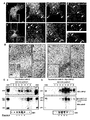
- Figure 1 Human cath-D binds to the extracellular domain of the ⁇ chain of LRP1 in vitro in a RAP insensitive manner.
- Cath-D triggers LRP1 RIP.
- cath-D promotes fibroblast invasive growth in a LRP1-dependent paracrine manner.
- Figure 8 Model of cath-D action through its interaction with LRP1 in fibroblast.
- Pro-cath-D secreted by cancer cells binds to residues 349-394 in the extracellular domain of the ⁇ chain of LRP1, possibly in lipid rafts, in fibroblasts. Upon binding, pro-cath-D is internalized and, due to the acidification within the early endosomes, is processed. Activated cath-D cleaves LRP1 within the (349-394) binding domain. Shed LRP1 ectodomain is released in the serum through recycling vesicules.
- Ectodomain shedding results in the production of a 25 kDa membrane-bound fragment (LRP1 ⁇ -CTF), that is further cleaved by ⁇ -secretases, leadind to the production of LRP1-ICD, that may affect gene transcription. Since mutated D231N cath-D binds to LRP1 and does not trigger LRP1 RIP, we propose that cath-D binding to LRP1 may also lead to the activation of other unknown intracellular signalling pathways that elicit fibroblast invasive growth.
- Figure Sup. 1 Expression of LRP1 in fibroblasts and breast cancer cells.
- FIG. 2 LRP1 expression is required for fibroblastic morphology in three-dimensional matrices.
- HMF fibroblasts transfected with either Luc siRNA (panels a, d, f), LRP1 siRNA1 (panels b and e) or LRP1 siRNA2 (panel g) were seeded in Matrigel 48 h post-transfection.
- Phase contrast images taken after 3 days of culture are shown (panels a, b, f, g).
- p-nitrotetrazolium violet cell staining after 5 day of culture is also given (panels d and e). Data from one representative experiment out of 3 is shown. Bars (---, 75 ⁇ m; -, 750 ⁇ m).
- Cells transiently expressing Luc siRNA, LRP1 siRNA1 or LRP1 siRNA2 were analyzed by immunoblotting 48 h post-transfection (panels c, h).
- FIG. 4 Inhibition of LRP1 in HMF fibroblasts prevents cath-D invasive growth stimulation.
- HMF fibroblasts transfected with either Luc siRNA (panels a, b) or LRP1 siRNA2 (panels c, d) were embedded 48 h post-transfection in the presence of a bottom layer of 3Y1-Ad12 cancer cell lines secreting either no cath-D (control, panels a and c) or human cath-D (cath-D, panels b and d).
- Phase contrast optical photomicrographs of HMF fibroblasts after 3 days of co-culture are shown in panels a-d. Bars (---, 75 ⁇ m).
- Pro-cath-D secretion was analyzed after 3 days of co-culture of either Luc (panel e, left panel) or LRP1 (panel e, right panel) siRNA HMF fibroblasts with 3Y1Ad12/control or 3Y1Ad12/cath-D cells by immunoblotting.
- NS non specific contaminant protein.
- HMF cells were transfected with either Luc siRNA or LRP1 siRNA2 and LRP1 ⁇ expression was monitored 48 h post-transfection before the co-culture assays (panel f).
- HMF Human mammary fibroblasts
- Cath-D-deficient CD55-/- immortalized mouse fibroblasts were kindly provided by Dr Christoph Peters (University of Freiburg, Germany) and Dr Paul Saftig (Christian-Albrechts-University, Germany).
- CD55-/-SV40 and CD55-/-cath-D cell lines were obtained by cath-D stable transfection (Laurent-Matha et al, 2005).
- PEA13 and PEA10 mouse fibroblasts were purchased from ATCC.
- the anti-human cath-D monoclonal antibody (#610800, BD Biosciences) used for immuno-blotting recognizes 52, 48 and 34 kDa cath-D forms.
- the anti-human cath-D M1G8 monoclonal antibody used for immunoprecipitation recognizes 52, 48 and 34 kDa cath-D forms.
- the anti-human cath-D monoclonal antibody M2E8 used for immuno-fluorescence interacts specifically with 52 kDa pro-cath-D.
- Anti-HA monoclonal antibody (12CA5 clone) was purchased from Roche, anti-GAPDH monoclonal antibody from Santa Cruz Biotechnology and anti-ßactin polyclonal antibody (#A2066) from Sigma.
- DAPT and pepstatin A were purchased from Sigma.
- the 49-mer (349-397) LRP1 peptide was synthetized by GeneCust.
- Plasmids. pcDNA3-Myc-tagged LRP1 ⁇ chain, LRP1-IV-HA mini-receptor and SEAP-LRP1 ⁇ were described elsewhere (Barnes et al., 2003 ; Zhang et al., 2004 ; von Arnim et al., 2005).
- the SEAP-LRP1 ⁇ was used instead of full-length LRP1 because of its functional similarity to and better expression than full-length LRP1 as previously described (Von Arnim et al., 2005).
- pcDNA3.1(-)cath-D and pSG5-neo-cath-D expression plasmids were previously described (Glondu et al., 2001; Laurent-Matha et al., 2005).
- pcDNA3.1(+)Myc-tagged LRP1 ⁇ chain was generated by inserting cDNA encoding LRP1 ⁇ obtained by Kpn I and Xba I digestion of pcDNA3-Myc-tagged LRP1 ⁇ into pcDNA3.1(+) previously digested by Kpn I and Xba I.
- pcDNA3.1(+)LRP1 ⁇ extracellular domain (1-476) expression plasmid was created by inserting the PCR-amplified cDNA encoding LRP1 ⁇ (1-476) from pcDNA3.1(+)Myc-tagged LRP1 ⁇ into pcDNA3.1(+) previously digested by Nhe I and Xba I.
- pGEX-4T-1 LRP1 ⁇ (307-479) was generated by inserting PCR-amplified cDNA encoding LRP1 ⁇ (307-479) from pYESTrp2-LRP1 ⁇ (307-479) fished by two-yeast hybrid into pGEX-4T-1 previously digested by Eco RI .
- pGEX-2TK -34, -14 and -4 kDa cath-D domains were obtained by inserting PCR-amplified cDNA encoding cath-D fragments into pGEX-4T-1 previously digested by Eco RI.
- Yeast two-hybrid assay Full-length 52 kDa precursor, 48 kDa intermediate, 34 kDa mature heavy chain, 14 kDa mature light chain and 4 kDa pro-fragment of human cath-D were each fused with the LexA DNA-binding domain in the pMW101 vector.
- a cDNA library derived from normal breast tissue was cloned into the galactosidase-inducible pYESTrp2 vector (Invitrogen) containing B42 activation domain.
- the yeast two-hybrid screen was performed as described previously (Slentz-Kesler et al., 2000).
- PEA13 cells were co-transfected with a 10-fold excess of wild-type cath-D or empty pSG5-neo vector and a pTK-Hyg hygromycin-resistance expression vector (Clontech) using Lipofectamine (Invitrogen). Colonies growing in the presence of hygromycin B (100 ⁇ g/ml, Invitrogen) were pooled and human cath-D was quantified using an immunoradiometric assay (Glondu et al., 2001).
- 30,000 HMF cells were plated in 6-well plates and after 3 days cells were transiently transfected with 10 ⁇ g human LRP1 or Luc siRNAs using Oligofectamine (Invitrogen). 35,000 MDA-MB-231 cells were plated in 12-well plates and after 2 days were transiently transfected with 2 ⁇ g human LRP1 or Luc siRNAs. In other experiments, 50,000 CD55-/-cath-D cells were plated in 12-well plates and after 2 days were transiently transfected with 2.5 ⁇ g mouse LRP1 or Luc siRNAs.
- CD55-/-SV40, CD55-/- cath-D, PEA10, PEA13 and PEA13-cath-D cells were re-suspended at 4°C in Matrigel (0.2 ml, 10 mg/ml) (BD Biosciences) and quickly added to a pre-set layer of Matrigel in 24-well plates (Glondu et al., 2001).
- Outgrowth assays were also performed with HMF, MDA-MB-231 and CD55-/-cath-D cells, 24 or 48 h post-transfection with LRP or Luc siRNAs.
- Duplexes of 21-nucleotide human LRP1 siRNA1 (target sequence AAGCAGTTTGCCTGCAGAGAT (SEQ ID NO: 6), residues 666-684) (Li et al., 2003), human LRP1 siRNA2 (target sequence AAGCTATGAGTTTAAGAAGTT (SEQ ID NO: 7)) (# D-040764-04-0010 Dharmacon), mouse LRP1 siRNA3 (target sequence AAGCAUCUCAGUAGACUAUCA (SEQ ID NO: 8)) (Fears and al., 2005), mouse LRP1 siRNA4 (target sequence AAGCAGTTTGCCTGCAGAGAC (SEQ ID NO: 9)) or firefly luciferase (Luc) siRNA (target sequence AACGTACGCGGAATACTTCGA (SEQ ID NO: 10)) were synthesized by MWG Biotech S.A. (France) or Dharmacon.
- human LRP1 siRNA2 target sequence AAGCTATGAGTTT
- pro-cath-D or LRP1 ⁇ (1-476) were obtained by transcription and translation of pcDNA3.1(-)cath-D or pcONA3.1(+)LRP1 ⁇ extracellular domain(1-476) expression plasmids, respectively, using the TNT T7 -coupled reticulocyte lysate system (Promega).
- GST, GST-LRP1 ⁇ (307-479), GST-LRP1 ⁇ shorter fragments, GST-cath-D-52K, GST-cath-D-34K, GST-cath-D-14K and GST-cath-D-pro-domain fusion proteins were produced in Escherichia coli B strain BL21 using isopropyl-1-thio- ⁇ -D-galactopyranoside (1 mM) for 3 h at 37°C. GST fusion proteins were purified on glutathione-Sepharose beads (Amersham Biosciences).
- COS-1 cells were transfected at 100% confluency with 10 ⁇ g of pcDNA3-Myc- LRP1 ⁇ or LRP1-IV-HA mini-receptor with or without 10 ⁇ g pcDNA3.1(-) or pcDNA3.1(-)cath-D vectors.
- Transient transfections were carried out using Lipofectamine and Opti-MEM (Gibco-BRL).
- Lysates were incubated with 3 ⁇ g anti-cath-D M1 G8 monoclonal antibody or 40 ⁇ l anti-LRP1 11 H4 hybridoma overnight at 4°C, and subsequently with 25 ⁇ l 10% protein A- or G-Sepharose, respectively, for 2 h at 4°C on an agitator. Sepharose beads were washed 4 times with PLC buffer, boiled for 3 min in SDS sample buffer and resolved by SDS-PAGE.
- HMF fibroblasts were incubated for 24 h in DEM medium supplemented with the conditioned medium prepared from 3Y1-Ad12-cath-D cells corresponding to 3 x 10 6 cpm of total TCA-precipitable proteins, 10% FCS, 10 mM of methionine and 5 mM of cysteine. After incubation, the medium was discarded and cellular extracts were prepared. 35 S-Cath-D endocytosed into cells was immunoprecipitated with M1G8 anti-cath-D antibody and analyzed by 15% SDS-PAGE and autoradiography (Capony et al., 1987).
- Immunoblotting Cells grown to 80% confluency were lysed in PLC buffer. Equal amounts of protein (100 ⁇ g) from cell extracts quantitated by the Bradford assay, or equal volumes (80 ⁇ l) of conditioned media, were separated by SDS-PAGE on a 7.5 % gel for anti-LRP1 ⁇ immunoblotting or on a 12 % gel for anti-cath-D immuno-blotting.
- Proteins were electro-transferred to PVDF membranes (VWR) and incubated with 11 H4 anti-LRP1 ⁇ hybridoma (1/10 dilution), 1 ⁇ g/ml anti-human cath-D, 1 ⁇ g/ml anti-HA (12CA5 clone, Roche), 1 ⁇ g/ml anti-GAPDH (Santa Cruz Biotechnology, Inc.) monoclonal antibodies, or anti-ßactin polyclonal antibody (A2066, Sigma) (1/1000 dilution). Proteins were visualized with horseradish peroxidase-conjugated sheep anti-mouse immunoglobulin (Amersham) followed by the enhanced chemiluminescence system ECL (Perkin Life Sciences).
- COS cells grown in 100 mm Petri dishes and transfected as described above were lysed in MEB buffer (150 mM NaCl, 20 mM Morpholine Ethane Sulfonic acid, [pH 6.5]) containing 1% Triton X100, 1 mM Na 3 VO 4 , 50 mM NaF, and protease inhibitor cocktail, and were homogenized with a dounce homogenizer.
- MEB buffer 150 mM NaCl, 20 mM Morpholine Ethane Sulfonic acid, [pH 6.5]
- the preparation (4 ml) was mixed with an equal volume of 80% sucrose solution in MEB buffer in an ultracentrifuge tube over-layed with 4 ml of 30% sucrose and 1.5 ml of 5 % sucrose, and centrifuged at 39 000 rpm for 16 h at 4°C in a Beckman SW40 Ti. After centrifugation, 8 fractions (1 ml each) were collected from the top of each tube. An equivalent aliquot of each fraction was subjected to immunoblot analysis.
- COS cells grown in 12-well plates and transfected as previously described were fixed with 4 % paraformaldehyde, permeabilized with 0.1% Triton X100, and blocked with 2.5 % goat serum (Sigma). Cells were then incubated with an anti-LRP1 ⁇ rabbit polyclonal antibody (1/200) followed by a FITC-conjugated goat anti-rabbit IgG (1/200; Jackon ImmunoResearch).
- Immunogold Cells were fixed with 3% paraformaldehyde and 0.1% glutaraldehyde in a phosphate buffer [pH 7.2] overnight at 4°C and were then cryoprotected. Ultra-thin sections (85 nm) were incubated in PBS first with 0.1 % cold water fish gelatin, 1 % BSA, and 0.05 % Tween 20 (incubation buffer), then with rabbit anti-LRP1 ⁇ (1/20) and mouse anti-cath-D M2E8 (4 ⁇ g/ml), and subsequently with 15 nm goat anti-rabbit-gold and 6 nm goat anti-mouse-gold 5 (Aurion) diluted 1/20. Sections were observed using a Hitachi 7100 transmission electron microscope.
- LRP1 ectodomain secretion assay COS cells plated in 12-well plates were transfected with a ⁇ -galactosidase reporter, LRP1 ⁇ -fused N-terminally to secreted alkaline phosphatase (SEAP) and either empty or cath-D vectors. The medium was changed 24 h later, and then collected after another 24 h. Measurement of SEAP activity in the conditioned media was carried out in duplicate by chemiluminescent assay (Roche Applied Science). SEAP activity was normalized to ⁇ -galactosidase activity measured by hydrolysis of o -nitrophenyl- ⁇ -D-galactopyranoside in cells lysed in the reporter lysis buffer (Promega).
- RNA extraction, RT-PCR and Q-PCR RNA extraction and reverse transcription were performed and Q-PCR was carried out using a LightCycler and the DNA double strand specific SYBR Green I dye for detection (Roche). Q-PCR was performed using gene-specific oligonucleotides and results were normalized to RS9 levels.
- a yeast two-hybrid approach identified cath-D as a ligand for the extracellular domain of the LRP1 ⁇ chain.
- yeast two-hybrid screens using LexA DNA-binding domain fusion proteins containing various fragments of cath-D as bait and a cDNA library isolated from normal breast tissue fused to the transactivation domain of B42. Results from the initial yeast two-hybrid screen were confirmed by one-on-one two-hybrid assays and indicates that human cath-D binds to the ⁇ chain of LRP1.
- the clone we isolated in our screen is 100% identical to amino acids 307-479 of the ⁇ chain of LRP1 ( Fig. 1A ).
- Cath-D associates in vitro with the extracellular domain of the ⁇ chain of LRP1.
- GST-fusion proteins containing amino acids 307-479 of the extracellular domain of the LRP1 ⁇ chain ( Fig. 1C panel a, GST-LRP1 ⁇ (307-479)) were immobilized on glutathione-agarose beads and incubated with full-length pro-cath-D, labelled with 35 S-Met by in vitro translation.
- the results show binding of pro-cath-D to GST-LRP1 ⁇ (307-479) ( Fig. 1C , panel b).
- the 52, 34, 14 and 4 kDa cath-D fragments were expressed as GST fusion proteins ( FIG. 1D , panels a and c) and tested for their ability to bind to the full-length extracellular domain of LRP1 ⁇ (1-476).
- LRP1 ⁇ (1-476) was shown to bind specifically to both the 52, 34 and 14 kDa cath-D polypeptides ( Fig. 1D , panels b and d).
- GST-RAP Fig. 1E, panel a
- Pro-cath-D and the ⁇ chain of LRP1 associate in vivo and co-distribute in cells .
- a cDNA encoding Myc-LRP1 ⁇ in which the ⁇ chain of LRP1 was linked at the amino-terminus to a signal peptide and a Myc epitope (Barnes et al., 2003).
- Myc-LRP1 ⁇ was expressed in COS cells in absence or presence of wild-type or D231 N mutated cath-D.
- Our results show that both wild-type and D231 N cath-D precursor co-immunoprecipitate with the ⁇ chain of LRP1 ( Fig.
- D231N cath-D displayed an increased electrophoretic mobility, corresponding to a shift of about 1 kDa in the apparent molecular mass ( Fig. 3A , panel a), as previously described (Laurent-Matha et al., 2006).
- a longer gel exposure revealed that the 48 and 34 kDa cath-D polypeptides also co-immunoprecipitate with LRP1 ⁇ ( Fig. 3A , panel c). These results suggest that the LRP1 ⁇ chain interacts preferentially with the 52 kDa cath-D precursor ( Fig. 3A , panel a).
- pro-cath-D and the associated LRP1 ⁇ chain can be directed to the lysosomes, as revealed by co-immunoprecipitation of LRP1 ⁇ chain with the 48 and 34 kDa forms of cath-D ( Fig. 3A , panel c).
- anti-LRP1 ⁇ chain immunoblotting of anti-cath-D immunoprecipitate confirmed that the ⁇ chain of LRP1 co-immunoprecipitates with cath-D and D231N cath-D ( Fig. 3A , panel b).
- these results demonstrate that the pro-cath-D precursor, as well its mature forms, interact with the ⁇ chain of LRP1 in cells.
- Pro-cath-D and LRP1 co-localize partially in cells.
- COS cells transiently co-expressing cath-D and Myc-LRP1 ⁇ were analyzed by confocal microscopy after double staining with a monoclonal antibody specific for the pro-cath-D precursor and a polyclonal antibody specific for the cytoplasmic tail of the LRP1 ⁇ .
- Figure 4A shows that a fraction of pro-cath-D (in red) was co-localized with LRP1 ⁇ (in green) in punctuate structures.
- Pro-cath-D also partially co-localized with LRP1 ⁇ in the presence of 10 mM M6P, known to prevent its endocytosis via the M6P receptors ( Fig.
- Pro-cath-D was mainly located in the non-raft fractions 7 and 8, even through some pro-cath-D and its 48 kDa intermediate form were also detected in the raft-rich fractions 5 and 6 ( Fig. 4C , panel a, medium panel).
- Overexpression of Myc-LRP1 ⁇ which localized largely to the raft-rich fractions ( Fig. 4C , panel b, top panel), resulted in a large increase in the presence of pro-cath-D and its neo-processed forms (e.g.
- LRP1 RIP ⁇ chain of LRP1 can undergo ectodomain shedding by the ⁇ -secretase type 1 membrane-associated aspartic protease BACE1, followed by intracellular cleavage by ⁇ -secretases, in a process called RIP (Regulated Intramembrane Proteolysis) (von Arnim et al., 2005).
- cath-D led to a 2.5 fold increase in SEAT-LRP1 ectodomain shedding when compare to controls ( Fig. 5A , panel a).
- Pretreatment of cath-D expressing cells with the aspartic protease inhibitor, pepstatin A prevented SEAP-LRP1 ⁇ ectodomain shedding ( Fig. 5A , panel b).
- Cleavage specificity was also confirmed by co-transfecting cath-D with an anti-cath-D siRNA (Bidère et al., 2003) that led to an inhibition of SEAP-LRP1 ⁇ cleavage ( Fig. 5A , panel b).
- LRP1 is highly expressed in fibroblasts.
- Fig. Sup1 To examine the functional significance of the interaction between cath-D and the LRP1 ⁇ chain , we first screened a variety of cell lines for LRP1 and cath-D expressions (Fig. Sup1). Our survey suggests that LRP1 is highly expressed in fibroblast cell lines and this conclusion is consistent with the data obtained from cancer biopsies (Christensen et al., 1996; Obermeyer et al., 2007). In contrast, breast cancer cells express low levels of the LRP1 ⁇ chain while over-secreting pro-cath-D (Capony et al., 1989).
- Cath-D interacts with endogenous LRP1 in fibroblasts.
- cath-D interacts with LRP1 endogenously expressed in fibroblasts.
- endogenous LRP1 expression was inhibited by siRNA1 ( Fig. 6A ; see Fig. Sup. 3A, panel a).
- siRNA1 Fig. 6A ; see Fig. Sup. 3A, panel a.
- Addition of the ⁇ -secretase inhibitor DAPT led to a further increase in the level of membrane bound LRP1 ⁇ -CTF, that was significantly higher in the presence of pro-cath-D as compared to control (161.8 % +/- 6.6 (n 4), p ⁇ 0.005, Student' t test).
- LRP1 is a mediator of cath-D stimulation of fibroblast invasive growth.
- LRP1 expression was necessary for basal outgrowth of HMF human mammary fibroblasts in 3D Matrigel.
- HMF transfected with control Luc siRNA grew poorly in Matrigel and adopted an elongated morphology typical for fibroblasts (Fig. Sup. 2, panels a, f).
- Inhibition of LRP1 expression by siRNA1 and siRNA2 led to a rounded morphology and to a decrease of the protrusions (Fig.
- Cath-D then cleaves LRP1 at 3 major sites within the 349-394 binding domain in the ⁇ chain. Shed LRP1 ectodomain is then released in the serum through recycling vesicles. Ectodomain shedding results in the production of a 25 kDa membrane-bound fragment (LRP1 ⁇ -CTF), that is further cleaved by ⁇ -secretases, leading to the production of LRP1 ⁇ -ICD, that might affect gene transcription according to previous reports (May et al., 2002; von Arnim et al., 2005). Our results therefore support a model in which pro-cath-D that is secreted by breast cancer cells acts as an extracellular messenger that signals by binding to LRP1 present on the surface of fibroblasts.
- LRP1 ⁇ -CTF membrane-bound fragment
- Cath-D is an aspartic protease that is overexpressed and secreted in the extracellular environment by human epithelial breast cancer cells (Westley and Rochefort, 1980; Vignon et al., 1986; Capony et al., 1987; Capony et al., 1989).
- Cath-D stimulates tumor cell proliferation, fibroblast invasive growth, and tumor angiogenesis (Vignon et al., 1986; Garcia et al., 1990; Vetvicka et al., 1994; Liaudet et al., 1994; Liaudet et al., 1995; Glondu et al., 2001; Glondu et al., 2002; Berchem et al., 2002; Laurent-Matha et al., 2005).
- M6P receptors bind and can mediate pro-cath-D uptake into cells (Vignon et al., 1986; Von Figura and Hasilik, 1986; Laurent-Matha et al., 1998; Heylen et al., 2002).
- M6P receptors do not mediate the cath-D mitogenic effects has led to a search for alternative cath-D receptors (Fusek and Vetvicka, 1994; Glondu et al., 2001; Rochefort and Liaudet-Coopman, 1999; Benes et al., 2006; Liaudet-Coopman et al., 2006).
- LRP1 was originally identified as an internalization receptor that is related to the LDL receptor (Herz and Strickland, 2001). Recently, it has become clear that LRP1 may be involved in signal transduction by protein tyrosine kinases (Herz and Strickland, 2001).
- Tyrosine phosphorylation of the NPXY motifs that are present in the cytoplasmic region of LRP1 has been observed in response to PDGF-BB (Loukinova et al., 2002; Boucher et al., 2002; Boucher et al., 2003; Boucher and Gotthardt, 2004; Newton et al., 2005), CTGF (Yang et al., 2004), or tPA (Hu et al., 2006) and in fibroblasts transformed with v-Src (Barnes et al., 2001; Barnes et al., 2003).
- RIP is a process that involves two cleavage events (Brown et al., 2000). The first cleavage event occurs in the extracellular region close the plasma membrane and results in the release of the extracellular region, a process also known as ectodomain shedding. Ectodomain shedding leaves behind an integral membrane protein with a very short extracellular domain that is subject to proteolytical cleavage within its transmembrane domain.
- Intramembrane cleavage can be carried out by a small group of enzymes that include ⁇ -secretases (Kopan and Ilagan, 2004). Upon release into the cytosol, cytoplasmic fragments can interact with intracellular signalling proteins, diffuse to nucleus and control gene transcription (Brown et al., 2000).
- LRP1 ⁇ ectodomain can be cleaved by the ⁇ -secretase type I membrane-associated aspartic protease BACE1 (Von Arnim et al., 2005), previously implicated in APP RIP and Alzheimer disease (Vassar et al., 1999).
- LRP1 can then act as a substrate for ⁇ -secretases (May et al., 2002; Von Arnim et al., 2005). When expressed by itself, the cytoplasmic domain of LRP1 localizes to the nucleus where it can associate with Fe65 and Tip60 (Konishita et al., 2003). Together these observations support the notion that LRP1 is subject to RIP and our results strongly suggest that LRP1 RIP may be induced, not only by BACE1 in the neuronal pathology (Von Arnim et al., 2005), but as well by cath-D in the breast tumor micro-environment.
- LRP1 has been localized not only to clathrin coated pits, but also to lipid rafts either transiently in the absence of a stimulus (Wu et al., 2005) or in response to PDGF-BB or insulin (Boucher et al., 2002; Zhang et al., 2004). It is now well recognized that BACE 1 and ⁇ -secretases are also highly enriched in lipid raft micro-domains (Vetrivel et al., 2004), suggesting that LRP1 RIP may take place in this particular micro-environment.
- cath-D binding to LRP1 may lead to the activation of one or more intracellular signalling pathways that elicit fibroblast invasive growth.
- Previous reports have described variable effects of LRP1 on proliferation, motility or invasion (Lillis et al., 2005). Discrepancies in the results may be based on differences in the ligands or cell lines that were used in those studies (Lillis et al., 2005).
- LRP1 we describe a role for LRP1 as a positive regulator of the invasive outgrowth of mammary tissue fibroblasts that is seen in response to pro-cath-D.
- LRP1 is expressed on fibroblasts in breast cancer biopsies and on the invasive front of fibroblasts in colon cancer biopsies (Christensen et al., 1996, Obermeyer et al., 2007), and because the C766T LRP1 gene polymorphism is associated with an increased risk of developing breast cancer (Benes et al., 2003), LRP1 may be involved in the onset and progression of various human malignancies Others and we believe that cath-D is involved in creating the micro-environment that can sustain breast tumor growth and progression.
- cath-D in addition to be a prognostic marker for breast cancer, may become a potential target for therapeutic intervention.
- the identification of a LRP1 peptide (349-397) that can inhibit pro-cath-D binding opens new therapeutic strategies.
- Our study further indicates that soluble LRP1 ⁇ chain ecto-domain may be detected in the circulation of breast cancer patients that overexpress cath-D. This may be used as a diagnostic tool for early detection of breast cancer.
Landscapes
- Health & Medical Sciences (AREA)
- Chemical & Material Sciences (AREA)
- Organic Chemistry (AREA)
- Life Sciences & Earth Sciences (AREA)
- Genetics & Genomics (AREA)
- Immunology (AREA)
- Medicinal Chemistry (AREA)
- Molecular Biology (AREA)
- General Health & Medical Sciences (AREA)
- Biochemistry (AREA)
- Zoology (AREA)
- Engineering & Computer Science (AREA)
- Proteomics, Peptides & Aminoacids (AREA)
- Biophysics (AREA)
- Wood Science & Technology (AREA)
- Bioinformatics & Cheminformatics (AREA)
- Biomedical Technology (AREA)
- General Engineering & Computer Science (AREA)
- Biotechnology (AREA)
- Microbiology (AREA)
- Cell Biology (AREA)
- Toxicology (AREA)
- Gastroenterology & Hepatology (AREA)
- Medicines That Contain Protein Lipid Enzymes And Other Medicines (AREA)
- Peptides Or Proteins (AREA)
- Investigating Or Analysing Biological Materials (AREA)
Priority Applications (2)
| Application Number | Priority Date | Filing Date | Title |
|---|---|---|---|
| EP07301421A EP2045266A1 (de) | 2007-10-02 | 2007-10-02 | Verfahren zur Behandlung und Diagnostizierung einer krebserzeugenden cath-D- oder Alzheimer-Erkrankung |
| PCT/EP2008/063109 WO2009043858A2 (en) | 2007-10-02 | 2008-09-30 | Methods for treating and diagnosing a cancer secreting cath-d or alzheimer's disease |
Applications Claiming Priority (1)
| Application Number | Priority Date | Filing Date | Title |
|---|---|---|---|
| EP07301421A EP2045266A1 (de) | 2007-10-02 | 2007-10-02 | Verfahren zur Behandlung und Diagnostizierung einer krebserzeugenden cath-D- oder Alzheimer-Erkrankung |
Publications (1)
| Publication Number | Publication Date |
|---|---|
| EP2045266A1 true EP2045266A1 (de) | 2009-04-08 |
Family
ID=39107040
Family Applications (1)
| Application Number | Title | Priority Date | Filing Date |
|---|---|---|---|
| EP07301421A Withdrawn EP2045266A1 (de) | 2007-10-02 | 2007-10-02 | Verfahren zur Behandlung und Diagnostizierung einer krebserzeugenden cath-D- oder Alzheimer-Erkrankung |
Country Status (2)
| Country | Link |
|---|---|
| EP (1) | EP2045266A1 (de) |
| WO (1) | WO2009043858A2 (de) |
Cited By (3)
| Publication number | Priority date | Publication date | Assignee | Title |
|---|---|---|---|---|
| WO2011054644A1 (en) * | 2009-10-14 | 2011-05-12 | Fraunhofer-Gesellschaft zur Förderung der angewandten Forschung e.V. | A low density lipoprotein-related protein 1 splice variant as cancer marker |
| WO2016188911A1 (en) * | 2015-05-22 | 2016-12-01 | INSERM (Institut National de la Santé et de la Recherche Médicale) | Human monoclonal antibodies fragments inhibiting both the cath-d catalytic activity and its binding to the lrp1 receptor |
| WO2020127411A1 (en) * | 2018-12-19 | 2020-06-25 | INSERM (Institut National de la Santé et de la Recherche Médicale) | Methods and compositions for treating cancers by immuno-modulation using antibodies against cathespin-d |
Families Citing this family (2)
| Publication number | Priority date | Publication date | Assignee | Title |
|---|---|---|---|---|
| US10273294B2 (en) | 2010-10-11 | 2019-04-30 | University Of Southern California | Compositions and methods for treating HIF-1α over-expressing cancers |
| WO2012051207A2 (en) * | 2010-10-11 | 2012-04-19 | University Of Southern California | FRAGMENT OF SECRETED HEAT SHOCK PROTEIN-90ALPHA (Hsup90ALPHA) AS VACCINES OR EPITOPE FOR MONOCLONAL ANTIBODY DRUGS OR TARGET FOR SMALL MOLECULE DRUGS AGAINST A RANGE OF SOLID HUMAN TUMORS |
Citations (1)
| Publication number | Priority date | Publication date | Assignee | Title |
|---|---|---|---|---|
| JP2005304496A (ja) * | 2004-03-23 | 2005-11-04 | Joji Inasawa | 食道癌の検出方法、および、抗食道癌物質のスクリーニング方法 |
-
2007
- 2007-10-02 EP EP07301421A patent/EP2045266A1/de not_active Withdrawn
-
2008
- 2008-09-30 WO PCT/EP2008/063109 patent/WO2009043858A2/en active Application Filing
Patent Citations (1)
| Publication number | Priority date | Publication date | Assignee | Title |
|---|---|---|---|---|
| JP2005304496A (ja) * | 2004-03-23 | 2005-11-04 | Joji Inasawa | 食道癌の検出方法、および、抗食道癌物質のスクリーニング方法 |
Non-Patent Citations (6)
| Title |
|---|
| BEAUJOUIN M ET AL: "Overexpression of both catalytically active and -inactive cathepsin D by cancer cells enhances apoptosis-dependent chemo-sensitivity", ONCOGENE, vol. 25, no. 13, March 2006 (2006-03-01), pages 1967 - 1973, XP002471251, ISSN: 0950-9232 * |
| GLONDU MURIELLE ET AL: "A mutated cathepsin-D devoid of its catalytic activity stimulates the growth of cancer cells", ONCOGENE, vol. 20, no. 47, 18 October 2001 (2001-10-18), pages 6920 - 6929, XP002471252, ISSN: 0950-9232 * |
| KINOSHITA AYAE ET AL: "The intracellular domain of the low density lipoprotein receptor-related protein modulates transactivation mediated by amyloid precursor protein and Fe65.", THE JOURNAL OF BIOLOGICAL CHEMISTRY 17 OCT 2003, vol. 278, no. 42, 17 October 2003 (2003-10-17), pages 41182 - 41188, XP002480007, ISSN: 0021-9258 * |
| LAURENT-MATHA VALERIE ET AL: "Catalytically inactive human cathepsin D triggers fibroblast invasive growth", JOURNAL OF CELL BIOLOGY, vol. 168, no. 3, 31 January 2005 (2005-01-31), pages 489 - 499, XP002471253, ISSN: 0021-9525 * |
| LIAUDET-COOPMAN ET AL: "Cathepsin D: newly discovered functions of a long-standing aspartic protease in cancer and apoptosis", CANCER LETTERS, NEW YORK, NY, US, vol. 237, no. 2, 18 June 2006 (2006-06-18), pages 167 - 179, XP005463597, ISSN: 0304-3835 * |
| MA SUK LING ET AL: "Low-density lipoprotein receptor-related protein 8 (apolipoprotein E receptor 2) gene polymorphisms in Alzheimer's disease.", NEUROSCIENCE LETTERS 8 NOV 2002, vol. 332, no. 3, 8 November 2002 (2002-11-08), pages 216 - 218, XP002480008, ISSN: 0304-3940 * |
Cited By (4)
| Publication number | Priority date | Publication date | Assignee | Title |
|---|---|---|---|---|
| WO2011054644A1 (en) * | 2009-10-14 | 2011-05-12 | Fraunhofer-Gesellschaft zur Förderung der angewandten Forschung e.V. | A low density lipoprotein-related protein 1 splice variant as cancer marker |
| WO2016188911A1 (en) * | 2015-05-22 | 2016-12-01 | INSERM (Institut National de la Santé et de la Recherche Médicale) | Human monoclonal antibodies fragments inhibiting both the cath-d catalytic activity and its binding to the lrp1 receptor |
| US10526415B2 (en) | 2015-05-22 | 2020-01-07 | INSERM (Institut National de la Santé et de la Recherche Médicale) | Human monoclonal antibodies fragments inhibiting both the Cath-D catalytic activity and its binding to the LRP1 receptor |
| WO2020127411A1 (en) * | 2018-12-19 | 2020-06-25 | INSERM (Institut National de la Santé et de la Recherche Médicale) | Methods and compositions for treating cancers by immuno-modulation using antibodies against cathespin-d |
Also Published As
| Publication number | Publication date |
|---|---|
| WO2009043858A2 (en) | 2009-04-09 |
| WO2009043858A9 (en) | 2009-07-09 |
| WO2009043858A3 (en) | 2009-05-28 |
Similar Documents
| Publication | Publication Date | Title |
|---|---|---|
| Song et al. | Rhomboid domain containing 1 promotes colorectal cancer growth through activation of the EGFR signalling pathway | |
| Najy et al. | The ectodomain shedding of E-cadherin by ADAM15 supports ErbB receptor activation | |
| Klessner et al. | EGFR and ADAMs cooperate to regulate shedding and endocytic trafficking of the desmosomal cadherin desmoglein 2 | |
| McNutt et al. | Antagonism of secreted PCSK9 increases low density lipoprotein receptor expression in HepG2 cells | |
| Van Veen et al. | Negative regulation of urokinase receptor activity by a GPI-specific phospholipase C in breast cancer cells | |
| Baldwin et al. | Protein arginine methyltransferase 7 promotes breast cancer cell invasion through the induction of MMP9 expression | |
| Chalaris et al. | ADAM17-mediated shedding of the IL6R induces cleavage of the membrane stub by γ-secretase | |
| US20160251662A1 (en) | Compositions and methods for treating cancer and modulating stress granule formation | |
| US20090162878A1 (en) | Methods and compositions for detecting and quantifying sappb | |
| US20030054985A1 (en) | N-cadherin modulated migration, invasion, and metastasis | |
| US9062309B2 (en) | Use of a growth-stimulating protein | |
| US20060034848A1 (en) | Methods and compositions for treating Alzheimer's disease | |
| EP2045266A1 (de) | Verfahren zur Behandlung und Diagnostizierung einer krebserzeugenden cath-D- oder Alzheimer-Erkrankung | |
| WO2007015587A1 (ja) | アポトーシス促進剤、細胞増殖阻害剤、癌の予防・治療剤、及びそのスクリーニング方法 | |
| JP2007521015A (ja) | 癌の診断及び治療に用いられる示差的に発現する腫瘍特異的ポリペプチド | |
| Xie et al. | Disruption and inactivation of the PP2A complex promotes the proliferation and angiogenesis of hemangioma endothelial cells through activating AKT and ERK | |
| JP2024038346A (ja) | 血液から癌を診断する方法 | |
| Kamble et al. | Proteolytic cleavage of Trop2 at Arg87 is mediated by matriptase and regulated by Val194 | |
| Ma et al. | CD82 suppresses ADAM17-dependent E-cadherin cleavage and cell migration in prostate cancer | |
| US7595158B2 (en) | Bcl2L12 polypeptide activators and inhibitors | |
| Matou-Nasri et al. | CD95-mediated apoptosis in Burkitt's lymphoma B-cells is associated with Pim-1 down-regulation | |
| Cong et al. | A disintegrin and metalloprotease 10 activity sheds the ectodomain of the amyloid precursor-like protein 2 and regulates protein expression in proximal tubule cells | |
| US20110039789A1 (en) | Use of Huntingtin Protein for the Diagnosis and the Treatment of Cancer | |
| US20150276760A1 (en) | Method for Screening a Compound Capable of Inhibiting the Notch1 Transcriptional Activity | |
| JP2007523893A (ja) | Atp7aの使用による、神経変性疾患の治療 |
Legal Events
| Date | Code | Title | Description |
|---|---|---|---|
| PUAI | Public reference made under article 153(3) epc to a published international application that has entered the european phase |
Free format text: ORIGINAL CODE: 0009012 |
|
| AK | Designated contracting states |
Kind code of ref document: A1 Designated state(s): AT BE BG CH CY CZ DE DK EE ES FI FR GB GR HU IE IS IT LI LT LU LV MC MT NL PL PT RO SE SI SK TR |
|
| AX | Request for extension of the european patent |
Extension state: AL BA HR MK RS |
|
| AKX | Designation fees paid | ||
| REG | Reference to a national code |
Ref country code: DE Ref legal event code: 8566 |
|
| STAA | Information on the status of an ep patent application or granted ep patent |
Free format text: STATUS: THE APPLICATION IS DEEMED TO BE WITHDRAWN |
|
| 18D | Application deemed to be withdrawn |
Effective date: 20091009 |