EP2037940B1 - Rna interference mediated inhibition of aurorakinase b and its combinations as anticancer therapy - Google Patents
Rna interference mediated inhibition of aurorakinase b and its combinations as anticancer therapy Download PDFInfo
- Publication number
- EP2037940B1 EP2037940B1 EP07859578A EP07859578A EP2037940B1 EP 2037940 B1 EP2037940 B1 EP 2037940B1 EP 07859578 A EP07859578 A EP 07859578A EP 07859578 A EP07859578 A EP 07859578A EP 2037940 B1 EP2037940 B1 EP 2037940B1
- Authority
- EP
- European Patent Office
- Prior art keywords
- sirna
- cancer
- cells
- aurkb
- nucleic acid
- Prior art date
- Legal status (The legal status is an assumption and is not a legal conclusion. Google has not performed a legal analysis and makes no representation as to the accuracy of the status listed.)
- Not-in-force
Links
Images


Classifications
-
- C—CHEMISTRY; METALLURGY
- C12—BIOCHEMISTRY; BEER; SPIRITS; WINE; VINEGAR; MICROBIOLOGY; ENZYMOLOGY; MUTATION OR GENETIC ENGINEERING
- C12N—MICROORGANISMS OR ENZYMES; COMPOSITIONS THEREOF; PROPAGATING, PRESERVING, OR MAINTAINING MICROORGANISMS; MUTATION OR GENETIC ENGINEERING; CULTURE MEDIA
- C12N15/00—Mutation or genetic engineering; DNA or RNA concerning genetic engineering, vectors, e.g. plasmids, or their isolation, preparation or purification; Use of hosts therefor
- C12N15/09—Recombinant DNA-technology
- C12N15/11—DNA or RNA fragments; Modified forms thereof; Non-coding nucleic acids having a biological activity
- C12N15/113—Non-coding nucleic acids modulating the expression of genes, e.g. antisense oligonucleotides; Antisense DNA or RNA; Triplex- forming oligonucleotides; Catalytic nucleic acids, e.g. ribozymes; Nucleic acids used in co-suppression or gene silencing
- C12N15/1137—Non-coding nucleic acids modulating the expression of genes, e.g. antisense oligonucleotides; Antisense DNA or RNA; Triplex- forming oligonucleotides; Catalytic nucleic acids, e.g. ribozymes; Nucleic acids used in co-suppression or gene silencing against enzymes
-
- A—HUMAN NECESSITIES
- A61—MEDICAL OR VETERINARY SCIENCE; HYGIENE
- A61K—PREPARATIONS FOR MEDICAL, DENTAL OR TOILETRY PURPOSES
- A61K47/00—Medicinal preparations characterised by the non-active ingredients used, e.g. carriers or inert additives; Targeting or modifying agents chemically bound to the active ingredient
- A61K47/50—Medicinal preparations characterised by the non-active ingredients used, e.g. carriers or inert additives; Targeting or modifying agents chemically bound to the active ingredient the non-active ingredient being chemically bound to the active ingredient, e.g. polymer-drug conjugates
- A61K47/51—Medicinal preparations characterised by the non-active ingredients used, e.g. carriers or inert additives; Targeting or modifying agents chemically bound to the active ingredient the non-active ingredient being chemically bound to the active ingredient, e.g. polymer-drug conjugates the non-active ingredient being a modifying agent
- A61K47/54—Medicinal preparations characterised by the non-active ingredients used, e.g. carriers or inert additives; Targeting or modifying agents chemically bound to the active ingredient the non-active ingredient being chemically bound to the active ingredient, e.g. polymer-drug conjugates the non-active ingredient being a modifying agent the modifying agent being an organic compound
- A61K47/554—Medicinal preparations characterised by the non-active ingredients used, e.g. carriers or inert additives; Targeting or modifying agents chemically bound to the active ingredient the non-active ingredient being chemically bound to the active ingredient, e.g. polymer-drug conjugates the non-active ingredient being a modifying agent the modifying agent being an organic compound the modifying agent being a steroid plant sterol, glycyrrhetic acid, enoxolone or bile acid
-
- A—HUMAN NECESSITIES
- A61—MEDICAL OR VETERINARY SCIENCE; HYGIENE
- A61P—SPECIFIC THERAPEUTIC ACTIVITY OF CHEMICAL COMPOUNDS OR MEDICINAL PREPARATIONS
- A61P35/00—Antineoplastic agents
-
- A—HUMAN NECESSITIES
- A61—MEDICAL OR VETERINARY SCIENCE; HYGIENE
- A61P—SPECIFIC THERAPEUTIC ACTIVITY OF CHEMICAL COMPOUNDS OR MEDICINAL PREPARATIONS
- A61P43/00—Drugs for specific purposes, not provided for in groups A61P1/00-A61P41/00
-
- C—CHEMISTRY; METALLURGY
- C12—BIOCHEMISTRY; BEER; SPIRITS; WINE; VINEGAR; MICROBIOLOGY; ENZYMOLOGY; MUTATION OR GENETIC ENGINEERING
- C12Y—ENZYMES
- C12Y207/00—Transferases transferring phosphorus-containing groups (2.7)
- C12Y207/11—Protein-serine/threonine kinases (2.7.11)
- C12Y207/11001—Non-specific serine/threonine protein kinase (2.7.11.1), i.e. casein kinase or checkpoint kinase
-
- C—CHEMISTRY; METALLURGY
- C12—BIOCHEMISTRY; BEER; SPIRITS; WINE; VINEGAR; MICROBIOLOGY; ENZYMOLOGY; MUTATION OR GENETIC ENGINEERING
- C12N—MICROORGANISMS OR ENZYMES; COMPOSITIONS THEREOF; PROPAGATING, PRESERVING, OR MAINTAINING MICROORGANISMS; MUTATION OR GENETIC ENGINEERING; CULTURE MEDIA
- C12N2310/00—Structure or type of the nucleic acid
- C12N2310/10—Type of nucleic acid
- C12N2310/14—Type of nucleic acid interfering N.A.
Definitions
- the present invention relates to use of short nucleic acid molecules, such as short interfering nucleic acid (siNA) molecules, for modulating gene and protein expression, including compounds, compositions and uses of small nucleic acid molecules to modulate Aurora B (AurkB) expression.
- short nucleic acid molecules such as short interfering nucleic acid (siNA) molecules
- siNA short interfering nucleic acid
- the compounds and methods of the present invention have applications in cancer therapy either alone or in combination with other therapies.
- Aurora kinases are a family of serine/threonine kinases. Aurora A and B kinase are associated with mitotic events of cell cycle, while Aurora C kinase is expressed only in testis ( Sasai K, Katayama H, Stenoien DL, Fujii S, Nissan R, Kimura M, et al, Aurora-C kinase is a novel chromosomal passenger protein that can complement Aurora-B kinase function in mitotic cells. Cell Motil. Cytoskeleton, 2004; 59:249-263 ).
- Aurora-A kinases also called Aurora-2, STK6, ARK1 and Aurora/IPL-1 related kinase, associates with centrosomes and microtubules during mitosis.
- Aurora A kinase (hereinafter “Aurora A”) localizes to centrosomes and regulates the association between cell cycle machinery and centrosomes ( Hirota T, Kunitoku N, Sasayama T, Marumoto T, Zhang D, Nitta M. Aurora-A and an Interacting Activator, the LIM Protein Ajuba, Are Required for Mitotic Commitment in Human Cells.
- Aurora B kinases also known as Aurorakinase B, Aurora B, Aurora-1, and hereinafter "AurkB"
- Aurorakinase B also known as Aurorakinase B, Aurora B, Aurora-1, and hereinafter "AurkB”
- AurkB localizes to the kinetochores from prophase to metaphase and to the central spindle and the midbody in cytokinesis ( Carmena M. and Earnshaw WC. The cellular geography of aurora Kinases. Nat. Rev. Mol. Cell Biol 2003; 4: 842-854 ).
- AurkB associates with chromosome passenger proteins, inner centromere protein (INCENP), Survivin and Borealin protein to form a quaternary chromosome passenger complex which, along with its sub complexes (AurkB and INCENP) are thought to be required for spindle check point, cytokinesis and phosphorylation of Histone H3, respectively.
- IRCNP inner centromere protein
- Survivin and Borealin protein to form a quaternary chromosome passenger complex which, along with its sub complexes (AurkB and INCENP) are thought to be required for spindle check point, cytokinesis and phosphorylation of Histone H3, respectively.
- Schumacher JM, Golden A, and Donovan P. AIR-2 An Aurora/Ipll-related protein Kinase associated with chromosomes and midbody microtubules is required for polar body extrusion and cytokinesis in Caneorhabditis elegans embryos. J
- AIM-1 a mammalian midbody-associated protein required for cytokinesis. EMBO J. 1998; 17: 667-676 ; Giet R and Glover DM. Drosophilia aurora B Kinase is required for histone H3 phosphorylation and condensing recruitment during chromosome condensation and to organize the central spindle during cytokinasis. J. Cell Biol 2001; 152: 669-682 ).
- AurkB phosphorylates MCAK and thus plays a vital role in regulation of bi-orientation during mitosis ( Giet R, Petretti C and Prigent C. aurora Kinases, aneuploidy and cancer, a coincidence or a real link? Trends. Cell Biol. 2005; 5: 241-250 ).
- aurora B Kinase AURKB
- AurkB is intimately involved in preventing chromosomal instability.
- AurkB Survivin, part of AurkB complex, is a key protector against apoptosis and/or mitotic catastrophe ( Andrews PD. Aurora Kinases: shining lights on the therapeutic horizon? Oncogene 2005; 24: 5005-5015 ). AurkB therefore appears to have a direct role in tumorigenesis.
- Aurora inhibitors have been reported several authors ( Ditchfield C, Johnson VL, Tighe A, Ellston R, Haworth C, Johnson T et al. Aurora B couples chromosome alignment with anaphase by targeting BubR1, Mad2 and Cenp-E to kinetochores. J Cell Biol 2004; 161: 267-280 ; Hauf S, Cole RW,Terra S, Zimmer C, Schnapp G, Walter R et al. The small molecule Hesperadin reveals a role for Aurora B in correcting kinetochore-microtubule attachment and in maintaining the spindle assembly checkpoint.
- AZD1152 (AstraZeneca) has been shown to inhibit spindle aggregation at the time of mitosis and is being evaluated to determine its effect as a specific inhibitor of Aurora kinase and its impact on cell division in proliferating tumors.
- Hesperadin (by Boehringer Ingelheim) inhibits AurkB and 25 other kinases, but not Aurora A or C, and causes only polyploidy with no apparent loss to cell viability.
- VX-680 (Vertex) inhibits all three Aurora and, in a panel of cancer cells, was found to inhibit proliferation, increase apoptosis induction, and induce tumor cell death. Merck is presently conducting three clinical studies of VX-680 in patients with hematologic cancers, recurrent or non-responsive solid tumors, or cancers for which standard therapy does not currently exist. In these studies, the safety and tolerability of VX-680 are being evaluated when administered as either a 24-hour continuous infusion or as a 5-day continuous infusion. Of the Aurora inhibitors in clinical trials, only VX-680 is highly specific for Aurora, but is associated with toxic bone marrow side effects, and the mechanism of tumor cell death is not completely understood ( Giet R, Petretti C and Prigent C. Aurora Kinases, aneuploidy and cancer, a coincidence or a real link? Trends. Cell Biol 2005; 5: 241-250 ).
- Japanese patent application JP 2005-278472 describes a peptide that inhibits activities of AurkB in the inner centromere protein, specifically bonding to the Aurora protein; and also describes a medicinal composition, screening methods, and kits.
- Japanese patent application JP 2005-320351 describes a process for preparing tri- and tetra-substituted pyrimidines, the use in preparing Aurora kinase inhibitors, and methods of treatment.
- US patent application 20050267065 describes compositions and methods for modulating the expression of AurkB, including using chemically modified nucleotides, including small interfering nucleotides. The use of chemically modified nucleotides poses a potential risk of unwanted side effects.
- the epidermal growth factor receptor (EGFR, also known as ErbB-1, or HER1 in humans) is a protein tyrosine kinase. Activation of EGFR leads to transmission of proliferative signals to the nucleus and, via activation of transcription factors, leads to increased proliferation, increased migration, increased adhesion, increased angiogenesis and inhibition of programmed cell death pathways ( Bundy, L., Wells, S., Sealy, L. "C/EBPbeta-2 confers EGF-independent growth and disrupts the normal acinar architecture of human mammary epithelial cells," Mol. Cancer. 2005; 4: 43 ).
- EGFR is over-expressed in a number of cancers such as head and neck squamous cell carcinoma (HNSCC), non-small cell lung carcinoma (NSCLC), prostate, gastric, epidermal and skin cancers.
- HNSCC head and neck squamous cell carcinoma
- NSCLC non-small cell lung carcinoma
- EGFR is expressed constitutively in many highly aggressive tumors ( Rusch V, Mendelsohn J, Dmitrovsky E. "The epidermal growth factor receptor and its ligands as therapeutic targets in human tumors.” Cytokine Growth Factor Rev 1996; 7: 133-41 ; Salomon DS, Brandt R, Ciardiello F, Normanno N.
- EGFR inhibitors such as cetuximab, erbolitin, etc.
- therapeutic treatments often include use of cytotoxic chemotherapeutic drugs such as cisplatin, foldfox, etc.
- cytotoxic chemotherapeutic drugs such as cisplatin, foldfox, etc.
- no advantage was demonstrated over standard chemotherapy alone ( Sui G., Soohoo C., el Affar B., Gay F., Shi Y., Forrester W.C. & Shi Y. A DNA vector-based RNAi technology to suppress gene expression in mammalian cells. Proc. Natl Acad Sci. USA 2002; 99, 5515-5520 ). Accordingly, there is also a need for improved EGFR inhibitors.
- siNAs Chemically synthesized short nucleic acid molecules, such as siNAs, can specifically and effectively direct homology-specific post transcriptional gene silencing, and therefore may be used as highly effective, selective and potent therapeutics, with minimal side effects.
- a molecule that specifically inhibits AurkB can block the mechanics of cell division, and therefore is very useful in combination therapy.
- siNA may be used to block EGFR expression.
- the present invention provides potent short nucleic acid molecules without any chemical modification having high stability and specificity for AurkB and or EGFR, and are useful as therapeutics alone, or in combination with other therapies for cancer.
- the present invention provides short nucleic acid molecules for modulation of AurkB gene expression, as set out in claim 1.
- the present invention provides AurkB-targeting short nucleic acid molecules for the treatment of cancer, including cervical, prostrate, breast, lung and oral cancer. Such molecules may be used alone or in combination with other treatments for the management and treatment of cancer.
- the AurkB-targeting short nucleic acid molecule is provided in combination with a short nucleic acid molecule that targets EGFR.
- short nucleic acids of the present invention may be combined with conjugates such as lipids, polymers and monoclonal antibodies.
- the present invention includes short nucleic acid molecules which are specifically targeted.
- the short nucleic acid molecules are RNA, including siRNA.
- the present invention also includes advantageously stable and/or potent nucleic acid molecules for inhibition of AurkB and EGFR, and for the related treatment of cancer.
- the present invention provides siNAs having between 19 to 30 nucleotides, between 25 and 29 nucleotides, or having 27 nucleotides, where the sequence is designed for better stability and efficacy in knockdown (i.e., reduction) of AurkB gene expression.
- siNAs can be used alone or in combination with other therapies.
- the present invention provides stable compositions of siNA with or without conjugation with cholesterol.
- the invention encompasses compounds, compositions and uses of 27-mer short interfering nucleic acid molecules in modulation of AurkB gene expression.
- the compounds of the present invention are useful in therapy of cancer either alone or in combination with other treatments or therapies.
- the short nucleic acid molecules of the present invention is also a short interfering nucleic acid (siNA), a short interfering RNA (siRNA), a double stranded RNA (dsRNA), a micro RNA ( ⁇ RNA), and/or a short hairpin RNA (shRNA) molecule.
- the short nucleic acid molecules can be unmodified or modified chemically.
- the present invention relates to short interfering RNA having 27 nucleotides.
- the nucleic acid molecule of the present invention has between 19 to 30 nucleotides, between 25 and 29 nucleotides, or 27 nucleotides. In one embodiment, the nucleic acid molecule of the present invention comprises 19-30 nucleotides complementary to RNA having an AurkB nucleic acid sequence.
- Nucleotides of the present invention can be chemically synthesized, expressed from a vector, or enzymatically synthesized.
- a siRNA molecule of the invention comprises a double stranded RNA wherein one strand of the RNA is complimentary to the RNA of AurkB. In another embodiment, a siRNA molecule of the invention comprises a double stranded RNA wherein one strand of the RNA comprises a portion of a sequence of RNA having AurkB sequence.
- the invention targets AurkB as set forth in GenBank Accession Number NM-004217.
- the present invention is not limited to nucleotides targeting one variant of AurkB, but also includes nucleotides that target AurkB-related molecules including single nucleotide polymorphisms of AurkB, AurkB homologs, and AurkB splice and transcript variants.
- the present invention also contemplates nucleotides that target genes involved in AurkB regulatory pathway as a means of regulating AurkB.
- the present invention provides compositions and methods used to regulate AurkB.
- AurkB may be regulated by a small nucleic acid molecule which targets AurkB directly, or by targeting molecules which regulate the AurkB pathway.
- Small nucleic acid molecule that target AurkB may be used alone, or in combination with other small nucleic acid molecules or small chemical molecules.
- the targeting of AurkB is used to regulate cancer or disease states that respond to modulation of AurkB expression levels in the cell.
- chemically synthesized siNA of 27 nucleotides in length are used to reduce expression levels of AurkB either alone or in combination with other small nucleic acid molecules directed against genes that are involved in various cancers, such as EGFR.
- small nucleic acid molecules disclosed in Tables 1 or 2 may be used alone or in combination for cancer therapy.
- cancer regulation by siNA is suitable for cancer management and treatment.
- the present invention provides short nucleic acid molecules for treatment of cancer such as cervical, oral, lung, skin and prostate cancers.
- the present invention provides a combination of small nucleic acid molecule targeting AurkB and EGFR as a treatment for cancers.
- suitable small nucleic acid molecules include those listed in Tables 1 and 2.
- the invention features a mammalian cell, for example a human cell, including a small nucleic acid molecule of the invention.
- the present invention features a method of down-regulating AurkB activity in a cell, comprising contacting the cell with an enzymatic nucleic acid molecule or antisense nucleic acid molecule, or other nucleic acid molecule of the invention, under conditions suitable for down-regulating of Aurorakinase activity.
- the present invention features a method of down-regulating (also called “knocking down”) Aurorakinase activity in a cell, comprising contacting the cell with an enzymatic nucleic acid molecule or antisense nucleic acid molecule or other nucleic acid molecule of the invention, under conditions suitable for down-regulating Aurorakinase activity.
- the present invention can be used in a method of treatment of a subject having a condition associated with the level of AurkB, comprising contacting cells of the subject with the enzymatic nucleic acid molecule or antisense nucleic acid molecule or other nucleic acid molecule of the invention, under conditions suitable for the treatment.
- the invention comprises the use of one or more drug therapies under conditions suitable for said treatment.
- the present invention also features a use of nucleic acid as defined in the claims in a method for treatment of cancer, for example breast cancer, lung cancer, prostate cancer, colorectal cancer, brain cancer, esophageal cancer, stomach cancer, bladder cancer, pancreatic cancer, cervical cancer, head and neck cancer, ovarian cancer, melanoma, lymphoma, glioma, or multidrug resistant cancer, comprising administering to a subject an enzymatic nucleic acid molecule or antisense nucleic acid molecule or other nucleic acid molecule of the invention under conditions suitable for said treatment.
- cancer for example breast cancer, lung cancer, prostate cancer, colorectal cancer, brain cancer, esophageal cancer, stomach cancer, bladder cancer, pancreatic cancer, cervical cancer, head and neck cancer, ovarian cancer, melanoma, lymphoma, glioma, or multidrug resistant cancer
- other drug therapies contemplated by the invention include monoclonal antibodies, chemotherapy, or radiation therapy or a combination thereof.
- compositions comprising the enzymatic nucleic acid and/or antisense nucleic acid molecules of the invention in a pharmaceutically acceptable carrier.
- the invention also features a use of the nucleic acid of the invention for administering to a cell, such as mammalian cell (e.g., a human cell) a nucleic acid of the invention.
- a cell such as mammalian cell (e.g., a human cell) a nucleic acid of the invention.
- a cell can be in culture or in a mammal, such as a human.
- the use for administering comprises contacting the cell with the enzymatic nucleic acid molecule or antisense molecule or other nucleic acid molecule of the invention under conditions suitable for such administration.
- the method of administration can be in the presence of a delivery reagent, for example a lipid, cationic lipid, phospholipid, or liposome.
- short nucleic acid molecule refers to any nucleic acid molecule capable of modulating gene expression.
- short interfering nucleic acid refers to any nucleic acid molecule capable of modulating gene expression.
- short interfering nucleic acid refers to any nucleic acid molecule capable of modulating gene expression.
- short interfering nucleic acid refers to any nucleic acid molecule capable of modulating gene expression.
- short interfering nucleic acid “siNA” or “siNA molecules
- short interfering nucleic acid molecule short interfering oligonucleotide molecule
- RNA as used herein means a molecule comprising at least one ribonucleotide residue and includes double stranded RNA, single stranded RNA, isolated RNA, partially purified, pure or synthetic RNA, recombinantly produced RNA, as well as altered RNA such as analogs or analogs of naturally occurring RNA.
- modulate means that the expression of the gene or level of RNA molecule or equivalent RNA molecules encoding one or more protein or protein subunits or peptides, or activity of one or more protein subunits or peptides is up regulated or down regulated such that the expression, level, or activity is greater than or less than that observed in the absence of the modulator.
- modulate includes "inhibit"
- Mock treated refers to samples to which is applied siRNA 7 and 8, which are scrambled siRNAs for the gene AurkB, and EGFR respectively. Because of the scrambling of the sequence, these siRNA do not target any gene of interest. Mock treatment is the standard negative control herein.
- gene means a nucleic acid that encodes a RNA sequence including but not limited to structural genes encoding a polypeptide.
- AurkB refers to any AurkB protein, peptide, or polypeptide having Aurorakinase or AurkB family activity such as encoded by genbank accession number NM_004217: It also refers to any nucleic acid sequence encoding AurkB protein, peptide, or polypeptide having isoforms, mutant genes, splice variants and polymorphisms.
- target nucleic acid means any nucleic acid sequence whose expression or activity is to be modulated.
- the target nucleic acid can be DNA or RNA.
- sense region means a nucleotide sequence of a small nucleic acid molecule having complementary to a target nucleic acid sequence.
- the sense region of a small nucleic acid molecule can comprise a nucleic acid sequence having homology with a target nucleic acid sequence.
- antisense region means a nucleotide sequence of a small nucleic acid molecule having a complementarity to a target nucleic acid sequence. It can also comprise a nucleic acid sequence having complementarity to a sense region of the small nucleic acid molecule.
- complementarity means that a nucleic acid can form hydrogen bonds with another nucleic acid molecule.
- cancer or “proliferative disease” as used herein means any disease, condition, trait, genotype or phenotype characterized by unregulated cell growth or replication as is known in the art. It can include all types of cancer, tumors, lymphomas, carcinomas that can respond to the modulation of disease-related AurkB gene expression in a cell or tissue alone or in combination with other therapies.
- the present invention provides short interfering nucleic acid (siNA) molecules and their uses in modulation of AurkB gene expression.
- siNA short interfering nucleic acid
- siNA The design of suitable siNA involved the design of the siRNA with 21, 23, and 27 nucleotides for modulation of AurkB, without chemical modification.
- the AurkB and EGFR target genes were screened for accessible sites and siRNA was synthesized considering the open reading frame (ORF) sequences of AurkB and EGFR.
- ORF open reading frame
- target genes such as AurkB and EGFR were screened for accessible sites and siNA were synthesized considering the ORF sequences of AurkB and EGFR
- siNA synthesis was done by commercially available methods (e.g., Qiagen) using chemically-protected phosphoramidite monomers. Resultant oligomers were purified by PAGE, desalting, or IE-HPLC. The quality of each siNA was analyzed by MALDI-TOF and yields were determined by an integrated spectrophotometer.
- Efficacy testing of the molecules were done in different cell lines.
- the following cell lines were obtained from ATCC and cultured as per ATCC recommendations: PC3 (prostate cancer); HeLa (cervical cancer) and SCC-4 (oral cancer); A549 (non-small lung cancer); A431 (skin cancer); SKBR-3, MCF-7 and HCC-38 (breast cancer); HFF-2 (normal diploid fibroblasts); and ARPE-19 (normal diploid retinal pigmented epithelial cells)
- Confluent monolayers were transfected with siNAs and incubated, as detailed elsewhere herein. Transfection efficiencies were determined by transfection with Cy-3 labeled siNA, and counting Cy 3-labeled cells, 16 hours after transfection. Cells were also examined for morphological changes compared with untreated cells.
- the potency of different siRNAs was examined by assessing their ability to inhibit the proliferation of cancer cell lines, using different parameters.
- Cytotoxicity induced by siRNA was studied by analyzing the amount of LDH released into the medium due to cell necrosis.
- Proliferative potential measured by colony forming assays with respect to the concentration of siRNA. Proliferative potential was also measured by BrdU incorporation, as BrdU is incorporated only into actively proliferating cells. After transfection of siRNA for 72 hours the cells were incubated with BrdU as per the protocol of Cabiochem. The quantity of BrdU incorporated was estimated by the absorbance values, compared with mock treated cells.
- the proliferative and metastatic potential of cancer cell lines treated with siRNA was also examined by measuring levels of Cyclin 1 or pCNA (proliferative cell nuclear antigen) mRNA using real-time quantitative PCR. These markers are indicators of proliferation and metastasis in head and neck, oral, and lung cancer, among others.
- the protein levels of AurkB were analyzed by western blot. Although transfections of siRNA resulted in decreased mRNA levels, cells have various mechanisms by which they can cope up with the loss of mRNA levels.
- AurkB localised at centromers and kinetochores was also examined to determine the difference between treated and mock-treated cells. This provides insight into whether repression of AurkB is sufficient to interfere with cell division.
- the present siNA may be used with or without additional factors.
- the present invention also provides cholesterol conjugated siNA of AurkB and EGFR complexed with PEI. The serum stability of these molecules were studied.
- the present invention also provides cholesterol conjugated siNAs of AurkB and EGRI linked to thiolated F(ab)2 fragments of monoclonal antibodies of anti-EGFR. The effects of these conjugated siNAs were evaluated for their interferon responses.
- the present invention demonstrates that siNAs compositions were able to inhibit tumor growth and cause tumor regression in animal models. Therefore the present compositions and methods are suitable and effective for therapy of cancer and proliferative diseases.
- the present invention provides methods and composition for inhibiting AurkB and/or EGFR expression, and therefore activity. Such inhibition is useful in the treatment and management of cancers and other proliferative diseases.
- EXAMPLE 1 Design of 21, 22, 23, and 27 nucleotide siRNA for inhibition of AurkB and EGFR.
- siRNA of 21, 23 or 27 nucleotides in length were designed using general methods disclosed in the literature ( Henschel, A., Buchholz, F., Habermann, B. DEQOR: a web-based tool for the design and quality control of siRNAs Nucleic Acids Res. 2004; 32: W 113-W120 ; Ui-Tei K, Naito Y, Takahashi F, Haraguchi T. Guidelines for the selection of highly effective siRNA sequences for mammalian and chick RNA interference. Nucleic Acids Research 2004; 32(3):936-48 ; Sui G., Soohoo C., el Affar B., Gay F., Shi Y., Forrester W.C. & Shi Y.
- siRNA The following basic requirement were met when designing siRNA:
- siRNAs 1-6 (shown in Table 2 below) were generated based on target sequences in ORFs, as presented in Table 1 below.
- Target ORF sequences of AurkB (siRNA 1-3) and EGFR (siRNA 4-6) for which siRNA were synthesized siRNA Gene ID Target Sequence in ORF Start site End site 1 (21mer) NM 004217 5'- TTGATGACTTTGAGATTGG -3' (SEQ ID NO: 1) 281 300 2 (22mer) NM 004217 5'- GGAGGAGGATCTACTTGATT-3' (SEQ ID NO: 2) 500 520 3(27mer) NM 004217 5'- GAGGATCTACTTGATTCTAGAGTA-3' SEQ ID NO: 3) 504 528 4 (21mer) NM 00526 5'-AAGAAAGUUUGCCAAGGCA -3' (SEQ ID NO: 4) 328 346 5(23mer) NM 00526 5'- GAGTGCAACCAGCAACAATTC-3' (SEQ ID NO: 5) 3360 3380 6 (27mer) NM_00526 5'-- G
- siRNA molecules were synthesized by chemical means employing commercially available machinery from various companies such as Applied Biosystems, Beckmen etc. These could be synthesized by any of the following standard chemical methods or procured from Qiagen. The chemical methods were classified based on the type of protecting group incorporated at the 2'-carbon position of the ribose sugar -
- the siRNAs were purified by desalting, PAGE (Polyacrylamidegel electrophoresis) or IE-HPLC (Ion Exchange - High Performance Liquid Chromatography).
- the quality of each RNA strand was analyzed by MALDI-TOF and yields were determined by integrated spectrophotometer absorbance at 260 nm. During quality control by MALDI-TOF a difference of 4 atomic mass units was the maximum allowed difference from that predicted by theory. After obtaining comparable yields for each strand as determined by absorbance at 260 nm the sense and antisense strands were annealed and then vacuum lyophilized.
- RNA suspension buffer 100 mM KCI, 30 mM HEPES buffer (pH 7.5), 1 mM MgCl 2 ), heated for 1 min at 90°C and incubated at 37°C for 1 h to dissolve the lyophilized powder.
- siRNA having different 3'-end modifications and lengths were synthesized (Table 2).
- Table 2 siRNA synthesized and their end modifications for AurkB and EGFR genes.
- siRNA Duplex sequence with overhangs Yield 1 SENSE ANTISENSE 5'-r(UUG AUG ACU UUG AGA UUG G)dTdT - 3' (SEQ ID NO: 7) 296 ⁇ g/mL 5'-r(CCA AUC UCA AAG UCA UCA A)dTdT - 3'(SEQ ID NO: 8) 2 SENSE ANTISENSE 5'-r(GGA GGA GGA UCU ACU UGA UU)dTdT- 3'(SEQ ID NO: 9) 310 ⁇ g/mL 5'-r(AAU CAA GUA GAU CCU CCU CC)dTdT- 3'(SEQ ID NO: 10) 3 SENSE ANTSENSE 5'-r(GGA GGA UCU ACU UGA UUC UAG AG)dTdA - 3' (SEQ ID NO: 11) 297 ⁇ g/mL 5'-r(UAC UCU AGA AUC AAG UAG AUC
- EXAMPLE 3 EXPRESION ANALYSIS OF AURK B AND EGFR IN DIFFERENT CANCER CELL LINES
- AurkB and EGFR mRNA in different cancer cell-lines were compared with those of normal diploid cells. Expression levels of genes were compared by quantitative real time PCR.
- the cell-lines used in this study include PC3 (prostate cancer), A549 (non small cell lung cancer), A431 (epidermoid cancer), HeLa (cervical cancer), SCC-4 (squamous cell carcinoma of tongue); and, of normal cells, HFF-2 (normal diploid fibroblasts) and ARPE-19 (diploid retinal pigmented epithelial cells).
- the preparation of first strand cDNA for real time PCR analysis was carried out using Qiagen Fast lane cell cDNA kit (Cat.no. 215011) with minor modifications.
- gDNA wipeout buffer Qiagen
- First strand cDNA was synthesized by the addition of Quantiscript reverse transcriptase at 42.5°C for 45 min followed by incubation at 95° C for 3 min.
- the first strand cDNA prepared was either used immediately for quantitative real time PCR or stored till further use at -20° C.
- Cancer cells (HeLa, A549, PC3 and A431) were maintained at a confluence of 60-70- %. Fresh medium was added 24 h prior to harvest.
- the first strand cDNA was prepared as described above from the experimental cells following protocol of the Fast lane cell cDNA kit (Qiagen). First strand cDNA from antisense, mock and untreated samples were used as template and quantified the levels of mRNA by normalizing against the internal control ⁇ -actin.
- AurkB and EGFR varied by several fold in comparison with that of the normal diploid cancer cells. Except for prostate cancer cell line PC3, all other cancer cell lines tested in this study expressed more of both AurkB and EGFR mRNA than control cells, as shown in the Table 3. Table 3.
- Protein lysates were made using mammalian protein extraction reagent (MPER, Calbiochem) following the manufacturer's protocol. Based on a Bradford total protein estimation, equal quantities of proteins were resolved over 15% SDS PAGE for AurkB and 10 % SDS-PAGE for EGFR. The proteins resolved over the SDS-PAGE were subjected to western blot transfer at 110 V for 70 min onto a pre-wet nitrocellulose membrane along with pre-stained rainbow molecular weight markers (Cat # RPN755, Amersham Biosciences). The transfer of proteins by electro blotting was confirmed by Ponceau S staining (Sigma).
- the blot was incubated in blocking solution (5 % skim milk powder) for 1h at room temperature on an rocking platform. Before incubating with rabbit anti-AurkB (Cat # A5102, Sigma) and mouse alpha tubulin antibody (Cat # T6199, Sigma) as internal control, the blot was washed over an orbital shaker three times for 5 min each, with a change of PBST (phosphate buffered saline containing 0.1% Tween 20). The blot was then incubated with primary antibodies overnight at 4°C before washing with PBST as above to remove any non-specific bound primary antibodies.
- blocking solution 5 % skim milk powder
- rabbit anti-AurkB Cat # A5102, Sigma
- mouse alpha tubulin antibody Cat # T6199, Sigma
- the blots were incubated with secondary antibodies conjugated with alkaline phosphatase for two hours at room temperature over an orbital shaker.
- secondary antibodies include rabbit anti-mouse antibody conjugated with alkaline phosphatase (Cat # A3438, Sigma) to detect tubulin while goat anti-rabbit antibody (Cat # 111-055-003, Jackson) was used to detect AurkB antibody.
- the blots were washed three times with PBST for 10 min each before being developed with BCIP/NBT substrate solution (Cat # B6404, Sigma).
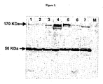
- the protein bands corresponding to AurkB (41 KDa), EGFR (170 KDa) and tubulin (50 KDa) as endogenous control were detected as shown in Figures 1 and 2 .
- Cell lines ARPE-19 normal diploid retinal pigmented epithelial cells, Lane 1), HeLa (cervical cancer, Lane 2), A431 (epidermoid cancer, Lane 3), A549 (non small cell lung cancer, Lane 4) and PC3 (prostate cancer cells, Lane 5) were analyzed for expression of Aurorakinase B. Crude protein lysates from the respective cell lines were resolved over 15 % SDS-PAGE and blotted onto nitro cellulose membrane. Membranes with transferred proteins were probed with rabbit anti-aurorakinase B antibody and mouse anti-alpha tubulin monoclonal antibody detected by goat anti-rabbit antibody ALP conjugated goat anti-mouse antibody conjugated with ALP. In blot 41KDa represents AurkB while 50 KDa represents tubulin detected as endogenous control. AurkB is expressed in all cells.
- Membranes with transferred proteins were probed with rabbit anti-EGFR antibody and mouse anti-alpha tubulin monoclonal antibody detected by goat anti-rabbit ALP conjugated and goat anti-mouse antibody conjugated with ALP.
- blot 170KDa represents EGFR while 50 KDa represents tubulin detected as endogenous control.
- EGFR is expressed in all cells, but epidermoid carcinoma cell line A431 and squamous cell carcinoma of tongue SCC-4 expressed greater amounts.
- HeLa cervical cancer
- SCC-4 oral cancer
- A549 non small cell lung cancer
- A431 epidermoid cancer
- PC-3 prostate cancer
- HFF-2 normal diploid fibroblasts
- ARPE-19 normal diploid retinal pigmented epithelial cells
- siRNAs such as EGFR and AurkB
- siRNAs such as EGFR and AurkB
- siRNA-liposome complexes were mixed thoroughly and added drop wise gently to each well containing cells, mixed, then incubated at 37°C in 5% CO 2 .
- Transfection efficiencies were obtained for each cell line by counting number of cells showing Cy3 labeled siRNA 16 h after transfection. Cells were trypsinised, washed once in PBS, and suspended in PBS. Cells were observed with an inverted fluorescent microscope and the number of fluorescent cells and total number of cells were counted from 15 different fields. The percentage of Cy3 labeled cells corresponds to the transfection efficiency, and ranged from 97 % for HeLa to 70 % for A549 cells.
- PC3 (prostate) and HeLa (cervical) cancer cell lines were transfected with differing siRNAs (of different lengths) alone, and in combinations. Twenty four hours after transfection, cells were plated in triplicate at a density of 8000 cells per well in a 96-well plate. After 72h, cells were incubated for three hours with BrdU according to kit instructions (cat # QIA58, Calbiochem). BrdU incorporation was stopped by the addition of a fixation reagent and cells were permeabilized to allow labeling with anti-BrdU antibody.
- the antibody is conjugated with horseradish peroxidase (HRP), which converts H 2 O 2 to chromogenic product which is measured by absorbance values at 450 nm with a reference filter at 540 nm.
- HRP horseradish peroxidase
- RNAi pathway in which mRNA complementary to the siRNA is degraded, thereby reducing levels of mRNA and the resultant protein encoded by the mRNA.
- the potency of a siRNA may be determined by measuring mRNA levels after siRNA transfection. Quantitative real time PCR was used to determine mRNA levels of both AurkB and EGFR among different cell lines transfected with siRNA, compared with mock transfected cells (Table 5). Table 5. Fold decrease in expression of AurkB and EGFR levels after 72h after siRNA transfection of different cancer cell-lines, as determined by real time PCR.
- siRNA 3 and 6 were more effective in reducing AurkB and EGFR mRNA than siRNA 3 or 6 alone.
- siRNA treatment reduced AurkB protein expression by as much as 90 % (e.g. Figure 6 ) and EGFR as much as 95% (e.g. Figure 7 ) compared mock treated samples.
- the decline in protein expression reflects the decline in mRNA levels seen with real-time PCR.
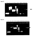
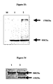
- lung cancer cell line A549 was transfected with siRNA 3 and analyzed for AurkB expression 72 h later, compared with mock-treated cells. Equal quantity of protein was loaded on all wells and, to ensure similar quantities of protein get transferred endogenous control protein alpha tubulin (50 kDa) was also detected in blot. Lane 1. siRNA 3 transfected A549 cell line where arrow head indicates faint band of Aurora B, a 41 kDa protein. Lane 2. Mock treated sample where Aurora B protein was detected in fairly good quantities over the siRNA treated samples. Lane 3. Rainbow low Molecular weight markers.
- Figure 7 shows a Western blot of A549 cells, 72 h after transfection with siRNA 6. Equal quantity of protein was loaded on all wells and to ensure similar quantities of protein get transferred, endogenous control tubulin (50 kDa) was also detected in blot. Lane 1. siRNA 6 transfected A549 cell line where arrow head indicates missing band of EGFR, a 170 kDa protein. Lane 2. Mock treated sample where EGFR protein was detected in fairly good quantities over the siRNA treated samples. Lane 3. Rainbow high molecular weight markers.
- Ki-67 expression was determined by ELISA ( Sui G., Soohoo C., el Affar B., Gay F., Shi Y., Forrester W.C. & Shi Y. (2002) A DNA vector-based RNAi technology to suppress gene expression in mammalian cells. Proc. Natl Acad Sci. USA, 99, 5515-5520 ). Monoclonal anti-Ki-67 antigen antibody (Sigma) was used as primary antibody while goat anti-mouse IgM - ⁇ chain-specific HRP conjugated antibody as secondary antibody. Absorbance values were obtained at 450 nm and their mean and standard deviation compared to mock treated and untreated controls.
- PCNA mRNA was measured using quantitative real time PCR in PC3, A431, SCC-4 & A549 cell lines transfected with siRNA.
- Table 7 The treatment of lung cancer cell line A549 with siRNA3 significantly reduced PCNA levels.
- the combination of siRNA 3 and 6 showed an additive effect in reducing levels of PCNA over individually treated siRNA in SCC-4 cells.
- the reduced levels of PCNA expression, over mock-transfected samples, indicates that the proliferation potential of these cell lines has been considerably reduced. Table 7.
- siRNA 3 and 6 successfully reduced respective mRNA as well as protein levels, relative to their mock or untreated samples.
- the affect of such inhibition on cellular metabolism was determined by measuring the ability oftransfected cells to reduce NAD or NADP to NADH or NADPH, respectively.
- 24h after siRNA transfection cells (HeLa, A431, A549 & PC3) were trypsinized, counted and replated at a concentration of 1000 cells per well in a 96-well plate. Cells were incubated for another 48h before measuring cell metabolic status following the protocol of the Cell-titer 96 aqueous non-radioactive assay kit from Promega (cat # G5421).
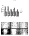
- Figure 8 shows different cell lines treated with siRNA 3 (10 nM), siRNA 6 (10 nM) and combination of siRNA 3 and 6 at 10 nM as well as 5nM concentrations each.
- the statistical significance was determined between mock treated cells vs siRNA transfected cells by paired two tail t-test where P ⁇ 0.05.
- Significant proliferation inhibition was obtained in all cell lines (PC3, A549, A431 and HeLa) in which AurkB, EGFR or both were targeted. However the there was no additive effect found in any of the cell lines when both mRNAs were inhibited simultaneously using siRNAs 3 and 6 in combination.
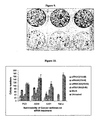
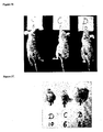
- Colony forming assays were used to assess the ability of siRNA-treated cells to initiate and develop a tumor. 24 h after transfection of cells (HeLa, A431, A549 and PC3) in 24 well plates, the cells were trypsinized, counted, and replated at a concentration of 300 cells per 6-well plate, in triplicate. Controls of mock treated and untreated cell-lines were also prepared. After 10 days of incubation, cells were washed once with PBS and stained with 300 ⁇ L of 0.1 % crystal violet for 5 min. before washing three times with PBS. This can be seen readily in Figure 9 , which shows prostate cancer cell line PC3 transfected and seeded in triplicate at a density of 300 cells/well in a six well plate. In Figure 9 , A-C represent mock treated cells while D-F represent siRNA 3 and 6 treated cells.
- Rate of colony formation inhibition Control colony forming rate - experimental colony forming rate / control colony forming rate x 100.
- FIG. 10 shows colony forming efficiency of different cell lines treated with siRNA 3 (10 nM), siRNA 6 (10 nM) and a combination of siRNA 3 as well as 6 at 10 nM and 5nM concentrations each. All cell lines treated with siRNA 3 and 6 showed different levels of inhibition to form colonies in comparison with that of mock treated and untreated cell lines.
- Cell line HeLa showed 100 % colony forming inhibition when treated with siRNA 3, which suggests 100 % dependency of cervical cancer on AurkB proteins.
- PC3, A549 and A431 AurkB inhibition resulted in significant inhibition of colony forming ability over mock treated cells.
- EGFR repression also resulted in a decrease in colony forming ability.
- the statistical significance was determined between mock treated cells vs siRNA transfected cells by paired two tail t-test where P ⁇ 0.05.
- Significant inhibition of colony forming ability was observed in all the cells treated with siRNA in comparison with that of mock treated cells.
- LDH release was used to determine the cytotoxicity of reduced levels of AurkB and EGFR on transfected cells.
- Cell-lines PC3, A431, A549, HeLa, ARPE-19 and HFF-2
- siRNA siRNA
- LDH was released following inhibition of AurkB and/or EGFR mRNA, indicating cellular necrosis and/or compromise of membrane integrity.
- Normal diploid cell line ARPE-19 remain only partially effected in comparison with that of the cancer cell lines tested.
- Inhibition of AurkB in A549 and A431 resulted in 150 % and 60 % release of LDH over mock treated cells respectively whereas PC3 and HeLa showed 65 % and 52 % release of LDH.
- repression of EGFR has resulted in 150 %, 90 %, 47 % and 100 % release of LDH from PC3, A549, A431 and HeLa cells, respectively, over their mock treated samples.
- Annexin V was examined in transfected PC3, A549, A431, HeLa, HFF-2 and ARPE-19 cells. Following Calbiochem's Annexin V-PE Apoptosis detection kit, cells were trypsinized, suspended in buffer and stained with PE labeled Annexin V at different time intervals (8 h, 16 h, 24 h, 48 h and 72 h) after siRNA transfection. The number of cells showing binding to Annexin V was recorded from a total of 150 cells per sample. Apotosis induction was not observed.
- siRNA 3 resultsed in 70 % and 50 % inhibition of migration of PC3 and A549 cell-lines, respectively.
- siRNA 6 transfection inhibited cell migration to the extant of 40% and 70 %, respectively, for cell-lines PC3 & A549.
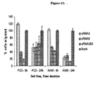
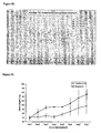
- Cancer cell-lines as well as normal diploid cell-lines were transfected with siRNA 3 and/or 6 in a 24-well plate. 24 h after transfection, the cells were trypsinized and seeded at a density of 1000 cells/well, in triplicate, in a 96-well plate. 48 h later, the number of cells that were metabolically active was determined using Cell - titer 96 aqueous solution kit from Promega for every 24 h until 8 days after transfection. The number of cells present at every 24 h was determined and thus the growth rate of siRNA transfected cells was by MTS assay indicating the metabolic status of the cells. The statistical significance was determined by paired two tail t-test where P ⁇ 0.05. The results are summarized in Figs 14 to 17 .
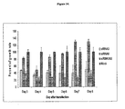
- Figure 14 shows the growth rate epidermoid carcinoma cell line A431 over a period of 8 days from the date of transfection (with siRNA 3, 6 or 3 & 6), as determined by MTS.
- Statistically significant data was obtained for the following: Day 3 and 4 - between siRNA (3, 6 as well as 3 & 6) treated and mock treated samples. Further among siRNA 3, siRNA 6 and siRNA 3 & 6 combination treated cells. Day 5, 6, 7 and 8 - between siRNA (3 as well as 3 & 6) treated and mock treated samples. Further among siRNA 3 and siRNA 3 & 6 combination treated cells. Inhibition of both AurkB and EGFR showed the lowest growth rate.
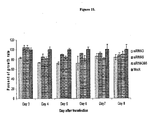
- Figure 15 shows prostate cancer cell line PC3 percent growth rate over a period of 8 days from the date of transfection (with siRNA 3, 6 or 3 & 6) compared with that of mock treated cells.
- Statistically significant data was obtained for the following: Day 3 - between siRNA 3 treated and mock treated cells; Day 4, 5 and 6 - between siRNA 3 as well as combination of siRNA 3 and 6 treated and mock treated cells; Day 7 and 6 - combination treated cells and mock treated cells.
- Figure 16 shows non-small cell lung cancer cell line A549 growth rate over a period of 8 days from the date of transfection (with siRNA 3, 6 or 3 & 6) compared with that of mock treated cells. Statistically significant data was obtained for the following: Day 3 & 4 - between siRNA 3 treated and mock treated cells.
- Figure 17 shows normal diploid retinal pigmented epithelial cells ARPE-19 growth rate over a period of 8 days from the date of transfection compared with that of mock treated cells.
- Statistically significant data was obtained for the following: Day 4 & 5 - significant growth inhibition has been observed between siRNA 3 and siRNA 3 & 6 combination treated cells vs mock treated cells. With siRNA 6 treated cells there was no significant decrease in growth rate on any of the days. The increase in growth rate of siRNA treated cells has been observed on Day 8 was due to confluence in mock treated cells. siRNA treated cells slowly reach confluence due to inhibition in growth of these cells observed during until the fourth day of transfection.
- cancer cells A431 epidermoid cancer
- normal diploid cells ARPE-19 showed very limited effect on inhibition in growth of cells. This indicates that siRNA selectively inhibited cancer cells.
- siRNA transfected cells from cell lines PC-3 and HeLa
- cell lines PC-3 and HeLa were plated at a density of 8000 cells per well in a 96-well plate to determine the effect of siRNA 3 and/or 6 on the cell cycle and, thus, their ability to control the growth index of the tumor cell-lines.
- 72h after transfection cells were subjected to BrDu incorporation to determine the number of cells that were in the S-phase of cell cycle as described above. From the absorbance values the percent of cells that were in S-phase of the cell cycle was obtained with reference to the mock treated cells.
- Cholesterol was conjugated to the 3'-end of either sense or antisense strand of siRNA 3 or 6.
- the cholesterol conjugated siRNA was compared with unconjugated siRNA for serum stability as well as for the ability to inhibit AurkB and EGFR expression in vitro.
- Serum stability was tested on 1 ⁇ g of siRNA, incubated with 10 ⁇ L of 100 % human serum at 37°C for 24h, 48h and 72 h. At the end of the incubation, siRNAs were examined over 1 % agarose gel electrophoresis. A mobility shift observed in the siRNA conjugated with cholesterol was due to binding of various serum proteins and thus protecting siRNA from degradation by serum nucleases. As seen in Figures 18 and 19 , sense strands conjugated with cholesterol were more stable than conjugated antisense strands or unconjugated siRNA. In addition, the complexation of siRNA with the Hiperfect transfection agent did not enhanced serum stability.
- siRNA conjugated with cholesterol at 3'- end of sense strand was found to be more stable and could be detected even after 48 h of incubation at 37°C in 100 % serum conditions.
- siRNA 6 was found to be more stable over the siRNA3. This could be due to an increase in GC content and a resulting high Tm.
- PBST phosphate buffered saline containing 0.1% Tween 20
- HRP substrate was added. Absorbance values were recorded at 450 nm with a reference filter of 560 nm.
- siRNA The specificity of siRNA for the target mRNA was tested in prostate cancer cells (PC3), transfected with siRNA as described earlier. 72 h after transfection, total RNA was prepared following the protocol of Qiagen total RNA isolation kit (RNeasy Mini kit), and a total of 2 ⁇ g RNA was suspended in 10 ⁇ L of water. The quality of RNA was checked on denaturing formaldehyde gels and the OD ratio was determined using Perkin Elmer Spectrophotometer. One ⁇ g of total RNA was converted into DIG-labeled cRNA following the protocol of Nano In-vitro Transcription amplification kit from Applied Biosystems.
- the arrays were hybridized at 55 °C for 17 h and subsequently washed and bound to antibody against DIG coupled with alkaline phosphatase. After the addition of chemiluminescent detection substrate, the arrays were scanned. Autogridding was performed by the imaging software and the result file was created that transforms the intensity of each gene into a numeric value, higher the signal- higher the numeric value and higher is the amount of gene present in the sample.
- Controls were added for each and every step of the assay starting from reverse transcription (RT), in vitro transcription (IVT), hybridization and chemiluminescence detection. A quality report is generated for these controls that help to ascertain the success of the microarray experiment. Once satisfied with the QC report we proceeded with secondary analysis using Spotfire.
- the software normalized the data and performed a "t test" to determine the differentially expressed genes between two conditions. It also averaged the replicates and determined a fold change value for the two conditions.
- the probe IDs that were differentially expressed were sorted through a Panther database to determine which pathways or biological processes were affected amongst the differentially expressed genes, number of genes either down regulated or unregulated.
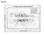
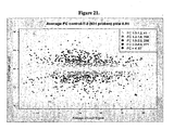
- FIG. 20 - 22 The change in transcription expression levels ranging from a fold change of minimum 1 to above 4.5 was depicted in Figures 20 - 22 where the prostate cancer cells were inhibited for AurkB (siRNA 3) ( Figure 20 ), EGFR (siRNA 6) ( Figure 21 ) or both (siRNA 3 and 6) ( Figure 22 ).
- the figures indicate the number of mRNAs that have either been upregulated or down regulated with respect to the untreated controls.
- the transcription changes observed indicates that there was no general gene repression when both AurkB and EGFR were knocked down simultaneously rather individually.
- the down stream targets for AurkB, Histone H 3 and EGFR down stream targets, AKT were inhibited.
- TIMPS Tissue Inhibitors of Metallic proteases
- Caspases for induction of apoptosis
- MMPs matrix metallo proteases
- xenograft prostate cancer tumors were induced in 6-8 week old male SCID mice by subcutaneously injecting, into one flank, human prostate cancer cell-line PC3 at a density of 3x10 6 cells mL -1 in 100 ⁇ L volume of PBS.
- Mouse developed tumors of approximately 40 - 50 mm 3 by the end of three weeks. Mouse were divided into two groups. Group R consisted of five animals treated with the combination of siRNA 3 and 6. Group C consisted of 3 animals that received only transfection agent in PBS. 8 days after siRNA treatment, the size of the tumors was evaluated and their standard deviations were observed as indicated in Figure 23 .
- the siRNA 3 and 6 combination treated animals showed approximately 50 % reduction in tumor growth in comparison with that of the placebo treated animals.
- the statistical significance was determined by paired two tail t-test where P ⁇ 0.05. Statistical significance in terms of causing tumor regression was observed between treated animals and placebo animals on Day 3, 4 and 5.
- Tumor tissue was retrieved from the animals, total protein was extracted and subjected to western blot analysis. Total protein lysates from cancer tissues were resolved over 15% SDS-PAGE and probed with anti-AurkB antibody as well as anti-tubulin antibody as endogenous control. As shown in Figure 24 , treatment with the siRNA 3 and 6 combination resulted in complete inhibition of AurkB expression, while mice treated with siRNA 3 alone showed partial inhibition of AurkB expression.
- tumors were induced in forty 6-8 weeks old male athymic Nude mice by injecting prostate cancer cell-line PC3 at a density of 3x 10 6 cells mL -1 in 100 ⁇ L volume of PBS subcutaneously in one of the flanks. Mice developed tumors of approximately 50 - 100 mm 3 by the end of three weeks. Mice which developed tumors were divided into following four groups -
- siRNA treatment also decreased vascularization of the xenografted prostate tissue. This phenomenon was not due solely to inhibition of EGFR, as it was also observed in mice treated with siRNA3, targeting AurkB.
- Figure 28 shows that mice treated with placebo had an observably vascularized tumor (Group D), compared with those treated with siRNA 3 (Group A).
- arrowheads indicate the vascularization of the prostate tumor in placebo-treated mice after 35 days of treatment. By comparison, there is an absence of vascularization in mice treated with siRNA 3 ( Figure 31 ).
- results obtained shows that, for regression of the prostate tumors, there exists a critical volume and dosage of the tumors.
- siRNA tested combination of siRNA 3 and 6 caused an 81 % tumor regression ( Figures 25 - 27 ).
- Group A siRNA3, AurkB
- Group B siRNA6, EGFR
- Group B siRNA6, EGFR
- Observations of gross pathology suggest that targeting AurkB was associated with inhibition of angiogenesis; as observed in Figures 30 and 31 .
- the results indicate that the initial volume of prostate tumor and dosage quantity required need to be determined for the effective application of siRNA 3, 6 and their combination as therapeutic drugs.
- the tumor regressions obtained, gross pathological observations and general behavior patterns of the siRNA treated and untreated animals indicate that indeed these siRNA have potential to be applied as therapeutic drugs under clinical conditions.
- Transfection of MCF-7 with siRNA 3 repressed AurkB expression.
- siRNA 3 targeting AurkB
- transfected cells expressed barely detectable AurkB protein, while AurkB was readily observed in mock treated cells.
- the principal aim of using siRNA 3, 6 or 3 & 6 is to inhibit the expression levels of the respective protein AurkB, EGFR or both together. If there is no visible band in siRNA transfected cells, it indicates efficiency of siRNA in knocking down expression of the proteins of interest. Again, equal quantities of protein were loaded onto the gel, and protein alpha tubulin (50 kDa) was also detected as a control.
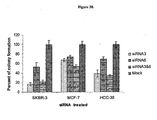
- BrdU incorporation was examined in SKBR-3, MCF-7 and HCC-38 cell lines, after transfection with siRNA (3, 6 and combination of 3 & 6) or mock, siRNA 7, as shown in Figure 36 .
- 48 h after transfection cells were incubated with BrdU over night and analyzed for BrdU incorporation by measuring absorbance values at 450 nm. All the experiments were done in triplicates and their mean absorbance values were presented as percent of cells in S-phase of cell cycle, which is an indicator of cell proliferation / growing potential of tumor. The statistical significance was determined between mock treated cells vs siRNA treated cells by paired two tail t-test where P ⁇ 0.05. Statistical significance was found between mock treated and all siRNA treated cells, as well as between AurkB, EGFR and their combination treated cells.
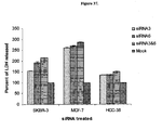
- FIG. 37 shows release of LDH from siRNA treated cells against mock treated cells.
- simultaneous repression of both AurkB and EGFR resulted in more LDH release compared with AurkB or EGFR alone.
- MCF-7 showed maximum LDH release of 150 - 300 % over mock
- SKBR-3 showed between 50 - 150 %
- HCC-38 showed 20 -70% respectively over mock treated samples.
- Colony forming efficiency was also examined in breast cancer cell lines treated with siRNA 3 (10 nM), siRNA 6 (10 nM), a combination of siRNA 3 and 6 at 10 nM concentrations each, and mock at 10 nM.
- Statistical significance was determined between mock treated cells versus siRNA transfected cells by paired two tail t-test where P ⁇ 0.05.
- significant inhibition of colony forming ability was observed in all the cells treated with siRNA 3, siRNA 6, or both siRNA 3 and 6, as compared to with that of mock treated cells.
- siRNA 3 alone was able to inhibit colony formation by 80%.
- EXAMPLE 8 AURK B siRNA CAUSED CANCER CELLS TO ARREST IN G2 PHASE OF CELL CYCLE.
- Cancer cell lines (HeLa, A549 and PC3) transfected with siRNA 3 for AurkB and siRNA6 for EGFR, either independently or simultaneously, were harvested after 72 h. The cells were washed with PBS and fixed in 70% ice-cold ethanol at 4°C for 60 min. Cells were then washed with PBS and treated with propidium iodide (5 ⁇ g/mL of propidium iodide and 0.2 mg/mL -1 RNase in 0.1% sodium citrate) for 30 min at 4°C. Propidium iodide stained cells were subjected to flow analysis using (FACScaliber, Becton Dickinson, San Jose, CA). Data were acquired for 10,000 gated events using Cell Quest software and analyzed using ModFitLT2.0 (Verity Software House, Topsham, ME).
- AurkB knock down results in arrest of cancer cells at G2 phase of cell cycle:
- Knock down of AurkB resulted in arrest of cells at G2 phase of cell cycle, as shown in Figure 39A.
- siRNA 3 For example, in HeLa cervical cancer cells transfected with siRNA 3, 77% of cells were in G2 phase, while A549 and PC3 transfected cells showed 22% and 37% of cells in G2 phase, respectively, as shown in Table 10.
- Similar results were reported earlier when Aurora kinases were inhibited by small molecule inhibitors ( Rojanala S, Han H, Munoz R, Browne W, Nagle R, Von Hoff D and Bearss D.
- the mitotic serine kinase, Aurora-2 is a potential target for drug development in human pancreatic cancer. Mol. Cancer Therapeutics 2004; 451-457 ).
- HeLa cells were transfected with siRNA 3 (for AurkK) and mock siRNA 7. After 72 h, the cells were trypsinsed and labeled with propidium iodide for analysis of cell cycle using a flow cytometer.
- siRNA 3 (for AurkB) transfected cells 77% were in G2 phase, 6% were in S phase and only 16% were in G1 phase.
- mock siRNA 7 transfected cells only 17% were in G2 phase, 36% were in S phase, while 46% were in G1 phase. This demonstrates that cells knocked down for AurkB go into G2 arrest, are less likely to go into S-phase, and exhibit no apoptosis induction.
- EXAMPLE 9 AURK B AND EGFR siRNAs, EITHER ALONE OR TOGETHER, INDUCE SENESCENCE IN CANCER CELLS.
- HeLa and A431 cells were transfected in a 24-well plate with siRNA 3 and 6, either independently or simultaneously, as described above. After 72 h, transfected cells were subjected to a senescence assay following the protocol of Chemicon's senescence assay kit. Briefly, cells were washed twice with PBS and fixed in a fixative solution for 30 min. at room temperature. After fixation, cells were washed twice with PBS and incubated with X-gal solution overnight at 37°C. After incubation, cells were observed using a light microscope at 200X total magnification. Images were collected at 10 different fields for each well of a 24-well plate using a digital camera.
- Digitally captured images were analyzed by counting number of cells showing blue color and the total number of cells in a given image. The percentage of cells showing senescence was derived from total number of cells. All experiments were performed in triplicate; statistical significance was determined by t-test where P ⁇ 0.05.
- Knock down of AurkB (by transfected cells with siRNA 3) induced senescence in 27% of HeLa cervical cancer cells, as compared to mock treated cells (using siRNA 7). No significant induction of senescence was seen in cells transfected with siRNA 7 (mock) or siRNA 6 (for EGFR). On the other hand, when both AurkB and EGRR were knocked down simultaneously (via transfected cells with both siRNA 3 and 6), enhanced induction senescence was observed, as shown in Table 11 and Figure 40. Data will indicate that similar results occur in A431 epidermal cancer cells. Table 11. Knock down of AurkB, alone or with knock down of EGFR, results in induction of senescence in cervical cancer cells.
- siRNA 3 and/or siRNA 6 were effective against various cancers in vitro, and that in vitro results also reflected results in in vivo experiments with prostate tumors, the efficacy of siRNAs is tested against human breast cancer. Protocols for breast cancer cells are as described above for other cancers, unless otherwise indicated.
- Breast cancer cell lines SKBR-3, MCF-7 and HCC-38 are all able to form tumors in nude mice. Following the protocol used with PC3 prostate cancer cells described in Example 6, breast cancer cells are injected into mice and allowed to form a tumor. siRNA are then injected weekly into the tumor. Mice are then examined for general health, tumor size, tumor vascularization and gross pathology.
- Results will indicate that statistically significant breast tumor regression occurs in mice treated with siRNA 3 and/or 6, as compared to placebo. Results will indicate that siRNA 3 and/or 6 also decreases vascularization of xenographed breast tissue, and this phenomenon is not be due solely to inhibition of EGFR.
- compositions and uses disclosed and claimed herein can be made and executed without undue experimentation in light of the present disclosure. While the compositions and uses of this invention have been described in terms of preferred embodiments, it will be apparent to those of skill in the art that variations may be applied to the compositions and/or methods and in the steps or in the sequence of steps of the methods described herein. More specifically, it will be apparent that certain agents that are chemically or physiologically related may be substituted for the agents described herein while the same or similar results would be achieved.
Landscapes
- Health & Medical Sciences (AREA)
- Life Sciences & Earth Sciences (AREA)
- Engineering & Computer Science (AREA)
- Chemical & Material Sciences (AREA)
- Genetics & Genomics (AREA)
- Organic Chemistry (AREA)
- Bioinformatics & Cheminformatics (AREA)
- General Health & Medical Sciences (AREA)
- Biomedical Technology (AREA)
- Molecular Biology (AREA)
- Zoology (AREA)
- Wood Science & Technology (AREA)
- General Engineering & Computer Science (AREA)
- Medicinal Chemistry (AREA)
- Public Health (AREA)
- Animal Behavior & Ethology (AREA)
- Biotechnology (AREA)
- Pharmacology & Pharmacy (AREA)
- Biochemistry (AREA)
- Veterinary Medicine (AREA)
- Chemical Kinetics & Catalysis (AREA)
- Epidemiology (AREA)
- Botany (AREA)
- Virology (AREA)
- Nuclear Medicine, Radiotherapy & Molecular Imaging (AREA)
- Physics & Mathematics (AREA)
- Biophysics (AREA)
- General Chemical & Material Sciences (AREA)
- Plant Pathology (AREA)
- Microbiology (AREA)
- Pharmaceuticals Containing Other Organic And Inorganic Compounds (AREA)
- Medicines That Contain Protein Lipid Enzymes And Other Medicines (AREA)
- Medicines Containing Antibodies Or Antigens For Use As Internal Diagnostic Agents (AREA)
- Medicinal Preparation (AREA)
Description
- The present invention relates to use of short nucleic acid molecules, such as short interfering nucleic acid (siNA) molecules, for modulating gene and protein expression, including compounds, compositions and uses of small nucleic acid molecules to modulate Aurora B (AurkB) expression. The compounds and methods of the present invention have applications in cancer therapy either alone or in combination with other therapies.
- The Aurora kinases are a family of serine/threonine kinases. Aurora A and B kinase are associated with mitotic events of cell cycle, while Aurora C kinase is expressed only in testis (Sasai K, Katayama H, Stenoien DL, Fujii S, Honda R, Kimura M, et al, Aurora-C kinase is a novel chromosomal passenger protein that can complement Aurora-B kinase function in mitotic cells. Cell Motil. Cytoskeleton, 2004; 59:249-263). Aurora-A kinases, also called Aurora-2, STK6, ARK1 and Aurora/IPL-1 related kinase, associates with centrosomes and microtubules during mitosis. Aurora A kinase (hereinafter "Aurora A") localizes to centrosomes and regulates the association between cell cycle machinery and centrosomes (Hirota T, Kunitoku N, Sasayama T, Marumoto T, Zhang D, Nitta M. Aurora-A and an Interacting Activator, the LIM Protein Ajuba, Are Required for Mitotic Commitment in Human Cells. Cell 2003; 114: 585-598; Dutertre S, Cazales M, Quaranta M, Froment, C, Trabut, V, Dozier, C. et al. Phosphorylation of CDC25B by Aurora-A at the centrosome contributes to the G2-M transition. J Cell. Sci. 2004; 117: 2523-2531).
- Aurora B kinases, also known as Aurorakinase B, Aurora B, Aurora-1, and hereinafter "AurkB", localizes to the kinetochores from prophase to metaphase and to the central spindle and the midbody in cytokinesis (Carmena M. and Earnshaw WC. The cellular geography of aurora Kinases. Nat. Rev. Mol. Cell Biol 2003; 4: 842-854). AurkB associates with chromosome passenger proteins, inner centromere protein (INCENP), Survivin and Borealin protein to form a quaternary chromosome passenger complex which, along with its sub complexes (AurkB and INCENP) are thought to be required for spindle check point, cytokinesis and phosphorylation of Histone H3, respectively. (Schumacher JM, Golden A, and Donovan P. AIR-2: An Aurora/Ipll-related protein Kinase associated with chromosomes and midbody microtubules is required for polar body extrusion and cytokinesis in Caneorhabditis elegans embryos. J. Cell. Biol. 1998; 143: 1635-1646; Terada Y, Tatsuka M, Suzuki F, Yasuda Y, Fujita S, and Otsu M. AIM-1: a mammalian midbody-associated protein required for cytokinesis. EMBO J. 1998; 17: 667-676; Giet R and Glover DM. Drosophilia aurora B Kinase is required for histone H3 phosphorylation and condensing recruitment during chromosome condensation and to organize the central spindle during cytokinasis. J. Cell Biol 2001; 152: 669-682). AurkB phosphorylates MCAK and thus plays a vital role in regulation of bi-orientation during mitosis (Giet R, Petretti C and Prigent C. aurora Kinases, aneuploidy and cancer, a coincidence or a real link? Trends. Cell Biol. 2005; 5: 241-250).
- Any discrepancy in functioning of Aurora kinases can lead to mitotic catastrophe resulting in anueploidy or polyploidy, a hallmark of cancer. Indeed, chromosomal instability cause cancer and is clearly associated with cancer evolution and thus resistance to treatment (Duesberg P et al., "The chromosomal basis of cancer" Cell Oncol. 2005; 27(5-6):293-318; Duesberg P et al., "Cancer drug resistance: The central role of the karyotype." Drug Resist Updat. 2007 Mar 26). Aurora kinases have been linked to the chromosomal instability. In addition, malfunctions of Aurora kinases are found in a number of cancers, such as non-small cell lung cancer, epidermal, prostate, colon, pancreatic, ovary, breast and oral cancers including all head and neck cancers. (Keen N. and Taylor S. Aurora-kinase inhibitors as anticancer agents. Nature Rev 2004; 4: 927-936). Inhibition of AurkB leads to improper segregation of sister chromatids and failure of cytokinesis. Over-expression of AurkB has been linked to cell proliferation and development of aggressive tumors leading to malignancy. (Ota T, Suto S, Katayama H, Han Z, Suzuki F, Maeda M et al. Increased mitotic phosphorylation of histone H3 attributable to AIM-1/Aurora-B over expression contributes to chromosome number instability. Cancer Res 2002; 62: 5168-5177; Vischioni B, Oudejans JJ, Vos W, Rodriguez JA and Giaccone G. Frequent overexpression of aurora B Kinase, a novel drug target, in non-small cell lung carcinoma patients. Mol. Cancer Ther. 2006; 5: 2905-2913; Smith SL, Bowers NL, Betticher DC, Gautschi O, Ratschiller D, Hoban PR. Overexpression of aurora B Kinase (AURKB) in primary non-small cell lung carcinoma is frequent, generally driven from one allele, and correlates with the level of genetic instability. Br. J. Cancer 2005; 19:719-29). Studies so far have not indicated whether the malfunctioning of Aurora is a cause or consequence of cancers. AurkB is intimately involved in preventing chromosomal instability. (Liu Q, Kaneko S, Yang L, Feldman RI, Nicosia SV, Chen J et al. Aurora-A Abrogation of p53 DNA binding and transactivation activity by phosphorylation of serine 215. J Biol Chem. 2004; 279: 52175-52182; Katayama H, Sasai K, Kawai H, Yuan Z, Bondaruk J et al. BRCAI phosphorylation by aurora kinase A induces Mdm2-mediated destabilization and inhibition of p53. Nat. Genetics 2004; 36: 55-62; Ouchi M, Fujiuchi N, Sasai K, Katayama H, Minamishima YA et al. BRCAI phosphorylation by Aurora-A in the regulation of G2 to M transition. J Biol. Chem 2004; 279:19643-19648). Survivin, part of AurkB complex, is a key protector against apoptosis and/or mitotic catastrophe (Andrews PD. Aurora Kinases: shining lights on the therapeutic horizon? Oncogene 2005; 24: 5005-5015). AurkB therefore appears to have a direct role in tumorigenesis.
- Aurora inhibitors have been reported several authors (Ditchfield C, Johnson VL, Tighe A, Ellston R, Haworth C, Johnson T et al. Aurora B couples chromosome alignment with anaphase by targeting BubR1, Mad2 and Cenp-E to kinetochores. J Cell Biol 2004; 161: 267-280; Hauf S, Cole RW,Terra S, Zimmer C, Schnapp G, Walter R et al. The small molecule Hesperadin reveals a role for Aurora B in correcting kinetochore-microtubule attachment and in maintaining the spindle assembly checkpoint. J Cell Biol 2003; 161: 281-294 and Harrington EA, Bebbington D, Moore J, Rasmussen RK, Ajose-Adeogun AO, Nakayama,T. VX-680, a potent and selective small-molecule inhibitor of the Aurora kinases, suppresses tumor growth in vivo. Nat. Med 2004; 10:262-267). These inhibitors include
ZM447439 - VX-680 (Vertex) inhibits all three Aurora and, in a panel of cancer cells, was found to inhibit proliferation, increase apoptosis induction, and induce tumor cell death. Merck is presently conducting three clinical studies of VX-680 in patients with hematologic cancers, recurrent or non-responsive solid tumors, or cancers for which standard therapy does not currently exist. In these studies, the safety and tolerability of VX-680 are being evaluated when administered as either a 24-hour continuous infusion or as a 5-day continuous infusion. Of the Aurora inhibitors in clinical trials, only VX-680 is highly specific for Aurora, but is associated with toxic bone marrow side effects, and the mechanism of tumor cell death is not completely understood (Giet R, Petretti C and Prigent C. Aurora Kinases, aneuploidy and cancer, a coincidence or a real link? Trends. Cell Biol 2005; 5: 241-250).
- Additional Aurora inhibitors have been described. Japanese patent application
JP 2005-278472 JP 2005-320351 US patent application 20050267065 describes compositions and methods for modulating the expression of AurkB, including using chemically modified nucleotides, including small interfering nucleotides. The use of chemically modified nucleotides poses a potential risk of unwanted side effects. - The epidermal growth factor receptor (EGFR, also known as ErbB-1, or HER1 in humans) is a protein tyrosine kinase. Activation of EGFR leads to transmission of proliferative signals to the nucleus and, via activation of transcription factors, leads to increased proliferation, increased migration, increased adhesion, increased angiogenesis and inhibition of programmed cell death pathways (Bundy, L., Wells, S., Sealy, L. "C/EBPbeta-2 confers EGF-independent growth and disrupts the normal acinar architecture of human mammary epithelial cells," Mol. Cancer. 2005; 4: 43). EGFR is over-expressed in a number of cancers such as head and neck squamous cell carcinoma (HNSCC), non-small cell lung carcinoma (NSCLC), prostate, gastric, epidermal and skin cancers. EGFR is expressed constitutively in many highly aggressive tumors (Rusch V, Mendelsohn J, Dmitrovsky E. "The epidermal growth factor receptor and its ligands as therapeutic targets in human tumors." Cytokine Growth Factor Rev 1996; 7: 133-41; Salomon DS, Brandt R, Ciardiello F, Normanno N. "Epidermal growth factor related peptides and their receptors in human malignancies." Crit Rev Oncol Hematol 1995; 19: 183-232)
and tumors showing over expression of EGFR are often found to be resistant to chemotherapeutic drugs (James H. Doroshow "Targeting EGFR in Non-Small Lung cancer." N. Engl. J. Med. 2005; 353(2):200-2002). - EGFR inhibitors such as cetuximab, erbolitin, etc, are known. To enhance the tumoricidal affects of EGFR inhibitors, therapeutic treatments often include use of cytotoxic chemotherapeutic drugs such as cisplatin, foldfox, etc. However, in randomized clinical trials EGFR inhibitors when combined with cytotoxic chemotherapy, no advantage was demonstrated over standard chemotherapy alone (Sui G., Soohoo C., el Affar B., Gay F., Shi Y., Forrester W.C. & Shi Y. A DNA vector-based RNAi technology to suppress gene expression in mammalian cells. Proc. Natl Acad Sci. USA 2002; 99, 5515-5520). Accordingly, there is also a need for improved EGFR inhibitors.
- Chemically synthesized short nucleic acid molecules, such as siNAs, can specifically and effectively direct homology-specific post transcriptional gene silencing, and therefore may be used as highly effective, selective and potent therapeutics, with minimal side effects. A molecule that specifically inhibits AurkB can block the mechanics of cell division, and therefore is very useful in combination therapy. Similarly, siNA may be used to block EGFR expression.
- The present invention provides potent short nucleic acid molecules without any chemical modification having high stability and specificity for AurkB and or EGFR, and are useful as therapeutics alone, or in combination with other therapies for cancer.
- The present invention provides short nucleic acid molecules for modulation of AurkB gene expression, as set out in
claim 1. In related embodiments, the present invention provides AurkB-targeting short nucleic acid molecules for the treatment of cancer, including cervical, prostrate, breast, lung and oral cancer. Such molecules may be used alone or in combination with other treatments for the management and treatment of cancer. In one embodiment, the AurkB-targeting short nucleic acid molecule is provided in combination with a short nucleic acid molecule that targets EGFR. In other embodiments, short nucleic acids of the present invention may be combined with conjugates such as lipids, polymers and monoclonal antibodies. - The present invention includes short nucleic acid molecules which are specifically targeted. In some embodiments, the short nucleic acid molecules are RNA, including siRNA. The present invention also includes advantageously stable and/or potent nucleic acid molecules for inhibition of AurkB and EGFR, and for the related treatment of cancer.
- In some embodiments, the present invention provides siNAs having between 19 to 30 nucleotides, between 25 and 29 nucleotides, or having 27 nucleotides, where the sequence is designed for better stability and efficacy in knockdown (i.e., reduction) of AurkB gene expression. Such siNAs can be used alone or in combination with other therapies. The present invention provides stable compositions of siNA with or without conjugation with cholesterol. In related embodiments, the invention encompasses compounds, compositions and uses of 27-mer short interfering nucleic acid molecules in modulation of AurkB gene expression. The compounds of the present invention are useful in therapy of cancer either alone or in combination with other treatments or therapies.
- In one embodiment the short nucleic acid molecules of the present invention is also a short interfering nucleic acid (siNA), a short interfering RNA (siRNA), a double stranded RNA (dsRNA), a micro RNA (µRNA), and/or a short hairpin RNA (shRNA) molecule. The short nucleic acid molecules can be unmodified or modified chemically. In the some embodiments the present invention relates to short interfering RNA having 27 nucleotides.
- In one embodiment, the nucleic acid molecule of the present invention has between 19 to 30 nucleotides, between 25 and 29 nucleotides, or 27 nucleotides. In one embodiment, the nucleic acid molecule of the present invention comprises 19-30 nucleotides complementary to RNA having an AurkB nucleic acid sequence.
- Nucleotides of the present invention can be chemically synthesized, expressed from a vector, or enzymatically synthesized.
- In one embodiment, a siRNA molecule of the invention comprises a double stranded RNA wherein one strand of the RNA is complimentary to the RNA of AurkB. In another embodiment, a siRNA molecule of the invention comprises a double stranded RNA wherein one strand of the RNA comprises a portion of a sequence of RNA having AurkB sequence.
- In one embodiment, the invention targets AurkB as set forth in GenBank Accession Number NM-004217. However, the present invention is not limited to nucleotides targeting one variant of AurkB, but also includes nucleotides that target AurkB-related molecules including single nucleotide polymorphisms of AurkB, AurkB homologs, and AurkB splice and transcript variants. The present invention also contemplates nucleotides that target genes involved in AurkB regulatory pathway as a means of regulating AurkB.
- In other embodiments, the present invention provides compositions and methods used to regulate AurkB. AurkB may be regulated by a small nucleic acid molecule which targets AurkB directly, or by targeting molecules which regulate the AurkB pathway. Small nucleic acid molecule that target AurkB may be used alone, or in combination with other small nucleic acid molecules or small chemical molecules. In related embodiments, the targeting of AurkB is used to regulate cancer or disease states that respond to modulation of AurkB expression levels in the cell.
- In some embodiments, chemically synthesized siNA of 27 nucleotides in length are used to reduce expression levels of AurkB either alone or in combination with other small nucleic acid molecules directed against genes that are involved in various cancers, such as EGFR. In some embodiments, small nucleic acid molecules disclosed in Tables 1 or 2 may be used alone or in combination for cancer therapy.
- In further related embodiments, cancer regulation by siNA is suitable for cancer management and treatment. Thus, the present invention provides short nucleic acid molecules for treatment of cancer such as cervical, oral, lung, skin and prostate cancers.
- In one embodiment, the present invention provides a combination of small nucleic acid molecule targeting AurkB and EGFR as a treatment for cancers. In some embodiments, suitable small nucleic acid molecules include those listed in Tables 1 and 2.
- In one embodiment, the invention features a mammalian cell, for example a human cell, including a small nucleic acid molecule of the invention.
- The present invention features a method of down-regulating AurkB activity in a cell, comprising contacting the cell with an enzymatic nucleic acid molecule or antisense nucleic acid molecule, or other nucleic acid molecule of the invention, under conditions suitable for down-regulating of Aurorakinase activity.
- The present invention features a method of down-regulating (also called "knocking down") Aurorakinase activity in a cell, comprising contacting the cell with an enzymatic nucleic acid molecule or antisense nucleic acid molecule or other nucleic acid molecule of the invention, under conditions suitable for down-regulating Aurorakinase activity.
- In one embodiment, the present invention can be used in a method of treatment of a subject having a condition associated with the level of AurkB, comprising contacting cells of the subject with the enzymatic nucleic acid molecule or antisense nucleic acid molecule or other nucleic acid molecule of the invention, under conditions suitable for the treatment.
- In one embodiment, the invention comprises the use of one or more drug therapies under conditions suitable for said treatment.
- The present invention also features a use of nucleic acid as defined in the claims in a method for treatment of cancer, for example breast cancer, lung cancer, prostate cancer, colorectal cancer, brain cancer, esophageal cancer, stomach cancer, bladder cancer, pancreatic cancer, cervical cancer, head and neck cancer, ovarian cancer, melanoma, lymphoma, glioma, or multidrug resistant cancer, comprising administering to a subject an enzymatic nucleic acid molecule or antisense nucleic acid molecule or other nucleic acid molecule of the invention under conditions suitable for said treatment.
- In another embodiment, other drug therapies contemplated by the invention include monoclonal antibodies, chemotherapy, or radiation therapy or a combination thereof.
- The present invention features compositions comprising the enzymatic nucleic acid and/or antisense nucleic acid molecules of the invention in a pharmaceutically acceptable carrier.
- The invention also features a use of the nucleic acid of the invention for administering to a cell, such as mammalian cell (e.g., a human cell) a nucleic acid of the invention. Such a cell can be in culture or in a mammal, such as a human. The use for administering comprises contacting the cell with the enzymatic nucleic acid molecule or antisense molecule or other nucleic acid molecule of the invention under conditions suitable for such administration. The method of administration can be in the presence of a delivery reagent, for example a lipid, cationic lipid, phospholipid, or liposome.
- The following drawings form part of the present specification and are included to further demonstrate certain aspects of the present disclosure, the inventions of which can be better understood by reference to one or more of these drawings in combination with the detailed description of specific embodiments presented herein.
-
Figure 1 . Expression of AurkB in ARPE-19 (normal diploid retinal pigmented epithelial cells, Lane 1), HeLa (cervical cancer, Lane 2), A431 (epidermoid cancer, Lane 3), A549 (non small cell lung cancer, Lane 4) and PC3 (prostate cancer cells, Lane 5) were analyzed for expression of AurkB (41KDa) and tubulin (50 KDa) as endogenous control. -
Figure 2 . Expression of EGFR in cell lines HeLa (cervical cancer, Lane 1), PC3 (prostate cancer cells, Lane 2), A549 (non small cell lung cancer, Lane 3), A431 (epidermoid cancer, Lane 4), SCC-4 (squamous cell carcinoma of tongue, Lane 5), HFF-2 (normal diploid fibro blasts, Lane 6) and ARPE-19 (normal diploid retinal pigmented epithelial cells, Lane 7). EGFR (170KDa), tubulin (50 KDa) -
Figure 3 . HeLa cells transfected with Cy3 labeled siRNA. Arrowhead indicates Cy3 labeled siRNA at the end of 16 h of transfection distributed inside the cytosol. -
Figure 4 . Morphology of cells transfected with siRNA. Scale bar I cm = 11 µm.- 4A Transfected with Mock (
siRNA 7 and 8). Arrowhead indicates cells firmly adhered to the substratum and growing normally. - 4B-
siRNA 3 transfected cells. Arrowhead indicates cells assuming spherical shape before detaching from substratum and disintegrating.
- 4A Transfected with Mock (
-
Figure 5 . BrdU incorporation into PC3 (prostate cancer cell line) treated with siRNA of different lengths. Statistical significance (two tailed t-test, P≤ 0.05) was found between mock treated and all siRNA treated cells, betweensiRNA siRNA siRNA siRNA -
Figure 6 . Inhibition of AurkB expression after transfection withsiRNA 3.Lane 1.siRNA 3 transfected A549 cell line where arrow head indicates faint band of AurkB, a 41 kDa protein.Lane 2. Mock treated sample where AurkB protein was detected in fairly good quantities over the siRNA treated samples.Lane 3. Rainbow low molecular weight markers. -
Figure 7 . Inhibition of EGFR expression after transfection withsiRNA 6.Lane 1.siRNA 6 transfected A549 cell line where arrow head indicates missing band of EGFR, a 170 kDa protein.Lane 2. Mock treated sample where EGFR protein was detected in fairly good quantities over the siRNA treated samples.Lane 3. Rainbow high molecular weight markers. -
Figure 8 . Metabolic activity of different cell lines treated with different siRNA. Statistical significance was determined between mock treated cells vs siRNA transfected cells by paired two tail t-test where P≤ 0.05. -
Figure 9 . Colony formation assay of PC3 cells transfected with mock (A-C) orsiRNA 3 and 6 (D-F) -
Figure 10 . Colony forming efficiency of different cell lines treated with siRNA 3 (10 nM), siRNA 6 (10 nM) and a combination ofsiRNA -
Figure 11 . LDH release following repression of AurkB and/or EGFR. -
Figure 12 . Scratch assay showing migration of cells from periphery of scratch, over time, in A549 cell line transfected withSIRNA siRNA -
Figure 13 : Effect of siRNA on cell migration as determined by scratch assay. -
Figure 14 . Growth rate of epidermoid carcinoma cell line A431 over a period of 8 days from the date of transfection (withsiRNA -
Figure 15 . Prostate cancer cell line PC3 growth rate over a period of 8 days from the date of transfection (withsiRNA -
Figure 16 . Non-small cell lung cancer cell line A549 growth rate over a period of 8 days from the date of transfection (withsiRNA -
Figure 17 . Normal diploid retinal pigmented epithelial cells ARPE-19 growth rate over a period of 8 days from the date of transfection was compared with that of mock treated cells, as determined by MTS assay. -
Figure 18 . Serum stability ofsiRNA 3 after 48 h of incubation at 37°C was derived by resolving serum-siRNA mixture over a 2.5 % agarose gel electrophoresis followed by ethidium bromide staining of the gel.siRNA 3 alone,siRNA 3 conjugated with 3'-cholesterol at antisense strand andsiRNA 3 conjugated with 3'-cholesterol on sense strand.-
Lane 1 & 9.siRNA 3 alone at 0 and 48 h respectively. -
Lane 2 & 10.siRNA 3 complexed with Hiperfect transfection agent at 0 and 48 h respectively. -
Lane 3 & 11.siRNA 3 antisense strand with cholesterol at 0 and 48 h respectively. -
Lane 4 & 12.siRNA 3 antisense strand with cholesterol and complexed with Hiperfect transfection agent at 0 and 48 h respectively. -
Lane 5 & 13.siRNA 3 sense strand with cholesterol at 0 and 48 h respectively. -
Lane 6 & 14.siRNA 3 sense strand with cholesterol and complexed with Hiperfect transfection agent at 0 and 48 h respectively. Arrowhead indicatessiRNA 3 remained even after incubation with 100 % serum at 37 °C for 48h.
-
-
Figure 19 . Serum stability ofsiRNA 6 after 48 h of incubation at 37°C was derived by resolving serum-siRNA mixture over a 2.5 % agarose gel electrophoresis followed by ethidium bromide staining of the gel.siRNA 6 alone,siRNA 6 conjugated with 3'-cholesterol at antisense strand andsiRNA 6 conjugated with 3'-cholesterol on sense strand.-
Lane 1 & 9.siRNA 6 alone at 0 and 48 h respectively. -
Lane 2 & 10.siRNA 6 complexed with Hiperfect transfection agent at 0 and 48 h respectively. -
Lane 3 & 11.siRNA 6 antisense strand with cholesterol at 0 and 48 h respectively. -
Lane 4 & 12.siRNA 6 antisense strand with cholesterol and complexed with Hiperfect transfection agent at 0 and 48 h respectively. -
Lane 5 & 13.siRNA 6 sense strand with cholesterol at 0 and 48 h respectively. -
Lane 6 & 14.siRNA 6 sense strand with cholesterol and complexed with Hiperfect transfection agent at 0 and 48 h respectively. Arrowhead indicatessiRNA 6 remained even after incubation with 100 % serum at 37 °C for 48h.
-
-
Figure 20 . Effect of repression of AurkB bysiRNA 3 in prostate cancer cells PC3 by microarray analysis. Microarray analysis has identified a total of 829 genes either upregulated or down regulated as depicted in the figure. -
Figure 21 . Effect of repression of EGFR bysiRNA 6 in prostate cancer cells PC3 by microarray analysis. Microarray analysis has identified a total of 931 genes either upregulated or down regulated as depicted in the figure. -
Figure 22 . Effect of simultaneous repression of AurkB and EGFR bysiRNA -
Figure 23 . Xenografted prostate cancer male SCID mice treated with combination of siRNA3 and 6 by intratumoral delivery. -
Figure 24 . Western blot of tumors. R1 representsanimal 1 treated withsiRNA animal number 2 treated withsiRNA 3. P represents placebo treated animal. M represents molecular weight markers. Arrowheads indicates 41 KDa AurkB and 50 KDa tubulin. -
Figure 25 . Tumor volume from prostate cancer xenografted male athymic nude mice treated with siRNA 3 (Group A, n=6), siRNA 6 (Group B, n=6)siRNA -
Figure 26 . Prostate cancer xenografted male athymic nude mice treated with a combination ofsiRNA 3 and siRNA 6 (Group C) and Placebo (Group D). Arrowheads indicate the complete regression of prostate tumors after 35 days of treatment initiation in animals C8 and C7, while placebo-treated animal D10 was showing presence of xenografted tumor. -
Figure 27 . Prostate cancer xenografted male athymic nude mice treated with siRNA 6 (Group B, Animal 26),siRNA -
Figure 28 . Prostate cancer xenografted male athymic nude mice treated with siRNA 3 (Group A) and Placebo (Group D). Arrowheads indicate the °vascularization of the prostate tumor after 35 days of treatment initiation. -
Figure 29 . Prostate cancer xenografted male athymic nude mice treated withsiRNA 3 and Placebo. At the end of 35 days of treatment tumors were retrieved and compared for gross pathology. "D" represents placebo treated animal 32. "A" representssiRNA 3 treatedanimal number 28. -
Figure 30 . Prostate cancer xenografted male athymic nude mice treated with placebo. Arrowheads indicate the vascularization of the prostate tumor after 35 days of treatment initiation. -
Figure 31 . Prostate cancer xenografted male athymic nude mice treated withsiRNA 3. Arrowhead indicates absence of vascularization of the prostate tumor after 35 days of treatment initiation. -
Figure 32 : Western blot of EGFR expression (170kDa) in breast cancer cell line SKBR3, 72h after transfection with -siRNA 6 (Lane 1) or mock (Lane 2). Endogenous control tubulin (50 kDa). Lane M: Rainbow high molecular weight markers. -
Figure 33 : Western blot of EGFR expression (170kDa) in breast cancer cell line MCF-7, 72h after transfection with siRNA 6 (Lane 1) or mock (Lane 2). Endogenous control tubulin (50 kDa). Lane M: Rainbow high molecular weight markers. -
Figure 34 . Western blot of AurkB expression in breast cancer cell line MCF-7, 72h after transfection with siRNA 3 (Lane 1) or mock (Lane 2). -
Figure 35 . Metabolic activity, determined by MTS assay, of breast cancer cell lines SKBR-3, MCF-7 and HCC-38 treated with siRNA 3 (10 nM), siRNA 6 (10 nM), combination ofsiRNA -
Figure 36 . BrdU incorporation into breast cancer cell lines (SKBR-3, MCF-7 and HCC-38) treated with siRNA (3, 6 or combination of 3 & 6). 10nM of siRNA7 was provided as mock control. -
Figure 37 . LDH release by breast cancer cells treated with siRNA3 and/or siRNA6, against mock treatment. 10nM of siRNA7 was provided as mock control. -
Figure 38 . Colony forming efficiency of breast cancer cell lines treated with siRNA 3 (10 nM), siRNA 6 (10 nM) or a combination ofsiRNA - Figure 39. Cell cycle analysis of HeLa cervical cancer cells transfected with siRNA 3 (for AurkB) or
mock siRNA 7. A: siRNA 3 (for AurkB) transfected cells - 77% were in G2 phase, 6% were in S phase and 16% were in G1 phase. B:mock siRNA 7 transfected cells - 17% were in G2 phase, 36% were in S phase and 46% were in G1 phase. - Figure 40. HeLa cervical cancer cells were transfected with
siRNA - The term "short nucleic acid molecule" refers to any nucleic acid molecule capable of modulating gene expression. The terms "short interfering nucleic acid", "siNA" or "siNA molecules, "short interfering nucleic acid molecule", "short interfering oligonucleotide molecule" refer to any nucleic acid molecule capable of inhibiting or down-regulating gene expression.
- Typically, short interfering nucleic acid molecules are composed primarily of RNA, and may be referred to as "short interfering RNA" or "siRNA." A siNA may, however, include nucleotides other than RNA, such as in DNAi (interfering DNA), or other modified bases. Thus, the term "RNA" as used herein means a molecule comprising at least one ribonucleotide residue and includes double stranded RNA, single stranded RNA, isolated RNA, partially purified, pure or synthetic RNA, recombinantly produced RNA, as well as altered RNA such as analogs or analogs of naturally occurring RNA.
- The term "modulate" or "modulates" means that the expression of the gene or level of RNA molecule or equivalent RNA molecules encoding one or more protein or protein subunits or peptides, or activity of one or more protein subunits or peptides is up regulated or down regulated such that the expression, level, or activity is greater than or less than that observed in the absence of the modulator. The term "modulate" includes "inhibit"
- The term "mock treated" refers to samples to which is applied
siRNA - The term "gene" as used herein means a nucleic acid that encodes a RNA sequence including but not limited to structural genes encoding a polypeptide.
- The term "AurkB" as used herein refers to any AurkB protein, peptide, or polypeptide having Aurorakinase or AurkB family activity such as encoded by genbank accession number NM_004217: It also refers to any nucleic acid sequence encoding AurkB protein, peptide, or polypeptide having isoforms, mutant genes, splice variants and polymorphisms.
- The term "target nucleic acid" as used herein means any nucleic acid sequence whose expression or activity is to be modulated. The target nucleic acid can be DNA or RNA.
- The term "sense region" as used herein means a nucleotide sequence of a small nucleic acid molecule having complementary to a target nucleic acid sequence. In addition, the sense region of a small nucleic acid molecule can comprise a nucleic acid sequence having homology with a target nucleic acid sequence.
- The term "antisense region" as used herein means a nucleotide sequence of a small nucleic acid molecule having a complementarity to a target nucleic acid sequence. It can also comprise a nucleic acid sequence having complementarity to a sense region of the small nucleic acid molecule.
- The term "complementarity" or "complementary" as used herein means that a nucleic acid can form hydrogen bonds with another nucleic acid molecule.
- The term "cancer" or "proliferative disease" as used herein means any disease, condition, trait, genotype or phenotype characterized by unregulated cell growth or replication as is known in the art. It can include all types of cancer, tumors, lymphomas, carcinomas that can respond to the modulation of disease-related AurkB gene expression in a cell or tissue alone or in combination with other therapies.
- In one embodiment, the present invention provides short interfering nucleic acid (siNA) molecules and their uses in modulation of AurkB gene expression. The main features of the studies conducted are as follows:
- 1. Design of siNA.
- 2. Preparation of siNA
- 3. Efficacy testing of the compounds
- 4. Comparative data of siRNA having 21, 22, and 27 nucleotides
- 5. Potency evaluation in animal models
- The design of suitable siNA involved the design of the siRNA with 21, 23, and 27 nucleotides for modulation of AurkB, without chemical modification. The AurkB and EGFR target genes were screened for accessible sites and siRNA was synthesized considering the open reading frame (ORF) sequences of AurkB and EGFR. The following general requirements were considered in siNA design:
- i. No runs of four or more A, T, G, or U in a row
- ii. The following sequences were avoided, as they can induce an interferon response. A) 5'-UGUGU-3' and B) 5'-GUCCUUCAA-3'
- iii. The first 200 bases were omitted from the start codon to avoid binding to regulatory element.
- iv. Each siNA duplex was checked in silico to avoid silencing of off-target effects made on BLAST search considering the following parameters:
- A.Low complexity filtering was removed to avoid insignificance by BLAST resulting in limited or no query sequencer.
- B.The word size was set to 7 letters, the minimal value algorithm
- C.Expected value threshold was set at 1000 to avoid the probability of short sequence occurrence.
- Further the target genes such as AurkB and EGFR were screened for accessible sites and siNA were synthesized considering the ORF sequences of AurkB and EGFR
- siNA synthesis was done by commercially available methods (e.g., Qiagen) using chemically-protected phosphoramidite monomers. Resultant oligomers were purified by PAGE, desalting, or IE-HPLC. The quality of each siNA was analyzed by MALDI-TOF and yields were determined by an integrated spectrophotometer.
- Efficacy testing of the molecules were done in different cell lines. The following cell lines were obtained from ATCC and cultured as per ATCC recommendations: PC3 (prostate cancer); HeLa (cervical cancer) and SCC-4 (oral cancer); A549 (non-small lung cancer); A431 (skin cancer); SKBR-3, MCF-7 and HCC-38 (breast cancer); HFF-2 (normal diploid fibroblasts); and ARPE-19 (normal diploid retinal pigmented epithelial cells)
- Confluent monolayers were transfected with siNAs and incubated, as detailed elsewhere herein. Transfection efficiencies were determined by transfection with Cy-3 labeled siNA, and counting Cy 3-labeled cells, 16 hours after transfection. Cells were also examined for morphological changes compared with untreated cells.
- The potency of different siRNAs was examined by assessing their ability to inhibit the proliferation of cancer cell lines, using different parameters.
- Cytotoxicity induced by siRNA was studied by analyzing the amount of LDH released into the medium due to cell necrosis.
- Proliferative potential measured by colony forming assays, with respect to the concentration of siRNA. Proliferative potential was also measured by BrdU incorporation, as BrdU is incorporated only into actively proliferating cells. After transfection of siRNA for 72 hours the cells were incubated with BrdU as per the protocol of Cabiochem. The quantity of BrdU incorporated was estimated by the absorbance values, compared with mock treated cells.
- Cells transfected with the siRNA were analyzed for reduction in levels of specific mRNAs using real time quantitative PCR analysis or Northem blot analysis. In real-time PCR, the preparation of the first strand cDNA for the above was carried out using the kit from Qiagen. The first strand cDNA from siRNA, mock and untreated samples were used as templates and mRNA quantity were detected by normalizing against internal control. The fold change in mRNA levels was determined by the protocol of Kenneth JL and Thomas DS ("Analysis of relative gene expression data using real-time quantitative PCR and 2-ΔΔCt method" Methods 2001; 25: 402-408).
- The proliferative and metastatic potential of cancer cell lines treated with siRNA was also examined by measuring levels of
Cyclin 1 or pCNA (proliferative cell nuclear antigen) mRNA using real-time quantitative PCR. These markers are indicators of proliferation and metastasis in head and neck, oral, and lung cancer, among others. - The protein levels of AurkB were analyzed by western blot. Although transfections of siRNA resulted in decreased mRNA levels, cells have various mechanisms by which they can cope up with the loss of mRNA levels.
- The quantity of AurkB localised at centromers and kinetochores was also examined to determine the difference between treated and mock-treated cells. This provides insight into whether repression of AurkB is sufficient to interfere with cell division.
- The present siNA may be used with or without additional factors. However, the present invention also provides cholesterol conjugated siNA of AurkB and EGFR complexed with PEI. The serum stability of these molecules were studied. The present invention also provides cholesterol conjugated siNAs of AurkB and EGRI linked to thiolated F(ab)2 fragments of monoclonal antibodies of anti-EGFR. The effects of these conjugated siNAs were evaluated for their interferon responses.
- The present invention demonstrates that siNAs compositions were able to inhibit tumor growth and cause tumor regression in animal models. Therefore the present compositions and methods are suitable and effective for therapy of cancer and proliferative diseases.
- The present invention provides methods and composition for inhibiting AurkB and/or EGFR expression, and therefore activity. Such inhibition is useful in the treatment and management of cancers and other proliferative diseases.
- The following examples are included to demonstrate preferred embodiments of the invention. It should be appreciated by those of skill in the art that the techniques disclosed in the examples which follow represent techniques discovered by the inventor to function well in the practice of the invention, and thus can be considered to constitute preferred modes for its practice. However, those of skill in the art should, in light of the present disclosure, appreciate that many changes can be made in the specific embodiments which are disclosed and still obtain a like or similar result.
- siRNA of 21, 23 or 27 nucleotides in length were designed using general methods disclosed in the literature (Henschel, A., Buchholz, F., Habermann, B. DEQOR: a web-based tool for the design and quality control of siRNAs Nucleic Acids Res. 2004; 32: W 113-W120; Ui-Tei K, Naito Y, Takahashi F, Haraguchi T. Guidelines for the selection of highly effective siRNA sequences for mammalian and chick RNA interference. Nucleic Acids Research 2004; 32(3):936-48; Sui G., Soohoo C., el Affar B., Gay F., Shi Y., Forrester W.C. & Shi Y. A DNA vector-based RNAi technology to suppress gene expression in mammalian cells. Proc. Natl Acad. Sci. USA 2002; 99: 5515-5520; Elbashir SM, Harborth J, Weber K, Tuschl T. Analysis of gene function in somatic mammalian cells using small interfering RNAs. Methods 2002; 26(2):199-213; Kim DH, Behlke MA, Rose SD, M-S chang S Choi, Rosii JJ. Synthetic dsRNA dicer-substrates enhance RNAi potency and efficacy. Nat Biotechnol, 2005; 23(2):222-6; Hornung V, Guenthner-Biller M, Bourquin C. Sequence-specific potent induction of IFN-α by short interfering RNA in plasmacytoid dendritic cells through TLR7. Nature Medicine 2005; 11: 263 - 270; Judge AD, Sood V, Shaw JR, Fang D, McClintock K. Sequence-dependent stimulation of the mammalian innate immune response by synthetic siRNA. Nat Biotechnol., 2005; 23(4):457-62.).
- The following basic requirement were met when designing siRNA:
- For designing 21 nucleotide siRNAs:
- 1. GC content is between 30-50 %
- 2. 3'- of each siRNA has an overhang of dTdT
- For designing 22 & 23 nucleotide siRNAs:
- 1. All siRNAs start at 5'- with G or C
- 2. 3'- of each siRNA strand has a overhang of dTdT
- 3. GC content of duplex is between 40 -50 %
- 4. At least 5 A/U bases in the first 7 bases of 5'-terminal strand of antisense strand
- For designing 27 nucleotide siRNAs:
- 1. GC content of duplex is between 40 -55 %.
- 2. Sense strand is of 25 nucleotides, whereas antisense strand is 27 nucleotides resulting in a 2 nucleotide overhang at 3'- of the antisense strand.
- 3. Last 2 nucleotides of 3'-sense strand contains deoxysugar instead of ribosugar backbone.
- 4. 5'- of sense strand contains an overhang while 3'- is blunt ended.
- The sequence of AurkB and EGFR genes were screened for accessible sites that met the above-mentioned criteria using various online available algorithms, and this analysis was supplemented with manual inspection. Based on these criteria, the following sites were taken into consideration for siRNA to AurkB (siRNA 1-3) and EGFR (siRNA 4-6). Specifically, siRNAs 1-6 (shown in Table 2 below) were generated based on target sequences in ORFs, as presented in Table 1 below.
Table 1: Target ORF sequences of AurkB (siRNA 1-3) and EGFR (siRNA 4-6) for which siRNA were synthesized siRNA Gene ID Target Sequence in ORF Start site End site 1 (21mer) NM 004217 5'- TTGATGACTTTGAGATTGG -3' (SEQ ID NO: 1) 281 300 2 (22mer) NM 004217 5'- GGAGGAGGATCTACTTGATT-3' (SEQ ID NO: 2) 500 520 3(27mer) NM 004217 5'- GAGGATCTACTTGATTCTAGAGTA-3' SEQ ID NO: 3) 504 528 4 (21mer) NM 00526 5'-AAGAAAGUUUGCCAAGGCA -3' (SEQ ID NO: 4) 328 346 5(23mer) NM 00526 5'- GAGTGCAACCAGCAACAATTC-3' (SEQ ID NO: 5) 3360 3380 6 (27mer) NM_00526 5'- GCAACATCTCCGAAAGCCAACAAGG-3' (SEQ ID NO: 6) 2494 2518 - The siRNA molecules were synthesized by chemical means employing commercially available machinery from various companies such as Applied Biosystems, Beckmen etc. These could be synthesized by any of the following standard chemical methods or procured from Qiagen. The chemical methods were classified based on the type of protecting group incorporated at the 2'-carbon position of the ribose sugar -
- 1. 21-t-butyldimethylsilyl (TBDMS)
- 2. 21-O-triisopropylsilyloxymethyl (TOM)
- 3. 21-acetoxyethoxy chemistry (ACE)
- Chemical synthesis of oligonucleotides is well known in the art, and is not detailed further.
- The siRNAs were purified by desalting, PAGE (Polyacrylamidegel electrophoresis) or IE-HPLC (Ion Exchange - High Performance Liquid Chromatography). The quality of each RNA strand was analyzed by MALDI-TOF and yields were determined by integrated spectrophotometer absorbance at 260 nm. During quality control by MALDI-TOF a difference of 4 atomic mass units was the maximum allowed difference from that predicted by theory. After obtaining comparable yields for each strand as determined by absorbance at 260 nm the sense and antisense strands were annealed and then vacuum lyophilized. At the time of experiment the lyophilized powders were suspended in RNA suspension buffer (100 mM KCI, 30 mM HEPES buffer (pH 7.5), 1 mM MgCl2), heated for 1 min at 90°C and incubated at 37°C for 1 h to dissolve the lyophilized powder. By following these manufacturing protocols the following siRNA having different 3'-end modifications and lengths were synthesized (Table 2).
Table 2: siRNA synthesized and their end modifications for AurkB and EGFR genes. siRNA Duplex sequence with overhangs Yield 1 SENSE ANTISENSE 5'-r(UUG AUG ACU UUG AGA UUG G)dTdT - 3' (SEQ ID NO: 7) 296µg/mL 5'-r(CCA AUC UCA AAG UCA UCA A)dTdT - 3'(SEQ ID NO: 8) 2 SENSE ANTISENSE 5'-r(GGA GGA GGA UCU ACU UGA UU)dTdT- 3'(SEQ ID NO: 9) 310µg/mL 5'-r(AAU CAA GUA GAU CCU CCU CC)dTdT- 3'(SEQ ID NO: 10) 3 SENSE ANTSENSE 5'-r(GGA GGA UCU ACU UGA UUC UAG AG)dTdA - 3' (SEQ ID NO: 11) 297µg/mL 5'-r(UAC UCU AGA AUC AAG UAG AUC CUC CUC)- 3'(SEQ ID NO: 12) 4 SENSE ANTISENSE 5'-r(AAG AAA GUU UGC CAA GGC A)dTdT - 3'(SEQ ID NO: 13) 296µg/mL 5'-r(UGC CUU GGC AAA CUU UCU U)dTdT- 3'(SEQ ID NO: 14) 5 SENSE ANTISENSE 5'-r(GAG UGC AAC CAG CAA UUC)dTdT - 3'(SEQ ID NO: 15) 325µg/mL 5'-r(GAA UUG UUG CUG GUU GCA CUC)dTdT - 3'(SEQ ID NO: 16) 6 SENSE ANTISENSE 5'-r(GCA ACA UCU CCG AAA GCC AAC AA)dGdG - 3'(SEQ ID NO: 17) 301µg/mL 5'-r(CCU UGU UGG CUU UCG GAG AUG UUG CUU) - 3'(SEQ ID NO: 18) 7* SENSE ANTISENSE 5'-r(GAG AGU AUC AGG GAC UUG UUC)dTdT (SEQ ID NO: 19) 325µg/mL 5'-r(GAA CAA GUC CCU GAU ACU CUC)dTdT (SEQ ID NO: 20) 8* SENSE ANTISENSE 5'-r(GAC GCA UCA AUA GGC CAA UAC)dTdT (SEQ ID NO: 21) 325µg/mL 5'-r(GUA UUG GCC UAU UGA UGC GUC)dTdT (SEQ ID NO: 22) * siRNA 7 and 8 are scrambled siRNA for the gene AurkB and EGFR respectively. This means that these siRNA do not target any gene of interest. These were used as negative controls and always referred in experiments as mock treated. - Levels of AurkB and EGFR mRNA in different cancer cell-lines were compared with those of normal diploid cells. Expression levels of genes were compared by quantitative real time PCR. The cell-lines used in this study include PC3 (prostate cancer), A549 (non small cell lung cancer), A431 (epidermoid cancer), HeLa (cervical cancer), SCC-4 (squamous cell carcinoma of tongue); and, of normal cells, HFF-2 (normal diploid fibroblasts) and ARPE-19 (diploid retinal pigmented epithelial cells). The preparation of first strand cDNA for real time PCR analysis was carried out using Qiagen Fast lane cell cDNA kit (Cat.no. 215011) with minor modifications. Briefly 20,000 cells were pelleted, washed once, lysed, and genomic DNA contamination eliminated by the addition of gDNA wipeout buffer (Qiagen) and incubating at 42.5°C for 30min. First strand cDNA was synthesized by the addition of Quantiscript reverse transcriptase at 42.5°C for 45 min followed by incubation at 95° C for 3 min. The first strand cDNA prepared was either used immediately for quantitative real time PCR or stored till further use at -20° C.
- Cancer cells (HeLa, A549, PC3 and A431) were maintained at a confluence of 60-70- %. Fresh medium was added 24 h prior to harvest. The first strand cDNA was prepared as described above from the experimental cells following protocol of the Fast lane cell cDNA kit (Qiagen). First strand cDNA from antisense, mock and untreated samples were used as template and quantified the levels of mRNA by normalizing against the internal control β-actin.
- The expression of AurkB and EGFR varied by several fold in comparison with that of the normal diploid cancer cells. Except for prostate cancer cell line PC3, all other cancer cell lines tested in this study expressed more of both AurkB and EGFR mRNA than control cells, as shown in the Table 3.
Table 3. Relative expression of AurkB and EGFR mRNA in cancer cells compared with diploid cells Cell line mRNA Fold change over HFF-2 cells Fold change over ARPE-19 cells HeLa AurkB 2.63 fold increase 3.41 fold increase EGFR 24.81 fold increase 6.78 fold increase A549 AurkB 2.54 fold increase 3.29 fold increase EGFR 5.61 fold increase 1.53 fold increase PC3 AurkB 1.48 fold decrease 1.72 fold decrease EGFR 2.65 fold decrease 6.27 fold decrease A431 AurkB 2.44 fold increase 3.15 fold increase EGFR 129 fold increase 35.40 fold increase - Cells were grown to 60-70 % confluence with a change of medium 24 h prior to harvesting cells. Protein lysates were made using mammalian protein extraction reagent (MPER, Calbiochem) following the manufacturer's protocol. Based on a Bradford total protein estimation, equal quantities of proteins were resolved over 15% SDS PAGE for AurkB and 10 % SDS-PAGE for EGFR. The proteins resolved over the SDS-PAGE were subjected to western blot transfer at 110 V for 70 min onto a pre-wet nitrocellulose membrane along with pre-stained rainbow molecular weight markers (Cat # RPN755, Amersham Biosciences). The transfer of proteins by electro blotting was confirmed by Ponceau S staining (Sigma). The blot was incubated in blocking solution (5 % skim milk powder) for 1h at room temperature on an rocking platform. Before incubating with rabbit anti-AurkB (Cat # A5102, Sigma) and mouse alpha tubulin antibody (Cat # T6199, Sigma) as internal control, the blot was washed over an orbital shaker three times for 5 min each, with a change of PBST (phosphate buffered saline containing 0.1% Tween 20). The blot was then incubated with primary antibodies overnight at 4°C before washing with PBST as above to remove any non-specific bound primary antibodies. After washing with PBST the blots were incubated with secondary antibodies conjugated with alkaline phosphatase for two hours at room temperature over an orbital shaker. These secondary antibodies include rabbit anti-mouse antibody conjugated with alkaline phosphatase (Cat # A3438, Sigma) to detect tubulin while goat anti-rabbit antibody (Cat # 111-055-003, Jackson) was used to detect AurkB antibody. The blots were washed three times with PBST for 10 min each before being developed with BCIP/NBT substrate solution (Cat # B6404, Sigma). The protein bands corresponding to AurkB (41 KDa), EGFR (170 KDa) and tubulin (50 KDa) as endogenous control were detected as shown in
Figures 1 and2 . - In
Figure 1 , Cell lines ARPE-19 (normal diploid retinal pigmented epithelial cells, Lane 1), HeLa (cervical cancer, Lane 2), A431 (epidermoid cancer, Lane 3), A549 (non small cell lung cancer, Lane 4) and PC3 (prostate cancer cells, Lane 5) were analyzed for expression of Aurorakinase B. Crude protein lysates from the respective cell lines were resolved over 15 % SDS-PAGE and blotted onto nitro cellulose membrane. Membranes with transferred proteins were probed with rabbit anti-aurorakinase B antibody and mouse anti-alpha tubulin monoclonal antibody detected by goat anti-rabbit antibody ALP conjugated goat anti-mouse antibody conjugated with ALP. In blot 41KDa represents AurkB while 50 KDa represents tubulin detected as endogenous control. AurkB is expressed in all cells. - In
Figure 2 . Cell lines HeLa (cervical cancer, Lane 1), PC3 (prostate cancer cells, Lane 2), A549 (non small cell lung cancer, Lane 3), A431 (epidermoid cancer, Lane 4), SCC-4 (Squamous cell carcinoma of tongue, Lane 5), HFF-2 (normal diploid fibro blasts, Lane 6) and ARPE-19 (normal diploid retinal pigmented epithelial cells, Lane 7), and were analyzed for expression of epidermal growth factor receptor 1(EGFR). Crude protein lysates from the respective cell lines were resolved over 10 % SDS-PAGE and blotted onto nitro cellulose membrane. Membranes with transferred proteins were probed with rabbit anti-EGFR antibody and mouse anti-alpha tubulin monoclonal antibody detected by goat anti-rabbit ALP conjugated and goat anti-mouse antibody conjugated with ALP. In blot 170KDa represents EGFR while 50 KDa represents tubulin detected as endogenous control. EGFR is expressed in all cells, but epidermoid carcinoma cell line A431 and squamous cell carcinoma of tongue SCC-4 expressed greater amounts. - HeLa (cervical cancer), SCC-4 (oral cancer), A549 (non small cell lung cancer), A431 (epidermoid cancer), PC-3 (prostate cancer), HFF-2 (normal diploid fibroblasts) and ARPE-19 (normal diploid retinal pigmented epithelial cells) cell lines were obtained from ATCC and were maintained at 70-80 % confluence with change of medium prior to 24 h of transfection in T-25 flasks (cat #156367, Nunc). Cell lines were used for all transfections of siRNA before reaching passage number ten, unless otherwise indicated. At the time of transfection cells were trypsinized and reseeded into either 24-well plate (cat # 143982, Nunc), or any other standard tissue culture disposable plastic ware, at an appropriate cell density. For experiments, unless otherwise stated, all transfections were carried-out in a 24-well plate with varying cell densities depending on cell-lines used for a given experiment. Each well of 24-well plate was seeded with appropriate cell densities one hour prior to transfections with growth medium not exceeding 400µL and incubated at 37°C with 5 % CO2. To this medium diluted siRNA were added to a final concentration of 10nM (in 97µL of Opti-MEM I added 0.3µL of siRNA from a 20µM stock. 3 µL of Hiperfect (Qiagen), a transfection agent containing both anionic and cationic lipids which forms liposomes, was then added and mixed by vortexing before incubating at room temperature for 10 min. For combination of siRNAs such as EGFR and AurkB individual siRNA were mixed 10nM each and used. In all experiments a mock control of
siRNA 7 and/orsiRNA 8 was used. - At the end of incubation siRNA-liposome complexes, were mixed thoroughly and added drop wise gently to each well containing cells, mixed, then incubated at 37°C in 5% CO2. Transfection efficiencies were obtained for each cell line by counting number of cells showing Cy3 labeled siRNA 16 h after transfection. Cells were trypsinised, washed once in PBS, and suspended in PBS. Cells were observed with an inverted fluorescent microscope and the number of fluorescent cells and total number of cells were counted from 15 different fields. The percentage of Cy3 labeled cells corresponds to the transfection efficiency, and ranged from 97 % for HeLa to 70 % for A549 cells. See Table 4 and
Figure 3 (showing an example transfection with siRNA 7) (siRNAs 1-8 all gave similar results).Table 4. Transfection efficiencies as determined by Cy3-labeled siRNA for different cell lines. Cell line transfected % Transfection A431 89±3.0 HeLa 97±5.0 PC3 85±3.0 SCC-4 N.D A549 70±1.0 HFF-2 93±1.0 ARPE-19 85±5.0 - 48h after transfection, cells (A431, SCC-4, A549 & HeLa) showed varied morphological features in comparison with untreated cell lines. The combination of
siRNA siRNA 6 but no morphological changes. Cells treated withsiRNA 3 assumed a spherical shape and detached gradually from the culture plate by 72 h of transfection as shown in theFigure 4 . - PC3 (prostate) and HeLa (cervical) cancer cell lines were transfected with differing siRNAs (of different lengths) alone, and in combinations. Twenty four hours after transfection, cells were plated in triplicate at a density of 8000 cells per well in a 96-well plate. After 72h, cells were incubated for three hours with BrdU according to kit instructions (cat # QIA58, Calbiochem). BrdU incorporation was stopped by the addition of a fixation reagent and cells were permeabilized to allow labeling with anti-BrdU antibody. The antibody is conjugated with horseradish peroxidase (HRP), which converts H2O2 to chromogenic product which is measured by absorbance values at 450 nm with a reference filter at 540 nm. The absorbance lead to an estimation of the proportion of cells that were in S-phase after treatment with siRNA compared with cells treated with mock.
- All experiments were performed in triplicate and their mean averages and standard deviations were obtained. The statistical significance was determined between mock treated cells vs siRNA treated cells by paired two tail t-test where P≤ 0.05. BrdU incorporation following transfection with
siRNA siRNA siRNA 3 or 6 (Figure 5 ). Statistical significance was found between mock treated and all siRNA treated cells, betweensiRNA siRNA siRNA siRNA siRNA 3 is more potent thansiRNA 2, andsiRNA 6 is more potent thansiRNAs - Without being bound by theory, it is believed that transfection of cells with siRNA results in activation of the RNAi pathway, in which mRNA complementary to the siRNA is degraded, thereby reducing levels of mRNA and the resultant protein encoded by the mRNA. The potency of a siRNA may be determined by measuring mRNA levels after siRNA transfection. Quantitative real time PCR was used to determine mRNA levels of both AurkB and EGFR among different cell lines transfected with siRNA, compared with mock transfected cells (Table 5).
Table 5. Fold decrease in expression of AurkB and EGFR levels after 72h after siRNA transfection of different cancer cell-lines, as determined by real time PCR. Cell-line siRNA3 Fold change in AurkB mRNA siRNA6 Fold change in EGFR mRNA siRNA 3 & 6 combination Fold change AurkB mRNA EGFR mRNA HeLa 4.80 5.72 5.21 6.84 A431 5.20 3.09 N.D N.D PC3 5.13 1.51 6.8 3.46 A549 1.79 4.47 N.D N.D SCC-4 3.77 4.86 5.06 2.61 - In general, the combination of
siRNA siRNA - PC3, A431, A549 and HeLa cells transfected with siRNA were subjected to total protein extraction after 72 h of transfection. siRNA treatment reduced AurkB protein expression by as much as 90 % (e.g.
Figure 6 ) and EGFR as much as 95% (e.g.Figure 7 ) compared mock treated samples. The decline in protein expression reflects the decline in mRNA levels seen with real-time PCR. - For example, in
Figure 6 , lung cancer cell line A549 was transfected withsiRNA 3 and analyzed for AurkB expression 72 h later, compared with mock-treated cells. Equal quantity of protein was loaded on all wells and, to ensure similar quantities of protein get transferred endogenous control protein alpha tubulin (50 kDa) was also detected in blot.Lane 1.siRNA 3 transfected A549 cell line where arrow head indicates faint band of Aurora B, a 41 kDa protein.Lane 2. Mock treated sample where Aurora B protein was detected in fairly good quantities over the siRNA treated samples.Lane 3. Rainbow low Molecular weight markers. -
Figure 7 shows a Western blot of A549 cells, 72 h after transfection withsiRNA 6. Equal quantity of protein was loaded on all wells and to ensure similar quantities of protein get transferred, endogenous control tubulin (50 kDa) was also detected in blot.Lane 1.siRNA 6 transfected A549 cell line where arrow head indicates missing band of EGFR, a 170 kDa protein.Lane 2. Mock treated sample where EGFR protein was detected in fairly good quantities over the siRNA treated samples.Lane 3. Rainbow high molecular weight markers. -
- 1) Biomarker analysis for cell proliferation efficiency: Ki-67 antigen and Proliferative Cell Nuclear Antigen (PCNA) are markers for cancer proliferation and metastasis. Expression of these antigens correlates with poor clinical prognosis.
- 72 h after transfection, Ki-67 expression was determined by ELISA (Sui G., Soohoo C., el Affar B., Gay F., Shi Y., Forrester W.C. & Shi Y. (2002) A DNA vector-based RNAi technology to suppress gene expression in mammalian cells. Proc. Natl Acad Sci. USA, 99, 5515-5520). Monoclonal anti-Ki-67 antigen antibody (Sigma) was used as primary antibody while goat anti-mouse IgM - µ chain-specific HRP conjugated antibody as secondary antibody. Absorbance values were obtained at 450 nm and their mean and standard deviation compared to mock treated and untreated controls. Cancer cells transfected with siRNA showed decrease in expression levels of Ki-67 antigen over untreated cells, ranging from 91.3% of untreated cells to as low as 39% of untreated (Table 6). The decrease in levels of Ki-67 supports that inhibition of AurkB, EGFR, or both, decreased the mitotic index of transfected cells.
Table 6. Inhibition of AurkB, EGFR or both decreases expression of Ki-67 antigen as determined by ELISA. Treatment A431 A549 PC3 HeLa siRNA 3 64.7 78.26 81.81 42.1 siRNA 664.7 91.3 39 68.42 siRNA 3&6 50 73.91 39.5 43.85 Untreated 100 100 100 100 - PCNA mRNA was measured using quantitative real time PCR in PC3, A431, SCC-4 & A549 cell lines transfected with siRNA. (Table 7) The treatment of lung cancer cell line A549 with siRNA3 significantly reduced PCNA levels. The combination of
siRNA Table 7. Fold decrease in expression of PCNA on transfection of different cell lines with siRNA against AurkB and/or EGFR siRNA transfected A431 cells A549 cells PC3 cells SCC-4 cells siRNA3 1.89 4.09 1.69 1.62 siRNA6 0.85 0.87 1.51 1.19 siRNA3&6 N.D N.D 1.34 2.33 -
siRNA siRNA Figure 8 . -
Figure 8 shows different cell lines treated with siRNA 3 (10 nM), siRNA 6 (10 nM) and combination ofsiRNA siRNAs - Colony forming assays were used to assess the ability of siRNA-treated cells to initiate and develop a tumor. 24 h after transfection of cells (HeLa, A431, A549 and PC3) in 24 well plates, the cells were trypsinized, counted, and replated at a concentration of 300 cells per 6-well plate, in triplicate. Controls of mock treated and untreated cell-lines were also prepared. After 10 days of incubation, cells were washed once with PBS and stained with 300 µL of 0.1 % crystal violet for 5 min. before washing three times with PBS. This can be seen readily in
Figure 9 , which shows prostate cancer cell line PC3 transfected and seeded in triplicate at a density of 300 cells/well in a six well plate. InFigure 9 , A-C represent mock treated cells while D-F representsiRNA - The number of colonies of containing at least 50 cells were counted with an inverted microscope. Standard deviation was derived from triplicates and the percentage of colony formation inhibition was obtained using the following formula:

- Treatment of different cancer cell-lines inhibited colony forming ability from 100 - 57 %, in a dose dependent manner.
Figure 10 shows colony forming efficiency of different cell lines treated with siRNA 3 (10 nM), siRNA 6 (10 nM) and a combination ofsiRNA 3 as well as 6 at 10 nM and 5nM concentrations each. All cell lines treated withsiRNA siRNA 3, which suggests 100 % dependency of cervical cancer on AurkB proteins. In cell lines PC3, A549 and A431, AurkB inhibition resulted in significant inhibition of colony forming ability over mock treated cells. EGFR repression also resulted in a decrease in colony forming ability. The statistical significance was determined between mock treated cells vs siRNA transfected cells by paired two tail t-test where P≤ 0.05. Significant inhibition of colony forming ability was observed in all the cells treated with siRNA in comparison with that of mock treated cells. However there was no significant results obtained in colony inhibition between mock treated and untreated cells. And also there was no additive effect observed by simultaneous repression of AurkB and EGFR. - LDH release was used to determine the cytotoxicity of reduced levels of AurkB and EGFR on transfected cells. Cell-lines (PC3, A431, A549, HeLa, ARPE-19 and HFF-2) were transfected with siRNA and plated at a density of 20,000 cells/well in a 24-well plate, in triplicate. After 72 h, cells were briefly centrifuged to clear dead floating cells and 100µL of spent medium was withdrawn into a separate 96-well plate to assess LDH, following the protocol of Sigma LDH assay kit (Cat. # TOX-7). The absorbance values were measured at 490 nm with a reference filter of 690 nm. The mean and standard deviation was calculated from triplicate wells and compared against untreated cell-lines. As shown in
Figure 11 , LDH was released following inhibition of AurkB and/or EGFR mRNA, indicating cellular necrosis and/or compromise of membrane integrity. Normal diploid cell line ARPE-19 remain only partially effected in comparison with that of the cancer cell lines tested. Inhibition of AurkB in A549 and A431 resulted in 150 % and 60 % release of LDH over mock treated cells respectively whereas PC3 and HeLa showed 65 % and 52 % release of LDH. By comparison, repression of EGFR has resulted in 150 %, 90 %, 47 % and 100 % release of LDH from PC3, A549, A431 and HeLa cells, respectively, over their mock treated samples. Repression of both EGFR and AurkB simultaneously did not cause any additive affect on release of LDH. All siRNA treated cells showed statistically significant (at P≤ 0.05) release of LDH in comparison with that of mock treated cells except in case of ARPE-19 cells where no significance was found following AurkB inhibition. Limited quantities of LDH released from the normal cells in comparison over cancer cells shows that there is an inherent instability in case of cancer cells to cope-up with the loss of AurkB or EGFR. - To determine whether siRNA transfection induces apoptosis, Annexin V was examined in transfected PC3, A549, A431, HeLa, HFF-2 and ARPE-19 cells. Following Calbiochem's Annexin V-PE Apoptosis detection kit, cells were trypsinized, suspended in buffer and stained with PE labeled Annexin V at different time intervals (8 h, 16 h, 24 h, 48 h and 72 h) after siRNA transfection. The number of cells showing binding to Annexin V was recorded from a total of 150 cells per sample. Apotosis induction was not observed.
- The ability of cancer cells to metastasize is enhanced by their secretion of matrix metalloproteinases, and expression of E-cadherin. Cell-lines (A549 and PC-3) were plated in a collagen IV coated 24-well plates at a density of
3x 10⁴ cells/per well immediately after transfection in triplicate. Twenty Four hours after plating in 24-well plate a scratch was inflicted using a 10µL pipette tip across the well where a monolayer of cells have been established. The number of cells that migrated into the scratched area was counted at 8h and 24 h after scratching. This can be seen inFigure 12 , showing the scratch assay-showing invasibility of lung cancer cell line A549. Cells were seeded at a density of 3 x 10⁴ per well in collagen (0.3mg/mL) coated 24-well plate.siRNA - A-C:
siRNA - A & D: 0h of scratch infliction. B & E: 8h of scratch infliction. C & F: 24h of scratch infliction.
- This effect was quantified, as shown in
Figure 13 . Cell lines PC3 and A549 were transfected with siRNA in a 24-well plate. At the end of 24 h of transfection a scratch was made across each well of 24-well plate with the aid of 10µL pipette tip. Wells were washed once with PBS and fresh growth medium was added to remove detached and dead cells while making scratch. Cells were observed after 8 h of scratch as well as at 24 h of scratch infliction. Numbers of cells that have migrated into the scratched are were recorded. Of the cell lines PC3 and A549 showed 60 % and 47 % inhibition in migration of cells respectively. At 24 h, only 45 % and 17 % of cells exposed to siRNA 3 (AurkB knockdown cells) have migrated in case of PC3 and A549 cells respectively. In case cells exposed ot siRNA 6 (EGFR knockdown cells), 47 % and 23 % cells have migrated for PC3 and A549 cells respectively. Whereas the cell migrations stood at 57 % and 9 % when exposed to bothsiRNA - Overall, transfection of
siRNA 3 resulted in 70 % and 50 % inhibition of migration of PC3 and A549 cell-lines, respectively.siRNA 6 transfection inhibited cell migration to the extant of 40% and 70 %, respectively, for cell-lines PC3 & A549. Transfection withsiRNA - Cancer cell-lines as well as normal diploid cell-lines were transfected with
siRNA 3 and/or 6 in a 24-well plate. 24 h after transfection, the cells were trypsinized and seeded at a density of 1000 cells/well, in triplicate, in a 96-well plate. 48 h later, the number of cells that were metabolically active was determined using Cell - titer 96 aqueous solution kit from Promega for every 24 h until 8 days after transfection. The number of cells present at every 24 h was determined and thus the growth rate of siRNA transfected cells was by MTS assay indicating the metabolic status of the cells. The statistical significance was determined by paired two tail t-test where P≤ 0.05. The results are summarized inFigs 14 to 17 . -
Figure 14 shows the growth rate epidermoid carcinoma cell line A431 over a period of 8 days from the date of transfection (withsiRNA Day 3 and 4 - between siRNA (3, 6 as well as 3 & 6) treated and mock treated samples. Further amongsiRNA 3,siRNA 6 andsiRNA 3 & 6 combination treated cells.Day siRNA 3 andsiRNA 3 & 6 combination treated cells. Inhibition of both AurkB and EGFR showed the lowest growth rate. -
Figure 15 shows prostate cancer cell line PC3 percent growth rate over a period of 8 days from the date of transfection (withsiRNA siRNA 3 treated and mock treated cells;Day siRNA 3 as well as combination ofsiRNA Day 7 and 6 - combination treated cells and mock treated cells. -
Figure 16 shows non-small cell lung cancer cell line A549 growth rate over a period of 8 days from the date of transfection (withsiRNA Day 3 & 4 - betweensiRNA 3 treated and mock treated cells. -
Figure 17 shows normal diploid retinal pigmented epithelial cells ARPE-19 growth rate over a period of 8 days from the date of transfection compared with that of mock treated cells. Statistically significant data was obtained for the following:Day 4 & 5 - significant growth inhibition has been observed betweensiRNA 3 andsiRNA 3 & 6 combination treated cells vs mock treated cells. WithsiRNA 6 treated cells there was no significant decrease in growth rate on any of the days. The increase in growth rate of siRNA treated cells has been observed onDay 8 was due to confluence in mock treated cells. siRNA treated cells slowly reach confluence due to inhibition in growth of these cells observed during until the fourth day of transfection. - Of all the cells treated, cancer cells A431 (epidermoid cancer) showed maximum inhibitory effect even after 8 days of transfection. However normal diploid cells ARPE-19 showed very limited effect on inhibition in growth of cells. This indicates that siRNA selectively inhibited cancer cells.
- Because cancer cells always remain in a state of proliferation, the number of cells that remain at a given time in S-phase of cell cycle determines the growth potential of a tumor. siRNA transfected cells (from cell lines PC-3 and HeLa) were plated at a density of 8000 cells per well in a 96-well plate to determine the effect of
siRNA 3 and/or 6 on the cell cycle and, thus, their ability to control the growth index of the tumor cell-lines. 72h after transfection, cells were subjected to BrDu incorporation to determine the number of cells that were in the S-phase of cell cycle as described above. From the absorbance values the percent of cells that were in S-phase of the cell cycle was obtained with reference to the mock treated cells. All the experiments were performed in triplicate and their mean averages and standard deviations were obtained. With prostate cancer cell line PC3, transfection withsiRNA 3 and/or 6 and inhibited the ability of cells to progress through the S-phase of the cell cycle. Inhibition was greatest with PC3 (25% of control), while HeLa cells were 75-80 % of mock-treated control, indicating much less inhibition. The data also indicated an additive effect of transformation on PC3 with bothsiRNA - Cholesterol was conjugated to the 3'-end of either sense or antisense strand of
siRNA - Serum stability was tested on 1µg of siRNA, incubated with 10 µL of 100 % human serum at 37°C for 24h, 48h and 72 h. At the end of the incubation, siRNAs were examined over 1 % agarose gel electrophoresis. A mobility shift observed in the siRNA conjugated with cholesterol was due to binding of various serum proteins and thus protecting siRNA from degradation by serum nucleases. As seen in
Figures 18 and 19 , sense strands conjugated with cholesterol were more stable than conjugated antisense strands or unconjugated siRNA. In addition, the complexation of siRNA with the Hiperfect transfection agent did not enhanced serum stability. siRNA conjugated with cholesterol at 3'- end of sense strand was found to be more stable and could be detected even after 48 h of incubation at 37°C in 100 % serum conditions. Of the two siRNA,siRNA 6 was found to be more stable over the siRNA3. This could be due to an increase in GC content and a resulting high Tm. - The ability to inhibit mRNA was examined in HeLa cells. Cholesterol-
conjugated siRNA - Cholesterol-conjugated siRNA 3 (AurkB) and siRNA 6 (EGFR) transfected either in combination or singly into different cancer cell lines (PC-3, A431, A549 and HFF-2) as described elsewhere and incubated for 72 h in 24-well plates. At the end of 72 h, plates were centrifuged to remove dead cells, and 100µL of supernatant was recovered. The supernatants were incubated overnight at 4°C in round bottom ELISA plate. Wells were then briefly washed with PBST (phosphate buffered saline containing 0.1% Tween 20) to remove unbound antigen, incubated with 5 % skim milk powder for 30 min at room temperature, washed 5 min each for three times in PBST, before incubating with anti-rabbit anti-interferon alpha antibodies for 1h at room temperature. Wells were then washed as before three times with PBST and incubated with HRP-conjugated goat anti-rabbit antibodies for additional 1h at room temperature. At the end of the incubation time, HRP substrate was added. Absorbance values were recorded at 450 nm with a reference filter of 560 nm. Based on the absorbance values with respective to the mock treated and untreated samples the interferon response was obtained. Their standard deviation values were measured from triplicates and results were showed in Table 8. siRNA transfection is often associated with IFN alpha production. However, there was no statistically significant release of IFN alpha either in treated or untreated cells, indicating that
siRNA Table 8. Effect of siRNA transfection on induction of Interferon α response siRNA PC3 A431 A549 HFF-2 3 0.131±0.008 0.294±0.00 0.131+0.00 0.243±0.01 6 0.196±0.033 0.285±0.01 0.196±0.03 0.332±0.01 3&6 0.185±0.021 0.300±0.00 0.185±0.02 0.270±0.00 Untreated 0.197±0.027 0.280±0.00 0.197±0.02 0.217±0.00 BSA 0.089±0.016 0.094±0.00 0.089+0.01 0.094±0.00 IFNα (25ng) 2.588±0.011 2.58±0.011 2.58±0.011 2.58±0.011 *Indicates statistically no significance was observed at P≤0.05. - The specificity of siRNA for the target mRNA was tested in prostate cancer cells (PC3), transfected with siRNA as described earlier. 72 h after transfection, total RNA was prepared following the protocol of Qiagen total RNA isolation kit (RNeasy Mini kit), and a total of 2µg RNA was suspended in 10µL of water. The quality of RNA was checked on denaturing formaldehyde gels and the OD ratio was determined using Perkin Elmer Spectrophotometer. One µg of total RNA was converted into DIG-labeled cRNA following the protocol of Nano In-vitro Transcription amplification kit from Applied Biosystems. Fourteen micrograms of the resulting amplified RNA was hybridized to the Human Genome Survey array, which contains a series of 60 bp probes for interrogation of 29,098 genes. The arrays were hybridized at 55 °C for 17 h and subsequently washed and bound to antibody against DIG coupled with alkaline phosphatase. After the addition of chemiluminescent detection substrate, the arrays were scanned. Autogridding was performed by the imaging software and the result file was created that transforms the intensity of each gene into a numeric value, higher the signal- higher the numeric value and higher is the amount of gene present in the sample.
- Controls were added for each and every step of the assay starting from reverse transcription (RT), in vitro transcription (IVT), hybridization and chemiluminescence detection. A quality report is generated for these controls that help to ascertain the success of the microarray experiment. Once satisfied with the QC report we proceeded with secondary analysis using Spotfire. The software normalized the data and performed a "t test" to determine the differentially expressed genes between two conditions. It also averaged the replicates and determined a fold change value for the two conditions. The probe IDs that were differentially expressed were sorted through a Panther database to determine which pathways or biological processes were affected amongst the differentially expressed genes, number of genes either down regulated or unregulated. The change in transcription expression levels ranging from a fold change of
minimum 1 to above 4.5 was depicted inFigures 20 - 22 where the prostate cancer cells were inhibited for AurkB (siRNA 3) (Figure 20 ), EGFR (siRNA 6) (Figure 21 ) or both (siRNA 3 and 6) (Figure 22 ). The figures indicate the number of mRNAs that have either been upregulated or down regulated with respect to the untreated controls. The transcription changes observed indicates that there was no general gene repression when both AurkB and EGFR were knocked down simultaneously rather individually. The down stream targets for AurkB,Histone H 3 and EGFR down stream targets, AKT were inhibited. Further the profile of the transcription in AurkB and EGFR knocked down cells indicate that there was an over all activation of Tissue Inhibitors of Metallic proteases (TIMPS), Caspases (for induction of apoptosis) and inhibition of matrix metallo proteases (MMPs) required for angiogenesis. - To test the efficacy of siRNA in vivo, xenograft prostate cancer tumors were induced in 6-8 week old male SCID mice by subcutaneously injecting, into one flank, human prostate cancer cell-line PC3 at a density of 3x10⁶ cells mL-1 in 100µL volume of PBS. Mouse developed tumors of approximately 40 - 50 mm3 by the end of three weeks. Mouse were divided into two groups. Group R consisted of five animals treated with the combination of
siRNA Figure 23 . Of the two groups tested, thesiRNA Day - Tumor tissue was retrieved from the animals, total protein was extracted and subjected to western blot analysis. Total protein lysates from cancer tissues were resolved over 15% SDS-PAGE and probed with anti-AurkB antibody as well as anti-tubulin antibody as endogenous control. As shown in
Figure 24 , treatment with thesiRNA siRNA 3 alone showed partial inhibition of AurkB expression. - To test the efficacy of siRNA on xenograft prostate cancer, tumors were induced in forty 6-8 weeks old male athymic Nude mice by injecting prostate cancer cell-line PC3 at a density of
3x 10⁶ cells mL-1 in 100 µL volume of PBS subcutaneously in one of the flanks. Mice developed tumors of approximately 50 - 100 mm3 by the end of three weeks. Mice which developed tumors were divided into following four groups - - 1. Group A - Mice treated with 100
µg siRNA 3 - 2. Group B - Mice treated with 100
µg siRNA 6 - 3. Group C - Mice treated with combination of 100
µg siRNA µg siRNA 6 - 4. Group D - Mice treated with Placebo
- 100 µg siRNA, conjugated with cholesterol at the sense strand and complexed with Hiperfect transfection agent, was injected intratumorally into each mouse. Weekly doses of 100 µg siRNA were continued for four weeks. The delivery of the cholesterol conjugated siRNA in complexation with mixture of cationic and anionic liposome formulation involved direct intra tumoral delivery as well as by subcutaneous delivery at the site of xenografted tumor in equal quantities. Placebo treated animals in Group D have received all the formulation lacking siRNA. Tumor measurements were taken for twice weekly using a digital Werner caliper scale. The volume of tumors was derived employing the following formula. Volume of tumor = (W2X L) 2 where W is width of the tumor; L is length of the tumor as determined by the digital caliper scale. Animals were sacrificed at the end of 35 days and observed for gross pathology. The gross pathological observations were:
- 1. Placebo-treated animals have showed very high vascularization of xenografted prostate tumors.
- 2. Placebo treated animals showed pale kidneys. Metastasis/outgrowth of the tumors to the lymphoid system was observed.
- 3. Placebo treated animals were weak and unhealthy from the behavioral observations in comparison over siRNA treated animals.
- At the end of 35 days the tumor regression values for each group were obtained as shown in
Figure 25 . InFigure 25 : Prostate cancer xenografted male athymic nude mice treated with siRNA 3 (Group A, n=6), siRNA 6 (Group B, n=6)siRNA siRNA 3 and/or 6 as well over the placebo. Of all the groups tested, the combination of siRNA 3&6 showed the best results, with 80 % tumor regression over the placebo; siRNA 3-treated animals showed 67 % regression; andsiRNA 6 showed 70 % tumor regression over placebo treated animals. The results obtained were significant with respective to the placebo as well as with others groups of animals at the following day of measurement and continued so till the end of experiment: - Day 21:
- Data was significant for Group C (Tumor regression) vs Group D
Data was significant for Group B (Tumor regression) vs Group D - Day 28:
- Data was significant for Group C (Tumor regression) vs Group D
Data was significant for Group B (Tumor regression) vs Group D
Data was significant for Group A (Tumor regression) vs Group D - Day 35:
- Data was significant for Group C (Tumor regression) vs Group D
Data was significant for Group B (Tumor regression) vs Group D
Data was significant for Group A (Tumor regression) vs Group D
Data was significant for Group C (Tumor regression) vs Group A - The summary of the data presented in
Figure 25 can be further understood by the pathology observed of nude mice containing the xenograft prostate cancer, as shown inFigures 26-31 . InFigure 26 it can be seen that treatment of mice with the combination ofsiRNA 3 andsiRNA 6 affected complete regression of prostate tumors after 35 days of treatment initiation in animals C8 and C7, while the tumor was clearly present in placebo-treated animal D10. InFigure 27 , it is clear that prostate tumors, isolated after 35 days, from placebo (Group D, Animal 10) were much larger than those isolated from animals treated with siRNA 6 (Group B, Animal 26) orsiRNA 3 & 6 combination (Group C, Animal 6). Similar results are also seen inFigure 29 . - siRNA treatment also decreased vascularization of the xenografted prostate tissue. This phenomenon was not due solely to inhibition of EGFR, as it was also observed in mice treated with siRNA3, targeting AurkB. For example,
Figure 28 shows that mice treated with placebo had an observably vascularized tumor (Group D), compared with those treated with siRNA 3 (Group A). InFigure 30 , arrowheads indicate the vascularization of the prostate tumor in placebo-treated mice after 35 days of treatment. By comparison, there is an absence of vascularization in mice treated with siRNA 3 (Figure 31 ). - Results obtained shows that, for regression of the prostate tumors, there exists a critical volume and dosage of the tumors. Of all the siRNA tested combination of
siRNA Figures 25 - 27 ). On the other hand, Group A (siRNA3, AurkB) animals showed 67 % of regression in comparison with that of the placebo treated animals (Figures 25 ,28 and29 ). Group B (siRNA6, EGFR) animals showed 70 % regression over placebo treated animals (Figures 25 and27 ). Observations of gross pathology suggest that targeting AurkB was associated with inhibition of angiogenesis; as observed inFigures 30 and31 . - The results also indicate that the initial volume of prostate tumor and dosage quantity required need to be determined for the effective application of
siRNA - Protocols used for breast cancer cells are as described above for other cancers, unless otherwise indicated. Breast cancer cell lines SKBR3 and MCF-7 were transfected with siRNA_6 and, 72 h later, analyzed by Western Blot for EGFR protein expression. As a control, cells were transfected with mock, i.e. cells transfected with
siRNA 7. Equal quantity of protein was loaded into all wells and, to ensure similar quantities of protein were transferred, blots were also examined for alpha tubulin as an endogenous control. As shown inFigure 32 (SKBR3) andFigure 33 (MCF-7),siRNA 6 transfection strongly repressed EGFR expression (170kDa band) - Transfection of MCF-7 with siRNA 3 (targeting AurkB) repressed AurkB expression. As shown in
Figure 34 , transfected cells expressed barely detectable AurkB protein, while AurkB was readily observed in mock treated cells. The principal aim of usingsiRNA - The results obtained with Western blots was confirmed by examination of the mRNA levels using real time PCR, as described earlier. Table 9 below shows that mRNA were repressed in cells transfected with
siRNA 3 and/orsiRNA 6.Table 9. Fold decrease in expression of AurkB and EGFR levels after 72h after siRNA transfection of different cancer cell-lines siRNA transfected A431 cells A549 cells PC3 cells SCC-4 cells siRNA3 1.89 4.09 1.69 1.62 siRNA6 0.85 0.87 1.51 1.19 siRNA3&6 N.D N.D 1.34 2.33 - The metabolic activity of different cell lines treated with siRNA 3 (10 nM), siRNA 6 (10 nM) and combination of
siRNA Figure 35 , all cell lines treated withsiRNA - BrdU incorporation was examined in SKBR-3, MCF-7 and HCC-38 cell lines, after transfection with siRNA (3, 6 and combination of 3 & 6) or mock,
siRNA 7, as shown inFigure 36 . 48 h after transfection cells were incubated with BrdU over night and analyzed for BrdU incorporation by measuring absorbance values at 450 nm. All the experiments were done in triplicates and their mean absorbance values were presented as percent of cells in S-phase of cell cycle, which is an indicator of cell proliferation / growing potential of tumor. The statistical significance was determined between mock treated cells vs siRNA treated cells by paired two tail t-test where P≤ 0.05. Statistical significance was found between mock treated and all siRNA treated cells, as well as between AurkB, EGFR and their combination treated cells. - Repression of AurkB, EGFR or both simultaneously, also results in induction of necrosis or the compromise on membrane integrity resulting in release of LDH into the medium.
Figure 37 shows release of LDH from siRNA treated cells against mock treated cells. Of all the cell lines tested, simultaneous repression of both AurkB and EGFR resulted in more LDH release compared with AurkB or EGFR alone. Of the cell lines tested, MCF-7 showed maximum LDH release of 150 - 300 % over mock, SKBR-3 showed between 50 - 150 %, while HCC-38 showed 20 -70% respectively over mock treated samples. All the experiments were done in triplicate and statistical significance (at P≤ 0.05) was found with respective to the mock treated and siRNA treated cells; betweensiRNA 3 treated,siRNA 6 treated, and siRNA_3 and 6 treated cells. However, in HCC-38 cells, there was no significant difference betweensiRNA - Colony forming efficiency was also examined in breast cancer cell lines treated with siRNA 3 (10 nM), siRNA 6 (10 nM), a combination of
siRNA Figure 38 , significant inhibition of colony forming ability was observed in all the cells treated withsiRNA 3,siRNA 6, or bothsiRNA siRNA siRNA 3 alone was able to inhibit colony formation by 80%. - Cancer cell lines (HeLa, A549 and PC3) transfected with
siRNA 3 for AurkB and siRNA6 for EGFR, either independently or simultaneously, were harvested after 72 h. The cells were washed with PBS and fixed in 70% ice-cold ethanol at 4°C for 60 min. Cells were then washed with PBS and treated with propidium iodide (5µg/mL of propidium iodide and 0.2 mg/mL-1 RNase in 0.1% sodium citrate) for 30 min at 4°C. Propidium iodide stained cells were subjected to flow analysis using (FACScaliber, Becton Dickinson, San Jose, CA). Data were acquired for 10,000 gated events using Cell Quest software and analyzed using ModFitLT2.0 (Verity Software House, Topsham, ME). - Knock down of AurkB (using siRNA 3) resulted in arrest of cells at G2 phase of cell cycle, as shown in Figure 39A. For example, in HeLa cervical cancer cells transfected with
siRNA 3, 77% of cells were in G2 phase, while A549 and PC3 transfected cells showed 22% and 37% of cells in G2 phase, respectively, as shown in Table 10. Similar results were reported earlier when Aurora kinases were inhibited by small molecule inhibitors (Rojanala S, Han H, Munoz R, Browne W, Nagle R, Von Hoff D and Bearss D. The mitotic serine kinase, Aurora-2, is a potential target for drug development in human pancreatic cancer. Mol. Cancer Therapeutics 2004; 451-457). No G2 arrest was noted, however, in EGFR knockdown cells (transfected with siRNA6). Further no additive effect on induction of G2 arrest in cells was noted in cells transfected with bothsiRNA 3 andsiRNA 6, i.e., where both AurkB and EGFR were knocked down. None of the cell lines tested in the present study exhibited apoptosis.Table 10: Knock down of AurkB (via transfection with siRNA3) results in arrest of cancer cells in G2 phase of mitotic cell division, as determined by propidium iodide staining followed by Flowcytometry analysis. Cell line & Apoptosis Phase of cell cycle 72 h AurkB knocked down cells in % Mock treated cell In % HeLa G0/G1 16.54 46.0 S 6.15 36.10 G2/M 77.31 17.86 Apoptosis 0.00 0.27 A549 G0/G1 44.08 52.10 S 33.69 38.63 G2/M 22.23 9.27 Apoptosis 0.21 3.92 PC3 G0/G1 40.11 42.21 S 22.18 37.24 G2/M 37.71 20.55 Apoptosis 0.10 0.00 - HeLa cells were transfected with siRNA 3 (for AurkK) and
mock siRNA 7. After 72 h, the cells were trypsinsed and labeled with propidium iodide for analysis of cell cycle using a flow cytometer. As shown in Figure 39A, in siRNA 3 (for AurkB) transfected cells, 77% were in G2 phase, 6% were in S phase and only 16% were in G1 phase. By contrast, as shown in Figure 39B, inmock siRNA 7 transfected cells, only 17% were in G2 phase, 36% were in S phase, while 46% were in G1 phase. This demonstrates that cells knocked down for AurkB go into G2 arrest, are less likely to go into S-phase, and exhibit no apoptosis induction. - These results indicate that cell death is induced by arrest of cells at G2 phase upon knockdown of AurkB (using siRNA 3) whereas there is no effect on cell cycle by EGFR knockdown (using siRNA 6). This might be the reason for the release of LDH into the medium by cells undergoing membrane integrity compromise in case of AurkB knockdown.
- HeLa and A431 cells were transfected in a 24-well plate with
siRNA - Knock down of AurkB (by transfected cells with siRNA 3) induced senescence in 27% of HeLa cervical cancer cells, as compared to mock treated cells (using siRNA 7). No significant induction of senescence was seen in cells transfected with siRNA 7 (mock) or siRNA 6 (for EGFR). On the other hand, when both AurkB and EGRR were knocked down simultaneously (via transfected cells with both
siRNA 3 and 6), enhanced induction senescence was observed, as shown in Table 11 and Figure 40. Data will indicate that similar results occur in A431 epidermal cancer cells.Table 11. Knock down of AurkB, alone or with knock down of EGFR, results in induction of senescence in cervical cancer cells. Cell line siRNA % of cells showing senescence HeLa siRNA3* 27.58 ± 3.7194 siRNA6 6.935 ± 1.0677 siRNA3x6 36.685 ± 3.1749 *Statistically significant over mock where P ≤ 0.05. - Most cancer cells show stimulation of senescence pathways after 72 h of knockdown of either AurkB or EGFR or both simultaneously, as compared to mock treated cells. This indicates that apart from G2 arrest, activation of senescence pathway is an additional mechanism of cell death induced by AurkB siRNAs.
- Having shown that
siRNA 3 and/orsiRNA 6 were effective against various cancers in vitro, and that in vitro results also reflected results in in vivo experiments with prostate tumors, the efficacy of siRNAs is tested against human breast cancer. Protocols for breast cancer cells are as described above for other cancers, unless otherwise indicated. - Breast cancer cell lines SKBR-3, MCF-7 and HCC-38 are all able to form tumors in nude mice. Following the protocol used with PC3 prostate cancer cells described in Example 6, breast cancer cells are injected into mice and allowed to form a tumor. siRNA are then injected weekly into the tumor. Mice are then examined for general health, tumor size, tumor vascularization and gross pathology.
- Results will indicate that statistically significant breast tumor regression occurs in mice treated with
siRNA 3 and/or 6, as compared to placebo. Results will indicate thatsiRNA 3 and/or 6 also decreases vascularization of xenographed breast tissue, and this phenomenon is not be due solely to inhibition of EGFR. - Initial volume of breast tumor and required dosage quantity should be determined for the effective application of
siRNA - All of the compositions and uses disclosed and claimed herein can be made and executed without undue experimentation in light of the present disclosure. While the compositions and uses of this invention have been described in terms of preferred embodiments, it will be apparent to those of skill in the art that variations may be applied to the compositions and/or methods and in the steps or in the sequence of steps of the methods described herein. More specifically, it will be apparent that certain agents that are chemically or physiologically related may be substituted for the agents described herein while the same or similar results would be achieved.
-
- <110> ADDEPALLI, MURALI KRISHNA VIDYADHAR, ESWAR CHANDRA GOPAVARAM, REDDY
- <120> RNA INTERFERENCE MEDIATED INHIBITION OF AURORAKINASE B AND ITS COMBINATIONS AS ANTICANCER THERAPY
- <130> 086778-0103
- <140>
<141> - <150> IN 974/MUM/2006
<151> 2006-06-21 - <160> 22
- <170> PatentIn ver. 3.3
- <210> 1
<211> 19
<212> DNA
<213> Homo sapiens - <400> 1
ttgatgactt tgagattgg 19 - <210> 2
<211> 20
<212> DNA
<213> Homo sapiens - <400> 2
ggaggaggat ctacttgatt 20 - <210> 3
<211> 24
<212> DNA
<213> Homo sapiens - <400> 3
gaggatctac ttgattctag agta 24 - <210> 4
<211> 19
<212> RNA
<213> Homo sapiens - <400> 4
aagaaaguuu gccaaggca 19 - <210> 5
<211> 21
<212> DNA
<213> Homo sapiens - <400> 5
gagtgcaacc agcaacaattc 21 - <210> 6
<211> 25
<212> DNA
<213> Homo sapiens - <400> 6
gcaacatctc cgaaagccaa caagg 25 - <210> 7
<211> 21
<212> DNA
<213> Artificial Sequence - <220>
<223> Description of Combined DNA/RNA Molecule: Synthetic oligonucleotide - <220>
<223> Description of Artificial sequence: synthetic oligonucleotide - <400> 7
uugaugacuu ugagauuggtt 21 - <210> 8
<211> 21
<212> DNA
<213> Artificial Sequence - <220>
<223> Description of Combined DNA/RNA Molecule: Synthetic oligonucleotide - <220>
<223> Description of Artificial Sequence: Synthetic oligonucleotide - <400> 8
ccaaucucaa agucaucaatt 21 - <210> 9
<211> 22
<212> DNA
<213> Artificial Sequence - <220>
<223> Description of Combined DNA/RNA Molecule: Synthetic oligonucleotide - <220>
<223> Description of Artificial Sequence: Synthetic oligonucleotide - <400> 9
ggaggaggau cuacuugauu tt 22 - <210> 10
<211> 22
<212> DNA
<213> Artificial sequence - <220>
<223> Description of combined DNA/RNA Molecule: Synthetic oligonucleotide - <220>
<223> Description of Artificial Sequence: Synthetic oligonucleotide - <400> 10
aaucaaguag auccuccucc tt 22 - <210> 11
<211> 25
<212> DNA
<213> Artificial Sequence - <220>
<223> Description of Combined DNA/RNA Molecule: Synthetic oligonucleotide - <220>
<223> Description of Artificial Sequence: Synthetic oligonucleotide - <400> 11
ggaggaucua cuugauucua gagta 25 - <210> 12
<211> 27
<212> RNA
<213> Artificial Sequence - <220>
<223> Description of Artificial Sequence: Synthetic oligonucleotide - <400> 12
uacucuagaa ucaaguagau ccuccuc 27 - <210> 13
<211> 21
<212> DNA
<213> Artificial Sequence - <220>
<223> Description of Combined DNA/RNA Molecule: Synthetic oligonucleotide - <220>
<223> Description of Artificial Sequence: Synthetic oligonucleotide - <400> 13
aagaaaguuu gccaaggcatt 21 - <210> 14
<211> 21
<212> DNA
<213> Artificial Sequence - <220>
<223> Description of Combined DNA/RNA Molecule: Synthetic oligonucleotide - <220>
<223> Description of Artificial Sequence: Synthetic oligonucleotide - <400> 14
ugccuuggca aacuuucuutt 21 - <210> 15
<211> 20
<212> DNA
<213> Artificial Sequence - <220>
<223> Description of combined DNA/RNA Molecule: synthetic oligonucleotide - <220>
<223> Description of Artificial Sequence: Synthetic oligonucleotide - <400> 15
gagugcaacc agcaauuctt 20 - <210> 16
<211> 23
<212> DNA
<213> Artificial Sequence - <220>
<223> Description of Combined DNA/RNA Molecule: Synthetic oligonucleotide - <220>
<223> Description of Artificial Sequence: Synthetic oligonucleotide - <400> 16
gaauuguugc ugguugcacu ctt 23 - <210> 17
<211> 25
<212> RNA
<213> Artificial Sequence - <220>
<223> Description of Artificial Sequence: Synthetic oligonucleotide - <400> 17
gcaacaucuc cgaaagccaa caagg 25 - <210> 18
<211> 27
<212> RNA
<213> Artificial Sequence - <220>
<223> Description of Artificial Sequence: Synthetic oligonucleotide - <400> 18
ccuuguuggc uuucggagau guugcuu 27 - <210> 19
<211> 23
<212> DNA
<213> Artificial Sequence - <220>
<223> Description of Combined DNA/RNA Molecule: Synthetic oligonucleotide - <220>
<223> Description of Artificial Sequence: Synthetic oligonucleotide - <400> 19
gagaguauca gggacuuguu ctt 23 - <210> 20
<211> 23
<212> DNA
<213> Artificial Sequence - <220>
<223> Description of combined DNA/RNA Molecule: Synthetic oligonucleotide - <220>
<223> Description of Artificial Sequence: Synthetic oligonucleotide - <400> 20
gaacaagucc cugauacucu ctt 23 - <210> 21
<211> 23
<212> DNA
<213> Artificial Sequence - <220>
<223> Description of combined DNA/RNA Molecule: Synthetic oligonucleotide - <220>
<223> Description of Artificial Sequence: Synthetic oligonucleotide - <400> 21
gacgcaucaa uaggccaaua ctt 23 - <210> 22
<211> 23
<212> DNA
<213> Artificial Sequence - <220>
<223> Description of Combined DNA/RNA Molecule: Synthetic oligonucleotide - <220>
<223> Description of Artificial Sequence: synthetic oligonucleotide - <400> 22
guauuggccu auugaugcgu ctt 23
Claims (15)
- A short nucleic acid molecule that modulates AurkB expression, wherein the short nucleic acid molecule comprises a nucleotide sequence that is complementary to a sequence selected from the group consisting of SEQ ID NO: 1, SEQ ID NO: 2 and SEQ ID NO: 3.
- A molecule according to claim 1, which comprises a sequence selected from the group consisting of: -siRNA 1 comprising sense strand SEQ ID NO: 7 and antisense strand SEQ ID NO: 8;siRNA 2 comprising sense strand SEQ ID NO: 9 and antisense strand SEQ ID NO: 10; andsiRNA 3 comprising sense strand SEQ ID NO: 11 and antisense strand SEQ ID NO: 12.
- A molecule according to claim 1 which:(a) directly modulates expression of AurkB, or(b) binds to at least one molecule that modulates expression of AurkB; or(c) inhibits AurkB expression.
- A molecule according to claim 1, wherein the short nucleic acid molecule is selected from the group consisting of a short interfering nucleic acid (siNA), short interfering RNA (siRNA), double stranded RNA (dsRNA), micro RNA (µRNA), short hairpin RNA (shRNA), and interfering DNA (DNAi) molecules.
- A molecule according to claim 1, wherein the short nucleic acid molecule is between 19 to 30 nucleotides, between 25 and 29 nucleotides, or is 27 nucleotides; or
wherein the short nucleic acid molecule comprises 19-30 nucleotides that are complementary to a sequence within a full length AurkB nucleotide sequence. - A composition comprising the molecule of any preceding claim, and a pharmaceutically acceptable carrier.
- A composition according to claim 6, further comprising a lipid, polymer and/or a monoclonal antibody or wherein the short nucleic acid molecule is conjugated to cholesterol.
- A composition according to according to claim 6 or 7, further comprising a second short nucleic acid molecule that modulates the expression of EGFR or reduces expression of EGFR.
- A composition according to claim 8, wherein:(a) the second short nucleic acid molecule comprises a nucleotide sequence that is complementary to a sequence selected from the group consisting of SEQ ID NO: 4, SEQ ID NO: 5 and SEQ ID NO: 6;(b) the second short nucleic acid molecule comprises a sequence selected from the group consisting of:siRNA 4 comprising sense strand SEQ ID NO: 13 and antisense strand SEQ ID NO: 14;siRNA 5 comprising sense strand SEQ ID NO: 15 and antisense strand SEQ ID NO: 16; andsiRNA 6 comprising sense strand SEQ ID NO: 17 and antisense strand SEQ ID NO: 18; or(c) the short nucleic acid molecule comprises siRNA 3 comprising sense strand SEQ ID NO: 11 and antisense strand SEQ ID NO: 12, and the second short nucleic acid molecule comprises siRNA 6 comprising sense strand SEQ ID NO: 17 and antisense strand SEQ ID NO: 18.
- A composition according to claim 6, further comprising providing a delivery agent selected from the group consisting of a lipid, a cationic lipid, a phospholipid, and a liposome, or
further comprising providing a monoclonal antibody, chemotherapy, radiation therapy, or a combination thereof. - A in vitro method for modulating AurkB expression in a cell, for inhibiting growth of a cancer cell, decreasing proliferation of a cancer cell and/or increasing necrosis of a cancer cell, arresting a cancer cell in G2 phase, or inducing senescence in a cancer cell, comprising contacting the cell with a short nucleic acid molecule, wherein the short nucleic acid molecule is according to any of claims 1 to 5, or a composition according to any of claims 6 to 10.
- A molecule according to any of claims 1 to 5, or a composition according to any of claims 6 to 10, for use in a method for treating cancer or a proliferative disease, or for decreasing proliferation of a cancer cell and/or increasing necrosis of a cancer cell, arresting a cancer cell in G2 phase, or inducing senescence in a cancer cell.
- A molecule according of claim 12, wherein said cancer is selected from the group consisting of breast cancer, lung cancer, prostate cancer, colorectal cancer, brain cancer, eosophageal cancer, stomach cancer, bladder cancer, pancreatic cancer, cervical cancer, head and neck cancer, ovarian cancer, melanoma, lymphoma, glioma, and multidrug resistant cancer.
- The use of a molecule according to any of claims 1 to 5, or a composition according to any of claims 6 to 10, in the manufacture of a medicament for treating cancer or a proliferative disease or for inhibiting growth of a cancer cell, decreasing proliferation of a cancer cell and/or increasing necrosis of a cancer cell, arresting a cancer cell in G2 phase, or inducing senescence in a cancer cell.
- A molecule according to any of claims 1 to 5, or a composition according to any of claims 6 to 10, for use in a method of treatment of the human or animal body by therapy.
Applications Claiming Priority (2)
| Application Number | Priority Date | Filing Date | Title |
|---|---|---|---|
| IN974MU2006 | 2006-06-21 | ||
| PCT/IN2007/000251 WO2008035365A2 (en) | 2006-06-21 | 2007-06-21 | Rna interference mediated inhibition of aurorakinase b and its combinations as anticancer therapy |
Publications (2)
| Publication Number | Publication Date |
|---|---|
| EP2037940A2 EP2037940A2 (en) | 2009-03-25 |
| EP2037940B1 true EP2037940B1 (en) | 2013-02-13 |
Family
ID=39051006
Family Applications (1)
| Application Number | Title | Priority Date | Filing Date |
|---|---|---|---|
| EP07859578A Not-in-force EP2037940B1 (en) | 2006-06-21 | 2007-06-21 | Rna interference mediated inhibition of aurorakinase b and its combinations as anticancer therapy |
Country Status (8)
| Country | Link |
|---|---|
| US (2) | US7615627B2 (en) |
| EP (1) | EP2037940B1 (en) |
| JP (1) | JP2009541304A (en) |
| AU (1) | AU2007298506B2 (en) |
| BR (1) | BRPI0712004A2 (en) |
| IL (1) | IL195943A0 (en) |
| RU (1) | RU2009101785A (en) |
| WO (1) | WO2008035365A2 (en) |
Families Citing this family (7)
| Publication number | Priority date | Publication date | Assignee | Title |
|---|---|---|---|---|
| WO2009114703A2 (en) * | 2008-03-12 | 2009-09-17 | Fox Chase Cancer Center | Combination therapy for the treatment of cancer |
| WO2012034942A1 (en) | 2010-09-13 | 2012-03-22 | Santaris Pharma A/S | Compounds for the modulation of aurora kinase b expression |
| WO2013063019A1 (en) | 2011-10-28 | 2013-05-02 | The Wistar Institute Of Anatomy And Biology | Methods and compositions for enhancing the therapeutic effect of anti-tumor t cells |
| WO2014204024A1 (en) * | 2013-06-18 | 2014-12-24 | 한국원자력의학원 | Composition for inducing cellular senescence containing agent for suppressing hs2st1 gene or protein codified by the gene, and method for inducing cellular senescence using same |
| KR101734308B1 (en) | 2015-01-21 | 2017-05-12 | 한국과학기술연구원 | RNA/DNA hybrid nanoparticle used for target-specific siRNA delivery and use thereof |
| CN117757789A (en) * | 2022-09-26 | 2024-03-26 | 领星生物科技(上海)有限公司 | siRNA for inhibiting tumor growth and application thereof |
| CN116173059A (en) * | 2022-12-16 | 2023-05-30 | 福州载基生物科技有限公司 | Application of human AURKB gene in preparation of antitumor drugs |
Family Cites Families (6)
| Publication number | Priority date | Publication date | Assignee | Title |
|---|---|---|---|---|
| US5734039A (en) * | 1994-09-15 | 1998-03-31 | Thomas Jefferson University | Antisense oligonucleotides targeting cooperating oncogenes |
| US7671035B2 (en) * | 2001-05-07 | 2010-03-02 | Idera Pharmaceuticals, Inc. | Epidermal growth factor receptor antisense oligonucleotides |
| MY141867A (en) | 2002-06-20 | 2010-07-16 | Vertex Pharma | Substituted pyrimidines useful as protein kinase inhibitors |
| WO2004070062A2 (en) * | 2003-02-04 | 2004-08-19 | Wyeth | Compositions and methods for diagnosing and treating cancers |
| JP2005278472A (en) | 2004-03-29 | 2005-10-13 | Pharma Design Inc | Aurora b coupling factor incenp |
| US7375212B2 (en) | 2004-05-24 | 2008-05-20 | Isis Pharmaceuticals, Inc. | Modulation of Aurora B expression |
-
2007
- 2007-06-19 US US11/812,513 patent/US7615627B2/en not_active Expired - Fee Related
- 2007-06-21 WO PCT/IN2007/000251 patent/WO2008035365A2/en active Application Filing
- 2007-06-21 RU RU2009101785/15A patent/RU2009101785A/en not_active Application Discontinuation
- 2007-06-21 JP JP2009516068A patent/JP2009541304A/en active Pending
- 2007-06-21 BR BRPI0712004-4A2A patent/BRPI0712004A2/en not_active IP Right Cessation
- 2007-06-21 AU AU2007298506A patent/AU2007298506B2/en not_active Ceased
- 2007-06-21 EP EP07859578A patent/EP2037940B1/en not_active Not-in-force
-
2008
- 2008-12-15 IL IL195943A patent/IL195943A0/en not_active IP Right Cessation
-
2009
- 2009-09-25 US US12/567,423 patent/US8026225B2/en not_active Expired - Fee Related
Also Published As
| Publication number | Publication date |
|---|---|
| BRPI0712004A2 (en) | 2013-09-17 |
| US7615627B2 (en) | 2009-11-10 |
| RU2009101785A (en) | 2010-07-27 |
| WO2008035365A3 (en) | 2008-07-31 |
| IL195943A0 (en) | 2009-09-01 |
| WO2008035365A8 (en) | 2009-07-23 |
| AU2007298506B2 (en) | 2012-04-05 |
| US20080038189A1 (en) | 2008-02-14 |
| EP2037940A2 (en) | 2009-03-25 |
| US8026225B2 (en) | 2011-09-27 |
| JP2009541304A (en) | 2009-11-26 |
| AU2007298506A1 (en) | 2008-03-27 |
| US20100285107A1 (en) | 2010-11-11 |
| WO2008035365A2 (en) | 2008-03-27 |
Similar Documents
| Publication | Publication Date | Title |
|---|---|---|
| JP2022036954A (en) | RNA INTERFERENCE AGENT FOR GST-π GENE MODULATION | |
| US10676742B2 (en) | Methods and compositions for the specific inhibition of transthyretin (TTR) by double-stranded RNA | |
| US9732347B2 (en) | Methods and compositions for the specific inhibition of androgen receptor by double-stranded RNA | |
| US8026225B2 (en) | Short nucleic acid molecule-mediated modulation of Aurora B kinase expression and combinations for use in anticancer therapy | |
| US9365850B2 (en) | Methods and compositions for the specific inhibition of MYC by double-stranded RNA | |
| JP4505749B2 (en) | Oligo double-stranded RNA that suppresses expression of Bcl-2 and pharmaceutical composition containing the same | |
| US9428752B2 (en) | Methods and compositions for the specific inhibition of beta-catenin by double-stranded RNA | |
| EP2520651A2 (en) | Sirna for inhibiting c-met expression and an anti-cancer composition comprising the same | |
| US20240043837A1 (en) | Modulation of signal transducer and activator of transcription 3 (stat3) expression | |
| JP2009541304A5 (en) | ||
| WO2023197009A2 (en) | Compositions and methods for treatment of cancers using modified sirna-gem agents | |
| US8188264B2 (en) | RNAi mediated knockdown of NuMA for cancer therapy | |
| US20180303863A1 (en) | Pharmaceutical Composition for Treating STK11-Mutation Cancer Using Cardiac Glycosides | |
| EP2350278A2 (en) | Means for inhibiting the expression of orc-1 | |
| WO2020139866A2 (en) | Compositions and methods for treating cancer | |
| JP6978561B2 (en) | RNA Interfering Agent for Modulating the GST-π Gene | |
| AU2007302803A1 (en) | RNA interference against Wrap53 to treat hyperproliferative diseases | |
| TWI715594B (en) | Rna interference agnets for gst-pi gene modulation | |
| WO2022197607A2 (en) | Methods of cancer treatment by delivery of sirnas against bclxl and mcl1 using a polypeptide nanoparticle. | |
| WO2024092120A2 (en) | Sirnas against kras and raf1 |
Legal Events
| Date | Code | Title | Description |
|---|---|---|---|
| PUAI | Public reference made under article 153(3) epc to a published international application that has entered the european phase |
Free format text: ORIGINAL CODE: 0009012 |
|
| 17P | Request for examination filed |
Effective date: 20081218 |
|
| AK | Designated contracting states |
Kind code of ref document: A2 Designated state(s): AT BE BG CH CY CZ DE DK EE ES FI FR GB GR HU IE IS IT LI LT LU LV MC MT NL PL PT RO SE SI SK TR |
|
| AX | Request for extension of the european patent |
Extension state: AL BA HR MK RS |
|
| DAX | Request for extension of the european patent (deleted) | ||
| 17Q | First examination report despatched |
Effective date: 20120209 |
|
| REG | Reference to a national code |
Ref country code: DE Ref legal event code: R079 Ref document number: 602007028469 Country of ref document: DE Free format text: PREVIOUS MAIN CLASS: A61K0031708800 Ipc: C12N0015110000 |
|
| RIC1 | Information provided on ipc code assigned before grant |
Ipc: A61P 35/00 20060101ALI20120627BHEP Ipc: A61K 47/48 20060101ALI20120627BHEP Ipc: C12N 15/11 20060101AFI20120627BHEP |
|
| GRAP | Despatch of communication of intention to grant a patent |
Free format text: ORIGINAL CODE: EPIDOSNIGR1 |
|
| RAP1 | Party data changed (applicant data changed or rights of an application transferred) |
Owner name: RELIANCE LIFE SCIENCES PVT., LTD. |
|
| GRAS | Grant fee paid |
Free format text: ORIGINAL CODE: EPIDOSNIGR3 |
|
| GRAA | (expected) grant |
Free format text: ORIGINAL CODE: 0009210 |
|
| AK | Designated contracting states |
Kind code of ref document: B1 Designated state(s): AT BE BG CH CY CZ DE DK EE ES FI FR GB GR HU IE IS IT LI LT LU LV MC MT NL PL PT RO SE SI SK TR |
|
| REG | Reference to a national code |
Ref country code: GB Ref legal event code: FG4D |
|
| REG | Reference to a national code |
Ref country code: AT Ref legal event code: REF Ref document number: 596521 Country of ref document: AT Kind code of ref document: T Effective date: 20130215 |
|
| REG | Reference to a national code |
Ref country code: IE Ref legal event code: FG4D |
|
| REG | Reference to a national code |
Ref country code: DE Ref legal event code: R096 Ref document number: 602007028469 Country of ref document: DE Effective date: 20130425 |
|
| REG | Reference to a national code |
Ref country code: AT Ref legal event code: MK05 Ref document number: 596521 Country of ref document: AT Kind code of ref document: T Effective date: 20130213 |
|
| REG | Reference to a national code |
Ref country code: NL Ref legal event code: VDEP Effective date: 20130213 |
|
| REG | Reference to a national code |
Ref country code: LT Ref legal event code: MG4D |
|
| PG25 | Lapsed in a contracting state [announced via postgrant information from national office to epo] |
Ref country code: IS Free format text: LAPSE BECAUSE OF FAILURE TO SUBMIT A TRANSLATION OF THE DESCRIPTION OR TO PAY THE FEE WITHIN THE PRESCRIBED TIME-LIMIT Effective date: 20130613 Ref country code: LT Free format text: LAPSE BECAUSE OF FAILURE TO SUBMIT A TRANSLATION OF THE DESCRIPTION OR TO PAY THE FEE WITHIN THE PRESCRIBED TIME-LIMIT Effective date: 20130213 Ref country code: BG Free format text: LAPSE BECAUSE OF FAILURE TO SUBMIT A TRANSLATION OF THE DESCRIPTION OR TO PAY THE FEE WITHIN THE PRESCRIBED TIME-LIMIT Effective date: 20130513 Ref country code: AT Free format text: LAPSE BECAUSE OF FAILURE TO SUBMIT A TRANSLATION OF THE DESCRIPTION OR TO PAY THE FEE WITHIN THE PRESCRIBED TIME-LIMIT Effective date: 20130213 Ref country code: ES Free format text: LAPSE BECAUSE OF FAILURE TO SUBMIT A TRANSLATION OF THE DESCRIPTION OR TO PAY THE FEE WITHIN THE PRESCRIBED TIME-LIMIT Effective date: 20130524 Ref country code: SE Free format text: LAPSE BECAUSE OF FAILURE TO SUBMIT A TRANSLATION OF THE DESCRIPTION OR TO PAY THE FEE WITHIN THE PRESCRIBED TIME-LIMIT Effective date: 20130213 |
|
| PG25 | Lapsed in a contracting state [announced via postgrant information from national office to epo] |
Ref country code: GR Free format text: LAPSE BECAUSE OF FAILURE TO SUBMIT A TRANSLATION OF THE DESCRIPTION OR TO PAY THE FEE WITHIN THE PRESCRIBED TIME-LIMIT Effective date: 20130514 Ref country code: PT Free format text: LAPSE BECAUSE OF FAILURE TO SUBMIT A TRANSLATION OF THE DESCRIPTION OR TO PAY THE FEE WITHIN THE PRESCRIBED TIME-LIMIT Effective date: 20130613 Ref country code: PL Free format text: LAPSE BECAUSE OF FAILURE TO SUBMIT A TRANSLATION OF THE DESCRIPTION OR TO PAY THE FEE WITHIN THE PRESCRIBED TIME-LIMIT Effective date: 20130213 Ref country code: BE Free format text: LAPSE BECAUSE OF FAILURE TO SUBMIT A TRANSLATION OF THE DESCRIPTION OR TO PAY THE FEE WITHIN THE PRESCRIBED TIME-LIMIT Effective date: 20130213 Ref country code: FI Free format text: LAPSE BECAUSE OF FAILURE TO SUBMIT A TRANSLATION OF THE DESCRIPTION OR TO PAY THE FEE WITHIN THE PRESCRIBED TIME-LIMIT Effective date: 20130213 Ref country code: SI Free format text: LAPSE BECAUSE OF FAILURE TO SUBMIT A TRANSLATION OF THE DESCRIPTION OR TO PAY THE FEE WITHIN THE PRESCRIBED TIME-LIMIT Effective date: 20130213 Ref country code: LV Free format text: LAPSE BECAUSE OF FAILURE TO SUBMIT A TRANSLATION OF THE DESCRIPTION OR TO PAY THE FEE WITHIN THE PRESCRIBED TIME-LIMIT Effective date: 20130213 |
|
| PG25 | Lapsed in a contracting state [announced via postgrant information from national office to epo] |
Ref country code: CZ Free format text: LAPSE BECAUSE OF FAILURE TO SUBMIT A TRANSLATION OF THE DESCRIPTION OR TO PAY THE FEE WITHIN THE PRESCRIBED TIME-LIMIT Effective date: 20130213 Ref country code: DK Free format text: LAPSE BECAUSE OF FAILURE TO SUBMIT A TRANSLATION OF THE DESCRIPTION OR TO PAY THE FEE WITHIN THE PRESCRIBED TIME-LIMIT Effective date: 20130213 Ref country code: SK Free format text: LAPSE BECAUSE OF FAILURE TO SUBMIT A TRANSLATION OF THE DESCRIPTION OR TO PAY THE FEE WITHIN THE PRESCRIBED TIME-LIMIT Effective date: 20130213 Ref country code: NL Free format text: LAPSE BECAUSE OF FAILURE TO SUBMIT A TRANSLATION OF THE DESCRIPTION OR TO PAY THE FEE WITHIN THE PRESCRIBED TIME-LIMIT Effective date: 20130213 Ref country code: EE Free format text: LAPSE BECAUSE OF FAILURE TO SUBMIT A TRANSLATION OF THE DESCRIPTION OR TO PAY THE FEE WITHIN THE PRESCRIBED TIME-LIMIT Effective date: 20130213 Ref country code: RO Free format text: LAPSE BECAUSE OF FAILURE TO SUBMIT A TRANSLATION OF THE DESCRIPTION OR TO PAY THE FEE WITHIN THE PRESCRIBED TIME-LIMIT Effective date: 20130213 |
|
| PLBE | No opposition filed within time limit |
Free format text: ORIGINAL CODE: 0009261 |
|
| STAA | Information on the status of an ep patent application or granted ep patent |
Free format text: STATUS: NO OPPOSITION FILED WITHIN TIME LIMIT |
|
| PG25 | Lapsed in a contracting state [announced via postgrant information from national office to epo] |
Ref country code: IT Free format text: LAPSE BECAUSE OF FAILURE TO SUBMIT A TRANSLATION OF THE DESCRIPTION OR TO PAY THE FEE WITHIN THE PRESCRIBED TIME-LIMIT Effective date: 20130213 |
|
| REG | Reference to a national code |
Ref country code: DE Ref legal event code: R119 Ref document number: 602007028469 Country of ref document: DE |
|
| 26N | No opposition filed |
Effective date: 20131114 |
|
| PG25 | Lapsed in a contracting state [announced via postgrant information from national office to epo] |
Ref country code: MC Free format text: LAPSE BECAUSE OF FAILURE TO SUBMIT A TRANSLATION OF THE DESCRIPTION OR TO PAY THE FEE WITHIN THE PRESCRIBED TIME-LIMIT Effective date: 20130213 |
|
| REG | Reference to a national code |
Ref country code: CH Ref legal event code: PL |
|
| REG | Reference to a national code |
Ref country code: DE Ref legal event code: R097 Ref document number: 602007028469 Country of ref document: DE Effective date: 20131114 |
|
| GBPC | Gb: european patent ceased through non-payment of renewal fee |
Effective date: 20130621 |
|
| REG | Reference to a national code |
Ref country code: IE Ref legal event code: MM4A |
|
| REG | Reference to a national code |
Ref country code: DE Ref legal event code: R119 Ref document number: 602007028469 Country of ref document: DE Effective date: 20140101 |
|
| REG | Reference to a national code |
Ref country code: FR Ref legal event code: ST Effective date: 20140228 |
|
| PG25 | Lapsed in a contracting state [announced via postgrant information from national office to epo] |
Ref country code: GB Free format text: LAPSE BECAUSE OF NON-PAYMENT OF DUE FEES Effective date: 20130621 Ref country code: LI Free format text: LAPSE BECAUSE OF NON-PAYMENT OF DUE FEES Effective date: 20130630 Ref country code: DE Free format text: LAPSE BECAUSE OF NON-PAYMENT OF DUE FEES Effective date: 20140101 Ref country code: IE Free format text: LAPSE BECAUSE OF NON-PAYMENT OF DUE FEES Effective date: 20130621 Ref country code: CH Free format text: LAPSE BECAUSE OF NON-PAYMENT OF DUE FEES Effective date: 20130630 |
|
| PG25 | Lapsed in a contracting state [announced via postgrant information from national office to epo] |
Ref country code: FR Free format text: LAPSE BECAUSE OF NON-PAYMENT OF DUE FEES Effective date: 20130701 |
|
| PG25 | Lapsed in a contracting state [announced via postgrant information from national office to epo] |
Ref country code: MT Free format text: LAPSE BECAUSE OF FAILURE TO SUBMIT A TRANSLATION OF THE DESCRIPTION OR TO PAY THE FEE WITHIN THE PRESCRIBED TIME-LIMIT Effective date: 20130213 |
|
| PG25 | Lapsed in a contracting state [announced via postgrant information from national office to epo] |
Ref country code: TR Free format text: LAPSE BECAUSE OF FAILURE TO SUBMIT A TRANSLATION OF THE DESCRIPTION OR TO PAY THE FEE WITHIN THE PRESCRIBED TIME-LIMIT Effective date: 20130213 Ref country code: CY Free format text: LAPSE BECAUSE OF FAILURE TO SUBMIT A TRANSLATION OF THE DESCRIPTION OR TO PAY THE FEE WITHIN THE PRESCRIBED TIME-LIMIT Effective date: 20130213 |
|
| PG25 | Lapsed in a contracting state [announced via postgrant information from national office to epo] |
Ref country code: HU Free format text: LAPSE BECAUSE OF FAILURE TO SUBMIT A TRANSLATION OF THE DESCRIPTION OR TO PAY THE FEE WITHIN THE PRESCRIBED TIME-LIMIT; INVALID AB INITIO Effective date: 20070621 Ref country code: LU Free format text: LAPSE BECAUSE OF NON-PAYMENT OF DUE FEES Effective date: 20130621 |