EP2037271B1 - Methods using O6-alkylguanine-DNA alkyltransferases - Google Patents
Methods using O6-alkylguanine-DNA alkyltransferases Download PDFInfo
- Publication number
- EP2037271B1 EP2037271B1 EP08168182A EP08168182A EP2037271B1 EP 2037271 B1 EP2037271 B1 EP 2037271B1 EP 08168182 A EP08168182 A EP 08168182A EP 08168182 A EP08168182 A EP 08168182A EP 2037271 B1 EP2037271 B1 EP 2037271B1
- Authority
- EP
- European Patent Office
- Prior art keywords
- label
- agt
- fusion protein
- protein
- molecule
- Prior art date
- Legal status (The legal status is an assumption and is not a legal conclusion. Google has not performed a legal analysis and makes no representation as to the accuracy of the status listed.)
- Expired - Lifetime
Links
Images


Classifications
-
- G—PHYSICS
- G01—MEASURING; TESTING
- G01N—INVESTIGATING OR ANALYSING MATERIALS BY DETERMINING THEIR CHEMICAL OR PHYSICAL PROPERTIES
- G01N33/00—Investigating or analysing materials by specific methods not covered by groups G01N1/00 - G01N31/00
- G01N33/48—Biological material, e.g. blood, urine; Haemocytometers
- G01N33/50—Chemical analysis of biological material, e.g. blood, urine; Testing involving biospecific ligand binding methods; Immunological testing
- G01N33/68—Chemical analysis of biological material, e.g. blood, urine; Testing involving biospecific ligand binding methods; Immunological testing involving proteins, peptides or amino acids
-
- G—PHYSICS
- G01—MEASURING; TESTING
- G01N—INVESTIGATING OR ANALYSING MATERIALS BY DETERMINING THEIR CHEMICAL OR PHYSICAL PROPERTIES
- G01N33/00—Investigating or analysing materials by specific methods not covered by groups G01N1/00 - G01N31/00
- G01N33/48—Biological material, e.g. blood, urine; Haemocytometers
- G01N33/50—Chemical analysis of biological material, e.g. blood, urine; Testing involving biospecific ligand binding methods; Immunological testing
- G01N33/53—Immunoassay; Biospecific binding assay; Materials therefor
- G01N33/531—Production of immunochemical test materials
-
- C—CHEMISTRY; METALLURGY
- C07—ORGANIC CHEMISTRY
- C07K—PEPTIDES
- C07K2319/00—Fusion polypeptide
- C07K2319/20—Fusion polypeptide containing a tag with affinity for a non-protein ligand
-
- C—CHEMISTRY; METALLURGY
- C07—ORGANIC CHEMISTRY
- C07K—PEPTIDES
- C07K2319/00—Fusion polypeptide
- C07K2319/60—Fusion polypeptide containing spectroscopic/fluorescent detection, e.g. green fluorescent protein [GFP]
Definitions
- the present invention relates to methods of transferring a label from a substrate to a fusion protein comprising a protein of interest and an O 6 -alkylguanine-DNA alkyltransferase (AGT), and in particular to methods which further comprise detecting and/or manipulating the labelled fusion protein.
- AGT O 6 -alkylguanine-DNA alkyltransferase
- German Patent Application No: 199 03 895 A (Kai Johnsson ) describes an ELISA assay for the detection of O 6 -alkylguanine-DNA alkyltransferase (AGT).
- ABT O 6 -alkylguanine-DNA alkyltransferase
- AGT transfers the alkyl group in a S N 2 reaction to one of its own cysteines, resulting in an irreversibly alkylated enzyme.
- overexpression of AGT in tumour cells enables them to acquire drug resistance, particularly to alkylating drugs such as procarbazine, dacarbazine, temozolomide and bis-2-chloroethyl-N-nitrosourea
- inhibitors of AGT have been proposed for use as sensitisers in chemotherapy [Pegg et al., 1995].
- DE 199 03 895 A discloses an assay for measuring levels of AGT which relies on the reaction between biotinylated O 6 -alkylguanine-derivatives and AGT which leads to biotinylation of the AGT.
- the assay is suggested for monitoring the level of AGT in tumour tissue, adjusting treatment using AGT inhibitors as sensitisers in chemotherapy and for use in screening for AGT inhibitors.
- the present invention provides a method for detecting a protein of interest in vitro or in vivo , in vivo meaning in bacterial or eukaryotic cells, in cell culture or in cell extracts, in a cell in all the compartments of a cell, on the surface of a cell or AGT fusion proteins pointing to the extracellular space, which comprises contacting an expressed fusion protein comprising the protein of interest and an O 6 - alkylguanine-DNA alkyltransferase (AGT) with a substrate having a label so that the AGT transfers the label from the substrate to become covalently bonded to the expressed fusion protein to permit detection thereof, wherein the substrate is a labelled benzyl guanine substrate represented by the general formula: wherein
- AGT O 6 -alkylguanine-DNA alkyltransferase
- a method which comprises contacting a fusion protein comprising protein of interest and an O 6 -alkylguanine-DNA alkyltransferase (AGT) and a substrate having a particular label so that the AGT transfers the label so that it is covalently bonded to the fusion protein, wherein the label is an affinity tag, a first member of a specific binding pair which is capable of specifically binding to the second member of the specific binding pair, a molecule which is attachable to a solid phase, a molecule which is capable of generating reactive radicals, a candidate compound or library of candidate compounds, a molecule which is capable of crosslinking to other biomolecules, a nucleic acid or a derivative thereof capable of undergoing base-pairing with its complementary strand, a lipid, or a hydrophobic molecule with membrane-inserting properties.
- the method may additionally involve the further step of detecting and/or manipulating the labelled
- AGT O 6 -alkylguanine-DNA alkyltransferase
- the method comprises one or more further steps, for example detecting and/or manipulating the labelled fusion protein.
- a method of detecting a fusion protein comprising protein of interest and an O 6 -alkylguanine-DNA alkyltransferase comprising contacting the fusion protein with a substrate having a particular label as defined hereinbefore so that the AGT transfers the label so that it is covalently bonded to the fusion protein and detecting the protein construct using the label.
- AGT O 6 -alkylguanine-DNA alkyltransferase
- a method of manipulating a fusion protein comprising protein of interest and an O 6 -alkylguanine-DNA alkyltransferase (AGT), the method comprising contacting the fusion protein with a substrate having a particular label as defined hereinbefore so that the AGT transfers the label so that it is covalently bonded to the fusion protein and manipulating the fusion protein using a physical and/or chemical property introduced by the label to the fusion protein.
- AGT O 6 -alkylguanine-DNA alkyltransferase
- the method may comprise detecting the protein construct using the label.
- a method of immobilising a fusion protein comprising protein of interest and an alkylguanine-DNA alkyltransferase (AGT) on a solid support comprising contacting the fusion protein with a substrate having a label which is attachable to a solid support, wherein the AGT transfers the label so that it is covalently bonded to the fusion protein which thereby can be subsequently attached to the solid support.
- the method may involve the further step of contacting the labelled fusion protein with the solid support so that it becomes immobilised on the solid support.
- the label may be covalently attached to the solid support in a subsequent reaction, or may be one member of a specific binding pair, the other member of which is attached or attachable to the solid support, either covalently or by any other means (e.g. using the specific binding pair of biotin and avidin or streptavidin).
- AGT fusion proteins both in vivo as well as in vitro .
- the term in vivo labelling of a AGT fusion protein means labelling in all compartments of a cell as well as of AGT fusion proteins pointing to the extracellular space. If the labelling of the AGT fusion protein is done in vivo and the protein fused to the AGT is a plasma membrane protein, the AGT part of the fusion protein can be either attached to the cytoplasmic or the extracellular side of the plasma membrane. If the labelling is done in vitro , the labelling of the fusion protein can be either performed in cell extracts or with purified or enriched forms of the AGT fusion protein.
- the protein of interest fused to the AGT comprises a DNA binding domain of a transcription factor or an activation domain of a transcription factor, a target substance or library of target substances is linked to the other of the DNA binding domain or the activation domain of the transcription factor, and the label is a candidate compound or library of candidate compounds suspected of interacting with the target substance(s).
- the method may further comprise transferring the candidate compound or library of candidate compounds to the AGT protein fusion and contacting the AGT fusion protein(s) labelled with the candidate compounds and the target substance(s) so that the interaction of a candidate compound joined to the AGT fusion protein and a target substance activates the transcription factor.
- the activated transcription factor can then drive the expression of a reporter which, if the method is carried out in cells, can be detected if the expression of the reporter confers a selective advantage on the cells.
- the method may involve one or more further steps such as detecting, isolating, identifying or characterising the candidate compound(s) or target substance(s).
- the O 6 -alkylguanine-DNA alkyltransferase or 'AGT' has the property of transferring a label present on a substrate to one of the cysteine residues of the AGT forming part of a fusion protein.
- the AGT is an O 6 -alkylguanine-DNA alkyltransferase, for example human O 6 -alkylguanine-DNA alkyltransferase which is described in Pegg et al. 1995 and references therein.
- other alkylguanine-DNA alkyltransferases are known, e.g.
- O 6 -alkylguanine-DNA alkyltransferase also includes variants of a wild-type AGT which may differ by virtue of one or more amino acid substitutions, deletions or additions, but which still retain the property of transferring a label present on a substrate to the AGT or the protein or peptide with which it forms a fusion.
- variants of AGTs may be chemically modified using techniques well known to those skilled in the art.
- AGT variants may be produced using protein engineering techniques known to the skilled person and/or using molecular evolution to generate and select new O 6 -alkylguanine-DNA alkyltransferases.
- the reference to the protein part of the fusion protein with the AGT is intended to include proteins, polypeptides and peptides of any length and both with and without secondary, tertiary or quaternary structure. Examples of applications of the present invention are provided below.
- the labelled substrate is a labelled benzylguanine substrate, and preferably is an O 6 -benzylguanine derivative.
- An example of such a derivative is an O 6 -benzylguanine derivative that is derivatised at the 4-position of the benzyl ring with the following general formula: wherein:
- the label part of the substrate can be chosen by those skilled in the art dependent on the application for which the fusion protein is intended.
- Examples of labels include:
- the following applications of the present invention are provided by way of examples and not limitation.
- the method disclosed herein is generally applicable to a range of applications and is capable of specifically and covalently labelling fusion proteins with (1) labels which are capable of sensing and inducing changes in the environment of the labelled fusion protein and/or (2) labels which aid in manipulating the fusion protein by the physical and/or chemical properties specifically introduced by the label to the fusion protein.
- the method disclosed herein can be used to label AGT fusion proteins both in vivo and in vitro .
- the present invention is based on the realisation that specific attachment of a label to a desired protein could be carried out by constructing a fusion protein between that protein of interest and taking advantage of the mechanism of an O 6 -alkylguanine-DNA alkyltransferase such as human DNA repair enzyme O 6 -alkylguanine-DNA alkyltransferase (hAGT).
- O 6 -alkylguanine-DNA alkyltransferase hAGT
- This enzyme irreversibly transfers the alkyl group from its substrate, O 6 -alkylguanine-DNA, to one of its cysteine residues ( Figure 1 ).
- a substrate analogue that rapidly reacts with hAGT is O 6 -benzylguanine, the second order rate constant being approximately 10 3 sec -1 M -1 ( Figure 1 ).
- the labelling of the endogenous AGT of the host is advantageously taken into account. If the endogenous AGT of the host does not accept O 6 -benzylguanine derivatives or related compounds as a substrate, the labelling of the fusion protein is specific. In mammalian cells (human, murine, rat), labelling of endogenous AGT is possible. In those experiments where the simultaneous labelling of the endogenous AGT as well as of the AGT fusion problem poses a problem, previously described AGT-deficient cell lines can be used [Kaina et al., 1991].
- the present invention can be employed in all applications of the technique were the covalent and specific attachment of a label to a AGT fusion protein is used to monitor or influence the behaviour of the AGT fusion protein or is used to manipulate the AGT fusion protein by virtue of the introduced label. Examples of applications for the use of this technology follow.
- AGT substrates such as O 6 -benzylguanine derivatives
- the substrate carries a detectable label which can be transferred to the AGT
- the present invention to be used to specifically and covalently attach the detectable label to the AGT fusion protein, either in a cell, on the surface of a cell ( in vivo ) or in vitro .
- This allows the detection and characterization of the AGT fusion protein in vivo or in vitro.
- the term in vivo includes labelling in all compartments of a cell as well as of AGT fusion proteins pointing to the extracellular space.
- AGT substrates such as O 6 -benzylguanine derivatives
- an affinity tag such as biotin
- the AGT fusion protein can be released from the affinity tag after its isolation.
- AGT substrates such as O 6 -benzylguanine derivatives
- AGT-protein fusions which are capable of generating reactive radicals, such as hydroxyl radicals, upon exposure to an external stimuli.
- the generated radicals can then inactivate the AGT fusion proteins as well as those proteins that are in close proximity of the AGT fusion protein, allowing the study the role of these proteins.
- labels are tethered metal-chelate complexes that produce hydroxyl radicals upon exposure to H 2 O 2 and ascorbate, and chromophores such as malachite green that produce hydroxyl radicals upon laser irradiation.
- CALI chromophore assisted laser induced inactivation
- the chromophore is brought in the spatial neighbourhood of the protein of interest by microinjecting chromophore-labelled antibodies specific to the protein of interest.
- labelling AGT fusion proteins with chromophores such as malachite green and subsequent laser irradiation would allow to inactivate the AGT fusion protein as well as those proteins that interact with the AGT fusion protein in a time-controlled and spatially-resolved manner.
- the method can be applied both in vivo or in vitro .
- AGT fusion proteins can be labelled with tethered metal-chelates and the AGT fusion protein and those proteins that interact with the AGT fusion protein can be inactivated in a specific manner upon exposure to H 2 O 2 and ascorbate.
- the method can not only be used to study the function of an AGT fusion protein or those that are in close proximity of the AGT fusion protein, but also to identify those proteins that are in close proximity of a AGT fusion protein.
- proteins which are in close proximity of the AGT fusion protein can be identified as such by either detecting fragments of that protein by a specific antibody, by the disappearance of those proteins on a high-resolution 2D-electrophoresis gels or by identification of the cleaved protein fragments via separation and sequencing techniques such as mass spectrometry or protein sequencing by N-terminal degradation.
- AGT substrates such as O 6 -benzylguanine derivatives
- labelled AGT substrates such as O 6 -benzylguanine derivatives
- binding partners of the ligand such as proteins
- the label is a ligand which is capable of binding to a binding partner
- contacting such a substrate with a AGT fusion protein will lead to specific attachment of the ligand to the fusion protein.
- the ligand binds to another protein Y and the dimerization of the protein Y with the labelled AGT fusion protein leads to a biological function or a measurable signal, the biological function or the measured signal depends on the addition of the AGT substrate carrying the label.
- a specific example would be the use of AGT substrates and AGT fusion protein in the so-called three-hybrid system described by Ho et al., 1996, to regulate gene expression with small molecules.
- AGT is fused to the DNA-binding domain of a transcription factor.
- a protein Y such as FKBP that binds a ligand, such as FK506, is fused to the activation domain of a transcription factor. Supplying the cells with the AGT substrate that carries the ligand, in this example FK506, will lead to the formation of a functional transcription factor and gene expression.
- O 6 -benzylguanine derivatives or related AGT substrates that carry a label and where the label is a drug or a biological active small molecule that binds to an yet unidentified protein Y.
- the goal would be to identify the target protein Y of the biological active molecule.
- AGT is fused to the DNA-binding domain of a transcription factor.
- a cDNA library of the organism which expresses the unknown target protein Y is fused to the activation domain of a transcription factor.
- O 6 -benzylguanine derivatives or related AGT substrate that carry a label and where the label is the drug or the biological active small molecule will lead to the formation of a functional transcription factor and gene expression only in the case where this molecule binds to its target protein Y present in the cDNA library and fused to the activation domain. If gene expression is coupled to a selective advantage, the corresponding host carrying the plasmid with the target gene Y of the drug or bioactive molecule can be identified.
- O 6 -benzylguanine or related AGT substrates that carry a label and where the label is a library of chemical molecules the goal would be to identify small molecules that bind to a protein Y under in vivo conditions, which might be a potential drug target.
- AGT is fused to the DNA-binding domain of a transcription factor.
- the target protein Y is fused to the activation domain of a transcription factor.
- Adding a library of small molecules attached as label to a O 6 -benzylguanine derivative will lead to the formation of a functional transcription factor and gene expression only in the case where the label (i.e. the small molecule) binds to its target protein Y fused to the activation domain. If gene expression is coupled to a selective advantage, those molecules of the library leading to the growth of the host can be identified.
- the label is biotin and the molecule attached to the surface is streptavidin or avidin.
- a carrier would be either a glass side, a microtiter plate or any functionalized polymer.
- the immobilization of the AGT substrate via its label allows the subsequent immobilization of a AGT fusion protein on the carrier by the transfer of the label to the fusion protein.
- Spotting (different) AGT fusion proteins in a spatially resolved manner on the carrier allows the creation of protein arrays.
- O 6 -benzylguanine derivatives or related AGT substrates which carry a label and where the label is a molecule that can cross-link to other proteins.
- cross-linkers are molecules containing functional groups such as maleimides, active esters or azides and others known to those proficient in the art and described in Nadeau et al., 2002.
- AGT substrates with AGT fusion proteins that interact with other proteins ( in vivo or in vitro ) can lead to the covalent cross-linking of the AGT fusion protein with its interacting protein via the label. This allows the identification of the protein interacting with the AGT fusion protein.
- Pep-hAGT were grown to an optical density OD 600nm of 0.6. Expression of Pep-hAGT was induced by adding IPTG to a final concentration of 1 mM. At the same time BG-Bt was added to a final concentration of 10 ⁇ M and the bacteria were incubated for 2 h at 37°C. Cells were harvested by centrifugation and the pellet washed twice to remove access BG-Bt. A resuspended aliquot of cells was analysed by Western Blotting. Biotinylated proteins were detected using a streptavidin-peroxidase conjugate ( NEN ) and a chemiluminescent peroxidase substrate ( Renaissance reagent plus, NEN ) ( Figure 2 ).
- NEN streptavidin-peroxidase conjugate
- chemiluminescent peroxidase substrate Renaissance reagent plus, NEN
- hAGT-DHFR-HA fusion protein is biotinylation in yeast using BG-Bt.
- the fusion protein is constructed on the DNA level using standard molecular biology procedures.
- the stop codon of hAGT is replaced by codons for the amino acids RSGI, which are then followed by the codon for the first amino acid of DHFR from mouse, a Met [Nunberg et al., 1980].
- the codons for the linker between hAGT and DHFR also encode for a Bgl II site, its DNA sequence being AGATCT.
- DHFR a codon for the first amino acid of the HA-tag [Kolodziej, 1991].
- the HA-tag is followed by a stop codon.
- Expression of hAGT-DHFR-HA was induced by adding CuSO 4 to a concentration of 100 ⁇ M and BG-Bt was simultaneously added to a concentration of 10 ⁇ M. Aliquots were taken after 2.5 h and 5 h and cells harvested by centrifugation.
- the pellet was washed twice to remove remaining BG-Bt. After lysis of the yeast cells by freeze/thaw cycling the cell extract was analysed for the presence of biotinylated hAGT-DHFR-HA fusion protein using an ELISA.
- the biotinylated hAGT-DHFR-HA was immobilized in streptavidin-coated microtiter wells and detected by using an antiHA-antibody ( Babco ) as a primary and an antimouse-HRP conjugate ( Sigma ) as a secondary antibody ( Figure 1 ) [Kolodziej, 1991].
- the ELISA was developed using the peroxidase substrate ABTS and the signal (absorbance) measured at OD 405nm .
- the signal for the in vivo biotinylated hAGT-DHFR-HA fusion protein was at least fivefold above background.
- the background signal was defined as the OD 405nm of cell lysates obtained from cells treated exactly as above but omitting the addition of BG-Bt.
- the following example demonstrates the feasibility of covalently labelling hAGT fusion proteins in yeast.
- the hAGT-DHFR-HA fusion protein is labelled with digoxigenin in yeast using BG-DIG.
- the construction of the hAGT-DHFR-HA fusion is described in example B.
- a culture of L40 yeast cells containing the expression vector p314AK1 in which the gene of a hAGT-DHFR fusion protein is under control of the p cup1 promoter was grown to an OD 600nm of 1.2.
- Expression of the hAGT-DHFR fusion protein was induced by adding CuSO 4 to a concentration of 100 ⁇ M and BG-DIG was simultaneously added to a concentration of 20 ⁇ M.
- HEK 293 endogenous hAGT in human cells
- BG-AcFc fluoresceine
- this example describes the use of a spectroscopic probe, it can be likewise performed with a particular label of the invention.
- HEK 293 cells were incubated with 5 ⁇ M BG-AcFc in PBS for 5min.
- the acetylated fluoresceine derivative BG-AcFc is cell-permeable and non-fluorescent but can be expected to be hydrolysed rapidly within the cell to yield the fluorescent BG-Fc.
- the cells were then washed by changing the PBS to remove any access substrate BG-AcFc and incubated in PBS for 20 min. Images were then taken with a confocal fluorescence microscope (Ext. 492 nm; Em. 510 nm).
- a confocal fluorescence microscope (Ext. 492 nm; Em. 510 nm).
- the HEK 293 cells were treated as above but incubated prior to addition of BG-AcFc overnight with O 6 -benzylguanine (1 ⁇ M). This should inactivate the endogenous hAGT and therefore prevent the accumulation of the fluorescence in the nucleus. As expected, no accumulation of fluorescence in the nucleus was observed when the cells were preincubated with O 6 -benzylguanine.
- recombinant Pep-hAGT (10 ⁇ M, as described in example A was incubated with 100 ⁇ M BG-Fc at 25 °C in 50 mM Tris-Cl, 10 mM DTT, 1 mM EDTA, 200 ⁇ g/ml BSA, 10% Glycerol, pH 7.4 for 10 minutes, followed by addition of 900 ⁇ l PBS (phosphate buffered saline: 137 mM NaCl, 2.7 mM KCI, 10 mM Na 2 HPO 4 , 1.8 mM KH 2 PO 4 , pH 7.4).
- PBS phosphate buffered saline: 137 mM NaCl, 2.7 mM KCI, 10 mM Na 2 HPO 4 , 1.8 mM KH 2 PO 4 , pH 7.4
- BG-Fc Separation of excess substrate BG-Fc was achieved by gel filtration on a NAP TM -10 Column (Pharmacia) according to the supplier's instruction.
- the Pep-hAGT was then characterized in a standard fluorescence spectrophotometer. The sample was excitated at 222, 238 and 490 nm, respectively and showed maximal emission at a wavelength of 523 nm, verifying that the protein was labelled with fluoresceine.
- a solution of 20 nM BG-Fc in PBS was measured as reference.
- the substrate's emission wavelength is 519 nm (excitation at 237, 323 and 490 nm respectively).
- fusion proteins with AGT can be directly labelled and manipulated (here immobilized) in cell extracts the following N- and C-terminal fusion proteins with hAGT proteins were constructed via standard molecular cloning procedures and cloned into a yeast expression vector:
- L40 yeast cells containing an expression vector encoding for one of the fusion protein were grown to an OD of 0.6 and expression of the fusion protein was induced by adding CuSO 4 to a concentration of 100 ⁇ M. Aliquots (2 ml) were taken after 5 h and cells harvested by centrifugation. After lysis of the cells by freeze/thaw cycling the yeast extract was incubated with BG-Bt-oligo (10 pmol) for 20 min at room temperature, leading to biotinylation of the fusion protein. Subsequently, the suspension was transferred into a streptavidin-coated microtiter plates ( Roche molecular biochemicals ) and incubated for 1 h.
- the immobilized fusion protein was detected using either an anti-HA-antibody ( Babco ) or an anti-hAGT-antibody (in the case of the SSN6-hAGT fusion protein) as a primary and an antimouse-peroxidase conjugate ( Sigma, #A4416 ) as a secondary antibody and subsequent incubation with the peroxidase substrate ABTS using standard biochemical procedures.
- an anti-HA-antibody Babco
- an anti-hAGT-antibody in the case of the SSN6-hAGT fusion protein
- an antimouse-peroxidase conjugate Sigma, #A4416
- the references are in alphabetic order.
Abstract
Description
- The present invention relates to methods of transferring a label from a substrate to a fusion protein comprising a protein of interest and an O6-alkylguanine-DNA alkyltransferase (AGT), and in particular to methods which further comprise detecting and/or manipulating the labelled fusion protein.
- Progress in understanding complex biological systems depends on characterizing the underlying interactions of biomolecules, in particular proteins. While the DNA sequencing of an increasing number of organisms has identified their open reading frames (ORF), the possibilities to study the behaviour of the corresponding proteins in the living cell and to characterize multi-protein interactions in vivo and in vitro are limited. Most strategies that aim at realizing this objective are based on the construction of a fusion protein that, upon changes in the environment of the coupled protein, elicits a physical, physiological or chemical response. Examples include the yeast-two hybrid system, split-ubiquitin and green fluorescent protein (GFP) fusion proteins. However, all these techniques have various limitations or disadvantages.
- German Patent Application No:
199 03 895 A (Kai Johnsson ) describes an ELISA assay for the detection of O6-alkylguanine-DNA alkyltransferase (AGT). The mutagenic and carcinogenic effects of electrophiles such as N-methyl-N-nitrosourea are mainly due to the O5-alkylation of guanine in DNA. To protect themselves against DNA-alkylation, mammals and bacteria possess a protein, O6-alkylguanine-DNA alkyltransferases (AGT) which repairs these lesions [Pegg et al., 1995]. AGT transfers the alkyl group in aS N2 reaction to one of its own cysteines, resulting in an irreversibly alkylated enzyme. As overexpression of AGT in tumour cells enables them to acquire drug resistance, particularly to alkylating drugs such as procarbazine, dacarbazine, temozolomide and bis-2-chloroethyl-N-nitrosourea, inhibitors of AGT have been proposed for use as sensitisers in chemotherapy [Pegg et al., 1995].DE 199 03 895 A discloses an assay for measuring levels of AGT which relies on the reaction between biotinylated O6-alkylguanine-derivatives and AGT which leads to biotinylation of the AGT. This in turn allows the separation of the AGT on a streptavidin coated plate and its detection, e.g. in an ELISA assay. The assay is suggested for monitoring the level of AGT in tumour tissue, adjusting treatment using AGT inhibitors as sensitisers in chemotherapy and for use in screening for AGT inhibitors. - Damoiseaux, Keppler and Johnsson (ChemBiochem., 4: 285-287, 2001) discloses the modified O6-alkylated guanine derivatives incorporated into oligodeoxyribonucleotides for use as of chemical probes for labelling AGT, again to facilitate detecting the levels of this enzyme in cancer cells to aid in research and in chemotherapy. Two types of variant AGT substrates and an assay for AGT in which it is labelled with biotin (the same as that described in
DE 199 03 895 A ) are disclosed. In addition, the use of these O6-alkylated derivatives in the directed evolution of the AGT is suggested. - The present invention provides a method for detecting a protein of interest in vitro or in vivo, in vivo meaning in bacterial or eukaryotic cells, in cell culture or in cell extracts, in a cell in all the compartments of a cell, on the surface of a cell or AGT fusion proteins pointing to the extracellular space, which comprises contacting an expressed fusion protein comprising the protein of interest and an O6- alkylguanine-DNA alkyltransferase (AGT) with a substrate having a label so that the AGT transfers the label from the substrate to become covalently bonded to the expressed fusion protein to permit detection thereof, wherein the substrate is a labelled benzyl guanine substrate represented by the general formula:

- R1 is a proton;
- R2 is a linker group;
and - label represents a group for use in detecting and/or manipulating the fusion protein;
or by the general formula:
- R1 is a group accepted by AGT allowing the AGT to transfer the label to the protein fusion;
- R2 is as above;
- R3 is a proton;
and - label is as above;
and wherein the label is an affinity tag, a first member of a specific binding pair which is capable of specifically binding to the second member of the specific binding pair, a molecule which is attachable to a solid phase, a molecule which is capable of generating reactive radicals, a candidate compound or library of candidate compounds, a molecule which is capable of crosslinking to other biomolecules, a nucleic acid or a derivative thereof capable of undergoing base-pairing with its complementary strand, a lipid, or a hydrophobic molecule with membrane-inserting properties. - Broadly there is disclosed a further use of O6-alkylguanine-DNA alkyltransferase (AGT) in a method of labelling, and optionally subsequently manipulating and/or detecting, a protein or peptide of interest in a system in which a fusion of the protein or peptide and AGT is contacted with a labelled substrate so that the AGT transfers the label from the substrate to the AGT fusion, thereby allowing the labelled AGT-protein fusion to be manipulated and or detected by virtue of the transferred label. This contrasts with the prior art uses of assays for measuring AGT levels in which the AGT is not present as a fusion protein.
- Accordingly, in a first aspect, there is described a method which comprises contacting a fusion protein comprising protein of interest and an O6-alkylguanine-DNA alkyltransferase (AGT) and a substrate having a particular label so that the AGT transfers the label so that it is covalently bonded to the fusion protein, wherein the label is an affinity tag, a first member of a specific binding pair which is capable of specifically binding to the second member of the specific binding pair, a molecule which is attachable to a solid phase, a molecule which is capable of generating reactive radicals, a candidate compound or library of candidate compounds, a molecule which is capable of crosslinking to other biomolecules, a nucleic acid or a derivative thereof capable of undergoing base-pairing with its complementary strand, a lipid, or a hydrophobic molecule with membrane-inserting properties. After transfer of the label to the fusion protein, the method may additionally involve the further step of detecting and/or manipulating the labelled fusion protein.
- There is now described a method of labelling a fusion protein comprising protein of interest and an O6-alkylguanine-DNA alkyltransferase (AGT), the method comprising contacting the fusion protein with a substrate having a particular label as defined hereinbefore so that the AGT transfers the label so that it is covalently bonded to the fusion protein.
- In some embodiments, the method comprises one or more further steps, for example detecting and/or manipulating the labelled fusion protein.
- There is also described a method of detecting a fusion protein comprising protein of interest and an O6-alkylguanine-DNA alkyltransferase (AGT), the method comprising contacting the fusion protein with a substrate having a particular label as defined hereinbefore so that the AGT transfers the label so that it is covalently bonded to the fusion protein and detecting the protein construct using the label.
- There is further described a method of manipulating a fusion protein comprising protein of interest and an O6-alkylguanine-DNA alkyltransferase (AGT), the method comprising contacting the fusion protein with a substrate having a particular label as defined hereinbefore so that the AGT transfers the label so that it is covalently bonded to the fusion protein and manipulating the fusion protein using a physical and/or chemical property introduced by the label to the fusion protein.
- In some embodiments, the method may comprise detecting the protein construct using the label.
- There is also described a method of immobilising a fusion protein comprising protein of interest and an alkylguanine-DNA alkyltransferase (AGT) on a solid support, the method comprising contacting the fusion protein with a substrate having a label which is attachable to a solid support, wherein the AGT transfers the label so that it is covalently bonded to the fusion protein which thereby can be subsequently attached to the solid support. The method may involve the further step of contacting the labelled fusion protein with the solid support so that it becomes immobilised on the solid support. In this aspect, the label may be covalently attached to the solid support in a subsequent reaction, or may be one member of a specific binding pair, the other member of which is attached or attachable to the solid support, either covalently or by any other means (e.g. using the specific binding pair of biotin and avidin or streptavidin).
- There is further described a method to label AGT fusion proteins both in vivo as well as in vitro. The term in vivo labelling of a AGT fusion protein means labelling in all compartments of a cell as well as of AGT fusion proteins pointing to the extracellular space. If the labelling of the AGT fusion protein is done in vivo and the protein fused to the AGT is a plasma membrane protein, the AGT part of the fusion protein can be either attached to the cytoplasmic or the extracellular side of the plasma membrane. If the labelling is done in vitro, the labelling of the fusion protein can be either performed in cell extracts or with purified or enriched forms of the AGT fusion protein.
- There is also described a method of determining the interaction of a candidate compound or library of candidate compounds and a target substance or library of target substance. Examples of compounds and substances include ligands and proteins, drugs and targets of the drug, or small molecules and proteins. In this method, the protein of interest fused to the AGT comprises a DNA binding domain of a transcription factor or an activation domain of a transcription factor, a target substance or library of target substances is linked to the other of the DNA binding domain or the activation domain of the transcription factor, and the label is a candidate compound or library of candidate compounds suspected of interacting with the target substance(s).
- In preferred embodiments, the method may further comprise transferring the candidate compound or library of candidate compounds to the AGT protein fusion and contacting the AGT fusion protein(s) labelled with the candidate compounds and the target substance(s) so that the interaction of a candidate compound joined to the AGT fusion protein and a target substance activates the transcription factor. The activated transcription factor can then drive the expression of a reporter which, if the method is carried out in cells, can be detected if the expression of the reporter confers a selective advantage on the cells. In some embodiments, the method may involve one or more further steps such as detecting, isolating, identifying or characterising the candidate compound(s) or target substance(s).
- In the present application, the O6-alkylguanine-DNA alkyltransferase or 'AGT' has the property of transferring a label present on a substrate to one of the cysteine residues of the AGT forming part of a fusion protein. In preferred embodiments, the AGT is an O6-alkylguanine-DNA alkyltransferase, for example human O6-alkylguanine-DNA alkyltransferase which is described in Pegg et al. 1995 and references therein. However, other alkylguanine-DNA alkyltransferases are known, e.g. murine or rat forms of the enzyme described in Roy et al., 1995, which can be employed in the present invention provided that they have the property defined above. In the present invention, O6-alkylguanine-DNA alkyltransferase also includes variants of a wild-type AGT which may differ by virtue of one or more amino acid substitutions, deletions or additions, but which still retain the property of transferring a label present on a substrate to the AGT or the protein or peptide with which it forms a fusion. Other variants of AGTs may be chemically modified using techniques well known to those skilled in the art. AGT variants may be produced using protein engineering techniques known to the skilled person and/or using molecular evolution to generate and select new O6-alkylguanine-DNA alkyltransferases.
- In the present invention, the reference to the protein part of the fusion protein with the AGT is intended to include proteins, polypeptides and peptides of any length and both with and without secondary, tertiary or quaternary structure. Examples of applications of the present invention are provided below.
- In the present invention, the labelled substrate is a labelled benzylguanine substrate, and preferably is an O6-benzylguanine derivative. An example of such a derivative is an O6-benzylguanine derivative that is derivatised at the 4-position of the benzyl ring with the following general formula:

- R1 is a proton;
- R2 is a linker group, for example a flexible linker such as a substituted or unsubstituted alkyl chain, a polyethylene glycol; and
- label is a molecule as defined hereinbefore responsible for the detection and/or manipulation of the fusion protein.
- Examples of modified O6-benzylguanine derivatives suitable for use in accordance with the present invention are provided in
Figure 1 . Further, the present inventors have found that the AGT can tolerate a considerable degree of flexibility in the identity of the substrate, allowing a wide range of substrates to be used with the following general formula:
- R1 is a group accepted by AGT, allowing the AGT to transfer the label to the AGT-protein fusion, for example a substituted or unsubstituted alkyl chain, a substituted or unsubstituted cycloalkyl group with a ring size between three and ten carbons, a substituted or unsubstituted heterocycle with a ring size between three and ten carbons, a substituted or unsubstituted aromatic heterocycle with a ring size between three and ten carbons;
- R2 is a linker group, for example a flexible linker of varying length such as a substituted or unsubstituted alkyl chain or a polyethylene glycol; and
- R3 is a proton ;
- label is a molecule as defined hereinbefore responsible for the detection and/or manipulation of the fusion protein.
- The label part of the substrate can be chosen by those skilled in the art dependent on the application for which the fusion protein is intended. Examples of labels include:
- (1) A molecule which is one part of a specific binding pair which is capable of specifically binding to a partner. Such specific binding pairs are well known in the art and include, for example, biotin, which can bind to avidin or streptavidin;
- (2) A molecule that is suspected to interact with other biomolecules;
- (3) A library of molecules that are suspected to interact with other biomolecules;
- (4) A molecule which is capable of crosslinking to other biomolecules as known to those skilled in the art [Nadeau et al., 2002];
- (5) A molecule which is capable of generating hydroxyl radicals upon exposure to H2O2 and ascorbate such as a tethered metal-chelate [Hori et al., 2002];
- (6) A molecule which is capable of generating reactive radicals upon irradiation with light such as malachite green [Jay et al., 1999];
- (7) A nucleic acid or a derivative thereof capable of undergoing base-pairing with its complementary strand;
- (8) A lipid or other hydrophobic molecule with membrane-inserting properties;
- (9) A biomolecule with desirable enzymatic, chemical or physical properties;
- (10) A molecule possessing a combination of any of the properties listed above.
- By way of example, embodiments of the present invention will now be described in more detail with reference to the accompanying figures.
-
-
Figure 1: A ) Mechanism of O6-alkylguanine-DNA alkyltransferase; B) Structure of O6-benzylguanine; C) General structure of O6-benzylguanine derivatives used in the examples; D) General scheme for labeling of AGT fusion proteins, X being the protein fused to AGT; E) Structures of AGT substrates used in the examples. The sequence of the oligonucleotide (22mer) is: 5'-GTGGTGGGCGCTGXAGGCGTGG-3' where X = BG-Bt (SEQ ID NO:1). -
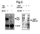
Figure 2 : Western blots after labelling of AGT fusion proteins in vivo. A) Western-Blot of total cell extract of E. coli expressing Pep-hAGT with and without BG-Bt in the medium. A streptavidin-peroxidase conjugate is used to detect biotinylated proteins. The band at 20 kD corresponds to a protein that is biotinylated in E. coli in the absence of BG-Bt. B) Western-Blot of total cell extract of yeast expressing a hAGT-DHFR-HA fusion protein with and without BG-DIG in the medium. An anti digoxigenin-peroxidase conjugate is used to detect digoxigenin-labelled proteins. - The following applications of the present invention are provided by way of examples and not limitation. The method disclosed herein is generally applicable to a range of applications and is capable of specifically and covalently labelling fusion proteins with (1) labels which are capable of sensing and inducing changes in the environment of the labelled fusion protein and/or (2) labels which aid in manipulating the fusion protein by the physical and/or chemical properties specifically introduced by the label to the fusion protein. The method disclosed herein can be used to label AGT fusion proteins both in vivo and in vitro.
- The present invention is based on the realisation that specific attachment of a label to a desired protein could be carried out by constructing a fusion protein between that protein of interest and taking advantage of the mechanism of an O6-alkylguanine-DNA alkyltransferase such as human DNA repair enzyme O6-alkylguanine-DNA alkyltransferase (hAGT). This enzyme irreversibly transfers the alkyl group from its substrate, O6-alkylguanine-DNA, to one of its cysteine residues (
Figure 1 ). A substrate analogue that rapidly reacts with hAGT is O6-benzylguanine, the second order rate constant being approximately 103 sec-1 M-1 (Figure 1 ). We have shown that substitutions of O6-benzylguanine at the C4 of the benzyl ring do not significantly affect the reactivity of hAGT against O6-benzylguanine derivatives. This enables the use of O6-benzylguanine derivatives that have a label attached to the C4 of the benzyl ring to covalently and specifically attach the label AGT fusion proteins in vivo or in vitro (Figure 1D ). The labelling is independent of the nature of the fusion protein. - If the labelling is done in vivo in cell culture or in cell extracts, the labelling of the endogenous AGT of the host is advantageously taken into account. If the endogenous AGT of the host does not accept O6-benzylguanine derivatives or related compounds as a substrate, the labelling of the fusion protein is specific. In mammalian cells (human, murine, rat), labelling of endogenous AGT is possible. In those experiments where the simultaneous labelling of the endogenous AGT as well as of the AGT fusion problem poses a problem, previously described AGT-deficient cell lines can be used [Kaina et al., 1991]. In general, the present invention can be employed in all applications of the technique were the covalent and specific attachment of a label to a AGT fusion protein is used to monitor or influence the behaviour of the AGT fusion protein or is used to manipulate the AGT fusion protein by virtue of the introduced label. Examples of applications for the use of this technology follow.
- The use of a labelled AGT substrates, such as O6-benzylguanine derivatives, where the substrate carries a detectable label which can be transferred to the AGT allows the present invention to be used to specifically and covalently attach the detectable label to the AGT fusion protein, either in a cell, on the surface of a cell (in vivo) or in vitro. This allows the detection and characterization of the AGT fusion protein in vivo or in vitro. The term in vivo includes labelling in all compartments of a cell as well as of AGT fusion proteins pointing to the extracellular space.
- The use of AGT substrates, such as O6-benzylguanine derivatives, which are labelled with an affinity tag such as biotin allows the present invention to be used to transfer an affinity tag to the AGT-protein fusion, thereby allowing the fusion protein to be bound by a binding partner of the affinity tag. By way of example, the addition of AGT substrates labelled with an affinity tag such as biotin to cells (bacterial or eukaryotic) expressing an AGT fusion protein, or to the cell extracts of such cells or to purified AGT fusion proteins, will lead to the covalent modification of the fusion protein with the affinity tag. This will then allow the isolation of the fusion protein using the interaction between the affinity tag and its binding partner, e.g. in the case of biotin, immobilized avidin or streptavidin. If the label is linked to the AGT-protein fusion via a linker containing a cleavable bond, such as a disulfide bridge, or if the linker is photocleavable, the AGT fusion protein can be released from the affinity tag after its isolation.
- AGT substrates, such as O6-benzylguanine derivatives, can be used to introduce labels into AGT-protein fusions which are capable of generating reactive radicals, such as hydroxyl radicals, upon exposure to an external stimuli. The generated radicals can then inactivate the AGT fusion proteins as well as those proteins that are in close proximity of the AGT fusion protein, allowing the study the role of these proteins. Examples of such labels are tethered metal-chelate complexes that produce hydroxyl radicals upon exposure to H2O2 and ascorbate, and chromophores such as malachite green that produce hydroxyl radicals upon laser irradiation. The use of chromophores and lasers to generate hydroxyl radicals is also known in the art as chromophore assisted laser induced inactivation (CALI) [Jay et al., 1998]. CALI is a method that is used to specifically inactivate certain proteins within a cell in a time-controlled and spatially-resolved manner and which is based upon the spatial neighbourhood of a chromophore and a protein. Upon laser irradiation the chromophore generates hydroxyl radicals, which inactivate all proteins within and only within about 0.1 nm of the chromophore. So far, the chromophore is brought in the spatial neighbourhood of the protein of interest by microinjecting chromophore-labelled antibodies specific to the protein of interest. In the present invention, labelling AGT fusion proteins with chromophores such as malachite green and subsequent laser irradiation would allow to inactivate the AGT fusion protein as well as those proteins that interact with the AGT fusion protein in a time-controlled and spatially-resolved manner. The method can be applied both in vivo or in vitro.
- In a similar manner, AGT fusion proteins can be labelled with tethered metal-chelates and the AGT fusion protein and those proteins that interact with the AGT fusion protein can be inactivated in a specific manner upon exposure to H2O2 and ascorbate. The method can not only be used to study the function of an AGT fusion protein or those that are in close proximity of the AGT fusion protein, but also to identify those proteins that are in close proximity of a AGT fusion protein. Here, proteins which are in close proximity of the AGT fusion protein can be identified as such by either detecting fragments of that protein by a specific antibody, by the disappearance of those proteins on a high-resolution 2D-electrophoresis gels or by identification of the cleaved protein fragments via separation and sequencing techniques such as mass spectrometry or protein sequencing by N-terminal degradation.
- The use of labelled AGT substrates, such as O6-benzylguanine derivatives can be used to transfer a ligand to the AGT-protein fusion. This allows binding partners of the ligand, such as proteins, to bind to the AGT-protein fusion. For example, where the label is a ligand which is capable of binding to a binding partner, contacting such a substrate with a AGT fusion protein will lead to specific attachment of the ligand to the fusion protein. If the ligand binds to another protein Y and the dimerization of the protein Y with the labelled AGT fusion protein leads to a biological function or a measurable signal, the biological function or the measured signal depends on the addition of the AGT substrate carrying the label. A specific example would be the use of AGT substrates and AGT fusion protein in the so-called three-hybrid system described by Ho et al., 1996, to regulate gene expression with small molecules. In this case, AGT is fused to the DNA-binding domain of a transcription factor. A protein Y, such as FKBP that binds a ligand, such as FK506, is fused to the activation domain of a transcription factor. Supplying the cells with the AGT substrate that carries the ligand, in this example FK506, will lead to the formation of a functional transcription factor and gene expression.
- The use of O6-benzylguanine derivatives or related AGT substrates that carry a label and where the label is a drug or a biological active small molecule that binds to an yet unidentified protein Y. Here the goal would be to identify the target protein Y of the biological active molecule. In this case, AGT is fused to the DNA-binding domain of a transcription factor. A cDNA library of the organism which expresses the unknown target protein Y is fused to the activation domain of a transcription factor. Adding the O6-benzylguanine derivatives or related AGT substrate that carry a label and where the label is the drug or the biological active small molecule will lead to the formation of a functional transcription factor and gene expression only in the case where this molecule binds to its target protein Y present in the cDNA library and fused to the activation domain. If gene expression is coupled to a selective advantage, the corresponding host carrying the plasmid with the target gene Y of the drug or bioactive molecule can be identified.
- The use of O6-benzylguanine or related AGT substrates that carry a label and where the label is a library of chemical molecules: Here the goal would be to identify small molecules that bind to a protein Y under in vivo conditions, which might be a potential drug target. In this case, AGT is fused to the DNA-binding domain of a transcription factor. The target protein Y is fused to the activation domain of a transcription factor. Adding a library of small molecules attached as label to a O6-benzylguanine derivative will lead to the formation of a functional transcription factor and gene expression only in the case where the label (i.e. the small molecule) binds to its target protein Y fused to the activation domain. If gene expression is coupled to a selective advantage, those molecules of the library leading to the growth of the host can be identified.
- The use of O6-benzylguanine derivatives or related AGT substrates carrying a label and where the label is a molecule that can be bound non-covalently by another molecule that is itself attached to the surface. An example is where the label is biotin and the molecule attached to the surface is streptavidin or avidin. Possible examples for a carrier would be either a glass side, a microtiter plate or any functionalized polymer. The immobilization of the AGT substrate via its label allows the subsequent immobilization of a AGT fusion protein on the carrier by the transfer of the label to the fusion protein. Spotting (different) AGT fusion proteins in a spatially resolved manner on the carrier allows the creation of protein arrays.
- The use of O6-benzylguanine derivatives or related AGT substrates which carry a label and where the label is a molecule that can cross-link to other proteins. Examples of such cross-linkers are molecules containing functional groups such as maleimides, active esters or azides and others known to those proficient in the art and described in Nadeau et al., 2002. Contacting such AGT substrates with AGT fusion proteins that interact with other proteins (in vivo or in vitro) can lead to the covalent cross-linking of the AGT fusion protein with its interacting protein via the label. This allows the identification of the protein interacting with the AGT fusion protein.
- The following examples are set forth so as to provide those of ordinary skill in the art with a complete disclosure and description of how to practice the invention, and are not intended to limit the scope of the invention.
- The following example, the labelling of Pep-hAGT in E. Coli using BG-Bt, demonstrates the feasibility of covalently labelling hAGT fusion proteins in E. coli. The sequence of the peptide fused to the N-terminus of hAGT (yielding Pep-hAGT) is (in single letter code) MHHHHHHSSA (SEQ ID NO:2) followed by the first amino acid of hAGT, a methionine. Liquid cultures of XL-1 Blue E. coli cells containing a pET-15b (Novagen) based expression vector encoding hAGT with an N-terminal fusion peptide, i.e. Pep-hAGT, were grown to an optical density OD600nm of 0.6. Expression of Pep-hAGT was induced by adding IPTG to a final concentration of 1 mM. At the same time BG-Bt was added to a final concentration of 10 µM and the bacteria were incubated for 2 h at 37°C. Cells were harvested by centrifugation and the pellet washed twice to remove access BG-Bt. A resuspended aliquot of cells was analysed by Western Blotting. Biotinylated proteins were detected using a streptavidin-peroxidase conjugate (NEN) and a chemiluminescent peroxidase substrate (Renaissance reagent plus, NEN) (
Figure 2 ). - The following example demonstrates the feasibility of covalently labelling hAGT fusion proteins in yeast. Here, a hAGT-DHFR-HA fusion protein is biotinylation in yeast using BG-Bt. The fusion protein is constructed on the DNA level using standard molecular biology procedures. In short, the stop codon of hAGT is replaced by codons for the amino acids RSGI, which are then followed by the codon for the first amino acid of DHFR from mouse, a Met [Nunberg et al., 1980]. The codons for the linker between hAGT and DHFR also encode for a Bgl II site, its DNA sequence being AGATCT. To construct the fusion between DHFR and the HA tag, the stop codon of DHFR is replaced by a codon for the first amino acid of the HA-tag [Kolodziej, 1991]. The HA-tag is followed by a stop codon. A culture of L40 yeast cells, containing the expression vector p314AK1 in which the hAGT-DHFR-HA protein is under control of the pcup1 promoter, was grown to an OD600 of 0.6. Expression of hAGT-DHFR-HA was induced by adding CuSO4 to a concentration of 100 µM and BG-Bt was simultaneously added to a concentration of 10 µM. Aliquots were taken after 2.5 h and 5 h and cells harvested by centrifugation. The pellet was washed twice to remove remaining BG-Bt. After lysis of the yeast cells by freeze/thaw cycling the cell extract was analysed for the presence of biotinylated hAGT-DHFR-HA fusion protein using an ELISA. In short, the biotinylated hAGT-DHFR-HA was immobilized in streptavidin-coated microtiter wells and detected by using an antiHA-antibody (Babco) as a primary and an antimouse-HRP conjugate (Sigma) as a secondary antibody (
Figure 1 ) [Kolodziej, 1991]. The ELISA was developed using the peroxidase substrate ABTS and the signal (absorbance) measured at OD405nm. The signal for the in vivo biotinylated hAGT-DHFR-HA fusion protein was at least fivefold above background. The background signal was defined as the OD405nm of cell lysates obtained from cells treated exactly as above but omitting the addition of BG-Bt. - The following example demonstrates the feasibility of covalently labelling hAGT fusion proteins in yeast. Here, the hAGT-DHFR-HA fusion protein is labelled with digoxigenin in yeast using BG-DIG. The construction of the hAGT-DHFR-HA fusion is described in example B. A culture of L40 yeast cells containing the expression vector p314AK1 in which the gene of a hAGT-DHFR fusion protein is under control of the pcup1 promoter was grown to an OD600nm of 1.2. Expression of the hAGT-DHFR fusion protein was induced by adding CuSO4 to a concentration of 100 µM and BG-DIG was simultaneously added to a concentration of 20 µM. After 2 h cells from 1 ml of shake-flask culture were harvested by centrifugation. The pellet was washed three times with medium to remove remaining BG-DIG. After lysis of the yeast cells by freeze/thaw cycling the cell extract was analysed for the presence of digoxigenated hAGT-DHFR fusion protein by Western blotting. Digoxigenated proteins were detected using an anti-digoxigenin-peroxidase conjugate (Roche) and a chemiluminescent peroxidase substrate (Renaissance reagent plus, NEN) (
Figure 2 ). - The following example demonstrates the feasibility of labelling AGT fusion proteins in mammalian cells. Here, endogenous hAGT in human cells (HEK 293) is labelled with fluoresceine using BG-AcFc. Although this example describes the use of a spectroscopic probe, it can be likewise performed with a particular label of the invention. HEK 293 cells were incubated with 5 µM BG-AcFc in PBS for 5min. The acetylated fluoresceine derivative BG-AcFc is cell-permeable and non-fluorescent but can be expected to be hydrolysed rapidly within the cell to yield the fluorescent BG-Fc. The cells were then washed by changing the PBS to remove any access substrate BG-AcFc and incubated in PBS for 20 min. Images were then taken with a confocal fluorescence microscope (Ext. 492 nm; Em. 510 nm). As a control experiment, the HEK 293 cells were treated as above but incubated prior to addition of BG-AcFc overnight with O6-benzylguanine (1 µM). This should inactivate the endogenous hAGT and therefore prevent the accumulation of the fluorescence in the nucleus. As expected, no accumulation of fluorescence in the nucleus was observed when the cells were preincubated with O6-benzylguanine. To independently confirm that the hAGT accepts BG-Fc as a substrate, recombinant Pep-hAGT (10 µM, as described in example A was incubated with 100 µM BG-Fc at 25 °C in 50 mM Tris-Cl, 10 mM DTT, 1 mM EDTA, 200 µg/ml BSA, 10% Glycerol, pH 7.4 for 10 minutes, followed by addition of 900 µl PBS (phosphate buffered saline: 137 mM NaCl, 2.7 mM KCI, 10 mM Na2HPO4, 1.8 mM KH2PO4, pH 7.4). Separation of excess substrate BG-Fc was achieved by gel filtration on a NAP™-10 Column (Pharmacia) according to the supplier's instruction. The Pep-hAGT was then characterized in a standard fluorescence spectrophotometer. The sample was excitated at 222, 238 and 490 nm, respectively and showed maximal emission at a wavelength of 523 nm, verifying that the protein was labelled with fluoresceine. A solution of 20 nM BG-Fc in PBS was measured as reference. The substrate's emission wavelength is 519 nm (excitation at 237, 323 and 490 nm respectively).
- To demonstrate that fusion proteins with AGT can be directly labelled and manipulated (here immobilized) in cell extracts the following N- and C-terminal fusion proteins with hAGT proteins were constructed via standard molecular cloning procedures and cloned into a yeast expression vector:
- (i) V5-NLS-B42-hAGT, where V5 stands for V5 epitope, NLS for SV40 large T antigen nuclear localization sequence and B42 stands for an artificial transcriptional activator B42 [Ma et al., 1987]. The last codon of the B42 transactivation domain is followed by the 22 amino acid sequence ASKKGTELGSTTSNGRQCAGIL (SEQ ID NO:3). The last three codons include a Eco RI site for the C-terminal cloning of the hAGT to B42. A Not I site is the C-terminal restriction site for the hAGT, whose sequence includes a stop codon;
- (ii) hAGT-HA-Ura3, where Ura3 stands for the yeast enzyme orotic acid decarboxylase and HA stands for the Ha epitope. Here, the stop codon for the hAGT is replaced by RS linker followed by the first amino acid of the HA-tag. The HA-tag is directly followed by the Ura3 gene;
- (iii) hAGT-DHFR-HA, where DHFR stands for the mouse dihydrofolate reductase and HA stands for the Ha epitope. Construction see example B; and
- (iv) SSN6-hAGT, where SSN6 stands for a yeast repressor of DNA transcription [Schultz et al., 1987]. Here, the stop codon of hAGT is replaced by codons for the amino acids RSGSG, which are then followed by the codon for the first amino acid of SSN6 of yeast, a methionine.
- The expression of all genes were controlled by the pCUP1 promoter. L40 yeast cells containing an expression vector encoding for one of the fusion protein were grown to an OD of 0.6 and expression of the fusion protein was induced by adding CuSO4 to a concentration of 100 µM. Aliquots (2 ml) were taken after 5 h and cells harvested by centrifugation. After lysis of the cells by freeze/thaw cycling the yeast extract was incubated with BG-Bt-oligo (10 pmol) for 20 min at room temperature, leading to biotinylation of the fusion protein. Subsequently, the suspension was transferred into a streptavidin-coated microtiter plates (Roche molecular biochemicals) and incubated for 1 h. After extensive washing of the well with PBS, the immobilized fusion protein was detected using either an anti-HA-antibody (Babco) or an anti-hAGT-antibody (in the case of the SSN6-hAGT fusion protein) as a primary and an antimouse-peroxidase conjugate (Sigma, #A4416) as a secondary antibody and subsequent incubation with the peroxidase substrate ABTS using standard biochemical procedures. In all cases, the signal which was measured as the OD405nm was at least five-fold above background. Background was measured for each fusion protein by omitting the addition of BG-Bt-oligo to the cell extracts.
- The references are in alphabetic order.
- RB Ali et al., Molecular and Cellular Biology, 18, 1660-1669 (1998)
- R Damoiseaux, A Keppler and K Johnsson, ChemBiochem, 4, 285-287 (2001)
- SN Ho et al., Nature 382, 822-6 (1996)
- R Hori and N Baichoo in Protein-Protein interactions: a molecular cloning manual; Ed. E Golemis, Cold Spring Harbor Laboratory Press; pp. 288-311 (2002)
- DG Jay and T Sakurai, Biochim. Biophys. Acta M39-48 (1999)
- Kaina et al., Carcinogenesis 12, 1857-67 (1991)
- PA Kolodziej and RA Young, Methods Enzymol. 194, 508-19 (1991)
- J Ma and M Ptashne, Cell 51, 113-9 (1987)
- OW Nadeau and GM Carlson in Protein-Protein interactions: a molecular cloning manual; Ed. E Golemis, Cold Spring Harbor Laboratory Press; pp. 75-92 (2002)
- JH Nunberg et al., Cell 19, 355-364 (1980)
- AE Pegg et al., Prog Nucleic Acid Res Mol Biol. 51, 167-223 (1995)
- J Schultz, M Carlson Mol Cell Biol 7, 3637-45 (1987)
-
- <110> ECOLE POLYTECHNIQUE FEDERALE DE LAUSANNE
- <120> Methods Using O6-Alkylguanine-DNA Alkyltransferases
- <130> P2068
- <140>
PCT/GB02/01636
<141> 2002-04-05 - <150>
US 60/282,766
<151> 2001-04-10 - <160> 3
- <170> PatentIn version 3.3
- <210> 1
<211> 22
<212> DNA
<213> Artificial sequence - <220>
<223>Fig 1E 22mer oligo part of R3, n at position 14 is BG-BT - <400> 1
gtggtgggcg ctgnaggcgt gg 22 - <210> 2
<211> 10
<212> PRT
<213> Artifical Sequence - <400> 2

- <210> 3
<211> 22
<212> PRT
<213> Artifical Sequence - <400> 3

Claims (18)
- A method for detecting a protein of interest in vitro or in vivo, in vivo meaning in bacterial or eukaryolic cells, in cell culture ur in cell extracts, in a cell in all compartments of a cell, on the surface of a cell or AGT fusion proteins pointing to the extracellular space, which comprises contacting an expressed fusion protein comprising the protein of interest and an O6- alkylguanine-DNA alkyltransferase (AGT) with a substrate having a label so that the AGT transfers the label from the substrate to become covalently bonded to the expressed fusion protein to permit detection thereof, wherein the substrate is a labelled benzyl guanine substrate represented by the general formula:
 R1 is a proton;R2 is a linker group;
andlabel represents a group for use in detecting and/or manipulating the fusion protein;wherein
or by the general formula: R1 is a group accepted by AGT allowing the AGT to transfer the label to the protein fusion;R2 is as above;R3 is a proton;
andlabel is as above;and wherein the label is an affinity tag, a first member of a specific binding pair which is capable of specifically binding to the second member of the specific binding pair, a molecule which is attachable to a solid phase, a molecule which is capable of generating reactive radicals, a candidate compound or library of candidate compounds, a molecule which is capable of crosslinking to other biomolecules, a nucleic acid or a derivative thereof capable of undergoing base-pairing with its complementary strand, a lipid, or a hydrophobic molecule with membrane-inserting properties. - The method according to claim 1, wherein the label is an affinity tag, a first member of a specific binding pair which is capable of specifically binding to the second member of the specific binding pair, or a molecule which is attachable to a solid phase.
- The method according to claim 1 or 2, which comprises detecting the labelled fusion protein in an in vitro system.
- The method according to claim 1 or 2, which comprises detecting the labelled fusion protein in a cell culture system.
- The method of claim 4, further comprising the initial step of transforming the cells with an expression vector comprising nucleic acid encoding the fusion protein linked to control sequences to direct its expression.
- The method according to any one of the preceding claims, further comprising manipulating the labelled fusion protein using a property introduced by the label to the fusion protein.
- The method according to any one of the preceding claims, wherein the labelled substrate is incorporated into a nucleic acid molecule, and the nucleic acid molecule is an oligonucleotide between 2 and 99 nucleotides in length.
- The method according to claim 1, wherein in the second formula R1 is a substituted or unsubstituted alkyl chain, a substituted or unsubstituted cycloalkyl group with a ring size between three and ten carbons, a substituted or unsubstituted heterocycle with a ring size between three and ten carbons, or a substituted or unsubstituted aromatic heterocycle with a ring size between three and ten carbons.
- The method according to any one of claims I to 8, wherein the linker group R2 is a substituted or unsubstituted alkyl chain or a polyethylene glycol.
- The method according to any one of claims 1 to 9, wherein the label is an affinity tag.
- The method according to any one of claims 1 to 9, wherein the label is a first member of a specific binding pair which is capable of specifically binding to the second member of the specific binding pair.
- The method according to any one of claims 1 to 11, wherein the label is a molecule which is attachable to a solid phase.
- The method according to claim 10, 11 or 12, wherein the label is biotin, avidin or streptavidin.
- The method according to claim 10, 11 or 12, wherein the label is linked to the fusion protein via a cleavable linker so that the fusion protein can be released from the label.
- The method according to claim 10, 11 or 12, wherein the label is linked to the fusion protein via a photocleavable linker so that the fusion protein can he released from the label.
- The method according to claim 11 further comprising contacting the fusion protein with a molecule comprising the second member of the specific binding pair.
- The method according to claim 11 or 16, wherein the first and second members of the specific binding pair are a first and second protein, an antibody and antigen, an enzyme and substrate or a ligand and receptor.
- The method according to claim 12, wherein the method comprises the further step of contacting the labelled fusion protein with the solid support so that it becomes immobilised on the solid support.
Priority Applications (1)
| Application Number | Priority Date | Filing Date | Title |
|---|---|---|---|
| EP10075132.0A EP2211177B1 (en) | 2001-04-10 | 2002-04-05 | Methods using O6-alkylguanine-DNA alkyltransferases |
Applications Claiming Priority (3)
| Application Number | Priority Date | Filing Date | Title |
|---|---|---|---|
| US28276601P | 2001-04-10 | 2001-04-10 | |
| EP02720187A EP1410023B1 (en) | 2001-04-10 | 2002-04-05 | Methods using o6-alkylguanine-dna alkyltransferases |
| EP06111733A EP1696234B1 (en) | 2001-04-10 | 2002-04-05 | Methods using O6-alkylguanine-DNA alkyltransferases |
Related Parent Applications (3)
| Application Number | Title | Priority Date | Filing Date |
|---|---|---|---|
| EP06111733A Division EP1696234B1 (en) | 2001-04-10 | 2002-04-05 | Methods using O6-alkylguanine-DNA alkyltransferases |
| EP02720187.0 Division | 2002-04-05 | ||
| EP06111733.9 Division | 2006-03-27 |
Related Child Applications (3)
| Application Number | Title | Priority Date | Filing Date |
|---|---|---|---|
| EP10075132.0A Division EP2211177B1 (en) | 2001-04-10 | 2002-04-05 | Methods using O6-alkylguanine-DNA alkyltransferases |
| EP10075095.9 Division-Into | 2010-03-03 | ||
| EP10075132.0 Division-Into | 2010-03-25 |
Publications (2)
| Publication Number | Publication Date |
|---|---|
| EP2037271A1 EP2037271A1 (en) | 2009-03-18 |
| EP2037271B1 true EP2037271B1 (en) | 2010-09-22 |
Family
ID=23083030
Family Applications (4)
| Application Number | Title | Priority Date | Filing Date |
|---|---|---|---|
| EP08168182A Expired - Lifetime EP2037271B1 (en) | 2001-04-10 | 2002-04-05 | Methods using O6-alkylguanine-DNA alkyltransferases |
| EP10075132.0A Expired - Lifetime EP2211177B1 (en) | 2001-04-10 | 2002-04-05 | Methods using O6-alkylguanine-DNA alkyltransferases |
| EP02720187A Expired - Lifetime EP1410023B1 (en) | 2001-04-10 | 2002-04-05 | Methods using o6-alkylguanine-dna alkyltransferases |
| EP06111733A Expired - Lifetime EP1696234B1 (en) | 2001-04-10 | 2002-04-05 | Methods using O6-alkylguanine-DNA alkyltransferases |
Family Applications After (3)
| Application Number | Title | Priority Date | Filing Date |
|---|---|---|---|
| EP10075132.0A Expired - Lifetime EP2211177B1 (en) | 2001-04-10 | 2002-04-05 | Methods using O6-alkylguanine-DNA alkyltransferases |
| EP02720187A Expired - Lifetime EP1410023B1 (en) | 2001-04-10 | 2002-04-05 | Methods using o6-alkylguanine-dna alkyltransferases |
| EP06111733A Expired - Lifetime EP1696234B1 (en) | 2001-04-10 | 2002-04-05 | Methods using O6-alkylguanine-DNA alkyltransferases |
Country Status (15)
| Country | Link |
|---|---|
| US (2) | US7939284B2 (en) |
| EP (4) | EP2037271B1 (en) |
| JP (2) | JP4195815B2 (en) |
| KR (1) | KR20040039194A (en) |
| CN (2) | CN1975422B (en) |
| AT (3) | ATE482395T1 (en) |
| AU (1) | AU2002251257B2 (en) |
| CA (1) | CA2443570A1 (en) |
| DE (3) | DE60212642T2 (en) |
| DK (1) | DK1410023T3 (en) |
| HK (1) | HK1068406A1 (en) |
| IL (1) | IL157880A0 (en) |
| NZ (3) | NZ537939A (en) |
| WO (1) | WO2002083937A2 (en) |
| ZA (1) | ZA200307442B (en) |
Families Citing this family (44)
| Publication number | Priority date | Publication date | Assignee | Title |
|---|---|---|---|---|
| EP1546371A1 (en) | 2002-10-03 | 2005-06-29 | Ecole Polytechnique Federale De Lausanne (Epfl) | Substrates for o6-alkylguanine-dna alkyltransferase |
| EP1546370A1 (en) * | 2002-10-03 | 2005-06-29 | Ecole Polytechnique Federale De Lausanne (Epfl) | Protein labelling with o6-alkylguanine-dna alkyltransferase |
| US7429472B2 (en) | 2003-01-31 | 2008-09-30 | Promega Corporation | Method of immobilizing a protein or molecule via a mutant dehalogenase that is bound to an immobilized dehalogenase substrate and linked directly or indirectly to the protein or molecule |
| WO2004072232A2 (en) | 2003-01-31 | 2004-08-26 | Promega Corporation | Covalent tethering of functional groups to proteins |
| NZ543653A (en) * | 2003-05-23 | 2008-10-31 | Epfl Ecole Polytechnique Federale Lausanne | Methods for protein labeling based on acyl carrier protein |
| JP2007525988A (en) * | 2004-03-02 | 2007-09-13 | ウペエフエル・エコル・ポリテクニック・フェデラル・ドゥ・ローザンヌ | O6-alkylguanine-DNA alkyltransferase mutants |
| US8163479B2 (en) | 2004-03-02 | 2012-04-24 | Ecole Polytechnique Federale De Lausanne | Specific substrates for O6-alkylguanine-DNA alkyltransferase |
| US7425436B2 (en) | 2004-07-30 | 2008-09-16 | Promega Corporation | Covalent tethering of functional groups to proteins and substrates therefor |
| US7825096B2 (en) | 2004-09-08 | 2010-11-02 | The United States Of America As Represented By The Department Of Health And Human Services | O6-alkylguanine-DNA alkyltransferase inactivators and beta-glucuronidase cleavable prodrugs |
| US7273714B2 (en) * | 2005-03-11 | 2007-09-25 | Vion Pharmaceuticals, Inc. | Alkylguanyltransferase assays |
| JP2008539198A (en) * | 2005-04-27 | 2008-11-13 | コヴァリス・バイオサイエンシス・アーゲー | Pyrimidines that react with O6-alkylguanine-DNA alkyltransferases |
| FR2890174B1 (en) | 2005-08-30 | 2009-04-24 | Cis Bio Internat Sa | METHOD FOR THE DEMONSTRATION OF A BIOLOGICAL PROCESS BY MEASURING FREIGHT |
| FR2890446B1 (en) | 2005-09-05 | 2008-04-18 | Cis Bio Internat Sa | METHOD FOR DETECTING INTRACELLULAR INTERACTION BETWEEN BIO-MOLECULES |
| KR20090034930A (en) | 2006-06-30 | 2009-04-08 | 암비트 바이오사이언시즈 코포레이션 | Detectable nucleic acid tag |
| US8420367B2 (en) | 2006-10-30 | 2013-04-16 | Promega Corporation | Polynucleotides encoding mutant hydrolase proteins with enhanced kinetics and functional expression |
| CA2670937C (en) * | 2006-12-05 | 2016-07-26 | Lasergen, Inc. | Photocleavable labeled nucleotides and nucleosides and labeled nucleotides and nucleosides and methods for their use in dna sequencing |
| US7897737B2 (en) | 2006-12-05 | 2011-03-01 | Lasergen, Inc. | 3′-OH unblocked, nucleotides and nucleosides, base modified with photocleavable, terminating groups and methods for their use in DNA sequencing |
| EP1978093A1 (en) * | 2007-04-04 | 2008-10-08 | Ecole Polytechnique Fédérale de Lausanne (EPFL) | Method of crosslinking two objects of interest |
| GB0711560D0 (en) * | 2007-06-14 | 2007-07-25 | Innova Biosciences Ltd | Improvements in or relating to production of conjugates |
| US20100209933A1 (en) | 2007-10-29 | 2010-08-19 | Mcreynolds Larry A | Methods and Compositions for Detection and Enrichment of Target Small RNAs |
| EP2093564A1 (en) | 2008-02-25 | 2009-08-26 | Technische Universität Dresden Medizinische Fakultät Carl Gustav Carus | Method for distinguishing secretory granules of different ages |
| MX2010013678A (en) | 2008-06-11 | 2011-02-24 | Lasergen Inc | Nucleotides and nucleosides and methods for their use in dna sequencing. |
| FR2936245B1 (en) | 2008-09-23 | 2012-07-06 | Cis Bio Int | NOVEL O6-ALKYLGUANIN-DNA ALKYLTRANSFERASE SUBSTRATES AND MUTANTS THEREOF |
| WO2011091146A1 (en) | 2010-01-20 | 2011-07-28 | New England Biolabs, Inc. | Compositions, methods and related uses for cleaving modified dna |
| EP2616817A4 (en) | 2010-09-15 | 2014-04-09 | Endacea Inc | Methods of use and kit for measurement of lipopolysaccharide with a time resolved fluorescence based assay |
| EP3670523B1 (en) | 2011-09-13 | 2021-08-11 | Agilent Technologies, Inc. | 3'-oh unblocked, fast photocleavable terminating nucleotides and their use in methods for nucleic acid sequencing |
| KR102007061B1 (en) * | 2011-12-09 | 2019-08-02 | 앵스띠뛰 파스퇴르 | Multiplex immuno screening assay |
| US9040239B2 (en) | 2012-03-15 | 2015-05-26 | New England Biolabs, Inc. | Composition and methods of oxygenation of nucleic acids containing 5-methylpyrimidine |
| JP6441893B2 (en) | 2013-03-19 | 2018-12-19 | ディレクティド・ジェノミクス・エル・エル・シー | Target sequence enrichment |
| JP5781589B2 (en) * | 2013-12-20 | 2015-09-24 | レーザーゲン インコーポレイテッド | Photocleavable labeled nucleotides and nucleosides and labeled nucleotides and nucleosides and their use in DNA sequencing |
| WO2016003795A1 (en) | 2014-07-02 | 2016-01-07 | New England Biolabs, Inc. | Universal n-glycan binding reagent |
| WO2016093838A1 (en) | 2014-12-11 | 2016-06-16 | New England Biolabs, Inc. | Enrichment of target sequences |
| CN104844710B (en) * | 2015-04-13 | 2018-02-02 | 西北大学 | The preparation method and application of the compound resins of oriented immobilization PEGA |
| CN113277985B (en) | 2016-07-20 | 2023-07-28 | 纳莹(上海)生物科技有限公司 | Fluorescent probe and preparation method and application thereof |
| US10934533B1 (en) | 2018-10-10 | 2021-03-02 | New England Biolabs, Inc. | Variant DNA polymerases having improved properties and method for improved isothermal amplification of a target DNA |
| CN111253417B (en) * | 2019-09-04 | 2022-03-25 | 大连民族大学 | Synthesis and application of purine derivative |
| WO2021163559A1 (en) | 2020-02-13 | 2021-08-19 | New England Biolabs, Inc. | Variant family d dna polymerases |
| WO2021163561A1 (en) | 2020-02-13 | 2021-08-19 | New England Biolabs, Inc. | Variant family d dna polymerases |
| WO2021183921A1 (en) | 2020-03-12 | 2021-09-16 | New England Biolabs, Inc. | A rapid diagnostic test for lamp |
| CN112010983A (en) * | 2020-08-11 | 2020-12-01 | 西北大学 | Method and kit for targeted screening of active ingredients of cough and asthma relieving traditional Chinese medicine |
| EP4110945A2 (en) | 2020-08-21 | 2023-01-04 | New England Biolabs, Inc. | A rapid diagnostic test for lamp |
| WO2022250676A1 (en) | 2021-05-27 | 2022-12-01 | New England Biolabs, Inc. | Dnase i variants, compositions, methods, and kits |
| US20230257730A1 (en) | 2021-11-24 | 2023-08-17 | New England Biolabs, Inc. | Double-Stranded DNA Deaminases |
| WO2023197015A1 (en) | 2022-04-08 | 2023-10-12 | New England Biolabs, Inc. | Compositions and analysis of dephosphorylated oligoribonucleotides |
Family Cites Families (3)
| Publication number | Priority date | Publication date | Assignee | Title |
|---|---|---|---|---|
| GB9224888D0 (en) * | 1992-11-27 | 1993-01-13 | Univ Singapore | Monoclonal antibody |
| DE69433161T2 (en) * | 1993-06-08 | 2004-07-08 | Cancer Research Technology Ltd. | O6-SUBSTITUTED GUANINE DERIVATIVES, METHOD FOR THE PRODUCTION THEREOF AND THEIR APPLICATION FOR TREATMENT OF TUMOR CELLS |
| DE19903895A1 (en) * | 1999-02-01 | 2000-08-03 | Kai Johnsson | Assay for O6-alkylguanine DNA alkyltransferase, e.g. in tumor tissue, includes immobilization via a reactive group transferred from a substrate |
-
2002
- 2002-04-05 EP EP08168182A patent/EP2037271B1/en not_active Expired - Lifetime
- 2002-04-05 EP EP10075132.0A patent/EP2211177B1/en not_active Expired - Lifetime
- 2002-04-05 EP EP02720187A patent/EP1410023B1/en not_active Expired - Lifetime
- 2002-04-05 KR KR10-2003-7012899A patent/KR20040039194A/en active IP Right Grant
- 2002-04-05 CN CN2006101603317A patent/CN1975422B/en not_active Expired - Lifetime
- 2002-04-05 CA CA002443570A patent/CA2443570A1/en not_active Abandoned
- 2002-04-05 AT AT08168182T patent/ATE482395T1/en not_active IP Right Cessation
- 2002-04-05 NZ NZ537939A patent/NZ537939A/en unknown
- 2002-04-05 EP EP06111733A patent/EP1696234B1/en not_active Expired - Lifetime
- 2002-04-05 DE DE60212642T patent/DE60212642T2/en not_active Expired - Lifetime
- 2002-04-05 DE DE60230689T patent/DE60230689D1/en not_active Expired - Lifetime
- 2002-04-05 US US10/474,796 patent/US7939284B2/en active Active
- 2002-04-05 IL IL15788002A patent/IL157880A0/en not_active IP Right Cessation
- 2002-04-05 DK DK02720187T patent/DK1410023T3/en active
- 2002-04-05 NZ NZ528594A patent/NZ528594A/en unknown
- 2002-04-05 NZ NZ540410A patent/NZ540410A/en unknown
- 2002-04-05 DE DE60237792T patent/DE60237792D1/en not_active Expired - Lifetime
- 2002-04-05 JP JP2002582274A patent/JP4195815B2/en not_active Expired - Lifetime
- 2002-04-05 CN CNB028081137A patent/CN1295510C/en not_active Expired - Lifetime
- 2002-04-05 AT AT02720187T patent/ATE331220T1/en not_active IP Right Cessation
- 2002-04-05 WO PCT/GB2002/001636 patent/WO2002083937A2/en active IP Right Grant
- 2002-04-05 AU AU2002251257A patent/AU2002251257B2/en not_active Ceased
- 2002-04-05 AT AT06111733T patent/ATE419378T1/en not_active IP Right Cessation
-
2003
- 2003-09-25 ZA ZA200307442A patent/ZA200307442B/en unknown
-
2005
- 2005-01-18 HK HK05100472A patent/HK1068406A1/en not_active IP Right Cessation
-
2007
- 2007-12-20 JP JP2007328757A patent/JP4226053B2/en not_active Expired - Fee Related
-
2011
- 2011-01-24 US US13/012,234 patent/US8367361B2/en not_active Expired - Lifetime
Also Published As
Similar Documents
| Publication | Publication Date | Title |
|---|---|---|
| EP2037271B1 (en) | Methods using O6-alkylguanine-DNA alkyltransferases | |
| AU2002251257A1 (en) | Methods of Using O6-alkylguanine-DNA Alkyltransferases | |
| US7666612B2 (en) | Methods for protein labeling based on acyl carrier protein | |
| JP2004532028A5 (en) | ||
| Tanaka et al. | Site‐specific protein modification on living cells catalyzed by sortase | |
| ZA200502211B (en) | Protein labelling with 06-alkylguanine-dna alkyltransferase | |
| Chung et al. | Open flower fluoroimmunoassay: a general method to make fluorescent protein-based immunosensor probes |
Legal Events
| Date | Code | Title | Description |
|---|---|---|---|
| PUAI | Public reference made under article 153(3) epc to a published international application that has entered the european phase |
Free format text: ORIGINAL CODE: 0009012 |
|
| 17P | Request for examination filed |
Effective date: 20081103 |
|
| AC | Divisional application: reference to earlier application |
Ref document number: 1696234 Country of ref document: EP Kind code of ref document: P Ref document number: 1410023 Country of ref document: EP Kind code of ref document: P |
|
| AK | Designated contracting states |
Kind code of ref document: A1 Designated state(s): AT BE CH CY DE DK ES FI FR GB GR IE IT LI LU MC NL PT SE TR |
|
| 17Q | First examination report despatched |
Effective date: 20090429 |
|
| AKX | Designation fees paid |
Designated state(s): AT BE CH CY DE DK ES FI FR GB GR IE IT LI LU MC NL PT SE TR |
|
| GRAP | Despatch of communication of intention to grant a patent |
Free format text: ORIGINAL CODE: EPIDOSNIGR1 |
|
| GRAS | Grant fee paid |
Free format text: ORIGINAL CODE: EPIDOSNIGR3 |
|
| GRAA | (expected) grant |
Free format text: ORIGINAL CODE: 0009210 |
|
| AC | Divisional application: reference to earlier application |
Ref document number: 1410023 Country of ref document: EP Kind code of ref document: P Ref document number: 1696234 Country of ref document: EP Kind code of ref document: P |
|
| AK | Designated contracting states |
Kind code of ref document: B1 Designated state(s): AT BE CH CY DE DK ES FI FR GB GR IE IT LI LU MC NL PT SE TR |
|
| REG | Reference to a national code |
Ref country code: GB Ref legal event code: FG4D |
|
| REG | Reference to a national code |
Ref country code: CH Ref legal event code: EP Ref country code: CH Ref legal event code: NV Representative=s name: KIRKER & CIE S.A. |
|
| REG | Reference to a national code |
Ref country code: IE Ref legal event code: FG4D |
|
| REF | Corresponds to: |
Ref document number: 60237792 Country of ref document: DE Date of ref document: 20101104 Kind code of ref document: P |
|
| PG25 | Lapsed in a contracting state [announced via postgrant information from national office to epo] |
Ref country code: FI Free format text: LAPSE BECAUSE OF FAILURE TO SUBMIT A TRANSLATION OF THE DESCRIPTION OR TO PAY THE FEE WITHIN THE PRESCRIBED TIME-LIMIT Effective date: 20100922 Ref country code: AT Free format text: LAPSE BECAUSE OF FAILURE TO SUBMIT A TRANSLATION OF THE DESCRIPTION OR TO PAY THE FEE WITHIN THE PRESCRIBED TIME-LIMIT Effective date: 20100922 |
|
| REG | Reference to a national code |
Ref country code: NL Ref legal event code: VDEP Effective date: 20100922 |
|
| PG25 | Lapsed in a contracting state [announced via postgrant information from national office to epo] |
Ref country code: SE Free format text: LAPSE BECAUSE OF FAILURE TO SUBMIT A TRANSLATION OF THE DESCRIPTION OR TO PAY THE FEE WITHIN THE PRESCRIBED TIME-LIMIT Effective date: 20100922 Ref country code: GR Free format text: LAPSE BECAUSE OF FAILURE TO SUBMIT A TRANSLATION OF THE DESCRIPTION OR TO PAY THE FEE WITHIN THE PRESCRIBED TIME-LIMIT Effective date: 20101223 |
|
| PG25 | Lapsed in a contracting state [announced via postgrant information from national office to epo] |
Ref country code: NL Free format text: LAPSE BECAUSE OF FAILURE TO SUBMIT A TRANSLATION OF THE DESCRIPTION OR TO PAY THE FEE WITHIN THE PRESCRIBED TIME-LIMIT Effective date: 20100922 Ref country code: PT Free format text: LAPSE BECAUSE OF FAILURE TO SUBMIT A TRANSLATION OF THE DESCRIPTION OR TO PAY THE FEE WITHIN THE PRESCRIBED TIME-LIMIT Effective date: 20110124 Ref country code: IT Free format text: LAPSE BECAUSE OF FAILURE TO SUBMIT A TRANSLATION OF THE DESCRIPTION OR TO PAY THE FEE WITHIN THE PRESCRIBED TIME-LIMIT Effective date: 20100922 |
|
| PG25 | Lapsed in a contracting state [announced via postgrant information from national office to epo] |
Ref country code: BE Free format text: LAPSE BECAUSE OF FAILURE TO SUBMIT A TRANSLATION OF THE DESCRIPTION OR TO PAY THE FEE WITHIN THE PRESCRIBED TIME-LIMIT Effective date: 20100922 |
|
| PG25 | Lapsed in a contracting state [announced via postgrant information from national office to epo] |
Ref country code: ES Free format text: LAPSE BECAUSE OF FAILURE TO SUBMIT A TRANSLATION OF THE DESCRIPTION OR TO PAY THE FEE WITHIN THE PRESCRIBED TIME-LIMIT Effective date: 20110102 |
|
| PLBE | No opposition filed within time limit |
Free format text: ORIGINAL CODE: 0009261 |
|
| STAA | Information on the status of an ep patent application or granted ep patent |
Free format text: STATUS: NO OPPOSITION FILED WITHIN TIME LIMIT |
|
| 26N | No opposition filed |
Effective date: 20110623 |
|
| PG25 | Lapsed in a contracting state [announced via postgrant information from national office to epo] |
Ref country code: DK Free format text: LAPSE BECAUSE OF FAILURE TO SUBMIT A TRANSLATION OF THE DESCRIPTION OR TO PAY THE FEE WITHIN THE PRESCRIBED TIME-LIMIT Effective date: 20100922 |
|
| REG | Reference to a national code |
Ref country code: DE Ref legal event code: R097 Ref document number: 60237792 Country of ref document: DE Effective date: 20110623 |
|
| PG25 | Lapsed in a contracting state [announced via postgrant information from national office to epo] |
Ref country code: MC Free format text: LAPSE BECAUSE OF NON-PAYMENT OF DUE FEES Effective date: 20110430 |
|
| REG | Reference to a national code |
Ref country code: IE Ref legal event code: MM4A |
|
| PG25 | Lapsed in a contracting state [announced via postgrant information from national office to epo] |
Ref country code: IE Free format text: LAPSE BECAUSE OF NON-PAYMENT OF DUE FEES Effective date: 20110405 |
|
| PG25 | Lapsed in a contracting state [announced via postgrant information from national office to epo] |
Ref country code: LU Free format text: LAPSE BECAUSE OF NON-PAYMENT OF DUE FEES Effective date: 20110405 Ref country code: CY Free format text: LAPSE BECAUSE OF FAILURE TO SUBMIT A TRANSLATION OF THE DESCRIPTION OR TO PAY THE FEE WITHIN THE PRESCRIBED TIME-LIMIT Effective date: 20100922 |
|
| PG25 | Lapsed in a contracting state [announced via postgrant information from national office to epo] |
Ref country code: TR Free format text: LAPSE BECAUSE OF FAILURE TO SUBMIT A TRANSLATION OF THE DESCRIPTION OR TO PAY THE FEE WITHIN THE PRESCRIBED TIME-LIMIT Effective date: 20100922 |
|
| REG | Reference to a national code |
Ref country code: FR Ref legal event code: PLFP Year of fee payment: 15 |
|
| REG | Reference to a national code |
Ref country code: FR Ref legal event code: PLFP Year of fee payment: 16 |
|
| REG | Reference to a national code |
Ref country code: FR Ref legal event code: PLFP Year of fee payment: 17 |
|
| REG | Reference to a national code |
Ref country code: CH Ref legal event code: PL |
|
| PG25 | Lapsed in a contracting state [announced via postgrant information from national office to epo] |
Ref country code: CH Free format text: LAPSE BECAUSE OF NON-PAYMENT OF DUE FEES Effective date: 20180430 Ref country code: LI Free format text: LAPSE BECAUSE OF NON-PAYMENT OF DUE FEES Effective date: 20180430 |
|
| PGFP | Annual fee paid to national office [announced via postgrant information from national office to epo] |
Ref country code: GB Payment date: 20210315 Year of fee payment: 20 |
|
| PGFP | Annual fee paid to national office [announced via postgrant information from national office to epo] |
Ref country code: DE Payment date: 20210428 Year of fee payment: 20 Ref country code: FR Payment date: 20210428 Year of fee payment: 20 |
|
| REG | Reference to a national code |
Ref country code: DE Ref legal event code: R071 Ref document number: 60237792 Country of ref document: DE |
|
| REG | Reference to a national code |
Ref country code: GB Ref legal event code: PE20 Expiry date: 20220404 |
|
| PG25 | Lapsed in a contracting state [announced via postgrant information from national office to epo] |
Ref country code: GB Free format text: LAPSE BECAUSE OF EXPIRATION OF PROTECTION Effective date: 20220404 |