EP2019840B1 - Antagonistic anti-human cd40 monoclonal antibody - Google Patents
Antagonistic anti-human cd40 monoclonal antibody Download PDFInfo
- Publication number
- EP2019840B1 EP2019840B1 EP07747426A EP07747426A EP2019840B1 EP 2019840 B1 EP2019840 B1 EP 2019840B1 EP 07747426 A EP07747426 A EP 07747426A EP 07747426 A EP07747426 A EP 07747426A EP 2019840 B1 EP2019840 B1 EP 2019840B1
- Authority
- EP
- European Patent Office
- Prior art keywords
- antibody
- cell
- cells
- ch5d12
- human
- Prior art date
- Legal status (The legal status is an assumption and is not a legal conclusion. Google has not performed a legal analysis and makes no representation as to the accuracy of the status listed.)
- Active
Links
- 230000003042 antagnostic effect Effects 0.000 title claims description 20
- 210000004027 cell Anatomy 0.000 claims description 316
- 102100040245 Tumor necrosis factor receptor superfamily member 5 Human genes 0.000 claims description 92
- 101150013553 CD40 gene Proteins 0.000 claims description 89
- 150000001413 amino acids Chemical class 0.000 claims description 81
- 229910052731 fluorine Inorganic materials 0.000 claims description 46
- 229910052698 phosphorus Inorganic materials 0.000 claims description 42
- 238000000034 method Methods 0.000 claims description 39
- 238000004519 manufacturing process Methods 0.000 claims description 37
- 238000011282 treatment Methods 0.000 claims description 36
- 229910052721 tungsten Inorganic materials 0.000 claims description 34
- 208000011231 Crohn disease Diseases 0.000 claims description 26
- 229910052757 nitrogen Inorganic materials 0.000 claims description 25
- 208000027866 inflammatory disease Diseases 0.000 claims description 23
- 238000006467 substitution reaction Methods 0.000 claims description 21
- 102000039446 nucleic acids Human genes 0.000 claims description 20
- 108020004707 nucleic acids Proteins 0.000 claims description 20
- 150000007523 nucleic acids Chemical class 0.000 claims description 20
- 201000006417 multiple sclerosis Diseases 0.000 claims description 19
- 208000023275 Autoimmune disease Diseases 0.000 claims description 17
- 208000024891 symptom Diseases 0.000 claims description 17
- 208000022559 Inflammatory bowel disease Diseases 0.000 claims description 15
- 238000003306 harvesting Methods 0.000 claims description 15
- 238000004113 cell culture Methods 0.000 claims description 12
- 239000003814 drug Substances 0.000 claims description 12
- 238000012217 deletion Methods 0.000 claims description 11
- 230000037430 deletion Effects 0.000 claims description 11
- 238000003780 insertion Methods 0.000 claims description 11
- 230000037431 insertion Effects 0.000 claims description 11
- 230000004048 modification Effects 0.000 claims description 11
- 238000012986 modification Methods 0.000 claims description 11
- 201000004681 Psoriasis Diseases 0.000 claims description 9
- 206010052779 Transplant rejections Diseases 0.000 claims description 8
- 229940122551 CD40 antagonist Drugs 0.000 claims description 7
- 230000001363 autoimmune Effects 0.000 claims description 7
- 206010009900 Colitis ulcerative Diseases 0.000 claims description 5
- 201000006704 Ulcerative Colitis Diseases 0.000 claims description 5
- 210000004978 chinese hamster ovary cell Anatomy 0.000 claims description 5
- 206010012438 Dermatitis atopic Diseases 0.000 claims description 4
- 206010034277 Pemphigoid Diseases 0.000 claims description 4
- 201000008937 atopic dermatitis Diseases 0.000 claims description 4
- 208000000594 bullous pemphigoid Diseases 0.000 claims description 4
- 210000004408 hybridoma Anatomy 0.000 claims description 4
- 206010039073 rheumatoid arthritis Diseases 0.000 claims description 4
- 201000000596 systemic lupus erythematosus Diseases 0.000 claims description 4
- 206010028980 Neoplasm Diseases 0.000 claims description 2
- 238000012258 culturing Methods 0.000 claims description 2
- 239000008194 pharmaceutical composition Substances 0.000 claims description 2
- 125000003275 alpha amino acid group Chemical group 0.000 claims 8
- 230000024203 complement activation Effects 0.000 claims 1
- 230000002950 deficient Effects 0.000 claims 1
- 230000027455 binding Effects 0.000 description 88
- 108090000765 processed proteins & peptides Proteins 0.000 description 70
- 102000004196 processed proteins & peptides Human genes 0.000 description 66
- 229920001184 polypeptide Polymers 0.000 description 64
- 210000001744 T-lymphocyte Anatomy 0.000 description 62
- 230000014509 gene expression Effects 0.000 description 41
- 238000004458 analytical method Methods 0.000 description 37
- 108090000623 proteins and genes Proteins 0.000 description 30
- 210000003719 b-lymphocyte Anatomy 0.000 description 28
- 230000000694 effects Effects 0.000 description 28
- 238000002965 ELISA Methods 0.000 description 27
- 241001529936 Murinae Species 0.000 description 27
- 102100032937 CD40 ligand Human genes 0.000 description 25
- 210000001072 colon Anatomy 0.000 description 25
- 235000018102 proteins Nutrition 0.000 description 25
- 102000004169 proteins and genes Human genes 0.000 description 25
- 239000006228 supernatant Substances 0.000 description 25
- 108010029697 CD40 Ligand Proteins 0.000 description 24
- 208000037265 diseases, disorders, signs and symptoms Diseases 0.000 description 24
- 210000003405 ileum Anatomy 0.000 description 24
- 238000001943 fluorescence-activated cell sorting Methods 0.000 description 23
- 210000002540 macrophage Anatomy 0.000 description 23
- 239000000872 buffer Substances 0.000 description 21
- 239000000523 sample Substances 0.000 description 20
- 102220488187 Olfactory receptor 2A12_I29L_mutation Human genes 0.000 description 19
- 238000001574 biopsy Methods 0.000 description 19
- 230000007423 decrease Effects 0.000 description 19
- 201000010099 disease Diseases 0.000 description 19
- 230000002829 reductive effect Effects 0.000 description 19
- 238000011156 evaluation Methods 0.000 description 17
- 238000002474 experimental method Methods 0.000 description 17
- 230000003902 lesion Effects 0.000 description 17
- 239000002609 medium Substances 0.000 description 17
- 201000002491 encephalomyelitis Diseases 0.000 description 16
- 230000004913 activation Effects 0.000 description 15
- 230000012010 growth Effects 0.000 description 15
- 230000001965 increasing effect Effects 0.000 description 15
- 230000003993 interaction Effects 0.000 description 15
- 239000013612 plasmid Substances 0.000 description 15
- 238000001890 transfection Methods 0.000 description 15
- 241001465754 Metazoa Species 0.000 description 14
- 238000010790 dilution Methods 0.000 description 13
- 239000012895 dilution Substances 0.000 description 13
- 239000000463 material Substances 0.000 description 13
- 239000000047 product Substances 0.000 description 13
- 241000282412 Homo Species 0.000 description 12
- 239000000499 gel Substances 0.000 description 12
- 230000001900 immune effect Effects 0.000 description 12
- 230000005764 inhibitory process Effects 0.000 description 12
- 210000002510 keratinocyte Anatomy 0.000 description 12
- 239000002953 phosphate buffered saline Substances 0.000 description 12
- 210000002966 serum Anatomy 0.000 description 12
- 241000282693 Cercopithecidae Species 0.000 description 11
- 230000003302 anti-idiotype Effects 0.000 description 11
- 238000011161 development Methods 0.000 description 11
- 230000018109 developmental process Effects 0.000 description 11
- 210000001616 monocyte Anatomy 0.000 description 11
- 101100099884 Homo sapiens CD40 gene Proteins 0.000 description 10
- 230000003247 decreasing effect Effects 0.000 description 10
- 210000004443 dendritic cell Anatomy 0.000 description 10
- 238000001727 in vivo Methods 0.000 description 10
- 230000001404 mediated effect Effects 0.000 description 10
- 230000004044 response Effects 0.000 description 10
- 238000012216 screening Methods 0.000 description 10
- 238000002415 sodium dodecyl sulfate polyacrylamide gel electrophoresis Methods 0.000 description 10
- OKKJLVBELUTLKV-UHFFFAOYSA-N Methanol Chemical compound OC OKKJLVBELUTLKV-UHFFFAOYSA-N 0.000 description 9
- 238000007792 addition Methods 0.000 description 9
- 238000003556 assay Methods 0.000 description 9
- 210000001772 blood platelet Anatomy 0.000 description 9
- 230000009266 disease activity Effects 0.000 description 9
- 238000001155 isoelectric focusing Methods 0.000 description 9
- 150000002482 oligosaccharides Chemical class 0.000 description 9
- 238000012360 testing method Methods 0.000 description 9
- 108090001007 Interleukin-8 Proteins 0.000 description 8
- 102000004890 Interleukin-8 Human genes 0.000 description 8
- 108091034117 Oligonucleotide Proteins 0.000 description 8
- 239000012634 fragment Substances 0.000 description 8
- 238000003364 immunohistochemistry Methods 0.000 description 8
- 241001515942 marmosets Species 0.000 description 8
- 238000012807 shake-flask culturing Methods 0.000 description 8
- 210000001519 tissue Anatomy 0.000 description 8
- 102000004127 Cytokines Human genes 0.000 description 7
- 108090000695 Cytokines Proteins 0.000 description 7
- 241000699666 Mus <mouse, genus> Species 0.000 description 7
- JLCPHMBAVCMARE-UHFFFAOYSA-N [3-[[3-[[3-[[3-[[3-[[3-[[3-[[3-[[3-[[3-[[3-[[5-(2-amino-6-oxo-1H-purin-9-yl)-3-[[3-[[3-[[3-[[3-[[3-[[5-(2-amino-6-oxo-1H-purin-9-yl)-3-[[5-(2-amino-6-oxo-1H-purin-9-yl)-3-hydroxyoxolan-2-yl]methoxy-hydroxyphosphoryl]oxyoxolan-2-yl]methoxy-hydroxyphosphoryl]oxy-5-(5-methyl-2,4-dioxopyrimidin-1-yl)oxolan-2-yl]methoxy-hydroxyphosphoryl]oxy-5-(6-aminopurin-9-yl)oxolan-2-yl]methoxy-hydroxyphosphoryl]oxy-5-(6-aminopurin-9-yl)oxolan-2-yl]methoxy-hydroxyphosphoryl]oxy-5-(6-aminopurin-9-yl)oxolan-2-yl]methoxy-hydroxyphosphoryl]oxy-5-(6-aminopurin-9-yl)oxolan-2-yl]methoxy-hydroxyphosphoryl]oxyoxolan-2-yl]methoxy-hydroxyphosphoryl]oxy-5-(5-methyl-2,4-dioxopyrimidin-1-yl)oxolan-2-yl]methoxy-hydroxyphosphoryl]oxy-5-(4-amino-2-oxopyrimidin-1-yl)oxolan-2-yl]methoxy-hydroxyphosphoryl]oxy-5-(5-methyl-2,4-dioxopyrimidin-1-yl)oxolan-2-yl]methoxy-hydroxyphosphoryl]oxy-5-(5-methyl-2,4-dioxopyrimidin-1-yl)oxolan-2-yl]methoxy-hydroxyphosphoryl]oxy-5-(6-aminopurin-9-yl)oxolan-2-yl]methoxy-hydroxyphosphoryl]oxy-5-(6-aminopurin-9-yl)oxolan-2-yl]methoxy-hydroxyphosphoryl]oxy-5-(4-amino-2-oxopyrimidin-1-yl)oxolan-2-yl]methoxy-hydroxyphosphoryl]oxy-5-(4-amino-2-oxopyrimidin-1-yl)oxolan-2-yl]methoxy-hydroxyphosphoryl]oxy-5-(4-amino-2-oxopyrimidin-1-yl)oxolan-2-yl]methoxy-hydroxyphosphoryl]oxy-5-(6-aminopurin-9-yl)oxolan-2-yl]methoxy-hydroxyphosphoryl]oxy-5-(4-amino-2-oxopyrimidin-1-yl)oxolan-2-yl]methyl [5-(6-aminopurin-9-yl)-2-(hydroxymethyl)oxolan-3-yl] hydrogen phosphate Polymers Cc1cn(C2CC(OP(O)(=O)OCC3OC(CC3OP(O)(=O)OCC3OC(CC3O)n3cnc4c3nc(N)[nH]c4=O)n3cnc4c3nc(N)[nH]c4=O)C(COP(O)(=O)OC3CC(OC3COP(O)(=O)OC3CC(OC3COP(O)(=O)OC3CC(OC3COP(O)(=O)OC3CC(OC3COP(O)(=O)OC3CC(OC3COP(O)(=O)OC3CC(OC3COP(O)(=O)OC3CC(OC3COP(O)(=O)OC3CC(OC3COP(O)(=O)OC3CC(OC3COP(O)(=O)OC3CC(OC3COP(O)(=O)OC3CC(OC3COP(O)(=O)OC3CC(OC3COP(O)(=O)OC3CC(OC3COP(O)(=O)OC3CC(OC3COP(O)(=O)OC3CC(OC3COP(O)(=O)OC3CC(OC3COP(O)(=O)OC3CC(OC3CO)n3cnc4c(N)ncnc34)n3ccc(N)nc3=O)n3cnc4c(N)ncnc34)n3ccc(N)nc3=O)n3ccc(N)nc3=O)n3ccc(N)nc3=O)n3cnc4c(N)ncnc34)n3cnc4c(N)ncnc34)n3cc(C)c(=O)[nH]c3=O)n3cc(C)c(=O)[nH]c3=O)n3ccc(N)nc3=O)n3cc(C)c(=O)[nH]c3=O)n3cnc4c3nc(N)[nH]c4=O)n3cnc4c(N)ncnc34)n3cnc4c(N)ncnc34)n3cnc4c(N)ncnc34)n3cnc4c(N)ncnc34)O2)c(=O)[nH]c1=O JLCPHMBAVCMARE-UHFFFAOYSA-N 0.000 description 7
- 239000000427 antigen Substances 0.000 description 7
- 108091007433 antigens Proteins 0.000 description 7
- 102000036639 antigens Human genes 0.000 description 7
- 102220430030 c.109A>G Human genes 0.000 description 7
- 210000003169 central nervous system Anatomy 0.000 description 7
- 102000005396 glutamine synthetase Human genes 0.000 description 7
- 108020002326 glutamine synthetase Proteins 0.000 description 7
- 230000002757 inflammatory effect Effects 0.000 description 7
- 238000001840 matrix-assisted laser desorption--ionisation time-of-flight mass spectrometry Methods 0.000 description 7
- 229920001542 oligosaccharide Polymers 0.000 description 7
- 230000037361 pathway Effects 0.000 description 7
- 210000003819 peripheral blood mononuclear cell Anatomy 0.000 description 7
- 238000004114 suspension culture Methods 0.000 description 7
- IJGRMHOSHXDMSA-UHFFFAOYSA-N Atomic nitrogen Chemical compound N#N IJGRMHOSHXDMSA-UHFFFAOYSA-N 0.000 description 6
- 102000053602 DNA Human genes 0.000 description 6
- 108020004414 DNA Proteins 0.000 description 6
- 108010065805 Interleukin-12 Proteins 0.000 description 6
- 102000013462 Interleukin-12 Human genes 0.000 description 6
- 239000007760 Iscove's Modified Dulbecco's Medium Substances 0.000 description 6
- BELBBZDIHDAJOR-UHFFFAOYSA-N Phenolsulfonephthalein Chemical compound C1=CC(O)=CC=C1C1(C=2C=CC(O)=CC=2)C2=CC=CC=C2S(=O)(=O)O1 BELBBZDIHDAJOR-UHFFFAOYSA-N 0.000 description 6
- PXIPVTKHYLBLMZ-UHFFFAOYSA-N Sodium azide Chemical compound [Na+].[N-]=[N+]=[N-] PXIPVTKHYLBLMZ-UHFFFAOYSA-N 0.000 description 6
- FAPWRFPIFSIZLT-UHFFFAOYSA-M Sodium chloride Chemical compound [Na+].[Cl-] FAPWRFPIFSIZLT-UHFFFAOYSA-M 0.000 description 6
- HEMHJVSKTPXQMS-UHFFFAOYSA-M Sodium hydroxide Chemical compound [OH-].[Na+] HEMHJVSKTPXQMS-UHFFFAOYSA-M 0.000 description 6
- 102220553208 Transcription factor RelB_K13A_mutation Human genes 0.000 description 6
- 239000006285 cell suspension Substances 0.000 description 6
- 229940079593 drug Drugs 0.000 description 6
- 238000004520 electroporation Methods 0.000 description 6
- 238000004128 high performance liquid chromatography Methods 0.000 description 6
- 230000005847 immunogenicity Effects 0.000 description 6
- 238000011534 incubation Methods 0.000 description 6
- 238000001802 infusion Methods 0.000 description 6
- 239000000178 monomer Substances 0.000 description 6
- 210000004400 mucous membrane Anatomy 0.000 description 6
- 229960003531 phenolsulfonphthalein Drugs 0.000 description 6
- 230000001185 psoriatic effect Effects 0.000 description 6
- 102220306359 rs533504444 Human genes 0.000 description 6
- 102220126689 rs886044337 Human genes 0.000 description 6
- 238000010186 staining Methods 0.000 description 6
- 230000004936 stimulating effect Effects 0.000 description 6
- 102220581640 Actin, cytoplasmic 1_M48L_mutation Human genes 0.000 description 5
- 108060003951 Immunoglobulin Proteins 0.000 description 5
- 241000282567 Macaca fascicularis Species 0.000 description 5
- 241000699670 Mus sp. Species 0.000 description 5
- 102220531943 Postacrosomal sheath WW domain-binding protein_Q5E_mutation Human genes 0.000 description 5
- 206010072731 White matter lesion Diseases 0.000 description 5
- 210000000612 antigen-presenting cell Anatomy 0.000 description 5
- 239000012298 atmosphere Substances 0.000 description 5
- 230000000903 blocking effect Effects 0.000 description 5
- 102220356871 c.322A>T Human genes 0.000 description 5
- 230000008859 change Effects 0.000 description 5
- 230000000112 colonic effect Effects 0.000 description 5
- 238000010276 construction Methods 0.000 description 5
- 208000035475 disorder Diseases 0.000 description 5
- 239000013613 expression plasmid Substances 0.000 description 5
- 238000000684 flow cytometry Methods 0.000 description 5
- 230000002163 immunogen Effects 0.000 description 5
- 102000018358 immunoglobulin Human genes 0.000 description 5
- 238000000338 in vitro Methods 0.000 description 5
- 230000002452 interceptive effect Effects 0.000 description 5
- 210000004698 lymphocyte Anatomy 0.000 description 5
- 230000008506 pathogenesis Effects 0.000 description 5
- 239000008188 pellet Substances 0.000 description 5
- 102220054556 rs200529550 Human genes 0.000 description 5
- 102200147808 rs267606623 Human genes 0.000 description 5
- 102220104078 rs77026017 Human genes 0.000 description 5
- 230000003068 static effect Effects 0.000 description 5
- 239000000758 substrate Substances 0.000 description 5
- 239000000725 suspension Substances 0.000 description 5
- 239000013598 vector Substances 0.000 description 5
- 239000011534 wash buffer Substances 0.000 description 5
- MZOFCQQQCNRIBI-VMXHOPILSA-N (3s)-4-[[(2s)-1-[[(2s)-1-[[(1s)-1-carboxy-2-hydroxyethyl]amino]-4-methyl-1-oxopentan-2-yl]amino]-5-(diaminomethylideneamino)-1-oxopentan-2-yl]amino]-3-[[2-[[(2s)-2,6-diaminohexanoyl]amino]acetyl]amino]-4-oxobutanoic acid Chemical compound OC[C@@H](C(O)=O)NC(=O)[C@H](CC(C)C)NC(=O)[C@H](CCCN=C(N)N)NC(=O)[C@H](CC(O)=O)NC(=O)CNC(=O)[C@@H](N)CCCCN MZOFCQQQCNRIBI-VMXHOPILSA-N 0.000 description 4
- YTPMCWYIRHLEGM-BQYQJAHWSA-N 1-[(e)-2-propylsulfonylethenyl]sulfonylpropane Chemical compound CCCS(=O)(=O)\C=C\S(=O)(=O)CCC YTPMCWYIRHLEGM-BQYQJAHWSA-N 0.000 description 4
- 230000003844 B-cell-activation Effects 0.000 description 4
- 102100022005 B-lymphocyte antigen CD20 Human genes 0.000 description 4
- 241000288950 Callithrix jacchus Species 0.000 description 4
- 241000283707 Capra Species 0.000 description 4
- 108010012236 Chemokines Proteins 0.000 description 4
- 102000019034 Chemokines Human genes 0.000 description 4
- 241000699802 Cricetulus griseus Species 0.000 description 4
- 238000012413 Fluorescence activated cell sorting analysis Methods 0.000 description 4
- WSFSSNUMVMOOMR-UHFFFAOYSA-N Formaldehyde Chemical compound O=C WSFSSNUMVMOOMR-UHFFFAOYSA-N 0.000 description 4
- DHMQDGOQFOQNFH-UHFFFAOYSA-N Glycine Chemical compound NCC(O)=O DHMQDGOQFOQNFH-UHFFFAOYSA-N 0.000 description 4
- 101000897405 Homo sapiens B-lymphocyte antigen CD20 Proteins 0.000 description 4
- 102100037850 Interferon gamma Human genes 0.000 description 4
- 108010074328 Interferon-gamma Proteins 0.000 description 4
- 108091054438 MHC class II family Proteins 0.000 description 4
- 102000012750 Membrane Glycoproteins Human genes 0.000 description 4
- 108010090054 Membrane Glycoproteins Proteins 0.000 description 4
- 241000288906 Primates Species 0.000 description 4
- 238000011579 SCID mouse model Methods 0.000 description 4
- 108060008682 Tumor Necrosis Factor Proteins 0.000 description 4
- 102100040247 Tumor necrosis factor Human genes 0.000 description 4
- 238000010171 animal model Methods 0.000 description 4
- 239000005557 antagonist Substances 0.000 description 4
- 238000013459 approach Methods 0.000 description 4
- 239000003153 chemical reaction reagent Substances 0.000 description 4
- 239000011248 coating agent Substances 0.000 description 4
- 238000000576 coating method Methods 0.000 description 4
- 230000009260 cross reactivity Effects 0.000 description 4
- 239000012228 culture supernatant Substances 0.000 description 4
- 238000013461 design Methods 0.000 description 4
- 239000001963 growth medium Substances 0.000 description 4
- 230000016784 immunoglobulin production Effects 0.000 description 4
- 230000002401 inhibitory effect Effects 0.000 description 4
- 239000003446 ligand Substances 0.000 description 4
- 238000005259 measurement Methods 0.000 description 4
- 239000000203 mixture Substances 0.000 description 4
- 230000035772 mutation Effects 0.000 description 4
- 230000036515 potency Effects 0.000 description 4
- 238000011809 primate model Methods 0.000 description 4
- 230000035755 proliferation Effects 0.000 description 4
- 230000009467 reduction Effects 0.000 description 4
- 102200093795 rs63750818 Human genes 0.000 description 4
- 102220166659 rs76788243 Human genes 0.000 description 4
- 230000004083 survival effect Effects 0.000 description 4
- 230000001225 therapeutic effect Effects 0.000 description 4
- 238000002560 therapeutic procedure Methods 0.000 description 4
- 230000001052 transient effect Effects 0.000 description 4
- 238000002054 transplantation Methods 0.000 description 4
- 230000035899 viability Effects 0.000 description 4
- 238000005406 washing Methods 0.000 description 4
- 210000004885 white matter Anatomy 0.000 description 4
- DGVVWUTYPXICAM-UHFFFAOYSA-N β‐Mercaptoethanol Chemical compound OCCS DGVVWUTYPXICAM-UHFFFAOYSA-N 0.000 description 4
- WZUVPPKBWHMQCE-XJKSGUPXSA-N (+)-haematoxylin Chemical compound C12=CC(O)=C(O)C=C2C[C@]2(O)[C@H]1C1=CC=C(O)C(O)=C1OC2 WZUVPPKBWHMQCE-XJKSGUPXSA-N 0.000 description 3
- QTBSBXVTEAMEQO-UHFFFAOYSA-N Acetic acid Chemical compound CC(O)=O QTBSBXVTEAMEQO-UHFFFAOYSA-N 0.000 description 3
- CSCPPACGZOOCGX-UHFFFAOYSA-N Acetone Chemical compound CC(C)=O CSCPPACGZOOCGX-UHFFFAOYSA-N 0.000 description 3
- 208000032116 Autoimmune Experimental Encephalomyelitis Diseases 0.000 description 3
- 238000012286 ELISA Assay Methods 0.000 description 3
- 102000009109 Fc receptors Human genes 0.000 description 3
- 108010087819 Fc receptors Proteins 0.000 description 3
- PEDCQBHIVMGVHV-UHFFFAOYSA-N Glycerine Chemical compound OCC(O)CO PEDCQBHIVMGVHV-UHFFFAOYSA-N 0.000 description 3
- WZUVPPKBWHMQCE-UHFFFAOYSA-N Haematoxylin Natural products C12=CC(O)=C(O)C=C2CC2(O)C1C1=CC=C(O)C(O)=C1OC2 WZUVPPKBWHMQCE-UHFFFAOYSA-N 0.000 description 3
- 102000018713 Histocompatibility Antigens Class II Human genes 0.000 description 3
- 101000914484 Homo sapiens T-lymphocyte activation antigen CD80 Proteins 0.000 description 3
- 101000946850 Homo sapiens T-lymphocyte activation antigen CD86 Proteins 0.000 description 3
- 206010061218 Inflammation Diseases 0.000 description 3
- 241000283984 Rodentia Species 0.000 description 3
- 102100027222 T-lymphocyte activation antigen CD80 Human genes 0.000 description 3
- 239000007983 Tris buffer Substances 0.000 description 3
- 230000000692 anti-sense effect Effects 0.000 description 3
- 230000009286 beneficial effect Effects 0.000 description 3
- 230000008901 benefit Effects 0.000 description 3
- 210000004369 blood Anatomy 0.000 description 3
- 239000008280 blood Substances 0.000 description 3
- 238000005119 centrifugation Methods 0.000 description 3
- 238000012512 characterization method Methods 0.000 description 3
- 206010009887 colitis Diseases 0.000 description 3
- 230000000139 costimulatory effect Effects 0.000 description 3
- 238000005138 cryopreservation Methods 0.000 description 3
- 238000001647 drug administration Methods 0.000 description 3
- 238000001962 electrophoresis Methods 0.000 description 3
- 238000001839 endoscopy Methods 0.000 description 3
- 210000002889 endothelial cell Anatomy 0.000 description 3
- 230000001747 exhibiting effect Effects 0.000 description 3
- MHMNJMPURVTYEJ-UHFFFAOYSA-N fluorescein-5-isothiocyanate Chemical compound O1C(=O)C2=CC(N=C=S)=CC=C2C21C1=CC=C(O)C=C1OC1=CC(O)=CC=C21 MHMNJMPURVTYEJ-UHFFFAOYSA-N 0.000 description 3
- 102000049849 human CD86 Human genes 0.000 description 3
- 210000005260 human cell Anatomy 0.000 description 3
- 230000028993 immune response Effects 0.000 description 3
- 230000006872 improvement Effects 0.000 description 3
- 230000006698 induction Effects 0.000 description 3
- 230000004054 inflammatory process Effects 0.000 description 3
- 230000000977 initiatory effect Effects 0.000 description 3
- 239000007788 liquid Substances 0.000 description 3
- 238000002595 magnetic resonance imaging Methods 0.000 description 3
- 210000004962 mammalian cell Anatomy 0.000 description 3
- 239000011159 matrix material Substances 0.000 description 3
- 210000004877 mucosa Anatomy 0.000 description 3
- 231100000219 mutagenic Toxicity 0.000 description 3
- 230000003505 mutagenic effect Effects 0.000 description 3
- BAPROVDXKNPHAM-UHFFFAOYSA-N n-(2-aminoethyl)-3-(3,5-ditert-butyl-4-hydroxyphenyl)propanamide Chemical compound CC(C)(C)C1=CC(CCC(=O)NCCN)=CC(C(C)(C)C)=C1O BAPROVDXKNPHAM-UHFFFAOYSA-N 0.000 description 3
- 239000013642 negative control Substances 0.000 description 3
- 210000000653 nervous system Anatomy 0.000 description 3
- 210000001672 ovary Anatomy 0.000 description 3
- 239000002245 particle Substances 0.000 description 3
- 230000007310 pathophysiology Effects 0.000 description 3
- 230000002085 persistent effect Effects 0.000 description 3
- 230000000144 pharmacologic effect Effects 0.000 description 3
- 229920000136 polysorbate Polymers 0.000 description 3
- 238000002360 preparation method Methods 0.000 description 3
- 230000008569 process Effects 0.000 description 3
- 238000000746 purification Methods 0.000 description 3
- 230000007115 recruitment Effects 0.000 description 3
- 229920002477 rna polymer Polymers 0.000 description 3
- 230000028327 secretion Effects 0.000 description 3
- 239000011780 sodium chloride Substances 0.000 description 3
- LENZDBCJOHFCAS-UHFFFAOYSA-N tris Chemical compound OCC(N)(CO)CO LENZDBCJOHFCAS-UHFFFAOYSA-N 0.000 description 3
- 230000006433 tumor necrosis factor production Effects 0.000 description 3
- XLYOFNOQVPJJNP-UHFFFAOYSA-N water Chemical compound O XLYOFNOQVPJJNP-UHFFFAOYSA-N 0.000 description 3
- 230000003442 weekly effect Effects 0.000 description 3
- YBJHBAHKTGYVGT-ZKWXMUAHSA-N (+)-Biotin Chemical compound N1C(=O)N[C@@H]2[C@H](CCCCC(=O)O)SC[C@@H]21 YBJHBAHKTGYVGT-ZKWXMUAHSA-N 0.000 description 2
- NHJVRSWLHSJWIN-UHFFFAOYSA-N 2,4,6-trinitrobenzenesulfonic acid Chemical compound OS(=O)(=O)C1=C([N+]([O-])=O)C=C([N+]([O-])=O)C=C1[N+]([O-])=O NHJVRSWLHSJWIN-UHFFFAOYSA-N 0.000 description 2
- 208000006820 Arthralgia Diseases 0.000 description 2
- 102100021943 C-C motif chemokine 2 Human genes 0.000 description 2
- 101710155857 C-C motif chemokine 2 Proteins 0.000 description 2
- 102100032367 C-C motif chemokine 5 Human genes 0.000 description 2
- 210000001266 CD8-positive T-lymphocyte Anatomy 0.000 description 2
- 102100035793 CD83 antigen Human genes 0.000 description 2
- 108010055166 Chemokine CCL5 Proteins 0.000 description 2
- 108091035707 Consensus sequence Proteins 0.000 description 2
- 206010012735 Diarrhoea Diseases 0.000 description 2
- 208000005189 Embolism Diseases 0.000 description 2
- CEAZRRDELHUEMR-URQXQFDESA-N Gentamicin Chemical compound O1[C@H](C(C)NC)CC[C@@H](N)[C@H]1O[C@H]1[C@H](O)[C@@H](O[C@@H]2[C@@H]([C@@H](NC)[C@@](C)(O)CO2)O)[C@H](N)C[C@@H]1N CEAZRRDELHUEMR-URQXQFDESA-N 0.000 description 2
- 229930182566 Gentamicin Natural products 0.000 description 2
- 239000004471 Glycine Substances 0.000 description 2
- 239000012981 Hank's balanced salt solution Substances 0.000 description 2
- 206010019233 Headaches Diseases 0.000 description 2
- HTTJABKRGRZYRN-UHFFFAOYSA-N Heparin Chemical compound OC1C(NC(=O)C)C(O)OC(COS(O)(=O)=O)C1OC1C(OS(O)(=O)=O)C(O)C(OC2C(C(OS(O)(=O)=O)C(OC3C(C(O)C(O)C(O3)C(O)=O)OS(O)(=O)=O)C(CO)O2)NS(O)(=O)=O)C(C(O)=O)O1 HTTJABKRGRZYRN-UHFFFAOYSA-N 0.000 description 2
- 101000946856 Homo sapiens CD83 antigen Proteins 0.000 description 2
- 101000946889 Homo sapiens Monocyte differentiation antigen CD14 Proteins 0.000 description 2
- 108010064593 Intercellular Adhesion Molecule-1 Proteins 0.000 description 2
- 102000015271 Intercellular Adhesion Molecule-1 Human genes 0.000 description 2
- 239000012097 Lipofectamine 2000 Substances 0.000 description 2
- 241000282560 Macaca mulatta Species 0.000 description 2
- 241000124008 Mammalia Species 0.000 description 2
- 102100035877 Monocyte differentiation antigen CD14 Human genes 0.000 description 2
- 206010028116 Mucosal inflammation Diseases 0.000 description 2
- 241000699660 Mus musculus Species 0.000 description 2
- 108091028043 Nucleic acid sequence Proteins 0.000 description 2
- 108091093037 Peptide nucleic acid Proteins 0.000 description 2
- 229920002684 Sepharose Polymers 0.000 description 2
- GLNADSQYFUSGOU-GPTZEZBUSA-J Trypan blue Chemical compound [Na+].[Na+].[Na+].[Na+].C1=C(S([O-])(=O)=O)C=C2C=C(S([O-])(=O)=O)C(/N=N/C3=CC=C(C=C3C)C=3C=C(C(=CC=3)\N=N\C=3C(=CC4=CC(=CC(N)=C4C=3O)S([O-])(=O)=O)S([O-])(=O)=O)C)=C(O)C2=C1N GLNADSQYFUSGOU-GPTZEZBUSA-J 0.000 description 2
- 238000010162 Tukey test Methods 0.000 description 2
- 102000018594 Tumour necrosis factor Human genes 0.000 description 2
- 108050007852 Tumour necrosis factor Proteins 0.000 description 2
- 108010000134 Vascular Cell Adhesion Molecule-1 Proteins 0.000 description 2
- 102100023543 Vascular cell adhesion protein 1 Human genes 0.000 description 2
- 230000005856 abnormality Effects 0.000 description 2
- 239000008351 acetate buffer Substances 0.000 description 2
- 230000002411 adverse Effects 0.000 description 2
- 230000000735 allogeneic effect Effects 0.000 description 2
- 230000004075 alteration Effects 0.000 description 2
- 238000000540 analysis of variance Methods 0.000 description 2
- 239000012491 analyte Substances 0.000 description 2
- 230000008485 antagonism Effects 0.000 description 2
- 230000003110 anti-inflammatory effect Effects 0.000 description 2
- 210000003651 basophil Anatomy 0.000 description 2
- 238000004166 bioassay Methods 0.000 description 2
- 230000004071 biological effect Effects 0.000 description 2
- 229960000074 biopharmaceutical Drugs 0.000 description 2
- 210000004556 brain Anatomy 0.000 description 2
- 230000006931 brain damage Effects 0.000 description 2
- 231100000874 brain damage Toxicity 0.000 description 2
- 208000029028 brain injury Diseases 0.000 description 2
- 150000005693 branched-chain amino acids Chemical class 0.000 description 2
- 238000004364 calculation method Methods 0.000 description 2
- 230000000747 cardiac effect Effects 0.000 description 2
- 230000020411 cell activation Effects 0.000 description 2
- 238000000423 cell based assay Methods 0.000 description 2
- 239000003795 chemical substances by application Substances 0.000 description 2
- 238000003501 co-culture Methods 0.000 description 2
- 230000000295 complement effect Effects 0.000 description 2
- NKLPQNGYXWVELD-UHFFFAOYSA-M coomassie brilliant blue Chemical compound [Na+].C1=CC(OCC)=CC=C1NC1=CC=C(C(=C2C=CC(C=C2)=[N+](CC)CC=2C=C(C=CC=2)S([O-])(=O)=O)C=2C=CC(=CC=2)N(CC)CC=2C=C(C=CC=2)S([O-])(=O)=O)C=C1 NKLPQNGYXWVELD-UHFFFAOYSA-M 0.000 description 2
- 230000003111 delayed effect Effects 0.000 description 2
- 230000001419 dependent effect Effects 0.000 description 2
- 238000001514 detection method Methods 0.000 description 2
- 230000006866 deterioration Effects 0.000 description 2
- 238000010494 dissociation reaction Methods 0.000 description 2
- 230000005593 dissociations Effects 0.000 description 2
- 238000009826 distribution Methods 0.000 description 2
- 231100000673 dose–response relationship Toxicity 0.000 description 2
- 239000012636 effector Substances 0.000 description 2
- 238000005516 engineering process Methods 0.000 description 2
- YQGOJNYOYNNSMM-UHFFFAOYSA-N eosin Chemical compound [Na+].OC(=O)C1=CC=CC=C1C1=C2C=C(Br)C(=O)C(Br)=C2OC2=C(Br)C(O)=C(Br)C=C21 YQGOJNYOYNNSMM-UHFFFAOYSA-N 0.000 description 2
- 210000003979 eosinophil Anatomy 0.000 description 2
- 210000002919 epithelial cell Anatomy 0.000 description 2
- 208000012997 experimental autoimmune encephalomyelitis Diseases 0.000 description 2
- 230000002349 favourable effect Effects 0.000 description 2
- 210000002950 fibroblast Anatomy 0.000 description 2
- 239000012909 foetal bovine serum Substances 0.000 description 2
- 239000012737 fresh medium Substances 0.000 description 2
- 230000006870 function Effects 0.000 description 2
- 231100000869 headache Toxicity 0.000 description 2
- 210000002443 helper t lymphocyte Anatomy 0.000 description 2
- 229960002897 heparin Drugs 0.000 description 2
- 229920000669 heparin Polymers 0.000 description 2
- 210000003630 histaminocyte Anatomy 0.000 description 2
- 230000006028 immune-suppresssive effect Effects 0.000 description 2
- 230000001506 immunosuppresive effect Effects 0.000 description 2
- 230000008595 infiltration Effects 0.000 description 2
- 238000001764 infiltration Methods 0.000 description 2
- 230000028709 inflammatory response Effects 0.000 description 2
- 230000019734 interleukin-12 production Effects 0.000 description 2
- QWTDNUCVQCZILF-UHFFFAOYSA-N isopentane Chemical compound CCC(C)C QWTDNUCVQCZILF-UHFFFAOYSA-N 0.000 description 2
- 210000003734 kidney Anatomy 0.000 description 2
- 210000001821 langerhans cell Anatomy 0.000 description 2
- 238000011031 large-scale manufacturing process Methods 0.000 description 2
- 231100000518 lethal Toxicity 0.000 description 2
- 230000001665 lethal effect Effects 0.000 description 2
- 230000000670 limiting effect Effects 0.000 description 2
- 230000000527 lymphocytic effect Effects 0.000 description 2
- 210000005210 lymphoid organ Anatomy 0.000 description 2
- DHRRIBDTHFBPNG-UHFFFAOYSA-L magnesium dichloride hexahydrate Chemical compound O.O.O.O.O.O.[Mg+2].[Cl-].[Cl-] DHRRIBDTHFBPNG-UHFFFAOYSA-L 0.000 description 2
- 210000005087 mononuclear cell Anatomy 0.000 description 2
- 238000010172 mouse model Methods 0.000 description 2
- 231100000350 mutagenesis Toxicity 0.000 description 2
- 238000002703 mutagenesis Methods 0.000 description 2
- 210000000440 neutrophil Anatomy 0.000 description 2
- 239000012188 paraffin wax Substances 0.000 description 2
- 210000005259 peripheral blood Anatomy 0.000 description 2
- 239000011886 peripheral blood Substances 0.000 description 2
- 239000000902 placebo Substances 0.000 description 2
- 229940068196 placebo Drugs 0.000 description 2
- 230000002265 prevention Effects 0.000 description 2
- 230000000750 progressive effect Effects 0.000 description 2
- 230000002035 prolonged effect Effects 0.000 description 2
- 239000012521 purified sample Substances 0.000 description 2
- 238000011084 recovery Methods 0.000 description 2
- 239000006152 selective media Substances 0.000 description 2
- 230000011664 signaling Effects 0.000 description 2
- 238000002741 site-directed mutagenesis Methods 0.000 description 2
- 210000003491 skin Anatomy 0.000 description 2
- 239000000243 solution Substances 0.000 description 2
- 230000000638 stimulation Effects 0.000 description 2
- 238000002198 surface plasmon resonance spectroscopy Methods 0.000 description 2
- 230000008685 targeting Effects 0.000 description 2
- 230000009258 tissue cross reactivity Effects 0.000 description 2
- 238000004448 titration Methods 0.000 description 2
- 238000011830 transgenic mouse model Methods 0.000 description 2
- 230000000007 visual effect Effects 0.000 description 2
- JKMHFZQWWAIEOD-UHFFFAOYSA-N 2-[4-(2-hydroxyethyl)piperazin-1-yl]ethanesulfonic acid Chemical compound OCC[NH+]1CCN(CCS([O-])(=O)=O)CC1 JKMHFZQWWAIEOD-UHFFFAOYSA-N 0.000 description 1
- XZKIHKMTEMTJQX-UHFFFAOYSA-N 4-Nitrophenyl Phosphate Chemical compound OP(O)(=O)OC1=CC=C([N+]([O-])=O)C=C1 XZKIHKMTEMTJQX-UHFFFAOYSA-N 0.000 description 1
- QFVHZQCOUORWEI-UHFFFAOYSA-N 4-[(4-anilino-5-sulfonaphthalen-1-yl)diazenyl]-5-hydroxynaphthalene-2,7-disulfonic acid Chemical compound C=12C(O)=CC(S(O)(=O)=O)=CC2=CC(S(O)(=O)=O)=CC=1N=NC(C1=CC=CC(=C11)S(O)(=O)=O)=CC=C1NC1=CC=CC=C1 QFVHZQCOUORWEI-UHFFFAOYSA-N 0.000 description 1
- YRNWIFYIFSBPAU-UHFFFAOYSA-N 4-[4-(dimethylamino)phenyl]-n,n-dimethylaniline Chemical compound C1=CC(N(C)C)=CC=C1C1=CC=C(N(C)C)C=C1 YRNWIFYIFSBPAU-UHFFFAOYSA-N 0.000 description 1
- ODHCTXKNWHHXJC-VKHMYHEASA-N 5-oxo-L-proline Chemical compound OC(=O)[C@@H]1CCC(=O)N1 ODHCTXKNWHHXJC-VKHMYHEASA-N 0.000 description 1
- OXEUETBFKVCRNP-UHFFFAOYSA-N 9-ethyl-3-carbazolamine Chemical compound NC1=CC=C2N(CC)C3=CC=CC=C3C2=C1 OXEUETBFKVCRNP-UHFFFAOYSA-N 0.000 description 1
- 208000004998 Abdominal Pain Diseases 0.000 description 1
- 229920000936 Agarose Polymers 0.000 description 1
- 102000002260 Alkaline Phosphatase Human genes 0.000 description 1
- 108020004774 Alkaline Phosphatase Proteins 0.000 description 1
- 108010032595 Antibody Binding Sites Proteins 0.000 description 1
- 206010003591 Ataxia Diseases 0.000 description 1
- 108090001008 Avidin Proteins 0.000 description 1
- VOVIALXJUBGFJZ-KWVAZRHASA-N Budesonide Chemical compound C1CC2=CC(=O)C=C[C@]2(C)[C@@H]2[C@@H]1[C@@H]1C[C@H]3OC(CCC)O[C@@]3(C(=O)CO)[C@@]1(C)C[C@@H]2O VOVIALXJUBGFJZ-KWVAZRHASA-N 0.000 description 1
- 210000003771 C cell Anatomy 0.000 description 1
- 102000014914 Carrier Proteins Human genes 0.000 description 1
- 206010051290 Central nervous system lesion Diseases 0.000 description 1
- 208000032544 Cicatrix Diseases 0.000 description 1
- 102000008186 Collagen Human genes 0.000 description 1
- 108010035532 Collagen Proteins 0.000 description 1
- 206010010099 Combined immunodeficiency Diseases 0.000 description 1
- 206010010947 Coordination abnormal Diseases 0.000 description 1
- 241000223935 Cryptosporidium Species 0.000 description 1
- 229930105110 Cyclosporin A Natural products 0.000 description 1
- PMATZTZNYRCHOR-CGLBZJNRSA-N Cyclosporin A Chemical compound CC[C@@H]1NC(=O)[C@H]([C@H](O)[C@H](C)C\C=C\C)N(C)C(=O)[C@H](C(C)C)N(C)C(=O)[C@H](CC(C)C)N(C)C(=O)[C@H](CC(C)C)N(C)C(=O)[C@@H](C)NC(=O)[C@H](C)NC(=O)[C@H](CC(C)C)N(C)C(=O)[C@H](C(C)C)NC(=O)[C@H](CC(C)C)N(C)C(=O)CN(C)C1=O PMATZTZNYRCHOR-CGLBZJNRSA-N 0.000 description 1
- 108010036949 Cyclosporine Proteins 0.000 description 1
- 201000004624 Dermatitis Diseases 0.000 description 1
- IAZDPXIOMUYVGZ-UHFFFAOYSA-N Dimethylsulphoxide Chemical compound CS(C)=O IAZDPXIOMUYVGZ-UHFFFAOYSA-N 0.000 description 1
- 239000012591 Dulbecco’s Phosphate Buffered Saline Substances 0.000 description 1
- 239000006144 Dulbecco’s modified Eagle's medium Substances 0.000 description 1
- 102000015689 E-Selectin Human genes 0.000 description 1
- 108010024212 E-Selectin Proteins 0.000 description 1
- KCXVZYZYPLLWCC-UHFFFAOYSA-N EDTA Chemical compound OC(=O)CN(CC(O)=O)CCN(CC(O)=O)CC(O)=O KCXVZYZYPLLWCC-UHFFFAOYSA-N 0.000 description 1
- 101000759376 Escherichia phage Mu Tail sheath protein Proteins 0.000 description 1
- 208000010201 Exanthema Diseases 0.000 description 1
- 208000009386 Experimental Arthritis Diseases 0.000 description 1
- 239000007995 HEPES buffer Substances 0.000 description 1
- 101000868215 Homo sapiens CD40 ligand Proteins 0.000 description 1
- 101001055222 Homo sapiens Interleukin-8 Proteins 0.000 description 1
- 101001063392 Homo sapiens Lymphocyte function-associated antigen 3 Proteins 0.000 description 1
- 101001115699 Homo sapiens Myelin-oligodendrocyte glycoprotein Proteins 0.000 description 1
- 101000738771 Homo sapiens Receptor-type tyrosine-protein phosphatase C Proteins 0.000 description 1
- 108010001336 Horseradish Peroxidase Proteins 0.000 description 1
- 208000027182 Ileal disease Diseases 0.000 description 1
- 108010054477 Immunoglobulin Fab Fragments Proteins 0.000 description 1
- 102000001706 Immunoglobulin Fab Fragments Human genes 0.000 description 1
- 108010067060 Immunoglobulin Variable Region Proteins 0.000 description 1
- 102000017727 Immunoglobulin Variable Region Human genes 0.000 description 1
- 206010062016 Immunosuppression Diseases 0.000 description 1
- 206010022004 Influenza like illness Diseases 0.000 description 1
- 102000000589 Interleukin-1 Human genes 0.000 description 1
- 108010002352 Interleukin-1 Proteins 0.000 description 1
- 108010002350 Interleukin-2 Proteins 0.000 description 1
- 102000000588 Interleukin-2 Human genes 0.000 description 1
- FFEARJCKVFRZRR-UHFFFAOYSA-N L-Methionine Natural products CSCCC(N)C(O)=O FFEARJCKVFRZRR-UHFFFAOYSA-N 0.000 description 1
- WHUUTDBJXJRKMK-VKHMYHEASA-N L-glutamic acid Chemical compound OC(=O)[C@@H](N)CCC(O)=O WHUUTDBJXJRKMK-VKHMYHEASA-N 0.000 description 1
- ZDXPYRJPNDTMRX-VKHMYHEASA-N L-glutamine Chemical compound OC(=O)[C@@H](N)CCC(N)=O ZDXPYRJPNDTMRX-VKHMYHEASA-N 0.000 description 1
- 229930182816 L-glutamine Natural products 0.000 description 1
- FFEARJCKVFRZRR-BYPYZUCNSA-N L-methionine Chemical compound CSCC[C@H](N)C(O)=O FFEARJCKVFRZRR-BYPYZUCNSA-N 0.000 description 1
- 229930195722 L-methionine Natural products 0.000 description 1
- FBOZXECLQNJBKD-ZDUSSCGKSA-N L-methotrexate Chemical compound C=1N=C2N=C(N)N=C(N)C2=NC=1CN(C)C1=CC=C(C(=O)N[C@@H](CCC(O)=O)C(O)=O)C=C1 FBOZXECLQNJBKD-ZDUSSCGKSA-N 0.000 description 1
- 102100030984 Lymphocyte function-associated antigen 3 Human genes 0.000 description 1
- 102100023231 Lysosomal alpha-mannosidase Human genes 0.000 description 1
- 101710135169 Lysosomal alpha-mannosidase Proteins 0.000 description 1
- 102000043131 MHC class II family Human genes 0.000 description 1
- 238000012307 MRI technique Methods 0.000 description 1
- 208000019695 Migraine disease Diseases 0.000 description 1
- 208000000112 Myalgia Diseases 0.000 description 1
- 108010083674 Myelin Proteins Proteins 0.000 description 1
- 102000006386 Myelin Proteins Human genes 0.000 description 1
- 230000004988 N-glycosylation Effects 0.000 description 1
- 208000030852 Parasitic disease Diseases 0.000 description 1
- 102000000447 Peptide-N4-(N-acetyl-beta-glucosaminyl) Asparagine Amidase Human genes 0.000 description 1
- 108010055817 Peptide-N4-(N-acetyl-beta-glucosaminyl) Asparagine Amidase Proteins 0.000 description 1
- 102000003992 Peroxidases Human genes 0.000 description 1
- 108010047620 Phytohemagglutinins Proteins 0.000 description 1
- 241000233870 Pneumocystis Species 0.000 description 1
- 229920001213 Polysorbate 20 Polymers 0.000 description 1
- XBDQKXXYIPTUBI-UHFFFAOYSA-N Propionic acid Substances CCC(O)=O XBDQKXXYIPTUBI-UHFFFAOYSA-N 0.000 description 1
- 206010037660 Pyrexia Diseases 0.000 description 1
- 206010037884 Rash pruritic Diseases 0.000 description 1
- 102100037422 Receptor-type tyrosine-protein phosphatase C Human genes 0.000 description 1
- 102000007056 Recombinant Fusion Proteins Human genes 0.000 description 1
- 108010008281 Recombinant Fusion Proteins Proteins 0.000 description 1
- 238000012300 Sequence Analysis Methods 0.000 description 1
- DBMJMQXJHONAFJ-UHFFFAOYSA-M Sodium laurylsulphate Chemical compound [Na+].CCCCCCCCCCCCOS([O-])(=O)=O DBMJMQXJHONAFJ-UHFFFAOYSA-M 0.000 description 1
- 206010050396 Subileus Diseases 0.000 description 1
- 230000006044 T cell activation Effects 0.000 description 1
- 229940126530 T cell activator Drugs 0.000 description 1
- 230000005867 T cell response Effects 0.000 description 1
- 208000001435 Thromboembolism Diseases 0.000 description 1
- 208000007536 Thrombosis Diseases 0.000 description 1
- 108700019146 Transgenes Proteins 0.000 description 1
- 241000826860 Trapezium Species 0.000 description 1
- 208000035896 Twin-reversed arterial perfusion sequence Diseases 0.000 description 1
- 206010067584 Type 1 diabetes mellitus Diseases 0.000 description 1
- 229940127174 UCHT1 Drugs 0.000 description 1
- 102000013127 Vimentin Human genes 0.000 description 1
- 108010065472 Vimentin Proteins 0.000 description 1
- 206010000210 abortion Diseases 0.000 description 1
- 238000002835 absorbance Methods 0.000 description 1
- 238000009825 accumulation Methods 0.000 description 1
- 239000002253 acid Substances 0.000 description 1
- 210000001642 activated microglia Anatomy 0.000 description 1
- 230000006978 adaptation Effects 0.000 description 1
- 238000001261 affinity purification Methods 0.000 description 1
- 230000029936 alkylation Effects 0.000 description 1
- 238000005804 alkylation reaction Methods 0.000 description 1
- VREFGVBLTWBCJP-UHFFFAOYSA-N alprazolam Chemical compound C12=CC(Cl)=CC=C2N2C(C)=NN=C2CN=C1C1=CC=CC=C1 VREFGVBLTWBCJP-UHFFFAOYSA-N 0.000 description 1
- 125000003277 amino group Chemical group 0.000 description 1
- 230000003321 amplification Effects 0.000 description 1
- 210000004102 animal cell Anatomy 0.000 description 1
- 238000000137 annealing Methods 0.000 description 1
- 239000003242 anti bacterial agent Substances 0.000 description 1
- 230000002682 anti-psoriatic effect Effects 0.000 description 1
- 229940088710 antibiotic agent Drugs 0.000 description 1
- 210000000628 antibody-producing cell Anatomy 0.000 description 1
- 230000006907 apoptotic process Effects 0.000 description 1
- 230000001174 ascending effect Effects 0.000 description 1
- 239000013584 assay control Substances 0.000 description 1
- LMEKQMALGUDUQG-UHFFFAOYSA-N azathioprine Chemical compound CN1C=NC([N+]([O-])=O)=C1SC1=NC=NC2=C1NC=N2 LMEKQMALGUDUQG-UHFFFAOYSA-N 0.000 description 1
- 229960002170 azathioprine Drugs 0.000 description 1
- POJOORKDYOPQLS-UHFFFAOYSA-L barium(2+) 5-chloro-2-[(2-hydroxynaphthalen-1-yl)diazenyl]-4-methylbenzenesulfonate Chemical compound [Ba+2].C1=C(Cl)C(C)=CC(N=NC=2C3=CC=CC=C3C=CC=2O)=C1S([O-])(=O)=O.C1=C(Cl)C(C)=CC(N=NC=2C3=CC=CC=C3C=CC=2O)=C1S([O-])(=O)=O POJOORKDYOPQLS-UHFFFAOYSA-L 0.000 description 1
- 108091008324 binding proteins Proteins 0.000 description 1
- 230000008033 biological extinction Effects 0.000 description 1
- 230000033228 biological regulation Effects 0.000 description 1
- 230000015572 biosynthetic process Effects 0.000 description 1
- 229960002685 biotin Drugs 0.000 description 1
- 235000020958 biotin Nutrition 0.000 description 1
- 239000011616 biotin Substances 0.000 description 1
- 210000001185 bone marrow Anatomy 0.000 description 1
- 229960004436 budesonide Drugs 0.000 description 1
- 239000007975 buffered saline Substances 0.000 description 1
- 244000309466 calf Species 0.000 description 1
- 150000001718 carbodiimides Chemical class 0.000 description 1
- 230000001364 causal effect Effects 0.000 description 1
- 230000001413 cellular effect Effects 0.000 description 1
- 238000006243 chemical reaction Methods 0.000 description 1
- 239000007795 chemical reaction product Substances 0.000 description 1
- 238000004587 chromatography analysis Methods 0.000 description 1
- 239000003593 chromogenic compound Substances 0.000 description 1
- 230000001684 chronic effect Effects 0.000 description 1
- 208000037976 chronic inflammation Diseases 0.000 description 1
- 208000037893 chronic inflammatory disorder Diseases 0.000 description 1
- 229960001265 ciclosporin Drugs 0.000 description 1
- 210000003040 circulating cell Anatomy 0.000 description 1
- 238000003759 clinical diagnosis Methods 0.000 description 1
- 230000007012 clinical effect Effects 0.000 description 1
- 229920001436 collagen Polymers 0.000 description 1
- 230000005757 colony formation Effects 0.000 description 1
- 230000006957 competitive inhibition Effects 0.000 description 1
- 230000009850 completed effect Effects 0.000 description 1
- 150000001875 compounds Chemical class 0.000 description 1
- 229940124301 concurrent medication Drugs 0.000 description 1
- 239000003246 corticosteroid Substances 0.000 description 1
- 229960001334 corticosteroids Drugs 0.000 description 1
- 239000002577 cryoprotective agent Substances 0.000 description 1
- 238000005520 cutting process Methods 0.000 description 1
- 230000001351 cycling effect Effects 0.000 description 1
- 230000016396 cytokine production Effects 0.000 description 1
- 210000001151 cytotoxic T lymphocyte Anatomy 0.000 description 1
- 230000006378 damage Effects 0.000 description 1
- 238000007405 data analysis Methods 0.000 description 1
- 238000013480 data collection Methods 0.000 description 1
- 206010012601 diabetes mellitus Diseases 0.000 description 1
- 239000010432 diamond Substances 0.000 description 1
- 230000004069 differentiation Effects 0.000 description 1
- 238000007865 diluting Methods 0.000 description 1
- AFABGHUZZDYHJO-UHFFFAOYSA-N dimethyl butane Natural products CCCC(C)C AFABGHUZZDYHJO-UHFFFAOYSA-N 0.000 description 1
- 229960001760 dimethyl sulfoxide Drugs 0.000 description 1
- 230000003292 diminished effect Effects 0.000 description 1
- LOKCTEFSRHRXRJ-UHFFFAOYSA-I dipotassium trisodium dihydrogen phosphate hydrogen phosphate dichloride Chemical compound P(=O)(O)(O)[O-].[K+].P(=O)(O)([O-])[O-].[Na+].[Na+].[Cl-].[K+].[Cl-].[Na+] LOKCTEFSRHRXRJ-UHFFFAOYSA-I 0.000 description 1
- 239000012153 distilled water Substances 0.000 description 1
- VHJLVAABSRFDPM-QWWZWVQMSA-N dithiothreitol Chemical compound SC[C@@H](O)[C@H](O)CS VHJLVAABSRFDPM-QWWZWVQMSA-N 0.000 description 1
- 238000012137 double-staining Methods 0.000 description 1
- 238000009509 drug development Methods 0.000 description 1
- 238000010828 elution Methods 0.000 description 1
- 210000002615 epidermis Anatomy 0.000 description 1
- 239000006167 equilibration buffer Substances 0.000 description 1
- 230000000763 evoking effect Effects 0.000 description 1
- 201000005884 exanthem Diseases 0.000 description 1
- 239000013604 expression vector Substances 0.000 description 1
- 238000000605 extraction Methods 0.000 description 1
- 238000000855 fermentation Methods 0.000 description 1
- 230000004151 fermentation Effects 0.000 description 1
- 230000027950 fever generation Effects 0.000 description 1
- 238000009472 formulation Methods 0.000 description 1
- 238000007710 freezing Methods 0.000 description 1
- 230000008014 freezing Effects 0.000 description 1
- 239000012595 freezing medium Substances 0.000 description 1
- 239000012520 frozen sample Substances 0.000 description 1
- 230000005714 functional activity Effects 0.000 description 1
- 239000007789 gas Substances 0.000 description 1
- 230000002496 gastric effect Effects 0.000 description 1
- 210000001035 gastrointestinal tract Anatomy 0.000 description 1
- 229930195712 glutamate Natural products 0.000 description 1
- 208000010758 granulomatous inflammation Diseases 0.000 description 1
- 230000005484 gravity Effects 0.000 description 1
- 229960004198 guanidine Drugs 0.000 description 1
- PJJJBBJSCAKJQF-UHFFFAOYSA-N guanidinium chloride Chemical compound [Cl-].NC(N)=[NH2+] PJJJBBJSCAKJQF-UHFFFAOYSA-N 0.000 description 1
- 230000036732 histological change Effects 0.000 description 1
- 102000047972 human MOG Human genes 0.000 description 1
- 238000012082 ileocolonoscopy Methods 0.000 description 1
- 230000008102 immune modulation Effects 0.000 description 1
- 210000000987 immune system Anatomy 0.000 description 1
- 230000036039 immunity Effects 0.000 description 1
- 238000002649 immunization Methods 0.000 description 1
- 229940072221 immunoglobulins Drugs 0.000 description 1
- 238000002991 immunohistochemical analysis Methods 0.000 description 1
- 230000002055 immunohistochemical effect Effects 0.000 description 1
- 238000011532 immunohistochemical staining Methods 0.000 description 1
- 208000016290 incoordination Diseases 0.000 description 1
- 230000004968 inflammatory condition Effects 0.000 description 1
- 230000004941 influx Effects 0.000 description 1
- 239000003112 inhibitor Substances 0.000 description 1
- 238000002347 injection Methods 0.000 description 1
- 239000007924 injection Substances 0.000 description 1
- 238000011081 inoculation Methods 0.000 description 1
- 230000010354 integration Effects 0.000 description 1
- 244000000056 intracellular parasite Species 0.000 description 1
- 238000001990 intravenous administration Methods 0.000 description 1
- 238000010253 intravenous injection Methods 0.000 description 1
- 238000011835 investigation Methods 0.000 description 1
- JDNTWHVOXJZDSN-UHFFFAOYSA-N iodoacetic acid Chemical compound OC(=O)CI JDNTWHVOXJZDSN-UHFFFAOYSA-N 0.000 description 1
- 150000002500 ions Chemical class 0.000 description 1
- 210000004153 islets of langerhan Anatomy 0.000 description 1
- 238000011813 knockout mouse model Methods 0.000 description 1
- 210000000265 leukocyte Anatomy 0.000 description 1
- 230000021633 leukocyte mediated immunity Effects 0.000 description 1
- 238000012417 linear regression Methods 0.000 description 1
- 230000004807 localization Effects 0.000 description 1
- 238000010234 longitudinal analysis Methods 0.000 description 1
- 230000007774 longterm Effects 0.000 description 1
- 206010025135 lupus erythematosus Diseases 0.000 description 1
- 210000001165 lymph node Anatomy 0.000 description 1
- 210000005004 lymphoid follicle Anatomy 0.000 description 1
- 210000003563 lymphoid tissue Anatomy 0.000 description 1
- 238000012423 maintenance Methods 0.000 description 1
- 230000014759 maintenance of location Effects 0.000 description 1
- 238000013507 mapping Methods 0.000 description 1
- 239000003550 marker Substances 0.000 description 1
- 239000012092 media component Substances 0.000 description 1
- 239000012913 medium supplement Substances 0.000 description 1
- GLVAUDGFNGKCSF-UHFFFAOYSA-N mercaptopurine Chemical compound S=C1NC=NC2=C1NC=N2 GLVAUDGFNGKCSF-UHFFFAOYSA-N 0.000 description 1
- 229960001428 mercaptopurine Drugs 0.000 description 1
- KBOPZPXVLCULAV-UHFFFAOYSA-N mesalamine Chemical compound NC1=CC=C(O)C(C(O)=O)=C1 KBOPZPXVLCULAV-UHFFFAOYSA-N 0.000 description 1
- 229960004963 mesalazine Drugs 0.000 description 1
- 108020004999 messenger RNA Proteins 0.000 description 1
- 229960004452 methionine Drugs 0.000 description 1
- 229960000485 methotrexate Drugs 0.000 description 1
- 206010027599 migraine Diseases 0.000 description 1
- 239000003226 mitogen Substances 0.000 description 1
- 230000004001 molecular interaction Effects 0.000 description 1
- 238000012544 monitoring process Methods 0.000 description 1
- 229940126619 mouse monoclonal antibody Drugs 0.000 description 1
- 210000005012 myelin Anatomy 0.000 description 1
- 210000005036 nerve Anatomy 0.000 description 1
- 210000004126 nerve fiber Anatomy 0.000 description 1
- 210000000944 nerve tissue Anatomy 0.000 description 1
- 210000004498 neuroglial cell Anatomy 0.000 description 1
- 230000007971 neurological deficit Effects 0.000 description 1
- 230000002981 neuropathic effect Effects 0.000 description 1
- 208000026113 non-human primate disease Diseases 0.000 description 1
- 231100000956 nontoxicity Toxicity 0.000 description 1
- 230000037311 normal skin Effects 0.000 description 1
- 238000003199 nucleic acid amplification method Methods 0.000 description 1
- 235000015097 nutrients Nutrition 0.000 description 1
- 230000036281 parasite infection Effects 0.000 description 1
- 230000001575 pathological effect Effects 0.000 description 1
- 230000007170 pathology Effects 0.000 description 1
- 108040007629 peroxidase activity proteins Proteins 0.000 description 1
- 230000003285 pharmacodynamic effect Effects 0.000 description 1
- 230000001885 phytohemagglutinin Effects 0.000 description 1
- 230000010118 platelet activation Effects 0.000 description 1
- 201000000317 pneumocystosis Diseases 0.000 description 1
- 229920002401 polyacrylamide Polymers 0.000 description 1
- 238000007694 polyacrylamide gel isoelectric focusing Methods 0.000 description 1
- 239000000256 polyoxyethylene sorbitan monolaurate Substances 0.000 description 1
- 235000010486 polyoxyethylene sorbitan monolaurate Nutrition 0.000 description 1
- 230000002980 postoperative effect Effects 0.000 description 1
- 230000003389 potentiating effect Effects 0.000 description 1
- 239000002243 precursor Substances 0.000 description 1
- 230000037452 priming Effects 0.000 description 1
- 230000000770 proinflammatory effect Effects 0.000 description 1
- 230000002062 proliferating effect Effects 0.000 description 1
- 230000001681 protective effect Effects 0.000 description 1
- 235000004252 protein component Nutrition 0.000 description 1
- 229940043131 pyroglutamate Drugs 0.000 description 1
- 238000011002 quantification Methods 0.000 description 1
- 206010037844 rash Diseases 0.000 description 1
- 230000009257 reactivity Effects 0.000 description 1
- 230000001105 regulatory effect Effects 0.000 description 1
- 238000011160 research Methods 0.000 description 1
- 230000000284 resting effect Effects 0.000 description 1
- 238000003757 reverse transcription PCR Methods 0.000 description 1
- 238000012552 review Methods 0.000 description 1
- 239000012146 running buffer Substances 0.000 description 1
- 239000012723 sample buffer Substances 0.000 description 1
- 238000005070 sampling Methods 0.000 description 1
- 238000003118 sandwich ELISA Methods 0.000 description 1
- 231100000241 scar Toxicity 0.000 description 1
- 230000037387 scars Effects 0.000 description 1
- 239000012679 serum free medium Substances 0.000 description 1
- 235000019333 sodium laurylsulphate Nutrition 0.000 description 1
- YEENEYXBHNNNGV-XEHWZWQGSA-M sodium;3-acetamido-5-[acetyl(methyl)amino]-2,4,6-triiodobenzoate;(2r,3r,4s,5s,6r)-2-[(2r,3s,4s,5r)-3,4-dihydroxy-2,5-bis(hydroxymethyl)oxolan-2-yl]oxy-6-(hydroxymethyl)oxane-3,4,5-triol Chemical compound [Na+].CC(=O)N(C)C1=C(I)C(NC(C)=O)=C(I)C(C([O-])=O)=C1I.O[C@H]1[C@H](O)[C@@H](CO)O[C@]1(CO)O[C@@H]1[C@H](O)[C@@H](O)[C@H](O)[C@@H](CO)O1 YEENEYXBHNNNGV-XEHWZWQGSA-M 0.000 description 1
- 239000002904 solvent Substances 0.000 description 1
- 230000009870 specific binding Effects 0.000 description 1
- 230000002269 spontaneous effect Effects 0.000 description 1
- 210000002536 stromal cell Anatomy 0.000 description 1
- 239000013595 supernatant sample Substances 0.000 description 1
- 230000001629 suppression Effects 0.000 description 1
- 239000004094 surface-active agent Substances 0.000 description 1
- 230000002459 sustained effect Effects 0.000 description 1
- 230000002123 temporal effect Effects 0.000 description 1
- 150000003573 thiols Chemical class 0.000 description 1
- 230000009424 thromboembolic effect Effects 0.000 description 1
- 231100000027 toxicology Toxicity 0.000 description 1
- 231100000041 toxicology testing Toxicity 0.000 description 1
- 230000010474 transient expression Effects 0.000 description 1
- YNJBWRMUSHSURL-UHFFFAOYSA-N trichloroacetic acid Chemical compound OC(=O)C(Cl)(Cl)Cl YNJBWRMUSHSURL-UHFFFAOYSA-N 0.000 description 1
- 230000002476 tumorcidal effect Effects 0.000 description 1
- 241000701161 unidentified adenovirus Species 0.000 description 1
- 230000003827 upregulation Effects 0.000 description 1
- 238000002562 urinalysis Methods 0.000 description 1
- 230000002792 vascular Effects 0.000 description 1
- 210000005048 vimentin Anatomy 0.000 description 1
- 238000010792 warming Methods 0.000 description 1
- 208000016261 weight loss Diseases 0.000 description 1
- 230000004580 weight loss Effects 0.000 description 1
- 238000002424 x-ray crystallography Methods 0.000 description 1
Images


Classifications
-
- C—CHEMISTRY; METALLURGY
- C07—ORGANIC CHEMISTRY
- C07K—PEPTIDES
- C07K16/00—Immunoglobulins [IGs], e.g. monoclonal or polyclonal antibodies
- C07K16/18—Immunoglobulins [IGs], e.g. monoclonal or polyclonal antibodies against material from animals or humans
-
- C—CHEMISTRY; METALLURGY
- C07—ORGANIC CHEMISTRY
- C07K—PEPTIDES
- C07K16/00—Immunoglobulins [IGs], e.g. monoclonal or polyclonal antibodies
- C07K16/18—Immunoglobulins [IGs], e.g. monoclonal or polyclonal antibodies against material from animals or humans
- C07K16/28—Immunoglobulins [IGs], e.g. monoclonal or polyclonal antibodies against material from animals or humans against receptors, cell surface antigens or cell surface determinants
-
- A—HUMAN NECESSITIES
- A61—MEDICAL OR VETERINARY SCIENCE; HYGIENE
- A61P—SPECIFIC THERAPEUTIC ACTIVITY OF CHEMICAL COMPOUNDS OR MEDICINAL PREPARATIONS
- A61P1/00—Drugs for disorders of the alimentary tract or the digestive system
- A61P1/04—Drugs for disorders of the alimentary tract or the digestive system for ulcers, gastritis or reflux esophagitis, e.g. antacids, inhibitors of acid secretion, mucosal protectants
-
- A—HUMAN NECESSITIES
- A61—MEDICAL OR VETERINARY SCIENCE; HYGIENE
- A61P—SPECIFIC THERAPEUTIC ACTIVITY OF CHEMICAL COMPOUNDS OR MEDICINAL PREPARATIONS
- A61P19/00—Drugs for skeletal disorders
- A61P19/02—Drugs for skeletal disorders for joint disorders, e.g. arthritis, arthrosis
-
- A—HUMAN NECESSITIES
- A61—MEDICAL OR VETERINARY SCIENCE; HYGIENE
- A61P—SPECIFIC THERAPEUTIC ACTIVITY OF CHEMICAL COMPOUNDS OR MEDICINAL PREPARATIONS
- A61P25/00—Drugs for disorders of the nervous system
-
- A—HUMAN NECESSITIES
- A61—MEDICAL OR VETERINARY SCIENCE; HYGIENE
- A61P—SPECIFIC THERAPEUTIC ACTIVITY OF CHEMICAL COMPOUNDS OR MEDICINAL PREPARATIONS
- A61P29/00—Non-central analgesic, antipyretic or antiinflammatory agents, e.g. antirheumatic agents; Non-steroidal antiinflammatory drugs [NSAID]
-
- A—HUMAN NECESSITIES
- A61—MEDICAL OR VETERINARY SCIENCE; HYGIENE
- A61P—SPECIFIC THERAPEUTIC ACTIVITY OF CHEMICAL COMPOUNDS OR MEDICINAL PREPARATIONS
- A61P35/00—Antineoplastic agents
-
- A—HUMAN NECESSITIES
- A61—MEDICAL OR VETERINARY SCIENCE; HYGIENE
- A61P—SPECIFIC THERAPEUTIC ACTIVITY OF CHEMICAL COMPOUNDS OR MEDICINAL PREPARATIONS
- A61P37/00—Drugs for immunological or allergic disorders
-
- A—HUMAN NECESSITIES
- A61—MEDICAL OR VETERINARY SCIENCE; HYGIENE
- A61P—SPECIFIC THERAPEUTIC ACTIVITY OF CHEMICAL COMPOUNDS OR MEDICINAL PREPARATIONS
- A61P37/00—Drugs for immunological or allergic disorders
- A61P37/02—Immunomodulators
-
- A—HUMAN NECESSITIES
- A61—MEDICAL OR VETERINARY SCIENCE; HYGIENE
- A61P—SPECIFIC THERAPEUTIC ACTIVITY OF CHEMICAL COMPOUNDS OR MEDICINAL PREPARATIONS
- A61P37/00—Drugs for immunological or allergic disorders
- A61P37/02—Immunomodulators
- A61P37/06—Immunosuppressants, e.g. drugs for graft rejection
-
- C—CHEMISTRY; METALLURGY
- C07—ORGANIC CHEMISTRY
- C07K—PEPTIDES
- C07K16/00—Immunoglobulins [IGs], e.g. monoclonal or polyclonal antibodies
- C07K16/18—Immunoglobulins [IGs], e.g. monoclonal or polyclonal antibodies against material from animals or humans
- C07K16/28—Immunoglobulins [IGs], e.g. monoclonal or polyclonal antibodies against material from animals or humans against receptors, cell surface antigens or cell surface determinants
- C07K16/2878—Immunoglobulins [IGs], e.g. monoclonal or polyclonal antibodies against material from animals or humans against receptors, cell surface antigens or cell surface determinants against the NGF-receptor/TNF-receptor superfamily, e.g. CD27, CD30, CD40, CD95
-
- C—CHEMISTRY; METALLURGY
- C07—ORGANIC CHEMISTRY
- C07K—PEPTIDES
- C07K2317/00—Immunoglobulins specific features
- C07K2317/20—Immunoglobulins specific features characterized by taxonomic origin
- C07K2317/24—Immunoglobulins specific features characterized by taxonomic origin containing regions, domains or residues from different species, e.g. chimeric, humanized or veneered
-
- C—CHEMISTRY; METALLURGY
- C07—ORGANIC CHEMISTRY
- C07K—PEPTIDES
- C07K2317/00—Immunoglobulins specific features
- C07K2317/70—Immunoglobulins specific features characterized by effect upon binding to a cell or to an antigen
- C07K2317/76—Antagonist effect on antigen, e.g. neutralization or inhibition of binding
Definitions
- the invention relates to human, humanized and monoclonal antibodies with decreased immunogenicity, uses and methods of production thereof.
- the invention in particular relates to antagonistic anti-human CD40 monoclonal antibodies.
- the CD40 molecule is a 50 kDa type I membrane glycoprotein and is expressed on B cells, monocytes/macrophages, dendritic cells (DCs) and activated endothelial cells. 1-6 Under certain conditions, CD40 can also be found on fibroblasts, epithelial cells and keratinocytes. 7 CD40 ligand (CD40L, CD154), a 32 kDa type II integral membrane glycoprotein, is transiently expressed on activated CD4 + T cells and a small population of activated CD8 + T cells. 8,9 In addition, CD40L has been found on a number of other cell types after activation, including mast cells, basophils, B cells, eosinophils, DCs and platelets. 10,11
- CD40L-CD40 interaction in the pathophysiology of various autoimmune diseases (for review, see reference 12 ).
- An anti-mouse CD40L monoclonal antibody (Mab) effectively prevents mucosal inflammation and interferon-y production by lamina limbal CD4 + T cells in TNBS-induced colitis.
- the CD40-CD40L interaction can be antagonized with monoclonal antibodies (Mabs) against either CD40L or CD40.
- Mabs monoclonal antibodies
- the expression of CD40L on activated platelets has resulted in thrombo-embolic events during treatment of humans with IgG 1 anti-human CD40L Mabs at higher dose levels and termination of the development of these Mabs. 17,19 Antagonizing CD40 therefore seems a more attractive approach.
- the non-stimulatory antagonistic activity of Mab 5D12 (anti-human CD40) was demonstrated in various in vitro studies using different CD40-bearing cell types 20,22 and chimeric 5D12 (ch5D12) antagonist activity was validated in vivo using various non-human primate disease models.
- ch5D12 is a molecularly engineered human IgG 4 antibody containing the murine variable domains of the heavy and light chains of 5D12 and was constructed to reduce the potential for immunogenicity and to enhance the in vivo half-life of the murine 5D12 Mab when used in humans.
- T cells from inflamed mucosa exhibit a higher proliferative capacity, 28,29 and secrete increased amounts of IFN- ⁇ and IL-2.
- Increased levels of T-cell associated cytokine mRNA transcripts have been found in mucosal biopsies from Crohn's disease patients.
- 33 A dominant role of CD40L on the activated CD4 + T cells has been suggested in our studies on CD40/CD40L expression in Crohn's disease lesions.
- CD40L can mediate a strong activation of CD40-bearing cells, predominantly B cells and macrophages, thus resulting in increased production of TNF- ⁇ and IL-12 in lesions.
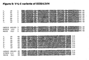
- the antibody 5D12 or at least the variable domains thereof, have a murine background.
- the present invention provides variants of the heavy and light chain variable domains of 5D12.
- the invention provides an anti-CD40 antibody comprising a light chain variable region and a heavy chain variable region, said heavy chain variable region comprising an amino acid sequence wherein:
- polypeptide comprising an amino acid sequence of formula (I) wherein:
- Said polypeptide comprises extensive sequence identity with the heavy chain variable domain of the 5D12 antibody, however, the polypeptide is less immunogenic in a human individual administered as such or in the context of an antibody comprising said polypeptide.
- X 1 -X 5 several amino acids may be present as indicated.
- a binding molecule comprising a polypeptide of the invention has good CD40 binding properties. It has been noted that production of the antibody in a cell varies somewhat with the kind of amino acid at the indicated positions. This will be detailed elsewhere herein below.
- the abovementioned polypeptide essentially spans the variable domain of the heavy chain of the murine 5D12 antibody.
- the amino acid sequence is altered at several positions with respect to murine sequence.
- the altered polypeptide couples good binding properties, while at the same time being well tolerated by humans provided with the polypeptide.
- various amino acids can be inserted without dramatically reducing and/or altering at least immunological properties of the polypeptide when compared to the original murine polypeptide.
- a polypeptide comprising a G, A, V, L, P, F, M, W, C, N, Q, S, T, Y, D, E, K, R or H at position X 1 .
- X 1 is G, A, V, L, P, F or M
- X 2 is G, A, V, L, I, P, F or M
- X 3 is G, A, V, L, I, P, F, M
- X 4 is G, A, V, L, I, P, F, M
- X 5 is G, A, V, L, I, P, F, M, W, C, N, Q, S, T or Y.
- polypeptides are better suited for high level production in a mammalian cell. Particularly in the context of an antibody.
- polypeptides are better suited for high level antibody production while at the same time exhibiting good tolerance properties in a human.
- the invention further provides an antibody as claimed comprising polypeptide of formula (I) wherein X 1 is L;X 2 is I; X 3 is P; X 4 is M; and/or X 5 is S.
- This polypeptide is particularly preferred because of its excellent production properties in the context of an antibody that mimics the binding and pharmacological properties of antibody ch5D12 whereas immunological properties are improved in humans when compared to the murine or ch5D12 antibody and wherein at least production of the polypeptide is not dramatically reduced when compared to the murine or chimeric counterpart.
- the invention further provides an antibody as claimed comprising a polypeptide of formula (I) wherein X 1 is I;X 2 is V; X 3 is P; X 4 is M; and/or X 5 is S.
- This polypeptide is particularly preferred because of its excellent production properties in the context of an antibody that mimics the binding and pharmacological properties of antibody ch5D12 whereas immunological properties are improved in humans when compared to the murine or ch5D12 antibody and wherein at least production of the polypeptide is not dramatically reduced when compared to the murine or chimeric counterpart.
- the invention preferably provides a polypeptide according to the invention, wherein: X 1 is I and X 2 is V; X 1 is L and X 2 is I; or X 1 is L and X 2 is V.
- the polypeptides are particularly preferred in combination with an X 3 is P; X 4 is M; and X 5 is either F or S;
- the invention provides a polypeptide according to the invention, wherein: X 1 is L; X 2 is V; X 3 is L; X 4 is L and X 5 is F. Production of antibodies comprising said polypeptide is very good, while simultaneously providing improved immunological properties in humans when compared to ch5D12. Further described is a polypeptide comprising amino acid sequence GFSX 1 S RYSVY WX 2 R, wherein: X 1 is L and X 2 is I; or X 1 is I and X 2 is V.
- This polypeptide comprises a modified CDR1 of a 5D12 heavy chain variable fragment.
- This CDR1 comprises at least one different amino acid when compared to the CDR1 of the heavy chain variable fragment of antibody 5D12.
- This amino acid change both leads to improved immunological properties of a modified 5D12 or ch5D 12 antibody wherein the modification comprises at least a replacement of the corresponding sequence of said polypeptide in 5D12 or ch5D12 with a polypeptide of the invention, whereas at the same time enabling good production of the antibody in a mammalian cell.
- a heavy chain variable domain comprises 113 amino acids.
- the polypeptide can be generated synthetically or by a cell. Preferably said heavy chain variable domain is produced by a cell. In nature at least five types of heavy chain exist: ⁇ , ⁇ , ⁇ , ⁇ and ⁇ , wherein each type defines a class of immunoglobulins.
- a polypeptide as described may be used as a binding body directly or may be incorporated into an antibody. When incorporated into an antibody the polypeptide is preferably combined with a constant part of an antibody heavy chain. To this end the invention further provides an antibody heavy chain comprising a polypeptide of formula (I) as claimed.
- the art knows many derivatives and analogues of variable domain antibodies. However, currently many different parts, derivatives and/or analogues of antibodies are in use.
- Non-limiting examples of such parts, derivatives and/or analogues are, single chain Fv-fragments, monobodies, VHH, Fab-fragments, artificial binding proteins such as for example avimers, and the like.
- a common denominator of such specific binding bodies is the presence of a heavy chain variable domain.
- a binding body comprising a polypeptide of formula (I).
- a preferred binding body is an antibody, as an antibody comprises a naturally occurring structure. Therefore, the invention in a preferred embodiment provides an antibody comprising a polypeptide according to the invention.
- a binding body is preferably a binding body that is well tolerated in an animal. Tolerance of an animal for a polypeptide is governed by many different aspects. Immunity, be it T-cell mediated, B-cell mediated or other is one of the variables that are encompassed in tolerance of the animal for a polypeptide. As mentioned above, the 5D12 antibody has a murine background. The polypeptide of formula (I) has a reduced immunogenicity in human. It is therefore sometimes referred to as a deimmunized variant of the heavy chain variable domain of 5D12. Thus in an aspect the invention provides an antibody comprising an epitope specificity of a 5D12 antibody, wherein the heavy chain of said antibody is a polypeptide of formula (I).
- Deimmunized as used herein is defined as less immunogenic in an animal than the original antibody.
- a polypeptide of formula (I) is deimmunized when compared to the heavy chain in 5D12 through the removal of known human T cell epitopes.
- T cell epitopes are amino acid sequences within proteins with the capacity to bind to MHC class II molecules. By removal of the T cell epitopes the antibody is less immunogenic.
- a variable domain of the invention is further humanized, such as for instance veneered. By using veneering techniques, exterior residues which are readily encountered by the immune system are selectively replaced with human residues to provide a hybrid molecule that comprises either a weakly immunogenic or substantially non-immunogenic veneered surface.
- An animal as used in the invention is preferably a mammal, more preferably a primate, most preferably a human.
- An antibody according to the invention preferably comprises a constant region of a human antibody. According to differences in their heavy chain constant domains, antibodies are grouped into five classes, or isotypes: IgG, IgA, IgM, IgD, and IgE. These classes or isotypes comprise at least one of said heavy chains that is named with a corresponding Greek letter.
- the invention provides an antibody according to the invention wherein said constant region is selected from the group of IgG, IgA, IgM, IgD, and IgE constant regions, more preferably said constant region comprises an IgG constant region, more preferably an IgG 1 constant region, preferably a mutated IgG 1 constant region, most preferably said constant region is an IgG 4 constant region.
- said IgG 4 constant region is preferably a human IgG 4 constant region.
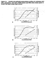
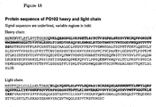
- the IgG 4 antibody of the invention comprises the constant regions of the heavy and light chain amino acid sequence as depicted in figure 18 .
- the IgG 4 antibody of the invention comprises the heavy and light chain amino acid sequence as depicted in figure 18 .
- constant region of IgG 4 Some variation in the constant region of IgG 4 occurs in nature and/or is allowed without changing the immunological properties of the resulting antibody. Typically between about 1-5 amino acid substitutions are allowed in the constant region.
- An antibody with an IgG 4 constant region or a mutated IgG 1 constant region has at least most of the pharmacological properties of an antibody but does not bind complement, and will thus not induce depletion of the cell its binds to in vivo.
- said constant region is a constant region of a human antibody.
- the invention provides a nucleic acid encoding an antibody according to the invention.
- a nucleic acid as used in the invention is typically but not exclusively a ribonucleic acid (RNA) or a deoxyribonucleic acid (DNA).
- Alternative nucleic acids are available for a person skilled in the art, such as for instance peptide nucleic acids (PNA).
- a nucleic acid according to the invention is for instance comprised in a cell. When said nucleic acid is expressed in said cell, said cell produces a polypeptide and/or a binding body and/or an antibody according to the invention. Therefore, the invention in one embodiment provides a cell comprising an antibody according to the invention and/or a nucleic acid according to the invention.
- Said cell is preferably an animal cell, more preferably a mammal cell, more preferably a primate cell, most preferably a human cell.
- a suitable cell is any cell capable of comprising and preferably of producing an antibody according to the invention and/or a nucleic acid according to the invention.
- the invention further provides a cell comprising an antibody according to the invention.
- a cell comprising an antibody according to the invention.
- said cell produces said antibody.
- said cell is a hybridoma cell, a Chinese hamster ovary (CHO) cell, an NS0 cell or a PER-C6TM cell.
- said cell is a CHO cell.
- a cell culture comprising a cell according to the invention.
- Various institutions and companies have developed cell lines for the large scale production of antibodies, for instance for clinical use. Non-limiting examples of such cell lines are CHO cells, NS0 cells or PER.C6TM cells. These cells are also used for other purposes such as the production of proteins.
- Cell lines developed for industrial scale production of proteins and antibodies are herein further referred to as industrial cell lines.
- the invention provides the use of a cell line developed for the large scale production of protein and/or antibody for the production of an antibody of the invention.
- the invention further provides a method for producing an antibody comprising culturing a cell of the invention and harvesting said antibody from said culture.
- said cell is cultured in a serum free medium.
- said cell is adapted for suspension growth.
- an antibody obtainable by a method for producing an antibody according to the invention.
- the antibody is preferably purified from the medium of the culture.
- said antibody is affinity purified.
- a cell of the invention is for instance a hybridoma cell line, a CHO cell, an NS0 cell or another cell type known for its suitability for antibody production for clinical purposes.
- said cell is a human cell.
- a preferred example of such a cell line is the PER.C6TM cell line or equivalent thereof.
- said cell is a CHO cell or a variant thereof.
- GS Glutamine synthetase
- a polypeptide of formula (I) in said antibody comprises X 1 -X 5 wherein:
- a heavy chain variable region wherein at least one of X 1 , X 2 , X 3 , X 4 or X 5 is the same as an amino acid at the corresponding position in a sequence that is shown by the invention to yield specifically good expression levels and wherein further at least one of X 1 , X 2 , X 3 , X 4 or X 5 is the same as an amino acid at the corresponding position in a 5D12 amino acid sequence.
- An advantage of a polypeptide wherein at least one of X 1 , X 2 , X 3 , X 4 or X 5 is the same as an amino acid at the corresponding position in a 5D12 amino acid sequence is that a binding body comprising such a polypeptide shows a better expression level than a polypeptide wherein none of X 1 , X 2 , X 3 , X 4 or X 5 are the same as an amino acid at the corresponding position in a 5D12 amino acid sequence.
- said better expression level is believed to be due to the fact that an amino acid at the position of X 1 , X 2 , X 3 , X 4 and/or X 5 contributes to a proper assembly of a binding body when said amino acid is the same as an amino acid at the corresponding position in a 5D12 amino acid sequence.
- an antibody comprising a polypeptide of formula (I) shows non-stimulatory antagonistic activity.
- a polypeptide according to the invention is therefore particularly suited for ameliorating a symptom of an inflammatory disorder.
- an antibody comprising a binding body of the invention is a monoclonal antibody.
- Monoclonal antibody technology allows for production of large amounts of essentially pure antibodies, thus obtaining predictable products. Therefore, the invention in one embodiment provides an antagonistic anti-human CD40 monoclonal antibody comprising a polypeptide according to the invention.
- a binding body according to the invention is all the more suitable for that purpose since it is in one embodiment deimmunized compared to a mouse 5D12 and/or a chimeric 5D12. Therefore, a binding body of the invention has a reduced immunogenicity and an enhanced half-life in humans compared to a mouse 5D12 and/or a chimeric 5D12. As a result thereof, a binding body of the invention has a sustainable pharmaceutical potential against various inflammatory disorders.
- the invention provides a deimmunized antagonistic anti-human CD40 monoclonal antibody according to the invention.
- the present invention provides 5D12 like molecules that comprise amino acid alterations with respect to the 5D12 amino acid sequence, wherein the alterations are at least in the heavy chain variable domain and preferably also in the light chain variable domain.
- the invention further describes a binding body comprising an amino acid sequence of formula (II) wherein:
- Said binding body is preferably an antagonistic anti-human CD40 monoclonal antibody according to the invention.
- An X selected from X 6 , X 7 , X 8 , X 9 , X 10 or X 11 is selected from a group resembling an amino acid at a corresponding position in a 5D12 amino acid sequence and/or resembling an amino acid at a corresponding position in a sequence that is shown by the invention to yield good expression levels, as shown in example 2. Therefore, a binding body as described herein preferably comprises an amino acid sequence of formula (II) wherein:
- the invention provides an antagonistic anti-human CD40 monoclonal antibody according to the invention comprising a polypeptide of formula (II) of the invention comprising an amino acid sequence as follows.
- the invention provides an antibody as claimed comprising a polypeptide of formula (II) of the invention wherein X 6 is T or S, X 7 is D or Q, X 8 is Q or P, X 9 is V or A, X 10 is V or L and X 11 is F or Y. More preferably wherein: X 6 is T, X 7 is Q, Xsis P, X 9 is A, X 10 is V and X 11 is Y.
- 5D12-like antibodies comprising a polypeptide of formula (I) and the preferred polypeptide of formula (II) combine good expression levels in a producing cell with good tolerance properties and pharmacodynamic properties in a human.
- An anti human-CD40 antagonist antibody of the invention comprises a heavy chain variable domain amino acid sequence of formula (I) and a light chain variable domain amino acid sequence of formula (II) as claimed.
- Such an antibody has good characteristics. It is of course possible to generate variants of such an original antibody by modifying one or more amino acids therein. Many of such variants will behave more or less similar when compared to said original. Such variants are also described herein. There are many ways to modify an antibody of the invention. A non-limiting example of such a modification is an antibody comprising a pyro-glutamate instead of a glutamate. Other non-limiting examples of such modifications are an insertion, deletion, inversion and/or substitution of one or more amino acids when compared to said original antibody.
- the present invention provides means and methods for generating such a variant. It also provides tests for determining the characteristics of said a variant. Also described is a variant of an original antibody of the invention said variant comprising an insertion, deletion, inversion and/or substitution of between about 1 to 10 amino acids when compared to the amino acid sequence of said original antibody. Preferably said insertion, deletion, inversion and/or substitution does not comprise the amino acids at position X 1 and position X 2 of the heavy chain variable domain of formula (I) of the original antibody.
- Said insertion, deletion, inversion, and/or substitution of between about 1 to 5 amino acids can be in any part of the antibody not being or involving the amino acids at positions X 1 and X 2 .
- said insertion, deletion, inversion, and/or substitution of between about 1 to 5 amino acids is in said heavy chain amino acid sequence or said light chain amino acid sequence when compared to the corresponding chain amino acid sequence in said original antibody.
- said insertion, deletion, inversion, and/or substitution of between about 1 to 5 amino acids is in said heavy chain amino acid sequence when compared to said heavy chain sequence of said original antibody.
- said method further comprises generating an antibody producer cell line that produces said selected antibody. This producer cell line can be said further cell line, or yet another cell line that produces said selected antibody.
- the invention provides a pharmaceutical composition comprising an antibody according to the invention, a nucleic acid according to the invention and/or a cell according to the invention. Also, the invention provides an antibody according to the invention, a nucleic acid according to the invention and/or a cell according to the invention for use as a medicament. Preferably a medicament for ameliorating a symptom of an autoimmune disorder and/or an inflammatory disorder and/or for reducing graft rejection and/or for the treatment of CD40 positive cancers.
- said autoimmune and/or an inflammatory disorder is selected from the group of inflammatory bowel disease, rheumatoid arthritis, systemic lupus erythematosus, multiple sclerosis, psoriasis, bullous pemphigoides and atopic dermatitis.
- a polypeptide as described herein is particularly suited for ameliorating a symptom of an inflammatory disorder because of its non-stimulatory CD40 antagonizing properties
- a polypeptide as described is suitable for ameliorating a symptom of several disorders.
- An inflammatory disorder as used in the invention is defined as any disease that involves an inflammatory component.
- an inflammatory disorder specifically includes an autoimmune disorder and/or graft rejection.
- the central role of CD40-CD40L interaction in the initiation, amplification and prolongation of immune responses makes a polypeptide as described specifically suitable for immune modulation in an autoimmune disorder.
- CD40 and CD40L in order to illustrate the role of CD40 and its ligand in inflammatory disorders.
- the CD40 molecule is a 50 kDa type I membrane glycoprotein and is expressed on B cells, monocytes/macrophages, and dendritic cells (DCs) 45-50 .
- DCs dendritic cells
- CD40 can be found on endothelial cells (ECs), fibroblasts, epithelial cells and keratinocytes 51 .
- CD40 ligand gp39, TBAM, TRAP, CD40L, CD154
- CD40L a 32 kDa type II integral membrane glycoprotein
- CD40L has been found on a number of other cell types after activation, including mast cells, basophils, B cells, eosinophils, DCs and platelets 54,55 .
- CD40L Engagement of CD40 by CD40L triggers a number of biological events in B cells including proliferation, expression of activation markers, immunoglobulin (Ig) production, isotype switching, homotypic adhesion and rescue from apoptosis 56,57 .
- Ig immunoglobulin
- the distribution of the CD40 molecule is not restricted to B cells as was originally postulated.
- Freshly isolated human monocytes express low levels of the CD40 molecule, which can be up-regulated by culture in the presence of IFN- ⁇ 47-49,58 .
- CD40 ligation of monocyte/macrophages induces the secretion of large amounts of proinflammatory mediators such as IL-1, TNF- ⁇ and IL-12, which induce inflammatory responses and tumoricidal activity 47-49,58 , and rescue them from apoptosis 48 .
- CD40 ligation also causes DCs to enhance their differentiation and activation, to enhance expression of costimulatory molecules such as CD86, CD80 and CD58, to increase cytokine production, and to inhibit apoptosis 50,59 .
- CD40 signaling can induce expression of intercellular adhesion molecule 1 (ICAM-1), vascular cell adhesion molecule 1 (VCAM-1) and E-selectin on ECs 55 .
- IAM-1 intercellular adhesion molecule 1
- VCAM-1 vascular cell adhesion molecule 1
- E-selectin E-selectin
- a specific role for CD40 and its ligand is demonstrated in murine models of autoimmune disease.
- Animal model studies have clearly indicated the involvement of the CD40L-CD40 interaction in the pathophysiology of various autoimmune diseases. In these studies, using mice suffering from spontaneous or experimental autoimmune diseases, interfering with CD40L-CD40 interactions had clear beneficial effects.
- a Mab to murine CD40L was shown to prevent or reduce disease symptoms in collagen-induced arthritis, experimental allergic encephalomyelitis (EAE; an animal model for MS), in (SWR x NZB)F1 lupus mice and in non-obese diabetic (NOD) mice that spontaneously develop T-cell dependent autoimmune diabetes.
- EAE experimental allergic encephalomyelitis
- SWR x NZB non-obese diabetic
- CD40-CD40L interactions also play a role in the pathogenesis of inflammatory bowel diseases, which includes Crohn's disease and ulcerative colitis.
- CD40L transgenic mice with high transgene copy numbers were shown to acquire lethal inflammatory bowel disease marked by infiltration of CD40 + cells and CD40L + T cells into diseased tissues 67 .
- Anti-CD40L Mab effectively prevent mucosal inflammation and IFN- ⁇ production by lamina intestinal CD4 + T cells in animals with colitis that had been induced by 2,4,6-trinitrobenzene sulfonic acid 44 .
- Multiple sclerosis is an autoimmune disease of the central (cerebrospinal) nervous system.
- MS multiple sclerosis
- the hardened areas of nerve tissue are called plaques.
- the symptoms, severity, and course of multiple sclerosis are highly variable, depending partly on the sites of the plaques and the extent of the deterioration of the white matter.
- the deterioration of white matter in the nervous system slows nerve impulses leading to nervous system incoordination.
- EAE Experimental autoimmune encephalomyelitis
- CNS central nervous system
- Marmosets immunized with rhMOG a recombinant protein representing the extracellular fragment of human MOG (amino acids 1-125) develop EAE in 100% of the cases, which is due to the presence of the monomorphic MHC class II susceptibility element Caja-DRB*W1201 in the repertoire of each monkey 82,84,85,107 .
- a particularly useful aspect of this model for therapy development is that lesions developing in the brain white matter can be visualized and tentatively characterized with clinically relevant magnetic resonance imaging techniques 92,105 . Longitudinal analysis of the brain white matter lesions with magnetic resonance imaging (MRI) showed a progressive increment of the volume and persistent inflammatory activity in the majority of the lesions. Furthermore, the characterization of the CNS pathology with MRI and previously described histological criteria 101 revealed that the majority of the lesions are in an early active stage 106 .
- MRI magnetic resonance imaging
- the rhMOG-induced EAE model has been used to test whether antibodies targeting co-stimulatory molecules of antigen presenting cells (APC) and T-cells are a potential treatment for MS.
- APC antigen presenting cells
- CD154 plays an important role in various immunopathogenic processes that operate in EAE, including B-cell activation, antigen-presenting cell (APC) activation, initiation of antigen-specific T-cell responses and induction of macrophage effector functions 90,93,97,7 .
- a study performed in 1996 confirmed that mice treated with an antibody against CD154 are protected against EAE 88 .
- a clinical trial in MS patients with antibody against CD154 was stopped due to unexpected side-effects that were not observed in the animal experiments 17 .
- the mouse monoclonal antibody (Mab) 5D12 (mu5D12) has been raised against human CD40.
- the 5D12 antibody appeared a potent inhibitor of CD40-CD40L mediated activation on several cell types and, unlike most other anti-CD40 Mabs, does not exert CD40 stimulatory activity 21,22,39 .
- Both the mouse anti-human CD40 antibody mu5D12 and the chimeric version of this antibody, ch5D12 displayed strong suppressive effects on the development of CNS white matter lesions and neurological deficit in the marmoset EAE model and showed no marked side-effects 23,24 .
- CD40 is prominently expressed within the CNS white matter lesions of MS patients as well as EAE-affected rodents 88 and non-human primates 95 .
- That CD40 bearing APC within the CNS such as infiltrated macrophages as well as perivascular and parenchymal glia cells, contribute significantly to the pathogenesis of EAE has been elegantly shown in bone marrow chimeric mice 83 .
- Psoriasis is an inflammatory skin disease afflicting 1-2% of the population.
- T cells and keratinocytes in the lesions are activated and express activation markers and co-stimulatory molecules. It is thought that some costimulatory molecules expressed on keratinocytes and T cells interact with each other and that these interactions contribute to disease activity 108-110 .
- One such set of molecules may be CD40, which is expressed on activated keratinocytes, and CD154 (CD40 ligand), which is transiently expressed on activated CD4+ T cells.
- CD40-CD154 ligation between T cells and keratinocytes may release from these cells inflammatory mediators that are seen in abundance in psoriatic lesions.
- CD40, CD154, and CD40-CD154 interactions have recently been reviewed 111 .
- Cultured keratinocytes also express CD40; expression is enhanced by IFN-treatment. Ligation of highly expressed CD40 on IFN-gamma treated keratinocytes (referred to as CD40++ keratinocytes throughout this paper) with CD154 results in up-regulation of ICAM and increased production of cytokines 112-114.
- CD154 acts as one of the signals for production by keratinocytes of chemokines and complement, which are found in abundance in psoriatic lesions 115 .
- CD40 ligation induces the release of chemokines (IL-8, RANTES, and MCP-1) 116 .
- chemokines IL-8, RANTES, and MCP-1
- Pasch et al. showed that the CD40-related release of the chemokines IL-8, MCP-1 and to a lesser extent of RANTES was inhibited by the antagonistic anti-CD40 antibody 5D12 116 .
- antagonist anti-CD40 mAb 5D12 may at least in part have an effect on the inflammation seen in psoriatic lesions.
- US2003/0165499 discloses a measurable anti-psoriatic effect of the 5D12 and other antagonistic anti-CD40 antibodies in a SCID mouse xenogeneic transplant model system that is used as a model for psoriasis treatment, showing that antagonistic anti-CD40 antibodies may be used for the treatment of psoriasis.
- a therapeutic effect of 5D12 in this in vivo system was demonstrated
- said autoimmune and/or an inflammatory disorder comprises inflammatory bowel disease.
- said inflammatory bowel disease comprises ulcerative colitis (UC) or Crohn's disease (CD).
- a polypeptide according to the invention is suited for treatment of diverse inflammatory disorders, including autoimmune disorders and graft rejection. Therefore, the invention in one embodiment provides a method for ameliorating a symptom of an autoimmune disorder and/or an inflammatory disorder and/or for reducing graft rejection.
- the invention provides use of an antibody according to the invention, a nucleic acid according to the invention and/or a cell according to the invention for the manufacture of a medicament for ameliorating a symptom of an autoimmune disorder and/or an inflammatory disorder and/or for reducing graft rejection.
- Ameliorating a symptom as used in the invention is defined as ameliorating at least one symptom of a disorder at least partially.
- the invention is of specific interest for autoimmune and/or inflammatory disorders for which at present no efficient treatment exists.
- these disorders are rheumatoid arthritis, systemic lupus erythematosus, multiple sclerosis, psoriasis, inflammatory bowel disease, bullous pemphigoides and atopic dermatitis.
- a polypeptide as described herein, optionally comprised in a binding body as described or a cell of the invention and/or encoded by a nucleic acid of the invention, is specifically suited for ameliorating a symptom of an autoimmune and/or inflammatory disorder as defined properties of a polypeptide as described allow for interfering with a CD40-CD40L pathway in a specific manner.
- the invention in one embodiment provides a use an antibody according to the invention, a nucleic acid according to the invention and/or a cell according to the invention for the manufacture of a medicament for ameliorating a symptom of an autoimmune disorder and/or an inflammatory disorder and/or for reducing graft rejection, wherein said autoimmune and/or said inflammatory disorder is selected from the group of rheumatoid arthritis, systemic lupus erythematosus, multiple sclerosis, psoriasis, inflammatory bowel disease, bullous pemphigoides and atopic dermatitis.
- Subjects could not have received treatment with cyclosporin A or methotrexate within 3 months before screening nor were they allowed to have prior exposure to treatment with Mabs.
- the average age of all subjects enrolled was 35.8 years of whom seven were males and 11 were females. All subjects were of Caucasian ethnicity. No apparent differences between patients in the different cohorts at inclusion were observed (Table 1), with the exception that in the lower three dose groups the majority of the subjects were females, while the highest dose group (10.0 mg/kg) consisted of only male subjects.
- ECG electrocardiogram
- vital signs vital signs
- physical examinations history of Crohn's disease signs and symptoms
- baseline laboratory values There were no significant differences regarding baseline characteristics amongst the four dose cohorts.
- ch5D12 is a molecularly engineered human IgG4 antibody containing the variable domains of the heavy and light chains of Mab 5D12 parent version. This Mab has been shown to bind to CD40-bearing cells and to antagonize CD40 mediated activation of various cells 20-27 .
- ch5D12 was administrated in an open label, single dose, multi-centre trial, studying four dose levels. There were five subjects in each treatment group except for the final dose cohort in which only three subjects were enrolled. Single administration at dose levels of 0.3, 1.0, 3.0, and 10.0 mg/kg of ch5D12 was done intravenously. After completing recruitment into one dose group, recruitment into the next group was only started when safety had been established at the current dose level.
- Clinical disease activity was assessed at each visit weekly for the first 28-day period and a subsequent last visit at day 56. Two subjects (3.0 mg/kg dose cohort) withdrew from the study after the day 28 assessments but their data are included in the evaluation. Subjects were to remain stable on the same dose of their current medication during the course of the study. No un-allowed concomitant medication was used during the study. Response to ch5D12 treatment was defined as a decrease in CDAI of ⁇ 100 points and clinical disease remission was defined as a CDAI of ⁇ 150 (total score) and a decrease of CDAI of at least 100.
- Serum concentrations of ch5D12 were determined by ELISA as previously described 27 .
- blood was collected in heparin tubes and diluted twofold in PBS.
- Peripheral blood mononuclear cells (PBMC) were isolated by gradient centrifugation (Lymphoprep, Nycomed, Roskilde, Denmark) and 500 000 cells were stained with FITC-labelled ch5D12 and PerCP-labelled anti-CD20 (Becton-Dickinson, Mountain View, CA, USA) and incubated on ice for 30 min.
- FITC-labelled ch5D12 and PerCP-labelled anti-CD20 Becton-Dickinson, Mountain View, CA, USA
- Mucosal biopsies from ileum and colon obtained during ileocolonoscopy prior to treatment on day 0 and on day 28 with a standard forceps were fixed in formalin 6% for routine analysis. Additional samples were immediately snap-frozen in Tissue-Tek optimum cutting temperature compound (Miles Laboratories Inc, Naperville, IL, USA) in liquid nitrogen cooled isopentane. Samples were stored at -80 °C until further use. Formalin fixed samples were routinely paraffin processed. Five-micrometer thick sections were prepared and stained with haematoxylin and eosin. The sections were analysed using a Leitz Wetzlar microscope (Wetzler, Germany). A total of four semi-serial sections for each sample were analysed.
- the frozen samples were used for immunohistochemical analysis, which was performed using a panel of Mabs to assess the presence of different subsets of lymphocytes and monocytes/macrophages.
- the panel was completed with Mabs directed against CD40 and CD40L.
- Immunohistochemical staining was performed on cryostat sections, dried overnight at room temperature, and fixed in absolute acetone for 10 min.
- CD3 clone: UCHT1, 1/10 dilution
- CD4 clone: MT310, 1/10 dilution
- CD8 clone: 144B, 1/20 dilution
- CD19 clone: HD37, 1/30 dilution
- CD40 clone: 5D12, 1/100 dilution
- CD68 clone: Kp1, 1/50 dilution
- CD154 clone: M90, 1/10 dilution
- the secondary Mab was a biotin-labelled anti-mouse immunoglobulin (1/400 dilution; Dako) applied for 30 min.
- Dako biotin-labelled anti-mouse immunoglobulin
- sections were also incubated in methanol solution containing 0.3% (v/v) H 2 O 2 for 20 min. After three washes with PBS, the avidin/biotin peroxidase-labelled complex (Dako) was added. In between the incubations, the sections were washed in phosphate buffered saline at pH 7 for 15 min.
- reaction product was visualized by incubating the section for 10 min in 0.05 m acetate buffer at pH 4.9, containing 0.05% 3-amino-9-ethyl-carbazole (Janssen, Beerse, Belgium) and 0.01% H 2 O 2 , resulting in a bright-red immunoreactive site. Afterwards, the slides were faintly counterstained with Harris haematoxylin, rinsed with distilled water and a cover-slip was applied with glycerol. Controls, which were negative, consisted of an omission of a primary or secondary antibody, use of chromogen alone, and use of irrelevant isotype-matched mouse IgG (vimentin; Dako).
- Paraffin sections stained with haematoxylin and eosin were all analysed by the same pathologist (K. G.) blinded for the origin of the samples. Disease activity was assessed using a histological numerical score 40 of which the maximum activity corresponded to 16. The immunohistochemically stained sections were also analysed in a blinded fashion. For 15 sets of biopsies, positively staining cells were counted in high power fields (magnification ⁇ 40). The fields were chosen according to the highest density of stromal cells. The number was expressed as a percentage of the total number of mononuclear cells in the stroma.
- PHA phytohemagglutinin mitogen
- CD3 and CD19 were percentages of T cells and B cells within the lymphocytic population, whereas, CD4 and CD8 cells were reported as percentages of CD3 cells. Only CD14 (monocytic population) is shown as a percentage of the total CD45 population.
- the mean peak levels of ch5D12 after a single i.v. injection were dose-dependent and dose-proportional ( Figure 1 ). After 24 h, approximately 50% of the administrated ch5D12 could be detected in serum in the highest three dose cohorts. For the 0.3 mg/kg cohort, only 15% was present in serum after 24 h. From non-human primate studies, a t1/2ß of 8-10 days has been calculated 27 which was confirmed by the current human data. Complete coating of CD40 on peripheral blood B cells was not achieved by the infusion of 0.3 mg/kg as determined by an ex vivo competition assay using FITC-labelled ch5D12 (not shown).
- the change from the baseline to day 28 histopathology activity score is shown in Figure 3 .
- Samples from the ileum of nine patients and samples from the colon of 11 patients at day 0 were available. In four cases, the ileum was not involved at screening while five patients had active ileal disease. The mean histology score at day 0 for those with active disease was 4.6 (range 3-7). The mean histology score for the five patients decreased to 1.0 (range 0-3) at day 28. In four of 11 patients, the colon was not involved at day 0 and the colonic samples from these patients remained normal at day 28. In seven patients the colon was clearly involved with a mean histology score at day 0 of 4.5 (range 2-12). This decreased at day 28 to a mean score of 1.7 (range 0-7).
- the score at day 28 was either 0 indicating a normal biopsy or 1 indicating only architectural abnormalities.
- the colonic score of one subject decreased, but the other colonic scores in this group remained high and the ileum score even increased.
- the higher dose cohorts either the ileum or colon or both decreased in the activity score at day 28 after treatment with ch5D12.
- a positive response [as defined in Ref. (41)] was observed in 81% (9/11) of subjects.
- seven subjects with active disease at day 0 showed reduced neutrophil activity to the point that neutrophils were not present in their biopsies at day 28.
- the pattern for the CD19+ B cells was comparable with that observed for the T cells.
- the amount of the B cells had normalized in all cases except for the one patient from the low-dose group.
- Figure 4 shows representative examples of the decrease of all three major cell types (T cells, B cells and monocyte/macrophage cells) in ileum biopsies of patient 011 (activity score: 7) and colonic biopsies of patient 012 (activity score: 12) before ch5D12 administration and at day 28 after ch5D12 administration.
- SAEs serious adverse events
- PBMC isolated at various time-points prior and after treatment were stimulated by the polyclonal T-cell activator PHA.
- PHA polyclonal T-cell activator
- Anti-ch5D12 antibodies were not detectable in the two lower dose cohorts throughout the entire study. In the 3.0 mg/kg cohort, one subject was found to have mounted an anti-ch5D12 response, although the signal was very low and could only be detected in the undiluted day 28 sample of this patient. In the highest dose group, another subject had anti-ch5D12 antibodies on day 28 and day 56 of the study. These could be detected with a 1:64 dilution. Therefore, only two subjects had a possible immune response to ch5D12 administration.
- ch5D12 In this open label, multi-centre trial, four dose levels of ch5D12 were administrated to subjects with moderate to severe Crohn's disease. Eighteen subjects were enrolled, with five subjects in each treatment group except for the final dose cohort in which only three subjects were enrolled. ch5D12 serum levels increased dose-dependently and the long serum half-life that was observed in cynomolgus monkeys 27 was confirmed in humans. This study shows the feasibility of antagonizing CD40-mediated cell activation using ch5D12, in the absence of major side effects. The CDAI analysis indicates that 72% of subjects experienced a clinical response and 22% experienced a remission during the study.
- ch5D12 has shown functional activity in a rhesus monkey renal transplantation model 25 and when used as immune suppressive strategy to prevent immune responses against adenoviral particles and their products 26 .
- a tissue cross-reactivity study on human and cynomolgus tissues showed that ch5D12 bound to the cell surface of B cells and DCs in lymphoid organs. No unexpected cross-reactivity was observed on either human or cynomolgus tissues.
- a safety and tolerability evaluation for ch5D12 was performed in cynomolgus monkeys, in which weekly administration of ch5D12 for 4 weeks was shown to be safe and without any side-effects in all monkeys.
- the CD40-CD154 (CD40L) co-stimulatory pathway has been validated in animal models as a promising clinical target for treatment of autoimmune disease and transplantation rejection. 15 Potentially the CD40-CD154 interaction can be inhibited by targeting either CD40 or CD154. All previous studies have focused on antagonizing CD154, which is selectively expressed on activated T cells and platelets. 8-11 Human clinical studies using an anti-human CD154 Mab have been halted because of thromboembolic events 12-14 . As this anti-CD154 Mab is constructed as a humanized IgG1, which binds extremely well to Fc-receptors, the anti-CD154 Mab may cross-link CD154 to Fc-receptors resulting in the formation of blood clots.
- CD40 was reported to be constitutively expressed on platelets and found to be functionally important using soluble CD154 as stimulus for platelet activation. These stimulatory effects that resulted in activation of platelets were abrogated by addition of anti-CD154 or anti-CD40 Mabs 42 .
- the IgG 4 Fc part of the ch5D12 Mab backbone has reduced capability to bind to Fc-receptors expressed on platelets as compared with an IgG 1 backbone. Further discrimination between the two approaches comes from the fact that ch5D12 targets B cells, monocytes, macrophages and DC, while an anti-CD154 Mab targets mainly activated T cells, resulting in markedly different clinical strategies. No indications of thromboembolism in any of the patients treated in this study were observed.
- ch5D12 The effect of ch5D12 is largely based on the interference with CD40 mediated cell activation.
- CD40 triggering induces cytokine and chemokine secretion by macrophages and DC, and enhances the antigen presenting capacity of the latter.
- a number of other inflammatory cytokines, such as IL-8 and IL-12 39 are induced by CD40 triggering and therefore ch5D12 might have anti-inflammatory properties in addition to the prevention of TNF- ⁇ production. Whether the effects observed depend mainly on a reduced T-cell activation, or a reduced release of cytokines and chemokines, awaits further investigation.
- the murine variable regions of 5D12 were cloned. Briefly, from lysated cells using Qiagen total RNA was isolated. By RT-PCR using the oligo's 5' CAG GTS MAR CTS SAG SAG TC W GG 3' and 5' GCA TGT ACT AGT AAT TTT TVT TGT CCA CYT TGG TGC T 3' to amplify the VH region and using the oligo's 5'CGA TAC GAS MTY CAG CTG ACC CAG TCT CCA 3' and 5'GAC TCA TCT AGA TAC ACT CAT TCC TGT TGA AGC TCT TG 3' to amplify the VL region of mu5D12.
- V-regions were recloned in pcDNA3002 ( Marissen et al, J. of Virol, 2005: 79:4672-4678 ), together with genomic sequences encoding human IgG 4 and human kappa to construct a chimeric IgG 4 immunoglobulin expression plasmid.
- T-cell epitopes In collaboration with Biovation using their proprietary DeImmunisation technology within the V regions potential T-cell epitopes were identified.
- Helper T cell epitopes comprise short amino acid sequences within proteins with the capacity to bind to MHC class II molecules. By removal of the T cell epitopes the antibody can no longer trigger T cell help and the subsequent immunogenicity against it.
- Identification was done using Peptide Threading.
- the Peptide Threading approach is a computer-based method for predicting the binding of peptides (small amino acid sequences) to 18 different human MHC class II molecules based on known structures solved by X-ray crystallography. Furthermore an analysis was done to search the V regions for the presence of known human T cell binding peptides from a database ( www.wehil.wehi.edu.au ).
- V regions Based on the primary murine V region amino acid sequences, surface humanized (veneered) V regions were designed. Within these veneered VH sequence 8 potential T-cell epitopes and within the veneered VL region 4 T-cell epitopes were identified. Identified T-cell epitopes were removed by amino acid substitution. Based on this analysis the DNA sequences encoding the deimmunized VH and VL region were synthesized and recloned in the pcDNA3002-hIgG 4 -kappa expression plasmid.
- the QuickChange Site-Directed Mutagenesis kit of Stratagene was used according to the manufacturer's protocol. In table 4 the used oligonucleotides are shown. Briefly, the plasmid was denatured followed by annealing of the sense and anti-sense primers containing the desired mutation. By cycling (30 sec 95°C; 30 sec 55°C; 600 sec 68°C) for 16 times using a Biometra T gradient PCR machine the mutagenic sequences were incorporated. The non-mutated parental plasmid was digested using DpnI, which only cuts methylated DNA.
- plasmids were generated using the oligonucleotides as shown in table 6. Two mutagenesis rounds were required to create double mutants (Table 7). Plasmids were used to transform XL1-blue competent cells.
- PER.C6TM (Crucell). Briefly, the day before the transfection PER.C6TM were trypsinized and counted. Cells were plated in DMEM substituted with 10% FCS at a density of 4 x 10 5 cells per well in a T24 well plate. Next day medium was replaced with 0.5 ml fresh medium. For each well 1 ⁇ g of plasmid DNA was diluted into 50 ⁇ l of OPTI-MEM I, which was combined with equal volume OPTI-MEM I containing 3 ⁇ l LipoFectAMlNE 2000 (LF2000) reagent (Invitrogen).
- DNA-LF2000 reagent complexes 100 ⁇ l were directly added to each well.
- the transfected cells were incubated at 37°C in a CO 2 incubator. After 48h the supernatants were analyzed for antibody expression.
- the amount of expressed antibody was measured by a sandwich ELISA. Briefly, 96 wells EIA/RIA plates (Corning 3590) were coated o/n at 4°C with polyclonal antibody anti-human IgG (Jackson 109-005-088) diluted 1/1000 in PBS (100 ⁇ l/well). After blocking with PBS containing 0.05% Tween and 1% BSA, plates were 3x washes with PBS-0.05% Tween. Subsequently as standard purified chimeric 5D12 was applied in titration starting at 400 ng/ml to zero in triplo. Samples (transient supernatants) also applied in titration.
- JY cells are EBV transformed human B-cells, which express CD40. Briefly, 100,000 JY cells were incubated with 100 ⁇ l supernatant from the transient expression experiment at 4°C for 20 minutes. To remove unbound antibody cells were washed with PBS containing 0.05% NaAzide and 1% BSA. As secondary antibody 1/100 diluted FITC labeled anti-human IgG (Jackson 109-095-127) was used. After 20 minutes incubation at 4°C cells were washed. Finally cell samples were measured on a FACScan (BD).
- BD FACScan
- VH region 8 first base of potential epitope: position 27, 30, 43, 46, 57, 61, 77 and 83
- VL region 4 position 7, 13, 28 and 86
- N-X-T/S N-glycosylation site
- DI5D12 fully deimmunized 5D12
- ch5D12 ch5D12
- each substitution can influence the characteristics of the expressed antibody. Therefore to investigate the influence of each changed position in the VH region on the characteristics of the antibody 12 additional variants were designed ( figure 7 ). In each variant 1 position was changed back to the original murine amino acid sequence.
- Each plasmid contains the human IgG 4 backbone to which the VH regions were linked and the human kappa region to which the VL regions were linked.
- the other 13 plasmids are deimmunized versions, in which the deimmunized VL region is used in combination with the deimmunized VH region or one of the 12 VH variants shown in figure 7 .
- PER.C6TM cells were transfected with the 14 plasmids using LipoFectamine2000.
- the tested variants can be divided in 3 groups: no or low improvement on the expression level (Q5E, K13A, E16Q, T17S and STS60NSA), variants P45L, M48L, S79F and T108S restore the expression levels as compared with ch5D12 (PG100) up to 40%, while the I29L and I37V variants restore the level above 50%. Specially the substitution at position 29 (I to L) seems to have large influence on the expression levels. The I29L variant was subsequently renamed PG102 antibody.
- Figure 9 shows the V-L-I variants that were designed in alignment with ch5D12 (PG100), DI5D12 and PG102 (I29L variant).
- V-L-I variants additional pcDNA3002 expression plasmids were constructed that contain the human IgG 4 backbone to which the VH region of one of the variants was linked and the human kappa region to which the VL regions were linked, as described above.
- PER.C6TM cells were transfected with the plasmids using LipoFectamine2000. After 48 hour the transient supernatants from each transfection were harvested and analysed for antibody expression. To exactly determine the various expression levels, all supernatants were analysed by quantitative ELISA measuring antibody levels. As shown in table 8, the various combinations of a V, L or I on positions 29 and 37 are associated with variable production levels. This further demonstrates that not just all V, L and I substitutions ultimately lead to higher production levels.
- Lonza Biologics (Lonza) was requested by PanGenetics BV to undertake the construction, selection and evaluation of a GS-CHO cell line expressing the human recombinant IgG 4 /kappa antibody PG102 (I29L variant).
- the cell lines were constructed by transfecting CHOK1SV host cells with vectors generated using Lonza's Glutamine Synthetase (GS) Gene Expression System ( WO2003054172 ). The gene sequences were supplied by PanGenetics.
- the cell line CHOK1SV is a suspension variant of the Chinese hamster ovary (CHO) cell line CHOK1, which has been adapted to chemically defined, animal component-free (CDACF) medium ( Bebbington et al (1192) Biotechnology 10:169-75 ; de la Cruz Edmonds MC et al (2006) Mol. Biotechnol. 34:179-90 ).
- CDACF animal component-free
- Cells were revived from vials of cryopreserved stocks by rapidly warming to 37 °C and diluting into approximately 50 mL of growth medium.
- the cryoprotectants were removed by centrifuging the cells, discarding the supernatant and resuspending the cells in fresh growth medium.
- cultures were expanded in a suitable volume of growth medium and subcultured every 4 days for growth in CDACF medium.
- the containers used were: 125 mL shake flasks (for a culture volume of 10 to 30 mL), 250 mL shake flasks (for a culture volume of 30 to 50 mL), 500 mL shake flasks (for a culture volume of 50 to 100 mL), 1 L roller bottles (for a culture volume of 100 to 200 mL), or 2 L roller bottles (for a culture volume of 200 to 400 mL).
- the headspace of each culture was gassed with 5% v/v CO 2 in air and the flasks sealed.
- the cultures were then incubated at 35.5 to 37.0 °C on a shaking platform rotating at 140 ⁇ 5 rpm.
- Total and viable cell concentrations were obtained by aseptically removing a sample of culture, staining the non-viable cells with Trypan Blue and microscopic examination using a modified Fuchs-Rosenthal haemocytometer. When appropriate, samples were diluted prior to counting.
- the total and viable cell concentrations were determined using a CASY-1 particle counter or a V i -CELL XR automated cell counter.
- CASY-1 counter a sample of culture was aseptically removed from each flask, diluted with Casyton buffer and the number of viable and total cells determined. Viable and non-viable cells were distinguished by size. Percentage viability was calculated as the ratio of viable cells to total cells multiplied by 100.
- V i -CELL XR counter 0.7 mL of cell culture was aseptically removed and an automated Trypan Blue count was performed. A series of camera pictures and a computer analysis algorithm are used to identify and count the viable and non-viable cells. For all counting methods, percentage viability was calculated as the ratio of viable to total cells multiplied by 100.
- the 96-well plates were incubated for approximately 3 to 4 weeks, following transfection to allow colony formation.
- the colonies were examined microscopically to verify that they were of a suitable size (covering greater than 60% of the bottom of the well), and that only one colony was present in each well.
- the culture supernatant was then assayed for antibody production using Lonza's ELISA assay for assembled antibody, described under "assembly ELISA".
- the percentage confluence of the well was assessed at the time of sampling. The value obtained by dividing the assay result by percentage confluence was used to rank the cell lines.
- the selected cell lines were allowed to multilayer and then re-fed with CD-CHO/25 ⁇ M MSX. After feeding, the cultures were returned to the incubator for a further four to seven days until the medium turned orange/yellow again and the cells lifted from the flasks.
- Suspension cultures were initiated from confluent T25 flask cultures.
- the suspension cultures were inoculated at a concentration of 0.05 to 0.2 x 10 6 viable cells/mL in 125 mL shake flasks containing CD-CHO/25 ⁇ M MSX medium. If the viable cell concentration did not reach 0.05 x 10 6 viable cells/mL after a maximum of 14 days in T25 flasks, 15 mL of each culture was automatically progressed into 15 mL of CD-CHO/25 ⁇ M MSX in 125 mL shake flasks. The cell lines were then serially subcultured into fresh CD-CHO/25 ⁇ M MSX medium, at an inoculation concentration of 0.2 x 10 6 viable cells/mL, on a 4 day subculture regime, until acceptable and reproducible growth characteristics were obtained.
- a bolus addition of the nutrient feed SF40 was made. Further bolus additions were made at approximately 24, 48 and 72 hours after the initial addition.
- a second feed (SF41) was applied once cell concentrations greater than 4.0 x 10 6 viable cells/mL were obtained. Samples of cultures were taken at appropriate intervals, centrifuged and the supernatants stored at -20 ⁇ 5 °C until assayed for antibody concentration by Protein A HPLC. Harvest supernatants from each overgrown culture were stored at -20 ⁇ 5 °C. The ten overgrown cultures with the highest antibody concentrations were further assessed for quality of the antibody produced.
- Samples were assessed for aggregate levels using the gel permeation high performance liquid chromatography (GP-HPLC) method before Protein A purification and antibody characterisation by reduced and non-reduced sodium dodecylsulphate poly acrylamide gel electrophoresis (SDS-PAGE) and isoelectric focusing (IEF) analysis and matrix assisted laser desorption ionisation time of flight mass spectrometry (MALDI TOF-MS) oligosaccharide analysis.
- GP-HPLC gel permeation high performance liquid chromatography
- IEF isoelectric focusing
- MALDI TOF-MS matrix assisted laser desorption ionisation time of flight mass spectrometry
- N f N i + 1 Ln ⁇ 2 . Ln ⁇ X f X i
- N f Generation number at time f
- N i Generation number at time i
- X f Viable cell concentration (cells/mL) at time f
- X i Viable cell concentration (cells/mL) at time i and f > i
- the time integral of the area under the growth curve (the time integral of the viable cell concentration (IVC); 10 9 cells.h/L) was calculated by summing the area (approximated to a right angle trapezium) between adjacent cell concentration values. The areas were calculated by multiplying the mean of the two viable cell concentrations (cells/mL) by the elapsed time between the two determinations (h). This method is based on that described by Renard et al. (1988) in Biotechnology Letters 10 (2) pages 91-96 .
- the overall specific production rate (q P overall: mg/10 9 cells/h) was calculated by linear regression analysis of the antibody concentration (mg/L) against the time integral of viable cell concentration. The slope of this line is equal to q P overall.
- the antibody concentration of supernatant samples was determined using an ELISA that measures assembled human IgG. This involved capture of assembled antibody in samples and standard onto a 96-well plate coated with goat anti-human IgG Fc. Bound antibody was revealed with goat anti-human light chain linked to horseradish peroxidase and the chromogenic substrate tetramethylbenzidine. Colour development was proportional to the mass of antibody present in the sample. The concentration was determined using a standard curve generated using an IgG standard (Lot number L07387/5/2)
- Gravity fed rmp Protein A Sepharose columns (5 mL) were prepared. The columns were cleaned before use with 6 M guanidine HCl and equilibrated with 50 mM glycine/glycinate-250 mM NaCl buffer, pH 8.0. The pH of the column load material was adjusted to pH 8.0 ⁇ 0.1 before application to the column.
- the rmp Protein A affinity column was washed with equilibration buffer and eluted with 0.1 M glycine buffer, pH 3.5. The column eluates were neutralised to approximately pH 7.0 with 2 M Tris Base and dialysed against Dulbecco's phosphate buffered saline in preparation for IEF and SDS PAGE analyses.
- the cell culture samples were prepared for analysis by protein A purification. Electrophoresis was carried out using 4 to 20% precast Novex polyacrylamide gels. For reducing SDS PAGE, samples were reduced using 2-mercaptoethanol and denatured by SDS at pH 8.0. Samples were heated for 2.0 ⁇ 0.5 minutes before they were loaded onto the gel (10 ⁇ g per lane). Non-reduced SDS PAGE was performed using the same gels and Novex non-reducing sample buffer (2-mercaptoethanol is not added). Samples were heated for a standard time of 1.0 ⁇ 0.5 minutes before they were loaded onto the gel (4 ⁇ g per lane). Following electrophoresis, proteins were visualised by staining for 75 minutes with Coomassie brilliant blue R250 and destained with methanol/acetic acid.
- the cell culture samples were prepared for analysis by protein A purification. IEF analysis was carried out using pre-cast agarose IEF gels, pH 3 to pH 10. Approximately 10 ⁇ g protein sample per lane was loaded onto the gel and then pre-focused for 70 Vh. After removal of the sample application strip, electrophoresis was continued for 1500 Vh (1500 V, 20 mA, 25 W per gel).
- the IEF gels were fixed in a trichloroacetic acid: sulphosalisylic acid: methanol solution and then stained with Coomassie Brilliant Blue R250 (Lot numbers of Coomassie Blue 003/L11905/118 and 003/L11905/131). The gels were then destained and dried. (Lot numbers of destain 003/L11905/117 and 003/L11905/121).
- the GP-HPLC method separates protein components according to their size. Large components, such as protein aggregates; are too large to penetrate the matrix particles to any great extent and therefore eluted from the column ahead of smaller components, such as protein monomer. Smaller components, such as fragments, penetrate the matrix more easily and eluted after the monomer. This technique was therefore used to separate antibody monomer from both aggregates and fragments.
- the monomeric component was identified by its characteristic retention time and position, relative to calibration markers.
- the proportion of sample components was determined by calculation of the peak areas of each component relative to the total integrated peak area.
- the oligosaccharide analysis of the PG102 antibody produced by each cell line was performed by MALDI TOF-MS using a MicromassTM MALDI-LR coupled with delayed extraction.
- the MALDI TOF-MS was operated in reflectron configuration with positive ion mode.
- the instrument was calibrated using a mixed N-glycan standard.
- Antibody was purified from harvest supernatants using a POROS Protein A column. The purified fractions were collected and the disulphide bonds were reduced with dithiothreitol followed by alkylation of the reduced thiols using iodoacetate. Oligosaccharides were released using the enzyme N-glycanase (PNGaseF) and were sandwiched between two layers of super-dihydroxy benzoic acid (super-DHB) matrix on the target plate for MALDI TOF-MS analysis.
- PNGaseF N-glycanase
- super-DHB super-dihydroxy benzoic acid
- the PG102 gene sequence was used by Lonza to constructed the double gene vector pPG102/DGV (not shown) that was used to transfect the CHOK1SV host cell line by electroporation.
- Six sets of transfections were performed using the vector pPG102/DGV and supernatants of 255 transfectants were screened for antibody production in 96-well plates by assembly ELISA. Detectable antibody levels were produced by 99.6% of the screened transfectants. Forty three high ranking transfectants were selected for further evaluation in 24-well plates.
- the growth and productivity characteristics of the 23 selected cell lines were evaluated in fed-batch shake flask culture. Singlet cultures were prepared for each cell line. Two feeds, SF40 and SF41, were added to the cultures when the viable cell concentration met the feeding criteria. The protocol used for the feed regime mimicks as closely as technically possible, Lonza's generic GS-CHO fermentation process. Growth and productivity data for the 23 selected cell lines are presented in Table 9. Protein A HPLC analysis of the harvested supernatants of the fed-batch shake flask cultures showed that four cell lines produced antibody concentrations of greater than 1000 mg/L. Cell line L107 produced the highest antibody concentration, 1674 mg/L.
- Protein A purified samples analysed by SDS PAGE under reducing conditions were also comparable to each other, exhibiting the heavy chain band at approximately 50 kDa and the light chain band at approximately 25 kDa (Data not shown).
- IEF analysis showed that the integrity of the Protein A purified antibody samples from the selected cell lines demonstrated comparable profiles, with six primary bands (3 major and 3 minor bands) in the pI range 8.15 to 9.30 for all cell lines except L97, M92 and M59. These three cell lines exhibited three major and two minor bands. Additional bands were observed in samples J3 and J4, although these may not all be visible on the gel image.
- the oligosaccharide analysis of the antibody produced by each of the ten cell lines selected for further evaluation in fed-batch shake flask cultures was determined by MALDI TOF-MS
- the predominant oligosaccharide structures in the antibody obtained from the ten cell lines were G0F and G1F, which are typical N-linked oligosaccharide structures observed on antibodies.
- Relatively low levels of oligomannose structures were detected with the highest level of 6.7% of total glycans measured for antibody derived from cell line L52.
- Oligomannose-5 (man-5) structures were observed in all samples at a range of 1.1 to 4.6% of total glycans. All of the samples analysed contained comparable levels of oligosaccharides and relatively low concentrations of man-5 (data not shown).
- PSS pre-seed stocks
- Cell line L107 was selected as the lead cell line as it exhibited the highest harvest antibody concentration of the 23 cell lines evaluated, along with acceptable growth and product quality characteristics ( Figure 11A ).
- Cell line L25 exhibited the second highest harvest antibody concentration of the twenty three cell lines evaluated, along with acceptable growth and product quality characteristics ( Figure 11B ). Therefore cell line L25 was selected as the first back-up cell line.
- Cell line M95 exhibited the third highest harvest antibody concentration of the twenty three cell lines evaluated, and had acceptable growth and product quality characteristics ( Figure 11C ). On this basis cell line M95 was selected as the second back-up cell line.
- Pre-seed stocks (20 vials per stock) were cryopreserved for each of the three selected cell lines. These stocks were stored in vapour phase liquid nitrogen refrigerators. Cell lines L107, L25 and M95 were renamed DC1, DC2 and DC3 respectively.
- Lonza Biologics was requested to undertake the construction, selection and evaluation of a GS-CHO cell line expressing the human recombinant IgG 4 /kappa antibody PG102.
- the gene sequence was supplied by PanGenetics and vectors were generated using Lonza's Glutamine Synthetase (GS) Gene Expression System.
- the cell line CHOK1SV a suspension variant of the Chinese hamster ovary (CHO) cell line CHO-K1 adapted to chemically defined, animal component-free medium, was transfected by electroporation.
- Three cell lines (L107, L25 and M95) were selected for further evaluation based on growth, productivity and product quality data. Pre-seed stocks of 20 vials of each cell line were cryopreserved. The three cell lines were renamed DC1, DC2 and DC3 respectively.
- ch5D12 (PG100) and PG102 (I29L variant) binding to human CD40 were compared by surface plasmon resonance at a constant temperature of 25.0 °C, using a Biacore 3000 instrument.
- Recombinant human CD40 extracellular domain fused to human IgG1-Fc domain (huCD40-Fc; R&D Systems catalogue number 149-CD-50) was used as the target antigen in these experiments.
- HuCD40-Fc (1 ⁇ g/mL in 10mM acetate buffer, pH 5.0) was coupled via the amine groups of Lys residues to carbodiimide-activated Biacore CM5 chips, resulting in an antigen immobilization level of 127.4 RU.
- dilution series of ch5D12 (PG100) and PG102 were prepared in Biacore Running Buffer (HEPES-buffered saline containing EDTA and surfactant; HBS-EP) Kinetic analyses were performed at a flow rate of 30 ⁇ L/min. Samples were allowed to bind for 4 min and to dissociate for 5 min, at the same flow rate. The chip surface was then regenerated for 30 seconds using 30mM NaOH and a baseline re-established in HBS-EP for 2.5 min before injection of the next sample. Alternate samples of PG102 and ch5D12 (PG100) were injected, increasing in concentration for the first set of replicates at each dilution and then decreasing for the second set of replicates.
- HBS-EP Biacore Running Buffer
- BIAcore control software (Version 3.2) was used for data collection and BIAevaluation software (Version 4.1) was used for analysis of kinetic data. Binding parameters were calculated using two different models of molecular interaction: a bivalent analyte model and Langmuir 1:1 binding model.
- the calculated affinities of the two antibody variants differed between models, however, the estimates of the dissociation constants (KD) obtained for each antibody with the same model were comparable; 2.71 ⁇ 10 -10 M and 2.37 ⁇ 10 -10 M for ch5D12 (PG100) and PG102 respectively using the Langmuir model and 5.25 ⁇ 10 -9 M and 5.41 ⁇ 10 -9 M using the bivalent analyte model.
- KD dissociation constants
- JY cells expressing human CD40 were blocked with 5% human serum for 30 min at room temperature.
- the anti-idiotype antibody to murine mAb 5D12 (clone 173-36-1) was pre-incubated at varying concentration (0 - 10 ⁇ g/mL) with either ch5D12 (PG100) (1 ⁇ g/mL), PG102 (1 ⁇ g/mL), or negative control chimaeric anti-huCD86 antibody (chFUN-1; 1 ⁇ g/mL) in a total volume of 50 ⁇ L/tube and incubated for 15 min.
- JY cells were washed with FACS buffer (1x PBS, 1% BSA and 0.05 % sodium azide) and supernatant was discarded.
- JY cells were resuspended in FACS buffer to a concentration of 2x10 6 cells/mL. 50 ⁇ L of the JY cell suspension was added to the pre-incubated antibody mixes to give a final volume of 100 ⁇ L/tube. Cells were incubated for 30 min at 4 °C before washing with 4 ml FACS buffer/tube and the supernatant discarded. The cell pellet was resupended in 100 ⁇ L of diluted goat-anti-human IgG-FITC (Jackson Immunoresearch Labs. Cat. No. 109-095-127) in FACS buffer (1:100) and incubated for 30 min at 4 °C. Cells were washed with 4 ml FACS buffer and supernatant was discarded. The pellet was resuspended in 200 ⁇ L FACS buffer and bound antibody was measured using a FACScan flow cytometer (Becton Dickinson).
- the anti-idiotype mAb 173-36-1 inhibited PG102 and ch5D12 (PG100) binding to huCD40 expressed on JY cells in a concentration-dependent manner ( Figure 13 ).
- the binding of chFUN-1 to CD86 that is also expressed on JY cells was not influenced by mAb 173-36-1.
- Table 12 shows a summary of calculated -logIC50 values for anti-idiotype-mediated inhibition of ch5D12 (PG100) and PG102 binding to JY cells.
- ELISA plates (Costar EIA/RIA plate, Corning catalogue number 3590) were coated with 100 ⁇ L/well huCD40-muIg (Ancell, catalogue number 504-020), diluted to 250 ng/mL in PBS and incubated overnight at 4 °C in a humid environment. Plates were washed 3 times with 200 ⁇ L/well wash buffer (0.05% Tween-20 in PBS). Subsequently, plates were blocked with 200 ⁇ L/well blocking buffer (5% BSA Fraction V [Roche, catalogue number 735094] in wash buffer) and incubated for 1 h at 37 °C (humid environment). Plates were washed 3 times with wash buffer.
- Test antibodies (ch5D12 (PG100), PG102, and negative control chimaeric anti-huCD86 antibody chFUN-1) were diluted in the range 0-1200 ng/mL in blocking buffer and transferred to assay plates in a final volume of 100 ⁇ L/well, followed by incubation for 1 h at 37 °C (humid environment). Plates were washed 3 times with wash buffer followed by addition of 100 ⁇ l/well goat anti-human kappa-AP detection antibody (Southern Biotech Associates catalogue number 2060-0) diluted 1:1000 in blocking buffer. Plates were incubated for 1 h at 37 °C in a humid environment before washing 3 times with wash buffer and a further single wash with PBS.
- PNP substrate was prepared by adding 1 tablet p-Nitrophenyl Phosphate tablets (Sigma, catalogue number N-2765) in 15 mL PNP substrate buffer (12.1 g Tris, 5.84 g NaCl, 1.02 g MgCl 2 .6H 2 O in 1L H 2 O (adjusted to pH 9.6 with HCl). PNP substrate was added to assay plates at 100 ⁇ L/well. Plates were incubated for several minutes (max 30 min) at 37 °C to allow colour development before the reaction was stopped by addition of 3M NaOH. Colour intensity was determined at 405 nm using a micro plate reader (Biorad, model 550).
- CD40-expressing JY cells were washed with FACS buffer (1x PBS, 1 % BSA and 0.05 % sodium azide). Supernatant was discarded and JY cells were resuspended in FACS buffer to a concentration of 2x10 6 cells/mL. Thereafter, 50 ⁇ L of the JY cell suspension was added to 50 ⁇ L of each of the test antibodies (ch5D12 (PG100), PG102, and negative control chimaeric anti-huCD86 antibody chFUN-1) prepared in the concentration range 0-900 ng/mL and incubated for 30 min at 4 °C. Cells were washed with 4 ml FACS buffer/tube and supernatant was discarded.
- FACS buffer 1x PBS, 1 % BSA and 0.05 % sodium azide
- Antibody 173-36-1 is directed against the variable regions of the ch5D12 (PG100) precursor, murine mAb 5D12, and did not inhibit binding of an anti-CD86 isotype control antibody.
- the half-maximal binding concentration of PG102 and ch5D12 (PG100) was determined by ELISA analysis of antibody binding to human CD40-Fc, and by FACS analysis of binding to JY cells. In ELISA assays there was no significant difference between the half-maximal binding concentration of PG102 and ch5D12 (PG100) for any batch tested.
- JY cells Epstein-Barr virus-transformed human B lymphoblastoid cell line
- IMDM Iscove's Modified Dulbecco's Medium
- FCS heat-inactivated foetal calf serum
- FCS heat-inactivated foetal calf serum
- PG100 Unlabelled ch5D12
- PG102 Protein-binding protein
- Antibodies were custom-labelled with PE by AbD Serotec (Oxford, UK). For each FACS analysis the following protocol was used. JY cells were harvested from cell culture and counted. Subsequently 1 x 10 5 CD40-expressing JY cells /200 ⁇ L incubation buffer (PBS, 1% BSA, 0.05% sodium azide) were incubated with 1 ⁇ g/mL labelled ch5D12 (PG100) or PG102 antibodies, in the presence of varying concentration (0-10 ⁇ g/mL) of unlabelled competing ch5D12 (PG100) or PG102, for 30 min at 4-8°C.
- PBS 1% BSA, 0.05% sodium azide
- Unlabelled ch5D12 (PG100) and PG102 antibodies displayed similar inhibitory characteristics in these assays ( Figure 16 ).
- These -log IC 50 values correspond top PG102 and ch5D12 (PG100) concentrations of approximately 20-30 ng/mL (i.e. ⁇ 130-200pM).
- PG102 The functionality of PG102 was measured using a cell-based functional bioassay. Briefly, on day 1, THP-1 and Jurkat 39.8/50 human cells were cultured in Iscove's Modified Dulbecco's Medium (IMDM, BioWhittaker, catalogue number BE 12-722F supplemented with 10 % foetal bovine serum (Gibco, ref 10270-106) and 50 ⁇ g/mL gentamycin (Invitrogen, catalogue number 15750-045). THP-1 cells were pulsed with rhuIFN-y at 1000 units/mL of cell culture.
- IMDM Iscove's Modified Dulbecco's Medium
- Gibco foetal bovine serum
- gentamycin Invitrogen, catalogue number 15750-045
- THP-1 and Jurkat 39.8/50 cells were required at a concentration of 4 x 10 5 cells/mL.
- the THP-1 and J39.8/50 cells were counted and their viability determined.
- Cells were then diluted to a concentration of approximately 1 x 10 6 cells/mL, Cell suspensions were mixed 1:1 and incubated at 37 °C in a humidified 5 % CO 2 atmosphere for 48h,
- THP-1 and J39.8/50 cells were washed as follows (In the case of THP-1 cells the wash step was to remove the IFN-y): 5 mL of IFN-y pulsed THP-1 and J39.8/50 cell suspension were transferred to a 50 mL falcon tube. 40 mL of Hank's Balanced Salt Solution (HBSS) was added, and the cells were centrifuged at 1500 rpm (using IEC centrifuge) for 6 min. The supernatant was discarded. The cell pellet was resuspended in pre-warmed IMDM supplemented with 10 % FCS. The THP-1 cells and J39.8%50 cells were adjusted to a concentration of 4 x 10 5 cells/mL.
- HBSS Hank's Balanced Salt Solution
- test sample ch5D12 (PG100) or PG102 (I29L variant) was serially diluted in warmed IMDM medium supplemented with 10% foetal bovine serum to obtain final assay concentrations in the range 0 - 160 ng/mL.
- the THP-1, J39.8/50 and test sample(s) were added in triplicate to round bottomed cell culture plates (NunclonTM) in the following order: 50 ⁇ L of THP-1 cells (equivalent to 2 x 10 4 cells per well), 50 ⁇ L of the test sample and 50 ⁇ L J39.8/50 cells. The total volume was 150 ⁇ l per well.
- the cell culture plates were wrapped in porous cling film and incubated at 37 °C in a humidified 5 % CO 2 atmosphere for 48 h.
- PG102 (1291 variant) and ch5D12 (PG100) showed similar inhibitory effects on evoked IL-8 release by THP-1 cells ( Figure 17 ).
- Calculated -log IC 50 values for the two antibodies were 8.42 and 8.28, respectively. These values correspond to IC 50 concentrations of approximately 30pM.
- ch5D12 can prevent B-cell activation and proliferation 27 .
- Safety studies were repeated for PG102 using a more prolonged protocol. Briefly, a 13 week intravenous toxicity study in cynomolgus monkey with a 14 week recovery period was designed, using 3 dose levels (0, 25 and 100 mg/kg i.v.) and 26 monkeys (6 monkeys (3M, 3F) per treatment group, plus 4 monkeys (2M, 2F) as recovery groups in each active treatment arm). The following measurements were done: TK, anti-PG102 response, flow cytometry (including CD40 coating of PBMC), lymph node biopsy and standard toxicology assessments such as haematology and immunohistochemistry.
- Anti-CD40 mAb (batch) -log half-maximal binding concentration n ch5D12 (PG100) (Batch 1) 7.54 ⁇ 0.03 4 ch5D12 (PG100) (Batch 2) 7.53 ⁇ 0.02 2 ch5D12 (PG100) (Batch 3) 7.48 ⁇ 0.10 2 ch5D12 (PG100) (Batch 4) 7.37 ⁇ 0.10 2 PG102 (Batch 1) 7.45 ⁇ 0.04 4 PG102 (Batch 2) 7.47 ⁇ 0.04 4 Table 14: Summary of calculated -log half-maximal binding concentration values for ch5D12 (PG100) and PG102 binding to JY cells expressing human CD40, as determined by ELISA. Data indicate the mean of duplicate determinations.
- a -log half-maximal binding concentration of 7.45 corresponds to a concentration of 35 ng/ml ( ⁇ 200 pmol).
Description
- The invention relates to human, humanized and monoclonal antibodies with decreased immunogenicity, uses and methods of production thereof. The invention in particular relates to antagonistic anti-human CD40 monoclonal antibodies.
- The CD40 molecule is a 50 kDa type I membrane glycoprotein and is expressed on B cells, monocytes/macrophages, dendritic cells (DCs) and activated endothelial cells.1-6 Under certain conditions, CD40 can also be found on fibroblasts, epithelial cells and keratinocytes.7 CD40 ligand (CD40L, CD154), a 32 kDa type II integral membrane glycoprotein, is transiently expressed on activated CD4+ T cells and a small population of activated CD8+ T cells.8,9 In addition, CD40L has been found on a number of other cell types after activation, including mast cells, basophils, B cells, eosinophils, DCs and platelets.10,11
- Studies in murine models have clearly demonstrated the involvement of the CD40L-CD40 interaction in the pathophysiology of various autoimmune diseases (for review, see reference12). Evidence from CD40L transgenic mice, which acquire lethal inflammatory bowel disease, provided the first evidence that CD40-CD40L interactions might also play a role in the pathogenesis of inflammatory bowel diseases.13 An anti-mouse CD40L monoclonal antibody (Mab) effectively prevents mucosal inflammation and interferon-y production by lamina propria CD4+ T cells in TNBS-induced colitis.14 In a Severe Combined Immunodeficiency (SCID) mouse inflammatory bowel disease model it was shown that treatment with anti-CD40L from the day of T-cell reconstitution completely prevented clinical and histological appearance of experimental colitis.15 Furthermore, anti-CD40L administration from
week 5 after T-cell reconstitution could still prevent progression of the disease and treated animals showed improvement in disease symptoms and histology compared with control animals.15 In addition, reconstitution of SCID mice with T cells from CD40L knock-out mice, further demonstrated the essential role of CD40L-expressing T cells in disease development and interleukin-12 production.16 - The CD40-CD40L interaction can be antagonized with monoclonal antibodies (Mabs) against either CD40L or CD40. The expression of CD40L on activated platelets has resulted in thrombo-embolic events during treatment of humans with IgG1 anti-human CD40L Mabs at higher dose levels and termination of the development of these Mabs.17,19 Antagonizing CD40 therefore seems a more attractive approach. The non-stimulatory antagonistic activity of Mab 5D12 (anti-human CD40) was demonstrated in various in vitro studies using different CD40-bearing cell types20,22 and chimeric 5D12 (ch5D12) antagonist activity was validated in vivo using various non-human primate disease models.23,27 ch5D12 is a molecularly engineered human IgG4 antibody containing the murine variable domains of the heavy and light chains of 5D12 and was constructed to reduce the potential for immunogenicity and to enhance the in vivo half-life of the murine 5D12 Mab when used in humans.
- Patients with Crohn's disease suffer from a debilitating inflammatory disorder of the gastrointestinal tract of which the precise aetiology and pathogenesis remain elusive.28,29 The disease is characterized by an influx into diseased mucosa of activated T cells, B cells and macrophages,30,31 local production of soluble mediators of inflammation, and damage of involved tissues.28, 29 Mucosal CD4+ T cells and macrophages and cytokines such as tumour necrosis factor (TNF)-α and IL-12 have been shown to play a central role in initiating an inflammatory loop in Crohn's disease.32,38 T cells from inflamed mucosa exhibit a higher proliferative capacity,28,29 and secrete increased amounts of IFN-γ and IL-2. Increased levels of T-cell associated cytokine mRNA transcripts have been found in mucosal biopsies from Crohn's disease patients.33 A dominant role of CD40L on the activated CD4+ T cells has been suggested in our studies on CD40/CD40L expression in Crohn's disease lesions.39 CD40L can mediate a strong activation of CD40-bearing cells, predominantly B cells and macrophages, thus resulting in increased production of TNF-α and IL-12 in lesions. Using immunohistochemistry, increased staining with 5D12 was found in all samples of diseased areas from Crohn's disease patients compared to non-diseased areas. Double staining for CD40 and CD20 (B cells) or CD68 (macrophages) indicated that in the sections from patients with Crohn's disease, CD40+ cells were mainly B cells in the lymphoid follicles and macrophages in the lamina propria. Lamina propria T cells from inflamed mucosa of Crohn's disease patients induced monocytes to produce significant amounts of IL-12 and TNF-α after 48 h of co-culture. Addition of 5D12 resulted in reduced IL-12 and TNF-α production; levels of production were reduced to the levels observed using control lamina propria T cells both in the absence and presence of IFN-γ.39
- It is an object of the invention to provide alternative molecules that share at least the kind of safety and/or efficacy of 5D12 in vivo, not necessarily the amount of said safety and/or efficacy. The antibody 5D12, or at least the variable domains thereof, have a murine background. The present invention provides variants of the heavy and light chain variable domains of 5D12. To this end the invention provides an anti-CD40 antibody comprising a light chain variable region and a heavy chain variable region, said heavy chain variable region comprising an amino acid sequence

 wherein:
- X1 is I and X2 is V;
- X1 is L and X2 is I;
- X1 is V and X2 is V;
- X1 is L and X2 is L; or
- X1 is L and X2 is V; and
- X3 is G, A, V, L, I, P, F, M, W, C, N, Q, S, T, Y, D, E, K, R or H;
- X4 is G, A, V, L, I, P, F, M, W, C, N, Q, S, T, Y, D, E, K, R or H; and
- X5 is G, A, V, L, I, P, F, M, W, C, N, Q, S, T, Y, D, E, K, R or H; and
- X6 is N, Q, S, T, Y, W or C;
- X7 is D, E, N, Q, S, T, Y, W or C;
- X8 is N, Q, S, T, Y, G, A, V, L, I, P, F, M, W or C;
- X9 is G, A, V, L, I, P, F, M;
- X10 is G, A, V, L, I, P, F, M; and
- X11 is N, Q, S, T, Y, G, A, V, L, I, P, F, M, W or C.
- Also described is a polypeptide comprising an amino acid sequence of formula (I)


 wherein:
- X1 is G, A, V, L, I, P, F, M, W, C, N, Q, S, T, Y, D, E, K, R or H;
- X2 is G, A, V, L, I, P, F, M, W, C, N, Q, S, T, Y, D, E, K, R or H;
- X3 is G, A, V, L, I, P, F, M, W, C, N, Q, S, T, Y, D, E, K, R or H;
- X4 is G, A, V, L, I, P, F, M, W, C, N, Q, S, T, Y, D, E, K, R or H; and
- X5 is G, A, V, L, I, P, F, M, W, C, N, Q, S, T, Y, D, E, K, R or H.
- Said polypeptide comprises extensive sequence identity with the heavy chain variable domain of the 5D12 antibody, however, the polypeptide is less immunogenic in a human individual administered as such or in the context of an antibody comprising said polypeptide. At the indicated positions X1-X5 several amino acids may be present as indicated. A binding molecule comprising a polypeptide of the invention has good CD40 binding properties. It has been noted that production of the antibody in a cell varies somewhat with the kind of amino acid at the indicated positions. This will be detailed elsewhere herein below.
- Also described is a a polypeptide of formula (I) comprising an amino acid sequence


- The abovementioned polypeptide essentially spans the variable domain of the heavy chain of the murine 5D12 antibody. The amino acid sequence is altered at several positions with respect to murine sequence. The altered polypeptide couples good binding properties, while at the same time being well tolerated by humans provided with the polypeptide. At the indicated positions various amino acids can be inserted without dramatically reducing and/or altering at least immunological properties of the polypeptide when compared to the original murine polypeptide. Also described is a polypeptide comprising a G, A, V, L, P, F, M, W, C, N, Q, S, T, Y, D, E, K, R or H at position X1. In this way at least the production properties of the polypeptide in a mammalian cell are not dramatically reduced and/or altered when compared to the original murine or chimeric polypeptide. Described is that X1 is G, A, V, L, P, F or M;
X2 is G, A, V, L, I, P, F or M;
X3 is G, A, V, L, I, P, F, M;
X4 is G, A, V, L, I, P, F, M; and
X5 is G, A, V, L, I, P, F, M, W, C, N, Q, S, T or Y. - These polypeptides are better suited for high level production in a mammalian cell. Particularly in the context of an antibody.
- Also described is a polypeptide wherein:
- X1 is G, A, V, L or M;
- X2 is G, A, V, L, I or M;
- X3 is G, A, V, L, I, P, F, M;
- X4 is G, A, V, L, I or M; and
- X5 is P, F, W, N, Q, S, T or Y.
- These polypeptides are better suited for high level antibody production while at the same time exhibiting good tolerance properties in a human. The invention further provides an antibody as claimed comprising polypeptide of formula (I) wherein X1 is L;X2 is I; X3 is P; X4 is M; and/or X5 is S. This polypeptide is particularly preferred because of its excellent production properties in the context of an antibody that mimics the binding and pharmacological properties of antibody ch5D12 whereas immunological properties are improved in humans when compared to the murine or ch5D12 antibody and wherein at least production of the polypeptide is not dramatically reduced when compared to the murine or chimeric counterpart.
- The invention further provides an antibody as claimed comprising a polypeptide of formula (I) wherein X1 is I;X2 is V; X3 is P; X4 is M; and/or X5 is S. This polypeptide is particularly preferred because of its excellent production properties in the context of an antibody that mimics the binding and pharmacological properties of antibody ch5D12 whereas immunological properties are improved in humans when compared to the murine or ch5D12 antibody and wherein at least production of the polypeptide is not dramatically reduced when compared to the murine or chimeric counterpart.
- An antibody that deviates at least at one position X1-X5 when compared to chimeric 5D12 at the corresponding positions, has better immunological properties than the chimeric 5D12 antibody in humans. In a preferred embodiment of such antibodies the invention preferably provides a polypeptide according to the invention, wherein: X1 is I and X2 is V; X1 is L and X2 is I; or X1 is L and X2 is V. The polypeptides are particularly preferred in combination with an X3 is P; X4 is M; and X5 is either F or S; In one embodiment the invention provides a polypeptide according to the invention, wherein: X1 is L; X2 is V; X3 is L; X4 is L and X5 is F. Production of antibodies comprising said polypeptide is very good, while simultaneously providing improved immunological properties in humans when compared to ch5D12. Further described is a polypeptide comprising amino acid sequence GFSX1S RYSVY WX2R, wherein: X1 is L and X2 is I; or X1 is I and X2 is V. This polypeptide comprises a modified CDR1 of a 5D12 heavy chain variable fragment. This CDR1 comprises at least one different amino acid when compared to the CDR1 of the heavy chain variable fragment of antibody 5D12. This amino acid change both leads to improved immunological properties of a modified 5D12 or
ch5D 12 antibody wherein the modification comprises at least a replacement of the corresponding sequence of said polypeptide in 5D12 or ch5D12 with a polypeptide of the invention, whereas at the same time enabling good production of the antibody in a mammalian cell. - A heavy chain variable domain comprises 113 amino acids. The polypeptide can be generated synthetically or by a cell. Preferably said heavy chain variable domain is produced by a cell. In nature at least five types of heavy chain exist: γ, δ, α, µ and ε, wherein each type defines a class of immunoglobulins. A polypeptide as described may be used as a binding body directly or may be incorporated into an antibody. When incorporated into an antibody the polypeptide is preferably combined with a constant part of an antibody heavy chain. To this end the invention further provides an antibody heavy chain comprising a polypeptide of formula (I) as claimed. The art knows many derivatives and analogues of variable domain antibodies. However, currently many different parts, derivatives and/or analogues of antibodies are in use.
- Non-limiting examples of such parts, derivatives and/or analogues are, single chain Fv-fragments, monobodies, VHH, Fab-fragments, artificial binding proteins such as for example avimers, and the like. A common denominator of such specific binding bodies is the presence of a heavy chain variable domain. Thus further described is a binding body comprising a polypeptide of formula (I).
- A preferred binding body is an antibody, as an antibody comprises a naturally occurring structure. Therefore, the invention in a preferred embodiment provides an antibody comprising a polypeptide according to the invention.
- A binding body is preferably a binding body that is well tolerated in an animal. Tolerance of an animal for a polypeptide is governed by many different aspects. Immunity, be it T-cell mediated, B-cell mediated or other is one of the variables that are encompassed in tolerance of the animal for a polypeptide. As mentioned above, the 5D12 antibody has a murine background. The polypeptide of formula (I) has a reduced immunogenicity in human. It is therefore sometimes referred to as a deimmunized variant of the heavy chain variable domain of 5D12. Thus in an aspect the invention provides an antibody comprising an epitope specificity of a 5D12 antibody, wherein the heavy chain of said antibody is a polypeptide of formula (I). Deimmunized as used herein is defined as less immunogenic in an animal than the original antibody. A polypeptide of formula (I) is deimmunized when compared to the heavy chain in 5D12 through the removal of known human T cell epitopes. T cell epitopes are amino acid sequences within proteins with the capacity to bind to MHC class II molecules. By removal of the T cell epitopes the antibody is less immunogenic. Preferably a variable domain of the invention is further humanized, such as for instance veneered. By using veneering techniques, exterior residues which are readily encountered by the immune system are selectively replaced with human residues to provide a hybrid molecule that comprises either a weakly immunogenic or substantially non-immunogenic veneered surface. An animal as used in the invention is preferably a mammal, more preferably a primate, most preferably a human.
- An antibody according to the invention preferably comprises a constant region of a human antibody. According to differences in their heavy chain constant domains, antibodies are grouped into five classes, or isotypes: IgG, IgA, IgM, IgD, and IgE. These classes or isotypes comprise at least one of said heavy chains that is named with a corresponding Greek letter. In a preferred embodiment the invention provides an antibody according to the invention wherein said constant region is selected from the group of IgG, IgA, IgM, IgD, and IgE constant regions, more preferably said constant region comprises an IgG constant region, more preferably an IgG1 constant region, preferably a mutated IgG1 constant region, most preferably said constant region is an IgG4 constant region. Furthermore, said IgG4 constant region is preferably a human IgG4 constant region. Preferably, the IgG4 antibody of the invention comprises the constant regions of the heavy and light chain amino acid sequence as depicted in
figure 18 . Preferably, the IgG4 antibody of the invention comprises the heavy and light chain amino acid sequence as depicted infigure 18 . Some variation in the constant region of IgG4 occurs in nature and/or is allowed without changing the immunological properties of the resulting antibody. Typically between about 1-5 amino acid substitutions are allowed in the constant region. An antibody with an IgG4 constant region or a mutated IgG1 constant region has at least most of the pharmacological properties of an antibody but does not bind complement, and will thus not induce depletion of the cell its binds to in vivo. Preferably said constant region is a constant region of a human antibody. - In one embodiment the invention provides a nucleic acid encoding an antibody according to the invention. A nucleic acid as used in the invention is typically but not exclusively a ribonucleic acid (RNA) or a deoxyribonucleic acid (DNA). Alternative nucleic acids are available for a person skilled in the art, such as for instance peptide nucleic acids (PNA). A nucleic acid according to the invention is for instance comprised in a cell. When said nucleic acid is expressed in said cell, said cell produces a polypeptide and/or a binding body and/or an antibody according to the invention. Therefore, the invention in one embodiment provides a cell comprising an antibody according to the invention and/or a nucleic acid according to the invention. Said cell is preferably an animal cell, more preferably a mammal cell, more preferably a primate cell, most preferably a human cell. For the purposes of the invention a suitable cell is any cell capable of comprising and preferably of producing an antibody according to the invention and/or a nucleic acid according to the invention.
- The invention further provides a cell comprising an antibody according to the invention. Preferably said cell produces said antibody. In a preferred embodiment said cell is a hybridoma cell, a Chinese hamster ovary (CHO) cell, an NS0 cell or a PER-C6™ cell. In a particularly preferred embodiment said cell is a CHO cell. Further provided is a cell culture comprising a cell according to the invention. Various institutions and companies have developed cell lines for the large scale production of antibodies, for instance for clinical use. Non-limiting examples of such cell lines are CHO cells, NS0 cells or PER.C6™ cells. These cells are also used for other purposes such as the production of proteins. Cell lines developed for industrial scale production of proteins and antibodies are herein further referred to as industrial cell lines. Thus in a preferred embodiment the invention provides the use of a cell line developed for the large scale production of protein and/or antibody for the production of an antibody of the invention.
- The invention further provides a method for producing an antibody comprising culturing a cell of the invention and harvesting said antibody from said culture. Preferably said cell is cultured in a serum free medium. Preferably said cell is adapted for suspension growth. Further provided is an antibody obtainable by a method for producing an antibody according to the invention. The antibody is preferably purified from the medium of the culture. Preferably said antibody is affinity purified.
- A cell of the invention is for instance a hybridoma cell line, a CHO cell, an NS0 cell or another cell type known for its suitability for antibody production for clinical purposes. In a particularly preferred embodiment said cell is a human cell. Preferably a cell that is transformed by an adenovirus E1 region or a functional equivalent thereof. A preferred example of such a cell line is the PER.C6™ cell line or equivalent thereof. In a particularly preferred embodiment said cell is a CHO cell or a variant thereof. Preferably a variant that makes use of a Glutamine synthetase (GS) vector system for expression of an antibody.
- It has been noted that some amino acids at the mentioned positions X1-X5 are less suited for high level production of an antibody comprising a polypeptide of formula (I) in an antibody producing cell. In a preferred embodiment a polypeptide of formula (I) in said antibody comprises X1-X5 wherein:
- X1 is L; X2 is I; X3 is P; X4 is M; and/or X5 is S. Particularly preferred is when X1 is I and X2 is V; X1 is L and X2 is I; X1 is L and X2 is L; X1 is V and X2 is V;
- X1 is L and X2 is L; or X1 is L and X2 is V. These latter polypeptides are particularly preferred in combination with an X3 is P; X4 is M; and X5 is either F or S, preferably S; In one embodiment the invention provides a polypeptide according to the invention, wherein: X1 is L; X2 is V; X3 is L; X4 is L and X5 is F.
- Production of antibodies comprising said polypeptide is good, while simultaneously providing improved immunological properties in humans when compared to ch5D12.
- Also described is a heavy chain variable region wherein at least one of X1, X2, X3, X4 or X5 is the same as an amino acid at the corresponding position in a sequence that is shown by the invention to yield specifically good expression levels and wherein further at least one of X1, X2, X3, X4 or X5 is the same as an amino acid at the corresponding position in a 5D12 amino acid sequence. An advantage of a polypeptide wherein at least one of X1, X2, X3, X4 or X5 is the same as an amino acid at the corresponding position in a 5D12 amino acid sequence, is that a binding body comprising such a polypeptide shows a better expression level than a polypeptide wherein none of X1, X2, X3, X4 or X5 are the same as an amino acid at the corresponding position in a 5D12 amino acid sequence. Without being bound by theory, said better expression level is believed to be due to the fact that an amino acid at the position of X1, X2, X3, X4 and/or X5 contributes to a proper assembly of a binding body when said amino acid is the same as an amino acid at the corresponding position in a 5D12 amino acid sequence.
- An antibody comprising a polypeptide of formula (I) shows non-stimulatory antagonistic activity. As the CD40L-CD40 interaction is involved in the pathophysiology of various inflammatory disorders such as autoimmune diseases and graft rejection, a polypeptide according to the invention is therefore particularly suited for ameliorating a symptom of an inflammatory disorder. In one embodiment an antibody comprising a binding body of the invention is a monoclonal antibody. Monoclonal antibody technology allows for production of large amounts of essentially pure antibodies, thus obtaining predictable products. Therefore, the invention in one embodiment provides an antagonistic anti-human CD40 monoclonal antibody comprising a polypeptide according to the invention. A binding body according to the invention is all the more suitable for that purpose since it is in one embodiment deimmunized compared to a mouse 5D12 and/or a chimeric 5D12. Therefore, a binding body of the invention has a reduced immunogenicity and an enhanced half-life in humans compared to a mouse 5D12 and/or a chimeric 5D12. As a result thereof, a binding body of the invention has a sustainable pharmaceutical potential against various inflammatory disorders. Thus, in a preferred embodiment the invention provides a deimmunized antagonistic anti-human CD40 monoclonal antibody according to the invention.
- As referred to previously, the present invention provides 5D12 like molecules that comprise amino acid alterations with respect to the 5D12 amino acid sequence, wherein the alterations are at least in the heavy chain variable domain and preferably also in the light chain variable domain. In this context the invention further describes a binding body comprising an amino acid sequence of formula (II)

 wherein:
- X6 is G, A, V, L, I, P, F, M, W, C, N, Q, S, T, Y, D, E, K, R or H;
- X7 is G, A, V, L, I, P, F, M, W, C, N, Q, S, T, Y, D, E, K, R or H;
- X8 is G, A, V, L, I, P, F, M, W, C, N, Q, S, T, Y, D, E, K, R or H;
- X9 is G, A, V, L, I, P, F, M, W, C, N, Q, S, T, Y, D, E, K, R or H;
- X10 is G, A, V, L, I, P, F, M, W, C, N, Q, S, T, Y, D, E, K, R or H; and
- X11 is G, A, V, L, I, P, F, M, W, C, N, Q, S, T, Y, D, E, K, R or H.
- Said binding body is preferably an antagonistic anti-human CD40 monoclonal antibody according to the invention. An X selected from X6, X7, X8, X9, X10 or X11 is selected from a group resembling an amino acid at a corresponding position in a 5D12 amino acid sequence and/or resembling an amino acid at a corresponding position in a sequence that is shown by the invention to yield good expression levels, as shown in example 2. Therefore, a binding body as described herein preferably comprises an amino acid sequence of formula (II) wherein:
- X6 is N, Q, S, T, Y, W or C;
- X7 is D, E, N, Q, S, T, Y, W or C;
- X8 is N, Q, S, T, Y, G, A, V, L, I, P, F, M, W or C;
- X9 is G, A, V, L, I, P, F, M;
- X10 is G, A, V, L, I, P, F, M; and
- X11 is N, Q, S, T, Y, G, A, V, L, I, P, F, M, W or C.
- In another embodiment the invention provides an antagonistic anti-human CD40 monoclonal antibody according to the invention comprising a polypeptide of formula (II) of the invention comprising an amino acid sequence as follows.


- In a preferred embodiment the invention provides an antibody as claimed comprising a polypeptide of formula (II) of the invention wherein X6 is T or S, X7 is D or Q, X8 is Q or P, X9 is V or A, X10 is V or L and X11 is F or Y. More preferably wherein: X6 is T, X7 is Q, Xsis P, X9 is A, X10 is V and X11 is Y. 5D12-like antibodies comprising a polypeptide of formula (I) and the preferred polypeptide of formula (II) combine good expression levels in a producing cell with good tolerance properties and pharmacodynamic properties in a human.
- An anti human-CD40 antagonist antibody of the invention comprises a heavy chain variable domain amino acid sequence of formula (I) and a light chain variable domain amino acid sequence of formula (II) as claimed. Such an antibody has good characteristics. It is of course possible to generate variants of such an original antibody by modifying one or more amino acids therein. Many of such variants will behave more or less similar when compared to said original. Such variants are also described herein. There are many ways to modify an antibody of the invention. A non-limiting example of such a modification is an antibody comprising a pyro-glutamate instead of a glutamate. Other non-limiting examples of such modifications are an insertion, deletion, inversion and/or substitution of one or more amino acids when compared to said original antibody. The present invention provides means and methods for generating such a variant. It also provides tests for determining the characteristics of said a variant. Also described is a variant of an original antibody of the invention said variant comprising an insertion, deletion, inversion and/or substitution of between about 1 to 10 amino acids when compared to the amino acid sequence of said original antibody. Preferably said insertion, deletion, inversion and/or substitution does not comprise the amino acids at position X1 and position X2 of the heavy chain variable domain of formula (I) of the original antibody.
- The invention provides a method for selecting an anti-human CD40 antagonist antibody comprising generating a first cell line that produces an original anti-human CD40 antagonist antibody and determining the amount of original antibody that is produced by said first cell line, said original antibody comprising the light chain amino acid sequence

 and the heavy chain variable domain amino acid sequence

 wherein X1 and X2 are pair wise selected from the group consisting of X1 = I and X2=V;X1=L and X2=I;X1=V and X2=V;X1=L and X2=L; or X1=L and X2=V,
said method further comprising
generating at least one further cell line that produces a variant of said original antibody wherein said variant antibody is a modified original antibody comprising an insertion, deletion, inversion, and/or substitution of between about 1 to 5 amino acids when compared to said original antibody, wherein said modification does not consist of a modification of the amino acids at the position(s) identified by X1 and X2, and determining the amount of variant antibody that is produced by said at least one further cell line,
said method further comprising selecting a variant antibody that was produced at an amount that was at least 50% of the amount of original antibody. - Said insertion, deletion, inversion, and/or substitution of between about 1 to 5 amino acids can be in any part of the antibody not being or involving the amino acids at positions X1 and X2. Preferably, said insertion, deletion, inversion, and/or substitution of between about 1 to 5 amino acids is in said heavy chain amino acid sequence or said light chain amino acid sequence when compared to the corresponding chain amino acid sequence in said original antibody. Preferably said insertion, deletion, inversion, and/or substitution of between about 1 to 5 amino acids is in said heavy chain amino acid sequence when compared to said heavy chain sequence of said original antibody. Preferably said method further comprises generating an antibody producer cell line that produces said selected antibody. This producer cell line can be said further cell line, or yet another cell line that produces said selected antibody. Preferably said method further comprises collecting said selected antibody. The invention further provides an isolated and/or recombinant anti-human CD40 antagonist antibody obtainable by a method according to the invention. Also described is an anti-human CD40 antagonist antibody comprises a modification of the heavy chain amino acid sequence

 wherein X1 and X2 are pair wise selected from the group consisting of X1 = I and X2=V;X1=L and X2=I;X1=V and X2=V;X1=L and X2=L; or X1=L and X2 = V,
said modification comprising an insertion, deletion, inversion, and/or substitution of between about 1 to 5 amino acids when compared to said heavy chain amino acid sequence and wherein said modification does not consist of a modification of the amino acids at the position(s) identified by X1 and X2. - In one embodiment the invention provides a pharmaceutical composition comprising an antibody according to the invention, a nucleic acid according to the invention and/or a cell according to the invention. Also, the invention provides an antibody according to the invention, a nucleic acid according to the invention and/or a cell according to the invention for use as a medicament. Preferably a medicament for ameliorating a symptom of an autoimmune disorder and/or an inflammatory disorder and/or for reducing graft rejection and/or for the treatment of CD40 positive cancers. In a preferred embodiment said autoimmune and/or an inflammatory disorder is selected from the group of inflammatory bowel disease, rheumatoid arthritis, systemic lupus erythematosus, multiple sclerosis, psoriasis, bullous pemphigoides and atopic dermatitis.
- As a polypeptide as described herein is particularly suited for ameliorating a symptom of an inflammatory disorder because of its non-stimulatory CD40 antagonizing properties, a polypeptide as described is suitable for ameliorating a symptom of several disorders. An inflammatory disorder as used in the invention is defined as any disease that involves an inflammatory component. For the interest of the invention, an inflammatory disorder specifically includes an autoimmune disorder and/or graft rejection. The central role of CD40-CD40L interaction in the initiation, amplification and prolongation of immune responses makes a polypeptide as described specifically suitable for immune modulation in an autoimmune disorder.
- Herein below information is provided on CD40 and CD40L in order to illustrate the role of CD40 and its ligand in inflammatory disorders. The CD40 molecule is a 50 kDa type I membrane glycoprotein and is expressed on B cells, monocytes/macrophages, and dendritic cells (DCs)45-50. Moreover, under pathological conditions CD40 can be found on endothelial cells (ECs), fibroblasts, epithelial cells and keratinocytes51. CD40 ligand (gp39, TBAM, TRAP, CD40L, CD154), a 32 kDa type II integral membrane glycoprotein, is transiently expressed on activated CD4+ T cells and a small population of activated CD8+ T cells52,53. In addition, CD40L has been found on a number of other cell types after activation, including mast cells, basophils, B cells, eosinophils, DCs and platelets54,55.
- Engagement of CD40 by CD40L triggers a number of biological events in B cells including proliferation, expression of activation markers, immunoglobulin (Ig) production, isotype switching, homotypic adhesion and rescue from apoptosis56,57. However, as described above, the distribution of the CD40 molecule is not restricted to B cells as was originally postulated. Freshly isolated human monocytes express low levels of the CD40 molecule, which can be up-regulated by culture in the presence of IFN-γ47-49,58. CD40 ligation of monocyte/macrophages induces the secretion of large amounts of proinflammatory mediators such as IL-1, TNF-α and IL-12, which induce inflammatory responses and tumoricidal activity47-49,58, and rescue them from apoptosis48. CD40 ligation also causes DCs to enhance their differentiation and activation, to enhance expression of costimulatory molecules such as CD86, CD80 and CD58, to increase cytokine production, and to inhibit apoptosis50,59. Furthermore, when expressed under inflammatory conditions, CD40 signaling can induce expression of intercellular adhesion molecule 1 (ICAM-1), vascular cell adhesion molecule 1 (VCAM-1) and E-selectin on ECs55. These results suggest that signaling through CD40 during T cell-EC interactions may be an important step in the regulation of EC activation and leukocyte recruitment into non-lymphoid tissues. In vivo studies have indicated the importance of the CD40-CD40L interactions in the generation of humoral immune responses60,61, in the priming and activation of antigen-specific T cells62, in the temporal activation of macrophages63, as well as in the protective cell-mediated immune responses through T-cell mediated macrophage activation against intracellular parasite infections such as Pneumocystis, Cryptosporidium, and Leishmanias64-66.
- A specific role for CD40 and its ligand is demonstrated in murine models of autoimmune disease. Animal model studies have clearly indicated the involvement of the CD40L-CD40 interaction in the pathophysiology of various autoimmune diseases. In these studies, using mice suffering from spontaneous or experimental autoimmune diseases, interfering with CD40L-CD40 interactions had clear beneficial effects. A Mab to murine CD40L was shown to prevent or reduce disease symptoms in collagen-induced arthritis, experimental allergic encephalomyelitis (EAE; an animal model for MS), in (SWR x NZB)F1 lupus mice and in non-obese diabetic (NOD) mice that spontaneously develop T-cell dependent autoimmune diabetes. Evidence indicates that CD40-CD40L interactions also play a role in the pathogenesis of inflammatory bowel diseases, which includes Crohn's disease and ulcerative colitis. CD40L transgenic mice with high transgene copy numbers were shown to acquire lethal inflammatory bowel disease marked by infiltration of CD40+ cells and CD40L+ T cells into diseased tissues67. Anti-CD40L Mab effectively prevent mucosal inflammation and IFN-γ production by lamina propria CD4+ T cells in animals with colitis that had been induced by 2,4,6-trinitrobenzene sulfonic acid44. Very recently a direct comparison was made between anti-TNF-a treatment and interference of the CD40-CD40L pathway using an anti-CD40L Mab in a SCID mouse experimental inflammatory bowel disease model. In this model, syngeneic CD45RBhigh CD4+ cells are injected into SCID mice that subsequently develop diarrhea or soft stools and show progressive weight loss starting 3-5 weeks after T-cell reconstitution, as symptoms of experimental inflammatory bowel disease. Treatment with either anti-TNF-α or anti-CD40L from the day of T-cell reconstitution completely prevented clinical and histological appearance of experimental inflammatory bowel disease. Furthermore, anti-CD40L administration from
week 5 after T-cell reconstitution could still prevent progression of the disease and treated animals showed improvement in disease symptoms and histology compared to control animals (unpublished observations). - Recent work has also demonstrated that interference with the CD40-CD40L pathway is strongly immunosuppressive in transplantation models. Combined treatment with allogeneic small lymphocytes or T cell-depleted small lymphocytes plus an antibody to mouse CD40L permitted indefinite pancreatic islet allograft survival in 37 out of 40 recipients that differed in major and minor histocompatibility loci68. From these experiments it was concluded that the effective interference of the CD40L-CD40 interaction most likely had resulted in preventing the induction of costimulatory molecules on the small resting lymphocytes by the alloreactive host T cells. In another recent study, it was demonstrated that administration of a Mab to mouse CD40L at the time of transplantation markedly prolonged survival of fully disparate murine cardiac allografts in both naive and sensitized hosts. However, when anti-CD40L therapy was delayed until
postoperative day 5, anti-CD40L failed to prolong graft survival. From this study, it was concluded that anti-CD40L therapy inhibited allograft rejection primarily by interfering with T cell help for effector functions. It has further been shown that interfering with CD80/CD86-CD28 and CD40-CD40L pathways simultaneously effectively aborts T cell clonal expansion in vitro and in vivo, promotes long-term survival of fully allogeneic skin grafts, and inhibits the development of chronic vascular rejection of primarily vascularized cardiac allografts. Furthermore, interfering with a CD40-CD40L pathway, optionally in combination with interference with a CD80/CD86-CD28 pathway, prevents kidney allograft rejection in a rhesus monkey kidney allograft model69,70. For further information on the effects of 5D12 in inflammatory disorders, see for instance references 71-81. - Multiple sclerosis is an autoimmune disease of the central (cerebrospinal) nervous system. In this disorder, the white matter surrounding nerve fibers becomes hardened. The term multiple sclerosis (MS) literally means "many scars." The hardened areas of nerve tissue are called plaques. The symptoms, severity, and course of multiple sclerosis are highly variable, depending partly on the sites of the plaques and the extent of the deterioration of the white matter. The deterioration of white matter in the nervous system slows nerve impulses leading to nervous system incoordination.
- Experimental autoimmune encephalomyelitis (EAE) in the common marmoset (Callithrix jacchus) is a useful preclinical model of multiple sclerosis (reviewed in 86,87,102,104). The central nervous system (CNS) white matter lesions that develop in the various versions of this EAE model share pathomorphological, radiological and immunological features with MS 95,98,101. Hence, the marmoset EAE model can bridge the wide immunological gap between humans and rodents that hampers the selection of promising treatments in the drug development pipeline at a preclinical stage 96,99,103.
- Marmosets immunized with rhMOG, a recombinant protein representing the extracellular fragment of human MOG (amino acids 1-125) develop EAE in 100% of the cases, which is due to the presence of the monomorphic MHC class II susceptibility element Caja-DRB*W1201 in the repertoire of each monkey 82,84,85,107. A particularly useful aspect of this model for therapy development is that lesions developing in the brain white matter can be visualized and tentatively characterized with clinically relevant magnetic resonance imaging techniques 92,105. Longitudinal analysis of the brain white matter lesions with magnetic resonance imaging (MRI) showed a progressive increment of the volume and persistent inflammatory activity in the majority of the lesions. Furthermore, the characterization of the CNS pathology with MRI and previously described histological criteria101 revealed that the majority of the lesions are in an early active stage106.
- The rhMOG-induced EAE model has been used to test whether antibodies targeting co-stimulatory molecules of antigen presenting cells (APC) and T-cells are a potential treatment for MS. The interaction of CD40 with its ligand CD154 plays an important role in various immunopathogenic processes that operate in EAE, including B-cell activation, antigen-presenting cell (APC) activation, initiation of antigen-specific T-cell responses and induction of macrophage effector functions 90,93,97,7. A study performed in 1996 confirmed that mice treated with an antibody against CD154 are protected against EAE88. However, a clinical trial in MS patients with antibody against CD154 was stopped due to unexpected side-effects that were not observed in the animal experiments17.
- The mouse monoclonal antibody (Mab) 5D12 (mu5D12) has been raised against human CD40. The 5D12 antibody appeared a potent inhibitor of CD40-CD40L mediated activation on several cell types and, unlike most other anti-CD40 Mabs, does not exert CD40 stimulatory activity 21,22,39. Both the mouse anti-human CD40 antibody mu5D12 and the chimeric version of this antibody, ch5D12, displayed strong suppressive effects on the development of CNS white matter lesions and neurological deficit in the marmoset EAE model and showed no marked side-effects 23,24. The same studies showed that intravenously injected anti-CD40 Mab into EAE affected common marmosets can gain access to the brain white matter lesions where CD40 molecules are prominently expressed on infiltrated macrophages and activated microglia95, as was found earlier in MS88. This has raised the question whether ch5D12 also has a therapeutic effect on already existing lesions.
- 't Hart et al monitored brain lesion development in 7 rhMOG-immunized monkeys by serial magnetic resonance imaging (MRI) at 2 weeks interval76. The results of this study demonstrated suppression of lesion inflammation in all 3 ch5D12-treated monkeys while lesion enlargement was diminished in 2 of the 3 ch5D12-treated monkeys.
- Prevention of CD40 engagement with its ligand CD154 on activated T cells early in the disease process has a significant impact on the clinical and neuropathological expression of EAE in rodents 88,89,91,94,100 and non-human primate models23,24.CD40 is prominently expressed within the CNS white matter lesions of MS patients as well as EAE-affected rodents 88 and non-human primates 95. That CD40 bearing APC within the CNS, such as infiltrated macrophages as well as perivascular and parenchymal glia cells, contribute significantly to the pathogenesis of EAE has been elegantly shown in bone marrow chimeric mice83.
- The results of 't Hart et al, Laman et al, and Boon et al. 76,79,81 indicate that antibody blockade of CD40 is a potentially effective treatment of MS. Importantly, the ch5D12 Mab has no apparent side effects in the marmoset EAE model nor in other primate species 80. The beneficial clinical effect of anti-CD40 antibody was demonstrated in placebo-controlled experiments in two EAE models in marmosets, namely induced with human myelin 79 or rhMOG 81. In addition,'t Hart et al have shown an inhibitory effect of anti-CD40 antibody treatment on already existing lesions 76.
- Psoriasis is an inflammatory skin disease afflicting 1-2% of the population. In this disease, T cells and keratinocytes in the lesions are activated and express activation markers and co-stimulatory molecules. It is thought that some costimulatory molecules expressed on keratinocytes and T cells interact with each other and that these interactions contribute to disease activity 108-110. One such set of molecules may be CD40, which is expressed on activated keratinocytes, and CD154 (CD40 ligand), which is transiently expressed on activated CD4+ T cells. CD40-CD154 ligation between T cells and keratinocytes may release from these cells inflammatory mediators that are seen in abundance in psoriatic lesions. CD40, CD154, and CD40-CD154 interactions have recently been reviewed111.
- Cultured keratinocytes also express CD40; expression is enhanced by IFN-treatment. Ligation of highly expressed CD40 on IFN-gamma treated keratinocytes (referred to as CD40++ keratinocytes throughout this paper) with CD154 results in up-regulation of ICAM and increased production of cytokines 112-114.
- So far, only one report has appeared that implicates this ligation in the pathogenesis of psoriasis113. The cell types that express CD40 in psoriatic lesions were not identified in the latter study and their incidence and lesional status were not described. The presence of CD154+ T cells in psoriatic lesions was also not studied. Thus, it remains unknown whether CD154 acts as one of the signals for production by keratinocytes of chemokines and complement, which are found in abundance in psoriatic lesions115.
- Pasch et al recently demonstrated the presence and localization of CD40+ and CD154+ cells by immunohistochemistry in lesional and non-lesional skin from ten psoriasis patients116. Increased positivity for CD40 was observed on clusters of keratinocytes, and high expression of CD40 was shown on almost all CD1a+ Langerhans cells in normal, non-lesional, and lesional epidermis. In addition, high expression of CD40 was present on almost all CD83+ cells in psoriatic lesions; they were rarely seen in non-lesional and normal skin116. Also, a small proportion of T cells showed CD154 expression, in most patients in juxtaposition to CD40+ cells. These results raised the possibility that CD154+ T cells may ligate with CD40+ keratinocytes, Langerhans cells, and CD83+ dendritic cells, and release mediators from them in the lesion.
- In addition, they demonstrated that CD40 ligation induces the release of chemokines (IL-8, RANTES, and MCP-1)116. In the same publication, Pasch et al. showed that the CD40-related release of the chemokines IL-8, MCP-1 and to a lesser extent of RANTES was inhibited by the antagonistic anti-CD40 antibody 5D12 116. These data suggest that antagonist anti-CD40 mAb 5D12 may at least in part have an effect on the inflammation seen in psoriatic lesions.
-
US2003/0165499 discloses a measurable anti-psoriatic effect of the 5D12 and other antagonistic anti-CD40 antibodies in a SCID mouse xenogeneic transplant model system that is used as a model for psoriasis treatment, showing that antagonistic anti-CD40 antibodies may be used for the treatment of psoriasis. A therapeutic effect of 5D12 in this in vivo system was demonstrated - In a particularly preferred embodiment said autoimmune and/or an inflammatory disorder comprises inflammatory bowel disease. In another preferred embodiment a use of the invention is provided wherein said inflammatory bowel disease comprises ulcerative colitis (UC) or Crohn's disease (CD).
- As illustrated above, a polypeptide according to the invention is suited for treatment of diverse inflammatory disorders, including autoimmune disorders and graft rejection. Therefore, the invention in one embodiment provides a method for ameliorating a symptom of an autoimmune disorder and/or an inflammatory disorder and/or for reducing graft rejection. In a further embodiment the invention provides use of an antibody according to the invention, a nucleic acid according to the invention and/or a cell according to the invention for the manufacture of a medicament for ameliorating a symptom of an autoimmune disorder and/or an inflammatory disorder and/or for reducing graft rejection. Ameliorating a symptom as used in the invention is defined as ameliorating at least one symptom of a disorder at least partially. The invention is of specific interest for autoimmune and/or inflammatory disorders for which at present no efficient treatment exists. Examples of these disorders are rheumatoid arthritis, systemic lupus erythematosus, multiple sclerosis, psoriasis, inflammatory bowel disease, bullous pemphigoides and atopic dermatitis. A polypeptide as described herein, optionally comprised in a binding body as described or a cell of the invention and/or encoded by a nucleic acid of the invention, is specifically suited for ameliorating a symptom of an autoimmune and/or inflammatory disorder as defined properties of a polypeptide as described allow for interfering with a CD40-CD40L pathway in a specific manner. Furthermore, since a binding body as described is preferably deimmunized, a binding body as described is present for a sustained period of time and thus shows its antagonizing activity in a patient for a considerable period of time. Therefore, the invention in one embodiment provides a use an antibody according to the invention, a nucleic acid according to the invention and/or a cell according to the invention for the manufacture of a medicament for ameliorating a symptom of an autoimmune disorder and/or an inflammatory disorder and/or for reducing graft rejection, wherein said autoimmune and/or said inflammatory disorder is selected from the group of rheumatoid arthritis, systemic lupus erythematosus, multiple sclerosis, psoriasis, inflammatory bowel disease, bullous pemphigoides and atopic dermatitis.
-
-
Figure 1 . ch5D12 serum concentration after single administration atday 0 at the four dose levels as indicated by the symbols. Values are given as µg ch5D12/mL serum and were determined by enzyme-linked immunosorbent assay as detailed in Ref27. -
Figure 2 . Changes in Crohn's Disease Activity Index (CDAI) scores during the 28-day period after the ch5D12 infusion. The shaded area indicates the period that ch5D12 levels exceed the 10 µg/mL concentration (see alsoFigure 1 ) which was found to be a functional antagonizing serum level in primates.24-27 In the 0.3 mg/kg cohort, serum levels never exceeded the 10 µg/mL level. -
Figure 3 . Histological disease activity scores atday 0 andday 28. Maximum activity score is 16 and activity score was performed according to Ref.40 Samples from the ileum (diamonds) from nine subjects and samples from the colon (squares) from 11 subjects were obtained atday 0 andday 28 and results are presented per dose level of ch5D12 [(a) 0.3 mg/kg; (b), 1.0 mg/kg; (c), 3.0 mg/kg and (d), 10.0 mg/kg]. -
Figure 4 . Colon biopsies from patient 012 (3.0 mg/kg) and ileum biopsies from patient 011 (3.0 mg/kg) are shown as examples for the reduction of the inflammatory response. Samples were stained with antibodies recognizing all T lymphocytes (CD3), B cells (CD19), macrophages (CD68) and CD40(+) cells before (a, c) and onday 28 after ch5D12 administration (b, d). HE, haematoxylin-eosin. -
Figure 5 . Consensus DNA and deduced amino acid sequence of the murine 5D12 VH and VL region. -
Figure 6 . Amino acid comparison of murine V regions with veneered and deimmunised V regions of 5D12. -
Figure 7 . Alignment of the 12 tested amino acid variants (Q5E, K13A, E16Q, T17S, 129L, 137V, P45L, M48L, STS60NSA, T68S, S79F, and T108S) of deimmunised 5D12 VH compared with the parent murine sequence (ch5D12) and the fully deimmunised sequence (DI5D12), -
Figure 8 . FACS analysis using JY cells. PER.C6 cell supernatant of the transient expressed variants (Q5E, K13A, E16Q, T17S, 129L, 137V, P45L, M48L, STS60NSA, T68S, S79F, and T108S) of 5D12 was harvested after 48h. As control also supernatant was harvested of cells transfected with either ch5D12 or DI5D12 together with supernatant from mock (no plasmid) transfected cells. Binding of expressed antibodies was tested by FACS using JY cells together with anti-human FITC labelled secondary antibody (1/100 diluted). As FACS control JY cells were incubated with only the secondary FITC labelled antibody. -
Figure 9 . Alignment of the additional V-L-I variants on positions 29 and 37 (29I-37V, 29V-37I, 29I-37L, 29V-37V, 29L-37L, 29V-37L, 29L-37V) of deimmunised 5D12 VH compared with the parent chimeric sequence (ch5D12), the fully deimmunised sequence (DI5D12; 29I-37I) and PG102 (29L-37I) -
Figure 10 . Distribution of antibody productivity for GS-CHO cell lines producing the PG102 antibody and 'overgrown' 24-well plate cultures. Number of cell lines is plotted on the X-axis, antibody concentration ranges are plotted on the Y-axis (antibody concentration ranges are distributed in ascending groups starting at 0-25 ug/ml). -
Figure 11 . Growth and antibody accumulation profiles for cell lines A) L107 (DC1; top panel), B) L25 (DC2; middle panel) and C) M95 (DC3; lower panel) in CDACF fed-batch shake flask culture. -
Figure 12 . Comparison of the binding of PG102(red) and ch5D12 (PG100)(blue) at 10µg/mL to CD40 immobilised at 3 different levels. -
Figure 13 . Inhibition of CD40 mAb binding to JY cells expressing human CD40 by the anti-idiotype mAb 173-36-1. Anti-CD40 mAbs were assayed at a concentration of 1 µg/ml. Data indicate mean ± s.e. mean of 4 separate determinations for each antibody. -
Figure 14 . CD40-Fc ELISA. Concentration-related binding of ch5D12 (PG100) and PG102 to human CD40-Fc. Plates were coated overnight with 250 ng/ml human CD40-Fc. The isotype control mAb, chFUN-1, is directed against human CD86. Data indicate mean ± s.e. mean (all n=3). -
Figure 15 . FACS quantification of anti-CD40 mAb binding to JY cells. ch5D12 (PG100) and PG102 displayed comparable half-maximal binding concentrations. The anti-CD86 mAb, chFUN-1, also showed binding to JY cells in these experiments due to the surface expression of human CD86 by this cell line. Data indicate the mean of duplicate determinations for each antibody. -
Figure 16 . Inhibition of ch5D12 (PG100)-PE and PG102-PE binding to JY cells by unlabelled ch5D12 (PG100) and PG102 antibodies. Labelled ch5D12 (PG100) (A) and PG102 (B) were incubated at 1 µg/ml in the presence of increasing concentration of competing unlabelled antibody. Labelled antibody binding was determined as mean fluorescence intensity (MFI) by flow cytometry. Maximal MFI for ch5D12 (PG100)PE and PG102PE binding was 369 fluorescence units (A) and 305 fluorescence units (B), respectively. Data indicate mean ± s.e. mean of 4 separate experiments. -
Figure 17 . Inhibition of IL-8 release by THP-1 cells after co-culture with Jurkat cells. THP-1 cells, pulsed with IFNgamma, are co-cultivated with Jurkat cells in the presence of increasing concentrations ch5D12 (PG100) or PG102. The IL-8 release from THP-1 cells, induced by binding of Jurkat cells to THP-1 cells via CD40-CD40L, was measured by ELISA. Data indicate the result from a single experiment -
Figure 18 . Nucleic acid and amino acid sequence of an IgG4 PG102 heavy and light chain. - Eighteen adult subjects (18-60 years of age) with a clinical diagnosis of Crohn's disease confirmed by radiological, endoscopic or histological evidence, and with a Crohn's Disease Activity Index (CDAI) score of at least 220 but not more than 450 (scored over 7 days prior to study drug administration) were selected for study inclusion. Subjects were allowed to have the following treatments before and during the study: mesalamine treatment for 8 or more weeks with the dose remaining stable for the 4 weeks before screening; a maximum of 30 mg of corticosteroids per day (or 9 mg budesonide per day) for 8 or more weeks, with the dose remaining stable 2 weeks before screening; mercaptopurine or azathioprine for 4 or more months with the dose remaining stable for 8 weeks before screening. Subjects could not have received treatment with cyclosporin A or methotrexate within 3 months before screening nor were they allowed to have prior exposure to treatment with Mabs. The average age of all subjects enrolled was 35.8 years of whom seven were males and 11 were females. All subjects were of Caucasian ethnicity. No apparent differences between patients in the different cohorts at inclusion were observed (Table 1), with the exception that in the lower three dose groups the majority of the subjects were females, while the highest dose group (10.0 mg/kg) consisted of only male subjects. There were no notable differences between the four cohorts with regard to baseline electrocardiogram (ECG), vital signs, physical examinations, history of Crohn's disease signs and symptoms, and baseline laboratory values. There were no significant differences regarding baseline characteristics amongst the four dose cohorts. The highest dose cohort however also showed the highest CDAI score at baseline. This study was approved by the Medical Ethical Boards of The University Hospitals Leuven, Belgium, Leiden University Medical Center, The Netherlands, Medizinische Klinik, Kiel, Germany, and Hadassah Medical Center, Jerusalem, Israel. All patients gave informed consent to the study.
- ch5D12 is a molecularly engineered human IgG4 antibody containing the variable domains of the heavy and light chains of Mab 5D12 parent version. This Mab has been shown to bind to CD40-bearing cells and to antagonize CD40 mediated activation of various cells20-27. ch5D12 was administrated in an open label, single dose, multi-centre trial, studying four dose levels. There were five subjects in each treatment group except for the final dose cohort in which only three subjects were enrolled. Single administration at dose levels of 0.3, 1.0, 3.0, and 10.0 mg/kg of ch5D12 was done intravenously. After completing recruitment into one dose group, recruitment into the next group was only started when safety had been established at the current dose level. Clinical disease activity was assessed at each visit weekly for the first 28-day period and a subsequent last visit at day 56. Two subjects (3.0 mg/kg dose cohort) withdrew from the study after the
day 28 assessments but their data are included in the evaluation. Subjects were to remain stable on the same dose of their current medication during the course of the study. No un-allowed concomitant medication was used during the study. Response to ch5D12 treatment was defined as a decrease in CDAI of ≥100 points and clinical disease remission was defined as a CDAI of ≤150 (total score) and a decrease of CDAI of at least 100. All subjects who underwent an endoscopy at screening and at day 28 (n = 11) were analysed for a decrease in their Crohn's Disease Endoscopic Index of Severity (CDEIS) scoring index. Biopsies were taken from these 11 patients prior to and atday 28 after the ch5D12 administration for histopathology and immunohistochemistry. Safety evaluations included physical examinations, vital signs, ECG, and laboratory data (chemistry, haematology and urinalysis) including, anti-dsDNA, pANCA, and human anti-chimeric antibody (HACA) evaluations. HACA were determined by enzyme-linked immunosorbent assay (ELISA) as explained in Ref. (27). - Serum concentrations of ch5D12 were determined by ELISA as previously described27. To determine coating of CD40 by the injected ch5D12, blood was collected in heparin tubes and diluted twofold in PBS. Peripheral blood mononuclear cells (PBMC) were isolated by gradient centrifugation (Lymphoprep, Nycomed, Roskilde, Denmark) and 500 000 cells were stained with FITC-labelled ch5D12 and PerCP-labelled anti-CD20 (Becton-Dickinson, Mountain View, CA, USA) and incubated on ice for 30 min. As a background control for 5D12-FITC binding, a separate tube of PBMC was stained only with PerCP-labelled anti-CD20 antibody. Unbound antibodies were washed away, and cells were analysed using a flow cytometer (FACSort; Becton-Dickinson). Acquisition of events was conducted by gating and acquiring CD20-expressing cells for a total of 5000 events. If the number of CD20-expressing cells was lower for a sample set, care was taken to acquire the same number of events in both the unstained and stained preparation.
- Mucosal biopsies from ileum and colon obtained during ileocolonoscopy prior to treatment on
day 0 and onday 28 with a standard forceps were fixed informalin 6% for routine analysis. Additional samples were immediately snap-frozen in Tissue-Tek optimum cutting temperature compound (Miles Laboratories Inc, Naperville, IL, USA) in liquid nitrogen cooled isopentane. Samples were stored at -80 °C until further use. Formalin fixed samples were routinely paraffin processed. Five-micrometer thick sections were prepared and stained with haematoxylin and eosin. The sections were analysed using a Leitz Wetzlar microscope (Wetzler, Germany). A total of four semi-serial sections for each sample were analysed. The frozen samples were used for immunohistochemical analysis, which was performed using a panel of Mabs to assess the presence of different subsets of lymphocytes and monocytes/macrophages. The panel was completed with Mabs directed against CD40 and CD40L. Immunohistochemical staining was performed on cryostat sections, dried overnight at room temperature, and fixed in absolute acetone for 10 min. Re-hydrated slides were incubated for 30 min with the following Mabs; CD3 (clone: UCHT1, 1/10 dilution) (Dako, Glostrup, Denmark), CD4 (clone: MT310, 1/10 dilution) (Dako), CD8 (clone: 144B, 1/20 dilution) (Dako), CD19 (clone: HD37, 1/30 dilution) (Dako), CD40 (clone: 5D12, 1/100 dilution) (PanGenetics, Amsterdam, The Netherlands), CD68 (clone: Kp1, 1/50 dilution) (Dako), CD154 (clone: M90, 1/10 dilution) (Serotec, Oxford, UK). The secondary Mab was a biotin-labelled anti-mouse immunoglobulin (1/400 dilution; Dako) applied for 30 min. To efficiently block endogenous peroxidase, sections were also incubated in methanol solution containing 0.3% (v/v) H2O2 for 20 min. After three washes with PBS, the avidin/biotin peroxidase-labelled complex (Dako) was added. In between the incubations, the sections were washed in phosphate buffered saline atpH 7 for 15 min. The reaction product was visualized by incubating the section for 10 min in 0.05 m acetate buffer at pH 4.9, containing 0.05% 3-amino-9-ethyl-carbazole (Janssen, Beerse, Belgium) and 0.01% H2O2, resulting in a bright-red immunoreactive site. Afterwards, the slides were faintly counterstained with Harris haematoxylin, rinsed with distilled water and a cover-slip was applied with glycerol. Controls, which were negative, consisted of an omission of a primary or secondary antibody, use of chromogen alone, and use of irrelevant isotype-matched mouse IgG (vimentin; Dako). - Paraffin sections stained with haematoxylin and eosin, were all analysed by the same pathologist (K. G.) blinded for the origin of the samples. Disease activity was assessed using a histological numerical score40 of which the maximum activity corresponded to 16. The immunohistochemically stained sections were also analysed in a blinded fashion. For 15 sets of biopsies, positively staining cells were counted in high power fields (magnification ×40). The fields were chosen according to the highest density of stromal cells. The number was expressed as a percentage of the total number of mononuclear cells in the stroma. These counts were used for the construction of a scoring system whereby the intensity of the positively staining cells was subdivided into four categories: -, +/-, + and ++. The normal value for CD3+, CD4+, CD8+, CD19+ and CD68+ for both the ileum and colon is +. The normal value for CD40+ and CD154+ cells is negative (-).
- To assess any non-specific immunosuppression by ch5D12, proliferation of peripheral blood T cells in response to the phytohemagglutinin mitogen (PHA) was evaluated. PHA stimulation (1 µg/mL) was performed on isolated PBMC as previously reported.41 In addition, percentages of circulating CD3+, CD4+, CD8+ and CD20+ cells were measured to exclude depletion of circulating cells. For flow cytometry, blood was collected at indicated times in heparin tubes, of which 100 µL whole blood in each test-tube was stained as follows (all Mabs were purchased from Becton-Dickinson): anti-CD3-FITC, anti-CD19-PE, anti CD45-PerCP, anti-CD45-FITC, anti-CD4-PE, anti-CD8-PE and anti-CD14-PE. The stained cells were then analysed in a FACScan flow cytometer (Becton-Dickinson). This protocol was conducted to calculate the ratio of CD4+ to CD8+ cells and to determine the proportion of B cells within the lymphocytic population. The percentages reported for CD3 and CD19 were percentages of T cells and B cells within the lymphocytic population, whereas, CD4 and CD8 cells were reported as percentages of CD3 cells. Only CD14 (monocytic population) is shown as a percentage of the total CD45 population.
- The mean peak levels of ch5D12 after a single i.v. injection were dose-dependent and dose-proportional (
Figure 1 ). After 24 h, approximately 50% of the administrated ch5D12 could be detected in serum in the highest three dose cohorts. For the 0.3 mg/kg cohort, only 15% was present in serum after 24 h. From non-human primate studies, a t1/2ß of 8-10 days has been calculated27 which was confirmed by the current human data. Complete coating of CD40 on peripheral blood B cells was not achieved by the infusion of 0.3 mg/kg as determined by an ex vivo competition assay using FITC-labelled ch5D12 (not shown). For the 1.0 mg/kg group, approximately 1 week of coating of CD40 on circulating B cells could be observed, while in the two highest dose groups this period increased to 2-3 weeks. This indicates that in the lowest dose group, antagonism of CD40 was incomplete, while complete antagonism was achieved in the 1.0 mg/kg cohort for 1 week and even longer in the higher dose groups. - The analysis of CDAI evolution indicates that 13 of 18 subjects (72%) experienced a favourable response after the ch5D12 infusion. Similarly, four of 18 subjects (22%) experienced a remission during that period. When evaluated at
day 21, a favourable response was recorded in 10 of 18 subjects.Cohorts Figure 2 ; Table 2). A post hoc repeated measure anova showed a statistically significant decrease in CDAI over the 56-day observation period (P < 0.001). The difference between the cohorts was not statistically significant. Only incohorts cohort 1 and 2). Incohort 3, two of five subjects had a decrease in CDEIS, whereas incohort 4, two of three subjects had a decrease. Remaining subjects in these two cohorts showed no change (not shown). - In the patients from whom biopsies were taken (n = 11), a single investigator performed evaluation of changes in histopathology and immunohistochemistry. The assessments performed on biopsy material show a clear influence of treatment upon microscopic activity of the disease as well as upon the intensity of the lamina propria mononuclear cell infiltrate.
- The change from the baseline to
day 28 histopathology activity score is shown inFigure 3 . Samples from the ileum of nine patients and samples from the colon of 11 patients atday 0 were available. In four cases, the ileum was not involved at screening while five patients had active ileal disease. The mean histology score atday 0 for those with active disease was 4.6 (range 3-7). The mean histology score for the five patients decreased to 1.0 (range 0-3) atday 28. In four of 11 patients, the colon was not involved atday 0 and the colonic samples from these patients remained normal atday 28. In seven patients the colon was clearly involved with a mean histology score atday 0 of 4.5 (range 2-12). This decreased atday 28 to a mean score of 1.7 (range 0-7). In five of seven patients, the score atday 28 was either 0 indicating a normal biopsy or 1 indicating only architectural abnormalities. In the lowest dose cohort (panel a), the colonic score of one subject decreased, but the other colonic scores in this group remained high and the ileum score even increased. In the higher dose cohorts (panels b-d), either the ileum or colon or both decreased in the activity score atday 28 after treatment with ch5D12. Conclusively, a positive response [as defined in Ref. (41)] was observed in 81% (9/11) of subjects. In addition, seven subjects with active disease atday 0 showed reduced neutrophil activity to the point that neutrophils were not present in their biopsies atday 28. - To assess which cells were targeted by treatment with ch5D12, an immunohistochemical evaluation was performed per protocol on the available biopsies using T-cell (CD3, CD4 and CD8) and B-cell (CD19) markers and a macrophage marker (CD68). Results are listed in Table 3. No reduction was observed at
day 28 in the lowest dose cohort while in all other cohorts decreased infiltration was observed. In the samples from the ileum at baseline, there was an increased CD3+ T cell number in six of nine cases. In the colon, an increased CD3+ T cell number was present in four of 11 patients, all of whom also had active disease on routine histology. Atday 28, the CD3+ T-cell numbers had normalized in all but one patient. In this patient (patient 001; lowest dose group; 0.3 mg/kg) there was a persistent CD3+ cell increase in the ileum and colon, in parallel with persistent inflammatory activity and granulomatous inflammation on routine histology. The CD4+ and CD8+ cells followed a similar pattern as CD3+. - The pattern for the CD19+ B cells was comparable with that observed for the T cells. At
day 0, there was an elevation of B cells in six of nine patients of whom ileal biopsies were available and in four of 11 samples from the colon. Atday 28, the amount of the B cells had normalized in all cases except for the one patient from the low-dose group. - A similar decrease on
day 28 was observed for CD68+ monocyte/macrophage cells. Atday 0, there was an increased amount of CD68+ monocyte/macrophage cells in five of nine patients of whom ileal biopsies were available and in four of 11 samples from the colon. Again all increases had normalized onday 28, except for one low-dose cohort patient that remained high for CD68+ monocyte/macrophage cells in the colonic sample and even an increase in the ileac biopsy, showing this patient had still active disease atday 28. -
Figure 4 shows representative examples of the decrease of all three major cell types (T cells, B cells and monocyte/macrophage cells) in ileum biopsies of patient 011 (activity score: 7) and colonic biopsies of patient 012 (activity score: 12) before ch5D12 administration and atday 28 after ch5D12 administration. - No subjects withdrew from the study because of adverse events (AEs) with the majority of these AEs being mild to moderate in severity. An evaluation of AEs occurring at ≥5% of the total number of AEs revealed the most common AEs being pyrexia, arthralgia, myalgia and headache. Arthralgia, occurring frequently both at baseline and during the follow-up period, decreased in frequency over the course of the follow-up period. Flu-like symptoms and pruritic rashes occurred in four subjects, of whom only one subject experienced a moderate rash starting shortly after study drug administration (3.0 mg/kg). Overall, there is no clear dose response for any single AE, with the possible exception of headache/migraine.
- There were six serious adverse events (SAEs) noted in six subjects; no events occurred in the 0.3 mg/kg cohort, three events in the 1.0 mg/kg cohort, two events in the 3.0 mg/kg cohort and one event in the 10.0 mg/kg cohort. Five of the six SAEs were considered possibly study drug related, one event was considered not related. All but one event were gastrointestinal events (two sub-ileus both in 1.0 mg/kg; two Crohn's disease aggravated, 1.0 and 3.0 mg/kg subjects; and one abdominal pain aggravated in a 3.0 mg/kg subject). Two of these events however, occurred at such a time-point that a causal relationship to the study drug is very unlikely (one subject on the day of infusion and another subject after undergoing endoscopy 29 days after study drug administration). None of the observed abnormalities appear study drug or dose related.
- As ch5D12 inhibits B-cell activation in vitro, total immunoglobulin levels were determined and found to be stable over the course of the follow-up period. To exclude the possibility that ch5D12 is able to deplete B cells from the circulation we performed subset analysis of PBMC over the course of the study. Percentages and absolute numbers of CD3, CD4, CD8 and CD14 positive cells were not affected by ch5D12 administration. Absolute counts of circulating CD19+ B cells transiently decreased shortly after ch5D12 administration and recovered to their original values after ch5D12 had been cleared from the circulation (not shown).
- To study whether treatment with ch5D12 would cause a general immune suppressive effect, PBMC isolated at various time-points prior and after treatment were stimulated by the polyclonal T-cell activator PHA. Unfortunately, cells from the 10.0 mg/kg dose cohort were not frozen appropriately and were therefore unable to respond to PHA, including the cells obtained before ch5D12 administration. The stimulation index induced by incubation with PHA was highly variable but no trend towards a decrease after ch5D12 treatment was detected (data not shown).
- Anti-ch5D12 antibodies were not detectable in the two lower dose cohorts throughout the entire study. In the 3.0 mg/kg cohort, one subject was found to have mounted an anti-ch5D12 response, although the signal was very low and could only be detected in the
undiluted day 28 sample of this patient. In the highest dose group, another subject had anti-ch5D12 antibodies onday 28 and day 56 of the study. These could be detected with a 1:64 dilution. Therefore, only two subjects had a possible immune response to ch5D12 administration. - In this open label, multi-centre trial, four dose levels of ch5D12 were administrated to subjects with moderate to severe Crohn's disease. Eighteen subjects were enrolled, with five subjects in each treatment group except for the final dose cohort in which only three subjects were enrolled. ch5D12 serum levels increased dose-dependently and the long serum half-life that was observed in cynomolgus monkeys27 was confirmed in humans. This study shows the feasibility of antagonizing CD40-mediated cell activation using ch5D12, in the absence of major side effects. The CDAI analysis indicates that 72% of subjects experienced a clinical response and 22% experienced a remission during the study. These results are promising; however the open-label design and the absence of a control group make it impossible to definitely attribute the observed changes to administration of the study drug. However, we do have several arguments in favour of anti-inflammatory effect of ch5D12. First, the observed decrease in CDEIS amongst subjects in the two highest dose cohorts supports treatment efficacy. Secondly, the results of the histological and immunohistochemistry analyses done on biopsies, show that ch5D12 reduces inflammatory activity in colon or ileum. In the low-dose cohort no clear effect was observed. Subjects in the 10.0 mg/kg cohort had the lowest histological activity scores at
day 0 and the reduction in activity score is therefore limited in this cohort. The highest scores at baseline were found in the 1.0 mg/kg and the 3.0 mg/kg groups and in these patients there was a clear reduction in histological scores atday 28. - Although two patients were identified having anti-ch5D12 antibodies in the current study, the levels of HACA antibodies remained very low and were only detectable at very low serum dilutions. It remains to be established whether anti-ch5D12 responses will be detected in subsequent studies.
- This trial represents the first administration of anti-CP40 Mab in general and of ch5D12 in particular to humans. However, evidence of biological activity of Mab 5D12 has previously been obtained in vivo in the experimental autoimmune encephalomyelitis (EAE) model in marmoset monkeys. Administration of 5D12 starting 2-4 weeks after induction of disease had clear beneficial effects23. This in vivo proof of concept initiated recombinant humanization and further development of 5D12. The biological activity of ch5D12 was also evaluated in the marmoset EAE model. At
day 50 after MOG-immunization, disease incidence in the placebo group was 100% (50% of the animals were killed because of their severity of EAE), while no disease signs were observed in the ch5D12-treated group24. Furthermore ch5D12 has shown functional activity in a rhesus monkey renal transplantation model25 and when used as immune suppressive strategy to prevent immune responses against adenoviral particles and their products26. A tissue cross-reactivity study on human and cynomolgus tissues showed that ch5D12 bound to the cell surface of B cells and DCs in lymphoid organs. No unexpected cross-reactivity was observed on either human or cynomolgus tissues. A safety and tolerability evaluation for ch5D12 was performed in cynomolgus monkeys, in which weekly administration of ch5D12 for 4 weeks was shown to be safe and without any side-effects in all monkeys. In this study, functional evidence was obtained that ch5D12 can prevent B-cell activation and proliferation27. Collectively these studies showed that antagonist anti-human CD40 Mab ch5D12 has no unexpected cross-reactivity, is safe, limits immune reactivity in cynomolgus monkeys, and inhibits a prototype chronic inflammatory disease of the central nervous system in marmoset monkeys. Hence, the preclinical assessment supported further development for use in patients. - The CD40-CD154 (CD40L) co-stimulatory pathway has been validated in animal models as a promising clinical target for treatment of autoimmune disease and transplantation rejection.15 Potentially the CD40-CD154 interaction can be inhibited by targeting either CD40 or CD154. All previous studies have focused on antagonizing CD154, which is selectively expressed on activated T cells and platelets.8-11 Human clinical studies using an anti-human CD154 Mab have been halted because of thromboembolic events12-14. As this anti-CD154 Mab is constructed as a humanized IgG1, which binds extremely well to Fc-receptors, the anti-CD154 Mab may cross-link CD154 to Fc-receptors resulting in the formation of blood clots. Recently, CD40 was reported to be constitutively expressed on platelets and found to be functionally important using soluble CD154 as stimulus for platelet activation. These stimulatory effects that resulted in activation of platelets were abrogated by addition of anti-CD154 or anti-CD40 Mabs42. We confirmed the non-stimulatory characteristics of ch5D12 also in experiments with human platelets, as addition of ch5D12 to prestimulated platelets using sub-optimal concentration of ADP or collagen was without any effect on the activation status of the platelets (M.F. Hoylaerts, unpublished observations, Leuven, Belgium). Furthermore, the IgG4 Fc part of the ch5D12 Mab backbone has reduced capability to bind to Fc-receptors expressed on platelets as compared with an IgG1 backbone. Further discrimination between the two approaches comes from the fact that ch5D12 targets B cells, monocytes, macrophages and DC, while an anti-CD154 Mab targets mainly activated T cells, resulting in markedly different clinical strategies. No indications of thromboembolism in any of the patients treated in this study were observed.
- The effect of ch5D12 is largely based on the interference with CD40 mediated cell activation. CD40 triggering induces cytokine and chemokine secretion by macrophages and DC, and enhances the antigen presenting capacity of the latter. As ch5D12 antagonizes the CD40 receptor through which TNF-α production is induced39, it might also antagonize TNF-α secretion which undoubtedly is an important cytokine in Crohn's disease43. However, also a number of other inflammatory cytokines, such as IL-8 and IL-1239, are induced by CD40 triggering and therefore ch5D12 might have anti-inflammatory properties in addition to the prevention of TNF-α production. Whether the effects observed depend mainly on a reduced T-cell activation, or a reduced release of cytokines and chemokines, awaits further investigation.
- This first-into-man study with ch5D12 shows promising clinical benefit in the absence of serious side-effects. Further clinical studies will be needed to establish the role and optimal dosing scheme for ch5D12 in maintenance of remissions and in the safety monitoring of multiple infusions. Obviously, therapeutic benefit by antagonizing CD40 will not be limited to Crohn's disease and can potentially be extended to many other inflammatory disorders.
- Starting from the 5D12 hybridoma cell line the murine variable regions of 5D12 were cloned. Briefly, from lysated cells using Qiagen total RNA was isolated. By RT-PCR using the oligo's 5' CAG GTS MAR CTS SAG SAG TC W GG 3' and 5' GCA TGT ACT AGT AAT TTT TVT TGT CCA CYT TGG TGC T 3' to amplify the VH region and using the oligo's 5'CGA TAC GAS MTY CAG CTG ACC CAG TCT CCA 3' and 5'GAC TCA TCT AGA TAC ACT CAT TCC TGT TGA AGC TCT TG 3' to amplify the VL region of mu5D12. After sequence analysis the consensus sequences for the V regions were determined. Subsequently the V-regions were recloned in pcDNA3002 (Marissen et al, J. of Virol, 2005: 79:4672-4678), together with genomic sequences encoding human IgG4 and human kappa to construct a chimeric IgG4 immunoglobulin expression plasmid.
- In collaboration with Biovation using their proprietary DeImmunisation technology within the V regions potential T-cell epitopes were identified. Helper T cell epitopes comprise short amino acid sequences within proteins with the capacity to bind to MHC class II molecules. By removal of the T cell epitopes the antibody can no longer trigger T cell help and the subsequent immunogenicity against it. Identification was done using Peptide Threading. The Peptide Threading approach is a computer-based method for predicting the binding of peptides (small amino acid sequences) to 18 different human MHC class II molecules based on known structures solved by X-ray crystallography. Furthermore an analysis was done to search the V regions for the presence of known human T cell binding peptides from a database (www.wehil.wehi.edu.au).
- Based on the primary murine V region amino acid sequences, surface humanized (veneered) V regions were designed. Within these veneered
VH sequence 8 potential T-cell epitopes and within the veneered VL region 4 T-cell epitopes were identified. Identified T-cell epitopes were removed by amino acid substitution. Based on this analysis the DNA sequences encoding the deimmunized VH and VL region were synthesized and recloned in the pcDNA3002-hIgG4-kappa expression plasmid. - To restore the murine sequences within the VH region of deimmunized pcDNA3002 expression plasmid the QuickChange Site-Directed Mutagenesis kit of Stratagene was used according to the manufacturer's protocol. In table 4 the used oligonucleotides are shown. Briefly, the plasmid was denatured followed by annealing of the sense and anti-sense primers containing the desired mutation. By cycling (30 sec 95°C; 30 sec 55°C; 600 sec 68°C) for 16 times using a Biometra T gradient PCR machine the mutagenic sequences were incorporated. The non-mutated parental plasmid was digested using DpnI, which only cuts methylated DNA. Subsequently the PCR mixture containing the mutated plasmid was transformed in XL1-Blue competent cells. Colonies were picked and analyzed by DNA sequences for the correct plasmid. A second set of experiments focused on generating additional variants of positions 29 and 37 specifically by substitution of I for structurally related V or L. In a similar way as described above, plasmids were generated using the oligonucleotides as shown in table 6. Two mutagenesis rounds were required to create double mutants (Table 7). Plasmids were used to transform XL1-blue competent cells.
- Expression of the various versions of 5D12 was done transiently in PER.C6™ (Crucell). Briefly, the day before the transfection PER.C6™ were trypsinized and counted. Cells were plated in DMEM substituted with 10% FCS at a density of 4 x 10⁵ cells per well in a T24 well plate. Next day medium was replaced with 0.5 ml fresh medium. For each well 1 µg of plasmid DNA was diluted into 50 µl of OPTI-MEM I, which was combined with equal volume OPTI-MEM I containing 3 µl LipoFectAMlNE 2000 (LF2000) reagent (Invitrogen). Mixture was incubated for 20 min at room-temperature to allow DNA-LF2000 reagent complexes to form. Subsequently the DNA-LF2000 reagent complexes (100 µl) were directly added to each well. The transfected cells were incubated at 37°C in a CO2 incubator. After 48h the supernatants were analyzed for antibody expression.
- The amount of expressed antibody was measured by a sandwich ELISA. Briefly, 96 wells EIA/RIA plates (Corning 3590) were coated o/n at 4°C with polyclonal antibody anti-human IgG (Jackson 109-005-088) diluted 1/1000 in PBS (100 µl/well). After blocking with PBS containing 0.05% Tween and 1% BSA, plates were 3x washes with PBS-0.05% Tween. Subsequently as standard purified chimeric 5D12 was applied in titration starting at 400 ng/ml to zero in triplo. Samples (transient supernatants) also applied in titration. All dilutions were done in PBS-0.05% Tween. Plates incubated for 1 hour at 37°C. After 3 times washing 1/5000 diluted detection antibody (anti-human kappa alkaline phosphatase labelled (Southern Biotech Associates 2060-04)) was applied to each well. Plates incubated for 1 hour at 37°C. After removal of incubation plates were 5 times washed. Staining was done with PNP substrate (Sigma N-2765) (1 mg PNP/ml substrate buffer - 100 mM Tris/HCl, 100 mM NaCl, 5 mM MgCl2.6H2O pH 9.5 -). OD405 nm was measured on a
BioRad 550 Titertek using 655 nm as reference. - By FACS analysis the antigen specificity of the expressed antibodies was analyzed. JY cells are EBV transformed human B-cells, which express CD40. Briefly, 100,000 JY cells were incubated with 100 µl supernatant from the transient expression experiment at 4°C for 20 minutes. To remove unbound antibody cells were washed with PBS containing 0.05% NaAzide and 1% BSA. As
secondary antibody 1/100 diluted FITC labeled anti-human IgG (Jackson 109-095-127) was used. After 20 minutes incubation at 4°C cells were washed. Finally cell samples were measured on a FACScan (BD). - After analysis of several clones the consensus sequence was determined for the murine V regions of 5D12 (
figure 5 ). - By human surfacing the V regions were redesigned, retaining critical murine amino acids. By the Peptide Threading method and the database search potential T-cell epitopes within these redesigned sequences were determined. Within the VH region 8 (first base of potential epitope:
position position figure 6 ). To remove the T-cell epitope atposition 61 in the VH region special attention was taken not to introduce a potential N-glycosylation site (N-X-T/S): NSA was substituted with STS. In thisfinal design 12 positions in the VH region were changed (counting position 60-62 as 1 change). For theVL region 8 positions were changed. - From preliminary experiments comparing the expression levels of fully deimmunized 5D12 (DI5D12) compared to ch5D12 (renamed PG100) it was found that due to the introduced mutations DI5D12 was expressed at lower levels as compared to the parent sequence (data not shown).
- In principal each substitution can influence the characteristics of the expressed antibody. Therefore to investigate the influence of each changed position in the VH region on the characteristics of the
antibody 12 additional variants were designed (figure 7 ). In eachvariant 1 position was changed back to the original murine amino acid sequence. - To test the expression levels and the functionality of the
various versions 14 peDNA3002 expression plasmids were constructed. Each plasmid contains the human IgG4 backbone to which the VH regions were linked and the human kappa region to which the VL regions were linked. One plasmid encoded for ch5D12, in which the murine V regions were used. The other 13 plasmids are deimmunized versions, in which the deimmunized VL region is used in combination with the deimmunized VH region or one of the 12 VH variants shown infigure 7 . Subsequently PER.C6™ cells were transfected with the 14 plasmids using LipoFectamine2000. Also included were negative mock transfections (no plasmid). After 48 hour the transient supernatants from each transfection were harvested and analysed for antibody expression. Infigure 8 the FACS data are shown. Clearly all tested constructs bind to JY cells, indicating that the CD40 binding specificity of 5D12 is preserved. - Furthermore the FACS data indicate that the produced antibody levels are different. Clearly there is a difference in expression level between DI5D12 and ch5D12 (PG100). Variants Q5E, K13A, E16Q, T17S, STS60NSA and T68S show the same expression characteristics as DI5D12. The other variants seem to increase the expression levels. To exactly determine the various expression levels, all supernatants were analysed by quantitative ELISA measuring antibody levels. As shown in table 5, the tested variants can be divided in 3 groups: no or low improvement on the expression level (Q5E, K13A, E16Q, T17S and STS60NSA), variants P45L, M48L, S79F and T108S restore the expression levels as compared with ch5D12 (PG100) up to 40%, while the I29L and I37V variants restore the level above 50%. Specially the substitution at position 29 (I to L) seems to have large influence on the expression levels. The I29L variant was subsequently renamed PG102 antibody.
- The observation that substitution at position 29 (I to L) and 37 (I to V) both have large influence on the expression levels was among others unexpected because amino acids I, L and V are structurally closely related and the 3 together form the group of branched-chain amino acids. In a subsequent study it was further established what the effect of combined substitutions of the amino acids I at positions 29 and 37 with structurally related amino acids V or L was.
-
Figure 9 shows the V-L-I variants that were designed in alignment with ch5D12 (PG100), DI5D12 and PG102 (I29L variant). - To test the expression levels and the functionality of these V-L-I variants, additional pcDNA3002 expression plasmids were constructed that contain the human IgG4 backbone to which the VH region of one of the variants was linked and the human kappa region to which the VL regions were linked, as described above. Subsequently PER.C6™ cells were transfected with the plasmids using LipoFectamine2000. After 48 hour the transient supernatants from each transfection were harvested and analysed for antibody expression. To exactly determine the various expression levels, all supernatants were analysed by quantitative ELISA measuring antibody levels. As shown in table 8, the various combinations of a V, L or I on positions 29 and 37 are associated with variable production levels. This further demonstrates that not just all V, L and I substitutions ultimately lead to higher production levels.
- By reducing the immunogenicity of a therapeutic antibody the characteristics can change. In this study it is shown that the deimmunization design result in an antibody which still has binding capacity to its antigen, however the antibody could only be produced at low levels. Most probably upon expression there is a problem in the assembly of the deimmunized VH and VL region. By changing each substituted position back to the original murine sequence within the VH region, it was revealed that some positions were essential for a proper expression of the antibody. Especially the sites which were based on the peptide threading approach had large influence on the expression level. The N-terminal changes, all introduced based on veneering had little influence. Remarkably the substitution of I by L in position 29 had the largest influence, which was unexpected as both amino acids are structurally closely related. The I29L variant was subsequently renamed PG102 antibody.
- Subsequent studies demonstrated that various combinations of a V, L or I at positions 29 and 37 led to variations in production levels, demonstrating that although the 3 branched-chain amino acids V, L and I are structurally closely related, substitution of each of these on positions 29 and 37 has an impact on production levels of the complete antibodies.
- Lonza Biologics (Lonza) was requested by PanGenetics BV to undertake the construction, selection and evaluation of a GS-CHO cell line expressing the human recombinant IgG4/kappa antibody PG102 (I29L variant).
- Cell lines were constructed by transfecting CHOK1SV host cells with vectors generated using Lonza's Glutamine Synthetase (GS) Gene Expression System (
WO2003054172 ). The gene sequences were supplied by PanGenetics. The cell line CHOK1SV is a suspension variant of the Chinese hamster ovary (CHO) cell line CHOK1, which has been adapted to chemically defined, animal component-free (CDACF) medium (Bebbington et al (1192) Biotechnology 10:169-75; de la Cruz Edmonds MC et al (2006) Mol. Biotechnol. 34:179-90). - All reagents, media components and materials were obtained from accredited suppliers. The formulation of the feeds SF40 and SF41, medium CM42, and the medium supplement SPE were obtained from Lonza. The medium CD CHO (Invitrogen) was also used with the addition of selective agent L-methionine sulphoximine (MSX) for routine subculture of CDACF adapted cell lines and with Phenol Red for transfection. The selective agent MSX was supplied by Sigma. These media and feeds do not contain antibiotics. The construction of the GS expression vector encoding the genes for the PG102 was also performed at Lonza. The CHOK1SV host cells were obtained from a vial of Lonza's cryopreserved working cell bank 269-W.
- Cells were revived from vials of cryopreserved stocks by rapidly warming to 37 °C and diluting into approximately 50 mL of growth medium. The cryoprotectants were removed by centrifuging the cells, discarding the supernatant and resuspending the cells in fresh growth medium.
- For static culture, cells were grown in T25 flasks with a culture volume of 5 to 15 mL. These static cultures were placed in an incubator at 35.5 to 37.0 °C containing an atmosphere of 10% v/v CO2 in air.
- For suspension culture, cultures were expanded in a suitable volume of growth medium and subcultured every 4 days for growth in CDACF medium. The containers used were: 125 mL shake flasks (for a culture volume of 10 to 30 mL), 250 mL shake flasks (for a culture volume of 30 to 50 mL), 500 mL shake flasks (for a culture volume of 50 to 100 mL), 1 L roller bottles (for a culture volume of 100 to 200 mL), or 2 L roller bottles (for a culture volume of 200 to 400 mL). The headspace of each culture was gassed with 5% v/v CO2 in air and the flasks sealed. The cultures were then incubated at 35.5 to 37.0 °C on a shaking platform rotating at 140 ± 5 rpm.
- Total and viable cell concentrations were obtained by aseptically removing a sample of culture, staining the non-viable cells with Trypan Blue and microscopic examination using a modified Fuchs-Rosenthal haemocytometer. When appropriate, samples were diluted prior to counting.
- Alternatively, the total and viable cell concentrations were determined using a CASY-1 particle counter or a Vi-CELL XR automated cell counter. For the CASY-1 counter, a sample of culture was aseptically removed from each flask, diluted with Casyton buffer and the number of viable and total cells determined. Viable and non-viable cells were distinguished by size. Percentage viability was calculated as the ratio of viable cells to total cells multiplied by 100. For the Vi-CELL XR counter, 0.7 mL of cell culture was aseptically removed and an automated Trypan Blue count was performed. A series of camera pictures and a computer analysis algorithm are used to identify and count the viable and non-viable cells. For all counting methods, percentage viability was calculated as the ratio of viable to total cells multiplied by 100.
- On the day of transfection, cells growing in non-selective medium were harvested by centrifugation and resuspended at a concentration of 1.43 x 10⁷ viable cells/mL. For each transfection, approximately 0.7 mL of the cell suspension and 40 µg of plasmid DNA were added to an electroporation cuvette. The electroporation cuvette was then placed in the electroporation apparatus and a single pulse of 300 V, 900 µF was delivered. Following electroporation, the cells from the cuvette were distributed into 96-well plates at approximately 5000 host cells/well, using the medium CD-CHO/phenol red. The plates were incubated at 35.5 to 37.0 °C in an atmosphere of 10% v/v CO2 in air. The day after transfection, 150 µL of the selective medium CD-CHO/phenol red/66.6 µM MSX, was added to each well. The final concentration of MSX in each well was 50 µM. The plates were monitored to determine when the non-transfected cells died to leave foci of transfected cells. Foci of transfected cells became apparent approximately 3 to 4 weeks after transfection. All the transfectants examined and progressed further came from wells containing only a single colony, as determined by Visual assessment.
- The 96-well plates were incubated for approximately 3 to 4 weeks, following transfection to allow colony formation. The colonies were examined microscopically to verify that they were of a suitable size (covering greater than 60% of the bottom of the well), and that only one colony was present in each well. The culture supernatant was then assayed for antibody production using Lonza's ELISA assay for assembled antibody, described under "assembly ELISA". The percentage confluence of the well was assessed at the time of sampling. The value obtained by dividing the assay result by percentage confluence was used to rank the cell lines.
- Cultures of high ranking cell lines were expanded into 24-well plates in the medium CD-CHO/phenol red/25 µM MSX and were left to reach confluence. On reaching confluence, this culture was used to inoculate a T25 flask in the medium CD-CHO/phenol red/25 µM MSX, whilst the remaining culture was re-fed with fresh CD-CHO/phenol red/25 µM MSX, and incubated for a further 14 days. At this point, culture supernatant was collected from the 24-well plates and the antibody concentration was measured using Lonza's ELISA assay for assembled antibody. A selection of the most productive cell lines was made. On reaching confluence in the T25 flasks, the selected cell lines were allowed to multilayer and then re-fed with CD-CHO/25 µM MSX. After feeding, the cultures were returned to the incubator for a further four to seven days until the medium turned orange/yellow again and the cells lifted from the flasks.
- Suspension cultures were initiated from confluent T25 flask cultures. The suspension cultures were inoculated at a concentration of 0.05 to 0.2 x 10⁶ viable cells/mL in 125 mL shake flasks containing CD-CHO/25 µM MSX medium. If the viable cell concentration did not reach 0.05 x 10⁶ viable cells/mL after a maximum of 14 days in T25 flasks, 15 mL of each culture was automatically progressed into 15 mL of CD-CHO/25 µM MSX in 125 mL shake flasks. The cell lines were then serially subcultured into fresh CD-CHO/25 µM MSX medium, at an inoculation concentration of 0.2 x 10⁶ viable cells/mL, on a 4 day subculture regime, until acceptable and reproducible growth characteristics were obtained.
- Singlet cultures of each selected cell line were prepared in 500 mL shake flasks with 100 mL of cell suspension using CM42/4 x SPE medium. The cultures were inoculated at 0.2 x 10⁶ viable cells/mL and the headspace of each culture was equilibrated with 5% v/v CO2 in air. The cultures were incubated at 35.5 to 37.0 °C on a shaking platform rotating at 140 ± 5 rpm until the viable cell concentration, post peak, was less than or equal to 0.6 x 10⁶ viable cells/mL or
day 15 was reached ('overgrown'). At this point the cultures were harvested. The cell concentration was determined daily using a Vi-CELL automated cell counter. At a cell concentration of 1.4 to 2.2 x 10⁶ viable cells/mL, a bolus addition of the nutrient feed SF40 was made. Further bolus additions were made at approximately 24, 48 and 72 hours after the initial addition. A second feed (SF41) was applied once cell concentrations greater than 4.0 x 10⁶ viable cells/mL were obtained. Samples of cultures were taken at appropriate intervals, centrifuged and the supernatants stored at -20 ± 5 °C until assayed for antibody concentration by Protein A HPLC. Harvest supernatants from each overgrown culture were stored at -20 ± 5 °C. The ten overgrown cultures with the highest antibody concentrations were further assessed for quality of the antibody produced. Samples were assessed for aggregate levels using the gel permeation high performance liquid chromatography (GP-HPLC) method before Protein A purification and antibody characterisation by reduced and non-reduced sodium dodecylsulphate poly acrylamide gel electrophoresis (SDS-PAGE) and isoelectric focusing (IEF) analysis and matrix assisted laser desorption ionisation time of flight mass spectrometry (MALDI TOF-MS) oligosaccharide analysis. - Cells were cryopreserved at key points throughout the work programme. The cells in growing cultures were recovered by centrifugation and resuspended in a cryopreservation mixture. This consisted of the appropriate growth medium (92.5% v/v) and dimethylsulphoxide (7.5% v/v).
- Aliquots of cells were then distributed into labelled vials, each aliquot containing approximately 1.5 mL of cryopreservation medium and approximately 0.5 to 1.5 x 10⁷ viable cells. The vials were then frozen in an automatic programmable cell freezing machine and subsequently transferred to liquid nitrogen refrigerators.
- For each of the selected cell lines, the generation number of the cells was defined as zero at the point the culture was passaged into suspension. The generation number was subsequently calculated at each subculture using a procedure derived from the following equation (rounded down to the nearest 0.5 generation).

Where: N f = Generation number at time f
N i = Generation number at time i
X f = Viable cell concentration (cells/mL) at time f
X i = Viable cell concentration (cells/mL) at time i and f > i - The time integral of the area under the growth curve (the time integral of the viable cell concentration (IVC); 109 cells.h/L) was calculated by summing the area (approximated to a right angle trapezium) between adjacent cell concentration values. The areas were calculated by multiplying the mean of the two viable cell concentrations (cells/mL) by the elapsed time between the two determinations (h). This method is based on that described by Renard et al. (1988) in Biotechnology Letters 10 (2) pages 91-96.
- Where appropriate the overall specific production rate (qP overall: mg/109cells/h) was calculated by linear regression analysis of the antibody concentration (mg/L) against the time integral of viable cell concentration. The slope of this line is equal to qP overall. The harvest specific production rate (qP harvest) was calculated by dividing the harvest antibody concentration by the value of the IVC at harvest.
- The antibody concentration of supernatant samples was determined using an ELISA that measures assembled human IgG. This involved capture of assembled antibody in samples and standard onto a 96-well plate coated with goat anti-human IgG Fc. Bound antibody was revealed with goat anti-human light chain linked to horseradish peroxidase and the chromogenic substrate tetramethylbenzidine. Colour development was proportional to the mass of antibody present in the sample. The concentration was determined using a standard curve generated using an IgG standard (Lot number L07387/5/2)
- Product concentration was quantified by Protein A High Performance Liquid Chromatography (HPLC) on an
Agilent 1100 HPLC. Antibody was selectively bound to a POROS Protein A immunodetection column. Unbound material was eluted from the column and the remaining bound antibody was released by decreasing the pH of the solvent. Elution was monitored by absorbance at 280 nm using a multiple-wavelength detector. Antibody was quantified (using Chemstation software) against an antibody standard (Lot L07385/4/10) and corrected for an extinction coefficient of
- Gravity fed rmp Protein A Sepharose columns (5 mL) were prepared. The columns were cleaned before use with 6 M guanidine HCl and equilibrated with 50 mM glycine/glycinate-250 mM NaCl buffer, pH 8.0. The pH of the column load material was adjusted to pH 8.0 ± 0.1 before application to the column. The rmp Protein A affinity column was washed with equilibration buffer and eluted with 0.1 M glycine buffer, pH 3.5. The column eluates were neutralised to approximately pH 7.0 with 2 M Tris Base and dialysed against Dulbecco's phosphate buffered saline in preparation for IEF and SDS PAGE analyses.
- The cell culture samples were prepared for analysis by protein A purification. Electrophoresis was carried out using 4 to 20% precast Novex polyacrylamide gels. For reducing SDS PAGE, samples were reduced using 2-mercaptoethanol and denatured by SDS at pH 8.0. Samples were heated for 2.0 ± 0.5 minutes before they were loaded onto the gel (10 µg per lane). Non-reduced SDS PAGE was performed using the same gels and Novex non-reducing sample buffer (2-mercaptoethanol is not added). Samples were heated for a standard time of 1.0 ± 0.5 minutes before they were loaded onto the gel (4 µg per lane). Following electrophoresis, proteins were visualised by staining for 75 minutes with Coomassie brilliant blue R250 and destained with methanol/acetic acid.
- The cell culture samples were prepared for analysis by protein A purification. IEF analysis was carried out using pre-cast agarose IEF gels,
pH 3 topH 10. Approximately 10 µg protein sample per lane was loaded onto the gel and then pre-focused for 70 Vh. After removal of the sample application strip, electrophoresis was continued for 1500 Vh (1500 V, 20 mA, 25 W per gel). The IEF gels were fixed in a trichloroacetic acid: sulphosalisylic acid: methanol solution and then stained with Coomassie Brilliant Blue R250 (Lot numbers of Coomassie Blue 003/L11905/118 and 003/L11905/131). The gels were then destained and dried. (Lot numbers of destain 003/L11905/117 and 003/L11905/121). - The GP-HPLC method separates protein components according to their size. Large components, such as protein aggregates; are too large to penetrate the matrix particles to any great extent and therefore eluted from the column ahead of smaller components, such as protein monomer. Smaller components, such as fragments, penetrate the matrix more easily and eluted after the monomer. This technique was therefore used to separate antibody monomer from both aggregates and fragments. The monomeric component was identified by its characteristic retention time and position, relative to calibration markers.
- Undiluted samples were injected to give 50 to 250 µg loads where the product concentration was 1 to 5 mg/mL (validated load range of Lonza's assay). The molecular mass range for the GF-250 column was 4 to 400 kDa.
- Sample components were detected at 280 nm and peak chromatograms were analysed using Agilent Chemstation software. Integration was performed using the perpendicular drop method for aggregates and tangent skim for fragments.
- The proportion of sample components was determined by calculation of the peak areas of each component relative to the total integrated peak area.
- The oligosaccharide analysis of the PG102 antibody produced by each cell line was performed by MALDI TOF-MS using a Micromass™ MALDI-LR coupled with delayed extraction. The MALDI TOF-MS was operated in reflectron configuration with positive ion mode. The instrument was calibrated using a mixed N-glycan standard.
- Antibody was purified from harvest supernatants using a POROS Protein A column. The purified fractions were collected and the disulphide bonds were reduced with dithiothreitol followed by alkylation of the reduced thiols using iodoacetate. Oligosaccharides were released using the enzyme N-glycanase (PNGaseF) and were sandwiched between two layers of super-dihydroxy benzoic acid (super-DHB) matrix on the target plate for MALDI TOF-MS analysis.
- The PG102 gene sequence was used by Lonza to constructed the double gene vector pPG102/DGV (not shown) that was used to transfect the CHOK1SV host cell line by electroporation. Six sets of transfections were performed using the vector pPG102/DGV and supernatants of 255 transfectants were screened for antibody production in 96-well plates by assembly ELISA. Detectable antibody levels were produced by 99.6% of the screened transfectants. Forty three high ranking transfectants were selected for further evaluation in 24-well plates.
- Cultures of the selected GS-CHO cell lines were first expanded into 24-well plates and then expanded into T25 static flasks. The remaining cells in the 24-well plates were fed with fresh medium and 'overgrown' before assessing the antibody productivity. The selected cell lines displayed a range of antibody concentrations from 8.3 to 120 mg/L (
Figure 10 ). - Cultures of the 23 cell lines with the highest product concentrations, as determined by assembly ELISA, were selected for further evaluation. The selected cultures were progressed into CDACF medium in suspension culture. All cell lines were successfully adapted to suspension culture and were selected for further evaluation in fed-batch cultures.
- The growth and productivity characteristics of the 23 selected cell lines were evaluated in fed-batch shake flask culture. Singlet cultures were prepared for each cell line. Two feeds, SF40 and SF41, were added to the cultures when the viable cell concentration met the feeding criteria. The protocol used for the feed regime mimicks as closely as technically possible, Lonza's generic GS-CHO fermentation process. Growth and productivity data for the 23 selected cell lines are presented in Table 9. Protein A HPLC analysis of the harvested supernatants of the fed-batch shake flask cultures showed that four cell lines produced antibody concentrations of greater than 1000 mg/L. Cell line L107 produced the highest antibody concentration, 1674 mg/L.
- Harvest samples of the ten cell lines with the highest antibody concentration were purified using rmp Protein A Sepharose chromatography prior to analysis. The Protein A purified antibodies were then analysed using SDS PAGE and IEF. Visual analysis of the Protein A purified samples analysed by SDS PAGE under non-reducing conditions showed all samples were comparable to each other, exhibiting an intact antibody band at approximately 200 kDa (data not shown). Additional minor bands were visible, although they may not all be visible on the gel image. Three bands with molecular mass between 116 and 200 kDa, one band with a molecular weight between 66 and 97 kDa, one band with a molecular weight between 37 and 55 kDa and two bands with molecular weights between 22 and 31 kDa were also observed. The extra band observed between 66 and 97 kDa is the half antibody, typical of IgG4 antibodies under non-reducing conditions. The same half antibody is also observed in the IgG4 inter assay control (IAC).
- Protein A purified samples analysed by SDS PAGE under reducing conditions were also comparable to each other, exhibiting the heavy chain band at approximately 50 kDa and the light chain band at approximately 25 kDa (Data not shown). IEF analysis showed that the integrity of the Protein A purified antibody samples from the selected cell lines demonstrated comparable profiles, with six primary bands (3 major and 3 minor bands) in the pI range 8.15 to 9.30 for all cell lines except L97, M92 and M59. These three cell lines exhibited three major and two minor bands. Additional bands were observed in samples J3 and J4, although these may not all be visible on the gel image.
- The oligosaccharide analysis of the antibody produced by each of the ten cell lines selected for further evaluation in fed-batch shake flask cultures was determined by MALDI TOF-MS The predominant oligosaccharide structures in the antibody obtained from the ten cell lines were G0F and G1F, which are typical N-linked oligosaccharide structures observed on antibodies. Relatively low levels of oligomannose structures were detected with the highest level of 6.7% of total glycans measured for antibody derived from cell line L52. Oligomannose-5 (man-5) structures were observed in all samples at a range of 1.1 to 4.6% of total glycans. All of the samples analysed contained comparable levels of oligosaccharides and relatively low concentrations of man-5 (data not shown).
- When antibody produced by each of the ten cell lines selected for further evaluation was analysed by GP-HPLC, the lower molecular weight components (LMWC) were the least critical but a level of under 25% was preferable. All samples displayed levels of under 25%, with the exception of L52, which displayed a level of 27.1% (Table 10).
- Two different calculations of proportions of aggregate would not usually be used, but was deemed necessary as LMWC levels varied so much between samples.
- Further analysis of the data, excluding the LMWC and looking only at monomer and larger peaks, demonstrated aggregate peak areas of less than 2.2% for all samples (Table 11). Levels of under 5% are considered acceptable.
- Three cell lines were selected for preparation of pre-seed stocks (PSS). The selection criteria used were a combination of high harvest antibody concentration, acceptable growth characteristics and acceptable product quality characteristics.
- Cell line L107 was selected as the lead cell line as it exhibited the highest harvest antibody concentration of the 23 cell lines evaluated, along with acceptable growth and product quality characteristics (
Figure 11A ). - Cell line L25 exhibited the second highest harvest antibody concentration of the twenty three cell lines evaluated, along with acceptable growth and product quality characteristics (
Figure 11B ). Therefore cell line L25 was selected as the first back-up cell line. - Cell line M95 exhibited the third highest harvest antibody concentration of the twenty three cell lines evaluated, and had acceptable growth and product quality characteristics (
Figure 11C ). On this basis cell line M95 was selected as the second back-up cell line. - Pre-seed stocks (20 vials per stock) were cryopreserved for each of the three selected cell lines. These stocks were stored in vapour phase liquid nitrogen refrigerators. Cell lines L107, L25 and M95 were renamed DC1, DC2 and DC3 respectively.
- Lonza Biologics was requested to undertake the construction, selection and evaluation of a GS-CHO cell line expressing the human recombinant IgG4/kappa antibody PG102.
- The gene sequence was supplied by PanGenetics and vectors were generated using Lonza's Glutamine Synthetase (GS) Gene Expression System. The cell line CHOK1SV, a suspension variant of the Chinese hamster ovary (CHO) cell line CHO-K1 adapted to chemically defined, animal component-free medium, was transfected by electroporation.
- From the CHOK1SV transfections, 254 cell lines were obtained that secreted the antibody. Forty three cell lines were evaluated for growth and productivity in 24-well plates. Antibody concentrations of up to 120 µg/mL were obtained. Twenty three cell lines were evaluated in singlet fed-batch shake flask cultures. Harvest antibody concentrations in the range of 121 to 1674 µg/mL were obtained. Harvest antibody concentrations in excess of 1000 mg/L were achieved for four cell lines. The ten cell lines with highest antibody concentrations were selected for product quality analysis. The product quality of purified antibody from each of the selected cell lines was comparable to each other by reduced and non-reduced sodium dodecylsulphate polyacrylamide gel electrophoresis and isoelectric focusing analysis. Oligosaccharide analysis of purified antibody from each of the ten selected cell lines showed relatively low (<4.6%) levels of oligomannose-5.
- GP-HPLC analysis showed that aggregate levels were below 1% for each of the top three cell lines.
- Three cell lines (L107, L25 and M95) were selected for further evaluation based on growth, productivity and product quality data. Pre-seed stocks of 20 vials of each cell line were cryopreserved. The three cell lines were renamed DC1, DC2 and DC3 respectively.
- The kinetics of ch5D12 (PG100) and PG102 (I29L variant) binding to human CD40 were compared by surface plasmon resonance at a constant temperature of 25.0 °C, using a Biacore 3000 instrument. Recombinant human CD40 extracellular domain fused to human IgG1-Fc domain (huCD40-Fc; R&D Systems catalogue number 149-CD-50) was used as the target antigen in these experiments. HuCD40-Fc (1µg/mL in 10mM acetate buffer, pH 5.0) was coupled via the amine groups of Lys residues to carbodiimide-activated Biacore CM5 chips, resulting in an antigen immobilization level of 127.4 RU. For kinetic analyses, dilution series of ch5D12 (PG100) and PG102 (range 10.0 µg/mL to 0.313 µg/mL) were prepared in Biacore Running Buffer (HEPES-buffered saline containing EDTA and surfactant; HBS-EP) Kinetic analyses were performed at a flow rate of 30 µL/min. Samples were allowed to bind for 4 min and to dissociate for 5 min, at the same flow rate. The chip surface was then regenerated for 30 seconds using 30mM NaOH and a baseline re-established in HBS-EP for 2.5 min before injection of the next sample. Alternate samples of PG102 and ch5D12 (PG100) were injected, increasing in concentration for the first set of replicates at each dilution and then decreasing for the second set of replicates.
- BIAcore control software (Version 3.2) was used for data collection and BIAevaluation software (Version 4.1) was used for analysis of kinetic data. Binding parameters were calculated using two different models of molecular interaction: a bivalent analyte model and Langmuir 1:1 binding model.
- Maximum levels of observed binding for ch5D12 (PG100) and PG102 antibodies were 152.5 RU and 140.2 RU, respectively.
- The calculated kinetic rate constants according to the Langmuir and bivalent binding models (ka and kd, or ka1 and kd1, respectively) differed slightly for the two antibodies. Thus, PG102 displayed faster association and dissociation rates than ch5D12 (PG100) (
Figure 12 ), although the overall affinity (KD) of the two antibodies did not differ appreciably within models. Kinetic parameters from these experiments are summarised below:
Langmuir (1:1) model:ch5D12 (PG100) PG102 ka 1.88 × 10⁶ M-1s-1 2.51 × 10⁶ M-1s-1 kd 5.07 × 10⁻⁴ s-1 5.96 × 10⁻⁴ s-1 KD 2.71 × 10⁻¹⁰ M 2.37 × 10⁻¹⁰ M
Bivalent binding (1:2) model:ch5D12 (PG100) PG102 ka1 6.41 × 10⁵ M-1s-1 7.82 × 10⁵ M-1s-1 kd1 1.60 × 10⁻³ s-1 2.47 × 10⁻³ s-1 ka2 1.21 × 10⁻³ RUs-1 1.31 × 10⁻³ RUs-1 kd2 2.55 × 10⁻³ s-1 2.24 × 10⁻³ s-1 KD 5.29 × 10⁻⁹ M 5.41 × 10⁻⁹ M - Analysis of the Goodness of Fit for the two methods indicated that the data is better described by the bivalent binding model.
- The calculated affinities of the two antibody variants differed between models, however, the estimates of the dissociation constants (KD) obtained for each antibody with the same model were comparable; 2.71 × 10⁻¹⁰ M and 2.37 × 10⁻¹⁰ M for ch5D12 (PG100) and PG102 respectively using the Langmuir model and 5.25 × 10⁻⁹ M and 5.41 × 10⁻⁹ M using the bivalent analyte model.
- To demonstrate that PG102 (I29L variant) binds to the same epitope of CD40 as its parent molecule ch5D12 (PG100), an anti-idiotypic antibody was used to inhibit binding of PG102 or ch5D12 to CD40-expressing JY cells, measured by FACS.
- JY cells expressing human CD40 were blocked with 5% human serum for 30 min at room temperature. The anti-idiotype antibody to murine mAb 5D12 (clone 173-36-1) was pre-incubated at varying concentration (0 - 10 µg/mL) with either ch5D12 (PG100) (1 µg/mL), PG102 (1 µg/mL), or negative control chimaeric anti-huCD86 antibody (chFUN-1; 1µg/mL) in a total volume of 50 µL/tube and incubated for 15 min. JY cells were washed with FACS buffer (1x PBS, 1% BSA and 0.05 % sodium azide) and supernatant was discarded. JY cells were resuspended in FACS buffer to a concentration of 2x10⁶ cells/mL. 50 µL of the JY cell suspension was added to the pre-incubated antibody mixes to give a final volume of 100 µL/tube. Cells were incubated for 30 min at 4 °C before washing with 4 ml FACS buffer/tube and the supernatant discarded. The cell pellet was resupended in 100 µL of diluted goat-anti-human IgG-FITC (Jackson Immunoresearch Labs. Cat. No. 109-095-127) in FACS buffer (1:100) and incubated for 30 min at 4 °C. Cells were washed with 4 ml FACS buffer and supernatant was discarded. The pellet was resuspended in 200 µL FACS buffer and bound antibody was measured using a FACScan flow cytometer (Becton Dickinson).
- The anti-idiotype mAb 173-36-1 inhibited PG102 and ch5D12 (PG100) binding to huCD40 expressed on JY cells in a concentration-dependent manner (
Figure 13 ). The binding of chFUN-1 to CD86 that is also expressed on JY cells was not influenced by mAb 173-36-1. There was no significant difference in the blocking effect of anti-idiotype mAb 173-36-1 on the binding of CD40-specific antibodies to CD40 on JY cells. For example, the calculated log IC50 values for mAb 173-3-1-mediated inhibition of ch5D12 (PG100) (Batch 1) and PG102 (Batch 2) were -5.74 ± 0.14 and -5.76 ± 0.11, respectively (p>0.05; both n=4). These values correspond to anti-idiotype concentrations of approximately 1.8 µg/mL (~12 nM). Table 12 shows a summary of calculated -logIC50 values for anti-idiotype-mediated inhibition of ch5D12 (PG100) and PG102 binding to JY cells. - ELISA plates (Costar EIA/RIA plate, Corning catalogue number 3590) were coated with 100µL/well huCD40-muIg (Ancell, catalogue number 504-020), diluted to 250 ng/mL in PBS and incubated overnight at 4 °C in a humid environment. Plates were washed 3 times with 200 µL/well wash buffer (0.05% Tween-20 in PBS). Subsequently, plates were blocked with 200 µL/well blocking buffer (5% BSA Fraction V [Roche, catalogue number 735094] in wash buffer) and incubated for 1 h at 37 °C (humid environment). Plates were washed 3 times with wash buffer. Test antibodies (ch5D12 (PG100), PG102, and negative control chimaeric anti-huCD86 antibody chFUN-1) were diluted in the range 0-1200 ng/mL in blocking buffer and transferred to assay plates in a final volume of 100 µL/well, followed by incubation for 1 h at 37 °C (humid environment). Plates were washed 3 times with wash buffer followed by addition of 100 µl/well goat anti-human kappa-AP detection antibody (Southern Biotech Associates catalogue number 2060-0) diluted 1:1000 in blocking buffer. Plates were incubated for 1 h at 37 °C in a humid environment before washing 3 times with wash buffer and a further single wash with PBS. PNP substrate was prepared by adding 1 tablet p-Nitrophenyl Phosphate tablets (Sigma, catalogue number N-2765) in 15 mL PNP substrate buffer (12.1 g Tris, 5.84 g NaCl, 1.02 g MgCl2.6H2O in 1L H2O (adjusted to pH 9.6 with HCl). PNP substrate was added to assay plates at 100 µL/well. Plates were incubated for several minutes (max 30 min) at 37 °C to allow colour development before the reaction was stopped by addition of 3M NaOH. Colour intensity was determined at 405 nm using a micro plate reader (Biorad, model 550).
- In a number of independent experiments serially-diluted ch5D12 (PG100) and PG102 (I29L variant) were found to bind comparably to immobilized CD40-Fc (
Figure 14 ), whilst the anti-CD86 control antibody did not display appreciable binding to CD40-Fc. Calculated half-maximal binding concentrations for all experiments are summarized in Table 13. No significant difference was observed in the half-maximal binding concentration for any of the anti-CD40 antibody batches evaluated. - CD40-expressing JY cells were washed with FACS buffer (1x PBS, 1 % BSA and 0.05 % sodium azide). Supernatant was discarded and JY cells were resuspended in FACS buffer to a concentration of 2x10⁶ cells/mL. Thereafter, 50 µL of the JY cell suspension was added to 50 µL of each of the test antibodies (ch5D12 (PG100), PG102, and negative control chimaeric anti-huCD86 antibody chFUN-1) prepared in the concentration range 0-900 ng/mL and incubated for 30 min at 4 °C. Cells were washed with 4 ml FACS buffer/tube and supernatant was discarded. Cell pellets were resuspended in 100 µL of goat anti-human IgG-FITC (Jackson Immunoresearch Labs. Catalogue number 109-095-127) diluted 1:100 in FACS buffer and incubated for 30 min at 4 °C. Cells were washed with 4 ml FACS buffer and supernatant was discarded. The pellet was resuspended in 200 µL FACS buffer and bound antibody was measured using a FACScan flow cytometer (Becton Dickinson).
- All tested batches of ch5D12 (PG100) and PG102 showed similar characteristics of binding to CD40 expressed on JY cells (
Figure 15 ) with comparable half-maximal antibody binding concentrations (Table 14). chFUN-1 also bound to JY cells, albeit with lower mean fluorescence intensity (MFI) than ch5D12 (PG100) and PG102. This is because JY cells also express human CD86. - The CD40-binding characteristics of various PG102 (I29L variant) and ch5D12 (PG100) batches have been determined using in vitro cellular and ELISA-based assays. Binding of PG102 and ch5D12 (PG100) to JY cells expressing CD40 was inhibited with equal potency by the anti-idiotype mAb 173-36-1. Thus, the range of calculated mean -log IC50 values for PG102 and CH5D12 (PG100) batches was 5.51 to 6.11 (n=8), and 5.43 to 6.04 (n=10), respectively (p>0.05). Antibody 173-36-1 is directed against the variable regions of the ch5D12 (PG100) precursor, murine mAb 5D12, and did not inhibit binding of an anti-CD86 isotype control antibody. In further experiments the half-maximal binding concentration of PG102 and ch5D12 (PG100) was determined by ELISA analysis of antibody binding to human CD40-Fc, and by FACS analysis of binding to JY cells. In ELISA assays there was no significant difference between the half-maximal binding concentration of PG102 and ch5D12 (PG100) for any batch tested. For example, calculated mean -log half-maximal binding concentrations for the PG102 and ch5D12 (PG100) batches were 7.47 ± 0.04 and 7.54 ± 0.03, respectively (P>0.05, both n=4), corresponding to approximately 30 ng/ml. Similar half-maximal binding concentrations were observed in FACS experiments of PG102 and ch5D12 (PG100) binding to CD40 expressed on JY cells. These results indicate that PG102 and ch5D12 (PG100) share the same CD40-binding paratope, as demonstrated by the comparable potency characteristics of the anti-idiotype antibody for inhibition of PG102 and ch5D12 (PG100) binding to CD40. Furthermore, both antibodies display similar in vitro potencies for binding to human CD40 in ELISA and cell based assays. Together, the data presented suggest that the binding properties of PG102 and ch5D12 (PG100) to human CD40 are the same.
- JY cells (Epstein-Barr virus-transformed human B lymphoblastoid cell line) were grown in Iscove's Modified Dulbecco's Medium (IMDM) containing 10% heat-inactivated foetal calf serum (FCS), 2 mM L-glutamine and 50 µg/mL gentamycin in an air-humidified atmosphere containing 5% CO2, at 37°C. Cells were harvested on the day of flow cytometric measurements. Unlabelled ch5D12 (PG100) (Batch 4) and PG102 (Batch 2) antibodies were used in these experiments. Both antibodies were also used in PE-labelled form for direct determination of binding to JY cells by flow cytometry. Antibodies were custom-labelled with PE by AbD Serotec (Oxford, UK). For each FACS analysis the following protocol was used. JY cells were harvested from cell culture and counted. Subsequently 1 x 10⁵ CD40-expressing JY cells /200 µL incubation buffer (PBS, 1% BSA, 0.05% sodium azide) were incubated with 1µg/mL labelled ch5D12 (PG100) or PG102 antibodies, in the presence of varying concentration (0-10 µg/mL) of unlabelled competing ch5D12 (PG100) or PG102, for 30 min at 4-8°C. Cells were washed followed by flow cytometric analysis in which the MFI at every concentration was determined using a FACScan flow cyometer (Becton & Dickinson). Five thousand events/sample were measured and analyzed using CellQuest® software (Becton & Dickinson).
- Unlabelled ch5D12 (PG100) and PG102 antibodies displayed similar inhibitory characteristics in these assays (
Figure 16 ). Mean -log IC50 values for inhibition of 1 µg/mL ch5D12 (PG100) binding by unlabelled ch5D12 (PG100) and PG102 were 7.50 ± 0.03 and 7.63 ± 0.03, respectively (both n=4), whilst mean - log IC50 values for inhibition of 1 ug/ml PG102 binding by unlabelled ch5D12 (PG100) and PG102 were 7.54 ± 0.01 and 7.65 ± 0.02, respectively (both n=4). These -log IC50 values correspond top PG102 and ch5D12 (PG100) concentrations of approximately 20-30 ng/mL (i.e. ∼130-200pM). - The functionality of PG102 was measured using a cell-based functional bioassay. Briefly, on
day 1, THP-1 and Jurkat 39.8/50 human cells were cultured in Iscove's Modified Dulbecco's Medium (IMDM, BioWhittaker, catalogue number BE 12-722F supplemented with 10 % foetal bovine serum (Gibco, ref 10270-106) and 50 µg/mL gentamycin (Invitrogen, catalogue number 15750-045). THP-1 cells were pulsed with rhuIFN-y at 1000 units/mL of cell culture. - On
day 3 of the bioassay THP-1 and Jurkat 39.8/50 cells were required at a concentration of 4 x 10⁵ cells/mL. The THP-1 and J39.8/50 cells were counted and their viability determined. Cells were then diluted to a concentration of approximately 1 x 10⁶ cells/mL, Cell suspensions were mixed 1:1 and incubated at 37 °C in a humidified 5 % CO2 atmosphere for 48h, - On
day 3 the THP-1 and J39.8/50 cells were washed as follows (In the case of THP-1 cells the wash step was to remove the IFN-y): 5 mL of IFN-y pulsed THP-1 and J39.8/50 cell suspension were transferred to a 50 mL falcon tube. 40 mL of Hank's Balanced Salt Solution (HBSS) was added, and the cells were centrifuged at 1500 rpm (using IEC centrifuge) for 6 min. The supernatant was discarded. The cell pellet was resuspended in pre-warmed IMDM supplemented with 10 % FCS. The THP-1 cells and J39.8%50 cells were adjusted to a concentration of 4 x 10⁵ cells/mL. - For inhibition assays the test sample ch5D12 (PG100) or PG102 (I29L variant) was serially diluted in warmed IMDM medium supplemented with 10% foetal bovine serum to obtain final assay concentrations in the range 0 - 160 ng/mL. The THP-1, J39.8/50 and test sample(s) were added in triplicate to round bottomed cell culture plates (Nunclon™) in the following order: 50µL of THP-1 cells (equivalent to 2 x 10⁴ cells per well), 50 µL of the test sample and 50 µL J39.8/50 cells. The total volume was 150 µl per well. The cell culture plates were wrapped in porous cling film and incubated at 37 °C in a humidified 5 % CO2 atmosphere for 48 h.
- On
day 5, after a culture period of 48 h, 70 µL of cell culture supernatants were aspirated and transferred to a round bottomed microtitre plate. The harvested cell culture supernatants were assayed for IL-8 content using a commercial ELISA (Biosource, Human IL-8 cytoset, catalogue number CHC1304) in accordance with the manufacturer's instructions. - PG102 (1291 variant) and ch5D12 (PG100) showed similar inhibitory effects on evoked IL-8 release by THP-1 cells (
Figure 17 ). Calculated -log IC50 values for the two antibodies were 8.42 and 8.28, respectively. These values correspond to IC50 concentrations of approximately 30pM. - A previous tissue cross-reactivity study on human and cynomolgus tissues showed that ch5D12 (PG100) bound to the cell surface of B cells and DCs in lymphoid organs. No unexpected cross-reactivity was observed on either human or cynomolgus tissues. This study was repeated for PG102 and similar results were obtained (data not shown) indicating that ch6D12 (PG100) and PG102 bind to a variety of tissue sections in a similar fashion.
- Also a previous safety and tolerability evaluation for ch5D12 was performed in cynomolgus monkeys, in which weekly administration of ch5D12 for 4 weeks was shown to be safe and without any side-effects in all monkeys. In this study, functional evidence was obtained that ch5D12 can prevent B-cell activation and proliferation27. Safety studies were repeated for PG102 using a more prolonged protocol. Briefly, a 13 week intravenous toxicity study in cynomolgus monkey with a 14 week recovery period was designed, using 3 dose levels (0, 25 and 100 mg/kg i.v.) and 26 monkeys (6 monkeys (3M, 3F) per treatment group, plus 4 monkeys (2M, 2F) as recovery groups in each active treatment arm). The following measurements were done: TK, anti-PG102 response, flow cytometry (including CD40 coating of PBMC), lymph node biopsy and standard toxicology assessments such as haematology and immunohistochemistry.
- The results of this study demonstrated that there is no toxicity observed for any of the dose levels tested. PG102 was safe and well-tolerated.
- Collectively these studies demonstrate that antagonist anti-human CD40 Mab PG102, as its parent antibody ch5D12 (PG100), has no unexpected cross-reactivity, and is safe and well tolerated in vivo.
Table 1 Table 1. Baseline demographics by cohort Cohort Dose of ch5D12 (mg/kg) Gender M/F Mean age Mean weight (kg) Mean length (cm) Mean BMI Mean CDAI Mean CDRIS 1 0.3 1/4 30 57 164 21 301 16 (n = 2) 2 1.0 2/3 43 72 169 25 276 14 (n = 1) 3 1.0 1/4 34 65 168 23 292 15 (n = 5) 4 10.0 3/0 34 70 170 24 331 15 (n = 3) Total 7/11 35 65 167 23 300 15 (n = 11) BMI. body mass Index: CDAI, Crohn's Disease Activity Index: CDEIS, Crohn's Disease Endoscopic Index of Severity. Table 2 Table 2. Decreases in Crohn's Disease Activity Index sores after ch5D12 treatment Subject CDAI at screening Decrease In CDAI at day 28Max CDAI decreasea Day of max CDAI decrease Remission (yes/not)b Day of remission Cohort 1: 0.3 mg/kg ch5D12 001 253 -30 41 7 No - 002 274 57 107 21 No - 003 320 86 129 14 No - 004 392 58 66 7 No - 005 265 117 149 14 Yes 14 Mean 301 58 98 12.6 Cohort 2: 1 mg/kg ch5D12 006 232 -43 69 7 No - 007 228 133 175 21 Yes 21 008 394 144 172 21 No - 009 288 56 123 21 No - 010c 241 -42 No decrease - No - Mean 277 50 108 14 Cohort 3: 3 mg/ kg ch5D12 011 299 27 27 28 No - 012 308 194 194 28 Yea 28 013 280 106 106 28 No - 014 327 -84 147 14 No - 015 249 103 103 28 No - Mean 293 69 115 252 Cohort 4: 10 mg/kg ch5D12 016 357 -65 109 21 No - 017 358 302 301 28 Yes 14 018 280 20 102 21 No - Mean 332 86 171 23.3 a Maximal decrease in CDAI over the 28-day observation period after ch5D12 Infusion,
b Remission was defined as a total CDAI score of ≤ 150 and a decrease of > 100.
c All subjects showed a decrease in their CDAI score except for subject 010 (1.0 mg/kg cohort). This subject had u screening score of 241.4 and the CDAI score never dropped below the value obtained at screening.
CDAI. Crohn's Disease Activity Index.Table 3 Table 3. Immunohistochemistry scoringa CD3 CD4 CD8 CD19 CD68 Subject ID Cohort (mg/kg) Sample origin 0 28 0 28 0 28 0 28 0 28 001 0.3 Ileum ++ ++ ++ ++ ++ ++ ++ ++ + ++ colon ++ ++ ++ ++ ++ ++ ++ ++ ++ ++ 005 0.3 Ileum NS NS NS NS NS NS NS NS NS NS Colon ++ + ++ + ++ + ++ + ++ + 009 1.0 Ileum ++ + ++ + ++ + ++ + ++ + colon + + + + + + + + + + 011 3.0 Ileum ++ + ++ + ++ + + + ++ + Colon + + + + + + + + + + 012 3.0 Ileum ++ + ++ + ++ + ++ + ++ + Colon ++ + ++ + ++ + ++ + ++ + 013 3.0 Ileum ++ + ++ + ++ + ++ + ++ + colon + + + + + + ++ + + + 014 3.0 Ileum ++ + ++ + ++ + ++ + ++ + Colon + + + + + + + + + + 015 3.0 Ileum NS NS NS NS NS NS NS NS NS NS Colon + + + + + + + + + + 016 10 Ileum + + + + + + + + + + Colon + + + + + + + + + + 017 10 Ileum + + + + + + + + + + Colon + + + + + + + + + + 018 10 Ileum + + + + + + + + + + Colon ++ + ++ + ++ + ++ + ++ + Lymphocyte results. 0 = screening 28 = day 28.
NS. no sample.
a All available biopsies were stained with antibodies recognizing all T lymphocytes (CD3), T-helper cells (CD4), cytotoxic T cells (CD8). B cells (CD19) and macrophages (CD68). The value for CD3+. CD4+. CD8+. CD19+ and CD68+ for both the Ileum and colon from non-inflamed tissue is single positive (+).Table 4 primer sequence 5'- 3' Q5E ctcccaggttaagcttgaggagtctggacctgg Q5Erev ccaggtccagactcctcaagcttaacctgggag K13A gacctggcctggtggcaccctcagagaccc K13Arev gggtctctgagggtgccaccaggccaggtc E16Q cctggtgaaaccctcacagaccctgtccatcac E16Qrev gtgatggacagggtctgtgagggtttcaccagg T17S gtgaaaccctcagagagcctgtccatcacatgc T17Srev gcatgtgatggacaggctctctgagggtttcac I29L gcactgtctctgggttctcactctccagatatagtgtatac I29Lrev gtatacactatatctggagagtgagaacccagagacagtgc I37V gatatagtgtatactgggttcgccagcctccagg I37Vrev cctggaggctggcgaacccagtatacactatatc P45L cctccaggaaagggtctggagtggatgggaatg P45Lrev cattcccatccactccagaccctttcctggagg M48L gggtccggagtggctgggaatgatgtg M48Lrev cacatcattcccagccactccggaccc STS60NSA gtggtggttccacagactataattcagctctcaaatccagactgacc STS60NSArev ggtcagtctggatttgagagctgaattatagtctgtggaaccaccac T68S ctcaaatccagactgagcatcagcaaggacacc T68Srev ggtgtccttgctgatgctcagtctggatttgag S79F cacctcgaagagccaggtcttcttaaaaatgaacagtctgc S79Frev gcagactgttcatttttaagaagacctggctcttcgaggtg T108S gggtcaaggaacctcggtcaccgtctc T108Srev gagacggtgaccgaggttccttgaccc - Mutagenic oligonucleotides. Oligonucleotides used to introduce the mutations at
amino acid postion 
- Overview expression data as measured by FACS and quantitative ELISA. For each 5D12 variant (Q5E, K13A, E16Q, T17S, I29L, I37V, P45L, M48L, STS60NSA, T68S, S79F, and T108S) together with ch5D12 and DI5D12 the MFI value (as determined by FACS) and the antibody concentration in the harvested supernatant (as determined by ELISA) is shown. Also MFI values shown for mock transfection and medium control. Ratio is calculated using the ELISA data compared with ch5D12 value (stated as 100%).
Table 6: mutagenic oligonucleotides. Oligonucleotides used to introduce the mutations at amino acid positions 29 and 37. Sense and antisense (rev) oligonucleotides are listed. Primer sequence 5'-3' I29V gcactgtctctgggttctcagtctctagatatagtgtatac I29Vrev gtatacactatatctagagactgagaacccagagacagtgc I29L gcactgtctctgggttctcactctccagatatagtgtatac I29Lrev gtatacactatatctggagagtgagaacccagagacagtgc I37L gatatagtgtatactggctgcgccagcctccagg I37Lrev cctggaggctggcgcagccagtatacactatatc I37V gatatagtgtatactgggttcgccagcctccagg I37Vrev cctggaggctggcgaacccagtatacactatatc Table 7: The creation of double mutants required 2 mutagenesis rounds. Below it is listed which primer sets were used in step 1 andstep 2 respectively to come to the additional DI5D12 variants.Mutant Step 1 Step 2PG102 [29L-37I (LI)] I29L+I29Lrev n.a. 29I-37V (IV) I37V+I37Vrev n.a. 29V-37I (VI) I29V+I29Vrev n.a. 29I-37L (IL) I37L+I37Lrev n.a. 29V-37V (VV) I29V+I29Vrev I37V+I37Vrev 29L-37L (LL) I29L+I29Lrev I37L+I37Lrev 29V-37L (VL) I29V+I29Vrev I37L+I37Lrev 29L-37V (LV) I29L+I29Lrev I37V+I37Vrev Table 8. Expression data as measured by quantitative ELISA. For each additional 5D12 variant (29I-37V, 29V-37I, 29I-37L, 29V-37V, 29L-37L, 29V-37L and 29L-37V), together with ch5D12 (PG100), DI5D12 and PG102 (I29L variant), the antibody concentration in the harvested supernatant is shown. variant name ELISA (ng/ml) 29L-37V ch5D12 (PG100) 2846 29I-37I DI5D12 587 29L-37I PG102 2226 29I-37V 1728 29V-37I 928 29I-37L 729 29V-37V 2027 29L-37L 1115 29V-37L 891 29L-37V 4263 
Table 10: GP-HPLC Analysis (including LMWC) Sample Number Culture ID Relative Peak Area (%) Total Aggregate Monomer Fragment LMWC 1 L107 0.62 84.8 ND 14.58 2 L25 0.65 76.5 ND 22.88 3 M95 0.74 84.7 ND 14.60 4 L52 0.94 72.0 ND 27.07 5 L73 1.48 87.9 ND 10.63 6 J3 1.01 92.5 ND 6.48 7 J4 1.68 73.4 0.04 24.91 8 L97 1.66 84.4 ND 13.92 9 M92 1.18 89.4 ND 9.42 10 M59 1.55 84.8 ND 13.63 ND=Not Detected Table 11: GP-HPLC Analysis (IgG products only) Sample Number Culture ID Relative Peak Area (%) without LMWC Total Aggregate Monomer Fragment 1 L107 0.72 99.3 ND 2 L25 0.84 99.2 ND 3 M95 0.87 99.1 ND 4 L52 1.28 98.7 ND 5 L73 1.66 98.3 ND 6 J3 1.08 98.9 ND 7 J4 2.24 97.7 0.05 8 L97 1.93 98.1 ND 9 M92 1.30 98.7 ND 10 M59 1.79 98.2 ND ND=Not Detected Table 12: Summary of calculated -log IC50 values for anti-idiotype-mediated inhibition of ch5D12 (PG100) and PG102 binding to JY cells. Data indicate the mean ± s.e. mean of n determinations. There was no significant difference in the inhibitory potency of the anti-idiotype with any of the antibody batches tested (p>0.05, ANOVA followed by Tukey's test for multiple comparisons). Anti-CD40 mAb (batch) -log IC50 n ch5D12 (PG100) (Batch 1) 5.74 ±0.14 4 ch5D12 (PG100) (Batch 2) 5.64 ± 0.01 2 ch5D12 (PG100) (Batch 3) 5.55 ± 0.06 2 ch5D12 (PG100) (Batch 4) 5.63 ± 0.02 2 PG102 (Batch 1) 5.85 ± 0.12 4 PG102 (Batch 2) 5.76 ± 0.11 4 Table 13. Summary of calculated -log half-maximal binding concentration values for ch5D12 (PG100) and PG102 binding to human CD40-Fc, as determined by ELISA. Data indicate the mean ± s.e. mean of n determinations. There was no significant difference in the binding of any antibody batch tested (p>0.05, ANOVA followed by Tukey's test for multiple comparisons). For reference, a -log half-maximal binding concentration of 7.45 corresponds to a concentration of approximately 35 ng/ml (~ 200 pmol). Anti-CD40 mAb (batch) -log half-maximal binding concentration n ch5D12 (PG100) (Batch 1) 7.54 ± 0.03 4 ch5D12 (PG100) (Batch 2) 7.53 ± 0.02 2 ch5D12 (PG100) (Batch 3) 7.48 ± 0.10 2 ch5D12 (PG100) (Batch 4) 7.37 ± 0.10 2 PG102 (Batch 1) 7.45 ± 0.04 4 PG102 (Batch 2) 7.47 ± 0.04 4 Table 14: Summary of calculated -log half-maximal binding concentration values for ch5D12 (PG100) and PG102 binding to JY cells expressing human CD40, as determined by ELISA. Data indicate the mean of duplicate determinations. For reference, a -log half-maximal binding concentration of 7.45 corresponds to a concentration of 35 ng/ml (∼ 200 pmol). Anti-CD40 mAb (batch) -log half-maximal binding concentration n ch5D12 (PG100) (Batch 1) 7.26 2 ch5D12 (PG100) (Batch 2) 7.34 2 ch5D12 (PG100) (Batch 3) 7.49 2 ch5D12 (PG100) (Batch 4) 7.59 2 PG102 (Batch 1) 7.44 2 PG102 (Batch 2) 7.48 2 -
- 1 Ranheim EA, Kipps TJ. Activated T cells induce expression of B7/BB1 on normal or leukemic B cells through a CD40-dependent signal. J Exp Med (1993); 177:925-35.
- 2 Hasbold J, Johnson-Leger C, Atkins CJ, Clark EA, Klaus GGB. Properties of mouse CD40: cellular distribution of CD40 and B cell activation by monoclonal anti-mouse CD40 antibodies. Eur J Immunol (1994); 24: 1835-42.
- 3 Alderson MR, Armitage RJ, Tough TW, Strockbine L, Fanslow WC, Spriggs MK. CD40 expression by monocytes: regulation by cytokines and activation of monocytes by the ligand for CD40. J Exp Med (1993); 178: 669-74.
- 4 Kiener PA, Moran-Davis P, Rankin BM, Wahl AF, Aruffo A, Hollenbaugh D. Stimulation of CD40 with purified soluble gp39 induces proinflammatory responses in human monocytes. J Immunol (1995); 155: 4917-25.
- 5 Shu U, Kiniwa M, Wu CY, et al. Activated T cells induce interleukin-12 production by monocytes via CD40-CD40 ligand interaction. Eur J Immunol (1995); 25: 1125-8.
- 6 Cella M, Scheidegger D, Palmer-Lehmann K, Lane P, Lanzavecchia A, Alber G. Ligation of CD40 on dendritic cells triggers production of high levels of interleukin-12 and enhances T cell stimulatory capacity: T-T help via APC activation. J Exp Med (1996); 184: 747-52.
- 7 van Kooten C, Banchereau J. Functions of CD40 on B cells, dendritic cells and other cells. Curr Opin Immunol (1997); 9: 330-7.
- 8 Cayabyab M, Phillips JH, Lanier LL. CD40 preferentially costimulates activation of CD4+ T lymphocytes. J Immunol (1994); 152: 1523-31.
- 9 Hermann P, Van-Kooten C, Gaillard C, Banchereau J, Blanchard D. CD40 ligand-positive CD8+ T cell clones allow B cell growth and differentiation. Eur J Immunol (1995); 25: 2972-7.
- 10 Grewal IS, Flavell RA. CD40 and CD154 in cell-mediated immunity. Annu Rev Immunol (1998); 16: 111-35.
- 11 Henn V, Slupsky JR, Grafe M, et al. CD40 ligand on activated platelets triggers an inflammatory reaction of endothelial cells. Nature (1998); 391: 591-4.
- 12 van Kooten C, Banchereau J. CD40-CD40 ligand. J Leukoc Biol (2000); 67: 2-17.
- 13 Clegg CH, Rulffes JT, Haugen HS, et al. Thymus dysfunction and chronic inflammatory disease in gp39 transgenic mice. Int Immunol (1997); 9: 1111-22.
- 14 Stüber E, Strober W, Neurath M. Blocking the CD40L-CD40 interaction in vivo specifically prevents the priming .
- 15 Liu Z, Geboes K, Colpaert S, et al. Prevention of experimental colitis in SCID mice reconstituted with CD45RBhigh CD4+ T cells by blocking the CD40-CD154 interactions. J Immunol (2000); 164: 6005-14.
- 16 De Jong YP, Comiskey M, Kalled SL, et al. Chronic murine colitis is dependent on the CD154/CD40 pathway and can be attenuated by anti-CD154 administration. Gastroenterology (2000); 119: 715-23.
- 17 Kawai T, Andrews D, Colvin RB, Sachs DH, Cosimi AB. Thromboembolic complications after treatment with monoclonal antibody against CD40 ligand. Nat Med (2000); 6: 114.
- 18 Buhler L, Alwayn IP, Appel JZ III, Robson SC, Cooper DK. Anti-CD154 monoclonal antibody and thromboembolism. Transplantation (2001); 71: 491.
- 19 Knosalla C, Gollackner B, Cooper DK. Anti-CD154 monoclonal antibody and thromboembolism revisited. Transplantation (2002); 74: 416-17.
- 20 de Boer M, Conroy J, Min HY, Kwekkeboom J. Generation of monoclonal antibodies to human lymphocyte cell surface antigens using insect cells expressing recombinant proteins. J Immunol Meth (1992); 152: 15-23.
- 21 Kwekkeboom J, de Boer M, Tager JM, de Groot C. CD40 plays an essential role in the activation of human B cells by murine EL4B5 cells. Immunology (1993); 79: 439-44.
- 22 Kwekkeboom J, de Rijk D, Kasran A, Barcy S, de Groot C, de Boer M. Helper effector function of human T cells stimulated by anti-CD3 Mab can be enhanced by co-stimulatory signals and is partially dependent on CD40-CD40 ligand interaction. Eur J Immunol (1994); 24: 508-17.
- 23 Laman JD, 't Hart BA, Brok, HPM, et al. Protection of marmoset monkeys against EAE by treatment with a murine antibody blocking CD40 (mu5D12). Eur J Immunol (2002); 32: 2218-28.
- 24 Boon L, Brok HPM, Bauer J, et al. Prevention of experimental autoimmune encephalomyelitis in the common marmoset (Callithrix jacchus) using a chimeric antagonist Mab against human CD40 is associated with altered B-cell responses. J Immunol (2001); 167: 2942-9.
- 25 Haanstra KG, Ringers J, Sick EA, et al. Prevention of kidney allograft rejection using anti-CD40 and anti-CD86 in primates. Transplantation (2003); 75: 637-43.
- 26 Haegel-Kronenberger H, Haanstra K, Ziller-Remy C, et al. Inhibition of costimulation allows for repeated systemic administration of adenoviral vector in rhesus monkeys. Gene Ther (2004); 11: 241-52.
- 27 Boon L, Laman JD, Ortiz-Buijsse A, et al. Preclinical assessment of anti-CD40 Mab 5D12 in cynomolgus monkeys. Toxicology (2002); 174: 53-65.
- 28 Sartor RB. Pathogenesis and immune mechanisms of chronic inflammatory bowel disease. Am J Gastroenterol (1997); 92: 5S-11S.
- 29 Fiocchi C. Inflammatory bowel disease: etiology and pathogenesis. Gastroenterology (1998); 15: 182-205.
- 30 Burgio VL, Fais S, Boirivant M, Perrone A, Pallone F. Peripheral monocyte and naive T-cell recruitment and activation in Crohn's disease. Gastroenterology (1995); 109: 1029-38.
- 31 Rugtveit J, Brandtzaeg P, Halstensen TS, Fausa O, Scott H. Increased macrophage subset in inflammatory bowel disease: apparent recruitment from peripheral blood monocytes. Gut (1994); 35: 669-74.
- 32 Fuss IJ, Neurath M, Boirivant M, et al. Disparate CD4+ lamina propria (LP) lymphokine secretion profiles in inflammatory bowel disease. J Immunol (1996); 157: 1261-70.
- 33 Mullin GE, Maycon ZR, Braun-Elwert L, et al. Inflammatory bowel disease mucosal biopsies have specialized lymphokine mRNA profiles. Inflam Bowel Dis (1996); 2: 16-26.
- 34 Pospai, D, Rene E, Fiasse R, et al. Crohn's disease stable remission after human immunodeficient virus infection. Dig Dis Sci (1998); 43: 412-9.
- 35 Stronkhorst A, Radema S, Yong SL, et al. CD4 antibody treatment in patients with active Crohn's disease: a .
- 36 Sadlack B, Merz H, Schorle H, Schimpl A, Feller AC, Horak I. Ulcerative colitis-like disease in mice with a disrupted interleukin-2 gene. Cell (1993); 75: 253-61.
- 37 Kuhn R, Lohler J, Rennick D, Rajewsky K, Muller K. Interleukin-10-deficient mice develop chronic enterocolitis. Cell (1993); 75: 263-74.
- 38 Powrie F, Leach MM, Mauze S, Menon S, Caddle LB, Coffman RA. Inhibition of Th1 responses prevents inflammatory bowel disease in SCID mice reconstituted with CD45RBhi CD4+ T cells. Immunity (1994); 1: 553-62.
- 39 Liu Z, Colpaert S, D'Haens GR, et al. Hyperexpression of CD40 Ligand (CD154) in inflammatory bowel disease and its role in pathogenic cytokine production. J Immunol (1999); 163: 4049-57.
- 40 D'Haens G, Geboes K, Peeters M, Baert F, Penninckx F, Rutgeerts P. Early lesions caused by infusion of intestinal contents in excluded ileum in Crohn's disease. Gastroenterology (1998); 114: 262-7.
- 41 Cornillie F, Shealy D, D'Haens G, et al. Infliximab induces potent anti-inflammatory and local immunomodulatory activity but no systemic immune suppression in patients with Crohn's disease. Aliment Pharmacol Ther (2001); 15: 2041-2.
- 42 Inwald DP, McDowall A, Peters MJ, Callard RE, Klein NJ. CD40 is constitutively expressed on platelets and provides a novel mechanism for platelet activation. Circ Res (2003); 92: 1041-8.
- 43 Van Assche G, Rutgeerts P. Anti-TNF agents in Crohn's disease. Expert Opin Investig Drugs (2000); 9: 103-11.
- 44 Stüber E, Strober W, Neurath M. Blocking the CD40L-CD40 interaction in vivo specifically prevents the priming of .
- 45 Ranheim EA, Kipps TJ. Activated T cells induce expression of B7/BB1 on normal or leukemic B cells through a CD40-dependent signal. J Exp Med (1993);177:925-935.
- 46 Hasbold J, Johnson-Leger C, Atkins CJ, Clark EA, Klaus GGB. Properties of mouse CD40: cellular distribution of CD40 and B cell activation by monoclonal anti-mouse CD40 antibodies. Eur J Immunol (1994);24:1835-1842.
- 47 Alderson MR, Armitage RJ, Tough TW, Strockbine L, Fanslow WC, Spriggs MK. CD40 expression by monocytes: regulation by cytokines and activation of monocytes by the ligand for CD40. J Exp Med (1993);178:669-674:
- 48 Kiener PA, Moran-Davis P, Rankin BM, Wahl AF, Aruffo A, Hollenbaugh D. Stimulation of CD40 with purified soluble gp39 induces proinflammatory responses in human monocytes. J Immunol (1995);155:4917-4925.
- 49 Shu U, Kiniwa M, Wu CY, Maliszewski C, Vezzio N, Hakimi J, Gately M, Delespesse G. Activated T cells induce interleukin-12 production by monocytes via CD40-CD40 ligand interaction. Eur J Immunol (1995);25:1125-1128.
- 50 Cella M, Scheidegger D, Palmer-Lehmann K, Lane P, Lanzavecchia A, Alber G. Ligation of CD40 on dendritic cells triggers production of high levels of interleukin-12 and enhances T cell stimulatory capacity: T-T help via APC activation. J Exp Med (1996);184:747-752.
- 51 van Kooten C, Banchereau J. Functions of CD40 on B cells, dendritic cells and other cells. Curr Opin Immunol (1997);9:330-337.
- 52 Cayabyab M, Phillips JH, Lanier LL. CD40 preferentially costimulates activation of CD4+ T lymphocytes. J Immunol (1994);152:1523-1531.
- 53 Hermann P, Van-Kooten C, Gaillard C, Banchereau J, Blanehard D. CD40 ligand-positive CD8+ T cell clones allow B cell growth and differentiation. Eur J Immunol (1995);25:2972-2977.
- 54 Grewal IS, Flavell RA. CD40 and CD154 in cell-mediated immunity. Annu Rev Immunol (1998);16:111-135.
- 55 Henn V, Slupsky JR, Grafe M, Anagnostopoulos I, Forster R, Muller-Berghaus G, Kroczek RA. CD40 ligand on activated platelets triggers an inflammatory reaction of endothelial cells. Nature (1998);391:591-594.
- 56 Banchereau J, Bazan F, Blanchard D, Briere F, Galizzi JP, van Kooten C, Liu YJ, Saeland S. The CD40 antigen and its ligand. Annu Rev Immunol (1994);12:881-922.
- 57 Foy TM, Aruffo A, Bajorath J, Buhlmann JE, Noelle RJ. Immune regulation by CD40 and its ligand gp39. Annu Rev Immunol (1996);14:591-617.
- 58 Kato T, Hakamada R, Yamane H, Nariuchi H. Induction of IL-12 p40 messenger RNA expression and IL-12 production of macrophages via CD40-CD40 ligand interaction. J Immunol (1996);156:3932-3938.
- 59 Ludewig B, Graf D, Gelderblom HR, Becker Y, Kroczek RA, Pauli G. Spontaneous apoptosis of dendritic cells is efficiently inhibited by TRAP (CD40-ligand) and TNF-a, but strongly enhanced by interleukin-10. Eur J Immunol (1995);25:1943-1950
- 60 Allen RC, Armitage RJ, Conley ME, Rosenblatt H, Jenkins NA, Copeland NG, Bedell MA, Edelhoff S, Disteche CM, Simoneaux DK, Fanshow WC, Belmont J, Spriggs MK. CD40 ligand gene defects responsible for X-linked hyper-IgM syndrome. Science (1993);259:990-993.
- 61 Xu J, Foy TM, Laman JD, Elliott EA, Dunn JJ; Waldschmidt TJ, Elsemore J, Noelle RJ, Flavell RA. Mice deficient for the CD40 ligand. Immunity (1994);1:423-431.
- 62 Grewal IS, Xu J, Flavell RA. Impairment of antigen-specific T-cell priming in mice lacking CD40 ligand. Nature (1995);378:617-620.
- 63 Stout RD, Suttles J, Xu J, Grewal IS, Flavell RA. Impaired T cell-mediated macrophage activation in CD40 ligand-deficient mice. J Immunol (1996);156:8-11.
- 64 Levy J, Espanol-Boren T, Thomas C, Fischer A, Tovo P, Bordigoni P, Resnick I, Fasth A, Baer M, Gomez L, Sanders EA, Tabone MD, Plantaz D, Etzioni A, Monafo V, Abinun M, Hammarstrom L, Abrabamsen T, Jones A, Finn A, Klemola T, DeVries E, Sanal O, Peitsch MC, Notarangelo LD. Clinical spectrum of X-linked hyper-IgM sysdrome. J Pediatr (1997);131:47-54.
- 65 Soong L, Xu JC, Grewal IS, Kima P, Sun J, Longley BJ Jr, Ruddle NH, McMahon-Pratt D, Flavell RA. Disruption of CD40-CD40 ligand interactions results in an enhanced susceptibility to Leishmania amazonensis infection. Immunity (1996);4:263-273.
- 66 Campbell KA, Ovendale PJ, Kennedy MK, Fanslow WC, Reed SG, Maliszewski CR. CD40 ligand is required for protective cell-mediated immunity to Leishmania major. Immunity (1996);4:283-289.
- 67 Clegg CH, Rulffes JT, Haugen HS, Hoggatt IH, Aruffo A, Durham SK, Farr AG, Hollenbaugh D. Thymus dysfunction and chronic inflammatory disease in gp39 transgenic mice. Int Immunol (1997);9:1111-1122.
- 68 Parker et al., Proc. Nat. Acad. Sci. USA (1995) 92:9560
- 69 Haanstra KG, Ringers J, Sick EA, Ramdien-Murli S, Kuhn EM, Boon L, Jonker M. Prevention of kidney allograft rejection using anti-CD40 and anti-CD86 in primates. Transplantation. (2003);75(5):637-43.
- 70 Haanstra KG, Sick EA, Ringers J, Wubben JAM, Kuhn EM, Boon L, Jonker M. Costimulation blockade followed by a 12-week period of cyclosporine A facilitates prolonged drug-free survival of Rhesus Monkey kidney allografts. Transplantation. (2005); 79: 1623-1626.
- 71 Boon L, Brok HP, Bauer J, Ortiz-Buijsse A, Schellekens MM, Ramdien-Murli S et al.Prevention of experimental autoimmune encephalomyelitis in the common marmoset(Callithrix jacchus) using a chimeric antagonist monoclonal antibody against humanCD40 is associated with altered B cell responses. J Immunol (2001); 167:2942-2949.
- 72 Laman JD, 't Hart BA, Brok H, Meurs M, Schellekens MM, Kasran A et al. Protection of marmoset monkeys against EAE by treatment with a murine antibody blocking CD40 (mu5D12). Eur J Immunol (2002); 32:2218-2228.
- 73 Boon L, Laman JD, Ortiz-Buijsse A, den Hartog MT, Hoffenberg S, Liu P et al. Preclinical assessment of anti-CD40 Mab 5D12 in cynomolgus monkeys. Toxicology (2002); 174:53-65.
- 74 Kasran A, Boon L, Wortel CH, Hogezand RA, Schreiber S, Goldin E, Boer M, Geboes K, Rutgeerts P, Ceuppens JL. Safety and tolerability of antagonist anti-human CD40 Mab ch5D12 in patients with moderate to severe Crohn's disease. Aliment Pharmacol Ther. (2005) ;22:111-22.
- 75 Haanstra KG, Sick EA, Ringers J, Wubben JA, Kuhn EM, Boon L, Jonker M. Costimulation blockade followed by a 12-week period of cyclosporine A facilitates prolonged drug-free survival of rhesus monkey kidney allografts. Transplantation. (2005);79:1623-6.
- 76 't Hart BA, Blezer EL, Brok HP, Boon L, de Boer M, Bauer J, Laman JD. Treatment with chimeric anti-human CD40 antibody suppresses MRI-detectable inflammation and enlargement of pre-existing brain lesions in common marmosets affected by MOG-induced EAE.J Neuroimmunol. (2005);163:31-9.
- 77 Koenen HJ, den Hartog MT, Heerkens S, Fasse E, Ortiz-Buijsse A, van Neerven RJ, Simons PJ, Joosten I, Boon L. A novel bispecific antihuman CD40/CD86 fusion protein with t-cell tolerizing potential. Transplantation. (2004) ;78:1429-38.
- 78 de Vos AF, Melief MJ, van Riel D, Boon L, van Eijk M, de Boer M, Laman JD. Antagonist anti-human CD40 antibody inhibits terminal center formation in cynomolgus monkeys.Eur J Immunol. (2004);34:3446-55.
- 79 Laman JD, 't Hart BA, Brok H, Meurs M, Schellekens MM, Kasran A, Boon L, Bauer J, Boer M, Ceuppens J. Protection of marmoset monkeys against EAE by treatment with a murine antibody blocking CD40 (mu5D12).Eur J Immunol. (2002);32:2218-28.
- 80 Boon L, Laman JD, Ortiz-Buijsse A, den Hartog MT, Hoffenberg S, Liu P, Shiau F, de Boer M. Preclinical assessment of anti-CD40 Mab 5D12 in cynomolgus monkeys. Toxicology. (2002);174:53-65.
- 81 Boon L, Brok HP, Bauer J, Ortiz-Buijsse A, Schellekens MM, Ramdien-Murli S, Blezer E, van Meurs M, Ceuppens J, de Boer M, 't Hart BA, Laman JD. Prevention of experimental autoimmune encephalomyelitis in the common marmoset (Callitrix jacchus) using a chimeric antagonist monoclonal antibody against human CD40 is associated with altered B cell responses. J Immunol. (2001);167:2942-9.
- 82 S.G. Antunes, N.G. de Groot, H. Brok, G. Doxiadis, A.A. Menezes, N. Otting and R.E. Bontrop, The common marmoset: a new world primate species with limited Mhc class II variability, Proc. Natl. Acad. Sci. U. S. A. (1998);95:, 11745-11750.
- 83 B. Becher, B.G. Durell, A.V. Miga, W.F. Hickey and R.J. Noelle, The clinical course of experimental autoimmune encephalomyelitis and inflammation is controlled by the expression of CD40 within the central nervous system, J. Exp. Med. (2001);193:. 967-974.
- 84 R.E. Bontrop, N. Otting, N.G. de Groot and G.G. Doxiadis, Major histocompatibility complex class II polymorphisms in primates, Immunol. Rev. (1999);167:. 339-350.
- 85 H.P. Brok, A. Uccelli, N. Kerlero De Rosbo, R.E. Bontrop, L. Roccatagliata, N.G. de Groot, E. Capello, J.D. Laman, K. Nicolay, G.L. Mancardi, A. Ben-Nun and B.A. 't Hart, Myelin/oligodendrocyte glycoprotein-induced autoimmune encephalomyelitis in common marmosets: the encephalitogenic T cell epitope pMOG24-36 is presented by a monomorphic MHC class II molecule, J. Immunol. (2000);165: 1093-1101.
- 86 H.P. Brok, J. Bauer, M. Jonker, E. Blezer, S. Amor, R.E. Bontrop, J.D. Laman and B.A. 't Hart, Non-human primate models of multiple sclerosis, Immunol. Rev. (2001);183: 173-185.
- 87 C.P. Genain and S.L. Hauser, Experimental allergic encephalomyelitis in the New World monkey Callithrix jacchus, Immunol. Rev. (2001);183: 159-172.
- 88 K. Gerritse, J.D. Laman, R.J. Noelle, A. Aruffo, J.A. Ledbetter, W.J. Boersma and E. Claassen, CD40-CD40 ligand interactions in experimental allergic encephalomyelitis and multiple sclerosis, Proc. Natl. Acad. Sci. U. S. A. (1996);93: 2499-2504.
- 89 A.M. Girvin, M.C. Dal Canto and S.D. Miller, CD40/CD40L interaction is essential for the induction of EAE in the absence of CD28-mediated costimulation, J. Autoimmun. (2002);18:83-94.
- 90 I.S. Grewal and R.A. Flavell, A central role of CD40 ligand in the regulation of CD4+ T-cell responses, Immunol. Today (1996);17:410-414.
- 91 L.M. Howard, A.J. Miga, C.L. Vanderlugt, M.C. Dal Canto, J.D. Laman, R.J. Noelle and S.D. Miller, Mechanisms of immunotherapeutic intervention by anti-CD40L (CD154) antibody in an animal model of multiple sclerosis, J. Clin. Invest. (1999);103:281-290.
- 92 E.K. Jordan, H.I. McFarland, B.K. Lewis, N. Tresser, M.A. Gates, M. Johnson, M. Lenardo, L.A. Matis, H.F. McFarland and J.A. Frank, Serial MR imaging of experimental autoimmune encephalomyelitis induced by human white matter or by chimeric myelin-basic and proteolipid protein in the common marmoset, AJNR Am. J. Neuroradiol. (1999);20:965-976.
- 93 J.D. Laman, E. Claassen and R.J. Noelle, Functions of CD40 and its ligand, gp39 (CD40L), Crit. Rev. Immunol.(1996);16:59-108.
- 94 J.D. Laman, C.B. Maassen, M.M. Schellekens, L. Visser, M. Kap, E. de Jong, M. van Puijenbroek, M.J. van Stipdonk, M. van Meurs, C. Schwarzler and U. Gunthert, Therapy with antibodies against CD40L (CD154) and CD44-variant isoforms reduces experimental autoimmune encephalomyelitis induced by a proteolipid protein peptide, Mult. Scler. (1998);4: 147-153.
- 95 J.D. Laman, M. van Meurs, M.M. Schellekens, M. de Boer, B. Melchers, L. Massacesi, H. Lassmann, E. Claassen and B.A. 't Hart, Expression of accessory molecules and cytokines in acute EAE in marmoset monkeys (Callithrix jacchus), J. Neuroimmunol. (1998);86:30-45.
- 96 J. Mestas and C.C. Hughes, Of mice and not men: differences between mouse and human immunology, J. Immunol. (2004);172: 2731-2738.
- 97 S.A. Quezada, L.Z. Jarvinen, E.F. Lind and R.J. Noelle, CD40/CD154 interactions at the interface of tolerance and immunity, Annu. Rev. Immunol. (2004);22:307-328.
- 98 C.S. Raine, B. Cannella, S.L. Hauser and C.P. Genain, Demyelination in primate autoimmune encephalomyelitis and acute multiple sclerosis lesions: a case for antigen-specific antibody mediation, Ann. Neurol. (1999);46:144-160.
- 99 D.H. Sachs, Tolerance: of mice and men, J. Clin. Invest. (2003);111:1819-1821.
- 100 E.B. Samoilova, J.L. Horton, H. Zhang and Y. Chen, CD40L blockade prevents autoimmune encephalomyelitis and hampers TH1 but not TH2 pathway of T cell differentiation, J. Mol. Med. (1997);75: 603-608.
- 101 B.A. 't Hart, J. Bauer, H.J. Muller, B. Melchers, K. Nicolay, H. Brok, R.E. Bontrop, H. Lassmann and L. Massacesi, Histopathological characterisation of magnetic resonance imaging-detectable brain white matter lesions in a primate model of multiple sclerosis: a correlative study in the experimental autoimmune encephalomyelitis model in common marmosets (Callithrix jacchus), Am. J. Pathol. (1998);153:649-663.
- 102 B.A. 't Hart, M. van Meurs, H.P. Brok, L. Massacesi, J. Bauer, L. Boon, R.E. Bontrop and J.D. Laman, A new primate model for multiple sclerosis in the common marmoset, Immunol. Today(2000);21:290-297.
- 103 B. 't Hart, S. Amor and M. Jonker, Evaluating the validity of animal models for research into therapies for immune-based disorders, Drug Discov. Today (2004);9: 517-524.
- 104 't Hart et al., 2004b B.A. 't Hart, J.D. Laman, J. Bauer, E.D. Blezer, Y. van Kooyk and R.Q. Hintzen, Modelling of multiple sclerosis: lessons learned in a non-human primate, Lancet Neurol (2004);3: 589-597.
- 105 B.A. 't Hart, J. Vogels, J. Bauer, H.P. Brok and E. Blezer, Non-invasive measurement of brain damage in a primate model of multiple sclerosis, Trends Mol. Med. (2004);10: 85-91.
- 106 B.A. 't Hart, J.T. Vogels, J. Bauer, H.P.M. Brok and E. Blezer, Non-invasive measurement of brain damage in a primate model of multiple sclerosis, Trends Mol. Mend. (2004);10: 85-91.
- 107 P. Villoslada, K. Abel, N. Heald, R. Goertsches, S.L. Hauser and C.P. Genain, Frequency, heterogeneity and encephalitogenicity of T cells specific for myelin oligodendrocyte glycoprotein in naive outbred primates, Eur. J. Immunol. (2001);31: 2942-2950.
- 108 Bata-Csorgo Z, Hammerberg C, Voorhees JJ, Cooper KD. Intralesional T-lymphocyte activation as a mediator of psoriatic epidermal hyperplasia. J Invest Dermatol (1995); 105(1 Suppl): 89S-94S.
- 109 Bos JD, De Rie MA. The pathogenesis of psoriasis: immunological facts and speculations. Immunol Today (1999); 20: 40-46.
- 110 Lebwohl M. Psoriasis. Lancet (2003); 361: 1197-1204.
- 111 van Kooten C, Banchereau J. CD40-CD40 ligand. J Leukoc Biol (2000); 67: 2-17.
- 112 Peguet-Navarro J, Dalbiez-Gauthier C, Moulon C, et al. CD40 ligation of human keratinocytes inhibits their proliferation and induces their differentiation. J Immunol (1997); 158: 144-152.
- 113 Denfeld RW, Hollenbaugh D, Fehrenbach A, et al. CD40 is functionally expressed on human keratinocytes. Eur J Immunol (1996); 26: 2329-2334.
- 114 Gaspari AA, Sempowski GD, Chess P, Gish J, Phipps RP. Human epidermal keratinocytes are induced to secrete interleukin-6 and co-stimulate T lymphocyte proliferation by a CD40-dependent mechanism. Eur J Immunol (1996); 26: 1371-1377.
- 115 Pasch MC, Bos JD, Asghar SS. Activation of complement from psoriasis. Clin Exp Dermatol (1998); 23: 189-190.
- 116 Pasch MC, Timar, K, van Meus M, Heyendaal VMR, Bos JD, Laman JD, Ashgar SS. In situ demonstration of CD40- and CD154-positive cells in psoriatic lesions and keratinocyte production of chemokines by CD40 ligation in vitro. J. Pathol (2004); 203:839-848.


Claims (31)
- An anti-CD40 antibody comprising a light chain variable region and a heavy chain variable region, said heavy chain variable region comprising an amino acid sequence

 wherein:
 X1 is I and X2 is V;X1 is L and X2 is I;X1 is V and X2 is V;X1 is L and X2 is L; orX1 is L and X2 is V; andX3 is G, A, V, L, I, P, F, M, W, C, N, Q, S, T, Y, D, E, K, R or H;X4 is G, A, V, L, I, P, F, M, W, C, N, Q, S, T, Y, D, E, K, R or H; andX5 is G, A, V, L, I, P, F, M, W, C, N, Q, S, T, Y, D, E, K, R or H; and said light chain variable domain comprising an amino acid sequencewherein:

 X6 is N, Q, S, T, Y, W or C;X7 is D, E, N, Q, S, T, Y, W or C;X8 is N, Q, S, T, Y, G, A, V, L, I, P, F, M, W or C;X9 is G, A, V, L, I, P, F, M;X10 is G, A, V, L, I, P, F, M; andX11 is N, Q, S, T, Y, G, A, V, L, I, P, F, M, W or C.
 - An antibody according to claim 1, wherein:X3 is G, A, V, L, I, P, F, M;X4 is G, A, V, L, I, P, F, M; andX5 is G, A, V, L, I, P, F, M, W, C, N, Q, S, T or Y.
- An antibody according to claim 1 or claim 2, wherein:X3 is G, A, V, L, I, P, F, M;X4 is G, A, V, L, I or M; andX5 is P, F, W, N, Q, S, T or Y.
- An antibody according to claim 3, wherein:X1 is L;X2 is I;X3 is P;X4 is M; and X5 is S.
- An antibody according to claim 1, wherein
X3 is P;
X4 is M; and
X5 is F or X5 is S, preferably S; - An antibody according to claim 1, wherein:X1 is L; X2 is V; X3 is L; X4 is L and X5 is F.
- An antibody according to any one of claims 1-6, comprising a constant region of a human antibody, preferably an IgG constant region.
- An antibody according to claim 7, wherein said constant region is a region that is deficient in complement activation, preferably human IgG4 constant region or a mutated human IgG1 constant region.
- A nucleic acid encoding an antibody according to any one of claims 1-8.
- A cell comprising an antibody according to any one of claims 1-8 and/or a nucleic acid according to claim 9.
- An antibody according to any one of claims 1-8 wherein the antibody is an antagonistic anti-human CD40 monoclonal antibody.
- An antagonistic anti-human CD40 monoclonal antibody according to claim 11 wherein the antibody is deimmunized.
- An antibody according to claim 12 wherein
X6 is T or S, X7 is D or Q, Xsis Q or P, X9 is V or A, X10 is V or L and X11 is F or Y. - An antibody according to any one of claims 12-13, wherein:X6 is T, X7 is Q, X8 is P, X9 is A, X10 is V and X11 is Y.
- A nucleic acid encoding an antibody according to any one of claims 12-14.
- A cell comprising an antibody according to any one of claims 12-14 or a nucleic acid according to claims 9 or 15.
- A cell according to claim 16, that produces said antibody.
- A cell according to claim 10, 16 or 17, that is a hybridoma cell, a Chinese hamster ovary cell, an NSO cell or a PER-C6™ cell.
- A cell culture comprising a cell according to any one of claims 16-18.
- A method for producing an antibody comprising culturing a cell according to any one of claims 10, 16-18 and harvesting said antibody from said culture.
- A pharmaceutical composition comprising an antibody according to any one of claims 1-8, 11-13, a nucleic acid according to claim 9 or 15 and/or a cell according to any one of claims 10, or 16-18.
- An antibody according to any one of claims 1-8, 11-14, a nucleic acid according to claim 9 or 15 and/or a cell according to any one of claims 10, or 16-18 for use as a medicament.
- Use of an antibody according to any one of claims 1-8, 11-14, a nucleic acid according to claim 9 or 15, and/or a cell according to any one of claims 10, or 16-18 for the manufacture of a medicament for ameliorating a symptom of an autoimmune disorder and/or an inflammatory disorder and/or for reducing graft rejection and/or for the treatment of CD40 positive cancers.
- Use according to claim 23 wherein said autoimmune and/or an inflammatory disorder is selected from the group of rheumatoid arthritis, systemic lupus erythematosus, multiple sclerosis, psoriasis, bullous pemphigoides and atopic dermatitis.
- Use according to claim 23, wherein said autoimmune and/or an inflammatory disorder comprises inflammatory bowel disease.
- Use according to claim 23, wherein said inflammatory bowel disease comprises ulcerative colitis (UC) or Crohn disease (CD).
- A method for selecting an anti-human CD40 antagonist antibody comprising generating a first cell line that produces an original anti-human CD40 antagonist antibody and determining the amount of original antibody that is produced by said first cell line, said original antibody comprising the light chain amino acid sequence

 and the heavy
chain variable domain amino acid sequence
 wherein X1 and X2 are pair wise selected from the group consisting of X1 = I and X2 = V; X1 = L and X2 = I; X1 = V and X2 = V; X1 = L and X2 = L; or X1 = L and X2 = V,
said method further comprising
generating at least one further cell line that produces a variant of said original antibody wherein said variant antibody is a modified original antibody comprising an insertion, deletion, inversion, and/or substitution of between about 1 to 5 amino acids when compared to said original antibody, wherein said modification does not consist of a modification of the amino acids at the position(s) identified by X1 and X2, and determining the amount of variant antibody that is produced by said at least one further cell line,
said method further comprising selecting a variant antibody that was produced at an amount that was at least 50% of the amount of original antibody. - A method according to claim 27, wherein said insertion, deletion, inversion, and/or substitution of between about 1 to 5 amino acids is in said heavy chain amino acid sequence or said light chain amino acid sequence when compared to the corresponding chain amino acid sequence in said original antibody.
- A method according to claim 28, wherein said insertion, deletion, inversion, and/or substitution of between about 1 to 5 amino acids is in said heavy chain amino acid sequence when compared to said heavy chain sequence of said original antibody.
- A method according to any one of claims 27-29, further comprising generating an antibody producer cell line producing said selected antibody.
- A method according to claim 30, further comprising collecting said selected antibody.
Priority Applications (1)
| Application Number | Priority Date | Filing Date | Title |
|---|---|---|---|
| EP07747426A EP2019840B1 (en) | 2006-05-09 | 2007-05-09 | Antagonistic anti-human cd40 monoclonal antibody |
Applications Claiming Priority (4)
| Application Number | Priority Date | Filing Date | Title |
|---|---|---|---|
| US79881906P | 2006-05-09 | 2006-05-09 | |
| EP06076028A EP1854810A1 (en) | 2006-05-09 | 2006-05-09 | Deimmunized antagonistic anti-human CD40 monoclonal antibody from the ch5D12 antibody |
| PCT/NL2007/050202 WO2007129895A2 (en) | 2006-05-09 | 2007-05-09 | Deimmunized antagonistic anti-human cd40 monoclonal antibody from the ch5d12 antibody |
| EP07747426A EP2019840B1 (en) | 2006-05-09 | 2007-05-09 | Antagonistic anti-human cd40 monoclonal antibody |
Publications (2)
| Publication Number | Publication Date |
|---|---|
| EP2019840A2 EP2019840A2 (en) | 2009-02-04 |
| EP2019840B1 true EP2019840B1 (en) | 2012-12-19 |
Family
ID=37635851
Family Applications (2)
| Application Number | Title | Priority Date | Filing Date |
|---|---|---|---|
| EP06076028A Withdrawn EP1854810A1 (en) | 2006-05-09 | 2006-05-09 | Deimmunized antagonistic anti-human CD40 monoclonal antibody from the ch5D12 antibody |
| EP07747426A Active EP2019840B1 (en) | 2006-05-09 | 2007-05-09 | Antagonistic anti-human cd40 monoclonal antibody |
Family Applications Before (1)
| Application Number | Title | Priority Date | Filing Date |
|---|---|---|---|
| EP06076028A Withdrawn EP1854810A1 (en) | 2006-05-09 | 2006-05-09 | Deimmunized antagonistic anti-human CD40 monoclonal antibody from the ch5D12 antibody |
Country Status (21)
| Country | Link |
|---|---|
| US (1) | US8669352B2 (en) |
| EP (2) | EP1854810A1 (en) |
| JP (1) | JP5179474B2 (en) |
| KR (1) | KR101506348B1 (en) |
| CN (1) | CN101490086B (en) |
| AR (1) | AR060998A1 (en) |
| AU (1) | AU2007249031B9 (en) |
| BR (1) | BRPI0711355A2 (en) |
| CA (1) | CA2651565A1 (en) |
| CL (1) | CL2007001335A1 (en) |
| DK (1) | DK2019840T3 (en) |
| ES (1) | ES2406163T3 (en) |
| HK (1) | HK1131627A1 (en) |
| IL (1) | IL195196A (en) |
| MX (1) | MX2008014304A (en) |
| NO (1) | NO20085104L (en) |
| NZ (1) | NZ572756A (en) |
| RU (1) | RU2491295C2 (en) |
| TW (1) | TWI455947B (en) |
| WO (1) | WO2007129895A2 (en) |
| ZA (2) | ZA200810381B (en) |
Families Citing this family (49)
| Publication number | Priority date | Publication date | Assignee | Title |
|---|---|---|---|---|
| EP2826788B1 (en) | 2006-06-21 | 2017-12-13 | MUSC Foundation for Research Development | Targeting complement factor h for treatment of diseases by the use of cr2-fh molecules |
| WO2011003098A1 (en) | 2009-07-02 | 2011-01-06 | Musc Foundation For Research Development | Methods of stimulating liver regeneration |
| HUE038451T2 (en) | 2009-08-06 | 2018-10-29 | Hoffmann La Roche | Method to improve virus removal in protein purification |
| CN102958535A (en) | 2009-11-05 | 2013-03-06 | 亚力史剑桥公司 | Treatment of paroxysmal nocturnal hemoglobinuria, hemolytic anemias and disease states involving intravascular and extravascular hemolysis |
| WO2011109280A1 (en) | 2010-03-05 | 2011-09-09 | Lerner Research Institute | Methods and compositions to treat immune-mediated disorders |
| PT3178851T (en) * | 2010-03-31 | 2020-07-17 | Boehringer Ingelheim Int | Anti-cd40 antibodies |
| MX2012013233A (en) | 2010-05-14 | 2013-05-20 | Univ Colorado Regents | Improved complement receptor 2 (cr2) targeting groups. |
| USRE49339E1 (en) | 2010-06-22 | 2022-12-20 | The Regents Of The University Of Colorado, A Body Corporate | Antibodies to the C3D fragment of complement component 3 |
| AR083847A1 (en) | 2010-11-15 | 2013-03-27 | Novartis Ag | FC VARIANTS (CONSTANT FRAGMENT) SILENCERS OF ANTI-CD40 ANTIBODIES |
| ES2733646T3 (en) | 2011-03-11 | 2019-12-02 | Beth Israel Deaconess Medical Ct Inc | Anti-CD40 antibodies and uses thereof |
| KR101734614B1 (en) | 2011-04-21 | 2017-05-12 | 브리스톨-마이어스 스큅 컴퍼니 | Antibody polypeptides that antagonize cd40 |
| KR101637915B1 (en) | 2011-10-13 | 2016-07-11 | 브리스톨-마이어스 스큅 컴퍼니 | Antibody polypeptides that antagonize cd40l |
| US10413620B2 (en) | 2012-08-17 | 2019-09-17 | The Regents Of The University Of Colorado, A Body Corporate | Light-emitting versions of the monoclonal antibody to C3D (MAB 3D29) for imaging |
| AU2013302441B2 (en) | 2012-08-17 | 2018-05-10 | The Regents Of The University Of Colorado, A Body Corporate | Compositions and methods for detecting complement activation |
| JP2016521744A (en) * | 2013-06-13 | 2016-07-25 | ファスト フォワード ファーマスーティカルズ ベー.フェー. | CD40 signaling inhibitor and additional compound for treating chronic inflammation and preventing digestive cancer or fibrosis, wherein the additional compound is bile acid, bile acid derivative, TGR5 receptor agonist, FXR agonist or combinations thereof |
| EP2883883A1 (en) | 2013-12-16 | 2015-06-17 | Cardio3 Biosciences S.A. | Therapeutic targets and agents useful in treating ischemia reperfusion injury |
| EP3113796A1 (en) | 2014-03-07 | 2017-01-11 | Bristol-Myers Squibb Company | Method of using antibody polypeptides that antagonize cd40 to treat ibd |
| PL3178931T3 (en) | 2014-08-07 | 2021-06-14 | Daiichi Sankyo Company, Limited | Anti-orai1 antibody |
| WO2016028810A1 (en) * | 2014-08-18 | 2016-02-25 | Biogen Ma Inc. | Anti-cd40 antibodies and uses thereof |
| AU2015360502A1 (en) | 2014-12-10 | 2017-06-29 | Regents Of The University Of Minnesota | Genetically modified cells, tissues, and organs for treating disease |
| EP3303395B1 (en) | 2015-05-29 | 2019-12-11 | AbbVie Inc. | Anti-cd40 antibodies and uses thereof |
| CN116063481A (en) | 2015-09-04 | 2023-05-05 | 普里玛托普医疗股份有限公司 | Humanized anti-CD 40 antibodies and uses thereof |
| US20190048089A1 (en) | 2015-09-30 | 2019-02-14 | Janssen Biotech, Inc. | Antagonistic Antibodies Specifically Binding Human CD40 and Methods of Use |
| WO2017220989A1 (en) | 2016-06-20 | 2017-12-28 | Kymab Limited | Anti-pd-l1 and il-2 cytokines |
| EP3601348A1 (en) * | 2017-03-29 | 2020-02-05 | Glycotope GmbH | Humanized anti-cd40 antibodies |
| EP3632932A4 (en) | 2017-06-01 | 2021-08-18 | Jiangsu Hengrui Medicine Co. Ltd. | Anti-cd40 antibody, antigen binding fragment thereof and medical use thereof |
| WO2019087094A1 (en) | 2017-11-03 | 2019-05-09 | Novartis Ag | Anti-cd40 antibodies for use in treatment of sjögren's syndrome |
| KR20200141986A (en) * | 2018-02-12 | 2020-12-21 | 다이어비티스-프리, 인크. | Improved antagonistic anti-human CD40 monoclonal antibody |
| MX2020010722A (en) | 2018-04-13 | 2020-11-06 | Novartis Ag | Anti-cd40 antibodies for use in prevention of graft rejection. |
| AU2019255781A1 (en) * | 2018-04-20 | 2020-10-29 | Lyvgen Biopharma Holdings Limited | Anti-CD40 antibodies and uses thereof |
| US20210251994A1 (en) | 2018-06-15 | 2021-08-19 | Flagship Pioneering Innovations V, Inc. | Increasing immune activity through modulation of postcellular signaling factors |
| CN116178544A (en) | 2018-09-28 | 2023-05-30 | 礼进生物医药科技(上海)有限公司 | anti-CD 40 binding molecules with engineered Fc domains and therapeutic uses thereof |
| US20210355216A1 (en) | 2018-10-05 | 2021-11-18 | INSERM (Institut National de la Santé et de la Recherche Médicale | Methods and systems for controlling the agonistic properties of antibody variable domains by light |
| WO2020102454A1 (en) | 2018-11-13 | 2020-05-22 | Regents Of The University Of Minnesota | Cd40 targeted peptides and uses thereof |
| CA3120793A1 (en) | 2018-11-30 | 2020-06-04 | Jiangsu Hengrui Medicine Co., Ltd. | Anti-cd40 antibody, antigen binding fragment and pharmaceutical use thereof |
| TW202021620A (en) | 2018-11-30 | 2020-06-16 | 大陸商江蘇恒瑞醫藥股份有限公司 | Pharmaceutical composition of cd40 antibody and use thereof |
| US20220098315A1 (en) | 2019-01-11 | 2022-03-31 | Novartis Ag | Anti-cd40 antibodies for use in treatment of hidradenitis |
| MA55805A (en) | 2019-05-03 | 2022-03-09 | Flagship Pioneering Innovations V Inc | METHODS OF MODULATING IMMUNE ACTIVITY |
| CA3137682A1 (en) | 2019-05-08 | 2020-11-12 | Novartis Ag | Anti-cd40 antibodies for use in treatment of t1dm and insulitis |
| AU2019465294A1 (en) | 2019-09-11 | 2022-03-24 | Novartis Ag | A method for preventing human virus associated disorders in patients |
| JP2023509359A (en) | 2019-12-17 | 2023-03-08 | フラグシップ パイオニアリング イノベーションズ ブイ,インコーポレーテッド | Combination anticancer therapy with inducers of iron-dependent cell degradation |
| JP2023532339A (en) | 2020-06-29 | 2023-07-27 | フラグシップ パイオニアリング イノベーションズ ブイ,インコーポレーテッド | Viruses engineered to promote sanotransmission and their use in treating cancer |
| JP2024506831A (en) | 2021-01-28 | 2024-02-15 | リジェネロン・ファーマシューティカルズ・インコーポレイテッド | Compositions and methods for treating cytokine release syndrome |
| KR20230165276A (en) | 2021-03-31 | 2023-12-05 | 플래그쉽 파이어니어링 이노베이션스 브이, 인크. | Thanotransmission polypeptides and their use in the treatment of cancer |
| AU2022303363A1 (en) | 2021-06-29 | 2024-01-18 | Flagship Pioneering Innovations V, Inc. | Immune cells engineered to promote thanotransmission and uses thereof |
| WO2023020475A1 (en) * | 2021-08-16 | 2023-02-23 | Utc Therapeutics (Shanghai) Co., Ltd. | Cd40-targetting antibodies and uses thereof |
| CN117500816A (en) | 2021-08-26 | 2024-02-02 | 映恩生物制药(苏州)有限公司 | Steroid and conjugate thereof |
| WO2023198851A1 (en) | 2022-04-14 | 2023-10-19 | Institut National de la Santé et de la Recherche Médicale | Methods for controlling the tumor cell killing by light |
| WO2024077191A1 (en) | 2022-10-05 | 2024-04-11 | Flagship Pioneering Innovations V, Inc. | Nucleic acid molecules encoding trif and additionalpolypeptides and their use in treating cancer |
Family Cites Families (8)
| Publication number | Priority date | Publication date | Assignee | Title |
|---|---|---|---|---|
| GB9125768D0 (en) | 1991-12-04 | 1992-02-05 | Hale Geoffrey | Therapeutic method |
| EP0945465B1 (en) * | 1992-07-09 | 2006-09-13 | Novartis Vaccines and Diagnostics, Inc. | Antagonistic monoclonal antibodies to human CD40 |
| US5874082A (en) | 1992-07-09 | 1999-02-23 | Chiron Corporation | Humanized anti-CD40 monoclonal antibodies and fragments capable of blocking B cell proliferation |
| WO1998003670A1 (en) | 1996-07-23 | 1998-01-29 | Tanox Pharma B.V. | Induction of t cell tolerance using a soluble molecule that can simultaneously block two costimulatory pathways |
| EP0983303B1 (en) * | 1997-05-21 | 2006-03-08 | Biovation Limited | Method for the production of non-immunogenic proteins |
| US6051228A (en) | 1998-02-19 | 2000-04-18 | Bristol-Myers Squibb Co. | Antibodies against human CD40 |
| AR039067A1 (en) * | 2001-11-09 | 2005-02-09 | Pfizer Prod Inc | ANTIBODIES FOR CD40 |
| US7563443B2 (en) * | 2004-09-17 | 2009-07-21 | Domantis Limited | Monovalent anti-CD40L antibody polypeptides and compositions thereof |
-
2006
- 2006-05-09 EP EP06076028A patent/EP1854810A1/en not_active Withdrawn
-
2007
- 2007-05-09 CL CL2007001335A patent/CL2007001335A1/en unknown
- 2007-05-09 US US11/801,344 patent/US8669352B2/en active Active
- 2007-05-09 ES ES07747426T patent/ES2406163T3/en active Active
- 2007-05-09 DK DK07747426.0T patent/DK2019840T3/en active
- 2007-05-09 WO PCT/NL2007/050202 patent/WO2007129895A2/en active Application Filing
- 2007-05-09 AU AU2007249031A patent/AU2007249031B9/en active Active
- 2007-05-09 RU RU2008148303/10A patent/RU2491295C2/en not_active IP Right Cessation
- 2007-05-09 ZA ZA200810381A patent/ZA200810381B/en unknown
- 2007-05-09 CN CN200780025878.9A patent/CN101490086B/en active Active
- 2007-05-09 MX MX2008014304A patent/MX2008014304A/en active IP Right Grant
- 2007-05-09 JP JP2009509467A patent/JP5179474B2/en active Active
- 2007-05-09 AR ARP070102017A patent/AR060998A1/en not_active Application Discontinuation
- 2007-05-09 CA CA002651565A patent/CA2651565A1/en not_active Abandoned
- 2007-05-09 TW TW096116566A patent/TWI455947B/en not_active IP Right Cessation
- 2007-05-09 KR KR1020087030080A patent/KR101506348B1/en active IP Right Grant
- 2007-05-09 BR BRPI0711355-2A patent/BRPI0711355A2/en not_active IP Right Cessation
- 2007-05-09 NZ NZ572756A patent/NZ572756A/en not_active IP Right Cessation
- 2007-05-09 EP EP07747426A patent/EP2019840B1/en active Active
-
2008
- 2008-11-10 IL IL195196A patent/IL195196A/en active IP Right Grant
- 2008-12-09 NO NO20085104A patent/NO20085104L/en not_active Application Discontinuation
-
2009
- 2009-11-30 HK HK09111185.3A patent/HK1131627A1/en not_active IP Right Cessation
- 2009-12-23 ZA ZA2009/09185A patent/ZA200909185B/en unknown
Non-Patent Citations (1)
| Title |
|---|
| SUZANNE L. ZEBEDEE ET AL: "Human combinatorial antibody libraries to hepatitis B surface antigen", PROC.NATL. ACAD. SCI., vol. 89, 1992, pages 3175 - 3179 * |
Also Published As
Similar Documents
| Publication | Publication Date | Title |
|---|---|---|
| EP2019840B1 (en) | Antagonistic anti-human cd40 monoclonal antibody | |
| AU2006308860B2 (en) | Uses of anti-CD40 antibodies | |
| US9688766B2 (en) | Stabilized human IgG4 antibodies | |
| CN105102067B (en) | Antibodies that bind TL1A and uses thereof | |
| JP2015535820A (en) | KIR3DL2 binder | |
| JP2004510752A (en) | Method of treating B cell malignancy using antagonist anti-CD40 antibody | |
| US20230059094A1 (en) | Improved antagonistic anti-human cd40 monoclonal antibodies | |
| WO2021133170A1 (en) | Anti-human hvem (tnfrsf14) antibodies and uses thereof | |
| EP1272205A1 (en) | Sialoadhesin factor-2 antibodies | |
| CN111447952A (en) | Compositions for cytotoxic T cell depletion | |
| MX2008005658A (en) | Uses of anti-cd40 antibodies |
Legal Events
| Date | Code | Title | Description |
|---|---|---|---|
| PUAI | Public reference made under article 153(3) epc to a published international application that has entered the european phase |
Free format text: ORIGINAL CODE: 0009012 |
|
| 17P | Request for examination filed |
Effective date: 20081203 |
|
| AK | Designated contracting states |
Kind code of ref document: A2 Designated state(s): AT BE BG CH CY CZ DE DK EE ES FI FR GB GR HU IE IS IT LI LT LU LV MC MT NL PL PT RO SE SI SK TR |
|
| AX | Request for extension of the european patent |
Extension state: AL BA HR MK RS |
|
| R17D | Deferred search report published (corrected) |
Effective date: 20081231 |
|
| 17Q | First examination report despatched |
Effective date: 20090902 |
|
| DAX | Request for extension of the european patent (deleted) | ||
| GRAP | Despatch of communication of intention to grant a patent |
Free format text: ORIGINAL CODE: EPIDOSNIGR1 |
|
| GRAS | Grant fee paid |
Free format text: ORIGINAL CODE: EPIDOSNIGR3 |
|
| GRAA | (expected) grant |
Free format text: ORIGINAL CODE: 0009210 |
|
| AK | Designated contracting states |
Kind code of ref document: B1 Designated state(s): AT BE BG CH CY CZ DE DK EE ES FI FR GB GR HU IE IS IT LI LT LU LV MC MT NL PL PT RO SE SI SK TR |
|
| REG | Reference to a national code |
Ref country code: GB Ref legal event code: FG4D |
|
| REG | Reference to a national code |
Ref country code: CH Ref legal event code: EP |
|
| REG | Reference to a national code |
Ref country code: AT Ref legal event code: REF Ref document number: 589394 Country of ref document: AT Kind code of ref document: T Effective date: 20130115 |
|
| REG | Reference to a national code |
Ref country code: DE Ref legal event code: R096 Ref document number: 602007027484 Country of ref document: DE Effective date: 20130207 |
|
| REG | Reference to a national code |
Ref country code: DK Ref legal event code: T3 |
|
| REG | Reference to a national code |
Ref country code: SE Ref legal event code: TRGR |
|
| RAP2 | Party data changed (patent owner data changed or rights of a patent transferred) |
Owner name: FAST FORWARD PHARMACEUTICALS B.V. |
|
| PG25 | Lapsed in a contracting state [announced via postgrant information from national office to epo] |
Ref country code: LT Free format text: LAPSE BECAUSE OF FAILURE TO SUBMIT A TRANSLATION OF THE DESCRIPTION OR TO PAY THE FEE WITHIN THE PRESCRIBED TIME-LIMIT Effective date: 20121219 |
|
| REG | Reference to a national code |
Ref country code: NL Ref legal event code: T3 |
|
| REG | Reference to a national code |
Ref country code: CH Ref legal event code: PUE Owner name: FAST FORWARD PHARMACEUTICALS B.V., NL Free format text: FORMER OWNER: PANGENETICS B.V., NL Ref country code: NL Ref legal event code: SD Effective date: 20130508 Ref country code: CH Ref legal event code: NV Representative=s name: BOVARD AG, CH |
|
| REG | Reference to a national code |
Ref country code: DE Ref legal event code: R081 Ref document number: 602007027484 Country of ref document: DE Owner name: FAST FORWARD PHARMACEUTICALS B.V., NL Free format text: FORMER OWNER: PANGENETICS B.V., UTRECHT, NL Effective date: 20130321 |
|
| REG | Reference to a national code |
Ref country code: LT Ref legal event code: MG4D |
|
| PG25 | Lapsed in a contracting state [announced via postgrant information from national office to epo] |
Ref country code: SI Free format text: LAPSE BECAUSE OF FAILURE TO SUBMIT A TRANSLATION OF THE DESCRIPTION OR TO PAY THE FEE WITHIN THE PRESCRIBED TIME-LIMIT Effective date: 20121219 Ref country code: LV Free format text: LAPSE BECAUSE OF FAILURE TO SUBMIT A TRANSLATION OF THE DESCRIPTION OR TO PAY THE FEE WITHIN THE PRESCRIBED TIME-LIMIT Effective date: 20121219 Ref country code: GR Free format text: LAPSE BECAUSE OF FAILURE TO SUBMIT A TRANSLATION OF THE DESCRIPTION OR TO PAY THE FEE WITHIN THE PRESCRIBED TIME-LIMIT Effective date: 20130320 |
|
| REG | Reference to a national code |
Ref country code: ES Ref legal event code: FG2A Ref document number: 2406163 Country of ref document: ES Kind code of ref document: T3 Effective date: 20130605 |
|
| REG | Reference to a national code |
Ref country code: GB Ref legal event code: 732E Free format text: REGISTERED BETWEEN 20130523 AND 20130529 |
|
| PG25 | Lapsed in a contracting state [announced via postgrant information from national office to epo] |
Ref country code: IS Free format text: LAPSE BECAUSE OF FAILURE TO SUBMIT A TRANSLATION OF THE DESCRIPTION OR TO PAY THE FEE WITHIN THE PRESCRIBED TIME-LIMIT Effective date: 20130419 Ref country code: BG Free format text: LAPSE BECAUSE OF FAILURE TO SUBMIT A TRANSLATION OF THE DESCRIPTION OR TO PAY THE FEE WITHIN THE PRESCRIBED TIME-LIMIT Effective date: 20130319 Ref country code: CZ Free format text: LAPSE BECAUSE OF FAILURE TO SUBMIT A TRANSLATION OF THE DESCRIPTION OR TO PAY THE FEE WITHIN THE PRESCRIBED TIME-LIMIT Effective date: 20121219 Ref country code: SK Free format text: LAPSE BECAUSE OF FAILURE TO SUBMIT A TRANSLATION OF THE DESCRIPTION OR TO PAY THE FEE WITHIN THE PRESCRIBED TIME-LIMIT Effective date: 20121219 Ref country code: EE Free format text: LAPSE BECAUSE OF FAILURE TO SUBMIT A TRANSLATION OF THE DESCRIPTION OR TO PAY THE FEE WITHIN THE PRESCRIBED TIME-LIMIT Effective date: 20121219 |
|
| PG25 | Lapsed in a contracting state [announced via postgrant information from national office to epo] |
Ref country code: RO Free format text: LAPSE BECAUSE OF FAILURE TO SUBMIT A TRANSLATION OF THE DESCRIPTION OR TO PAY THE FEE WITHIN THE PRESCRIBED TIME-LIMIT Effective date: 20121219 Ref country code: PL Free format text: LAPSE BECAUSE OF FAILURE TO SUBMIT A TRANSLATION OF THE DESCRIPTION OR TO PAY THE FEE WITHIN THE PRESCRIBED TIME-LIMIT Effective date: 20121219 Ref country code: PT Free format text: LAPSE BECAUSE OF FAILURE TO SUBMIT A TRANSLATION OF THE DESCRIPTION OR TO PAY THE FEE WITHIN THE PRESCRIBED TIME-LIMIT Effective date: 20130419 |
|
| PLBE | No opposition filed within time limit |
Free format text: ORIGINAL CODE: 0009261 |
|
| STAA | Information on the status of an ep patent application or granted ep patent |
Free format text: STATUS: NO OPPOSITION FILED WITHIN TIME LIMIT |
|
| REG | Reference to a national code |
Ref country code: AT Ref legal event code: PC Ref document number: 589394 Country of ref document: AT Kind code of ref document: T Owner name: FAST FORWARD PHARMACEUTICALS B.V., NL Effective date: 20130927 |
|
| 26N | No opposition filed |
Effective date: 20130920 |
|
| PG25 | Lapsed in a contracting state [announced via postgrant information from national office to epo] |
Ref country code: CY Free format text: LAPSE BECAUSE OF FAILURE TO SUBMIT A TRANSLATION OF THE DESCRIPTION OR TO PAY THE FEE WITHIN THE PRESCRIBED TIME-LIMIT Effective date: 20121219 |
|
| PG25 | Lapsed in a contracting state [announced via postgrant information from national office to epo] |
Ref country code: MC Free format text: LAPSE BECAUSE OF FAILURE TO SUBMIT A TRANSLATION OF THE DESCRIPTION OR TO PAY THE FEE WITHIN THE PRESCRIBED TIME-LIMIT Effective date: 20121219 |
|
| REG | Reference to a national code |
Ref country code: DE Ref legal event code: R097 Ref document number: 602007027484 Country of ref document: DE Effective date: 20130920 |
|
| PG25 | Lapsed in a contracting state [announced via postgrant information from national office to epo] |
Ref country code: MT Free format text: LAPSE BECAUSE OF FAILURE TO SUBMIT A TRANSLATION OF THE DESCRIPTION OR TO PAY THE FEE WITHIN THE PRESCRIBED TIME-LIMIT Effective date: 20121219 |
|
| PG25 | Lapsed in a contracting state [announced via postgrant information from national office to epo] |
Ref country code: HU Free format text: LAPSE BECAUSE OF FAILURE TO SUBMIT A TRANSLATION OF THE DESCRIPTION OR TO PAY THE FEE WITHIN THE PRESCRIBED TIME-LIMIT; INVALID AB INITIO Effective date: 20070509 |
|
| REG | Reference to a national code |
Ref country code: FR Ref legal event code: PLFP Year of fee payment: 10 |
|
| REG | Reference to a national code |
Ref country code: FR Ref legal event code: PLFP Year of fee payment: 11 |
|
| PGFP | Annual fee paid to national office [announced via postgrant information from national office to epo] |
Ref country code: DK Payment date: 20170524 Year of fee payment: 11 Ref country code: IE Payment date: 20170519 Year of fee payment: 11 Ref country code: CH Payment date: 20170519 Year of fee payment: 11 |
|
| PGFP | Annual fee paid to national office [announced via postgrant information from national office to epo] |
Ref country code: AT Payment date: 20170522 Year of fee payment: 11 Ref country code: BE Payment date: 20170519 Year of fee payment: 11 Ref country code: FI Payment date: 20170511 Year of fee payment: 11 Ref country code: ES Payment date: 20170628 Year of fee payment: 11 Ref country code: SE Payment date: 20170519 Year of fee payment: 11 Ref country code: IT Payment date: 20170526 Year of fee payment: 11 Ref country code: LU Payment date: 20170519 Year of fee payment: 11 |
|
| PGFP | Annual fee paid to national office [announced via postgrant information from national office to epo] |
Ref country code: TR Payment date: 20170427 Year of fee payment: 11 |
|
| REG | Reference to a national code |
Ref country code: FR Ref legal event code: PLFP Year of fee payment: 12 |
|
| PGFP | Annual fee paid to national office [announced via postgrant information from national office to epo] |
Ref country code: FR Payment date: 20180522 Year of fee payment: 12 |
|
| REG | Reference to a national code |
Ref country code: CH Ref legal event code: PL |
|
| REG | Reference to a national code |
Ref country code: DK Ref legal event code: EBP Effective date: 20180531 Ref country code: SE Ref legal event code: EUG |
|
| REG | Reference to a national code |
Ref country code: AT Ref legal event code: MM01 Ref document number: 589394 Country of ref document: AT Kind code of ref document: T Effective date: 20180509 |
|
| REG | Reference to a national code |
Ref country code: BE Ref legal event code: MM Effective date: 20180531 |
|
| PG25 | Lapsed in a contracting state [announced via postgrant information from national office to epo] |
Ref country code: SE Free format text: LAPSE BECAUSE OF NON-PAYMENT OF DUE FEES Effective date: 20180510 Ref country code: FI Free format text: LAPSE BECAUSE OF NON-PAYMENT OF DUE FEES Effective date: 20180509 Ref country code: AT Free format text: LAPSE BECAUSE OF NON-PAYMENT OF DUE FEES Effective date: 20180509 |
|
| REG | Reference to a national code |
Ref country code: IE Ref legal event code: MM4A |
|
| PG25 | Lapsed in a contracting state [announced via postgrant information from national office to epo] |
Ref country code: LI Free format text: LAPSE BECAUSE OF NON-PAYMENT OF DUE FEES Effective date: 20180531 Ref country code: CH Free format text: LAPSE BECAUSE OF NON-PAYMENT OF DUE FEES Effective date: 20180531 |
|
| PG25 | Lapsed in a contracting state [announced via postgrant information from national office to epo] |
Ref country code: LU Free format text: LAPSE BECAUSE OF NON-PAYMENT OF DUE FEES Effective date: 20180509 |
|
| PG25 | Lapsed in a contracting state [announced via postgrant information from national office to epo] |
Ref country code: IE Free format text: LAPSE BECAUSE OF NON-PAYMENT OF DUE FEES Effective date: 20180509 Ref country code: IT Free format text: LAPSE BECAUSE OF NON-PAYMENT OF DUE FEES Effective date: 20180509 |
|
| PG25 | Lapsed in a contracting state [announced via postgrant information from national office to epo] |
Ref country code: DK Free format text: LAPSE BECAUSE OF NON-PAYMENT OF DUE FEES Effective date: 20180531 Ref country code: BE Free format text: LAPSE BECAUSE OF NON-PAYMENT OF DUE FEES Effective date: 20180531 |
|
| REG | Reference to a national code |
Ref country code: ES Ref legal event code: FD2A Effective date: 20190913 |
|
| PG25 | Lapsed in a contracting state [announced via postgrant information from national office to epo] |
Ref country code: ES Free format text: LAPSE BECAUSE OF NON-PAYMENT OF DUE FEES Effective date: 20180510 |
|
| PG25 | Lapsed in a contracting state [announced via postgrant information from national office to epo] |
Ref country code: FR Free format text: LAPSE BECAUSE OF NON-PAYMENT OF DUE FEES Effective date: 20190531 |
|
| REG | Reference to a national code |
Ref country code: DE Ref legal event code: R082 Ref document number: 602007027484 Country of ref document: DE Representative=s name: VEREENIGDE OCTROOIBUREAUX V.O., DE Ref country code: DE Ref legal event code: R081 Ref document number: 602007027484 Country of ref document: DE Owner name: DIABETES-FREE, INC., NEW YORK, US Free format text: FORMER OWNER: FAST FORWARD PHARMACEUTICALS B.V., UTREOHT, NL |
|
| REG | Reference to a national code |
Ref country code: GB Ref legal event code: 732E Free format text: REGISTERED BETWEEN 20200813 AND 20200819 Ref country code: NL Ref legal event code: PD Owner name: DIABETES-FREE, INC.; US Free format text: DETAILS ASSIGNMENT: CHANGE OF OWNER(S), ASSIGNMENT; FORMER OWNER NAME: FAST FORWARD PHARMACEUTICALS B.V. Effective date: 20200807 |
|
| PGFP | Annual fee paid to national office [announced via postgrant information from national office to epo] |
Ref country code: NL Payment date: 20230629 Year of fee payment: 17 |
|
| PGFP | Annual fee paid to national office [announced via postgrant information from national office to epo] |
Ref country code: GB Payment date: 20230719 Year of fee payment: 17 |
|
| PGFP | Annual fee paid to national office [announced via postgrant information from national office to epo] |
Ref country code: DE Payment date: 20230719 Year of fee payment: 17 |