EP2014767A1 - T-Zellen-Immunmodulation durch Plazentazellpräparate - Google Patents
T-Zellen-Immunmodulation durch Plazentazellpräparate Download PDFInfo
- Publication number
- EP2014767A1 EP2014767A1 EP07011824A EP07011824A EP2014767A1 EP 2014767 A1 EP2014767 A1 EP 2014767A1 EP 07011824 A EP07011824 A EP 07011824A EP 07011824 A EP07011824 A EP 07011824A EP 2014767 A1 EP2014767 A1 EP 2014767A1
- Authority
- EP
- European Patent Office
- Prior art keywords
- amtc
- cells
- hla
- cmtc
- membrane
- Prior art date
- Legal status (The legal status is an assumption and is not a legal conclusion. Google has not performed a legal analysis and makes no representation as to the accuracy of the status listed.)
- Withdrawn
Links
Images


Classifications
-
- C—CHEMISTRY; METALLURGY
- C12—BIOCHEMISTRY; BEER; SPIRITS; WINE; VINEGAR; MICROBIOLOGY; ENZYMOLOGY; MUTATION OR GENETIC ENGINEERING
- C12N—MICROORGANISMS OR ENZYMES; COMPOSITIONS THEREOF; PROPAGATING, PRESERVING, OR MAINTAINING MICROORGANISMS; MUTATION OR GENETIC ENGINEERING; CULTURE MEDIA
- C12N5/00—Undifferentiated human, animal or plant cells, e.g. cell lines; Tissues; Cultivation or maintenance thereof; Culture media therefor
- C12N5/06—Animal cells or tissues; Human cells or tissues
- C12N5/0602—Vertebrate cells
- C12N5/0603—Embryonic cells ; Embryoid bodies
- C12N5/0605—Cells from extra-embryonic tissues, e.g. placenta, amnion, yolk sac, Wharton's jelly
-
- A—HUMAN NECESSITIES
- A61—MEDICAL OR VETERINARY SCIENCE; HYGIENE
- A61P—SPECIFIC THERAPEUTIC ACTIVITY OF CHEMICAL COMPOUNDS OR MEDICINAL PREPARATIONS
- A61P37/00—Drugs for immunological or allergic disorders
- A61P37/02—Immunomodulators
- A61P37/04—Immunostimulants
-
- A—HUMAN NECESSITIES
- A61—MEDICAL OR VETERINARY SCIENCE; HYGIENE
- A61P—SPECIFIC THERAPEUTIC ACTIVITY OF CHEMICAL COMPOUNDS OR MEDICINAL PREPARATIONS
- A61P9/00—Drugs for disorders of the cardiovascular system
- A61P9/10—Drugs for disorders of the cardiovascular system for treating ischaemic or atherosclerotic diseases, e.g. antianginal drugs, coronary vasodilators, drugs for myocardial infarction, retinopathy, cerebrovascula insufficiency, renal arteriosclerosis
-
- A—HUMAN NECESSITIES
- A61—MEDICAL OR VETERINARY SCIENCE; HYGIENE
- A61K—PREPARATIONS FOR MEDICAL, DENTAL OR TOILETRY PURPOSES
- A61K35/00—Medicinal preparations containing materials or reaction products thereof with undetermined constitution
- A61K35/12—Materials from mammals; Compositions comprising non-specified tissues or cells; Compositions comprising non-embryonic stem cells; Genetically modified cells
- A61K2035/122—Materials from mammals; Compositions comprising non-specified tissues or cells; Compositions comprising non-embryonic stem cells; Genetically modified cells for inducing tolerance or supression of immune responses
Definitions
- the present invention is concerned with a method for obtaining amniotic mesenchymal tissue cells (AMTC) and/or chorionic mesenchymal tissue cells (CMTC) as well as UCC, with cells obtainable by these methods and the use of the cells and preparations.
- AMTC amniotic mesenchymal tissue cells
- CMTC chorionic mesenchymal tissue cells
- amnion and chorion mesenchymal layers from human term placenta harbor cells that present with fibroblast-like morphology have clonogenic potential, display multipotent differentiation capacity including osteogenic, adipogenic, chondrogenic, vascular, endothelial, cardiomyocyte, skeletal muscle lineages.
- clonogenic potential display multipotent differentiation capacity including osteogenic, adipogenic, chondrogenic, vascular, endothelial, cardiomyocyte, skeletal muscle lineages.
- BM-MSC bone marrow derived mesenchymal stromal cells
- MSC MSC-associated multi-reactive protein
- MLR MLR-associated multi-reactive protein
- a critical characteristic of MSC is their ability to suppress T cell proliferation in MLR [Di Nicola, 2002 #11; Krampera, 2003 #12; Le Blanc, 2003 #22; Ueta, 2002 #17; Barry, 2005 #88] in addition to other immunomodulatory properties, such as their induction of Th2 responses, up regulation of T regulatory cells [Maccario, 2005 #18; Beyth, 2005 #38; Aggarwal, 2005 #43] and inhibitory effects on maturation of dendritic cells [Zhang, 2004 #44; Aggarwal, 2005 #43; Maccario, 2005 #18; Jiang, 2005 #45; Nauta, 2006 #42].
- the mesodermal (stromal) layers of amnion and chorion are considered avascular and therefore inert in terms of immune presentation, however, macrophage-like populations in the chorion (Hofbauer cells) have been described in previous reports [Enders, 1970 #113]. More recently a defined population of HLA-DR-expressing cells with macrophage-monocyte phenotypic characteristics has also been described in the mesenchymal layer of the amnion [Sutton, 1986 #83; Sutton, 1983 #84; Bulmer, 1988 #85] thus suggesting the presence of populations capable of active immune function within these tissues.
- a useful method for obtaining amniotic mesenchymal tissue cells (AMTC) and/or chorionic mesenchymal tissue cells (CMTC) comprises the following steps:
- the media used for culturing, resting etc. are those normally used in this field, like PBS, RPMI 1640 etc., that can contain commonly used additives like antibiotics, for example streptomycin and penicillin in the concentrations usually employed..
- the cells obtainable by this method can be used to prepare medicaments that modulate the immune system or immune response in a mammal, particularly a human in controlled manner. Therefore the cells as well as the use thereof are part of the invention.
- umbilical cord cells can be isolated according to the invention by the following method which comprises:
- the cells obtainable with this method are also useful for therapeutical preparations for modulating the actions of immune cells.
- Amnion fragments were incubated at 37°C for 8 min in PBS containing 2.4 U/ml dispase (Roche, Mannheim, Germany), and then transferred at room temperature for 10 min in RPMI 1640 medium (Cambrex, Verviers, Belgium) supplemented with 10% heat-inactivated fetal bovine serum (FBS) (Sigma), 100 U/ml penicillin, 100 ⁇ g/ml streptomycin and 2mM L-glutamine (Cambrex). Afterwards, the fragments were digested with collagenase (0.75 mg/ml) (Roche) and DNAse (20 ⁇ g/ml) (Roche) for approximately 3 h at 37°C. Resulting cell suspensions were gently centrifuged (150g for 3 min) and the supernatant was filtered through a 100- ⁇ m cell strainer (BD Falcon, Bedford, MA). Finally, cells were collected by centrifugation at 300g for 10 minutes.
- RPMI 1640 medium Cambr
- amniotic mesenchymal tissue cells ATC
- HLA-DR + HLA-DR-positive (HLA-DR + ) cells from fresh preparations of AMTC was performed using the MACS system and direct labelling. Cells were first incubated with anti-HLA-DR microbeads (Miltenyi Biotec, Bergisch Gladbach, Germany) at 4°C for 20 min. After washing, separation of the HLA-DR+ and HLA-DR-fractions was performed by two subsequent column purifications following manufacturer specifications. The percentage of HLA-DR + cells in the enriched and depleted fractions was determined by flow cytometry.
- PBMC peripheral blood mononuclear cells
- T lymphocytes were purified from PBMC after plastic adherence for 1.5-2 hours at 37°C and the selection of T cells was performed by indirect magnetic labelling system using Pan T cell Isolation Kit II (Miltenyi Biotec) according to the manufacturer's instructions. Purity was checked by FACS analysis and higher than 95% of recovered cells were CD3 positive.
- Freshly isolated AMTC were plated in 75cm 2 flasks (Corning Inc., Corning, NY) at a density of 4-5x10 6 cells/flask in 15ml of RPMI complete medium composed by RPMI 1640 medium (Cambrex, Verviers, Belgium) added with 10% heat-inactivated fetal bovine serum (FBS) (Sigma), 100 U/ml penicillin, 100 ⁇ g/ml streptomycin (both from Euroclone, Whetherrby, UK) and 2 mM L-glutamine (Cambrex). Confluent cells were washed in PBS and then detached with 0.25% trypsin (Sigma) before being replated in RPMI complete medium in 75cm 2 flasks at a density of 3x10 6 cells/flask.
- FBS heat-inactivated fetal bovine serum
- Confluent cells were washed in PBS and then detached with 0.25% trypsin (Sigma) before being
- AMTC For supernatant collection, AMTC were plated in 24-well plates at 1x10 6 cells/well, in a final volume of 1 ml of RPMI complete medium. Each day for 6 days, the supernatant was collected, centrifuged, filtered using a 0.2 ⁇ m sterile filter and supplemented with 10% of heat-inactivated fetal bovine serum (FBS, Sigma) before being frozen at -80°C until usage.
- FBS heat-inactivated fetal bovine serum
- AMTC HLA-DR-negative and -positive AMTC amniotic mesenchymal cells
- AMTC HLA-DR-negative and -positive AMTC were plated in RPMI complete medium and left to adhere overnight. The next day, the cells were irradiated (3000 cGy) and an equal number of PBMC or purified T cells were added. All cultures were carried out in triplicate, using round-bottom 96-well tissue culture plates (Corning Inc., Corning, NY), in a final volume of 200 ⁇ l of RPMI complete medium. Proliferation of T cells and PBMC was assessed after 2-3.
- transwell chambers with 0.4 ⁇ m pore size membrane were used to physically separate the lymphocytes from the AMTC.
- PBMC 1.5x10 6
- T cells 1.5x10 6
- PBMC gamma-irradiated (3000 cGy) allogeneic PBMC
- RPMI complete medium An equal number of fresh or cultured AMTC in a volume of 300 ⁇ l of RPMI complete medium was then added to the transwell chambers.
- Experiments were performed with different AMTC numbers and maintaining constant the number of PBMC to obtain ratios of PBMC:AMTC of 1:1,1:0.4, 1:0.2, 1:0.1 and 1:0.
- MLR were also performed in the presence supernatant collected from AMTC cultured for a variable number of days. All cultures were carried out in triplicate using round-bottom 96-well tissue culture plates (Corning), with the addition of 150 ⁇ l of AMTC "conditioned" medium in a final volume of 200 ⁇ l.
- cell proliferation was assessed after 5 days of culture by adding 1 ⁇ Ci/well (96-well tissue culture plates) or 5 ⁇ Ci/well (24-well tissue culture plates) of [ 3 H]-thymidine (INC Biomedicals) for 16-18 hours. Cells were then harvested with a Filtermate Harvester (Perkin Elmer, Life Sciences, Zaventem, Belgium), and thymidine incorporation was measured using a microplate scintillation and luminescence counter (Top Count NXT, Perkin-Elmer).
- AMTC HLA-DR-negative and -positive AMTC were seeded in 96-well plates and left to adhere overnight.
- AMTC were gamma-irradiated (3000 cGy) and an equal number of PBMC or purified T cells were added and activated with soluble 1 ⁇ g/ml of anti-CD3 monoclonal antibody (Orthoclone OKT3, Orthobiotech, NJ, USA) either alone or in combination with 7 ⁇ g/ml soluble anti-CD28 (clone CD28.2, Biolegend, San Diego, California).
- Cultures were carried out in triplicate, using round-bottom 96-well tissue culture plates (Corning), in a final volume of 200 ⁇ l of RPMI complete medium.
- PBMC or purified T cells were cultured in 24-well plates and stimulated by anti-CD3 monoclonal antibody anti-CD28 as described above. Cultures were carried out in a final volume of 1 ml of RPMI complete medium.
- AMTC 1.5x10 6
- AMTC were seeded in the inner transwell chamber in a volume of 300 ⁇ l of RPMI complete medium.
- PBMCs (1.5x10 6 ) were incubated with equal numbers of gamma-irradiated (3000 cGy) allogeneic PBMCs in 24-well tissue culture plates (Corning) in a final volume of 1 ml of RPMI complete medium.
- Equal numbers of gamma-irradiated (3000 cGy) AMTC were added in a volume of 300 ⁇ l of RPMI complete medium in the transwell chambers (Corning). After 5 days of culture, the transwell chambers containing AMTC were removed.
- Lymphocytes which had been cultured in the presence of AMTC via transwell were then collected, washed twice in phosphate buffered saline containing 100 U/ml penicillin and 100 ⁇ g/ml streptomycin, and re-cultured with the original or third party PBMC stimulators. Lymphocyte proliferation was assessed after 5 days of culture as described above.
- cell suspensions were incubated for 20 minutes at 4°C with fluorescein isothiocyanate (FITC) or phycoerythrin (PE)-conjugated antibodies specific for human CD1a (clone HI149), CD3 (clone UCHT1), CD11b (cloneICRF44), CD14 (cloneM ⁇ P9), CD16 (clone 3G8), CD45 (clone HI30), CD80 (clone L307.4), CD83 (clone HB15e), CD86 (clone 2331) and HLA-DR (clone TÜ36), or isotype controls IgG1 (clone X40), IgG2a (clone X39), IgG2b (clone MG2b-57).
- FITC fluorescein isothiocyanate
- PE phycoerythrin
- the sections were deparaffinized in xylene and rehydrated in graded ethanol.
- the endogenous peroxidase activity was blocked using 3% hydrogen peroxide solution.
- the primary antibody was then applied for 1hr at room temperature followed by incubation with the Super Enhancer TM Reagent for 20 min and then application of Poly-HRP Reagent for 30 min at room temperature.
- DAB-3,3'-diaminobenzidine was used as the chromogen, and hematoxylin for counterstaining.
- DNA was extracted from placental deciduas, unfractionated and HLA-DR-positive AMTC using the Nucleospin Tissue Kit II (BD Biosciences) according to the manufacturer's instructions.
- PCR analysis of the minisatellite polymorphic locus D1S80 was then performed using an ABI 9700 Thermal Cycler (Applied Biosystems, Foster City CA, USA) and GoTaq DNA Polymerase reagents (Promega, Madison, USA) as previously described (Maddalena Soncini, manuscript submitted March 2007).
- the PCR mixtures contained 200 ⁇ M dNTPs and 25 pmol of each primer in a total volume of 50 ⁇ l.
- the cycling conditions consisted of an initial denaturation step at 95 °C for 10 min, followed by 35 cycles of 95 °C for 15s, 66 °C for 45s, and 72 °C for 1 min. PCR products were then separated by electrophoresis on 2.5% agarose gel (Biorad, Hercules, California), which was then stained with ethidium bromide.
- the inhibitory effect observed in the transwell system was suggestive of soluble inhibitory factor(s). This possibility was confirmed by the observation that MLR inhibition was induced also by the addition of FCS supplemented supernatant collected from cultured AMTC.
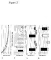
- the inhibitory factor(s) seemed to accumulate with time in the AMTC conditioned medium, since the inhibition effects were observed only in cultures added with supernatant obtained from AMTC cultured for more than 3 days and increased gradually with the length of AMTC culture ( Figure 2B ); similar results were obtained with essential and non essential amino acid plus FCS supplementation of AMTC culture supernatants (data not shown).
- the inhibitory potential of AMTC was maintained up to three AMTC culture passages in experiments with both cell-cell contact or transwell co-culture ( Figure 2C ).
- AMTC inhibit proliferation induced by CD3 and CD28 activation
- AMTC did not induce proliferation responses in neither PBMC nor purified T cells ( Figure 3A ), thus indicating that these cells do not induce allogeneic responses.
- AMTC inhibited PBMC and T cell proliferation induced by anti-CD3/anti-CD28 stimulation ( Figure 3B ) either when co-cultured in cell-cell contact or in the transwell system.
- AMTC inhibited PBMC proliferation induced by anti-CD3 stimulation ( Figure 3C ).
- the anergy of purified T cells to stimulation with anti-CD3 was overcome by the addition of AMTC. This proliferative effect was observed only in the cell-cell contact setting and not in the transwell experiments ( Figure 3C ). Stimulation of AMTC with anti-CD3 did not induce cell proliferation (data not shown).
- HLA-DR + enriched (>90% HLA-DR-positive), and depleted ( ⁇ 5% HLA-DR-positive) AMTC fractions did not induce allogeneic T cell responses.
- HLA-DR + and unfractionated AMTC induced marked T cell proliferation in the presence anti-CD3 stimulation ( Figure 6A ).
- Figure 6B We also observed a dose dependent effect on T cell proliferation by both unfractionated and HLA-DR + AMTC.
- HLA-DR + AMTC cells decrease markedly in numbers during in vitro AMTC culture passages, with a percentage approximately of only 0.5-2% remaining after three passages. Such decline correlated with a reduction of the co-stimulatory effects of AMTC on T-cells prior stimulated by anti-CD3 ( Figure 6C ).
- AMTC In transwell experiments with purified T cells activated with anti-CD3 and anti-CD28 antibodies, AMTC always inhibited T cell proliferation in a dose-dependent manner, as shown in Figure 7A . In contrast, in cell-cell contact conditions, AMTC inhibited T-cell proliferation when added at higher concentrations (T cells: AMTC ratio of 1:1 or 1:1.3), while they induced T-cell activation when added at lower concentrations ( Figure 7A ).
- mesenchymal region of amnion from human term placenta contains cells with phenotypical, functional and immunomodulatory characteristics similar to those described for MSC derived from other sources such as BM, adipose tissue and cord blood. [Parolini, 2006 #64; Fukuchi, 2004 #95; Alviano, 2007 #93; Portmann-Lanz, 2006 #48; Chang, 2006 #94].
- Unfractionated AMTC are capable to inhibit MLR T cell proliferation not only when cultured in direct cell contact with "responder" cells, but also when separated from them by a transwell membrane.
- the inhibition effects were more pronounced when increasing numbers of AMTC were added to the cultures, suggesting a dose-dependent effect.
- the finding that inhibition of T cell proliferation was induced by AMTC in the transwell system suggests that a soluble factor is implicated in such phenomenon. This hypothesis is supported also by our findings that T cell proliferation was inhibited by the addition of AMTC culture supernatant.
- HLA-DR + and HLA-DR - AMTC failed to induce T cell proliferation in the absence of additional stimuli.
- the lack of T cell responses against HLA-DR + allogeneic cells may appear surprising.
- previous studies have shown that exposure to INF-gamma can induce high level of expression of MHC II on BM-MSC which remain unable to induceT cell proliferation [Tse, 2003 #47; Klyushnenkova, 2005 #92].
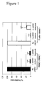
- FIG. 1 Suppression of allogeneic response by AMTC.
- Human PBMC black bars
- human T cells white bars
- AMTC irradiated allogeneic PBMC
- AMTC were added either in direct contact or in transwell chambers.
- Proliferation was assessed by [3H]-thymidine incorporation after five days of culture and expressed as a percentage of proliferation observed in the absence of AMTC.
- Data are mean and SD for more than thirty (PBMC+ PBMC*), or seven independent experiments (T cells+ PBMC*). ***P ⁇ 0.001 vs. corresponding control sample (Student's t test).
- FIG. 2 AMTC inhibitory conditions in MLR.
- A In MLR, responder PBMC were incubated with irradiated allogeneic stimulator PBMC. Increasing concentrations of AMTC were added either in direct contact ( ⁇ ) or in transwell chambers (•). Data are mean ⁇ SD from three independent experiments.
- B MLR were performed alone (black bar) or in the presence of supernatants (SN) from AMTC cultures (white bars). SN were collected from days 1 to 6 of AMTC culture. Data are mean and SD from nine independent experiments. *P ⁇ 0.05, ***P ⁇ 0.001 vs. corresponding control sample (Student's t test).
- MLRs were set up between responder PBMC (R) and equal numbers of allogeneic stimulator PBMC (S1*), with and without the addition of an equal number of AMTC in a transwell chamber. Responders which had been inhibited by AMTC (Ri) were then collected and re-stimulated with the original (Ri+S1*) or third party (Ri + S2*) irradiated allogeneic PBMC, in the absence of AMTC. Data are mean and SD from three independent experiments. ***P ⁇ 0.001 vs. corresponding control sample (Student's t test).
- FIG. 3 Effect of AMTC on unstimulated and CD3, CD3/CD28 stimulated PBMC and T cells.
- PBMC and T cells were either cultured alone (A), or stimulated with anti-CD3 plus anti-CD28 antibody (B), or with anti-CD3 antibody alone (C). All experiments were performed in the absence (black bars) or presence (white bars) of AMTC, both in direct cell contact and in transwell systems. Proliferation was assessed by [3H]-thymidine incorporation after culturing and expressed in counts per minute (cpm). Data are mean and SD from at least three independent experiments. **P ⁇ 0.01, ***P ⁇ 0.001 vs. corresponding control sample (Student's t test).
- FIG. 4 Representative FACS analysis of cells isolated from the mesenchymal amniotic region.
- A Gating strategies to characterize AMTC.
- R1 was defined based on Side (SSC) and Forward (FSC) Scatter properties of AMTC.
- i Analysis of R1 events using FLH-3 shows two distinct subpopulations, individuated by gates R2 and R3 (ii) that can be back-gated to R1 (iii).
- B Surface expression of HLA-DR in total (gate R1) and R2- and R3-gated AMTC.
- C Surface expression of indicated hematopoietic markers on cells gated in R2 (HLA-DR-positive cells). Red histograms show positive cells, while black histograms show IgG isotype control stainings. Percentages of positive cells are indicated.
- FIG. 5 Determination of subpopulations present in the mesenchymal amniotic region.
- A Immunohistochemical staining of representative paraffin sections of term placental amniotic and chorionic membranes. CTC (chorionc trophoblastic cells), CMC (chorionic mesenchymal cells), AEC (amniotic epithelial cells), AMTC (amniotic mesenchymal cells). Left panel: section stained with anti-human CD68 antibody; middle panel: section stained with anti-human HLA-DR antibody; right panel: section stained with anti-human CD45RO antibody. Original magnification x40.
- B RFLP analysis of DNA extracted from HLA-DR+ AMTC. Placental deciduas and total AMTC were used as maternal and fetal controls, respectively.
- FIG. 6 Effect of cells isolated from mesenchymal amniotic region on CD3 stimulated T cells.
- A Purified T cells were cultured alone or in direct contact with unfractionated, HLA-DR-negative or HLA-DR-positive AMTC, with or without anti-CD3. T cells proliferation was assessed by [3H]-thymidine incorporation after three days of culture and expressed in counts per minute (cpm). Data are mean and SD from at least four independent experiments. ***P ⁇ 0.001 vs. corresponding control sample (Student's t test).
- FIG. 7 Effect of amnion mesenchymal cells on purified T cells stimulated by CD3 plus CD28.
- A Purified T cells, stimulated with anti-CD3 and anti-CD28, were cultured alone or in presence of increasing concentrations of total AMTC, in either direct contact (•) or in transwell chambers ( ⁇ ). T cells proliferation was assessed by [3H]-thymidine incorporation after two-three days of culture and expressed in counts per minute (cpm). Data are mean and SD from three independent experiments.
- B Purified T cells, stimulated with anti-CD3 and anti-CD28, were cultured alone or in direct contact with increasing concentrations of total AMTC (•), HLA-DR-negative ( ⁇ ) or HLA-DR-positive ( ⁇ ) AMTC. T cells proliferation was measured by [3H]-thymidine incorporation after two-three days of culture and expressed in counts per minute (cpm). Data are mean and SD from three independent experiments.
Priority Applications (4)
| Application Number | Priority Date | Filing Date | Title |
|---|---|---|---|
| EP07011824A EP2014767A1 (de) | 2007-06-15 | 2007-06-15 | T-Zellen-Immunmodulation durch Plazentazellpräparate |
| PCT/EP2008/004845 WO2008151846A2 (en) | 2007-06-15 | 2008-06-16 | T cell immunomodulation by placenta cell preparations |
| EP08773473.7A EP2171042B1 (de) | 2007-06-15 | 2008-06-16 | T-zellen-immunmodulation durch plazentazellpräparationen |
| US12/664,713 US8524283B2 (en) | 2007-06-15 | 2008-06-16 | T cell immunomodulation by placenta cell preparations |
Applications Claiming Priority (1)
| Application Number | Priority Date | Filing Date | Title |
|---|---|---|---|
| EP07011824A EP2014767A1 (de) | 2007-06-15 | 2007-06-15 | T-Zellen-Immunmodulation durch Plazentazellpräparate |
Publications (1)
| Publication Number | Publication Date |
|---|---|
| EP2014767A1 true EP2014767A1 (de) | 2009-01-14 |
Family
ID=38657798
Family Applications (2)
| Application Number | Title | Priority Date | Filing Date |
|---|---|---|---|
| EP07011824A Withdrawn EP2014767A1 (de) | 2007-06-15 | 2007-06-15 | T-Zellen-Immunmodulation durch Plazentazellpräparate |
| EP08773473.7A Active EP2171042B1 (de) | 2007-06-15 | 2008-06-16 | T-zellen-immunmodulation durch plazentazellpräparationen |
Family Applications After (1)
| Application Number | Title | Priority Date | Filing Date |
|---|---|---|---|
| EP08773473.7A Active EP2171042B1 (de) | 2007-06-15 | 2008-06-16 | T-zellen-immunmodulation durch plazentazellpräparationen |
Country Status (3)
| Country | Link |
|---|---|
| US (1) | US8524283B2 (de) |
| EP (2) | EP2014767A1 (de) |
| WO (1) | WO2008151846A2 (de) |
Cited By (1)
| Publication number | Priority date | Publication date | Assignee | Title |
|---|---|---|---|---|
| US10104880B2 (en) | 2008-08-20 | 2018-10-23 | Celularity, Inc. | Cell composition and methods of making the same |
Families Citing this family (10)
| Publication number | Priority date | Publication date | Assignee | Title |
|---|---|---|---|---|
| AU2009316376B2 (en) * | 2008-11-21 | 2015-08-20 | Celularity Inc. | Treatment of diseases, disorders or conditions of the lung using placental cells |
| US20110256202A1 (en) * | 2010-02-18 | 2011-10-20 | Samson Tom | Immunocompatible amniotic membrane products |
| US9352003B1 (en) | 2010-05-14 | 2016-05-31 | Musculoskeletal Transplant Foundation | Tissue-derived tissuegenic implants, and methods of fabricating and using same |
| US10130736B1 (en) | 2010-05-14 | 2018-11-20 | Musculoskeletal Transplant Foundation | Tissue-derived tissuegenic implants, and methods of fabricating and using same |
| US8883210B1 (en) | 2010-05-14 | 2014-11-11 | Musculoskeletal Transplant Foundation | Tissue-derived tissuegenic implants, and methods of fabricating and using same |
| JP6512759B2 (ja) * | 2013-08-19 | 2019-05-15 | 国立研究開発法人国立循環器病研究センター | 羊膜間葉系細胞組成物の製造方法及び凍結保存方法、並びに治療剤 |
| WO2015168503A1 (en) * | 2014-05-02 | 2015-11-05 | Batu Biologics | Compositions and means for induction of tumor immunity |
| WO2016187413A1 (en) | 2015-05-21 | 2016-11-24 | Musculoskeletal Transplant Foundation | Modified demineralized cortical bone fibers |
| KR101980726B1 (ko) * | 2018-04-26 | 2019-05-21 | 이장호 | Hla-g 단백질을 상시 발현하는 세포주 및 이의 제조방법 |
| CN112725402A (zh) * | 2021-01-22 | 2021-04-30 | 华夏源细胞工程集团股份有限公司 | 一种检测人羊膜上皮细胞体外抑制淋巴细胞增殖抑制方法 |
Citations (2)
| Publication number | Priority date | Publication date | Assignee | Title |
|---|---|---|---|---|
| EP1288293A1 (de) * | 2001-08-30 | 2003-03-05 | Norio Sakuragawa | Humane neuronale Stammzellen aus humanen mesenchymalen Amnionzellschichten |
| WO2006071794A2 (en) * | 2004-12-23 | 2006-07-06 | Ethicon Incorporated | Postpartum cells derived from umbilical cord tissue, and methods of making and using the same |
Family Cites Families (7)
| Publication number | Priority date | Publication date | Assignee | Title |
|---|---|---|---|---|
| US2500083A (en) | 1947-12-09 | 1950-03-07 | Hartford Empire Co | Double vacuum bottom plate and take-out mechanism for glassware blow mold |
| GB9420018D0 (en) | 1994-10-04 | 1994-11-16 | Emhart Glass Mechinery Investm | Improvements in the manufacture of glass containers |
| US6457331B1 (en) | 2000-03-13 | 2002-10-01 | Emhart Glass S.A. | I.S. machine |
| DE10144112A1 (de) | 2001-09-08 | 2003-03-27 | Hermann Heye I I | Verfahren und Fertigformstation zum Fertigblasen eines Glasbehälters |
| CN101330935B (zh) * | 2005-10-21 | 2013-11-13 | 细胞研究私人有限公司 | 自脐带羊膜分离和培养干/祖细胞及其分化的细胞的应用 |
| KR100900309B1 (ko) * | 2007-05-29 | 2009-06-02 | 차의과학대학교 산학협력단 | 태반 융모막판막-유래 중간엽 줄기 세포의 고순도 분리방법 |
| KR101003121B1 (ko) * | 2008-12-23 | 2010-12-22 | 주식회사 하이닉스반도체 | 반도체 메모리 장치의 리프레쉬 회로 |
-
2007
- 2007-06-15 EP EP07011824A patent/EP2014767A1/de not_active Withdrawn
-
2008
- 2008-06-16 WO PCT/EP2008/004845 patent/WO2008151846A2/en active Application Filing
- 2008-06-16 EP EP08773473.7A patent/EP2171042B1/de active Active
- 2008-06-16 US US12/664,713 patent/US8524283B2/en active Active
Patent Citations (2)
| Publication number | Priority date | Publication date | Assignee | Title |
|---|---|---|---|---|
| EP1288293A1 (de) * | 2001-08-30 | 2003-03-05 | Norio Sakuragawa | Humane neuronale Stammzellen aus humanen mesenchymalen Amnionzellschichten |
| WO2006071794A2 (en) * | 2004-12-23 | 2006-07-06 | Ethicon Incorporated | Postpartum cells derived from umbilical cord tissue, and methods of making and using the same |
Non-Patent Citations (7)
| Title |
|---|
| ANKER IN'T P ET AL: "Isolation of mesenchymal stem cells of fetal or maternal origin from human placenta", STEM CELLS, ALPHAMED PRESS, DAYTON, OH, US, vol. 22, no. 7, 2004, pages 1338 - 1345, XP002371702, ISSN: 1066-5099 * |
| KADNER ET AL: "Human umbilical cord cells for cardiovascular tissue engineering: a comparative study", EUROPEAN JOURNAL OF CARDIO-THORACIC SURGERY, SPRINGER VERLAG, BERLIN, DE, vol. 25, no. 4, April 2004 (2004-04-01), pages 635 - 641, XP005045331, ISSN: 1010-7940 * |
| KOBAYASHI NAMIE ET AL: "Suppression of corneal neovascularization by culture supernatant of human amniotic cells.", CORNEA JAN 2002, vol. 21, no. 1, January 2002 (2002-01-01), pages 62 - 67, XP002458587, ISSN: 0277-3740 * |
| PAROLINI O., ET AL.: "isolation and characterization from human term placenta", STEM CELLS, November 2007 (2007-11-01), XP002458588 * |
| WHITTLE W L ET AL: "The characterization of human amnion epithelial and mesenchymal cells: The cellular expression, activity and glucocorticoid regulation of prostaglandin output", PLACENTA, vol. 21, no. 4, May 2000 (2000-05-01), pages 394 - 401, XP002962933, ISSN: 0143-4004 * |
| YAN XIAOJUAN ET AL: "Nuclear factor kappa B activation and regulation of cyclooxygenase type-2 expression in human amnion mesenchymal cells by interleukin-1beta", BIOLOGY OF REPRODUCTION, vol. 66, no. 6, June 2002 (2002-06-01), pages 1667 - 1671, XP002458586, ISSN: 0006-3363 * |
| ZHANG ET AL: "Mesenchymal progenitor cells derived from chorionic villi of human placenta for cartilage tissue engineering", BIOCHEMICAL AND BIOPHYSICAL RESEARCH COMMUNICATIONS, ACADEMIC PRESS INC. ORLANDO, FL, US, vol. 340, no. 3, 17 February 2006 (2006-02-17), pages 944 - 952, XP005234923, ISSN: 0006-291X * |
Cited By (1)
| Publication number | Priority date | Publication date | Assignee | Title |
|---|---|---|---|---|
| US10104880B2 (en) | 2008-08-20 | 2018-10-23 | Celularity, Inc. | Cell composition and methods of making the same |
Also Published As
| Publication number | Publication date |
|---|---|
| WO2008151846A3 (en) | 2009-06-25 |
| US20100221268A1 (en) | 2010-09-02 |
| WO2008151846A2 (en) | 2008-12-18 |
| US8524283B2 (en) | 2013-09-03 |
| EP2171042A2 (de) | 2010-04-07 |
| EP2171042B1 (de) | 2015-09-16 |
Similar Documents
| Publication | Publication Date | Title |
|---|---|---|
| Magatti et al. | Human amnion mesenchyme harbors cells with allogeneic T-cell suppression and stimulation capabilities | |
| EP2014767A1 (de) | T-Zellen-Immunmodulation durch Plazentazellpräparate | |
| Erkers et al. | Decidual stromal cells promote regulatory T cells and suppress alloreactivity in a cell contact-dependent manner | |
| Flemming et al. | Immunomodulative efficacy of bone marrow-derived mesenchymal stem cells cultured in human platelet lysate | |
| JP2020055809A (ja) | 胎盤幹細胞を使用する免疫調節 | |
| Pierdomenico et al. | Multipotent mesenchymal stem cells with immunosuppressive activity can be easily isolated from dental pulp | |
| Karlsson et al. | Stromal cells from term fetal membrane are highly suppressive in allogeneic settings in vitro | |
| Rizzo et al. | A functional role for soluble HLA-G antigens in immune modulation mediated by mesenchymal stromal cells | |
| US8900573B2 (en) | Immune privileged and modulatory progenitor cells | |
| KR20080056302A (ko) | 태반 유래 줄기세포로부터 생산된 희소돌기 아교세포 | |
| Alikarami et al. | The immunosuppressive activity of amniotic membrane mesenchymal stem cells on T lymphocytes | |
| CN109706180A (zh) | 一种脐带间充质干细胞过表达ido增强免疫抑制的方法及应用 | |
| US20150216911A1 (en) | Multipotent Prenatal Stem Cells | |
| US20210269768A1 (en) | Neonatal stromal cells having low mhc-i expression and uses therof | |
| US20220202868A1 (en) | Improved stem cell populations for allogeneic therapy | |
| Abomaray | Exploring the role of mesenchymal stromal cells in endometriosis | |
| Knuth et al. | Eppo Wolvius Elske Strabbing Martin Koudstaal Roberto Narcisi Eric Farrell | |
| WO2024028486A1 (en) | In vitro method for obtaining clinical-grade cd8+ cd45rclow/- regulatory t cells | |
| US20210100847A1 (en) | Stem cells for the treatment of chronic traumatic encephalopathy | |
| Götherström | Characterisation of human fetal mesenchymal stem cells | |
| MX2008004787A (en) | Immunomodulation using placental stem cells | |
| KR20150102383A (ko) | Ex vivo로 조작된 생 자가세포 제조를 위한 자가유래 면역세포 배양방법 |
Legal Events
| Date | Code | Title | Description |
|---|---|---|---|
| PUAI | Public reference made under article 153(3) epc to a published international application that has entered the european phase |
Free format text: ORIGINAL CODE: 0009012 |
|
| PUAI | Public reference made under article 153(3) epc to a published international application that has entered the european phase |
Free format text: ORIGINAL CODE: 0009012 |
|
| AK | Designated contracting states |
Kind code of ref document: A1 Designated state(s): AT BE BG CH CY CZ DE DK EE ES FI FR GB GR HU IE IS IT LI LT LU LV MC MT NL PL PT RO SE SI SK TR |
|
| AX | Request for extension of the european patent |
Extension state: AL BA HR MK RS |
|
| AKX | Designation fees paid | ||
| STAA | Information on the status of an ep patent application or granted ep patent |
Free format text: STATUS: THE APPLICATION IS DEEMED TO BE WITHDRAWN |
|
| 18D | Application deemed to be withdrawn |
Effective date: 20090715 |
|
| REG | Reference to a national code |
Ref country code: DE Ref legal event code: 8566 |