EP2014674B1 - Protein-protein interactions in human immunodeficiency virus - Google Patents
Protein-protein interactions in human immunodeficiency virus Download PDFInfo
- Publication number
- EP2014674B1 EP2014674B1 EP08006236.7A EP08006236A EP2014674B1 EP 2014674 B1 EP2014674 B1 EP 2014674B1 EP 08006236 A EP08006236 A EP 08006236A EP 2014674 B1 EP2014674 B1 EP 2014674B1
- Authority
- EP
- European Patent Office
- Prior art keywords
- seq
- hiv
- polypeptide
- dna
- cells
- Prior art date
- Legal status (The legal status is an assumption and is not a legal conclusion. Google has not performed a legal analysis and makes no representation as to the accuracy of the status listed.)
- Expired - Lifetime
Links
Images












Classifications
-
- C—CHEMISTRY; METALLURGY
- C07—ORGANIC CHEMISTRY
- C07K—PEPTIDES
- C07K14/00—Peptides having more than 20 amino acids; Gastrins; Somatostatins; Melanotropins; Derivatives thereof
- C07K14/005—Peptides having more than 20 amino acids; Gastrins; Somatostatins; Melanotropins; Derivatives thereof from viruses
-
- A—HUMAN NECESSITIES
- A61—MEDICAL OR VETERINARY SCIENCE; HYGIENE
- A61P—SPECIFIC THERAPEUTIC ACTIVITY OF CHEMICAL COMPOUNDS OR MEDICINAL PREPARATIONS
- A61P31/00—Antiinfectives, i.e. antibiotics, antiseptics, chemotherapeutics
- A61P31/12—Antivirals
- A61P31/14—Antivirals for RNA viruses
- A61P31/18—Antivirals for RNA viruses for HIV
-
- C—CHEMISTRY; METALLURGY
- C07—ORGANIC CHEMISTRY
- C07K—PEPTIDES
- C07K14/00—Peptides having more than 20 amino acids; Gastrins; Somatostatins; Melanotropins; Derivatives thereof
- C07K14/435—Peptides having more than 20 amino acids; Gastrins; Somatostatins; Melanotropins; Derivatives thereof from animals; from humans
- C07K14/46—Peptides having more than 20 amino acids; Gastrins; Somatostatins; Melanotropins; Derivatives thereof from animals; from humans from vertebrates
- C07K14/47—Peptides having more than 20 amino acids; Gastrins; Somatostatins; Melanotropins; Derivatives thereof from animals; from humans from vertebrates from mammals
-
- C—CHEMISTRY; METALLURGY
- C12—BIOCHEMISTRY; BEER; SPIRITS; WINE; VINEGAR; MICROBIOLOGY; ENZYMOLOGY; MUTATION OR GENETIC ENGINEERING
- C12N—MICROORGANISMS OR ENZYMES; COMPOSITIONS THEREOF; PROPAGATING, PRESERVING, OR MAINTAINING MICROORGANISMS; MUTATION OR GENETIC ENGINEERING; CULTURE MEDIA
- C12N15/00—Mutation or genetic engineering; DNA or RNA concerning genetic engineering, vectors, e.g. plasmids, or their isolation, preparation or purification; Use of hosts therefor
- C12N15/09—Recombinant DNA-technology
- C12N15/11—DNA or RNA fragments; Modified forms thereof; Non-coding nucleic acids having a biological activity
- C12N15/111—General methods applicable to biologically active non-coding nucleic acids
-
- C—CHEMISTRY; METALLURGY
- C12—BIOCHEMISTRY; BEER; SPIRITS; WINE; VINEGAR; MICROBIOLOGY; ENZYMOLOGY; MUTATION OR GENETIC ENGINEERING
- C12N—MICROORGANISMS OR ENZYMES; COMPOSITIONS THEREOF; PROPAGATING, PRESERVING, OR MAINTAINING MICROORGANISMS; MUTATION OR GENETIC ENGINEERING; CULTURE MEDIA
- C12N15/00—Mutation or genetic engineering; DNA or RNA concerning genetic engineering, vectors, e.g. plasmids, or their isolation, preparation or purification; Use of hosts therefor
- C12N15/09—Recombinant DNA-technology
- C12N15/11—DNA or RNA fragments; Modified forms thereof; Non-coding nucleic acids having a biological activity
- C12N15/113—Non-coding nucleic acids modulating the expression of genes, e.g. antisense oligonucleotides; Antisense DNA or RNA; Triplex- forming oligonucleotides; Catalytic nucleic acids, e.g. ribozymes; Nucleic acids used in co-suppression or gene silencing
- C12N15/1131—Non-coding nucleic acids modulating the expression of genes, e.g. antisense oligonucleotides; Antisense DNA or RNA; Triplex- forming oligonucleotides; Catalytic nucleic acids, e.g. ribozymes; Nucleic acids used in co-suppression or gene silencing against viruses
- C12N15/1132—Non-coding nucleic acids modulating the expression of genes, e.g. antisense oligonucleotides; Antisense DNA or RNA; Triplex- forming oligonucleotides; Catalytic nucleic acids, e.g. ribozymes; Nucleic acids used in co-suppression or gene silencing against viruses against retroviridae, e.g. HIV
-
- A—HUMAN NECESSITIES
- A61—MEDICAL OR VETERINARY SCIENCE; HYGIENE
- A61K—PREPARATIONS FOR MEDICAL, DENTAL OR TOILETRY PURPOSES
- A61K38/00—Medicinal preparations containing peptides
-
- C—CHEMISTRY; METALLURGY
- C12—BIOCHEMISTRY; BEER; SPIRITS; WINE; VINEGAR; MICROBIOLOGY; ENZYMOLOGY; MUTATION OR GENETIC ENGINEERING
- C12N—MICROORGANISMS OR ENZYMES; COMPOSITIONS THEREOF; PROPAGATING, PRESERVING, OR MAINTAINING MICROORGANISMS; MUTATION OR GENETIC ENGINEERING; CULTURE MEDIA
- C12N2310/00—Structure or type of the nucleic acid
- C12N2310/10—Type of nucleic acid
- C12N2310/14—Type of nucleic acid interfering nucleic acids [NA]
-
- C—CHEMISTRY; METALLURGY
- C12—BIOCHEMISTRY; BEER; SPIRITS; WINE; VINEGAR; MICROBIOLOGY; ENZYMOLOGY; MUTATION OR GENETIC ENGINEERING
- C12N—MICROORGANISMS OR ENZYMES; COMPOSITIONS THEREOF; PROPAGATING, PRESERVING, OR MAINTAINING MICROORGANISMS; MUTATION OR GENETIC ENGINEERING; CULTURE MEDIA
- C12N2320/00—Applications; Uses
- C12N2320/10—Applications; Uses in screening processes
- C12N2320/12—Applications; Uses in screening processes in functional genomics, i.e. for the determination of gene function
-
- C—CHEMISTRY; METALLURGY
- C12—BIOCHEMISTRY; BEER; SPIRITS; WINE; VINEGAR; MICROBIOLOGY; ENZYMOLOGY; MUTATION OR GENETIC ENGINEERING
- C12N—MICROORGANISMS OR ENZYMES; COMPOSITIONS THEREOF; PROPAGATING, PRESERVING, OR MAINTAINING MICROORGANISMS; MUTATION OR GENETIC ENGINEERING; CULTURE MEDIA
- C12N2740/00—Reverse transcribing RNA viruses
- C12N2740/00011—Details
- C12N2740/10011—Retroviridae
- C12N2740/16011—Human Immunodeficiency Virus, HIV
- C12N2740/16111—Human Immunodeficiency Virus, HIV concerning HIV env
- C12N2740/16122—New viral proteins or individual genes, new structural or functional aspects of known viral proteins or genes
-
- C—CHEMISTRY; METALLURGY
- C12—BIOCHEMISTRY; BEER; SPIRITS; WINE; VINEGAR; MICROBIOLOGY; ENZYMOLOGY; MUTATION OR GENETIC ENGINEERING
- C12N—MICROORGANISMS OR ENZYMES; COMPOSITIONS THEREOF; PROPAGATING, PRESERVING, OR MAINTAINING MICROORGANISMS; MUTATION OR GENETIC ENGINEERING; CULTURE MEDIA
- C12N2740/00—Reverse transcribing RNA viruses
- C12N2740/00011—Details
- C12N2740/10011—Retroviridae
- C12N2740/16011—Human Immunodeficiency Virus, HIV
- C12N2740/16211—Human Immunodeficiency Virus, HIV concerning HIV gagpol
- C12N2740/16222—New viral proteins or individual genes, new structural or functional aspects of known viral proteins or genes
Definitions
- the present invention relates to proteins that interact with Human Immunodeficiency Virus (HIV) proteins. More specifically, the present invention relates to complexes of polypeptides or polynucleotides encoding the polypeptides, fragments of the polypeptides, antibodies to the complexes, Selected interacting Domains (SID®) which are identified due to the protein-protein interactions, methods for screening drugs for agents which modulate the interaction of proteins and pharmaceutical compositions that are capable of modulating the protein-protein interactions.
- SID® Selected interacting Domains
- Protein-protein interactions enable two or more proteins to associate. A large number of non-covalent bonds form between the proteins when two protein surfaces are precisely matched. These bonds account for the specificity of recognition.
- protein-protein interactions are involved, for example, in the assembly of enzyme subunits, in antibody-antigen recognition, in the formation of biochemical complexes, in the correct folding of proteins, in the metabolism of proteins, in the transport of proteins, in the localization of proteins, in protein turnover, in first translation modifications, in the core structures of viruses and In signal transduction.
- the first protein known in the art as the "balt protein” is a chimeric protein which binds to a site on DNA upstream of a reporter gene by means of a DNA-binding domain or BD.
- the binding domain is the DNA-binding domain from either Gal4 or native E . coli LexA and the sites placed upstream of the reporter are Gal4 binding sites or LexA operators, respectively.
- the second protein is also a chimeric protein known as the "prey" in the art.
- This second chimeric protein carries an activation domain or AD.
- This activation domain is typically derived from Gal4, from VP16 or from B42.
- Another advantage of the two-hybrid plus one system is that it allows or prevents the formation of the transcriptional activator since the third partner can be expressed from a conditional promoter such as the methionine-repressed Met25 promoter which is positively regulated In medium lacking methionine.
- the presence of the methionine-regulated promoter provides an excellent control to evaluate the activation or inhibition properties of the third partner due to its "on" and "off” switch for the formation of the transcriptional activator.
- the three-hybrid method is described, for example in Tirode et al., The Journal of Biological Chemistry, 272, No. 37 pp. 22995-22999 (1997 ).
- WO99/42612 permits the screening of more prey polynucleotides with a given bait polynucleotide in a single step than in the prior art systems due to the cell to cell mating strategy between haploid yeast cells. Furthermore, this method is more thorough and reproducible, as well as sensitive. Thus, the presence of false negatives and/or false positives is extremely minimal as compared to the conventional prior art methods.
- HIV human immunodeficiency virus
- the etiologic agent of AIDS was discoveredin 1984 and reliable tests for HIV antibody as well as for the virus itself are currently available.
- HIV is caused by HIV, a human retrovirus of the lentivirus group.
- the four recognized retroviruses belong to two distinct groups; the human T lymphotropic retrovirus (or leukemia) such as HTLV-I and HTLV-II or the human immunodeficiency viruses such as HIV-1 and HIV-2.
- HTLV-I and HTLV-II are transforming viruses, while HIV-1 and HIV-2 are cytopathic viruses.
- the most common cause of AIDS throughout the world is HIV-1.
- HIV-2 is more closely related to some members of a group of simian immunodeficiency viruses and has about 40% sequence identity to HIV-1. HIV-2 has been identified predominantly in western Africa and is believed to be less pathogenic than HIV-1.
- HIV-1 has the usual retroviral genes such as env , gal and pol .
- the gag gene encodes the precursor virion core proteins for the matrix protein (MA), the capsid protein (CA), nucleocapsid protein (NC) and P6.
- the pol gene encodes the precursor for various virion enzymes such as protease (PR), reverse transcriptase (RT), RNAse H and integrase (IN).
- the env gene encodes the precursors for the envelope glycoprotein (Env gp) such as surface glycoprotein (gp 120/SU) and transmembrane protein (gp 41/TM).
- the transcriptional transactivator ( tat ) and the regulator of viral expression ( rev ) genes are each encoded by two overlapping exons and produce small nonvirion proteins which are essential for viral replication. Also several nonessential genes which are not implicated in viral expression are encoded by HIV.1 such as vif , vpr , vpu and nef .
- AIDS is a global epidemic with virtually every country in the world reporting cases. In the United States alone by the mid-1990s, approximately 120,000 cases among adults and adolescents, and approximately 2,000 cases among children less than 13 years old had been reported.
- the Virus can also be transmitted via blood or blood products and infected mothers can transmit HIV to their infants perinatally and as early as the first and second trimester of pregnancy.
- the virus can also be transmitted from the mother to infant via breast feeding.
- the prevalence of HIV infection among intravenous drug users is exceptionally high.
- HIV infection ranges from an asymptomatic state to severe disease.
- Seroconversion generally occurs between 8 to 12 weeks after infection.
- Neurologic disease is common in HIV-infected individuals, the most common being encephalopathy or AIDS demantia complex.
- inhibitors of RT and of PR are just entering human clinical trials (Nair 2002). All these inhibitors target the enzymatic activity of these viral enzymes. However, no inhibitors are directed against the interactions between viral proteins and potentially important cellular partners which ensure optimal viral replication in infected cells. HIV having evolved an extraordinary efficient capacity to exploit the cell's molecular machinery in the course of infection, understanding the dynamic interplay of host cell and virus is essential to the effort to control HIV infection.
- polynucleotides As used herein the terms “polynucleotides”, “nucleic acids” and “oligonucleotides” are used interchangeably and include, but are not limited to RNA, DNA, RNA/DNA sequences of more than one nucleotide in either single chain or duplex form.
- the polynucleotide sequences of the present invention may be prepared from any known method including, but not limited to, any synthetic method, any recombinant method, any ex vivo generation method and the like, as well as combinations thereof.
- polypeptide means herein a polymer of amino acids having no specific length.
- peptides, oligopeptides and proteins are included in the definition of “polypeptide” and these terms are used interchangeably throughout the specification, as well as in the claims.
- polypeptide does not exclude post-translational modifications such as polypeptides having covalent attachment of glycosyl groups, acetyl groups, phosphate groups, lipid groups and the like. Also encompassed by this definition of "polypeptide” are homologs thereof.
- orthologs structurally similar genes contained within a given species
- orthologs are functionally equivalent genes from a given species or strain, as determined for example, in a standard complementation assay.
- a polypeptide of interest can be used not only as a model for identifying similiar genes in given strains, but also to identify homologs and orthologs of the polypeptide of interest in other species.
- the orthologs for example, can also be identified in a conventional complementation assay.
- orthologs can be expected to exist in bacteria (or other kind of cells) in the same branch of the phylogenic tree, as set forth, for example, at ftp://ftp.cme.msu.edu/pub/rdp/SSU-rRNA/SSU/Prok.phylo.
- prey polynucleotide means a chimeric polynucleotide encoding a polypeptide comprising (i) a specific domain; and (ii) a polypeptide that is to be tested for interaction with a bait polypeptide.
- the specific domain is preferably a transcriptional activating domain.
- a "bait polynucleotide” is a chimeric polynucleotide encoding a chimeric polypeptide comprising (i) a complementary domain; and (ii) a polypeptide that is to be tested for interaction with at least one prey polypeptide.
- the complementary domain is preferably a DNA-binding domain that recognizes a binding site that is further detected and is contained in the host organism.
- complementary domain is meant a functional constitution of the activity when bait and prey are interacting; for example, enzymatic activity.
- specific domain is meant a functional interacting activation domain that may work through different mechanisms by interacting directly or indirectly through intermediary proteins with RNA polymerase II or III-associated proteins in the vicinity of the transcription start site.
- complementary means that, for example, each base of a first polynucleotide is paired with the complementary base of a second polynucleotide whose orientation is reversed.
- the complementary bases are A and T (or A and U) or C and G.
- sequence identity refers to the identity between two peptides or between two nucleic acids. Identity between sequences can be determined by comparing a position in each of the sequences which may be aligned for the purposes of comparison. When a position in the compared sequences is occupied by the same base or amino acid, then the sequences are identical at that position. A degree of sequence identity between nucleic acid sequences is a function of the number of identical nucleotides at positions shared by these sequences. A degree of identity between amino acid sequences is a function of the number of identical amino acid sequences that are shared between these sequences.
- two polypeptides may each (i) comprise a sequence (i.e., a portion of a complete polynucleotide sequence) that is similar between two polynucleotides, and (ii) may further comprise a sequence that is divergent between two polynucleotides
- sequence identity comparisons between two or more polynucleotides over a "comparison window" refers to the conceptual segment of at least 20 contiguous nucleotide positions wherein a polynucleotide sequence may be compared to a reference nucleotide sequence of at least 20 contiguous nucleotides and wherein the portion of the polynucleotide sequence in the comparison window may comprise additions or deletions (i.e., gaps) of 20 percent or less compared to the reference sequence (which does not comprise additions or deletions) for optimal alignment of the two sequences.
- the sequences are aligned for optimal comparison. For example, gaps can be introduced in the sequence of a first amino acid sequence or a first nucleic acid sequence for optimal alignment with the second amino acid sequence or second nucleic acid sequence.
- the amino acid residues or nucleotides at corresponding amino acid positions or nucleotide positions are then compared. When a position in the first sequence is occupied by the same amino acid residue or nucleotide as the corresponding position in the second sequence, the molecules are identical at that position.
- sequences can be the same length or may be different in length.
- Optimal alignment of sequences for determining a comparison window may be conducted by the local homology algorithm of Smith and Waterman (J. Theor. Biol., 91 (2) pgs. 370-380 (1981 ), by the homology alignment algorithm of Needleman and Wunsch, J. Miol. Biol., 48(3) pgs. 443-453 (1972 ), by the search for similarity via the method of Pearson and Lipman, PNAS, USA, 85(5) pgs. 2444-2448 (1988 ), by computerized implementations of these algorithms (GAP, BESTFIT, FASTA and TFASTA in the Wisconsin Genetics Software Package Release 7.0, Genetic Computer Group, 575, Science Drive, Madison, Wisconsin) or by inspection.
- the best alignment i.e., resulting in the highest percentage of identity over the comparison window generated by the various methods is selected.
- sequence identity means that two polynucleotide sequences are identical (i.e., on a nucleotide by nucleotide basis) over the window of comparison.
- percentage of sequence identity is calculated by comparing two optimally aligned sequences over the window of comparison, determining the number of positions at which the identical nucleic acid base (e.g., A, T, C, G, U, or I) occurs in both sequences to yield the number of matched positions, dividing the number of matched positions by the total number of positions in the window of comparison (i.e., the window size) and multiplying the result by 100 to yield the percentage of sequence identity.
- the same process can be applied to polypeptide sequences.
- the percentage of sequence identity of a nucleic acid sequence or an amino acid sequence can also be calculated using BLAST software (Version 2.06 of September 1998) with the default or user defined parameter.
- sequence similarity means that amino acids can be modified while retaining the same function. It is known that amino acids are classified according to the nature of their side groups and some amino acids such as the basic amino acids can be interchanged for one another while their basic function is maintained.
- isolated means that a biological material such as a nucleic acid or protein has been removed from its original environment in which it is naturally present.
- a biological material such as a nucleic acid or protein has been removed from its original environment in which it is naturally present.
- a polynucleotide present in a plant, mammal or animal is present in its natural state and is not considered to be isolated.
- the same polynucleotide separated from the adjacent nucleic acid sequences in which it is naturally inserted in the genome of the plant or animal is considered as being “isolated.”
- isolated is not meant to exclude artificial or synthetic mixtures with other compounds, or the presence of impurities which do not interfere with the biological activity and which may be present, for example, due to incomplete purification, addition of stabilizers or mixtures with pharmaceutically acceptable excipients and the like.
- isolated polypeptide or isolated protein as used herein means a polypeptide or protein which is substantially free of those compounds that are normally associated with the polypeptide or protein in a naturally state such as other proteins or polypeptides, nucleic acids, carbohydrates, lipids and the like.
- purified means at least one order of magnitude of purification is achieved, preferably two or three orders of magnitude, most preferably four or five orders of magnitude of purification of the starting material or of the natural material. Thus, the term “purified” as utilized herein does not mean that the material is 100% purified and thus excludes any other material.
- variants when referring to, for example, polynucleotides encoding a polypeptide variant of a given reference polypeptide are polynucleotides that differ from the reference polypeptide but generally maintain their functional characteristics of the reference polypeptide.
- a variant of a polynucleotide may be a naturally occurring allelic variant or it may be a variant that is known naturally not to occur.
- Such non-naturally occurring variants of the reference polynucleotide can be made by, for example, mutagenesis techniques, including those mutagenesis techniques that are applied to polynucleotides, cells or organisms.
- Variants of polynucleotides according to the present invention include, but are not limited to, nucleotide sequences which are at least 95% identical after alignment to the reference polynucleotide encoding the reference polypeptide. These variants can also have 96%, 97%, 98% and 99.999% sequence identity to the reference polynucleotide.
- Nucleotide changes present in a variant polynucleotide may be silent, which means that these changes do not alter the amino acid sequences encoded by the reference polynucleotide.
- Substitutions, additions and/or deletions can involve one or more nucleic acids. Alterations can produce conservative or non-conservative amino acid substitutions, deletions and/or additions.
- Variants of a prey or a SID® polypeptide encoded by a variant polynucleotide can possess a higher affinity of binding and/or a higher specificity of binding to its protein or polypeptide counterpart, against which it has been initially selected.
- variants can also loose their ability to bind to their protein or polypeptide counterpart.
- fragment of a polynucleotide or “fragment of a SID® polynucleotide” is meant that fragments of these sequences have at least 12 consecutive nucleotides, or between 12 and 5,000 consecutive nucleotides, or between 12 and 10,000 consecutive nucleotides, or between 12 and 20,000 consecutive nucleotides.
- fragments of a polypeptide or “fragment of a SID® polypeptide” is meant that fragments of these sequences have at least 4 consecutive amino acids, or between 4 and 1,700 consecutive amino acids, or between 4 and 3,300 consecutive amino acids, or between 4 and 6,600 consecutive amino acids.
- anabolic pathway is meant a reaction or series of reactions in a metabolic pathway that synthesize complex molecules from simpler ones, usually requiring the input of energy.
- An anabolic pathway is the opposite of a catabolic pathway.
- catabolic pathway is a series of reactions in a metabolic pathway that break down complex compounds into simpler ones, usually releasing energy in the process.
- a catabolic pathway is the opposite of an anabolic pathway.
- drug metabolism is meant the study of how drugs are processed and broken down by the body. Drug metabolism can involve the study of enzymes that break down drugs, the study of how different drugs interact within the body and how diet and other ingested compounds affect the way the body processes drugs.
- metabolic means the sum of all of the enzyme-catalyzed reactions in living cells that transform organic molecules.
- second metabolism is meant pathways producing specialized metabolic products that are not found in every cell.
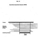
- SID® means a Selected Interacting Domain and is identified as follows: for each bait polypeptide screened, selected prey polypeptides are compared. Overlapping fragments in the same ORF or CDS define the selected interacting domain.
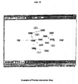
- PIM® means a protein-protein interaction map. This map is obtained from data acquired from a number of separate screens using different balt polypeptides and is designed to map out all of the interactions between the polypeptides.
- SID® polypeptide of the present invention or a variant thereof for its polypeptide counterpart can be assessed, for example, on a BlacoreTM apparatus marketed by Amersham Pharmacia Biotech Company such as described by Szabo et al. (Curr Opin Struct Biol 5 pgs. 699-705 (1995 )) and by Edwards and Leartherbarrow (Anal. Biochem 246 pgs. 1-6 (1997 )).
- the phrase "at least the same affinity" with respect to the binding affinity between a SID® polypeptide of the present invention to another polypeptide means that the Ka is identical or can be at least two-fold, at least three-fold or at least five fold greater than the Ka value of reference.
- modulating compound means a compound that inhibits or stimulates or can act on another protein which can inhibit or stimulate the protein-protein interaction of a complex of two polypeptides or the protein-protein interaction of two polypeptides.
- the present invention comprises within the limits of the claims complexes of polypeptides or polynucleotides encoding the polypeptides composed of a bait polypeptide, or a balt polynucleotide encoding a bait polypeptide and a prey polypeptide or a prey polynucleotide encoding a prey polypeptide.
- the prey polypeptide or prey polynucleotide encoding the prey polypeptide is capable of interacting with a bait polypeptide of interest in various hybrid systems.
- Protein-protein interactions can also be detected using complementation assays such as those described by Pelletier e t al. at http://www.abrf.org/JBT/Articles/JBT0012/jbt0012.hml. WO 00/07038 and WO98/34120 .
- the method is not limited to detecting protein-protein interactions using yeast, but also includes similar methods that can be used in detecting protein-protein interactions in for example, mammalian systems as described, for example in Takacs et al. (Proc. Natl. Acad. Sci., USA, 90 (21):10375-79 (1993 )) and Vasavada et al (Proc. Natl. Acad Sci., USA, 88 (23):10686-90 (1991 )), as well as a bacterial two-hybrid system as described in Karimova et al. (1998). WO99/28746 , WO00/66722 and Legrain et al. (FEBS Letters, 480 pgs. 32-36 (2000 )).
- Protein-protein interactions can also be detected using fluorescence energy transfer techniques ( Fluorescence resonance energy transfer analysis of protein-protein interactions in single living cells by multifocal multiphoton microscopy. Majoul I, Straub M, Duden R, Hell SW, Soling HD J Biotechnol 2002 Jan;82(3)257-77 ).
- suitable cells include, but are not limited to, VERO cells, HELA cells such as ATCC No. CCL2, CHO cell lines such as ATCC No. CCL61, COS cells such as COS-7 cells and ATCC No. CRL 1650 cells, W138, BHK, HepG2, 3T3 such as ATCC No. CRL6361, A549. PC12. K562 cells, 293 cells, Sf9 cells such as ATCC No. CRL1711 and Cv1 cells such as ATCC No. CCL70.
- suitable cells include, but are not limited to, prokaryotic host cells strains such as Escherichia coli (e.g., strain DH5- ⁇ ), Bacillus subtilis , Salmonella typhimurium, or strains of the genera of Pseudomonas , Streptomyces and Staphylococcus .
- prokaryotic host cells strains such as Escherichia coli (e.g., strain DH5- ⁇ ), Bacillus subtilis , Salmonella typhimurium, or strains of the genera of Pseudomonas , Streptomyces and Staphylococcus .
- yeast cells such as those of Saccharomyces such as Saccharomyces cerevisiae .
- balt polynucleotide as well as the prey polynucleotide can be prepared according to the methods known in the art such as those described above in the publications and patents reciting the known method per se.
- the balt polynucleotide of the present invention is obtained from HIV particles cDNA, or variants of cDNA fragment from a library of HIV particles, and fragments from the genome or transcriptome of HIV particles cDNA ranging from about 12 to about 5,000, or about 12 to about 10,000 or from about 12 to about 20,000.
- the micoorganism utilized to make the library of HIV is YU2 described by LI et al (J. Virol. 8 : 3973-3985 (1991 )).
- the nucleotide sequence of YU2 is shown in figure 18 or can be obtained from Genebank Accession No. M93258.
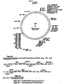
- a prey library is derived from a cDNA library from poly A+ RNA of CEMC7 cells and constructed in the specially designed prey vector pP6 as shown in Figure 10 after ligation of suitable linkers such that every cDNA insert is fused to a nucleotide sequence in the vector that encodes the transcription activation domain of a reporter gene.
- Any transcription activation domain can be used in the present invention. Examples include, but are not limited to, Gal4,VP16, B42, His and the like.
- Toxic reporter genes, suoh as CAT R , CYH2, CYH1, URA3, bacterial and fungi toxins and the like can be used in reverse two-hybrid systems.
- prey polypeptides encoded by the nucleotide inserts of the human CEMC7 prey library thus prepared are termed "prey polypeptides" in the context of the presently described selection method of the prey polynucleotides.
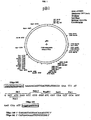
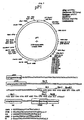
- balt polynucleotides can be inserted in balt plasmid pB6 or pB27 as illustrated in Figure 3 and Figure 17 , respectively.
- the balt polynucleotide Insert is fused to a poliynucleotide encoding the binding domain of, for example, the Gal4 DNA binding domain and the shuttle expression vector is used to transform cells.
- balt polynudeotides used in the examples are described in Table 1.
- any cells can be utilized in transforming the balt and prey polynucleotides of the present invention including mammalian cells, bacterial cells, yeast cells, insect cells and the like.
- Protein-protein interactions may be identified in yeast.
- a prey positive clone is identified containing a vector which comprises a nucleic acid insert encoding a prey polypeptide which binds to a balt polypeptide of interest.
- the method in which protein-protein interactions are identified comprises the following steps:
- Escherichia coli is used in a bacterial two-hybrid system, which encompasses a similar principle to that described above for yeast, but does not involve mating for characterizing the prey polynucleotide.
- Mammalian cells and a method similar to that described above for yeast for characterizing the prey polynucleotide may be used.
- the prey polynucleotide that has been selected by testing the library of preys in a screen using the two-hybrid, two plus one hybrid methods and the like encodes the polypeptide interacting with the protein of interest.
- the present invention is also directed, in a general aspect, to a complex of polypeptides, polynucleotides encoding the polypeptides composed of a bait polypeptide or bait polynucleotide encoding the bait polypeptide and a prey polypeptide or prey polynucleotide encoding the prey polypeptide capable of interacting with the bait polypeptide of interest in accordance with the claims.
- complexes are identified in Table 2.
- the present invention relates to a complex of polynucleotides in accordance with the claims consisting of a first polynucleotide, or a fragment thereof, encoding a prey polypeptide that interacts with a bait polypeptide and a second polynucleotide or a fragment thereof.
- This fragment has at least 12 consecutive nucleotides, but can have between 12 and 5,000 consecutive nucleotides, or between 12 and 10,000 consecutive nucleotides or between 12 and 20,000 consecutive nucleotides.
- the present invention relates to an isolated complex in accordance with the claims of at least two polypeptides encoded by two polynucleotides wherein said two polypeptides are associated in the complex by affinity binding.
- the present invention relates to an isolated complex comprising at least a polypeptide as described in column 1, line 3, of Table 2 and a polypeptide as described in column 4, line 3, of Table 2 in accordance with the claims.
- the present invention is not limited to these polypeptide complexes alone but also includes the isolated complex of the two polypeptides in which fragments and/or homologous polypeptides exhibit at least 95% sequence identity, as well as from 96% sequence identity to 99.999% sequence identity.
- Also encompassed in another embodiment of the present invention is an isolated complex in which the SID® of the prey polypeptide encoded by SEQ ID N° 17 in Table 3 forms the isolated complex.
- nucleic acids coding for a Selected interacting Domain (SID®) polypeptide or a variant thereof or any of the nucleic acids set forth in Table 3 can be inserted into an expression vector which contains the necessary elements for the transcription and translation d the inserted protein-coding sequence.
- transcription elements include a regulatory region and a promoter.
- the nucleic acid which may encode a marker compound of the present invention is operably linked to a promoter in the expression vector.
- the expression vector may also include a replication origin.
- Useful expression vectors that can be used include, for example, segments of chromosomal, non-chromosomal and synthetic DNA sequences.
- Suitable vectors include, but are not limited to, derivatives of SV40 and pcDNA and known bacterial plasmids such as col El, pCR1, pBR322, pMal-C2, pET, pGEX as described by Smith et al (1988), pMB9 and derivatives thereof, plasmids such as RP4, phage DNAs such as the numerous derivatives of phage I such as NM989, as well as other phage DNA such as M13 and filamentous single stranded phage DNA; yeast plasmids such as the 2 micron plasmid or derivatives of the 2m plasmid, as well as centomeric and integrative yeast shuttle vectors; vectors useful in eukaryotic cells such as vectors useful in
- both non-fusion transfer vectors such as, but not limited to pVL941 ( Bam HI cloning site Summers), pVL1393 ( Bam HI, Sma I, Xba I, ECo RI, Not I, Xma III, Bg III and Pst I cloning sites; Invitrogen), pVL1392 ( Bg III, Pst I, Not I, Xma III, Eco RI, Xbal I, Sma I and Bam HI cloning site; Summers and Invitrogen) and pBlueBacIII ( Bam HI, Bgl II, Pst I, Nco I and Hind III cloning site, with blue/white recombinant screening, Invitrogen), and fusion transfer vectors such as, but not limited to, pAc700 ( Bam HI and Kpn I cloning sites, in which the Bam
- Mammalian expression vectors contemplated for use in the invention include vectors with inducible promoters, such as the dihydrofolate reductase promoters, any expression vector with a DHFR expression cassette or a DHFR/mothotrexate co-amplification vector such as pED ( Pst I, Sal I, SbaI, SmaI and Eco RI cloning sites, with the vector expressing both the cloned gene and DHFR; Kaufman, 1991).
- inducible promoters such as the dihydrofolate reductase promoters
- any expression vector with a DHFR expression cassette or a DHFR/mothotrexate co-amplification vector such as pED ( Pst I, Sal I, SbaI, SmaI and Eco RI cloning sites, with the vector expressing both the cloned gene and DHFR; Kaufman, 1991).
- glutamine synthetase/methionine sulfoximine co-amplification vector such as pEE14 ( Hind III, Xbal I, Sma I, Sba I, EcoRI and Bcl I cloning sites in which the vector expresses glutamine synthetase and the cloned gene; Celltech).
- a vector that directs episomal expression under the control of the Epstein Barr Virus (EBV) or nuclear antigen (EBNA) can be used such as pREP4 ( Bam HI, Sfi I, Xho I, Not I, Nhe I, Hind III, Nhe I, Pvu II and Kpn I cloning sites, constitutive RSV-LTR promoter, hygromycin selectable marker; Invitrogen), pCEP4 ( Bam HI, Sfi I, Xho I, Not I, Nhe I, Hind III, Nhe I, Pvu II and Kpn I cloning sites, constitutive hCMV immediate early gene promoter, hygromycin selectable marker; Invitrogen), pMEP4 ( Kpn I, Pvu I, Nhe I, Hind III, Not I, Xho I, Sfi I, BamHI cloning sites, inducible methallothionein IIa gene promoter, h
- Selectable mammalian expression vectors for use in the invention include, but are not limited to, pRc/CMV ( Hind III, Bst XI, Not I, Sba I and Apa I cloning sites, G418 selection, Invitrogen), pRc/RSV ( Hind II, Spe I, Bst XI, Not I, Xba I cloning sites, G418 selection, Invitrogen) and the like.
- Vaccinia virus mammalian expression vectors include, but are not limited to, pSC11 ( Sma I cloning site, TK- and ⁇ -gal selection), pMJ601 ( Sal I, Sma I, Afl I , Nar I, Bsp MII, Bam HI, Apa I, Nhe I, Sac II, Kpn I and Hind III cloning sites; TK- and ⁇ -gal selection), pTKgptF1S ( Eco RI, Pst I, Sal II, Acc I, Hind II, Sba I, Bam HI and Hpa cloning sites, TK or XPRT selection) and the like.
- pSC11 Sma I cloning site, TK- and ⁇ -gal selection
- pMJ601 Sal I, Sma I, Afl I , Nar I, Bsp MII, Bam HI, Apa I, Nhe I, Sac II, Kpn I and Hind
- Yeast expression systems that can also be used in the present include, but are not limited to, the non-fusion pYES2 vector ( Xba I, Sph I, Sho I, Not I, Gst XI, Eco RI, Bst XI, Bam HI, Sac I, Kpn I and Hind III cloning sites, Invitrogen), the fusion pYESHisA, B, C ( Xbal I, Sph I, Sho I, Not I, Bst XI, Eco RI, Bam HI, Sac I, Kpn I and Hind III cloning sites, N-terminal peptide purified with ProBond resin and cleaved with enterokinase; Invitrogen), pRS vectors and the like.
- the non-fusion pYES2 vector Xba I, Sph I, Sho I, Not I, Gst XI, Eco RI, Bst XI, Bam HI, Sac I, K
- mammalian and typically human cells as well as bacterial, yeast, fungl, insect, nematode and plant cells an used in the present invention and may be transfected by the nucleic add or recombinant vector as defined herein.
- suitable cells include, but are not limited to, VERO cells, HELA cells such as ATCC No. CCL2, CHO cell lines such as ATCC No. CCL61, COS cells such as COS-7 cells and ATCC No. CRL 1650 cells, W138, BHK, HepG2, 3T3 such as ATCC No. CRL6361, A549, PC12, K562 cells, 293 cells, Sf9 cells such as ATCC No. CRL1711 and Cv1 cells such as ATCC No. CCL70.
- suitable cells include, but are not limited to, prokaryotic host cells strains such as Escherichia coli , (e.g., strain DH5- ⁇ ), Bacillus subtilis , Salmonelis typhimurium, or strains of the genera of Pseudomonas, Streptomyces and Staphylococcus.
- prokaryotic host cells strains such as Escherichia coli , (e.g., strain DH5- ⁇ ), Bacillus subtilis , Salmonelis typhimurium, or strains of the genera of Pseudomonas, Streptomyces and Staphylococcus.
- yeast cells such as those of Saccharomyces such as Saccharomyces cervisiae .
- the present invention relates to and also encompasses SID® polynucleotides in accordance with the claims.
- SID® polynucleotides of the present invention are represented by the shared nucleic acid sequences of SEQ ID N° 17 encoding the SID® polypeptides of SEQ ID N° 40 in columns 5 and 7 of Table 3 (prey 007766 Human TRNSR), respectively.
- the present invention is not limited to the SID® sequences as described in the above paragraph, but also includes variant of the SID® sequences between 95% and 99.999% sequence identity to its protein or polypeptide counterpart.
- variants d polynucleotide or polypeptides can be created by known mutagenesis techniques either in vitro or in vivo .
- Such a variant can be created such that it has altered binding characteristics with respect to the target protein and more specifically that the variant binds the target sequence with either higher or tower affinity.
- Polynucleotides that are complementary to the above sequences which include the polynucleotides of the SID®'s variants and those that have specific sequence identity are also included in the present invention.
- polynucleotide encoding the SID® polypeptide, or variant thereof can also be inserted into recombinant vectors which are described in detail above.
- the present invention also relates to a composition
- a composition comprising the above-mentioned recombinant vectors containing the SID® polynucleotides as claimed or variants thereof, as well as recombinant host cells transformed by the vectors.
- the recombinant host cells that can be used In the present invention were discussed In greater detail above.
- compositions comprising the recombinant vectors can contain physiological acceptable carriers such as diluents, adjuvants, excipients and any vehicle in which this composition can be delivered therapeutically and can include, but is are not limited to sterile liquids such as water and oils.
- the present invention also discloses a method of selecting modulating compounds, as well as the modulating molecules or compounds themselves which may be used in a pharmaceutical composition.
- modulating compounds may act as a cofactor, as an inhibitor, as antibodies, as tags, as a competitive inhibitor, as an activator or alternatively have agonistic or antagonistic activity on the protein-protein interactions.
- the activity of the modulating compound does not necessarily, for example, have to be 100% activation or inhibition. Indeed, even partial activation or inhibition can be achieved that is of pharmaceutical interest.
- the modulating compound can be selected according to a method which comprises:
- the present invention relates to a modulating compound that inhibits the protein-protein interactions of a complex of two polypeptides of columns 1 and 4, line 3 of Table 2.
- the present invention also relates to a modulating compound that activates the protein-protein interactions of a complex of two Polypeptides of columns 1 and 4, line 3 of Table 2.
- the present invention relates to a method of selecting a modulating compound, which modulating compound inhibits the interactions of two polypeptides of columns 1 and 4, line 3 of Table 2. This method comprises:
- any toxic reported gene can be utilized including those reporter genes that can be used for negative selection including the URA3 gene, the CYH1 gene, the CYH2 gene and the like.
- a kit for screening a modulating compound comprises a recombinant host cell which comprises a reporter gene the expression of which is toxic for the recombinant host cell.
- the host cell is transformed with two vectors.
- the first vector comprises a polynucleotide encoding a first hybrid polypeptide having a DNA binding domain; and the second vector comprises a polynucleotide encoding a second hybrid polypeptide having a transcriptional activate domain that activates said toxic reporter gene when the first and second hybrid polypeptides interact.
- a kit is provided for screening a modulating compound by providing a recombinant host cell, as described in the paragraph above, but instead of a DNA binding domain, the first vector encodes a first hybrid polypeptide containing a first domain of a protein.
- the second vector encodes a second polypeptide containing a second part of a complementary domain of a protein that activates the toxic reporter gene when the first and second hybrid polypeptides interact.
- the activating domain can be 842 Gal 4, VP16 (HSV) and the DNA-binding domain can be derived from Gal4 or Lex A.
- the protein or enzyme can be adenylate cyclase, guanylate cyclase, DHFR and the like.
- the present invention relates to a pharmaceutical composition
- a pharmaceutical composition comprising the modulating compounds in accordance with the claims for preventing or treating AIDS in a human or animal, most preferably In a mammal, or inhibiting HIV replication in indicator cells sensitive to HIV infection.
- This pharmaceutical composition comprises a pharmaceutically acceptable amount of the modulating compound.
- the pharmaceutically acceptable amount can be estimated from cell culture assays.
- a dose can be formulated in animal models to achieve a circulating concentration range that includes or encompasses a concentration point or range having the desired effect in an in vitro system. This information can thus be used to accurately determine the doses in other mammals, including humans and animals.
- the therapeutically effective dose refers to that amount of the compound that results in amelioration of symptoms in a patient. Toxicity and therapeutic efficacy of such compounds can be determined by standard pharmaceutical procedures in cell cultures or in experimental animals. For example, the LD50 (the dose lethal to 50% of the population) as well as the ED50 (the dose therapeutically effective in 50% of the population) can be determined using methods known in the art. The dose ratio between toxic and therapeutic effects is the therapeutic index which can be expressed as the ratio between LD 50 and ED50 compounds that exhibit high therapeutic indexes.
- the data obtained from the cell culture and animal studies can be used in formulating a range of dosage of such compounds which lies preferably within a range of circulating concentrations that include the ED50 with little or no toxicity.
- the pharmaceutical composition can be administered via any route such as locally, orally, systemically, intravenously, intramuscularly, mucosally, using a patch and can be encapsulated in liposomes, microparticles, microcapsules, and the like.
- the pharmaceutical composition can be embedded in liposomes or even encapsulated.
- any pharmaceutically acceptable carrier or adjuvant can be used in the pharmaceutical composition.
- the modulating compound will be preferably in a soluble form combined with a pharmaceutically acceptable carrier.
- the techniques for formulating and administering these compounds can be found in " Remington's Pharmaceutical Sciences ". Mack Publication Co., Easton, PA, latest edition.
- the mode of administration optimum dosages and galenic forms can be determined by the criteria known in the art taken Into account the seriousness of the general condition of the mammal, the tolerance of the treatment and the side effects.
- the present invention also discloses a method of treating or preventing AIDS in a human or mammal in need of such treatment.
- This method comprises administering to a mammal in need of such treatment a pharmaceutically effective amount of a modulating compound which binds to a targeted mammalian or human or inner ear cell protein.
- the modulating compound is a polynucleotide which may be placed under the control of a regulatory sequence which is functional in the mammal or human.
- the present invention relates to a pharmaceutical composition
- a pharmaceutical composition comprising a SID® polypeptide, or variant thereof in accordance with the claims.
- the SID® polypeptide, or variant thereof can be used in a pharmaceutical composition provided that it is endowed with highly specific binding properties to a bait polypeptide of interest.
- the original properties of the SID® polypeptide or variants thereof interfere with the naturally occuring interaction between a first protein and a second protein within the cells of the organism.
- the SID® polypeptide binds specifically to either the first polypeptide or the second polypeptide.
- SID® polypeptides of the present invention or variants thereof interfere with protein-protein interactions between HIV protein.
- the present invention relates to a pharmaceutical composition
- a pharmaceutical composition comprising a pharmaceutically acceptable amount of a SID® polypeptide or variant thereof, provided that the variant has the above-mentioned two characteristic; i.e., that it is endowed with highly specific binding properties to a bait polypeptide of interest and is devoid of biological activity of the naturally occurring protein.
- the present invention relates to a pharmaceutical composition in accordance with the claims comprising a pharmaceutically effective amount of a polynucleotide encoding a SID® polypeptide on a variant thereof wherein the polynucleotide is placed under the control of an appropriate regulatory sequence.
- Appropriate regulatory sequences that are used are polynucleotide sequences derived from promoter elements and the like.
- Polynucleotides that can be used in the pharmaceutical composition of the present invention include the nucleotide sequence of SEQ ID N°17.
- the pharmaceutical composition of the present invention can also include a recombinant expression vector comprising the polynucleotide encoding the SID® polypeptide in accordance with the claims, or variant thereof.
- compositions can be administered by any route such as orally, systemically, intravenously, intramuscularly, intradermally, mucosally, encapsulated, using a patch and the like.
- Any pharmaceutically acceptable carrier or adjuvant can be used in this pharmaceutically composition.
- SID® polypeptides as active ingredients will be preferably in a soluble form combined with a pharmaceutically acceptable carrier.
- the techniques for formulating and administering these compounds can be found in " Remington's Pharmaceutical Sciences " supra .
- the amount of pharmaceutically acceptable SID® polypeptides can be determined as described above for the modulating compounds using cell culture and animal models.
- the primate models of AIDS provide insights into pathogenesis, transmission, and immune responses to infection and are useful In testing vaccines and drugs.
- the HIV-1/chimpanzee, SIV(mac)macaque, and SHIV/macaque models are the most widely used (Primate models of AIDS., Joag SV., Microbes Infect 2000 Feb;2(2):223-9 ; A new approach to AIDS research and prevention: the use of gene-mutated HIV-1/SIV chimeric viruses for anti-HIV-1live-attenuated vaccines.
- Haga T, Kuwata T, UI M, Igarashi T. Miyazaki Y, Hayami M.Microbiol Immunol 1998;42(4):245-51 are the most widely used (Primate models of AIDS., Joag SV., Microbes Infect 2000 Feb;2(2):223-9 ; A new approach to AIDS research and prevention: the use of gene-mutated HIV-1/SIV chimeric viruses for anti-HIV
- Such compounds can be used In a pharmaceutical composition to treat or prevent AIDS.
- a method of preventing or treating AIDS in a mammal comprising the steps of administering to a mammal in need of such treatment a pharmaceutically effective amount of:
- Nucleic acids comprising a sequence of SEQ ID N° 15 to 37 which encodes the protein of sequence SEQ ID N° 38 to 60 and/or functional derivatives thereof are administered to modulate complex (from Table 2) function by way of gene therapy. Any of the methodologies relating to gene therapy available within the art may be used such as those described by Goldspiel et al Clin. Pharm. 12 pgs. 488-505 (1993 ).
- Delivery of the therapeutic nucleic acid into a patient may be direct in vivo gene therapy (i.e., the patient is directly exposed to the nucleic acid or nucleic acid-containing vector) or indirect ex vivo gene therapy (i.e., cells are first transformed with the nucleic add in vitro and then transplanted into the patient).
- direct in vivo gene therapy i.e., the patient is directly exposed to the nucleic acid or nucleic acid-containing vector
- indirect ex vivo gene therapy i.e., cells are first transformed with the nucleic add in vitro and then transplanted into the patient.
- an expression vector containing the nudeic acid is administered in such a manner that it becomes intracellular; i.e., by infection using a defective or attenuated retroviral or other viral vectors as described, for example in U.S. Patent 4,980,286 or by Robbins et al, Pharmacol. Ther., 80 No. 1 pgs. 35-47 (1998 ).
- the various retroviral vectors that are known in the art are such as those described in Miller et al. (Meth. Enzymol. 217 pgs. 581-599 (1993 )) which have been modified to delete those retroviral sequences which are not required for packaging of the viral genome and subsequent integration into host cell DNA.
- adenoviral vectors can be used which are advantageous due to their ability to infect non-dividing cells and such high-capacity adenoviral vectors are described in Kochanek (Human Gene Therapy, 10, pgs. 2451-2459 (1999 )).
- Chimeric viral vectors that can be used are those described by Reynolds et al. (Molecular Medecine Today, pgs. 25 -31 (1999 )).
- Hybrid vectors can also be used and are described by Jacoby et al. (Gene Therapy, 4, pgs. 1282-1283 (1997 )).
- Direct injection of naked DNA or through the use of microparticle bombardment (e.g., Gene Gun®; Blolistic, Dupont) or by coating it with lipids can also be used in gene therapy.
- Cell-surface receptors/transfecting agents or through encapsulation in liposomes, microparticles or microcapsules or by administering the nucleic add in linkage to a peptide which is known to enter the nucleus or by administering it in linkage to a ligand predisposed to receptor-mediated endocytosis See Wu & Wu, J. Biol. Chem., 262 pgs. 4429-4432 (1987 )
- a nucleic acid ligand compound may be produced in which the ligand comprises a fusogenic viral peptide designed so as to disrupt endosomes, thus allowing the nudelc acid to avoid subsequent lysosomal degradation.
- the nucleic add may be targeted in vivo for cell specific endocytosis and expression by targeting a specific receptor such as that described in WO92/06180 , WO93/14188 and WO 93/20221 .
- the nucleic acid may be introduced intracellularly and incorporated within the host cell genome for expression by homologous recombination (See Zijistra et al, Nature, 342, pgs. 435-428(1989 )).
- a gene is transferred into cells in vitro using tissue culture and the cells are delivered to the patient by various methods such as injecting subcutaneously, application of the cells into a skin graft and the intravenous injection of recombinant blood cells such as hematopoietic stem or progenitor cells.
- Cells into which a nucleic acid can be introduced for the purposes of gene therapy include, for example, epithelial cells, endothelial cells, keratinocytes, fibroblasts, muscle cells, hepatocytes and blood cells.
- the blood cells that can be used include, for example, T-lymphocytes, B-lymphocytes, monocytes, macrophages, neutrophils, eosinophils, megakaryotcytes, granulocytes, hematopoietic cells or progenitor cells and the like.
- microarrays can contain more than 10,000 spots of a protein that can be robotically deposited on a surface of a glass slide or nylon filter.
- the proteins attach covalently to the slide surface, yet retain their ability to interact with other proteins or small molecules in solution.
- the protein samples can be made to adhere to glass slides by coating the slides with an aldehyde-containing reagent that attaches to primary amines.
- a process for creating microarrays is described, for example by MacBeath and Schreiber (Science, Volume 289, Number 5485, pgs, 1760-1763 (2000 )) or ( Service, Science, Vol, 289, Number 5485 pg. 1673 (2000 )).
- An apparatus for controlling, dispensing and measuring small quantities of fluid is described, for example, in U.S. Patent No. 6,112,605 .
- siRNAs double stranded silencing RNAs
- the protocols used in these experiments are described in detail in the examples. Briefly, cells used as targets for infection with HIV-1 virions were transfected twice (Day 1 and day 2) with a particular siRNA targeting a specific partner of a given viral protein prior infection with HIV-1 virions.. After infection, virus production was checked by p24 assay In the medium. The different siRNAs used, their respective target cellular genes and their sequences are listed in table 21.
- siRNA against Tsg101 The effects of these siRNAs on viral replication in indicator cells were appreciated by p24 assays (see Figures 19 to 21 ), and compared to that of the siRNA against luciferase used as a negative control, and to the effects provoked by siRNA against Tsg101, which was used as a positive control since siRNA against Tsg101 was previously reported in the literature as being responsible for more than 80% decrease in virus production (Garrus, von Schwedler et al. 2001).
- SID® Screen molecules that inhibit human immunodeficiency virus also is described as well as molecules that inhibit human immunodeficiency virus obtained by this screening method.
- the screening can occur in mammalian or yeast cells.
- the inhibition can be detected by fluorescence polarization, FRET. BRET, filter binding assays or radioactive techniques.
- cDMA For mRNA sample from CEMC7 cells, random-primed 2 cDMA was prepared from 5 ⁇ g of polyA+ mRNA using a TimeSaver cDNA Synthesis Kit (Amersham Pharmacia Biotech) and with 5 ⁇ g of random N9-mers or 1 ⁇ g of oligo dT 18-mers, respectively, according to the manufacturer's instructions. Following phenotic extraction, the cDNA was precipitated and resuspended in water. The resuspended cDNA was phosphorylated by incubating in the presence of T4 DNA Kinase (Biolabs) and ATP for 30 minutes at 37°C. The resulting phosphorylated cDNA was then purified over a separation column (Chromaspin TE 400, Clontech), according to the manufacturer's protocol.
- the first 5080bp of Genomic DNA from U2 clone was amplify by PCR using :
- the amplification was digested by SmaI and then cloned in SK vector (Stratagene) digest by Smal.
- SK vector Stratagene
- the insert containing the first 5080bp has been validated by full sequencing. It is call U2-F1 clone.
- ADN from U2-F1 clone was extract by maxiprep (Quiagen). 100 ⁇ g of plasmidic DNA was digest by Smal and gel purify 2 times (KIT gelextract Bio101). 75 ⁇ g of DNA was recovered and ligated in 50 ⁇ l with T4 DNA ligase. This concatenation of the F1 fragment reduce the ligation bias due to over representation of natural extremity.
- the obtained nebulized genomic DNA was successively treated with Mung Bean Nuclease (Biolabs) for 30 minutes at 30°C, with T4 DNA polymerase (Biolabs) for 10 minutes at 37°C, and Klenow enzyme (Pharmacia) for 10 minutes at room temperature and for 1 hour at 16°C.
- DNA was then extracted, precipitated and resuspended in water.
- Oligonucleotide HGX931 (5' end phosphorylated) 1 ⁇ g/ ⁇ l and HGX932 1 ⁇ g/ ⁇ l were used. Sequence of the oligo HGX931: 5'-GGGCCACGAA-3' (SEQ ID No.63) Sequence of the oligo HGX932: 5'-TTCGTGGCCCCTG-3' (SEQ ID No.64)
- Linkers were preincubated (5 minutes at 95°C, 10 minutes at 68°C, 15 minutes at 42°C) then cooled down at room temperature and ligated with cDNA fragments at 16°C overnight.
- Linkers were removed on a separation column (Chromaspin TE 400, Clontech), according to the manufacturer's protocol.
- Oligonucleotide PL160 (5' end phosphorylated) 1 ⁇ g/ ⁇ l and PL159 2 ⁇ g/ ⁇ l. Sequence of the oligo PL160: 5'-ATCCCGGACGAAGGCC-3' (SEQ ID No. 65) Sequence of the oligo PL159: 5'-GGCCTTCGTCCGG-3' (SEQ ID No. 66)
- Linkers were preincubated (5 minutes at 95°C, 10 minutes at 68°C, 15 minutes at 42°C) then cooled down at room temperature and ligated with genomic DNA inserts at 4°C overnight.
- Linkers were removed on a separation column (Chromaspin TE 400, Clontech), according to the manufacturer's protocol.
- Plasmid pP6 (see Figure 10 ) was prepared by replacing the Spe I- Xho I fragment of pGAD3S2X with the double-stranded oligonucleotide:
- the pP6 vector was successively digested with Sfi I and BamH I restriction enzymes (Biolabs) for 1 hour at 37°C, extracted, precipitated, and resuspended in water. Digested plasmid vector backbones were purified on a separation column (Chromaspin TE 400, Clontech), according to the manufacturer's protocol.
- Plasmid pP6 (see Figure 10 ) was prepared by replacing the SpellXhoI fragment of pGAD3S2X with the double-stranded oligonucleotide:
- the pP6 vector was successively digested with Sfi 1 and Bam HI restriction enzymes (Biolabs) for 1 hour at 37°C, extracted, precipitated and resuspended in water. Digested plasmid vector backbones were purified on a separation column (Chromaspin TE 400, Clontech), according to the manufacturer's protocol.
- the prepared vector was ligated overnight at 15°C with the blunt-ended cDNA described in section 2 using T4 DNA ligase (Biolabs). The DNA was then precipitated and resuspended in water.
- the prepared vector was ligated overnight at 15°C with the blunt-ended genomic DNA described in section 2 using T4 DNA ligase (Biolabs). The DNA was then precipitated and resuspended in water.
- the DNA from section 1.A.4 was transformed into Electromax DH10B electrocompetent cells (Gibco BRL) with a Cell Porator apparatus (Gibco BRL). 1 ml SOC medium was added and the transformed cells were incubated at 37°C for 1 hour. 9 mls of SOC medium per tube was added and the cells were plated on LB+ampicillin medium. The colonies were scraped with liquid LB medium, aliquoted and frozen at -80°C.
- Saccharomyces cerevisiae strain (YHGX13 (MAT ⁇ Gal4 ⁇ Gal80 ⁇ ade2-101::KAN R , his3, leu2-3, -112, trp1-901, ura3-52 URA3::UASGAL1-LacZ, Met) was transformed with the cDNA library.
- the plasmid DNA contained in E . coli were extracted (Qiagen) from aliquoted E. coli frozen cells (1.A.5.). Saccharomyces cerevisiae yeast YHGX13 in YPGlu were grown.
- Yeast transformation was performed according to standard protocol ( Giest et al. Yeast, 11, 355-360, 1995 ) using yeast carrier DNA (Clontech). This experiment leads to 10 4 to 5 x 10 4 cells/ ⁇ g DNA. 2 x 10 4 cells were spread on DO-Leu medium per plate. The cells were aliquoted into vials containing 1 ml of cells and frozen at -80°C.
- Saccharomyces cerevisiae strain (Y187 (MAT ⁇ Gal4 ⁇ Gal80 ⁇ ade2-101, his3, leu2-3, -112, trp1-901, ura3-52 URA3::UASGAL1-LacZ Met) was transformed with the Staphylococcus aureus genomic DNA library.
- the plasmid DNAs contained in E. coli were extracted (Qiagen) from aliquoted E. coli frozen cells (1.A.5.). Saccharomyces cerevisiae yeast Y187 were grown in YPGlu.
- Yeast transformation was performed according to standard protocol ( Glest et al. Yeast, 11, 355-360, 1995 ) using yeast carrier DNA (Clontech). This experiment leads to 10 4 to 5 x 10 4 cells/) ⁇ g DNA. 2 x 10 4 cells per plate were spread on DO-Leu medium. The cells were aliquoted into vials containing 1 ml of cells and frozen at - 80°C.
- bait fragments were cloned into plasmid pB6 or plasmid pB27.
- Plasmid pB6 (see Figure 3 ) was prepared by replacing the Nco 1/ Sal 1 polylinker fragment of pAS ⁇ with the double-stranded DNA fragment:
- Plasmid pB27 (see figure 17 ) was prepared by replacing the ampicillin resistance of pB20 with the tetracyclin resistance.
- the amplification of the bait ORF was obtained by PCR using the Pfu proof-reading Taq polymerase (Stratagene), 10 pmol of each specific amplification primer and 200 ng of plasmid DNA as template.
- the PCR program was set up as follows :
- the amplification was checked by agarose gel electrophoresis.
- the PCR fragments were purified with Qiaquick column (Qiagen) according to the manufacturer's protocol.
- the PCR fragments were purified with Qiaquick column (Qiagen) according to the manufacturer's protocol.
- the digested PCR fragments were ligated into an adequately digested and dephosphorytated bait vector (pB6 or pB27) according to standard protocol (Sambrook et al. ) and were transformed into competent bacterial cells. The cells were grown, the DNA extracted and the plasmid was sequenced.
- pB6 or pB27 an adequately digested and dephosphorytated bait vector
- Example 2 Screening the collection with the two-hybrid in yeast system
- the mating procedure allows a direct selection on selective plates because the two fusion proteins are already produced in the parental cells. No replica plating is required.
- bait-encoding plasmids were first transformed into S. cerevisiae (CG1945 strain (MATa Gal4-542 Gal180-538 ade2-101 his3 ⁇ 200, leu2-3,112, trp1-901, ura3-52, lys2-801, URA3::GAL4 17mers (X3)-CyC1TATA-LacZ, LYS2::GAL1UAS-GAL1TATA-HIS3 CYH R )) according to step 1.B. and spread on DO-Trp medium.
- S. cerevisiae CG1945 strain (MATa Gal4-542 Gal180-538 ade2-101 his3 ⁇ 200, leu2-3,112, trp1-901, ura3-52, lys2-801, URA3::GAL4 17mers (X3)-CyC1TATA-LacZ, LYS2::GAL1UAS-GAL1TATA-HIS3 CYH R )
- bait-encoding plasmids were first transformed into S. cerevisiae (L40 ⁇ gal4 strain (MATa ade2, trp1-901, leu2 3,112, lys2-801, his3 ⁇ 200, LYS2::(lexAop) 4 -HIS3, ura3-52::URA3 (lexAop) 6- LacZ, GAL4::Kan R )) according to step 1.B. and spread on DO-Trp medium.
- S. cerevisiae L40 ⁇ gal4 strain (MATa ade2, trp1-901, leu2 3,112, lys2-801, his3 ⁇ 200, LYS2::(lexAop) 4 -HIS3, ura3-52::URA3 (lexAop) 6- LacZ, GAL4::Kan R )
- the cells carrying the bait plasmid obtained at step 1.C. were precultured in 20 ml DO-Trp medium and grown at 30°C with vigorous agitation.
- the OD 600nm of the DO-Trp pre-culture of cells carrying the bait plasmid was measured.
- the OD 600nm must lie between 0.1 and 0.5 in order to correspond to a linear measurement
- the OD 600nm of the DO-Trp culture was measured. It should be around 1.
- the amount of bait culture (in ml) that makes up 50 OD 600nm units for the mating with the prey library was estimated.
- a vial containing the library of step 1B was thawed slowly on ice. 1.0ml of the vial was added to 20 ml YPGlu. Those cells were recovered at 30°C, under gentle agitation for 10 minutes.
- the 50 OD 600nm units of bait culture was placed into a 50 ml falcon tube.
- step 1 B culture was added to the bait culture, then centrifuged, the supernatant discarded and resuspended in 1.6ml YPGlu medium.
- the cells were distributed onto two 15cm YPGlu plates with glass beads. The cells were spread by shaking the plates. The plate cells-up at 30°C for 4h30min were incubated.
- the plates were washed and rinsed with 6ml and 7ml respectively of DO-Leu-Trp-His.
- Two parallel serial ten-fold dilutions were performed in 500 ⁇ l DO-Leu-Trp-His up to 1/10,000. 50 ⁇ l of each 1/1,000 dilution was spread onto DO-Leu-Trp plates. 22.4ml of collected cells were spread in 400 ⁇ l aliquots on DO-Leu-Trp-His+Tet plates.
- Clones that were able to grow on DO-Leu-Trp-His+Tetracyclin were then selected. This medium allows one to isolate diploid clones presenting an interaction.
- the His+ colonies were counted on control plates.
- the number of His+ cell clones will define which protocol is to be processed :
- the X-Gal overlay assay was performed directly on the selective medium plates after scoring the number of His + colonies.
- a waterbath was set up.
- the water temperature should be 50°C.
- the temperature of the overlay mix should be between 45°C and 50°C.
- the overlay-mix was poured over the plates in portions of 10 ml. When the top layer was settled, they were collected. The plates were incubated overlay-up at 30°C and the time was noted. Blue colonies were checked for regularly. If no blue colony appeared, overnight incubation was performed. Using a pen the number of positives was marked. The positives colonies were streaked on fresh DO-Leu-Trp-His plates with a sterile toothpick.
- His+ colonies were grown overnight at 30°C in microliter plates containing DO-Leu-Trp-His+Tetracyclin medium with shaking. The day after the overnight culture, the 96 colonies were stamped on a 15cm plate of DO-Leu-Trp-His. 4 control yeast colonies were spotted on the same plate. After 2 days of growing at 30°C, an overlay assay was performed on this plate with 80ml of overlay mixture (see step 2.B.). After 2 hours of incubation, the plate was photographed with a CCD camera. The blue intensity was quantified by Genetools ® software (SYNGENE) and normalized to the control spots.
- Genetools ® software SYNGENE
- PCR amplification of fragments of plasmid DNA directly on yeast colonies is a quick and efficient procedure to identify sequences cloned into this plasmid. It is directly derived from a published protocol ( Wang H. et al., Analytical Biochemistry, 237, 145-146, (1996 )). However, it is not a standardized protocol and it varies from strain to strain and it is dependent of experimental conditions (number of cells, Taq polymerase source, etc). This protocol should be optimized to specific local conditions.
- the positive colonies were grown overnight at 30°C on a 96 well cell culture cluster (Costar), containing 150 ⁇ l DO-Leu-Trp-His+Tetracyclin with shaking. The culture was resuspended and 100 ⁇ l was transferred immediately on a Thermowell 96 (Costar) and centrifuged for 5 minutes at 4,000 rpm at room temperature. The supernatant was removed. 5 ⁇ l NaOH was added to each well and shaken for 1 minute.
- Thermowell was placed in the thermocycler (GeneAmp 9700, Perkin Elmer) for 5 minutes at 99.9°C and then 10 minutes at 4°C. In each well, the PCR mix was added and shaken well.
- the PCR program was set up as followed:
- the quality, the quantity and the length of the PCR fragment was checked on an agarose gel.
- the length of the cloned fragment was the estimated length of the PCR fragment minus 300 base pairs that corresponded to the amplified flanking plasmid sequences.
- plasmids from yeast by electroporation can be rescued.
- This experiment allows the recovery of prey plasmids from yeast cells by transformation of E. coli with a yeast cellular extract.
- the prey plasmid can then be amplified and the cloned fragment can be sequenced.
- the cell patch on DO-Leu-Trp-His was prepared with the cell culture of section 2.C.
- the cell of each patch was scraped into an Eppendorf tube, 300 ⁇ l of glass beads was added in each tube, then, 200 ⁇ l extraction buffer and 200 ⁇ l phenol:chloroform:isoamyl alcohol (25:24:1) was added.
- the tubes were centrifuged for 10 minutes at 15,000 rpm. 180 ⁇ l supernatant was transferred to a sterile Eppendorf tube and 500 ⁇ l each of ethanol/NH 4 Ac was added and the tubes were vortexed. The tubes were centrifuged for 15 minutes at 15,000 rpm at 4°C. The pellet was washed with 200 ⁇ l 70% ethanol and the ethanol was removed and the pellet was dried. The pellet was resuspended in 10 ⁇ l water. Extracts were stored at -20°C.
- Electrocompetent MC1066 cells prepared according to standard protocols (Sambrook et al. supra ).
- yeast plasmid DNA-extract 1 ⁇ l was added to a pre-chilled Eppendorf tube, and kept on ice.
- plasmid yeast DNA-extract sample 1 ⁇ l plasmid yeast DNA-extract sample was mixed and 20 ⁇ l electrocompetent cells was added and transferred in a cold electroporation cuvette.
- the Biorad electroporator was set on 200 ohms resistance, 25 ⁇ F capacity; 2.5 kV.
- the cuvette was placed in the cuvette holder and electroporation was performed.
- the previous protocol leads to the identification of prey polynucleotide sequences.
- a suitable software program e.g., Blastwun, available on the Internet site of the University of Washington: http://bioweb.pasteur.fr/seganal/interfaces/blastwu.html
- the mRNA transcript that is encoded by the prey fragment may be identified and whether the fusion protein encoded is in the same open reading frame of translation as the predicted protein or not can be determined.
- prey nucleotide sequences can be compared with one another and those which share identity over a significant region (60nt) can be grouped together to form a contiguous sequence (Contig) whose identity can be ascertained in the same manner as for individual prey fragments described above.
- SID® Selected Interacting Domain
- mice are immunized with an immunogen comprising the above mentioned complexes conjugated to keyhole limpet hemocyanin using glutaraldehyde or EDC as is well known in the art.
- the complexes can also be stabilized by crosslinking as described in WO 00/37483 .
- the immunogen is then mixed with an adjuvant.
- Each mouse receives four injections of 10 ⁇ g to 100 ⁇ g of immunogen, and after the fourth injection, blood samples are taken from the mice to determine if the serum contains antibodies to the immunogen. Serum titer is determined by ELISA or RIA. Mice with sera indicating the presence of antibody to the immunogen are selected for hybridoma production.
- Spleens are removed from immune mice and single-cell suspension is prepared (Harlow et al 1988). Cell fusions are performed essentially as described by Kohler et al. Briefly. P365.3 myeloma cells (ATTC Rockville, Md) or NS-1 myeloma cells are fused with spleen cells using polyethylene glycol as described by Harlow et al (1989). Cells are plated at a density of 2 x 10 5 cells/well in 96-well tissue culture plates. Individual wells are examined for growth and the supernatants of wells with growth are tested for the presence of complex-specific antibodies by ELISA or RIA using the protein-protein complex of columns 1 and 4 of Table 2 as a target protein. Cells in positive wells are expanded and subcloned to establish and confirm monoclonality.
- Clones with the desired specificities are expanded and grown as ascites in mice or in a hollow fiber system to produce sufficient quantities of antibodies for characterization and assay development.
- Antibodies are tested for binding to bait polypeptide of column 1 of Table 2 alone or to prey polypeptide of column 4 of Table 2 alone, to determine which are specific for the protein-protein complex of columns 1 and 4 of Table 2 as opposed to those that bind to the individual proteins.
- Monoclonal antibodies against each of the complexes set forth in columns 1 and 4 of Table 2 are prepared in a similar manner by mixing specified proteins together, immunizing an animal, fusing spleen cells with myeloma cells and isolating clones which produce antibodies specific for the protein complex, but not for individual proteins.
- Each specific protein-protein complex of columns 1 and 4 of Table 2 may be used to screen for modulating compounds.
- One appropriate construction for this modulating compound screening may be:
- lyophylized S and AS single strand of a siRNA pair was resuspended in water at 100mM.
- 20mM single strands were incubated in annealing buffer (100mM potassium acetate, 30mM HEPES-KOH at pH 7.4, 2mM magnesium acetate) for 1 min at 90°C and cooled down to 37°C over a 4 hour period.
- the formation of duplex was verified on 15% acrylamide 1X TBE gels.
- siRNA Luc directed against the exogenous Luciferase gene not expressed neither in Hela cells nor in the HIV-1 genome is a negative control for siRNA. Therefore, the level of HIV-1 infection reached in the presence of the siRNA Luc was taken as the reference corresponding to the control point of 100% infection. The level of p24 production reached 5ng of p24/ml in this control. All the effects of the other siRNAs used in this experiment were calculated by reference to siRNA Luc.
- siRNA HIV5' and siRNA Nef are directed against pNLAD8 sequences in the Nef gene and in the 5' region of Gag and are positive controls for effects of siRNAs targeting directly viral sequences.
- siRNA against the cellular gene coding for Tsg101 was previously reported as inhibitory for HIV infection (Garrus, von Schwedler et al. 2001) since Tsg101 is needed for the budding of HIV-1. This Tsg101 siRNA is therefore a positive control for the effect of siRNA directed against a cellular gene required for HIV-1 infection.
- Treatment by siRNA against INI1/SNF5 has a positive effect on HIV-1 infection.
- Fig.20 The experiment illustrated in Fig.20 was performed and the results were expressed identically to those shown in Fig. 19 .
- the p24 antigen detection kit uses a murine monoclonal antibody to HIV-1 p24 antigen coated onto microtiter strip wells. Diluted supernatant from infected cell cultures were lysed and added to the coated wells. Following a wash step, biotinylated human anti-HIV-1 IgG was added to the well. Following another wash, streptavidin-horseradish peroxidase was added which complexes with biotinylated antibodies. In a final step, a substrate reagent containing tetramethylbenzidine and hydrogen peroxide was added which react with complexed peroxidase. Absorbance is measured spectrophotometrically at 450/570 nm.
- SREBP-1 and ATF6 were verified by western blot analysis using rabbit anti-ATF6 antibodies (Haze, Yoshida et al. 1999) and mouse anti-SREBP-1 2A4 (Santa cruz).
- Lane1 mock transfected and mock infected cells
- lane 2 HXB2 infected cells without siRNA
- lane 3 to 7 HXB2 infected cells with siRNA against Luciferase (luc), ATF6 alpha, Tip47, SREBP1, SREBP2 respectively.
- Tip 47 is a cellular protein which is a putative partner of TM Gp41 used here as a control (Diaz and Pfeffer 1998).
- the reverse transcription step was realized in 96 wells-plate with the TaqMan reverse transcription kit (Applied Biosystems) following the recommendations of the manufacturer.
- the cDNA of the gene of interest was then quantified in 96 wells-plate by the SyBR green methodology using the SyBR Green PCR master Mix kit (Applied Bisosystems) in an ABI 7000 machine following the recommendations of the manufacturer.
- 8 ng of cDNA was used as template and 300 nM of forward and reverse oligonucleotides probing specifically the gene for which the mRNA was quantified were added. Values were normalized with the value obtained for the mRNA of the hGAPDH or hGUS genes which serve as internal experimental controls.
- the forward and reverse oligonucleotides probing the gene of interest were designed using the Primer Express software (Applied Biosystems). These oligonucleotides were validated by Q-PCR experiments showing that they allow a quantitative measurement (quantification of cDNA diluted in cascade and PCR efficacy determination).
- siRNA transfection have been performed in the same condition as before the HIV infection process.
- HIV-1 integrase is a protein of 289 amino acid residues, with a MW of the integrase monomer of 31 KD. It is essential for integration of the proviral DNA in the genome of infected cells. Integrase is composed of three domains which have been individually determined by x-ray crystallography or NMR, but the structure of the complete protein has not been solved yet. The core domain contains the catalytic site. A triad of acidic residues, the D,D-35-E motif, plays a key role in catalysis. This domain is well conserved not only among retroviruses but also among many DNA transposons in both prokaryotes and eukaryotes.
- the N-terminal domain includes the conserved HHCC motif, which binds zinc. Although this domain does indeed bind zinc its structure is totally different from that of zinc fingers. It has an SH3 fold, although there is no known functional relationship with the SH3 domains of other proteins.
- the function of the N-terminal domain of integrase is at present unknown.
- the C-terminal domain is less well conserved. Although the core domain of integrase is clearly responsible for catalysis, the functional roles of the other two domains are less clear.
- the C-terminal domain binds DNA nonspecifically. Integrase is a karyophilic protein, member of the pre-integration complex (PIC) with Vpr, the Nucleocapsid protein (NC) and the matrix protein (MA). The motifs and the mechanism involved in integrase import into the nucleus remain to be elucidated.
- Ini1 the human homolog of the yeast SNF5 chromatin remodeling factor, was the only cellular protein which has been shown to interact with HIV-1 IN (Kalpana, Marmon et al. 1994). However the functional implicatiojn of INI1/SNF5 in the functions of HIV-1 integrate has not been demonstrated yet. Turelli et al. have shown that the SWI/SNF component INI1 trigger the export of the nuclear body constituent PML (Turelli, Doucas et al. 2001) Sequestration of PML in the nucleus, for example by arsenic treatment, provokes a marked increase in the efficiency of HIV-1 transduction. Therefore, (Turelli, Doucas et al.
- Snurportin1 fragments selected from the IN screen with the random primed cDNA library indicate that Snurportin1 interact with HIV-1 integrase.
- the selective interacting domain (SID) on Snurportin for interaction with HIV-1 IN could be mapped between amino add residues 33-269 in the Snurportin sequence (see table 2).
- Silencing of Snurportin1 gene expression with specific siRNA prior HIV-1 infection shows that HIV-1 infection and production of virus particles is strongly inhibited when Snurportin1 expression is impaired, to an extent, 69%, comparable to that obtained with siRNA against Tsg101 ( Fig. 19 ).
- This experiment demonstrates that the Snurportin1 protein is required for efficient infection and production of HIV-1 virion particles.
- targeting Snurportin1 or its cellular partners and disrupting the Snurportin1-IN interaction should allow to isolate novel anti-HIV molecules.
- Snurportin1 is an m3G-cap-specific nuclear import receptor with a novel domain structure. Snurportin interacts specifically with m3G-cap but not m7G-cap structures. Snurportin1 enhances the m3G-cap dependent nuclear import of U snRNPs. Snurportin functions as an snRNP-specific nuclear import receptor (Huber, Cronshagen et al. 1998). Recycling of snurportin 1 to the cytoplasm. has been shown to be dependent of CRM1 (Paraskeva, Izaurralde et al. 1999). Thus Snurportin can play a role in the nuclear transport of IN and of the HIV-1 pre-integration complex (PIC) of which integrase is an important component.
- PIC HIV-1 pre-integration complex
- Transportin-SR fragments selected from the IN screen with the random primed cDNA library indicate that Transportin-SR interact with HIV-1 Integrase.
- the multiple transportin polypeptide fragments from the random primed and the oligo dT primed CEM cDNA library interacting with HIV-1 IN demonstrate that this interaction of Transportin-SR with IN is highly specific, and allow to map the SID on Transportin-SR for interact ⁇ ion with HIV-1 IN between Transportin SR amino add residues 62-334 (see table 2).
- Secondary screen with Transportin-SR fragment aa61-aa333 as a bait against the library of HIV genome DNA random fragments shows that Transportin-SR interacts only with IN and not with any other protein of HIV-1 represented in the library.
- Transportin-SR is a nuclear receptor for SR proteins (Kataoka, Bachorik et al. 1999). Thus taking into account its known function, transportin-SR can play, together with Snurportin, a role in the nuclear transport of IN and of the HIV-1 pre-integration complex (PIC) of which integrase is an important component.
- Table 1 bait name and sequence 1 : Bait name 2: Nucleic acid ID No. 3: Nucleic acid sequence 4: Nucleic Positions 5: Amino acid ID No.
- Prey name 4 SID nucleic acid ID No. 5: SID nucleic acid sequence 6: SID amino acid ID No. 7: SID amino acid sequence IN 1 prey0245 55 Human VBP1 15 38 IN 1 prey0245 55 Human VBP1 16 39 IN 1 prey0077 66 Human TRN SR 17 40 IN 1 prey0246 05 Human RNUT1 18 41 IN 1 prey0016 26 Human HBOA 19 42 IN 1 prey0016 26 Human HBOA 20 43 IN 1 prey0071 51 Human MCM7 21 44 IN 1 prey0245 67 Human EIF3S3 22 45 IN 1 prey0245 67 .
- Human BIF3S3 23 46 IN 1 prey0000 22 Human PIASY 24 47 RT 2 prey0267 78 Human AKAP1 25 48 RT 2 prey0267 84 Human ELAVL1 26 49 PR_v1 3 prey4723 9 27 50 PR 3 prey0306 12 Human AIM1 28 51 PR_v1 3 prey3061 2 29 52 PR 3 prey0306 79 Human UBE1 30 53 GAG-v1 4 .
Landscapes
- Health & Medical Sciences (AREA)
- Life Sciences & Earth Sciences (AREA)
- Chemical & Material Sciences (AREA)
- Genetics & Genomics (AREA)
- Engineering & Computer Science (AREA)
- Organic Chemistry (AREA)
- Biomedical Technology (AREA)
- Molecular Biology (AREA)
- Zoology (AREA)
- General Health & Medical Sciences (AREA)
- Biotechnology (AREA)
- Biochemistry (AREA)
- Bioinformatics & Cheminformatics (AREA)
- Virology (AREA)
- Wood Science & Technology (AREA)
- Biophysics (AREA)
- General Engineering & Computer Science (AREA)
- Medicinal Chemistry (AREA)
- Physics & Mathematics (AREA)
- Microbiology (AREA)
- Gastroenterology & Hepatology (AREA)
- Plant Pathology (AREA)
- Proteomics, Peptides & Aminoacids (AREA)
- AIDS & HIV (AREA)
- Toxicology (AREA)
- Tropical Medicine & Parasitology (AREA)
- Animal Behavior & Ethology (AREA)
- Oncology (AREA)
- Chemical Kinetics & Catalysis (AREA)
- General Chemical & Material Sciences (AREA)
- Nuclear Medicine, Radiotherapy & Molecular Imaging (AREA)
- Pharmacology & Pharmacy (AREA)
- Communicable Diseases (AREA)
- Public Health (AREA)
- Veterinary Medicine (AREA)
- Micro-Organisms Or Cultivation Processes Thereof (AREA)
- Medicines That Contain Protein Lipid Enzymes And Other Medicines (AREA)
- Peptides Or Proteins (AREA)
- Measuring Or Testing Involving Enzymes Or Micro-Organisms (AREA)
- Pharmaceuticals Containing Other Organic And Inorganic Compounds (AREA)
Description
- The present invention relates to proteins that interact with Human Immunodeficiency Virus (HIV) proteins. More specifically, the present invention relates to complexes of polypeptides or polynucleotides encoding the polypeptides, fragments of the polypeptides, antibodies to the complexes, Selected interacting Domains (SID®) which are identified due to the protein-protein interactions, methods for screening drugs for agents which modulate the interaction of proteins and pharmaceutical compositions that are capable of modulating the protein-protein interactions.
- Most biological processes involve specific protein-protein interactions. Protein-protein interactions enable two or more proteins to associate. A large number of non-covalent bonds form between the proteins when two protein surfaces are precisely matched. These bonds account for the specificity of recognition. Thus, protein-protein interactions are involved, for example, in the assembly of enzyme subunits, in antibody-antigen recognition, in the formation of biochemical complexes, in the correct folding of proteins, in the metabolism of proteins, in the transport of proteins, in the localization of proteins, in protein turnover, in first translation modifications, in the core structures of viruses and In signal transduction.
- General methodologies to identify interacting proteins or to study these interactions have been developed. Among these methods are the two-hybrid system originally developed by Fields and co-workers and described, for example, in
U.S. Patent Nos. 5,283,173 ,5,468,614 and5,667,973 . - The earliest and simplest two-hybrid system, which acted as basis for development of other versions, is an in vivo assay between two specifically constructed proteins. The first protein, known in the art as the "balt protein" is a chimeric protein which binds to a site on DNA upstream of a reporter gene by means of a DNA-binding domain or BD. Commonty, the binding domain is the DNA-binding domain from either Gal4 or native E . coli LexA and the sites placed upstream of the reporter are Gal4 binding sites or LexA operators, respectively.
- The second protein is also a chimeric protein known as the "prey" in the art. This second chimeric protein carries an activation domain or AD. This activation domain is typically derived from Gal4, from VP16 or from B42.
- Besides the two-hybrid systems, other improved systems have been developed to detected protein-protein interactions. For example, a two-hybrid plus one system was developed that allows the use of two proteins as bait to screen available cDNA libraries to detect a third partner. This method permits the detection between proteins that are part of a larger protein complex such as the RNA polymerase II holoenzyme and the TFIIH or TFIID complexes. Thenefore, this method, in general, permits the detection of ternary complex formation as well as inhibitors preventing the interaction between the two previously defined fused proteins.
- Another advantage of the two-hybrid plus one system is that it allows or prevents the formation of the transcriptional activator since the third partner can be expressed from a conditional promoter such as the methionine-repressed Met25 promoter which is positively regulated In medium lacking methionine. The presence of the methionine-regulated promoter provides an excellent control to evaluate the activation or inhibition properties of the third partner due to its "on" and "off" switch for the formation of the transcriptional activator. The three-hybrid method is described, for example in Tirode et al., The Journal of Biological Chemistry, 272, No. 37 pp. 22995-22999 (1997).
- Besides the two and two-hybrid plus one systems, yet another variant is that described in Vidal et al, Proc. Natl. Sci. 93 pgs. 10315-10320 called the reverse two- and one-hybrid systems where a collection of molecules can be screened that inhibit a specific protein-protein or protein-DNA interactions, respectively.
- A summary of the available methodologies for detecting protein-protein interactions is described in Vidal and Legrain, Nucleic Acids Research Vol. 27, No. 4 pgs. 919-929 (1999) and Legrain and Selig, FEBS Letters 480 pgs. 32-38 (2000).
- However, the above conventionally used approaches and especially the commonly used two-hybrid methods have their drawbacks. For example, it is known In the art that, more often than not, false positives and false negatives exist in the screening method. In fact, a doctrine has been developed in this field for interpreting the results and in common practice an additional technique such as co-immunoprecipitation or gradient sedimentation of the putative interactors from the appropriate cell or tissue type are generally performed. The methods used for Interpreting the results are described by Brent and Finley, Jr. in Ann. Rev. Genet, 31 pgs. 663-704 (1997 ). Thus, the data interpretation is very questionable using the conventional systems.
- One method to overcorne the difficulties encountered with the methods in the prior art is described In
WO99/42612 - The method described in
WO99/42612 - The etiologic agent of AIDS, namely human immunodeficiency virus (HIV), was discoveredin 1984 and reliable tests for HIV antibody as well as for the virus itself are currently available. AIDS is caused by HIV, a human retrovirus of the lentivirus group. The four recognized retroviruses belong to two distinct groups; the human T lymphotropic retrovirus (or leukemia) such as HTLV-I and HTLV-II or the human immunodeficiency viruses such as HIV-1 and HIV-2. HTLV-I and HTLV-II are transforming viruses, while HIV-1 and HIV-2 are cytopathic viruses. The most common cause of AIDS throughout the world is HIV-1. HIV-2 is more closely related to some members of a group of simian immunodeficiency viruses and has about 40% sequence identity to HIV-1. HIV-2 has been identified predominantly in western Africa and is believed to be less pathogenic than HIV-1.
- HIV-1 has the usual retroviral genes such as env, gal and pol. The gag gene encodes the precursor virion core proteins for the matrix protein (MA), the capsid protein (CA), nucleocapsid protein (NC) and P6. The pol gene encodes the precursor for various virion enzymes such as protease (PR), reverse transcriptase (RT), RNAse H and integrase (IN). The env gene encodes the precursors for the envelope glycoprotein (Env gp) such as surface glycoprotein (
gp 120/SU) and transmembrane protein (gp 41/TM). - The transcriptional transactivator (tat) and the regulator of viral expression (rev) genes are each encoded by two overlapping exons and produce small nonvirion proteins which are essential for viral replication. Also several nonessential genes which are not implicated in viral expression are encoded by HIV.1 such as vif, vpr, vpu and nef.
- AIDS is a global epidemic with virtually every country in the world reporting cases. In the United States alone by the mid-1990s, approximately 120,000 cases among adults and adolescents, and approximately 2,000 cases among children less than 13 years old had been reported.
- Sexual contact is the major mode of transmission of HIV world wide. The Virus can also be transmitted via blood or blood products and infected mothers can transmit HIV to their infants perinatally and as early as the first and second trimester of pregnancy. The virus can also be transmitted from the mother to infant via breast feeding. The prevalence of HIV infection among intravenous drug users is exceptionally high.
- The clinical manifestations of HIV infection range from an asymptomatic state to severe disease. The majority of individuals experience no recognizable symptoms upon initial infection but some patients suffer from acute illness about three to six weeks after primary infection. This acute illness is characterized by fever, rigors, arthralgias, myalgias, maculopapulor rash, urticaria, abdominal cramps, diarrhea and aseptic meningitis. Seroconversion generally occurs between 8 to 12 weeks after infection. Neurologic disease is common in HIV-infected individuals, the most common being encephalopathy or AIDS demantia complex.
- Currently AIDS infected patients are treated with HIV anti-proteases in a three cocktail treatment. However, this medication is very costly and although prolongs the life of the AIDS infected individual, does not cure the HIV infection.
- Although the development of potent anti-HIV drugs targeting two viral enzymes such as Reverse transcriptase (RT) and Protease (PR), has allowed HIV-infected people to live longer and to benefit of a higher quality of life, it is clear that these drugs do not cure the HIV. infection. Moreover, their prolonged use often results in significant toxicity and in the emergence of drug-resistant viruses. Importantly, the ability of HIV to establish latent reservoirs early in the course of infection ensures the persistence of the virus even in the face of intensive drug therapy and vigorous antiviral immune response. Thus, there is an urgent need for the development of novel anti-HIV therapies to overcome the problems of resistance to the present drugs and to improve treatment efficiency (Greene and Peterlin 2002).
- Besides inhibitors of RT and of PR, inhibitors of the third viral enzyme, Integrase (IN) are just entering human clinical trials (Nair 2002). All these inhibitors target the enzymatic activity of these viral enzymes. However, no inhibitors are directed against the interactions between viral proteins and potentially important cellular partners which ensure optimal viral replication in infected cells. HIV having evolved an extraordinary efficient capacity to exploit the cell's molecular machinery in the course of infection, understanding the dynamic interplay of host cell and virus is essential to the effort to control HIV infection.
- This shows that it is still needed to explore all mechanisms of HIV particles and to identify drug targets for AIDS.
- The different objects of the present invention are as disclosed in the attached set of claims.
-
-
Fig. 1 is a schematic representation of the pB1 plasmid. -
Fig. 2 is a schematic representation of the pB5 plasmid. -
Fig. 3 is a schematic representation of the pB6 plasmid. -
Fig. 4 is a schematic representation of the pB13 plasmid. -
Fig. 5 is a schematic representation of the pB14 plasmid. -
Fig. 6 is a schematic representation of the pB20 plasmid. -
Fig. 7 is a schematic representation of the pP1 plasmid. -
Fig. 8 is a schematic representation of the pP2 plasmid. -
Fig. 9 is a schematic representation of the pP3 plasmid. -
Fig. 10 is a schematic representation of the pP6 plasmid. -
Fig. 11 is a schematic representation of the pP7 plasmid. -
Fig. 12 is a schematic representation of vectors expressing the T25 fragment. -
Fig. 13 is a schematic representation of vectors expressing the T18 fragment. -
Fig. 14 is a schematic representation of various vectors of pCmAHL1, pT25 and pT18. -
Fig. 16 is a schematic representation identifying the SID®'s of proteins of the present invention. In this figure the "Full-length prey protein" is the Open Reading Frame (ORF) or coding sequence (CDS) where the identified prey polypeptides are included. The Selected Interaction Domain (SID®) is determined by the commonly shared polypeptide domain of every selected prey fragment. -
Fig. 16 is a protein map (PIM®). -
Fig. 17 is a schematic representation of the pB27 plasmid. -
Fig. 18 Is the nucleotide sequence of a YU2 isolate from HIV-1 (SEQ ID N°133) -
Fig. 19 is a graph showing the effects of siRNAs against the novel cellular proteins interacting with HIV-1 Integrase on HIV-1 infection in HeLa cells transiently expressing CD4 and CCR5. -
Fig. 20 is a graph showing the effects of siRNAs against the novel cellular proteins interacting with HIV-1 proteins RT, Protease, Pr55 Gag, on HIV-1 infection in HeLa cells transiently expressing CD4 and CCR5. -
Fig. 21 is a graph showing the effects of siRNA-directed inhibition of HIV-1 infection by the X4 HIV-1 isolate HXB2 in HeLa P4-2 cells. -
Fig. 22 is a graph showing a FACS analysis of cell cycle showing that cell cycle and cell viability were not affected by transfection of siRNA against the novel cellular partners of HIV-1 proteins described in the present intention. -
Fig. 23 is a Western blot analysis of the effects of siRNAs against SREBP1, SREBP2, ATF6 alpha, the cellular gene Tip47, and Luciferase, on the expression of SREBP1, ATF6 alpha, HIV-1 env, and HIV-1 Gag products on HIV-1 HXB2 infected cells. -
Fig. 24 is a Western blot analysis of the effects of siRNAs against MCM7 and Luciferase, on the expression of MCM7 in Hela cells. - As used herein the terms "polynucleotides", "nucleic acids" and "oligonucleotides" are used interchangeably and include, but are not limited to RNA, DNA, RNA/DNA sequences of more than one nucleotide in either single chain or duplex form. The polynucleotide sequences of the present invention may be prepared from any known method including, but not limited to, any synthetic method, any recombinant method, any ex vivo generation method and the like, as well as combinations thereof.
- The term '"polypeptide" means herein a polymer of amino acids having no specific length. Thus, peptides, oligopeptides and proteins are included in the definition of "polypeptide" and these terms are used interchangeably throughout the specification, as well as in the claims. The term "polypeptide" does not exclude post-translational modifications such as polypeptides having covalent attachment of glycosyl groups, acetyl groups, phosphate groups, lipid groups and the like. Also encompassed by this definition of "polypeptide" are homologs thereof.
- By the term "homologs" is meant structurally similar genes contained within a given species, orthologs are functionally equivalent genes from a given species or strain, as determined for example, in a standard complementation assay. Thus, a polypeptide of interest can be used not only as a model for identifying similiar genes in given strains, but also to identify homologs and orthologs of the polypeptide of interest in other species. The orthologs, for example, can also be identified in a conventional complementation assay. In addition or alternatively, such orthologs can be expected to exist in bacteria (or other kind of cells) in the same branch of the phylogenic tree, as set forth, for example, at ftp://ftp.cme.msu.edu/pub/rdp/SSU-rRNA/SSU/Prok.phylo.
- As used herein the term "prey polynucleotide" means a chimeric polynucleotide encoding a polypeptide comprising (i) a specific domain; and (ii) a polypeptide that is to be tested for interaction with a bait polypeptide. The specific domain is preferably a transcriptional activating domain.
- As used herein, a "bait polynucleotide" is a chimeric polynucleotide encoding a chimeric polypeptide comprising (i) a complementary domain; and (ii) a polypeptide that is to be tested for interaction with at least one prey polypeptide. The complementary domain is preferably a DNA-binding domain that recognizes a binding site that is further detected and is contained in the host organism.
- As used herein "complementary domain" is meant a functional constitution of the activity when bait and prey are interacting; for example, enzymatic activity.
- As used herein "specific domain" is meant a functional interacting activation domain that may work through different mechanisms by interacting directly or indirectly through intermediary proteins with RNA polymerase II or III-associated proteins in the vicinity of the transcription start site.
- As used herein the term "complementary" means that, for example, each base of a first polynucleotide is paired with the complementary base of a second polynucleotide whose orientation is reversed. The complementary bases are A and T (or A and U) or C and G.
- The term "sequence identity" refers to the identity between two peptides or between two nucleic acids. Identity between sequences can be determined by comparing a position in each of the sequences which may be aligned for the purposes of comparison. When a position in the compared sequences is occupied by the same base or amino acid, then the sequences are identical at that position. A degree of sequence identity between nucleic acid sequences is a function of the number of identical nucleotides at positions shared by these sequences. A degree of identity between amino acid sequences is a function of the number of identical amino acid sequences that are shared between these sequences. Since two polypeptides may each (i) comprise a sequence (i.e., a portion of a complete polynucleotide sequence) that is similar between two polynucleotides, and (ii) may further comprise a sequence that is divergent between two polynucleotides, sequence identity comparisons between two or more polynucleotides over a "comparison window" refers to the conceptual segment of at least 20 contiguous nucleotide positions wherein a polynucleotide sequence may be compared to a reference nucleotide sequence of at least 20 contiguous nucleotides and wherein the portion of the polynucleotide sequence in the comparison window may comprise additions or deletions (i.e., gaps) of 20 percent or less compared to the reference sequence (which does not comprise additions or deletions) for optimal alignment of the two sequences.
- To determine the percent identity of two amino acids sequences or two nucleic acid sequences, the sequences are aligned for optimal comparison. For example, gaps can be introduced in the sequence of a first amino acid sequence or a first nucleic acid sequence for optimal alignment with the second amino acid sequence or second nucleic acid sequence. The amino acid residues or nucleotides at corresponding amino acid positions or nucleotide positions are then compared. When a position in the first sequence is occupied by the same amino acid residue or nucleotide as the corresponding position in the second sequence, the molecules are identical at that position.
- The percent identity between the two sequences is a function of the number of identical positions shared by the sequences. Hence % identity = number of identical positions / total number of overlapping
positions X 100. - In this comparison the sequences can be the same length or may be different in length. Optimal alignment of sequences for determining a comparison window may be conducted by the local homology algorithm of Smith and Waterman (J. Theor. Biol., 91 (2) pgs. 370-380 (1981), by the homology alignment algorithm of Needleman and Wunsch, J. Miol. Biol., 48(3) pgs. 443-453 (1972), by the search for similarity via the method of Pearson and Lipman, PNAS, USA, 85(5) pgs. 2444-2448 (1988), by computerized implementations of these algorithms (GAP, BESTFIT, FASTA and TFASTA in the Wisconsin Genetics Software Package Release 7.0, Genetic Computer Group, 575, Science Drive, Madison, Wisconsin) or by inspection.
- The best alignment (i.e., resulting in the highest percentage of identity over the comparison window) generated by the various methods is selected.
- The term "sequence identity" means that two polynucleotide sequences are identical (i.e., on a nucleotide by nucleotide basis) over the window of comparison. The term "percentage of sequence identity" is calculated by comparing two optimally aligned sequences over the window of comparison, determining the number of positions at which the identical nucleic acid base (e.g., A, T, C, G, U, or I) occurs in both sequences to yield the number of matched positions, dividing the number of matched positions by the total number of positions in the window of comparison (i.e., the window size) and multiplying the result by 100 to yield the percentage of sequence identity. The same process can be applied to polypeptide sequences.
- The percentage of sequence identity of a nucleic acid sequence or an amino acid sequence can also be calculated using BLAST software (Version 2.06 of September 1998) with the default or user defined parameter.
- The term "sequence similarity" means that amino acids can be modified while retaining the same function, It is known that amino acids are classified according to the nature of their side groups and some amino acids such as the basic amino acids can be interchanged for one another while their basic function is maintained.
- The term "isolated" as used herein means that a biological material such as a nucleic acid or protein has been removed from its original environment in which it is naturally present. For example, a polynucleotide present in a plant, mammal or animal is present in its natural state and is not considered to be isolated. The same polynucleotide separated from the adjacent nucleic acid sequences in which it is naturally inserted in the genome of the plant or animal is considered as being "isolated."
- The term "isolated" is not meant to exclude artificial or synthetic mixtures with other compounds, or the presence of impurities which do not interfere with the biological activity and which may be present, for example, due to incomplete purification, addition of stabilizers or mixtures with pharmaceutically acceptable excipients and the like.
- "Isolated polypeptide" or "isolated protein" as used herein means a polypeptide or protein which is substantially free of those compounds that are normally associated with the polypeptide or protein in a naturally state such as other proteins or polypeptides, nucleic acids, carbohydrates, lipids and the like.
- The term "purified" as used herein means at least one order of magnitude of purification is achieved, preferably two or three orders of magnitude, most preferably four or five orders of magnitude of purification of the starting material or of the natural material. Thus, the term "purified" as utilized herein does not mean that the material is 100% purified and thus excludes any other material.
- The term "variants" when referring to, for example, polynucleotides encoding a polypeptide variant of a given reference polypeptide are polynucleotides that differ from the reference polypeptide but generally maintain their functional characteristics of the reference polypeptide. A variant of a polynucleotide may be a naturally occurring allelic variant or it may be a variant that is known naturally not to occur. Such non-naturally occurring variants of the reference polynucleotide can be made by, for example, mutagenesis techniques, including those mutagenesis techniques that are applied to polynucleotides, cells or organisms.
- Generally, differences are limited so that the nucleotide sequences of the reference and variant are closely similar overall and, in many regions identical.
- Variants of polynucleotides according to the present invention include, but are not limited to, nucleotide sequences which are at least 95% identical after alignment to the reference polynucleotide encoding the reference polypeptide. These variants can also have 96%, 97%, 98% and 99.999% sequence identity to the reference polynucleotide.
- Nucleotide changes present in a variant polynucleotide may be silent, which means that these changes do not alter the amino acid sequences encoded by the reference polynucleotide.
- Substitutions, additions and/or deletions can involve one or more nucleic acids. Alterations can produce conservative or non-conservative amino acid substitutions, deletions and/or additions.
- Variants of a prey or a SID® polypeptide encoded by a variant polynucleotide can possess a higher affinity of binding and/or a higher specificity of binding to its protein or polypeptide counterpart, against which it has been initially selected. In another context, variants can also loose their ability to bind to their protein or polypeptide counterpart.
- By "fragment of a polynucleotide" or "fragment of a SID® polynucleotide" is meant that fragments of these sequences have at least 12 consecutive nucleotides, or between 12 and 5,000 consecutive nucleotides, or between 12 and 10,000 consecutive nucleotides, or between 12 and 20,000 consecutive nucleotides.
- By "fragments of a polypeptide" or "fragment of a SID® polypeptide" is meant that fragments of these sequences have at least 4 consecutive amino acids, or between 4 and 1,700 consecutive amino acids, or between 4 and 3,300 consecutive amino acids, or between 4 and 6,600 consecutive amino acids.
- By "anabolic pathway" is meant a reaction or series of reactions in a metabolic pathway that synthesize complex molecules from simpler ones, usually requiring the input of energy. An anabolic pathway is the opposite of a catabolic pathway.
- As used herein, a "catabolic pathway" is a series of reactions in a metabolic pathway that break down complex compounds into simpler ones, usually releasing energy in the process. A catabolic pathway is the opposite of an anabolic pathway.
- As used herein, "drug metabolism" is meant the study of how drugs are processed and broken down by the body. Drug metabolism can involve the study of enzymes that break down drugs, the study of how different drugs interact within the body and how diet and other ingested compounds affect the way the body processes drugs.
- As used herein, "metabolism" means the sum of all of the enzyme-catalyzed reactions in living cells that transform organic molecules.
- By "secondary metabolism" is meant pathways producing specialized metabolic products that are not found in every cell.
- As used herein, "SID®" means a Selected Interacting Domain and is identified as follows: for each bait polypeptide screened, selected prey polypeptides are compared. Overlapping fragments in the same ORF or CDS define the selected interacting domain.
- As used herein the term "PIM®" means a protein-protein interaction map. This map is obtained from data acquired from a number of separate screens using different balt polypeptides and is designed to map out all of the interactions between the polypeptides.
- The term "affinity of binding", as used herein, can be defined as the affinity constant Ka when a given SID® polypeptide of the present invention which binds to a polypeptide and Is the following mathematical relationship:

wherein [free SID®], [free polypeptide] and [SID®/polypeptide complex] consist of the concentrations at equilibrium respectively of the free SID® polypeptide, of the free polypeptide onto which the SID® polypeptide binds and of the complex formed between SID® polypeptide and the polypeptide onto which said SID® polypeptide specifically binds. - The affinity of a SID® polypeptide of the present invention or a variant thereof for its polypeptide counterpart can be assessed, for example, on a Blacore™ apparatus marketed by Amersham Pharmacia Biotech Company such as described by Szabo et al. (Curr )) and by Edwards and Leartherbarrow (Anal. Biochem 246 pgs. 1-6 (1997)).
- As used herein the phrase "at least the same affinity" with respect to the binding affinity between a SID® polypeptide of the present invention to another polypeptide means that the Ka is identical or can be at least two-fold, at least three-fold or at least five fold greater than the Ka value of reference.
- As used herein, the term "modulating compound" means a compound that inhibits or stimulates or can act on another protein which can inhibit or stimulate the protein-protein interaction of a complex of two polypeptides or the protein-protein interaction of two polypeptides.
- More specifically, the present invention comprises within the limits of the claims complexes of polypeptides or polynucleotides encoding the polypeptides composed of a bait polypeptide, or a balt polynucleotide encoding a bait polypeptide and a prey polypeptide or a prey polynucleotide encoding a prey polypeptide. The prey polypeptide or prey polynucleotide encoding the prey polypeptide is capable of interacting with a bait polypeptide of interest in various hybrid systems.
- As described in the background of the present invention, there are various methods known In the art to identify prey polypeptides that interact with bait polymides of interest. These methods include, but are not limited to, generic two-hybrid systems as described by Fields et al. (Nature, 340:245-246 (1989)) and more specifically in
U.S. Patent Nos. 5,283,173 ,5,466,614 and5,667,973 , the reverse two-hybrid system described by Vidal et al. (supra); the two plus one hybrid method described, for example, in Tirode et al. (supra); the yeast forward and reverse 'n'-hybrid systems as described in Vidal and Legrain (supra); the method described inWO 99/42612 - Any method known in the art to detect protein-protein interaction and variants thereof can be used. It is however better to use the method described in
WO99/42612 WO00/66722 - Protein-protein interactions can also be detected using complementation assays such as those described by Pelletier et al. at http://www.abrf.org/JBT/Articles/JBT0012/jbt0012.hml.
WO 00/07038 WO98/34120 - Although the above methods are described for applications in the yeast system, the method is not limited to detecting protein-protein interactions using yeast, but also includes similar methods that can be used in detecting protein-protein interactions in for example, mammalian systems as described, for example in Takacs et al. (Proc. Natl. Acad. Sci., USA, 90 (21):10375-79 (1993)) and Vasavada et al (Proc. Natl. Acad Sci., USA, 88 (23):10686-90 (1991)), as well as a bacterial two-hybrid system as described in
Karimova et al. (1998). WO99/28746 WO00/66722 - Protein-protein interactions can also be detected using fluorescence energy transfer techniques (Fluorescence resonance energy transfer analysis of protein-protein interactions in single living cells by multifocal multiphoton microscopy. Majoul I, Straub M, Duden R, Hell SW, Soling HD J Biotechnol 2002 Jan;82(3)257-77).
- The above-described methods are limited to the use of yeast, mammalian cells and Escherichia coli cells, the present invention is not limited in this manner. Consequently, mammalian and typically human cells, as well as bacterial, yeast, fungus, insect, nematode and plant cells may be transfected by the nucleic add or recombinant vector as defined herein.
- Examples of suitable cells include, but are not limited to, VERO cells, HELA cells such as ATCC No. CCL2, CHO cell lines such as ATCC No. CCL61, COS cells such as COS-7 cells and ATCC No. CRL 1650 cells, W138, BHK, HepG2, 3T3 such as ATCC No. CRL6361, A549. PC12. K562 cells, 293 cells, Sf9 cells such as ATCC No. CRL1711 and Cv1 cells such as ATCC No. CCL70.
- Other suitable cells that can be used herein include, but are not limited to, prokaryotic host cells strains such as Escherichia coli (e.g., strain DH5-α), Bacillus subtilis, Salmonella typhimurium, or strains of the genera of Pseudomonas, Streptomyces and Staphylococcus.
- Further suitable cells that can be used herein include yeast cells such as those of Saccharomyces such as Saccharomyces cerevisiae.
- The balt polynucleotide, as well as the prey polynucleotide can be prepared according to the methods known in the art such as those described above in the publications and patents reciting the known method per se.
- The balt polynucleotide of the present invention is obtained from HIV particles cDNA, or variants of cDNA fragment from a library of HIV particles, and fragments from the genome or transcriptome of HIV particles cDNA ranging from about 12 to about 5,000, or about 12 to about 10,000 or from about 12 to about 20,000. The micoorganism utilized to make the library of HIV is YU2 described by LI et al (J. Virol. 8 : 3973-3985 (1991)). The nucleotide sequence of YU2 is shown in
figure 18 or can be obtained from Genebank Accession No. M93258. - A prey library is derived from a cDNA library from poly A+ RNA of CEMC7 cells and constructed in the specially designed prey vector pP6 as shown in
Figure 10 after ligation of suitable linkers such that every cDNA insert is fused to a nucleotide sequence in the vector that encodes the transcription activation domain of a reporter gene. Any transcription activation domain can be used in the present invention. Examples include, but are not limited to, Gal4,VP16, B42, His and the like. Toxic reporter genes, suoh as CATR, CYH2, CYH1, URA3, bacterial and fungi toxins and the like can be used in reverse two-hybrid systems. - The polypeptides encoded by the nucleotide inserts of the human CEMC7 prey library thus prepared are termed "prey polypeptides" in the context of the presently described selection method of the prey polynucleotides.
- The balt polynucleotides can be inserted in balt plasmid pB6 or pB27 as illustrated in
Figure 3 andFigure 17 , respectively. The balt polynucleotide Insert is fused to a poliynucleotide encoding the binding domain of, for example, the Gal4 DNA binding domain and the shuttle expression vector is used to transform cells. - The balt polynudeotides used in the examples are described in Table 1.
- As stated above, any cells can be utilized in transforming the balt and prey polynucleotides of the present invention including mammalian cells, bacterial cells, yeast cells, insect cells and the like.
- Protein-protein interactions may be identified in yeast. In using known methods a prey positive clone is identified containing a vector which comprises a nucleic acid insert encoding a prey polypeptide which binds to a balt polypeptide of interest. The method in which protein-protein interactions are identified comprises the following steps:
- i) mating at least one first haploid recombinant yeast cell clone from a recombinant yeast cell clone library that has been transformed with a plasmid containing the prey polynucleotide to be assayed with a second haploid recombinant yeast cell clone transformed with a plasmid containing a ball polynucleotide encoding for the bait polypeptide;
- ii) cultivating diploid cell clones obtained in step I) on a selective medium; and
- iii) selecting recombinant cell clones which grow on the selective medium.
This method may further comprise the step of: - iv) characterizing the prey polynucleotide contained in each recombinant cell clone which is selected in step iii).
- In lieu of yeast, Escherichia coli is used in a bacterial two-hybrid system, which encompasses a similar principle to that described above for yeast, but does not involve mating for characterizing the prey polynucleotide.
- Mammalian cells and a method similar to that described above for yeast for characterizing the prey polynucleotide may be used.
- By performing the yeast, bacterial or mammalian two-hybrid system, It is possible to identify for one particular balt an interacting prey polypeptide. The prey polynucleotide that has been selected by testing the library of preys in a screen using the two-hybrid, two plus one hybrid methods and the like, encodes the polypeptide interacting with the protein of interest.
- The present invention is also directed, in a general aspect, to a complex of polypeptides, polynucleotides encoding the polypeptides composed of a bait polypeptide or bait polynucleotide encoding the bait polypeptide and a prey polypeptide or prey polynucleotide encoding the prey polypeptide capable of interacting with the bait polypeptide of interest in accordance with the claims. These complexes are identified in Table 2.
- In another aspect, the present invention relates to a complex of polynucleotides in accordance with the claims consisting of a first polynucleotide, or a fragment thereof, encoding a prey polypeptide that interacts with a bait polypeptide and a second polynucleotide or a fragment thereof. This fragment has at least 12 consecutive nucleotides, but can have between 12 and 5,000 consecutive nucleotides, or between 12 and 10,000 consecutive nucleotides or between 12 and 20,000 consecutive nucleotides.
- In yet another embodiment, the present invention relates to an isolated complex in accordance with the claims of at least two polypeptides encoded by two polynucleotides wherein said two polypeptides are associated in the complex by affinity binding.
- In yet another embodiment, the present invention relates to an isolated complex comprising at least a polypeptide as described in
column 1,line 3, of Table 2 and a polypeptide as described in column 4,line 3, of Table 2 in accordance with the claims. The present invention is not limited to these polypeptide complexes alone but also includes the isolated complex of the two polypeptides in which fragments and/or homologous polypeptides exhibit at least 95% sequence identity, as well as from 96% sequence identity to 99.999% sequence identity. - Also encompassed in another embodiment of the present invention is an isolated complex in which the SID® of the prey polypeptide encoded by SEQ ID N° 17 in Table 3 forms the isolated complex.
- Besides the isolated complexes described above, nucleic acids coding for a Selected interacting Domain (SID®) polypeptide or a variant thereof or any of the nucleic acids set forth in Table 3 can be inserted into an expression vector which contains the necessary elements for the transcription and translation d the inserted protein-coding sequence. Such transcription elements include a regulatory region and a promoter. Thus, the nucleic acid which may encode a marker compound of the present invention is operably linked to a promoter in the expression vector. The expression vector may also include a replication origin.
- A wide variety of host/expression vector combinations are employed In expressing the nucleic acids of the present invention. Useful expression vectors that can be used include, for example, segments of chromosomal, non-chromosomal and synthetic DNA sequences. Suitable vectors include, but are not limited to, derivatives of SV40 and pcDNA and known bacterial plasmids such as col El, pCR1, pBR322, pMal-C2, pET, pGEX as described by Smith et al (1988), pMB9 and derivatives thereof, plasmids such as RP4, phage DNAs such as the numerous derivatives of phage I such as NM989, as well as other phage DNA such as M13 and filamentous single stranded phage DNA; yeast plasmids such as the 2 micron plasmid or derivatives of the 2m plasmid, as well as centomeric and integrative yeast shuttle vectors; vectors useful in eukaryotic cells such as vectors useful in insect or mammalian cells; vectors derived from combinations of plasmids and phage DNAs, such as plasmids that have been modified to employ phage DNA or the expression control sequences; and the like.
- For example in a baculovirus expression system, both non-fusion transfer vectors, such as, but not limited to pVL941 (BamHI cloning site Summers), pVL1393 (BamHI, SmaI, XbaI, ECoRI, NotI, XmaIII, BgIII and PstI cloning sites; Invitrogen), pVL1392 (BgIII, PstI, NotI, XmaIII, EcoRI, XbalI, SmaI and BamHI cloning site; Summers and Invitrogen) and pBlueBacIII (BamHI, BglII, PstI, NcoI and HindIII cloning site, with blue/white recombinant screening, Invitrogen), and fusion transfer vectors such as, but not limited to, pAc700 (BamHI and KpnI cloning sites, in which the BamHI recognition site begins with the initiation codon; Summers), pAc701 and pAc70-2 (same as pAc700, with different reading frames), pAc360 (
BamHI cloning site 36 base pairs downstream of a polyhedrin initiation codon; Invitrogen (1995)) and pBlueBacHisA, B, C (three different reading frames with BamHI, BglII. PstI. NcoI and HindIII cloning site, an N-terminal peptide for ProBond purification and blue/white recombinant screening of plaques; Invitrogen (220) can be used. - Mammalian expression vectors contemplated for use in the invention include vectors with inducible promoters, such as the dihydrofolate reductase promoters, any expression vector with a DHFR expression cassette or a DHFR/mothotrexate co-amplification vector such as pED (PstI, SalI, SbaI, SmaI and EcoRI cloning sites, with the vector expressing both the cloned gene and DHFR; Kaufman, 1991). Alternatively a glutamine synthetase/methionine sulfoximine co-amplification vector, such as pEE14 (HindIII, XbalI, SmaI, SbaI, EcoRI and BclI cloning sites in which the vector expresses glutamine synthetase and the cloned gene; Celltech). A vector that directs episomal expression under the control of the Epstein Barr Virus (EBV) or nuclear antigen (EBNA) can be used such as pREP4 (BamHI, SfiI, XhoI, NotI, NheI, HindIII, NheI, PvuII and KpnI cloning sites, constitutive RSV-LTR promoter, hygromycin selectable marker; Invitrogen), pCEP4 (BamHI, SfiI, XhoI, NotI, NheI, HindIII, NheI, PvuII and KpnI cloning sites, constitutive hCMV immediate early gene promoter, hygromycin selectable marker; Invitrogen), pMEP4 (KpnI, PvuI, NheI, HindIII, NotI, XhoI, SfiI, BamHI cloning sites, inducible methallothionein IIa gene promoter, hygromycin selectable marker, Invitrogen), pREP8 (BamHI, XhoI, NotI, HindIII, NheI and KpnI cloning sites, RSV-LTR promoter, histidinol selectable marker; Invitrogen), pREP9 (KpnI, NheI, HindIII, NotI, XhoI, SfiI, BamHI cloning sites, RSV-LTR promoter, G418 selectable marker; Invitrogen), and pEBVHis (RSV-LTR promoter, hygromycin selectable marker, N-terminal peptide purifiable via ProBond resin and cleaved by enterokinase; Invitrogen).
- Selectable mammalian expression vectors for use in the invention include, but are not limited to, pRc/CMV (HindIII, BstXI, NotI, SbaI and ApaI cloning sites, G418 selection, Invitrogen), pRc/RSV (HindII, SpeI, BstXI, NotI, XbaI cloning sites, G418 selection, Invitrogen) and the like. Vaccinia virus mammalian expression vectors (see, for example Kaufman 1991 that can be used in the present invention include, but are not limited to, pSC11 (SmaI cloning site, TK- and β-gal selection), pMJ601 (SalI, SmaI, AflI, NarI, BspMII, BamHI, ApaI, NheI, SacII, KpnI and HindIII cloning sites; TK- and β-gal selection), pTKgptF1S (EcoRI, PstI, SalII, AccI, HindII, SbaI, BamHI and Hpa cloning sites, TK or XPRT selection) and the like.
- Yeast expression systems that can also be used in the present include, but are not limited to, the non-fusion pYES2 vector (XbaI, SphI, ShoI, NotI, GstXI, EcoRI, BstXI, BamHI, SacI, KpnI and HindIII cloning sites, Invitrogen), the fusion pYESHisA, B, C (XbalI, SphI, ShoI, NotI, BstXI, EcoRI, BamHI, SacI, KpnI and HindIII cloning sites, N-terminal peptide purified with ProBond resin and cleaved with enterokinase; Invitrogen), pRS vectors and the like.
- Consequently, mammalian and typically human cells, as well as bacterial, yeast, fungl, insect, nematode and plant cells an used in the present invention and may be transfected by the nucleic add or recombinant vector as defined herein.
- Examples of suitable cells include, but are not limited to, VERO cells, HELA cells such as ATCC No. CCL2, CHO cell lines such as ATCC No. CCL61, COS cells such as COS-7 cells and ATCC No. CRL 1650 cells, W138, BHK, HepG2, 3T3 such as ATCC No. CRL6361, A549, PC12, K562 cells, 293 cells, Sf9 cells such as ATCC No. CRL1711 and Cv1 cells such as ATCC No. CCL70.
- Other suitable cells that can be used in the present invention include, but are not limited to, prokaryotic host cells strains such as Escherichia coli, (e.g., strain DH5-α), Bacillus subtilis, Salmonelis typhimurium, or strains of the genera of Pseudomonas, Streptomyces and Staphylococcus.
- Further suitable cells that can be used In the present invention include yeast cells such as those of Saccharomyces such as Saccharomyces cervisiae.
- Besides the specific isolated complexes, as described above, the present invention relates to and also encompasses SID® polynucleotides in accordance with the claims. As explained above, for each bait polypeptide, several prey polypeptides may be identified by comparing and selecting the intersection of every isolated fragment that are included in the same polypeptide. Thus the SID® polynucleotides of the present invention are represented by the shared nucleic acid sequences of SEQ ID N° 17 encoding the SID® polypeptides of SEQ ID N° 40 in
columns 5 and 7 of Table 3 (prey 007766 Human TRNSR), respectively. - The present invention is not limited to the SID® sequences as described in the above paragraph, but also includes variant of the SID® sequences between 95% and 99.999% sequence identity to its protein or polypeptide counterpart.
- According to the present invention variants d polynucleotide or polypeptides can be created by known mutagenesis techniques either in vitro or in vivo. Such a variant can be created such that it has altered binding characteristics with respect to the target protein and more specifically that the variant binds the target sequence with either higher or tower affinity.
- Polynucleotides that are complementary to the above sequences which include the polynucleotides of the SID®'s variants and those that have specific sequence identity are also included in the present invention.
- The polynucleotide encoding the SID® polypeptide, or variant thereof can also be inserted into recombinant vectors which are described in detail above.
- The present invention also relates to a composition comprising the above-mentioned recombinant vectors containing the SID® polynucleotides as claimed or variants thereof, as well as recombinant host cells transformed by the vectors. The recombinant host cells that can be used In the present invention were discussed In greater detail above.
- The compositions comprising the recombinant vectors can contain physiological acceptable carriers such as diluents, adjuvants, excipients and any vehicle in which this composition can be delivered therapeutically and can include, but is are not limited to sterile liquids such as water and oils.
- The present invention also discloses a method of selecting modulating compounds, as well as the modulating molecules or compounds themselves which may be used in a pharmaceutical composition. These modulating compounds may act as a cofactor, as an inhibitor, as antibodies, as tags, as a competitive inhibitor, as an activator or alternatively have agonistic or antagonistic activity on the protein-protein interactions.
- The activity of the modulating compound does not necessarily, for example, have to be 100% activation or inhibition. Indeed, even partial activation or inhibition can be achieved that is of pharmaceutical interest.
- The modulating compound can be selected according to a method which comprises:
- (a) cultivating a recombinant host cell with a modulating compound on a selective medium and a reporter gene the expression of which is toxic for said recombinant host cell wherein said recombinant host cell is transformed with two vectors:
- (i) wherein said first vector comprises a polynucleotide encoding a first hybrid polypeptide having a DNA binding domain;
- (ii) wherein said second vector comprises a polynucleotide encoding a second hybrid polypeptide having a transcriptional activating domain that activates said toxic reporter gene when the first and second hybrid polypeptides interact;
- (b) selecting said modulating compound which inhibits or permits the growth of said recombinant host cell.
- Thus, the present invention relates to a modulating compound that inhibits the protein-protein interactions of a complex of two polypeptides of
columns 1 and 4,line 3 of Table 2. The present invention also relates to a modulating compound that activates the protein-protein interactions of a complex of two Polypeptides ofcolumns 1 and 4,line 3 of Table 2. - In yet another embodiment, the present invention relates to a method of selecting a modulating compound, which modulating compound inhibits the interactions of two polypeptides of
columns 1 and 4,line 3 of Table 2. This method comprises: - (a) cultivating a recombinant host cell with a modulating compound on a selective medium and a reporter gene the expression of which is toxic for said recombinant host cell wherein said recombinant host cell is transformed with two vectors:
- (i) wherein said first vector comprises a polynucleotide encoding a first hybrid polypeptide having a first domain of an enzyme;
- (ii) wherein said second vector comprises a polynucleotide encoding a second hybrid polypeptide having an enzymatic transcriptional activating domain that activates said toxic reporter gene when the first and second hybrid polypeptides interact;
- (b) selecting said modulating compound which inhibits or permits the growth of said recombinant host cell.
- In the two methods described above any toxic reported gene can be utilized including those reporter genes that can be used for negative selection including the URA3 gene, the CYH1 gene, the CYH2 gene and the like.
- A kit for screening a modulating compound is also disclosed. This kit comprises a recombinant host cell which comprises a reporter gene the expression of which is toxic for the recombinant host cell. The host cell is transformed with two vectors. The first vector comprises a polynucleotide encoding a first hybrid polypeptide having a DNA binding domain; and the second vector comprises a polynucleotide encoding a second hybrid polypeptide having a transcriptional activate domain that activates said toxic reporter gene when the first and second hybrid polypeptides interact.
- A kit is provided for screening a modulating compound by providing a recombinant host cell, as described in the paragraph above, but instead of a DNA binding domain, the first vector encodes a first hybrid polypeptide containing a first domain of a protein. The second vector encodes a second polypeptide containing a second part of a complementary domain of a protein that activates the toxic reporter gene when the first and second hybrid polypeptides interact.
- In the selection methods described above, the activating domain can be 842 Gal 4, VP16 (HSV) and the DNA-binding domain can be derived from Gal4 or Lex A. The protein or enzyme can be adenylate cyclase, guanylate cyclase, DHFR and the like.
- Examples of modulating compounds are set forth in Table 3.
- In yet another embodiment, the present invention relates to a pharmaceutical composition comprising the modulating compounds in accordance with the claims for preventing or treating AIDS in a human or animal, most preferably In a mammal, or inhibiting HIV replication in indicator cells sensitive to HIV infection.
- This pharmaceutical composition comprises a pharmaceutically acceptable amount of the modulating compound. The pharmaceutically acceptable amount can be estimated from cell culture assays. For example, a dose can be formulated in animal models to achieve a circulating concentration range that includes or encompasses a concentration point or range having the desired effect in an in vitro system. This information can thus be used to accurately determine the doses in other mammals, including humans and animals.
- The therapeutically effective dose refers to that amount of the compound that results in amelioration of symptoms in a patient. Toxicity and therapeutic efficacy of such compounds can be determined by standard pharmaceutical procedures in cell cultures or in experimental animals. For example, the LD50 (the dose lethal to 50% of the population) as well as the ED50 (the dose therapeutically effective in 50% of the population) can be determined using methods known in the art. The dose ratio between toxic and therapeutic effects is the therapeutic index which can be expressed as the ratio between LD 50 and ED50 compounds that exhibit high therapeutic indexes.
- The data obtained from the cell culture and animal studies can be used in formulating a range of dosage of such compounds which lies preferably within a range of circulating concentrations that include the ED50 with little or no toxicity.
- The pharmaceutical composition can be administered via any route such as locally, orally, systemically, intravenously, intramuscularly, mucosally, using a patch and can be encapsulated in liposomes, microparticles, microcapsules, and the like. The pharmaceutical composition can be embedded in liposomes or even encapsulated.
- Any pharmaceutically acceptable carrier or adjuvant can be used in the pharmaceutical composition. The modulating compound will be preferably in a soluble form combined with a pharmaceutically acceptable carrier. The techniques for formulating and administering these compounds can be found in "Remington's Pharmaceutical Sciences". Mack Publication Co., Easton, PA, latest edition.
- The mode of administration optimum dosages and galenic forms can be determined by the criteria known in the art taken Into account the seriousness of the general condition of the mammal, the tolerance of the treatment and the side effects.
- The present invention also discloses a method of treating or preventing AIDS in a human or mammal in need of such treatment. This method comprises administering to a mammal in need of such treatment a pharmaceutically effective amount of a modulating compound which binds to a targeted mammalian or human or inner ear cell protein. In a preferred embodiment, the modulating compound is a polynucleotide which may be placed under the control of a regulatory sequence which is functional in the mammal or human.
- In yet another embodiment, the present invention relates to a pharmaceutical composition comprising a SID® polypeptide, or variant thereof in accordance with the claims. The SID® polypeptide, or variant thereof can be used in a pharmaceutical composition provided that it is endowed with highly specific binding properties to a bait polypeptide of interest.
- The original properties of the SID® polypeptide or variants thereof interfere with the naturally occuring interaction between a first protein and a second protein within the cells of the organism. Thus, the SID® polypeptide binds specifically to either the first polypeptide or the second polypeptide.
- Therefore, the SID® polypeptides of the present invention or variants thereof interfere with protein-protein interactions between HIV protein.
- Thus, the present invention relates to a pharmaceutical composition comprising a pharmaceutically acceptable amount of a SID® polypeptide or variant thereof, provided that the variant has the above-mentioned two characteristic; i.e., that it is endowed with highly specific binding properties to a bait polypeptide of interest and is devoid of biological activity of the naturally occurring protein.
- In yet another embodiment, the present invention relates to a pharmaceutical composition in accordance with the claims comprising a pharmaceutically effective amount of a polynucleotide encoding a SID® polypeptide on a variant thereof wherein the polynucleotide is placed under the control of an appropriate regulatory sequence. Appropriate regulatory sequences that are used are polynucleotide sequences derived from promoter elements and the like.
- Polynucleotides that can be used in the pharmaceutical composition of the present invention include the nucleotide sequence of SEQ ID N°17.
- Besides the SID® polypeptides and polynucleotides, the pharmaceutical composition of the present invention can also include a recombinant expression vector comprising the polynucleotide encoding the SID® polypeptide in accordance with the claims, or variant thereof.
- The above described pharmaceutical compositions can be administered by any route such as orally, systemically, intravenously, intramuscularly, intradermally, mucosally, encapsulated, using a patch and the like. Any pharmaceutically acceptable carrier or adjuvant can be used in this pharmaceutically composition.
- The SID® polypeptides as active ingredients will be preferably in a soluble form combined with a pharmaceutically acceptable carrier. The techniques for formulating and administering these compounds can be found in "Remington's Pharmaceutical Sciences" supra.
- The amount of pharmaceutically acceptable SID® polypeptides can be determined as described above for the modulating compounds using cell culture and animal models.
- The primate models of AIDS provide insights into pathogenesis, transmission, and immune responses to infection and are useful In testing vaccines and drugs. The HIV-1/chimpanzee, SIV(mac)macaque, and SHIV/macaque models are the most widely used (Primate models of AIDS., Joag SV., Microbes Infect 2000 Feb;2(2):223-9; A new approach to AIDS research and prevention: the use of gene-mutated HIV-1/SIV chimeric viruses for anti-HIV-1live-attenuated vaccines.Haga T, Kuwata T, UI M, Igarashi T. Miyazaki Y, Hayami M.Microbiol Immunol 1998;42(4):245-51)
- Also, a human CD4/CCR5 transgenic rat model for infection by human
immunodeficiency virus type 1 has been recently developed. (J Exp Mad 2002 Mar 18;195(6):719-36.Progress toward a human CD4/CCR5 transgenic rat model for de novo infection by human .) - Such compounds can be used In a pharmaceutical composition to treat or prevent AIDS.
- Thus, it is also disclosed a method of preventing or treating AIDS in a mammal said method comprising the steps of administering to a mammal in need of such treatment a pharmaceutically effective amount of:
- (1) a SiD® polypeptide of SEQ ID N° 38 to 60 or a variant thereof which binds to a targeted HIV protein; or
- (2) or SID® polynucleotide encoding a SiD® polypeptide of SEQ ID N° 15 to 37 or a variant or a fragment thereof wherein said polynucleotide is placed under the control of a regulatory sequence which is functional in said mammal; or
- (3) a recombinant expression vector comprising a polynucleotide encoding a SID® polypeptide which binds to a HIV protein.
- Nucleic acids comprising a sequence of SEQ ID N° 15 to 37 which encodes the protein of sequence SEQ ID N° 38 to 60 and/or functional derivatives thereof are administered to modulate complex (from Table 2) function by way of gene therapy. Any of the methodologies relating to gene therapy available within the art may be used such as those described by Goldspiel et al Clin. Pharm. 12 pgs. 488-505 (1993).
- Delivery of the therapeutic nucleic acid into a patient may be direct in vivo gene therapy (i.e., the patient is directly exposed to the nucleic acid or nucleic acid-containing vector) or indirect ex vivo gene therapy (i.e., cells are first transformed with the nucleic add in vitro and then transplanted into the patient).
- For example for in vivo gene therapy, an expression vector containing the nudeic acid is administered in such a manner that it becomes intracellular; i.e., by infection using a defective or attenuated retroviral or other viral vectors as described, for example in
U.S. Patent 4,980,286 or by Robbins et al, Pharmacol. Ther., 80 No. 1 pgs. 35-47 (1998). - The various retroviral vectors that are known in the art are such as those described in Miller et al. (Meth. Enzymol. 217 pgs. 581-599 (1993)) which have been modified to delete those retroviral sequences which are not required for packaging of the viral genome and subsequent integration into host cell DNA. Also adenoviral vectors can be used which are advantageous due to their ability to infect non-dividing cells and such high-capacity adenoviral vectors are described in Kochanek (Human Gene Therapy, 10, pgs. 2451-2459 (1999)). Chimeric viral vectors that can be used are those described by Reynolds et al. (Molecular Medecine Today, pgs. 25 -31 (1999)). Hybrid vectors can also be used and are described by Jacoby et al. (Gene Therapy, 4, pgs. 1282-1283 (1997)).
- Direct injection of naked DNA or through the use of microparticle bombardment (e.g., Gene Gun®; Blolistic, Dupont) or by coating it with lipids can also be used in gene therapy. Cell-surface receptors/transfecting agents or through encapsulation in liposomes, microparticles or microcapsules or by administering the nucleic add in linkage to a peptide which is known to enter the nucleus or by administering it in linkage to a ligand predisposed to receptor-mediated endocytosis (See Wu & Wu, J. Biol. Chem., 262 pgs. 4429-4432 (1987)) can be used to target cell types which specifically express the receptors of interest.
- A nucleic acid ligand compound may be produced in which the ligand comprises a fusogenic viral peptide designed so as to disrupt endosomes, thus allowing the nudelc acid to avoid subsequent lysosomal degradation. The nucleic add may be targeted in vivo for cell specific endocytosis and expression by targeting a specific receptor such as that described in
WO92/06180 WO93/14188 WO 93/20221 - In ex vivo gene therapy, a gene is transferred into cells in vitro using tissue culture and the cells are delivered to the patient by various methods such as injecting subcutaneously, application of the cells into a skin graft and the intravenous injection of recombinant blood cells such as hematopoietic stem or progenitor cells.
- Cells into which a nucleic acid can be introduced for the purposes of gene therapy include, for example, epithelial cells, endothelial cells, keratinocytes, fibroblasts, muscle cells, hepatocytes and blood cells. The blood cells that can be used include, for example, T-lymphocytes, B-lymphocytes, monocytes, macrophages, neutrophils, eosinophils, megakaryotcytes, granulocytes, hematopoietic cells or progenitor cells and the like.
- Protein chips or protein microarrays are disclosed. It is well known in the art that microarrays can contain more than 10,000 spots of a protein that can be robotically deposited on a surface of a glass slide or nylon filter. The proteins attach covalently to the slide surface, yet retain their ability to interact with other proteins or small molecules in solution. In some instances the protein samples can be made to adhere to glass slides by coating the slides with an aldehyde-containing reagent that attaches to primary amines. A process for creating microarrays is described, for example by MacBeath and Schreiber (Science, Volume 289, Number 5485, pgs, 1760-1763 (2000)) or (Service, Science, Vol, 289, Number 5485 pg. 1673 (2000)). An apparatus for controlling, dispensing and measuring small quantities of fluid is described, for example, in
U.S. Patent No. 6,112,605 . - There is also provided a record of protein-protein interactions, PIM®'s and any data encompassed in the following Tables. It will be appreciated that this record can be provided in paper or electronic or digital form.
- As seen below In the examples, the development of future anti-HIV therapies by focusing on interrupting key interactions between viral and host proteins during various steps of the virus life cycle are disclosed. To identify these key cellular factors essential for HIV-1 replication, systematic, exhaustive and large-scale two-hybrid screens with all proteins of the R5 HIV-1 isolate YU2 listed In table 1. using highly complex random and oligo dT primed cDNA libraries from CEM cells was performed. Results of these screens are reported below in detail and are listed in Table 2. In order to demonstrate that these interactions are required for viral replication and viral spread, gene silencing experiments according (Elbashir, Harborth et al. 2001), using transfection of double stranded silencing RNAs (siRNAs) specifically directed against mRNAs coding for selected cellular partners of viral proteins was performed. The protocols used in these experiments are described in detail in the exemples. Briefly, cells used as targets for infection with HIV-1 virions were transfected twice (
Day 1 and day 2) with a particular siRNA targeting a specific partner of a given viral protein prior infection with HIV-1 virions.. After infection, virus production was checked by p24 assay In the medium. The different siRNAs used, their respective target cellular genes and their sequences are listed in table 21. The effects of these siRNAs on viral replication in indicator cells were appreciated by p24 assays (seeFigures 19 to 21 ), and compared to that of the siRNA against luciferase used as a negative control, and to the effects provoked by siRNA against Tsg101, which was used as a positive control since siRNA against Tsg101 was previously reported in the literature as being responsible for more than 80% decrease in virus production (Garrus, von Schwedler et al. 2001). The selectivity of the effects of siRNAs on their cognate mRNA, but not on a control mRNA (GAPDH mRNA), was checked by Q-RT PCR assay of each cellular mRNA targets. Also checked was the viability and the cell cycle of the indicator cells used for viral infection were not affected by treatment with the siRNAs (Figure 22 ), indicating that the effect of inhibition of virus produdion resulting from the treatment with some siRNAs was not due to a toxic effect of these siRNAs on the indicator cells for HIV infection. - Furthermore, a highly complex library of two hundred thousand of small HIV DNA random fragments of the 5' half of the HIV-1 YU2 DNA obtained after breakage at random of this 5' half part of the HIV-1 YU2 DNA was constructed, using a procedure of DNA nebulisation. In some cases, this library was used in secondary screens with some of the cellular preys identified In the primary screens. The very high number of viral protein fragments often selected In these secondary screens due to the very high complexity of the library used, allowed us to define very precisely the Selected interacting Domain (SID) on these viral proteins.
- Use of a SID® or an interaction or a prey to screen molecules that inhibit human immunodeficiency virus also is described as well as molecules that inhibit human immunodeficiency virus obtained by this screening method. The screening can occur in mammalian or yeast cells. Furthermore, the inhibition can be detected by fluorescence polarization, FRET. BRET, filter binding assays or radioactive techniques.
- In order to fully illustrate the present invention and advantages thereof, the following specific examples are given, it being understood that the same are intended only as illustrative and in nowise limitative.
- For mRNA sample from CEMC7 cells, random-primed 2 cDMA was prepared from 5 µg of polyA+ mRNA using a TimeSaver cDNA Synthesis Kit (Amersham Pharmacia Biotech) and with 5 µg of random N9-mers or 1 µg of oligo dT 18-mers, respectively, according to the manufacturer's instructions. Following phenotic extraction, the cDNA was precipitated and resuspended in water. The resuspended cDNA was phosphorylated by incubating in the presence of T4 DNA Kinase (Biolabs) and ATP for 30 minutes at 37°C. The resulting phosphorylated cDNA was then purified over a separation column (Chromaspin TE 400, Clontech), according to the manufacturer's protocol.
- The first 5080bp of Genomic DNA from U2 clone was amplify by PCR using :
- oli3285 geno_YU2_5p1 TCCCCCGGGCGGAGGCTAGAAGGAGAGAGATGGGTG (SEQ ID No. 61)
- oli3286 geno_YU2_3p1 TCCCCCGGGGCTCTAGGTTAGGATCTACTGGCTCCAT (SEQ ID No. 62)
- The amplification was digested by SmaI and then cloned in SK vector (Stratagene) digest by Smal. The insert containing the first 5080bp has been validated by full sequencing. It is call U2-F1 clone.
- ADN from U2-F1 clone was extract by maxiprep (Quiagen). 100µg of plasmidic DNA was digest by Smal and gel purify 2 times (KIT gelextract Bio101). 75 µg of DNA was recovered and ligated in 50µl with T4 DNA ligase. This concatenation of the F1 fragment reduce the ligation bias due to over representation of natural extremity.
- This concatena of the first 5080bp of Genomic DNA from U2 clone was fragmented in a nebulizer (GATC) for 1 minute, precipitated and resuspended in water.
- The obtained nebulized genomic DNA was successively treated with Mung Bean Nuclease (Biolabs) for 30 minutes at 30°C, with T4 DNA polymerase (Biolabs) for 10 minutes at 37°C, and Klenow enzyme (Pharmacia) for 10 minutes at room temperature and for 1 hour at 16°C.
- DNA was then extracted, precipitated and resuspended in water.
- Oligonucleotide HGX931 (5' end phosphorylated) 1 µg/µl and HGX932 1µg/µl were used.
Sequence of the oligo HGX931: 5'-GGGCCACGAA-3' (SEQ ID No.63)
Sequence of the oligo HGX932: 5'-TTCGTGGCCCCTG-3' (SEQ ID No.64) - Linkers were preincubated (5 minutes at 95°C, 10 minutes at 68°C, 15 minutes at 42°C) then cooled down at room temperature and ligated with cDNA fragments at 16°C overnight.
- Linkers were removed on a separation column (Chromaspin TE 400, Clontech), according to the manufacturer's protocol.
- Oligonucleotide PL160 (5' end phosphorylated) 1 µg/µl and
PL159 2 µg/µl.
Sequence of the oligo PL160: 5'-ATCCCGGACGAAGGCC-3' (SEQ ID No. 65)
Sequence of the oligo PL159: 5'-GGCCTTCGTCCGG-3' (SEQ ID No. 66) - Linkers were preincubated (5 minutes at 95°C, 10 minutes at 68°C, 15 minutes at 42°C) then cooled down at room temperature and ligated with genomic DNA inserts at 4°C overnight.
- Linkers were removed on a separation column (Chromaspin TE 400, Clontech), according to the manufacturer's protocol.
- Plasmid pP6 (see
Figure 10 ) was prepared by replacing the SpeI-XhoI fragment of pGAD3S2X with the double-stranded oligonucleotide:

- The pP6 vector was successively digested with SfiI and BamHI restriction enzymes (Biolabs) for 1 hour at 37°C, extracted, precipitated, and resuspended in water. Digested plasmid vector backbones were purified on a separation column (Chromaspin TE 400, Clontech), according to the manufacturer's protocol.
- Plasmid pP6 (see
Figure 10 ) was prepared by replacing the SpellXhoI fragment of pGAD3S2X with the double-stranded oligonucleotide:

- The pP6 vector was successively digested with Sfi1 and BamHI restriction enzymes (Biolabs) for 1 hour at 37°C, extracted, precipitated and resuspended in water. Digested plasmid vector backbones were purified on a separation column (Chromaspin TE 400, Clontech), according to the manufacturer's protocol.
- The prepared vector was ligated overnight at 15°C with the blunt-ended cDNA described in
section 2 using T4 DNA ligase (Biolabs). The DNA was then precipitated and resuspended in water. - The prepared vector was ligated overnight at 15°C with the blunt-ended genomic DNA described in
section 2 using T4 DNA ligase (Biolabs). The DNA was then precipitated and resuspended in water. - The DNA from section 1.A.4 was transformed into Electromax DH10B electrocompetent cells (Gibco BRL) with a Cell Porator apparatus (Gibco BRL). 1 ml SOC medium was added and the transformed cells were incubated at 37°C for 1 hour. 9 mls of SOC medium per tube was added and the cells were plated on LB+ampicillin medium. The colonies were scraped with liquid LB medium, aliquoted and frozen at -80°C.
- The Saccharomyces cerevisiae strain (YHGX13 (MATα Gal4Δ Gal80Δ ade2-101::KANR, his3, leu2-3, -112, trp1-901, ura3-52 URA3::UASGAL1-LacZ, Met)) was transformed with the cDNA library.
- The plasmid DNA contained in E. coli were extracted (Qiagen) from aliquoted E. coli frozen cells (1.A.5.). Saccharomyces cerevisiae yeast YHGX13 in YPGlu were grown.
- Yeast transformation was performed according to standard protocol (Giest et al. Yeast, 11, 355-360, 1995) using yeast carrier DNA (Clontech). This experiment leads to 104 to 5 x 10⁴ cells/µg DNA. 2 x 10⁴ cells were spread on DO-Leu medium per plate. The cells were aliquoted into vials containing 1 ml of cells and frozen at -80°C.
- The Saccharomyces cerevisiae strain (Y187 (MATα Gal4Δ Gal80Δ ade2-101, his3, leu2-3, -112, trp1-901, ura3-52 URA3::UASGAL1-LacZ Met)) was transformed with the Staphylococcus aureus genomic DNA library.
- The plasmid DNAs contained in E. coli were extracted (Qiagen) from aliquoted E. coli frozen cells (1.A.5.). Saccharomyces cerevisiae yeast Y187 were grown in YPGlu.
- Yeast transformation was performed according to standard protocol (Glest et al. Yeast, 11, 355-360, 1995) using yeast carrier DNA (Clontech). This experiment leads to 104 to 5 x 10⁴ cells/)µg DNA. 2 x 10⁴ cells per plate were spread on DO-Leu medium. The cells were aliquoted into vials containing 1 ml of cells and frozen at - 80°C.
- For fusions of the bait protein to the DNA-binding domain of the GAL4 protein of S. cerevisiae, bait fragments were cloned into plasmid pB6 or plasmid pB27.
- Plasmid pB6 (see
Figure 3 ) was prepared by replacing the Nco1/Sal1 polylinker fragment of pASΔΔ with the double-stranded DNA fragment:

- Plasmid pB27 (see
figure 17 ) was prepared by replacing the ampicillin resistance of pB20 with the tetracyclin resistance. - MCS sequence EcoRI/PstI:


- The amplification of the bait ORF was obtained by PCR using the Pfu proof-reading Taq polymerase (Stratagene), 10 pmol of each specific amplification primer and 200 ng of plasmid DNA as template.
- The PCR program was set up as follows :

- The amplification was checked by agarose gel electrophoresis.
- The PCR fragments were purified with Qiaquick column (Qiagen) according to the manufacturer's protocol.
- Purified PCR fragments were digested with adequate restriction enzymes.
- The PCR fragments were purified with Qiaquick column (Qiagen) according to the manufacturer's protocol.
- The digested PCR fragments were ligated into an adequately digested and dephosphorytated bait vector (pB6 or pB27) according to standard protocol (Sambrook et al.) and were transformed into competent bacterial cells. The cells were grown, the DNA extracted and the plasmid was sequenced.
- The mating two-hybrid in yeast system (as described by Legrain et al., Nature Genetics, vol. 16, 277-282 (1997), Toward a functional analysis of the yeast genome through exhaustive two-hybrid screens) was used for its advantages but one could also screen the cDNA collection in classical two-hybrid system as described in Fields et al. or in a yeast reverse two-hybrid system.
- The mating procedure allows a direct selection on selective plates because the two fusion proteins are already produced in the parental cells. No replica plating is required.
- This protocol was written for the use of the library transformed into the YHGX13 strain.
- For bait proteins fused to the DNA-binding domain of GAL4, bait-encoding plasmids were first transformed into S. cerevisiae (CG1945 strain (MATa Gal4-542 Gal180-538 ade2-101 his3Δ200, leu2-3,112, trp1-901, ura3-52, lys2-801, URA3::GAL4 17mers (X3)-CyC1TATA-LacZ, LYS2::GAL1UAS-GAL1TATA-HIS3 CYHR)) according to step 1.B. and spread on DO-Trp medium.
- For bait proteins fused to the DNA-binding domain of LexA, bait-encoding plasmids were first transformed into S. cerevisiae (L40Δgal4 strain (MATa ade2, trp1-901, leu2 3,112, lys2-801, his3Δ200, LYS2::(lexAop)4-HIS3, ura3-52::URA3 (lexAop)6-LacZ, GAL4::KanR)) according to step 1.B. and spread on DO-Trp medium.
- The cells carrying the bait plasmid obtained at step 1.C. were precultured in 20 ml DO-Trp medium and grown at 30°C with vigorous agitation.
- The OD600nm of the DO-Trp pre-culture of cells carrying the bait plasmid was measured. The OD600nm must lie between 0.1 and 0.5 in order to correspond to a linear measurement
- 50 ml DO-Trp at OD600nm 0.006/ml was inoculated and grown overnight at 30°C with vigorous agitation.
-
- 2 YPGlu 15cm plates
- 50 ml tube with 13 ml DO-Leu-Trp-His
- 100 ml flask with 5 ml of YPGlu
- 8 DO-Leu-Trp-His plates
- 2 DO-Leu-Trp plates
- The OD600nm of the DO-Trp culture was measured. It should be around 1.
- For the mating, twice as many bait cells as library cells were used. To get a good mating efficiency, one must collect the cells at 108 cells per cm2.
- The amount of bait culture (in ml) that makes up 50 OD600nm units for the mating with the prey library was estimated.
- A vial containing the library of step 1B was thawed slowly on ice. 1.0ml of the vial was added to 20 ml YPGlu. Those cells were recovered at 30°C, under gentle agitation for 10 minutes.
- The 50 OD600nm units of bait culture was placed into a 50 ml falcon tube.
- The library of step 1 B culture was added to the bait culture, then centrifuged, the supernatant discarded and resuspended in 1.6ml YPGlu medium.
- The cells were distributed onto two 15cm YPGlu plates with glass beads. The cells were spread by shaking the plates. The plate cells-up at 30°C for 4h30min were incubated.
- The plates were washed and rinsed with 6ml and 7ml respectively of DO-Leu-Trp-His. Two parallel serial ten-fold dilutions were performed in 500µl DO-Leu-Trp-His up to 1/10,000. 50µl of each 1/1,000 dilution was spread onto DO-Leu-Trp plates. 22.4ml of collected cells were spread in 400µl aliquots on DO-Leu-Trp-His+Tet plates.
- Clones that were able to grow on DO-Leu-Trp-His+Tetracyclin were then selected. This medium allows one to isolate diploid clones presenting an interaction.
- The His+ colonies were counted on control plates.
- The number of His+ cell clones will define which protocol is to be processed :
- Upon 60.106 Trp+Leu+ colonies :
- if the number His+ cell clones <285: then use the process stamp overlay protocol on all colonies
- if the number of His+ cell clones >285 and <5000: then process via overlay and then stamp overlay protocols on blue colonies (2.B and 2.C).
- if number of His+ cell clones >5000: repeat screen using DO-Leu-Trp-His+Tetracyclin plates containing 3-aminotriazol.
- The X-Gal overlay assay was performed directly on the selective medium plates after scoring the number of His+ colonies.
- A waterbath was set up. The water temperature should be 50°C.
- 0.5 M Na2HPO4 pH 7.5.
- 1.2% Bacto-agar.
- 2% X-Gal in DMF.
- Overlay mixture : 0.25 M Na2HPO4 pH7.5. 0.5% agar, 0.1 % SDS, 7% DMF (LABOSI), 0.04% X-Gal (ICN). For each plate, 10 ml overlay mixture are needed.
- DO-Leu-Trp-His plates.
- Sterile toothpicks.
- The temperature of the overlay mix should be between 45°C and 50°C. The overlay-mix was poured over the plates in portions of 10 ml. When the top layer was settled, they were collected. The plates were incubated overlay-up at 30°C and the time was noted. Blue colonies were checked for regularly. If no blue colony appeared, overnight incubation was performed. Using a pen the number of positives was marked. The positives colonies were streaked on fresh DO-Leu-Trp-His plates with a sterile toothpick.
- His+ colonies were grown overnight at 30°C in microliter plates containing DO-Leu-Trp-His+Tetracyclin medium with shaking. The day after the overnight culture, the 96 colonies were stamped on a 15cm plate of DO-Leu-Trp-His. 4 control yeast colonies were spotted on the same plate. After 2 days of growing at 30°C, an overlay assay was performed on this plate with 80ml of overlay mixture (see step 2.B.). After 2 hours of incubation, the plate was photographed with a CCD camera. The blue intensity was quantified by Genetools® software (SYNGENE) and normalized to the control spots.
- PCR amplification of fragments of plasmid DNA directly on yeast colonies is a quick and efficient procedure to identify sequences cloned into this plasmid. It is directly derived from a published protocol (Wang H. et al., Analytical Biochemistry, 237, 145-146, (1996)). However, it is not a standardized protocol and it varies from strain to strain and it is dependent of experimental conditions (number of cells, Taq polymerase source, etc). This protocol should be optimized to specific local conditions.
-
- For 1 well, PCR mix composition was:
- 32.5 µl water,
- 5 µl 10X PCR buffer (Pharmacia),
- 1
µl dNTP 10 mM, - 0.5 µl Taq polymerase (5u/µl) (Pharmacia),
- 0.5
µl oligonucleotide ABS1 10 pmole/µl: 5'-GCGTTTGGAATCACTACAGG-3' (SEQ ID No.75) - 0.5
µl oligonucleotide ABS2 10 pmole/µl: 5'-CACGATGCACGTTGAAGTG-3' (SEQ ID No.76)
- 1 N NaOH.
- The positive colonies were grown overnight at 30°C on a 96 well cell culture cluster (Costar), containing 150 µl DO-Leu-Trp-His+Tetracyclin with shaking. The culture was resuspended and 100 µl was transferred immediately on a Thermowell 96 (Costar) and centrifuged for 5 minutes at 4,000 rpm at room temperature. The supernatant was removed. 5 µl NaOH was added to each well and shaken for 1 minute.
- The Thermowell was placed in the thermocycler (GeneAmp 9700, Perkin Elmer) for 5 minutes at 99.9°C and then 10 minutes at 4°C. In each well, the PCR mix was added and shaken well.
- The PCR program was set up as followed:

- The quality, the quantity and the length of the PCR fragment was checked on an agarose gel. The length of the cloned fragment was the estimated length of the PCR fragment minus 300 base pairs that corresponded to the amplified flanking plasmid sequences.
- The previous protocol of PCR on yeast cell may not be successful, in such a case, plasmids from yeast by electroporation can be rescued. This experiment allows the recovery of prey plasmids from yeast cells by transformation of E. coli with a yeast cellular extract. The prey plasmid can then be amplified and the cloned fragment can be sequenced.
-
- Glass beads 425-600 µm (Sigma)
- Phenol/chloroform (1/1) premixed with isoamyl alcohol (Amresco)
- Extraction buffer: 2% Triton X100, 1% SDS, 100 mM NaCl, 10 mM TrisHCl pH 8.0, 1 mM EDTA pH 8.0.
- Mix ethanol/NH4Ac : 6 volumes ethanol with 7.5 M NH4 Acetate, 70% Ethanol and yeast cells in patches on plates.
-
- SOC medium
- M9 medium
- Selective plates : M9-Leu+Ampicillin
- 2 mm electroporation cuvettes (Eurogentech)
- The cell patch on DO-Leu-Trp-His was prepared with the cell culture of section 2.C. The cell of each patch was scraped into an Eppendorf tube, 300 µl of glass beads was added in each tube, then, 200 µl extraction buffer and 200 µl phenol:chloroform:isoamyl alcohol (25:24:1) was added.
- The tubes were centrifuged for 10 minutes at 15,000 rpm.
180 µl supernatant was transferred to a sterile Eppendorf tube and 500 µl each of ethanol/NH4Ac was added and the tubes were vortexed. The tubes were centrifuged for 15 minutes at 15,000 rpm at 4°C. The pellet was washed with 200 µl 70% ethanol and the ethanol was removed and the pellet was dried. The pellet was resuspended in 10 µl water. Extracts were stored at -20°C. - Materials: Electrocompetent MC1066 cells prepared according to standard protocols (Sambrook et al. supra).
- 1 µl of yeast plasmid DNA-extract was added to a pre-chilled Eppendorf tube, and kept on ice.
- 1 µl plasmid yeast DNA-extract sample was mixed and 20 µl electrocompetent cells was added and transferred in a cold electroporation cuvette.
The Biorad electroporator was set on 200 ohms resistance, 25 µF capacity; 2.5 kV. The cuvette was placed in the cuvette holder and electroporation was performed. - 1 ml of SOC was added Into the cuvette and the cell-mix was transferred into a sterile Eppendorf tube. The cells were recovered for 30 minutes at 37°C, then spun down for 1 minute at 4,000 x g and the supernatant was poured off. About 100 µl medium was kept and used to resuspend the cells and spread them on selective plates (e.g., M9-Leu plates). The plates were then incubated for 36 hours at 37°C.
- One colony was grown and the plasmids were extracted. The presence and the size of the insert were checked for through enzymatic digestion and agarose gel electrophoresis. The insert was then sequenced.
- For each bait, the previous protocol leads to the identification of prey polynucleotide sequences. Using a suitable software program (e.g., Blastwun, available on the Internet site of the University of Washington: http://bioweb.pasteur.fr/seganal/interfaces/blastwu.html), the mRNA transcript that is encoded by the prey fragment may be identified and whether the fusion protein encoded is in the same open reading frame of translation as the predicted protein or not can be determined.
- Alternatively, prey nucleotide sequences can be compared with one another and those which share identity over a significant region (60nt) can be grouped together to form a contiguous sequence (Contig) whose identity can be ascertained in the same manner as for individual prey fragments described above.
- By comparing and selecting the intersection of all isolated fragments that are included in the same polypeptide, one can define the Selected Interacting Domain (SID®) is determined as illustrated in
Figure 15 . The SID® is illustrated in Table 3. - The protein-protein complex of
columns 1 and 4 of Table 2 is injected into mice and polyclonal and monoclonal antibodies are made following the procedure set forth in Sambrook et al supra. - More specifically, mice are immunized with an immunogen comprising the above mentioned complexes conjugated to keyhole limpet hemocyanin using glutaraldehyde or EDC as is well known in the art. The complexes can also be stabilized by crosslinking as described in
WO 00/37483 - Spleens are removed from immune mice and single-cell suspension is prepared (Harlow et al 1988). Cell fusions are performed essentially as described by Kohler et al. Briefly. P365.3 myeloma cells (ATTC Rockville, Md) or NS-1 myeloma cells are fused with spleen cells using polyethylene glycol as described by Harlow et al (1989). Cells are plated at a density of 2 x 10⁵ cells/well in 96-well tissue culture plates. Individual wells are examined for growth and the supernatants of wells with growth are tested for the presence of complex-specific antibodies by ELISA or RIA using the protein-protein complex of
columns 1 and 4 of Table 2 as a target protein. Cells in positive wells are expanded and subcloned to establish and confirm monoclonality. - Clones with the desired specificities are expanded and grown as ascites in mice or in a hollow fiber system to produce sufficient quantities of antibodies for characterization and assay development. Antibodies are tested for binding to bait polypeptide of
column 1 of Table 2 alone or to prey polypeptide of column 4 of Table 2 alone, to determine which are specific for the protein-protein complex ofcolumns 1 and 4 of Table 2 as opposed to those that bind to the individual proteins. - Monoclonal antibodies against each of the complexes set forth in
columns 1 and 4 of Table 2 are prepared in a similar manner by mixing specified proteins together, immunizing an animal, fusing spleen cells with myeloma cells and isolating clones which produce antibodies specific for the protein complex, but not for individual proteins. - Each specific protein-protein complex of
columns 1 and 4 of Table 2 may be used to screen for modulating compounds. - One appropriate construction for this modulating compound screening may be:
- bait polynucleotide inserted in pB6 or pB27:
- prey polynucleotide inserted in pP6;
- transformation of these two vectors in a permeable yeast cell;
- growth of the transformed yeast cell on a medium containing compound to be tested,
- and observation of the growth of the yeast cells.
-
- EIF3S3 S GGAGUGCUUUUGGGUCUGGTT (SEQ ID NO. 77)
EIF3S3 AS CCAGACCCAAAAGCACUCCTT (SEQ ID NO. 78) - HBO1 S GUGAUGGCACAUCCCGACGTT (SEQ ID NO. 79)
HBO1 AS CGUCGGGAUGUGCCAUCACTT (SEQ ID NO. 80) - LEDGF S GUUCCUGAUGGAGCUGUAATT (SEQ ID NO. 81)
LEDGF AS UUACAGCUCCAUCAGGAACTT (SEQ ID NO. 82) - MCM7 S GAAGCAGUUCAAGUAUGGGTT (SEQ ID NO. 83)
MCM7 AS CCCAUACUUGAACUGCUUCTT (SEQ ID NO. 84) - SNUPORTIN S CCAUGCCAGAAGACUGGCUTT (SEQ ID NO. 85)
SNUPORTIN AS AGCCAGUCUUCUGGCAUGGTT (SEQ ID NO. 86) - TRANSPORTIN S GGAGCGCGCCUCUUUUUGGTT (SEQ ID NO. 87)
TRANSPORTIN AS CCAAAAAGAGGCGCGCUCCTT (SEQ ID NO. 88) - TSG101 S CCUCCAGUCUUCUCUCGUCTT (SEQ ID NO. 89)
TSG101 AS GACGAGAGAAGACUGGAGGTT (SEQ ID NO. 90) - VBP1 S CAGCCUGGGAAUGAGACUGTT (SEQ ID NO. 91)
VBP1 AS CAGUCUCAUUCCCAGGCUGTT (SEQ ID NO. 92) - or from Eurogentech:
- AIMS GAAAGUGAAAUCUCCGCGGTT 200 (SEQ ID NO. 93)
AIMAS CCGCGGAGAUUUCACUUUCTT 200 (SEQ ID NO. 94) - AKAP1 S GGAACCUCUCCCCGUGGAATT 200 (SEQ ID NO. 95)
AKAP1AS UUCCACGGGGAGAGGUUCCTT 200 (SEQ ID NO. 96) - ATF6S UGAGACGUAUGAAAACAAUTT 200 (SEQ ID NO. 97)
ATF6AS AUUGUUUUCAUACGUCUCATT 200 (SEQ ID NO. 98) - BAP1S GUGGAGGAGAUCUACGACCTT 200 (SEQ ID NO. 99)
BAP1AS GGUCGUAGAUCUCCUCCACTT 200 (SEQ ID NO. 100) - CK2S GAACUGGAAGACAACCCCATT 200 (SEQ ID NO. 101)
CK2AS UGGGGUUGUCUUCCAGUUCTT 200 (SEQ ID NO. 102) - ELAVS GCCUGUUCAGCAGCAUUGGTT (SEQ ID NO. 103)
ELAVAS CCAAUGCUGCUGAACAGGCTT (SEQ ID NO. 104) - HIV5' S CUAGAGAUCCCUCAGACCCTT' (SEQ ID NO. 105)
HIV5' AS GGGUCUGAGGGAUCUCUAGTT (SEQ ID NO. 106) - LUC S CGUACGCGGAAUACUUCGATT (SEQ ID NO. 107)
LUC AS UCGAAGUAUUCCGCGUACGTT (SEQ ID NO. 108) - NEF S CAAUGACUUACAAGGCAGCTT (SEQ ID NO. 109)
NEF AS GCUGCCUUGUAAGUCAUUGTT (SEQ ID NO. 110) - PIASYS GAGUGGACUGAAGCACGAGTT (SEQ ID NO. 111)
PIASYAS CUCGUGCUUCAGUCCACUCTT (SEQ ID NO. 112) - RCBP1 S GGAAGUAGGAAGCAUCAUUTT (SEQ ID NO. 113)
RCBP1AS AAUGAUGCUUCCUACUUCCTT (SEQ ID NO. 114) - SNF5S GAGAUACCCCUCACUCUGGTT (SEQ ID NO. 115)
SNF5AS CCAGAGUGAGGGGUAUCUCTT (SEQ ID NO. 116) - SREBP1S GACAUGCUUCAGCUUAUCATT (SEQ ID NO. 117)
SREBP1AS UGAUAAGCUGAAGCAUGUCTT (SEQ ID NO. 118) - SREBP2S UCAAGUGGGAGAGUUCCCUTT (SEQ ID NO. 119)
SREBP2AS AGGGAACUCUCCCACUUGATT (SEQ ID NO. 120) - UBE1S CCAACGGAAUGGCCAAGAATT (SEQ ID NO. 121)
UEB1AS UUCUUGGCCAUUCCGUUGGTT (SEQ ID NO. 122) - TIP47S GACUGUCUGCGACGCAGCATT (SEQ ID NO. 123)
TIP47AS UGCUGCGUCGCAGACAGUCTT (SEQ ID NO. 124) - Each lyophylized S and AS single strand of a siRNA pair was resuspended in water at 100mM. For annealing, 20mM single strands were incubated in annealing buffer (100mM potassium acetate, 30mM HEPES-KOH at pH 7.4, 2mM magnesium acetate) for 1 min at 90°C and cooled down to 37°C over a 4 hour period. The formation of duplex was verified on 15% acrylamide 1X TBE gels.
- 200,000 Hela cells (ATCC#CCL-2) were co-transfected using Lipofectamine Plus (Invitrogen) with 0.5µg of each expression plasmids encoding CD4 and CCR5, together with 30nM of siRNA according Elbashir et al (2001). Two days after transfection, cells were washed three times with PBS and infected with the pNLAD8 strain of HIV-1 (Freed and Martin 1994) using 25 ng of p24 antigen per well. Three days later, supernatants were collected and viral replication was quantified by measuring the p24 antigen in supernatants using the Beckman Coulter P24 antigen detection kit. Each effect of siRNA was measured in duplicate. Cells untransfected with neither CD4 nor CCR5 expression vectors are not permissive for HIV-1 NLAD8 infection and are the negative control of infection. siRNA Luc directed against the exogenous Luciferase gene not expressed neither in Hela cells nor in the HIV-1 genome is a negative control for siRNA. Therefore, the level of HIV-1 infection reached in the presence of the siRNA Luc was taken as the reference corresponding to the control point of 100% infection. The level of p24 production reached 5ng of p24/ml in this control. All the effects of the other siRNAs used in this experiment were calculated by reference to siRNA Luc. siRNA HIV5' and siRNA Nef are directed against pNLAD8 sequences in the Nef gene and in the 5' region of Gag and are positive controls for effects of siRNAs targeting directly viral sequences. siRNA against the cellular gene coding for Tsg101 was previously reported as inhibitory for HIV infection (Garrus, von Schwedler et al. 2001) since Tsg101 is needed for the budding of HIV-1. This Tsg101 siRNA is therefore a positive control for the effect of siRNA directed against a cellular gene required for HIV-1 infection. Treatment by siRNA against INI1/SNF5 has a positive effect on HIV-1 infection. Treatment with siRNAs against novel cellular proteins interacting with HIV-1 Integrase, LEDGF, MCM7, HBO1, Snurportin, VBP1, Transportin-SR, EIF3S3, have an inhibitory effect on HIV-1 infection, showing that these Integrase interacting proteins are needed for optimal HIV-1 replication and infection.
- The results are shown in
Figure 19 . - The experiment illustrated in
Fig.20 was performed and the results were expressed identically to those shown inFig. 19 . The same controls as those described in the legend ofFig. 19 were also used with similar results (control siRNAs HIV5' and Nef which target directly the virus sequence are not shown). The level of p24 production reached in the 100% control in the presence of siRNA Luc was 4ng of p24/ml in this experiment. The inhibitory effects on HIV-1 infection, of treatments with siRNAs against novel cellular proteins interacting with HIV-1 RT (Akap1 and ELAV1), HIV-1 protease (AIM1, CSNK2B) HIV-1 Integrase (Piasy), HIV-1 Gag precursor and NCp7 (Bap1), and HIV-1 Vpu (polyRC BP1) respectively are shown. These results indicate that these cellular partners of the HIV-1 proteins listed above, are needed for optimal HIV-1 replication and infection. - 200,000 Hela P4-2 cells expressing CD4 (NIH-AIDS program) were transfected two times in 24h interval using Oligofectamine with 30nM of siRNA. One day after the second transfection, cells were washed three times with PBS and infected with the HXB2 strain of HIV-1 (Ratner, Haseltine et al. 1985) using 25 ng of p24 antigen per well. Three days later, supernatants were collected and viral replication was quantified by measuring the p24 antigen in supernatants using a P24 antigen detection kit (Beckman Coulter). As in
Figures 19 and20 , results obtained in the presence of siRNA Luc were taken as thereference point 100% infection. In this experiment, since all cells expressed constitutively CD4 and the coreceptor for X4 viruses CXCR4, the level of infection reached were higher (45ng of p24/ml in the 100% control point). The effects on HIV-1 infection, of treatments with siRNA against novel cellular proteins interacting with HIV-1 Env TM Gp41 cytoplasmic domain (SREBP1, SREBP2 and ATF6 alpha) are shown. These results as shown inFigure 21 indicate that SREBP1 and ATF6, but not SREBP2 are the cellular partners of the HIV-1 Env TM Gp41 cytoplasmic domain, which are required for optimal HIV-1 replication and infection. - The p24 antigen detection kit uses a murine monoclonal antibody to HIV-1 p24 antigen coated onto microtiter strip wells. Diluted supernatant from infected cell cultures were lysed and added to the coated wells. Following a wash step, biotinylated human anti-HIV-1 IgG was added to the well. Following another wash, streptavidin-horseradish peroxidase was added which complexes with biotinylated antibodies. In a final step, a substrate reagent containing tetramethylbenzidine and hydrogen peroxide was added which react with complexed peroxidase. Absorbance is measured spectrophotometrically at 450/570 nm.
- 200,000 Hela were transfectd two times in 24h interval using Oligofectamine with 30nM of siRNA. One day after the second transfection, cells were trypsinized, resuspended in PBS and fixed in cold Ethanol for an 1h at 4°C. Cells were then resuspended in PBS containing 100 µg/ml of RNAse A and 10 µg/ml of propidium iodide. The cell cycle was analyzed by Fluorescence-Activated Cell Sorting (FACS) using a coultroncis Epics Elite instrument. The peaks corresponding to the different phases of the cell cycle, GO-G1, S, and G2 were quantified according to Sherwood et al., Exp. Cell. Res. 1994 211 :275-281. As an example
Figure 22 shows results of treatment of cells with 11 siRNAs. Identical results were obtained with all the other siRNAs used in experiments shown inFigures 19 to 21 and not shown inFigure 22 . - 200,000 Hela cells were transfected two times in 24h interval using Oligofectamine (invitrogen) with or without 30nM of siRNAs as indicated. As a control siRNA Luc targeting the luciferase mRNA described in (Elbashir, Harborth et al. 2001) was used. One day after the second transfection, cells were washed three times with PBS and infected with the HXB2 strain of HIV-1 using 25 ng of p24 antigen per well, Three days later, cells were washed in PBS and lysed in buffer (50mM Tris HCl ph 7.5. 1mM EDTA, 150mM NaCl, 10% glycerol, 1% NP40, 1% antiprotease cocktail (Sigma). Cell lysates were submitted to SDS PAGE electrophoresis. Expression of Gag proteins (p160Gag-Pol, p41, p55Gag, CAp24) and Env proteins (Pr160 Env and SU gp120) of HXB2 HIV-1 virus in infected P4-2 cells was verified by western blot analysis using mouse anti-CA p24 mAb (Hybridolabs) and mouse anti-SU gp120 (110H) (Hybridolab). Expression of SREBP-1 and ATF6 was verified by western blot analysis using rabbit anti-ATF6 antibodies (Haze, Yoshida et al. 1999) and mouse anti-SREBP-1 2A4 (Santa cruz). Lane1: mock transfected and mock infected cells; lane 2: HXB2 infected cells without siRNA;
lane 3 to 7: HXB2 infected cells with siRNA against Luciferase (luc), ATF6 alpha, Tip47, SREBP1, SREBP2 respectively.Tip 47 is a cellular protein which is a putative partner of TM Gp41 used here as a control (Diaz and Pfeffer 1998). - The results are shown in
Figure 23 . - Treatment with siRNA against MCM7 resulted in strong inhibition of MCM7 protein expression detected by western blot using anti-MCM7 antibodies. As soon as cells were treated with 10nM of MCM7 siRNA, more than 80% decrease of MCM7 expression level was obtained. The results are shown in
Figure 24 . At 30nM MCM7 siRNA, MCM7 expression became almost undetectable (left panel). This effect was specific for MCM7 siRNA since the siRNA Luc which targets Luciferase had no effect on the level of MCM7 (right panel). - To monitor the effect of siRNA on target genes, quantitative-PCR was carried-out using an Applied Biosytems 7000 SDS machine. Transfected cells are lysed and RNA was extracted using the Rneasy Minikit and the Qia Shredder from Qiagen following the recommendations of the manufacturer. 1 mg of RNA was then used for a reverse transcription reaction to generate the cDNA which served as template in the following Q-PCR reaction. The reverse transcription step was realized in 96 wells-plate with the TaqMan reverse transcription kit (Applied Biosystems) following the recommendations of the manufacturer. The cDNA of the gene of interest was then quantified in 96 wells-plate by the SyBR green methodology using the SyBR Green PCR master Mix kit (Applied Bisosystems) in an ABI 7000 machine following the recommendations of the manufacturer. For each reaction, 8 ng of cDNA was used as template and 300 nM of forward and reverse oligonucleotides probing specifically the gene for which the mRNA was quantified were added. Values were normalized with the value obtained for the mRNA of the hGAPDH or hGUS genes which serve as internal experimental controls.
- The forward and reverse oligonucleotides probing the gene of interest were designed using the Primer Express software (Applied Biosystems).These oligonucleotides were validated by Q-PCR experiments showing that they allow a quantitative measurement (quantification of cDNA diluted in cascade and PCR efficacy determination).
- The efficacy of all siRNA have been validated by Q-PCR in duplicate. The siRNA transfection have been performed in the same condition as before the HIV infection process.
- HIV-1 integrase is a protein of 289 amino acid residues, with a MW of the integrase monomer of 31 KD. It is essential for integration of the proviral DNA in the genome of infected cells. Integrase is composed of three domains which have been individually determined by x-ray crystallography or NMR, but the structure of the complete protein has not been solved yet. The core domain contains the catalytic site. A triad of acidic residues, the D,D-35-E motif, plays a key role in catalysis. This domain is well conserved not only among retroviruses but also among many DNA transposons in both prokaryotes and eukaryotes. The N-terminal domain includes the conserved HHCC motif, which binds zinc. Although this domain does indeed bind zinc its structure is totally different from that of zinc fingers. It has an SH3 fold, although there is no known functional relationship with the SH3 domains of other proteins. The function of the N-terminal domain of integrase is at present unknown. The C-terminal domain is less well conserved. Although the core domain of integrase is clearly responsible for catalysis, the functional roles of the other two domains are less clear. The C-terminal domain binds DNA nonspecifically. Integrase is a karyophilic protein, member of the pre-integration complex (PIC) with Vpr, the Nucleocapsid protein (NC) and the matrix protein (MA). The motifs and the mechanism involved in integrase import into the nucleus remain to be elucidated.
- Prior to this invention, Ini1, the human homolog of the yeast SNF5 chromatin remodeling factor, was the only cellular protein which has been shown to interact with HIV-1 IN (Kalpana, Marmon et al. 1994). However the functional implicatiojn of INI1/SNF5 in the functions of HIV-1 integrate has not been demonstrated yet. Turelli et al. have shown that the SWI/SNF component INI1 trigger the export of the nuclear body constituent PML (Turelli, Doucas et al. 2001) Sequestration of PML in the nucleus, for example by arsenic treatment, provokes a marked increase in the efficiency of HIV-1 transduction. Therefore, (Turelli, Doucas et al. 2001) have raised the hypothesis that, by inducing export of PML INI1 in fact mediates an antiviral response opposing retroviral integration. This effect of INI1 could explain why, using the early phase of the retroviral life cycle, only a fraction of internalized virions end up integrating proviral DNA Into the genome of infected cells. Interestingly, the positive effects of siRNA against SNF5/INI detected repetitively herein, although at a magnitude varying slightly from 172% in the experiment shown in
Fig. 19 to 227% in the experiment shown inFig. 20 ), confirm that INI1/SNF5 may have an inhibitory role in HIV-1 integration and infection. These results reveal a so far unsuspected cellular response that interferes with the early steps of HIV replication. - As shown In table 2 Snurportin1 fragments selected from the IN screen with the random primed cDNA library indicate that Snurportin1 interact with HIV-1 integrase. The selective interacting domain (SID) on Snurportin for interaction with HIV-1 IN could be mapped between amino add residues 33-269 in the Snurportin sequence (see table 2). Silencing of Snurportin1 gene expression with specific siRNA prior HIV-1 infection, shows that HIV-1 infection and production of virus particles is strongly inhibited when Snurportin1 expression is impaired, to an extent, 69%, comparable to that obtained with siRNA against Tsg101 (
Fig. 19 ). This experiment demonstrates that the Snurportin1 protein is required for efficient infection and production of HIV-1 virion particles. Thus targeting Snurportin1 or its cellular partners and disrupting the Snurportin1-IN interaction should allow to isolate novel anti-HIV molecules. - Snurportin1 is an m3G-cap-specific nuclear import receptor with a novel domain structure. Snurportin interacts specifically with m3G-cap but not m7G-cap structures. Snurportin1 enhances the m3G-cap dependent nuclear import of U snRNPs. Snurportin functions as an snRNP-specific nuclear import receptor (Huber, Cronshagen et al. 1998). Recycling of
snurportin 1 to the cytoplasm. has been shown to be dependent of CRM1 (Paraskeva, Izaurralde et al. 1999). Thus Snurportin can play a role in the nuclear transport of IN and of the HIV-1 pre-integration complex (PIC) of which integrase is an important component. - As shown in table 2 Transportin-SR fragments selected from the IN screen with the random primed cDNA library indicate that Transportin-SR interact with HIV-1 Integrase. The multiple transportin polypeptide fragments from the random primed and the oligo dT primed CEM cDNA library interacting with HIV-1 IN, demonstrate that this interaction of Transportin-SR with IN is highly specific, and allow to map the SID on Transportin-SR for interact§ion with HIV-1 IN between Transportin SR amino add residues 62-334 (see table 2). Secondary screen with Transportin-SR (fragment aa61-aa333) as a bait against the library of HIV genome DNA random fragments shows that Transportin-SR interacts only with IN and not with any other protein of HIV-1 represented in the library. The great number of positive fragments found In this screen allows us to define precisely the Transportin-SR SID on the HIV-1 IN protein as being located between
residues 62 and 176:SEQ: LDCTHLEGKViLVAVHVASGYIEAEVIPAETGQETAYFLLKLAGRWPVTT IH (SEQ ID NO. 128)
- Thus both SIDs on Transportin-SR and IN are precisely defined. Silencing of Transportin-SR gene expression with specific siRNA prior HIV-1 infection, shows that HIV-1 infection and production of virus particles is strongly inhibited when Transportin-SR expression is impaired, to an extent identical to that obtained with siRNA against Snurportin, and comparable to that obtained with sIRNA against Tsg101 (
Fig. 19 ). This experiment demonstrates that the Transportin-SR protein is required for efficient infection and production of HIV-1 virion particles. Thus targeting Transportin-SR or its cellular partners and disrupting the Transponin-SR -IN interaction should allow to isolate novel anti-HIV molecules. Quantitative PCR experiments show that siRNA against Transportin-SR reduce specifically the among of Transportin-SR RNA of more than 95%. - Transportin-SR is a nuclear receptor for SR proteins (Kataoka, Bachorik et al. 1999). Thus taking into account its known function, transportin-SR can play, together with Snurportin, a role in the nuclear transport of IN and of the HIV-1 pre-integration complex (PIC) of which integrase is an important component.
Table 1 : bait name and sequence 1 : Bait name 2: Nucleic acid ID No. 3: Nucleic acid sequence 4: Nucleic Positions 5: Amino acid ID No. 6: Amino acid Sequence 7: Bait construction IN 1 
[1-867] 8 
pB27 RT_v1 2 
[1 1680] 9 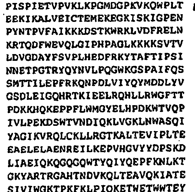
pB6 

PR_v1 3 
[1 297] 10 
pB27 GAG_v1 4 
[1 1503] 11 
pB6 

NC_v1 5 
[1 165] 12 
pB6 TM_v1 6 
[565 1038] 13 
pB6 VPU_v1 7 
[1 246] 14 
pB27 Table 2: bait-prey interactions 1: Bait name 2: Bait nucleic acid SEQ ID No. 3: Bait construct ion 4: Prey name 5: Prey construction IN 1 pB27 prey024555 - Human VBP1 dT IN 1 pB27 prey024555 - Human VBP1 RP IN 1 pB27 prey007766 - Human TRN-SR RP IN 1 pB27 prey024605 - Human RNUT1 RP IN 1 pB27 prey001626 - Human HBOA dT IN 1 pB27 prey001626 - Human HBOA RP IN 1 pB27 prey007151 - Human MCM7 dT IN 1 pB27 prey024567 - Human EIF3S3 dT IN 1 pB27 prey024S67 - Human EIF3S3 RP IN 1 pB27 prey000022 - Human PIASY RP RT_v1 2 pB27 prey026178 - Human AKAP1 RP RT_v1 2 pB27 prey026784 - Human ELAVL1 RP PR_v1 3 pB27 prey47239 (CSNK2B) hCSNK2B Human CEMC7 Random Primed PR_v1 3 pB27 prey030612 - Human AIM1 dT PR_v1 3 pB27 prey030612 - Human AIM1 RP PR_v1 3 pB27 prey030679 - Human UBE1 dT GAG_v1 4 pB6 prey17662 (BAP1 KIAA0272; prey17663; prey17658) hBAP1 hBAP1 hhucep 6 Human CEMC7 Random Primed NC_v1 5 pB6 prey145885 (BAP1 HUCEP 6 HUCEP 13 KIAA0272) hBAP1 Human CEMC7 Random Primed TM_v1 6 pB6 prey34104 (ATF6; prey34106) hATF6 Human CEMC7 Random Primed TM_v1 6 pB6 hgx33 hsterol regulatory element bindingprotein 2 hSREBF2 Human CEMC7 Random Primed TM_v1 6 pB6 prey15532 (SREBF1 SREBP1; prey15533) hSREBF1 hSREBP 1 Human CEMC7 Random Primed VPU_v1 7 pB27 prey6634 (PCBP1 HNRPE1 hnRNP E1 HNRPX hnRNP X; prey6635) hPCBP1 hhnRNP E1 Human CEMC7 dT Primed VPU_v1 7 pB27 prey6634 (PCBP1 HNRPE1 hnRNP E1 HNRPX hnRNP X; prey6635) hPCBP1 hhnRNP E1 Human CEMC7 Random Primed VPU_v1 7 pB27 prey7766 (TRN_SR TRN SR2 MTR10A; prey7769) hTRN_SR hMtr10a Human CEMC7 Random Primed Table 3: SID® 1: Bait name 2: Bait nucleic acid SEQ ID No. 3: Prey name 4: SID nucleic acid ID No. 5: SID nucleic acid sequence 6: SID amino acid ID No. 7: SID amino acid sequence IN 1 prey0245 55 Human VBP1 15 
38 
IN 1 prey0245 55 Human VBP1 16 
39 
IN 1 prey0077 66 Human TRN SR 17 
40 


IN 1 prey0246 05 Human RNUT1 18 
41 
IN 1 prey0016 26 Human HBOA 19 
42 
IN 1 prey0016 26 Human HBOA 20 
43 
IN 1 prey0071 51 Human MCM7 21 
44 
IN 1 prey0245 67 Human EIF3S3 22 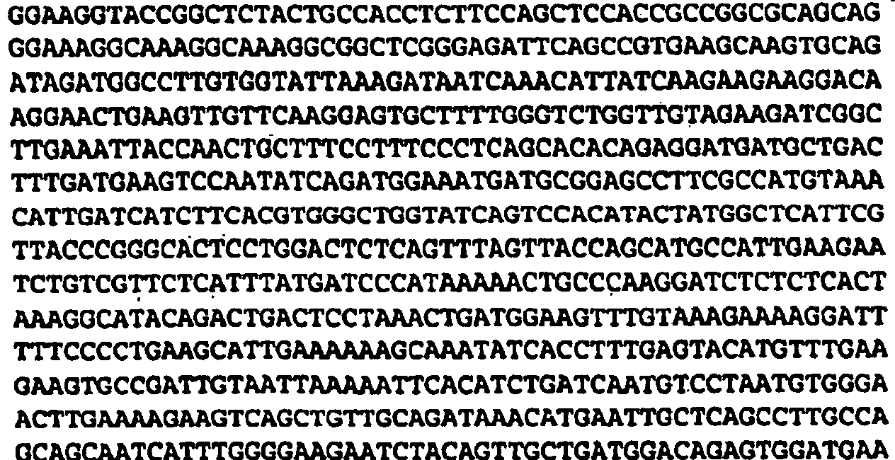
45 


IN 1 prey0245 67 . Human BIF3S3 23 
46 
IN 1 prey0000 22 Human PIASY24 
47 
RT 2 prey0267 78 Human AKAP1 25 
48 


RT 2 prey0267 84 Human ELAVL1 26 
49 
PR_v1 3 prey4723 927 
50 
PR 3 prey0306 12 Human AIM1 28 
51 
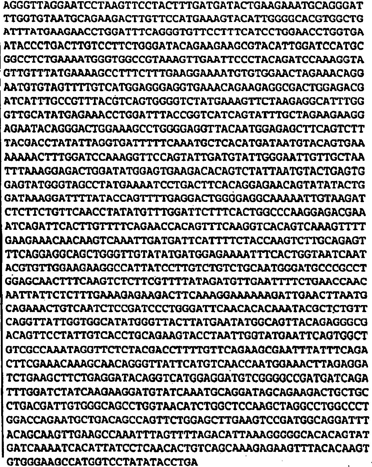

PR_v1 3 prey3061 229 
52 


PR 3 prey0306 79 Human UBE1 30 
53 
GAG-v1 4 . prey1766 231 
54 
HC_v1 5 prey1458 85 32 
55 
TM_v1 6 prey3410 4 33 
56 
TM_v1 6 hgx33 34 
57 


VPU_v1 7 prey6634 35 
58 
VPU_v1 7 prey6634 36 
59 
VPU_v1 7 prey7766 37 
60 
-
- Brennan, C. M. and J. A. Steitz (2001). "HuR and mRNA stability." Cell Mol Life Sci 58(2): 266-77.
- Brown, M. S., J. Ye, et al. (2000). "Regulated intramembrane proteolysis: a control mechanism conserved from bacteria to humans." Cell 100(4): 391-8.
- Bukrinskaya, A., B. Brichacek, et al. (1998). "Establishment of a functional human immunodeficiency virus type 1 (HIV-1) reverse transcription complex involves the cytosketeton." J Exp Med 188(11): 2113-25.
- Caron, M., M. Auclair, et al. (2001). "The HIV protease inhibitor indinavir impairs sterol regulatory element-binding protein-1 intranuclear localization, inhibits preadipocyte differentiation, and induces insulin resistance." Diabetes 50(6): 1378-88.
- Cherepanov, P., G. Maertens, et al. (2002). "HIV-1 integrase forms stable tetramers and associates with LEDGF/p75 protein in human cells." J Biol Chem 28: 28.
- Diaz, E. and S. R. Pfeffer (1998). "TIP47: a cargo selection device for mannose 6-phosphate receptor trafficking." Cell 93(3): 433-43.
- Elbashir, S. M., J. Harborth, et al. (2001). °Duplexes of 21-nucleotide RNAs mediate RNA interference in cultured mammalian cells." Nature 411 (6836): 494-8.
- Freed, E. O. and M. A. Martin (1994). "HIV-1 infection of non-dividing cells." Nature 369(6476): 107-8.
- Fujita, M., T. Kiyono, et al. (1996). "hCDC47, a human member of the MCM family. Dissociation of the nucleus-bound form during S phase." J Biol Chem 271(8): 4349-54.
- Garrus, J. E., U. K. von Schwedler, et al. (2001). "Tsg101 and the vacuolar protein sorting pathway are essential for HIV-1 budging." Cell 107(1): 55-65.
- Ge, H., Y. Si, et al. (1998). "Isolation of cDNAs encoding novel transcription coactivators p52 and p75 reveals an alternate regulatory mechanism of transcriptional activation." Embo J 17(22): 6723-9.
- Ge, H., Y. Si, et al. (1998). "A novel transcriptional coactivator, p52, functionally interacts with the essential splicing factor ASF/SF2." Mol Cell 2(6): 751-9.
- Gong, L. and E. T. Yeh (1999). "Identification of the activating and conjugating enzymes of the NEDD8 conjugation pathway." J Biol Chem 274(17): 12036-42.
- Graf, M., A. Bojak, et al. (2000). "Concerted action of muttiple cis-acting sequences is required for Rev dependence of late human .
- Greene, W. C. and B. M. Peterlin (2002). "Charting HIV's remarkable voyage through the cell: Basic science as a passport to future therapy." Nat Med 8(7): 673-80.
- Haneda, E., T. Furuya, et al. (2000). "Biochemical characterization of casein kinase II as a protein kinase responsible for stimulation of HIV-1 protease in vitro." Biochem Biophys Res Commun 275(2): 434-9.
- Hartl, F. U. and M. Hayer-Hartl (2002). "Molecular chaperones in the cytosol: from nascent chain to folded protein." Science 295(5561): 1852-8.
- Haze, K, H. Yoshida, et al. (1999). "Mammalian transcription factor ATF6 is synthesized as a transmembrane protein and activated by proteolysis in response to endoplasmic reticulum stress." Mol Biol Cell 10(11):3787-99.
- Herberg, F. W., A. Maleszka, et al. (2000). "Analysis of A-klnase anchoring protein (AKAP) interaction with protein kinase A (PKA) regulatory subunits: PKA isoform specificity in AKAP binding." J Mol Biol 298(2): 329-39.
- Hoppe, T., M. Rape, et al. (2001). "Membrane-bound transcription factors: regulated release by RIP or RUP." Curr Opin Cell Biol 13(3): 344-8.
- Horton, J. D., J. L. Goldstein, et al. (2002). "SREBPs: activator of the complete program of cholesterol and fatty acid synthesis in the liver." J Clin Invest 109(9):1125-31.
- Huber, J., U. Cronshagen, et al. (1998). "Snurportin1, an m3G-cap-specific nuclear import receptor with a novel domain structure." Embo J 17(14):4114-26.
- lizuka, M. and B. Stillman (1999). "Histone acetyltransferase HBO1 interacts with the ORC1 subunit of the human initiator protein." J Biol Chem 274(33): 23027-34.
- Jensen, D. E., M. Proctor, et al. (1998). "BAP1: a novel ubiquitin hydrolase which binds to the BRCA1 RING finger and enhances BRCA1-mediated cell growth suppression." Oncogene 16(9):1097-112.
- Kalpana, G. V., S. Marmon, et al., (1994). "Binding and stimulation of HIV-1 integrase by a human homolog of yeast transcription factor SNF5." Science 266(5193):2002-6.
- Kataoka, N., J. L. Bachorik, et al. (1999). "Transportin-SR, a nuclear import receptor for SR proteins." J Cel Biol 145(6): 1145-52.
- Leffers, H., K. Dejgaard, et al. (1995). "Characterisation of two major cellular poly(rC)-binding human proteins, each containing three K-homologous (KH) domains." Eur J Biochem 230(2): 447-53.
- Margottin, F., S. P. Bour, et al. (1998). "A novel human WD protein, h-beta TrCp, that interacts with HIV-1 Vpu connects CD4 to the ER degradation pathway through an F-box motif." Mol Cell 1(4): 565-74.
- Nair, V. (2002). "HIV integrase as a target for antiviral chemotherapy." Rev Med Virol 12(3):179-93.
- Olsen, H. S., A. W. Cochrane, et al. (1992). "Interaction of cellular factors with intragenic cis-acting repressive sequences within the HIV genome." Virology 191(2):709-15.
- Paraskeva, E., E. Izaurralde, et al. (1999). "CRM1-mediated recycling of .
- Ratner, L., W. Haseltine, et al. (1985). "Complete nucleotide sequence of the AIDS virus, HTLV-III." Nature 313(6000):277-84.
- Ray, M. E., G. Wistow, et al. (1997). "AlM1, a novel non-lens member of the betagamma-crystallin superfamily, is associated with the control of tumorigenicity in human malignant melanoma." Proc-Natl Acad Sci U S A 94(7): 3229-34.
- Sachdev, S., L Bruhn, et al. (2001). "PIASy, a nuclear matrix-associated SUMO E3 ligase, represses LEF1 activity by sequestration into nuclear bodices." Genes Dev 15(23): 3088-103.
- Schwartz, S., B. K. Felber, et al. (1992). "Distinct RNA sequences in the gag region of human .
- Singh, D. P., N. Fatma, et al. (2001). "LEDGF binds to heat shock and stress-related element to activate the expression of stress-related genes." Biochem Biophys Res Commun 283(4): 943-55.
- Singh, D. P., A. Kimura, et al. (2000). "Lens epithelium-derived growth factor (LEDGF/p75) and p52 are derived from a single gene by alternative spliang." Gene 242(1-2): 265-73.
- Trendelenburg, G., M. Hummel, et al. (1996). "Molecular characterization of AKAP149, a novel A kinase anchor protein with a KH domain." Biochem Biophys Res Commun 225(1):313-9.
- Turelli, P., V. Doucas, et al. (2001). "Cytoplasmic recruitment of INI1 and PML on incoming HIV preintegration complexes: interference with early steps of viral replication." Mol Cell 7(6):1245-54.
- Vainberg, I. E., S. A. Lewis, et al. (1998). "Prefoldin, a chaperone that delivers unfolded proteins to cytosolic chaperonin." Cell 93(5): 863-73.
- Ye, J., R. B. Rawson, et al. (2000). "ER stress induces cleavage of membrane-bound ATF6 by the same proteases that process SREBPs." Mol Cell 6(6): 1355-64.
-
- <110> Cellvir
- <120> Protein-protein interactions in Human Immunodeficiency Virus
- <130> B5055ACA - JAZ/LV/KN
- <140> EP N° 08/
<141> 31-03-2008 - <150>
US 60/333,346
US 60/385,132 - <151> 26-11-2001
31-05-2002 - <160> 132
- <170> PatentIn version 3.3
- <210> 1
<211> 860
<212> DNA
<213> HIV - <400> 1
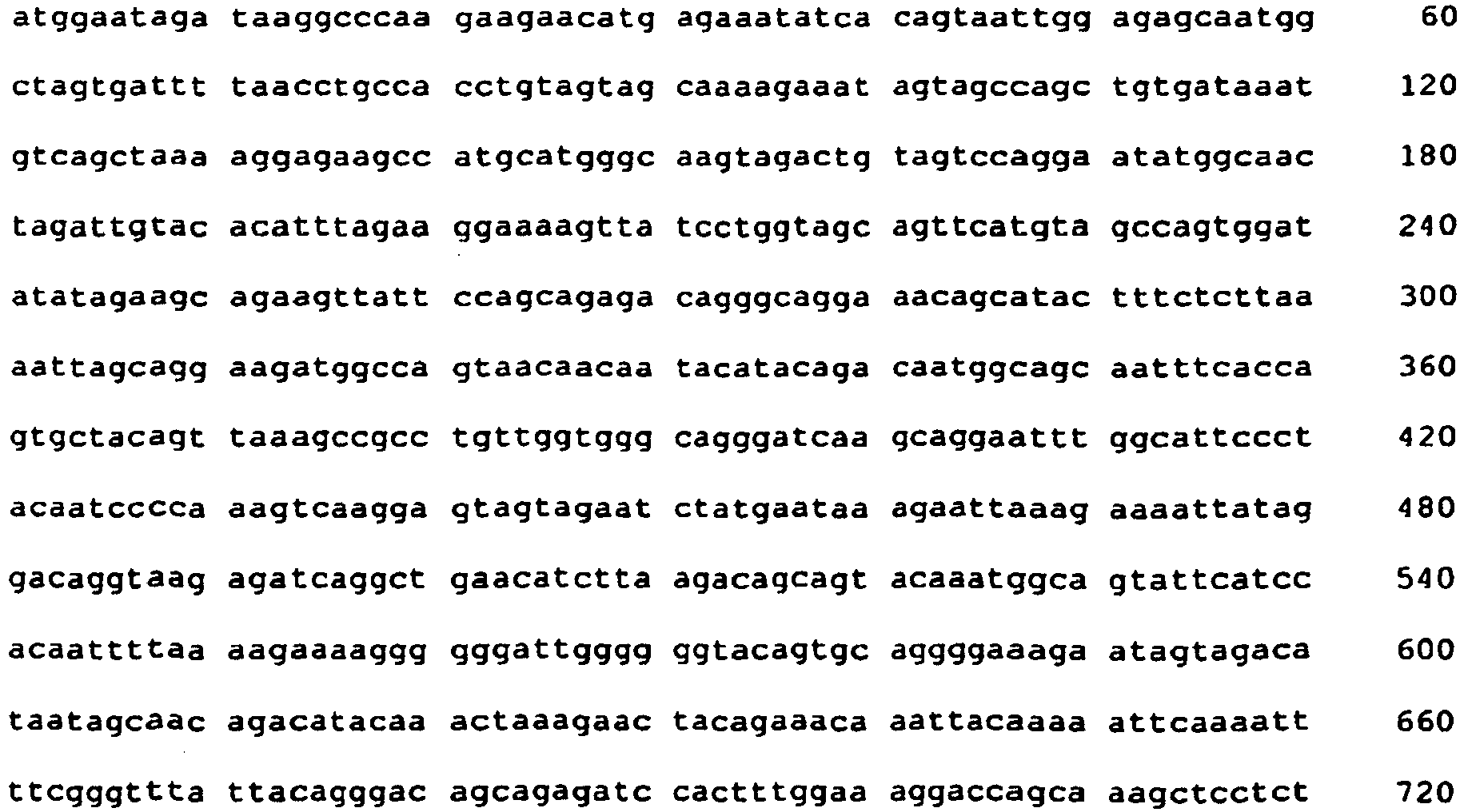

- <210> 2
<211> 1680
<212> DNA
<213> HIV - <400> 2


- <210> 3
<211> 297
<212> DNA
<213> HIV - <400> 3

- <210> 4
<211> 1503
<212> DNA
<213> HIV - <400> 4


- <210> 5
<211> 165
<212> DNA
<213> HIV - <400> 5

- <210> 6
<211> 474
<212> DNA
<213> HIV - <400> 6


- <210> 7
<211> 246
<212> DNA
<213> HIV - <400> 7

- <210> 8
<211> 288
<212> PRT
<213> HIV - <400> 8


- <210> 9
<211> 560
<212> PRT
<213> HIV - <220>
<223> Translation of SEQ ID N°2 - <400> 9


- <210> 10
<211> 99
<212> PRT
<213> HIV - <220>
<223> Translation of SEQ ID N°3 - <400> 10

- <210> 11
<211> 500
<212> PRT
<213> HIV - <220>
<223> Translation of SEQ ID N°4 - <400> 11


- <210> 12
<211> 55
<212> PRT
<213> HIV - <220>
<223> Translation of SEQ ID N°5 - <400> 12

- <210> 13
<211> 157
<212> PRT
<213> HIV - <220>
<223> Translation of SEQ ID N°6 - <400> 13


- <210> 14
<211> 81
<212> PRT
<213> HIV - <220>
<223> Translation of SEQ ID N°7 - <400> 14


- <210> 15
<211> 574
<212> DNA
<213> Homosapiens - <400> 15

- <210> 16
<211> 594
<212> DNA
<213> Homosapiens - <400> 16


- <210> 17
<211> 817
<212> DNA
<213> Homosapiens - <400> 17

- <210> 18
<211> 708
<212> DNA
<213> Homosapiens - <400> 18


- <210> 19
<211> 841
<212> DNA
<213> Homosapiens - <400> 19

- <210> 20
<211> 806
<212> DNA
<213> Homosapiens - <400> 20


- <210> 21
<211> 412
<212> DNA
<213> Homosapiens - <400> 21

- <210> 22
<211> 1045
<212> DNA
<213> Homosapiens - <400> 22


- <210> 23
<211> 908
<212> DNA
<213> Homosapiens - <400> 23


- <210> 24
<211> 454
<212> DNA
<213> Homosapiens - <400> 24

- <210> 25
<211> 813
<212> DNA
<213> Homosapiens - <400> 25


- <210> 26
<211> 124
<212> DNA
<213> Homosapiens - <400> 26

- <210> 27
<211> 181
<212> DNA
<213> Homosapiens - <400> 27

- <210> 28
<211> 2890
<212> DNA
<213> Homosapiens - <400> 28


- <210> 29
<211> 683
<212> DNA
<213> Homosapiens - <400> 29

- <210> 30
<211> 394
<212> DNA
<213> Homosapiens - <400> 30

- <210> 31
<211> 469
<212> DNA
<213> Homosapiens - <400> 31

- <210> 32
<211> 208
<212> DNA
<213> Homosapiens - <400> 32

- <210> 33
<211> 388
<212> DNA
<213> Homosapiens - <400> 33

- <210> 34
<211> 583
<212> .DNA
<213> Homosapiens - <400> 34

- <210> 35
<211> 775
<212> DNA
<213> Homosapiens - <400> 35


- <210> 36
<211> 144
<212> DNA
<213> Homosapiens - <400> 36

- <210> 37
<211> 306
<212> DNA
<213> Homosapiens - <400> 37

- <210> 38
<211> 190
<212> PRT
<213> Homosapiens - <220>
<223> Translation of SEQ ID N°15 - <400> 38

- <210> 39
<211> 197
<212> PRT
<213> Homosapiens - <220>
<223> Translation of SEQ ID N°16 - <400> 39

- <210> 40
<211> 272
<212> PRT
<213> Homosapiens - <220>
<223> Translation of SEQ ID N°17 - <400> 40


- <210> 41
<211> 235
<212> PRT
<213> Homosapiens - <220>
<223> Translation of SEQ ID N°18 - <400> 41


- <210> 42
<211> 279
<212> PRT
<213> Homosapiens - <220>
<223> Translation of SEQ ID N°19 - <400> 42


- <210> 43
<211> 268
<212> PRT
<213> Homosapiens - <220>
<223> Translation of SEQ ID N°20 - <400> 43


- <210> 44
<211> 136
<212> PRT
<213> Homosapiens - <220>
<223> Translation of SEQ ID N°21 - <400> 44

- <210> 45
<211> 347
<212> PRT
<213> Homosapiens - <220>
<223> Translation of SEQ ID N°22 - <400> 45


- <210> 46
<211> 302
<212> PRT
<213> Homosapiens - <220>
<223> Translation of SEQ ID N°23 - <400> 46


- <210> 47
<211> 151
<212> PRT
<213> Homosapiens - <220>
<223> Translation of SEQ ID N°24 - <400> 47

- <210> 48
<211> 270
<212> PRT
<213> Homosapiens - <220>
<223> Translation of SEQ ID N°25 - <400> 48


- <210> 49
<211> 40
<212> PRT
<213> Homosapiens - <220>
<223> Translation of SEQ ID N°26 - <400> 49

- <210> 50
<211> 60
<212> PRT
<213> Homosapiens - <220>
<223> Translation of SEQ ID N°27 - <400> 50

- <210> 51
<211> 962
<212> PRT
<213> Homosapiens - <220>
<223> Translation of SEQ ID N°28 - <400> 51
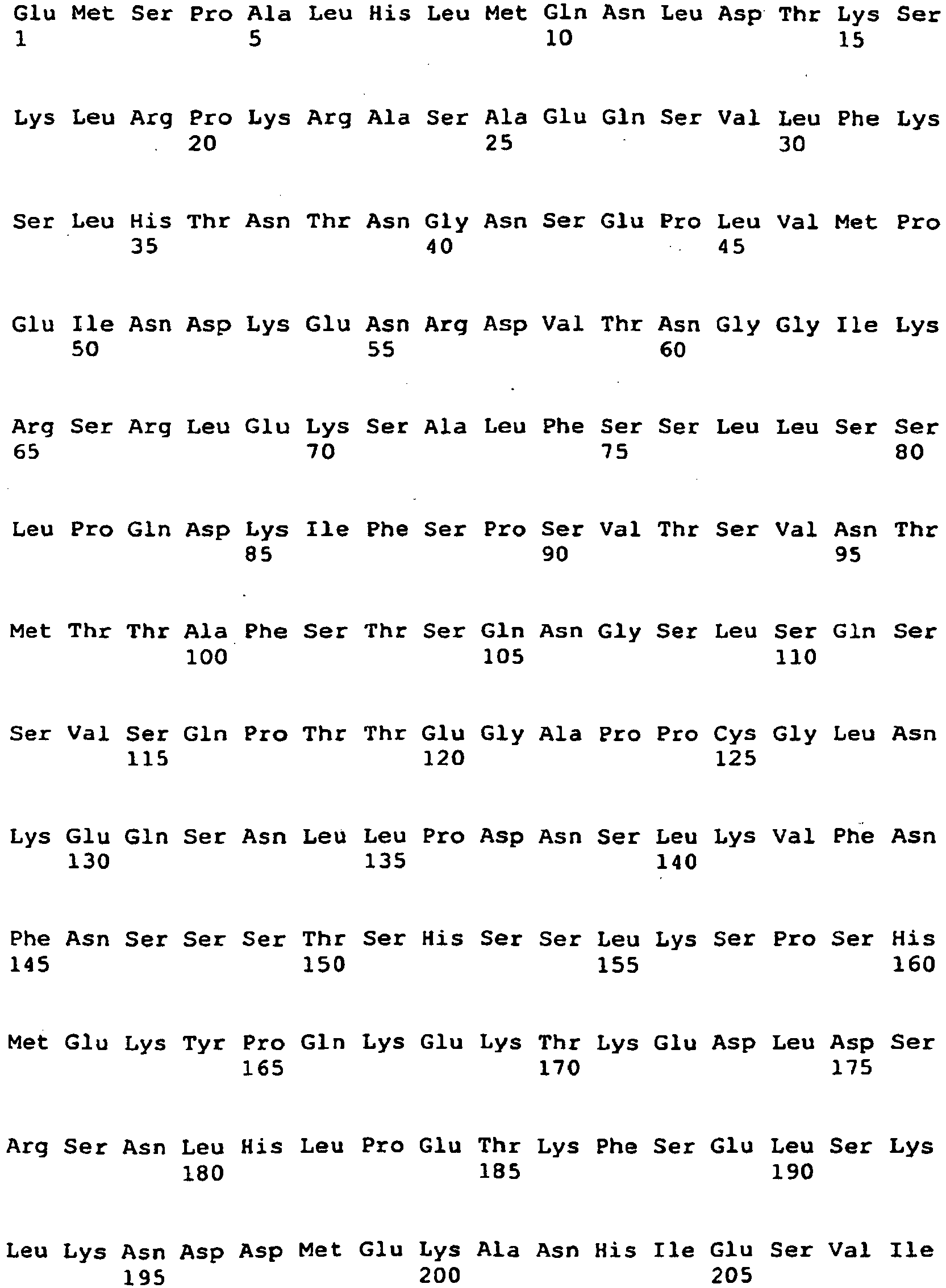


- <210> 52
<211> 227
<212> PRT
<213> Homosapiens - <220>
<223> Translation of SEQ ID N°29 - <400> 52


- <210> 53
<211> 130
<212> PRT
<213> Homosapiens - <220>
<223> Translation of SEQ ID N°30 - <400> 53


- <210> 54
<211> 155
<212> PRT
<213> Homosapiens - <220>
<223> Translation of SEQ ID N°31 - <400> 54

- <210> 55
<211> 68
<212> PRT
<213> Homosapiens - <220>
<223> Translation of SEQ ID N°32 - <400> 55

- <210> 56
<211> 129
<212> PRT
<213> Homosapiens - <220>
<223> Translation of SEQ ID N°33 - <400> 56


- <210> 57
<211> 194
<212> PRT
<213> Homosapiens - <220>
<223> Translation of SEQ ID N°34 - <400> 57


- <210> 58
<211> 257
<212> PRT
<213> Homosapiens - <220>
<223> Translation of SEQ ID N°35 - <400> 58


- <210> 59
<211> 47
<212> PRT
<213> Homosapiens - <220>
<223> Translation of SEQ ID N°36 - <400> 59


- <210> 60
<211> 101
<212> PRT
<213> Homosapiens - <220>
<223> Translation of SEQ ID N°37 - <400> 60

- <210> 61
<211> 36
<212> DNA
<213> Artificial - <400> 61
tcccccgggc ggaggctaga aggagagaga tgggtg 36 - <210> 62
<211> 37
<212> DNA
<213> artificial - <400> 62
tcccccgggg ctctaggtta ggatctactg gctccat 37 - <210> 63
<211> 10
<212> DNA
<213> artificial - <400> 63
gggccacgaa 10 - <210> 64
<211> 13
<212> DNA
<213> artificial - <400> 64
ttcgtggccc ctg 13 - <210> 65
<211> 16
<212> DNA
<213> artificial - <400> 65
atcccggacg aaggcc 16 - <210> 66
<211> 13
<212> DNA
<213> artificial - <400> 66
ggccttcgtc cgg 13 - <210> 67
<211> 67
<212> DNA
<213> artificial - <400> 67

- <210> 68
<211> 68
<212> DNA
<213> artificial - <400> 68

- <210> 69
<211> 67
<212> DNA
<213> artificial - <400> 69

- <210> 70
<211> 69
<212> DNA
<213> artificial - <400> 70

- <210> 71
<211> 68
<212> DNA
<213> artificial - <400> 71

- <210> 72
<211> 68
<212> DNA
<213> artificial - <400> 72

- <210> 73
<211> 76
<212> DNA
<213> artificial - <400> 73

- <210> 74
<211> 68
<212> DNA
<213> artificial - <400> 74

- <210> 75
<211> 20
<212> DNA
<213> artificial - <400> 75
gcgtttggaa tcactacagg 20 - <210> 76
<211> 19
<212> DNA
<213> artificial - <400> 76
cacgatgcac gttgaagtg 19 - <210> 77
<211> 21
<212> DNA
<213> artificial - <220>
<221> misc_RNA
<222> (1)..(21)
<223> siRNA TT complex protect against RNAse degradation - <400> 77
ggagugcuuu ugggucuggt t 21 - <210> 78
<211> 21
<212> DNA
<213> artificial - <220>
<221> misc_RNA
<222> (1)..(21)
<223> siRNA TT complex protect against RNAse degradation - <400> 78
ccagacccaa aagcacucct t 21 - <210> 79
<211> 21
<212> DNA
<213> artificial - <220>
<221> misc_RNA
<222> (1)..(21)
<223> si RNA tt complex protect against RNAse degradation - <400> 79
gugauggcac aucccgacgt t 21 - <210> 80
<211> 21
<212> DNA
<213> artificial - <220>
<221> misc_RNA
<222> (1)..(21)
<223> siRNA TT complex protect against RNAse degradation - <400> 80
cgucgggaug ugccaucact t 21 - <210> 81
<211> 21
<212> DNA
<213> artificial - <220>
<221> misc_RNA
<222> (1)..(21)
<223> siRNA TT complex protect against RNAse degradation - <400> 81
guuccugaug gagcuguaat t 21 - <210> 82
<211> 21
<212> DNA
<213> artificial - <220>
<221> misc_RNA
<222> (1)..(21)
<223> siRNA TT complex protect against RNAse degradation - <400> 82
uuacagcucc aucaggaact t 21 - <210> 83
<211> 21
<212> DNA
<213> artificial - <220>
<221> misc_RNA
<222> (1)..(21)
<223> siRNA TT complex protect against RNAse degradation - <400> 83
gaagcaguuc aaguaugggt t 21 - <210> 84
<211> 21
<212> DNA
<213> artificial - <220>
<221> misc_RNA
<222> (1)..(21)
<223> siRNA TT complex protect against RNAse degradation - <400> 84
cccauacuug aacugcuuct t 21 - <210> 85
<211> 21
<212> DNA
<213> artificial - <220>
<221> misc_RNA
<222> (1)..(21)
<223> siRNA TT complex protect against RNAse degradation - <400> 85
ccaugccaga agacuggcut t 21 - <210> 86
<211> 21
<212> DNA
<213> artificial - <220>
<221> misc RNA
<222> (1)..(21)
<223> siRNA TT complex protect against RNAse degradation - <400> 86
agccagucuu cuggcauggt t 21 - <210> 87
<211> 21
<212> DNA
<213> artificial - <220>
<221> misc_RNA
<222> (1)..(21)
<223> siRNA TT complex protect against RNAse degradation - <400> 87
ggagcgcgcc ucuuuuuggt t 21 - <210> 88
<211> 21
<212> DNA
<213> artificial - <220>
<221> misc_RNA
<222> (1)..(21)
<223> siRNA TT complex protect against RNAse degradation - <400> 88
ccaaaaagag gcgcgcucct t 21 - <210> 89
<211> 21
<212> DNA
<213> artificial - <220>
<221> misc_RNA
<222> (1)..(21)
<223> siRNA TT complex protect against RNAse degradation - <400> 89
ccuccagucu ucucucguct t 21 - <210> 90
<211> 21
<212> DNA
<213> artificial - <220>
<221> misc_RNA
<222> (1)..(21)
<223> siRNA TT complex protect against RNAse degradation - <400> 90
gacgagagaa gacuggaggt t 21 - <210> 91
<211> 21
<212> DNA
<213> artificial - <220>
<221> misc_RNA
<222> (1)..(21)
<223> siRNA TT complex protect against RNAse degradation - <400> 91
cagccuggga augagacugt t 21 - <210> 92
<211> 21
<212> DNA
<213> artificial - <220>
<221> misc_RNA
<222> (1)..(21)
<223> siRNA TT complex protect against RNAse degradation - <400> 92
cagucucauu cccaggcugt t 21 - <210> 93
<211> 21
<212> DNA
<213> artificial - <220>
<221> misc_RNA
<222> (1)..(21)
<223> siRNA TT complex protect against RNAse degradation - <400> 93
gaaagugaaa ucuccgcggt t 21 - <210> 94
<211> 21
<212> DNA
<213> artificial - <220>
<221> misc_RNA
<222> (1)..(21)
<223> siRNA TT complex protect against RNAse degradation - <400> 94
ccgcggagau uucacuuuct t 21 - <210> 95
<211> 21
<212> DNA
<213> artificial - <220>
<221> misc_RNA
<222> (1)..(21)
<223> siRNA TT complex protect against RNAse degradation - <400> 95
ggaaccucuc cccguggaat t 21 - <210> 96
<211> 21
<212> DNA
<213> artificial - <220>
<221> misc_RNA
<222> (1)..(21)
<223> siRNA TT complex protect against RNAse degradation - <400> 96
uuccacgggg agagguucct t 21 - <210> 97
<211> 21
<212> DNA
<213> artificial - <220>
<221> misc_RNA
<222> (1)..(21)
<223> siRNA TT complex protect against RNAse degradation - <400> 97
ugagacguau gaaaacaaut t 21 - <210> 98
<211> 21
<212> DNA
<213> artificial - <220>
<221> misc_RNA
<222> (1)..(21)
<223> siRNA TT complex protect against RNAse degradation - <400> 98
auuguuuuca uacgucucat t 21 - <210> 99
<211> 21
<212> DNA
<213> artificial - <220>
<221> misc_RNA
<222> (1)..(21)
<223> siRNA TT complex protect against RNAse degradation - <400> 99
guggaggaga ucuacgacct t 21 - <210> 100
<211> 21
<212> DNA
<213> artificial - <220>
<221> misc_RNA
<222> (1)..(21)
<223> siRNA TT complex protect against RNAse degradation - <400> 100
ggucguagau cuccuccact t 21 - <210> 101
<211> 21
<212> DNA
<213> artificial - <220>
<221> misc_RNA
<222> (1)..(21)
<223> siRNA TT complex protect against RNAse degradation - <400> 101
gaacuggaag acaaccccat t 21 - <210> 102
<211> 21
<212> DNA
<213> artificial - <220>
<221> misc_RNA
<222> (1)..(21)
<223> siRNA TT complex protect against RNAse degradation - <400> 102
ugggguuguc uuccaguuct t 21 - <210> 103
<211> 21
<212> DNA
<213> artificial - <220>
<221> misc_RNA
<222> (1)..(21)
<223> siRNA TT complex protect against RNAse degradation - <400> 103
gccuguucag cagcauuggt t 21 - <210> 104
<211> 21
<212> DNA
<213> artificial - <220>
<221> misc_RNA
<222> (1)..(21)
<223> siRNA TT complex protect against RNAse degradation - <400> 104
ccaaugcugc ugaacaggct t 21 - <210> 105
<211> 21
<212> DNA
<213> artificial - <220>
<221> misc_RNA
<222> (1)..(21)
<223> siRNA TT complex protect against RNAse degradation - <400> 105
cuagagaucc cucagaccct t 21 - <210> 106
<211> 21
<212> DNA
<213> artificial - <220>
<221> misc_RNA
<222> (1)..(21)
<223> siRNA TT complex protect against RNAse degradation - <400> 106
gggucugagg gaucucuagt t 21 - <210> 107
<211> 21
<212> DNA
<213> artificial - <220>
<221> misc_RNA
<222> (1)..(21)
<223> siRNA TT complex protect against RNAse degradation - <400> 107
cguacgcgga auacuucgat t 21 - <210> 108
<211> 21
<212> DNA
<213> artificial - <220>
<221> misc_RNA
<222> (1)..(21)
<223> siRNA TT complex protect against RNAse degradation - <400> 108
ucgaaguauu ccgcguacgt t 21 - <210> 109
<211> 21
<212> DNA
<213> artificial - <220>
<221> misc_RNA
<222> (1)..(21)
<223> siRNA TT complex protect against RNAse degradation - <400> 109
caaugacuua caaggcagct t 21 - <210> 110
<211> 21
<212> DNA
<213> artificial - <220>
<221> misc_RNA
<222> (1)..(21)
<223> siRNA TT complex protect against RNAse degradation - <400> 110
gcugccuugu aagucauugt t 21 - <210> 111
<211> 21
<212> DNA
<213> artificial - <220>
<221> misc_RNA
<222> (1)..(21)
<223> siRNA TT complex protect against RNAse degradation - <400> 111
gaguggacug aagcacgagt t 21 - <210> 112
<211> 21
<212> DNA
<213> artificial - <220>
<221> misc_RNA
<222> (1)..(21)
<223> siRNA TT complex protect against RNAse degradation - <400> 112
cucgugcuuc aguccacuct t 21 - <210> 113
<211> 21
<212> DNA
<213> artificial - <220>
<221> misc_RNA
<222> (1)..(21)
<223> siRNA TT complex protect against RNAse degradation - <400> 113
ggaaguagga agcaucauut t 21 - <210> 114
<211> 21
<212> DNA
<213> artificial - <220>
<221> misc_RNA
<222> (1)..(21)
<223> siRNA TT complex protect against RNAse degradation - <400> 114
aaugaugcuu ccuacuucct t 21 - <210> 115
<211> 21
<212> DNA
<213> artificial - <220>
<221> misc_RNA
<222> (1)..(21)
<223> siRNA TT complex protect against RNAse degradation - <400> 115
gagauacccc ucacucuggt t 21 - <210> 116
<211> 21
<212> DNA
<213> artificial - <220>
<221> misc_RNA
<222> (1)..(21)
<223> siRNA TT complex protect against RNAse degradation - <400> 116
ccagagugag ggguaucuct t 21 - <210> 117
<211> 21
<212> DNA
<213> artificial - <220>
<221> misc_RNA
<222> (1)..(21)
<223> siRNA TT complex protect against RNAse degradation - <400> 117
gacaugcuuc agcuuaucat t 21 - <210> 118
<211> 21
<212> DNA
<213> artificial - <220>
<221> misc_RNA
<222> (1)..(21)
<223> siRNA TT complex protect against RNAse degradation - <400> 118
ugauaagcug aagcauguct t 21 - <210> 119
<211> 21
<212> DNA
<213> artificial - <220>
<221> misc_RNA
<222> (1)..(21)
<223> siRNA TT complex protect against RNAse degradation - <400> 119
ucaaguggga gaguucccut t 21 - <210> 120
<211> 21
<212> DNA
<213> artificial - <220>
<221> misc_RNA
<222> (1)..(21)
<223> siRNA TT complex protect against RNAse degradation - <400> 120
agggaacucu cccacuugat t 21 - <210> 121
<211> 21
<212> DNA
<213> artificial - <220>
<221> misc_RNA
<222> (1)..(21)
<223> siRNA TT complex protect against RNAse degradation - <400> 121
ccaacggaau ggccaagaat t 21 - <210> 122
<211> 21
<212> DNA
<213> artificial - <220>
<221> misc_RNA
<222> (1)..(21)
<223> siRNA TT complex protect against RNAse degradation - <400> 122
uucuuggcca uuccguuggt t 21 - <210> 123
<211> 21
<212> DNA
<213> artificial - <220>
<221> misc_RNA
<222> (1)..(21)
<223> siRNA TT complex protect against RNAse degradation - <400> 123
gacugucugc gacgcagcat t 21 - <210> 124
<211> 21
<212> DNA
<213> artificial - <220>
<221> misc_RNA
<222> (1)..(21)
<223> siRNA TT complex protect against RNAse degradation - <400> 124
ugcugcgucg cagacaguct t 21 - <210> 125
<211> 458
<212> DNA
<213> Homo sapiens - <400> 125


- <210> 126
<211> 152
<212> PRT
<213> Homo sapiens - <400> 126

- <210> 127
<211> 159
<212> DNA
<213> Homo sapiens - <400> 127

- <210> 128
<211> 52
<212> PRT
<213> Homo sapiens - <400> 128

- <210> 129
<211> 395
<212> DNA
<213> Homo sapiens - <400> 129


- <210> 130
<211> 131
<212> PRT
<213> Homo sapiens - <400> 130

- <210> 131
<211> 232
<212> DNA
<213> Homo sapiens - <400> 131

- <210> 132
<211> 77
<212> PRT
<213> Homo sapiens - <400> 132

Claims (23)
- A polypeptide consisting of sequence SEQ ID NO: 128.
- A variant of the polypeptide according to claim 1, wherein said variant is at least 95.0% identical to SEQ ID NO: 128, as calculated by the algorithm of Needleman and Wunsch.
- A polypeptide consisting of sequence SEQ ID NO: 40.
- A variant of the polypeptide according to claim 3, wherein said variant is at least 95.0% identical to SEQ ID NO: 40 as calculated by the algorithm of Needleman and Wunsch.
- A polynucleotide consisting of sequence SEQ ID NO: 127 or sequence SEQ ID NO: 17.
- A variant of the polynucleotide according to claim 5, wherein said variant is at least 95.0% identical to SEQ ID NO: 127 or SEQ ID NO: 17, as calculated by the algorithm of Needleman and Wunsch.
- A variant of the polynucleotide according to claim 5, wherein said nucleotide changes are silent and do not after the amino acid sequence encoded by the polynucleotide as defined in SEQ ID NO: 127 or SEQ ID NO: 17.
- A recombinant vector consisting of an HIV-1 or Transportin-SR polynucleotide sequence according to any one of claims 5 to 7 and the necessary elements for the transcription and translation of the HIV-1 or Transportin-SR polynucleotide sequence.
- A recombinant host cell containing the recombinant vector according to claim 8.
- A composition comprising the vector according to daim 8 or the recombinant host cell of claim 9.
- A pharmaceutical composition comprising the polypeptide of any one of claims 1 to 4, and optionally a pharmaceutically acceptable carrier or adjuvant.
- A pharmaceutical composition comprising a pharmaceutically effective amount of a recombinant vector as defined in any one of claims 8 to 9, optionally a pharmaceutically acceptable carrier or adjuvant.
- A pharmaceutical composition according to claim 12, wherein said vector includes the nucleotide sequence as defined in SEQ ID NO:17.
- A pharmaceutical composition according to any one of claims 11 to 13, for use in treating or preventing AIDS in a human or mammal.
- A pharmaceutical composition according to claim 11, for use in treating or preventing AIDS in a mammal, wherein said polypeptide is a polypeptide as defined in SEQ ID NO: 40 or a variant having at least 95.0% identity to SEQ ID NO: 40.
- A pharmaceutical composition according to any one of claims 11 to 13, for use in the inhibition of HIV replication in cells sensitive to HIV infection.
- Use of a polypeptide according to any one of claims 1 to 4, to screen molecules that inhibit type-1 human immunodeficiency virus.
- A complex between HIV integrase and Transportin-SR, wherein the HIV integrase has a selected interacting domain having a sequence comprising SEQ ID NO: 128, and Transportin-SR has a selected interacting domain having a sequence comprising SEQ ID NO: 40.
- A complex between HIV integrase and Transportin-SR according to claim 18, wherein the selected interacting domain of the HIV integrase comprises a sequence that is at least 95.0% identical to SEQ ID NO: 128.
- A complex between HIV integrase and Transportin-SR according to claim 18 or 19, wherein the selected interacting domain of the Transportin-SR comprises a sequence that is at least 95.0% identical to SEQ ID NO: 40.
- A siRNA directed against the transportin-SR gene for use in the treatment of HIV-1 infection.
- Use of a complex as defined in any one of claims 18 to 20 to isolate anti-HIV molecules able to disrupt the transportin-SR/integrase interaction.
- Use of a polypeptide as defined in any one of claims 1 to 4 to screen molecules that inhibit human immunodeficiency virus.
Applications Claiming Priority (3)
| Application Number | Priority Date | Filing Date | Title |
|---|---|---|---|
| US33334601P | 2001-11-26 | 2001-11-26 | |
| US38513202P | 2002-05-31 | 2002-05-31 | |
| EP02792922A EP1451214A2 (en) | 2001-11-26 | 2002-11-26 | Protein-protein interactions in human immunodeficiency virus |
Related Parent Applications (2)
| Application Number | Title | Priority Date | Filing Date |
|---|---|---|---|
| PCT/EP2002/013868 Previously-Filed-Application WO2003046176A2 (en) | 2001-11-26 | 2002-11-26 | Protein-protein interactions in human immunodeficiency virus |
| EP02792922A Division EP1451214A2 (en) | 2001-11-26 | 2002-11-26 | Protein-protein interactions in human immunodeficiency virus |
Publications (2)
| Publication Number | Publication Date |
|---|---|
| EP2014674A1 EP2014674A1 (en) | 2009-01-14 |
| EP2014674B1 true EP2014674B1 (en) | 2014-02-12 |
Family
ID=26988677
Family Applications (3)
| Application Number | Title | Priority Date | Filing Date |
|---|---|---|---|
| EP08006236.7A Expired - Lifetime EP2014674B1 (en) | 2001-11-26 | 2002-11-26 | Protein-protein interactions in human immunodeficiency virus |
| EP09178541.0A Expired - Lifetime EP2186820B1 (en) | 2001-11-26 | 2002-11-26 | Protein-protein interactions in human immunodeficiency virus |
| EP02792922A Withdrawn EP1451214A2 (en) | 2001-11-26 | 2002-11-26 | Protein-protein interactions in human immunodeficiency virus |
Family Applications After (2)
| Application Number | Title | Priority Date | Filing Date |
|---|---|---|---|
| EP09178541.0A Expired - Lifetime EP2186820B1 (en) | 2001-11-26 | 2002-11-26 | Protein-protein interactions in human immunodeficiency virus |
| EP02792922A Withdrawn EP1451214A2 (en) | 2001-11-26 | 2002-11-26 | Protein-protein interactions in human immunodeficiency virus |
Country Status (6)
| Country | Link |
|---|---|
| US (2) | US7763254B2 (en) |
| EP (3) | EP2014674B1 (en) |
| AU (1) | AU2002358631A1 (en) |
| CA (1) | CA2468326C (en) |
| ES (2) | ES2605445T3 (en) |
| WO (1) | WO2003046176A2 (en) |
Families Citing this family (18)
| Publication number | Priority date | Publication date | Assignee | Title |
|---|---|---|---|---|
| AU2002327531A1 (en) | 2001-08-22 | 2003-03-10 | Myriad Genetics, Inc | Therapeutic compositions and methods for treating viral infection |
| WO2004029246A2 (en) * | 2002-09-26 | 2004-04-08 | K.U.Leuven Research And Development | Integrase cofactor |
| US9879266B2 (en) | 2002-11-14 | 2018-01-30 | Thermo Fisher Scientific Inc. | Methods and compositions for selecting siRNA of improved functionality |
| US9719092B2 (en) | 2002-11-14 | 2017-08-01 | Thermo Fisher Scientific Inc. | RNAi targeting CNTD2 |
| JP2006507841A (en) | 2002-11-14 | 2006-03-09 | ダーマコン, インコーポレイテッド | Functional and ultrafunctional siRNA |
| US9771586B2 (en) | 2002-11-14 | 2017-09-26 | Thermo Fisher Scientific Inc. | RNAi targeting ZNF205 |
| US7612196B2 (en) | 2002-11-14 | 2009-11-03 | Dharmacon, Inc. | siRNA targeting cyclin-dependent kinase inhibitor 1B (p27, Kip1) (CDKN1B) |
| US9228186B2 (en) | 2002-11-14 | 2016-01-05 | Thermo Fisher Scientific Inc. | Methods and compositions for selecting siRNA of improved functionality |
| WO2006006948A2 (en) | 2002-11-14 | 2006-01-19 | Dharmacon, Inc. | METHODS AND COMPOSITIONS FOR SELECTING siRNA OF IMPROVED FUNCTIONALITY |
| US9719094B2 (en) | 2002-11-14 | 2017-08-01 | Thermo Fisher Scientific Inc. | RNAi targeting SEC61G |
| US10011836B2 (en) | 2002-11-14 | 2018-07-03 | Thermo Fisher Scientific Inc. | Methods and compositions for selecting siRNA of improved functionality |
| US9839649B2 (en) | 2002-11-14 | 2017-12-12 | Thermo Fisher Scientific Inc. | Methods and compositions for selecting siRNA of improved functionality |
| BRPI0504117A (en) * | 2005-09-05 | 2007-05-22 | Fundacao De Amparo A Pesquisa | epitopes, combination of epitopes, uses of epitopes or their combination, composition, uses of composition, anti-HIV-1 prophylactic vaccines, therapeutic vaccines, method for identifying epitopes and methods for treatment or prevention. |
| GB0801940D0 (en) * | 2008-02-01 | 2008-03-12 | Univ Leuven Kath | Inhibitors of lentiviral replication |
| EP2196453A1 (en) | 2008-12-10 | 2010-06-16 | Cellvir | Novel substituted aryl derivatives, their process of preparation and their therapeutical uses as anti-HIV agents |
| US9879317B2 (en) * | 2011-02-17 | 2018-01-30 | The United States Of America, As Represented By The Secretary, Department Of Health And Human Services, Centers For Disease Control And Prevention | Real-time PCR point mutation assays for detecting HIV-1 resistance to antiviral drugs |
| CN103540644B (en) * | 2012-07-10 | 2015-04-01 | 中国医学科学院病原生物学研究所 | Method for screening anti-HIV-1 (human immunodeficiency virus-1) medicament |
| WO2016168578A1 (en) * | 2015-04-17 | 2016-10-20 | Genisphere, Llc | siRNA INHIBITION OF HUMAN ANTIGEN R EXPRESSION FOR TREATMENT OF CANCER |
Citations (1)
| Publication number | Priority date | Publication date | Assignee | Title |
|---|---|---|---|---|
| WO2001024810A1 (en) * | 1999-10-05 | 2001-04-12 | Epimmune Inc. | Inducing cellular immune responses to human immunodeficiency virus-1 using peptide and nucleic acid compositions |
Family Cites Families (19)
| Publication number | Priority date | Publication date | Assignee | Title |
|---|---|---|---|---|
| US4980286A (en) | 1985-07-05 | 1990-12-25 | Whitehead Institute For Biomedical Research | In vivo introduction and expression of foreign genetic material in epithelial cells |
| US5283173A (en) | 1990-01-24 | 1994-02-01 | The Research Foundation Of State University Of New York | System to detect protein-protein interactions |
| CA2092323A1 (en) | 1990-10-01 | 1992-04-02 | George Y. Wu | Targeting viruses and cells for selective internalization by cells |
| WO1993014188A1 (en) | 1992-01-17 | 1993-07-22 | The Regents Of The University Of Michigan | Targeted virus |
| CA2133411A1 (en) | 1992-04-03 | 1993-10-14 | Alexander T. YOUNG | Gene therapy using targeted viral vectors |
| US6222024B1 (en) * | 1994-05-24 | 2001-04-24 | The Trustees Of Columbia University In The City Of New York | Nucleic acids encoding a human immunodeficiency virus type 1 (HIV-1) integrase interactor protein (INI-1) |
| US6309821B1 (en) * | 1996-05-16 | 2001-10-30 | Incyte Genomics, Inc. | DNA encoding a PAC10 human homolog |
| US6083762A (en) | 1996-05-31 | 2000-07-04 | Packard Instruments Company | Microvolume liquid handling system |
| CA2196496A1 (en) | 1997-01-31 | 1998-07-31 | Stephen William Watson Michnick | Protein fragment complementation assay for the detection of protein-protein interactions |
| US6333154B1 (en) | 1997-12-04 | 2001-12-25 | Institut Pasteur | Bacterial multi-hybrid system and applications thereof |
| US6187535B1 (en) | 1998-02-18 | 2001-02-13 | Institut Pasteur | Fast and exhaustive method for selecting a prey polypeptide interacting with a bait polypeptide of interest: application to the construction of maps of interactors polypeptides |
| EP1027608B1 (en) | 1998-07-30 | 2010-01-20 | Odyssey Thera, Inc. | Protein fragment complementation assays |
| AU2200900A (en) | 1998-12-22 | 2000-07-12 | Myriad Genetics, Inc. | Protein-protein interactions in neurodegenerative disorders |
| WO2000058473A2 (en) * | 1999-03-31 | 2000-10-05 | Curagen Corporation | Nucleic acids including open reading frames encoding polypeptides; 'orfx' |
| EP1173560A1 (en) | 1999-04-30 | 2002-01-23 | Hybrigenics S.A. | Collection of prokaryotic dna for two hybrid systems helicobacter pylori protein-protein interactions and application thereof |
| US6436703B1 (en) * | 2000-03-31 | 2002-08-20 | Hyseq, Inc. | Nucleic acids and polypeptides |
| WO2003087299A2 (en) * | 2002-04-05 | 2003-10-23 | Sunesis Pharmaceuticals, Inc. | Methods of characterizing molecular interaction sites on proteins |
| WO2004029246A2 (en) * | 2002-09-26 | 2004-04-08 | K.U.Leuven Research And Development | Integrase cofactor |
| GB2393440A (en) | 2002-09-26 | 2004-03-31 | Leuven K U Res & Dev | Inhibition of retroviral integrase |
-
2002
- 2002-11-26 AU AU2002358631A patent/AU2002358631A1/en not_active Abandoned
- 2002-11-26 ES ES09178541.0T patent/ES2605445T3/en not_active Expired - Lifetime
- 2002-11-26 EP EP08006236.7A patent/EP2014674B1/en not_active Expired - Lifetime
- 2002-11-26 CA CA2468326A patent/CA2468326C/en not_active Expired - Fee Related
- 2002-11-26 EP EP09178541.0A patent/EP2186820B1/en not_active Expired - Lifetime
- 2002-11-26 WO PCT/EP2002/013868 patent/WO2003046176A2/en not_active Ceased
- 2002-11-26 ES ES08006236.7T patent/ES2462740T3/en not_active Expired - Lifetime
- 2002-11-26 EP EP02792922A patent/EP1451214A2/en not_active Withdrawn
-
2004
- 2004-05-26 US US10/853,807 patent/US7763254B2/en not_active Expired - Fee Related
-
2008
- 2008-02-25 US US12/072,390 patent/US8197821B2/en not_active Expired - Fee Related
Patent Citations (1)
| Publication number | Priority date | Publication date | Assignee | Title |
|---|---|---|---|---|
| WO2001024810A1 (en) * | 1999-10-05 | 2001-04-12 | Epimmune Inc. | Inducing cellular immune responses to human immunodeficiency virus-1 using peptide and nucleic acid compositions |
Non-Patent Citations (3)
| Title |
|---|
| "Homo sapiens mRNA for nuclear transport receptor", EMBL, XP002502598 * |
| CYNTHIA GIBAS ET PER JAMBEK: "Introduction à la bioinformatique", O'REILLY, Paris, pages: 161 - 163 * |
| LI Y ET AL: "Molecular characterization of human immunodeficiency virus type 1 cloned directly from uncultured human brain tissue: identification of replication-competent and -defective viral genomes.", JOURNAL OF VIROLOGY AUG 1991 LNKD- PUBMED:1830110, vol. 65, no. 8, August 1991 (1991-08-01), pages 3973 - 3985, ISSN: 0022-538X * |
Also Published As
| Publication number | Publication date |
|---|---|
| CA2468326A1 (en) | 2003-06-05 |
| US7763254B2 (en) | 2010-07-27 |
| US20090004719A1 (en) | 2009-01-01 |
| EP2186820A1 (en) | 2010-05-19 |
| WO2003046176A3 (en) | 2003-11-27 |
| EP2186820B1 (en) | 2016-06-08 |
| US8197821B2 (en) | 2012-06-12 |
| US20060034860A1 (en) | 2006-02-16 |
| EP2014674A1 (en) | 2009-01-14 |
| ES2462740T3 (en) | 2014-05-26 |
| CA2468326C (en) | 2015-11-17 |
| AU2002358631A1 (en) | 2003-06-10 |
| WO2003046176A2 (en) | 2003-06-05 |
| ES2605445T3 (en) | 2017-03-14 |
| EP1451214A2 (en) | 2004-09-01 |
| AU2002358631A8 (en) | 2003-06-10 |
Similar Documents
| Publication | Publication Date | Title |
|---|---|---|
| US8197821B2 (en) | Human immunodeficiency virus integrase—Transportin—SR protein—protein interactions | |
| Bogerd et al. | The type I human T-cell leukemia virus (HTLV-I) Rex trans-activator binds directly to the HTLV-I Rex and the type 1 human immunodeficiency virus Rev RNA response elements. | |
| SK287890B6 (en) | Isolated polypeptide that binds to TRAF2 and modulates activity of NF-B, DNA sequence encoding its, pharmaceutical composition containing them and their use | |
| Richter et al. | Strategies for inhibiting function of HIV-1 accessory proteins: a necessary route to AIDS therapy? | |
| WO1996023899A1 (en) | Methods of selecting a random peptide that binds to a target protein | |
| US20090035853A1 (en) | Tsg101-gag interaction and use thereof | |
| Lapidot et al. | Insight into the mechanisms of aminoglycoside derivatives interaction with HIV‐1 entry steps and viral gene transcription | |
| SK109997A3 (en) | Eukaryotic initiation factor 5a (eif-5a) mutants | |
| WO1999027102A9 (en) | Cd4+ t-lymphocyte protease genes and inhibitors thereof | |
| JP2004533815A (en) | TSG101-GAG interaction and use thereof | |
| JP4927546B2 (en) | Interactive polypeptides containing heptapeptide patterns and cell permeation domains | |
| CA2254819A1 (en) | Inhibition of hiv-1 replication by antisense rna expression | |
| JP3143740B2 (en) | RNA with high affinity for HIV Tat protein | |
| US20070203325A1 (en) | Integrase-dirived HIV-inhibiting agents | |
| US20020182694A1 (en) | Novel gene and polypeptide which opposes the Fas pathway | |
| Tien et al. | Understanding regulation of HIV-1 protease precursor autoprocessing | |
| Soros | Graduate Department of Molecular and Medical Genetics | |
| WO2002098918A2 (en) | Novel targets for a therapeutic intervention in ebv related diseases | |
| Elkins | Study of the HIV-1 gene regulatory proteins TAT and REV by the two-hybrid system in yeast | |
| Luukkonen et al. | Reduced infectivity of human immunodeficiency virus type 1 produced in the presence of a truncated Gag protein containing p7 gag and p6 gag | |
| Rekosh et al. | Selection and characterization of human immunodeficiency virus type 1 mutants that are resistant to inhibition by the transdominant negative RevM10 protein | |
| Oufir et al. | Artificial 64‐residue HIV‐1 enhancer‐binding peptide is a potent inhibitor of viral replication in HIV‐1‐infected cells | |
| Stotland | Use of Retroviral Peptide Libraries for the Identification of Novel Cellular Targets of HIV-1 and the Discovery of Novel Inhibitors | |
| Soros | Modulation of HIV-1 Rev transport and function | |
| KR20160097076A (en) | Composition for preventing or treating for acquired immune deficiency syndrome comprising SRp30c protein or gene thereof |
Legal Events
| Date | Code | Title | Description |
|---|---|---|---|
| PUAI | Public reference made under article 153(3) epc to a published international application that has entered the european phase |
Free format text: ORIGINAL CODE: 0009012 |
|
| AC | Divisional application: reference to earlier application |
Ref document number: 1451214 Country of ref document: EP Kind code of ref document: P |
|
| AK | Designated contracting states |
Kind code of ref document: A1 Designated state(s): AT BE BG CH CY CZ DE DK EE ES FI FR GB GR IE IT LI LU MC NL PT SE SK TR |
|
| 17P | Request for examination filed |
Effective date: 20090515 |
|
| 17Q | First examination report despatched |
Effective date: 20090616 |
|
| AKX | Designation fees paid |
Designated state(s): AT BE BG CH CY CZ DE DK EE ES FI FR GB GR IE IT LI LU MC NL PT SE SK TR |
|
| RAP1 | Party data changed (applicant data changed or rights of an application transferred) |
Owner name: LABORATOIRE BIODIM |
|
| GRAP | Despatch of communication of intention to grant a patent |
Free format text: ORIGINAL CODE: EPIDOSNIGR1 |
|
| RAP1 | Party data changed (applicant data changed or rights of an application transferred) |
Owner name: INSERM-TRANSFERT SA Owner name: LABORATOIRE BIODIM Owner name: UNIVERSITE PARIS DESCARTES Owner name: CENTRE NATIONAL DE LA RECHERCHE SCIENTIFIQUE (CNRS |
|
| GRAS | Grant fee paid |
Free format text: ORIGINAL CODE: EPIDOSNIGR3 |
|
| GRAJ | Information related to disapproval of communication of intention to grant by the applicant or resumption of examination proceedings by the epo deleted |
Free format text: ORIGINAL CODE: EPIDOSDIGR1 |
|
| GRAP | Despatch of communication of intention to grant a patent |
Free format text: ORIGINAL CODE: EPIDOSNIGR1 |
|
| INTG | Intention to grant announced |
Effective date: 20130709 |
|
| GRAS | Grant fee paid |
Free format text: ORIGINAL CODE: EPIDOSNIGR3 |
|
| GRAP | Despatch of communication of intention to grant a patent |
Free format text: ORIGINAL CODE: EPIDOSNIGR1 |
|
| INTG | Intention to grant announced |
Effective date: 20130823 |
|
| GRAA | (expected) grant |
Free format text: ORIGINAL CODE: 0009210 |
|
| AC | Divisional application: reference to earlier application |
Ref document number: 1451214 Country of ref document: EP Kind code of ref document: P |
|
| AK | Designated contracting states |
Kind code of ref document: B1 Designated state(s): AT BE BG CH CY CZ DE DK EE ES FI FR GB GR IE IT LI LU MC NL PT SE SK TR |
|
| REG | Reference to a national code |
Ref country code: GB Ref legal event code: FG4D |
|
| REG | Reference to a national code |
Ref country code: CH Ref legal event code: EP |
|
| REG | Reference to a national code |
Ref country code: AT Ref legal event code: REF Ref document number: 652121 Country of ref document: AT Kind code of ref document: T Effective date: 20140215 |
|
| RAP2 | Party data changed (patent owner data changed or rights of a patent transferred) |
Owner name: LABORATOIRE BIODIM Owner name: CENTRE NATIONAL DE LA RECHERCHE SCIENTIFIQUE (CNRS Owner name: INSTITUT NATIONAL DE LA SANTE ET DE LA RECHERCHE M Owner name: UNIVERSITE PARIS DESCARTES |
|
| REG | Reference to a national code |
Ref country code: IE Ref legal event code: FG4D |
|
| REG | Reference to a national code |
Ref country code: DE Ref legal event code: R096 Ref document number: 60245995 Country of ref document: DE Effective date: 20140327 |
|
| REG | Reference to a national code |
Ref country code: ES Ref legal event code: FG2A Ref document number: 2462740 Country of ref document: ES Kind code of ref document: T3 Effective date: 20140526 |
|
| REG | Reference to a national code |
Ref country code: NL Ref legal event code: VDEP Effective date: 20140212 |
|
| REG | Reference to a national code |
Ref country code: AT Ref legal event code: MK05 Ref document number: 652121 Country of ref document: AT Kind code of ref document: T Effective date: 20140212 |
|
| PG25 | Lapsed in a contracting state [announced via postgrant information from national office to epo] |
Ref country code: NL Free format text: LAPSE BECAUSE OF FAILURE TO SUBMIT A TRANSLATION OF THE DESCRIPTION OR TO PAY THE FEE WITHIN THE PRESCRIBED TIME-LIMIT Effective date: 20140212 Ref country code: AT Free format text: LAPSE BECAUSE OF FAILURE TO SUBMIT A TRANSLATION OF THE DESCRIPTION OR TO PAY THE FEE WITHIN THE PRESCRIBED TIME-LIMIT Effective date: 20140212 Ref country code: FI Free format text: LAPSE BECAUSE OF FAILURE TO SUBMIT A TRANSLATION OF THE DESCRIPTION OR TO PAY THE FEE WITHIN THE PRESCRIBED TIME-LIMIT Effective date: 20140212 Ref country code: SE Free format text: LAPSE BECAUSE OF FAILURE TO SUBMIT A TRANSLATION OF THE DESCRIPTION OR TO PAY THE FEE WITHIN THE PRESCRIBED TIME-LIMIT Effective date: 20140212 Ref country code: CY Free format text: LAPSE BECAUSE OF FAILURE TO SUBMIT A TRANSLATION OF THE DESCRIPTION OR TO PAY THE FEE WITHIN THE PRESCRIBED TIME-LIMIT Effective date: 20140212 Ref country code: PT Free format text: LAPSE BECAUSE OF FAILURE TO SUBMIT A TRANSLATION OF THE DESCRIPTION OR TO PAY THE FEE WITHIN THE PRESCRIBED TIME-LIMIT Effective date: 20140612 |
|
| PG25 | Lapsed in a contracting state [announced via postgrant information from national office to epo] |
Ref country code: CZ Free format text: LAPSE BECAUSE OF FAILURE TO SUBMIT A TRANSLATION OF THE DESCRIPTION OR TO PAY THE FEE WITHIN THE PRESCRIBED TIME-LIMIT Effective date: 20140212 Ref country code: DK Free format text: LAPSE BECAUSE OF FAILURE TO SUBMIT A TRANSLATION OF THE DESCRIPTION OR TO PAY THE FEE WITHIN THE PRESCRIBED TIME-LIMIT Effective date: 20140212 Ref country code: EE Free format text: LAPSE BECAUSE OF FAILURE TO SUBMIT A TRANSLATION OF THE DESCRIPTION OR TO PAY THE FEE WITHIN THE PRESCRIBED TIME-LIMIT Effective date: 20140212 |
|
| REG | Reference to a national code |
Ref country code: DE Ref legal event code: R097 Ref document number: 60245995 Country of ref document: DE |
|
| PG25 | Lapsed in a contracting state [announced via postgrant information from national office to epo] |
Ref country code: SK Free format text: LAPSE BECAUSE OF FAILURE TO SUBMIT A TRANSLATION OF THE DESCRIPTION OR TO PAY THE FEE WITHIN THE PRESCRIBED TIME-LIMIT Effective date: 20140212 |
|
| PLBE | No opposition filed within time limit |
Free format text: ORIGINAL CODE: 0009261 |
|
| STAA | Information on the status of an ep patent application or granted ep patent |
Free format text: STATUS: NO OPPOSITION FILED WITHIN TIME LIMIT |
|
| 26N | No opposition filed |
Effective date: 20141113 |
|
| PGFP | Annual fee paid to national office [announced via postgrant information from national office to epo] |
Ref country code: ES Payment date: 20141124 Year of fee payment: 13 Ref country code: FR Payment date: 20141016 Year of fee payment: 13 |
|
| REG | Reference to a national code |
Ref country code: DE Ref legal event code: R097 Ref document number: 60245995 Country of ref document: DE Effective date: 20141113 |
|
| PGFP | Annual fee paid to national office [announced via postgrant information from national office to epo] |
Ref country code: IT Payment date: 20141113 Year of fee payment: 13 |
|
| REG | Reference to a national code |
Ref country code: DE Ref legal event code: R119 Ref document number: 60245995 Country of ref document: DE |
|
| PG25 | Lapsed in a contracting state [announced via postgrant information from national office to epo] |
Ref country code: LU Free format text: LAPSE BECAUSE OF FAILURE TO SUBMIT A TRANSLATION OF THE DESCRIPTION OR TO PAY THE FEE WITHIN THE PRESCRIBED TIME-LIMIT Effective date: 20141126 Ref country code: MC Free format text: LAPSE BECAUSE OF FAILURE TO SUBMIT A TRANSLATION OF THE DESCRIPTION OR TO PAY THE FEE WITHIN THE PRESCRIBED TIME-LIMIT Effective date: 20140212 |
|
| PGFP | Annual fee paid to national office [announced via postgrant information from national office to epo] |
Ref country code: BE Payment date: 20141127 Year of fee payment: 13 |
|
| REG | Reference to a national code |
Ref country code: CH Ref legal event code: PL |
|
| GBPC | Gb: european patent ceased through non-payment of renewal fee |
Effective date: 20141126 |
|
| PG25 | Lapsed in a contracting state [announced via postgrant information from national office to epo] |
Ref country code: LI Free format text: LAPSE BECAUSE OF NON-PAYMENT OF DUE FEES Effective date: 20141130 Ref country code: CH Free format text: LAPSE BECAUSE OF NON-PAYMENT OF DUE FEES Effective date: 20141130 |
|
| REG | Reference to a national code |
Ref country code: IE Ref legal event code: MM4A |
|
| PG25 | Lapsed in a contracting state [announced via postgrant information from national office to epo] |
Ref country code: GB Free format text: LAPSE BECAUSE OF NON-PAYMENT OF DUE FEES Effective date: 20141126 Ref country code: DE Free format text: LAPSE BECAUSE OF NON-PAYMENT OF DUE FEES Effective date: 20150602 Ref country code: IE Free format text: LAPSE BECAUSE OF NON-PAYMENT OF DUE FEES Effective date: 20141126 |
|
| PG25 | Lapsed in a contracting state [announced via postgrant information from national office to epo] |
Ref country code: BG Free format text: LAPSE BECAUSE OF FAILURE TO SUBMIT A TRANSLATION OF THE DESCRIPTION OR TO PAY THE FEE WITHIN THE PRESCRIBED TIME-LIMIT Effective date: 20140212 |
|
| PG25 | Lapsed in a contracting state [announced via postgrant information from national office to epo] |
Ref country code: GR Free format text: LAPSE BECAUSE OF FAILURE TO SUBMIT A TRANSLATION OF THE DESCRIPTION OR TO PAY THE FEE WITHIN THE PRESCRIBED TIME-LIMIT Effective date: 20140513 |
|
| PG25 | Lapsed in a contracting state [announced via postgrant information from national office to epo] |
Ref country code: IT Free format text: LAPSE BECAUSE OF NON-PAYMENT OF DUE FEES Effective date: 20151126 Ref country code: TR Free format text: LAPSE BECAUSE OF FAILURE TO SUBMIT A TRANSLATION OF THE DESCRIPTION OR TO PAY THE FEE WITHIN THE PRESCRIBED TIME-LIMIT Effective date: 20140212 |
|
| REG | Reference to a national code |
Ref country code: FR Ref legal event code: ST Effective date: 20160729 |
|
| PG25 | Lapsed in a contracting state [announced via postgrant information from national office to epo] |
Ref country code: FR Free format text: LAPSE BECAUSE OF NON-PAYMENT OF DUE FEES Effective date: 20151130 |
|
| REG | Reference to a national code |
Ref country code: ES Ref legal event code: FD2A Effective date: 20161228 |
|
| PG25 | Lapsed in a contracting state [announced via postgrant information from national office to epo] |
Ref country code: ES Free format text: LAPSE BECAUSE OF NON-PAYMENT OF DUE FEES Effective date: 20151127 |
|
| PG25 | Lapsed in a contracting state [announced via postgrant information from national office to epo] |
Ref country code: BE Free format text: LAPSE BECAUSE OF NON-PAYMENT OF DUE FEES Effective date: 20151130 |