EP1995256A1 - EPHA2 T-cell epitope agonists and uses therefor - Google Patents
EPHA2 T-cell epitope agonists and uses therefor Download PDFInfo
- Publication number
- EP1995256A1 EP1995256A1 EP07002832A EP07002832A EP1995256A1 EP 1995256 A1 EP1995256 A1 EP 1995256A1 EP 07002832 A EP07002832 A EP 07002832A EP 07002832 A EP07002832 A EP 07002832A EP 1995256 A1 EP1995256 A1 EP 1995256A1
- Authority
- EP
- European Patent Office
- Prior art keywords
- epha2
- cell
- residues
- seq
- cell epitope
- Prior art date
- Legal status (The legal status is an assumption and is not a legal conclusion. Google has not performed a legal analysis and makes no representation as to the accuracy of the status listed.)
- Withdrawn
Links
- 210000001744 T-lymphocyte Anatomy 0.000 title claims abstract description 276
- 239000000556 agonist Substances 0.000 title claims abstract description 170
- 101150076616 EPHA2 gene Proteins 0.000 title 1
- 102100030340 Ephrin type-A receptor 2 Human genes 0.000 title 1
- 108010055196 EphA2 Receptor Proteins 0.000 claims abstract description 415
- 102000051096 EphA2 Receptor Human genes 0.000 claims abstract description 411
- 108090000765 processed proteins & peptides Proteins 0.000 claims abstract description 152
- 238000000034 method Methods 0.000 claims abstract description 87
- 206010028980 Neoplasm Diseases 0.000 claims abstract description 79
- 102000004196 processed proteins & peptides Human genes 0.000 claims abstract description 62
- 238000011510 Elispot assay Methods 0.000 claims abstract description 44
- 230000009257 reactivity Effects 0.000 claims abstract description 38
- 238000003114 enzyme-linked immunosorbent spot assay Methods 0.000 claims abstract description 35
- FWMNVWWHGCHHJJ-SKKKGAJSSA-N 4-amino-1-[(2r)-6-amino-2-[[(2r)-2-[[(2r)-2-[[(2r)-2-amino-3-phenylpropanoyl]amino]-3-phenylpropanoyl]amino]-4-methylpentanoyl]amino]hexanoyl]piperidine-4-carboxylic acid Chemical compound C([C@H](C(=O)N[C@H](CC(C)C)C(=O)N[C@H](CCCCN)C(=O)N1CCC(N)(CC1)C(O)=O)NC(=O)[C@H](N)CC=1C=CC=CC=1)C1=CC=CC=C1 FWMNVWWHGCHHJJ-SKKKGAJSSA-N 0.000 claims description 221
- 210000004027 cell Anatomy 0.000 claims description 107
- -1 cinnamoyl Chemical group 0.000 claims description 38
- 125000003275 alpha amino acid group Chemical group 0.000 claims description 37
- 239000003446 ligand Substances 0.000 claims description 36
- 201000011510 cancer Diseases 0.000 claims description 27
- 235000001014 amino acid Nutrition 0.000 claims description 26
- 230000028993 immune response Effects 0.000 claims description 26
- 150000001875 compounds Chemical class 0.000 claims description 23
- 150000001413 amino acids Chemical class 0.000 claims description 22
- 108090000623 proteins and genes Proteins 0.000 claims description 22
- 210000000612 antigen-presenting cell Anatomy 0.000 claims description 21
- YBJHBAHKTGYVGT-ZKWXMUAHSA-N (+)-Biotin Chemical compound N1C(=O)N[C@@H]2[C@H](CCCCC(=O)O)SC[C@@H]21 YBJHBAHKTGYVGT-ZKWXMUAHSA-N 0.000 claims description 18
- 238000003776 cleavage reaction Methods 0.000 claims description 15
- 239000000203 mixture Substances 0.000 claims description 15
- 230000007017 scission Effects 0.000 claims description 15
- 210000004443 dendritic cell Anatomy 0.000 claims description 14
- 229920001184 polypeptide Polymers 0.000 claims description 14
- 235000018102 proteins Nutrition 0.000 claims description 12
- 102000004169 proteins and genes Human genes 0.000 claims description 12
- 238000011191 terminal modification Methods 0.000 claims description 12
- 238000006467 substitution reaction Methods 0.000 claims description 10
- 239000011616 biotin Substances 0.000 claims description 9
- 229960002685 biotin Drugs 0.000 claims description 9
- 235000020958 biotin Nutrition 0.000 claims description 9
- UWYZHKAOTLEWKK-UHFFFAOYSA-N 1,2,3,4-tetrahydroisoquinoline Chemical compound C1=CC=C2CNCCC2=C1 UWYZHKAOTLEWKK-UHFFFAOYSA-N 0.000 claims description 8
- 102000043131 MHC class II family Human genes 0.000 claims description 8
- 108091054438 MHC class II family Proteins 0.000 claims description 8
- UCMIRNVEIXFBKS-UHFFFAOYSA-N beta-alanine Chemical compound NCCC(O)=O UCMIRNVEIXFBKS-UHFFFAOYSA-N 0.000 claims description 8
- 125000001295 dansyl group Chemical group [H]C1=C([H])C(N(C([H])([H])[H])C([H])([H])[H])=C2C([H])=C([H])C([H])=C(C2=C1[H])S(*)(=O)=O 0.000 claims description 8
- BTCSSZJGUNDROE-UHFFFAOYSA-N gamma-aminobutyric acid Chemical compound NCCCC(O)=O BTCSSZJGUNDROE-UHFFFAOYSA-N 0.000 claims description 8
- 230000004048 modification Effects 0.000 claims description 8
- 238000012986 modification Methods 0.000 claims description 8
- 125000006850 spacer group Chemical group 0.000 claims description 8
- WCKQPPQRFNHPRJ-UHFFFAOYSA-N 4-[[4-(dimethylamino)phenyl]diazenyl]benzoic acid Chemical compound C1=CC(N(C)C)=CC=C1N=NC1=CC=C(C(O)=O)C=C1 WCKQPPQRFNHPRJ-UHFFFAOYSA-N 0.000 claims description 7
- 125000000539 amino acid group Chemical group 0.000 claims description 7
- 239000003814 drug Substances 0.000 claims description 7
- DCWXELXMIBXGTH-UHFFFAOYSA-N phosphotyrosine Chemical compound OC(=O)C(N)CC1=CC=C(OP(O)(O)=O)C=C1 DCWXELXMIBXGTH-UHFFFAOYSA-N 0.000 claims description 7
- FUOOLUPWFVMBKG-UHFFFAOYSA-N 2-Aminoisobutyric acid Chemical compound CC(C)(N)C(O)=O FUOOLUPWFVMBKG-UHFFFAOYSA-N 0.000 claims description 6
- 210000004899 c-terminal region Anatomy 0.000 claims description 6
- 238000004519 manufacturing process Methods 0.000 claims description 6
- 239000012528 membrane Substances 0.000 claims description 6
- 125000003088 (fluoren-9-ylmethoxy)carbonyl group Chemical group 0.000 claims description 5
- 108010049175 N-substituted Glycines Proteins 0.000 claims description 5
- 238000001261 affinity purification Methods 0.000 claims description 5
- GNBHRKFJIUUOQI-UHFFFAOYSA-N fluorescein Chemical compound O1C(=O)C2=CC=CC=C2C21C1=CC=C(O)C=C1OC1=CC(O)=CC=C21 GNBHRKFJIUUOQI-UHFFFAOYSA-N 0.000 claims description 5
- 230000012010 growth Effects 0.000 claims description 5
- 238000012544 monitoring process Methods 0.000 claims description 5
- ANRHNWWPFJCPAZ-UHFFFAOYSA-M thionine Chemical compound [Cl-].C1=CC(N)=CC2=[S+]C3=CC(N)=CC=C3N=C21 ANRHNWWPFJCPAZ-UHFFFAOYSA-M 0.000 claims description 5
- OGNSCSPNOLGXSM-UHFFFAOYSA-N (+/-)-DABA Natural products NCCC(N)C(O)=O OGNSCSPNOLGXSM-UHFFFAOYSA-N 0.000 claims description 4
- BVAUMRCGVHUWOZ-ZETCQYMHSA-N (2s)-2-(cyclohexylazaniumyl)propanoate Chemical compound OC(=O)[C@H](C)NC1CCCCC1 BVAUMRCGVHUWOZ-ZETCQYMHSA-N 0.000 claims description 4
- GIANIJCPTPUNBA-QMMMGPOBSA-N (2s)-3-(4-hydroxyphenyl)-2-nitramidopropanoic acid Chemical compound [O-][N+](=O)N[C@H](C(=O)O)CC1=CC=C(O)C=C1 GIANIJCPTPUNBA-QMMMGPOBSA-N 0.000 claims description 4
- ODHCTXKNWHHXJC-VKHMYHEASA-N 5-oxo-L-proline Chemical compound OC(=O)[C@@H]1CCC(=O)N1 ODHCTXKNWHHXJC-VKHMYHEASA-N 0.000 claims description 4
- SLXKOJJOQWFEFD-UHFFFAOYSA-N 6-aminohexanoic acid Chemical compound NCCCCCC(O)=O SLXKOJJOQWFEFD-UHFFFAOYSA-N 0.000 claims description 4
- PMMYEEVYMWASQN-DMTCNVIQSA-N Hydroxyproline Chemical compound O[C@H]1CN[C@H](C(O)=O)C1 PMMYEEVYMWASQN-DMTCNVIQSA-N 0.000 claims description 4
- SNDPXSYFESPGGJ-BYPYZUCNSA-N L-2-aminopentanoic acid Chemical compound CCC[C@H](N)C(O)=O SNDPXSYFESPGGJ-BYPYZUCNSA-N 0.000 claims description 4
- AHLPHDHHMVZTML-BYPYZUCNSA-N L-Ornithine Chemical compound NCCC[C@H](N)C(O)=O AHLPHDHHMVZTML-BYPYZUCNSA-N 0.000 claims description 4
- ZGUNAGUHMKGQNY-ZETCQYMHSA-N L-alpha-phenylglycine zwitterion Chemical compound OC(=O)[C@@H](N)C1=CC=CC=C1 ZGUNAGUHMKGQNY-ZETCQYMHSA-N 0.000 claims description 4
- MRAUNPAHJZDYCK-BYPYZUCNSA-N L-nitroarginine Chemical compound OC(=O)[C@@H](N)CCCNC(=N)N[N+]([O-])=O MRAUNPAHJZDYCK-BYPYZUCNSA-N 0.000 claims description 4
- SNDPXSYFESPGGJ-UHFFFAOYSA-N L-norVal-OH Natural products CCCC(N)C(O)=O SNDPXSYFESPGGJ-UHFFFAOYSA-N 0.000 claims description 4
- LRQKBLKVPFOOQJ-YFKPBYRVSA-N L-norleucine Chemical compound CCCC[C@H]([NH3+])C([O-])=O LRQKBLKVPFOOQJ-YFKPBYRVSA-N 0.000 claims description 4
- BZQFBWGGLXLEPQ-UHFFFAOYSA-N O-phosphoryl-L-serine Natural products OC(=O)C(N)COP(O)(O)=O BZQFBWGGLXLEPQ-UHFFFAOYSA-N 0.000 claims description 4
- AHLPHDHHMVZTML-UHFFFAOYSA-N Orn-delta-NH2 Natural products NCCCC(N)C(O)=O AHLPHDHHMVZTML-UHFFFAOYSA-N 0.000 claims description 4
- UTJLXEIPEHZYQJ-UHFFFAOYSA-N Ornithine Natural products OC(=O)C(C)CCCN UTJLXEIPEHZYQJ-UHFFFAOYSA-N 0.000 claims description 4
- 108091005804 Peptidases Proteins 0.000 claims description 4
- 239000004365 Protease Substances 0.000 claims description 4
- 102100037486 Reverse transcriptase/ribonuclease H Human genes 0.000 claims description 4
- FWWLSFFIEMECEB-UHFFFAOYSA-N acetic acid;7-methoxychromen-2-one Chemical compound CC(O)=O.C1=CC(=O)OC2=CC(OC)=CC=C21 FWWLSFFIEMECEB-UHFFFAOYSA-N 0.000 claims description 4
- 125000002777 acetyl group Chemical group [H]C([H])([H])C(*)=O 0.000 claims description 4
- 125000000266 alpha-aminoacyl group Chemical group 0.000 claims description 4
- 150000001408 amides Chemical class 0.000 claims description 4
- 125000001584 benzyloxycarbonyl group Chemical group C(=O)(OCC1=CC=CC=C1)* 0.000 claims description 4
- 229940000635 beta-alanine Drugs 0.000 claims description 4
- ORQXBVXKBGUSBA-UHFFFAOYSA-N cyclohexyl D-alanine Natural products OC(=O)C(N)CC1CCCCC1 ORQXBVXKBGUSBA-UHFFFAOYSA-N 0.000 claims description 4
- 229950006137 dexfosfoserine Drugs 0.000 claims description 4
- PMMYEEVYMWASQN-UHFFFAOYSA-N dl-hydroxyproline Natural products OC1C[NH2+]C(C([O-])=O)C1 PMMYEEVYMWASQN-UHFFFAOYSA-N 0.000 claims description 4
- 125000002485 formyl group Chemical group [H]C(*)=O 0.000 claims description 4
- 229960003692 gamma aminobutyric acid Drugs 0.000 claims description 4
- 125000001165 hydrophobic group Chemical group 0.000 claims description 4
- 229960002591 hydroxyproline Drugs 0.000 claims description 4
- 125000001419 myristoyl group Chemical group O=C([*])C([H])([H])C([H])([H])C([H])([H])C([H])([H])C([H])([H])C([H])([H])C([H])([H])C([H])([H])C([H])([H])C([H])([H])C([H])([H])C([H])([H])C([H])([H])[H] 0.000 claims description 4
- 229960003104 ornithine Drugs 0.000 claims description 4
- 125000001312 palmitoyl group Chemical group O=C([*])C([H])([H])C([H])([H])C([H])([H])C([H])([H])C([H])([H])C([H])([H])C([H])([H])C([H])([H])C([H])([H])C([H])([H])C([H])([H])C([H])([H])C([H])([H])C([H])([H])C([H])([H])[H] 0.000 claims description 4
- UYWQUFXKFGHYNT-UHFFFAOYSA-N phenylmethyl ester of formic acid Natural products O=COCC1=CC=CC=C1 UYWQUFXKFGHYNT-UHFFFAOYSA-N 0.000 claims description 4
- BZQFBWGGLXLEPQ-REOHCLBHSA-N phosphoserine Chemical compound OC(=O)[C@@H](N)COP(O)(O)=O BZQFBWGGLXLEPQ-REOHCLBHSA-N 0.000 claims description 4
- USRGIUJOYOXOQJ-GBXIJSLDSA-N phosphothreonine Chemical compound OP(=O)(O)O[C@H](C)[C@H](N)C(O)=O USRGIUJOYOXOQJ-GBXIJSLDSA-N 0.000 claims description 4
- 229940043131 pyroglutamate Drugs 0.000 claims description 4
- FGMPLJWBKKVCDB-UHFFFAOYSA-N trans-L-hydroxy-proline Natural products ON1CCCC1C(O)=O FGMPLJWBKKVCDB-UHFFFAOYSA-N 0.000 claims description 4
- OPOTYPXOKUZEKY-QMMMGPOBSA-N (2s)-2-nitramido-3-phenylpropanoic acid Chemical compound [O-][N+](=O)N[C@H](C(=O)O)CC1=CC=CC=C1 OPOTYPXOKUZEKY-QMMMGPOBSA-N 0.000 claims description 3
- LJRDOKAZOAKLDU-UDXJMMFXSA-N (2s,3s,4r,5r,6r)-5-amino-2-(aminomethyl)-6-[(2r,3s,4r,5s)-5-[(1r,2r,3s,5r,6s)-3,5-diamino-2-[(2s,3r,4r,5s,6r)-3-amino-4,5-dihydroxy-6-(hydroxymethyl)oxan-2-yl]oxy-6-hydroxycyclohexyl]oxy-4-hydroxy-2-(hydroxymethyl)oxolan-3-yl]oxyoxane-3,4-diol;sulfuric ac Chemical compound OS(O)(=O)=O.N[C@@H]1[C@@H](O)[C@H](O)[C@H](CN)O[C@@H]1O[C@H]1[C@@H](O)[C@H](O[C@H]2[C@@H]([C@@H](N)C[C@@H](N)[C@@H]2O)O[C@@H]2[C@@H]([C@@H](O)[C@H](O)[C@@H](CO)O2)N)O[C@@H]1CO LJRDOKAZOAKLDU-UDXJMMFXSA-N 0.000 claims description 3
- DHMQDGOQFOQNFH-UHFFFAOYSA-N Glycine Chemical class NCC(O)=O DHMQDGOQFOQNFH-UHFFFAOYSA-N 0.000 claims description 3
- 125000001112 N-acetylglycine group Chemical group [H]C([H])([H])C(=O)N([H])C(C(=O)[*])([H])[H] 0.000 claims description 3
- 239000003085 diluting agent Substances 0.000 claims description 3
- 230000002401 inhibitory effect Effects 0.000 claims description 3
- 125000003473 lipid group Chemical group 0.000 claims description 3
- XJENLUNLXRJLEZ-UHFFFAOYSA-M lissamine rhodamine Chemical compound [Na+].C=12C=C(C)C(N(CC)CC)=CC2=[O+]C=2C=C(N(CC)CC)C(C)=CC=2C=1C1=CC=C(S([O-])(=O)=O)C=C1S([O-])(=O)=O XJENLUNLXRJLEZ-UHFFFAOYSA-M 0.000 claims description 3
- 229960001639 penicillamine Drugs 0.000 claims description 3
- 239000000546 pharmaceutical excipient Substances 0.000 claims description 3
- 101710191666 Lactadherin Proteins 0.000 claims description 2
- 102100039648 Lactadherin Human genes 0.000 claims description 2
- 102000018713 Histocompatibility Antigens Class II Human genes 0.000 claims 2
- 108010027412 Histocompatibility Antigens Class II Proteins 0.000 claims 2
- 108700018351 Major Histocompatibility Complex Proteins 0.000 claims 1
- 230000020382 suppression by virus of host antigen processing and presentation of peptide antigen via MHC class I Effects 0.000 claims 1
- 208000006265 Renal cell carcinoma Diseases 0.000 abstract description 77
- 238000011282 treatment Methods 0.000 abstract description 30
- 239000012634 fragment Substances 0.000 abstract description 23
- 238000003556 assay Methods 0.000 abstract description 22
- 238000001727 in vivo Methods 0.000 abstract description 6
- 230000004043 responsiveness Effects 0.000 abstract description 3
- 101000938346 Homo sapiens Ephrin type-A receptor 2 Proteins 0.000 abstract 1
- 102000044745 human EPHA2 Human genes 0.000 abstract 1
- 230000027455 binding Effects 0.000 description 59
- 208000037265 diseases, disorders, signs and symptoms Diseases 0.000 description 47
- 201000010099 disease Diseases 0.000 description 45
- 108010088729 HLA-A*02:01 antigen Proteins 0.000 description 43
- 238000001356 surgical procedure Methods 0.000 description 41
- 150000007523 nucleic acids Chemical class 0.000 description 36
- 102000039446 nucleic acids Human genes 0.000 description 35
- 108020004707 nucleic acids Proteins 0.000 description 35
- 230000005867 T cell response Effects 0.000 description 33
- 108700028369 Alleles Proteins 0.000 description 32
- 230000004044 response Effects 0.000 description 28
- 239000000427 antigen Substances 0.000 description 27
- 108091007433 antigens Proteins 0.000 description 27
- 102000036639 antigens Human genes 0.000 description 27
- 210000004881 tumor cell Anatomy 0.000 description 27
- 239000003153 chemical reaction reagent Substances 0.000 description 24
- 108010046732 HLA-DR4 Antigen Proteins 0.000 description 22
- 102000004127 Cytokines Human genes 0.000 description 21
- 108090000695 Cytokines Proteins 0.000 description 21
- 238000004458 analytical method Methods 0.000 description 21
- 238000002512 chemotherapy Methods 0.000 description 19
- 230000014509 gene expression Effects 0.000 description 19
- 108010002616 Interleukin-5 Proteins 0.000 description 18
- 210000005259 peripheral blood Anatomy 0.000 description 18
- 239000011886 peripheral blood Substances 0.000 description 18
- 238000001262 western blot Methods 0.000 description 18
- 230000015556 catabolic process Effects 0.000 description 15
- 238000006731 degradation reaction Methods 0.000 description 15
- 206010061289 metastatic neoplasm Diseases 0.000 description 15
- 230000036039 immunity Effects 0.000 description 13
- 238000000338 in vitro Methods 0.000 description 13
- 210000005105 peripheral blood lymphocyte Anatomy 0.000 description 13
- 241001465754 Metazoa Species 0.000 description 12
- 210000001151 cytotoxic T lymphocyte Anatomy 0.000 description 12
- 238000005516 engineering process Methods 0.000 description 12
- 238000002965 ELISA Methods 0.000 description 10
- 108091054437 MHC class I family Proteins 0.000 description 10
- 102000043129 MHC class I family Human genes 0.000 description 10
- 230000000694 effects Effects 0.000 description 10
- 238000002474 experimental method Methods 0.000 description 9
- 238000000684 flow cytometry Methods 0.000 description 9
- 238000002347 injection Methods 0.000 description 9
- 239000007924 injection Substances 0.000 description 9
- 239000002502 liposome Substances 0.000 description 9
- 239000013642 negative control Substances 0.000 description 9
- 238000001959 radiotherapy Methods 0.000 description 9
- 230000000638 stimulation Effects 0.000 description 9
- 229960005486 vaccine Drugs 0.000 description 9
- 210000001266 CD8-positive T-lymphocyte Anatomy 0.000 description 8
- 241000699670 Mus sp. Species 0.000 description 8
- 108010043958 Peptoids Proteins 0.000 description 8
- 102000004887 Transforming Growth Factor beta Human genes 0.000 description 8
- 108090001012 Transforming Growth Factor beta Proteins 0.000 description 8
- 238000001514 detection method Methods 0.000 description 8
- 230000003902 lesion Effects 0.000 description 8
- ZRKFYGHZFMAOKI-QMGMOQQFSA-N tgfbeta Chemical compound C([C@H](NC(=O)[C@H](C(C)C)NC(=O)CNC(=O)[C@H](CCC(O)=O)NC(=O)[C@H](CCCNC(N)=N)NC(=O)[C@H](CC(N)=O)NC(=O)[C@H](CC(C)C)NC(=O)[C@H]([C@@H](C)O)NC(=O)[C@H](CCC(O)=O)NC(=O)[C@H]([C@@H](C)O)NC(=O)[C@H](CC(C)C)NC(=O)CNC(=O)[C@H](C)NC(=O)[C@H](CO)NC(=O)[C@H](CCC(N)=O)NC(=O)[C@@H](NC(=O)[C@H](C)NC(=O)[C@H](C)NC(=O)[C@@H](NC(=O)[C@H](CC(C)C)NC(=O)[C@@H](N)CCSC)C(C)C)[C@@H](C)CC)C(=O)N[C@@H]([C@@H](C)O)C(=O)N[C@@H](C(C)C)C(=O)N[C@@H](CC=1C=CC=CC=1)C(=O)N[C@@H](C)C(=O)N1[C@@H](CCC1)C(=O)N[C@@H]([C@@H](C)O)C(=O)N[C@@H](CC(N)=O)C(=O)N[C@@H](CCC(O)=O)C(=O)N[C@@H](C)C(=O)N[C@@H](CC=1C=CC=CC=1)C(=O)N[C@@H](CCCNC(N)=N)C(=O)N[C@@H](C)C(=O)N[C@@H](CC(C)C)C(=O)N1[C@@H](CCC1)C(=O)N1[C@@H](CCC1)C(=O)N[C@@H](CCCNC(N)=N)C(=O)N[C@@H](CCC(O)=O)C(=O)N[C@@H](CCCNC(N)=N)C(=O)N[C@@H](CO)C(=O)N[C@@H](CCCNC(N)=N)C(=O)N[C@@H](CC(C)C)C(=O)N[C@@H](CC(C)C)C(O)=O)C1=CC=C(O)C=C1 ZRKFYGHZFMAOKI-QMGMOQQFSA-N 0.000 description 8
- 210000001519 tissue Anatomy 0.000 description 8
- 108010002350 Interleukin-2 Proteins 0.000 description 7
- 102000000588 Interleukin-2 Human genes 0.000 description 7
- 238000011161 development Methods 0.000 description 7
- 230000018109 developmental process Effects 0.000 description 7
- 238000000126 in silico method Methods 0.000 description 7
- 239000006228 supernatant Substances 0.000 description 7
- 238000002560 therapeutic procedure Methods 0.000 description 7
- 108091023037 Aptamer Proteins 0.000 description 6
- 108091026890 Coding region Proteins 0.000 description 6
- 102000002812 Heat-Shock Proteins Human genes 0.000 description 6
- 108010004889 Heat-Shock Proteins Proteins 0.000 description 6
- 102000017727 Immunoglobulin Variable Region Human genes 0.000 description 6
- 108010067060 Immunoglobulin Variable Region Proteins 0.000 description 6
- 206010060862 Prostate cancer Diseases 0.000 description 6
- 239000002671 adjuvant Substances 0.000 description 6
- 238000011065 in-situ storage Methods 0.000 description 6
- 239000000463 material Substances 0.000 description 6
- 239000000047 product Substances 0.000 description 6
- 210000002307 prostate Anatomy 0.000 description 6
- 239000013598 vector Substances 0.000 description 6
- 238000005406 washing Methods 0.000 description 6
- 101150013553 CD40 gene Proteins 0.000 description 5
- 108010001336 Horseradish Peroxidase Proteins 0.000 description 5
- 102000008394 Immunoglobulin Fragments Human genes 0.000 description 5
- 108010021625 Immunoglobulin Fragments Proteins 0.000 description 5
- 108010047761 Interferon-alpha Proteins 0.000 description 5
- 102000006992 Interferon-alpha Human genes 0.000 description 5
- 108010076504 Protein Sorting Signals Proteins 0.000 description 5
- 108091008874 T cell receptors Proteins 0.000 description 5
- 102000016266 T-Cell Antigen Receptors Human genes 0.000 description 5
- 102100040245 Tumor necrosis factor receptor superfamily member 5 Human genes 0.000 description 5
- 230000001270 agonistic effect Effects 0.000 description 5
- 230000008901 benefit Effects 0.000 description 5
- 210000004698 lymphocyte Anatomy 0.000 description 5
- 230000003211 malignant effect Effects 0.000 description 5
- 238000009258 post-therapy Methods 0.000 description 5
- 201000001514 prostate carcinoma Diseases 0.000 description 5
- 239000000018 receptor agonist Substances 0.000 description 5
- 229940044601 receptor agonist Drugs 0.000 description 5
- 230000028327 secretion Effects 0.000 description 5
- 238000002415 sodium dodecyl sulfate polyacrylamide gel electrophoresis Methods 0.000 description 5
- 238000012360 testing method Methods 0.000 description 5
- 230000001225 therapeutic effect Effects 0.000 description 5
- 108010035452 HLA-A1 Antigen Proteins 0.000 description 4
- 108010091938 HLA-B7 Antigen Proteins 0.000 description 4
- 108010058597 HLA-DR Antigens Proteins 0.000 description 4
- 102000006354 HLA-DR Antigens Human genes 0.000 description 4
- 101710154606 Hemagglutinin Proteins 0.000 description 4
- ROHFNLRQFUQHCH-YFKPBYRVSA-N L-leucine Chemical compound CC(C)C[C@H](N)C(O)=O ROHFNLRQFUQHCH-YFKPBYRVSA-N 0.000 description 4
- 101710093908 Outer capsid protein VP4 Proteins 0.000 description 4
- 101710135467 Outer capsid protein sigma-1 Proteins 0.000 description 4
- 108090000708 Proteasome Endopeptidase Complex Proteins 0.000 description 4
- 102000004245 Proteasome Endopeptidase Complex Human genes 0.000 description 4
- 101710176177 Protein A56 Proteins 0.000 description 4
- 239000012980 RPMI-1640 medium Substances 0.000 description 4
- FAPWRFPIFSIZLT-UHFFFAOYSA-M Sodium chloride Chemical compound [Na+].[Cl-] FAPWRFPIFSIZLT-UHFFFAOYSA-M 0.000 description 4
- 239000011324 bead Substances 0.000 description 4
- 210000000481 breast Anatomy 0.000 description 4
- 238000013461 design Methods 0.000 description 4
- 239000001963 growth medium Substances 0.000 description 4
- 239000000185 hemagglutinin Substances 0.000 description 4
- 238000009169 immunotherapy Methods 0.000 description 4
- 230000001976 improved effect Effects 0.000 description 4
- 230000031261 interleukin-10 production Effects 0.000 description 4
- 210000000265 leukocyte Anatomy 0.000 description 4
- 239000006166 lysate Substances 0.000 description 4
- 230000001404 mediated effect Effects 0.000 description 4
- 201000001441 melanoma Diseases 0.000 description 4
- 230000001394 metastastic effect Effects 0.000 description 4
- OHDXDNUPVVYWOV-UHFFFAOYSA-N n-methyl-1-(2-naphthalen-1-ylsulfanylphenyl)methanamine Chemical compound CNCC1=CC=CC=C1SC1=CC=CC2=CC=CC=C12 OHDXDNUPVVYWOV-UHFFFAOYSA-N 0.000 description 4
- 239000000816 peptidomimetic Substances 0.000 description 4
- 238000002823 phage display Methods 0.000 description 4
- 239000013641 positive control Substances 0.000 description 4
- 239000000523 sample Substances 0.000 description 4
- UCSJYZPVAKXKNQ-HZYVHMACSA-N streptomycin Chemical compound CN[C@H]1[C@H](O)[C@@H](O)[C@H](CO)O[C@H]1O[C@@H]1[C@](C=O)(O)[C@H](C)O[C@H]1O[C@@H]1[C@@H](NC(N)=N)[C@H](O)[C@@H](NC(N)=N)[C@H](O)[C@H]1O UCSJYZPVAKXKNQ-HZYVHMACSA-N 0.000 description 4
- 239000000758 substrate Substances 0.000 description 4
- 238000012546 transfer Methods 0.000 description 4
- 230000023750 transforming growth factor beta production Effects 0.000 description 4
- 238000002255 vaccination Methods 0.000 description 4
- WHTVZRBIWZFKQO-AWEZNQCLSA-N (S)-chloroquine Chemical compound ClC1=CC=C2C(N[C@@H](C)CCCN(CC)CC)=CC=NC2=C1 WHTVZRBIWZFKQO-AWEZNQCLSA-N 0.000 description 3
- 108091003079 Bovine Serum Albumin Proteins 0.000 description 3
- 208000026310 Breast neoplasm Diseases 0.000 description 3
- 241000283707 Capra Species 0.000 description 3
- BWGNESOTFCXPMA-UHFFFAOYSA-N Dihydrogen disulfide Chemical compound SS BWGNESOTFCXPMA-UHFFFAOYSA-N 0.000 description 3
- 102000010834 Extracellular Matrix Proteins Human genes 0.000 description 3
- 108010037362 Extracellular Matrix Proteins Proteins 0.000 description 3
- 108010086377 HLA-A3 Antigen Proteins 0.000 description 3
- 108010014597 HLA-B44 Antigen Proteins 0.000 description 3
- ROHFNLRQFUQHCH-UHFFFAOYSA-N Leucine Natural products CC(C)CC(N)C(O)=O ROHFNLRQFUQHCH-UHFFFAOYSA-N 0.000 description 3
- 206010058467 Lung neoplasm malignant Diseases 0.000 description 3
- 241000699666 Mus <mouse, genus> Species 0.000 description 3
- 239000002202 Polyethylene glycol Substances 0.000 description 3
- QTBSBXVTEAMEQO-UHFFFAOYSA-N acetic acid Substances CC(O)=O QTBSBXVTEAMEQO-UHFFFAOYSA-N 0.000 description 3
- 238000013459 approach Methods 0.000 description 3
- 238000013528 artificial neural network Methods 0.000 description 3
- 238000002869 basic local alignment search tool Methods 0.000 description 3
- 239000013592 cell lysate Substances 0.000 description 3
- 230000005859 cell recognition Effects 0.000 description 3
- 238000005119 centrifugation Methods 0.000 description 3
- 230000008859 change Effects 0.000 description 3
- 229960003677 chloroquine Drugs 0.000 description 3
- WHTVZRBIWZFKQO-UHFFFAOYSA-N chloroquine Natural products ClC1=CC=C2C(NC(C)CCCN(CC)CC)=CC=NC2=C1 WHTVZRBIWZFKQO-UHFFFAOYSA-N 0.000 description 3
- 230000003247 decreasing effect Effects 0.000 description 3
- 239000012636 effector Substances 0.000 description 3
- 210000001808 exosome Anatomy 0.000 description 3
- 210000002744 extracellular matrix Anatomy 0.000 description 3
- 239000012091 fetal bovine serum Substances 0.000 description 3
- MHMNJMPURVTYEJ-UHFFFAOYSA-N fluorescein-5-isothiocyanate Chemical compound O1C(=O)C2=CC(N=C=S)=CC=C2C21C1=CC=C(O)C=C1OC1=CC(O)=CC=C21 MHMNJMPURVTYEJ-UHFFFAOYSA-N 0.000 description 3
- 102000054766 genetic haplotypes Human genes 0.000 description 3
- 210000000987 immune system Anatomy 0.000 description 3
- 230000002163 immunogen Effects 0.000 description 3
- 238000011534 incubation Methods 0.000 description 3
- 238000011081 inoculation Methods 0.000 description 3
- 230000002601 intratumoral effect Effects 0.000 description 3
- 210000003734 kidney Anatomy 0.000 description 3
- 230000007774 longterm Effects 0.000 description 3
- 238000012423 maintenance Methods 0.000 description 3
- 239000011159 matrix material Substances 0.000 description 3
- 108091005601 modified peptides Proteins 0.000 description 3
- 230000002018 overexpression Effects 0.000 description 3
- 208000008443 pancreatic carcinoma Diseases 0.000 description 3
- 230000026731 phosphorylation Effects 0.000 description 3
- 238000006366 phosphorylation reaction Methods 0.000 description 3
- 229920001223 polyethylene glycol Polymers 0.000 description 3
- 230000026938 proteasomal ubiquitin-dependent protein catabolic process Effects 0.000 description 3
- 238000010188 recombinant method Methods 0.000 description 3
- 230000003248 secreting effect Effects 0.000 description 3
- 210000002966 serum Anatomy 0.000 description 3
- 239000011780 sodium chloride Substances 0.000 description 3
- 238000011477 surgical intervention Methods 0.000 description 3
- 230000004083 survival effect Effects 0.000 description 3
- 230000001960 triggered effect Effects 0.000 description 3
- 230000003612 virological effect Effects 0.000 description 3
- 206010006187 Breast cancer Diseases 0.000 description 2
- 108020004705 Codon Proteins 0.000 description 2
- 108091035707 Consensus sequence Proteins 0.000 description 2
- 241001146209 Curio rowleyanus Species 0.000 description 2
- 206010061818 Disease progression Diseases 0.000 description 2
- 238000012286 ELISA Assay Methods 0.000 description 2
- 102000004190 Enzymes Human genes 0.000 description 2
- 108090000790 Enzymes Proteins 0.000 description 2
- 102000050554 Eph Family Receptors Human genes 0.000 description 2
- 108091008815 Eph receptors Proteins 0.000 description 2
- 102000020086 Ephrin-A1 Human genes 0.000 description 2
- 108010043945 Ephrin-A1 Proteins 0.000 description 2
- 102100029966 HLA class II histocompatibility antigen, DP alpha 1 chain Human genes 0.000 description 2
- 108010064885 HLA-DR3 Antigen Proteins 0.000 description 2
- WZUVPPKBWHMQCE-UHFFFAOYSA-N Haematoxylin Chemical compound C12=CC(O)=C(O)C=C2CC2(O)C1C1=CC=C(O)C(O)=C1OC2 WZUVPPKBWHMQCE-UHFFFAOYSA-N 0.000 description 2
- 101000864089 Homo sapiens HLA class II histocompatibility antigen, DP alpha 1 chain Proteins 0.000 description 2
- 101000930802 Homo sapiens HLA class II histocompatibility antigen, DQ alpha 1 chain Proteins 0.000 description 2
- 101000968032 Homo sapiens HLA class II histocompatibility antigen, DR beta 3 chain Proteins 0.000 description 2
- 101001057504 Homo sapiens Interferon-stimulated gene 20 kDa protein Proteins 0.000 description 2
- 101001055144 Homo sapiens Interleukin-2 receptor subunit alpha Proteins 0.000 description 2
- 101000954493 Human papillomavirus type 16 Protein E6 Proteins 0.000 description 2
- 101000767631 Human papillomavirus type 16 Protein E7 Proteins 0.000 description 2
- 102100026878 Interleukin-2 receptor subunit alpha Human genes 0.000 description 2
- ZDXPYRJPNDTMRX-VKHMYHEASA-N L-glutamine Chemical compound OC(=O)[C@@H](N)CCC(N)=O ZDXPYRJPNDTMRX-VKHMYHEASA-N 0.000 description 2
- 229930182816 L-glutamine Natural products 0.000 description 2
- OUYCCCASQSFEME-QMMMGPOBSA-N L-tyrosine Chemical compound OC(=O)[C@@H](N)CC1=CC=C(O)C=C1 OUYCCCASQSFEME-QMMMGPOBSA-N 0.000 description 2
- OKJIRPAQVSHGFK-UHFFFAOYSA-N N-acetylglycine Chemical compound CC(=O)NCC(O)=O OKJIRPAQVSHGFK-UHFFFAOYSA-N 0.000 description 2
- 239000000020 Nitrocellulose Substances 0.000 description 2
- 108700026244 Open Reading Frames Proteins 0.000 description 2
- 229930182555 Penicillin Natural products 0.000 description 2
- JGSARLDLIJGVTE-MBNYWOFBSA-N Penicillin G Chemical compound N([C@H]1[C@H]2SC([C@@H](N2C1=O)C(O)=O)(C)C)C(=O)CC1=CC=CC=C1 JGSARLDLIJGVTE-MBNYWOFBSA-N 0.000 description 2
- 102000007079 Peptide Fragments Human genes 0.000 description 2
- 108010033276 Peptide Fragments Proteins 0.000 description 2
- 102000003992 Peroxidases Human genes 0.000 description 2
- 108091000080 Phosphotransferase Proteins 0.000 description 2
- 229920001213 Polysorbate 20 Polymers 0.000 description 2
- 238000000692 Student's t-test Methods 0.000 description 2
- 230000024932 T cell mediated immunity Effects 0.000 description 2
- 102000046299 Transforming Growth Factor beta1 Human genes 0.000 description 2
- 101800002279 Transforming growth factor beta-1 Proteins 0.000 description 2
- 239000002253 acid Substances 0.000 description 2
- 208000037842 advanced-stage tumor Diseases 0.000 description 2
- 230000008484 agonism Effects 0.000 description 2
- 230000003092 anti-cytokine Effects 0.000 description 2
- 230000000259 anti-tumor effect Effects 0.000 description 2
- 230000030741 antigen processing and presentation Effects 0.000 description 2
- 239000012298 atmosphere Substances 0.000 description 2
- 230000009286 beneficial effect Effects 0.000 description 2
- 230000015572 biosynthetic process Effects 0.000 description 2
- 230000000903 blocking effect Effects 0.000 description 2
- 210000004369 blood Anatomy 0.000 description 2
- 239000008280 blood Substances 0.000 description 2
- 238000009566 cancer vaccine Methods 0.000 description 2
- 229940022399 cancer vaccine Drugs 0.000 description 2
- 230000010261 cell growth Effects 0.000 description 2
- 239000006285 cell suspension Substances 0.000 description 2
- 230000001413 cellular effect Effects 0.000 description 2
- 230000007969 cellular immunity Effects 0.000 description 2
- 238000002038 chemiluminescence detection Methods 0.000 description 2
- 108091006116 chimeric peptides Proteins 0.000 description 2
- HVYWMOMLDIMFJA-DPAQBDIFSA-N cholesterol Chemical compound C1C=C2C[C@@H](O)CC[C@]2(C)[C@@H]2[C@@H]1[C@@H]1CC[C@H]([C@H](C)CCCC(C)C)[C@@]1(C)CC2 HVYWMOMLDIMFJA-DPAQBDIFSA-N 0.000 description 2
- 210000001072 colon Anatomy 0.000 description 2
- 230000002153 concerted effect Effects 0.000 description 2
- DDRJAANPRJIHGJ-UHFFFAOYSA-N creatinine Chemical compound CN1CC(=O)NC1=N DDRJAANPRJIHGJ-UHFFFAOYSA-N 0.000 description 2
- 230000009260 cross reactivity Effects 0.000 description 2
- 229940042399 direct acting antivirals protease inhibitors Drugs 0.000 description 2
- 230000005750 disease progression Effects 0.000 description 2
- BFMYDTVEBKDAKJ-UHFFFAOYSA-L disodium;(2',7'-dibromo-3',6'-dioxido-3-oxospiro[2-benzofuran-1,9'-xanthene]-4'-yl)mercury;hydrate Chemical compound O.[Na+].[Na+].O1C(=O)C2=CC=CC=C2C21C1=CC(Br)=C([O-])C([Hg])=C1OC1=C2C=C(Br)C([O-])=C1 BFMYDTVEBKDAKJ-UHFFFAOYSA-L 0.000 description 2
- 229940079593 drug Drugs 0.000 description 2
- 239000003937 drug carrier Substances 0.000 description 2
- 229940088598 enzyme Drugs 0.000 description 2
- 210000002919 epithelial cell Anatomy 0.000 description 2
- 208000021045 exocrine pancreatic carcinoma Diseases 0.000 description 2
- 108020001507 fusion proteins Proteins 0.000 description 2
- 102000037865 fusion proteins Human genes 0.000 description 2
- 239000000499 gel Substances 0.000 description 2
- 238000009396 hybridization Methods 0.000 description 2
- 230000001900 immune effect Effects 0.000 description 2
- 238000002991 immunohistochemical analysis Methods 0.000 description 2
- 238000000099 in vitro assay Methods 0.000 description 2
- 230000006698 induction Effects 0.000 description 2
- 206010022000 influenza Diseases 0.000 description 2
- 238000001990 intravenous administration Methods 0.000 description 2
- 238000002955 isolation Methods 0.000 description 2
- 230000000670 limiting effect Effects 0.000 description 2
- 150000002632 lipids Chemical class 0.000 description 2
- 238000011068 loading method Methods 0.000 description 2
- 201000005296 lung carcinoma Diseases 0.000 description 2
- 238000002826 magnetic-activated cell sorting Methods 0.000 description 2
- 239000002609 medium Substances 0.000 description 2
- 230000003278 mimic effect Effects 0.000 description 2
- 210000003205 muscle Anatomy 0.000 description 2
- 229920001220 nitrocellulos Polymers 0.000 description 2
- 239000002773 nucleotide Substances 0.000 description 2
- 125000003729 nucleotide group Chemical group 0.000 description 2
- 229940049954 penicillin Drugs 0.000 description 2
- 239000000137 peptide hydrolase inhibitor Substances 0.000 description 2
- 108040007629 peroxidase activity proteins Proteins 0.000 description 2
- 150000003904 phospholipids Chemical class 0.000 description 2
- 102000020233 phosphotransferase Human genes 0.000 description 2
- 230000010287 polarization Effects 0.000 description 2
- 238000002264 polyacrylamide gel electrophoresis Methods 0.000 description 2
- 230000008488 polyadenylation Effects 0.000 description 2
- 229920000642 polymer Polymers 0.000 description 2
- 239000000256 polyoxyethylene sorbitan monolaurate Substances 0.000 description 2
- 235000010486 polyoxyethylene sorbitan monolaurate Nutrition 0.000 description 2
- 230000008569 process Effects 0.000 description 2
- 238000012545 processing Methods 0.000 description 2
- 230000004063 proteosomal degradation Effects 0.000 description 2
- 230000009467 reduction Effects 0.000 description 2
- 239000012146 running buffer Substances 0.000 description 2
- 238000012163 sequencing technique Methods 0.000 description 2
- 230000003381 solubilizing effect Effects 0.000 description 2
- 230000009870 specific binding Effects 0.000 description 2
- 238000010186 staining Methods 0.000 description 2
- 238000010561 standard procedure Methods 0.000 description 2
- 238000007619 statistical method Methods 0.000 description 2
- 229960005322 streptomycin Drugs 0.000 description 2
- 238000001308 synthesis method Methods 0.000 description 2
- 238000003786 synthesis reaction Methods 0.000 description 2
- OUYCCCASQSFEME-UHFFFAOYSA-N tyrosine Natural products OC(=O)C(N)CC1=CC=C(O)C=C1 OUYCCCASQSFEME-UHFFFAOYSA-N 0.000 description 2
- 239000003981 vehicle Substances 0.000 description 2
- 108091032973 (ribonucleotides)n+m Proteins 0.000 description 1
- UAIUNKRWKOVEES-UHFFFAOYSA-N 3,3',5,5'-tetramethylbenzidine Chemical compound CC1=C(N)C(C)=CC(C=2C=C(C)C(N)=C(C)C=2)=C1 UAIUNKRWKOVEES-UHFFFAOYSA-N 0.000 description 1
- OXEUETBFKVCRNP-UHFFFAOYSA-N 9-ethyl-3-carbazolamine Chemical compound NC1=CC=C2N(CC)C3=CC=CC=C3C2=C1 OXEUETBFKVCRNP-UHFFFAOYSA-N 0.000 description 1
- 108010022579 ATP dependent 26S protease Proteins 0.000 description 1
- 102000007469 Actins Human genes 0.000 description 1
- 108010085238 Actins Proteins 0.000 description 1
- 108091007381 CBL proteins Proteins 0.000 description 1
- 102000008203 CTLA-4 Antigen Human genes 0.000 description 1
- 108010021064 CTLA-4 Antigen Proteins 0.000 description 1
- 229940045513 CTLA4 antagonist Drugs 0.000 description 1
- 208000005623 Carcinogenesis Diseases 0.000 description 1
- 206010009944 Colon cancer Diseases 0.000 description 1
- 108020004414 DNA Proteins 0.000 description 1
- 241000702421 Dependoparvovirus Species 0.000 description 1
- 206010061819 Disease recurrence Diseases 0.000 description 1
- 102100035813 E3 ubiquitin-protein ligase CBL Human genes 0.000 description 1
- KCXVZYZYPLLWCC-UHFFFAOYSA-N EDTA Chemical compound OC(=O)CN(CC(O)=O)CCN(CC(O)=O)CC(O)=O KCXVZYZYPLLWCC-UHFFFAOYSA-N 0.000 description 1
- 102100037813 Focal adhesion kinase 1 Human genes 0.000 description 1
- 102100033067 Growth factor receptor-bound protein 2 Human genes 0.000 description 1
- 108010011438 H 77 Proteins 0.000 description 1
- 102100028972 HLA class I histocompatibility antigen, A alpha chain Human genes 0.000 description 1
- 102100028976 HLA class I histocompatibility antigen, B alpha chain Human genes 0.000 description 1
- 108010013476 HLA-A24 Antigen Proteins 0.000 description 1
- 108010058607 HLA-B Antigens Proteins 0.000 description 1
- 108010093013 HLA-DR1 Antigen Proteins 0.000 description 1
- 108010021108 HLA-DR12 antigen Proteins 0.000 description 1
- 108700037339 HLA-DR13 antigen Proteins 0.000 description 1
- 108010055807 HLA-DR14 Proteins 0.000 description 1
- 108010063970 HLA-DR15 antigen Proteins 0.000 description 1
- 108010051539 HLA-DR2 Antigen Proteins 0.000 description 1
- 108010070562 HLA-DR5 Antigen Proteins 0.000 description 1
- 108010001041 HLA-DR7 Antigen Proteins 0.000 description 1
- 108010086066 HLA-DR8 antigen Proteins 0.000 description 1
- 108010029172 HLA-DR9 antigen Proteins 0.000 description 1
- 101100005713 Homo sapiens CD4 gene Proteins 0.000 description 1
- 101100099884 Homo sapiens CD40 gene Proteins 0.000 description 1
- 101000878536 Homo sapiens Focal adhesion kinase 1 Proteins 0.000 description 1
- 101000871017 Homo sapiens Growth factor receptor-bound protein 2 Proteins 0.000 description 1
- 101000825399 Homo sapiens SHC-transforming protein 1 Proteins 0.000 description 1
- 101000664408 Homo sapiens Sarcolemmal membrane-associated protein Proteins 0.000 description 1
- 101000689199 Homo sapiens Src-like-adapter Proteins 0.000 description 1
- 108010003272 Hyaluronate lyase Proteins 0.000 description 1
- 102000001974 Hyaluronidases Human genes 0.000 description 1
- 102000003960 Ligases Human genes 0.000 description 1
- 108090000364 Ligases Proteins 0.000 description 1
- 102100040323 Low molecular weight phosphotyrosine protein phosphatase Human genes 0.000 description 1
- 101710186835 Low molecular weight phosphotyrosine protein phosphatase Proteins 0.000 description 1
- 101710084451 Low molecular weight protein-tyrosine phosphatase A Proteins 0.000 description 1
- 206010027476 Metastases Diseases 0.000 description 1
- 102000005431 Molecular Chaperones Human genes 0.000 description 1
- 108010006519 Molecular Chaperones Proteins 0.000 description 1
- 108091028043 Nucleic acid sequence Proteins 0.000 description 1
- 108091005461 Nucleic proteins Chemical group 0.000 description 1
- 108700020796 Oncogene Proteins 0.000 description 1
- 241000283973 Oryctolagus cuniculus Species 0.000 description 1
- 206010061902 Pancreatic neoplasm Diseases 0.000 description 1
- 102000003993 Phosphatidylinositol 3-kinases Human genes 0.000 description 1
- 108090000430 Phosphatidylinositol 3-kinases Proteins 0.000 description 1
- 208000000236 Prostatic Neoplasms Diseases 0.000 description 1
- 229940079156 Proteasome inhibitor Drugs 0.000 description 1
- 102000002727 Protein Tyrosine Phosphatase Human genes 0.000 description 1
- 108091034057 RNA (poly(A)) Proteins 0.000 description 1
- 108700005075 Regulator Genes Proteins 0.000 description 1
- 208000007660 Residual Neoplasm Diseases 0.000 description 1
- 102000014400 SH2 domains Human genes 0.000 description 1
- 108050003452 SH2 domains Proteins 0.000 description 1
- 102100022340 SHC-transforming protein 1 Human genes 0.000 description 1
- 102100024519 Src-like-adapter Human genes 0.000 description 1
- 230000006044 T cell activation Effects 0.000 description 1
- 210000000662 T-lymphocyte subset Anatomy 0.000 description 1
- 108091023040 Transcription factor Proteins 0.000 description 1
- 102000040945 Transcription factor Human genes 0.000 description 1
- 239000007983 Tris buffer Substances 0.000 description 1
- 108060008683 Tumor Necrosis Factor Receptor Proteins 0.000 description 1
- 102100037236 Tyrosine-protein kinase receptor UFO Human genes 0.000 description 1
- 102100033019 Tyrosine-protein phosphatase non-receptor type 11 Human genes 0.000 description 1
- 101710116241 Tyrosine-protein phosphatase non-receptor type 11 Proteins 0.000 description 1
- 241000700618 Vaccinia virus Species 0.000 description 1
- 241000700605 Viruses Species 0.000 description 1
- 230000003213 activating effect Effects 0.000 description 1
- 230000004913 activation Effects 0.000 description 1
- 239000004480 active ingredient Substances 0.000 description 1
- 230000001154 acute effect Effects 0.000 description 1
- 108091005764 adaptor proteins Proteins 0.000 description 1
- 102000035181 adaptor proteins Human genes 0.000 description 1
- 230000001464 adherent effect Effects 0.000 description 1
- 238000003314 affinity selection Methods 0.000 description 1
- 230000003321 amplification Effects 0.000 description 1
- 238000010171 animal model Methods 0.000 description 1
- 230000003042 antagnostic effect Effects 0.000 description 1
- 239000002518 antifoaming agent Substances 0.000 description 1
- 230000000890 antigenic effect Effects 0.000 description 1
- 230000007503 antigenic stimulation Effects 0.000 description 1
- 230000035578 autophosphorylation Effects 0.000 description 1
- 108010023337 axl receptor tyrosine kinase Proteins 0.000 description 1
- 230000031018 biological processes and functions Effects 0.000 description 1
- 238000001574 biopsy Methods 0.000 description 1
- 238000001815 biotherapy Methods 0.000 description 1
- 201000008275 breast carcinoma Diseases 0.000 description 1
- 239000000872 buffer Substances 0.000 description 1
- 230000036952 cancer formation Effects 0.000 description 1
- 231100000504 carcinogenesis Toxicity 0.000 description 1
- 230000008614 cellular interaction Effects 0.000 description 1
- 230000036755 cellular response Effects 0.000 description 1
- 125000003636 chemical group Chemical group 0.000 description 1
- 238000006243 chemical reaction Methods 0.000 description 1
- 239000007795 chemical reaction product Substances 0.000 description 1
- 235000012000 cholesterol Nutrition 0.000 description 1
- 230000001684 chronic effect Effects 0.000 description 1
- 230000004186 co-expression Effects 0.000 description 1
- 238000000576 coating method Methods 0.000 description 1
- 208000029742 colonic neoplasm Diseases 0.000 description 1
- 239000003086 colorant Substances 0.000 description 1
- 238000011220 combination immunotherapy Methods 0.000 description 1
- 238000013329 compounding Methods 0.000 description 1
- 230000021615 conjugation Effects 0.000 description 1
- 238000012937 correction Methods 0.000 description 1
- 230000000139 costimulatory effect Effects 0.000 description 1
- 229940109239 creatinine Drugs 0.000 description 1
- 238000004132 cross linking Methods 0.000 description 1
- 230000016396 cytokine production Effects 0.000 description 1
- 230000003111 delayed effect Effects 0.000 description 1
- 238000002716 delivery method Methods 0.000 description 1
- 238000000151 deposition Methods 0.000 description 1
- 230000001066 destructive effect Effects 0.000 description 1
- 235000014113 dietary fatty acids Nutrition 0.000 description 1
- 238000010494 dissociation reaction Methods 0.000 description 1
- 230000005593 dissociations Effects 0.000 description 1
- 210000003162 effector t lymphocyte Anatomy 0.000 description 1
- 239000003995 emulsifying agent Substances 0.000 description 1
- 210000002889 endothelial cell Anatomy 0.000 description 1
- 239000003623 enhancer Substances 0.000 description 1
- 230000006862 enzymatic digestion Effects 0.000 description 1
- 230000002255 enzymatic effect Effects 0.000 description 1
- 102000012803 ephrin Human genes 0.000 description 1
- 108060002566 ephrin Proteins 0.000 description 1
- 230000008029 eradication Effects 0.000 description 1
- 238000011156 evaluation Methods 0.000 description 1
- 230000001747 exhibiting effect Effects 0.000 description 1
- 239000013604 expression vector Substances 0.000 description 1
- 229930195729 fatty acid Natural products 0.000 description 1
- 239000000194 fatty acid Substances 0.000 description 1
- 150000004665 fatty acids Chemical class 0.000 description 1
- 239000000945 filler Substances 0.000 description 1
- 230000002068 genetic effect Effects 0.000 description 1
- 210000003128 head Anatomy 0.000 description 1
- 208000014829 head and neck neoplasm Diseases 0.000 description 1
- 230000003862 health status Effects 0.000 description 1
- 238000004128 high performance liquid chromatography Methods 0.000 description 1
- 210000005260 human cell Anatomy 0.000 description 1
- 229960002773 hyaluronidase Drugs 0.000 description 1
- 210000004408 hybridoma Anatomy 0.000 description 1
- 239000000017 hydrogel Substances 0.000 description 1
- 238000010191 image analysis Methods 0.000 description 1
- 230000008073 immune recognition Effects 0.000 description 1
- 230000008629 immune suppression Effects 0.000 description 1
- 230000003053 immunization Effects 0.000 description 1
- 238000002649 immunization Methods 0.000 description 1
- 238000003018 immunoassay Methods 0.000 description 1
- 238000003119 immunoblot Methods 0.000 description 1
- 230000002998 immunogenetic effect Effects 0.000 description 1
- 238000003364 immunohistochemistry Methods 0.000 description 1
- 238000011501 immunologic monitoring Methods 0.000 description 1
- 238000001114 immunoprecipitation Methods 0.000 description 1
- 230000003308 immunostimulating effect Effects 0.000 description 1
- 230000001506 immunosuppresive effect Effects 0.000 description 1
- 238000011293 immunotherapeutic strategy Methods 0.000 description 1
- 238000010348 incorporation Methods 0.000 description 1
- 230000001939 inductive effect Effects 0.000 description 1
- 230000008595 infiltration Effects 0.000 description 1
- 238000001764 infiltration Methods 0.000 description 1
- 239000003112 inhibitor Substances 0.000 description 1
- 230000000977 initiatory effect Effects 0.000 description 1
- 230000003993 interaction Effects 0.000 description 1
- 230000022023 interleukin-5 production Effects 0.000 description 1
- 238000010255 intramuscular injection Methods 0.000 description 1
- 239000007927 intramuscular injection Substances 0.000 description 1
- 210000003292 kidney cell Anatomy 0.000 description 1
- 108020001756 ligand binding domains Proteins 0.000 description 1
- 239000000314 lubricant Substances 0.000 description 1
- 210000004072 lung Anatomy 0.000 description 1
- 201000005202 lung cancer Diseases 0.000 description 1
- 208000020816 lung neoplasm Diseases 0.000 description 1
- 208000037841 lung tumor Diseases 0.000 description 1
- 239000012139 lysis buffer Substances 0.000 description 1
- 230000006674 lysosomal degradation Effects 0.000 description 1
- 230000002132 lysosomal effect Effects 0.000 description 1
- 201000004792 malaria Diseases 0.000 description 1
- 208000015486 malignant pancreatic neoplasm Diseases 0.000 description 1
- 208000026037 malignant tumor of neck Diseases 0.000 description 1
- 238000004949 mass spectrometry Methods 0.000 description 1
- 230000007246 mechanism Effects 0.000 description 1
- 238000002844 melting Methods 0.000 description 1
- 230000008018 melting Effects 0.000 description 1
- ZAHQPTJLOCWVPG-UHFFFAOYSA-N mitoxantrone dihydrochloride Chemical compound Cl.Cl.O=C1C2=C(O)C=CC(O)=C2C(=O)C2=C1C(NCCNCCO)=CC=C2NCCNCCO ZAHQPTJLOCWVPG-UHFFFAOYSA-N 0.000 description 1
- 210000001616 monocyte Anatomy 0.000 description 1
- 229940126619 mouse monoclonal antibody Drugs 0.000 description 1
- 210000000822 natural killer cell Anatomy 0.000 description 1
- 210000003739 neck Anatomy 0.000 description 1
- 230000007472 neurodevelopment Effects 0.000 description 1
- 230000007935 neutral effect Effects 0.000 description 1
- 238000003199 nucleic acid amplification method Methods 0.000 description 1
- 229920002113 octoxynol Polymers 0.000 description 1
- 239000003921 oil Substances 0.000 description 1
- 231100000590 oncogenic Toxicity 0.000 description 1
- 230000002246 oncogenic effect Effects 0.000 description 1
- 238000011275 oncology therapy Methods 0.000 description 1
- 239000003002 pH adjusting agent Substances 0.000 description 1
- 201000002528 pancreatic cancer Diseases 0.000 description 1
- 239000012188 paraffin wax Substances 0.000 description 1
- 239000008188 pellet Substances 0.000 description 1
- 230000002093 peripheral effect Effects 0.000 description 1
- 239000013612 plasmid Substances 0.000 description 1
- 239000004033 plastic Substances 0.000 description 1
- 229920003023 plastic Polymers 0.000 description 1
- 108091033319 polynucleotide Proteins 0.000 description 1
- 102000040430 polynucleotide Human genes 0.000 description 1
- 239000002157 polynucleotide Substances 0.000 description 1
- 231100000683 possible toxicity Toxicity 0.000 description 1
- 239000003755 preservative agent Substances 0.000 description 1
- 125000002924 primary amino group Chemical group [H]N([H])* 0.000 description 1
- 210000005267 prostate cell Anatomy 0.000 description 1
- 208000023958 prostate neoplasm Diseases 0.000 description 1
- 239000003207 proteasome inhibitor Substances 0.000 description 1
- 230000001681 protective effect Effects 0.000 description 1
- 238000001243 protein synthesis Methods 0.000 description 1
- 108020000494 protein-tyrosine phosphatase Proteins 0.000 description 1
- 230000017854 proteolysis Effects 0.000 description 1
- 238000010379 pull-down assay Methods 0.000 description 1
- 239000002096 quantum dot Substances 0.000 description 1
- 230000033300 receptor internalization Effects 0.000 description 1
- 108091008598 receptor tyrosine kinases Proteins 0.000 description 1
- 102000027426 receptor tyrosine kinases Human genes 0.000 description 1
- 108020003175 receptors Proteins 0.000 description 1
- 102000005962 receptors Human genes 0.000 description 1
- 210000003289 regulatory T cell Anatomy 0.000 description 1
- 230000001105 regulatory effect Effects 0.000 description 1
- 210000005084 renal tissue Anatomy 0.000 description 1
- 238000002271 resection Methods 0.000 description 1
- 238000003757 reverse transcription PCR Methods 0.000 description 1
- 238000000518 rheometry Methods 0.000 description 1
- PYWVYCXTNDRMGF-UHFFFAOYSA-N rhodamine B Chemical compound [Cl-].C=12C=CC(=[N+](CC)CC)C=C2OC2=CC(N(CC)CC)=CC=C2C=1C1=CC=CC=C1C(O)=O PYWVYCXTNDRMGF-UHFFFAOYSA-N 0.000 description 1
- 229920002477 rna polymer Polymers 0.000 description 1
- 150000003839 salts Chemical class 0.000 description 1
- 238000012216 screening Methods 0.000 description 1
- 230000011664 signaling Effects 0.000 description 1
- 239000007790 solid phase Substances 0.000 description 1
- 239000002904 solvent Substances 0.000 description 1
- 241000894007 species Species 0.000 description 1
- 230000007480 spreading Effects 0.000 description 1
- 238000003892 spreading Methods 0.000 description 1
- 238000003860 storage Methods 0.000 description 1
- 230000008093 supporting effect Effects 0.000 description 1
- 230000001629 suppression Effects 0.000 description 1
- 239000004094 surface-active agent Substances 0.000 description 1
- 238000012353 t test Methods 0.000 description 1
- 238000004885 tandem mass spectrometry Methods 0.000 description 1
- 238000011285 therapeutic regimen Methods 0.000 description 1
- 229940021747 therapeutic vaccine Drugs 0.000 description 1
- 238000013518 transcription Methods 0.000 description 1
- 230000035897 transcription Effects 0.000 description 1
- 238000001890 transfection Methods 0.000 description 1
- 230000014616 translation Effects 0.000 description 1
- 150000003626 triacylglycerols Chemical class 0.000 description 1
- 239000013638 trimer Substances 0.000 description 1
- LENZDBCJOHFCAS-UHFFFAOYSA-N tris Chemical compound OCC(N)(CO)CO LENZDBCJOHFCAS-UHFFFAOYSA-N 0.000 description 1
- 210000004926 tubular epithelial cell Anatomy 0.000 description 1
- 102000003298 tumor necrosis factor receptor Human genes 0.000 description 1
- 241000701161 unidentified adenovirus Species 0.000 description 1
- XLYOFNOQVPJJNP-UHFFFAOYSA-N water Substances O XLYOFNOQVPJJNP-UHFFFAOYSA-N 0.000 description 1
- 239000001993 wax Substances 0.000 description 1
Images


Classifications
-
- C—CHEMISTRY; METALLURGY
- C07—ORGANIC CHEMISTRY
- C07K—PEPTIDES
- C07K14/00—Peptides having more than 20 amino acids; Gastrins; Somatostatins; Melanotropins; Derivatives thereof
- C07K14/435—Peptides having more than 20 amino acids; Gastrins; Somatostatins; Melanotropins; Derivatives thereof from animals; from humans
- C07K14/46—Peptides having more than 20 amino acids; Gastrins; Somatostatins; Melanotropins; Derivatives thereof from animals; from humans from vertebrates
- C07K14/47—Peptides having more than 20 amino acids; Gastrins; Somatostatins; Melanotropins; Derivatives thereof from animals; from humans from vertebrates from mammals
- C07K14/4701—Peptides having more than 20 amino acids; Gastrins; Somatostatins; Melanotropins; Derivatives thereof from animals; from humans from vertebrates from mammals not used
- C07K14/4748—Tumour specific antigens; Tumour rejection antigen precursors [TRAP], e.g. MAGE
-
- A—HUMAN NECESSITIES
- A61—MEDICAL OR VETERINARY SCIENCE; HYGIENE
- A61K—PREPARATIONS FOR MEDICAL, DENTAL OR TOILETRY PURPOSES
- A61K38/00—Medicinal preparations containing peptides
- A61K38/16—Peptides having more than 20 amino acids; Gastrins; Somatostatins; Melanotropins; Derivatives thereof
- A61K38/17—Peptides having more than 20 amino acids; Gastrins; Somatostatins; Melanotropins; Derivatives thereof from animals; from humans
- A61K38/177—Receptors; Cell surface antigens; Cell surface determinants
- A61K38/1793—Receptors; Cell surface antigens; Cell surface determinants for cytokines; for lymphokines; for interferons
-
- A—HUMAN NECESSITIES
- A61—MEDICAL OR VETERINARY SCIENCE; HYGIENE
- A61K—PREPARATIONS FOR MEDICAL, DENTAL OR TOILETRY PURPOSES
- A61K39/00—Medicinal preparations containing antigens or antibodies
- A61K39/0005—Vertebrate antigens
- A61K39/0011—Cancer antigens
-
- A—HUMAN NECESSITIES
- A61—MEDICAL OR VETERINARY SCIENCE; HYGIENE
- A61K—PREPARATIONS FOR MEDICAL, DENTAL OR TOILETRY PURPOSES
- A61K39/00—Medicinal preparations containing antigens or antibodies
- A61K39/46—Cellular immunotherapy
- A61K39/461—Cellular immunotherapy characterised by the cell type used
- A61K39/4611—T-cells, e.g. tumor infiltrating lymphocytes [TIL], lymphokine-activated killer cells [LAK] or regulatory T cells [Treg]
-
- A—HUMAN NECESSITIES
- A61—MEDICAL OR VETERINARY SCIENCE; HYGIENE
- A61K—PREPARATIONS FOR MEDICAL, DENTAL OR TOILETRY PURPOSES
- A61K39/00—Medicinal preparations containing antigens or antibodies
- A61K39/46—Cellular immunotherapy
- A61K39/464—Cellular immunotherapy characterised by the antigen targeted or presented
- A61K39/4643—Vertebrate antigens
- A61K39/4644—Cancer antigens
- A61K39/464402—Receptors, cell surface antigens or cell surface determinants
- A61K39/464422—Ephrin Receptors [Eph]
-
- A—HUMAN NECESSITIES
- A61—MEDICAL OR VETERINARY SCIENCE; HYGIENE
- A61P—SPECIFIC THERAPEUTIC ACTIVITY OF CHEMICAL COMPOUNDS OR MEDICINAL PREPARATIONS
- A61P35/00—Antineoplastic agents
-
- A—HUMAN NECESSITIES
- A61—MEDICAL OR VETERINARY SCIENCE; HYGIENE
- A61P—SPECIFIC THERAPEUTIC ACTIVITY OF CHEMICAL COMPOUNDS OR MEDICINAL PREPARATIONS
- A61P35/00—Antineoplastic agents
- A61P35/04—Antineoplastic agents specific for metastasis
-
- A—HUMAN NECESSITIES
- A61—MEDICAL OR VETERINARY SCIENCE; HYGIENE
- A61P—SPECIFIC THERAPEUTIC ACTIVITY OF CHEMICAL COMPOUNDS OR MEDICINAL PREPARATIONS
- A61P37/00—Drugs for immunological or allergic disorders
-
- A—HUMAN NECESSITIES
- A61—MEDICAL OR VETERINARY SCIENCE; HYGIENE
- A61P—SPECIFIC THERAPEUTIC ACTIVITY OF CHEMICAL COMPOUNDS OR MEDICINAL PREPARATIONS
- A61P37/00—Drugs for immunological or allergic disorders
- A61P37/02—Immunomodulators
-
- A—HUMAN NECESSITIES
- A61—MEDICAL OR VETERINARY SCIENCE; HYGIENE
- A61P—SPECIFIC THERAPEUTIC ACTIVITY OF CHEMICAL COMPOUNDS OR MEDICINAL PREPARATIONS
- A61P37/00—Drugs for immunological or allergic disorders
- A61P37/02—Immunomodulators
- A61P37/04—Immunostimulants
-
- A—HUMAN NECESSITIES
- A61—MEDICAL OR VETERINARY SCIENCE; HYGIENE
- A61P—SPECIFIC THERAPEUTIC ACTIVITY OF CHEMICAL COMPOUNDS OR MEDICINAL PREPARATIONS
- A61P43/00—Drugs for specific purposes, not provided for in groups A61P1/00-A61P41/00
-
- C—CHEMISTRY; METALLURGY
- C07—ORGANIC CHEMISTRY
- C07K—PEPTIDES
- C07K14/00—Peptides having more than 20 amino acids; Gastrins; Somatostatins; Melanotropins; Derivatives thereof
- C07K14/435—Peptides having more than 20 amino acids; Gastrins; Somatostatins; Melanotropins; Derivatives thereof from animals; from humans
- C07K14/46—Peptides having more than 20 amino acids; Gastrins; Somatostatins; Melanotropins; Derivatives thereof from animals; from humans from vertebrates
- C07K14/47—Peptides having more than 20 amino acids; Gastrins; Somatostatins; Melanotropins; Derivatives thereof from animals; from humans from vertebrates from mammals
-
- C—CHEMISTRY; METALLURGY
- C07—ORGANIC CHEMISTRY
- C07K—PEPTIDES
- C07K14/00—Peptides having more than 20 amino acids; Gastrins; Somatostatins; Melanotropins; Derivatives thereof
- C07K14/435—Peptides having more than 20 amino acids; Gastrins; Somatostatins; Melanotropins; Derivatives thereof from animals; from humans
- C07K14/705—Receptors; Cell surface antigens; Cell surface determinants
-
- C—CHEMISTRY; METALLURGY
- C07—ORGANIC CHEMISTRY
- C07K—PEPTIDES
- C07K7/00—Peptides having 5 to 20 amino acids in a fully defined sequence; Derivatives thereof
- C07K7/04—Linear peptides containing only normal peptide links
- C07K7/06—Linear peptides containing only normal peptide links having 5 to 11 amino acids
-
- C—CHEMISTRY; METALLURGY
- C07—ORGANIC CHEMISTRY
- C07K—PEPTIDES
- C07K7/00—Peptides having 5 to 20 amino acids in a fully defined sequence; Derivatives thereof
- C07K7/04—Linear peptides containing only normal peptide links
- C07K7/08—Linear peptides containing only normal peptide links having 12 to 20 amino acids
-
- G—PHYSICS
- G01—MEASURING; TESTING
- G01N—INVESTIGATING OR ANALYSING MATERIALS BY DETERMINING THEIR CHEMICAL OR PHYSICAL PROPERTIES
- G01N33/00—Investigating or analysing materials by specific methods not covered by groups G01N1/00 - G01N31/00
- G01N33/48—Biological material, e.g. blood, urine; Haemocytometers
- G01N33/50—Chemical analysis of biological material, e.g. blood, urine; Testing involving biospecific ligand binding methods; Immunological testing
- G01N33/5005—Chemical analysis of biological material, e.g. blood, urine; Testing involving biospecific ligand binding methods; Immunological testing involving human or animal cells
- G01N33/5008—Chemical analysis of biological material, e.g. blood, urine; Testing involving biospecific ligand binding methods; Immunological testing involving human or animal cells for testing or evaluating the effect of chemical or biological compounds, e.g. drugs, cosmetics
- G01N33/5044—Chemical analysis of biological material, e.g. blood, urine; Testing involving biospecific ligand binding methods; Immunological testing involving human or animal cells for testing or evaluating the effect of chemical or biological compounds, e.g. drugs, cosmetics involving specific cell types
- G01N33/5047—Cells of the immune system
- G01N33/505—Cells of the immune system involving T-cells
-
- G—PHYSICS
- G01—MEASURING; TESTING
- G01N—INVESTIGATING OR ANALYSING MATERIALS BY DETERMINING THEIR CHEMICAL OR PHYSICAL PROPERTIES
- G01N33/00—Investigating or analysing materials by specific methods not covered by groups G01N1/00 - G01N31/00
- G01N33/48—Biological material, e.g. blood, urine; Haemocytometers
- G01N33/50—Chemical analysis of biological material, e.g. blood, urine; Testing involving biospecific ligand binding methods; Immunological testing
- G01N33/5005—Chemical analysis of biological material, e.g. blood, urine; Testing involving biospecific ligand binding methods; Immunological testing involving human or animal cells
- G01N33/5091—Chemical analysis of biological material, e.g. blood, urine; Testing involving biospecific ligand binding methods; Immunological testing involving human or animal cells for testing the pathological state of an organism
-
- G—PHYSICS
- G01—MEASURING; TESTING
- G01N—INVESTIGATING OR ANALYSING MATERIALS BY DETERMINING THEIR CHEMICAL OR PHYSICAL PROPERTIES
- G01N33/00—Investigating or analysing materials by specific methods not covered by groups G01N1/00 - G01N31/00
- G01N33/48—Biological material, e.g. blood, urine; Haemocytometers
- G01N33/50—Chemical analysis of biological material, e.g. blood, urine; Testing involving biospecific ligand binding methods; Immunological testing
- G01N33/53—Immunoassay; Biospecific binding assay; Materials therefor
- G01N33/569—Immunoassay; Biospecific binding assay; Materials therefor for microorganisms, e.g. protozoa, bacteria, viruses
- G01N33/56966—Animal cells
- G01N33/56972—White blood cells
-
- A—HUMAN NECESSITIES
- A61—MEDICAL OR VETERINARY SCIENCE; HYGIENE
- A61K—PREPARATIONS FOR MEDICAL, DENTAL OR TOILETRY PURPOSES
- A61K39/00—Medicinal preparations containing antigens or antibodies
- A61K2039/555—Medicinal preparations containing antigens or antibodies characterised by a specific combination antigen/adjuvant
- A61K2039/55511—Organic adjuvants
- A61K2039/55566—Emulsions, e.g. Freund's adjuvant, MF59
-
- A—HUMAN NECESSITIES
- A61—MEDICAL OR VETERINARY SCIENCE; HYGIENE
- A61K—PREPARATIONS FOR MEDICAL, DENTAL OR TOILETRY PURPOSES
- A61K2239/00—Indexing codes associated with cellular immunotherapy of group A61K39/46
- A61K2239/31—Indexing codes associated with cellular immunotherapy of group A61K39/46 characterized by the route of administration
-
- A—HUMAN NECESSITIES
- A61—MEDICAL OR VETERINARY SCIENCE; HYGIENE
- A61K—PREPARATIONS FOR MEDICAL, DENTAL OR TOILETRY PURPOSES
- A61K2239/00—Indexing codes associated with cellular immunotherapy of group A61K39/46
- A61K2239/38—Indexing codes associated with cellular immunotherapy of group A61K39/46 characterised by the dose, timing or administration schedule
-
- G—PHYSICS
- G01—MEASURING; TESTING
- G01N—INVESTIGATING OR ANALYSING MATERIALS BY DETERMINING THEIR CHEMICAL OR PHYSICAL PROPERTIES
- G01N2333/00—Assays involving biological materials from specific organisms or of a specific nature
- G01N2333/435—Assays involving biological materials from specific organisms or of a specific nature from animals; from humans
- G01N2333/52—Assays involving cytokines
- G01N2333/54—Interleukins [IL]
- G01N2333/5409—IL-5
-
- G—PHYSICS
- G01—MEASURING; TESTING
- G01N—INVESTIGATING OR ANALYSING MATERIALS BY DETERMINING THEIR CHEMICAL OR PHYSICAL PROPERTIES
- G01N2333/00—Assays involving biological materials from specific organisms or of a specific nature
- G01N2333/435—Assays involving biological materials from specific organisms or of a specific nature from animals; from humans
- G01N2333/52—Assays involving cytokines
- G01N2333/555—Interferons [IFN]
- G01N2333/57—IFN-gamma
Definitions
- Eph2A T-cell epitope agonists are provided.
- the Eph2A T-cell epitope agonists are useful in methods for diagnosing cancer, for quantifying EphA2-reactive T-cells in a patient and in eliciting an immune response to EphA2 and modulating the immune system to recognize cancerous cells.
- CD4 + T cells (at least Th1-type) play critical roles In the optimal induction and maintenance of clinically beneficial tumor immunity ( Pardoll DM, et al. Curr Opin Immunol 10; 588-594,1998 and Toes RE, et al. J Exp Med 189; 753-756, 1999 ).
- CD4 + and CD8 + T cell epitopes derived from antigens that are unique to, or that are overexpressed on tumor cells may provide effective vaccine components.
- Eph family of molecules constitutes the largest family of receptor tyrosine kinases in the human genome.
- Eph kinases include two major classes (EphA and EphB), which are distinguished by their specificities for the ligands ephrln-A and ephrin-B, respectively (Eph Nomenclature Committee. Unified nomenclature for Eph family receptors and their ligands. The ephrins. Cell 90: 403-404, 1997 ). Largely known for their role in neuronal development, recent reports suggest that Eph receptors play a role in carcinogenesis.
- EphA2 is overexpressed and functionally altered in a large number of different cancers, where it appears to promote the development of disseminated disease.
- EphA2 localizes to sites of cell-to-cell contact, where it may play a role as a negative regulator of cell growth.
- EphA2 is frequently overexpressed and often functionally dysregulated in advanced cancers, where it contributes to many different aspects of malignant character.
- T cell-mediated Immunity provides a safeguard against the development and progression of renal cell carcinoma (RCC) and may effectively mediate the regression of established lesions.
- RCC lesions are typically Infiltrated with large numbers of lymphocytes, though the benefits of leukocytic infiltration upon beneficial clinical outcome remain unknown. While this may reflect variance In the functional subsets of CD4 + and CD8 + T cells In these infiltrates, data addressing the prognostic benefit of Th1/Tc1-blased immunity versus Th2/Tc2-biased immunity in RCC patients has been equivocal.
- a better understanding of the constitutive nature and specificity of CD8 + and CD4 + T cell responses in RCC patients will likely provide insights necessary to design, implement and monitor more effective treatment options,
- novel EphA2 T-cell epitope agonists and uses therefor including diagnostic and prognostic methods, methods for eliciting an immune response to EphA2 and treatments for cancer.
- the agonists are useful in the detection and staging of RCC. It is demonstrate herein that high levels of EphA2 expression are observed In the setting of renal cell carcinoma (method of staging RCC) and that patients with RCC exhibit both CD8 + and CD4 + T cell responses to novel EphA2-derived epitopes.
- the reactivity of T cells against EphA2 is useful In distinguishing disease status and outcome and the EphA2 T-cell epitope agonists described herein are useful in eliciting an immune response to EphA2, as a cancer therapy.
- an EphA2 T-cell epitope agonist Is provided comprising an EphA2 T-cell epitope.
- the EphA2 T-cell epitope agonist may be a peptide comprising an EphA2 T-cell epitope.
- the peptide consists of from about 9 to about 35 amino acids, from about 9 to about 25 amino acids or less than about 20 amino acids.
- the peptide can be a portion or fragment of native human EphA2 (SEQ ID NO: 2) and typically comprises at least about 9 contiguous amino acids of SEQ ID NO: 2 or a conservative derivative of a portion of SEQ ID NO: 2 in which one or more amino acid residues are inserted into the peptide or one or more amino acids of SEQ ID NO: 2 are deleted from the peptide or substituted with one or more different amino acid residues, so long as the binding of the conservative derivative to an MHC molecule is substantially equal to or enhanced as compared to binding of EphA2 or a fragment thereof to the MHC molecule.
- the EphA2 T-cell epitope agonist can be a modified peptide comprising one or more of N-terminal modifications, C-terminal modifications, internal modifications or non-standard residues, for example and without limitation, a solubilizing group; a hydrophobic group; a lipid group; a hydrophilic group; a tag; a fluorescent tag; a polypeptide tag; a transmembrane signal sequence or a portion thereof; an amino acid enantiomer and one of an acetyl, benzyloxycarbonyl, biotin, cinnamoyl, dabcyl, dabsyl, dansyl, dinitrophenyl, cyanine, fluorescein, fmoc, formyl, lissamine rhodamine, myristoyl, n-methyl, palmitoyl, steroyl, 7-methoxycoumarin acetic acid, biotin, dabcyl, dabsyl
- the EphA2 T-cell epltope agonist comprises a T-cell epitope contained in one or more of the following EphA2 epitope sequences: TLADFDPRV (SEQ ID NO: 2, residues 883-891); VLLLVLAGV (SEQ ID NO: 2, residues 546-554); VLAGVGFFI (SEQ ID NO: 2, residues 550-558); IMNDMPIYM (SEQ ID NO: 2, residues 58-66); SLLGLKDQV (SEQ ID NO: 2, residues 961-969); WLVPIGQCL (SEQ ID NO: 2, residues 253-261); LLWGCALAA (SEQ ID NO: 2, residues 12-20); GLTRTSVTV (SEQ ID NO: 2, residues 391-399); NLYYAESDL (SEQ ID NO: 2, residues 120-128); KLNVEERSV (SEQ ID NO: 2, residues 162-170); IMGQFSHHN (SEQ ID NO: 2, residues 6
- the EphA2 T-cell epitope agonist can comprise a peptide, or a modified version thereof, comprising one or more of the following amino acid sequences: TLADFDPRV (SEQ ID NO: 2, residues 883-891); VLLLVLAGV (SEQ ID NO: 2, residues 546-554); VLAGVGFFI (SEQ ID NO: 2, residues 550-558); IMNDMPIYM (SEQ ID NO: 2, residues 58-66); SLLGLKDQV (SEQ ID NO: 2, residues 961-969); WLVPIGQCL (SEQ ID NO: 2, residues 253-261); LLWGCALAA (SEQ ID NO: 2, residues 12-20); GLTRTSVTV (SEQ ID NO: 2, residues 391-399); NLYYAESDL (SEQ ID NO: 2, residues 120-128); KLNVEERSV (SEQ ID NO: 2, residues 162-170); IMGQFSHHN (SEQ ID NO: 2, residues 162-170); IMGQ
- a composition is provided that comprises one or more EphA2 T-cell epitope agonist as described above and a pharmaceutically acceptable carrier.
- a method of monitoring the number and/or status of EphA2-reactive T-cells in a patient comprises determining the patient's immune reactivity to a compound or composition containing an EphA2 T-cell epitope agonist containing one or more EphA2 T-cell epitopes, as described above.
- the method comprises determining the patient's immune reactivity to a compound or composition containing one or more EphA2 T-cell epitopes using an ELISPOT assay.
- the ELISPOT assay may detect a CD8 + response to an MHC class I protein-presented EphA2 epitope or a conservative derivative thereof.
- the MHC class I protein can be an HLA-A2 protein.
- the ELISPOT assay also may detect a CD4 + response to an MHC class II protein-presented EphA2 epitope or a conservative derivative thereof.
- the MHC class II protein can be an HLA-DR4 protein.
- a method for inhibiting growth in a patient of a cancer in which EphA2 Is overexpressed comprising administering to the patient an amount of an EphA2 T-cell epitope agonist as described above, effective to elicit an immune response to EphA2 in the patient.
- the method comprises contacting an antigen-presenting cell of a patient with the EphA2 T-cell epitope agonist.
- the method is an ex vivo method comprising: isolating cells comprising an antigen-presenting cell from the patient; contacting the antigen-presenting cell with the EphA2 T-cell epitope agonist; and reintroducing the EphA2 T-cell epltope agonist-contacted antigen-presenting cell into the patient.
- the method may further comprise administering to the patient an EphA2 ligand or an agonist thereof, such as, without limitation, a binding reagent capable of binding to EphA2; and ephrlnA1 or an agonist thereof.
- an Isolated nucleic acid comprising from 5' to 3' and operably linked, a promoter, a coding sequence, other than a full length EphA2 coding sequence, encoding a peptide comprising one or more EphA2 T-cell epitopes and a polyadenylation signal.
- the nucleic acid Is useful in preparing the EphA2 T-cell agonist by recombinant methods and/or by transfer of the nucleic acid into a patient's cells, either ex vivo or in vivo, to produce the EphA2 T-cell agonist in vlvo.
- a method comprising contacting a tumor cell that expresses EphA2 on its surface with an EphA2 ligand or an agonist thereof comprising one of: a binding reagent capable of binding to EphA2; and ephrinA1 or an agonist thereof.
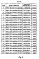
- EphA2 T-cell epitope agonists are compounds containing one or more T-cell epitopes of EphA2 and typically are peptides corresponding to portions of the EphA2 amino acid sequence ( Figure 1 , SEQ ID NO: 2). Also provided are methods for making the agonists and recombinant systems for production of the agonists.
- the EphA2 T-cell epitope agonists are useful in methods for determining a patient's immune status, or immune reactivity to EphA2 by quantifying the number of EphA2-reactive T-cells In the patient
- the agonists also are useful in modulating a patient's immune responsiveness to EphA2 as a cancer treatment.
- the term "agonist” Is a ligand that Is capable of combining with (binding) a receptor on a cell and initiating a reaction or activity that mlmics the activity of a natural ligand, which, in the context of the present disclosure is native EphA2 as shown in Figure 1 .
- EphA2 T-cell epitope agonists those agonists mimic the activity of T-cell epitopes of native EphA2.
- epitope agonists refers to a physical structure that contains and/or defines an antigenic determinant.
- Peptide agonists are peptides, peptide derivatives or peptide analogs that mimic a naturally-occurring ligand, which, in the context of the present disclosure, is an EphA2 T-cell epitope.
- EphA2 T-cell epitope peptide agonists are therefore peptides, peptide derivatives or peptide analogs containing EphA2 T-cell epitopes.
- EphA2 T-cell epitope peptide agonists may be fragments or portions of the EphA2 protein, but also can be conservative derivatives, as defined below, of fragments or portions of the EphA2 protein.
- An EphA2 T-cell epitope agonist can be a "binding reagent," typically an antibody.
- binding reagent refers to any compound, composition or molecule capable of specifically or substantially specifically (that is with limited cross-reactivity) binding another compound or molecule, which, in the case of immune-recognition contains an epitope.
- the binding reagents are antibodies, preferably monoclonal antibodies, or derivatives or analogs thereof, including without limitation: Fv fragments; single chain Fv (scFv) fragments; Fab' fragments; F(ab')2 fragments; humanized antibodies and antibody fragments; camelized antibodies and antibody fragments; and multivalent versions of the foregoing.
- Multivalent binding reagents also may be used, as appropriate, including without limitation: monospecific or bispecific antibodies, such as disulfide stabilized Fv fragments, scFv tandems ((scFv) 2 fragments), diabodies, tribodies or tetrabodies, which typically are covalently linked or otherwise stabilized (i.e., leucine zipper or helix stabilized) scFv fragments.
- Boding reagents also include aptamers, as are described in the art. ,
- Antigen-specific binding reagents including antibodies and their derivatives and analogs and aptamers
- Polyclonal antibodies can be generated by immunization of an animal.
- Monoclonal antibodies can be prepared according to standard (hybridoma) methodology.
- Antibody derivatives and analogs, including humanized antibodies can be prepared recombinantly by isolating a DNAfragmentfrom DNA encoding a monoclonal antibody and subcloning the appropriate V regions into an appropriate expression vector according to standard methods. Phage display and aptamer technology is described In the literature and permit in vitro clonal amplification of antigen-specific binding reagents with very affinity low cross-reactivity.
- Phage display reagents and systems are available commercially, and include the Recombinant Phage Antibody System (RPAS), commercially available from Amersham Pharmacia Biotech, Inc. of Piscataway, New Jersey and the pSKAN Phagemid Display System, commercially available from MoBiTec, LLC of Marco Island, Florida. Aptamer technology is described for example and without limitation in U.S. Patent Nos. 5,270,163 , 5,475096 , 5,840867 and 6,544,776 .
- RPAS Recombinant Phage Antibody System
- a “gene” is an operative genetic determinant In its broadest sense.
- a gene Includes an "expressed sequence” that encodes a protein or is transcribed into a functional RNA product, for example an open reading frame (ORF).
- a typical gene includes an expressed sequence, along with operably linked regulatory sequences, including, but not limited to, promoters, enhancers, transcription factor binding sequences, operators and terminators (for example poly(A) sequences).
- Promoters can be, for example and without limitation, constitutive or semi-constitutive (for example, CMV and RSV promoters), tissue-specific promoters (for example, a muscle creatinine kinase (MCK) promoter) or Induceable (for example and without limitation tetracycline-regulatable systems, such as the BD Tet-OnTM and BD Tet-OffTM Gene Expression Systems, commercially available form BD Blosclences Clontech of Palo Alto, California).
- BD Tet-OnTM and BD Tet-OffTM Gene Expression Systems commercially available form BD Blosclences Clontech of Palo Alto, California.
- regulatory sequences are operably linked in a manner sufficient to cause correct and/or desired transcription of the expressed sequence in a cell.
- expression or “gene expression,” and like words and phrases, mean the overall process by which the information encoded in a nucleic acid, typically a gene, is converted into a ribonucleic acid and/or a protein, or a post-translationally modified version thereof, and/or an observable phenotype.
- nucleic acid may be, without limitation, any polynucleotide or polydeoxynucleotide. Without limitation, a nucleic acid may be single-stranded or double stranded.
- Figure 1 SEQ ID NO: 2 and Figure 2 , (SEQ ID NO: 1, respectively)
- a “conservative derivative” is a nucleic acid or a peptide containing conservative substitutions, which include, in the case of a nucleic acid, substitutions with nucleotide bases that account for codon degeneracy, for example and without limitation in reference to the Ala codon, the substitution of GCC or GCG for GCA, or, In the case of both nucleic acids and peptides, that represent conservative amino acid substitutions, including but not limited to the conservative substitution groups: Ser and Thr; Leu, lie and Val; Glu and Asp; and Gin and Asn.
- Conservative substitutions also may be determined by other methods, such as, without limitation, those used by the BLAST (Basic Local Alignment Search Tool) algorithm, such as a BLOSUM Substitution Scoring Matrix, such as the BLOSUM 62 matrix,
- BLAST Basic Local Alignment Search Tool
- BLOSUM Substitution Scoring Matrix such as the BLOSUM 62 matrix
- the similarity between two nucleic acid or protein sequences may be determined by a variety of methods. For example, the similarities may be determined in silico by an algorithm, for example a BLAST algorithm, which is the reference standard used herein.
- the similarity between two nucleic acid sequences also may be determined by specific hybridization, which means that a nucleic acid will hybridize specifically in a genome to a reference nucleic acid (namely, the EphA2 sequence provided herein or portions thereof).
- the hybridization conditions for achieving specificity naturally will differ, depending on such factors including, without limitation, the length of sequence overlap of the respective nucleic acids, its (melting temperature) Tm, the specific genome and the assay conditions.
- “Derivatives” also include chemically modified nucleic acids or peptides corresponding to portions of the EphA nucleotide or amino acid sequence and conservative derivatives thereof.
- the nucleic acids or peptides, or conservative derivatives thereof may be derivatized to contain chemical groups that, for example: modify the solubllity of the nucleic acid or peptide, for example by the addition of a PEG group; permit affinity purification of the peptide or nucleic acid, for example by the addition of a biotin or poly(his) tag;) or permit detection of the compound, for example by conjugation with a fluorochrome, such as fluorescein isothiocyanate, Cy3 or Cy5 or a radionuclide-containing or caging group for in vitro or in vivo detection and location of the nucleic acid, peptide or derivative thereof.
- a fluorochrome such as fluorescein isothiocyanate, Cy3 or Cy5 or a radionuclide-containing
- modified nucleic acids and peptides are only limited examples of the large variety of useful modifications of nucleic acids and peptides that are known in the art.
- a more complete, but non exhaustive list of such modifications include one or more of N-terminal modifications, C-terminal modifications, internal modifications or non-standard residues for example, and without limitation the following groups and/or residues: a solubilizing group (such as, without limitation a polyethylene glycol (PEG) group), a hydrophobic group, a lipid group, a hydrophilic group, a tag (such as, without limitation: a fluorescent tag (such as fluorescein (e.g., FITC) or a cyanine dye (e.g., Cy3 or Cy5)) or a polypeptide tag (e.g., poly-histidine, for affinity purification, for example)), a transmembrane signal sequence or a portion thereof, an amino acid enantiomer, acetyl, benzyl
- a common peptidomimetic is a "peptoid", which Is a polymer that that includes one or more N-substituted amino acid residues, such as N-substituted glycine.
- peptoids a polymer that that includes one or more N-substituted amino acid residues, such as N-substituted glycine.
- peptoids a polymer that that includes one or more N-substituted amino acid residues, such as N-substituted glycine.
- T-cell epitopes of EphA2 are described and analyzed for their ability to elicit an EphA2-speclfic immune response. Those epitopes were Identified In silico in the context of the MHC Class II allele HLA-DR ⁇ 1*0401 (DR4) or the Class I allele HLA-A0201 (HLA-A2). HLA-DR ⁇ 1*0401 and HLA-A0201 are two alleles among many.
- Non-limiting examples of other Class II HLA-DR alleles are shown In Figure 3 , including HLA-DR1, HLA-DR3, HLA-DR4, HLA-DR7, HLA-DR8, HLA-DR9, HLA-DR11, HLA-DR12, HLA-DR13, HLA-DR14 and HLA-DR15 alleles (www.anthonynolan.org,uk/HIG/lists/class2list.htrnl). More common Class II alleles include, without limitation: HLA-DR2, HLA-DR3, HLA-DR4 and HLA-DR5.
- EphA2 MHC-II-contalning fragments can be identified that are specific to alleles other than HLA-DR ⁇ 1*0401 or HLA-A0201.
- the algorithm described below for use in identifying proteasomal cleavage products, or any like algorithms can be used to select candidate testing for screening In ELISPOT assays and ELISA assays as described herein, or like assays, for Immunostimulatory activity in EphA2-reactive PBLs from a patient with the same MHC haplotype.
- MHC class I HLA-A or HLA-B alleles are provided in Figure 4 (www.anthonynolan.org.uk/HIG/lists/class1llst.html). More common alleles include, without limitation: HLA-A1, HLA-A2, HLA-A3, HLA-B7 and HLA-B44.
- Figures 5-8 provide in silico predicted MHC Class I binding peptides within the EphA2 amino acid sequence for the Class I alleles HLA-A1, HLA-A3, HLA-B7 and HLA-B44, respectively
- Figures 9-17 provide in silico predicted MHC Class II binding peptides within the EphA2 amino acid sequence for the Class II alleles HLA-DR ⁇ 1*0101, HLA-DR ⁇ 1*0301, HLA-DR ⁇ 1*0401, HLA-DR ⁇ 1*0701, HLA-DR ⁇ 1*0801, HLA-DR ⁇ 1*1101, HLA-DR ⁇ 1*1301, HLA-DR ⁇ 1*1501 and HLA-DR ⁇ 5*0101, respectively.
- the available software useful in identifying MHC-consensus binding regions contains consensus sequences for many MHC Class I and Class II alleles (including, without limitation, 39 Class I alleles, including HLA-A1, HLA-A24 and HLA-B7 alleles, available for searching in the NIH BIMAS "HLA Peptide Binding Predictions" software (for example, at http://bimas.dcrt.nih.gov/molbio/hla_bind/) and 51 Class II alleles, including 49 HLA-DR ⁇ 1 alleles and 2 HLA-DR ⁇ 5 alleles, Singh et al., ProPred: prediction of HLA-DR binding sites Bioinformatics (2001) Dec;17(12):1236-7 ; both of which were utilized to identify the putative EphA2 Class I and Class II T-cell epitopes identified in one or more of Figures 5-17 ), methods for identifying consensus binding sequences are well-described in the literature.
- Luckey et al ., 2001 describes methods for identifying Class I binding sequences - briefly by the steps of acid-treating cells to elute Class I molecules, affinity purifying the various alleles, eluting bound peptides form the affinity-purified HLA molecules and sequencing the eluted peptides.
- Methods for identifying consensus binding sequences for each allele, and the recognition that many alleles can bind the same or very similar peptide sequence repertoirs (HLA supertypes) is discussed in Southwood et al ., 1998.
- EphA2 T-cell epitopes which can be accomplished by eluting processed EphA2 peptide fragments from any MHC molecule purified from a cell, such as an APC (antigen presenting cell), for any MHC allele, and sequencing the eluted peptides, all according to well-established methods. This completely avolds the in silico step.
- EphA2 T-cell epltope agonists described herein can be used in ELISPOT, or like assays, to screen patients for their immune reactivity to EphA2, and can be used to stimulate a patient's immune response to EphA2. In this manner an immunogenic composition or cocktail can be prepared for a given patient, depending on that patient's HLA-DR haplotype.
- an immune response to sub-dominant EphA2 T-cell epitopes can be elicited in a patient, which could overwhelm the patient's tolerance to one or more dominant epitopes.
- EphA2 T-cell epitope agonist compounds are useful in an assay to establish a patient's existing immunity to EphA2.
- a population of a patient's PBLs may be stimulated with a compound containing one or more EphA2 T-cell epitopes, as described herein.
- the one or more EphA2 T-cell epltopes are selected to match the patient's MHC haplotype. Hence, if the patient has the HLA-DR ⁇ 1*0701 allele, a compound containing one or more EphA2 HLA-DR ⁇ 1*0701-binding peptides is selected.
- the PBL Once stimulated with the compound for a sufficient period of time (typically 6 hours to 48 hours), the PBL, population Is tested for stimulation by the antigen.
- This testing can be performed by a variety of methods, such as by ELISPOT to determine IFN- ⁇ or IL-5 production, or ELISA to determine TGF- ⁇ or IL-10 production, purportedly indicative of antigen-specific suppression.
- CFC Cytokine Flow Cytometry
- MHC-peptide tetramer analysis in which isolated MHC-peptide tetramers are used to stimulate an antigen-specific response in PBL and bound cells can be counted by flow cytometry
- QRT-PCR Quantitative Reverse Transcription Polymerase Chain Reaction
- Image analysis-assisted cytokine ELISPOT assay is a sensitive method for direct ex vivo monitoring of antigen-specific CD4 + or CD8 + T cells.
- the procedure measures both the frequency and cytokine signatures of antigen-specific T cells In freshly isolated cellular material.
- the assay determines various parameters of T cell immunity such as the clonal size (magnitude) and the Th1/Th2 effector class of the T cell pool.
- ELISPOT is a superior method through which the actual secretory processes of individual pharmacologically unmanipulated cells can be studied. The technology is non destructive and the lymphocytes can be preserved for further analysis.
- the ELISPOT assay uses plates coated with an antibody, typically an anti-cytokine antibody. In the Examples below, the plates are coated with IL-5 (Th2 cytokine profile) and IFN- ⁇ (Th1 cytokine profile).
- the ELISPOT assay includes the steps of incubating cytokine producing cells, such as PBLs, in the antibody-coated plates in the presence of an antigen. The cells are washed away, leaving just the antibodies, some of which will be bound to its cytokine ligand.
- a standard "sandwich assay” is then performed In which tagged or labeled anti-cytokine antibody is bound to the previously bound cytokine and is detected by standard methods, such as by a standard biotin-avidin-HRP (horseradish peroxidase) method. Bound cytokine is therefore represented on the plate as a spot at the site of the complex. The colored spots are then counted and their size analyzed either visually or more commonly by computer analysis, providing data that is then used to calculate the cytokine secretion frequency.
- sandwich assays Both ELISPOT assays and ELISAs are examples of sandwich assays.
- sandwich assay refers to an immunoassay where the antigen is sandwiched between two binding reagents, which are typically antibodies.
- the first binding reagent/antibody being attached to a surface and the second binding reagent/antibody comprising a detectable group.
- detectable groups include, for example and without limitation: fluorochromes, enzymes, epitopes for binding a second binding reagent (for example, when the second binding reagent/antibody is a mouse antibody, which is detected by a fluorescently-labeted anti-mouse antibody), for example an antigen or a member of a binding pair, such as biotin.
- the surface may be a planar surface, such as in the case of an ELISPOT assay or a typical grid-type array, as described herein, or a non-planar surface, as with coated bead array technologies, where each "species" of bead is labeled with, for example, a fluorochrome (such as Luminex technology, as described In U.S. Patent Nos. 6,599,331 , 6,592,822 and 6,268,222 ), or a quantum dot (for example, as described in U.S. Patent No. 6,306.610 ).
- a fluorochrome such as Luminex technology, as described In U.S. Patent Nos. 6,599,331 , 6,592,822 and 6,268,222
- a quantum dot for example, as described in U.S. Patent No. 6,306.610 .
- the agonists described herein are compounds that contain one or more EphA2 T-cell epitopes.
- the agonist can be, for example, with respect to T-cell epitopes defined by their binding to HLA-A2 and DR4, 1) peptides having one of the amino acid sequences: TLADFDPRV (SEQ ID NO: 2, residues 883-891); VLLLVLAGV (SEQ ID NO: 2, residues 546-554); VLAGVGFFI (SEQ ID NO: 2, residues 550-558); IMNDMPIYM (SEQ ID NO: 2, residues 58-66); SLLGLKDQV (SEQ ID NO: 2, residues 961-969); WLVPIGQCL (SEQ ID NO: 2, residues 253-261); LLWGCALAA (SEQ ID NO: 2, residues 12-20); GLTRTSVTV (SEQ ID NO: 2, residues 391-399); NLYYAESDL (SEQ ID NO: 2, residues 120-128); KLNVEERSV (
- the agonist is a single peptide containing two or more of the amino acid sequences containing the T-cell epitope of: TLADFDPRV (SEQ ID NO: 2, residues 883-891); VLLLVLAGV (SEQ ID NO: 2, residues 546-554); VLAGVGFFI (SEQ ID NO: 2, residues 550-558); IMNDMPIYM (SEQ ID NO: 2, residues 58-66); SLLGLKDQV (SEQ ID NO: 2, residues 961-969); WLVPIGQCL (SEQ ID NO: 2, residues 253-261); LLWGCALAA(SEQ ID NO: 2, residues 12-20); GLTRTSVTV (SEQ ID NO: 2, residues 391-399); NLYYAESDL (SEQ ID NO: 2, residues 120-128); KLNVEERSV (SEQ ID NO: 2, residues 162-170); IMGQFSHHN (SEQ ID NO: 2, residues 666-674); YSVCNVMSG (SEQ ID NO
- Each sequence is separated from the other by a peptide spacer, that may be of any length, but typically ranges from 0 to 10 amino acids in length.
- a peptide spacer that may be of any length, but typically ranges from 0 to 10 amino acids in length.
- an AAY trimer spacer greatly improved the efficacy of an a Human Papilioma Virus (HPV16) multivalent epltope string vaccine.
- the peptide can be engineered to ensure that protease cleavage site are located between the epitope sequences to ensure proper processing of the peptide.
- This "string of beads" configuration can contain any number and combination of epitopes. De Groot et al . 2001, Velders et al .
- One or more epitopes also may be combined in a chimeric peptide with a second amino acid sequence that has a desired functionality.
- the functionality can be immunogenic in nature, permitting affinity purification of the peptide.
- a protease cleavage site can be included between the EphA2 T-cell epitope agonist peptide and the immunogenic portion.
- the functionality also can facilitate the delivery of the EphA2 T-cell epitope agonist peptide, by including amino acid sequences that facilitate delivery of the peptide.
- One example of this is to include a portion of lactadherin or other protein to facilitate presentation of the peptide to dendritic cells In membrane vesicles or nanoparticies, such as exosomes. Methods for modifying and expressing chimeric peptides for incorporation Into membrane vesicles are described In international Patent Publication No. WO 03/016522 .
- the agonist in any form described above, can be administered by any useful route to vaccinate or otherwise elicit an immune response in a patient.
- the agonist is Injected into the patient, optionally with an adjuvant, such as Freund's incomplete Adjuvant, Freund's Complete Adjuvant, or as an exosome, as described above.
- an adjuvant such as Freund's incomplete Adjuvant, Freund's Complete Adjuvant, or as an exosome, as described above.
- the agonist can be delivered in a variety of compositions which include the agonist and any desirable, pharmaceutically acceptable carrier.
- Carrier includes as a class, without limitation, any compound or composition useful in facilitating storage, stability, administration and/or delivery of the active ingredients described herein, including, without limitation, suitable vehicles, solvents, diluents, excipients, pH modifiers, buffers, salts, colorants, flavorings, rheology odifiers, lubricants, coatings, fillers, antifoaming agents, erodeable polymers, hydrogels, surfactants, emulsifiers, adjuvants, preservatives, phospholipids, fatty acids, mono-, di- and tri-glycerides and derivatives thereof, waxes, oil and water, as are broadly known in the pharmaceutical arts.
- the route is immaterial.
- Atypical route of administration is intramuscular injection.
- the agonist can be administered once or multiple times over a desired time period to ellcit a desired immune response. Suitable intervals for administering multiple doses typically range from once a week to once a year, but typically ranges from once every seven to 90 days, and more typically, once every seven to 30 days.
- Optimal administration Intervals may be gauged by a patient's immune response, and the severity of the patient's condition.
- the amount of the agonist administered also may vary greatly, depending, among other parameters, upon the structure of the agonist, the route of delivery and the patient's health status.
- the amount of agonist administered at any given time to elicit an immune response to the agonist is an amount effective to do so.
- the number of times the agonist is administered and the interval for administering multiple doses is a number and interval effective to elicit an immune response to the agonist.
- the agonist also may be delivered to a patient by liposomes.
- Liposomes can be directed to the site of lymphoid cells, where the liposomes then deliver the selected agonist composition.
- Liposomes for use are formed from typical vesicle - forming lipids, which include neutral and negatively charged phospholipids and a 5-sterol, such as cholesterol,
- the selection of lipids is generally guided by consideration of, for example, liposome size, acid labillty and stability of the liposomes in the bloodstream.
- a variety of methods are available for preparing liposomes, as described in Szoka et al., Ann. Rev. Biophys. Bloeng. 9:467 (1980 ) and U.S. Patent Nos. 4,235,871 , 4,501,728 , 4,837,028 and 5,019,369 .
- the agonists also may be delivered in the presence of heat shock proteins.
- Heat shock proteins act as molecular chaperones in the cells, ensuring proper folding of nascent peptides into proteins and shuttling new and old proteins inside the cell. Heat shock proteins are believed to play a role In the presentation of peptides on the cell surface to help the immune system recognize diseased cells.
- United States Patent Nos. 5,935,576 , 6,007,821 and 6,017,540 describe such uses for heat shock proteins, methods of making heat shock protein complexes and treatment methods.
- An effective immune response also can be elicited by ex vivo methods.
- antigen is presented to PBL populations of antigen presenting cells (often referred to as pulsing APCs), such as dendritic cells, obtained from a patient in vitro , and the immune-stimulated cells are delivered back to the patient.
- pulsing APCs antigen presenting cells
- dendritic cells dendritic cells
- the agonist may be delivered to the APCs in vitro in any form, including by directly depositing the agonist on the cells, or by liposome or exosome delivery, as described herein, alone or in the presence of additional factors, such as heat shock proteins or appropriate cytokines.
- EphA2 ligand agonists could take the form of, but would not be restricted to, anti-EphA2 antibodles, EphrinA1-Ig constructs or synthetic peptides that induce degradation of EphA2 protein In treated tumor cells.
- a patient is administered an EphA2 ligand or an agonist thereof.
- the EphA2 ligand or agonist thereof can be a binding reagent, such as an antibody (for example a monoclonal antibody, or a derivative or an analog of an antibody, including without limitation: Fv fragments; single chain Fv (scFv) fragments; Fab' fragments; F(ab')2 fragments; camelized antibodies and antibody fragments; multivalent versions of the foregoing; monospecific or bispecific antibodies, such as disulfide stabilized Fv fragments, scFv tandems ((scFv) 2 fragments), antibody multimers, which typically are covalently linked or otherwise stabilized (for example and without limitation, leucine zipper or helix stabilized); scFv fragments; recombinant antibodies or antibody fragments), or in vitro -generated EphA2-specific compounds, such as aptamers and compounds generated by phage display selection and propagation methods.
- the EphA2 ligand or agonist thereof can be delivered to a patient by any effective route, in any effective amount and by any effective interval, as described above in reference to the EphA2 T-cell receptor agonist Delivery of the EphA2 ligand or agonist thereof Is in combination with the delivery of the EphA2 T-cell receptor agonist-containing compounds, and a therapeutic regimen typically, but not necessarily alternates delivery of the EphA2 T-cell receptor agonists and the EphA2 ligand or agonist thereof.
- the EphA2 T-cell receptor agonists are delivered to a patient either directly by the direct or ex vivo methods described above, and one week later, the EphA2 ligand or agonist thereof is administered. This is repeated any desired and effective number of times, with one treatment per week, alternating between the EphA2 T-cell receptor agonists and the EphA2 ligand or agonist thereof.
- Any peptide described herein can be manufactured by any of the myriad of known recombinant methods for producing a peptide, or can be synthesized by common protein synthesis methods, such as by solid phase chemistries as are broadly known.
- a gene is prepared that contains an appropriate promoter, terminator and a coding region that encodes the desired peptide.
- the coding region also can encode signal sequences enabling secretion of the peptide by cells and/or suitable tags, preferably cleavable, that permit affinity purification of the peptide.
- a nucleic acid containing a gene for expressing an EphA2 agonist in a human cell also may be delivered directly to a patient, or ex vivo to a patient's cells (for delivery back into the patient) such that the cells of the patient express the agonist, thereby eliciting an immune response. Delivery of a gene, rather than Just the agonist can result in a more robust immune response resulting from the extended expression of the gene In the patient's cells.
- the nucleic acid can be delivered by viral-mediated (such as by a recombinant adenovirus, adeno-associated virus or vaccinia virus) or non-viral-mediated delivery methods (such as by liposome or by direct injection of naked nucleic acid, for instance Into muscle).
- Peripheral Blood and Tumor Specimens Peripheral Blood and Tumor Specimens .
- Peripheral blood samples were obtained by venlpuncture from 40 patients diagnosed with RCC and 14 normal individuals and were collected into heparinized tubes.
- Peripheral blood lymphocytes (PBLs) were isolated by centrifugation on a Ficoll-Hypaque gradient (LSM, Organon-Teknika, Durham, NC). RCC tumor lesions and matched normal kidney tissue were surgically-resected and paraffin-embedded. Informed consent under an IRB-approved protocol was obtained from all patients prior to sample acquisition. Patient and normal donor information is provided in Table 1.
- HLA-A2 positive or/and HLA-DR4 positive were HLA-A2 positive or/and HLA-DR4 positive, as determined by fluorescence-activated cell sorter analysis using the HLA-A2-specific antibodles (BB7.2 and MA2.1) and HLA-DR4-specific antibody (anti-HLA-DR4 monoclonal antibody clone 359-13F10, IgG, kindly provided by Dr. Janice Blum, Indiana University School of Medicine, Indianapolis, IN).
- HLA-A2 positive or/and HLA-DR4 positive were HLA-A2 positive or/and HLA-DR4 positive, as determined by fluorescence-activated cell sorter analysis using the HLA-A2-specific antibodles (BB7.2 and MA2.1) and HLA-DR4-specific antibody (anti-HLA-DR4 monoclonal antibody clone 359-13F10, IgG, kindly provided by Dr. Janice Blum, Indiana University School of Medicine, Indianapolis, IN).
- RCC patients and normal individuals 9 patients and 6
- HLA-A2 and/or DR4 positive RCC patients evaluated Disease status at RCC time of evaluation HLA Typing: Patlent Age Sex Stage Treatment (Months) A2 (+/-) DR4 (+/-) SLR30-pre 63 F I none Local Dis. + - SLR31 66 M I none Local Dls. + - SLR32 62 F I none Local Dis. + - SLR33 54 F I none Local Dls + - SLR34 71 M I none Local Dis. + + SLR35 75 F I none Local Dis. + + SLR36-pre 60 M I none Local Dis. + + SLR37 52 M I none Local Dis. + - SLR38-pre 69 M I none Local Dls.
- C Chemotherapy
- IFN- ⁇ recombinant interferon-alpha therapy
- IL-2 recombinant Interleukin-2 therapy
- Mets Metastatic Disease
- NED No evidence of disease
- R Radiotherapy
- S Surgery.
- HLA-A2 and -DR4 status was determined using allele-specific monoclonal antibodies and flow cytometry gating on peripheral blood monocytes, as described in Materials and Methods.
- T2.DR4 HLA-A2+/-DR ⁇ 1*0401+; Pratt, R.L. et al. Oncogene 21:7690-7699 (2002 ) cell line (kindly provided from Dr. Janice Blum, Indiana University School of Medicine, indianapolis, IN) was used as the peptide-presenting cell in ELISPOT assay.
- SLR20-SLR26 clear cell RCC lines were evaluated in Western Blotting analyses.
- the normal human proximal tubular epithelial kidney cell line HK-2 (American Type Tissue Collection, ATCC, Rockville, MD) was also evaluated in these analyses.
- HK-2 represents a normal control cell line, although it has been transformed by transfection with the HPV-16 E6/E7 genes ( Ryan MJ, et al. Kidney Int. 45:48-57 (1994 )).
- the EphA2+ PC-3 prostate carcinoma cell line was included as a positive control for Western blotting ( Walker-Daniels J, et al. Prostate 41:275-280 (1999 )).
- All cell lines were maintained in RPMI-1640 culture medium supplemented with 10% heat-Inactivated fetal bovine serum,100 U/ml penicillin, 100 ⁇ g/ml streptomycin and 10mM L-glutamine (all reagents from GIBCO/Life Technologies, Grand Island, New York) in a hurnidified atmosphere of 5% CO 2 at 37°C.
- EphA2 protein sequence was obtained from GENBANK (accession number AAH37166; Figure 1 ) and analyzed for HLA-A0201 and HLA-DR ⁇ 1*0401 binding peptides using neural network algorithms ( Honeyman MC, Brusic V, Stone NL, Harrison LC., "Neural network-based prediction of candidate T-cell epitope,” Nat Blotechnol.
- EphA2 Peptides Selected Start Amino Acid AA Sequence of Nonamer Binding Score* Proteasome Generated? Synthesized For Analysis 883 TLADFDPRV 1 1084 YES YES 546 VLLLVLAGV 2 1006 YES YES 550 VLAGVGFFI 3 556 NO NO 58 IMNDMPIYM 4 138 NO NO 961 SLLGLKDQV 5 127 YES YES 253 WLVPIGQCL 6 98 NO NO 12 LLWGCALAA 7 71 NO NO 391 GLTRTSVTV 8 70 YES YES 120 NLYYAESDL 9 68 NO NO 162 KLNVEERSV 10 49 YES YES *The higher the binding score, the greater the stability of the predicted peptide-MHC complex.
- EphA2 Peptides Sequence Start Core AA# AA Sequence of Nonamer Binding Score Synthesized For Analysis 666 IMGQFSHHN 1 577 663 EAGIMGQFSHHNIIR 2 67 YSVCNVMSG 3 95 63 PIYMYSVCNVMSG 4 55 MQNIMNDMP 5 39 53 DLMQNIMNDMPIYMYS 6 1 SEQ ID NO: 2, residues 666-674. 2 SEQ ID NO: 2, residues 663-677. 3 SEQ ID NO: 2, residues 67-75. 4 SEQ ID NO: 2, residues 63-75. 5 SEQ ID NO: 2, residues 55-63. 6 SEQ ID NO: 2, residues 53-68.
- PBLs were resuspended at 10 7 /ml in AIM-V medium (GIBCO/Life Technologies) and were incubated for 60 min at 37°C in a humidified 5% CO 2 incubator.
- Nonadherent (T cell-enriched) cells were gently washed out with PBS and subsequently frozen.
- the plastic adherent cells were cultured in AIM-V medium supplemented with 1000 units/ml rhGM-CSF (immunex Corporation, Seattle, WA) and 1000 units/ml rhIL-4 (Schering-Plough, Kenllworth, NJ). Seven days later, dendritic cells (DCs) were harvested and used to stimulate autologous CD8 + or CD4 + T cells.
- DCs dendritic cells
- Non-adherent autologous cells were used as "enriched" sources of T cell responders.
- CD8 + T cells in HLA-A2-positive patients and healthy donors
- CD4 + T cells in HLA-DR4-positive patients and healthy donors
- MCS Miltenyl Biotec, Auburn, CA
- Two hundred thousand DCs were cocultured with 2 x 10 6 CD8 + or CD4 + T cells with 10 ⁇ g/ml peptide for 1 week.
- the responder CD8 + T cells or CD4 + T cells were harvested and analyzed In ELISPOT assays.
- IFN- 7 and IL-5 ELISPOT assays for Peptide-Reactive CD8 + T cells and CD4+ T cell Responses were performed, as previously described ( Tatsumi T, et al, J. Exp. Med. 196; 619-628, 2002 ). CD8 + T cell responses were evaluated by IFN- ⁇ ELISPOT assays only, while CD4 + T cell responses were evaluated by both IFN- ⁇ (Th1) and IL-5 (Th2) ELISPOT assays.
- 96-well multiscreen hemagglutinin antigen plates (Millipore, Bedford, MA) were coated with 10 ⁇ g/ml of antihuman IFN ⁇ mAb (1-D1K; Mabtech, Sweden) or 5 ⁇ g/ml of antihuman IL-5 (Pharmingen-BD, San Diego, CA) in PBS (GIBCO/Life Technologies) overnight at 4°C. Unbound antibody was removed by four successive washing with PBS.
- CD8+T cells or CD4+ T cells and T2.DR4 cells (2 x 10 4 cells) pulsed with 10 ⁇ g/ml synthetic peptides were seeded in triplicates in multi-screen hemagglutinin antigen plates.
- Control wells contained CD8+ or CD4+ T cells with T2.DR4 cells pulsed with HIV-nef 190-198 peptide (AFHHVAREL, SEQ ID NO: 3) or Malaria-CS 326-345 peptide (EYLNKIQNSLSTEWSPCSVT; SEQ ID NO: 4), respectively, or T2.DR4 cells alone.
- Culture medium was AIM-V (GIBCO/Life Technologies) at a final volume of 200 ⁇ l/well. The plates were incubated at 37°C In 5% CO 2 for 24 hr for IFN- ⁇ assessments, and 40 hr for IL-5 assessments. After incubation, the supernatants of the culture wells were harvested for ELISA analyses, and cells were removed by washing with PBS/0.05% Tween 20 (PBS/T).
- Captured cytokines were detected at sites of their secretion by incubation for 2 hr with biotinylated mAb anti-human IFN- ⁇ (7-B6-1; Mabtec) at 2 ⁇ g/ml in PBS/0.5%BSA or , biotinylated mAb anti-human IL-5 (Pharmingen) at 2 ⁇ g/ml in PBS/0.5%BSA. Plates were washed six times with PBS/T, and avidin-peroxidase complex (diluted 1:100; Vectastain Elite Kit; Vector Laboratories, Burlingame, CA) was added for 1 hr.
- Unbound complex was removed by three successive washes with PBS/T, then with three rinses with PBS alone.
- AEC substrate (Sigma, St. Louis, MO) was added and incubated for 5 min for the IFN- ⁇ ELISPOT assay and the TMB substrate for peroxidase (Vector Laboratories) was added and incubated for 10 min for the IL-5 ELISPOT assay.
- Spots were Imaged using the Zeiss Autolmager (and statistical comparison determined using a Student two-tailed T-test analysis). The data are represented as mean IFN- ⁇ or IL-5 spots per 100,000 CD4+ T cells analyzed.
- ELISAs The supernatants harvested from CD4+ T cell ELISPOT plates were analyzed In TGF- ⁇ and IL-10 ELISAs. Supernatants were isolated from ELISPOT plates at the endpoint of the culture period and frozen at -20°C until analysis In specific cytokine ELISAs. Cytokine capture and detection antibodies and recombinant cytokine were purchased from BD-Pharmingen (San Diego, CA) and used in ELISA assays per the manufacturer's instructions. The limit of detection for the TGF- ⁇ and IL-10 assays were 60 pg/ml and 7 pg/ml, respectively.
- Tumor cells (5-10 x 10 6 ) were analyzed for EphA2 expression via Western blots using the anti-human EphA2 polyclonal antibody (clone: H-77) (Santa Cruz Biotechnology, Inc., Santa Cruz, CA).
- Cell pellets were lysed using 200 ⁇ l of 1% NP-40 in PBS containing protease Inhibitors (Complete, Boehringer Mannheim, Indianapolis, IN) for 1 hour on Ice. After centrifugation at 13,500 x g for 30 minutes, the supernatant was mixed 1:1 with SDS-PAGE running buffer and proteins separated on 10% PAGE gels, prior to electro-blotting onto nitrocellulose membranes (Millipore, Bedford, MA).
- Blots were imaged on Kodak X-Omat Blue XB-1 film (NEN Llfe Science Products, Boston, MA) using horseradish peroxidase (HRP)-conjugated goat anti-rabbit lg (Biorad, Hercules, CA) and the ECL chemiluminescence detection kit (NEN Life Science Products).
- HRP horseradish peroxidase
- RCC tumor specimens were obtained surgically under an IRB-approved protocol and paraffin-embedded. Five ⁇ m sections were de-paraffinized and rehydrated in ddH 2 0 and then PBS. Anti-EphA2 mAb (Ab 208; mlgG1) or isotype-matched control mAb was incubated on sections for 1 h at RT.
- EphA2 was evaluated Independently by two investigators with a microscope under 40X magnification.
- EphA2 was overexpressed in malignant renal epithellal cells.
- Western blot analyses were used to evaluate EphA2 protein levels In RCC cell lines ( Figure 18 ).
- Metastatic RCC lines tended to express EphA2 more strongly than primary RCC lines, approaching the strong staining previously noted for the prostate carcinoma PC-3 ( Walker-Daniels J, et al. Prostate 41; 275-280, 1999 ).
- the HK-2 cell line is HPV-16 E6/E7-transformed and expresses levels of EphA2 consistent with that observed for primary RCC lines.
- Normal PBLs failed to express detectable levels of EphA2 protein. Consistent with these findings, immunohistochemical analyses performed on paraffin-embedded RCC specimens ( Figure 19 ) verified strong expression of EphA2.
- anti-EphA2 and control anti- ⁇ -actin antibodies were used in performing Western Blot analyses of lysates generated from the Indicated RCC cell lines, the normal kidney tubular epithelial cell line HK2 and normal peripheral blood lymphocytes (PBLs) (negative control). Primary and metastatic clear cell RCC lines were assessed, as indicated. The PC3 prostate cell line and normal donor PBLs served as positive and negative controls.
- EphA2 epitopes recognized by T cells To identify potential T cell epitopes, the EphA2 protein sequence was subjected to algorithms designed to identify putative HLA-A2 binding motifs and sites of proteasomal cleavage. Similarly, a neural network algorithm was used to identify EphA2 peptide sequences that would be predicted to bind HLA-DR4 and have the potential to represent CD4 + T cell epitopes (Honeyman M et al ., 1998). In aggregate, 8 peptides were synthesized for subsequent analyses: 5 peptides were predicted to serve as CTL epitopes and 3 peptides were predicted to serve as Th epitopes (Table 2).
- Peripheral blood T cells were isolated from normal HLA-A2 + and/or -DR4 + donors and stimulated with autologous DCs that had be previously loaded with relevant synthetic peptides. Responder T cells were subsequently evaluated for specific reactivity against peptide-pulsed T2.DR4 (HLA-A2 + /DR4 + ) antigen-presenting cells and renal cell carcinoma cell lines that expressed both the EphA2 antigen and HLA-A2 and/or HLA-DR4.
- the IFN- ⁇ ELlSPOT assay was used to evaluate 8 HLA-A2 + donor CD8 + T cell responses to the 5 putative CTL epitopes and 7 HLA-DR4 + donor CD4 + T cell reactivities against the 3 potential Th epitopes.
- Each peptide was recognized by at least one normal donor (Table 3), and only one (HLA-DR4 + ) donor failed to respond to any of the EphA2 (Th) epitopes.
- HLA-DR4 + the EphA2 546-554 and EphA2 883-891 peptides were most commonly reacted against (each in 6/8 donors evaluated), with the responses to EphA2 883-891 typically being of a higher frequency.
- 6/7 donors responded against at least one predicted EphA2-derived Th epitope, with responses against the EphA 53-76 and EphA2 663-677 most prevalent.
- HLA-A2-Presented EphA2 Peptides CD8 + T Cell Response to Peptide on T2.
- DR4 a Normal Donor # 162 391 546 883 961 A2-1 9 0 b 31 0 2 A2-2 40 81 14 85 21 A2-3 3 14 10 0 21 A2-4 2 0 11 58 0 A2-5 11 0 14 172 4 A2-6 0 91 76 145 13 A2-7 132 0 0 37 0 A2-8 15 0 0 165 0 Total Responses: 5/8 3/8 6/8 6/8 3/8 HLA-DR4-Presented EphA2 Peptides: CD4 + T Cell Response to Peptide on T2.
- DR4 a Normal Donor # 53 63 663 DR4-1 43 11 21 DR4-2 38 36 57 DR4-3 4 7 14 DR4-4 0 0 0 DR4-5 0 156 41 DR4-6 0 121 67 DR4-7 54 48 72 Total Responses: 3/7 6/7 6/7 6
- a value of "0" reflects a frequency ⁇ 1/100,000 T cells.
- T cell reactivity against T2.DR4 cells pulsed with the HLA-A2-presented HIV-nef 180-180 epitope served as the CD8+ T cell negative control, while HLA-DR4-presonted Malarial circumsporozooite (CS) 326-345 epitope served as the CD4+ T cell negative control.
- CS Malarial circumsporozooite
- peripheral blood CD8 + T cells Analysis of peptide-specific IFN- ⁇ release by peripheral blood CD8 + T cells in ELISPOT assays .
- Peripheral blood CD8 + T cells responses was assessed against these sequences in 29 HLA-A2 + RCC patients (Table 1) and 10 HLA-A2 + normal donors.
- CD8 + T cells were enriched to 98% purity for all experiments. Responses were evaluated using IFN- ⁇ ELISPOT assays after 7 day "primary" in vitro stimulations.
- peripheral blood CD8 + T cells were isolated from HLA-A2 + normal donors or patients with RCC and stimulated with immature, autologous dendritic cells pre-pulsed with the individual EphA2-derived epitopes, as outlined in Materials and Methods. After one week, responder T cells were analyzed In IFN- ⁇ ELISPOT assays for reactivity against T2.DR4 (HLA-A2 + ) cells pulsed with the indicated EphA2 epitope. Data are reported as IFN- ⁇ spots/100,000 CD8 + T cells and represent the mean of triplicate determinations.
- T cell reactivity against T2.DR4 cells pulsed with the HLA-A2-presented HIV-nef 190-198 epitope served as the negative control in all cases, and this value was subtracted from all experimental determinations In order to determine EphA2-specific spot numbers.
- Each symbol within a panel represents an individual donor's response.
- the frequencies of CD8 + T cell responses against EphA2 peptides in HLA-A2 + patients prior to surgery (Pre-Op) or patients with residual disease after surgery (Post-RD) were as low as those observed in normal HLA-A2 + donors.
- IFN- ⁇ and IL-5 release by CD4 + T cells in ELISPOT assay Peptide-specific IFN- ⁇ and IL-5 release by CD4 + T cells in ELISPOT assay .
- IFN- ⁇ (Th1-type) and IL-5 (Th2-type) ELISPOT assays were used to discern altered frequency and functional bias of patient-derived Th cells against EphA2 peptides.
- Peripheral blood T cells were stimulated for one week with peptide-pulsed Immature autologous DC (which do not appear to skew the Th1/Th2 balance, ref. 47) prior to CD4 + T cell Isolation and ELISPOT analyses.
- the frequencies of CD4 + T cell responders against EphA2 peptides were evaluated in 19 HLA-DR4 + RCC patients (Table 1).
- T cell reactivity towards EphA2 related to disease progression Patients with Stage I disease patients displayed strongly Th1-polarlzed reactivity against EphA2 peptides whereas patients with more advanced stages of the disease polarized towards strong Th2 reactivity.
- peripheral blood was obtained from 19 HLA-DR4 + patients (Table 1) and CD4 + T cells isolated by positive MACS TM -bead selection as described in Materials and Methods, below. After a one-week In vitro stimulation with EphA2 Th peptide-pulsed, autologous DCs, responder CD4 + T cells were evaluated against T2.DR4 cells pulsed with the indicated EphA2 epitopes in IFN- ⁇ and IL-5 ELISPOT assays.
- T cell reactivity against T2.DR4 cells pulsed with the HLA-DR4-presented Malarial circumsporozoolte (CS) 326-345 epitope served as the negative control in all cases, and this value was subtracted from all experimental determinations In order to determine EphA2-specific spot numbers.
- CS Malarial circumsporozoolte
- TGF- ⁇ and IL-10 production from RCC patient CD4 + T cells against EphA2 peptides were measured.
- TGF- ⁇ and IL-10 production following in vitro peptide-stimulation was measured.
- supernatants were harvested from the culture wells of IFN- ⁇ ELISPOT assays and analyzed for levels of TGF- ⁇ 1 using a commercial ELISA procedure.
- IFN- ⁇ and IL-5 ELISPOT data for these patients' CD4+ T cells is also provided.
- TGF- ⁇ production by responder CD4 + T cells was only observed in a subset (i.e. 3 of 8) of Stage IV patients and notably, these same patients displayed coordinately weak Th1- or Th2-type (IFN- ⁇ and IL-5 ELISPOT) CD4 + T cell reactivity against EphA2 peptides.
- IL-10 production (above the detection limit of the ELISA) was not observed for any specimen tested.
- cytokine release assays provide a powerful means to monitor the specificity and magnitude of evolving anti-tumor CD8 + and CD4 + T cell responses In the peripheral blood of patients before, during and after treatment ( Keilholz U, et al. J Immunother 25; 97-138, 2002 ).
- how, and to what extent, T cells in patients with RCC, recognize novel EphA2-derived epitopes was evaluated using cytoklne-specific ELISPOT assays and ELISAs.
- EphA2-specific CD8 + T cell activity is Inversely proportional to the presence of active disease In these patients and is increased within 6 weeks following therapeutic intervention that results in disease-free status.
- two HLA-A2 + patients with Stage IV disease were identified who were long-term survivors (> 40 months) post-surgery. Both of these individuals displayed elevated peripheral blood frequencies of IFN- ⁇ -secreting CD8 + T cells reactive against EphA2-derived epitopes ( Figure 20 ).
- Increased maintenance of high anti-EphA2 CD8 + T cell activity in these patients may relate to their continued survival with active disease.
- EphA2 may provide a much-needed target antigen for the design of immunotherapies for renal cell carcinoma.
- EphA2 Is over-expressed In a large number of RCC specimens, Including 22 of 24 (92%) RCC cell lines and 29 of 30 (97%) RCC biopsy samples, respectively ( Figure 18 and data not shown).
- These findings are consistent with evidence emerging from studies of other tumor types, which indicates that high levels of EphA2 apply to many cancers, Including breast, colon, head and neck (Tatsumi et al., unpublished data), prostate and lung carcinoma, as well as, melanoma. If the present studies can be extended to these other clinical indications, EphA2-specific T cell activity could provide an opportunity for therapeutic intervention for these tumor types as well.
- CD8 + T cell reactivity against EphA2 peptides differed greatly between RCC patients with active disease and those patients rendered free of disease. Yet, anti-EphA2 CD8 + T cell reactivity did not distinguish RCC disease stage.
- RCC tumors may suppress the generation, functionality and durability of CD8 + T cell responses against EphA2 in situ. This hypothesis is consistent with general tumor-associated immune suppression of peripheral CTL and NK cell activity, as has been previously reported ( Kiessling R, et al. Cancer Immunol Immunother 48; 353-362, 1999 ).
- CD8 + T cell reactivity against EphA2-derived CTL epitopes significantly Increased in the peripheral blood of three HLA-A2 + patients with Stage I RCC after surgery that rendered these individuals free of disease.
- surgical intervention without "cure” did not change the low frequency of CD8 + T cell reactivity towards EphA2 peptides.
- Th1-type biased CD4 + T cell response could only be observed in a subset of Stage I RCC patients, and Th2- or Tr-type biased CD4 + T cell responses were almost always observed In Stage IV RCC patients. It is important to stress that polarization away from Th1-type immunity in patients with advanced stage disease was tumor-specific, since individuals with Stage IV disease responded to influenza- and EBV-derived T helper epitopes in a "normal" Th1-biased manner ( Tatsumi et al., J. Exp. Med. 2002 and data not shown).
- Th3/Tr CD4 + T cell subsets may play dominant roles as antigen-specific T "suppressor" cells, In part due to secretion of immunosuppressive cytokines such as TGF- ⁇ and/or IL-10 ( Krause I, et al. Crit Rev Immuno/ 20; 1-16, 2000 ). Based on detection of TGF- ⁇ (but not IL-10) production in 3 of 8 (38%) patients with Stage IV disease, It Is possible that the population of human CD4 + CD25 + T suppressor cells may hinder the patient's ability to productively eliminate EphA2-overexpressing tumors ( Levings MK, et al. J Exp Med. 196;1335-1346, 2002 ).
- Th2- or T suppressor-type responses are prevalent against EphA2 epitopes In advanced Stage RCC patients and likely contribute to the hyporeactivity of tumor-specific cellular immunity noted in these individuals.
- EphA2-based adjuvant vaccination of Stage I patients could have utility for eliciting protective immunity in patients at high risk for disease recurrence or to prevent prospective metastases.
- Vaccination with both EphA2-derived CD4 + and CD8 + T cell epitopes may prompt high frequency anti-EphA2 CTL induction that is stabilized by the concurrent activation of specific Th1-type CD4 + T cells.
- dendritic cell (DC) -based vaccines incorporating EphA2 peptides may allow for prevlously muted Th1-type immunity to be functionally resurrected in patients with advanced stage disease, yielding potential therapeutic benefit.
- the T2.DR4 HLA-A2+/-DRB1*0401+cell line was used as the peptide-presenting cell in ELISPOT assays.
- the EphA2+ HLA-A2- PC-3 prostate carcinoma cell line was used as positive control for Western Blot analysis of EphA2 protein expression and was also used as a negative control target In ELISPOT assays.
- SLR24 an EphA2+ HLA-A2+ cell line ( Tatsumi, T., et al. Cancer Res, 63: 4481-4489, 2003 ) was tested in Western Blot and ELISPOT assays and was also applied In the Hu-SCID treatment model.
- All cell lines were maintained In RPMI-1640 culture medium supplemented with 10% heat-inactivated fetal bovine serum (FBS), 100 U/ml penicillin, 100 ⁇ g/ml streptomycin and 10mM L-glutamine (all reagents from GIBCO/Life Technologies, Grand Island, New York) in a humidified atmosphere under 5% CO 2 tension at 37°C.
- FBS heat-inactivated fetal bovine serum
- penicillin 100 ⁇ g/ml streptomycin
- 10mM L-glutamine all reagents from GIBCO/Life Technologies, Grand Island, New York
- mice Six-to-eight week old female C.B-17 scid/scid mice were purchased from Taconic Labs (Germantown, NY), and maintained in micro-isolator cages. Animals were handled under aseptic conditions per an Institutional Animal Care and Use Committee (IACUC)-approved protocol and in accordance with recommendations for the proper care and use of laboratory animals.
- IACUC Institutional Animal Care and Use Committee
- samples were also analyzed for Axi (clone C-20, Santa Cruz Biotechnology, Santa Cruz, CA) protein content
- Axi clone C-20, Santa Cruz Biotechnology, Santa Cruz, CA
- Single tumor cell suspensions Isolated from confluent tissue culture flasks or from the enzymatic digestion of resected lesions were lysed using 500 ⁇ l lysis buffer (1% Triton-X, 150nM NaCl, 10mM Tris pH7.4, 1mM EDTA, 0.2mM SOV, 0.5% NP-40) in PBS containing protease inhibitors (Complete, Roche Diagnostic, Mannhelm, Germany) for 30min at 4°C.
- EphA2 Immunoprecipitation for EphA2 were performed using the anti-EphA2 antibody D7 (Upstate Biotech, Inc.). Anti-phosphotyrosine antibodies (Clone py99, Santa Cruz Blotechnology, San Dlego, CA) were used to assess pEphA2 content. Mouse anti- ⁇ -actin antibody (clone AC-15, Abcam, Cambridge, MA) was used as a loading control.
- B61.Ig and mAb208 were kindly provided by Medlmmune (Galthersburg, MD).
- B61.Ig is a chimeric protein consisting of the ligand binding domain of ephrin-A1 fused with the Fc portion of a mouse IgG antibody and was used in in vitro assays at 30 ⁇ g/ml, where indicated.
- mAb208 is a mouse monoclonal antibody specific for EphA2 and was used In in vitro assays at 8 ⁇ g/ml, where indicated.
- ELISPOT Assays In vitro T cell responses were evaluated by IFN- ⁇ ELISPOT assays as previously described ( Tatsumi, T., et al. Cancer Res. 2003 ). Briefly, 96-well multiscreen hemagglutinin antigen plates (Millipore, Bedford, MA) were coated with 10 ⁇ g/ml of anti-human IFN- ⁇ mAb (1-D1K; Mabtech, Sweden) in PBS (GIBCO/Life Technologies) overnight at 4°C. Unbound antibody was removed by four successive washing with PBS.
- Flow Cytometry For phenotypic analysis of control or ligand agonist-treated tumor cells, PE- or FITC-conjugated monoclonal antibodies against HLA class I (W6/32; pan-class I specific; Serotec Inc., Raleigh, NC) or human CD40 (Ancell Corp., Bayport, MN) and appropriate isotype controls (purchased from BD Biosciences, San Jose, CA) were used, and flow cytometric analysis was performed using a FACscan (Becton Dickinson, San Jose, CA) flow cytometer. The results of the flow cytometric analysis are reported In arbitrary mean fluorescence intensity (MFI) units.
- MFI mean fluorescence intensity
- HU-SCID Tumor Model C.B17-scid/scid mice were injected s.c. In the right flank with 1 x 10 8 SLR24 RCC cells and tumors allowed to establish to a size of approximately 30 mm2 (i.e. day 18 post-injection).
- mice were then randomized into 4 groups (5 animals each with comparable tumor sizes) that received either no treatment, a single Intratumoral injection of 50 ⁇ g of B61-Ig (in 50 ⁇ l saline) on day 18, a single tall-vein injection with 5 x 10 6 cloned E883 (anti-EphA2 883-891 specific) CD8+ T cells in 100 ⁇ l saline on day 19, or the combined d18 (B61-Ig) plus d19 (E883 adoptive transfer) regimen. Animals were evaluated every 3-4 days for tumor size, with tumor-free status noted on day 40 post-tumor inoculation.
- tumors were surgically resected from euthanized mice, digested into single-cell suspensions using a DNAse, hyaluronidase, DNAse cocktail as previously described ( Itoh, T., et al. J Immunol. 153:1202-1215,1994 ) and filtered through Nltex mesh (Tetko, Kansas City, MO), prior to tumor cell solublization and Western Blotting, as outlined above.
- B61.Ig is a fusion protein consisting of the EphA2 binding domain of ephrin-A1 (a major ligand of EphA2) fused to a human Fc domain.
- Cellular lysates were resolved by SDS-PAGE and EphA2 protein was immunoprecipitated using the anti-EphA2 antibodies D7 in pull-down assays.
- Western blot analyses were then performed using anti-EphA2 and anti-phosphotyrosine antibodies, respectively. Data are representative of 3 independent experiments performed.
- EphA2Agonist Treatment Enhances CD8+ T cell Recognition of EphA2+ Tumors In Vitro. Since agonistic antibodies triggered proteasomal degradation of EphA2, it was hypothesized that this could increase presentation of EphA2 peptides on HLA class I complexes. If correct, then It logically would follow that agonism of EphA2 could enhance recognition by EphA2-speciflc CD8+ T cells. To address this question, EphA2+ SLR24 RCC cells were incubated with B61-Ig prior to evaluating their ability to be recognized by the EphA2 883-891 specific CTL clone 142 (CL.142).
- Control target cells include T2.DR4 cells pulsed with the HLA-A2-presented HIV-nef 180-188 (negative control for peptide specificity) and PC3 (HLA-A2-/EphA2 + ) prostate carcinoma cells. B61.Ig treatment (30 ⁇ g/ml) was applied overnight to ensure EphA2 degradation and HLA antigen processing and presentation of EphA2 epitopes). Data are reported at IFN- ⁇ specific spots/10,000 CL.142 cells and are derived from one representative experiment of 3 performed.
- the SLR24 RCC cell line was either not treated or treated with mAb208 (10 ⁇ g/ml) as outlined in the description of Figures 28A and 28B , above. Treated cells were then analyzed for expression of HLA class I and CD40 molecules by flow cytometry as described In Materials and Methods. Data presented is the mean fluorescence intensity of expression for the indicated markers.
- EphA2 Agonist Treatment Enhances the efficacy of adoptively transferred anti-EphA2 CD8+ T Cells In a Hu-SCID Tumor Model.
- a Hu-SCID tumor model system was established.
- SLR24 tumors were injected into C.B-17 scid/scid mice and allowed to progress to a size of approximately 30 mm 2 , at which time, animals were either left untreated, or treated with intratumoral injection of B61-Ig (day 18 post-tumor inoculation) and/or intravenous delivery of a HLA-A2-restricted, anti-EphA2883-891 CD8+ T cell clone (E883).
- B61-Ig day 18 post-tumor inoculation
- E883 intravenous delivery of a HLA-A2-restricted, anti-EphA2883-891 CD8+ T cell clone
- EphA2-reactive CD8+ T cells are rendered more effective in mediating the regression of tumor lesions in situ.
- EphA2 In normal epithelia, EphA2 localizes to cell-cell boundaries, where it constitutively binds its ligands. Consequently, the EphA2 in non-transformed cells is tyrosine phosphorylated and mediates signals that serve to limit epithelial cell growth.
- phosphorylated EphA2 molecules form signaling complexes with adapter proteins that contain SH2 domains (e.g., c-Cbl, SHC, SLAP, and GRB2) alters enzymatic activity of selected downstream effectors (e.g., FAK, SHP-2, PI 3-kinase, LMW-PTP). These signals, in turn, decrease the ability of EphA2 + epithelial cells to establish or maintain stable contacts with the surrounding extracellular matrix (ECM).
- ECM extracellular matrix
- c-CbI contains an ublquitin-E3 ligase and targets proteins for degradation via the proteasome.
- the results herein indicate that proteasomal degradation increases T cell recognition of EphA2, presumably by cleaving EphA2 into peptides that are loaded into HLA complexes for subsequent antigen presentation to effector T cells.
- Tumor cells generally have unstable cell-cell contacts, which appears to preclude access of EphA2 to its membrane-anchored ligands. This is consistent with experimental evidence that the EphA2 in tumor cells is overexpressed, but unphosphorylated. Compounding decreased ligand binding, EphA2 molecules expressed by tumor cells serve as substrates for certain oncogenic tyrosine phosphatases, which provide an additional means of decreasing EphA2 phosphotyrosine content. Regardless of the cause, decreased phosphotyrosine content causes the EphA2 in tumor cells to increase their malignant character. In part, the increased invasiveness relates to increased tumor cell interactions with the ECM. These changes are frequently observed In clinical specimens of cancer. Under such conditions, EphA2 ligand agonists can restore a normalized pattern of contact inhibited growth and reduce the invasiveness of EphA2+ tumor cells.
Abstract
Description
- Eph2A T-cell epitope agonists are provided. The Eph2A T-cell epitope agonists are useful in methods for diagnosing cancer, for quantifying EphA2-reactive T-cells in a patient and in eliciting an immune response to EphA2 and modulating the immune system to recognize cancerous cells.
- The molecular identification of tumor antigens recognized by the immune system has paved the way for the development of new immunotherapeutic strategies for the treatment of cancer. While many cytotoxic T lymphocyte (CTL)-defined tumor-assoclated epltopes have been applied clinically in cancer vaccinations (Coulle PG, et al. Proc Natl Acad Sci USA 98: 10290-1295, 2001; Yu JS, et al. Cancer Res 61: 842-847, 2001; Jager E, et al. Proc Natl Acad Sci USA 97: 12198-12203, 2000; and Nestle FO, et al. Nat Med 4:328-332, 1998.), comparatively few class II-restricted epitopes recognized by CD4+ T cells have been identified and clinically-integrated to date (Topalian SL, et al. Proc Natl Acad Sci USA 91; 9461-9465,1994; Chaux P, et al. J Exp Med 189; 767-777, 1999; Pleper R, et al. J Exp Med 189; 757-765,1999; Wang RF, et al. Science 284, 1351-1354, 1999; Topalian SL, et al. J Exp Med 183; 1965-1971, 196; Jager E, et al. J Exp Med 191; 625-630, 2000; Zarour HM, et al. Cancer Res 60; 4946-4952, 2000; and Zarour HM, et al. Proc Natl Acad Sci USA 97; 400-405, 2000). Current paradigms suggest that CD4+ T cells (at least Th1-type) play critical roles In the optimal induction and maintenance of clinically beneficial tumor immunity (Pardoll DM, et al. Curr Opin Immunol 10; 588-594,1998 and Toes RE, et al. J Exp Med 189; 753-756, 1999). Hence, CD4+ and CD8+ T cell epitopes derived from antigens that are unique to, or that are overexpressed on tumor cells may provide effective vaccine components.
- The Eph family of molecules constitutes the largest family of receptor tyrosine kinases in the human genome. Eph kinases include two major classes (EphA and EphB), which are distinguished by their specificities for the ligands ephrln-A and ephrin-B, respectively (Eph Nomenclature Committee. Unified nomenclature for Eph family receptors and their ligands. The ephrins. Cell 90: 403-404, 1997). Largely known for their role in neuronal development, recent reports suggest that Eph receptors play a role in carcinogenesis. For example, EphA2 is overexpressed and functionally altered in a large number of different cancers, where it appears to promote the development of disseminated disease. In normal cells, EphA2 localizes to sites of cell-to-cell contact, where it may play a role as a negative regulator of cell growth. In contrast, EphA2 is frequently overexpressed and often functionally dysregulated in advanced cancers, where it contributes to many different aspects of malignant character. These changes In EphA2 have been observed in a wide array of solid tumors, including melanoma, prostate, breast and lung tumors. The highest degree of EphA2 expression among tumors is most commonly observed in metastatic lesions.
- In the clinical setting, several findings suggest that T cell-mediated Immunity provides a safeguard against the development and progression of renal cell carcinoma (RCC) and may effectively mediate the regression of established lesions. RCC lesions are typically Infiltrated with large numbers of lymphocytes, though the benefits of leukocytic infiltration upon beneficial clinical outcome remain unknown. While this may reflect variance In the functional subsets of CD4+ and CD8+ T cells In these infiltrates, data addressing the prognostic benefit of Th1/Tc1-blased immunity versus Th2/Tc2-biased immunity in RCC patients has been equivocal. A better understanding of the constitutive nature and specificity of CD8+ and CD4+ T cell responses in RCC patients will likely provide insights necessary to design, implement and monitor more effective treatment options,
- Provided herein are novel EphA2 T-cell epitope agonists and uses therefor, including diagnostic and prognostic methods, methods for eliciting an immune response to EphA2 and treatments for cancer. The agonists are useful in the detection and staging of RCC. It is demonstrate herein that high levels of EphA2 expression are observed In the setting of renal cell carcinoma (method of staging RCC) and that patients with RCC exhibit both CD8+ and CD4+ T cell responses to novel EphA2-derived epitopes. Moreover, the reactivity of T cells against EphA2 is useful In distinguishing disease status and outcome and the EphA2 T-cell epitope agonists described herein are useful in eliciting an immune response to EphA2, as a cancer therapy.
- In one embodiment, an EphA2 T-cell epitope agonist Is provided comprising an EphA2 T-cell epitope. The EphA2 T-cell epitope agonist may be a peptide comprising an EphA2 T-cell epitope. In certain embodiments, the peptide consists of from about 9 to about 35 amino acids, from about 9 to about 25 amino acids or less than about 20 amino acids. The peptide can be a portion or fragment of native human EphA2 (SEQ ID NO: 2) and typically comprises at least about 9 contiguous amino acids of SEQ ID NO: 2 or a conservative derivative of a portion of SEQ ID NO: 2 in which one or more amino acid residues are inserted into the peptide or one or more amino acids of SEQ ID NO: 2 are deleted from the peptide or substituted with one or more different amino acid residues, so long as the binding of the conservative derivative to an MHC molecule is substantially equal to or enhanced as compared to binding of EphA2 or a fragment thereof to the MHC molecule.
- The EphA2 T-cell epitope agonist can be a modified peptide comprising one or more of N-terminal modifications, C-terminal modifications, internal modifications or non-standard residues, for example and without limitation, a solubilizing group; a hydrophobic group; a lipid group; a hydrophilic group; a tag; a fluorescent tag; a polypeptide tag; a transmembrane signal sequence or a portion thereof; an amino acid enantiomer and one of an acetyl, benzyloxycarbonyl, biotin, cinnamoyl, dabcyl, dabsyl, dansyl, dinitrophenyl, cyanine, fluorescein, fmoc, formyl, lissamine rhodamine, myristoyl, n-methyl, palmitoyl, steroyl, 7-methoxycoumarin acetic acid, biotin, dabcyl, dabsyl, dansyl, dlsulphide, acetamidomethyl, aminohexanoic acid, aminolsobutyric acid, beta alanine, cyclohexylalanine, d-cyclohexylalanine, e-acetyl lysine, gamma aminobutyric acid, hydroxyproline, nitro-arginine, nitro-phenylalanine, nitro-tyrosine, norleucine, norvaline, octahydroindole carboxylate, ornithine, penicilliamine, phenylglycine, phosphoserine, phosphothreonine, phosphotyrosine, L-malonyltyrosine, pyroglutamate, tetrahydroisoquinoline, amide, N-substituted glycine; non-amino acyl and N-acetylglycine group. In certain embodiments, the EphA2 T-cell epitope agonist is a peptiod or a peptidomimetic comprising an EphA2 T-cell epitope.
- In certain embodiments, the EphA2 T-cell epltope agonist comprises a T-cell epitope contained in one or more of the following EphA2 epitope sequences: TLADFDPRV (SEQ ID NO: 2, residues 883-891); VLLLVLAGV (SEQ ID NO: 2, residues 546-554); VLAGVGFFI (SEQ ID NO: 2, residues 550-558); IMNDMPIYM (SEQ ID NO: 2, residues 58-66); SLLGLKDQV (SEQ ID NO: 2, residues 961-969); WLVPIGQCL (SEQ ID NO: 2, residues 253-261); LLWGCALAA (SEQ ID NO: 2, residues 12-20); GLTRTSVTV (SEQ ID NO: 2, residues 391-399); NLYYAESDL (SEQ ID NO: 2, residues 120-128); KLNVEERSV (SEQ ID NO: 2, residues 162-170); IMGQFSHHN (SEQ ID NO: 2, residues 666-674); YSVCNVMSG (SEQ ID NO: 2, residues 67-75); MQNIMNDMP (SEQ ID NO: 2, residues 55-63) and a sequence presented in one or more of
Figures 5-17 . - As a non-limiting example, the EphA2 T-cell epitope agonist can comprise a peptide, or a modified version thereof, comprising one or more of the following amino acid sequences: TLADFDPRV (SEQ ID NO: 2, residues 883-891); VLLLVLAGV (SEQ ID NO: 2, residues 546-554); VLAGVGFFI (SEQ ID NO: 2, residues 550-558); IMNDMPIYM (SEQ ID NO: 2, residues 58-66); SLLGLKDQV (SEQ ID NO: 2, residues 961-969); WLVPIGQCL (SEQ ID NO: 2, residues 253-261); LLWGCALAA (SEQ ID NO: 2, residues 12-20); GLTRTSVTV (SEQ ID NO: 2, residues 391-399); NLYYAESDL (SEQ ID NO: 2, residues 120-128); KLNVEERSV (SEQ ID NO: 2, residues 162-170); IMGQFSHHN (SEQ ID NO: 2, residues 666-674); YSVCNVMSG (SEQ ID NO: 2, residues 67-75); MQNIMNDMP (SEQ ID NO: 2, residues 55-63) and a sequence presented in one or more of
Figures 5-17 , or a conservative derivative thereof. In one embodiment, the EphA2 T-cell epitope agonist comprises two or more EphA2 T-cell epltopes separated by a spacer. - A composition is provided that comprises one or more EphA2 T-cell epitope agonist as described above and a pharmaceutically acceptable carrier. In another embodiment, a method of monitoring the number and/or status of EphA2-reactive T-cells in a patient is provided, the method comprises determining the patient's immune reactivity to a compound or composition containing an EphA2 T-cell epitope agonist containing one or more EphA2 T-cell epitopes, as described above. In one embpodiment, the method comprises determining the patient's immune reactivity to a compound or composition containing one or more EphA2 T-cell epitopes using an ELISPOT assay. The ELISPOT assay may detect a CD8+ response to an MHC class I protein-presented EphA2 epitope or a conservative derivative thereof. The MHC class I protein can be an HLA-A2 protein. The ELISPOT assay also may detect a CD4+ response to an MHC class II protein-presented EphA2 epitope or a conservative derivative thereof. The MHC class II protein can be an HLA-DR4 protein.
- In a further embodiment, a method is provided for inhibiting growth in a patient of a cancer in which EphA2 Is overexpressed, comprising administering to the patient an amount of an EphA2 T-cell epitope agonist as described above, effective to elicit an immune response to EphA2 in the patient. In one embodiment, the method comprises contacting an antigen-presenting cell of a patient with the EphA2 T-cell epitope agonist. In another embodiment, the method is an ex vivo method comprising: isolating cells comprising an antigen-presenting cell from the patient; contacting the antigen-presenting cell with the EphA2 T-cell epitope agonist; and reintroducing the EphA2 T-cell epltope agonist-contacted antigen-presenting cell into the patient. The method may further comprise administering to the patient an EphA2 ligand or an agonist thereof, such as, without limitation, a binding reagent capable of binding to EphA2; and ephrlnA1 or an agonist thereof.
- Also provided is an Isolated nucleic acid comprising from 5' to 3' and operably linked, a promoter, a coding sequence, other than a full length EphA2 coding sequence, encoding a peptide comprising one or more EphA2 T-cell epitopes and a polyadenylation signal. The nucleic acid Is useful in preparing the EphA2 T-cell agonist by recombinant methods and/or by transfer of the nucleic acid into a patient's cells, either ex vivo or in vivo, to produce the EphA2 T-cell agonist in vlvo.
- In another embodiment, a method is provided comprising contacting a tumor cell that expresses EphA2 on its surface with an EphA2 ligand or an agonist thereof comprising one of: a binding reagent capable of binding to EphA2; and ephrinA1 or an agonist thereof.
-
-
Figures 1 and2 provide the amino acid (SEQ ID NO: 2) and nucleic acid (SEQ ID NO: 1) sequences of human EphA2 (GenBank Accession Nos. AAH37166 and BC037166 (also NM_004431), respectively). -
Figures 3 and4 provide non limiting lists of human MHC Class II and Class I alleles, respectively, -
Figures 5-8 provide in silico predicted MHC Class I binding peptides within the EphA2 amino acid sequence for the Class I alleles HLA-A1, HLA-A3, HLA-B7 and HLA-B44, respectively (portions of SEQ ID NO: 2, as indicated). InFigures 5-8 , the "Score" refers to an estimate of the half time of dissociation (T1/2) of a molecule containing the listed sequence. -
Figures 9-17 provide in silico predicted MHC Class II binding peptides within the EphA2 amino acid sequence for the Class II alleles HLA-DRβ1*0101, HLA-DRβp1*0301, HLA-DRβ1*0701, HLA-DRβ1*0801, HLA-DRβ1*1101, HLA-DRβ1*1301, HLA-DRβ1*1501 and HLA-DRβ5*0101, respectively (portions of SEQ ID NO: 2, as indicated). InFigures 9-17 , the "Score" refers to a comparison of binding to a theoretical positive control generated by the software used to identify the peptides. -
Figure 18 is a Western blot showing analysis of lysates generated from the indicated RCC cell lines. -
Figure 19 are photomicrographs showing expression of EphA2 In RCC cell lines. -
Figure 20 provides graphs showing IFN-γ ELISPOT (enzyme-linked immunospot) analysis of RCC patient CD8+ T cell responses to EphA2-derived epitopes versus disease status. -
Figure 21 provides graphs showing IFN-γ ELISPOT analysis of RCC patient CD8+ T cell responses to EphA2-derived epitopes versus disease stage. -
Figure 22 provides graphs showing observed changes in peripheral blood CD8+ T cell responses to EphA2 epitopes pre- versus post-surgery in 4 HLA-A2+ patients with RCC. -
Figure 23 provides graphs showing disease-stage skewing of functional CD4+ T cell responses to EphA2 Th epltopes in HLA-DR4+ RCC patients with active disease. -
Figure 24 provides graphs showing therapy-associated enhancement of Th1-type, and reduction In Th2-type, CD4+ T cell responses to EphA2 Th epitopes In an HLA-A2+/DR4+ patient with Stage I RCC. -
Figure 25 provides graphs showing suppressor CD4+ T cell responses to EphA2 Th epitopes in HLA-DR4+ patients with advanced Stage IV RCC. -
Figure 26 is a Western blot showng that EphA2 agonists induce the phosphorylation of EphA2. -
Figure 27 shows that EphA2 agonists induce the degradation of EphA2. -
Figures 28A and 28B show that EphA2 agonists-induced degradation is inhibited by MG132, but not by chloroquine. -
Figure 29 Is a graph showing that EphA2 agonists sensitize the RCC cell line SLR24 to recognition by anti-EphA2 CD8+ T cell clone CL.142. -
Figures 30A and 30B demonstrate "agonistic" triggering of tumor cell EphA2 in situ enhances the therapeutic efficacy of adoptively transferred anfl-EphA2 specific CD8+ T cells. - Provided herein are EphA2 T-cell epitope agonists. The agonists are compounds containing one or more T-cell epitopes of EphA2 and typically are peptides corresponding to portions of the EphA2 amino acid sequence (
Figure 1 , SEQ ID NO: 2). Also provided are methods for making the agonists and recombinant systems for production of the agonists. The EphA2 T-cell epitope agonists are useful in methods for determining a patient's immune status, or immune reactivity to EphA2 by quantifying the number of EphA2-reactive T-cells In the patient The agonists also are useful in modulating a patient's immune responsiveness to EphA2 as a cancer treatment. - As used herein, the term "agonist" Is a ligand that Is capable of combining with (binding) a receptor on a cell and initiating a reaction or activity that mlmics the activity of a natural ligand, which, in the context of the present disclosure is native EphA2 as shown in
Figure 1 . In the case of EphA2 T-cell epitope agonists, those agonists mimic the activity of T-cell epitopes of native EphA2. The term "epitope" refers to a physical structure that contains and/or defines an antigenic determinant. "Peptide agonists" are peptides, peptide derivatives or peptide analogs that mimic a naturally-occurring ligand, which, in the context of the present disclosure, is an EphA2 T-cell epitope. EphA2 T-cell epitope peptide agonists are therefore peptides, peptide derivatives or peptide analogs containing EphA2 T-cell epitopes. EphA2 T-cell epitope peptide agonists may be fragments or portions of the EphA2 protein, but also can be conservative derivatives, as defined below, of fragments or portions of the EphA2 protein. An EphA2 T-cell epitope agonist can be a "binding reagent," typically an antibody. - The term "binding reagent" and like terms, refers to any compound, composition or molecule capable of specifically or substantially specifically (that is with limited cross-reactivity) binding another compound or molecule, which, in the case of immune-recognition contains an epitope. Typically, the binding reagents are antibodies, preferably monoclonal antibodies, or derivatives or analogs thereof, including without limitation: Fv fragments; single chain Fv (scFv) fragments; Fab' fragments; F(ab')2 fragments; humanized antibodies and antibody fragments; camelized antibodies and antibody fragments; and multivalent versions of the foregoing. Multivalent binding reagents also may be used, as appropriate, including without limitation: monospecific or bispecific antibodies, such as disulfide stabilized Fv fragments, scFv tandems ((scFv)2 fragments), diabodies, tribodies or tetrabodies, which typically are covalently linked or otherwise stabilized (i.e., leucine zipper or helix stabilized) scFv fragments. "Binding reagents" also include aptamers, as are described in the art. ,
- Methods of making antigen-specific binding reagents, including antibodies and their derivatives and analogs and aptamers, are well-known in the art. Polyclonal antibodies can be generated by immunization of an animal. Monoclonal antibodies can be prepared according to standard (hybridoma) methodology. Antibody derivatives and analogs, including humanized antibodies can be prepared recombinantly by isolating a DNAfragmentfrom DNA encoding a monoclonal antibody and subcloning the appropriate V regions into an appropriate expression vector according to standard methods. Phage display and aptamer technology is described In the literature and permit in vitro clonal amplification of antigen-specific binding reagents with very affinity low cross-reactivity. Phage display reagents and systems are available commercially, and include the Recombinant Phage Antibody System (RPAS), commercially available from Amersham Pharmacia Biotech, Inc. of Piscataway, New Jersey and the pSKAN Phagemid Display System, commercially available from MoBiTec, LLC of Marco Island, Florida. Aptamer technology is described for example and without limitation in
U.S. Patent Nos. 5,270,163 ,5,475096 ,5,840867 and6,544,776 . - A "gene" is an operative genetic determinant In its broadest sense. A gene Includes an "expressed sequence" that encodes a protein or is transcribed into a functional RNA product, for example an open reading frame (ORF). A typical gene includes an expressed sequence, along with operably linked regulatory sequences, including, but not limited to, promoters, enhancers, transcription factor binding sequences, operators and terminators (for example poly(A) sequences). Promoters can be, for example and without limitation, constitutive or semi-constitutive (for example, CMV and RSV promoters), tissue-specific promoters (for example, a muscle creatinine kinase (MCK) promoter) or Induceable (for example and without limitation tetracycline-regulatable systems, such as the BD Tet-On™ and BD Tet-Off™ Gene Expression Systems, commercially available form BD Blosclences Clontech of Palo Alto, California). Two sequences are considered to be "operably linked" if they are arranged in c/s to act in an expected manner In relationship to each other. In a gene, regulatory sequences are operably linked in a manner sufficient to cause correct and/or desired transcription of the expressed sequence in a cell. The terms "expression" or "gene expression," and like words and phrases, mean the overall process by which the information encoded in a nucleic acid, typically a gene, is converted into a ribonucleic acid and/or a protein, or a post-translationally modified version thereof, and/or an observable phenotype.
- As used herein, a "nucleic acid" may be, without limitation, any polynucleotide or polydeoxynucleotide. Without limitation, a nucleic acid may be single-stranded or double stranded. In context of the human EphA2 peptide and nucleotide sequences disclosed herein (
Figure 1 , SEQ ID NO: 2 andFigure 2 , (SEQ ID NO: 1, respectively), reference Is made to conservative derivatives. A "conservative derivative" is a nucleic acid or a peptide containing conservative substitutions, which include, in the case of a nucleic acid, substitutions with nucleotide bases that account for codon degeneracy, for example and without limitation in reference to the Ala codon, the substitution of GCC or GCG for GCA, or, In the case of both nucleic acids and peptides, that represent conservative amino acid substitutions, including but not limited to the conservative substitution groups: Ser and Thr; Leu, lie and Val; Glu and Asp; and Gin and Asn. Conservative substitutions also may be determined by other methods, such as, without limitation, those used by the BLAST (Basic Local Alignment Search Tool) algorithm, such as a BLOSUM Substitution Scoring Matrix, such as the BLOSUM 62 matrix, Importantly, a conservative derivative substantially retains the function of the native nucleic acid or peptide to which the coservative derivative corresponds. In the contaext of an EphA2 T-cell epitope agonist, a conservative derivative, as with all "derivatives," substantially retains the ability to stimulate an appropriate Immune response to EphA2 in the assays described herein. - The similarity between two nucleic acid or protein sequences may be determined by a variety of methods. For example, the similarities may be determined in silico by an algorithm, for example a BLAST algorithm, which is the reference standard used herein. The similarity between two nucleic acid sequences also may be determined by specific hybridization, which means that a nucleic acid will hybridize specifically in a genome to a reference nucleic acid (namely, the EphA2 sequence provided herein or portions thereof). The hybridization conditions for achieving specificity naturally will differ, depending on such factors including, without limitation, the length of sequence overlap of the respective nucleic acids, its (melting temperature) Tm, the specific genome and the assay conditions.
- "Derivatives" also include chemically modified nucleic acids or peptides corresponding to portions of the EphA nucleotide or amino acid sequence and conservative derivatives thereof. The nucleic acids or peptides, or conservative derivatives thereof may be derivatized to contain chemical groups that, for example: modify the solubllity of the nucleic acid or peptide, for example by the addition of a PEG group; permit affinity purification of the peptide or nucleic acid, for example by the addition of a biotin or poly(his) tag;) or permit detection of the compound, for example by conjugation with a fluorochrome, such as fluorescein isothiocyanate, Cy3 or Cy5 or a radionuclide-containing or caging group for in vitro or in vivo detection and location of the nucleic acid, peptide or derivative thereof. These examples of modified nucleic acids and peptides, and used therefor, are only limited examples of the large variety of useful modifications of nucleic acids and peptides that are known in the art. A more complete, but non exhaustive list of such modifications include one or more of N-terminal modifications, C-terminal modifications, internal modifications or non-standard residues for example, and without limitation the following groups and/or residues: a solubilizing group (such as, without limitation a polyethylene glycol (PEG) group), a hydrophobic group, a lipid group, a hydrophilic group, a tag (such as, without limitation: a fluorescent tag (such as fluorescein (e.g., FITC) or a cyanine dye (e.g., Cy3 or Cy5)) or a polypeptide tag (e.g., poly-histidine, for affinity purification, for example)), a transmembrane signal sequence or a portion thereof, an amino acid enantiomer, acetyl, benzyloxycarbonyl, biotin, cinnamoyl, dabcyl, dabsyl, dansyl, dinitrophenyl, cyanine, fluorescein, fmoc, formyl, lissamine, rhodamine, myristoyl, n-methyl, palmitoyl, steroyl, 7-methoxycoumarin acetic acid, biotin, dabcyl, dabsyl, dansyl, disulphide, acetamldomethyl, aminohexanoic acid, aminoisobutyric acid, beta alanine, cyclohexylalanine, d-cyclohexylalanine, e-acetyl lysine, gamma aminobutyric acid, hydroxyproline, nitro-arginine, nitro-phenylalanine, nitro-tyrosine, norleucine, norvaline, octahydroindole carboxylate, ornithine, penicillamine, phenylglycine, phosphoserine, phosphothreonine, phosphotyrosine, L-malonyltyrosine, pyroglutamate, tetrahydroisoquinoline, amide, N-substituted glycines and/or non-amino acyl groups (peptoids), N-acetylglycine.
- "Derivatives" also include peptide analogs, which are peptides containing one or more modified bases and/or a modified peptide backbone (a typical or normal peptide backbone having the structure: ... -NN-CR-CO-NH-CR-CO-NH-CR-CO- ...). Peptide analogs include "peptidomlmetics", which are compounds containing one or more non-peptidic structural elements that are capable of mimicking or antagonizing the biological action(s) of a natural parent peptide. A peptidomimetic does not have classical peptide characteristics such as enzymatically scissile peptide bonds. A common peptidomimetic is a "peptoid", which Is a polymer that that includes one or more N-substituted amino acid residues, such as N-substituted glycine. Non-limiting examples of peptoids, peptoid synthesis methods, uses for peptoids and methods of using peptolds are provided In Simon, R. et al. (1992), Proc. Natl. Acad. Sci. USA, 89:9367-9371; Murphy, J.E. et al., (1998) Proc. Natl. Acad. Scl. USA, 95:1517-1522 and In United States Patent Nos.
5,811,387 ,5,877,278 ,5,965,695 and6,075,121 , which are incorporated herein by reference for their teachings of peptoid structures, peptold synthesis methods, uses for peptoids and methods of using peptoids. - In the examples, certain T-cell epitopes of EphA2 are described and analyzed for their ability to elicit an EphA2-speclfic immune response. Those epitopes were Identified In silico in the context of the MHC Class II allele HLA-DRβ1*0401 (DR4) or the Class I allele HLA-A0201 (HLA-A2). HLA-DRβ1*0401 and HLA-A0201 are two alleles among many. Non-limiting examples of other Class II HLA-DR alleles are shown In
Figure 3 , including HLA-DR1, HLA-DR3, HLA-DR4, HLA-DR7, HLA-DR8, HLA-DR9, HLA-DR11, HLA-DR12, HLA-DR13, HLA-DR14 and HLA-DR15 alleles (www.anthonynolan.org,uk/HIG/lists/class2list.htrnl). More common Class II alleles include, without limitation: HLA-DR2, HLA-DR3, HLA-DR4 and HLA-DR5. Southwood et al., 1998, Honeyman et al, 1998 and De Groot et al., Vaccine (2001) 19:4385-4395 describe algorithms, consensus sequences and other methods for identifying MHC-binding sequences in connection with a variety of HLA-DRβ1 alleles. By applying the algorithms described in those references, or other algorithms that search for consensus MHC-II binding sequences (such as the ProPred software/algorithms referenced herein), other EphA2 MHC-II-contalning fragments can be identified that are specific to alleles other than HLA-DRβ1*0401 or HLA-A0201, Once the consensus MHC II binding sequences are identifled, the algorithm described below for use in identifying proteasomal cleavage products, or any like algorithms, can be used to select candidate testing for screening In ELISPOT assays and ELISA assays as described herein, or like assays, for Immunostimulatory activity in EphA2-reactive PBLs from a patient with the same MHC haplotype. - As with the Class II alleles described above, a large number of MHC class I alleles other than HLA-A0201 also have been identified and EphA2 T-cell epitopes specific to those alleles can be determined in a like manner. Non-limiting examples of MHC class I HLA-A or HLA-B alleles are provided in
Figure 4 (www.anthonynolan.org.uk/HIG/lists/class1llst.html). More common alleles include, without limitation: HLA-A1, HLA-A2, HLA-A3, HLA-B7 and HLA-B44. - As indicated above, the consensus binding sequences have been resolved for many of the MHC Class I and II alleles provided in
Figures 3 and4 .Figures 5-8 provide in silico predicted MHC Class I binding peptides within the EphA2 amino acid sequence for the Class I alleles HLA-A1, HLA-A3, HLA-B7 and HLA-B44, respectivelyFigures 9-17 provide in silico predicted MHC Class II binding peptides within the EphA2 amino acid sequence for the Class II alleles HLA-DRβ1*0101, HLA-DRβ1*0301, HLA-DRβ1*0401, HLA-DRβ1*0701, HLA-DRβ1*0801, HLA-DRβ1*1101, HLA-DRβ1*1301, HLA-DRβ1*1501 and HLA-DRβ5*0101, respectively. - Although the available software useful in identifying MHC-consensus binding regions contains consensus sequences for many MHC Class I and Class II alleles (including, without limitation, 39 Class I alleles, including HLA-A1, HLA-A24 and HLA-B7 alleles, available for searching in the NIH BIMAS "HLA Peptide Binding Predictions" software (for example, at http://bimas.dcrt.nih.gov/molbio/hla_bind/) and 51 Class II alleles, including 49 HLA-DRβ1 alleles and 2 HLA-DRβ5 alleles, Singh et al., ProPred: prediction of HLA-DR binding sites Bioinformatics (2001) Dec;17(12):1236-7; both of which were utilized to identify the putative EphA2 Class I and Class II T-cell epitopes identified in one or more of
Figures 5-17 ), methods for identifying consensus binding sequences are well-described in the literature. For example, Luckey et al., 2001 describes methods for identifying Class I binding sequences - briefly by the steps of acid-treating cells to elute Class I molecules, affinity purifying the various alleles, eluting bound peptides form the affinity-purified HLA molecules and sequencing the eluted peptides. Methods for identifying consensus binding sequences for each allele, and the recognition that many alleles can bind the same or very similar peptide sequence repertoirs (HLA supertypes) is discussed in Southwood et al., 1998. Nevertheless, the overall goal is to identify specific EphA2 T-cell epitopes, which can be accomplished by eluting processed EphA2 peptide fragments from any MHC molecule purified from a cell, such as an APC (antigen presenting cell), for any MHC allele, and sequencing the eluted peptides, all according to well-established methods. This completely avolds the in silico step. - The EphA2 T-cell epltope agonists described herein, can be used in ELISPOT, or like assays, to screen patients for their immune reactivity to EphA2, and can be used to stimulate a patient's immune response to EphA2. In this manner an immunogenic composition or cocktail can be prepared for a given patient, depending on that patient's HLA-DR haplotype. As also mentioned herein, an immune response to sub-dominant EphA2 T-cell epitopes can be elicited in a patient, which could overwhelm the patient's tolerance to one or more dominant epitopes.
- The EphA2 T-cell epitope agonist compounds are useful in an assay to establish a patient's existing immunity to EphA2. As described herein, a population of a patient's PBLs may be stimulated with a compound containing one or more EphA2 T-cell epitopes, as described herein. The one or more EphA2 T-cell epltopes are selected to match the patient's MHC haplotype. Hence, if the patient has the HLA-DRβ1*0701 allele, a compound containing one or more EphA2 HLA-DRβ1*0701-binding peptides is selected. Once stimulated with the compound for a sufficient period of time (typically 6 hours to 48 hours), the PBL, population Is tested for stimulation by the antigen. This testing can be performed by a variety of methods, such as by ELISPOT to determine IFN-γ or IL-5 production, or ELISA to determine TGF-β or IL-10 production, purportedly indicative of antigen-specific suppression.
- A number of assays are used to detect antigen-specific Immune responses (see, Kellholz, U. et al., "Immunologic Monitoring of Cancer Vaccine Therapy: Results of a Workshop Sponsored by the Society for Biological Therapy," J. Immunother., (2002) 25(2):97-138). The ELISPOT assay described herein Is quite sensitive and accurate. Other assays that are promising substitutes include, but are not limited to: 1) Cytokine Flow Cytometry (CFC), in which cytokine production is detected intracellularly In a cell population and which only requires about a six hour stimulation period; 2) MHC-peptide tetramer analysis in which isolated MHC-peptide tetramers are used to stimulate an antigen-specific response in PBL and bound cells can be counted by flow cytometry; and 3) Quantitative Reverse Transcription Polymerase Chain Reaction (QRT-PCR) assays In which the expression of one or more gene target, such as cytokines, can be monitored, permitting rapid quantitation of expression levels from a small sample of cells. Each of these assays are described in further detail in Keilholz et al., 2002 and In the literature. Any of the described assays may be used alone, or in combination with others to determine if a patient's PBL are capable of producing a suitable antigen-specific response. Of note, the compounds containing the EphA2 T-cell epitopes described herein are useful in the ELISPOT, CFC and tetramers assays described above, but not in the QRT-PCR assay, which requires design of suitable PCR primer and probe sets according to established methods.
- Image analysis-assisted cytokine ELISPOT assay is a sensitive method for direct ex vivo monitoring of antigen-specific CD4+ or CD8+ T cells. The procedure measures both the frequency and cytokine signatures of antigen-specific T cells In freshly isolated cellular material. The assay determines various parameters of T cell immunity such as the clonal size (magnitude) and the Th1/Th2 effector class of the T cell pool. ELISPOT is a superior method through which the actual secretory processes of individual pharmacologically unmanipulated cells can be studied. The technology is non destructive and the lymphocytes can be preserved for further analysis. Under the ELISPOT technique, cytokine release can be detected at the single cell level, allowing for the determination of cytokine-producing cell frequencies. The ELISPOT assay uses plates coated with an antibody, typically an anti-cytokine antibody. In the Examples below, the plates are coated with IL-5 (Th2 cytokine profile) and IFN-γ (Th1 cytokine profile). The ELISPOT assay includes the steps of incubating cytokine producing cells, such as PBLs, in the antibody-coated plates in the presence of an antigen. The cells are washed away, leaving just the antibodies, some of which will be bound to its cytokine ligand. A standard "sandwich assay" is then performed In which tagged or labeled anti-cytokine antibody is bound to the previously bound cytokine and is detected by standard methods, such as by a standard biotin-avidin-HRP (horseradish peroxidase) method. Bound cytokine is therefore represented on the plate as a spot at the site of the complex. The colored spots are then counted and their size analyzed either visually or more commonly by computer analysis, providing data that is then used to calculate the cytokine secretion frequency.
- Both ELISPOT assays and ELISAs are examples of sandwich assays. The term "sandwich assay" refers to an immunoassay where the antigen is sandwiched between two binding reagents, which are typically antibodies. The first binding reagent/antibody being attached to a surface and the second binding reagent/antibody comprising a detectable group. Examples of detectable groups include, for example and without limitation: fluorochromes, enzymes, epitopes for binding a second binding reagent (for example, when the second binding reagent/antibody is a mouse antibody, which is detected by a fluorescently-labeted anti-mouse antibody), for example an antigen or a member of a binding pair, such as biotin. The surface may be a planar surface, such as in the case of an ELISPOT assay or a typical grid-type array, as described herein, or a non-planar surface, as with coated bead array technologies, where each "species" of bead is labeled with, for example, a fluorochrome (such as Luminex technology, as described In
U.S. Patent Nos. 6,599,331 ,6,592,822 and6,268,222 ), or a quantum dot (for example, as described inU.S. Patent No. 6,306.610 ). - The agonists described herein are compounds that contain one or more EphA2 T-cell epitopes. The agonist can be, for example, with respect to T-cell epitopes defined by their binding to HLA-A2 and DR4, 1) peptides having one of the amino acid sequences: TLADFDPRV (SEQ ID NO: 2, residues 883-891); VLLLVLAGV (SEQ ID NO: 2, residues 546-554); VLAGVGFFI (SEQ ID NO: 2, residues 550-558); IMNDMPIYM (SEQ ID NO: 2, residues 58-66); SLLGLKDQV (SEQ ID NO: 2, residues 961-969); WLVPIGQCL (SEQ ID NO: 2, residues 253-261); LLWGCALAA (SEQ ID NO: 2, residues 12-20); GLTRTSVTV (SEQ ID NO: 2, residues 391-399); NLYYAESDL (SEQ ID NO: 2, residues 120-128); KLNVEERSV (SEQ ID NO: 2, residues 162-170); IMGQFSHHN (SEQ ID NO: 2, residues 666-674); YSVCNVMSG (SEQ ID NO: 2, residues 67-75); MQNIMNDMP (SEQ ID NO: 2, residues 55-63); or a sequence listed in one or more of
Figures 5-17 or longer peptides containing those sequences; 2) peptides containing derivatives or conservative derivatives of those peptide sequences, in which one or more amino acids are deleted or are substituted with one or more different amino acids, the derivatives or conservative derivatives containing the T-cell epitopes defined by the peptide sequences listed above, 3) peptides, including fragments of EphA2, containing 2 or more of those peptide sequences or derivatives thereof, peptides containing the T-cell epitope defined by those peptide sequences, or peptide analogs or other compounds containing one or more of the T-cell epitopes defined by those peptide sequences. - In one embodiment, the agonist is a single peptide containing two or more of the amino acid sequences containing the T-cell epitope of: TLADFDPRV (SEQ ID NO: 2, residues 883-891); VLLLVLAGV (SEQ ID NO: 2, residues 546-554); VLAGVGFFI (SEQ ID NO: 2, residues 550-558); IMNDMPIYM (SEQ ID NO: 2, residues 58-66); SLLGLKDQV (SEQ ID NO: 2, residues 961-969); WLVPIGQCL (SEQ ID NO: 2, residues 253-261); LLWGCALAA(SEQ ID NO: 2, residues 12-20); GLTRTSVTV (SEQ ID NO: 2, residues 391-399); NLYYAESDL (SEQ ID NO: 2, residues 120-128); KLNVEERSV (SEQ ID NO: 2, residues 162-170); IMGQFSHHN (SEQ ID NO: 2, residues 666-674); YSVCNVMSG (SEQ ID NO: 2, residues 67-75); MQNIMNDMP (SEQ ID NO: 2, residues 55-63) or a sequence listed in one or more of
Figures 5-17 . Each sequence is separated from the other by a peptide spacer, that may be of any length, but typically ranges from 0 to 10 amino acids in length. In Velders et al., J. Immunol. (2001) 166:5366-5373, an AAY trimer spacer greatly improved the efficacy of an a Human Papilioma Virus (HPV16) multivalent epltope string vaccine. The peptide can be engineered to ensure that protease cleavage site are located between the epitope sequences to ensure proper processing of the peptide. This "string of beads" configuration can contain any number and combination of epitopes. De Groot et al. 2001, Velders et al. 2001 and Ling-Ling et al. J. Virol. (1997) 71:2292-2302 describe peptides having this configuration, methods for making and optimizing such constructs, methods for identifying candidate epitopes and recombinant systems useful in making such vaccines. The benefit of using a "string of beads" approach is that subdominant epitopes, or multiple copies of the same epitope may be included in a single peptide, thereby eliciting an immune response to an epitope or epitopes to which Immunity is not normally elicited in a response to the native (i.e. EphA2) peptide. The concept of eliciting immune responses to such cryptic or subdominant epitopes is called "epitope spreading" and can lead to a more robust immune response than a typical immune response to native peptides. - One or more epitopes also may be combined in a chimeric peptide with a second amino acid sequence that has a desired functionality. The functionality can be immunogenic in nature, permitting affinity purification of the peptide. A protease cleavage site can be included between the EphA2 T-cell epitope agonist peptide and the immunogenic portion. The functionality also can facilitate the delivery of the EphA2 T-cell epitope agonist peptide, by including amino acid sequences that facilitate delivery of the peptide. One example of this is to include a portion of lactadherin or other protein to facilitate presentation of the peptide to dendritic cells In membrane vesicles or nanoparticies, such as exosomes. Methods for modifying and expressing chimeric peptides for incorporation Into membrane vesicles are described In international Patent Publication No.
WO 03/016522 - The agonist, in any form described above, can be administered by any useful route to vaccinate or otherwise elicit an immune response in a patient. In one embodiment, the agonist is Injected into the patient, optionally with an adjuvant, such as Freund's incomplete Adjuvant, Freund's Complete Adjuvant, or as an exosome, as described above. The agonist can be delivered in a variety of compositions which include the agonist and any desirable, pharmaceutically acceptable carrier. "Carrier" includes as a class, without limitation, any compound or composition useful in facilitating storage, stability, administration and/or delivery of the active ingredients described herein, including, without limitation, suitable vehicles, solvents, diluents, excipients, pH modifiers, buffers, salts, colorants, flavorings, rheology odifiers, lubricants, coatings, fillers, antifoaming agents, erodeable polymers, hydrogels, surfactants, emulsifiers, adjuvants, preservatives, phospholipids, fatty acids, mono-, di- and tri-glycerides and derivatives thereof, waxes, oil and water, as are broadly known in the pharmaceutical arts. So long as the agonist is delivered to lymphoid cells, the route is immaterial. Atypical route of administration is intramuscular injection. The agonist can be administered once or multiple times over a desired time period to ellcit a desired immune response. Suitable intervals for administering multiple doses typically range from once a week to once a year, but typically ranges from once every seven to 90 days, and more typically, once every seven to 30 days. Optimal administration Intervals may be gauged by a patient's immune response, and the severity of the patient's condition. The amount of the agonist administered also may vary greatly, depending, among other parameters, upon the structure of the agonist, the route of delivery and the patient's health status. In any case, the amount of agonist administered at any given time to elicit an immune response to the agonist is an amount effective to do so. Similarly, the number of times the agonist is administered and the interval for administering multiple doses is a number and interval effective to elicit an immune response to the agonist.
- The agonist also may be delivered to a patient by liposomes. Liposomes can be directed to the site of lymphoid cells, where the liposomes then deliver the selected agonist composition. Liposomes for use are formed from typical vesicle - forming lipids, which include neutral and negatively charged phospholipids and a 5-sterol, such as cholesterol, The selection of lipids is generally guided by consideration of, for example, liposome size, acid labillty and stability of the liposomes in the bloodstream. A variety of methods are available for preparing liposomes, as described in Szoka et al., Ann. Rev. Biophys. Bloeng. 9:467 (1980) and
U.S. Patent Nos. 4,235,871 ,4,501,728 ,4,837,028 and5,019,369 . - The agonists also may be delivered in the presence of heat shock proteins. Heat shock proteins act as molecular chaperones in the cells, ensuring proper folding of nascent peptides into proteins and shuttling new and old proteins inside the cell. Heat shock proteins are believed to play a role In the presentation of peptides on the cell surface to help the immune system recognize diseased cells. United States Patent Nos.
5,935,576 ,6,007,821 and6,017,540 describe such uses for heat shock proteins, methods of making heat shock protein complexes and treatment methods. - An effective immune response also can be elicited by ex vivo methods. In such methods, antigen is presented to PBL populations of antigen presenting cells (often referred to as pulsing APCs), such as dendritic cells, obtained from a patient in vitro, and the immune-stimulated cells are delivered back to the patient. This method ensures that the agonists are delivered to the APCs and avoids both any potential toxicity to the patient of the peptide and typically requires lesser amounts of agonist. Methods for isolating PBLs, APCs and DCs are well known, and the agonist may be delivered to the APCs in vitro in any form, including by directly depositing the agonist on the cells, or by liposome or exosome delivery, as described herein, alone or in the presence of additional factors, such as heat shock proteins or appropriate cytokines.
- Recent reports suggest that cross-linking of EphA2 on the cell surface of tumor cells by a ligand agonist provokes EphA2 phosphorylation, internalization and degradation (Walker-Daniels et al., Mol. Cancer Res. 1: 79-87, 2002). This triggered degradation of EphA2 protein is believed to result in the acute generation of EphA2 epitope presented by MHC class I and/or class II proteins, making the tumor more easily recognized by EphA2-specific T cells, and potentially resulting in improved clinical eradication of cancer cells in vivo. This supports the concerted use of EphA2-based vaccines to expand and activate effector anti-EphA2 T cells in cancer patients with EphA2-ligand agonists to increase the likelihood for productive recognition of tumor cells by vaccine-induced lymphocytes in combinational Immunotherapy approaches. EphA2 ligand agonists could take the form of, but would not be restricted to, anti-EphA2 antibodles, EphrinA1-Ig constructs or synthetic peptides that induce degradation of EphA2 protein In treated tumor cells.
- In one embodiment, a patient is administered an EphA2 ligand or an agonist thereof. The EphA2 ligand or agonist thereof can be a binding reagent, such as an antibody (for example a monoclonal antibody, or a derivative or an analog of an antibody, including without limitation: Fv fragments; single chain Fv (scFv) fragments; Fab' fragments; F(ab')2 fragments; camelized antibodies and antibody fragments; multivalent versions of the foregoing; monospecific or bispecific antibodies, such as disulfide stabilized Fv fragments, scFv tandems ((scFv)2 fragments), antibody multimers, which typically are covalently linked or otherwise stabilized (for example and without limitation, leucine zipper or helix stabilized); scFv fragments; recombinant antibodies or antibody fragments), or in vitro-generated EphA2-specific compounds, such as aptamers and compounds generated by phage display selection and propagation methods. The EphA2 ligand or agonist thereof can be ephrinA1 or an agonist thereof.
- The EphA2 ligand or agonist thereof can be delivered to a patient by any effective route, in any effective amount and by any effective interval, as described above in reference to the EphA2 T-cell receptor agonist Delivery of the EphA2 ligand or agonist thereof Is in combination with the delivery of the EphA2 T-cell receptor agonist-containing compounds, and a therapeutic regimen typically, but not necessarily alternates delivery of the EphA2 T-cell receptor agonists and the EphA2 ligand or agonist thereof. In one embodiment, the EphA2 T-cell receptor agonists are delivered to a patient either directly by the direct or ex vivo methods described above, and one week later, the EphA2 ligand or agonist thereof is administered. This is repeated any desired and effective number of times, with one treatment per week, alternating between the EphA2 T-cell receptor agonists and the EphA2 ligand or agonist thereof.
- Any peptide described herein can be manufactured by any of the myriad of known recombinant methods for producing a peptide, or can be synthesized by common protein synthesis methods, such as by solid phase chemistries as are broadly known. In a recombinant method, a gene is prepared that contains an appropriate promoter, terminator and a coding region that encodes the desired peptide. The coding region also can encode signal sequences enabling secretion of the peptide by cells and/or suitable tags, preferably cleavable, that permit affinity purification of the peptide.
- A nucleic acid containing a gene for expressing an EphA2 agonist in a human cell also may be delivered directly to a patient, or ex vivo to a patient's cells (for delivery back into the patient) such that the cells of the patient express the agonist, thereby eliciting an immune response. Delivery of a gene, rather than Just the agonist can result in a more robust immune response resulting from the extended expression of the gene In the patient's cells. The nucleic acid can be delivered by viral-mediated (such as by a recombinant adenovirus, adeno-associated virus or vaccinia virus) or non-viral-mediated delivery methods (such as by liposome or by direct injection of naked nucleic acid, for instance Into muscle).
- Peripheral Blood and Tumor Specimens. Peripheral blood samples were obtained by venlpuncture from 40 patients diagnosed with RCC and 14 normal individuals and were collected into heparinized tubes. Peripheral blood lymphocytes (PBLs) were isolated by centrifugation on a Ficoll-Hypaque gradient (LSM, Organon-Teknika, Durham, NC). RCC tumor lesions and matched normal kidney tissue were surgically-resected and paraffin-embedded. Informed consent under an IRB-approved protocol was obtained from all patients prior to sample acquisition. Patient and normal donor information is provided in Table 1. All individuals included were HLA-A2 positive or/and HLA-DR4 positive, as determined by fluorescence-activated cell sorter analysis using the HLA-A2-specific antibodles (BB7.2 and MA2.1) and HLA-DR4-specific antibody (anti-HLA-DR4 monoclonal antibody clone 359-13F10, IgG, kindly provided by Dr. Janice Blum, Indiana University School of Medicine, Indianapolis, IN). Among the RCC patients and normal individuals, 9 patients and 6 normal Individuals expressed both the HLA-A2 and HLA-DR4 major histocompatibility antigens.
Table 1. HLA-A2 and/or DR4 positive RCC patients evaluated Disease status at RCC time of evaluation HLA Typing: Patlent Age Sex Stage Treatment (Months) A2 (+/-) DR4 (+/-) SLR30-pre 63 F I none Local Dis. + - SLR31 66 M I none Local Dls. + - SLR32 62 F I none Local Dis. + - SLR33 54 F I none Local Dls + - SLR34 71 M I none Local Dis. + + SLR35 75 F I none Local Dis. + + SLR36-pre 60 M I none Local Dis. + + SLR37 52 M I none Local Dis. + - SLR38-pre 69 M I none Local Dls. + - SLR39 65 M I S NED (3) + - SLR30-post 63 F I S NED (1.5) + - SLR40 53 M I S NED (3) + - SLR36-post 60 M I S NED (2) + + SLR41 64 F I S NED(2) + - SLR38-post 69 M I S NED (2) + - SLR42 58 F I S Local Dis. (3) + - SLR43 53 F I S Local Dis. (1.5) + - SLR44-pre 69 M IV none Mets. + - SLR45 65 M IV none Mets + - SLR46 45 F IV none Mats + - SLR47 53 F IV S NED (1.5) + - SLR48 54 M IV S Mets. (61) + - SLR49 52 F IV S, R, IFN-α, IL-2 Mets. (41) + - SLR44-post 69 M IV S Mets (2) + - SLR50 54 M IV S,R,C Mets (21) + - SLR51 41 M IV S,R,IL-2 Mets + + SLR52 58 M IV S,R,IFN-α Mets + + SLR53 52 M IV S Mets + - SLR54 49 F IV IL-2,C Mets + + SLR55 79 M IV IFN-α,C Mets + + SLR56 56 M IV R,IL-2,IFN-α,C Mets + - SLR57 68 F IV none Mets + - SLR58 55 F IV none Mets + + SLR59 52 F I none Local Dis. - + SLR60-pre 58 M I none Local Dis. - + SLR61 60 M I none Local Dis. - + SLR62 64 M I S NED (3) - + SLR63 53 F I S NED (1.5) - + SLR60-post 58 M I S NED (2) - + SLR64 65 M I S NED (10) - + SLR65 53 M II S Local Dls. - + SLR66 45 M IV none Mets. - + SLR67 57 M IV C,R Mets - + SLR68 69 M IV S,R,C Mets - + SLR69 49 M IV S,C,R, IL-2.IFNα Mets - + - In Table 1, individual CCF designations reflect specimen number based on date harvested. In 5 cases, both pre- and (6 weeks) post-therapy blood specimens were available for analysis, as Indicated. Where indicated, the time of peripheral blood isolation (in months) post-therapy is provided. Abbreviations used: C, Chemotherapy; IFN-γ, recombinant interferon-alpha therapy; IL-2, recombinant Interleukin-2 therapy; Mets, Metastatic Disease; NED, No evidence of disease; R, Radiotherapy; S, Surgery. HLA-A2 and -DR4 status was determined using allele-specific monoclonal antibodies and flow cytometry gating on peripheral blood monocytes, as described in Materials and Methods.
- Cell Lines and Media. The T2.DR4 (HLA-A2+/-DRβ1*0401+; Pratt, R.L. et al. Oncogene 21:7690-7699 (2002)) cell line (kindly provided from Dr. Janice Blum, Indiana University School of Medicine, indianapolis, IN) was used as the peptide-presenting cell in ELISPOT assay. The following SLR20-SLR26 clear cell RCC lines were evaluated in Western Blotting analyses. The normal human proximal tubular epithelial kidney cell line HK-2 (American Type Tissue Collection, ATCC, Rockville, MD) was also evaluated in these analyses. Hypothetically, HK-2 represents a normal control cell line, although it has been transformed by transfection with the HPV-16 E6/E7 genes (Ryan MJ, et al. Kidney Int. 45:48-57 (1994)). The EphA2+ PC-3 prostate carcinoma cell line was included as a positive control for Western blotting (Walker-Daniels J, et al. Prostate 41:275-280 (1999)). All cell lines were maintained in RPMI-1640 culture medium supplemented with 10% heat-Inactivated fetal bovine serum,100 U/ml penicillin, 100 µg/ml streptomycin and 10mM L-glutamine (all reagents from GIBCO/Life Technologies, Grand Island, New York) in a hurnidified atmosphere of 5% CO2 at 37°C.
- Peptides selection and synthesis. The protein sequence of EphA2 protein was obtained from GENBANK (accession number AAH37166;
Figure 1 ) and analyzed for HLA-A0201 and HLA-DRβ1*0401 binding peptides using neural network algorithms (Honeyman MC, Brusic V, Stone NL, Harrison LC., "Neural network-based prediction of candidate T-cell epitope," Nat Blotechnol. (1998) 16:966-969 and Southwood S, Sidney J, Kondo A, del guercio M-F, Appella E, Hoffman S, Kubo RT, Chesnut RW, Grey HM, SetteA., "Several common HLA-DR types share largely overlapping peptide binding repertoires," J. Immunol. (1998) 160:3363-3383). The top ten candidate HLA-A2 binding peptides were then analyzed for their ability to be generated by proteasomal cleavage using the PAProC prediction algorithm (C. Kuttler, A.K. Nussbaum, T.P Dick, H.-G. Rammensee, H. Schild, K.P Hadeler, "An algorithm for the prediction of proteasomal cleavages," J. Mol. Biol. (2000) 298:417-429; A.K. Nussbaum, C. Kuttler, K.P. Hadeler, H.-G. Rammensee, H. Schild, PAProC: A Prediction Algorithm for Proteasomal Cleavages avallable on the WWW, Immunogenetics 53 (2001), 87-94; and A.K. Nussbaum, "From the test tube to the World Wide Web - The cleavage specificity of the proteasome," dissertation, University of Tuebingen, Germany, 2001 (www.uni-tuebingen.de/uni/kxi/)), with only those peptides capable of being processed by the proteasome selected for synthesis. All peptides were synthesized by Fmoc chemistry. Peptides were >90% pure based on HPLC profile and MS/MS mass spectrometric analysis. In total, five HLA-0201 and three HLA-DR0401 predicted binding peptides that received high binding scores in this study (Table 2), were evaluated.Table 2. Selection of EphA2 Peptides for Analysis. Selected HLA-A2 Presented EphA2 Peptides: Selected Start Amino Acid AA Sequence of Nonamer Binding Score* Proteasome Generated? Synthesized For Analysis 883 TLADFDPRV1 1084 YES YES 546 VLLLVLAGV2 1006 YES YES 550 VLAGVGFFI3 556 NO NO 58 IMNDMPIYM4 138 NO NO 961 SLLGLKDQV5 127 YES YES 253 WLVPIGQCL6 98 NO NO 12 LLWGCALAA7 71 NO NO 391 GLTRTSVTV8 70 YES YES 120 NLYYAESDL9 68 NO NO 162 KLNVEERSV10 49 YES YES *The higher the binding score, the greater the stability of the predicted peptide-MHC complex. Binding scores and qualitative determination of proteasomal processing were predicted using on-line algorithms as described in Materials and Methods.
1 SEQ ID NO: 2, residues 883-891.
2 SEQ ID NO: 2, residues 546-554.
3 SEQ ID NO: 2, residues 550-558.
4 SEQ ID NO: 2, residues 58-66.
5 SEQ ID NO: 2, residues 961-969.
6 SEQ ID NO: 2, residues 253-261.
7 SEQ ID NO: 2, residues 12-20.
8 SEQ ID NO: 2, residues 391-399.
9 SEQ ID NO: 2, residues 120-128.
10 SEQ ID NO: 2, residues 162-170.Sequence Start Core AA# AA Sequence of Nonamer Binding Score Synthesized For Analysis 666 IMGQFSHHN1 577 663EAGIMGQFSHHNIIR2 67 YSVCNVMSG3 95 63PIYMYSVCNVMSG4 55 MQNIMNDMP5 39 53DLMQNIMNDMPIYMYS6 1 SEQ ID NO: 2, residues 666-674.
2 SEQ ID NO: 2, residues 663-677.
3 SEQ ID NO: 2, residues 67-75.
4 SEQ ID NO: 2, residues 63-75.
5 SEQ ID NO: 2, residues 55-63.
6 SEQ ID NO: 2, residues 53-68. - Antigen Stimulation of PBLs. PBLs were resuspended at 107/ml in AIM-V medium (GIBCO/Life Technologies) and were incubated for 60 min at 37°C in a humidified 5% CO2 incubator. Nonadherent (T cell-enriched) cells were gently washed out with PBS and subsequently frozen. The plastic adherent cells were cultured in AIM-V medium supplemented with 1000 units/ml rhGM-CSF (immunex Corporation, Seattle, WA) and 1000 units/ml rhIL-4 (Schering-Plough, Kenllworth, NJ). Seven days later, dendritic cells (DCs) were harvested and used to stimulate autologous CD8+ or CD4+ T cells. Non-adherent autologous cells were used as "enriched" sources of T cell responders. CD8+ T cells (in HLA-A2-positive patients and healthy donors) or CD4+ T cells (in HLA-DR4-positive patients and healthy donors) were positively isolated to >98% purity using specific magnetic beads (MACS; Miltenyl Biotec, Auburn, CA). Two hundred thousand DCs were cocultured with 2 x 10⁶ CD8+ or CD4+ T cells with 10 µg/ml peptide for 1 week. On
day 7 of in vitro stimulation, the responder CD8+ T cells or CD4+T cells were harvested and analyzed In ELISPOT assays. - IFN-7 and IL-5 ELISPOT assays for Peptide-Reactive CD8 + T cells and CD4+ T cell Responses. To evaluate the frequencies of peripheral blood T cells recognizing peptide epitopes, ELISPOT assays for IFN-γ and IL-5 were performed, as previously described (Tatsumi T, et al, J. Exp. Med. 196; 619-628, 2002). CD8+ T cell responses were evaluated by IFN-γ ELISPOT assays only, while CD4+ T cell responses were evaluated by both IFN-γ (Th1) and IL-5 (Th2) ELISPOT assays. For ELISPOT assays, 96-well multiscreen hemagglutinin antigen plates (Millipore, Bedford, MA) were coated with 10 µg/ml of antihuman IFNγ mAb (1-D1K; Mabtech, Stockholm, Sweden) or 5 µg/ml of antihuman IL-5 (Pharmingen-BD, San Diego, CA) in PBS (GIBCO/Life Technologies) overnight at 4°C. Unbound antibody was removed by four successive washing with PBS. After blocking the plates with RPMI-1640/10% human serum (1hr at 37°C), 105 CD8+T cells or CD4+ T cells and T2.DR4 cells (2 x 10⁴ cells) pulsed with 10 µg/ml synthetic peptides were seeded in triplicates in multi-screen hemagglutinin antigen plates. Control wells contained CD8+ or CD4+ T cells with T2.DR4 cells pulsed with HIV-nef190-198 peptide (AFHHVAREL, SEQ ID NO: 3) or Malaria-CS326-345 peptide (EYLNKIQNSLSTEWSPCSVT; SEQ ID NO: 4), respectively, or T2.DR4 cells alone. Culture medium was AIM-V (GIBCO/Life Technologies) at a final volume of 200 µl/well. The plates were incubated at 37°C In 5% CO2 for 24 hr for IFN-γ assessments, and 40 hr for IL-5 assessments. After incubation, the supernatants of the culture wells were harvested for ELISA analyses, and cells were removed by washing with PBS/0.05% Tween 20 (PBS/T). Captured cytokines were detected at sites of their secretion by incubation for 2 hr with biotinylated mAb anti-human IFN-γ (7-B6-1; Mabtec) at 2 µg/ml in PBS/0.5%BSA or , biotinylated mAb anti-human IL-5 (Pharmingen) at 2 µg/ml in PBS/0.5%BSA. Plates were washed six times with PBS/T, and avidin-peroxidase complex (diluted 1:100; Vectastain Elite Kit; Vector Laboratories, Burlingame, CA) was added for 1 hr. Unbound complex was removed by three successive washes with PBS/T, then with three rinses with PBS alone. AEC substrate (Sigma, St. Louis, MO) was added and incubated for 5 min for the IFN-γ ELISPOT assay and the TMB substrate for peroxidase (Vector Laboratories) was added and incubated for 10 min for the IL-5 ELISPOT assay. Spots were Imaged using the Zeiss Autolmager (and statistical comparison determined using a Student two-tailed T-test analysis). The data are represented as mean IFN-γ or IL-5 spots per 100,000 CD4+ T cells analyzed.
- ELISAs. The supernatants harvested from CD4+ T cell ELISPOT plates were analyzed In TGF-β and IL-10 ELISAs. Supernatants were isolated from ELISPOT plates at the endpoint of the culture period and frozen at -20°C until analysis In specific cytokine ELISAs. Cytokine capture and detection antibodies and recombinant cytokine were purchased from BD-Pharmingen (San Diego, CA) and used in ELISA assays per the manufacturer's instructions. The limit of detection for the TGF-β and IL-10 assays were 60 pg/ml and 7 pg/ml, respectively.
- Western blot analysis. Tumor cells (5-10 x 106) were analyzed for EphA2 expression via Western blots using the anti-human EphA2 polyclonal antibody (clone: H-77) (Santa Cruz Biotechnology, Inc., Santa Cruz, CA). Cell pellets were lysed using 200 µl of 1% NP-40 in PBS containing protease Inhibitors (Complete, Boehringer Mannheim, Indianapolis, IN) for 1 hour on Ice. After centrifugation at 13,500 x g for 30 minutes, the supernatant was mixed 1:1 with SDS-PAGE running buffer and proteins separated on 10% PAGE gels, prior to electro-blotting onto nitrocellulose membranes (Millipore, Bedford, MA). Blots were imaged on Kodak X-Omat Blue XB-1 film (NEN Llfe Science Products, Boston, MA) using horseradish peroxidase (HRP)-conjugated goat anti-rabbit lg (Biorad, Hercules, CA) and the ECL chemiluminescence detection kit (NEN Life Science Products).
- Immunohistochemistry for EphA2 In RCC tissue. RCC tumor specimens were obtained surgically under an IRB-approved protocol and paraffin-embedded. Five µm sections were de-paraffinized and rehydrated in
ddH 20 and then PBS. Anti-EphA2 mAb (Ab 208; mlgG1) or isotype-matched control mAb was incubated on sections for 1 h at RT. After PBS washing, sections were incubated with biotinylated goat anti-rabbit IgG (Vector Laboratories) for 20 min at room temperature, and after washing, were then incubated with avidin-biotin-complex peroxidase (Vectastain ABC kits, Vector Laboratories). After a subsequent wash, reaction products were developed by Nova Red substrate kit (Vector Laboratories), and nuclel were counterstained with hematoxylin. The expression of EphA2 was evaluated Independently by two investigators with a microscope under 40X magnification. - Statistical Analysis. Statistical significance of differences between the two groups was determined by applying Student's t test or two sample t test with Welch correction after each group had been tested for equal variance. Statistical significance was defined as a p value of less than 0.05.
- Expression of EphA2 in tumor cell lines and in RCC tissues. EphA2 was overexpressed in malignant renal epithellal cells. Western blot analyses were used to evaluate EphA2 protein levels In RCC cell lines (
Figure 18 ). Metastatic RCC lines tended to express EphA2 more strongly than primary RCC lines, approaching the strong staining previously noted for the prostate carcinoma PC-3 (Walker-Daniels J, et al. Prostate 41; 275-280, 1999). While used as a model for normal proximal kidney endothelial cells, the HK-2 cell line is HPV-16 E6/E7-transformed and expresses levels of EphA2 consistent with that observed for primary RCC lines. Normal PBLs failed to express detectable levels of EphA2 protein. Consistent with these findings, immunohistochemical analyses performed on paraffin-embedded RCC specimens (Figure 19 ) verified strong expression of EphA2. - In
Figure 18 , anti-EphA2 and control anti-β-actin antibodies were used in performing Western Blot analyses of lysates generated from the Indicated RCC cell lines, the normal kidney tubular epithelial cell line HK2 and normal peripheral blood lymphocytes (PBLs) (negative control). Primary and metastatic clear cell RCC lines were assessed, as indicated. The PC3 prostate cell line and normal donor PBLs served as positive and negative controls. InFigure 19A-D , primary (Figure 19A and 19B ) and metastatic (Figure 19C and 19D ) RCC paraffin tissue sections were stained using anti-EphA2 antibody (Ab 208;Figure 19A and19C ) or isotype control antibody (Figure 19B and19D ) in immunohistochemical analyses (40X magnification). - Identification of EphA2 epitopes recognized by T cells. To identify potential T cell epitopes, the EphA2 protein sequence was subjected to algorithms designed to identify putative HLA-A2 binding motifs and sites of proteasomal cleavage. Similarly, a neural network algorithm was used to identify EphA2 peptide sequences that would be predicted to bind HLA-DR4 and have the potential to represent CD4+ T cell epitopes (Honeyman M et al., 1998). In aggregate, 8 peptides were synthesized for subsequent analyses: 5 peptides were predicted to serve as CTL epitopes and 3 peptides were predicted to serve as Th epitopes (Table 2).
- Peripheral blood T cells were isolated from normal HLA-A2+ and/or -DR4+ donors and stimulated with autologous DCs that had be previously loaded with relevant synthetic peptides. Responder T cells were subsequently evaluated for specific reactivity against peptide-pulsed T2.DR4 (HLA-A2+/DR4+) antigen-presenting cells and renal cell carcinoma cell lines that expressed both the EphA2 antigen and HLA-A2 and/or HLA-DR4. The IFN-γ ELlSPOT assay was used to evaluate 8 HLA-A2+ donor CD8+ T cell responses to the 5 putative CTL epitopes and 7 HLA-DR4+ donor CD4+ T cell reactivities against the 3 potential Th epitopes.
- Each peptide was recognized by at least one normal donor (Table 3), and only one (HLA-DR4+) donor failed to respond to any of the EphA2 (Th) epitopes. Among the HLA-A2 donors, the EphA2546-554 and EphA2883-891 peptides were most commonly reacted against (each in 6/8 donors evaluated), with the responses to EphA2883-891 typically being of a higher frequency. Among the HLA-DR4+ donors evaluated, 6/7 donors responded against at least one predicted EphA2-derived Th epitope, with responses against the EphA53-76 and EphA2663-677 most prevalent. When cloned T cells were derived from these bulk populations of responder T cells, they were capable of recognizing EphA2+ RCC lines in the appropriate HLA class I- or class II- (HLA-A2 or -DR4) restricted manner (data not shown).
Table 3. Normal donor T cell responses to putative EphA2-derived peptide epitopes. HLA-A2-Presented EphA2 Peptides: CD8+ T Cell Response to Peptide on T2.DR4a: Normal Donor # 162 391 546 883 961 A2-1 9 0b 31 0 2 A2-2 40 81 14 85 21 A2-3 3 14 10 0 21 A2-4 2 0 11 58 0 A2-5 11 0 14 172 4 A2-6 0 91 76 145 13 A2-7 132 0 0 37 0 A2-8 15 0 0 165 0 Total Responses: 5/8 3/8 6/8 6/8 3/8 HLA-DR4-Presented EphA2 Peptides: CD4+ T Cell Response to Peptide on T2.DR4a: Normal Donor # 53 63 663 DR4-1 43 11 21 DR4-2 38 36 57 DR4-3 4 7 14 DR4-4 0 0 0 DR4-5 0 156 41 DR4-6 0 121 67 DR4-7 54 48 72 Total Responses: 3/7 6/7 6/7 aT cell responses over T2.DR4 pulsed with control peptides/100,000 T cells.
bA value of "0" reflects a frequency < 1/100,000 T cells. T cell reactivity against T2.DR4 cells pulsed with the HLA-A2-presented HIV-nef180-180 epitope served as the CD8+ T cell negative control, while HLA-DR4-presonted Malarial circumsporozooite (CS)326-345 epitope served as the CD4+ T cell negative control. These control values were subtracted from experimental determinations In order to determine EphA2-specific T cell responder spot numbers. Values significantly (p< 0.05) elevated over T2.DR4 + control peptide values are underlined. - Analysis of peptide-specific IFN-γ release by peripheral blood CD8 + T cells in ELISPOT assays . Peripheral blood CD8+ T cells responses was assessed against these sequences in 29 HLA-A2+ RCC patients (Table 1) and 10 HLA-A2+ normal donors. CD8+ T cells were enriched to 98% purity for all experiments. Responses were evaluated using IFN-γ ELISPOT assays after 7 day "primary" in vitro stimulations.
- In
Figure 20 , peripheral blood CD8+ T cells were isolated from HLA-A2+ normal donors or patients with RCC and stimulated with immature, autologous dendritic cells pre-pulsed with the individual EphA2-derived epitopes, as outlined in Materials and Methods. After one week, responder T cells were analyzed In IFN-γ ELISPOT assays for reactivity against T2.DR4 (HLA-A2+) cells pulsed with the indicated EphA2 epitope. Data are reported as IFN-γ spots/100,000 CD8+ T cells and represent the mean of triplicate determinations. T cell reactivity against T2.DR4 cells pulsed with the HLA-A2-presented HIV-nef190-198 epitope served as the negative control in all cases, and this value was subtracted from all experimental determinations In order to determine EphA2-specific spot numbers. Each symbol within a panel represents an individual donor's response.
As shown inFigure 20 , the frequencies of CD8+ T cell responses against EphA2 peptides in HLA-A2+ patients prior to surgery (Pre-Op) or patients with residual disease after surgery (Post-RD) were as low as those observed in normal HLA-A2+ donors. In contrast, elevated ELISPOT reactivity to EphA2 epitopes was observed in RCC patients who were categorized as disease-free (no-evidence of disease: NED) after surgery (Post-NED). Interestingly, CD8+ T cells from RCC patients exhibiting long-term survival (Post-LTS; > 2 year survival post-surgery) despite having some degree of active disease, also showed elevated ELISPOT reactivity to EphA2 CTL epitopes. There were no significant differences in anti-EphA2 CD8+ T cell responses when comparing patients with Stage I vs. Stage IV, if the patient had active disease (Figure 21 , showing data reported inFigure 20 re-plotted as a function of disease-stage). Only patients that were analyzed at a time when they were disease-free (i.e. no evidence of disease, NED) or if they were long-term survivors, exhibited CD8+ T cells with elevated reactivity to EphA2 epitopes (Figure 21 ). - The change of CD8+ T cell reactivity against EphA2 peptides pre- and post-therapy In 4 HLA-A2+ patients was evaluated. In
Figure 22 , Peripheral blood CD8+ T cells were isolated pre- and (6 week) post-surgery from patients with RCC, and evaluated for reactivity to EphA2 epitopes in IFN-γ ELISPOT assays, as outlined in theFigure 20 description, above. The three Stage I RCC patients (■, ○, ▼) were rendered free of disease as a result of surgical intervention, while the single Stage IV RCC patient (V) had residual disease after surgery. Each symbol within a panel represents an individual patient's response. Three of these Individuals were Stage I patients who had local disease prior to surgical intervention, while the remaining patient had Stage IV disease. Notably, CD8+ T cell reactivity against EphA2 peptides was very low prior to surgery In all four RCC patients. After being rendered free of disease, CD8+ T cell reactivity against EphA2-derived CTL epitopes was significantly increased in each of the three Stage I patients. In marked contrast, the single evaluable Stage IV RCC patient, who had residual tumor burden after surgery, remained poorly responsive to EphA2 peptides (Figure 22 ). - Peptide-specific IFN-γ and IL-5 release by CD4+ T cells in ELISPOT assay. IFN-γ (Th1-type) and IL-5 (Th2-type) ELISPOT assays were used to discern altered frequency and functional bias of patient-derived Th cells against EphA2 peptides. Peripheral blood T cells were stimulated for one week with peptide-pulsed Immature autologous DC (which do not appear to skew the Th1/Th2 balance, ref. 47) prior to CD4+ T cell Isolation and ELISPOT analyses. The frequencies of CD4+ T cell responders against EphA2 peptides were evaluated in 19 HLA-DR4+ RCC patients (Table 1).
- The functional nature of T cell reactivity towards EphA2 related to disease progression. Patients with Stage I disease patients displayed strongly Th1-polarlzed reactivity against EphA2 peptides whereas patients with more advanced stages of the disease polarized towards strong Th2 reactivity. In
Figure 23 , peripheral blood was obtained from 19 HLA-DR4+ patients (Table 1) and CD4+ T cells isolated by positive MACS™-bead selection as described in Materials and Methods, below. After a one-week In vitro stimulation with EphA2 Th peptide-pulsed, autologous DCs, responder CD4+ T cells were evaluated against T2.DR4 cells pulsed with the indicated EphA2 epitopes in IFN-γ and IL-5 ELISPOT assays. Data are reported as IFN-γ spots/100,000 CD4+ T cells and represent the mean of triplicate determinations. T cell reactivity against T2.DR4 cells pulsed with the HLA-DR4-presented Malarial circumsporozoolte (CS)326-345 epitope served as the negative control in all cases, and this value was subtracted from all experimental determinations In order to determine EphA2-specific spot numbers. Each symbol within a panel represents an individual patient's response. Not every patient reacted against each peptide, but their responses were consistently polarized in accordance with the patient's disease stage. - One set of matched blood samples from an HLA-DR4+ patient pre- and post-therapy fortunately was available. This individual had been rendered free of disease after surgery. While the CD4+ T cells from this donor were Th1-biased before and after surgery, the frequency of IFN-γ spots associated with T cell responses against the EphA253-68 and EphA263-75 (but not the EphA2663-677) epitopes increased post-treatment. In
Figure 24 , pre- and post-surgery peripheral blood was available for a single RCC patient with Stage I disease. CD4+ T cells were isolated and analyzed for reactivity to EphA2 Th epitopes, as outlined In theFigure 23 description, above. A statlstlcally-significant increase in Th1-type (IFN-γ) and decrease in Th2-type (IL-5) CD4+ T cell response post-surgery was noted for the EphA253-68 epitope. Therapy-induced changes In CD4+ T cell response to the EphA263-75 epitope were similar, with the IFN-γ results approaching a p value of 0.05 and the significant reductions in IL-5 responses noted (p < 0.001). T cell responses to the EphA2663-677 epitope pre-/post-sugery were not significantly different. The ratio of Th1/Th2-type responses pre- and post-therapy is also indicated for peptides EphA253-68 and EphA263-75. p values for significant differences are indicated. - This donor was also HLA-A2 and it was observed that increased Th1-type OD4+ T cell-mediated immunity to EphA2 occurred In concert with increased frequencies of circulating IFN-γ-secreting anti-EphA2 CD8+ T cells In this patient (
Figure 22 ; filled circles). - TGF-β and IL-10 production from RCC patient CD4+ T cells against EphA2 peptides. To evaluate whether Th3/Tr1 CD4+ T cells were present in the peripheral blood of RCC patients, TGF-β and IL-10 production following in vitro peptide-stimulation was measured. In
Figure 25 , supernatants were harvested from the culture wells of IFN-γ ELISPOT assays and analyzed for levels of TGF-β1 using a commercial ELISA procedure. Of 19 HLA-DR4 patients evaluated, only the supernatants of 3 (of 8 evaluated) patients with Stage IV RCC contained detectable quantities of TGF-β1. The corresponding IFN-γ and IL-5 ELISPOT data for these patients' CD4+ T cells is also provided. Each symbol within a panel represents an individual patient's response. TGF-β production by responder CD4+ T cells was only observed in a subset (i.e. 3 of 8) of Stage IV patients and notably, these same patients displayed coordinately weak Th1- or Th2-type (IFN-γ and IL-5 ELISPOT) CD4+ T cell reactivity against EphA2 peptides. IL-10 production (above the detection limit of the ELISA) was not observed for any specimen tested. - The molecular definition of tumor-associated antigens has facilitated the development of immunotherapies designed to prime and boost tumor-specific T cell responses in cancer patients. In concert with these advances, cytokine release assays provide a powerful means to monitor the specificity and magnitude of evolving anti-tumor CD8+ and CD4+ T cell responses In the peripheral blood of patients before, during and after treatment (Keilholz U, et al. ). In the present example, how, and to what extent, T cells in patients with RCC, recognize novel EphA2-derived epitopes was evaluated using cytoklne-specific ELISPOT assays and ELISAs.
- The major finding of this example Is a demonstration that renal cell carcinoma patients exhibit detectable CD4+ and CD8+ T cell reactivity towards the receptor tyrosine kinase EphA2 that is aberrantly expressed at a high frequency in RCC tumors. EphA2-specific CD8+ T cell activity is Inversely proportional to the presence of active disease In these patients and is increased within 6 weeks following therapeutic intervention that results in disease-free status. Interestingly, two HLA-A2+ patients with Stage IV disease were identified who were long-term survivors (> 40 months) post-surgery. Both of these individuals displayed elevated peripheral blood frequencies of IFN-γ-secreting CD8+ T cells reactive against EphA2-derived epitopes (
Figure 20 ). Continued maintenance of high anti-EphA2 CD8+ T cell activity in these patients may relate to their continued survival with active disease. - Somewhat in contrast with the CD8+ T cell results, it is also shown herein that a fine balance of patient Th1-type versus Th2-type CD4+ T cell responses to EphA2 peptides distinguishes between disease-grades. In particular, the most advanced forms of RCC tend to polarize towards Th2- or Tr-type anti-EphA2 responses. This polarization in functional CD4+ T cell responsiveness, combined with the potential suppressive activity mediated by T regulatory cells in patients with Stage IV disease, may play facilitating roles in disease progression.
- These findings are unique in part because they indicate that that EphA2 may provide a much-needed target antigen for the design of immunotherapies for renal cell carcinoma. First, EphA2 Is over-expressed In a large number of RCC specimens, Including 22 of 24 (92%) RCC cell lines and 29 of 30 (97%) RCC biopsy samples, respectively (
Figure 18 and data not shown). These findings are consistent with evidence emerging from studies of other tumor types, which indicates that high levels of EphA2 apply to many cancers, Including breast, colon, head and neck (Tatsumi et al., unpublished data), prostate and lung carcinoma, as well as, melanoma. If the present studies can be extended to these other clinical indications, EphA2-specific T cell activity could provide an opportunity for therapeutic intervention for these tumor types as well. - Interestingly, CD8+ T cell reactivity against EphA2 peptides (as determined in IFN-γ ELISPOT assays) differed greatly between RCC patients with active disease and those patients rendered free of disease. Yet, anti-EphA2 CD8+ T cell reactivity did not distinguish RCC disease stage. One potential explanation for this finding is that RCC tumors may suppress the generation, functionality and durability of CD8+T cell responses against EphA2 in situ. This hypothesis is consistent with general tumor-associated immune suppression of peripheral CTL and NK cell activity, as has been previously reported (Kiessling R, et al. Cancer Immunol Immunother 48; 353-362, 1999). Notably, CD8+ T cell reactivity against EphA2-derived CTL epitopes significantly Increased in the peripheral blood of three HLA-A2+ patients with Stage I RCC after surgery that rendered these individuals free of disease. In contrast, in a Stage IV patient, surgical intervention without "cure" did not change the low frequency of CD8+ T cell reactivity towards EphA2 peptides. These results are consistent with the requirement for RCC tumor clearance in situ (That is, termination of chronic (tumor) antigenic stimulation) to allow for elevation in functional Tc1-like anti-tumor CD8+ T cell responses (Liu H, et al. J Immunol 168:3477-3483, 2002 and Moser JM, et al. ). An alternative explanation is that expansion or maintenance of EphA2-specific CD8+ T cell activity may require the concerted support of specific Th1-type responses or a shift of existing patient Th2-type or T suppressor-type to Th1-type immunity, particularly in the advanced cancer setting (Tatsumi et al., J. Exp. Med. 2002).
- Th1-type biased CD4+ T cell response could only be observed in a subset of Stage I RCC patients, and Th2- or Tr-type biased CD4+ T cell responses were almost always observed In Stage IV RCC patients. It is important to stress that polarization away from Th1-type immunity in patients with advanced stage disease was tumor-specific, since individuals with Stage IV disease responded to influenza- and EBV-derived T helper epitopes in a "normal" Th1-biased manner (Tatsumi et al., J. Exp. Med. 2002 and data not shown).
- While longitudinal data was available for only one HLA-DR4+ patient with Stage I disease (
Figure 24 ), Th1-type Immunity against at least some EphA2 epitopes was strengthened and EphA2-specific, Th2-type responses lessened after surgical resection of the patient's tumor. These results are consistent with previous reports that in most cancers, the immune response Is believed to be suppressed (or deviated) in advanced stage cancer patients. These results also suggest that the nature of CD4+ T cell responses against "late-stage" EphA2 peptides correlates with RCC disease stage. This finding contrasts with these previous observations for CD4+T cell responses against the "early-stage" MAGE-6 epitopes where disease-state, but not disease-stage correlations were noted. - Th3/Tr CD4+ T cell subsets may play dominant roles as antigen-specific T "suppressor" cells, In part due to secretion of immunosuppressive cytokines such as TGF-β and/or IL-10 (Krause I, et al. Crit Rev Immuno/ 20; 1-16, 2000). Based on detection of TGF-β (but not IL-10) production in 3 of 8 (38%) patients with Stage IV disease, It Is possible that the population of human CD4+CD25+ T suppressor cells may hinder the patient's ability to productively eliminate EphA2-overexpressing tumors (Levings MK, et al. J Exp Med. 196;1335-1346, 2002). These same patients falled to exhibit discernable Th1-type or Th2-type reactivity to EphA2 peptides, supporting the overall suppressive dominance of EphA2-specific T suppressor-type immunity over Th1- or Th2-type responses. These results suggest that Th2- or T suppressor-type responses are prevalent against EphA2 epitopes In advanced Stage RCC patients and likely contribute to the hyporeactivity of tumor-specific cellular immunity noted in these individuals. Future studies could test this hypothesis using flow cytometry analyses to detect HLA-DR4/EphA2 peptide tetramer binding and co-expression of CD25, CTLA-4 or the giucocorticoid-induced tumor necrosis factor receptor (as markers of T suppressor cells, Leavings et al., J Exp Med. 2002).
- A broad array of therapeutic vaccines are currently active or being contemplated for diverse forms of cancer. Constructive immunologic information must be gained from all ongoing trials to provide a basis for an Improved design. Hence, there is a great need for the development of innovative methods for the Immunological monitoring of clinically-important T cell responses, which could ultimately serve as "surrogate" endpoints. While no single assay is likely to prove sufficiently comprehensive, it is shown herein that the combination of IFN-γ and IL-5 ELISPOT assays and TGF-β ELISAs provides a sensitive means of evaluating functional T cell responses from patients with RCC or melanoma (Tatsumi et al., J. Exp. Med. 2002). These assays are amenable to in vitro detection and frequency determination of both CD8+ and CD4+ T cells specific for tumor-associated antigens. Using such techniques, our novel EphA2-derived T cell epitopes may prove useful in evaluating tumor-specific immunity in the many different cancer types In which EphA2 overexpression has been documented.
- These same epitopes clearly also have potential to serve as components of a cancer vaccine. Unlike MAGE-6 reactive T cells, which are skewed toward Th2-type responses In early-stage disease (Tatsumi et al., J. Exp. Med. 2002), the imbalance in Th reactivity associated with EphA2 does not appear to occur until later-stage disease, Hence, EphA2-based adjuvant vaccination of Stage I patients could have utility for eliciting protective immunity in patients at high risk for disease recurrence or to prevent prospective metastases. Vaccination with both EphA2-derived CD4+ and CD8+ T cell epitopes may prompt high frequency anti-EphA2 CTL induction that is stabilized by the concurrent activation of specific Th1-type CD4+ T cells. Alternatively under appropriate re-polanzing or activating conditions (Vieira PL, et al. J Immunol 164; 4507-4512, 2000), dendritic cell (DC) -based vaccines incorporating EphA2 peptides may allow for prevlously muted Th1-type immunity to be functionally resurrected in patients with advanced stage disease, yielding potential therapeutic benefit.
- Given its broad range of EphA2 overexpression among advanced-stage tumors of diverse histologles, vaccines based on antigens such as EphA2 have tremendous potential In high-incidence tumor types such as breast, prostate, colon and lung cancer, and in extremely aggressive cancers, such as pancreatic carcinoma (where we have recently observed a 100% incidence of EphA2 overexpression, data not shown). Autologous DC-EphA2 vaccines are currently under development for the treatment of patients with RCC, melanoma, prostate, head and neck or pancreatic cancer.
- Cell Lines and Media. The T2.DR4 (HLA-A2+/-DRB1*0401+cell line was used as the peptide-presenting cell in ELISPOT assays. The EphA2+ HLA-A2- PC-3 prostate carcinoma cell line was used as positive control for Western Blot analysis of EphA2 protein expression and was also used as a negative control target In ELISPOT assays. SLR24, an EphA2+ HLA-A2+ cell line (Tatsumi, T., et al. Cancer Res, 63: 4481-4489, 2003) was tested in Western Blot and ELISPOT assays and was also applied In the Hu-SCID treatment model. All cell lines were maintained In RPMI-1640 culture medium supplemented with 10% heat-inactivated fetal bovine serum (FBS), 100 U/ml penicillin, 100 µg/ml streptomycin and 10mM L-glutamine (all reagents from GIBCO/Life Technologies, Grand Island, New York) in a humidified atmosphere under 5% CO2 tension at 37°C.
- Mice. Six-to-eight week old female C.B-17 scid/scid mice were purchased from Taconic Labs (Germantown, NY), and maintained in micro-isolator cages. Animals were handled under aseptic conditions per an Institutional Animal Care and Use Committee (IACUC)-approved protocol and in accordance with recommendations for the proper care and use of laboratory animals.
- Western Blot Analyses. Tumor cells were grown to 80-90% confluency, serum starved overnight, then treated with agonists where indicated. In addition, resected SLR24 lesions were obtained pre- and 24h post-intratumoral injection with B61-Ig, as outlined below. Tumor samples were analyzed for EphA2 expression via Western blots using the rabbit anti-human EphA2 polyclonal antibody (clone: C-20), Santa Cruz Biotechnology, Inc., Santa Cruz, CA). In some experiments, samples were also analyzed for Axi (clone C-20, Santa Cruz Biotechnology, Santa Cruz, CA) protein content Single tumor cell suspensions Isolated from confluent tissue culture flasks or from the enzymatic digestion of resected lesions were lysed using 500µl lysis buffer (1% Triton-X, 150nM NaCl, 10mM Tris pH7.4, 1mM EDTA, 0.2mM SOV, 0.5% NP-40) in PBS containing protease inhibitors (Complete, Roche Diagnostic, Mannhelm, Germany) for 30min at 4°C. After centrifugation at 13,500 x g for 20 minutes, the supernatant was mixed 1:1 with SDS-PAGE running buffer and proteins separated on 7.5% PAGE gels, prior to electro-blotting onto nitrocellulose membranes (Millipore, Bedford, MA). Blots were imaged on Kodak X-Omat Blue XB-1 film (NEN Life Science Products, Boston, MA) after using horseradish peroxidase (HRP)-conjugated goat anti-rabbit Ig (Blorad, Hercules, CA) and the Western Lighting™ chemiluminescence detection kit (Perkin Elmer, Boston, MA). Immunoprecipitation for EphA2 were performed using the anti-EphA2 antibody D7 (Upstate Biotech, Inc.). Anti-phosphotyrosine antibodies (Clone py99, Santa Cruz Blotechnology, San Dlego, CA) were used to assess pEphA2 content. Mouse anti-β-actin antibody (clone AC-15, Abcam, Cambridge, MA) was used as a loading control.
- EphA2 Agonists. B61.Ig and mAb208 were kindly provided by Medlmmune (Galthersburg, MD). B61.Ig is a chimeric protein consisting of the ligand binding domain of ephrin-A1 fused with the Fc portion of a mouse IgG antibody and was used in in vitro assays at 30µg/ml, where indicated. mAb208 is a mouse monoclonal antibody specific for EphA2 and was used In in vitro assays at 8µg/ml, where indicated.
- Anti-EphA2 CD8+ T Cell Clones. The CL.142 and E883, HLA-A2-restrlcted CD8+ human T cell clones specific for EphA2883-891, were generated as previously described (Tatsumi, T., et al. Cancer Res. 2003).
- ELISPOT Assays. In vitro T cell responses were evaluated by IFN-γ ELISPOT assays as previously described (Tatsumi, T., et al. Cancer Res. 2003). Briefly, 96-well multiscreen hemagglutinin antigen plates (Millipore, Bedford, MA) were coated with 10 µg/ml of anti-human IFN-γ mAb (1-D1K; Mabtech, Stockholm, Sweden) in PBS (GIBCO/Life Technologies) overnight at 4°C. Unbound antibody was removed by four successive washing with PBS. After blocking the plates with RPMI-1640/10% human serum (1hr at 37°C), 105 CD8+T cells and T2.DR4 cells (2 x 10⁴ cells) pulsed with 10 µg/ml EphA2883-891 peptide (TLADFDPRV, SEQ ID NO: 2, residues 883-891) or SLR24 +/- treatment overnight with B61.Ig were seeded in triplicate in multi-screen hemagglutinin antigen plates. Control wells contained CD8+ with T2.DR4 cells pulsed with HIV-nef180-198 peptide (AFHHVAREL, SEQ ID NO: 3) or PC3, an HLA-A2- EphA2+ tumor cell line, or T2.DR4 cells alone. Culture medium (AIM-V; GIBCO/Life Technologies) was added to yield a final volume of 200 µl/well. The plates were Incubated at 37°C in 5% CO2 for 24 hr for IFN-γ assessments. Cells were removed from the ELISPOT wells by washing with PBS/0.05% Tween 20 (PBS/T). Captured cytokines were detected at sites of their secretion by incubation for 2 hr with biotinylated mAb anti-human IFN- γ (7-B6-1; Mabtec) at 2 µg/ml in PBS/0.5%. Plates were washed six times using PBS/T, and avidin-peroxidase complex (diluted 1:100; Vectastain Elite Kit; Vector Laboratories, Burlingame, CA) was added for 1 hr. Unbound complex was removed by three successive washes using PBS/T, then with three rinses with PBS alone. AEC substrate (3-Amino-9-ethylcarbazol; Sigma, St. Louis, MO) was added and Incubated for 5 min for the IFN- γ ELISPOT. Spots were imaged using the Zeiss Autolmager.
- Flow Cytometry. For phenotypic analysis of control or ligand agonist-treated tumor cells, PE- or FITC-conjugated monoclonal antibodies against HLA class I (W6/32; pan-class I specific; Serotec Inc., Raleigh, NC) or human CD40 (Ancell Corp., Bayport, MN) and appropriate isotype controls (purchased from BD Biosciences, San Jose, CA) were used, and flow cytometric analysis was performed using a FACscan (Becton Dickinson, San Jose, CA) flow cytometer. The results of the flow cytometric analysis are reported In arbitrary mean fluorescence intensity (MFI) units.
- HU-SCID Tumor Model. C.B17-scid/scid mice were injected s.c. In the right flank with 1 x 10⁸ SLR24 RCC cells and tumors allowed to establish to a size of approximately 30 mm2 (i.e.
day 18 post-injection). The tumor-bearing mice were then randomized into 4 groups (5 animals each with comparable tumor sizes) that received either no treatment, a single Intratumoral injection of 50 µg of B61-Ig (in 50 µl saline) onday 18, a single tall-vein injection with 5 x 10⁶ cloned E883 (anti-EphA2883-891 specific) CD8+ T cells in 100 µl saline onday 19, or the combined d18 (B61-Ig) plus d19 (E883 adoptive transfer) regimen. Animals were evaluated every 3-4 days for tumor size, with tumor-free status noted onday 40 post-tumor inoculation. For the analyses of EphA2 content in SLR24 tumor lesions pre- and post-administration of B61-Ig, tumors were surgically resected from euthanized mice, digested into single-cell suspensions using a DNAse, hyaluronidase, DNAse cocktail as previously described (Itoh, T., et al. J Immunol. 153:1202-1215,1994) and filtered through Nltex mesh (Tetko, Kansas City, MO), prior to tumor cell solublization and Western Blotting, as outlined above. - Statistical Analyses. Statistical differences between groups were evaluated using a two-tailed Student's T test, with p values < 0.05 considered significant.
- B61.Ig and mAb208 Induce EphA2 Phosphorylation and Degradation. Previous studies have demonstrated that tumor cells have unstable cell-cell contacts and that this impairs the ability of EphA2 to interact with its ligands on apposing cells. Consequently, the EphA2 in malignant cells generally is not itself tyrosine phosphorylated. Consistent with this, Western Blot analyses verified that the EphA2 In malignant cells (e.g., PC3) is weakly phosphorylated. In
Figure 26 , PC3 (2-4 x 10⁶) cells were treated at the indicated time points (In min) with either B61.Ig (30 µg/ml) or mAb208 (8 µg/ml). B61.Ig is a fusion protein consisting of the EphA2 binding domain of ephrin-A1 (a major ligand of EphA2) fused to a human Fc domain. Cellular lysates were resolved by SDS-PAGE and EphA2 protein was immunoprecipitated using the anti-EphA2 antibodies D7 in pull-down assays. Western blot analyses were then performed using anti-EphA2 and anti-phosphotyrosine antibodies, respectively. Data are representative of 3 independent experiments performed. However, treatment of these cells with reagents that can bind EphA2, even in the absence of stable intercellular contacts (agonistic monoclonal antibodies and artificial ligands), is sufficient to increase EphA2 phosphotyrosine content. Immunoblotting of cell lysates verified that this treatment subsequently induces EphA2 protein degradation. To verify equal loading, the membranes were probed with antibodies specific for β-actin, which did not change in response to EphA2 agonist treatment. The specificity for EphA2 was further verified by showing that the levels of the Axl receptor tyrosine kinase were not altered In response to EphA2-specific reagents. InFigure 27 , PC3 (left panel) and SLR24 (right panel) cells were treated for 6 hours with either B61.Ig (30 µg/ml) or mAb208 (8 µg/ml) at 37°C. Cell lysates were resolved by 12.5% SDS-PAGE and Western blot analyses were performed using polyclonal anti-EphA2 and control anti-β-actin antibodies. Anti-AXL antibodies were used to image identically-prepared lysates as a specificity control in these experiments. Data are representative of 3 independent experiments performed on each tumor cell line. Comparable findings were obtained in multiple and different EphA2-overexpressing cell systems, including cell models of breast, lung, pancreatic and renal cell carcinoma (Figure 27 and data not shown). - Based on evidence that ligand-mediated stimulation of EphA2 Induces receptor internalization and degradation within proteasomes, these findings were verified and extended to show that antibody stimulation similarly induces proteasomal cleavage of EphA2. In
Figures 28A and 28BPC3 cells were either not treated or treated with B61.Ig (Figure 28A ) or mAb208 (Figure 28B ), as described previously with respect toFigure 26 . MG-132 (50 µM) and chloroquine (Chi.; 100 µM) were also added to cultures, where indicated, 30 min. prior to the addition of EphA2 agonists and remained in the cultures for the duration of the 24h experiment. Cell lysates were generated and resolved using SDS-PAGE. Western blot analyses were then performed using anti-EphA2 antibodies and negative control anti-β-actin antibodies. Data are representative of 3 independent experiments performed. EphA2 degradation was blocked by the treatment with the 26S proteasome inhibitor, MG-132. In contrast, the addition of the endosomal/lysosomal inhibitor chloroquine did not prevent EphA2 degradation, thus indicating that proteasomal and not lysosomal degradation of EphA2 is the major mechanism responsible for EphA2 degradation. - EphA2Agonist Treatment Enhances CD8+ T cell Recognition of EphA2+ Tumors In Vitro. Since agonistic antibodies triggered proteasomal degradation of EphA2, it was hypothesized that this could increase presentation of EphA2 peptides on HLA class I complexes. If correct, then It logically would follow that agonism of EphA2 could enhance recognition by EphA2-speciflc CD8+ T cells. To address this question, EphA2+ SLR24 RCC cells were incubated with B61-Ig prior to evaluating their ability to be recognized by the EphA2883-891 specific CTL clone 142 (CL.142). As a readout of T cell activation, the samples were subjected to IFN-γ-based ELISPOT assays. In
Figure 29 , the anti-EphA2 CTL clone CL142 (Dobrzanski, P., et al. Cancer Res. 64:910-919, 2004) was analyzed for reactivity against T2.DR4 (A2+) cells pulsed with the EphA2883-891 peptide epitope, or against untreated or agonlst-triggered HLA-A2+/EphA2+ SLR24 cells as targets in IFN-γ ELISPOT assays. Control target cells Include: T2.DR4 cells pulsed with the HLA-A2-presented HIV-nef180-188 (negative control for peptide specificity) and PC3 (HLA-A2-/EphA2+) prostate carcinoma cells. B61.Ig treatment (30 µg/ml) was applied overnight to ensure EphA2 degradation and HLA antigen processing and presentation of EphA2 epitopes). Data are reported at IFN-γ specific spots/10,000 CL.142 cells and are derived from one representative experiment of 3 performed. - Pretreatment of SLR24 with B61.Ig significantly enhanced CL.142 recognition of SLR24 relative to untreated control cells. It was then considered that the increased tumor cell recognition could have been due to changes in tumor cell expression of HLA class I or costimulatory molecules. To address this, SLR24 cells were treated with agonistic antibodies and surface levels of HLA class I and CD40 evaluated by flow cytometry. Notably, the staining intensity of both HLA class I and CD40 was not significantly altered following agonism of EphA2 (Table 4). As a further control for the selectivity of this effect, it was observed that recognition of SLR24 tumor cells by HLA-A2 alloreactive CTLs was not altered pre- vs. post-treatment with EphA2 agonists (data not shown).
Table 4. EphA2 Agonists Affect HLA Class I or CD40 Expression on SLR24 tumor cells. Mean Fluorescence Intensity Treatment MG-132 (+/-) Control W6/32 CD40 Untreated - 0.5 124.7 14.8 mAb208 - 5.5 116.5 19.8 - The SLR24 RCC cell line was either not treated or treated with mAb208 (10 µg/ml) as outlined in the description of
Figures 28A and 28B , above. Treated cells were then analyzed for expression of HLA class I and CD40 molecules by flow cytometry as described In Materials and Methods. Data presented is the mean fluorescence intensity of expression for the indicated markers. - EphA2 Agonist Treatment Enhances the efficacy of adoptively transferred anti-EphA2 CD8+ T Cells In a Hu-SCID Tumor Model. To determine whether the conditional (agonist-induced) increase in anti-EphA2 CD8+ T cell reactivity against EphA2+ tumors could be of potential clinical significance, a Hu-SCID tumor model system was established. SLR24 tumors were injected into C.B-17 scid/scid mice and allowed to progress to a size of approximately 30 mm2, at which time, animals were either left untreated, or treated with intratumoral injection of B61-Ig (
day 18 post-tumor inoculation) and/or intravenous delivery of a HLA-A2-restricted, anti-EphA2883-891 CD8+ T cell clone (E883). InFigure 30A ; Female CB17-scid/scid mice were injected with 1 x 10⁶ human SLR24 (HLA-A2+/EphA2+) RCC cells s.c. In the right flank and allowed to establish to a size of approximately 30 mm2 (i.e. d18). Animals were then randomized into 4 cohorts (5 animals each) receiving no treatment (control), intratumoral injection of B61-Ig (50 µg) on d18 to trigger EphA2 degradation and proteasomal processing, intravenous (tail-vein) injection of 5 x 10⁸ cloned E883 anti-EphA2883-891 CD8+ T cells on d19, or both the B61-Ig (d18) and CD8+T cell (d19) injections. Animals were evaluated every 3-4 days for tumor size, with tumor-free status noted onday 40 post-tumor inoculation. Arrows indicate treatment days. InFigure 30B , in repeat experiments, tumors were resected from animals on day 19 (i.e. 24 hours after B61-Ig administration and Western Blots performed to validate EphA2 degradation in situ. - As depicted in
Figure 30A , while the administration of either B61-Ig or E883 T cells promoted the delayed growth of SLR24 tumors, no animals were cured by these therapies. In marked contrast, the combinational therapy including B61-Ig delivery (which promoted EphA2 processing in situ,Figure 30B ) and the adoptive transfer of anti-EphA2 CD8+ T cells, promoted the rapid resolution of disease in 5/5 treated mice. in control cohorts, B61-Ig treatment combined with the adoptive transfer of cloned CD8+ T cells specific for the HLA-A2-prosented influenza matrix58-88 epitope, provided no enhanced benefit vs. B61-Ig treatment alone (data not shown). - The major finding of the present study is that the treatment of tumor cells with agonists that promote EphA2 autophosphorylation and proteasomal processing also result in Improved recognition by EphA2-specific CD8+ T-cells both in vitro and in vivo. As a consequence, EphA2-reactive CD8+ T cells are rendered more effective in mediating the regression of tumor lesions in situ.
- In normal epithelia, EphA2 localizes to cell-cell boundaries, where it constitutively binds its ligands. Consequently, the EphA2 in non-transformed cells is tyrosine phosphorylated and mediates signals that serve to limit epithelial cell growth. In particular, phosphorylated EphA2 molecules form signaling complexes with adapter proteins that contain SH2 domains (e.g., c-Cbl, SHC, SLAP, and GRB2) alters enzymatic activity of selected downstream effectors (e.g., FAK, SHP-2, PI 3-kinase, LMW-PTP). These signals, in turn, decrease the ability of EphA2+ epithelial cells to establish or maintain stable contacts with the surrounding extracellular matrix (ECM).
- The interaction with c-CbI is particularly relevant to the present findings. c-CbI contains an ublquitin-E3 ligase and targets proteins for degradation via the proteasome. The results herein indicate that proteasomal degradation increases T cell recognition of EphA2, presumably by cleaving EphA2 into peptides that are loaded into HLA complexes for subsequent antigen presentation to effector T cells.
- Tumor cells generally have unstable cell-cell contacts, which appears to preclude access of EphA2 to its membrane-anchored ligands. This is consistent with experimental evidence that the EphA2 in tumor cells is overexpressed, but unphosphorylated. Compounding decreased ligand binding, EphA2 molecules expressed by tumor cells serve as substrates for certain oncogenic tyrosine phosphatases, which provide an additional means of decreasing EphA2 phosphotyrosine content. Regardless of the cause, decreased phosphotyrosine content causes the EphA2 in tumor cells to increase their malignant character. In part, the increased invasiveness relates to increased tumor cell interactions with the ECM. These changes are frequently observed In clinical specimens of cancer. Under such conditions, EphA2 ligand agonists can restore a normalized pattern of contact inhibited growth and reduce the invasiveness of EphA2+ tumor cells.
- The ability of agonistic reagents to conditionally and specifically trigger EphA2 degradation provide opportunities for the development of new therapeutic strategies for the treatment patients with EphA2+ cancers. In particular, these results suggest that clinical impact of endogenous, anti-EphA2 T cell-mediate immunity could be enhanced by combining passive and active EphA2-specific immunotherapies. Previous studies have shown that a subset of T cells isolated from cancer patients can be stimulated following presentation of EphA2 peptides on HLAmolecules (Tatsumi, T, Cancer Res. 2003 and Alves, P.M. et al. Cancer Res. 63:8476-8480, 2003). These studies were conducted using ex vivo stimulation ofT cells using DC-based vaccination approaches, which could certainly be employed in the clinic.
- Description continued in accordance with the following clauses.
- 1. An EphA2 T-cell epitope agonist comprising an EphA2 T-cell epitope.
- 2. The EphA2 T-cell epitope agonist as claimed in
clause 1, consisting essentially of an EphA2 T-cell epitope. - 3. The EphA2 T-cell epitope agonist as claimed in
clause 1, comprising a peptide comprising an EphA2 T-cell epitope. - 4. The EphA2 T-cell epltope agonist as claimed in
clause 3, wherein the peptide consists of from about 9 to about 35 amino acids. - 5. The EphA2 T-cell epitope agonist as claimed in
clause 3, wherein the peptide consists of from about 9 to about 25 amino adds. - 6. The EphA2 T-cell epltope agonist as claimed in
clause 3, wherein the peptide is less than about 20 amino acids. - 7. The EphA2 T-cell epitope agonist as claimed In
clause 3, wherein the peptide comprises at least about 9 contiguous amino acids of SEQ ID NO: 2. - 8. The EphA2 T-cell epitope agonist as claimed in
clause 7, wherein the peptide comprises from about 9 to about 25 contiguous amino acids of SEQ ID NO: 2. - 9. The EphA2 T-cell epitope agonist as claimed in
clause 7, comprising a conservative derivative of the peptide in which:- a. one or more amino acid residues are inserted into the peptide, or
- b. one or more amino acid residues of the peptide is substituted with one or more different amino acid residues, wherein the binding of the conservative derivative to an MHC molecule is substantially equal to or enhanced as compared to binding of EphA2 or a fragment thereof to the MHC molecule.
- 10. The EphA2 T-cell epitope agonist as claimed in
clause 3, wherein the peptide fragment can be generated by proteasomal cleavage at a proteasomal cleavage site. - 11. The EphA2 T-cell epitope agonist as claimed in
clause 10, wherein the proteasomal cleavage site is determined according to a proteasomal cleavage prediction algorithm. - 12. The EphA2 T-cell epitope agonist as claimed in
clause 1, In which the agonist is a modified peptide comprising one or more of N-terminal modifications, C-terminal modifications, internal modifications or non-standard residues. - 13. The EphA2 T-cell epitope agonist as claimed in
clause 12, wherein the N-terminal modifications, C-terminal modifications, internal modifications or non-standard residues are selected from the group consisting of a solubillzing group; a hydrophobic group; a lipld group; a hydrophilic group; a tag; a fluorescent tag; a polypeptide tag; a transmembrane signal sequence or a portion thereof; an amino acid enantiomer and one of an acetyl, benzyloxycarbonyl, biotin, cinnamoyl, dabcyl, dabsyl, dansyl, dinitrophenyl, cyanine, fluorescein, fmoc, formyl, lissamine rhodamine, myristoyl, n-methyl, palmitoyl, steroyl, 7-methoxycoumarin acetic acid, blotin, daboyl, dabsyl, dansyl, disulphide, acetamidomethyl, aminohexanoic acid, aminoisobutyric acid, beta alanine, cyclohexylalanine, d-cyclohexylalanine, e-acetyl lysine, gamma aminobutyric acid, hydroxyproline, nitro-arginine, nitro-phenylaianlne, nitro-tyrosine, norleucine, norvaline, octahydroindole carboxylate, ornithine, penicillamine, phenylglycine, phosphoserine, phosphothreonine, phosphotyrosine, L-malonyltyrosine, pyroglutamate, tetrahydroisoquinoline; amide, N-substituted glycine; non-amino acyl and N-acetylglycine group. - 14. The EphA2 T-cell epitope agonist as claimed in
clause 1, In which the agonist is a peptiod or a peptidomimetic comprising an EphA2 T-cell epitope. - 15. The EphA2 T-cell epitope agonist as claimed in
clause 1, comprising a T-cell epitope contained in one or more of the following EphA2 epitope sequences: TLADFDPRV (SEQ ID NO: 2, residues 883-891); VLLLVLAGV (SEQ ID NO: 2, residues 546-554); VLAGVGFFI (SEQ ID NO: 2, residues 550-558); IMNDMPIYM (SEQ ID NO: 2, residues 58-66); SLLGLKDQV (SEQ ID NO: 2, residues 961-969); WLVPIGQCL (SEQ ID NO: 2, residues 253-261); LLWGCALAA (SEQ ID NO: 2, residues 12-20); GLTRTSVTV (SEQ ID NO: 2, residues 391-399); NLYYAESDL (SEQ ID NO: 2, residues 120-128); KLNVEERSV (SEQ ID NO: 2, residues 162-170); IMGQFSHHN (SEQ ID NO: 2, residues 666-674); YSVCNVMSG (SEQ ID NO: 2, residues 67-75); MQNIMNDMP (SEQ ID NO: 2, residues 55-63) and a sequence presented in one or more ofFigures 5-17 . - 16. The EphA2 T-cell epitope agonist as claimed in
clause 1, comprising a peptide, or a modified version thereof, comprising one or more of the following amino acid sequences: TLADFDPRV (SEQ ID NO: 2, residues 883-891); VLLLVLAGV (SEQ ID NO: 2, residues 546-554); VLAGVGFFI (SEQ ID NO: 2, residues 550-558); IMNDMPIYM (SEQ ID NO: 2, residues 58-66): SLLGLKDQV (SEQ ID NO: 2, residues 961-969); WLVPIGQCL (SEQ ID NO: 2, residues 253-261); LLWGCALAA (SEQ ID NO: 2, residues 12-20); GLTRTSVTV (SEQ ID NO: 2, residues 391-399); NLYYAESDL (SEQ ID NO: 2, residues 120-128); KLNVEERSV (SEQ ID NO: 2, residues 162-170); IMGQFSHHN (SEQ ID NO: 2, residues 666-674); YSVCNVMSG (SEQ ID NO: 2, residues 67-75); MQNIMNDMP (SEQ ID NO: 2, residues 55-63) and a sequence presented in one or more ofFigures 5-17 , or a conservative derivative thereof. - 17. The EphA2 T-cell epitope agonist as claimed in
clause 1, comprising a peptide, or a modified version thereof, consisting essentially of one or more of the following amino acid sequences: TLADFDPRV (SEQ ID NO: 2, residues 883-891); VLLLVLAGV (SEQ ID NO: 2, residues 546-554); VLAGVGFFI (SEQ ID NO: 2, residues 550-558); IMNDMPIYM (SEQ ID NO: 2, residues 58-66); SLLGLKDQV (SEQ ID NO: 2, residues 961-969); WLVPIGQCL (SEQ ID NO: 2, residues 253-281); LLWGCALAA (SEQ ID NO: 2, residues 12-20); GLTRTSVTV (SEQ ID NO: 2, residues 391-399); NLYYAESDL (SEQ ID NO: 2, residues 120-128); KLNVEERSV (SEQ ID NO: 2, residues 162-170); IMGQFSHHN (SEQ ID NO: 2, residues 666-674); YSVCNVMSG (SEQ ID NO: 2, residues 67-75); MQNIMNDMP (SEQ ID NO: 2, residues 55-63) and a sequence presented in one or more ofFigures 5-17 , or a conservative derivative thereof. - 18. The EphA2 T-cell epitope agonist as claimed in
clause 1, consisting of one of the following peptides; TLADFDPRV (SEQ ID NO: 2, residues 683-891); VLLLVLAGV (SEQ ID NO: 2, residues 546-554); VLAGVGFFI (SEQ ID NO: 2, residues 550-558); IMNDMPIYM (SEQ ID NO: 2, residues 58-66); SLLGLKDQV (SEQ ID NO: 2, residues 961-969); WLVPIGQCL (SEQ ID NO: 2, residues 253-281); LLWGCALAA (SEQ ID NO: 2, residues 12-20); GLTRTSVTV (SEQ ID NO: 2, residues 391-399); NLYYAESDL (SEQ ID NO: 2, residues 120-128); KLNVEERSV (SEQ ID NO: 2, residues 162-170); IMGQFSHHN (SEQ ID NO: 2, residues 666-674); YSVCNVMSG (SEQ ID NO: 2, residues 67-75); MQNIMNDMP (SEQ ID NO: 2, residues 55-63) and a sequence presented In one or more ofFigures 5-17 , or a conservative derivative thereof. - 19. The EphA2 T-cell epitope agonist as claimed in
clause 1, consisting of one of the following peptides: TLADFDPRV (SEQ ID NO: 2, residues 883-891); VLLLVLAGV (SEQ ID NO: 2, residues 546-554); VLAGVGFFI (SEQ ID NO: 2, residues 550-558); IMNDMPIYM (SEQ ID NO: 2, residues 58-66); SLLGLKDQV (SEQ ID NO: 2, residues 961-969); WLVPIGQCL (SEQ ID NO: 2, residues 253-261); LLWGCALAA (SEQ ID NO: 2, residues 12-20); GLTRTSVTV (SEQ ID NO: 2, residues 391-399); NLYYAESDL (SEQ ID NO: 2, residues 120-128); KLNVEERSV (SEQ ID NO: 2, residues 162-170); IMGQFSHHN (SEQ ID NO: 2, residues 666-674); YSVCNVMSG (SEQ ID NO: 2, residues 67-75); MQNIMNDMP (SEQ ID NO: 2, residues 55-63); EAGIMGQFSHHNIIR (SEQ ID NO: 2, residues 663-677); PIYMYSVCNVMSG (SEQ ID NO: 2, residues 63-75); and DLMQNIMNDMPIYMYS (SEQ ID NO: 2, residues 53-68). - 20. The EphA2 T-cell epitope agonist as claimed in
clause 1, comprising a peptide consisting of between about 9 and about 100 contiguous amino acids of the native EphA2 protein. - 21. The EphA2 T-cell epitope agonist as claimed in
clause 1, comprising a peptide consisting of between about 9 and about 25 contiguous amino acids of an EphA2 protein. - 22. The EphA2 T-cell epitope agonist as claimed in
clause 1, wherein the EphA2 T-cell epitope is a T-cell epitope contained substantially between EphA2 amino acid residues 12-20; 53-75; 120-128; 162-170; 253-261; 391-399; 546-558; 663-677; 883-891 or 961-969 of SEQ ID NO: 2. - 23. The EphA2 T-cell epltope agonist as claimed in
clause 1, comprising two or more EphA2 T-cell epitopes separated by a spacer. - 24. The EphA2 T-cell epitope agonist as claimed in
clause 1, comprising a T-cell epitope containing peptide presented by an MHC allele listed inFigures 3 and4 . - 25. A composition comprising one or more EphA2 T-cell epitope agonist as claimed in
clause 1 and a pharmaceutically acceptable excipient, carrier, diluent, adjuvant or vehicle. - 26. An EphA2 T-cell epitope agonist comprising the amino acid sequence TLADFDPRV (SEQ ID NO: 2, residues 883-891).
- 27. An EphA2 T-cell epitope agonist comprising the amino acid sequence VLLLVLAGV (SEQ ID NO: 2, residues 546-554).
- 28. An EphA2 T-cell epitope agonist comprising the amino acid sequence VLAGVGFFI (SEQ ID NO: 2, residues 550-558).
- 29. An EphA2 T-cell epitope agonist comprising the amino acid sequence IMNDMPIYM (SEQ ID NO: 2, residues 58-66).
- 30. An EphA2 T-cell epitope agonist comprising the amino acid sequence SLLGLKDQV (SEQ ID NO: 2, residues 961-969).
- 31. An EphA2 T-cell epitope agonist comprising the amino acid sequence WLVPIGQCL (SEQ ID NO: 2, residues 253-261).
- 32. An EphA2 T-cell epitope agonist comprising the amino acid sequence LLWGCALAA (SEQ ID NO: 2, residues 12-20).
- 33. An EphA2 T-cell epitope agonist comprising the amino acid sequence GLTRTSVTV (SEQ ID NO: 2, residues 391-399).
- 34. An EphA2 T-cell epitope agonist comprising the amino acid sequence NLYYAESDL (SEQ ID NO: 2, residues 120-128).
- 35. An EphA2 T-cell epitope agonist comprising the amino acid sequence KLNVEERSV (SEQ ID NO: 2, residues 162-170).
- 36. An EphA2 T-cell epitope agonist comprising the amino acid sequence IMGQFSHHN (SEQ ID NO: 2, residues 666-674).
- 37. An EphA2 T-cell epitope agonist comprising the amino acid sequence YSVCNVMSG (SEQ ID NO: 2, residues 67-75).
- 38. An EphA2 T-cell epltope agonist comprising the amino acid sequence MQNIMNDMP (SEQ ID NO: 2, residues 55-63).
- 39. A method of monitoring the number and/or status of EphA2-reactive T-cells in a patient, comprising determining the patient's immune reactivity to a compound or composition containing an EphA2 T-cell epitope agonist containing one or more EphA2 T-cell epitopes.
- 40. The method of clause 39, comprising determining the patient's immune reactivity to a compound or composition containing one or more EphA2 T-cell epitopes using an ELISPOT assay.
- 41. The method of
clause 40, wherein the ELISPOT assay detects a CD8+ response to an MHC class I protein-presented EphA2 epitope or a conservative derivative thereof. - 42. The method of clause 41, wherein the MHC class I protein is an HLA-A2 protein.
- 43. The method of clause 41, wherein the ELISPOT assay detects production of IFN-γ.
- 44. The method of clause 37, wherein the ELISPOT assay detects a CD4+ response to an MHC class II protein-presented EphA2 epitope or a conservative derivative thereof.
- 45. The method of clause 41, wherein the MHC class II protein is an HLA-DR4 protein.
- 46. The method of clause 41, wherein the ELISPOT assay detects production of IL-5.
- 47. The method of clause 36, comprising contacting a population of cells containing a T-cell and an antigen-presenting cell with a compound or composition containing one or more EphA2 T-cell epitope agonists.
- 48. The method of clause 47, wherein the EphA2 T-cell epitope agonist comprises a . T-cell epitope contained In one of the following sequences: TLADFDPRV (SEQ ID NO: 2, residues 883-891); VLLLVLAGV (SEQ ID NO: 2, residues 546-554); VLAGVGFFI (SEQ ID NO: 2, residues 550-558); IMNDMPIYM (SEQ ID NO: 2, residues 58-66); SLLGLKDQV (SEQ ID NO: 2, residues 961-969); WLVPIGQCL (SEQ ID NO: 2, residues 253-261); LLWGCALAA (SEQ ID NO: 2, residues 12-20); GLTRTSVTV (SEQ ID NO: 2, residues 391-399); NLYYAESDL (SEQ ID NO: 2, residues 120-128); KLNVEERSV (SEQ ID NO: 2, residues 162-170); IMGQFSHHN (SEQ ID NO: 2, residues 666-674); YSVCNVMSG (SEQ ID NO: 2, residues 67-75); MQNIMNDMP (SEQ ID NO: 2, residues 55-63) and a sequence presented in one or more of
Figures 5-17 . - 49. The method of clause 47, wherein the EphA2 T-cell epitope agonist comprises one or more sequences selected from the group consisting of: TLADFDPRV (SEQ ID NO: 2, residues 883-891); VLLLVLAGV (SEQ ID NO: 2, residues 546-554); VLAGVGFFI (SEQ ID NO: 2, residues 550-558); IMNDMPIYM (SEQ ID NO: 2, residues 58-66); SLLGLKDQV (SEQ ID NO: 2, residues 961-969); WLVPIGQCL (SEQ ID NO: 2, residues 253-261); LLWGCALAA (SEQ ID NO: 2, residues 12-20); GLTRTSVTV (SEQ ID NO: 2, residues 391-399); NLYYAESDL (SEQ ID NO: 2, residues 120-128); KLNVEERSV (SEQ ID NO: 2, residues 162-170); IMGQFSHHN (SEQ ID NO: 2, residues 666-674); YSVCNVMSG (SEQ ID NO: 2, residues 67-75); MQNIMNDMP (SEQ ID NO:2, residues 55-63); EAGIMGQFSHHNIIR (SEQ ID NO: 2, residues 663-677); PIYMYSVCNVMSG (SEQ ID NO: 2, residues 63-75); and DLMQNIMNDMPIYMYS (SEQ ID NO: 2, residues 53-68) and a sequence presented in one or more of
Figures 5-17 . - 50. A method for inhibiting growth in a patient of a cancer in which EphA2 is overexpressed, comprising administering to the patient an amount of an EphA2 T-cell epitope agonist as claimed in
clause 1 effective to elicit an immune response to EphA2 in the patient. - 51. The method of
clause 50, comprising contacting an antigen-presenting cell of a patient with the EphA2 T-cell epitope agonist, - 52. The method of clause 51, wherein the contacting an antigen-presenting cell of a patient with EphA2 T-cell epitope agonist is repeated two or more times.
- 53. The method of clause 51, comprising:
- a) Isolating cells comprising an antigen-presenting cell from the patient;
- b) contacting the antigen-presenting cell with the EphA2 T-cell epitope agonist; and
- c) reintroducing the EphA2 T-cell epitope agonist-contacted antigen-presenting cell into the patient.
- 54. The method of
clause 50, wherein the EphA2 T-cell epltope agonist comprises a T-cell epitope contained in one of the following EphA2 epitope sequences: TLADFDPRV (SEQ ID NO: 2, residues 883-891); VLLLVLAGV (SEQ ID NO: 2, residues 546-554); VLAGVGFFI (SEQ ID NO: 2, residues 550-558); IMNDMPIYM (SEQ ID NO: 2, residues 58-66); SLLGLKDQV (SEQ ID NO: 2, residues 961-969); WLVPIGQCL (SEQ ID NO: 2, residues 253-261); LLWGCALAA (SEQ ID NO: 2, residues 12-20); GLTRTSVTV (SEQ ID NO: 2, residues 391-399); NLYYAESDL (SEQ ID NO: 2, residues 120-128); KLNVEERSV (SEQ ID NO: 2, residues 162-170); IMGQFSHHN (SEQ ID NO: 2, residues 666-674); YSVCNVMSG (SEQ ID NO: 2, residues 67-75); MQNIMNDMP (SEQ ID NO: 2, residues 55-63) and a sequence presented in one or more ofFigures 5-17 . - 55. The method of
clause 50, wherein the EphA2 T-cell epitope agonist comprises an amino acid sequence selected from the group consisting of: TLADFDPRV (SEQ ID NO: 2, residues 883-891); VLLLVLAGV (SEQ ID NO: 2, residues 546-554); VLAGVGFFI (SEQ ID NO: 2, residues 550-558); IMNDMPIYM (SEQ ID NO: 2, residues 58-66); SLLGLKDQV (SEQ ID NO: 2, residues 961-969); WLVPIGQCL (SEQ ID NO: 2, residues 253-261); LLWGCALAA (SEQ ID NO: 2, residues 12-20); GLTRTSVTV (SEQ ID NO: 2, residues 391-399); NLYYAESDL (SEQ ID NO: 2, residues 120-128); KLNVEERSV (SEQ ID NO: 2, residues 162-170); IMGQFSHHN (SEQ ID NO: 2, residues 666-674); YSVCNVMSG (SEQ ID NO: 2, residues 67-75); MQNIMNDMP (SEQ ID NO: 2, residues 55-63) and a sequence presented in one or more ofFigures 5-17 . - 56. The method of
clause 50, further comprising administering to a patient an EphA2 ligand or an agonist thereof. - 57. The method of
clause 56, wherein the EphA2 ligand or an agonist thereof is one of:- (a) a binding reagent capable of binding to EphA2; and
- (b) ephrinA1 or an agonist thereof.
- 58. The method of
clause 56, wherein at least one of the contacting an antigen-presenting cell of a patient with EphA2 T-cell epitope agonist and the administering to a patient an EphA2 ligand or an agonist thereof is repeated two or more times. - 59. The method of clause 58, wherein the contacting an antigen-presenting cell of a patient with an EphA2 T-cell epitope agonist and the administering to a patient an EphA2 ligand or an agonist thereof are alternated.
- 60. The method of
clause 50, wherein the EphA2 T-cell epltope agonist is introduced directly into the patient - 61. The method of
clause 60, wherein the EphA2 T-cell epitope agonist is injected Into the patient intramuscularly. - 62. The method of
clause 50, wherein the EphA2 T-cell epitope agonist is administered by introducing a gene for expressing the EphA2 T-cell epitope agonist into a cell of a patient. - 63. An isolated nucleic acid comprising from 5' to 3' and operably linked, a promoter, a coding sequence, other than a full length EphA2 coding sequence, encoding a peptide comprising one or more EphA2 T-cell epitopes and a polyadenylation signal.
- 64. The nucleic acid of clause 63, wherein the promoter is either constitutive, tissue-specific , tissue specific or inducible.
- 65. The nucleic acid of clause 63, wherein the sequence encoding a peptide comprising an EphA2 T-cell epitope agonist comprises a sequence encoding an affinity selection tag.
- 66. The nucleic acid of clause 65, wherein the tag is a poly-histidine tag.
- 67. The nucleic acid of clause 63, further comprising a sequence for propagation of the nucleic acid in a cell.
- 68. The nucleic acid of clause 67, in which the sequence for propagation of the nucleic acid in a cell Is a plasmid backbone.
- 69. The nucleic acid of clause 63, wherein the sequence encoding a peptide comprising an EphA2 T-cell epitope agonist comprises a sequence encoding a transmembrane signal sequence.
- 70. The nucleic acid of clause 69, wherein the sequence encoding a peptide encodes a peptide comprising two or more sequences encoding an EphA2 T-cell epltope, wherein the EphA2 T-cell epitopes optionally are separated by a spacer, preferably of from 1 to about 10 amino acids.
- 71. A method of modulating a patients immune response to EphA2, comprising contacting a tumor cell that expresses EphA2 on its surface with an EphA2 ligand or an agonist thereof comprising one of:
- (a) a binding reagent capable of binding to EphA2; and
- (b) ephrinA1 or an agonist thereof.
- 72. The method of clause 71, wherein the binding reagent Is selected from the group consisting of an Fv fragment; a single chain Fv (scFv) fragment; an Fab' fragment; an F(ab')2 fragment; camelized antibodies and antibody fragments; multivalent versions of the foregoing; a monospecific or bispecific antibody; a disulfide stabilized Fv fragment, an scFv tandem; a diabody; a tribody; a tetrabody; a leucine zipper or helix stabilized scFv fragment; a recombinant antibody or fragment thereof; an aptamer and phage display product.
- 73. The method of clause 71, wherein the EphA2 ligand or an agonist thereof is ephrinA1 or an agonist thereof.


Claims (28)
- A purified EphA2 T-cell epitope that binds to a major histocompatibility complex ("MHC") Class II molecule, wherein the epitope is a peptide that consists of 9 to 35 contiguous amino acid residues of native human EphA2 (SEQ ID NO:2) or a derivative or a conservative derivative thereof.
- A purified polypeptide comprising two or more EphA2 T-cell epitopes of claim 1, wherein the two or more EphA2 T-cell epitopes which are separated by a peptide spacer.
- A purified polypeptide comprising a first EphA2 T-cell epitope and a second EphA2 T-cell epitope, wherein the first EphA2 T-cell epitope is the purified EphA2 T-cell epitope of claim 1, and wherein the first and second EphA2 T-cell epitopes are separated by a peptide spacer.
- A purified chimeric polypeptide comprising the EphA2 T-cell epitope of claim 1 and a second amino acid sequence.
- The purified chimeric polypeptide of claim 4, wherein the heterologous amino acid sequence permits affinity purification or facilitates delivery of the purified chimeric polypeptide.
- The purified EphA2 T-cell epitope of any preceding claim, wherein the EphA2 T-cell epitope consists of 9 to 20 amino acids.
- The purified polypeptide of claims 2 or 3, wherein each EphA2 T-cell epitope is separated from the other by a peptide spacer or a protease cleavage site.
- The purified chimeric polypeptide of claim 7, wherein a protease cleavage site is included between the EphA2 T-cell epitope and the heterologous amino acid sequence.
- The purified chimeric polypeptide of claim 8, wherein the heterologous amino acid sequence is a poly(his) tag.
- The purified chimeric polypeptide of claim 5, wherein the heterologous amino acid sequence is a portion of lactadherin or other protein to facilitate presentation of the peptide to dendritic cells in membrane vesicles.
- The purified polypeptide of claim 2, wherein said two or more EphA2 T-cell epitopes are the same EphA2 T-cell epitope.
- The purified chimeric polypeptide of any preceding claim, wherein the MHC Class II molecule to which the EphA2 T-cell epitope binds is selected from the list of MHC Class II molecules shown in FIG. 3.
- The purified EphA2 T-cell epitope of claim 12, wherein the MHC Class II molecule to which the EphA2 T-cell epitope binds is HLA-DRβ1*0401.
- The purified EphA2 T-cell epitope of any preceding claim, wherein the said or first purified EphA2 T-cell epitope has an amino acid sequence selected from the group consisting of:IMGQFSHHN (SEQ ID NO: 2, residues 666-674); YSVCNVMSG (SEQ ID NO: 2, residues 67-75); EAGIMGQFSHHNIIR (SEQ ID NO: 2, residues 663-677); PIYMYSVCNVMSG (SEQ ID NO: 2, residues 63-75); MQNIMNDMP (residues 55-63); DLMQNIMNDMPIYMYS (residues 53-73); and derivatives and conservative derivatives thereof.
- The purified EphA2 T-cell epitope of claim 14, wherein the purified EphA2T-cell epitope has the amino acid sequence EAGIMGQFSHHNIIR (SEQ ID NO: 2, residues 663-677).
- The purified EphA2 T-cell epitope of claim 14, wherein the said or first EphA2 T-cell epitope is a conservative derivative comprising one, two, or three conservative amino acid substitutions.
- The purified EphA2 T-cell epitope of claim 16, wherein said conservative amino acid substitutions are selected from the conservative substitution groups: Ser and Thr; Leu, Ile and Val; Glu and Asp; and Gln and Asn.
- The purified EphA2 T-cell epitope of claim 14, wherein the EphA2 T-cell epitope is a derivative comprising one or more N-terminal modifications, C-terminal modifications, internal modifications, or non-standard residues, wherein said N-terminal modifications, C-terminal modifications, internal modifications or non-standard residues arc selected from the group consisting of: a solubilising group; a hydrophobic group; a lipid group; a hydrophilic group; a fluorescent tag; an amino acid enantiomer and one of an acetyl, benzyloxycarbonyl, biotin, cinnamoyl, dabcyl, dabsyl, dansyl, dinitrophenyl, cyanine, fluorescein, fmoc, formyl, lissamine rhodamine, myristoyl, n-methyl, palmitoyl, steroyl, 7-methoxycoumarin acetic acid, biotin, dabcyl, dabsyl, dansyl, disulphide, acetamidomethyl, aminohexanoic acid, aminoisobutyric acid, beta alanine, cyclohexylalanine, d-cyclohexylalanine, e-acetyl lysine, gamma aminobutyric acid, hydroxyproline, nitro-arginine, nitro-phenylalanine, nitro-tyrosine, norleucine, norvaline, octahydroindole carboxylate, ornithine, penicillamine, phenylglycine, phosphoserine, phosphothreonine, phosphotyrosine, L-malonyltyrosine, pyroglutamate, tetrahydroisoquinoline, amide, N-substituted glycine; non-amino acyl and N-acetylglycine group.
- A composition comprising the purified EphA2 T-cell epitope of any preceding claim and a pharmaceutically acceptable excipient, carrier, diluent, or vehicle.
- A method of monitoring the number and/or status of EphA2-reactive T-cells in a patient, comprising determining the patient's immune reactivity to a compound or composition containing an EphA2 T-cell epitope agonist containing one or more EphA2 T-cell epitopes according to any preceding claim.
- The method of claim 20, comprising determining the patient's immune reactivity to a compound or composition containing one or more EphA2 T-cell epitopes using an ELISPOT assay.
- Use of an EphA2 T-cell epitope as claimed in any of claims 1 to 18 for the manufacture of a medicament for eliciting an immune response.
- Use of an EphA2 T-cell epitope as claimed in any of claims 1 to 18 for the manufacture of a medicament for inhibiting growth in a patient of a cancer in which EphA2 is overexpressed, wherein the EphA2 T-cell epitope is effective to elicit an immune response to EphA2 in the patient.
- The use of claim 22 or 23, wherein the medicament is intended to be contacted with an antigen-presenting cell of a patient.
- The use of claim 24, wherein the contact with an antigen-presenting cell of a patient is repeated two or more times.
- The use of claim 22 or 23, comprising:a) isolating cells comprising an antigen-presenting cell from the patient;b) contacting the antigen-presenting cell with the medicament; andc) reintroducing the medicament contacted antigen-presenting cell into the patient.
- The use of claims 22 - 26, further comprising intended administration to a patient an EphA2 ligand or an agonist thereof.
- The use of claims 22 - 27, wherein the EphA2 T-cell epitope is intended to be administered by introducing a gene for expressing the EphA2 T-cell epitope into a cell of a patient.
Applications Claiming Priority (2)
| Application Number | Priority Date | Filing Date | Title |
|---|---|---|---|
| US49104603P | 2003-07-30 | 2003-07-30 | |
| EP04779136A EP1651671A2 (en) | 2003-07-30 | 2004-07-22 | Epha2 t-cell epitope agonists and uses therefor |
Related Parent Applications (1)
| Application Number | Title | Priority Date | Filing Date |
|---|---|---|---|
| EP04779136A Division EP1651671A2 (en) | 2003-07-30 | 2004-07-22 | Epha2 t-cell epitope agonists and uses therefor |
Publications (1)
| Publication Number | Publication Date |
|---|---|
| EP1995256A1 true EP1995256A1 (en) | 2008-11-26 |
Family
ID=34115462
Family Applications (2)
| Application Number | Title | Priority Date | Filing Date |
|---|---|---|---|
| EP07002832A Withdrawn EP1995256A1 (en) | 2003-07-30 | 2004-07-22 | EPHA2 T-cell epitope agonists and uses therefor |
| EP04779136A Withdrawn EP1651671A2 (en) | 2003-07-30 | 2004-07-22 | Epha2 t-cell epitope agonists and uses therefor |
Family Applications After (1)
| Application Number | Title | Priority Date | Filing Date |
|---|---|---|---|
| EP04779136A Withdrawn EP1651671A2 (en) | 2003-07-30 | 2004-07-22 | Epha2 t-cell epitope agonists and uses therefor |
Country Status (6)
| Country | Link |
|---|---|
| US (9) | US20050048550A1 (en) |
| EP (2) | EP1995256A1 (en) |
| JP (1) | JP2007527225A (en) |
| AU (1) | AU2004261603A1 (en) |
| CA (1) | CA2533789C (en) |
| WO (1) | WO2005012350A2 (en) |
Cited By (1)
| Publication number | Priority date | Publication date | Assignee | Title |
|---|---|---|---|---|
| CN102258773A (en) * | 2011-06-01 | 2011-11-30 | 中国人民解放军南京军区福州总医院 | Mesylate protein kinase receptor EphA2 dominant epitope compound and preparation method and application thereof |
Families Citing this family (23)
| Publication number | Priority date | Publication date | Assignee | Title |
|---|---|---|---|---|
| US7224272B2 (en) * | 2002-12-10 | 2007-05-29 | Current Technologies, Llc | Power line repeater system and method |
| JP2007527225A (en) | 2003-07-30 | 2007-09-27 | ユニバーシティ オブ ピッツバーグ オブ ザ コモンウェルス システム オブ ハイヤー エデュケイション | EPHA2T-cell epitope agonist and uses thereof |
| WO2006034334A2 (en) * | 2004-09-21 | 2006-03-30 | University Of Pittsburgh Of The Commonwealth System Of Higher Education | Peptide analogs capable of enhancing stimulation of a glioma-specific ctl response |
| WO2006045110A2 (en) * | 2004-10-18 | 2006-04-27 | Medimmune, Inc. | High cell density process for growth of listeria |
| US20090070895A1 (en) * | 2004-12-24 | 2009-03-12 | Anne Rae | Vacuole targeting peptide and nucleic acid |
| DE102006004612A1 (en) * | 2006-02-01 | 2007-08-16 | Ralf Dr. med. Jochem | Composition, useful for treatment and prophylaxis of bone tumors and metastases preferably occurring in bone tissue, comprises killed/weakly pathogenic microorganism containing a gene for antigenic fragments of bone sialprotein |
| WO2007008488A2 (en) * | 2005-07-06 | 2007-01-18 | Henry M. Jackson Foundation For The Advancement Of Military Medicine, Inc. | Stable quantitation and detection of immune response levels with non-zero background peptides |
| US9216212B2 (en) | 2005-08-05 | 2015-12-22 | University Of Massachusetts | Virus-like particles as vaccines for paramyxovirus |
| US7951384B2 (en) * | 2005-08-05 | 2011-05-31 | University Of Massachusetts | Virus-like particles as vaccines for paramyxovirus |
| DK2199390T3 (en) | 2007-08-30 | 2017-04-03 | Daiichi Sankyo Co Ltd | ANTI-EphA2 ANTIBODY |
| DE102007052517A1 (en) | 2007-10-29 | 2009-04-30 | Autoimmun Diagnostika Gmbh | ELISPOT process with two filter systems |
| WO2010039224A2 (en) * | 2008-09-30 | 2010-04-08 | University Of Massachusetts Medical School | Respiratory syncytial virus (rsv) sequences for protein expression and vaccines |
| US20100189727A1 (en) * | 2008-12-08 | 2010-07-29 | Tegopharm Corporation | Masking Ligands For Reversible Inhibition Of Multivalent Compounds |
| US20110244589A1 (en) * | 2009-10-14 | 2011-10-06 | Klein Jeffrey A | In-vivo platelet function test by online bleeding volume measurement |
| CN103237901B (en) | 2010-03-01 | 2016-08-03 | 卡里斯生命科学瑞士控股有限责任公司 | For treating the biomarker of diagnosis |
| WO2011127219A1 (en) | 2010-04-06 | 2011-10-13 | Caris Life Sciences Luxembourg Holdings | Circulating biomarkers for disease |
| CA2809362C (en) | 2010-08-24 | 2020-03-10 | University Of Pittsburgh - Of The Commonwealth System Of Higher Educatn | Interleukin-13 receptor alpha 2 peptide-based brain cancer vaccines |
| WO2012145509A2 (en) | 2011-04-19 | 2012-10-26 | The Research Foundation Of State University Of New York | Adeno-associated-virus rep sequences, vectors, and viruses |
| EP3912634B1 (en) | 2012-05-16 | 2023-10-04 | Stemline Therapeutics Inc. | Cancer stem cell targeted cancer vaccines |
| WO2016126611A1 (en) | 2015-02-02 | 2016-08-11 | The University Of Birmingham | Targeting moiety peptide epitope complexes having a plurality of t-cell epitopes |
| KR20180084782A (en) | 2015-10-14 | 2018-07-25 | 엑스-써마 인코포레이티드 | Compositions and methods for reducing ice crystal formation |
| CN106084027A (en) * | 2016-06-24 | 2016-11-09 | 安徽未名细胞治疗有限公司 | The CTL of specific tumor antigen EphA2 identifies epitope peptide and application thereof |
| CN113072645A (en) * | 2021-06-04 | 2021-07-06 | 诺未科技(北京)有限公司 | Cancer vaccine targeting EphA2 antigen |
Citations (2)
| Publication number | Priority date | Publication date | Assignee | Title |
|---|---|---|---|---|
| WO2002048336A2 (en) * | 2000-12-11 | 2002-06-20 | Children's Medical Center Corporation | Short peptides from the 'a-region' of protein kinases which selectively modulate protein kinase activity |
| WO2002066605A2 (en) * | 2001-02-15 | 2002-08-29 | Diadexus, Inc. | Compositions and methods relating to breast specific genes and proteins |
Family Cites Families (6)
| Publication number | Priority date | Publication date | Assignee | Title |
|---|---|---|---|---|
| US5837448A (en) * | 1992-05-15 | 1998-11-17 | The Salk Institute For Biological Studies | Protein-tyrosine kinase genes |
| AU702522B2 (en) * | 1994-04-15 | 1999-02-25 | Amgen, Inc. | HEK5, HEK7, HEK8, HEK11, new EPH-like receptor protein tyrosine kinases |
| EP1443951A4 (en) * | 2001-08-03 | 2006-05-03 | Arbor Vita Corp | Molecular interactions in cells |
| FR2838742B1 (en) | 2002-04-23 | 2004-07-09 | Inst Nat Sante Rech Med | EPITOPES T OF THE EPHA2 ANTIGEN |
| JP2007527225A (en) | 2003-07-30 | 2007-09-27 | ユニバーシティ オブ ピッツバーグ オブ ザ コモンウェルス システム オブ ハイヤー エデュケイション | EPHA2T-cell epitope agonist and uses thereof |
| WO2006034334A2 (en) * | 2004-09-21 | 2006-03-30 | University Of Pittsburgh Of The Commonwealth System Of Higher Education | Peptide analogs capable of enhancing stimulation of a glioma-specific ctl response |
-
2004
- 2004-07-22 JP JP2006521960A patent/JP2007527225A/en active Pending
- 2004-07-22 WO PCT/US2004/023931 patent/WO2005012350A2/en not_active Application Discontinuation
- 2004-07-22 CA CA2533789A patent/CA2533789C/en active Active
- 2004-07-22 EP EP07002832A patent/EP1995256A1/en not_active Withdrawn
- 2004-07-22 AU AU2004261603A patent/AU2004261603A1/en not_active Abandoned
- 2004-07-22 EP EP04779136A patent/EP1651671A2/en not_active Withdrawn
- 2004-07-22 US US10/897,711 patent/US20050048550A1/en not_active Abandoned
-
2005
- 2005-09-23 US US11/233,796 patent/US7297337B2/en active Active
-
2007
- 2007-10-22 US US11/977,179 patent/US8114407B2/en active Active
-
2012
- 2012-01-20 US US13/355,343 patent/US8574584B2/en active Active
-
2013
- 2013-10-31 US US14/069,208 patent/US9359402B2/en active Active
-
2016
- 2016-06-03 US US15/173,209 patent/US10131699B2/en active Active
-
2018
- 2018-10-04 US US16/152,242 patent/US10781240B2/en active Active
-
2020
- 2020-08-19 US US16/997,197 patent/US11787846B2/en active Active
-
2023
- 2023-09-06 US US18/461,658 patent/US20240101625A1/en active Pending
Patent Citations (2)
| Publication number | Priority date | Publication date | Assignee | Title |
|---|---|---|---|---|
| WO2002048336A2 (en) * | 2000-12-11 | 2002-06-20 | Children's Medical Center Corporation | Short peptides from the 'a-region' of protein kinases which selectively modulate protein kinase activity |
| WO2002066605A2 (en) * | 2001-02-15 | 2002-08-29 | Diadexus, Inc. | Compositions and methods relating to breast specific genes and proteins |
Non-Patent Citations (17)
| Title |
|---|
| BRUSIC VLADIMIR ET AL: "Prediction of promiscuous peptides that bind HLA class I molecules", IMMUNOLOGY AND CELL BIOLOGY, vol. 80, no. 3, June 2002 (2002-06-01), pages 280 - 285, XP002309873, ISSN: 0818-9641 * |
| CARLES-KINCH KELLY ET AL: "Antibody targeting of the EphA2 tyrosine kinase inhibits malignant cell behavior", CANCER RESEARCH, vol. 62, no. 10, 15 May 2002 (2002-05-15), pages 2840 - 2847, XP002309753, ISSN: 0008-5472 * |
| DATABASE Geneseq [online] 24 October 2002 (2002-10-24), "Kinase-associated signal transduction modulating peptide 44.", XP002463940, retrieved from EBI accession no. GSN:ABJ04211 Database accession no. ABJ04211 * |
| DATABASE Geneseq [online] 6 December 2002 (2002-12-06), "Human breast specific protein SEQ ID NO 127.", XP002463941, retrieved from EBI accession no. GSP:ABP66625 Database accession no. ABP66625 * |
| DATABASE UniProt [online] 1 October 2002 (2002-10-01), "Ephrin receptor EphA2.", XP002309755, retrieved from EBI accession no. UNIPROT:Q8N3Z2 Database accession no. Q8N3Z2 * |
| DE GROOT A S ET AL: "From genome to vaccine: in silico predictions, ex vivo verification", VACCINE, BUTTERWORTH SCIENTIFIC. GUILDFORD, GB, vol. 19, no. 31, 14 August 2001 (2001-08-14), pages 4385 - 4395, XP004296099, ISSN: 0264-410X * |
| HERREM CHRISTOPHER J ET AL: "CD8+ and CD4+ T cell-mediated immunity against novel EphA2-derived epitopes in patients with renal cell carcinoma.", FASEB JOURNAL, vol. 17, no. 7, 14 April 2003 (2003-04-14), & 90TH ANNIVERSARY ANNUAL MEETING OF THE AMERICAN ASSOCIATION OF IMMUNOLOGISTS; DENVER, CO, USA; MAY 06-10, 2003, pages C333, XP008039362, ISSN: 0892-6638 * |
| HONEYMAN M C ET AL: "NEURAL NETWORK-BASED PREDICTION OF CANDIDATE T-CELL EPITOPES", NATURE BIOTECHNOLOGY, NATURE PUBLISHING, US, vol. 16, no. 10, 1998, pages 966 - 969, XP000929190, ISSN: 1087-0156 * |
| KOOLPE MITCHELL ET AL: "An ephrin mimetic peptide that selectively targets the EphA2 receptor.", JOURNAL OF BIOLOGICAL CHEMISTRY, vol. 277, no. 49, 6 December 2002 (2002-12-06), pages 46974 - 46979, XP002309754, ISSN: 0021-9258 * |
| KUTTLER C ET AL: "An algorithm for the prediction of proteasomal cleavages", JOURNAL OF MOLECULAR BIOLOGY, LONDON, GB, vol. 298, no. 3, 5 May 2000 (2000-05-05), pages 417 - 429, XP004461631, ISSN: 0022-2836 * |
| NUSSBAUM ALEXANDER KONRAD ET AL: "PAProC: A prediction algorithm for proteasomal cleavages available on the WWW", IMMUNOGENETICS, vol. 53, no. 2, March 2001 (2001-03-01), pages 87 - 94, XP002309874, ISSN: 0093-7711 * |
| SOUTHWOOD S ET AL: "Several common HLA-DR types share largely overlapping peptide binding repertoires", JOURNAL OF IMMUNOLOGY, THE WILLIAMS AND WILKINS CO. BALTIMORE, US, vol. 160, no. 7, 1 April 1998 (1998-04-01), pages 3363 - 3373, XP002230025, ISSN: 0022-1767 * |
| TATSUMI TOMOHIDE ET AL: "Disease stage variation in CD4+ and CD8+ T-cell reactivity to the receptor tyrosine kinase Epha2 in patients with renal cell carcinoma.", CANCER RESEARCH, vol. 63, no. 15, 1 August 2003 (2003-08-01), pages 4481 - 4489, XP002309750, ISSN: 0008-5472 * |
| TATSUMI TOMOHIDE ET AL: "Disease-associated bias in T helper type 1 (Th1)/Th2 CD4+ T cell responses against MAGE-6 in HLA-DRB1*0401+ patients with renal cell carcinoma or melanoma", JOURNAL OF EXPERIMENTAL MEDICINE, vol. 196, no. 5, 2 September 2002 (2002-09-02), pages 619 - 628, XP002309752, ISSN: 0022-1007 * |
| WALKER-DANIELS J ET AL: "OVEREXPRESSION OF THE EPHA2 TYROSINE KINASE IN PROSTATE CANCER", PROSTATE, WILEY-LISS, NEW YORK, NY, US, vol. 41, no. 4, 1 December 1999 (1999-12-01), pages 275 - 280, XP000978523, ISSN: 0270-4137 * |
| WALKER-DANIELS JENNIFER ET AL: "c-Cbl-dependent EphA2 protein degradation is induced by ligand binding.", MOLECULAR CANCER RESEARCH, vol. 1, no. 1, November 2002 (2002-11-01), pages 79 - 87, XP002309751, ISSN: 1541-7786 * |
| ZABELL K M ET AL: "Expression and purification of the intact cytoplasmic domain of the human ephrin receptor A2 tyrosine kinase in Escherichia coli", PROTEIN EXPRESSION AND PURIFICATION, ACADEMIC PRESS, SAN DIEGO, CA LNKD- DOI:10.1016/J.PEP.2005.10.031, vol. 47, no. 1, 1 May 2006 (2006-05-01), pages 210 - 216, XP024908732, ISSN: 1046-5928, [retrieved on 20060501] * |
Cited By (1)
| Publication number | Priority date | Publication date | Assignee | Title |
|---|---|---|---|---|
| CN102258773A (en) * | 2011-06-01 | 2011-11-30 | 中国人民解放军南京军区福州总医院 | Mesylate protein kinase receptor EphA2 dominant epitope compound and preparation method and application thereof |
Also Published As
| Publication number | Publication date |
|---|---|
| US20050048550A1 (en) | 2005-03-03 |
| US7297337B2 (en) | 2007-11-20 |
| CA2533789A1 (en) | 2005-02-10 |
| US20140134198A1 (en) | 2014-05-15 |
| EP1651671A2 (en) | 2006-05-03 |
| US8574584B2 (en) | 2013-11-05 |
| US10131699B2 (en) | 2018-11-20 |
| US11787846B2 (en) | 2023-10-17 |
| AU2004261603A1 (en) | 2005-02-10 |
| US20120201840A1 (en) | 2012-08-09 |
| US20170002050A1 (en) | 2017-01-05 |
| JP2007527225A (en) | 2007-09-27 |
| CA2533789C (en) | 2013-09-17 |
| US20060019899A1 (en) | 2006-01-26 |
| US20240101625A1 (en) | 2024-03-28 |
| WO2005012350A3 (en) | 2005-07-14 |
| US20080226665A1 (en) | 2008-09-18 |
| US10781240B2 (en) | 2020-09-22 |
| US8114407B2 (en) | 2012-02-14 |
| US20190023756A1 (en) | 2019-01-24 |
| WO2005012350A2 (en) | 2005-02-10 |
| US9359402B2 (en) | 2016-06-07 |
| US20210198333A1 (en) | 2021-07-01 |
Similar Documents
| Publication | Publication Date | Title |
|---|---|---|
| US11787846B2 (en) | EphA2 T-cell epitope agonists and uses therefore | |
| JP6298101B2 (en) | Method for activating helper T cells | |
| US10500257B2 (en) | Method of selecting WT1 vaccine adaptive patient | |
| KR101592855B1 (en) | Cancer vaccine composition | |
| US20090274714A1 (en) | Tumour-associated peptides binding to human leukocyte antigen (hla) class i or ii molecules and related anti-cancer vaccine | |
| CA2659124A1 (en) | Cytotoxic t-lymphocyte-inducing immunogens for prevention, treatment, and diagnosis of cancer | |
| KR20080052643A (en) | Tumor-associated peptides binding promiscuously to human leukocyte antigen(hla) class ii molecules | |
| JP4365405B2 (en) | Tumor associated peptides that bind to MHC molecules | |
| JP7216420B2 (en) | Method for examining cancer therapeutic effect and composition for inducing immune response | |
| JP5736333B2 (en) | Tumor-associated peptide that binds to human leukocyte antibody (HLA) class II molecules indiscriminately | |
| Herrem | Characterization and immune targeting of a novel tumor antigen, Epha2 |
Legal Events
| Date | Code | Title | Description |
|---|---|---|---|
| APBK | Appeal reference recorded |
Free format text: ORIGINAL CODE: EPIDOSNREFNE |
|
| APBN | Date of receipt of notice of appeal recorded |
Free format text: ORIGINAL CODE: EPIDOSNNOA2E |
|
| APBR | Date of receipt of statement of grounds of appeal recorded |
Free format text: ORIGINAL CODE: EPIDOSNNOA3E |
|
| APBV | Interlocutory revision of appeal recorded |
Free format text: ORIGINAL CODE: EPIDOSNIRAPE |
|
| PUAI | Public reference made under article 153(3) epc to a published international application that has entered the european phase |
Free format text: ORIGINAL CODE: 0009012 |
|
| 17P | Request for examination filed |
Effective date: 20070309 |
|
| AC | Divisional application: reference to earlier application |
Ref document number: 1651671 Country of ref document: EP Kind code of ref document: P |
|
| AK | Designated contracting states |
Kind code of ref document: A1 Designated state(s): AT BE BG CH CY CZ DE DK EE ES FI FR GB GR HU IE IT LI LU MC NL PL PT RO SE SI SK TR |
|
| AKX | Designation fees paid |
Designated state(s): AT BE BG CH CY CZ DE DK EE ES FI FR GB GR HU IE IT LI LU MC NL PL PT RO SE SI SK TR |
|
| R17P | Request for examination filed (corrected) |
Effective date: 20070309 |
|
| 17Q | First examination report despatched |
Effective date: 20091125 |
|
| STAA | Information on the status of an ep patent application or granted ep patent |
Free format text: STATUS: THE APPLICATION IS DEEMED TO BE WITHDRAWN |
|
| 18D | Application deemed to be withdrawn |
Effective date: 20110329 |