EP1982991A1 - Verfahren zur modifizierung von peptid und verfahren zur identifizierung von peptid - Google Patents
Verfahren zur modifizierung von peptid und verfahren zur identifizierung von peptid Download PDFInfo
- Publication number
- EP1982991A1 EP1982991A1 EP07713921A EP07713921A EP1982991A1 EP 1982991 A1 EP1982991 A1 EP 1982991A1 EP 07713921 A EP07713921 A EP 07713921A EP 07713921 A EP07713921 A EP 07713921A EP 1982991 A1 EP1982991 A1 EP 1982991A1
- Authority
- EP
- European Patent Office
- Prior art keywords
- peptide
- fragment
- molecular weight
- modifying
- acid residue
- Prior art date
- Legal status (The legal status is an assumption and is not a legal conclusion. Google has not performed a legal analysis and makes no representation as to the accuracy of the status listed.)
- Withdrawn
Links
Images


Classifications
-
- G—PHYSICS
- G01—MEASURING; TESTING
- G01N—INVESTIGATING OR ANALYSING MATERIALS BY DETERMINING THEIR CHEMICAL OR PHYSICAL PROPERTIES
- G01N33/00—Investigating or analysing materials by specific methods not covered by groups G01N1/00 - G01N31/00
- G01N33/48—Biological material, e.g. blood, urine; Haemocytometers
- G01N33/50—Chemical analysis of biological material, e.g. blood, urine; Testing involving biospecific ligand binding methods; Immunological testing
- G01N33/68—Chemical analysis of biological material, e.g. blood, urine; Testing involving biospecific ligand binding methods; Immunological testing involving proteins, peptides or amino acids
- G01N33/6803—General methods of protein analysis not limited to specific proteins or families of proteins
-
- C—CHEMISTRY; METALLURGY
- C07—ORGANIC CHEMISTRY
- C07K—PEPTIDES
- C07K1/00—General methods for the preparation of peptides, i.e. processes for the organic chemical preparation of peptides or proteins of any length
- C07K1/107—General methods for the preparation of peptides, i.e. processes for the organic chemical preparation of peptides or proteins of any length by chemical modification of precursor peptides
Definitions
- the present invention relates to a method for modifying a peptide and a method for identifying a peptide using the same.
- amino acid sequences such as that of the peptides and the proteins is determined as a deduced amino acid sequence based on the corresponding gene information, that is, base sequence of genome gene encoding these amino acid sequences and of cDNA prepared from mRNA.
- these amino acid sequences of the protein are determined from any methods for directly analyzing the sequence of the peptide. In such cases, the information of the amino acid sequence of the peptide is still necessary for specifying genome gene encoding the peptide and cDNA prepared from mRNA.
- Method for obtaining information of an amino acid sequence includes the peptide mass fingerprint (PMF) method.
- This method is a method for identifying the original peptide based on information such as molecular weight in which the separated band as electrophored, for example, in SDS-PAGE is cut out, the band is digested into a plurality of peptide fragments using proteolytic enzyme(s) such as cutting-site-specific protease, and information of each peptide fragment such as the molecular weight is measured in accordance with mass analysis.
- proteolytic enzyme(s) such as cutting-site-specific protease
- Method for improving the accuracy of the identification of the peptide using PMF method includes a method for comparing the spectrum of mass analysis of the peptide fragment obtained, from the digestion with several proteolytic enzymes. However, in this method, many spectrums will be obtained, and peaks to be compared are obtained, whereby making the identification complicate.
- the another method includes a method for comparing the molecular weights of the pre-modified and post-modified peptide fragment originated from the peptide, for example by unspecifically modifying amino acid residue consisted of the peptide.
- this method has a problem that the step of tracing/comparing a change between the pre-modified fragment and the post-modified fragment is complex, since the peptide is unspecifically modified.
- the still another method may include a method for comparing molecular weights of pre-modified and post-modified peptide fragment originated from the peptide using enzyme specifically modifying the amino acid residue of the peptide.
- enzyme specifically modifying the amino acid residue of the peptide.
- this method has problems that in the step of obtaining the molecular weight information after the modification reaction, enzyme fragment originated from the modification enzyme which is subjected to the cleavage treatment of the proteolytic enzyme affects the peak of the peptide fragment to be targeted. So, this method is not convenient.
- the present invention is made based on the above-mentioned problem, and is to provide method for easily and specifically modifying specific amino acid residue(s) constituting a peptide.
- the present invention is to provide methodology of improving the accuracy of identification of the peptide using a new information of the peptide obtained from the number of modified amino acid residue by said specific modification method as mentioned.
- the present invention it is possible to specifically modifying the glutamic acid residue of the peptide in the substrate.
- the identification accuracy of the peptide is improved, since the number of glutamic acid(s) constituting the peptide as a result of this modification method.
- the substrate is not limited so far as it is a gel composition capable of retaining the peptide, and includes, for example, polyacrylamide gel.
- the substrate can be a member used not only for conventional one dimensional SDS-PAGE, but also for two-dimensional electrophoresis, as gel electrophoresis.
- the alcohol is not limited so far as it is a compound containing an alcoholic hydroxyl group capable of esterification reaction with the amino acid residue, and can be, for example, alcohol having 1 to 5 carbon atoms.
- the alcohol include lower alcohols such as methanol, ethanol, propanol and butanol. Among them, methanol is preferable.
- atomic group of the alcohol other than the hydroxyl group can be one having various masses, and includes, for example, in the case of hydrogen, 2 H and group having the same. These atoms is not limited so far as it does not inhibit the esterification reaction of alcohol with the amino acid residue, as mentioned above.
- the peptide may or may not contain the glutamic acid residue.
- the peptide may be a peptide which comprises intramolecular bonding such as S-S bonding, is coordinated with metal, changes the states of the side chain in accordance with the redox statues, and can be subjected to post-translational modification such as phosphate and sugar chain.
- S-S bonding sterically inhibits the attack of the reagent to the glutamic acid
- the present invention is applicable by reducing reaction to cleavage the S-S bonding according to the conventional method.
- molecular weight of the peptide is not limited so far as it can be retained in the substrate, and may be, being not limited to, in the range of 7 to 200 KDa.
- the perhalogenated carboxylic acid is not limited so far as it contains lower carbon chain and also contains any halogen atoms such as fluorine and chlorine.
- it includes trichloroacetic acid, trifluoroacetic acid, pentafluoropropionic acid and heptafluorobutyric acid.
- trifluoroacetic acid is preferable, in view of permeability to the substrate.
- reacting condition such as temperature, time, concentration and pH may be appropriately adjusted.
- the reacting temperature is not limited to far as the peptide is not eluted from the substrate and the liquid phase can be held in the liquid state.
- the temperature may be preferably in the range of 0 to 40°C, more preferably in the range of 20 to 40°C, and especially preferably in the range of room temperature to 25°C.
- side reaction such as the cleavage reaction of the peptide with the perhalogenated carboxylic acid will be occurred.
- the reaction will not be in progress, since the liquid phase is solidified.
- the reacting time may be changed in accordance with the reacting temperature, and be, for example, in the range of 2 to 24 hours.
- the selective esterification reaction of the glutamic acid residue with the perhalogenated carboxylic acid is not enough in progress.
- side reaction such as esterification of aspartic residue of the peptide is occurred.
- the signal(s) of the spectrum of mass analysis of the peptide as obtained from the method for modifying a peptide according to the present invention are distorted, side reaction such as hydrolysis of amino acids of serine and threonine is occurred, and the sample peptide is eluted from the substrate.
- Concentrations of each component of the aqueous solution of the perhalogenated carboxylic acid containing the alcohol used for the method for modifying a peptide according to the present invention is not limited in that the concentration of the alcohol is preferable in the range of 5 to 30 v/v%, and the concentration of the perhalogenated carboxylic acid is preferable in the range of 10 to 40 v/v%.
- the concentration of the alcohol is preferable in the range of 5 to 30 v/v%
- concentration of the perhalogenated carboxylic acid is preferable in the range of 10 to 40 v/v%.
- the concentrations of each component in the case of using methanol as the alcohol and trifluoroacetic acid as the perhalogenated carboxylic acid, with regard to the concentration of the aqueous solution of the perhalogenated carboxylic acid containing the alcohol is preferably 100 to 600: 150 to 350: 300 to 700 of the volume ratio of water, methanol and trifluoroacetic acid, and more preferably 500:250:500, in view of obtainable good results.
- carboxylic group of the glutamic acid residue of the peptide supported in the substrate is only specifically transformed, into the corresponding esterified alcohol (e.g. methylcarboxyl group, in the case of methanol).
- the reacting step is based on the above-mentioned method for modifying a peptide according to the present invention.
- the glutamic acid residue of the peptide supported in the substrate is transformed into the corresponding ester (e.g. in the case of using methanol, its methylated molecule) of the alcohol used in the reaction.
- ester e.g. in the case of using methanol, its methylated molecule
- the material, condition and the others of the reaction are mentioned above.
- a substituent group, corresponding to the alcohols, bound to the glutamic acid residue of the peptide as esterified by the reacting step is referred to as "substituted residue".
- pH of the substrate/liquid phase may be adjusted with any pH regulators in the reacting step.
- the preferred pH is as the optimum pH of the protease as used in the following digesting step, and may be preferably in the range of 7 to 9.
- the conventional pH regulators can be preferably used as the pH regulator.
- the preferred pH regulator includes ammonium bicarbonate solution.
- a dewatering treatment for removing water in the gel can be performed in the reacting step.
- Materials used for the dewatering treatment is not limited so far as the gel material is not solubilized and the material has an affinity for water, and may be, for example, a poplar aprotic solvent including nitriles having 4 or less of carbon atoms such as acetonitrile (CH 3 CN), and ketones having 4 or less of carbon atoms such as acetone.
- a poplar aprotic solvent including nitriles having 4 or less of carbon atoms such as acetonitrile (CH 3 CN), and ketones having 4 or less of carbon atoms such as acetone.
- the dewatering treatment can be performed in dependant on the desired degree of dewatering.
- the piece of the substrate may be immersed in the dehydrating agent with several times.
- the piece of the substrate may be immersed for 20 minutes with three times.
- the piece of the substrate is stained with any dyes such as CBB (coomassie brilliant blue), the dye is removed due to solvent replacement, and the piece of the substrate will be decolored. Therefore, it can be recognized that the solvent replacement is finished by the change of the color.
- CBB centassie brilliant blue
- the intramolecular/intermolecular bonding, or molecular group(s) of post-translational modification may be subjected to a degradation/cleavage treatment using an appropriate agent, prior to the reacting step.
- an appropriate agent for example, in the case of the peptide containing S-S bonding in the intermolecule or intramolecule to be subjected, the S-S bonding may be cleaved to reduce it with reducing agents such as DTT.
- so obtained SH group as reduced from the S-S bonding may be further treated with alkylating agent to prevent the re-formation of S-S bonding.
- the digesting step is a step of immersing the substrate in a protease solution.
- the protease is not limited so far as it can cleave the subject peptide at the predetermined position.
- trypsin one of serine proteases as well known in the art, may be used.
- the peptide as supported in the substrate is cleaved into the peptide fragment as cleaved at the predetermined position with the action of the protease.
- the peptide fragment originated from the peptide as supported in the substrate is eluted from the substrate into the liquid phase with the digesting step, and the liquid phase is supplied for the following measuring step.
- the measuring step is a step of measuring a molecular weight of the peptide and/or the peptide fragment.
- the measuring step is not limited so far as it can measure the molecular weight of the peptide, and may be performed with various mass spectroscopes.
- the mass spectroscope includes ion trap mass spectrometer, quadrupole mass spectrometer, magnetic sector-type mass spectrometer, time-of-flight mass spectrometer and fourier transform mass spectrometer.
- examples of the ionization method includes electrospray ionization (ESI) method, matrix-assisted laser desorption/ionization (MALDI) method and fast atom bombardment (FAB) method.
- MALDI-TOF-MS is preferably used.
- MALDI-TOF-MS can suppress the lack of a part of the atomic group from the amino acid residue constituting the peptide fragment in the ionization process.
- the subject proteins is separated from the sample to electrophore in the gel, that the gel is subjected to the above-mentioned treatment, and that it is recovered to supply the measurement, it is possible to measure both of the corresponding anion and cation. Therefore, an improved and reproducible analysis can be performed with MALDI-TOF-MS.
- the determining step is a step of determining the peptide based on information such as the molecular weight of the peptide fragment originated from the peptide as obtained in the above-mentioned procedure.
- the determining step can be performed based on the molecular weights of the peptide fragments corresponding to the group as performed the above-mentioned reacting step and as not performed the reacting step of the peptide.
- the substituted residue is bound to the glutamic acid residue with the determining step.
- the substituted residue as bound will reflect on the spectrum of mass analysis as the change of the molecular weight. Therefore, in the case of focusing on a specific peptide fragment, the change of the molecular weight originated from the substituted residue originated from performing or not performing the determining step will reflect that at least one glutamic acid residue is contained in this specific peptide fragment.
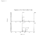
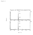
- Figure 3 is a spectrum of mass analysis of the 134 to 139 residues of the myoglobin (Fragment a), wherein upper spectrum corresponds not to perform the reacting step of the present invention, and lower spectrum corresponds to perform the reacting step of the present invention. According to a comparison between the upper spectrum and lower spectrum, a peak around 762 (m/z) which does not appear in the upper spectrum is appeared in the lower spectrum. This indicates that the glutamic acid residue is contained in the peptide fragment of 134 to 139 residues of the myoglobin in relation to the molecular weight of the Fragment a (747.428) as observed in the upper spectrum.
- the change of the molecular weight originated from the substituted residue depends on the substituted residue corresponding to the alcohols in the method for modifying a peptide according to the present invention, although the above-mentioned example shows in the case of using methanol as the alcohol for the reacting step (the substituted residue is methyl group). Therefore, the description of the determining step can apply such that it replaced with the molecular weight of the substituted residue used for the reacting step.
- peptide fragments within the error range of the molecular weight as observed in the measurement are cyclopaedically extracted from the database in the PMF method. That is, peptide fragment(s) which is in the specific range of the molecular weight are cyclopaedically searched, and all these peptide fragments are handled as candidate of the protein. Therefore, the peptide fragment, the candidate protein, which is extracted from the database is based on the molecular weight as one of indexes.
- variable number such as presence or absence of the glutamic acid residue or number of the glutamic acid residue is obtained, along with the molecular weight. Therefore, it is possible to utilize the presence or absence of the glutamic acid residue or the number of the glutamic acid residue as the indexes in addition to the molecular weight of the peptide fragment as the above-mentioned indexes for the searching.
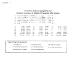
- Figure 1 shows sequence of horse myoglobin, deduced sequence of digested fragment as digested with trypsin and observed sequence of digested fragment.
- the upper portion indicates the amino acid sequence of horse myoglobin (Sequence Number 1)
- the middle portion indicates the deduced trypsin-digested fragment from the amino acid sequence
- the lower portion indicates the digested fragment as observed by MALDI-TOF-MS analysis.
- Each digested fragment as indicated in the lower portion correspond to 134 to 139 residues (Fragment a; Sequence Number 2), 32 to 42 residues (Fragment b; Sequence Number 3), 64 to 77 residues (Fragment c; Sequence Number 4), 119 to 133 residues (Fragment d; Sequence Number 5), 17 to 31 resides (Fragment e; Sequence Number 6), 1 to 16 residues (Fragment f; Sequence Number 7), 80 to 96 residues (Fragment g; Sequence Number 8) and 103 to 118 residues (Fragment h; Sequence Number 9) of myoglobin, respectively.
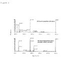
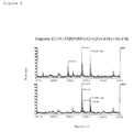
- Figure 2 is figures showing spectrum of mass analysis of myoglobin as obtained in accordance with the method of the present invention in which the glutamic acid residue thereof is esterified, wherein (A) corresponds to the trypsin digestion of horse myoglobin, and (B) corresponds to the trypsin digestion of horse myoglobin which glutamic acid residue is esterified.
- Figures 3 to 9 are that the horizontal axis of the spectrum of mass analysis as indicated in Figure 2 is enlarged and these figures indicates each of Fragments a to h, respectively.
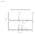
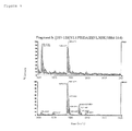
- Figures 3 to 9 corresponds respectively to: Figure 3 : 134 to 139 residues (Fragment a; Sequence Number 2); Figure 4 : 32 to 42 residues (Fragment b; Sequence Number 3); Figure 5 : 64 to 77 residues (Fragment c; Sequence Number 4); Figure 6 : 119 to 133 residues (Fragment d; Sequence Number 5); Figure 7 : 17 to 31 residues (Fragment e; (Sequence Number 6); Figure 8 : 1 to 16 residues (Fragment f; Sequence Number 7) and 80 to 96 residues (Fragment g; Sequence Number 8); and Figure 9 : 103 to 118 residues (Fragment h; Sequence Number 9); of myoglobin.
- the peak was newly appeared at a position which is shifted frog the original one by 14 of the molecular weight, and the new peak was also appeared at a position which is further shifted from the peak by 14 of the molecular weight (in Figure 4 , corresponding respectively to the peaks at 1285.6866 and 1299.7527 with regard to the original peak at 1271.6727, and in Figure 7 , corresponding respectively to the peaks at 1621.0698 and 1635.0870 with regard to the original peak at 1607.0612).
- the modification reaction such as the esterification is not occurred or is less occurred if occurring, in the aspartic residue container in the peptide, and that the modification reaction is specific to the glutamic acid residue.
- the identification is possible for the methyl ester of the glutamic acid residue in view of the intensity, even if the esterification of the aspartic residue is occurred.
- the spectrum originated from molecule(s) such as Ser-N, Thr-N and Asp-C wherein the molecule has a weak bonding to the strong acid such as hydrochloric acid was not observed, and deamination of glutamine and asparagine is not observed. Therefore, it can be recognized that the method for modifying a peptide according to the present invention is very useful for the structural analysis of protein.
Landscapes
- Life Sciences & Earth Sciences (AREA)
- Chemical & Material Sciences (AREA)
- Health & Medical Sciences (AREA)
- Molecular Biology (AREA)
- Engineering & Computer Science (AREA)
- Proteomics, Peptides & Aminoacids (AREA)
- Medicinal Chemistry (AREA)
- Organic Chemistry (AREA)
- Physics & Mathematics (AREA)
- Biomedical Technology (AREA)
- General Health & Medical Sciences (AREA)
- Biochemistry (AREA)
- Hematology (AREA)
- Immunology (AREA)
- Analytical Chemistry (AREA)
- Urology & Nephrology (AREA)
- Biophysics (AREA)
- Pathology (AREA)
- Bioinformatics & Computational Biology (AREA)
- Microbiology (AREA)
- Cell Biology (AREA)
- Biotechnology (AREA)
- General Physics & Mathematics (AREA)
- Bioinformatics & Cheminformatics (AREA)
- General Chemical & Material Sciences (AREA)
- Food Science & Technology (AREA)
- Genetics & Genomics (AREA)
- Chemical Kinetics & Catalysis (AREA)
- Peptides Or Proteins (AREA)
- Investigating Or Analysing Biological Materials (AREA)
- Measuring Or Testing Involving Enzymes Or Micro-Organisms (AREA)
- Other Investigation Or Analysis Of Materials By Electrical Means (AREA)
Applications Claiming Priority (2)
| Application Number | Priority Date | Filing Date | Title |
|---|---|---|---|
| JP2006031098 | 2006-02-08 | ||
| PCT/JP2007/052205 WO2007091628A1 (ja) | 2006-02-08 | 2007-02-08 | ペプチドの修飾方法及びペプチドの同定方法 |
Publications (2)
| Publication Number | Publication Date |
|---|---|
| EP1982991A1 true EP1982991A1 (de) | 2008-10-22 |
| EP1982991A4 EP1982991A4 (de) | 2009-10-21 |
Family
ID=38345222
Family Applications (1)
| Application Number | Title | Priority Date | Filing Date |
|---|---|---|---|
| EP07713921A Withdrawn EP1982991A4 (de) | 2006-02-08 | 2007-02-08 | Verfahren zur modifizierung von peptid und verfahren zur identifizierung von peptid |
Country Status (5)
| Country | Link |
|---|---|
| US (1) | US20110053196A1 (de) |
| EP (1) | EP1982991A4 (de) |
| JP (1) | JP4771297B2 (de) |
| CN (1) | CN101379074A (de) |
| WO (1) | WO2007091628A1 (de) |
Families Citing this family (1)
| Publication number | Priority date | Publication date | Assignee | Title |
|---|---|---|---|---|
| GB0906698D0 (en) * | 2009-04-17 | 2009-06-03 | Queen Mary & Westfield College | Method for quantifying modified peptides |
Family Cites Families (9)
| Publication number | Priority date | Publication date | Assignee | Title |
|---|---|---|---|---|
| JP2000053773A (ja) * | 1998-08-06 | 2000-02-22 | Fuji Oil Co Ltd | 改質大豆たん白質及びその製法 |
| GB9821393D0 (en) * | 1998-10-01 | 1998-11-25 | Brax Genomics Ltd | Protein profiling 2 |
| AU2003211941A1 (en) * | 2002-02-14 | 2003-09-04 | Ajinomoto Co., Inc. | Method of analyzing aminofunctional compound and analytical reagent |
| JP4102581B2 (ja) * | 2002-03-25 | 2008-06-18 | 日本電気株式会社 | ペプチドのc末端アミノ酸配列解析方法 |
| JP4257492B2 (ja) * | 2002-11-29 | 2009-04-22 | 日本電気株式会社 | ペプチドのc末端アミノ酸配列解析方法 |
| JP4086642B2 (ja) * | 2002-12-10 | 2008-05-14 | 日本電気株式会社 | ペプチドのc末端アミノ酸配列解析方法 |
| JP3534191B1 (ja) * | 2002-12-26 | 2004-06-07 | 日本電気株式会社 | 質量分析法を利用するペプチドc末端アミノ酸配列解析方法 |
| US7422865B2 (en) * | 2003-01-13 | 2008-09-09 | Agilent Technologies, Inc. | Method of identifying peptides in a proteomic sample |
| JP2006031098A (ja) | 2004-07-12 | 2006-02-02 | Nec Soft Ltd | 金融機関の業務システム |
-
2007
- 2007-02-08 US US12/278,711 patent/US20110053196A1/en not_active Abandoned
- 2007-02-08 JP JP2007557883A patent/JP4771297B2/ja not_active Expired - Fee Related
- 2007-02-08 EP EP07713921A patent/EP1982991A4/de not_active Withdrawn
- 2007-02-08 CN CNA2007800049247A patent/CN101379074A/zh active Pending
- 2007-02-08 WO PCT/JP2007/052205 patent/WO2007091628A1/ja not_active Ceased
Also Published As
| Publication number | Publication date |
|---|---|
| EP1982991A4 (de) | 2009-10-21 |
| US20110053196A1 (en) | 2011-03-03 |
| WO2007091628A1 (ja) | 2007-08-16 |
| CN101379074A (zh) | 2009-03-04 |
| JP4771297B2 (ja) | 2011-09-14 |
| JPWO2007091628A1 (ja) | 2009-07-02 |
Similar Documents
| Publication | Publication Date | Title |
|---|---|---|
| Keough et al. | Peer Reviewed: Sulfonic Acid Derivatives for Peptide Sequencing by MALDI MS. | |
| Gauci et al. | Quantitative proteomics: assessing the spectrum of in-gel protein detection methods | |
| Griffin et al. | Structural analysis of proteins by capillary HPLC electrospray tandem mass spectrometry | |
| US8119411B2 (en) | Method for analyzing C-terminal amino acid sequence of peptide using mass spectrometry | |
| Fan et al. | Mass spectrometry in the discovery of peptides involved in intercellular communication: From targeted to untargeted peptidomics approaches | |
| AU2001273568A1 (en) | Methods and kits for sequencing polypeptides | |
| AU757356B2 (en) | Methods and kits for sequencing polypeptides | |
| Reid et al. | Capillary column chromatography improves sample preparation for mass spectrometric analysis: Complete characterization of human α‐enolase from two‐dimensional gels following in situ proteolytic digestion | |
| EP1982991A1 (de) | Verfahren zur modifizierung von peptid und verfahren zur identifizierung von peptid | |
| Friso et al. | The workflow for quantitative proteome analysis of chloroplast development and differentiation, chloroplast mutants, and protein interactions by spectral counting | |
| JP2005189232A (ja) | 複合体混合物中のタンパク質の同定と定量的分析のための選択的ペプチド単離法 | |
| JP4604345B2 (ja) | タンパク質のn末端のアミノ酸配列決定方法 | |
| Bunk et al. | Isotope dilution liquid chromatography-tandem mass spectrometry for quantitative amino acid analysis | |
| Staes et al. | Benchmarking DIA data analysis workflows | |
| JP4543929B2 (ja) | タンパク質の解析方法 | |
| JP4257492B2 (ja) | ペプチドのc末端アミノ酸配列解析方法 | |
| US20070112181A1 (en) | Method for enrichment/seperation of protein or peptide | |
| Smith et al. | Protein Sequencing | |
| HELLMAN | Peptide Mapping Using | |
| EP1983346A1 (de) | Verfahren zur peptidbindungsspaltung am c-terminus des peptids sowie verfahren zur bestimmung der c-terminalen aminosäurensequenz des peptids | |
| JP2009092411A (ja) | ペプチドの同定方法 | |
| Jiang et al. | Combination of MALDI-TOF mass spectrometry with immobilized enzyme microreactor for peptide mapping | |
| EP1582872A1 (de) | Verfahren zur analyse der c-terminalen aminosäuresequenz eines peptides | |
| Williams et al. | Comparative Analysis of Proteoform Clean-Up Methods for Improved Top-Down Proteomics | |
| Po | Analysis of the complex proteoform characterisation challenge using cAMP-dependent protein kinase A |
Legal Events
| Date | Code | Title | Description |
|---|---|---|---|
| PUAI | Public reference made under article 153(3) epc to a published international application that has entered the european phase |
Free format text: ORIGINAL CODE: 0009012 |
|
| 17P | Request for examination filed |
Effective date: 20080717 |
|
| AK | Designated contracting states |
Kind code of ref document: A1 Designated state(s): DE FR GB |
|
| RBV | Designated contracting states (corrected) |
Designated state(s): DE FR GB |
|
| DAX | Request for extension of the european patent (deleted) | ||
| RBV | Designated contracting states (corrected) |
Designated state(s): DE FR GB |
|
| A4 | Supplementary search report drawn up and despatched |
Effective date: 20090921 |
|
| 17Q | First examination report despatched |
Effective date: 20091113 |
|
| RIC1 | Information provided on ipc code assigned before grant |
Ipc: G01N 33/68 20060101ALI20101202BHEP Ipc: C07K 1/107 20060101AFI20101202BHEP Ipc: C07K 1/13 20060101ALI20101202BHEP |
|
| RTI1 | Title (correction) |
Free format text: METHOD FOR MODIFYING A PEPTIDE AND METHOD FOR IDENTIFYING A PEPTIDE |
|
| GRAP | Despatch of communication of intention to grant a patent |
Free format text: ORIGINAL CODE: EPIDOSNIGR1 |
|
| GRAC | Information related to communication of intention to grant a patent modified |
Free format text: ORIGINAL CODE: EPIDOSCIGR1 |
|
| STAA | Information on the status of an ep patent application or granted ep patent |
Free format text: STATUS: THE APPLICATION IS DEEMED TO BE WITHDRAWN |
|
| 18D | Application deemed to be withdrawn |
Effective date: 20110712 |