EP1967856A2 - Tumour marker proteins and uses thereof - Google Patents
Tumour marker proteins and uses thereof Download PDFInfo
- Publication number
- EP1967856A2 EP1967856A2 EP08007680A EP08007680A EP1967856A2 EP 1967856 A2 EP1967856 A2 EP 1967856A2 EP 08007680 A EP08007680 A EP 08007680A EP 08007680 A EP08007680 A EP 08007680A EP 1967856 A2 EP1967856 A2 EP 1967856A2
- Authority
- EP
- European Patent Office
- Prior art keywords
- tumour
- marker protein
- tumour marker
- cancer
- autoantibodies
- Prior art date
- Legal status (The legal status is an assumption and is not a legal conclusion. Google has not performed a legal analysis and makes no representation as to the accuracy of the status listed.)
- Withdrawn
Links
- 108090000623 proteins and genes Proteins 0.000 title claims abstract description 144
- 102000004169 proteins and genes Human genes 0.000 title claims abstract description 144
- 239000000439 tumor marker Substances 0.000 title claims abstract description 128
- 206010028980 Neoplasm Diseases 0.000 claims abstract description 182
- 201000011510 cancer Diseases 0.000 claims abstract description 131
- 239000012530 fluid Substances 0.000 claims abstract description 70
- 238000001514 detection method Methods 0.000 claims abstract description 36
- 230000029142 excretion Effects 0.000 claims abstract description 35
- 208000002151 Pleural effusion Diseases 0.000 claims abstract description 33
- 206010040102 Seroma Diseases 0.000 claims abstract description 27
- 238000002360 preparation method Methods 0.000 claims abstract description 20
- 210000004881 tumor cell Anatomy 0.000 claims abstract description 8
- 239000003153 chemical reaction reagent Substances 0.000 claims description 65
- 238000000034 method Methods 0.000 claims description 63
- 101001133056 Homo sapiens Mucin-1 Proteins 0.000 claims description 61
- 102100034256 Mucin-1 Human genes 0.000 claims description 59
- 238000003018 immunoassay Methods 0.000 claims description 47
- 210000001124 body fluid Anatomy 0.000 claims description 39
- 238000003556 assay Methods 0.000 claims description 33
- 102100025064 Cellular tumor antigen p53 Human genes 0.000 claims description 26
- 101000623901 Homo sapiens Mucin-16 Proteins 0.000 claims description 21
- 102100023123 Mucin-16 Human genes 0.000 claims description 21
- 102100038895 Myc proto-oncogene protein Human genes 0.000 claims description 19
- 101710135898 Myc proto-oncogene protein Proteins 0.000 claims description 19
- 101710150448 Transcriptional regulator Myc Proteins 0.000 claims description 19
- 102000052609 BRCA2 Human genes 0.000 claims description 18
- 108700020462 BRCA2 Proteins 0.000 claims description 18
- 101150008921 Brca2 gene Proteins 0.000 claims description 18
- 102000036365 BRCA1 Human genes 0.000 claims description 17
- 108700020463 BRCA1 Proteins 0.000 claims description 17
- 101150072950 BRCA1 gene Proteins 0.000 claims description 17
- 238000012360 testing method Methods 0.000 claims description 15
- 108060003951 Immunoglobulin Proteins 0.000 claims description 13
- 102000018358 immunoglobulin Human genes 0.000 claims description 13
- 239000011324 bead Substances 0.000 claims description 12
- 238000005259 measurement Methods 0.000 claims description 10
- 239000007787 solid Substances 0.000 claims description 9
- 210000002700 urine Anatomy 0.000 claims description 9
- 238000004519 manufacturing process Methods 0.000 claims description 8
- 238000004587 chromatography analysis Methods 0.000 claims description 4
- 210000003608 fece Anatomy 0.000 claims description 4
- 238000000338 in vitro Methods 0.000 claims description 4
- 210000000582 semen Anatomy 0.000 claims description 3
- 230000001225 therapeutic effect Effects 0.000 claims description 3
- 210000000941 bile Anatomy 0.000 claims description 2
- 238000001727 in vivo Methods 0.000 claims description 2
- 238000002798 spectrophotometry method Methods 0.000 claims description 2
- 238000011156 evaluation Methods 0.000 claims 2
- 239000000203 mixture Substances 0.000 claims 2
- 206010003445 Ascites Diseases 0.000 abstract description 23
- 238000011160 research Methods 0.000 abstract description 5
- 235000018102 proteins Nutrition 0.000 description 119
- 210000002966 serum Anatomy 0.000 description 41
- 239000000427 antigen Substances 0.000 description 38
- 102000036639 antigens Human genes 0.000 description 38
- 108091007433 antigens Proteins 0.000 description 38
- LOKCTEFSRHRXRJ-UHFFFAOYSA-I dipotassium trisodium dihydrogen phosphate hydrogen phosphate dichloride Chemical compound P(=O)(O)(O)[O-].[K+].P(=O)(O)([O-])[O-].[Na+].[Na+].[Cl-].[K+].[Cl-].[Na+] LOKCTEFSRHRXRJ-UHFFFAOYSA-I 0.000 description 32
- 239000002953 phosphate buffered saline Substances 0.000 description 32
- 230000009257 reactivity Effects 0.000 description 27
- 206010006187 Breast cancer Diseases 0.000 description 23
- 208000037265 diseases, disorders, signs and symptoms Diseases 0.000 description 22
- 201000010099 disease Diseases 0.000 description 18
- 238000003745 diagnosis Methods 0.000 description 16
- 230000028993 immune response Effects 0.000 description 16
- 208000026310 Breast neoplasm Diseases 0.000 description 15
- 238000002965 ELISA Methods 0.000 description 14
- 206010052428 Wound Diseases 0.000 description 13
- 208000027418 Wounds and injury Diseases 0.000 description 13
- 230000000259 anti-tumor effect Effects 0.000 description 12
- 239000010839 body fluid Substances 0.000 description 11
- 238000000746 purification Methods 0.000 description 11
- 108010022366 Carcinoembryonic Antigen Proteins 0.000 description 9
- 102100025475 Carcinoembryonic antigen-related cell adhesion molecule 5 Human genes 0.000 description 9
- 229920000136 polysorbate Polymers 0.000 description 9
- 238000011282 treatment Methods 0.000 description 9
- 101100346932 Mus musculus Muc1 gene Proteins 0.000 description 8
- 230000008859 change Effects 0.000 description 8
- 208000008884 Aneurysmal Bone Cysts Diseases 0.000 description 7
- 210000003567 ascitic fluid Anatomy 0.000 description 7
- 230000008901 benefit Effects 0.000 description 7
- 210000004027 cell Anatomy 0.000 description 7
- 239000012634 fragment Substances 0.000 description 7
- 238000003384 imaging method Methods 0.000 description 7
- 238000012544 monitoring process Methods 0.000 description 7
- 238000012216 screening Methods 0.000 description 7
- 238000001042 affinity chromatography Methods 0.000 description 6
- 230000001900 immune effect Effects 0.000 description 6
- 230000004044 response Effects 0.000 description 6
- 210000004369 blood Anatomy 0.000 description 5
- 239000008280 blood Substances 0.000 description 5
- 229940022399 cancer vaccine Drugs 0.000 description 5
- 238000005119 centrifugation Methods 0.000 description 5
- 238000002523 gelfiltration Methods 0.000 description 5
- 239000003550 marker Substances 0.000 description 5
- 239000000463 material Substances 0.000 description 5
- 108090000765 processed proteins & peptides Proteins 0.000 description 5
- 108010001336 Horseradish Peroxidase Proteins 0.000 description 4
- 241000283973 Oryctolagus cuniculus Species 0.000 description 4
- 229920002684 Sepharose Polymers 0.000 description 4
- 238000011394 anticancer treatment Methods 0.000 description 4
- 229940072221 immunoglobulins Drugs 0.000 description 4
- 230000001613 neoplastic effect Effects 0.000 description 4
- 238000011176 pooling Methods 0.000 description 4
- 230000008569 process Effects 0.000 description 4
- 239000000047 product Substances 0.000 description 4
- 239000006228 supernatant Substances 0.000 description 4
- 208000001333 Colorectal Neoplasms Diseases 0.000 description 3
- 206010061535 Ovarian neoplasm Diseases 0.000 description 3
- 208000000236 Prostatic Neoplasms Diseases 0.000 description 3
- 210000003719 b-lymphocyte Anatomy 0.000 description 3
- 238000011088 calibration curve Methods 0.000 description 3
- 238000011161 development Methods 0.000 description 3
- 230000018109 developmental process Effects 0.000 description 3
- VHJLVAABSRFDPM-QWWZWVQMSA-N dithiothreitol Chemical compound SC[C@@H](O)[C@H](O)CS VHJLVAABSRFDPM-QWWZWVQMSA-N 0.000 description 3
- 230000000306 recurrent effect Effects 0.000 description 3
- 239000000243 solution Substances 0.000 description 3
- 238000003860 storage Methods 0.000 description 3
- 210000001519 tissue Anatomy 0.000 description 3
- 238000001262 western blot Methods 0.000 description 3
- UAIUNKRWKOVEES-UHFFFAOYSA-N 3,3',5,5'-tetramethylbenzidine Chemical compound CC1=C(N)C(C)=CC(C=2C=C(C)C(N)=C(C)C=2)=C1 UAIUNKRWKOVEES-UHFFFAOYSA-N 0.000 description 2
- 206010009944 Colon cancer Diseases 0.000 description 2
- 206010061818 Disease progression Diseases 0.000 description 2
- 238000012286 ELISA Assay Methods 0.000 description 2
- 102000004190 Enzymes Human genes 0.000 description 2
- 108090000790 Enzymes Proteins 0.000 description 2
- 102000008394 Immunoglobulin Fragments Human genes 0.000 description 2
- 108010021625 Immunoglobulin Fragments Proteins 0.000 description 2
- 206010033128 Ovarian cancer Diseases 0.000 description 2
- 206010058823 Ovarian mass Diseases 0.000 description 2
- 206010060862 Prostate cancer Diseases 0.000 description 2
- 239000012505 Superdex™ Substances 0.000 description 2
- 239000007983 Tris buffer Substances 0.000 description 2
- 208000007097 Urinary Bladder Neoplasms Diseases 0.000 description 2
- 230000002159 abnormal effect Effects 0.000 description 2
- 238000011091 antibody purification Methods 0.000 description 2
- 230000001580 bacterial effect Effects 0.000 description 2
- 210000000481 breast Anatomy 0.000 description 2
- 230000000711 cancerogenic effect Effects 0.000 description 2
- 231100000315 carcinogenic Toxicity 0.000 description 2
- 239000005018 casein Substances 0.000 description 2
- BECPQYXYKAMYBN-UHFFFAOYSA-N casein, tech. Chemical compound NCCCCC(C(O)=O)N=C(O)C(CC(O)=O)N=C(O)C(CCC(O)=N)N=C(O)C(CC(C)C)N=C(O)C(CCC(O)=O)N=C(O)C(CC(O)=O)N=C(O)C(CCC(O)=O)N=C(O)C(C(C)O)N=C(O)C(CCC(O)=N)N=C(O)C(CCC(O)=N)N=C(O)C(CCC(O)=N)N=C(O)C(CCC(O)=O)N=C(O)C(CCC(O)=O)N=C(O)C(COP(O)(O)=O)N=C(O)C(CCC(O)=N)N=C(O)C(N)CC1=CC=CC=C1 BECPQYXYKAMYBN-UHFFFAOYSA-N 0.000 description 2
- 235000021240 caseins Nutrition 0.000 description 2
- 238000006243 chemical reaction Methods 0.000 description 2
- 230000003247 decreasing effect Effects 0.000 description 2
- 230000001419 dependent effect Effects 0.000 description 2
- 238000010790 dilution Methods 0.000 description 2
- 239000012895 dilution Substances 0.000 description 2
- 230000005750 disease progression Effects 0.000 description 2
- 239000003814 drug Substances 0.000 description 2
- 239000000499 gel Substances 0.000 description 2
- 238000001641 gel filtration chromatography Methods 0.000 description 2
- 230000013595 glycosylation Effects 0.000 description 2
- 238000006206 glycosylation reaction Methods 0.000 description 2
- 230000012010 growth Effects 0.000 description 2
- 210000004408 hybridoma Anatomy 0.000 description 2
- 210000000987 immune system Anatomy 0.000 description 2
- PGLTVOMIXTUURA-UHFFFAOYSA-N iodoacetamide Chemical compound NC(=O)CI PGLTVOMIXTUURA-UHFFFAOYSA-N 0.000 description 2
- 210000000056 organ Anatomy 0.000 description 2
- 238000002264 polyacrylamide gel electrophoresis Methods 0.000 description 2
- VGTPCRGMBIAPIM-UHFFFAOYSA-M sodium thiocyanate Chemical compound [Na+].[S-]C#N VGTPCRGMBIAPIM-UHFFFAOYSA-M 0.000 description 2
- 230000009870 specific binding Effects 0.000 description 2
- 229940124597 therapeutic agent Drugs 0.000 description 2
- 238000002560 therapeutic procedure Methods 0.000 description 2
- LENZDBCJOHFCAS-UHFFFAOYSA-N tris Chemical compound OCC(N)(CO)CO LENZDBCJOHFCAS-UHFFFAOYSA-N 0.000 description 2
- 230000002485 urinary effect Effects 0.000 description 2
- 102000002260 Alkaline Phosphatase Human genes 0.000 description 1
- 108020004774 Alkaline Phosphatase Proteins 0.000 description 1
- 208000002109 Argyria Diseases 0.000 description 1
- 206010005003 Bladder cancer Diseases 0.000 description 1
- 241000283707 Capra Species 0.000 description 1
- 102000005483 Cell Cycle Proteins Human genes 0.000 description 1
- 108010031896 Cell Cycle Proteins Proteins 0.000 description 1
- 230000004544 DNA amplification Effects 0.000 description 1
- 101100125027 Dictyostelium discoideum mhsp70 gene Proteins 0.000 description 1
- 206010061819 Disease recurrence Diseases 0.000 description 1
- 102000003886 Glycoproteins Human genes 0.000 description 1
- 108090000288 Glycoproteins Proteins 0.000 description 1
- 101150031823 HSP70 gene Proteins 0.000 description 1
- 102000002812 Heat-Shock Proteins Human genes 0.000 description 1
- 108010004889 Heat-Shock Proteins Proteins 0.000 description 1
- 241000238631 Hexapoda Species 0.000 description 1
- 241000282412 Homo Species 0.000 description 1
- 102100022420 Inactive rhomboid protein 1 Human genes 0.000 description 1
- 101710136320 Inactive rhomboid protein 1 Proteins 0.000 description 1
- 108700020796 Oncogene Proteins 0.000 description 1
- 102000003992 Peroxidases Human genes 0.000 description 1
- 206010036790 Productive cough Diseases 0.000 description 1
- 102000007056 Recombinant Fusion Proteins Human genes 0.000 description 1
- 108010008281 Recombinant Fusion Proteins Proteins 0.000 description 1
- 206010038111 Recurrent cancer Diseases 0.000 description 1
- 240000004808 Saccharomyces cerevisiae Species 0.000 description 1
- BQCADISMDOOEFD-UHFFFAOYSA-N Silver Chemical compound [Ag] BQCADISMDOOEFD-UHFFFAOYSA-N 0.000 description 1
- 238000010521 absorption reaction Methods 0.000 description 1
- 230000004075 alteration Effects 0.000 description 1
- BFNBIHQBYMNNAN-UHFFFAOYSA-N ammonium sulfate Chemical class N.N.OS(O)(=O)=O BFNBIHQBYMNNAN-UHFFFAOYSA-N 0.000 description 1
- 230000003698 anagen phase Effects 0.000 description 1
- 230000005875 antibody response Effects 0.000 description 1
- 230000000890 antigenic effect Effects 0.000 description 1
- 238000001574 biopsy Methods 0.000 description 1
- 230000015572 biosynthetic process Effects 0.000 description 1
- 239000012888 bovine serum Substances 0.000 description 1
- 239000000872 buffer Substances 0.000 description 1
- 208000035269 cancer or benign tumor Diseases 0.000 description 1
- 210000001175 cerebrospinal fluid Anatomy 0.000 description 1
- 230000004087 circulation Effects 0.000 description 1
- 238000003776 cleavage reaction Methods 0.000 description 1
- 230000002860 competitive effect Effects 0.000 description 1
- 230000000295 complement effect Effects 0.000 description 1
- 239000013078 crystal Substances 0.000 description 1
- ATDGTVJJHBUTRL-UHFFFAOYSA-N cyanogen bromide Chemical compound BrC#N ATDGTVJJHBUTRL-UHFFFAOYSA-N 0.000 description 1
- 238000002405 diagnostic procedure Methods 0.000 description 1
- 238000000502 dialysis Methods 0.000 description 1
- 230000004069 differentiation Effects 0.000 description 1
- 101150052825 dnaK gene Proteins 0.000 description 1
- 230000000694 effects Effects 0.000 description 1
- 238000001962 electrophoresis Methods 0.000 description 1
- 238000010828 elution Methods 0.000 description 1
- 230000002255 enzymatic effect Effects 0.000 description 1
- 230000001747 exhibiting effect Effects 0.000 description 1
- 239000007850 fluorescent dye Substances 0.000 description 1
- 210000000232 gallbladder Anatomy 0.000 description 1
- 102000035122 glycosylated proteins Human genes 0.000 description 1
- 108091005608 glycosylated proteins Proteins 0.000 description 1
- PCHJSUWPFVWCPO-UHFFFAOYSA-N gold Chemical compound [Au] PCHJSUWPFVWCPO-UHFFFAOYSA-N 0.000 description 1
- 238000003306 harvesting Methods 0.000 description 1
- 230000036541 health Effects 0.000 description 1
- 229910001385 heavy metal Inorganic materials 0.000 description 1
- 230000028996 humoral immune response Effects 0.000 description 1
- 230000008073 immune recognition Effects 0.000 description 1
- 230000002163 immunogen Effects 0.000 description 1
- 238000002955 isolation Methods 0.000 description 1
- 230000003902 lesion Effects 0.000 description 1
- 210000002751 lymph Anatomy 0.000 description 1
- 230000003211 malignant effect Effects 0.000 description 1
- 210000004962 mammalian cell Anatomy 0.000 description 1
- 238000009607 mammography Methods 0.000 description 1
- 239000011159 matrix material Substances 0.000 description 1
- 230000001404 mediated effect Effects 0.000 description 1
- 206010061289 metastatic neoplasm Diseases 0.000 description 1
- 239000011325 microbead Substances 0.000 description 1
- 238000007392 microtiter assay Methods 0.000 description 1
- 230000004048 modification Effects 0.000 description 1
- 238000012986 modification Methods 0.000 description 1
- 239000013642 negative control Substances 0.000 description 1
- 230000009826 neoplastic cell growth Effects 0.000 description 1
- 210000002445 nipple Anatomy 0.000 description 1
- 102000039446 nucleic acids Human genes 0.000 description 1
- 108020004707 nucleic acids Proteins 0.000 description 1
- 150000007523 nucleic acids Chemical class 0.000 description 1
- 108040007629 peroxidase activity proteins Proteins 0.000 description 1
- 229920001184 polypeptide Polymers 0.000 description 1
- 230000004481 post-translational protein modification Effects 0.000 description 1
- 239000002244 precipitate Substances 0.000 description 1
- 125000002924 primary amino group Chemical group [H]N([H])* 0.000 description 1
- 102000004196 processed proteins & peptides Human genes 0.000 description 1
- 210000002307 prostate Anatomy 0.000 description 1
- 238000001742 protein purification Methods 0.000 description 1
- 238000012207 quantitative assay Methods 0.000 description 1
- 238000003127 radioimmunoassay Methods 0.000 description 1
- 230000022532 regulation of transcription, DNA-dependent Effects 0.000 description 1
- 230000001105 regulatory effect Effects 0.000 description 1
- 238000005096 rolling process Methods 0.000 description 1
- 150000003839 salts Chemical class 0.000 description 1
- 230000007017 scission Effects 0.000 description 1
- 230000035945 sensitivity Effects 0.000 description 1
- 238000013207 serial dilution Methods 0.000 description 1
- 230000019491 signal transduction Effects 0.000 description 1
- 229910052709 silver Inorganic materials 0.000 description 1
- 239000004332 silver Substances 0.000 description 1
- 238000004611 spectroscopical analysis Methods 0.000 description 1
- 210000003802 sputum Anatomy 0.000 description 1
- 208000024794 sputum Diseases 0.000 description 1
- 238000010561 standard procedure Methods 0.000 description 1
- 239000007858 starting material Substances 0.000 description 1
- 239000000126 substance Substances 0.000 description 1
- 239000000758 substrate Substances 0.000 description 1
- 210000004243 sweat Anatomy 0.000 description 1
- 230000001839 systemic circulation Effects 0.000 description 1
- 230000009885 systemic effect Effects 0.000 description 1
- 238000002604 ultrasonography Methods 0.000 description 1
- 201000005112 urinary bladder cancer Diseases 0.000 description 1
- 238000002255 vaccination Methods 0.000 description 1
- 229960005486 vaccine Drugs 0.000 description 1
- 210000005253 yeast cell Anatomy 0.000 description 1
Images


Classifications
-
- G—PHYSICS
- G01—MEASURING; TESTING
- G01N—INVESTIGATING OR ANALYSING MATERIALS BY DETERMINING THEIR CHEMICAL OR PHYSICAL PROPERTIES
- G01N33/00—Investigating or analysing materials by specific methods not covered by groups G01N1/00 - G01N31/00
- G01N33/48—Biological material, e.g. blood, urine; Haemocytometers
- G01N33/50—Chemical analysis of biological material, e.g. blood, urine; Testing involving biospecific ligand binding methods; Immunological testing
- G01N33/53—Immunoassay; Biospecific binding assay; Materials therefor
- G01N33/564—Immunoassay; Biospecific binding assay; Materials therefor for pre-existing immune complex or autoimmune disease, i.e. systemic lupus erythematosus, rheumatoid arthritis, multiple sclerosis, rheumatoid factors or complement components C1-C9
-
- C—CHEMISTRY; METALLURGY
- C07—ORGANIC CHEMISTRY
- C07K—PEPTIDES
- C07K14/00—Peptides having more than 20 amino acids; Gastrins; Somatostatins; Melanotropins; Derivatives thereof
- C07K14/435—Peptides having more than 20 amino acids; Gastrins; Somatostatins; Melanotropins; Derivatives thereof from animals; from humans
- C07K14/46—Peptides having more than 20 amino acids; Gastrins; Somatostatins; Melanotropins; Derivatives thereof from animals; from humans from vertebrates
- C07K14/47—Peptides having more than 20 amino acids; Gastrins; Somatostatins; Melanotropins; Derivatives thereof from animals; from humans from vertebrates from mammals
- C07K14/4701—Peptides having more than 20 amino acids; Gastrins; Somatostatins; Melanotropins; Derivatives thereof from animals; from humans from vertebrates from mammals not used
- C07K14/4748—Tumour specific antigens; Tumour rejection antigen precursors [TRAP], e.g. MAGE
-
- G—PHYSICS
- G01—MEASURING; TESTING
- G01N—INVESTIGATING OR ANALYSING MATERIALS BY DETERMINING THEIR CHEMICAL OR PHYSICAL PROPERTIES
- G01N33/00—Investigating or analysing materials by specific methods not covered by groups G01N1/00 - G01N31/00
- G01N33/48—Biological material, e.g. blood, urine; Haemocytometers
- G01N33/50—Chemical analysis of biological material, e.g. blood, urine; Testing involving biospecific ligand binding methods; Immunological testing
- G01N33/68—Chemical analysis of biological material, e.g. blood, urine; Testing involving biospecific ligand binding methods; Immunological testing involving proteins, peptides or amino acids
- G01N33/6854—Immunoglobulins
-
- A—HUMAN NECESSITIES
- A61—MEDICAL OR VETERINARY SCIENCE; HYGIENE
- A61P—SPECIFIC THERAPEUTIC ACTIVITY OF CHEMICAL COMPOUNDS OR MEDICINAL PREPARATIONS
- A61P35/00—Antineoplastic agents
-
- C—CHEMISTRY; METALLURGY
- C07—ORGANIC CHEMISTRY
- C07K—PEPTIDES
- C07K1/00—General methods for the preparation of peptides, i.e. processes for the organic chemical preparation of peptides or proteins of any length
- C07K1/14—Extraction; Separation; Purification
- C07K1/36—Extraction; Separation; Purification by a combination of two or more processes of different types
-
- G—PHYSICS
- G01—MEASURING; TESTING
- G01N—INVESTIGATING OR ANALYSING MATERIALS BY DETERMINING THEIR CHEMICAL OR PHYSICAL PROPERTIES
- G01N33/00—Investigating or analysing materials by specific methods not covered by groups G01N1/00 - G01N31/00
- G01N33/48—Biological material, e.g. blood, urine; Haemocytometers
- G01N33/50—Chemical analysis of biological material, e.g. blood, urine; Testing involving biospecific ligand binding methods; Immunological testing
- G01N33/53—Immunoassay; Biospecific binding assay; Materials therefor
- G01N33/574—Immunoassay; Biospecific binding assay; Materials therefor for cancer
- G01N33/57473—Immunoassay; Biospecific binding assay; Materials therefor for cancer involving carcinoembryonic antigen, i.e. CEA
-
- G—PHYSICS
- G01—MEASURING; TESTING
- G01N—INVESTIGATING OR ANALYSING MATERIALS BY DETERMINING THEIR CHEMICAL OR PHYSICAL PROPERTIES
- G01N33/00—Investigating or analysing materials by specific methods not covered by groups G01N1/00 - G01N31/00
- G01N33/48—Biological material, e.g. blood, urine; Haemocytometers
- G01N33/50—Chemical analysis of biological material, e.g. blood, urine; Testing involving biospecific ligand binding methods; Immunological testing
- G01N33/53—Immunoassay; Biospecific binding assay; Materials therefor
- G01N33/574—Immunoassay; Biospecific binding assay; Materials therefor for cancer
- G01N33/57484—Immunoassay; Biospecific binding assay; Materials therefor for cancer involving compounds serving as markers for tumor, cancer, neoplasia, e.g. cellular determinants, receptors, heat shock/stress proteins, A-protein, oligosaccharides, metabolites
- G01N33/57488—Immunoassay; Biospecific binding assay; Materials therefor for cancer involving compounds serving as markers for tumor, cancer, neoplasia, e.g. cellular determinants, receptors, heat shock/stress proteins, A-protein, oligosaccharides, metabolites involving compounds identifable in body fluids
-
- G—PHYSICS
- G01—MEASURING; TESTING
- G01N—INVESTIGATING OR ANALYSING MATERIALS BY DETERMINING THEIR CHEMICAL OR PHYSICAL PROPERTIES
- G01N2800/00—Detection or diagnosis of diseases
- G01N2800/24—Immunology or allergic disorders
Definitions
- the invention relates to tumour marker proteins and their preparation from fluids from one or more cancer patients, wherein said fluids are those which collect in a body cavity or space which is naturally occurring or which is the result of cancer or medical intervention for cancer.
- Exemplary fluids are ascites, pleural effusion, seroma, hydrocoele and wound drainage fluid.
- the invention also relates to preparation of tumour marker proteins from excretions taken from patients with cancer.
- tumour marker proteins are useful in cancer detection methods which involve detecting or quantitatively measuring autoantibodies to circulating tumour markers or markers expressed on or in tumour cells and in various research applications.
- the invention is also directed to such uses.
- Tumour markers are often found to be altered forms of wild-type proteins expressed by "normal" cells, in which case the alteration may be a change in primary amino acid sequence, a change in secondary, tertiary or quaternary structure or a change in post-translational modification, for example, abnormal glycosylation.
- wild-type proteins which are up-regulated or over-expressed in tumour cells, possibly as a result of gene amplification or abnormal transcriptional regulation, may also be tumour markers.
- tumour markers present in bodily fluids tend to focus on the detection of tumour markers which reflect tumour bulk and as such are of value late in the disease process, for example in the diagnosis of metastatic disease.
- the most widely used of these markers include carcinoembryonic antigen (CEA) and the glycoprotein termed CA 15.3, both of which have been useful mainly as indicators of systemic disease burden and of relapse following therapy ( Molina, R., Zanon, G., Filella, X. et al. Use of serial carcinoembryonic antigen and CA 15.3 assays in detecting relapses in breast cancer patients. (1995) Breast Cancer Res Treat 36: 41-48 ).
- tumour markers are of limited use earlier in the course of the disease, for example in early detection or in the screening of asymptomatic patients.
- the present inventors have sought to identify markers which do not depend on tumour bulk per se.
- tumour marker protein Differences between a wild type protein expressed by "normal” cells and a corresponding tumour marker protein may, in some instances, lead to the tumour marker protein being recognised by an individual's immune system as "non-self” and thus eliciting an immune response in that individual.
- This may be a humoral (i.e B cell-mediated) immune response leading to the production of autoantibodies immunologically specific to the tumour marker protein.
- Autoantibodies are naturally occurring antibodies directed to an antigen which an individual's immune system recognises as foreign even though that antigen actually originated in the individual. They may be present in the circulation as circulating free autoantibodies or in the form of circulating immune complexes consisting of autoantibodies bound to their target tumour marker protein.
- tumour marker protein in bodily fluids, assays may be developed to measure the immune response of the individual to the presence of tumour marker protein in terms of autoantibody production. Such assays essentially constitute indirect detection of the presence of tumour marker protein. Because of the nature of the immune response, it is likely that autoantibodies can be elicited by a very small amount of circulating tumour marker protein and indirect methods which rely on detecting the immune response to tumour markers will consequently be more sensitive than methods for the direct measurement of tumour markers in bodily fluids.
- Assay methods based on the detection of autoantibodies may therefore be of particular value early in the disease process and possibly also in relation to screening of asymptomatic patients, for example in screening to identify individuals "at risk” of developing disease amongst a population of asymptomatic individuals. Furthermore, they may be useful for earlier detection of recurrent disease.
- Tumour marker proteins observed to elicit serum autoantibodies include a particular class of mutant p53 protein, described in US Patent No. 5,652,115 , which can be defined by its ability to bind to the 70 kd heat shock protein (hsp70).
- p53 autoantibodies can be detected in patients with a number of different benign and malignant conditions (described in US 5,652,115 ) but are in each case present in only a subset of patients. For example, one study utilizing an ELISA assay for detection of autoantibodies directed against the p53 protein in the serum of breast cancer patients reported that p53 autoantibodies were produced by 26% of patients and 1.3% of control subjects ( Mudenda, B., Green, J. A., Green, B.
- a second tumour marker protein known to elicit serum autoantibodies is the epithelial mucin MUC1 ( Hinoda, Y. et al. (1993) Immunol Lett. 35: 163-168 ; Kotera, Y. et al. (1994) Cancer Res. 54: 2856-2860 ).
- WO 99/58978 describes methods for use in the detection/diagnosis of cancer which are based on evaluating the immune response of an individual to two or more distinct tumour markers. These methods generally involve contacting a sample of bodily fluid taken from the individual with a panel of two or more distinct tumour marker antigens, each derived from a separate tumour marker protein, and detecting the formation of complexes of the tumour marker antigens bound to circulating autoantibodies immunologically specific for the tumour marker proteins. The presence of such circulating autoantibodies is taken as an indication of the presence of cancer.
- Cancer detection methods based on detection of circulating autoantibodies are frequently immunoassays utilizing an "immunoassay reagent" reactive with the circulating autoantibodies.
- the "reagents" used in such assays comprise recombinant tumour marker proteins (expressed in bacterial, insect, yeast or mammalian cells) or chemically synthesised tumour marker antigens, which may comprise substantially whole tumour marker proteins, or fragments thereof, such as short peptide antigens.
- Other potential sources of tumour-associated proteins for use as the basis of immunoassay reagents for the detection of anti-tumour auto-antibodies include cultured tumour cells (and the spent media used for their growth), tumour tissue, and serum from individuals with neoplasia. The majority of these sources have significant drawbacks, as discussed below.
- tumour cells With cultured tumour cells (and their spent media) the amount of expressed protein can vary depending on growth phase at the time of harvest, leading to variations in quality and quantity. In addition, the desired protein is generally present at low concentration, therefore it is time-consuming to purify sufficient quantities of protein. Furthermore, the cell stock will be clonal, unlike cell stock in a tumour which is likely to have become heterogeneous in nature during the growth of the neoplasm, therefore producing variations in protein (especially in the degree of glycosylation).
- Recombinant proteins expressed in bacterial cells are not glycosylated, and thus significantly different from naturally glycosylated proteins. In addition, refolding of recombinantly expressed proteins may not be appropriate, thus giving an incorrect conformation for auto-antibody recognition.
- Tumour tissue is usually only available in small quantities and the purification of proteins therefrom is laborious and time consuming.
- Serum samples are usually available only in small quantities, therefore it is difficult to purify sufficient quantities of protein.
- tumour marker antigens purified from bodily fluids derived from a body cavity or space in which a tumour is present or with which it is or was associated such as ascites fluid, pleural effusion, seroma, hydrocoele or wound drainage fluid, or from excretions, as the "reagent" in auto-antibody immunoassays.
- the inventors have observed that use of reagents comprising tumour marker antigens purified from bodily fluids derived from the above defined body cavities or spaces results in increased sensitivity (as compared to the use of reagents derived from a "normal” body fluid) and produces a more "clinically relevant” result.
- reagents comprising tumour marker antigens purified from bodily fluids derived from the above defined body cavities or spaces results in increased sensitivity (as compared to the use of reagents derived from a "normal” body fluid) and produces a more "clinically relevant” result.
- the invention in a first aspect relates to a method of detecting cancer-associated anti-tumour autoantibodies, which method is an immunoassay comprising contacting a sample to be tested for the presence of such autoantibodies with an immunoassay reagent and detecting the presence of complexes formed by specific binding of the immunoassay reagent to any cancer-associated anti-tumour autoantibodies present in the sample, wherein the immunoassay reagent comprises tumour marker protein prepared from bodily fluid derived from a body cavity or space within which a tumour is or was present or with which a tumour is or was associated, from one or more cancer patients and/or tumour marker protein prepared from an excretion from one or more cancer patients, wherein said tumour marker protein exhibits selective reactivity with cancer-associated anti-tumour autoantibodies.
- the invention relates to use of tumour marker protein prepared from bodily fluid derived from a body cavity or space within which a tumour is or was present or with which a tumour is or was associated, of one or more cancer patients and/or tumour marker protein derived from an excretion of one or more cancer patients in the manufacture of an immunoassay reagent exhibiting selective reactivity with cancer-associated anti-tumour autoantibodies.
- the invention relates to a method of preparing a tumour marker protein which method comprises isolating said tumour marker protein from bodily fluid wherein said fluid is:
- the invention relates to a method of preparing a tumour marker protein which method comprises isolating said tumour marker protein from an excretion wherein:
- tumour marker protein preparations prepared using the methods described above which are substantially immunoglobulin free and to kits and reagents comprising said preparations.
- the invention relates to a method of detecting "cancer-associated" anti-tumour autoantibodies.
- cancer-associated anti-tumour autoantibodies refers to autoantibodies which are characteristic of the cancer disease state, and which are directed against epitopes present on forms of tumour marker proteins which are preferentially expressed in the cancer disease state.
- the method of the invention comprises an immunoassay to detect and/or quantitatively measure autoantibodies immunologically specific for one or more tumour marker proteins, and is characterised in that the "immunoassay reagent" used in the immunoassay comprises tumour marker protein prepared from bodily fluid derived from a body cavity or space in which a tumour is or was present or with which a tumour is or was associated, from one or more cancer patients and/or tumour marker protein prepared from an excretion of one or more cancer patients.
- the excretion will have passed through an organ in which cancer is present wherein the excretion is in contact with said cancer, or the excretion will include one or more components which have been in contact with cancer elsewhere in the body.
- a particular example is bile which may be in contact with cancer in the gall bladder but will appear in the faeces.
- the immunoassay reagent exhibits "selective reactivity" with cancer-associated anti-tumour autoantibodies.
- selective reactivity means a tumour marker protein has a greater affinity for autoantibodies to the tumour-associated antigen than it does for any antibody or autoantibody made to the same antigen which exists in the normal i.e. non-tumour possessing state.
- body cavity or space includes any body cavity or space, whether it be a natural cavity or a space or cavity arising as a result of diseases or medical intervention including collapsed or former cavities.
- the fluid is derived from such a cavity or space in which a tumour is or was present or with which a tumour is or was associated.
- the "bodily fluid derived from a body cavity” will be a tumour-induced body fluid, meaning a body fluid which is produced during the disease process, for example in response to or as a consequence of the presence of tumour cells.
- Exemplary body fluids are ascites, pleural effusion, seroma, hydrocoele and wound drainage fliud.
- fluids derived from a body cavity or space do not include fluids derived from the systemic circulation, such as whole blood or serum.
- excretion includes, inter alia, urine, faeces, and seminal fluid.
- Immunoassays for example ELISA, radioimmunoassays and the like, are well known to those skilled in the art (see Immunoassay, E. Diamandis and T. Christopoulus, Academic Press, Inc., San Diego, CA, 1996 ).
- Immunoassays for the detection of antibodies having a particular immunological specificity generally require the use of a reagent that exhibits specific immunological reactivity with the antibody under test. Depending on the format of the assay this reagent may be immobilised on a solid support.
- a sample to be tested for the presence of the antibody is brought into contact with the reagent and if antibodies of the required immunological reactivity are present in the sample they will immunologically react with the reagent to form autoantibody-reagent complexes which may then be detected or quantitatively measured.
- tumour marker protein for use as the basis of the "immunoassay reagent" may be isolated from bodily fluids derived from a body cavity or space from one or more cancer patients and/or from excretions from one or more cancer patients using standard protein purification techniques, such as are generally known in the art.
- tumour marker proteins may be isolated by affinity chromatography using a suitable antibody (or antibody fragment) immunologically specific for the tumour marker protein.
- suitable antibody or antibody fragment
- the inventors have shown in the accompanying examples that several different tumour marker proteins may be purified using purification methods based on affinity chromatography. It would be apparent to the skilled reader that analogous purification methods used for any other tumour marker proteins, with the use of a suitable antibody or antibody fragment.
- the starting material of bodily fluids derived from a body cavity and/or excretions is/are taken from one or more cancer patients.
- cancer patient includes an individual previously diagnosed as having cancer.
- the fluid/excretion may be taken from a single patient or samples from two or more patients may be pooled together. Samples may be pooled from two or more patients having the same or different stages of the same or different types of cancers. Samples may also be pooled from different types of bodily fluids or excretions from a single or multiple patients.
- an immunoassay reagent prepared from fluid and/or excretion taken from cancer patient(s) with a particular type of cancer may be used to assist in the diagnosis of the same types of cancers in other individuals.
- the "cancer patient" from which the fluid/excretion is taken may be the same patient which it is later intended to test using the assay reagent.
- a stock of reagent prepared from a patient diagnosed with cancer may be used at a later date to assess the immune status of the same patient, for example to monitor disease progression and/or to assess the effectiveness of a course of anti-cancer treatment in that patient.
- tumour marker preparation may comprise substantially whole tumour marker protein, for example tumour marker protein substantially in the form in which it is isolated from the fluid/excretion, or it may comprise a fragment of the tumour marker protein.
- any such "fragment” must retain immunological reactivity with the (auto)antibodies for which it is desired to test using the reagent. Suitable fragments might, for example, be prepared by chemical or enzymatic cleavage of the isolated tumour marker protein.
- the "reagent” or “tumour marker protein preparation” may comprise a tumour marker protein, or fragment thereof, linked to one or more further molecules which impart some desirable characteristic not naturally present in the tumour marker protein.
- the tumour marker protein may be conjugated to a revealing label, such as a fluorescent label, coloured label, luminescent label, radiolabel or heavy metal such as colloidal gold.
- tumour marker protein as prepared by the method described herein can also be immobilized for use on a solid support such as a bead or surface of a well of a multiwell plate.
- the immobilization may be by absorption or by covalent attachment.
- tumour marker protein (or assay reagent comprising such protein) is preferably substantially immunoglobulin free by virtue of the fact that following isolation, for example, by affinity chromatography, the protein preparation is treated to specifically remove contaminating immunoglobulins.
- an immunoassay reagent comprising a tumour marker protein (or fragment thereof) isolated from body cavity fluids and/or excretions taken from one or more cancer patients provides significant advantages over the use of other reagents, such as recombinantly expressed or chemically synthesised polypeptides, in the clinical detection of cancer (including diagnosis, monitoring of disease recurrence or disease progression, etc).
- tumour marker proteins isolated from cancer patients could vary depending upon the source material (e.g. tissue or fluid) from which the tumour marker protein is isolated.
- the characteristics of proteins isolated from urine may be different to those isolated from whole blood or serum, which may be different again to those isolated from ascites or pleural effusion. This may in turn affect the utility of the tumour marker protein as an assay reagent.
- reagents prepared from tumour marker proteins isolated from body cavity-derived fluids or excretions from cancer patients are generally more specific for cancer-associated autoantibodies than reagents based on the equivalent proteins isolated from "normal” individuals.
- This increased specificity for cancer-associated autoantibodies means that immunoassays based on the use of reagents prepared from body cavity-derived fluids or excretions from cancer patients produce results that are more "clinically relevant" in the detection of an immune response to cancer.
- antigens derived from cavity-derived fluids and excretions are immunologically similar to antigens derived from serum. Accordingly, it was surprising to observe that antigens prepared from cavity-derived fluids and excretions of cancer patients perform well as immunoassay reagents.
- tumour marker antigens isolated from cancer patients will be more specific for cancer autoantibodies than the equivalent "normal" proteins. This is indeed the case with tumour marker antigens isolated from ascites, pleural effusion or seroma, as shown in the accompanying Examples.
- tumour marker proteins there are further practical advantages associated with the use of ascites fluid, pleural effusion, seroma, hydrocoele or wound drainage fluid, as a source of tumour marker proteins. These fluids may be readily removed from patients in relatively large volumes as part of the therapeutic strategy. This material, which would otherwise be discarded, is a valuable source of useful assay reagent.
- tumour marker proteins in such fluids are produced in large volumes.
- concentration of tumour marker proteins in such fluids would be high enough to enable such fluids to be used as a practical source of antigens.
- concentration of tumour marker proteins are more dilute in such fluids as compared to blood or serum.
- the inventors observed that the concentrations of tumour marker proteins in such fluids are in fact significantly higher than in serum. Accordingly, there are substantial benefits to be gained in terms of yield in recovering tumour marker proteins from such fluids.
- the methods of the invention may comprise immunoassays to (simultaneously) detect two or more types of autoantibodies, each having specificity for different tumour marker proteins or for different epitopes on the same tumour marker proteins.
- These methods will typically involve use of a panel of two or more assay reagents, each reagent comprising a different tumour marker protein.
- panel assays utilise a panel of two or more reagents to monitor the overall immune response of an individual to a tumour or other carcinogenic/neoplastic change. These methods thus detect a "profile" of the immune response in a given individual, indicating which tumour markers elicit an immune response resulting in autoantibody production.
- the use of a panel of two or more reagents to monitor production of autoantibodies against two or more different tumour markers is generally more sensitive than the detection of autoantibodies to single markers and gives a much lower frequency of false negative results.
- the methods of the invention are preferred for the detection of circulating free autoantibodies, but may be adapted for detection of autoantibodies present in immune complexes, as would be appreciated by the skilled reader, for example by the competitive use of labelled tumour marker.
- the method of the invention will be used to detect the presence of cancer-associated anti-tumour autoantibodies in human subjects or patients, and will most preferably take the form of an in vitro immunoassay, performed on samples of bodily fluid taken from the subject/patient.
- in vitro immunoassays are non-invasive and can be repeated as often as is thought necessary to build up a profile of autoantibody production in a patient, either prior to the onset of disease, as in the screening of "at risk” individuals, or throughout the course of disease (further discussed below in relation to preferred applications of the method).
- the type of bodily fluid used may vary depending upon the type of cancer involved and the clinical situation in which the assay is used. In general, it is preferred to perform the assays on samples of serum or plasma.
- the "immunoassay" used to detect/quantitate cancer-associated autoantibodies may be carried out according to standard techniques known in the art.
- the immunoassay may be an ELISA.
- ELISAs are generally well known in the art.
- a reagent having specificity for the autoantibodies under test is immobilised on a solid surface (e.g. the wells of a standard microtiter assay plate, or the surface of a microbead) and a sample of body fluid to be tested for the presence of autoantibodies is brought into contact with the immobilised reagent.
- any autoantibodies of the desired specificity present in the sample will bind to the immobilised reagent.
- the bound autoantibody/reagent complexes may then be detected using any suitable method.
- a labelled secondary anti-human immunoglobulin antibody which specifically recognises an epitope common to one or more classes of human immunoglobulins, is used to detect the autoantibody/reagent complexes.
- the secondary antibody will be anti-IgG or anti-IgM.
- the secondary antibody is usually labelled with a detectable marker, typically an enzyme marker such as, for example, peroxidase or alkaline phosphatase, allowing quantitative detection by the addition of a substrate for the enzyme which generates a detectable product, for example a coloured, chemiluminescent or fluorescent product.
- a detectable marker typically an enzyme marker such as, for example, peroxidase or alkaline phosphatase, allowing quantitative detection by the addition of a substrate for the enzyme which generates a detectable product, for example a coloured, chemiluminescent or fluorescent product.
- detectable markers typically an enzyme marker such as, for example, peroxidase or alkaline phosphatase
- ELISA's may be performed in a qualitative format, in which the objective is merely to determine the presence or absence of autoantibodies in the sample, or in a quantitative format, which provides a measurement of the quantity of autoantibodies present in the sample.
- a standard curve may be generated by measuring the signal obtained (using the same detection reaction as will be used for the assay) from a series of standard samples containing known concentrations of antibodies having similar specificity as the autoantibodies under test. The quantity of autoantibodies present in the sample under test may then be interpolated from the standard curve.
- Panel assays may be performed in a multi-well format in which each one of the two or more assay reagents is placed in a separate well of a multi-well assay plate or, alternatively, in a single-pot format in which the two or more assay reagents are placed in a single container.
- the method of the invention may be adapted for use in the detection of autoantibodies to essentially any tumour marker protein for which a suitable "assay reagent" may be prepared from bodily fluid derived from a body cavity and/or from an excretion from a cancer patient.
- the method may be adapted to detect/measure autoantibodies to the epidermal growth factor receptor-related protein c-erbB2 ( Dsouza, B. et al. (1993) Oncogene. 8: 1797-1806 ), the glycoprotein MUC1 ( Batra, S. K. et al. (1992) Int. J. Pancreatology. 12: 271-283 ) and the signal transduction/cell cycle regulatory proteins Myc ( Blackwood, E. M.
- the assay method of the invention may be employed in a variety of different clinical situations.
- the method may be used in the detection or diagnosis of cancer, in monitoring the progress of cancer or other neoplastic disease in a patient, in detecting early neoplastic or early carcinogenic change in an asymptomatic human subject, in screening a population of asymptomatic human subjects in order to identify those subjects who are at increased risk of developing cancer, in monitoring the response of a cancer patient to anti-cancer treatment, in the detection of recurrent disease in a patient previously diagnosed as having cancer who has undergone anti-cancer treatment to reduce the amount of cancer present, or in the selection of an anti-cancer vaccine for use in a particular patient.
- the immunoassays are used in the diagnosis of cancer
- the presence of an elevated level of autoantibodies, as compared to "normal" control individuals is taken as an indication that the individual has cancer.
- the "normal" control individuals will preferably be age-matched controls not having any diagnosis of cancer based on clinical, imaging and/or biochemical criteria.
- the presence of an elevated level of autoantibodies, as compared to a "normal control”, is taken as an indication of the presence of cancer in the patient.
- the "normal control” may be levels of autoantibodies present in control individuals, preferably age-matched, not having any diagnosis of cancer based on clinical, imaging and/or biochemical criteria.
- the "normal control” may be a "base-line” level established for the particular patient under test.
- the "base-line” level may be, for example, the level of autoantibodies present when either a first diagnosis of cancer or a diagnosis of recurrent cancer was made.
- the "base-line” value may also be, for example, the level before a new treatment is commenced.
- a change in the level of autoantibodies would be taken as an indication of the effectiveness of the therapy.
- the direction of the "change” i.e. increase vs decrease
- the direction of the "change" in autoantibody levels indicating a positive result may be readily determined, for example by monitoring autoantibody levels in comparison to other clinical or biochemical indicators of response to the treatment.
- the immunoassays are used in screening a population of asymptomatic human subjects to identify those subjects who are at increased risk of developing cancer, individuals having an elevated level of autoantibodies, as compared to "normal” control individuals, are identified as being “at risk” of developing cancer.
- the "normal" control individuals will preferably be age-matched controls not identified as having any predisposition to developing cancer or any significant elevated risk of developing cancer. An exception to this may be where age itself is a major risk factor.
- the immunoassays When the immunoassays are used in monitoring the response of a cancer patient to anti-cancer treatment, the presence of a decreased level of autoantibodies after treatment is taken as an indication that the patient has responded positively to the treatment.
- a base-line level of autoantibodies taken before treatment is commenced may be used for comparison purposes in order to determine whether treatment results in a "decrease" in autoantibody levels.
- the presence of an increased level of autoantibodies in the patient, as compared to a "normal control”, is taken as an indication that disease has recurred.
- the "normal control” may be levels of autoantibodies present in control individuals, preferably age-matched not having any diagnosis of cancer based on clinical, imaging and/or biochemical criteria.
- the "normal control” may be a "base-line” level established for the particular patient under test.
- the "base-line” level may be, for example, the level of autoantibodies present during a period of remission from disease based on clinical, imaging and/or biochemical criteria.
- the assay method of the invention may be applied in the detection of many different types of cancer, of which examples are breast, bladder, colorectal, prostate and ovarian cancers.
- the assays may complement existing methods of screening and surveillance.
- immunoassays for autoantibodies could be used to alert clinicians to biopsy small lesions on mammograms which radiographically do not appear suspicious or to carry out breast imaging or to repeat imaging earlier than planned.
- the assay methods of the invention are expected to be more objective and reproducible compared to current imaging techniques (i.e. mammography and ultrasound), the success of which can be operator-dependent.
- Panel assays may be tailored having regard to the particular clinical application.
- a panel of reagents for detection of autoantibodies to at least p53 and c-erbB2 is particularly useful for many types of cancer and can optionally be supplemented with other markers having a known association with the particular cancer, or a stage of the particular cancer, to be detected.
- the panel might include MUC 1 and /or c-myc and/or BRCA1 and/or BRCA2 and/or PSA whereas bladder cancer the panel might optionally include MUC 1 and/or c-myc, for colorectal cancer ras and/or APC, for prostate cancer PSA and/or BRCA 1 and/or BRCA2 or for ovarian cancer BRCA1 and/or BRCA2 and/or CA125.
- MUC 1 and/or c-myc and/or BRCA1 and/or BRCA2 and/or PSA whereas bladder cancer the panel might optionally include MUC 1 and/or c-myc, for colorectal cancer ras and/or APC, for prostate cancer PSA and/or BRCA 1 and/or BRCA2 or for ovarian cancer BRCA1 and/or BRCA2 and/or CA125.
- p53 or c-erbB2 are not necessarily essential.
- suitable panels could be selected from the following:
- colorectal cancer suitable panels could be selected for example from the following:
- suitable panels could be selected for example from the following:
- suitable panels could be selected for example from the following:
- the immunoassay method of the invention may be used in the selection of an anti-cancer vaccine for use in a particular patient.
- a sample of bodily fluid taken from the patient is tested using a panel of two or more immunoassay reagents, each corresponding to a different tumour marker protein, in order to determine the relative strength of the patient's immune response to each of the different tumour marker proteins.
- the "strength of immune response" to a given tumour marker protein or proteins is indicated by the presence and/or the amount of cancer-associated autoantibodies specific to that tumour marker protein detected using the immunoassay; where autoantibodies are quantified, the greater the level of cancer-associated auto-antibodies, the stronger the immune response.
- the tumour marker protein or proteins identified as eliciting the strongest immune response or responses in the patient i.e. the highest level of autoantibodies
- the invention provides a method of monitoring whether vaccination of a subject with an anti-cancer vaccine based on a particular tumour marker protein has been successful in eliciting a humoral immune response (i.e. antibodies against the said tumour marker protein).
- a humoral immune response i.e. antibodies against the said tumour marker protein.
- This method is based on the same immunoassay methodology used to measure cancer-associated anti-tumour autoantibodies (i.e. use of an immunoassay reagent based on tumour marker protein purified from a body cavity fluid or an excretion taken from a cancer patient), the only difference being what is measured in the assay is an antibody response rather than an autoantibody response.
- a sample of bodily fluid taken from a patient previously treated with the anti-cancer vaccine e.g. an immunogenic preparation comprising the relevant tumour marker protein, or an antigenic fragment thereof or a vaccine comprising a nucleic acid encoding said relevant tumour marker protein
- an immunoassay reagent again comprises a sample of the said tumour marker protein prepared from bodily fluid derived from a body cavity or space as defined herein from one or more cancer patients and/or tumour marker protein prepared from an excretion from one or more dancer patients.
- the method of the invention may be used in any application where it is desired to test for the presence of cancer-associated anti-tumour autoantibodies.
- the method of the invention may have applications in the laboratory as a research tool.
- tumour marker protein preparations provided by the invention are advantageously used as (components of) immunoassay reagents for use in the assay methods of the invention.
- the utility of the tumour marker protein preparations is not limited to such use. For example, they too may have applications in the laboratory as research tools.
- the availability of large quantities of protein as provided by the bodily fluids defined herein allows pre-clinical and clinical testing, either in vitro or in vivo in humans or non-human animals, to determine efficacy of particular tumour marker proteins as therapeutic agents. Such testing methods would be applicable to each or all of the various tumour marker proteins described herein.
- tumour marker preparations of the invention is as a calibration material to be used in conjunction with the development of diagnostic tests for the presence of cancer or risk of cancer, which tests are based upon determination of the presence and/or level of any particular tumour marker protein in a clinical sample from a patient.
- the tumour marker protein preparations of the invention can be used to construct calibration curves for such tests.
- this aspect of the invention includes:
- Such standard curves may be constructed for any or all of the specific tumour marker proteins described herein.
- Monoclonal anti-MUC1 antibody B55 (also known as NCRC 11, Xoma Corporation) is conjugated to CNBr-sepharose beads. Other anti-MUC1 monoclonal antibodies may be substituted for B55.
- Tumour-induced body fluids e.g. pleural effusion, ascites, seroma or wound drainage fluid
- PBS phosphate buffered saline
- Diluted body fluids are incubated with the anti-MUC1 sepharose beads (25 ml diluted fluid to 1 ml packed volume of beads) overnight at 4 °C with rolling ("batch” method) or re-circulated overnight through a packed column containing anti-MUC1 sepharose beads ("column” method).
- MUC1 pooled fractions are incubated with dithiothreitol (DTT) to 50 mM for 30 mins, then iodoacetamide (to 75 mM) before being subjected to gel filtration on an S300 column.
- DTT dithiothreitol
- Resulting fractions (5ml) are assayed for MUC1 and human immunoglobulin (Ig) content by ELISA.
- MUC1 containing fractions (uncontaminated with human Ig) are pooled and stored at -20°C.
- tumour-induced body fluid e.g. pleural effusion, ascites, seroma or wound drainage fluid
- the resultant precipitate is collected by centrifugation (3500 rpm for 30 min in a standard benchtop centrifuge) and resuspended in 1 ⁇ 2 volume PBS.
- This resuspension is subjected to gel filtration chromatography through an S300 column (2.5 x 100 cm) using PBS as the eluting buffer.
- Fractions (5 or 10 ml) are collected and assayed by ELISA for MUC16, using for instance anti-CA125 from ICN or the anti-MUC16 antibody VK8 (Memorial Sloane Kettering, New York), prior to pooling MUC16 positive fractions and storage at -20°C.
- MUC16 pools are incubated with NaSCN (to 1.5M) for 10 mins, DTT (to 50mM) for 30 mins, then iodoacetamide (to 75mM) for 30 mins before being subjected to gel filtration on, for instance, an S300 or a Superdex TM 75 column.
- NaSCN to 1.5M
- DTT to 50mM
- iodoacetamide to 75mM
- Resulting fractions (5ml) are assayed for MUC16 and human immunoglobulin (Ig) content by ELISA.
- MUC16 containing fractions (uncontaminated with human Ig) are pooled and stored at -20°C.
- MUC1, MUC16 and c-myc fractions are assessed by denaturing polyacrylamide gel electrophoresis and Western blotting, performed according to standard protocols using BioRad TM Mini Protean III TM system and BioRad TM DryBlot TM system.
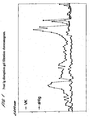
- Protein patterns were revealed on gels for c-myc by silver staining ( Figure 2 ).
- Western blots of c-myc were immuno-probed using monoclonal antibodies 9E10 ( Figure 3 ). In each case, c-myc as well as immunoglobulin heavy and light chains are identified.
- Tumour antigen e.g. MUC1, MUC16 or c-myc prepared according to Examples 1-3
- diluted appropriately in PBS is plated out at 50 ⁇ l per well in a standard 96 well microtiter plate and left to air dry overnight; Plate washed once with PBS/Tween TM to remove residual salt crystals; Plate blocked for 60 mins with 0.1% casein or 1% BSA in PBS; Plate washed x3 with PBS/Tween TM ; Serum (diluted 1/100 in PBS/0.1% casein) plated out in triplicate (50 ⁇ l per well), also monoclonal antibody controls; Incubate for 60 mins at room temperature with shaking; Wash plate x4 with PBS/Tween TM ; Add horseradish peroxidase (HRP)-conjugated anti-Ig antibody (Dako) to each well (50 ⁇ l per well) at 1/8000 dilution for anti-human and 1/1000 for anti-mouse
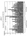
- cancer-associated autoantibodies to MUC1 and MUC16 were measured in a variety of sera using MUC1 and MUC16 isolated from the various sources as described herein. Results generated are shown in Figures 4 to 6 .
- Figure 4 shows a comparison of patient serum auto-antibody reactivity against MUC1 isolated from various body fluids: urine (from "normal” individuals), pleural effusion from a cancer patient and serum from advanced breast cancer patients (ABC serum).
- the patient serum tested was from either individuals with no evidence of breast cancer themselves but with a family history of breast cancer (i.e. one or more relatives who had breast cancer at a young age) or individuals with primary breast cancer.
- Standard auto-antibody ELISAs were performed as described above, utilising MUC1 isolated from urine (normal), pleural effusion or ABC serum as antigen. Data was normalised to an internal control reaction using the DF3 anti-MUC1 monoclonal antibody (as opposed to a serum sample) against each of the MUC1 antigens.
- MUC1 derived from normal urine was consistently lower in its reactivity than MUC1 derived from either pleural effusion (PE) or ABC serum.
- PE pleural effusion
- MUC1 derived from PE was of similar reactivity to cancer-associated MUC1 autoantibodies as MUC1 isolated from the serum of patients with ABC and therefore of equal diagnostic value.
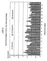
- Figure 5 shows the results of an identical exercise to Figure 4 except that all serum samples tested were for normal individuals (no breast cancer or family history of breast cancer). As can be seen, there is no significant difference in the reactivity of the serum to the three different antigens.
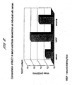
- Figure 6 shows reactivity of MUC16 cancer-associated autoantibodies from serum of patients with ovarian masses (pre-operative) against MUC16 (CA125) isolated from the serum of normal individuals and from ascites fluid in a patient with breast cancer.
- Antigens were prepared as in Example 2 and autoantibodies detected using ELISA assay as described in Example 4.
- MUC1 levels found in the serum of a patient with cancer were compared with the levels found in ascites fluid, pleural effusion, wound drainage fluid or seroma, in each case in the same patient from whom the serum sample was taken.
- MUC1 in the samples was quantified according to the following protocol:
- Human antibodies from seroma from patient M were purified by immunoaffinity chromatography against MUC1 derived from seroma fluid from the same cancer patient M. Purified antibodies were then tested against BSA conjugated protein core peptide to MUC1 and MUC1 derived from:- patient M's urine taken two years prior to cancer diagnosis; patient M's seroma taken after cancer diagnosis.
- the antibody purification from seroma was carried out according to the following protocol:
- Seroma fluid diluted 10 fold in PBS pH 7.6, was applied at 0.5ml/min by overnight re-circulation at 4°C, to an affinity matrix in column format, consisting of CNBr sepharose (Pharmacia) coupled (following the manufacturers instructions) to Pt-MUC1.
- CNBr sepharose Pharmacia
- the column was washed with 15ml of PBS (ensuring return of A 280 nm reading to zero) prior to elution of antibody using 10ml of 3M NaSCN, at 1m/min.
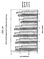
- MUC1 was purified from pooled ascitic fluid and from pooled pleural effusion from patients with advanced breast cancer using the protocol described in Example 1 and its reactivity against serum from patients with primary breast cancer measured as described in Example 4.
- the antigen from the pooled fluids was compared in each case with antigen isolated from 3 individual samples of ascitic fluid or pleural effusion respectively from patients with ABC. The results are shown in Figures 10 and 11 .
- pooled samples provide greater consistency of product so that one would not expect the reactivity to significantly vary between batches from pooled samples.
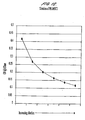
- MUC1 Serial dilutions of MUC1 which had been isolated from pleural effusion were prepared. Their MUC1 concentrations were measured by the method as shown in example 4 except that no human sera were used. Detection was by mouse B55 antibody followed by Dako anti-mouse HRP using an end-point rather than a kinetic reading.
- CEA Carcinoembryonic antigen
Landscapes
- Health & Medical Sciences (AREA)
- Life Sciences & Earth Sciences (AREA)
- Immunology (AREA)
- Engineering & Computer Science (AREA)
- Chemical & Material Sciences (AREA)
- Molecular Biology (AREA)
- Hematology (AREA)
- Urology & Nephrology (AREA)
- Biomedical Technology (AREA)
- Cell Biology (AREA)
- General Health & Medical Sciences (AREA)
- Medicinal Chemistry (AREA)
- Biochemistry (AREA)
- Analytical Chemistry (AREA)
- Microbiology (AREA)
- Physics & Mathematics (AREA)
- Pathology (AREA)
- Food Science & Technology (AREA)
- General Physics & Mathematics (AREA)
- Biotechnology (AREA)
- Oncology (AREA)
- Hospice & Palliative Care (AREA)
- Organic Chemistry (AREA)
- Proteomics, Peptides & Aminoacids (AREA)
- Genetics & Genomics (AREA)
- Biophysics (AREA)
- Rheumatology (AREA)
- Rehabilitation Therapy (AREA)
- Toxicology (AREA)
- Zoology (AREA)
- Gastroenterology & Hepatology (AREA)
- Public Health (AREA)
- Animal Behavior & Ethology (AREA)
- Chemical Kinetics & Catalysis (AREA)
- Pharmacology & Pharmacy (AREA)
- Veterinary Medicine (AREA)
- Nuclear Medicine, Radiotherapy & Molecular Imaging (AREA)
- General Chemical & Material Sciences (AREA)
- Peptides Or Proteins (AREA)
- Investigating Or Analysing Biological Materials (AREA)
Abstract
Description
- The invention relates to tumour marker proteins and their preparation from fluids from one or more cancer patients, wherein said fluids are those which collect in a body cavity or space which is naturally occurring or which is the result of cancer or medical intervention for cancer. Exemplary fluids are ascites, pleural effusion, seroma, hydrocoele and wound drainage fluid. The invention also relates to preparation of tumour marker proteins from excretions taken from patients with cancer.
- The said tumour marker proteins are useful in cancer detection methods which involve detecting or quantitatively measuring autoantibodies to circulating tumour markers or markers expressed on or in tumour cells and in various research applications. The invention is also directed to such uses.
- The development and progression of cancer in a patient is generally found to be associated with the presence of markers in the bodily fluid of the patient, these "tumour markers" reflecting different aspects of the biology of the cancer (see Fateh-Maghadam, A. & Steilber, P. (1993) Sensible use of tumour markers. Published by Verlag GMBH, ISBN 3-926725-07-9). Tumour markers are often found to be altered forms of wild-type proteins expressed by "normal" cells, in which case the alteration may be a change in primary amino acid sequence, a change in secondary, tertiary or quaternary structure or a change in post-translational modification, for example, abnormal glycosylation. In addition, wild-type proteins which are up-regulated or over-expressed in tumour cells, possibly as a result of gene amplification or abnormal transcriptional regulation, may also be tumour markers.
- Established assays for tumour markers present in bodily fluids tend to focus on the detection of tumour markers which reflect tumour bulk and as such are of value late in the disease process, for example in the diagnosis of metastatic disease. The most widely used of these markers include carcinoembryonic antigen (CEA) and the glycoprotein termed CA 15.3, both of which have been useful mainly as indicators of systemic disease burden and of relapse following therapy (Molina, R., Zanon, G., Filella, X. et al. Use of serial carcinoembryonic antigen and CA 15.3 assays in detecting relapses in breast cancer patients. (1995) Breast Cancer Res Treat 36: 41-48). These markers are of limited use earlier in the course of the disease, for example in early detection or in the screening of asymptomatic patients. Thus, in the search for tumour markers present in bodily fluid that are of use in assisting diagnosis earlier in the disease process the present inventors have sought to identify markers which do not depend on tumour bulk per se.
- Differences between a wild type protein expressed by "normal" cells and a corresponding tumour marker protein may, in some instances, lead to the tumour marker protein being recognised by an individual's immune system as "non-self" and thus eliciting an immune response in that individual. This may be a humoral (i.e B cell-mediated) immune response leading to the production of autoantibodies immunologically specific to the tumour marker protein. Autoantibodies are naturally occurring antibodies directed to an antigen which an individual's immune system recognises as foreign even though that antigen actually originated in the individual. They may be present in the circulation as circulating free autoantibodies or in the form of circulating immune complexes consisting of autoantibodies bound to their target tumour marker protein.
- As an alternative to the direct measurement or detection of tumour marker protein in bodily fluids, assays may be developed to measure the immune response of the individual to the presence of tumour marker protein in terms of autoantibody production. Such assays essentially constitute indirect detection of the presence of tumour marker protein. Because of the nature of the immune response, it is likely that autoantibodies can be elicited by a very small amount of circulating tumour marker protein and indirect methods which rely on detecting the immune response to tumour markers will consequently be more sensitive than methods for the direct measurement of tumour markers in bodily fluids. Assay methods based on the detection of autoantibodies may therefore be of particular value early in the disease process and possibly also in relation to screening of asymptomatic patients, for example in screening to identify individuals "at risk" of developing disease amongst a population of asymptomatic individuals. Furthermore, they may be useful for earlier detection of recurrent disease.
- Tumour marker proteins observed to elicit serum autoantibodies include a particular class of mutant p53 protein, described in
US Patent No. 5,652,115 , which can be defined by its ability to bind to the 70 kd heat shock protein (hsp70). p53 autoantibodies can be detected in patients with a number of different benign and malignant conditions (described inUS 5,652,115 ) but are in each case present in only a subset of patients. For example, one study utilizing an ELISA assay for detection of autoantibodies directed against the p53 protein in the serum of breast cancer patients reported that p53 autoantibodies were produced by 26% of patients and 1.3% of control subjects (Mudenda, B., Green, J. A., Green, B. et al. The relationship between serum p53 autoantibodies and characteristics of human breast cancer, (1994) Br J Cancer 69: 4445-4449). A second tumour marker protein known to elicit serum autoantibodies is the epithelial mucin MUC1 (Hinoda, Y. et al. (1993) Immunol Lett. 35: 163-168; Kotera, Y. et al. (1994) Cancer Res. 54: 2856-2860). -
WO 99/58978 - Cancer detection methods based on detection of circulating autoantibodies are frequently immunoassays utilizing an "immunoassay reagent" reactive with the circulating autoantibodies. Typically, the "reagents" used in such assays comprise recombinant tumour marker proteins (expressed in bacterial, insect, yeast or mammalian cells) or chemically synthesised tumour marker antigens, which may comprise substantially whole tumour marker proteins, or fragments thereof, such as short peptide antigens. Other potential sources of tumour-associated proteins for use as the basis of immunoassay reagents for the detection of anti-tumour auto-antibodies include cultured tumour cells (and the spent media used for their growth), tumour tissue, and serum from individuals with neoplasia. The majority of these sources have significant drawbacks, as discussed below.
- With cultured tumour cells (and their spent media) the amount of expressed protein can vary depending on growth phase at the time of harvest, leading to variations in quality and quantity. In addition, the desired protein is generally present at low concentration, therefore it is time-consuming to purify sufficient quantities of protein. Furthermore, the cell stock will be clonal, unlike cell stock in a tumour which is likely to have become heterogeneous in nature during the growth of the neoplasm, therefore producing variations in protein (especially in the degree of glycosylation).
- Recombinant proteins expressed in bacterial cells are not glycosylated, and thus significantly different from naturally glycosylated proteins. In addition, refolding of recombinantly expressed proteins may not be appropriate, thus giving an incorrect conformation for auto-antibody recognition.
- Tumour tissue is usually only available in small quantities and the purification of proteins therefrom is laborious and time consuming.
- Serum samples are usually available only in small quantities, therefore it is difficult to purify sufficient quantities of protein.
- The present inventors have now determined that significant advantages can be gained by the use of tumour marker antigens purified from bodily fluids derived from a body cavity or space in which a tumour is present or with which it is or was associated, such as ascites fluid, pleural effusion, seroma, hydrocoele or wound drainage fluid, or from excretions, as the "reagent" in auto-antibody immunoassays. In particular, the inventors have observed that use of reagents comprising tumour marker antigens purified from bodily fluids derived from the above defined body cavities or spaces results in increased sensitivity (as compared to the use of reagents derived from a "normal" body fluid) and produces a more "clinically relevant" result. There are also significant practical advantages to be gained from the use of such fluids as a source of assay reagent.
- In a first aspect the invention relates to a method of detecting cancer-associated anti-tumour autoantibodies, which method is an immunoassay comprising contacting a sample to be tested for the presence of such autoantibodies with an immunoassay reagent and detecting the presence of complexes formed by specific binding of the immunoassay reagent to any cancer-associated anti-tumour autoantibodies present in the sample, wherein the immunoassay reagent comprises tumour marker protein prepared from bodily fluid derived from a body cavity or space within which a tumour is or was present or with which a tumour is or was associated, from one or more cancer patients and/or tumour marker protein prepared from an excretion from one or more cancer patients, wherein said tumour marker protein exhibits selective reactivity with cancer-associated anti-tumour autoantibodies.
- In a second aspect the invention relates to use of tumour marker protein prepared from bodily fluid derived from a body cavity or space within which a tumour is or was present or with which a tumour is or was associated, of one or more cancer patients and/or tumour marker protein derived from an excretion of one or more cancer patients in the manufacture of an immunoassay reagent exhibiting selective reactivity with cancer-associated anti-tumour autoantibodies.
- In a third aspect, the invention relates to a method of preparing a tumour marker protein which method comprises isolating said tumour marker protein from bodily fluid wherein said fluid is:
- (i) collected from a body cavity or space in which a tumour is or was present or with which a tumour is or was associated, and
- (ii) said fluid represents the pooled fluid samples from two or more cancer patients.
- In a fourth aspect, the invention relates to a method of preparing a tumour marker protein which method comprises isolating said tumour marker protein from an excretion wherein:
- (i) said excretion or any component thereof has been in contact with a tumour or tumour cells, and
- (ii) said excretion represents pooled excretion samples from two or more cancer patients.
- In a fifth aspect the invention relates to tumour marker protein preparations prepared using the methods described above which are substantially immunoglobulin free and to kits and reagents comprising said preparations.
- In the first aspect, the invention relates to a method of detecting "cancer-associated" anti-tumour autoantibodies.
- The term "cancer-associated" anti-tumour autoantibodies refers to autoantibodies which are characteristic of the cancer disease state, and which are directed against epitopes present on forms of tumour marker proteins which are preferentially expressed in the cancer disease state.
- The method of the invention comprises an immunoassay to detect and/or quantitatively measure autoantibodies immunologically specific for one or more tumour marker proteins, and is characterised in that the "immunoassay reagent" used in the immunoassay comprises tumour marker protein prepared from bodily fluid derived from a body cavity or space in which a tumour is or was present or with which a tumour is or was associated, from one or more cancer patients and/or tumour marker protein prepared from an excretion of one or more cancer patients. Generally, the excretion will have passed through an organ in which cancer is present wherein the excretion is in contact with said cancer, or the excretion will include one or more components which have been in contact with cancer elsewhere in the body. A particular example is bile which may be in contact with cancer in the gall bladder but will appear in the faeces.
- The immunoassay reagent exhibits "selective reactivity" with cancer-associated anti-tumour autoantibodies. As used herein "selective reactivity" means a tumour marker protein has a greater affinity for autoantibodies to the tumour-associated antigen than it does for any antibody or autoantibody made to the same antigen which exists in the normal i.e. non-tumour possessing state.
- The term "body cavity or space" includes any body cavity or space, whether it be a natural cavity or a space or cavity arising as a result of diseases or medical intervention including collapsed or former cavities. The fluid is derived from such a cavity or space in which a tumour is or was present or with which a tumour is or was associated. Preferably the "bodily fluid derived from a body cavity" will be a tumour-induced body fluid, meaning a body fluid which is produced during the disease process, for example in response to or as a consequence of the presence of tumour cells. Exemplary body fluids are ascites, pleural effusion, seroma, hydrocoele and wound drainage fliud.
- For the avoidance of doubt "bodily fluids derived from a body cavity or space" do not include fluids derived from the systemic circulation, such as whole blood or serum.
- The term "excretion" includes, inter alia, urine, faeces, and seminal fluid.
- The general features of immunoassays, for example ELISA, radioimmunoassays and the like, are well known to those skilled in the art (see Immunoassay, E. Diamandis and T. Christopoulus, Academic Press, Inc., San Diego, CA, 1996). Immunoassays for the detection of antibodies having a particular immunological specificity (e.g. autoantibodies having immunological reactivity with a given tumour marker protein) generally require the use of a reagent that exhibits specific immunological reactivity with the antibody under test. Depending on the format of the assay this reagent may be immobilised on a solid support. A sample to be tested for the presence of the antibody is brought into contact with the reagent and if antibodies of the required immunological reactivity are present in the sample they will immunologically react with the reagent to form autoantibody-reagent complexes which may then be detected or quantitatively measured.
- Suitable samples of tumour marker protein for use as the basis of the "immunoassay reagent" may be isolated from bodily fluids derived from a body cavity or space from one or more cancer patients and/or from excretions from one or more cancer patients using standard protein purification techniques, such as are generally known in the art. For example, tumour marker proteins may be isolated by affinity chromatography using a suitable antibody (or antibody fragment) immunologically specific for the tumour marker protein. The inventors have shown in the accompanying examples that several different tumour marker proteins may be purified using purification methods based on affinity chromatography. It would be apparent to the skilled reader that analogous purification methods used for any other tumour marker proteins, with the use of a suitable antibody or antibody fragment.
- The starting material of bodily fluids derived from a body cavity and/or excretions is/are taken from one or more cancer patients. In this context the term "cancer patient" includes an individual previously diagnosed as having cancer. The fluid/excretion may be taken from a single patient or samples from two or more patients may be pooled together. Samples may be pooled from two or more patients having the same or different stages of the same or different types of cancers. Samples may also be pooled from different types of bodily fluids or excretions from a single or multiple patients. Advantageously, an immunoassay reagent prepared from fluid and/or excretion taken from cancer patient(s) with a particular type of cancer may be used to assist in the diagnosis of the same types of cancers in other individuals.
- In one embodiment the "cancer patient" from which the fluid/excretion is taken may be the same patient which it is later intended to test using the assay reagent. For example, a stock of reagent prepared from a patient diagnosed with cancer may be used at a later date to assess the immune status of the same patient, for example to monitor disease progression and/or to assess the effectiveness of a course of anti-cancer treatment in that patient.
- The "immunoassay reagent" or "tumour marker preparation" may comprise substantially whole tumour marker protein, for example tumour marker protein substantially in the form in which it is isolated from the fluid/excretion, or it may comprise a fragment of the tumour marker protein. To be effective as an immunoassay reagent any such "fragment" must retain immunological reactivity with the (auto)antibodies for which it is desired to test using the reagent. Suitable fragments might, for example, be prepared by chemical or enzymatic cleavage of the isolated tumour marker protein.
- Depending on the precise nature of the immunoassay in which it will be used, the "reagent" or "tumour marker protein preparation" may comprise a tumour marker protein, or fragment thereof, linked to one or more further molecules which impart some desirable characteristic not naturally present in the tumour marker protein. For example, the tumour marker protein may be conjugated to a revealing label, such as a fluorescent label, coloured label, luminescent label, radiolabel or heavy metal such as colloidal gold.
- The tumour marker protein as prepared by the method described herein can also be immobilized for use on a solid support such as a bead or surface of a well of a multiwell plate. The immobilization may be by absorption or by covalent attachment.
- The tumour marker protein (or assay reagent comprising such protein) is preferably substantially immunoglobulin free by virtue of the fact that following isolation, for example, by affinity chromatography, the protein preparation is treated to specifically remove contaminating immunoglobulins.
- The use of an immunoassay reagent comprising a tumour marker protein (or fragment thereof) isolated from body cavity fluids and/or excretions taken from one or more cancer patients provides significant advantages over the use of other reagents, such as recombinantly expressed or chemically synthesised polypeptides, in the clinical detection of cancer (including diagnosis, monitoring of disease recurrence or disease progression, etc).
- It might be expected that the precise characteristics of tumour marker proteins isolated from cancer patients could vary depending upon the source material (e.g. tissue or fluid) from which the tumour marker protein is isolated. For example, the characteristics of proteins isolated from urine may be different to those isolated from whole blood or serum, which may be different again to those isolated from ascites or pleural effusion. This may in turn affect the utility of the tumour marker protein as an assay reagent.
- In fact, the inventors have surprisingly observed that reagents prepared from tumour marker proteins isolated from body cavity-derived fluids or excretions from cancer patients, particularly ascites fluid, pleural effusion, seroma or wound drainage fluid are generally more specific for cancer-associated autoantibodies than reagents based on the equivalent proteins isolated from "normal" individuals. This increased specificity for cancer-associated autoantibodies means that immunoassays based on the use of reagents prepared from body cavity-derived fluids or excretions from cancer patients produce results that are more "clinically relevant" in the detection of an immune response to cancer.
- Prior to the present invention, it was not clear how reagents comprising antigens prepared from body cavity-derived fluids or excretions from cancer patients would perform as reagents for immunological detection of autoantibodies. In particular, it was not known whether such antigens would exhibit higher specificity for cancer-associated autoantibodies. It could not be predicted whether antigens from such sources would perform similarly to or better than antigen prepared from blood or serum, in terms of their ability to detect cancer-associated autoantibodies. Whilst it was known that tumour marker proteins may be present in fluids derived from body cavities and spaces, there is generally more potential for the antigens in these body cavities and spaces to be broken down. This in turn would mean that they might not detect autoantibodies as well as serum-derived antigens. Furthermore, it could not be concluded with certainty that antigens derived from cavity-derived fluids and excretions are immunologically similar to antigens derived from serum. Accordingly, it was surprising to observe that antigens prepared from cavity-derived fluids and excretions of cancer patients perform well as immunoassay reagents.
- The inventors postulate that the improved specificity observed with the use of reagents prepared from fluids derived from body cavities of cancer patients, such as ascites, pleural effusion, seroma or wound drainage fluid, is due to the origin of such fluids within the body cavities or spaces of cancer patients. It is postulated that fluids originating in body cavities or spaces due to the presence of a tumour in contact with the major organs may pick up more "cancer-associated" forms of the tumour marker protein, which are actually relevant to the cancer disease state, and contain less of the corresponding "normal" proteins. Since it is generally differences between "tumour" marker proteins and their "normal" counterparts which trigger the development of an immune response (i.e. autoantibody production), the inventors hypothesise that reagents based on the use of tumour markers isolated from cancer patients will be more specific for cancer autoantibodies than the equivalent "normal" proteins. This is indeed the case with tumour marker antigens isolated from ascites, pleural effusion or seroma, as shown in the accompanying Examples.
- There are further practical advantages associated with the use of ascites fluid, pleural effusion, seroma, hydrocoele or wound drainage fluid, as a source of tumour marker proteins. These fluids may be readily removed from patients in relatively large volumes as part of the therapeutic strategy. This material, which would otherwise be discarded, is a valuable source of useful assay reagent.
- Given that fluids such as ascites fluid, pleural effusion, seroma, hydrocoele or wound drainage fluid are produced in large volumes, there was doubt as to whether the concentration of tumour marker proteins in such fluids would be high enough to enable such fluids to be used as a practical source of antigens. One might reasonably expect the concentration of tumour marker proteins to be more dilute in such fluids as compared to blood or serum. Surprisingly, the inventors observed that the concentrations of tumour marker proteins in such fluids are in fact significantly higher than in serum. Accordingly, there are substantial benefits to be gained in terms of yield in recovering tumour marker proteins from such fluids.
- Furthermore, it has also been observed by the inventors that additional significant advantages can be secured by pooling body cavity fluid samples or excretions from two or more patients. Apart from increasing protein yield, the product secures at least as good a detection rate as marker protein from an individual sample while, at the same time, being more consistent in its characteristics from batch to batch. Thus, adequate affinity of the antigen can be relied upon every time.
- In particular embodiments the methods of the invention may comprise immunoassays to (simultaneously) detect two or more types of autoantibodies, each having specificity for different tumour marker proteins or for different epitopes on the same tumour marker proteins. These methods will typically involve use of a panel of two or more assay reagents, each reagent comprising a different tumour marker protein. These methods, which may be hereinafter referred to as "panel assays", utilise a panel of two or more reagents to monitor the overall immune response of an individual to a tumour or other carcinogenic/neoplastic change. These methods thus detect a "profile" of the immune response in a given individual, indicating which tumour markers elicit an immune response resulting in autoantibody production. The use of a panel of two or more reagents to monitor production of autoantibodies against two or more different tumour markers is generally more sensitive than the detection of autoantibodies to single markers and gives a much lower frequency of false negative results.
- The methods of the invention are preferred for the detection of circulating free autoantibodies, but may be adapted for detection of autoantibodies present in immune complexes, as would be appreciated by the skilled reader, for example by the competitive use of labelled tumour marker.
- In preferred applications the method of the invention will be used to detect the presence of cancer-associated anti-tumour autoantibodies in human subjects or patients, and will most preferably take the form of an in vitro immunoassay, performed on samples of bodily fluid taken from the subject/patient. Such in vitro immunoassays are non-invasive and can be repeated as often as is thought necessary to build up a profile of autoantibody production in a patient, either prior to the onset of disease, as in the screening of "at risk" individuals, or throughout the course of disease (further discussed below in relation to preferred applications of the method). As used herein the term "bodily fluid", when referring to the material to be tested for the presence of autoantibodies by immunoassay, includes inter alia plasma, serum, whole blood, urine, sweat, lymph, faeces, cerebrospinal fluid, ascites, pleural effusion, seminal fluid, sputum or nipple aspirate. The type of bodily fluid used may vary depending upon the type of cancer involved and the clinical situation in which the assay is used. In general, it is preferred to perform the assays on samples of serum or plasma.
- As aforesaid, the "immunoassay" used to detect/quantitate cancer-associated autoantibodies may be carried out according to standard techniques known in the art. In a most preferred embodiment the immunoassay may be an ELISA. ELISAs are generally well known in the art. In a typical "sandwich" ELISA a reagent having specificity for the autoantibodies under test is immobilised on a solid surface (e.g. the wells of a standard microtiter assay plate, or the surface of a microbead) and a sample of body fluid to be tested for the presence of autoantibodies is brought into contact with the immobilised reagent. Any autoantibodies of the desired specificity present in the sample will bind to the immobilised reagent. The bound autoantibody/reagent complexes may then be detected using any suitable method. In one preferred embodiment a labelled secondary anti-human immunoglobulin antibody, which specifically recognises an epitope common to one or more classes of human immunoglobulins, is used to detect the autoantibody/reagent complexes. Typically the secondary antibody will be anti-IgG or anti-IgM. The secondary antibody is usually labelled with a detectable marker, typically an enzyme marker such as, for example, peroxidase or alkaline phosphatase, allowing quantitative detection by the addition of a substrate for the enzyme which generates a detectable product, for example a coloured, chemiluminescent or fluorescent product. Other types of detectable labels known in the art may be used with equivalent effect.
- ELISA's may be performed in a qualitative format, in which the objective is merely to determine the presence or absence of autoantibodies in the sample, or in a quantitative format, which provides a measurement of the quantity of autoantibodies present in the sample. For quantitative assays, a standard curve may be generated by measuring the signal obtained (using the same detection reaction as will be used for the assay) from a series of standard samples containing known concentrations of antibodies having similar specificity as the autoantibodies under test. The quantity of autoantibodies present in the sample under test may then be interpolated from the standard curve.
- Panel assays may be performed in a multi-well format in which each one of the two or more assay reagents is placed in a separate well of a multi-well assay plate or, alternatively, in a single-pot format in which the two or more assay reagents are placed in a single container.
- The method of the invention may be adapted for use in the detection of autoantibodies to essentially any tumour marker protein for which a suitable "assay reagent" may be prepared from bodily fluid derived from a body cavity and/or from an excretion from a cancer patient. In particular, the method may be adapted to detect/measure autoantibodies to the epidermal growth factor receptor-related protein c-erbB2 (Dsouza, B. et al. (1993) Oncogene. 8: 1797-1806), the glycoprotein MUC1 (Batra, S. K. et al. (1992) Int. J. Pancreatology. 12: 271-283) and the signal transduction/cell cycle regulatory proteins Myc (Blackwood, E. M. et al. (1994) Molecular Biology of the Cell 5: 597-609), p53 (Matlashewski, G. et al. (1984) EMBO J. 3: 3257-3262; Wolf, D. et al. (1985) Mol. Cell. Biol. 5: 1887-1893) and ras (or Ras) (Capella, G. et al. (1991) Environ Health Perspectives. 93: 125-131), and also BRCA1 (Scully, R. et al. (1997) PNAS 94: 5605-10), BRCA2 (Sharan, S. K. et al. (1997) Nature. 386: 804-810), APC (Su, L. K. et al. (1993) Cancer Res. 53: 2728-2731; Munemitsu, S. et al. (1995) PNAS 92: 3046-50), CA125 (Nouwen, E. J. et al. (1990) Differentiation. 45: 192-8), PSA (Rosenberg, R. S. et al. (1998) Biochem Biophys Res Commun. 248: 935-939), carcinoembryonic antigen CEA (Duffy, M.J. (2001) Clin Chem, Apr 47(4):624-30), and CA19.9 (Haga, Y. et al (1989) Clin Biochem (1989) Oct 22(5): 363-8). However, the invention is not intended to be limited to the detection of autoantibodies to these particular tumour markers.
- The assay method of the invention may be employed in a variety of different clinical situations. In particular, the method may be used in the detection or diagnosis of cancer, in monitoring the progress of cancer or other neoplastic disease in a patient, in detecting early neoplastic or early carcinogenic change in an asymptomatic human subject, in screening a population of asymptomatic human subjects in order to identify those subjects who are at increased risk of developing cancer, in monitoring the response of a cancer patient to anti-cancer treatment, in the detection of recurrent disease in a patient previously diagnosed as having cancer who has undergone anti-cancer treatment to reduce the amount of cancer present, or in the selection of an anti-cancer vaccine for use in a particular patient.
- The inventors have generally observed that levels of cancer-associated autoantibodies show a positive correlation with disease state (see also
WO 99/58979 - For example, when the immunoassays are used in the diagnosis of cancer, the presence of an elevated level of autoantibodies, as compared to "normal" control individuals, is taken as an indication that the individual has cancer. The "normal" control individuals will preferably be age-matched controls not having any diagnosis of cancer based on clinical, imaging and/or biochemical criteria.
- When the immunoassays are used in monitoring the progress of cancer or other neoplastic disease in a patient, the presence of an elevated level of autoantibodies, as compared to a "normal control", is taken as an indication of the presence of cancer in the patient. The "normal control" may be levels of autoantibodies present in control individuals, preferably age-matched, not having any diagnosis of cancer based on clinical, imaging and/or biochemical criteria. Alternatively, the "normal control" may be a "base-line" level established for the particular patient under test. The "base-line" level may be, for example, the level of autoantibodies present when either a first diagnosis of cancer or a diagnosis of recurrent cancer was made. Any increase above the base-line level would be taken as an indication that the amount of cancer present in the patient has increased, whereas any decrease below the base-line would be taken as an indication that the amount of cancer present in the patient has decreased. The "base-line" value may also be, for example, the level before a new treatment is commenced. A change in the level of autoantibodies would be taken as an indication of the effectiveness of the therapy. The direction of the "change" (i.e. increase vs decrease) indicating a positive response to treatment will be dependent upon the precise nature of the treatment. For any given treatment the direction of the "change" in autoantibody levels indicating a positive result may be readily determined, for example by monitoring autoantibody levels in comparison to other clinical or biochemical indicators of response to the treatment.
- When the immunoassays are used in screening a population of asymptomatic human subjects to identify those subjects who are at increased risk of developing cancer, individuals having an elevated level of autoantibodies, as compared to "normal" control individuals, are identified as being "at risk" of developing cancer. The "normal" control individuals will preferably be age-matched controls not identified as having any predisposition to developing cancer or any significant elevated risk of developing cancer. An exception to this may be where age itself is a major risk factor.
- When the immunoassays are used in monitoring the response of a cancer patient to anti-cancer treatment, the presence of a decreased level of autoantibodies after treatment is taken as an indication that the patient has responded positively to the treatment. A base-line level of autoantibodies taken before treatment is commenced may be used for comparison purposes in order to determine whether treatment results in a "decrease" in autoantibody levels.
- When the immunoassays are used in detection of recurrent disease, the presence of an increased level of autoantibodies in the patient, as compared to a "normal control", is taken as an indication that disease has recurred. The "normal control" may be levels of autoantibodies present in control individuals, preferably age-matched not having any diagnosis of cancer based on clinical, imaging and/or biochemical criteria. Alternatively, the "normal control" may be a "base-line" level established for the particular patient under test. The "base-line" level may be, for example, the level of autoantibodies present during a period of remission from disease based on clinical, imaging and/or biochemical criteria.
- The assay method of the invention may be applied in the detection of many different types of cancer, of which examples are breast, bladder, colorectal, prostate and ovarian cancers. The assays may complement existing methods of screening and surveillance. For example, in the case of primary breast cancer immunoassays for autoantibodies could be used to alert clinicians to biopsy small lesions on mammograms which radiographically do not appear suspicious or to carry out breast imaging or to repeat imaging earlier than planned. In the clinic, the assay methods of the invention are expected to be more objective and reproducible compared to current imaging techniques (i.e. mammography and ultrasound), the success of which can be operator-dependent.
- "Panel assays" may be tailored having regard to the particular clinical application. A panel of reagents for detection of autoantibodies to at least p53 and c-erbB2 is particularly useful for many types of cancer and can optionally be supplemented with other markers having a known association with the particular cancer, or a stage of the particular cancer, to be detected. For example for breast cancer the panel might include
MUC 1 and /or c-myc and/or BRCA1 and/or BRCA2 and/or PSA whereas bladder cancer the panel might optionally includeMUC 1 and/or c-myc, for colorectal cancer ras and/or APC, for prostate cancer PSA and/orBRCA 1 and/or BRCA2 or for ovarian cancer BRCA1 and/or BRCA2 and/or CA125. There are other preferred embodiments in which p53 or c-erbB2 are not necessarily essential. For example, in the case of breast cancer suitable panels could be selected from the following: - p53 and
MUC 1 with optional c-erbB2 and/or c-myc, and/or BRCA1 and/or BRCA2 and/or PSA; - p53 and c-myc with optional c-erbB2 and/or MUC1 and/or BRCA1 and/or BRCA2 and/or PSA;
- p53 and BRCA1 with optional c-erB2 and/or
MUC 1 and/or c-myc and/or BRCA2 and/or PSA; - p53 and BRCA2 with optional c-erbB2 and/or
MUC 1 and/or c-myc and/or BRCA1 and/or PSA; - c-erbB2 and
MUC 1 with optional p53 and/or c-myc, and/or BRCA1 and/or BRCA2 and/or PSA; - c-erbB2 and c-myc with optional p53 and/or MUC1 and/or BRCA1 and/or BRCA2 and/or PSA;
- c-erbB2 and BRCA1 with optional p53 and/or
MUC 1 and/or c-myc and/or BRCA2 and/or PSA; - c-erbB2 and BRCA2 with optional p53 and/or
MUC 1 and/or c-myc and/or BRCA1 and/or PSA. - In the case of colorectal cancer suitable panels could be selected for example from the following:
- p53 and ras with optional c-erbB2 and/or APC;
- p53 and APC with optional c-erbB2 and/or Ras;
- Ras and APC with optional p53 and/or c-erbB2
- Such panels might also include CEA or CA19-9.
- In the case of prostate cancer suitable panels could be selected for example from the following:
- p53 and PSA with optional BRCA1 and/or BRCA2 and/or c-erbB2;
- c-erbB2 and PSA with optional p53 and/or BRCA1 and/or BRCA2.
- In the case of ovarian cancer suitable panels could be selected for example from the following:
- p53 and CA125 with optional c-erbB2 and/or BRCA1 and/or BRCA2;
- c-erbB2 and CA125 with optional p53 and/or BRCA1 and/or BRCA2.
- In a further embodiment, the immunoassay method of the invention may be used in the selection of an anti-cancer vaccine for use in a particular patient. In this embodiment a sample of bodily fluid taken from the patient is tested using a panel of two or more immunoassay reagents, each corresponding to a different tumour marker protein, in order to determine the relative strength of the patient's immune response to each of the different tumour marker proteins. The "strength of immune response" to a given tumour marker protein or proteins is indicated by the presence and/or the amount of cancer-associated autoantibodies specific to that tumour marker protein detected using the immunoassay; where autoantibodies are quantified, the greater the level of cancer-associated auto-antibodies, the stronger the immune response. The tumour marker protein or proteins identified as eliciting the strongest immune response or responses in the patient (i.e. the highest level of autoantibodies) is or are then selected to form the basis of an anti-cancer vaccine for use in the patient.
- In a further embodiment, the invention provides a method of monitoring whether vaccination of a subject with an anti-cancer vaccine based on a particular tumour marker protein has been successful in eliciting a humoral immune response (i.e. antibodies against the said tumour marker protein). This method is based on the same immunoassay methodology used to measure cancer-associated anti-tumour autoantibodies (i.e. use of an immunoassay reagent based on tumour marker protein purified from a body cavity fluid or an excretion taken from a cancer patient), the only difference being what is measured in the assay is an antibody response rather than an autoantibody response.
- In this embodiment a sample of bodily fluid taken from a patient previously treated with the anti-cancer vaccine (e.g. an immunogenic preparation comprising the relevant tumour marker protein, or an antigenic fragment thereof or a vaccine comprising a nucleic acid encoding said relevant tumour marker protein) is contacted with an immunoassay reagent and complexes formed by specific binding of the immunoassay reagent to cancer-associated antibodies present in the sample are detected. The immunoassay reagent again comprises a sample of the said tumour marker protein prepared from bodily fluid derived from a body cavity or space as defined herein from one or more cancer patients and/or tumour marker protein prepared from an excretion from one or more dancer patients.
- In addition to clinical applications in the detection of cancer, etc., the method of the invention may be used in any application where it is desired to test for the presence of cancer-associated anti-tumour autoantibodies. For example, the method of the invention may have applications in the laboratory as a research tool.
- The tumour marker protein preparations provided by the invention are advantageously used as (components of) immunoassay reagents for use in the assay methods of the invention. However, the utility of the tumour marker protein preparations is not limited to such use. For example, they too may have applications in the laboratory as research tools. Moreover, it is possible for tumour marker proteins to have utility as therapeutic agents. The availability of large quantities of protein as provided by the bodily fluids defined herein allows pre-clinical and clinical testing, either in vitro or in vivo in humans or non-human animals, to determine efficacy of particular tumour marker proteins as therapeutic agents. Such testing methods would be applicable to each or all of the various tumour marker proteins described herein.
- Another utility for tumour marker preparations of the invention is as a calibration material to be used in conjunction with the development of diagnostic tests for the presence of cancer or risk of cancer, which tests are based upon determination of the presence and/or level of any particular tumour marker protein in a clinical sample from a patient. The tumour marker protein preparations of the invention can be used to construct calibration curves for such tests. In particular this aspect of the invention includes:
- A method of calibrating an assay for measurement or detection of a given tumour marker protein in a clinical sample which method comprises the steps of:
- a) preparing at least two samples of a tumour marker protein prepared according to the method of the invention, each of which comprises said given tumour marker protein and each of which has a different tumour marker protein concentration to each of the other said samples:
- b) carrying out a quantitative measurement of the concentration of said tumour marker protein in each of said samples using:
- i) a spectrometric or spectrophotometric method and/or,
- ii) an antibody reagent to said tumour marker protein, and
- c) constructing a standard curve for tumour marker protein concentration based on the measurements obtained in step (b).
- Such standard curves may be constructed for any or all of the specific tumour marker proteins described herein.
- The invention will be further understood with reference to the following experimental Examples, together with the accompanying Figures in which:
-
Figure 1 shows a post~Ig disruption gel filtration chromatogram of a preparation of MUC16 (CA125) from ascites; -
Figure 2 shows a silver stained gel of c-myc purification from ascitic fluid, post immunoaffinity chromatography; -
Figure 3 shows an immunoprobed blot, c-myc purification from ascitic fluid, post immunoaffinity chromatography; -
Figure 4 shows a comparison of patient serum (patients with no evidence of breast cancer themselves but with a family history of breast cancer and those with primary breast cancer) auto-antibody reactivity against MUC1 isolated from various body fluids: urine (from "normal" individuals), pleural effusion from a cancer patient and serum from advanced breast cancer patients (ABC serum); -
Figure 5 shows autoantibody reactivity in serum from normal individuals against MUC1 from various body fluids: urinary MUC1 (normal), pleural effusion from a cancer patient and from advanced breast cancer patients (ABC serum); -
Figure 6 shows the autoantibody reactivity in serum samples from pre-operative patients with ovarian masses against normal MUC16 (CA125) and against tumour-associated MUC16 from ascites; -
Figure 7 shows the cancer-associated MUC1 concentration in sera, pleural effusion and ascitic fluid; -
Figure 8 shows the cancer-associated MUC1 concentration in serum, wound drainage fluid and in seroma; -
Figure 9 shows the reactivity of purified autoantibodies from seroma of patient M with cancer against purified urinary MUC1 from patient M taken two years prior to cancer diagnosis, MUC1 derived from the seroma of patient M, after diagnosis with cancer and bovine serum albumen conjugated to MUC1 protein core peptide; -
Figure 10 shows serum autoantibody reactivity against MUC1 purified from pooled ascites fluid and against MUC1 purified from individual ascites samples from cancer patients; -
Figure 11 shows serum autoantibody reactivity against MUC1 purified from pooled pleural effusions and against MUC1 purified from individuals pleural effusion samples from cancer patients; and -
Figure 12 shows a calibration curve prepared from MUC1 from a pleural effusion. - Monoclonal anti-MUC1 antibody B55 (also known as
NCRC 11, Xoma Corporation) is conjugated to CNBr-sepharose beads. Other anti-MUC1 monoclonal antibodies may be substituted for B55. - Tumour-induced body fluids (e.g. pleural effusion, ascites, seroma or wound drainage fluid) are diluted 1/10 with phosphate buffered saline (PBS) and filtered to 0.45 µm.
- Diluted body fluids are incubated with the anti-MUC1 sepharose beads (25 ml diluted fluid to 1 ml packed volume of beads) overnight at 4 °C with rolling ("batch" method) or re-circulated overnight through a packed column containing anti-MUC1 sepharose beads ("column" method).
-
- Beads are packed by centrifugation and the supernatant removed;
- Beads re-suspended in 5-10 ml PBS and rolled for 10 mins then packed by centrifugation and the supernatant removed;
repeat 5 times (or until A280 nm ~0) ; - Beads re-suspended in 5 ml 100
mM DEA pH 11, and rolled at room temperature for 10 mins; - Beads packed by centrifugation and the supernatant removed, pH adjusted to 7 by the addition of
pH 7 Tris buffer, dialysed against PBS for 24 hours minimum (100 DEA fraction) ; - Beads re-suspended in 5 ml PBS and rolled for 10 mins then packed by centrifugation and the supernatant removed, pH adjusted to 7 by the addition of
pH 7 Tris buffer, dialysed against PBS for 24 hours minimum (post-DEA fraction); - MUC1 content of each fraction confirmed by ELISA using, for instance, the monoclonal anti-MUC1 antibody C595 (available from Cancer Research Campaign Laboratories, UK) (see example 5 for details) or B55, prior to pooling of the two fractions and storage at - 20°C.
-
- Column washed with 5 column volumes of PBS, or until eluate reads ∼0 at A280 nm;
- 1 column volume of 100 mM DEA pH11 applied, followed by 5 column volumes of PBS;
- Eluate fractions (2ml) collected from the time of DEA application through the application of PBS;
- Fractions dialysed overnight against PBS;
- Fractions assayed for MUC1 content by ELISA using, for instance, the monoclonal anti-MUC1 antibody C595 or B55, prior to pooling MUC1 positive fractions and storage at -20°C.
- In order to remove contaminating immunoglobulins, MUC1 pooled fractions are incubated with dithiothreitol (DTT) to 50 mM for 30 mins, then iodoacetamide (to 75 mM) before being subjected to gel filtration on an S300 column.
- Resulting fractions (5ml) are assayed for MUC1 and human immunoglobulin (Ig) content by ELISA.
- MUC1 containing fractions (uncontaminated with human Ig) are pooled and stored at -20°C.
- One volume (e.g. 50 ml) of saturated ammonium sulphate was added to one volume (e.g. 50 ml) of tumour-induced body fluid (e.g. pleural effusion, ascites, seroma or wound drainage fluid) and incubated overnight at 4°C.
- The resultant precipitate is collected by centrifugation (3500 rpm for 30 min in a standard benchtop centrifuge) and resuspended in ½ volume PBS.
- This resuspension is subjected to gel filtration chromatography through an S300 column (2.5 x 100 cm) using PBS as the eluting buffer.
- Fractions (5 or 10 ml) are collected and assayed by ELISA for MUC16, using for instance anti-CA125 from ICN or the anti-MUC16 antibody VK8 (Memorial Sloane Kettering, New York), prior to pooling MUC16 positive fractions and storage at -20°C.
- In order to remove contaminating immunoglobulins, MUC16 pools are incubated with NaSCN (to 1.5M) for 10 mins, DTT (to 50mM) for 30 mins, then iodoacetamide (to 75mM) for 30 mins before being subjected to gel filtration on, for instance, an S300 or a Superdex™ 75 column.
- Resulting fractions (5ml) are assayed for MUC16 and human immunoglobulin (Ig) content by ELISA.
- MUC16 containing fractions (uncontaminated with human Ig) are pooled and stored at -20°C.
- For a sample prepared in the manner described above, fractions from a post-Ig disruption gel filtration were assayed for MUC16 using anti-MUC16 antibody VK8 and for human Ig using an anti-human Ig. The results are shown in
Figure 1 . As is clearly demonstrated, two substantially immunoglobulin free MUC16 peaks are eluted. - Methodology as per purification of MUC1 (Example 1), except that:
- Monoclonal anti-c-myc antibody 9E10 (ATCC) is used (or equivalent anti-c-myc antibody).
- Gel filtration is performed on a Superdex™ 75 column.
- Purity of MUC1, MUC16 and c-myc fractions are assessed by denaturing polyacrylamide gel electrophoresis and Western blotting, performed according to standard protocols using BioRad™ Mini Protean III™ system and BioRad™ DryBlot™ system.
- Protein patterns were revealed on gels for c-myc by silver staining (
Figure 2 ). Western blots of c-myc were immuno-probed using monoclonal antibodies 9E10 (Figure 3 ). In each case, c-myc as well as immunoglobulin heavy and light chains are identified. - Tumour antigen (e.g. MUC1, MUC16 or c-myc prepared according to Examples 1-3) diluted appropriately in PBS is plated out at 50 µl per well in a standard 96 well microtiter plate and left to air dry overnight;
Plate washed once with PBS/Tween™ to remove residual salt crystals;
Plate blocked for 60 mins with 0.1% casein or 1% BSA in PBS;
Plate washed x3 with PBS/Tween™;
Serum (diluted 1/100 in PBS/0.1% casein) plated out in triplicate (50 µl per well), also monoclonal antibody controls;
Incubate for 60 mins at room temperature with shaking;
Wash plate x4 with PBS/Tween™;
Add horseradish peroxidase (HRP)-conjugated anti-Ig antibody (Dako) to each well (50 µl per well) at 1/8000 dilution for anti-human and 1/1000 for anti-mouse;
Incubate for 60 mins at room temperature with shaking;
Wash plate x4 with PBS/Tween™;
Add 50µl TMB (tetramethylbenzadine) per well and read kinetically over a 10 min period at A650 nm. - Using the method as described in Example 4, cancer-associated autoantibodies to MUC1 and MUC16 were measured in a variety of sera using MUC1 and MUC16 isolated from the various sources as described herein. Results generated are shown in
Figures 4 to 6 . -
Figure 4 shows a comparison of patient serum auto-antibody reactivity against MUC1 isolated from various body fluids: urine (from "normal" individuals), pleural effusion from a cancer patient and serum from advanced breast cancer patients (ABC serum). The patient serum tested was from either individuals with no evidence of breast cancer themselves but with a family history of breast cancer (i.e. one or more relatives who had breast cancer at a young age) or individuals with primary breast cancer. - Standard auto-antibody ELISAs were performed as described above, utilising MUC1 isolated from urine (normal), pleural effusion or ABC serum as antigen. Data was normalised to an internal control reaction using the DF3 anti-MUC1 monoclonal antibody (as opposed to a serum sample) against each of the MUC1 antigens.
- As can be seen from the Figure, MUC1 derived from normal urine (nMUC1) was consistently lower in its reactivity than MUC1 derived from either pleural effusion (PE) or ABC serum. Furthermore, MUC1 derived from PE was of similar reactivity to cancer-associated MUC1 autoantibodies as MUC1 isolated from the serum of patients with ABC and therefore of equal diagnostic value.
-
Figure 5 shows the results of an identical exercise toFigure 4 except that all serum samples tested were for normal individuals (no breast cancer or family history of breast cancer). As can be seen, there is no significant difference in the reactivity of the serum to the three different antigens. -
Figure 6 shows reactivity of MUC16 cancer-associated autoantibodies from serum of patients with ovarian masses (pre-operative) against MUC16 (CA125) isolated from the serum of normal individuals and from ascites fluid in a patient with breast cancer. Antigens were prepared as in Example 2 and autoantibodies detected using ELISA assay as described in Example 4. - As can be seen, greatly enhanced reactivity of the cancer-associated MUC16 autoantibodies is seen with the MUC16 antigen from ascites fluid as compared to the "normal" MUC16. This experimental result therefore confirms the usefulness of ascites fluid as an antigen source for detection of cancer-associated autoantibodies.
- MUC1 levels found in the serum of a patient with cancer were compared with the levels found in ascites fluid, pleural effusion, wound drainage fluid or seroma, in each case in the same patient from whom the serum sample was taken. MUC1 in the samples was quantified according to the following protocol:
- Aliquot 50µl per well antibody solution into triplicate wells of a microtitre plate (usually 1µg ml-2 C595(IgG) and appropriate negative control) and incubate at RT with shaking for 1hr for the protein to adsorb to the plate.
- Wash the plate x4 with PBS/Tween using 250µl per well.
- Block the plate using 1% BSA 100µl per well and incubate at RT with shaking for 1hr.
- Wash the plate x4 with PBS/Tween using 250µl per well. Apply 50µl per well of fluid being tested, diluted 1/10 in PBS and incubate at RT with shaking for 1hr.
- Wash the plate x4 with PBS/Tween using 250µl per well.
- Add 50µl per well biotinylated C595 (1µg/ml) and incubate at RT with shaking for 1hr.
- Wash the plate x4 with PBS/Tween using 250µl per well.
- Add 50µl per well extra-avidin peroxidise at 1/1000 dilution and incubate at RT with shaking for 1hr.
- Wash the plate x4 with PBS/Tween using 250µl per well.
- Add 50µl per well TMB substrate and read kinetically at 650nm for 10 minutes.
- The results are shown in
Figures 7 and8 . - As will be readily apparent from the data serum levels of the cancer-associated MUC1 antigen are significantly lower than the level found in either ascites fluid, pleural effusion, seroma or wound drainage fluid. Accordingly, there are substantial benefits to be gained in terms of yield in recovering tumour marker antigen from those body cavity fluids.
- Human antibodies from seroma from patient M were purified by immunoaffinity chromatography against MUC1 derived from seroma fluid from the same cancer patient M. Purified antibodies were then tested against BSA conjugated protein core peptide to MUC1 and MUC1 derived from:- patient M's urine taken two years prior to cancer diagnosis; patient M's seroma taken after cancer diagnosis. The antibody purification from seroma was carried out according to the following protocol:
- Purification of human anti-MUC1 auto-antibodies was by affinity chromatography.
- Seroma fluid, diluted 10 fold in PBS pH 7.6, was applied at 0.5ml/min by overnight re-circulation at 4°C, to an affinity matrix in column format, consisting of CNBr sepharose (Pharmacia) coupled (following the manufacturers instructions) to Pt-MUC1.
- After seroma fluid application, the column was washed with 15ml of PBS (ensuring return of A280nm reading to zero) prior to elution of antibody using 10ml of 3M NaSCN, at 1m/min.
- Fractions of 1ml were collected throughout, desalted by dialysis against PBS and tested by ELISA for the presence of antibody.
- Positive fractions were pooled, purity of antibody verified (by PAGE) and antibody concentration determined.
- Assay of the purified antibodies against the three MUC1 antigens identified above was carried out according to the following protocol:
- Aliquot 50µl per well of the MUC1 antigen solution into triplicate wells of a microtitre plate and dry down at RT overnight.
- Wash the plate x2 with PBS/Tween using 250µl per well.
- Block the plate with 1% BSA using 100µl per well and incubate at RT with shaking for 1hr.
- Wash the plate x2 with PBS/Tween using 250µl per well.
- Add 50µl per well purified antibody solution at 1µg/ml and incubate at RT with shaking for 1hr.
- Wash the plate x4 with PBS/Tween using 250µl per well.
- Add 50µl per well α-human Ig HRP (DAKO), freshly diluted as per manufacturers instructions, and incubate at RT with shaking for 1hr.
- Wash the plate x4 with PBS/Tween using 250µl per well.
- Add 50µl per well TMB substrate and read kinetically at 650nm for 10 minutes.
- The results are shown in
Figure 9 . - Reactivity of the antibodies against MUC1 peptide was negligible. Reactivity of antibodies against normal MUC1 was considerably lower than that seen towards patient M's seroma derived MUC1. It can be inferred from this result that normal MUC1 molecule is substantially different with regard to its immune recognition, to that found in seroma fluid from an individual with cancer.
- MUC1 was purified from pooled ascitic fluid and from pooled pleural effusion from patients with advanced breast cancer using the protocol described in Example 1 and its reactivity against serum from patients with primary breast cancer measured as described in Example 4. The antigen from the pooled fluids was compared in each case with antigen isolated from 3 individual samples of ascitic fluid or pleural effusion respectively from patients with ABC. The results are shown in
Figures 10 and11 . - In the case of both ascitic fluid and pleural effusion the reactivity of the MUC1 from pooled fluid is as good as that isolated from individual samples. Furthermore, while there is great scope for variability of reactivity using samples from individuals, pooled samples provide greater consistency of product so that one would not expect the reactivity to significantly vary between batches from pooled samples.
- Serial dilutions of MUC1 which had been isolated from pleural effusion were prepared. Their MUC1 concentrations were measured by the method as shown in example 4 except that no human sera were used. Detection was by mouse B55 antibody followed by Dako anti-mouse HRP using an end-point rather than a kinetic reading.
- The results are shown in
Figure 12 and confirm the utility of the tumour marker proteins prepared in accordance with the invention as a calibration material. - The following lists sources of antibodies which may be used in the purification of tumour marker proteins by affinity chromatography. Affinity chromatography may be performed following the general methodology described in Example 1 (in relation to MUC1), with appropriate modification. It will be appreciated that other antibodies specific for the relevant marker protein may also be used.
Carcinoembryonic antigen (CEA): - 1116NS-3d, ATCC number CRL-8019, B lymphocyte hybridoma producing monoclonal antibody against CEA; T84.66A3.1A.1F2, ATCC number HB-8747, B lymphocyte hybridoma producing monoclonal antibody against CEA.
- Rabbit anti-human p53 polyclonal, commercially available from from Serotec Ltd, Kidlington, Oxford 0X5 1JE, United Kingdom.
- Monoclonal anti-p53, clone BP53-12, commercially available from Sigma.
- Mouse anti-human CA19-9 monoclonal, type clone 1116-NS-19-9, IgGl, commercially available from from Serotec Ltd, Kidlington, Oxford OX5 1JE, United Kingdom.
- Rabbit polyclonal IgG, commercially available from Santa Cruz Biotechnology, Inc., Santa Cruz, California, USA.
- Rabbit polyclonal IgG, commercially available from Santa Cruz Biotechnology, Inc., Santa Cruz, California, USA.
- Goat polyclonal IgG, commercially available from Santa Cruz Biotechnology, Inc., Santa Cruz, California, USA.
- Rabbit polyclonal IgG, commercially available from Santa Cruz Biotechnology, Inc., Santa Cruz, California, USA.
- Mouse monoclonal IgG, commercially available from Santa Cruz Biotechnology, Inc., Santa Cruz, California, USA.
Claims (16)
- A method of preparing an immunoassay reagent comprising a tumour marker protein for use in detection of autoantibodies specific for said tumour marker protein which method comprises:(i) isolating said tumour marker protein by immunoaffinity chromatography from either:(a) a bodily fluid collected from a body cavity or space in which a tumour is or was present or with which a tumour is or was associated; or(b) an excretion or any component thereof that has been in contact with a tumour or tumour cells;(ii) removing contaminating immunoglobulin from said tumour marker protein;wherein said bodily fluid or excretion represents the pooled samples from two or more cancer patients.
- A method as claimed in claim 1 wherein said bodily fluid is acites, pleural effusion, seroma, hydrocoele or wound drainage fluid or a mixture thereof.
- A method as claimed in claim 1 wherein said excretion is urine, faeces or seminal fluid.
- A method as claimed in claim 1 wherein the relevant component of said excretion is bile.
- A method as claimed in any one of claims 1 to 4 wherein said tumour marker protein is MUC1.
- A method as claimed in any one of claims 1 to 4 wherein said tumour marker protein is c-erbB2, p53, ras, BRCA1, BRCA2, APC, PSA, CEA, CA19.9, MUC16 or c-myc.
- A method as claimed in any one of claims 1 to 6 wherein said tumour marker protein is immobilized to solid support.
- A method as claimed in claim 7 wherein said solid support is the surface of a well of a multiwell plate or is a bead.
- A method as claimed in claim 8 wherein said immobilized tumour marker protein is absorbed or covalently attached to said solid support.
- A preparation of a tumour marker protein prepared by the method of any one of claims 1 to 9 and which is substantially immunoglobulin free.
- A kit or reagent suitable for carrying out an immunoassay which comprises a preparation of a tumour marker protein as claimed in claim 10 immobilized to a solid support.
- A kit or reagent as claimed in claim 11 wherein said solid support is the surface of a well of a multiwell plate or is a bead.
- A kit or reagent as claimed in claim 10 or 11 wherein said immobilized tumour marker protein is absorbed or covalently attached to said solid support.
- Use of a preparation as claimed in claim 10 in the evaluation in an in vitro test for the therapeutic efficacy or safety of said tumour marker protein.
- Use of a preparation as claimed in claim 10 in manufacture of a composition for the evaluation in an in vivo test of the therapeutic efficacy or safety of said tumour marker protein.
- A method of calibrating an assay for measurement or detection of a given tumour marker protein in a clinical sample which method comprises the steps of:(a) preparing at least two samples of a preparation of claim 10, each of which comprises said given tumour marker protein and each of which has a different tumour marker protein concentration to each of the other said samples:(b) carrying out a quantitative measurement of the concentration of said tumour marker protein in each of said samples using:(i) a spectrophotometric method and/or,(ii) an antibody reagent to said tumour marker protein, and(c) constructing a standard curve for a tumour marker protein concentration based on the measurements obtained in step (b).
Applications Claiming Priority (2)
| Application Number | Priority Date | Filing Date | Title |
|---|---|---|---|
| GB0226622A GB2395270B (en) | 2002-11-14 | 2002-11-14 | Tumour marker proteins and uses thereof |
| EP03773863A EP1563307B1 (en) | 2002-11-14 | 2003-11-13 | Tumour marker proteins and uses thereof |
Related Parent Applications (1)
| Application Number | Title | Priority Date | Filing Date |
|---|---|---|---|
| EP03773863A Division EP1563307B1 (en) | 2002-11-14 | 2003-11-13 | Tumour marker proteins and uses thereof |
Publications (2)
| Publication Number | Publication Date |
|---|---|
| EP1967856A2 true EP1967856A2 (en) | 2008-09-10 |
| EP1967856A3 EP1967856A3 (en) | 2009-04-15 |
Family
ID=9947849
Family Applications (2)
| Application Number | Title | Priority Date | Filing Date |
|---|---|---|---|
| EP03773863A Expired - Lifetime EP1563307B1 (en) | 2002-11-14 | 2003-11-13 | Tumour marker proteins and uses thereof |
| EP08007680A Withdrawn EP1967856A3 (en) | 2002-11-14 | 2003-11-13 | Tumour marker proteins and uses thereof |
Family Applications Before (1)
| Application Number | Title | Priority Date | Filing Date |
|---|---|---|---|
| EP03773863A Expired - Lifetime EP1563307B1 (en) | 2002-11-14 | 2003-11-13 | Tumour marker proteins and uses thereof |
Country Status (14)
| Country | Link |
|---|---|
| US (2) | US8592169B2 (en) |
| EP (2) | EP1563307B1 (en) |
| JP (1) | JP4759267B2 (en) |
| AT (1) | ATE397217T1 (en) |
| AU (1) | AU2003282245B2 (en) |
| CA (1) | CA2545930C (en) |
| CY (1) | CY1108294T1 (en) |
| DE (1) | DE60321375D1 (en) |
| DK (1) | DK1563307T3 (en) |
| ES (1) | ES2305530T3 (en) |
| GB (3) | GB2395270B (en) |
| PT (1) | PT1563307E (en) |
| SI (1) | SI1563307T1 (en) |
| WO (1) | WO2004044590A1 (en) |
Cited By (1)
| Publication number | Priority date | Publication date | Assignee | Title |
|---|---|---|---|---|
| CN108431605A (en) * | 2015-12-11 | 2018-08-21 | 善福德健康 | Reagent for monitoring breast cancer treatment and method |
Families Citing this family (27)
| Publication number | Priority date | Publication date | Assignee | Title |
|---|---|---|---|---|
| GB9810040D0 (en) | 1998-05-11 | 1998-07-08 | Univ Nottingham | Blood borne tumour markers |
| GB9827228D0 (en) * | 1998-12-10 | 1999-02-03 | Univ Nottingham | Cancer detection method and reagents |
| US20030232399A1 (en) * | 2000-06-14 | 2003-12-18 | Robertson John Forsyth Russell | Cancer detection methods and reagents |
| GB2395270B (en) | 2002-11-14 | 2006-08-16 | Univ Nottingham | Tumour marker proteins and uses thereof |
| US7858323B2 (en) | 2004-06-09 | 2010-12-28 | The Regents Of The University Of Michigan | Phage microarray profiling of the humoral response to disease |
| JP2006242869A (en) * | 2005-03-04 | 2006-09-14 | Kyushu Univ | Anti-nasp antibody and anti-tmf antibody as tumor marker |
| GB2426581A (en) | 2005-05-27 | 2006-11-29 | Univ Nottingham | Immunoassay methods |
| BRPI0610267B8 (en) * | 2005-05-27 | 2021-07-27 | Oncimmune Ltd | methods of detecting an unhealthy state or a susceptibility to disease in a mammalian individual; and, use of the method |
| WO2006132587A1 (en) * | 2005-06-10 | 2006-12-14 | Forskarpatent I Syd Ab | Tumour marker in brca2 associated breast cancer |
| JP5211315B2 (en) * | 2006-07-25 | 2013-06-12 | 国立大学法人愛媛大学 | Tumor marker, tumor diagnostic kit, and method for measuring tumor marker |
| CN101632020B (en) | 2006-09-13 | 2013-11-27 | 昂西免疫有限公司 | Improved immunoassay methods |
| JP5495159B2 (en) * | 2006-09-13 | 2014-05-21 | オンクイミューン リミテッド | Improved immunoassay method |
| GB2441824A (en) * | 2006-09-13 | 2008-03-19 | Oncimmune Ltd | Immunoassay method |
| ES2530209T3 (en) * | 2007-02-23 | 2015-02-27 | Physicians Choice Laboratory Services, Llc | Diagnostic methods aimed at clinical intervention |
| WO2008125262A1 (en) | 2007-04-12 | 2008-10-23 | Proteosys Ag | Autoimmune regulation of prostate cancer by annexin a3 |
| GB0725239D0 (en) * | 2007-12-24 | 2008-02-06 | Oncimmune Ltd | Calibrator for autoantibody assay |
| NZ593514A (en) * | 2009-01-14 | 2013-03-28 | Us Health | p-mTOR and p-AKT as Ratio Based Biomarkers for the Prognosis and/or Diagnosis of Cancer |
| EP2548025A4 (en) | 2010-03-17 | 2013-09-25 | Univ Michigan | Using phage epitopes to profile the immune response |
| PT2552959T (en) | 2010-03-26 | 2017-04-21 | Memorial Sloan Kettering Cancer Center | Antibodies to muc16 and methods of use thereof |
| US20130040839A1 (en) * | 2011-06-27 | 2013-02-14 | Ambergen, Inc. | Method for Diagnosing or Determining the Prognosis of Colorectal Cancer (CRC) Using Novel Autoantigens: Gene Expression Guided Autoantigen Discovery |
| US20140295426A1 (en) * | 2011-07-28 | 2014-10-02 | Veridex Llc | Methods for Diagnosing Cancer by Characterization of Tumor Cells Associated with Pleural or Serous Fluids |
| EP3189332A4 (en) * | 2014-09-04 | 2018-03-21 | Provista Diagnostics Inc. | Biomarkers for detection of breast cancer |
| BR112017019978A2 (en) | 2015-03-17 | 2018-06-19 | Memorial Sloan Kettering Cancer Center | antibody or antigen-binding fragment, conjugate, antigen receptor, strand, strand conjugate, fusion protein, t-cell, polynucleotide, vector, ex vivo cell, method of producing a chain, method of producing an antibody, pharmaceutical composition , method for treating cancer, glycopeptide, method for generating an antibody |
| EP4182693A2 (en) | 2020-07-14 | 2023-05-24 | Oncimmune Limited | Use of antigen combination for detecting autoantibodies in lung cancer |
| GB2600701A (en) | 2020-11-04 | 2022-05-11 | Oncimmune Ltd | Antibody assay |
| CN113721021B (en) * | 2021-09-16 | 2023-07-07 | 郑州大学 | Application of PRKCZ autoantibody in esophageal squamous carcinoma auxiliary diagnosis |
| GB2622246A (en) | 2022-09-08 | 2024-03-13 | Oncimmune Germany Gmbh | Antibody assay |
Citations (3)
| Publication number | Priority date | Publication date | Assignee | Title |
|---|---|---|---|---|
| US5652115A (en) | 1992-04-14 | 1997-07-29 | Duke University | Method of detecting tumors containing complexes of p53 and HSP70 |
| WO1999058978A2 (en) | 1998-05-11 | 1999-11-18 | The University Of Nottingham | Tumour markers |
| WO1999058979A1 (en) | 1998-05-13 | 1999-11-18 | South Eastern Sydney Area Health Service | A method of monitoring pancreatic tissue viability |
Family Cites Families (60)
| Publication number | Priority date | Publication date | Assignee | Title |
|---|---|---|---|---|
| NZ211453A (en) | 1984-03-22 | 1989-01-06 | Biotechnology Research Enterpr | Aryl azides, their preparation and use in the detection, localisation and isolation of polynucleotides |
| ATE77700T1 (en) | 1986-03-10 | 1992-07-15 | Imre Corp | IMMUNOCOMPLEX TEST PROCEDURE. |
| AU2253688A (en) | 1987-07-24 | 1989-03-01 | Xoma Corporation | Methods for breast cancer detection |
| US4937185A (en) | 1987-07-31 | 1990-06-26 | The Ohio State University Research Foundation | Detection of circulating antibodies to a cancer marker protein |
| US5241052A (en) * | 1988-03-04 | 1993-08-31 | New England Deaconess Hospital Corporation | Carcinoma orosomucoid-related antigen, a monoclonal antibody thereto, and their uses |
| US7282345B1 (en) | 1989-08-04 | 2007-10-16 | Schering Ag | C-erbB-2 external domain: GP75 |
| CA2072620C (en) * | 1989-11-03 | 2007-06-12 | Donald L. Morton | Urinary tumor associated antigen, antigenic subunits uses and methods of detection |
| US5157020A (en) | 1990-05-24 | 1992-10-20 | Research Corporation Tech., Inc. | Synthetic senescent cell antigen |
| DK9091D0 (en) | 1991-01-18 | 1991-01-18 | Novo Nordisk As | PROCEDURE FOR MANUFACTURING ANTIBODIES |
| DE4120412C1 (en) | 1991-06-20 | 1993-01-07 | Henning Berlin Gmbh Chemie- Und Pharmawerk, 1000 Berlin, De | |
| US6280962B1 (en) | 1991-11-25 | 2001-08-28 | Yoreh Biotechnologies Ltd. | Whole blood/mitogen assay for the early detection of a subject with cancer and kit |
| US6322989B1 (en) | 1991-11-25 | 2001-11-27 | Yoreh Biotechnologies, Ltd. | Whole blood/mitogen assay for the early detection of a subject with ovarian or breast cancer and kit |
| PT1024191E (en) | 1991-12-02 | 2008-12-22 | Medical Res Council | Production of anti-self antibodies from antibody segment repertoires and displayed on phage |
| US5753522A (en) * | 1991-12-06 | 1998-05-19 | Legacy Good Samaritan Hospital And Medical Center | Purified protein for identifying a cancer-associated retinopathy autoantibody |
| US5801005A (en) | 1993-03-17 | 1998-09-01 | University Of Washington | Immune reactivity to HER-2/neu protein for diagnosis of malignancies in which the HER-2/neu oncogene is associated |
| US5763164A (en) | 1993-04-16 | 1998-06-09 | Northwestern University | Immunogenic cancer proteins and peptides and methods of use |
| US5747268A (en) * | 1993-04-22 | 1998-05-05 | Dade International Inc. | Tumor marker control |
| US5744144A (en) * | 1993-07-30 | 1998-04-28 | University Of Pittsburgh University Patent Committee Policy And Procedures | Synthetic multiple tandem repeat mucin and mucin-like peptides, and uses thereof |
| US5721105A (en) | 1994-02-20 | 1998-02-24 | B.R.A.H.M.S. Diagnostica Gmbh | Method for the immunological determination of proteins and kit for carrying out the method |
| JPH07294530A (en) | 1994-04-20 | 1995-11-10 | Nippon Igaku Rinshiyou Kensa Kenkyusho:Kk | Detection of antibody in body fluids against pathogn |
| CA2148971A1 (en) | 1994-05-26 | 1995-11-27 | Izak Bahar | Calibrator matrix |
| EP0764030B1 (en) | 1994-06-24 | 2000-09-20 | Vladimir P. Torchilin | Composition containing autoantibodies for tumor therapy and prophylaxis |
| IL110464A0 (en) | 1994-07-26 | 1994-10-21 | Univ Ramot | Novel proteins for the diagnosis, imaging, and therapy of human cancer |
| US5561049A (en) | 1994-09-21 | 1996-10-01 | Behringwerke Ag | Method for detecting antibodies |
| US5876728A (en) | 1995-02-15 | 1999-03-02 | Howard David Kass | Natural composition extracted from plants used in the treatment of cancer |
| AUPN568095A0 (en) | 1995-09-27 | 1995-10-26 | Austin Research Institute, The | Anti-Galalpha(1,3)Gal antibody binding peptides |
| GB9521544D0 (en) | 1995-10-20 | 1995-12-20 | Univ Dundee | Activation of P53 protein and therapeutic applications thereof |
| JPH09189702A (en) | 1996-01-09 | 1997-07-22 | Hitachi Chem Co Ltd | Method for measuring antimucin antibody, cancer measuring method, and cancer diagnostic medicine |
| ATE283069T1 (en) | 1996-08-16 | 2004-12-15 | Univ Johns Hopkins Med | MELANOMA CELL LINES WHICH SHARE IMMUNOMINANT MELANOMA ANTIGENS AND METHOD FOR USE THEREOF |
| US8021666B2 (en) * | 1997-02-18 | 2011-09-20 | Sloan-Kettering Institute For Cancer Research | Method and compositions for stimulation of an immune response to CD20 using a xenogeneic CD20 antigen |
| WO1998055872A1 (en) | 1997-06-03 | 1998-12-10 | Amdl, Inc. | Immunoassay for the detection of cancer |
| CA2665707C (en) * | 1997-06-26 | 2013-02-12 | The Regents Of The University Of Michigan | Method for identification of cellular protein antigens and presence of antibodies to specific cellular protein antigens in serum |
| DE19805815C1 (en) | 1998-02-13 | 1999-11-11 | Ansgar W Lohse | Diagnostic for the detection of autoimmune hepatitis |
| JP4083855B2 (en) | 1998-02-16 | 2008-04-30 | エーザイ・アール・アンド・ディー・マネジメント株式会社 | Lung cancer prognosis test method |
| GB9821170D0 (en) * | 1998-09-30 | 1998-11-25 | Univ Ulster | Marker |
| WO2000026676A1 (en) * | 1998-11-03 | 2000-05-11 | Cell Genesys, Inc. | Cancer-associated antigens and methods of their identification |
| EP1125134B1 (en) * | 1998-11-05 | 2009-11-04 | The Regents Of The University Of Michigan | S100 proteins and autoantibodies as serum markers for cancer |
| US6387639B1 (en) | 1998-11-10 | 2002-05-14 | Sloan-Kettering Institute For Cancer Research | Ma family polypeptides and anti-Ma antibodies |
| GB9827228D0 (en) | 1998-12-10 | 1999-02-03 | Univ Nottingham | Cancer detection method and reagents |
| JP2000281255A (en) * | 1999-03-31 | 2000-10-10 | Seiko Epson Corp | Recording device |
| US7807810B2 (en) | 1999-04-08 | 2010-10-05 | The Ohio State University Research Foundation | Nucleic acids encoding the major outer membrane protein of the causative agent of human granulocytic ehrlichiosis and peptides encoded thereby |
| US6645465B2 (en) | 1999-08-06 | 2003-11-11 | Michigan, University Of The Regents | Annexin proteins and autoantibodies as serum markers for cancer |
| US20020064801A1 (en) | 1999-12-01 | 2002-05-30 | Ryan Jeffrey R. | Novel and practical serological assay for the clinical diagnosis of leishmaniasis |
| US6667160B2 (en) | 2000-03-15 | 2003-12-23 | Kenneth D. Fine | Method for diagnosing immunologic food sensitivity |
| US20030232399A1 (en) * | 2000-06-14 | 2003-12-18 | Robertson John Forsyth Russell | Cancer detection methods and reagents |
| US20030138860A1 (en) * | 2000-06-14 | 2003-07-24 | Robertson John Forsyth Russell | Cancer detection methods and reagents |
| US6379550B1 (en) * | 2000-07-24 | 2002-04-30 | Health Research, Inc. | Method for detecting PSA and its molecular forms using thiophilic gel |
| CA2431201A1 (en) | 2000-12-13 | 2002-08-01 | Bio-Rad Laboratories, Inc. | Standard diluent for multiplex assays |
| CA2451045A1 (en) | 2001-06-21 | 2003-09-04 | New York University | Mycobacterial proteins as early antigens for serodiagnosis and vaccines |
| US20030199464A1 (en) * | 2002-04-23 | 2003-10-23 | Silviu Itescu | Regeneration of endogenous myocardial tissue by induction of neovascularization |
| US20030049692A1 (en) | 2002-09-16 | 2003-03-13 | Norman Latov | Detection of anti-glycolipid antibodies by latex agglutination assay |
| GB2395270B (en) | 2002-11-14 | 2006-08-16 | Univ Nottingham | Tumour marker proteins and uses thereof |
| JP2005352771A (en) | 2004-06-10 | 2005-12-22 | Hitachi Software Eng Co Ltd | Pattern recognition system by expression profile |
| US20060141547A1 (en) | 2004-11-16 | 2006-06-29 | Das Hasi R | Novel diagnostic marker, a diagnostic kit and a method for diagnosis of rheumatoid arthritis |
| GB2426581A (en) | 2005-05-27 | 2006-11-29 | Univ Nottingham | Immunoassay methods |
| EP1789435B1 (en) | 2005-09-12 | 2010-02-24 | Industry Foundation of Chonnam National University | A method for production of mature natural killer cell |
| TWI304443B (en) | 2005-12-30 | 2008-12-21 | Nat Health Research Institutes | Alpha-enolase specific antibody and method of use |
| JP5495159B2 (en) | 2006-09-13 | 2014-05-21 | オンクイミューン リミテッド | Improved immunoassay method |
| CN101632020B (en) | 2006-09-13 | 2013-11-27 | 昂西免疫有限公司 | Improved immunoassay methods |
| GB0725239D0 (en) * | 2007-12-24 | 2008-02-06 | Oncimmune Ltd | Calibrator for autoantibody assay |
-
2002
- 2002-11-14 GB GB0226622A patent/GB2395270B/en not_active Expired - Fee Related
- 2002-11-14 GB GB0604694A patent/GB2424070B/en not_active Expired - Fee Related
- 2002-11-14 GB GB0604693A patent/GB2424273B/en not_active Expired - Fee Related
-
2003
- 2003-11-13 AU AU2003282245A patent/AU2003282245B2/en not_active Ceased
- 2003-11-13 EP EP03773863A patent/EP1563307B1/en not_active Expired - Lifetime
- 2003-11-13 ES ES03773863T patent/ES2305530T3/en not_active Expired - Lifetime
- 2003-11-13 US US10/534,773 patent/US8592169B2/en not_active Expired - Lifetime
- 2003-11-13 EP EP08007680A patent/EP1967856A3/en not_active Withdrawn
- 2003-11-13 SI SI200331307T patent/SI1563307T1/en unknown
- 2003-11-13 JP JP2004550841A patent/JP4759267B2/en not_active Expired - Fee Related
- 2003-11-13 PT PT03773863T patent/PT1563307E/en unknown
- 2003-11-13 DE DE60321375T patent/DE60321375D1/en not_active Expired - Lifetime
- 2003-11-13 CA CA2545930A patent/CA2545930C/en not_active Expired - Fee Related
- 2003-11-13 AT AT03773863T patent/ATE397217T1/en active
- 2003-11-13 WO PCT/GB2003/004950 patent/WO2004044590A1/en active IP Right Grant
- 2003-11-13 DK DK03773863T patent/DK1563307T3/en active
-
2008
- 2008-08-25 CY CY20081100911T patent/CY1108294T1/en unknown
-
2013
- 2013-11-19 US US14/083,874 patent/US20140080736A1/en not_active Abandoned
Patent Citations (3)
| Publication number | Priority date | Publication date | Assignee | Title |
|---|---|---|---|---|
| US5652115A (en) | 1992-04-14 | 1997-07-29 | Duke University | Method of detecting tumors containing complexes of p53 and HSP70 |
| WO1999058978A2 (en) | 1998-05-11 | 1999-11-18 | The University Of Nottingham | Tumour markers |
| WO1999058979A1 (en) | 1998-05-13 | 1999-11-18 | South Eastern Sydney Area Health Service | A method of monitoring pancreatic tissue viability |
Non-Patent Citations (20)
| Title |
|---|
| 1995, BREAST CANCER RES TREAT, vol. 36, pages 41 - 48 |
| BATRA, S. K. ET AL., INT. J. PANCREATOLOGY, vol. 12, 1992, pages 271 - 283 |
| BLACKWOOD, E. M. ET AL., MOLECULAR BIOLOGY OF THE CELL, vol. 5, 1994, pages 597 - 609 |
| CAPELLA,--G. ET AL., ENVIRON HEALTH PERSPECTIVES, vol. 93, 1991, pages 125 - 131 |
| DSOUZA, B. ET AL., ONCOGENE, vol. 8, 1993, pages 1797 - 1806 |
| DUFFY, M.J., CLIN CHEM, vol. 47, no. 4, April 2001 (2001-04-01), pages 624 - 30 |
| E. DIAMANDIS; T. CHRISTOPOULUS: "Immunoassay", 1996, ACADEMIC PRESS, INC. |
| FATEH-MAGHADAM, A.; STEILBER, P.: "Sensible use of tumour markers", 1993, VERLAG GMBH |
| HAGA, Y. ET AL., CLIN BIOCHEM, vol. 22, no. 5, 1989, pages 363 - 8 |
| HINODA, Y. ET AL., IMMUNOL LETT., vol. 35, 1993, pages 163 - 168 |
| KOTERA, Y. ET AL., CANCER RES., vol. 54, 1994, pages 2856 - 2860 |
| MATLASHEWSKI, G. ET AL., EMBO J., vol. 3, 1984, pages 3257 - 3262 |
| MUDENDA, B.; GREEN, J. A.; GREEN, B. ET AL.: "The relationship between serum p53 autoantibodies and characteristics of human breast cancer", BR J CANCER, vol. 69, 1994, pages 4445 - 4449 |
| MUNEMITSU, S. ET AL., PNAS, vol. 92, 1995, pages 3046 - 50 |
| NOUWEN, E. J. ET AL., DIFFERENTIATION, vol. 45, 1990, pages 192 - 8 |
| ROSENBERG, R. S. ET AL., BIOCHEM BIOPHYS RES COMMUN., vol. 248, 1998, pages 935 - 939 |
| SCULLY, R. ET AL., PNAS, vol. 94, 1997, pages 5605 - 10 |
| SHARAN, S. K. ET AL., NATURE, vol. 386, 1997, pages 804 - 810 |
| SU, L. K. ET AL., CANCER RES., vol. 53, 1993, pages 2728 - 2731 |
| WOLF, D. ET AL., MOL. CELL. BIOL., vol. 5, 1985, pages 1887 - 1893 |
Cited By (1)
| Publication number | Priority date | Publication date | Assignee | Title |
|---|---|---|---|---|
| CN108431605A (en) * | 2015-12-11 | 2018-08-21 | 善福德健康 | Reagent for monitoring breast cancer treatment and method |
Also Published As
| Publication number | Publication date |
|---|---|
| US8592169B2 (en) | 2013-11-26 |
| US20060094069A1 (en) | 2006-05-04 |
| SI1563307T1 (en) | 2008-10-31 |
| CA2545930A1 (en) | 2004-05-27 |
| AU2003282245B2 (en) | 2010-05-20 |
| GB2424070B (en) | 2007-06-27 |
| EP1563307B1 (en) | 2008-05-28 |
| DK1563307T3 (en) | 2008-09-29 |
| EP1967856A3 (en) | 2009-04-15 |
| GB2424273B (en) | 2007-06-27 |
| GB0604694D0 (en) | 2006-04-19 |
| GB2395270B (en) | 2006-08-16 |
| ES2305530T3 (en) | 2008-11-01 |
| JP4759267B2 (en) | 2011-08-31 |
| CY1108294T1 (en) | 2014-02-12 |
| PT1563307E (en) | 2008-07-16 |
| EP1563307A1 (en) | 2005-08-17 |
| GB0604693D0 (en) | 2006-04-19 |
| GB2395270A (en) | 2004-05-19 |
| GB0226622D0 (en) | 2002-12-24 |
| US20140080736A1 (en) | 2014-03-20 |
| GB2424070A (en) | 2006-09-13 |
| WO2004044590A1 (en) | 2004-05-27 |
| ATE397217T1 (en) | 2008-06-15 |
| DE60321375D1 (en) | 2008-07-10 |
| JP2006506612A (en) | 2006-02-23 |
| AU2003282245A1 (en) | 2004-06-03 |
| GB2424273A (en) | 2006-09-20 |
| CA2545930C (en) | 2016-07-12 |
Similar Documents
| Publication | Publication Date | Title |
|---|---|---|
| EP1563307B1 (en) | Tumour marker proteins and uses thereof | |
| CA2354702C (en) | Cancer detection method and reagents | |
| EP2073008B1 (en) | Improved immunoassay methods | |
| JP5736598B2 (en) | Improved immunoassay method | |
| JP2002532686A5 (en) | ||
| US20030138860A1 (en) | Cancer detection methods and reagents | |
| EP2620772A1 (en) | Gastric cancer biomarkers and methods of use thereof | |
| JP5765635B2 (en) | Immunoassay method for complex of Ku86 and its autoantibody, kit used therefor, and cancer determination method using the same | |
| CN107110848B (en) | Method for detecting arteriosclerosis and cancer using deoxyhypusine synthase gene as index | |
| GB2600701A (en) | Antibody assay | |
| JP2023533815A (en) | Use of antigen combinations to detect autoantibodies in lung cancer | |
| AU2012209004B2 (en) | Improved immunoassay methods | |
| TW201928351A (en) | Antibody assay | |
| JP2011107064A (en) | Immunoassay method of autoantibody to clathrin heavy chain, kit used for the same, and cancer determination method using the same |
Legal Events
| Date | Code | Title | Description |
|---|---|---|---|
| PUAI | Public reference made under article 153(3) epc to a published international application that has entered the european phase |
Free format text: ORIGINAL CODE: 0009012 |
|
| AC | Divisional application: reference to earlier application |
Ref document number: 1563307 Country of ref document: EP Kind code of ref document: P |
|
| AK | Designated contracting states |
Kind code of ref document: A2 Designated state(s): AT BE BG CH CY CZ DE DK EE ES FI FR GR HU IE IT LI LU MC NL PT RO SE SI SK TR |
|
| PUAL | Search report despatched |
Free format text: ORIGINAL CODE: 0009013 |
|
| AK | Designated contracting states |
Kind code of ref document: A3 Designated state(s): AT BE BG CH CY CZ DE DK EE ES FI FR GR HU IE IT LI LU MC NL PT RO SE SI SK TR |
|
| 17P | Request for examination filed |
Effective date: 20090310 |
|
| 17Q | First examination report despatched |
Effective date: 20091118 |
|
| AKX | Designation fees paid |
Designated state(s): AT BE BG CH CY CZ DE DK EE ES FI FR GR HU IE IT LI LU MC NL PT RO SE SI SK TR |
|
| STAA | Information on the status of an ep patent application or granted ep patent |
Free format text: STATUS: THE APPLICATION IS DEEMED TO BE WITHDRAWN |
|
| 18D | Application deemed to be withdrawn |
Effective date: 20100531 |