EP1957539B1 - Human monoclonal antibodies to protein tyrosine kinase 7 (ptk7) and their use - Google Patents
Human monoclonal antibodies to protein tyrosine kinase 7 (ptk7) and their use Download PDFInfo
- Publication number
- EP1957539B1 EP1957539B1 EP06848508.5A EP06848508A EP1957539B1 EP 1957539 B1 EP1957539 B1 EP 1957539B1 EP 06848508 A EP06848508 A EP 06848508A EP 1957539 B1 EP1957539 B1 EP 1957539B1
- Authority
- EP
- European Patent Office
- Prior art keywords
- seq
- antibody
- variable region
- chain variable
- ptk7
- Prior art date
- Legal status (The legal status is an assumption and is not a legal conclusion. Google has not performed a legal analysis and makes no representation as to the accuracy of the status listed.)
- Revoked
Links
Images


Classifications
-
- A—HUMAN NECESSITIES
- A61—MEDICAL OR VETERINARY SCIENCE; HYGIENE
- A61K—PREPARATIONS FOR MEDICAL, DENTAL OR TOILETRY PURPOSES
- A61K38/00—Medicinal preparations containing peptides
- A61K38/16—Peptides having more than 20 amino acids; Gastrins; Somatostatins; Melanotropins; Derivatives thereof
- A61K38/17—Peptides having more than 20 amino acids; Gastrins; Somatostatins; Melanotropins; Derivatives thereof from animals; from humans
- A61K38/18—Growth factors; Growth regulators
-
- C—CHEMISTRY; METALLURGY
- C07—ORGANIC CHEMISTRY
- C07K—PEPTIDES
- C07K16/00—Immunoglobulins [IGs], e.g. monoclonal or polyclonal antibodies
- C07K16/18—Immunoglobulins [IGs], e.g. monoclonal or polyclonal antibodies against material from animals or humans
- C07K16/28—Immunoglobulins [IGs], e.g. monoclonal or polyclonal antibodies against material from animals or humans against receptors, cell surface antigens or cell surface determinants
- C07K16/30—Immunoglobulins [IGs], e.g. monoclonal or polyclonal antibodies against material from animals or humans against receptors, cell surface antigens or cell surface determinants from tumour cells
-
- A—HUMAN NECESSITIES
- A61—MEDICAL OR VETERINARY SCIENCE; HYGIENE
- A61P—SPECIFIC THERAPEUTIC ACTIVITY OF CHEMICAL COMPOUNDS OR MEDICINAL PREPARATIONS
- A61P35/00—Antineoplastic agents
-
- A—HUMAN NECESSITIES
- A61—MEDICAL OR VETERINARY SCIENCE; HYGIENE
- A61P—SPECIFIC THERAPEUTIC ACTIVITY OF CHEMICAL COMPOUNDS OR MEDICINAL PREPARATIONS
- A61P35/00—Antineoplastic agents
- A61P35/02—Antineoplastic agents specific for leukemia
-
- C—CHEMISTRY; METALLURGY
- C07—ORGANIC CHEMISTRY
- C07K—PEPTIDES
- C07K16/00—Immunoglobulins [IGs], e.g. monoclonal or polyclonal antibodies
-
- C—CHEMISTRY; METALLURGY
- C07—ORGANIC CHEMISTRY
- C07K—PEPTIDES
- C07K16/00—Immunoglobulins [IGs], e.g. monoclonal or polyclonal antibodies
- C07K16/40—Immunoglobulins [IGs], e.g. monoclonal or polyclonal antibodies against enzymes
-
- A—HUMAN NECESSITIES
- A61—MEDICAL OR VETERINARY SCIENCE; HYGIENE
- A61K—PREPARATIONS FOR MEDICAL, DENTAL OR TOILETRY PURPOSES
- A61K39/00—Medicinal preparations containing antigens or antibodies
- A61K2039/505—Medicinal preparations containing antigens or antibodies comprising antibodies
-
- C—CHEMISTRY; METALLURGY
- C07—ORGANIC CHEMISTRY
- C07K—PEPTIDES
- C07K2317/00—Immunoglobulins specific features
- C07K2317/20—Immunoglobulins specific features characterized by taxonomic origin
- C07K2317/21—Immunoglobulins specific features characterized by taxonomic origin from primates, e.g. man
-
- C—CHEMISTRY; METALLURGY
- C07—ORGANIC CHEMISTRY
- C07K—PEPTIDES
- C07K2317/00—Immunoglobulins specific features
- C07K2317/50—Immunoglobulins specific features characterized by immunoglobulin fragments
- C07K2317/56—Immunoglobulins specific features characterized by immunoglobulin fragments variable (Fv) region, i.e. VH and/or VL
- C07K2317/565—Complementarity determining region [CDR]
-
- C—CHEMISTRY; METALLURGY
- C07—ORGANIC CHEMISTRY
- C07K—PEPTIDES
- C07K2317/00—Immunoglobulins specific features
- C07K2317/70—Immunoglobulins specific features characterized by effect upon binding to a cell or to an antigen
- C07K2317/77—Internalization into the cell
Definitions
- RTKs Receptor tyrosine kinases
- the regulation of RTK signals is important for regulation of cell growth, differentiation, axonal growth, epithelial growth, development, adhesion, migration, and apoptosis ( Prenzel et al. (2001) Endocr. Relat. Cancer 8:11-31 ; Hubbard and Till (2000) Annu. Rev. Biochem. 69:373-98 ).
- RTKs are known to be involved in the development and progression of several forms of cancer. In most of the RTK-related cancers, there has been an amplification of the receptor protein rather than a mutation of the gene ( Kobus and Fleming (2005) Biochemistry 44:1464-70 ).
- PTK7 Protein tyrosine kinase 7
- RT-PCR RT-PCR-PCR-PCR-derived cell lines
- CCK4 colon carcinoma kinase 4
- PTK7 belongs to a subset of RTKs that lack detectable catalytic tyrosine kinase activity but retain signal transduction activity and is thought to possibly function as a cell adhesion molecule.
- the mRNA for PTK7 was found to be variably expressed in colon carcinoma derived cell lines but not found to be expressed in human adult colon tissues (Mossie et al., supra ). PTK7 expression was also seen in some melanoma cell lines and melanoma biopsies ( Easty, et al. (1997) Int. J. Cancer 71:1061-5 ). An alternative splice form was found to be expressed in hepatomas and colon cancer cells ( Jung et al. (2002) Biochim Biophys Acta 1579: 153-63 ). In addition, PTK7 was found to be highly overexpressed in acute myeloid leukemia samples ( Muller-Tidow et al., (2004) Clin. Cancer Res.
- agents that recognize PTK7, and methods of using such agents are desired.
- the present invention provides isolated monoclonal antibodies, in particular human monoclonal antibodies, that bind to PTK7 and that exhibit numerous desirable properties. These properties include high affinity binding to human PTK7 and binding to Wilms' tumor cells. Also provided are the antibodies and compositions of the invention for use in methods for treating a variety of PTK7 mediated diseases.
- the invention pertains to an isolated human monoclonal antibody, or an antigen-binding portion thereof, wherein the antibody:
- a preferred antibody or antigen-binding portion thereof comprises:
- the antibodies of the invention can be, for example, full-length antibodies, for example of an IgG1 or IgG4 isotype.
- the antibodies can be antibody fragments, such as Fab or Fab'2 fragments, or single chain antibodies.
- the invention also provides an immunoconjugate comprising an antibody of the invention, or antigen-binding portion thereof, linked to a therapeutic agent, such as a cytotoxin or a radioactive isotope.
- a therapeutic agent such as a cytotoxin or a radioactive isotope.
- compositions comprising an antibody, or antigen-binding portion thereof, or immunoconjugate of the invention and a pharmaceutically acceptable carrier are also provided.
- Nucleic acid molecules encoding the antibodies, or antigen-binding portions thereof, of the invention are also encompassed by the invention, as well as expression vectors comprising such nucleic acids and host cells comprising such expression vectors.
- an in vitro method for preparing an antibody of the invention which comprises expressing the antibody in a host cell of the invention and isolating the antibody from the host cell.
- transgenic mouse comprising human immunoglobulin heavy and light chain transgenes, wherein the mouse expresses an antibody of the invention, as well as hybridomas prepared from such a mouse, wherein the hybridoma produces the antibody of the invention.
- the invention provides an antibody, or antigen-binding portion thereof, of the invention for use in a method of treating or preventing a cancer characterized by growth of tumor cells expressing PTK7.
- the cancer may be colon cancer (including small intestine cancer), lung cancer, breast cancer, pancreatic cancer, melanoma ( e.g ., metastatic malignant melanoma), acute myeloid leukemia, kidney cancer, bladder cancer, ovarian cancer or prostate cancer.
- renal cancer e.g ., renal cell carcinoma
- glioblastoma brain tumors
- chronic or acute leukemias including acute lymphocytic leukemia (ALL), adult T-cell leukemia (T-ALL), chronic myeloid leukemia, acute lymphoblastic leukemia, chronic lymphocytic leukemia, lymphomas (e.g ., Hodgkin's and non-Hodgkin's lymphoma, lymphocytic lymphoma, primary CNS lymphoma, T-cell lymphoma, Burkitt's lymphoma, anaplastic large-cell lymphomas (ALCL), cutaneous T-cell lymphomas, nodular small cleaved-cell lymphomas, peripheral T-cell lymphomas, Lennert's lymphomas, immunoblastic lymphomas, T-cell leukemia/lymphomas (ATLL), entroblastic/centrocytic (cb/cc) folli
- ALL acute lympho
- the present invention relates to isolated monoclonal antibodies, particularly human monoclonal antibodies, that bind specifically to PTK7.
- the antibodies of the invention exhibit one or more desireable functional properties, such as high affinity binding to PTK7 and/or the ability to inhibit growth of tumor cells in vitro or in vivo.
- the antibodies of the invention are derived from particular heavy and light chain germline sequences and/or comprise particular structural features such as CDR regions comprising particular amino acid sequences.
- the invention provides isolated antibodies, methods of making such antibodies, immunoconjugates and bispecific molecules comprising such antibodies and pharmaceutical compositions containing the antibodies, immunconjugates or bispecific molecules of the invention.
- the invention also relates to methods of using the antibodies, such as to treat diseases such as cancer.
- human antibodies of the invention may, in certain cases, cross-react with PTK7 from species other than human. In certain embodiments, the antibodies may be completely specific for one or more human PTK7 and may not exhibit species or other types of non-human cross-reactivity.
- the complete amino acid sequence of an exemplary human PTK7 has Genbank accession number NM_002821 (SEQ ID NO:58).
- immune response refers to the action of, for example, lymphocytes, antigen presenting cells, phagocytic cells, granulocytes, and soluble macromolecules produced by the above cells or the liver (including antibodies, cytokines, and complement) that results in selective damage to, destruction of, or elimination from the human body of invading pathogens, cells or tissues infected with pathogens, cancerous cells, or, in cases of autoimmunity or pathological inflammation, normal human cells or tissues.
- a “signal transduction pathway” refers to the biochemical relationship between a variety of signal transduction molecules that play a role in the transmission of a signal from one portion of a cell to another portion of a cell.
- the phrase "cell surface receptor” includes, for example, molecules and complexes of molecules capable of receiving a signal and the transmission of such a signal across the plasma membrane of a cell.
- An example of a “cell surface receptor” of the present invention is the PTK7 receptor.
- antibody as referred to herein includes whole antibodies and any antigen binding fragment (i.e ., "antigen-binding portion") or single chains thereof.
- An “antibody” refers to a glycoprotein comprising at least two heavy (H) chains and two light (L) chains inter-connected by disulfide bonds, or an antigen binding portion thereof.
- Each heavy chain is comprised of a heavy chain variable region (abbreviated herein as V H ) and a heavy chain constant region.
- the heavy chain constant region is comprised of three domains, C H1 , C H2 and C H3 .
- Each light chain is comprised of a light chain variable region (abbreviated herein as V L ) and a light chain constant region.
- the light chain constant region is comprised of one domain, C L .
- the V H and V L regions can be further subdivided into regions of hypervariability, termed complementarity determining regions (CDR), interspersed with regions that are more conserved, termed framework regions (FR).
- CDR complementarity determining regions
- FR framework regions
- Each V H and V L is composed of three CDRs and four FRs, arranged from amino-terminus to carboxy-terminus in the following order: FR1, CDR1, FR2, CDR2, FR3, CDR3, FR4.
- the variable regions of the heavy and light chains contain a binding domain that interacts with an antigen.
- the constant regions of the antibodies may mediate the binding of the immunoglobulin to host tissues or factors, including various cells of the immune system (e.g ., effector cells) and the first component (Clq) of the classical complement system.
- antibody portion refers to one or more fragments of an antibody that retain the ability to specifically bind to an antigen (e.g ., PTK7). It has been shown that the antigen-binding function of an antibody can be performed by fragments of a full-length antibody.
- binding fragments encompassed within the term "antigen-binding portion" of an antibody include (i) a Fab fragment, a monovalent fragment consisting of the V L , V H , C L and C H 1 domains; (ii) a F(ab') 2 fragment, a bivalent fragment comprising two Fab fragments linked by a disulfide bridge at the hinge region; (iii) a Fab' fragment, which is essentially an Fab with part of the hinge region (see, FUNDAMENTAL IMMUNOLOGY (Paul ed., 3.sup.rd ed.
- the two domains of the Fv fragment, V L and V H are coded for by separate genes, they can be joined, using recombinant methods, by a synthetic linker that enables them to be made as a single protein chain in which the V L and V H regions pair to form monovalent molecules (known as single chain Fv (scFv); see e.g., Bird et al. (1988) Science 242:423-426 ; and Huston et al. (1988) Proc. Natl. Acad. Sci. USA 85:5879-5883 ).
- single chain Fv single chain Fv
- Such single chain antibodies are also intended to be encompassed within the term "antigen-binding portion" of an antibody.
- an "isolated antibody”, as used herein, is intended to refer to an antibody that is substantially free of other antibodies having different antigenic specificities (e.g., an isolated antibody that specifically binds PTK7 is substantially free of antibodies that specifically bind antigens other than PTK7).
- An isolated antibody that specifically binds PTK7 may, however, have cross-reactivity to other antigens, such as PTK7 molecules from other species.
- an isolated antibody may be substantially free of other cellular material and/or chemicals.
- monoclonal antibody or “monoclonal antibody composition” as used herein refer to a preparation of antibody molecules of single molecular composition.
- a monoclonal antibody composition displays a single binding specificity and affinity for a particular epitope.
- human antibody is intended to include antibodies having variable regions in which both the framework and CDR regions are derived from human germline immunoglobulin sequences. Furthermore, if the antibody contains a constant region, the constant region also is derived from human germline immunoglobulin sequences.
- the human antibodies of the invention may include amino acid residues not encoded by human germline immunoglobulin sequences ( e.g ., mutations introduced by random or site-specific mutagenesis in vitro or by somatic mutation in vivo).
- the term "human antibody”, as used herein is not intended to include antibodies in which CDR sequences derived from the germline of another mammalian species, such as a mouse, have been grafted onto human framework sequences.
- human monoclonal antibody refers to antibodies displaying a single binding specificity which have variable regions in which both the framework and CDR regions are derived from human germline immunoglobulin sequences.
- the human monoclonal antibodies are produced by a hybridoma which includes a B cell obtained from a transgenic nonhuman animal, e.g ., a transgenic mouse, having a genome comprising a human heavy chain transgene and a light chain transgene fused to an immortalized cell.
- recombinant human antibody includes all human antibodies that are prepared, expressed, created or isolated by recombinant means, such as (a) antibodies isolated from an animal (e.g ., a mouse) that is transgenic or transchromosomal for human immunoglobulin genes or a hybridoma prepared therefrom (described further below), (b) antibodies isolated from a host cell transformed to express the human antibody, e.g ., from a transfectoma, (c) antibodies isolated from a recombinant, combinatorial human antibody library, and (d) antibodies prepared, expressed, created or isolated by any other means that involve splicing of human immunoglobulin gene sequences to other DNA sequences.
- Such recombinant human antibodies have variable regions in which the framework and CDR regions are derived from human germline immunoglobulin sequences.
- such recombinant human antibodies can be subjected to in vitro mutagenesis (or, when an animal transgenic for human Ig sequences is used, in vivo somatic mutagenesis) and thus the amino acid sequences of the V H and V L regions of the recombinant antibodies are sequences that, while derived from and related to human germline V H and V L sequences, may not naturally exist within the human antibody germline repertoire in vivo.
- isotype refers to the antibody class (e.g ., IgM or IgGI) that is encoded by the heavy chain constant region genes.
- an antibody recognizing an antigen and "an antibody specific for an antigen” are used interchangeably herein with the term “an antibody which binds specifically to an antigen.”
- human antibody derivatives refers to any modified form of the human antibody, e.g ., a conjugate of the antibody and another agent or antibody.
- humanized antibody is intended to refer to antibodies in which CDR sequences derived from the germline of another mammalian species, such as a mouse, have been grafted onto human framework sequences. Additional framework region modifications may be made within the human framework sequences.
- chimeric antibody is intended to refer to antibodies in which the variable region sequences are derived from one species and the constant region sequences are derived from another species, such as an antibody in which the variable region sequences are derived from a mouse antibody and the constant region sequences are derived from a human antibody.
- an antibody that "specifically binds to human PTK7" is intended to refer to an antibody that binds to human PTK7 with a K D of 1 x 10 -7 M or less, more preferably 5 x 10 -8 M or less, more preferably 1 x 10 -8 M or less, more preferably 5 x 10 -9 M or less.
- does not substantially bind to a protein or cells, as used herein, means does not bind or does not bind with a high affinity to the protein or cells, i.e. binds to the protein or cells with a K D of 1 x 10 -6 M or more, more preferably 1 x 10 -5 M or more, more preferably 1 x 10 -4 M or more, more preferably 1 x 10 -3 M or more, even more preferably 1 x 10 -2 M or more.
- K assoc or "K a ", as used herein, is intended to refer to the association rate of a particular antibody-antigen interaction
- K dis or "K d ,” as used herein, is intended to refer to the dissociation rate of a particular antibody-antigen interaction
- K D is intended to refer to the dissociation constant, which is obtained from the ratio of K d to K a ( i.e,. K d /K a ) and is expressed as a molar concentration (M).
- K D values for antibodies can be determined using methods well established in the art. A preferred method for determining the K D of an antibody is by using surface plasmon resonance, preferably using a biosensor system such as a Biacore® system.
- high affinity for an IgG antibody refers to an antibody having a K D of 10 -8 M or less, more preferably 10 -9 M or less and even more preferably 10 -10 M or less for a target antigen.
- high affinity binding can vary for other antibody isotypes.
- “high affinity” binding for an IgM isotype refers to an antibody having a K D of 10 -7 M or less, more preferably 10 -8 M or less, even more preferably 10 -9 M or less.
- the term “subject” includes any human or nonhuman animal.
- nonhuman animal includes all vertebrates, e.g ., mammals and non-mammals, such as nonhuman primates, sheep, dogs, cats, horses, cows, chickens, amphibians, reptiles, etc.
- the antibodies of the invention are characterized by particular functional features or properties of the antibodies.
- the antibodies bind specifically to PTK7.
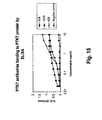
- an antibody of the invention binds to PTK7 with high affinity, for example with a K D of 1 x 10 -7 M or less.
- the antibody binds to human PTK7 with a K D of 5 x 10 -8 M or less, binds to human PTK7 with a K D of 1 x 10 -8 M or less, binds to human PTK7 with a K D of 5 x 10 -9 M or less, or binds to human PTK7 with a K D of between 1 x 10 -8 M and 1 x 10 -10 M or less.
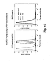
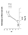
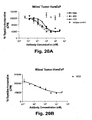
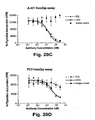
- the antibody binds to Wilms' tumor cells with an EC 50 of 4.0 nM or less, or binds to Wilms' tumor cells with an EC 50 of 3.5 nM or less.
- Standard assays to evaluate the binding ability of the antibodies toward PTK7 are known in the art, including for example, ELISAs, Western blots and RIAs.
- the binding kinetics (e.g ., binding affinity) of the antibodies also can be assessed by standard assays known in the art, such as by ELISA, Scatchard and Biacore analysis.
- the antibodies of the present invention may bind to a kidney carcinoma tumor cell line, for example, the Wilms' tumor cell line. Suitable assays for evaluating any of the above-described characteristics are described in detail in the Examples.
- the antibodies of the invention include the human monoclonal antibodies 4D5, 12C6 and 7C8.
- Other antibodies disclosed herein include 3G8, 3G8a and 12C6. All antibodies were isolated and structurally characterized as described in Examples 1 and 2. Those having ordinary skill in the art shall appreciate that the antibodies 3G8 and 3G8a, as well as the antibodies 12C6 and 12C6a have the same heavy chain sequence, while differing in their light chain sequences.
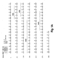
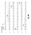
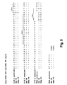
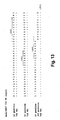
- the V H amino acid sequences of 3G8, 3G8a, 4D5, 12C6, 12C6a and 7C8 are shown in SEQ ID NOs: 1 (3G8 and 3G8a), 2 (4D5), 3 (12C6 and 12C6a) and 4 (7C8).
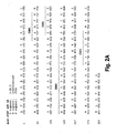
- the V L amino acid sequences of 3G8, 3G8a, 4D5, 12C6, 12C6a, and 7C8 are shown in SEQ ID NOs: 5, 6, 7, 8, 9 and 10, respectively.
- V H and V L sequences can be "mixed and matched" to create other anti-PTK7 binding molecules.
- PTK7 binding of such "mixed and matched" antibodies can be tested using the binding assays described above and in the Examples (e.g., ELISAs).
- a V H sequence from a particular V H /V L pairing is replaced with a structurally similar V H sequence.
- a V L sequence from a particular V H /V L pairing is replaced with a structurally similar V L sequence.
- an isolated monoclonal antibody, or antigen binding portion thereof comprising:
- the antibody, or antibody binding protein thereof, of the invention may comprise:
- antibodies that comprise the heavy chain and light chain CDR1s, CDR2s and CDR3s of 3G8, 3G8a, 4D5, 12C6, 12C6a and 7C8, or combinations thereof.
- the amino acid sequences of the V H CDR1s of 3G8, 3G8a, 4D5, 12C6, 12C6a and 7C8 are shown in SEQ ID NOs: 11 (3G8 and 3G8a), 12 (4D5), 13 (12C6 and 12C6a) and 14 (7C8).
- the amino acid sequences of the V H CDR2s of 3G8, 3G8a, 4D5, 12C6, 12C6a and 7C8 are shown in SEQ ID NOs: 15 (3G8 and 3G8a), 16 (4D5), 17 (12C6 and 12C6a) and 18 (7C8).
- the amino acid sequences of the V H CDR3s of 3G8, 3G8a, 4D5, 12C6, 12C6a and 7C8 are shown in SEQ ID NOs: 19 (3G8 and 3G8a), 20 (4D5), 21 (12C6 and 12C6a) and 22 (7C8).
- the amino acid sequences of the V k CDR1s of 3G8, 3G8a, 4D5, 12C6, 12C6a and 7C8 are shown in SEQ ID NOs: 23, 24, 25, 26, 27 and 28, respectively.
- the amino acid sequences of the V k CDR2s of 3G8, 3G8a, 4D5, 12C6, 12C6a and 7C8 are shown in SEQ ID NOs: 29, 30, 31, 32, 33 and 34, respectively.
- the amino acid sequences of the V k CDR3s of 3G8, 3G8a, 4D5, 12C6, 12C6a and 7C8 are shown in SEQ ID NOs: 35, 36, 37, 38, 39 and 40, respectively.
- the CDR regions are delineated using the Kabat system ( Kabat, E. A., et al. (1991) Sequences of Proteins of Immunological Interest, Fifth Edition, U.S. Department of Health and Human Services, NIH Publication No. 91-3242 ).
- V H CDR1, CDR2, and CDR3 sequences and V k CDR1, CDR2, and CDR3 sequences can be "mixed and matched" (i.e., CDRs from different antibodies can be mixed and match, although each antibody must contain a V H CDR1, CDR2, and CDR3 and a V k CDR1, CDR2, and CDR3) to create other anti-PTK7 binding molecules of the invention.
- PTK7 binding of such "mixed and matched" antibodies can be tested using the binding assays described above and in the Examples (e.g ., ELISAs, Biacore analysis).
- the CDR1, CDR2 and/or CDR3 sequence from a particular V H sequence is replaced with a structurally similar CDR sequence(s).
- V k CDR sequences are mixed and matched, the CDR1, CDR2 and/or CDR3 sequence from a particular V k sequence preferably is replaced with a structurally similar CDR sequence(s).
- V H and V L sequences can be created by substituting one or more V H and/or V L CDR region sequences with structurally similar sequences from the CDR sequences disclosed herein for monoclonal antibodies antibodies 3G8, 3G8a, 4D5, 12C6, 12C6a and 7C8.
- an isolated monoclonal antibody, or antigen binding portion thereof comprising:
- the antibody, or antigen binding portion thereof of the invention may comprise:
- antibodies comprising:
- the CDR3 domain independently from the CDR1 and/or CDR2 domain(s), alone can determine the binding specificity of an antibody for a cognate antigen and that multiple antibodies can predictably be generated having the same binding specificity based on a common CDR3 sequence. See, for example, Klimka et al., British J. of Cancer 83(2):252-260 (2000 ) (describing the production of a humanized anti-CD30 antibody using only the heavy chain variable domain CDR3 of murine anti-CD30 antibody Ki-4); Beiboer et al., J. Mol. Biol.
- monoclonal antibodies comprising one or more heavy and/or light chain CDR3 domains from an antibody derived from a human or non-human animal, wherein the monoclonal antibody is capable of specifically binding to PTK7.
- monoclonal antibodies comprising one or more heavy and/or light chain CDR3 domain from a non-human antibody, such as a mouse or rat antibody, wherein the monoclonal antibody is capable of specifically binding to PTK7.
- Such antibodies comprising one or more heavy and/or light chain CDR3 domain from a non-human antibody (a) are capable of competing for binding with; (b) retain the functional characteristics; (c) bind to the same epitope; and/or (d) have a similar binding affinity as the corresponding parental non-human antibody.
- monoclonal antibodies comprising one or more heavy and/or light chain CDR3 domain from a human antibody, such as, for example, a human antibody obtained from a non-human animal, wherein the human antibody is capable of specifically binding to PTK7.
- monoclonal antibodies comprising one or more heavy and/or light chain CDR3 domain from a first human antibody, such as, for example, a human antibody obtained from a non-human animal, wherein the first human antibody is capable of specifically binding to PTK7 and wherein the CDR3 domain from the first human antibody replaces a CDR3 domain in a human antibody that is lacking binding specificity for PTK7 to generate a second human antibody that is capable of specifically binding to PTK7.
- Such antibodies comprising one or more heavy and/or light chain CDR3 domain from the first human antibody (a) are capable of competing for binding with; (b) retain the functional characteristics; (c) bind to the same epitope; and/or (d) have a similar binding affinity as the corresponding parental first human antibody.
- the first human antibody may be 3G8, 3G8a, 4D5, 12C6, 12C6a or 7C8.
- the invention provides antibodies that bind to the same epitope on human PTK7 as any of the PTK7 monoclonal antibodies of the invention (i.e., antibodies that have the ability to cross-compete for binding to PTK7 with any of the monoclonal antibodies of the invention).
- the reference antibody for cross-competition studies can be the monoclonal antibody 4D5 (having V H and V L sequences as shown in SEQ ID NOs: 2 and 7, respectively), or the monoclonal antibody 12C6 (having V H and V L sequences as shown in SEQ ID NOs: 3 and 8, respectively), or the monoclonal antibody 7C8 (having V H and V L sequences as shown in SEQ ID NOs: 4 and 10, respectively).
- Such cross-competing antibodies can be identified based on their ability to cross-compete with 4D5, 12C6 or 7C8 in standard PTK7 binding assays. For example, BIAcore analysis, ELISA assays or flow cytometry may be used to demonstrate cross-competition with the antibodies of the current invention.
- the ability of a test antibody to inhibit the binding of, for example, 4D5, 12C6 or 7C8, to human PTK7 demonstrates that the test antibody can compete with 4D5, 12C6 or-7C8 for binding to human PTK7 and thus binds to the same epitope on human PTK7 as 4D5, 12C6 or 7C8.
- the antibody that binds to the same epitope on human PTK7 as 4D5, 12C6 or 7C8 is a human monoclonal antibody.
- Such human monoclonal antibodies can be prepared and isolated as described in the Examples.
- nucleic acid molecules that encode the antibodies of the invention.
- the nucleic acids may be present in whole cells, in a cell lysate, or in a partially purified or substantially pure form.
- a nucleic acid is "isolated” or “rendered substantially pure” when purified away from other cellular components or other contaminants, e.g., other cellular nucleic acids or proteins, by standard techniques, including alkaline/SDS treatment, CsCl banding, column chromatography, agarose gel electrophoresis and others well known in the art. See, F. Ausubel, et al., ed. (1987) Current Protocols in Molecular Biology, Greene Publishing and Wiley Interscience, New York .
- a nucleic acid of the invention can be, for example, DNA or RNA and may or may not contain intronic sequences.
- the nucleic acid is a cDNA molecule.
- Nucleic acids can be obtained using standard molecular biology techniques.
- hybridomas e.g., hybridomas prepared from transgenic mice carrying human immunoglobulin genes as described further below
- cDNAs encoding the light and heavy chains of the antibody made by the hybridoma can be obtained by standard PCR amplification or cDNA cloning techniques.
- nucleic acid encoding the antibody can be recovered from the library.
- Preferred nucleic acids molecules are those encoding the VH and VL sequences of the 3G8, 3G8a, 4D5, 12C6, 12C6a or 7C8 monoclonal antibodies.
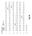
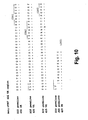
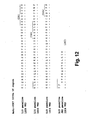
- DNA sequences encoding the VH sequences of 3G8, 3G8a, 4D5, 12C6, 12C6a and 7C8 are shown in SEQ ID NOs: 41 (3G8 and 3G8a), 42 (4D5), 43 (12C6 and 12C6a) and 44 (7C8).
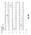
- DNA sequences encoding the VL sequences of 3G8, 3G8a, 4D5, 12C6, 12C6a and 7C8 are shown in SEQ ID NOs: 45, 46, 47, 48, 49 and 50, respectively.
- VH and VL segments are obtained, these DNA fragments can be further manipulated by standard recombinant DNA techniques, for example to convert the variable region genes to full-length antibody chain genes, to Fab fragment genes or to a scFv gene.
- a VL- or VH-encoding DNA fragment is operatively linked to another DNA fragment encoding another protein, such as an antibody constant region or a flexible linker.
- the term "operatively linked”, as used in this context, is intended to mean that the two DNA fragments are joined such that the amino acid sequences encoded by the two DNA fragments remain in-frame.
- the isolated DNA encoding the VH region can be converted to a full-length heavy chain gene by operatively linking the VH-encoding DNA to another DNA molecule encoding heavy chain constant regions (CH1, CH2 and CH3).
- heavy chain constant regions CH1, CH2 and CH3
- the sequences of human heavy chain constant region genes are known in the art (see e.g., Kabat, E. A., el al. (1991) Sequences of Proteins of Immunological Interest, Fifth Edition, U.S. Department of Health and Human Services, NIH Publication No. 91-3242 ) and DNA fragments encompassing these regions can be obtained by standard PCR amplification.
- the heavy chain constant region can be an IgG1, IgG2, IgG3, IgG4, IgA, IgE, IgM or IgD constant region, but most preferably is an IgG1 or IgG4 constant region.
- the VH-encoding DNA can be operatively linked to another DNA molecule encoding only the heavy chain CH1 constant region.
- the isolated DNA encoding the VL region can be converted to a full-length light chain gene (as well as a Fab light chain gene) by operatively linking the VL-encoding DNA to another DNA molecule encoding the light chain constant region, CL.
- the sequences of human light chain constant region genes are known in the art (see e.g., Kabat, E. A., et al. (1991) Sequences of Proteins of Immunological Interest, Fifth Edition, U.S. Department of Health and Human Services, NIH Publication No. 91-3242 ) and DNA fragments encompassing these regions can be obtained by standard PCR amplification.
- the light chain constant region can be a kappa or lambda constant region, but most preferably is a kappa constant region.
- the VH- and VL-encoding DNA fragments are operatively linked to another fragment encoding a flexible linker, e.g., encoding the amino acid sequence (Gly 4 -Ser) 3 , such that the VH and VL sequences can be expressed as a contiguous single-chain protein, with the VL and VH regions joined by the flexible linker (see e.g., Bird et al. (1988) Science 242:423-426 ; Huston et al. (1988) Proc. Natl. Acad. Sci. USA 85:5879-5883 ; McCafferty et al., (1990) Nature 348:552-554 ).
- a flexible linker e.g., encoding the amino acid sequence (Gly 4 -Ser) 3
- Monoclonal antibodies can be produced by a variety of techniques, including conventional monoclonal antibody methodology e.g., the standard somatic cell hybridization technique of Kohler and Milstein (1975) Nature 256: 495 . Although somatic cell hybridization procedures are preferred, in principle, other techniques for producing monoclonal antibody can be employed e.g., viral or oncogenic transformation of B lymphocytes.
- hybridomas The preferred animal system for preparing hybridomas is the murine system.
- Hybridoma production in the mouse is a very well-established procedure. Immunization protocols and techniques for isolation of immunized splenocytes for fusion are known in the art. Fusion partners (e.g., murine myeloma cells) and fusion procedures are also known.
- Chimeric or humanized antibodies can be prepared based on the sequence of a murine monoclonal antibody prepared as described above.
- DNA encoding the heavy and light chain immunoglobulins can be obtained from the murine hybridoma of interest and engineered to contain non-murine (e.g., human) immunoglobulin sequences using standard molecular biology techniques.
- the murine variable regions can be linked to human constant regions using methods known in the art (see e.g., U.S. Patent No. 4,816,567 to Cabilly et al .).
- the murine CDR regions can be inserted into a human framework using methods known in the art (see e.g., U.S. Patent No. 5,225,539 to Winter , and U.S. Patent Nos. 5,530,101 ; 5,585,089 ; 5,693,762 and 6,180,370 to Queen et al .).
- the antibodies of the invention are human monoclonal antibodies.
- Such human monoclonal antibodies directed against PTK7 can be generated using transgenic or transchromosomic mice carrying parts of the human immune system rather than the mouse system.
- transgenic and transchromosomic mice include mice referred to herein as HuMAb mice and KM mice TM , respectively, and are collectively referred to herein as "human Ig mice.”
- the HuMAb mouse® (Medarex, Inc.) contains human immunoglobulin gene miniloci that encode unrearranged human heavy ( ⁇ and ⁇ ) and ⁇ light chain immunoglobulin sequences, together with targeted mutations that inactivate the endogenous ⁇ and ⁇ chain loci (see e.g., Lonberg, et al. (1994) Nature 368 (6474): 856-859 ). Accordingly, the mice exhibit reduced expression of mouse IgM or ⁇ , and in response to immunization, the introduced human heavy and light chain transgenes undergo class switching and somatic mutation to generate high affinity human IgG ⁇ monoclonal (Lonberg, N. et al. (1994), supra ; reviewed in Lonberg, N.
- human antibodies of the invention can be raised using a mouse that carries human immunoglobulin sequences on transgenes and transchomosomes, such as a mouse that carries a human heavy chain transgene and a human light chain transchromosome.
- KM mice TM Such mice, referred to herein as "KM mice TM ", are described in detail in PCT Publication WO 02/43478 to Ishida et al .
- transgenic animal systems expressing human immunoglobulin genes are available in the art and can be used to raise anti-PTK7 antibodies of the invention.
- an alternative transgenic system referred to as the Xenomouse (Abgenix, Inc.) can be used; such mice are described in, for example, U.S. Patent Nos. 5,939,598 ; 6,075,181 ; 6,114,598 ; 6, 150,584 and 6,162,963 to Kucherlapati et al .
- mice carrying both a human heavy chain transchromosome and a human light chain tranchromosome referred to as "TC mice” can be used; such mice are described in Tomizuka et al. (2000) Proc. Natl. Acad. Sci. USA 97:722-727 .
- cows carrying human heavy and light chain transchromosomes have been described in the art ( Kuroiwa et al. (2002) Nature Biotechnology 20:889-894 ) and can be used to raise anti-PTK7 antibodies of the invention.
- Human monoclonal antibodies of the invention can also be prepared using phage display methods for screening libraries of human immunoglobulin genes. Such phage display methods for isolating human antibodies are established in the art. See for example: U.S. Patent Nos. 5,223,409 ; 5,403,484 ; and 5,571,698 to Ladner et al .; U.S. Patent Nos. 5,427,908 and 5,580,717 to Dower et al .; U.S. Patent Nos. 5,969,108 and 6,172,197 to McCafferty et al .; and U.S. Patent Nos. 5,885,793 ; 6,521,404 ; 6,544,731 ;. 6,555,313 ; 6,582,915 and 6,593,081 to Griffiths et al .
- Human monoclonal antibodies of the invention can also be prepared using SCID mice into which human immune cells have been reconstituted such that a human antibody response can be generated upon immunization.
- SCID mice into which human immune cells have been reconstituted such that a human antibody response can be generated upon immunization.
- Such mice are described in, for example, U.S. Patent Nos. 5,476,996 and 5,698,767 to Wilson et al .
- mice When human Ig mice are used to raise human antibodies of the invention, such mice can be immunized with a purified or enriched preparation of PTK7 antigen and/or recombinant PTK7, or a PTK7 fusion protein, as described by Lonberg, N. et al. (1994) Nature 368 (6474): 856-859 ; Fishwild, D. et al. (1996) Nature Biotechnology 14: 845-851 ; and PCT Publication WO 98/24884 and WO 01/14424 .
- the mice will be 6-16 weeks of age upon the first infusion.
- a purified or recombinant preparation (5-50 ⁇ g) of PTK7 antigen can be used to immunize the human Ig mice intraperitoneally.
- Example 1 Detailed procedures to generate fully human monoclonal antibodies to PTK7 are described in Example 1 below. Cumulative experience with various antigens has shown that the transgenic mice respond when initially immunized intraperitoneally (IP) with antigen in complete Freund's adjuvant, followed by every other week IP immunizations (up to a total of 6) with antigen in incomplete Freund's adjuvant. However, adjuvants other than Freund's are also found to be effective. In addition, whole cells in the absence of adjuvant are found to be highly immunogenic. The immune response can be monitored over the course of the immunization protocol with plasma samples being obtained by retroorbital bleeds.
- mice with sufficient titers of and-PTK7 human immunoglobulin can be used for fusions.
- Mice can be boosted intravenously with antigen 3 days before sacrifice and removal of the spleen. It is expected that 2-3 fusions for each immunization may need to be performed. Between 6 and 24 mice are typically immunized for each antigen.
- both HCo7 and HCo12 strains are used.
- both HCo7 and HCo12 transgene can be bred together into a single mouse having two different human heavy chain transgenes (HCo7/HCo12).
- the KM mouse TM strain can be used, as described in Example 1.
- splenocytes and/or lymph node cells from immunized mice can be isolated and fused to an appropriate immortalized cell line, such as a mouse myeloma cell line.
- an appropriate immortalized cell line such as a mouse myeloma cell line.
- the resulting hybridomas can be screened for the production of antigen-specific antibodies.
- single cell suspensions of splenic lymphocytes from immunized mice can be fused to one-sixth the number of P3X63-Ag8.653 nonsecreting mouse myeloma cells (ATCC, CRL 1580) with 50% PEG.
- Cells are plated at approximately 2 x 10 5 in flat bottom microtiter plate, followed by a two week incubation in selective medium containing 20% fetal Clone Serum, 18% "653" conditioned media, 5% origen (IGEN), 4 mM L-glutamine, 1 mM sodium pyruvate, 5mM HEPES, 0.055 mM 2-mercaptoethanol, 50 units/ml penicillin, 50 mg/ml streptomycin, 50 mg/ml gentamycin and 1X HAT (Sigma; the HAT is added 24 hours after the fusion). After approximately two weeks, cells can be cultured in medium in which the HAT is replaced with HT.
- selective medium containing 20% fetal Clone Serum, 18% "653" conditioned media, 5% origen (IGEN), 4 mM L-glutamine, 1 mM sodium pyruvate, 5mM HEPES, 0.055 mM 2-mercaptoethanol, 50 units/ml penicillin,
- selected hybridomas can be grown in two-liter spinner-flasks for monoclonal antibody purification.
- Supernatants can be filtered and concentrated before affinity chromatography with protein A-sepharose (Pharmacia, Piscataway, N.J.).
- Eluted IgG can be checked by gel electrophoresis and high performance liquid chromatography to ensure purity.
- the buffer solution can be exchanged into PBS, and the concentration can be determined by OD 280 using 1.43 extinction coefficient.
- the monoclonal antibodies can be aliquoted and stored at -80° C.
- Antibodies of the invention also can be produced in a host cell transfectoma using, for example, a combination of recombinant DNA techniques and gene transfection methods as is well known in the art (e.g., Morrison, S. (1985) Science 229:1202 ).
- DNAs encoding partial or full-length light and heavy chains can be obtained by standard molecular biology techniques (e.g., PCR amplification or cDNA cloning using a hybridoma that expresses the antibody of interest) and the DNAs can be inserted into expression vectors such that the genes are operatively linked to transcriptional and translational control sequences.
- operatively linked is intended to mean that an antibody gene is ligated into a vector such that transcriptional and translational control sequences within the vector serve their intended function of regulating the transcription and translation of the antibody gene.
- the expression vector and expression control sequences are chosen to be compatible with the expression host cell used.
- the antibody light chain gene and the antibody heavy chain gene can be inserted into separate vector or, more typically, both genes are inserted into the same expression vector.
- the antibody genes are inserted into the expression vector by standard methods (e.g., ligation of complementary restriction sites on the antibody gene fragment and vector, or blunt end ligation if no restriction sites are present).
- the light and heavy chain variable regions of the antibodies described herein can be used to create full-length antibody genes of any antibody isotype by inserting them into expression vectors already encoding heavy chain constant and light chain constant regions of the desired isotype such that the V H segment is operatively linked to the C H segment(s) within the vector and the V K segment is operatively linked to the C L segment within the vector.
- the recombinant expression vector can encode a signal peptide that facilitates secretion of the antibody chain from a host cell.
- the antibody chain gene can be cloned into the vector such that the signal peptide is linked in-frame to the amino terminus of the antibody chain gene.
- the signal peptide can be an immunoglobulin signal peptide or a heterologous signal peptide ( i.e ., a signal peptide from a non-immunoglobulin protein).
- the recombinant expression vectors of the invention carry regulatory sequences that control the expression of the antibody chain genes in a host cell.
- the term "regulatory sequence” is intended to include promoters, enhancers and other expression control elements (e.g., polyadenylation signals) that control the transcription or translation of the antibody chain genes.
- Such regulatory sequences are described, for example, in Goeddel (Gene Expression Technology. Methods in Enzymology 185, Academic Press, San Diego, CA (1990 )). It will be appreciated by those skilled in the art that the design of the expression vector, including the selection of regulatory sequences, may depend on such factors as the choice of the host cell to be transformed, the level of expression of protein desired, etc.
- Preferred regulatory sequences for mammalian host cell expression include viral elements that direct high levels of protein expression in mammalian cells, such as promoters and/or enhancers derived from cytomegalovirus (CMV), Simian Virus 40 (SV40), adenovirus, ( e.g ., the adenovirus major late promoter (AdMLP) and polyoma.
- CMV cytomegalovirus
- SV40 Simian Virus 40
- AdMLP adenovirus major late promoter
- nonviral regulatory sequences may be used, such as the ubiquitin promoter or ⁇ -globin promoter.
- regulatory elements composed of sequences from different sources such as the SR ⁇ , promoter system, which contains sequences from the SV40 early promoter and the long terminal repeat of human T cell leukemia virus type 1 ( Takebe, Y. et al. (1988) Mol. Cell. Biol. 8:466-472 ).
- the recombinant expression vectors of the invention may carry additional sequences, such as sequences that regulate replication of the vector in host cells (e.g ., origins of replication) and selectable marker genes.
- the selectable marker gene facilitates selection of host cells into which the vector has been introduced (see, e. g., U.S. Pat. Nos. 4,399,216 , 4,634,665 and 5,179,017, all by Axel et al .).
- the selectable marker gene confers resistance to drugs, such as G418, hygromycin or methotrexate, on a host cell into which the vector has been introduced.
- Preferred selectable marker genes include the dihydrofolate reductase (DHFR) gene (for use in dhfr- host cells with methotrexate selection/amplification) and the neo gene (for G418 selection).
- DHFR dihydrofolate reductase
- the expression vector(s) encoding the heavy and light chains is transfected into a host cell by standard techniques.
- the various forms of the term "transfection" are intended to encompass a wide variety of techniques commonly used for the introduction of exogenous DNA into a prokaryotic or eukaryotic host cell, e.g., electroporation, calcium-phosphate precipitation, DEAE-dextran transfection and the like.
- Preferred mammalian host cells for expressing the recombinant antibodies of the invention include Chinese Hamster Ovary (CHO cells) (including dhfr- CHO cells, described in Urlaub and Chasin, (1980) Proc. Natl. Acad. Sci. USA 77:4216-4220 , used with a DHFR selectable marker, e.g., as described in R. J. Kaufman and P. A. Sharp (1982) Mol. Biol. 159:601-621 ), NSO myeloma, cells, COS cells and SP2 cells.
- Chinese Hamster Ovary CHO cells
- dhfr- CHO cells described in Urlaub and Chasin, (1980) Proc. Natl. Acad. Sci. USA 77:4216-4220 , used with a DHFR selectable marker, e.g., as described in R. J. Kaufman and P. A. Sharp (1982) Mol. Biol. 159:601-621
- another preferred expression system is the GS gene expression system disclosed in WO 87/04462 , WO 89/01036 and EP 338,841 .
- the antibodies are produced by culturing the host cells for a period of time sufficient to allow for expression of the antibody in the host cells or, more preferably, secretion of the antibody into the culture medium in which the host cells are grown.
- Antibodies can be recovered from the culture medium using standard protein purification methods.
- Antibodies of the invention can be tested for binding to PTK7 by, for example, standard ELISA. Briefly, microtiter plates are coated with purified PTK7 at 0.25 ⁇ g/ml in PBS, and then blocked with 5% bovine serum albumin in PBS. Dilutions of antibody (e.g ., dilutions of plasma from PTK7-immunized mice) are added to each well and incubated for 1-2 hours at 37°C.
- mice which develop the highest titers will be used for fusions.
- An ELISA assay as described above can also be used to screen for hybridomas that show positive reactivity with PTK7 immunogen.
- Hybridomas that bind with high avidity to PTK7 are subcloned and further characterized.
- One clone from each hybridoma, which retains the reactivity of the parent cells (by ELISA) can be chosen for making a 5-10 vial cell bank stored at -140 °C, and for antibody purification.
- selected hybridomas can be grown in two-liter spinner-flasks for monoclonal antibody purification.
- Supernatants can be filtered and concentrated before affinity chromatography with protein A-sepharose (Pharmacia, Piscataway, NJ).
- Eluted IgG can be checked by gel electrophoresis and high performance liquid chromatography to ensure purity.
- the buffer solution can be exchanged into PBS, and the concentration can be determined by OD 280 using 1.43 extinction coefficient.
- the monoclonal antibodies can be aliquoted and stored at -80 °C.
- each antibody can be biotinylated using commercially available reagents (Pierce, Rockford, IL). Competition studies using unlabeled monoclonal antibodies and biotinylated monoclonal antibodies can be performed using PTK7 coated-ELISA plates as described above. Biotinylated mAb binding can be detected with a strep-avidin-alkaline phosphatase probe.
- isotype ELISAs can be performed using reagents specific for antibodies of a particular isotype. For example, to determine the isotype of a human monoclonal antibody, wells of microtiter plates can be coated with 1 ⁇ g/ml of anti-human immunoglobulin overnight at 4° C. After blocking with 1% BSA, the plates are reacted with 1 ⁇ g /ml or less of test monoclonal antibodies or purified isotype controls, at ambient temperature for one to two hours. The wells can then be reacted with either human IgG1 or human IgM-specific alkaline phosphatase-conjugated probes. Plates are developed and analyzed as described above.
- Anti-PTK7 human IgGs can be further tested for reactivity with PTK7 antigen by Western blotting. Briefly, PTK7 can be prepared and subjected to sodium dodecyl sulfate polyacrylamide gel electrophoresis. After electrophoresis, the separated antigens are transferred to nitrocellulose membranes, blocked with 10% fetal calf serum, and probed with the monoclonal antibodies to be tested. Human IgG binding can be detected using anti-human IgG alkaline phosphatase and developed with BCIP/NBT substrate tablets (Sigma Chem. Co., St. Louis, Mo.).
- the present invention features an anti-PTK7 antibody, or a fragment thereof, conjugated to a therapeutic moiety, such as a cytotoxin, a drug (e.g., an immunosuppressant) or a radiotoxin.
- a therapeutic moiety such as a cytotoxin, a drug (e.g., an immunosuppressant) or a radiotoxin.
- Examples include taxol, cytochalasin B, gramicidin D, ethidium bromide, emetine, mitomycin, etoposide, tenoposide, vincristine, vinblastine, colchicin, doxorubicin, daunorubicin, dihydroxy anthracin dione, mitoxantrone, mithramycin, actinomycin D, 1 -dehydrotestosterone, glucocorticoids, procaine, tetracaine, lidocaine, propranolol, and puromycin and analogs or homologs thereof.
- Therapeutic agents also include, for example, antimetabolites (e.g ., methotrexate, 6-mercaptopurine, 6-thioguanine, cytarabine, 5-fluorouracil decarbazine), alkylating agents (e.g ., mechlorethamine, thioepa chlorambucil, melphalan, carmustine (BSNU) and lomustine (CCNU), cyclothosphamide, busulfan, dibromomannitol, streptozotocin, mitomycin C, and cis-dichlorodiamine platinum (II) (DDP) cisplatin), anthracyclines (e.g ., daunorubicin (formerly daunomycin) and doxorubicin), antibiotics ( e.g ., dactinomycin (formerly actinomycin), bleomycin, mithramycin, and anthramycin (AMC)), and anti-mitotic agents
- An example of a calicheamicin antibody conjugate is commercially available (MylotargTM; Wyeth-Ayerst).
- Examples of therapeutic cytotoxins may be found, for example, in US Patent Nos: 6548530 and 6281354 and US Patent application Nos: US 2003/0064984 , US 2003/0073852 and US 2003/0050331 .
- Cytotoxins can be conjugated to antibodies of the invention using linker technology available in the art.
- linker types that have been used to conjugate a cytotoxin to an antibody include, but are not limited to, hydrazones, thioethers, esters, disulfides and peptide-containing linkers.
- a linker can be chosen that is, for example, susceptible to cleavage by low pH within the lysosomal compartment or susceptible to cleavage by proteases, such as proteases preferentially expressed in tumor tissue such as cathepsins ( e.g ., cathepsins B, C, D).
- Antibodies of the present invention also can be conjugated to a radioactive isotope to generate cytotoxic radiopharmaceuticals, also referred to as radioimmunoconjugates.
- radioactive isotopes that can be conjugated to antibodies for use diagnostically or therapeutically include, but are not limited to, iodine 131 , indium 111 , yttrium 90 and tutetium 177 .
- Method for preparing radioimmunconjugates are established in the art. Examples of radioimmunoconjugates are commercially available, including ZevalinTM (IDEC Pharmaceuticals) and BexxarTM (Corixa Pharmaceuticals), and similar methods can be used to prepare radioimmunoconjugates using the antibodies of the invention.
- the antibody conjugates of the invention can be used to modify a given biological response, and the drug moiety is not to be construed as limited to classical chemical therapeutic agents.
- the drug moiety may be a protein or polypeptide possessing a desired biological activity.
- proteins may include, for example, an enzymatically active toxin, or active fragment thereof, such as abrin, ricin A, pseudomonas exotoxin, or diphtheria toxin; a protein such as tumor necrosis factor or interferon- ⁇ ; or, biological response modifiers such as, for example, lymphokines, interleukin-1 ("IL-1"), interleukin-2 (“IL-2”), interleukin-6 (“IL-6”), granulocyte macrophage colony stimulating factor (“GM-CSF”), granulocyte colony stimulating factor (“G-CSF”), or other growth factors.
- IL-1 interleukin-1
- IL-2 interleukin-2
- IL-6 interleukin-6
- the present invention provides a composition, e.g ., a pharmaceutical composition, containing one or a combination of monoclonal antibodies, or antigen-binding portion(s) thereof, of the present invention, formulated together with a pharmaceutically acceptable carrier.
- a pharmaceutical composition of the invention can comprise a combination of antibodies (or immunoconjugates) that bind to different epitopes on the target antigen or that have complementary activities.
- compositions of the invention also can be administered in combination therapy, i.e ., combined with other agents.
- the combination therapy can include an anti-PTK7 antibody of the present invention combined with at least one other anti-inflammatory or immunosuppressant agent. Examples of therapeutic agents that can be used in combination therapy are described in greater detail below in the section on uses of the antibodies of the invention.
- pharmaceutically acceptable carrier includes any and all solvents, dispersion media, coatings, antibacterial and antifungal agents, isotonic and absorption delaying agents, and the like that are physiologically compatible.
- the carrier is suitable for intravenous, intramuscular, subcutaneous, parenteral, spinal or epidermal administration ( e.g ., by injection or infusion).
- the active compound i.e ., antibody, immunoconjuage, or bispecific molecule
- the pharmaceutical compounds of the invention may include one or more pharmaceutically acceptable salts.
- a "pharmaceutically acceptable salt” refers to a salt that retains the desired biological activity of the parent compound and does not impart any undesired toxicological effects (see e.g ., Berge, S.M., et al. (1977) J. Pharm. Sci. 66:1-19 ). Examples of such salts include acid addition salts and base addition salts.
- Acid addition salts include those derived from nontoxic inorganic acids, such as hydrochloric, nitric, phosphoric, sulfuric, hydrobromic, hydroiodic, phosphorous and the like, as well as from nontoxic organic acids such as aliphatic mono- and dicarboxylic acids, phenyl-substituted alkanoic acids, hydroxy alkanoic acids, aromatic-acids, aliphatic and aromatic sulfonic acids and the like.
- nontoxic inorganic acids such as hydrochloric, nitric, phosphoric, sulfuric, hydrobromic, hydroiodic, phosphorous and the like
- nontoxic organic acids such as aliphatic mono- and dicarboxylic acids, phenyl-substituted alkanoic acids, hydroxy alkanoic acids, aromatic-acids, aliphatic and aromatic sulfonic acids and the like.
- Base addition salts include those derived from alkaline earth metals, such as sodium, potassium, magnesium, calcium and the like, as well as from nontoxic organic amines, such as N,N'-dibenzylethylenediamine, N-methylglucamine, chloroprocaine, choline, diethanolamine, ethylenediamine, procaine and the like.
- a pharmaceutical composition of the invention also may include a pharmaceutically acceptable anti-oxidant.
- pharmaceutically acceptable antioxidants include: (1) water soluble antioxidants, such as ascorbic acid, cysteine hydrochloride, sodium bisulfate, sodium metabisulfite, sodium sulfite and the like; (2) oil-soluble antioxidants, such as ascorbyl palmitate, butylated hydroxyanisole (BHA), butylated hydroxytoluene (BHT), lecithin, propyl gallate, alpha-tocopherol, and the like; and (3) metal chelating agents, such as citric acid, ethylenediamine tetraacetic acid (EDTA), sorbitol, tartaric acid, phosphoric acid, and the like.
- water soluble antioxidants such as ascorbic acid, cysteine hydrochloride, sodium bisulfate, sodium metabisulfite, sodium sulfite and the like
- oil-soluble antioxidants such as ascorbyl palmitate, butylated
- aqueous and nonaqueous carriers examples include water, ethanol, polyols (such as glycerol, propylene glycol, polyethylene glycol, and the like), and suitable mixtures thereof, vegetable oils, such as olive oil, and injectable organic esters, such as ethyl oleate.
- polyols such as glycerol, propylene glycol, polyethylene glycol, and the like
- vegetable oils such as olive oil
- injectable organic esters such as ethyl oleate.
- Proper fluidity can be maintained, for example, by the use of coating materials, such as lecithin, by the maintenance of the required particle size in the case of dispersions, and by the use of surfactants.
- compositions may also contain adjuvants such as preservatives, wetting agents, emulsifying agents and dispersing agents. Prevention of presence of microorganisms may be ensured both by sterilization procedures, supra, and by the inclusion of various antibacterial and antifungal agents, for example, paraben, chlorobutanol, phenol sorbic acid, and the like. It may also be desirable to include isotonic agents, such as sugars, sodium chloride, and the like into the compositions. In addition, prolonged absorption of the injectable pharmaceutical form may be brought about by the inclusion of agents which delay absorption such as aluminum monostearate and gelatin.
- Pharmaceutically acceptable carriers include sterile aqueous solutions or dispersions and sterile powders for the extemporaneous preparation of sterile injectable solutions or dispersion.
- sterile aqueous solutions or dispersions and sterile powders for the extemporaneous preparation of sterile injectable solutions or dispersion.
- the use of such media and agents for pharmaceutically active substances is known in the art. Except insofar as any conventional media or agent is incompatible with the active compound, use thereof in the pharmaceutical compositions of the invention is contemplated. Supplementary active compounds can also be incorporated into the compositions.
- compositions typically must be sterile and stable under the conditions of manufacture and storage.
- the composition can be formulated as a solution, microemulsion, liposome, or other ordered structure suitable to high drug concentration.
- the carrier can be a solvent or dispersion medium containing, for example, water, ethanol, polyol (for example, glycerol, propylene glycol, and liquid polyethylene glycol, and the like), and suitable mixtures thereof.
- the proper fluidity can be maintained, for example, by the use of a coating such as lecithin, by the maintenance of the required particle size in the case of dispersion and by the use of surfactants.
- isotonic agents for example, sugars, polyalcohols such as mannitol, sorbitol, or sodium chloride in the composition.
- Prolonged absorption of the injectable compositions can be brought about by including in the composition an agent that delays absorption, for example, monostearate salts and gelatin.
- Sterile injectable solutions can be prepared by incorporating the active compound in the required amount in an appropriate solvent with one or a combination of ingredients enumerated above, as required, followed by sterilization microfiltration.
- dispersions are prepared by incorporating the active compound into a sterile vehicle that contains a basic dispersion medium and the required other ingredients from those enumerated above.
- the preferred methods of preparation are vacuum drying and freeze-drying (lyophilization) that yield a powder of the active ingredient plus any additional desired ingredient from a previously sterile-filtered solution thereof.
- the amount of active ingredient which can be combined with a carrier material to produce a single dosage form will vary depending upon the subject being treated, and the particular mode of administration.
- the amount of active ingredient which can be combined with a carrier material to produce a single dosage form will generally be that amount of the composition which produces a therapeutic effect. Generally, out of one hundred per cent, this amount will range from about 0.01 per cent to about ninety-nine percent of active ingredient, preferably from about 0.1 per cent to about 70 per cent, most preferably from about 1 per cent to about 30 per cent of active ingredient in combination with a pharmaceutically acceptable carrier.
- Dosage regimens are adjusted to provide the optimum desired response (e.g ., a therapeutic response). For example, a single bolus may be administered, several divided doses may be administered over time or the dose may be proportionally reduced or increased as indicated by the exigencies of the therapeutic situation. It is especially advantageous to formulate parenteral compositions in dosage unit form for ease of administration and uniformity of dosage.
- Dosage unit form as used herein refers to physically discrete units suited as unitary dosages for the subjects to be treated; each unit contains a predetermined quantity of active compound calculated to produce the desired therapeutic effect in association with the required pharmaceutical carrier.
- the specification for the dosage unit forms of the invention are dictated by and directly dependent on (a) the unique characteristics of the active compound and the particular therapeutic effect to be achieved, and (b) the limitations inherent in the art of compounding such an active compound for the treatment of sensitivity in individuals.
- the dosage ranges from about 0.0001 to 100 mg/kg, and more usually 0.01 to 5 mg/kg, of the host body weight.
- dosages can be 0.3 mg/kg body weight, I mg/kg body weight, 3 mg/kg body weight, 5 mg/kg body weight or 10 mg/kg body weight or within the range of 1-10 mg/kg.
- An exemplary treatment regime entails administration once per week, once every two weeks, once every three weeks, once every four weeks, once a month, once every 3 months or once every three to 6 months.
- Preferred dosage regimens for an anti-PTK7 antibody of the invention include 1 mg/kg body weight or 3 mg/kg body weight via intravenous administration, with the antibody being given using one of the following dosing schedules: (i) every four weeks for six dosages, then every three months; (ii) every three weeks; (iii) 3 mg/kg body weight once followed by 1 mg/kg body weight every three weeks.
- two or more monoclonal antibodies with different binding specificities are administered simultaneously, in which case the dosage of each antibody administered falls within the ranges indicated.
- Antibody is usually administered on multiple occasions. Intervals between single dosages can be, for example, weekly, monthly, every three monthgs or yearly. Intervals can also be irregular as indicated by measuring blood levels of antibody to the target antigen in the patient. In some methods, dosage is adjusted to achieve a plasma antibody concentration of about 1-1000 ⁇ g /ml and in some methods about 25-300 ⁇ g /ml.
- antibody can be administered as a sustained release formulation, in which case less frequent administration is required. Dosage and frequency vary depending on the half-life of the antibody in the patient. In general, human antibodies show the longest half life, followed by humanized antibodies, chimeric antibodies, and nonhuman antibodies. The dosage and frequency of administration can vary depending on whether the treatment is prophylactic or therapeutic. In prophylactic applications, a relatively low dosage is administered at relatively infrequent intervals over a long period of time. Some patients continue to receive treatment for the rest of their lives. In therapeutic applications, a relatively high dosage at relatively short intervals is sometimes required until progression of the disease is reduced or terminated, and preferably until the patient shows partial or complete amelioration of symptoms of disease. Thereafter, the patient can be administered a prophylactic regime.
- Actual dosage levels of the active ingredients in the pharmaceutical compositions of the present invention may be varied so as to obtain an amount of the active ingredient which is effective to achieve the desired therapeutic response for a particular patient, composition, and mode of administration, without being toxic to the patient.
- the selected dosage level will depend upon a variety of pharmacokinetic factors including the activity of the particular compositions of the present invention employed, or the ester, salt or amide thereof, the route of administration, the time of administration; the rate of excretion of the particular compound being employed, the duration of the treatment, other drugs, compounds and/or materials used in combination with the particular compositions employed, the age, sex, weight, condition, general health and prior medical history of the patient being treated, and like factors well known in the medical arts.
- a “therapeutically effective dosage” of an anti-PTK7 antibody of the invention preferably results in a decrease in severity of disease symptoms, an increase in frequency and duration of disease symptom-free periods, or a prevention of impairment or disability due to the disease affliction.
- a "therapeutically effective dosage” preferably inhibits cell growth or tumor growth by at least about 20%, more preferably by at least about 40%, even more preferably by at least about 60%, and still more preferably by at least about 80% relative to untreated subjects.
- the ability of a compound to inhibit tumor growth can be evaluated in an animal model system predictive of efficacy in human tumors.
- this property of a composition can be evaluated by examining the ability of the compound to inhibit, such inhibition in vitro by assays known to the skilled practitioner.
- a therapeutically effective amount of a therapeutic compound can decrease tumor size, or otherwise ameliorate symptoms in a subject.
- One of ordinary skill in the art would be able to determine such amounts based on such factors as the subject's size, the severity of the subject's symptoms, and the particular composition or route of administration selected.
- a composition of the present invention can be administered via one or more routes of administration using one or more of a variety of methods known in the art.
- routes and/or mode of administration will vary depending upon the desired results.
- Preferred routes of administration for antibodies of the invention include intravenous, intramuscular, intradermal, intraperitoneal, subcutaneous, spinal or other parenteral routes of administration, for example by injection or infusion.
- parenteral administration means modes of administration other than enteral and topical administration, usually by injection, and includes, without limitation, intravenous, intramuscular, intraarterial, intrathecal, intracapsular, intraorbital, intracardiac, intradermal, intraperitoneal, transtracheal, subcutaneous, subcuticular, intraarticular, subcapsular, subarachnoid, intraspinal, epidural and intrasternal injection and infusion.
- an antibody of the invention can be administered via a non-parenteral route, such as a topical, epidermal or mucosal route of administration, for example, intranasally, orally, vaginally, rectally, sublingually or topically.
- a non-parenteral route such as a topical, epidermal or mucosal route of administration, for example, intranasally, orally, vaginally, rectally, sublingually or topically.
- the active compounds can be prepared with carriers that will protect the compound against rapid release, such as a controlled release formulation, including implants, transdermal patches, and microencapsulated delivery systems.
- a controlled release formulation including implants, transdermal patches, and microencapsulated delivery systems.
- Biodegradable, biocompatible polymers can be used, such as ethylene vinyl acetate, polyanhydrides, polyglycolic acid, collagen, polyorthoesters, and polylactic acid. Many methods for the preparation of such formulations are patented or generally known to those skilled in the art. See, e.g., Sustained and Controlled Release Drug Delivery Systems, J.R. Robinson, ed., Marcel Dekker, Inc., New York, 1978 .
- compositions can be administered with medical devices known in the art.
- a therapeutic composition of the invention can be administered with a needleless hypodermic injection device, such as the devices disclosed in U.S. Patent Nos. 5,399,163 ; 5,383,851 ; 5,312,335 ; 5,064,413 ; 4,941,880 ; 4,790,824 ; or 4,596,556 .
- a needleless hypodermic injection device such as the devices disclosed in U.S. Patent Nos. 5,399,163 ; 5,383,851 ; 5,312,335 ; 5,064,413 ; 4,941,880 ; 4,790,824 ; or 4,596,556 .
- Examples of well-known implants and modules useful in the present invention include: U.S. Patent No. 4,487,603 , which discloses an implantable micro-infusion pump for dispensing medication at a controlled rate; U.S. Patent No.
- U.S. Patent No. 4,447,233 which discloses a medication infusion pump for delivering medication at a precise infusion rate
- U.S. Patent No. 4,447,224 which discloses a variable flow implantable infusion apparatus for continuous drug delivery
- U.S. Patent No. 4,439,196 which discloses an osmotic drug delivery system having multi-chamber compartments
- U.S. Patent No. 4,475,196 which discloses an osmotic drug delivery system.
- Many other such implants, delivery systems, and modules are known to those skilled in the art.
- the human monoclonal antibodies of the invention can be formulated to ensure proper distribution in vivo.
- the blood-brain barrier excludes many highly hydrophilic compounds.
- the therapeutic compounds of the invention cross the BBB (if desired)
- they can be formulated, for example, in liposomes.
- liposomes For methods of manufacturing liposomes, see, e.g., U.S. Patents 4,522,811 ; 5,374,548 ; and 5,399,331 .
- the liposomes may comprise one or more moieties which are selectively transported into specific cells or organs, thus enhance targeted drug delivery ( see, e.g., V.V. Ranade (1989) J. Clin. Pharmacol. 29:685 ).
- Exemplary targeting moieties include folate or biotin (see, e.g., U.S. Patent 5,416,016 to Low et al .); mannosides ( Umezawa et al., (1988) Biochem. Biophys. Res. Commun. 153:1038 ); antibodies ( P.G. Bloeman et al. (1995) FEBS Lett. 357:140 ; M. Owais et al. (1995) Antimicrob. Agents Chemother. 39:180 ); surfactant protein A receptor ( Briscoe et al. (1995) Am. J. Physiol. 1233:134 ); p120 ( Schreier et al. (1994) J. Biol. Chem.
- the antibodies, antibody compositions and methods of the present invention have numerous in vitro and in vivo diagnostic and therapeutic utilities involving the diagnosis and treatment of PTK7 mediated disorders.
- the antibodies of the present invention are human antibodies.
- these molecules can be administered to cells in culture, in vitro or ex vivo, or to human subjects, e.g., in vivo, to treat, prevent and to diagnose a variety of disorders.
- the term "subject" is intended to include human and non-human animals.
- Non-human animals includes all vertebrates, e.g ., mammals and non-mammals, such as non-human primates, sheep, dogs, cats, cows, horses, chickens, amphibians, and reptiles.
- Preferred subjects include human patients having disorders mediated by PTK7 activity.
- the methods are particularly suitable for treating human patients having a disorder associated with aberrant PTK7 expression.
- the antibodies of the invention can be used to specifically detect PTK7 expression on the surface of cells and, moreover, can be used to purify PTK7 via immunoaffinity purification.
- the invention further provides methods for detecting the presence of human PTK7 antigen in a sample, or measuring the amount of human PTK7 antigen, comprising contacting the sample, and a control sample, with a human monoclonal antibody, or an antigen binding portion thereof, which specifically binds to human PTK7, under conditions that allow for formation of a complex between the antibody or portion thereof and human PTK7. The formation of a complex is then detected, wherein a difference complex formation between the sample compared to the control sample is indicative the presence of human PTK7 antigen in the sample.
- PTK7 is expressed in colon carcinoma derived cell lines but not found to be expressed in human adult colon tissues ( Mossie et al. (1995) Oncogene 11:2179-84 ). PTK7 expression was also seen in some melanoma cell lines and melanoma biopsies ( Easty, et al. (1997) Int. J. Cancer 71:1061-5 ). In addition, PTK7 was found to be highly overexpressed in acute myeloid leukemia samples ( Muller-Tidow et al., (2004) Clin. Cancer Res. 10:1241-9 ). An anti-PTK7 antibody may be used alone to inhibit the growth of cancerous tumors. Alternatively, an anti-PTK7 antibody may be used in conjunction with other immunogenic agents, standard cancer treatments or other antibodies, as described below.
- Preferred cancers whose growth may be inhibited using the antibodies of the invention include cancers typically responsive to immunotherapy.
- preferred cancers for treatment include colon cancer (including small intestine cancer), lung cancer, breast cancer, pancreatic cancer, melanoma (e.g ., metastatic malignant melanoma), acute myeloid leukemia, kidney cancer, bladder cancer, ovarian cancer and prostate cancer.
- renal cancer e.g ., renal cell carcinoma
- glioblastoma brain tumors
- chronic or acute leukemias including acute lymphocytic leukemia (ALL), adult T-cell leukemia (T-ALL), chronic myeloid leukemia, acute lymphoblastic leukemia, chronic lymphocytic leukemia, lymphomas (e.g ., Hodgkin's and non-Hodgkin's lymphoma, lymphocytic lymphoma, primary CNS lymphoma, T-cell lymphoma, Burkitt's lymphoma, anaplastic large-cell lymphomas (ALCL), cutaneous T-cell lymphomas, nodular small cleaved-cell lymphomas, peripheral T-cell lymphomas, Lennert's lymphomas, immunoblastic lymphomas, T-cell leukemia/lymphomas (ATLL), entroblastic/centrocytic
- the human antibodies, antibody compositions and methods of the present invention can be used to treat a subject with a tumorigenic disorder, e.g ., a disorder characterized by the presence of tumor cells expressing PTK7 including, for example, colon cancer (including small intestine cancer), melanoma (e.g ., metastatic malignant melanoma), acute myeloid leukemia, lung cancer, breast cancer, bladder cancer, pancreatic cancer, ovarian cancer and prostate cancer.
- a tumorigenic disorder e.g ., a disorder characterized by the presence of tumor cells expressing PTK7 including, for example, colon cancer (including small intestine cancer), melanoma (e.g ., metastatic malignant melanoma), acute myeloid leukemia, lung cancer, breast cancer, bladder cancer, pancreatic cancer, ovarian cancer and prostate cancer.
- Examples of other subjects with a tumorigenic disorder include subjects having renal cancer (e.g ., renal cell carcinoma), glioblastoma, brain tumors, chronic or acute leukemias including acute lymphocytic leukemia (ALL), adult T-cell leukemia (T-ALL), chronic myeloid leukemia, acute lymphoblastic leukemia, chronic lymphocytic leukemia, lymphomas (e.g ., Hodgkin's and non-Hodgkin's lymphoma, lymphocytic lymphoma, primary CNS lymphoma, T-cell lymphoma, Burkitt's lymphoma, anaplastic large-cell lymphomas (ALCL), cutaneous T-cell lymphomas, nodular small cleaved-cell lymphomas, peripheral T-cell lymphomas, Lennert's lymphomas, immunoblastic lymphomas, T-cell leukemia/lymphomas (ATLL), entroblastic/centrocytic (c
- the invention provides an antibody or antigen binding portion of the invention for use in a method of inhibiting growth of tumor cells in a subject, said method comprising administering to the subject a therapeutically effective amount of said antibody or antigen-binding portion.
- the antibodies (e.g., human monoclonal antibodies and compositions) of the invention can be used to detect levels of PTK7 or levels of cells which contain PTK7 on their membrane surface, which levels can then be linked to certain disease symptoms.
- the antibodies can be used to inhibit or block PTK7 function which, in turn, can be linked to the prevention or amelioration of certain disease symptoms, thereby implicating PTK7 as a mediator of the disease. This can be achieved by contacting an experimental sample and a control sample with the anti-PTK7 antibody under conditions that allow for the formation of a complex between the antibody and PTK7. Any complexes formed between the antibody and PTK7 are detected and compared in the experimental sample and the control.
- the antibodies (e.g ., human antibodies and compositions) of the invention can be initially tested for binding activity associated with therapeutic or diagnostic use in vitro.
- compositions of the invention can be tested using the flow cytometric assays described in the Examples below.
- the antibodies may have additional utility in therapy and diagnosis of PTK7-related diseases.
- the human monoclonal antibodies and the immunoconjugates can be used to elicit in vivo or in vitro one or more of the following biological activities: to inhibit the growth of and/or kill a cell expressing PTK7; to mediate phagocytosis or ADCC of a cell expressing PTK7 in the presence of human effector cells; or to block PTK7 ligand binding to PTK7.
- the antibodies may be used in vivo to treat, prevent or diagnose a variety of PTK7-related diseases.
- PTK7-related diseases include, among others, colon cancer (including small intestine cancer), melanoma (e.g ., metastatic malignant melanoma), acute myeloid leukemia, lung cancer, breast cancer, bladder cancer, pancreatic cancer, ovarian cancer and prostate cancer.
- Suitable routes of administering the antibody compositions (e.g ., human monoclonal antibodies and immunoconjugates) of the invention in vivo and in vitro are well known in the art and can be selected by those of ordinary skill.
- the antibody compositions can be administered by injection (e.g ., intravenous or subcutaneous). Suitable dosages of the molecules used will depend on the age and weight of the subject and the concentration and/or formulation of the antibody composition.
- human anti-PTK7 antibodies of the invention can be co-administered with one or other more therapeutic agents, e.g ., a cytotoxic agent, a radiotoxic agent or an immunosuppressive agent.
- the antibody can be linked to the agent (as an immunocomplex) or can be administered separate from the agent. In the latter case (separate administration), the antibody can be administered before, after or concurrently with the agent or can be co-administered with other known therapies, e.g ., an anti-cancer therapy, e.g ., radiation.
- Such therapeutic agents include, among others, anti-neoplastic agents such as doxorubicin (adriamycin), cisplatin bleomycin sulfate, carmustine, chlorambucil and cyclophosphamide hydroxyurea which, by themselves, are only effective at levels which are toxic or subtoxic to a patient.
- anti-neoplastic agents such as doxorubicin (adriamycin), cisplatin bleomycin sulfate, carmustine, chlorambucil and cyclophosphamide hydroxyurea which, by themselves, are only effective at levels which are toxic or subtoxic to a patient.
- Cisplatin is intravenously administered as a 100 mg/ dose once every four weeks and adriamycin is intravenously administered as a 60-75 mg/ml dose once every 21 days.
- Co-administration of the human anti-PTK7 antibodies or antigen binding fragments thereof, of the present invention with chemotherapeutic agents provides two anti-cancer agents which operate via different mechanisms which yield a cytotoxic effect to human tumor cells. Such co-administration can solve problems due to development of resistance to drugs or a change in the antigenicity of the tumor cells which would render them unreactive with the antibody.
- immunoconjugates of the invention can be used to target compounds (e.g., therapeutic agents, labels, cytotoxins, radiotoxoins immunosuppressants, etc.) to cells which have PTK7 cell surface receptors by linking such compounds to the antibody.
- compounds e.g., therapeutic agents, labels, cytotoxins, radiotoxoins immunosuppressants, etc.
- an anti-PTK7 antibody can be conjugated to any of the toxin compounds described in US Patent Nos. 6,281,354 and 6,548,530 , US patent publication Nos. 20030050331 , 20030064984 , 20030073852 and 20040087497 or published in WO 03/022806 .
- the invention also provides methods for localizing ex vivo or in vivo cells expressing PTK7 (e.g ., with a detectable label, such as a radioisotope, a fluorescent compound, an enzyme or an enzyme co-factor).
- a detectable label such as a radioisotope, a fluorescent compound, an enzyme or an enzyme co-factor.
- the immunoconjugates can be used to kill cells which have PTK7 cell surface receptors by targeting cytotoxins or radiotoxins to PTK7.
- Target-specific effector cells e.g ., effector cells linked to compositions ( e.g ., human antibodies, multispecific and bispecific molecules) of the invention can also be used as therapeutic agents.
- Effector cells for targeting can be human leukocytes such as macrophages, neutrophils or monocytes. Other cells include eosinophils, natural killer cells and other IgG- or IgA-receptor bearing cells. If desired, effector cells can be obtained from the subject to be treated.
- the target-specific effector cells can be administered as a suspension of cells in a physiologically acceptable solution.
- the number of cells administered can be in the order of 10 8 -10 9 but will vary depending on the therapeutic purpose. In general, the amount will be sufficient to obtain localization at the target cell, e.g ., a tumor cell expressing PTK7 and to effect cell killing by, e.g ., phagocytosis. Routes of administration can also vary.
- Target-specific effector cells can be performed in conjunction with other techniques for removal of targeted cells.
- anti-tumor therapy using the compositions (e.g ., human antibodies) of the invention and/or effector cells armed with these compositions can be used in conjunction with chemotherapy.
- combination immunotherapy may be used to direct two distinct cytotoxic effector populations toward tumor cell rejection.
- anti-PTK7 antibodies linked to anti-Fc-gamma RI or anti-CD3 may be used in conjunction with IgG- or IgA-receptor specific binding agents.
- compositions (e.g ., human antibodies and immunoconjugates) of the invention which have complement binding sites, such as portions from IgG1, -2 or -3 or IgM which bind complement, can also be used in the presence of complement.
- ex vivo treatment of a population of cells comprising target cells with a binding agent of the invention and appropriate effector cells can be supplemented by the addition of complement or serum containing complement.
- Phagocytosis of target cells coated with a binding agent of the invention can be improved by binding of complement proteins.
- target cells coated with the compositions (e.g., human antibodies) of the invention can also be lysed by complement.
- the compositions of the invention do not activate complement.
- compositions e.g., human antibodies and immunoconjugates
- compositions comprising human antibodies and serum or complement. These compositions are advantageous in that the complement is located in close proximity to the human antibodies.
- the human antibodies of the invention and the complement or serum can be administered separately.
- patients treated with antibody compositions of the invention can be additionally administered (prior to, simultaneously with or following administration of a human antibody of the invention) with another therapeutic agent, such as a cytotoxic or radiotoxic agent, which enhances or augments the therapeutic effect of the human antibodies.
- another therapeutic agent such as a cytotoxic or radiotoxic agent, which enhances or augments the therapeutic effect of the human antibodies.
- the subject can be additionally treated with an agent that modulates, e.g., enhances or inhibits, the expression or activity of Fc ⁇ or Fc ⁇ receptors by, for example, treating the subject with a cytokine.
- cytokines for administration during treatment with the multispecific molecule include of granulocyte colony-stimulating factor (G-CSF), granulocyte- macrophage colony-stimulating factor (GM-CSF), interferon- ⁇ (IFN- ⁇ ) and tumor necrosis factor (TNF).
- G-CSF granulocyte colony-stimulating factor
- GM-CSF granulocyte- macrophage colony-stimulating factor
- IFN- ⁇ interferon- ⁇
- TNF tumor necrosis factor
- compositions (e.g ., human antibodies) of the invention can also be used to target cells expressing Fc ⁇ R or PTK7, for example for labeling such cells.
- the binding agent can be linked to a molecule that can be detected.
- the invention provides methods for localizing ex vivo or in vitro cells expressing Fc receptors, such as Fc ⁇ R or PTK7.
- the detectable label can be, e.g., a radioisotope, a fluorescent compound, an enzyme or an enzyme co-factor.
- kits comprising the antibody compositions of the invention (e.g ., human antibodies or immunoconjugates) and instructions for use.
- the kit can further contain one more more additional reagents, such as an immunosuppressive reagent, a cytotoxic agent or a radiotoxic agent or one or more additional human antibodies of the invention (e.g ., a human antibody having a complementary activity which binds to an epitope in the PTK7 antigen distinct from the first human antibody).
- Kits typically include a label indicating the intended use of the contents of the kit.
- the term label includes any writing, or recorded material supplied on or with the kit, or which otherwise accompanies the kit.
- Immunization protocols utilized as antigen both (i) a recombinant fusion protein comprising the extracellular portion of PTK7 with both a myc and his tag and (ii) membrane bound full-length PTK7. Both antigens were generated by recombinant transfection methods in a CHO cell line.
- Fully human monoclonal antibodies to PTK7 were prepared using the HCo7 and HCo12 strains of HuMab transgenic mice and the KM strain of transgenic transchromosomic mice, each of which express human antibody genes.
- the endogenous mouse kappa light chain gene has been homozygously disrupted as described in Chen et al. (1993) EMBO J. 12:811-820 and the endogenous mouse heavy chain gene has been homozygously disrupted as described in Example 1 of PCT Publication WO 01/09187 .
- Each of these mouse strains carries a human kappa light chain transgene, KCo5, as described in Fishwild et al. (1996) Nature Biotechnology 14:845-851 .
- the HCo7 strain carries the HCo7 human heavy chain transgene as described in U.S. Patent Nos. 5, 770, 429 ; 5,545,806 ; 5,625,825 ; and 5,545,807 _
- the HCo72 strain carries the HCo12 human heavy chain transgene as described in Example 2 of WO 01/09187 or example 2 WO 01/14424 .
- the KM strain contains the SC20 transchromosome as described in PCT Publication WO 02/43478 .
- HuMab mice and KM mice TM were immunized with purified recombinant PTK7 fusion protein and PTK7-transfected CHO cells as antigen.
- General immunization schemes for HuMab mice are described in Lonberg, N. et al (1994) Nature 368(6474): 856-859 ; Fishwild, D. et al. (1996) Nature Biotechnology 14: 845-851 and PCT Publication WO 98/24884 .
- the mice were 6-16 weeks of age upon the first infusion of antigen.
- a purified recombinant preparation (5-50 ⁇ g) of PTK7 fusion protein antigen and 5 - 10x10 6 cells were used to immunize the HuMab mice and KM mice TM intraperitonealy, subcutaneously (Sc) or via footpad injection.
- mice were immunized twice with antigen in complete Freund's adjuvant or Ribi adjuvant IP, followed by 3-21 days IP (up to a total of 11 immunizations) with the antigen in incomplete Freund's or Ribi adjuvant.
- the immune response was monitored by retroorbital bleeds.
- the plasma was screened by ELISA (as described below), and mice with sufficient titers of anti-PTK7 human immunogolobulin were used for fusions. Mice were boosted intravenously with antigen 3 days before sacrifice and removal of the spleen. Typically, 10-35 fusions for each antigen were performed. Several dozen mice were immunized for each antigen.
- the plates were washed with PBS/Tween and then incubated with a goat-anti-human IgG polyclonal antibody conjugated with horseradish peroxidase (HRP) for 1 hour at room temperature. After washing, the plates were developed with ABTS substrate (Sigma, A-1888, 0.22 mg/ml) and analyzed by spectrophotometer at OD 415-495. Mice that developed the highest titers of anti-PTK7 antibodies were used for fusions. Fusions were performed as described below and hybridoma supernatants were tested for anti-PTK7 activity by ELISA.
- HRP horseradish peroxidase
- mice The mouse splenocytes, isolated from the HuMab mice, were fused with PEG to a mouse myeloma cell line based upon standard protocols. The resulting hybridomas were then screened for the production of antigen-specific antibodies. Single cell suspensions of splenocytes from immunized mice were fused to one-fourth the number of SP2/0 nonsecreting mouse myeloma cells (ATCC, CRL 1581) with 50% PEG (Sigma).
- Cells were plated at approximately 1x10 5 /well in flat bottom microtiter plate, followed by about two week incubation in selective medium containing 10% fetal bovine serum, 10%.P388D1 (ATCC, CRL TIB-63) conditioned medium, 3-5% origen (IGEN) in DMEM (Mediatech, CRL 10013, with high glucose, L-glutamine and sodium pyruvate) plus 5 mM HEPES, 0.055 mM 2-mercaptoethanol, 50 mg/ml gentamycin and 1x HAT (Sigma, CRL P-7185). After 1-2 weeks, cells were cultured in medium in which the HAT was replaced with HT.
- selective medium containing 10% fetal bovine serum, 10%.P388D1 (ATCC, CRL TIB-63) conditioned medium, 3-5% origen (IGEN) in DMEM (Mediatech, CRL 10013, with high glucose, L-glutamine and sodium pyruvate) plus 5 mM HEPES,
- Hybridoma clones 3G8, 3G8a, 4D5, 12C6, 12C6a and 7C8 were selected for further analysis.
- the cDNA sequences encoding the heavy and light chain variable regions of the 3G8, 3G8a, 4D5, 12C6, 12C6a and 7C8 monoclonal antibodies were obtained from the 3G8, 3G8a, 4D5, 12C6, 12C6a and 7C8 hybridomas, respectively, using standard PCR techniques and were sequenced using standard DNA sequencing techniques.
- nucleotide and amino acid sequences of the heavy chain variable region of 3G8 are shown in Figure 1A and in SEQ ID NO:41 and 1, respectively.
- nucleotide and amino acid sequences of the light chain variable region of 3G8 are shown in Figure 1B and in SEQ ID NO:45 and 5, respectively.
- nucleotide and amino acid sequences of the heavy chain variable region of 3G8a are shown in Figure 1A and in SEQ ID NO:41 and 1, respectively.
- nucleotide and amino acid sequences of the light chain variable region of 3G8a are shown in Figure 1C and in SEQ ID NO:46 and 6, respectively.
- nucleotide and amino acid sequences of the heavy chain variable region of 4D5 are shown in Figure 2A and in SEQ ID NO:42 and 2, respectively.
- nucleotide and amino acid sequences of the light chain variable region of 4D5 are shown in Figure 2B and in SEQ ID NO:47 and 7, respectively.
- nucleotide and amino acid sequences of the heavy chain variable region of 12C6 are shown in Figure 3A and in SEQ ID NO:43 and 3, respectively.
- nucleotide and amino acid sequences of the light chain variable region of 12C6 are shown in Figure 3B and in SEQ ID NO:48 and 8, respectively.
- nucleotide and amino acid sequences of the heavy chain variable region of 12C6a are shown in Figure 3A and in SEQ ID NO:43 and 3, respectively.
- nucleotide and amino acid sequences of the light chain variable region of 12C6a are shown in Figure 3C and in SEQ ID NO:49 and 9, respectively.
- nucleotide and amino acid sequences of the heavy chain variable region of 7C8 are shown in Figure 4A and in SEQ ID NO:44 and 4, respectively.
- nucleotide and amino acid sequences of the light chain variable region of 7C8 are shown in Figure 4B and in SEQ ID NO:50 and 10, respectively.
- monoclonal antibodies 12C6 and 12C6a utilize a heavy chain variable region derived from a human DP-44 germline sequence present in the HCo7 transgene of the HuMab Mouse ® strain. Since DP-44 is not a germline sequence that is utilized in the native human immunoglobulin repertoire, it may be advantageous to mutate the VH sequence of 12C6 and 12C6a to reduce potential immunogenicity. Preferably, one or more framework residues of the 12C6 or 12C6a VH sequence is mutated to a residue(s) present in the framework of a structurally related VH germline sequence that is utilized in the native human immunoglobulin repertoire.
- Figure 7 shows the alignment of the 12C6 and 12C6a VH sequence with the DP44 germline sequence and also to two structurally related human germline sequences, VH 3-23 and VH 3-7. Given the relatedness of these sequences, one can predict that one can select a human antibody that specifically binds to human PTK7 and that utilizes a VH region derived from a VH 3-23 or VH 3-7 germline sequence. Moreover, one can mutate one or more residues within the 12C6 or 12C6a VH sequence that differ from the residue(s) at the comparable position in the VH 3-23 or VH 3-7 sequence to the residue(s) that is present in VH 3-23 or VH 3-7, or to a conservative amino acid substitution thereof.
- binding affinity and binding kinetics of anti-PTK7 antibodies were examined by Biacore analysis. Binding specificity, and cross-competition were examined by flow cytometry.
- HEK3 cell lines that express recombinant human PTK7 at the cell surface were developed and used to determine the specificity of PTK7 human monoclonal antibodies by flow cytometry.
- HEK3 cells were transfected with expression plasmids containing full length cDNA encoding transmembrane forms of PTK7. Binding of the 7C8 anti-PTK7 human monoclonal antibody was assessed by incubating the transfected cells with the anti-PTK7 human monoclonal antibody at a concentration of 10 ⁇ g/ml. The cells were washed and binding was detected with a FITC-labeled anti-human IgG Ab.
- anti-PTK7 antibodies were also assessed by standard ELISA to examine the specificity of binding for PTK7.
- Recombinant extracellular domain of PTK7 was tested for binding against the anti-PTK7 human monoclonal antibodies 3G8, 4D5, 12C6 and 12C6a at different concentrations. Standard ELISA procedures were performed. The anti-PTK7 human monoclonal antibodies were added at a starting concentration of 10 ⁇ g/ml and serially diluted at a 1:2 dilution. Goat-anti-human IgG (kappa chain-specific) polyclonal antibody conjugated with horseradish peroxidase (HRP) was used as secondary antibody. The results are shown in Figure 15 . Each of the anti-PTK7 human monoclonal antibodies 3G8, 4D5, 12C6 and 12C6a bound to PTK7. This data demonstrates the specificity of anti-PTK7 human monoclonal antibodies for PTK7.
- HRP horseradish peroxidase
- Flow cytometry was used to determine epitope grouping of anti-PTK7 HuMAbs.
- Wilms' tumor cells G-401 (ATCC Acc No. CRL-1441) were transfected with expression plasmids containing full length cDNA encoding transmembrane forms of PTK7.
- Epitope binding of each anti-PTK7 human monoclonal antibody was assessed by incubating 1x10 5 transfected cells with 10 ⁇ g/ml of cold anti-PTK7 human monoclonal antibody, washed, and followed by the addition of 10 ⁇ g/ml of a fluorescence-conjugated anti-PTK7 human monoclonal antibody.
- Binding was detected with a FITC-labeled anti-human IgG Ab.
- Flow cytometric analyses were performed using a FACScan flow cytometry (Becton Dickinson, San Jose, CA).
- the anti-PTK7 antibodies have been categorized into 3 epitope groups - group A, which includes 7D11, group B, which includes 3G8 and 3G8a and group C, which includes 7C8, 12C6 and 12C6a.
- the nephroblastoma Wilms' tumor cell line G-401 (ATCC Acc No. CRL-1441) was tested for binding of the HuMAb anti-PTK7 human monoclonal antibodies 12C6 and 7C8 at different concentrations. Binding of the anti-PTK7 human monoclonal antibodies was assessed by incubating 1x10 5 cells with antibody at a starting concentration of 30 ⁇ g/ml and serially diluting the antibody at a 1:10 dilution. The cells were washed and binding was detected with a PE-labeled anti-human IgG Ab. Flow cytometric analyses were performed using a FACScan flow cytometry (Becton Dickinson, San Jose, CA). The results are shown in Figure 16 .
- the anti-PTK7 monoclonal antibodies 12C6 and 7C8 bound to the nephroblastoma Wilms' tumor cell line in a concentration dependent manner, as measured by the mean fluorescent intensity (MFI) of staining.
- MFI mean fluorescent intensity
- the EC 50 values for the anti-PTK7 monoclonal antibodies 12C6 and 7C8 was 4.035 nM and 3.428 nM, respectively.
- Anti-PTK7 antibodies were tested for binding to a variety of cancer cell lines by flow cytometry.
- Binding of the 3G8, 12C6a, 4D5 and 12C6 anti-PTK7 human monoclonal antibodies to a panel of cancer cell lines was assessed by incubating cancer cell lines with anti-PTK7 human monoclonal antibodies at a concentration of 10 ⁇ g/ml.
- the cancer cell lines that were tested were A-431 (ATCC Acc No. CRL-1555), Wilms tumor cells G-401 (ATCC Acc No. CRL-1441), Saos-2 (ATCC Acc No. HTB-85), SKOV-3 (ATCC Acc No. HTB-77), PC3 (ATCC Acc No. CRL-1435), DMS 114 (ATCC Acc No. CRL-2066), ACHN (ATCC Acc No.
- the anti-PTK7 monoclonal antibodies 3G8, 12C6a, 4D5 and 12C6 bound to the cancer cell lines A-431, Wilms tumor cells G-401, Saos-2, SKOV-3, PC3, DMS 114, ACHN, LNCaP, DU 145, LoVo and MIA PaCa-2, as measured by the mean fluorescent intensity (MFI) of staining.
- MFI mean fluorescent intensity
- Example 7 Binding of anti-PTK7 to human T, B and dendritic cells
- Anti-PTK7 antibodies were tested for binding to CD4+, CD8+ T-cells, CD19+ B-cells and human blood myeloid dendritic cells expressing PTK7 on their cell surface by flow cytometry.
- Human T cells were activated by anti-CD3 antibody to induce PTK7 expression on T cells prior to binding with a human anti-PTK7 monoclonal antibody. Binding of the 7c8 anti-PTK7 human monoclonal antibody was assessed by incubating the cells with anti-PTK7 human monoclonal antibodies at a concentration of 10 ⁇ g/ml. In some experiments, a known antibody that binds a T and B-cell specific marker was used as a positive control. The cells were washed and binding was detected with a FITC-labeled anti-human IgG Ab. Flow cytometric analyses were performed using a FACScan flow cytometry (Becton Dickinson, San Jose, CA).
- Anti-PTK7 HuMAbs were tested for the ability to internalize into PTK7-expressing cell lines using a Hum-Zap internalization assay.
- the Hum-Zap assay tests for internalization of a primary human antibody through binding of a secondary antibody with affinity for human IgG conjugated to the toxin saporin.
- the PTK7-expressing cancer cell lines Wilms tumor G-401 (ATCC Acc No. CRL-1441), A-431 (ATCC Acc No. CRL-1555) and PC3 (ATCC Acc No. CRL-1435) were seeded at 1x10 4 cells/well in 100 ⁇ l wells directly.
- the anti-PTK7 HuMAb antibodies 3G8, 4D5, 12C6 or 7C8 were added to the wells at a starting concentration of 30 nM and titrated down at 1:3 serial dilutions.
- An isotype control antibody that is non-specific for PTK7 was used as a negative control.
- the Hum-Zap Advanced Targeting Systems, San Diego, CA, IT-22-25 was added at a concentration of 11 nM and plates were allowed to incubate for 72 hours. The plates were then pulsed with 1.0 ⁇ Ci of 3 H-thymidine for 24 hours, harvested and read in a Top Count Scintillation Counter (Packard Instruments, Meriden, CT). The results are shown in Figures 20A-D .
- the anti-PTK7 antibodies 3G8, 4D5, 12C6 and 7C8 showed an antibody concentration dependent decrease in 3 H-thymidine incorporation in the PTK7-expressing Wilms' Tumor cancer cell line.
- the anti-PTK7 antibodies 12C6 and 7C8 showed an antibody concentration dependent decrease in 3 H-thymidine incorporation in the PTK7-expressing cancer cell lines A-431 and PC3.
- the EC 50 value for the anti-PTK7 antibodies 3G8, 4D5, 12C6 and 7C8 in Wilms' tumor cells was 0.6437 nM, 0.2516 nM, 0.2053 nM and 0.1788 nM, respectively.
- the EC 50 value for the anti-PTK7 antibodies 12C6 and 7C8 in A-431 cells was 0.1657 nM and 0.1826 nM, respectively.
- the EC 50 value for the anti-PTK7 antibodies 12C6 and 7C8 in PC3 tumor cells was 0.3175 nM and 0.2648 nM, respectively. This data demonstrates that the anti-PTK7 antibodies 3G8, 4D5, 12C6 and 7C8 internalize into cancer cells.
- Example 9 Assessment of cell killing of a toxin-conjugated anti-PTK7 antibody on human cancer cell lines
- anti-PTK7 monoclonal antibodies conjugated to a toxin were tested for the ability to kill PTK7+ human cancer cell lines in a cell proliferation assay.
- the anti-PTK7 HuMAb antibody 12C6a was conjugated to a toxin via a linker, such as a peptidyl, hydrazone or disulfide linker.
- a linker such as a peptidyl, hydrazone or disulfide linker.
- Examples of toxin compounds that may be conjugated to the antibodies of the current invention are described in WO 2007/038658 .
- the PTK7-expressing Wilms' tumor human kidney cancer cell line G-401 (ATCC Acc No. CRL-1441) was seeded at 10 4 cells/well in 100 ⁇ l wells for 3 hours.
- An anti-PTK7 antibody-toxin conjugate was added to the wells at a starting concentration of 100 nM and titrated down at 1:3 serial dilutions. Plates were allowed to incubate for 48 hours.
- Figure 21 shows the effects of the 12C6a-conjugate on the Wilms' tumor cells.
- the anti-PTK7 antibody 12C6a showed an antibody-toxin concentration dependent decrease in 3 H-thymidine incorporation in PTK7-expressing Wilms' tumor human kidney cancer cell line.
- Example 10 Assessment of cell killing of a toxin-conjugated anti-PTK7 antibody on human tumor cell lines
- anti-PTK7 monoclonal antibodies conjugated to a toxin were tested for the ability to kill PTK7 + human tumor cell lines having either low, intermediate or high cell surface expression of PTK7 in a cell proliferation assay.
- the anti-PTK7 HuMAb antibody 12C6a was conjugated to a toxin via a linker, such as a peptidyl, hydrazone or disulfide linker.
- a linker such as a peptidyl, hydrazone or disulfide linker.
- Examples of toxin compounds that may be conjugated to the antibodies of the current invention are described in WO 2007/038658 .
- the PTK7-expressing human tumor cancer cell lines A-431, SKOV3, and LoVo were seeded at 10 4 cells/well in 100 ⁇ l wells. The cell lines were previously tested for cell surface expression of PTK7 in a standard FACS assay.
- the A-431 cell line expressed the highest level of PTK7 cell surface expression and the LoVo cell line expressed the lowest level of PTK7 cell surface expression.
- An anti-PTK7 antibody-toxin conjugate was added to the wells at a starting concentration of 20 nM and titrated down at 1:2 serial dilutions.
- An isotype control antibody was used as a negative control. Plates were allowed to incubate for 3 hours and the unbound (free) antibody-toxin conjugates were washed out. The plates continued to incubate for 96 hrs and the killing activity (FU, fluorescent unit) was measured by cell viability in CellTiter-Glo ® Luminescent assay according to protocol (Promega, WI, USA, Technical bulletin No. 288) using a BIO-TEK reader (Bio-Tek Instruments, Inc, VT, USA). The results are shown in Figure 22 .
- the anti-PTK7-toxin conjugate showed an antibody-toxin concentration dependent decrease in the proliferation assay in PTK7-expressing A431 high , SKOV3 inter , and LoVo low .
- the ability of the anti-PTK7 HuMAbs 3G8, 12C6a, and 2E11 to recognize PTK7 by immunohistochemistry was examined using clinical biopsies from lung cancer, breast cancer, bladder cancer, pancreatic cancer, colon cancer, ovarian cancer, small intestine cancer & prostate cancer.
- FAP Fibroblast Activation Protein antibody
- antibodies directed against PTK7 were tested for the ability to affect cell invasion in a CHO cell line transfected with PTK7.
- the assay was done using a HTS 96-Multiwell Insert System (Cat# 351162, BD Biosciences, CA) according to the protocol.
- Either a CHO parent cell line, CHO cells transfected with full-length PTK7 or a control HEK293 cell line were mixed with either a pool of antiPTK7 HuMabs or an isotype control antibody prior to the addition of the cells into the inserts.
- the mixture (cells+Ab pool) was added into an insert well in the invasion plate.
- Example 13 Treatment of in vivo pancreatic cancer cell xenograft model using naked and cytotoxin-conjugated anti-PTK7 antibodies
- This Example discloses the in vivo treatment of mice implanted with a pancreatic cell carcinoma tumor with toxin-conjugated anti-PTK7 antibodies to examine the in vivo effect of the antibodies on tumor growth.
- HPAC human pancreatic adenocarcinoma, ATCC Accession Number CRL-2119
- HPAC human pancreatic adenocarcinoma, ATCC Accession Number CRL-2119
- Male Ncr athymic nude mice (Taconic, Hudson, NY) between 6-8 weeks of age were implanted subcutaneously in the right flank with 2.5 x10 6 HPAC cells in 0.2 ml of PBS/Matrigel (1:1) per mouse. Mice were weighed and measured for tumors three dimensionally using an electronic caliper twice weekly after implantation. Tumor volumes were calculated as height x width x length/2. Mice with HPAC tumors averaging 90 mm 3 were randomized into treatment groups.
- mice were administered a single intravenous dose with PBS vehicle, naked anti-PTK7 antibody or toxin-conjugated anti-PTK7 HuMAb on Day 0 at the dosage indicated ( ⁇ mol/kg).
- Examples of toxin compounds that may be conjugated to the antibodies of the current invention were described in the pending U.S. Patent Application Publication number 2006/0024317 A1 .
- Mice were monitored for tumor growth for 61 days post dosing. Mice were euthanized when the tumors reached tumor end point (2000 mm 3 ) or ulcerated.
- Anti-PTK7 antibodies conjugated to a toxin slowed tumor growth progression. The results are shown in Figure 24 .
- anti-tumor effect of the anti-PTK7 toxin conjugate was dose dependent, with the greatest effect seen at a dose of 0.3 ⁇ mol/kg.
- Treatment with anti-PTK7 toxin conjugate was well tolerated, with subjects never experiencing greater than 5% median body weight loss (data not shown).
- treatment with an anti-PTK7 antibody-toxin conjugate has a direct in vivo inhibitory effect on pancreatic cancer tumor growth.
- Example 14 Treatment of in vivo breast cancer cell xenograft model using naked and cytotoxin-conjugated anti-PTK7 antibodies
- This Example discloses the in vivo treatment of mice implanted with an adriamycin resistance breast carcinoma tumor with toxin-conjugated anti-PTK7 antibodies to examine the in vivo effect of the antibodies on tumor growth.
- MCF7-adr human breast cancer cell line resistant to adriamycin
- Female CB17.SCID mice Teconic, Hudson, NY between 6-8 weeks of age were implanted subcutaneously with 1.7 mg 90-day release estrogen pellets, 3.0 mm size (Innovative Research of America, Sarasota, FL) at the neck region one day prior to being implanted subcutaneously in the right flank with 10 x10 6 MCF7-Adr cells in 0.2 ml of PBS/Matrigel (1:1) per mouse. Mice were weighed and measured for tumors three dimensionally using an electronic caliper twice weekly after implantation. Tumor volumes were calculated as height x width x length/2.
- mice with MCF7-adr tumors averaging 160 mm 3 were randomized into treatment groups.
- the mice were administered a single intravenous dose at 0.1 ⁇ mol/kg with PBS vehicle, naked anti-PTK7 antibody or toxin-conjugated anti-PTK7 HuMAb on Day 0.
- Examples of toxin compounds that may be conjugated to the antibodies of the current invention were described in the pending U.S. Patent Application Publication number 2006/0024317 A1 .
- Mice were monitored for tumor growth for 63 days post dosing. Mice were euthanized when the tumors were ulcerated. The results are shown in Figure 25 .
- Anti-PTK7 antibody toxin conjugates slowed tumor growth progression.
- VK CDR1 a.a. 7C8 6 VK a.a. 3G8a 7 VK a.a. 4D5 29 VK CDR2 a.a. 3G8 8 VK a.a. 12C6 30 VK CDR2 a.a. 3G8a 9 VK a.a. 12C6a 31 VK CDR2 a.a. 4D5 10 VK a.a. 7C8 32 VK CDR2 a.a. 12C6 33 VK CDR2 a.a. 12C6a 11 VH CDR1 a.a. 3G8 34 VK CDR2 a.a. 7C8 12 VH CDR1 a.a.
Description
- Receptor tyrosine kinases (RTKs) are transmembrane signaling proteins that transmit biological signals from the extracellular environment to the interior of the cell. The regulation of RTK signals is important for regulation of cell growth, differentiation, axonal growth, epithelial growth, development, adhesion, migration, and apoptosis (Prenzel et al. (2001) Endocr. Relat. Cancer 8:11-31; Hubbard and Till (2000) Annu. Rev. Biochem. 69:373-98). RTKs are known to be involved in the development and progression of several forms of cancer. In most of the RTK-related cancers, there has been an amplification of the receptor protein rather than a mutation of the gene (Kobus and Fleming (2005) Biochemistry 44:1464-70 ).
- Protein tyrosine kinase 7 (PTK7), a member of the receptor protein tyrosine kinase family, was first isolated from normal human melanocytes and cloned by RT-PCR (Lee et al., (1993) Oncogene 8:3403-10; Park et al., (1996) J. Biochem 119:235-9). Separately, the gene was cloned from human colon carcinoma-derived cell lines and named colon carcinoma kinase 4 (CCK4) (Mossie et al. (1995) Oncogene 11:2179-84). PTK7 belongs to a subset of RTKs that lack detectable catalytic tyrosine kinase activity but retain signal transduction activity and is thought to possibly function as a cell adhesion molecule.
- The mRNA for PTK7 was found to be variably expressed in colon carcinoma derived cell lines but not found to be expressed in human adult colon tissues (Mossie et al., supra). PTK7 expression was also seen in some melanoma cell lines and melanoma biopsies (Easty, et al. (1997) Int. J. Cancer 71:1061-5). An alternative splice form was found to be expressed in hepatomas and colon cancer cells (Jung et al. (2002) Biochim Biophys Acta 1579: 153-63). In addition, PTK7 was found to be highly overexpressed in acute myeloid leukemia samples (Muller-Tidow et al., (2004) Clin. Cancer Res. 10:1241-9). By immunohistochemistry, tumor specific staining of PTK7 was observed in breast, colon, lung, pancreatic, kidney and bladder cancers, as described in
PCT Publication WO 04/17992 WO 2004/017992 describes PTK7, agents which interact with or modulate the expression or activity of PTK7, and their use in treating carcinoma. Kyriakos et al. (Cancer Research 52, February 1992, No. 4, pages 835-842) describes the fate of antibodies bound to the surface of tumor cells in vitro. - Accordingly, agents that recognize PTK7, and methods of using such agents, are desired.
- The present invention provides isolated monoclonal antibodies, in particular human monoclonal antibodies, that bind to PTK7 and that exhibit numerous desirable properties. These properties include high affinity binding to human PTK7 and binding to Wilms' tumor cells. Also provided are the antibodies and compositions of the invention for use in methods for treating a variety of PTK7 mediated diseases.
- The invention pertains to an isolated human monoclonal antibody, or an antigen-binding portion thereof, wherein the antibody:
- (a) specifically binds to human PTK7; and
- (b) binds to a Wilms' tumor cell line having ATCC Acc No. CRL-1441 with an EC50 of 4.0 nM or less, in an assay which comprises incubating 1 x 10⁵ cells with the antibody at a starting concentration of 30µg/ml and serially diluting the antibody at a 1:10 dilution; and
- (c) binds to the same epitope on human PTK7 as a reference antibody comprising:
- (i) a heavy chain variable region comprising the amino acid sequence of SEQ ID NO:2 and a light chain variable region comprising the amino acid sequence of SEQ ID NO:7; or
- (ii) a heavy chain variable region comprising the amino acid sequence of SEQ ID NO:3 and a light chain variable region comprising the amino acid sequence of SEQ ID NO:8; or
- (iii) a heavy chain variable region comprising the amino acid sequence of SEQ ID NO:4 and a light chain variable region comprising the amino acid sequence of SEQ ID NO:10.
- A preferred antibody or antigen-binding portion thereof comprises:
- (a) a heavy chain variable region CDR1 comprising SEQ ID NO:12;
- (b) a heavy chain variable region CDR2 comprising SEQ ID NO:16;
- (c) a heavy chain variable region CDR3 comprising SEQ ID NO:20;
- (d) a light chain variable region CDR1 comprising SEQ ID NO:25;
- (e) a light chain variable region CDR2 comprising SEQ ID NO:31; and
- (f) a light chain variable region CDR3 comprising SEQ ID NO:37;
- (a) a heavy chain variable region CDR1 comprising SEQ ID NO:14;
- (b) a heavy chain variable region CDR2 comprising SEQ ID NO:18;
- (c) a heavy chain variable region CDR3 comprising SEQ ID NO:22;
- (d) a light chain variable region CDR1 comprising SEQ ID NO:28;
- (e) a light chain variable region CDR2 comprising SEQ ID NO:34; and
- (f) a light chain variable region CDR3 comprising SEQ ID NO:40.
- (a) a heavy chain variable region CDR1 comprising SEQ ID NO:13;
- (b) a heavy chain variable region CDR2 comprising SEQ ID NO:17;
- (c) a heavy chain variable region CDR3 comprising SEQ ID NO:21;
- (d) a light chain variable region CDR1 comprising SEQ ID NO:26;
- (e) a light chain variable region CDR2 comprising SEQ ID NO:32; and
- (f) a light chain variable region CDR3 comprising SEQ ID NO:38.
- Other preferred antibodies of the invention, or antigen binding portions thereof comprise:
- (a) a heavy chain variable region comprising the amino acid sequence of SEQ ID NO:2; and
- (b) a light chain variable region comprising the amino acid sequence of SEQ ID NO:7;
- (a) a heavy chain variable region comprising the amino acid sequence of SEQ ID NO:4; and
- (b) a light chain variable region comprising the amino acid sequence of SEQ ID NO:10;
- (a) a heavy chain variable region comprising the amino acid sequence of SEQ ID NO:3; and
- (b) a light chain variable region comprising the amino acid sequence of SEQ ID NO:8.
- The antibodies of the invention can be, for example, full-length antibodies, for example of an IgG1 or IgG4 isotype. Alternatively, the antibodies can be antibody fragments, such as Fab or Fab'2 fragments, or single chain antibodies.
- The invention also provides an immunoconjugate comprising an antibody of the invention, or antigen-binding portion thereof, linked to a therapeutic agent, such as a cytotoxin or a radioactive isotope.
- Compositions comprising an antibody, or antigen-binding portion thereof, or immunoconjugate of the invention and a pharmaceutically acceptable carrier are also provided.
- Nucleic acid molecules encoding the antibodies, or antigen-binding portions thereof, of the invention are also encompassed by the invention, as well as expression vectors comprising such nucleic acids and host cells comprising such expression vectors.
- Also provided is an in vitro method for preparing an antibody of the invention which comprises expressing the antibody in a host cell of the invention and isolating the antibody from the host cell.
- Moreover, disclosed herein is a transgenic mouse comprising human immunoglobulin heavy and light chain transgenes, wherein the mouse expresses an antibody of the invention, as well as hybridomas prepared from such a mouse, wherein the hybridoma produces the antibody of the invention.
- In yet another aspect, the invention provides an antibody, or antigen-binding portion thereof, of the invention for use in a method of treating or preventing a cancer characterized by growth of tumor cells expressing PTK7. The cancer may be colon cancer (including small intestine cancer), lung cancer, breast cancer, pancreatic cancer, melanoma (e.g., metastatic malignant melanoma), acute myeloid leukemia, kidney cancer, bladder cancer, ovarian cancer or prostate cancer. Examples of other cancers include renal cancer (e.g., renal cell carcinoma), glioblastoma, brain tumors, chronic or acute leukemias including acute lymphocytic leukemia (ALL), adult T-cell leukemia (T-ALL), chronic myeloid leukemia, acute lymphoblastic leukemia, chronic lymphocytic leukemia, lymphomas (e.g., Hodgkin's and non-Hodgkin's lymphoma, lymphocytic lymphoma, primary CNS lymphoma, T-cell lymphoma, Burkitt's lymphoma, anaplastic large-cell lymphomas (ALCL), cutaneous T-cell lymphomas, nodular small cleaved-cell lymphomas, peripheral T-cell lymphomas, Lennert's lymphomas, immunoblastic lymphomas, T-cell leukemia/lymphomas (ATLL), entroblastic/centrocytic (cb/cc) follicular lymphomas cancers, diffuse large cell lymphomas of B lineage, angioimmunoblastic lymphadenopathy (AILD)-like T cell lymphoma and HIV associated body cavity based lymphomas), embryonal carcinomas, undifferentiated carcinomas of the rhino-pharynx (e.g., Schmincke's tumor), Castleman's disease, Kaposi's Sarcoma, multiple myeloma, Waldenstrom's macroglobulinemia and other B-cell lymphomas, nasopharangeal carcinomas, bone cancer, skin cancer, cancer of the head or neck, cutaneous or intraocular malignant melanoma, uterine cancer, rectal cancer, cancer of the anal region, stomach cancer, testicular cancer, uterine cancer, carcinoma of the fallopian tubes, carcinoma of the endometrium, carcinoma of the cervix, carcinoma of the vagina, carcinoma of the vulva, cancer of the esophagus, cancer of the small intestine, cancer of the endocrine system, cancer of the thyroid gland, cancer of the parathyroid gland, cancer of the adrenal gland, sarcoma of soft tissue, cancer of the urethra, cancer of the penis, solid tumors of childhood, cancer of the bladder, cancer of the kidney or ureter, carcinoma of the renal pelvis, neoplasm of the central nervous system (CNS), tumor angiogenesis, spinal axis tumor, brain stem glioma, pituitary adenoma, epidermoid cancer, squamous cell cancer, environmentally induced cancers including those induced by asbestos, e.g., mesothelioma and combinations of said cancers.
-
-
Figure 1A shows the nucleotide sequence (SEQ ID NO:41) and amino acid sequence (SEQ ID NO:1) of the heavy chain variable region of the 3G8 and 3G8a human monoclonal antibodies. The CDR1 (SEQ ID NO:11), CDR2 (SEQ ID NO:15) and CDR3 (SEQ ID NO:19) regions are delineated and the V, D and J germline derivations are indicated. -
Figure 1B shows the nucleotide sequence (SEQ ID NO:45) and amino acid sequence (SEQ ID NO:5) of the light chain variable region of the 3G8 human monoclonal antibody. The CDR1 (SEQ ID NO:23), CDR2 (SEQ ID NO:29) and CDR3 (SEQ ID NO:35) regions are delineated and the V and J germline derivations are indicated. -
Figure 1C shows the nucleotide sequence (SEQ ID NO:46) and amino acid sequence (SEQ ID NO:6) of the light chain variable region of the 3G8a human monoclonal antibody. The CDR1 (SEQ ID NO:24), CDR2 (SEQ ID NO:30) and CDR3 (SEQ ID NO:36) regions are delineated and the V and J germline derivations are indicated. -
Figure 2A shows the nucleotide sequence (SEQ ID NO:42) and amino acid sequence (SEQ ID NO:2) of the heavy chain variable region of the 4D5 human monoclonal antibody. The CDR1 (SEQ ID NO:12), CDR2 (SEQ ID NO:16) and CDR3 (SEQ ID NO:20) regions are delineated and the V, D and J germline derivations are indicated. -
Figure 2B shows the nucleotide sequence (SEQ ID NO:47) and amino acid sequence (SEQ ID NO:7) of the light chain variable region of the 4D5 human monoclonal antibody. The CDR1 (SEQ ID NO:25), CDR2 (SEQ ID NO:31) and CDR3 (SEQ ID NO:37) regions are delineated and the V and J germline derivations are indicated. -
Figure 3A shows the nucleotide sequence (SEQ ID NO:43) and amino acid sequence (SEQ ID NO:3) of the heavy chain variable region of the 12C6 human monoclonal antibodies. The CDR1 (SEQ ID NO:13), CDR2 (SEQ ID NO:17) and CDR3 (SEQ ID NO:21) regions are delineated and the V, D and J germline derivations are indicated. -
Figure 3B shows the nucleotide sequence (SEQ ID NO:48) and amino acid sequence (SEQ ID NO:8) of the light chain variable region of the 12C6 human monoclonal antibody. The CDR1 (SEQ ID NO:26), CDR2 (SEQ ID NO:32) and CDR3 (SEQ ID NO:38) regions are delineated and the V and J germline derivations are indicated. -
Figure 3C shows the nucleotide sequence (SEQ ID NO:49) and amino acid sequence (SEQ ID NO:9) of the light chain variable region of the 12C6a human monoclonal antibody. The CDR1 (SEQ ID NO:27), CDR2 (SEQ ID NO:33) and CDR3 (SEQ ID NO:39) regions are delineated and the V and J germline derivations are indicated. -
Figure 4A shows the nucleotide sequence (SEQ ID NO:44) and amino acid sequence (SEQ ID NO:4) of the heavy chain variable region of the 7C8 human monoclonal antibody. The CDR1 (SEQ ID NO:14), CDR2 (SEQ ID NO:18) and CDR3 (SEQ ID NO:22) regions are delineated and the V, D and J germline derivations are indicated. -
Figure 4B shows the nucleotide sequence (SEQ ID NO:50) and amino acid sequence (SEQ ID NO:10) of the light chain variable region of the 7C8 human monoclonal antibody. The CDR1 (SEQ ID NO:28), CDR2 (SEQ ID NO:34) and CDR3 (SEQ ID NO:40) regions are delineated and the V and J germline derivations are indicated. -
Figure 5 shows the alignment of the amino acid sequences of the heavy chain variable regions of 3G8 (SEQ ID NO: 1)and 3G8a (SEQ ID NO: 1) with the human germline VH 3-30.3 amino acid sequence,(SEQ ID NO:51) (JH4b germline disclosed as SEQ ID NO: 59). -
Figure 6 shows the alignment of the amino acid sequence of the heavy chain variable region of 4D5 (SEQ ID NO: 2) with the human germline VH 3-30.3 amino acid sequence (SEQ ID NO:51) (JH4b germline disclosed as SEQ ID NO: 60). -
Figure 7 shows the alignment of the amino acid sequences of the heavy chain variable regions of 12C6 (SEQ ID NO: 3) and 12C6a (SEQ ID NO: 2) with the human germline VH DP44 amino acid sequence (SEQ ID NO:52) (3-7, 3-23, and JH4b germlines disclosed as SEQ m NOS 61-63, respectively). -
Figure 8 shows the alignment of the amino acid sequence of the heavy chain variable region of 7C8 (SEQ ID NO: 4) with the human germline VH 3-33 amino acid sequence (SEQ ID NO:53) (3H6b germline disclosed as SEQ ID NO: 64). -
Figure 9 shows the alignment of the amino acid sequences of the light chain variable regions of 3G8 (SEQ ID NO: 5) and 3G8a (SEQ ID NO: 6) with the human germline Vk L15 amino acid sequence (SEQ ID NO:54) (JK1 germline disclosed as SEQ ID NO: 65). -
Figure 10 shows the alignment of the amino acid sequence of the light chain variable region of 4D5 (SEQ ID NO: 7) with the human germline Vk A10 amino acid sequence (SEQ ID NO:55) (JK5 germline disclosed as SEQ ID NO: 66). -
Figure 11 shows the alignment of the amino acid sequence of the light chain variable region of 12C6 (SEQ ID NO: 8) with the human germline Vk A27 amino acid sequence (SEQ ID NO:56) (JK2 germline disclosed as SEQ ID NO: 67). -
Figure 12 shows the alignment of the amino acid sequence of the light chain variable region of 12C6a (SEQ ID NO: 9) with the human germline Vk L15 amino acid sequence (SEQ ID NO:54) (JK2 germline disclosed as SEQ ID NO: 68). -
Figure 13 shows the alignment of the amino acid sequence of the light chain variable region of 7C8 (SEQ ID NO: 10) with the human germline Vk L6 amino acid sequence (SEQ ID NO:57) (JK3 germline disclosed as SEQ ID NO: 69). -
Figure 14 shows the results of flow cytometry experiments demonstrating that the human monoclonal antibody 7C8, directed against human PTK7, binds the cell surface of HEK3 cells tranfected with full-length human PTK7. -
Figure 15 shows the results of ELISA experiments demonstrating that human monoclonal antibodies against human PTK7 specifically bind to PTK7. -
Figure 16 shows the results of flow cytometry experiments demonstrating that antibodies directed against human PTK7 binds the cell surface of Wilms' tumor cells. -
Figure 17 shows the results of flow cytometry experiments demonstrating that antibodies directed against human PTK7 binds the cell surface of a variety of cancer cell lines. -
Figure 18 shows the results of flow cytometry experiments demonstrating that antibodies directed against human PTK7 binds the cell surface of dendritic cells. -
Figure 19 shows the results of flow cytometry experiments demonstrating that antibodies directed against human PTK7 bind to CD4+ and CD8+ T-lymphocytes, but not to B-lymphocytes. -
Figure 20 shows the results of Hum-Zap internalization experiments demonstrating that human monoclonal antibodies against human PTK7 can internalize into PTK7+ cells. (A) Internalization of the human antibodies 3G8, 4D5 and 7C8 into Wilms' tumor cells. (B) Internalization of the human antibody 12C6 into Wilms' tumor cells. (C) Internalization of the human antibodies 7C8 and 12C6 into A-431 tumor cells. (D) Internalization of the human antibodies 7C8 and 12C6 into PC3 tumor cells. -
Figure 21 shows the results of a cell proliferation assay demonstrating that toxin-conjugated human monoclonal anti-PTK7 antibodies kill human kidney cancer cell lines. -
Figure 22 shows the results of a cell proliferation assay demonstrating that toxin-conjugated human monoclonal anti-PTK7 antibodies kill cell lines expressing low to high levels of PTK7 expression. -
Figure 23 shows the results of an invasion assay demonstrating that anti-PTK7 antibodies inhibit the invasion mobility of cells expressing PTK7 on the cell surface. -
Figure 24 shows the results of an in vivo tumor xenograft study demonstrating that anti-PTK7 antibodies conjugated to a toxin slowed tumor growth progression in pancreatic cancer. -
Figure 25 shows the results of an in vivo tumor xenograft study demonstrating that anti-PTK7 antibodies conjugated to a toxin slowed tumor growth progression in breast cancer. - In one aspect, the present invention relates to isolated monoclonal antibodies, particularly human monoclonal antibodies, that bind specifically to PTK7. In certain embodiments, the antibodies of the invention exhibit one or more desireable functional properties, such as high affinity binding to PTK7 and/or the ability to inhibit growth of tumor cells in vitro or in vivo. In certain embodiments, the antibodies of the invention are derived from particular heavy and light chain germline sequences and/or comprise particular structural features such as CDR regions comprising particular amino acid sequences. The invention provides isolated antibodies, methods of making such antibodies, immunoconjugates and bispecific molecules comprising such antibodies and pharmaceutical compositions containing the antibodies, immunconjugates or bispecific molecules of the invention. The invention also relates to methods of using the antibodies, such as to treat diseases such as cancer.
- In order that the present invention may be more readily understood, certain terms are first defined. Additional definitions are set forth throughout the detailed description.
- The terms "PTK7" and "CCK4" are used interchangeably and include variants, isoforms and species homologs of human PTK7. Accordingly, human antibodies of the invention may, in certain cases, cross-react with PTK7 from species other than human. In certain embodiments, the antibodies may be completely specific for one or more human PTK7 and may not exhibit species or other types of non-human cross-reactivity. The complete amino acid sequence of an exemplary human PTK7 has Genbank accession number NM_002821 (SEQ ID NO:58).
- The term "immune response" refers to the action of, for example, lymphocytes, antigen presenting cells, phagocytic cells, granulocytes, and soluble macromolecules produced by the above cells or the liver (including antibodies, cytokines, and complement) that results in selective damage to, destruction of, or elimination from the human body of invading pathogens, cells or tissues infected with pathogens, cancerous cells, or, in cases of autoimmunity or pathological inflammation, normal human cells or tissues.
- A "signal transduction pathway" refers to the biochemical relationship between a variety of signal transduction molecules that play a role in the transmission of a signal from one portion of a cell to another portion of a cell. As used herein, the phrase "cell surface receptor" includes, for example, molecules and complexes of molecules capable of receiving a signal and the transmission of such a signal across the plasma membrane of a cell. An example of a "cell surface receptor" of the present invention is the PTK7 receptor.
- The term "antibody" as referred to herein includes whole antibodies and any antigen binding fragment (i.e., "antigen-binding portion") or single chains thereof. An "antibody" refers to a glycoprotein comprising at least two heavy (H) chains and two light (L) chains inter-connected by disulfide bonds, or an antigen binding portion thereof. Each heavy chain is comprised of a heavy chain variable region (abbreviated herein as VH) and a heavy chain constant region. The heavy chain constant region is comprised of three domains, CH1, CH2 and CH3. Each light chain is comprised of a light chain variable region (abbreviated herein as VL) and a light chain constant region. The light chain constant region is comprised of one domain, CL. The VH and VL regions can be further subdivided into regions of hypervariability, termed complementarity determining regions (CDR), interspersed with regions that are more conserved, termed framework regions (FR). Each VH and VL is composed of three CDRs and four FRs, arranged from amino-terminus to carboxy-terminus in the following order: FR1, CDR1, FR2, CDR2, FR3, CDR3, FR4. The variable regions of the heavy and light chains contain a binding domain that interacts with an antigen. The constant regions of the antibodies may mediate the binding of the immunoglobulin to host tissues or factors, including various cells of the immune system (e.g., effector cells) and the first component (Clq) of the classical complement system.
- The term "antigen-binding portion" of an antibody (or simply "antibody portion"), as used herein, refers to one or more fragments of an antibody that retain the ability to specifically bind to an antigen (e.g., PTK7). It has been shown that the antigen-binding function of an antibody can be performed by fragments of a full-length antibody. Examples of binding fragments encompassed within the term "antigen-binding portion" of an antibody include (i) a Fab fragment, a monovalent fragment consisting of the VL, VH, CL and
C H1 domains; (ii) a F(ab')2 fragment, a bivalent fragment comprising two Fab fragments linked by a disulfide bridge at the hinge region; (iii) a Fab' fragment, which is essentially an Fab with part of the hinge region (see, FUNDAMENTAL IMMUNOLOGY (Paul ed., 3.sup.rd ed. 1993); (iv) a Fd fragment consisting of the VH andC H1 domains; (v) a Fv fragment consisting of the VL and VH domains of a single arm of an antibody, (vi) a dAb fragment (Ward et al., (1989) Nature 341:544-546), which consists of a VH domain; (vii) an isolated complementarity determining region (CDR); and (viii) a nanobody, a heavy chain variable region containing a single variable domain and two constant domains. Furthermore, although the two domains of the Fv fragment, VL and VH, are coded for by separate genes, they can be joined, using recombinant methods, by a synthetic linker that enables them to be made as a single protein chain in which the VL and VH regions pair to form monovalent molecules (known as single chain Fv (scFv); see e.g., Bird et al. (1988) Science 242:423-426; and Huston et al. (1988) Proc. Natl. Acad. Sci. USA 85:5879-5883). Such single chain antibodies are also intended to be encompassed within the term "antigen-binding portion" of an antibody. These antibody fragments are obtained using conventional techniques known to those with skill in the art, and the fragments are screened for utility in the same manner as are intact antibodies. - An "isolated antibody", as used herein, is intended to refer to an antibody that is substantially free of other antibodies having different antigenic specificities (e.g., an isolated antibody that specifically binds PTK7 is substantially free of antibodies that specifically bind antigens other than PTK7). An isolated antibody that specifically binds PTK7 may, however, have cross-reactivity to other antigens, such as PTK7 molecules from other species. Moreover, an isolated antibody may be substantially free of other cellular material and/or chemicals.
- The terms "monoclonal antibody" or "monoclonal antibody composition" as used herein refer to a preparation of antibody molecules of single molecular composition. A monoclonal antibody composition displays a single binding specificity and affinity for a particular epitope.
- The term "human antibody", as used herein, is intended to include antibodies having variable regions in which both the framework and CDR regions are derived from human germline immunoglobulin sequences. Furthermore, if the antibody contains a constant region, the constant region also is derived from human germline immunoglobulin sequences. The human antibodies of the invention may include amino acid residues not encoded by human germline immunoglobulin sequences (e.g., mutations introduced by random or site-specific mutagenesis in vitro or by somatic mutation in vivo). However, the term "human antibody", as used herein, is not intended to include antibodies in which CDR sequences derived from the germline of another mammalian species, such as a mouse, have been grafted onto human framework sequences.
- The term "human monoclonal antibody" refers to antibodies displaying a single binding specificity which have variable regions in which both the framework and CDR regions are derived from human germline immunoglobulin sequences. In one embodiment, the human monoclonal antibodies are produced by a hybridoma which includes a B cell obtained from a transgenic nonhuman animal, e.g., a transgenic mouse, having a genome comprising a human heavy chain transgene and a light chain transgene fused to an immortalized cell.
- The term "recombinant human antibody", as used herein, includes all human antibodies that are prepared, expressed, created or isolated by recombinant means, such as (a) antibodies isolated from an animal (e.g., a mouse) that is transgenic or transchromosomal for human immunoglobulin genes or a hybridoma prepared therefrom (described further below), (b) antibodies isolated from a host cell transformed to express the human antibody, e.g., from a transfectoma, (c) antibodies isolated from a recombinant, combinatorial human antibody library, and (d) antibodies prepared, expressed, created or isolated by any other means that involve splicing of human immunoglobulin gene sequences to other DNA sequences. Such recombinant human antibodies have variable regions in which the framework and CDR regions are derived from human germline immunoglobulin sequences. In certain embodiments, however, such recombinant human antibodies can be subjected to in vitro mutagenesis (or, when an animal transgenic for human Ig sequences is used, in vivo somatic mutagenesis) and thus the amino acid sequences of the VH and VL regions of the recombinant antibodies are sequences that, while derived from and related to human germline VH and VL sequences, may not naturally exist within the human antibody germline repertoire in vivo.
- As used herein, "isotype" refers to the antibody class (e.g., IgM or IgGI) that is encoded by the heavy chain constant region genes.
- The phrases "an antibody recognizing an antigen" and "an antibody specific for an antigen" are used interchangeably herein with the term "an antibody which binds specifically to an antigen."
- The term "human antibody derivatives" refers to any modified form of the human antibody, e.g., a conjugate of the antibody and another agent or antibody.
- The term "humanized antibody" is intended to refer to antibodies in which CDR sequences derived from the germline of another mammalian species, such as a mouse, have been grafted onto human framework sequences. Additional framework region modifications may be made within the human framework sequences.
- The term "chimeric antibody" is intended to refer to antibodies in which the variable region sequences are derived from one species and the constant region sequences are derived from another species, such as an antibody in which the variable region sequences are derived from a mouse antibody and the constant region sequences are derived from a human antibody.
- As used herein, an antibody that "specifically binds to human PTK7" is intended to refer to an antibody that binds to human PTK7 with a KD of 1 x 10⁻⁷ M or less, more preferably 5 x 10⁻⁸ M or less, more preferably 1 x 10⁻⁸ M or less, more preferably 5 x 10⁻⁹ M or less.
- The term "does not substantially bind" to a protein or cells, as used herein, means does not bind or does not bind with a high affinity to the protein or cells, i.e. binds to the protein or cells with a KD of 1 x 10⁻⁶ M or more, more preferably 1 x 10⁻⁵ M or more, more preferably 1 x 10⁻⁴ M or more, more preferably 1 x 10⁻³ M or more, even more preferably 1 x 10⁻² M or more.
- The term "Kassoc" or "Ka", as used herein, is intended to refer to the association rate of a particular antibody-antigen interaction, whereas the term "Kdis" or "Kd," as used herein, is intended to refer to the dissociation rate of a particular antibody-antigen interaction. The term "KD", as used herein, is intended to refer to the dissociation constant, which is obtained from the ratio of Kd to Ka (i.e,. Kd/Ka) and is expressed as a molar concentration (M). KD values for antibodies can be determined using methods well established in the art. A preferred method for determining the KD of an antibody is by using surface plasmon resonance, preferably using a biosensor system such as a Biacore® system.
- As used herein, the term "high affinity" for an IgG antibody refers to an antibody having a KD of 10-8 M or less, more preferably 10-9 M or less and even more preferably 10-10 M or less for a target antigen. However, "high affinity" binding can vary for other antibody isotypes. For example, "high affinity" binding for an IgM isotype refers to an antibody having a KD of 10-7 M or less, more preferably 10-8 M or less, even more preferably 10-9 M or less.
- As used herein, the term "subject" includes any human or nonhuman animal. The term "nonhuman animal" includes all vertebrates, e.g., mammals and non-mammals, such as nonhuman primates, sheep, dogs, cats, horses, cows, chickens, amphibians, reptiles, etc.
- Various aspects of the invention are described in further detail in the following subsections.
- The antibodies of the invention are characterized by particular functional features or properties of the antibodies. For example, the antibodies bind specifically to PTK7. Preferably, an antibody of the invention binds to PTK7 with high affinity, for example with a KD of 1 x 10⁻⁷ M or less. Preferably, the antibody binds to human PTK7 with a KD of 5 x 10⁻⁸ M or less, binds to human PTK7 with a KD of 1 x 10⁻⁸ M or less, binds to human PTK7 with a KD of 5 x 10⁻⁹ M or less, or binds to human PTK7 with a KD of between 1 x 10⁻⁸ M and 1 x 10⁻¹⁰ M or less. The antibody binds to Wilms' tumor cells with an EC50 of 4.0 nM or less, or binds to Wilms' tumor cells with an EC50 of 3.5 nM or less. Standard assays to evaluate the binding ability of the antibodies toward PTK7 are known in the art, including for example, ELISAs, Western blots and RIAs. The binding kinetics (e.g., binding affinity) of the antibodies also can be assessed by standard assays known in the art, such as by ELISA, Scatchard and Biacore analysis. As another example, the antibodies of the present invention may bind to a kidney carcinoma tumor cell line, for example, the Wilms' tumor cell line. Suitable assays for evaluating any of the above-described characteristics are described in detail in the Examples.
- The antibodies of the invention include the human monoclonal antibodies 4D5, 12C6 and 7C8. Other antibodies disclosed herein include 3G8, 3G8a and 12C6. All antibodies were isolated and structurally characterized as described in Examples 1 and 2. Those having ordinary skill in the art shall appreciate that the antibodies 3G8 and 3G8a, as well as the antibodies 12C6 and 12C6a have the same heavy chain sequence, while differing in their light chain sequences. The VH amino acid sequences of 3G8, 3G8a, 4D5, 12C6, 12C6a and 7C8 are shown in SEQ ID NOs: 1 (3G8 and 3G8a), 2 (4D5), 3 (12C6 and 12C6a) and 4 (7C8). The VL amino acid sequences of 3G8, 3G8a, 4D5, 12C6, 12C6a, and 7C8 are shown in SEQ ID NOs: 5, 6, 7, 8, 9 and 10, respectively.
- Given that each of these antibodies can bind to PTK7, the VH and VL sequences can be "mixed and matched" to create other anti-PTK7 binding molecules. PTK7 binding of such "mixed and matched" antibodies can be tested using the binding assays described above and in the Examples (e.g., ELISAs). Preferably, when VH and VL chains are mixed and matched, a VH sequence from a particular VH/VL pairing is replaced with a structurally similar VH sequence. Likewise, preferably a VL sequence from a particular VH/VL pairing is replaced with a structurally similar VL sequence.
- Accordingly, in one aspect, disclosed herein is an isolated monoclonal antibody, or antigen binding portion thereof comprising:
- (a) a heavy chain variable region comprising an amino acid sequence selected from the group consisting of SEQ ID NOs: 1, 2, 3 and 4; and
- (b) a light chain variable region comprising an amino acid sequence selected from the group consisting of SEQ ID NOs: 5, 6, 7, 8, 9 and 10;
- The antibody, or antibody binding protein thereof, of the invention may comprise:
- (a) a heavy chain variable region comprising the amino acid sequence of SEQ ID N0:2; and (b) a light chain variable region comprising the amino acid sequence of SEQ ID NO:7; or
- (a) a heavy chain variable region comprising the amino acid sequence of SEQ ID NO:4; and (b) a light chain variable region comprising the amino acid sequence of SEQ ID NO:10; or
- (a) a heavy chain variable region comprising the amino acid sequence of SEQ ID NO:3; and (b) a light chain variable region comprising the amino acid sequence of SEQ ID NO:8.
- Other disclosed heavy and light chain combinations include:
- (a) a heavy chain variable region comprising the amino acid sequence of SEQ ID NO:1; and (b) a light chain variable region comprising the amino acid sequence of SEQ ID NO:5; or
- (a) a heavy chain variable region comprising the amino acid sequence of SEQ ID NO:1; and (b) a light chain variable region comprising the amino acid sequence of SEQ ID NO:6; or
- (a) a heavy chain variable region comprising the amino acid sequence of SEQ ID NO:3; and (b) a light chain variable region comprising the amino acid sequence of SEQ ID NO:9.
- In another aspect, disclosed herein are antibodies that comprise the heavy chain and light chain CDR1s, CDR2s and CDR3s of 3G8, 3G8a, 4D5, 12C6, 12C6a and 7C8, or combinations thereof. The amino acid sequences of the VH CDR1s of 3G8, 3G8a, 4D5, 12C6, 12C6a and 7C8 are shown in SEQ ID NOs: 11 (3G8 and 3G8a), 12 (4D5), 13 (12C6 and 12C6a) and 14 (7C8). The amino acid sequences of the VH CDR2s of 3G8, 3G8a, 4D5, 12C6, 12C6a and 7C8 are shown in SEQ ID NOs: 15 (3G8 and 3G8a), 16 (4D5), 17 (12C6 and 12C6a) and 18 (7C8). The amino acid sequences of the VH CDR3s of 3G8, 3G8a, 4D5, 12C6, 12C6a and 7C8 are shown in SEQ ID NOs: 19 (3G8 and 3G8a), 20 (4D5), 21 (12C6 and 12C6a) and 22 (7C8). The amino acid sequences of the Vk CDR1s of 3G8, 3G8a, 4D5, 12C6, 12C6a and 7C8 are shown in SEQ ID NOs: 23, 24, 25, 26, 27 and 28, respectively. The amino acid sequences of the Vk CDR2s of 3G8, 3G8a, 4D5, 12C6, 12C6a and 7C8 are shown in SEQ ID NOs: 29, 30, 31, 32, 33 and 34, respectively. The amino acid sequences of the Vk CDR3s of 3G8, 3G8a, 4D5, 12C6, 12C6a and 7C8 are shown in SEQ ID NOs: 35, 36, 37, 38, 39 and 40, respectively. The CDR regions are delineated using the Kabat system (Kabat, E. A., et al. (1991) Sequences of Proteins of Immunological Interest, Fifth Edition, U.S. Department of Health and Human Services, NIH Publication No. 91-3242).
- Given that each of these antibodies can bind to PTK7 and that antigen-binding specificity is provided primarily by the CDR1, CDR2, and CDR3 regions, the VH CDR1, CDR2, and CDR3 sequences and Vk CDR1, CDR2, and CDR3 sequences can be "mixed and matched" (i.e., CDRs from different antibodies can be mixed and match, although each antibody must contain a VH CDR1, CDR2, and CDR3 and a Vk CDR1, CDR2, and CDR3) to create other anti-PTK7 binding molecules of the invention. PTK7 binding of such "mixed and matched" antibodies can be tested using the binding assays described above and in the Examples (e.g., ELISAs, Biacore analysis). Preferably, when VH CDR sequences are mixed and matched, the CDR1, CDR2 and/or CDR3 sequence from a particular VH sequence is replaced with a structurally similar CDR sequence(s). Likewise, when Vk CDR sequences are mixed and matched, the CDR1, CDR2 and/or CDR3 sequence from a particular Vk sequence preferably is replaced with a structurally similar CDR sequence(s). It will be readily apparent to the ordinarily skilled artisan that novel VH and VL sequences can be created by substituting one or more VH and/or VL CDR region sequences with structurally similar sequences from the CDR sequences disclosed herein for monoclonal antibodies antibodies 3G8, 3G8a, 4D5, 12C6, 12C6a and 7C8.
- Accordingly, in another aspect, disclosed herein is an isolated monoclonal antibody, or antigen binding portion thereof comprising:
- (a) a heavy chain variable region CDR1 comprising an amino acid sequence selected from the group consisting of SEQ ID NOs: 11, 12, 13 and 14;
- (b) a heavy chain variable region CDR2 comprising an amino acid sequence selected from the group consisting of SEQ ID NOs: 15, 16, 17 and 18;
- (c) a heavy chain variable region CDR3 comprising an amino acid sequence selected from the group consisting of SEQ ID NOs: 19, 20, 21 and 22;
- (d) a light chain variable region CDR1 comprising an amino acid sequence selected from the group consisting of SEQ ID NOs: 23, 24, 25, 26, 27 and 28;
- (e) a light chain variable region CDR2 comprising an amino acid sequence selected from the group consisting of SEQ ID NOs: 29, 30, 31, 32, 33 and 34; and
- (f) a light chain variable region CDR3 comprising an amino acid sequence selected from the group consisting of SEQ ID NOs: 35, 36, 37, 38, 39 and 40;
- The antibody, or antigen binding portion thereof of the invention may comprise:
- (a) a heavy chain variable region CDR1 comprising SEQ ID NO:12;
- (b) a heavy chain variable region CDR2 comprising SEQ ID NO:16;
- (c) a heavy chain variable region CDR3 comprising SEQ ID NO:20;
- (d) a light chain variable region CDR1 comprising SEQ ID NO:25;
- (e) a light chain variable region CDR2 comprising SEQ ID NO:31; and
- (f) a light chain variable region CDR3 comprising SEQ ID NO:37;
- (a) a heavy chain variable region CDR1 comprising SEQ ID NO: 14;
- (b) a heavy chain variable region CDR2 comprising SEQ ID NO:18;
- (c) a heavy chain variable region CDR3 comprising SEQ ID NO:22;
- (d) a light chain variable region CDR1 comprising SEQ ID NO:28;
- (e) a light chain variable region CDR2 comprising SEQ ID NO:34; and
- (f) a light chain variable region CDR3 comprising SEQ ID NO:40;
- (a) a heavy chain variable region CDR1 comprising SEQ ID NO:13;
- (b) a heavy chain variable region CDR2 comprising SEQ ID NO:17;
- (c) a heavy chain variable region CDR3 comprising SEQ ID NO:21;
- (d) a light chain variable region CDR1 comprising SEQ ID NO:26;
- (e) a light chain variable region CDR2 comprising SEQ ID NO:32; and
- (f) a light chain variable region CDR3 comprising SEQ ID NO:38.
- Also disclosed herein are antibodies comprising:
- (a) a heavy chain variable region CDR1 comprising SEQ ID NO:11;
- (b) a heavy chain variable region CDR2 comprising SEQ ID NO:15;
- (c) a heavy chain variable region CDR3 comprising SEQ ID NO:19;
- (d) a light chain variable region CDR1 comprising SEQ ID NO:23;
- (e) a light chain variable region CDR2 comprising SEQ ID NO:29; and
- (f) a light chain variable region CDR3 comprising SEQ ID NO:35;
- (a) a heavy chain variable region CDR1 comprising SEQ ID NO:11;
- (b) a heavy chain variable region CDR2 comprising SEQ ID NO:15;
- (c) a heavy chain variable region CDR3 comprising SEQ ID NO:19;
- (d) a light chain variable region CDR1 comprising SEQ ID NO:24;
- (e) a light chain variable region CDR2 comprising SEQ ID NO:30; and
- (f) a light chain variable region CDR3 comprising SEQ ID NO:36;
- (a) a heavy chain variable region CDR1 comprising SEQ ID NO:13;
- (b) a heavy chain variable region CDR2 comprising SEQ ID NO:17;
- (c) a heavy chain variable region CDR3 comprising SEQ ID NO:21;
- (d) a light chain variable region CDR1 comprising SEQ ID NO:27;
- (e) a light chain variable region CDR2 comprising SEQ ID NO:33; and
- (f) a light chain variable region CDR3 comprising SEQ ID NO:39;
- It is well known in the art that the CDR3 domain, independently from the CDR1 and/or CDR2 domain(s), alone can determine the binding specificity of an antibody for a cognate antigen and that multiple antibodies can predictably be generated having the same binding specificity based on a common CDR3 sequence. See, for example, Klimka et al., British J. of Cancer 83(2):252-260 (2000) (describing the production of a humanized anti-CD30 antibody using only the heavy chain variable domain CDR3 of murine anti-CD30 antibody Ki-4); Beiboer et al., J. Mol. Biol. 296:833-849 (2000) (describing recombinant epithelial glycoprotein-2 (EGP-2) antibodies using only the heavy chain CDR3 sequence of the parental murine MOC-31 anti-EGP-2 antibody); Rader et al., Proc. Natl. Acad. Sci. U.S.A. 95:8910-8915 (1998) (describing a panel of humanized anti-integrin αvβ3 antibodies using a heavy and light chain variable CDR3 domain of a murine anti-integrin αvβ3 antibody LM609 wherein each member antibody comprises a distinct sequence outside the CDR3 domain and capable of binding the same epitope as the parent muring antibody with affinities as high or higher than the parent murine antibody); Barbas et al., J. Am. Chem. Soc. 116:2161-2162 (1994) (disclosing that the CDR3 domain provides the most significant contribution to antigen binding); Barbas et al., Proc. Natl. Acad. Sci. U.S.A. 92:2529-2533 (1995) (describing the grafting of heavy chain CDR3 seqeunces of three Fabs (SI-1, SI-40, and SI-32) against human placental DNA onto the heavy chain of an anti-tetanus toxoid Fab thereby replacing the existing heavy chain CDR3 and demonstrating that the CDR3 domain alone conferred binding specificity); and Ditzel et al., J. Immunol. 157:739-749 (1996) (describing grafting studies wherein transfer of only the heavy chain CDR3 of a parent polyspecific Fab LNA3 to a heavy chain of a monospecific IgG tetanus toxoid-binding Fab p313 antibody was sufficient to retain binding specificity of the parent Fab).
- Accordingly, disclosed herein are monoclonal antibodies comprising one or more heavy and/or light chain CDR3 domains from an antibody derived from a human or non-human animal, wherein the monoclonal antibody is capable of specifically binding to PTK7. Also disclosed herein are monoclonal antibodies comprising one or more heavy and/or light chain CDR3 domain from a non-human antibody, such as a mouse or rat antibody, wherein the monoclonal antibody is capable of specifically binding to PTK7. Such antibodies comprising one or more heavy and/or light chain CDR3 domain from a non-human antibody (a) are capable of competing for binding with; (b) retain the functional characteristics; (c) bind to the same epitope; and/or (d) have a similar binding affinity as the corresponding parental non-human antibody.
- Also disclosed herein are monoclonal antibodies comprising one or more heavy and/or light chain CDR3 domain from a human antibody, such as, for example, a human antibody obtained from a non-human animal, wherein the human antibody is capable of specifically binding to PTK7. Also disclosed herein are monoclonal antibodies comprising one or more heavy and/or light chain CDR3 domain from a first human antibody, such as, for example, a human antibody obtained from a non-human animal, wherein the first human antibody is capable of specifically binding to PTK7 and wherein the CDR3 domain from the first human antibody replaces a CDR3 domain in a human antibody that is lacking binding specificity for PTK7 to generate a second human antibody that is capable of specifically binding to PTK7. Such antibodies comprising one or more heavy and/or light chain CDR3 domain from the first human antibody (a) are capable of competing for binding with; (b) retain the functional characteristics; (c) bind to the same epitope; and/or (d) have a similar binding affinity as the corresponding parental first human antibody. The first human antibody may be 3G8, 3G8a, 4D5, 12C6, 12C6a or 7C8.
- The invention provides antibodies that bind to the same epitope on human PTK7 as any of the PTK7 monoclonal antibodies of the invention (i.e., antibodies that have the ability to cross-compete for binding to PTK7 with any of the monoclonal antibodies of the invention). The reference antibody for cross-competition studies can be the monoclonal antibody 4D5 (having VH and VL sequences as shown in SEQ ID NOs: 2 and 7, respectively), or the monoclonal antibody 12C6 (having VH and VL sequences as shown in SEQ ID NOs: 3 and 8, respectively), or the monoclonal antibody 7C8 (having VH and VL sequences as shown in SEQ ID NOs: 4 and 10, respectively). Such cross-competing antibodies can be identified based on their ability to cross-compete with 4D5, 12C6 or 7C8 in standard PTK7 binding assays. For example, BIAcore analysis, ELISA assays or flow cytometry may be used to demonstrate cross-competition with the antibodies of the current invention. The ability of a test antibody to inhibit the binding of, for example, 4D5, 12C6 or 7C8, to human PTK7 demonstrates that the test antibody can compete with 4D5, 12C6 or-7C8 for binding to human PTK7 and thus binds to the same epitope on human PTK7 as 4D5, 12C6 or 7C8. The antibody that binds to the same epitope on human PTK7 as 4D5, 12C6 or 7C8 is a human monoclonal antibody. Such human monoclonal antibodies can be prepared and isolated as described in the Examples.
- Another aspect of the invention pertains to nucleic acid molecules that encode the antibodies of the invention. The nucleic acids may be present in whole cells, in a cell lysate, or in a partially purified or substantially pure form. A nucleic acid is "isolated" or "rendered substantially pure" when purified away from other cellular components or other contaminants, e.g., other cellular nucleic acids or proteins, by standard techniques, including alkaline/SDS treatment, CsCl banding, column chromatography, agarose gel electrophoresis and others well known in the art. See, F. Ausubel, et al., ed. (1987) Current Protocols in Molecular Biology, Greene Publishing and Wiley Interscience, New York. A nucleic acid of the invention can be, for example, DNA or RNA and may or may not contain intronic sequences. In a preferred embodiment, the nucleic acid is a cDNA molecule.
- Nucleic acids can be obtained using standard molecular biology techniques. For antibodies expressed by hybridomas (e.g., hybridomas prepared from transgenic mice carrying human immunoglobulin genes as described further below), cDNAs encoding the light and heavy chains of the antibody made by the hybridoma can be obtained by standard PCR amplification or cDNA cloning techniques. For antibodies obtained from an immunoglobulin gene library (e.g., using phage display techniques), nucleic acid encoding the antibody can be recovered from the library.
- Preferred nucleic acids molecules are those encoding the VH and VL sequences of the 3G8, 3G8a, 4D5, 12C6, 12C6a or 7C8 monoclonal antibodies. DNA sequences encoding the VH sequences of 3G8, 3G8a, 4D5, 12C6, 12C6a and 7C8 are shown in SEQ ID NOs: 41 (3G8 and 3G8a), 42 (4D5), 43 (12C6 and 12C6a) and 44 (7C8). DNA sequences encoding the VL sequences of 3G8, 3G8a, 4D5, 12C6, 12C6a and 7C8 are shown in SEQ ID NOs: 45, 46, 47, 48, 49 and 50, respectively.
- Once DNA fragments encoding VH and VL segments are obtained, these DNA fragments can be further manipulated by standard recombinant DNA techniques, for example to convert the variable region genes to full-length antibody chain genes, to Fab fragment genes or to a scFv gene. In these manipulations, a VL- or VH-encoding DNA fragment is operatively linked to another DNA fragment encoding another protein, such as an antibody constant region or a flexible linker. The term "operatively linked", as used in this context, is intended to mean that the two DNA fragments are joined such that the amino acid sequences encoded by the two DNA fragments remain in-frame.
- The isolated DNA encoding the VH region can be converted to a full-length heavy chain gene by operatively linking the VH-encoding DNA to another DNA molecule encoding heavy chain constant regions (CH1, CH2 and CH3). The sequences of human heavy chain constant region genes are known in the art (see e.g., Kabat, E. A., el al. (1991) Sequences of Proteins of Immunological Interest, Fifth Edition, U.S. Department of Health and Human Services, NIH Publication No. 91-3242) and DNA fragments encompassing these regions can be obtained by standard PCR amplification. The heavy chain constant region can be an IgG1, IgG2, IgG3, IgG4, IgA, IgE, IgM or IgD constant region, but most preferably is an IgG1 or IgG4 constant region. For a Fab fragment heavy chain gene, the VH-encoding DNA can be operatively linked to another DNA molecule encoding only the heavy chain CH1 constant region.
- The isolated DNA encoding the VL region can be converted to a full-length light chain gene (as well as a Fab light chain gene) by operatively linking the VL-encoding DNA to another DNA molecule encoding the light chain constant region, CL. The sequences of human light chain constant region genes are known in the art (see e.g., Kabat, E. A., et al. (1991) Sequences of Proteins of Immunological Interest, Fifth Edition, U.S. Department of Health and Human Services, NIH Publication No. 91-3242) and DNA fragments encompassing these regions can be obtained by standard PCR amplification. The light chain constant region can be a kappa or lambda constant region, but most preferably is a kappa constant region.
- To create a scFv gene, the VH- and VL-encoding DNA fragments are operatively linked to another fragment encoding a flexible linker, e.g., encoding the amino acid sequence (Gly4 -Ser)3, such that the VH and VL sequences can be expressed as a contiguous single-chain protein, with the VL and VH regions joined by the flexible linker (see e.g., Bird et al. (1988) Science 242:423-426; Huston et al. (1988) Proc. Natl. Acad. Sci. USA 85:5879-5883; McCafferty et al., (1990) Nature 348:552-554).
- Monoclonal antibodies (mAbs) can be produced by a variety of techniques, including conventional monoclonal antibody methodology e.g., the standard somatic cell hybridization technique of Kohler and Milstein (1975) Nature 256: 495. Although somatic cell hybridization procedures are preferred, in principle, other techniques for producing monoclonal antibody can be employed e.g., viral or oncogenic transformation of B lymphocytes.
- The preferred animal system for preparing hybridomas is the murine system. Hybridoma production in the mouse is a very well-established procedure. Immunization protocols and techniques for isolation of immunized splenocytes for fusion are known in the art. Fusion partners (e.g., murine myeloma cells) and fusion procedures are also known.
- Chimeric or humanized antibodies can be prepared based on the sequence of a murine monoclonal antibody prepared as described above. DNA encoding the heavy and light chain immunoglobulins can be obtained from the murine hybridoma of interest and engineered to contain non-murine (e.g., human) immunoglobulin sequences using standard molecular biology techniques. For example, to create a chimeric antibody, the murine variable regions can be linked to human constant regions using methods known in the art (see e.g.,
U.S. Patent No. 4,816,567 to Cabilly et al .). To create a humanized antibody, the murine CDR regions can be inserted into a human framework using methods known in the art (see e.g.,U.S. Patent No. 5,225,539 to Winter , andU.S. Patent Nos. 5,530,101 ;5,585,089 ;5,693,762 and6,180,370 to Queen et al .). - The antibodies of the invention are human monoclonal antibodies. Such human monoclonal antibodies directed against PTK7 can be generated using transgenic or transchromosomic mice carrying parts of the human immune system rather than the mouse system. These transgenic and transchromosomic mice include mice referred to herein as HuMAb mice and KM mice™, respectively, and are collectively referred to herein as "human Ig mice."
- The HuMAb mouse® (Medarex, Inc.) contains human immunoglobulin gene miniloci that encode unrearranged human heavy (µ and γ) and κ light chain immunoglobulin sequences, together with targeted mutations that inactivate the endogenous µ and κ chain loci (see e.g., Lonberg, et al. (1994) Nature 368 (6474): 856-859). Accordingly, the mice exhibit reduced expression of mouse IgM or κ, and in response to immunization, the introduced human heavy and light chain transgenes undergo class switching and somatic mutation to generate high affinity human IgGκ monoclonal (Lonberg, N. et al. (1994), supra; reviewed in Lonberg, N. (1994) Handbook of Experimental Pharmacology 113:49-101; Lonberg, N. and Huszar, D. (1995) Intern. Rev. Immunol. 13: 65-93, and Harding, F. and Lonberg, N. (1995) Ann. N. Y. Acad. Sci. 764:536-546). The preparation and use of HuMab mice, and the genomic modifications carried by such mice, is further described in Taylor, L. et al. (1992) Nucleic Acids Research 20:6287-6295; Chen, J. et al. (1993) International Immunology 5: 647-656; Tuaillon et al. (1993) Proc. Natl. Acad. Sci. USA 90:3720-3724; Choi et al. (1993) Nature Genetics 4:117-123; Chen, J. et al. (1993) EMBO J. 12: 821-830; Tuaillon et al. (1994) J. Immunol. 152:2912-2920; Taylor, L. et al. (1994) International Immunology 6: 579-591; and Fishwild, D. et al. (1996) Nature Biotechnology 14: 845-851. See further,
U.S. Patent Nos. 5,545,806 ;5,569,825 ;5,625,126 ;5,633,425 ;5,789,650 ;5,877,397 ;5,661,016 ;5,814,318 ;5,874,299 ; and5,770,429; all to Lonberg and Kay ;U.S. Patent No. 5,545,807 to Surani et al .;PCT Publication Nos. WO 92/03918 WO 93/12227 WO 94/25585 WO 97/13852 WO 98/24884 WO 99/45962, all to Lonberg and Kay PCT Publication No. WO 01/14424 to Korman et al - In another embodiment, human antibodies of the invention can be raised using a mouse that carries human immunoglobulin sequences on transgenes and transchomosomes, such as a mouse that carries a human heavy chain transgene and a human light chain transchromosome. Such mice, referred to herein as "KM mice™", are described in detail in
PCT Publication WO 02/43478 to Ishida et al - Still further, alternative transgenic animal systems expressing human immunoglobulin genes are available in the art and can be used to raise anti-PTK7 antibodies of the invention. For example, an alternative transgenic system referred to as the Xenomouse (Abgenix, Inc.) can be used; such mice are described in, for example,
U.S. Patent Nos. 5,939,598 ;6,075,181 ;6,114,598 ;6, 150,584 and6,162,963 to Kucherlapati et al . - Moreover, alternative transchromosomic animal systems expressing human immunoglobulin genes are available in the art and can be used to raise anti-PTK7 antibodies of the invention. For example, mice carrying both a human heavy chain transchromosome and a human light chain tranchromosome, referred to as "TC mice" can be used; such mice are described in Tomizuka et al. (2000) Proc. Natl. Acad. Sci. USA 97:722-727. Furthermore, cows carrying human heavy and light chain transchromosomes have been described in the art (Kuroiwa et al. (2002) Nature Biotechnology 20:889-894 ) and can be used to raise anti-PTK7 antibodies of the invention.
- Human monoclonal antibodies of the invention can also be prepared using phage display methods for screening libraries of human immunoglobulin genes. Such phage display methods for isolating human antibodies are established in the art. See for example:
U.S. Patent Nos. 5,223,409 ;5,403,484 ; and5,571,698 to Ladner et al .;U.S. Patent Nos. 5,427,908 and5,580,717 to Dower et al .;U.S. Patent Nos. 5,969,108 and6,172,197 to McCafferty et al .; andU.S. Patent Nos. 5,885,793 ;6,521,404 ;6,544,731 ;.6,555,313 ;6,582,915 and6,593,081 to Griffiths et al . - Human monoclonal antibodies of the invention can also be prepared using SCID mice into which human immune cells have been reconstituted such that a human antibody response can be generated upon immunization. Such mice are described in, for example,
U.S. Patent Nos. 5,476,996 and5,698,767 to Wilson et al . - When human Ig mice are used to raise human antibodies of the invention, such mice can be immunized with a purified or enriched preparation of PTK7 antigen and/or recombinant PTK7, or a PTK7 fusion protein, as described by Lonberg, N. et al. (1994) Nature 368 (6474): 856-859; Fishwild, D. et al. (1996) Nature Biotechnology 14: 845-851; and
PCT Publication WO 98/24884 WO 01/14424 - Detailed procedures to generate fully human monoclonal antibodies to PTK7 are described in Example 1 below. Cumulative experience with various antigens has shown that the transgenic mice respond when initially immunized intraperitoneally (IP) with antigen in complete Freund's adjuvant, followed by every other week IP immunizations (up to a total of 6) with antigen in incomplete Freund's adjuvant. However, adjuvants other than Freund's are also found to be effective. In addition, whole cells in the absence of adjuvant are found to be highly immunogenic. The immune response can be monitored over the course of the immunization protocol with plasma samples being obtained by retroorbital bleeds. The plasma can be screened by ELISA (as described below), and mice with sufficient titers of and-PTK7 human immunoglobulin can be used for fusions. Mice can be boosted intravenously with
antigen 3 days before sacrifice and removal of the spleen. It is expected that 2-3 fusions for each immunization may need to be performed. Between 6 and 24 mice are typically immunized for each antigen. Usually both HCo7 and HCo12 strains are used. In addition, both HCo7 and HCo12 transgene can be bred together into a single mouse having two different human heavy chain transgenes (HCo7/HCo12). Alternatively or additionally, the KM mouse™ strain can be used, as described in Example 1. - To generate hybridomas producing human monoclonal antibodies of the invention, splenocytes and/or lymph node cells from immunized mice can be isolated and fused to an appropriate immortalized cell line, such as a mouse myeloma cell line. The resulting hybridomas can be screened for the production of antigen-specific antibodies. For example, single cell suspensions of splenic lymphocytes from immunized mice can be fused to one-sixth the number of P3X63-Ag8.653 nonsecreting mouse myeloma cells (ATCC, CRL 1580) with 50% PEG. Cells are plated at approximately 2 x 10⁵ in flat bottom microtiter plate, followed by a two week incubation in selective medium containing 20% fetal Clone Serum, 18% "653" conditioned media, 5% origen (IGEN), 4 mM L-glutamine, 1 mM sodium pyruvate, 5mM HEPES, 0.055 mM 2-mercaptoethanol, 50 units/ml penicillin, 50 mg/ml streptomycin, 50 mg/ml gentamycin and 1X HAT (Sigma; the HAT is added 24 hours after the fusion). After approximately two weeks, cells can be cultured in medium in which the HAT is replaced with HT. Individual wells can then be screened by ELISA for human monoclonal IgM and IgG antibodies. Once extensive hybridoma growth occurs, medium can be observed usually after 10-14 days. The antibody secreting hybridomas can be replanted, screened again, and if still positive for human IgG, the monoclonal antibodies can be subcloned at least twice by limiting dilution. The stable subclones can then be cultured in vitro to generate small amounts of antibody in tissue culture medium for characterization.
- To purify human monoclonal antibodies, selected hybridomas can be grown in two-liter spinner-flasks for monoclonal antibody purification. Supernatants can be filtered and concentrated before affinity chromatography with protein A-sepharose (Pharmacia, Piscataway, N.J.). Eluted IgG can be checked by gel electrophoresis and high performance liquid chromatography to ensure purity. The buffer solution can be exchanged into PBS, and the concentration can be determined by OD280 using 1.43 extinction coefficient. The monoclonal antibodies can be aliquoted and stored at -80° C.
- Antibodies of the invention also can be produced in a host cell transfectoma using, for example, a combination of recombinant DNA techniques and gene transfection methods as is well known in the art (e.g., Morrison, S. (1985) Science 229:1202).
- For example, to express the antibodies, or antibody fragments thereof, DNAs encoding partial or full-length light and heavy chains, can be obtained by standard molecular biology techniques (e.g., PCR amplification or cDNA cloning using a hybridoma that expresses the antibody of interest) and the DNAs can be inserted into expression vectors such that the genes are operatively linked to transcriptional and translational control sequences. In this context, the term "operatively linked" is intended to mean that an antibody gene is ligated into a vector such that transcriptional and translational control sequences within the vector serve their intended function of regulating the transcription and translation of the antibody gene. The expression vector and expression control sequences are chosen to be compatible with the expression host cell used. The antibody light chain gene and the antibody heavy chain gene can be inserted into separate vector or, more typically, both genes are inserted into the same expression vector. The antibody genes are inserted into the expression vector by standard methods (e.g., ligation of complementary restriction sites on the antibody gene fragment and vector, or blunt end ligation if no restriction sites are present). The light and heavy chain variable regions of the antibodies described herein can be used to create full-length antibody genes of any antibody isotype by inserting them into expression vectors already encoding heavy chain constant and light chain constant regions of the desired isotype such that the VH segment is operatively linked to the CH segment(s) within the vector and the VK segment is operatively linked to the CL segment within the vector. Additionally or alternatively, the recombinant expression vector can encode a signal peptide that facilitates secretion of the antibody chain from a host cell. The antibody chain gene can be cloned into the vector such that the signal peptide is linked in-frame to the amino terminus of the antibody chain gene. The signal peptide can be an immunoglobulin signal peptide or a heterologous signal peptide (i.e., a signal peptide from a non-immunoglobulin protein).
- In addition to the antibody chain genes, the recombinant expression vectors of the invention carry regulatory sequences that control the expression of the antibody chain genes in a host cell. The term "regulatory sequence" is intended to include promoters, enhancers and other expression control elements (e.g., polyadenylation signals) that control the transcription or translation of the antibody chain genes. Such regulatory sequences are described, for example, in Goeddel (Gene Expression Technology. Methods in Enzymology 185, Academic Press, San Diego, CA (1990)). It will be appreciated by those skilled in the art that the design of the expression vector, including the selection of regulatory sequences, may depend on such factors as the choice of the host cell to be transformed, the level of expression of protein desired, etc. Preferred regulatory sequences for mammalian host cell expression include viral elements that direct high levels of protein expression in mammalian cells, such as promoters and/or enhancers derived from cytomegalovirus (CMV), Simian Virus 40 (SV40), adenovirus, (e.g., the adenovirus major late promoter (AdMLP) and polyoma. Alternatively, nonviral regulatory sequences may be used, such as the ubiquitin promoter or β-globin promoter. Still further, regulatory elements composed of sequences from different sources, such as the SRα, promoter system, which contains sequences from the SV40 early promoter and the long terminal repeat of human T cell leukemia virus type 1 (Takebe, Y. et al. (1988) Mol. Cell. Biol. 8:466-472).
- In addition to the antibody chain genes and regulatory sequences, the recombinant expression vectors of the invention may carry additional sequences, such as sequences that regulate replication of the vector in host cells (e.g., origins of replication) and selectable marker genes. The selectable marker gene facilitates selection of host cells into which the vector has been introduced (see, e.g.,
U.S. Pat. Nos. 4,399,216 ,4,634,665 and5,179,017, all by Axel et al .). For example, typically the selectable marker gene confers resistance to drugs, such as G418, hygromycin or methotrexate, on a host cell into which the vector has been introduced. Preferred selectable marker genes include the dihydrofolate reductase (DHFR) gene (for use in dhfr- host cells with methotrexate selection/amplification) and the neo gene (for G418 selection). - For expression of the light and heavy chains, the expression vector(s) encoding the heavy and light chains is transfected into a host cell by standard techniques. The various forms of the term "transfection" are intended to encompass a wide variety of techniques commonly used for the introduction of exogenous DNA into a prokaryotic or eukaryotic host cell, e.g., electroporation, calcium-phosphate precipitation, DEAE-dextran transfection and the like. Although it is theoretically possible to express the antibodies of the invention in either prokaryotic or eukaryotic host cells, expression of antibodies in eukaryotic cells, and most preferably mammalian host cells, is the most preferred because such eukaryotic cells, and in particular mammalian cells; are more likely than prokaryotic cells to assemble and secrete a properly folded and immunologically active antibody. Prokaryotic expression of antibody genes has been reported to be ineffective for production of high yields of active antibody (Boss, M. A. and Wood, C. R. (1985) Immunology Today 6:12-13).
- Preferred mammalian host cells for expressing the recombinant antibodies of the invention include Chinese Hamster Ovary (CHO cells) (including dhfr- CHO cells, described in Urlaub and Chasin, (1980) Proc. Natl. Acad. Sci. USA 77:4216-4220, used with a DHFR selectable marker, e.g., as described in R. J. Kaufman and P. A. Sharp (1982) Mol. Biol. 159:601-621), NSO myeloma, cells, COS cells and SP2 cells. In particular, for use with NSO myeloma cells, another preferred expression system is the GS gene expression system disclosed in
WO 87/04462 WO 89/01036 EP 338,841 - Antibodies of the invention can be tested for binding to PTK7 by, for example, standard ELISA. Briefly, microtiter plates are coated with purified PTK7 at 0.25 µg/ml in PBS, and then blocked with 5% bovine serum albumin in PBS. Dilutions of antibody (e.g., dilutions of plasma from PTK7-immunized mice) are added to each well and incubated for 1-2 hours at 37°C. The plates are washed with PBS/Tween and then incubated with secondary reagent (e.g., for human antibodies, a goat-anti-human IgG Fc-specific polyclonal reagent) conjugated to alkaline phosphatase for 1 hour at 37°C. After washing, the plates are developed with pNPP substrate (1 mg/ml), and analyzed at OD of 405-650. Preferably, mice which develop the highest titers will be used for fusions.
- An ELISA assay as described above can also be used to screen for hybridomas that show positive reactivity with PTK7 immunogen. Hybridomas that bind with high avidity to PTK7 are subcloned and further characterized. One clone from each hybridoma, which retains the reactivity of the parent cells (by ELISA), can be chosen for making a 5-10 vial cell bank stored at -140 °C, and for antibody purification.
- To purify anti-PTK7 antibodies, selected hybridomas can be grown in two-liter spinner-flasks for monoclonal antibody purification. Supernatants can be filtered and concentrated before affinity chromatography with protein A-sepharose (Pharmacia, Piscataway, NJ). Eluted IgG can be checked by gel electrophoresis and high performance liquid chromatography to ensure purity. The buffer solution can be exchanged into PBS, and the concentration can be determined by OD280 using 1.43 extinction coefficient. The monoclonal antibodies can be aliquoted and stored at -80 °C.
- To determine if the selected anti-PTK7 monoclonal antibodies bind to unique epitopes, each antibody can be biotinylated using commercially available reagents (Pierce, Rockford, IL). Competition studies using unlabeled monoclonal antibodies and biotinylated monoclonal antibodies can be performed using PTK7 coated-ELISA plates as described above. Biotinylated mAb binding can be detected with a strep-avidin-alkaline phosphatase probe.
- To determine the isotype of purified antibodies, isotype ELISAs can be performed using reagents specific for antibodies of a particular isotype. For example, to determine the isotype of a human monoclonal antibody, wells of microtiter plates can be coated with 1 µg/ml of anti-human immunoglobulin overnight at 4° C. After blocking with 1% BSA, the plates are reacted with 1 µg /ml or less of test monoclonal antibodies or purified isotype controls, at ambient temperature for one to two hours. The wells can then be reacted with either human IgG1 or human IgM-specific alkaline phosphatase-conjugated probes. Plates are developed and analyzed as described above.
- Anti-PTK7 human IgGs can be further tested for reactivity with PTK7 antigen by Western blotting. Briefly, PTK7 can be prepared and subjected to sodium dodecyl sulfate polyacrylamide gel electrophoresis. After electrophoresis, the separated antigens are transferred to nitrocellulose membranes, blocked with 10% fetal calf serum, and probed with the monoclonal antibodies to be tested. Human IgG binding can be detected using anti-human IgG alkaline phosphatase and developed with BCIP/NBT substrate tablets (Sigma Chem. Co., St. Louis, Mo.).
- In another aspect, the present invention features an anti-PTK7 antibody, or a fragment thereof, conjugated to a therapeutic moiety, such as a cytotoxin, a drug (e.g., an immunosuppressant) or a radiotoxin. Such conjugates are referred to herein as "immunoconjugates". Immunoconjugates that include one or more cytotoxins are referred to as "immunotoxins." A cytotoxin or cytotoxic agent includes any agent that is detrimental to (e.g., kills) cells. Examples include taxol, cytochalasin B, gramicidin D, ethidium bromide, emetine, mitomycin, etoposide, tenoposide, vincristine, vinblastine, colchicin, doxorubicin, daunorubicin, dihydroxy anthracin dione, mitoxantrone, mithramycin, actinomycin D, 1 -dehydrotestosterone, glucocorticoids, procaine, tetracaine, lidocaine, propranolol, and puromycin and analogs or homologs thereof. Therapeutic agents also include, for example, antimetabolites (e.g., methotrexate, 6-mercaptopurine, 6-thioguanine, cytarabine, 5-fluorouracil decarbazine), alkylating agents (e.g., mechlorethamine, thioepa chlorambucil, melphalan, carmustine (BSNU) and lomustine (CCNU), cyclothosphamide, busulfan, dibromomannitol, streptozotocin, mitomycin C, and cis-dichlorodiamine platinum (II) (DDP) cisplatin), anthracyclines (e.g., daunorubicin (formerly daunomycin) and doxorubicin), antibiotics (e.g., dactinomycin (formerly actinomycin), bleomycin, mithramycin, and anthramycin (AMC)), and anti-mitotic agents (e.g., vincristine and vinblastine).
- Other preferred examples of therapeutic cytotoxins that can be conjugated to an antibody of the invention include duocarmycins, calicheamicins, maytansines and auristatins, and derivatives thereof. An example of a calicheamicin antibody conjugate is commercially available (Mylotarg™; Wyeth-Ayerst). Examples of therapeutic cytotoxins may be found, for example, in
US Patent Nos: 6548530 and6281354 and US Patent application Nos:US 2003/0064984 ,US 2003/0073852 andUS 2003/0050331 . - Cytotoxins can be conjugated to antibodies of the invention using linker technology available in the art. Examples of linker types that have been used to conjugate a cytotoxin to an antibody include, but are not limited to, hydrazones, thioethers, esters, disulfides and peptide-containing linkers. A linker can be chosen that is, for example, susceptible to cleavage by low pH within the lysosomal compartment or susceptible to cleavage by proteases, such as proteases preferentially expressed in tumor tissue such as cathepsins (e.g., cathepsins B, C, D).
- For further discussion of types of cytotoxins, linkers and methods for conjugating therapeutic agents to antibodies, see also Saito, G. et. al. (2003) Adv. Drug Deliv. Rev. 55:99-215; Trail, P.A. et al. (2003) Cancer Immunol. Immunother. 52:328-337; Payne; G. (2003) Cancer. Cell 3:207-212; Allen, T.M. (2002) Nat Rev Cancer 2:750-763; Pastan, I. and Kreitman, R. J. (2002) Curr. Opin. Investig. Drugs 3:1089-1091; Senter, P.D. and Springer, C.J. (2001) Adv. Drug Deliv. Rev. 53:247-264.
- Antibodies of the present invention also can be conjugated to a radioactive isotope to generate cytotoxic radiopharmaceuticals, also referred to as radioimmunoconjugates. Examples of radioactive isotopes that can be conjugated to antibodies for use diagnostically or therapeutically include, but are not limited to, iodine131, indium111, yttrium90 and tutetium177. Method for preparing radioimmunconjugates are established in the art. Examples of radioimmunoconjugates are commercially available, including Zevalin™ (IDEC Pharmaceuticals) and Bexxar™ (Corixa Pharmaceuticals), and similar methods can be used to prepare radioimmunoconjugates using the antibodies of the invention.
- The antibody conjugates of the invention can be used to modify a given biological response, and the drug moiety is not to be construed as limited to classical chemical therapeutic agents. For example, the drug moiety may be a protein or polypeptide possessing a desired biological activity. Such proteins may include, for example, an enzymatically active toxin, or active fragment thereof, such as abrin, ricin A, pseudomonas exotoxin, or diphtheria toxin; a protein such as tumor necrosis factor or interferon-γ; or, biological response modifiers such as, for example, lymphokines, interleukin-1 ("IL-1"), interleukin-2 ("IL-2"), interleukin-6 ("IL-6"), granulocyte macrophage colony stimulating factor ("GM-CSF"), granulocyte colony stimulating factor ("G-CSF"), or other growth factors.
- Techniques for conjugating such therapeutic moiety to antibodies are well known, see, e.g., Arnon et al., "Monoclonal Antibodies For Immunotargeting Of Drugs In Cancer Therapy", in Monoclonal Antibodies And Cancer Therapy, Reisfeld et al. (eds.), pp. 243-56 (Alan R. Liss, Inc. 1985); Hellstrom et al., "Antibodies For Drug Delivery", in Controlled Drug Delivery (2nd Ed.), Robinson et al. (eds.), pp. 623-53 (Marcel Dekker, Inc. 1987); Thorpe, "Antibody Carriers Of Cytotoxic Agents In Cancer Therapy: A Review", in Monoclonal Antibodies '84: Biological And Clinical Applications, Pinchera et al. (eds.), pp. 475-506 (1985); "Analysis, Results, And Future Prospective Of The Therapeutic Use Of Radiolabeled Antibody In Cancer Therapy", in Monoclonal Antibodies For Cancer Detection And Therapy, Baldwin et al. (eds.), pp. 303-16 (Academic Press 1985), and Thorpe et al., "The Preparation And Cytotoxic Properties Of Antibody-Toxin Conjugates", Immunol. Rev., 62:119-58 (1982).
- In another aspect, the present invention provides a composition, e.g., a pharmaceutical composition, containing one or a combination of monoclonal antibodies, or antigen-binding portion(s) thereof, of the present invention, formulated together with a pharmaceutically acceptable carrier. Such compositions may include one or a combination of (e.g., two or more different) antibodies or immunoconjugates of the invention. For example, a pharmaceutical composition of the invention can comprise a combination of antibodies (or immunoconjugates) that bind to different epitopes on the target antigen or that have complementary activities.
- Pharmaceutical compositions of the invention also can be administered in combination therapy, i.e., combined with other agents. For example, the combination therapy can include an anti-PTK7 antibody of the present invention combined with at least one other anti-inflammatory or immunosuppressant agent. Examples of therapeutic agents that can be used in combination therapy are described in greater detail below in the section on uses of the antibodies of the invention.
- As used herein, "pharmaceutically acceptable carrier" includes any and all solvents, dispersion media, coatings, antibacterial and antifungal agents, isotonic and absorption delaying agents, and the like that are physiologically compatible. Preferably, the carrier is suitable for intravenous, intramuscular, subcutaneous, parenteral, spinal or epidermal administration (e.g., by injection or infusion). Depending on the route of administration, the active compound, i.e., antibody, immunoconjuage, or bispecific molecule, may be coated in a material to protect the compound from the action of acids and other natural conditions that may inactivate the compound.
- The pharmaceutical compounds of the invention may include one or more pharmaceutically acceptable salts. A "pharmaceutically acceptable salt" refers to a salt that retains the desired biological activity of the parent compound and does not impart any undesired toxicological effects (see e.g., Berge, S.M., et al. (1977) J. Pharm. Sci. 66:1-19). Examples of such salts include acid addition salts and base addition salts. Acid addition salts include those derived from nontoxic inorganic acids, such as hydrochloric, nitric, phosphoric, sulfuric, hydrobromic, hydroiodic, phosphorous and the like, as well as from nontoxic organic acids such as aliphatic mono- and dicarboxylic acids, phenyl-substituted alkanoic acids, hydroxy alkanoic acids, aromatic-acids, aliphatic and aromatic sulfonic acids and the like. Base addition salts include those derived from alkaline earth metals, such as sodium, potassium, magnesium, calcium and the like, as well as from nontoxic organic amines, such as N,N'-dibenzylethylenediamine, N-methylglucamine, chloroprocaine, choline, diethanolamine, ethylenediamine, procaine and the like.
- A pharmaceutical composition of the invention also may include a pharmaceutically acceptable anti-oxidant. Examples of pharmaceutically acceptable antioxidants include: (1) water soluble antioxidants, such as ascorbic acid, cysteine hydrochloride, sodium bisulfate, sodium metabisulfite, sodium sulfite and the like; (2) oil-soluble antioxidants, such as ascorbyl palmitate, butylated hydroxyanisole (BHA), butylated hydroxytoluene (BHT), lecithin, propyl gallate, alpha-tocopherol, and the like; and (3) metal chelating agents, such as citric acid, ethylenediamine tetraacetic acid (EDTA), sorbitol, tartaric acid, phosphoric acid, and the like.
- Examples of suitable aqueous and nonaqueous carriers that may be employed in the pharmaceutical compositions of the invention include water, ethanol, polyols (such as glycerol, propylene glycol, polyethylene glycol, and the like), and suitable mixtures thereof, vegetable oils, such as olive oil, and injectable organic esters, such as ethyl oleate. Proper fluidity can be maintained, for example, by the use of coating materials, such as lecithin, by the maintenance of the required particle size in the case of dispersions, and by the use of surfactants.
- These compositions may also contain adjuvants such as preservatives, wetting agents, emulsifying agents and dispersing agents. Prevention of presence of microorganisms may be ensured both by sterilization procedures, supra, and by the inclusion of various antibacterial and antifungal agents, for example, paraben, chlorobutanol, phenol sorbic acid, and the like. It may also be desirable to include isotonic agents, such as sugars, sodium chloride, and the like into the compositions. In addition, prolonged absorption of the injectable pharmaceutical form may be brought about by the inclusion of agents which delay absorption such as aluminum monostearate and gelatin.
- Pharmaceutically acceptable carriers include sterile aqueous solutions or dispersions and sterile powders for the extemporaneous preparation of sterile injectable solutions or dispersion. The use of such media and agents for pharmaceutically active substances is known in the art. Except insofar as any conventional media or agent is incompatible with the active compound, use thereof in the pharmaceutical compositions of the invention is contemplated. Supplementary active compounds can also be incorporated into the compositions.
- Therapeutic compositions typically must be sterile and stable under the conditions of manufacture and storage. The composition can be formulated as a solution, microemulsion, liposome, or other ordered structure suitable to high drug concentration. The carrier can be a solvent or dispersion medium containing, for example, water, ethanol, polyol (for example, glycerol, propylene glycol, and liquid polyethylene glycol, and the like), and suitable mixtures thereof. The proper fluidity can be maintained, for example, by the use of a coating such as lecithin, by the maintenance of the required particle size in the case of dispersion and by the use of surfactants. In many cases, it will be preferable to include isotonic agents, for example, sugars, polyalcohols such as mannitol, sorbitol, or sodium chloride in the composition. Prolonged absorption of the injectable compositions can be brought about by including in the composition an agent that delays absorption, for example, monostearate salts and gelatin.
- Sterile injectable solutions can be prepared by incorporating the active compound in the required amount in an appropriate solvent with one or a combination of ingredients enumerated above, as required, followed by sterilization microfiltration. Generally, dispersions are prepared by incorporating the active compound into a sterile vehicle that contains a basic dispersion medium and the required other ingredients from those enumerated above. In the case of sterile powders for the preparation of sterile injectable solutions, the preferred methods of preparation are vacuum drying and freeze-drying (lyophilization) that yield a powder of the active ingredient plus any additional desired ingredient from a previously sterile-filtered solution thereof.
- The amount of active ingredient which can be combined with a carrier material to produce a single dosage form will vary depending upon the subject being treated, and the particular mode of administration. The amount of active ingredient which can be combined with a carrier material to produce a single dosage form will generally be that amount of the composition which produces a therapeutic effect. Generally, out of one hundred per cent, this amount will range from about 0.01 per cent to about ninety-nine percent of active ingredient, preferably from about 0.1 per cent to about 70 per cent, most preferably from about 1 per cent to about 30 per cent of active ingredient in combination with a pharmaceutically acceptable carrier.
- Dosage regimens are adjusted to provide the optimum desired response (e.g., a therapeutic response). For example, a single bolus may be administered, several divided doses may be administered over time or the dose may be proportionally reduced or increased as indicated by the exigencies of the therapeutic situation. It is especially advantageous to formulate parenteral compositions in dosage unit form for ease of administration and uniformity of dosage. Dosage unit form as used herein refers to physically discrete units suited as unitary dosages for the subjects to be treated; each unit contains a predetermined quantity of active compound calculated to produce the desired therapeutic effect in association with the required pharmaceutical carrier. The specification for the dosage unit forms of the invention are dictated by and directly dependent on (a) the unique characteristics of the active compound and the particular therapeutic effect to be achieved, and (b) the limitations inherent in the art of compounding such an active compound for the treatment of sensitivity in individuals.
- For administration of the antibody, the dosage ranges from about 0.0001 to 100 mg/kg, and more usually 0.01 to 5 mg/kg, of the host body weight. For example dosages can be 0.3 mg/kg body weight, I mg/kg body weight, 3 mg/kg body weight, 5 mg/kg body weight or 10 mg/kg body weight or within the range of 1-10 mg/kg. An exemplary treatment regime entails administration once per week, once every two weeks, once every three weeks, once every four weeks, once a month, once every 3 months or once every three to 6 months. Preferred dosage regimens for an anti-PTK7 antibody of the invention include 1 mg/kg body weight or 3 mg/kg body weight via intravenous administration, with the antibody being given using one of the following dosing schedules: (i) every four weeks for six dosages, then every three months; (ii) every three weeks; (iii) 3 mg/kg body weight once followed by 1 mg/kg body weight every three weeks.
- In some methods, two or more monoclonal antibodies with different binding specificities are administered simultaneously, in which case the dosage of each antibody administered falls within the ranges indicated. Antibody is usually administered on multiple occasions. Intervals between single dosages can be, for example, weekly, monthly, every three monthgs or yearly. Intervals can also be irregular as indicated by measuring blood levels of antibody to the target antigen in the patient. In some methods, dosage is adjusted to achieve a plasma antibody concentration of about 1-1000 µg /ml and in some methods about 25-300 µg /ml.
- Alternatively, antibody can be administered as a sustained release formulation, in which case less frequent administration is required. Dosage and frequency vary depending on the half-life of the antibody in the patient. In general, human antibodies show the longest half life, followed by humanized antibodies, chimeric antibodies, and nonhuman antibodies. The dosage and frequency of administration can vary depending on whether the treatment is prophylactic or therapeutic. In prophylactic applications, a relatively low dosage is administered at relatively infrequent intervals over a long period of time. Some patients continue to receive treatment for the rest of their lives. In therapeutic applications, a relatively high dosage at relatively short intervals is sometimes required until progression of the disease is reduced or terminated, and preferably until the patient shows partial or complete amelioration of symptoms of disease. Thereafter, the patient can be administered a prophylactic regime.
- Actual dosage levels of the active ingredients in the pharmaceutical compositions of the present invention may be varied so as to obtain an amount of the active ingredient which is effective to achieve the desired therapeutic response for a particular patient, composition, and mode of administration, without being toxic to the patient. The selected dosage level will depend upon a variety of pharmacokinetic factors including the activity of the particular compositions of the present invention employed, or the ester, salt or amide thereof, the route of administration, the time of administration; the rate of excretion of the particular compound being employed, the duration of the treatment, other drugs, compounds and/or materials used in combination with the particular compositions employed, the age, sex, weight, condition, general health and prior medical history of the patient being treated, and like factors well known in the medical arts.
- A "therapeutically effective dosage" of an anti-PTK7 antibody of the invention preferably results in a decrease in severity of disease symptoms, an increase in frequency and duration of disease symptom-free periods, or a prevention of impairment or disability due to the disease affliction. For example, for the treatment of tumors, a "therapeutically effective dosage" preferably inhibits cell growth or tumor growth by at least about 20%, more preferably by at least about 40%, even more preferably by at least about 60%, and still more preferably by at least about 80% relative to untreated subjects. The ability of a compound to inhibit tumor growth can be evaluated in an animal model system predictive of efficacy in human tumors. Alternatively, this property of a composition can be evaluated by examining the ability of the compound to inhibit, such inhibition in vitro by assays known to the skilled practitioner. A therapeutically effective amount of a therapeutic compound can decrease tumor size, or otherwise ameliorate symptoms in a subject. One of ordinary skill in the art would be able to determine such amounts based on such factors as the subject's size, the severity of the subject's symptoms, and the particular composition or route of administration selected.
- A composition of the present invention can be administered via one or more routes of administration using one or more of a variety of methods known in the art. As will be appreciated by the skilled artisan, the route and/or mode of administration will vary depending upon the desired results. Preferred routes of administration for antibodies of the invention include intravenous, intramuscular, intradermal, intraperitoneal, subcutaneous, spinal or other parenteral routes of administration, for example by injection or infusion. The phrase "parenteral administration" as used herein means modes of administration other than enteral and topical administration, usually by injection, and includes, without limitation, intravenous, intramuscular, intraarterial, intrathecal, intracapsular, intraorbital, intracardiac, intradermal, intraperitoneal, transtracheal, subcutaneous, subcuticular, intraarticular, subcapsular, subarachnoid, intraspinal, epidural and intrasternal injection and infusion.
- Alternatively, an antibody of the invention can be administered via a non-parenteral route, such as a topical, epidermal or mucosal route of administration, for example, intranasally, orally, vaginally, rectally, sublingually or topically.
- The active compounds can be prepared with carriers that will protect the compound against rapid release, such as a controlled release formulation, including implants, transdermal patches, and microencapsulated delivery systems. Biodegradable, biocompatible polymers can be used, such as ethylene vinyl acetate, polyanhydrides, polyglycolic acid, collagen, polyorthoesters, and polylactic acid. Many methods for the preparation of such formulations are patented or generally known to those skilled in the art. See, e.g., Sustained and Controlled Release Drug Delivery Systems, J.R. Robinson, ed., Marcel Dekker, Inc., New York, 1978.
- Therapeutic compositions can be administered with medical devices known in the art. For example, in a preferred embodiment, a therapeutic composition of the invention can be administered with a needleless hypodermic injection device, such as the devices disclosed in
U.S. Patent Nos. 5,399,163 ;5,383,851 ;5,312,335 ;5,064,413 ;4,941,880 ;4,790,824 ; or4,596,556 . Examples of well-known implants and modules useful in the present invention include:U.S. Patent No. 4,487,603 , which discloses an implantable micro-infusion pump for dispensing medication at a controlled rate;U.S. Patent No. 4,486,194 , which discloses a therapeutic device for administering medicants through the skin;U.S. Patent No. 4,447,233 , which discloses a medication infusion pump for delivering medication at a precise infusion rate;U.S. Patent No. 4,447,224 , which discloses a variable flow implantable infusion apparatus for continuous drug delivery;U.S. Patent No. 4,439,196 , which discloses an osmotic drug delivery system having multi-chamber compartments; andU.S. Patent No. 4,475,196 , which discloses an osmotic drug delivery system. Many other such implants, delivery systems, and modules are known to those skilled in the art. - In certain embodiments, the human monoclonal antibodies of the invention can be formulated to ensure proper distribution in vivo. For example, the blood-brain barrier (BBB) excludes many highly hydrophilic compounds. To ensure that the therapeutic compounds of the invention cross the BBB (if desired), they can be formulated, for example, in liposomes. For methods of manufacturing liposomes, see, e.g.,
U.S. Patents 4,522,811 ;5,374,548 ; and5,399,331 . The liposomes may comprise one or more moieties which are selectively transported into specific cells or organs, thus enhance targeted drug delivery (see, e.g., V.V. Ranade (1989) J. Clin. Pharmacol. 29:685). Exemplary targeting moieties include folate or biotin (see, e.g.,U.S. Patent 5,416,016 to Low et al .); mannosides (Umezawa et al., (1988) Biochem. Biophys. Res. Commun. 153:1038); antibodies (P.G. Bloeman et al. (1995) FEBS Lett. 357:140; M. Owais et al. (1995) Antimicrob. Agents Chemother. 39:180); surfactant protein A receptor (Briscoe et al. (1995) Am. J. Physiol. 1233:134); p120 ( Schreier et al. (1994) J. Biol. Chem. 269:9090); see also K. Keinanen; M.L. Laukkanen (1994) FEBS Lett. 346:123; J.J. Killion; I.J. Fidler (1994) Immunomethods 4:273. - The antibodies, antibody compositions and methods of the present invention have numerous in vitro and in vivo diagnostic and therapeutic utilities involving the diagnosis and treatment of PTK7 mediated disorders. The antibodies of the present invention are human antibodies. For example, these molecules can be administered to cells in culture, in vitro or ex vivo, or to human subjects, e.g., in vivo, to treat, prevent and to diagnose a variety of disorders. As used herein, the term "subject" is intended to include human and non-human animals. Non-human animals includes all vertebrates, e.g., mammals and non-mammals, such as non-human primates, sheep, dogs, cats, cows, horses, chickens, amphibians, and reptiles. Preferred subjects include human patients having disorders mediated by PTK7 activity. The methods are particularly suitable for treating human patients having a disorder associated with aberrant PTK7 expression. When antibodies to PTK7 are administered together with another agent, the two can be administered in either order or simultaneously.
- Given the specific binding of the antibodies of the invention for PTK7, the antibodies of the invention can be used to specifically detect PTK7 expression on the surface of cells and, moreover, can be used to purify PTK7 via immunoaffinity purification.
- The invention further provides methods for detecting the presence of human PTK7 antigen in a sample, or measuring the amount of human PTK7 antigen, comprising contacting the sample, and a control sample, with a human monoclonal antibody, or an antigen binding portion thereof, which specifically binds to human PTK7, under conditions that allow for formation of a complex between the antibody or portion thereof and human PTK7. The formation of a complex is then detected, wherein a difference complex formation between the sample compared to the control sample is indicative the presence of human PTK7 antigen in the sample.
- PTK7 is expressed in colon carcinoma derived cell lines but not found to be expressed in human adult colon tissues (Mossie et al. (1995) Oncogene 11:2179-84). PTK7 expression was also seen in some melanoma cell lines and melanoma biopsies (Easty, et al. (1997) Int. J. Cancer 71:1061-5). In addition, PTK7 was found to be highly overexpressed in acute myeloid leukemia samples (Muller-Tidow et al., (2004) Clin. Cancer Res. 10:1241-9). An anti-PTK7 antibody may be used alone to inhibit the growth of cancerous tumors. Alternatively, an anti-PTK7 antibody may be used in conjunction with other immunogenic agents, standard cancer treatments or other antibodies, as described below.
- Preferred cancers whose growth may be inhibited using the antibodies of the invention include cancers typically responsive to immunotherapy. Non-limiting examples of preferred cancers for treatment include colon cancer (including small intestine cancer), lung cancer, breast cancer, pancreatic cancer, melanoma (e.g., metastatic malignant melanoma), acute myeloid leukemia, kidney cancer, bladder cancer, ovarian cancer and prostate cancer. Examples of other cancers that may be treated using the methods of the invention include renal cancer (e.g., renal cell carcinoma), glioblastoma, brain tumors, chronic or acute leukemias including acute lymphocytic leukemia (ALL), adult T-cell leukemia (T-ALL), chronic myeloid leukemia, acute lymphoblastic leukemia, chronic lymphocytic leukemia, lymphomas (e.g., Hodgkin's and non-Hodgkin's lymphoma, lymphocytic lymphoma, primary CNS lymphoma, T-cell lymphoma, Burkitt's lymphoma, anaplastic large-cell lymphomas (ALCL), cutaneous T-cell lymphomas, nodular small cleaved-cell lymphomas, peripheral T-cell lymphomas, Lennert's lymphomas, immunoblastic lymphomas, T-cell leukemia/lymphomas (ATLL), entroblastic/centrocytic (cb/cc) follicular lymphomas cancers, diffuse large cell lymphomas ofB lineage, angioimmunoblastic lymphadenopathy (AILD)-like T cell lymphoma and HIV associated body cavity based lymphomas), embryonal carcinomas, undifferentiated carcinomas of the rhino-pharynx (e.g., Schmincke's tumor), Castleman's disease, Kaposi's Sarcoma, multiple myeloma, Waldenstrom's macroglobulinemia and other B-cell lymphomas, nasopharangeal carcinomas, bone cancer, skin cancer, cancer of the head or neck, cutaneous or intraocular malignant melanoma, uterine cancer, rectal cancer, cancer of the anal region, stomach cancer, testicular cancer, uterine cancer, carcinoma of the fallopian tubes, carcinoma of the endometrium, carcinoma of the cervix, carcinoma of the vagina, carcinoma of the vulva, cancer of the esophagus, cancer of the small intestine, cancer of the endocrine system, cancer of the thyroid gland, cancer of the parathyroid gland, cancer of the adrenal gland, sarcoma of soft tissue, cancer of the urethra, cancer of the penis, solid tumors of childhood, cancer of the bladder, cancer of the kidney or ureter, carcinoma of the renal pelvis, neoplasm of the central nervous system (CNS), tumor angiogenesis, spinal axis tumor, brain stem glioma, pituitary adenoma, epidermoid cancer, squamous cell cancer, environmentally induced cancers including those induced by asbestos, e.g., mesothelioma and combinations of said cancers.
- Furthermore, given the expression of PTK7 on various tumor cells, the human antibodies, antibody compositions and methods of the present invention can be used to treat a subject with a tumorigenic disorder, e.g., a disorder characterized by the presence of tumor cells expressing PTK7 including, for example, colon cancer (including small intestine cancer), melanoma (e.g., metastatic malignant melanoma), acute myeloid leukemia, lung cancer, breast cancer, bladder cancer, pancreatic cancer, ovarian cancer and prostate cancer. Examples of other subjects with a tumorigenic disorder include subjects having renal cancer (e.g., renal cell carcinoma), glioblastoma, brain tumors, chronic or acute leukemias including acute lymphocytic leukemia (ALL), adult T-cell leukemia (T-ALL), chronic myeloid leukemia, acute lymphoblastic leukemia, chronic lymphocytic leukemia, lymphomas (e.g., Hodgkin's and non-Hodgkin's lymphoma, lymphocytic lymphoma, primary CNS lymphoma, T-cell lymphoma, Burkitt's lymphoma, anaplastic large-cell lymphomas (ALCL), cutaneous T-cell lymphomas, nodular small cleaved-cell lymphomas, peripheral T-cell lymphomas, Lennert's lymphomas, immunoblastic lymphomas, T-cell leukemia/lymphomas (ATLL), entroblastic/centrocytic (cb/cc) follicular lymphomas cancers, diffuse large cell lymphomas ofB lineage, angioimmunoblastic lymphadenopathy (AILD)-like T cell lymphoma and HIV associated body cavity based lymphomas), embryonal carcinomas, undifferentiated carcinomas of the rhino-pharynx (e.g., Schmincke's tumor), Castleman's disease, Kaposi's Sarcoma, multiple myeloma, Waldenstrom's macroglobulinemia and other B-cell lymphomas, nasopharangeal carcinomas, bone cancer, skin cancer, cancer of the head or neck, cutaneous or intraocular malignant melanoma, uterine cancer, rectal cancer, cancer of the anal region, stomach cancer, testicular cancer, uterine cancer, carcinoma of the fallopian tubes, carcinoma of the endometrium, carcinoma of the cervix, carcinoma of the vagina, carcinoma of the vulva, cancer of the esophagus, cancer of the small intestine, cancer of the endocrine system, cancer of the thyroid gland, cancer of the parathyroid gland, cancer of the adrenal gland, sarcoma of soft tissue, cancer of the urethra, cancer of the penis, solid tumors of childhood, cancer of the bladder, cancer of the kidney or ureter, carcinoma of the renal pelvis, neoplasm of the central nervous system (CNS), tumor angiogenesis, spinal axis tumor, brain stem glioma, pituitary adenoma, epidermoid cancer, squamous cell cancer, environmentally induced cancers including those induced by asbestos, e.g., mesothelioma and combinations of said cancers.
- Accordingly, in one embodiment, the invention provides an antibody or antigen binding portion of the invention for use in a method of inhibiting growth of tumor cells in a subject, said method comprising administering to the subject a therapeutically effective amount of said antibody or antigen-binding portion.
- The antibodies (e.g., human monoclonal antibodies and compositions) of the invention can be used to detect levels of PTK7 or levels of cells which contain PTK7 on their membrane surface, which levels can then be linked to certain disease symptoms. Alternatively, the antibodies can be used to inhibit or block PTK7 function which, in turn, can be linked to the prevention or amelioration of certain disease symptoms, thereby implicating PTK7 as a mediator of the disease. This can be achieved by contacting an experimental sample and a control sample with the anti-PTK7 antibody under conditions that allow for the formation of a complex between the antibody and PTK7. Any complexes formed between the antibody and PTK7 are detected and compared in the experimental sample and the control.
- The antibodies (e.g., human antibodies and compositions) of the invention can be initially tested for binding activity associated with therapeutic or diagnostic use in vitro. For example, compositions of the invention can be tested using the flow cytometric assays described in the Examples below.
- The antibodies (e.g., human antibodies, immunoconjugates and compositions) of the invention may have additional utility in therapy and diagnosis of PTK7-related diseases. For example, the human monoclonal antibodies and the immunoconjugates can be used to elicit in vivo or in vitro one or more of the following biological activities: to inhibit the growth of and/or kill a cell expressing PTK7; to mediate phagocytosis or ADCC of a cell expressing PTK7 in the presence of human effector cells; or to block PTK7 ligand binding to PTK7.
- The antibodies (e.g., human antibodies and compositions) may be used in vivo to treat, prevent or diagnose a variety of PTK7-related diseases. Examples of PTK7-related diseases include, among others, colon cancer (including small intestine cancer), melanoma (e.g., metastatic malignant melanoma), acute myeloid leukemia, lung cancer, breast cancer, bladder cancer, pancreatic cancer, ovarian cancer and prostate cancer.
- Suitable routes of administering the antibody compositions (e.g., human monoclonal antibodies and immunoconjugates) of the invention in vivo and in vitro are well known in the art and can be selected by those of ordinary skill. For example, the antibody compositions can be administered by injection (e.g., intravenous or subcutaneous). Suitable dosages of the molecules used will depend on the age and weight of the subject and the concentration and/or formulation of the antibody composition.
- As previously described, human anti-PTK7 antibodies of the invention can be co-administered with one or other more therapeutic agents, e.g., a cytotoxic agent, a radiotoxic agent or an immunosuppressive agent. The antibody can be linked to the agent (as an immunocomplex) or can be administered separate from the agent. In the latter case (separate administration), the antibody can be administered before, after or concurrently with the agent or can be co-administered with other known therapies, e.g., an anti-cancer therapy, e.g., radiation. Such therapeutic agents include, among others, anti-neoplastic agents such as doxorubicin (adriamycin), cisplatin bleomycin sulfate, carmustine, chlorambucil and cyclophosphamide hydroxyurea which, by themselves, are only effective at levels which are toxic or subtoxic to a patient. Cisplatin is intravenously administered as a 100 mg/ dose once every four weeks and adriamycin is intravenously administered as a 60-75 mg/ml dose once every 21 days. Co-administration of the human anti-PTK7 antibodies or antigen binding fragments thereof, of the present invention with chemotherapeutic agents provides two anti-cancer agents which operate via different mechanisms which yield a cytotoxic effect to human tumor cells. Such co-administration can solve problems due to development of resistance to drugs or a change in the antigenicity of the tumor cells which would render them unreactive with the antibody.
- In one embodiment, immunoconjugates of the invention can be used to target compounds (e.g., therapeutic agents, labels, cytotoxins, radiotoxoins immunosuppressants, etc.) to cells which have PTK7 cell surface receptors by linking such compounds to the antibody. For example, an anti-PTK7 antibody can be conjugated to any of the toxin compounds described in
US Patent Nos. 6,281,354 and6,548,530 ,US patent publication Nos. 20030050331 ,20030064984 ,20030073852 and20040087497 or published inWO 03/022806 - Target-specific effector cells, e.g., effector cells linked to compositions (e.g., human antibodies, multispecific and bispecific molecules) of the invention can also be used as therapeutic agents. Effector cells for targeting can be human leukocytes such as macrophages, neutrophils or monocytes. Other cells include eosinophils, natural killer cells and other IgG- or IgA-receptor bearing cells. If desired, effector cells can be obtained from the subject to be treated. The target-specific effector cells can be administered as a suspension of cells in a physiologically acceptable solution. The number of cells administered can be in the order of 108-109 but will vary depending on the therapeutic purpose. In general, the amount will be sufficient to obtain localization at the target cell, e.g., a tumor cell expressing PTK7 and to effect cell killing by, e.g., phagocytosis. Routes of administration can also vary.
- Therapy with target-specific effector cells can be performed in conjunction with other techniques for removal of targeted cells. For example, anti-tumor therapy using the compositions (e.g., human antibodies) of the invention and/or effector cells armed with these compositions can be used in conjunction with chemotherapy. Additionally, combination immunotherapy may be used to direct two distinct cytotoxic effector populations toward tumor cell rejection. For example, anti-PTK7 antibodies linked to anti-Fc-gamma RI or anti-CD3 may be used in conjunction with IgG- or IgA-receptor specific binding agents.
- The compositions (e.g., human antibodies and immunoconjugates) of the invention which have complement binding sites, such as portions from IgG1, -2 or -3 or IgM which bind complement, can also be used in the presence of complement. In one embodiment, ex vivo treatment of a population of cells comprising target cells with a binding agent of the invention and appropriate effector cells can be supplemented by the addition of complement or serum containing complement. Phagocytosis of target cells coated with a binding agent of the invention can be improved by binding of complement proteins. In another embodiment target cells coated with the compositions (e.g., human antibodies) of the invention can also be lysed by complement. In yet another embodiment, the compositions of the invention do not activate complement.
- The compositions (e.g., human antibodies and immunoconjugates) of the invention can also be administered together with complement. Accordingly, within the scope of the invention are compositions comprising human antibodies and serum or complement. These compositions are advantageous in that the complement is located in close proximity to the human antibodies. Alternatively, the human antibodies of the invention and the complement or serum can be administered separately.
- Accordingly, patients treated with antibody compositions of the invention can be additionally administered (prior to, simultaneously with or following administration of a human antibody of the invention) with another therapeutic agent, such as a cytotoxic or radiotoxic agent, which enhances or augments the therapeutic effect of the human antibodies.
- In other embodiments, the subject can be additionally treated with an agent that modulates, e.g., enhances or inhibits, the expression or activity of Fcγ or Fcγ receptors by, for example, treating the subject with a cytokine. Preferred cytokines for administration during treatment with the multispecific molecule include of granulocyte colony-stimulating factor (G-CSF), granulocyte- macrophage colony-stimulating factor (GM-CSF), interferon-γ (IFN-γ) and tumor necrosis factor (TNF).
- The compositions (e.g., human antibodies) of the invention can also be used to target cells expressing FcγR or PTK7, for example for labeling such cells. For such use, the binding agent can be linked to a molecule that can be detected. Thus, the invention provides methods for localizing ex vivo or in vitro cells expressing Fc receptors, such as FcγR or PTK7. The detectable label can be, e.g., a radioisotope, a fluorescent compound, an enzyme or an enzyme co-factor.
- Also within the scope of the present invention are kits comprising the antibody compositions of the invention (e.g., human antibodies or immunoconjugates) and instructions for use. The kit can further contain one more more additional reagents, such as an immunosuppressive reagent, a cytotoxic agent or a radiotoxic agent or one or more additional human antibodies of the invention (e.g., a human antibody having a complementary activity which binds to an epitope in the PTK7 antigen distinct from the first human antibody). Kits typically include a label indicating the intended use of the contents of the kit. The term label includes any writing, or recorded material supplied on or with the kit, or which otherwise accompanies the kit.
- The present invention is further illustrated by the following examples which should not be construed as further limiting.
- Immunization protocols utilized as antigen both (i) a recombinant fusion protein comprising the extracellular portion of PTK7 with both a myc and his tag and (ii) membrane bound full-length PTK7. Both antigens were generated by recombinant transfection methods in a CHO cell line.
- Fully human monoclonal antibodies to PTK7 were prepared using the HCo7 and HCo12 strains of HuMab transgenic mice and the KM strain of transgenic transchromosomic mice, each of which express human antibody genes. In each of these mouse strains, the endogenous mouse kappa light chain gene has been homozygously disrupted as described in Chen et al. (1993) EMBO J. 12:811-820 and the endogenous mouse heavy chain gene has been homozygously disrupted as described in Example 1 of
PCT Publication WO 01/09187 U.S. Patent Nos. 5, 770, 429 ;5,545,806 ;5,625,825 ; and5,545,807 _ The HCo72 strain carries the HCo12 human heavy chain transgene as described in Example 2 ofWO 01/09187 WO 01/14424 PCT Publication WO 02/43478 - To generate fully human monoclonal antibodies to PTK7, HuMab mice and KM mice™ were immunized with purified recombinant PTK7 fusion protein and PTK7-transfected CHO cells as antigen. General immunization schemes for HuMab mice are described in Lonberg, N. et al (1994) Nature 368(6474): 856-859; Fishwild, D. et al. (1996) Nature Biotechnology 14: 845-851 and
PCT Publication WO 98/24884 - Transgenic mice were immunized twice with antigen in complete Freund's adjuvant or Ribi adjuvant IP, followed by 3-21 days IP (up to a total of 11 immunizations) with the antigen in incomplete Freund's or Ribi adjuvant. The immune response was monitored by retroorbital bleeds. The plasma was screened by ELISA (as described below), and mice with sufficient titers of anti-PTK7 human immunogolobulin were used for fusions. Mice were boosted intravenously with
antigen 3 days before sacrifice and removal of the spleen. Typically, 10-35 fusions for each antigen were performed. Several dozen mice were immunized for each antigen. - To select HuMab or KM mice™ producing antibodies that bound PTK7, sera from immunized mice were tested by ELISA as described by Fishwild, D. et al. (1996). Briefly, microtiter plates were coated with purified recombinant PTK7 fusion protein from transfected CHO cells at 1-2 µg /ml in PBS, 100 µl/wells incubated 4 °C overnight then blocked with 200 µl/well of 5% fetal bovine serum in PBS/Tween (0.05%). Dilutions of sera from PTK7-immunized mice were added to each well and incubated for 1-2 hours at ambient temperature. The plates were washed with PBS/Tween and then incubated with a goat-anti-human IgG polyclonal antibody conjugated with horseradish peroxidase (HRP) for 1 hour at room temperature. After washing, the plates were developed with ABTS substrate (Sigma, A-1888, 0.22 mg/ml) and analyzed by spectrophotometer at OD 415-495. Mice that developed the highest titers of anti-PTK7 antibodies were used for fusions. Fusions were performed as described below and hybridoma supernatants were tested for anti-PTK7 activity by ELISA.
- The mouse splenocytes, isolated from the HuMab mice, were fused with PEG to a mouse myeloma cell line based upon standard protocols. The resulting hybridomas were then screened for the production of antigen-specific antibodies. Single cell suspensions of splenocytes from immunized mice were fused to one-fourth the number of SP2/0 nonsecreting mouse myeloma cells (ATCC, CRL 1581) with 50% PEG (Sigma). Cells were plated at approximately 1x10 5/well in flat bottom microtiter plate, followed by about two week incubation in selective medium containing 10% fetal bovine serum, 10%.P388D1 (ATCC, CRL TIB-63) conditioned medium, 3-5% origen (IGEN) in DMEM (Mediatech, CRL 10013, with high glucose, L-glutamine and sodium pyruvate) plus 5 mM HEPES, 0.055 mM 2-mercaptoethanol, 50 mg/ml gentamycin and 1x HAT (Sigma, CRL P-7185). After 1-2 weeks, cells were cultured in medium in which the HAT was replaced with HT. Individual wells were then screened by ELISA (described above) for human anti-PTK7 monoclonal IgG antibodies. Once extensive hybridoma growth occurred, medium was monitored usually after 10-14 days. The antibody-secreting hybridomas were replated, screened again and, if still positive for human IgG, anti-PTK7 monoclonal antibodies were subcloned at least twice by limiting dilution. The stable subclones were then cultured in vitro to generate small amounts of antibody in tissue culture medium for further characterization.
- Hybridoma clones 3G8, 3G8a, 4D5, 12C6, 12C6a and 7C8 were selected for further analysis.
- The cDNA sequences encoding the heavy and light chain variable regions of the 3G8, 3G8a, 4D5, 12C6, 12C6a and 7C8 monoclonal antibodies were obtained from the 3G8, 3G8a, 4D5, 12C6, 12C6a and 7C8 hybridomas, respectively, using standard PCR techniques and were sequenced using standard DNA sequencing techniques.
- The nucleotide and amino acid sequences of the heavy chain variable region of 3G8 are shown in
Figure 1A and in SEQ ID NO:41 and 1, respectively. - The nucleotide and amino acid sequences of the light chain variable region of 3G8 are shown in
Figure 1B and in SEQ ID NO:45 and 5, respectively. - Comparison of the 3G8 heavy chain immunoglobulin sequence to the known human germline immunoglobulin heavy chain sequences demonstrated that the 3G8 heavy chain utilizes a VH segment from human germline VH 3-30.3, an undetermined D segment, and a JH segment from human germline JH 4b. The alignment of the 3G8 VH sequence to the germline VH 3-30.3 sequence is shown in
Figure 5 . Further analysis of the 3G8 VH sequence using the Kabat system of CDR region determination led to the delineation of the heavy chain CDR1, CDR2 and CD3 regions as shown inFigures 1A and5 , and in SEQ ID NOs: 11, 15 and 19, respectively. - Comparison of the 3G8 light chain immunoglobulin sequence to the known human germline immunoglobulin light chain sequences demonstrated that the 3G8 light chain utilizes a VL segment from human germline VK L15 and a JK segment from
human germline JK 1. The alignment of the 3G8 VL sequence to the germline VK L15 sequence is shown inFigure 9 . Further analysis of the 3G8 VL sequence using the Kabat system of CDR region determination led to the delineation of the light chain CDR1, CDR2 and CD3 regions as shown inFigures 1B and9 , and in SEQ ID NOs: 23, 29 and 35, respectively. - The nucleotide and amino acid sequences of the heavy chain variable region of 3G8a are shown in
Figure 1A and in SEQ ID NO:41 and 1, respectively. - The nucleotide and amino acid sequences of the light chain variable region of 3G8a are shown in
Figure 1C and in SEQ ID NO:46 and 6, respectively. - Comparison of the 3G8a heavy chain immunoglobulin sequence to the known human germline immunoglobulin heavy chain sequences demonstrated that the 3G8a heavy chain utilizes a VH segment from human germline VH 3-30.3, an undetermined D segment, and a JH segment from human germline JH 4b. The alignment of the 3G8a VH sequence to the germline VH 3-30.3 sequence is shown in
Figure 5 . Further analysis of the 3G8a VH sequence using the Kabat system of CDR region determination led to the delineation of the heavy chain CDR1, CDR2 and CD3 regions as shown inFigures 1A and5 , and in SEQ ID NOs: 11, 15 and 19, respectively. - Comparison of the 3G8a light chain immunoglobulin sequence to the known human germline immunoglobulin light chain sequences demonstrated that the 3G8a light chain utilizes a VL segment from human germline VK L15 and a JK segment from
human germline JK 3. The alignment of the 3G8a VL sequence to the germline VK L15 sequence is shown inFigure 9 . Further analysis of the 3G8a VL sequence using the Kabat system of CDR region determination led to the delineation of the light chain CDR1, CDR2 and CD3 regions as shown inFigures 1C and9 , and in SEQ ID NOs: 24, 30 and 36, respectively. - The nucleotide and amino acid sequences of the heavy chain variable region of 4D5 are shown in
Figure 2A and in SEQ ID NO:42 and 2, respectively. - The nucleotide and amino acid sequences of the light chain variable region of 4D5 are shown in
Figure 2B and in SEQ ID NO:47 and 7, respectively. - Comparison of the 4D5 heavy chain immunoglobulin sequence to the known human germline immunoglobulin heavy chain sequences demonstrated that the 4D5 heavy chain utilizes a VH segment from human germline VH 3-30.3, an undetermined D segment, and a JH segment from human germline JH 4b. The alignment of the 4D5 VH sequence to the germline VH 3-30.3 sequence is shown in
Figure 6 . Further analysis of the 4D5 VH sequence using the Kabat system of CDR region determination led to the delineation of the heavy chain CDR1, CDR2 and CD3 regions as shown inFigures 2A and6 , and in SEQ ID NOs: 12, 16 and 20, respectively. - Comparison of the 4D5 light chain immunoglobulin sequence to the known human germline immunoglobulin light chain sequences demonstrated that the 4D5 light chain utilizes a VL segment from human germline VK A10 and a JK segment from human germline JK 5. The alignment of the 4D5 VL sequence to the germline VK A10 sequence is shown in
Figure 10 . Further analysis of the 4D5 VL sequence using the Kabat system of CDR region determination led to the delineation of the light chain CDR1, CDR2 and CD3 regions as shown inFigures 2B and10 , and in SEQ ID NOs: 25, 31 and 37, respectively. - The nucleotide and amino acid sequences of the heavy chain variable region of 12C6 are shown in
Figure 3A and in SEQ ID NO:43 and 3, respectively. - The nucleotide and amino acid sequences of the light chain variable region of 12C6 are shown in
Figure 3B and in SEQ ID NO:48 and 8, respectively. - Comparison of the 12C6 heavy chain immunoglobulin sequence to the known human germline immunoglobulin heavy chain sequences demonstrated that the 12C6 heavy chain utilizes a VH segment from human germline VH DP44, an undetermined D segment, and a JH segment from human germline JH 4b. The alignment of the 12C6 VH sequence to the germline VH DP44 sequence is shown in
Figure 7 . Further analysis of the 12C6 VH sequence using the Kabat system of CDR region determination led to the delineation of the heavy chain CDR1, CDR2 and CD3 regions as shown inFigures 3A and7 , and in SEQ ID NOs: 13, 17 and 21, respectively. - Comparison of the 12C6 light chain immunoglobulin sequence to the known human germline immunoglobulin light chain sequences demonstrated that the 12C6 light chain utilizes a VL segment from human germline VK A27 and a JK segment from
human germline JK 2. The alignment of the 12C6 VL sequence to the germline VK A27 sequence is shown inFigure 11 . Further analysis of the 12C6 VL sequence using the Kabat system of CDR region determination led to the delineation of the light chain CDR1, CDR2 and CD3 regions as shown inFigures 3B and11 , and in SEQ ID NOs: 26, 32 and 38, respectively. - The nucleotide and amino acid sequences of the heavy chain variable region of 12C6a are shown in
Figure 3A and in SEQ ID NO:43 and 3, respectively. - The nucleotide and amino acid sequences of the light chain variable region of 12C6a are shown in
Figure 3C and in SEQ ID NO:49 and 9, respectively. - Comparison of the 12C6a heavy chain immunoglobulin sequence to the known human germline immunoglobulin heavy chain sequences demonstrated that the 12C6a heavy chain utilizes a VH segment from human germline VH DP44, an undetermined D segment, and a JH segment from human germline JH 4b. The alignment of the 12C6a VH sequence to the germline VH DP44 sequence is shown in
Figure 7 . Further analysis of the 12C6a VH sequence using the Kabat system of CDR region determination led to the delineation of the heavy chain CDR1, CDR2 and CD3 regions as shown inFigures 3A and7 , and in SEQ ID NOs: 13, 17 and 21, respectively. - Comparison of the 12C6a light chain immunoglobulin sequence to the known human germline immunoglobulin light chain sequences demonstrated that the 12C6a light chain utilizes a VL segment from human germline VK L15 and a JK segment from
human germline JK 2. The alignment of the 12C6a VL sequence to the germline VK L15 sequence is shown inFigure 12 . Further analysis of the 12C6a VL sequence using the Kabat system of CDR region determination led to the delineation of the light chain CDR1, CDR2 and CD3 regions as shown inFigures 3C and12 , and in SEQ ID NOs: 27, 33 and 39, respectively. - The nucleotide and amino acid sequences of the heavy chain variable region of 7C8 are shown in
Figure 4A and in SEQ ID NO:44 and 4, respectively. - The nucleotide and amino acid sequences of the light chain variable region of 7C8 are shown in
Figure 4B and in SEQ ID NO:50 and 10, respectively. - Comparison of the 7C8 heavy chain immunoglobulin sequence to the known human germline immunoglobulin heavy chain sequences demonstrated that the 7C8 heavy chain utilizes a VH segment from human germline VH 3-33, a D segment from human germline 3-10, and a JH segment from human germline JH 6b. The alignment of the 7C8 VH sequence to the germline VH 3-33 sequence is shown in
Figure 8 . Further analysis of the 7C8 VH sequence using the Kabat system of CDR region determination led to the delineation of the heavy chain CDR1, CDR2 and CD3 regions as shown inFigures 4A and8 , and in SEQ ID NOs: 14, 18 and 22, respectively. - Comparison of the 7C8 light chain immunoglobulin sequence to the known human germline immunoglobulin light chain sequences demonstrated that the 7C8 light chain utilizes a VL segment from human germline VK L6 and a JK segment from
human germline JK 3. The alignment of the 7C8 VL sequence to the germline VK L6 sequence is shown inFigure 13 . Further analysis of the 7C8 VL sequence using the Kabat system of CDR region determination led to the delineation of the light chain CDR1, CDR2 and CD3 regions as shown inFigures 4B and13 , and in SEQ ID NOs: 28, 34 and 40, respectively. - As discussed in Example 2 above, monoclonal antibodies 12C6 and 12C6a utilize a heavy chain variable region derived from a human DP-44 germline sequence present in the HCo7 transgene of the HuMab Mouse® strain. Since DP-44 is not a germline sequence that is utilized in the native human immunoglobulin repertoire, it may be advantageous to mutate the VH sequence of 12C6 and 12C6a to reduce potential immunogenicity. Preferably, one or more framework residues of the 12C6 or 12C6a VH sequence is mutated to a residue(s) present in the framework of a structurally related VH germline sequence that is utilized in the native human immunoglobulin repertoire. For example,
Figure 7 shows the alignment of the 12C6 and 12C6a VH sequence with the DP44 germline sequence and also to two structurally related human germline sequences, VH 3-23 and VH 3-7. Given the relatedness of these sequences, one can predict that one can select a human antibody that specifically binds to human PTK7 and that utilizes a VH region derived from a VH 3-23 or VH 3-7 germline sequence. Moreover, one can mutate one or more residues within the 12C6 or 12C6a VH sequence that differ from the residue(s) at the comparable position in the VH 3-23 or VH 3-7 sequence to the residue(s) that is present in VH 3-23 or VH 3-7, or to a conservative amino acid substitution thereof. - In this example, binding affinity and binding kinetics of anti-PTK7 antibodies were examined by Biacore analysis. Binding specificity, and cross-competition were examined by flow cytometry.
- HEK3 cell lines that express recombinant human PTK7 at the cell surface were developed and used to determine the specificity of PTK7 human monoclonal antibodies by flow cytometry. HEK3 cells were transfected with expression plasmids containing full length cDNA encoding transmembrane forms of PTK7. Binding of the 7C8 anti-PTK7 human monoclonal antibody was assessed by incubating the transfected cells with the anti-PTK7 human monoclonal antibody at a concentration of 10 µg/ml. The cells were washed and binding was detected with a FITC-labeled anti-human IgG Ab. Flow cytometric analyses were performed using a FACScan flow cytometry (Becton Dickinson, San Jose, CA). The results are depicted in
Figures 14 . The anti-PTK7 human monoclonal antibody 7C8 bound to the HEK3 cells transfected with PTK7 but not to HEK3 cells that were not transfected with human PTK7. This data demonstrates the specificity of anti-PTK7 human monoclonal antibodies for PTK7. - The binding of anti-PTK7 antibodies was also assessed by standard ELISA to examine the specificity of binding for PTK7.
- Recombinant extracellular domain of PTK7 was tested for binding against the anti-PTK7 human monoclonal antibodies 3G8, 4D5, 12C6 and 12C6a at different concentrations. Standard ELISA procedures were performed. The anti-PTK7 human monoclonal antibodies were added at a starting concentration of 10 µg/ml and serially diluted at a 1:2 dilution. Goat-anti-human IgG (kappa chain-specific) polyclonal antibody conjugated with horseradish peroxidase (HRP) was used as secondary antibody. The results are shown in
Figure 15 . Each of the anti-PTK7 human monoclonal antibodies 3G8, 4D5, 12C6 and 12C6a bound to PTK7. This data demonstrates the specificity of anti-PTK7 human monoclonal antibodies for PTK7. - Flow cytometry was used to determine epitope grouping of anti-PTK7 HuMAbs. Wilms' tumor cells G-401 (ATCC Acc No. CRL-1441) were transfected with expression plasmids containing full length cDNA encoding transmembrane forms of PTK7. Epitope binding of each anti-PTK7 human monoclonal antibody was assessed by incubating 1x10⁵ transfected cells with 10 µg/ml of cold anti-PTK7 human monoclonal antibody, washed, and followed by the addition of 10 µg/ml of a fluorescence-conjugated anti-PTK7 human monoclonal antibody. Binding was detected with a FITC-labeled anti-human IgG Ab. Flow cytometric analyses were performed using a FACScan flow cytometry (Becton Dickinson, San Jose, CA). Upon analysis of the data, the anti-PTK7 antibodies have been categorized into 3 epitope groups - group A, which includes 7D11, group B, which includes 3G8 and 3G8a and group C, which includes 7C8, 12C6 and 12C6a.
- The nephroblastoma Wilms' tumor cell line G-401 (ATCC Acc No. CRL-1441) was tested for binding of the HuMAb anti-PTK7 human monoclonal antibodies 12C6 and 7C8 at different concentrations. Binding of the anti-PTK7 human monoclonal antibodies was assessed by incubating 1x10⁵ cells with antibody at a starting concentration of 30 µg/ml and serially diluting the antibody at a 1:10 dilution. The cells were washed and binding was detected with a PE-labeled anti-human IgG Ab. Flow cytometric analyses were performed using a FACScan flow cytometry (Becton Dickinson, San Jose, CA). The results are shown in
Figure 16 . The anti-PTK7 monoclonal antibodies 12C6 and 7C8 bound to the nephroblastoma Wilms' tumor cell line in a concentration dependent manner, as measured by the mean fluorescent intensity (MFI) of staining. The EC50 values for the anti-PTK7 monoclonal antibodies 12C6 and 7C8 was 4.035 nM and 3.428 nM, respectively. - These data demonstrate that the anti-PTK7 HuMAbs bind to kidney cancer cell lines.
- Anti-PTK7 antibodies were tested for binding to a variety of cancer cell lines by flow cytometry.
- Binding of the 3G8, 12C6a, 4D5 and 12C6 anti-PTK7 human monoclonal antibodies to a panel of cancer cell lines was assessed by incubating cancer cell lines with anti-PTK7 human monoclonal antibodies at a concentration of 10 µg/ml. The cancer cell lines that were tested were A-431 (ATCC Acc No. CRL-1555), Wilms tumor cells G-401 (ATCC Acc No. CRL-1441), Saos-2 (ATCC Acc No. HTB-85), SKOV-3 (ATCC Acc No. HTB-77), PC3 (ATCC Acc No. CRL-1435), DMS 114 (ATCC Acc No. CRL-2066), ACHN (ATCC Acc No. CRL-1611), LNCaP (ATCC Acc No. CRL-1740), DU 145 (ATCC Acc No. HTB-81), LoVo (ATCC Acc No. CCL-229) and MIA PaCa-2 (ATCC Acc No. CRL-1420). An isotype control antibody was used as a negative control. The cells were washed and binding was detected with a FITC-labeled anti-human IgG Ab. Flow cytometric analyses were performed using a FACScan flow cytometry (Becton Dickinson, San Jose, CA). The results are shown in
Figures 17 . The anti-PTK7 monoclonal antibodies 3G8, 12C6a, 4D5 and 12C6 bound to the cancer cell lines A-431, Wilms tumor cells G-401, Saos-2, SKOV-3, PC3, DMS 114, ACHN, LNCaP, DU 145, LoVo and MIA PaCa-2, as measured by the mean fluorescent intensity (MFI) of staining. These data demonstrate that the anti-PTK7 HuMAbs bind to a range of cancer cells that express cell surface PTK7. - Anti-PTK7 antibodies were tested for binding to CD4+, CD8+ T-cells, CD19+ B-cells and human blood myeloid dendritic cells expressing PTK7 on their cell surface by flow cytometry.
- Human T cells were activated by anti-CD3 antibody to induce PTK7 expression on T cells prior to binding with a human anti-PTK7 monoclonal antibody. Binding of the 7c8 anti-PTK7 human monoclonal antibody was assessed by incubating the cells with anti-PTK7 human monoclonal antibodies at a concentration of 10 µg/ml. In some experiments, a known antibody that binds a T and B-cell specific marker was used as a positive control. The cells were washed and binding was detected with a FITC-labeled anti-human IgG Ab. Flow cytometric analyses were performed using a FACScan flow cytometry (Becton Dickinson, San Jose, CA). The results are shown in
Figures 18 (activated human T cells and B-cells) and 19 (dendritic cells). The anti-PTK7 monoclonal antibody 7C8 bound to activated human CD4+ and CD8+ T cells and dendritic cells, but not to B-cells, as measured by the mean fluorescent intensity (MFI) of staining. These data demonstrate that the anti-PTK7 HuMAbs bind to human T-cells and dendritic cells. - Anti-PTK7 HuMAbs were tested for the ability to internalize into PTK7-expressing cell lines using a Hum-Zap internalization assay. The Hum-Zap assay tests for internalization of a primary human antibody through binding of a secondary antibody with affinity for human IgG conjugated to the toxin saporin.
- The PTK7-expressing cancer cell lines Wilms tumor G-401 (ATCC Acc No. CRL-1441), A-431 (ATCC Acc No. CRL-1555) and PC3 (ATCC Acc No. CRL-1435) were seeded at 1x10⁴ cells/well in 100 µl wells directly. The anti-PTK7 HuMAb antibodies 3G8, 4D5, 12C6 or 7C8 were added to the wells at a starting concentration of 30 nM and titrated down at 1:3 serial dilutions. An isotype control antibody that is non-specific for PTK7 was used as a negative control. The Hum-Zap (Advanced Targeting Systems, San Diego, CA, IT-22-25) was added at a concentration of 11 nM and plates were allowed to incubate for 72 hours. The plates were then pulsed with 1.0 µCi of 3H-thymidine for 24 hours, harvested and read in a Top Count Scintillation Counter (Packard Instruments, Meriden, CT). The results are shown in
Figures 20A-D . The anti-PTK7 antibodies 3G8, 4D5, 12C6 and 7C8 showed an antibody concentration dependent decrease in 3H-thymidine incorporation in the PTK7-expressing Wilms' Tumor cancer cell line. The anti-PTK7 antibodies 12C6 and 7C8 showed an antibody concentration dependent decrease in 3H-thymidine incorporation in the PTK7-expressing cancer cell lines A-431 and PC3. The EC50 value for the anti-PTK7 antibodies 3G8, 4D5, 12C6 and 7C8 in Wilms' tumor cells was 0.6437 nM, 0.2516 nM, 0.2053 nM and 0.1788 nM, respectively. The EC50 value for the anti-PTK7 antibodies 12C6 and 7C8 in A-431 cells was 0.1657 nM and 0.1826 nM, respectively. The EC50 value for the anti-PTK7 antibodies 12C6 and 7C8 in PC3 tumor cells was 0.3175 nM and 0.2648 nM, respectively. This data demonstrates that the anti-PTK7 antibodies 3G8, 4D5, 12C6 and 7C8 internalize into cancer cells. - In this example, anti-PTK7 monoclonal antibodies conjugated to a toxin were tested for the ability to kill PTK7+ human cancer cell lines in a cell proliferation assay.
- The anti-PTK7 HuMAb antibody 12C6a was conjugated to a toxin via a linker, such as a peptidyl, hydrazone or disulfide linker. Examples of toxin compounds that may be conjugated to the antibodies of the current invention are described in
WO 2007/038658 . The PTK7-expressing Wilms' tumor human kidney cancer cell line G-401 (ATCC Acc No. CRL-1441) was seeded at 104 cells/well in 100 µl wells for 3 hours. An anti-PTK7 antibody-toxin conjugate was added to the wells at a starting concentration of 100 nM and titrated down at 1:3 serial dilutions. Plates were allowed to incubate for 48 hours. The plates were then pulsed with 1 µCi of 3H-thymidine for 24 hours before termination of the culture, harvested and read in a Top Count Scintillation Counter (Packard Instruments).Figure 21 shows the effects of the 12C6a-conjugate on the Wilms' tumor cells. The anti-PTK7 antibody 12C6a showed an antibody-toxin concentration dependent decrease in 3H-thymidine incorporation in PTK7-expressing Wilms' tumor human kidney cancer cell line. - This data demonstrates that anti-PTK7 antibodies conjugated to toxin show specific cytotoxicity to human kidney cancer cells.
- In this example, anti-PTK7 monoclonal antibodies conjugated to a toxin were tested for the ability to kill PTK7+ human tumor cell lines having either low, intermediate or high cell surface expression of PTK7 in a cell proliferation assay.
- The anti-PTK7 HuMAb antibody 12C6a was conjugated to a toxin via a linker, such as a peptidyl, hydrazone or disulfide linker. Examples of toxin compounds that may be conjugated to the antibodies of the current invention are described in
WO 2007/038658 . The PTK7-expressing human tumor cancer cell lines A-431, SKOV3, and LoVo were seeded at 104 cells/well in 100µl wells. The cell lines were previously tested for cell surface expression of PTK7 in a standard FACS assay. The A-431 cell line expressed the highest level of PTK7 cell surface expression and the LoVo cell line expressed the lowest level of PTK7 cell surface expression. An anti-PTK7 antibody-toxin conjugate was added to the wells at a starting concentration of 20 nM and titrated down at 1:2 serial dilutions. An isotype control antibody was used as a negative control. Plates were allowed to incubate for 3 hours and the unbound (free) antibody-toxin conjugates were washed out. The plates continued to incubate for 96 hrs and the killing activity (FU, fluorescent unit) was measured by cell viability in CellTiter-Glo® Luminescent assay according to protocol (Promega, WI, USA, Technical bulletin No. 288) using a BIO-TEK reader (Bio-Tek Instruments, Inc, VT, USA). The results are shown inFigure 22 . The anti-PTK7-toxin conjugate showed an antibody-toxin concentration dependent decrease in the proliferation assay in PTK7-expressing A431high, SKOV3inter, and LoVolow. - This data demonstrates that anti-PTK7 antibodies conjugated to toxin show specific cytotoxicity to various human cancer cells.
- The ability of the anti-PTK7 HuMAbs 3G8, 12C6a, and 2E11 to recognize PTK7 by immunohistochemistry was examined using clinical biopsies from lung cancer, breast cancer, bladder cancer, pancreatic cancer, colon cancer, ovarian cancer, small intestine cancer & prostate cancer.
- For immunohistochemistry, 5 µm frozen sections were used (Ardais Inc, USA). After drying for 30 minutes, sections were fixed with acetone (at room temperature for 10 minutes) and air-dried for 5 minutes. Slides were rinsed in PBS and then pre-incubated with 10% normal goat serum in PBS for 20 min and subsequently incubated with 10 µg/ml fitcylated antibody in PBS with 10% normal goat serum for 30 min at room temperature. Next, slides were washed three times with PBS and incubated for 30 min with mouse anti-FITC ( 10µg/ml DAKO ) at room temperature. Slides were washed again with PBS and incubated with Goat antimouse HRP conjugate (DAKO) for 30 minutes at room temperature. Slides were washed again 3x with PBS. Diaminobenzidine (Sigma) was used as substrate, resulting in brown staining. After washing with distilled water, slides were counter-stained with hematoxyllin for 1 min. Subsequently, slides were washed for 10 secs in running distilled water and mounted in glycergel (DAKO). Clinical biopsy immunohistochemical staining displayed positive staining in the lung cancer, breast cancer, bladder cancer, pancreatic cancer, colon cancer, ovarian cancer, small intestine cancer & prostate cancer sections. Normal tissue was always negative for PTK7 staining whereas within malignant tissue, both cancer activated fibroblasts and cancerous epithelial cells were observed to be positive for PTK7 staining. The identity of the cancer activated fibroblasts was confirmed in bladder cancer and breast cancer sections by staining with a Fibroblast Activation Protein antibody ( FAP, Alexis Biochemicals, San Diego, USA ). FAP is a known marker of cancer activated fibroblasts (Hofheinz et al. (2003) Oncologie 26:44-48).
- In this example, antibodies directed against PTK7 were tested for the ability to affect cell invasion in a CHO cell line transfected with PTK7.
The assay was done using a HTS 96-Multiwell Insert System (Cat# 351162, BD Biosciences, CA) according to the protocol. Either a CHO parent cell line, CHO cells transfected with full-length PTK7 or a control HEK293 cell line were mixed with either a pool of antiPTK7 HuMabs or an isotype control antibody prior to the addition of the cells into the inserts. The mixture (cells+Ab pool) was added into an insert well in the invasion plate. Following incubation at 37°C with 5% CO2 for 24 hours, the cells were labeled with a fluorescent dye and cells that invaded to the bottom of the membrane were quantitated using a fluorescence plate reader. The results are shown inFigure 23 . This data demonstrates that anti-PTK7 antibodies inhibit the invasion mobility of cells expressing PTK7 on the cell surface. - This Example discloses the in vivo treatment of mice implanted with a pancreatic cell carcinoma tumor with toxin-conjugated anti-PTK7 antibodies to examine the in vivo effect of the antibodies on tumor growth.
- HPAC (human pancreatic adenocarcinoma, ATCC Accession Number CRL-2119) or other suitable pancreatic cancer cells were expanded in vitro using standard laboratory procedures. Male Ncr athymic nude mice (Taconic, Hudson, NY) between 6-8 weeks of age were implanted subcutaneously in the right flank with 2.5 x10⁶ HPAC cells in 0.2 ml of PBS/Matrigel (1:1) per mouse. Mice were weighed and measured for tumors three dimensionally using an electronic caliper twice weekly after implantation. Tumor volumes were calculated as height x width x length/2. Mice with HPAC tumors averaging 90 mm3 were randomized into treatment groups. The mice were administered a single intravenous dose with PBS vehicle, naked anti-PTK7 antibody or toxin-conjugated anti-PTK7 HuMAb on
Day 0 at the dosage indicated (µmol/kg). Examples of toxin compounds that may be conjugated to the antibodies of the current invention were described in the pendingU.S. Patent Application Publication number 2006/0024317 A1 . Mice were monitored for tumor growth for 61 days post dosing. Mice were euthanized when the tumors reached tumor end point (2000 mm3) or ulcerated. Anti-PTK7 antibodies conjugated to a toxin slowed tumor growth progression. The results are shown inFigure 24 . The anti-tumor effect of the anti-PTK7 toxin conjugate was dose dependent, with the greatest effect seen at a dose of 0.3 µmol/kg. Treatment with anti-PTK7 toxin conjugate was well tolerated, with subjects never experiencing greater than 5% median body weight loss (data not shown). Thus, treatment with an anti-PTK7 antibody-toxin conjugate has a direct in vivo inhibitory effect on pancreatic cancer tumor growth. - This Example discloses the in vivo treatment of mice implanted with an adriamycin resistance breast carcinoma tumor with toxin-conjugated anti-PTK7 antibodies to examine the in vivo effect of the antibodies on tumor growth.
- MCF7-adr (human breast cancer cell line resistant to adriamycin) were expanded in vitro using standard laboratory procedures. Female CB17.SCID mice (Taconic, Hudson, NY) between 6-8 weeks of age were implanted subcutaneously with 1.7 mg 90-day release estrogen pellets, 3.0 mm size (Innovative Research of America, Sarasota, FL) at the neck region one day prior to being implanted subcutaneously in the right flank with 10 x106 MCF7-Adr cells in 0.2 ml of PBS/Matrigel (1:1) per mouse. Mice were weighed and measured for tumors three dimensionally using an electronic caliper twice weekly after implantation. Tumor volumes were calculated as height x width x length/2. Mice with MCF7-adr tumors averaging 160 mm3 were randomized into treatment groups. The mice were administered a single intravenous dose at 0.1 µmol/kg with PBS vehicle, naked anti-PTK7 antibody or toxin-conjugated anti-PTK7 HuMAb on
Day 0. Examples of toxin compounds that may be conjugated to the antibodies of the current invention were described in the pendingU.S. Patent Application Publication number 2006/0024317 A1 . Mice were monitored for tumor growth for 63 days post dosing. Mice were euthanized when the tumors were ulcerated. The results are shown inFigure 25 . Anti-PTK7 antibody toxin conjugates slowed tumor growth progression. Thus, treatment with an anti-PTK7 antibody-toxin conjugate has a direct in vivo inhibitory effect on breast cancer tumor growth.SEQ ID NO: SEQUENCE SEQ ID NO: SEQUENCE 1 VH a.a. 3G8, 3G8a 23 VK CDR1 a.a. 3G8 2 VH a.a. 4D5 24 VK CDR1 a.a. 3G8a 3 VH a.a. 12C6, 12C6a 25 VK CDR1 a.a. 4D5 4 VH a.a. 7C8 26 VK CDR1 a.a. 12C6 27 VK CDR1 a.a. 12C6a 5 VK a.a. 3G8 28 VK CDR1 a.a. 7C8 6 VK a.a. 3G8a 7 VK a.a. 4D5 29 VK CDR2 a.a. 3G8 8 VK a.a. 12C6 30 VK CDR2 a.a. 3G8a 9 VK a.a. 12C6a 31 VK CDR2 a.a. 4D5 10 VK a.a. 7C8 32 VK CDR2 a.a. 12C6 33 VK CDR2 a.a. 12C6a 11 VH CDR1 a.a. 3G8 34 VK CDR2 a.a. 7C8 12 VH CDR1 a.a. 4D5 13 VH CDR1 a.a. 12C6 35 VK CDR3 a.a. 3G8 14 VH CDR1 a.a. 7C8 36 VK CDR3 a.a. 3G8a 37 VK CDR3 a.a. 4D5 15 VH CDR2 a.a. 3G8 38 VK CDR3 a.a. 12C6 16 VH CDR2 a.a. 4D5 39 VK CDR3 a.a. 12C6a 17 VH CDR2 a.a. 12C6 40 VK CDR3 a.a. 7C8 18 VH CDR2 a.a. 7C8 41 VH n.t. 3G8, 3G8a 19 VH CDR3 a.a. 3G8 42 VH n.t. 4D5 20 VH CDR3 a.a. 4D5 43 VH n.t. 12C6, 12C6a 21 VH CDR3 a.a. 12C6 44 VH n.t. 7C8 22 VH CDR3 a.a. 7C8 45 VK n.t. 3G8 51 VH 3-30.3 germline a.a. 46 VK n.t. 3G8a 52 VH DP44 germline a.a. 47 VK n.t. 4D5 53 VH 3-33 germline a.a. 48 VK n.t. 12C6 49 VK n.t. 12C6a 54 VK L15 germline a.a. 50 VK n.t. 7C8 55 VK A10 germline a.a. 56 VK A27 germline a.a. 57 VK L6 germline a. a. 58 PTK7 a.a. 59 JH4b germline a.a 60 JH4b germline a.a. 61 3-7 germline a.a. 62 3-23 germline a.a. 63 JH4b germline a.a 64 JH6b germline a.a. 65 JK1 germline a.a. 66 JK5 germline a.a. 67 JK2 germline a.a. 68 JK2 germline a.a. 69 JK3 germline a.a. -
- <110> MEDAREX, INC.
- <120> HUMAN MONOCLONAL ANTIBODIES TO PROTEIN TYROSINE KINASE 7 (PTK7) AND METHODS FOR USING ANTI-PTK7 ANTIBODIES
- <130> MXI-345PC
- <140>
<141> - <150>
60/748,373
<151> 2005-12-08 - <160> 69
- <170> PatentIn Ver. 3.3
- <210> 1
<211> 116
<212> PRT
<213> Homo sapiens - <400> 1

- <210> 2
<211> 115
<212> PRT
<213> Homo sapiens - <400> 2


- <210> 3
<211> 112
<212> PRT
<213> Homo sapiens - <400> 3

- <210> 4
<211> 126
<212> PRT
<213> Homo sapiens - <400> 4

- <210> 5
<211> 107
<212> PRT
<213> Homo sapiens - <400> 5

- <210> 6
<211> 107
<212> PRT
<213> Homo sapiens - <400> 6

- <210> 7
<211> 107
<212> PRT
<213> Homo sapiens - <400> 7

- <210> 8
<211> 109
<212> PRT
<213> Homo sapiens - <400> 8

- <210> 9
<211> 107
<212> PRT
<213> Homo sapiens - <400> 9

- <210> 10
<211> 108
<212> PRT
<213> Homo sapiens - <400> 10

- <210> 11
<211> 5
<212> PRT
<213> Homo sapiens - <400> 11

- <210> 12
<211> 5
<212> PRT
<213> Homo sapiens - <400> 12

- <210> 13
<211> 5
<212> PRT
<213> Homo sapiens - <400> 13

- <210> 14
<211> 5
<212> PRT
<213> Homo sapiens - <400> 14

- <210> 15
<211> 17
<212> PRT
<213> Homo sapiens - <400> 15

- <210> 16
<211> 17
<212> PRT
<213> Homo sapiens - <400> 16

- <210> 17
<211> 16
<212> PRT
<213> Homo sapiens - <400> 17

- <210> 18
<211> 17
<212> PRT
<213> Homo sapiens - <400> 18

- <210> 19
<211> 7
<212> PRT
<213> Homo sapiens - <400> 19

- <210> 20
<211> 6
<212> PRT
<213> Homo sapiens - <400> 20

- <210> 21
<211> 4
<212> PRT
<213> Homo sapiens - <400> 21

- <210> 22
<211> 17
<212> PRT
<213> Homo sapiens - <400> 22

- <210> 23
<211> 11
<212> PRT
<213> Homo sapiens - <400> 23

- <210> 24
<211> 11
<212> PRT
<213> Homo sapiens - <400> 24

- <210> 25
<211> 11
<212> PRT
<213> Homo sapiens - <400> 25

- <210> 26
<211> 12
<212> PRT
<213> Homo sapiens - <400> 26

- <210> 27
<211> 11
<212> PRT
<213> Homo sapiens - <400> 27

- <210> 28
<211> 11
<212> PRT
<213> Homo sapiens - <400> 28

- <210> 29
<211> 7
<212> PRT
<213> Homo sapiens - <400> 29

- <210> 30
<211> 7
<212> PRT
<213> Homo sapiens - <400> 30

- <210> 31
<211> 7
<212> PRT
<213> Homo sapiens - <400> 31

- <210> 32
<211> 7
<212> PRT
<213> Homo sapiens - <400> 32

- <210> 33
<211> 7
<212> PRT
<213> Homo sapiens - <400> 33

- <210> 34
<211> 7
<212> PRT
<213> Homo sapiens - <400> 34

- <210> 35
<211> 9
<212> PRT
<213> Homo sapiens - <400> 35

- <210> 36
<211> 9
<212> PRT
<213> Homo sapiens - <400> 36

- <210> 37
<211> 9
<212> PRT
<213> Homo sapiens - <400> 37

- <210> 38
<211> 10
<212> PRT
<213> Homo sapiens - <400> 38

- <210> 39
<211> 9
<212> PRT
<213> Homo sapiens - <400> 39

- <210> 40
<211> 10
<212> PRT
<213> Homo sapiens - <400> 40

- <210> 41
<211> 348
<212> DNA
<213> Homo sapiens - <220>
<221> CDS
<222> (1)..(348) - <400> 41

- <210> 42
<211> 345
<212> DNA
<213> Homo sapiens - <220>
<221> CDS
<222> (1)..(345) - <400> 42


- <210> 43
<211> 336
<212> DNA
<213> Homo sapiens - <220>
<221> CDS
<222> (1)..(336) - <400> 43


- <210> 44
<211> 378
<212> DNA
<213> Homo sapiens - <220>
<221> CDS
<222> (1)..(378) - <400> 44

- <210> 45
<211> 321
<212> DNA
<213> Homo sapiens - <220>
<221> CDS
<222> (1)..(321) - <400> 45

- <210> 46
<211> 321
<212> DNA
<213> Homo sapiens - <220>
<221> CDS
<222> (1)..(321) - <400> 46


- <210> 47
<211> 321
<212> DNA
<213> Homo sapiens - <220>
<221> CDS
<222> (1)..(321) - <400> 47

- <210> 48
<211> 327
<212> DNA
<213> Homo sapiens - <220>
<221> CDS
<222> (1)..(327) - <400> 48

- <210> 49
<211> 321
<212> DNA
<213> Homo sapiens - <220>
<221> CDS
<222> (1)..(321). - <400> 49


- <210> 50
<211> 324
<212> DNA
<213> Homo sapiens - <220>
<221> CDS
<222> (1)..(324) - <400> 50


- <210> 51
<211> 98
<212> PRT
<213> Homo sapiens - <400> 51

- <210> 52
<211> 97
<212> PRT
<213> Homo sapiens - <400> 52


- <210> 53
<211> 98
<212> PRT
<213> Homo sapiens - <400> 53

- <210> 54
<211> 95
<212> PRT
<213> Homo sapiens - <400> 54


- <210> 55
<211> 95
<212> PRT
<213> Homo sapiens - <400> 55

- <210> 56
<211> 96
<212> PRT
<213> Homo sapiens - <400> 56

- <210> 57
<211> 95
<212> PRT
<213> Homo sapiens - <400> 57

- <210> 58
<211> 1070
<212> PRT
<213> Homo sapiens - <400> 58


- <210> 59
<211> 13
<212> PRT
<213> Homo sapiens - <400> 59

- <210> 60
<211> 15
<212> PRT
<213> Homo sapiens - <400> 60

- <210> 61
<211> 97
<212> PRT
<213> Homo sapiens - <400> 61

- <210> 62
<211> 97
<212> PRT
<213> Homo sapiens - <400> 62

- <210> 63
<211> 12
<212> PRT
<213> Homo sapiens - <400> 63

- <210> 64
<211> 18
<212> PRT
<213> Homo sapiens - <400> 64

- <210> 65
<211> 11
<212> PRT
<213> Homo sapiens - <400> 65

- <210> 66
<211> 12
<212> PRT
<213> Homo sapiens - <400> 66

- <210> 67
<211> 12
<212> PRT
<213> Homo sapiens - <400> 67

- <210> 68
<211> 12
<212> PRT
<213> Homo sapiens - <400> 68

- <210> 69
<211> 12
<212> PRT
<213> Homo sapiens - <400> 69

Claims (13)
- An isolated human monoclonal antibody, or an antigen-binding portion thereof, wherein the antibody:(a) specifically binds to human PTK7; and(b) binds to a Wilms' tumor cell line having ATCC Acc No. CRL-1441 with an EC50 of 4.0 nM or less, in an assay which comprises incubating 1 x 10⁵ cells with the antibody at a starting concentration of 30µg/ml and serially diluting the antibody at a 1:10 dilution; and(c) binds to the same epitope on human PTK7 as a reference antibody comprising:(i) a heavy chain variable region comprising the amino acid sequence of SEQ ID NO:2 and a light chain variable region comprising the amino acid sequence of SEQ ID NO:7; or(ii) a heavy chain variable region comprising the amino acid sequence of SEQ ID NO:3 and a light chain variable region comprising the amino acid sequence of SEQ ID NO:8; or(iii) a heavy chain variable region comprising the amino acid sequence of SEQ ID NO:4 and a light chain variable region comprising the amino acid sequence of SEQ ID NO:10.
- The antibody of claim 1 which is:(a) a full length-antibody of an IgG 1 or IgG4 isotype; or(b) an antibody fragment or a single chain antibody.
- The antibody, or antigen-binding portion thereof, of claim 1 or 2, wherein the antibody binds to Wilms' tumor cells with an EC50 of 3.5 nM or less.
- The antibody, or antigen-binding portion thereof, of any one of claims 1 to 3, which comprises:(a) a heavy chain variable region CDR1 comprising SEQ ID NO:12,
a heavy chain variable region CDR2 comprising SEQ ID NO:16,
a heavy chain variable region CDR3 comprising SEQ ID NO:20,
a light chain variable region CDR1 comprising SEQ ID NO:25,
a light chain variable region CDR2 comprising SEQ ID NO:31 and
a light chain variable region CDR3 comprising SEQ ID NO:37; or(b) a heavy chain variable region CDR1 comprising SEQ ID NO:14,
a heavy chain variable region CDR2 comprising SEQ ID NO:18,
a heavy chain variable region CDR3 comprising SEQ ID NO:22,
a light chain variable region CDR1 comprising SEQ ID NO:28,
a light chain variable region CDR2 comprising SEQ ID NO:34 and
a light chain variable region CDR3 comprising SEQ ID NO:40; or(c) a heavy chain variable region CDR1 comprising SEQ ID NO:13,
a heavy chain variable region CDR2 comprising SEQ ID NO:17,
a heavy chain variable region CDR3 comprising SEQ ID NO:21,
a light chain variable region CDR1 comprising SEQ ID NO:26,
a light chain variable region CDR2 comprising SEQ ID NO:32 and
a light chain variable region CDR3 comprising SEQ ID NO:38 - The antibody, or antigen-binding portion thereof, of any one of claims 1 to 4, which comprises:(a) a heavy chain variable region comprising the amino acid sequence of SEQ ID NO:2 and a light chain variable region comprising the amino acid sequence of SEQ ID NO:7; or(b) a heavy chain variable region comprising the amino acid sequence of SEQ ID NO:4 and a light chain variable region comprising the amino acid sequence of SEQ ID NO: 10; or(c) a heavy chain variable region comprising the amino acid sequence of SEQ ID NO:3 and a light chain variable region comprising the amino acid sequence of SEQ ID NO:8.
- An immunoconjugate comprising the antibody, or antigen-binding portion thereof, of any one of claims 1 to 5, linked to a therapeutic agent, such as a cytotoxin or a radioactive isotope.
- A composition comprising the antibody or antigen-binding portion thereof, of any one of claims 1 to 5 or the immunoconjugate of claim 6 and a pharmaceutically acceptable carrier.
- An isolated nucleic acid molecule encoding the antibody or antigen-binding portion thereof, of any one of claims 1 to 5.
- An expression vector comprising the nucleic acid molecule of claim 8.
- A host cell comprising the expression vector of claim 9.
- An in-vitro method for preparing an anti-PTK7 antibody which comprises expressing the antibody in the host cell of claim 10 and isolating the antibody from the host cell.
- The antibody or antigen-binding portion thereof, of any one of claims 1 to 5 for use in a method of treating or preventing a cancer characterized by growth of tumor cells expressing PTK7.
- The antibody, or antigen-binding portion thereof, of claim 12, wherein the cancer is selected from colon cancer, lung cancer, breast cancer, pancreatic cancer, melanoma, acute myeloid leukemia, kidney cancer, bladder cancer, ovarian cancer and prostate cancer.
Priority Applications (4)
| Application Number | Priority Date | Filing Date | Title |
|---|---|---|---|
| PL06848508T PL1957539T3 (en) | 2005-12-08 | 2006-12-08 | Human monoclonal antibodies to protein tyrosine kinase 7 (ptk7) and their use |
| SI200631569T SI1957539T1 (en) | 2005-12-08 | 2006-12-08 | Human monoclonal antibodies to protein tyrosine kinase 7 (ptk7) and their use |
| MEP-2013-230A ME02002B (en) | 2005-12-08 | 2006-12-08 | Human monoclonal antibodies to protein tyrosine kinase 7 (ptk7) and their use |
| CY20131100598T CY1114151T1 (en) | 2005-12-08 | 2013-07-15 | HUMAN monoclonal antibodies for the movement of Tyrosine Protein 7 (RTK7) and their use |
Applications Claiming Priority (2)
| Application Number | Priority Date | Filing Date | Title |
|---|---|---|---|
| US74837305P | 2005-12-08 | 2005-12-08 | |
| PCT/US2006/046837 WO2007067730A2 (en) | 2005-12-08 | 2006-12-08 | Human monoclonal antibodies to protein tyrosine kinase 7 ( ptk7 ) and their use |
Publications (2)
| Publication Number | Publication Date |
|---|---|
| EP1957539A2 EP1957539A2 (en) | 2008-08-20 |
| EP1957539B1 true EP1957539B1 (en) | 2013-04-17 |
Family
ID=38123515
Family Applications (1)
| Application Number | Title | Priority Date | Filing Date |
|---|---|---|---|
| EP06848508.5A Revoked EP1957539B1 (en) | 2005-12-08 | 2006-12-08 | Human monoclonal antibodies to protein tyrosine kinase 7 (ptk7) and their use |
Country Status (22)
| Country | Link |
|---|---|
| US (3) | US8222375B2 (en) |
| EP (1) | EP1957539B1 (en) |
| JP (1) | JP5401639B2 (en) |
| KR (1) | KR101373464B1 (en) |
| CN (1) | CN101360761B (en) |
| AU (1) | AU2006321841C1 (en) |
| BR (1) | BRPI0619582A2 (en) |
| CA (1) | CA2632552C (en) |
| DK (1) | DK1957539T3 (en) |
| EA (1) | EA017812B1 (en) |
| ES (1) | ES2406063T3 (en) |
| HK (1) | HK1121174A1 (en) |
| IL (1) | IL191788A (en) |
| ME (1) | ME02002B (en) |
| NO (1) | NO20082559L (en) |
| NZ (2) | NZ594466A (en) |
| PL (1) | PL1957539T3 (en) |
| PT (1) | PT1957539E (en) |
| RS (1) | RS52804B (en) |
| SG (1) | SG177194A1 (en) |
| SI (1) | SI1957539T1 (en) |
| WO (1) | WO2007067730A2 (en) |
Cited By (1)
| Publication number | Priority date | Publication date | Assignee | Title |
|---|---|---|---|---|
| US9777070B2 (en) | 2014-04-30 | 2017-10-03 | Pfizer Inc | Anti-PTK7 antibody-drug conjugates |
Families Citing this family (58)
| Publication number | Priority date | Publication date | Assignee | Title |
|---|---|---|---|---|
| SG177194A1 (en) | 2005-12-08 | 2012-01-30 | Medarex Inc | Human monoclonal antibodies to protein tyrosine kinase 7 (ptk7) and methods for using anti-ptk7 antibodies |
| BRPI0620601A2 (en) * | 2005-12-08 | 2011-11-16 | Medarex Inc | isolated human monoclonal antibody or antigen-binding portion thereof, composition, immunoconjugate, isolated nucleic acid molecule, expression vector, host cell, method for preparing an anti-o8e antibody and method for treating or preventing a disease defined by the growth of o8e expressing tumor cells |
| WO2009026274A1 (en) | 2007-08-22 | 2009-02-26 | Medarex, Inc. | Site-specific attachment of drugs or other agents to engineered antibodies with c-terminal extensions |
| US7960125B2 (en) * | 2007-09-25 | 2011-06-14 | The Board Of Trustees Of The Leland Stanford Junior University | Identification of thymically derived CD4 T cells by protein tyrosine kinase 7 expression |
| US8236318B2 (en) * | 2007-11-07 | 2012-08-07 | Celldex Therapeutics Inc. | Antibodies that bind human dendritic and epithelial cell 205 (DEC-205) |
| WO2009073546A2 (en) * | 2007-11-30 | 2009-06-11 | Medarex, Inc. | Monoclonal antibody partner molecule conjugates directed to protein tyrosine kinase 7 (ptk7) |
| KR101123130B1 (en) * | 2008-03-17 | 2012-03-30 | 연세대학교 산학협력단 | Inhibitors of cell migration, invasion, or angiogenesis by blocking the function of PTK7 protein |
| WO2009116764A2 (en) * | 2008-03-17 | 2009-09-24 | 연세대학교 산학협력단 | Cell migration, cell infiltration, or neovascularization inhibitor through degradation of function of ptk7 protein |
| WO2011004028A2 (en) | 2009-07-10 | 2011-01-13 | Innate Pharma | Tlr3 binding agents |
| US8394922B2 (en) | 2009-08-03 | 2013-03-12 | Medarex, Inc. | Antiproliferative compounds, conjugates thereof, methods therefor, and uses thereof |
| EP2380909A1 (en) * | 2010-04-26 | 2011-10-26 | Max-Planck-Gesellschaft zur Förderung der Wissenschaften e.V. | PTK-7 protein involved in breast cancer |
| EP2571361A4 (en) | 2010-05-19 | 2013-11-13 | Univ North Carolina | Pyrazolopyrimidine compounds for the treatment of cancer |
| SA112330278B1 (en) * | 2011-02-18 | 2015-10-09 | ستيم سينتركس، انك. | Novel modulators and methods of use |
| US8852599B2 (en) | 2011-05-26 | 2014-10-07 | Bristol-Myers Squibb Company | Immunoconjugates, compositions for making them, and methods of making and use |
| BR112014007788A2 (en) | 2011-10-03 | 2017-04-18 | Univ North Carolina Chapel Hill | Pyrrolopyrimidine compounds for cancer treatment |
| AU2013221873B2 (en) | 2012-02-13 | 2016-11-17 | Bristol-Myers Squibb Company | Enediyne compounds, conjugates thereof, and uses and methods therefor |
| IN2014DN09610A (en) | 2012-05-22 | 2015-07-31 | Univ North Carolina | |
| CA2874918A1 (en) | 2012-05-31 | 2013-12-05 | Innate Pharma | Tlr3 binding agents |
| EP2909211A4 (en) | 2012-10-17 | 2016-06-22 | Univ North Carolina | Pyrazolopyrimidine compounds for the treatment of cancer |
| EP2925752A4 (en) | 2012-11-27 | 2016-06-01 | Univ North Carolina | Pyrimidine compounds for the treatment of cancer |
| EP2956173B1 (en) | 2013-02-14 | 2017-03-29 | Bristol-Myers Squibb Company | Tubulysin compounds, methods of making and use |
| KR20160068738A (en) | 2013-08-14 | 2016-06-15 | 윌리엄 마쉬 라이스 유니버시티 | Derivatives of uncialamycin, methods of synthesis and their use as antitumor agents |
| US9555031B2 (en) | 2014-04-11 | 2017-01-31 | The University Of North Carolina At Chapel Hill | Therapeutic uses of selected pyrrolopyrimidine compounds with anti-mer tyrosine kinase activity |
| US10077287B2 (en) | 2014-11-10 | 2018-09-18 | Bristol-Myers Squibb Company | Tubulysin analogs and methods of making and use |
| CN107250157B (en) | 2014-11-21 | 2021-06-29 | 百时美施贵宝公司 | Antibodies comprising modified heavy chain constant regions |
| MX2017009144A (en) | 2015-01-14 | 2017-11-22 | Bristol Myers Squibb Co | Heteroarylene-bridged benzodiazepine dimers, conjugates thereof, and methods of making and using. |
| HUE061253T2 (en) | 2015-05-29 | 2023-06-28 | Bristol Myers Squibb Co | Antibodies against ox40 and uses thereof |
| WO2017112624A1 (en) | 2015-12-21 | 2017-06-29 | Bristol-Myers Squibb Company | Variant antibodies for site-specific conjugation |
| EP3405498A1 (en) | 2016-01-22 | 2018-11-28 | Merck Sharp & Dohme Corp. | Anti-coagulation factor xi antibodies |
| US10709708B2 (en) | 2016-03-17 | 2020-07-14 | The University Of North Carolina At Chapel Hill | Method of treating cancer with a combination of MER tyrosine kinase inhibitor and an epidermal growth factor receptor (EGFR) inhibitor |
| JP2019518013A (en) | 2016-05-10 | 2019-06-27 | ブリストル−マイヤーズ スクイブ カンパニーBristol−Myers Squibb Company | Antibody drug conjugates of tubulysin analogues with improved stability |
| WO2017218371A1 (en) | 2016-06-14 | 2017-12-21 | Merck Sharp & Dohme Corp. | Anti-coagulation factor xi antibodies |
| ES2902179T3 (en) | 2016-08-19 | 2022-03-25 | Bristol Myers Squibb Co | Seco-cyclopropapyrroloindole compounds, antibody-drug conjugates thereof, and methods of making and use |
| US10398783B2 (en) | 2016-10-20 | 2019-09-03 | Bristol-Myers Squibb Company | Antiproliferative compounds and conjugates made therefrom |
| CN116333129A (en) | 2017-05-25 | 2023-06-27 | 百时美施贵宝公司 | Antibodies comprising modified heavy chain constant regions |
| US10457681B2 (en) | 2017-08-16 | 2019-10-29 | Bristol_Myers Squibb Company | Toll-like receptor 7 (TLR7) agonists having a tricyclic moiety, conjugates thereof, and methods and uses therefor |
| US10508115B2 (en) | 2017-08-16 | 2019-12-17 | Bristol-Myers Squibb Company | Toll-like receptor 7 (TLR7) agonists having heteroatom-linked aromatic moieties, conjugates thereof, and methods and uses therefor |
| US10472361B2 (en) | 2017-08-16 | 2019-11-12 | Bristol-Myers Squibb Company | Toll-like receptor 7 (TLR7) agonists having a benzotriazole moiety, conjugates thereof, and methods and uses therefor |
| US10487084B2 (en) | 2017-08-16 | 2019-11-26 | Bristol-Myers Squibb Company | Toll-like receptor 7 (TLR7) agonists having a heterobiaryl moiety, conjugates thereof, and methods and uses therefor |
| US10494370B2 (en) | 2017-08-16 | 2019-12-03 | Bristol-Myers Squibb Company | Toll-like receptor 7 (TLR7) agonists having a pyridine or pyrazine moiety, conjugates thereof, and methods and uses therefor |
| WO2019209811A1 (en) | 2018-04-24 | 2019-10-31 | Bristol-Myers Squibb Company | Macrocyclic toll-like receptor 7 (tlr7) agonists |
| EA202092747A1 (en) | 2018-05-29 | 2021-03-16 | Бристол-Маерс Сквибб Компани | MODIFIED SELF-DESTRUCTING FRAGMENTS FOR APPLICATION IN PRODrugs and Conjugates AND METHODS OF APPLICATION AND MANUFACTURING |
| US11554120B2 (en) | 2018-08-03 | 2023-01-17 | Bristol-Myers Squibb Company | 1H-pyrazolo[4,3-d]pyrimidine compounds as toll-like receptor 7 (TLR7) agonists and methods and uses therefor |
| SG11202104631SA (en) * | 2018-11-07 | 2021-06-29 | Crispr Therapeutics Ag | Anti-ptk7 immune cell cancer therapy |
| WO2020112781A1 (en) | 2018-11-28 | 2020-06-04 | Bristol-Myers Squibb Company | Antibodies comprising modified heavy constant regions |
| DK3886914T3 (en) | 2018-11-30 | 2023-07-03 | Bristol Myers Squibb Co | ANTIBODY COMPRISING A GLUTAMINE-CONTAINING LIGHT CHAIN C-TERMINAL EXTENSION, CONJUGATES THEREOF AND METHODS AND USES |
| WO2020123425A2 (en) | 2018-12-12 | 2020-06-18 | Bristol-Myers Squibb Company | Antibodies modified for transglutaminase conjugation, conjugates thereof, and methods and uses |
| KR102236127B1 (en) * | 2019-08-08 | 2021-04-07 | 주식회사 하울바이오 | Anti-tyrosinase antibodies inhibiting tyrosinase and use thereof |
| WO2021055306A1 (en) | 2019-09-16 | 2021-03-25 | Bristol-Myers Squibb Company | Dual capture method for analysis of antibody-drug conjugates |
| TW202208412A (en) * | 2020-05-06 | 2022-03-01 | 瑞士商Crispr治療公司 | Masked chimeric antigen receptor specific to tyrosine-protein kinase like 7 (ptk7) and immune cells expressing such |
| WO2022015113A1 (en) * | 2020-07-16 | 2022-01-20 | 연세대학교 산학협력단 | Antibody specifically binding to ptk7 and use thereof |
| CN115894696A (en) * | 2021-08-18 | 2023-04-04 | 和迈生物科技有限公司 | anti-PTK 7 single domain antibody and application thereof |
| CN114213541B (en) * | 2021-12-20 | 2022-12-30 | 南京诺唯赞生物科技股份有限公司 | Monoclonal antibody of totipotent nuclease and preparation method thereof |
| WO2023180484A1 (en) | 2022-03-23 | 2023-09-28 | Synaffix B.V. | Antibody-conjugates for targeting of tumours expressing ptk7 |
| WO2023225639A1 (en) | 2022-05-19 | 2023-11-23 | Mythic Therapeutics, Inc. | Ptk7-binding proteins with ph-dependent binding and uses thereof |
| GB202209196D0 (en) * | 2022-06-22 | 2022-08-10 | Almac Discovery Ltd | Bi-specific antigen binding molecules |
| WO2024037621A1 (en) * | 2022-08-19 | 2024-02-22 | Sichuan Kelun-Biotech Biopharmaceutical Co., Ltd. | Ptk7-binding protein and use thereof |
| WO2024054030A1 (en) * | 2022-09-06 | 2024-03-14 | 연세대학교 산학협력단 | Anti-ptk7 antibody, and use thereof |
Family Cites Families (34)
| Publication number | Priority date | Publication date | Assignee | Title |
|---|---|---|---|---|
| US4708930A (en) * | 1984-11-09 | 1987-11-24 | Coulter Corporation | Monoclonal antibody to a human carcinoma tumor associated antigen |
| WO1996037610A2 (en) | 1995-05-25 | 1996-11-28 | MAX-PLANCK-Gesellschaft zur Förderung der Wissenschaften e.V. | Cck-4, a receptor tyrosine kinase, and methods for diagnosis and treatment of cck-4 signal transduction disorders |
| CA2233936A1 (en) | 1995-10-03 | 1997-04-10 | The Scripps Research Institute | Cbi analogs of cc-1065 and the duocarmycins |
| AU6887698A (en) | 1997-04-08 | 1998-10-30 | Sugen, Inc. | Study and treatment of diseases related to specific cellular functions of receptor protein tyrosine kinases |
| JP2002503228A (en) | 1997-05-22 | 2002-01-29 | ザ スクリップス リサーチ インスティテュート | Duocarmycin and analogs of CC-1065 |
| MXPA02000962A (en) | 1999-07-29 | 2002-07-02 | Medarex Inc | Human monoclonal antibodies to her2 neu. |
| JP2005503760A (en) | 2001-01-24 | 2005-02-10 | プロテイン デザイン ラブス, インコーポレイテッド | Breast cancer diagnosis method, composition and breast cancer modulator screening method |
| EP1463928A2 (en) | 2001-04-18 | 2004-10-06 | Protein Design Labs | Methods of diagnosis of lung cancer, compositions and methods of screening for modulators of lung cancer |
| WO2002096910A1 (en) | 2001-05-31 | 2002-12-05 | Medarex, Inc. | Cytotoxins, prodrugs, linkers and stabilizers useful therefor |
| GB0114033D0 (en) | 2001-06-09 | 2001-08-01 | Innovata Biomed Ltd | Process |
| WO2002100353A2 (en) | 2001-06-11 | 2002-12-19 | Medarex, Inc. | Cd10-activated prodrug compounds |
| HUP0600225A3 (en) | 2001-06-13 | 2010-01-28 | Genmab As | Human monoclonal antibodies to epidermal growth factor receptor (egfr) |
| US20040076955A1 (en) | 2001-07-03 | 2004-04-22 | Eos Biotechnology, Inc. | Methods of diagnosis of bladder cancer, compositions and methods of screening for modulators of bladder cancer |
| EP1423110A4 (en) | 2001-09-07 | 2005-04-27 | Scripps Research Inst | Cbi analogues of cc-1065 and the duocarmycins |
| JP2003088388A (en) * | 2001-09-14 | 2003-03-25 | Herikkusu Kenkyusho:Kk | NEW FULL-LENGTH cDNA |
| AU2002330039A1 (en) | 2001-09-17 | 2003-04-01 | Eos Biotechnology, Inc. | Methods of diagnosis of cancer compositions and methods of screening for modulators of cancer |
| WO2003031930A2 (en) | 2001-10-09 | 2003-04-17 | The Johns Hopkins University | A phosphatase associated with metastasis |
| WO2003042661A2 (en) | 2001-11-13 | 2003-05-22 | Protein Design Labs, Inc. | Methods of diagnosis of cancer, compositions and methods of screening for modulators of cancer |
| JP2005517674A (en) | 2002-01-03 | 2005-06-16 | フラームス・インテルウニフェルシタイル・インステイチュート・フォール・ビオテヒノロヒー・ヴェーゼットウェー | Novel immunoconjugates useful for tumor treatment |
| PT1461359E (en) * | 2002-01-18 | 2007-06-29 | Pf Medicament | Novel anti-igf-ir antibodies and uses thereof |
| US20040197792A1 (en) * | 2002-07-15 | 2004-10-07 | Sugen, Inc. | Novel Kinases |
| GB0219776D0 (en) * | 2002-08-24 | 2002-10-02 | Oxford Glycosciences Uk Ltd | A protein involved in carcinoma |
| AU2004219820B2 (en) * | 2003-03-10 | 2008-06-12 | Japan Science And Technology Agency | Marker for detecting mesenchymal stem cell and method of distinguishing mesenchymal stem cell using the marker |
| JP2007516693A (en) * | 2003-06-09 | 2007-06-28 | ザ・リージェンツ・オブ・ザ・ユニバーシティ・オブ・ミシガン | Compositions and methods for the treatment and diagnosis of cancer |
| WO2005063987A1 (en) | 2003-12-19 | 2005-07-14 | Aveo Pharmaceuticals, Inc. | Gp153: methods and compositions for treating cancer |
| MXPA06012446A (en) | 2004-04-29 | 2007-01-17 | Genzyme Corp | Methods and compositions for the treatment of polycystic diseases. |
| US7517903B2 (en) | 2004-05-19 | 2009-04-14 | Medarex, Inc. | Cytotoxic compounds and conjugates |
| RU2402548C2 (en) | 2004-05-19 | 2010-10-27 | Медарекс, Инк. | Chemical linkers and conjugates thereof |
| JP2008504270A (en) * | 2004-06-23 | 2008-02-14 | ジェンザイム・コーポレイション | Methods and compositions for treating cystic disease |
| WO2006110593A2 (en) | 2005-04-07 | 2006-10-19 | Macrogenics, Inc. | Biological targets for the diagnosis, treatment and prevention of cancer |
| US20080279868A1 (en) | 2005-09-26 | 2008-11-13 | Medarex, Inc. | Antibody-Drug Conjugates and Methods of Use |
| SG177194A1 (en) | 2005-12-08 | 2012-01-30 | Medarex Inc | Human monoclonal antibodies to protein tyrosine kinase 7 (ptk7) and methods for using anti-ptk7 antibodies |
| WO2009073546A2 (en) | 2007-11-30 | 2009-06-11 | Medarex, Inc. | Monoclonal antibody partner molecule conjugates directed to protein tyrosine kinase 7 (ptk7) |
| AR072999A1 (en) | 2008-08-11 | 2010-10-06 | Medarex Inc | HUMAN ANTIBODIES THAT JOIN GEN 3 OF LYMPHOCYTARY ACTIVATION (LAG-3) AND THE USES OF THESE |
-
2006
- 2006-12-08 SG SG2011090792A patent/SG177194A1/en unknown
- 2006-12-08 JP JP2008544528A patent/JP5401639B2/en not_active Expired - Fee Related
- 2006-12-08 CA CA2632552A patent/CA2632552C/en not_active Expired - Fee Related
- 2006-12-08 BR BRPI0619582-2A patent/BRPI0619582A2/en not_active Application Discontinuation
- 2006-12-08 KR KR1020087015763A patent/KR101373464B1/en active IP Right Grant
- 2006-12-08 US US12/095,986 patent/US8222375B2/en active Active
- 2006-12-08 WO PCT/US2006/046837 patent/WO2007067730A2/en active Application Filing
- 2006-12-08 RS RS20130230A patent/RS52804B/en unknown
- 2006-12-08 PL PL06848508T patent/PL1957539T3/en unknown
- 2006-12-08 CN CN2006800510578A patent/CN101360761B/en not_active Expired - Fee Related
- 2006-12-08 ME MEP-2013-230A patent/ME02002B/en unknown
- 2006-12-08 DK DK06848508.5T patent/DK1957539T3/en active
- 2006-12-08 PT PT68485085T patent/PT1957539E/en unknown
- 2006-12-08 AU AU2006321841A patent/AU2006321841C1/en not_active Ceased
- 2006-12-08 EP EP06848508.5A patent/EP1957539B1/en not_active Revoked
- 2006-12-08 ES ES06848508T patent/ES2406063T3/en active Active
- 2006-12-08 NZ NZ594466A patent/NZ594466A/en not_active IP Right Cessation
- 2006-12-08 EA EA200801509A patent/EA017812B1/en not_active IP Right Cessation
- 2006-12-08 NZ NZ569236A patent/NZ569236A/en not_active IP Right Cessation
- 2006-12-08 SI SI200631569T patent/SI1957539T1/en unknown
-
2008
- 2008-05-28 IL IL191788A patent/IL191788A/en not_active IP Right Cessation
- 2008-06-02 NO NO20082559A patent/NO20082559L/en not_active Application Discontinuation
-
2009
- 2009-02-10 HK HK09101212.1A patent/HK1121174A1/en not_active IP Right Cessation
-
2012
- 2012-04-09 US US13/442,499 patent/US9505845B2/en active Active
- 2012-05-10 US US13/468,624 patent/US9102738B2/en active Active
Cited By (1)
| Publication number | Priority date | Publication date | Assignee | Title |
|---|---|---|---|---|
| US9777070B2 (en) | 2014-04-30 | 2017-10-03 | Pfizer Inc | Anti-PTK7 antibody-drug conjugates |
Also Published As
Similar Documents
| Publication | Publication Date | Title |
|---|---|---|
| EP1957539B1 (en) | Human monoclonal antibodies to protein tyrosine kinase 7 (ptk7) and their use | |
| US20220403046A1 (en) | Human monoclonal antibodies to fucosyl-gm1 and methods for using anti-fucosyl-gm1 antibodies | |
| EP1934261B1 (en) | Human monoclonal antibodies to cd70 | |
| EP1851250B1 (en) | Human monoclonal antibody to prostate specific membrane antigen (psma) | |
| DK2178921T3 (en) | Monoclonal antibodies to glypican 3 | |
| AU2012241084B2 (en) | Human monoclonal antibodies to fucosyl-GM1 and methods for using anti-fucosyl-GM1 | |
| AU2013202283A1 (en) | Human monoclonal antibodies to fucosyl-GM1 and methods for using anti-fucosyl GM1 |
Legal Events
| Date | Code | Title | Description |
|---|---|---|---|
| PUAI | Public reference made under article 153(3) epc to a published international application that has entered the european phase |
Free format text: ORIGINAL CODE: 0009012 |
|
| 17P | Request for examination filed |
Effective date: 20080619 |
|
| AK | Designated contracting states |
Kind code of ref document: A2 Designated state(s): AT BE BG CH CY CZ DE DK EE ES FI FR GB GR HU IE IS IT LI LT LU LV MC NL PL PT RO SE SI SK TR |
|
| AX | Request for extension of the european patent |
Extension state: RS |
|
| REG | Reference to a national code |
Ref country code: HK Ref legal event code: DE Ref document number: 1121174 Country of ref document: HK |
|
| 17Q | First examination report despatched |
Effective date: 20100622 |
|
| RAX | Requested extension states of the european patent have changed |
Extension state: RS Payment date: 20080619 |
|
| GRAP | Despatch of communication of intention to grant a patent |
Free format text: ORIGINAL CODE: EPIDOSNIGR1 |
|
| RAP1 | Party data changed (applicant data changed or rights of an application transferred) |
Owner name: MEDAREX, INC. |
|
| GRAS | Grant fee paid |
Free format text: ORIGINAL CODE: EPIDOSNIGR3 |
|
| GRAA | (expected) grant |
Free format text: ORIGINAL CODE: 0009210 |
|
| AK | Designated contracting states |
Kind code of ref document: B1 Designated state(s): AT BE BG CH CY CZ DE DK EE ES FI FR GB GR HU IE IS IT LI LT LU LV MC NL PL PT RO SE SI SK TR |
|
| AX | Request for extension of the european patent |
Extension state: RS |
|
| REG | Reference to a national code |
Ref country code: GB Ref legal event code: FG4D |
|
| REG | Reference to a national code |
Ref country code: CH Ref legal event code: NV Representative=s name: OFFICE ERNEST T. FREYLINGER S.A., CH Ref country code: CH Ref legal event code: EP |
|
| REG | Reference to a national code |
Ref country code: DK Ref legal event code: T3 |
|
| REG | Reference to a national code |
Ref country code: IE Ref legal event code: FG4D |
|
| REG | Reference to a national code |
Ref country code: AT Ref legal event code: REF Ref document number: 607268 Country of ref document: AT Kind code of ref document: T Effective date: 20130515 Ref country code: CH Ref legal event code: PFUS Owner name: MEDAREX, L.L.C., US Free format text: FORMER OWNER: MEDAREX, INC., US |
|
| REG | Reference to a national code |
Ref country code: ES Ref legal event code: FG2A Ref document number: 2406063 Country of ref document: ES Kind code of ref document: T3 Effective date: 20130605 |
|
| REG | Reference to a national code |
Ref country code: DE Ref legal event code: R096 Ref document number: 602006035788 Country of ref document: DE Effective date: 20130613 |
|
| REG | Reference to a national code |
Ref country code: NL Ref legal event code: SD Effective date: 20130613 |
|
| REG | Reference to a national code |
Ref country code: PT Ref legal event code: SC4A Free format text: AVAILABILITY OF NATIONAL TRANSLATION Effective date: 20130617 |
|
| REG | Reference to a national code |
Ref country code: RO Ref legal event code: EPE |
|
| REG | Reference to a national code |
Ref country code: FR Ref legal event code: TP Owner name: MEDAREX, L.L.C., US Effective date: 20130620 |
|
| REG | Reference to a national code |
Ref country code: HK Ref legal event code: GR Ref document number: 1121174 Country of ref document: HK |
|
| REG | Reference to a national code |
Ref country code: SE Ref legal event code: TRGR |
|
| REG | Reference to a national code |
Ref country code: NL Ref legal event code: T3 |
|
| REG | Reference to a national code |
Ref country code: EE Ref legal event code: FG4A Ref document number: E008080 Country of ref document: EE Effective date: 20130530 |
|
| REG | Reference to a national code |
Ref country code: DE Ref legal event code: R082 Ref document number: 602006035788 Country of ref document: DE Representative=s name: OFFICE FREYLINGER S.A., LU |
|
| REG | Reference to a national code |
Ref country code: PL Ref legal event code: T3 |
|
| REG | Reference to a national code |
Ref country code: GB Ref legal event code: 732E Free format text: REGISTERED BETWEEN 20130822 AND 20130828 |
|
| REG | Reference to a national code |
Ref country code: ES Ref legal event code: PC2A Owner name: MEDAREX, L.L.C. Effective date: 20130912 |
|
| REG | Reference to a national code |
Ref country code: GR Ref legal event code: EP Ref document number: 20130401435 Country of ref document: GR Effective date: 20130829 |
|
| REG | Reference to a national code |
Ref country code: DE Ref legal event code: R082 Ref document number: 602006035788 Country of ref document: DE Representative=s name: OFFICE FREYLINGER S.A., LU Effective date: 20130819 Ref country code: DE Ref legal event code: R081 Ref document number: 602006035788 Country of ref document: DE Owner name: E. R. SQUIBB & SONS, L.L.C. (N.D.GES.D. STAATE, US Free format text: FORMER OWNER: MEDAREX, INC., PRINCETON, N.J., US Effective date: 20130819 Ref country code: DE Ref legal event code: R081 Ref document number: 602006035788 Country of ref document: DE Owner name: MEDAREX, L.L.C., PRINCETON, US Free format text: FORMER OWNER: MEDAREX, INC., PRINCETON, N.J., US Effective date: 20130819 Ref country code: DE Ref legal event code: R081 Ref document number: 602006035788 Country of ref document: DE Owner name: MEDAREX, L.L.C., US Free format text: FORMER OWNER: MEDAREX, INC., PRINCETON, US Effective date: 20130819 |
|
| REG | Reference to a national code |
Ref country code: SI Ref legal event code: SP73 Owner name: MEDAREX, LLC.; US Effective date: 20130903 |
|
| REG | Reference to a national code |
Ref country code: HU Ref legal event code: GB9C Owner name: MEDAREX, L.L.C., US Free format text: FORMER OWNER(S): MEDAREX, INC., US Ref country code: HU Ref legal event code: FH1C Free format text: FORMER REPRESENTATIVE(S): DR. LANG TIVADARNE, SBGK SZABADALMI UEGYVIVOEI IRODA, HU Representative=s name: DR. KOCSOMBA NELLI UEGYVEDI IRODA, HU |
|
| REG | Reference to a national code |
Ref country code: SK Ref legal event code: T3 Ref document number: E 14900 Country of ref document: SK |
|
| REG | Reference to a national code |
Ref country code: AT Ref legal event code: PC Ref document number: 607268 Country of ref document: AT Kind code of ref document: T Owner name: MEDAREX, L.L.C., US Effective date: 20131119 |
|
| PLBI | Opposition filed |
Free format text: ORIGINAL CODE: 0009260 |
|
| PLAX | Notice of opposition and request to file observation + time limit sent |
Free format text: ORIGINAL CODE: EPIDOSNOBS2 |
|
| 26 | Opposition filed |
Opponent name: ALBRECHT VON MENGES Effective date: 20140117 |
|
| REG | Reference to a national code |
Ref country code: HU Ref legal event code: AG4A |
|
| REG | Reference to a national code |
Ref country code: DE Ref legal event code: R026 Ref document number: 602006035788 Country of ref document: DE Effective date: 20140117 |
|
| REG | Reference to a national code |
Ref country code: PT Ref legal event code: PC4A Owner name: MEDAREX, L.L.C., US Effective date: 20140513 |
|
| PLAF | Information modified related to communication of a notice of opposition and request to file observations + time limit |
Free format text: ORIGINAL CODE: EPIDOSCOBS2 |
|
| PLBB | Reply of patent proprietor to notice(s) of opposition received |
Free format text: ORIGINAL CODE: EPIDOSNOBS3 |
|
| REG | Reference to a national code |
Ref country code: CH Ref legal event code: PFUS Owner name: E. R. SQUIBB AND SONS, L.L.C., US Free format text: FORMER OWNER: MEDAREX, L.L.C., US |
|
| REG | Reference to a national code |
Ref country code: DE Ref legal event code: R082 Ref document number: 602006035788 Country of ref document: DE Representative=s name: OFFICE FREYLINGER S.A., LU |
|
| REG | Reference to a national code |
Ref country code: EE Ref legal event code: GB1A Ref document number: E008080 Country of ref document: EE |
|
| RAP2 | Party data changed (patent owner data changed or rights of a patent transferred) |
Owner name: E. R. SQUIBB & SONS, L.L.C. |
|
| REG | Reference to a national code |
Ref country code: PT Ref legal event code: PC4A Owner name: E. R. SQUIBB & SONS, L.L.C., US Effective date: 20150202 |
|
| REG | Reference to a national code |
Ref country code: GB Ref legal event code: 732E Free format text: REGISTERED BETWEEN 20150212 AND 20150219 |
|
| REG | Reference to a national code |
Ref country code: DE Ref legal event code: R082 Ref document number: 602006035788 Country of ref document: DE Representative=s name: OFFICE FREYLINGER S.A., LU Effective date: 20150205 Ref country code: DE Ref legal event code: R081 Ref document number: 602006035788 Country of ref document: DE Owner name: E. R. SQUIBB & SONS, L.L.C. (N.D.GES.D. STAATE, US Free format text: FORMER OWNER: MEDAREX, L.L.C., PRINCETON, N.J., US Effective date: 20150205 |
|
| REG | Reference to a national code |
Ref country code: SI Ref legal event code: SP73 Owner name: E.R. SQUIBB & SONS, L.L.C.; US Effective date: 20150210 |
|
| REG | Reference to a national code |
Ref country code: NL Ref legal event code: SD Effective date: 20150408 |
|
| REG | Reference to a national code |
Ref country code: NL Ref legal event code: SD Effective date: 20150408 |
|
| REG | Reference to a national code |
Ref country code: HU Ref legal event code: GB9C Owner name: E.R. SQUIBB & SONS, L.L.C., US Free format text: FORMER OWNER(S): MEDAREX, INC., US; MEDAREX, L.L.C., US Ref country code: HU Ref legal event code: FH1C Free format text: FORMER REPRESENTATIVE(S): SBGK SZABADALMI UEGYVIVOEI IRODA, HU Representative=s name: DR. KOCSOMBA NELLI UEGYVEDI IRODA, HU |
|
| REG | Reference to a national code |
Ref country code: FR Ref legal event code: TP Owner name: E.R. SQUIBB & SONS, LLC, US Effective date: 20150623 |
|
| REG | Reference to a national code |
Ref country code: SK Ref legal event code: PC4A Ref document number: E 14900 Country of ref document: SK Owner name: E.R. SQUIBB & SONS, L.L.C., PRINCETON, NJ, US Free format text: FORMER OWNER: MEDAREX, L.L.C., PRINCETON, NJ, US Effective date: 20150715 |
|
| REG | Reference to a national code |
Ref country code: FR Ref legal event code: PLFP Year of fee payment: 10 |
|
| REG | Reference to a national code |
Ref country code: FR Ref legal event code: RM Effective date: 20151021 |
|
| APBM | Appeal reference recorded |
Free format text: ORIGINAL CODE: EPIDOSNREFNO |
|
| APBP | Date of receipt of notice of appeal recorded |
Free format text: ORIGINAL CODE: EPIDOSNNOA2O |
|
| APAH | Appeal reference modified |
Free format text: ORIGINAL CODE: EPIDOSCREFNO |
|
| APBM | Appeal reference recorded |
Free format text: ORIGINAL CODE: EPIDOSNREFNO |
|
| APBP | Date of receipt of notice of appeal recorded |
Free format text: ORIGINAL CODE: EPIDOSNNOA2O |
|
| APBQ | Date of receipt of statement of grounds of appeal recorded |
Free format text: ORIGINAL CODE: EPIDOSNNOA3O |
|
| REG | Reference to a national code |
Ref country code: AT Ref legal event code: PC Ref document number: 607268 Country of ref document: AT Kind code of ref document: T Owner name: E. R. SQUIBB & SONS, L.L.C., US Effective date: 20160419 |
|
| APBY | Invitation to file observations in appeal sent |
Free format text: ORIGINAL CODE: EPIDOSNOBA2O |
|
| REG | Reference to a national code |
Ref country code: FR Ref legal event code: PLFP Year of fee payment: 11 |
|
| APCA | Receipt of observations in appeal recorded |
Free format text: ORIGINAL CODE: EPIDOSNOBA4O |
|
| REG | Reference to a national code |
Ref country code: HU Ref legal event code: HC9C Owner name: E. R. SQUIBB & SONS, L.L.C., US Free format text: FORMER OWNER(S): MEDAREX, INC., US; E.R. SQUIBB & SONS, L.L.C., US; MEDAREX, L.L.C., US |
|
| REG | Reference to a national code |
Ref country code: FR Ref legal event code: PLFP Year of fee payment: 12 |
|
| PGFP | Annual fee paid to national office [announced via postgrant information from national office to epo] |
Ref country code: TR Payment date: 20171207 Year of fee payment: 12 |
|
| PGFP | Annual fee paid to national office [announced via postgrant information from national office to epo] |
Ref country code: LU Payment date: 20181126 Year of fee payment: 13 |
|
| PGFP | Annual fee paid to national office [announced via postgrant information from national office to epo] |
Ref country code: IS Payment date: 20181106 Year of fee payment: 13 Ref country code: CZ Payment date: 20181120 Year of fee payment: 13 Ref country code: LT Payment date: 20181126 Year of fee payment: 13 Ref country code: IE Payment date: 20181210 Year of fee payment: 13 Ref country code: RO Payment date: 20181112 Year of fee payment: 13 Ref country code: HU Payment date: 20181123 Year of fee payment: 13 Ref country code: PT Payment date: 20181207 Year of fee payment: 13 Ref country code: NL Payment date: 20181213 Year of fee payment: 13 Ref country code: DK Payment date: 20181211 Year of fee payment: 13 Ref country code: AT Payment date: 20181126 Year of fee payment: 13 Ref country code: SK Payment date: 20181112 Year of fee payment: 13 Ref country code: FI Payment date: 20181210 Year of fee payment: 13 Ref country code: SE Payment date: 20181211 Year of fee payment: 13 Ref country code: DE Payment date: 20181127 Year of fee payment: 13 Ref country code: PL Payment date: 20181120 Year of fee payment: 13 Ref country code: EE Payment date: 20181126 Year of fee payment: 13 Ref country code: MC Payment date: 20181128 Year of fee payment: 13 Ref country code: GR Payment date: 20181112 Year of fee payment: 13 |
|
| PGFP | Annual fee paid to national office [announced via postgrant information from national office to epo] |
Ref country code: LV Payment date: 20181128 Year of fee payment: 13 Ref country code: BG Payment date: 20181128 Year of fee payment: 13 Ref country code: CH Payment date: 20181217 Year of fee payment: 13 Ref country code: BE Payment date: 20181119 Year of fee payment: 13 Ref country code: GB Payment date: 20181205 Year of fee payment: 13 Ref country code: FR Payment date: 20181120 Year of fee payment: 13 Ref country code: SI Payment date: 20181114 Year of fee payment: 13 |
|
| PGFP | Annual fee paid to national office [announced via postgrant information from national office to epo] |
Ref country code: ES Payment date: 20190102 Year of fee payment: 13 Ref country code: IT Payment date: 20181220 Year of fee payment: 13 |
|
| PGFP | Annual fee paid to national office [announced via postgrant information from national office to epo] |
Ref country code: CY Payment date: 20181106 Year of fee payment: 13 |
|
| PLAB | Opposition data, opponent's data or that of the opponent's representative modified |
Free format text: ORIGINAL CODE: 0009299OPPO |
|
| R26 | Opposition filed (corrected) |
Opponent name: ALBRECHT VON MENGES Effective date: 20140117 |
|
| REG | Reference to a national code |
Ref country code: DE Ref legal event code: R103 Ref document number: 602006035788 Country of ref document: DE Ref country code: DE Ref legal event code: R064 Ref document number: 602006035788 Country of ref document: DE |
|
| APBU | Appeal procedure closed |
Free format text: ORIGINAL CODE: EPIDOSNNOA9O |
|
| REG | Reference to a national code |
Ref country code: DE Ref legal event code: R119 Ref document number: 602006035788 Country of ref document: DE |
|
| RDAF | Communication despatched that patent is revoked |
Free format text: ORIGINAL CODE: EPIDOSNREV1 |
|
| STAA | Information on the status of an ep patent application or granted ep patent |
Free format text: STATUS: PATENT REVOKED |
|
| REG | Reference to a national code |
Ref country code: EE Ref legal event code: MM4A Ref document number: E008080 Country of ref document: EE Effective date: 20191231 Ref country code: FI Ref legal event code: MAE |
|
| RDAG | Patent revoked |
Free format text: ORIGINAL CODE: 0009271 |
|
| STAA | Information on the status of an ep patent application or granted ep patent |
Free format text: STATUS: PATENT REVOKED |
|
| REG | Reference to a national code |
Ref country code: DK Ref legal event code: EBP Effective date: 20191231 |
|
| REG | Reference to a national code |
Ref country code: SE Ref legal event code: EUG |
|
| PG25 | Lapsed in a contracting state [announced via postgrant information from national office to epo] |
Ref country code: RO Free format text: LAPSE BECAUSE OF NON-PAYMENT OF DUE FEES Effective date: 20191208 Ref country code: FI Free format text: LAPSE BECAUSE OF NON-PAYMENT OF DUE FEES Effective date: 20191208 Ref country code: CZ Free format text: LAPSE BECAUSE OF NON-PAYMENT OF DUE FEES Effective date: 20191208 Ref country code: CY Free format text: LAPSE BECAUSE OF NON-PAYMENT OF DUE FEES Effective date: 20191208 |
|
| REG | Reference to a national code |
Ref country code: CH Ref legal event code: PL |
|
| REG | Reference to a national code |
Ref country code: NL Ref legal event code: MM Effective date: 20200101 |
|
| REG | Reference to a national code |
Ref country code: AT Ref legal event code: MM01 Ref document number: 607268 Country of ref document: AT Kind code of ref document: T Effective date: 20191208 |
|
| REG | Reference to a national code |
Ref country code: FI Ref legal event code: MGE |
|
| 27W | Patent revoked |
Effective date: 20200116 |
|
| GBPR | Gb: patent revoked under art. 102 of the ep convention designating the uk as contracting state |
Effective date: 20200116 |
|
| REG | Reference to a national code |
Ref country code: BE Ref legal event code: MM Effective date: 20191231 |
|
| PG25 | Lapsed in a contracting state [announced via postgrant information from national office to epo] |
Ref country code: MC Free format text: LAPSE BECAUSE OF NON-PAYMENT OF DUE FEES Effective date: 20200102 Ref country code: SI Free format text: LAPSE BECAUSE OF NON-PAYMENT OF DUE FEES Effective date: 20191209 Ref country code: LV Free format text: LAPSE BECAUSE OF NON-PAYMENT OF DUE FEES Effective date: 20191208 |
|
| REG | Reference to a national code |
Ref country code: SI Ref legal event code: KO00 Effective date: 20150526 |
|
| REG | Reference to a national code |
Ref country code: SK Ref legal event code: MM4A Ref document number: E 14900 Country of ref document: SK Effective date: 20191208 Ref country code: SK Ref legal event code: MC4A Ref document number: E 14900 Country of ref document: SK Effective date: 20200116 |
|
| PG25 | Lapsed in a contracting state [announced via postgrant information from national office to epo] |
Ref country code: NL Free format text: LAPSE BECAUSE OF NON-PAYMENT OF DUE FEES Effective date: 20200101 |
|
| REG | Reference to a national code |
Ref country code: EE Ref legal event code: MF4A Ref document number: E008080 Country of ref document: EE Effective date: 20200818 |
|
| REG | Reference to a national code |
Ref country code: FI Ref legal event code: MGE |
|
| PG25 | Lapsed in a contracting state [announced via postgrant information from national office to epo] |
Ref country code: SE Free format text: LAPSE BECAUSE OF NON-PAYMENT OF DUE FEES Effective date: 20191209 Ref country code: LU Free format text: LAPSE BECAUSE OF NON-PAYMENT OF DUE FEES Effective date: 20191208 Ref country code: SK Free format text: LAPSE BECAUSE OF NON-PAYMENT OF DUE FEES Effective date: 20191208 |
|
| REG | Reference to a national code |
Ref country code: AT Ref legal event code: MA03 Ref document number: 607268 Country of ref document: AT Kind code of ref document: T Effective date: 20200116 |
|
| PG25 | Lapsed in a contracting state [announced via postgrant information from national office to epo] |
Ref country code: HU Free format text: LAPSE BECAUSE OF NON-PAYMENT OF DUE FEES Effective date: 20191209 Ref country code: LI Free format text: LAPSE BECAUSE OF NON-PAYMENT OF DUE FEES Effective date: 20191231 Ref country code: BE Free format text: LAPSE BECAUSE OF NON-PAYMENT OF DUE FEES Effective date: 20191231 Ref country code: CH Free format text: LAPSE BECAUSE OF NON-PAYMENT OF DUE FEES Effective date: 20191231 |
|
| PG25 | Lapsed in a contracting state [announced via postgrant information from national office to epo] |
Ref country code: DK Free format text: LAPSE BECAUSE OF NON-PAYMENT OF DUE FEES Effective date: 20191231 |
|
| PG25 | Lapsed in a contracting state [announced via postgrant information from national office to epo] |
Ref country code: TR Free format text: LAPSE BECAUSE OF NON-PAYMENT OF DUE FEES Effective date: 20191208 |