EP1948816B1 - Improved methods for beaming - Google Patents
Improved methods for beaming Download PDFInfo
- Publication number
- EP1948816B1 EP1948816B1 EP06817236A EP06817236A EP1948816B1 EP 1948816 B1 EP1948816 B1 EP 1948816B1 EP 06817236 A EP06817236 A EP 06817236A EP 06817236 A EP06817236 A EP 06817236A EP 1948816 B1 EP1948816 B1 EP 1948816B1
- Authority
- EP
- European Patent Office
- Prior art keywords
- dna
- amplicons
- beads
- homo sapiens
- pcr
- Prior art date
- Legal status (The legal status is an assumption and is not a legal conclusion. Google has not performed a legal analysis and makes no representation as to the accuracy of the status listed.)
- Not-in-force
Links
- 0 CCC*1***CC1 Chemical compound CCC*1***CC1 0.000 description 1
Images


Classifications
-
- C—CHEMISTRY; METALLURGY
- C12—BIOCHEMISTRY; BEER; SPIRITS; WINE; VINEGAR; MICROBIOLOGY; ENZYMOLOGY; MUTATION OR GENETIC ENGINEERING
- C12Q—MEASURING OR TESTING PROCESSES INVOLVING ENZYMES, NUCLEIC ACIDS OR MICROORGANISMS; COMPOSITIONS OR TEST PAPERS THEREFOR; PROCESSES OF PREPARING SUCH COMPOSITIONS; CONDITION-RESPONSIVE CONTROL IN MICROBIOLOGICAL OR ENZYMOLOGICAL PROCESSES
- C12Q1/00—Measuring or testing processes involving enzymes, nucleic acids or microorganisms; Compositions therefor; Processes of preparing such compositions
- C12Q1/68—Measuring or testing processes involving enzymes, nucleic acids or microorganisms; Compositions therefor; Processes of preparing such compositions involving nucleic acids
- C12Q1/6844—Nucleic acid amplification reactions
- C12Q1/6858—Allele-specific amplification
-
- A—HUMAN NECESSITIES
- A61—MEDICAL OR VETERINARY SCIENCE; HYGIENE
- A61P—SPECIFIC THERAPEUTIC ACTIVITY OF CHEMICAL COMPOUNDS OR MEDICINAL PREPARATIONS
- A61P9/00—Drugs for disorders of the cardiovascular system
- A61P9/10—Drugs for disorders of the cardiovascular system for treating ischaemic or atherosclerotic diseases, e.g. antianginal drugs, coronary vasodilators, drugs for myocardial infarction, retinopathy, cerebrovascula insufficiency, renal arteriosclerosis
-
- G—PHYSICS
- G01—MEASURING; TESTING
- G01R—MEASURING ELECTRIC VARIABLES; MEASURING MAGNETIC VARIABLES
- G01R31/00—Arrangements for testing electric properties; Arrangements for locating electric faults; Arrangements for electrical testing characterised by what is being tested not provided for elsewhere
- G01R31/327—Testing of circuit interrupters, switches or circuit-breakers
- G01R31/3277—Testing of circuit interrupters, switches or circuit-breakers of low voltage devices, e.g. domestic or industrial devices, such as motor protections, relays, rotation switches
-
- G—PHYSICS
- G01—MEASURING; TESTING
- G01R—MEASURING ELECTRIC VARIABLES; MEASURING MAGNETIC VARIABLES
- G01R31/00—Arrangements for testing electric properties; Arrangements for locating electric faults; Arrangements for electrical testing characterised by what is being tested not provided for elsewhere
- G01R31/50—Testing of electric apparatus, lines, cables or components for short-circuits, continuity, leakage current or incorrect line connections
- G01R31/52—Testing for short-circuits, leakage current or ground faults
-
- H—ELECTRICITY
- H02—GENERATION; CONVERSION OR DISTRIBUTION OF ELECTRIC POWER
- H02H—EMERGENCY PROTECTIVE CIRCUIT ARRANGEMENTS
- H02H1/00—Details of emergency protective circuit arrangements
- H02H1/0007—Details of emergency protective circuit arrangements concerning the detecting means
- H02H1/0015—Using arc detectors
-
- H—ELECTRICITY
- H02—GENERATION; CONVERSION OR DISTRIBUTION OF ELECTRIC POWER
- H02H—EMERGENCY PROTECTIVE CIRCUIT ARRANGEMENTS
- H02H3/00—Emergency protective circuit arrangements for automatic disconnection directly responsive to an undesired change from normal electric working condition with or without subsequent reconnection ; integrated protection
- H02H3/26—Emergency protective circuit arrangements for automatic disconnection directly responsive to an undesired change from normal electric working condition with or without subsequent reconnection ; integrated protection responsive to difference between voltages or between currents; responsive to phase angle between voltages or between currents
- H02H3/32—Emergency protective circuit arrangements for automatic disconnection directly responsive to an undesired change from normal electric working condition with or without subsequent reconnection ; integrated protection responsive to difference between voltages or between currents; responsive to phase angle between voltages or between currents involving comparison of the voltage or current values at corresponding points in different conductors of a single system, e.g. of currents in go and return conductors
- H02H3/33—Emergency protective circuit arrangements for automatic disconnection directly responsive to an undesired change from normal electric working condition with or without subsequent reconnection ; integrated protection responsive to difference between voltages or between currents; responsive to phase angle between voltages or between currents involving comparison of the voltage or current values at corresponding points in different conductors of a single system, e.g. of currents in go and return conductors using summation current transformers
Definitions
- This invention is related to the area of analytical biochemistry and diagnostics. In particular, it relates to detecting subtle and rare differences in nucleic acid molecules.
- mutant forms of the oncogenes and tumor suppressor genes that are responsible for the initiation and progression of tumors.
- This approach was first used to detect bladder and colon tumors through examination of urine and stool, respectively (9, 10), and has since been used to detect several other tumor types (11-14).
- the mutant genes are not only "markers” for cancer, but are the proximate causes of tumor growth (1), they have major conceptual advantages over conventional markers such as fecal occult blood or serum PSA. In particular, conventional markers are not pathogenically involved in the tumorigenic process and are much less specific for neoplasia than are mutations.
- Such sensitive and accurate assays for the detection and quantification of rare variants among a large excess of normal sequences have important applications in many areas of biomedical research.
- Examples in basic scientific research include the analysis of replication fidelity in various in vitro systems and the determination of mutation rates in cells after treatment with mutagens.
- Examples in clinical medicine include the identification of mutations in the blood, urine, or stool of cancer patients and the identification of fetal DNA sequences in the plasma of pregnant women.
- Dressmen et al. disclose an approach, called BEAMing (beads, emulsions, amplification, and magnets), which allows the transformation of a population of DNA fragments into a population of beads each containing thousands of copies of the identical sequence.
- BEAMing beams, emulsions, amplification, and magnets
- the bead population generated in this fashion has been shown to accurately represent the initial DNA population. Because 10 8 beads can be generated in a single test tube and analyzed by standard flow cytometry, this technique has the capacity not only to identify genetic variations present in the original DNA population, but also to quantify precisely their number in comparison to wild-type sequences.
- beads generated through the BEAMing process provide excellent templates for nucleotide sequencing, for example, sequencing-by-synthesis.
- the beads can also be used as templates for both the high-throughput methods recently described for this purpose.
- WO 03/106678 discloses a method of performing a chemical reaction by introducing a discontinuous first phase comprising at least one of the reactants, into a continuous second phase by forming an emulsion, subjecting the emulsion to a physical or chemical change such that the discontinuous first phase coalesces to a substantially continuous phase and providing conditions in which the chemical reaction can take place in the newly formed continuous first phase.
- One embodiment of the invention provides a method for amplifying a region of analyte DNA molecules.
- a region of analyte DNA molecules is amplified using a high fidelity DNA polymerase to form a set of first amplicons.
- Microemulsions comprising said first amplicons and reagent beads are formed.
- the reagent beads are bound to a plurality of molecules of a primer for amplifying the set of first amplicons.
- the first amplicons are amplified in the microemulsions.
- Product beads are formed which are bound to a plurality of copies of second amplicons.
- the microemulsions are broken.
- the second amplicons are amplified using rolling circle amplification to form third amplicons.
- Fig. 1 Effect of the PCR amplicon size on plasma DNA concentration and mutation frequency.
- A The concentration of total APC fragments (wild-type plus mutant) of various sizes was determined using digital PCR of plasma DNA from three different patients (patients 29, 30 and 32).
- B The fraction of mutant APC fragments was determined by digital sequencing of PCR products.
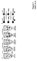
- FIG. 2 Schematic of the BEAM-based assay.
- A Extended beads were prepared by modifications of the BEAMing procedure described in Dressman et al (16)
- B Single base extensions were performed on the extended beads (gold spheres). Normal DNA sequences contained a G at the queried position, while mutant sequences contained an A.
- Fig. 3 Processing of flow cytometry data obtained by BEAMing.
- A Dot plot of forward scatter (FCS) and side scatter (SCC) signals of beads.
- B Histogram of single beads with regards to PE signal. Only beads containing extended PCR products had PE signals, as depicted in Fig. 2B .
- C Dot plot showing the Cy5 and FITC fluorescence intensity profiles of PE-positive beads. The beads clustered in three distinct populations colored red, green, and blue. Sequencing of individual beads sorted from each population showed that the red and green beads contained homogeneous wild-type and mutant sequences, respectively, while the blue beads contained a mixture of wild-type and mutant sequences.
- Fig. 4 Examples of flow cytometric profiles of beads generated from plasma DNA. Cy5 and FITC fluorescence intensity profiles of PE-positive beads from four patients are shown. The patients, mutations, and fraction of mutant APC fragments are indicated.
- Fig. 5 Fraction of mutant APC gene fragments in the plasma of patients with various colorectal tumors (adenomas (Ad) and Dukes' stage A, B, and D carcinomas).
- DNA from normal lymphoid cells or plasma DNA from healthy donors were used as controls ("Normal”).
- the "mutants” observed in assays with normal cellular DNA represent errors generated during the PCR process rather than mutations present in the template DNA (see text).
- the red lines represent the mean, min, and max values of the normal controls.
- Figure 7 Correlation between the total amount of template DNA per emulsion PCR and the fraction of emulsions that contain a single DNA molecule.
- PIK3CA exon 9 , PIK3CA exon 20 ,and KRAS exon 2 amplicons were amplified from normal lymphozyte DNA and quantified by a Picogreen assay. Equal amounts of the individual PCR products were mixed, diluted ,and used as templates for the emulsion PCR.
- sequence-specific fluorescent probes were hybidized to the beads after the emulsion PCR.
- Figure 8 Quantification of different template ratios by BEAMing.
- PIK3CA amplicons were mixed with KRAS amplicons in a ratio of 1:1, 1:10, 1:100, and 1:1000 and used as templates for emulsion PCR.
- A Examples of flow cytometric profiles. Cy5-labeled KRAS probes and FAM-labeled PIK3CA probes were hybridized to the beads.
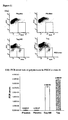
- Figure 9 Rolling circle amplification (RCA) on beads.
- A P53 sequence specific FAM probes were hybridized to detect DNA bound to magnetic beads (100x magnifications) after different incubation times
- B Relative Fluorescent intesity of FAM probes hybridized to the beads after RCA.
- Figure 10 “BEAMing Up” for quantification of mutations in the presence of excessive amount of wild-type DNA.
- A Example of flow cytometric profiles for quantification of TP53 codon 273 mutations in a series of dilutions.
- C Example of flow cytometric data for quantification of PIK3CA A3140G mutation in a series of dilutions.
- Figure 11 Quantification of error rates of commonly used polymerases for PCR.
- A Example of flow cytometric profiles of PIK3CA exon 20 A3140G.
- the inventors have developed Methods which improve the sensitivity of assays for rare and subtle nucleic acid differences.
- the assays are called BEAMing assays, and involve amplification of nucleic acid molecules on beads in microemulsions.
- One improvement involves the use of a high fidelity DNA polymerase in a preparatory amplification reaction.
- Another improvement involves the use of a rolling circle amplification to amplify the nucleic acids bound to beads as a result of BEAMing.
- Another improvement involves the use of a single base extension reaction to determine a sequence feature on an amplified product of BEAMing which has been further amplified using a rolling circle (isothermal) amplification.
- High fidelity DNA polymerases which can be used are those which provide a higher rate of fidelity (lower rate of errors) than Taq polymerase. Preferably these provide an error rate of less than 10 -5 , more preferably an error rate of less than 5 x 10 -6 , and even more preferably an error rate of less than 10 -6 .
- Suitable polymerases include: PhusionTM DNA polymerase (NEB), Taq High FidelityTM, and PfuUltraTM. These are used in a thermal cycling polymerase chain reaction, as is conventional in the art.
- Microemulsions are formed with beads and primers as previously taught. Because BEAMing requires thermal cycling, an emulsifier which is thermostable can be used.
- an emulsifier which is thermostable can be used.
- One such emulsifier is Abil ® EM90 (Degussa - Goldschmidt Chemical, Hopewell, VA).
- Other such emulsifiers can be used as are known in the art.
- Amplicons can be any size which is efficiently amplified using polymerase chain reaction.
- amplicons are preferably shorter than or equal to 300 bp, or shorter than or equal to 200 bp, or shorter than or equal to 100 bp.
- Templates from serum of colon cancer patients are apparently degraded to small sizes. Thus amplification of a smaller amplicon results in a more efficient and sensitive detection.
- the dependence of detection on size is quite strong as shown in Fig. 1 ,
- Single base extension reaction with differentially labeled dideoxynucleotides provides a sensitive means for detecting sequence features. If upon detection of products, individual beads are found with multiple, distinct labels, for example, representing a mutant and a wild type nucleotide, they can be discarded from further analysis. Multiple, distinct labels in this context indicates that a bead was present in a microemulsion with two distinct templates of analyte DNA, rather than the desired single template, or that an error occurred early in an amplification reaction in a microemulsion, such that the erroneous and the correct templates were both amplified.
- One means for detecting a sequence feature on an amplicon bound to a bead employs a single base extension (SBE) reaction.
- This reaction typically employs labeled dideoxynucleotide triphosphates to ensure that only a single monomer addition occurs.
- Dideoxynucleotide triphosphates can be conveniently labeled with any type of detectable label, including radioactive, fluorescent, and luminescent moieties. Different labels can be attached to different dideoxynucleotide triphosphates (ddNTPs) so that different products can be detected in the same sample.
- ddNTPs dideoxynucleotide triphosphates
- unlabeled dNTPs Prior to addition of all reagents necessary for initiation of the SBE reaction, unlabeled ddNTPs can be added to block non-specific extension.
- At least one unlabeled ddNTP is added at a concentration five to 40 fold higher than the concentration of the labeled ddNTPs.
- concentration is at least ten to twenty times higher.
- Rox-ddATP for the mutant
- FITC-ddCTP for the wild type
- ddGTP for blocking the nonspecific extension at the ratio of 1: 2-10: 20:20.
- the unlabeled ddNTP reduce nonspecific incorporation.
- Another optional step for improving the specificity and/or sensitivity of the SBE reaction is to denature the double stranded nucleic acid duplexes attached to the beads prior to the SBE reaction.
- the double strands can be heated or treated with sodium hydroxide. After the separation of the two strands, the single strands which are not bound to the beads can be separated from the beads and the bead-bound strands, and the single strands can be discarded.
- Microemulsions can be formed according to any technique known in the art. Previously for BEAMing, a magnetic stirring bar was used to create microemulsions. Other means can also be used, including, without limitation, tissue homogenizers, whether mechanical or sonicator-type. Suitable mechanical homogenizers include rotor-stator type as well as blade type. Tissue homogenizers appear to form microemulsions of more uniform size than magnetic stirring bars.
- isothermal amplification also known as rolling circle amplification.
- a molecular inversion probe or a padlock probe can be used. They probe may require filling-in, or not, prior to a template-driven ligation reaction to generate a circle. If filling-in is required the region to be filled in will typically be from 1 to 30 nucleotides.
- the isothermal amplification can amplify the ultimately detected signal quite significantly.
- a sequence feature can be detected using SBE (single base extension) reaction, as described above.
- the nucleotide sequence of the amplicon on the beads can be determined by any sequencing method known in the art, including sequencing-by-synthesis..
- Samples which may be used as sources of analyte DNA include blood, plasma, urine, stool, sputum, tears, saliva, and bone marrow. Solid tissues can also provide analyte DNA. Samples can be obtained from cancer patients, from related family members, from pregnant women, and from neonates. Sources of analyte DNA may be treated, for example with test agents, and the effects of the test agents on the analyte DNA can be determined.
- the mutant DNA fragments found in the circulation are derived from necrotic neoplastic cells that had been engulfed by macrophages. As tumors enlarge and invade, they are more likely to outgrow their blood supply. Thus invasive tumors generally contain large regions of necrosis, while benign tumors rarely do (26-29). Necrotic cells are not thought to release DNA into the extracellular milieu (30). However, cells that die from necrosis or apoptosis are routinely phagocytosed by macrophages or other scavenger cells.
- necrosis involves the killing not only of neoplastic cells, but also of surrounding stromal and inflammatory cells within the tumor, the DNA released from necrotic regions is likely to contain wild-type DNA sequences as well as mutant sequences. This may explain the increase in total (non-mutant) circulating DNA observed in the plasma of patients with advanced cancers.
- plasma-based assays for mutant DNA fragments are inferior in several ways to more conventional techniques for early colorectal cancer detection.
- Colonoscopy is the gold standard, with sensitivity rates >80% for adenomas and >90% for cancers (32).
- adenomas detected by colonoscopy can often be removed through the colonoscope, alleviating the need for surgery.
- a variety of issues limit the widespread applicability of colonoscopy (either conventional or virtual) to the screening of asymptomatic patients (3, 5, 33). This has stimulated the development of non-invasive technologies.
- One of the most promising of these is the analysis of fecal DNA for mutations (34). Because of the frequent presence of mutant DNA molecules in feces from both adenomas and early cancers, fecal DNA analysis is superior to plasma with regards to sensitivity.
- plasma-based assays have potential advantages with regards to ease of implementation and compliance.
- BEAMing Up represents an advance for the accurate detection and quantification of rare genetic variants in a population of DNA molecules.
- the approach provides robust signals and extremely high signal to noise ratios.
- the supplier of Taq and Taq High Fidelity cloned PCR products produced by the two enzymes into plasmid vectors then transforms bacteria with the plasmids.
- the mutation frequency was determined by dividing the total mutations by the total transformed cells.
- the error rate was determined by dividing the mutation frequency by the number of amino acids that can cause phenotypic changes in the two independent marker genes amplified (130 and 134 for rpsL and lacZ, respectively).
- the error rates for Taq using this assay were 4.2 x 10 -5 and 1.9 x 10 -5 epc for the rpsL and lacZ, respectively.
- the manufacturer of PfuUltra used a lacI system similar to that described above, employing cloning of PCR products and phenotypic evaluation of colonies. They calculated an error rate of 4.3 x 10 -7 for PfuUltra and 1.4 x 10 -6 for Phusion. The manufacturer of Phusion used the same assay and found an error rate of 4.4 x 10 -7 epc for Pfhusion but 6.93 x 10 -7 for PfuUltra. We found that both PfuUltra and Phusion resulted in similar error rates (6.0 and 4.8 x 10 -7 , respectively).
- BEAMing Up eliminates the need for cloning, bacterial transformation, colony selection, and confirmation of mutations by sequencing of colonies. It also eliminates the need for assumptions about the number of residues that can be mutated to result in a specific phenotype and thereby provides a more direct measure of mutation frequency. It should prove useful for many types of experiments wherein the fidelity of processes related to replication or transcription is important. It should facilitate the identification of rare mutations in clinical samples. And because of the much higher amount of DNA per bead, the technique could be useful for increasing read length or accuracy of high throughput sequencing studies using DNA-bound beads as templates.
- Primers were designed to generate ⁇ 100 bp amplicons that included one or more mutation sites.
- a universal tag (5'-tcccgcgaaattaatacgac-3') was added to the 5' end of either the forward or reverse primer used to generate each amplicon. This universal tag was identical to the one bound to the beads used for BEAMing. The sequences of these primers are listed in Table 2, which is published as supporting information on the PNAS web site.
- PCR was performed in 50 ⁇ l reactions containing 10 ⁇ l 5 ⁇ PhusionTM HF buffer, 0.2 mM of each ddNTP, 1 ⁇ M of each primer, 1/50,000 dilution of SYBR ® green I (Invitrogen), 1.5 U PhusionTM DNA polymerase (NEB, Beverly, MA), and 15 ⁇ l of purified plasma DNA (equivalent to 100 ⁇ l plasma) or genomic DNA purified from normal mononuclear cells of the blood of healthy volunteers.
- the amplifications were carried out with an iCycler PCR detection system (BioRad, Hercules, CA).
- PCR cycling conditions for all amplicons were as follows: 98°C for 1 min; 3 cycles of 98°C for 10 sec, 70°C for 10 sec, 72°C for 10 sec; 3 cycles of 98°C for 10 sec, 67°C for 10 sec, 72°C for 10 sec; 3 cycles of 98°C for 10 sec, 64°C for 10 sec, 72°C for 10 sec; 30 cycles of 98°C for 10 sec, 61 °C for 10 sec, 72°C for 10 sec.
- concentration of PCR products was determined using a PicoGreenTM dsDNA quantification assay (Invitrogen).
- a common oligonucleotide (5'-tcccgcgaaattaatacgac-3') was synthesized with a dual biotin group at the 5' end and with a six carbon linker (C6) between the biotin and the other nucleotides (IDT, Coralville, IA).
- This oligonucleotide was coupled to streptavidin-coated magnetic beads (MyOneTM, Dynal, Oslo, Norway) according to the protocol published previously (16).
- the water-in-oil emulsions were prepared by modifications of the method described by Ghadessy and Holliger (17) using a homogenization protocol originally described by Bemath et al. (18).
- the aqueous phase contained 67 mM Tris-HCl pH 8.8, 16.6 mM (NH 4 ) 2 SO 4 , 6.7 mM MgCl 2 , 10 mM 2-mercaptoethanol, 0.2 mM of each dNTP, 0.05 ⁇ M forward primer (5'-tcccgcgaaattaatacgac-3') and 8 ⁇ M reverse primer, 0.2 U/ ⁇ l Platinum ® Taq polymerase (Invitrogen), 3 ⁇ 10 5 / ⁇ l oligonucleotide-coupled beads and 0.1 pg/ ⁇ l template DNA.
- the reverse primers are listed in Table 2, which is published as supporting information on the PNAS web site.
- the water-oil mix was vortexed for 10 sec then emulsified for 50 sec using an Ultra-Turrax ® homogenizer (T25 basic; IKA, Wilmington, NC) with a disposable OmniTipTM(Omni International Inc., Marietta, GA) at the minimum speed.
- the emulsions were aliquoted into eight wells of a 96-well PCR plate and cycled under the following conditions: 94°C for 2 min; 50 cycles of 94°C for 10 sec, 58°C for 15 sec, and 70°C for 15 sec.
- the emulsions were pooled into a 15 ml tube and demulsified through the addition of 10 ml of NX buffer (100 mM NaCl, 1% Triton X-100, 10 mM Tris-HCl pH 7.5, 1 mM EDTA, 1% SDS). After vortexing for 10 sec, the beads were pelleted by centrifugation for 5 min at 4,100g. The top phase was removed and the beads were resuspended in 800 ⁇ l NX buffer and transferred to a 1.5 ml tube.
- NX buffer 100 mM NaCl, 1% Triton X-100, 10 mM Tris-HCl pH 7.5, 1 mM EDTA, 1% SDS.
- the beads were collected using a magnet (MPC-S, Dynal) and washed with 800 ⁇ l wash buffer (20 mM Tris-HCl, pH 8.4, 50 mM KCl). The double-stranded DNA on the beads was converted to single-stranded DNA by incubation in 800 ⁇ l 0.1 M NaOH for 2 min at room temperature. The beads were washed twice with 800 ⁇ l wash buffer using the magnet and finally resuspended in 200 ⁇ l of wash buffer. Single base extension and flow cytometry were performed as described in supporting information published on the PNAS web site.
- Tissue samples matched blood samples and clinical data were collected by Indivumed from surgical patients of the Israelitic Hospital and the Clinic Alten Eichen (both in Hamburg, Germany) following strictly controlled SOP criteria. IRB approval was given by the Ethical-board of the Physicians Association of Hamburg, Germany and patients' samples and data were collected after obtaining informed and written consent. The samples used in the current study were randomly chosen from those contributing through this protocol. Shortly before surgery, 18 ml EDTA blood was taken from a central catheter, chilled to 8°C immediately, and transported to the lab within 30 minutes for plasma preparation.
- the blood cells were pelleted for 15 min at 200g in a Leucosep ® -tube (Greiner, Frickenhausen, Germany) filled with 15 ml Ficoll-Paque solution. After centrifugation the supernatant (i.e., plasma) was transferred into 1.5 ml tubes, immediately frozen, and stored at -80 °C. The plasma samples were thawed at room temperature for 5 min and any remaining debris pelleted at 16,000g for 5 min.
- the supernatant was transferred to a new tube and digested with 500 ⁇ g/ml proteinase K (Invitrogen, Carlsbad, CA) in 2.5 mM Tris-HCl, 0.25 mM EDTA pH 7.5, and 1% SDS overnight.
- the DNA was extracted twice with phenol-chloroform (VWR, Cat#IB05174) and precipitated with two volumes ethanol in the presence of 3.3 M ammonium acetate and 3.3% (v/v) seeDNATM (GE Healthcare, Piscataway, NJ).
- the DNA from 1 ml plasma was dissolved in 150 ⁇ l of 10 mM Tris-HCl, 1 mM EDTA, pH 7.5. Tumor DNA was purified with the DNeasy tissue kit (Qiagen, Valencia, CA) according to the manufacturer's instructions.
- Digital PCR followed by direct sequencing of PCR products generated from single template molecules was used to determine the APC mutation status of the primary colon tumors and to analyze plasma DNA fragments of different sizes.
- Tumor DNA was diluted in 96 well PCR plates so that one or two template molecules were contained within each 10 ⁇ l reaction. To obtain a robust and uniform amplification, nested PCR reactions were performed.
- the first amplification comprised a 1296 bp region of the APC mutation cluster region (F1 5'-ACGTCATGTGGATCAGCCTATTG-3'; R1 5'-GGTAATTTTGAAGCAGTCTGGGC-3').
- the second amplification was split into two separate PCR reactions (A and B), with each one including half of this region (primers for A: F2 A 5'- TCTGGACAAAGCAGTAAAACCG-3'; R2 A 5'-CTTGGTGGCATGGTTTGTC-3'; primers for B: F2 B 5'-GCTCAGACACCCAAAAGTCC-3'; R2 B 5'-ACGTGATGACTTTGTTGGCATGGC-3').
- the PCR mix contained 1 ⁇ PCR buffer, 1 ⁇ M of each oligonucleotide, 1 mM of each dNTP, 6% DMSO, and 0.05 U/ ⁇ l Platinum ® Taq polymerase (Invitrogen).
- the following temperature profile was used for the amplification: 94°C for 2 min; 3 cycles of 94°C for 30 s, 67°C for 30 s, 70°C for 1 min; 3 cycles of 94°C for 30 s, 64°C for 30 s, 70°C for 1 min, 3 cycles of 94°C for 30 s, 61°C for 30 s, 70°C for 1 min; 50 cycles of 94°C for 30 s, 61°C for 30 s, 70°C for 1 min.
- One ⁇ l of the first amplification was added to each of the second 10 ⁇ l PCR reactions.
- the second PCR employed the following cycling conditions: 2 min at 94°C; 15 cycles of 94°C for 30 s, 58°C for 30 s, 70°C for 1 min.
- the PCR products were purified using the AMpure ® PCR purification system (Agencourt, Beverly MA) and sequencing reactions were performed with BigDye ® Terminator v3.1 (Applied Biosystems, Foster City, CA). Sequencing reactions were resolved on an automated 384 capillary DNA sequencer (Spectrumedix, State College, PA). Data analysis was performed using the Mutation Explorer ® package (SoftGenetics, State College, PA). Of 12 relatively large adenomas (> 1 cm), 11 were found to contain APC mutations within the region analyzed.
- Single base extension reactions were performed in 80 ⁇ l of 1 ⁇ SBE buffer (150 mM Tris-HCl pH 9.5, 67 mM MgCl 2 ) containing 3 ⁇ 10 6 magnetic beads from the emulsion PCR, 2.5 ⁇ M FITC-labeled ddATP (Perkin-Elmer, Wellesley, MA), 3.5 ⁇ M Cy5-labeled ddGTP (GE Healthcare), 25 ⁇ M of unlabeled ddCTP and ddUTP (USB, Cleveland, Ohio), 0.3 ⁇ M biotinylated primer, 20 U/ ⁇ l ThermoSequenaseTM (GE Healthcare).
- the primers used for SBE are listed in Table 3, which is published as supporting information on the PNAS web site. This composition was used when the wild-type sequence at the queried position was G and the mutant sequence was A; appropriate substitutions for the indicated ddNTPs were made when other bases were queried. Also note that the streptavidin present on MyOneTM beads is denatured during the emulsion PCR and does not bind biotin thereafter, so the primer used for SBE only binds to extended PCR products via hybridization and not to the beads themselves. The reactions were carried out at 94°C for 2 min, 65°C for 1 min, and 70°C for 2 min.
- the beads were recovered by magnetic separation, washed once with 200 ⁇ l wash buffer and once with 200 ⁇ l wash buffer plus 0.1% BSA, and then resuspended in 180 ⁇ l of binding buffer (5 mM Tris-HCl pH 7.5, 0.5 mM EDTA, 1 M NaCl).
- binding buffer 5 mM Tris-HCl pH 7.5, 0.5 mM EDTA, 1 M NaCl.
- the beads were mixed with 20 ⁇ l of 10 ⁇ g/ml streptavidin-conjugated phycoerythrin (PE, Invitrogen) to label the biotin-conjugated primer and incubated at room temperature for 10 min.
- PE streptavidin-conjugated phycoerythrin
- Beads were analyzed with a LSR II flow cytometer or sorted with a FACSAriaTM (both from BD Biosciences, San Jose, CA). The flow rate was typically set at 5000 events per second and a minimum of 2 ⁇ 10 6 events for each bead population was collected. These events were gated to exclude doublets and other aggregates. For the calculations of mutant frequency, only single beads with a PE signal at least 10-fold above the mean background signal were considered. In selected cases, beads were recovered by flow sorting and individual beads used in sequencing reactions. This was accomplished by first diluting the sorted beads in 96 well PCR plates so that one of every two wells (on average) contained a bead.
- the single-stranded DNA bound to each bead was then converted to double-stranded DNA by a DNA polymerase and released by a restriction enzyme digest that only cleaved the universal primer sequence on the beads.
- the DNA polymerase reaction was performed in a volume of 2 ⁇ l under a layer of mineral oil and contained 1 ⁇ PCR buffer, 1 ⁇ M of the reverse oligonucleotide used for BEAMing, 1 mM of each dNTP and 0.05 U/ ⁇ l Platinum ® Taq polymerase. The following temperature profile was used for the Taq polymerization: 95°C for 2 min, 58°C for 15 s, and 70°C for 1 min.
- Circulating Mutant DNA is degraded.
- the fraction of mutant molecules was strikingly dependent on size of the amplicon, increasing by more than 100 fold over the size range tested ( Fig. 1 B ). For example, though APC fragments of >1296 bp could be identified in the plasma of all three patients, there were no mutant APC sequences found in ⁇ 1000 fragments of this size. With very small amplicons ( ⁇ 100 bp), at least 8% of the plasma APC gene fragments were found to be mutant in all three patients.
- mutant DNA fragments present in the circulation of cancer patients are degraded compared to the circulating DNA derived from non-neoplastic cells. This conclusion is consistent with previous studies of other tumor types (21, 22) and has important implications for the detection of such mutant molecules.
- small amplicons can be used to enrich for DNA sequences derived from cancer cells.
- Figure 3 shows a representative flow cytometry result wherein the interpretation of the profiles was confirmed experimentally.
- a total of 342,573 beads were analyzed by flow cytometry.
- the single bead population (295,645) was used for fluorescence analysis ( Fig. 3 A ).
- 30,236 exhibited a PE signal ( Fig. 3B ), indicating that they had been extended during the emulsion PCR.
- the FITC and Cy5 signals reflected the number of beads containing mutant or wild-type sequences, respectively.
- Beads containing the wild-type DNA sequences (30,186) had high Cy5 but background FITC signal ("red beads" in Fig. 3C ).
- the BEAMing approach can, in principle, detect a very small fraction of fragments containing mutant sequences within a much larger pool of fragments containing wild-type sequence. Because >50 million beads are used in a single emulsion PCR and flow cytometry can be performed at speeds of >50,000 beads per sec, the capacity to enumerate such mutations is not limited by the beads themselves. Instead, two other features limit the sensitivity. First, there is a finite number of DNA fragments present in clinical samples. As noted above, this number ranged from 1,350 to 230,000 fragments per ml in the patients with tumors (Table 4) and from1150 to 8280 fragments/ml in control patients. This gives an upper bound to the sensitivity of the assays.
- a second limiting feature is the error rates of the polymerases used for PCR.
- the first is a conventional PCR that employs plasma DNA fragments as templates and the second is an oil-in-water emulsion PCR that uses the initial PCR products as templates.
- the emulsion PCR errors occurring during the early rounds of PCR can result in heterogeneous beads containing both wild-type and mutant sequences. These are easily eliminated from consideration, as described in Fig. 3C .
- the errors introduced in the first PCR cannot be eliminated, as they give rise to beads with homogeneous mutant sequences, indistinguishable from those resulting from genuine mutations in the original plasma DNA templates.
- the fraction of mutant molecules present after the first PCR equals the product of the mutation rate of the polymerase and the number of cycles carried out.
- BEAMing provides a quantitative way to determine the error rate of any polymerase used in PCR, without requiring cloning in bacterial vectors (Li et al. , unpublished data).
- the error rates with the polymerase used in the current study averaged 3.0 ⁇ 10 -7 mutations/bp/PCR cycle and ranged from 1.7 ⁇ 10 -7 to 6.5 ⁇ 10 -7 mutations/bp/PCR cycle, depending on the mutation site assessed.
- a BEAMing assay was then designed for each of the mutations identified in the 33 tumors and applied to the DNA purified from the plasma of the corresponding patients (Table 4).
- DNA from normal lymphocytes or plasma from patients without cancer were used as negative controls.
- DNA from the tumors of the 33 patients was used as positive controls.
- All six patients with advanced lesions (Dukes' D, defined as having at least one distant metastatic lesion) were found to contain mutant DNA fragments in their plasma.
- Dukes' A or B defined as having no lymph node involvement and no distant metastases
- ten (63%) were found to contain mutant DNA fragments in their plasma.
- adenomas only 1 patient's plasma was found to contain mutant DNA fragments. Representative flow cytometric results are shown in Fig. 4 and summarized in Table 4.
- the fraction of mutant molecules found in the plasma of the 17 cases with detectable mutations also varied according to tumor stage (p ⁇ 0,0001, Fisher Exact test).
- Dukes' D an average of 11.1% (range 1.9% to 27%) of the total APC gene fragments were mutant.
- Dukes' B an average of 0.9% (range 0.03% to 1.75%) of the plasma APC gene fragments were mutant.
- Dukes' A the fraction was even lower, averaging 0.04% (range 0.01% to 0.12%).
- only 0.02% of the plasma DNA fragments were mutant.
- the median fraction of positive beads found in the control DNA samples from patients without cancer was 0.0009% (range 0.003% to 0.0005%).
- Table 4 also lists the concentration of total APC fragments (wild-type plus mutant) in these patients' plasma. There was no direct relationship between the concentration of total APC fragments and the mutational load. Though patients with advanced cancers tended to have higher concentrations of total APC fragments than the other patients, this increase was not due to DNA from neoplastic cells. Furthermore, no correlation was found between tumor burden (volume of primary tumor plus metastatic sites) and either the concentration of APC fragments or percentage of mutant APC fragments in the circulation.
- Step 1 PCR amplification from DNA samples.
- Step 2 BEAMing. Oil-in-water (w/o) emulsions are formed in which single DNA molecules within each aqueous compartment are amplified and bound to beads.
- Step 3 Filling gaps.
- a padlock (6) or cirularizable probe (7, 8) was hybridized to the seqences on the beads.
- a 0-30 bp gap was filled in with a polymerase and the ends ligated.
- Step 4 Rolling circle amplification. Sequences to be queried on the beads are further amplified through rolling circle amplification.
- Step 5 Single base extension. Fluorescently-labeled dideoxy nucleotide terminators are used to distinguish beads containing sequences that diverge at positions of interest.
- Step 6 Flow cytometry. The population of beads is analyzed to determine the proportions containing each sequence of interest.
- Phusion TM DNA polymerase was used for the initial amplification of genomic DNA unless otherwise indicated in the text. Primers were designed to generate amplicons of 100 bp. A universal tag (5'-tcccgcgaaattaatacgac-3'), the sequence of which was identical to the one coated on the beads used for BEAMing, was added to the 5' end of the forward or reverse primer. PCR was performed in 50 ul reactions containing 10 ⁇ l 5 ⁇ PhusionTM HF buffer, 0.2 mM of each dNTP, 1 ⁇ M of each primer, 1.5 U PhusionTM DNA polymerase (NEB), and 15 ⁇ l purified cell line DNA.
- NEB Phusion TM DNA polymerase
- PCR cycling conditions were as follows: 98°C for 1 min; 3 cycles of 98°C for 10 sec, 70°C for 10 sec, 72°C for 10 sec; 3 cycles of 98°C for 10 sec, 67°C for 10 sec, 72°C for 10 sec; 3 cycles of 98°C for 10 sec, 64°C for 10 sec, 72°C for 10 sec; 30 cycles of 98°C for 10 sec, 61 °C for 10 sec, 72°C for 10 sec.
- the amount of PCR product was quantified by using a PicoGreenTM dsDNA quantification kit (Invitrogen).
- oligonucleotide labeled at its 5' end with a dual biotin group was coupled to streptavidin-coated 1 micron magnetic beads (Dynal MyOneTM) as described in Dressman et al.
- a 240 ul PCR mixture was prepared and added to 960 ⁇ l of 7% (w/v) Abil® EM90 (Degussa AG) in mineral oil (Sigma).
- the PCR mixture contained 67 mM Tris-HCl pH 8.8, 16.6 mM (NH4)2SO4, 6.7 mM MgCl2, 10 mM 2-mercaptoethanol, 0.2 mM of each ddNTP, 0.05 ⁇ M of forward primer identical in sequence to the universal tag described above, 8 ⁇ M reverse primer, 0.2 U/ ⁇ l Platinum® Taq polymerase (Invitrogen), 10 ⁇ 108 oligonucleotide coupled beads and ⁇ 20 pg template DNA.
- the water-oil mixture was vortexed for 10 sec at maximum speed (Vortex Genie 2) and then emulsified for 50 sec using an Ultra-Turrax homogenizer (T25) with a disposable OmniTip (Omni International, Inc.) at the minimum speed.
- the emulsions were transferred to a 96 well PCR plate, using 100 ul/well.
- the PCR cycling conditions were 94°C for 2 min; 50 cycles of 94°C for 10 sec, 58°C for 15 sec, and 70°C for 15 sec.
- the emulsion was broken in 10 ml NX-SDS buffer (100 mM NaCl, 1% Triton X-100, 10 mM Tris-HCl pH 7.5, 1 mM EDTA, 1% SDS) by centrifugation for 5 min at 4,500 g.
- the beads were then incubated with 0.1 M NaOH for 2 min to remove the non-biotinylated strand of the PCR product, collected with a magnet, and resuspended in 1x PCR buffer.
- a padlock probe (100 nM) was hybridized to ⁇ 10 7 beads in 2xSSC, 20% formamide and 0.5 ug/ul sonicated salmon sperm DNA at 37°C for 15 minutes. Probe was ligated in 10 U/ ⁇ l T4 DNA ligase (NEB),10 mM Tris-acetate pH 7.5, 10 mM MgAc2, 250 mM NaCl, 1 mM ATP and 0.2 ug/ul BSA at 37 °C for 15 min.
- NEB DNA ligase
- Beads were then resuspended in 100 ul of 1x ⁇ 29 DNA polymerase reaction buffer (NEB), 0.1 ug/ul BSA and 0.3 mM dNTP mixture containing 1 U/ul Phi29 DNA polymerase (NEB) and incubated at 37°C from 5 min to 6 hr.
- NEB 1x ⁇ 29 DNA polymerase reaction buffer
- BSA 0.1 ug/ul BSA
- 0.3 mM dNTP mixture containing 1 U/ul Phi29 DNA polymerase
- a circularizable probe (150 nM) was hybridized to ⁇ 10 7 beads in Ampligase 1x Ampligase reaction buffer (Epicentre) at 55°C for 15 min. Then, 50 uM dNTP (USB), 0.05 U/ul Stoffel fragment DNA (Applied Biosystems) and 1U/ul Ampligase were added and extension plus ligation performed at 55°C for 30 min.
- a fluorescein-labeled oligonucleotide complementary to the sequences amplified during the RCA was hybridized to the beads in SBE buffer (150 mM Tris-HCl pH 9.5, 67 mM MgCl2, 5% formamide) at 50° C for 15 minutes.
- SBE single base extensions
- Beads were analyzed with a LSR II flow cytometer and data were analyzed with FACSDivaTM software (BD Biosciences). The flow rate was typically set at 5000-10,000 events per second. Events were gated to exclude doublets and aggregates. For the calculations of mutant frequency, only single beads exhibiting hybridization to the SBE primer were considered.
- the fraction of single-template beads was maximal at ⁇ 30 pg of template per emulsion PCR. At this concentration, 12% of the beads were single template, 9% were double-template, and the remaining 79% of the beads were negative. In subsequent experiments, we therefore used 20 to 40 pg of template DNA per reaction.
- the signal to noise ratio obtained upon analysis of beads was also increased.
- a fluorescein-labeled oligonucleotide complementary to the original PCR product strand attached to the beads After hybridization to the original beads produced by BEAMing, the average signal intensity was 25-fold higher than that observed on beads that had been produced by BEAMing with an unrelated template, yielding a SNR of 25:1.
- the SNR increased to more than 9000-fold ( Fig. 9b ). Based on the relative signals obtained, we estimated that the length of DNA strands attached to the beads had increased from 100 bases to 40,000 bases by RCA.
- the reason for the SNR increase is because the background fluorescence signal following hybridization to beads without a complementary PCR product is due to autofluorescence plus non-specific binding of the probe to the beads. This background fluorescence signal is not increased much by RCA, while the specific hybridization signal is dramatically increased.
- Fig. 10a shows that there were some beads that contained homogeneous mutant p53 sequences even when the template used to produce them was normal human genomic DNA (panel showing 0% mutations). These apparent mutations were caused by errors during the initial PCR used to generate the templates for BEAMing. PCR errors introduced during the emulsion PCR or RCA steps would not result in "mutant" beads, as such beads would be classified as multi-template beads and therefore not included in the analysis. However, errors during the initial PCR used to generate templates would be indistinguishable from mutations occurring in vivo: droplets containing such single mutant molecules would give rise to beads containing homogeneous mutant sequences. Accordingly, we suspected that the procedures described here could be used to directly assess the error rates of polymerases commonly used for PCR.
Landscapes
- Chemical & Material Sciences (AREA)
- Engineering & Computer Science (AREA)
- Health & Medical Sciences (AREA)
- Organic Chemistry (AREA)
- Life Sciences & Earth Sciences (AREA)
- Physics & Mathematics (AREA)
- Zoology (AREA)
- Wood Science & Technology (AREA)
- Proteomics, Peptides & Aminoacids (AREA)
- Bioinformatics & Cheminformatics (AREA)
- General Physics & Mathematics (AREA)
- General Health & Medical Sciences (AREA)
- Chemical Kinetics & Catalysis (AREA)
- Molecular Biology (AREA)
- General Engineering & Computer Science (AREA)
- Genetics & Genomics (AREA)
- Biochemistry (AREA)
- Microbiology (AREA)
- Immunology (AREA)
- Biotechnology (AREA)
- Analytical Chemistry (AREA)
- Biophysics (AREA)
- Power Engineering (AREA)
- Urology & Nephrology (AREA)
- Cardiology (AREA)
- Pharmacology & Pharmacy (AREA)
- Veterinary Medicine (AREA)
- Public Health (AREA)
- Medicinal Chemistry (AREA)
- Vascular Medicine (AREA)
- Heart & Thoracic Surgery (AREA)
- General Chemical & Material Sciences (AREA)
- Nuclear Medicine, Radiotherapy & Molecular Imaging (AREA)
- Animal Behavior & Ethology (AREA)
- Measuring Or Testing Involving Enzymes Or Micro-Organisms (AREA)
- Pharmaceuticals Containing Other Organic And Inorganic Compounds (AREA)
- Preparation Of Compounds By Using Micro-Organisms (AREA)
- Investigating Or Analysing Biological Materials (AREA)
- Medicines That Contain Protein Lipid Enzymes And Other Medicines (AREA)
- Nanotechnology (AREA)
Priority Applications (4)
| Application Number | Priority Date | Filing Date | Title |
|---|---|---|---|
| PL06817236T PL1948816T3 (pl) | 2005-10-24 | 2006-10-20 | Ulepszone sposoby beamingu |
| SI200631283T SI1948816T1 (sl) | 2005-10-24 | 2006-10-20 | Izboljĺ ani postopki za beaming |
| EP11191989.0A EP2428579B1 (en) | 2005-10-24 | 2006-10-20 | Improved methods for BEAMing |
| CY20121100229T CY1112525T1 (el) | 2005-10-24 | 2012-03-07 | Βελτιωμενες μεθοδοι βεαμινg |
Applications Claiming Priority (2)
| Application Number | Priority Date | Filing Date | Title |
|---|---|---|---|
| US72923505P | 2005-10-24 | 2005-10-24 | |
| PCT/US2006/041115 WO2007050465A2 (en) | 2005-10-24 | 2006-10-20 | Improved methods for beaming |
Publications (3)
| Publication Number | Publication Date |
|---|---|
| EP1948816A2 EP1948816A2 (en) | 2008-07-30 |
| EP1948816A4 EP1948816A4 (en) | 2009-11-04 |
| EP1948816B1 true EP1948816B1 (en) | 2011-12-07 |
Family
ID=37968413
Family Applications (2)
| Application Number | Title | Priority Date | Filing Date |
|---|---|---|---|
| EP11191989.0A Not-in-force EP2428579B1 (en) | 2005-10-24 | 2006-10-20 | Improved methods for BEAMing |
| EP06817236A Not-in-force EP1948816B1 (en) | 2005-10-24 | 2006-10-20 | Improved methods for beaming |
Family Applications Before (1)
| Application Number | Title | Priority Date | Filing Date |
|---|---|---|---|
| EP11191989.0A Not-in-force EP2428579B1 (en) | 2005-10-24 | 2006-10-20 | Improved methods for BEAMing |
Country Status (11)
| Country | Link |
|---|---|
| US (4) | US9360526B2 (es) |
| EP (2) | EP2428579B1 (es) |
| AT (1) | ATE536420T1 (es) |
| CY (1) | CY1112525T1 (es) |
| DK (1) | DK1948816T3 (es) |
| ES (2) | ES2426031T3 (es) |
| HK (1) | HK1121193A1 (es) |
| PL (1) | PL1948816T3 (es) |
| PT (1) | PT1948816E (es) |
| SI (1) | SI1948816T1 (es) |
| WO (1) | WO2007050465A2 (es) |
Cited By (1)
| Publication number | Priority date | Publication date | Assignee | Title |
|---|---|---|---|---|
| EP3853373A4 (en) * | 2018-09-17 | 2022-06-22 | The University of North Carolina at Chapel Hill | PROCEDURE FOR QUANTIFICATION OF DNA FRAGMENTS IN A SAMPLE BY SIZE |
Families Citing this family (33)
| Publication number | Priority date | Publication date | Assignee | Title |
|---|---|---|---|---|
| US20100022414A1 (en) | 2008-07-18 | 2010-01-28 | Raindance Technologies, Inc. | Droplet Libraries |
| US7968287B2 (en) | 2004-10-08 | 2011-06-28 | Medical Research Council Harvard University | In vitro evolution in microfluidic systems |
| PL1712639T3 (pl) | 2005-04-06 | 2009-02-27 | Maurice Stroun | Sposób diagnozowania nowotworu przez wykrywanie krążącego DNA i RNA |
| ATE536420T1 (de) | 2005-10-24 | 2011-12-15 | Univ Johns Hopkins | Verbesserte verfahren für beaming |
| EP4190448A3 (en) | 2006-05-11 | 2023-09-20 | Bio-Rad Laboratories, Inc. | Microfluidic devices |
| WO2008097559A2 (en) | 2007-02-06 | 2008-08-14 | Brandeis University | Manipulation of fluids and reactions in microfluidic systems |
| US8592221B2 (en) | 2007-04-19 | 2013-11-26 | Brandeis University | Manipulation of fluids, fluid components and reactions in microfluidic systems |
| GB2453173A (en) * | 2007-09-28 | 2009-04-01 | Dxs Ltd | Polynucleotide primers |
| US20100041048A1 (en) | 2008-07-31 | 2010-02-18 | The Johns Hopkins University | Circulating Mutant DNA to Assess Tumor Dynamics |
| GB2467691A (en) | 2008-09-05 | 2010-08-11 | Aueon Inc | Methods for stratifying and annotating cancer drug treatment options |
| US9399797B2 (en) | 2010-02-12 | 2016-07-26 | Raindance Technologies, Inc. | Digital analyte analysis |
| EP2534267B1 (en) | 2010-02-12 | 2018-04-11 | Raindance Technologies, Inc. | Digital analyte analysis |
| WO2012040387A1 (en) | 2010-09-24 | 2012-03-29 | The Board Of Trustees Of The Leland Stanford Junior University | Direct capture, amplification and sequencing of target dna using immobilized primers |
| EP2673614B1 (en) | 2011-02-11 | 2018-08-01 | Raindance Technologies, Inc. | Method for forming mixed droplets |
| EP3736281A1 (en) | 2011-02-18 | 2020-11-11 | Bio-Rad Laboratories, Inc. | Compositions and methods for molecular labeling |
| DE202012013668U1 (de) | 2011-06-02 | 2019-04-18 | Raindance Technologies, Inc. | Enzymquantifizierung |
| US8658430B2 (en) | 2011-07-20 | 2014-02-25 | Raindance Technologies, Inc. | Manipulating droplet size |
| EP2788500A1 (en) * | 2011-12-09 | 2014-10-15 | F.Hoffmann-La Roche Ag | Identification of non-responders to her2 inhibitors |
| US20130309667A1 (en) * | 2012-05-15 | 2013-11-21 | Anthony P. Shuber | Primers for analyzing methylated sequences and methods of use thereof |
| US10119134B2 (en) | 2013-03-15 | 2018-11-06 | Abvitro Llc | Single cell bar-coding for antibody discovery |
| DE102013211113A1 (de) * | 2013-06-14 | 2014-12-18 | Siemens Aktiengesellschaft | Verfahren zur kombinierten Quantifizierung und Sequenzierung von mindestens einer Ziel-Nukleinsäure |
| US11901041B2 (en) | 2013-10-04 | 2024-02-13 | Bio-Rad Laboratories, Inc. | Digital analysis of nucleic acid modification |
| US9944977B2 (en) | 2013-12-12 | 2018-04-17 | Raindance Technologies, Inc. | Distinguishing rare variations in a nucleic acid sequence from a sample |
| ES2727656T3 (es) | 2014-09-15 | 2019-10-17 | Abvitro Llc | Secuenciación de alto rendimiento de banco de nucleótidos |
| JP6702305B2 (ja) * | 2015-03-24 | 2020-06-03 | 日立金属株式会社 | セラミックハニカム構造体 |
| US11085073B2 (en) | 2015-04-24 | 2021-08-10 | Qiagen Gmbh | Method for immobilizing a nucleic acid molecule on a solid support |
| WO2016170179A1 (en) * | 2015-04-24 | 2016-10-27 | Qiagen Gmbh | Method for immobilizing a nucleic acid molecule on solid support |
| US9938572B1 (en) | 2015-09-08 | 2018-04-10 | Raindance Technologies, Inc. | System and method for forming an emulsion |
| CN106929598B (zh) * | 2017-05-04 | 2019-07-05 | 湖南融健基因生物科技有限公司 | 一种乳液中进行滚环扩增的数字核酸分析方法 |
| CN111565731B (zh) | 2017-10-24 | 2024-03-08 | 达莉亚·伊兰妮 | 治疗缺血性疾病的方法 |
| US11474109B2 (en) | 2018-11-16 | 2022-10-18 | Scintimetrics, Inc. | Compositions and methods for controllably merging emulsion droplets and sample analysis |
| EP3725894A1 (en) | 2019-04-16 | 2020-10-21 | Blod Diagnostik GmbH | Apparatus and methods for nucleic acid target enrichment, suspension and quantification |
| CN114902024A (zh) * | 2019-11-15 | 2022-08-12 | 闪璨迈璀公司 | 基于荧光团扩散的组合物和方法 |
Family Cites Families (12)
| Publication number | Priority date | Publication date | Assignee | Title |
|---|---|---|---|---|
| US5888778A (en) * | 1997-06-16 | 1999-03-30 | Exact Laboratories, Inc. | High-throughput screening method for identification of genetic mutations or disease-causing microorganisms using segmented primers |
| GB9900298D0 (en) | 1999-01-07 | 1999-02-24 | Medical Res Council | Optical sorting method |
| US6355431B1 (en) * | 1999-04-20 | 2002-03-12 | Illumina, Inc. | Detection of nucleic acid amplification reactions using bead arrays |
| DE10048797A1 (de) * | 2000-10-02 | 2002-04-18 | Bayer Ag | Wirkstoffhaltige Emulsionen |
| AUPS298102A0 (en) | 2002-06-13 | 2002-07-04 | Nucleics Pty Ltd | Method for performing chemical reactions |
| CA2513889A1 (en) * | 2003-01-29 | 2004-08-19 | 454 Corporation | Double ended sequencing |
| WO2005010145A2 (en) * | 2003-07-05 | 2005-02-03 | The Johns Hopkins University | Method and compositions for detection and enumeration of genetic variations |
| ES2432040T3 (es) | 2004-01-28 | 2013-11-29 | 454 Life Sciences Corporation | Amplificación de ácido nucleico con emulsión de flujo continuo |
| BRPI0510811B1 (pt) * | 2004-05-13 | 2018-12-26 | Goel Anita | métodos para amplificação de ácido nucléico, método de detecção de patógeno e métodos para execução de amplificação de ácido nucléico |
| US20060228721A1 (en) | 2005-04-12 | 2006-10-12 | Leamon John H | Methods for determining sequence variants using ultra-deep sequencing |
| ATE536420T1 (de) | 2005-10-24 | 2011-12-15 | Univ Johns Hopkins | Verbesserte verfahren für beaming |
| US20080242560A1 (en) * | 2006-11-21 | 2008-10-02 | Gunderson Kevin L | Methods for generating amplified nucleic acid arrays |
-
2006
- 2006-10-20 AT AT06817236T patent/ATE536420T1/de active
- 2006-10-20 DK DK06817236.0T patent/DK1948816T3/da active
- 2006-10-20 PL PL06817236T patent/PL1948816T3/pl unknown
- 2006-10-20 US US12/091,395 patent/US9360526B2/en active Active
- 2006-10-20 ES ES11191989T patent/ES2426031T3/es active Active
- 2006-10-20 WO PCT/US2006/041115 patent/WO2007050465A2/en active Application Filing
- 2006-10-20 PT PT06817236T patent/PT1948816E/pt unknown
- 2006-10-20 SI SI200631283T patent/SI1948816T1/sl unknown
- 2006-10-20 EP EP11191989.0A patent/EP2428579B1/en not_active Not-in-force
- 2006-10-20 EP EP06817236A patent/EP1948816B1/en not_active Not-in-force
- 2006-10-20 ES ES06817236T patent/ES2381204T3/es active Active
- 2006-10-24 US US12/091,405 patent/US20120014879A1/en not_active Abandoned
-
2009
- 2009-01-30 HK HK09100870.6A patent/HK1121193A1/xx not_active IP Right Cessation
-
2012
- 2012-03-07 CY CY20121100229T patent/CY1112525T1/el unknown
-
2016
- 2016-06-06 US US15/174,427 patent/US10150991B2/en active Active
-
2018
- 2018-11-08 US US16/184,338 patent/US10837050B2/en active Active
Cited By (2)
| Publication number | Priority date | Publication date | Assignee | Title |
|---|---|---|---|---|
| EP3853373A4 (en) * | 2018-09-17 | 2022-06-22 | The University of North Carolina at Chapel Hill | PROCEDURE FOR QUANTIFICATION OF DNA FRAGMENTS IN A SAMPLE BY SIZE |
| US11788157B2 (en) | 2018-09-17 | 2023-10-17 | The University Of North Carolina At Chapel Hill | Methods of identifying patients as having an increased likelihood of having a human papillomavirus (HPV)-associated cancer or recurrence of an Hpv-associated cancer |
Also Published As
| Publication number | Publication date |
|---|---|
| PT1948816E (pt) | 2012-03-19 |
| ES2381204T3 (es) | 2012-05-24 |
| US20200123600A1 (en) | 2020-04-23 |
| ES2426031T3 (es) | 2013-10-18 |
| PL1948816T3 (pl) | 2012-07-31 |
| CY1112525T1 (el) | 2015-12-09 |
| EP2428579B1 (en) | 2013-05-29 |
| HK1121193A1 (en) | 2009-04-17 |
| WO2007050465A3 (en) | 2007-10-25 |
| US20130040300A9 (en) | 2013-02-14 |
| EP1948816A2 (en) | 2008-07-30 |
| US20110059435A1 (en) | 2011-03-10 |
| ATE536420T1 (de) | 2011-12-15 |
| US10150991B2 (en) | 2018-12-11 |
| DK1948816T3 (da) | 2012-04-02 |
| WO2007050465A2 (en) | 2007-05-03 |
| US20160376645A1 (en) | 2016-12-29 |
| US20120014879A1 (en) | 2012-01-19 |
| US9360526B2 (en) | 2016-06-07 |
| US10837050B2 (en) | 2020-11-17 |
| SI1948816T1 (sl) | 2012-04-30 |
| EP2428579A1 (en) | 2012-03-14 |
| EP1948816A4 (en) | 2009-11-04 |
Similar Documents
| Publication | Publication Date | Title |
|---|---|---|
| US10837050B2 (en) | Methods for beaming | |
| AU2017268486B2 (en) | Methods for detecting nucleic acid sequence variants | |
| US10557134B2 (en) | Protection of barcodes during DNA amplification using molecular hairpins | |
| US11098348B2 (en) | Nanopore detection of target polynucleotides from sample background | |
| JP6544861B2 (ja) | 高い選択性の核酸増幅プライマー | |
| JP3844996B2 (ja) | 反復性pcr産物の融解曲線解析方法 | |
| JP2010535031A (ja) | 標的配列の濃縮 | |
| CN116334190A (zh) | 改进的多核苷酸序列检测方法 | |
| JP2019536474A (ja) | メチル化dnaの多重検出方法 | |
| KR20160033239A (ko) | 핵산 에세이에서의 향상된 용융 식별 및 복합화를 위한 프로브 | |
| US20190106735A1 (en) | Method for analyzing cancer gene using multiple amplification nested signal amplification and kit | |
| JP2013090622A (ja) | 多型検出用プローブ、多型検出方法、薬効判定方法及び多型検出用キット | |
| JP5593582B2 (ja) | 核酸の迅速な検出方法 | |
| JP5813263B1 (ja) | 遺伝子変異の検出方法及びそれに用いる蛍光標識オリゴヌクレオチド | |
| JP6205216B2 (ja) | 変異検出用プローブ、変異検出方法、薬効判定方法及び変異検出用キット | |
| EP3639022A1 (en) | Compositions and methods for detection of genomic variations | |
| KR101606530B1 (ko) | Hla-b*27 및 hla-b*51의 동시 검출 방법 및 이의 용도 | |
| WO2024063036A1 (ja) | ゲノムdnaのメチル化検出法 | |
| US20230194500A1 (en) | Ultrasensitive rna quantification using nanopores | |
| EP4350002A1 (en) | Nachweis von molekularen analyten auf der grundlage massgeschneiderter sondenkonkurrenz | |
| JP2024034434A (ja) | 高感度かつ定量的な遺伝子検査方法 | |
| KR101513276B1 (ko) | 변형 mlpa 를 이용한 카피 수 다형성의 다중 분석 방법 | |
| JP2007215413A (ja) | 欠損の検出方法 | |
| JP2010233500A (ja) | 遺伝子型の識別方法 |
Legal Events
| Date | Code | Title | Description |
|---|---|---|---|
| PUAI | Public reference made under article 153(3) epc to a published international application that has entered the european phase |
Free format text: ORIGINAL CODE: 0009012 |
|
| 17P | Request for examination filed |
Effective date: 20080523 |
|
| AK | Designated contracting states |
Kind code of ref document: A2 Designated state(s): AT BE BG CH CY CZ DE DK EE ES FI FR GB GR HU IE IS IT LI LT LU LV MC NL PL PT RO SE SI SK TR |
|
| RIN1 | Information on inventor provided before grant (corrected) |
Inventor name: KINZLER, KENNETH, W. Inventor name: DIEHL, FRANK Inventor name: LI, MING Inventor name: VOGELSTEIN, BERT |
|
| RIN1 | Information on inventor provided before grant (corrected) |
Inventor name: KINZLER, KENNETH, W. Inventor name: VOGELSTEIN, BERT Inventor name: DIEHL, FRANK Inventor name: LI, MING |
|
| REG | Reference to a national code |
Ref country code: HK Ref legal event code: DE Ref document number: 1121193 Country of ref document: HK |
|
| A4 | Supplementary search report drawn up and despatched |
Effective date: 20091007 |
|
| 17Q | First examination report despatched |
Effective date: 20091204 |
|
| GRAP | Despatch of communication of intention to grant a patent |
Free format text: ORIGINAL CODE: EPIDOSNIGR1 |
|
| DAX | Request for extension of the european patent (deleted) | ||
| GRAS | Grant fee paid |
Free format text: ORIGINAL CODE: EPIDOSNIGR3 |
|
| GRAA | (expected) grant |
Free format text: ORIGINAL CODE: 0009210 |
|
| AK | Designated contracting states |
Kind code of ref document: B1 Designated state(s): AT BE BG CH CY CZ DE DK EE ES FI FR GB GR HU IE IS IT LI LT LU LV MC NL PL PT RO SE SI SK TR |
|
| REG | Reference to a national code |
Ref country code: GB Ref legal event code: FG4D |
|
| REG | Reference to a national code |
Ref country code: CH Ref legal event code: EP |
|
| REG | Reference to a national code |
Ref country code: IE Ref legal event code: FG4D |
|
| REG | Reference to a national code |
Ref country code: DE Ref legal event code: R096 Ref document number: 602006026284 Country of ref document: DE Effective date: 20120223 |
|
| REG | Reference to a national code |
Ref country code: CH Ref legal event code: NV Representative=s name: SCHNEIDER FELDMANN AG PATENT- UND MARKENANWAELTE |
|
| REG | Reference to a national code |
Ref country code: PT Ref legal event code: SC4A Free format text: AVAILABILITY OF NATIONAL TRANSLATION Effective date: 20120306 |
|
| REG | Reference to a national code |
Ref country code: NL Ref legal event code: T3 |
|
| REG | Reference to a national code |
Ref country code: SE Ref legal event code: TRGR |
|
| REG | Reference to a national code |
Ref country code: DK Ref legal event code: T3 |
|
| REG | Reference to a national code |
Ref country code: RO Ref legal event code: EPE |
|
| REG | Reference to a national code |
Ref country code: HK Ref legal event code: GR Ref document number: 1121193 Country of ref document: HK |
|
| REG | Reference to a national code |
Ref country code: ES Ref legal event code: FG2A Ref document number: 2381204 Country of ref document: ES Kind code of ref document: T3 Effective date: 20120524 |
|
| REG | Reference to a national code |
Ref country code: GR Ref legal event code: EP Ref document number: 20120400565 Country of ref document: GR Effective date: 20120417 |
|
| REG | Reference to a national code |
Ref country code: EE Ref legal event code: FG4A Ref document number: E006594 Country of ref document: EE Effective date: 20120306 |
|
| PG25 | Lapsed in a contracting state [announced via postgrant information from national office to epo] |
Ref country code: BE Free format text: LAPSE BECAUSE OF FAILURE TO SUBMIT A TRANSLATION OF THE DESCRIPTION OR TO PAY THE FEE WITHIN THE PRESCRIBED TIME-LIMIT Effective date: 20111207 |
|
| REG | Reference to a national code |
Ref country code: PL Ref legal event code: T3 |
|
| REG | Reference to a national code |
Ref country code: SK Ref legal event code: T3 Ref document number: E 11857 Country of ref document: SK |
|
| PLBE | No opposition filed within time limit |
Free format text: ORIGINAL CODE: 0009261 |
|
| STAA | Information on the status of an ep patent application or granted ep patent |
Free format text: STATUS: NO OPPOSITION FILED WITHIN TIME LIMIT |
|
| 26N | No opposition filed |
Effective date: 20120910 |
|
| REG | Reference to a national code |
Ref country code: DE Ref legal event code: R097 Ref document number: 602006026284 Country of ref document: DE Effective date: 20120910 |
|
| REG | Reference to a national code |
Ref country code: HU Ref legal event code: AG4A Ref document number: E017120 Country of ref document: HU |
|
| PGFP | Annual fee paid to national office [announced via postgrant information from national office to epo] |
Ref country code: LU Payment date: 20141030 Year of fee payment: 9 |
|
| PGFP | Annual fee paid to national office [announced via postgrant information from national office to epo] |
Ref country code: DK Payment date: 20141027 Year of fee payment: 9 |
|
| PGFP | Annual fee paid to national office [announced via postgrant information from national office to epo] |
Ref country code: GR Payment date: 20141030 Year of fee payment: 9 Ref country code: IE Payment date: 20141027 Year of fee payment: 9 Ref country code: ES Payment date: 20141027 Year of fee payment: 9 Ref country code: CH Payment date: 20141027 Year of fee payment: 9 Ref country code: FI Payment date: 20141029 Year of fee payment: 9 Ref country code: SE Payment date: 20141029 Year of fee payment: 9 |
|
| PGFP | Annual fee paid to national office [announced via postgrant information from national office to epo] |
Ref country code: NL Payment date: 20141026 Year of fee payment: 9 |
|
| PGFP | Annual fee paid to national office [announced via postgrant information from national office to epo] |
Ref country code: IT Payment date: 20141023 Year of fee payment: 9 |
|
| REG | Reference to a national code |
Ref country code: FR Ref legal event code: PLFP Year of fee payment: 10 |
|
| PGFP | Annual fee paid to national office [announced via postgrant information from national office to epo] |
Ref country code: EE Payment date: 20151007 Year of fee payment: 10 Ref country code: BG Payment date: 20151030 Year of fee payment: 10 Ref country code: GB Payment date: 20151027 Year of fee payment: 10 Ref country code: LT Payment date: 20151001 Year of fee payment: 10 Ref country code: TR Payment date: 20151007 Year of fee payment: 10 Ref country code: DE Payment date: 20151028 Year of fee payment: 10 |
|
| PGFP | Annual fee paid to national office [announced via postgrant information from national office to epo] |
Ref country code: AT Payment date: 20151002 Year of fee payment: 10 Ref country code: HU Payment date: 20151009 Year of fee payment: 10 Ref country code: MC Payment date: 20151006 Year of fee payment: 10 Ref country code: LV Payment date: 20151006 Year of fee payment: 10 Ref country code: PT Payment date: 20151006 Year of fee payment: 10 Ref country code: CZ Payment date: 20151014 Year of fee payment: 10 Ref country code: IS Payment date: 20151001 Year of fee payment: 10 Ref country code: SI Payment date: 20151002 Year of fee payment: 10 Ref country code: PL Payment date: 20151002 Year of fee payment: 10 Ref country code: RO Payment date: 20151001 Year of fee payment: 10 Ref country code: SK Payment date: 20151001 Year of fee payment: 10 Ref country code: CY Payment date: 20151005 Year of fee payment: 10 |
|
| REG | Reference to a national code |
Ref country code: DK Ref legal event code: EBP Effective date: 20151031 |
|
| PG25 | Lapsed in a contracting state [announced via postgrant information from national office to epo] |
Ref country code: LU Free format text: LAPSE BECAUSE OF NON-PAYMENT OF DUE FEES Effective date: 20151020 |
|
| REG | Reference to a national code |
Ref country code: SE Ref legal event code: EUG Ref country code: CH Ref legal event code: PL |
|
| REG | Reference to a national code |
Ref country code: NL Ref legal event code: MM Effective date: 20151101 |
|
| REG | Reference to a national code |
Ref country code: GR Ref legal event code: ML Ref document number: 20120400565 Country of ref document: GR Effective date: 20160506 |
|
| REG | Reference to a national code |
Ref country code: IE Ref legal event code: MM4A |
|
| PG25 | Lapsed in a contracting state [announced via postgrant information from national office to epo] |
Ref country code: IT Free format text: LAPSE BECAUSE OF NON-PAYMENT OF DUE FEES Effective date: 20151020 Ref country code: CH Free format text: LAPSE BECAUSE OF NON-PAYMENT OF DUE FEES Effective date: 20151031 Ref country code: LI Free format text: LAPSE BECAUSE OF NON-PAYMENT OF DUE FEES Effective date: 20151031 Ref country code: GR Free format text: LAPSE BECAUSE OF NON-PAYMENT OF DUE FEES Effective date: 20160506 |
|
| PG25 | Lapsed in a contracting state [announced via postgrant information from national office to epo] |
Ref country code: NL Free format text: LAPSE BECAUSE OF NON-PAYMENT OF DUE FEES Effective date: 20151101 Ref country code: SE Free format text: LAPSE BECAUSE OF NON-PAYMENT OF DUE FEES Effective date: 20151021 |
|
| REG | Reference to a national code |
Ref country code: FR Ref legal event code: PLFP Year of fee payment: 11 |
|
| PG25 | Lapsed in a contracting state [announced via postgrant information from national office to epo] |
Ref country code: DK Free format text: LAPSE BECAUSE OF NON-PAYMENT OF DUE FEES Effective date: 20151031 Ref country code: IE Free format text: LAPSE BECAUSE OF NON-PAYMENT OF DUE FEES Effective date: 20151020 |
|
| REG | Reference to a national code |
Ref country code: DE Ref legal event code: R119 Ref document number: 602006026284 Country of ref document: DE |
|
| REG | Reference to a national code |
Ref country code: EE Ref legal event code: MM4A Ref document number: E006594 Country of ref document: EE Effective date: 20161031 |
|
| REG | Reference to a national code |
Ref country code: LT Ref legal event code: MM4D Effective date: 20161020 |
|
| REG | Reference to a national code |
Ref country code: AT Ref legal event code: MM01 Ref document number: 536420 Country of ref document: AT Kind code of ref document: T Effective date: 20161020 |
|
| GBPC | Gb: european patent ceased through non-payment of renewal fee |
Effective date: 20161020 |
|
| PG25 | Lapsed in a contracting state [announced via postgrant information from national office to epo] |
Ref country code: ES Free format text: LAPSE BECAUSE OF NON-PAYMENT OF DUE FEES Effective date: 20151021 Ref country code: FI Free format text: LAPSE BECAUSE OF NON-PAYMENT OF DUE FEES Effective date: 20151020 Ref country code: MC Free format text: LAPSE BECAUSE OF NON-PAYMENT OF DUE FEES Effective date: 20161031 |
|
| REG | Reference to a national code |
Ref country code: SK Ref legal event code: MM4A Ref document number: E 11857 Country of ref document: SK Effective date: 20161020 |
|
| PG25 | Lapsed in a contracting state [announced via postgrant information from national office to epo] |
Ref country code: RO Free format text: LAPSE BECAUSE OF NON-PAYMENT OF DUE FEES Effective date: 20161020 Ref country code: CY Free format text: LAPSE BECAUSE OF NON-PAYMENT OF DUE FEES Effective date: 20161020 Ref country code: SK Free format text: LAPSE BECAUSE OF NON-PAYMENT OF DUE FEES Effective date: 20161020 Ref country code: EE Free format text: LAPSE BECAUSE OF NON-PAYMENT OF DUE FEES Effective date: 20161031 Ref country code: LT Free format text: LAPSE BECAUSE OF NON-PAYMENT OF DUE FEES Effective date: 20161020 Ref country code: GB Free format text: LAPSE BECAUSE OF NON-PAYMENT OF DUE FEES Effective date: 20161020 Ref country code: CZ Free format text: LAPSE BECAUSE OF NON-PAYMENT OF DUE FEES Effective date: 20161020 Ref country code: DE Free format text: LAPSE BECAUSE OF NON-PAYMENT OF DUE FEES Effective date: 20170503 Ref country code: IS Free format text: LAPSE BECAUSE OF FAILURE TO SUBMIT A TRANSLATION OF THE DESCRIPTION OR TO PAY THE FEE WITHIN THE PRESCRIBED TIME-LIMIT Effective date: 20170501 |
|
| PG25 | Lapsed in a contracting state [announced via postgrant information from national office to epo] |
Ref country code: PT Free format text: LAPSE BECAUSE OF NON-PAYMENT OF DUE FEES Effective date: 20170420 Ref country code: SI Free format text: LAPSE BECAUSE OF NON-PAYMENT OF DUE FEES Effective date: 20161021 Ref country code: AT Free format text: LAPSE BECAUSE OF NON-PAYMENT OF DUE FEES Effective date: 20161020 Ref country code: LV Free format text: LAPSE BECAUSE OF NON-PAYMENT OF DUE FEES Effective date: 20161020 Ref country code: HU Free format text: LAPSE BECAUSE OF NON-PAYMENT OF DUE FEES Effective date: 20161021 |
|
| REG | Reference to a national code |
Ref country code: SI Ref legal event code: KO00 Effective date: 20170727 |
|
| REG | Reference to a national code |
Ref country code: FR Ref legal event code: PLFP Year of fee payment: 12 |
|
| PG25 | Lapsed in a contracting state [announced via postgrant information from national office to epo] |
Ref country code: PL Free format text: LAPSE BECAUSE OF NON-PAYMENT OF DUE FEES Effective date: 20161020 |
|
| REG | Reference to a national code |
Ref country code: ES Ref legal event code: FD2A Effective date: 20180710 |
|
| REG | Reference to a national code |
Ref country code: FR Ref legal event code: PLFP Year of fee payment: 13 |
|
| PG25 | Lapsed in a contracting state [announced via postgrant information from national office to epo] |
Ref country code: BG Free format text: LAPSE BECAUSE OF NON-PAYMENT OF DUE FEES Effective date: 20170808 |
|
| PGFP | Annual fee paid to national office [announced via postgrant information from national office to epo] |
Ref country code: FR Payment date: 20191025 Year of fee payment: 14 |
|
| PG25 | Lapsed in a contracting state [announced via postgrant information from national office to epo] |
Ref country code: FR Free format text: LAPSE BECAUSE OF NON-PAYMENT OF DUE FEES Effective date: 20201031 |
|
| PG25 | Lapsed in a contracting state [announced via postgrant information from national office to epo] |
Ref country code: TR Free format text: LAPSE BECAUSE OF NON-PAYMENT OF DUE FEES Effective date: 20161020 |