EP1935342A1 - Artefakt-Eliminierung für eine medizintechnische Beckenregistrierung mit getrackter, systembekannter Beckenstütze - Google Patents
Artefakt-Eliminierung für eine medizintechnische Beckenregistrierung mit getrackter, systembekannter Beckenstütze Download PDFInfo
- Publication number
- EP1935342A1 EP1935342A1 EP06026291A EP06026291A EP1935342A1 EP 1935342 A1 EP1935342 A1 EP 1935342A1 EP 06026291 A EP06026291 A EP 06026291A EP 06026291 A EP06026291 A EP 06026291A EP 1935342 A1 EP1935342 A1 EP 1935342A1
- Authority
- EP
- European Patent Office
- Prior art keywords
- support
- image
- registration
- ray
- identification
- Prior art date
- Legal status (The legal status is an assumption and is not a legal conclusion. Google has not performed a legal analysis and makes no representation as to the accuracy of the status listed.)
- Granted
Links
Images
Classifications
-
- A—HUMAN NECESSITIES
- A61—MEDICAL OR VETERINARY SCIENCE; HYGIENE
- A61B—DIAGNOSIS; SURGERY; IDENTIFICATION
- A61B6/00—Apparatus or devices for radiation diagnosis; Apparatus or devices for radiation diagnosis combined with radiation therapy equipment
- A61B6/52—Devices using data or image processing specially adapted for radiation diagnosis
- A61B6/5211—Devices using data or image processing specially adapted for radiation diagnosis involving processing of medical diagnostic data
- A61B6/5252—Devices using data or image processing specially adapted for radiation diagnosis involving processing of medical diagnostic data removing objects from field of view, e.g. removing patient table from a CT image
-
- A—HUMAN NECESSITIES
- A61—MEDICAL OR VETERINARY SCIENCE; HYGIENE
- A61B—DIAGNOSIS; SURGERY; IDENTIFICATION
- A61B6/00—Apparatus or devices for radiation diagnosis; Apparatus or devices for radiation diagnosis combined with radiation therapy equipment
- A61B6/04—Positioning of patients; Tiltable beds or the like
- A61B6/0407—Supports, e.g. tables or beds, for the body or parts of the body
- A61B6/0421—Supports, e.g. tables or beds, for the body or parts of the body with immobilising means
-
- A—HUMAN NECESSITIES
- A61—MEDICAL OR VETERINARY SCIENCE; HYGIENE
- A61B—DIAGNOSIS; SURGERY; IDENTIFICATION
- A61B6/00—Apparatus or devices for radiation diagnosis; Apparatus or devices for radiation diagnosis combined with radiation therapy equipment
- A61B6/12—Arrangements for detecting or locating foreign bodies
-
- A—HUMAN NECESSITIES
- A61—MEDICAL OR VETERINARY SCIENCE; HYGIENE
- A61B—DIAGNOSIS; SURGERY; IDENTIFICATION
- A61B6/00—Apparatus or devices for radiation diagnosis; Apparatus or devices for radiation diagnosis combined with radiation therapy equipment
- A61B6/54—Control of apparatus or devices for radiation diagnosis
- A61B6/547—Control of apparatus or devices for radiation diagnosis involving tracking of position of the device or parts of the device
-
- A—HUMAN NECESSITIES
- A61—MEDICAL OR VETERINARY SCIENCE; HYGIENE
- A61G—TRANSPORT, PERSONAL CONVEYANCES OR ACCOMMODATION SPECIALLY ADAPTED FOR PATIENTS OR DISABLED PERSONS; OPERATING TABLES OR CHAIRS; CHAIRS FOR DENTISTRY; FUNERAL DEVICES
- A61G13/00—Operating tables; Auxiliary appliances therefor
- A61G13/10—Parts, details or accessories
- A61G13/12—Rests specially adapted therefor; Arrangements of patient-supporting surfaces
-
- A—HUMAN NECESSITIES
- A61—MEDICAL OR VETERINARY SCIENCE; HYGIENE
- A61B—DIAGNOSIS; SURGERY; IDENTIFICATION
- A61B90/00—Instruments, implements or accessories specially adapted for surgery or diagnosis and not covered by any of the groups A61B1/00 - A61B50/00, e.g. for luxation treatment or for protecting wound edges
- A61B90/39—Markers, e.g. radio-opaque or breast lesions markers
- A61B2090/3983—Reference marker arrangements for use with image guided surgery
-
- A—HUMAN NECESSITIES
- A61—MEDICAL OR VETERINARY SCIENCE; HYGIENE
- A61G—TRANSPORT, PERSONAL CONVEYANCES OR ACCOMMODATION SPECIALLY ADAPTED FOR PATIENTS OR DISABLED PERSONS; OPERATING TABLES OR CHAIRS; CHAIRS FOR DENTISTRY; FUNERAL DEVICES
- A61G13/00—Operating tables; Auxiliary appliances therefor
- A61G13/10—Parts, details or accessories
- A61G13/12—Rests specially adapted therefor; Arrangements of patient-supporting surfaces
- A61G13/1205—Rests specially adapted therefor; Arrangements of patient-supporting surfaces for specific parts of the body
- A61G13/1225—Back
-
- A—HUMAN NECESSITIES
- A61—MEDICAL OR VETERINARY SCIENCE; HYGIENE
- A61G—TRANSPORT, PERSONAL CONVEYANCES OR ACCOMMODATION SPECIALLY ADAPTED FOR PATIENTS OR DISABLED PERSONS; OPERATING TABLES OR CHAIRS; CHAIRS FOR DENTISTRY; FUNERAL DEVICES
- A61G13/00—Operating tables; Auxiliary appliances therefor
- A61G13/10—Parts, details or accessories
- A61G13/12—Rests specially adapted therefor; Arrangements of patient-supporting surfaces
- A61G13/1205—Rests specially adapted therefor; Arrangements of patient-supporting surfaces for specific parts of the body
- A61G13/123—Lower body, e.g. pelvis, hip, buttocks
-
- A—HUMAN NECESSITIES
- A61—MEDICAL OR VETERINARY SCIENCE; HYGIENE
- A61G—TRANSPORT, PERSONAL CONVEYANCES OR ACCOMMODATION SPECIALLY ADAPTED FOR PATIENTS OR DISABLED PERSONS; OPERATING TABLES OR CHAIRS; CHAIRS FOR DENTISTRY; FUNERAL DEVICES
- A61G13/00—Operating tables; Auxiliary appliances therefor
- A61G13/10—Parts, details or accessories
- A61G13/12—Rests specially adapted therefor; Arrangements of patient-supporting surfaces
- A61G13/1205—Rests specially adapted therefor; Arrangements of patient-supporting surfaces for specific parts of the body
- A61G13/1245—Knees, upper or lower legs
-
- A—HUMAN NECESSITIES
- A61—MEDICAL OR VETERINARY SCIENCE; HYGIENE
- A61G—TRANSPORT, PERSONAL CONVEYANCES OR ACCOMMODATION SPECIALLY ADAPTED FOR PATIENTS OR DISABLED PERSONS; OPERATING TABLES OR CHAIRS; CHAIRS FOR DENTISTRY; FUNERAL DEVICES
- A61G2200/00—Information related to the kind of patient or his position
- A61G2200/30—Specific positions of the patient
- A61G2200/32—Specific positions of the patient lying
- A61G2200/322—Specific positions of the patient lying lateral
Definitions
- the invention relates to the art of artifact elimination for a medical pelvic registration with a tracked pelvic support.
- pelvic surgery especially in hip replacement surgery, patients are usually operated on their side, and it is necessary to fix the patient's pelvis. This is done via patient positioners that have supportive devices for the lower body area of the patient.
- the reference means are used to indirectly locate the ASIS and pubis points and thereby register the anterior pelvic plane.
- a major problem with existing positioning devices is when fluoroscopy images are taken. It is in the nature of the positioning devices that at least a portion of their support devices in the beam path of the fluoroscopy and therefore artifacts on the recording arise. Although the selected materials for the X-ray support device are permeable, shadows and edges still appear on the images. These artifact contours overlay the anatomical structure (pelvis) and could result in a misinterpretation of the images. The navigation software collects anatomical structures to register the pelvis, and the shadows and edges mentioned here could lead to a wrong contour selection and thus to a wrong registration. In addition, it becomes difficult to define dots in the area of the column mappings because of the contrast due to the artifacts.
- the present invention uses knowledge of the dimensions or dimensions of the support as well as the knowledge of the position of the support to disregard the support image during registration. Because you know where the column is located and what its illustration will look like, you can avoid misrepresenting the mapped column elements.
- the invention makes it possible to position a patient laterally during a pelvic operation and to easily register the anatomical pelvic landmarks when the procedure is supported by medical navigation.
- the problem with support artefacts in the recording no longer occurs because the support can be tracked by the reference device, its geometric data (dimensions, internal structure, etc.) are stored in a database of the navigation software and the recognition of the support thus created is used to compute the column artifacts in the image to disregard them at registration.
- the imaging elements of the identified image of the post are subtracted from the image, which may be done by appropriate graphics software and graphics hardware.
- the picture shows the actual pelvic points and pelvic contours to be used for registration.
- identifying the column image it is possible to use the gray levels of the column imaging elements as identification aids, in particular known or substantially known gray levels, as commonly produced by the materials of the column.
- identification aids in particular known or substantially known gray levels, as commonly produced by the materials of the column.
- spacing ratios of the column elements outer edges, inner edges, corners
- Such starting points for the calculation of the column image can be, for example, clearly reproducible column elements, but it is also possible to attach additional markers in or on the column and to use these as identification aids or starting points for distance determinations.
- imaginary lines or planes may also be used as such starting points or identification tools, for example an imaginary connecting line between special, clearly reproducible markers.
- the support preferably already consists of a material permeable to X-rays, and in one embodiment variant, a marker or a plurality of markers made of a material that is substantially impermeable to X-radiation is / are arranged on the support at a known distance from the support elements. These markers serve as identification aids or "starting points" for the calculation of the position of the column image.
- the positioner itself so that, while holding the pelvis in the region between the ASIS and Pubis landmarks, it allows the picking of these landmarks with a navigation pointer, so as to provide them Digitize points during registration. This can be done by a shape for the support, which allows free access to these landmark points.
- the invention further includes a program that, when running on a computer or loaded in a computer, causes the computer to perform one of the methods described above. Furthermore, the invention comprises a computer program storage medium having such a program.
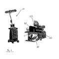
- FIG. 1 A general overview of the field of application of the present invention is the FIG. 1 , There, a patient 40 is shown, which lies laterally on the patient couch 50. The patient is supported or fixed, and the front support of the patient positioner used for this purpose is indicated by the reference numeral 1.
- a C-arm fluoroscope 30 is arranged on the patient couch 50, which has a so-called registration kit 35 at its radiation source. With the aid of the registration kit 35, which has reference devices, the position of the C-arm can be determined using a medical navigation system 20, which is located on the left of the patient couch. Thus, the location of the fluoroscopy taken the recording angle can be determined in space.
- the holding device for the support 1 carries a navigation reference.
- This navigation reference is in the FIG. 1 because of the proportions not visible, but it is in the FIGS. 2 to 5 shown.
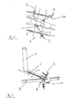
- the FIG. 2 shows in a view from above lying on the patient table 50 patients, which is supported with three support devices in the pelvic region.
- the two rear support devices have been designated as a whole by the reference numerals 6 and 7, the front support means are referred to in more detail, and their components are also in the FIGS. 3 to 5 to see.
- It comprises the rod-like support 1 and the support arm 2, on which a reference device 3 is attached at the other end.
- This reference arrangement has three marker spheres which are emitted by the navigation system 20 (FIG. FIG. 1 ) can be detected, and with this detection, the exact position of the support 1 in space is known because of the rigid position assignment between support 1 and reference device 3.
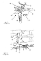
- the holding device 8 is mounted displaceably on a rail 5, which is fixed to the patient bed 50. With the rotary quick release 13, the position of the holding device 8 can be fixed along the rail 5.
- the reference numeral 12 indicates a quick-release mechanism which fixes the rotation of the arm 2, and the quick-release mechanism 11 makes it possible to fix the arm in its sliding longitudinal guide.
- An adapter 10 for the reference device is - as stated - almost at the rear end of the arm 2, and on this adapter a quick-release mechanism 9 is placed for the reference device 3, which allows the quick attachment and detachment of the reference device 3. With the help of this construction, the support 1 can now be positioned at a desired point on the front of the patient's pelvis in order to fix it.
- the design of the support 1 as an elongated rod and the possibilities of movement and fixing possibilities of the holding device 8 ensure a very free positioning possibility for the support 1, at the same time a firm grip in the fixation position given is. This results in the possibility to fix the patient already at the front of the pelvis in such a way that the ASIS and Pubis points 15a, 15b, 15c, 15d are not covered by the support 1 but to rest the support between these points comes. These body landmarks can then be approached with a navigation pointer in a fixed patient pool and their position registered.
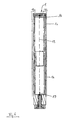
- the longitudinal support 1 is particularly well suited for artifact elimination according to the present invention. It is composed of an upper part 1a and a lower part 1b, which are put together in the middle. It is tubular and consists essentially of an X-ray transparent material. In the lid and in the lower insert of the support 1 markers 16 and 17 are used, which consist of a non-X-ray transparent material, and reference numerals 18a and 18b indicate areas which comprise the edge contours of the support 1. An imaginary connecting line between the markers 16 and 17 is designated by the reference numeral 19.
- the invention is provided with a support according to FIG. 6
- the location of the anterior pelvic plane must be determined with the navigation system, which position is determined by the ASIS and Pubis points 15a to 15d.
- the fluoroscopy device 30 FIG. FIG. 1
- reference arrangements are attached to the pelvis and the thigh of the patient. Two fluoroscopic images are taken in the area of the pelvis, and the ASIS (Anterior Superior Iliac Spine) point on the side to be treated is approached and registered with a navigation pointer.
- the system calculates the anatomical landmarks of the pelvis, especially in the anterior pelvic plane and mid-sagittal plane, based on the imaged edges of the pelvis on the fluoroscopic image.
- the present invention is used.
- the image of the support causes shadows and edges to be on the image.
- the support is made of a different material or different materials, different grayscale values are created on the recording.
- the markers 16 and 17 are made of a non-X-ray permeable material (steel), they form almost black on the image.
- the edges of the column appear in a light gray color.
- the black images of the markers 16 and 17 are detected on the recording and it is searched for special gray values, which at a predetermined distance to the markers 16, 17 and to imaginary connecting line 19 stand.
- special gray values are, for example, by the areas 18a and 18b in FIG. 6 demonstrated. They must cover the edges of the column image.
- edges with their special gray values are then no longer taken into account in the subsequent acquisition of the anatomical structures of the pelvis, so that it is ensured that the pelvic contours used for the registration actually describe mapped pelvic lines.
- the registration may then take place correctly due to the inventive artifact elimination, and its dependent navigation of the engagement will provide the correct positional support.
Landscapes
- Health & Medical Sciences (AREA)
- Life Sciences & Earth Sciences (AREA)
- Engineering & Computer Science (AREA)
- Medical Informatics (AREA)
- Biomedical Technology (AREA)
- Animal Behavior & Ethology (AREA)
- General Health & Medical Sciences (AREA)
- Public Health (AREA)
- Veterinary Medicine (AREA)
- High Energy & Nuclear Physics (AREA)
- Biophysics (AREA)
- Physics & Mathematics (AREA)
- Nuclear Medicine, Radiotherapy & Molecular Imaging (AREA)
- Optics & Photonics (AREA)
- Pathology (AREA)
- Radiology & Medical Imaging (AREA)
- Heart & Thoracic Surgery (AREA)
- Molecular Biology (AREA)
- Surgery (AREA)
- Computer Vision & Pattern Recognition (AREA)
- Apparatus For Radiation Diagnosis (AREA)
Abstract
Description
- Die Erfindung betrifft das technische Gebiet der Artefakt-Eliminierung für eine medizinische Beckenregistrierung mit getrackter Beckenstütze.
- Bei Beckenoperationen, insbesondere bei Hüftersatz-Operationen werden Patienten meist auf der Seite liegend operiert, und dabei ist es notwendig, das Becken des Patienten zu fixieren. Dies geschieht über Patientenpositionierer, die Stützvorrichtungen für den Unterkörperbereich des Patienten aufweisen. In dieser Lage ist es schwierig, Navigationsinformationen durch das Abgreifen von Körper-Landmarken (ASIS- und Pubis-Punkte) im Rahmen einer medizinischen Navigation zu ermitteln, weil diese charakteristischen Becken-Landmarken zumindest im unteren Bereich meist kaum zugänglich sind. Deshalb wird oft mit einer Röntgen- oder Fluoroskopie-Registrierung gearbeitet.
- Viele der herkömmlichen Patientenpositionierer verwenden Kissen oder Stäbe, die an einem Armsystem des Operationstisches oder an ähnlichen festen Einrichtungen fixiert sind. Ein solcher mechanischer Positionierer ist beispielsweise aus der
US 6,311,349 bekannt. - Neben den mechanischen Positionierern gibt es noch diejenigen, die Navigations-Referenzeinrichtungen für eine medizintechnische Navigation aufweisen, und ein solcher ist beispielsweise aus der
WO 2004/089 192 A2 bekannt. Die Referenzeinrichtungen werden verwendet, um die ASIS- und Pubis-Punkte indirekt zu lokalisieren und damit die vordere Beckenebene zu registrieren. - Ein Hauptproblem der existierenden Positionierungseinrichtungen zeigt sich, wenn Fluoroskopiebilder aufgenommen werden. Es liegt in der Natur der Positionierungseinrichtungen, dass mindestens ein Teil ihrer Stützvorrichtungen im Strahlengang der Fluoroskopieaufnahme liegt und deshalb Artefakte auf der Aufnahme entstehen. Auch wenn die gewählten Materialien für die Stützvorrichtung für Röntgenstrahlung durchlässig sind, entstehen noch immer Schatten und Kanten auf den Bildern. Diese Artefakt-Konturen überlagern die anatomische Struktur (Becken) und könnten in einer Missinterpretation der Aufnahmen resultieren. Die Navigationssoftware erfasst anatomische Strukturen, um das Becken zu registrieren, und die angesprochenen Schatten und Kanten könnten hier zu einer falschen Konturenwahl und somit zu einer falschen Registrierung führen. Zusätzlich wird es schwierig, Punkte in dem Bereich der Stützen-Abbildungen zu definieren, weil dort aufgrund der Artefakte der Kontrast leidet.
- Es ist die Aufgabe der vorliegenden Erfindung, eine Artefakt-Eliminierung bei der medizinischen Beckenregistrierung vorzuschlagen, welche die oben genannten Nachteile des Standes der Technik überwindet. Insbesondere sollen Fehlerregistrierungen vermieden werden.
- Diese Aufgabe wird gemäß einem ersten Aspekt der Erfindung durch ein Artefakt-Eliminierungsverfahren gemäß dem Anspruch 1 und gemäß einem zweiten Aspekt der Erfindung durch ein Artefakt-Eliminierungssystem gemäß dem Anspruch 8 gelöst. Die Unteransprüche definieren bevorzugte Ausführungsformen der Erfindung.
- Das erfindungsgemäße Artefakt-Eliminierungsverfahren dient der medizintechnischen Beckenregistrierung zur positionellen Registrierung eines mit mindestens einer Stütze abgestützten Patientenbeckens, und es weist die folgenden Schritte auf:
- die geometrischen Daten der Stütze werden im Datenspeicher eines für die Registrierung verwendeten medizintechnischen Navigationssystems abgespeichert,
- mindestens zwei Röntgen- bzw. Fluoroskopieaufnahmen des Patientenbeckens werden erstellt, wobei die Stütze sich im Strahlengang der Aufnahme befindet,
- die Position der Stütze und des Röntgen- bzw. Fluoroskopiegeräts während der Aufnahme werden durch das Navigationssystem ermittelt,
- mit Hilfe der ermittelten Positionsdaten für die Stütze und das Röntgen- bzw. Fluoroskopiegerät wird die Abbildung der Stütze in der Aufnahme identifiziert,
- die Aufnahme wird für die Registrierung verwendet, wobei die Abbildungselemente der identifizierten Abbildung der Stütze als bei der Registrierung nicht zu verwendende Teile der Abbildung definiert werden.
- Mit anderen Worten nutzt die vorliegende Erfindung die Kenntnis über die Dimensionen bzw. Abmessungen der Stütze sowie die Kenntnis über die Position der Stütze dazu, die Stützenabbildung bei der Registrierung unberücksichtigt zu lassen. Weil man weiß, wo die Stütze liegt und wie ihre Abbildung aussehen wird, kann man vermeiden, dass die abgebildeten Stützenelemente fälschlicherweise der Registrierung zugrunde gelegt werden.
- Durch die Erfindung wird es möglich, einen Patienten seitlich während einer Beckenoperation zu positionieren und problemlos die anatomischen Beckenlandmarken zu registrieren, wenn die Prozedur durch eine medizintechnische Navigation unterstützt wird. Das Problem mit Stützen-Artefakten in der Aufnahme tritt nicht mehr auf, weil die Stütze durch die Referenzeinrichtung getrackt werden kann, ihre geometrischen Daten (Abmessungen, Innenstruktur, etc.) in einer Datenbank der Navigationssoftware hinterlegt werden und die so geschaffene Erkennbarkeit der Stütze dann verwendet wird, um im Bild die Stützen-Artefakte zu berechnen, um sie bei der Registrierung unberücksichtigt zu lassen.
- Bei einer Ausführungsform der Erfindung werden die Abbildungselemente der identifizierten Abbildung der Stütze von der Aufnahme subtrahiert, was durch geeignete Grafiksoftware und Grafikhardware geschehen kann. Im Bild verbleiben die tatsächlich zu verwendenden Beckenpunkte und Beckenkonturen für die Registrierung.
- Grundsätzlich können alle Stützenelemente, die in der Aufnahme abgebildet werden, aus der Aufnahme "herausgerechnet" werden. Andererseits genügt es oft, die abgebildeten Konturen der Stütze in der Aufnahme zu identifizieren und als nicht zu verwendende Teile zu definieren.
- Bei der Identifizierung der Stützenabbildung besteht die Möglichkeit, die Grauwerte der Stützen-Abbildungselemente als Identifizierungshilfsmittel zu verwenden, insbesondere bekannte oder im wesentlichen bekannte Grauwerte, wie sie von den Materialien der Stütze üblicherweise erzeugt werden. Bei der Identifizierung der Stützenabbildung ist es von Vorteil, wenn Abstandsverhältnisse der Stützenelemente (Außenkanten, Innenkanten, Ecken) verwendet werden, d.h. bekannte Abstände solcher Elemente zu bestimmten Punkten oder Teilen der Stütze, die sich gut in der Aufnahme identifizieren lassen. Solche Ausgangspunkte für die Berechnung der Stützenabbildung können beispielsweise deutlich abbildbare Stützenelemente sein, es besteht aber auch die Möglichkeit, zusätzliche Marker in oder an der Stütze anzubringen und diese als Identifizierungshilfsmittel bzw. Ausgangspunkte für Abstandsbestimmungen zu benutzen. Bei einer Ausführungsform der Erfindung können auch gedachte Linien oder Ebenen als solche Ausgangspunkte oder Identifizierungshilfsmittel verwendet werden, beispielsweise eine gedachte Verbindungslinie zwischen speziellen, deutlich abbildbaren Markern.
- Das erfindungsgemäße Artefakt-Eliminierungssystem dient der Eliminierung von Artefakten in Röntgen- bzw. Fluoroskopieaufnahmen, die bei einem medizinischen Beckenregistrierungsverfahren verwendet werden, das zur positionellen Registrierung eines mit mindestens einer Stütze abgestützten Patientenbeckens dient. Das System umfasst:
- ein medizintechnisches Navigationssystem;
- eine Trackingvorrichtung, welche die Position von Navigations-Referenzeinrichtungen bestimmen kann;
- ein Röntgen- bzw. Fluoroskopiegerät; und
- eine der Stütze positionell zugeordnete Navigations-Referenzanordnung.
- Gekennzeichnet ist das System durch
- ein medizintechnisches Navigationssystem; und durch
- eine Logik im Navigationssystem, welche aufgrund der Information über die geometrischen Daten der Stütze die Abbildung der Stütze in einer Röntgen- bzw. Fluoroskopieaufnahme bei der Registrierung unberücksichtigt lässt.
- Die Stütze besteht bevorzugt schon aus einem für Röntgenstrahlen durchlässigen Material, und bei einer Ausgestaltungsvariante ist/sind an der Stütze im bekannten Abstand zu den Stützenelementen ein Marker oder mehrere Marker aus einem für Röntgenstrahlung im Wesentlichen undurchlässigen Material angeordnet. Diese Marker dienen als Identifizierungshilfsmittel bzw. "Ausgangspunkte" für die Berechnung der Lage der Stützenabbildung.
- Es besteht im Rahmen der Erfindung ferner die Möglichkeit, den Positionierer selbst so zu gestalten, dass er, obwohl er das Becken im Bereich zwischen den ASIS- und Pubis-Landmarken hält, das Abgreifen dieser Landmarken mit einem Navigations-Pointer gestattet, um so diese Punkte bei der Registrierung zu digitalisieren. Dies kann durch eine Formgebung für die Stütze geschehen, welche einen freien Zugang zu diesen Landmarkenpunkten gestattet.
- Die Erfindung umfasst weiter ein Programm, das, wenn es auf einem Computer läuft oder in einem Computer geladen ist, den Computer veranlasst, eines der Verfahren durchzuführen, wie sie oben beschrieben worden sind. Ferner umfasst die Erfindung ein Computerprogramm-Speichermedium, das ein solches Programm aufweist.
- Die Erfindung wird im Weiteren anhand der beiliegenden Zeichnungen näher erläutert. Sie kann alle hierin beschriebenen Merkmale einzeln sowie in jedweder Kombination umfassen. In den Zeichnungen zeigen:
- Figur 1
- ein generelles Operationssaal-Setup für die Durchführung einer Beckenregistrierung mit Hilfe der vorliegenden Erfindung;
- Figur 2
- eine Ansicht von oben auf einen abgestützten Patienten;
- Figur 3
- eine perspektivische Ansicht einer Patienten-Stützvorrichtung mit Stütze und Stützenhalterung;
- Figur 4
- ein Detail der Stützenhalterung aus
Figur 3 ; - Figur 5
- eine schematische Ansicht der Patienten-Beckenabstützung; und
- Figur 6
- eine Ausführungsform einer Stütze zur Verwendung mit der vorliegenden Erfindung.
- Einen allgemeinen Überblick über das Anwendungsumfeld der vorliegenden Erfindung gibt die
Figur 1 . Dort ist ein Patient 40 dargestellt, der seitlich auf der Patientenliege 50 liegt. Der Patient ist abgestützt bzw. fixiert, und die vordere Stütze des hierzu verwendeten Patientenpositionierers ist mit dem Bezugszeichen 1 angedeutet. Zur Erstellung von Röntgenbildern, insbesondere zur Fluoroskopie-Unterstützung der Beckenregistrierung ist an der Patientenliege 50 ein C-Bogen-Fluoroskop 30 angeordnet, das an seiner Strahlenquelle ein so genanntes Registrierungskit 35 aufweist. Mit Hilfe des Registrierungskits 35, das Referenzeinrichtungen aufweist, kann die Lage des C-Bogens unter Verwendung eines medizintechnischen Navigationssystems 20 ermittelt werden, das links von der Patientenliege steht. Somit kann auch die Lage der gemachten Fluoroskopieaufnahme der Aufnahmewinkel im Raum bestimmt werden. - Auch die Haltevorrichtung für die Stütze 1 trägt eine Navigationsreferenz. Diese Navigationsreferenz ist in der
Figur 1 wegen der Größenverhältnisse nicht sichtbar, sie ist aber in denFiguren 2 bis 5 dargestellt. DieFigur 2 zeigt in einer Ansicht von oben den auf der Patientenliege 50 liegenden Patienten, der mit drei Stützvorrichtungen im Beckenbereich abgestützt ist. Die beiden hinteren Stützvorrichtungen sind im Ganzen mit dem Bezugszeichen 6 und 7 bezeichnet worden, die vordere Stützeinrichtung ist detaillierter bezeichnet, und ihre Bestandteile sind auch in denFiguren 3 bis 5 zu sehen. Sie umfasst die stabartige Stütze 1 sowie den Haltearm 2, auf dem am anderen Ende eine Referenzeinrichtung 3 angebracht ist. Diese Referenzanordnung weist drei Markerkugeln auf, die vom Navigationssystem 20 (Figur 1 ) erfasst werden können, und mit dieser Erfassung wird wegen der starren Positionszuordnung zwischen Stütze 1 und Referenzeinrichtung 3 die genaue Lage der Stütze 1 im Raum bekannt. - Aus der
Figur 2 ist noch erkennbar, dass sich Teile der Stützvorrichtung, nämlich die Stütze 1 selbst sowie Teile des Arms und der Haltevorrichtung 8 in dem Strahlengang des Fluoroskopiegeräts vor dem Becken befinden, wobei der Strahlengang durch die zwei parallelen Linien mit dem Bezugszeichen 4 angedeutet ist. - Die Haltevorrichtung 8 ist an einer Schiene 5 verschieblich befestigt, die an der Patientenliege 50 fixiert ist. Mit dem Dreh-Schnellverschluss 13 kann die Position der Haltevorrichtung 8 entlang der Schiene 5 fixiert werden. Das Bezugszeichen 12 deutet auf einen Schnellverschluss-Mechanismus, der die Drehung bzw. das Verschwenken des Armes 2 fixiert bzw. freigibt und der Schnellverschluss-Mechanismus 11 gestattet es, den Arm in seiner Schiebe-Längsführung zu fixieren. Ein Adapter 10 für die Referenzeinrichtung ist - wie gesagt - fast am hinteren Ende des Armes 2 angeordnet, und auf diesem Adapter ist ein Schnellverschluss-Mechanismus 9 für die Referenzeinrichtung 3 aufgesetzt, der das schnelle Anbringen und Abnehmen der Referenzeinrichtung 3 gestattet. Mit Hilfe dieser Konstruktion kann nunmehr die Stütze 1 an einem gewünschten Punkt vorne am Becken des Patienten positioniert werden, um diesen zu fixieren.
- Die Ausgestaltung der Stütze 1 als länglicher Stab und die Bewegungsmöglichkeiten sowie Fixierungsmöglichkeiten der Halteeinrichtung 8 (Schwenkbewegung des Armes, Schiebebewegung des Armes und seitliche Verstellung an der Schiene 5) gewährleisten eine sehr freie Positionierungsmöglichkeit für die Stütze 1, wobei gleichzeitig ein fester Halt in der Fixierungsposition gegeben ist. Hieraus ergibt sich nun die Möglichkeit, den Patienten schon an der Vorderseite des Beckens so zu fixieren, dass die ASIS- und Pubis-Punkte 15a, 15b, 15c, 15d nicht von der Stütze 1 abgedeckt werden, sondern die Stütze zwischen diesen Punkten zu liegen kommt. Diese Körperlandmarken können dann bei fixiertem Patientenbecken mit einem Navigationspointer angefahren und ihre Lage registriert werden.
- Die in
Figur 6 im Längsschnitt dargestellte Stütze 1 eignet sich besonders gut für die Artefakt-Eliminierung gemäß der vorliegenden Erfindung. Sie ist aus einem Oberteil 1a und einem Unterteil 1b aufgebaut, die in der Mitte zusammengesteckt sind. Sie ist röhrenförmig, und sie besteht im Wesentlichen aus einem für Röntgenstrahlung durchlässigen Material. Im Deckel und im unteren Einsatz der Stütze 1 sind Marker 16 und 17 eingesetzt, die aus einem für Röntgenstrahlung nicht durchlässigem Material bestehen, und mit den Bezugszeichen 18a und 18b sind Bereiche angedeutet, welche die Kantenkonturen der Stütze 1 umfassen. Eine gedachte Verbindungslinie zwischen den Markern 16 und 17 ist mit dem Bezugszeichen 19 bezeichnet. - Die Erfindung wird mit einer Stütze gemäß
Figur 6 wie folgt durchgeführt: Ganz allgemein muss zur Navigationsunterstützung die Lage der vorderen Beckenebene mit dem Navigationssystem ermittelt werden, wobei diese Lage durch die ASIS- und Pubis-Punkte 15a bis 15d bestimmt wird. Bei einer fluoroskopischen Registrierung mit dem Fluoroskopiegerät 30 (Figur 1 ) werden hierzu am Becken und am Oberschenkel des Patienten Referenzanordnungen befestigt. Es werden zwei Fluoroskopbilder im Bereich des Beckens erstellt und der ASIS-Punkt (Anterior Superior Iliac Spine) auf der zu behandelnden Seite wird mit einem Navigationspointer angefahren und registriert. Das System errechnet dann die anatomischen Landmarken des Beckens speziell in der Anterior-Beckenebene und der mittleren Sagittalebene basierend auf den abgebildeten Kanten des Beckens auf der Fluoroskopieaufnahme. - Weil diese Kanten aber möglicherweise durch die Kanten bzw. Konturen der Stütze 1, die trotz der Strahlendurchlässigkeit als Artefakte mit abgebildet werden, gestört werden, kommt die vorliegende Erfindung zum Einsatz. Wenn das Fluoroskopiebild mit der Stütze 1 und den zusätzlichen Markern 16 und 17 im Strahlengang gemacht wird, bewirkt die Abbildung der Stütze, dass sich Schatten und Kanten auf der Abbildung befinden. Weil die Stütze aus einem anderen Material bzw. unterschiedlichen Materialien besteht, entstehen auf der Aufnahme unterschiedliche Grauwerte. Weil die Marker 16 und 17 aus einem für Röntgenstrahlen nicht durchlässigen Material (Stahl) hergestellt sind, bilden sie sich fast schwarz auf der Aufnahme ab. Die Kanten der Stütze erscheinen in einer leichten Graufärbung.
- Durch eine erfindungsgemäß bereitgestellte Hard- und Software (zum Beispiel im Navigationssystem) werden die schwarzen Abbildungen der Marker 16 und 17 auf der Aufnahme detektiert und es wird nach speziellen Grauwerten gesucht, die in einem vorbestimmten Abstand zu den Markern 16, 17 bzw. zu der gedachten Verbindungslinie 19 stehen. Diese speziellen Abstände werden beispielsweise durch die Bereiche 18a und 18b in
Figur 6 aufgezeigt. Sie müssen die Kanten der Stützenabbildung umfassen. Diese Kanten mit ihren speziellen Grauwerten werden dann bei der abfolgenden Erfassung der anatomischen Strukturen des Beckens nicht mehr berücksichtigt, so dass sichergestellt wird, dass die für die Registrierung verwendeten Beckenkonturen auch tatsächlich abgebildete Beckenlinien beschreiben. Die Registrierung kann dann aufgrund der erfindungsgemäßen Artefakt-Eliminierung korrekt stattfinden und die von ihr abhängige Navigation des Eingriffs wird die positionsrichtige Unterstützung liefern.
Claims (12)
- Artefakt-Eliminierungsverfahren für ein medizintechnisches Beckenregistrierungsverfahren zur positionellen Registrierung eines mit mindestens einer Stütze (1) abgestützten Patientenbeckens (14), mit den folgenden Schritten:- die geometrischen Daten der Stütze (1) werden im Datenspeicher eines für die Registrierung verwendeten medizintechnischen Navigationssystems (20) abgespeichert,- mindestens zwei Röntgen- bzw. Fluoroskopieaufnahmen des Patientenbeckens (14) werden erstellt, wobei die Stütze (1) sich im Strahlengang der Aufnahme befindet,- die Position der Stütze (1) und des Röntgen- bzw. Fluoroskopiegeräts (30) während der Aufnahme werden durch das Navigationssystem (20) ermittelt,- mit Hilfe der ermittelten Positionsdaten für die Stütze und das Röntgen- bzw. Fluoroskopiegerät wird die Abbildung der Stütze (1) in der Aufnahme identifiziert,- die Aufnahme wird für die Registrierung verwendet, wobei die Abbildungselemente der identifizierten Abbildung der Stütze (1) als bei der Registrierung nicht zu verwendende Teile der Abbildung definiert werden.
- Verfahren nach Anspruch 1, bei dem die Abbildungselemente der identifizierten Abbildung der Stütze (1) von der Aufnahme subtrahiert werden.
- Verfahren nach Anspruch 1 oder 2, bei dem als Abbildungselemente die abgebildeten Konturen der Stütze (1) in der Aufnahme identifiziert und als nicht zu verwendende Teile definiert werden.
- Verfahren nach einem der Ansprüche 1 bis 3, bei welchem bei der Identifizierung der Stützenabbildung die Grauwerte der Stützen-Abbildungselemente als Identifizierungshilfsmittel verwendet werden, insbesondere bekannte oder im Wesentlichen bekannten Grauwerte, wie sie von den Materialien der Stütze üblicherweise erzeugt werden.
- Verfahren nach einem der Ansprüche 1 bis 4, bei welchem bei der Identifizierung der Stützenabbildung bekannte Abstände von Stützenelementen zu deutlich abbildbaren Stützenelementen als Identifizierungshilfsmittel verwendet werden.
- Verfahren nach einem der Ansprüche 1 bis 5, bei welchem bei der Identifizierung der Stützenabbildung bekannte Abstände von Stützenelementen zu speziellen, deutlich abbildbaren Marker in oder an der Stütze als Identifizierungshilfsmittel verwendet werden.
- Verfahren nach einem der Ansprüche 1 bis 6, bei welchem bei der Identifizierung der Stützenabbildung bekannte Abstände von Stützenelementen zu Verbindungslinien von speziellen, deutlich abbildbaren Markern in oder an der Stütze als Identifizierungshilfsmittel verwendet werden.
- Artefakt-Eliminierungssystem zur Eliminierung von Artefakten in Röntgen- bzw. Fluoroskopieaufnahmen, die bei einem medizintechnischen Beckenregistrierungsverfahren verwendet werden, das zur positionellen Registrierung eines mit mindestens einer Stütze (1) abgestützten Patientenbeckens (14) dient, mit- einem medizintechnischen Navigationssystem (20);- eine Trackingvorrichtung, welche die Position von Navigations-Referenzeinrichtungen bestimmen kann;- einem Röntgen- bzw. Fluoroskopiegerät (30); und mit- einer der Stütze (1) positionell zugeordneten Navigations-Referenzeinrichtung; gekennzeichnet durch- einen Datenspeicher im Navigationssystem (20), in dem die geometrischen Daten der Stütze (1) abgespeichert werden; und durch- eine Logik im Navigationssystem (20), welche aufgrund der Information über die geometrischen Daten der Stütze (1) die Abbildung der Stütze (1) in einer Röntgen- bzw. Fluoroskopieaufnahme bei der Registrierung unberücksichtigt lässt.
- Artefakt-Eliminierungssystem nach Anspruch 8, dadurch gekennzeichnet, dass die Stütze aus einem für Röntgenstrahlung im Wesentlichen durchlässigen Material besteht.
- Artefakt-Eliminierungssystem nach Anspruch 8 oder 9, dadurch gekennzeichnet, dass an der Stütze in bekanntem Abstand zu den Stützenelementen ein Marker oder mehrere Marker aus einem für Röntgenstrahlung im Wesentlichen undurchlässigen Material angeordnet sind.
- Programm, das, wenn es auf einem Computer läuft oder in einem Computer geladen ist, den Computer veranlasst, ein Verfahren gemäß einem der Ansprüche 1 bis 7 durchzuführen.
- Computerprogramm-Speichermedium, das ein Programm nach Anspruch 11 aufweist.
Priority Applications (2)
| Application Number | Priority Date | Filing Date | Title |
|---|---|---|---|
| EP06026291.2A EP1935342B1 (de) | 2006-12-19 | 2006-12-19 | Artefakt-Eliminierung für eine medizintechnische Beckenregistrierung mit getrackter, systembekannter Beckenstütze |
| US11/958,833 US7995827B2 (en) | 2006-12-19 | 2007-12-18 | Artefact elimination for a medical pelvic registration using a tracked pelvic support known to the system |
Applications Claiming Priority (1)
| Application Number | Priority Date | Filing Date | Title |
|---|---|---|---|
| EP06026291.2A EP1935342B1 (de) | 2006-12-19 | 2006-12-19 | Artefakt-Eliminierung für eine medizintechnische Beckenregistrierung mit getrackter, systembekannter Beckenstütze |
Publications (2)
| Publication Number | Publication Date |
|---|---|
| EP1935342A1 true EP1935342A1 (de) | 2008-06-25 |
| EP1935342B1 EP1935342B1 (de) | 2015-02-25 |
Family
ID=38042927
Family Applications (1)
| Application Number | Title | Priority Date | Filing Date |
|---|---|---|---|
| EP06026291.2A Active EP1935342B1 (de) | 2006-12-19 | 2006-12-19 | Artefakt-Eliminierung für eine medizintechnische Beckenregistrierung mit getrackter, systembekannter Beckenstütze |
Country Status (2)
| Country | Link |
|---|---|
| US (1) | US7995827B2 (de) |
| EP (1) | EP1935342B1 (de) |
Cited By (1)
| Publication number | Priority date | Publication date | Assignee | Title |
|---|---|---|---|---|
| DE102017124315A1 (de) * | 2017-10-18 | 2019-04-18 | Condor Gmbh Medicaltechnik | Seitenstütze an einem OP- oder Röntgentisch |
Families Citing this family (18)
| Publication number | Priority date | Publication date | Assignee | Title |
|---|---|---|---|---|
| US7689014B2 (en) * | 2000-01-18 | 2010-03-30 | Z-Kat Inc | Apparatus and method for measuring anatomical objects using coordinated fluoroscopy |
| US20130108134A1 (en) * | 2011-10-26 | 2013-05-02 | Hui Luo | Method for pelvic image analysis |
| US9510771B1 (en) | 2011-10-28 | 2016-12-06 | Nuvasive, Inc. | Systems and methods for performing spine surgery |
| US9119542B2 (en) * | 2012-05-18 | 2015-09-01 | Hologic, Inc. | Patient support pads for use in detecting lymphedema through x-ray scans |
| US9589247B2 (en) | 2012-11-12 | 2017-03-07 | Global Healthcare Exchange, Llc | Systems and methods for supply chain management |
| US9848922B2 (en) | 2013-10-09 | 2017-12-26 | Nuvasive, Inc. | Systems and methods for performing spine surgery |
| WO2015054543A1 (en) | 2013-10-09 | 2015-04-16 | Nuvasive, Inc. | Surgical spinal correction |
| US10709509B2 (en) | 2014-06-17 | 2020-07-14 | Nuvasive, Inc. | Systems and methods for planning, performing, and assessing spinal correction during surgery |
| GB201508065D0 (en) * | 2015-05-12 | 2015-06-24 | Rolls Royce Plc | A method of scanning Aerofoil blades |
| USD857211S1 (en) * | 2017-03-03 | 2019-08-20 | Brainlab Ag | Medical cart |
| US11877806B2 (en) | 2018-12-06 | 2024-01-23 | Covidien Lp | Deformable registration of computer-generated airway models to airway trees |
| CN109730880B (zh) * | 2019-01-07 | 2021-03-23 | 青岛市妇女儿童医院 | 一种婴幼儿体检专用床 |
| CN110013410A (zh) * | 2019-03-05 | 2019-07-16 | 常州市武进人民医院 | 一种助产用腿部支撑装置 |
| WO2022235523A1 (en) * | 2021-05-06 | 2022-11-10 | Nuvasive, Inc. | Lateral surgery system |
| CN113456227B (zh) * | 2021-08-05 | 2024-02-02 | 佗道医疗科技有限公司 | 一种影像注册装置 |
| AU2022329379A1 (en) | 2021-08-16 | 2024-04-04 | Medos International Sarl | Surgical array stabilizers, and related systems and methods |
| CN115350042B (zh) * | 2022-08-09 | 2023-08-18 | 李涛 | 一种用于骨髓穿刺的辅助装置 |
| CN117731506A (zh) * | 2024-01-22 | 2024-03-22 | 中国人民解放军陆军军医大学第一附属医院 | 一种腰椎穿刺体位保持装置 |
Citations (10)
| Publication number | Priority date | Publication date | Assignee | Title |
|---|---|---|---|---|
| US5442674A (en) * | 1993-01-27 | 1995-08-15 | Ge Medical Systems | Device and automatic method for the geometrical calibration of an X-ray imaging system |
| US6311349B1 (en) | 1999-05-26 | 2001-11-06 | New York Society For The Relief Of The Ruptured And Crippled Maintaining The Hospital For Special Surgery | Pelvic positioner |
| US20020154735A1 (en) * | 1998-06-29 | 2002-10-24 | Simon David A. | System and methods for the reduction and elimination of image artifacts in the calibration of X-ray imagers |
| US6493573B1 (en) * | 1999-10-28 | 2002-12-10 | Winchester Development Associates | Method and system for navigating a catheter probe in the presence of field-influencing objects |
| WO2004017263A2 (en) * | 2002-08-14 | 2004-02-26 | Koninklijke Philips Electronics N.V. | Method and apparatus for generating improved computer tomography images of natural tissues |
| US20040199072A1 (en) * | 2003-04-01 | 2004-10-07 | Stacy Sprouse | Integrated electromagnetic navigation and patient positioning device |
| WO2004089192A2 (en) | 2003-04-02 | 2004-10-21 | Kinamed, Inc. | Pelvic plane locator and patient positioner |
| EP1522300A1 (de) * | 2003-10-06 | 2005-04-13 | Zimmer GmbH | Stützvorrichtung |
| WO2005084541A1 (en) * | 2004-03-05 | 2005-09-15 | Depuy International Ltd | Pelvis registration method and apparatus |
| EP1627601A1 (de) * | 2004-08-20 | 2006-02-22 | General Electric Company | Verringerung von Metallartefakten in 3D-Röntgenbildrekonstruktion durch räumliche Artefakteninformation |
Family Cites Families (4)
| Publication number | Priority date | Publication date | Assignee | Title |
|---|---|---|---|---|
| US6470207B1 (en) * | 1999-03-23 | 2002-10-22 | Surgical Navigation Technologies, Inc. | Navigational guidance via computer-assisted fluoroscopic imaging |
| US7542791B2 (en) * | 2003-01-30 | 2009-06-02 | Medtronic Navigation, Inc. | Method and apparatus for preplanning a surgical procedure |
| WO2005009220A2 (en) * | 2003-07-21 | 2005-02-03 | Johns Hopkins University | Registration of ultrasound to fluoroscopy for real time optimization of radiation implant procedures |
| US7835784B2 (en) * | 2005-09-21 | 2010-11-16 | Medtronic Navigation, Inc. | Method and apparatus for positioning a reference frame |
-
2006
- 2006-12-19 EP EP06026291.2A patent/EP1935342B1/de active Active
-
2007
- 2007-12-18 US US11/958,833 patent/US7995827B2/en active Active
Patent Citations (10)
| Publication number | Priority date | Publication date | Assignee | Title |
|---|---|---|---|---|
| US5442674A (en) * | 1993-01-27 | 1995-08-15 | Ge Medical Systems | Device and automatic method for the geometrical calibration of an X-ray imaging system |
| US20020154735A1 (en) * | 1998-06-29 | 2002-10-24 | Simon David A. | System and methods for the reduction and elimination of image artifacts in the calibration of X-ray imagers |
| US6311349B1 (en) | 1999-05-26 | 2001-11-06 | New York Society For The Relief Of The Ruptured And Crippled Maintaining The Hospital For Special Surgery | Pelvic positioner |
| US6493573B1 (en) * | 1999-10-28 | 2002-12-10 | Winchester Development Associates | Method and system for navigating a catheter probe in the presence of field-influencing objects |
| WO2004017263A2 (en) * | 2002-08-14 | 2004-02-26 | Koninklijke Philips Electronics N.V. | Method and apparatus for generating improved computer tomography images of natural tissues |
| US20040199072A1 (en) * | 2003-04-01 | 2004-10-07 | Stacy Sprouse | Integrated electromagnetic navigation and patient positioning device |
| WO2004089192A2 (en) | 2003-04-02 | 2004-10-21 | Kinamed, Inc. | Pelvic plane locator and patient positioner |
| EP1522300A1 (de) * | 2003-10-06 | 2005-04-13 | Zimmer GmbH | Stützvorrichtung |
| WO2005084541A1 (en) * | 2004-03-05 | 2005-09-15 | Depuy International Ltd | Pelvis registration method and apparatus |
| EP1627601A1 (de) * | 2004-08-20 | 2006-02-22 | General Electric Company | Verringerung von Metallartefakten in 3D-Röntgenbildrekonstruktion durch räumliche Artefakteninformation |
Cited By (2)
| Publication number | Priority date | Publication date | Assignee | Title |
|---|---|---|---|---|
| DE102017124315A1 (de) * | 2017-10-18 | 2019-04-18 | Condor Gmbh Medicaltechnik | Seitenstütze an einem OP- oder Röntgentisch |
| DE102017124315B4 (de) | 2017-10-18 | 2025-02-27 | Condor Medtec Gmbh | Seitenstütze an einem OP- oder Röntgentisch |
Also Published As
| Publication number | Publication date |
|---|---|
| US20080144914A1 (en) | 2008-06-19 |
| EP1935342B1 (de) | 2015-02-25 |
| US7995827B2 (en) | 2011-08-09 |
Similar Documents
| Publication | Publication Date | Title |
|---|---|---|
| EP1935342B1 (de) | Artefakt-Eliminierung für eine medizintechnische Beckenregistrierung mit getrackter, systembekannter Beckenstütze | |
| EP1346687B1 (de) | Verfahren zur Navigations-Kalibrierung von Röntgenbilddaten und höhenreduziertes Kalibrierungsinstrument | |
| DE19917867B4 (de) | Verfahren und Vorrichtung zur Bildunterstützung bei der Behandlung von Behandlungszielen mit Integration von Röntgenerfassung und Navigationssystem | |
| DE10202091B4 (de) | Vorrichtung zur Ermittlung einer Koordinatentransformation | |
| DE102011007796B4 (de) | Verfahren zur Ermittlung einer Zielposition für eine medizinische Maßnahme | |
| EP1153572B1 (de) | Verfahren zur Registrierung eines Patientendatensatzes aus einem bildgebenden Verfahren bei navigationsunterstützen chirurgischen Eingriffen mittels Röntgenbildzuordnung | |
| EP1629789B1 (de) | Fluoroskopiebild-Verifizierung | |
| DE10210287B4 (de) | Verfahren und Vorrichtung zur markerlosen Registrierung für navigationsgeführte Eingriffe | |
| DE10215808B4 (de) | Verfahren zur Registrierung für navigationsgeführte Eingriffe | |
| DE102010020781B4 (de) | Bestimmung und Überprüfung der Koordinatentransformation zwischen einem Röntgensystem und einem Operationsnavigationssystem | |
| EP1894538B1 (de) | Verfahren und Vorrichtung zum Bestimmen der Lage von Beckenebenen | |
| EP1982680A1 (de) | Patienten-Seitenlagerungsvorrichtung für Beckenbehandlungen mit Vakuummatratze | |
| EP2997540B1 (de) | Intraoperative bildregistrierung mittels bezugsmarkern | |
| DE102004004620A1 (de) | Verfahren zur Registrierung und Überlagerung von Bilddaten bei Serienaufnahmen in der medizinischen Bildgebung | |
| DE10322738A1 (de) | Verfahren zur markerlosen automatischen Fusion von 2D-Fluoro-C-Bogen-Bildern mit präoperativen 3D-Bildern unter Verwendung eines intraoperativ gewonnenen 3D-Datensatzes | |
| DE102005059804A1 (de) | Verfahren und Vorrichtung zur Bewegungskorrektur bei der Bildgebung während einer medizinischen Intervention | |
| DE102013204552B4 (de) | Verfahren zur artefaktfreien Wiedergabe von Metallteilen in dreidimensional rekonstruierten Bildern | |
| EP1905355B1 (de) | Beckenregistrierungsvorrichtung für die medizintechnische Navigation | |
| DE102020116073A1 (de) | Medizintechnische Vorrichtung, Verfahren zum Registrieren eines Modelldatensatzes, Datenverarbeitungsprogramm und Programmspeichermedium | |
| DE102008012857B4 (de) | Medizinsystem und Verfahren zur ortsrichtigen Zuordnung eines Bilddatensatzes zu einem elektromagnetischen Navigationssystem | |
| EP1348394A1 (de) | Planungs- bzw. Navigationsunterstützung durch generische und erfasste Patientdaten mit zweidimensionaler Anpassung | |
| DE19951503A1 (de) | Medizinisches System mit einem Bildgebungs- und einem Navigationssystem | |
| DE10108633B4 (de) | Vorrichtung und Verfahren zur Bestimmung der räumlichen Beziehung von einander unabhängig aufgenommenen Röntgendatensätzen (2D oder 3D) | |
| EP1114621A2 (de) | Anordnung zur Darstellung von Schichtbildern | |
| DE102009016482A1 (de) | Vorrichtung zur Unterstützung der Platzierung eines Implantats |
Legal Events
| Date | Code | Title | Description |
|---|---|---|---|
| PUAI | Public reference made under article 153(3) epc to a published international application that has entered the european phase |
Free format text: ORIGINAL CODE: 0009012 |
|
| 17P | Request for examination filed |
Effective date: 20061219 |
|
| AK | Designated contracting states |
Kind code of ref document: A1 Designated state(s): AT BE BG CH CY CZ DE DK EE ES FI FR GB GR HU IE IS IT LI LT LU LV MC NL PL PT RO SE SI SK TR |
|
| AX | Request for extension of the european patent |
Extension state: AL BA HR MK RS |
|
| AKX | Designation fees paid |
Designated state(s): DE FR GB IT |
|
| 17Q | First examination report despatched |
Effective date: 20090723 |
|
| RAP1 | Party data changed (applicant data changed or rights of an application transferred) |
Owner name: BRAINLAB AG |
|
| GRAP | Despatch of communication of intention to grant a patent |
Free format text: ORIGINAL CODE: EPIDOSNIGR1 |
|
| INTG | Intention to grant announced |
Effective date: 20141006 |
|
| GRAS | Grant fee paid |
Free format text: ORIGINAL CODE: EPIDOSNIGR3 |
|
| GRAA | (expected) grant |
Free format text: ORIGINAL CODE: 0009210 |
|
| AK | Designated contracting states |
Kind code of ref document: B1 Designated state(s): DE FR GB IT |
|
| REG | Reference to a national code |
Ref country code: GB Ref legal event code: FG4D Free format text: NOT ENGLISH |
|
| REG | Reference to a national code |
Ref country code: DE Ref legal event code: R096 Ref document number: 502006014205 Country of ref document: DE Effective date: 20150402 |
|
| REG | Reference to a national code |
Ref country code: DE Ref legal event code: R097 Ref document number: 502006014205 Country of ref document: DE |
|
| REG | Reference to a national code |
Ref country code: FR Ref legal event code: PLFP Year of fee payment: 10 |
|
| PG25 | Lapsed in a contracting state [announced via postgrant information from national office to epo] |
Ref country code: IT Free format text: LAPSE BECAUSE OF FAILURE TO SUBMIT A TRANSLATION OF THE DESCRIPTION OR TO PAY THE FEE WITHIN THE PRESCRIBED TIME-LIMIT Effective date: 20150225 |
|
| PLBE | No opposition filed within time limit |
Free format text: ORIGINAL CODE: 0009261 |
|
| STAA | Information on the status of an ep patent application or granted ep patent |
Free format text: STATUS: NO OPPOSITION FILED WITHIN TIME LIMIT |
|
| 26N | No opposition filed |
Effective date: 20151126 |
|
| REG | Reference to a national code |
Ref country code: FR Ref legal event code: PLFP Year of fee payment: 11 |
|
| REG | Reference to a national code |
Ref country code: DE Ref legal event code: R082 Ref document number: 502006014205 Country of ref document: DE Representative=s name: MURGITROYD & COMPANY, DE Ref country code: DE Ref legal event code: R081 Ref document number: 502006014205 Country of ref document: DE Owner name: SMITH & NEPHEW PTE. LIMITED, SG Free format text: FORMER OWNER: BRAINLAB AG, 85622 FELDKIRCHEN, DE Ref country code: DE Ref legal event code: R081 Ref document number: 502006014205 Country of ref document: DE Owner name: SMITH & NEPHEW, INC., WILMINGTON, US Free format text: FORMER OWNER: BRAINLAB AG, 85622 FELDKIRCHEN, DE Ref country code: DE Ref legal event code: R081 Ref document number: 502006014205 Country of ref document: DE Owner name: SMITH & NEPHEW ORTHOPAEDICS AG, CH Free format text: FORMER OWNER: BRAINLAB AG, 85622 FELDKIRCHEN, DE Ref country code: DE Ref legal event code: R081 Ref document number: 502006014205 Country of ref document: DE Owner name: SMITH & NEPHEW ASIA PACIFIC PTE LIMITED, SG Free format text: FORMER OWNER: BRAINLAB AG, 85622 FELDKIRCHEN, DE Ref country code: DE Ref legal event code: R082 Ref document number: 502006014205 Country of ref document: DE Representative=s name: SCHWABE SANDMAIR MARX PATENTANWAELTE RECHTSANW, DE Ref country code: DE Ref legal event code: R081 Ref document number: 502006014205 Country of ref document: DE Owner name: BRAINLAB AG, DE Free format text: FORMER OWNER: BRAINLAB AG, 85622 FELDKIRCHEN, DE Ref country code: DE Ref legal event code: R082 Ref document number: 502006014205 Country of ref document: DE Representative=s name: SSM SANDMAIR PATENTANWAELTE RECHTSANWALT PARTN, DE |
|
| REG | Reference to a national code |
Ref country code: FR Ref legal event code: CA Effective date: 20170706 |
|
| REG | Reference to a national code |
Ref country code: FR Ref legal event code: PLFP Year of fee payment: 12 |
|
| REG | Reference to a national code |
Ref country code: DE Ref legal event code: R082 Ref document number: 502006014205 Country of ref document: DE Representative=s name: MURGITROYD GERMANY PATENTANWALTSGESELLSCHAFT M, DE Ref country code: DE Ref legal event code: R082 Ref document number: 502006014205 Country of ref document: DE Representative=s name: MURGITROYD & COMPANY, DE |
|
| REG | Reference to a national code |
Ref country code: DE Ref legal event code: R082 Ref document number: 502006014205 Country of ref document: DE Representative=s name: MURGITROYD & COMPANY, DE Ref country code: DE Ref legal event code: R081 Ref document number: 502006014205 Country of ref document: DE Owner name: SMITH & NEPHEW PTE. LIMITED, SG Free format text: FORMER OWNER: BRAINLAB AG, 81829 MUENCHEN, DE Ref country code: DE Ref legal event code: R081 Ref document number: 502006014205 Country of ref document: DE Owner name: SMITH & NEPHEW ORTHOPAEDICS AG, CH Free format text: FORMER OWNER: BRAINLAB AG, 81829 MUENCHEN, DE Ref country code: DE Ref legal event code: R081 Ref document number: 502006014205 Country of ref document: DE Owner name: SMITH & NEPHEW, INC., WILMINGTON, US Free format text: FORMER OWNER: BRAINLAB AG, 81829 MUENCHEN, DE Ref country code: DE Ref legal event code: R081 Ref document number: 502006014205 Country of ref document: DE Owner name: SMITH & NEPHEW ASIA PACIFIC PTE LIMITED, SG Free format text: FORMER OWNER: BRAINLAB AG, 81829 MUENCHEN, DE |
|
| REG | Reference to a national code |
Ref country code: GB Ref legal event code: 732E Free format text: REGISTERED BETWEEN 20200430 AND 20200506 |
|
| REG | Reference to a national code |
Ref country code: DE Ref legal event code: R082 Ref document number: 502006014205 Country of ref document: DE Representative=s name: MURGITROYD & COMPANY, DE Ref country code: DE Ref legal event code: R081 Ref document number: 502006014205 Country of ref document: DE Owner name: SMITH & NEPHEW, INC., WILMINGTON, US Free format text: FORMER OWNERS: SMITH & NEPHEW, INC., WILMINGTON, DE, US; SMITH & NEPHEW ORTHOPAEDICS AG, BAAR, CH; SMITH & NEPHEW PTE. LIMITED, SINGAPORE, SG Ref country code: DE Ref legal event code: R081 Ref document number: 502006014205 Country of ref document: DE Owner name: SMITH & NEPHEW ASIA PACIFIC PTE LIMITED, SG Free format text: FORMER OWNERS: SMITH & NEPHEW, INC., WILMINGTON, DE, US; SMITH & NEPHEW ORTHOPAEDICS AG, BAAR, CH; SMITH & NEPHEW PTE. LIMITED, SINGAPORE, SG Ref country code: DE Ref legal event code: R081 Ref document number: 502006014205 Country of ref document: DE Owner name: SMITH & NEPHEW ORTHOPAEDICS AG, CH Free format text: FORMER OWNERS: SMITH & NEPHEW, INC., WILMINGTON, DE, US; SMITH & NEPHEW ORTHOPAEDICS AG, BAAR, CH; SMITH & NEPHEW PTE. LIMITED, SINGAPORE, SG |
|
| P01 | Opt-out of the competence of the unified patent court (upc) registered |
Effective date: 20230523 |
|
| PGFP | Annual fee paid to national office [announced via postgrant information from national office to epo] |
Ref country code: DE Payment date: 20250930 Year of fee payment: 20 |
|
| PGFP | Annual fee paid to national office [announced via postgrant information from national office to epo] |
Ref country code: GB Payment date: 20251001 Year of fee payment: 20 |
|
| PGFP | Annual fee paid to national office [announced via postgrant information from national office to epo] |
Ref country code: FR Payment date: 20251008 Year of fee payment: 20 |