EP1932926A2 - Verfahren zur Diagnostik von Eierstock Endometriose - Google Patents
Verfahren zur Diagnostik von Eierstock Endometriose Download PDFInfo
- Publication number
- EP1932926A2 EP1932926A2 EP08152833A EP08152833A EP1932926A2 EP 1932926 A2 EP1932926 A2 EP 1932926A2 EP 08152833 A EP08152833 A EP 08152833A EP 08152833 A EP08152833 A EP 08152833A EP 1932926 A2 EP1932926 A2 EP 1932926A2
- Authority
- EP
- European Patent Office
- Prior art keywords
- oex
- gene
- ovarian endometriosis
- endometriosis
- expression
- Prior art date
- Legal status (The legal status is an assumption and is not a legal conclusion. Google has not performed a legal analysis and makes no representation as to the accuracy of the status listed.)
- Withdrawn
Links
- 201000002595 endometriosis of ovary Diseases 0.000 title claims abstract description 247
- 208000030747 ovarian endometriosis Diseases 0.000 title claims abstract description 247
- 238000000034 method Methods 0.000 title claims abstract description 179
- 108090000623 proteins and genes Proteins 0.000 claims abstract description 494
- 150000001875 compounds Chemical class 0.000 claims abstract description 143
- 238000012216 screening Methods 0.000 claims abstract description 47
- 230000014509 gene expression Effects 0.000 claims description 248
- 108090000765 processed proteins & peptides Proteins 0.000 claims description 175
- 102000004196 processed proteins & peptides Human genes 0.000 claims description 162
- 229920001184 polypeptide Polymers 0.000 claims description 156
- 210000004027 cell Anatomy 0.000 claims description 151
- 102000004169 proteins and genes Human genes 0.000 claims description 132
- 238000012360 testing method Methods 0.000 claims description 94
- 208000031513 cyst Diseases 0.000 claims description 78
- 230000001105 regulatory effect Effects 0.000 claims description 72
- 150000007523 nucleic acids Chemical group 0.000 claims description 64
- 230000002357 endometrial effect Effects 0.000 claims description 63
- 239000000203 mixture Substances 0.000 claims description 57
- 102000039446 nucleic acids Human genes 0.000 claims description 46
- 108020004707 nucleic acids Proteins 0.000 claims description 46
- 230000004071 biological effect Effects 0.000 claims description 45
- 239000012472 biological sample Substances 0.000 claims description 45
- 230000027455 binding Effects 0.000 claims description 43
- 230000000694 effects Effects 0.000 claims description 39
- 230000007423 decrease Effects 0.000 claims description 34
- 239000012634 fragment Substances 0.000 claims description 32
- 239000000523 sample Substances 0.000 claims description 31
- 210000002919 epithelial cell Anatomy 0.000 claims description 30
- 206010011732 Cyst Diseases 0.000 claims description 27
- 108020004459 Small interfering RNA Proteins 0.000 claims description 27
- 239000004055 small Interfering RNA Substances 0.000 claims description 23
- 108700008625 Reporter Genes Proteins 0.000 claims description 22
- 238000001514 detection method Methods 0.000 claims description 21
- 108020004414 DNA Proteins 0.000 claims description 19
- 239000000074 antisense oligonucleotide Substances 0.000 claims description 19
- 238000012230 antisense oligonucleotides Methods 0.000 claims description 19
- 108020004999 messenger RNA Proteins 0.000 claims description 15
- 102000040430 polynucleotide Human genes 0.000 claims description 15
- 108091033319 polynucleotide Proteins 0.000 claims description 15
- 239000002157 polynucleotide Substances 0.000 claims description 15
- 239000013598 vector Substances 0.000 claims description 13
- 238000009396 hybridization Methods 0.000 claims description 11
- 230000002103 transcriptional effect Effects 0.000 claims description 11
- 108091028043 Nucleic acid sequence Proteins 0.000 claims description 10
- 108091034117 Oligonucleotide Proteins 0.000 claims description 10
- 239000003153 chemical reaction reagent Substances 0.000 claims description 10
- 238000002360 preparation method Methods 0.000 claims description 8
- 238000000338 in vitro Methods 0.000 claims description 6
- 239000008194 pharmaceutical composition Substances 0.000 claims description 6
- 235000018102 proteins Nutrition 0.000 description 127
- 201000009273 Endometriosis Diseases 0.000 description 66
- 102000055046 tissue-factor-pathway inhibitor 2 Human genes 0.000 description 63
- 108010016054 tissue-factor-pathway inhibitor 2 Proteins 0.000 description 63
- 206010028980 Neoplasm Diseases 0.000 description 59
- 101710126866 Intelectin Proteins 0.000 description 56
- 230000003248 secreting effect Effects 0.000 description 45
- 210000001519 tissue Anatomy 0.000 description 45
- 241000282414 Homo sapiens Species 0.000 description 41
- 239000012071 phase Substances 0.000 description 41
- 201000011510 cancer Diseases 0.000 description 39
- 239000002299 complementary DNA Substances 0.000 description 34
- 239000002773 nucleotide Substances 0.000 description 31
- 125000003729 nucleotide group Chemical group 0.000 description 31
- 238000011282 treatment Methods 0.000 description 29
- 239000000047 product Substances 0.000 description 27
- 108090000994 Catalytic RNA Proteins 0.000 description 26
- 102000053642 Catalytic RNA Human genes 0.000 description 26
- 230000027758 ovulation cycle Effects 0.000 description 26
- 108091092562 ribozyme Proteins 0.000 description 26
- 230000002611 ovarian Effects 0.000 description 24
- 230000035752 proliferative phase Effects 0.000 description 23
- 108091032973 (ribonucleotides)n+m Proteins 0.000 description 22
- 210000000612 antigen-presenting cell Anatomy 0.000 description 21
- 208000037265 diseases, disorders, signs and symptoms Diseases 0.000 description 20
- 230000000875 corresponding effect Effects 0.000 description 18
- 208000024891 symptom Diseases 0.000 description 18
- 239000000427 antigen Substances 0.000 description 17
- 108091007433 antigens Proteins 0.000 description 17
- 102000036639 antigens Human genes 0.000 description 17
- 210000005168 endometrial cell Anatomy 0.000 description 17
- 238000010186 staining Methods 0.000 description 17
- 229960005486 vaccine Drugs 0.000 description 17
- 150000001413 amino acids Chemical class 0.000 description 16
- 239000004480 active ingredient Substances 0.000 description 15
- 210000004443 dendritic cell Anatomy 0.000 description 15
- 230000002062 proliferating effect Effects 0.000 description 15
- 238000003757 reverse transcription PCR Methods 0.000 description 15
- 239000003814 drug Substances 0.000 description 14
- 238000009472 formulation Methods 0.000 description 14
- 108020000948 Antisense Oligonucleotides Proteins 0.000 description 13
- 230000005809 anti-tumor immunity Effects 0.000 description 13
- 239000003795 chemical substances by application Substances 0.000 description 13
- 201000010099 disease Diseases 0.000 description 13
- 230000006698 induction Effects 0.000 description 13
- 230000001939 inductive effect Effects 0.000 description 13
- 230000001225 therapeutic effect Effects 0.000 description 13
- 230000002401 inhibitory effect Effects 0.000 description 11
- 239000000463 material Substances 0.000 description 11
- 238000002493 microarray Methods 0.000 description 11
- 239000000843 powder Substances 0.000 description 11
- 230000002265 prevention Effects 0.000 description 11
- 210000001744 T-lymphocyte Anatomy 0.000 description 10
- 238000004458 analytical method Methods 0.000 description 10
- 239000003826 tablet Substances 0.000 description 10
- 210000004881 tumor cell Anatomy 0.000 description 10
- 101150112014 Gapdh gene Proteins 0.000 description 9
- 230000000295 complement effect Effects 0.000 description 9
- 238000011161 development Methods 0.000 description 9
- 239000003112 inhibitor Substances 0.000 description 9
- 239000003550 marker Substances 0.000 description 9
- 102100022278 Arachidonate 5-lipoxygenase-activating protein Human genes 0.000 description 8
- 101000755875 Homo sapiens Arachidonate 5-lipoxygenase-activating protein Proteins 0.000 description 8
- 235000001014 amino acid Nutrition 0.000 description 8
- 238000003556 assay Methods 0.000 description 8
- 230000012010 growth Effects 0.000 description 8
- 208000000509 infertility Diseases 0.000 description 8
- 230000036512 infertility Effects 0.000 description 8
- 231100000535 infertility Toxicity 0.000 description 8
- 210000003819 peripheral blood mononuclear cell Anatomy 0.000 description 8
- 238000004393 prognosis Methods 0.000 description 8
- 238000002560 therapeutic procedure Methods 0.000 description 8
- 241001465754 Metazoa Species 0.000 description 7
- 102100026327 Oviduct-specific glycoprotein Human genes 0.000 description 7
- 102000007056 Recombinant Fusion Proteins Human genes 0.000 description 7
- 108010008281 Recombinant Fusion Proteins Proteins 0.000 description 7
- 230000000692 anti-sense effect Effects 0.000 description 7
- 210000001151 cytotoxic T lymphocyte Anatomy 0.000 description 7
- 238000003745 diagnosis Methods 0.000 description 7
- 208000035475 disorder Diseases 0.000 description 7
- 229940079593 drug Drugs 0.000 description 7
- 230000009545 invasion Effects 0.000 description 7
- 239000000126 substance Substances 0.000 description 7
- YBJHBAHKTGYVGT-ZKWXMUAHSA-N (+)-Biotin Chemical compound N1C(=O)N[C@@H]2[C@H](CCCCC(=O)O)SC[C@@H]21 YBJHBAHKTGYVGT-ZKWXMUAHSA-N 0.000 description 6
- 108091003079 Bovine Serum Albumin Proteins 0.000 description 6
- 208000005171 Dysmenorrhea Diseases 0.000 description 6
- 206010013935 Dysmenorrhoea Diseases 0.000 description 6
- 108010037362 Extracellular Matrix Proteins Proteins 0.000 description 6
- 102000010834 Extracellular Matrix Proteins Human genes 0.000 description 6
- 102100036241 HLA class II histocompatibility antigen, DQ beta 1 chain Human genes 0.000 description 6
- 101001121378 Homo sapiens Oviduct-specific glycoprotein Proteins 0.000 description 6
- 238000000636 Northern blotting Methods 0.000 description 6
- 241000283973 Oryctolagus cuniculus Species 0.000 description 6
- 208000000450 Pelvic Pain Diseases 0.000 description 6
- 102100028965 Proteoglycan 4 Human genes 0.000 description 6
- 230000003321 amplification Effects 0.000 description 6
- 230000015572 biosynthetic process Effects 0.000 description 6
- 210000004369 blood Anatomy 0.000 description 6
- 239000008280 blood Substances 0.000 description 6
- 238000002474 experimental method Methods 0.000 description 6
- 239000013604 expression vector Substances 0.000 description 6
- 239000000284 extract Substances 0.000 description 6
- 102000037865 fusion proteins Human genes 0.000 description 6
- 108020001507 fusion proteins Proteins 0.000 description 6
- 238000003125 immunofluorescent labeling Methods 0.000 description 6
- 230000001965 increasing effect Effects 0.000 description 6
- 239000007924 injection Substances 0.000 description 6
- 238000002347 injection Methods 0.000 description 6
- 238000010208 microarray analysis Methods 0.000 description 6
- 238000003199 nucleic acid amplification method Methods 0.000 description 6
- 210000002826 placenta Anatomy 0.000 description 6
- 230000004044 response Effects 0.000 description 6
- 230000002441 reversible effect Effects 0.000 description 6
- 239000000243 solution Substances 0.000 description 6
- 238000004659 sterilization and disinfection Methods 0.000 description 6
- 239000000758 substrate Substances 0.000 description 6
- 238000001262 western blot Methods 0.000 description 6
- 208000005623 Carcinogenesis Diseases 0.000 description 5
- 102000004190 Enzymes Human genes 0.000 description 5
- 108090000790 Enzymes Proteins 0.000 description 5
- 241000588724 Escherichia coli Species 0.000 description 5
- 102100036243 HLA class II histocompatibility antigen, DQ alpha 1 chain Human genes 0.000 description 5
- 101000894525 Homo sapiens Transforming growth factor-beta-induced protein ig-h3 Proteins 0.000 description 5
- 241000699666 Mus <mouse, genus> Species 0.000 description 5
- 102100021398 Transforming growth factor-beta-induced protein ig-h3 Human genes 0.000 description 5
- 239000002246 antineoplastic agent Substances 0.000 description 5
- 238000013459 approach Methods 0.000 description 5
- 210000004556 brain Anatomy 0.000 description 5
- 230000036952 cancer formation Effects 0.000 description 5
- 239000002775 capsule Substances 0.000 description 5
- 231100000504 carcinogenesis Toxicity 0.000 description 5
- 230000001413 cellular effect Effects 0.000 description 5
- 229940088598 enzyme Drugs 0.000 description 5
- 210000002744 extracellular matrix Anatomy 0.000 description 5
- 206010073071 hepatocellular carcinoma Diseases 0.000 description 5
- 231100000844 hepatocellular carcinoma Toxicity 0.000 description 5
- 238000003364 immunohistochemistry Methods 0.000 description 5
- 238000009169 immunotherapy Methods 0.000 description 5
- 238000001727 in vivo Methods 0.000 description 5
- 239000007788 liquid Substances 0.000 description 5
- 230000007246 mechanism Effects 0.000 description 5
- 239000012528 membrane Substances 0.000 description 5
- 230000004048 modification Effects 0.000 description 5
- 238000012986 modification Methods 0.000 description 5
- 239000000725 suspension Substances 0.000 description 5
- 229940124597 therapeutic agent Drugs 0.000 description 5
- 230000014616 translation Effects 0.000 description 5
- 102100037513 40S ribosomal protein S23 Human genes 0.000 description 4
- 102100034799 CCAAT/enhancer-binding protein delta Human genes 0.000 description 4
- 102100036360 Cadherin-3 Human genes 0.000 description 4
- CURLTUGMZLYLDI-UHFFFAOYSA-N Carbon dioxide Chemical compound O=C=O CURLTUGMZLYLDI-UHFFFAOYSA-N 0.000 description 4
- 102100025064 Cellular tumor antigen p53 Human genes 0.000 description 4
- 102100030149 Complement C1r subcomponent Human genes 0.000 description 4
- 108020004635 Complementary DNA Proteins 0.000 description 4
- 102100021430 Cyclic pyranopterin monophosphate synthase Human genes 0.000 description 4
- 101710173790 Cyclic pyranopterin monophosphate synthase Proteins 0.000 description 4
- -1 D-galactofuranosyl residues Chemical group 0.000 description 4
- 239000006144 Dulbecco’s modified Eagle's medium Substances 0.000 description 4
- 208000004483 Dyspareunia Diseases 0.000 description 4
- 102100029966 HLA class II histocompatibility antigen, DP alpha 1 chain Human genes 0.000 description 4
- 102100040505 HLA class II histocompatibility antigen, DR alpha chain Human genes 0.000 description 4
- 108010065026 HLA-DQB1 antigen Proteins 0.000 description 4
- 101001097953 Homo sapiens 40S ribosomal protein S23 Proteins 0.000 description 4
- 101000945965 Homo sapiens CCAAT/enhancer-binding protein delta Proteins 0.000 description 4
- 101000714553 Homo sapiens Cadherin-3 Proteins 0.000 description 4
- 101001123332 Homo sapiens Proteoglycan 4 Proteins 0.000 description 4
- 101000714762 Homo sapiens Transmembrane protein 176A Proteins 0.000 description 4
- 206010061218 Inflammation Diseases 0.000 description 4
- 241000124008 Mammalia Species 0.000 description 4
- 108700020796 Oncogene Proteins 0.000 description 4
- 102100036380 Transmembrane protein 176A Human genes 0.000 description 4
- XSQUKJJJFZCRTK-UHFFFAOYSA-N Urea Chemical compound NC(N)=O XSQUKJJJFZCRTK-UHFFFAOYSA-N 0.000 description 4
- 230000004913 activation Effects 0.000 description 4
- 238000007792 addition Methods 0.000 description 4
- 238000001042 affinity chromatography Methods 0.000 description 4
- 239000000556 agonist Substances 0.000 description 4
- 230000004075 alteration Effects 0.000 description 4
- 239000011324 bead Substances 0.000 description 4
- 230000004663 cell proliferation Effects 0.000 description 4
- 238000006243 chemical reaction Methods 0.000 description 4
- 230000001684 chronic effect Effects 0.000 description 4
- 238000007796 conventional method Methods 0.000 description 4
- 238000012258 culturing Methods 0.000 description 4
- 210000000805 cytoplasm Anatomy 0.000 description 4
- 230000003247 decreasing effect Effects 0.000 description 4
- 230000001419 dependent effect Effects 0.000 description 4
- 230000003828 downregulation Effects 0.000 description 4
- 210000002889 endothelial cell Anatomy 0.000 description 4
- 239000003623 enhancer Substances 0.000 description 4
- 229940011871 estrogen Drugs 0.000 description 4
- 239000000262 estrogen Substances 0.000 description 4
- 230000002349 favourable effect Effects 0.000 description 4
- 239000000499 gel Substances 0.000 description 4
- 210000004907 gland Anatomy 0.000 description 4
- 239000008187 granular material Substances 0.000 description 4
- 230000028993 immune response Effects 0.000 description 4
- 210000000987 immune system Anatomy 0.000 description 4
- 238000011532 immunohistochemical staining Methods 0.000 description 4
- 238000001114 immunoprecipitation Methods 0.000 description 4
- 230000004054 inflammatory process Effects 0.000 description 4
- 230000005764 inhibitory process Effects 0.000 description 4
- 210000003734 kidney Anatomy 0.000 description 4
- 238000002372 labelling Methods 0.000 description 4
- 210000004072 lung Anatomy 0.000 description 4
- 238000012423 maintenance Methods 0.000 description 4
- 239000011159 matrix material Substances 0.000 description 4
- 210000004379 membrane Anatomy 0.000 description 4
- 239000013642 negative control Substances 0.000 description 4
- 239000003755 preservative agent Substances 0.000 description 4
- 230000035755 proliferation Effects 0.000 description 4
- 210000000813 small intestine Anatomy 0.000 description 4
- 239000007787 solid Substances 0.000 description 4
- 210000002536 stromal cell Anatomy 0.000 description 4
- 238000010396 two-hybrid screening Methods 0.000 description 4
- 102000040650 (ribonucleotides)n+m Human genes 0.000 description 3
- 108090001008 Avidin Proteins 0.000 description 3
- 241000283690 Bos taurus Species 0.000 description 3
- 108700039887 Essential Genes Proteins 0.000 description 3
- 102100038644 Four and a half LIM domains protein 2 Human genes 0.000 description 3
- 208000032612 Glial tumor Diseases 0.000 description 3
- 206010018338 Glioma Diseases 0.000 description 3
- 102100029492 Glycogen phosphorylase, muscle form Human genes 0.000 description 3
- 108010093061 HLA-DPA1 antigen Proteins 0.000 description 3
- 108010086786 HLA-DQA1 antigen Proteins 0.000 description 3
- 101000721661 Homo sapiens Cellular tumor antigen p53 Proteins 0.000 description 3
- 101001031714 Homo sapiens Four and a half LIM domains protein 2 Proteins 0.000 description 3
- 101000700475 Homo sapiens Glycogen phosphorylase, muscle form Proteins 0.000 description 3
- 101000612875 Homo sapiens Testis-specific Y-encoded-like protein 1 Proteins 0.000 description 3
- 108010021625 Immunoglobulin Fragments Proteins 0.000 description 3
- 102000008394 Immunoglobulin Fragments Human genes 0.000 description 3
- 229910019142 PO4 Inorganic materials 0.000 description 3
- 239000002202 Polyethylene glycol Substances 0.000 description 3
- 108010029485 Protein Isoforms Proteins 0.000 description 3
- 102000001708 Protein Isoforms Human genes 0.000 description 3
- 102100025670 Protein S100-A13 Human genes 0.000 description 3
- 101710110946 Protein S100-A13 Proteins 0.000 description 3
- 241000700159 Rattus Species 0.000 description 3
- 102000000625 Secreted frizzled-related protein 4 Human genes 0.000 description 3
- 108050008088 Secreted frizzled-related protein 4 Proteins 0.000 description 3
- 229920002684 Sepharose Polymers 0.000 description 3
- 102100040953 Testis-specific Y-encoded-like protein 1 Human genes 0.000 description 3
- 108090000435 Urokinase-type plasminogen activator Proteins 0.000 description 3
- 102100031358 Urokinase-type plasminogen activator Human genes 0.000 description 3
- 239000002671 adjuvant Substances 0.000 description 3
- 239000005557 antagonist Substances 0.000 description 3
- 230000006907 apoptotic process Effects 0.000 description 3
- 210000003719 b-lymphocyte Anatomy 0.000 description 3
- 108010005774 beta-Galactosidase Proteins 0.000 description 3
- 229960002685 biotin Drugs 0.000 description 3
- 235000020958 biotin Nutrition 0.000 description 3
- 239000011616 biotin Substances 0.000 description 3
- 229910002092 carbon dioxide Inorganic materials 0.000 description 3
- 239000013592 cell lysate Substances 0.000 description 3
- 238000010367 cloning Methods 0.000 description 3
- 210000001072 colon Anatomy 0.000 description 3
- 239000003636 conditioned culture medium Substances 0.000 description 3
- 238000012217 deletion Methods 0.000 description 3
- 230000037430 deletion Effects 0.000 description 3
- 238000001378 electrochemiluminescence detection Methods 0.000 description 3
- 210000004696 endometrium Anatomy 0.000 description 3
- 238000005516 engineering process Methods 0.000 description 3
- 239000012091 fetal bovine serum Substances 0.000 description 3
- 239000012894 fetal calf serum Substances 0.000 description 3
- 239000001963 growth medium Substances 0.000 description 3
- 210000002216 heart Anatomy 0.000 description 3
- RAXXELZNTBOGNW-UHFFFAOYSA-N imidazole Natural products C1=CNC=N1 RAXXELZNTBOGNW-UHFFFAOYSA-N 0.000 description 3
- 230000001900 immune effect Effects 0.000 description 3
- 230000003993 interaction Effects 0.000 description 3
- 230000003902 lesion Effects 0.000 description 3
- 210000004185 liver Anatomy 0.000 description 3
- 239000000314 lubricant Substances 0.000 description 3
- 230000003211 malignant effect Effects 0.000 description 3
- 239000002609 medium Substances 0.000 description 3
- 238000012775 microarray technology Methods 0.000 description 3
- 244000005700 microbiome Species 0.000 description 3
- 230000035772 mutation Effects 0.000 description 3
- 210000001672 ovary Anatomy 0.000 description 3
- 239000000137 peptide hydrolase inhibitor Substances 0.000 description 3
- 239000010452 phosphate Substances 0.000 description 3
- 229920001223 polyethylene glycol Polymers 0.000 description 3
- 230000000750 progressive effect Effects 0.000 description 3
- 238000002271 resection Methods 0.000 description 3
- 210000002966 serum Anatomy 0.000 description 3
- 238000001356 surgical procedure Methods 0.000 description 3
- 239000000375 suspending agent Substances 0.000 description 3
- 238000003786 synthesis reaction Methods 0.000 description 3
- 238000013519 translation Methods 0.000 description 3
- 230000003827 upregulation Effects 0.000 description 3
- 210000004291 uterus Anatomy 0.000 description 3
- XLYOFNOQVPJJNP-UHFFFAOYSA-N water Substances O XLYOFNOQVPJJNP-UHFFFAOYSA-N 0.000 description 3
- 210000005253 yeast cell Anatomy 0.000 description 3
- 102100026726 40S ribosomal protein S11 Human genes 0.000 description 2
- 102100035916 60S ribosomal protein L11 Human genes 0.000 description 2
- 108010085238 Actins Proteins 0.000 description 2
- 229920000936 Agarose Polymers 0.000 description 2
- 102000052030 Aldehyde Dehydrogenase 1 Family Human genes 0.000 description 2
- 101710196131 Aldehyde dehydrogenase 1 Proteins 0.000 description 2
- 102100040069 Aldehyde dehydrogenase 1A1 Human genes 0.000 description 2
- 101710150756 Aldehyde dehydrogenase, mitochondrial Proteins 0.000 description 2
- 102100021986 Apoptosis-stimulating of p53 protein 2 Human genes 0.000 description 2
- 241000894006 Bacteria Species 0.000 description 2
- 102100026189 Beta-galactosidase Human genes 0.000 description 2
- 206010006187 Breast cancer Diseases 0.000 description 2
- 208000026310 Breast neoplasm Diseases 0.000 description 2
- 108010009992 CD163 antigen Proteins 0.000 description 2
- 210000001266 CD8-positive T-lymphocyte Anatomy 0.000 description 2
- 102100021849 Calretinin Human genes 0.000 description 2
- 241000282472 Canis lupus familiaris Species 0.000 description 2
- 201000009030 Carcinoma Diseases 0.000 description 2
- 102000014914 Carrier Proteins Human genes 0.000 description 2
- 241000282693 Cercopithecidae Species 0.000 description 2
- 108091026890 Coding region Proteins 0.000 description 2
- 208000001333 Colorectal Neoplasms Diseases 0.000 description 2
- 108010028774 Complement C1 Proteins 0.000 description 2
- 102000016917 Complement C1 Human genes 0.000 description 2
- 108010078044 Complement C1r Proteins 0.000 description 2
- 102100025406 Complement C1s subcomponent Human genes 0.000 description 2
- 108090000955 Complement C2 Proteins 0.000 description 2
- 102000004381 Complement C2 Human genes 0.000 description 2
- 108010028780 Complement C3 Proteins 0.000 description 2
- 102000016918 Complement C3 Human genes 0.000 description 2
- 102100034622 Complement factor B Human genes 0.000 description 2
- 108010047041 Complementarity Determining Regions Proteins 0.000 description 2
- 102100036871 Cyclin-J Human genes 0.000 description 2
- 230000004544 DNA amplification Effects 0.000 description 2
- 201000008163 Dentatorubral pallidoluysian atrophy Diseases 0.000 description 2
- 102100024425 Dihydropyrimidinase-related protein 3 Human genes 0.000 description 2
- 101150029707 ERBB2 gene Proteins 0.000 description 2
- 241000282326 Felis catus Species 0.000 description 2
- 108010088842 Fibrinolysin Proteins 0.000 description 2
- 102100024185 G1/S-specific cyclin-D2 Human genes 0.000 description 2
- 101710184740 GTP 3',8-cyclase Proteins 0.000 description 2
- 102100039556 Galectin-4 Human genes 0.000 description 2
- 108010010803 Gelatin Proteins 0.000 description 2
- PEDCQBHIVMGVHV-UHFFFAOYSA-N Glycerine Chemical compound OCC(O)CO PEDCQBHIVMGVHV-UHFFFAOYSA-N 0.000 description 2
- 206010018462 Glycogen storage disease type V Diseases 0.000 description 2
- 102000003886 Glycoproteins Human genes 0.000 description 2
- 108090000288 Glycoproteins Proteins 0.000 description 2
- 239000000579 Gonadotropin-Releasing Hormone Substances 0.000 description 2
- 102100031150 Growth arrest and DNA damage-inducible protein GADD45 alpha Human genes 0.000 description 2
- 102100031153 Growth arrest and DNA damage-inducible protein GADD45 beta Human genes 0.000 description 2
- NYHBQMYGNKIUIF-UUOKFMHZSA-N Guanosine Chemical compound C1=NC=2C(=O)NC(N)=NC=2N1[C@@H]1O[C@H](CO)[C@@H](O)[C@H]1O NYHBQMYGNKIUIF-UUOKFMHZSA-N 0.000 description 2
- 102100028966 HLA class I histocompatibility antigen, alpha chain F Human genes 0.000 description 2
- 102100040485 HLA class II histocompatibility antigen, DRB1 beta chain Human genes 0.000 description 2
- 108010013476 HLA-A24 Antigen Proteins 0.000 description 2
- 108010062347 HLA-DQ Antigens Proteins 0.000 description 2
- 108010067802 HLA-DR alpha-Chains Proteins 0.000 description 2
- 102100025255 Haptoglobin Human genes 0.000 description 2
- 108050005077 Haptoglobin Proteins 0.000 description 2
- 241000282412 Homo Species 0.000 description 2
- 101001119215 Homo sapiens 40S ribosomal protein S11 Proteins 0.000 description 2
- 101000752711 Homo sapiens Apoptosis-stimulating of p53 protein 2 Proteins 0.000 description 2
- 101000762242 Homo sapiens Cadherin-15 Proteins 0.000 description 2
- 101000794279 Homo sapiens Complement C1r subcomponent Proteins 0.000 description 2
- 101000934958 Homo sapiens Complement C1s subcomponent Proteins 0.000 description 2
- 101000710032 Homo sapiens Complement factor B Proteins 0.000 description 2
- 101000713131 Homo sapiens Cyclin-J Proteins 0.000 description 2
- 101001053501 Homo sapiens Dihydropyrimidinase-related protein 3 Proteins 0.000 description 2
- 101000980741 Homo sapiens G1/S-specific cyclin-D2 Proteins 0.000 description 2
- 101000608765 Homo sapiens Galectin-4 Proteins 0.000 description 2
- 101001066158 Homo sapiens Growth arrest and DNA damage-inducible protein GADD45 alpha Proteins 0.000 description 2
- 101001066164 Homo sapiens Growth arrest and DNA damage-inducible protein GADD45 beta Proteins 0.000 description 2
- 101000986080 Homo sapiens HLA class I histocompatibility antigen, alpha chain F Proteins 0.000 description 2
- 101000961145 Homo sapiens Immunoglobulin heavy constant gamma 3 Proteins 0.000 description 2
- 101000990912 Homo sapiens Matrilysin Proteins 0.000 description 2
- 101000636209 Homo sapiens Matrix-remodeling-associated protein 5 Proteins 0.000 description 2
- 101000946889 Homo sapiens Monocyte differentiation antigen CD14 Proteins 0.000 description 2
- 101001128456 Homo sapiens Myosin regulatory light polypeptide 9 Proteins 0.000 description 2
- 101000602237 Homo sapiens Neuroblastoma suppressor of tumorigenicity 1 Proteins 0.000 description 2
- 101000611643 Homo sapiens Protein phosphatase 1 regulatory subunit 15A Proteins 0.000 description 2
- 101000756373 Homo sapiens Retinol-binding protein 1 Proteins 0.000 description 2
- 101000667595 Homo sapiens Ribonuclease pancreatic Proteins 0.000 description 2
- 101001041393 Homo sapiens Serine protease HTRA1 Proteins 0.000 description 2
- 101000800047 Homo sapiens Testican-2 Proteins 0.000 description 2
- 101000711846 Homo sapiens Transcription factor SOX-9 Proteins 0.000 description 2
- 108010001336 Horseradish Peroxidase Proteins 0.000 description 2
- 108060003951 Immunoglobulin Proteins 0.000 description 2
- 102100039348 Immunoglobulin heavy constant gamma 3 Human genes 0.000 description 2
- GUBGYTABKSRVRQ-QKKXKWKRSA-N Lactose Natural products OC[C@H]1O[C@@H](O[C@H]2[C@H](O)[C@@H](O)C(O)O[C@@H]2CO)[C@H](O)[C@@H](O)[C@H]1O GUBGYTABKSRVRQ-QKKXKWKRSA-N 0.000 description 2
- 102100032241 Lactotransferrin Human genes 0.000 description 2
- 108090001090 Lectins Proteins 0.000 description 2
- 102000004856 Lectins Human genes 0.000 description 2
- 240000007472 Leucaena leucocephala Species 0.000 description 2
- 235000010643 Leucaena leucocephala Nutrition 0.000 description 2
- 108060001084 Luciferase Proteins 0.000 description 2
- 102100030417 Matrilysin Human genes 0.000 description 2
- 206010027476 Metastases Diseases 0.000 description 2
- 101710198870 Molybdenum cofactor biosynthesis protein B Proteins 0.000 description 2
- 102100035877 Monocyte differentiation antigen CD14 Human genes 0.000 description 2
- 241000699670 Mus sp. Species 0.000 description 2
- 102100031787 Myosin regulatory light polypeptide 9 Human genes 0.000 description 2
- 102100037142 Neuroblastoma suppressor of tumorigenicity 1 Human genes 0.000 description 2
- 239000000020 Nitrocellulose Substances 0.000 description 2
- 239000004677 Nylon Substances 0.000 description 2
- 240000007594 Oryza sativa Species 0.000 description 2
- 235000007164 Oryza sativa Nutrition 0.000 description 2
- 206010033128 Ovarian cancer Diseases 0.000 description 2
- 238000012408 PCR amplification Methods 0.000 description 2
- 101710082735 Probable GTP 3',8-cyclase Proteins 0.000 description 2
- 101710183799 Probable cyclic pyranopterin monophosphate synthase Proteins 0.000 description 2
- 102100040714 Protein phosphatase 1 regulatory subunit 15A Human genes 0.000 description 2
- 101710127913 Proteoglycan 4 Proteins 0.000 description 2
- 101710197580 Proteolipid protein 2 Proteins 0.000 description 2
- 102100030486 Proteolipid protein 2 Human genes 0.000 description 2
- 102100039120 Retinoblastoma-like protein 1 Human genes 0.000 description 2
- 102100022941 Retinol-binding protein 1 Human genes 0.000 description 2
- 102100039832 Ribonuclease pancreatic Human genes 0.000 description 2
- 102100025831 Scavenger receptor cysteine-rich type 1 protein M130 Human genes 0.000 description 2
- 108091058545 Secretory proteins Proteins 0.000 description 2
- 102000040739 Secretory proteins Human genes 0.000 description 2
- 102000012479 Serine Proteases Human genes 0.000 description 2
- 108010022999 Serine Proteases Proteins 0.000 description 2
- 102100021119 Serine protease HTRA1 Human genes 0.000 description 2
- 101000857870 Squalus acanthias Gonadoliberin Proteins 0.000 description 2
- 102100024519 Src-like-adapter Human genes 0.000 description 2
- 108091081024 Start codon Proteins 0.000 description 2
- 229930006000 Sucrose Natural products 0.000 description 2
- CZMRCDWAGMRECN-UGDNZRGBSA-N Sucrose Chemical compound O[C@H]1[C@H](O)[C@@H](CO)O[C@@]1(CO)O[C@@H]1[C@H](O)[C@@H](O)[C@H](O)[C@@H](CO)O1 CZMRCDWAGMRECN-UGDNZRGBSA-N 0.000 description 2
- 102100034204 Transcription factor SOX-9 Human genes 0.000 description 2
- 108010078814 Tumor Suppressor Protein p53 Proteins 0.000 description 2
- 239000000443 aerosol Substances 0.000 description 2
- 125000000539 amino acid group Chemical group 0.000 description 2
- 238000010171 animal model Methods 0.000 description 2
- 229940041181 antineoplastic drug Drugs 0.000 description 2
- 210000003567 ascitic fluid Anatomy 0.000 description 2
- 230000006472 autoimmune response Effects 0.000 description 2
- 230000001580 bacterial effect Effects 0.000 description 2
- 230000008901 benefit Effects 0.000 description 2
- 239000011230 binding agent Substances 0.000 description 2
- 108091008324 binding proteins Proteins 0.000 description 2
- 239000000872 buffer Substances 0.000 description 2
- 239000004202 carbamide Substances 0.000 description 2
- 210000004413 cardiac myocyte Anatomy 0.000 description 2
- 239000000969 carrier Substances 0.000 description 2
- 101150055766 cat gene Proteins 0.000 description 2
- 230000015556 catabolic process Effects 0.000 description 2
- 230000008859 change Effects 0.000 description 2
- HVYWMOMLDIMFJA-DPAQBDIFSA-N cholesterol Chemical compound C1C=C2C[C@@H](O)CC[C@]2(C)[C@@H]2[C@@H]1[C@@H]1CC[C@H]([C@H](C)CCCC(C)C)[C@@]1(C)CC2 HVYWMOMLDIMFJA-DPAQBDIFSA-N 0.000 description 2
- 208000009060 clear cell adenocarcinoma Diseases 0.000 description 2
- 230000001332 colony forming effect Effects 0.000 description 2
- 238000004590 computer program Methods 0.000 description 2
- 238000009799 cystectomy Methods 0.000 description 2
- 231100000433 cytotoxic Toxicity 0.000 description 2
- 230000001472 cytotoxic effect Effects 0.000 description 2
- 230000003013 cytotoxicity Effects 0.000 description 2
- 231100000135 cytotoxicity Toxicity 0.000 description 2
- 238000006731 degradation reaction Methods 0.000 description 2
- 238000013461 design Methods 0.000 description 2
- 238000002405 diagnostic procedure Methods 0.000 description 2
- 230000009274 differential gene expression Effects 0.000 description 2
- 229940042399 direct acting antivirals protease inhibitors Drugs 0.000 description 2
- 235000021186 dishes Nutrition 0.000 description 2
- 239000002270 dispersing agent Substances 0.000 description 2
- 239000002552 dosage form Substances 0.000 description 2
- 230000009977 dual effect Effects 0.000 description 2
- 239000000839 emulsion Substances 0.000 description 2
- 239000012530 fluid Substances 0.000 description 2
- MHMNJMPURVTYEJ-UHFFFAOYSA-N fluorescein-5-isothiocyanate Chemical compound O1C(=O)C2=CC(N=C=S)=CC=C2C21C1=CC=C(O)C=C1OC1=CC(O)=CC=C21 MHMNJMPURVTYEJ-UHFFFAOYSA-N 0.000 description 2
- 239000008273 gelatin Substances 0.000 description 2
- 229920000159 gelatin Polymers 0.000 description 2
- 235000019322 gelatine Nutrition 0.000 description 2
- 235000011852 gelatine desserts Nutrition 0.000 description 2
- 239000011521 glass Substances 0.000 description 2
- XLXSAKCOAKORKW-AQJXLSMYSA-N gonadorelin Chemical compound C([C@@H](C(=O)NCC(=O)N[C@@H](CC(C)C)C(=O)N[C@@H](CCCNC(N)=N)C(=O)N1[C@@H](CCC1)C(=O)NCC(N)=O)NC(=O)[C@H](CO)NC(=O)[C@H](CC=1C2=CC=CC=C2NC=1)NC(=O)[C@H](CC=1N=CNC=1)NC(=O)[C@H]1NC(=O)CC1)C1=CC=C(O)C=C1 XLXSAKCOAKORKW-AQJXLSMYSA-N 0.000 description 2
- 229940035638 gonadotropin-releasing hormone Drugs 0.000 description 2
- 229960000789 guanidine hydrochloride Drugs 0.000 description 2
- PJJJBBJSCAKJQF-UHFFFAOYSA-N guanidinium chloride Chemical compound [Cl-].NC(N)=[NH2+] PJJJBBJSCAKJQF-UHFFFAOYSA-N 0.000 description 2
- 238000001794 hormone therapy Methods 0.000 description 2
- 210000002865 immune cell Anatomy 0.000 description 2
- 230000003053 immunization Effects 0.000 description 2
- 102000018358 immunoglobulin Human genes 0.000 description 2
- 230000001976 improved effect Effects 0.000 description 2
- 208000015181 infectious disease Diseases 0.000 description 2
- 239000004615 ingredient Substances 0.000 description 2
- 238000011081 inoculation Methods 0.000 description 2
- BPHPUYQFMNQIOC-NXRLNHOXSA-N isopropyl beta-D-thiogalactopyranoside Chemical compound CC(C)S[C@@H]1O[C@H](CO)[C@H](O)[C@H](O)[C@H]1O BPHPUYQFMNQIOC-NXRLNHOXSA-N 0.000 description 2
- 108010084553 jacalin Proteins 0.000 description 2
- 108010045069 keyhole-limpet hemocyanin Proteins 0.000 description 2
- 239000008101 lactose Substances 0.000 description 2
- 239000002523 lectin Substances 0.000 description 2
- 150000002617 leukotrienes Chemical class 0.000 description 2
- 229940040145 liniment Drugs 0.000 description 2
- 239000000865 liniment Substances 0.000 description 2
- 239000002502 liposome Substances 0.000 description 2
- 210000002540 macrophage Anatomy 0.000 description 2
- 210000001161 mammalian embryo Anatomy 0.000 description 2
- 238000004519 manufacturing process Methods 0.000 description 2
- 238000005259 measurement Methods 0.000 description 2
- 230000002175 menstrual effect Effects 0.000 description 2
- 230000009401 metastasis Effects 0.000 description 2
- 238000002156 mixing Methods 0.000 description 2
- 238000000465 moulding Methods 0.000 description 2
- 229920001220 nitrocellulos Polymers 0.000 description 2
- 229920001778 nylon Polymers 0.000 description 2
- 210000000056 organ Anatomy 0.000 description 2
- 208000025661 ovarian cyst Diseases 0.000 description 2
- 230000002018 overexpression Effects 0.000 description 2
- 230000036961 partial effect Effects 0.000 description 2
- 239000000546 pharmaceutical excipient Substances 0.000 description 2
- 239000013612 plasmid Substances 0.000 description 2
- 229940012957 plasmin Drugs 0.000 description 2
- 229920002401 polyacrylamide Polymers 0.000 description 2
- 230000003389 potentiating effect Effects 0.000 description 2
- 230000035935 pregnancy Effects 0.000 description 2
- 230000003449 preventive effect Effects 0.000 description 2
- 230000001737 promoting effect Effects 0.000 description 2
- 230000000069 prophylactic effect Effects 0.000 description 2
- 238000011002 quantification Methods 0.000 description 2
- 108010014186 ras Proteins Proteins 0.000 description 2
- 230000009467 reduction Effects 0.000 description 2
- 230000002829 reductive effect Effects 0.000 description 2
- 230000001850 reproductive effect Effects 0.000 description 2
- PYWVYCXTNDRMGF-UHFFFAOYSA-N rhodamine B Chemical compound [Cl-].C=12C=CC(=[N+](CC)CC)C=C2OC2=CC(N(CC)CC)=CC=C2C=1C1=CC=CC=C1C(O)=O PYWVYCXTNDRMGF-UHFFFAOYSA-N 0.000 description 2
- 235000009566 rice Nutrition 0.000 description 2
- 210000002027 skeletal muscle Anatomy 0.000 description 2
- 238000002415 sodium dodecyl sulfate polyacrylamide gel electrophoresis Methods 0.000 description 2
- 239000007790 solid phase Substances 0.000 description 2
- 239000002904 solvent Substances 0.000 description 2
- 239000007921 spray Substances 0.000 description 2
- 238000010561 standard procedure Methods 0.000 description 2
- 230000004936 stimulating effect Effects 0.000 description 2
- 230000000638 stimulation Effects 0.000 description 2
- 230000004960 subcellular localization Effects 0.000 description 2
- 239000005720 sucrose Substances 0.000 description 2
- 238000002198 surface plasmon resonance spectroscopy Methods 0.000 description 2
- 239000004094 surface-active agent Substances 0.000 description 2
- 230000009885 systemic effect Effects 0.000 description 2
- 238000013518 transcription Methods 0.000 description 2
- 230000035897 transcription Effects 0.000 description 2
- 238000002255 vaccination Methods 0.000 description 2
- 238000005406 washing Methods 0.000 description 2
- 102100030799 28S ribosomal protein S2, mitochondrial Human genes 0.000 description 1
- SATHPVQTSSUFFW-UHFFFAOYSA-N 4-[6-[(3,5-dihydroxy-4-methoxyoxan-2-yl)oxymethyl]-3,5-dihydroxy-4-methoxyoxan-2-yl]oxy-2-(hydroxymethyl)-6-methyloxane-3,5-diol Chemical compound OC1C(OC)C(O)COC1OCC1C(O)C(OC)C(O)C(OC2C(C(CO)OC(C)C2O)O)O1 SATHPVQTSSUFFW-UHFFFAOYSA-N 0.000 description 1
- LKDMKWNDBAVNQZ-UHFFFAOYSA-N 4-[[1-[[1-[2-[[1-(4-nitroanilino)-1-oxo-3-phenylpropan-2-yl]carbamoyl]pyrrolidin-1-yl]-1-oxopropan-2-yl]amino]-1-oxopropan-2-yl]amino]-4-oxobutanoic acid Chemical compound OC(=O)CCC(=O)NC(C)C(=O)NC(C)C(=O)N1CCCC1C(=O)NC(C(=O)NC=1C=CC(=CC=1)[N+]([O-])=O)CC1=CC=CC=C1 LKDMKWNDBAVNQZ-UHFFFAOYSA-N 0.000 description 1
- 102100036610 AN1-type zinc finger protein 5 Human genes 0.000 description 1
- 102100023388 ATP-dependent RNA helicase DHX15 Human genes 0.000 description 1
- 102100022900 Actin, cytoplasmic 1 Human genes 0.000 description 1
- 102100022454 Actin, gamma-enteric smooth muscle Human genes 0.000 description 1
- 102100038820 Actin-related protein 2/3 complex subunit 1B Human genes 0.000 description 1
- 102000007469 Actins Human genes 0.000 description 1
- 102100035886 Adenine DNA glycosylase Human genes 0.000 description 1
- 101710186708 Agglutinin Proteins 0.000 description 1
- 102100039075 Aldehyde dehydrogenase family 1 member A3 Human genes 0.000 description 1
- 102000002260 Alkaline Phosphatase Human genes 0.000 description 1
- 108020004774 Alkaline Phosphatase Proteins 0.000 description 1
- 102100034112 Alkyldihydroxyacetonephosphate synthase, peroxisomal Human genes 0.000 description 1
- 108700028369 Alleles Proteins 0.000 description 1
- 102100027165 Alpha-2-macroglobulin receptor-associated protein Human genes 0.000 description 1
- 102100040743 Alpha-crystallin B chain Human genes 0.000 description 1
- 102000052589 Anaphase-Promoting Complex-Cyclosome Apc4 Subunit Human genes 0.000 description 1
- 229920000189 Arabinogalactan Polymers 0.000 description 1
- 239000001904 Arabinogalactan Substances 0.000 description 1
- 102100030356 Arginase-2, mitochondrial Human genes 0.000 description 1
- 208000002109 Argyria Diseases 0.000 description 1
- 244000025352 Artocarpus heterophyllus Species 0.000 description 1
- 235000008725 Artocarpus heterophyllus Nutrition 0.000 description 1
- 206010003445 Ascites Diseases 0.000 description 1
- 241000416162 Astragalus gummifer Species 0.000 description 1
- 102100022716 Atypical chemokine receptor 3 Human genes 0.000 description 1
- 102100027954 BAG family molecular chaperone regulator 3 Human genes 0.000 description 1
- 208000032791 BCR-ABL1 positive chronic myelogenous leukemia Diseases 0.000 description 1
- 208000035143 Bacterial infection Diseases 0.000 description 1
- 102100023658 Beta-chimaerin Human genes 0.000 description 1
- 102100036848 C-C motif chemokine 20 Human genes 0.000 description 1
- 102100031102 C-C motif chemokine 4 Human genes 0.000 description 1
- 102100025250 C-X-C motif chemokine 14 Human genes 0.000 description 1
- 102100037084 C4b-binding protein alpha chain Human genes 0.000 description 1
- 102100029758 Cadherin-4 Human genes 0.000 description 1
- 108010028326 Calbindin 2 Proteins 0.000 description 1
- 102100023073 Calcium-activated potassium channel subunit alpha-1 Human genes 0.000 description 1
- 102100025570 Cancer/testis antigen 1 Human genes 0.000 description 1
- 241000283707 Capra Species 0.000 description 1
- 102100033040 Carbonic anhydrase 12 Human genes 0.000 description 1
- 102100032378 Carboxypeptidase E Human genes 0.000 description 1
- 108010058255 Carboxypeptidase H Proteins 0.000 description 1
- 102100025975 Cathepsin G Human genes 0.000 description 1
- 108090000617 Cathepsin G Proteins 0.000 description 1
- 102100035888 Caveolin-1 Human genes 0.000 description 1
- 241000700199 Cavia porcellus Species 0.000 description 1
- 108091006146 Channels Proteins 0.000 description 1
- 102100038731 Chitinase-3-like protein 2 Human genes 0.000 description 1
- 102000009016 Cholera Toxin Human genes 0.000 description 1
- 108010049048 Cholera Toxin Proteins 0.000 description 1
- 108090000317 Chymotrypsin Proteins 0.000 description 1
- 102100029057 Coagulation factor XIII A chain Human genes 0.000 description 1
- 102100035595 Cohesin subunit SA-2 Human genes 0.000 description 1
- 102000029816 Collagenase Human genes 0.000 description 1
- 108060005980 Collagenase Proteins 0.000 description 1
- 206010009944 Colon cancer Diseases 0.000 description 1
- 102100022785 Creatine kinase B-type Human genes 0.000 description 1
- MIKUYHXYGGJMLM-GIMIYPNGSA-N Crotonoside Natural products C1=NC2=C(N)NC(=O)N=C2N1[C@H]1O[C@@H](CO)[C@H](O)[C@@H]1O MIKUYHXYGGJMLM-GIMIYPNGSA-N 0.000 description 1
- 235000019750 Crude protein Nutrition 0.000 description 1
- 102100028908 Cullin-3 Human genes 0.000 description 1
- 102000005889 Cysteine-Rich Protein 61 Human genes 0.000 description 1
- 108010019961 Cysteine-Rich Protein 61 Proteins 0.000 description 1
- 102100029368 Cytochrome P450 2C18 Human genes 0.000 description 1
- 102000004127 Cytokines Human genes 0.000 description 1
- 108090000695 Cytokines Proteins 0.000 description 1
- NYHBQMYGNKIUIF-UHFFFAOYSA-N D-guanosine Natural products C1=2NC(N)=NC(=O)C=2N=CN1C1OC(CO)C(O)C1O NYHBQMYGNKIUIF-UHFFFAOYSA-N 0.000 description 1
- 239000012623 DNA damaging agent Substances 0.000 description 1
- 102100037799 DNA-binding protein Ikaros Human genes 0.000 description 1
- 101710088194 Dehydrogenase Proteins 0.000 description 1
- 108010008532 Deoxyribonuclease I Proteins 0.000 description 1
- 102100030012 Deoxyribonuclease-1 Human genes 0.000 description 1
- 102100022874 Dexamethasone-induced Ras-related protein 1 Human genes 0.000 description 1
- 229920002307 Dextran Polymers 0.000 description 1
- 239000004338 Dichlorodifluoromethane Substances 0.000 description 1
- 102100028572 Disabled homolog 2 Human genes 0.000 description 1
- 102100029721 DnaJ homolog subfamily B member 1 Human genes 0.000 description 1
- 102100023275 Dual specificity mitogen-activated protein kinase kinase 3 Human genes 0.000 description 1
- 102100037569 Dual specificity protein phosphatase 10 Human genes 0.000 description 1
- 102100025734 Dual specificity protein phosphatase CDC14A Human genes 0.000 description 1
- 102100023965 Dynein light chain Tctex-type 3 Human genes 0.000 description 1
- 102100032249 Dystonin Human genes 0.000 description 1
- 102100025189 E3 ubiquitin-protein ligase RBBP6 Human genes 0.000 description 1
- 102100035079 ETS-related transcription factor Elf-3 Human genes 0.000 description 1
- 239000006145 Eagle's minimal essential medium Substances 0.000 description 1
- 208000005431 Endometrioid Carcinoma Diseases 0.000 description 1
- 102000004533 Endonucleases Human genes 0.000 description 1
- 108010042407 Endonucleases Proteins 0.000 description 1
- 108010009900 Endothelial Protein C Receptor Proteins 0.000 description 1
- 102000009839 Endothelial Protein C Receptor Human genes 0.000 description 1
- 102000035210 Epididymal Secretory Proteins Human genes 0.000 description 1
- 108010006450 Epididymal Secretory Proteins Proteins 0.000 description 1
- 102100039603 Epithelial splicing regulatory protein 2 Human genes 0.000 description 1
- 241000283074 Equus asinus Species 0.000 description 1
- 241000283073 Equus caballus Species 0.000 description 1
- 102100021655 Extracellular sulfatase Sulf-1 Human genes 0.000 description 1
- 108010054265 Factor VIIa Proteins 0.000 description 1
- 108010080805 Factor XIa Proteins 0.000 description 1
- 102100037362 Fibronectin Human genes 0.000 description 1
- 102100033506 G-rich sequence factor 1 Human genes 0.000 description 1
- 102100040510 Galectin-3-binding protein Human genes 0.000 description 1
- 101710121821 Galectin-6 Proteins 0.000 description 1
- 206010061178 Genital haemorrhage Diseases 0.000 description 1
- 102100033053 Glutathione peroxidase 3 Human genes 0.000 description 1
- 102000005720 Glutathione transferase Human genes 0.000 description 1
- 108010070675 Glutathione transferase Proteins 0.000 description 1
- 102100023903 Glycerol kinase Human genes 0.000 description 1
- 102100039329 Glycerol kinase 2 Human genes 0.000 description 1
- 108700016170 Glycerol kinases Proteins 0.000 description 1
- 208000032000 Glycogen storage disease due to muscle glycogen phosphorylase deficiency Diseases 0.000 description 1
- 102100021184 Golgi membrane protein 1 Human genes 0.000 description 1
- 102100039619 Granulocyte colony-stimulating factor Human genes 0.000 description 1
- 108010017213 Granulocyte-Macrophage Colony-Stimulating Factor Proteins 0.000 description 1
- 102100039620 Granulocyte-macrophage colony-stimulating factor Human genes 0.000 description 1
- 108091027874 Group I catalytic intron Proteins 0.000 description 1
- 108091029499 Group II intron Proteins 0.000 description 1
- 101150009006 HIS3 gene Proteins 0.000 description 1
- 102100028976 HLA class I histocompatibility antigen, B alpha chain Human genes 0.000 description 1
- 102100030595 HLA class II histocompatibility antigen gamma chain Human genes 0.000 description 1
- 108010088729 HLA-A*02:01 antigen Proteins 0.000 description 1
- 108010039343 HLA-DRB1 Chains Proteins 0.000 description 1
- 206010060781 Haemorrhagic ovarian cyst Diseases 0.000 description 1
- 102100027772 Haptoglobin-related protein Human genes 0.000 description 1
- 102100040352 Heat shock 70 kDa protein 1A Human genes 0.000 description 1
- 102100031624 Heat shock protein 105 kDa Human genes 0.000 description 1
- 102100031962 Heme-binding protein 1 Human genes 0.000 description 1
- 102100021519 Hemoglobin subunit beta Human genes 0.000 description 1
- 102100039894 Hemoglobin subunit delta Human genes 0.000 description 1
- 108090000100 Hepatocyte Growth Factor Proteins 0.000 description 1
- 102100021866 Hepatocyte growth factor Human genes 0.000 description 1
- 108010068250 Herpes Simplex Virus Protein Vmw65 Proteins 0.000 description 1
- 102100035621 Heterogeneous nuclear ribonucleoprotein A1 Human genes 0.000 description 1
- SQUHHTBVTRBESD-UHFFFAOYSA-N Hexa-Ac-myo-Inositol Natural products CC(=O)OC1C(OC(C)=O)C(OC(C)=O)C(OC(C)=O)C(OC(C)=O)C1OC(C)=O SQUHHTBVTRBESD-UHFFFAOYSA-N 0.000 description 1
- 102100029009 High mobility group protein HMG-I/HMG-Y Human genes 0.000 description 1
- 108010088652 Histocompatibility Antigens Class I Proteins 0.000 description 1
- 102100037487 Histone H1.0 Human genes 0.000 description 1
- 102100022846 Histone acetyltransferase KAT2B Human genes 0.000 description 1
- 102100021453 Histone deacetylase 5 Human genes 0.000 description 1
- 102100038719 Histone deacetylase 7 Human genes 0.000 description 1
- 102100022373 Homeobox protein DLX-5 Human genes 0.000 description 1
- 101000636137 Homo sapiens 28S ribosomal protein S2, mitochondrial Proteins 0.000 description 1
- 101001073740 Homo sapiens 60S ribosomal protein L11 Proteins 0.000 description 1
- 101000782077 Homo sapiens AN1-type zinc finger protein 5 Proteins 0.000 description 1
- 101000907886 Homo sapiens ATP-dependent RNA helicase DHX15 Proteins 0.000 description 1
- 101000678433 Homo sapiens Actin, gamma-enteric smooth muscle Proteins 0.000 description 1
- 101000809459 Homo sapiens Actin-related protein 2/3 complex subunit 1B Proteins 0.000 description 1
- 101001000351 Homo sapiens Adenine DNA glycosylase Proteins 0.000 description 1
- 101000959046 Homo sapiens Aldehyde dehydrogenase family 1 member A3 Proteins 0.000 description 1
- 101000836956 Homo sapiens Alpha-2-macroglobulin receptor-associated protein Proteins 0.000 description 1
- 101000797282 Homo sapiens Alpha-actinin-4 Proteins 0.000 description 1
- 101000891982 Homo sapiens Alpha-crystallin B chain Proteins 0.000 description 1
- 101000694718 Homo sapiens Amine oxidase [flavin-containing] A Proteins 0.000 description 1
- 101000792835 Homo sapiens Arginase-2, mitochondrial Proteins 0.000 description 1
- 101000678890 Homo sapiens Atypical chemokine receptor 3 Proteins 0.000 description 1
- 101000697871 Homo sapiens BAG family molecular chaperone regulator 3 Proteins 0.000 description 1
- 101000906924 Homo sapiens Beta-chimaerin Proteins 0.000 description 1
- 101000713099 Homo sapiens C-C motif chemokine 20 Proteins 0.000 description 1
- 101000777471 Homo sapiens C-C motif chemokine 4 Proteins 0.000 description 1
- 101000858068 Homo sapiens C-X-C motif chemokine 14 Proteins 0.000 description 1
- 101000740685 Homo sapiens C4b-binding protein alpha chain Proteins 0.000 description 1
- 101000794580 Homo sapiens Cadherin-4 Proteins 0.000 description 1
- 101001049859 Homo sapiens Calcium-activated potassium channel subunit alpha-1 Proteins 0.000 description 1
- 101000898072 Homo sapiens Calretinin Proteins 0.000 description 1
- 101000856237 Homo sapiens Cancer/testis antigen 1 Proteins 0.000 description 1
- 101000898449 Homo sapiens Cathepsin B Proteins 0.000 description 1
- 101000715467 Homo sapiens Caveolin-1 Proteins 0.000 description 1
- 101000883325 Homo sapiens Chitinase-3-like protein 2 Proteins 0.000 description 1
- 101000918352 Homo sapiens Coagulation factor XIII A chain Proteins 0.000 description 1
- 101000642968 Homo sapiens Cohesin subunit SA-2 Proteins 0.000 description 1
- 101000916238 Homo sapiens Cullin-3 Proteins 0.000 description 1
- 101000919360 Homo sapiens Cytochrome P450 2C18 Proteins 0.000 description 1
- 101000599038 Homo sapiens DNA-binding protein Ikaros Proteins 0.000 description 1
- 101000620808 Homo sapiens Dexamethasone-induced Ras-related protein 1 Proteins 0.000 description 1
- 101000915391 Homo sapiens Disabled homolog 2 Proteins 0.000 description 1
- 101000931227 Homo sapiens DnaJ homolog subfamily A member 1 Proteins 0.000 description 1
- 101000866018 Homo sapiens DnaJ homolog subfamily B member 1 Proteins 0.000 description 1
- 101001115394 Homo sapiens Dual specificity mitogen-activated protein kinase kinase 3 Proteins 0.000 description 1
- 101000881127 Homo sapiens Dual specificity protein phosphatase 10 Proteins 0.000 description 1
- 101000932600 Homo sapiens Dual specificity protein phosphatase CDC14A Proteins 0.000 description 1
- 101000904012 Homo sapiens Dynein light chain Tctex-type 3 Proteins 0.000 description 1
- 101001016186 Homo sapiens Dystonin Proteins 0.000 description 1
- 101001077300 Homo sapiens E3 ubiquitin-protein ligase RBBP6 Proteins 0.000 description 1
- 101000877379 Homo sapiens ETS-related transcription factor Elf-3 Proteins 0.000 description 1
- 101001012038 Homo sapiens Endothelial protein C receptor Proteins 0.000 description 1
- 101001066265 Homo sapiens Endothelial transcription factor GATA-2 Proteins 0.000 description 1
- 101000814080 Homo sapiens Epithelial splicing regulatory protein 2 Proteins 0.000 description 1
- 101000820630 Homo sapiens Extracellular sulfatase Sulf-1 Proteins 0.000 description 1
- 101001027128 Homo sapiens Fibronectin Proteins 0.000 description 1
- 101000870806 Homo sapiens G-rich sequence factor 1 Proteins 0.000 description 1
- 101000967904 Homo sapiens Galectin-3-binding protein Proteins 0.000 description 1
- 101000871067 Homo sapiens Glutathione peroxidase 3 Proteins 0.000 description 1
- 101000888011 Homo sapiens Glycerol kinase 2 Proteins 0.000 description 1
- 101001040742 Homo sapiens Golgi membrane protein 1 Proteins 0.000 description 1
- 101000746367 Homo sapiens Granulocyte colony-stimulating factor Proteins 0.000 description 1
- 101001082627 Homo sapiens HLA class II histocompatibility antigen gamma chain Proteins 0.000 description 1
- 101000930802 Homo sapiens HLA class II histocompatibility antigen, DQ alpha 1 chain Proteins 0.000 description 1
- 101000930800 Homo sapiens HLA class II histocompatibility antigen, DQ beta 1 chain Proteins 0.000 description 1
- 101001037759 Homo sapiens Heat shock 70 kDa protein 1A Proteins 0.000 description 1
- 101000866478 Homo sapiens Heat shock protein 105 kDa Proteins 0.000 description 1
- 101000843842 Homo sapiens Heme-binding protein 1 Proteins 0.000 description 1
- 101000899111 Homo sapiens Hemoglobin subunit beta Proteins 0.000 description 1
- 101001035503 Homo sapiens Hemoglobin subunit delta Proteins 0.000 description 1
- 101000854014 Homo sapiens Heterogeneous nuclear ribonucleoprotein A1 Proteins 0.000 description 1
- 101000986380 Homo sapiens High mobility group protein HMG-I/HMG-Y Proteins 0.000 description 1
- 101001026554 Homo sapiens Histone H1.0 Proteins 0.000 description 1
- 101001047006 Homo sapiens Histone acetyltransferase KAT2B Proteins 0.000 description 1
- 101000899255 Homo sapiens Histone deacetylase 5 Proteins 0.000 description 1
- 101001032113 Homo sapiens Histone deacetylase 7 Proteins 0.000 description 1
- 101000901627 Homo sapiens Homeobox protein DLX-5 Proteins 0.000 description 1
- 101001003310 Homo sapiens Immediate early response gene 5 protein Proteins 0.000 description 1
- 101001044094 Homo sapiens Inositol monophosphatase 2 Proteins 0.000 description 1
- 101000999377 Homo sapiens Interferon-related developmental regulator 1 Proteins 0.000 description 1
- 101001081606 Homo sapiens Islet cell autoantigen 1 Proteins 0.000 description 1
- 101001050320 Homo sapiens Junctional adhesion molecule B Proteins 0.000 description 1
- 101001056452 Homo sapiens Keratin, type II cytoskeletal 6A Proteins 0.000 description 1
- 101001139130 Homo sapiens Krueppel-like factor 5 Proteins 0.000 description 1
- 101000740227 Homo sapiens Lathosterol oxidase Proteins 0.000 description 1
- 101001017828 Homo sapiens Leucine-rich repeat flightless-interacting protein 1 Proteins 0.000 description 1
- 101000780205 Homo sapiens Long-chain-fatty-acid-CoA ligase 5 Proteins 0.000 description 1
- 101000764535 Homo sapiens Lymphotoxin-alpha Proteins 0.000 description 1
- 101001056308 Homo sapiens Malate dehydrogenase, cytoplasmic Proteins 0.000 description 1
- 101000967073 Homo sapiens Metal regulatory transcription factor 1 Proteins 0.000 description 1
- 101001027945 Homo sapiens Metallothionein-1E Proteins 0.000 description 1
- 101000958791 Homo sapiens Mitotic-spindle organizing protein 1 Proteins 0.000 description 1
- 101001030211 Homo sapiens Myc proto-oncogene protein Proteins 0.000 description 1
- 101000582994 Homo sapiens Myelin regulatory factor Proteins 0.000 description 1
- 101000818546 Homo sapiens N-formyl peptide receptor 2 Proteins 0.000 description 1
- 101000651887 Homo sapiens Neutral and basic amino acid transport protein rBAT Proteins 0.000 description 1
- 101001023833 Homo sapiens Neutrophil gelatinase-associated lipocalin Proteins 0.000 description 1
- 101000597417 Homo sapiens Nuclear RNA export factor 1 Proteins 0.000 description 1
- 101000995096 Homo sapiens Nuclear factor of activated T-cells, cytoplasmic 1 Proteins 0.000 description 1
- 101000744394 Homo sapiens Oxidized purine nucleoside triphosphate hydrolase Proteins 0.000 description 1
- 101000988394 Homo sapiens PDZ and LIM domain protein 5 Proteins 0.000 description 1
- 101000574223 Homo sapiens Palmitoyl-protein thioesterase 1 Proteins 0.000 description 1
- 101000854777 Homo sapiens Pantetheinase Proteins 0.000 description 1
- 101001096050 Homo sapiens Perilipin-2 Proteins 0.000 description 1
- 101000660828 Homo sapiens Phenylalanine-tRNA ligase beta subunit Proteins 0.000 description 1
- 101000733743 Homo sapiens Phorbol-12-myristate-13-acetate-induced protein 1 Proteins 0.000 description 1
- 101001096169 Homo sapiens Phosphatidylserine decarboxylase proenzyme, mitochondrial Proteins 0.000 description 1
- 101001097889 Homo sapiens Platelet-activating factor acetylhydrolase Proteins 0.000 description 1
- 101001126471 Homo sapiens Plectin Proteins 0.000 description 1
- 101001026214 Homo sapiens Potassium voltage-gated channel subfamily A member 5 Proteins 0.000 description 1
- 101001105683 Homo sapiens Pre-mRNA-processing-splicing factor 8 Proteins 0.000 description 1
- 101000952113 Homo sapiens Probable ATP-dependent RNA helicase DDX5 Proteins 0.000 description 1
- 101000859935 Homo sapiens Protein CREG1 Proteins 0.000 description 1
- 101000979748 Homo sapiens Protein NDRG1 Proteins 0.000 description 1
- 101000757232 Homo sapiens Protein arginine N-methyltransferase 2 Proteins 0.000 description 1
- 101000609336 Homo sapiens Pyrroline-5-carboxylate reductase 2 Proteins 0.000 description 1
- 101000926083 Homo sapiens Rab GDP dissociation inhibitor beta Proteins 0.000 description 1
- 101000636109 Homo sapiens Ras suppressor protein 1 Proteins 0.000 description 1
- 101000703242 Homo sapiens Regulator of G-protein signaling 9 Proteins 0.000 description 1
- 101000665882 Homo sapiens Retinol-binding protein 4 Proteins 0.000 description 1
- 101000856696 Homo sapiens Rho GDP-dissociation inhibitor 2 Proteins 0.000 description 1
- 101000692943 Homo sapiens Ribonuclease K6 Proteins 0.000 description 1
- 101000654335 Homo sapiens Secernin-1 Proteins 0.000 description 1
- 101000628575 Homo sapiens Serine/threonine-protein kinase 19 Proteins 0.000 description 1
- 101000864800 Homo sapiens Serine/threonine-protein kinase Sgk1 Proteins 0.000 description 1
- 101000869480 Homo sapiens Serum amyloid A-1 protein Proteins 0.000 description 1
- 101000701440 Homo sapiens Stanniocalcin-1 Proteins 0.000 description 1
- 101000861263 Homo sapiens Steroid 21-hydroxylase Proteins 0.000 description 1
- 101000625818 Homo sapiens TBC1 domain family member 2B Proteins 0.000 description 1
- 101000893741 Homo sapiens Tissue alpha-L-fucosidase Proteins 0.000 description 1
- 101000835083 Homo sapiens Tissue factor pathway inhibitor 2 Proteins 0.000 description 1
- 101000662809 Homo sapiens Trafficking protein particle complex subunit 4 Proteins 0.000 description 1
- 101000801701 Homo sapiens Tropomyosin alpha-1 chain Proteins 0.000 description 1
- 101000987017 Homo sapiens Tumor protein p53-inducible protein 11 Proteins 0.000 description 1
- 101000763003 Homo sapiens Two pore channel protein 1 Proteins 0.000 description 1
- 101000840051 Homo sapiens Ubiquitin-60S ribosomal protein L40 Proteins 0.000 description 1
- 101000644684 Homo sapiens Ubiquitin-conjugating enzyme E2 N Proteins 0.000 description 1
- 101000807820 Homo sapiens V-type proton ATPase subunit S1 Proteins 0.000 description 1
- 101100102342 Homo sapiens VANGL1 gene Proteins 0.000 description 1
- 101000743353 Homo sapiens Vacuolar protein sorting-associated protein 11 homolog Proteins 0.000 description 1
- 101000666856 Homo sapiens Vasoactive intestinal polypeptide receptor 1 Proteins 0.000 description 1
- 101000954157 Homo sapiens Vasopressin V1a receptor Proteins 0.000 description 1
- 101000931048 Homo sapiens Zinc finger protein DPF3 Proteins 0.000 description 1
- 101710146024 Horcolin Proteins 0.000 description 1
- 208000033830 Hot Flashes Diseases 0.000 description 1
- 206010060800 Hot flush Diseases 0.000 description 1
- 102100020688 Immediate early response gene 5 protein Human genes 0.000 description 1
- 102000009786 Immunoglobulin Constant Regions Human genes 0.000 description 1
- 108010009817 Immunoglobulin Constant Regions Proteins 0.000 description 1
- 108010067060 Immunoglobulin Variable Region Proteins 0.000 description 1
- 102000017727 Immunoglobulin Variable Region Human genes 0.000 description 1
- 102100021608 Inositol monophosphatase 2 Human genes 0.000 description 1
- 102100037850 Interferon gamma Human genes 0.000 description 1
- 108010074328 Interferon-gamma Proteins 0.000 description 1
- 102100036527 Interferon-related developmental regulator 1 Human genes 0.000 description 1
- 108010002350 Interleukin-2 Proteins 0.000 description 1
- 108090000978 Interleukin-4 Proteins 0.000 description 1
- 108010002586 Interleukin-7 Proteins 0.000 description 1
- 102100027640 Islet cell autoantigen 1 Human genes 0.000 description 1
- 102100023430 Junctional adhesion molecule B Human genes 0.000 description 1
- 102100025656 Keratin, type II cytoskeletal 6A Human genes 0.000 description 1
- 102100020680 Krueppel-like factor 5 Human genes 0.000 description 1
- 102100024629 Laminin subunit beta-3 Human genes 0.000 description 1
- 102100037199 Lathosterol oxidase Human genes 0.000 description 1
- 108091026898 Leader sequence (mRNA) Proteins 0.000 description 1
- 101710189395 Lectin Proteins 0.000 description 1
- 102100033303 Leucine-rich repeat flightless-interacting protein 1 Human genes 0.000 description 1
- 102100034318 Long-chain-fatty-acid-CoA ligase 5 Human genes 0.000 description 1
- 102100026238 Lymphotoxin-alpha Human genes 0.000 description 1
- 102100026475 Malate dehydrogenase, cytoplasmic Human genes 0.000 description 1
- 101710175625 Maltose/maltodextrin-binding periplasmic protein Proteins 0.000 description 1
- 101710179758 Mannose-specific lectin Proteins 0.000 description 1
- 101710150763 Mannose-specific lectin 1 Proteins 0.000 description 1
- 101710150745 Mannose-specific lectin 2 Proteins 0.000 description 1
- 238000000585 Mann–Whitney U test Methods 0.000 description 1
- 102000002274 Matrix Metalloproteinases Human genes 0.000 description 1
- 108010000684 Matrix Metalloproteinases Proteins 0.000 description 1
- 208000017657 Menopausal disease Diseases 0.000 description 1
- 102100040514 Metal regulatory transcription factor 1 Human genes 0.000 description 1
- 102100037510 Metallothionein-1E Human genes 0.000 description 1
- 102100038336 Mitotic-spindle organizing protein 1 Human genes 0.000 description 1
- 101000964222 Mus musculus Amyloid beta A4 precursor protein-binding family B member 1-interacting protein Proteins 0.000 description 1
- 102100039459 Myelin and lymphocyte protein Human genes 0.000 description 1
- 102100030372 Myelin regulatory factor Human genes 0.000 description 1
- 208000033761 Myelogenous Chronic BCR-ABL Positive Leukemia Diseases 0.000 description 1
- 102100030330 Myosin regulatory light chain 12B Human genes 0.000 description 1
- 101710092698 Myosin regulatory light chain 2 Proteins 0.000 description 1
- 108700026495 N-Myc Proto-Oncogene Proteins 0.000 description 1
- 102100021126 N-formyl peptide receptor 2 Human genes 0.000 description 1
- 102100030124 N-myc proto-oncogene protein Human genes 0.000 description 1
- 206010029113 Neovascularisation Diseases 0.000 description 1
- 102100027341 Neutral and basic amino acid transport protein rBAT Human genes 0.000 description 1
- 102100035405 Neutrophil gelatinase-associated lipocalin Human genes 0.000 description 1
- 241000187654 Nocardia Species 0.000 description 1
- 102100035402 Nuclear RNA export factor 1 Human genes 0.000 description 1
- 206010073269 Ovarian endometrioid carcinoma Diseases 0.000 description 1
- 206010061535 Ovarian neoplasm Diseases 0.000 description 1
- 108090000816 Oviductin Proteins 0.000 description 1
- 102100039792 Oxidized purine nucleoside triphosphate hydrolase Human genes 0.000 description 1
- 102100029181 PDZ and LIM domain protein 5 Human genes 0.000 description 1
- 208000002193 Pain Diseases 0.000 description 1
- 102000005327 Palmitoyl protein thioesterase Human genes 0.000 description 1
- 108020002591 Palmitoyl protein thioesterase Proteins 0.000 description 1
- 102100025824 Palmitoyl-protein thioesterase 1 Human genes 0.000 description 1
- 241000282579 Pan Species 0.000 description 1
- 108020002230 Pancreatic Ribonuclease Proteins 0.000 description 1
- 102000005891 Pancreatic ribonuclease Human genes 0.000 description 1
- 102100020749 Pantetheinase Human genes 0.000 description 1
- 108090000526 Papain Proteins 0.000 description 1
- 241000282520 Papio Species 0.000 description 1
- 229930040373 Paraformaldehyde Natural products 0.000 description 1
- 241001494479 Pecora Species 0.000 description 1
- 102000057297 Pepsin A Human genes 0.000 description 1
- 108090000284 Pepsin A Proteins 0.000 description 1
- 108010067902 Peptide Library Proteins 0.000 description 1
- 102100035312 Phenylalanine-tRNA ligase beta subunit Human genes 0.000 description 1
- 102100033716 Phorbol-12-myristate-13-acetate-induced protein 1 Human genes 0.000 description 1
- 102100037908 Phosphatidylserine decarboxylase proenzyme, mitochondrial Human genes 0.000 description 1
- 102100039421 Phytanoyl-CoA dioxygenase, peroxisomal Human genes 0.000 description 1
- 108090000113 Plasma Kallikrein Proteins 0.000 description 1
- 102100034869 Plasma kallikrein Human genes 0.000 description 1
- 108010022233 Plasminogen Activator Inhibitor 1 Proteins 0.000 description 1
- 102100039418 Plasminogen activator inhibitor 1 Human genes 0.000 description 1
- 102100037518 Platelet-activating factor acetylhydrolase Human genes 0.000 description 1
- 102100030477 Plectin Human genes 0.000 description 1
- 239000004793 Polystyrene Substances 0.000 description 1
- 102100037445 Potassium voltage-gated channel subfamily A member 5 Human genes 0.000 description 1
- 102100021231 Pre-mRNA-processing-splicing factor 8 Human genes 0.000 description 1
- 102100037434 Probable ATP-dependent RNA helicase DDX5 Human genes 0.000 description 1
- 208000033063 Progressive myoclonic epilepsy Diseases 0.000 description 1
- 239000004365 Protease Substances 0.000 description 1
- 101800004937 Protein C Proteins 0.000 description 1
- 102000017975 Protein C Human genes 0.000 description 1
- 102100027796 Protein CREG1 Human genes 0.000 description 1
- 102000003923 Protein Kinase C Human genes 0.000 description 1
- 102100024980 Protein NDRG1 Human genes 0.000 description 1
- 108010076504 Protein Sorting Signals Proteins 0.000 description 1
- 102100022988 Protein arginine N-methyltransferase 2 Human genes 0.000 description 1
- 102000004022 Protein-Tyrosine Kinases Human genes 0.000 description 1
- 108090000412 Protein-Tyrosine Kinases Proteins 0.000 description 1
- 108700020978 Proto-Oncogene Proteins 0.000 description 1
- 102000052575 Proto-Oncogene Human genes 0.000 description 1
- 108091034057 RNA (poly(A)) Proteins 0.000 description 1
- 108020005067 RNA Splice Sites Proteins 0.000 description 1
- 238000010240 RT-PCR analysis Methods 0.000 description 1
- 102100034328 Rab GDP dissociation inhibitor beta Human genes 0.000 description 1
- 102100030800 Ras suppressor protein 1 Human genes 0.000 description 1
- 108090000244 Rat Proteins Proteins 0.000 description 1
- 102100030814 Regulator of G-protein signaling 9 Human genes 0.000 description 1
- 108050002592 Retinoblastoma-like protein 1 Proteins 0.000 description 1
- 102100038246 Retinol-binding protein 4 Human genes 0.000 description 1
- 102100025622 Rho GDP-dissociation inhibitor 2 Human genes 0.000 description 1
- 102100026386 Ribonuclease K6 Human genes 0.000 description 1
- 102000004167 Ribonuclease P Human genes 0.000 description 1
- 108090000621 Ribonuclease P Proteins 0.000 description 1
- 108010000605 Ribosomal Proteins Proteins 0.000 description 1
- 102000002278 Ribosomal Proteins Human genes 0.000 description 1
- 241000607142 Salmonella Species 0.000 description 1
- 101800001700 Saposin-D Proteins 0.000 description 1
- 102100030053 Secreted frizzled-related protein 3 Human genes 0.000 description 1
- 108091081021 Sense strand Proteins 0.000 description 1
- 101710187074 Serine proteinase inhibitor Proteins 0.000 description 1
- 102100026757 Serine/threonine-protein kinase 19 Human genes 0.000 description 1
- 102100030070 Serine/threonine-protein kinase Sgk1 Human genes 0.000 description 1
- 102100032277 Serum amyloid A-1 protein Human genes 0.000 description 1
- 102100022056 Serum response factor Human genes 0.000 description 1
- FAPWRFPIFSIZLT-UHFFFAOYSA-M Sodium chloride Chemical compound [Na+].[Cl-] FAPWRFPIFSIZLT-UHFFFAOYSA-M 0.000 description 1
- 241000251131 Sphyrna Species 0.000 description 1
- 102100030511 Stanniocalcin-1 Human genes 0.000 description 1
- 229920002472 Starch Polymers 0.000 description 1
- 102100027545 Steroid 21-hydroxylase Human genes 0.000 description 1
- 238000000692 Student's t-test Methods 0.000 description 1
- 241000282887 Suidae Species 0.000 description 1
- 230000024932 T cell mediated immunity Effects 0.000 description 1
- 102100024766 TBC1 domain family member 2B Human genes 0.000 description 1
- 102000018679 Tacrolimus Binding Proteins Human genes 0.000 description 1
- 108010027179 Tacrolimus Binding Proteins Proteins 0.000 description 1
- 102100036221 Tax1-binding protein 3 Human genes 0.000 description 1
- 101710193790 Tax1-binding protein 3 Proteins 0.000 description 1
- 244000247617 Teramnus labialis var. labialis Species 0.000 description 1
- 108091036066 Three prime untranslated region Proteins 0.000 description 1
- 108090000190 Thrombin Proteins 0.000 description 1
- IQFYYKKMVGJFEH-XLPZGREQSA-N Thymidine Chemical compound O=C1NC(=O)C(C)=CN1[C@@H]1O[C@H](CO)[C@@H](O)C1 IQFYYKKMVGJFEH-XLPZGREQSA-N 0.000 description 1
- 102400000336 Thyrotropin-releasing hormone Human genes 0.000 description 1
- 101800004623 Thyrotropin-releasing hormone Proteins 0.000 description 1
- 102000004852 Thyrotropin-releasing hormone receptors Human genes 0.000 description 1
- 108090001094 Thyrotropin-releasing hormone receptors Proteins 0.000 description 1
- 102000003978 Tissue Plasminogen Activator Human genes 0.000 description 1
- 108090000373 Tissue Plasminogen Activator Proteins 0.000 description 1
- 102100040526 Tissue alpha-L-fucosidase Human genes 0.000 description 1
- 102100030951 Tissue factor pathway inhibitor Human genes 0.000 description 1
- 101710120037 Toxin CcdB Proteins 0.000 description 1
- 102100037496 Trafficking protein particle complex subunit 4 Human genes 0.000 description 1
- 229920001615 Tragacanth Polymers 0.000 description 1
- 239000007983 Tris buffer Substances 0.000 description 1
- 239000013504 Triton X-100 Substances 0.000 description 1
- 229920004890 Triton X-100 Polymers 0.000 description 1
- 102100033632 Tropomyosin alpha-1 chain Human genes 0.000 description 1
- 108090000631 Trypsin Proteins 0.000 description 1
- 102000004142 Trypsin Human genes 0.000 description 1
- 102000004243 Tubulin Human genes 0.000 description 1
- 108090000704 Tubulin Proteins 0.000 description 1
- 102100027873 Tumor protein p53-inducible protein 11 Human genes 0.000 description 1
- 102100026736 Two pore channel protein 1 Human genes 0.000 description 1
- 102000006837 U5 Small Nuclear Ribonucleoprotein Human genes 0.000 description 1
- 108010086857 U5 Small Nuclear Ribonucleoprotein Proteins 0.000 description 1
- 102100028462 Ubiquitin-60S ribosomal protein L40 Human genes 0.000 description 1
- 102100020695 Ubiquitin-conjugating enzyme E2 N Human genes 0.000 description 1
- 108091023045 Untranslated Region Proteins 0.000 description 1
- 102100037090 V-type proton ATPase subunit S1 Human genes 0.000 description 1
- 101150059444 VANGL1 gene Proteins 0.000 description 1
- 102100038309 Vacuolar protein sorting-associated protein 11 homolog Human genes 0.000 description 1
- 102100037187 Vasopressin V1a receptor Human genes 0.000 description 1
- 108010020277 WD repeat containing planar cell polarity effector Proteins 0.000 description 1
- 102100022748 Wilms tumor protein Human genes 0.000 description 1
- 102100036296 Zinc finger protein DPF3 Human genes 0.000 description 1
- HMNZFMSWFCAGGW-XPWSMXQVSA-N [3-[hydroxy(2-hydroxyethoxy)phosphoryl]oxy-2-[(e)-octadec-9-enoyl]oxypropyl] (e)-octadec-9-enoate Chemical compound CCCCCCCC\C=C\CCCCCCCC(=O)OCC(COP(O)(=O)OCCO)OC(=O)CCCCCCC\C=C\CCCCCCCC HMNZFMSWFCAGGW-XPWSMXQVSA-N 0.000 description 1
- 238000012084 abdominal surgery Methods 0.000 description 1
- 230000001594 aberrant effect Effects 0.000 description 1
- 230000005856 abnormality Effects 0.000 description 1
- 230000009471 action Effects 0.000 description 1
- 230000003213 activating effect Effects 0.000 description 1
- 239000000654 additive Substances 0.000 description 1
- 208000009956 adenocarcinoma Diseases 0.000 description 1
- 239000000853 adhesive Substances 0.000 description 1
- 230000001070 adhesive effect Effects 0.000 description 1
- 239000000910 agglutinin Substances 0.000 description 1
- 125000005600 alkyl phosphonate group Chemical group 0.000 description 1
- 108010029483 alpha 1 Chain Collagen Type I Proteins 0.000 description 1
- 229940037003 alum Drugs 0.000 description 1
- 238000000540 analysis of variance Methods 0.000 description 1
- 239000003098 androgen Substances 0.000 description 1
- 229940030486 androgens Drugs 0.000 description 1
- 230000033115 angiogenesis Effects 0.000 description 1
- 230000002491 angiogenic effect Effects 0.000 description 1
- 230000001740 anti-invasion Effects 0.000 description 1
- 230000000259 anti-tumor effect Effects 0.000 description 1
- 230000000890 antigenic effect Effects 0.000 description 1
- 239000004599 antimicrobial Substances 0.000 description 1
- 239000003963 antioxidant agent Substances 0.000 description 1
- 235000006708 antioxidants Nutrition 0.000 description 1
- 230000005975 antitumor immune response Effects 0.000 description 1
- 230000001640 apoptogenic effect Effects 0.000 description 1
- 235000019312 arabinogalactan Nutrition 0.000 description 1
- 238000003491 array Methods 0.000 description 1
- 238000002820 assay format Methods 0.000 description 1
- 239000012298 atmosphere Substances 0.000 description 1
- 208000022362 bacterial infectious disease Diseases 0.000 description 1
- 210000000270 basal cell Anatomy 0.000 description 1
- 108010056708 bcr-abl Fusion Proteins Proteins 0.000 description 1
- 102000004441 bcr-abl Fusion Proteins Human genes 0.000 description 1
- 102000006995 beta-Glucosidase Human genes 0.000 description 1
- 108010047754 beta-Glucosidase Proteins 0.000 description 1
- 239000013060 biological fluid Substances 0.000 description 1
- 230000033228 biological regulation Effects 0.000 description 1
- 230000000903 blocking effect Effects 0.000 description 1
- 210000004204 blood vessel Anatomy 0.000 description 1
- 210000001124 body fluid Anatomy 0.000 description 1
- 230000037396 body weight Effects 0.000 description 1
- 210000000988 bone and bone Anatomy 0.000 description 1
- 239000001569 carbon dioxide Substances 0.000 description 1
- 238000004113 cell culture Methods 0.000 description 1
- 230000022131 cell cycle Effects 0.000 description 1
- 230000005779 cell damage Effects 0.000 description 1
- 208000037887 cell injury Diseases 0.000 description 1
- 239000002458 cell surface marker Substances 0.000 description 1
- 239000001913 cellulose Substances 0.000 description 1
- 229920002678 cellulose Polymers 0.000 description 1
- 238000005119 centrifugation Methods 0.000 description 1
- 238000001311 chemical methods and process Methods 0.000 description 1
- 230000035572 chemosensitivity Effects 0.000 description 1
- 235000012000 cholesterol Nutrition 0.000 description 1
- 229960002376 chymotrypsin Drugs 0.000 description 1
- 229940110456 cocoa butter Drugs 0.000 description 1
- 235000019868 cocoa butter Nutrition 0.000 description 1
- 229960002424 collagenase Drugs 0.000 description 1
- 229940000425 combination drug Drugs 0.000 description 1
- 239000007891 compressed tablet Substances 0.000 description 1
- 238000007906 compression Methods 0.000 description 1
- 230000006835 compression Effects 0.000 description 1
- 238000012790 confirmation Methods 0.000 description 1
- 208000035196 congenital hypomyelinating 2 neuropathy Diseases 0.000 description 1
- 230000021615 conjugation Effects 0.000 description 1
- 230000006552 constitutive activation Effects 0.000 description 1
- 238000010276 construction Methods 0.000 description 1
- 238000011109 contamination Methods 0.000 description 1
- 238000013270 controlled release Methods 0.000 description 1
- 230000002596 correlated effect Effects 0.000 description 1
- 239000012531 culture fluid Substances 0.000 description 1
- 239000012228 culture supernatant Substances 0.000 description 1
- 210000004748 cultured cell Anatomy 0.000 description 1
- 125000004122 cyclic group Chemical group 0.000 description 1
- XUJNEKJLAYXESH-UHFFFAOYSA-N cysteine Natural products SCC(N)C(O)=O XUJNEKJLAYXESH-UHFFFAOYSA-N 0.000 description 1
- 235000018417 cysteine Nutrition 0.000 description 1
- 230000016396 cytokine production Effects 0.000 description 1
- 230000009089 cytolysis Effects 0.000 description 1
- 230000001086 cytosolic effect Effects 0.000 description 1
- 239000002254 cytotoxic agent Substances 0.000 description 1
- 229940127089 cytotoxic agent Drugs 0.000 description 1
- 231100000599 cytotoxic agent Toxicity 0.000 description 1
- 230000006378 damage Effects 0.000 description 1
- 230000003111 delayed effect Effects 0.000 description 1
- 238000005115 demineralization Methods 0.000 description 1
- 230000002328 demineralizing effect Effects 0.000 description 1
- 230000002074 deregulated effect Effects 0.000 description 1
- 239000002274 desiccant Substances 0.000 description 1
- 239000003599 detergent Substances 0.000 description 1
- 239000000104 diagnostic biomarker Substances 0.000 description 1
- PXBRQCKWGAHEHS-UHFFFAOYSA-N dichlorodifluoromethane Chemical compound FC(F)(Cl)Cl PXBRQCKWGAHEHS-UHFFFAOYSA-N 0.000 description 1
- 235000019404 dichlorodifluoromethane Nutrition 0.000 description 1
- 238000007865 diluting Methods 0.000 description 1
- 239000003085 diluting agent Substances 0.000 description 1
- 239000007884 disintegrant Substances 0.000 description 1
- 238000002224 dissection Methods 0.000 description 1
- 239000003937 drug carrier Substances 0.000 description 1
- 238000012377 drug delivery Methods 0.000 description 1
- 239000000975 dye Substances 0.000 description 1
- 230000004064 dysfunction Effects 0.000 description 1
- 239000008157 edible vegetable oil Substances 0.000 description 1
- 210000002257 embryonic structure Anatomy 0.000 description 1
- 239000003995 emulsifying agent Substances 0.000 description 1
- 201000003908 endometrial adenocarcinoma Diseases 0.000 description 1
- 201000003914 endometrial carcinoma Diseases 0.000 description 1
- 208000028730 endometrioid adenocarcinoma Diseases 0.000 description 1
- 201000009609 endometrioid ovary carcinoma Diseases 0.000 description 1
- 208000029382 endometrium adenocarcinoma Diseases 0.000 description 1
- 230000002255 enzymatic effect Effects 0.000 description 1
- 210000003979 eosinophil Anatomy 0.000 description 1
- 238000011156 evaluation Methods 0.000 description 1
- 238000010195 expression analysis Methods 0.000 description 1
- 238000000605 extraction Methods 0.000 description 1
- 229940012414 factor viia Drugs 0.000 description 1
- 230000006126 farnesylation Effects 0.000 description 1
- 230000035558 fertility Effects 0.000 description 1
- 210000002950 fibroblast Anatomy 0.000 description 1
- 239000000945 filler Substances 0.000 description 1
- 239000000706 filtrate Substances 0.000 description 1
- 238000001914 filtration Methods 0.000 description 1
- 239000000796 flavoring agent Substances 0.000 description 1
- GNBHRKFJIUUOQI-UHFFFAOYSA-N fluorescein Chemical compound O1C(=O)C2=CC=CC=C2C21C1=CC=C(O)C=C1OC1=CC(O)=CC=C21 GNBHRKFJIUUOQI-UHFFFAOYSA-N 0.000 description 1
- 235000013355 food flavoring agent Nutrition 0.000 description 1
- 238000004108 freeze drying Methods 0.000 description 1
- 108010081954 galacto-N-biose Proteins 0.000 description 1
- 238000003633 gene expression assay Methods 0.000 description 1
- 102000034356 gene-regulatory proteins Human genes 0.000 description 1
- 108091006104 gene-regulatory proteins Proteins 0.000 description 1
- 230000002068 genetic effect Effects 0.000 description 1
- 230000008303 genetic mechanism Effects 0.000 description 1
- 150000004676 glycans Chemical class 0.000 description 1
- 102000006602 glyceraldehyde-3-phosphate dehydrogenase Human genes 0.000 description 1
- 108020004445 glyceraldehyde-3-phosphate dehydrogenase Proteins 0.000 description 1
- 235000011187 glycerol Nutrition 0.000 description 1
- 201000004534 glycogen storage disease V Diseases 0.000 description 1
- 229940029575 guanosine Drugs 0.000 description 1
- 210000003494 hepatocyte Anatomy 0.000 description 1
- 238000004128 high performance liquid chromatography Methods 0.000 description 1
- 229940125697 hormonal agent Drugs 0.000 description 1
- 102000053563 human MYC Human genes 0.000 description 1
- 230000007062 hydrolysis Effects 0.000 description 1
- 238000006460 hydrolysis reaction Methods 0.000 description 1
- 229960002411 imatinib Drugs 0.000 description 1
- KTUFNOKKBVMGRW-UHFFFAOYSA-N imatinib Chemical compound C1CN(C)CCN1CC1=CC=C(C(=O)NC=2C=C(NC=3N=C(C=CN=3)C=3C=NC=CC=3)C(C)=CC=2)C=C1 KTUFNOKKBVMGRW-UHFFFAOYSA-N 0.000 description 1
- 230000002519 immonomodulatory effect Effects 0.000 description 1
- 230000036039 immunity Effects 0.000 description 1
- 238000002649 immunization Methods 0.000 description 1
- 238000003018 immunoassay Methods 0.000 description 1
- 230000016784 immunoglobulin production Effects 0.000 description 1
- 238000012744 immunostaining Methods 0.000 description 1
- 229960003444 immunosuppressant agent Drugs 0.000 description 1
- 239000003018 immunosuppressive agent Substances 0.000 description 1
- 230000001024 immunotherapeutic effect Effects 0.000 description 1
- 238000011534 incubation Methods 0.000 description 1
- 239000000411 inducer Substances 0.000 description 1
- 239000003701 inert diluent Substances 0.000 description 1
- 206010022000 influenza Diseases 0.000 description 1
- 238000001802 infusion Methods 0.000 description 1
- CDAISMWEOUEBRE-GPIVLXJGSA-N inositol Chemical compound O[C@H]1[C@H](O)[C@@H](O)[C@H](O)[C@H](O)[C@@H]1O CDAISMWEOUEBRE-GPIVLXJGSA-N 0.000 description 1
- 229960000367 inositol Drugs 0.000 description 1
- 238000003780 insertion Methods 0.000 description 1
- 230000037431 insertion Effects 0.000 description 1
- 210000002570 interstitial cell Anatomy 0.000 description 1
- 238000007918 intramuscular administration Methods 0.000 description 1
- 238000007912 intraperitoneal administration Methods 0.000 description 1
- 238000001990 intravenous administration Methods 0.000 description 1
- 238000011835 investigation Methods 0.000 description 1
- 238000002955 isolation Methods 0.000 description 1
- 239000007951 isotonicity adjuster Substances 0.000 description 1
- 101150066555 lacZ gene Proteins 0.000 description 1
- 108700010824 lambda-like proteins Proteins 0.000 description 1
- 238000002357 laparoscopic surgery Methods 0.000 description 1
- 210000000265 leukocyte Anatomy 0.000 description 1
- 230000000670 limiting effect Effects 0.000 description 1
- 108010013555 lipoprotein-associated coagulation inhibitor Proteins 0.000 description 1
- 239000007791 liquid phase Substances 0.000 description 1
- 238000011068 loading method Methods 0.000 description 1
- 230000033001 locomotion Effects 0.000 description 1
- 230000007774 longterm Effects 0.000 description 1
- 239000007937 lozenge Substances 0.000 description 1
- 230000001050 lubricating effect Effects 0.000 description 1
- 210000004698 lymphocyte Anatomy 0.000 description 1
- 239000012139 lysis buffer Substances 0.000 description 1
- 230000036210 malignancy Effects 0.000 description 1
- 210000004962 mammalian cell Anatomy 0.000 description 1
- 238000007726 management method Methods 0.000 description 1
- 230000003821 menstrual periods Effects 0.000 description 1
- 230000001394 metastastic effect Effects 0.000 description 1
- 206010061289 metastatic neoplasm Diseases 0.000 description 1
- 238000012737 microarray-based gene expression Methods 0.000 description 1
- 238000002715 modification method Methods 0.000 description 1
- 239000007932 molded tablet Substances 0.000 description 1
- 238000010369 molecular cloning Methods 0.000 description 1
- 238000012544 monitoring process Methods 0.000 description 1
- 239000012120 mounting media Substances 0.000 description 1
- 210000003097 mucus Anatomy 0.000 description 1
- 238000012243 multiplex automated genomic engineering Methods 0.000 description 1
- 101150029137 mutY gene Proteins 0.000 description 1
- QCQYVCMYGCHVMR-AAZUGDAUSA-N n-[(2r,3r,4s,5r)-4,5,6-trihydroxy-1-oxo-3-[(2r,3r,4s,5r,6r)-3,4,5-trihydroxy-6-(hydroxymethyl)oxan-2-yl]oxyhexan-2-yl]acetamide Chemical compound CC(=O)N[C@@H](C=O)[C@H]([C@@H](O)[C@H](O)CO)O[C@@H]1O[C@H](CO)[C@H](O)[C@H](O)[C@H]1O QCQYVCMYGCHVMR-AAZUGDAUSA-N 0.000 description 1
- 229940100662 nasal drops Drugs 0.000 description 1
- 210000000822 natural killer cell Anatomy 0.000 description 1
- 230000031942 natural killer cell mediated cytotoxicity Effects 0.000 description 1
- 239000005445 natural material Substances 0.000 description 1
- 229930014626 natural product Natural products 0.000 description 1
- 239000006199 nebulizer Substances 0.000 description 1
- 230000009826 neoplastic cell growth Effects 0.000 description 1
- 239000002687 nonaqueous vehicle Substances 0.000 description 1
- 230000001473 noxious effect Effects 0.000 description 1
- 229920001542 oligosaccharide Polymers 0.000 description 1
- 150000002482 oligosaccharides Chemical class 0.000 description 1
- 230000002246 oncogenic effect Effects 0.000 description 1
- 238000010397 one-hybrid screening Methods 0.000 description 1
- 150000002894 organic compounds Chemical class 0.000 description 1
- 201000003707 ovarian clear cell carcinoma Diseases 0.000 description 1
- 208000030806 ovarian endometrioid adenocarcinoma Diseases 0.000 description 1
- 210000003101 oviduct Anatomy 0.000 description 1
- 230000036542 oxidative stress Effects 0.000 description 1
- 229940055729 papain Drugs 0.000 description 1
- 235000019834 papain Nutrition 0.000 description 1
- 229920002866 paraformaldehyde Polymers 0.000 description 1
- 238000007911 parenteral administration Methods 0.000 description 1
- 235000010603 pastilles Nutrition 0.000 description 1
- 230000008289 pathophysiological mechanism Effects 0.000 description 1
- 230000007310 pathophysiology Effects 0.000 description 1
- 230000037361 pathway Effects 0.000 description 1
- 239000008188 pellet Substances 0.000 description 1
- 229940111202 pepsin Drugs 0.000 description 1
- 210000003200 peritoneal cavity Anatomy 0.000 description 1
- 230000008823 permeabilization Effects 0.000 description 1
- NBIIXXVUZAFLBC-UHFFFAOYSA-K phosphate Chemical compound [O-]P([O-])([O-])=O NBIIXXVUZAFLBC-UHFFFAOYSA-K 0.000 description 1
- 239000008363 phosphate buffer Substances 0.000 description 1
- PTMHPRAIXMAOOB-UHFFFAOYSA-N phosphoramidic acid Chemical compound NP(O)(O)=O PTMHPRAIXMAOOB-UHFFFAOYSA-N 0.000 description 1
- 239000002504 physiological saline solution Substances 0.000 description 1
- 239000000419 plant extract Substances 0.000 description 1
- 229920000729 poly(L-lysine) polymer Polymers 0.000 description 1
- 229920001282 polysaccharide Polymers 0.000 description 1
- 239000005017 polysaccharide Substances 0.000 description 1
- 229920002223 polystyrene Polymers 0.000 description 1
- 239000011148 porous material Substances 0.000 description 1
- 239000013641 positive control Substances 0.000 description 1
- 230000029279 positive regulation of transcription, DNA-dependent Effects 0.000 description 1
- 239000004297 potassium metabisulphite Substances 0.000 description 1
- 239000002243 precursor Substances 0.000 description 1
- 230000008569 process Effects 0.000 description 1
- 230000002384 proinvasive effect Effects 0.000 description 1
- 239000003380 propellant Substances 0.000 description 1
- 229960000856 protein c Drugs 0.000 description 1
- 230000004853 protein function Effects 0.000 description 1
- 239000012460 protein solution Substances 0.000 description 1
- 230000002797 proteolythic effect Effects 0.000 description 1
- 238000000746 purification Methods 0.000 description 1
- 230000002285 radioactive effect Effects 0.000 description 1
- 230000009257 reactivity Effects 0.000 description 1
- 102000005962 receptors Human genes 0.000 description 1
- 108020003175 receptors Proteins 0.000 description 1
- 230000006798 recombination Effects 0.000 description 1
- 235000020095 red wine Nutrition 0.000 description 1
- 238000010992 reflux Methods 0.000 description 1
- 238000011160 research Methods 0.000 description 1
- 229920005989 resin Polymers 0.000 description 1
- 239000011347 resin Substances 0.000 description 1
- 238000010839 reverse transcription Methods 0.000 description 1
- 108010008790 ribosomal phosphoprotein P1 Proteins 0.000 description 1
- 108010025552 ribosomal protein L11 Proteins 0.000 description 1
- 102000004413 ribosomal protein S11 Human genes 0.000 description 1
- 108090000930 ribosomal protein S11 Proteins 0.000 description 1
- 238000005070 sampling Methods 0.000 description 1
- 238000003118 sandwich ELISA Methods 0.000 description 1
- CDAISMWEOUEBRE-UHFFFAOYSA-N scyllo-inosotol Natural products OC1C(O)C(O)C(O)C(O)C1O CDAISMWEOUEBRE-UHFFFAOYSA-N 0.000 description 1
- 230000035945 sensitivity Effects 0.000 description 1
- 239000003001 serine protease inhibitor Substances 0.000 description 1
- 230000035939 shock Effects 0.000 description 1
- 230000019491 signal transduction Effects 0.000 description 1
- 239000010703 silicon Substances 0.000 description 1
- 229910052710 silicon Inorganic materials 0.000 description 1
- 238000002741 site-directed mutagenesis Methods 0.000 description 1
- 150000003384 small molecules Chemical class 0.000 description 1
- 210000002460 smooth muscle Anatomy 0.000 description 1
- 239000011780 sodium chloride Substances 0.000 description 1
- 239000001488 sodium phosphate Substances 0.000 description 1
- 229910000162 sodium phosphate Inorganic materials 0.000 description 1
- 239000006104 solid solution Substances 0.000 description 1
- 238000001179 sorption measurement Methods 0.000 description 1
- 238000001228 spectrum Methods 0.000 description 1
- 210000000952 spleen Anatomy 0.000 description 1
- 239000003381 stabilizer Substances 0.000 description 1
- 238000007447 staining method Methods 0.000 description 1
- 239000008107 starch Substances 0.000 description 1
- 235000019698 starch Nutrition 0.000 description 1
- 238000007619 statistical method Methods 0.000 description 1
- 238000007920 subcutaneous administration Methods 0.000 description 1
- 238000006467 substitution reaction Methods 0.000 description 1
- 239000006228 supernatant Substances 0.000 description 1
- 239000000829 suppository Substances 0.000 description 1
- 230000001629 suppression Effects 0.000 description 1
- 230000004083 survival effect Effects 0.000 description 1
- 238000013268 sustained release Methods 0.000 description 1
- 239000012730 sustained-release form Substances 0.000 description 1
- 230000002194 synthesizing effect Effects 0.000 description 1
- 229920003002 synthetic resin Polymers 0.000 description 1
- 239000000057 synthetic resin Substances 0.000 description 1
- 239000006188 syrup Substances 0.000 description 1
- 235000020357 syrup Nutrition 0.000 description 1
- 108091029536 tRNA precursor Proteins 0.000 description 1
- 210000001550 testis Anatomy 0.000 description 1
- 229940126585 therapeutic drug Drugs 0.000 description 1
- 239000002562 thickening agent Substances 0.000 description 1
- RYYWUUFWQRZTIU-UHFFFAOYSA-K thiophosphate Chemical compound [O-]P([O-])([O-])=S RYYWUUFWQRZTIU-UHFFFAOYSA-K 0.000 description 1
- 229960004072 thrombin Drugs 0.000 description 1
- 210000001541 thymus gland Anatomy 0.000 description 1
- 229960000187 tissue plasminogen activator Drugs 0.000 description 1
- 238000011200 topical administration Methods 0.000 description 1
- 230000000699 topical effect Effects 0.000 description 1
- 239000003053 toxin Substances 0.000 description 1
- 231100000765 toxin Toxicity 0.000 description 1
- 239000000196 tragacanth Substances 0.000 description 1
- 235000010487 tragacanth Nutrition 0.000 description 1
- 229940116362 tragacanth Drugs 0.000 description 1
- 238000005809 transesterification reaction Methods 0.000 description 1
- 239000012096 transfection reagent Substances 0.000 description 1
- 230000009466 transformation Effects 0.000 description 1
- 230000001131 transforming effect Effects 0.000 description 1
- 230000014621 translational initiation Effects 0.000 description 1
- 238000002054 transplantation Methods 0.000 description 1
- 229960000575 trastuzumab Drugs 0.000 description 1
- CYRMSUTZVYGINF-UHFFFAOYSA-N trichlorofluoromethane Chemical compound FC(Cl)(Cl)Cl CYRMSUTZVYGINF-UHFFFAOYSA-N 0.000 description 1
- 229940029284 trichlorofluoromethane Drugs 0.000 description 1
- LENZDBCJOHFCAS-UHFFFAOYSA-N tris Chemical compound OCC(N)(CO)CO LENZDBCJOHFCAS-UHFFFAOYSA-N 0.000 description 1
- RYFMWSXOAZQYPI-UHFFFAOYSA-K trisodium phosphate Chemical compound [Na+].[Na+].[Na+].[O-]P([O-])([O-])=O RYFMWSXOAZQYPI-UHFFFAOYSA-K 0.000 description 1
- 210000002993 trophoblast Anatomy 0.000 description 1
- 239000012588 trypsin Substances 0.000 description 1
- 230000007306 turnover Effects 0.000 description 1
- 238000003160 two-hybrid assay Methods 0.000 description 1
- 238000007039 two-step reaction Methods 0.000 description 1
- 229940121358 tyrosine kinase inhibitor Drugs 0.000 description 1
- 239000005483 tyrosine kinase inhibitor Substances 0.000 description 1
- 150000004917 tyrosine kinase inhibitor derivatives Chemical class 0.000 description 1
- 238000002604 ultrasonography Methods 0.000 description 1
- 230000009452 underexpressoin Effects 0.000 description 1
- 210000002700 urine Anatomy 0.000 description 1
- 239000003981 vehicle Substances 0.000 description 1
- 210000003462 vein Anatomy 0.000 description 1
- WQAJKOXBERTWBK-UHFFFAOYSA-N verdine Natural products CC1CNC2C(C1)OC3(CCC4C5CCC6(O)CC(O)CC(O)C6(C)C5C(=O)C4=C3C)C2C WQAJKOXBERTWBK-UHFFFAOYSA-N 0.000 description 1
- 108700026220 vif Genes Proteins 0.000 description 1
- 239000008215 water for injection Substances 0.000 description 1
- 239000000080 wetting agent Substances 0.000 description 1
Images

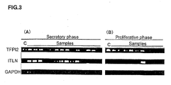
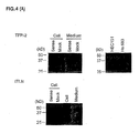


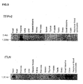
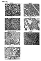

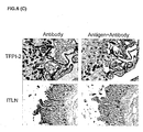
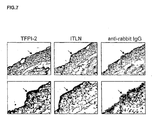
Classifications
-
- C—CHEMISTRY; METALLURGY
- C12—BIOCHEMISTRY; BEER; SPIRITS; WINE; VINEGAR; MICROBIOLOGY; ENZYMOLOGY; MUTATION OR GENETIC ENGINEERING
- C12Q—MEASURING OR TESTING PROCESSES INVOLVING ENZYMES, NUCLEIC ACIDS OR MICROORGANISMS; COMPOSITIONS OR TEST PAPERS THEREFOR; PROCESSES OF PREPARING SUCH COMPOSITIONS; CONDITION-RESPONSIVE CONTROL IN MICROBIOLOGICAL OR ENZYMOLOGICAL PROCESSES
- C12Q1/00—Measuring or testing processes involving enzymes, nucleic acids or microorganisms; Compositions therefor; Processes of preparing such compositions
- C12Q1/68—Measuring or testing processes involving enzymes, nucleic acids or microorganisms; Compositions therefor; Processes of preparing such compositions involving nucleic acids
- C12Q1/6876—Nucleic acid products used in the analysis of nucleic acids, e.g. primers or probes
- C12Q1/6883—Nucleic acid products used in the analysis of nucleic acids, e.g. primers or probes for diseases caused by alterations of genetic material
-
- A—HUMAN NECESSITIES
- A61—MEDICAL OR VETERINARY SCIENCE; HYGIENE
- A61P—SPECIFIC THERAPEUTIC ACTIVITY OF CHEMICAL COMPOUNDS OR MEDICINAL PREPARATIONS
- A61P15/00—Drugs for genital or sexual disorders; Contraceptives
-
- G—PHYSICS
- G01—MEASURING; TESTING
- G01N—INVESTIGATING OR ANALYSING MATERIALS BY DETERMINING THEIR CHEMICAL OR PHYSICAL PROPERTIES
- G01N33/00—Investigating or analysing materials by specific methods not covered by groups G01N1/00 - G01N31/00
- G01N33/48—Biological material, e.g. blood, urine; Haemocytometers
- G01N33/50—Chemical analysis of biological material, e.g. blood, urine; Testing involving biospecific ligand binding methods; Immunological testing
- G01N33/68—Chemical analysis of biological material, e.g. blood, urine; Testing involving biospecific ligand binding methods; Immunological testing involving proteins, peptides or amino acids
- G01N33/689—Chemical analysis of biological material, e.g. blood, urine; Testing involving biospecific ligand binding methods; Immunological testing involving proteins, peptides or amino acids related to pregnancy or the gonads
-
- C—CHEMISTRY; METALLURGY
- C12—BIOCHEMISTRY; BEER; SPIRITS; WINE; VINEGAR; MICROBIOLOGY; ENZYMOLOGY; MUTATION OR GENETIC ENGINEERING
- C12Q—MEASURING OR TESTING PROCESSES INVOLVING ENZYMES, NUCLEIC ACIDS OR MICROORGANISMS; COMPOSITIONS OR TEST PAPERS THEREFOR; PROCESSES OF PREPARING SUCH COMPOSITIONS; CONDITION-RESPONSIVE CONTROL IN MICROBIOLOGICAL OR ENZYMOLOGICAL PROCESSES
- C12Q2600/00—Oligonucleotides characterized by their use
- C12Q2600/158—Expression markers
-
- G—PHYSICS
- G01—MEASURING; TESTING
- G01N—INVESTIGATING OR ANALYSING MATERIALS BY DETERMINING THEIR CHEMICAL OR PHYSICAL PROPERTIES
- G01N2800/00—Detection or diagnosis of diseases
- G01N2800/36—Gynecology or obstetrics
- G01N2800/364—Endometriosis, i.e. non-malignant disorder in which functioning endometrial tissue is present outside the uterine cavity
Definitions
- the invention relates to methods of diagnosing ovarian endometriosis.
- Endometriosis is defined as the presence of endometrial glands and stroma outside the uterus. This gynecologic disorder occurs in approximately 14% of women of reproductive age ( Rice (2002) Ann. N.Y Acad. Sci. 955: 343-352 ). Its most common symptoms are progressive dysmenorrhea, dyspareunia, chronic pelvic pain and infertility. Endometriosis with ovarian cysts can be diagnosed by taking pictures such as ultrasonography or magnetic resonance image (MRI), however those without ovarian cysts are difficult to diagnose without surgical procedures ( Rice (2002) Ann. N.Y. Acad. Sci. 955: 343-52 ) since the symptoms as mentioned above are not specific to endometriosis.
- MRI magnetic resonance image
- Treatment for endometriosis commonly involves surgical resection and/or medication with hormonal agents such as gonadotropin-releasing hormone (GnRH) agonists and androgens.
- hormonal agents such as gonadotropin-releasing hormone (GnRH) agonists and androgens.
- GnRH gonadotropin-releasing hormone
- Methods for treatment of the disease have also a problem with a view to high recurrence rate. It has been reported that the recurrence rate at five years after operation was about 20% ( Redwine (1991) Fertil. Steril. 56: 628-34 ) and that at five years after medical treatment was as much as 53% ( Waller and Shaw (1993) Fertil. Steril. 59: 511-5 ).
- TFPI-2 also known as placental protein 5 (PP5), is a serine proteinase inhibitor containing 3 tandemly arranged Kunitz-type proteinase inhibitor domains, homologous to tissue factor pathway inhibitor ( Sprecher et al. (1994) Proc. Natl. Acad. Sci. USA 91: 3353-7 ; Rao et al. (1996) Arch. Biochem. Biophys. 335: 82-92 ; Miyagi et al. (1994) J. Biochem. 116: 939-42 ).
- the protein is constitutively secreted by several endothelial cell types ( lino et al. (1998) Arterioscler. Thromb. Vasc. Biol.
- TFPI-2 is a strong inhibitor of plasmin as well as of trypsin, chymotrypsin, plasma kallikrein, cathepsin G, factor VIIa and factor XIa, but not urokinase-type plasminogen activator (uPA), tissue plasminogen activator or thrombin ( Anlagener et al. (1994) Proc. Natl. Acad. Sci. USA 91: 3353-7 ; Rao et al. (1995) Arch. Biochem.
- TFPI-2 potentiate hepatocyte growth factor-induced invasion of hepatocellular carcinoma cells and is capable of inducing its own invasion ( Neaud et al. (2000) J. Biol. Chem. 275: 35565-9 ), whereas it has been demonstrated that the expression of the inhibitor inversely correlates during the progression of human glioma ( Rao et al. (2001) Clin. Cancer Res. 7: 570-6 ).
- Various gynecological studies about TFPI-2 were performed in terms of progression of pregnancy, but there is no report that this protein relates to the development of endometriosis.
- ITLN Intelectin
- ITLN is a secretory glycoprotein consisting of 295 amino acids and N-linked oligosaccharides, and comprises a basic structural unit of a 120 kDa homotrimer in which 40 kDa polypeptides are bridged by disulfide bonds.
- ITLN participates in the progression or maintenance of various gynecologic disorders or tumor disease.
- Invasion is a characteristic feature of endometriosis.
- endometriosis develops from endometrial fragments that are retrogradely menstruated into the peritoneal cavity.
- the proteolytic pathway of invasion depends on the balance of a lot of components including serine proteases, such as uPA and plasmin, matrix metalloproteinases and protease inhibitors ( Shapiro (1998) Curr. Opin. Cell Biol. 10: 602-8 ; Toi et al. (1998) Breast Cancer Res. Treat. 52: 113-24 ; Andreasen et al. (1997) Int.
- TFPI-2 may be important in the regulation of matrix turnover by serine proteases since TFPI-2 is found primarily in ECM.
- Neaud et al. reported that TFPI-2 induce an invasive activity in three different human hepatocellular carcinoma cell lines and stable transfectant ( Neaud et al. (2000) J. Biol. Chem. 275: 35565-9 ).
- TFPI-2 strongly inhibits the in vitro invasion of the highly invasive HT1080 cell line ( Rao et al. (1998) Int. J.
- TFPI-2 may have both an indirect anti-invasive effect and a direct pro-invasive effect.
- Other protease inhibitors for example, plasminogen activator inhibitor-1, also share a dual effect ( Deng et al. (1996) J. Cell Biol. 134: 1563-71 ).
- the interesting phenomenon that an endometriotic cell can be metastatic and invasive though the disease being a benign disorder may be explained by the fact that TFPI-2 may have such a dual effect and controls the invasion of endometriotic cells.
- ITLN The potential mechanism that ITLN is involved in the pathophysiology of endometriosis might be as follows.
- One hypothesis is that bacterial infection participates in the development of endometriosis. It has been reported that ITLN recognizes galactofuranosyl residues of bacteria ( Tsuji et al. (2001) J. Biol. Chem. 276: 23456-63 ).
- the expression of ITLN may be elevated as a result of the response to the infection.
- ITLN has been known to be involved in the autoimmune response of endometriosis.
- TFPI-2 in placenta is the highest in normal human tissues. While TFPI-2 circulates in blood of normal men and non-pregnant women in extremely low concentrations, its level increases 40-fold to 70-fold in the plasma of pregnant women ( Buztow et al. (1988) Clin. Chem. 34: 1591-3 ). The expression of TFPI-2 in endometrial cysts indicates the possibility that this protein is secreted in the serum of endometriosis patients as well as in that of pregnant women. If this prediction is true, TFPI-2 may serve as a better marker for diagnosis of endometriosis.
- ITLN has been reported to be secreted in blood. Nevertheless, ITLN is a secretory protein which is suggested to circulate in blood of patients of endometriosis. Detection of these proteins in the serum of endometriosis patients is expected to establish a new diagnostic method for the disease.
- cDNA microarray technologies have enabled to obtain comprehensive profiles of gene expression in normal and malignant cells, and compare the gene expression in malignant and corresponding normal cells ( Okabe et al. (2001) Cancer Res. 61:2129-37 ; Kitahara et al. (2001) Cancer Res. 61: 3544-9 ; Lin et al. (2002) Oncogene 21:4120-8 ; Hasegawa et al. (2002) Cancer Res. 62:7012-7 ). This approach enables to disclose the complex nature of cancer cells, and helps to understand the mechanism of carcinogenesis.
- microarray technologies include (i) discovery of genes that contribute to tumorigenesis, (ii) discovery of useful diagnostic biomarker(s) and novel molecular target(s) for anti-cancer agents and (iii) identification of genes involved in conferring chemosensitivity. Recently, molecules associated with development of certain types of cancers identified by microarray technologies have been clinically proven to be good targets for developing effective novel drugs for cancers.
- HCC hepatocellular carcinomas
- FTIs farnexyltransferase
- a tyrosine kinase inhibitor, STI-571, which selectively inactivates bcr-abl fusion proteins, has been developed to treat chronic myelogenous leukemias wherein constitutive activation of ber-ab1 tyrosine kinase plays a crucial role in the transformation of leukocytes.
- Agents of these kinds are designed to suppress oncogenic activity of specific gene products ( Fujita et al. (2001) Cancer Res. 61: 7722-6 ). Therefore, gene products commonly up-regulated in cancerous cells may serve as potential targets for developing novel anti-cancer agents.
- CTLs cytotoxic T lymphocytes
- TAAs tumor-associated antigens
- TAAs are now in the stage of clinical development as targets of immunotherapy. TAAs discovered so far include MAGE ( van der Bruggen et al. (1991) Science 254: 1643-7 ), gp100 ( Kawakami et al. (1994) J. Exp. Med. 180: 347-52 ), SART ( Shichijo et al. (1998) J. Exp. Med. 187: 277-88 ) and NY-ESO-1 ( Chen et al. (1997) Proc. Natl. Acad. Sci. USA 94: 1914-8 ).
- MAGE van der Bruggen et al. (1991) Science 254: 1643-7
- gp100 Kawakami et al. (1994) J. Exp. Med. 180: 347-52
- SART Shichijo et al. (1998) J. Exp. Med. 187: 277-88
- NY-ESO-1 Chen et al. (1997) Proc. Natl. Acad. Sci. USA 94
- gene products which had been demonstrated to be specifically overexpressed in tumor cells have been shown to be recognized as targets inducing cellular immune responses.
- Such gene products include p53 ( Umano et al. (2001) Brit. J. Cancer 84: 1052-7 ), HER2/neu ( Tanaka et al. (2001) Brit. J. Cancer 84: 94-9 ), CEA ( Nukaya et al. (1999) Int. J. Cancer 80: 92-7 ) and so on.
- PBMCs peripheral blood mononuclear cells
- the present invention is based on the discovery that a pattern of gene expressions is correlated with ovarian endometriosis.
- the genes that are differentially expressed in ovarian endometriosis are collectively referred to herein as "ovarian endometriosis-associated genes", "OEX nucleic acids” or “OEX polynucleotides” and the corresponding polypeptides encoded by the genes are referred to as "OEX polypeptides" or "OEX proteins”.
- the invention features a method of diagnosing or determining a predisposition to ovarian endometriosis in a subject by determining an expression level of an ovarian endometriosis-associated gene in a subject-derived biological sample, such as tissue sample.
- ovarian endometriosis-associated gene refers to a gene that is characterized by an expression level that differs in a cell obtained from an ovarian endometrial cell compared to a normal cell.
- normal cell indicates a cell obtained from ovarian or uterine tissue but not an endimetrial cyst cell.
- the ovarian endometriosis-associated genes include OEX 1-242.
- control level may be a normal control level or an ovarian endometriosis control level.
- a normal control level indicates a level of gene expression detected in a normal, healthy individual or in a population of individuals known not to be suffering from ovarian endometriosis.
- a control level may be a single expression pattern derived from a single reference population or may be a plurality of expression patterns.
- the control level can be a database of expression patterns from previously tested cells.
- a normal healthy individual is one with no clinical symptoms of ovarian endometriosis.
- OEX-1-97 and 186-242 detected in a test sample compared to a normal control level indicates that the subject (from whom the sample was obtained) suffers from or is at risk of developing ovarian endometriosis.
- a decrease in the expression level of OEX 98-185 detected in a test sample compared to a normal control level indicates that said subject suffers from or is at risk of developing ovarian endometriosis.
- the expression level of a panel of endometriosis-associated genes in a sample is compared to an ovarian endometriosis control level of the same panel of genes.
- ovarian endometriosis control level refers to the expression profile of the endometriosis-associated genes found in a population suffering from endometriosis.
- a decrease or similarity in the expression level of OEX 1-97 and 186-242 compared to an ovarian endometriosis control level indicates that the subject suffers from or is at risk of developing ovarian endometriosis.
- an increase or similarity in the expression level of OEX 98-185 compared to a normal control level indicates that said subject suffers from or is at risk of developing ovarian endometriosis.
- the expression of a gene may be determined as being altered when its expression level increases or decreases 10%, 25%, 50% or more compared to the control level.
- the expression of a gene may be determined as being altered when its expression level increases or decreases 1, 2, 5 or more fold compared to the control level.
- the expression level may be determined by detecting hybridization, e.g., on an array, of an ovarian endometriosis-associated gene probe to a gene transcript in the subject-derived biological sample.
- the subject-derived biological sample used in the present invention may be any sample including tissue samples obtained from a test subject, e.g., a patient known to or suspected of having ovarian endometriosis.
- tissue samples obtained from a test subject, e.g., a patient known to or suspected of having ovarian endometriosis.
- the tissue sample preferably contains an epithelial cell.
- the biological sample is an epithelial cell from an endometrial cyst.
- the invention also provides an ovarian endometriosis reference expression profile of the expression level of two or more genes of OEX 1-242.
- the invention provides an ovarian endometriosis reference expression profile of the expression levels of two or more genes of OEX 1-97 and 186-242 or OEX 98-185.
- the invention further provides methods of screening a compound that inhibits or enhances the expression or activity of an ovarian endometriosis-associated gene, OEX 1-242 by contacting with a test compound a test cell expressing an ovarian endometriosis-associated gene or a cell into which a vector comprising a reporter gene linked downstream of a transcriptional regulatory region of an ovarian endometriosis-associated gene has been introduced and determining the expression level of the ovarian endometriosis-associated gene.
- the test cell may be an epithelial cell such as those from an endometrial cyst.
- Compounds altering the expression level of an ovarian endometriosis-associated gene or reporter gene are expected to reduce the symptom of endometriosis.
- a decrease in the expression level of one or more genes of OEX 1-97 and OEX 186-242 or that of the reporter gene linked downstream of a transcriptional regulatory region of OEX 1-97 or OEX 186-242 compared to a normal control level of the gene indicates that the test compound is an inhibitor of the ovarian endometriosis-associated gene and are expected to reduce the symptom of endometriosis.
- an increase of the expression level of one or more genes of OEX 98-185 or that of the reporter gene linked downstream of a transcriptional regulatory region of OEX 98-185 compared to a normal control level of the gene indicates that the test compound is an enhancer of expression of the ovarian endometriosis-associated gene and are expected to reduce the symptom of endometriosis.
- the present invention provides method of screening a compound that inhibits or enhances the expression of an ovarian endometriosis-associated gene, wherein a polypeptide encoded by an ovarian endometriosis-associated gene is contacted with a test compound and determining the binding activity of the compound and the gene or the biological activity of the polypeptide.
- Compounds altering the binding activity of the compound and the gene or the biological activity of the polypeptide are expected to reduce the symptom of endometriosis.
- a decrease in the binding activity with or the biological activity of one or more polypeptides encoded by OEX 1-97 and OEX 186-242 compared to a normal control level of the gene indicates that the test compound is an inhibitor of the ovarian endometriosis-associated gene and is expected to reduce the symptom of endometriosis.
- an increase of the binding activity with or the biological activity of one or more polypeptides encoded by OEX 98-185 compared to a normal control level of the gene indicates that the test compound is an enhancer of the ovarian endometriosis-associated gene and is expected to reduce the symptom of endometriosis.
- the invention also provides a kit comprising a detection reagent that binds to two or more OEX nucleic acids or which binds to a gene product encoded by the nucleic acid sequences. Also provided is an array of two or more nucleic acids that bind to OEX nucleic acids.
- Therapeutic methods of the present invention include methods of treating or preventing ovarian endometriosis in a subject by inhibiting the expression of a gene selected from the group of OEX 1-97 and OEX 186-242 or the activity of a polypeptide encoded by the gene.
- the method can be achieved, for example, by administering to the subject an antisense, short interfering RNA (siRNA), ribozyme or antibody composition.
- siRNA short interfering RNA
- the antisense composition reduces the expression of a specific target gene sequence.
- the antisense composition contains a nucleotide sequence that is complementary to a coding sequence selected from the group consisting of OEX 1-97 and OEX 186-242.
- the siRNA composition reduces the expression of a nucleic acid sequence selected from the group consisting of OEX 1-97 and OEX 186-242.
- a nucleic acid-specific ribozyme composition may be constructed so as to reduce the expression of a nucleic acid sequence selected from the group consisting of OEX 1-97 and OEX 186-242.
- the treatment or prevention of ovarian endometriosis in a subject is carried out by administering to said subject an antibody or fragment thereof that binds to a polypeptide encoded a gene selected from the group consisting of OEX 1-97 and 186-242.
- the invention also includes vaccines and vaccination methods.
- a method of treating or preventing ovarian endometriosis in a subject is carried out by administering to the subject a vaccine containing a polypeptide encoded by a nucleic acid selected from the group consisting of OEX 1-97 and 186-242 or an immunologically active fragment of such a polypeptide.
- An immunologically active fragment is, for example, a polypeptide that is shorter than the full-length of the naturally-occurring protein and which induces an immune response.
- an immunologically active fragment of a length of at least 8 amino acid residues that stimulates immune cells, such as T cell or B cell is encompassed by the immunologically active fragment of the present invention.
- Immune cell stimulation is measured by detecting cell proliferation, elaboration of cytokines (e.g., IL-2) or production of an antibody.
- ovarian endometriosis can be treated by administering a protein encoded by OEX 98-185.
- the protein may be directly administered to the patient or, alternatively, may be expressed in vivo subsequent to being introduced into the patient, for example, by administering an expression vector or host cell carrying the down-regulated marker gene of interest. Suitable mechanisms for in vivo expression of a gene of interest are known in the art.
- the present invention provides compositions for treating or preventing ovarian endometriosis.
- the composition preferably comprises at least one active component selected from the group of (1) an antisense, siRNA or ribozyme against a gene selected from the group of OEX 1-97 and OEX 186-242; (2) an antibody or fragment thereof binding to a polypeptide encoded by a gene selected from the group of OEX 1-97 and OEX 186-242; (3) polynucleotide select from group consisting of OEX 98-185, or polypeptide encoded by thereof; and (4) a compound selected by any of the method of screening for a compound that alters the expression or activity of an ovarian endometriosis-associated gene of the present invention.
- the present invention is based in part on the discovery of changes in expression patterns of multiple nucleic acid sequences in endothelial cells of endometrial cysts of patients with ovarian endometriosis.
- the difference in gene expression was identified using a comprehensive cDNA microarray system.
- TFPI-2 tissue factor pathway inhibitor-2
- ITLN intelectin
- cDNA microarray analysis was performed on over 20,000 genes. As a result, genes that were commonly over-expressed or suppressed (down-regulated or underexpressed) among ovarian endometriosis patients were selected. Two hundred forty-two genes were found to be differentially expressed in epithelial cells from endometrial cysts. Twenty-four genes were up-regulated throughout the menstrual cycle and thirty were down-regulated throughout the menstrual cycle. Seventy genes and ESTs were up-regulated during the proliferative phase of the menstrual cycle, and fourteen genes were down-regulated during the proliferative phase of the menstrual cycle. Sixty genes and ESTs were up-regulated during the secretory phase of the menstrual cycle, and forty-four genes were down-regulated during the proliferative phase of the menstrual cycle.
- ovarian endometriosis-associated genes ovarian endometriosis-associated genes
- OEX nucleic acids or OEX polynucleotides
- OEX polypeptides or "OEX proteins”.
- OEX refers to any of the sequences disclosed herein (OEX 1-242).
- the genes have been previously described and are presented along with a database accession number.
- ovarian endometriosis By measuring expression of the various genes in a sample comprising a cell or population of cells, ovarian endometriosis can be diagnosed in a patient. Similarly, by measuring the expression of these genes in response to various agents, agents for treating ovarian endometriosis can be identified. Table 1. Genes up-regulated in endometrial cysts throughout the menstrual cycle ratio ⁇ 2 GenBank Accession No.
- the left-most column indicates the numbers of samples in that category. Accession numbers, gene symbols and names were retrieved from the Unigene Database (build#131). Table 3. Genes up-regulated in endometrial cysts only during the secretory phase of the menstrual cycle ratio ⁇ 2 Genbank Accession No.
- the left-most column indicates the numbers of samples in that category. Accession numbers, gene symbols and names were retrieved from the Unigene Database (build#131). Table 4. Genes down-regulated in endometrial cysts throughout the menstrual cycle ratio ⁇ 0.3 Genbank Accession No.
- coli homolog 123 17 U40462 ZNFN1A1 zinc finger protein, subfamily 1A, 1 (Ikaros) 124 17 AA234506 LRRFIP1 leucine rich repeat (in FLII) interacting protein 1 125 17 AI249000 LIM LIM protein (similar to rat protein kinase C-binding enigma) 126 17 AA496218 STAG2 stromal antigen 2 127 Genes with normalized expression ratios (cyst/normal) of ⁇ 0.3 in more than 70% of the 23 cases were examined. The left-most column indicates the numbers of samples in that category. Accession numbers, gene symbols and names were retrieved from the Unigene Database (build#131). Table 5.
- the left-most column indicates the numbers of samples in that category. Accession numbers, gene symbols and names were retrieved from the Unigene Database (build#131). Table 6. Genes down-regulated in endometrial cysts only during the secretory phase of the menstrual cycle ratio ⁇ 0.3 GenBank Accession No.
- the left-most column indicates the numbers of samples in that category. Accession numbers, gene symbols and names were retrieved from the Unigene Database (build#131). Table 7. Genes up-regulated in endometrial cysts throughout the menstrual cycle Accession No. Symbol Gene name OEX Assignment M63262 ALOX5AP arachidonate 5-lipoxygenase-activating protein 1 X63629 CDH3 cadherin 3, type 1, P-cadherin (placental) 6 Z22970 CD 163 CD163 antigen 186 X04701 C1R complement component 1, r subcomponent 68 M20431 DC classII histocompatibility antigen alpha-chain 187 X00637 HP haptoglobin 9 K03431 HPR haptoglobin-related protein 188 V00497 HBB hemoglobin, beta 189 U01317 HBD hemoglobin, delta 190 AA583491 HCA112 hepatocellular carcinoma-associated antigen 112 2 X00457 Human
- the present invention provides a method of diagnosing ovarian endometriosis.
- a subject suffering from or in the risk of developing ovarian endometriosis can be diagnosed.
- the subject diagnosed according to the present method is preferably a mammal.
- the mammal can be, for example, human, non-human primate, mouse, rat, dog, cat, horse or cow.
- Ovarian endometriosis may be diagnosed by examining the expression of one or more OEX-associated genes in a subject-derived biological sample, (e.g., a subject-derived tissue sample) that contain or are suspected to contain an ovarian endometrial cell.
- a subject-derived biological sample e.g., a subject-derived tissue sample
- the method of diagnosing ovarian endometriosis of the present invention involves determining (measuring) the expression level of at least one and up to all the OEX associated genes listed in Tables 1-9.
- sequence information provided by the GenBank database entries for the known sequences the ovarian endometriosis-associated genes are detected and measured using techniques well known to one of ordinary skill in the art.
- OEX 1-242 The expression of 1, 2, 3, 4, 5, 25, 35, 50, 100 or more of the sequences represented by OEX 1-242 may be determined and if desired, expression of these nucleotides can be determined along with other nucleotides whose expression level is known to be altered according to one of the herein described parameters or conditions, e.g., ovarian endometriosis or non-ovarian endometriosis.
- the expression level of one or more of the ovarian endometriosis-associated gene in a biological sample derived from a subject is compared to a control level of the same gene.
- the biological sample may be any sample derived from the subject so long as the expression of the selected ovarian endometriosis-associated gene can be detected in a patient of ovarian endometriosis and includes individual cells and cell populations such as a test cells obtained from a bodily tissue or a bodily fluid (biological fluid such as blood, serum and urine).
- the biological sample comprises a test cell population comprising an epithelial cell derived from a tissue that is known to be or suspected to be an endometrial cyst.
- the control level of an ovarian endometriosis-associated gene in the diagnostic method of the present invention is the expression level of the same gene in a reference population.
- Cells in a reference cell population are derived from the same type of sample as that to the biological sample examined in the method.
- the reference cell population includes one or more cells for which the parameter to be compared is known, i.e., endometriotic or non-endometriotic cell.
- a control cell population may be derived from a database of molecular information derived from cells for which the assay parameter or condition is known.
- the expression level of OEX nucleotide(s) in a subject-derived biological sample may be compared to multiple control levels of the OEX nucleotide(s).
- the control levels may be derived from biological samples with different known parameters (i.e. , endometriotic or non-endometriotic).
- the expression level of OEX nucleotide(s) in a subject-derived biological sample may be compared to the control level corresponding to ovarian endometrial cells (ovarian endometriosis control level), and then to the control corresponding to non-ovarian endometrial cells (normal cells) (normal control level).
- the control level may be a single expression pattern derived from a single reference population or may be a plurality of expression patterns.
- the control level can be a database of expression patterns from previously tested cells.
- Whether a pattern of gene expression levels in a subject-derived biological sample compared to that in the control level indicates ovarian endometriosis or a predisposition thereto depends on the kind of the sample used for determining the control level. For example, if the control level is detected in a sample of reference cell population composed of non-endometriotic cells, a similar gene expression level in the subject-derived biological sample indicates the subject to be non-endometriotic.
- control levels are referred to as "normal control level”.
- a normal control level indicates a level of gene expression detected in a normal, healthy individual or in a population of individuals known not to be suffering from ovarian endometriosis.
- a normal healthy individual is one with no clinical symptoms of ovarian endometriosis.
- control level is detected in a sample of reference cell population made up of endometriotic cells, a similar gene expression profile between the subject-derived biological sample and the reference cell population indicates that the biological sample includes endometriotic cells.
- control levels are referred to as “ovarian endometriosis control level”.
- ovarian endometriosis control level refers to the expression profile of the endometriosis-associated genes found in a population suffering from endometriosis.
- the expression level of an ovarian endometriosis-associated gene in a subject-derived biological sample can be considered as being altered if the expression level differs from the control level by more than 1.0, 1.5, 2.0, 5.0, 10.0 or more folds.
- an expression level of a gene in a subject-derived biological sample increased or decrease by 10%, 25%, 50% or more compared to the control level indicates alteration of the gene expression in the subject sample.
- OEX-1-97 and 186-242 detected in a subject-derived biological sample compared to a normal control level indicates that the subject suffers from or is at risk of developing ovarian endometriosis.
- a decrease in the level of OEX 98-185 detected in a subject-derived biological sample compared to a normal control level indicates that the subject suffers from or is at risk of developing ovarian endometriosis.
- a decrease or similarity in the expression level of OEX 1-97 and 186-242 detected in a subject-derived biological sample compared to an ovarian endometriosis control level indicates that the subject suffers from or is at risk of developing ovarian endometriosis.
- an increase or similarity in the level of OEX 98-185 detected in a subject-derived biological sample compared to a normal control level indicates that said subject suffers from or is at risk of developing ovarian endometriosis.
- control nucleic acid is one whose expression is known not to differ between the endometriotic or non-endometriotic state of the cell. Expression levels of the control nucleic acid in the test and reference nucleic acid can be used to normalize signal levels in the compared populations.
- a control nucleic acid is also called “housekeeping gene” and genes such as ⁇ -actin, glyceraldehyde 3-phosphate dehydrogenase or ribosomal protein P1 may be used in the present invention.
- RNA level may be determined at the RNA level using any method known in the art.
- sequences within the sequence database entries corresponding to OEX nucleic acid sequences can be used to construct probes for detecting OEX RNAs by, e.g., Northern blot hybridization analyses.
- a probe preferably includes at least 10, 20, 50, 100, 200 or more continuous nucleotides of a reference sequence (i.e ., the nucleotide sequence of OEX nucleic acids (Tables 1-9)).
- the expression level is measured using reverse-transcription-based PCR (RT-PCR) assays, e.g., using primers specific for the OEX nucleic acid sequences.
- RT-PCR reverse-transcription-based PCR
- the expression level can also be determined at the protein level by measuring the levels or the biological activity of the expressed polypeptides encoded by the genes described herein.
- Such methods are well known in the art and include immunoassays based on antibodies against polypeptides encoded by the genes.
- the biological activities of respective polypeptides encoded by the ovarian endometriosis-associated genes are also well known in the art, and one skilled in the art can adopt appropriate conventional methods for measuring the biological activities of the polypeptides depending on the kind of polypeptide to be measured.
- the gene (DNA sequence) in a subject-derived biological sample may be compared to that of a reference cell population to determine whether the subject suffers from or is at risk of developing ovarian endometriosis.
- the differentially expressed OEX nucleotides identified herein also allow for the course of treatment of ovarian endometriosis to be monitored.
- a biological sample such as a test cell population, is obtained from a subject undergoing treatment for ovarian endometriosis.
- the method for assessment can be conducted according to the method of diagnosing ovarian endometriosis of the present invention described above.
- biological samples are obtained from the subject at various time points before, during or after the treatment.
- the expression level of one or more of the OEX associated genes, in the biological sample is then determined and compared to a control level derived, for example, from a reference cell population which includes cells whose state of ovarian endometriosis (i.e ., endometriotic or non-endometriotic) is known.
- the control level is determined in a biological sample that has not been exposed to the treatment.
- control level is derived from a biological sample which contains no ovarian endometrial cells
- a similarity between the expression level in the subject-derived biological sample and the control level indicates that the treatment is efficacious.
- a difference between the expression level of the OEX nucleotides in the subject-derived biological sample and the control level indicates a less favorable clinical outcome or prognosis.
- OEX 1-97 and OEX 186-242 pathologically up-regulated gene
- OEX 98-185 pathologically down-regulated gene
- a treatment is applied prophylactically
- "efficacious” indicates that the treatment retards or prevents formation of ovarian endometrial cysts.
- the assessment of endometrial cysts can be made using standard clinical protocols.
- Ovarian endometriosis is diagnosed for example, by identifying symptomatic anomalies, e.g., progressive dysmenorrhea, dyspareunia, chronic pelvic pain and infertility, along with surgical identification of endometrial glands and stroma outside the uterus.
- symptomatic anomalies e.g., progressive dysmenorrhea, dyspareunia, chronic pelvic pain and infertility
- the present invention further provides a method of assessing the prognosis of a patient with ovarian endometriosis by comparing the expression level of one or more OEX nucleotides in a patient-derived biological sample, such as test cell population, to a control level.
- a patient-derived biological sample such as test cell population
- the expression level of one or more OEX nucleotides in a biological sample derived from patients may be measured over a spectrum of disease stages to assess the prognosis of the patient.
- the method for assessment can be conducted according to the method of diagnosing ovarian endometriosis of the present invention described above.
- a decrease in the expression level of one or more of OEX 98-185 compared to a normal control level or an increase of the expression level of one or more of OEX 1-97 and 186-242 compared to a normal control indicates less favorable prognosis.
- An increase in the expression level of one or more of OEX 98-185 indicates a more favorable prognosis, and a decrease in the expression level of OEX 1-97 and 186-242 indicates a more favorable prognosis for the patient.
- An ovarian endometriosis reference expression profile is provided by the present invention.
- Such expression profiles of the present invention comprise a pattern of gene expression of two or more OEX nucleotides (OEX 1-242) in a cell of endometrial cysts or a normal healthy endometrial cell (non-endometriotic cell).
- the present expression profile may comprise a pattern of gene expression of two or more genes of OEX 1-97 and OEX 186-242 or OEX 98-185.
- the expression profile can be used in diagnosing ovarian endometriosis or a predisposition to developing ovarian endometriosis in a subject, monitoring the course of treatment of ovarian endometriosis and assessing prognosis of a subject with ovarian endometriosis.
- the invention further provides methods of screening a compound that alters, i.e., inhibits or enhances the expression of a marker gene, ovarian endometriosis-associated gene (OEX 1-242), by (1) contacting a test compound with a test cell expressing an ovarian endometriosis-associated gene or a cell into which a vector comprising a reporter gene linked downstream of a transcriptional regulatory region of an ovarian endometriosis-associated gene has been introduced; (2) determining the expression level of the ovarian endometriosis-associated gene; and (3) selecting the compound that alters the expression level compared to that in the absence of the test compound.
- the method is based on screening a compound to determine if it converts an expression profile of OEX 1-242 characteristic of an ovarian endometriosis state to a pattern indicative of a non-ovarian endometriosis state.
- a decrease in the expression level of an ovarian endometriosis-associated gene or a reporter gene linked downstream of a transcriptional regulatory region of an ovarian endometriosis-associated gene compared to the expression level detected in the absence of a test compound indicates that the test compound is an inhibitor.
- enhanced expression level of the genes compared to the expression level detected in the absence of a test compound indicates the test compound to function as an enhancer.
- the expression level detected in the absence of a test compound may be a normal control level or ovarian endometriosis control level.
- a gene up-regulated in the endothelial cells of endometrial cysts of patients with ovarian endometriosis e.g., any one selected from OEX 1-97 and OEX 186-242
- a compound that inhibits the expression of the gene is expected to inhibit endometriosis.
- a gene down-regulated in the endothelial cells of endometrial cysts of patients with ovarian endometriosis e.g., any one selected from OEX 98-185 or the transcriptional regulatory region thereof is used in the screening method, a compound that enhances the expression of the gene is expected to inhibit endometriosis.
- a cell may be exposed to a test compound or a combination of test compounds (sequentially or consequentially).
- Compounds selected by the screening of the present invention serve as candidate compounds for treating or preventing ovarian endometriosis.
- Compounds effective in stimulating expression of underexpressed (down-regulated) marker genes or in suppressing expression of overexpressed (up-regulated) marker genes are deemed to lead to a clinical benefit. Therefore, such compounds are further tested for the ability to prevent endometrial cyst growth in endometrial glands and/or stroma of animals or test subjects.
- compounds in which a part of the structure is converted by addition, deletion, substitution and/or insertion are also included in the compounds obtainable by the screening of the present invention.
- test cell used in the screening may be any cell so long as it expresses the ovarian endometriosis-associated gene.
- the test cell may be a test cell population consisting of multiple cells.
- the test cell or test cell population contains an epithelial cell, such as those derived from an endometrial cyst.
- the test cell or test cell population may be an immortalized cell or cell line derived from an endometrial cyst cell.
- a transcriptional regulatory region of an ovarian endometriosis-associated gene can be obtained from genomic libraries using probes comprising the 5' region of the OEX nucleotides (OEX 1-242).
- Any reporter gene may be used in the screening so long as its expression can be detected in the screening.
- reporter genes include the ⁇ -gal gene, CAT gene and luciferase gene. Detection of the expression of the reporter gene can be conducted based on conventional methods in accordance with the type of the reporter gene. Although there is no restriction on the cell into which the vector is introduced, preferable examples include epithelial cell.
- the present invention further provides a method of screening for a compound that alters the activity of an ovarian endometriosis-associated gene.
- An embodiment of this screening method comprises the steps of: (a) contacting a test compound with a polypeptide encoded by an ovarian endometriosis-associated gene; (b) detecting the binding activity between the polypeptide and the test compound; and (c) selecting the compound that binds to the polypeptide.
- the method utilizes the biological activity of an OEX polypeptide as an index.
- the screening method includes the steps of.
- OEX polypeptides used for the present screening are selected from:
- biological activity refers to activities such as occurrence, growth or proliferation of endometrial cyst cell. Whether an objective polypeptide has the biological activity or not can be judged by introducing the polypeptide or a DNA encoding the polypeptide into a cell, and detecting growth or proliferation of the cells, inrease in colony forming activity, etc.
- Methods for preparing polypeptides having the biological activity of a given protein are well known in the art and include methods introducing mutations into the protein.
- the OEX polypeptides include those having the amino acid sequences of the human OEX proteins in which one or more amino acids are mutated, provided the resulting mutated polypeptides have the biological activity of the OEX proteins.
- the number of amino acids to be mutated in such a mutant is generally 10 amino acids or less, preferably 6 amino acids or less and more preferably 3 amino acids or less.
- polypeptide to which one ore more amino acid residues are added include fusion proteins containing the OEX protein. Fusion proteins can be made by techniques well known to those skilled in the art, such as linking DNA encoding the OEX protein with DNA encoding other peptides or proteins, so that the frames match, inserting the fused DNA into an expression vector and expressing it in a host. There is no restriction as to the peptides or proteins fused to the OEX protein and include FLAG ( Hopp et al.
- OEX polypeptides having the biological activity of any of the OEX proteins is, for example, the method using hybridization technique ( Sambrook et al. (1989) Molecular Cloning 2nd ed. 9.47-9.58, Cold Spring Harbor Lab. Press ).
- hybridization technique Sambrook et al. (1989) Molecular Cloning 2nd ed. 9.47-9.58, Cold Spring Harbor Lab. Press ).
- One skilled in the art can readily isolate a DNA having high homology with a whole or part of the DNA sequence encoding an OEX protein, and isolate polypeptides having the biological activity of the OEX protein from the isolated DNA.
- the OEX polypeptides include those that are encoded by DNA that hybridize with a whole or part of a gene selected from OEX 1-242 and have the biological activity of the OEX protein.
- polypeptides include mammal homologues corresponding to the protein derived from human (for example, a polypeptide encoded by a monkey, rat, rabbit and bovine gene).
- mammal homologues corresponding to the protein derived from human (for example, a polypeptide encoded by a monkey, rat, rabbit and bovine gene).
- isolating a cDNA highly homologous to a gene selected from OEX 1-242 from animals it is particularly preferable to use tissues from colorectal cancers with metastasis.
- a gene amplification method for example, the PCR method, can be utilized to isolate a DNA encoding a polypeptide having the biological activity of the OEX protein, using a primer synthesized based on the sequence information of the protein encoding DNA (OEX 1-242).
- An OEX polypeptide used in the method of the present invention may have variations in amino acid sequence, molecular weight, isoelectric point, the presence or absence of sugar chains or form, depending on the cell or host used to produce it or the purification method utilized. Nevertheless, so long as it has a biological activity equivalent to that of the OEX protein, it may be used in the method of the present invention and such methods utilizing polypeptides with a biological activity equivalent to the OEX protein are within the scope of the present invention.
- the OEX polypeptides used in the present invention can be prepared as recombinant proteins or natural proteins, by methods well known to those skilled in the art.
- a recombinant protein can be prepared by inserting a DNA, which encodes the OEX polypeptide, into an appropriate expression vector, introducing the vector into an appropriate host cell, obtaining the extract, and purifying the polypeptide.
- a natural protein can be isolated by methods known to a person skilled in the art, for example, by contacting the affinity column, in which antibodies binding to the OEX protein described below are bound, with the extract of tissues or cells expressing the OEX polypeptide.
- the antibodies can be polyclonal antibodies or monoclonal antibodies.
- the OEX polypeptide to be contacted with a test compound can be, for example, a purified polypeptide, a soluble protein, a form bound to a carrier or a fusion protein fused with other polypeptides.
- supports that may be used for binding proteins include insoluble polysaccharides, such as agarose, cellulose and dextran; and synthetic resins, such as polyacrylamide, polystyrene and silicon; preferably commercial available beads and plates (multi-well plates, biosensor chip, etc.) prepared from the above materials may be used. When using beads, they bay be filled into a column.
- binding of a protein to a support may be conducted according to routine methods, such as chemical bonding and physical adsorption.
- a protein may be bound to a support via antibodies that specifically recognizing the protein.
- binding of a protein to a support can be also conducted by means of avidin and biotin binding.
- a method of screening for proteins for example, that bind to the OEX polypeptide using any of the OEX polypeptides described above, many methods well known by a person skilled in the art can be used. Such a screening can be conducted by, for example, immunoprecipitation method, specifically, in the following manner.
- an immune complex is formed by adding an antibody to cell lysate prepared using an appropriate detergent.
- the antibody used in the immunoprecipitation for the screening recognizes any of the proteins endcoded by OEX 1-242.
- OEX protein fused with a recognition site epitope
- antibodies against the epitope may be used for the immunoprecipitaion.
- the immune complex consists of the OEX protein, a polypeptide comprising the binding ability with the OEX protein, and an antibody.
- An immune complex can be precipitated, for example by Protein A sepharose or Protein G sepharose when the antibody is a mouse IgG antibody. If the OEX polypeptide is prepared as a fusion protein with an epitope, such as GST, an immune complex can be formed in the same manner as in the use of the antibody against the OEX polypeptide, using a substance specifically binding to these epitopes, such as glutathione-Sepharose 4B.
- Immunoprecipitation can be performed by following or according to, for example, the methods in the literature ( Harlow and Lane (1988) Antibodies, 511-52, Cold Spring Harbor Laboratory publications, New York ).
- SDS-PAGE is commonly used for analysis of immunoprecipitated proteins and the bound protein can be analyzed by the molecular weight of the protein using gels with an appropriate concentration. Since the protein bound to the OEX polypeptide may be difficult to detect by a common staining method, such as Coomassie staining or silver staining, the detection sensitivity for the protein can be improved by culturing cells in culture medium containing radioactive isotope, 35 S-methionine or 35 S-cystein, labeling proteins in the cells, and detecting the proteins. The target protein can be purified directly from the SDS-polyacrylamide gel and its sequence can be determined, when the molecular weight of a protein has been revealed.
- a common staining method such as Coomassie staining or silver staining
- a protein binding to the OEX polypeptide can be obtained by preparing a cDNA library from cells, tissues, organs or cultured cells expected to express a protein binding to the OEX polypeptide using a phage vector ( e.g. , ZAP), expressing the protein on LB-agarose, fixing the protein expressed on a filter, reacting the purified and labeled OEX polypeptide with the above filter, and detecting the plaques expressing proteins bound to the OEX polypeptide according to the label.
- a phage vector e.g. , ZAP
- the OEX polypeptide may be labeled by utilizing the binding between biotin and avidin, or by utilizing an antibody that specifically binds to the OEX polypeptide, or a peptide or polypeptide (for example, GST) that is fused to the OEX polypeptide.
- labeling substances such as radioisotope ( 3 H, 14 C, 32 P, 33 P, 35 S, 125 I, 131 I, etc.), enzymes (alkaline phosphatase, horseradish peroxidase, ⁇ -galactosidase, ⁇ -glucosidase, etc.), fluorescent substances (fluorescein isothiosyanete (FITC), rhodamine, etc.) and biotin/avidin, may be used for the labeling in the present method.
- FITC fluorescein isothiosyanete
- biotin/avidin may be used for the labeling in the present method.
- the detection or measurement can be carried out by liquid scintillation.
- OEX proteins labeled with enzymes can be detected or measured by adding a substrate of the enzyme to detect the enzymatic change of the substrate, such as generation of color, with absorptiometer. Further, in case where a fluorescent substance is used as the label, the bound protein may be detected or measured using fluorophotometer.
- a two-hybrid system utilizing cells may be used ("MATCHMAKER Two-Hybrid system”, “Mammalian MATCHMAKER Two-Hybrid Assay Kit”, “MATCHMAKER one-Hybrid system” (Clontech); “HybriZAP Two-Hybrid Vector System” (Stratagene); the references “ Dalton and Treisman (1992) Cell 68: 597-612 ", “ Fields and Sternglanz (1994) Trends Genet 10: 286-92 ").
- the OEX polypeptide is fused to the SRF-binding region or GAL4-binding region and expressed in yeast cells.
- a cDNA library is prepared from cells expected to express a protein binding to the OEX polypeptide, such that the library, when expressed, is fused to the VP16 or GAL4 transcriptional activation region.
- the cDNA library is then introduced into the above yeast cells and the cDNA derived from the library is isolated from the positive clones detected (when a protein binding to the OEX polypeptide is expressed in yeast cells, the binding of the two activates a reporter gene, making positive clones detectable).
- a protein encoded by the cDNA can be prepared by introducing the cDNA isolated above to E. coli and expressing the protein.
- reporter gene for example, Ade2 gene, lacZ gene, CAT gene, luciferase gene and such can be used besides HIS3 gene.
- a compound binding to the OEX polypeptide can also be screened using affinity chromatography.
- the OEX polypeptide may be immobilized on a carrier of an affinity column, and a test compound, containing a protein capable of binding to the ODX polypeptide, is applied to the column.
- a test compound herein may be, for example, cell extracts, cell lysates, etc. After loading the test compound, the column is washed, and compounds bound to the OEX polypeptide can be prepared.
- test compound When the test compound is a protein, the amino acid sequence of the obtained protein is analyzed, an oligo DNA is synthesized based on the sequence, and cDNA libraries are screened using the oligo DNA as a probe to obtain a DNA encoding the protein.
- a biosensor using the surface plasmon resonance phenomenon may be used as a mean for detecting or quantifying the bound compound in the present invention.
- the interaction between the OEX polypeptide and a test compound can be observed real-time as a surface plasmon resonance signal, using only a minute amount of polypeptide and without labeling (for example, BIAcore, Pharmacia). Therefore, it is possible to evaluate the binding between the OEX polypeptide and a test compound using a biosensor such as BIAcore.
- a compound isolated by this screening is a candidate for agonists or antagonists of the OEX polypeptide.
- agonist refers to molecules that activate the function of the OEX polypeptide by binding thereto.
- antagonist refers to molecules that inhibit the function of the OEX polypeptide by binding thereto.
- a compound isolated by this screening is a candidate for compounds which inhibit the in vivo interaction of the OEX polypeptide with molecules (including DNAs and proteins).
- the biological activity to be detected in the present method is cell proliferation
- it can be detected, for example, by preparing cells which express the OEX polypeptide, culturing the cells in the presence of a test compound, and determining the speed of cell proliferation, measuring the cell cycle and such, as well as by measuring the colony forming activity.
- a decrease in the binding activity or biological activity of one or more polypeptides encoded by OEX 1-97 and OEX 186-242 compared to a normal control level of the gene detected by the screening method indicates that the test compound is an inhibitor of the ovarian endometriosis-associated gene and is expected to reduce the symptom of endometriosis.
- an increase of the binding activity with or the biological activity of one or more polypeptides encoded by OEX 98-185 compared to a normal control level of the gene detected by the screening method indicates that the test compound is an enhancer of the ovarian endometriosis-associated gene and is expected to reduce the symptom of endometriosis.
- a compound isolated by the above screenings is a candidate for drugs which can be applied for the treatment or prevention of ovarian, endometriosis.
- compound in which a part of the structure of the compound that alters the activity of the OEX protein is converted by addition, deletion and/or replacement are also included in the compounds obtainable by the screening method of the present invention.
- test compound for example, cell extracts, cell culture supernatant, products of fermenting microorganism, extracts from marine organism, plant extracts, purified or crude proteins, peptides, non-peptide compounds, synthetic micromolecular compounds and natural compounds can be used in the screening methods of the present invention.
- the test compound of the present invention can be also obtained using any of the numerous approaches in combinatorial library methods known in the art, including (1) biological libraries, (2) spatially addressable parallel solid phase or solution phase libraries, (3) synthetic library methods requiring deconvolution, (4) the "one-bead one-compound” library method and (5) synthetic library methods using affinity chromatography selection.
- Differences in the genetic makeup of individuals can result in differences in their relative abilities to metabolize various drugs.
- a compound that is metabolized in a subject to act as an anti-ovarian endometriosis agent can manifest itself by inducing a change in gene expression pattern in the subject's cells from that characteristic of an ovarian endometriosal state to a gene expression pattern characteristic of a non-ovarian endometriosal state.
- the differentially expressed OEX associated genes disclosed herein allow for selection of a putative therapeutic or prophylactic anti-ovarian endometriosis agent specifically adequate for a subject by testing candidate compounds in a test cell population from the selected subject.
- test cell or test cell population derived from the subject is exposed to a candidate therapeutic agent and the expression of one or more of OEX 1-242 genes is determined.
- the test cell is or the test cell population contains an ovarian endometrial cell expressing an ovarian endometriosis associated gene.
- the test cell is or the test cell population contains an epithelial cell.
- a test cell or test cell population is incubated in the presence of a candidate agent and the pattern of gene expression in the test cell or cell population is measured and compared to one or more reference profiles (an ovarian endometriosis reference expression profile or an non-ovarian endometriosis reference expression profile).
- a decrease in the expression of one or more of OEX 1-97 and 186-242 or an increase in the expression of one or more of OEX 98-185 in a test cell or test cell population relative to that in a reference cell population containing ovarian endometriosis is indicative that the agent is therapeutic.
- the test agent can be any compound or composition.
- the test agent is an immunomodulatory agent.
- the invention also provides a kit comprising an OEX-detection reagent, e.g., a nucleic acid that specifically binds to or identifies one or more of OEX nucleic acids.
- an OEX-detection reagent e.g., a nucleic acid that specifically binds to or identifies one or more of OEX nucleic acids.
- Such nucleic acid specifically binding to or identifying one or more of OEX nucleic acids are exemplified by oligonucleotide sequences that are complementary to a portion of an OEX nucleic acid or antibodies which bind to polypeptides encoded by an OEX nucleic acid.
- the reagents are packaged together in the form of a kit.
- the reagents such as a nucleic acid or antibody (either bound to a solid matrix or packaged separately with reagents for binding them to the matrix), a control reagent (positive and/or negative) and/or a means of detection of the nucleic acid or antibody are preferably packaged in separate containers. Instructions (written, tape, VCR, CD-ROM, etc.) for carrying out the assay are included in the kit.
- the assay format of the kit may be Northern hybridization or a sandwich ELISA known in the art.
- an OEX detection reagent is immobilized on a solid matrix such as a porous strip to form at least one OEX detection site.
- the measurement or detection region of the porous strip may include a plurality of detection sites, each detection site containing an OEX-detection reagent.
- a test strip may also contain sites for negative and/or positive controls. Alternatively, control sites are located on a separate strip from the test strip.
- the different detection sites may contain different amounts of immobilized nucleic acids, i.e ., a higher amount in the first detection site and lesser amounts in subsequent sites.
- the number of sites displaying a detectable signal provides a quantitative indication of the amount of OEX present in the sample.
- the detection sites may be configured in any suitably detectable shape and are typically in the shape of a bar or dot spanning the width of a teststrip.
- the kit contains a nucleic acid substrate array comprising one or more ovarian endometriosis-associated genes.
- the nucleic acids on the array specifically identify one or more nucleic acid sequences represented by OEX 1-242.
- the expression level of 2, 3, 4, 5, 6, 7, 8, 9, 10, 15, 20, 25, 40 or 50 or more of the sequences represented by OEX 1-242 are identified by virtue if the level of binding to an array test strip or chip.
- the substrate array can be on a solid substrate, e.g., a "chip" as described in US Patent No.5,744,305 .
- the invention also includes a nucleic acid substrate array comprising one or more ovarian endometriosis-associated genes.
- the nucleic acids on the array specifically corresponds to one or more nucleic acid sequences represented by OEX 1-242.
- the expression level of 2, 3, 4, 5, 6, 7, 8, 9, 10, 15, 20, 25, 40 or 50 or more of the OEX nucleic acids represented by OEX 1-242 are identified by detecting the binding of nucleotides to the array.
- the invention also includes an isolated plurality of nucleic acids (i.e ., a mixture if two or more OEX nucleic acids).
- the nucleic acids are in a liquid phase or a solid phase, e.g., immobilized on a solid support such as a nitrocellulose membrane.
- the plurality includes one or more of the nucleic acids represented by OEX 1-242. According to a further embodiment of the present invention, the plurality includes 2, 3, 4, 5, 6, 7, 8, 9, 10, 15, 20, 25, 40 or 50 or more of the nucleic acids represented by OEX 1-242.
- the invention provides a method for treating or preventing ovarian endometriosis in a subject.
- Therapeutic compounds are administered prophylactically or therapeutically to subject suffering from or at risk of (or susceptible to) developing endometriosis.
- Such subjects are identified using standard clinical methods or by detecting an aberrant expression level or activity of OEX 1-242.
- Prophylactic administration occurs prior to the manifestation of overt clinical symptoms of disease, such that a disease or disorder is prevented or, alternatively, delayed in its progression.
- the therapeutic method includes increasing the expression or function, or both of one or more gene products of genes whose expression is decreased ("under-expressed genes") in an ovarian endometrial cell relative to normal cells of the same tissue type from which the ovarian endometrial cells are derived.
- the subject is treated with an effective amount of a compound, which increases the amount of one of more of the under-expressed genes (OEX 97-185) in the subject.
- Administration can be systemic or local.
- Therapeutic compounds include polypeptide products of the under-expressed gene, or a biologically active fragment thereof, a nucleic acid encoding an under-expressed gene downstream of expression control elements permitting expression of the gene in the ovarian endometrial cells, compounds that increase the expression level of such gene endogenously existing in the ovarian endometrial cells (i. e., compounds that up-regulate the expression of the under-expressed gene(s)).
- Administration of such therapeutic compounds counter the effects of aberrantly-under expressed gene(s) in the subjects' ovarian cells and are expected to improve the clinical condition of the subject.
- Such compounds can be obtained by the screening method of the present invention described above.
- the method also includes decreasing the expression or function, or both, of one or more gene products of genes whose expression is aberrantly increased ("over-expressed gene") in ovarian endometrial cells.
- the subject is treated with an effective amount of a compound, which decreases the amount of one of more of the over-expressed genes (OEX 1-96 and 186-242) in the subject.
- Administration can be systemic or local.
- Therapeutic compounds include compounds that decrease the expression level of such gene endogenously existing in the ovarian endometrial cells (i.e., compounds that down-regulate the expression of the over-expressed gene(s)).
- Administration of such therapeutic compounds counter the effects of aberrantly-over expressed gene(s) in the subjects ovarian cells and are expected to improve the clinical condition of the subject.
- Such compounds can be obtained by the screening method of the present invention described above.
- over-expressed genes may be also inhibited in any of several ways known in the art including administering to the subject a nucleic acid that inhibits or antagonizes the expression of the over-expressed gene(s).
- Antisense oligonucleotides, siRNA or ribozymes which disrupts expression of the over-expressed gene(s) can be used for inhibiting the expression of over-expressed genes.
- antisense-oligonucleotides corresponding to any of the nucleotide sequence of OEX 1-97 or OEX 186-242 can be used to reduce the expression level of the OEX 1-97 or OEX 186-242.
- Antisense-oligonucleotides corresponding to OEX 1-97 or OEX 186-242 that are up-regulated in ovarian endometriosis are useful for the treatment or prevention of ovarian endometriosis.
- the antisense-oligonucleotides of the present invention may act by binding to any of the polypeptides encoded by the OEX 1-97 or OEX 186-242, or mRNAs corresponding thereto, thereby inhibiting the transcription or translation of the genes, promoting the degradation of the mRNAs, and/or inhibiting the expression of proteins encoded by the OEX nucleotides, and finally inhibiting the function of the proteins.
- the term "antisense-oligonucleotides" as used herein encompasses both nucleotides that are entirely complementary to the target sequence and those having a mismatch of one or more nucleotides, so long as the antisense-oligonucleotides can specifically hybridize to the target sequence.
- the antisense-oligonucleotides of the present invention include polynucleotides that have a homology of at least 70% or higher, preferably at 80% or higher, more preferably 90% or higher, even more preferably 95% or higher over a span of at least 15 continuous nucleotides to any of the nucleotide sequence of OEX 1-97 and OEX 186-242. Algorithms known in the art can be used to determine the homology. Furthermore, derivatives or modified products of the antisense-oligonucleotides can also be used as antisense-oligonucleotides in the present invention. Examples of such modified products include lower alkyl phosphonate modifications such as methyl-phosphonate-type or ethyl-phosphonate-type, phosphorothioate modifications and phosphoroamidate modifications.
- the antisense-oligonucleotides and derivatives thereof act on cells producing the proteins encoded by marker genes (OEX 1-97, OEX 186-242) by binding to the DNAs or mRNAs encoding the proteins, inhibiting their transcription or translation, promoting the degradation of the mRNAs and inhibiting the expression of the proteins, thereby resulting in the inhibition of the protein function.
- marker genes OEX 1-97, OEX 186-242
- An antisense-oligonucleotides and derivatives thereof can be made into an external preparation, such as a liniment or a poultice, by mixing with a suitable base material which is inactive against the derivative.
- the antisense-oligonucleotides of the invention inhibit the expression of at least one OEX protein encoded by any one of OEX 1-97 and OEX 186-242, and thus is useful for suppressing the biological activity of the protein.
- the nucleic acids that inhibit one or more gene products of overexpressed genes also include small interfering RNAs (siRNA) comprising a combination of a sense strand nucleic acid and an antisense strand nucleic acid of the nucleotide sequence encoding an over-expressed OEX protein, such as OEX 1-97 and 186-242.
- siRNA refers to a double stranded RNA molecule which prevents translation of a target mRNA. Standard techniques of introducing siRNA into the cell can be used in the treatment or prevention of the present invention, including those in which DNA is a template from which RNA is transcribed.
- the siRNA is constructed such that a single transcript has both the sense and complementary antisense sequences from the target gene, e.g., a hairpin.
- the method is used to suppress gene expression of a cell with up-regulated expression of an OEX gene. Binding of the siRNA to the OEX gene transcript in the target cell results in a reduction of OEX protein production by the cell.
- the length of the oligonucleotide is at least 10 nucleotides and may be as long as the naturally occurring transcript. Preferably, the oligonucleotide is 19-25 nucleotides in length. Most preferably, the oligonucleotide is less than 75, 50 or 25 nucleotides in length.
- the nucleotide sequence of siRNAs may be designed using a siRNA design computer program available from the Ambion website (http://www.ambion.com/techlib/misc/siRNA_finder.html). Nucleotide sequences for the siRNA are selected by the computer program based on the following protocol:
- siRNAs inhibit the expression of over-expressed OEX protein and is thereby useful for suppressing the biological activity of the protein. Therefore, a composition comprising the siRNA is useful in treating or preventing ovarian endometriosis.
- the nucleic acids that inhibit one or more gene products of overexpressed genes also include ribozymes against the over-expressed gene(s) (OEX 1-97 and OEX 186-242).
- the ribozymes inhibit the expression of over-expressed OEX protein and is thereby useful for suppressing the biological activity of the protein. Therefore, a composition comprising the ribozyme is useful in treating or preventing ovarian endometriosis.
- ribozymes are classified into large ribozymes and small ribozymes.
- a large ribozyme is known as an enzyme that cleaves the phosphate ester bond of nucleic acids. After the reaction with the large ribozyme, the reacted site consists of a 5'-phosphate and 3'-hydroxyl group.
- the large ribozyme is further classified into (1) group I intron RNA catalyzing transesterification at the 5'-splice site by guanosine; (2) group II intron RNA catalyzing self-splicing through a two step reaction via lariat structure; and (3) RNA component of the ribonuclease P that cleaves the tRNA precursor at the 5' site through hydrolysis.
- small ribozymes have a smaller size (about 40 bp) compared to the large ribozymes and cleave RNAs to generate a 5'-hydroxyl group and a 2'-3' cyclic phosphate.
- Hammerhead type ribozymes (Koizumi et al.
- ribozymes inhibiting the expression of an over-expressed OEX protein can be constructed based on the sequence information of the nucleotide sequence encoding the OEX protein according to conventional methods for producing ribozymes.
- the ribozymes inhibit the expression of over-expressed OEX protein and is thereby useful for suppressing the biological activity of the protein. Therefore, a composition comprising the ribozyme is useful in treating or preventing ovarian endometriosis.
- the function of one or more gene products of the over-expressed genes is inhibited by administering a compound that binds to or otherwise inhibits the function of the gene products.
- the compound is an antibody which binds to the over-expressed gene product or gene products.
- the present invention refers to the use of antibodies, particularly antibodies against a protein encoded by an up-regulated marker gene, or a fragment of the antibody.
- antibody refers to an immunoglobulin molecule having a specific structure that interacts (binds) specifically with a molecule comprising the antigen used for synthesizing the antibody (i.e., the up-regulated marker gene product) or with an antigen closely related to it.
- An antibody that binds to the over-expressed OEX nucleotide may be in any form, such as monoclonal or polyclonal antibodies, and includes antiserum obtained by immunizing an animal such as a rabbit with the polypeptide, all classes of polyclonal and monoclonal antibodies, human antibodies and humanized antibodies produced by genetic recombination.
- the antibody used in the method of treating or preventing ovarian endometriosis of the present invention may be a fragment of an antibody or a modified antibody, so long as it binds to one or more of the proteins encoded by the marker genes.
- the antibody fragment may be Fab, F(ab')2, Fv or single chain Fv (scFv), in which Fv fragments from H and L chains are ligated by an appropriate linker ( Huston et al. (1988) Proc. Natl. Acad. Sci. USA 85: 5879-83 ).
- an antibody fragment may be generated by treating an antibody with an enzyme, such as papain or pepsin.
- a gene encoding the antibody fragment may be constructed, inserted into an expression vector, and expressed in an appropriate host cell (see, for example, Co et al. (1994) J. Immunol. 152: 2968-76 ; Better M. and Horwitz (1989) Methods Enzymol. 178:476-96 ; Pluckthun and Skerra (1989) Methods Enzymol. 178: 497-515 ; Lamoyi (1986) Methods Enzymol. 121: 652-63 ; Rousseaux et al. (1986) Methods Enzymol. 121:663-9 ; Bird and Walker (1991) Trends Biotechnol. 9: 132-7 ).
- An antibody may be modified by conjugation with a variety of molecules, such as polyethylene glycol (PEG).
- PEG polyethylene glycol
- the modified antibody can be obtained by chemically modifying an antibody. These modification methods are conventional in the field.
- an antibody may be obtained as a chimeric antibody, between a variable region derived from nonhuman antibody and the constant region derived from human antibody, or as a humanized antibody, comprising the complementarity determining region (CDR) derived from nonhuman antibody, the frame work region (FR) derived from human antibody, and the constant region.
- CDR complementarity determining region
- FR frame work region
- the present invention provides a method for treating or preventing ovarian endometriosis, using an antibody against an over-expressed OEX polypeptide.
- a pharmaceutically effective amount of an antibody against the OEX polypeptide is administered.
- An antibody against an over-expressed OEX polypeptide is administered at a dosage sufficient to reduce the activity of the OEX protein.
- an antibody binding to a cell surface marker specific for tumor cells can be used as a tool for drug delivery.
- an antibody against an over-expressed OEX polypeptide conjugated with a cytotoxic agent may be administered at a dosage sufficient to injure tumor cells.
- the present invention also relates to a method of treating or preventing ovarian endometriosis in a subject comprising administering to said subject a vaccine comprising a polypeptide encoded by a nucleic acid selected from the group consisting of OEX 1-97 and OEX 186-242 or an immunologically active fragment of said polypeptide, or a polynucleotide encoding the polypeptide or the fragment thereof.
- Administration of the polypeptide induces an anti-tumor immunity in a subject.
- the polypeptide or the immunologically active fragments thereof are useful as vaccines against ovarian endometriosis.
- An ovarian endometriosis which is benign tumor can be treated or prevented via inducing anti-tumor immunity in a subject.
- the proteins or fragments thereof may be administered in a form bound to the T cell recepor (TCR) or presented on an antigen presenting cell (APC), such as macrophage, dendritic cell (DC) or B-cells. Due to the strong antigen presenting ability of DC, the use of DC is most preferable among the APCs.
- TCR T cell recepor
- APC antigen presenting cell
- DC dendritic cell
- B-cells B-cells. Due to the strong antigen presenting ability of DC, the use of DC is most preferable among the APCs.
- the phrase "vaccine against ovarian endometriosis” refers to a substance that has the function to induce anti-tumor immunity or immunity to suppress ovarian endometriosis upon inoculation into animals.
- anti-tumor immunity includes immune responses such as follows:
- the protein when a certain protein induces any one of these immune responses upon inoculation into an animal, the protein is decided to have anti-tumor immunity inducing effect.
- the induction of the anti-tumor immunity by a protein can be detected by observing in vivo or in vitro the response of the immune system in the host against the protein.
- cytotoxic T lymphocytes For example, a method for detecting the induction of cytotoxic T lymphocytes is well known.
- a foreign substance that enters the living body is presented to T cells and B cells by the action of antigen presenting cells (APCs).
- APCs antigen presenting cells
- T cells that respond to the antigen presented by APC in antigen specific manner differentiate into cytotoxic T cells (or cytotoxic T lymphocytes; CTLs) due to stimulation by the antigen, and then proliferate (this is referred to as activation of T cells). Therefore, CTL induction by a certain peptide can be evaluated by presenting the peptide to T cell by APC, and detecting the induction of CTL.
- APC has the effect of activating CD4+ T cells, CD8+ T cells, macrophages, eosinophils and NK cells. Since CD4+ T cells and CD8+ T cells are also important in anti-tumor immunity, the anti-tumor immunity inducing action of the peptide can be evaluated using the activation effect of these cells as indicators.
- a method for evaluating the inducing action of CTL using dendritic cells (DCs) as APC is well known in the art.
- DC is a representative APC having the strongest CTL inducing action among APCs.
- the test polypeptide is initially contacted with DC and then this DC is contacted with T cells. Detection of T cells having cytotoxic effects against the cells of interest after the contact with DC shows that the test polypeptide has an activity of inducing the cytotoxic T cells.
- Activity of CTL against tumors can be detected, for example, using the lysis of 51 Cr-labeled tumor cells as the indicator.
- the method of evaluating the degree of tumor cell damage using 3 H-thymidine uptake activity or LDH (lactose dehydrogenase)-release as the indicator is also well known.
- peripheral blood mononuclear cells may also be used as the APC.
- the induction of CTL is reported to be enhanced by culturing PBMC in the presence of GM-CSF and IL-4.
- CTL has been shown to be induced by culturing PBMC in the presence of keyhole limpet hemocyanin (KLH) and IL-7.
- KLH keyhole limpet hemocyanin
- test polypeptides confirmed to possess CTL inducing activity by these methods are polypeptides having DC activation effect and subsequent CTL inducing activity. Therefore, polypeptides that induce CTL against tumor cells are useful as vaccines against ovarian endometriosis. Furthermore, APC that acquired the ability to induce CTL against ovarian endometriosis by contacting with the polypeptides are useful as vaccines against ovarian endometriosis. Furthermore, CTL that acquired cytotoxicity due to presentation of the polypeptide antigens by APC can be also used as vaccines against ovarian endometriosis. Such therapeutic methods for ovarian endometriosis using anti-tumor immunity due to APC and CTL are referred to as cellular immunotherapy.
- the induction of anti-tumor immunity by a polypeptide can be confirmed by observing the induction of antibody production against tumors. For example, when antibodies against a polypeptide are induced in a laboratory animal immunized with the polypeptide, and when growth, proliferation or metastasis of tumor cells is suppressed by those antibodies, the polypeptide can be determined to have an ability to induce anti-tumor immunity.
- Anti-tumor immunity is induced by administering the vaccine of this invention, and the induction of anti-tumor immunity enables treatment and prevention of ovarian endometriosis.
- Therapy against or prevention of the onset of ovarian endometriosis includes any of the steps, such as inhibition of the growth of endometrial cyst cells, involution of endometrial cyst cells and suppression of occurrence of endometrial cyst cells.
- Decrease in mortality of individuals having ovarian endometriosis, decrease of endometriosis markers in the blood, alleviation of detectable symptoms accompanying ovarian endometriosis and such are also included in the therapy or prevention of ovarian endometriosis.
- Such therapeutic and preventive effects are preferably statistically significant.
- the above-mentioned protein having immunological activity, or a polynucleotide or vector encoding the protein may be combined with an adjuvant.
- An adjuvant refers to a compound that enhances the immune response against the protein when administered together (or successively) with the protein having immunological activity.
- adjuvants include cholera toxin, salmonella toxin, alum and such, but are not limited thereto.
- the vaccine of this invention may be combined appropriately with a pharmaceutically acceptable carrier. Examples of such carriers are sterilized water, physiological saline, phosphate buffer, culture fluid and such.
- the vaccine may contain as necessary, stabilizers, suspensions, preservatives, surfactants and such.
- the vaccine is administered systemically or locally. Vaccine administration may be performed by single administration or boosted by multiple administrations.
- ovarian endometriosis can be treated or prevented, for example, by the ex vivo method. More specifically, PBMCs of the subject receiving treatment or prevention are collected, the cells are contacted with the polypeptide ex vivo, and following the induction of APC or CTL, the cells may be administered to the subject.
- APC can be also induced by introducing a vector encoding the polypeptide into PBMCs ex vivo.
- APC or CTL induced in vitro can be cloned prior to administration. By cloning and growing cells having high activity of damaging target cells, cellular immunotherapy can be performed more effectively.
- APC and CTL isolated in this manner may be used for cellular immunotherapy not only against individuals from whom the cells are derived, but also against similar types of diseases in other individuals.
- compositions for treating or preventing ovarian endometriosis are provided.
- compositions for treating or preventing ovarian endometriosis comprising a compound selected by the present method of screening for a compound that alters the expression or activity of an ovarian endometriosis-associated gene.
- Such therapeutic compositions are administered throughout the menstrual cycle or concordantly with a specific phase, e.g., proliferative or secretory phase of the cycle and may be administered to humans and other mammals, such as mice, rats, guinea-pig, rabbits, cats, dogs, sheep, pigs, cattle, monkeys, baboons or chimpanzees.
- compositions of the present compositions include those suitable for oral, rectal, nasal, topical (including buccal and sub-lingual), vaginal or parenteral (including intramuscular, sub-cutaneous and intravenous) administration, or for administration by inhalation or insufflation.
- the formulations are optionally packaged in discrete dosage units.
- compositions suitable for oral administration include capsules, cachets or tablets, each containing a predetermined amount of the active ingredient. Formulations also include powders, granules, solutions, suspensions or emulsions. The active ingredient is optionally administered as a bolus electuary or paste. Tablets and capsules for oral administration may contain conventional excipients such as binding agents, fillers, lubricants, disintegrant or wetting agents. A tablet may be made by compression or molding, optionally with one or more formulational ingredients.
- Compressed tablets may be prepared by compressing in a suitable machine the active ingredients in a free-flowing form such as a powder or granules, optionally mixed with a binder, lubricant, inert diluent, lubricating, surface active or dispersing agent. Molded tablets may be made via molding in a suitable machine a mixture of the powdered compound moistened with an inert liquid diluent. The tablets may be coated according to methods well known in the art. Oral fluid preparations may be in the form of, for example, aqueous or oily suspensions, solutions, emulsions, syrups or elixirs, or may be presented as a dry product for reconstitution with water or other suitable vehicle prior to use.
- Such liquid preparations may contain conventional additives such as suspending agents, emulsifying agents, non-aqueous vehicles (which may include edible oils) or preservatives.
- the tablets may optionally be formulated so as to provide slow or controlled release of the active ingredient in vivo.
- a package of tablets may contain one tablet to be taken on each of the month. The formulation or does of medicament varies with respect to the phase (proliferative or secretory) of the menstrual cycle.
- Formulations for parenteral administration include aqueous and non-aqueous sterile injection solutions which may contain anti-oxidants, buffers, bacteriostats and solutes which render the formulation isotonic with the blood of the intended recipient; and aqueous and non-aqueous sterile suspensions which may include suspending agents and thickening agents.
- the formulations may be presented in unit dose or multi-dose containers, for example sealed ampoules and vials, and may be stored in a freeze-dried (lyophilized) condition requiring only the addition of the sterile liquid carrier, for example, saline, water-for-injection, immediately prior to use. Alternatively, the formulations may be presented for continuous infusion.
- Extemporaneous injection solutions and suspensions may be prepared from sterile powders, granules and tablets of the kind previously described.
- Formulations for rectal administration include suppositories with standard carriers such as cocoa butter or polyethylene glycol.
- Formulations for topical administration in the mouth include lozenges, which contain the active ingredient in a flavored base such as sucrose and acacia or tragacanth, and pastilles comprising the active ingredient in a base such as gelatin, glycerin, sucrose or acacia.
- a liquid spray or dispersible powder or in the form of drops may be used. Drops may be formulated with an aqueous or non-aqueous base also comprising one or more dispersing agents, solubilizing agents or suspending agents.
- compositions are conveniently delivered from an insufflator, nebulizer, pressurized packs or other convenient means of delivering an aerosol spray.
- Pressurized packs may comprise a suitable propellant such as dichlorodifluoromethane, trichlorofluoromethane, dichiorotetrafluoroethane, carbon dioxide or other suitable gas.
- the dosage unit may be determined by providing a valve to deliver a metered amount.
- the compositions may take the form of a dry powder composition, for example, a powder mix of an active ingredient and a suitable powder base such as lactose or starch.
- a powder mix of an active ingredient and a suitable powder base such as lactose or starch.
- the powder composition may be presented in unit dosage form in, for example, capsules, cartridges, gelatin or blister packs from which the powder may be administered with the aid of an inhalator or insufflators.
- formulations include implantable devices and adhesive patches; which release a therapeutic agent.
- compositions adapted to give sustained release of the active ingredient, may be employed.
- the pharmaceutical compositions may also contain other active ingredients such as antimicrobial agents, immunosuppressants or preservatives.
- formulations of this invention may include other agents conventional in the art having regard to the type of formulation in question, for example, those suitable for oral administration may include flavoring agents.
- Preferred unit dosage formulations are those containing an effective dose, as recited below, of the active ingredient or an appropriate fraction thereof.
- the compositions e.g., polypeptides and organic compounds are administered orally or via injection at a dose of from about 0.1 to about 250 mg/kg per day.
- the dose range for adult humans is generally from about 5 mg to about 17.5 g/day, preferably about 5 mg to about 10 g/day, and most preferably about 100 mg to about 3 g/day.
- Tablets or other unit dosage forms of presentation provided in discrete units may conveniently contain an amount which is effective at such dosage or as a multiple of the same, for instance, units containing about 5 mg to about 500 mg, usually from about 100 mg to about 500 mg.
- the dose employed will depend upon a number of factors, including the age and sex of the subject, the precise disorder being treated, and its severity. Also the route of administration may vary depending upon the condition and its severity.
- the present invention further provides a composition for treating or preventing ovarian endometriosis comprising active ingredient that inhibits the expression of any one of the gene selected from the group of OEX 1-97 or OEX 186-242.
- active ingredient can be an antisense-oligonucleotide, siRNA or ribozyme against the gene, or derivatives, such as expression vector, of the antisense-oligonucleotide, siRNA or ribozyme.
- the active ingredient may be made into an external preparation, such as liniment or a poultice, by mixing with a suitable base material which is inactive against the derivatives.
- the active ingredient can be formulated into tablets, powders, granules, capsules, liposome capsules, injections, solutions, nose-drops and freeze-drying agents by adding excipients, isotonic agents, solubilizers, preservatives, pain-killers and such. These can be prepared according to conventional methods for preparing nucleic acid containing pharmaceuticals.
- the antisense-oligonucleotide derivative, siRNA derivative or ribozyme derivative is given to the patient by direct application to the ailing site or by injection into a blood vessel so that it will reach the site of ailment.
- a mounting medium can also be used in the composition to increase durability and membrane-permiability. Examples of mounting mediums include liposome, poly-L-lysine, lipid, cholesterol, lipofectin and derivatives thereof.
- compositions can be adjusted suitably according to the patient's condition and used in desired amounts.
- a dose range of 0.1 to 100 mg/kg, preferably 0.1 to 50 mg/kg can be administered.
- Another embodiment of the present invention is a composition for treating or preventing ovarian endometriosis comprising an antibody against a polypeptide encoded by any one of the genes selected from the group of OEX 1-97 and OEX 186-242 or fragments of the antibody that bind to the polypeptide.
- the dose of an antibody or fragments thereof for treating or preventing ovarian endometriosis is about 0.1 mg to about 100 mg per day, preferably about 1.0 mg to about 50 mg per day and more preferably about 1.0 mg to about 20 mg per day, when administered orally to a normal adult (weight 60 kg).
- Endometrial cysts were obtained after informed preoperative consent from 6 patients who underwent cystectomy at the Department of Obstetrics and Gynecology, the University of Tokyo Hospital.
- tissue sections were mounted in O.C.T compound (Sakura Finetechnical) immediately after resection.
- TFPI-2 forward primer 5'-TGACAGCATGAGGAAACAAATC-3'(SEQ.ID.NO.23); reverse primer: 5'-ACGACCCCAAGAAATGAGTG-3' (SEQ.ID.NO.24); ITLN forward primer: 5'-GCATTGGTGGAGGAGGATAC-3' (SEQ.ID.NO.25); reverse primer: 5'-TGCCATTAACATTCTAGCTACTGG-3' (SEQ.ID.NO.26); G3PDH forward primer: 5'-CGACCACTTTGTCAAGCTCA-3' (SEQ.ID.NO.21); reverse primer: 5'-GGTTGAGC ACAGGGTACTTTATT-3' (SEQ.ID.NO.22)).
- the expression of G3PDH served as an internal control. PCR reactions were optimized for the number of cycles to ensure product intensity within the linear phase of amplification.
- TFPI-2 forward primer 5'-GGAAAATTCGGAAGAAGCAA-3' (SEQ.ID.NO.27); reverse primer: 5'-ACGACCCCAA GAAATGAGTG-3' (SEQ.ID.NO.24); ITLN forward primer: 5'-CGGGATTTGTTCAGTTCAGG-3' (SEQ.ID.NO.28); reverse primer: 5'-TGCCATTAACATTCTAGCTACTGG-3' (SEQ.ID.NO.26): Conditions of hybridization and washing were described previously ( Nakagawa et al. (2000) Oncogene 19: 210-6 ).
- Recombinant proteins were prepared from the human TFPI-2 cDNA clone (GenBank Accession No. D29992) or ITLN (GenBank Accession No. BC020664) without encoding sequence of signal peptides (TFPI2: N-terminal 22 residues, ITLN: N-terminal 18 residues) by inserting into Escherichia coli expression vector pET28a (Novagen) and transforming into BL21-CodonPlus ® (DE3)-RIL competent cells (Stratagene). Protein expression was induced by 0.5mM isopropyl b-D-thiogalactoside (IPTG) with incubation at 37°C for 3 hr, and harvested by centrifugation.
- IPTG isopropyl b-D-thiogalactoside
- coli cell pellet that expressed recombinant TFPI-2 or ITLN were dissolved in 100mM sodium phosphate dehydrate (pH 8.0) containing 6M guanidine hydrochloride, 10mM Tris and 10mM imidazole. Histidine-tagged TFPI-2 or ITLN protein was purified by BD TALON TM Metal Affinity Resins (BD Biosciences). Then, 6 M guanidine hydrochloride was replaced with 8M urea. These purified recombinant proteins were refolded by diluting; the concentration of urea was reduced to 4M (TFPI-2) or 2.5M (ITLN).
- ITLN protein was further purified by anion-exchanged high performance liquid chromatography using a Mono-Q HR5/5 column (Amersham Biosciences) with ⁇ KTAexplorer 10S (Amersham Biosciences). These protein solutions were injected into rabbits every week, and after 10 times of immunization, antiserum was collected (MBL). The specific antibodies were isolated by affinity chromatography using Affi-Gel 10 (for TFPI-2, Bio-Rad) or Affi-Gel 15 (for ITLN, Bio-Rad) covalently bound to purify recombinant proteins.
- Human endometrial adenocarcinoma HEC-151 cell line was provided from Kitasato University (Sagamihara, Japan) and was maintained in Eagle's minimal essential medium with 10% fetal bovine serum (FBS).
- Human glioma Hs.683 cell line was purchased from the American Type Culture Collection (Manassas, VA, USA).
- Hs.683 and Cos-7 cells were maintained in Dulbecco's modified Eagle's medium with 10% FBS. Cells were maintained at 37°C in an atmosphere of humidified air with 5% CO 2 .
- stable transformants were also incubated with mouse anti-myc 9E10 monoclonal antibody (Santa Cruz Biotechnology, 0.2 ⁇ g/ml) and stained with a rabbit anti-mouse secondary antibody conjugated to FITC.
- Achieved, paraformaldehyde-fixed, paraffin-embedded tissue sections were purchased from Biochain Institute, Inc. Sections were stained with DAKO EnVision TM + System, HRP (DAB) (Dako) according to manufacturer's protocol.
- the polyclonal antibodies were used at 2.5 ⁇ g/ml (anti-TFPI-2 antibody) or at 2 ⁇ g/ml (anti-ITLN antibody).
- antibodies were pre-incubated overnight at 4°C with the corresponding recombinant proteins that were used as the antigen (1.5 ⁇ g each).
- Cos-7 cells were seeded and transfected with pcDNA3.1/myc-His ⁇ (-)-TFPI-2 or pcDNA3.1/myc-His ⁇ (-)-ITLN (Invitrogen), or with an empty vector as a control, using FuGENE6 Transfection Reagent (Roche), according to manufacturer's protocol.
- Cells were cultured for up to 3 weeks in the medium containing 0.4mg/ml of G418. Individual clones were isolated with cloning cylinders. The cell clones that expressed TFPI-2 or ITLN (as confirmed by RT-PCR, Western blotting, and immunofluorescent staining) were maintained in the medium containing 0.4mg/ml of G418 and used for further investigations.
- Epithelial cells were scraped immediately after resection and suspended in ice-cold Dulbecco's modified Eagle's medium (DMEM: Sigma) supplemented with 10% fetal calf serum (FCS), then separated from interstitial cells under a stereomicroscope, and their purity was confirmed under a phase-contrast microscope as described previously ( Jimbo et al. (1997) Am. J. Pathol. 150: 1173-8 ). Isolation of epithelial cells was as described elsewhere ( Hornung et al. (1998) Fertil. Steril. 69: 909-15 ; Sugawara et al. (1997) Biol. Reprod. 57: 936-42 ; Zhang et al. (1995) J. Cell Sci.
- Endometrial tissue was minced into small pieces and digested with 0.25% collagenase (Sigma) for 1 hour at 37°C. Using serial filtration, tissue debris was separated with 100-mm nylon sieves (Falcon) to remove mucus and undigested tissue. Then each filtrate was passed through a 40-mm nylon sieve that allowed stromal cells to go through. Epithelial glands were backwashed onto tissue culture dishes with DMEM/10%FCS and incubated at 37°C for 30 minutes in 5% CO 2 to attach fibroblasts on the dishes. Epithelial cells were recovered in the supernatant and used for extraction of total RNA.
- Tissue obtained from diseased tissue epimetriosis cysts
- normal tissues was evaluated to identify genes which are differently expressed in a disease state (endometriosis).
- the assays were carried out as follows.
- RNA amplified by this method accurately reflected the proportions in the original RNA source. This correspondence had been confirmed earlier by semi-quantitative reverse transcription-polymerase chain reaction (RT-PCR) experiments. As a result, data from microarrays proved to be consistent with results obtained by RT-PCR regardless whether the total RNA or aRNA was used as the template ( Ono et al. (2000) Cancer Res. 60: 5007-11 ).
- RT-PCR was performed for each gene as described previously ( Okabe et al. (2001) Cancer Res. 61: 2129-2137 ).
- the PCR products were spotted on type 7 glass slides (Amersham Biosciences) with a Microarray Spotter Generation III (Amersham Biosciences). 4,608 genes were spotted in duplicate on each slide. Five different sets of slides were prepared (in total 23,040 genes), on each of which the same 52 housekeeping genes and two negative-control genes were spotted as well.
- the representative 10 commonly up-regulated genes throughout the menstrual cycle were selected and examined for their expression levels by the semi-quantitative RT-PCR experiments.
- a 3- ⁇ g aliquot of aRNA from each sample was reverse-transcribed for single-stranded cDNAs using random primer (Roche) and Superscript II (Life Technologies, Inc.).
- Each cDNA mixture was diluted for subsequent PCR amplification with the same primer sets that were prepared for the target DNA- or G3PDH-specific reactions.
- the primer sequences are listed in Table 11. Genbank Accession No.
- G3PDH served as an internal control. PCR reactions were optimized for the number of cycles to ensure product intensity within the linear phase of amplification.
- S100 calcium-binding protein A13 S100 calcium-binding protein A13
- MYRL2 myosin regulatory light chain 2 and smooth muscle isoform
- FHL2 myosin regulatory light chain 2
- TSPYL smooth muscle isoform
- genes encoding elements of the immune system encode elements of the immune system.
- genes encoding histocompatibility proteins HLA-DPA1, HLA-DQA1, HLA-DQB1, HLA-DRA, HLA-DRB1 and CD 14 were all over-expressed throughout the menstrual cycle.
- genes encoding complement factors C3, BF, C1S, C1R and C2, as well as CEBPD and HLA-F were up-regulated in cysts mainly during the secretory phase. Over-expression of those genes leads to endometriosis and to poor reproductive and production of autoantibodies.
- endometriosis appears to be an estrogen-dependent disease whose major symptoms are dysmenorrhea, dyspareunia, chronic pelvic pain and infertility.
- the results of the microarray analysis indicated concordance with these clinical features.
- high expression of complement components indicates that pelvic pain results from severe inflammation in the endometriotic lesion.
- the gene encoding ALOX5AP which is required for leukotriene biosynthesis, was up-regulated in 22 of the 23 patients examined ( Fig. 2 ).
- Progressive inflammation or adhesion induced by over-expression of these genes causes physical damage to tubes and ovaries, leading to infertility.
- local intraperitoneal inflammation generates ascites.
- Peritoneal fluid in patients with endometriosis reduces fertility by inhibiting fimbrial capability of the cumulus-oocyte complex, movement of sperm, and growth of the embryo.
- TGFBI One of the up-regulated genes is TGFBI.
- the product of this gene inhibits natural killer activity while inducing angiogenesis and proliferation of endometrial stromal cells.
- TGFBI significantly inhibits development of early mice embryos. Increased expression of TGFBI in endometriotic tissue is involved in the progression of endometriosis and in infertility.
- OVGP1 oviductal glycoprotein 1
- infertility The down-regulation of oviductal glycoprotein 1 (OVGP1), observed in endometrial cysts during both phases of the menstrual cycle, is also noteworthy in relation to infertility.
- OVGP1 plays a role in protecting the early embryo and the fallopian tube from extracellular environments that are potentially noxious, such as peritoneal fluid, retrograde reflux of menstrual fluid, microorganisms and spermatozoa. Lowered expression of OVGP1 therefore may contribute to infertility in patients with endometriosis.
- TP53BP2 tumor suppressor TP53 in cysts from ten of the 14 patients was detected in the secretory menstrual phase and in cysts from six of the nine patients in the proliferative phase. TP53BP2 was also under-expressed in seven of the nine cases in the proliferative phase.
- endometriosis exhibits tumor-like features that include cellular proliferation, cellular invasion and neoangiogenesis.
- the differential gene expression patterns described herein are useful to diagnose endometrioid and clear-cell carcinoma of the ovary.
- endometrioid and clear-cell carcinoma of the ovary In one study about 39.2% of patients with ovarian clear-cell carcinomas and 21.2% of patients with ovarian endometrioid carcinomas were affected with ovarian endometriosis ( Yoshikawa et al. (2000) Gynecol. Obest. Invest. 50: 11-7 ).
- Shimizu et al. have noted that clear-cell carcinomas of the ovary tend to show negative expression of p53 ( Shimizu et al. (1999) Cancer 85: 669-77 ).
- TP53 and/or TP53BP2 are involved in "malignant" endometriosis.
- GADD34, GADD45A and GADD45B, proteins associated with apoptosis were also down-regulated in the cysts in spite of the fact that transcriptional levels of those genes tend to increase under stressful conditions of growth-arrest or following treatment with DNA-damaging agents.
- PIG11 which generates or responds to oxidative stress and has a role in p53-dependent apoptosis, was down-regulated in 11 of the 14 cases examined in the secretory phase and in six of the nine cases in the proliferative phase.
- decreased expression of these genes in endometrial epithelial cells affects apoptotic signals and may be associated with the tumor-like character of this disease.
- SFRP4 secreted frizzled-related protein 4
- TFPI2 tissue factor pathway inhibitor 2
- the experiments also revealed down-regulation of numerous genes in the cysts; 36 genes, including 14 ESTs, were commonly under-expressed in more than 50% of the patients during either proliferative or secretory phases when the cut-off signal intensity ratio of Cy5/Cy3 was set to 0.2. Forty seven other genes were selected, including 13 ESTs, as being under-expressed only in the proliferative phase, and 276 genes including 156 ESTs only in the secretory phase.
- genes encode elements of the immune system; for example HLA-DRA, IGHG3, IGL ⁇ , C1R and CD163 were all over-expressed throughout the menstrual cycle.
- genes encoding complement factors C3, BF, C1S and C2, as well as CEBPD and HLA-DQB1 were "5 fold up-regulated" in cysts mainly during the secretory phase.
- endometriosis appears to be an estrogen-dependent disease, the major symptoms of which are dysmenorrhea, dyspareunia, chronic pelvic pain and infertility.
- the results of the microarray analysis indicated concordance with these clinical features; e.g., high expression of complement components suggests that pelvic pain could result from severe inflammation in the endometriotic lesion.
- the correlation is supported by data indicating that the gene encoding ALOX5AP, which is required for leukotriene biosynthesis, was "5 fold up-regulated" in 21 of the 23 patients examined.
- nucleic acid sequences identified as "5 fold up-regulated" genes are useful as target of therapeutic agents for alleviating ovarian endometriosis or a predisposition to developing ovarian endometriosis.
- a cDNA microarray was used to analyze gene-expression profiles of 23,040 genes in ovarian endometrial cysts from 23 patients ( Arimoto et al. (2003) Int. J. Oncol. 22: 551-60 ).
- TFPI-2 which was overexpressed in all of 9 informative cases whose signal intensities of the gene were higher than the cut-off in the secretory phase and was down-regulated in more than 50% of the cases in the proliferative phase was focused and selected for further studies.
- ITLN which was also up-regulated in 8 of 9 informative cases in the secretory phase and was up-regulated in all of 3 informative cases in the proliferative phase was also focused and selected for further studies.
- semi-quantitative RT-PCR analysis was performed to confirm elevated expression of TFPI-2 in the secretory phase and that of ITLN throughout the menstrual cycle in endometriosis ( Fig. 3 ).
- TFPI-2 and ITLN are secreted proteins as described previously
- a population of mammalian cells that stably over-express these proteins were established by transfecting pcDNA3.1(-)- TFPI - 2 -myc-his or pcDNA3.1(-)- ITLN-myc- his into cos7 cells, and then the expression and subcellular localization in some transformants was confirmed by Western blotting ( Fig. 4A ) and immunofluorescent staining ( Fig. 4B ).
- TFPI-2 triplet bands were observed from the TFPI-2 sense transformants with anti-TFPI-2 polyclonal antibody, and corresponding TFPI-2 proteins were also detected in culture medium (Sense), but not from the vector transfectants (Mock) ( Fig. 4A ; left panel).
- Sense culture medium
- Mock vector transfectants
- Fig. 4A left panel
- endogenous expression of TFPI-2 protein in HEC-151 and Hs.683 cells detected by Western blotting ( Fig. 4A , right panel) and immunofluorescent staining ( Fig. 4C ) with anti-TFPI-2 polyclonal antibody revealed that TFPI-2 protein localizes in the cytoplasm such as secreted proteins.
- TFPI-2 was initially demonstrated as being specifically expressed in the placenta ( Fig. 5 , upper panel), and ITLN in the colon, and to a less degree in the heart, small intestine, thymus, testis and spleen ( Fig. 5 , lower panel) by Northern blot analyses.
- immunohistochemical staining on human normal tissues was carried out, and TFPI-2 protein was demonstrated to be expressed in syncytiotrophoblasts and decidual cells of placenta, but much weaker in the cytoplasm of hepatocytes around the vein and cardiac muscle cells, and almost no staining was observed in brain, kidney, lung and skeletal muscle ( Fig. 6A ).
- TFPI-2 or ITLN antibodies can specifically recognize the corresponding proteins in placenta or in small intestine.
- a cross-inhibition assay was performed. As a result, reduced staining of these cells showed that both polyclonal antibodies reacted specifically to the corresponding protein ( Fig. 6C ).
- TFPI-2 and ITLN proteins were investigated by immunohistochemistry. Positive staining for both proteins were observed on epithelial cells of endometrial cysts by anti-TFPI-2 antibody or anti-ITLN-antibody ( Fig. 7 , left and middle panels), whereas no positive staining in the same enodometrial cysts tissues could be observed using anti-rabbit IgG as the negative control.
- TFPI-2 Various gynecological studies about TFPI-2 were performed in terms of progression of pregnancy, but there is no report that this protein relates to the development of endometriosis. Further, little is known whether ITLN participates in the progression or maintenance of various gynecologic disorders or tumor disease. Therefore, the present inventors examined whether both of TFPI-2 and ITLN are secreted through the establishment of stable transformants of each gene, and demonstrated that these genes were overexpressed in endometrial cysts by semi-quantitative RT-PCR and immunohistochemistry ( Fig. 3 and 7 ).
- TFPI-2 and ITLN proteins in human carcinogenesis. Since the expression of these transcripts is relatively low in normal human adult tissues, these genes themselves might serve as novel targets for therapy.
- the gene-expression analysis of ovarian endometriosis described herein, obtained through a combination of laser-capture dissection and genome-wide cDNA microarray, has identified specific genes as targets for prevention and therapy of ovarian endometriosis. Based on the expression of a subset of these differentially expressed genes, the present invention provides molecular diagnostic markers for identifying or detecting ovarian endometriosis.
- the methods described herein are also useful in the identification of additional molecular targets for prevention, diagnosis and treatment of ovarian endometriosis.
- the data reported herein add to a comprehensive understanding of ovarian endometriosis, facilitate development of novel diagnostic strategies and provide clues for identification of molecular targets for therapeutic drugs and preventative agents. Such information contributes to a more profound understanding of carcinogenesis, and provides indicators for developing novel strategies for diagnosis, treatment and ultimately prevention of ovarian endometriosis.
Landscapes
- Health & Medical Sciences (AREA)
- Life Sciences & Earth Sciences (AREA)
- Chemical & Material Sciences (AREA)
- Engineering & Computer Science (AREA)
- Proteomics, Peptides & Aminoacids (AREA)
- Organic Chemistry (AREA)
- Molecular Biology (AREA)
- Analytical Chemistry (AREA)
- General Health & Medical Sciences (AREA)
- Immunology (AREA)
- Biotechnology (AREA)
- Microbiology (AREA)
- Urology & Nephrology (AREA)
- Hematology (AREA)
- Medicinal Chemistry (AREA)
- Biomedical Technology (AREA)
- Biochemistry (AREA)
- Genetics & Genomics (AREA)
- Pathology (AREA)
- Zoology (AREA)
- Bioinformatics & Cheminformatics (AREA)
- Physics & Mathematics (AREA)
- Reproductive Health (AREA)
- Wood Science & Technology (AREA)
- Pregnancy & Childbirth (AREA)
- Cell Biology (AREA)
- Chemical Kinetics & Catalysis (AREA)
- Biophysics (AREA)
- General Chemical & Material Sciences (AREA)
- Veterinary Medicine (AREA)
- General Engineering & Computer Science (AREA)
- Gynecology & Obstetrics (AREA)
- Pharmacology & Pharmacy (AREA)
- Animal Behavior & Ethology (AREA)
- Public Health (AREA)
- Nuclear Medicine, Radiotherapy & Molecular Imaging (AREA)
- Endocrinology (AREA)
- Food Science & Technology (AREA)
- General Physics & Mathematics (AREA)
- Measuring Or Testing Involving Enzymes Or Micro-Organisms (AREA)
Applications Claiming Priority (3)
| Application Number | Priority Date | Filing Date | Title |
|---|---|---|---|
| US40736502P | 2002-08-30 | 2002-08-30 | |
| US45092003P | 2003-02-28 | 2003-02-28 | |
| EP03795220A EP1537243B1 (de) | 2002-08-30 | 2003-08-12 | Verfahren zur diagnostik von eierstock endometriose |
Related Parent Applications (1)
| Application Number | Title | Priority Date | Filing Date |
|---|---|---|---|
| EP03795220A Division EP1537243B1 (de) | 2002-08-30 | 2003-08-12 | Verfahren zur diagnostik von eierstock endometriose |
Publications (2)
| Publication Number | Publication Date |
|---|---|
| EP1932926A2 true EP1932926A2 (de) | 2008-06-18 |
| EP1932926A3 EP1932926A3 (de) | 2008-10-08 |
Family
ID=39399142
Family Applications (1)
| Application Number | Title | Priority Date | Filing Date |
|---|---|---|---|
| EP08152833A Withdrawn EP1932926A3 (de) | 2002-08-30 | 2003-08-12 | Verfahren zur Diagnostik von Eierstock-Endometriose |
Country Status (1)
| Country | Link |
|---|---|
| EP (1) | EP1932926A3 (de) |
Cited By (1)
| Publication number | Priority date | Publication date | Assignee | Title |
|---|---|---|---|---|
| DE102012002929A1 (de) | 2012-02-14 | 2013-08-14 | Jürgen Lewald | Minimalinvasives Verfahren für die Diagnose und die Therapieverlaufskontrolle der Endometriose |
Citations (3)
| Publication number | Priority date | Publication date | Assignee | Title |
|---|---|---|---|---|
| US5223409A (en) | 1988-09-02 | 1993-06-29 | Protein Engineering Corp. | Directed evolution of novel binding proteins |
| US5744305A (en) | 1989-06-07 | 1998-04-28 | Affymetrix, Inc. | Arrays of materials attached to a substrate |
| US20020103360A1 (en) | 1998-09-01 | 2002-08-01 | Yang Pan | Novel protein related to melanoma-inhibiting protein and uses thereof |
Family Cites Families (2)
| Publication number | Priority date | Publication date | Assignee | Title |
|---|---|---|---|---|
| GB9806104D0 (en) * | 1998-03-20 | 1998-05-20 | Smithkline Beecham Biolog | Novel compounds |
| DE10048633A1 (de) * | 2000-09-25 | 2002-04-18 | Schering Ag | Methode zur in vitro Diagnostik von Endometriose |
-
2003
- 2003-08-12 EP EP08152833A patent/EP1932926A3/de not_active Withdrawn
Patent Citations (5)
| Publication number | Priority date | Publication date | Assignee | Title |
|---|---|---|---|---|
| US5223409A (en) | 1988-09-02 | 1993-06-29 | Protein Engineering Corp. | Directed evolution of novel binding proteins |
| US5403484A (en) | 1988-09-02 | 1995-04-04 | Protein Engineering Corporation | Viruses expressing chimeric binding proteins |
| US5571698A (en) | 1988-09-02 | 1996-11-05 | Protein Engineering Corporation | Directed evolution of novel binding proteins |
| US5744305A (en) | 1989-06-07 | 1998-04-28 | Affymetrix, Inc. | Arrays of materials attached to a substrate |
| US20020103360A1 (en) | 1998-09-01 | 2002-08-01 | Yang Pan | Novel protein related to melanoma-inhibiting protein and uses thereof |
Non-Patent Citations (134)
| Title |
|---|
| ALEXANDER-MILLER ET AL., PROC. NATL. ACAD. SCI. USA, vol. 93, 1996, pages 4102 - 7 |
| ANDREASEN ET AL., INT. J. CANCER, vol. 72, 1997, pages 1 - 22 |
| ARIMOTO ET AL., INT. J. ONCOL, vol. 22, 2003, pages 551 - 60 |
| ARIMOTO ET AL., INT. J. ONCOL., vol. 22, 2003, pages 551 - 60 |
| BETTER M.; HORWITZ, METHODS ENZYMOL., vol. 178, 1989, pages 476 - 96 |
| BIRD; WALKER, TRENDS BIOTECHNOL., vol. 9, 1991, pages 132 - 7 |
| BOON, INT. J. CANCER, vol. 54, 1993, pages 177 - 80 |
| BOON; DER BRUGGEN, J. EXP. MED., vol. 183, 1996, pages 725 - 9 |
| BOON; VAN DER BRUGGEN, J. EXP. MED., vol. 183, 1996, pages 725 - 9 |
| BRICHARD ET AL., J. EXP. MED., vol. 178, 1993, pages 489 - 95 |
| BRINTON ET AL., AM. J. OBSTET. GYNECOL, vol. 176, 1997, pages 572 - 9 |
| BURNS; SCHENKEN, CLIN. OBEST. GYNECOL, vol. 42, 1999, pages 586 - 610 |
| BUTTERFIELD ET AL., CANCER RES., vol. 59, 1999, pages 3134 - 42 |
| BUZAYAN, NATURE, vol. 323, 1986, pages 349 |
| BUZTOW ET AL., CLIN. CHEM., vol. 34, 1988, pages 1591 - 3 |
| CARELL ET AL., ANGEW. CHEM. INT. ED. ENGL, vol. 33, 1994, pages 2061 |
| CARELL ET AL., ANGEW. CHEM. INT. ED. ENGL., vol. 33, 1994, pages 2059 |
| CHEN ET AL., PROC. NATL. ACAD. SCI. USA, vol. 94, 1997, pages 1914 - 8 |
| CHO ET AL., SCIENCE, vol. 261, 1993, pages 1303 |
| CLEVERS, CELL, vol. 103, 2000, pages 311 - 20 |
| CO ET AL., J. IMMUNOL., vol. 152, 1994, pages 2968 - 76 |
| CULL ET AL., PROC. NATL. ACAD. SCI. USA, vol. 89, 1992, pages 1865 |
| CWIRLA ET AL., PROC. NATL. ACAD. SCI. USA, vol. 87, 1990, pages 6378 |
| DALTON; TREISMAN, CELL, vol. 68, 1992, pages 597 - 612 |
| DATE ET AL., TISSUE ANTIGENS, vol. 47, 1996, pages 93 - 101 |
| DELVIN, SCIENCE, vol. 249, 1990, pages 404 |
| DENG ET AL., J. CELL BIOL., vol. 134, 1996, pages 1563 - 71 |
| DESMAELE ET AL., NATURE, vol. 414, 2001, pages 308 - 13 |
| DEWITT ET AL., PROC. NATL. ACAD. SCI. USA, vol. 90, 1993, pages 6909 |
| ERB ET AL., PROC. NATL. ACAD. SCI. USA, vol. 91, 1994, pages 11422 |
| EVERS ET AL.: "Progress Management of Endometriosis", 1995, pages: 175 - 84 |
| EYSTER ET AL., FERTIL. STERIL., vol. 77, 2002, pages 38 - 42 |
| FELICI, J. MOL. BIOL., vol. 222, 1991, pages 301 |
| FIELDS; STERNGLANZ, TRENDS GENET, vol. 10, 1994, pages 286 - 92 |
| FODOR, NATURE, vol. 364, 1993, pages 555 |
| FUJIE ET AL., INT. J. CANCER, vol. 80, 1999, pages 169 - 72 |
| FUJITA ET AL., CANCER RES., vol. 61, 2001, pages 7722 - 6 |
| GALLOP ET AL., J. MED. CHEM., vol. 37, 1994, pages 1233 |
| GIUDICE ET AL., J. REPROD. MED, vol. 43, 1998, pages 252 - 62 |
| HARLOW; LANE: "Antibodies", 1988, COLD SPRING HARBOR LABORATORY, pages: 511 - 52 |
| HARRIS, J. NATL. CANCER INST, vol. 88, 1996, pages 1442 - 5 |
| HASEGAWA ET AL., CANCER RES., vol. 62, 2002, pages 7012 - 7 |
| HASEGAWA S. ET AL., CANCER RES., vol. 62, 2002, pages 7012 - 7 |
| HASHIMOTO-GOTOH ET AL., GENE, vol. 152, 1995, pages 271 - 5 |
| HE ET AL., CELL, vol. 99, 1999, pages 335 - 45 |
| HOGAN, NATURE, vol. 384, 1996, pages 17 - 9 |
| HOPP ET AL., BIOTECHNOLOGY, vol. 6, 1988, pages 1204 - 10 |
| HORNUNG ET AL., FERTIL. STERIL., vol. 69, 1998, pages 909 - 15 |
| HOUGHTEN, BIO/TECHNIQUES, vol. 13, 1992, pages 412 |
| HU ET AL., CANCER RES., vol. 56, 1996, pages 2479 - 83 |
| HUSTON ET AL., PROC. NATL. ACAD. SCI. USA, vol. 85, 1988, pages 5879 - 83 |
| IINO ET AL., ARTERIOSCLER. THROMB. VASC. BIOL., vol. 18, 1998, pages 40 - 6 |
| IMANISHI ET AL.: "Proceeding of the eleventh International Histocompatibility Workshop and Conference", vol. 1065, 1992, OXFORD UNIVERSITY PRESS |
| JIMBO ET AL., AM. J. PATHOL., vol. 150, 1997, pages 1173 - 8 |
| KANETA ET AL., JPN. J. CANCER RES., vol. 93, 2002, pages 849 - 56 |
| KAWAKAMI ET AL., J. EXP. MED., vol. 180, 1994, pages 347 - 52 |
| KAWANO ET AL., CANCER RES., vol. 60, 2000, pages 3550 - 8 |
| KIKUCHI ET AL., INT. J. CANCER, vol. 81, 1999, pages 459 - 66 |
| KIKUCHI ET AL., ONCOGENE, vol. 22, 2003, pages 2192 - 205 |
| KIKUCHI; SASAKI, NUCLEIC ACIDS RES., vol. 19, 1992, pages 6751 |
| KITAHARA ET AL., CANCER RES., vol. 61, 2001, pages 3544 - 9 |
| KITAHARA ET AL., NEOPLASIA, vol. 4, 2002, pages 295 - 303 |
| KNOWLES, BIOCHEM. J., vol. 170, 1978, pages 181 - 3 |
| KOIZUMI ET AL., FEBS LETT., vol. 228, 1988, pages 225 |
| KOIZUMI ET AL., NUCLEIC ACIDS RES., vol. 17, 1989, pages 7059 |
| KONDO ET AL., J. IMMUNOL., vol. 155, 1995, pages 4307 - 12 |
| KRAMER ET AL., NUCLEIC ACIDS RES., vol. 12, 1984, pages 9441 - 56 |
| KRAMER; FRITZ, METHODS ENZYMOL., vol. 154, 1987, pages 350 - 67 |
| KUBO ET AL., J. IMMUNOL., vol. 152, 1994, pages 3913 - 24 |
| KUNKEL, METHODS ENZYMOL., vol. 85, 1988, pages 2763 - 6 |
| KUNKEL, PROC. NATL. ACAD. SCI. USA, vol. 82, 1985, pages 488 - 92 |
| LAM, NATURE, vol. 354, 1991, pages 82 |
| LAMOYI, METHODS ENZYMOL., vol. 121, 1986, pages 652 - 63 |
| LANG; YEAMAN, J. AUTOIMMUN, vol. 16, 2001, pages 151 - 61 |
| LEBOVIC ET AL., FERTIL. STERIL, vol. 75, 2001, pages 1 - 10 |
| LIN ET AL., CANCER RES., vol. 61, 2001, pages 6345 - 9 |
| LIN ET AL., ONCOGENE, vol. 21, 2002, pages 4120 - 8 |
| MATHUR ET AL., FERTIL. STERIL, vol. 54, 1990, pages 56 - 63 |
| MIYAGI ET AL., J. BIOCHEM., vol. 116, 1994, pages 939 - 42 |
| MOLITOR, AM. J. OBSTET. GYNECOL., vol. 110, 1971, pages 275 - 84 |
| MUKHERJI ET AL., PROC. NATL. ACAD. SCI. USA, vol. 92, 1995, pages 8078 - 82 |
| MULLER; KNOWLES, FEBS LETT., vol. 174, 1984, pages 253 - 7 |
| NAGAYAMA S. ET AL., CANCER RES., vol. 62, 2002, pages 5859 - 66 |
| NAKAGAWA ET AL., ONCOGENE, vol. 19, 2000, pages 210 - 6 |
| NEAUD ET AL., J. BIOL. CHEM., vol. 275, 2000, pages 35565 - 9 |
| NISHIZAKA ET AL., CANCER RES., vol. 60, 2000, pages 4830 - 7 |
| NUKAYA ET AL., INT. J. CANCER, vol. 80, 1999, pages 92 - 7 |
| OISO ET AL., INT. J. CANCER, vol. 81, 1999, pages 387 - 94 |
| OKABE ET AL., CANCER RES, vol. 61, 2001, pages 2129 - 37 |
| OKABE ET AL., CANCER RES., vol. 61, 2001, pages 2129 - 2137 |
| OKABE ET AL., CANCER RES., vol. 61, 2001, pages 2129 - 37 |
| OKUTSU ET AL., MOL. CANCER THER., vol. 1, 2002, pages 1035 - 42 |
| ONO ET AL., CANCER RES., vol. 60, 2000, pages 5007 - 11 |
| PETERSON ET AL., BIOCHEMISTRY, vol. 35, 1996, pages 266 - 72 |
| PLUCKTHUN; SKERRA, METHODS ENZYMOL., vol. 178, 1989, pages 497 - 515 |
| RAO ET AL., ARCH. BIOCHEM. BIOPHYS, vol. 319, 1995, pages 55 - 62 |
| RAO ET AL., ARCH. BIOCHEM. BIOPHYS, vol. 335, 1996, pages 82 - 92 |
| RAO ET AL., ARCH. BIOCHEM. BIOPHYS., vol. 317, 1995, pages 311 - 4 |
| RAO ET AL., ARCH. BIOCHEM. BIOPHYS., vol. 335, 1996, pages 82 - 92 |
| RAO ET AL., CLIN. CANCER RES, vol. 7, 2001, pages 570 - 6 |
| RAO ET AL., CLIN. CANCER RES., vol. 7, 2001, pages 570 - 6 |
| RAO ET AL., INT. J. CANCER, vol. 76, 1998, pages 749 - 56 |
| RAO ET AL., J. INVEST. DERMATOL., vol. 104, 1995, pages 379 - 83 |
| REDWINE, FERTIL. STERIL, vol. 56, 1991, pages 628 - 34 |
| RICE, ANN. N.Y. ACAD. SCI, vol. 955, 2002, pages 343 - 52 |
| RICE, ANN. N.Y. ACAD. SCI., vol. 955, 2002, pages 343 - 352 |
| ROSENBEG ET AL., NATURE MED., vol. 4, 1998, pages 321 - 7 |
| ROUSSEAUX ET AL., METHODS ENZYMOL., vol. 121, 1986, pages 663 - 9 |
| SAMBROOK ET AL.: "Molecular Cloning", 1989, COLD SPRING HARBOR LAB. PRESS, pages: 9.47 - 9.58 |
| SCOTT; SMITH, SCIENCE, vol. 249, 1990, pages 386 |
| SHAPIRO, CURR. OPIN. CELL BIOL., vol. 10, 1998, pages 602 - 8 |
| SHICHIJO ET AL., J. EXP. MED., vol. 187, 1998, pages 277 - 88 |
| SHIMIZU ET AL., CANCER, vol. 85, 1999, pages 669 - 77 |
| SKOLNIK ET AL., CELL, vol. 65, 1991, pages 83 - 90 |
| SPRECHER ET AL., PROC. NATL. ACAD. SCI. USA, vol. 91, 1994, pages 3353 - 7 |
| SUGAWARA ET AL., BIOL. REPROD, vol. 57, 1997, pages 936 - 42 |
| TAMURA ET AL., JPN. J. CANCER RES., vol. 92, 2001, pages 762 - 7 |
| TANAKA ET AL., BRIT. J. CANCER, vol. 84, 2001, pages 94 - 9 |
| TANAKA ET AL., CANCER RES., vol. 57, 1997, pages 4465 - 8 |
| TOI ET AL., BREAST CANCER RES. TREAT, vol. 52, 1998, pages 113 - 24 |
| TSUJI ET AL., J. BIOL. CHEM., vol. 276, 2001, pages 23456 - 63 |
| UMANO ET AL., BRIT. J. CANCER, vol. 84, 2001, pages 1052 - 7 |
| VAN DER BRUGGEN ET AL., SCIENCE, vol. 254, 1991, pages 1643 - 7 |
| VAN DER BURG ET AL., J. IMMUNOL., vol. 156, 1996, pages 3308 - 14 |
| VERDINE, NATURE, vol. 384, 1996, pages 11 - 13 |
| VISSERS ET AL., CANCER RES., vol. 59, 1999, pages 5554 - 9 |
| WALLER; SHAW, FERTIL. STERIL, vol. 59, 1993, pages 511 - 5 |
| WILLIAMS ET AL., TISSUE ANTIGEN, vol. 49, 1997, pages 129 |
| WRIGHTON ET AL., SCIENCE, vol. 273, 1996, pages 458 - 64 |
| YAGYU ET AL., INT. J. ONCOL., vol. 220, 2002, pages 1173 - 8 |
| YOSHIKAWA ET AL., GYNECOL. OBEST. INVEST., vol. 50, 2000, pages 11 - 7 |
| ZHANG ET AL., J. CELL SCI., vol. 108, 1995, pages 323 - 32 |
| ZOLLER; SMITH, METHODS ENZYMOL., vol. 100, 1983, pages 468 - 500 |
| ZUCKERMANN ET AL., J. MED. CHEM., vol. 37, 1994, pages 2678 |
Cited By (1)
| Publication number | Priority date | Publication date | Assignee | Title |
|---|---|---|---|---|
| DE102012002929A1 (de) | 2012-02-14 | 2013-08-14 | Jürgen Lewald | Minimalinvasives Verfahren für die Diagnose und die Therapieverlaufskontrolle der Endometriose |
Also Published As
| Publication number | Publication date |
|---|---|
| EP1932926A3 (de) | 2008-10-08 |
Similar Documents
| Publication | Publication Date | Title |
|---|---|---|
| JP4942052B2 (ja) | 卵巣子宮内膜症の診断法 | |
| US20050214836A1 (en) | Method of diagnosing ovarian endometriosis | |
| US20070092519A1 (en) | Method for diagnosing chronic myeloid leukemia | |
| US20060194199A1 (en) | Method for diagnosing testicular seminomas | |
| US20050259483A1 (en) | Genes and polypeptides relating to prostate cancers | |
| US8029981B2 (en) | Hypoxia-inducible protein 2 (HIG2), a diagnostic marker for clear cell renal cell carcinoma | |
| WO2007013665A2 (en) | Method of diagnosing small cell lung cancer | |
| WO2005028675A2 (en) | Methods for detecting, diagnosing and treating hepatocellular carcinomas (hcc) | |
| US20130084578A1 (en) | Breast cancer related gene znfn3a1 | |
| WO2007013575A2 (en) | Method for diagnosing and treating renal cell carcinoma | |
| TW200418988A (en) | Method for diagnosing prostate cancer | |
| KR20190122105A (ko) | 과민성 방광 질환의 진단을 위한 바이오마커 및 이를 이용한 약제 스크리닝 방법 | |
| US20060204960A1 (en) | Method for diagnosing diffuse-type gastric cancers | |
| JP2006500945A (ja) | 結腸直腸癌の転移を治療または予防する方法 | |
| EP1932926A2 (de) | Verfahren zur Diagnostik von Eierstock Endometriose | |
| US20080063640A1 (en) | Pin-Prc Transition Genes | |
| EP1907859B1 (de) | Gene und polypeptide mit bezug zu prostatakrebs | |
| US20060111314A1 (en) | Method for treating or preventing metastasis of colorectal cancers | |
| US20080199468A1 (en) | Method For Diagnosing Colorectal Cancers | |
| US8182997B2 (en) | Prostate cancer related gene STYK1 | |
| WO2007013359A2 (en) | Cancer related gene rasgef1a |
Legal Events
| Date | Code | Title | Description |
|---|---|---|---|
| PUAI | Public reference made under article 153(3) epc to a published international application that has entered the european phase |
Free format text: ORIGINAL CODE: 0009012 |
|
| AC | Divisional application: reference to earlier application |
Ref document number: 1537243 Country of ref document: EP Kind code of ref document: P |
|
| AK | Designated contracting states |
Kind code of ref document: A2 Designated state(s): AT BE BG CH CY CZ DE DK EE ES FI FR GB GR HU IE IT LI LU MC NL PT RO SE SI SK TR |
|
| PUAL | Search report despatched |
Free format text: ORIGINAL CODE: 0009013 |
|
| AK | Designated contracting states |
Kind code of ref document: A3 Designated state(s): AT BE BG CH CY CZ DE DK EE ES FI FR GB GR HU IE IT LI LU MC NL PT RO SE SI SK TR |
|
| RIN1 | Information on inventor provided before grant (corrected) |
Inventor name: KATAGIRI, TOYOMASA,C/O THE UNIVERSITY OF TOKYO Inventor name: NAKAMURA, YUSUKE |
|
| RIN1 | Information on inventor provided before grant (corrected) |
Inventor name: KATAGIRI, TOYOMASAC/O THE UNIVERSITY OF TOKYO Inventor name: NAKAMURA, YUSUKE |
|
| 17P | Request for examination filed |
Effective date: 20090126 |
|
| 17Q | First examination report despatched |
Effective date: 20090325 |
|
| AKX | Designation fees paid |
Designated state(s): DE FR GB |
|
| STAA | Information on the status of an ep patent application or granted ep patent |
Free format text: STATUS: THE APPLICATION IS DEEMED TO BE WITHDRAWN |
|
| 18D | Application deemed to be withdrawn |
Effective date: 20130301 |