EP1929322B1 - Biomolekulare strukturbestimmung unter anwendung von swarm-intelligenz - Google Patents
Biomolekulare strukturbestimmung unter anwendung von swarm-intelligenz Download PDFInfo
- Publication number
- EP1929322B1 EP1929322B1 EP06779200.2A EP06779200A EP1929322B1 EP 1929322 B1 EP1929322 B1 EP 1929322B1 EP 06779200 A EP06779200 A EP 06779200A EP 1929322 B1 EP1929322 B1 EP 1929322B1
- Authority
- EP
- European Patent Office
- Prior art keywords
- noesy
- noe
- molecular structure
- fit
- experimental
- Prior art date
- Legal status (The legal status is an assumption and is not a legal conclusion. Google has not performed a legal analysis and makes no representation as to the accuracy of the status listed.)
- Not-in-force
Links
- 238000001228 spectrum Methods 0.000 claims description 45
- 238000000034 method Methods 0.000 claims description 28
- 230000003595 spectral effect Effects 0.000 claims description 21
- 238000000329 molecular dynamics simulation Methods 0.000 claims description 16
- 238000000205 computational method Methods 0.000 claims description 2
- 238000004590 computer program Methods 0.000 claims description 2
- 230000000593 degrading effect Effects 0.000 claims 2
- 238000005016 nuclear Overhauser enhanced spectroscopy Methods 0.000 claims 2
- 238000005481 NMR spectroscopy Methods 0.000 description 35
- 239000003795 chemical substances by application Substances 0.000 description 35
- XLYOFNOQVPJJNP-ZSJDYOACSA-N Heavy water Chemical compound [2H]O[2H] XLYOFNOQVPJJNP-ZSJDYOACSA-N 0.000 description 32
- 238000004364 calculation method Methods 0.000 description 31
- 239000000126 substance Substances 0.000 description 27
- 235000018102 proteins Nutrition 0.000 description 18
- 102000004169 proteins and genes Human genes 0.000 description 18
- 108090000623 proteins and genes Proteins 0.000 description 18
- 230000006870 function Effects 0.000 description 13
- 238000000655 nuclear magnetic resonance spectrum Methods 0.000 description 13
- 230000003993 interaction Effects 0.000 description 11
- 238000002474 experimental method Methods 0.000 description 10
- 238000005381 potential energy Methods 0.000 description 9
- 238000002922 simulated annealing Methods 0.000 description 8
- 238000002156 mixing Methods 0.000 description 7
- 230000008569 process Effects 0.000 description 7
- 230000004888 barrier function Effects 0.000 description 6
- LWIHDJKSTIGBAC-UHFFFAOYSA-K tripotassium phosphate Chemical compound [K+].[K+].[K+].[O-]P([O-])([O-])=O LWIHDJKSTIGBAC-UHFFFAOYSA-K 0.000 description 6
- 238000004458 analytical method Methods 0.000 description 5
- 230000008878 coupling Effects 0.000 description 5
- 238000010168 coupling process Methods 0.000 description 5
- 238000005859 coupling reaction Methods 0.000 description 5
- 239000000523 sample Substances 0.000 description 5
- 238000012360 testing method Methods 0.000 description 5
- 238000002096 two-dimensional nuclear Overhauser enhancement spectroscopy Methods 0.000 description 5
- 238000013459 approach Methods 0.000 description 4
- 238000004422 calculation algorithm Methods 0.000 description 4
- 239000003446 ligand Substances 0.000 description 4
- 238000002360 preparation method Methods 0.000 description 4
- 108090000765 processed proteins & peptides Proteins 0.000 description 4
- 230000009466 transformation Effects 0.000 description 4
- 102000004190 Enzymes Human genes 0.000 description 3
- 108090000790 Enzymes Proteins 0.000 description 3
- 108010067306 Fibronectins Proteins 0.000 description 3
- 102000016359 Fibronectins Human genes 0.000 description 3
- 238000000137 annealing Methods 0.000 description 3
- 238000010276 construction Methods 0.000 description 3
- 230000000875 corresponding effect Effects 0.000 description 3
- 239000013078 crystal Substances 0.000 description 3
- 230000001419 dependent effect Effects 0.000 description 3
- 230000001965 increasing effect Effects 0.000 description 3
- 108700032428 mouse talin Proteins 0.000 description 3
- 239000003016 pheromone Substances 0.000 description 3
- 229910000160 potassium phosphate Inorganic materials 0.000 description 3
- 235000011009 potassium phosphates Nutrition 0.000 description 3
- 238000004088 simulation Methods 0.000 description 3
- TVZRAEYQIKYCPH-UHFFFAOYSA-N 3-(trimethylsilyl)propane-1-sulfonic acid Chemical compound C[Si](C)(C)CCCS(O)(=O)=O TVZRAEYQIKYCPH-UHFFFAOYSA-N 0.000 description 2
- YZCKVEUIGOORGS-OUBTZVSYSA-N Deuterium Chemical compound [2H] YZCKVEUIGOORGS-OUBTZVSYSA-N 0.000 description 2
- 101001027128 Homo sapiens Fibronectin Proteins 0.000 description 2
- 241000257303 Hymenoptera Species 0.000 description 2
- 238000012565 NMR experiment Methods 0.000 description 2
- FAPWRFPIFSIZLT-UHFFFAOYSA-M Sodium chloride Chemical compound [Na+].[Cl-] FAPWRFPIFSIZLT-UHFFFAOYSA-M 0.000 description 2
- 108010083809 Talin Proteins 0.000 description 2
- 102000006463 Talin Human genes 0.000 description 2
- 238000005411 Van der Waals force Methods 0.000 description 2
- 230000015556 catabolic process Effects 0.000 description 2
- 239000003153 chemical reaction reagent Substances 0.000 description 2
- 238000006731 degradation reaction Methods 0.000 description 2
- 229910052805 deuterium Inorganic materials 0.000 description 2
- 235000013305 food Nutrition 0.000 description 2
- 230000002068 genetic effect Effects 0.000 description 2
- 238000011068 loading method Methods 0.000 description 2
- 239000011159 matrix material Substances 0.000 description 2
- 239000002547 new drug Substances 0.000 description 2
- 238000012585 nuclear overhauser effect spectroscopy experiment Methods 0.000 description 2
- 230000010363 phase shift Effects 0.000 description 2
- 229920001184 polypeptide Polymers 0.000 description 2
- 102000004196 processed proteins & peptides Human genes 0.000 description 2
- 238000012545 processing Methods 0.000 description 2
- 230000000644 propagated effect Effects 0.000 description 2
- CZDYPVPMEAXLPK-UHFFFAOYSA-N tetramethylsilane Chemical compound C[Si](C)(C)C CZDYPVPMEAXLPK-UHFFFAOYSA-N 0.000 description 2
- 230000001052 transient effect Effects 0.000 description 2
- XLYOFNOQVPJJNP-UHFFFAOYSA-N water Substances O XLYOFNOQVPJJNP-UHFFFAOYSA-N 0.000 description 2
- 238000002424 x-ray crystallography Methods 0.000 description 2
- 238000004791 1D NOESY Methods 0.000 description 1
- 238000005160 1H NMR spectroscopy Methods 0.000 description 1
- 238000000362 1H--1H nuclear Overhauser enhancement spectroscopy Methods 0.000 description 1
- 238000004466 2D NOESY spectrum Methods 0.000 description 1
- 238000012573 2D experiment Methods 0.000 description 1
- 238000005084 2D-nuclear magnetic resonance Methods 0.000 description 1
- VOVIALXJUBGFJZ-KWVAZRHASA-N Budesonide Chemical compound C1CC2=CC(=O)C=C[C@]2(C)[C@@H]2[C@@H]1[C@@H]1C[C@H]3OC(CCC)O[C@@]3(C(=O)CO)[C@@]1(C)C[C@@H]2O VOVIALXJUBGFJZ-KWVAZRHASA-N 0.000 description 1
- 241000006460 Cyana Species 0.000 description 1
- 241000588724 Escherichia coli Species 0.000 description 1
- 241000238631 Hexapoda Species 0.000 description 1
- 241000235058 Komagataella pastoris Species 0.000 description 1
- ONIBWKKTOPOVIA-UHFFFAOYSA-N Proline Natural products OC(=O)C1CCCN1 ONIBWKKTOPOVIA-UHFFFAOYSA-N 0.000 description 1
- 240000004808 Saccharomyces cerevisiae Species 0.000 description 1
- NINIDFKCEFEMDL-UHFFFAOYSA-N Sulfur Chemical group [S] NINIDFKCEFEMDL-UHFFFAOYSA-N 0.000 description 1
- 230000001133 acceleration Effects 0.000 description 1
- 125000001931 aliphatic group Chemical group 0.000 description 1
- 230000004075 alteration Effects 0.000 description 1
- 125000003368 amide group Chemical group 0.000 description 1
- 238000013528 artificial neural network Methods 0.000 description 1
- 125000003118 aryl group Chemical group 0.000 description 1
- 230000006399 behavior Effects 0.000 description 1
- 230000008901 benefit Effects 0.000 description 1
- 238000003339 best practice Methods 0.000 description 1
- 230000008238 biochemical pathway Effects 0.000 description 1
- 230000003851 biochemical process Effects 0.000 description 1
- 230000004071 biological effect Effects 0.000 description 1
- 238000001460 carbon-13 nuclear magnetic resonance spectrum Methods 0.000 description 1
- 150000007942 carboxylates Chemical class 0.000 description 1
- 238000000546 chi-square test Methods 0.000 description 1
- 238000004891 communication Methods 0.000 description 1
- 239000002131 composite material Substances 0.000 description 1
- 150000001875 compounds Chemical class 0.000 description 1
- 230000008876 conformational transition Effects 0.000 description 1
- 239000000470 constituent Substances 0.000 description 1
- 230000002596 correlated effect Effects 0.000 description 1
- 125000004122 cyclic group Chemical group 0.000 description 1
- 125000000151 cysteine group Chemical group N[C@@H](CS)C(=O)* 0.000 description 1
- 230000007423 decrease Effects 0.000 description 1
- 238000013461 design Methods 0.000 description 1
- 230000003467 diminishing effect Effects 0.000 description 1
- 239000006185 dispersion Substances 0.000 description 1
- 239000003814 drug Substances 0.000 description 1
- 229940079593 drug Drugs 0.000 description 1
- 230000000694 effects Effects 0.000 description 1
- 230000003028 elevating effect Effects 0.000 description 1
- 238000011156 evaluation Methods 0.000 description 1
- 230000005284 excitation Effects 0.000 description 1
- 230000001747 exhibiting effect Effects 0.000 description 1
- 230000002349 favourable effect Effects 0.000 description 1
- 238000011049 filling Methods 0.000 description 1
- 238000007667 floating Methods 0.000 description 1
- 230000002431 foraging effect Effects 0.000 description 1
- 229910052739 hydrogen Inorganic materials 0.000 description 1
- 239000001257 hydrogen Substances 0.000 description 1
- 230000006872 improvement Effects 0.000 description 1
- 230000002452 interceptive effect Effects 0.000 description 1
- 125000001449 isopropyl group Chemical group [H]C([H])([H])C([H])(*)C([H])([H])[H] 0.000 description 1
- 229920002521 macromolecule Polymers 0.000 description 1
- 239000000463 material Substances 0.000 description 1
- 238000005259 measurement Methods 0.000 description 1
- 125000002496 methyl group Chemical group [H]C([H])([H])* 0.000 description 1
- 239000006151 minimal media Substances 0.000 description 1
- 238000000302 molecular modelling Methods 0.000 description 1
- 238000012544 monitoring process Methods 0.000 description 1
- 230000035772 mutation Effects 0.000 description 1
- 238000001208 nuclear magnetic resonance pulse sequence Methods 0.000 description 1
- 102000039446 nucleic acids Human genes 0.000 description 1
- 108020004707 nucleic acids Proteins 0.000 description 1
- 150000007523 nucleic acids Chemical class 0.000 description 1
- 238000005457 optimization Methods 0.000 description 1
- 230000037361 pathway Effects 0.000 description 1
- 238000006116 polymerization reaction Methods 0.000 description 1
- 125000002924 primary amino group Chemical class [H]N([H])* 0.000 description 1
- 238000000358 protein NMR Methods 0.000 description 1
- 238000001472 pulsed field gradient Methods 0.000 description 1
- 230000001105 regulatory effect Effects 0.000 description 1
- 230000002441 reversible effect Effects 0.000 description 1
- 239000011780 sodium chloride Substances 0.000 description 1
- 238000004611 spectroscopical analysis Methods 0.000 description 1
- 230000002269 spontaneous effect Effects 0.000 description 1
- 238000010561 standard procedure Methods 0.000 description 1
- 238000012916 structural analysis Methods 0.000 description 1
- 230000001629 suppression Effects 0.000 description 1
- 238000000293 three-dimensional nuclear magnetic resonance spectroscopy Methods 0.000 description 1
- 238000012546 transfer Methods 0.000 description 1
- 238000000844 transformation Methods 0.000 description 1
- 230000007704 transition Effects 0.000 description 1
- 238000013519 translation Methods 0.000 description 1
- 230000003313 weakening effect Effects 0.000 description 1
Images












Classifications
-
- G—PHYSICS
- G01—MEASURING; TESTING
- G01R—MEASURING ELECTRIC VARIABLES; MEASURING MAGNETIC VARIABLES
- G01R33/00—Arrangements or instruments for measuring magnetic variables
- G01R33/20—Arrangements or instruments for measuring magnetic variables involving magnetic resonance
- G01R33/44—Arrangements or instruments for measuring magnetic variables involving magnetic resonance using nuclear magnetic resonance [NMR]
- G01R33/46—NMR spectroscopy
- G01R33/465—NMR spectroscopy applied to biological material, e.g. in vitro testing
Definitions
- the present invention is concerned with biomolecular structure determination and in particular with a method for determining biomolecular structures from ambiguous experimental data using the principles of swarm intelligence.
- the enzyme was found to have a binding pocket where the target ligand molecule fits exactly before undergoing some biochemical process, making enzymes highly specific.
- drugs are artificial ligands that have the correct shape to fit in a binding pocket of a protein receptor, which is part of some biochemical pathway that they have established.
- a three-dimensional structural analysis of the receptor while not sufficient to rationally design a ligand, can be used to propose a set of moiecules that are likely to be a ligand and which may potentially be a new drug.
- each observation arises from one or more characteristics of a component of the molecule. Given sufficient experimental observations, the molecular structure may be modelled with reliability. However, when multiple components of the molecule share the same characteristic(s) the experimental observation is ambiguous, in that it can be assigned to more than one component of the molecule. Thus, the information content of the experimental observation is reduced leading to a less reliable model of the molecular structure.
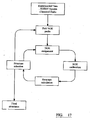
- the current best-practice in molecular modelling from experimental data that includes ambiguous observables is to adopt an iterative, or cyclic, scheme that aims to gradually resolve the experimental ambiguities with each cycle ( Figure 15 ).
- Initial models are generated that satisfy the unambiguous experimental observables. These preliminary models are used to evaluate the possible assignments of the remaining observables with the aim of resolving their ambiguity.
- the newly-resolved assignments are combined with the previously unambiguous observables in the subsequent cycle of molecular model generation and reassignment.
- the iterative scheme continues until all ambiguous experimental observables have been resolved and the final molecular model is produced.
- An iterative assignment scheme for resolving ambiguity in experimental observations is shown in Figure 15 .
- Colonies of social insects can perform complex tasks (e.g. foraging, cooperative hunting and nest building) despite the simplicity of the individuals. While walking to/from a food source ants deposit pheromones on the ground which attracts other ants through a process known as "stigmergy". In time, a pheromone trail will mark the optimal route from nest to food. Such "swarm intelligence" has been used in mathematical algorithms that can potentially solve complex combinatorial optimization problems.
- Nuclei with an odd mass or odd atomic number have nuclear spin (in a similar fashion to the spin of electrons); this includes 1 H and 13 C (but not 12 C) and 15 N.
- the spins of nuclei are sufficiently different that techniques, for example NMR experiments can be sensitive for only one particular isotope of one particular element by selective excitation on a multichannel NMR spectrometer.
- the NMR behaviour of 1 H, 13 C and 15 N nuclei are typically exploited by Biochemists, being the typical constituents of proteins, which by application of suitable pulse sequences can be used to deduce their structure.
- the precise resonant frequency of the energy transition is dependent on the effective magnetic field at the nucleus. This field is affected by electron shielding which is in turn dependent on the chemical environment. As a result, information about the chemical environment of a nucleus can be derived from it resonant frequency. In general, the more the nucleus is electronegative, the higher the resonant frequency. Other factors such as ring currents (anisotropy) and bond strain affect the frequency shift, The chemical shift is defined with respect to the resonant frequency of a reference compound, for example tetramethylsilane (TMS). In this case the chemical shift of TMS would be defined to be zero. This scale is useful practically because it is independent of magnetic field strength.
- ⁇ ppm 10 6 ⁇ ⁇ - ⁇ ref / ⁇ ref
- the NMR technique is particularly useful because it permits the through-space distances between nuclei to be measured. This is possible because of dipolar spin-spin coupling between nuclei.
- the dipolar coupling is dependent on the distance between the nuclei (1/r 6 ) and their relative orientation to the magnetic field (1-cos 2 ⁇ ).
- NMK (as applied here) the angular component averages to zero and hence dipolar couplings are not observed directly in NMR spectra. They do, however, affect the relaxation rates of nuclei. Distance information is therefore extracted from relaxation experiments, which take advantage of phenomena such as the nuclear Overhauser enhancement.
- the nuclear Overhauser enhancement is related to the distance between the nuclei and hence can be used as a method for measuring nuclei.
- NOESY NOE S pectroscop Y
- the NOESY experiment is typically applied. In its most basic incarnation it is a 2D experiment that correlates all protons.
- magnetisation is transferred between nuclei that are closely separated in space.
- the cross-relaxation rate can be shown to be exactly proportional to 1/r 6 .
- the intensities for a particular mixing time are easy to calculate. However, this is not true in a protein, where the density of protons can be high. In general this problem has to be solved by taking into account all possible proton pathways - the so called full relaxation matrix approach. Each possible interaction is represented by a cross-relaxation rate in a 2D matrix, and the intensities are computed using linear algebra approaches. This is computationally intensive and complicated. However, even in a protein, if the mixing time is kept short, then the NOESY cross-peak intensity is approximately proportional to 1/r6.
- the donor and acceptor protons in an NOE are identified from the chemical shifts of the NOESY crosspeak.
- the donor and acceptor protons can be assigned unambiguously.
- the NOE is said to be ambiguous. In order to resolve this ambiguity, we need to know the three-dimensional structure, but in order to determine the three-dimensional structure we need to assign all the NOEs. This dilemma is known as the NOE assignment problem and is illustrated in Figure 16 .
- the unambiguous NOEs are calibrated into distance restraints in order to calculate a preliminary set of structures, a fraction of which are selected for NOE reassignment. Because only a subset of the possible experimental observations is used, these initial conformers have a very low level of precision. However, they may be of sufficient accuracy to discriminate between the assignment possibilities of a subset of the ambiguous NOEs. Therefore, the list of assigned NOEs is expanded and the fraction of unused experimental observations decreases. Eventually, after multiple cycles of reassignment, re-calibration and re-calculation the procedure yields a solution structure that is compatible with the crosspeaks picked from the NOESY data.
- the multi-step iterative assignment procedure has flaws at every stage.
- the peak-picking can be highly erroneous and subjective, particularly in the case of identifying the chemical shifts, and measuring the intensities/volumes of overlapping NOESY crosspeaks.
- Another disadvantage is that mistakes made in the assignment of the initial set of unambiguous NOEs are propagated through every subsequent cycle, leading to consequent mis-assignments and mis-calibrations and resulting in an erroneous solution structure.
- An iterative assignment also performs a restraint calibration on a limited subset of NOEs rather than the whole spectrum leading to further subjectivity. Furthermore, the poor definition of the initial set of biomolecular structures requires that many conformers must be calculated for a statistically meaningful analysis of the ambiguous NOEs and the selection of structures is prone to subjectivity and bias. In addition, the iterative assignment is time consuming, and is highly wasteful in that the number of structure calculations performed far exceeds the number of conformers required to define the solution structure.
- the method includes creating in a computer a swarm of molecular structure generators, each having the same covalent structure as the biomolecule whose three-dimensional conformation is to be determined, but with differing initial conformations (i.e step (a) defined in claims 1 and 4, respectively).
- the molecular structure generators then explore possible conformations of the biomolecule subject to restraints which are derived from experimentally obtained NMR spectra.
- the present invention seeks to alleviate the problems associated with the prior art by utilizing the principle of swarm intelligence to achieve determination of bio-molecular structures from ambiguous experimental observables.
- a computational method of determining a three-dimensional conformation of a biomolecule is defined by the features set out in claim 1.
- an apparatus adapted to determine a three-dimensional conformation of a biomolecule is defined by the features set out in claim 4.
- the biomolecules are able to communicate via a common restraint list.
- Any single conformer which encounters a favourable sub-structure, for example, a native-like ⁇ -helical turn or ⁇ -hairpin in proteins, may encourage the other members of the swarm to adopt this conformation by updating the molecular restraints. Therefore, the molecules cooperate in finding the optimal restraints and thus determine their own molecular structure.
- the present invention offers particular advantageous over previously used techniques such as iterative assignment. Firstly, there need be no peak picking of the NOESY spectra which is highly speculative and, therefore, no problems associated with overlapped and/or aliased crosspeaks. Furthermore, there need be no calibration of interproton distance restraints. Instead, those distances that give rise to "good” spectral solutions are remembered. Interproton distance restraints are transient and modified on-the-fly during the course of the swarm evolution, so assignment errors are neither propagated nor amplified. The technique is also objective: all possible donor-acceptor NOE pars may be considered from the outset and there need be no selection of conformers.
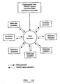
- FIG 16 a swarm intelligence schema is illustrated for resolving ambiguity in experimental observations.
- the swarm of molecular structure generators are able to communicate via a global set of molecular restraints derived from the observables and determine the molecular structure in a cooperative manner.
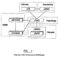
- NMRSwarm can be split into four approximatelyautonomous modules: Geometry, Library, Topology, Swarm and NMR. These provide the C++ classes for the program, the building blocks upon which the architecture is constructed. Two additional modules, Utility and Molecule perform supplementary roles.
- the Utility module provides numerous base classes that are inherited by many other classes in the other Modules.
- the Molecule module provides a family of classes that are the bases for the construction of the Library, Topology and Swarm objects.
- the key interactions between objects in the NMRSwarm program are depicted in Figure 1 .
- the Swarm itself consists of an array of molecular Agents, each of which is cloned from a Topology template (:Topo) which is turn is constructed from a Library (:Libr) of residues and linkers and a database of geometric parameters (:Geom).
- Topology also provides atomic information to the NMR module for the construction of resonances and spectral crosspeaks which are accesed by the Swarm during backcalculation of NMR spectra.
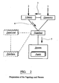
- the first stage in the swarm intelligence structure calculation process in NMRSwarm is the preparation of one or more segments in the molecular topology ( Figure 2 ). This is performed by "polymerization" of Library residues (:LibrResi) using Library links (:LibrLink) according to a user-defined sequence.
- the Library residues and patches must first be generated from templates input from file or the graphical user interface (GUI), and suitable Geometry data.
- GUI graphical user interface
- the completed Topology has all the information required to clone Agents and to produce NMR resonance lists for each set of the NMR conditions. It also contains a master NOE array for management of the NOE restraints during the swarm intelligence structure calculation process.
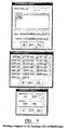
- NmrCond An NMR condtions object (:NmrCond) is constructed for each set of conditions (field strength, pH, ionic strength, etc.) under which NMR spectra were recorded on the specified Topology.
- the NmrCond object shown in Figure 3 automatically generates a complete (but unassigned) list of NMR resonances (:NmrReso) for the Topology which may be edited by the user via the GUI or modified by loading of previously stored resonance information from file.
- NOESY NMR spectra can then be loaded into each set of NMR conditions.
- NMRSwarm automatically generates all possible NOE crosspeaks (:NmrXpk) and links these to the NOE array in the Topology.
- Subsequent alterations to the NMR resonances result in spontaneous changes in the magnitude, shape and/or positions of the NOE crosspeaks in the spectra. Regions of the NMR spectra which contain artefacts may be omitted from analysis through the application of masks (:NmrMask).
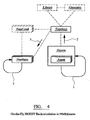
- the swarm intelligence structure calculation method in NMRSwarm is summarized in Figure 4 .
- the Swarm of molecular Agents are undergoing restrained molecular dynamics simulations starting from randomized conformations but all governed by a global NOE restraint list (in the: Topo).
- the current inter-proton resonance distances at any point in time for one randomly-chosen Agent are copied into a "test slot" of the NOE array. From here they are used to back-calculate hypothetical NOES spectra that would be produced if the current conformation was the correct one.
- the goodness-of-fit between the back-calculated and experimental NOESY crosspeaks is evaluated from a ⁇ 2 test of the intensities. Those test interproton resonance distances that resulted in an improvement in the ⁇ 2 of the crosspeaks are recorded and used to update the NOE restraints in the global NOE list. The new NOE distance restraints are passed back to all Agents in the Swarm via the Topology, and the restrained molecular dynamics simulations continue. In this way, the NOE distances in those regions of the conformation of the tested Agent that agree well with the experiment data are communicated to all other Agents in the Swarm. The backcalculation-and-update cycle is then repeated for each of the other Agents.
- a biomolecular swarm is defined as a colony of molecular agents that are searching their conformational (interproton distance) space through molecular generators, which, in this case, are parallel restrained molecular dynamics simulations.
- Each of these agents back-calculates simulated NOES data from its current conformation, and, depending on its degree of agreement with the experimental NOESY data, updates a global interproton distance list that is then applied as restraints in the molecular generators.
- the agents communicate by stigmergy, mutually updating and reacting to the restraint list.
- NOE restraint list will evolve to the correct set of interproton distances that describes the conformation of the molecule.
- the swarm of molecular agents is then attracted by this set of restraints towards the correct three-dimensional structure.
- GUI graphical user interface
- Figure 6 shows the GUI comprising six separate interfaces allowing complete observation of, interaction with and intervention in the entire NMRSwarm system, i.e. the Geometry, Library, Topology, NMR Conditions, NMR Spectrum and Swarm modules.
- the Geometry multiple document interface of NMRSwarm allows the user to create, edit, load, save and delete Geometry files.
- Each document has multiple window tabs which provide access to the Geometry atoms, bonds, angles, dihedrals and impropers, which is shown in Figure 7 .
- Clicking on any of the Geometry Atoms updates the Atom attributes editor sliders at the bottom of the table allowing the user to modify these values.
- Clicking on any of the Geometry constraints updates the equilibrium distance and force constant (and periodicity in the case of dihedrals) editor sliders at the bottom of the table and allows the user to manually edit these values for the selected constraint.
- the Library multiple document interface of NMRSwarm shown in Figure 8 allows the user to load, save and delete Library files.
- Each document has an OpenGL-based molecular viewer, which presents the Library Residues in any of a number of viewing modes (lines, ball-and-stick, space-fill or tubes) and multiple window tabs, which provide access to information on the Library Residues, Patches and Links.
- Topology multiple document interface of NMRSwarm allows the user to create, edit, load, save and delete Topology files.
- the topologies are written to files in an XML (extensible markup language) format.
- Topology segments are built from sequences of library residues using a single-letter code and the appropriate Library and Geometry document files ( Figure 9 ). Each segment is given a four-letter name similar to the accession code of a Protein Data Bank (PDB) entry.
- PDB Protein Data Bank
- Inter-residue Library links are selected for each pair of residues, for example a trans peptide bond (PEPT) or a cisproline peptide cond (PPCP) in a polypeptide chain.
- PEPT trans peptide bond
- PPCP cisproline peptide cond
- the first and last residues may be modified using a Library patch, e.g. with primary amine (NTAM) and carboxylate (CTER) groups for the N- and Ctermini of a polypeptide.
- NTAM primary amine
- CTER carboxylate
- the resultant topology segment may be examined using the OpenGLbased molecular viewer of NMRSwarm which presents the topology in any of a number of viewing modes (lines, ball-and-stick, spacefill or tubes).
- the Topology GUI also presents a list view tree of the segment structure together with tabulated lists of its geometric constraints.
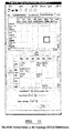
- the icosapeptide segment AVTQTYGGNSNGEPCVLPFT is shown in the segment viewer in the Topology GUI of NMRSwarm.
- the tabulated data in the Topology GUI is a cascading interproton NOE viewer/editor.
- the contents of the NOE table are updates in real time to allow for monitoring of the progress of the NOE assignment process.
- the Swarm multi-document GUI of NMRSwarm features an OpenGL viewer, which shows a real-time display of the molecular dynamics simulations and allows for independent rotation, translation and selection of the molecular agents. Pull-down menus allow the user to start and stop the molecular dynamics and NOESY back-calculation loops (i.e. the swarm intelligence calculations), and to load or save coordinate files in a PDB format.
- Figure 12 shows a nine-agent swarm of the icosapeptide AVTQTYGGNSNGEPCVLPFT in the "tube" viewing style.
- the agents have undergone 25ps of swarm intelligence based structure determination coupled to a two-dimensional 1H-1H NOESY spectrum.
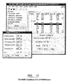
- NMR Conditions multi-document GUI of NMRSwarm shown in Figure 13 allows the user to create, edit, load, save and delete NMR conditions. Conditions are loaded from file or generated ab initio using a pre-existing Topology. A pop-up menu allows the user to edit the field strength, pH and temperature of the conditions, whilst an interactive list view tree provides a convenient means of modifying the chemical shifts, linewidths and intensities of selected resonances.
- the NMR Spectrum multi-document GUI of NMRSwarm allows the user to load NMR spectra, couple them to a topology and view the progress of the swarm calculation in real time from the level of agreement between the experimental and back-calculated or restraint intensities.
- NMRSwarm offers a three-dimensional perspective view of the spectra in addition to the orthographic projection.
- the user may zoom, rotate and pan through the spectrum and adjust the contour level, the contour level multiplier and the number of contours. This allows to user to easily identify correctly assigned NOEs as well as those that are over- or under-constrained. In addition, errors in resonance chemical shift and/or linewidth may be seen.
- a region of a 2D NOESY spectrum is shown is perspective mode with experimental NOE crosspeaks shown in green and NOE restraint crosspeaks in red. Where crosspeaks appear yellow, such as that marked with an asterisk (*), there is an excellent agreement between the experimental data and NOE restraint distance.
- a molecular topology which defines the covalent structure of the molecule, was generated for each protein using the program CNS version 1.0 (Brünger et al., 1998) and the residue templates described in the file "topallhdg.pro". The molecular topology is written to an ASCII file in a STAR format.
- cysteine residues were oxidised (by removing the ⁇ -sulphydryl protons) but no bond or bond angle constraints were applied between the sulphur atoms.
- the STAR format molecular topology files generated by CNS are read directly into the NMRSwarm program.
- Each molecular agent object contains coordinates, velocities and accelerations for each atom in the molecule (as described by the molecular topology), together with kinetic and potential energy variables.
- Each molecular resonance object comprises a chemical shift ( ⁇ ), linewidth ( ⁇ ) and occupancy ( ⁇ ) for a given nucleus (or group of magnetically-identical nuclei) in the molecule for a given set of conditions (temperature, pH, ionic strength, field strength, etc.).
- the chemical shift and linewidth of each molecular resonance is measured from well-separated, high-intensity crosspeaks in the NOESY spectra.
- the chemical shifts and/or linewidths are estimated from analysis of chemically-equivalent resonances in better resolved regions of the spectra.
- chemical shifts are documented in parts per million (ppm) to 3 decimal places, and linewidths to the nearest Hz.
- the fractional occupancy ( ⁇ ) of a given resonance in a sample is the proportion of atoms that contain an NMR-active nucleus.
- ⁇ fractional occupancy
- the constant ⁇ determines the degree of coverage of the Gaussian resonance; a value of 3.0 encompasses 99.7% of the total intensity.
- Molecular resonances are read into and written from NMRSwarm in a STAR file format.
- NOESY spectral data are imported into NMRSwarm in either FELIX or NMRView format.
- the spectrometer frequency ( ⁇ ), spectral width ( sw ), reference shift and reference point of each spectral dimension are retrieved from the header of the NOESY file.
- the field strength ( ⁇ ) is supplied by the user.
- the spectral intensities are read into memory in a serial format, decomposing the submatrix format in the FELIX and NMRView files.
- any molecular resonance that is observable along a particular dimension (according to the gyromagnetic ratio, ⁇ ) but is outside the spectral width is aliased.
- Aliased resonances have ⁇ values greater than 1.0.
- ⁇ d (p) is the chemical shift of pixel p in dimension d (taking any resonance aliasing into account)
- ⁇ is the degree of intensity coverage in any dimension
- P S is the set of all pixels in the NOESY spectrum.
- the degree of ambiguity of a NOESY crosspeak x is one determinant of the force constant applied to the NOE restraint during restrained molecular dynamics.
- X S is the set of all crosspeaks in the NOESY spectrum.
- ISPA isolated spin pair approximation
- E potential the potential energy function
- a harmonic approximation is used to account for deformations in bond length, bond angles, chirality and planarity in the molecular structure.
- the non-bonded energy term, E vdw is a soft, purely repulsive function without attractive or electrostatic components. Interactions between bonded atoms, and those between atoms that are bonded to a common third atom are excluded from E vdw . Interactions between atoms that are connected through three covalent bonds (so called 1-4 non-bonded interactions) are computed using modified equilibrium distances. Non-bonded energy terms are truncated at an interatomic distance r i for atom pairs that are too close to each other to minimize numerical instabilities in the case of strained initial coordinates or close contacts during high simulation temperatures.
- the weighting on the non-bonded energy term, w vdw is regulated during the structure calculation to allow or inhibit close atom contacts.
- k NOE is the individual NOE force constant
- r l is the lower bound on the flat well of the potential
- r u is the upper bound
- r b is a barrier above which a linear asymptote is applied.
- the pseudo-harmonic E NOE potential with a high-end asymptote is shown in Figure 19 . This asymptote reduces numerical instabilities for those interproton distances that deviate dramatically from the upper bound.
- k NOE , r l and r u are specific to each NOE and are determined on-the-fly during the molecular structure calculation.
- Restrained molecular dynamics simulations have a much larger radius of convergence than techniques involving distance geometry and conjugate-gradient minimization because, due to the kinetic energy of the system, they are able to overcome the barriers surrounding local potential energy minima. This ability may be enhanced by elevating the temperature (thus increasing the kinetic energy) and by weakening the van der Waals repulsion (thus diminishing the energy barriers) in a procedure known as "simulated annealing". In this procedure the system is driven towards the global potential energy minimum by thermal coupling to a "heat bath”: increased kinetic energy, resulting from random conformational changes which reduce the potential energy, is dissipated to the bath making the reverse conformational transition very unlikely. Following the high temperature dynamics, the system is cooled and the van der Waals repulsion restored, effectively trapping the system in the global potential energy minimum.
- Each NOE can give rise to one or more crosspeaks in multiple NOESY spectra. Therefore, NOE restraints are constructed from multiple contributions, each arising from a separate crosspeak.
- Each crosspeak contribution has its own ambiguity factor ( ⁇ x ) which depends on the degree of spectral overlap, its own equilibrium distance ( r 0 ) that best fits the experimental data, and its own force constant ( k NOE ) that reflects the degree of fit to the experimental data.
- X N is the set of crosspeaks that arise from NOE n .
- the upper and lower bounds on the equilibrium distance, r u and r l are set at a fixed percentage above and below r 0 , respectively.
- Any crosspeak which shows an improved fit (i.e. lower ⁇ 2 ) to the experimental data has its associated equilibrium distance ( r 0 ) updated to the current (r -6 summed) interproton distance.
- the new ⁇ 2 value (which is set to infinity initially) is memorised as a record of the quality of the fit.
- NOE restraints with an equilibrium distance ( r 0 ) less than the barrier ( r b ) are combined.
- the NOEs are sorted with respect to r 0 and neighbouring pairs in the list are pooled into combined restraints.
- n - 1 combined NOE restraints are generated from n individual NOEs.
- Each combined NOE restraint is applied in the molecular dynamics as an ambiguous restraint with an r - 6 summed distance.
- Distinct but non-stereospecifically assigned prochiral resonances are arbitrarily assigned to either the pro- R or pro- S moiety in the assignment lists.
- the stereoassignments of a constant proportion (typically 10%) of randomly chosen prochiral groups were swapped for each agent. Only those exchanges that result in an improved fit to the experimental data (as judged by the sum of ⁇ 2 differences for all related NOESY crosspeaks) are accepted. The best prochiral assignments are applied to the NOE restraints during the molecular dynamics.
- the quality of the experimental restraints is determined from the correlation coefficient between a back-calculated restraints NOESY spectrum (a spectrum that would be produced if all interproton distances matched those of the restraints) and the experimental data.
- the level of agreement between the experimental restraints and the molecular structures is determined by the root mean square deviation (rmsd) from the equilibrium interproton distances.
- the stereochemical quality of the final structures was determined by ramachandran analysis using the program PROCHECK-NMR.
- Deuterium oxide was supplied by CK Gas Products Ltd. (Wokingham, UK). Miili-Q water was supplied by Millipore (Watford, UK). All other chemical reagents were supplied by Sigma (Poole, UK).
- N-labelled FERM3 module corresponding to residues 309-405 of mature mouse talin containing the point mutation C336S, was produced from Escherichia coli in M9 minimal media by Dr.Kate Wegener (Oxford University) as previously described (de Pereda JM et al., 2005).
- Samples for NMR spectroscopy were dissolved to a concentration of 1.5mM in either 5% D 2 O/95% H 2 O, or 99.96% D 2 O. The samples were buffered with 50mM potassium phosphate to pH 6.1 (pH meter uncorrected for deuterium) and contained 100mM NaCl.
- NOESY data was performed using the program NMRPipe (Delaglio et al., 1995). All 1 H dimensions were processed with Lorentzian to Gaussian transformations to improve the resonance lineshape and enhance resolution (although other lineshapes and window functions could be used equally well). The 1 H dimensions of the 2D spectra were zero-filled to 1024 complex points to improve the digital resolution and then Fourier transformed and phased. The 1 H dimensions of the 3D NOESY spectra were processed in the same way except that zero-filling was performed to 512 complex points only. The 3D NOESY 15 N dimensions were doubled in acquisition time through the use of linear prediction followed by the application of a Lorentzian-Gaussian window function, Fourier transformation and phasing.
- the 1 F2 module solution structure was determined from a 20-agent swarm intelligence biomolecular structure calculation staring from randomised backbone conformations.
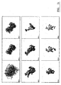
- Figure 21 shows the evolution of the fibronectin 1 F2 module swarm.
- the snapshots are from the 400ps 20-agent swarm intelligence structure determination of the human fibronectin 1 F2 module with the ⁇ -strands coloured cyan and the ⁇ -helices red.
- the molecules have been rotated with the same transformation that best overlays the backbone heavy atoms (N, C ⁇ and C) at 400ps.
- FIG. 22 a comparison between experimental and back-calculated 1 F2 NOESY data is shown.
- Fig.22(a) an excerpt from the experimental 50ms D 2 O NOESY spectrum of 1 F2 showing many of the aromatic (x-axis) to aliphatic (y-axis) NOEs that characterize the module fold and in Fig. 22(b) a NOESY spectrum back-calculated using an isolated spin pair r 0 -6 summation from the applied NOE restraints at time 400ps.
- 13 of the agents are representative of the 1 F2 module fold on the basis of their low potential energy. These agents are in close agreement with the previously-determined NMR structure of 1 F2 (Pickford et al., 1997) as shown by their backbone heavy atom (N, C ⁇ and C) root mean square deviation (rmsd) of 0.91 ⁇ .
- FIG. 23 a comparison of the Swarm Intelligence NMR structure of 1 F2 with that derived previously from solution NMR data is made.
- Fig. 23(a) an overlay of the selected 1 F2 molecular agents at the end of the swarm intelligence simulated annealing is shown. The average coordinates from the agents are shown as a ribbon representation in Fig 23(b).
- Fig 23(c) shows the solution NMR structure of the 1 F2 module as determined previously using the traditional iterative assignment scheme (Pickford et al ., 1997).
- the FERM3 module from mouse talin was determined from a 20-agent swarm intelligence biomolecular structure calculation starting from randomised backbone conformations.
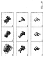
- Figure 24 the Evolution of the FERM3 swarm can be appreciated. It shows snapshots from the 400ps 20-agent swarm intelligence structure determination of the FERM3 module from mouse talin with the ⁇ -strands coloured cyan and the ⁇ -helices red. At each time point, the molecules have been rotated with the same transformation that best overlays the backbone heavy atoms (N, C ⁇ and C) at 400ps.
- 16 of the agents were selected as representative of the FERM3 module fold on the basis of their low potential energy. These agents are in close agreement with the coordinates of the FERM3 module in the previously-determined crystal structure of the FERM3-PIPKI ⁇ (de Pereda et al ., 2005) as shown by their backbone heavy atom (N, C ⁇ and C) root mean square deviation (rmsd) of 1.04 ⁇ .
- Fig 25 a comparison of the Swarm Intelligence NMR structure of FERM3 with that derived from X-ray crystallography is shown.
- Fig 25(a) depicts the overlay of FERM3 molecular agents at the end of the swarm intelligence simulated annealing.
- Fig 25(b) depicts the average coordinates from the selected agents are shown as a ribbon representation.
- Fig 25 (c) The crystal structure of the FERM3 module (de Pereda et al ., 2005).
Landscapes
- Physics & Mathematics (AREA)
- Health & Medical Sciences (AREA)
- Life Sciences & Earth Sciences (AREA)
- General Health & Medical Sciences (AREA)
- Molecular Biology (AREA)
- Spectroscopy & Molecular Physics (AREA)
- High Energy & Nuclear Physics (AREA)
- Condensed Matter Physics & Semiconductors (AREA)
- General Physics & Mathematics (AREA)
- Management, Administration, Business Operations System, And Electronic Commerce (AREA)
- Magnetic Resonance Imaging Apparatus (AREA)
Claims (5)
- Berechnungsverfahren zur Bestimmung einer dreidimensionalen Konformation eines Biomoleküls aus experimentellen Daten der Kern-Overhauser-Effekt-Spektroskopie, NOESY, die mehrdeutige NOE-Kreuzsignale aufweisen, welche zwei oder mehr Protonenresonanzen zugeordnet werden können, wobei das Verfahren Folgendes umfasst:(a) Erzeugen von einem Schwarm molekularer Strukturgeneratoren in einem Computer, von denen jeder dieselbe kovalente Struktur wie das Biomolekül aufweist, allerdings mit einer randomisierten Anfangskonformation;(b) Durchführen einer eingeschränkten Molekulardynamik-Simulation, wodurch die molekularen Strukturgeneratoren veranlasst werden, mögliche Konformationen von dem Biomolekül zu erkunden, welche den Einschränkungen einer gemeinsamen NOE-Restraint-Liste von Interproton-Distanzen unterliegen, welche die Konformation von dem Biomolekül beschreibt, wobei die Restraint-Liste aus den experimentellen NOESY-Daten abgeleitet und allen molekularen Strukturgeneratoren gemeinsam ist;(c) für jeden molekularen Strukturgenerator in dem Schwarm:(i) Rückrechnen eines hypothetischen NOESY-Spektrums aus der gegenwärtigen Konformation von dem molekularen Strukturgenerator;(ii) Normalisieren des rückgerechneten NOESY-Spektrums gegen die experimentellen NOESY-Daten durch Berechnung von Korrelationskoeffizienten über die nicht-diagonalen Spektralintensitäten;(iii)Berechnung einer Anpassungsgüte zwischen dem rückgerechneten Kreuzsignal und dem entsprechenden Kreuzsignal in den experimentellen NOESY-Daten für jedes der Kreuzsignale von dem rückgerechneten NOESY-Spektrum;(iv) Aktualisieren der NOE-Restraint-Liste mit denjenigen Interproton-Distanzen, welche im Vergleich zu einem gespeicherten Datensatz der Anpassungsgüte zu einer verbesserten Anpassungsgüte führten; und(v) Herabsetzen der gespeicherten Anpassungsgüte für den molekularen Strukturgenerator; und(d) Wiederholung der Schritte (b) und (c), wobei für die erste Wiederholung der Schritte (b) und (c) der Vergleich in Schritt (c)(iv) in Bezug auf einen gespeicherten Datensatz der Anpassungsgüte durchgeführt wird, die auf Unendlich gesetzt wird, und, für die weiteren Wiederholungen der Schritte (b) und (c), der Vergleich in Schritt (c)(iv) in Bezug auf den herabgesetzten gespeicherten Datensatz der Anpassungsgüte der vorhergehenden Wiederholung gemacht wird, wobei die molekularen Strukturgeneratoren kooperieren, um die optimalen Einschränkungen zu finden und auf diese Weise die dreidimensionale Konformation zu bestimmen.
- Verfahren gemäß Anspruch 1, in dem die randomisierte Anfangskonformation zufällige Atomkoordinaten und Torsionswinkel aufweist.
- Computerprogramm, welches, wenn es auf einem Computer läuft, die dreidimensionale Konformation eines Biomoleküls unter Verwendung des Verfahrens von Anspruch 1 bestimmt.
- Apparatur, die eingerichtet ist zur Bestimmung einer dreidimensionalen Konformation eines Biomoleküls aus experimentellen Daten der Kern-Overhauser-Effekt-Spektroskopie, NOESY, die mehrdeutige NOE-Kreuzsignale aufweisen, welche zwei oder mehr Protonenresonanzen zugeordnet werden können, wobei das Verfahren Folgendes umfasst:(a) Mittel zum Erzeugen von einem Schwarm molekularer Strukturgeneratoren in einem Computer, von denen jeder dieselbe kovalente Struktur wie das Biomolekül aufweist, allerdings mit einer randomisierten Anfangskonformation;(b) Mittel zum Durchführen einer eingeschränkten Molekulardynamik-Simulation, wodurch die molekularen Strukturgeneratoren veranlasst werden, mögliche Konformationen von dem Biomolekül zu erkunden, welche den Einschränkungen einer gemeinsamen NOE-Restraint-Liste von Interproton-Distanzen unterliegen, welche die Konformation von dem Biomolekül beschreibt, wobei die Restraint-Liste aus den experimentellen NOESY-Daten abgeleitet und allen molekularen Strukturgeneratoren gemeinsam ist;(c) Mittel zur Durchführung der folgenden Schritte für jeden molekularen Strukturgenerator in dem Schwarm:(i) Rückrechnen eines hypothetischen NOESY-Spektrums aus der gegenwärtigen Konformation von dem molekularen Strukturgenerator;(ii) Normalisieren des rückgerechneten NOESY-Spektrums gegen die experimentellen NOESY-Daten durch Berechnung von Korrelationskoeffizienten über die nicht-diagonalen Spektralintensitäten;(iii)Berechnung einer Anpassungsgüte zwischen dem rückgerechneten Kreuzsignal und dem entsprechenden Kreuzsignal in den experimentellen NOESY-Daten für jedes der Kreuzsignale von dem rückgerechneten NOESY-Spektrum;(iv) Aktualisieren der NOE-Restraint-Liste mit denjenigen Interproton-Distanzen, welche im Vergleich zu einem gespeicherten Datensatz der Anpassungsgüte zu einer verbesserten Anpassungsgüte führten; und(v) Herabsetzen der gespeicherten Anpassungsgüte für den molekularen Strukturgenerator; und(d) Wiederholung der Schritte (b) und (c), wobei für die erste Wiederholung der Schritte (b) und (c) der Vergleich in Schritt (c)(iv) in Bezug auf einen gespeicherten Datensatz der Anpassungsgüte durchgeführt wird, die auf Unendlich gesetzt ist, und, für die weiteren Wiederholungen der Schritte (b) und (c), der Vergleich in Schritt (c)(iv) in Bezug auf den herabgesetzten gespeicherten Datensatz der Anpassungsgüte der vorhergehenden Wiederholung gemacht wird, wobei die molekularen Strukturgeneratoren kooperieren, um die optimalen Einschränkungen zu finden und auf diese Weise die dreidimensionale Konformation bestimmen.
- Apparatur gemäß Anspruch 4, in der die randomisierte Anfangskonformation zufällige Atomkoordinaten und Torsionswinkel aufweist.
Applications Claiming Priority (2)
| Application Number | Priority Date | Filing Date | Title |
|---|---|---|---|
| GBGB0517349.7A GB0517349D0 (en) | 2005-08-24 | 2005-08-24 | Biomolecular structure determination |
| PCT/GB2006/003169 WO2007023289A1 (en) | 2005-08-24 | 2006-08-24 | Biomolecular structure determination involving swarm intelligence |
Publications (2)
| Publication Number | Publication Date |
|---|---|
| EP1929322A1 EP1929322A1 (de) | 2008-06-11 |
| EP1929322B1 true EP1929322B1 (de) | 2013-07-17 |
Family
ID=35198352
Family Applications (1)
| Application Number | Title | Priority Date | Filing Date |
|---|---|---|---|
| EP06779200.2A Not-in-force EP1929322B1 (de) | 2005-08-24 | 2006-08-24 | Biomolekulare strukturbestimmung unter anwendung von swarm-intelligenz |
Country Status (5)
| Country | Link |
|---|---|
| US (1) | US20080221848A1 (de) |
| EP (1) | EP1929322B1 (de) |
| JP (1) | JP5148493B2 (de) |
| GB (1) | GB0517349D0 (de) |
| WO (1) | WO2007023289A1 (de) |
Families Citing this family (5)
| Publication number | Priority date | Publication date | Assignee | Title |
|---|---|---|---|---|
| US7849024B2 (en) * | 2006-08-16 | 2010-12-07 | Drvision Technologies Llc | Imaging system for producing recipes using an integrated human-computer interface (HCI) for image recognition, and learning algorithms |
| EP2161587A1 (de) | 2008-09-03 | 2010-03-10 | Bruker BioSpin AG | Verfahren und System zur automatischen Analyse von NMR-Spektren |
| CN111126549A (zh) * | 2019-12-24 | 2020-05-08 | 昆明理工大学 | 一种基于策略改进樽海鞘智能算法的双星光谱拟合方法 |
| CN112096693B (zh) * | 2020-08-04 | 2022-09-20 | 深圳市行健自动化股份有限公司 | 水下生产液压控制系统在线诊断与校验方法、存储介质和控制终端 |
| CN114521024B (zh) * | 2022-02-23 | 2025-01-03 | 江南大学 | 无线网络中多用户信道的认知分配方法 |
Family Cites Families (5)
| Publication number | Priority date | Publication date | Assignee | Title |
|---|---|---|---|---|
| DE10144661C2 (de) * | 2001-09-11 | 2003-08-14 | Forschungsverbund Berlin Ev | Vorrichtung und Verfahren zur Zuordnung der NMR-Signale von Polypeptiden |
| JP3607948B2 (ja) * | 2001-10-31 | 2005-01-05 | 国立大学法人 北海道大学 | Nmrシグナル帰属支援システム |
| JP2003294819A (ja) * | 2002-03-29 | 2003-10-15 | Yoshifumi Nishimura | 多重複合体の化学シフトを同定する方法、多重複合体の立体構造を解析する方法、多重複合体のモデルを構築する方法、新規核酸分子及び試薬 |
| AU2002345287A1 (en) * | 2002-07-10 | 2004-02-02 | Institut Suisse De Bioinformatique | Peptide and protein identification method |
| US7904252B2 (en) * | 2003-11-28 | 2011-03-08 | Hitachi, Ltd. | Method for measuring a structural change in a protein |
-
2005
- 2005-08-24 GB GBGB0517349.7A patent/GB0517349D0/en not_active Ceased
-
2006
- 2006-08-24 US US12/064,734 patent/US20080221848A1/en not_active Abandoned
- 2006-08-24 EP EP06779200.2A patent/EP1929322B1/de not_active Not-in-force
- 2006-08-24 JP JP2008527512A patent/JP5148493B2/ja not_active Expired - Fee Related
- 2006-08-24 WO PCT/GB2006/003169 patent/WO2007023289A1/en not_active Ceased
Also Published As
| Publication number | Publication date |
|---|---|
| GB0517349D0 (en) | 2005-10-05 |
| WO2007023289A1 (en) | 2007-03-01 |
| EP1929322A1 (de) | 2008-06-11 |
| JP2009506313A (ja) | 2009-02-12 |
| US20080221848A1 (en) | 2008-09-11 |
| JP5148493B2 (ja) | 2013-02-20 |
Similar Documents
| Publication | Publication Date | Title |
|---|---|---|
| Baran et al. | Automated analysis of protein NMR assignments and structures | |
| Tian et al. | A dipolar coupling based strategy for simultaneous resonance assignment and structure determination of protein backbones | |
| Alexander et al. | De novo high-resolution protein structure determination from sparse spin-labeling EPR data | |
| Güntert | Automated NMR structure calculation with CYANA | |
| Bouvignies et al. | Solution structure of a minor and transiently formed state of a T4 lysozyme mutant | |
| Schwalbe et al. | A refined solution structure of hen lysozyme determined using residual dipolar coupling data | |
| Marion | An introduction to biological NMR spectroscopy | |
| Prestegard et al. | Nuclear magnetic resonance in the era of structural genomics | |
| Mantsyzov et al. | A maximum entropy approach to the study of residue‐specific backbone angle distributions in α‐synuclein, an intrinsically disordered protein | |
| Bouvignies et al. | Visualizing side chains of invisible protein conformers by solution NMR | |
| Mollova et al. | Global structure of RNA determined with residual dipolar couplings | |
| Rinaldelli et al. | FANTEN: a new web-based interface for the analysis of magnetic anisotropy-induced NMR data | |
| Wylie et al. | Determinations of 15N chemical shift anisotropy magnitudes in a uniformly 15N, 13C-labeled microcrystalline protein by three-dimensional magic-angle spinning nuclear magnetic resonance spectroscopy | |
| US7920972B2 (en) | G-matrix Fourier transformation (GFT) nuclear magnetic resonance (NMR) experiments for resonance assignment and structure determination of organic molecules | |
| Shin et al. | Structural proteomics by NMR spectroscopy | |
| Zheng et al. | Automated protein fold determination using a minimal NMR constraint strategy | |
| Schnell et al. | Rapid and accurate structure determination of coiled‐coil domains using NMR dipolar couplings: Application to cGMP‐dependent protein kinase Iα | |
| Lemak et al. | A novel strategy for NMR resonance assignment and protein structure determination | |
| Schroeder et al. | Three-dimensional structure determination of peptides using solution nuclear magnetic resonance spectroscopy | |
| Wieske et al. | Ensemble determination by NMR data deconvolution | |
| Krishnan et al. | Macromolecular structure determination: comparison of X-ray crystallography and NMR spectroscopy | |
| Kawale et al. | Characterization of backbone dynamics using solution NMR spectroscopy to discern the functional plasticity of structurally analogous proteins | |
| EP1929322B1 (de) | Biomolekulare strukturbestimmung unter anwendung von swarm-intelligenz | |
| Baldwin et al. | Measurement of the signs of methyl 13C chemical shift differences between interconverting ground and excited protein states by R 1 ρ: an application to αB-crystallin | |
| Allard et al. | Another piece of the ribosome: solution structure of S16 and its location in the 30S subunit |
Legal Events
| Date | Code | Title | Description |
|---|---|---|---|
| PUAI | Public reference made under article 153(3) epc to a published international application that has entered the european phase |
Free format text: ORIGINAL CODE: 0009012 |
|
| 17P | Request for examination filed |
Effective date: 20080318 |
|
| AK | Designated contracting states |
Kind code of ref document: A1 Designated state(s): AT BE BG CH CY CZ DE DK EE ES FI FR GB GR HU IE IS IT LI LT LU LV MC NL PL PT RO SE SI SK TR |
|
| 17Q | First examination report despatched |
Effective date: 20090730 |
|
| DAX | Request for extension of the european patent (deleted) | ||
| GRAP | Despatch of communication of intention to grant a patent |
Free format text: ORIGINAL CODE: EPIDOSNIGR1 |
|
| GRAS | Grant fee paid |
Free format text: ORIGINAL CODE: EPIDOSNIGR3 |
|
| GRAA | (expected) grant |
Free format text: ORIGINAL CODE: 0009210 |
|
| AK | Designated contracting states |
Kind code of ref document: B1 Designated state(s): AT BE BG CH CY CZ DE DK EE ES FI FR GB GR HU IE IS IT LI LT LU LV MC NL PL PT RO SE SI SK TR |
|
| REG | Reference to a national code |
Ref country code: GB Ref legal event code: FG4D |
|
| REG | Reference to a national code |
Ref country code: CH Ref legal event code: EP |
|
| REG | Reference to a national code |
Ref country code: IE Ref legal event code: FG4D |
|
| REG | Reference to a national code |
Ref country code: AT Ref legal event code: REF Ref document number: 622528 Country of ref document: AT Kind code of ref document: T Effective date: 20130815 |
|
| REG | Reference to a national code |
Ref country code: DE Ref legal event code: R096 Ref document number: 602006037389 Country of ref document: DE Effective date: 20130912 |
|
| REG | Reference to a national code |
Ref country code: AT Ref legal event code: MK05 Ref document number: 622528 Country of ref document: AT Kind code of ref document: T Effective date: 20130717 |
|
| REG | Reference to a national code |
Ref country code: NL Ref legal event code: VDEP Effective date: 20130717 |
|
| REG | Reference to a national code |
Ref country code: LT Ref legal event code: MG4D |
|
| PG25 | Lapsed in a contracting state [announced via postgrant information from national office to epo] |
Ref country code: BE Free format text: LAPSE BECAUSE OF FAILURE TO SUBMIT A TRANSLATION OF THE DESCRIPTION OR TO PAY THE FEE WITHIN THE PRESCRIBED TIME-LIMIT Effective date: 20130717 Ref country code: AT Free format text: LAPSE BECAUSE OF FAILURE TO SUBMIT A TRANSLATION OF THE DESCRIPTION OR TO PAY THE FEE WITHIN THE PRESCRIBED TIME-LIMIT Effective date: 20130717 Ref country code: IS Free format text: LAPSE BECAUSE OF FAILURE TO SUBMIT A TRANSLATION OF THE DESCRIPTION OR TO PAY THE FEE WITHIN THE PRESCRIBED TIME-LIMIT Effective date: 20131117 Ref country code: SE Free format text: LAPSE BECAUSE OF FAILURE TO SUBMIT A TRANSLATION OF THE DESCRIPTION OR TO PAY THE FEE WITHIN THE PRESCRIBED TIME-LIMIT Effective date: 20130717 Ref country code: LT Free format text: LAPSE BECAUSE OF FAILURE TO SUBMIT A TRANSLATION OF THE DESCRIPTION OR TO PAY THE FEE WITHIN THE PRESCRIBED TIME-LIMIT Effective date: 20130717 Ref country code: CY Free format text: LAPSE BECAUSE OF FAILURE TO SUBMIT A TRANSLATION OF THE DESCRIPTION OR TO PAY THE FEE WITHIN THE PRESCRIBED TIME-LIMIT Effective date: 20130724 Ref country code: PT Free format text: LAPSE BECAUSE OF FAILURE TO SUBMIT A TRANSLATION OF THE DESCRIPTION OR TO PAY THE FEE WITHIN THE PRESCRIBED TIME-LIMIT Effective date: 20131118 |
|
| PG25 | Lapsed in a contracting state [announced via postgrant information from national office to epo] |
Ref country code: LV Free format text: LAPSE BECAUSE OF FAILURE TO SUBMIT A TRANSLATION OF THE DESCRIPTION OR TO PAY THE FEE WITHIN THE PRESCRIBED TIME-LIMIT Effective date: 20130717 Ref country code: ES Free format text: LAPSE BECAUSE OF FAILURE TO SUBMIT A TRANSLATION OF THE DESCRIPTION OR TO PAY THE FEE WITHIN THE PRESCRIBED TIME-LIMIT Effective date: 20131028 Ref country code: NL Free format text: LAPSE BECAUSE OF FAILURE TO SUBMIT A TRANSLATION OF THE DESCRIPTION OR TO PAY THE FEE WITHIN THE PRESCRIBED TIME-LIMIT Effective date: 20130717 Ref country code: GR Free format text: LAPSE BECAUSE OF FAILURE TO SUBMIT A TRANSLATION OF THE DESCRIPTION OR TO PAY THE FEE WITHIN THE PRESCRIBED TIME-LIMIT Effective date: 20131018 Ref country code: FI Free format text: LAPSE BECAUSE OF FAILURE TO SUBMIT A TRANSLATION OF THE DESCRIPTION OR TO PAY THE FEE WITHIN THE PRESCRIBED TIME-LIMIT Effective date: 20130717 Ref country code: SI Free format text: LAPSE BECAUSE OF FAILURE TO SUBMIT A TRANSLATION OF THE DESCRIPTION OR TO PAY THE FEE WITHIN THE PRESCRIBED TIME-LIMIT Effective date: 20130717 Ref country code: PL Free format text: LAPSE BECAUSE OF FAILURE TO SUBMIT A TRANSLATION OF THE DESCRIPTION OR TO PAY THE FEE WITHIN THE PRESCRIBED TIME-LIMIT Effective date: 20130717 |
|
| PG25 | Lapsed in a contracting state [announced via postgrant information from national office to epo] |
Ref country code: CY Free format text: LAPSE BECAUSE OF FAILURE TO SUBMIT A TRANSLATION OF THE DESCRIPTION OR TO PAY THE FEE WITHIN THE PRESCRIBED TIME-LIMIT Effective date: 20130717 |
|
| REG | Reference to a national code |
Ref country code: CH Ref legal event code: PL |
|
| PG25 | Lapsed in a contracting state [announced via postgrant information from national office to epo] |
Ref country code: CZ Free format text: LAPSE BECAUSE OF FAILURE TO SUBMIT A TRANSLATION OF THE DESCRIPTION OR TO PAY THE FEE WITHIN THE PRESCRIBED TIME-LIMIT Effective date: 20130717 Ref country code: MC Free format text: LAPSE BECAUSE OF FAILURE TO SUBMIT A TRANSLATION OF THE DESCRIPTION OR TO PAY THE FEE WITHIN THE PRESCRIBED TIME-LIMIT Effective date: 20130717 Ref country code: DK Free format text: LAPSE BECAUSE OF FAILURE TO SUBMIT A TRANSLATION OF THE DESCRIPTION OR TO PAY THE FEE WITHIN THE PRESCRIBED TIME-LIMIT Effective date: 20130717 Ref country code: LI Free format text: LAPSE BECAUSE OF NON-PAYMENT OF DUE FEES Effective date: 20130831 Ref country code: SK Free format text: LAPSE BECAUSE OF FAILURE TO SUBMIT A TRANSLATION OF THE DESCRIPTION OR TO PAY THE FEE WITHIN THE PRESCRIBED TIME-LIMIT Effective date: 20130717 Ref country code: RO Free format text: LAPSE BECAUSE OF FAILURE TO SUBMIT A TRANSLATION OF THE DESCRIPTION OR TO PAY THE FEE WITHIN THE PRESCRIBED TIME-LIMIT Effective date: 20130717 Ref country code: EE Free format text: LAPSE BECAUSE OF FAILURE TO SUBMIT A TRANSLATION OF THE DESCRIPTION OR TO PAY THE FEE WITHIN THE PRESCRIBED TIME-LIMIT Effective date: 20130717 Ref country code: CH Free format text: LAPSE BECAUSE OF NON-PAYMENT OF DUE FEES Effective date: 20130831 |
|
| REG | Reference to a national code |
Ref country code: IE Ref legal event code: MM4A |
|
| PLBE | No opposition filed within time limit |
Free format text: ORIGINAL CODE: 0009261 |
|
| STAA | Information on the status of an ep patent application or granted ep patent |
Free format text: STATUS: NO OPPOSITION FILED WITHIN TIME LIMIT |
|
| PG25 | Lapsed in a contracting state [announced via postgrant information from national office to epo] |
Ref country code: IT Free format text: LAPSE BECAUSE OF FAILURE TO SUBMIT A TRANSLATION OF THE DESCRIPTION OR TO PAY THE FEE WITHIN THE PRESCRIBED TIME-LIMIT Effective date: 20130717 |
|
| 26N | No opposition filed |
Effective date: 20140422 |
|
| PG25 | Lapsed in a contracting state [announced via postgrant information from national office to epo] |
Ref country code: IE Free format text: LAPSE BECAUSE OF NON-PAYMENT OF DUE FEES Effective date: 20130824 |
|
| REG | Reference to a national code |
Ref country code: DE Ref legal event code: R097 Ref document number: 602006037389 Country of ref document: DE Effective date: 20140422 |
|
| PGFP | Annual fee paid to national office [announced via postgrant information from national office to epo] |
Ref country code: DE Payment date: 20140826 Year of fee payment: 9 |
|
| PGFP | Annual fee paid to national office [announced via postgrant information from national office to epo] |
Ref country code: FR Payment date: 20140901 Year of fee payment: 9 Ref country code: GB Payment date: 20140822 Year of fee payment: 9 |
|
| PG25 | Lapsed in a contracting state [announced via postgrant information from national office to epo] |
Ref country code: TR Free format text: LAPSE BECAUSE OF FAILURE TO SUBMIT A TRANSLATION OF THE DESCRIPTION OR TO PAY THE FEE WITHIN THE PRESCRIBED TIME-LIMIT Effective date: 20130717 |
|
| PG25 | Lapsed in a contracting state [announced via postgrant information from national office to epo] |
Ref country code: BG Free format text: LAPSE BECAUSE OF FAILURE TO SUBMIT A TRANSLATION OF THE DESCRIPTION OR TO PAY THE FEE WITHIN THE PRESCRIBED TIME-LIMIT Effective date: 20130717 Ref country code: LU Free format text: LAPSE BECAUSE OF NON-PAYMENT OF DUE FEES Effective date: 20130824 Ref country code: HU Free format text: LAPSE BECAUSE OF FAILURE TO SUBMIT A TRANSLATION OF THE DESCRIPTION OR TO PAY THE FEE WITHIN THE PRESCRIBED TIME-LIMIT; INVALID AB INITIO Effective date: 20060824 |
|
| REG | Reference to a national code |
Ref country code: DE Ref legal event code: R119 Ref document number: 602006037389 Country of ref document: DE |
|
| GBPC | Gb: european patent ceased through non-payment of renewal fee |
Effective date: 20150824 |
|
| REG | Reference to a national code |
Ref country code: FR Ref legal event code: ST Effective date: 20160429 |
|
| PG25 | Lapsed in a contracting state [announced via postgrant information from national office to epo] |
Ref country code: GB Free format text: LAPSE BECAUSE OF NON-PAYMENT OF DUE FEES Effective date: 20150824 Ref country code: DE Free format text: LAPSE BECAUSE OF NON-PAYMENT OF DUE FEES Effective date: 20160301 |
|
| PG25 | Lapsed in a contracting state [announced via postgrant information from national office to epo] |
Ref country code: FR Free format text: LAPSE BECAUSE OF NON-PAYMENT OF DUE FEES Effective date: 20150831 |