EP1915461B1 - Composition and method for determination of ck19 expression - Google Patents
Composition and method for determination of ck19 expression Download PDFInfo
- Publication number
- EP1915461B1 EP1915461B1 EP06776902.6A EP06776902A EP1915461B1 EP 1915461 B1 EP1915461 B1 EP 1915461B1 EP 06776902 A EP06776902 A EP 06776902A EP 1915461 B1 EP1915461 B1 EP 1915461B1
- Authority
- EP
- European Patent Office
- Prior art keywords
- primer pair
- sample
- cells
- nucleic acid
- sequence
- Prior art date
- Legal status (The legal status is an assumption and is not a legal conclusion. Google has not performed a legal analysis and makes no representation as to the accuracy of the status listed.)
- Not-in-force
Links
Images


Classifications
-
- C—CHEMISTRY; METALLURGY
- C12—BIOCHEMISTRY; BEER; SPIRITS; WINE; VINEGAR; MICROBIOLOGY; ENZYMOLOGY; MUTATION OR GENETIC ENGINEERING
- C12Q—MEASURING OR TESTING PROCESSES INVOLVING ENZYMES, NUCLEIC ACIDS OR MICROORGANISMS; COMPOSITIONS OR TEST PAPERS THEREFOR; PROCESSES OF PREPARING SUCH COMPOSITIONS; CONDITION-RESPONSIVE CONTROL IN MICROBIOLOGICAL OR ENZYMOLOGICAL PROCESSES
- C12Q1/00—Measuring or testing processes involving enzymes, nucleic acids or microorganisms; Compositions therefor; Processes of preparing such compositions
- C12Q1/68—Measuring or testing processes involving enzymes, nucleic acids or microorganisms; Compositions therefor; Processes of preparing such compositions involving nucleic acids
- C12Q1/6876—Nucleic acid products used in the analysis of nucleic acids, e.g. primers or probes
-
- C—CHEMISTRY; METALLURGY
- C12—BIOCHEMISTRY; BEER; SPIRITS; WINE; VINEGAR; MICROBIOLOGY; ENZYMOLOGY; MUTATION OR GENETIC ENGINEERING
- C12N—MICROORGANISMS OR ENZYMES; COMPOSITIONS THEREOF; PROPAGATING, PRESERVING, OR MAINTAINING MICROORGANISMS; MUTATION OR GENETIC ENGINEERING; CULTURE MEDIA
- C12N15/00—Mutation or genetic engineering; DNA or RNA concerning genetic engineering, vectors, e.g. plasmids, or their isolation, preparation or purification; Use of hosts therefor
- C12N15/09—Recombinant DNA-technology
-
- C—CHEMISTRY; METALLURGY
- C12—BIOCHEMISTRY; BEER; SPIRITS; WINE; VINEGAR; MICROBIOLOGY; ENZYMOLOGY; MUTATION OR GENETIC ENGINEERING
- C12Q—MEASURING OR TESTING PROCESSES INVOLVING ENZYMES, NUCLEIC ACIDS OR MICROORGANISMS; COMPOSITIONS OR TEST PAPERS THEREFOR; PROCESSES OF PREPARING SUCH COMPOSITIONS; CONDITION-RESPONSIVE CONTROL IN MICROBIOLOGICAL OR ENZYMOLOGICAL PROCESSES
- C12Q1/00—Measuring or testing processes involving enzymes, nucleic acids or microorganisms; Compositions therefor; Processes of preparing such compositions
- C12Q1/68—Measuring or testing processes involving enzymes, nucleic acids or microorganisms; Compositions therefor; Processes of preparing such compositions involving nucleic acids
- C12Q1/6804—Nucleic acid analysis using immunogens
-
- C—CHEMISTRY; METALLURGY
- C12—BIOCHEMISTRY; BEER; SPIRITS; WINE; VINEGAR; MICROBIOLOGY; ENZYMOLOGY; MUTATION OR GENETIC ENGINEERING
- C12Q—MEASURING OR TESTING PROCESSES INVOLVING ENZYMES, NUCLEIC ACIDS OR MICROORGANISMS; COMPOSITIONS OR TEST PAPERS THEREFOR; PROCESSES OF PREPARING SUCH COMPOSITIONS; CONDITION-RESPONSIVE CONTROL IN MICROBIOLOGICAL OR ENZYMOLOGICAL PROCESSES
- C12Q1/00—Measuring or testing processes involving enzymes, nucleic acids or microorganisms; Compositions therefor; Processes of preparing such compositions
- C12Q1/68—Measuring or testing processes involving enzymes, nucleic acids or microorganisms; Compositions therefor; Processes of preparing such compositions involving nucleic acids
- C12Q1/6876—Nucleic acid products used in the analysis of nucleic acids, e.g. primers or probes
- C12Q1/6883—Nucleic acid products used in the analysis of nucleic acids, e.g. primers or probes for diseases caused by alterations of genetic material
- C12Q1/6886—Nucleic acid products used in the analysis of nucleic acids, e.g. primers or probes for diseases caused by alterations of genetic material for cancer
-
- C—CHEMISTRY; METALLURGY
- C12—BIOCHEMISTRY; BEER; SPIRITS; WINE; VINEGAR; MICROBIOLOGY; ENZYMOLOGY; MUTATION OR GENETIC ENGINEERING
- C12Q—MEASURING OR TESTING PROCESSES INVOLVING ENZYMES, NUCLEIC ACIDS OR MICROORGANISMS; COMPOSITIONS OR TEST PAPERS THEREFOR; PROCESSES OF PREPARING SUCH COMPOSITIONS; CONDITION-RESPONSIVE CONTROL IN MICROBIOLOGICAL OR ENZYMOLOGICAL PROCESSES
- C12Q2600/00—Oligonucleotides characterized by their use
- C12Q2600/118—Prognosis of disease development
-
- C—CHEMISTRY; METALLURGY
- C12—BIOCHEMISTRY; BEER; SPIRITS; WINE; VINEGAR; MICROBIOLOGY; ENZYMOLOGY; MUTATION OR GENETIC ENGINEERING
- C12Q—MEASURING OR TESTING PROCESSES INVOLVING ENZYMES, NUCLEIC ACIDS OR MICROORGANISMS; COMPOSITIONS OR TEST PAPERS THEREFOR; PROCESSES OF PREPARING SUCH COMPOSITIONS; CONDITION-RESPONSIVE CONTROL IN MICROBIOLOGICAL OR ENZYMOLOGICAL PROCESSES
- C12Q2600/00—Oligonucleotides characterized by their use
- C12Q2600/158—Expression markers
-
- C—CHEMISTRY; METALLURGY
- C12—BIOCHEMISTRY; BEER; SPIRITS; WINE; VINEGAR; MICROBIOLOGY; ENZYMOLOGY; MUTATION OR GENETIC ENGINEERING
- C12Q—MEASURING OR TESTING PROCESSES INVOLVING ENZYMES, NUCLEIC ACIDS OR MICROORGANISMS; COMPOSITIONS OR TEST PAPERS THEREFOR; PROCESSES OF PREPARING SUCH COMPOSITIONS; CONDITION-RESPONSIVE CONTROL IN MICROBIOLOGICAL OR ENZYMOLOGICAL PROCESSES
- C12Q2600/00—Oligonucleotides characterized by their use
- C12Q2600/16—Primer sets for multiplex assays
Description
- The present invention relates to a composition and method for quantitative determination of CK-19 mRNA positive cells in biological samples.
- In particular, the invention relates to a method for the detection of circulating tumor cells (CTCs) based on the quantitative determination of the molecular marker CK-19 in biological samples such as those from patients suffering from cancer. Using the method according to the invention detection can take place before the initiation of any adjuvant treatment in order to provide information concerning the effectiveness of the therapy.
- During the last years there is an increasing body of evidence that detection and characterization of tumor cells in bone marrow or peripheral blood of breast cancer patients may be clinically relevant in terms of disease-free interval and overall survival (A. C. Lambrechts et al; 1998). Moreover, the prospective evaluation of minimal residual disease (MRD) may give information concerning the effectiveness of adjuvant therapy (K. Pantel et al; 2003). Therefore, highly sensitive methods for the early detection of circulating cancer cells are very important for the early diagnosis and more effective treatment of MRD.
- The intermediate filament cytokeratin-19 (CK-19) is stably and abundantly expressed in the majority of epithelial tumor cells and is one of the most frequently used markers for the detection of occult tumor cells in the peripheral blood of patients with breast cancer (S. Braun et al; 2000; Y.H.Datta et al; 1994; A. Schoenfield et al; 1997). The present inventors have recently shown that the detection of CK-19 mRNA positive cells in the peripheral blood represents one of the most powerful determinants of outcome in patients with operable breast cancer before the initiation of any adjuvant treatment, with patients negative for CK-19 mRNA having a better chance of long-term survival and disease free interval (A. Stathopoulou et al; 2002).
- Furthermore, in a previous study we have developed a quantitative method based on real-time monitoring during PCR of fluorescently-labeled specific hybridization probes for CK-19 mRNA (A. Stathopoulou et al; 2003). By applying that method in patients with breast cancer, either stage I/II (operable) or IV (metastatic), as well as, in healthy blood donors we have found positive cells in 70/337 (20.77%) and in 2/89 (2.2%) peripheral blood samples, respectively. In this way, we observed a false positive rate (2.2%) for normal blood donors, when a cutoff level of 0.6 MCF-7 cell equivalents/5µg RNA (detection limit of the method) was set. By using this statistically calculated cut-off, some peripheral blood samples of patients and healthy donors were regarded as negative, despite showing an amplification curve for CK-19 at very high crossing points (Cps). These amplification curves were due to amplification of low level illegitimately transcribed CK-19 from hematopoietic cells (J. A. Lopez-Guerrero et al; 1997), CK-19a and CK-19b pseudogenes (P.Ruud et al; 1999; E. S. Savtchenko et al; 1988) or amplification of contaminating genomic DNA, co extracted with total RNA from our samples. However, for samples found to be very close to this cut-off, the interpretation of this "grey zone" results was very critical for the treatment of our early breast cancer patients (V. Bozionellou et al; in press).
- Thus there still exists a need for improved primers and methods for quantitative determination of mRNA transcripts in a biological sample. In particular there exists a need for improved primers and methods for determination of CK-19 mRNA positive cells in peripheral blood of operable cancer patients, which methods gives reduced background compared with previously known primers and methods, a high sensibility and a low frequency of false positives.
-
WO 96/17080 WO 2005/068612 andEP 1 510 587 - Thus, in one aspect the invention relates to a primer pair consisting of one primer having the sequence of SEQ ID NO: 1 and one primer having the sequence of SEQ ID NO: 2.
- In another aspect the present invention provides an improved method for quantitative determination of CK-19 mRNA in a test sample comprising the steps of
- (i) forming a reaction mixture comprising nucleic acid amplification reagents, the primer pair according to the invention and a test sample;
- (ii) subjecting the mixture to amplification conditions to generate at least one copy of a nucleic acid sequence complementary to the CK-19 sequence; and
- (iii) determining the amount of the CK-19 mRNA in the sample using real-time monitoring during PCR.
- The gene is the human CK-19 gene and the sample is preferably a blood sample, a sample from the bone marrow or a sample derived from the lymph nodes. Using the CK-19 primers and the method according to the invention the early detection of circulating tumor cells (CTCs) based on the quantitative determination of the molecular marker CK-19 in biological samples of patients suffering from cancer may be performed with higher accuracy than by using previously known methods and primers. Surprisingly the observed background and the sensitivity of the method are significantly improved.
- In a further aspect the invention relates to a method for determining the prospects of adjuvant therapy in a patient suffering from breast cancer comprising the steps of
- (i) isolating nucleic acids from a blood sample that has been provided from the patient;
- (ii) optionally reverse transcribing the isolated nucleic acids, when the origin of the nucleic acid is RNA;
- (iii) forming a reaction mixture comprising nucleic acid amplification reagents, the primer pair according to the invention and an aliquot of the nucleic acids isolated in step (i);
- (iv) subjecting the mixture to amplification conditions to generate at least one copy of a nucleic acid sequence complementary to the target sequence;
- (v) quantification of the CK-19 mRNA positive cells in the sample using real-time monitoring during PCR; and
- (vi) based on the amount of CK-19 mRNA positive cells in the sample determining the prospects of adjuvant therapy.
- Using the method according to the invention enables the detection of smaller amounts of CK-19 mRNA positive cells in the biological sample of the patients. Furthermore, the frequency of false positive determinations is very low. This means that a reliable determination can be made at an earlier time in the progress of the disease with the consequence that the prospects of the patient is significantly improved.
- In another aspect the disclosure provides a housekeeping primer pair. The housekeeping primer pair hybridizes to a housekeeping gene, which is ubiquitous to a given cell type/organism. The use of housekeeping primer pairs significantly improves the applicability of quantitative real-time PCR, since the incidence of false negatives can be avoided.
- In a further aspect the invention relates to a kit comprising the primer pair according to the invention and amplification reagents, and optionally further primers hybridizing to other markers of cancer cells. The disclosure also provides a method of determining the presence of CK-19 mRNA in a biological fluid such as blood. The method includes at least one of and preferably all of the following steps:
- a) separating epithelial mononuclear cells from the biological fluid,
- B) contacting the separated mononuclear cells with an antibody that binds, preferably specifically, an antigen expressed by the epithelial mononuclear cells. Preferably, the antibody is bound to a solid support and the contacting is sufficient to form a binding complex between the cells, antibody and solid support,
- c) separating the binding complex from any unbound material,
- d) isolating nucleic acid from endothelial mononuclear cells bound to the complex,
- e) forming a reaction mixture comprising nucleic acid amplification reagents, a primer pair as disclosed herein and the nucleic acid isolated from the epithelial mononuclear cells,
- f) subjecting the mixture to amplification conditions to generate at least one copy of a nucleic acid sequence complementary to the CK-19 target sequence; and
- g) determining the amount of the CK-19 mRNA in the biological fluid using PCR, preferably real-time PCR.
-
-
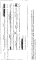
Figure 1 depicts CK-19 cDNA and CK-19 pseudo α gene sequence alignment and hybridization sites for primers and probes used in protocols A and B. Points I and II represent junctions betweenexons 1/2 andexons 2/3, respectively; -
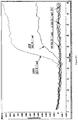
Figure 2 is a real-time PCR for genomic DNA by using four combinations of primers with the same hybridization probes [A) CK19-do2/CK19-for2, B) CK19-do2/CK19-for, C) CK19-do/CK19-for, D) CK19-do/CK19-for2]; and -
Figure 3 is a graph showing CK-19 mRNA positive cell levels expressed as MCF-7 cell equivalents/5 µg RNA obtained by protocols A and B. -
Figure 4 is a typical real-time PCR graph for PBGD. The graph shows the real time PCR amplification curves for the housekeeping gene generated using the housekeeping primer pair of the disclosure detected using a Taqman probe of the disclosure using biological samples (peripheral blood) from five healthy donors (normal sample 1-5), which efficiently amplify the PBGD gene. In the figure it is observed that no amplification occurs in the two samples containing genomic DNA (DNA isolated from healthy individuals). This is a consequence of the design of the housekeeping primers so that genomic DNA is not amplified. Negative control (NC) corresponds to PCR reaction that does not contain a nucleic acid template. -
Figure 5 is an agarose gel electrophoresis (2%) for the PBGD PCR products. The actual PCR products were loaded on an agarose gel. 10 µl of the reactions (half of the total volume) was loaded and detected using standard ethidium bromide staining. Samples 1-5 correspond to normal samples 1-5 infigure 4 , whereas negative control corresponds to the negative control infigure 4 . -
Figure 6 is a schematic drawing showing certain experimental steps for isolating circulating tumor cells (CTCs) from peripheral blood (PB). -
Figure 7 is a schematic drawing showing immunomagnetic enrichment using the monoclonal antibody Ber-EP4 and the magnetic dynabeads epithelial enrich kit.. -
Figures 8A-C are graphs showing real-time PCR 5 results for three groups of samples referenced below in the Example section.Figure 8A (1st group = PB spiked with known amount of MCF-7 cells, immunomagnetic enrichment),Figure 8B (2nd group= PBS spiked with known amount of MCF-7 cells),Figure 8C 10 (3rd group, same as 1st group except no immunomagnetic enrichment. - The present invention is as disclosed in the appended claims.
- The present disclosure provides primers and methods for detecting mRNA of genes comprising at least one intron using real-time monitoring during PCR.
- The quantitative detection of mRNA is accomplished using any available technique for quantitative determination of PCR products. Preferably this method is real-time PCR, but any other suitable method is within the scope of the disclosure e.g. competitive PCR. The quantification may be performed by any suitable method and the choice of method is within the skill of the art.
- The disclosure further provides diagnostic methods and kits for detecting the presence of mRNA of a gene comprising at least one intron. In a first aspect of the present invention is provided a primer pair consisting of one primer having the sequence of SEQ ID NO: 1 and one primer having the sequence of SEQ ID NO: 2.
- The primers described herein may comprise deoxyribonucleic acid (DNA), ribonucleic acid (RNA) or nucleic acid analogs such as uncharged nucleic acid analogs including peptide nucleic acids (PNAs) which are disclosed in
WO 92/20702 US 5,185,444 ;5,034,50.6 and5,142,047 . Such sequences can routinely be synthesized using a variety of techniques. In an alternative embodiment the primers comprise labels. - As used herein "target sequence" means a sequence that is detected, amplified, both amplified and detected or is complementary to the sequences provided herein or otherwise has at least one intron in its native state i.e. as genomic DNA or extra chromosomal DNA. While the term target sequence is sometimes referred to as single stranded, those skilled in the art will recognize that the target sequence may be double stranded.
- The target sequence is the mRNA-transcript of the CK19 gene.
- There is provided the primer pair having the sequences according SEQ ID NO: 1 (5'CGGGACAAGATTCTTGGT-3' FORWARD) and SEQ ID NO: 2 (5'CGTTGATGTCGGCGTCCA-3' REVERSE), respectively, which primers can be employed to amplify the CK19 target sequence.
- It should be understood that the sequences of SEQ ID NO: 1 and 2 are the primer pair according to the invention. According to the invention the main feature of the primer pair is that it comprises at least one intron-spanning site. This provides a primer pair that will only bind to a sequence in which the introns have been spliced out, e.g. mRNA, cDNA. It should be understood that said "splicing" may occur naturally i.e. to provide for the detection of mRNA in a biological sample. However, the term also encompasses an engineered sequence having the introns "spliced out" of the sequence, e.g. cDNA.
- In the present disclosure the forward primer is the primer that is extended in the same direction as the coding strand of the target nucleic acid. Conversely, the reverse primer is the primer that is extended in the same direction as the non-coding strand of the target nucleic acid. Consequently, the primers align with their 3'-ends facing each other.
- In a second aspect of the invention is provided a method of detecting the presence of a CK-19 mRNA in a test sample using the primers of the invention comprising the steps of (i) forming a reaction mixture comprising nucleic acid amplification reagents, the primer pair of the invention and a test sample; (ii) subjecting the mixture to amplification conditions to generate at least one copy of a nucleic acid sequence complementary to the CK-19 sequence; and (iii) quantification of the CK-19 mRNA in the sample using real-time PCR monitoring.
- In the present description "test sample" and "biological sample" are used interchangeably. In this context "test sample" means anything suspected of containing the target sequence. The test sample can be derived from any biological source, such as for example blood, bone marrow, lymph nodes, bronchial alveolar lavage, saliva, throat swabs, ocular lens fluid, spinal fluid, sweat, sputa, urine, milk, ascites fluid, mucous, synovial fluid, peritoneal fluid, cerebrospinal fluid, amniotic fluid, tissues such as breast tissues and the like; or fermentation broths, cell cultures, chemical reaction mixtures and the like. Lung cells or tissue may also be used. Most typically the test sample is derived from blood, such as peripheral blood, bone marrow or lymph nodes. The test sample may be used directly as obtained from the source or following a pre-treatment to modify the character of the sample. Thus, the test sample can be pre-treated prior to use by, for example, preparing plasma from blood, disrupting cells, preparing liquids from solid materials, diluting viscous fluids, filtering liquids, distilling liquids, concentrating liquids, inactivating interfering components such as epithelial cells, adding reagents purifying nucleic acids and the like. In a preferred embodiment the pre-treatment is centrifugation.
- A "biological fluid" is a biological sample having (or made to have) a liquid form. Examples include peripheral blood, plasma, or an extract obtained from cells or tissue.
- The optional reverse transcription step in the method of the invention is included wherever necessary in order to amplify the target sequence, i.e. when the nature of the target sequence is RNA. This process, designated reverse transcription, occurs under the direction of an RNA-dependent DNA polymerase enzyme called a reverse transcriptase. The process furthermore requires buffers and reagents, such as dNTPs, for the reverse transcription. Reverse transcription kits are commercial available and it is within the skill to perform this process.
- The nucleic acid amplification reagents used in the invention includes reagents which are well known and may include, but are not limited to, an enzyme with polymerase activity e.g. heat stable polymerases such as the Taq-polymerase (and, as necessary, reverse transcriptase activity e.g. when monitoring mRNA), enzyme cofactors such as magnesium or manganese; salts and deoxynucleotide triphosphates (dNTPs).
- The term "amplification conditions" is generally defined as conditions, which promote hybridizing or annealing of primer sequences to a target sequence and subsequent extension of the primer sequence. It is well known in the art that such annealing is dependant on several parameters, including temperature, ionic strength, sequence length, complementarity and G:C content of the sequences. For example, lowering the temperature in the environment of complementary nucleic acid sequences promotes annealing. For any given set of sequences, melt temperature, or Tm, can be estimated by any of several known methods. Typically, diagnostic applications utilize hybridization temperatures, which are close to (i.e. within 10°C) the melt temperature. Ionic strength or "salt" concentration also impacts the melt temperature, since small cations tend to stabilize the formation of duplexes by negating the negative charge on the phosphodiester backbone. Typical salt concentrations depend on the nature and valency of the cation but are readily understood by those skilled in the art. Similarly, high G:C content and increased sequence length are also known to stabilize duplex formation because G:C pairings involve 3 hydrogen bonds where A:T pairs have just two, and because longer sequences have more hydrogen bonds holding the sequences together. Thus, a high G:C content and longer sequence lengths impact the hybridization conditions by elevating the melt temperature. Once sequences are selected for a given diagnostic application, the G:C content and length will be known and can be accounted for in determining precisely what hybridization conditions will encompass. Since ionic strength is typically optimized for enzymatic activity the only parameter left to vary is the temperature. Generally, the hybridization temperature is selected close to or at the Tm of the primers or probe. Thus, obtaining suitable hybridization conditions for a particular primer, probe, or primer and probe set is well within ordinary skill of one practicing this art. The amplification product produced as above can be detected during or subsequently to the amplification of the target sequence using any suitable method and a probe disclosed in greater detail below.
- The disclosure furthermore discloses any sequence specific probes, such as hybridization probes, Taqman probes or molecular Beacon type probes, for detecting/quantification of the amplification product. Furthermore, the probe may be used to ensure specificity. Said probes may have the sequence according to SEQ ID NO: 3 and 4 for detecting amplification of the CK19 gene. Construction of probes for detecting amplification of a target sequences is within the skill of the art.
- The probes are preferably labelled. The label can be either directly detectable as with for example fluorophores, chemiluminophores, fluorescent particles and the like or indirectly detectable as with specific binding partners and nucleic acids. Preferred labels are directly detectable, and particular preferred labels are fluorescent dyes, such as Sybr Green I, FAM, HEX, VIC, fluoroscein LC Red 610, LC Red640 , LC Red670, LC Red 705, and other fluorescent dyes known in the art.
- In one embodiment the probe may initially be part of the amplification reaction mixture in which case it is desirable to select conditions such that the probe sequence has a lower melt temperature than the primer sequence. In this way the temperature can initially be chosen so that the probe does not hybridize to the target sequence i.e. over the Tm of the probe. After copies of the target sequence are synthesized the temperature can be lowered in order to let the probe hybridize to the newly synthesized target sequence, provided that this target sequence originally was present in the test sample, and subsequently the possible presence of this target sequence will be detectable. Alternatively the probe is added separately. Preferably the probe does not hybridize to sequences corresponding to the primer sequences.
- In another variant of this second aspect of the invention, step (i) of the method may further comprise a housekeeping primer pair that hybridizes to a housekeeping gene in order to ensure that amplifiable material is present in the test samples and in order to avoid false negative results. Said housekeeping primer pair may be the commercially available housekeeping primer pair for hypoxanthine-guanine phosphoribosyl transferase (HPRT) (purchased from Roche applied Science).
- Alternatively, a housekeeping primer pair identified by the present inventors may be used in the method of the invention.
- Therefore, the present disclosure provides a housekeeping primer pair having the sequence according to
SEQ ID NO - In the context of the present invention "housekeeping primer pair" and "primer pair" are not the same. In the context of the present invention the term "housekeeping primer pair" is intended to mean a primer pair, which is capable of hybridizing to a target sequence of a gene, which is ubiquitous to a given cell. In other words a "housekeeping primer pair" can be used as an internal control in a method or kit of the invention, i.e. as a negative control.
- Said housekeeping primer pair of the disclosure hybridizes to the housekeeping gene PBGD: Human non-erythropoietic porphobilinogen deaminase (PBGD; hydroxymethylbilane synthase; Accession no: X04808), the third enzyme of the heme biosynthetic pathway, which catalyzes the stepwise condensation of four porphobilinogen units to yield hydroxymethylbilane, which is in turn converted to uroporphyrinogen III by cosynthetase. A housekeeping gene is a gene that is essential to a cell and thus always present under any conditions. The housekeeping primer pair designed by the present inventors for the PBGD mRNA amplification is: Forward(HGF1) 5'-GGTGGGTGTGCTGCACGAT-3' (SEQ ID NO 5) and Reverse(HGR) 5'-ATCTTCATGCTGGGCAGGGA-3' (SEQ ID NO 6).
- Said housekeeping primer pair is suitable for the methods and the kit of the present invention. However, the use of the housekeeping primer pair is not limited to said methods and the kit, but may be used whenever the samples (cells) to be tested ubiquitously comprise the gene encoding human non-erythropoietic porphobilinogen deaminase.
- In Real-time PCR hybridization probes, Taqman probe or a molecular beacon type probe for the visualization of PCR products may be used as described previous in relation to the primer pair of
claim 1. One preferred Taqman probe is: 6FAM-ATGAAGGATGGGCAACTGTACCTGACTGG-TMR. - The skilled person will appreciate that instead of the Taqman probe described above, any set of hybridization probes may be used for the detection of the amplified target sequence of the PBGD, or any other suitable housekeeping gene, in the biological sample. Further, taking advantage of the existence of different fluorescent channels available in PCR machines known in the art (machines having 3-6 fluorescent channels are commercially available) the amplification of the housekeeping gene can be done in the same run as the amplification of the CK-19 gene or any other suitable target gene, since a housekeeping primer pair can be use as internal control in many different cases.
- The housekeeping primers are preferably designed in a way that avoids amplification of genomic DNA or cDNA in order to avoid non-specific amplification of contaminating genomic DNA in the sample. This may be accomplished using in principle the same criteria for designing the household primers as is used for designing the CK-19 primers according to the invention.
- In a third aspect of the invention is disclosed a method of determining the prospects of adjuvant therapy in a patient suffering from cancer comprising the steps of (i) isolating nucleic acids from a blood sample that has been provided from the patient; (ii) forming a reaction mixture comprising nucleic acid amplification reagents, the primer pair according to
claim 1 and an aliquot of the nucleic acids isolated in step (i); (iii) subjecting the mixture to amplification conditions to generate at least one copy of a nucleic acid sequence complementary to the target sequence; (iv) quantification of the CK-19 mRNA positive cells in the sample using real-time PCR monitoring; and (v) based on the amount of CK-19 mRNA positive cells in the sample determining the prospects of adjuvant therapy. - According to the invention the primer pair has the sequence according to SEQ ID NO: 1 and 2.
- In a particular preferred embodiment the sample is peripheral mononuclear blood cells. In yet another preferred embodiment the cancer is operable breast cancer.
- In general the method of the invention may also be used to detect/quantify circulating tumor cells (CTCs) based on the CK-19 marker in cancer types of epithelial origin including but not limited to squamous epithelium, such as squamous cell papilloma and squamous cell carcinoma; transitional epithelium, such as transitional cell papilloma and transitional cell carcinoma; basal cell, such as basal cell carcinoma; glandular epithelium, such as adenoma, cystadenoma and adenocarcinoma; kidney tubules epithelium, such as renal tubular adenoma, renal cell carcinoma and Grawitz tumor; hepatocytes such as hepatocellular adenoma and hepatocellular carcinoma; bile ducts epithelium, such as cholangiocellular adenoma and cholangiocellular carcinoma; and melanocytes, such as melanocytic nevus and malignant melanoma.
- In this third aspect of the invention a sample may be pre-treated similarly to the "test sample" as described earlier. Thus, in a preferred aspect the exemplary blood sample is centrifuged prior to isolation of the nucleic acid in order to isolate the peripheral mononuclear blood cells (PBMCs). This may be done using any centrifugation technique known in the art, such as Ficoll enrichment, PAX gene blood collection system, immunomagnetic separation and enrichment, and a preferred centrifugation technique is gradient centrifugation. The "nucleic acid amplification reagents" and "amplification conditions" in context of this aspect of the invention are the same as described above.
- In a fourth aspect the present invention provides a kit for use in the method of the third aspect of the invention. Said kit comprises the primer pair of the invention, optional further primers that hybridize to other markers on cancer cells and amplification reagents.
- Said amplification reagents and the primer pair may either be provided separately or, where appropriate, be mixed.
- In a preferred embodiment the further primers hybridize to HER2/neu and cytokeratins, such as CK20, CK8 etc., maspin, GABA An, B305D-C, PIP, S100A9, S100A14, PSA, mucin, carcinoembryonic antigen, β-subunit of human chorionic gonadotropin, mammaglobin, epidermal growth factor, Ep-CAM and several other mRNA markers known in the art. The choice and combination of additional markers is within the skill of the art. Combination of primers is optional depending on the type of cancer indication. The primer pair has the sequence according to SEQ ID NO: 1 and 2.
- Combinations of two or more primers may ensure that the incidence of false negatives is reduced given the fact that more than one marker on a cancer cell is detected.
- In another preferred embodiment of the invention the kit comprises an internal control in order to avoid false negatives, wherein the internal control preferably is a housekeeping primer pair. Said housekeeping primer pair preferably has the sequences according to
SEQ ID NO - In yet another preferred embodiment of the kit according to the invention all ingredients are lyophilized. In yet another embodiment two or more, e.g. all, lyophilized reagents are mixed. In this case the user, e.g. a clinician, may simply dissolve the mixture in a suitable buffer and add the sample to be tested before the amplification. Besides simplifying the handling procedure, lyophilization makes the reagents more stable for storage.
- As will be appreciated, a nucleoside is a base-sugar combination. The base portion of the nucleoside is normally a heterocyclic base. The two most common classes of such heterocyclic bases are the purines and the pyrimidines. Nucleotides are nucleosides that further include a phosphate group covalently linked to the sugar portion of the nucleoside. For those nucleosides that include a pentofuranosyl sugar, the phosphate group can be linked to either the 2', 3' or 5' hydroxyl moiety of the sugar. In forming oligonucleotides, the phosphate groups covalently link adjacent nucleosides to one another to form a linear polymeric compound. In turn, the respective ends of this linear polymeric structure can be further joined to form a circular structure, however, open linear structures are generally preferred. In addition, linear structures may also have internal nucleobase complementarity and may therefore fold in a manner as to produce a double stranded structure. Within the oligonucleotide structure, the phosphate groups are commonly referred to as forming the internucleoside backbone of the oligonucleotide. The normal linkage or * backbone of RNA and DNA is a 3' to 5' phosphodiester linkage.
- Additional examples of primers and primer pairs within the scope of the present invention include oligonucleotides with modified backbones or non-natural internucleoside linkages. As defined in this specification, oligonucleotides having modified backbones include those that retain a phosphorus atom in the backbone and those that do not have a phosphorus atom in the backbone. For the purposes of this specification, and as sometimes referenced in the field, modified oligonucleotides that do not have a phosphorus atom in their internucleoside backbone can also be considered to be oligonucleosides.
- Accordingly, the invention encompasses primers and primer pairs in wich one or both primers include modified oligonucleotide backbones. Such backbones include phosphorothioates, chiralphosphorothioates, phosphorodithioates, phosphotriesters, aminoalkylphosphotri-esters, methyl and other alkyl phosphonates including 3'-alkylene phosphonates, 5'-alkylene phosphonates and chiral phosphonates, phosphinates, phosphoramidates including 3'-amino phosphoramidate and aminoalkylphosphoramidates, thionophosphoramidates, thionoalkylphosphonates, thionoalkylphosphotriest- ers, selenophosphates and borano-phosphates having normal 3'-5' linkages, 2'-5' linked analogs of these, and those having inverted polarity wherein one or more internucleotide linkages is a 3' to 3', 5' to 5' or 2' to 2' linkage. Additional oligonucleotides having inverted polarity comprise a single 3' to 3' linkage at the 3'-most internucleotide linkage i.e. a single inverted nucleoside residue that may be abasic (the nucleobase is missing or has a hydroxyl group in place thereof). Various salts, mixed salts and free acid forms are also included. See, for example, the following patents
3,687,808 4,469,863 4,476,301 5,023,243 5,177,196 5,188,897 5,264,423 5,276,019 5,278,302 5,286,717 5,321,131 5,399,676 5,405,939 5,453,496 5,455,233 5,466,677 5,476,925 5,519,126 5,536,821 5,541,306 5,550,111 5,563,253 5,571,799 5,587,361 5,194,599 5,565,555 5,527,899 5,721,218 5,672,697 5,625,050 - It is a further object of the invention to provide suitable primers and primer pairs in which one or both of the primers do not include a phosphorus atom. Such embodiments will have backbones that are formed by short chain alkyl or cycloalkyl internucleoside linkages, mixed heteroatom and alkyl or cycloalkyl internucleoside linkages, or one or more short chain heteroatomic or heterocyclic internucleoside linkages. These include those having morpholino linkages (formed in part from the sugar portion of a nucleoside as discussed above); siloxane backbones; sulfide, sulfoxide and sulfone backbones; formacetyl and thioformacetyl backbones; methylene formacetyl and thioformacetyl backbones; riboacetyl backbones; alkene containing backbones; sulfamate backbones; methyleneimino and methylenehydrazino backbones; sulfonate and sulfonamide backbones; amide backbones; and others having mixed N, O, S and CH2 component parts. See, for instance, the following patents
5,034,506 5,166,315 5,185,444 5,214,134 5,216,141 5,235,033 5,264,562 5,264,564 5,405,938 5,434,257 5,466,677 5,470,967 5,489,677 5,541,307 5,561,225 5,596,086 5,602,240 5,610,289 5,602,240 5,608,046 5,610,289 5,618,704 5,623,070 5,663,312 5,633,360 5,677,437 5,792,608 5,646,269 5,677,439 - In some invention embodiments, it will be useful to have one or both primers bear novel groups ie., not associated with naturally-occuring nucleosides. One such oligomeric compound, an oligonucleotide mimetic that has been shown to have excellent hybridization properties, is referred to as a peptide nucleic acid (PNA; see discussion above). In PNA compounds, the sugar-backbone of an oligonucleotide is replaced with an amide containing backbone, in particular an aminoethylglycine backbone. The nucleobases are retained and are bound directly or indirectly to aza nitrogen atoms of the amide portion of the backbone. See, for instance, the following patents:
5,539,082 5,714,331 5,719,262 - Further suitable primers and primer pairs in accord with the invention include oligonucleotides with phosphorothioate backbones and oligonucleosides with heteroatom backbones, and in particular --CH2-NH--O--CH2--, --CH2--N(CH3)--O--CH2-- (known as a methylene (methylimino) or MMI backbone], --CH2--O-N(CH3)--CH2, --CH2--N(CH3)--N(CH3)--CH2-- and --O-N(CH3)--CH2--CH2--, and --O--P--O--CH2--. Also preferred are oligonucleotides having morpholino backbone structures. See the previous discussion and
U.S. Pat. No. 5,034,506 . See alsoU.S. Pat. Nos. 5,489,677 , and5,602,240 . - In some invention embodiments, it may be useful to have primers and primer pairs in which the oligonucleotides are modified to have one or more substituted sugar moieties. Preferred oligonucleotides with this modification include one of the following at the 2' position: OH; F; O--, S--, or N-alkyl; O--, S--, or N-alkenyl; O--, S-- or N-alkynyl; or O-alkyl-O-alkyl, wherein the alkyl, alkenyl and alkynyl may be substituted or unsubstituted C1 to C10 alkyl or C2 to C10 alkenyl and alkynyl. Additional modifications include O[(CH2)nO]mCH3, O(CH2nOCH3, O(CH2)nNH2, O(CH2)nCH3, O(CH2)nONH2, and O(CH2)nON[(CH2)nCH3]2, where n and m are from 1 to about 10. Other exemplary oligonucleotides comprise one of the following at the 2' position: C1 to C10 lower alkyl, substituted lower alkyl, alkenyl, alkynyl, alkaryl, aralkyl, O-alkaryl or O-aralkyl, SH, SCH3, OCN, Cl, Br, CN, CF3, OCF3, SOCH3, SO2CH3, ONO2, NO2, N3, NH2, heterocycloalkyl, heterocycloalkaryl, aminoalkylamino, poly-alkylamino, substituted silyl, an RNA cleaving group, a reporter group, or a nucleic acid intercalator. Additional modifications include 2'-methoxyethoxy (2'-O-CH2CH2OCH3, also known as 2'-O-(2-methoxyethyl) or 2'-MOE) (Martin et al., Helv. Chim. Acta, 1995, 78, 486-504) i.e., an alkoxyalkoxy group. A further illustrative modification preferred includes 2'-dimethylaminooxyethoxy, i.e., a O(CH2)2ON(CH3)2 group, also known as 2'-DMAOE, as described in examples hereinbelow, and 2'-dimethylamino-ethoxyethoxy (also known as 2'-O-dimethyl-amino-ethoxy-ethyl or 2'-DMAEOE), i.e., 2'-O--CH2--O--CH2--N(CH3)2.
- Other suitable primers and primer pairs are within the scope of thep present invention. These include those primers having modifications that include 2'-methoxy(2'-O--CH3), 2'-aminopropoxy(2'-OCH2CH2CH2NH2), 2'-allyl(2'-CH2-CH=CH2), 2'-O-allyl (2'-O--CH2-CH=CH2) and 2'-fluoro(2'-F). The 2'-modification may be in the arabino (up) position or ribo (down) position. An illustrative 2'-arabino modification is 2'-F. Similar modifications may also be made at other positions on the oligonucleotide, particularly the 3' position of the sugar on the 3' terminal nucleotide or in 2'-5' linked oligonucleotides and the 5' position of 5' terminal nucleotide. Oligonucleotides may also have sugar mimetics such as cyclobutyl moieties in place of the pentofuranosyl sugar. See, for example, the following patents:
4,981,957 5,118,800 5,319,080 5,359,044 5,393,878 5,446,137 5,466,786 5,514,785 5;519,134 5,567,811 5,576,427 5,591,722 5,597,909 5,610,300 5,627,053 5,639,873 5,646,265 5,658,873 5,670,633 5,792,747 5,700,920 - Still further primer pairs according to the invention include one or more primers with a Locked Nucleic Acid (LNA). A preferred LNA features a 2'-hydroxyl group linked to the 3' or 4' carbon atom of the sugar ring thereby forming a bicyclic sugar moiety. The linkage is preferably a methylene (--CH2-)n group bridging the 2' oxygen atom and the 4' carbon atom wherein n is 1 or 2. LNAs and preparation thereof are described in International Published Patent Application Nos.
WO 98/39352 WO 99/14226 6,794,499 ;6,670,461 ;2003/0082807 (Xylo-LNA);2003/0087230 (L-ribo-LNA); and2003/0224377 . - As will be appreciated, oligonucleotides may also include nucleobase (often referred to in the art simply as "base") modifications or substitutions. As used herein, "unmodified" or "natural" nucleobases include the purine bases adenine (A) and guanine (G), and the pyrimidine bases thymine (T), cytosine (C) and uracil (U). Modified nucleobases include other synthetic and natural nucleobases such as 5-methylcytosine (5-me-C), 5-hydroxymethyl cytosine, xanthine, hypoxanthine, 2-aminoadenine, 6-methyl and other alkyl derivatives of adenine and guanine, 2-propyl and other alkyl derivatives of adenine and guanine, 2-thiouracil, 2-thiothymine and 2-thiocytosine, 5-halouracil and cytosine, 5-propynyl (--C≡C--CH3) uracil and cytosine and other alkynyl derivatives of pyrimidine bases, 6-azo uracil, cytosine and thymine, 5-uracil (pseudouracil), 4-thiouracil, 8-halo, 8-amino, 8-thiol, 8-thioalkyl, 8-hydroxyl and other 8-substituted adenines and guanines, 5-halo particularly 5-bromo, 5-trifluoromethyl and other 5-substituted uracils and cytosines, 7-methylguanine and 7-methyladenine, 2-F-adenine, 2 -amino-adenine, 8-azaguanine and 8-azaadenine, 7-deazaguanine and 7-deazaadenine and 3-deazaguanine and 3-deazaadenine. Further modified nucleobases include tricyclic pyrimidines such as phenoxazine cytidine (1H-pyrimido[5,4-b][1,4]benzoxazi-n-2(3H)-one), phenothiazine cytidine (1H-pyrimido[5,4-b][1,4]benzothiazin-2(3H)-one), G-clamps such as a substituted phenoxazine cytidine (e.g. 9-(2-aminoethoxy)-H-pyrimido[5,4-b] [1,4)benzoxazin-2(3H)-one), carbazole cytidine (2H-pyrimido[4,5-b)indol-2-one), pyridoindole cytidine (H-pyrido[3',2':4,Spyrrolo[2,3-dlpyri-midin-2-one). Modified nucleobases may also include those in which the purine or pyrimidine base is replaced with other heterocycles, for example 7-deaza-adenine, 7-deazaguanosine, 2-aminopyridine and 2-pyridone. Further nucleobases include those disclosed in
U.S. Pat. No. 3,687,808 , those disclosed in The Concise Encyclopedia Of Polymer Science And Engineering, pages 858-859, Kroschwitz, J. I., ed. John Wiley & Sons, 1990, those disclosed by Englisch et al., Angewandte Chemie, International Edition, 1991, 30, 613, and those disclosed by Sanghvi, Y. S., . Additional modifications include 5-substituted pyrimidines, 6-azapyrimidines and N-2, N-6 and 0-6 substituted purines, including 2-aminopropyladenine, 5-propynyluracil and 5-propynylcytosine. 5-methylcytosine substitutions have been shown to increase nucleic acid duplex stability by 0.6-1.2.degree. C. (Sanghvi, Y. S., Crooke, S. T. and Lebleu, B., eds., Antisense Research and Applications, CRC Press, Boca Raton, 1993, pp. 276-278) and are illustrative base substitutions, even more particularly when combined with 2'-O-methoxyethyl sugar modifications. See, for instance,U.S. Pat. No. 3,687,808 , as well asU.S. Pat. Nos.: 4,845,205 ;5,130,302 ;5,134,066 ;5,175,273 ;5,367,066 ;5,432,272 ;5,457,187 ;5,459,255 ;5,484,908 ;5,502,177 ;5,525,711 ;5,552,540 ;5,587,469 ;5,594,121 ,5,596,091 ;5,614,617 ;5,645,985 ;5,830,653 ;5,763,588 ;6,005,096 ; and5,681,941 ,5,750,692 . - A primer or primer pair in accord with the invention is "modified" if it includes at least one of the foregoing oligonucleotide modifications. As will be readily apparent, certain of the modified primers and primer pairs will not be optimal for some invention emodiments such as performing real-time PCR. However, the modified primers can be useful as electrophoretic markers, and/or as "antisense" compositions, for instance.
- For PCR applications in which increased target affinity and specificity is useful or when enhanced robustness is helpful, one or both of the sequences represented by SEQ ID NO: 1 or SEQ ID NO: 2 can modified to include at least one LNA, for example, 1 (one), 2 (two), 3 (three), 4 (four) or 5 (five) of such LNAs. See, for example, Vester, B and J. Wengel (2004) Biochemistry 43: 13233; and references cited therein, for additional disclosure relating to making and using LNA oligonucleotides.
- As discussed above, the disclosure also provides a method determining the presence of CK-19 mRNA in a biological fluid. In one aspect, the method includes the following steps (a)-(g):
- a) separating any mononuclear cells from the biological fluid. The separation step can be performed using nearly any method capable of separating cells from a biological fluid such as filtration and/or centrifugation. In aspects in which centrifugation is selected, it will often be preferred to use a Ficoll or other suitable cell separating gradient. Use of the Ficoll Histopaque-1077 system (Sigma Aldrich, St. Louis, MO (USA)) is preferred for many applications such as those in which the biological fluid is peripheral blood.
- b) contacting the separated mononuclear cells with a polyclonal or monoclonal antibody (or antigen binding fragment thereof such as Fab, F(ab')2, single-chain antibodies, and the like) that specifically binds an antigen expressed by the epithelial mononuclear cells. In one aspect, the antigen is a glycoprotein expressed by cells, for instance on the cell surface or cytoplasm. An illustrative antibody is one that specifically binds the antigen CDC326, for instance, ber-EP4, B302 (323/A3), B29.1 (VU-ID9), VU-1D9, HEA125. These and other suitable antibodies can be obtained from a variety of commercial sources such as Abcam plc (Cambridge, UK); Dako UK LTD. (Cambridgeshire, UK), and Santa Cruz Biotechnology INC (Santa Cruz, CA (USA)). The antibody (or antigen binding fragment thereof) can be pre-bound to any suitable solid support, for instance, glass fiber filter paper, nitrocellulose, scintered glass, plastic, synthetic polymer, cellulose,cellulose acetate, polytetrafluoroethylene,polyethylene, polypropylene, or polyvinylidine fluoride. In one aspect, the solid support is in a bead format, preferably one that includes a magnetic or paramagnetic material. A preferred bead is one manufactured by Dynal. Preferably, the contacting step of the method is sufficient to form a binding complex between the cells, antibody and solid support.
- c) separating the binding complex from any unbound material, for instance, by filtration and/or centrifugation,
- d) isolating nucleic acid (e.g, RNA such as mRNA) from endothelial mononuclear cells bound to the complex. Typically, and as described above, cDNA will be made from the RNA isolated from the cells,
- e) forming a reaction mixture comprising nucleic acid amplification reagents, a primer pair as disclosed herein, for instance primers having the sequence represented by SEQ ID Nos. 1 and 2, and the nucleic acid isolated from the mononuclear cells,
- f) subjecting the mixture to amplification conditions to generate at least one copy of a nucleic acid sequence complementary to the CK-19 target sequence; and
- g) detecting CK-19 mRNA in the biological sample using PCR, preferably RT-PCR. If desired, the amount of the CK-19 mRNA in the biological fluid can determined.
- By the term, "specific binding" or a similar term is meant a molecule disclosed herein which binds another molecule, thereby forming a specific binding pair. However, the molecule does not recognize or bind to other molecules as determined by, e.g., Western blotting ELISA, RIA, mobility shift assay, enzyme-immuno assay, competitive assays, saturation assays or other protein binding assays know in the art. See generally, Harlow and Lane in, Antibodies: A Laboratory Manual (1988) and references cited therein for examples of methods for detecting specific binding between molecules.
- In aspects of the foregoing method in which the solid support is a magnetic bead, the method will further include the step of impressing a magnetic field on the binding complex to separate the complex from any unbound material. The separated bead complex can then be manipulated to isolate the cells (and prepare nucleic acid therefrom) using standard procedures. See for instance, information from Dynal (Epithelial Enriched Dynabeads).
- The method is flexible and compatible with use of one or a combination of primer pairs as disclosed herein. Use of a particular primer pair will depend on intended use. However for many aspects, the primers represented by SEQ ID No: 1 and SEQ ID No.2 will be sufficient. A preferred biological fluid is peripheral blood.
- If desired, the method is readily adapted to include use of one or more suitable control assays such as those mentioned in the Examples. For instance, it will often be useful to prepare a standard curve of CK-19 expressing cells in aspects in which the user wishes not only to detect but to quantify mononuclear cells in a particular biological sample. The Examples below show how to make an illustrative standard curve in which peripheral blood is spiked with MCF-7 cells. It will be appreciated that other cells can be used to create the standard curve. It will also be appreciated that once the standard curve is prepared, it need not be repeated every time the method is practiced. For instance, in embodiments in which the invention is used in a clinical setting, the standard curve could be prepared once (or a most a few times) in which one or only a few types of biological samples are assayed such as peripheral blood obtained from patients.
- If desired, the foregoing method can also be adapted to include use of one or more of the housekeeping genes disclosed herein. Amplified CK-19 target sequence can be detected and optionally quantified using the probes disclosed herein.
- As will be apparent from the foregoing, the present invention is flexible and can be used to detect and optionally quantify CK-19 as expressed in a variety of biological samples including normal and abnormal (e.g., cancerous) tissues. Regarding normal tissues, the following are exemplary: hair follicles, secretory cells of sweat glands, Merkell cells, luminal epithelial cells of breast ducts, surface mucosa and glands of endometrium and endocervix, exocervix, ovary surface mesothelium, Fallopian tube epithelium, cyto- and syncytiotrophoblast cells, amnion, umbilical cord surface epithelium, luminal- and basal cells of prostate, testes rete epithelium, ductuli efferentes, epididymal tubules, Bowman's capsule, proximal-, distal- and collecting tubules of the kidney, urothel, bile duct- and gall bladder epithelium, squamous epithelium-, taste buds-, secretory glandular cells and glandular ducts of tongue, squamous epithelium- and submucosal glands of esophagus, surface mucosa- and glands of stomach, surface mucosa- and crypts of small- and large intestine, pancreas ducts, secretory- and duct cells of salivary glands, thyroid epithelium, surface mucosa- and glands of trachea, bronchial mucosa and - glands, alveoli, pleura-mesothelium, Hassal's corpuscles and thymus epithelial cells. Regarding abnormal tissues, the following list is illustrative: human breast tumors, fibroadenomas, fibrocystic diseases, cystosarcoma phyllodes, infiltrating ductal carcinomas, infiltrating lobular carcinomas, medullary carcinomas and metastases, invasive carcinoma, intraductal papillomas, pure in situ carcinomas, tissue having Paget's disease, thyroid adenoma, colon-, gastric- and lung adenocarcinomas, ovarian- and urinary bladder carcinomas, teratomas, embryonal carcinomas, testicular cancers, epidermal tumour, squamous- and basal cell carcinomas, and keratocanthomas.
- Certain aspects of the forgoing invention have been disclosed in Greek patent application
GR 20050100430 as filed on August 17, 2005 U.S Provisional Application No. 60/795,149 as filed on April 4, 2006 - The human mammary carcinoma cell line MCF-7 which expresses the CK-19 gene (obtained from the American Type Culture Collection; ATCC), was used as positive control and cultured as previously described (A. Stathopoulou et al; 2001).
- Peripheral blood in EDTA was obtained from 160 patients with stage I/II (early stage) breast cancer postoperatively and 62 female healthy volunteers (aged 18-65 years). To reduce blood contamination by epithelial cells from the skin, the first 5mL of blood were discarded and the collection tube was at the end disconnected before withdrawing the needle. Peripheral blood samples from healthy donors and patients were collected and processed in the same manner. All patients and donors gave their informed consent and the study has been approved by the Ethical and Scientific Committees of the participating Institutions. The peripheral blood mononuclear cells (PBMC) were isolated within one hour of venipuncture by gradient centrifugation with Ficoll Hypaque-1077 (Sigma Chemical Company, LTD, England), as previously described (A. Stathopoulou et al; 2001), and cell pellets were kept at -80 °C until total RNA extraction.
- Total RNA isolation was performed by using Trizol LS reagent (Invitrogen, Corp., Carlsbad, USA) according to the manufacturer's instructions. All preparation and handling steps of RNA took place in a laminar flow hood, under RNAse free conditions. The isolated RNA was dissolved in RNA storage buffer (Ambion, USA) and stored at -70°C until used. RNA concentration was determined using the RiboGreen RNA Quantitation Kit (Molecular Probes, Eugene, OR, USA), with the LightCycler (Roche Diagnostics, Manheim, Germany) serving as a simple fluorimeter. The RNA quantification was performed in the following way: 5 µL of a supplied with the kit RNA solution of known concentration or its dilutions or the unknown sample was added along with 5 µL of the fluorophore RiboGreen in the LightCycler glass capillaries. A standard curve was created by using the fluorescence values of the RNA standard solutions measured using the LightCycler instrument in the Real Fluorimeter Mode (range 5-500 ng/mL). The fluorescence of the samples was measured in triplicate and the RNA concentration was calculated with the use of the standard curve.
- Reverse transcription of RNA was carried out with the THERMOSCRIPT RT-PCR System (Invitrogen, USA). Total RNA prepared from the MCF-7 cell line was used as a positive control. cDNA was synthesized from 5 µg of total RNA isolated from PBMC of healthy volunteers and breast cancer patients, according to the manufacturer's instructions.
- RNA integrity was tested in the cDNA preparations by real-time PCR amplification of the human hypoxanthine-guanine phosphoribosyl transferase (HPRT) gene using the LightCycler-h-HPRT gene set (Roche Diagnostics), according to the manufacturer's instructions. However, since current scientific data suggest that normalization to single housekeeping genes is inappropriate [C. Tricarico et al; 2002 and K. Dheda et al; 2004], our results were not normalized to the amount of the HPRT gene but rather to the quantity of total RNA that was used for cDNA synthesis, as previously described (A. Stathopoulou et al; 2003).
- The oligonucleotide sequences of the new primer pair CK19-do2 and CK19-for2 used (protocol B), were firstly designed and evaluated in-silico by using the
primer Premier 5 software (Premier Biosoft International, Palo Alto, CA, USA) in order to avoid primer-dimer formation, false priming sites and formation of hairpin structures. Furthermore, forward primer (CK19-for2) was selected to position on an intron-exon junction, so that hybridization to genomic CK-19 DNA was completely avoided. Moreover, the primers and probes were designed to differentiate between the highly homologous CK-19a pseudogene according to a search in the BLAST Sequence Similarity Search tool (NCBI, NIH) (seeFigure 1 ). Especially, the reverse primer (CK19-do2) was designed to a specific location of the CK-19 mRNA in order to have two mismatches at its 3'-end for CK-19a pseudogene (Figure 1 ) so that Taq DNA polymerase elongation is not possible and false positive results from CK-19a pseudogene amplification are avoided. Hybridization probes (TIBmol, Berlin, Germany) were the same as previously described (protocol A) (A. Stathopoulou et al; 2003). Primers were synthesized at the Lab of Microchemistry (FORTH, Crete, Greece). All primers and hybridization probes sequences are shown in Table 1.Table 1. Sequences of primers and hybridization probes used in this study for protocol B. a Labeled with fluorescein; b Labeled with LC Red640 (TIB MOLBIOL) Gene Use Name Oligonucleotide sequence (5'-3') CK-19 Forward primer CK19-for2 CgggACAAgATTCTTggT Reverse primer CK19-do2 CgTTgATGTCggCCTCCA Hybridization probe CK19-FLa TgTCCTgCAgATCgACAACgCCC-FL Hybridization probe CK19-LCb LCRed640-CTggCTgCAgATgACTTCCgAACC - In the process of evaluating the specificity of the new primer pair concerning the genomic DNA we proceeded to the real-time PCR amplification of a genomic DNA sample isolated from peripheral blood of a healthy donor by using 4 combinations of the previously used (CK19-do and CK19-for) and the newly designed primers (CK19-do2 and CK19-for2).
- Quantification is based on real-time monitoring during PCR of fluorescently labeled specific hybridization probes for CK-19. The point where the fluorescence rises above background noise (crossing point, Cp) is best quantified through the LightCycler software as the second derivative maximum of the curve. Real-time RT-PCR for CK-19 mRNA was performed using the LightCycler system (Roche Diagnostics). For protocol A, the primers (CK19-do and CK19-for) and the hybridization probes (CK19-FL and CK19-LC) were used as previously described (A. Stathopoulou et al; 2003). For protocol B, our newly designed primers CK19-do2 and CK19-for2 with the same hybridization probes as in protocol A, were used; see table 1.
- Real-time PCR was performed in a total volume of 20µL in the LightCycler glass capillaries. For the PCR, 2 µL of cDNA were placed into a 18-µL reaction volume containing 2 µL of the PCR Synthesis Buffer minus Mg2+ (10x), 1 µL of MgCl2 (50 mM), 0.4 µL dNTPs (10 mM), 0.3 µL BSA (10 pg/mL), 0.2 µL Taq platinum DNA polymerase (5 U/ µL) (Invitrogen, USA), 1 µL of the sense primer CK19-for2 (3 µM), 1 µL of the antisense primer CK19-do2 (3 µM), 1 µL of the hybridization probe CK19-FL (3 µM), 1 µL of the hybridization probe CK19-LC (3 µM) and DEPC-H2O (added to the final volume). PCR reaction was initiated after a 10 min denaturation at 95 °C (hot start PCR) and terminated with a 30 sec cooling step at 40 °C. The cycling protocol consisted of denaturation step at 95 °C for 10 sec, annealing at 55 °C for 20 sec and extension at 72 °C for 20 sec and repeated for 50 times. Fluorescence detection was performed at the end of each annealing step for 0 sec.
- For quantification, an external calibration curve was obtained by using external standard cDNAs. Total RNA was prepared from 1x10⁶ MCF-7 cells (as verified by a hemocytometer). Serial dilutions of this RNA preparation in DEPC-treated water, corresponding to 1-1000 MCF-7 cells, were used for cDNA synthesis. These cDNAs were kept in aliquots at -20 °C and used throughout the study as external standards. This calibration curve was created by plotting the number of MCF-7 cells corresponding to each external standard cDNA vs the value of its crossing point (Cp). The number of circulating CK-19 mRNA positive cells for all tested samples was expressed as MCF-7 cell equivalents per 5 µg of total-RNA, as determined by LightCycler software 3.1, according to the external standard calibration curve, as previously described (A. Stathopoulou et al; 2003).
- To ensure that amplifiable material was present in all specimens and to avoid false negative results, real-time amplification of the housekeeping gene hypoxanthine-guanine phosphoribosyl transferase (HPRT) (LightCycler-h-HPRT gene set, Roche Applied Science) was performed for all samples.
- Following protocol was used for amplification of the housekeeping gene. Real-time PCR was performed in a total volume of 20µL in the LightCycler glass capillaries. For the PCR, 2 µL of cDNA were placed into a 18-µL reaction volume containing 2 µL of the PCR Synthesis Buffer minus Mg2+ (10x), 1 µL of MgCl2 (50 mM), 0.4 µL dNTPs (10 mM), 0.3 µL BSA (10 µg/mL), 0.2 µL Taq platinum DNA polymerase (5 U/ µL) (Invitrogen, USA), 1 µL of each the housekeeping sense and antisense primers (3 µM), 1 µL of the hybridization probe CK19-FL (3 µM), 2 µL of the Taqman probe (6FAM-ATGAAGGATGGGCAACTGTACCTGACTGG-TMR) (3 µM) and DEPC-H2O (added to the final volume). PCR reaction was initiated after a 10 min denaturation at 95 °C (hot start PCR) and terminated with a 30 sec cooling step at 40 °C. The cycling protocol consisted of denaturation step at 95 °C for 10 sec, annealing at 55 °C for 20 sec and extension at 72 °C for 20 sec and repeated for 50 times. Fluorescence detection was performed at the end of each extension step for 0 sec.
- To reduce risk of contamination, RNA extraction, cDNA synthesis, preparation of the real-time RT-PCR steps and thermocycling were performed in separate rooms. Preparation of the PCR mixture was set up in a hood (BioTechne Hepa, TECHNE, Cambridge, UK) and for every extraction or synthesis step during the whole procedure we have used filter tips and included a positive and a negative sample control.
- The McNemar and Fischer exact test was used to compare real-time PCR results for CK-19 mRNA detection on the same cDNAs by both sets of primer pairs. The Wilcoxon test for paired non-normally distributed groups was used to compare the CK-19 positive cell levels in our samples estimated by the two protocols (P<0.05 was considered as statistically significant). Data analysis was carried out with the Statmost statistical package (Statmost, DataMost Corp, USA).
- The specificity of the optimized protocol B for real-time RT-PCR for CK-19 was evaluated by applying 4 combinations of primers [A) CK19-do2/CK19-for2, B) CK19-do2/CK19-for, C) CK19-do/CK19-for, D) CK19-do/CK19-for2] in a genomic DNA sample (see
Figure 2 ). The primer pair CK19-do2/CK19-for2 showed no amplification of any product, while the other three combinations demonstrated amplification. - We improved our previously reported real-time assay (A. Stathopoulou et al; 2003) by designing a new highly specific primer pair for CK-19. Only slight modifications regarding the conditions of the PCR reaction for protocol B were necessary: the amplification temperature was lowered from 60 to 55 °C and the amplification time was increased from 10 to 20 sec.
- We evaluated the analytical sensitivity and linearity of the protocol B real-time RT-PCR for CK-19, by analyzing the cDNA external standards (prepared as described above) in 4 experiments. Calibration curves from these data showed linearity over the entire quantification range (1-1000 MCF-7 cells) and correlation coefficients greater than 0.99 in all cases, indicating a precise log-linear relationship. The mean slope and intercept of the calibration curve was -3.226 ± 0.14 (CV=4.3%, n=4) and 32.30 ± 0.22 (CV=0.7%, n=4), respectively, while the PCR efficiency expressed as E = [10-1/slope] - 1 (I.R. Peters et al; 2004) was 1.04 ± 0.06 (CV=2.9%, n=4). The analytical detection limit of the method defined as 3.3 times the standard deviation of the Cp of the first external standard (1 MCF-7 cell equivalent) divided by the mean slope of the calibration curve (D.L. = 3.3SD/slope) was found to correspond to 0.4 MCF-7 cell equivalents.
- To determine within-run precision of protocol B, CK-19 mRNA was quantified in four cDNA samples corresponding to 1, 10, 100 and 1000 MCF-7 cells, in the same run, in 6 parallel determinations, in the LightCycler.
Table 2. Within-run and between-run precision of the Real-time RT-PCR protocol B for CK-19 mRNA. Reproducibility of the assay MCF-7 cell equivalents Within-run precision (n=6) Between-run precision (n=4) Crossing point (Cp) MCF-7 cells Crossing point (Cp) MCF-7 cells Mean (SD) CV% Mean (SD) CV% Mean (SD) CV% Mean (SD) CV% 1 33.6 (0.42) 1.25 1.04 (0.25) 25 32.3 (0.34) 1.05 1.09 (0.15) 13.8 10 29.6 (0.11) 0.37 10.5 (0.7) 6.6 29.1 (0.21) 0.76 9.64 (1.8) 18.9 100 26.0 (0.1) 0.42 86.5 (5.4) 6.3 25.8 (0.24) 0.93 89.5 (6.0) 6.7 1000 21.7 (0.04) 0.21 1084 (31.0) 2.9 22.3 (0.25) 1.12 972 (97.2) 10 - Table 2 demonstrates within-run CV's for MCF-7 cells as determined by the calibration curve ranged from 2.9% to 25%, while for the corresponding Cp values ranged from 0.21% to 1.25%. Furthermore, to determine between-run precision of the assay, the same cDNA samples were frozen (-20 °C) in aliquots and analyzed over a period of one month on 4 separate assays performed in 4 different days. Table 2 indicates between-run CV's for MCF-7 cells as determined by the calibration curve ranged from 6.7% to 18.9%, while for the corresponding Cp values ranged from 0.76% to 1.12%.
- The specificity and sensitivity of the optimized protocol B for real-time RT-PCR for CK-19 was evaluated in respect to protocol A. Both quantitative protocols were applied in a total of 222 peripheral blood samples obtained from 62 healthy female blood donors and 160 patients with operable (stage I/II) breast cancer. All these samples were tested for their RNA quality and cDNA synthesis by the expression of the HPRT housekeeping gene. Total RNA in each sample was fluorimetrically quantified by the Ribo Green. The same amount of RNA was used for cDNA synthesis and for normalization of our quantitative RT-PCR data (A. Stathopoulou et al; 2003).
- The specificity of the new set of primers was evaluated by re-examining 62 out of 89 peripheral blood samples from the healthy volunteers we had previously analyzed with protocol A (A. Stathopoulou et al; 2003). By applying protocol A, 2 out of these 89 samples were considered as positive according to the analytical cut-off of the assay (Cp = 32.17 ± 0.70, CV(%) = 2.2), while none of the 62 samples (the two positive samples were included) showed any amplification when they were analyzed with protocol B. The sensitivity of the optimized method was evaluated by analyzing 160 peripheral blood samples of operable breast cancer patients with both protocols.
Table 3. Comparison of protocol A and B for real-time PCR for the detection of CK-19 positive cells in peripheral blood samples. Concordance: 89,2% (198/222), (P = 0,0022, McNemar & Fischer exact test) Comparison of Protocol A and Protocol B Protocol A Protocol B Total + - + 29 20 49 - 4 169 173 Total 33 189 222 - As can be seen in Table 3, 33 (20.6%) of these samples were found positive. Twenty samples that were in the gray zone and characterized as positive with protocol A, were found negative by protocol B, while 4 samples that were characterized as negative with protocol A since they gave amplification curves with Cps greater than the cutoff, were found positive with protocol B. By including all the peripheral blood samples tested (healthy donors n=69 and breast cancer patients n=160) 29 samples were positive and 169 were negative with both protocols, so there was an 89.2% concordance (198/222) of positivity and negativity between the two protocols (McNemar and Fisher exact test, n=222, P=0.0022) (Table 3). As can be seen in
Figure 3 CK-19 mRNA positive cell levels expressed as MCF-7 cell equivalents/5 µg RNA obtained by these two protocols correlated very well (r = 0.986, n= 29) as can be seen inFigure 3 , and did not differ significantly (Wilcoxon test for paired data, n=29, P=0.164. - Three sample groups were tested to evaluate the efficacy of new protocols to isolated circulating tumor cells (CTCs) from peripheral blood.
Figure 6 shows each sample group along with subsequent manipulation. - The 1st group consisted of peripheral blood samples, spiked with known amounts of MCF-7 cells. These samples were added to a Ficoll Histopaque-1077 system (Sigma Aldrich, St. Louis, MO) and centrifuged at 1,500 rpm for 30 min. The mononuclear cell layer was removed, washed twice with PBS, diluted to 1 mL with PBS/0.1% bovine serum albumin, and incubated with Epithelial Enriched Dynabeads (1 x 10⁷ beads in a volume of 20 µL) while rocking for 1 hour. The cell suspension was placed on a magnet for at least 6 min and the supernatant was carefully removed. The cells attached to the magnetic beads were washed thrice with 1 mL PBS/0.1% bovine serum albumin and lysed with the lysis binding buffer supplied with the kit. The lysed cell suspension (with beads attached) was stored at -80°C until processing. The MCF-7 epithelial cells were enriched by immunomagnetic capture using the monoclonal antibody, Ber-EP4, and the magnetic Dynabeads Epithelial Enrich kit according to the manufacturer's instructions (Dynal). The manufacturers showed that up to a 5 log enrichment of epithelial cells and a yield of 70% viable, bead-free tumor cells can be obtained using this kit (Dynal). The Ber-EP4 antibody recognizes two glycoproteins on the surface and in the cytoplasm of epithelial cells except the superficial layers of squamous epithelia, hepatocytes, and parietal cells.
- The 2nd group consisted of samples prepared by spiking known amounts of MCF-7 cells in PBS, and following the same procedure as for the 1st group. This group of samples was used as a reference for the recovery of the MCF-7 cells after Ficoll isolation with (1st group) or without (3rd group) immunomagnetic enrichment.
- The 3rd group consisted of peripheral blood samples, spiked with known amounts of MCF-7 cells, added to Ficoll Histopaque-1077 (Sigma Aldrich, St. Louis, MO) and centrifuged at 1,500 rpm for 30 minutes. The mononuclear cell layer was removed, washed twice with PBS and PBMCs were stored at -80°C until processing.
- mRNA isolation and reverse transcription. Total RNA isolation was performed by using Trizol LS reagent (Invitrogen) according to the manufacturer's instructions. All preparation and handling steps of RNA took place in a laminar flow hood, under RNAse free conditions. The isolated RNA was dissolved in RNA storage buffer (Ambion, USA) and stored at -80°C until used. RNA concentration was determined with NanoDrop Spectrophotometer ND-1000 (NanoDrop). Reverse transcription of RNA was carried out with the Superscript III Platinum Two Step qRT-PCR kit (Invitrogen).
- Real-time PCR (quantitative PCR). Real-time RT-PCR for CK-19 was performed in a total volume of 20µL in the LightCycler glass capillaries. For the PCR, 2 µL of cDNA were placed into a 18-µL reaction volume containing 2 µL of the PCR Synthesis Buffer minus Mg2+ (10x), 1 µL of MgCl2 (50 mM), 0.4 µL dNTPs (10 mM), 0.3 µL BSA (10 pg/mL), 0.2 µL Taq platinum DNA polymerase (5 U/ µL) (Invitrogen, USA), 1 µL of the sense primer CK19-for2 (3 µM), 1 µL of the antisense primer CK19-do2 (3 µM), 1 µL of the hybridization probe CK19-FL (3 µM), 1 µL of the hybridization probe CK19-LC (3 µM) and DEPC-H2O (added to the final volume). PCR reaction was initiated after a 10 min denaturation at 95 °C (hot start PCR) and terminated with a 30 sec cooling step at 40 °C. The cycling protocol consisted of denaturation step at 95 °C for 10 sec, annealing at 55 °C for 20 sec and extension at 72 °C for 20 sec and the cycle was repeated for 50 times. Fluorescence detection was performed at the end of each annealing step for 0 sec.
- Referring now to
Figure 8A-C , it can be seen that high sensitivity was achieved when ficoll separation of peripheral blood mononuclear cells (PBMC) spiked with MCF-7 cells was followed by immunomagnetic enrichment. In these experiments, detection limits down to 1 MCF-7 cell/ml PB was achievable. SeeFigure 8A . - The present invention discloses, for instance, methods for the quantitative determination of circulating tumor cells identified in biological samples of patients. An example is a patient suffering from breast cancer. The invention methods use Real-Time PCR amplification of specific CK-19 mRNA transcripts using a primer pair of the invention.
- CK-19, being an epithelial marker abundantly expressed in tumors, is also a marker (alone or in combination with other markers) for the identification of circulating tumor cells in biological samples of patients bearing tumors of epithelial origin, including endometrial (Ji XQ et al, Gynecol Oncol. 2006 Feb;100(2):355-60), colorectal (Yeh CS et al, Int J Oncol. 2006 Feb; 28 (2) :411-20; Wang JY et al, World J Surg. 2006 Jun;30(6):1007-13), gastric (Wu CS et al, Int J Cancer. 2006 ), head & neck (Tao L et al, Br J Cancer. 2006 Apr 24;94(8) : 1164-9), prostate (O'Hara SM et al, Clin Chem. 2004 May;50(5) : 826-35) and malignant pleural effusions caused by various types of tumors (Xe F et al, J Zhejiang Univ Sci. 2004 Oct;5(10) :1286-9). Such biological samples may include peripheral blood, bone marrow, lymph nodes, spinal fluid and ocular lens fluid.
- To identify CK-19 mRNA positive circulating tumor cells from biological samples derived from patients bearing the aforementioned tumors, one or a combination of the methods disclosed herein can be used. For instance, clinical samples are collected and total RNA prepared using isolated peripheral blood mononuclear cells (PBMCs). The immunomagnetic purification strategy outlined above can be used, for instance. RNA is quantified, if desired, and stored at -70°C for long term storage. Alternatively, the RNA is used (5 µg) to perform a reverse transcription reaction to synthesize cDNA (target sequence). Samples from healthy individuals are used as controls and will be processed in parallel to clinical samples in an identical manner. It will be appreciated that such controls need not be performed if a control sample to be tested has a known CK-19 expression profile. Synthesized cDNAs are used in Real-time PCR reactions using a primer pair and hybridization probe pair as described above to amplify the CK-19 sequence. The present invention comprises the use of the primer pair set forth as SEQ ID Nos. 1 and 2.
- For quantification, an external calibration curve will be prepared by using external standard cDNAs prepared from RNA isolated from 1x10⁶ MCF-7 cells as described earlier in the application.
- The present inventors have developed a specific and sensitive method for quantification of circulating CK-19 mRNA positive cells in peripheral blood samples of breast cancer patients (A. Stathopoulou et al; 2003). Despite the very low false positive rate of this assay, since only 2 in 89 (2.2%) healthy blood donors were found positive for CK-19 mRNA, there were samples with amplifiable cDNA sequence, considered as negative, since they were detected at very high crossing points below the analytical detection limit of the assay. The evaluation of results for patient samples showing an amplification curve at a Cp slightly lower than the cut-off has proved to be very difficult and critical. This "gray decision zone" had led us to design and evaluate a new set of primers (CK19-do2 and CK19-for2). Our main goal was to avoid false positive results due to either genomic DNA contamination or illegitimate expression, as well as, false negative, due to a very low initial concentration of CK-19 mRNA in our samples. By testing the 4 different combinations of the old and the new CK-19 primer pairs with pure genomic DNA we have clearly shown that this new primer pair in combination with this pair of hybridization probes is highly specific and is not affected by the presence of a high concentration of genomic DNA and CK-19 pseudogenes. In retesting the samples from a subgroup (n=62) of the same previously studied healthy volunteers with the new primer pair, we have seen a significant improvement in the specificity of the assay since none of these samples had amplifiable product of CK-19 mRNA.
- By using this new highly specific pair of primers for the real-time PCR quantification of CK-19 mRNA the present inventors have considerably improved the specificity of this method. In this way, clear distribution between positive and negative samples is achieved and the difficult interpretation of the gray-zone results in the previous assay is completely avoided. For the majority of samples, the two sets of primer pairs give almost the same results. In a total of 222 samples tested, 29 samples were found positive and 169 negative by both primer pairs [concordance of 89.2% (198/222)]. However, for the 10 positive samples whose concentration were very close to the analytical detection limit of the method in protocol A and thus were in the "gray-zone" four were found to be true positives by protocol B (40%), while six were found to be false positives (60%). In the other set of 19 samples that were found negative by protocol A, with concentrations slightly below the analytical detection limit, 2 were found to be false negative by protocol B (10.5%). In this way, a small percentage of
patient samples 29/222 (13%) that were in a "gray-zone" of CK-19 detection, as determined by protocol A, could be more definitely characterized as positive or negative by protocol B, since this protocol is not affected by trace amounts of genomic DNA co extracted with total RNA. - Review of the following references will enhance appreciation of the present invention.
- A. C. Lambrechts, L. J. Veer, S. Rodenhuis, Ann. Oncol. 9 (1998) 1269-1276.
- K. Pantel, V. Muller, M. Auer, N. Nusser, N. Harbeck, S. Braun, Clin. Cancer Res. 9 (2003) 6326-6334.
- S. Braun, K. Pantel, P. Muller, W. Janni, F. Hepp, C. R. Kentenich, S. Gastroph, A. Wischnik, T. Dimpfl, G. Kindermann, G. Riethmuller, G. Schlimok, N. Engl. J. Med. 342 (2000) 525-533.
- Y. H. Datta, P. T. Adams, W. R. Drobyski, S. P. Ethier, V. H. Terry, M. S. Roth, J. Clin. Oncol. 12 (1994) 475-482.
- A. Schoenfeld, K. H. Kruger, J. Gomm, H. D. Sinnett, J. C. Gazet, N. Sacks, H. G. Bender, Y. Luqmani, R.C. Coombes, Eur. J. Cancer 33 (1997) 854-861.
- A. Stathopoulou, I. Vlachonikolis, D. Mavroudis, M. Perraki, Ch. Kouroussis, S. Apostolaki, N. Malamos, S. Kakolyris, A. Kotsakis, N. Xenidis, D. Reppa, V. Georgoulias, J. Clin. Oncol. 20 (2002) 3404-3412.
- A. Stathopoulou, A. Gizi, M. Perraki, S. Apostolaki, N. Malamos, D.Mavroudis, V. Georgoulias, E. Lianidou, Clin. Cancer Res. 9 (2003) 5145-5151.
- J. A. Lopez-Guerrero, P. Bolufer-Gilabert, M. Sanz-Alonso, E. Barragan-Gonzalez, J. Palau-Perez, J. De la Rubia-Comos, A. Sempere-Talens, S. Bonanad-Boix, Clin. Chim. Acta 263 (1997) 105-116.
- P. Ruud, O. Fodstad, E. Hovig, Int. J. Cancer 80 (1999) 119-125.
- E. S. Savtchenko, T. A. Schiff, C. K. Jiang, I. M. Freedberg, M. Blumenberg, Am. J. Hum. Genet. 43 (1988) 630-637.
- V. Bozionellou, D. Mavroudis, M. Perraki, S. Papadopoulos, S. Apostolaki, E. Stathopoulos, A. Stathopoulou, E. Lianidou, V. Georgoulias, Trastuzumab (herceptin) administration can effectively target chemotherapy-resistant cytokeratin-19 (ck-19) mRNA-positive tumor cells in the peripheral blood and bone marrow of patients with breast cancer, Clin. Cancer Res. (in press).
- A. Stathopoulou, K. Angelopoulou, V. Georgoulias, E.S. Lianidou, Clin Biochem. 34 (2001) 651-659.
- C. Tricarico, P. Pinzani, S. Bianchi, M. Paglierani, V. Distante, M. Pazzagli, S. Bustin, C. Orlando, Anal. Biochem. 309 (2002) 293-300.
- K. Dheda, J.F. Huggett, S.A. Bustin, M.A. Johnson, G. Rook, A.Zumla, Biotechniques, 37 (2004) 118-119.
- I.R. Peters, C.R. Helps, E.J. Hall, M.J. Day, J. Immunol. Methods 286 (2004) 203-217.
-
- <110> MEDEXIS S.A.
- <120> COMPOSITION AND METHOD FOR DETERMINATION OF CK19
EXPRESSION - <130> 16548WO
- <150>
GR 20050100430
<151> 2005-08-17 - <150>
US 60/795,149
<151> 2006-04-25 - <160> 6
- <170> PatentIn version 3.3
- <210> 1
<211> 18
<212> DNA
<213> Artificial - <220>
<223> PCR primer (forward) - <400> 1
cgggacaaga ttcttggt 18 - <210> 2
<211> 18
<212> DNA
<213> Artificial - <220>
<223> PCR primer (reverse) - <400> 2
cgttgatgtc ggcctcca 18 - <210> 3
<211> 23
<212> DNA
<213> Artificial - <220>
<223> Hybridization probe - <400> 3
tgtcctgcag atcgacaacg ccc 23 - <210> 4
<211> 24
<212> DNA
<213> Artificial - <220>
<223> Hybridization probe - <400> 4
ctggctgcag atgacttccg aacc 24 - <210> 5
<211> 19
<212> DNA
<213> Artificial - <220>
<223> PCR primer (forward) - <400> 5
ggtgggtgtg ctgcacgat 19 - <210> 6
<211> 20
<212> DNA
<213> Artificial - <220>
<223> PCR primer (reverse) - <400> 6
atcttcatgc tgggcaggga 20
Claims (20)
- Primer pair consisting of one primer having the sequence of SEQ ID NO: 1 and one primer having the sequence of SEQ ID NO: 2.
- Primer pair according to claim 1, wherein the primers comprises nucleotides, analogues of nucleotides or labels.
- Method of quantitative determining the presence of CK-19 mRNA in a test sample comprising the steps:(i) forming a reaction mixture comprising nucleic acid amplification reagents, the primer pair of claim 1 and a test sample;(ii) subjecting the mixture to amplification conditions to generate at least one copy of a nucleic acid sequence complementary to the CK-19 sequence; and(iii) determining the amount of the CK-19 mRNA in the sample using real-time monitoring during PCR.
- Method according to claim 3, wherein the test sample is subjected to reverse transcription prior to forming the reaction mixture of step (i).
- Method according to claim 3 or 4, wherein the test sample is selected from a blood sample, a sample from the bone marrow and a sample derived from the lymph nodes.
- Method according to claim 5, wherein the sample is a blood sample.
- Method according to claim 6, wherein the blood sample is centrifuged before forming the reaction mixture.
- Method according to any of the claims 3-7, wherein the reaction mixture of step (i) further comprises a housekeeping primer pair.
- Method according to claim 8, wherein the housekeeping primer pair has the sequence identified as SEQ ID NO: 5 and 6.
- A method of determining the prospects of adjuvant therapy in a patient suffering from breast cancer comprising the steps of(i) isolating nucleic acids from a blood sample that has been provided from the patient;(ii) optionally reverse transcribing the isolated nucleic acids, when the origin of the nucleic acid is RNA;(iii) forming a reaction mixture comprising nucleic acid amplification reagents, the primer pair of claim 1 and an aliquot of the nucleic acids isolated in step (i) or the reverse transcribed nucleic acid of step (ii);(iv) subjecting the mixture to amplification conditions to generate at least one copy of a nucleic acid sequence complementary to the target sequence;(v) quantitating the CK-19 mRNA positive cells in the sample using real-time monitoring during PCR; and(vi) based on the amount of CK-19 mRNA positive cells in the sample determining the prospects of adjuvant therapy.
- The method according to claim 10, wherein the cancer is operable breast cancer.
- The method according to claim 10 or 11, wherein the sample is centrifuged prior to isolation of the nucleic acid in order to isolate the peripheral mononuclear blood cells.
- The method according to any of the claims 10-12, wherein the reaction mixture of step (iii) further comprises a housekeeping primer pair.
- The method according to claim 13, wherein the housekeeping primer pair has the sequence identified as SEQ ID NO: 5 and 6.
- Diagnostic kit for amplifying a CK19 target sequence comprising:(i) the primer pair of claim 1;(ii) optionally further primers hybridizing to other markers of cancer cells;(iii) amplification reagents.
- Diagnostic kit according to claim 15, wherein the amplification reagents comprise the hybridization probes of SEQ ID NO: 3 and 4.
- Diagnostic kit according to claim 15 or 16, wherein the kit further comprises a housekeeping primer pair, preferably with the sequences identified as SEQ ID NO: 5 and 6.
- Diagnostic kit according to any of the claims 15 to 17, wherein the amplification reagents and the primer pair (s) are lyophilized.
- The primer pair of claim 1, wherein at least one of the first and second primers comprises a modified internucleoside linkage, modified sugar moiety or modified nucleobase.
- The primer pair of claim 19, wherein at least one of the first and second primers includes at least one locked nucleic acid (LNA) unit.
Applications Claiming Priority (3)
| Application Number | Priority Date | Filing Date | Title |
|---|---|---|---|
| GR20050100430A GR1005424B (en) | 2005-08-17 | 2005-08-17 | Method for quantitative determination of ck19 expression |
| US79514906P | 2006-04-25 | 2006-04-25 | |
| PCT/EP2006/008097 WO2007020081A1 (en) | 2005-08-17 | 2006-08-16 | Composition and method for determination of ck19 expression |
Publications (2)
| Publication Number | Publication Date |
|---|---|
| EP1915461A1 EP1915461A1 (en) | 2008-04-30 |
| EP1915461B1 true EP1915461B1 (en) | 2018-08-01 |
Family
ID=37199187
Family Applications (1)
| Application Number | Title | Priority Date | Filing Date |
|---|---|---|---|
| EP06776902.6A Not-in-force EP1915461B1 (en) | 2005-08-17 | 2006-08-16 | Composition and method for determination of ck19 expression |
Country Status (7)
| Country | Link |
|---|---|
| US (2) | US20100015604A1 (en) |
| EP (1) | EP1915461B1 (en) |
| JP (1) | JP2009504168A (en) |
| KR (1) | KR20080036646A (en) |
| AU (1) | AU2006281569A1 (en) |
| CA (1) | CA2617693A1 (en) |
| WO (1) | WO2007020081A1 (en) |
Families Citing this family (12)
| Publication number | Priority date | Publication date | Assignee | Title |
|---|---|---|---|---|
| US8921102B2 (en) | 2005-07-29 | 2014-12-30 | Gpb Scientific, Llc | Devices and methods for enrichment and alteration of circulating tumor cells and other particles |
| JP4968577B2 (en) * | 2006-04-11 | 2012-07-04 | エフ.ホフマン−ラ ロシュ アーゲー | Rapid measurement method of cytokeratin 19 (CK19) mRNA, and primer and probe therefor |
| WO2007147074A2 (en) | 2006-06-14 | 2007-12-21 | Living Microsystems, Inc. | Use of highly parallel snp genotyping for fetal diagnosis |
| US8372584B2 (en) | 2006-06-14 | 2013-02-12 | The General Hospital Corporation | Rare cell analysis using sample splitting and DNA tags |
| US8137912B2 (en) | 2006-06-14 | 2012-03-20 | The General Hospital Corporation | Methods for the diagnosis of fetal abnormalities |
| US20080050739A1 (en) | 2006-06-14 | 2008-02-28 | Roland Stoughton | Diagnosis of fetal abnormalities using polymorphisms including short tandem repeats |
| US20110111417A1 (en) * | 2008-05-14 | 2011-05-12 | Millennium Pharmaceuticals, Inc. | Methods and kits for monitoring the effects of immunomodulators on adaptive immunity |
| CA2737643C (en) | 2008-09-20 | 2020-10-06 | Hei-Mun Fan | Noninvasive diagnosis of fetal aneuploidy by sequencing |
| KR101292163B1 (en) * | 2010-11-23 | 2013-08-12 | 한양대학교 산학협력단 | An Anti―Cancer Composition Containing an Antibody of KRT19 |
| US20120208189A1 (en) | 2011-01-14 | 2012-08-16 | Life Technologies Corporation | Methods for isolation, identification, and quantification of mirnas |
| KR101320633B1 (en) * | 2011-05-25 | 2013-10-30 | 엠앤디 (주) | Method of providing information for diagnosis of cancer using quantitative realtime PCR and diagnostic kit comprising thereof |
| GB2524948A (en) * | 2014-03-07 | 2015-10-14 | Oxford Gene Technology Operations Ltd | Detecting Increase or Decrease in the Amount of a Nucleic Acid having a Sequence of Interest |
Family Cites Families (112)
| Publication number | Priority date | Publication date | Assignee | Title |
|---|---|---|---|---|
| CA464874A (en) | 1950-05-02 | Toledo Scale Company | Valve connector device | |
| US2004219A (en) | 1931-11-25 | 1935-06-11 | Textile Machine Works | Automatically variable tensioning device for draw-off reels |
| US3687808A (en) | 1969-08-14 | 1972-08-29 | Univ Leland Stanford Junior | Synthetic polynucleotides |
| US4469863A (en) | 1980-11-12 | 1984-09-04 | Ts O Paul O P | Nonionic nucleic acid alkyl and aryl phosphonates and processes for manufacture and use thereof |
| US5023243A (en) | 1981-10-23 | 1991-06-11 | Molecular Biosystems, Inc. | Oligonucleotide therapeutic agent and method of making same |
| US4476301A (en) | 1982-04-29 | 1984-10-09 | Centre National De La Recherche Scientifique | Oligonucleotides, a process for preparing the same and their application as mediators of the action of interferon |
| US5118800A (en) | 1983-12-20 | 1992-06-02 | California Institute Of Technology | Oligonucleotides possessing a primary amino group in the terminal nucleotide |
| US5550111A (en) | 1984-07-11 | 1996-08-27 | Temple University-Of The Commonwealth System Of Higher Education | Dual action 2',5'-oligoadenylate antiviral derivatives and uses thereof |
| FR2567892B1 (en) | 1984-07-19 | 1989-02-17 | Centre Nat Rech Scient | NOVEL OLIGONUCLEOTIDES, THEIR PREPARATION PROCESS AND THEIR APPLICATIONS AS MEDIATORS IN DEVELOPING THE EFFECTS OF INTERFERONS |
| US5367066A (en) | 1984-10-16 | 1994-11-22 | Chiron Corporation | Oligonucleotides with selectably cleavable and/or abasic sites |
| FR2575751B1 (en) | 1985-01-08 | 1987-04-03 | Pasteur Institut | NOVEL ADENOSINE DERIVATIVE NUCLEOSIDES, THEIR PREPARATION AND THEIR BIOLOGICAL APPLICATIONS |
| DE3687030T2 (en) | 1985-03-15 | 1993-03-11 | James Summerton | STEREO-REGULAR POLYNUCLEOTID-BINDING POLYMERS. |
| US5235033A (en) | 1985-03-15 | 1993-08-10 | Anti-Gene Development Group | Alpha-morpholino ribonucleoside derivatives and polymers thereof |
| US5185444A (en) | 1985-03-15 | 1993-02-09 | Anti-Gene Deveopment Group | Uncharged morpolino-based polymers having phosphorous containing chiral intersubunit linkages |
| US5405938A (en) | 1989-12-20 | 1995-04-11 | Anti-Gene Development Group | Sequence-specific binding polymers for duplex nucleic acids |
| US5034506A (en) | 1985-03-15 | 1991-07-23 | Anti-Gene Development Group | Uncharged morpholino-based polymers having achiral intersubunit linkages |
| US5166315A (en) | 1989-12-20 | 1992-11-24 | Anti-Gene Development Group | Sequence-specific binding polymers for duplex nucleic acids |
| US5264423A (en) | 1987-03-25 | 1993-11-23 | The United States Of America As Represented By The Department Of Health And Human Services | Inhibitors for replication of retroviruses and for the expression of oncogene products |
| US5276019A (en) | 1987-03-25 | 1994-01-04 | The United States Of America As Represented By The Department Of Health And Human Services | Inhibitors for replication of retroviruses and for the expression of oncogene products |
| DE3851889T2 (en) | 1987-06-24 | 1995-04-13 | Florey Howard Inst | NUCLEOSIDE DERIVATIVES. |
| US5188897A (en) | 1987-10-22 | 1993-02-23 | Temple University Of The Commonwealth System Of Higher Education | Encapsulated 2',5'-phosphorothioate oligoadenylates |
| US4924624A (en) | 1987-10-22 | 1990-05-15 | Temple University-Of The Commonwealth System Of Higher Education | 2,',5'-phosphorothioate oligoadenylates and plant antiviral uses thereof |
| EP0406309A4 (en) | 1988-03-25 | 1992-08-19 | The University Of Virginia Alumni Patents Foundation | Oligonucleotide n-alkylphosphoramidates |
| US5278302A (en) | 1988-05-26 | 1994-01-11 | University Patents, Inc. | Polynucleotide phosphorodithioates |
| US5216141A (en) | 1988-06-06 | 1993-06-01 | Benner Steven A | Oligonucleotide analogs containing sulfur linkages |
| US5175273A (en) | 1988-07-01 | 1992-12-29 | Genentech, Inc. | Nucleic acid intercalating agents |
| US5194599A (en) | 1988-09-23 | 1993-03-16 | Gilead Sciences, Inc. | Hydrogen phosphonodithioate compositions |
| US5134066A (en) | 1989-08-29 | 1992-07-28 | Monsanto Company | Improved probes using nucleosides containing 3-dezauracil analogs |
| US5591722A (en) | 1989-09-15 | 1997-01-07 | Southern Research Institute | 2'-deoxy-4'-thioribonucleosides and their antiviral activity |
| US5399676A (en) | 1989-10-23 | 1995-03-21 | Gilead Sciences | Oligonucleotides with inverted polarity |
| US5721218A (en) | 1989-10-23 | 1998-02-24 | Gilead Sciences, Inc. | Oligonucleotides with inverted polarity |
| US5264564A (en) | 1989-10-24 | 1993-11-23 | Gilead Sciences | Oligonucleotide analogs with novel linkages |
| EP0942000B1 (en) | 1989-10-24 | 2004-06-23 | Isis Pharmaceuticals, Inc. | 2'-Modified oligonucleotides |
| US5264562A (en) | 1989-10-24 | 1993-11-23 | Gilead Sciences, Inc. | Oligonucleotide analogs with novel linkages |
| US5177198A (en) | 1989-11-30 | 1993-01-05 | University Of N.C. At Chapel Hill | Process for preparing oligoribonucleoside and oligodeoxyribonucleoside boranophosphates |
| US5130302A (en) | 1989-12-20 | 1992-07-14 | Boron Bilogicals, Inc. | Boronated nucleoside, nucleotide and oligonucleotide compounds, compositions and methods for using same |
| US5670633A (en) | 1990-01-11 | 1997-09-23 | Isis Pharmaceuticals, Inc. | Sugar modified oligonucleotides that detect and modulate gene expression |
| US5646265A (en) | 1990-01-11 | 1997-07-08 | Isis Pharmceuticals, Inc. | Process for the preparation of 2'-O-alkyl purine phosphoramidites |
| US5459255A (en) | 1990-01-11 | 1995-10-17 | Isis Pharmaceuticals, Inc. | N-2 substituted purines |
| US5587361A (en) | 1991-10-15 | 1996-12-24 | Isis Pharmaceuticals, Inc. | Oligonucleotides having phosphorothioate linkages of high chiral purity |
| US5587470A (en) | 1990-01-11 | 1996-12-24 | Isis Pharmaceuticals, Inc. | 3-deazapurines |
| US5681941A (en) | 1990-01-11 | 1997-10-28 | Isis Pharmaceuticals, Inc. | Substituted purines and oligonucleotide cross-linking |
| US5321131A (en) | 1990-03-08 | 1994-06-14 | Hybridon, Inc. | Site-specific functionalization of oligodeoxynucleotides for non-radioactive labelling |
| US5470967A (en) | 1990-04-10 | 1995-11-28 | The Dupont Merck Pharmaceutical Company | Oligonucleotide analogs with sulfamate linkages |
| GB9009980D0 (en) | 1990-05-03 | 1990-06-27 | Amersham Int Plc | Phosphoramidite derivatives,their preparation and the use thereof in the incorporation of reporter groups on synthetic oligonucleotides |
| DK0455905T3 (en) | 1990-05-11 | 1998-12-07 | Microprobe Corp | Dipsticks for nucleic acid hybridization assays and method for covalent immobilization of oligonucleotides |
| US5541307A (en) | 1990-07-27 | 1996-07-30 | Isis Pharmaceuticals, Inc. | Backbone modified oligonucleotide analogs and solid phase synthesis thereof |
| US5610289A (en) | 1990-07-27 | 1997-03-11 | Isis Pharmaceuticals, Inc. | Backbone modified oligonucleotide analogues |
| US5489677A (en) | 1990-07-27 | 1996-02-06 | Isis Pharmaceuticals, Inc. | Oligonucleoside linkages containing adjacent oxygen and nitrogen atoms |
| US5618704A (en) | 1990-07-27 | 1997-04-08 | Isis Pharmacueticals, Inc. | Backbone-modified oligonucleotide analogs and preparation thereof through radical coupling |
| ATE154246T1 (en) | 1990-07-27 | 1997-06-15 | Isis Pharmaceuticals Inc | NUCLEASE RESISTANT PYRIMIDINE MODIFIED OLIGONUCLEOTIDES THAT DETECTE AND MODULATE GENE EXPRESSION |
| US5677437A (en) | 1990-07-27 | 1997-10-14 | Isis Pharmaceuticals, Inc. | Heteroatomic oligonucleoside linkages |
| US5602240A (en) | 1990-07-27 | 1997-02-11 | Ciba Geigy Ag. | Backbone modified oligonucleotide analogs |
| US5608046A (en) | 1990-07-27 | 1997-03-04 | Isis Pharmaceuticals, Inc. | Conjugated 4'-desmethyl nucleoside analog compounds |
| US5623070A (en) | 1990-07-27 | 1997-04-22 | Isis Pharmaceuticals, Inc. | Heteroatomic oligonucleoside linkages |
| WO1992002534A2 (en) | 1990-08-03 | 1992-02-20 | Sterling Drug, Inc. | Compounds and methods for inhibiting gene expression |
| US5177196A (en) | 1990-08-16 | 1993-01-05 | Microprobe Corporation | Oligo (α-arabinofuranosyl nucleotides) and α-arabinofuranosyl precursors thereof |
| US5214134A (en) | 1990-09-12 | 1993-05-25 | Sterling Winthrop Inc. | Process of linking nucleosides with a siloxane bridge |
| US5561225A (en) | 1990-09-19 | 1996-10-01 | Southern Research Institute | Polynucleotide analogs containing sulfonate and sulfonamide internucleoside linkages |
| WO1992005186A1 (en) | 1990-09-20 | 1992-04-02 | Gilead Sciences | Modified internucleoside linkages |
| US5432272A (en) | 1990-10-09 | 1995-07-11 | Benner; Steven A. | Method for incorporating into a DNA or RNA oligonucleotide using nucleotides bearing heterocyclic bases |
| US5672697A (en) | 1991-02-08 | 1997-09-30 | Gilead Sciences, Inc. | Nucleoside 5'-methylene phosphonates |
| US5719262A (en) | 1993-11-22 | 1998-02-17 | Buchardt, Deceased; Ole | Peptide nucleic acids having amino acid side chains |
| US5539082A (en) | 1993-04-26 | 1996-07-23 | Nielsen; Peter E. | Peptide nucleic acids |
| US5714331A (en) | 1991-05-24 | 1998-02-03 | Buchardt, Deceased; Ole | Peptide nucleic acids having enhanced binding affinity, sequence specificity and solubility |
| US5571799A (en) | 1991-08-12 | 1996-11-05 | Basco, Ltd. | (2'-5') oligoadenylate analogues useful as inhibitors of host-v5.-graft response |
| EP0538194B1 (en) | 1991-10-17 | 1997-06-04 | Novartis AG | Bicyclic nucleosides, oligonucleotides, their method of preparation and intermediates therein |
| US5594121A (en) | 1991-11-07 | 1997-01-14 | Gilead Sciences, Inc. | Enhanced triple-helix and double-helix formation with oligomers containing modified purines |
| DE637965T1 (en) | 1991-11-26 | 1995-12-14 | Gilead Sciences Inc | INCREASED FORMATION OF TRIPLE AND DOUBLE HELICOS FROM OLIGOMERS WITH MODIFIED PYRIMIDINES. |
| TW393513B (en) | 1991-11-26 | 2000-06-11 | Isis Pharmaceuticals Inc | Enhanced triple-helix and double-helix formation with oligomers containing modified pyrimidines |
| US5484908A (en) | 1991-11-26 | 1996-01-16 | Gilead Sciences, Inc. | Oligonucleotides containing 5-propynyl pyrimidines |
| US5792608A (en) | 1991-12-12 | 1998-08-11 | Gilead Sciences, Inc. | Nuclease stable and binding competent oligomers and methods for their use |
| US5359044A (en) | 1991-12-13 | 1994-10-25 | Isis Pharmaceuticals | Cyclobutyl oligonucleotide surrogates |
| FR2687679B1 (en) | 1992-02-05 | 1994-10-28 | Centre Nat Rech Scient | OLIGOTHIONUCLEOTIDES. |
| US5633360A (en) | 1992-04-14 | 1997-05-27 | Gilead Sciences, Inc. | Oligonucleotide analogs capable of passive cell membrane permeation |
| US5434257A (en) | 1992-06-01 | 1995-07-18 | Gilead Sciences, Inc. | Binding compentent oligomers containing unsaturated 3',5' and 2',5' linkages |
| EP0577558A2 (en) | 1992-07-01 | 1994-01-05 | Ciba-Geigy Ag | Carbocyclic nucleosides having bicyclic rings, oligonucleotides therefrom, process for their preparation, their use and intermediates |
| US5476925A (en) | 1993-02-01 | 1995-12-19 | Northwestern University | Oligodeoxyribonucleotides including 3'-aminonucleoside-phosphoramidate linkages and terminal 3'-amino groups |
| EP0691968B1 (en) | 1993-03-30 | 1997-07-16 | Sanofi | Acyclic nucleoside analogs and oligonucleotide sequences containing them |
| EP0691977B1 (en) | 1993-03-31 | 1997-11-26 | Sanofi | Oligonucleotides with amide linkages replacing phosphodiester linkages |
| DE4311944A1 (en) | 1993-04-10 | 1994-10-13 | Degussa | Coated sodium percarbonate particles, process for their preparation and detergent, cleaning and bleaching compositions containing them |
| US5502177A (en) | 1993-09-17 | 1996-03-26 | Gilead Sciences, Inc. | Pyrimidine derivatives for labeled binding partners |
| US5457187A (en) | 1993-12-08 | 1995-10-10 | Board Of Regents University Of Nebraska | Oligonucleotides containing 5-fluorouracil |
| US5446137B1 (en) | 1993-12-09 | 1998-10-06 | Behringwerke Ag | Oligonucleotides containing 4'-substituted nucleotides |
| US5519134A (en) | 1994-01-11 | 1996-05-21 | Isis Pharmaceuticals, Inc. | Pyrrolidine-containing monomers and oligomers |
| US5596091A (en) | 1994-03-18 | 1997-01-21 | The Regents Of The University Of California | Antisense oligonucleotides comprising 5-aminoalkyl pyrimidine nucleotides |
| US5627053A (en) | 1994-03-29 | 1997-05-06 | Ribozyme Pharmaceuticals, Inc. | 2'deoxy-2'-alkylnucleotide containing nucleic acid |
| US5625050A (en) | 1994-03-31 | 1997-04-29 | Amgen Inc. | Modified oligonucleotides and intermediates useful in nucleic acid therapeutics |
| US5446677A (en) | 1994-04-28 | 1995-08-29 | Johnson Service Company | Diagnostic system for use in an environment control network |
| US5646269A (en) | 1994-04-28 | 1997-07-08 | Gilead Sciences, Inc. | Method for oligonucleotide analog synthesis |
| US5525711A (en) | 1994-05-18 | 1996-06-11 | The United States Of America As Represented By The Secretary Of The Department Of Health And Human Services | Pteridine nucleotide analogs as fluorescent DNA probes |
| US5597909A (en) | 1994-08-25 | 1997-01-28 | Chiron Corporation | Polynucleotide reagents containing modified deoxyribose moieties, and associated methods of synthesis and use |
| GB9423912D0 (en) * | 1994-11-26 | 1995-01-11 | Imp Cancer Res Tech | Detecting tumours |
| US5792747A (en) | 1995-01-24 | 1998-08-11 | The Administrators Of The Tulane Educational Fund | Highly potent agonists of growth hormone releasing hormone |
| US6218529B1 (en) * | 1995-07-31 | 2001-04-17 | Urocor, Inc. | Biomarkers and targets for diagnosis, prognosis and management of prostate, breast and bladder cancer |
| JP3756313B2 (en) | 1997-03-07 | 2006-03-15 | 武 今西 | Novel bicyclonucleosides and oligonucleotide analogues |
| US6794499B2 (en) | 1997-09-12 | 2004-09-21 | Exiqon A/S | Oligonucleotide analogues |
| CA2303299C (en) | 1997-09-12 | 2016-02-23 | Exiqon A/S | Oligonucleotide analogues |
| US7084125B2 (en) | 1999-03-18 | 2006-08-01 | Exiqon A/S | Xylo-LNA analogues |
| ATE356824T1 (en) | 1999-05-04 | 2007-04-15 | Santaris Pharma As | L-RIBO-LNA ANALOGUE |
| CN1471587A (en) | 2000-06-21 | 2004-01-28 | 日立化成工业株式会社 | Gene markers for lung cancer |
| EP1446412B1 (en) | 2001-09-04 | 2012-03-07 | Exiqon A/S | Novel lna compositions and uses thereof |
| KR100445560B1 (en) * | 2001-10-31 | 2004-08-21 | (주)바이오넥스 | Method of manufacturing kit for isolating nucleic acids or biological materials, kit manufactured by the method, and apparatus using the kit |
| US7026120B2 (en) | 2002-04-15 | 2006-04-11 | Abbott Laboratories | Probes for detecting tumor cells |
| WO2003097878A1 (en) * | 2002-05-21 | 2003-11-27 | Sysmex Corporation | Nucleic acid amplification primers for detecting cytokeratins and examination method with the use of the primers |
| CA2491023A1 (en) * | 2002-07-19 | 2004-01-29 | Althea Technologies, Inc. | Strategies for gene expression analysis |
| US20040022764A1 (en) | 2002-07-31 | 2004-02-05 | Hanan Polansky | Inhibition of microcompetition with a foreign polynucleotide as treatment of chronic disease |
| US20040048258A1 (en) | 2002-09-10 | 2004-03-11 | Windber Research Institute | Multiple-gene diagnostic probes and assay kits and method for the assessment of multiple markers for breast cancer prognosis |
| US20040219565A1 (en) * | 2002-10-21 | 2004-11-04 | Sakari Kauppinen | Oligonucleotides useful for detecting and analyzing nucleic acids of interest |
| US7267950B2 (en) * | 2003-05-01 | 2007-09-11 | Veridex, Lcc | Rapid extraction of RNA from cells and tissues |
| DE602004015037D1 (en) * | 2003-05-01 | 2008-08-28 | Veridex Llc | Intraoperative lymph node test |
| CA2553303C (en) * | 2004-01-14 | 2014-06-10 | Novahep Ab | Human hepatic progenitor cells and methods of use thereof |
-
2006
- 2006-08-16 EP EP06776902.6A patent/EP1915461B1/en not_active Not-in-force
- 2006-08-16 KR KR1020087006461A patent/KR20080036646A/en not_active Application Discontinuation
- 2006-08-16 US US11/990,575 patent/US20100015604A1/en not_active Abandoned
- 2006-08-16 WO PCT/EP2006/008097 patent/WO2007020081A1/en active Application Filing
- 2006-08-16 JP JP2008526444A patent/JP2009504168A/en active Pending
- 2006-08-16 CA CA002617693A patent/CA2617693A1/en not_active Abandoned
- 2006-08-16 AU AU2006281569A patent/AU2006281569A1/en not_active Abandoned
-
2013
- 2013-09-19 US US14/031,051 patent/US9593374B2/en active Active
Also Published As
| Publication number | Publication date |
|---|---|
| US9593374B2 (en) | 2017-03-14 |
| US20100015604A1 (en) | 2010-01-21 |
| US20140057265A1 (en) | 2014-02-27 |
| WO2007020081A1 (en) | 2007-02-22 |
| CA2617693A1 (en) | 2007-02-22 |
| EP1915461A1 (en) | 2008-04-30 |
| WO2007020081B1 (en) | 2007-04-05 |
| JP2009504168A (en) | 2009-02-05 |
| AU2006281569A1 (en) | 2007-02-22 |
| KR20080036646A (en) | 2008-04-28 |
Similar Documents
| Publication | Publication Date | Title |
|---|---|---|
| EP1915461B1 (en) | Composition and method for determination of ck19 expression | |
| US11015227B2 (en) | Methods and compositions to generate unique sequence DNA probes, labeling of DNA probes and the use of these probes | |
| EP1272668B1 (en) | Methods, compositions and kits for the detection and monitoring of breast cancer | |
| JP4898710B2 (en) | Methods and compositions for detecting drug resistant EGFR mutants | |
| US7732141B2 (en) | Methods for evaluating drug-resistance gene expression in the cancer patient | |
| US20110159498A1 (en) | Methods, agents and kits for the detection of cancer | |
| AU2005250479B2 (en) | Diagnosing or predicting the course of breast cancer | |
| JP2008505644A (en) | Identification of markers in esophageal cancer, colon cancer, head and neck cancer, and melanoma | |
| CN109735623B (en) | Colorectal cancer biomarker | |
| AU2007211251A1 (en) | Method to detect breast cancer cells | |
| Kremer et al. | PCR analysis of IgH-gene rearrangements in small lymphoid infiltrates microdissected from sections of paraffin-embedded bone marrow biopsy specimens | |
| US10253370B2 (en) | High-sensitivity sequencing to detect BTK inhibitor resistance | |
| CN101365800A (en) | Composition and method for determination of CK19 expression | |
| KR100868883B1 (en) | Detection of Neuroblastoma Cell | |
| KR101586847B1 (en) | Method of providing information for early diagnosis of breast cancer with reverse transcription quantitative PCR and diagnostic kit using tissue and blood | |
| Evans | MOLECULAR DIAGNOSIS | |
| KR101528331B1 (en) | Method of providing information for early diagnosis of breast cancer with reverse transcription quantitative PCR and diagnostic kit using tissue and blood | |
| TW201335375A (en) | Method for improving sensitivity and specificity of screening assays for KRAS codons 12 and 13 mutations |
Legal Events
| Date | Code | Title | Description |
|---|---|---|---|
| PUAI | Public reference made under article 153(3) epc to a published international application that has entered the european phase |
Free format text: ORIGINAL CODE: 0009012 |
|
| 17P | Request for examination filed |
Effective date: 20080212 |
|
| AK | Designated contracting states |
Kind code of ref document: A1 Designated state(s): AT BE BG CH CY CZ DE DK EE ES FI FR GB GR HU IE IS IT LI LT LU LV MC NL PL PT RO SE SI SK TR |
|
| 17Q | First examination report despatched |
Effective date: 20100401 |
|
| RAP1 | Party data changed (applicant data changed or rights of an application transferred) |
Owner name: GEORGOULIAS, VASILEIOS Owner name: STATHOPOULOU, ALIKI Owner name: LIANIDOU, EVRIKLIA Owner name: MAVROUDIS, DIMITRIOS |
|
| RIN1 | Information on inventor provided before grant (corrected) |
Inventor name: LIANIDOU, EVRIKLIA Inventor name: GEORGOULIAS, VASILEIOS Inventor name: MAVROUDIS, DIMITRIOS Inventor name: STATHOPOULOU, ALIKI |
|
| DAX | Request for extension of the european patent (deleted) | ||
| 18D | Application deemed to be withdrawn |
Effective date: 20120301 |
|
| 18RA | Request filed for re-establishment of rights before grant |
Effective date: 20130228 |
|
| 18RR | Decision to grant the request for re-establishment of rights before grant |
Free format text: ANGENOMMEN Effective date: 20170328 |
|
| D18D | Application deemed to be withdrawn (deleted) | ||
| RAP1 | Party data changed (applicant data changed or rights of an application transferred) |
Owner name: DX4U GMBH |
|
| RIN1 | Information on inventor provided before grant (corrected) |
Inventor name: GEORGOULIAS, VASILEIOS Inventor name: MAVROUDIS, DIMITROS Inventor name: LIANIDOU, EVRIKLIA Inventor name: STATHOPOULOU, ALIKI |
|
| REG | Reference to a national code |
Ref country code: DE Ref legal event code: R079 Ref document number: 602006055972 Country of ref document: DE Free format text: PREVIOUS MAIN CLASS: C12Q0001680000 Ipc: C12Q0001688600 |
|
| GRAJ | Information related to disapproval of communication of intention to grant by the applicant or resumption of examination proceedings by the epo deleted |
Free format text: ORIGINAL CODE: EPIDOSDIGR1 |
|
| GRAP | Despatch of communication of intention to grant a patent |
Free format text: ORIGINAL CODE: EPIDOSNIGR1 |
|
| GRAP | Despatch of communication of intention to grant a patent |
Free format text: ORIGINAL CODE: EPIDOSNIGR1 |
|
| RIC1 | Information provided on ipc code assigned before grant |
Ipc: C12Q 1/6886 20180101AFI20180201BHEP |
|
| INTG | Intention to grant announced |
Effective date: 20180223 |
|
| GRAS | Grant fee paid |
Free format text: ORIGINAL CODE: EPIDOSNIGR3 |
|
| GRAA | (expected) grant |
Free format text: ORIGINAL CODE: 0009210 |
|
| AK | Designated contracting states |
Kind code of ref document: B1 Designated state(s): AT BE BG CH CY CZ DE DK EE ES FI FR GB GR HU IE IS IT LI LT LU LV MC NL PL PT RO SE SI SK TR |
|
| REG | Reference to a national code |
Ref country code: CH Ref legal event code: EP Ref country code: AT Ref legal event code: REF Ref document number: 1024385 Country of ref document: AT Kind code of ref document: T Effective date: 20180815 |
|
| REG | Reference to a national code |
Ref country code: IE Ref legal event code: FG4D |
|
| REG | Reference to a national code |
Ref country code: DE Ref legal event code: R096 Ref document number: 602006055972 Country of ref document: DE |
|
| REG | Reference to a national code |
Ref country code: NL Ref legal event code: MP Effective date: 20180801 |
|
| REG | Reference to a national code |
Ref country code: LT Ref legal event code: MG4D |
|
| PG25 | Lapsed in a contracting state [announced via postgrant information from national office to epo] |
Ref country code: FI Free format text: LAPSE BECAUSE OF FAILURE TO SUBMIT A TRANSLATION OF THE DESCRIPTION OR TO PAY THE FEE WITHIN THE PRESCRIBED TIME-LIMIT Effective date: 20180801 Ref country code: PL Free format text: LAPSE BECAUSE OF FAILURE TO SUBMIT A TRANSLATION OF THE DESCRIPTION OR TO PAY THE FEE WITHIN THE PRESCRIBED TIME-LIMIT Effective date: 20180801 Ref country code: IS Free format text: LAPSE BECAUSE OF FAILURE TO SUBMIT A TRANSLATION OF THE DESCRIPTION OR TO PAY THE FEE WITHIN THE PRESCRIBED TIME-LIMIT Effective date: 20181201 Ref country code: LT Free format text: LAPSE BECAUSE OF FAILURE TO SUBMIT A TRANSLATION OF THE DESCRIPTION OR TO PAY THE FEE WITHIN THE PRESCRIBED TIME-LIMIT Effective date: 20180801 Ref country code: NL Free format text: LAPSE BECAUSE OF FAILURE TO SUBMIT A TRANSLATION OF THE DESCRIPTION OR TO PAY THE FEE WITHIN THE PRESCRIBED TIME-LIMIT Effective date: 20180801 Ref country code: BG Free format text: LAPSE BECAUSE OF FAILURE TO SUBMIT A TRANSLATION OF THE DESCRIPTION OR TO PAY THE FEE WITHIN THE PRESCRIBED TIME-LIMIT Effective date: 20181101 Ref country code: SE Free format text: LAPSE BECAUSE OF FAILURE TO SUBMIT A TRANSLATION OF THE DESCRIPTION OR TO PAY THE FEE WITHIN THE PRESCRIBED TIME-LIMIT Effective date: 20180801 |
|
| REG | Reference to a national code |
Ref country code: CH Ref legal event code: PK Free format text: BERICHTIGUNGEN |
|
| RIC2 | Information provided on ipc code assigned after grant |
Ipc: C12Q 1/6886 20180101AFI20180201BHEP |
|
| PG25 | Lapsed in a contracting state [announced via postgrant information from national office to epo] |
Ref country code: LV Free format text: LAPSE BECAUSE OF FAILURE TO SUBMIT A TRANSLATION OF THE DESCRIPTION OR TO PAY THE FEE WITHIN THE PRESCRIBED TIME-LIMIT Effective date: 20180801 Ref country code: ES Free format text: LAPSE BECAUSE OF FAILURE TO SUBMIT A TRANSLATION OF THE DESCRIPTION OR TO PAY THE FEE WITHIN THE PRESCRIBED TIME-LIMIT Effective date: 20180801 |
|
| REG | Reference to a national code |
Ref country code: GR Ref legal event code: EP Ref document number: 20180403047 Country of ref document: GR Effective date: 20190225 |
|
| REG | Reference to a national code |
Ref country code: DE Ref legal event code: R082 Ref document number: 602006055972 Country of ref document: DE Representative=s name: MURGITROYD & COMPANY, DE Ref country code: DE Ref legal event code: R081 Ref document number: 602006055972 Country of ref document: DE Owner name: ONCOLAB DIAGNOSTICS GMBH, AT Free format text: FORMER OWNER: DX4U GMBH, WIEN, AT |
|
| REG | Reference to a national code |
Ref country code: CH Ref legal event code: PL |
|
| REG | Reference to a national code |
Ref country code: GB Ref legal event code: 732E Free format text: REGISTERED BETWEEN 20190321 AND 20190327 |
|
| PG25 | Lapsed in a contracting state [announced via postgrant information from national office to epo] |
Ref country code: CH Free format text: LAPSE BECAUSE OF NON-PAYMENT OF DUE FEES Effective date: 20180831 Ref country code: MC Free format text: LAPSE BECAUSE OF FAILURE TO SUBMIT A TRANSLATION OF THE DESCRIPTION OR TO PAY THE FEE WITHIN THE PRESCRIBED TIME-LIMIT Effective date: 20180801 Ref country code: LU Free format text: LAPSE BECAUSE OF NON-PAYMENT OF DUE FEES Effective date: 20180816 Ref country code: CZ Free format text: LAPSE BECAUSE OF FAILURE TO SUBMIT A TRANSLATION OF THE DESCRIPTION OR TO PAY THE FEE WITHIN THE PRESCRIBED TIME-LIMIT Effective date: 20180801 Ref country code: LI Free format text: LAPSE BECAUSE OF NON-PAYMENT OF DUE FEES Effective date: 20180831 Ref country code: EE Free format text: LAPSE BECAUSE OF FAILURE TO SUBMIT A TRANSLATION OF THE DESCRIPTION OR TO PAY THE FEE WITHIN THE PRESCRIBED TIME-LIMIT Effective date: 20180801 |
|
| REG | Reference to a national code |
Ref country code: DE Ref legal event code: R097 Ref document number: 602006055972 Country of ref document: DE |
|
| REG | Reference to a national code |
Ref country code: BE Ref legal event code: MM Effective date: 20180831 |
|
| REG | Reference to a national code |
Ref country code: IE Ref legal event code: MM4A |
|
| PG25 | Lapsed in a contracting state [announced via postgrant information from national office to epo] |
Ref country code: SK Free format text: LAPSE BECAUSE OF FAILURE TO SUBMIT A TRANSLATION OF THE DESCRIPTION OR TO PAY THE FEE WITHIN THE PRESCRIBED TIME-LIMIT Effective date: 20180801 Ref country code: DK Free format text: LAPSE BECAUSE OF FAILURE TO SUBMIT A TRANSLATION OF THE DESCRIPTION OR TO PAY THE FEE WITHIN THE PRESCRIBED TIME-LIMIT Effective date: 20180801 |
|
| PLBE | No opposition filed within time limit |
Free format text: ORIGINAL CODE: 0009261 |
|
| STAA | Information on the status of an ep patent application or granted ep patent |
Free format text: STATUS: NO OPPOSITION FILED WITHIN TIME LIMIT |
|
| 26N | No opposition filed |
Effective date: 20190503 |
|
| PG25 | Lapsed in a contracting state [announced via postgrant information from national office to epo] |
Ref country code: IE Free format text: LAPSE BECAUSE OF NON-PAYMENT OF DUE FEES Effective date: 20180816 |
|
| PG25 | Lapsed in a contracting state [announced via postgrant information from national office to epo] |
Ref country code: SI Free format text: LAPSE BECAUSE OF FAILURE TO SUBMIT A TRANSLATION OF THE DESCRIPTION OR TO PAY THE FEE WITHIN THE PRESCRIBED TIME-LIMIT Effective date: 20180801 Ref country code: FR Free format text: LAPSE BECAUSE OF NON-PAYMENT OF DUE FEES Effective date: 20181001 Ref country code: BE Free format text: LAPSE BECAUSE OF NON-PAYMENT OF DUE FEES Effective date: 20180831 |
|
| REG | Reference to a national code |
Ref country code: AT Ref legal event code: PC Ref document number: 1024385 Country of ref document: AT Kind code of ref document: T Owner name: ONCOLAB DIAGNOSTICS GMBH, AT Effective date: 20190729 |
|
| PG25 | Lapsed in a contracting state [announced via postgrant information from national office to epo] |
Ref country code: TR Free format text: LAPSE BECAUSE OF FAILURE TO SUBMIT A TRANSLATION OF THE DESCRIPTION OR TO PAY THE FEE WITHIN THE PRESCRIBED TIME-LIMIT Effective date: 20180801 |
|
| PG25 | Lapsed in a contracting state [announced via postgrant information from national office to epo] |
Ref country code: HU Free format text: LAPSE BECAUSE OF FAILURE TO SUBMIT A TRANSLATION OF THE DESCRIPTION OR TO PAY THE FEE WITHIN THE PRESCRIBED TIME-LIMIT; INVALID AB INITIO Effective date: 20060816 Ref country code: PT Free format text: LAPSE BECAUSE OF FAILURE TO SUBMIT A TRANSLATION OF THE DESCRIPTION OR TO PAY THE FEE WITHIN THE PRESCRIBED TIME-LIMIT Effective date: 20180801 |
|
| PG25 | Lapsed in a contracting state [announced via postgrant information from national office to epo] |
Ref country code: CY Free format text: LAPSE BECAUSE OF FAILURE TO SUBMIT A TRANSLATION OF THE DESCRIPTION OR TO PAY THE FEE WITHIN THE PRESCRIBED TIME-LIMIT Effective date: 20180801 Ref country code: RO Free format text: LAPSE BECAUSE OF FAILURE TO SUBMIT A TRANSLATION OF THE DESCRIPTION OR TO PAY THE FEE WITHIN THE PRESCRIBED TIME-LIMIT Effective date: 20180801 |
|
| PGFP | Annual fee paid to national office [announced via postgrant information from national office to epo] |
Ref country code: AT Payment date: 20210820 Year of fee payment: 16 Ref country code: IT Payment date: 20210830 Year of fee payment: 16 |
|
| PGFP | Annual fee paid to national office [announced via postgrant information from national office to epo] |
Ref country code: DE Payment date: 20210819 Year of fee payment: 16 Ref country code: GR Payment date: 20210823 Year of fee payment: 16 Ref country code: GB Payment date: 20210820 Year of fee payment: 16 |
|
| REG | Reference to a national code |
Ref country code: AT Ref legal event code: UEP Ref document number: 1024385 Country of ref document: AT Kind code of ref document: T Effective date: 20180801 |
|
| REG | Reference to a national code |
Ref country code: DE Ref legal event code: R119 Ref document number: 602006055972 Country of ref document: DE |
|
| REG | Reference to a national code |
Ref country code: AT Ref legal event code: MM01 Ref document number: 1024385 Country of ref document: AT Kind code of ref document: T Effective date: 20220816 |
|
| GBPC | Gb: european patent ceased through non-payment of renewal fee |
Effective date: 20220816 |
|
| PG25 | Lapsed in a contracting state [announced via postgrant information from national office to epo] |
Ref country code: AT Free format text: LAPSE BECAUSE OF NON-PAYMENT OF DUE FEES Effective date: 20220816 |
|
| PG25 | Lapsed in a contracting state [announced via postgrant information from national office to epo] |
Ref country code: GR Free format text: LAPSE BECAUSE OF NON-PAYMENT OF DUE FEES Effective date: 20230306 |
|
| PG25 | Lapsed in a contracting state [announced via postgrant information from national office to epo] |
Ref country code: IT Free format text: LAPSE BECAUSE OF NON-PAYMENT OF DUE FEES Effective date: 20220816 Ref country code: DE Free format text: LAPSE BECAUSE OF NON-PAYMENT OF DUE FEES Effective date: 20230301 |
|
| PG25 | Lapsed in a contracting state [announced via postgrant information from national office to epo] |
Ref country code: GB Free format text: LAPSE BECAUSE OF NON-PAYMENT OF DUE FEES Effective date: 20220816 |