EP1902131B1 - Phage display using cotranslational translocation of fusion polypeptides - Google Patents
Phage display using cotranslational translocation of fusion polypeptides Download PDFInfo
- Publication number
- EP1902131B1 EP1902131B1 EP06763985A EP06763985A EP1902131B1 EP 1902131 B1 EP1902131 B1 EP 1902131B1 EP 06763985 A EP06763985 A EP 06763985A EP 06763985 A EP06763985 A EP 06763985A EP 1902131 B1 EP1902131 B1 EP 1902131B1
- Authority
- EP
- European Patent Office
- Prior art keywords
- phage
- signal sequence
- library
- signal
- interest
- Prior art date
- Legal status (The legal status is an assumption and is not a legal conclusion. Google has not performed a legal analysis and makes no representation as to the accuracy of the status listed.)
- Expired - Lifetime
Links
Images


Classifications
-
- C—CHEMISTRY; METALLURGY
- C12—BIOCHEMISTRY; BEER; SPIRITS; WINE; VINEGAR; MICROBIOLOGY; ENZYMOLOGY; MUTATION OR GENETIC ENGINEERING
- C12N—MICROORGANISMS OR ENZYMES; COMPOSITIONS THEREOF; PROPAGATING, PRESERVING, OR MAINTAINING MICROORGANISMS; MUTATION OR GENETIC ENGINEERING; CULTURE MEDIA
- C12N15/00—Mutation or genetic engineering; DNA or RNA concerning genetic engineering, vectors, e.g. plasmids, or their isolation, preparation or purification; Use of hosts therefor
- C12N15/09—Recombinant DNA-technology
- C12N15/10—Processes for the isolation, preparation or purification of DNA or RNA
-
- C—CHEMISTRY; METALLURGY
- C07—ORGANIC CHEMISTRY
- C07K—PEPTIDES
- C07K1/00—General methods for the preparation of peptides, i.e. processes for the organic chemical preparation of peptides or proteins of any length
- C07K1/04—General methods for the preparation of peptides, i.e. processes for the organic chemical preparation of peptides or proteins of any length on carriers
- C07K1/047—Simultaneous synthesis of different peptide species; Peptide libraries
-
- C—CHEMISTRY; METALLURGY
- C12—BIOCHEMISTRY; BEER; SPIRITS; WINE; VINEGAR; MICROBIOLOGY; ENZYMOLOGY; MUTATION OR GENETIC ENGINEERING
- C12N—MICROORGANISMS OR ENZYMES; COMPOSITIONS THEREOF; PROPAGATING, PRESERVING, OR MAINTAINING MICROORGANISMS; MUTATION OR GENETIC ENGINEERING; CULTURE MEDIA
- C12N15/00—Mutation or genetic engineering; DNA or RNA concerning genetic engineering, vectors, e.g. plasmids, or their isolation, preparation or purification; Use of hosts therefor
- C12N15/09—Recombinant DNA-technology
- C12N15/10—Processes for the isolation, preparation or purification of DNA or RNA
- C12N15/1034—Isolating an individual clone by screening libraries
- C12N15/1037—Screening libraries presented on the surface of microorganisms, e.g. phage display, E. coli display
-
- C—CHEMISTRY; METALLURGY
- C12—BIOCHEMISTRY; BEER; SPIRITS; WINE; VINEGAR; MICROBIOLOGY; ENZYMOLOGY; MUTATION OR GENETIC ENGINEERING
- C12N—MICROORGANISMS OR ENZYMES; COMPOSITIONS THEREOF; PROPAGATING, PRESERVING, OR MAINTAINING MICROORGANISMS; MUTATION OR GENETIC ENGINEERING; CULTURE MEDIA
- C12N15/00—Mutation or genetic engineering; DNA or RNA concerning genetic engineering, vectors, e.g. plasmids, or their isolation, preparation or purification; Use of hosts therefor
- C12N15/09—Recombinant DNA-technology
- C12N15/11—DNA or RNA fragments; Modified forms thereof; Non-coding nucleic acids having a biological activity
- C12N15/62—DNA sequences coding for fusion proteins
- C12N15/625—DNA sequences coding for fusion proteins containing a sequence coding for a signal sequence
-
- C—CHEMISTRY; METALLURGY
- C12—BIOCHEMISTRY; BEER; SPIRITS; WINE; VINEGAR; MICROBIOLOGY; ENZYMOLOGY; MUTATION OR GENETIC ENGINEERING
- C12N—MICROORGANISMS OR ENZYMES; COMPOSITIONS THEREOF; PROPAGATING, PRESERVING, OR MAINTAINING MICROORGANISMS; MUTATION OR GENETIC ENGINEERING; CULTURE MEDIA
- C12N15/00—Mutation or genetic engineering; DNA or RNA concerning genetic engineering, vectors, e.g. plasmids, or their isolation, preparation or purification; Use of hosts therefor
- C12N15/09—Recombinant DNA-technology
- C12N15/63—Introduction of foreign genetic material using vectors; Vectors; Use of hosts therefor; Regulation of expression
-
- C—CHEMISTRY; METALLURGY
- C12—BIOCHEMISTRY; BEER; SPIRITS; WINE; VINEGAR; MICROBIOLOGY; ENZYMOLOGY; MUTATION OR GENETIC ENGINEERING
- C12N—MICROORGANISMS OR ENZYMES; COMPOSITIONS THEREOF; PROPAGATING, PRESERVING, OR MAINTAINING MICROORGANISMS; MUTATION OR GENETIC ENGINEERING; CULTURE MEDIA
- C12N7/00—Viruses; Bacteriophages; Compositions thereof; Preparation or purification thereof
-
- C—CHEMISTRY; METALLURGY
- C07—ORGANIC CHEMISTRY
- C07K—PEPTIDES
- C07K2319/00—Fusion polypeptide
- C07K2319/01—Fusion polypeptide containing a localisation/targetting motif
- C07K2319/034—Fusion polypeptide containing a localisation/targetting motif containing a motif for targeting to the periplasmic space of Gram negative bacteria as a soluble protein, i.e. signal sequence should be cleaved
-
- C—CHEMISTRY; METALLURGY
- C07—ORGANIC CHEMISTRY
- C07K—PEPTIDES
- C07K2319/00—Fusion polypeptide
- C07K2319/40—Fusion polypeptide containing a tag for immunodetection, or an epitope for immunisation
- C07K2319/43—Fusion polypeptide containing a tag for immunodetection, or an epitope for immunisation containing a FLAG-tag
-
- C—CHEMISTRY; METALLURGY
- C07—ORGANIC CHEMISTRY
- C07K—PEPTIDES
- C07K2319/00—Fusion polypeptide
- C07K2319/50—Fusion polypeptide containing protease site
-
- C—CHEMISTRY; METALLURGY
- C07—ORGANIC CHEMISTRY
- C07K—PEPTIDES
- C07K2319/00—Fusion polypeptide
- C07K2319/70—Fusion polypeptide containing domain for protein-protein interaction
- C07K2319/735—Fusion polypeptide containing domain for protein-protein interaction containing a domain for self-assembly, e.g. a viral coat protein (includes phage display)
-
- C—CHEMISTRY; METALLURGY
- C40—COMBINATORIAL TECHNOLOGY
- C40B—COMBINATORIAL CHEMISTRY; LIBRARIES, e.g. CHEMICAL LIBRARIES
- C40B40/00—Libraries per se, e.g. arrays, mixtures
- C40B40/02—Libraries contained in or displayed by microorganisms, e.g. bacteria or animal cells; Libraries contained in or displayed by vectors, e.g. plasmids; Libraries containing only microorganisms or vectors
Definitions
- the present invention relates to a novel phage display method, phage or phagemid vectors used therein and the phage particles so obtained.
- Phage display is a selection technique that allows to extract polypeptides with desired properties from a large collection of variants ( Russel, M., Lowman, H.B., and Clackson, T., Introduction to phage biology and phage display, in "Phage Display", Clackson, T. and Lowman, H.B., eds., Oxford University Press, 2004, pp. 1-26 ). Phage display has been intensively investigated for the selection from combinatorial antibody or peptide libraries.
- filamentous phages constitute a large family of bacterial viruses that infect many Gram-negative bacteria.
- the best-known filamentous phages are those that infect Escherichia coli; these are f1/M13/fd and IKe.
- Phages f1, M13, and fd are those that have so far been used for filamentous phage display. Their genomes are more than 98% identical and their gene products are interchangeable.
- a unique aspect of filamentous phage assembly in contrast to the assembly of many other bacteriophages, is that it is a secretory process. Incorporation of coat polypeptides into the growing phage occurs in the cytoplasmic membrane, and nascent phages are extruded from the cell as they assemble (Russel et al., loc. cit.). The E. coli cell does not lyse in this process.
- the five viral coat proteins (pIII, pVI, pVII, pVIII and pIX) are inserted in the cytoplasmic membrane prior to their incorporation into phage particles ( Figure 1 ). For example, the major part of pIII is translocated across the membrane into the periplasm, while its C-terminal hydrophobic tail anchors the protein in the membrane.
- POI polypeptide of interest
- the POI is stably linked to the phage particle in the periplasm by, for example, formation of a disulfide bond (Cys-Display) or formation of a leucine-zipper (pJuFo system) with a corresponding phage coat protein.
- the Sec pathway is used for translocation of the fusion polypeptide comprising the POI.
- the polypeptide is first synthesized at the ribosome and then posttranslationally translocated, in its unfolded state, by the Sec translocon ( Figure 2 , (3)-(4)-(5)).
- Intracellular and extracellular proteins of a wide range of sizes and structures have been functionally displayed on filamentous phage (Russel et al., loc. cit.). Nevertheless, some polypeptides are recalcitrant to display due to individual properties, mostly because of unknown reasons. This makes the success of the display of a certain protein unpredictable. Thus, it has usually been recommended to first test the efficiency of display on filamentous phage for each protein to be used. In addition, when a combinatorial library is created for phage display, not all clones will display with similar efficiency; this is especially true for libraries generated from cDNAs.
- the display problems of polypeptides may be a result of their interference with the phage production, their periplasmic aggregation, their proteolysis, their toxicity to E. coli or their incompatibility with the used translocation pathway.
- the step preceding translocation is an important factor influencing the incorporation of the fusion polypeptides into the phage particles. If the polypeptides fold prematurely, they can be refractory to translocation or even exhibit cytoplasmic toxicity. Thus, it is important whether the protein is translocated posttranslationally (potentially allowing premature folding) or cotranslationally (not permitting cytoplasmic folding).
- the posttranslational Sec pathway which is almost exclusively used in phage display, is inherently incapable of translocating proteins that cannot remain in an unfolded state in the cytoplasm, since the Sec translocon itself can only transport unfolded polypeptides ( Huber, D., Boyd, D., Xia, Y., Olma, M.H., Gerstein, M., and Beckwith, J., J. Bacteriol. 187, 2983-2991, 2005 ; Paschke et al., loc. cit.).
- Rosander, A., Frykberg, L., Ausmess, N., and Müller, P., Molecular Plant-Microbe Interactions 16, 727-737, 2003 disclose signal sequence trapping of exported proteins from the Gram negative bacterium Bradyrhizobium japonicum. The authors use a phage display method where the coat protein lacks a signal sequence and hence depends on inserted fragment from the DNA library prepared by randomly fragmented choromosomal DNA encoding signal sequences for successful export.
- Rosander, A., Bjerketorp J., Frykberg, L., and Jacobsson K., J. of Microbial Methods 51, 43-55, 2002 reports a phage display method which may be used as a novel screening method to identify extracellular proteins.
- the authors use a phagemid vector lacking the signal sequences which normally orientates the encoded fusion protein to the E. coli cell membrane.
- Fragmented chromosonal DNA from S . aureus provides the necessary signal sequences and allows identification of various secreted proteins and transmembrane proteins.
- the technical problem underlying the present invention is to identify novel translocation approaches for the efficient display of those polypeptides on filamentous phages that are displayed inefficiently by using posttranslational translocation.
- the solution to this technical problem is achieved by providing the embodiments characterized in the claims.
- the present invention relates to a filamentous phage display method wherein the polypeptides of interest (POI) displayed on the phage particles are cotranslationally translocated across the cytoplasmic membrane of Gram-negative bacteria, in particular based on the signal recognition particle pathway.
- POI polypeptides of interest
- the present invention allows phage display by cotranslational translocation of the fusion polypeptides comprising the POI.
- This method is particularly suitable for polypeptides, which are known to be difficult to display on phages, and for proteins of cDNA libraries and other combinatorial libraries, in particular when derived from very fast folding, stable protein scaffolds.
- the invention further relates to phage or phagemid vectors comprising a gene construct coding for a fusion polypeptide comprising the POI to be displayed on the phage particle and an N-terminal signal sequence promoting cotranslational translocation based on the signal recognition particle pathway, and to phages obtained by the method of the invention.
- filamentous phage display refers to phage display based on filamentous phages.
- Filamentous phages constitute a large family of bacterial viruses that infect many Gram-negative bacteria.
- Preferred filamentous phages are those that infect E coli; in particular f1/M13/fd and IKe.
- Methods to practice filamentous phage display are well known to the person skilled in the art (e.g. Russel et al., loc. cit.).
- signal sequence refers to an N-terminal stretch of amino acids of a polypeptide resulting in targeting of the polypeptide to a translocase.
- N-terminal signal sequences generally comprise 15 to 52 amino acids.
- Most signal sequences contain a positively charged N-terminal region (n-region), an apolar hydrophobic core (h-region) and a more polar C-terminal region (c-region).
- the c-region contains the cleavage site for signal peptidase.
- Signal peptidase is a membrane-bound protease that removes the signal sequence from the polypeptide during the translocation reaction.
- Such signal sequences comprising 18 to 30 amino acids are preferred.
- the determination of signal sequences is well known to the person skilled in the art. For example, they can be obtained from databases such as Swiss-Prot or GenBank or using annotated genome-wide data sets.
- preprotein refers to a polypeptide comprising an N-terminal signal sequence.
- the signal sequence is cleaved from the preprotein during the translocation reaction thus yielding the mature protein.
- fusion polypeptide refers to a polypeptide comprising an N-terminal signal sequence, the polypeptide of interest (POI) and an additional amino acid sequence allowing display on filamentous phage.
- this additional sequence comprises a filamentous phage coat polypeptide or a fragment thereof.
- this sequence connects the POI to the phage particle in the periplasm by formation of a stable linkage, for example, by formation of a disulfide bond (Cys-Display) or by formation of a leucine-zipper (pJuFo system) with a corresponding phage coat protein.
- the POI and the corresponding phage coat protein are translocated independently across the cytoplasmic membrane.
- translocation refers to the translocation of a polypeptide across a biological membrane mediated by a translocon ( Holland, I.B. et al., Biochim. Biophys. Acta 1694, 5-16, 2004 ). The translocation occurs posttranslationally or cotranslationally.
- a translocase is hence an enzyme or enzyme complex that specifically transports a polypeptide through the translocon (Holland et al., loc. cit.).
- the term "Sec pathway” refers to a protein transport mechanism for posttranslational translocation of preproteins across the cytoplasmic membrane of Gram-negative bacteria through the Sec translocon (Holland et al., loc. cit.).
- the Sec pathway is mediated by molecular chaperones, most often SecB, that keep preproteins in an unfolded state before translocation.
- the Sec pathway is the major route of protein translocation in Gram-negative bacteria.
- SRP pathway refers to a protein transport mechanism for cotranslational translocation of preproteins across the cytoplasmic membrane of Gram-negative bacteria through the Sec translocon ( Schierle, C.F., Berkmen, M., Huber, D., Kumamoto, C., Boyd, D., and Beckwith, J., J. Bacteriol. 185, 5706-5713, 2003 ; Huber et al., loc. cit.).
- the SRP pathway is mediated by the signal recognition particle (SRP), a ribonucleoprotein consisting of the 54-kDa protein homolog (Fifty-four homolog; Ffh) and a 4.5S RNA.
- the signal sequence interacts with SRP as soon as it appears from the ribosome.
- the complex consisting of SRP, nascent polypeptide and ribosome is then transferred via the SRP-receptor to the Sec translocon where the polypeptide is cotranslationally translocated through the Sec translocon.
- the Sec and SRP pathways converge at the Sec translocase that transports the proteins in an unfolded state through the membrane. It is the amino acid composition of the signal sequence that will strongly favor the targeting of the preprotein to the SRP pathway over the Sec pathway for its translocation (Huber et al., loc. cit.).
- DsbA refers to the periplasmic E. coli thiol:disulfide interchange protein DsbA (Swiss-Prot accession number P24991). DsbA is a substrate of the SRP pathway (Huber et al., loc. cit.). DsbA is exported cotranslationally to avoid its folding in the cytoplasm, which would inhibit its export.
- TrxA refers to the E. coli protein thioredoxin 1 (Swiss-Prot accession number P00274). TrxA can be used as a reporter protein to distinguish signal sequences that target a preprotein to the SRP pathway or the Sec pathway (Schierle et al., loc. cit.; Huber et al., loc. cit.).
- Tat pathway refers to the twin-arginine protein translocation (Tat) pathway (Paschke et al., loc. cit.).
- the Tat pathway differs fundamentally from the Sec pathway and SRP pathway. In contrast to the Sec translocon, the Tat translocon exports only fully folded proteins. In contrast to the SRP pathway, the Tat translocon passes proteins posttranslationally through the membrane.
- the signal sequence of the fusion polypeptide comprising the POI to be displayed on the phage particle is a signal sequence promoting cotranslational translocation.
- the signal sequence of interest is genetically fused to the mature MalE (Swiss-Prot accession number P02928, residues 27 to 396).
- MalE Stewiss-Prot accession number P02928, residues 27 to 396.
- This artificial preprotein is then expressed in E. coli and the yield of cotranslational proteolytic processing of its nascent chain is analyzed by two-dimensional gel electrophoresis as described ( Josefsson, L.-G. and Randall, L.L., Methods Enzymol. 97, 77-85, 1983 ; Schierle et al., loc.cit.).
- signal sequences are those that promote yields of cotranslational translocation of over 80%, more preferably over 90%, when fused to mature MalE. Most preferred signal sequences are those that do only promote cotranslational translocation and no posttranslational translocation when fused to mature MalE. Alternatively, signal sequences already known to promote cotranslational translocation, such as the signal sequence from DsbA, may be used.
- the signal sequence of the fusion polypeptide comprising the POI to be displayed on the phage particle is a signal sequence targeting the signal recognition pathway.
- the signal recognition pathway of Gram-negative bacteria and methods to test if a signal sequence of interest targets a preprotein to the SRP pathway are well known to the person skilled in the art.
- the translocation of TrxA fused to a signal sequence targeting the SRP pathway is strongly inhibited in E. coli bearing a mutation in the gene ffh (e.g. a ffh 77 or ffh 87 mutant strain), which encodes a component of the SRP (Schierle et al., loc. cit.; Huber et al., loc. cit.).
- those signal sequences that target the SRP pathway promote translocation of TrxA across the cytoplasmic membrane in wild-type E.
- Signal sequences can thus be grouped into two distinct classes: Those that target the SRP pathway and those that do not target the SRP pathway.
- Signal sequences that target the Sec pathway can be redirected to the SRP pathway by increasing overall hydrophobicity of the signal sequence, in particular by increasing the hydrophobicity of its h-region. For example, modest alterations of the MalE signal sequence that simply increase its hydrophobicity by replacing polar or small (Gly or Ala) amino acids in the h-region by large hydrophobic residues reroute the protein from the Sec to the SRP pathway.
- signal sequences already known to target the SRP pathway such as the signal sequence from DsbA, may be used.
- signal sequences using the SRP pathway are those of a subset of autotransporters, such as that of the hemoglobin protease (Hbp, UniProtKB accession number 088093).
- Hbp hemoglobin protease
- the Hbp signal sequence is relatively long (52 amino acids) and contains a N-terminal extension that precedes a classical signal sequence.
- the h-region of the Hbp signal sequence is not particularly hydrophobic.
- the signal sequence of SecM (Swiss-Prot accession number P62395) is another example of a long signal sequence that comprises an N-terminal extension and a moderately hydrophobic h-region that is known to target the SRP pathway.
- the signal sequence of the fusion polypeptide comprising the POI to be displayed on the phage particle is a signal sequence promoting translocation of TrxA across the cytoplasmic membrane of Gram-negative bacteria.
- TrxA Many commonly used signal sequences, e.g. those of PhoA (Swiss-Prot accession number P00634) and MalE (Swiss-Prot accession number P02928), do only inefficiently promote the translocation of TrxA across the cytoplasmic membrane (Schierle et al., loc. cit.). In contrast, the signal sequence from DsbA promotes efficient translocation of TrxA. Subcellular fractionation of host cells expressing TrxA fused to the signal sequence of interest allows to discriminate those signal sequences that are able to promote translocation of TrxA across the cytoplasmic membrane into the periplasm from those that are not (Huber et al., loc. cit.). The quantity of TrxA in the periplasmic fraction describes the efficiency of the signal sequence to promote translocation of TrxA. Alternatively, signal sequences already known to promote translocation of TrxA, such as the signal sequence from DsbA, may be used.
- the signal sequence of the fusion polypeptide comprising the POI to be displayed on the phage particle is a signal sequence selected from the group consisting of TorT, SfmC, FocC, CcmH, Yral, ToIB, NikA, Flgl and DsbA, and homologs thereof.
- the signal sequence of the fusion polypeptide comprising the POI to be displayed on the phage particle is a signal sequence selected from the group consisting of TorT, SfmC, TolB and DsbA.
- the term "homolog" of a signal sequence means an amino acid sequence with 70%, preferably 80%, and in particular 90% or more amino acid identity with any of the signal sequences mentioned hereinbefore, while conserving the overall charge, hydrophobicity and cleavage properties of the n-region, h-region and c-region of the signal sequence, respectively.
- Examples of such homologs are amino acid sequences wherein one, two, three or four, in particular one or two, amino acids are replaced by other amino acids, wherein one, two, three or four amino acids are deleted, or one or two amino acids are added, or combinations of replacements, deletions and additions as mentioned hereinbefore.
- apolar amino acids are preferably replaced by other apolar amino acids, e.g. Ile by Leu, Val, Ala, Trp, Phe or Met or vice versa, polar amino acids by other polar amino acids, e.g. Thr by Ser, Asn or Gln or vice versa, negative charged amino acids by other negative charged amino acids, e.g. Asp by Glu or vice versa, or positive charged amino acids by other positive charged amino acids, e.g. Lys by Arg or His or vice versa.
- TorT TorT
- SfmC FocC
- CcmH Yral
- TolB NikA
- Flgl Flgl
- DsbA DsbA The proteins have the following Swiss-Prot accession numbers: TorT P38683, SfmC P77249, FocC P62609, CcmH P33925, Yral P42914, TolB P0A855, NikA P33590, Flgl P0A6S3, and DsbA P24991.
- the signal sequence is the DsbA signal sequence or any amino acid sequence possessing 90% identity with the DsbA signal sequence.
- the method is applicable to any of the filamentous phage display methods, for example with phages f1, M13, fd and lke, in particular f1, M13 and fd.
- the method of the invention comprises the steps of
- the filamentous phage or phagemid vector is constructed using standard methods of gene technology.
- the signal sequence encoding part of the expression cassette for the fusion polypeptide of an established phage or phagemid vector is replaced by the coding sequence of said signal sequence using standard DNA techniques.
- a novel phage or phagemid vector containing an expression cassette for the fusion polypeptide containing said signal sequence is constructed de novo using general knowledge on the composition of such vectors (e.g. Russel et al., loc. cit) and standard DNA synthesis and assembly methods.
- Phage or phagemid vectors useful for that purpose are, for example, pAK100, pComb3, pEXmide3, pHEN1, pJuFo or pSEX.
- An example of such a phagemid vector (pDST23) is described in Figure 3 and in the accompanying Example, as an illustration of the invention without limiting the invention to this particular embodiment.
- the signal sequence of the fusion polypeptide in step (a) promotes translocation of the fusion polypeptide through the SRP pathway.
- the signal sequence of the fusion polypeptide in step (a) promotes the cotranslational translocation of TrxA.
- step (b) standard methods for the preparation of combinatorial libraries of vectors are used.
- combinatorial DNA libraries encoding the proteins of interest are generated by random or site-directed mutagenesis, by DNA shuffling, by preparation of cDNA through amplification of cellular mRNA or by consensus design and then ligated into the expression cassette of said vector by standard DNA techniques.
- step (c) standard methods for the transformation of Gram-negative bacteria are used.
- the bacteria are transformed by the combinatorial library of vectors of step (b) by electroporation or chemical means. Such methods are well known to the person skilled in the art.
- step (d) standard phage display selection cycles are performed.
- phage display selection cycles are well known to the person skilled in the art (e.g. Russel et al., loc. cit.).
- the property of the displayed polypeptide of interest of step (d) is specific binding to a target molecule of interest.
- phage particles displaying a POI binding to the target molecule are separated from phage particles displaying irrelevant polypeptides by applying the amplified phage particles in each selection cycle to the target molecule functionally immobilized on a surface, washing unbound phage particles away, eluting the bound phage particles and using the eluted phage particles as input for the amplification of phage particles of the next selection cycle.
- a signal sequence or “the signal sequence” is mentioned, such an expression also means “one or more”, e.g. one, two, three or four, signal sequences of different composition. Using more than one signal sequences may be advantageous for particular applications, and is also within the ambit of this invention.
- the invention further relates to a phage or phagemid vector comprising a gene construct coding for a fusion polypeptide comprising the POI fused to an N-terminal signal sequence promoting cotranslational translocation of TrxA, in particular a signal sequence that is selected from the group consisting of signal sequences from TorT, SfmC, FocC, CcmH, Yral, TolB, NikA Flgl, and DsbA, and homologs thereof, preferably selected from TorT, SfmC, TolB and DsbA. Most preferred is a phage or phagemid vector comprising the signal sequence DsbA or a homolog thereof.
- the fusion polypeptide comprises the POI fused to the phage coat protein pIII or pVIII, or to a fragment of the coat protein pIII.
- a fragment is, for example, a fragment comprising amino acids 250 to 406 of pIII.
- the signal sequence of the periplasmic enzyme DsbA directs fused reporter proteins to the SRP pathway and thus enhances their cotranslational export. This indicates that the relatively hydrophobic DsbA signal peptide interacts with SRP and promotes cotranslational translocation of DsbA.
- the signal sequence of DsbA can be used as a generic signal sequence for other proteins, as will be shown below in the Example.
- homologs of the signal sequence of DsbA, and signal sequences of TorT, SfmC, FocC, CcmH, Yral, ToIB, NikA, and Flgl and homologs thereof may be used.
- the method of the invention is particularly suitable for the application with libraries of compounds, for example DNA libraries, in particular cDNA libraries.
- libraries of compounds for example DNA libraries, in particular cDNA libraries.
- the method of the invention allows reliable presentation of polypeptides obtained by expression of such libraries.
- a particular embodiment of the invention is the described filamentous phage display method wherein POIs encoded by a DNA library are displayed on the phage particles.
- cotranslational translocation for the POIs encoded by a DNA library is accomplished based on the signal recognition particle pathway, such as a signal sequence promoting cotranslational translocation of TrxA.
- the method as described herein for the phage display of repeat proteins is particularly preferred.
- phage display As with any selection technology, the success of phage display selections strongly depends on the diversity of displayed library members.
- a large combinatorial DNA library does not by itself guarantee that a large diversity of library members can be displayed.
- the polypeptides to be displayed have to be translocated across the cytoplasmic membrane before their incorporation into phage particles.
- Current filamentous phage display methods all use a posttranslational pathway for translocation of the fusion polypeptide across the cytoplasmic membrane (Russel et al., loc. cit.; Paschke et al., loc. cit.).
- library members incompatible with these pathways will be refractory to display thus clearly reducing the displayed library diversity.
- DNA libraries considered are all possible combinatorial DNA libraries including those produced by random or site-directed mutagenesis, by DNA shuffling, or by consensus design. Such methods will generate library members with novel properties, such as binding properties; some of them will be incompatible with the translocation using the Sec pathway.
- libraries include DNA libraries that encode peptide libraries, antibody libraries or libraries based on alternative scaffolds (Russel et al., loc. cit.; Nygren, P.A. and Skerra, A., J. Immunol. Methods, 290, 3-28, 2004 ).
- cDNA libraries are cDNA libraries, especially those of eukaryotic origin. cDNA libraries encode a great variety of naturally occurring cellular proteins including cytoplasmic proteins, membrane proteins and extracellular proteins. Some of these naturally occurring library members will be incompatible with translocation using the Sec pathway.
- DNA libraries considered are those encoding single-chain Fv (scFv) antibody libraries that are used to select intracellularly active scFv fragments (intrabodies).
- scFv single-chain Fv
- intrabodies A prerequisite for intrabodies is that they fold well and are stable in the cytoplasm of E. coli. Thus, cytoplasmic-stable and well-folded intrabodies are inefficiently translocated by the posttranslational mechanism of the Sec pathway.
- DNA libraries considered are those encoding alternative scaffold libraries based on repeat proteins, including ankyrin repeat proteins, leucine-rich repeat proteins, tetratricopeptide repeat protein, pentatricopeptide repeat proteins or armadillo/HEAT repeat proteins.
- a preferred DNA library is a library encoding designed ankyrin repeat proteins (DARPins) ( Binz, H.K., Amstutz, P., Kohl, A., Stumpp, M.T., Briand, C., Forrer, P., Grütter, M.G., and Plückthun, A., Nat. Biotechnol., 22, 575-582, 2004 ). Examples of DARPins displayed on phage particles are shown in the Example.
- DARPins ankyrin repeat proteins
- the invention further describes the phages produced by the method of the invention.
- the invention further relates to the periplasmic expression of very fast folding and cytoplasmically stable proteins, in particular to the periplasmic expression of DARPins.
- the DsbAss and DARPins encoding phagemids of the Examples can directly be used for the efficient periplasmic expression of the DARPins in a non-suppressing E. coli.
- standard periplasmic expression vectors can be adapted by replacing the DNA encoding the signal sequence used for periplasmic expression by DNA encoding a signal sequence of the current invention, in particular by the DNA encoding the DsbAss.
- the efficient periplasmic expression of DARPins is instrumental to the expression of DARPins fused to effector proteins, in particular toxins or cytokines, which are very difficult to express in the cytoplasm.
- the new phage display method exemplified herein below allows to efficiently incorporate a very broad range of POIs to be displayed into phage particles and thus enables efficient phage display.
- the key difference of this new method compared to traditional phage display methods is the use of signal sequences directing the fusion polypeptide comprising the POI to be displayed to the cotranslational SRP pathway ( Figure 2 , (1)-(2)).
- the POI to be displayed is efficiently translocated across the cytoplasmic membrane into the periplasm, thus enabling efficient incorporation of the POI into the phage particles.
- Preferred fusion proteins also comprise one of the filamentous phage coat proteins or truncated versions of the coat proteins.
- the POI is anchored to the cytoplasmic membrane by the hydrophobic extension of the phage coat protein after translocation ( Figure 1 ) and before incorporation into the phage particle.
- a signal sequence targeting the SRP pathway is the signal sequence of the E. coli protein DsbA (DsbAss).
- DsbAss E. coli protein DsbA
- All other elements of the exemplified pDST phagemids are derived from classical phagemids such as the pAK100 series, which carry the signal sequences of PelB (PelBss) or the PhoA (PhoAss) directing the polypeptides of interest to be displayed via the posttranslational Sec pathway ( Figure 2 , (3)-(4)-(5)).
- the display yields were compared between phage particles produced from pDST phagemids encoding PhoAss and pDST phagemids encoding DsbAss.
- Phage particles were produced with standard protocols as described below and purified by CsCl gradient centrifugation.
- Western Blotting showed that for the display of a single-chain Fv antibody the pDST phagemid produced phage particles that have about the same display yields of the POI independent of the signal sequence used ( Figure 4A , scFv).
- Phage particles displaying DARPins specifically binding the proteins APH and JNK2 were compared after production from either DsbAss containing pDST phagemids or PhoAss containing pDST phagemids. Based on the detection of bound phage particles as quantified with an anti-M13 antibody, an increased display yield of more than 100-fold was observed ( Figure 5 ).
- test mixtures containing three different types of phage particles produced from phagemids encoding either DsbAss or PhoAss were mixed in various dilutions.
- phage particles displaying DARPins specifically binding the proteins APH and JNK2 were spiked at a 1:10 7 dilution into phage particles displaying unselected DARPins E3_5 and E3_19.
- These two test mixtures were used as input libraries for standard phage display selections on the target proteins APH and JNK2 (Table 2).
- the APH and JNK2 specific phage particles could be enriched from the test mixtures produced from DsbAss-encoding phagemids around 1000-fold per selection cycle (already more than 10% of the tested clones were specific for their target after only two cycles of selection), no enrichment from the test library produced from PhoAss containing phagemids could be observed even after five selection cycles (no specific clones observed).
- the display yields were compared by phage ELISA between phage particles produced from pDST phagemids encoding PhoAss, PelBss, DsbAss, TorTss, TolBss or SfmCss.
- Phage particles displaying DARPins specifically binding the proteins APH and JNK2 were compared after production from individual pDST phagemids.
- a derivative of phagemid pAK100 ( Krebber, A., Bornhauser, S., Burmester, J., Honegger, A., Willuda, J., Bosshard, H.R., and Plückthun, A., J. Immunol. Methods 201, 35-55, 1997 ) encoding the DARPin 3a was the starting point for the cloning of the first phagemid of this study, called pDST23.
- oligonucleotides oDST4 SEQ ID NO:5
- oDST5 SEQ ID NO:6
- oDST6 SEQ ID NO:7
- oDST8 SEQ ID NO:8
- These four oligonucleotides encode the E. coli DsbA signal sequence.
- the E . coli DsbA protein can be found in the Swiss-Prot database (accession number P24991). Its signal sequence is MKKIWLALAG LVLAFSASA (SEQ ID NO:1).
- the oligonucleotides oDST4, oDST5, oDST6, and oDST8 were annealed and amplified with oligonucleotides oDST6 and oDST8 by PCR.
- the resulting DNA fragment encodes the DsbA signal sequence and is flanked by the restriction endonuclease sites Xbal and BamH I .
- This DNA fragment was digested with Xbal and BamH I and ligated into the similarly treated and dephosphorylated pAK100 derivative.
- the resulting phagemid pDST23 ( Figure 3 ) was isolated and the correct sequence was verified by DNA sequencing.
- pDST22 a second phagemid called pDST22 was generated.
- four oligonucleotides - called oDST4p (SEQ ID NO:10), oDST5p (SEQ ID NO:11), oDST6 (SEQ ID NO:7), and oDST8p (SEQ ID NO:12) - were annealed and amplified with oDST6 and oDST8p by PCR.
- the resulting DNA fragment encodes the PhoA signal sequence and is flanked by the restriction endonuclease sites Xba I and BamH I.
- This DNA fragment was digested with Xba I and BamH I and ligated into the similarly treated and dephosphorylated pDST23.
- the resulting phagemid pDST22 was isolated and the correct sequence was verified by DNA sequencing.
- the other phagemids used in this study are listed in Table 1 and were obtained as follows:
- the coding sequences of the proteins of interest were PCR amplified using appropriate designed PCR primers and template DNA, such as prepared cDNA or public available plasmid DNA. Thereby, either a BamH I or a Bgl II restriction sites was introduced 5-prime to each of the coding sequences and two restriction sites ( EcoR I and Pst I) were introduced 3-prime to each of the coding sequences.
- PCR fragments were digested either with BamH I or Bgl II and either EcoR I or Pst I, and then ligated into the similarly treated and dephosphorylated phagemids pDST23 or pDST22.
- the open reading frame of the expression cassette for the fusion polypeptide comprising the cloned PCR product was maintained for all constructs, especially the correct reading frame for the C-terminal fusion to the C-terminal domain of phage protein III (CTp3) was maintained.
- CTp3 C-terminal fusion to the C-terminal domain of phage protein III
- phagemids encoding the PelBss (pDST80 and pDST81, respectively), SfmCss (pDST86 and pDST87, respectively), TolBss (pDST84 and pDST85, respectively) and TorTss (pDST88 and pDST89, respectively) were generated, using the same cloning strategy as described above for DsbAss and PhoAss.
- the cultures were infected with VCS M13 helper phage at 4 x 10 10 pfu (plaque forming units) per ml (multiplicity of infection ⁇ 20) and the cells were incubated for 30 min at 37°C without agitation and then for 30 min at 37° C with shaking.
- the medium was changed by harvesting the cells by centrifugation (3500 g, 24°C, 10 min) and resuspending the pellet in 50 ml of 2xYT medium containing 34 ⁇ g/ml cam, 50 ⁇ g/ml kanamycin (kan) and 0.1 mM isopropyl- ⁇ -D-thiogalactoside (IPTG).
- the cells were removed by centrifugation (5600 g, 4°C, 10 min).
- the culture supernatant was incubated on ice for 1 h with one-fourth volume of ice-cold PEG/NaCl solution (20 % polyethyleneglycol (PEG) 6000, 2.5 M NaCl).
- the precipitated phage particles were then collected by centrifugation at (5600 g, 4°C, 15 min) and redissolved in 1 ml of TBS 150 (25 mM Tris/HCl, 150 mM NaCl, pH 7.5). Further purification of the phage particles was done by CsCl gradient centrifugation as follows.
- the volume was adjusted to 4 ml with TBS 150 .
- the CsCl solution was transferred into a 1 ⁇ 2 x 11 ⁇ 2 inch polyallomer tube ( Beckmann, USA, No 358980 ) and centrifuged at 100000 r.p.m. for 4 h in a TLN-100 rotor (Beckman Instruments) at 4°C. After centrifugation the phage band was recovered. The phages were transferred to 1 ⁇ 2 x 2 inch polycarbonate tubes ( Beckmann, USA, No 349622 ), which were filled with TBS 150 to 3 ml. After centrifugation at 50000 r.p.m.
- the pelleted phages were redissolved in 3 ml TBS. After an additional centrifugation at 50,000 r.p.m. for 1 h in a TLA-100.3 rotor at 4°C, the phages were dissolved in 1 ml TBS. The total concentration of phage particles was quantified spectrophotometrically.
- the infective titer of the phage samples was determined by titration on E. coli XL-1 Blue cells using 2xYT agar plates containing 1% glucose and 34 ⁇ g/ml cam. The colonies were counted after overnight incubation at 37°C.
- PSKAN3 (1:1000 in MTTBS 150 ,20 min at RT) as primary antibody, which recognizes the C-terminal domain of pIII.
- a F(ab') 2 fragment goat anti-mouse IgG horseradish peroxidase conjugate (Pierce, USA, No. 31438) (1:10000 in MTTBS 150 , 1 h at RT) was used as secondary antibody.
- the proteins were detected with ChemiGlow West substrate (Alpha Innotech, USA).
- the blocked membrane was incubated with murine anti-FLAG M1 antibody (Sigma, USA, No. F3040) (1:5000 in MTTBS 150 , 1 h at RT) as primary antibody.
- a goat anti-mouse IgG alkaline phosphatase conjugate (Sigma, USA, No. A3562) (1:10000 in MTTBS 150 , 1 h at RT) was used as secondary antibody.
- the proteins were detected with the substrates 5-bromo-4-chloro-3-indolyl phosphate (BCIP) and nitro blue tetrazolium (NBT) (Fluka, Switzerland).
- BCIP 5-bromo-4-chloro-3-indolyl phosphate
- NBT nitro blue tetrazolium
- Phage ELISAs were carried out to assay the amount of functionally displayed DARPins on M13 phage particles.
- Biotinylated APH and JNK2 proteins (Binz et al., loc. cit.) were immobilized as follows: Neutravidin (66 nM, 100 ⁇ l/well; Socochim, Switzerland) in TBS 150 was immobilized on MaxiSorp plates (Nunc, Denmark, No. 442404) by overnight incubation at 4°C. The wells were blocked with 300 ⁇ l BTTBS 150 (TBS 150 , 0.1 % Tween 20, 1 % BSA) for 1 h at room temperature.
- Binding of the biotinylated APH and JNK2 proteins (100 ⁇ l, 1 ⁇ M) in BTTBS 150 was done for 1 h at 4°C. Dilution series of phage particles in BTTBS 150 were added to the wells and incubated at RT for 2 h. After washing the wells five times with 300 ⁇ l TTBS 150 (TBS 150 , 0.1 % Tween 20) for 5 min, bound phage particles were detected with anti-M13 horseradish peroxidase conjugate (Amersham Pharmacia Biotech, UK, No. 27-9421-01) and soluble BM Blue POD substrate (Roche Diagnostics, Germany, No. 1484281).
- E3_5, E3_19, 3a and 2_3 displaying phage particles were produced from either phagemids encoding the PhoA signal sequence (pDST30, pDST65, pDST22, pDST34) or from phagemids encoding the DsbA signal sequence (pDST32, pDST66, pDST23, pDST37), respectively. These phage particles were used to prepare mixtures of phage particles produced from phagemids encoding either PhoAss or DsbAss.
- phage particles displaying the target-specific DARPins 3a or 2_3 were added in a 1:10 7 dilution.
- Biotinylated APH and JNK2 proteins were coated as described for the phage ELISA.
- 0.1 ml of phage particle mixtures (10 13 cfu/ml) were added to 0.1 ml BTTBS 150 and incubated for 2 h.
- the phage particles were eluted by incubating for 15 min with 0.2 ml elution buffer (0.2 M glycine/HCl, pH 2.2) at about 22°C followed by an elution for 30 min with 0.2 ml trypsin (10 mg/ml in TBS 150 ) at 37°C.
- the combined eluates neutralized with 10 ⁇ l of 2 M Tris-base) were used for the infection of 4 ml of exponentially growing E. coli XL1-Blue.
- the cells were spread on 2xYT agar plates containing 1% glucose and 34 ⁇ g/ml cam and 15 ⁇ g/ml tet and grown overnight at 37°C.
- the cells were washed from the plates with 2xYT containing 1% glucose, 15 % glycerol, 34 ⁇ g/ml cam and 15 ⁇ g/ml tet and used for the phage production for the next cycle of panning.
- the identity of 9 to 16 eluted phage particles was determined. This was done by infection of E . coli with these phage particles and screening of the colonies by PCR with clone-specific primers.
Landscapes
- Health & Medical Sciences (AREA)
- Genetics & Genomics (AREA)
- Life Sciences & Earth Sciences (AREA)
- Engineering & Computer Science (AREA)
- Chemical & Material Sciences (AREA)
- Organic Chemistry (AREA)
- Biomedical Technology (AREA)
- Zoology (AREA)
- Wood Science & Technology (AREA)
- Bioinformatics & Cheminformatics (AREA)
- Biotechnology (AREA)
- General Engineering & Computer Science (AREA)
- Molecular Biology (AREA)
- Biochemistry (AREA)
- General Health & Medical Sciences (AREA)
- Biophysics (AREA)
- Microbiology (AREA)
- Physics & Mathematics (AREA)
- Plant Pathology (AREA)
- Crystallography & Structural Chemistry (AREA)
- Virology (AREA)
- Medicinal Chemistry (AREA)
- Proteomics, Peptides & Aminoacids (AREA)
- Analytical Chemistry (AREA)
- Immunology (AREA)
- Micro-Organisms Or Cultivation Processes Thereof (AREA)
- Peptides Or Proteins (AREA)
- Measuring Or Testing Involving Enzymes Or Micro-Organisms (AREA)
- Control Of Indicators Other Than Cathode Ray Tubes (AREA)
Abstract
Description
- The present invention relates to a novel phage display method, phage or phagemid vectors used therein and the phage particles so obtained.
- Display of polypeptides on bacteriophage (phage display) is a selection technique that allows to extract polypeptides with desired properties from a large collection of variants (Russel, M., Lowman, H.B., and Clackson, T., Introduction to phage biology and phage display, in "Phage Display", Clackson, T. and Lowman, H.B., eds., Oxford University Press, 2004, pp. 1-26). Phage display has been intensively investigated for the selection from combinatorial antibody or peptide libraries.
- By far the most widely used bacteriophages used in phage display are filamentous phages. Filamentous phages constitute a large family of bacterial viruses that infect many Gram-negative bacteria. The best-known filamentous phages are those that infect Escherichia coli; these are f1/M13/fd and IKe. Phages f1, M13, and fd are those that have so far been used for filamentous phage display. Their genomes are more than 98% identical and their gene products are interchangeable.
- A unique aspect of filamentous phage assembly, in contrast to the assembly of many other bacteriophages, is that it is a secretory process. Incorporation of coat polypeptides into the growing phage occurs in the cytoplasmic membrane, and nascent phages are extruded from the cell as they assemble (Russel et al., loc. cit.). The E. coli cell does not lyse in this process. The five viral coat proteins (pIII, pVI, pVII, pVIII and pIX) are inserted in the cytoplasmic membrane prior to their incorporation into phage particles (
Figure 1 ). For example, the major part of pIII is translocated across the membrane into the periplasm, while its C-terminal hydrophobic tail anchors the protein in the membrane. - One prerequisite for filamentous phage display is the translocation of the polypeptide of interest (POI) across the cytoplasmic membrane. This is normally achieved by genetically fusing the POI to a phage coat protein and translocation of the corresponding fusion polypeptide. Alternatively, the POI and phage coat protein are translocated independently.
- In this situation the POI is stably linked to the phage particle in the periplasm by, for example, formation of a disulfide bond (Cys-Display) or formation of a leucine-zipper (pJuFo system) with a corresponding phage coat protein. In conventional filamentous phage display using fusions to pIII, the Sec pathway is used for translocation of the fusion polypeptide comprising the POI. In this pathway, the polypeptide is first synthesized at the ribosome and then posttranslationally translocated, in its unfolded state, by the Sec translocon (
Figure 2 , (3)-(4)-(5)). That is, the translocation across the cytoplasmic membrane begins only after a substantial amount of the polypeptide chain has been synthesized. However, the contribution of the mechanism of translocation for the success of phage display has not been fully elucidated, and the possibility to use a cotranslational translocation pathway was not explored in the prior art. - Intracellular and extracellular proteins of a wide range of sizes and structures have been functionally displayed on filamentous phage (Russel et al., loc. cit.). Nevertheless, some polypeptides are recalcitrant to display due to individual properties, mostly because of unknown reasons. This makes the success of the display of a certain protein unpredictable. Thus, it has usually been recommended to first test the efficiency of display on filamentous phage for each protein to be used. In addition, when a combinatorial library is created for phage display, not all clones will display with similar efficiency; this is especially true for libraries generated from cDNAs. The display problems of polypeptides may be a result of their interference with the phage production, their periplasmic aggregation, their proteolysis, their toxicity to E. coli or their incompatibility with the used translocation pathway. Especially, the step preceding translocation is an important factor influencing the incorporation of the fusion polypeptides into the phage particles. If the polypeptides fold prematurely, they can be refractory to translocation or even exhibit cytoplasmic toxicity. Thus, it is important whether the protein is translocated posttranslationally (potentially allowing premature folding) or cotranslationally (not permitting cytoplasmic folding). Current filamentous phage display methods use posttranslational pathways for translocation of the fusion polypeptide across the cytoplasmic membrane (Russel et al., loc. cit.; Paschke, M. and Höhne, W., Gene 350, 79-88, 2005). Thus, polypeptides incompatible with these pathways will be refractory to display, making phage display selections very inefficient or even impossible. For example, the posttranslational Sec pathway, which is almost exclusively used in phage display, is inherently incapable of translocating proteins that cannot remain in an unfolded state in the cytoplasm, since the Sec translocon itself can only transport unfolded polypeptides (Huber, D., Boyd, D., Xia, Y., Olma, M.H., Gerstein, M., and Beckwith, J., J. Bacteriol. 187, 2983-2991, 2005; Paschke et al., loc. cit.).
- Rosander, A., Frykberg, L., Ausmess, N., and Müller, P., Molecular Plant-Microbe Interactions 16, 727-737, 2003, disclose signal sequence trapping of exported proteins from the Gram negative bacterium Bradyrhizobium japonicum. The authors use a phage display method where the coat protein lacks a signal sequence and hence depends on inserted fragment from the DNA library prepared by randomly fragmented choromosomal DNA encoding signal sequences for successful export.
- Rosander, A., Bjerketorp J., Frykberg, L., and Jacobsson K., J. of Microbial Methods 51, 43-55, 2002, reports a phage display method which may be used as a novel screening method to identify extracellular proteins. The authors use a phagemid vector lacking the signal sequences which normally orientates the encoded fusion protein to the E. coli cell membrane. Fragmented chromosonal DNA from S. aureus provides the necessary signal sequences and allows identification of various secreted proteins and transmembrane proteins.
- Lee, H.C. and Bernstein, H.D., Proceedings of National Academy of Science U.S.A. 98, 3471-3476, 2001 report that membrane proteins are generally exported by the SRP pathway.
- Jestin J.-L., Volioti G., and Winter G., Research in Microbiology 152, 187-191, 2001 describe an improved display of a protein (Stoffel fragment of Taq polymerase fused to the p3 protein of the phage) on filamentous phage based on randomization of a peIB signal sequence and a sequence within 100 nucleotides upstream of the signal peptidase cleavage site known to positively influence translational regulation and translocation.
- Thus, the technical problem underlying the present invention is to identify novel translocation approaches for the efficient display of those polypeptides on filamentous phages that are displayed inefficiently by using posttranslational translocation. The solution to this technical problem is achieved by providing the embodiments characterized in the claims.
- The present invention relates to a filamentous phage display method wherein the polypeptides of interest (POI) displayed on the phage particles are cotranslationally translocated across the cytoplasmic membrane of Gram-negative bacteria, in particular based on the signal recognition particle pathway.
- Accordingly, the present invention allows phage display by cotranslational translocation of the fusion polypeptides comprising the POI. This method is particularly suitable for polypeptides, which are known to be difficult to display on phages, and for proteins of cDNA libraries and other combinatorial libraries, in particular when derived from very fast folding, stable protein scaffolds.
- The invention further relates to phage or phagemid vectors comprising a gene construct coding for a fusion polypeptide comprising the POI to be displayed on the phage particle and an N-terminal signal sequence promoting cotranslational translocation based on the signal recognition particle pathway, and to phages obtained by the method of the invention.
-
-
Figure 1 . Membrane Insertion and Display of the Polypeptide of Interest N-termini (N), C-termini (C), polypeptide of interest (POI); N-terminal domains of pIII (N1, N2), C-terminal domain of pIII (CT).- A) The display of the polypeptide of interest (POI) on a filamentous phage particle always includes translocation of the POI across the cytoplasmic membrane (cm) into the periplasm (pp). Most often, the POI is translocated as a fusion polypeptide including the coat protein III (pIII) or a fragment thereof. pIII comprises two N-terminal domains (N1, N2) and the C-terminal domain (CT). In this specific example, the fusion polypeptide consists of the N-terminal POI and the C-terminal CT. The fusion polypeptide and pIII are anchored to the cytoplasmic membrane through a C-terminal hydrophobic stretch at the CT moiety after translocation and prior to their incorporation into the phage particle. Cytoplasm (cp), outer membrane (om), extracellular space (ex).
- B) A simplified view of a filamentous phage particle displaying pIII and the fusion polypeptide of A). The N-terminal domains of pIII (N1, N2) and the POI are incorporated into the phage particle via the CT moiety.
-
Figure 2 . Translocation of Polypeptides Across the Cytoplasmic Membrane of Gram-negative Bacteria.
A simplified view of the three major pathways known for the translocation of polypeptides across the cytoplasmic membrane (cm) into the periplasm (pp) of Gram-negative bacteria. These pathways are the SRP pathway, the Sec pathway and the Tat pathway. Both the Sec and the SRP pathway rely on the Sec translocon (SecTR), whereas the Tat pathway relies on the Tat translocon (TatTR). Both the Sec and the Tat pathway translocate polypeptides posttranslationally whereas the SRP pathway translocates polypeptides cotranslationally. The Sec and SRP pathways converge at the Sec translocase that transports the proteins in an unfolded (uf) state through the membrane. In contrast, the Tat translocon only translocates folded (f) proteins. The amino acid composition of the signal sequence (ss) strongly favors the targeting of the preprotein to one of these pathways. After translocation, the signal sequence is cleaved off from the preprotein by a peptidase. The SRP pathway is mediated by the signal recognition particle (SRP), a ribonucleoprotein consisting of the 54-kDa protein homolog and a 4.5S RNA. In this pathway, it is the SRP that targets the preprotein to the Sec translocon. The SRP recognizes and binds corresponding signal sequences emerging from the ribosome (R), delivers the ribosome-nascent chain complex to the SRP-receptor (SR) and subsequently, the preprotein is cotranslationally translocated through the Sec translocon. Polypeptides that are incompatible with the Sec pathway because of their premature cytoplasmic folding can thus be efficiently translocated by the SRP pathway while being translated. (1) The SRP binds to particularly hydrophobic signal sequences of nascent proteins emerging from the ribosome. (2) The SRP directs the ribosome nascent chain complex via the SRP-receptor to the Sec translocon, were the cotranslational translocation takes place.
Most preproteins have less hydrophobic signal sequences and undergo SecB dependent export. (3) Trigger factor (TF), a cytosolic chaperone that has a general affinity for nascent polypeptides, binds to the mature region of nascent preproteins and remains effectively bound until the translation is almost finished. (4) Following TF dissociation, cytosolic factors such as SecB help to maintain preproteins in an extended unfolded conformation. (5) Preproteins that retain an extended conformation are efficiently transported trough the Sec translocon. (6) However, if folding of the preprotein occurs in the cytoplasm, the protein is usually degraded (10) or may even plug up the Sec translocon. Preproteins with signal sequences containing the twin-arginine motif are destined to the Tat translocon. (7) Association with a chaperone (TC), such as DnaK or another Tat-specific factor, probably shields the signal sequence until folding is completed (8). This same factor or an additional factor may also promote correct folding. Tat translocation proceeds only if the preprotein is correctly folded; otherwise, the preprotein is degraded by the proteolytic machinery (9, 10) of the cell. -
Figure 3 . Schematic Representation of the pDST Phagemid Vector Series.- A) Enlarged view of the expression cassette of the pDST phagemid vector series. The expression cassette comprises a promoter/operator element of the lacZ gene of E. coli (lacZ p/o), a ribosome binding site (not depicted), the coding sequences for the signal sequence (ss) and a polypeptide of interest (POI) to be displayed, a suppressor stop codon (TAG), the coding sequences for a flexible glycine/serine linker (G/S) and for the C-terminal domain (amino acids 250-406) of protein III of filamentous phage (CTpIII) mediating incorporation of the fusion polypeptide into the phage particle, two stop codons (TGATAA, not depicted) and a transcription terminator element (not depicted). The coding sequence of the POI is flanked by DNA sequences encoding a Flag-tag (Flag) and a c-myc-tag (c-myc). The single letter amino acid sequences for the DsbA signal sequence (DsbAss) as a representative of signal sequences targeting the SRP pathway and for the PhoA signal sequence (PhoAss) as a representative of a signal sequences targeting the Sec pathway are shown. These signal sequences contain a positively charged N-terminal region (n-region), an apolar hydrophobic core (h-region) and a more polar C-terminal region (c-region).
- B) Schematic representation of the phagemid pDST23. In addition to the elements shown in A) the filamentous phage replication origin (Ff ori), the lac repressor gene from E. coli (lacI), which produces lac repressor needed for the tight control of the lacZp/o, the ColE1 origin of replication (ColE1 ori) for bacterial replication of the vector, the antibiotic resistance gene (cat) encoding a chloramphenicol-acetyl-transferase mediating chloramphenicol resistance and the restriction sites XbaI, BamHI, PstI, and EcoRI are depicted. The in pDST23 encoded POI is the designed ankyrin repeat protein (DARPin) 3a.
-
Figure 4 . Display Yield Comparison by Western Blot Analysis
A) and B) CsCl-purified phage particles produced by the use of the respective phagemids were separated by SDS-PAGE, blotted onto PVDF membranes and detected with antibodies specific for the C-terminal domain of protein III (anti-pIII) or the Flag-tag (anti-FlagM1). Aliquots applied per lane have been normalized and correspond to 5 x 10¹¹ phage particles. The display yields on phage particles for various polypeptides are analyzed. The abbreviated names of the polypeptides are indicated on top of the lanes and refer to the polypeptides listed in Table 1. In addition, Table 1 indicates the corresponding phagemids used to produce the phage particles, and an outline of the expression cassette is given inFigure 3A . The display yields are compared for each polypeptide using either the PhoA signal sequences (lanes labeled "p") or the DsbA signal sequence (lanes labeled "d") to translocate the corresponding fusion polypeptide by the Sec pathway or the SRP pathway, respectively. Stronger bands indicate higher display yields. The molecular weights of marker proteins in kDa are indicated at both sides of the blot. The band at 62 kDa in the anti-pIII blot corresponds to the pIII wild-type protein. -
Figure 5 . Display Yield Comparison by ELISA Analysis
Phage particles displaying either the cJun N-terminal kinase 2 (JNK2)-binding DARPin called 2_3 (open symbols) or the aminoglycoside kinase APH(3')-IIIa (APH)-binding DARPin called 3a (filled symbols) were incubated in neutravidin coated and BSA-blocked wells containing immobilized biotinylated JNK2 or APH proteins, respectively. After washing, the bound phage particles were detected with anti-M13 antibody coupled to horseradish peroxidase and visualized with soluble BM Blue POD substrate. The data plotted show the absorbance (Abs) on the y-coordinate measured at 360 nm after subtracting the background measured at 392 nm versus the number of phage particles (pp) applied per well on the x-axis. Phage particles produced using the DsbA signal sequence encoding phagemid variants (triangle symbols) showed half-maximum signal already at about 8x10⁸ phages per well, whereas phage particles produced from PhoA signal sequence encoding variants (square symbols) showed half-maximum signal only at about 2x10¹¹ phage particles per well, indicating more than 100-fold lower display yields. Thus, the use of DsbA signal sequence-mediated SRP pathway for the translocation of the fusion polypeptides strongly increased the display yields in comparison to using the PhoA signal sequence-mediated Sec pathway. -
Figure 6 . Quantification of Display Yield by ELISA Analysis
Phage particles displaying DARPin 2_3 or 3a were analyzed as described forFigure 5 . The results are given in a column diagram (logarithmic scale) with PhoAss display yields set to 1. The display level (fold increase) for each signal sequence used corresponds to the number of phage particles giving a signal of OD450 = 0.5 relative to the number of PhoAss-containing phage particles giving a signal of OD450 = 0.5. PelBss: Signal sequence of Erwinia carotovora PelB (a putative Sec-dependent signal sequence). SRP-dependent signal sequences of the E. coli proteins TorT (TorTss), TolB (TolBss) and SfmC (SfmCss) were tested in addition to the DsbAss. An increased display yield of up to 700-fold was observed with the SRP-dependent TorTss compared to the Sec-dependent PhoAss. The SRP-dependent TolBss and SfmCss gave an increased display yield up to 300-fold. The putative Sec-dependent PelBss showed only a two- to sixfold increased display yield. - In the context of the present invention, the term "filamentous phage display" refers to phage display based on filamentous phages. Filamentous phages constitute a large family of bacterial viruses that infect many Gram-negative bacteria. Preferred filamentous phages are those that infect E coli; in particular f1/M13/fd and IKe. Methods to practice filamentous phage display are well known to the person skilled in the art (e.g. Russel et al., loc. cit.).
- In the context of the present invention, the term "signal sequence" refers to an N-terminal stretch of amino acids of a polypeptide resulting in targeting of the polypeptide to a translocase. In E. coli, N-terminal signal sequences generally comprise 15 to 52 amino acids. Most signal sequences contain a positively charged N-terminal region (n-region), an apolar hydrophobic core (h-region) and a more polar C-terminal region (c-region). The c-region contains the cleavage site for signal peptidase. Signal peptidase is a membrane-bound protease that removes the signal sequence from the polypeptide during the translocation reaction. Such signal sequences comprising 18 to 30 amino acids are preferred. The determination of signal sequences is well known to the person skilled in the art. For example, they can be obtained from databases such as Swiss-Prot or GenBank or using annotated genome-wide data sets.
- In the context of the present invention, the term "preprotein" refers to a polypeptide comprising an N-terminal signal sequence. The signal sequence is cleaved from the preprotein during the translocation reaction thus yielding the mature protein.
- In the context of the present invention, the term "fusion polypeptide" refers to a polypeptide comprising an N-terminal signal sequence, the polypeptide of interest (POI) and an additional amino acid sequence allowing display on filamentous phage. Preferably, this additional sequence comprises a filamentous phage coat polypeptide or a fragment thereof. Alternatively, this sequence connects the POI to the phage particle in the periplasm by formation of a stable linkage, for example, by formation of a disulfide bond (Cys-Display) or by formation of a leucine-zipper (pJuFo system) with a corresponding phage coat protein. In this alternative strategy, the POI and the corresponding phage coat protein are translocated independently across the cytoplasmic membrane.
- In the context of the present invention, the term "translocation" refers to the translocation of a polypeptide across a biological membrane mediated by a translocon (Holland, I.B. et al., Biochim. Biophys. Acta 1694, 5-16, 2004). The translocation occurs posttranslationally or cotranslationally. A translocase is hence an enzyme or enzyme complex that specifically transports a polypeptide through the translocon (Holland et al., loc. cit.).
- In the context of the present invention, the term "Sec pathway" refers to a protein transport mechanism for posttranslational translocation of preproteins across the cytoplasmic membrane of Gram-negative bacteria through the Sec translocon (Holland et al., loc. cit.). The Sec pathway is mediated by molecular chaperones, most often SecB, that keep preproteins in an unfolded state before translocation. The Sec pathway is the major route of protein translocation in Gram-negative bacteria.
- In the context of the present invention, the term "SRP pathway" refers to a protein transport mechanism for cotranslational translocation of preproteins across the cytoplasmic membrane of Gram-negative bacteria through the Sec translocon (Schierle, C.F., Berkmen, M., Huber, D., Kumamoto, C., Boyd, D., and Beckwith, J., J. Bacteriol. 185, 5706-5713, 2003; Huber et al., loc. cit.). The SRP pathway is mediated by the signal recognition particle (SRP), a ribonucleoprotein consisting of the 54-kDa protein homolog (Fifty-four homolog; Ffh) and a 4.5S RNA. In the SRP pathway, the signal sequence interacts with SRP as soon as it appears from the ribosome. The complex consisting of SRP, nascent polypeptide and ribosome is then transferred via the SRP-receptor to the Sec translocon where the polypeptide is cotranslationally translocated through the Sec translocon.
- The Sec and SRP pathways converge at the Sec translocase that transports the proteins in an unfolded state through the membrane. It is the amino acid composition of the signal sequence that will strongly favor the targeting of the preprotein to the SRP pathway over the Sec pathway for its translocation (Huber et al., loc. cit.).
- In the context of the present invention, the term "DsbA" refers to the periplasmic E. coli thiol:disulfide interchange protein DsbA (Swiss-Prot accession number P24991). DsbA is a substrate of the SRP pathway (Huber et al., loc. cit.). DsbA is exported cotranslationally to avoid its folding in the cytoplasm, which would inhibit its export.
- In the context of the present invention, the term "TrxA" refers to the E. coli protein thioredoxin 1 (Swiss-Prot accession number P00274). TrxA can be used as a reporter protein to distinguish signal sequences that target a preprotein to the SRP pathway or the Sec pathway (Schierle et al., loc. cit.; Huber et al., loc. cit.).
- In the context of the present invention, the term "Tat pathway" refers to the twin-arginine protein translocation (Tat) pathway (Paschke et al., loc. cit.). The Tat pathway differs fundamentally from the Sec pathway and SRP pathway. In contrast to the Sec translocon, the Tat translocon exports only fully folded proteins. In contrast to the SRP pathway, the Tat translocon passes proteins posttranslationally through the membrane.
- In one particular embodiment the signal sequence of the fusion polypeptide comprising the POI to be displayed on the phage particle is a signal sequence promoting cotranslational translocation.
- Methods to test if a signal sequence of interest promotes cotranslational translocation across the cytoplasmic membrane of Gram-negative bacteria are well known to the person skilled in the art. For example, the signal sequence of interest is genetically fused to the mature MalE (Swiss-Prot accession number P02928, residues 27 to 396). This artificial preprotein is then expressed in E. coli and the yield of cotranslational proteolytic processing of its nascent chain is analyzed by two-dimensional gel electrophoresis as described (Josefsson, L.-G. and Randall, L.L., Methods Enzymol. 97, 77-85, 1983; Schierle et al., loc.cit.). Removal of the N-terminal signal sequence of interest while the preprotein chains are still nascent indicates that translocation is initiated before the synthesis of the polypeptide is complete and thus that translocation is cotranslational. Preferred signal sequences are those that promote yields of cotranslational translocation of over 80%, more preferably over 90%, when fused to mature MalE. Most preferred signal sequences are those that do only promote cotranslational translocation and no posttranslational translocation when fused to mature MalE. Alternatively, signal sequences already known to promote cotranslational translocation, such as the signal sequence from DsbA, may be used.
- In another particular embodiment, the signal sequence of the fusion polypeptide comprising the POI to be displayed on the phage particle is a signal sequence targeting the signal recognition pathway.
- The signal recognition pathway of Gram-negative bacteria and methods to test if a signal sequence of interest targets a preprotein to the SRP pathway are well known to the person skilled in the art. For example, the translocation of TrxA fused to a signal sequence targeting the SRP pathway is strongly inhibited in E. coli bearing a mutation in the gene ffh (e.g. a ffh77 or ffh87 mutant strain), which encodes a component of the SRP (Schierle et al., loc. cit.; Huber et al., loc. cit.). Thus, those signal sequences that target the SRP pathway promote translocation of TrxA across the cytoplasmic membrane in wild-type E. coli, but very inefficiently in an ffh mutant strain. Signal sequences can thus be grouped into two distinct classes: Those that target the SRP pathway and those that do not target the SRP pathway. Signal sequences that target the Sec pathway can be redirected to the SRP pathway by increasing overall hydrophobicity of the signal sequence, in particular by increasing the hydrophobicity of its h-region. For example, modest alterations of the MalE signal sequence that simply increase its hydrophobicity by replacing polar or small (Gly or Ala) amino acids in the h-region by large hydrophobic residues reroute the protein from the Sec to the SRP pathway. Alternatively, signal sequences already known to target the SRP pathway, such as the signal sequence from DsbA, may be used. Other examples of signal sequences using the SRP pathway are those of a subset of autotransporters, such as that of the hemoglobin protease (Hbp, UniProtKB accession number 088093). Unusually, the Hbp signal sequence is relatively long (52 amino acids) and contains a N-terminal extension that precedes a classical signal sequence. In addition, the h-region of the Hbp signal sequence is not particularly hydrophobic. The signal sequence of SecM (Swiss-Prot accession number P62395) is another example of a long signal sequence that comprises an N-terminal extension and a moderately hydrophobic h-region that is known to target the SRP pathway.
- In still another particular embodiment, the signal sequence of the fusion polypeptide comprising the POI to be displayed on the phage particle is a signal sequence promoting translocation of TrxA across the cytoplasmic membrane of Gram-negative bacteria.
- Many commonly used signal sequences, e.g. those of PhoA (Swiss-Prot accession number P00634) and MalE (Swiss-Prot accession number P02928), do only inefficiently promote the translocation of TrxA across the cytoplasmic membrane (Schierle et al., loc. cit.). In contrast, the signal sequence from DsbA promotes efficient translocation of TrxA. Subcellular fractionation of host cells expressing TrxA fused to the signal sequence of interest allows to discriminate those signal sequences that are able to promote translocation of TrxA across the cytoplasmic membrane into the periplasm from those that are not (Huber et al., loc. cit.). The quantity of TrxA in the periplasmic fraction describes the efficiency of the signal sequence to promote translocation of TrxA. Alternatively, signal sequences already known to promote translocation of TrxA, such as the signal sequence from DsbA, may be used.
- In a preferred embodiment, the signal sequence of the fusion polypeptide comprising the POI to be displayed on the phage particle is a signal sequence selected from the group consisting of TorT, SfmC, FocC, CcmH, Yral, ToIB, NikA, Flgl and DsbA, and homologs thereof.
- In a particularly preferred embodiment, the signal sequence of the fusion polypeptide comprising the POI to be displayed on the phage particle is a signal sequence selected from the group consisting of TorT, SfmC, TolB and DsbA.
- In the context of the present invention, the term "homolog" of a signal sequence means an amino acid sequence with 70%, preferably 80%, and in particular 90% or more amino acid identity with any of the signal sequences mentioned hereinbefore, while conserving the overall charge, hydrophobicity and cleavage properties of the n-region, h-region and c-region of the signal sequence, respectively. Examples of such homologs are amino acid sequences wherein one, two, three or four, in particular one or two, amino acids are replaced by other amino acids, wherein one, two, three or four amino acids are deleted, or one or two amino acids are added, or combinations of replacements, deletions and additions as mentioned hereinbefore. In replacements of amino acids, apolar amino acids are preferably replaced by other apolar amino acids, e.g. Ile by Leu, Val, Ala, Trp, Phe or Met or vice versa, polar amino acids by other polar amino acids, e.g. Thr by Ser, Asn or Gln or vice versa, negative charged amino acids by other negative charged amino acids, e.g. Asp by Glu or vice versa, or positive charged amino acids by other positive charged amino acids, e.g. Lys by Arg or His or vice versa.
- For example, for the preferred signal sequence of DsbA with the amino sequence MKKIWLALAG LVLAFSASA (SEQ ID NO:1), the replacement of amino acid Lys2 by Arg, Ala9 by Leu, Ala14 by Val and Ser16 by Thr is possible.
- The signal sequences of TorT, SfmC, FocC, CcmH, Yral, TolB, NikA, Flgl and DsbA are known to promote cotranslational translocation of TrxA by targeting the SRP pathway, and possess, in most cases, a higher overall hydrophobicity compared to signal sequences targeting the Sec pathway (Huber et al., loc. cit.). The proteins have the following Swiss-Prot accession numbers: TorT P38683, SfmC P77249, FocC P62609, CcmH P33925, Yral P42914, TolB P0A855, NikA P33590, Flgl P0A6S3, and DsbA P24991.
- Hydrophobicity calculations alone do not allow to discriminate SRP dependent and non-SRP dependent signal sequences in the high hydrophobicity range (Huber et al., loc. cit.). Thus, features other than hydrophobicity (e.g. the structure of the signal peptide) influence the preference for one translocation pathway.
- In a particularly preferred embodiment, the signal sequence is the DsbA signal sequence or any amino acid sequence possessing 90% identity with the DsbA signal sequence.
- The method is applicable to any of the filamentous phage display methods, for example with phages f1, M13, fd and lke, in particular f1, M13 and fd.
- In particular, the method of the invention comprises the steps of
- (a) constructing a filamentous phage or phagemid vector containing an expression cassette for a fusion polypeptide that possesses an N-terminal signal sequence promoting cotranslational translocation of the fusion polypeptide across the cytoplasmic membrane of Gram-negative bacteria;
- (b) constructing a combinatorial library of phage or phagemid vectors by cloning of a DNA library encoding the polypeptides of interests into the expression cassette of the vector of step (a);
- (c) transforming suitable Gram-negative bacteria with the library of vectors of step (b); and (d) performing phage display selection cycles to separate phage particles based on the properties of the displayed proteins of interest.
- In step (a), the filamentous phage or phagemid vector is constructed using standard methods of gene technology. For example, the signal sequence encoding part of the expression cassette for the fusion polypeptide of an established phage or phagemid vector is replaced by the coding sequence of said signal sequence using standard DNA techniques. Alternatively, a novel phage or phagemid vector containing an expression cassette for the fusion polypeptide containing said signal sequence is constructed de novo using general knowledge on the composition of such vectors (e.g. Russel et al., loc. cit) and standard DNA synthesis and assembly methods. Phage or phagemid vectors useful for that purpose are, for example, pAK100, pComb3, pEXmide3, pHEN1, pJuFo or pSEX. An example of such a phagemid vector (pDST23) is described in
Figure 3 and in the accompanying Example, as an illustration of the invention without limiting the invention to this particular embodiment. - Preferably, the signal sequence of the fusion polypeptide in step (a) promotes translocation of the fusion polypeptide through the SRP pathway.
- More preferably, the signal sequence of the fusion polypeptide in step (a) promotes the cotranslational translocation of TrxA.
- In step (b), standard methods for the preparation of combinatorial libraries of vectors are used. For example, combinatorial DNA libraries encoding the proteins of interest are generated by random or site-directed mutagenesis, by DNA shuffling, by preparation of cDNA through amplification of cellular mRNA or by consensus design and then ligated into the expression cassette of said vector by standard DNA techniques.
- In step (c), standard methods for the transformation of Gram-negative bacteria are used. For example, the bacteria are transformed by the combinatorial library of vectors of step (b) by electroporation or chemical means. Such methods are well known to the person skilled in the art.
- In step (d), standard phage display selection cycles are performed. Such phage display selection cycles are well known to the person skilled in the art (e.g. Russel et al., loc. cit.).
- Preferably, the property of the displayed polypeptide of interest of step (d) is specific binding to a target molecule of interest. In this case, phage particles displaying a POI binding to the target molecule are separated from phage particles displaying irrelevant polypeptides by applying the amplified phage particles in each selection cycle to the target molecule functionally immobilized on a surface, washing unbound phage particles away, eluting the bound phage particles and using the eluted phage particles as input for the amplification of phage particles of the next selection cycle.
- It is understood that, whenever in the context of this invention, "a signal sequence" or "the signal sequence" is mentioned, such an expression also means "one or more", e.g. one, two, three or four, signal sequences of different composition. Using more than one signal sequences may be advantageous for particular applications, and is also within the ambit of this invention.
- The invention further relates to a phage or phagemid vector comprising a gene construct coding for a fusion polypeptide comprising the POI fused to an N-terminal signal sequence promoting cotranslational translocation of TrxA, in particular a signal sequence that is selected from the group consisting of signal sequences from TorT, SfmC, FocC, CcmH, Yral, TolB, NikA Flgl, and DsbA, and homologs thereof, preferably selected from TorT, SfmC, TolB and DsbA. Most preferred is a phage or phagemid vector comprising the signal sequence DsbA or a homolog thereof.
- Preferably, the fusion polypeptide comprises the POI fused to the phage coat protein pIII or pVIII, or to a fragment of the coat protein pIII. Such a fragment is, for example, a fragment comprising amino acids 250 to 406 of pIII.
- The signal sequence of the periplasmic enzyme DsbA directs fused reporter proteins to the SRP pathway and thus enhances their cotranslational export. This indicates that the relatively hydrophobic DsbA signal peptide interacts with SRP and promotes cotranslational translocation of DsbA. Thus, the signal sequence of DsbA can be used as a generic signal sequence for other proteins, as will be shown below in the Example. Likewise, homologs of the signal sequence of DsbA, and signal sequences of TorT, SfmC, FocC, CcmH, Yral, ToIB, NikA, and Flgl and homologs thereof may be used.
- The method of the invention is particularly suitable for the application with libraries of compounds, for example DNA libraries, in particular cDNA libraries. In contrast to methods used hitherto in phage display, the method of the invention allows reliable presentation of polypeptides obtained by expression of such libraries.
- A particular embodiment of the invention is the described filamentous phage display method wherein POIs encoded by a DNA library are displayed on the phage particles. Preferably, cotranslational translocation for the POIs encoded by a DNA library is accomplished based on the signal recognition particle pathway, such as a signal sequence promoting cotranslational translocation of TrxA. Most preferred is the method as described herein for the phage display of repeat proteins.
- As with any selection technology, the success of phage display selections strongly depends on the diversity of displayed library members. A large combinatorial DNA library does not by itself guarantee that a large diversity of library members can be displayed. In phage display, the polypeptides to be displayed have to be translocated across the cytoplasmic membrane before their incorporation into phage particles. Current filamentous phage display methods all use a posttranslational pathway for translocation of the fusion polypeptide across the cytoplasmic membrane (Russel et al., loc. cit.; Paschke et al., loc. cit.). Thus, library members incompatible with these pathways will be refractory to display thus clearly reducing the displayed library diversity.
- DNA libraries considered are all possible combinatorial DNA libraries including those produced by random or site-directed mutagenesis, by DNA shuffling, or by consensus design. Such methods will generate library members with novel properties, such as binding properties; some of them will be incompatible with the translocation using the Sec pathway. Such libraries include DNA libraries that encode peptide libraries, antibody libraries or libraries based on alternative scaffolds (Russel et al., loc. cit.; Nygren, P.A. and Skerra, A., J. Immunol. Methods, 290, 3-28, 2004).
- Further DNA libraries considered are cDNA libraries, especially those of eukaryotic origin. cDNA libraries encode a great variety of naturally occurring cellular proteins including cytoplasmic proteins, membrane proteins and extracellular proteins. Some of these naturally occurring library members will be incompatible with translocation using the Sec pathway.
- Further DNA libraries considered are those encoding single-chain Fv (scFv) antibody libraries that are used to select intracellularly active scFv fragments (intrabodies). A prerequisite for intrabodies is that they fold well and are stable in the cytoplasm of E. coli. Thus, cytoplasmic-stable and well-folded intrabodies are inefficiently translocated by the posttranslational mechanism of the Sec pathway.
- Further DNA libraries considered are those encoding alternative scaffold libraries based on repeat proteins, including ankyrin repeat proteins, leucine-rich repeat proteins, tetratricopeptide repeat protein, pentatricopeptide repeat proteins or armadillo/HEAT repeat proteins.
- A preferred DNA library is a library encoding designed ankyrin repeat proteins (DARPins) (Binz, H.K., Amstutz, P., Kohl, A., Stumpp, M.T., Briand, C., Forrer, P., Grütter, M.G., and Plückthun, A., Nat. Biotechnol., 22, 575-582, 2004). Examples of DARPins displayed on phage particles are shown in the Example.
- The invention further describes the phages produced by the method of the invention.
- The invention further relates to the periplasmic expression of very fast folding and cytoplasmically stable proteins, in particular to the periplasmic expression of DARPins. The DsbAss and DARPins encoding phagemids of the Examples can directly be used for the efficient periplasmic expression of the DARPins in a non-suppressing E. coli. Alternatively, standard periplasmic expression vectors can be adapted by replacing the DNA encoding the signal sequence used for periplasmic expression by DNA encoding a signal sequence of the current invention, in particular by the DNA encoding the DsbAss. For example, the efficient periplasmic expression of DARPins is instrumental to the expression of DARPins fused to effector proteins, in particular toxins or cytokines, which are very difficult to express in the cytoplasm.
- The new phage display method exemplified herein below allows to efficiently incorporate a very broad range of POIs to be displayed into phage particles and thus enables efficient phage display. The key difference of this new method compared to traditional phage display methods is the use of signal sequences directing the fusion polypeptide comprising the POI to be displayed to the cotranslational SRP pathway (
Figure 2 , (1)-(2)). In this way, the POI to be displayed is efficiently translocated across the cytoplasmic membrane into the periplasm, thus enabling efficient incorporation of the POI into the phage particles. Preferred fusion proteins also comprise one of the filamentous phage coat proteins or truncated versions of the coat proteins. In this case, the POI is anchored to the cytoplasmic membrane by the hydrophobic extension of the phage coat protein after translocation (Figure 1 ) and before incorporation into the phage particle. - One particular example of a signal sequence targeting the SRP pathway is the signal sequence of the E. coli protein DsbA (DsbAss). All other elements of the exemplified pDST phagemids are derived from classical phagemids such as the pAK100 series, which carry the signal sequences of PelB (PelBss) or the PhoA (PhoAss) directing the polypeptides of interest to be displayed via the posttranslational Sec pathway (
Figure 2 , (3)-(4)-(5)). - In one series of experiments, the display yields were compared between phage particles produced from pDST phagemids encoding PhoAss and pDST phagemids encoding DsbAss. Phage particles were produced with standard protocols as described below and purified by CsCl gradient centrifugation. Western Blotting showed that for the display of a single-chain Fv antibody the pDST phagemid produced phage particles that have about the same display yields of the POI independent of the signal sequence used (
Figure 4A , scFv). In stark contrast however, all four tested DARPins could only be efficiently displayed when using the DsbAss containing pDST phagemids, and almost no protein displayed could be detected when using the PhoAss containing pDST phagemids (Figure 4A , DARPins). Similarly, the DsbAss containing pDST phagemids resulted in considerably higher display yields in case of the polypeptides GCN4 (Figure 4A ), lambda head protein D, TrxA and APH (Figure 4B ). Only slightly higher display yields were observed for proteins Taq polymerase, phage Lambda protein phosphatase (λPP) and no displayed protein could be detected in case of c-jun N-terminal kinase 2 (JNK2,Figure 4B ). This demonstrates that the display yields on phage particles produced by using DsbAss containing pDST phagemids are at least comparable to those produced by classical phagemids using PhoAss, but show strongly increased display yields when displaying DARPins and other rapid folded and thermodynamically stable proteins. - To quantify this difference, phage particles were used in phage ELISA experiments. Phage particles displaying DARPins specifically binding the proteins APH and JNK2 were compared after production from either DsbAss containing pDST phagemids or PhoAss containing pDST phagemids. Based on the detection of bound phage particles as quantified with an anti-M13 antibody, an increased display yield of more than 100-fold was observed (
Figure 5 ). - To demonstrate that the higher display yields obtained by using DsbAss also benefit selection experiments, two test mixtures containing three different types of phage particles produced from phagemids encoding either DsbAss or PhoAss were mixed in various dilutions. For both test mixtures phage particles displaying DARPins specifically binding the proteins APH and JNK2 were spiked at a 1:107 dilution into phage particles displaying unselected DARPins E3_5 and E3_19. These two test mixtures were used as input libraries for standard phage display selections on the target proteins APH and JNK2 (Table 2). Whereas the APH and JNK2 specific phage particles could be enriched from the test mixtures produced from DsbAss-encoding phagemids around 1000-fold per selection cycle (already more than 10% of the tested clones were specific for their target after only two cycles of selection), no enrichment from the test library produced from PhoAss containing phagemids could be observed even after five selection cycles (no specific clones observed).
- In another series of experiments, the display yields were compared by phage ELISA between phage particles produced from pDST phagemids encoding PhoAss, PelBss, DsbAss, TorTss, TolBss or SfmCss. Phage particles displaying DARPins specifically binding the proteins APH and JNK2 were compared after production from individual pDST phagemids. Based on the detection of bound phage particles as quantified with an anti-M13 antibody, an increased display yield of up to 700-fold was observed with the SRP-dependent signal sequences (DsbAss, TorTss, TolBss or SfmCss) compared to the Sec-dependent signal sequence PhoAss. (
Figure 6 ).Table 1. Description of proteins displayed on the surface of filamentous bacteriophage Proteina Abbr. Phagemid Descriptionb Ref. PhoAss DsbAss scFv_gpD scFv pDST24 pDST31 E1 - T245 of single-chain Fv binding gpD containing a disulfide bond c DARPin 3a 3a pDST22 pDST23 D13 - Q166 of DARPin 3a binding APH d DARPin JNK22_2_3 2 3 pDST34 pDST37 D13 - Q133 of DARPin JNK2_2_3 binding JNK2 e DARPin E3_5 E3 5 pDST30 DARPin pDST32 D13 - Q166 of unselected E3_5 f DARPin E3 19 E3_19 pDST65 DARPin pDST66 D13 - Q166 of unselected E3_19 g GCN4 GCN4 pDST39 pDST40 R249 - R281 of a peptide derived from transcription factor GCN4 (E259-, S262P) h pDΔN2 gpD pDST41 pDST42 T21 - V110 of the capsid stabilizing protein of bacteriophage λ i JNK2α2 JNK2 pDST45 pDST46 S2 - R424 of mitogen-activated protein kinase JNK2 j TrxA TrxA pDST47 pDST48 S1 - A108 of thioredoxin (TrxA gene of E. coli) k Stoffel fragment Taq polymerase Taq pDST51 pDST52 S290 - E832 of Taq DNA polymerase l λ-phosphatase λPP pDST53 pDST54 M1 - A221 of bacteriophage λ Ser Thr protein phosphatase m APH APH pDST55 pDST56 A2 - F264 of aminoglycoside phosphotransferase (C19S, C156S, S194C) n ascFv, single chain Fv antibody fragment; DARPin, designed ankyrin repeat protein; PhoAss, PhoA signal sequence; DsbAss, DsbA signal sequence bThe first and last amino acids used are indicated in single letter amino acid code, point mutations are mentioned c(SEQ ID NO:2) d(SEQ ID NO:3) eBinz, H.K. et al., loc. cit. fGenBank accession number AAO25689 gGenBank accession number AAO25690 h(SEQ ID NO:4) iSwiss-Prot accession number P03712 jSwiss-Prot accession number P45984 kSwiss-Prot accession number P00274 lSwiss-Prot accession number P19821 mSwiss-Prot accession number P03772 nSwiss-Prot accession number P0A3Y5 Table 2. Enrichment of DARPins 3a and 2_3 presenting phageaAntigen Signal sequence of phagemid Cycle of panning (Positive colonies/amount of colonies tested)b 1st 2nd 3rd 4th 5th APH PhoAss 0/11 0/15 0/14 n.d. 0/9 DsbAss 0/14 4/16 14/14 n.d. n.d. JNK2 PhoAss 0/14 0/16 0/14 n.d. 0/11 DsbAss 0/14 2/16 14/14 n.d. n.d. aInput mixtures produced from phagemids encoding either PhoAss or DsbAss were produced as described in the Example. To a 1:1 mixture of phage particles displaying the unselected DARPins E3_5 and E3_19, phage particles displaying the target specific DARPins 3a or 2_3 were added in a 1:107 dilution.bColonies were screened by DNA sequencing Table 3. Signal sequences Abbrev. Source SEO ID NO Swish Prot Accession no. DsbAss E. coli thio-disulfide interchange protein DsbA 1 POAEG4 PhoAss E. coli alkaline phosphatase PhoA 9 P00634 PelBss Erwinia carotovora pectate lyase PelB 13 P11431 SfmCss E. coli chaperone protein SfmC 14 P77249 TolBss E. coli protein TolB 15 POA855 TorTss E. coli perimplasmic protein TorT 16 P38683 - Chemicals were purchased from Fluka (Switzerland). Oligonucleotides were from Microsynth (Switzerland). Vent DNA polymerase, restriction enzymes and buffers were from New England Biolabs (USA) or Fermentas (Lithuania). Helper phage VCS M13 was from Stratagene (USA). All cloning and phage amplification was performed in E. coli XL1-Blue from Stratagene (USA).
- Unless stated otherwise, all molecular biology methods were performed according to described protocols (Ausubel, F.M., Brent, R., Kingston, R.E., Moore, D.D., Sedman, J.G., Smith, J.A. and Stuhl, K. eds., Current Protocols in Molecular Biology, New York: John Wiley and Sons, 1999). Brief protocols are given below.
- Unless stated otherwise, all phage display related methods were performed according to described protocols (Clackson, T. and Lowman, H.B. eds., Phage Display A Practical Approach, New York: Oxford University Press, 2004; Barbas III, C.F., Burton, D.R., Scott, J.K. eds., Phage Display: A Laboratory Manual, Cold Spring Harbor Laboratory Press, 2001). Brief protocols are given below.
- A derivative of phagemid pAK100 (Krebber, A., Bornhauser, S., Burmester, J., Honegger, A., Willuda, J., Bosshard, H.R., and Plückthun, A., J. Immunol. Methods 201, 35-55, 1997) encoding the
DARPin 3a was the starting point for the cloning of the first phagemid of this study, called pDST23.
To replace the signal sequence of this pAK100 derivative, the oligonucleotides oDST4 (SEQ ID NO:5), oDST5 (SEQ ID NO:6), oDST6 (SEQ ID NO:7), and oDST8 (SEQ ID NO:8) were designed. These four oligonucleotides encode the E. coli DsbA signal sequence. The E. coli DsbA protein can be found in the Swiss-Prot database (accession number P24991). Its signal sequence is MKKIWLALAG LVLAFSASA (SEQ ID NO:1). The oligonucleotides oDST4, oDST5, oDST6, and oDST8 were annealed and amplified with oligonucleotides oDST6 and oDST8 by PCR. The resulting DNA fragment encodes the DsbA signal sequence and is flanked by the restriction endonuclease sites Xbal and BamHI. This DNA fragment was digested with Xbal and BamHI and ligated into the similarly treated and dephosphorylated pAK100 derivative. The resulting phagemid pDST23 (Figure 3 ) was isolated and the correct sequence was verified by DNA sequencing.
To allow direct experimental comparison to phagemids encoding the signal sequence of PhoA (SEQ ID NO:9), a second phagemid called pDST22 was generated. Again, four oligonucleotides - called oDST4p (SEQ ID NO:10), oDST5p (SEQ ID NO:11), oDST6 (SEQ ID NO:7), and oDST8p (SEQ ID NO:12) - were annealed and amplified with oDST6 and oDST8p by PCR. The resulting DNA fragment encodes the PhoA signal sequence and is flanked by the restriction endonuclease sites XbaI and BamHI. This DNA fragment was digested with XbaI and BamHI and ligated into the similarly treated and dephosphorylated pDST23. The resulting phagemid pDST22 was isolated and the correct sequence was verified by DNA sequencing.
The other phagemids used in this study are listed in Table 1 and were obtained as follows: The coding sequences of the proteins of interest were PCR amplified using appropriate designed PCR primers and template DNA, such as prepared cDNA or public available plasmid DNA. Thereby, either a BamHI or a BglII restriction sites was introduced 5-prime to each of the coding sequences and two restriction sites (EcoRI and PstI) were introduced 3-prime to each of the coding sequences. These PCR fragments were digested either with BamHI or BglII and either EcoRI or PstI, and then ligated into the similarly treated and dephosphorylated phagemids pDST23 or pDST22. The open reading frame of the expression cassette for the fusion polypeptide comprising the cloned PCR product was maintained for all constructs, especially the correct reading frame for the C-terminal fusion to the C-terminal domain of phage protein III (CTp3) was maintained. The first and the last amino acids of the cloned proteins of interest are given in Table 1 as well as the reference or accession number for either the GenBank or the Swiss-Prot databases. The correct sequence of all phagemids was verified by DNA sequencing. - For the designed ankyrin proteins (DARPins) 3a and 2_3, phagemids encoding the PelBss (pDST80 and pDST81, respectively), SfmCss (pDST86 and pDST87, respectively), TolBss (pDST84 and pDST85, respectively) and TorTss (pDST88 and pDST89, respectively) were generated, using the same cloning strategy as described above for DsbAss and PhoAss.
- 5 ml 2xYT medium containing 1% glucose, 34 µg/ml chloramphenicol (cam) and 15 µg/ml tetracycline (tet) were inoculated with a single colony of E. coli XL-1 Blue harboring the phagemid of interest and the cells were grown overnight at 30 °C with shaking. Fresh 5 ml 2xYT medium containing 1% glucose, 34 µg/ml cam and 15 µg/ml tet were inoculated with the overnight cultures at a ratio of 1:100 (OD600 ≈ 0.04) and grown at 37°C to an OD600 of 0.5 with shaking. The cultures were infected with VCS M13 helper phage at 4 x 10¹⁰ pfu (plaque forming units) per ml (multiplicity of infection ~ 20) and the cells were incubated for 30 min at 37°C without agitation and then for 30 min at 37° C with shaking. The medium was changed by harvesting the cells by centrifugation (3500 g, 24°C, 10 min) and resuspending the pellet in 50 ml of 2xYT medium containing 34 µg/ml cam, 50 µg/ml kanamycin (kan) and 0.1 mM isopropyl-β-D-thiogalactoside (IPTG). After growth for 14 to 16 h at 30°C with shaking, the cells were removed by centrifugation (5600 g, 4°C, 10 min). The culture supernatant was incubated on ice for 1 h with one-fourth volume of ice-cold PEG/NaCl solution (20 % polyethyleneglycol (PEG) 6000, 2.5 M NaCl). The precipitated phage particles were then collected by centrifugation at (5600 g, 4°C, 15 min) and redissolved in 1 ml of TBS150 (25 mM Tris/HCl, 150 mM NaCl, pH 7.5). Further purification of the phage particles was done by CsCl gradient centrifugation as follows. After addition of 1.6 g of CsCl, the volume was adjusted to 4 ml with TBS150. The CsCl solution was transferred into a ½ x 1½ inch polyallomer tube (
Beckmann, USA, No 358980 ) and centrifuged at 100000 r.p.m. for 4 h in a TLN-100 rotor (Beckman Instruments) at 4°C. After centrifugation the phage band was recovered. The phages were transferred to ½ x 2 inch polycarbonate tubes (Beckmann, USA, No 349622 ), which were filled with TBS150 to 3 ml. After centrifugation at 50000 r.p.m. for 1 h in a TLA-100.3 rotor at 4°C, the pelleted phages were redissolved in 3 ml TBS. After an additional centrifugation at 50,000 r.p.m. for 1 h in a TLA-100.3 rotor at 4°C, the phages were dissolved in 1 ml TBS. The total concentration of phage particles was quantified spectrophotometrically. The infective titer of the phage samples was determined by titration on E. coli XL-1 Blue cells using 2xYT agar plates containing 1% glucose and 34 µg/ml cam. The colonies were counted after overnight incubation at 37°C. - 5 x 10¹¹ phage particles, purified by CsCI gradient, were applied to 15% sodium dodecyl sulfate polyacrylamide gel electrophoresis (SDS-PAGE) under reducing conditions and transferred to a polyvinylidene fluoride (PVDF) Immobilon-P Transfer Membrane (Millipore, USA) by electroblotting. The membrane was blocked with MTTBS150 (TBS150, 0.1
% Tween
In a second experiment, the blocked membrane was incubated with murine anti-FLAG M1 antibody (Sigma, USA, No. F3040) (1:5000 in MTTBS150, 1 h at RT) as primary antibody. A goat anti-mouse IgG alkaline phosphatase conjugate (Sigma, USA, No. A3562) (1:10000 in MTTBS150, 1 h at RT) was used as secondary antibody. The proteins were detected with the substrates 5-bromo-4-chloro-3-indolyl phosphate (BCIP) and nitro blue tetrazolium (NBT) (Fluka, Switzerland). - Phage ELISAs were carried out to assay the amount of functionally displayed DARPins on M13 phage particles. Biotinylated APH and JNK2 proteins (Binz et al., loc. cit.) were immobilized as follows: Neutravidin (66 nM, 100 µl/well; Socochim, Switzerland) in TBS150 was immobilized on MaxiSorp plates (Nunc, Denmark, No. 442404) by overnight incubation at 4°C. The wells were blocked with 300 µl BTTBS150 (TBS150, 0.1
% Tween
Dilution series of phage particles in BTTBS150 were added to the wells and incubated at RT for 2 h. After washing the wells five times with 300 µl TTBS150 (TBS150, 0.1 % Tween 20) for 5 min, bound phage particles were detected with anti-M13 horseradish peroxidase conjugate (Amersham Pharmacia Biotech, UK, No. 27-9421-01) and soluble BM Blue POD substrate (Roche Diagnostics, Germany, No. 1484281). - E3_5, E3_19, 3a and 2_3 displaying phage particles were produced from either phagemids encoding the PhoA signal sequence (pDST30, pDST65, pDST22, pDST34) or from phagemids encoding the DsbA signal sequence (pDST32, pDST66, pDST23, pDST37), respectively. These phage particles were used to prepare mixtures of phage particles produced from phagemids encoding either PhoAss or DsbAss. To a 1:1 mixture of phage particles displaying the non-binding DARPins E3_5 and E3_19, phage particles displaying the target-
specific DARPins 3a or 2_3 were added in a 1:107 dilution. Biotinylated APH and JNK2 proteins were coated as described for the phage ELISA. To each well 0.1 ml of phage particle mixtures (1013 cfu/ml) were added to 0.1 ml BTTBS150 and incubated for 2 h. After washing (3 times for the first selection cycle, 4 times for the second cycle and 5 times for additional cycles) with TTBS150 and (3 times for the first selection cycle, 4 times for the second cycle and 5 times for additional cycles) with TBS150, the phage particles were eluted by incubating for 15 min with 0.2 ml elution buffer (0.2 M glycine/HCl, pH 2.2) at about 22°C followed by an elution for 30 min with 0.2 ml trypsin (10 mg/ml in TBS150) at 37°C. The combined eluates (neutralized with 10 µl of 2 M Tris-base) were used for the infection of 4 ml of exponentially growing E. coli XL1-Blue. After 30 min at 37°C without agitation and 30 min at 37°C with shaking, the cells were spread on 2xYT agar plates containing 1% glucose and 34 µg/ml cam and 15 µg/ml tet and grown overnight at 37°C. The cells were washed from the plates with 2xYT containing 1% glucose, 15 % glycerol, 34 µg/ml cam and 15 µg/ml tet and used for the phage production for the next cycle of panning. After each panning cycle, the identity of 9 to 16 eluted phage particles was determined. This was done by infection of E. coli with these phage particles and screening of the colonies by PCR with clone-specific primers. -
- <110> Universität Zürich
- <120> Phage display using cotranslational translocation of fusion polypeptides
- <130> P332A
- <150>
EP05106236.2
<151> 2005-07-08 - <160> 16
- <170> PatentIn version 3.3
- <210> 1
<211> 19
<212> PRT
<213> Escherichia coli - <400> 1

- <210> 2
<211> 245
<212> PRT
<213> Artificial - <220>
<223> E1 - T245 of single-chain Fv binding gpD containing a disulfide bond - <400> 2


- <210> 3
<211> 154
<212> PRT
<213> Artificial - <220>
<223> D13 - Q166 ofDARPin 3a binding APH - <400> 3
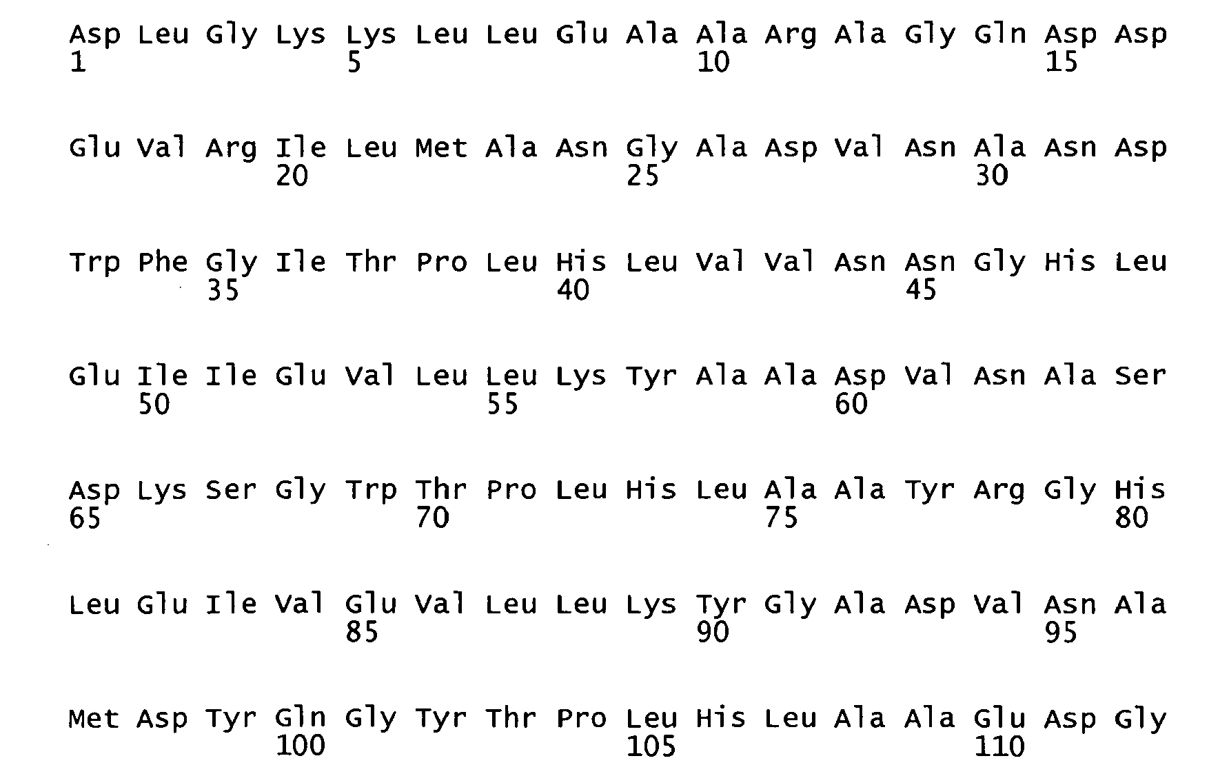

- <210> 4
<211> 32
<212> PRT
<213> Artificial - <220>
<223> Peptide derived from transcription factor GCN4 (R249 to R281, E259-, S262P) - <400> 4

- <210> 5
<211> 53
<212> DNA
<213> Artificial - <220>
<223> oDST4 oligonucleotide - <400> 5
agagcatgcg taggagaaaa taaaatgaaa aagatttggc tggcgctggc tgg 53 - <210> 6
<211> 55
<212> DNA
<213> Artificial - <220>
<223> oDST5 oligonucleotide - <400> 6
tctttgtagt ccgccgatgc gctaaacgct aaaactaaac cagccagcgc cagcc 55 - <210> 7
<211> 21
<212> DNA
<213> Artificial - <220>
<223> oD5T6 oligonucleotide - <400> 7
gctctagagc atgcgtagga g 21 - <210> 8
<211> 25
<212> DNA
<213> Artificial - <220>
<223> oDST8 oligonucleotide - <400> 8
gcggatccat ctttgtagtc cgccg 25 - <210> 9
<211> 21
<212> PRT
<213> Escherichia coli - <400> 9

- <210> 10
<211> 60
<212> DNA
<213> Artificial - <220>
<223> oDST4p oligonucleotide - <400> 10
agagcatgcg taggagaaaa taaaatgaaa caaagcacta ttgcactggc actcttaccg 60 - <210> 11
<211> 59
<212> DNA
<213> Artificial - <220>
<223> oDsT5p oligonucleotide - <400> 11
ccatctttgt agtcggcttt ggtaacaggg gtgaagagca acggtaagag tgccagtgc 59 - <210> 12
<211> 23
<212> DNA
<213> Artificial - <220>
<223> oDST8p oligonucleotide - <400> 12
gcggatccat ctttgtagtc ggc 23 - <210> 13
<211> 22
<212> PRT
<213> Erwinia carotovora - <400> 13


- <210> 14
<211> 23
<212> PRT
<213> Escherichia coli - <400> 14

- <210> 15
<211> 21
<212> PRT
<213> Escherichia coli - <400> 15

- <210> 16
<211> 18
<212> PRT
<213> Escherichia coli - <400> 16

Claims (15)
- A filamentous phage display method wherein polypeptides of interest encoded by a DNA library are displayed on the phage particles, wherein said DNA library is a cDNA library or a combinatorial DNA library that encodes a peptide library, an antibody library or a library based on alternative scaffolds, and wherein the polypeptides of interest are cotranslationally translocated across the cytoplasmic membrane of Gram-negative bacteria.
- The method of claim 1 wherein the cotranslational translocation is accomplished based on the signal recognition particle pathway.
- The method of claim 1 or 2 wherein the signal sequence used for the translocation of the polypeptide of interest to be displayed on the phage particle is a signal sequence promoting cotranslational translocation of TrxA.
- The method of claim 1, 2 or 3 wherein the signal sequence used for the translocation of the polypeptides of interest to be displayed on the phage particle is selected from the group consisting of signal sequences of TorT, SfmC, FocC, CcmH, Yral, ToIB, NikA, Flgl, and DsbA, and amino acid sequences with 70% identity with one of said signal sequences, wherein said amino acid sequences conserve the overall charge, hydrophobicity and cleavage properties of the n-region, h-region and c-region of said signal sequence, respectively.
- The method of claim 4 wherein the signal sequence used for the translocation of the polypeptides of interest to be displayed on the phage particles is selected from the group consisting of signal sequences of TorT, SfmC, TolB and DsbA.
- The method of anyone of claims 1 to 5 wherein the polypeptides of interest to be displayed on the phage particles are repeat proteins.
- The method of anyone of claims 1 to 6 comprising the steps of(a) constructing a filamentous phage or phagemid vector containing an expression cassette for a fusion polypeptide that possesses an N-terminal signal sequence promoting cotranslational translocation of the fusion polypeptide across the cytoplasmic membrane of Gram-negative bacteria;(b) constructing a combinatorial library of phage or phagemid vectors by cloning of the DNA library encoding the polypeptides of interests into the expression cassette of the vector of step (a);(c) transforming suitable Gram-negative bacteria with the library of vectors of step (b); and (d) performing phage display selection cycles to separate phage particles based on the properties of the displayed polypeptides of interest.
- A filamentous phage display method wherein a polypeptide of interest displayed on the phage particle is cotranslationally translocated across the cytoplasmic membrane of Gram-negative bacteria using a signal sequence selected from the group consisting of signal sequences of TorT, SfmC, FocC, CcmH, Yral, TolB, NikA, Flgl, and DsbA, and amino acid sequences with 70% identity with one of said signal sequences, wherein said amino acid sequences conserve the overall charge, hydrophobicity and cleavage properties of the n-region, h-region and c-region of said signal sequence, respectively.
- The method of claim 8 wherein the signal sequence is selected from the group consisting of signal sequences of TorT, SfmC, TolB and DsbA.
- A phage or phagemid vector suitable for the method of claim 1 comprising a gene construct coding for a fusion protein comprising the polypeptide of interest to be displayed on the phage particle and a signal sequence selected from the group consisting of signal sequences of TorT, SfmC, FocC, CcmH, Yral, ToIB, NikA, Flgl, and DsbA, and amino acid sequences with 70% identity with one of said signal sequences, wherein said amino acid sequences conserve the overall charge, hydrophobicity and cleavage properties of the n-region, h-region and c-region of said signal sequence, respectively.
- The vector according to claim 10 wherein the signal sequence is selected from the group consisting of signal sequences of TorT, SfmC, ToIB, and DsbA.
- A library of phage or phagemid vectors comprising gene constructs coding for fusion proteins wherein each of the fusion proteins comprises a signal sequence promoting cotranslational translocation of TrxA and a polypeptide of interest to be displayed on a phage particle wherein each polypeptide of interest is encoded by a member of a DNA library, wherein said DNA library is a cDNA library or a combinatorial DNA library that encodes a peptide library, an antibody library or a library based on alternative scaffolds.
- The library of vectors of claim 12 wherein the signal sequence is selected from the group consisting of signal sequences of TorT, SfmC, FocC, CcmH, Yral, TolB, NikA, Flgl, and DsbA, and amino acid sequences with 70% identity with one of said signal sequences, wherein said amino acid sequences conserve the overall charge, hydrophobicity and cleavage properties of the n-region, h-region and c-region of said signal sequence, respectively.
- The library of vectors of claim 12 wherein the signal sequence is selected from the group consisting of signal sequences of TorT, SfmC, TolB, and DsbA.
- The library of vectors of any one of claims 12 to 14 wherein the DNA library encoding the polypeptides of interest is a DNA library encoding repeat proteins.
Priority Applications (2)
| Application Number | Priority Date | Filing Date | Title |
|---|---|---|---|
| PL06763985T PL1902131T3 (en) | 2005-07-08 | 2006-06-30 | Phage display using cotranslational translocation of fusion polypeptides |
| EP06763985A EP1902131B1 (en) | 2005-07-08 | 2006-06-30 | Phage display using cotranslational translocation of fusion polypeptides |
Applications Claiming Priority (3)
| Application Number | Priority Date | Filing Date | Title |
|---|---|---|---|
| EP05106236 | 2005-07-08 | ||
| PCT/EP2006/063729 WO2007006665A1 (en) | 2005-07-08 | 2006-06-30 | Phage display using cotranslational translocation of fusion polypeptides |
| EP06763985A EP1902131B1 (en) | 2005-07-08 | 2006-06-30 | Phage display using cotranslational translocation of fusion polypeptides |
Publications (2)
| Publication Number | Publication Date |
|---|---|
| EP1902131A1 EP1902131A1 (en) | 2008-03-26 |
| EP1902131B1 true EP1902131B1 (en) | 2009-11-11 |
Family
ID=35063135
Family Applications (1)
| Application Number | Title | Priority Date | Filing Date |
|---|---|---|---|
| EP06763985A Expired - Lifetime EP1902131B1 (en) | 2005-07-08 | 2006-06-30 | Phage display using cotranslational translocation of fusion polypeptides |
Country Status (20)
| Country | Link |
|---|---|
| US (2) | US8846577B2 (en) |
| EP (1) | EP1902131B1 (en) |
| JP (1) | JP5042218B2 (en) |
| KR (1) | KR101268939B1 (en) |
| CN (1) | CN101213299B (en) |
| AT (1) | ATE448305T1 (en) |
| AU (1) | AU2006268780B2 (en) |
| BR (1) | BRPI0613593A2 (en) |
| CA (1) | CA2614512C (en) |
| DE (1) | DE602006010386D1 (en) |
| DK (1) | DK1902131T3 (en) |
| ES (1) | ES2335136T3 (en) |
| IL (1) | IL188518A (en) |
| MX (1) | MX2008000345A (en) |
| NO (1) | NO20080665L (en) |
| NZ (1) | NZ564695A (en) |
| PL (1) | PL1902131T3 (en) |
| RU (1) | RU2412249C2 (en) |
| WO (1) | WO2007006665A1 (en) |
| ZA (1) | ZA200710687B (en) |
Families Citing this family (42)
| Publication number | Priority date | Publication date | Assignee | Title |
|---|---|---|---|---|
| WO2004003144A2 (en) | 2002-07-01 | 2004-01-08 | Human Genome Sciences, Inc. | Antibodies that specifically bind to reg iv |
| KR101491867B1 (en) * | 2007-01-31 | 2015-02-10 | 피페넥스 인크. | Bacterial leader sequences for increased expression |
| NZ592591A (en) | 2008-11-03 | 2012-04-27 | Molecular Partners Ag | Binding proteins comprising ankyrin repeast domains that inhibit the vegf-a receptor interaction |
| GB0917647D0 (en) * | 2009-10-08 | 2009-11-25 | Glaxosmithkline Biolog Sa | Expression system |
| KR101968766B1 (en) * | 2009-11-05 | 2019-04-12 | 제넨테크, 인크. | Methods and composition for secretion of heterologous polypeptides |
| TWI510246B (en) | 2010-04-30 | 2015-12-01 | Molecular Partners Ag | Modified binding proteins inhibiting the vegf-a receptor interaction |
| EP3514169B1 (en) | 2010-11-26 | 2024-03-06 | Molecular Partners AG | Designed repeat proteins binding to serum albumin |
| SI2675480T1 (en) | 2011-02-15 | 2019-05-31 | Immunogen, Inc. | Methods of preparation of conjugates |
| TW201302779A (en) | 2011-04-13 | 2013-01-16 | Glaxosmithkline Biolog Sa | Fusion protein and combination vaccine |
| WO2012149439A2 (en) | 2011-04-29 | 2012-11-01 | Janssen Biotech, Inc. | Il4/il13 binding repeat proteins and uses |
| WO2012177837A2 (en) | 2011-06-21 | 2012-12-27 | Immunogen, Inc. | Novel maytansinoid derivatives with peptide linker and conjugates thereof |
| SG11201408196RA (en) | 2012-06-28 | 2015-03-30 | Molecular Partners Ag | Designed ankyrin repeat proteins binding to platelet-derived growth factor |
| US9956341B2 (en) | 2012-07-03 | 2018-05-01 | Milestone Scientific, Inc. | Drug infusion with pressure sensing and non-continuous flow for identification of and injection into fluid-filled anatomic spaces |
| HK1211208A1 (en) | 2012-08-22 | 2016-05-20 | Immunogen, Inc. | Cytotoxic benzodiazepine derivative |
| EP2738180A1 (en) | 2012-11-30 | 2014-06-04 | Molecular Partners AG | Binding proteins comprising at least two binding domains against HER2. |
| HK1219422A1 (en) | 2013-02-28 | 2017-04-07 | Immunogen, Inc. | Conjugates comprising cell-binding agents and cytotoxic agents |
| HK1219423A1 (en) | 2013-02-28 | 2017-04-07 | Immunogen, Inc. | Conjugates comprising cell-binding agents and cytotoxic agents |
| US11453708B2 (en) | 2013-05-31 | 2022-09-27 | Molecular Partners Ag | Designed ankyrin repeat proteins binding to hepatocyte growth factor |
| WO2014194030A2 (en) | 2013-05-31 | 2014-12-04 | Immunogen, Inc. | Conjugates comprising cell-binding agents and cytotoxic agents |
| SG11201607519VA (en) * | 2014-03-14 | 2016-10-28 | Genentech Inc | Methods and compositions for secretion of heterologous polypeptides |
| EP3122900A1 (en) | 2014-03-24 | 2017-02-01 | F. Hoffmann-La Roche AG | Cancer treatment with c-met antagonists and correlation of the latter with hgf expression |
| EP3189057A1 (en) | 2014-09-03 | 2017-07-12 | ImmunoGen, Inc. | Cytotoxic benzodiazepine derivatives |
| CR20170111A (en) | 2014-09-03 | 2017-05-23 | Immunogen Inc | CYTOTOXIC BENZODIAZEPIN DERIVATIVES |
| US9458211B1 (en) | 2015-04-02 | 2016-10-04 | Molecular Partners Ag | Recombinant proteins that simultaneously bind HGF, VEGF-A and serum albumin, comprising ankyrin repeat domains |
| US10220180B2 (en) | 2015-10-16 | 2019-03-05 | Milestone Scientific, Inc. | Method and apparatus for performing a peripheral nerve block |
| WO2018045184A1 (en) | 2016-08-31 | 2018-03-08 | President And Fellows Of Harvard College | Engineered bacteria secreting therapeutic proteins and methods of use thereof |
| MY196882A (en) | 2016-09-22 | 2023-05-08 | Molecular Partners Ag | Recombinant binding proteins and their use |
| US10632255B2 (en) | 2017-02-15 | 2020-04-28 | Milestone Scientific, Inc. | Drug infusion device |
| IL298414B2 (en) | 2017-02-28 | 2024-12-01 | Immunogen Inc | History of ethansinoids with peptide linkers and their conjugates |
| WO2018195243A1 (en) | 2017-04-20 | 2018-10-25 | Immunogen, Inc. | Cytotoxic benzodiazepine derivatives and conjugates thereof |
| US11471595B2 (en) | 2017-05-04 | 2022-10-18 | Milestone Scientific, Inc. | Method and apparatus for performing a peripheral nerve block |
| WO2019133652A1 (en) | 2017-12-28 | 2019-07-04 | Immunogen, Inc. | Benzodiazepine derivatives |
| AU2020240310A1 (en) | 2019-03-21 | 2021-10-07 | Immunogen, Inc. | Methods of preparing cell-binding agent-drug conjugates |
| US20230094471A1 (en) | 2019-03-29 | 2023-03-30 | Immunogen, Inc. | Cytotoxic bis-benzodiazepine derivatives and conjugates thereof with cell-binding agents for inhibiting abnormal cell growth or for treating proliferative diseases |
| RS64961B1 (en) | 2019-04-26 | 2024-01-31 | Immunogen Inc | Camptothecin derivatives |
| US10646660B1 (en) | 2019-05-16 | 2020-05-12 | Milestone Scientific, Inc. | Device and method for identification of a target region |
| AU2020402581A1 (en) | 2019-12-11 | 2022-06-23 | Molecular Partners Ag | Designed ankyrin repeat domains with altered surface residues |
| US20210330349A1 (en) | 2020-04-24 | 2021-10-28 | Milestone Scientific, Inc. | Device and Method for Needle/Catheter Location Utilizing Correlation Analysis |
| MX2022013945A (en) | 2020-05-06 | 2023-02-14 | Molecular Partners Ag | Novel ankyrin repeat binding proteins and their uses. |
| IL298168A (en) | 2020-05-14 | 2023-01-01 | Molecular Partners Ag | Multispecific proteins |
| WO2022190016A1 (en) | 2021-03-09 | 2022-09-15 | Molecular Partners Ag | Novel darpin based multi-specific t-cell engagers |
| EP4695384A2 (en) * | 2023-04-13 | 2026-02-18 | 5Prime Biosciences, Inc. | Compositions and methods for upregulation of reverse transcription and reduction of sequence bias in rna sequencing |
Family Cites Families (2)
| Publication number | Priority date | Publication date | Assignee | Title |
|---|---|---|---|---|
| ES2299244T3 (en) | 1998-05-13 | 2008-05-16 | Domantis Limited | SYSTEM OF EXPOSURE OF PAYMENTS FOR THE SELECTION OF CORRECTLY FOLDED PROTEINS. |
| RU2008104610A (en) * | 2005-07-07 | 2009-08-20 | Рибовакс Байотекнолоджиз Са (Ch) | NEW PHAGE DISPLAY METHODS |
-
2006
- 2006-06-30 KR KR1020087003094A patent/KR101268939B1/en not_active Expired - Fee Related
- 2006-06-30 DE DE602006010386T patent/DE602006010386D1/en not_active Expired - Lifetime
- 2006-06-30 BR BRPI0613593-5A patent/BRPI0613593A2/en not_active Application Discontinuation
- 2006-06-30 WO PCT/EP2006/063729 patent/WO2007006665A1/en not_active Ceased
- 2006-06-30 AU AU2006268780A patent/AU2006268780B2/en not_active Ceased
- 2006-06-30 DK DK06763985.6T patent/DK1902131T3/en active
- 2006-06-30 JP JP2008519913A patent/JP5042218B2/en not_active Expired - Fee Related
- 2006-06-30 ES ES06763985T patent/ES2335136T3/en not_active Expired - Lifetime
- 2006-06-30 MX MX2008000345A patent/MX2008000345A/en active IP Right Grant
- 2006-06-30 NZ NZ564695A patent/NZ564695A/en not_active IP Right Cessation
- 2006-06-30 EP EP06763985A patent/EP1902131B1/en not_active Expired - Lifetime
- 2006-06-30 RU RU2008104174/10A patent/RU2412249C2/en not_active IP Right Cessation
- 2006-06-30 CA CA2614512A patent/CA2614512C/en not_active Expired - Fee Related
- 2006-06-30 CN CN2006800243863A patent/CN101213299B/en not_active Expired - Fee Related
- 2006-06-30 AT AT06763985T patent/ATE448305T1/en active
- 2006-06-30 PL PL06763985T patent/PL1902131T3/en unknown
- 2006-06-30 US US11/988,389 patent/US8846577B2/en active Active
-
2007
- 2007-12-07 ZA ZA200710687A patent/ZA200710687B/en unknown
- 2007-12-31 IL IL188518A patent/IL188518A/en not_active IP Right Cessation
-
2008
- 2008-02-05 NO NO20080665A patent/NO20080665L/en not_active Application Discontinuation
-
2014
- 2014-08-26 US US14/468,597 patent/US9447142B2/en not_active Expired - Lifetime
Also Published As
| Publication number | Publication date |
|---|---|
| CN101213299A (en) | 2008-07-02 |
| US8846577B2 (en) | 2014-09-30 |
| ATE448305T1 (en) | 2009-11-15 |
| NZ564695A (en) | 2009-02-28 |
| RU2412249C2 (en) | 2011-02-20 |
| US20110118146A1 (en) | 2011-05-19 |
| AU2006268780B2 (en) | 2011-03-24 |
| IL188518A (en) | 2011-04-28 |
| MX2008000345A (en) | 2008-04-02 |
| KR20080035603A (en) | 2008-04-23 |
| JP2009500027A (en) | 2009-01-08 |
| PL1902131T3 (en) | 2010-05-31 |
| US9447142B2 (en) | 2016-09-20 |
| HK1122839A1 (en) | 2009-05-29 |
| IL188518A0 (en) | 2008-04-13 |
| DK1902131T3 (en) | 2010-03-01 |
| JP5042218B2 (en) | 2012-10-03 |
| ZA200710687B (en) | 2008-11-26 |
| CA2614512C (en) | 2014-01-07 |
| BRPI0613593A2 (en) | 2011-01-18 |
| CN101213299B (en) | 2013-03-27 |
| EP1902131A1 (en) | 2008-03-26 |
| WO2007006665A1 (en) | 2007-01-18 |
| AU2006268780A1 (en) | 2007-01-18 |
| RU2008104174A (en) | 2009-08-20 |
| CA2614512A1 (en) | 2007-01-18 |
| KR101268939B1 (en) | 2013-05-30 |
| NO20080665L (en) | 2008-02-05 |
| ES2335136T3 (en) | 2010-03-22 |
| US20150057186A1 (en) | 2015-02-26 |
| DE602006010386D1 (en) | 2009-12-24 |
Similar Documents
| Publication | Publication Date | Title |
|---|---|---|
| US9447142B2 (en) | Phage display using contranslational translocation of fusion polypeptides | |
| US11078479B2 (en) | Polypeptide display libraries and methods of making and using thereof | |
| US9732143B2 (en) | Methods and materials for enhancing functional protein expression in bacteria | |
| Hayhurst et al. | Escherichia coliskp chaperone coexpression improves solubility and phage display of single-chain antibody fragments | |
| Steiner et al. | Signal sequences directing cotranslational translocation expand the range of proteins amenable to phage display | |
| US6274345B1 (en) | Expression vectors encoding Escherichia coli OmpC as a cell surface anchoring motif | |
| WO2002055561A1 (en) | Method for surface display of proteins on genetic carriers | |
| EP2401373A1 (en) | Signal sequence-independent pix phage display | |
| HK1122839B (en) | Phage display using contranslational translocation of fusion polypeptides | |
| Steiner et al. | Expanding the range of proteins amenable to phage display with signal sequences directing cotranslational translocation | |
| EP3448873B1 (en) | Engineered fha domains | |
| CN116348479A (en) | Bacterial pilus protein complex FimGt-DsF stabilizes protein complexes for filamentous phage generation |
Legal Events
| Date | Code | Title | Description |
|---|---|---|---|
| PUAI | Public reference made under article 153(3) epc to a published international application that has entered the european phase |
Free format text: ORIGINAL CODE: 0009012 |
|
| 17P | Request for examination filed |
Effective date: 20080107 |
|
| AK | Designated contracting states |
Kind code of ref document: A1 Designated state(s): AT BE BG CH CY CZ DE DK EE ES FI FR GB GR HU IE IS IT LI LT LU LV MC NL PL PT RO SE SI SK TR |
|
| DAX | Request for extension of the european patent (deleted) | ||
| 17Q | First examination report despatched |
Effective date: 20080602 |
|
| GRAP | Despatch of communication of intention to grant a patent |
Free format text: ORIGINAL CODE: EPIDOSNIGR1 |
|
| DAX | Request for extension of the european patent (deleted) | ||
| GRAS | Grant fee paid |
Free format text: ORIGINAL CODE: EPIDOSNIGR3 |
|
| GRAA | (expected) grant |
Free format text: ORIGINAL CODE: 0009210 |
|
| AK | Designated contracting states |
Kind code of ref document: B1 Designated state(s): AT BE BG CH CY CZ DE DK EE ES FI FR GB GR HU IE IS IT LI LT LU LV MC NL PL PT RO SE SI SK TR |
|
| REG | Reference to a national code |
Ref country code: GB Ref legal event code: FG4D |
|
| REG | Reference to a national code |
Ref country code: CH Ref legal event code: EP |
|
| REG | Reference to a national code |
Ref country code: IE Ref legal event code: FG4D |
|
| REF | Corresponds to: |
Ref document number: 602006010386 Country of ref document: DE Date of ref document: 20091224 Kind code of ref document: P |
|
| REG | Reference to a national code |
Ref country code: SE Ref legal event code: TRGR |
|
| REG | Reference to a national code |
Ref country code: DK Ref legal event code: T3 |
|
| REG | Reference to a national code |
Ref country code: ES Ref legal event code: FG2A Ref document number: 2335136 Country of ref document: ES Kind code of ref document: T3 |
|
| LTIE | Lt: invalidation of european patent or patent extension |
Effective date: 20091111 |
|
| PG25 | Lapsed in a contracting state [announced via postgrant information from national office to epo] |
Ref country code: PT Free format text: LAPSE BECAUSE OF FAILURE TO SUBMIT A TRANSLATION OF THE DESCRIPTION OR TO PAY THE FEE WITHIN THE PRESCRIBED TIME-LIMIT Effective date: 20100311 Ref country code: FI Free format text: LAPSE BECAUSE OF FAILURE TO SUBMIT A TRANSLATION OF THE DESCRIPTION OR TO PAY THE FEE WITHIN THE PRESCRIBED TIME-LIMIT Effective date: 20091111 Ref country code: LT Free format text: LAPSE BECAUSE OF FAILURE TO SUBMIT A TRANSLATION OF THE DESCRIPTION OR TO PAY THE FEE WITHIN THE PRESCRIBED TIME-LIMIT Effective date: 20091111 Ref country code: IS Free format text: LAPSE BECAUSE OF FAILURE TO SUBMIT A TRANSLATION OF THE DESCRIPTION OR TO PAY THE FEE WITHIN THE PRESCRIBED TIME-LIMIT Effective date: 20100311 |
|
| PG25 | Lapsed in a contracting state [announced via postgrant information from national office to epo] |
Ref country code: CY Free format text: LAPSE BECAUSE OF FAILURE TO SUBMIT A TRANSLATION OF THE DESCRIPTION OR TO PAY THE FEE WITHIN THE PRESCRIBED TIME-LIMIT Effective date: 20091111 Ref country code: SI Free format text: LAPSE BECAUSE OF FAILURE TO SUBMIT A TRANSLATION OF THE DESCRIPTION OR TO PAY THE FEE WITHIN THE PRESCRIBED TIME-LIMIT Effective date: 20091111 Ref country code: LV Free format text: LAPSE BECAUSE OF FAILURE TO SUBMIT A TRANSLATION OF THE DESCRIPTION OR TO PAY THE FEE WITHIN THE PRESCRIBED TIME-LIMIT Effective date: 20091111 |
|
| REG | Reference to a national code |
Ref country code: PL Ref legal event code: T3 |
|
| REG | Reference to a national code |
Ref country code: HU Ref legal event code: AG4A Ref document number: E007210 Country of ref document: HU |
|
| PG25 | Lapsed in a contracting state [announced via postgrant information from national office to epo] |
Ref country code: BG Free format text: LAPSE BECAUSE OF FAILURE TO SUBMIT A TRANSLATION OF THE DESCRIPTION OR TO PAY THE FEE WITHIN THE PRESCRIBED TIME-LIMIT Effective date: 20100211 Ref country code: RO Free format text: LAPSE BECAUSE OF FAILURE TO SUBMIT A TRANSLATION OF THE DESCRIPTION OR TO PAY THE FEE WITHIN THE PRESCRIBED TIME-LIMIT Effective date: 20091111 Ref country code: EE Free format text: LAPSE BECAUSE OF FAILURE TO SUBMIT A TRANSLATION OF THE DESCRIPTION OR TO PAY THE FEE WITHIN THE PRESCRIBED TIME-LIMIT Effective date: 20091111 |
|
| PG25 | Lapsed in a contracting state [announced via postgrant information from national office to epo] |
Ref country code: SK Free format text: LAPSE BECAUSE OF FAILURE TO SUBMIT A TRANSLATION OF THE DESCRIPTION OR TO PAY THE FEE WITHIN THE PRESCRIBED TIME-LIMIT Effective date: 20091111 |
|
| PLBE | No opposition filed within time limit |
Free format text: ORIGINAL CODE: 0009261 |
|
| STAA | Information on the status of an ep patent application or granted ep patent |
Free format text: STATUS: NO OPPOSITION FILED WITHIN TIME LIMIT |
|
| 26N | No opposition filed |
Effective date: 20100812 |
|
| PG25 | Lapsed in a contracting state [announced via postgrant information from national office to epo] |
Ref country code: GR Free format text: LAPSE BECAUSE OF FAILURE TO SUBMIT A TRANSLATION OF THE DESCRIPTION OR TO PAY THE FEE WITHIN THE PRESCRIBED TIME-LIMIT Effective date: 20100212 |
|
| PG25 | Lapsed in a contracting state [announced via postgrant information from national office to epo] |
Ref country code: MC Free format text: LAPSE BECAUSE OF NON-PAYMENT OF DUE FEES Effective date: 20100630 |
|
| PG25 | Lapsed in a contracting state [announced via postgrant information from national office to epo] |
Ref country code: LU Free format text: LAPSE BECAUSE OF NON-PAYMENT OF DUE FEES Effective date: 20100630 |
|
| PG25 | Lapsed in a contracting state [announced via postgrant information from national office to epo] |
Ref country code: TR Free format text: LAPSE BECAUSE OF FAILURE TO SUBMIT A TRANSLATION OF THE DESCRIPTION OR TO PAY THE FEE WITHIN THE PRESCRIBED TIME-LIMIT Effective date: 20091111 |
|
| REG | Reference to a national code |
Ref country code: FR Ref legal event code: PLFP Year of fee payment: 11 |
|
| REG | Reference to a national code |
Ref country code: FR Ref legal event code: PLFP Year of fee payment: 12 |
|
| PGFP | Annual fee paid to national office [announced via postgrant information from national office to epo] |
Ref country code: ES Payment date: 20170801 Year of fee payment: 13 Ref country code: CZ Payment date: 20170628 Year of fee payment: 12 Ref country code: DK Payment date: 20170621 Year of fee payment: 12 |
|
| PGFP | Annual fee paid to national office [announced via postgrant information from national office to epo] |
Ref country code: NL Payment date: 20170620 Year of fee payment: 12 Ref country code: BE Payment date: 20170620 Year of fee payment: 12 Ref country code: AT Payment date: 20170622 Year of fee payment: 12 Ref country code: SE Payment date: 20170620 Year of fee payment: 12 Ref country code: PL Payment date: 20170524 Year of fee payment: 12 Ref country code: IT Payment date: 20170622 Year of fee payment: 12 |
|
| PGFP | Annual fee paid to national office [announced via postgrant information from national office to epo] |
Ref country code: HU Payment date: 20170616 Year of fee payment: 12 |
|
| REG | Reference to a national code |
Ref country code: FR Ref legal event code: PLFP Year of fee payment: 13 |
|
| REG | Reference to a national code |
Ref country code: DK Ref legal event code: EBP Effective date: 20180630 |
|
| PG25 | Lapsed in a contracting state [announced via postgrant information from national office to epo] |
Ref country code: CZ Free format text: LAPSE BECAUSE OF NON-PAYMENT OF DUE FEES Effective date: 20180630 |
|
| REG | Reference to a national code |
Ref country code: NL Ref legal event code: MM Effective date: 20180701 |
|
| REG | Reference to a national code |
Ref country code: AT Ref legal event code: MM01 Ref document number: 448305 Country of ref document: AT Kind code of ref document: T Effective date: 20180630 |
|
| REG | Reference to a national code |
Ref country code: BE Ref legal event code: MM Effective date: 20180630 |
|
| PG25 | Lapsed in a contracting state [announced via postgrant information from national office to epo] |
Ref country code: NL Free format text: LAPSE BECAUSE OF NON-PAYMENT OF DUE FEES Effective date: 20180701 |
|
| REG | Reference to a national code |
Ref country code: IE Ref legal event code: MM4A |
|
| PG25 | Lapsed in a contracting state [announced via postgrant information from national office to epo] |
Ref country code: AT Free format text: LAPSE BECAUSE OF NON-PAYMENT OF DUE FEES Effective date: 20180630 Ref country code: IE Free format text: LAPSE BECAUSE OF NON-PAYMENT OF DUE FEES Effective date: 20180630 Ref country code: IT Free format text: LAPSE BECAUSE OF NON-PAYMENT OF DUE FEES Effective date: 20180630 Ref country code: HU Free format text: LAPSE BECAUSE OF NON-PAYMENT OF DUE FEES Effective date: 20180701 |
|
| PG25 | Lapsed in a contracting state [announced via postgrant information from national office to epo] |
Ref country code: BE Free format text: LAPSE BECAUSE OF NON-PAYMENT OF DUE FEES Effective date: 20180630 Ref country code: SE Free format text: LAPSE BECAUSE OF NON-PAYMENT OF DUE FEES Effective date: 20180701 |
|
| PG25 | Lapsed in a contracting state [announced via postgrant information from national office to epo] |
Ref country code: DK Free format text: LAPSE BECAUSE OF NON-PAYMENT OF DUE FEES Effective date: 20180630 |
|
| REG | Reference to a national code |
Ref country code: ES Ref legal event code: FD2A Effective date: 20190916 |
|
| PG25 | Lapsed in a contracting state [announced via postgrant information from national office to epo] |
Ref country code: ES Free format text: LAPSE BECAUSE OF NON-PAYMENT OF DUE FEES Effective date: 20180701 |
|
| PG25 | Lapsed in a contracting state [announced via postgrant information from national office to epo] |
Ref country code: PL Free format text: LAPSE BECAUSE OF NON-PAYMENT OF DUE FEES Effective date: 20180630 |
|
| PGFP | Annual fee paid to national office [announced via postgrant information from national office to epo] |
Ref country code: DE Payment date: 20250618 Year of fee payment: 20 |
|
| PGFP | Annual fee paid to national office [announced via postgrant information from national office to epo] |
Ref country code: GB Payment date: 20250620 Year of fee payment: 20 |
|
| PGFP | Annual fee paid to national office [announced via postgrant information from national office to epo] |
Ref country code: FR Payment date: 20250626 Year of fee payment: 20 |
|
| PGFP | Annual fee paid to national office [announced via postgrant information from national office to epo] |
Ref country code: CH Payment date: 20250701 Year of fee payment: 20 |
|
| REG | Reference to a national code |
Ref country code: CH Ref legal event code: H14 Free format text: ST27 STATUS EVENT CODE: U-0-0-H10-H14 (AS PROVIDED BY THE NATIONAL OFFICE) Effective date: 20260630 Ref country code: DE Ref legal event code: R071 Ref document number: 602006010386 Country of ref document: DE |
|
| PG25 | Lapsed in a contracting state [announced via postgrant information from national office to epo] |
Ref country code: GB Free format text: LAPSE BECAUSE OF EXPIRATION OF PROTECTION Effective date: 20260629 |
|
| REG | Reference to a national code |
Ref country code: GB Ref legal event code: PE20 Expiry date: 20260629 |