EP1879570B1 - Inhibition of reactive oxygen species and protection of mammalian cells - Google Patents
Inhibition of reactive oxygen species and protection of mammalian cells Download PDFInfo
- Publication number
- EP1879570B1 EP1879570B1 EP06751658A EP06751658A EP1879570B1 EP 1879570 B1 EP1879570 B1 EP 1879570B1 EP 06751658 A EP06751658 A EP 06751658A EP 06751658 A EP06751658 A EP 06751658A EP 1879570 B1 EP1879570 B1 EP 1879570B1
- Authority
- EP
- European Patent Office
- Prior art keywords
- cells
- ceo
- oxygen species
- reactive oxygen
- ros
- Prior art date
- Legal status (The legal status is an assumption and is not a legal conclusion. Google has not performed a legal analysis and makes no representation as to the accuracy of the status listed.)
- Expired - Lifetime
Links
Images


Classifications
-
- A—HUMAN NECESSITIES
- A61—MEDICAL OR VETERINARY SCIENCE; HYGIENE
- A61P—SPECIFIC THERAPEUTIC ACTIVITY OF CHEMICAL COMPOUNDS OR MEDICINAL PREPARATIONS
- A61P27/00—Drugs for disorders of the senses
- A61P27/02—Ophthalmic agents
-
- A—HUMAN NECESSITIES
- A61—MEDICAL OR VETERINARY SCIENCE; HYGIENE
- A61K—PREPARATIONS FOR MEDICAL, DENTAL OR TOILETRY PURPOSES
- A61K33/00—Medicinal preparations containing inorganic active ingredients
- A61K33/24—Heavy metals; Compounds thereof
- A61K33/244—Lanthanides; Compounds thereof
Definitions
- This invention relates to biological uses of nanoceria particles, and in particular to compositions for use in the protection of mammalian retinal cells from reactive oxygen species in vivo and is supported in part by funding from the National Science Foundation and National Institutes of Health under the Contract numbers: P20 RR17703, FY014427, FY13050, and FY12190.
- Cerium is a silvery metallic element, belonging to the lanthanide group. Cerium Oxide (CeO 2 ) is used in precision polishing and lapping applications. Ultra fine nano-size cerium oxide, less than 10 nanometers, is more efficient for coating purposes. Recently, it was reported by B. Rzigalinski et al. that nanoparticles prolong the life of cortical neurons in culture 4 fold over the cells without treatment; decreased the intracellular Ca2+ concentration and prevented UV damage of cortical neurons. See B.
- ROS reactive oxygen species
- Retina tissue generates a large amount of ROS which are involved in light-induced retina degeneration and age-related macular degeneration (AMD).
- ALD age-related macular degeneration
- the present invention tests the hypothesis that nanoparticles can promote the lifespan of retinal neurons in culture and protect them from apoptosis induced by hydrogen peroxide (H 2 O 2 ) in vitro by decreasing the intracellular concentration of reactive oxygen species.
- AMD age-related macular degeneration
- compositions of present invention promote a longer lifespan for retinal neurons.
- the greatest benefit of the nanoceria is its ability to get inside the cells and provide protection from reactive oxygen species (ROS).
- ROS reactive oxygen species
- An objective of the present invention is to provide a composition for use in protecting retinal neurons from apoptosis induced by reactive oxygen species in vivo.
- Another objective of the present invention is to inhibit the rise in the intracellular concentration of reactive oxygen species (ROS).
- ROS reactive oxygen species
- a further objective of the present invention is to provide a composition for use in a method for inhibiting apoptosis induced by reactive oxygen species in retinal neurons in vivo in a dose and time dependent manner.
- a further objective of the present invention is to provide a composition for use in preventing an increase in the intracellular reactive oxygen species (ROS) in a dose and time dependent manner.
- ROS reactive oxygen species
- a further objective of the present invention is to manufacture and modify cerium oxide (CeO 2 ) nanoparticles for effective use in neuronal protection in retinal cells.
- a further objective of the present invention is to manufacture and modify cerium oxide (CeO 2 ) nanoparticles for effective use in mammalian retinal cells in vivo to inhibit damage caused by reactive oxygen species (ROS).
- CeO 2 cerium oxide
- a preferred composition for use in promoting longevity of retinal neurons includes at least one of CeO.sub.n1 wherein 0 ⁇ n1 ⁇ 2 or CeO 2 in the form of ultra-fine particles.
- the preferred ultra-fine particles have a diameter in a range between 1 nanometer (nm) and 10 nm and the preferred CeO.sub.n1 is further defined as n1 equals 2.
- a preferred composition for use in inhibiting apoptosis induced by hydrogen peroxide oxidation of retinal neurons includes at least one of CeO.sub.n1 wherein 0 ⁇ n1 ⁇ 2 or CeO 2 in the form of ultra-fine particles.
- the preferred ultra-fine particles have a diameter in a range between 1 nanometer (nm) and 10 nm and the preferred CeO.sub.n1 is further defined as n1 equals 2.
- a preferred composition for use in inhibiting apoptosis of retinal neurons in a dose and time dependent manner includes at least one of CeO.sub.n1 wherein 0 ⁇ n1 ⁇ 2 or CeO 2 in the form of ultra-fine particles.
- the preferred ultra-fine particles have a diameter in a range between 1 nanometer (nm) and 10 nm and the preferred CeO.sub.n1 is further defined as n1 equals 2.
- a more preferred composition that decreases the concentration of intracellular reactive oxygen species includes at least one of CeO.sub.n1 wherein 0 ⁇ n1 ⁇ 2 or CeO 2 in the form of ultra-fine particles.
- the more preferred composition is used in the treatment of diseases of the retina selected from the group consisting of light-induced retina degeneration and age-related macular degeneration, and is also used in vivo for the treatment of other diseases in mammalian retinal cells to inhibit damage caused by reactive oxygen species (ROS).
- the mammalian retinal cells that can be treated by the composition of the present invention include, but are not limited to, retinal neurons.
- a preferred method for promoting longevity of retinal neurons includes preparing ultra- fine particles of at least one of CeO.sub.n1 wherein 0 ⁇ n1 ⁇ 2 or CeO 2 in a preselected concentration, and adding the preselected concentration of CeO.sub.n1 wherein 0 ⁇ n1 ⁇ 2 or CeO 2 to primary retinal neurons.
- the preferred ultra-fine particles have a diameter in a range between 1 nanometer (nm) and 10 nm and the CeO.sub.n1 is further defined as n1 equals 2.
- the preferred preselected concentrations of CeO 2 are in a range between 3 nanomolar (nM) and fifty nanomolar (nM), more preferably in a range between 3 nanomolar (nM) and twenty nanomolar (nM).
- the preselected concentrations of CeO 2 are administered to mammalian cells in vivo to protect the mammalian body system from damage to retinal tissue due to reactive oxygen species (ROS).
- ROS reactive oxygen species
- the invention provides a composition for use in medical treatment of diseases as defined in the appended claims.
- the present invention describes in detail a composition for use for in vivo treatment of mammalian retinal cells with ultra fine nano-size cerium oxide particles, less than 10 nanometers in diameter, to protect that "body system” from damage to tissue due to reactive oxygen species (ROS).
- ROS reactive oxygen species
- Hydrogen peroxide is one of many reactive oxygen species. H 2 O 2 is added directly to cultures for the in vitro treatments. H 2 O 2 is not added to the live tissue samples, since H 2 O 2 is one of the ROS products of light damage. The discussion below confirms that nanoceria particles inhibit all forms of reactive oxygen species (ROS).
- ROS reactive oxygen species
- nanoceria particles can protect the brain against stroke and reperfusion injury, the heart cells from effects of cardiac infarction, the skin from UV rays and burn injuries.
- Neurodegeneration e.g., Alzheimer's, Parkinson's,
- dementia amyotrophic lateral sclerosis
- mental retardation due to loss of brain cells
- This protection can extend to diseases which produce chronic problems such as cirrhosis of the liver or kidney or the multi-organ effects of aging itself.
- the nanoceria can become the universal treatment for all major and minor diseases and events which involve reactive oxygen species.
- the nanoceria (CeO 2 nanoparticles) have the ability to destroy toxic products of metabolism known as reactive oxygen species (ROS). It has been shown herein that the nanoceria particles prevent the ROS induced death of mammalian retinal neurons in culture (in vitro) and subsequently prolonged the life of the cells in culture and protected the cells from ROS. In the embodiment of the invention, the ability of the composition to provide mammalian retinal cell protection in vivo is disclosed.
- a method and composition are disclosed for promoting the lifespan of retinal neurons and protecting the nerve cells in the eye from apoptosis induced by hydrogen peroxide (H 2 O 2 ) in vitro by decreasing generation of intracellular reactive oxygen species.
- the treatment of the eye with ultra fine nano-size cerium oxide is a significant advance in biological uses of cerium oxide. Persons afflicted with such conditions as, light-induced retina degeneration and age-related macular degeneration (AMD) have hope for brighter, clearer vision.
- a primary retinal neuron culture is obtained from albino rat pups. Retinae of Sprague-Dawley albino rat pups (0-2-day old) were dissected out and mechanically dissociated in 25ml of DMEM/F12 medium. After being filtered through 230 ⁇ m and 140 ⁇ m sieves, the dissociated cells were centrifuged at 1200rpm for 5min. The cell pellets were re-suspended in the medium to 1 ⁇ 10 5 cells/ml. 1ml of the cell suspension was plated in each well pre-treated with 10 ⁇ g/ml of poly-D-lysine. The cells were maintained in the medium until day 7, when different concentrations of CeO 2 nanoparticles were added to the cultures.
- Fig. 1 The timeline for the addition of CeO 2 nanoparticles is shown in Fig. 1 .
- the treated neuronal cells were harvested on day 14, day 19, day 24 and day 29 after the beginning of treatment on day 7.
- the mixture was incubated on ice for 10 minutes.
- the fluorescent emissions of FITC and PI were detected by flow cytometry (Beckman Coulter) with the excitation filters of 492 nanometers (nm) and 550nm.
- the FITC fluorescent emissions signals the presence of cells undergoing apoptosis; whereas, the PI signals with an automatic red color fluorescence the binding to DNA fragments identifying cells in a necrotic stage.
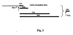
- Fig. 3 shows a treatment timeline with CeO 2 incubation after the 7 th day of treatment at the time intervals of 12 hours, 24 hours, 72 hours and 96 hours with each treated sample subsequently exposed to 12 hours incubation time with H 2 O 2 .
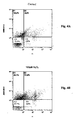
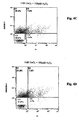
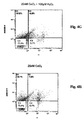
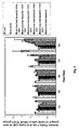
- Figures 4A - 4H are representative flow cytometry plots of retinal neurons with and without incubation with CeO 2 nanoparticles. Measurements were taken after 96 hours. The cytometry plot shows activity of approximately 10,000 cells in four quadrants, as described below.
- C1 represents the percentage of 10,000 cells showing Annexin V positive signals, which are interpreted to indicate the percentage of 10,000 cells undergoing apoptosis.
- C2 represents the percentage of 10,000 cells showing both Annexin V and PI positive signals, which is interpreted as the percentage of 10,000 cells which are in late apoptotic or necrotic stage. (AnnexinV signals apoptotic stage; PI signals necrotic stage.)
- C3 represents the percentage of 10,000 cells showing neither Annexin V nor PI positive signals, which is interpreted as the percentage of 10,000 cells which are still viable.
- C4 represents the percentage of 10,000 cells showing a PI positive signal, which is interpreted as the percentage of 10,000 cells in a necrotic stage.
- Quadrants C2 and C4 show the percentage of 10,000 cells committed to die. In quadrant C1 the percentage of 10,000 cells undergoing apoptosis are shown and some may be salvaged. It is important to observe the percentage of 10,000 cells in quadrant C2 for the efficacy of the cerium oxide treatment.
- Fig. 4A Focusing on the viable cells, the control in Fig. 4A has no CeO 2 nanoparticle treatment and 73.9% of the cell population is viable after 96 hours.
- Fig. 4B is treated with 100 ⁇ M H 2 O 2 , causing a deadly assault and leaving the lowest percentage (57.7%) of viable cells.
- the addition of gradually increasing concentrations of cerium oxide with 100 [mu]M H 2 O 2 are shown in Figures 4C - 4G .
- Fig. 4C is treated with 1nM CeO 2 nanoparticles and 100 ⁇ M H 2 O 2 and 60.6% of the cell population remains viable.
- Fig. 4D is treated with 3nM CeO 2 nanoparticles and 100 ⁇ M H 2 O 2 and 67.0% of the cell population is viable.
- Fig. 4E is treated with 5nM CeO 2 nanoparticles and 100 ⁇ M H 2 O 2 , with 68.9% of the cell population remaining viable.
- Fig. 4F is treated with 10nM CeO 2 nanoparticles and 100 ⁇ M H 2 O 2 and 72.6% of the cell population is viable.
- Fig. 4G is treated with 20nM CeO 2 nanoparticles and 100 ⁇ M H 2 O 2 and 72.3% of the cell population remains viable.
- Fig. 4H is treated with 20nM CeO 2 nanoparticles without the addition of H 2 O 2 and 73.4% of the cell population remains viable showing that the CeO 2 nanoparticles alone have no negative effect on the cell population.
- FIG. 4A The data in Figures 4A - 4H can also be summarized from the analysis of cells undergoing apoptosis as shown in quadrant C1.
- the control, Fig. 4A shows 16.2% of cells undergoing apoptosis under normal conditions, without any treatment, after 96 hours.
- Fig. 4B shows 26.1% of cells undergoing apoptosis after the 100 ⁇ M H 2 O 2 challenge.
- Figures 4C - 4G show there are 28.3%, 21.9%, 22.2%, 18.5% and 15.8% of cells undergoing apoptosis with 1, 3, 5, 10 and 20nM CeO 2 nanoparticle treatment, respectively.
- Fig. 4H shows 15.7% of cells undergoing apoptosis with 20nM CeO 2 nanoparticle treatment, which is a slight improvement over no treatment at all as shown by the control in Fig. 4A .
- cerium oxide nanoparticles inhibit apoptosis in retinal neurons in vitro in a dose and time dependent manner.
- Fig. 5 shows relative viable retinal neurons with and without incubation of different concentration of CeO 2 nanoparticles for different time periods.
- the concentration of CeO 2 nanoparticles were 1nM, 3nM, 5nM, 10nM, 20nM each with 100 ⁇ M H 2 O 2 ; one sample was treated with only 100 ⁇ M H 2 O 2 and another sample was treated with only 20nM CeO 2 nanoparticles.
- the measurement of relative viable cells for each group was determined after 12 hours, 24 hours, 48 hours, 72 hours and 96 hours. The effects showed dose and time dependency.
- 5nM cerium oxide nanoparticles started to decrease the apoptosis at 24h of incubation. As incubation time increased, the protective effect from 5nM nanoparticles was more significantly increased. Additionally, 10nM and 20nM nanoparticles began to have effects late with 96h of incubation. (Statistical analysis was done by ANOVA, and Duncan test for post hoc analysis. Data are presented in M ⁇ S.D. n ⁇ 3, *p ⁇ 0.05, **p ⁇ 0.01). Fig. 5 shows that among the various treatment doses, 5nM CeO 2 nanoparticle treatment gave the earliest response with moderate protective effect and 20nM CeO 2 nanoparticle treatment gave the highest protective effect with late response.
- ROS reactive oxygen species
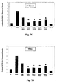
- Fig. 6 is an experimental paradigm showing the beginning of the CeO 2 nanoparticle treatment on the 7 th day and simultaneous incubation with H 2 O 2 and DCFH-DA and a timeline of exposure at 30 minutes, 12 hours, 24 hours, and 96 hours.
- Figures 7A - 7D show how the CeO 2 nanoparticles decreased the generation of ROS in a dose and time dependent manner.
- the ROS generated in the groups of 5nM, 10nM, and 20nM nanoparticle incubation were statistically significantly less than the group without treatment at the earliest (12h) of the tested points. 3nM nanoparticle incubation had an effect at 24h. However, 1nM nanoparticles did not show any significant decrease within the tested time point.
- the cerium oxide of the present invention includes those cerium compounds that have reacted with atmospheric oxygen to have stable oxide layers identified as CeO.sub.n1, wherein O is less than 1 or equal to 2 (0 ⁇ n1 ⁇ 2) or CeO 2 .
- the cerium oxide particles of the present invention are characterized as ultra-fine and are preferably in a size range of from 1 nanometer in diameter to 10 nanometers in diameter; more preferably from 1 nm to 7 nm. A judicious selection of particle size is required by someone skilled in the art.
- the results of testing in the above examples document the ability of cerium oxide nanoparticles to promote the longevity of retinal neurons in vitro and inhibit apoptosis induced by hydrogen peroxide on retinal neurons in vitro in a dose and time dependent manner. It has also been determined that cerium oxide nanoparticles decrease generation of intracellular reactive oxygen species in a dose and time dependent manner.
- the present invention represents a significant advance in the treatment in degenerative diseases of the retina, such as light-induced retina degeneration and age-related macular degeneration (AMD).
- a rat "light damage” model for retinal degeneration was used as the test system.
- the data demonstrate that the nanoceria prevented the death of retinal neurons when given prior to the "light insult”.
- the nanoceria particles protected the cells at the time of exposure as well as prevented the subsequent death seen days later in the untreated animals.
- the in vivo route of administration including, direct injection into the eye, intravenous, intraperitoneal, intramuscular, oral or topically on the eye or skin may improve the result.
- the time of administration of the nanoparticle, both before or after an insult is important.
- the nanoceria will also prevent the death of retinal cells due to glaucoma, diabetic retinopathy, inherited retinal degeneration (for example, Retinitis Pigmentosa), macular degeneration, retinal detachment or any disease or event which proceeds through the production of ROS. These particles should preserve and prolong vision when administered in vivo.
- Rats were injected intravitreally with 2 microliters ( ⁇ l) of nanoceria (concentrations from 0.1 to 1.0 micromolar) three days before they were exposed for six hours to a bright light (2700 LUX) in a light box.
- the animals were returned to normal lighting for 3.5 days, then killed, the eyes enucleated, fixed, processed for paraffin embedding, sectioned, and either stained with H & E or processed for immunocytochemistry.
- the number of photoreceptors remaining was determined with the H&E sections by using a microscope connected to a digital camera to record the images and then measuring the thickness of the outer nuclear layer every 240 microns from the optic nerve along both the superior and the inferior retina to the ora seratta. The data was then plotted as retina thickness versus the distance from the optic nerve head. The changes in other layers of retinal cells were recorded with images but not quantified. Cells actively undergoing apoptosis were also visualized using a commercially available "Apoptosis kit" and recording the microscopic images with a fluorescence microscope.
- nanoceria particles could be used in vivo for preventing blindness or death of retinal cells, or, more in general and outside the scope of the invention as claimed, for preventing the death in vivo of any other cell type in any disease.
- the chemical properties of CeO 2 nanoparticles enable the destruction of reactive oxygen species (ROS) produced by toxins and/or products of oxygen metabolism within cells.
- ROS reactive oxygen species
- nanoceria can protect any cell type from ROS induced damages.
- Diseases which could possibly be prevented, cured or ameliorated would include hereditary blindness, macular degeneration, glaucoma, diabetic retinopathy, retinal detachment, and other blinding diseases which involve ROS would be potentially solved.
- CNS central nervous system
- neuronal death in strokes degenerative diseases such as Alzheimer's Disease, Parkinson's, Huntington's Disease, and the death of peripheral nerves are preventable or at least the rate of cell death can be decreased and would result in prolonged function of the cells, tissues, organs, and individual.
Landscapes
- Health & Medical Sciences (AREA)
- Public Health (AREA)
- Chemical & Material Sciences (AREA)
- Veterinary Medicine (AREA)
- Medicinal Chemistry (AREA)
- Pharmacology & Pharmacy (AREA)
- Life Sciences & Earth Sciences (AREA)
- Animal Behavior & Ethology (AREA)
- General Health & Medical Sciences (AREA)
- Epidemiology (AREA)
- Inorganic Chemistry (AREA)
- Ophthalmology & Optometry (AREA)
- Engineering & Computer Science (AREA)
- Bioinformatics & Cheminformatics (AREA)
- Chemical Kinetics & Catalysis (AREA)
- General Chemical & Material Sciences (AREA)
- Nuclear Medicine, Radiotherapy & Molecular Imaging (AREA)
- Organic Chemistry (AREA)
- Pharmaceuticals Containing Other Organic And Inorganic Compounds (AREA)
- Medicines Containing Material From Animals Or Micro-Organisms (AREA)
- Polysaccharides And Polysaccharide Derivatives (AREA)
- Medicines That Contain Protein Lipid Enzymes And Other Medicines (AREA)
Abstract
Description
- This invention claims the benefit of priority from United States Provisional
- Application Serial No.
60/676,043 filed April 29, 2005 60/716,630 filed September 13, 2005 - This invention relates to biological uses of nanoceria particles, and in particular to compositions for use in the protection of mammalian retinal cells from reactive oxygen species in vivo and is supported in part by funding from the National Science Foundation and National Institutes of Health under the Contract numbers: P20 RR17703, FY014427, FY13050, and FY12190.
- Cerium is a silvery metallic element, belonging to the lanthanide group. Cerium Oxide (CeO2) is used in precision polishing and lapping applications. Ultra fine nano-size cerium oxide, less than 10 nanometers, is more efficient for coating purposes. Recently, it was reported by B. Rzigalinski et al. that nanoparticles prolong the life of cortical neurons in
culture 4 fold over the cells without treatment; decreased the intracellular Ca2+ concentration and prevented UV damage of cortical neurons. See B. Rzigalinski et al., "Cerium Oxide Nanoparticles Extend Cell Longevity and Act as Free Radical Scavengers" at website http://www.med.miami.edu/mnbws/Rzigalinski112.html. Based on its chemical characteristics, this effect is partially due to a decrease of reactive oxygen species (ROS). - Retina tissue generates a large amount of ROS which are involved in light-induced retina degeneration and age-related macular degeneration (AMD). The present invention tests the hypothesis that nanoparticles can promote the lifespan of retinal neurons in culture and protect them from apoptosis induced by hydrogen peroxide (H2O2) in vitro by decreasing the intracellular concentration of reactive oxygen species.
- In 2004, T. H. Margrain et al. discuss the state of research in the treatment of age-related macular degeneration in Progress in Retinal and Eye Research, 2004, 23: 523-531, "Do Blue Light Filters Confer Protection Against Age-Related Macular Degeneration?" The problem of apoptosis in the body is discussed in an article by P. Moongkarndi et al. in "Antiproliferation, Antioxidation and Induction of Apoptosis by Garcinia Mangostana (Mangosteen) on SKBR3 Human Breast Cancer Cell Line", Jl. of Ethnopharmacology, 2004, 90: 161-166.
- Often persons suffering from light-induced retina degeneration and age-related macular degeneration (AMD) are without satisfactory remedies to prevent the eventual outcome of blindness. There are some proteins available for neuronal protection of retinal cells, however, they are big molecules and over time their effect may fade away.
- It is desirable to find reliable solutions to prolong the lifespan of retinal neurons so that blindness is avoided for persons with retina degeneration and AMD.
- In addition to diseases of the eye, many human diseases are due to the death of cells in specific tissues or organs. The majority of those diseases are due to accumulation of metabolic insults from reactive oxygen species originating within or outside of the cells. These diseases include all forms of blindness whether hereditary, light-induced, or physical damage such as occurs in retinal detachment. In addition, damage can be due to ageing, stroke, cardiac infarction, bums, etc, which proceed through reactive oxygen species.
- The compositions of present invention promote a longer lifespan for retinal neurons. The greatest benefit of the nanoceria is its ability to get inside the cells and provide protection from reactive oxygen species (ROS).
- An objective of the present invention is to provide a composition for use in protecting retinal neurons from apoptosis induced by reactive oxygen species in vivo.
- Another objective of the present invention is to inhibit the rise in the intracellular concentration of reactive oxygen species (ROS).
- A further objective of the present invention is to provide a composition for use in a method for inhibiting apoptosis induced by reactive oxygen species in retinal neurons in vivo in a dose and time dependent manner.
- A further objective of the present invention is to provide a composition for use in preventing an increase in the intracellular reactive oxygen species (ROS) in a dose and time dependent manner.
- A further objective of the present invention is to manufacture and modify cerium oxide (CeO2) nanoparticles for effective use in neuronal protection in retinal cells.
- A further objective of the present invention is to manufacture and modify cerium oxide (CeO2) nanoparticles for effective use in mammalian retinal cells in vivo to inhibit damage caused by reactive oxygen species (ROS).
- A preferred composition for use in promoting longevity of retinal neurons includes at least one of CeO.sub.n1 wherein 0<n1<2 or CeO2 in the form of ultra-fine particles. The preferred ultra-fine particles have a diameter in a range between 1 nanometer (nm) and 10 nm and the preferred CeO.sub.n1 is further defined as n1 equals 2.
- A preferred composition for use in inhibiting apoptosis induced by hydrogen peroxide oxidation of retinal neurons includes at least one of CeO.sub.n1 wherein 0<n1<2 or CeO2 in the form of ultra-fine particles. The preferred ultra-fine particles have a diameter in a range between 1 nanometer (nm) and 10 nm and the preferred CeO.sub.n1 is further defined as n1 equals 2.
- A preferred composition for use in inhibiting apoptosis of retinal neurons in a dose and time dependent manner includes at least one of CeO.sub.n1 wherein 0<n1<2 or CeO2 in the form of ultra-fine particles. The preferred ultra-fine particles have a diameter in a range between 1 nanometer (nm) and 10 nm and the preferred CeO.sub.n1 is further defined as n1 equals 2.
- A more preferred composition that decreases the concentration of intracellular reactive oxygen species (ROS) includes at least one of CeO.sub.n1 wherein 0<n1<2 or CeO2 in the form of ultra-fine particles. The more preferred composition is used in the treatment of diseases of the retina selected from the group consisting of light-induced retina degeneration and age-related macular degeneration, and is also used in vivo for the treatment of other diseases in mammalian retinal cells to inhibit damage caused by reactive oxygen species (ROS).
- The mammalian retinal cells that can be treated by the composition of the present invention, include, but are not limited to, retinal neurons.
- A preferred method for promoting longevity of retinal neurons includes preparing ultra- fine particles of at least one of CeO.sub.n1 wherein 0<n1<2 or CeO2 in a preselected concentration, and adding the preselected concentration of CeO.sub.n1 wherein 0<n1<2 or CeO2 to primary retinal neurons. The preferred ultra-fine particles have a diameter in a range between 1 nanometer (nm) and 10 nm and the CeO.sub.n1 is further defined as n1 equals 2. The preferred preselected concentrations of CeO2 are in a range between 3 nanomolar (nM) and fifty nanomolar (nM), more preferably in a range between 3 nanomolar (nM) and twenty nanomolar (nM).
- It is also preferred that the preselected concentrations of CeO2 are administered to mammalian cells in vivo to protect the mammalian body system from damage to retinal tissue due to reactive oxygen species (ROS).
- The invention provides a composition for use in medical treatment of diseases as defined in the appended claims.
- Further objects and advantages of the present invention will be apparent from the following detailed description of a presently preferred embodiment which is illustrated schematically in the accompanying drawings.
-
-
Fig. 1 is a timeline of exposure of a primary retinal neuron culture to treatment with cerium oxide nanoparticles. -
Fig. 2 is a graph showing the percentage of apoptotic retinal neurons in culture with and without 5 nanomoles (nM) cerium oxide nanoparticle treatment at different time points. -
Fig. 3 shows the initial cerium oxide nanoparticle treatments followed by hydrogen peroxide incubation, and time line of exposure. -
Fig. 4A is a flow cytometry plot of the control sample of primary retinal neurons with no cerium oxide nanoparticle treatments and no hydrogen peroxide incubation. -
Fig. 4B is a flow cytometry plot of retinal neurons in the presence of 100 micromoles (µM) hydrogen peroxide. -
Fig. 4C is a flow cytometry plot of retinal neurons treated with 1nM cerium oxide in the presence of 100 micromoles (µM) hydrogen peroxide. -
Fig. 4D is a flow cytometry plot of retinal neurons treated with 3nM cerium oxide in the presence of 100 micromoles (µM) hydrogen peroxide. -
Fig. 4E is a flow cytometry plot of retinal neurons treated with 5nM cerium oxide in the presence of 100 micromoles (µM) hydrogen peroxide. -
Fig. 4F is a flow cytometry plot of retinal neurons treated with 10nM cerium oxide in the presence of 100 micromoles (µM) hydrogen peroxide. -
Fig. 4G is a flow cytometry plot of retinal neurons treated with 20nM cerium oxide in the presence of 100 micromoles (µM) hydrogen peroxide. -
Fig. 4H is a flow cytometry plot of retinal neurons treated with 20nM cerium oxide. -
Fig. 5 shows relative viable retinal neurons with and without incubation of different concentrations of cerium oxide nanoparticles for time periods between approximately 12 hours and approximately 96 hours. -
Fig. 6 shows the initial cerium oxide nanoparticle treatments, hydrogen peroxide and DCFH-DA incubation, and time line of exposure. -
Fig. 7A is a graph of the intracellular level of reactive oxygen species (ROS) of retinal neurons after 30 minutes incubation with cerium oxide nanoparticles. -
Fig. 7B is a graph of the intracellular level of reactive oxygen species (ROS) of retinal neurons after 12 hours incubation with cerium oxide nanoparticles. -
Fig. 7C is a graph of the intracellular level of reactive oxygen species (ROS) of retinal neurons after 24 hours incubation with cerium oxide nanoparticles. -
Fig. 7D is a graph of the intracellular level of reactive oxygen species (ROS) of retinal neurons after 96 hours incubation with cerium oxide nanoparticles. - Before explaining the disclosed embodiments of the present invention in detail it is to be understood that the invention is not limited in its application to the details of the particular arrangements shown. The invention is defined in the appended claims.
- The present invention describes in detail a composition for use for in vivo treatment of mammalian retinal cells with ultra fine nano-size cerium oxide particles, less than 10 nanometers in diameter, to protect that "body system" from damage to tissue due to reactive oxygen species (ROS).
- Hydrogen peroxide (H2O2) is one of many reactive oxygen species. H2O2 is added directly to cultures for the in vitro treatments. H2O2 is not added to the live tissue samples, since H2O2 is one of the ROS products of light damage. The discussion below confirms that nanoceria particles inhibit all forms of reactive oxygen species (ROS).
- Furthermore and not as part of the claimed invention, nanoceria particles can protect the brain against stroke and reperfusion injury, the heart cells from effects of cardiac infarction, the skin from UV rays and burn injuries. Neurodegeneration (e.g., Alzheimer's, Parkinson's,
- dementia, amyotrophic lateral sclerosis) and potentially mental retardation (due to loss of brain cells) within the central and peripheral nervous systems can also be inhibited. This protection can extend to diseases which produce chronic problems such as cirrhosis of the liver or kidney or the multi-organ effects of aging itself. The nanoceria can become the universal treatment for all major and minor diseases and events which involve reactive oxygen species.
- The nanoceria (CeO2 nanoparticles) have the ability to destroy toxic products of metabolism known as reactive oxygen species (ROS). It has been shown herein that the nanoceria particles prevent the ROS induced death of mammalian retinal neurons in culture (in vitro) and subsequently prolonged the life of the cells in culture and protected the cells from ROS. In the embodiment of the invention, the ability of the composition to provide mammalian retinal cell protection in vivo is disclosed.
- A method and composition are disclosed for promoting the lifespan of retinal neurons and protecting the nerve cells in the eye from apoptosis induced by hydrogen peroxide (H2O2) in vitro by decreasing generation of intracellular reactive oxygen species. The treatment of the eye with ultra fine nano-size cerium oxide is a significant advance in biological uses of cerium oxide. Persons afflicted with such conditions as, light-induced retina degeneration and age-related macular degeneration (AMD) have hope for brighter, clearer vision.
- The examples below provide further detail on the preparation and treatment of retinal nerve cells with CeO2 nanoparticles.
- A primary retinal neuron culture is obtained from albino rat pups. Retinae of Sprague-Dawley albino rat pups (0-2-day old) were dissected out and mechanically dissociated in 25ml of DMEM/F12 medium. After being filtered through 230µm and 140µm sieves, the dissociated cells were centrifuged at 1200rpm for 5min. The cell pellets were re-suspended in the medium to 1×10⁵ cells/ml. 1ml of the cell suspension was plated in each well pre-treated with 10µg/ml of poly-D-lysine. The cells were maintained in the medium until
day 7, when different concentrations of CeO2 nanoparticles were added to the cultures. The timeline for the addition of CeO2 nanoparticles is shown inFig. 1 . The treated neuronal cells were harvested on day 14, day 19,day 24 and day 29 after the beginning of treatment onday 7. The percentage of apoptotic retinal neurons in the culture with and without 5nM CeO2 nanoparticle treatment is shown inFig. 2 at the 12th day, 14th day, 19th day, 24th day and 29th day. Data are shown in M±S.D. Statistics were collected by Student t-test (n=3, *p<0.05, **p<0.01).Fig. 2 confirms that at every testing period the control with no CeO2 nanoparticle treatment had a higher percentage of apoptotic retinal neurons in the culture, in contrast to the decreased percentage of apoptotic retinal neurons in the cells treated with 5nM CeO2 nanoparticles. - The detection of apoptosis by flow cytometry is illustrated in
Figures 3 ,4A - 4H and5 . After periods of incubation with CeO2 or H2O2, the cells were washed with serumfree medium 3 times, followed by treatment of 1ml of 1×trypsin for 2min. After centrifuging, the cell pellet was resuspended in 500µl of 1×PBS containing 5µl of Annexin V-FITC and 25µl of Propidium Iodide (PI). The kit used for the analysis is commercially available from Beckman Coulter and is known as the "ANNEXIN V-FITC - Kit." The mixture was incubated on ice for 10 minutes. The fluorescent emissions of FITC and PI were detected by flow cytometry (Beckman Coulter) with the excitation filters of 492 nanometers (nm) and 550nm. The FITC fluorescent emissions signals the presence of cells undergoing apoptosis; whereas, the PI signals with an automatic red color fluorescence the binding to DNA fragments identifying cells in a necrotic stage.
-
Fig. 3 shows a treatment timeline with CeO2 incubation after the 7th day of treatment at the time intervals of 12 hours, 24 hours, 72 hours and 96 hours with each treated sample subsequently exposed to 12 hours incubation time with H2O2. -
Figures 4A - 4H are representative flow cytometry plots of retinal neurons with and without incubation with CeO2 nanoparticles. Measurements were taken after 96 hours. The cytometry plot shows activity of approximately 10,000 cells in four quadrants, as described below. - C1 represents the percentage of 10,000 cells showing Annexin V positive signals, which are interpreted to indicate the percentage of 10,000 cells undergoing apoptosis.
- C2 represents the percentage of 10,000 cells showing both Annexin V and PI positive signals, which is interpreted as the percentage of 10,000 cells which are in late apoptotic or necrotic stage. (AnnexinV signals apoptotic stage; PI signals necrotic stage.)
- C3 represents the percentage of 10,000 cells showing neither Annexin V nor PI positive signals, which is interpreted as the percentage of 10,000 cells which are still viable.
- C4 represents the percentage of 10,000 cells showing a PI positive signal, which is interpreted as the percentage of 10,000 cells in a necrotic stage.
- Quadrants C2 and C4 show the percentage of 10,000 cells committed to die. In quadrant C1 the percentage of 10,000 cells undergoing apoptosis are shown and some may be salvaged. It is important to observe the percentage of 10,000 cells in quadrant C2 for the efficacy of the cerium oxide treatment.
- Focusing on the viable cells, the control in
Fig. 4A has no CeO2 nanoparticle treatment and 73.9% of the cell population is viable after 96 hours.Fig. 4B is treated with 100 µM H2O2, causing a deadly assault and leaving the lowest percentage (57.7%) of viable cells. The addition of gradually increasing concentrations of cerium oxide with 100 [mu]M H2O2 , are shown inFigures 4C - 4G . -
Fig. 4C is treated with 1nM CeO2 nanoparticles and 100 µM H2O2 and 60.6% of the cell population remains viable.Fig. 4D is treated with 3nM CeO2 nanoparticles and 100 µM H2O2 and 67.0% of the cell population is viable.Fig. 4E is treated with 5nM CeO2 nanoparticles and 100 µM H2O2, with 68.9% of the cell population remaining viable.Fig. 4F is treated with 10nM CeO2 nanoparticles and 100 µM H2O2 and 72.6% of the cell population is viable.Fig. 4G is treated with 20nM CeO2 nanoparticles and 100 µM H2O2 and 72.3% of the cell population remains viable. -
Fig. 4H is treated with 20nM CeO2 nanoparticles without the addition of H2O2 and 73.4% of the cell population remains viable showing that the CeO2 nanoparticles alone have no negative effect on the cell population. - The data in
Figures 4A - 4H can also be summarized from the analysis of cells undergoing apoptosis as shown in quadrant C1. The control,Fig. 4A shows 16.2% of cells undergoing apoptosis under normal conditions, without any treatment, after 96 hours.Fig. 4B shows 26.1% of cells undergoing apoptosis after the 100 µM H2O2 challenge.Figures 4C - 4G show there are 28.3%, 21.9%, 22.2%, 18.5% and 15.8% of cells undergoing apoptosis with 1, 3, 5, 10 and 20nM CeO2 nanoparticle treatment, respectively.Fig. 4H shows 15.7% of cells undergoing apoptosis with 20nM CeO2 nanoparticle treatment, which is a slight improvement over no treatment at all as shown by the control inFig. 4A . - Thus, cerium oxide nanoparticles inhibit apoptosis in retinal neurons in vitro in a dose and time dependent manner.
-
Fig. 5 shows relative viable retinal neurons with and without incubation of different concentration of CeO2 nanoparticles for different time periods. The concentration of CeO2 nanoparticles were 1nM, 3nM, 5nM, 10nM, 20nM each with 100 µM H2O2; one sample was treated with only 100 µM H2O2 and another sample was treated with only 20nM CeO2 nanoparticles. The measurement of relative viable cells for each group was determined after 12 hours, 24 hours, 48 hours, 72 hours and 96 hours. The effects showed dose and time dependency. - 5nM cerium oxide nanoparticles started to decrease the apoptosis at 24h of incubation. As incubation time increased, the protective effect from 5nM nanoparticles was more significantly increased. Additionally, 10nM and 20nM nanoparticles began to have effects late with 96h of incubation. (Statistical analysis was done by ANOVA, and Duncan test for post hoc analysis. Data are presented in M±S.D. n≥3, *p<0.05, **p<0.01).
Fig. 5 shows that among the various treatment doses, 5nM CeO2 nanoparticle treatment gave the earliest response with moderate protective effect and 20nM CeO2 nanoparticle treatment gave the highest protective effect with late response. - Intracelluar reactive oxygen species (ROS) production was measured in both CeO2 nanoparticle treated and control cells using 29,79-dichlorofluorescein diacetate (DCFH-DA, Sigma). Briefly, the retinal neurons were exposed to CeO2 nanoparticles with different concentrations and various incubation times. After incubation, the cells were incubated with 10µM DCFH-DA (dissolved in dimethylsulfoxide (DMSO)) at 37°C for 30 min. The cells then were incubated with 1mM H2O2 at 37°C for 30 min after the excess DCFH-DA was washed with PBS. The cells were harvested as described above. The intensity of fluorescence was detected by flow cytometry with the excitation filter of 485nm. The ROS level was calculated as a ratio: ROS = mean intensity of treated cells divided by mean intensity of control cells.
-
Fig. 6 is an experimental paradigm showing the beginning of the CeO2 nanoparticle treatment on the 7th day and simultaneous incubation with H2O2 and DCFH-DA and a timeline of exposure at 30 minutes, 12 hours, 24 hours, and 96 hours.Figures 7A - 7D show how the CeO2 nanoparticles decreased the generation of ROS in a dose and time dependent manner. The ROS generated in the groups of 5nM, 10nM, and 20nM nanoparticle incubation were statistically significantly less than the group without treatment at the earliest (12h) of the tested points. 3nM nanoparticle incubation had an effect at 24h. However, 1nM nanoparticles did not show any significant decrease within the tested time point. Statistical analysis was done by ANOVA, and Duncan test for post hoc analysis. Data were shown in M±S.D. n=3, * * p<0.01; *p<0.05. - The cerium oxide of the present invention includes those cerium compounds that have reacted with atmospheric oxygen to have stable oxide layers identified as CeO.sub.n1, wherein O is less than 1 or equal to 2 (0<n1 <2) or CeO2.
- The cerium oxide particles of the present invention are characterized as ultra-fine and are preferably in a size range of from 1 nanometer in diameter to 10 nanometers in diameter; more preferably from 1 nm to 7 nm. A judicious selection of particle size is required by someone skilled in the art.
- The results of testing in the above examples document the ability of cerium oxide nanoparticles to promote the longevity of retinal neurons in vitro and inhibit apoptosis induced by hydrogen peroxide on retinal neurons in vitro in a dose and time dependent manner. It has also been determined that cerium oxide nanoparticles decrease generation of intracellular reactive oxygen species in a dose and time dependent manner. Thus, the present invention represents a significant advance in the treatment in degenerative diseases of the retina, such as light-induced retina degeneration and age-related macular degeneration (AMD).
- In the embodiment of the present invention a rat "light damage" model for retinal degeneration was used as the test system. The data demonstrate that the nanoceria prevented the death of retinal neurons when given prior to the "light insult". The nanoceria particles protected the cells at the time of exposure as well as prevented the subsequent death seen days later in the untreated animals. The in vivo route of administration, including, direct injection into the eye, intravenous, intraperitoneal, intramuscular, oral or topically on the eye or skin may improve the result. Similarly, the time of administration of the nanoparticle, both before or after an insult, is important. Thus, the nanoceria will also prevent the death of retinal cells due to glaucoma, diabetic retinopathy, inherited retinal degeneration (for example, Retinitis Pigmentosa), macular degeneration, retinal detachment or any disease or event which proceeds through the production of ROS. These particles should preserve and prolong vision when administered in vivo.
- Rats were injected intravitreally with 2 microliters (µl) of nanoceria (concentrations from 0.1 to 1.0 micromolar) three days before they were exposed for six hours to a bright light (2700 LUX) in a light box. The animals were returned to normal lighting for 3.5 days, then killed, the eyes enucleated, fixed, processed for paraffin embedding, sectioned, and either stained with H & E or processed for immunocytochemistry.
- The number of photoreceptors remaining was determined with the H&E sections by using a microscope connected to a digital camera to record the images and then measuring the thickness of the outer nuclear layer every 240 microns from the optic nerve along both the superior and the inferior retina to the ora seratta. The data was then plotted as retina thickness versus the distance from the optic nerve head. The changes in other layers of retinal cells were recorded with images but not quantified. Cells actively undergoing apoptosis were also visualized using a commercially available "Apoptosis kit" and recording the microscopic images with a fluorescence microscope.
- The data demonstrate that the nanoceria at all concentrations tested prevented the immediate death of retinal cells shortly after exposure to light as well as the ongoing death seen days later in the untreated animals. We therefore have demonstrated in vivo that these nanoceria particles can protect cells within the rat retina from light-induced cell death.
- Prior to the present invention, it was not known that nanoceria particles could be used in vivo for preventing blindness or death of retinal cells, or, more in general and outside the scope of the invention as claimed, for preventing the death in vivo of any other cell type in any disease. The chemical properties of CeO2 nanoparticles enable the destruction of reactive oxygen species (ROS) produced by toxins and/or products of oxygen metabolism within cells.
- Thus, nanoceria can protect any cell type from ROS induced damages. Diseases which could possibly be prevented, cured or ameliorated would include hereditary blindness, macular degeneration, glaucoma, diabetic retinopathy, retinal detachment, and other blinding diseases which involve ROS would be potentially solved. In general, and outside the scope of the invention as claimed, in other cells within the central nervous system (CNS), neuronal death in strokes, degenerative diseases such as Alzheimer's Disease, Parkinson's, Huntington's Disease, and the death of peripheral nerves are preventable or at least the rate of cell death can be decreased and would result in prolonged function of the cells, tissues, organs, and individual.
- The invention has been described, disclosed, illustrated and shown in various terms of certain embodiments or modifications which it has presumed in practice, and is defined in the appended claims.
Claims (1)
- A composition for use in medical treatment of diseases of the retina selected from the group consisting of light-induced retina degeneration and age-related macular degeneration comprising at least one of CeO.sub.n1 wherein 0<n1<2 or CeO2 in the form of ultra-fine particles
wherein the ultra-fine particles have a diameter in a range between 1 nanometer (nm) and 10 nm.
Priority Applications (1)
| Application Number | Priority Date | Filing Date | Title |
|---|---|---|---|
| EP10181322A EP2359907A1 (en) | 2005-04-29 | 2006-04-27 | Inhibition of reactive oxygen species and protection of mammalian cells |
Applications Claiming Priority (3)
| Application Number | Priority Date | Filing Date | Title |
|---|---|---|---|
| US67604305P | 2005-04-29 | 2005-04-29 | |
| US71663005P | 2005-09-13 | 2005-09-13 | |
| PCT/US2006/016050 WO2006118954A2 (en) | 2005-04-29 | 2006-04-27 | Inhibition of reactive oxygen species and protection of mammalian cells |
Publications (3)
| Publication Number | Publication Date |
|---|---|
| EP1879570A2 EP1879570A2 (en) | 2008-01-23 |
| EP1879570A4 EP1879570A4 (en) | 2008-10-01 |
| EP1879570B1 true EP1879570B1 (en) | 2010-09-29 |
Family
ID=37308499
Family Applications (2)
| Application Number | Title | Priority Date | Filing Date |
|---|---|---|---|
| EP10181322A Withdrawn EP2359907A1 (en) | 2005-04-29 | 2006-04-27 | Inhibition of reactive oxygen species and protection of mammalian cells |
| EP06751658A Expired - Lifetime EP1879570B1 (en) | 2005-04-29 | 2006-04-27 | Inhibition of reactive oxygen species and protection of mammalian cells |
Family Applications Before (1)
| Application Number | Title | Priority Date | Filing Date |
|---|---|---|---|
| EP10181322A Withdrawn EP2359907A1 (en) | 2005-04-29 | 2006-04-27 | Inhibition of reactive oxygen species and protection of mammalian cells |
Country Status (7)
| Country | Link |
|---|---|
| US (2) | US7727559B2 (en) |
| EP (2) | EP2359907A1 (en) |
| AT (1) | ATE482748T1 (en) |
| AU (1) | AU2006242541B2 (en) |
| CA (1) | CA2610296C (en) |
| DE (1) | DE602006017199D1 (en) |
| WO (1) | WO2006118954A2 (en) |
Cited By (1)
| Publication number | Priority date | Publication date | Assignee | Title |
|---|---|---|---|---|
| CN108685847A (en) * | 2018-06-29 | 2018-10-23 | 温州医科大学 | A kind of long-acting anti-inflammatory ophthalmically acceptable pharmaceutical dispersions for oxidative damage induction inflammation |
Families Citing this family (27)
| Publication number | Priority date | Publication date | Assignee | Title |
|---|---|---|---|---|
| US8703200B2 (en) | 2005-04-29 | 2014-04-22 | The Board Of Regents Of The University Of Oklahoma | Inhibition of neovascularization by cerium oxide nanoparticles |
| US20110111007A1 (en) * | 2005-04-29 | 2011-05-12 | Mcginnis James F | Inhibition of retinal cell degeneration or neovascularization by cerium oxide nanoparticles |
| US10857178B2 (en) * | 2005-06-27 | 2020-12-08 | Edward Via Virginia College Of Osteopathic Medicine | Cerium oxide nanoparticles for treatment and prevention of Alzheimer's disease, Parkinson's disease, and disorders associated with free radical production and/or mitochondrial dysfunction |
| US9649337B2 (en) | 2005-06-27 | 2017-05-16 | Edward Via Virginia College Of Osteopathic Medicine | Cerium oxide nanoparticles for the treatment and prevention of stroke and cardiovascular disease |
| WO2007002662A2 (en) * | 2005-06-27 | 2007-01-04 | Edward Via Virginia College Of Osteopathic Medicine | Anti-inflammatory, radioprotective, and longevity enhancing capabilities of cerium oxide nanoparticles |
| US7959949B2 (en) | 2006-04-27 | 2011-06-14 | University Of Central Florida Research Foundation, Inc. | Functionalized nanoceria composition for ophthalmic treatment |
| WO2008064357A2 (en) * | 2006-11-22 | 2008-05-29 | University Of Florida Research Foundation, Inc. | Nanoparticles for protection of cells from oxidative stress |
| CA2672998C (en) * | 2007-01-02 | 2013-08-06 | University Of Central Florida Research Foundation, Inc. | Methods and materials for stimulating proliferation of stem cells |
| US9119391B1 (en) | 2007-07-16 | 2015-09-01 | University Of Central Florida Research Foundation, Inc. | Polymer coated ceria nanoparticles for selective cytoprotection |
| US8916199B1 (en) | 2008-04-25 | 2014-12-23 | University of Central Florida Research Foundation, Ind. | Inhibition of angiogenesis associated with ovarian cancer by nanoparticles of cerium oxide |
| WO2009151515A1 (en) | 2008-05-06 | 2009-12-17 | Qd Vision, Inc. | Solid state lighting devices including quantum confined semiconductor nanoparticles |
| US9207385B2 (en) | 2008-05-06 | 2015-12-08 | Qd Vision, Inc. | Lighting systems and devices including same |
| US9127202B1 (en) | 2008-07-18 | 2015-09-08 | University Of Central Florida Research Foundation, Inc. | Biocompatible nano rare earth oxide upconverters for imaging and therapeutics |
| US20100098768A1 (en) * | 2008-10-16 | 2010-04-22 | Clarkson University | Method of neuroprotection from oxidant injury using metal oxide nanoparticles |
| US8883519B1 (en) | 2009-03-17 | 2014-11-11 | University Of Central Florida Research Foundation, Inc. | Oxidase activity of polymeric coated cerium oxide nanoparticles |
| JP5710597B2 (en) | 2009-04-28 | 2015-04-30 | キユーデイー・ビジヨン・インコーポレーテツド | Optical material, optical component and method |
| US9585840B1 (en) | 2009-07-10 | 2017-03-07 | University Of Central Florida Research Foundation, Inc. | Redox active cerium oxide nanoparticles and associated methods |
| US8795731B1 (en) | 2009-10-12 | 2014-08-05 | University Of Central Florida Research Foundation, Inc. | Cerium oxide nanoparticle-based device for the detection of reactive oxygen species and monitoring of chronic inflammation |
| WO2012036786A1 (en) | 2010-09-17 | 2012-03-22 | University Of L'aquila | Nanoparticles of cerium oxide targeted to an amyloid-beta antigen of alzheimer's disease |
| US11116792B1 (en) | 2010-12-22 | 2021-09-14 | Biocurity Holdings, Inc. | Cerium oxide nanoparticle formulation for use in skin radioprotection and associated methods |
| US8951539B1 (en) | 2011-06-07 | 2015-02-10 | University Of Central Florida Research Foundation, Inc. | Methods of promoting angiogenesis using cerium oxide nanoparticles |
| US9161950B2 (en) | 2011-09-21 | 2015-10-20 | University Of Central Florida Foundation, Inc. | Neuronal protection by cerium oxide nanoparticles |
| US9415065B2 (en) | 2012-04-04 | 2016-08-16 | University Of Central Florida Research Foundation, Inc. | Methods of using cerium oxide nanoparticles to mitigate or protect against radiation injury |
| US9463437B2 (en) | 2013-02-14 | 2016-10-11 | University Of Central Florida Research Foundation, Inc. | Methods for scavenging nitric oxide using cerium oxide nanoparticles |
| US9669055B1 (en) | 2014-04-11 | 2017-06-06 | Marshall University Research Corporation | Methods for treating sepsis |
| US11389536B2 (en) | 2015-07-17 | 2022-07-19 | BioCurity Pharmaceuticals Inc. | Treatment of cancer with a combination of radiation, cerium oxide nanoparticles, and a chemotherapeutic agent |
| EP4074319A1 (en) | 2021-04-15 | 2022-10-19 | Fundació Hospital Universitari Vall d'Hebron - Institut de Recerca | Ophthalmic topical composition with ceria nanoparticles for treating diseases of posterior segment of the eye |
Family Cites Families (10)
| Publication number | Priority date | Publication date | Assignee | Title |
|---|---|---|---|---|
| US676043A (en) | 1899-11-22 | 1901-06-11 | John C Lincoln | Electric drill. |
| US716630A (en) | 1902-03-24 | 1902-12-23 | August Eimer | Crucible. |
| US6291506B1 (en) * | 1998-08-04 | 2001-09-18 | Wisconsin Alumni Research Foundation | Method of reducing retinal ganglion cell degeneration |
| WO2000035415A1 (en) * | 1998-12-11 | 2000-06-22 | Cognis Deutschland Gmbh | Utilization of nanoscalar organic filters which provide protection against uv light |
| US7930285B2 (en) * | 2000-03-22 | 2011-04-19 | Comscore, Inc. | Systems for and methods of user demographic reporting usable for identifying users and collecting usage data |
| FR2809637B1 (en) * | 2000-06-05 | 2003-01-24 | Rhodia Terres Rares | COLLOIDAL DISPERSION OF A CERIUM COMPOUND OR A CERIUM COMPOUND AND AT LEAST ONE OTHER ELEMENT SELECTED AMONG RARE EARTHS AND TRANSITION METALS, INCLUDING AN AMINO ACID |
| CN1189505C (en) * | 2000-07-05 | 2005-02-16 | 日本Arc株式会社 | Transparent laminate, plastic lens for eyeglass and primer compsn. |
| EP1455804A4 (en) * | 2001-12-21 | 2005-01-05 | Alcon Inc | Use of nanoparticles as carriers for biocides in ophthalmic compositions |
| US7431758B2 (en) * | 2002-10-28 | 2008-10-07 | Nissan Chemical Industries, Ltd. | Cerium oxide particles and production method therefor |
| US20060172977A1 (en) * | 2003-04-25 | 2006-08-03 | Nolan Gerard M | Method and composition for preventing, reducing and reversing ocular ischemic neuronal damage |
-
2006
- 2006-04-27 EP EP10181322A patent/EP2359907A1/en not_active Withdrawn
- 2006-04-27 EP EP06751658A patent/EP1879570B1/en not_active Expired - Lifetime
- 2006-04-27 AT AT06751658T patent/ATE482748T1/en not_active IP Right Cessation
- 2006-04-27 AU AU2006242541A patent/AU2006242541B2/en not_active Ceased
- 2006-04-27 US US11/412,665 patent/US7727559B2/en not_active Expired - Lifetime
- 2006-04-27 WO PCT/US2006/016050 patent/WO2006118954A2/en not_active Ceased
- 2006-04-27 CA CA2610296A patent/CA2610296C/en not_active Expired - Fee Related
- 2006-04-27 DE DE602006017199T patent/DE602006017199D1/en not_active Expired - Lifetime
-
2007
- 2007-04-27 US US11/796,347 patent/US7347987B2/en not_active Expired - Lifetime
Cited By (1)
| Publication number | Priority date | Publication date | Assignee | Title |
|---|---|---|---|---|
| CN108685847A (en) * | 2018-06-29 | 2018-10-23 | 温州医科大学 | A kind of long-acting anti-inflammatory ophthalmically acceptable pharmaceutical dispersions for oxidative damage induction inflammation |
Also Published As
| Publication number | Publication date |
|---|---|
| CA2610296C (en) | 2016-06-14 |
| AU2006242541B2 (en) | 2011-12-08 |
| US20060246152A1 (en) | 2006-11-02 |
| CA2610296A1 (en) | 2006-11-09 |
| US7727559B2 (en) | 2010-06-01 |
| AU2006242541A1 (en) | 2006-11-09 |
| US20070202193A1 (en) | 2007-08-30 |
| EP1879570A2 (en) | 2008-01-23 |
| DE602006017199D1 (en) | 2010-11-11 |
| EP2359907A1 (en) | 2011-08-24 |
| WO2006118954A2 (en) | 2006-11-09 |
| EP1879570A4 (en) | 2008-10-01 |
| WO2006118954A8 (en) | 2007-08-23 |
| WO2006118954A3 (en) | 2007-05-24 |
| ATE482748T1 (en) | 2010-10-15 |
| US7347987B2 (en) | 2008-03-25 |
Similar Documents
| Publication | Publication Date | Title |
|---|---|---|
| EP1879570B1 (en) | Inhibition of reactive oxygen species and protection of mammalian cells | |
| Passamaneck et al. | Cell proliferation is necessary for the regeneration of oral structures in the anthozoan cnidarian Nematostella vectensis | |
| Gomes et al. | Renoprotective, anti-oxidative and anti-apoptotic effects of oral low-dose quercetin in the C57BL/6J model of diabetic nephropathy | |
| Zou et al. | A novel function of monomeric amyloid β-protein serving as an antioxidant molecule against metal-induced oxidative damage | |
| Huang et al. | Alzheimer's disease, β-amyloid protein and zinc | |
| Flint Beal | Coenzyme Q 10 as a possible treatment for neurodegenerative diseases | |
| KR101106344B1 (en) | Mineral ions in structured water | |
| Prasannaraj et al. | Enhanced cytotoxicity of biomolecules loaded metallic silver nanoparticles against human liver (HepG2) and prostate (PC3) cancer cell lines | |
| Schochet et al. | Intraneuronal conglomerates in sporadic motor neuron disease: A light and electron microscopic study | |
| Heidari et al. | The effect of titanium dioxide nanoparticles on mice midbrain substantia nigra | |
| Sundararajan et al. | Investigation of therapeutic potential of cerium oxide nanoparticles in Alzheimer’s disease using transgenic Drosophila | |
| Su et al. | Mechanism of action of platinum nanoparticles implying from antioxidant to metabolic programming in light-induced retinal degeneration model | |
| Kim et al. | Au nanozyme-driven antioxidation for preventing frailty | |
| Hasan et al. | RETRACTED ARTICLE: Roflumilast and tadalafil improve learning and memory deficits in intracerebroventricular Aβ1–42 rat model of Alzheimer’s disease through modulations of hippocampal cAMP/cGMP/BDNF signaling pathway | |
| Chen et al. | Nanoceria particles prevent ROI-induced blindness | |
| Zheng et al. | Particulate matter aggravates Alzheimer's disease by activating the NLRP3 inflammasome to release ASC specks | |
| Anguchamy et al. | Enhancing the neuroprotective effect of squid outer skin astaxanthin against rotenone-induced neurotoxicity in in-vitro model for Parkinson's disease | |
| Zhao et al. | Selenium nanoparticles decorated with polysaccharides from Sargassum fusiforme protects against 6-OHDA-induced neurotoxicity in PC12 cells and rat model of Parkinson's disease | |
| Manikandaselvi et al. | Neuroprotective activity of S-allylcysteine on haloperidol induced Parkinson’s disease in albino mice | |
| CN118236349A (en) | Astaxanthin sea cucumber peptide nano dry powder capable of alleviating liver fibrosis and preparation method thereof | |
| CN108324711A (en) | A kind of application of compound in terms of antagonist protein builds up treatment senile dementia | |
| Bawankule et al. | Protective effect of Ocimum sanctum on ethanol-induced oxidative stress in Swiss Albino Mice brain | |
| Jalili et al. | Effects of royal jelly on the prefrontal cortex in a rat-morphine toxicity model | |
| WO2011044434A2 (en) | A composition and a method of treating cns disorders and hyperpigmentation | |
| Bianchi et al. | The relationship between lipofuscin and neuromelanin in some sites of the nervous system of the horse. |
Legal Events
| Date | Code | Title | Description |
|---|---|---|---|
| PUAI | Public reference made under article 153(3) epc to a published international application that has entered the european phase |
Free format text: ORIGINAL CODE: 0009012 |
|
| 17P | Request for examination filed |
Effective date: 20071129 |
|
| AK | Designated contracting states |
Kind code of ref document: A2 Designated state(s): AT BE BG CH CY CZ DE DK EE ES FI FR GB GR HU IE IS IT LI LT LU LV MC NL PL PT RO SE SI SK TR |
|
| AX | Request for extension of the european patent |
Extension state: AL BA HR MK YU |
|
| DAX | Request for extension of the european patent (deleted) | ||
| A4 | Supplementary search report drawn up and despatched |
Effective date: 20080902 |
|
| RIC1 | Information provided on ipc code assigned before grant |
Ipc: A61P 27/02 20060101ALI20080825BHEP Ipc: G02B 3/00 20060101ALI20080825BHEP Ipc: G02B 7/00 20060101ALI20080825BHEP Ipc: B32B 23/02 20060101ALI20080825BHEP Ipc: A61K 33/24 20060101ALI20080825BHEP Ipc: G02C 7/02 20060101AFI20080825BHEP |
|
| 17Q | First examination report despatched |
Effective date: 20090511 |
|
| GRAP | Despatch of communication of intention to grant a patent |
Free format text: ORIGINAL CODE: EPIDOSNIGR1 |
|
| RIC1 | Information provided on ipc code assigned before grant |
Ipc: A61P 27/02 20060101AFI20100304BHEP Ipc: A61K 33/24 20060101ALI20100304BHEP |
|
| GRAS | Grant fee paid |
Free format text: ORIGINAL CODE: EPIDOSNIGR3 |
|
| GRAA | (expected) grant |
Free format text: ORIGINAL CODE: 0009210 |
|
| AK | Designated contracting states |
Kind code of ref document: B1 Designated state(s): AT BE BG CH CY CZ DE DK EE ES FI FR GB GR HU IE IS IT LI LT LU LV MC NL PL PT RO SE SI SK TR |
|
| REG | Reference to a national code |
Ref country code: GB Ref legal event code: FG4D |
|
| REG | Reference to a national code |
Ref country code: CH Ref legal event code: EP |
|
| REG | Reference to a national code |
Ref country code: IE Ref legal event code: FG4D |
|
| REF | Corresponds to: |
Ref document number: 602006017199 Country of ref document: DE Date of ref document: 20101111 Kind code of ref document: P |
|
| PG25 | Lapsed in a contracting state [announced via postgrant information from national office to epo] |
Ref country code: LT Free format text: LAPSE BECAUSE OF FAILURE TO SUBMIT A TRANSLATION OF THE DESCRIPTION OR TO PAY THE FEE WITHIN THE PRESCRIBED TIME-LIMIT Effective date: 20100929 Ref country code: FI Free format text: LAPSE BECAUSE OF FAILURE TO SUBMIT A TRANSLATION OF THE DESCRIPTION OR TO PAY THE FEE WITHIN THE PRESCRIBED TIME-LIMIT Effective date: 20100929 Ref country code: AT Free format text: LAPSE BECAUSE OF FAILURE TO SUBMIT A TRANSLATION OF THE DESCRIPTION OR TO PAY THE FEE WITHIN THE PRESCRIBED TIME-LIMIT Effective date: 20100929 |
|
| REG | Reference to a national code |
Ref country code: CH Ref legal event code: PK Ref country code: CH Ref legal event code: NV Representative=s name: OK PAT AG PATENTE MARKEN LIZENZEN |
|
| REG | Reference to a national code |
Ref country code: NL Ref legal event code: VDEP Effective date: 20100929 |
|
| LTIE | Lt: invalidation of european patent or patent extension |
Effective date: 20100929 |
|
| PG25 | Lapsed in a contracting state [announced via postgrant information from national office to epo] |
Ref country code: SI Free format text: LAPSE BECAUSE OF FAILURE TO SUBMIT A TRANSLATION OF THE DESCRIPTION OR TO PAY THE FEE WITHIN THE PRESCRIBED TIME-LIMIT Effective date: 20100929 |
|
| PG25 | Lapsed in a contracting state [announced via postgrant information from national office to epo] |
Ref country code: LV Free format text: LAPSE BECAUSE OF FAILURE TO SUBMIT A TRANSLATION OF THE DESCRIPTION OR TO PAY THE FEE WITHIN THE PRESCRIBED TIME-LIMIT Effective date: 20100929 Ref country code: GR Free format text: LAPSE BECAUSE OF FAILURE TO SUBMIT A TRANSLATION OF THE DESCRIPTION OR TO PAY THE FEE WITHIN THE PRESCRIBED TIME-LIMIT Effective date: 20101230 Ref country code: SE Free format text: LAPSE BECAUSE OF FAILURE TO SUBMIT A TRANSLATION OF THE DESCRIPTION OR TO PAY THE FEE WITHIN THE PRESCRIBED TIME-LIMIT Effective date: 20100929 |
|
| PG25 | Lapsed in a contracting state [announced via postgrant information from national office to epo] |
Ref country code: NL Free format text: LAPSE BECAUSE OF FAILURE TO SUBMIT A TRANSLATION OF THE DESCRIPTION OR TO PAY THE FEE WITHIN THE PRESCRIBED TIME-LIMIT Effective date: 20100929 Ref country code: RO Free format text: LAPSE BECAUSE OF FAILURE TO SUBMIT A TRANSLATION OF THE DESCRIPTION OR TO PAY THE FEE WITHIN THE PRESCRIBED TIME-LIMIT Effective date: 20100929 Ref country code: CZ Free format text: LAPSE BECAUSE OF FAILURE TO SUBMIT A TRANSLATION OF THE DESCRIPTION OR TO PAY THE FEE WITHIN THE PRESCRIBED TIME-LIMIT Effective date: 20100929 Ref country code: SK Free format text: LAPSE BECAUSE OF FAILURE TO SUBMIT A TRANSLATION OF THE DESCRIPTION OR TO PAY THE FEE WITHIN THE PRESCRIBED TIME-LIMIT Effective date: 20100929 Ref country code: EE Free format text: LAPSE BECAUSE OF FAILURE TO SUBMIT A TRANSLATION OF THE DESCRIPTION OR TO PAY THE FEE WITHIN THE PRESCRIBED TIME-LIMIT Effective date: 20100929 Ref country code: IS Free format text: LAPSE BECAUSE OF FAILURE TO SUBMIT A TRANSLATION OF THE DESCRIPTION OR TO PAY THE FEE WITHIN THE PRESCRIBED TIME-LIMIT Effective date: 20110129 Ref country code: PT Free format text: LAPSE BECAUSE OF FAILURE TO SUBMIT A TRANSLATION OF THE DESCRIPTION OR TO PAY THE FEE WITHIN THE PRESCRIBED TIME-LIMIT Effective date: 20110131 |
|
| PG25 | Lapsed in a contracting state [announced via postgrant information from national office to epo] |
Ref country code: BE Free format text: LAPSE BECAUSE OF FAILURE TO SUBMIT A TRANSLATION OF THE DESCRIPTION OR TO PAY THE FEE WITHIN THE PRESCRIBED TIME-LIMIT Effective date: 20100929 |
|
| REG | Reference to a national code |
Ref country code: DE Ref legal event code: R081 Ref document number: 602006017199 Country of ref document: DE Owner name: THE BOARD OF REGENTS OF THE UNIVERSITY OF OKLA, US Free format text: FORMER OWNER: UNIVERSITY OF CENTRAL FLORIDA R, UNIVERSITY OF OKLAHOMA, CORP., , US Effective date: 20110429 Ref country code: DE Ref legal event code: R081 Ref document number: 602006017199 Country of ref document: DE Owner name: UNIVERSITY OF CENTRAL FLORIDA RESEARCH FOUNDAT, US Free format text: FORMER OWNER: UNIVERSITY OF CENTRAL FLORIDA R, UNIVERSITY OF OKLAHOMA, CORP., , US Effective date: 20110429 Ref country code: DE Ref legal event code: R081 Ref document number: 602006017199 Country of ref document: DE Owner name: THE BOARD OF REGENTS OF THE UNIVERSITY OF OKLA, US Free format text: FORMER OWNERS: UNIVERSITY OF CENTRAL FLORIDA RESEARCH FOUNDATION, INC., ORLANDO, FLA., US; UNIVERSITY OF OKLAHOMA, CORP., OKLAHOMA, OKLA., US Effective date: 20110429 Ref country code: DE Ref legal event code: R081 Ref document number: 602006017199 Country of ref document: DE Owner name: UNIVERSITY OF CENTRAL FLORIDA RESEARCH FOUNDAT, US Free format text: FORMER OWNERS: UNIVERSITY OF CENTRAL FLORIDA RESEARCH FOUNDATION, INC., ORLANDO, FLA., US; UNIVERSITY OF OKLAHOMA, CORP., OKLAHOMA, OKLA., US Effective date: 20110429 |
|
| PG25 | Lapsed in a contracting state [announced via postgrant information from national office to epo] |
Ref country code: ES Free format text: LAPSE BECAUSE OF FAILURE TO SUBMIT A TRANSLATION OF THE DESCRIPTION OR TO PAY THE FEE WITHIN THE PRESCRIBED TIME-LIMIT Effective date: 20110109 |
|
| PLBE | No opposition filed within time limit |
Free format text: ORIGINAL CODE: 0009261 |
|
| STAA | Information on the status of an ep patent application or granted ep patent |
Free format text: STATUS: NO OPPOSITION FILED WITHIN TIME LIMIT |
|
| REG | Reference to a national code |
Ref country code: GB Ref legal event code: S117 Free format text: REQUEST FILED; REQUEST FOR CORRECTION UNDER SECTION 117 FILED ON 6 JANUARY 2011 |
|
| REG | Reference to a national code |
Ref country code: GB Ref legal event code: S117 Free format text: CORRECTIONS ALLOWED; REQUEST FOR CORRECTION UNDER SECTION 117 FILED ON 6 JANUARY 2011 ALLOWED ON 21 JULY 2011 |
|
| REG | Reference to a national code |
Ref country code: FR Ref legal event code: CD |
|
| PG25 | Lapsed in a contracting state [announced via postgrant information from national office to epo] |
Ref country code: DK Free format text: LAPSE BECAUSE OF FAILURE TO SUBMIT A TRANSLATION OF THE DESCRIPTION OR TO PAY THE FEE WITHIN THE PRESCRIBED TIME-LIMIT Effective date: 20100929 Ref country code: PL Free format text: LAPSE BECAUSE OF FAILURE TO SUBMIT A TRANSLATION OF THE DESCRIPTION OR TO PAY THE FEE WITHIN THE PRESCRIBED TIME-LIMIT Effective date: 20100929 |
|
| REG | Reference to a national code |
Ref country code: DE Ref legal event code: R097 Ref document number: 602006017199 Country of ref document: DE Effective date: 20110630 |
|
| PG25 | Lapsed in a contracting state [announced via postgrant information from national office to epo] |
Ref country code: MC Free format text: LAPSE BECAUSE OF NON-PAYMENT OF DUE FEES Effective date: 20110430 |
|
| PG25 | Lapsed in a contracting state [announced via postgrant information from national office to epo] |
Ref country code: CY Free format text: LAPSE BECAUSE OF FAILURE TO SUBMIT A TRANSLATION OF THE DESCRIPTION OR TO PAY THE FEE WITHIN THE PRESCRIBED TIME-LIMIT Effective date: 20100929 Ref country code: LU Free format text: LAPSE BECAUSE OF NON-PAYMENT OF DUE FEES Effective date: 20110427 |
|
| PG25 | Lapsed in a contracting state [announced via postgrant information from national office to epo] |
Ref country code: BG Free format text: LAPSE BECAUSE OF FAILURE TO SUBMIT A TRANSLATION OF THE DESCRIPTION OR TO PAY THE FEE WITHIN THE PRESCRIBED TIME-LIMIT Effective date: 20101229 Ref country code: TR Free format text: LAPSE BECAUSE OF FAILURE TO SUBMIT A TRANSLATION OF THE DESCRIPTION OR TO PAY THE FEE WITHIN THE PRESCRIBED TIME-LIMIT Effective date: 20100929 |
|
| PG25 | Lapsed in a contracting state [announced via postgrant information from national office to epo] |
Ref country code: HU Free format text: LAPSE BECAUSE OF FAILURE TO SUBMIT A TRANSLATION OF THE DESCRIPTION OR TO PAY THE FEE WITHIN THE PRESCRIBED TIME-LIMIT Effective date: 20100929 |
|
| REG | Reference to a national code |
Ref country code: FR Ref legal event code: PLFP Year of fee payment: 10 |
|
| REG | Reference to a national code |
Ref country code: FR Ref legal event code: PLFP Year of fee payment: 11 |
|
| REG | Reference to a national code |
Ref country code: FR Ref legal event code: PLFP Year of fee payment: 12 |
|
| PGFP | Annual fee paid to national office [announced via postgrant information from national office to epo] |
Ref country code: DE Payment date: 20170426 Year of fee payment: 12 Ref country code: CH Payment date: 20170421 Year of fee payment: 12 Ref country code: FR Payment date: 20170420 Year of fee payment: 12 Ref country code: IE Payment date: 20170419 Year of fee payment: 12 Ref country code: GB Payment date: 20170419 Year of fee payment: 12 |
|
| PGFP | Annual fee paid to national office [announced via postgrant information from national office to epo] |
Ref country code: IT Payment date: 20170428 Year of fee payment: 12 |
|
| REG | Reference to a national code |
Ref country code: DE Ref legal event code: R119 Ref document number: 602006017199 Country of ref document: DE |
|
| REG | Reference to a national code |
Ref country code: CH Ref legal event code: PL |
|
| GBPC | Gb: european patent ceased through non-payment of renewal fee |
Effective date: 20180427 |
|
| REG | Reference to a national code |
Ref country code: IE Ref legal event code: MM4A |
|
| PG25 | Lapsed in a contracting state [announced via postgrant information from national office to epo] |
Ref country code: DE Free format text: LAPSE BECAUSE OF NON-PAYMENT OF DUE FEES Effective date: 20181101 |
|
| PG25 | Lapsed in a contracting state [announced via postgrant information from national office to epo] |
Ref country code: CH Free format text: LAPSE BECAUSE OF NON-PAYMENT OF DUE FEES Effective date: 20180430 Ref country code: LI Free format text: LAPSE BECAUSE OF NON-PAYMENT OF DUE FEES Effective date: 20180430 Ref country code: GB Free format text: LAPSE BECAUSE OF NON-PAYMENT OF DUE FEES Effective date: 20180427 |
|
| PG25 | Lapsed in a contracting state [announced via postgrant information from national office to epo] |
Ref country code: IE Free format text: LAPSE BECAUSE OF NON-PAYMENT OF DUE FEES Effective date: 20180427 Ref country code: FR Free format text: LAPSE BECAUSE OF NON-PAYMENT OF DUE FEES Effective date: 20180430 Ref country code: IT Free format text: LAPSE BECAUSE OF NON-PAYMENT OF DUE FEES Effective date: 20180427 |