EP1872337B1 - Method and system for pre-operative prediction - Google Patents
Method and system for pre-operative prediction Download PDFInfo
- Publication number
- EP1872337B1 EP1872337B1 EP06721556A EP06721556A EP1872337B1 EP 1872337 B1 EP1872337 B1 EP 1872337B1 EP 06721556 A EP06721556 A EP 06721556A EP 06721556 A EP06721556 A EP 06721556A EP 1872337 B1 EP1872337 B1 EP 1872337B1
- Authority
- EP
- European Patent Office
- Prior art keywords
- operative
- image
- photograph
- post
- model
- Prior art date
- Legal status (The legal status is an assumption and is not a legal conclusion. Google has not performed a legal analysis and makes no representation as to the accuracy of the status listed.)
- Expired - Lifetime
Links
Images


Classifications
-
- G—PHYSICS
- G06—COMPUTING OR CALCULATING; COUNTING
- G06T—IMAGE DATA PROCESSING OR GENERATION, IN GENERAL
- G06T7/00—Image analysis
- G06T7/80—Analysis of captured images to determine intrinsic or extrinsic camera parameters, i.e. camera calibration
-
- A—HUMAN NECESSITIES
- A61—MEDICAL OR VETERINARY SCIENCE; HYGIENE
- A61B—DIAGNOSIS; SURGERY; IDENTIFICATION
- A61B34/00—Computer-aided surgery; Manipulators or robots specially adapted for use in surgery
- A61B34/10—Computer-aided planning, simulation or modelling of surgical operations
-
- G—PHYSICS
- G06—COMPUTING OR CALCULATING; COUNTING
- G06T—IMAGE DATA PROCESSING OR GENERATION, IN GENERAL
- G06T19/00—Manipulating three-dimensional [3D] models or images for computer graphics
- G06T19/20—Editing of three-dimensional [3D] images, e.g. changing shapes or colours, aligning objects or positioning parts
-
- G—PHYSICS
- G06—COMPUTING OR CALCULATING; COUNTING
- G06T—IMAGE DATA PROCESSING OR GENERATION, IN GENERAL
- G06T7/00—Image analysis
- G06T7/30—Determination of transform parameters for the alignment of images, i.e. image registration
- G06T7/33—Determination of transform parameters for the alignment of images, i.e. image registration using feature-based methods
-
- A—HUMAN NECESSITIES
- A61—MEDICAL OR VETERINARY SCIENCE; HYGIENE
- A61B—DIAGNOSIS; SURGERY; IDENTIFICATION
- A61B34/00—Computer-aided surgery; Manipulators or robots specially adapted for use in surgery
- A61B34/10—Computer-aided planning, simulation or modelling of surgical operations
- A61B2034/101—Computer-aided simulation of surgical operations
- A61B2034/105—Modelling of the patient, e.g. for ligaments or bones
-
- A—HUMAN NECESSITIES
- A61—MEDICAL OR VETERINARY SCIENCE; HYGIENE
- A61B—DIAGNOSIS; SURGERY; IDENTIFICATION
- A61B90/00—Instruments, implements or accessories specially adapted for surgery or diagnosis and not covered by any of the groups A61B1/00 - A61B50/00, e.g. for luxation treatment or for protecting wound edges
- A61B90/36—Image-producing devices or illumination devices not otherwise provided for
- A61B2090/364—Correlation of different images or relation of image positions in respect to the body
-
- G—PHYSICS
- G06—COMPUTING OR CALCULATING; COUNTING
- G06T—IMAGE DATA PROCESSING OR GENERATION, IN GENERAL
- G06T2207/00—Indexing scheme for image analysis or image enhancement
- G06T2207/30—Subject of image; Context of image processing
- G06T2207/30004—Biomedical image processing
-
- G—PHYSICS
- G06—COMPUTING OR CALCULATING; COUNTING
- G06T—IMAGE DATA PROCESSING OR GENERATION, IN GENERAL
- G06T2210/00—Indexing scheme for image generation or computer graphics
- G06T2210/41—Medical
-
- G—PHYSICS
- G06—COMPUTING OR CALCULATING; COUNTING
- G06T—IMAGE DATA PROCESSING OR GENERATION, IN GENERAL
- G06T2219/00—Indexing scheme for manipulating 3D models or images for computer graphics
- G06T2219/20—Indexing scheme for editing of 3D models
- G06T2219/2021—Shape modification
Definitions
- the present invention relates to a method for the pre-operative prediction of a body or a part of a body, e.g. the face, after surgery.
- the invention also relates to a planning system wherein the method can be applied.
- 3D geometric description of (a part of) the body.
- medical imaging modalities such as CT and MRI
- 3D photographic systems The latter can be subdivided into two categories, i.e. those using active methods, which project a specific pattern on the body, and those using passive methods, which acquire a 3D geometric description of the body from one or more images and illumination conditions, with or without the use of a priori geometric knowledge.
- 3D photographic systems deliver the texture of the body, which is used to render the 3D surface.
- Motion simulation can be based on heuristic rules, physics-based knowledge, or it can be image-derived (e.g., building a statistical deformation model based on a set of images from different persons and/or expressions).
- the result can be natural or artificial.
- the facial motion of one person can be used to drive the facial motion of another person.
- 3D visualisation or rendering uses a texture map and a reflectance model of the (part of the) body.
- Texture mapping refers to a computer graphics technique wherein a texture image (or texture map) is applied to a polygonal mesh or some other surface representation by coupling the texture image (or texture map) (with associated colour/gray value) to the 3D surface. The result is that (some portion of) the texture image is mapped onto the surface when the surface is rendered.
- Texture is derived from one or more 2D or 3D photographs of the body.
- a texture map is typically delivered simultaneously with the 3D shape description.
- a method to match or register these 2D photographs with the 3D surface description is needed. Matching can be done based on a set of corresponding points, or on a metric (e.g., mutual information) that expresses the correspondence between 2D-image-derived features and 3D-shape-based properties.
- the model of body reflectance can be based on skin or skin-like diffuse and specular (mirror-like reflection) properties.
- 2D visualisation has been used to show (a part of) the body under simulated or artificial illumination conditions and for animation by morphing (part of) the body.
- photo-realism is the primary concern.
- the methods of Xia et al. and of Iwakiri et al. use a set of photographs comprising a frontal (0° view), right (90° view) and left (270° view) photograph of the patient, which are projected as a texture map onto the 3D head mesh obtained from CT for 3D visualisation.
- the present invention aims to provide a device and method for pre-operatively simulating or predicting an accurate image of the patient's appearance after surgery, in particular maxillofacial or plastic surgery.
- the invention aims to provide a planning system wherein the method can be applied.
- the present invention relates to a method for pre-operatively obtaining a prediction of a post-operative image of at least part of a body as defined in claim 1.
- the predicted post-operative image is a 3D image.
- a plurality of pre-operative 2D photographs is advantageously acquired and subsequently used in the later method steps.
- the method advantageously further comprises a step of generating from the 3D pre-operative description a 3D pre-operative surface mesh of at least the contours of the at least part of the body.
- the step of deriving the predicted image comprises deriving from the 3D pre-operative surface mesh a prediction of the 3D post-operative surface mesh of at least the contours of the at least part of the body. The prediction of the contours is then used in the determination of the deformation field.
- the 3D pre-operative description is obtained using a 3D image acquisition system.
- a 3D image acquisition system can be a Computerised Tomography system, a Magnetic Resonance Imaging system or a 3D photographic system.
- the step of matching is preferably performed by means of a set of corresponding points on said 3D pre-operative description and said 2D pre-operative photograph.
- said step of matching is performed by means a metric expressing the correspondence between features derived from the pre-operative 2D photograph and properties based on the 3D pre-operative description.
- the method further comprises the step of taking a picture of a calibration object. Said picture of the calibration object can then be used for calibrating the camera with which the pre-operative 2D photograph are acquired.
- a step is performed of creating from the matched pre-operative 2D photographs a texture map for 3D visualisation.
- the 3D pre-operative description comprises typically a soft tissue description of the at least part of the body.
- it also comprises information about the internal structure of the at least part of the body.
- the invention also relates to a surgical planning system for pre-operatively showing a predicted post-operative image of at least part of a body, as defined in claim 17.
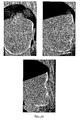
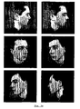
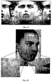
- Fig. la represents a 3D pre-operative surface mesh, projected onto the 2D pre-operative photographs after registration.
- Fig.1b and Fig.1c show 2D pre-operative and post-operative photographs, respectively.
- Fig.1d represents two views of the rendered surface mesh, using a texture map obtained from the set of 2D photographs.
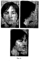
- Fig. 2a represents a 3D pre-operative surface mesh, projected onto the 2D pre-operative photographs after registration.
- Fig.2b and Fig.2c show 2D pre-operative and post-operative photographs, respectively.
- Fig.2d represents two views of the rendered surface mesh, using a texture map obtained from the set of 2D photographs.
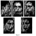
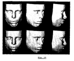
- Fig. 3a represents a 3D surface mesh, obtained with a 3D photographic system, projected onto the 2D photographs after registration.
- Fig.3b shows 2D photographs.
- Fig.3c offers six views of the rendered surface mesh using a texture map obtained from the set of 2D photographs.
- Fig. 4 represents a calibration object.
- Fig. 5 represents on the left a set of points specified manually onto the 3D rendered untextured surface, obtained from the 3D surface mesh and on the right a set of (bright) points specified manually onto the 2D photograph, together with the above set of (dark) points obtained by matching the 3D surface with the 2D photograph.
- Fig. 6 represents an iterative improvement of the accuracy by using additional corresponding points on the 2D photograph and the projected surface mesh.
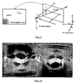
- Fig. 7 gives a schematic representation of the 3D image space I, the 3D camera space C and the 2D photographic image.
- Fig. 8 represents a spherical texture map assembled from the photographs in Fig. 3b .
- Fig. 9 represents from left to right the initial pre-operative skull.
- the skull is cut into parts that can be repositioned.
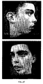
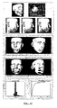
- Fig. 10 represents on the top row the pre-operative facial skin surface and on the bottom row the predicted post-operative skin surface.
- Fig. 11 illustrates the planning system accuracy.
- bone displacement field up to 13.7 mm.
- Middle the rendered surfaces correspond to positions on the face where the difference between simulated (pre-operative) and real (post-operative) surface are less than 2 mm and 1 mm, respectively.
- histogram left
- cumulative histogram right
- Fig. 12 represents snapshots of the 3D planning system at work, with facility for soft tissue prediction.
- the face is shown as a 3D rendered texture surface (pre-operative state). Snapshots of the 3D planning system at work, with facility for soft tissue prediction.
- the face is shown as a 3D rendered textured surface (simulated post-operative state).
- Fig. 13 represents a cylindrical texture map assembled from the photographs in Fig.2b .
- Fig. 14 represents the deformation field (short lines) projected onto the 2D image.
- Fig. 15 represents the displacement field (short lines) and boundary of the dilated region (outer contour). Outside this area, the displacements are zero and the image is not deformed.
- the present invention differs from the prior art in two fundamental aspects. Firstly, in the approach according to the present invention one or more 2D photographs taken from any viewing position can be used. A viewing position is to be considered as a vector having a direction as well as a magnitude, i.e. a viewing position consists of a viewing direction and a camera distance. The number of 2D photographs is thus arbitrary. Secondly, the visualisation is not restricted to 3D visualisation, for which the 2D photographs are used as texture maps, but a single arbitrary pre-operative 2D photograph can be deformed into a simulated post-operative 2D photograph using a physics-based reliable, personalised and accurately predicting 3D deformation field. While 3D visualisation using texture mapping lacks photo-realism (e.g. unnatural texture blending and hair modelling artefacts (particularly when using medical imaging, such as CT for 3D image acquisition), the simulated post-operative 2D photograph has intrinsically the same photo-realism as the original pre-operative photograph.
- 3D visualisation using texture mapping lacks
- 'planning system' is meant a software environment that allows a physician to plan or simulate the procedure of an intervention. It can for example be used to predict the outcome of that intervention, to try out different procedures, to optimise the procedure, to prepare it and to improve the communication between the medical staff and with the patient.
- Real time 3D visualisation using texture mapping offers an added value to the surgeon while using the 3D planning system, e.g. when adjusting or repositioning bony structures or an implant.
- Accuracy and integration in the planning procedure are of primary importance, and photo-realism is of minor importance. Visualisation is possible from any viewing direction.
- the 2D geometrically deformed photographs offer both high accuracy and high photo-realism and are for example an excellent means to discuss the expected outcome of a surgical procedure with the patient.
- visualisation is restricted to the viewing directions of the original 2D photographs, the number as well as the viewing directions can be arbitrarily chosen.
- a 3D pre-operative image is acquired of (a part of) a patient's body.
- a 3D image acquisition system is preferably used thereto, such as CT (Computerised Tomography), MRI (Magnetic Resonance Imaging) or any other 3D scanning or photographic system.
- 3D medical imaging modalities such as CT or MRI, offer geometric information of the body contour (further also referred to as the 'soft tissue') and internal structures, such as the bony structures.
- the 3D contour of the skin and other tissues, such as bone are segmented. In the case of skin and bone, segmentation can for example be performed by simple thresholding.
- any other 3D scanning device can be used to obtain the outer body contour, such as a 3D photographic system.
- 3D photographic systems can be subdivided into two categories, i.e. those using active methods, which project a specific pattern on the body, and those using passive methods, which acquire a 3D geometric description of the body from one or more images and illumination conditions, with or without the use of a priori geometric knowledge.
- 'Modeling and animating realistic faces from images' Pighin et al., Int J Comp Vision 50(2), pp.143-169, 2002
- a 3D generic face model is interactively fitted to a set of images to acquire the 3D shape.
- a set of (one or more) 2D photographs of (a part of) the body from any viewing direction and camera distance (i.e. any viewing position as previously defined) using any camera is acquired.
- one or more 2D pictures are taken from arbitrarily chosen directions.
- the 3D data are used to generate a 3D surface mesh of the body contour (the 'soft tissue') and, if needed by the planning system, of other tissues such as bone.
- Surface meshes such as the triangular meshes shown in Figs. 1a , 2a and 3a , can for example be created using the marching cubes algorithm.
- a registration method is then applied to match or register the 3D pre-operative surface description with the 2D photographs.
- One way to align or register the 3D surface with a 2D photograph is shown in Fig.5 , where a set of corresponding points on the 3D surface and the 2D photograph, respectively, is used.
- the problem then is how to transfer a point from the 3D image space I to the related camera space C and further to the corresponding 2D photographic image. It is assumed that the camera can be modelled as a perspective pinhole camera with its optical centre located at c (see Fig. 7 ).
- the geometric relation between I and C can then be expressed by a rotation R and a translation I.
- the internal calibration parameters are very sensitive to small errors on the position of the corresponding reference points. As already mentioned, it is therefore recommended to take first a picture of a separate calibration object with accurately known geometry and texture ( Fig. 4 ), to calculate the internal parameters of the camera, and to freeze these settings during the remainder of the photo session.
- the corresponding reference points on the acquired 3D image and 2D photographic image of (part of) the body ( Fig. 5 ) are subsequently used for the external calibration.
- the accuracy of the registration can iteratively be improved by adding corresponding points on the 2D photograph and the projected surface mesh ( Fig. 6 ).
- registration of the 3D surface with a 2D photograph can also be performed for example based on the optimisation of an objective function that expresses the correspondence between 2D-image-derived features and 3D-shape-based properties (e.g., mutual information).
- a 2D texture map is created from the registered 2D photographs.
- the surface mesh and corresponding texture are used for 3D visualisation.
- the texture map is then mapped onto the 3D body surface.
- View-dependent (using a single texture map for fast displaying, e.g. a virtual sphere enclosing the 3D body contour) and view-independent texture mapping ( Fig.8 ) assume a known relationship between the 3D surface coordinates and the 2D photographic coordinates, as well as a method to calculate the texture values from the colours or gray values in the available photographs. Once the 2D photographs and 3D surface are matched, the mapping between the 3D surface coordinates and the 2D coordinates in each photograph is known.
- Each 3D mesh point corresponds to a point in each 2D photograph and its texture value is nonzero if the 3D mesh point is visible in and front facing at least one of the 2D photographs.
- a corresponding "visible mesh” is generated by removing the vertices that are invisible from the camera position, together with the triangles they belong to.
- the texture value can for example be calculated as a normalised weighted combination of the corresponding colour or gray values in the contributing photographs as proposed in the above-mentioned papers by Pighin or by Iwakiri.
- This weight function should provide a smooth and seamless transition between the photographic patches.
- the weight function ( ⁇ - ⁇ / 2 ) 2 has been used, with ⁇ the angle between the surface normal and the line from the surface point to the camera position of the photograph.
- a 3D patient-specific planning system (e.g., for maxillofacial surgery, breast augmentation, nose correction, etc.), including a soft tissue prediction, is used to simulate the post-operative shape.
- the soft tissue prediction is used to deform the pre-operative surface mesh of the soft tissue with associated texture map into a predicted post-operative soft tissue mesh with associated remapped texture.
- the post-operative soft-tissue mesh and corresponding texture map is used for 3D visualisation of the soft tissue.
- Motion simulation can be based on heuristic rules, physics-based knowledge, or it can be image-derived (e.g., building a statistical deformation model based on a set of images from different expressions or a linear combination of a set of textured face meshes each corresponding to a facial expression, such as joy, anger, sadness).
- the result can be natural or artificial (e.g., the facial motion of one person can be used to drive the facial motion of another person) .
- the invention makes use of a personalised and accurately predicting 3D deformation field for maxillofacial and plastic surgery.
- a 3D planning system for maxillofacial surgery which yields an accurate personalised 3D deformation field of the face.
- a maxillofacial procedure can be subdivided into two separate parts, i.e., the bone-related planning and the soft tissue simulation.
- the bone-related planner allows the surgeon to reshape the skull in a 3D environment. Reshaping the skull implies cutting the skull into different parts and repositioning each of the different parts ( Fig. 9 ).
- the new facial shape of the patient can be simulated ( Figs. 10 ).
- a mathematical model is used that is able to accurately simulate the behaviour of the facial tissues.
- Known models are the finite element model (FEM), the mass-spring model (MSM) and the mass-tensor model (MTM).
- a set of boundary conditions is used, which are generated from the bone-related planning.
- 'Very fast soft tissue predictions with mass tensor model for maxillofacial surgery planning systems' (Mollemans et al., Proc Computer Assisted Radiology and Surgery (CARS), 2005) for example, it is assumed that the soft tissue is attached to the bone in a number of locations and that the soft tissue in these points follows the same motion trajectory as the corresponding attaching skull points.
- the deformation of the remainder of the soft tissue is found by requiring that the total force in each such soft tissue point should be zero or by integrating a motion equation over time.
- Fig. 11 shows the accuracy of the simulation.
- Fig. 12 shows a few snapshots of the planning system at work on the same patient as used in Figs 9-11 .
- the soft tissue with associated texture moves in real time and simultaneously with the bone displacements.
- Fig. 13 shows the associated texture map for this patient.
- the 3D pre-operative and post-operative surface meshes are projected onto the pre-operative 2D photographs.
- the vertices of the pre-operative 3D surface meshes that are visible from the camera viewpoint were previously mapped or projected onto the pre-operative 2D photographs (Fig. la, 2a, 3a) using the registration parameters and matrices previously obtained. For each of these vertices a displacement vector and corresponding vertex in the post-operative 3D surface mesh is known. These corresponding vertices are also projected onto the pre-operative 2D photographs. Since the pre-operative 3D surface mesh is deformed into a post-operative mesh, some vertices that were previously visible, may become invisible now. These vertices are also removed as well as their associated vertex in the pre-operative mesh.
- the projected deformation field acquired from the pre-operative and post-operative soft-tissue meshes, is used to geometrically deform the pre-operative 2D photographs and predict the post-operative 2D photographs ( Fig. 1c , 2c ).
- a patient-specific 3D deformation model is used to deform the 2D photographs geometrically. From the projected pre-operative and post-operative soft-tissue meshes, the 2D displacement of all the projected mesh vertices in the 2D photograph is known. Hence, the 2D geometric deformation vector is known in a discrete number of points in the 2D photograph ( Fig. 14 ). The displacement in each pixel of the photograph can then be calculated by interpolation between the discrete deformation vectors. Outside the projected mesh, the deformation is in principle zero. However, due to small mismatches between the 2D photograph and the projected pre-operative surface mesh, it may be recommended to slightly extrapolate the deformation field outside the mesh.
- Fig. 15 is a typical example.
- the contour of an enlarged region can be used as the zero-deformation borderline.
- interpolation of the discrete deformation field can for example be performed using bicubic spline functions.
- the above method can also be used in practice without the steps of determining a deformation field and using the deformation field to deform the one or more pre-operative 2D photographs.
- the latter step results in a predicted post-operative 2D photograph.
- 3D visualisation using texture mapping lacks photo-realism (e.g. unnatural texture blending and hair modelling artefacts (particularly when using medical imaging, such as CT for 3D image acquisition) and the texture map mostly needs retouching.
- the simulated post-operative 2D photograph on the other hand, has intrinsically the same photo-realism as the original pre-operative photograph.
Landscapes
- Engineering & Computer Science (AREA)
- Physics & Mathematics (AREA)
- Health & Medical Sciences (AREA)
- Theoretical Computer Science (AREA)
- General Physics & Mathematics (AREA)
- Life Sciences & Earth Sciences (AREA)
- Computer Vision & Pattern Recognition (AREA)
- Surgery (AREA)
- Molecular Biology (AREA)
- Heart & Thoracic Surgery (AREA)
- Computer Graphics (AREA)
- Nuclear Medicine, Radiotherapy & Molecular Imaging (AREA)
- Robotics (AREA)
- General Engineering & Computer Science (AREA)
- Architecture (AREA)
- General Health & Medical Sciences (AREA)
- Animal Behavior & Ethology (AREA)
- Medical Informatics (AREA)
- Computer Hardware Design (AREA)
- Software Systems (AREA)
- Biomedical Technology (AREA)
- Public Health (AREA)
- Veterinary Medicine (AREA)
- Magnetic Resonance Imaging Apparatus (AREA)
- Processing Or Creating Images (AREA)
- Measuring And Recording Apparatus For Diagnosis (AREA)
- Image Processing (AREA)
- Apparatus For Radiation Diagnosis (AREA)
- Image Generation (AREA)
- Materials For Medical Uses (AREA)
- Pharmaceuticals Containing Other Organic And Inorganic Compounds (AREA)
Applications Claiming Priority (2)
| Application Number | Priority Date | Filing Date | Title |
|---|---|---|---|
| GBGB0507204.6A GB0507204D0 (en) | 2005-04-08 | 2005-04-08 | Maxillofacial and plastic surgery |
| PCT/BE2006/000035 WO2006105625A1 (en) | 2005-04-08 | 2006-04-07 | Method and system for pre-operative prediction |
Publications (2)
| Publication Number | Publication Date |
|---|---|
| EP1872337A1 EP1872337A1 (en) | 2008-01-02 |
| EP1872337B1 true EP1872337B1 (en) | 2011-11-02 |
Family
ID=34610863
Family Applications (1)
| Application Number | Title | Priority Date | Filing Date |
|---|---|---|---|
| EP06721556A Expired - Lifetime EP1872337B1 (en) | 2005-04-08 | 2006-04-07 | Method and system for pre-operative prediction |
Country Status (7)
| Country | Link |
|---|---|
| US (1) | US8428315B2 (https=) |
| EP (1) | EP1872337B1 (https=) |
| JP (1) | JP4979682B2 (https=) |
| AT (1) | ATE532155T1 (https=) |
| ES (1) | ES2376905T3 (https=) |
| GB (1) | GB0507204D0 (https=) |
| WO (1) | WO2006105625A1 (https=) |
Families Citing this family (39)
| Publication number | Priority date | Publication date | Assignee | Title |
|---|---|---|---|---|
| US7953260B2 (en) * | 2006-06-09 | 2011-05-31 | Craniosim Solutions, Inc. | Predicting movement of soft tissue of the face in response to movement of underlying bone |
| EP1982652A1 (en) | 2007-04-20 | 2008-10-22 | Medicim NV | Method for deriving shape information |
| KR100896065B1 (ko) * | 2007-12-17 | 2009-05-07 | 한국전자통신연구원 | 3차원 얼굴 표정 애니메이션 생성 방법 |
| US8795204B2 (en) | 2008-01-09 | 2014-08-05 | Allergan, Inc. | Anatomical recognition and dimensional analysis of breast volume to assist breast surgery |
| US8294708B2 (en) | 2008-01-09 | 2012-10-23 | Allergan, Inc. | Anatomical recognition, orientation and display of an upper torso to assist breast surgery |
| FR2942125B1 (fr) | 2009-02-17 | 2012-02-17 | Obl | Ensemble sur mesure d'au moins deux plaques d'osteosynthese, elles-memes preparees sur mesure, et procede de mise en place desdites plaques |
| US8531473B2 (en) * | 2009-07-28 | 2013-09-10 | Technion R&D Foundation Ltd. | Photogrammetric texture mapping using casual images |
| EP2306400B1 (en) | 2009-09-04 | 2015-02-11 | Medicim NV | Method for digitizing dento-maxillofacial objects |
| US10970655B2 (en) * | 2010-05-03 | 2021-04-06 | Technion Research & Development Foundation Ltd | Surgery planning based on predicted results |
| AU2011256145A1 (en) * | 2010-05-21 | 2012-12-20 | My Orthodontics Pty Ltd | Prediction of post-procedural appearance |
| WO2012109596A1 (en) | 2011-02-11 | 2012-08-16 | Embrey Cattle Company | System and method for modeling a biopsy specimen |
| EP3429195A1 (en) * | 2012-02-27 | 2019-01-16 | Perceptiko AG | Method and system for image processing in video conferencing for gaze correction |
| US9275302B1 (en) * | 2012-08-24 | 2016-03-01 | Amazon Technologies, Inc. | Object detection and identification |
| GB201216224D0 (en) | 2012-09-12 | 2012-10-24 | Nobel Biocare Services Ag | An improved virtual splint |
| GB201216230D0 (en) | 2012-09-12 | 2012-10-24 | Nobel Biocare Services Ag | An improved surgical template |
| GB201216214D0 (en) | 2012-09-12 | 2012-10-24 | Nobel Biocare Services Ag | A digital splint |
| WO2014169301A1 (en) * | 2013-04-12 | 2014-10-16 | Bui The Duy | Systems and methods for delivering cross-linked halyuronic acid into a patient |
| US20140161328A1 (en) * | 2012-12-12 | 2014-06-12 | Jud Ireland | System and Method for Automatically Selecting a Condom Size from a Picture Reference |
| GB201302194D0 (en) * | 2013-02-07 | 2013-03-27 | Crisalix Sa | 3D platform for aesthetic simulation |
| US10134167B2 (en) | 2013-03-15 | 2018-11-20 | Dreamworks Animation Llc | Using curves to emulate soft body deformation |
| KR101536115B1 (ko) * | 2013-08-26 | 2015-07-14 | 재단법인대구경북과학기술원 | 수술 내비게이션 시스템 운용 방법 및 수술 내비게이션 시스템 |
| BR112016006144B1 (pt) | 2013-09-24 | 2022-02-22 | Koninklijke Philips N.V. | Método para calcular um plano de intervenção cirúrgica, método de visualização do plano cirúrgico, e aparelho para calcular um plano de intervenção cirúrgica |
| GB2518673A (en) * | 2013-09-30 | 2015-04-01 | Ortery Technologies Inc | A method using 3D geometry data for virtual reality presentation and control in 3D space |
| US20170000497A1 (en) | 2013-11-29 | 2017-01-05 | The Johns Hopkins University | Cranial reference mount |
| US10022475B2 (en) * | 2014-05-01 | 2018-07-17 | Bao Tran | Body augmentation device |
| AU2015353523B2 (en) | 2014-11-24 | 2019-10-24 | The Johns Hopkins University | Computer-assisted cranioplasty |
| EP3223752A4 (en) | 2014-11-24 | 2018-09-12 | The Johns Hopkins University | A cutting machine for resizing raw implants during surgery |
| US10140745B2 (en) * | 2015-01-09 | 2018-11-27 | Vital Mechanics Research Inc. | Methods and systems for computer-based animation of musculoskeletal systems |
| AU2016316683B2 (en) | 2015-09-04 | 2020-07-23 | The Johns Hopkins University | Low-profile intercranial device |
| EP3355769A4 (en) | 2015-09-28 | 2020-02-05 | Montefiore Medical Center | METHODS AND DEVICES FOR PEROPERATORY VIEWING OF 3D PATIENT SURFACE IMAGES |
| CN105938627B (zh) * | 2016-04-12 | 2020-03-31 | 湖南拓视觉信息技术有限公司 | 用于人脸虚拟整形的处理方法和系统 |
| GB201708520D0 (en) | 2017-05-27 | 2017-07-12 | Dawood Andrew | A method for reducing artefact in intra oral scans |
| US10248891B2 (en) | 2017-06-20 | 2019-04-02 | At&T Intellectual Property I, L.P. | Image prediction |
| CN107993280A (zh) * | 2017-11-30 | 2018-05-04 | 广州星天空信息科技有限公司 | 基于三维模型的美容方法及系统 |
| EP3899876B1 (en) | 2018-12-20 | 2024-10-16 | Medicim NV | Automated trimming of a surface mesh |
| US11744643B2 (en) | 2019-02-04 | 2023-09-05 | Covidien Lp | Systems and methods facilitating pre-operative prediction of post-operative tissue function |
| KR102273150B1 (ko) * | 2020-11-24 | 2021-07-05 | 애니메디솔루션 주식회사 | 의료 영상 데이터 및 가상의 영상 데이터에 기초하여 보형물을 설계하는, 보형물 제작 방법, 이 방법을 실행시키기 위한 매체에 저장된 컴퓨터 프로그램, 및 그 컴퓨터 판독가능 기록 매체 |
| US11948250B2 (en) * | 2021-10-28 | 2024-04-02 | Shanghai United Imaging Intelligence Co., Ltd. | Multi-view patient model construction |
| KR102820806B1 (ko) * | 2022-05-26 | 2025-06-16 | 한국전자통신연구원 | 사람에 대한 3d 모델을 생성하는 전자 장치 및 그 동작 방법 |
Family Cites Families (12)
| Publication number | Priority date | Publication date | Assignee | Title |
|---|---|---|---|---|
| US6081273A (en) * | 1996-01-31 | 2000-06-27 | Michigan State University | Method and system for building three-dimensional object models |
| JPH1074271A (ja) * | 1996-08-30 | 1998-03-17 | Nippon Telegr & Teleph Corp <Ntt> | 3次元似顔作成方法および装置 |
| US6072496A (en) * | 1998-06-08 | 2000-06-06 | Microsoft Corporation | Method and system for capturing and representing 3D geometry, color and shading of facial expressions and other animated objects |
| CA2439082A1 (en) * | 2001-02-24 | 2002-09-06 | Eyesee360, Inc. | Method and apparatus for processing photographic images |
| US6645413B2 (en) * | 2001-02-26 | 2003-11-11 | Stanley Winston Jacobs | Method for making face masks for reconstructive surgical patients |
| US7292716B2 (en) * | 2001-10-31 | 2007-11-06 | Imagnosis Inc. | Medical simulation apparatus and method for controlling 3-dimensional image display in the medical simulation apparatus |
| GB0208909D0 (en) * | 2002-04-18 | 2002-05-29 | Canon Europa Nv | Three-dimensional computer modelling |
| US7103399B2 (en) * | 2003-09-08 | 2006-09-05 | Vanderbilt University | Apparatus and methods of cortical surface registration and deformation tracking for patient-to-image alignment in relation to image-guided surgery |
| JP2006004158A (ja) * | 2004-06-17 | 2006-01-05 | Olympus Corp | 画像処理プログラム、画像処理方法、画像処理装置及び記録媒体 |
| US8233681B2 (en) * | 2004-09-24 | 2012-07-31 | The University Of North Carolina At Chapel Hill | Methods, systems, and computer program products for hierarchical registration between a blood vessel and tissue surface model for a subject and a blood vessel and tissue surface image for the subject |
| US20060161052A1 (en) * | 2004-12-08 | 2006-07-20 | Perception Raisonnement Action En Medecine | Computer assisted orthopaedic surgery system for ligament graft reconstruction |
| US7609859B2 (en) * | 2005-06-14 | 2009-10-27 | Mitsubishi Electric Research Laboratories, Inc. | Method and system for generating bi-linear models for faces |
-
2005
- 2005-04-08 GB GBGB0507204.6A patent/GB0507204D0/en not_active Ceased
-
2006
- 2006-04-07 ES ES06721556T patent/ES2376905T3/es not_active Expired - Lifetime
- 2006-04-07 EP EP06721556A patent/EP1872337B1/en not_active Expired - Lifetime
- 2006-04-07 WO PCT/BE2006/000035 patent/WO2006105625A1/en not_active Ceased
- 2006-04-07 AT AT06721556T patent/ATE532155T1/de active
- 2006-04-07 JP JP2008504587A patent/JP4979682B2/ja not_active Expired - Fee Related
-
2007
- 2007-10-05 US US11/868,313 patent/US8428315B2/en active Active
Also Published As
| Publication number | Publication date |
|---|---|
| JP2008534181A (ja) | 2008-08-28 |
| ES2376905T3 (es) | 2012-03-20 |
| ATE532155T1 (de) | 2011-11-15 |
| EP1872337A1 (en) | 2008-01-02 |
| WO2006105625A1 (en) | 2006-10-12 |
| US20080159608A1 (en) | 2008-07-03 |
| US8428315B2 (en) | 2013-04-23 |
| JP4979682B2 (ja) | 2012-07-18 |
| GB0507204D0 (en) | 2005-05-18 |
Similar Documents
| Publication | Publication Date | Title |
|---|---|---|
| EP1872337B1 (en) | Method and system for pre-operative prediction | |
| Montúfar et al. | Automatic 3-dimensional cephalometric landmarking based on active shape models in related projections | |
| Xia et al. | Three-dimensional virtual-reality surgical planning and soft-tissue prediction for orthognathic surgery | |
| Corazza et al. | A markerless motion capture system to study musculoskeletal biomechanics: visual hull and simulated annealing approach | |
| Mankovich et al. | Surgical planning using three-dimensional imaging and computer modeling | |
| EP0741540B1 (en) | Imaging device and method | |
| EP1624823B1 (en) | Unified Workstation for Virtual Craniofacial Diagnosis Treatment and Planning | |
| CN113302660A (zh) | 对动态解剖结构进行可视化的方法 | |
| KR101744079B1 (ko) | 치과 시술 시뮬레이션을 위한 얼굴모델 생성 방법 | |
| US20020176612A1 (en) | System and method of digitally modelling craniofacial features for the purposes of diagnosis and treatment predictions | |
| EP1027681A1 (en) | Method and apparatus for generating 3d models from medical images | |
| CN102663818A (zh) | 颅颌面三维形貌模型的构建方法及其装置 | |
| Xin et al. | Image fusion in craniofacial virtual reality modeling based on CT and 3dMD photogrammetry | |
| Buchaillard et al. | 3D statistical models for tooth surface reconstruction | |
| Mahoney et al. | Computer-generated facial depiction | |
| Tiddeman et al. | Construction and visualisation of three-dimensional facial statistics | |
| CN115222887A (zh) | 一种基于容貌的颅颌面骨骼手术规划的设计方法 | |
| Lee et al. | Computer-aided prototype system for nose surgery | |
| Scharver et al. | Pre-surgical cranial implant design using the PARIS/spl trade/prototype | |
| Vannier | Evaluation of 3D imaging | |
| Rhee et al. | Scan-based volume animation driven by locally adaptive articulated registrations | |
| Berar et al. | Statistical skull models from 3D X-ray images | |
| JP2005074136A (ja) | 顔形状のモデリング方法及び顔形状モデリング用プログラム | |
| Stoyanov et al. | Current issues of photorealistic rendering for virtual and augmented reality in minimally invasive surgery | |
| Haider et al. | Automated 3D–2D projective registration of human facial images using edge features |
Legal Events
| Date | Code | Title | Description |
|---|---|---|---|
| PUAI | Public reference made under article 153(3) epc to a published international application that has entered the european phase |
Free format text: ORIGINAL CODE: 0009012 |
|
| 17P | Request for examination filed |
Effective date: 20071108 |
|
| AK | Designated contracting states |
Kind code of ref document: A1 Designated state(s): AT BE BG CH CY CZ DE DK EE ES FI FR GB GR HU IE IS IT LI LT LU LV MC NL PL PT RO SE SI SK TR |
|
| RIN1 | Information on inventor provided before grant (corrected) |
Inventor name: SUETENS, KEVIN Inventor name: LOECKX, DIRK Inventor name: MOLLEMANS, WOUTER Inventor name: SUETENS, PAUL Inventor name: SCHUTYSER, FILIP Inventor name: MASSELUS, VINCENT Inventor name: DUTRE, PHILIP |
|
| DAX | Request for extension of the european patent (deleted) | ||
| RAP1 | Party data changed (applicant data changed or rights of an application transferred) |
Owner name: MEDICIM N.V. |
|
| REG | Reference to a national code |
Ref country code: DE Ref legal event code: R079 Ref document number: 602006025543 Country of ref document: DE Free format text: PREVIOUS MAIN CLASS: G06T0017000000 Ipc: G06T0017100000 |
|
| GRAP | Despatch of communication of intention to grant a patent |
Free format text: ORIGINAL CODE: EPIDOSNIGR1 |
|
| RIC1 | Information provided on ipc code assigned before grant |
Ipc: G06T 17/10 20060101AFI20110429BHEP |
|
| GRAS | Grant fee paid |
Free format text: ORIGINAL CODE: EPIDOSNIGR3 |
|
| GRAA | (expected) grant |
Free format text: ORIGINAL CODE: 0009210 |
|
| AK | Designated contracting states |
Kind code of ref document: B1 Designated state(s): AT BE BG CH CY CZ DE DK EE ES FI FR GB GR HU IE IS IT LI LT LU LV MC NL PL PT RO SE SI SK TR |
|
| REG | Reference to a national code |
Ref country code: GB Ref legal event code: FG4D |
|
| REG | Reference to a national code |
Ref country code: CH Ref legal event code: EP |
|
| REG | Reference to a national code |
Ref country code: IE Ref legal event code: FG4D |
|
| REG | Reference to a national code |
Ref country code: DE Ref legal event code: R096 Ref document number: 602006025543 Country of ref document: DE Effective date: 20120105 |
|
| REG | Reference to a national code |
Ref country code: NL Ref legal event code: T3 |
|
| REG | Reference to a national code |
Ref country code: CH Ref legal event code: NV Representative=s name: ANDRE ROLAND S.A. |
|
| REG | Reference to a national code |
Ref country code: SE Ref legal event code: TRGR |
|
| REG | Reference to a national code |
Ref country code: ES Ref legal event code: FG2A Ref document number: 2376905 Country of ref document: ES Kind code of ref document: T3 Effective date: 20120320 |
|
| LTIE | Lt: invalidation of european patent or patent extension |
Effective date: 20111102 |
|
| PG25 | Lapsed in a contracting state [announced via postgrant information from national office to epo] |
Ref country code: LT Free format text: LAPSE BECAUSE OF FAILURE TO SUBMIT A TRANSLATION OF THE DESCRIPTION OR TO PAY THE FEE WITHIN THE PRESCRIBED TIME-LIMIT Effective date: 20111102 Ref country code: IS Free format text: LAPSE BECAUSE OF FAILURE TO SUBMIT A TRANSLATION OF THE DESCRIPTION OR TO PAY THE FEE WITHIN THE PRESCRIBED TIME-LIMIT Effective date: 20120302 |
|
| PG25 | Lapsed in a contracting state [announced via postgrant information from national office to epo] |
Ref country code: PL Free format text: LAPSE BECAUSE OF FAILURE TO SUBMIT A TRANSLATION OF THE DESCRIPTION OR TO PAY THE FEE WITHIN THE PRESCRIBED TIME-LIMIT Effective date: 20111102 Ref country code: PT Free format text: LAPSE BECAUSE OF FAILURE TO SUBMIT A TRANSLATION OF THE DESCRIPTION OR TO PAY THE FEE WITHIN THE PRESCRIBED TIME-LIMIT Effective date: 20120302 Ref country code: SI Free format text: LAPSE BECAUSE OF FAILURE TO SUBMIT A TRANSLATION OF THE DESCRIPTION OR TO PAY THE FEE WITHIN THE PRESCRIBED TIME-LIMIT Effective date: 20111102 Ref country code: BE Free format text: LAPSE BECAUSE OF FAILURE TO SUBMIT A TRANSLATION OF THE DESCRIPTION OR TO PAY THE FEE WITHIN THE PRESCRIBED TIME-LIMIT Effective date: 20111102 Ref country code: GR Free format text: LAPSE BECAUSE OF FAILURE TO SUBMIT A TRANSLATION OF THE DESCRIPTION OR TO PAY THE FEE WITHIN THE PRESCRIBED TIME-LIMIT Effective date: 20120203 Ref country code: LV Free format text: LAPSE BECAUSE OF FAILURE TO SUBMIT A TRANSLATION OF THE DESCRIPTION OR TO PAY THE FEE WITHIN THE PRESCRIBED TIME-LIMIT Effective date: 20111102 |
|
| PG25 | Lapsed in a contracting state [announced via postgrant information from national office to epo] |
Ref country code: CY Free format text: LAPSE BECAUSE OF FAILURE TO SUBMIT A TRANSLATION OF THE DESCRIPTION OR TO PAY THE FEE WITHIN THE PRESCRIBED TIME-LIMIT Effective date: 20111102 |
|
| PG25 | Lapsed in a contracting state [announced via postgrant information from national office to epo] |
Ref country code: EE Free format text: LAPSE BECAUSE OF FAILURE TO SUBMIT A TRANSLATION OF THE DESCRIPTION OR TO PAY THE FEE WITHIN THE PRESCRIBED TIME-LIMIT Effective date: 20111102 Ref country code: SK Free format text: LAPSE BECAUSE OF FAILURE TO SUBMIT A TRANSLATION OF THE DESCRIPTION OR TO PAY THE FEE WITHIN THE PRESCRIBED TIME-LIMIT Effective date: 20111102 Ref country code: CZ Free format text: LAPSE BECAUSE OF FAILURE TO SUBMIT A TRANSLATION OF THE DESCRIPTION OR TO PAY THE FEE WITHIN THE PRESCRIBED TIME-LIMIT Effective date: 20111102 Ref country code: BG Free format text: LAPSE BECAUSE OF FAILURE TO SUBMIT A TRANSLATION OF THE DESCRIPTION OR TO PAY THE FEE WITHIN THE PRESCRIBED TIME-LIMIT Effective date: 20120202 Ref country code: DK Free format text: LAPSE BECAUSE OF FAILURE TO SUBMIT A TRANSLATION OF THE DESCRIPTION OR TO PAY THE FEE WITHIN THE PRESCRIBED TIME-LIMIT Effective date: 20111102 |
|
| PG25 | Lapsed in a contracting state [announced via postgrant information from national office to epo] |
Ref country code: RO Free format text: LAPSE BECAUSE OF FAILURE TO SUBMIT A TRANSLATION OF THE DESCRIPTION OR TO PAY THE FEE WITHIN THE PRESCRIBED TIME-LIMIT Effective date: 20111102 |
|
| PLBE | No opposition filed within time limit |
Free format text: ORIGINAL CODE: 0009261 |
|
| STAA | Information on the status of an ep patent application or granted ep patent |
Free format text: STATUS: NO OPPOSITION FILED WITHIN TIME LIMIT |
|
| 26N | No opposition filed |
Effective date: 20120803 |
|
| REG | Reference to a national code |
Ref country code: DE Ref legal event code: R097 Ref document number: 602006025543 Country of ref document: DE Effective date: 20120803 |
|
| PG25 | Lapsed in a contracting state [announced via postgrant information from national office to epo] |
Ref country code: MC Free format text: LAPSE BECAUSE OF NON-PAYMENT OF DUE FEES Effective date: 20120430 |
|
| REG | Reference to a national code |
Ref country code: IE Ref legal event code: MM4A |
|
| PG25 | Lapsed in a contracting state [announced via postgrant information from national office to epo] |
Ref country code: IE Free format text: LAPSE BECAUSE OF NON-PAYMENT OF DUE FEES Effective date: 20120407 |
|
| PG25 | Lapsed in a contracting state [announced via postgrant information from national office to epo] |
Ref country code: FI Free format text: LAPSE BECAUSE OF FAILURE TO SUBMIT A TRANSLATION OF THE DESCRIPTION OR TO PAY THE FEE WITHIN THE PRESCRIBED TIME-LIMIT Effective date: 20111102 |
|
| PG25 | Lapsed in a contracting state [announced via postgrant information from national office to epo] |
Ref country code: TR Free format text: LAPSE BECAUSE OF FAILURE TO SUBMIT A TRANSLATION OF THE DESCRIPTION OR TO PAY THE FEE WITHIN THE PRESCRIBED TIME-LIMIT Effective date: 20111102 |
|
| PG25 | Lapsed in a contracting state [announced via postgrant information from national office to epo] |
Ref country code: LU Free format text: LAPSE BECAUSE OF NON-PAYMENT OF DUE FEES Effective date: 20120407 |
|
| PG25 | Lapsed in a contracting state [announced via postgrant information from national office to epo] |
Ref country code: HU Free format text: LAPSE BECAUSE OF FAILURE TO SUBMIT A TRANSLATION OF THE DESCRIPTION OR TO PAY THE FEE WITHIN THE PRESCRIBED TIME-LIMIT Effective date: 20060407 |
|
| REG | Reference to a national code |
Ref country code: FR Ref legal event code: PLFP Year of fee payment: 11 |
|
| REG | Reference to a national code |
Ref country code: FR Ref legal event code: PLFP Year of fee payment: 12 |
|
| REG | Reference to a national code |
Ref country code: FR Ref legal event code: PLFP Year of fee payment: 13 |
|
| PGFP | Annual fee paid to national office [announced via postgrant information from national office to epo] |
Ref country code: SE Payment date: 20220310 Year of fee payment: 17 Ref country code: NL Payment date: 20220314 Year of fee payment: 17 |
|
| PGFP | Annual fee paid to national office [announced via postgrant information from national office to epo] |
Ref country code: AT Payment date: 20220325 Year of fee payment: 17 |
|
| PGFP | Annual fee paid to national office [announced via postgrant information from national office to epo] |
Ref country code: IT Payment date: 20230310 Year of fee payment: 18 |
|
| P01 | Opt-out of the competence of the unified patent court (upc) registered |
Effective date: 20230515 |
|
| PGFP | Annual fee paid to national office [announced via postgrant information from national office to epo] |
Ref country code: ES Payment date: 20230510 Year of fee payment: 18 Ref country code: CH Payment date: 20230502 Year of fee payment: 18 |
|
| REG | Reference to a national code |
Ref country code: SE Ref legal event code: EUG |
|
| REG | Reference to a national code |
Ref country code: NL Ref legal event code: MM Effective date: 20230501 |
|
| REG | Reference to a national code |
Ref country code: AT Ref legal event code: MM01 Ref document number: 532155 Country of ref document: AT Kind code of ref document: T Effective date: 20230407 |
|
| PG25 | Lapsed in a contracting state [announced via postgrant information from national office to epo] |
Ref country code: SE Free format text: LAPSE BECAUSE OF NON-PAYMENT OF DUE FEES Effective date: 20230408 Ref country code: NL Free format text: LAPSE BECAUSE OF NON-PAYMENT OF DUE FEES Effective date: 20230501 Ref country code: AT Free format text: LAPSE BECAUSE OF NON-PAYMENT OF DUE FEES Effective date: 20230407 |
|
| PGFP | Annual fee paid to national office [announced via postgrant information from national office to epo] |
Ref country code: GB Payment date: 20240229 Year of fee payment: 19 |
|
| PGFP | Annual fee paid to national office [announced via postgrant information from national office to epo] |
Ref country code: FR Payment date: 20240308 Year of fee payment: 19 |
|
| PGFP | Annual fee paid to national office [announced via postgrant information from national office to epo] |
Ref country code: DE Payment date: 20240306 Year of fee payment: 19 |
|
| REG | Reference to a national code |
Ref country code: CH Ref legal event code: PL |
|
| PG25 | Lapsed in a contracting state [announced via postgrant information from national office to epo] |
Ref country code: CH Free format text: LAPSE BECAUSE OF NON-PAYMENT OF DUE FEES Effective date: 20240430 |
|
| PG25 | Lapsed in a contracting state [announced via postgrant information from national office to epo] |
Ref country code: IT Free format text: LAPSE BECAUSE OF NON-PAYMENT OF DUE FEES Effective date: 20240407 |
|
| REG | Reference to a national code |
Ref country code: ES Ref legal event code: FD2A Effective date: 20250527 |
|
| PG25 | Lapsed in a contracting state [announced via postgrant information from national office to epo] |
Ref country code: ES Free format text: LAPSE BECAUSE OF NON-PAYMENT OF DUE FEES Effective date: 20240408 |
|
| REG | Reference to a national code |
Ref country code: DE Ref legal event code: R119 Ref document number: 602006025543 Country of ref document: DE |
|
| GBPC | Gb: european patent ceased through non-payment of renewal fee |
Effective date: 20250407 |
|
| PG25 | Lapsed in a contracting state [announced via postgrant information from national office to epo] |
Ref country code: DE Free format text: LAPSE BECAUSE OF NON-PAYMENT OF DUE FEES Effective date: 20251104 |
|
| PG25 | Lapsed in a contracting state [announced via postgrant information from national office to epo] |
Ref country code: GB Free format text: LAPSE BECAUSE OF NON-PAYMENT OF DUE FEES Effective date: 20250407 |
|
| PG25 | Lapsed in a contracting state [announced via postgrant information from national office to epo] |
Ref country code: FR Free format text: LAPSE BECAUSE OF NON-PAYMENT OF DUE FEES Effective date: 20250430 |