EP1837401B1 - A fibrinolysin of agkistrodon acutus venom and its usage - Google Patents
A fibrinolysin of agkistrodon acutus venom and its usage Download PDFInfo
- Publication number
- EP1837401B1 EP1837401B1 EP04802470A EP04802470A EP1837401B1 EP 1837401 B1 EP1837401 B1 EP 1837401B1 EP 04802470 A EP04802470 A EP 04802470A EP 04802470 A EP04802470 A EP 04802470A EP 1837401 B1 EP1837401 B1 EP 1837401B1
- Authority
- EP
- European Patent Office
- Prior art keywords
- fii
- recombinant
- fibrinolytic
- venom
- activity
- Prior art date
- Legal status (The legal status is an assumption and is not a legal conclusion. Google has not performed a legal analysis and makes no representation as to the accuracy of the status listed.)
- Expired - Lifetime
Links
- 241001505404 Deinagkistrodon acutus Species 0.000 title abstract description 36
- 239000002435 venom Substances 0.000 title abstract description 32
- 231100000611 venom Toxicity 0.000 title abstract description 32
- 210000001048 venom Anatomy 0.000 title abstract description 32
- 108010088842 Fibrinolysin Proteins 0.000 title abstract description 13
- 229940001501 fibrinolysin Drugs 0.000 title abstract 5
- 239000013598 vector Substances 0.000 claims abstract description 8
- 101710196208 Fibrinolytic enzyme Proteins 0.000 claims description 20
- 125000003275 alpha amino acid group Chemical group 0.000 claims description 2
- 102000040430 polynucleotide Human genes 0.000 claims 2
- 108091033319 polynucleotide Proteins 0.000 claims 2
- 239000002157 polynucleotide Substances 0.000 claims 2
- 239000008194 pharmaceutical composition Substances 0.000 claims 1
- 230000009424 thromboembolic effect Effects 0.000 claims 1
- 108090000623 proteins and genes Proteins 0.000 abstract description 25
- 230000000694 effects Effects 0.000 abstract description 22
- 240000004808 Saccharomyces cerevisiae Species 0.000 abstract description 19
- 230000014509 gene expression Effects 0.000 abstract description 17
- 208000007536 Thrombosis Diseases 0.000 abstract description 16
- 208000037265 diseases, disorders, signs and symptoms Diseases 0.000 abstract description 7
- 201000010099 disease Diseases 0.000 abstract 1
- 101150021631 FII gene Proteins 0.000 description 89
- 230000003480 fibrinolytic effect Effects 0.000 description 41
- 238000000034 method Methods 0.000 description 35
- 239000013612 plasmid Substances 0.000 description 30
- 238000002415 sodium dodecyl sulfate polyacrylamide gel electrophoresis Methods 0.000 description 24
- 239000003527 fibrinolytic agent Substances 0.000 description 22
- 239000002299 complementary DNA Substances 0.000 description 18
- 108091032973 (ribonucleotides)n+m Proteins 0.000 description 17
- BWGVNKXGVNDBDI-UHFFFAOYSA-N Fibrin monomer Chemical compound CNC(=O)CNC(=O)CN BWGVNKXGVNDBDI-UHFFFAOYSA-N 0.000 description 17
- 108010049003 Fibrinogen Proteins 0.000 description 14
- 102000008946 Fibrinogen Human genes 0.000 description 14
- 229940012952 fibrinogen Drugs 0.000 description 14
- 239000006228 supernatant Substances 0.000 description 14
- 102000009123 Fibrin Human genes 0.000 description 13
- 108010073385 Fibrin Proteins 0.000 description 13
- 229950003499 fibrin Drugs 0.000 description 13
- 239000003998 snake venom Substances 0.000 description 13
- OKKJLVBELUTLKV-UHFFFAOYSA-N Methanol Chemical compound OC OKKJLVBELUTLKV-UHFFFAOYSA-N 0.000 description 12
- 102000004169 proteins and genes Human genes 0.000 description 11
- 108020004414 DNA Proteins 0.000 description 10
- 229920005654 Sephadex Polymers 0.000 description 10
- 239000012507 Sephadex™ Substances 0.000 description 10
- 239000000872 buffer Substances 0.000 description 10
- 210000004027 cell Anatomy 0.000 description 10
- 239000000047 product Substances 0.000 description 9
- 210000001147 pulmonary artery Anatomy 0.000 description 9
- 238000000746 purification Methods 0.000 description 9
- 238000012216 screening Methods 0.000 description 9
- 229960000103 thrombolytic agent Drugs 0.000 description 9
- KFZMGEQAYNKOFK-UHFFFAOYSA-N Isopropanol Chemical compound CC(C)O KFZMGEQAYNKOFK-UHFFFAOYSA-N 0.000 description 8
- FAPWRFPIFSIZLT-UHFFFAOYSA-M Sodium chloride Chemical compound [Na+].[Cl-] FAPWRFPIFSIZLT-UHFFFAOYSA-M 0.000 description 8
- 239000000203 mixture Substances 0.000 description 8
- 230000002537 thrombolytic effect Effects 0.000 description 8
- 238000001962 electrophoresis Methods 0.000 description 7
- 238000002523 gelfiltration Methods 0.000 description 7
- 239000002609 medium Substances 0.000 description 7
- 239000012528 membrane Substances 0.000 description 7
- 229940012957 plasmin Drugs 0.000 description 7
- 239000000243 solution Substances 0.000 description 7
- 108010088751 Albumins Proteins 0.000 description 6
- 102000009027 Albumins Human genes 0.000 description 6
- 239000005695 Ammonium acetate Substances 0.000 description 6
- USFZMSVCRYTOJT-UHFFFAOYSA-N Ammonium acetate Chemical compound N.CC(O)=O USFZMSVCRYTOJT-UHFFFAOYSA-N 0.000 description 6
- HEDRZPFGACZZDS-UHFFFAOYSA-N Chloroform Chemical compound ClC(Cl)Cl HEDRZPFGACZZDS-UHFFFAOYSA-N 0.000 description 6
- LFQSCWFLJHTTHZ-UHFFFAOYSA-N Ethanol Chemical compound CCO LFQSCWFLJHTTHZ-UHFFFAOYSA-N 0.000 description 6
- 208000032843 Hemorrhage Diseases 0.000 description 6
- 241000283973 Oryctolagus cuniculus Species 0.000 description 6
- 235000019257 ammonium acetate Nutrition 0.000 description 6
- 229940043376 ammonium acetate Drugs 0.000 description 6
- 238000004458 analytical method Methods 0.000 description 6
- 230000015556 catabolic process Effects 0.000 description 6
- 238000006243 chemical reaction Methods 0.000 description 6
- 238000006731 degradation reaction Methods 0.000 description 6
- 208000035475 disorder Diseases 0.000 description 6
- 230000006698 induction Effects 0.000 description 6
- 238000001802 infusion Methods 0.000 description 6
- 238000004255 ion exchange chromatography Methods 0.000 description 6
- 108020004999 messenger RNA Proteins 0.000 description 6
- 238000002360 preparation method Methods 0.000 description 6
- 108090000435 Urokinase-type plasminogen activator Proteins 0.000 description 5
- 102000003990 Urokinase-type plasminogen activator Human genes 0.000 description 5
- 238000004587 chromatography analysis Methods 0.000 description 5
- 238000010353 genetic engineering Methods 0.000 description 5
- 238000004519 manufacturing process Methods 0.000 description 5
- 239000008188 pellet Substances 0.000 description 5
- 230000009466 transformation Effects 0.000 description 5
- 229960005356 urokinase Drugs 0.000 description 5
- XLYOFNOQVPJJNP-UHFFFAOYSA-N water Chemical compound O XLYOFNOQVPJJNP-UHFFFAOYSA-N 0.000 description 5
- 108091003079 Bovine Serum Albumin Proteins 0.000 description 4
- CURLTUGMZLYLDI-UHFFFAOYSA-N Carbon dioxide Chemical compound O=C=O CURLTUGMZLYLDI-UHFFFAOYSA-N 0.000 description 4
- 229920002271 DEAE-Sepharose Polymers 0.000 description 4
- 241001465754 Metazoa Species 0.000 description 4
- 102000013566 Plasminogen Human genes 0.000 description 4
- 108010051456 Plasminogen Proteins 0.000 description 4
- 206010037340 Pulmonary artery thrombosis Diseases 0.000 description 4
- AVKUERGKIZMTKX-NJBDSQKTSA-N ampicillin Chemical compound C1([C@@H](N)C(=O)N[C@H]2[C@H]3SC([C@@H](N3C2=O)C(O)=O)(C)C)=CC=CC=C1 AVKUERGKIZMTKX-NJBDSQKTSA-N 0.000 description 4
- 229960000723 ampicillin Drugs 0.000 description 4
- 230000004071 biological effect Effects 0.000 description 4
- 229940098773 bovine serum albumin Drugs 0.000 description 4
- 229920002301 cellulose acetate Polymers 0.000 description 4
- 238000012512 characterization method Methods 0.000 description 4
- 238000010367 cloning Methods 0.000 description 4
- 230000029087 digestion Effects 0.000 description 4
- 239000012634 fragment Substances 0.000 description 4
- 230000004927 fusion Effects 0.000 description 4
- 239000000499 gel Substances 0.000 description 4
- 210000004907 gland Anatomy 0.000 description 4
- 230000007062 hydrolysis Effects 0.000 description 4
- 238000006460 hydrolysis reaction Methods 0.000 description 4
- BPHPUYQFMNQIOC-NXRLNHOXSA-N isopropyl beta-D-thiogalactopyranoside Chemical compound CC(C)S[C@@H]1O[C@H](CO)[C@H](O)[C@H](O)[C@H]1O BPHPUYQFMNQIOC-NXRLNHOXSA-N 0.000 description 4
- 230000007246 mechanism Effects 0.000 description 4
- 108091008146 restriction endonucleases Proteins 0.000 description 4
- 239000000523 sample Substances 0.000 description 4
- 238000012163 sequencing technique Methods 0.000 description 4
- 229920000936 Agarose Polymers 0.000 description 3
- 241000283690 Bos taurus Species 0.000 description 3
- 101000805600 Deinagkistrodon acutus Zinc metalloproteinase recombinant fibrinogenase II Proteins 0.000 description 3
- 108090000790 Enzymes Proteins 0.000 description 3
- 102000004190 Enzymes Human genes 0.000 description 3
- PEDCQBHIVMGVHV-UHFFFAOYSA-N Glycerine Chemical compound OCC(O)CO PEDCQBHIVMGVHV-UHFFFAOYSA-N 0.000 description 3
- 101150062179 II gene Proteins 0.000 description 3
- 108091028043 Nucleic acid sequence Proteins 0.000 description 3
- 108090000190 Thrombin Proteins 0.000 description 3
- 230000003213 activating effect Effects 0.000 description 3
- 239000002671 adjuvant Substances 0.000 description 3
- 239000011543 agarose gel Substances 0.000 description 3
- 238000013019 agitation Methods 0.000 description 3
- 239000003146 anticoagulant agent Substances 0.000 description 3
- 230000015572 biosynthetic process Effects 0.000 description 3
- 210000004369 blood Anatomy 0.000 description 3
- 239000008280 blood Substances 0.000 description 3
- 238000005119 centrifugation Methods 0.000 description 3
- 239000003795 chemical substances by application Substances 0.000 description 3
- 238000010276 construction Methods 0.000 description 3
- FFYPMLJYZAEMQB-UHFFFAOYSA-N diethyl pyrocarbonate Chemical compound CCOC(=O)OC(=O)OCC FFYPMLJYZAEMQB-UHFFFAOYSA-N 0.000 description 3
- 229940079593 drug Drugs 0.000 description 3
- 239000003814 drug Substances 0.000 description 3
- 238000005516 engineering process Methods 0.000 description 3
- 229940088598 enzyme Drugs 0.000 description 3
- 238000002474 experimental method Methods 0.000 description 3
- 238000000605 extraction Methods 0.000 description 3
- 210000003191 femoral vein Anatomy 0.000 description 3
- 238000000855 fermentation Methods 0.000 description 3
- 230000004151 fermentation Effects 0.000 description 3
- 230000001471 fibrinogenolytic effect Effects 0.000 description 3
- 108020001507 fusion proteins Proteins 0.000 description 3
- 210000004408 hybridoma Anatomy 0.000 description 3
- 239000013641 positive control Substances 0.000 description 3
- 238000003259 recombinant expression Methods 0.000 description 3
- 239000012723 sample buffer Substances 0.000 description 3
- 239000011780 sodium chloride Substances 0.000 description 3
- 239000007858 starting material Substances 0.000 description 3
- 229960004072 thrombin Drugs 0.000 description 3
- 239000003656 tris buffered saline Substances 0.000 description 3
- 210000005253 yeast cell Anatomy 0.000 description 3
- 108020004463 18S ribosomal RNA Proteins 0.000 description 2
- UPXRTVAIJMUAQR-UHFFFAOYSA-N 4-(9h-fluoren-9-ylmethoxycarbonylamino)-1-[(2-methylpropan-2-yl)oxycarbonyl]pyrrolidine-2-carboxylic acid Chemical compound C1C(C(O)=O)N(C(=O)OC(C)(C)C)CC1NC(=O)OCC1C2=CC=CC=C2C2=CC=CC=C21 UPXRTVAIJMUAQR-UHFFFAOYSA-N 0.000 description 2
- BSYNRYMUTXBXSQ-UHFFFAOYSA-N Aspirin Chemical compound CC(=O)OC1=CC=CC=C1C(O)=O BSYNRYMUTXBXSQ-UHFFFAOYSA-N 0.000 description 2
- 241000283707 Capra Species 0.000 description 2
- 101710201511 Coagulation factor X-activating enzyme heavy chain Proteins 0.000 description 2
- 241000271537 Crotalus atrox Species 0.000 description 2
- 241000588724 Escherichia coli Species 0.000 description 2
- HTTJABKRGRZYRN-UHFFFAOYSA-N Heparin Chemical compound OC1C(NC(=O)C)C(O)OC(COS(O)(=O)=O)C1OC1C(OS(O)(=O)=O)C(O)C(OC2C(C(OS(O)(=O)=O)C(OC3C(C(O)C(O)C(O3)C(O)=O)OS(O)(=O)=O)C(CO)O2)NS(O)(=O)=O)C(C(O)=O)O1 HTTJABKRGRZYRN-UHFFFAOYSA-N 0.000 description 2
- 101150004367 Il4i1 gene Proteins 0.000 description 2
- 102000016943 Muramidase Human genes 0.000 description 2
- 108010014251 Muramidase Proteins 0.000 description 2
- 241000699670 Mus sp. Species 0.000 description 2
- 108010062010 N-Acetylmuramoyl-L-alanine Amidase Proteins 0.000 description 2
- 229920001213 Polysorbate 20 Polymers 0.000 description 2
- 241000270295 Serpentes Species 0.000 description 2
- 101710154810 Snake venom metalloproteinase Proteins 0.000 description 2
- 101710144366 Snake venom metalloproteinase acutolysin-C Proteins 0.000 description 2
- 101710177459 Snake venom metalloproteinase fibrolase Proteins 0.000 description 2
- UIIMBOGNXHQVGW-UHFFFAOYSA-M Sodium bicarbonate Chemical compound [Na+].OC([O-])=O UIIMBOGNXHQVGW-UHFFFAOYSA-M 0.000 description 2
- 108010023197 Streptokinase Proteins 0.000 description 2
- 108090000373 Tissue Plasminogen Activator Proteins 0.000 description 2
- 102000003978 Tissue Plasminogen Activator Human genes 0.000 description 2
- 101710197154 Zinc metalloproteinase-disintegrin VMP-II Proteins 0.000 description 2
- 101710112278 Zinc metalloproteinase-disintegrin-like Proteins 0.000 description 2
- 101710200352 Zinc metalloproteinase-disintegrin-like VMP-III Proteins 0.000 description 2
- 101710112987 Zinc metalloproteinase-disintegrin-like crotastatin Proteins 0.000 description 2
- 229960001138 acetylsalicylic acid Drugs 0.000 description 2
- 230000009471 action Effects 0.000 description 2
- 238000002583 angiography Methods 0.000 description 2
- 229940127219 anticoagulant drug Drugs 0.000 description 2
- 239000000427 antigen Substances 0.000 description 2
- 108091007433 antigens Proteins 0.000 description 2
- 102000036639 antigens Human genes 0.000 description 2
- 238000003556 assay Methods 0.000 description 2
- 230000001580 bacterial effect Effects 0.000 description 2
- 230000003115 biocidal effect Effects 0.000 description 2
- 230000000903 blocking effect Effects 0.000 description 2
- 229910002092 carbon dioxide Inorganic materials 0.000 description 2
- 229920002678 cellulose Polymers 0.000 description 2
- 239000001913 cellulose Substances 0.000 description 2
- 230000009089 cytolysis Effects 0.000 description 2
- 238000010790 dilution Methods 0.000 description 2
- 239000012895 dilution Substances 0.000 description 2
- 230000002526 effect on cardiovascular system Effects 0.000 description 2
- 230000003203 everyday effect Effects 0.000 description 2
- 239000013604 expression vector Substances 0.000 description 2
- 102000037865 fusion proteins Human genes 0.000 description 2
- 230000005484 gravity Effects 0.000 description 2
- 239000001963 growth medium Substances 0.000 description 2
- 238000003306 harvesting Methods 0.000 description 2
- 229960002897 heparin Drugs 0.000 description 2
- 229920000669 heparin Polymers 0.000 description 2
- 230000002209 hydrophobic effect Effects 0.000 description 2
- 230000036039 immunity Effects 0.000 description 2
- 229940027941 immunoglobulin g Drugs 0.000 description 2
- 238000001727 in vivo Methods 0.000 description 2
- 238000002347 injection Methods 0.000 description 2
- 239000007924 injection Substances 0.000 description 2
- 238000010255 intramuscular injection Methods 0.000 description 2
- 239000007927 intramuscular injection Substances 0.000 description 2
- 229960004184 ketamine hydrochloride Drugs 0.000 description 2
- 229960000274 lysozyme Drugs 0.000 description 2
- 239000004325 lysozyme Substances 0.000 description 2
- 235000010335 lysozyme Nutrition 0.000 description 2
- 230000002101 lytic effect Effects 0.000 description 2
- 239000000463 material Substances 0.000 description 2
- 238000005259 measurement Methods 0.000 description 2
- 239000013642 negative control Substances 0.000 description 2
- 150000007523 nucleic acids Chemical group 0.000 description 2
- 239000000256 polyoxyethylene sorbitan monolaurate Substances 0.000 description 2
- 235000010486 polyoxyethylene sorbitan monolaurate Nutrition 0.000 description 2
- 239000002243 precursor Substances 0.000 description 2
- 238000005070 sampling Methods 0.000 description 2
- 210000002966 serum Anatomy 0.000 description 2
- 229960005202 streptokinase Drugs 0.000 description 2
- 239000000126 substance Substances 0.000 description 2
- 239000000758 substrate Substances 0.000 description 2
- 239000013589 supplement Substances 0.000 description 2
- 238000003786 synthesis reaction Methods 0.000 description 2
- 238000012360 testing method Methods 0.000 description 2
- 210000001519 tissue Anatomy 0.000 description 2
- 229960000187 tissue plasminogen activator Drugs 0.000 description 2
- 238000005406 washing Methods 0.000 description 2
- AXAVXPMQTGXXJZ-UHFFFAOYSA-N 2-aminoacetic acid;2-amino-2-(hydroxymethyl)propane-1,3-diol Chemical compound NCC(O)=O.OCC(N)(CO)CO AXAVXPMQTGXXJZ-UHFFFAOYSA-N 0.000 description 1
- 102000029791 ADAM Human genes 0.000 description 1
- 108091022885 ADAM Proteins 0.000 description 1
- QTBSBXVTEAMEQO-UHFFFAOYSA-M Acetate Chemical compound CC([O-])=O QTBSBXVTEAMEQO-UHFFFAOYSA-M 0.000 description 1
- 241000271510 Agkistrodon contortrix Species 0.000 description 1
- 206010002091 Anaesthesia Diseases 0.000 description 1
- 206010002198 Anaphylactic reaction Diseases 0.000 description 1
- 241000972773 Aulopiformes Species 0.000 description 1
- 241000894006 Bacteria Species 0.000 description 1
- 101710171633 Beta-fibrinogenase Proteins 0.000 description 1
- 206010008190 Cerebrovascular accident Diseases 0.000 description 1
- 208000035473 Communicable disease Diseases 0.000 description 1
- 102000012410 DNA Ligases Human genes 0.000 description 1
- 108010061982 DNA Ligases Proteins 0.000 description 1
- 206010011878 Deafness Diseases 0.000 description 1
- 101710088194 Dehydrogenase Proteins 0.000 description 1
- 102000007260 Deoxyribonuclease I Human genes 0.000 description 1
- 108010008532 Deoxyribonuclease I Proteins 0.000 description 1
- 102000016911 Deoxyribonucleases Human genes 0.000 description 1
- 108010053770 Deoxyribonucleases Proteins 0.000 description 1
- KCXVZYZYPLLWCC-UHFFFAOYSA-N EDTA Chemical compound OC(=O)CN(CC(O)=O)CCN(CC(O)=O)CC(O)=O KCXVZYZYPLLWCC-UHFFFAOYSA-N 0.000 description 1
- 238000002965 ELISA Methods 0.000 description 1
- 241000272060 Elapidae Species 0.000 description 1
- 208000005189 Embolism Diseases 0.000 description 1
- 108010079356 FIIa Proteins 0.000 description 1
- 108010090109 Fibrolase Proteins 0.000 description 1
- 102000001390 Fructose-Bisphosphate Aldolase Human genes 0.000 description 1
- 108010068561 Fructose-Bisphosphate Aldolase Proteins 0.000 description 1
- 241000233866 Fungi Species 0.000 description 1
- WQZGKKKJIJFFOK-GASJEMHNSA-N Glucose Natural products OC[C@H]1OC(O)[C@H](O)[C@@H](O)[C@@H]1O WQZGKKKJIJFFOK-GASJEMHNSA-N 0.000 description 1
- 101000740205 Homo sapiens Sal-like protein 1 Proteins 0.000 description 1
- 108091006905 Human Serum Albumin Proteins 0.000 description 1
- 102000008100 Human Serum Albumin Human genes 0.000 description 1
- 206010021143 Hypoxia Diseases 0.000 description 1
- 238000012404 In vitro experiment Methods 0.000 description 1
- 102000008192 Lactoglobulins Human genes 0.000 description 1
- 108010060630 Lactoglobulins Proteins 0.000 description 1
- 102000003960 Ligases Human genes 0.000 description 1
- 108090000364 Ligases Proteins 0.000 description 1
- 108010006035 Metalloproteases Proteins 0.000 description 1
- 102000005741 Metalloproteases Human genes 0.000 description 1
- 102000004108 Neurotransmitter Receptors Human genes 0.000 description 1
- 108090000590 Neurotransmitter Receptors Proteins 0.000 description 1
- 101710163270 Nuclease Proteins 0.000 description 1
- 238000012408 PCR amplification Methods 0.000 description 1
- 238000010222 PCR analysis Methods 0.000 description 1
- 229930182555 Penicillin Natural products 0.000 description 1
- JGSARLDLIJGVTE-MBNYWOFBSA-N Penicillin G Chemical compound N([C@H]1[C@H]2SC([C@@H](N2C1=O)C(O)=O)(C)C)C(=O)CC1=CC=CC=C1 JGSARLDLIJGVTE-MBNYWOFBSA-N 0.000 description 1
- QGMRQYFBGABWDR-UHFFFAOYSA-M Pentobarbital sodium Chemical compound [Na+].CCCC(C)C1(CC)C(=O)NC(=O)[N-]C1=O QGMRQYFBGABWDR-UHFFFAOYSA-M 0.000 description 1
- ISWSIDIOOBJBQZ-UHFFFAOYSA-N Phenol Chemical compound OC1=CC=CC=C1 ISWSIDIOOBJBQZ-UHFFFAOYSA-N 0.000 description 1
- 108010001014 Plasminogen Activators Proteins 0.000 description 1
- 102000001938 Plasminogen Activators Human genes 0.000 description 1
- 108091034057 RNA (poly(A)) Proteins 0.000 description 1
- 239000012980 RPMI-1640 medium Substances 0.000 description 1
- 102000007056 Recombinant Fusion Proteins Human genes 0.000 description 1
- 108010008281 Recombinant Fusion Proteins Proteins 0.000 description 1
- 206010038563 Reocclusion Diseases 0.000 description 1
- 102100037204 Sal-like protein 1 Human genes 0.000 description 1
- 229920002684 Sepharose Polymers 0.000 description 1
- 101710145796 Staphylokinase Proteins 0.000 description 1
- 208000006011 Stroke Diseases 0.000 description 1
- 241000950638 Symphysodon discus Species 0.000 description 1
- 208000001435 Thromboembolism Diseases 0.000 description 1
- 108050006955 Tissue-type plasminogen activator Proteins 0.000 description 1
- 102000005924 Triose-Phosphate Isomerase Human genes 0.000 description 1
- 108700015934 Triose-phosphate isomerases Proteins 0.000 description 1
- 239000007984 Tris EDTA buffer Substances 0.000 description 1
- 101710099833 Venom protein Proteins 0.000 description 1
- 230000003187 abdominal effect Effects 0.000 description 1
- 238000001042 affinity chromatography Methods 0.000 description 1
- 230000032683 aging Effects 0.000 description 1
- 230000037005 anaesthesia Effects 0.000 description 1
- 230000036783 anaphylactic response Effects 0.000 description 1
- 208000003455 anaphylaxis Diseases 0.000 description 1
- 230000000702 anti-platelet effect Effects 0.000 description 1
- 230000002785 anti-thrombosis Effects 0.000 description 1
- 229940127218 antiplatelet drug Drugs 0.000 description 1
- 239000008346 aqueous phase Substances 0.000 description 1
- 210000001367 artery Anatomy 0.000 description 1
- 230000001746 atrial effect Effects 0.000 description 1
- 230000033228 biological regulation Effects 0.000 description 1
- 208000034158 bleeding Diseases 0.000 description 1
- 230000000740 bleeding effect Effects 0.000 description 1
- 238000010805 cDNA synthesis kit Methods 0.000 description 1
- 239000001569 carbon dioxide Substances 0.000 description 1
- 210000000349 chromosome Anatomy 0.000 description 1
- 238000003776 cleavage reaction Methods 0.000 description 1
- 229940121657 clinical drug Drugs 0.000 description 1
- 230000007012 clinical effect Effects 0.000 description 1
- 238000012505 colouration Methods 0.000 description 1
- 238000004440 column chromatography Methods 0.000 description 1
- NKLPQNGYXWVELD-UHFFFAOYSA-M coomassie brilliant blue Chemical compound [Na+].C1=CC(OCC)=CC=C1NC1=CC=C(C(=C2C=CC(C=C2)=[N+](CC)CC=2C=C(C=CC=2)S([O-])(=O)=O)C=2C=CC(=CC=2)N(CC)CC=2C=C(C=CC=2)S([O-])(=O)=O)C=C1 NKLPQNGYXWVELD-UHFFFAOYSA-M 0.000 description 1
- 239000007857 degradation product Substances 0.000 description 1
- 239000003599 detergent Substances 0.000 description 1
- 238000011161 development Methods 0.000 description 1
- 230000018109 developmental process Effects 0.000 description 1
- UQLDLKMNUJERMK-UHFFFAOYSA-L di(octadecanoyloxy)lead Chemical compound [Pb+2].CCCCCCCCCCCCCCCCCC([O-])=O.CCCCCCCCCCCCCCCCCC([O-])=O UQLDLKMNUJERMK-UHFFFAOYSA-L 0.000 description 1
- 239000012153 distilled water Substances 0.000 description 1
- 231100000673 dose–response relationship Toxicity 0.000 description 1
- 238000010828 elution Methods 0.000 description 1
- 239000003995 emulsifying agent Substances 0.000 description 1
- ZMMJGEGLRURXTF-UHFFFAOYSA-N ethidium bromide Chemical compound [Br-].C12=CC(N)=CC=C2C2=CC=C(N)C=C2[N+](CC)=C1C1=CC=CC=C1 ZMMJGEGLRURXTF-UHFFFAOYSA-N 0.000 description 1
- 229960005542 ethidium bromide Drugs 0.000 description 1
- 238000011156 evaluation Methods 0.000 description 1
- 239000000246 fibrin derivative Substances 0.000 description 1
- 239000000706 filtrate Substances 0.000 description 1
- WSFSSNUMVMOOMR-UHFFFAOYSA-N formaldehyde Substances O=C WSFSSNUMVMOOMR-UHFFFAOYSA-N 0.000 description 1
- 238000005194 fractionation Methods 0.000 description 1
- 239000008103 glucose Substances 0.000 description 1
- 210000002216 heart Anatomy 0.000 description 1
- 230000007954 hypoxia Effects 0.000 description 1
- 239000003547 immunosorbent Substances 0.000 description 1
- 230000006872 improvement Effects 0.000 description 1
- 238000000338 in vitro Methods 0.000 description 1
- 238000001361 intraarterial administration Methods 0.000 description 1
- 238000007918 intramuscular administration Methods 0.000 description 1
- 238000005342 ion exchange Methods 0.000 description 1
- 238000002955 isolation Methods 0.000 description 1
- 210000003734 kidney Anatomy 0.000 description 1
- 238000011031 large-scale manufacturing process Methods 0.000 description 1
- 210000004185 liver Anatomy 0.000 description 1
- 239000012160 loading buffer Substances 0.000 description 1
- 210000004072 lung Anatomy 0.000 description 1
- 239000006166 lysate Substances 0.000 description 1
- 239000011159 matrix material Substances 0.000 description 1
- 238000012986 modification Methods 0.000 description 1
- 230000004048 modification Effects 0.000 description 1
- 208000010125 myocardial infarction Diseases 0.000 description 1
- 210000004165 myocardium Anatomy 0.000 description 1
- 210000002569 neuron Anatomy 0.000 description 1
- 239000002547 new drug Substances 0.000 description 1
- 108020004707 nucleic acids Proteins 0.000 description 1
- 102000039446 nucleic acids Human genes 0.000 description 1
- 229920002113 octoxynol Polymers 0.000 description 1
- 229940049954 penicillin Drugs 0.000 description 1
- 229960002275 pentobarbital sodium Drugs 0.000 description 1
- 102000013415 peroxidase activity proteins Human genes 0.000 description 1
- 108040007629 peroxidase activity proteins Proteins 0.000 description 1
- 230000003285 pharmacodynamic effect Effects 0.000 description 1
- 230000033885 plasminogen activation Effects 0.000 description 1
- 229940127126 plasminogen activator Drugs 0.000 description 1
- 239000000106 platelet aggregation inhibitor Substances 0.000 description 1
- 229920000151 polyglycol Polymers 0.000 description 1
- 239000010695 polyglycol Substances 0.000 description 1
- 239000002244 precipitate Substances 0.000 description 1
- 238000001556 precipitation Methods 0.000 description 1
- 230000002265 prevention Effects 0.000 description 1
- 108090000765 processed proteins & peptides Proteins 0.000 description 1
- 238000012545 processing Methods 0.000 description 1
- 230000002685 pulmonary effect Effects 0.000 description 1
- 239000011541 reaction mixture Substances 0.000 description 1
- 238000005215 recombination Methods 0.000 description 1
- 230000006798 recombination Effects 0.000 description 1
- 238000011160 research Methods 0.000 description 1
- 239000011347 resin Substances 0.000 description 1
- 229920005989 resin Polymers 0.000 description 1
- 108020004418 ribosomal RNA Proteins 0.000 description 1
- 235000019515 salmon Nutrition 0.000 description 1
- 150000003839 salts Chemical class 0.000 description 1
- 238000013341 scale-up Methods 0.000 description 1
- 230000007017 scission Effects 0.000 description 1
- 230000001932 seasonal effect Effects 0.000 description 1
- 239000006152 selective media Substances 0.000 description 1
- 238000002864 sequence alignment Methods 0.000 description 1
- 238000013207 serial dilution Methods 0.000 description 1
- 235000017557 sodium bicarbonate Nutrition 0.000 description 1
- 229910000030 sodium bicarbonate Inorganic materials 0.000 description 1
- 239000007787 solid Substances 0.000 description 1
- 238000003756 stirring Methods 0.000 description 1
- 238000010254 subcutaneous injection Methods 0.000 description 1
- 239000007929 subcutaneous injection Substances 0.000 description 1
- 238000002560 therapeutic procedure Methods 0.000 description 1
- 230000001988 toxicity Effects 0.000 description 1
- 231100000419 toxicity Toxicity 0.000 description 1
- 239000003053 toxin Substances 0.000 description 1
- 231100000765 toxin Toxicity 0.000 description 1
- 108700012359 toxins Proteins 0.000 description 1
- 238000000108 ultra-filtration Methods 0.000 description 1
- 210000003462 vein Anatomy 0.000 description 1
- 230000002861 ventricular Effects 0.000 description 1
Images








Classifications
-
- C—CHEMISTRY; METALLURGY
- C12—BIOCHEMISTRY; BEER; SPIRITS; WINE; VINEGAR; MICROBIOLOGY; ENZYMOLOGY; MUTATION OR GENETIC ENGINEERING
- C12N—MICROORGANISMS OR ENZYMES; COMPOSITIONS THEREOF; PROPAGATING, PRESERVING, OR MAINTAINING MICROORGANISMS; MUTATION OR GENETIC ENGINEERING; CULTURE MEDIA
- C12N9/00—Enzymes; Proenzymes; Compositions thereof; Processes for preparing, activating, inhibiting, separating or purifying enzymes
- C12N9/14—Hydrolases (3)
- C12N9/48—Hydrolases (3) acting on peptide bonds (3.4)
- C12N9/50—Proteinases, e.g. Endopeptidases (3.4.21-3.4.25)
- C12N9/64—Proteinases, e.g. Endopeptidases (3.4.21-3.4.25) derived from animal tissue
- C12N9/6402—Proteinases, e.g. Endopeptidases (3.4.21-3.4.25) derived from animal tissue from non-mammals
- C12N9/6418—Proteinases, e.g. Endopeptidases (3.4.21-3.4.25) derived from animal tissue from non-mammals from snakes
-
- A—HUMAN NECESSITIES
- A61—MEDICAL OR VETERINARY SCIENCE; HYGIENE
- A61P—SPECIFIC THERAPEUTIC ACTIVITY OF CHEMICAL COMPOUNDS OR MEDICINAL PREPARATIONS
- A61P43/00—Drugs for specific purposes, not provided for in groups A61P1/00-A61P41/00
-
- A—HUMAN NECESSITIES
- A61—MEDICAL OR VETERINARY SCIENCE; HYGIENE
- A61P—SPECIFIC THERAPEUTIC ACTIVITY OF CHEMICAL COMPOUNDS OR MEDICINAL PREPARATIONS
- A61P7/00—Drugs for disorders of the blood or the extracellular fluid
-
- A—HUMAN NECESSITIES
- A61—MEDICAL OR VETERINARY SCIENCE; HYGIENE
- A61P—SPECIFIC THERAPEUTIC ACTIVITY OF CHEMICAL COMPOUNDS OR MEDICINAL PREPARATIONS
- A61P7/00—Drugs for disorders of the blood or the extracellular fluid
- A61P7/02—Antithrombotic agents; Anticoagulants; Platelet aggregation inhibitors
-
- A—HUMAN NECESSITIES
- A61—MEDICAL OR VETERINARY SCIENCE; HYGIENE
- A61P—SPECIFIC THERAPEUTIC ACTIVITY OF CHEMICAL COMPOUNDS OR MEDICINAL PREPARATIONS
- A61P7/00—Drugs for disorders of the blood or the extracellular fluid
- A61P7/04—Antihaemorrhagics; Procoagulants; Haemostatic agents; Antifibrinolytic agents
-
- A—HUMAN NECESSITIES
- A61—MEDICAL OR VETERINARY SCIENCE; HYGIENE
- A61P—SPECIFIC THERAPEUTIC ACTIVITY OF CHEMICAL COMPOUNDS OR MEDICINAL PREPARATIONS
- A61P9/00—Drugs for disorders of the cardiovascular system
- A61P9/10—Drugs for disorders of the cardiovascular system for treating ischaemic or atherosclerotic diseases, e.g. antianginal drugs, coronary vasodilators, drugs for myocardial infarction, retinopathy, cerebrovascula insufficiency, renal arteriosclerosis
-
- A—HUMAN NECESSITIES
- A61—MEDICAL OR VETERINARY SCIENCE; HYGIENE
- A61K—PREPARATIONS FOR MEDICAL, DENTAL OR TOILETRY PURPOSES
- A61K38/00—Medicinal preparations containing peptides
Definitions
- This invention involves technology field of genetic engineering, which Specifically includes the fibrino(gen)lytic gene No:2 from Agikistrodon acutus, the gene vector, the genetic engineering host cell using the gene vector and the drugs prepared by the gene to counteract the thromboemolic disorders.
- thromboemolic disorders especially myocardial infarction and apoplexy have become one of the main causes of mortality and morbidity.
- the morbidity of cardiovascular and cerebrovascular thromboemolic disorders have ranked the first and second place among all the discuses. It is estimated that there are at least 300 million cases annually. It is reasonably expected that the morbidity of thromboemolic disorders should be higher in the future.
- the drugs of thromboemolic disorders mainly include antiplatelet agents (i.e. aspirin), anticoagulant (i.e. heparin) and thrombolytic agents (ie. streptokinase and urokinase).
- Antiplatelet and anticoagulant therapy such as aspirin and heparin is thought to enhance the effects of thrombolysis by prevention of new fibrin formation.
- thrombolytic agents such as Streptokinase, urokinase and tissue plasminogen-activator(t-PA) are plasminogen activators and effectively dissolve the thrombus by activating plasminogen.
- the common characterization of the thrombolytic agents is the indirect, slow and weak thrombolytic effect, especially when acting on the larger thrumbosis.
- myocardium and neuron will die owing to hypoxia in few minutes after the blood stream is cut off, due to their indirect thrombolysis mechanism, the above three drugs have some side effects, such as less selectivity with thrombosis and hemorrhage. It limits their broad application that staphylokinase can induce anaphylaxis, and tissue plasminogen activator is also expensive.
- thrombolytic agents are ineffective to 25% patients even though combinated with other agents. Moreover, reocclusion occurs in about 5%-30% of the initially successful cases treated with thrombolytic agents, which has no susceptive on the thrombolytic agents.
- the thrombolytic agents can not completely meet the requirements of the clinical application. It is urgent to develop a more specific and efficient thrombolytic drugs.
- thrombin-likc enzymes isolated from some snake venom used in Chinese clinics such as ancord, ahylysantinfarctase, is to convert fibrinogen into fibrin derivative and lower plasma viscosity to achieve antithrombotic effect with the similar mechanism as thrombin. Their efficacy and mechanism can not be compared with thrombolytic agents at all.
- the unique venom of Agikistrodon acutus in China contains components which can directly degrade fibrin/fibrinogen without activating plasminogen and have a strong biological activity.
- fibrinolytic proteins are obtained from 15 fractions using column chromatography purification methods, among which Fraction II possesses biological characteristics of high value: (1) Directly- thrombolysis: the fibrinolytic factors can degrade fibrin using the heated plate to inactivate plasminogen; (2) Quick action: In vitro experiments, the action of fibrinolytic factor are twice as fast us urokinase; (3) Highly efficiency: biological activity of fibrinolytic factor is higher than that of urokinase (130ug fibrinolytic factor is equivalent to 450U UK); (4) Low side effects: Although not-purified venom of Agikistrodon acutus has obvious side effect of bleeding, hemorrhage are not observed at the dosage of 500 ug/ml fibrinolytic factor. These strongly support fibrinolytic factor from the venom of
- yeast can grow fast and be bulk fermentation without special culture medium, the yeast's DNA is relatively simple and convenient Cloning Screening.
- Yeast is fungus, so the mechanism of gene expression and regulation and processing modification of expression product are more completed than E. coli does.
- yeast can glycosylate amd form the correct disulfide bonds, etc.
- snake venom protein successfully expressed in yeast.
- XP002495053 discloses a nucleic acid sequence encoding a Deinagkistrodon acutus acurhagin precursor.
- W.-J. Wang et al., Thromb. Haemost. 2002, 87, 641-650 relates to the purification and characterization of the metalloproteinase acurhagin from Agkistrodon acutus venom.
- EP 0 323 722 A1 discloses compositions and a method for the recombinant production of crotalidus venom fibrolase.
- fibrinolytic enzyme FII from Agkistrodon acutus can directly degrade fibrin, hemorrhage was not observed at the dosage of the effect fibrinolytic activity.
- the components of crude snake venom arc complex and difficult to get a single component even purification. It isn't conducive to large-scale production because of the limited output of Snake venom.
- FII fibrinolytic enzyme from the venom of Agkistrodon acutus
- FII fibrinolytic enzyme
- the recombination FII was purified by ion exchange chromatography and Hydrophobic chromatography. Its purity determination was tested by SDS-PAGE, fibrinolytic activity is determined by fibrin plate method, the effect on IgG and albumin was observed, and the fibrinolytic activity in vivo was determined by Beagle's pulmonary artery thrombosis model.
- FII fibrinolytic enzyme
- FII from Agkistrodon acutus snake venom have low yield, instable quality.
- recombinant FII in yeast by genetic engineering technology, the recombinant FII had higher fibrinolytic activity, more stable quality higher Output and lower cost than nature FII.
- the recombinant FII can dissolve effectively thrombus in animal thrombolysis models, and little hemorrhage was observed. It's a strong suggested that the recombinant FII will become a novel and effective thrombolytic agent which have tremendous benefits on Social and economic.
- Agkistrodon acutus snake venom(3g) was dissolved in 10ml 0.05M ammonium acetate(PH8.0), and the supernatant was applied to a column(3.0*80cm) of DEAE-Sephadex A-50 equilibrated with 0.05M ammonium acetate(PII8.0) at 25°C for 24h. Fractions were eluted with a linear gradient consisting of 0.05M ammonium acetate(PH 8.0) as starting buffer and 1M ammonium acetate(PH 5.0) as limit buffer. A volume of 3ml fraction was collected at a fow rate of 12ml/h. The absortance of eluate was tested at 280nm.
- FII (85mg) obtained from DEAF-Sephadex A-50 ion exchange chromatography was dissolved in 2ml 0.05M ammonium acetate(PF18.0), and the supernatant was applied to a column(1.1*100cm) of Sephadex G-75 equilibrated with 0.05M ummonium acetate(PII8.0) at 25°C for 24h. Fractions were eluted with 0.05M ammonium acetate(PH 8.0) A volume of 3ml fraction was collected at a low rate of 12ml/h. The absortance of eluate was tested at 280nm. The peak fractions tested fibrinolytic activity. Then the fibrinolytic fraction was dialyed and lyophilized.
- Sodium dodecyl sulfate-polyacrylamide gel electrophoresis was performed according to the method of Laemmli (12% resolving gels, pH 8.8; 4% stacking gels, pH 6.8; Tris- glycine as the electrophoresis buffer composition; volve,200v; Time, 45min)
- Molecular weight standards were MBP- ⁇ -galactosidase (175,000), MBP-paramyosin (83,000), Glutamic dehydrogenase (62,000), Aldolase (47,500), Triosephosphate isomerase (32,500), ⁇ -Lactoglobulin A (25,000), Lysozyme (16,500).
- the protein bands were stained with 0.25% coomassie brilliant blue R-250 solution.
- Bovine fibrinogen (75 ⁇ l, 0.2mg/ml) was pipetted into 1.5ml microcentrifuge tubes.
- FII 25 ⁇ l, 2mg/ml
- the solution was incubated at 37°C for 1 hours. Then the reaction in the tube was stopped by the addition of 10 ⁇ l of 125 Mm EDTA followed by 50 ⁇ l of SDS-PAGE sample buffer. 20 ⁇ l of sample was applied for SDS-PAGE analysis to determine whether FII degradation of degradation of fibrinogen varied from that of plasmin degradation, the same experiment was repeated except for plasmin in place of FII.
- FII is an ⁇ , ⁇ -fibrinogenase with the ⁇ , ⁇ chains being degraded, while the ⁇ chain appeared unaffected.
- the MW or the degradation fraction by FII was 45,000Da.
- the ⁇ ⁇ and ⁇ chains of fibrinogen were degraded by plasmin.
- the MW of the degradation fractions by plasmin was 47,00ODa, 44,00ODa, 23,000Da, respectively.
- the bands after degradation by FII and plasmin were different, which indicate they have different cleavage sites ( Fig6 ).
- Fibrinolytic activity was demonstrated using a modified fibrin plate technique (Astrup and Mullertz, 1952). Bovine Fibrinogen (0.2%,20ml) was clotted in a Petri dish(diameter, 9.5cm) with thrombin (80 ⁇ l,100U/ml). Each concentration was tested three times. Fribrinolytic activity was expressed as the diameter product (mm 2 ) of the lysed zone caused by 20ul of each fraction. Each fraction was put on the surface of the dish and incubated at 37°C for 12h. arokinase 500U/ml was used for positive control and the saline solution was used for the negative control.
- RNA electrophoris show 28 S and 18 S rRNA clearly and the fluorescence intensity of 28 S was two times that of 18 S rRNA, indicating a foundation for a high quality library.
- RNA was subsequently purified by means of oligo (dT)-cellulose affinity chromatography, following its manufacturer protocol.
- the total amount of mRNA was 10ug.
- cellulose acetate membrane was treated with chloroform for 30 min, the membrane were subsequently blocked overnight at 4 °C in 1% BSA , 4 ug/ml lysozyme and 1 ug/ml DNAase with gentle agitation for 30 min, After blocking, filters were washed three times for 15 min per wash in approximately 100 ml per filter of Tris buffered saline (TBS) with 0.2% Tween 20 and 0.05% Triton-X 100 (TBS-TT), followed by two washes in 100 ml/filter of TBS to remove residual detergent.
- TBS Tris buffered saline
- TBS-TT Triton-X 100
- the membrane was probed with rabbit-antibody at u 1:1000 dilution in the blocking buffer with gentle agitation for 3 h at room temperature, The membrance were washed three times again and incubated for 1 h with 100 ml of a goat anti-rabbit immunoglobulin G (whole molecule)-peroxidase conjugate, followed by further washing for three times.
- rabbit-antibody at u 1:1000 dilution in the blocking buffer with gentle agitation for 3 h at room temperature
- the membrance were washed three times again and incubated for 1 h with 100 ml of a goat anti-rabbit immunoglobulin G (whole molecule)-peroxidase conjugate, followed by further washing for three times.
- the membrane was soaked in DAB colouration solution with gentle agitation for 30 min, rinsed with distilled water, Positive colonies with brown color were detected.
- Results primary screening was performed with platen (9 cm in diameter, including 5 ⁇ 103clones). 200,000 clones were screened and 15 positive colonies were found. Repeated screening were performed in those positive clones and positive reactions were observed in 11 clones ( Fig. 10 ). Those 11 positive clones were plated on Luria-Bertani plates containing 100 mg ⁇ L-1 of ampicillin, detected with monoclone antibody, positive reactions (C1 and C4 antibody were used) were observed in 9 clones.
- the plasmids were used for capillary sequencing applying the standard primers T7 and SP6.
- sequence of a signal to 5' of target gene was introduced by two-step PCR, and the restriction end nuclease site as well.
- the production of the second round PCR was extracted after electrophoresis on 1% agarose gel.
- the target fragment extracted from agarose gel and PPIC-9K plasmid were both digested with XhoI and NotI, and then retrieved after electrophoresis respectively; and then connected in T4 reaction system for 12h in 16°C. Then transformed to competent Top 10F cells and plated on I.B/penicillin plate. Picked up positive colonies, extracted plasmid and identified by restriction endonuclease analyses, and then purified the plasmid containing target fragment.
- PPIC-9K plasmid with or without target fragment were digested both with Sacl and NotI, connected and transformed. And then picked up positive colonies, identified and purified PPIC-9K-FII plasmid.
- the PCR product wus about 700bp and was the same large with the gene of F II.
- the retrieved PCR product after electrophoresis and PPIC9K plasmid were digested both with XhoI and NotI, and then constructed recombinant PPIC9K-FII plasmid identified by restriction endonuclease digestion.
- the YPD plates grew at 30°C. Recombinant colony first showed up in the plate with low G418 concentration on the forth day. On the 6th day, colonies from plate with 3mg/ml of G418 were selected to isolate DNA for PCR analysis.
- plasmid PPIC9K-FII was isolated and linearized at SacI site.
- the linear plasmid was purified and transformed into competent yeast with PEG method.
- the first colony showed up in YPD plate with G418 low concentration (0.5mg/ml) on day 4th, and on day 6th, 6 colonies were selected from the plate with 3mg/ml G418.
- the fraction displaying fibrinogenolytic activity was then mixed 2M NaCl and applied to another column of Butyl-toyopearl 4 FF previous equilibrated 1M NaCl, 20mM PBS.
- the column was washed with 1M NaCl, 20mM PBS to remove unbound material and the remained proteins were then eluted with 2mM PBS.
- the fibrinolytic activity of FII was demonstrated using a heated fibrin plate which was incubated at 85°C for 30min in order to inactivate the plasmin activity.
- 20 microliters of different concentration of recombinant FII (2mg/ml, 1mg/ml, 0.5mg/ml, 0.25 mg/ml, 0.125 mg/ml), were applied onto the prepared plate surface.
- Nature FII 0.5mg/ml and urokinase 500U/ml were used for positive control and the saline solution was used for the negative control. Each concentration was tested 3 times.
- the plate was incubated in a humid chamber at 37°C for 12h. Fibrinolytic activity was measured by the area of the clear lysed zone.
- Beagle's Pulmonary Artery thrombosis experimental models were performed by the method of Nowk.
- the blood clot was prepared by incubating the 15ml experimental Beagle's whole blood at 37°C for 2h in vitro before application, Two different groups were established, containing 6 animals each : the treatment group was given 0.24 mg/kg r FII.
- the NS control group was infused with 0.1ml/kg NS. Animals were anaesthetized by an intramuscular injection of 30 mg/kg pentobarbital sodium, followed by intramuscular supplements of ketamine hydrochloride given throughout the experiment. The left femoral vein was separated from surrounding tissue after anesthesia.
- a 6-mm 2 Fcohra/Humterhead catheter sheath was drown into the left femoral vein, through Infrarenal abdominal vein, Right atrial, Right ventricular, Pulmonary trunk , and was finally introduced in the right lower Pulmonary artery.
- the artificial thrombus was injected into the catheter positioned in the right lower Pulmonary artery.
- Selective intra-arterial digital subtraction angiography (DSA) was performed on a biplane high-resolution angiography system with a matrix of 1024*1024 pixels to record the thromboembolism in the right lower Pulmonary artery.
Landscapes
- Health & Medical Sciences (AREA)
- Engineering & Computer Science (AREA)
- Life Sciences & Earth Sciences (AREA)
- Chemical & Material Sciences (AREA)
- Bioinformatics & Cheminformatics (AREA)
- Organic Chemistry (AREA)
- Medicinal Chemistry (AREA)
- General Health & Medical Sciences (AREA)
- Veterinary Medicine (AREA)
- Animal Behavior & Ethology (AREA)
- Chemical Kinetics & Catalysis (AREA)
- Nuclear Medicine, Radiotherapy & Molecular Imaging (AREA)
- General Chemical & Material Sciences (AREA)
- Pharmacology & Pharmacy (AREA)
- Public Health (AREA)
- Biomedical Technology (AREA)
- Wood Science & Technology (AREA)
- Zoology (AREA)
- Genetics & Genomics (AREA)
- Diabetes (AREA)
- Hematology (AREA)
- General Engineering & Computer Science (AREA)
- Biochemistry (AREA)
- Molecular Biology (AREA)
- Biotechnology (AREA)
- Microbiology (AREA)
- Heart & Thoracic Surgery (AREA)
- Vascular Medicine (AREA)
- Urology & Nephrology (AREA)
- Cardiology (AREA)
- Medicines That Contain Protein Lipid Enzymes And Other Medicines (AREA)
- Cosmetics (AREA)
- Enzymes And Modification Thereof (AREA)
- Medicines Containing Material From Animals Or Micro-Organisms (AREA)
- Micro-Organisms Or Cultivation Processes Thereof (AREA)
- Pharmaceuticals Containing Other Organic And Inorganic Compounds (AREA)
Abstract
Description
- This invention involves technology field of genetic engineering, which Specifically includes the fibrino(gen)lytic gene No:2 from Agikistrodon acutus, the gene vector, the genetic engineering host cell using the gene vector and the drugs prepared by the gene to counteract the thromboemolic disorders.
- At present, thromboemolic disorders, especially myocardial infarction and apoplexy have become one of the main causes of mortality and morbidity. With the successful control over the most infectious diseases, the improvement of people living standard, the prevalent increase of people's life-span and the aging of population structure in China, the morbidity of cardiovascular and cerebrovascular thromboemolic disorders have ranked the first and second place among all the discuses. It is estimated that there are at least 300 million cases annually. It is reasonably expected that the morbidity of thromboemolic disorders should be higher in the future.
- Nowadays, the drugs of thromboemolic disorders mainly include antiplatelet agents (i.e. aspirin), anticoagulant (i.e. heparin) and thrombolytic agents (ie. streptokinase and urokinase). Antiplatelet and anticoagulant therapy such as aspirin and heparin is thought to enhance the effects of thrombolysis by prevention of new fibrin formation. However they have no effect on existed thrombus. The clinically available thrombolytic agents, such as Streptokinase, urokinase and tissue plasminogen-activator(t-PA), are plasminogen activators and effectively dissolve the thrombus by activating plasminogen. That is to say, the common characterization of the thrombolytic agents is the indirect, slow and weak thrombolytic effect, especially when acting on the larger thrumbosis. On the condition when cardiovascular and cerebrovascular thrombosis occur, myocardium and neuron will die owing to hypoxia in few minutes after the blood stream is cut off, due to their indirect thrombolysis mechanism, the above three drugs have some side effects, such as less selectivity with thrombosis and hemorrhage. It limits their broad application that staphylokinase can induce anaphylaxis, and tissue plasminogen activator is also expensive.
- It should be especially noted that thrombolytic agents are ineffective to 25% patients even though combinated with other agents. Moreover, reocclusion occurs in about 5%-30% of the initially successful cases treated with thrombolytic agents, which has no susceptive on the thrombolytic agents.
- In conclusion, the thrombolytic agents can not completely meet the requirements of the clinical application. It is urgent to develop a more specific and efficient thrombolytic drugs.
- Foreign studies on snake venom show that the venom of Agkistrodon contortrix and Crotalus atrox(western diamondback rattlesnake) contain directly-fibrinolytic enzymes without activating plasminogen. Which have showed thrombolytic activities in the venous thrombolysis model in rat. Additional when the thrombolytic activities were determined in vivo, hemorrhage and putrescence was not observed by microscopic examination for tissue sections from heart, liver, lung and kidney.
- The thrombin-likc enzymes isolated from some snake venom used in Chinese clinics, such as ancord, ahylysantinfarctase, is to convert fibrinogen into fibrin derivative and lower plasma viscosity to achieve antithrombotic effect with the similar mechanism as thrombin. Their efficacy and mechanism can not be compared with thrombolytic agents at all.
- The unique venom of Agikistrodon acutus in China contains components which can directly degrade fibrin/fibrinogen without activating plasminogen and have a strong biological activity. Several fibrinolytic proteins are obtained from 15 fractions using column chromatography purification methods, among which Fraction II possesses biological characteristics of high value: (1) Directly- thrombolysis: the fibrinolytic factors can degrade fibrin using the heated plate to inactivate plasminogen; (2) Quick action: In vitro experiments, the action of fibrinolytic factor are twice as fast us urokinase; (3) Highly efficiency: biological activity of fibrinolytic factor is higher than that of urokinase (130ug fibrinolytic factor is equivalent to 450U UK); (4) Low side effects: Although not-purified venom of Agikistrodon acutus has obvious side effect of bleeding, hemorrhage are not observed at the dosage of 500 ug/ml fibrinolytic factor. These strongly support fibrinolytic factor from the venom of Agikistrodon acutus may be an attractive agent for treating thromboemolic disorders.
- However, there are regional and seasonal diversities in the composition and biological activity of the venom of Agikistrodon acutus from natural source, in fact it is difficult to achieve steady clinical effect; furthermore, owing to the complexity of crude snake components, it is still difficult to get single component even purification, which may lead to some toxicity and side effects in clinical application; Natural snake venom has the characteristics of limited production, high costs and low market occupancy. Its molecular structure and the relationship between structure and activity still can not be explained with protein technology. It is out of question to establish the scientific base of the clinical application of the fibrinolytic factor from the venom of Agikistrodon acutus through resolving these above issue.
- Yeast can grow fast and be bulk fermentation without special culture medium, the yeast's DNA is relatively simple and convenient Cloning Screening. Yeast is fungus, so the mechanism of gene expression and regulation and processing modification of expression product are more completed than E. coli does. For example yeast can glycosylate amd form the correct disulfide bonds, etc. There have some precedents that snake venom protein successfully expressed in yeast. Using the antibody of FII from Agkistrodon acutus as probe, we screened the gene of FII from cDNA library and cloned to the yeast expression vector PPIC9K, then transformed into the yeast cell and achieved high expression level of FII from Agkistrodon acutus in yeast cell, at last we purified the recombinant FII from Agkistrodon acutus by means of Chromatography and determinated its fibrinolytic activity. Abundance recombinant FII from Agkistrodon acutus by genetic engineering were obtained in our study, which lay the foundation for the follow-up Pharmacodynamics study and the evaluation of the
class 1 new drug. - Snake venom Researches have played a key role in the development of the biomedicine, such as isolation of DNA endonuclease from the snake venom which has promoted to the study of molecular biology greatly, the discovery of toxins from Coral snake which played a crucial role in the purification of neurotransmitter receptors. Compared with all kinds of chemical and biological clinical agents from snake venoms, recombinant FII from the venom of Agkistrodon acutus can overcome regional diversities and chemical inhomogeneity in the composition and biological activity, increase production and efficacy, become a new potential clinical drug with huge market value and own intellectual property rights.
- DATABASE EMBL [Online] 16 March 2004, "Deinagkistrodon acutus acurhagin precursor mRNA, complete cds.", XP002495053 discloses a nucleic acid sequence encoding a Deinagkistrodon acutus acurhagin precursor.
- W.-J. Wang et al., Thromb. Haemost. 2002, 87, 641-650 relates to the purification and characterization of the metalloproteinase acurhagin from Agkistrodon acutus venom.
-
EP 0 323 722 A1 - fibrinolytic enzyme FII from Agkistrodon acutus can directly degrade fibrin, hemorrhage was not observed at the dosage of the effect fibrinolytic activity. However the components of crude snake venom arc complex and difficult to get a single component even purification. It isn't conducive to large-scale production because of the limited output of Snake venom. These above issue can be resolved through expressing recombinant fibrinolytic enzyme FII from Agkistrodon acutus by genetic engineering technology. We isolated FII, a fibrinolytic enzyme from the venom of Agkistrodon acutus, achieved the screening and cloning of the FII gene, then expressed the recombinant fibrinolytic enzyme in yeast cell, purified it and test its fibrinolytic activity.
- Methods: the nature fibrinolytic enzyme, FII was isolated from Agkistrodon acutus venom by ion exchange chromatography and-gel filtration. Its fibrinolytic activity was determinated by Fibrin and fibrinogen used as substrate. Its reacting site on fibrinogen was observed by SDS-PAGE. Polyclonal antibody, monoclonal antibodies and cDNA library of Agkistrodon acutus venom were prepared. The gene of FII from cDNA library was screened by using the antibody of FII from Agkistrodon acutus as probe, which was cloned to the yeast expression vector PPIC9K by digestion and connection, yeast strains of high expression was obtained by transformation and screening. The recombination FII was purified by ion exchange chromatography and Hydrophobic chromatography. Its purity determination was tested by SDS-PAGE, fibrinolytic activity is determined by fibrin plate method, the effect on IgG and albumin was observed, and the fibrinolytic activity in vivo was determined by Beagle's pulmonary artery thrombosis model.
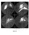
- Result: with a three-step procedure, we obtained a fibrinolytic enzyme, FII, which appeared as a single band on SDS-PAGE and the molecular weight was 25,500. FII cleaved, primarily, the Λ α , B β chains of fibrinogen and fibrin, while the γ chain was minimally affected. FII degraded fibrin iri a dose-dependent manner. Through preparation of polyclonal antibody, monoclonal antibodies and cDNA library, we screened the gene FII from the cDNA library, constructed recombinant plasmid, PPIC9K-FII. After transformation, recombinant plasmid insert into yeast's chromosome determined by PCR. Using fibrinolytic activity as standard, we screened a high expression strain, and the fibrinolytic activity reached the peak on the third days of expression. With a two-step procedure, we obtained recombinant fibrinolytic enzyme FII, which appeared as a single band on SDS-PAGE. Recombinant FII can strongly degrade fibrin, while have no effect on IgG and albumin. Recombinant FII can efficiently dissolve the preformed thrombus in the right lower Pulmonary artery when measurements made at 1 hours post recombinant F II infusion. The average of the recanalization rate of thrombosis of the right lower Pulmonary artery was 83.3%. Conclusion: recombinant fibrinolytic enzyme FII was highly expressed in yeast, recombinant FII have high fibrinolytic activity.
- Nature FII from Agkistrodon acutus snake venom have low yield, instable quality. We expressed recombinant FII in yeast by genetic engineering technology, the recombinant FII had higher fibrinolytic activity, more stable quality higher Output and lower cost than nature FII. The recombinant FII can dissolve effectively thrombus in animal thrombolysis models, and little hemorrhage was observed. It's a strong suggested that the recombinant FII will become a novel and effective thrombolytic agent which have tremendous benefits on Social and economic.
-
Fig1 : DEAE-SephadexΛ-50 ion exchange chromatograph of Agkistrodon acutus snake venom -
Fig2 : the first SephadexG-75 gel filtration of FII -
Fig3 : the second SephadexG-75 gel filtration of FIIa -
Fig4 : Determination of the molecular weight of FII by SDS-PAGE -
Fig5 : the Standard curve of Protein Markers using by SDS-PAGE. -
Fig6 : analysis of the degradation products of fibrinogen hydrolysis by SDS-PAGE. -
Fig7 : fibrinolytic activity of FII. -
Fig8 : analysis of the total RNA of Agkistrodon acutus venom by Agarose-Formaldehyde Electrophoresis -
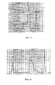
Fig9 : the structure of cDNA library of Agkistrodon acutus venom -
Fig10 : the positive results of FII in the cDNA library of Agkistrodon acutus venom screened by monoclonal antibodies -
Fig11 : the nine Positive clones' sequences of inserting DNA fraction -
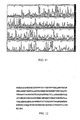
Fig12 : the amoid Sequence deduced by the DNA sequence of FII. -
Fig13 : analysis of FII fusion Protein expressing in E.coil, by SDS-PAGE. -
Fig14 : the two-step of constructing the target DNA-vctor of F II gene in E.coil. -
Fig 15 : the diploid restrictions in the plasmid Ppic9k-FII. -
Fig16 : fibrinolytic activity of FII in the supernatant by SDS-PAGE. -
Fig17 : DEAL-Sepharose icon exchange chromatography of the supernatant. -
Fig 18 : Buty-toyopcarl chromatography of the supernatant. -
Fig19 : determination of the recombinant FII purity by SDS-PAGE. -
Fig20 : directly fibrinolytic activity of recombinant FII. -
Fig21 : the effect on IgG and albumin of recombinant FII by SDS-PAGE. -
Fig22 : The effect of recombinant FII on the Beagle's pulmonary artery thrombosis model. - Agkistrodon acutus snake venom(3g) was dissolved in 10ml 0.05M ammonium acetate(PH8.0), and the supernatant was applied to a column(3.0*80cm) of DEAE-Sephadex A-50 equilibrated with 0.05M ammonium acetate(PII8.0) at 25°C for 24h. Fractions were eluted with a linear gradient consisting of 0.05M ammonium acetate(PH 8.0) as starting buffer and 1M ammonium acetate(PH 5.0) as limit buffer. A volume of 3ml fraction was collected at a fow rate of 12ml/h. The absortance of eluate was tested at 280nm. The peak fractions tested fibrinolytic activity. Then the fibrinolytic fraction was dialyed and lyophilized.
fibrinolytic activity was demonstrated using a modified fibrin plate technique(Astrup and Mullertz, 1952). Each concentration was tested three times. Bovine plasma(0.2%,20ml) was clotted in a Petri dish(diameter, 9.5cm) with thrombin (80µl, 100U/ml). Fribrinolytic activity was expressed as the diameter product (mm2) of the lysed zone caused by 20ul of each fraction. Each fraction was put on the surface of the dish and incubated at 37°C for 12h. - Results: Ion exchange chromatography of crude venom on DEAE-sephadex A-50 yielded ten fractions(
Fig1 ). The second fraction (FII) has higher fibrinolytic activity. Its fibrinolytic activity was 60.23±16.47 mm2/µg. - FII (85mg) obtained from DEAF-Sephadex A-50 ion exchange chromatography was dissolved in 2ml 0.05M ammonium acetate(PF18.0), and the supernatant was applied to a column(1.1*100cm) of Sephadex G-75 equilibrated with 0.05M ummonium acetate(PII8.0) at 25°C for 24h. Fractions were eluted with 0.05M ammonium acetate(PH 8.0) A volume of 3ml fraction was collected at a low rate of 12ml/h. The absortance of eluate was tested at 280nm. The peak fractions tested fibrinolytic activity. Then the fibrinolytic fraction was dialyed and lyophilized.
- Results: After further fractionation by gel filtration on a Sephadex G-75 column, two fractions of FII were obtained (
Fig2 ). The fibrinolytic activity was localized in the first fraction. Its fibrinolytic activity was 87.51±14.95 mm2/µg. - The fibrinolytic fraction from the first Sephadex G-75 gel filtration was again applied to Sephadex G-75 get column by the above method.
- Results: The single peak was obtained (
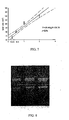
Fig3 ). Its fibrinolytic activity was 90.49±12.41 mm2/µg. - Sodium dodecyl sulfate-polyacrylamide gel electrophoresis (SDS-PAGE) was performed according to the method of Laemmli (12% resolving gels, pH 8.8; 4% stacking gels, pH 6.8; Tris- glycine as the electrophoresis buffer composition; volve,200v; Time, 45min) Molecular weight standards were MBP-β-galactosidase (175,000), MBP-paramyosin (83,000), Glutamic dehydrogenase (62,000), Aldolase (47,500), Triosephosphate isomerase (32,500), β-Lactoglobulin A (25,000), Lysozyme (16,500). The protein bands were stained with 0.25% coomassie brilliant blue R-250 solution.
- Results: On the SDS-PAGE gel, the purified FII appeared as a single protein band (
Fig4 ). The equation, IgMW-1.55X15.51, r=0.9-5, was obtain according to the regression curve (X-axis, IgMW; Y-axis, the relative mobility of standard mobility(Rf(x))). Its calculated molecular weight is 25,550(Fig5 ). 2.2 fibrinogenolytic activity determination of the fibrinolytic enzyme FII from Agkistrodon acutus venom - Bovine fibrinogen (75µl, 0.2mg/ml) was pipetted into 1.5ml microcentrifuge tubes. FII (25µl, 2mg/ml) was added to the tube and the solution was incubated at 37°C for 1 hours. Then the reaction in the tube was stopped by the addition of 10µl of 125 Mm EDTA followed by 50µl of SDS-PAGE sample buffer. 20µl of sample was applied for SDS-PAGE analysis to determine whether FII degradation of degradation of fibrinogen varied from that of plasmin degradation, the same experiment was repeated except for plasmin in place of FII.
- Resuls: SDS-PAGE showed FII is an α, β-fibrinogenase with the α, β chains being degraded, while the γ chain appeared unaffected. The MW or the degradation fraction by FII was 45,000Da. The α β and γ chains of fibrinogen were degraded by plasmin. The MW of the degradation fractions by plasmin was 47,00ODa, 44,00ODa, 23,000Da, respectively. The bands after degradation by FII and plasmin were different, which indicate they have different cleavage sites (
Fig6 ). - Fibrinolytic activity was demonstrated using a modified fibrin plate technique (Astrup and Mullertz, 1952). Bovine Fibrinogen (0.2%,20ml) was clotted in a Petri dish(diameter, 9.5cm) with thrombin (80µl,100U/ml). Each concentration was tested three times. Fribrinolytic activity was expressed as the diameter product (mm2) of the lysed zone caused by 20ul of each fraction. Each fraction was put on the surface of the dish and incubated at 37°C for 12h. arokinase 500U/ml was used for positive control and the saline solution was used for the negative control.
- Results: FII, at 20ul at several different concentrations, 0.25,0.5, 1 and 2mg/ml was applied (table 1). It was shown that the lysed areas increased with the increased concentration (
Fig7 ). - Method:
- (1) Animal immunity. Rabbits were immunized by Multi-point Subcutaneous injection at the back with the Emulsifier mixed with 1ml of FII (3mg/ml) and 1ml Freund's adjuvant. After the first injection, the subsequent boosters were given per week, totally 4 week.
- (2) Preparation of antiserum. The immunized rabbits were anaesthetized by an intramuscular injection of 30 mg/kg ketamine hydrochloride. Blood samples were taken through a catheter inserted into a Carotid and stored at 4 °C overnight. The sera were subsequently separated by centrifugation and aliquots were stored at 4 °C.
- (3) Determinations of antibody titer by eiizymc-linked immunosorbent assay (ELISA). Microtiter plates (96 wells) were coated overnight at 4°C with 100 ml (0.1 mg/ml) of FII in sodium bicarbonate buffer, 100 µl per well and was stored at 4 °C overnight. The plates were washed two times with PBST containing 0.05% Tween-20 and unbound sites were blocked for 1h at 37 °C with blocked liquor containing 2 X PBS ,1% bovine serum albumin (BSA). To measure the serum titers, 100 ml of serial dilutions of serum in 1X PBS containing 1% BSA and a rabbit antiserum(primary antibody) were added to the plates and incubated for 1h at 37 °C. The plates were washed three times again and incubated for 1 h with 100 ml of a goat anti-rabbit immunoglobulin G (whole molecule)-peroxidase conjugate, followed by further washing for three times. The substrate solution for the peroxidase assay A and B were added into every well, the antibody titer was identified according to color of reaction system.
- Result: NO.1 and 2 rabbits were immunized by the Purified fibrinolytic enzyme F 11 from Agkistrodon acutus venom using as antigen. After 4 weeks, antibody titer was determinated by ELISA. The antibody titer was 1:20000 in each rabbit.
- Method:
- (1) immunity, hybridoma Fusion and cloning screening. Balb/c mice (7-week old) were subcutaneously injected with a mixture containing 0.2ml (1mg/ml)F II and an equal volume of Freund's complete adjuvant. A boost injection with the same amount of antigen in Freund's complete adjuvant was administered at 2-week intervals, totally 3 cycles. 0.2ml (1mg/ml) FII was injected through vena caudalis, 3 days later, hybridoma fusion was performed. In brief, the splenocytcs were harvested from immunized mice, mixed with SP2/10 cells at a 10:1 ratio, and fusion was carried out with 5θ% PEG(u 3700). The fused cells were cultured in 5 96-well plates, und cultured in a 5% CO2 incubator. HAT selective medium including 15% FBS were consecutively applied for 4 days, followed by HT medium for 6 days, from the eleventh day on RPMI-1640 medium was kept in using. Antibody produced in medium was measured by indirect enzyme-linked immunosorbent assay (indirect ELISA) as described above.
- (2)Result: 2() cell lines were established after 2 cycles of hybridoma fusion. Among which, 2 cell lines with higher specificity were named C1 and C4, respectively. The monoclone antibody produced by the two cell lines all were
Ig G 1, testing by Ig G typing kit, used according to the instructions of the manufacturer. - Method:
- (1) The pairs of venom glands were extracted from the snakes' heads and were dissected and stored in solid carbon dioxide immediately.
- (2) Using the RNA purification kit (Promega) and following its manufacturer protocol. The venom gland (for a total of about 1.7 g) was mixed. Trizol, chloroform and isopropyl alcohol were used respectively to homogenize the gland, centrifuge for 10 minutes at 12,000 rpm and separate the aqueous phase of RNA, deposit the total RNA with isopropyl alcohol. The RNA pellet was washed twice with 75% ethanol and briefly stir dried. In the end, the RNA was dissolved in diethylpyrocarbonate (DEPC) treated H2O. The integrity of total RNA was checked hy discerning the 28 S, 18 S and 5 S bands of ribosomal RNA in 1% agarose gel. The purity of the total RNA was checked by the ratio of A260/A280.
- Result: the total RNA appeared as a long smear with clear bands of 28 S, 18 S and 5 S. the ratio of OD260/OD280 to the total RNA was 20, and the total amount wus 426ug. So we can draw the conclusion that the RNA didn't degrade and that the quality is high. RNA electrophoris (
Fig.8 ) show 28 S and 18 S rRNA clearly and the fluorescence intensity of 28 S was two times that of 18 S rRNA, indicating a foundation for a high quality library. - Method: mRNA was subsequently purified by means of oligo (dT)-cellulose affinity chromatography, following its manufacturer protocol. Poly (A) RNA was enriched from total RNA: sample buffer solution sufficiently wash the cellulose column, the total RNA (3mg) in DEPC treated H2O was incubated at 65°C for 10 minutes. Decrease temperature to room temperature quickly by means of iced bath. Eluted with sample buffer solution for three times, followed by eluant, mRNA was washed and isolated.
- The intactness of the RNA was assessed using 1% agarose ethidium bromide electrophoresis and The integrity of the mRNA was checked by 1 % agarose electrophoris.
- Result: The total amount of mRNA was 10ug.
- Method: using the cDNA synthesis kit (Promega) and following its manufacturer protocol. See (
Fig.9 ) - (1) Synthesis of the first strand cDNA
- (2) Synthesis of the second strand cDNA
- (3) Adding Not and
Sal 1 adaptors at the end of cDNA - (4) Ligation and transformation of cDNA cDNA and
pSport 1 vector were ligated. The reaction mixtures of cDNA, pSport I vector, T4 ligase 10 × buffer, T4 DNA ligase were incubated at 16 °C overnight. 10 ul of product of ligation was added into 50 ul competence E coli DH5, in order, 4 °C, 30 min, 42 °C 120s, 4 °C, 5 min, 37 °C, 10min, Then the suspended cells were then placed into a new 1.5 mL EP tube and rotated at 37 °C for 1 h under 250 rpm. 2 µL of transformation product were diluted 10, 100 and 1000 times and plated on Luria-Bertani plates containing 100 mg L-1 of ampicillin with isopropy-β-D-thiogalactoside and 5-brom-4-chloro-3-indolyl-beta-D-galactopyranoside; Plates were incubated ut 37 °C overnight. The titer of the library was calculated and stored the library at -80 °C. - Method: after proper dilution, 100 µl bacterium in cDNA library were spreaded into plates containing LB medium supplemented with 1% glucose, 8% glycerol and 50ug/ml ampicillin and grown overnight at 37°C.
- Cellulose acetate membrane was soaked with IPTG(10 mmol/l) and spreaded on the surface of J.B plates, three asymmetrical spot was left on the surface of cellulose acetate membrane to facilitate orientation. Unveil cellulose acetate membrane and spreaded on the surface of agarose plate, plates were incubated at 37 °C for 6-8h for induction of fusion protein expression.
- After 6-8h of induction, cellulose acetate membrane was treated with chloroform for 30 min, the membrane were subsequently blocked overnight at 4 °C in 1% BSA , 4 ug/ml lysozyme and 1 ug/ml DNAase with gentle agitation for 30 min, After blocking, filters were washed three times for 15 min per wash in approximately 100 ml per filter of Tris buffered saline (TBS) with 0.2
% Tween 20 and 0.05% Triton-X 100 (TBS-TT), followed by two washes in 100 ml/filter of TBS to remove residual detergent. To detect colonies producing recombinant protein, the membrane was probed with rabbit-antibody at u 1:1000 dilution in the blocking buffer with gentle agitation for 3 h at room temperature, The membrance were washed three times again and incubated for 1 h with 100 ml of a goat anti-rabbit immunoglobulin G (whole molecule)-peroxidase conjugate, followed by further washing for three times. - The membrane was soaked in DAB colouration solution with gentle agitation for 30 min, rinsed with distilled water, Positive colonies with brown color were detected.
- Results: primary screening was performed with platen (9 cm in diameter, including 5×10³clones). 200,000 clones were screened and 15 positive colonies were found. Repeated screening were performed in those positive clones and positive reactions were observed in 11 clones (
Fig. 10 ). Those 11 positive clones were plated on Luria-Bertani plates containing 100 mg·L-1 of ampicillin, detected with monoclone antibody, positive reactions (C1 and C4 antibody were used) were observed in 9 clones. - Method: the plasmids were extracted by alkaline lysis (QIAGEN):
- (1) Pick a single colony from a freshly streaked selective plate and inoculate a starter culture of 2-5 ml LB medium containing the appropriate selective antibiotic. Incubate for approximately 8 h at 37°C with vigorous shaking (approx. 300 rpm).
- (2) Dilute the
starter culture 1/500 to 1/1000 into 3 ml selective LB medium. Grow at 37°C for 12-16 h with vigorous shaking (upprox. 300 rpm). - (3) Harvest the bacterial cells by centrifugation at 6000 × g for 15 min at 4°C.
- (4) Resuspend the bacterial pellet in 0.3 ml of Buffer P1.
- (5) Add 0.3 ml of Buffer P2, mix thoroughly by vigorously inverting the sealed tube 4-6 times, and incubate at room temperature (15-25°C) for 5 min.
- (6) Add 0.3 ml of chilled Buffer P3, mix immediately and thoroughly by vigorously inverting 4-6 times, and incubate on ice for 5 min.
- (7) Centrifuge at maximum speed in a microcentrifuge for 10 min. Remove supernatant containing plasmid DNA promptly.
- (8) Equilibrate a QlAGEN-
tip 20 by applying 1 ml Buffer QRT, and allow the column to empty by gravity flow. - (9) Apply: the supernatant from
step 7 to the QIAGEN-tip 20 and allow it to enter the resin by gravity flow. - (10) Wash the QIAGEN-
tip 20 with 2 x 2 ml Buffer QC. - (11) Elute DNA with 0.8 ml Buffer QF. Collect the eluate in a 1.5 ml or 2 ml mier
- (12) Precipitate DNA by adding 0.7 volumes (0.56 ml per 0.8 ml of elution volume) or room-temperature isopropanol to the cluted DNA. Mix and centrifuge immediately at > 10,000 rpm for 30 min in a microcentrifuge. Carefully decant the supernatant.
- (13) Wash DNA pellet with 1 ml of 70% ethanol and centrifuge at 10,000 rpm for 10 min.
- (14) Air-dry the pellet for 5-10 min, and redissolve the DNA in a suitable volume of buffer.
- The plasmids were used for capillary sequencing applying the standard primers T7 and SP6.
- Result: Pick a single colony from the 9 positive plate respectively and inoculate a starter culture of 2-5 ml LB medium containing the appropriate selective antibiotic. Incubate for approximately 8 h at 37°C with vigorous shaking for extracting plasmid. PCR reaction was performed applying the standard primers T7 and SP6.specifical bonds of product were observed in each clone. The plasmids were used for capillary sequencing applying the standard primers T7 and SP6 (
Fig.11 ). - Method: in order to identify the genomic origin of those positive clones, those sequences were compared to the nucleic acid database and protein database in GenBank by BLASTn and BLASTp. Most of those 9 sequences are homologous to the SVMPs family (sharing homology to reported gene with the highest identities of 85%). Among which, FII-2 and FII-4 shared the most conservative sequences, belonging to ADAM family, which have relationship with SVMPs family. So, we named the gene of FII-2 as FII und expressed the protein preferentially, and the amino acid sequences of FII was deduced (
Fig.12 ). - Method: Added 0.2ml of E.Coli shaked overnight (colonies containing F II plasmid confirmed by extraction) to SOB medium containing 20ml of ampicillin shaked in 37°C for 3h until A600 was up to about 0.5. Added IPTG to the final concentration of 1 umol/L. Shaked for 4h and centrifuged at 1,500 rpm for 20 min, and discarded the supernatant. Added 0.5ml of water and 0.5ml of SDS loading buffer to the pellet, heated in 100°C for 5min, and centrifuged at 10,000. 20ul of supernatant was taken to load for SDS-PAGE.
- Result: Lysate of E.Coli containing FII plasmid after induced by IPTG showed a band of 40kD in SDS-PAGE, while those of E.Coli without IPTG induction or without plasmid did not showed such a band, indicating that the gene of FII can be expressed as fusion protein in F.Coli.
- Method: (see
fig.14 ) - The sequence of a signal to 5' of target gene was introduced by two-step PCR, and the restriction end nuclease site as well.
- Primer 1:5' AGA GAG GCT GAA GCT AAT CTT ACT CCT GAA C 3'
- Primer 2:5' CT CTC GAG AAA AGA GAG GCT GAA GCT AAT C 3'
- Primer 3:5' GAG CGG CCG CCT CAC GCC TCC AAA AGT TC 3'
- (1)The first round PCR: forward primer is
primer 1, and reverse primer isprimer 3; the template is F II plasmid.
- (2) The second round PCR: forward primer is
primer 2, and reverse primer isprimer 3; the template is the production of the first round of PCR. And the PCR condition is the same with the first round PCR. - The production of the second round PCR was extracted after electrophoresis on 1% agarose gel. The target fragment extracted from agarose gel and PPIC-9K plasmid were both digested with XhoI and NotI, and then retrieved after electrophoresis respectively; and then connected in T4 reaction system for 12h in 16°C. Then transformed to competent Top 10F cells and plated on I.B/penicillin plate. Picked up positive colonies, extracted plasmid and identified by restriction endonuclease analyses, and then purified the plasmid containing target fragment.
- PPIC-9K plasmid with or without target fragment were digested both with Sacl and NotI, connected and transformed. And then picked up positive colonies, identified and purified PPIC-9K-FII plasmid.
- Result: After the above two-step PCR, the PCR product wus about 700bp and was the same large with the gene of F II. The retrieved PCR product after electrophoresis and PPIC9K plasmid were digested both with XhoI and NotI, and then constructed recombinant PPIC9K-FII plasmid identified by restriction endonuclease digestion.
- Digested recombinant PPIC9K-FII plasmid both with SucI and NotI. and subcloned the little digestion fragment to PPIC9K plasmid also digested both with SacI and Notl to construct recombinant expression PPIC9K-FII plasmid. The product identified by restriction endonuclease digestion was 700bp and was the same with anticipation (see
fig.15 ). Sequencing by Sanger method showed that the DNA sequence of F II did exist in recombinant expression PPIC9K- FII plasmid. - Method: 50ug of recombinant plasmid PPIC9K-FII was isolated from the E.coli and linearized at the SacI restriction enzyme site, followed by extraction with phenol and chloroform, and precipitation with ethanol. After that, the DNA was dissolved in TE buffer and stored at -30°C. The linear plasmid mixed with the salmon sperm DNA and was added to competent yeast prepared with 3% polyglycol, which was plated to two RD plates well-distributedly. The plates grew at 30°C upside down. 96 hours later, the yeast strains were washed down the RD plates and plated on YPD plates containing a variety of G418 concentrations including 0.5, 1, 2, 3, 4 mg/ml. The YPD plates grew at 30°C. Recombinant colony first showed up in the plate with low G418 concentration on the forth day. On the 6th day, colonies from plate with 3mg/ml of G418 were selected to isolate DNA for PCR analysis.
- Results: plasmid PPIC9K-FII was isolated and linearized at SacI site. The linear plasmid was purified and transformed into competent yeast with PEG method. The first colony showed up in YPD plate with G418 low concentration (0.5mg/ml) on day 4th, and on day 6th, 6 colonies were selected from the plate with 3mg/ml G418.
- Method: 6 recombinant strains which have been confirmed by PCR to contain our insert genes were inoculated in 10ml of MD media respectively, grew at 30°C overnight with shaking . Then harvest the cells by centrifugation, and added them into BMMY media to induce expression. Add 100% methanol to a final concentration of 1 % methanol every 24 hours to maintain induction. Sampling was also performed every day from the induction cultured and reacted with fibrinogen at 37°C, followed by SDS-PAGE, to analyze its fibrino(gen)lytic activity. 3 tubes of samples were quickly frozen on 3rd day.
- Select a tube of frozen strain to 100ml MD media and cultivated at 30°C with shaking overnight and amplified to 2L culture in two 5 L flasks respectively. 24 hours later the cultures was transfered to BMMY media in jar fermenter of 100L volume and continued to grow at 30°C. And supplement with 1% methanol every 24h. After 96 hours fermentation, centrifuge the culture and saved the supernatant which was later ultrafiltrated into 2 L.
- Results: 6 recombinant strains which have been confirmed to contain FII gene was cultivated. Sampling was done every day and its supernatant was used to react with fibrinogen at 37°C for 1 hour, and subjected to SDS-PAGE analysis. The 3rd day samples from
number Fig. 16 ), natural FII was used as a positive control. The recombinant FII is identical with natural Fll in terms of hydrolysis modality and dominant fraction after hydrolysis, which suggested that these three yeast strains are capable of expressing recombinant FII in high efficiency. Large scale recombinant Fll can be obtained in culture media supernatant after 96 hours fermentation with methanol induction. - Method:
- (1) ultra filtration. The supernatant liquor was ultra filtrated by Millipore until the column was 2L, added double column of 0.05M NH4Ac and continued to ultra filtrate.
- (2) DEAE-Sepharose FF. The supernantant ultrafiltered was applied to a column of DEAE-Sepharose FF previously equilibrated with 0.05M NH4Ac (PH8.0). The column was washed with 0.1 M NH4Ac (PH6.5) to remove unbound material and the remained proteins were then eluted with 0.2M NII4Ac (PH5.2).
- Results: The supernantant ultrafiltered on DEAE-sepharose yielded 5 fractions (
Fig17 ). The fibrinolytic activity was localized in the fraction in the 0.2M NH4Ac (PH5.2). - Method: The fraction displaying fibrinogenolytic activity was then mixed 2M NaCl and applied to another column of Butyl-
toyopearl 4 FF previous equilibrated 1M NaCl, 20mM PBS. The column was washed with 1M NaCl, 20mM PBS to remove unbound material and the remained proteins were then eluted with 2mM PBS. - Results: The supernantant ultrafiltered of the fraction on Butyl-toyopearl gave 3 fractions (
Fig 18 ). The fibrinolytic activity was localized in the third fraction recombant F II was obtained by ultrafiltering, purifying from salts and lyophilizing the fraction displaying fibrinogenolytic activity. - Method: The Purity determination of recombinant FII was performed by the method of SDS-PAGE mentioned as decribe in methods 2.1.
- Results: On the SDS-PAGE gel, the purified recombinat FII appeared as a single protein band (
Fig 19 ). - Method: The fibrinolytic activity of FII was demonstrated using a heated fibrin plate which was incubated at 85°C for 30min in order to inactivate the plasmin activity. 20 microliters of different concentration of recombinant FII (2mg/ml, 1mg/ml, 0.5mg/ml, 0.25 mg/ml, 0.125 mg/ml), were applied onto the prepared plate surface. Nature FII 0.5mg/ml and urokinase 500U/ml were used for positive control and the saline solution was used for the negative control. Each concentration was tested 3 times. The plate was incubated in a humid chamber at 37°C for 12h. Fibrinolytic activity was measured by the area of the clear lysed zone.
- Results: The fibrinolytic activity of FII was demonstrated using a heated fibrin plate technique. It was shown that rF II could formed clear lysis zone on the fibrin plate with increasing concentration, its fibrinolytic activity was higher than nature F II (the lysed zone by 0.5mg/ml rF II was 50mm2, and the lysed zone by 0.5mg/ml nature FII was only 40mm2) ( table 2,
Fig20 ). The plasmin was inactivated when the fibrin plate was heated to 85°C; it was showed that recombinant FII could dissolve fibrin without depending on the plasminogen activation. - Method: Human IgG(10µl, 10mg/ml) mixed with r-F II (10µl, 0.5mg/ml) was incubated at 37°C for 24h using a pipette. After the indicated time, 10µl Protein electrophoresis Solution was added. The sample was analyzed by SDS-PAGE. The same experiment was repeated except for human albumin in place of human IgG.
- Results: When IgG was incubated with recombinant FII, the Heavy Chain disappeared, while the light chain still insusceptible to the enzyme. When albumin was incubated with recombinant FII, the peptide chains almost remained integrity. It suggested that recombinant FII have no effect on albumin (
Fig21 ). - Method: Beagle's Pulmonary Artery thrombosis experimental models were performed by the method of Nowk. The blood clot was prepared by incubating the 15ml experimental Beagle's whole blood at 37°C for 2h in vitro before application, Two different groups were established, containing 6 animals each : the treatment group was given 0.24 mg/kg r FII. The NS control group was infused with 0.1ml/kg NS. Animals were anaesthetized by an intramuscular injection of 30 mg/kg pentobarbital sodium, followed by intramuscular supplements of ketamine hydrochloride given throughout the experiment. The left femoral vein was separated from surrounding tissue after anesthesia. A 6-mm2 Fcohra/Humterhead catheter sheath was drown into the left femoral vein, through Infrarenal abdominal vein, Right atrial, Right ventricular, Pulmonary trunk , and was finally introduced in the right lower Pulmonary artery. The artificial thrombus was injected into the catheter positioned in the right lower Pulmonary artery. Selective intra-arterial digital subtraction angiography (DSA) was performed on a biplane high-resolution angiography system with a matrix of 1024*1024 pixels to record the thromboembolism in the right lower Pulmonary artery. Two h ours after the infusion of the preformed thrombus, single dosages of recombinant F II were injected through the femoral vein. The recanalization rate of thrombosis of the right lower Pulmonary artery was measured by the lateral and auteroposterior(AP) projection images before rF II infusion and at 15,30,60,90 and 120 mins post-infusion to evaluate the effect of r FII on Artery thrombosis.
- Results: Measurements made at 1 hours post recombinant F II infusion showed that recombinant F II efficiently dissolve the preformed thrombus in the right lower Pulmonary artery (
Fig22 ). The average of the recanalization rate of thrombosis in the right lower Pulmonary artery was 83.3%. Little changes were detected in the NS control groups measured 1 hours post infusion. The average of the recanalization rates of thrombosis in the right lower Pulmonary artery was 0.6% (table 3).
Claims (6)
- An isolated polynucleotide consisting of the following sequence:

- A DNA vector containing the polynucleotide according to claim 1.
- A host cell containing the DNA vector according to claim 2.
- A recombinant fibrinolytic enzyme produced by the host cell according to claim 3 and consisting of the amino acid sequence

- A pharmaceutical composition including the recombinant fibrinolytic enzyme according to claim 4.
- The recombinant fibrinolytic enzyme according to claim 4 for use against thromboembolic disorders.
Applications Claiming Priority (1)
| Application Number | Priority Date | Filing Date | Title |
|---|---|---|---|
| PCT/CN2004/001457 WO2006063486A1 (en) | 2004-12-15 | 2004-12-15 | A fibrinolysin of agkistrodon acutus venom and its usage |
Publications (3)
| Publication Number | Publication Date |
|---|---|
| EP1837401A1 EP1837401A1 (en) | 2007-09-26 |
| EP1837401A4 EP1837401A4 (en) | 2008-10-29 |
| EP1837401B1 true EP1837401B1 (en) | 2010-03-17 |
Family
ID=36587508
Family Applications (1)
| Application Number | Title | Priority Date | Filing Date |
|---|---|---|---|
| EP04802470A Expired - Lifetime EP1837401B1 (en) | 2004-12-15 | 2004-12-15 | A fibrinolysin of agkistrodon acutus venom and its usage |
Country Status (6)
| Country | Link |
|---|---|
| US (1) | US20080312170A1 (en) |
| EP (1) | EP1837401B1 (en) |
| JP (1) | JP2008522633A (en) |
| AT (1) | ATE461284T1 (en) |
| DE (1) | DE602004026124D1 (en) |
| WO (1) | WO2006063486A1 (en) |
Family Cites Families (3)
| Publication number | Priority date | Publication date | Assignee | Title |
|---|---|---|---|---|
| ZA889415B (en) * | 1987-12-18 | 1989-09-27 | Chiron Corp | Compositions and method for recombinant production of crotalidus venum fibrolase |
| CN1060806C (en) * | 1997-11-12 | 2001-01-17 | 中国科学院上海生物化学研究所 | Gene sequence of fibrin ferment of Agkistrodon acutus snakes |
| CN1258594C (en) * | 2003-08-20 | 2006-06-07 | 中山大学 | Long-noded pit viper poison dissolving fiber protein No.2 gene and use thereof |
-
2004
- 2004-12-15 EP EP04802470A patent/EP1837401B1/en not_active Expired - Lifetime
- 2004-12-15 DE DE602004026124T patent/DE602004026124D1/en not_active Expired - Lifetime
- 2004-12-15 JP JP2007545811A patent/JP2008522633A/en active Pending
- 2004-12-15 US US11/793,071 patent/US20080312170A1/en not_active Abandoned
- 2004-12-15 WO PCT/CN2004/001457 patent/WO2006063486A1/en not_active Ceased
- 2004-12-15 AT AT04802470T patent/ATE461284T1/en not_active IP Right Cessation
Also Published As
| Publication number | Publication date |
|---|---|
| EP1837401A4 (en) | 2008-10-29 |
| EP1837401A1 (en) | 2007-09-26 |
| JP2008522633A (en) | 2008-07-03 |
| WO2006063486A1 (en) | 2006-06-22 |
| US20080312170A1 (en) | 2008-12-18 |
| ATE461284T1 (en) | 2010-04-15 |
| DE602004026124D1 (en) | 2010-04-29 |
Similar Documents
| Publication | Publication Date | Title |
|---|---|---|
| US8143027B2 (en) | Method of making a plasminogen activator polypeptide with clot-specific streptokinase activity | |
| AU5459298A (en) | Cloning and characterization of napsin, an aspartic protease | |
| JPH01502798A (en) | Novel protease enzyme | |
| US8377676B2 (en) | Mutated nucleotide sequences of batroxobin, mutated α factor secretion signal sequence and processes for preparing batroxobin using the same | |
| Fan et al. | Cloning, sequence analysis and expression in E. coli of the cDNA of the thrombin‐like enzyme (pallabin) from the venom of Agkistrodon halys pallas | |
| JPH022376A (en) | Vector and compound for developing enzyme precursor type human protein c | |
| EP1837401A1 (en) | A fibrinolysin of agkistrodon acutus venom and its usage | |
| RU2183214C2 (en) | Natural and recombinant thrombin inhibitors, their preparing and use | |
| KR940010023B1 (en) | Method of separating activated human protein c | |
| US6955910B2 (en) | Method for large scale production of recombinant DNA-derived TPA or K2S molecules | |
| JPH01501996A (en) | De-epidermal cell growth factor plasminogen activator | |
| US6015685A (en) | Ancrod proteins, their preparation and use | |
| RU2247777C2 (en) | Plasminogen urokinase type modified activator, dna sequence, recombinant plasmid, strain-producer, method for preparing plasminogen urokinase type modified activator and pharmaceutical composition eliciting thrombolytic effect | |
| US7993893B2 (en) | Haemocoagulase | |
| CN101967195B (en) | Thrombus targeting thrombolytic fusion protein mAnxB1ScuPA and its construction and application | |
| CN102258483B (en) | A kind of antithrombotic recombinant batroxobin lyophilized preparation | |
| CN1288245C (en) | Long-noded pit viper dissolving fiber protein No.4 gene and use thereof | |
| US20060153831A1 (en) | Antithrombosis enzyme from the snake venom of Agkistrodon acutus | |
| WO2003008594A1 (en) | Construction of the plasmid for expressing the thrombus-targeting thrombolytic fusion protein | |
| CN102337282A (en) | Thrombolysis protease gene NKS1 and purpose thereof | |
| JPH08510903A (en) | Preparation of Recombinant Factor XA Inhibitor of Hibiscus medicinalis | |
| CN1258594C (en) | Long-noded pit viper poison dissolving fiber protein No.2 gene and use thereof | |
| CN100355884C (en) | Novel earthworm fibrinolytic enzyme, its encoding sequence and uses | |
| WO2006066441A1 (en) | A fibrinolysin of agkistrodon acutus venom and its usage | |
| RU2373281C2 (en) | Method of producing recombinant staphylokinase with regulation of oxygen level |
Legal Events
| Date | Code | Title | Description |
|---|---|---|---|
| PUAI | Public reference made under article 153(3) epc to a published international application that has entered the european phase |
Free format text: ORIGINAL CODE: 0009012 |
|
| 17P | Request for examination filed |
Effective date: 20070711 |
|
| AK | Designated contracting states |
Kind code of ref document: A1 Designated state(s): AT BE BG CH CY CZ DE DK EE ES FI FR GB GR HU IE IS IT LI LT LU MC NL PL PT RO SE SI SK TR |
|
| RIN1 | Information on inventor provided before grant (corrected) |
Inventor name: YAN, GUANGMEI,C/O SUN YAT-SEN UNIVERSITY Inventor name: SHAN, HONG,C/O SUN YAT-SEN UNIVERSITY Inventor name: CHEN, JIASHU,C/O SUN YAT-SEN UNIVERSITY Inventor name: QIU, PENGXIN,C/O SUN YAT-SEN UNIVERSITY |
|
| DAX | Request for extension of the european patent (deleted) | ||
| RIN1 | Information on inventor provided before grant (corrected) |
Inventor name: SHAN, HONG,C/O SUN YAT-SEN UNIVERSITY Inventor name: QIU, PENGXIN,C/O SUN YAT-SEN UNIVERSITY Inventor name: CHEN, JIASHU,C/O SUN YAT-SEN UNIVERSITY Inventor name: YAN, GUANGMEI,C/O SUN YAT-SEN UNIVERSITY |
|
| A4 | Supplementary search report drawn up and despatched |
Effective date: 20080926 |
|
| 17Q | First examination report despatched |
Effective date: 20090212 |
|
| GRAP | Despatch of communication of intention to grant a patent |
Free format text: ORIGINAL CODE: EPIDOSNIGR1 |
|
| GRAS | Grant fee paid |
Free format text: ORIGINAL CODE: EPIDOSNIGR3 |
|
| GRAA | (expected) grant |
Free format text: ORIGINAL CODE: 0009210 |
|
| AK | Designated contracting states |
Kind code of ref document: B1 Designated state(s): AT BE BG CH CY CZ DE DK EE ES FI FR GB GR HU IE IS IT LI LT LU MC NL PL PT RO SE SI SK TR |
|
| REG | Reference to a national code |
Ref country code: GB Ref legal event code: FG4D |
|
| REG | Reference to a national code |
Ref country code: CH Ref legal event code: EP |
|
| REG | Reference to a national code |
Ref country code: IE Ref legal event code: FG4D |
|
| REF | Corresponds to: |
Ref document number: 602004026124 Country of ref document: DE Date of ref document: 20100429 Kind code of ref document: P |
|
| REG | Reference to a national code |
Ref country code: NL Ref legal event code: VDEP Effective date: 20100317 |
|
| PG25 | Lapsed in a contracting state [announced via postgrant information from national office to epo] |
Ref country code: LT Free format text: LAPSE BECAUSE OF FAILURE TO SUBMIT A TRANSLATION OF THE DESCRIPTION OR TO PAY THE FEE WITHIN THE PRESCRIBED TIME-LIMIT Effective date: 20100317 |
|
| LTIE | Lt: invalidation of european patent or patent extension |
Effective date: 20100317 |
|
| PG25 | Lapsed in a contracting state [announced via postgrant information from national office to epo] |
Ref country code: AT Free format text: LAPSE BECAUSE OF FAILURE TO SUBMIT A TRANSLATION OF THE DESCRIPTION OR TO PAY THE FEE WITHIN THE PRESCRIBED TIME-LIMIT Effective date: 20100317 Ref country code: SI Free format text: LAPSE BECAUSE OF FAILURE TO SUBMIT A TRANSLATION OF THE DESCRIPTION OR TO PAY THE FEE WITHIN THE PRESCRIBED TIME-LIMIT Effective date: 20100317 Ref country code: PL Free format text: LAPSE BECAUSE OF FAILURE TO SUBMIT A TRANSLATION OF THE DESCRIPTION OR TO PAY THE FEE WITHIN THE PRESCRIBED TIME-LIMIT Effective date: 20100317 Ref country code: FI Free format text: LAPSE BECAUSE OF FAILURE TO SUBMIT A TRANSLATION OF THE DESCRIPTION OR TO PAY THE FEE WITHIN THE PRESCRIBED TIME-LIMIT Effective date: 20100317 |
|
| PG25 | Lapsed in a contracting state [announced via postgrant information from national office to epo] |
Ref country code: CY Free format text: LAPSE BECAUSE OF FAILURE TO SUBMIT A TRANSLATION OF THE DESCRIPTION OR TO PAY THE FEE WITHIN THE PRESCRIBED TIME-LIMIT Effective date: 20100317 Ref country code: SE Free format text: LAPSE BECAUSE OF FAILURE TO SUBMIT A TRANSLATION OF THE DESCRIPTION OR TO PAY THE FEE WITHIN THE PRESCRIBED TIME-LIMIT Effective date: 20100317 Ref country code: GR Free format text: LAPSE BECAUSE OF FAILURE TO SUBMIT A TRANSLATION OF THE DESCRIPTION OR TO PAY THE FEE WITHIN THE PRESCRIBED TIME-LIMIT Effective date: 20100618 Ref country code: ES Free format text: LAPSE BECAUSE OF FAILURE TO SUBMIT A TRANSLATION OF THE DESCRIPTION OR TO PAY THE FEE WITHIN THE PRESCRIBED TIME-LIMIT Effective date: 20100628 Ref country code: EE Free format text: LAPSE BECAUSE OF FAILURE TO SUBMIT A TRANSLATION OF THE DESCRIPTION OR TO PAY THE FEE WITHIN THE PRESCRIBED TIME-LIMIT Effective date: 20100317 Ref country code: BE Free format text: LAPSE BECAUSE OF FAILURE TO SUBMIT A TRANSLATION OF THE DESCRIPTION OR TO PAY THE FEE WITHIN THE PRESCRIBED TIME-LIMIT Effective date: 20100317 Ref country code: RO Free format text: LAPSE BECAUSE OF FAILURE TO SUBMIT A TRANSLATION OF THE DESCRIPTION OR TO PAY THE FEE WITHIN THE PRESCRIBED TIME-LIMIT Effective date: 20100317 Ref country code: NL Free format text: LAPSE BECAUSE OF FAILURE TO SUBMIT A TRANSLATION OF THE DESCRIPTION OR TO PAY THE FEE WITHIN THE PRESCRIBED TIME-LIMIT Effective date: 20100317 |
|
| PG25 | Lapsed in a contracting state [announced via postgrant information from national office to epo] |
Ref country code: SK Free format text: LAPSE BECAUSE OF FAILURE TO SUBMIT A TRANSLATION OF THE DESCRIPTION OR TO PAY THE FEE WITHIN THE PRESCRIBED TIME-LIMIT Effective date: 20100317 Ref country code: IS Free format text: LAPSE BECAUSE OF FAILURE TO SUBMIT A TRANSLATION OF THE DESCRIPTION OR TO PAY THE FEE WITHIN THE PRESCRIBED TIME-LIMIT Effective date: 20100717 Ref country code: CZ Free format text: LAPSE BECAUSE OF FAILURE TO SUBMIT A TRANSLATION OF THE DESCRIPTION OR TO PAY THE FEE WITHIN THE PRESCRIBED TIME-LIMIT Effective date: 20100317 Ref country code: BG Free format text: LAPSE BECAUSE OF FAILURE TO SUBMIT A TRANSLATION OF THE DESCRIPTION OR TO PAY THE FEE WITHIN THE PRESCRIBED TIME-LIMIT Effective date: 20100617 |
|
| PLBE | No opposition filed within time limit |
Free format text: ORIGINAL CODE: 0009261 |
|
| STAA | Information on the status of an ep patent application or granted ep patent |
Free format text: STATUS: NO OPPOSITION FILED WITHIN TIME LIMIT |
|
| PG25 | Lapsed in a contracting state [announced via postgrant information from national office to epo] |
Ref country code: PT Free format text: LAPSE BECAUSE OF FAILURE TO SUBMIT A TRANSLATION OF THE DESCRIPTION OR TO PAY THE FEE WITHIN THE PRESCRIBED TIME-LIMIT Effective date: 20100719 Ref country code: DK Free format text: LAPSE BECAUSE OF FAILURE TO SUBMIT A TRANSLATION OF THE DESCRIPTION OR TO PAY THE FEE WITHIN THE PRESCRIBED TIME-LIMIT Effective date: 20100317 |
|
| 26N | No opposition filed |
Effective date: 20101220 |
|
| PG25 | Lapsed in a contracting state [announced via postgrant information from national office to epo] |
Ref country code: IT Free format text: LAPSE BECAUSE OF FAILURE TO SUBMIT A TRANSLATION OF THE DESCRIPTION OR TO PAY THE FEE WITHIN THE PRESCRIBED TIME-LIMIT Effective date: 20100317 |
|
| PG25 | Lapsed in a contracting state [announced via postgrant information from national office to epo] |
Ref country code: MC Free format text: LAPSE BECAUSE OF NON-PAYMENT OF DUE FEES Effective date: 20101231 |
|
| REG | Reference to a national code |
Ref country code: CH Ref legal event code: PL |
|
| GBPC | Gb: european patent ceased through non-payment of renewal fee |
Effective date: 20101215 |
|
| REG | Reference to a national code |
Ref country code: FR Ref legal event code: ST Effective date: 20110831 |
|
| PG25 | Lapsed in a contracting state [announced via postgrant information from national office to epo] |
Ref country code: CH Free format text: LAPSE BECAUSE OF NON-PAYMENT OF DUE FEES Effective date: 20101231 Ref country code: IE Free format text: LAPSE BECAUSE OF NON-PAYMENT OF DUE FEES Effective date: 20101215 Ref country code: FR Free format text: LAPSE BECAUSE OF NON-PAYMENT OF DUE FEES Effective date: 20110103 Ref country code: LI Free format text: LAPSE BECAUSE OF NON-PAYMENT OF DUE FEES Effective date: 20101231 |
|
| REG | Reference to a national code |
Ref country code: DE Ref legal event code: R119 Ref document number: 602004026124 Country of ref document: DE Effective date: 20110701 |
|
| PG25 | Lapsed in a contracting state [announced via postgrant information from national office to epo] |
Ref country code: GB Free format text: LAPSE BECAUSE OF NON-PAYMENT OF DUE FEES Effective date: 20101215 Ref country code: DE Free format text: LAPSE BECAUSE OF NON-PAYMENT OF DUE FEES Effective date: 20110701 |
|
| PG25 | Lapsed in a contracting state [announced via postgrant information from national office to epo] |
Ref country code: HU Free format text: LAPSE BECAUSE OF FAILURE TO SUBMIT A TRANSLATION OF THE DESCRIPTION OR TO PAY THE FEE WITHIN THE PRESCRIBED TIME-LIMIT Effective date: 20100918 Ref country code: LU Free format text: LAPSE BECAUSE OF NON-PAYMENT OF DUE FEES Effective date: 20101215 |
|
| PG25 | Lapsed in a contracting state [announced via postgrant information from national office to epo] |
Ref country code: TR Free format text: LAPSE BECAUSE OF FAILURE TO SUBMIT A TRANSLATION OF THE DESCRIPTION OR TO PAY THE FEE WITHIN THE PRESCRIBED TIME-LIMIT Effective date: 20100317 |