EP1805222B1 - Antibodies directed to the mammalian eag1 ion channel protein - Google Patents
Antibodies directed to the mammalian eag1 ion channel protein Download PDFInfo
- Publication number
- EP1805222B1 EP1805222B1 EP05797335A EP05797335A EP1805222B1 EP 1805222 B1 EP1805222 B1 EP 1805222B1 EP 05797335 A EP05797335 A EP 05797335A EP 05797335 A EP05797335 A EP 05797335A EP 1805222 B1 EP1805222 B1 EP 1805222B1
- Authority
- EP
- European Patent Office
- Prior art keywords
- antibody
- eag1
- seq
- cells
- derivative
- Prior art date
- Legal status (The legal status is an assumption and is not a legal conclusion. Google has not performed a legal analysis and makes no representation as to the accuracy of the status listed.)
- Not-in-force
Links
Images


Classifications
-
- C—CHEMISTRY; METALLURGY
- C07—ORGANIC CHEMISTRY
- C07K—PEPTIDES
- C07K16/00—Immunoglobulins [IGs], e.g. monoclonal or polyclonal antibodies
- C07K16/18—Immunoglobulins [IGs], e.g. monoclonal or polyclonal antibodies against material from animals or humans
- C07K16/28—Immunoglobulins [IGs], e.g. monoclonal or polyclonal antibodies against material from animals or humans against receptors, cell surface antigens or cell surface determinants
- C07K16/30—Immunoglobulins [IGs], e.g. monoclonal or polyclonal antibodies against material from animals or humans against receptors, cell surface antigens or cell surface determinants from tumour cells
-
- A—HUMAN NECESSITIES
- A61—MEDICAL OR VETERINARY SCIENCE; HYGIENE
- A61P—SPECIFIC THERAPEUTIC ACTIVITY OF CHEMICAL COMPOUNDS OR MEDICINAL PREPARATIONS
- A61P1/00—Drugs for disorders of the alimentary tract or the digestive system
- A61P1/16—Drugs for disorders of the alimentary tract or the digestive system for liver or gallbladder disorders, e.g. hepatoprotective agents, cholagogues, litholytics
-
- A—HUMAN NECESSITIES
- A61—MEDICAL OR VETERINARY SCIENCE; HYGIENE
- A61P—SPECIFIC THERAPEUTIC ACTIVITY OF CHEMICAL COMPOUNDS OR MEDICINAL PREPARATIONS
- A61P1/00—Drugs for disorders of the alimentary tract or the digestive system
- A61P1/18—Drugs for disorders of the alimentary tract or the digestive system for pancreatic disorders, e.g. pancreatic enzymes
-
- A—HUMAN NECESSITIES
- A61—MEDICAL OR VETERINARY SCIENCE; HYGIENE
- A61P—SPECIFIC THERAPEUTIC ACTIVITY OF CHEMICAL COMPOUNDS OR MEDICINAL PREPARATIONS
- A61P17/00—Drugs for dermatological disorders
- A61P17/06—Antipsoriatics
-
- A—HUMAN NECESSITIES
- A61—MEDICAL OR VETERINARY SCIENCE; HYGIENE
- A61P—SPECIFIC THERAPEUTIC ACTIVITY OF CHEMICAL COMPOUNDS OR MEDICINAL PREPARATIONS
- A61P21/00—Drugs for disorders of the muscular or neuromuscular system
-
- A—HUMAN NECESSITIES
- A61—MEDICAL OR VETERINARY SCIENCE; HYGIENE
- A61P—SPECIFIC THERAPEUTIC ACTIVITY OF CHEMICAL COMPOUNDS OR MEDICINAL PREPARATIONS
- A61P25/00—Drugs for disorders of the nervous system
-
- A—HUMAN NECESSITIES
- A61—MEDICAL OR VETERINARY SCIENCE; HYGIENE
- A61P—SPECIFIC THERAPEUTIC ACTIVITY OF CHEMICAL COMPOUNDS OR MEDICINAL PREPARATIONS
- A61P25/00—Drugs for disorders of the nervous system
- A61P25/02—Drugs for disorders of the nervous system for peripheral neuropathies
-
- A—HUMAN NECESSITIES
- A61—MEDICAL OR VETERINARY SCIENCE; HYGIENE
- A61P—SPECIFIC THERAPEUTIC ACTIVITY OF CHEMICAL COMPOUNDS OR MEDICINAL PREPARATIONS
- A61P25/00—Drugs for disorders of the nervous system
- A61P25/14—Drugs for disorders of the nervous system for treating abnormal movements, e.g. chorea, dyskinesia
- A61P25/16—Anti-Parkinson drugs
-
- A—HUMAN NECESSITIES
- A61—MEDICAL OR VETERINARY SCIENCE; HYGIENE
- A61P—SPECIFIC THERAPEUTIC ACTIVITY OF CHEMICAL COMPOUNDS OR MEDICINAL PREPARATIONS
- A61P25/00—Drugs for disorders of the nervous system
- A61P25/28—Drugs for disorders of the nervous system for treating neurodegenerative disorders of the central nervous system, e.g. nootropic agents, cognition enhancers, drugs for treating Alzheimer's disease or other forms of dementia
-
- A—HUMAN NECESSITIES
- A61—MEDICAL OR VETERINARY SCIENCE; HYGIENE
- A61P—SPECIFIC THERAPEUTIC ACTIVITY OF CHEMICAL COMPOUNDS OR MEDICINAL PREPARATIONS
- A61P29/00—Non-central analgesic, antipyretic or antiinflammatory agents, e.g. antirheumatic agents; Non-steroidal antiinflammatory drugs [NSAID]
-
- A—HUMAN NECESSITIES
- A61—MEDICAL OR VETERINARY SCIENCE; HYGIENE
- A61P—SPECIFIC THERAPEUTIC ACTIVITY OF CHEMICAL COMPOUNDS OR MEDICINAL PREPARATIONS
- A61P35/00—Antineoplastic agents
-
- A—HUMAN NECESSITIES
- A61—MEDICAL OR VETERINARY SCIENCE; HYGIENE
- A61K—PREPARATIONS FOR MEDICAL, DENTAL OR TOILETRY PURPOSES
- A61K39/00—Medicinal preparations containing antigens or antibodies
- A61K2039/505—Medicinal preparations containing antigens or antibodies comprising antibodies
-
- C—CHEMISTRY; METALLURGY
- C07—ORGANIC CHEMISTRY
- C07K—PEPTIDES
- C07K2317/00—Immunoglobulins specific features
- C07K2317/30—Immunoglobulins specific features characterized by aspects of specificity or valency
- C07K2317/34—Identification of a linear epitope shorter than 20 amino acid residues or of a conformational epitope defined by amino acid residues
-
- C—CHEMISTRY; METALLURGY
- C07—ORGANIC CHEMISTRY
- C07K—PEPTIDES
- C07K2317/00—Immunoglobulins specific features
- C07K2317/70—Immunoglobulins specific features characterized by effect upon binding to a cell or to an antigen
- C07K2317/73—Inducing cell death, e.g. apoptosis, necrosis or inhibition of cell proliferation
-
- C—CHEMISTRY; METALLURGY
- C07—ORGANIC CHEMISTRY
- C07K—PEPTIDES
- C07K2317/00—Immunoglobulins specific features
- C07K2317/70—Immunoglobulins specific features characterized by effect upon binding to a cell or to an antigen
- C07K2317/77—Internalization into the cell
Definitions
- the present invention relates to a particularly advantageous antibody, antibody fragment or derivative thereof, which specifically binds to/interacts with at least one epitope of the extracellular or intracellular domain of the mammalian EAG1 ion channel and to nuclei acid molecules encoding the same and to vectors comprising said nucleic acid molecules.
- the invention additionally relates to methods for the preparation of said antibody, antibody fragments or derivatives thereof and to pharmaceutical compositions comprising the same. Furthermore, the use of said antibody, antibody fragment or derivative thereof and also diagnostic compositions comprising said components are disclosed in the specification.
- the invention also relates to a method of assessing for the presence of EAG1 expressing cells and for a method of blocking EAG1 function in said cells.
- the invention further relates to a method of treating diseases with the help of said antibody or antibody fragment or derivative thereof.
- Potassium channels are ubiquitously present in cells.
- One reason for this is supposed to be that the channels are involved in the regulation of the resting potential of cells, which has been regarded as their major role.
- the channels are involved in the regulation of the resting potential of cells, which has been regarded as their major role.
- experimental evidence has been presented [Ouadid-Ahidouch H et al., 2001] suggesting their implication in the cell division cycle hinting at their possible involvement in cancerogenesis.
- members of the eag family EAG1, and herg have been proposed to be preferentially expressed in cancer cells [Meyer R et al., 1999; Bianchi I et al., 1998]. Since said channels are also expressed in various cell types and in particular in dividing cells, including cancer cells such as neoplastic cells it is of high medical interest to provide tools which might be used in therapeutic and/or diagnostic applications related to said potassium channels.
- Antibodies which are directed against the human EAG1 ion channels were known in the prior art.
- European Patent application no. EP1073738 for example describes antibodies directed against said channel as well as the EAG1 ion channel.
- the technical problem underlying the present invention was to provide such antibodies which may be employed for the further specific study, diagnosis, prevention and treatment of defects and/or diseases interrelated with EAG1 from different mammalian species.
- an antibody, antibody fragment or derivative thereof comprising at least one complementarity determining region (CDR) of the VH. and/or V L region, wherein the amino acid sequence determining said CDR(s) is selected from the group consisting of (V L ) SEQ ID Nos: 160 to 162, 166 to 168, 172 to 174, and 178 to 180 and selected from the group consisting of (V H ) SEQ ID NOs: 163 to 165, 169 to 171, 175 to 177, and 181 to 183.
- CDR complementarity determining region
- antibody fragment or derivative thereof in accordance with the present invention relates to antibody fragments and derivatives of the antibody of the invention as well as of the antibody fragments of the invention.
- Antibody fragments include Fab fragments, Fab' fragments F(ab') 2 fragments as well as Fv fragments.
- Derivatives of the antibody include scFv constructs, chimeric antibodies or humanized antibodies as long as they exhibit the desired capability of binding to EAG1.
- the antibodies are for therapeutic purposes are optionally de-immunized. Examples of how to make de-immunized (humanized) antibodies may be found in U.S. Pat. Nos. 6,054,297 , 5,886,152 and 5,877,293 .
- the antibody, fragment or derivative thereof is preferentially labeled. Suitable labels include radioactive labels and fluorescent labels.
- complementary determining region is well-defined in the art (see, for example, Harlow and Lane, “Antibodies, a laboratory manual", CSH Press, Cold Spring Harbour, 1988 ) and refers to the stretches of amino acids within the variable region of an antibody that primarily makes contact with the antigen.
- the antibody, antibody fragment or derivative thereof of the invention specifically discriminates between mammalian, in particular human, EAG 1 and EAG2 while also recognizing other mammalian EAG1 channels. This is crucial if the properties of the antibody are to be taken advantage of in a clinical scenario, because failure to recognize rodent EAG1, while still discriminating from mouse EAG2, would restrict the possibility to use animal models to test for efficacy and -more importantly-safety of the antibody preparation.
- variable domain the heavy chain V H and light chain V L
- FRs relatively conserved framework regions
- the present invention encompasses antibodies, antibody fragments or derivatives thereof comprising at least one CDR of the above-described variable domains and which advantageously have substantially the same, similar or improved binding properties as the antibody described in the appended examples.
- an antibody that comprises at least one CDR as recited in the attached sequence listing and required by the main embodiment of the invention the skilled artisan can combine further CDRs from the originally identified monoclonal antibodies or different antibodies for an enhanced specificity and/or affinity.
- CDR- grafting is well-known in the art and can also be used to fine-tune the specific affinity in other properties of the antibody, fragment or derivative thereof of the invention, as long as the original specificity is retained.
- the antibody, fragment or derivative comprises all six CDRs of the original mouse antibody. Also described herein is that the CDRs from different originally identified monoclonal antibodies may be combined in a new antibody entity. In these cases, it is preferred that the three CDRs of the heavy chain originate from the same antibody whereas the three CDRs of the light chain all originate from a different (but all from the same) antibody.
- the antibodies of the present invention or their corresponding immunoglobulin chain(s) can be further modified using conventional techniques known in the art, for example, by using amino acid deletion(s), insertion(s), substitution(s), addition(s), and/or recombination(s) and/or any other modification(s) known in the art either alone or in combination.
- the antibodies of the invention furthermore show advantageous properties with respect to their binding specificity and biological activity.
- the antibodies of the invention not only recognize the human EAG1 ion channel, but also are able to recognize EAG1 ion channels of other mammalian species.
- Said species include but are not limited to rat, mouse, and non human primates.
- the EAG1 antibody of the invention may exhibit at least one of the following characteristics:
- the antibodies of the invention allow the specific recognition of the mammalian EAG1 potassium channels both in vitro and in vivo.
- Binding of the antibody of the invention to EAG1 may exhibit at least one of the following characteristics:
- EAG1 expressing cells which, have bound the antibody of the invention on the cell surface are finally attacked by immune system functions such as the complement system or cell mediated cytotoxicity.
- the antibodies of the invention show advantageous properties with respect to their binding specificity and biological activity, in particular with respect to their capacity to recognize epitopes of the EAG1 ion channel in different mammals and to decrease cell growth.
- the pharmaceutical and/or diagnostic applications of the antibodies of the invention include, but are not limited to humans, some of the antibodies of the invention (antibodies ImAb 3 and ImAb 4) were humanized; SEQ ID NOs 9 to 40) and were further developed in order to minimize potential negative immunogenic side effects when used in humans.
- the murine antibodies were adapted to the human antibody sequence in order to reduce the immunogenicity in humans by genetic engineering.
- the subtype IgG1 (heavy chain) and kappa (light chain) were chosen to evoke the strongest immune activation.
- a fusion protein that contained the pore region of Eag1 loop between fifth and sixth transmembrane segment, pos. 369 - 433; Region A) and a segment of the C-terminus of Eag1 (Pos. 850 - 920; region B) was used as the antigen. Similarity in those regions between Eag1 and Eag2 is 69% and 62% respectively. Region A is extracellular, region B is, under the accepted topographic model, intracellular.
- the antibodies generated were checked by ELISA and BIAcore for selectivity between Eag1 and Eag2. Only a surprisingly small number of them qualified and were subcloned. Of these, five have been maintained. Four of them recognize an epitope in region A, and only one recognizes an epitope in region B. All four "A-type” antibodies recognize linear epitopes, and three of them share a single one, although, their CDRs are possibly different. The "B-type” antibody recognizes a three-dimensional epitope.
- the properties of the resulting antibodies were characterized with respect to their binding affinities ( Fig 2 ), specificity ( Fig 3 ), the epitope they recognize and bind ( Fig. 4 ) and the inhibtion of the EAG1 ion channel Fig 6 .
- the properties of the antibodies of the invention to induce ion channel internalisation were investigated by immunoflurescence ( Fig. 5 ).
- the ability of the antibodies to inhibit cell growth were characterized in cell proliferation assays ( Fig 7 , 8a , 8b ) and soft agar assays ( Fig. 9 ). The results of these experiments showed that the antibodies indeed possess unexpected biological specificities.
- said antibody, antibody fragment or derivative thereof specifically binds to/interacts with at least one epitope of the extracellular or intracellular domain of the mammalian EAG1 ion channel, and does not bind to/interact with the mammalian EAG2 ion channel.
- extracellular domain is a term well-known in the art and relates to the portion of the EAG1 channel extending into the extracellular environment. This domain comprises, among others, amino acids 374-452 of the mammalian EAG1 molecule.
- intracellular domain denotes the portion of the mammalian EAG1 channel extending into the cytoplasm.
- the domain comprises amino acids 872-932.
- the antibody is a monoclonal antibody.
- Monoclonal antibodies can be prepared, for example, by the well-established techniques as originally described in Kohler and Milstein, Nature 256 (1975), 495 , and Galfre, Meth. Enzymol. 73 (1981), 3 , which comprise the fusion of mouse myeloma cells to spleen cells derived from immunized mammals with modifications developed by the art.
- An effective strategy to target tumor cells is the usage of monoclonal antibodies.
- HerceptinTM an antibody directed against the receptor tyrosine kinase HER2
- Other strategies to use monoclonal antibodies in tumor therapy include immunotoxins, like MylotargTM, a recombinant IgG4 kappa antibody conjugated to calicheamicin, and antibodies labelled with radioisotopes, as for example ZevalinTM.
- the antibody fragment or derivative thereof is a Fab-fragment, a F(ab 2 )'- fragment.
- Fragments or derivatives of the above antibodies directed to the aforementioned epitopes can be obtained by using methods which are described, e.g., in Harlow and Lane “Antibodies, A Laboratory Manual", CSH Press, Cold Spring Harbor, 1988 .
- surface plasmon resonance as employed in the BIAcore system can be used to increase the efficiency of phage antibodies which bind to an epitope of EAG1 ( Schier, Human Antibodies Hybridomas 7 (1996), 97-105 ; Malmborg, J. Immunol. Methods 183 (1995), 7-13 ).
- the nucleic acid molecules, vectors and host cells may be used to make mutated EAG1 antibodies.
- the antibodies may be mutated in the variable domains of the heavy and/or light chains to alter a binding property of the antibody.
- a mutation may be made in one or more of the CDR regions to increase or decrease the Kd of the antibody for EAG1, or to alter the binding specificity of the antibody.
- Techniques in site directed mutagenesis are well-known in the art. See, e.g., Sambrook et al. and Ausubel et al., supra.
- mutations are made at an amino acid residue that is known to be changed compared to germline in a variable region of an EAG1 antibody.
- the nucleic acid molecules are mutated in one or more of the framework regions.
- a mutation may be made in a framework region or constant domain to increase the half-life of the EAG antibody. See, e.g., WO 00/09560, published February 24,2000 .
- a mutation in a framework region or constant domain may also be made to alter the immunogenicity of the antibody, to provide a site for covalent or non-covalent binding to another molecule, or to alter such properties as complement fixation. Mutations may be made in each of the framework regions, the constant domain and the variable regions in a single mutated antibody. Alternatively, mutations may be made in only one of the framework regions, the variable regions or the constant domain in a single mutated antibody.
- chimeric antibodies are described, for example, in W089/09622 .
- Methods for the production of humanized antibodies are described in, e.g., EP-A1 0 239 400 and W090/07861 .
- a further source of antibodies to be utilized in accordance with the present invention are so-called xenogenic antibodies.
- the general principle for the production of xenogenic antibodies such as human antibodies in mice is described in, e.g., WO 91/10741 , WO 94/02602 , WO 96/34096 and WO 96/33735 .
- the antibody of the invention may exist in a variety of forms besides complete antibodies; including, for example, Fv, Fab and F(ab)2.
- the light chain (V L ) is selected from the group consisting of SEQ ID NOs 2, 6, 10, 14, 18, 22, 26, 30, 34, 38, 43 and 47 and the heavy chain (V H ) is selected from the group consisting of SEQ ID NOs.4, 8, 12, 16, 20, 24, 28, 32, 36, 40, 44 and 48.
- the invention further relates to a nucleic acid molecule encoding the antibody, antibody fragment or derivative thereof of the invention.
- the nucleic acid molecule of the invention encoding the above-described antibody, antibody fragment or derivative thereof may be, e.g. DNA, cDNA, RNA or synthetically produced DNA or RNA or recombinantly produced chimeric nucleic acid molecule comprising any of those nucleic acid molecules either alone or in combination.
- the nucleic acid molecule may also be genomic DNA corresponding to the entire gene or a substantial portion thereof or to fragments and derivatives thereof.
- the nucleotide sequence may correspond to the naturally occurring nucleotide sequence or may contain single or multiple nucleotide substitutions, deletions or additions.
- the nucleic acid molecule of the invention may be a cDNA molecule.
- the invention also relates to a vector comprising a nucleic acid molecule of the invention.
- Said vector may be, for example, a phage, plasmid, viral or retroviral vector.
- Retroviral vectors may be replication competent or replication defective. In the latter case, viral propagation generally will occur only in complementing host/cells.
- the nucleic acid molecules of the invention may be joined to a vector containing selectable markers for propagation in a host.
- a plasmid vector is introduced in a precipitate such as a calcium phosphate precipitate or rubidium chloride precipitate, or in a complex with a charged lipid or in carbon-based clusters, such as fullerens.
- a virus it may be packaged in vitro using an appropriate packaging cell line prior to application to host cells.
- the vector of the invention is an expression vector wherein the nucleic acid molecule is operatively linked to one or more control sequences allowing the transcription and optionally expression in prokaryotic and/or eukaryotic host cells.
- Expression of said nucleic acid molecule comprises transcription of the nucleic acid molecule, preferably into a translatable mRNA.
- Regulatory elements ensuring expression in eukaryotic cells preferably mammalian cells, are well known to those skilled in the art. They usually comprise regulatory sequences ensuring initiation of transcription and optionally poly-A signals ensuring termination of transcription and stabilization of the transcript. Additional regulatory elements may include transcriptional, as well as translational enhancers.
- Possible regulatory elements permitting expression in prokaryotic host cells comprise, e.g., the lac, trp or tac promoter in E. coli , and examples for regulatory elements permitting expression in eukaryotic host cells are the AOXI or GAL1 promoter in yeast or the CMV-, SV40- , RSV-promoter (Rous sarcoma virus), CMV-enhancer, SV40-enhancer or a globin intron in mammalian and other animal cells.
- Beside elements which are responsible for the initiation of transcription such regulatory elements may also comprise transcription termination signals, such as the SV40-poly-A site or the tk-poly-A site, downstream of the polynucleotide.
- suitable expression vectors are known in the art such as Okayama-Berg cDNA expression vector pcDV1 (Pharmacia), pCDM8, pRc/CMV, pcDNA1, pcDNA3 (In-vitrogene), pSPORTI (GIBCO BRL).
- said vector is an expression vector and/or a gene transfer or targeting vector.
- Expression vectors derived from viruses such as retroviruses, vaccinia virus, adenoassociated virus, herpes viruses, or bovine papilloma virus, may be used for delivery of the polynucleotides or vector of the invention into targeted cell population.
- nucleic acid molecules of the invention can be reconstituted into liposomes for delivery to target cells.
- the invention further relates to a host cell comprising the vector of the invention.
- Said host may be a prokaryotic or eukaryotic cell.
- the polynucleotide or vector of the invention which is present in the host cell may either be integrated into the genome of the host cell or it may be maintained extrachromosomally.
- the nucleic acid molecule of the invention can be used for "gene targeting” and/or “gene replacement", for restoring a mutant gene or for creating a mutant gene via homologous recombination; see for example Mouellic, Proc. Natl. Acad. Sci. USA, 87 (1990), 4712-4716 ; Joyner, Gene Targeting, A Practical Approach, Oxford University Press .
- the host cell can be any prokaryotic or eukaryotic cell, such as a bacterial, insect, fungal, plant, animal, mammalian or, preferably, human cell.
- Preferred fungal cells are, for example, those of the genus Saccharomyces, in particular those of the species S. cerevisiae.
- prokaryotic is meant to include all bacteria which can be transformed or transfected with a polynucleotide for the expression of a variant polypeptide of the invention.
- Prokaryotic hosts may include gram negative as well as gram positive bacteria such as, for example, E. coil, S. typhimurium, Serratia marcescens and Bacillus subtilis.
- a polynucleotide coding for a mutant form of variant polypeptides of the invention can be used to transform or transfect the host using any of the techniques commonly known to those of ordinary skill in the art. Methods for preparing fused, operably linked genes and expressing them in bacteria or animal cells are well-known in the art ( Sambrook, Molecular Cloning A Laboratory Manual, Cold Spring Harbor Laboratory (2001, Third Editi on). The genetic constructs and methods described therein can be utilized for expression of variant antibodies, antibody fragments or derivatives thereof of the invention in, e.g., prokaryotic hosts. In general, expression vectors containing promoter sequences which facilitate the efficient transcription of the inserted nucleic acid molecule are used in connection with the host.
- the expression vector typically contains an origin of replication, a promoter, and a terminator, as well as specific genes which are capable of providing phenotypic selection of the transformed cells.
- the transformed prokaryotic hosts can be grown in fermentors and cultured according to techniques known in the art to achieve optimal cell growth.
- the antibodies, antibody fragments or derivatives thereof of the invention can then be isolated from the grown medium, cellular lysates, or cellular membrane fractions.
- the isolation and purification of the microbially or otherwise expressed antibodies, antibody fragments or derivatives thereof of the invention may be by any conventional means such as, for example, preparative chromatographic separations and immunological separations such as those involving the use of monoclonal or polyclonal antibodies.
- the host is a bacteria, fungal, plant, amphibian or animal cell.
- Preferred animal cells include but are not limited to Chinese hamster ovary (CHO) cells, baby hamster kidney (BHK) cells, monkey kidney cells (COS), 3T3 cells, NSO cells and a number of other cell lines.
- said animal cell is an insect cell.
- Preferred insect cells include but are not limited to cells of the SF9 cell lines
- said host is a human cell or human cell line.
- Said human cells include, but are not limited to Human embryonic kidney cells (HEK293, 293T, 293 freestyle).
- said human cell lines include, but are not limited to HeLa cells, human hepatocellular carcinoma cells (e. g., Hep G2), A549 cells.
- Cell lines of particular preference are selected through determining which cell lines have high expression levels.
- transgenic non-human animals comprising one or more nucleic acid molecules of the invention that may be used to produce antibodies of the invention.
- Antibodies can be produced in and recovered from tissue or body fluids, such as milk, blood or urine, of goats, cows, horses, pigs, rats, mice, rabbits, hamsters or other mammals. See, e. g., U. S. Patent Nos. 5,827,690 , 5,756,687 , 5,750,172 , and 5,741,957 .
- non-human transgenic animals that comprise human immunoglobulin loci can be produced by immunizing with EAG1 or a portion thereof.
- the invention additionally relates to a method for the preparation of an antibody, antibody fragment or derivative thereof, comprising culturing the host of the invention under conditions that allow synthesis of said antibody, antibody fragment or derivative thereof and recovering said antibody, antibody fragment or derivative thereof from said culture.
- the transformed hosts can be grown in fermentors and cultured according to techniques known in the art to achieve optimal cell growth.
- the whole antibodies, their dimers, individual light and heavy chains, or other immunoglobulin forms of the present invention can be purified according to standard procedures of the art, including ammonium sulfate precipitation, affinity columns, column chromatography, gel electrophoresis and the like; see, Scopes, "Protein Purification", Springer-Verlag, N.Y. (1982 ).
- the antibody or its corresponding immunoglobulin chain(s) of the invention can then be isolated from the growth medium, cellular lysates, or cellular membrane fractions.
- the isolation and purification of the, e.g., microbially expressed antibodies or immunoglobulin chains of the invention may be by any conventional means such as, for example, preparative chromatographic separations and immunological separations such as those involving the use of monoclonal or polyclonal antibodies directed, e.g., against the constant region of the antibody of the invention.
- the antibodies of the invention can be further coupled to other moieties for, e.g., drug targeting and imaging applications.
- Such coupling may be conducted chemically after expression of the antibody or antigen to site of attachment or the coupling product may be engineered into the antibody or antigen of the invention at the DNA level.
- the DNAs are then expressed in a suitable host system, and the expressed proteins are collected and renatured, if necessary.
- the antibody, antibody fragment or derivative thereof may be coupled to an effector, such as calicheamicin, Auristatin E or monomethylauristatin E (MMAE), a radioisotope or a toxic chemotherapeutic agent such as geldanamycin and maytansine.
- an effector such as calicheamicin, Auristatin E or monomethylauristatin E (MMAE)
- MMAE monomethylauristatin E
- these antibody conjugates are useful in targeting cells, e.g. cancer cells, expressing EAG1, for elimination.
- the linking of antibodies/antibody fragments of the invention to radioisotopes e.g. provides advantages to tumor treatments. Unlike chemotherapy and other forms of cancer treatment, radioimmunotherapy or the administration of a radioisotope-antibody combination directly targets the cancer cells with minimal damage to surrounding normal, healthy tissue.
- Preferred radioisotopes include g. 3 H, 14 C, 15 N, 35 S, 90 Y, 99 Tc,
- the antibodies of the invention can be used to treat cancer when being conjugated with toxic chemotherapeutic drugs such as geldanamycin ( Mandler et al., J. Natl. Cancer Inst., 92(19), 1549-51 (2000 )) and maytansine, for example, the maytansinoid drug, DM1 ( Liu et al., Proc. Natl. Acad. Sci. U.S.A. 93:8618-8623 (1996 )) and auristatin -E ( Doronina et al., Nat. Biotechnol.. 21:778-784 (2003 ).
- toxic chemotherapeutic drugs such as geldanamycin ( Mandler et al., J. Natl. Cancer Inst., 92(19), 1549-51 (2000 )
- maytansine for example, the maytansinoid drug, DM1 ( Liu et al., Proc. Natl. Acad. Sci
- the invention further relates to a pharmaceutical composition
- a pharmaceutical composition comprising the antibody, antibody fragment or derivative thereof, the nucleic acid molecule, the vector, the host of the invention or an antibody, antibody fragment or derivative thereof obtained by the method of the invention.
- composition as employed herein comprises at least one compound of the invention.
- a composition is a pharmaceutical or a diagnostic composition.
- the composition may be in solid, liquid or gaseous form and may be, inter alia, in a form of (a) powder(s), (a) tablet(s), (a) solution(s) or (an) aerosol(s).
- Said composition may comprise at least two, preferably three, more preferably four, most preferably five compounds of the invention or nucleic acid molecules encoding said compounds.
- Said composition may also comprise optimized antibodies, antibody fragments or derivatives thereof obtainable by the methods of the invention.
- said pharmaceutical composition optionally comprises a pharmaceutically acceptable carrier and/or diluent.
- the herein disclosed pharmaceutical composition may be partially useful for the treatment of hyperproliferative diseases, skin diseases, inflammatory diseases or neuro-degenerative diseases.
- Said disorders comprise, but are not limited to psoriasis, Alzheimer's disease, multiple sclerosis, lateral amyotrophic sclerosis or Parkinsons's disease breast, lung, colon, kidney, lymphoma, skin, ovary, prostate, pancreas, esophagus, barret, stomach, bladder, cervix, liver, thyroid cancer, melanoma, hyperplastic or neoplastic diseases or other EAG expressing or overexpressing hyperproliferative diseases.
- the present invention provides for pharmaceutical compositions comprising the compounds of the invention to be used for the treatment of diseases/disorders associated with EAG1 expression or overexpression.
- Suitable pharmaceutical carriers include phosphate buffered saline solutions, water, emulsions, such as oil/water emulsions, various types of wetting agents, sterile solutions etc.
- Compositions comprising such carriers can be formulated by well known conventional methods. These pharmaceutical compositions can be administered to the subject at a suitable dose. Administration of the suitable compositions may be effected by different ways, e.g., by intravenous, intraperitoneal, subcutaneous, intramuscular, topical, intradermal, intranasal or intrabronchial administration.
- compositions of the invention may also be administered directly to the target site, e.g., by biolistic delivery to an external or internal target site, like the brain.
- the dosage regimen will be determined by the attending physician and clinical factors. As is well known in the medical arts, dosages for any one patient depends upon many factors, including the patient's size, body surface area, age, the particular compound to be administered, sex, time and route of administration, general health, and other drugs being administered concurrently. Proteinaceous pharmaceutically active matter may be present in amounts between 1 ⁇ g and 100 mg/kg body weight per dose; however, doses below or above this exemplary range are envisioned, especially considering the aforementioned factors. If the regimen is a continuous infusion, it should also be in the range of 1 pg to 100 mg per kilogram of body weight per minute.
- compositions of the invention may be administered locally or systemically.
- Preparations for parenteral administration include sterile aqueous or non-aqueous solutions, suspensions, and emulsions.
- non-aqueous solvents are propylene glycol, polyethylene glycol, vegetable oils such as olive oil, and injectable organic esters such as ethyl oleate.
- Aqueous carriers include water, alcoholic/aqueous solutions, emulsions or suspensions, including saline and buffered media.
- Parenteral vehicles include sodium chloride solution, Ringer's dextrose, dextrose and sodium chloride, lactated Ringer's, or fixed oils.
- Intravenous vehicles include fluid and nutrient replenishers, electrolyte replenishers (such as those based on Ringer's dextrose), and the like. Preservatives and other additives may also be present such as, for example, antimicrobials, anti-oxidants, chelating agents, and inert gases and the like.
- the pharmaceutical composition of the invention may comprise further agents depending on the intended use of the pharmaceutical composition. It is particularly preferred that the pharmaceutical composition comprises further agents like, e.g. an additional antineoplastic agent, small molecule inhibitor, antitumor agent or chemotherapeutic agent,
- the invention also relates to a pharmaceutical composition
- a pharmaceutical composition comprising the antibody, antibody fragment or derivative thereof of the invention in combination with at least one anti-neoplastic agent. Said combination is effective, for example, in inhibiting abnormal cell growth.
- the anti-neoplastic agent is selected from the group of therapeutic proteins including but not limited to antibodies or immunomodulatory proteins.
- the anti-neoplastic agent may be selected from the group of small molecule inhibitors or chemotherapeutic agents consisting of mitotic inhibitors, kinase inhibitors, alkylating agents, anti-metabolites, intercalating antibiotics, growth factor inhibitors, cell cycle inhibitors, enzymes, topoisomerase inhibitors, histone deacetylase inhibitors, anti-survival agents, biological response modifiers, anti-hormones, e. g. anti-androgens, and antiangiogenesis agents.
- composition of the invention can also be used for veterinary purposes.
- the invention relates to the use of the antibody, antibody fragment or derivative thereof of the invention, the nucleic acid molecule, the vector, the host of the invention or an antibody, antibody fragment or derivative thereof obtained by the method of the invention for the preparation of a pharmaceutical composition for prevention or treatment of a hyperproliferative disease, inflammatory disease, psoriasis, or a neurodegenerative disease.
- said neurodegenerative disease is Alzheimer's disease, multiple sclerosis, lateral amyotrophic sclerosis or Parkinson's disease.
- said hyperproliferative disease is in particular breast, lung, colon, kidney, lymphoma, skin, ovary, prostate, pancreas, esophagus, barret, stomach, bladder, cervix, liver, thyroid cancer, melanoma, hyperplastic or neoplastic diseases or other EAG expressing or overexpreesing hyperproliferative diseases.
- the present invention relates to a diagnostic composition
- a diagnostic composition comprising the antibody, antibody fragment or derivative thereof of the invention, the nucleic acid molecule, the vector, the host of the invention or an antibody, antibody fragment or derivative thereof obtained by the method of the invention and optionally a pharmaceutically acceptable carrier.
- the diagnostic composition of the invention is useful in the detection of an undesired expression or over-expression of the mammalian EAG1 potassium channel in different cells, tissues or another suitable sample, comprising contacting a sample with an antibody of the invention, and detecting the presence of EAG1 in the sample Accordingly, the diagnostic composition of the invention may be used for assessing the onset or the disease status of a hyperproliferative disease.
- malignant cells such as cancer cells expressing EAG1
- the cells which have bound the antibody of the invention might thus be attacked by immune system functions such as the complement system or by cell-mediated cytotoxicity, therefore reducing in number of or eradicating cancer cells.
- the antibody, antibody fragment or derivative thereof of the invention is coupled to a labelling group.
- a labelling group refers to a detectable marker, e.g. a radiolabelled amino acid or biotinyl moieties that can be detected by marked avidin.
- suitable labelling groups include, but are not limited to, the following: radioisotopes or radionuclides (e.g.
- fluorescent groups e.g. FITC, rhodamine, lanthanide phosphors
- enzymatic groups e.g. horseradish peroxidase, -galactosidase,luciferase, alkaline phosphatase
- chemiluminescent groups e.g. horseradish peroxidase, -galactosidase,luciferase, alkaline phosphatase
- biotinyl groups e.g.leucine zipper pair sequences, binding sites for secondary antibodies, metal binding domains, epitope tags.
- the labelling groups are attached by spacer arms of various lengths to reduce potential steric hindrance.
- the above embodiment of the invention is particularly important. Since the antibodies of the invention show a broad scope of applicability with respect to different mammalian species that can be treated, the diagnostic composition of the invention is also useful and applicable in different mammalian species.
- the present invention relates to an in vitro method of assessing for the presence of EAG1 expressing cells comprising contacting the antibody or antibody fragment or derivative thereof of the invention with cells or a tissue suspected of carrying EAG1 on their/its surface.
- the present invention relates to an in vitro method of blocking EAG1 function comprising contacting the antibody or antibody fragment or derivative thereof of the invention with cells or a tissue suspected of carrying EAG 1 on their/its surface.
- Also described herein is a method of treating a disease selected from a hyperproliferative disease, inflammatory disease, psoriasis, or a neurodegenerative disease comprising administering to a patient in need thereof a suitable dose of the antibody or antibody fragment or derivative thereof of the present invention.
- said neurodegenerative disease may be Alzheimer's disease, multiple sclerosis, lateral amyotrophic sclerosis or Parkinson's disease.
- said hyperproliferation disease may be breast, lung, colon, kidney, lymphoma, skin, ovary, prostate, pancreas, esophagus, barret, stomach, bladder, cervix, liver, thyroid cancer and hyperplastic and neoplastic diseases or other EAG expressing or overexpressing hyperproliferative diseases.
- said inflammatory disease may be pancreatitis or hepatitis.
- a method of treating a disease wherein the antibody of the invention is administered to a mammal and wherein said disease is correlated directly or indirectly with the abnormal level of expression of EAG1.
- the invention relates to a kit comprising the antibody, antibody fragment or derivative thereof of the invention, the nucleic acid molecule encoding said components and/or the vector of the invention.
- a single colony of Epicurian Coli cells transformed with the vector encoding h1z was inoculated in 500 ml LB medium supplemented with Chloramphenicol 34 ⁇ g/ml and Ampicillin 100 ⁇ g/ml, at 31°C, 140 rpm. After 15 h of incubation, the culture was diluted 1/10 with LB-medium supplemented with the necessary antibiotics and incubated for 2 h at 31°C with shaking (140 rpm). Overexpression of the fusion protein was induced by adding IPTG (final conc. 1 mM). The culture was incubated for 6 hours and then centrifuged 20 min at 2100xg at 4°C. The pellets were resupended in His-Resuspension Buffer and centrifuged for 10 min at 3.500 rpm and 4°C. The pellets were flash frozen in liquid Nitrogen and stored at -70°C.
- Frozen bacterial cell pellet were resuspended in 15 ml 50 mM Tris/HCl, pH 7.9, 2 mM EDTA buffer supplemented with ⁇ 1 mg/ml lysozyme and shaken for 15 min at room temperature. Urea was added up to 8 M final concentration and the sample was sonicated. The solution was allowed to denature overnight with rotation at 4°C and thereafter centrifuged for 30 min at 14.000 x g. The supernatant was adjusted to 6 M urea in 1 x binding buffer and filtered through a 0.45 ⁇ m filter.
- Affinity purification was performed using a BioCAD chromatography system, using a linear gradient from 100% binding buffer (20 mM Tris/HCl, pH 7.9 , 5 mM Imidazole, 500 mM NaCl 6 M Urea) to 100% elution buffer (20 mM Tris/HCl, pH 7.9, 1 M Imidazole, 500 mM NaCl 6 M Urea).
- the supernatant was loaded on His-tag resin peek column that was charged with 250 mM NiSO4 and pre-equilibrated with binding Buffer.
- the the appropriate fractions were dialysed at 4°C for 24 h against 3 changes of 3 M urea in PBS, 3x 1 M Urea, 3x PBS and 3x 0,5 x PBS.
- the dialysed fusion protein was lyophylized and stored at -20 C until use.
- mice (age 8 weeks) were primed by injecting with 50 ng antigen emulsified in complete adjuvant (Biogenes GmbH) and injected into the peritoneum at minus 120 days before fusion. All other injections were performed into peritoneum and intravenous by the following time schedule.
- Immunization Days from fusion Amount of antigen Adjuvant Injection route 1 120 100 Complete i.p. 2 60 100 Incomplete i.p. 3 30 100 Incomplete i.p. 4 15 100 Incomplete i.p. 5 8 100 Incomplete i.p. 6 3 200 w/o adjuvant i.p. + i.v. 7 2 200 w/o adjuvant i.p. + i.v. 8 1 200 w/o adjuvant i.p. + i.v.
- the myeloma cell line used was SP2/0-Ag14 from the German Collection of Microorganisms and Cell Cultures. The cells were described as not synthesizing or secreting immunoglobulin chains, being resistant to azaguanine at 20 pg/ml, and not growing in HAT medium.
- the SP2/0 cells were routinely maintained in tissue culture flasks in standard culture media (DMEM + 10% fetal calf serum) supplemented with 20 Ng/ml 8-AZG to kill any HPRT+ revertants which can grow in HAT-medium. One week prior to fusion SP2/0 cells were maintained in standard culture media without 8-AZG.
- the spleen from immunized mice was aseptically removed and single cell suspension was prepared.
- Spleen lymphocytes were fused with SP2/0 myeloma cell line (ratio 10 lymphocytes / 1 SP2/0) in the presence of polyethylene glycol 4000.
- the cells so produced were then resuspended in DMEM containing HAT (hypoxantine 10 -4 M, aminopterin 10 -5 M and thymidine 4x10 -5 M) and 20% preselected fetal calf serum.
- HAT hyperxantine 10 -4 M, aminopterin 10 -5 M and thymidine 4x10 -5 M
- the cells were then plated into five 96 well tissue culture plates (Coming-Costar) containing peritoneal exudate cells as a feeder layer.
- the plates were incubated for 2 weeks at 37°C in a humid atmosphere containing 5% carbon dioxide. During this period cells were fed two times with HAT medium and two times with HT medium. Wells with a positive growth of cells were screened for specific immunoglobulin content using an enzyme-linked immunosorbent assay (ELISA).
- ELISA enzyme-linked immunosorbent assay
- An indirect ELISA assay was used for screening the culture supernatants. Assay were carried out in 96 well flat bottom polystyrene microtiter plates prepared as follows: a 100 ⁇ l aliquot of a solution of 0.1 M carbonate/bicarbonate buffer, pH 9.6 containing immunogen (as positive control) or the equivalent fusion protein corresponding to Eag2 (as negative control) at a concentration 5 ⁇ g/ml was added to wells on the plate. After incubation overnight in a moist chamber at 4°C the plate was washed four times with TBS containing 0.01 % Triton X-100 and blocked with 22% Gelatin from cold water fish in TBS for 1 hour at room temperature.
- Cells from positive IgG producer wells were transferred into wells of a 24 well plate and cultivated for one week. Cells from wells, which were still positive after this period were subjected to preclonal distribution (the cells were plated into five 96 well tissue culture plates containing peritoneal exudate cells as a feeder layer at concentration 10 cells per well) for further selection of a stable antibody producers. This procedure allows to select a most stable cell sub-populations, because due to random chromosome loss, wells with cells secreting the antibody of interest may gradually lose of antibody production.
- the subtype of the murine antibodies was identified by immunhistological methods. All the hybridomas expressed secreted antibodies of the subtype IgG2b heavy chain and kappa light chain.
- Total RNA was prepared from the hybridoma cells using the RNeasy kit (Qiagen). 1 ⁇ g of total RNA was translated into cDNA using the SMART PCR cDNA Synthesis Kit (Clontech). The primary cDNA was amplified for 20 cycles using the Long-distance polymerase chain reaction of the SMART PCR cDNA Synthesis Kit (Clontech). Subsequently the genes of the light and heavy chains expressed by the hybridomas were amplified by a PCR using specific primers and the proof-reading DNA polymerase Pwo (Roche).
- P1 and P2 were used.
- P1 is specific for the 5'-prime sequence of all cDNAs generated by the SMART PCR Synthesis Kit.
- P2 is a specific primer for the 3'-prime region of constant region of the murine IgG2b.
- P8 GTA ACA ACG CAG AGT ACG CGG G (SEQ ID NO: 49)
- P6 TCA TTT ACC CGG AGA CCG G (SEQ ID NO: 50)
- P1 and P3 were used.
- P1 is specific for the 5'-prime sequence of the all cDNAs generated by the SMART PCR Synthesis Kit.
- P3 is a specific primer for the 3'-prime region of constant region of the murine kappa light chain.
- P8 GTA ACA ACG CAG AGT ACG CGG G (SEQ ID NO: 49)
- P4 CTA ACA CTC ATT CCT GTT GAA GCT C (SEQ ID NO: 51)
- thermocycler 1 ⁇ l of 100 ⁇ l first strand reaction cDNA), 1x PCR Buffer for Pwo (Roche), 200 ⁇ M each Nucleotide (dNTP, Roche), each primer 0,6 ⁇ M, 2,5 U Pwo proof reading polymerase (Roche), I a final volume of 50 ⁇ l was incubated in a thermocycler as follows: 94°C 3 min 94°C 25 sec 65°C 30 sec 72°C 40 sec for light and 60 sec for heavy chain 10 cycles 94°C 25 sec 65°C 30 sec 72°C 40 sec + 2 sec / cycle for light and 60 sec + 4 sec / cycle for heavy chain 20 cycles 72°C 5 min
- the PCR products were analyzed on a 1% agarose gel. A single band of 750 bp for the light chain and a band of 1600 bp for the heavy chain were found.
- the PCR products were purified by QIAquick PCR purification kit (Qiagen) and phosphorylated using the polynucleotide kinase (PNK, Roche). 10 pmol DNA double strand (5 ⁇ g of light chain DNA or 10 ⁇ g heavy chain DNA) was incubated for 30 min at 37°C in 50 mM TrisHCl, 10 mM MgCl2, 0,1 mM EDTA, 5 mM DTT, 0,1 mM Spermidine ph 8,2, including 100 ⁇ M ATP and 50 U PNK.
- the phosphorylated DNA was purified from an agarose gel by gel elution and ligated into a pBluescript II KS+ vector which had been cutted with the restriction enzyme EcoRV (Roche) and dephosphorylated with calf intestine alkaline phosphatase (Roche).
- the sequence of the cloned DNA was determined by DNA sequencing using T3 and T7 primer (Seqlab GmbH, Goettingen).
- the DNA sequence of murine light chain ImAb4 is shown in SEQ ID NO. 1
- the Protein sequence of murine light chain ImAb4 is shown in SEQ ID NO: 2
- the protein domains such as the signal peptide required for the secretion of the antibody and the complementarity determining regions (CDR) required for the specific binding of the antibody to its target were identified.
- the complementarity determining regions (CDR) and constant region of the antibody sequences were defined according to Chothia ( Chothia C., Novotny J., Bruccoleri R., Karplus M. Journal of Molecular Biology. 186(3):651-63, 1985 ).
- Signal peptide 1. -19. aminoacid LC-CDR1 43. - 58. aminoacid LC-CDR2 74. - 80. aminoacid LC-CDR3 113. -121. aminoacid Constant region: 122. - 238. aminoacid
- the DNA sequence of murine heavy chain ImAb4 is shown in SEQ ID NO: 3
- the Protein sequence of heavy chain ImAb4 is shown in SEQ ID NO: 4
- the protein domains such as the signal peptide required for the secretion of the antibody and the complementarity determining regions (CDR) required for the specific binding of the antibody to its target were identified.
- the complementarity determining regions (CDR) and constant region of the antibody sequences were defined according to Chothia ( Chothia C., Novotny J., Bruccoleri R., Karplus M. Journal of Molecular Biology. 186(3):651-63, 1985 ).
- Signal peptide 1. -18. aminoacid HC-CDR1 44. - 54. aminoacid HC-CDR2 69. -84. aminoacid HC-CDR3 117. - 126. aminoacid Constant region: 127. - 473. aminoacid
- the DNA sequence of murine light chain ImAb3 is shown in SEQ ID NO: 5
- the Protein sequence of murine light chain ImAb3 is shown in SEQ ID NO: 6
- the protein domains such as the signal peptide required for the secretion of the antibody and the complementarity determining regions (CDR) required for the specific binding of the antibody to its target were identified.
- the complementarity determining regions (CDR) and constant region of the antibody sequences were defined according to Chothia ( Chothia C., Novotny J., Bruccoleri R., Karplus M. Journal of Molecular Biology. 186(3):651-63, 1985 ).
- Signal peptide 1. - 20. aminoacid LC-CDR1 44. - 60. aminoacid LC-CDR2 76. - 82. aminoacid LC-CDR3 115. - 122. aminoacid Constant region 123. - 239. aminoacid
- the DNA sequence of murine heavy chain ImAb3 is shown in SEQ ID NO: 7
- the Protein sequence of murine heavy chain ImAb3 is shown in SEQ ID NO:8
- the protein domains such as the signal peptide required for the secretion of the antibody and the complementarity determining regions (CDR) required for the specific binding of the antibody to its target were identified.
- the complementarity determining regions (CDR) and constant region of the antibody sequences were defined according to Chothia ( Chothia C., Novotny J., Bruccoleri R., Karplus M. Journal of Molecular Biology. 186(3):651-63, 1985 ).
- Signal peptide 1. - 19. aminoacid HC-CDR1 45. - 54. aminoacid HC-CDR2 69. - 87. aminoacid HC-CDR3 120. -129. aminoacid Constant region 130. - 476. aminoacid
- the murine antibodies constant regions were replaced by human constant regions.
- Human light chain kappa and heavy chain IgG1 were cloned from blood cells of a human volunteer using the same approach as for the murine antibodies but specific human primers:
- the following PCRs were performed. First the human constant region was fused to the 3'-prime end of the murine variable region by using chimeric 3'-prime primers, that contained murine and human sequences (primer P7). Both the human constant region and the murine variable region were fused by a final PCR using both DNA fragments as a template and one specific primer for each DNA fragment.
- the murine variable region (409 bp) of the light chain ImAb3 was amplified with primers:
- Human light chain constant region (376 bp) was amplified using:
- Both fragments were fused by PCR to generate the chimeric DNA-fragment (762 bp).
- the DNA product was phosphorylated and cloned into EcoRV-cutted pBuescript II KS+.
- the DNA was sequenced, cutted by restriction enzymes Notl and Xhol and ligated into the eukaryotic expression vector pBudCE4.1 (Invitrogen, V532-20).
- variable region of the murine heavy chain ImAb3 was fused to the constant region of human IgG1 by PCR.
- the murine variable region of the heavy chain ImAb3 (488 bp) was first amplified with primers:
- the human heavy chain constant region (1048 bp) was amplified using:
- the final fragment (1513 bp) was cloned blunt end into EcoRV-cutted pBluescript II KS+, sequenced and cloned into pBud CE4.1 after HindIII and EcoRV digestion.
- the Protein sequence of chimeric light chain ImAb3 is shown in SEQ ID NO: 10
- the Protein sequence of chimeric heavy chain ImAb3 is shown in SEQ ID NO: 12
- the human constant regions were amplified by specific primers and fused to the murine variable regions by the following PCR.
- the murine variable region (432 bp) of the light chain ImAb4 was amplified with primers:
- the human light chain constant region (377 bp) was amplified using:
- Both fragment were fused by PCR.
- the DNA fragment was cloned into pBluescript II KS+ (EcoRV digested) and sequenced.
- the murine variable region of the heavy chain ImAb4 (455 bp) was amplified with primers:
- the human heavy chain constant region (1054 bp) was amplified using:
- Both fragment were fused by PCR and unique restriction sites (HindIII at 5'-prime and Xbal at 3'-prime) were introduced.
- the final fragment (1489 bp) was cloned blunt end into EcoRV-cutted pBluescript 11 KS+, sequenced and cloned into pBud CE4.1 after Hindlll and Xbal digestion.
- the Protein sequence of chimeric light chain ImAb4 is shown in SEQ ID NO: 14
- the DNA sequence of chimeric heavy chain ImAb4 is shown in SEQ ID NO: 15
- the Protein sequence of chimeric heavy chain ImAb4 is shown in SEQ ID NO: 16
- variable region of the chimeric antibodies was compared to human antibody variable regions on the protein level (Genbank). The closest human counterpart within consensus human genome was identified. The sequence of the murine variable region was changed outside the complementarity-determining-regions to human sequence by the introduction of point mutations on the DNA level.
- LC-ImAb3 to human B3 HC-ImAb3 to human VH3-72 LC-ImAb4 to human A17 HC-ImAb4 to human VH4-59
- the DNA sequence of LC-ImAb4-humA17 is shown in SEQ ID NO: 21.
- the protein sequence of LC-ImAb3-humA17 is shown in SEQ ID NO: 30
- the DNA sequence of HC ImAB1 V H is shown in SEQ ID NO: 42.
- the protein sequence of HC ImAB1 V H is shown in SEQ ID NO: 44.
- the DNA sequence of said light chain is shown in SEQ ID NO: 41.
- the protein sequence of said light chain is shown in SEQ ID NO: 43.
- the DNA sequence of said heavy chain is shown in SEQ ID NO: 46.
- the protein sequence of said heavy chain is shown in SEQ ID NO: 48.
- the DNA sequence of said light chain is shown in SEQ ID NO: 45.
- the protein sequence of said light chain is shown in SEQ ID NO: 47.
- the DNA constructs of the mutated antibodies were fully sequenced and liberalized by Pvul digestion.
- the DNA was purified from an agarose gel, extracted with Phenol/Chloroform and precipitated with ethanol.
- DNA was transfected into CHO cells and the antibodies purified from the supernatants by affinity purification.
- BIAcore TM binding analysis was performed.
- BIAcore chips were coated with Eag1 C-terminus (amino acids 694 to 962), the H5 region (amino acids 374 to 452) or Eag2 (the equivalent regions fused in a single construct).
- the interaction with anti-Eag1 antibodies ImAb1 and ImAb3 were analysed using 10 ⁇ g/ml antibody at a flow rate of 20 ⁇ l/min.
- the result as indicated in Fig.2 show that both anti-Eag1 antibodies of the invention are selective for the Eag1 antigen and do not cross-react with Eag2.
- the results demonstrate that ImAb1 specifically binds to the C-terminus of the Eag1 antigen, whereas ImAb3 specifically recognizes the pore domain of Eag1.
- the animals were transcardially perfused with a fixative consisting of 4 % p -formaldehyde in 0.12 M phosphate buffer (pH 7.2). After perfusion, brains were removed, fixed for an additional hour at 4°C, rinsed three times with PBS and stored overnight at 4°C. Coronal and sagital sections (40-50 ⁇ m) were cut in cold PBS using a vibratome (Leica, Vienna, Austria).
- Controls were done by either omitting the primary antibody or by prior incubation of the primary antibody with the corresponding fusion protein (10 ⁇ g/ml final concentration) at 4°C for 24 h and then following the procedure as described above. Sections were analysed with a Zeiss Axiophot microscope. The results as indicated in Fig. 3 demonstrate that anti-Eag1 antibodies of the invention not only recognizes human Eag1, but also cross-reacts with other mammalian species such as rat.
- Peptide sequences was: 1. MHHHHHHSSGMGD ,2. HHHHHSSGMGDYE ,3. HHHSSGMGDYEIF ,4. HSSGMGDYEIFDE ,5. SGMGDYEIFDEDT ,6. MGDYEIFDEDTKT ,7. DYEIFDEDTKTIR ,8. EIFDEDTKTIRNN ,9. FDEDTKTIRNNSW ,10. EDTKTIRNNSWLY ,11. TKTIRNNSWLYQL ,12. TIRNNSWLYQLAM ,13. RNNSWLYQLAMDI ,14. NSWLYQLAMDIGT ,15. WLYQLAMDIGTPY ,16. YQLAMDIGTPYQF ,17.
- LAMDIGTPYQFNG 18. MDIGTPYQFNGSG ,19. IGTPYQFNGSGSG ,20. TPYQFNGSGSGKW ,21. YQFNGSGSGKWEG ,22. FNGSGSGKWEGGP ,23. GSGSGKWEGGPSK ,24. GSGKWEGGPSKNS ,25. GKWEGGPSKNSVY ,26. WEGGPSKNSVYIS ,27. GGPSKNSVYISSL ,28. PSKNSVYISSLYF ,29. KNSVYISSLYFTM ,30. SVYISSLYFTMTS ,31. YISSLYFTMTSLT ,32. SSLYFTMTSLTSV ,33.
- LYFTMTSLTSVGF 34. FTMTSLTSVGFGN ,35. MTSLTSVGFGNIA ,36. SLTSVGFGNIAPS ,37. TSVGFGNIAPSTD ,38. VGFGNIAPSTDEI ,39. FGNIAPSTDIEKI ,40. NIAPSTDIEKIFL ,41. APSTDIEKIFLES ,42. STDIEKIFLESPQ ,43. DIEKIFLESPKDR,44. EKIFLESPKDRSP ,45. IFLESPKDRSPIL ,46. LESPKDRSPILAE ,47. SPQDRSPILAEVK ,48. QDRSPILAEVKHS ,49. RSPILAEVKHSFY ,50.
- PILAEVKHSFYPI 51. LAEVKHSFYPIPE ,52. EVKHSFYPIPEQT ,53. KHSFYPIPEQTLQ ,54. SFYPIPEQTLQAT ,55. YPIPEQTLQATVL ,56. IPEQTLQATVLEV ,57. EQTLQATVLEVRH ,58. TLQATVLEVRHEL ,59. QATVLEVRHELKE ,60. TVLEVRHELKEDI ,61. LEVRHELKEDIKA ,62. VRHELKEDIKALN ,63. HELKEDIKALNAK ,64. LKEDIKALNAKMT ,65.
- EDIKALNAKMTNI 66. IKALNAKMTNIEK ,67. ALNAKMTNIEKQL ,68. NAKMTNIEKQLSE ,69. KMTNIEKQLSEIL ,70. TNIEKQLSEILRI ,71. IEKQLSEILRILT ,72. KQLSEILRILTSL ,73. LSEILRILTSLEH ,74. EILRILTSLEHHH ,75. LRILTSLEHHHHH ,76. RILTSLEHHHHHH;
- the membrane was rinsed in ethanol, washed three times with TBS and blocked with 3% BSA in TBS overnight at room temperature with shaking. The membrane was then washed once with the same volume of T-TBS for 10 min. and incubated for 3 hours, with shaking, with the desired primary anti-Eag1 antibody.
- ImAb1-5 were diluted 1:2000 (from 1mg/ml stock solution in PBS). The primary antibody was then discarded and the membrane was washed three times with TBST for 10 min.
- the membrane was then incubated with an appropriate volume of HRP-conjugated secondary antibody solution for 2 hours with shaking.
- Anti-mouse HRP antibody was diluted 1:5000 in blocking buffer, washed three times with the same volume of T-TBS for 10 min, incubated with detection reagent - ECL solution for 1 min with gentle shaking and developed.
- Each of these peptides are localized within the pore domain region of Eag1.
- EXAMPLE 4 DETERMINATION OF INTERNALIZATION OF ANTI-EAG1 ANTIBODIES OF INVENTION
- CHO cells were grown on coverslips and incubated in normal medium (Ham's F12, 10% FCS) overnight (37C, 5% CO 2 ) with anti-Eag1 antibodies ImAb1 and ImAb4 directly labeled with Cy3 (100 ⁇ g). Cultures were thereafter incubated with app. 2 ⁇ g/ml Hoechst 33342 for 10 min. After washing, cells were observed in vivo using a 63x water immersion objective in a standard fluorescence microscope (Zeiss Axiophot).
- Fig.5 shows that both anti-Eag1 antibodies of the invention bind to Eag1 antigen on living cells expressing the antigen and are internalized into the cells within 24h. It is demonstrated, that ImAb4, which recognizes the extracellular core domain, as well as ImAb1, which binds to an intracellular epitope at the C-terminus of the Eag1 antigen, are both internalized into Eag1-expressing cells.
- IPC-298 cells were seeded in 60 ⁇ l/well 10% FCS-containing medium (DMEM 4500 mg/ml glucose) on 96-well plates overnight. Cells were pre-incubated in quadruplicates with 5 ⁇ g/ml anti-Eag1 monoclonal antibodies, ImAb1 and ImAb3, diluted in FCS-containing medium with 40 mM KCI for 1h at 37°C in 5% CO 2 . Treatment of the cells with 40 mM KCI ensures an open conformation of the ion channel Eag1 and might accelerate binding of the monoclonal anti-EAG1 antibodies to its corresponding epitope.
- FCS-containing medium DMEM 4500 mg/ml glucose
- IPC-298 cells were seeded in 60 ⁇ l/well FCS-containing medium on 96-well plates overnight.
- 100 ⁇ g/well (1ng/ ⁇ l) Mab-ZAP a chemical conjugate of affinity-purified goat anti-mouse IgG and saporin (Advanced Targeting System) was pre-incubated with different concentrations of mouse monoclonal anti-Eag1 antibody ImAb4 (10 ng/ ⁇ l, 5 ng/ ⁇ l, 1 ng/ ⁇ l, 0.5 ng/ ⁇ l, 0.1 ng/ ⁇ l) in 40 ⁇ l FCS-containing medium for 1h at 37°C and then added directly to the cells in quadruplicates.
- IgG-SAP Advanced Targeting System
- IgG-SAP Advanced Targeting System
- Cells were then left to grow for 72 hours at 37°C in 5% CO 2 .
- 10 ⁇ l/well AlamarBlue TM (BIOSOURCE) was added and incubated at 37°C in the dark. Absorbance was measured using a spectrofluorometer at 590 nm every 30 min.
- Fig. 8a and b show that the antibodies of the invention are internalised into the cells and that the secondary immuntoxin-labelled ("piggybacked") anti-Eag1 monoclonal antibodies of the invention inhibits human cancer cell growth.
- the potency of cell growth inhibition depends on the relation of primary antibody to secondary immunotoxin.
- this result demonstrates that an armed anti-Eag1 antibody of the invention provides a tool to inhibit human cancer cell growth.
- Soft agar assays were conducted in order to investigate the ability of immunotoxin-labelled antibodies of the invention to inhibit anchorage independent cell growth.
- 100 pg Mab-ZAP a chemical conjugate of affinity-purified goat anti-mouse IgG and saporin (Advanced Targeting System) was pre-incubated with 2.5 ng anti-Eag1 antibody ImAb 4 in OptiMEM (Gibco) containing 20 mM KCI at 4°C for 30 min.
- OptiMEM Advanced Targeting System
- Mab-ZAP was pre-incubated with 2.5ng control IgG. After 30 min pre-incubation, 2000 IPC-298 cells in OptiMEM with 20 mM KCI were added and further incubated at 37°C for 30 min.
- IPC-298 cells pre-incubated with immunotoxin-labelled anti-EAG1 antibody, were resuspended in 50 ⁇ l/well 0.25% Difco noble agar containing OptiMEM with 0.5% FCS and plated on 50 ⁇ l/well 0.5% agarose underlayer containing OptiMEM with 0.5% FCS in quadruplicates. Additionally, 50 ⁇ l/well 0.25% feeding agar containing OptiMEM with 0.5% FCS was plated. Colonies were allowed to form for 10 days and were stained with 50 ⁇ l MTT (1 mg/ml in PBS) for 1.5 hours. Wells were scanned using an Epson scanner and colonies were counted using the Scion Image software. The result as indicated in Fig. 9 demonstrates that anti-Eag1 antibody ImAb4 labelled ("piggybacked”) with a secondary immunotoxin inhibits anchorage independent tumor cell growth.
- EXAMPLE 9 USE OF ANTI-EAG1 ANTIBODIES OF THE INVENTION AS A DIAGNOSTIC AGENT
- the Immunofluorescence in Fig.10a shows that the anti-Eag1 antibody ImAb4 of the invention binds to endogenous Eag1 antigen in human cancer cells.
- the results indicate that the anti-Eag1 antibody of invention preferentially binds to dividing human cancer cells (in a certain stage of mitosis) and shows that anti-Eag1 antibody ImAb4 provides a diagnostic tool for detection of anti-Eag1 antigen in proliferating human cancer cells.
- EXAMPLE 10 USE OF ANTI-EAG1 ANTIBODIES OF THE INVENTION AS A DIAGNOSTIC AGENT
- ELISA Enzyme-linked Immunosorbant Assay
- the wells were treated with lysates of human tumor cells suspected of containing the Eag1 antigen or with lysates of Chinese hamster ovary (CHO) cells stably expressing the human Eag1 antigen or with lysates of non-transfected CHO K1 cells. After rinsing away the samples, the wells were incubated with a second rabbit polyclonal anti-Eag1 antibody. After rinsing away excess second antibody, the wells were incubated with a goat anti-rabbit Abs conjugated to horseradish peroxidase (HRP), which served as a detection antibody.
- HRP horseradish peroxidase
- the samples (cell lysates) for detection of Eag1 were prepared as follows: Cells (CHO K1, CHO Eag1 clone 1, IPC-298 melanoma and PC3 prostate cancer cells) were seeded in culture dishes (10 cm, Nunc). Cells were cultured for 24h at 37°C in 5% CO 2 using their corresponding medium (DMEM F12 medium for CHO cells, DMEM 4500 mg/ml glucose for IPC-298 cells, Hams F12 medium for PC3 cells) supplemented with 10% FCS or 7% FCS (PC3 cells) (Sigma).
- DMEM F12 medium for CHO cells DMEM 4500 mg/ml glucose for IPC-298 cells
- Hams F12 medium for PC3 cells supplemented with 10% FCS or 7% FCS (PC3 cells) (Sigma).
- the sandwich ELISA for detection of Eag1 in human tumor cell lysates was performed as follows: 100 ⁇ l of capture anti-Eag1 antibodies ImAb1 and ImAb4 at a concentration of 1 ⁇ g/ml each in PBS were coated on ELISA microtiter plates (Nunc Maxisorp). After incubation at 4°C overnight, the plates were treated with 150 ⁇ l of blocking buffer (0.5% BSA in PBS) with gently agitation for 4-6h at 4°C. The plates were washed (3x) using 0.05% Tween 20 in PBS (washing buffer).
- the plates were incubated with cell lysates (100 ⁇ g protein concentration) overnight at 4°C, washed with washing buffer (3x) and then incubated with 100 ⁇ l/well of rabbit polyclonal anti-EAG1 detection antibody (iOnGen) diluted 1:1000 in dilution buffer (0.5% BSA, 0.05% Tween 20, 5 mM EDTA in PBS) for 2h at 25°C. After washing the plates were incubated with 100 ⁇ l HRP-conjugated goat-anti rabbit IgG (0.2 ⁇ g/ml in dilution buffer) for 30 min at 25°C, washed as before, and then treated with a suitable chromogenic substrate and the color generation was measured using an ELISA plate reader ( Fig. 10b ).
- iOnGen rabbit polyclonal anti-EAG1 detection antibody
- Tissues from the tissue register Schludgeum Kassel were analysed by immunohistochemistry in order to investigate the ability of an EAG1 antibody of the invention to stain EAG1 protein in paraffin embedded tissues.
- the use of fixed tissue was approved by the review board of the Schltechnikum Kassel. Tissue was fixed for 16 to 20 hours in 4% neutral buffered formalin and then embedded in paraffin. With a microtome 2-4 ⁇ m thin sections of selected tissue blocks were cut, mounted on silanized glass slides (Sigma) and dried at 60°C for 30 min and at 38°C overnight.
- Sections were deparaffinized by incubation in xylene bath for 5 minutes twice, in acetone for 5 minutes twice and finally in distilled water for 5 minutes. Heat pretreatment of the sections was done in 10 mM citrate buffer, pH 6.0 in a microwave oven for 30 minutes at 250W, followed by washing in distilled water. Endogenous peroxidase was blocked by incubation in a freshly prepared solution of 0.3% H2O2 in methanol for 20 minutes at room temperature followed by washing in distilled water for 5 minutes.
- control sections were incubated with IgG2b negative control (DAKO) instead of eag-1 antibody.
- DAKO IgG2b negative control
- a cancer in a subject based on expression levels of the EAG1 antigen.
- samples of blood or biopsies were taken from subjects diagnosed as being at various stages in the progression of the disease, and/or at various points in the therapeutic treatment of the cancer.
- the level of the EAG1 antigen present in the samples was determined using a method that specifically determines the amount of the antigen that is present.
- a method includes an ELISA or a IHC method, such as the method described under items A. and B.
- a range of levels of the EAG1 antigen expression that may be considered characteristic of each stage was designated.
- a sample of blood or a biopsy was taken from the subject and the level of the EAG1 antigen present in the sample was determined.
- the level of antigen expression so obtained was used to identify in which range of concentrations the value falls.
- the range so identified correlates with a stage of progression or a stage of therapy identified in the various populations of diagnosed subjects, thereby providing a stage in the subject under study.
- EXAMPLE 11 USES OF EAG1 ANTIBODIES AND ANTIBODY CONJUGATES OF THE INVENTION FOR TUMOR TREATMENT
- human patients are injected over a certain amount of time with an effective amount of EAG1 antibody of the invention.
- the human patients are monitored to determine whether their tumors progress, in particular, the tumor growth and metastasis.
- a tumor patient treated with the EAG1 antibodies of the invention has a lower level of tumor growth and metastasis compared to the level of tumor growth and metastasis of tumors in tumor patients treated with control antibodies.
- Control antibodies that may be used include antibodies of the same isotype as the anti-EAG1 antibodies tested and further, may not have theability to bind to Eag1 tumor antigen.
- EAG1 antibody conjugates of the invention For targeted tumor therapy with EAG1 antibody conjugates of the invention, human patients or animals exhibiting tumors are injected over a certain amount of time with an effective amount of EAG1 antibody conjugate of the invention.
- the EAG1 antibody conjugate administered is maytansine-EAG1 antibody conjugate (or MMEA-EAG1 antibody conjuagate) or radioisotope-EAG1 antibody conjugate.
- the human patients or animals are monitored to determine whether their tumors progress, in particular, tumor growth and metastasis.
- a human patient or animal exhibiting tumors and undergoing treatment with either maytansine-EAG1 antibody or radioisotope-EAG1 antibody conjugates has a lower level of tumor growth and metastasis when compared to a control patient or animal exhibiting tumors and undergoing treatment with control antibody conjugates, such as control maytansine-antibody or control radioisotope-antibody.
- Control maytansine-antibodies that may be used include conjugates comprising maytansine linked to antibodies of the same isotype of the EAG1 antibodies of the invention, but more specifically, not having the ability to bind to EAG1 tumor antigen.
- Control radioisotope-antibodies that may be used include conjugates comprising radioisotope linked to antibodies of the same isotype of the EAG1 antibodies of the invention, but more specifically, not having the ability to bind to EAG1 tumor antigen.
- EXAMPLE 12 PRODUCTION OF RECOMBINANT HUMANIZED ANTI-EAG1 ANTIBODY HU- IMAB3
- hu-ImAb3 For production and purification of hu-ImAb3 a CHOK1 monoclonal cell line expressing humanized anti-EAG1 antibody hu-ImAb3 has been generated. Therefore, 300 000 CHO K1 cells/well were seeded in a 6-well culture dish in DMEM/F12 medium containing 10% FCS 24h. For transfection, 1 ⁇ g of each vector KK56humpTracer (hu-ImAb3 light chain) and 1 ⁇ g LK56humpcDNA3(hu-ImAb3 heavy chain), in a total volume of 500 ⁇ l Opti-MEM (Gibco, Cat.No. 31985-047), were incubated with 10 ⁇ l Lipofectamine 2000 transfection reagent (Invitrogen, Cat.No. 11668-019) for 20min at room temperature.
- Opti-MEM Gibco, Cat.No. 31985-047
- CHOK1 cells were washed twice with Opti-MEM and 1.5 ml Opti-MEM was added to each well.
- the transfection mix was carefully added to each well and incubated for 4h at 37°C in 5% CO 2 .
- the transfection medium was removed and 2ml DMEM/F12 medium containing 10% FCS was added to the cells.
- After 24h incubation at 37°C in 5% CO 2 transfected cells were plated in three different dilution factors on 15cm plates and selected with Zeozin (0,5mg/ml) and G418 (1 mg/ml). Medium (containing antibiotics) was changed every second day. Single clones were picked by pipetting 20 ⁇ l of cells on a single 12-well plate.
- Monoclonal cell lines were cultivated and further selected until cells could be plated on two 6-wells. In each case one well was used for further cultivation whereas the other one was used for testing for hu-ImAb3 expression.
- the monoclonal cell line CHOhu-ImAb3 clone 5 was cultivated using an INTEGRA CELLine 1000 system.
- 4x10 7 cells were incubated with 12ml Cytodex microcarrier beads in DMEM/F12 supplemented with 10% low IgG FCS in a final volume of 15m1.
- 25ml DMEM/F12 were added to the nutrition-compartment of the CELLine 1000 to wet the membrane.
- CHOhu-ImAb3 cI.5 cells were pipetted to the cell-compartment and incubated for 90 min, with gentle shaking every ca. 10 min to allow the cells to adhere to the beads.
- 500ml DMEM/F12 supplemented with 10% FCS was filled into the nutrient-compartment. 7 days later cells were removed and the medium in the nutrition-compartment was changed.
- Akta Explorer System rProteinA-Sepharose FF; binding buffer: 20mM NaPO 4 pH8.8; elution buffer: 0.1 M Glycine; 0.15M NaCl pH3.3
- Antibody was dialyzed (PBS), sterile filtrated, endotoxin tested and the concentration was determined by BCA-Test.
- Soft agar assays were conducted in order to investigate the ability of the anti-EAG1 antibodies of the invention to inhibit anchorage independent cell growth.
- the soft agar colony formation assay is a standard in vitro assay to test for transformed cells, as only such transformed cells can grow in soft agar. 750 to 1000 cells (depending on the cell line) were preincubated with the indicated antibodies at 15 ⁇ g/ml in IMDM medium (Gibco) containing 0.1% to 0.5% FCS (depending on the cell line) for 75min. Next the cells were resuspended in 0.25% to 0.3% Difco noble agar containing 0.1% to 0.5% FCS (depending on the cell line).
- Figs. 12a-i show the results of these experiments performed with anti-EAG1 antibodies of the invention. These results demonstrate that mouse ImAb3 of the invention inhibit anchorage independent cell growth of NCI-ADR breast cancer cells ( Fig. 12a ), SKOV3 ovary carcinoma cells ( Fig. 12b , c ), HT144 melanoma cells ( Fig. 12d ), BX-PC3 pancreas cancer cells ( Fig.
- Fig. 12e HT1080 fibrosacrcoma cells
- Fig. 12g SKMes1 lung squamous carcinoma cells
- Fig. 12b demonstrates that also anti-EAG1 antibody ImAb5 reduces colony formation of SKOV3 ovary carcinoma cells.
- the numbers and the size of colonies were analyzed using the Scanalyzer HTS camera system (LemnaTec, Wuerselen) including the software SAW Version 4.0.
- EXAMPLE 14 INHIBITION OF SKOV3 CELL PROLIFERATION BY MOUSE ANTI-EAG1 ANTIBODY IMAB3 OF THE INVENTION
- SKOV3 cells were seeded on 96-well plates in 100 ⁇ l/well medium (DMEM 4500 mg/ml glucose) supplemented with 10% FCS. After 24h, cells were washed with PBS and incubated for 24h in 60 ⁇ l/well medium containing 0.5% FCS. At next cells were treated in quadruplicates with 15 ⁇ g/ml anti-Eag1 monoclonal antibody ImAb3 or the corresponding control antibody (mouse IgG2b) diluted in 40 ⁇ l/well. Cells were incubated at 37°C in 5% CO 2 for 3 days.
- EXAMPLE 15 ANTI-EAG ANTIBODY IMAB3 INHIBITS HUMAN BREAST CARCINOMA GROWTH IN FEMALE SCID MICE
- Treatment started at the day of randomization with a loading dose for the anti-EAG1 antibody ImAb3 of 58 mg/kg followed by 20 mg/kg intraperitoneal injections once a week.
- the control group received the vehicle PBS with the same treatment schedule. Animals were treated for 96 days (13 weeks).
- EXAMPLE 16 INHIBITION OF ANCHORAGE INDEPENDENT CELL GROWTH BY HUMAN ANTI-EAG1 ANTIBODY IMAB3 IN COMBINATION WITH A SECOND THERAPEUTIC MONOCLONAL ANTIBODY OR AN ANTI-NEOPLASTIC AGENT
- the monotherapy of hyperproliferative diseases with antibodies or other anti-neoplastic agents may often be hampered through problems such as, on the one hand, the development of resistance to drugs, and on the other hand, a change in the antigenicity of cells that would render them unreactive with the antibody. These problems might be evaded by using anti-EAG1 antibodies of the invention in combination with another therapeutic antibody, such as an antibody directed against a receptor tyrosine kinase, or other anti-neoplastic agents.
- Said combined treatment is also advantageous because it combines two anti-cancer agents, each operating via a different mechanism of action to yield a cytotoxic response to prevent or treat hyper-proliferative diseases.
- SKOV3 cells were preincubated with 7.5 ⁇ g/ml ImAb3 or control antibody (mouse IgG2b) in IMDM medium containing 0.5% FCS for 75min. After this preincubation 7.5 ⁇ g/ml Erbitux was added and cells were resuspended in 0.25% Difco noble agar (0.5% FCS). The cells were then plated on a 0.5% Difco noble agar underlayer containing 20%FCS in triplicate in a 96-well plate.
- Figs. 15a and 15b demonstrate that combined treatment of anti-EAG1 antibody ImAb3 with the anti-EGFR antibody Erbitux efficiently inhibits the growth of ovary (SKOV3) and pancreas (BxPC3) tumor cells.
- SKOV3 cells were preincubated with ImAb3 or control antibody (mouse IgG2b) at 5.5 ⁇ g/ml (in case of the combination with Taxol) or 7.5 ⁇ g/ml (in case of the combination with Cisplatin) in IMDM medium containing 0.5% FCS for 75min.
- the chemotherapeutic agents Taxol (3nM) or Cisplatin (500 nM) were added and cells were resuspended in 0.25% Difco noble agar (0.5% FCS). Cells were plated on a 0.5% Difco noble agar underlayer containing 20%FCS in triplicate in a 96-well plate and incubated for 8 days.
- Fig. 15c demonstrates that combined treatment of SKOV3 ovary carcinoma cells with anti-EAG1 antibody ImAb3 and the anti-neoplastic agent Taxol results in a stronger reduction of the anchorage independent growth of human cancer cells than the use of the novel monoclonal antibody or Taxol alone. Furthermore it is shown in Figs. 15d and 15e that treatment of human ovary carcinoma as well as melanoma cells with a combination of the anti-EAG1 antibody ImAb3 and the chemotherapeutic agent Cisplatin results in a stronger reduction of the anchorage independent growth of human cancer cells than the use of the novel monoclonal antibody or Cisplatin alone. The numbers and the size of colonies were analyzed using the Scanalyzer HTS camera system (LemnaTec) including the software SAW Version 4.0.
- EXAMPLE 17 INHIBITION OF SKOV3 CELL PROLIFERATION BY MOUSE ANTI-EAG1 ANTIBODY IMAB3 OF THE INVENTION IN COMBINATION WITH THE ANTI-NEOPLASTIC AGENT TAXOL
- SKOV3 cells were seeded on 96-well plates in 100 ⁇ l/well medium (DMEM 4500 mg/ml glucose) supplemented with 10% FCS overnight. Cells were washed with PBS and starved for 24h in 60 ⁇ l/well medium containing 0.5% FCS. Cells were treated in quadruplicates with 30 ⁇ g/ml anti-Eag1 Monoclonal antibody ImAb3, PBS or 1nM Taxol diluted in 20 ⁇ l/well as indicated in Fig. 16 at 37°C in 5% CO 2 for 1h. After this pre-incubation step, 1nM Taxol, 30 ⁇ g/ml ImAb3 or DMSO were added in 20 ⁇ l/well as indicated in Fig.
- EXAMPLE 18 INHIBITION OF HUMAN CANCER CELL PROLIFERATION BY HUMAN ANTI-EAG1 ANTIBODIES OF THE INVENTION CONJUGATED TO THE IMMUNOTOXIN SAPORIN
- ImAb3 was completely abolished by incubation of the antibody with excess peptide harbouring the sequence of the epitope for ImAb3.
- the antibody and the peptide were incubated (1:1 representing approximately 50 times molar excess of the peptide) at room temperature for 1h and the mixture was used as described for the antibody alone.
- Use of a peptide with the epitope sequence for ImAb4 did not affect the action of ImAb3.
- Fig. 19 shows a pseudo-color image showing near-infrared fluorescence intensity (A) and lifetime (B) on a mouse carrying and Eag1-expressing tumor after IV injection of specific anti-Eag1 antibody labeled with AlexaFluo 680.
- the positive lymph node (arrow) had not been clinically evident. Measurements of lifetime allow the determination of the specificity of the signal. Only the colored areas were scanned in each case.
- MDA-MB-435S cells (10 7 ) were implanted subcutaneously into the flank of female SCID mice.
- Antibody ImAb4 500 ⁇ g was incubated with AlexaFluo 680 anti mouse secondary antibody at room temperature for 1 h. 100 ⁇ g of the resulting labeled antibody were injected into the tail vein of the mouse. 24 hours thereafter, the mouse was anesthetized and scanned using the eXplore Optix 2 system (General Electric Co). After a whole body, low-resolution scan, the areas with positive signals were scanned at high resolution (longer exposure time). The location of the positive signals was confirmed by necropsy.
- a chimeric protein between a single chain antibody and DsRed2 has been generated, as well as a single chain antibody carrying a polylysine sequence to allow high efficiency labeling with near-infrared dyes (e.g. Cy5.5).
- near-infrared dyes e.g. Cy5.5
Landscapes
- Health & Medical Sciences (AREA)
- Chemical & Material Sciences (AREA)
- Organic Chemistry (AREA)
- Life Sciences & Earth Sciences (AREA)
- General Health & Medical Sciences (AREA)
- Medicinal Chemistry (AREA)
- Veterinary Medicine (AREA)
- Nuclear Medicine, Radiotherapy & Molecular Imaging (AREA)
- Animal Behavior & Ethology (AREA)
- General Chemical & Material Sciences (AREA)
- Public Health (AREA)
- Chemical Kinetics & Catalysis (AREA)
- Engineering & Computer Science (AREA)
- Bioinformatics & Cheminformatics (AREA)
- Pharmacology & Pharmacy (AREA)
- Neurology (AREA)
- Biomedical Technology (AREA)
- Neurosurgery (AREA)
- Immunology (AREA)
- Proteomics, Peptides & Aminoacids (AREA)
- Molecular Biology (AREA)
- Biochemistry (AREA)
- Genetics & Genomics (AREA)
- Biophysics (AREA)
- Cell Biology (AREA)
- Pain & Pain Management (AREA)
- Peptides Or Proteins (AREA)
- Rheumatology (AREA)
- Psychiatry (AREA)
- Hospice & Palliative Care (AREA)
- Dermatology (AREA)
- Orthopedic Medicine & Surgery (AREA)
- Physical Education & Sports Medicine (AREA)
- Gastroenterology & Hepatology (AREA)
- Psychology (AREA)
- Medicines Containing Antibodies Or Antigens For Use As Internal Diagnostic Agents (AREA)
- Micro-Organisms Or Cultivation Processes Thereof (AREA)
- Investigating Or Analysing Biological Materials (AREA)
- Measuring Or Testing Involving Enzymes Or Micro-Organisms (AREA)
- Pharmaceuticals Containing Other Organic And Inorganic Compounds (AREA)
Abstract
Description
- The present invention relates to a particularly advantageous antibody, antibody fragment or derivative thereof, which specifically binds to/interacts with at least one epitope of the extracellular or intracellular domain of the mammalian EAG1 ion channel and to nuclei acid molecules encoding the same and to vectors comprising said nucleic acid molecules. The invention additionally relates to methods for the preparation of said antibody, antibody fragments or derivatives thereof and to pharmaceutical compositions comprising the same. Furthermore, the use of said antibody, antibody fragment or derivative thereof and also diagnostic compositions comprising said components are disclosed in the specification. The invention also relates to a method of assessing for the presence of EAG1 expressing cells and for a method of blocking EAG1 function in said cells. The invention further relates to a method of treating diseases with the help of said antibody or antibody fragment or derivative thereof.
- Potassium channels are ubiquitously present in cells. One reason for this is supposed to be that the channels are involved in the regulation of the resting potential of cells, which has been regarded as their major role. However, given the above mentioned ubiquitous presence of the channels in different cell types, it has been speculated that they might also be involved in more general functions, such as "housekeeping" functions. In particular, experimental evidence has been presented [Ouadid-Ahidouch H et al., 2001] suggesting their implication in the cell division cycle hinting at their possible involvement in cancerogenesis. Indeed, members of the eag family EAG1, and herg have been proposed to be preferentially expressed in cancer cells [Meyer R et al., 1999; Bianchi I et al., 1998]. Since said channels are also expressed in various cell types and in particular in dividing cells, including cancer cells such as neoplastic cells it is of high medical interest to provide tools which might be used in therapeutic and/or diagnostic applications related to said potassium channels.
- Holt et al. (2003), Trends Biotechnol., 21(11), 484-90 describe the smallest antigen-binding fragments of antibodies, so-called domain antibodies, which are the robust variable regions of the heavy and light chains of immunglobulins. Davies et al. (1996), Immunotechnology, 2(3), 169-79 found that all three CDRs participate in antigen binding by single VH domains.
WO 99/51743 - Antibodies which are directed against the human EAG1 ion channels were known in the prior art. European Patent application no.
EP1073738 for example describes antibodies directed against said channel as well as the EAG1 ion channel. - In order to further broaden diagnostic and/or therapeutic applications it was desirable to have antibodies that specifically discriminate between mammalian, in particular human, EAG1 and EAG2, while also recognizing other mammalian EAG1 channels.
- Thus, the technical problem underlying the present invention was to provide such antibodies which may be employed for the further specific study, diagnosis, prevention and treatment of defects and/or diseases interrelated with EAG1 from different mammalian species.
- The solution to said technical problem is achieved by providing the embodiments characterized in the claims.
- Also described herein is an antibody, antibody fragment or derivative thereof comprising at least one complementarity determining region (CDR) of the VH. and/or VL region, wherein the amino acid sequence determining said CDR(s) is selected from the group consisting of (VL) SEQ ID Nos: 160 to 162, 166 to 168, 172 to 174, and 178 to 180 and selected from the group consisting of (VH) SEQ ID NOs: 163 to 165, 169 to 171, 175 to 177, and 181 to 183.
- The term "antibody fragment or derivative thereof" in accordance with the present invention relates to antibody fragments and derivatives of the antibody of the invention as well as of the antibody fragments of the invention. Antibody fragments include Fab fragments, Fab' fragments F(ab')2 fragments as well as Fv fragments. Derivatives of the antibody include scFv constructs, chimeric antibodies or humanized antibodies as long as they exhibit the desired capability of binding to EAG1.
- The antibodies are for therapeutic purposes are optionally de-immunized. Examples of how to make de-immunized (humanized) antibodies may be found in
U.S. Pat. Nos. 6,054,297 ,5,886,152 and5,877,293 . For diagnostic purposes, the antibody, fragment or derivative thereof is preferentially labeled. Suitable labels include radioactive labels and fluorescent labels. - The term "complementary determining region" is well-defined in the art (see, for example, Harlow and Lane, "Antibodies, a laboratory manual", CSH Press, Cold Spring Harbour, 1988) and refers to the stretches of amino acids within the variable region of an antibody that primarily makes contact with the antigen.
- As mentioned above, the antibody, antibody fragment or derivative thereof of the invention specifically discriminates between mammalian, in particular human,
EAG 1 and EAG2 while also recognizing other mammalian EAG1 channels. This is crucial if the properties of the antibody are to be taken advantage of in a clinical scenario, because failure to recognize rodent EAG1, while still discriminating from mouse EAG2, would restrict the possibility to use animal models to test for efficacy and -more importantly-safety of the antibody preparation. - As has been indicated above, the specificity of the antibody, antibody fragment or derivative thereof lies in the amino acid sequence of the complementarity determining region, a phenomenon which is known in the art. Each variable domain (the heavy chain VH and light chain VL) of an antibody comprises three complementarity determining regions sometimes called hypervariable regions, flanked by four relatively conserved framework regions or "FRs". Often, the specificity of an antibody is determined or largely determined by a CDR such as a CDR of the VH chain. The person skilled in the art will readily appreciate that the variable domain of the antibody, antibody fragment or derivative thereof having the above-described CDRs can be used for the construction of antibodies of further improved specificity and biological function. Insofar, the present invention encompasses antibodies, antibody fragments or derivatives thereof comprising at least one CDR of the above-described variable domains and which advantageously have substantially the same, similar or improved binding properties as the antibody described in the appended examples. Starting from an antibody that comprises at least one CDR as recited in the attached sequence listing and required by the main embodiment of the invention, the skilled artisan can combine further CDRs from the originally identified monoclonal antibodies or different antibodies for an enhanced specificity and/or affinity. CDR- grafting is well-known in the art and can also be used to fine-tune the specific affinity in other properties of the antibody, fragment or derivative thereof of the invention, as long as the original specificity is retained. It is required that the antibody, fragment or derivative comprises all six CDRs of the original mouse antibody. Also described herein is that the CDRs from different originally identified monoclonal antibodies may be combined in a new antibody entity. In these cases, it is preferred that the three CDRs of the heavy chain originate from the same antibody whereas the three CDRs of the light chain all originate from a different (but all from the same) antibody. The antibodies of the present invention or their corresponding immunoglobulin chain(s) can be further modified using conventional techniques known in the art, for example, by using amino acid deletion(s), insertion(s), substitution(s), addition(s), and/or recombination(s) and/or any other modification(s) known in the art either alone or in combination. Methods for introducing such modifications in the DNA sequence underlying the amino acid sequence of an immunoglobulin chain are well known to the person skilled in the art; see, e.g., Sambrook, Molecular Cloning A Laboratory Manual, Cold Spring Harbor Laboratory (1989) N.Y.
- The antibodies of the invention furthermore show advantageous properties with respect to their binding specificity and biological activity. In particular, it could be shown that the antibodies of the invention not only recognize the human EAG1 ion channel, but also are able to recognize EAG1 ion channels of other mammalian species. Said species include but are not limited to rat, mouse, and non human primates.
- The EAG1 antibody of the invention may exhibit at least one of the following characteristics:
- biding to a 3-dimensional or linear epitope in the assembly region
- binding to a linear or 3-dimensional epitope in the extracellular pore-domain- binding to the extracellular domain
- binding to the c-terminal intracellular domain
- binding with high affinity
- These antibodies thus have the advantage that they can be used in the specific detection of EAG1 over a broad range of experimental animals as well as for human tissue. Costs for the production of antibodies recognizing EAG1 in different species may thus be decreased.
- The antibodies of the invention allow the specific recognition of the mammalian EAG1 potassium channels both in vitro and in vivo.
- Binding of the antibody of the invention to EAG1 may exhibit at least one of the following characteristics:
- inhibiting K+ channel mediated current,
- resulting in internalisation of ion channels,
- interfering with subunit assembly of ion channels,
- decreasing the release or activation of second messengers,
- decreasing or inhibiting cell growth,
- interfering with the formation of ion channel homo-/heteromultimers.
- EAG1 expressing cells which, have bound the antibody of the invention on the cell surface are finally attacked by immune system functions such as the complement system or cell mediated cytotoxicity.
- As mentioned above and in other words, the antibodies of the invention show advantageous properties with respect to their binding specificity and biological activity, in particular with respect to their capacity to recognize epitopes of the EAG1 ion channel in different mammals and to decrease cell growth. Since the pharmaceutical and/or diagnostic applications of the antibodies of the invention include, but are not limited to humans, some of the antibodies of the invention (antibodies ImAb 3 and ImAb 4) were humanized; SEQ ID NOs 9 to 40) and were further developed in order to minimize potential negative immunogenic side effects when used in humans.
- The original monoclonal antibodies that form part of the invention and gave rise to further preferred embodiments of the invention, were raised in mice. The murine antibodies were adapted to the human antibody sequence in order to reduce the immunogenicity in humans by genetic engineering. In such engineered antibodies, the subtype IgG1 (heavy chain) and kappa (light chain) were chosen to evoke the strongest immune activation.
- Experimentally, for the monoclonal antibody generation, a fusion protein that contained the pore region of Eag1 (loop between fifth and sixth transmembrane segment, pos. 369 - 433; Region A) and a segment of the C-terminus of Eag1 (Pos. 850 - 920; region B) was used as the antigen. Similarity in those regions between Eag1 and Eag2 is 69% and 62% respectively. Region A is extracellular, region B is, under the accepted topographic model, intracellular.
- The antibodies generated were checked by ELISA and BIAcore for selectivity between Eag1 and Eag2. Only a surprisingly small number of them qualified and were subcloned. Of these, five have been maintained. Four of them recognize an epitope in region A, and only one recognizes an epitope in region B. All four "A-type" antibodies recognize linear epitopes, and three of them share a single one, although, their CDRs are possibly different. The "B-type" antibody recognizes a three-dimensional epitope.
- The properties of the resulting antibodies were characterized with respect to their binding affinities (
Fig 2 ), specificity (Fig 3 ), the epitope they recognize and bind (Fig. 4 ) and the inhibtion of the EAG1 ion channelFig 6 . The properties of the antibodies of the invention to induce ion channel internalisation were investigated by immunoflurescence (Fig. 5 ). Furthermore the ability of the antibodies to inhibit cell growth were characterized in cell proliferation assays (Fig 7 ,8a ,8b ) and soft agar assays (Fig. 9 ). The results of these experiments showed that the antibodies indeed possess unexpected biological specificities. - In accordance with the present invention said antibody, antibody fragment or derivative thereof specifically binds to/interacts with at least one epitope of the extracellular or intracellular domain of the mammalian EAG1 ion channel, and does not bind to/interact with the mammalian EAG2 ion channel.
- The term "extracellular domain" according to the present invention is a term well-known in the art and relates to the portion of the EAG1 channel extending into the extracellular environment. This domain comprises, among others, amino acids 374-452 of the mammalian EAG1 molecule.
- The term "intracellular domain" as used in connection with the present invention denotes the portion of the mammalian EAG1 channel extending into the cytoplasm. The domain comprises amino acids 872-932.
- In a preferred embodiment of the invention, the antibody is a monoclonal antibody.
- Monoclonal antibodies can be prepared, for example, by the well-established techniques as originally described in Kohler and Milstein, Nature 256 (1975), 495, and Galfre, Meth. Enzymol. 73 (1981), 3, which comprise the fusion of mouse myeloma cells to spleen cells derived from immunized mammals with modifications developed by the art.
- An effective strategy to target tumor cells, that is based on the discovery of the mechanisms of tumor development, is the usage of monoclonal antibodies. For example Herceptin™, an antibody directed against the receptor tyrosine kinase HER2, improves the median survival rate of breast cancer patients by approximately 25% compared with chemotherapy alone, and has only very mild side effects. Other strategies to use monoclonal antibodies in tumor therapy include immunotoxins, like Mylotarg™, a recombinant IgG4 kappa antibody conjugated to calicheamicin, and antibodies labelled with radioisotopes, as for example Zevalin™.
- In an additionally preferred embodiment of the invention, the antibody fragment or derivative thereof is a Fab-fragment, a F(ab2)'- fragment.
- Fragments or derivatives of the above antibodies directed to the aforementioned epitopes can be obtained by using methods which are described, e.g., in Harlow and Lane "Antibodies, A Laboratory Manual", CSH Press, Cold Spring Harbor, 1988. When derivatives of said antibodies are obtained by the phage display technique, surface plasmon resonance as employed in the BIAcore system can be used to increase the efficiency of phage antibodies which bind to an epitope of EAG1 (Schier, Human Antibodies Hybridomas 7 (1996), 97-105; Malmborg, J. Immunol. Methods 183 (1995), 7-13).
- The nucleic acid molecules, vectors and host cells may be used to make mutated EAG1 antibodies. The antibodies may be mutated in the variable domains of the heavy and/or light chains to alter a binding property of the antibody. For example, a mutation may be made in one or more of the CDR regions to increase or decrease the Kd of the antibody for EAG1, or to alter the binding specificity of the antibody. Techniques in site directed mutagenesis are well-known in the art. See, e.g., Sambrook et al. and Ausubel et al., supra. Furthermore mutations are made at an amino acid residue that is known to be changed compared to germline in a variable region of an EAG1 antibody. In another aspect described herein, the nucleic acid molecules are mutated in one or more of the framework regions. A mutation may be made in a framework region or constant domain to increase the half-life of the EAG antibody. See, e.g.,
WO 00/09560, published February 24,2000 - The production of chimeric antibodies is described, for example, in
W089/09622 EP- andA1 0 239 400W090/07861 WO 91/10741 WO 94/02602 WO 96/34096 WO 96/33735 - In a further preferred embodiment of the antibody, antibody fragment or derivative thereof of the invention, the light chain (VL) is selected from the group consisting of
SEQ ID NOs - The invention further relates to a nucleic acid molecule encoding the antibody, antibody fragment or derivative thereof of the invention. The nucleic acid molecule of the invention encoding the above-described antibody, antibody fragment or derivative thereof may be, e.g. DNA, cDNA, RNA or synthetically produced DNA or RNA or recombinantly produced chimeric nucleic acid molecule comprising any of those nucleic acid molecules either alone or in combination. The nucleic acid molecule may also be genomic DNA corresponding to the entire gene or a substantial portion thereof or to fragments and derivatives thereof. The nucleotide sequence may correspond to the naturally occurring nucleotide sequence or may contain single or multiple nucleotide substitutions, deletions or additions.
- The nucleic acid molecule of the invention may be a cDNA molecule.
- The invention also relates to a vector comprising a nucleic acid molecule of the invention. Said vector may be, for example, a phage, plasmid, viral or retroviral vector. Retroviral vectors may be replication competent or replication defective. In the latter case, viral propagation generally will occur only in complementing host/cells.
- The nucleic acid molecules of the invention may be joined to a vector containing selectable markers for propagation in a host. Generally, a plasmid vector is introduced in a precipitate such as a calcium phosphate precipitate or rubidium chloride precipitate, or in a complex with a charged lipid or in carbon-based clusters, such as fullerens. Should the vector be a virus, it may be packaged in vitro using an appropriate packaging cell line prior to application to host cells.
- Preferably, the vector of the invention is an expression vector wherein the nucleic acid molecule is operatively linked to one or more control sequences allowing the transcription and optionally expression in prokaryotic and/or eukaryotic host cells. Expression of said nucleic acid molecule comprises transcription of the nucleic acid molecule, preferably into a translatable mRNA. Regulatory elements ensuring expression in eukaryotic cells, preferably mammalian cells, are well known to those skilled in the art. They usually comprise regulatory sequences ensuring initiation of transcription and optionally poly-A signals ensuring termination of transcription and stabilization of the transcript. Additional regulatory elements may include transcriptional, as well as translational enhancers. Possible regulatory elements permitting expression in prokaryotic host cells comprise, e.g., the lac, trp or tac promoter in E. coli, and examples for regulatory elements permitting expression in eukaryotic host cells are the AOXI or GAL1 promoter in yeast or the CMV-, SV40- , RSV-promoter (Rous sarcoma virus), CMV-enhancer, SV40-enhancer or a globin intron in mammalian and other animal cells. Beside elements which are responsible for the initiation of transcription such regulatory elements may also comprise transcription termination signals, such as the SV40-poly-A site or the tk-poly-A site, downstream of the polynucleotide. In this context, suitable expression vectors are known in the art such as Okayama-Berg cDNA expression vector pcDV1 (Pharmacia), pCDM8, pRc/CMV, pcDNA1, pcDNA3 (In-vitrogene), pSPORTI (GIBCO BRL). Preferably, said vector is an expression vector and/or a gene transfer or targeting vector. Expression vectors derived from viruses such as retroviruses, vaccinia virus, adenoassociated virus, herpes viruses, or bovine papilloma virus, may be used for delivery of the polynucleotides or vector of the invention into targeted cell population. Methods which are well known to those skilled in the art can be used to construct recombinant viral vectors; see, for example, the techniques described in Sambrook, Molecular Cloning A Laboratory Manual, Cold Spring Harbor Laboratory (2001, Third Edition) N.Y. and Ausubel, Current Protocols in Molecular Biology, Green Publishing Associates and Wiley Interscience, N.Y. (1994). Alternatively, the nucleic acid molecules of the invention can be reconstituted into liposomes for delivery to target cells.
- The invention further relates to a host cell comprising the vector of the invention. Said host may be a prokaryotic or eukaryotic cell. The polynucleotide or vector of the invention which is present in the host cell may either be integrated into the genome of the host cell or it may be maintained extrachromosomally. In this respect, it is also to be understood that the nucleic acid molecule of the invention can be used for "gene targeting" and/or "gene replacement", for restoring a mutant gene or for creating a mutant gene via homologous recombination; see for example Mouellic, Proc. Natl. Acad. Sci. USA, 87 (1990), 4712-4716; Joyner, Gene Targeting, A Practical Approach, Oxford University Press.
- The host cell can be any prokaryotic or eukaryotic cell, such as a bacterial, insect, fungal, plant, animal, mammalian or, preferably, human cell. Preferred fungal cells are, for example, those of the genus Saccharomyces, in particular those of the species S. cerevisiae. Then term "prokaryotic" is meant to include all bacteria which can be transformed or transfected with a polynucleotide for the expression of a variant polypeptide of the invention. Prokaryotic hosts may include gram negative as well as gram positive bacteria such as, for example, E. coil, S. typhimurium, Serratia marcescens and Bacillus subtilis. A polynucleotide coding for a mutant form of variant polypeptides of the invention can be used to transform or transfect the host using any of the techniques commonly known to those of ordinary skill in the art. Methods for preparing fused, operably linked genes and expressing them in bacteria or animal cells are well-known in the art (Sambrook, Molecular Cloning A Laboratory Manual, Cold Spring Harbor Laboratory (2001, Third Edition). The genetic constructs and methods described therein can be utilized for expression of variant antibodies, antibody fragments or derivatives thereof of the invention in, e.g., prokaryotic hosts. In general, expression vectors containing promoter sequences which facilitate the efficient transcription of the inserted nucleic acid molecule are used in connection with the host. The expression vector typically contains an origin of replication, a promoter, and a terminator, as well as specific genes which are capable of providing phenotypic selection of the transformed cells. The transformed prokaryotic hosts can be grown in fermentors and cultured according to techniques known in the art to achieve optimal cell growth. The antibodies, antibody fragments or derivatives thereof of the invention can then be isolated from the grown medium, cellular lysates, or cellular membrane fractions. The isolation and purification of the microbially or otherwise expressed antibodies, antibody fragments or derivatives thereof of the invention may be by any conventional means such as, for example, preparative chromatographic separations and immunological separations such as those involving the use of monoclonal or polyclonal antibodies.
- In a preferred embodiment of the invention, the host is a bacteria, fungal, plant, amphibian or animal cell. Preferred animal cells include but are not limited to Chinese hamster ovary (CHO) cells, baby hamster kidney (BHK) cells, monkey kidney cells (COS), 3T3 cells, NSO cells and a number of other cell lines.
- Also described herein is that said animal cell is an insect cell. Preferred insect cells include but are not limited to cells of the SF9 cell lines
- In a more preferred embodiment of the invention, said host is a human cell or human cell line. Said human cells include, but are not limited to Human embryonic kidney cells (HEK293, 293T, 293 freestyle). Furthermore, said human cell lines include, but are not limited to HeLa cells, human hepatocellular carcinoma cells (e. g., Hep G2), A549 cells.
- Cell lines of particular preference are selected through determining which cell lines have high expression levels.
- It is likely that antibodies, expressed by different cell lines or in transgenic animals will have different glycosylation status. However, all antibodies encoded by the nucleic acid molecules provided herein, or comprising the amino acid sequences provided herein are part of the instant invention, regardless of the glycosylation status of the antibodies.
- Also described herein are transgenic non-human animals comprising one or more nucleic acid molecules of the invention that may be used to produce antibodies of the invention. Antibodies can be produced in and recovered from tissue or body fluids, such as milk, blood or urine, of goats, cows, horses, pigs, rats, mice, rabbits, hamsters or other mammals. See, e. g.,
U. S. Patent Nos. 5,827,690 ,5,756,687 ,5,750,172 , and5,741,957 . As described above, non-human transgenic animals that comprise human immunoglobulin loci can be produced by immunizing with EAG1 or a portion thereof. - The invention additionally relates to a method for the preparation of an antibody, antibody fragment or derivative thereof, comprising culturing the host of the invention under conditions that allow synthesis of said antibody, antibody fragment or derivative thereof and recovering said antibody, antibody fragment or derivative thereof from said culture.
- The transformed hosts can be grown in fermentors and cultured according to techniques known in the art to achieve optimal cell growth. Once expressed, the whole antibodies, their dimers, individual light and heavy chains, or other immunoglobulin forms of the present invention, can be purified according to standard procedures of the art, including ammonium sulfate precipitation, affinity columns, column chromatography, gel electrophoresis and the like; see, Scopes, "Protein Purification", Springer-Verlag, N.Y. (1982). The antibody or its corresponding immunoglobulin chain(s) of the invention can then be isolated from the growth medium, cellular lysates, or cellular membrane fractions. The isolation and purification of the, e.g., microbially expressed antibodies or immunoglobulin chains of the invention may be by any conventional means such as, for example, preparative chromatographic separations and immunological separations such as those involving the use of monoclonal or polyclonal antibodies directed, e.g., against the constant region of the antibody of the invention.
- It will be apparent to those skilled in the art that the antibodies of the invention can be further coupled to other moieties for, e.g., drug targeting and imaging applications. Such coupling may be conducted chemically after expression of the antibody or antigen to site of attachment or the coupling product may be engineered into the antibody or antigen of the invention at the DNA level. The DNAs are then expressed in a suitable host system, and the expressed proteins are collected and renatured, if necessary.
- The antibody, antibody fragment or derivative thereof may be coupled to an effector, such as calicheamicin, Auristatin E or monomethylauristatin E (MMAE), a radioisotope or a toxic chemotherapeutic agent such as geldanamycin and maytansine. Preferably, these antibody conjugates are useful in targeting cells, e.g. cancer cells, expressing EAG1, for elimination. Moreover, the linking of antibodies/antibody fragments of the invention to radioisotopes e.g. provides advantages to tumor treatments. Unlike chemotherapy and other forms of cancer treatment, radioimmunotherapy or the administration of a radioisotope-antibody combination directly targets the cancer cells with minimal damage to surrounding normal, healthy tissue. Preferred radioisotopes include g. 3H, 14C, 15N, 35S, 90Y, 99Tc, 111In, 125I, 131I).
- Furthermore, the antibodies of the invention can be used to treat cancer when being conjugated with toxic chemotherapeutic drugs such as geldanamycin (Mandler et al., J. Natl. Cancer Inst., 92(19), 1549-51 (2000)) and maytansine, for example, the maytansinoid drug, DM1 (Liu et al., Proc. Natl. Acad. Sci. U.S.A. 93:8618-8623 (1996)) and auristatin -E (Doronina et al., Nat. Biotechnol.. 21:778-784 (2003). Different linkers that release the drugs under acidic or reducing conditions or upon exposure to specific proteases are employed with this technology. The antibodies of the invention may be conjugated as described in the art.
- Yet, the invention further relates to a pharmaceutical composition comprising the antibody, antibody fragment or derivative thereof, the nucleic acid molecule, the vector, the host of the invention or an antibody, antibody fragment or derivative thereof obtained by the method of the invention.
- The term "composition" as employed herein comprises at least one compound of the invention. Preferably, such a composition is a pharmaceutical or a diagnostic composition.
- The composition may be in solid, liquid or gaseous form and may be, inter alia, in a form of (a) powder(s), (a) tablet(s), (a) solution(s) or (an) aerosol(s). Said composition may comprise at least two, preferably three, more preferably four, most preferably five compounds of the invention or nucleic acid molecules encoding said compounds. Said composition may also comprise optimized antibodies, antibody fragments or derivatives thereof obtainable by the methods of the invention.
- It is preferred that said pharmaceutical composition, optionally comprises a pharmaceutically acceptable carrier and/or diluent. The herein disclosed pharmaceutical composition may be partially useful for the treatment of hyperproliferative diseases, skin diseases, inflammatory diseases or neuro-degenerative diseases. Said disorders comprise, but are not limited to psoriasis, Alzheimer's disease, multiple sclerosis, lateral amyotrophic sclerosis or Parkinsons's disease breast, lung, colon, kidney, lymphoma, skin, ovary, prostate, pancreas, esophagus, barret, stomach, bladder, cervix, liver, thyroid cancer, melanoma, hyperplastic or neoplastic diseases or other EAG expressing or overexpressing hyperproliferative diseases.
- The present invention provides for pharmaceutical compositions comprising the compounds of the invention to be used for the treatment of diseases/disorders associated with EAG1 expression or overexpression.
- Examples of suitable pharmaceutical carriers, excipients and/or diluents are well known in the art and include phosphate buffered saline solutions, water, emulsions, such as oil/water emulsions, various types of wetting agents, sterile solutions etc. Compositions comprising such carriers can be formulated by well known conventional methods. These pharmaceutical compositions can be administered to the subject at a suitable dose. Administration of the suitable compositions may be effected by different ways, e.g., by intravenous, intraperitoneal, subcutaneous, intramuscular, topical, intradermal, intranasal or intrabronchial administration. The compositions of the invention may also be administered directly to the target site, e.g., by biolistic delivery to an external or internal target site, like the brain. The dosage regimen will be determined by the attending physician and clinical factors. As is well known in the medical arts, dosages for any one patient depends upon many factors, including the patient's size, body surface area, age, the particular compound to be administered, sex, time and route of administration, general health, and other drugs being administered concurrently. Proteinaceous pharmaceutically active matter may be present in amounts between 1 µg and 100 mg/kg body weight per dose; however, doses below or above this exemplary range are envisioned, especially considering the aforementioned factors. If the regimen is a continuous infusion, it should also be in the range of 1 pg to 100 mg per kilogram of body weight per minute.
- Progress can be monitored by periodic assessment. The compositions of the invention may be administered locally or systemically. Preparations for parenteral administration include sterile aqueous or non-aqueous solutions, suspensions, and emulsions. Examples of non-aqueous solvents are propylene glycol, polyethylene glycol, vegetable oils such as olive oil, and injectable organic esters such as ethyl oleate. Aqueous carriers include water, alcoholic/aqueous solutions, emulsions or suspensions, including saline and buffered media. Parenteral vehicles include sodium chloride solution, Ringer's dextrose, dextrose and sodium chloride, lactated Ringer's, or fixed oils. Intravenous vehicles include fluid and nutrient replenishers, electrolyte replenishers (such as those based on Ringer's dextrose), and the like. Preservatives and other additives may also be present such as, for example, antimicrobials, anti-oxidants, chelating agents, and inert gases and the like. Furthermore, the pharmaceutical composition of the invention may comprise further agents depending on the intended use of the pharmaceutical composition. It is particularly preferred that the pharmaceutical composition comprises further agents like, e.g. an additional antineoplastic agent, small molecule inhibitor, antitumor agent or chemotherapeutic agent,
- The invention also relates to a pharmaceutical composition comprising the antibody, antibody fragment or derivative thereof of the invention in combination with at least one anti-neoplastic agent. Said combination is effective, for example, in inhibiting abnormal cell growth.
- Many anti-neoplastic agents are presently known in the art. In one embodiment, the anti-neoplastic agent is selected from the group of therapeutic proteins including but not limited to antibodies or immunomodulatory proteins. The anti-neoplastic agent may be selected from the group of small molecule inhibitors or chemotherapeutic agents consisting of mitotic inhibitors, kinase inhibitors, alkylating agents, anti-metabolites, intercalating antibiotics, growth factor inhibitors, cell cycle inhibitors, enzymes, topoisomerase inhibitors, histone deacetylase inhibitors, anti-survival agents, biological response modifiers, anti-hormones, e. g. anti-androgens, and antiangiogenesis agents.
- Furthermore, the pharmaceutical composition of the invention can also be used for veterinary purposes.
- Additionally, the invention relates to the use of the antibody, antibody fragment or derivative thereof of the invention, the nucleic acid molecule, the vector, the host of the invention or an antibody, antibody fragment or derivative thereof obtained by the method of the invention for the preparation of a pharmaceutical composition for prevention or treatment of a hyperproliferative disease, inflammatory disease, psoriasis, or a neurodegenerative disease.
- In a preferred embodiment of the use of the invention, said neurodegenerative disease is Alzheimer's disease, multiple sclerosis, lateral amyotrophic sclerosis or Parkinson's disease.
- In another preferred embodiment of the use of the invention, said hyperproliferative disease is in particular breast, lung, colon, kidney, lymphoma, skin, ovary, prostate, pancreas, esophagus, barret, stomach, bladder, cervix, liver, thyroid cancer, melanoma, hyperplastic or neoplastic diseases or other EAG expressing or overexpreesing hyperproliferative diseases.
- In yet another embodiment the present invention relates to a diagnostic composition comprising the antibody, antibody fragment or derivative thereof of the invention, the nucleic acid molecule, the vector, the host of the invention or an antibody, antibody fragment or derivative thereof obtained by the method of the invention and optionally a pharmaceutically acceptable carrier.
- The diagnostic composition of the invention is useful in the detection of an undesired expression or over-expression of the mammalian EAG1 potassium channel in different cells, tissues or another suitable sample, comprising contacting a sample with an antibody of the invention, and detecting the presence of EAG1 in the sample Accordingly, the diagnostic composition of the invention may be used for assessing the onset or the disease status of a hyperproliferative disease. Furthermore, malignant cells, such as cancer cells expressing EAG1, can be targeted with the antibody, antibody fragment or derivative thereof of the invention. The cells which have bound the antibody of the invention might thus be attacked by immune system functions such as the complement system or by cell-mediated cytotoxicity, therefore reducing in number of or eradicating cancer cells. These considerations equally apply to the diagnosis of metastases and re-current tumors.
- In another aspect of the present invention, the antibody, antibody fragment or derivative thereof of the invention is coupled to a labelling group. Such antibodies are particularly suitable for diagnostic applications. As used herein, the term "labelling group" refers to a detectable marker, e.g. a radiolabelled amino acid or biotinyl moieties that can be detected by marked avidin. Various methods for labelling polypeptides and glycoproteins, such as antibodies, are known in the art and may be used in performing the present invention. Examples of suitable labelling groups include, but are not limited to, the following: radioisotopes or radionuclides (e.g. 3H, 14C, 15N, 35S, 90Y, 99Tc, 111In, 125I, 131I), fluorescent groups (e.g. FITC, rhodamine, lanthanide phosphors), enzymatic groups (e.g. horseradish peroxidase, -galactosidase,luciferase, alkaline phosphatase), chemiluminescent groups, biotinyl groups, or predetermined polypeptide epitopes recognized by a secondary reporter (e.g.leucine zipper pair sequences, binding sites for secondary antibodies, metal binding domains, epitope tags).
- In certain aspects, it may be desirable, that the labelling groups are attached by spacer arms of various lengths to reduce potential steric hindrance.
- The above embodiment of the invention is particularly important. Since the antibodies of the invention show a broad scope of applicability with respect to different mammalian species that can be treated, the diagnostic composition of the invention is also useful and applicable in different mammalian species.
- In another embodiment the present invention relates to an in vitro method of assessing for the presence of EAG1 expressing cells comprising contacting the antibody or antibody fragment or derivative thereof of the invention with cells or a tissue suspected of carrying EAG1 on their/its surface.
- In an additional embodiment the present invention relates to an in vitro method of blocking EAG1 function comprising contacting the antibody or antibody fragment or derivative thereof of the invention with cells or a tissue suspected of carrying
EAG 1 on their/its surface. - Also described herein is a method of treating a disease selected from a hyperproliferative disease, inflammatory disease, psoriasis, or a neurodegenerative disease comprising administering to a patient in need thereof a suitable dose of the antibody or antibody fragment or derivative thereof of the present invention.
- In accordance with this method, said neurodegenerative disease may be Alzheimer's disease, multiple sclerosis, lateral amyotrophic sclerosis or Parkinson's disease.
- Also in accordance with this method, said hyperproliferation disease may be breast, lung, colon, kidney, lymphoma, skin, ovary, prostate, pancreas, esophagus, barret, stomach, bladder, cervix, liver, thyroid cancer and hyperplastic and neoplastic diseases or other EAG expressing or overexpressing hyperproliferative diseases.
- Further in accordance with this method, said inflammatory disease may be pancreatitis or hepatitis.
- Furthermore, described herein is a method of treating a disease wherein the antibody of the invention is administered to a mammal and wherein said disease is correlated directly or indirectly with the abnormal level of expression of EAG1.
- Finally, the invention relates to a kit comprising the antibody, antibody fragment or derivative thereof of the invention, the nucleic acid molecule encoding said components and/or the vector of the invention.
- All embodiments covering the compounds disclosed herein can be used as single compounds or in combination for the preparation of a medicament.
-
-
Fig. 1 shows the CDR peptide sequences of CDR1, 2 and 3 of the light and heavy chains of the mouse clones ImAb1, 3, 4 and 5. -
Fig. 2 shows the binding specificity/selectivity of the anti-Eag1 antibodies ImAb1 and ImAb3 of the invention. Said antibodies selectively recognize Eag1 and do not bind to the Eag2 antigen. In addition the BIAcore™ analysis shows that ImAb1 binds to the C-terminus of Eag1, whereas ImAb3 recognizes the pore domain of Eag1. -
Fig. 3 shows an immunohistochemistry (IHC) analysis with anti-Eag1 antibodies ImAb1 and ImAb4 of rat brain sections including rat hippocampus and rat cerebellum. The IHC study demonstrates the cross-reactivity of anti-Eag1 antibodies of the invention with rat. -
Fig. 4 Binding patterns of anti-Eag1 monoclonal antibodies with peptides on the SPOT cellulose membrane. -
Fig. 5 shows an immunofluorescence experiment with Cy3-labelled anti-Eag1 antibodies ImAb1 and ImAb3 on living Eag1 overexpressing CHO K1 cells. The result demonstrates that both Cy3-labeiled anti-Eag1 antibodies of the invention, which recognizes different epitopes, bind to Eag1 antigen on living CHO-Eag1 cells and internalize into the cells. -
Fig. 6 shows the inhibition of Eag1 current in Eag1-expressing oocytes by treatment with hybridoma supernatants of mouse monoclonal anti-Eag1 antibody ImAb3 and the ability of ImAb3 to block Eag1 current in Eag1 expression CHO cells. -
Fig. 7 shows the inhibition of IPC-298 melanoma cell proliferation by mouse anti-Eag1 antibodies ImAb1 and ImAb3 of the invention. Said antibodies inhibit basal cell growth in human cancer cells. -
Fig. 8a and b shows the inhibition of IPC-298 melanoma cell proliferation by mouse anti-Eag1 antibody ImAb4 labelled (in combination) with a secondary immunotoxin. The results demonstrate that the anti-Eag1 antibodies posses the ability to intemalise and that armed antibodies of the invention specifically inhibit human cancer cell growth. Said antibodies are suitable for conjugation as primary immunotoxin and are effective in the inhibition of human cancer cell growth. -
Fig. 9 shows the inhibition of anchorage independent cell growth of IPC-298 melanoma cells by anti-Eag1 antibody ImAb4 in the presence of a secondary immunotoxin. -
Fig. 10a shows an Immunofluorescence of Eag1 protein in human tumor cells using the anti-Eag1 antibody ImAb4. The result demonstrates that the antibodies of invention posses the ability to bind to endogenous Eag1 protein in human cancer cells. -
Fig. 10b shows the result of an Eag1-ELISA performed with anti-Eag1 antibodies of the invention. (Said antibodies are able to bind and detect Eag1 protein in lysates of human cancer cells.) -
Fig. 11 shows the expression of recombinant hu-ImAb3 in CHO K1 cells. -
Fig. 12 shows the inhibition of colony formation of several human cancer cells my mouse anti-EAG1 antibodies of the invention. Anti-EAG1 antibody ImAb3 inhibit anchorage independent cell growth of breast cancer cells (Fig. 12a ), ovary carcinoma cells (Fig. 12b ,c ), melanoma cells (Fig. 12d ), pancreas cancer cells (Fig. 12e ), fibrosarcoma cells (Fig. 12f ) and lung squamous carcinoma cells (Fig. 12g ). Anti-EAG1 antibody iMab5 inhibit anchorage independent cell growth of ovarian carcinoma cells (Fig. 12b ).Fig. 12c shows a dose-dependent inhibition of colony formation of ovary carcinoma cells by anti-EAG1 antibody ImAb3. -
Fig. 13 shows the inhibition of SKOV3 ovary carcinoma cell proliferation by mouse anti-EAG1 antibody ImAb3 of the invention. Said antibody inhibits basal cell growth in human cancer cells. -
Fig. 14 shows the reduction of human tumor growth in female SCID mice by anti-EAG1 antibody ImAb3 of the invention. Said antibody inhibits in vivo growth of human breast carcinoma cells MDA-MB-435s cells. -
Fig. 15 shows the inhibition of anchorage independent cell growth of human ovary carcinoma and human pancreas cancer cells by human anti-EAG1 antibody ImAb3 in combination with therapeutic monoclonal anti-EGFR antibody Erbitux (Fig. 15a and 5b).Fig. 15c demonstrates that combined treatment of human ovary cancer cells with ImAb3 and the anti-neoplastic agent Taxol very efficiently inhibits colony formation of this cell line. The inhibition of colony formation of human ovary carcinoma and melanoma cells with treatment of cells with anti-EAG1 antibody ImAb3 in combination with the anti-neoplastic agent Cisplatin is shown inFig. 15d and 15e. -
Fig. 16 demonstrates the inhibition of SKOV3 ovary carcinoma cell proliferation by mouse anti-EAG1 antibody ImAb3 of the invention in combination with the potent anti-neoplastic agent Taxol. Combined treatment of human ovary carcinoma cells with ImAb3 and Taxol inhibits basal cell growth in human cancer cells more efficiently then each anti-cancer drug alone. -
Fig. 17 shows the inhibition of cell proliferation of various different cancer cell lines by mouse anti-Eag1 antibody ImAb3 of the invention conjugated with the immunotoxin saporin. The results demonstrate that the Toxin-conjugated anti-Eag1 antibody ImAb3 very efficiently inhibits cell proliferation of a broad spectrum of human cancer cell lines. Saporin-conjugated anti-EAG1 antibody ImAb3 inhibits cell proliferation of melanoma cells (Fig. 17a ), ovary and pancreas carcinoma cells (Fig. 17b ), fibrosarcoma cells (Fig. 17b ), breast and colon carcinoma cells (Fig. 17c ). -
Fig. 18 depicts the inhibition of EAG1 currents by extracellular application of ImAb3 antibody. Representative traces attime Fig. 18a ).Fig. 18b shows the lack of effect of ImAb3 on HERG currents in technically reachable time range and concentrations. -
Fig. 19 shows pseudo-color images obtained from a mouse carrying an artificially implanted, EAG1 expressing tumor and a previously undetected metastatic implant (arrows). 100µg labeled antibody were injected IV 24 h before imaging.Fig. 19 A represents color-coded fluorescence intensity, whileFig. 19 B shows fluorescence lifetime, which is compatible with AlexaFluor 680 in the positive areas. - The examples illustrate the invention
- The following examples, including the experiments conducted and results achieved, are provided for illustrative purposes only and are not to be construed as limiting upon the present invention.
- A single colony of Epicurian Coli cells transformed with the vector encoding h1z was inoculated in 500 ml LB medium supplemented with Chloramphenicol 34 µg/ml and
Ampicillin 100 µg/ml, at 31°C, 140 rpm. After 15 h of incubation, the culture was diluted 1/10 with LB-medium supplemented with the necessary antibiotics and incubated for 2 h at 31°C with shaking (140 rpm). Overexpression of the fusion protein was induced by adding IPTG (final conc. 1 mM). The culture was incubated for 6 hours and then centrifuged 20 min at 2100xg at 4°C. The pellets were resupended in His-Resuspension Buffer and centrifuged for 10 min at 3.500 rpm and 4°C. The pellets were flash frozen in liquid Nitrogen and stored at -70°C. - Frozen bacterial cell pellet were resuspended in 15
ml 50 mM Tris/HCl, pH 7.9, 2 mM EDTA buffer supplemented with ∼ 1 mg/ml lysozyme and shaken for 15 min at room temperature. Urea was added up to 8 M final concentration and the sample was sonicated. The solution was allowed to denature overnight with rotation at 4°C and thereafter centrifuged for 30 min at 14.000 x g. The supernatant was adjusted to 6 M urea in 1 x binding buffer and filtered through a 0.45 µm filter. Affinity purification was performed using a BioCAD chromatography system, using a linear gradient from 100% binding buffer (20 mM Tris/HCl, pH 7.9 , 5 mM Imidazole, 500 mM NaCl 6 M Urea) to 100% elution buffer (20 mM Tris/HCl, pH 7.9, 1 M Imidazole, 500 mM NaCl 6 M Urea). The supernatant was loaded on His-tag resin peek column that was charged with 250 mM NiSO4 and pre-equilibrated with binding Buffer. The the appropriate fractions were dialysed at 4°C for 24 h against 3 changes of 3 M urea in PBS, 3x 1 M Urea, 3x PBS and3x 0,5 x PBS. The dialysed fusion protein was lyophylized and stored at -20 C until use. - 2 mice (
age 8 weeks) were primed by injecting with 50 ng antigen emulsified in complete adjuvant (Biogenes GmbH) and injected into the peritoneum at minus 120 days before fusion. All other injections were performed into peritoneum and intravenous by the following time schedule.Immunization Days from fusion Amount of antigen Adjuvant Injection route 1 120 100 Complete i.p. 2 60 100 Incomplete i.p. 3 30 100 Incomplete i.p. 4 15 100 Incomplete i.p. 5 8 100 Incomplete i.p. 6 3 200 w/o adjuvant i.p. + i.v. 7 2 200 w/o adjuvant i.p. + i.v. 8 1 200 w/o adjuvant i.p. + i.v. - The myeloma cell line used was SP2/0-Ag14 from the German Collection of Microorganisms and Cell Cultures. The cells were described as not synthesizing or secreting immunoglobulin chains, being resistant to azaguanine at 20 pg/ml, and not growing in HAT medium. The SP2/0 cells were routinely maintained in tissue culture flasks in standard culture media (DMEM + 10% fetal calf serum) supplemented with 20 Ng/ml 8-AZG to kill any HPRT+ revertants which can grow in HAT-medium. One week prior to fusion SP2/0 cells were maintained in standard culture media without 8-AZG.
- The spleen from immunized mice was aseptically removed and single cell suspension was prepared. Spleen lymphocytes were fused with SP2/0 myeloma cell line (
ratio 10 lymphocytes / 1 SP2/0) in the presence of polyethylene glycol 4000. The cells so produced were then resuspended in DMEM containing HAT (hypoxantine 10-4M, aminopterin 10-5 M and thymidine 4x10⁻⁵ M) and 20% preselected fetal calf serum. The cells were then plated into five 96 well tissue culture plates (Coming-Costar) containing peritoneal exudate cells as a feeder layer. The plates were incubated for 2 weeks at 37°C in a humid atmosphere containing 5% carbon dioxide. During this period cells were fed two times with HAT medium and two times with HT medium. Wells with a positive growth of cells were screened for specific immunoglobulin content using an enzyme-linked immunosorbent assay (ELISA). - An indirect ELISA assay was used for screening the culture supernatants. Assay were carried out in 96 well flat bottom polystyrene microtiter plates prepared as follows: a 100 µl aliquot of a solution of 0.1 M carbonate/bicarbonate buffer, pH 9.6 containing immunogen (as positive control) or the equivalent fusion protein corresponding to Eag2 (as negative control) at a
concentration 5 µg/ml was added to wells on the plate. After incubation overnight in a moist chamber at 4°C the plate was washed four times with TBS containing 0.01 % Triton X-100 and blocked with 22% Gelatin from cold water fish in TBS for 1 hour at room temperature. - 100 µl aliquots of the hybridoma supernatants were added to the appropriate assay wells. Two negative control wells were included on each plate: one containing fresh culture medium the other supernatant from a well containing non-fused SP2/0 myeloma cells. The assay plates were incubated in a moist chamber at 37°C for 2 h. It was then washed four times with tris-buffered saline (TBS, 50 mM Tris, pH 7.8, 150 mM sodium chloride) containing 0.01% Triton X-100. Determination of bound antibodies was accompanied with a number of alkaline phosphatase conjugates: goat anti-mouse IgG (Fc specific - for detection of IgG producers) and goat anti-mouse IgG (Fab specific - for detection both IgM and IgG producers) (Sigma, A-2429 and A-2179). Quantification of bound enzyme was performed with the help of a paranitrophenol-containing substrate buffer in a Bio-Rad Model 450 microplate reader.
- Cells from positive IgG producer wells were transferred into wells of a 24 well plate and cultivated for one week. Cells from wells, which were still positive after this period were subjected to preclonal distribution (the cells were plated into five 96 well tissue culture plates containing peritoneal exudate cells as a feeder layer at
concentration 10 cells per well) for further selection of a stable antibody producers. This procedure allows to select a most stable cell sub-populations, because due to random chromosome loss, wells with cells secreting the antibody of interest may gradually lose of antibody production. - Cells from wells, which gave a strongest positive signal in ELISA (IgG producers), were selected for cloning by limiting dilution. Cells from each positive well were distributed into two 96 well plates. In 8-10 days all plates were microscopically inspected for detection of monoclonal growth and culture supernatants from such wells were screened for specific immunoglobulin content by ELISA. Cells from six wells, which gave a best signal in ELISA were transferred into wells of 24 well plate and cultivated one more week. Then, assay procedure was repeated and one best clone from each primary population was subjected to a second limiting dilution cloning. All procedures that were made after the first cloning were repeated after the second one.
- Two times cloned monoclonal cell lines were adapted to growth in medium with 15% fetal calf serum without any further growth stimulators and frozen in freezing 90% fetal calf serum, 10% DMSO at 3x10⁶ cells per ml.
- All antibodies were tested for specificity for Eag1 in Western blot and BlAcore experiments. Antibodies recognizing Eag2 either in ELISA or BIACore tests or showing immunostaining on heart muscle preparations (indicating cross-reactivity with HERG) were discarded as non-specific for Eag1
- The subtype of the murine antibodies was identified by immunhistological methods. All the hybridomas expressed secreted antibodies of the subtype IgG2b heavy chain and kappa light chain. Total RNA was prepared from the hybridoma cells using the RNeasy kit (Qiagen). 1 µg of total RNA was translated into cDNA using the SMART PCR cDNA Synthesis Kit (Clontech). The primary cDNA was amplified for 20 cycles using the Long-distance polymerase chain reaction of the SMART PCR cDNA Synthesis Kit (Clontech). Subsequently the genes of the light and heavy chains expressed by the hybridomas were amplified by a PCR using specific primers and the proof-reading DNA polymerase Pwo (Roche).
- To amplify the heavy chain genes the primers P1 and P2 were used. P1 is specific for the 5'-prime sequence of all cDNAs generated by the SMART PCR Synthesis Kit. P2 is a specific primer for the 3'-prime region of constant region of the murine IgG2b.
P8: GTA ACA ACG CAG AGT ACG CGG G (SEQ ID NO: 49) P6: TCA TTT ACC CGG AGA CCG G (SEQ ID NO: 50) - To amplify the light chain genes the primers P1 and P3 were used. P1 is specific for the 5'-prime sequence of the all cDNAs generated by the SMART PCR Synthesis Kit. P3 is a specific primer for the 3'-prime region of constant region of the murine kappa light chain.
P8: GTA ACA ACG CAG AGT ACG CGG G (SEQ ID NO: 49) P4: CTA ACA CTC ATT CCT GTT GAA GCT C (SEQ ID NO: 51) - 1 µl of 100 µl first strand reaction cDNA), 1x PCR Buffer for Pwo (Roche), 200 µM each Nucleotide (dNTP, Roche), each
primer 94°C 3 min 94° C 25 sec 65° C 30 sec 72° C 40 sec for light and 60 sec for heavy chain 10 cycles 94° C 25 sec 65° C 30 sec 72° C 40 sec + 2 sec / cycle for light and 60 sec + 4 sec / cycle for heavy chain 20 cycles 72° C 5 min - The PCR products were analyzed on a 1% agarose gel. A single band of 750 bp for the light chain and a band of 1600 bp for the heavy chain were found. The PCR products were purified by QIAquick PCR purification kit (Qiagen) and phosphorylated using the polynucleotide kinase (PNK, Roche). 10 pmol DNA double strand (5 µg of light chain DNA or 10 µg heavy chain DNA) was incubated for 30 min at 37°C in 50 mM TrisHCl, 10 mM MgCl2, 0,1 mM EDTA, 5 mM DTT, 0,1
mM Spermidine ph - The DNA sequence of murine light chain ImAb4 is shown in SEQ ID NO. 1
- The Protein sequence of murine light chain ImAb4 is shown in SEQ ID NO: 2
- The protein domains such as the signal peptide required for the secretion of the antibody and the complementarity determining regions (CDR) required for the specific binding of the antibody to its target were identified. The complementarity determining regions (CDR) and constant region of the antibody sequences were defined according to Chothia (Chothia C., Novotny J., Bruccoleri R., Karplus M. Journal of Molecular Biology. 186(3):651-63, 1985).
Signal peptide 1. -19. aminoacid LC-CDR1 43. - 58. aminoacid LC-CDR2 74. - 80. aminoacid LC-CDR3 113. -121. aminoacid Constant region: 122. - 238. aminoacid - The DNA sequence of murine heavy chain ImAb4 is shown in SEQ ID NO: 3
- The Protein sequence of heavy chain ImAb4 is shown in SEQ ID NO: 4
- The protein domains such as the signal peptide required for the secretion of the antibody and the complementarity determining regions (CDR) required for the specific binding of the antibody to its target were identified. The complementarity determining regions (CDR) and constant region of the antibody sequences were defined according to Chothia (Chothia C., Novotny J., Bruccoleri R., Karplus M. Journal of Molecular Biology. 186(3):651-63, 1985).
Signal peptide 1. -18. aminoacid HC-CDR1 44. - 54. aminoacid HC-CDR2 69. -84. aminoacid HC-CDR3 117. - 126. aminoacid Constant region: 127. - 473. aminoacid - The DNA sequence of murine light chain ImAb3 is shown in SEQ ID NO: 5
- The Protein sequence of murine light chain ImAb3 is shown in SEQ ID NO: 6
- The protein domains such as the signal peptide required for the secretion of the antibody and the complementarity determining regions (CDR) required for the specific binding of the antibody to its target were identified. The complementarity determining regions (CDR) and constant region of the antibody sequences were defined according to Chothia (Chothia C., Novotny J., Bruccoleri R., Karplus M. Journal of Molecular Biology. 186(3):651-63, 1985).
Signal peptide 1. - 20. aminoacid LC-CDR1 44. - 60. aminoacid LC-CDR2 76. - 82. aminoacid LC-CDR3 115. - 122. aminoacid Constant region 123. - 239. aminoacid - The DNA sequence of murine heavy chain ImAb3 is shown in SEQ ID NO: 7
- The Protein sequence of murine heavy chain ImAb3 is shown in SEQ ID NO:8
- The protein domains such as the signal peptide required for the secretion of the antibody and the complementarity determining regions (CDR) required for the specific binding of the antibody to its target were identified. The complementarity determining regions (CDR) and constant region of the antibody sequences were defined according to Chothia (Chothia C., Novotny J., Bruccoleri R., Karplus M. Journal of Molecular Biology. 186(3):651-63, 1985).
Signal peptide 1. - 19. aminoacid HC-CDR1 45. - 54. aminoacid HC-CDR2 69. - 87. aminoacid HC-CDR3 120. -129. aminoacid Constant region 130. - 476. aminoacid - The murine antibodies constant regions were replaced by human constant regions. Human light chain kappa and heavy chain IgG1 were cloned from blood cells of a human volunteer using the same approach as for the murine antibodies but specific human primers:
- To amplify the human heavy chain genes the following primers were used:
- P8: GTA ACA ACG CAG AGT ACG CGG G (SEQ ID NO: 49)
- P16: TCA TTT ACC CGG AGA CAG GGA GAG GC (SEQ ID NO: 52)
- To amplify the human light chain genes the following primers were used:
- P8: GTA ACA ACG CAG AGT ACG CGG G (SEQ ID NO: 49)
- P15: CTA ACA CTC ACC CCT GTT GAA G (SEQ ID NO: 54)
- To fuse the murine variable regions to the human constant regions the following PCRs were performed. First the human constant region was fused to the 3'-prime end of the murine variable region by using chimeric 3'-prime primers, that contained murine and human sequences (primer P7). Both the human constant region and the murine variable region were fused by a final PCR using both DNA fragments as a template and one specific primer for each DNA fragment.
- The murine variable region (409 bp) of the light chain ImAb3 was amplified with primers:
- P8: GTA ACA ACG CAG AGT ACG CGG G (SEQ ID NO: 49)
- P111: CGT CCG AAG ATC ATA AGA TTG CTT GC (SEQ ID NO: 53)
- Human light chain constant region (376 bp) was amplified using:
- P15: CTA ACA CTC ACC CCT GTT GAA G (SEQ ID NO: 54)
- P113: GCA ATC TTA TGA TCT TCG GAC GTT CGG CGG AGG GAC CAA GGT G (SEQ ID NO: 55)
- Thereby an overlapping sequence with the murine variable region of the light chain was introduced.
- Both fragments were fused by PCR to generate the chimeric DNA-fragment (762 bp).
- P9: ACA ACG CAG AGT ACG CGG G (SEQ ID NO: 56)
- P15: CTA ACA CTC ACC CCT GTT GAA G (SEQ ID NO: 54)
- To introduce unique restrictions site at both ends of the DNA fragment (Notl and Xhol) another PCR (Pwo, Roche) was performed using the following primers:
- P216: ATC AGC GGC CGC ACA ACG CAG AGT ACG CGG G (SEQ ID NO: 57)
- P217: ATC ACT CGA GCT AAC ACT CAC CCC TGT TGA AG (SEQ ID NO: 58)
- The DNA product was phosphorylated and cloned into EcoRV-cutted pBuescript II KS+. The DNA was sequenced, cutted by restriction enzymes Notl and Xhol and ligated into the eukaryotic expression vector pBudCE4.1 (Invitrogen, V532-20).
- The variable region of the murine heavy chain ImAb3 was fused to the constant region of human IgG1 by PCR. The murine variable region of the heavy chain ImAb3 (488 bp) was first amplified with primers:
- P8: GTA ACA ACG CAG AGT ACG CGG G (SEQ ID NO: 49)
- P119: GAC ATC GAA GTA CCA CCT ACT ACC (SEQ ID NO: 59)
- The human heavy chain constant region (1048 bp) was amplified using:
- P121: TCA TTT ACC CGG AGA CAG GGA GAG GC (SEQ ID NO: 60)
-


- Both fragment were fused by PCR and unique restrictions site were introduced by another PCR (HindIII at 5'-prime and Xbal at 3'-prime).
- P220: ATC AAA GCT TAC AAC GCA GAG TAC GCG GGG GCG TAT G
- P221: ATC ATC TAG ATC ATT TAC CCG GAG ACA GGG AGA GGC TCT TC
- The final fragment (1513 bp) was cloned blunt end into EcoRV-cutted pBluescript II KS+, sequenced and cloned into pBud CE4.1 after HindIII and EcoRV digestion.
- The DNA sequence of chimeric light chain ImAb3 is shown in SEQ ID NO: 9
- The Protein sequence of chimeric light chain ImAb3 is shown in SEQ ID NO: 10
- The DNA sequence of chimeric heavy chain ImAb3 is shown in SEQ ID NO: 11
- The Protein sequence of chimeric heavy chain ImAb3 is shown in SEQ ID NO: 12
- To amplify the human heavy chain genes the following primers were used:
- P8: GTA ACA ACG CAG AGT ACG CGG G (SEQ ID NO: 49)
- P16: TCA TTT ACC CGG AGA CAG GGA GAG GC (SEQ ID NO: 52)
- To amplify the light chain genes the following primers were used:
- P8: GTA ACA ACG CAG AGT ACG CGG G (SEQ ID NO: 49)
- P15: CTA ACA CTC ACC CCT GTT GAA G (SEQ ID NO: 54)
- The human constant regions were amplified by specific primers and fused to the murine variable regions by the following PCR.
- The murine variable region (432 bp) of the light chain ImAb4 was amplified with primers:
- P8: GTA ACA ACG CAG AGT ACG CGG G (SEQ ID NO: 49)
- P118: CGT CGG AGG AAC ATG TGT ACT TTG AGA GC (SEQ ID NO: 64)
- The human light chain constant region (377 bp) was amplified using:
- P15: CTA ACA CTC ACC CCT GTT GAA G (SEQ ID NO: 54)
- P114: CAA AGT ACA CAT GTT CCT CCG ACG TTC GGC GGA GGG ACC AAG GTG (SEQ ID NO: 65)
- Thereby an overlapping sequence with the murine variable region of the light chain was introduced
- Both fragment were fused by PCR.
- P9: ACA ACG CAG AGT ACG CGG G (SEQ ID NO: 56)
- P15: CTA ACA CTC ACC CCT GTT GAA G (SEQ ID NO: 54)
- Unique restrictions site at both ends of the DNA fragments (785 bp) were introduced by PCR using the following primers:
- P116: ATC AGC GGC CGC ACA ACG CAG AGT ACG CGG G (SEQ ID NO: 66)
- P117: ATC ACT CGA GCT AAC ACT CAC CCC TGT TGA AG (SEQ ID NO: 67)
- The DNA fragment was cloned into pBluescript II KS+ (EcoRV digested) and sequenced.
- After Notl and Xhol digestion the DNA was cloned into pBud CE4.1.
- The murine variable region of the heavy chain ImAb4 (455 bp) was amplified with primers:
- P8: GTA ACA ACG CAG AGT ACG CGG G (SEQ ID NO: 49)
- P120: GTA GTT CAA AGT ATT TCC GTA GTT ACC (SEQ ID NO: 68)
- The human heavy chain constant region (1054 bp) was amplified using:
- P121: TCA TTT ACC CGG AGA CAG GGA GAG GC (SEQ ID NO: 60)

- Both fragment were fused by PCR and unique restriction sites (HindIII at 5'-prime and Xbal at 3'-prime) were introduced.
- P248 : aaa gct tAC AAC GCA GAG TAC GCG GGG (SEQ ID NO: 70)
- P249: ATC TAG ATC ATT TAC CCG GAG ACA GGG AGA G (SEQ ID NO: 71)
- The final fragment (1489 bp) was cloned blunt end into EcoRV-
cutted pBluescript 11 KS+, sequenced and cloned into pBud CE4.1 after Hindlll and Xbal digestion. - The DNA sequence of chimeric light chain ImAb4 is shown in SEQ ID NO: 13
- The Protein sequence of chimeric light chain ImAb4 is shown in SEQ ID NO: 14
- The DNA sequence of chimeric heavy chain ImAb4 is shown in SEQ ID NO: 15
- The Protein sequence of chimeric heavy chain ImAb4 is shown in SEQ ID NO: 16
- The variable region of the chimeric antibodies was compared to human antibody variable regions on the protein level (Genbank). The closest human counterpart within consensus human genome was identified. The sequence of the murine variable region was changed outside the complementarity-determining-regions to human sequence by the introduction of point mutations on the DNA level.
LC-ImAb3 to human B3
HC-ImAb3 to human VH3-72
LC-ImAb4 to human A17
HC-ImAb4 to human VH4-59 - Specific DNA-primers were used to introduce the point mutations using the QuikChange Site-Directed Mutagenesis Kit (Stratagene, Catalog #200518). The following primers in combination with its complementary primer were used to introduce single mutations.
-

- P71: GTG TCA GCA GGA GAG AGG GCC ACT ATA AAC TGC AAA TCC AGT CAG (SEQ ID NO: 73)
- P72: GAC ATT GTG ATG TCA CAG CCT CCA TCC TCC CTG GCT GTG (SEQ ID NO: 74)
- P73: GGG GTC CCT GAT CGC TTC TCA GGC AGT GGA TCT GGG ACA (SEQ ID NO: 75)
 P92: ACC AGC AGA AAC CAG GGC AGC CTC CTA AAC TGC TGA TCT AC (SEQ ID NO: 78)
-

- P75: GGT ATC CAG TGT GAG GTG CAG CTG GTG GAG TCT GGA GGA (SEQ ID NO: 80)
- P76: CTG AGA CTC TCC TGT GCA GCT TCT GGG TTC ACC TTC ACT (SEQ ID NO: 81)


- P93: ACA TGA GCT GGG TCC GCC AGG CTC CAG GAA AGG GAC TTG AG (SEQ ID NO: 84)

- The DNA sequence of LC-ImAb3-humB3 is shown in SEQ ID NO: 17
- The Protein sequence of LC-ImAb3-humB3 is shown in SEQ ID NO: 18
- The DNA sequence of HC-ImAb3-humVH3-72 is shown in SEQ ID NO: 19
- The Protein sequence of HC-ImAb3-humVH3-72 is shown in SEQ ID NO: 20
-
- P67: GTG ATG TTG TGA TGA CCC AAA GTC CAC TCT CCC TGC CTG TCA G (SEQ ID NO: 86)
- P82: CTC TCC CTG CCT GTC ACT CTT GGA CAA CCA GCC TCC ATC TCT TGC (SEQ ID NO: 87)
- P83: AAG CCA GGC CAG TCT CCA AGG CGC CTG ATC TAC AAA GTT TCC (SEQ ID NO: 88)
- P84: GGA GGC TGA GGA TGT GGG AGT TTA TTA CTG CTC TCA AAG TAC AC (SEQ ID NO: 89)


-

- P79: GCC TGG TGA AAC CTT CTG AGA CTC TGT CCC TCA CCT GCA C (SEQ ID NO: 93)
- P80: AAC TGG ATC CGG CAG CCT CCA GGA AAA GGA CTG GAG TGG ATG GGC (SEQ ID NO: 94)
- P81: TCC AAG AAC CAG TTC TCC CTG AAG TTG AGT TCT GTG ACT ACT GAG (SEQ ID NO: 95)
-


- P99: TCC CTC ACC TGC ACT GTC TCT GGC TAC TCA ATC (SEQ ID NO: 98) P100: ACA AAC TGG AGT GGA TCG GCT ACA TAA GCT ACA G (SEQ ID NO: 99)
- P101: TCA AAA GTC GAG TCA CTA TCA GTG TAG ACA CAT CCA AG (SEQ ID NO: 100)
- The DNA sequence of LC-ImAb4-humA17 is shown in SEQ ID NO: 21.
- The Protein sequence of LC-ImAb4-humA17 is shown in SEQ ID NO: 22
- The DNA sequence of HC-ImAb4-humVH4-59 is shown in SEQ ID NO: 23
- The Protein sequence of HC-ImAb4-humVH4-59 is shown in SEQ ID NO: 24
- Two additional humanised antibodies per murine antibody were generated with less homology. Their start sequences are the previously humanised antibody ImAb3 and ImAb4.
LC-ImAb3-humB3 to human A3
HC-ImAb3-humVH3-72 to human VH3-23
LC-ImAb3-humB3 to human A17
HC-ImAb3-humVH3-23 to human VH2-26
LC-ImAb4-humA17 to human A5-1
HC-ImAb4-humVH4-59 to human VH1-3
LC-ImAb4-humA17 to human O1
HC-ImAb4-humVH4-59 to human VH4-31 -

- P151 : CTC TCA CCA TCA GCA GAG TGG AGG CTG AAG ACG TGG C (SEQ ID NO: 102)
- P152 : AGG CTG AAG ACG TGG GAG TTT ATT ACT GCA AGC (SEQ ID NO: 103)
- P155 : CAG AAA CCA GGG CAG TCT CCT CAA CTG CTG ATC TAC TGG GC (SEQ ID NO: 104)
- P205 : CTT GGC TTG GTA CCT GCA GAA ACC AGG GC (SEQ ID NO: 105)
- P206 : GAC ACC AGG AGA GCC GGC CTC TAT AAG CTG CAA ATC CAG TC (SEQ ID NO: 106)
- P207 : CTT GGC TTG GTA CCT GCA GAA ACC AGG GC (SEQ ID NO: 107)
- P242 : GGA CAG ATT TCA CTC TCA AAA TCA GCA GAG TGG AGG CTG (SEQ ID NO: 108)
- The DNA sequence of LC-ImAb3-humA3 is shown in SEQ ID NO: 25
- The Protein sequence of LC-ImAb3-humA3 is shown in SEQ ID NO: 26
-
- P168 : TGT GAG GTG CAG CTG TTG GAG TCT GGA GGA GGC (SEQ ID NO: 109)
- P169 : GAC TTG AGT GGG TGA GTT TTA TTA GAA ACA AAG C (SEQ ID NO: 110)
- P170 : CAT CTC CAG AGA TAA TTC CAA AAA CAC CCT CTA TCT TCA AAT G (SEQ ID NO: 111)
- P171 : AAT GAA CAG CCT GAG AGC TGA GGA CAC TGC CG (SEQ ID NO: 112)
- P231 : GTT TAT TAC TGT GCA AAg GAT TTt GGT AGT AGG (SEQ ID NO: 113)
- The DNA sequence of HC-ImAb3-humVH3 23 is shown in SEQ ID NO: 27
- The Protein sequence of HC-ImAb3-humVH3 23 is shown in SEQ ID NO: 28
-
- P145 : ACC TGT GGG GAC GTT GTG ATG ACA CAG TCT CC (SEQ ID NO: 114)


- P148 : CTA CTT GGC TTG GTT CCA GCA GAG ACC AGG GCA GCC TCC (SEQ ID NO: 116)
- P151 : CTC TCA CCA TCA GCA GAG TGG AGG CTG AAG ACG TGG C (SEQ ID NO: 117)
- P152 : AGG CTG AAG ACG TGG GAG TTT ATT ACT GCA AGC (SEQ ID NO: 118)

- P203 : AGA CCA GGG CAG TCT CCT AGA CTG CGG ATC TAC TGG GCA TCC (SEQ ID NO: 120)
- P204 : CAG ATT TCA CTC TCA AAA TCA GCA GAG TGG AGG C (SEQ ID NO: 204)
- The DNA sequence of LC-ImAb3-humA17 is shown in SEQ ID NO: 29
- The protein sequence of LC-ImAb3-humA17 is shown in SEQ ID NO: 30
-
- P173 : AAT GGT ATC CAG TGT CAG GTG ACG CTGA AGG AGT CTG GAG GAG GC (SEQ ID NO: 122)
- P175 : GAC CAG TCT TGG TAA AGC CTA CGG AGA CTC TGA GAC TCT CCT G (SEQ ID NO: 123)

- P177 : TAC ATG AGC TGG ATC CGC CAG CCT CCA GGA AAG GGA CTT G (SEQ ID NO: 125)


- P182 : GTG GAC ACT GCC ACT TAT TAC TGT GCA AG (SEQ ID NO: 129)
- P246 : CAA AGA TAC TTC CAA ATC CCA GGT (SEQ ID NO: 130)

- The DNA sequence of HC-ImAb3-
humVH2 26 is shown in SEQ ID NO: 31 - The Protein sequence of HC-ImAb3-
humVH2 26 is shown in SEQ ID NO: 32 -
- P160 : CCT GCT TCC AGC AGT GAA ATT GTG ATG ACC CAA AGT CC (SEQ ID NO: 132)
- P162 : GTC CAC TCT CCC TGT CTA TCA CTC (SEQ ID NO: 133)
- P164 : TAT TTA CAT TGG TTC CTG CAG AAG GCA GGC CAG TCT CCA AGG C (SEQ ID NO: 134)
- P167 : GTG GAG GCT GAG GAT TTC GGA GTT TAT TAC TGC (SEQ ID NO: 135)
- P198 : ATT GTG ATG ACC CAA ACT CCA CTC TCC CTG TC (SEQ ID NO: 136)
- P199 : TCT ATC ACT CCT GGA GAA CAA GCC TCC ATC TCT TGC (SEQ ID NO: 137)
- P200 : TTC CTG CAG AAG GCA CGC CCG GTT CCA AGG CGC CTG ATC (SEQ ID NO: 138)
- P201 : CCA CGC CCG GTT TCA ACG CTC CTG ATC TAC AAA GTT TCC (SEQ ID NO: 139)
- The DNA sequence of LC-ImAb4-humA5-1 is shown in SEQ ID NO: 33
- The Protein sequence of LC-ImAb4-humA5-1 is shown in SEQ ID NO: 34
-
- P183 : GGT ATC CTG TCT CAA GTG CAG CTT CAG G (SEQ ID NO: 140)
- P186 : TCA AGT GCA GCT TGT GCA GTC GGG ACC TGG CCT GG (SEQ ID NO: 141)
- P187 : CTT GTG CAG TCG GGA GCT GAA (SEQ ID NO: 142)
- P189 : GCG TCT GTG AAA GTC AGC TGC AAG GCC TCT GGC TAC TCA ATC (SEQ ID NO: 143)
- P190 : GCC TGG AAC TGG GTC CGG CAG GCT CCA GGA CAG AGA C (SEQ ID NO: 144)
- P191 : GCA GCC TCC AGG ACA GAG ACT GGA GTG GAT CG (SEQ ID NO: 145)
- P192 : AGA GAC TGG AGT GGA TGG GCT ACA TAA GCT AC (SEQ ID NO: 146)
- P193 : GTC GAG TCA CTA TCA CTA GAG ACA CAT CCA AGA ACC (SEQ ID NO: 147)
- P194 : ATC ACT AGA GAC ACA TCC GCG AGC ACG GCC TAC ATG GAG TTG AG (SEQ ID NO: 148)
- P195 : AAG AAC CAG TTC TAC ATG GAG TTG AGT TCT CTG (SEQ ID NO: 149)
- P196 : GAA GTT GAG TTC TCT GAG ATC TGA GGA CAC AGC CGT ATA TT (SEQ ID NO: 150)
- P197 : TGA GAT CTG AGG ACA TGG CCG TAT ATT ACT G (SEQ ID NO: 151)

- The DNA sequence of HC-ImAb4-humVH1-3 is shown in SEQ ID NO: 35
- The Protein sequence of HGImAb4-humVH1-3 is shown in SEQ ID NO: 36
-
- P156 : CTT CCA GCA GTG ATA TTG TGA TGA CCC AAA CTC CAC TCT CCC TGC C (SEQ ID NO: 153)
- P157 : CTG CCT GTC ACT CCT GGA GAA CCA GCC TCC ATC TCT TGC (SEQ ID NO: 154)
- P158 : CCT ATT TAC ATT GGT ACC TGC AGA AGC CAG GCC AGT CTC C (SEQ ID NO: 155)
- P159 : CAG GCC AGT CTC CAC AGC TCC TGA TCT ACA AAG TTT CC (SEQ ID NO: 156)
- The DNA sequence of LC-ImAb4-humO1 is shown in SEQ ID NO: 37
- The Protein sequence of LC-ImAb4-humO1 is shown in SEQ ID NO: 38
-
- P183 : GGT ATC CTG TCT CAA GTG CAG CTT CAG G (SEQ ID NO: 157)
- P184 : GTG AAA CCT TCT CAG ACT CTG TCC CTC (SEQ ID NO: 158)
- P185 : TGG ATC CGG CAG CAT CCA GGA AAG GG (SEQ ID NO: 159)
- The DNA sequence of HC-ImAb4-humVH4-31 is shown in SEQ ID NO: 39
- The Protein sequence of HC-ImAb4-humVH4-31 is shown in SEQ ID NO: 40
-

- P18: AGT GAT GAG CAC TGA ACA CAG A (SEQ ID NO: 263)
HEAVY CHAIN ImAB1 DNA sequence, VH - The DNA sequence of HC ImAB1 VH is shown in SEQ ID NO: 42.
- HEAVY CHAIN ImAB1 Protein sequence, VH
- The protein sequence of HC ImAB1 VH is shown in SEQ ID NO: 44.
signal peptide: 1-19 variable chain: 20 - 133 CDR1 45 - 54 CDR2 69 - 84 CDR3 117 - 122 -

- P28: GGC CTA ATC GGC CCG TTT TAT TTC CAG CTT GGT C (SEQ ID NO: 261)
- LIGHT CHAIN ImAB1 DNA sequence, VL
- The DNA sequence of said light chain is shown in SEQ ID NO: 41.
- LIGHT CHAIN ImAB1 Protein sequence, VL
- The protein sequence of said light chain is shown in SEQ ID NO: 43.
signal peptide: 1 - 17 variable kappa chain: 18 - 128 CDR1 46 - 56 CDR2 72 - 78 CDR3 111 - 119 -
- P21: GAC CTG TCA CCA TGA AGT TGT G (SEQ ID NO: 266)

- HEAVY CHAIN ImAB5 DNA sequence, VH
- The DNA sequence of said heavy chain is shown in SEQ ID NO: 46.
- HEAVY CHAIN ImAB5 protein sequence, VH
- The protein sequence of said heavy chain is shown in SEQ ID NO: 48.
signal peptide: 1-19 variable chain: 20 - 140 CDR1 45 - 54 CDR2 69 - 87 CDR3 120 - 129 - Primers for light chain ImAb5

- P29: GGC CTA ATC GGC CCG TTT GAT TTC CAG CTT GGT G (SEQ ID NO: 265)
- LIGHT CHAIN ImAB5 DNA sequence, VL
- The DNA sequence of said light chain is shown in SEQ ID NO: 45.
- LIGHT CHAIN ImAB5 Protein sequence, VL
- The protein sequence of said light chain is shown in SEQ ID NO: 47.
signal peptide: 1 - 20 variable kappa chain: 21 - 133 CDR1 44 - 60 CDR2 76 - 82 CDR3 115 - 122 - The DNA constructs of the mutated antibodies were fully sequenced and liberalized by Pvul digestion. The DNA was purified from an agarose gel, extracted with Phenol/Chloroform and precipitated with ethanol. DNA was transfected into CHO cells and the antibodies purified from the supernatants by affinity purification.
- In order to analyse cross-reactivity of anti-Eag1 antibodies of the invention with the most homologous Eag family member Eag2 a BIAcore™ binding analysis was performed. BIAcore chips were coated with Eag1 C-terminus (amino acids 694 to 962), the H5 region (amino acids 374 to 452) or Eag2 (the equivalent regions fused in a single construct). The interaction with anti-Eag1 antibodies ImAb1 and ImAb3 were analysed using 10 µg/ml antibody at a flow rate of 20 µl/min. The result as indicated in
Fig.2 show that both anti-Eag1 antibodies of the invention are selective for the Eag1 antigen and do not cross-react with Eag2. In addition, the results demonstrate that ImAb1 specifically binds to the C-terminus of the Eag1 antigen, whereas ImAb3 specifically recognizes the pore domain of Eag1. - Cross-reactivity of anti-Eag1 antibodies of the invention were further analysed by Immunohistochemistry studies of rat brain sections of the cerebellum and the hippocampus. For the IHC, two postnatal-day 21 Sprague Dawley rats were anesthetized with a mixture of ketamine HCl (Ketaset; 100 mg/ml; Fort Dodge Laboratories, Inc., U.S.A.) and xylazine (Rompun; 20 mg/ml; Mile, Inc., U.S.A.) at 0.1 ml/100 g body weight. The animals were transcardially perfused with a fixative consisting of 4 % p-formaldehyde in 0.12 M phosphate buffer (pH 7.2). After perfusion, brains were removed, fixed for an additional hour at 4°C, rinsed three times with PBS and stored overnight at 4°C. Coronal and sagital sections (40-50 µm) were cut in cold PBS using a vibratome (Leica, Vienna, Austria). Slices were incubated for 1 h with 10% normal goat serum in PBS, then with anti-Eag1 antibodies ImAb1 (4 µg/ml) and ImAb4 (2 µg/ml) in PBS overnight at 4°C and processed using the avidin/biotin-peroxidase system (Vectastain kit, Vector Laboratories, Burlingame, CA). Antibody binding was visualized using 3'-3-diaminobenzidine tetrahydrochloride (DAB; DAB substrate kit for peroxidase, Vector Laboratories). Controls were done by either omitting the primary antibody or by prior incubation of the primary antibody with the corresponding fusion protein (10 µg/ml final concentration) at 4°C for 24 h and then following the procedure as described above. Sections were analysed with a Zeiss Axiophot microscope. The results as indicated in
Fig. 3 demonstrate that anti-Eag1 antibodies of the invention not only recognizes human Eag1, but also cross-reacts with other mammalian species such as rat. - 76 overlapping peptides (each 13 mer long;
shift 2 aminoacids.; spanning the same aa as the immunogen recombinant protein) were synthesized and covalently bound to a Whatman 50 cellulose support (SPOT membrane custom synthesized, Jerini AG). - Peptide sequences was: 1. MHHHHHHSSGMGD ,2. HHHHHSSGMGDYE ,3. HHHSSGMGDYEIF ,4. HSSGMGDYEIFDE ,5. SGMGDYEIFDEDT ,6. MGDYEIFDEDTKT ,7. DYEIFDEDTKTIR ,8. EIFDEDTKTIRNN ,9. FDEDTKTIRNNSW ,10. EDTKTIRNNSWLY ,11. TKTIRNNSWLYQL ,12. TIRNNSWLYQLAM ,13. RNNSWLYQLAMDI ,14. NSWLYQLAMDIGT ,15. WLYQLAMDIGTPY ,16. YQLAMDIGTPYQF ,17. LAMDIGTPYQFNG ,18. MDIGTPYQFNGSG ,19. IGTPYQFNGSGSG ,20. TPYQFNGSGSGKW ,21. YQFNGSGSGKWEG ,22. FNGSGSGKWEGGP ,23. GSGSGKWEGGPSK ,24. GSGKWEGGPSKNS ,25. GKWEGGPSKNSVY ,26. WEGGPSKNSVYIS ,27. GGPSKNSVYISSL ,28. PSKNSVYISSLYF ,29. KNSVYISSLYFTM ,30. SVYISSLYFTMTS ,31. YISSLYFTMTSLT ,32. SSLYFTMTSLTSV ,33. LYFTMTSLTSVGF ,34. FTMTSLTSVGFGN ,35. MTSLTSVGFGNIA ,36. SLTSVGFGNIAPS ,37. TSVGFGNIAPSTD ,38. VGFGNIAPSTDEI ,39. FGNIAPSTDIEKI ,40. NIAPSTDIEKIFL ,41. APSTDIEKIFLES ,42. STDIEKIFLESPQ ,43. DIEKIFLESPKDR,44. EKIFLESPKDRSP ,45. IFLESPKDRSPIL ,46. LESPKDRSPILAE ,47. SPQDRSPILAEVK ,48. QDRSPILAEVKHS ,49. RSPILAEVKHSFY ,50. PILAEVKHSFYPI ,51. LAEVKHSFYPIPE ,52. EVKHSFYPIPEQT ,53. KHSFYPIPEQTLQ ,54. SFYPIPEQTLQAT ,55. YPIPEQTLQATVL ,56. IPEQTLQATVLEV ,57. EQTLQATVLEVRH ,58. TLQATVLEVRHEL ,59. QATVLEVRHELKE ,60. TVLEVRHELKEDI ,61. LEVRHELKEDIKA ,62. VRHELKEDIKALN ,63. HELKEDIKALNAK ,64. LKEDIKALNAKMT ,65. EDIKALNAKMTNI ,66. IKALNAKMTNIEK ,67. ALNAKMTNIEKQL ,68. NAKMTNIEKQLSE ,69. KMTNIEKQLSEIL ,70. TNIEKQLSEILRI ,71. IEKQLSEILRILT ,72. KQLSEILRILTSL ,73. LSEILRILTSLEH ,74. EILRILTSLEHHH ,75. LRILTSLEHHHHH ,76. RILTSLEHHHHHH;
- The above peptides are shown in the sequence listing with SEQ ID NOs 184 to 259, respectively.
- The membrane was rinsed in ethanol, washed three times with TBS and blocked with 3% BSA in TBS overnight at room temperature with shaking. The membrane was then washed once with the same volume of T-TBS for 10 min. and incubated for 3 hours, with shaking, with the desired primary anti-Eag1 antibody. ImAb1-5 were diluted 1:2000 (from 1mg/ml stock solution in PBS). The primary antibody was then discarded and the membrane was washed three times with TBST for 10 min.
- The membrane was then incubated with an appropriate volume of HRP-conjugated secondary antibody solution for 2 hours with shaking. Anti-mouse HRP antibody was diluted 1:5000 in blocking buffer, washed three times with the same volume of T-TBS for 10 min, incubated with detection reagent - ECL solution for 1 min with gentle shaking and developed.
- The appearance of a number of dark spots on the film represented a positive signal where each spot corresponded to one of the 76 peptides.
- ImAb4 bound at spots 3-8
- ImAb3 and ImAb2 behaved equivalently and bound to spots 21 to 24 (strong signal) and spots 3-7 (weak signal);
- ImAb5 bound to spots 21 - 24 (
Fig.4 ). - Each of these peptides are localized within the pore domain region of Eag1.
- ImAb1 bound weakly to peptides 21-24 (pore domain;
Fig. 4 ). This antibody, however, should recognize mainly CAD domain region because ImAb1 was raised against fusion protein containing CAD domain part of Eag1. That this was not observed is most likely due to the discontinuous (3-D) nature of the corresponding epitope - All the results are summarized in the Table1. Similarity of the epitopes among
ImAbs - In order to analyse the internalization properties of the anti-Eag1 antibodies of invention, CHO cells were grown on coverslips and incubated in normal medium (Ham's F12, 10% FCS) overnight (37C, 5% CO2) with anti-Eag1 antibodies ImAb1 and ImAb4 directly labeled with Cy3 (100µg). Cultures were thereafter incubated with app. 2µg/ml Hoechst 33342 for 10 min. After washing, cells were observed in vivo using a 63x water immersion objective in a standard fluorescence microscope (Zeiss Axiophot). The result as indicated in
Fig.5 shows that both anti-Eag1 antibodies of the invention bind to Eag1 antigen on living cells expressing the antigen and are internalized into the cells within 24h. It is demonstrated, that ImAb4, which recognizes the extracellular core domain, as well as ImAb1, which binds to an intracellular epitope at the C-terminus of the Eag1 antigen, are both internalized into Eag1-expressing cells. - In order to analyse the functional properties of anti-Eag1 antibodies of the invention were analysed by a two-electrode voltage clamp experiment. Therefore, Oocyte preparation and electrophysiological recordings were performed as described in Stühmer, W., 1992, Methods in Enzymology 207. Oocytes were injected with synthetic mRNA coding for Eag1, incubated for 48h and recorded in NFR (115mM NaCl, 1.8 CaCl2, 2.5 KCI, 10 Hepes pH 7.2) until stable traces were recorded. Then, 10% of the corresponding hybridoma supernatant producing ImAb1 or ImAb3 was added to the chamber and current amplitudes were further monitored. The result as indicated in
Fig. 6 (left) shows that hybridoma supernatant of mouse monoclonal anti-Eag1 antibody ImAb3 possesses the ability to block Eag1 current in Eag1-expressing Oocytes. No inhibition of Eag1 current was detected using hybridoma supernatant of ImAb1. - In addition, a two-electrode voltage clamp experiment was performed using CHO cells stably expressing Eag1, which were incubated for three hours with 100 µg ImAb 3 in the presence of 40 mM KCl. Cells were then recorded and total current amplitude was normalized for cell surface using the automatic capacity compensation of the EPC9 amplified. The results as indicated in
Fig. 6 (right) shows that anti-Eag1 antibody of the invention inhibits Eag1 current in Eag1-overexpressing CHO cells. - In vitro experiments were conducted in order to determine the ability of the antibodies of the invention to inhibit cancer cell proliferation. 1000 IPC-298 cells were seeded in 60 µl/well 10% FCS-containing medium (DMEM 4500 mg/ml glucose) on 96-well plates overnight. Cells were pre-incubated in quadruplicates with 5 µg/ml anti-Eag1 monoclonal antibodies, ImAb1 and ImAb3, diluted in FCS-containing medium with 40 mM KCI for 1h at 37°C in 5% CO2. Treatment of the cells with 40 mM KCI ensures an open conformation of the ion channel Eag1 and might accelerate binding of the monoclonal anti-EAG1 antibodies to its corresponding epitope. After 1h incubation, supernatants were removed and replaced with 100 µl/well FCS-containing medium with 5 µg/ml anti-Eag1 antibodies, ImAb1 and ImAb3. Cells were then incubated at 37°C in 5% CO2 for 7 days. In order to assess proliferation and
cell viability 10 µl/well AlamarBlue™ (BIOSOURCE) was added and incubated at 37°C in the dark. Absorbance was measured using a spectrofluorometer at 590 nm every 30 min. The results as indicated inFig. 7 show that the antibodies of the invention inhibit human cancer cell growth. - In order to evaluate the specific suitability and efficacy of anti-Eag1 monoclonal antibodies for conjugation as primary immunotoxin, in vitro cell proliferation assays were performed in the presence of anti-Eag1 antibody in conjunction with a secondary antibody conjugated to saporin, a ribosome-inactivating protein from the seeds of the plant Saponaria officinalis. The secondary immunotoxin binds to the anti-Eag1 antibody is internalized into the cell alongside the primary antibody. Once the immunotoxin is intemalised, saporin breaks away from the targeting agent and inactivates the ribosomes, which causes protein inhibition and, ultimately, cell death.
- For the assay (
Fig. 8a ) 3000 IPC-298 cells were seeded in 60 µl/well FCS-containing medium on 96-well plates overnight. 100 µg/well (1ng/µl) Mab-ZAP was mixed with 10 ng/µl of mouse monoclonal anti-Eag1 antibodies ImAb1 or ImAb4 in 40 µl FCS-containing medium for 1 h at 37°C and then directly added to the cells in quadruplicates. As a control Mab-ZAP was added together with mouse monoclonal control antibody or IgG-SAP was used either alone or in combination with ImAb1. Cells were then left to grow for 72 hours at 37°C in 5% CO2. In order to assess proliferation andcell viability 20 µl/well CellTiter 96® AQueous One Solution reagent (Promega) containing the tetrazolium salt MTS and the electron coupling reagent phenazine methosulfate (PMS) was added and incubated at 37°C for color generation and incubated for 1-4 hours. The quantity of formazan product was measured by the amount of 490nm absorbance using an ELISA plate reader. - For the assay shown in
Fig. 8b 3000 IPC-298 cells were seeded in 60 µl/well FCS-containing medium on 96-well plates overnight. 100 µg/well (1ng/µl) Mab-ZAP, a chemical conjugate of affinity-purified goat anti-mouse IgG and saporin (Advanced Targeting System) was pre-incubated with different concentrations of mouse monoclonal anti-Eag1 antibody ImAb4 (10 ng/µl, 5 ng/µl, 1 ng/µl, 0.5 ng/µl, 0.1 ng/µl) in 40 µl FCS-containing medium for 1h at 37°C and then added directly to the cells in quadruplicates. As a control pre-immune goat IgG antibody conjugated to saporin (IgG-SAP, Advanced Targeting System) was used either alone or in combination with ImAb4. Cells were then left to grow for 72 hours at 37°C in 5% CO2. In order to assess proliferation andcell viability 10 µl/well AlamarBlue™ (BIOSOURCE) was added and incubated at 37°C in the dark. Absorbance was measured using a spectrofluorometer at 590 nm every 30 min. - The results as indicated in
Fig. 8a andb show that the antibodies of the invention are internalised into the cells and that the secondary immuntoxin-labelled ("piggybacked") anti-Eag1 monoclonal antibodies of the invention inhibits human cancer cell growth. The potency of cell growth inhibition depends on the relation of primary antibody to secondary immunotoxin. In summary, this result demonstrates that an armed anti-Eag1 antibody of the invention provides a tool to inhibit human cancer cell growth. - Soft agar assays were conducted in order to investigate the ability of immunotoxin-labelled antibodies of the invention to inhibit anchorage independent cell growth. 100 pg Mab-ZAP, a chemical conjugate of affinity-purified goat anti-mouse IgG and saporin (Advanced Targeting System) was pre-incubated with 2.5 ng anti-Eag1
antibody ImAb 4 in OptiMEM (Gibco) containing 20 mM KCI at 4°C for 30 min. For comparison Mab-ZAP was pre-incubated with 2.5ng control IgG. After 30 min pre-incubation, 2000 IPC-298 cells in OptiMEM with 20 mM KCI were added and further incubated at 37°C for 30 min. IPC-298 cells, pre-incubated with immunotoxin-labelled anti-EAG1 antibody, were resuspended in 50 µl/well 0.25% Difco noble agar containing OptiMEM with 0.5% FCS and plated on 50 µl/well 0.5% agarose underlayer containing OptiMEM with 0.5% FCS in quadruplicates. Additionally, 50 µl/well 0.25% feeding agar containing OptiMEM with 0.5% FCS was plated. Colonies were allowed to form for 10 days and were stained with 50 µl MTT (1 mg/ml in PBS) for 1.5 hours. Wells were scanned using an Epson scanner and colonies were counted using the Scion Image software. The result as indicated inFig. 9 demonstrates that anti-Eag1 antibody ImAb4 labelled ("piggybacked") with a secondary immunotoxin inhibits anchorage independent tumor cell growth. - Detection of Eag1 antigen in a sample by Immunofluorescence:
- For detection of Eag1 antigen in human tumor cells an Immunofluorescence staining protocol was established. Glass-Chamber slides (Falcon) were pre-coated with 300 µl medium containing 10% FCS at 37°C for 30 min. 40 000 MCF7 breast cancer cells/well and 40 000 IPC-298 melanoma cells/well were seeded on pre-coated glass chamber slides and cultured at 37°C in 5% CO2 for 24h. After removing the chambers, slides were washed with PBS and cells were fixed with 4% Paraformaldehyd (in PBS adjusted to pH 7.5 with 1 M NaOH) at 25°C for 10 min. Slides were air dried and re-hydrated with PBS for 3 min (3x). Cells were permeabilized with 0.5% Triton-
X 100 in PBS for 10 min at 25°C, slides were washed for 3 min in PBS (3x) and each well was blocked with 100 µl blocking buffer (5% FCS, 2.5% BSA in PBS) at 25°C for 1 hours. 2 µg/mlanti-EAG1 antibody ImAb 4 diluted in 40 µl blocking buffer/well was incubated for 1h at 25°C, washed 3x with PBS and 50 µl Alexa 546 goat anti-mouse IgG (Molecular Probes; diluted 1:200 in dilution buffer) was incubated as detection antibody for 1h at 25°C. Slides were washed with PBS (3x), 50 µl DAPI (Roche; diluted 1:1000 in PBS) was added to each well and incubated at 25°C for 5 min. Slides were washed withH 20, treated with Fluoromount G (Southern Biotechnology Associates) and stored at 4°C in the dark. Staining of the anti-EAG1 antigen withImAb 4 was analysed using a Fluorescence microscope at 573 nm. - The Immunofluorescence in
Fig.10a shows that the anti-Eag1 antibody ImAb4 of the invention binds to endogenous Eag1 antigen in human cancer cells. The results indicate that the anti-Eag1 antibody of invention preferentially binds to dividing human cancer cells (in a certain stage of mitosis) and shows that anti-Eag1 antibody ImAb4 provides a diagnostic tool for detection of anti-Eag1 antigen in proliferating human cancer cells. - An Enzyme-linked Immunosorbant Assay (ELISA) for the detection of Eag1 antigen in a sample was developed. In the assay, wells of a microtiter plate, such as a 96-well microtiter plate, were adsorbed for several hours with a mixture of 2 mouse monoclonal antibodies directed against the Eag1 antigen. The immobilized antibodies served as capture antibodies for any of the Eag1 antigen that may be present in the test sample. The wells were rinsed and treated with a blocking agent such as albumin to prevent non-specific absorption of the analyte.
- Subsequently the wells were treated with lysates of human tumor cells suspected of containing the Eag1 antigen or with lysates of Chinese hamster ovary (CHO) cells stably expressing the human Eag1 antigen or with lysates of non-transfected CHO K1 cells. After rinsing away the samples, the wells were incubated with a second rabbit polyclonal anti-Eag1 antibody. After rinsing away excess second antibody, the wells were incubated with a goat anti-rabbit Abs conjugated to horseradish peroxidase (HRP), which served as a detection antibody. After rinsing, the wells were treated with a suitable chromogenic substrate and the color generation was measured using an ELISA plate reader. The results show, that this ELISA assay provides a highly specific and very sensitive assay for the detection of Eag1 antigen in a test sample.
- The samples (cell lysates) for detection of Eag1 were prepared as follows: Cells (CHO K1,
CHO Eag1 clone 1, IPC-298 melanoma and PC3 prostate cancer cells) were seeded in culture dishes (10 cm, Nunc). Cells were cultured for 24h at 37°C in 5% CO2 using their corresponding medium (DMEM F12 medium for CHO cells, DMEM 4500 mg/ml glucose for IPC-298 cells, Hams F12 medium for PC3 cells) supplemented with 10% FCS or 7% FCS (PC3 cells) (Sigma). Medium was removed and cells were lysed in 750 µl lysis buffer (150 mM NaCl, 50 mM Hepes pH 7.5), 10% Glycerin, 5 mM EDTA pH 8.0, 1% Triton-X - The sandwich ELISA for detection of Eag1 in human tumor cell lysates was performed as follows: 100 µl of capture anti-Eag1 antibodies ImAb1 and ImAb4 at a concentration of 1µg/ml each in PBS were coated on ELISA microtiter plates (Nunc Maxisorp). After incubation at 4°C overnight, the plates were treated with 150 µl of blocking buffer (0.5% BSA in PBS) with gently agitation for 4-6h at 4°C. The plates were washed (3x) using 0.05
% Tween 20 in PBS (washing buffer). The plates were incubated with cell lysates (100 µg protein concentration) overnight at 4°C, washed with washing buffer (3x) and then incubated with 100 µl/well of rabbit polyclonal anti-EAG1 detection antibody (iOnGen) diluted 1:1000 in dilution buffer (0.5% BSA, 0.05% Tween Fig. 10b ). - Tissues from the tissue register Klinikum Kassel were analysed by immunohistochemistry in order to investigate the ability of an EAG1 antibody of the invention to stain EAG1 protein in paraffin embedded tissues. The use of fixed tissue was approved by the review board of the Klinikum Kassel. Tissue was fixed for 16 to 20 hours in 4% neutral buffered formalin and then embedded in paraffin. With a microtome 2-4 µm thin sections of selected tissue blocks were cut, mounted on silanized glass slides (Sigma) and dried at 60°C for 30 min and at 38°C overnight.
- Sections were deparaffinized by incubation in xylene bath for 5 minutes twice, in acetone for 5 minutes twice and finally in distilled water for 5 minutes. Heat pretreatment of the sections was done in 10 mM citrate buffer, pH 6.0 in a microwave oven for 30 minutes at 250W, followed by washing in distilled water. Endogenous peroxidase was blocked by incubation in a freshly prepared solution of 0.3% H2O2 in methanol for 20 minutes at room temperature followed by washing in distilled water for 5 minutes. Except for counterstaining with hematoxylin and mounting, the following steps were performed overnight using the Tecan-Immunostainer Genesis RSP 200 (Software: Gemini 3.40), which proceeds regarding manufacturer's EnVision+-staining procedure (DAKO Cytomation, ChemMate rabbit/mouse): Slides were rinsed twice in PBS/0-05% TWEEN pH 7.4 for 7 minutes and incubated with antibody eag-1 (provided by U3) for 4 hours (1:200 dilution in Antibody Diluent (DAKO)). The reaction was stopped with 100 µl PBS/0.05% TWEEN pH 7.4 per slide. After washing in 1400 µl PBS/0.05% TWEEN pH 7.4 for 7 minutes, the slides were incubated with secondary antibody/peroxidase-conjugate (30 minutes, 150 µl/slide, DAKO HRP/rabbit-mouse ChemMate). After washing as before the staining reaction was achieved with 120 µl/slide DAB solution (DAKO; 1:50 dilution in substrate buffer) for 10 minutes. The reaction was stopped with 100 µl PBS/0.05% TWEEN pH 7.4 for 20 min, followed by washing with 1400 µl PBS/0.05% TWEEN pH 7.4 for 7 minutes and then slides were washed every two hours with PBS/0.05% TWEEN pH 7.4, totally three times. Finally the slides were rinsed in water, counterstained with Harris' hematoxylin and covered with a glass slide. To exclude unspecific binding of the IgG2b molecule, control sections were incubated with IgG2b negative control (DAKO) instead of eag-1 antibody.
- Based on the results set forth and discussed under items A. and B., through use of the present invention, it is possible to stage a cancer in a subject based on expression levels of the EAG1 antigen. For a given type of cancer, samples of blood or biopsies were taken from subjects diagnosed as being at various stages in the progression of the disease, and/or at various points in the therapeutic treatment of the cancer. The level of the EAG1 antigen present in the samples was determined using a method that specifically determines the amount of the antigen that is present. Such a method includes an ELISA or a IHC method, such as the method described under items A. and B. Using a population of samples that provides statistically significant results for each stage of progression or therapy, a range of levels of the EAG1 antigen expression that may be considered characteristic of each stage was designated. In order to stage the progression of the cancer in a subject under study, or to characterize the response of the subject to a course of therapy, a sample of blood or a biopsy was taken from the subject and the level of the EAG1 antigen present in the sample was determined. The level of antigen expression so obtained was used to identify in which range of concentrations the value falls. The range so identified correlates with a stage of progression or a stage of therapy identified in the various populations of diagnosed subjects, thereby providing a stage in the subject under study.
- For targeted tumor therapy of human patients with anti-EAG1 antibody of the invention, such human patients are injected over a certain amount of time with an effective amount of EAG1 antibody of the invention. At periodic times during the treatment, the human patients are monitored to determine whether their tumors progress, in particular, the tumor growth and metastasis.
- A tumor patient treated with the EAG1 antibodies of the invention has a lower level of tumor growth and metastasis compared to the level of tumor growth and metastasis of tumors in tumor patients treated with control antibodies. Control antibodies that may be used include antibodies of the same isotype as the anti-EAG1 antibodies tested and further, may not have theability to bind to Eag1 tumor antigen.
- For targeted tumor therapy with EAG1 antibody conjugates of the invention, human patients or animals exhibiting tumors are injected over a certain amount of time with an effective amount of EAG1 antibody conjugate of the invention. For example, the EAG1 antibody conjugate administered is maytansine-EAG1 antibody conjugate (or MMEA-EAG1 antibody conjuagate) or radioisotope-EAG1 antibody conjugate. At periodic times during the treatment, the human patients or animals are monitored to determine whether their tumors progress, in particular, tumor growth and metastasis.
- A human patient or animal exhibiting tumors and undergoing treatment with either maytansine-EAG1 antibody or radioisotope-EAG1 antibody conjugates has a lower level of tumor growth and metastasis when compared to a control patient or animal exhibiting tumors and undergoing treatment with control antibody conjugates, such as control maytansine-antibody or control radioisotope-antibody. Control maytansine-antibodies that may be used include conjugates comprising maytansine linked to antibodies of the same isotype of the EAG1 antibodies of the invention, but more specifically, not having the ability to bind to EAG1 tumor antigen. Control radioisotope-antibodies that may be used include conjugates comprising radioisotope linked to antibodies of the same isotype of the EAG1 antibodies of the invention, but more specifically, not having the ability to bind to EAG1 tumor antigen.
- For production and purification of hu-ImAb3 a CHOK1 monoclonal cell line expressing humanized anti-EAG1 antibody hu-ImAb3 has been generated. Therefore, 300 000 CHO K1 cells/well were seeded in a 6-well culture dish in DMEM/F12 medium containing 10% FCS 24h. For transfection, 1µg of each vector KK56humpTracer (hu-ImAb3 light chain) and 1µg LK56humpcDNA3(hu-ImAb3 heavy chain), in a total volume of 500µl Opti-MEM (Gibco, Cat.No. 31985-047), were incubated with 10µl Lipofectamine 2000 transfection reagent (Invitrogen, Cat.No. 11668-019) for 20min at room temperature.
- CHOK1 cells were washed twice with Opti-MEM and 1.5 ml Opti-MEM was added to each well. The transfection mix was carefully added to each well and incubated for 4h at 37°C in 5% CO2. Next, the transfection medium was removed and 2ml DMEM/F12 medium containing 10% FCS was added to the cells. After 24h incubation at 37°C in 5% CO2 transfected cells were plated in three different dilution factors on 15cm plates and selected with Zeozin (0,5mg/ml) and G418 (1 mg/ml). Medium (containing antibiotics) was changed every second day. Single clones were picked by pipetting 20µl of cells on a single 12-well plate.
- Monoclonal cell lines were cultivated and further selected until cells could be plated on two 6-wells. In each case one well was used for further cultivation whereas the other one was used for testing for hu-ImAb3 expression.
- Therefore, 2.5x10⁶ cells were plated on a 10cm dish. After 24 hours the medium was removed, 5ml DMEM/F12 containing 5% low IgG was added and each monoclonal cell line was incubated for 48h at 37°C in 5% CO2. 2ml supernatant was removed, centrifuged and used for immunoprecipitation with 40µl Protein A/G-Sepharose (1:1). Immunoprecipitates were washed and analysed on a 10% SDS-Page. Expression of hu-ImAb3 was detected using a secondary anti-human Peroxidase-conjugated antibody detection system (
Fig. 11 ). - For production of hu-ImAb3, the monoclonal cell line CHOhu-
ImAb3 clone 5 was cultivated using anINTEGRA CELLine 1000 system. - Therefore, 4x10⁷ cells were incubated with 12ml Cytodex microcarrier beads in DMEM/F12 supplemented with 10% low IgG FCS in a final volume of 15m1. 25ml DMEM/F12 were added to the nutrition-compartment of the
CELLine 1000 to wet the membrane. CHOhu-ImAb3 cI.5 cells were pipetted to the cell-compartment and incubated for 90 min, with gentle shaking every ca. 10 min to allow the cells to adhere to the beads. 500ml DMEM/F12 supplemented with 10% FCS was filled into the nutrient-compartment. 7 days later cells were removed and the medium in the nutrition-compartment was changed. After centrifugation, cells and beads were resuspended and re-transferred to the cell-compartment. The centrifuged supernatant containing hu-ImAb3 was transferred into a fresh tube and kept at -20°C. Cell supernatant was harvested every fourth day. - For purification of the hu-ImAb3 antibody cell supernatants were centrifuged and sterile filtrated. The antibody was purified using the Akta Explorer System (rProteinA-Sepharose FF; binding buffer: 20mM NaPO4 pH8.8; elution buffer: 0.1 M Glycine; 0.15M NaCl pH3.3)
- Antibody was dialyzed (PBS), sterile filtrated, endotoxin tested and the concentration was determined by BCA-Test.
- Soft agar assays were conducted in order to investigate the ability of the anti-EAG1 antibodies of the invention to inhibit anchorage independent cell growth. The soft agar colony formation assay is a standard in vitro assay to test for transformed cells, as only such transformed cells can grow in soft agar. 750 to 1000 cells (depending on the cell line) were preincubated with the indicated antibodies at 15µg/ml in IMDM medium (Gibco) containing 0.1% to 0.5% FCS (depending on the cell line) for 75min. Next the cells were resuspended in 0.25% to 0.3% Difco noble agar containing 0.1% to 0.5% FCS (depending on the cell line). The cell suspension was plated on a 0.5% to 0.6% agarose underlayer containing 20% FCS in triplicate in a 96-well plate. Colonies were allowed to form for 7 to 12 days and were then stained with 50µl MTT (0.5 mg/ml in PBS) for 8h to 12h.
Figs. 12a-i show the results of these experiments performed with anti-EAG1 antibodies of the invention. These results demonstrate that mouse ImAb3 of the invention inhibit anchorage independent cell growth of NCI-ADR breast cancer cells (Fig. 12a ), SKOV3 ovary carcinoma cells (Fig. 12b ,c ), HT144 melanoma cells (Fig. 12d ), BX-PC3 pancreas cancer cells (Fig. 12e ), HT1080 fibrosacrcoma cells (Fig. 12f ) and SKMes1 lung squamous carcinoma cells (Fig. 12g ). In addition,Fig. 12b demonstrates that also anti-EAG1 antibody ImAb5 reduces colony formation of SKOV3 ovary carcinoma cells. The numbers and the size of colonies were analyzed using the Scanalyzer HTS camera system (LemnaTec, Wuerselen) including the software SAW Version 4.0. - In vitro experiments were conducted in order to determine the ability of anti-EAG-antibodies of the invention to inhibit cancer cell proliferation. 1000 SKOV3 cells were seeded on 96-well plates in 100 µl/well medium (DMEM 4500 mg/ml glucose) supplemented with 10% FCS. After 24h, cells were washed with PBS and incubated for 24h in 60 µl/well medium containing 0.5% FCS. At next cells were treated in quadruplicates with 15 µg/ml anti-Eag1 monoclonal antibody ImAb3 or the corresponding control antibody (mouse IgG2b) diluted in 40 µl/well. Cells were incubated at 37°C in 5% CO2 for 3 days. In order to assess proliferation and cell viability, 20
µl CellTiter 96® AQueous One Solution reagent (Promega) containing the tetrazolium salt MTS and the electron coupling reagent phenazine methosulfate (PMS) was added to each well and incubated at 37°C for various periods ranging from 10 min up to 3 hours. The quantity of the formazan product was measured by the amount of 590nm absorbance using an ELISA plate reader. The results as indicated inFig. 13 show that the antibody of the invention inhibits human cancer cell proliferation and/or viability. - In order to determine, if anti-EAG1 antibodies of the invention interfere with tumor growth of human breast cancer cells in SCID mice, 107 MDA-MB-435s cells were implanted subcuanteously in female SCID mice. Tumors were grown on the back of the animal. Treatments began when tumors were measurable; approximately 7 days post implantation. Prior to first treatment, mice were randomized and statistical tests were performed to assure uniformity in starting tumor volumes (mean, median and standard deviation) across treatment groups. After randomization, ten out of twelve mice per group that were initially implanted with MDA-MB-435s cells, were used for the actual study. One group received the monoclonal murine anti-EAG1 antibody ImAb3 and the second group received the vehicle PBS as a control. Treatment started at the day of randomization with a loading dose for the anti-EAG1 antibody ImAb3 of 58 mg/kg followed by 20 mg/kg intraperitoneal injections once a week. The control group received the vehicle PBS with the same treatment schedule. Animals were treated for 96 days (13 weeks).
- Tumor measurements and animal weights were taken twice weekly for duration of the study. Mean group tumor volumes were calculated by addition of the individual tumor volumes divided by the number of mice in the group. Data summarized in
Fig. 14 demonstrate that administration of the anti-EAG1 antibody ImAb3 resulted in reduction of human breast carcinoma growth in immunocompromized mice. - The monotherapy of hyperproliferative diseases with antibodies or other anti-neoplastic agents may often be hampered through problems such as, on the one hand, the development of resistance to drugs, and on the other hand, a change in the antigenicity of cells that would render them unreactive with the antibody. These problems might be evaded by using anti-EAG1 antibodies of the invention in combination with another therapeutic antibody, such as an antibody directed against a receptor tyrosine kinase, or other anti-neoplastic agents. Said combined treatment is also advantageous because it combines two anti-cancer agents, each operating via a different mechanism of action to yield a cytotoxic response to prevent or treat hyper-proliferative diseases.
- Surprisingly it was found that combined treatment of anti-EAG1 antibody ImAb3 with the anti-EGFR antibody Erbitux (Cetuximab; Merck) results in a stronger reduction of the anchorage independent growth of human cancer cells than the use of the novel monoclonal antibody or EGFR antibody alone.
- To test the efficacy of ImAb3 in combination with Erbitux on inhibition of anchorage independent cell growth of ovary carcinoma cells, 1000 SKOV3 cells were preincubated with 7.5 µg/ml ImAb3 or control antibody (mouse IgG2b) in IMDM medium containing 0.5% FCS for 75min. After this preincubation 7.5 µg/ml Erbitux was added and cells were resuspended in 0.25% Difco noble agar (0.5% FCS). The cells were then plated on a 0.5% Difco noble agar underlayer containing 20%FCS in triplicate in a 96-well plate.
- To test the efficacy of ImAb3 in combination with Erbitux on inhibition of anchorage independent cell growth of pancreas carcinoma cells, 1500 BxPC3 cells were preincubated with 15 µg/ml ImAb3 or control antibody in IMDM medium containing 0.5% FCS for 75 min. After this preincubation step 15µg/ml Erbitux was added and cells were resuspendend in 0.25% Difco noble agar (0.5% FCS) and plated on a 0.5% agar underlayer containing 20% FCS in triplicate in a 96-well plate.
- After 8 days incubation at 37°C, 5% CO2 colonies were stained with 50µl MTT (0.5mg/ml in PBS) over night. The numbers and the size of colonies were analyzed using the Scanalyzer HTS camera system (LemnaTec) including the software SAW Version 4.0.
Figs. 15a and15b demonstrate that combined treatment of anti-EAG1 antibody ImAb3 with the anti-EGFR antibody Erbitux efficiently inhibits the growth of ovary (SKOV3) and pancreas (BxPC3) tumor cells. - Furthermore the efficacy of ImAb3 in combination with the chemotherapeutic agents Taxol or Cisplatin on inhibition of anchorage independent cell growth was tested. Therefore 1000 SKOV3 cells were preincubated with ImAb3 or control antibody (mouse IgG2b) at 5.5µg/ml (in case of the combination with Taxol) or 7.5µg/ml (in case of the combination with Cisplatin) in IMDM medium containing 0.5% FCS for 75min. After the preincubation of the cells with ImAb3 or control antibody the chemotherapeutic agents Taxol (3nM) or Cisplatin (500 nM) were added and cells were resuspended in 0.25% Difco noble agar (0.5% FCS). Cells were plated on a 0.5% Difco noble agar underlayer containing 20%FCS in triplicate in a 96-well plate and incubated for 8 days.
- In order to analyze the combined effect of ImAb3 and Cisplatin on anchorage independent cell growth of human melanoma cells, 1000 HT144 cells were preincubated with 30µg/ml anti-EAG1 antibody ImAb3 or control antibody (mouse IgG2b) in IMDM medium containing 0.5% FCS for 75min. Next 500nM Cisplatin was added and cells were resuspended in 0.3% Difco noble agar (0.5% FCS) and plated on a 0.6% Difco noble agar underlayer containing 20%FCS in triplicate in a 96-well plate and incubated for 8 days.
-
Fig. 15c demonstrates that combined treatment of SKOV3 ovary carcinoma cells with anti-EAG1 antibody ImAb3 and the anti-neoplastic agent Taxol results in a stronger reduction of the anchorage independent growth of human cancer cells than the use of the novel monoclonal antibody or Taxol alone. Furthermore it is shown inFigs. 15d and 15e that treatment of human ovary carcinoma as well as melanoma cells with a combination of the anti-EAG1 antibody ImAb3 and the chemotherapeutic agent Cisplatin results in a stronger reduction of the anchorage independent growth of human cancer cells than the use of the novel monoclonal antibody or Cisplatin alone. The numbers and the size of colonies were analyzed using the Scanalyzer HTS camera system (LemnaTec) including the software SAW Version 4.0. - In order to determine the effect of a combined treatment of anti-EAG1 antibodies of the invention with the potent anti-neoplastic agent Taxol on cell proliferation, in vitro cell proliferation assays were conducted using both anti-cancer agents in combination.
- Therefore 750 SKOV3 cells were seeded on 96-well plates in 100 µl/well medium (DMEM 4500 mg/ml glucose) supplemented with 10% FCS overnight. Cells were washed with PBS and starved for 24h in 60 µl/well medium containing 0.5% FCS. Cells were treated in quadruplicates with 30µg/ml anti-Eag1 Monoclonal antibody ImAb3, PBS or 1nM Taxol diluted in 20µl/well as indicated in
Fig. 16 at 37°C in 5% CO2 for 1h. After this pre-incubation step, 1nM Taxol, 30µg/ml ImAb3 or DMSO were added in 20µl/well as indicated inFig. 16 and cells were then incubated at 37°C in 5% CO2 for 3 days. In order to assess proliferation andcell viability 20µl CellTiter 96® AQueous One Solution reagent (Promega) containing the tetrazolium salt MTS and the electron coupling reagent phenazine methosulfate (PMS) was added to each well and incubated at 37°C for various periods ranging from 10 min up to 3 hours. The quantity of the formazan product was measured by the amount of 590nm absorbance using an ELISA plate reader. The results as indicated inFig. 16 show that combined treatment of anti-EAG1 antibody ImAb3 with the potent anti-neoplastic agent Taxol efficiently inhibits the growth of human ovary carcinoma cells. In addition it is demonstrated that the order of treatment (pre-incubation of cells with Taxol followed by ImAb3 or pre-incubation with ImAb3 followed by Taxol) has no influence on the inhibitory effect on cell proliferation of SKOV3 cells by combined treatment of ImAb3 with Taxol. - In order to evaluate the specific suitability and efficacy of anti-Eag1 antibodies of the invention for conjugation as primary immunotoxin, in vitro cell proliferation assays were performed with anti-EAG1 antibodies of the invention conjugated to saporin, a ribosome-inactivating protein from the seeds of the plant Saponaria officinalis.
- Conjugation of the anti-Eag1 antibody ImAb3 to saporin (ImAb3-SAP) via disulfide linkage and purification of the conjugated antibody ImAb3-SAP was performed by Advanced Targeting Systems (San Diego, CA, USA).
- To test the ability of the saporin-conjugated anti-EAG1 antibody ImAb3-SAP to interfere with cancer cell proliferation, 1000 cancer cells/well were seeded on 96-well plates in 100 µl culture medium (depending on the cell line) supplemented with 10% FCS. After 24h, cells were washed with PBS and incubated for 24h in 60 µl/well medium containing 10% FCS. Cells were treated in quadruplicates with 1µg/ml saporin-conjugated anti-Eag1 monoclonal antibody ImAb3-SAP or control IgG-SAP diluted in 40 µl/well. Cells were then incubated at 37°C in 5% CO2 for 3 days. In order to assess proliferation and
cell viability 20 µl/well CellTiter 96® AQueous One Solution reagent (Promega) containing the tetrazolium salt MTS and the electron coupling reagent phenazine methosulfate (PMS) was added to each well and incubated at 37°C for various periods ranging from 10 min up to 3 hours. The quantity of the formazan product was measured by the amount of 590nm absorbance using an ELISA plate reader. - The results demonstrate that saporin-conjugated ImAb3-SAP efficiently inhibits cell proliferation in the melanoma cell lines HT144, RPM17951, C8161 and SkMeI2 (
Fig. 17a ), in ovary carcinoma cell lines SKOV3 and IGROV1, the pancreas carcinoma cell line BxPC3 and the fibrosarcoma cell line HT1080 (Fig. 17b ) and in the breast carcinoma cell lines T47D, NCI-ADR and the colon carcinoma cell line SW480 (Fig. 17c ). Therefore, the anti-EAG1 antibodies of the invention are suitable for conjugation as primary immunotoxin and were shown to be a very efficient agent to inhibit growth of human cancer cells. - To characterize effective affinity and specificity of the antibodies, block of Eag1 currents by ImAb3 in transfected HEK293 cells was tested in the whole cell configuration of the patch clamp technique. Currents were elicited by the depicted (
Fig. 18a ) pulse protocol (depolarization to +40 mV from a holding potential of -80 mV). Once a stable current level had been achieved, the antibody was applied to the bath chamber at a concentration of 300 nM, (approx 45 µg/ml). As a control, we used the related antibody ImAb4, which showed no effect on current amplitudes at the same concentration. The effect of ImAb3 was completely abolished by incubation of the antibody with excess peptide harbouring the sequence of the epitope for ImAb3. The antibody and the peptide were incubated (1:1 representing approximately 50 times molar excess of the peptide) at room temperature for 1h and the mixture was used as described for the antibody alone. Use of a peptide with the epitope sequence for ImAb4 did not affect the action of ImAb3. - To test for the specificity of this inhibition, the effect of ImAb3 on HERG currents was also tested in transfected HEK293 cells. Currents were elicited by a depolarization to +40 mV from a holding potential of -80 mV. HERG shows typically very fast inactivation and a slow deactivation that allows measuring current amplitudes in the tail current upon repolarization of the membrane. Addition of ImAb3 (N=6) did not affect current amplitude over a certain extent of current rundown observed also in the absence of added antibody (
Fig. 18 b) . -
Fig. 19 shows a pseudo-color image showing near-infrared fluorescence intensity (A) and lifetime (B) on a mouse carrying and Eag1-expressing tumor after IV injection of specific anti-Eag1 antibody labeled with AlexaFluo 680. The positive lymph node (arrow) had not been clinically evident. Measurements of lifetime allow the determination of the specificity of the signal. Only the colored areas were scanned in each case. - MDA-MB-435S cells (107) were implanted subcutaneously into the flank of female SCID mice. Antibody ImAb4 (500µg) was incubated with AlexaFluo 680 anti mouse secondary antibody at room temperature for 1 h. 100 µg of the resulting labeled antibody were injected into the tail vein of the mouse. 24 hours thereafter, the mouse was anesthetized and scanned using the
eXplore Optix 2 system (General Electric Co). After a whole body, low-resolution scan, the areas with positive signals were scanned at high resolution (longer exposure time). The location of the positive signals was confirmed by necropsy. To avoid manipulation of the antibody, a chimeric protein between a single chain antibody and DsRed2 has been generated, as well as a single chain antibody carrying a polylysine sequence to allow high efficiency labeling with near-infrared dyes (e.g. Cy5.5). -
- Ouadid-Ahidouch H, Le Bourhis X, Roudbaraki M, Toillon RA, Delcourt P, Prevarskaya N. Changes in the K+ current-density of MCF-7 cells during progression through the cell cycle: possible involvement of a h-ether-a-gogo K+ channel Receptors Channels. 2001;7(5):345-56.
- Meyer R, Schonherr R, Gavrilova-Ruch O, Wohlrab W, Heinemann SH. Identification of ether a go-go and calcium-activated potassium channels in human melanoma cells. J Membr Biol. 1999 .
- Bianchi L, Wible B, Arcangeli A, Taglialatela M, Morra F, Castaldo P, Crociani O, Rosati B, Faravelli L, Olivotto M, Wanke E. herg encodes a K+ current highly conserved in tumors of different histogenesis: a selective advantage for cancer cells? Cancer Res. 1998 .
- Kohler G, Milstein C. Continuous cultures of fused cells secreting antibody of predefined specificity. Nature. 1975 .
- Schier R, Marks JD. Efficient in vitro affinity maturation of phage antibodies using BIAcore guided selections. Hum Antibodies Hybridomas. 1996;7(3):97-105.
- Malmborg AC, Borrebaeck CA. BIAcore as a tool in antibody engineering. J Immunol Methods. 1995 .
- Le Mouellic H, Lallemand Y, Brulet P. Targeted replacement of the homeobox gene Hox-3.1 by the Escherichia coli lacZ in mouse chimeric embryos. Proc Natl Acad Sci U S A. 1990 Jun;87(12):4712-6.
- Mandler R, Wu C, Sausville EA, Roettinger AJ, Newman DJ, Ho DK, King CR, Yang D, Lippman ME, Landolfi NF, Dadachova E, Brechbiel MW, Waldmann TA. Immunoconjugates of geldanamycin and anti-HER2 monoclonal antibodies: antiproliferative activity on human breast carcinoma cell lines. J Natl Cancer Inst. 2000 .
- Sambrook, Molecular Cloning A Laboratory Manual, Cold Spring Harbor Laboratory (2001, Third Edition)
- Doronina AO, Toki BE, Torgov MY, Mendelsohn BA, Cerveny CG, Chace DF, DeBlanc RL, Gearing RP, Bovee TD, Siegall CB, Francisco JA, Wahl, AF, Meyer DL, Senter PD. Development of potent monoclonal auristatin conjugates for cancer therapy. Nat Biotechnol 2003 Jul 21 (7):778-784.
-
- <110> Max-Plank-Gesellschaft zur Förderung der Wissenschaften e.V.; U3 Pharma AG
- <120> Novel Antibodies Directed to the Mammalian EAG1 Ion Channel Protein
- <130> H 1234 PCT
- <160> 267
- <170> PatentIn version 3.1
- <210> 1
<211> 786
<212> DNA
<213> Mus musculus and Homo sapiens - <400> 1

- <210> 2
<211> 238
<212> PRT
<213> Mus musculus and Homo sapiens - <400> 2


- <210> 3
<211> 1562
<212> DNA
<213> Mus musculus and Homo sapiens - <400> 3


- <210> 4
<211> 473
<212> PRT
<213> Mus musculus and Homo sapiens - <400> 4

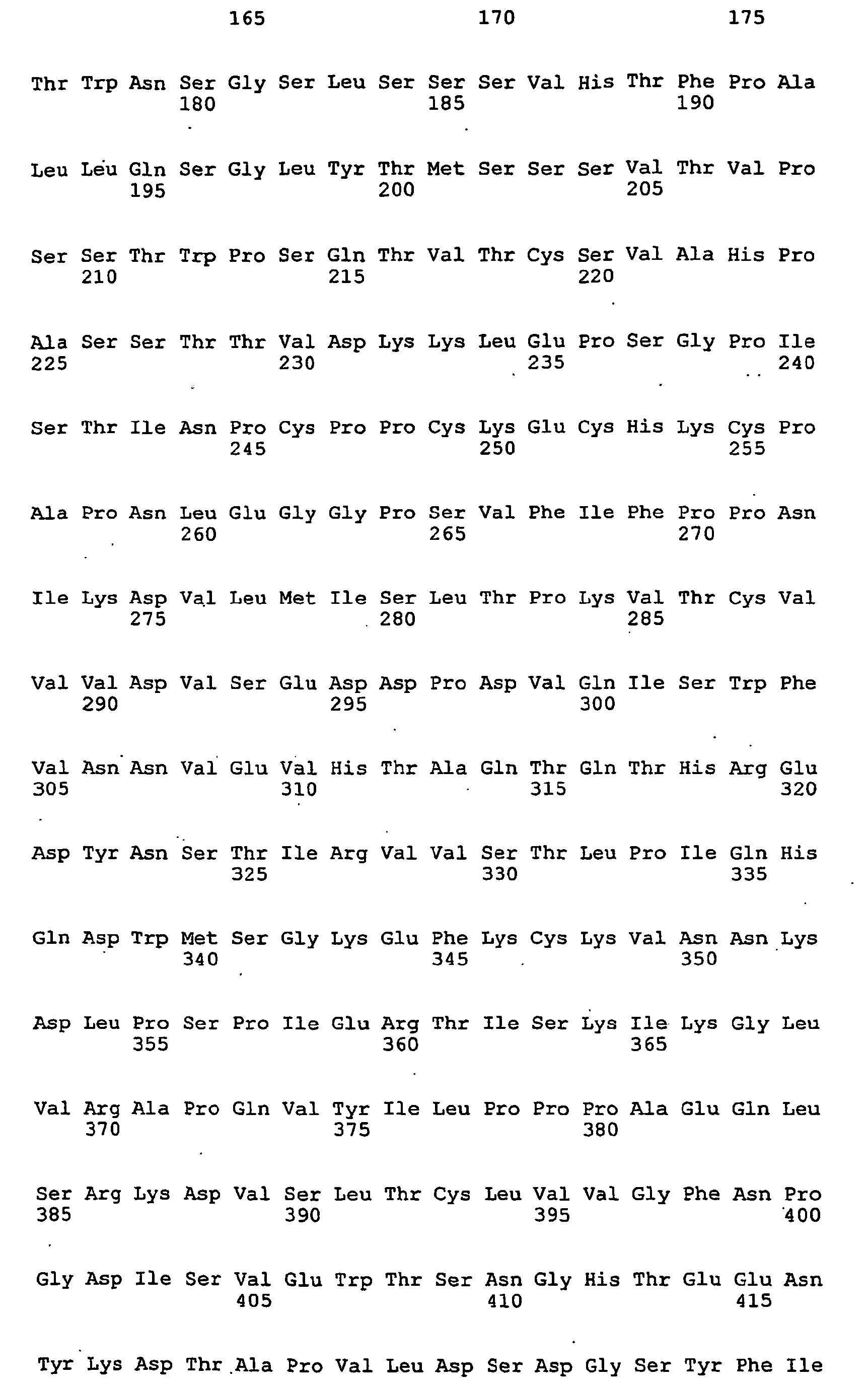

- <210> 5
<211> 763
<212> DNA
<213> Mus musculus and Homo sapiens - <400> 5

- <210> 6
<211> 239
<212> PRT
<213> Mus musculus and Homo sapiens - <400> 6

- <210> 7
<211> 1588
<212> DNA
<213> Mus musculus and Homo sapiens - <400> 7
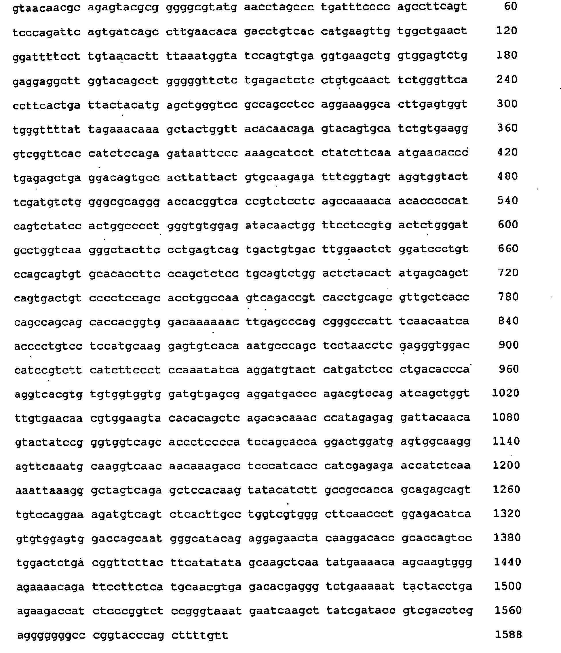
- <210> 8
<211> 476
<212> PRT
<213> Mus musculus and Homo sapiens - <400> 8


- <210> 9
<211> 794
<212> DNA
<213> Mus musculus and Homo sapiens - <400> 9

- <210> 10
<211> 239
<212> PRT
<213> Mus musculus and Homo sapiens - <400> 10


- <210> 11
<211> 1514
<212> DNA
<213> Mus musculus and Homo sapiens - <400> 11


- <210> 12
<211> 470
<212> PRT
<213> Mus musculus and Homo sapiens - <400> 12


- <210> 13
<211> 750
<212> DNA
<213> Mus musculus and Homo sapiens - <400> 13

- <210> 14
<211> 228
<212> PRT
<213> Mus musculus and Homo sapiens - <400> 14


- <210> 15
<211> 1481
<212> DNA
<213> Mus musculus and Homo sapiens - <400> 15


- <210> 16
<211> 467
<212> PRT
<213> Mus musculus and Homo sapiens - <400> 16


- <210> 17
<211> 763
<212> DNA
<213> Mus musculus and Homo sapiens - <400> 17


- <210> 18
<211> 239
<212> PRT
<213> Mus musculus and Homo sapiens - <400> 18


- <210> 19
<211> 1514
<212> DNA
<213> Mus musculus and Homo sapiens - <400> 19


- <210> 20
<211> 470
<212> PRT
<213> Mus musculus and Homo sapiens - <400> 20


- <210> 21
<211> 898
<212> DNA
<213> Mus musculus and Homo sapiens - <400> 21


- <210> 22
<211> 238
<212> PRT
<213> Mus musculus and Homo sapiens - <400> 22


- <210> 23
<211> 1481
<212> DNA
<213> Mus musculus and Homo sapiens - <400> 23
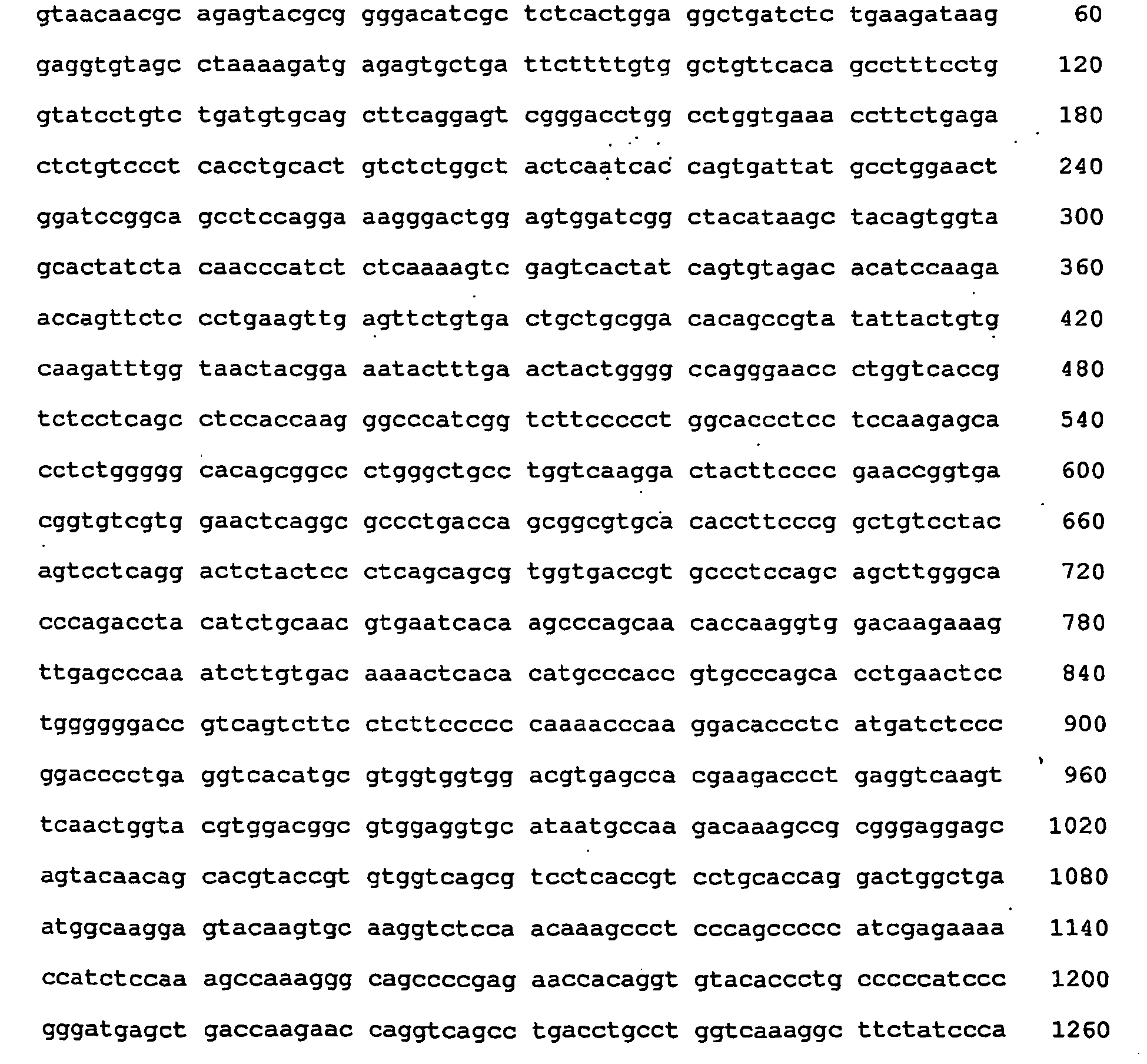

- <210> 24
<211> 467
<212> PRT
<213> Mus musculus and Homo sapiens - <400> 24


- <210> 25
<211> 813
<212> DNA
<213> Mus musculus and Homo sapiens - <400> 25
<210> 26

<211> 239
<212> PRT
<213> Mus musculus and Homo sapiens - <400> 26

- <210> 27
<211> 1558
<212> DNA
<213> Mus musculus and Homo sapiens - <400> 27

- <210> 28
<211> 470
<212> PRT
<213> Mus musculus and Homo sapiens - <400> 28


- <210> 29
<211> 772
<212> DNA
<213> Mus musculus and Homo sapiens - <400> 29

- <210> 30
<211> 239
<212> PRT
<213> Mus musculus and Homo sapiens - <400> 30
 <210> 31
<211> 1513
<212> DNA
<213> Mus musculus and Homo sapiens - <400> 31
 <210> 32
<211> 470
<212> PRT
<213> Mus musculus and Homo sapiens - <400> 32


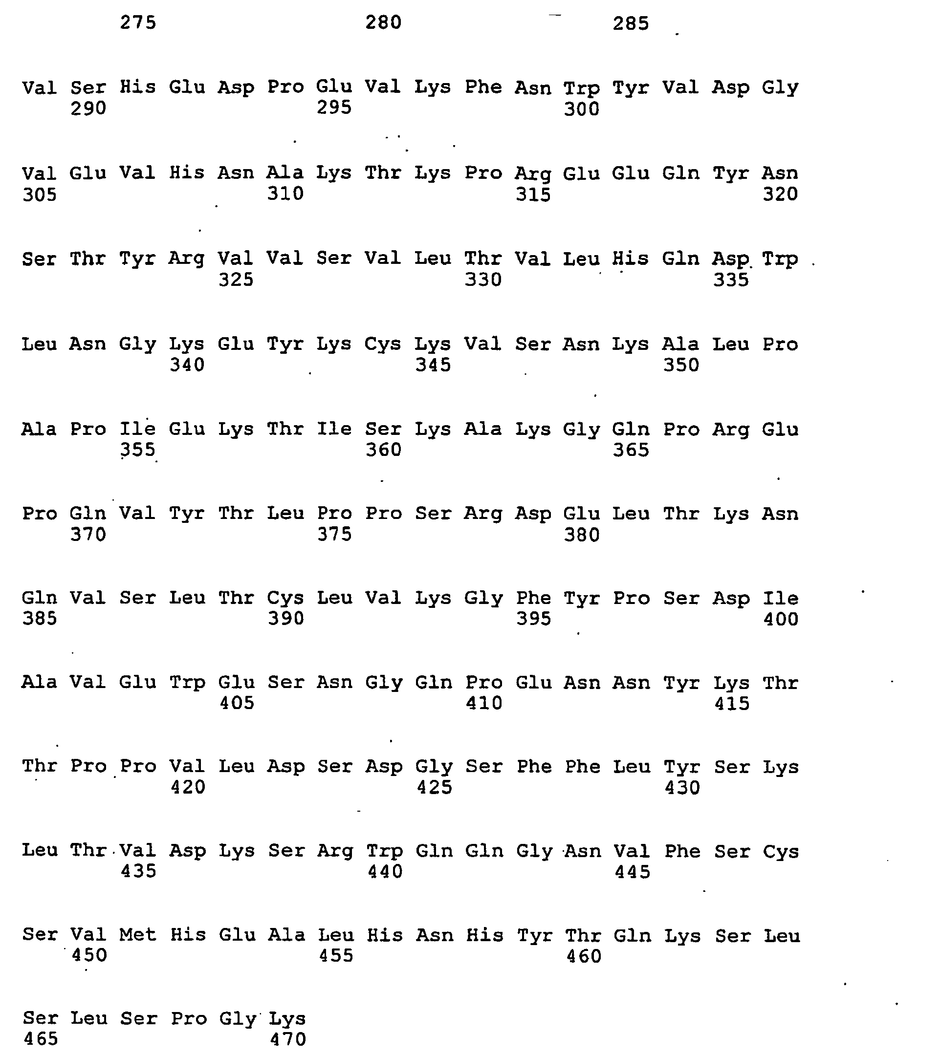
- <210> 33
<211> 913
<212> DNA
<213> Mus musculus and Homo sapiens - <400> 33


- <210> 34
<211> 238
<212> PRT
<213> Mus musculus and Homo sapiens - <400> 34


- <210> 35
<211> 1517
<212> DNA
<213> Mus musculus and Homo sapiens - <400> 35


- <210> 36
<211> 467
<212> PRT
<213> Mus musculus and Homo sapiens - <400> 36


- <210> 37
<211> 895
<212> DNA
<213> Mus musculus and Homo sapiens - <400> 37


- <210> 38
<211> 238
<212> PRT
<213> Mus musculus and Homo sapiens - <400> 38


- <210> 39
<211> 1587
<212> DNA
<213> Mus musculus and Homo sapiens - <400> 39


- <210> 40
<211> 467
<212> PRT
<213> Mus musculus and Homo sapiens - <400> 40


- <210> 41
<211> 384
<212> DNA
<213> Mus musculus - <400> 41

- <210> 42
<211> 399
<212> DNA
<213> Mus musculus - <400> 42

- <210> 43
<211> 236
<212> PRT
<213> Mus musculus - <400> 43


- <210> 44
<211> 227
<212> PRT
<213> Mus musculus - <400> 44


- <210> 45
<211> 399
<212> DNA
<213> Mus musculus - <400> 45

- <210> 46
<211> 420
<212> DNA
<213> Mus musculus - <400> 46
<210> 47

<211> 239
<212> PRT
<213> Mus musculus - <400> 47


- <210> 48
<211> 177
<212> PRT
<213> Mus musculus - <400> 48


- <210> 49
<211> 22
<212> DNA
<213> Artificial Sequence - <220>
<223> primer P8 for amplyfication - <400> 49
gtaacaacgc agagtacgcg gg 22 - <210> 50
<211> 19
<212> DNA
<213> Artificial Sequence - <220>
<223> primer P6 for amplyfication - <400> 50
tcatttaccc ggagaccgg 19 - <210> 51
<211> 25
<212> DNA
<213> Artificial Sequence - <220>
<223> primer P4 for amplyfication - <400> 51
ctaacactca ttcctgttga agctc 25 - <210> 52
<211> 26
<212> DNA
<213> Artificial Sequence . - <220>
<223> primer P16 for amplyfication - <400> 52
tcatttaccc ggagacaggg agaggc 26 - <210> 53
<211> 26
<212> DNA
<213> Artificial Sequence - <220>
<223> primer P111 for amplyfication - <400> 53
cgtccgaaga tcataagatt gcttgc 26 - <210> 54
<211> 22
<212> DNA
<213> Artificial Sequence - <220>
<223> primer P15 for amplyfication - <400> 54
ctaacactca cccctgttga ag 22 - <210> 55
<211> 43
<212> DNA
<213> Artificial Sequence - <220>
<223> primer P113 for amplyfication - <400> 55
gcaatcttat gatcttcgga cgttcggcgg agggaccaag gtg 43 - <210> 56
<211> 19
<212> DNA
<213> Artificial Sequence - <220>
<223> primer P9 for amplyfication - <400> 56
acaacgcaga gtacgcggg 19 - <210> 57
<211> 31
<212> DNA
<213> Artificial Sequence - <220>
<223> primer P216 for amplyfication - <400> 57
atcagcggcc gcacaacgca gagtacgcgg g 31 - <210> 58
<211> 32
<212> DNA
<213> Artificial Sequence - <220>
<223> primer P217 for amplyfication - <400> 58
atcactcgag ctaacactca cccctgttga ag 32 - <210> 59
<211> 24
<212> DNA
<213> Artificial Sequence - <220>
<223> primer P119 for amplyfication - <400> 59
gacatcgaag taccacctac tacc 24 - <210> 60
- <211> 26
<212> DNA
<213> Artificial Sequence - <220>
<223> primer P121 for amplyfication - <400> 60
tcatttaccc ggagacaggg agaggc 26 - <210> 61
<211> 48
<212> DNA
<213> Artificial Sequence - <220>
<223> primer P122 for amplyfication - <400> 61
ggtagtaggt ggtacttcga tgtctggggc cagggaaccc tggtcacc 48 - <210> 62
<211> 37
<212> DNA
<213> Artificial Sequence - <220>
<223> primer P220 for amplyfication - <400> 62
atcaaagctt acaacgcaga gtacgcgggg gcgtatg 37 - <210> 63
<211> 41
<212> DNA
<213> Artificial Sequence - <220>
<223> primer P221 for amplyfication - <400> 63
atcatctaga tcatttaccc ggagacaggg agaggctctt c 41 - <210> 64
<211> 29
<212> DNA
<213> Artificial Sequence - <220>
<223> primer P118 for amplyfication - <400> 64
cgtcggagga acatgtgtac tttgagagc 29 - <210> 65
<211> 45
<212> DNA
<213> Artificial Sequence - <220>
<223> primer P114 for amplyfication - <400> 65
caaagtacac atgttcctcc gacgttcggc ggagggacca aggtg 45 - <210> 66
<211> 31
<212> DNA
<213> Artificial Sequence - <220>
<223> primer P116 for amplyfication - <400> 66
atcagcggcc gcacaacgca gagtacgcgg g 31 - <210> 67
<211> 32
<212> DNA
<213> Artificial Sequence - <220>
<223> primer P117 for amplyfication - <400> 67
atcactcgag ctaacactca cccctgttga ag 32 - <210> 68
<211> 27
<212> DNA
<213> Artificial Sequence - <220>
<223> primer P120 for amplyfication - <400> 68
gtagttcaaa gtatttccgt agttacc 27 - <210> 69
<211> 51
<212> DNA
<213> Artificial Sequence - <220>
<223> primer P123 for amplyfication - <400> 69
ggtaactacg gaaatacttt gaactactgg ggccagggaa ccctggtcac c 51 - <210> 70
<211> 27
<212> DNA
<213> Artificial Sequence - <220>
<223> primer P248 for amplyfication - <400> 70
aaagcttaca acgcagagta cgcgggg 27 - <210> 71
<211> 31
<212> DNA
<213> Artificial Sequence - <220>
<223> primer P249 for amplyfication - <400> 71
atctagatca tttacccgga gacagggaga g 31 - <210> 72
<211> 46
<212> DNA
<213> Artificial Sequence - <220>
<223> primer P61 for amplyfication - <400> 72
ggggacattg tgatgacaca gtctccagac tccctggctg tgtcag 46 - <210> 73
<211> 45
<212> DNA
<213> Artificial Sequence - <220>
<223> primer P71 for amplyfication - <400> 73
gtgtcagcag gagagagggc cactataaac tgcaaatcca gtcag 45 - <210> 74
<211> 39
<212> DNA
<213> Artificial Sequence - <220>
<223> primer P72 for amplyfication - <400> 74
gacattgtga tgtcacagcc tccatcctcc ctggctgtg 39 - <210> 75
<211> 39
<212> DNA
<213> Artificial Sequence - <220>
<223> primer P73 for amplyfication - <400> 75
ggggtccctg atcgcttctc aggcagtgga tctgggaca 39 - <210> 76
<211> 48
<212> DNA
<213> Artificial Sequence - <220>
<223> primer P74 for amplyfication - <400> 76
ctcaccatca gcagtctgca ggctgaagac gtggcagttt attactgc 48 - <210> 77
- <211> 46
<212> DNA - <213> Artificial Sequence
- <220>
<223> primer P91 for amplyfication - <400> 77
agactccctg gctgtgtcac taggagagag ggccactata aactgc 46 - <210> 78
<211> 41
<212> DNA
<213> Artificial Sequence - <220>
<223> primer P92 for amplification - <400> 78
accagcagaa accagggcag cctcctaaac tgctgatcta c 41 - <210> 79
<211> 49
<212> DNA
<213> Artificial Sequence - <220>
<223> primer P63 for amplyfication - <400> 79
caccatctcc agagatgatt ccaaaaacag cctctatctt caaatgaac 49 - <210> 80
<211> 39
<212> DNA
<213> Artificial Sequence - <220>
<223> primer P75 for amplyfication - <400> 80
ggtatccagt gtgaggtgca gctggtggag tctggagga 39 - <210> 81
<211> 39
<212> DNA
<213> Artificial Sequence - <220>
<223> primer P76 for amplyfication - <400> 81
ctgagactct cctgtgcagc ttctgggttc accttcact 39 - <210> 82
<211> 48
<212> DNA
<213> Artificial Sequence - <220>
<223> primer P77 for amplyfication - <400> 82
cgccagcctc caggaaaggg acttgagtgg gtgggtttta ttagaaac 48 - <210> 83
<211> 50
<212> DNA
<213> Artificial Sequence - <220>
<223> primer P78 for amplyfication - <400> 83
tatcttcaaa tgaacagcct gaaaactgag gacagtgcca cttattactg 50 - <210> 84
<211> 41
<212> DNA
<213> Artificial Sequence - <220>
<223> primer P93 for amplyfication - <400> 84
acatgagctg ggtccgccag gctccaggaa agggacttga g 41 - <210> 85
<211> 47
<212> DNA
<213> Artificial Sequence - <220>
<223> primer P94 for amplyfication - <400> 85
cctgaaaact gaggacactg ccgtttatta ctgtgcaaga gatttcg 47 - <210> 86
<211> 43
<212> DNA
<213> Artificial Sequence - <220>
<223> primer P67 for amplyfication - <400> 86
gtgatgttgt gatgacccaa agtccactct ccctgcctgt cag 43 - <210> 87
<211> 45
<212> DNA
<213> Artificial Sequence - <220>
<223> primer P82 for amplyfication - <400> 87
ctctccctgc ctgtcactct tggacaacca gcctccatct cttgc 45 - <210> 88
<211> 42
<212> DNA
<213> Artificial Sequence - <220>
<223> primer P83 for amplyfication - <400> 88
aagccaggcc agtctccaag gcgcctgatc tacaaagttt cc 42 - <210> 89
<211> 44
<212> DNA
<213> Artificial Sequence - <220>
<223> primer P84 for amplyfication - <400> 89
ggaggctgag gatgtgggag tttattactg ctctcaaagt acac 44 - <210> 90
<211> 48
<212> DNA
<213> Artificial Sequence - <220>
<223> primer P97 for amplyfication - <400> 90
acacctattt acattggttc cagcagaggc caggccagtc tccaaggc 48 - <210> 91
<211> 51
<212> DNA
<213> Artificial Sequence - <220>
<223> primer P98 for amplyfication - <400> 91
gagtggaggc tgaggatgtg ggagtttatt actgctctca aagtacacat g 51 - <210> 92
<211> 48
<212> DNA
<213> Artificial Sequence - <220>
<223> primer P65 for amplyfication - <400> 92
catctctcaa aagtcgagtc tctatcagtg tagacacatc caagaacc 48 - <210> 93
<211> 40
<212> DNA
<213> Artificial Sequence - <220>
<223> primer P79 for amplyfication - <400> 93
gcctggtgaa accttctgag actctgtccc tcacctgcac 40 - <210> 94
<211> 45
<212> DNA
<213> Artificial Sequence - <220>
<223> primer P80 for amplyfication - <400> 94
aactggatcc ggcagcctcc aggaaaagga ctggagtgga tgggc 45 - <210> 95
<211> 45
<212> DNA
<213> Artificial Sequence - <220>
<223> primer P81 for amplyfication - <400> 95
tccaagaacc agttctccct gaagttgagt tctgtgacta ctgag 45 - <210> 96
<211> 50
<212> DNA
<213> Artificial Sequence - <220>
<223> primer P95 for amplyfication - <400> 96
ggaactggat ccggcagcct ccaggaaagg gactggagtg gatgggctac 50 - <210> 97
<211> 54
<212> DNA
<213> Artificial Sequence - <220>
<223> primer P96 for amplyfication - <400> 97
gttgagttct gtgactgctg cggacacagc cgtatattac tgtgcaagat ttgg 54 - <210> 98
<211> 33
<212> DNA
<213> Artificial Sequence - <220>
<223> primer P99 for amplyfication - <400> 98
tccctcacct gcactgtctc tggctactcaatc 33 - <210> 99
<211> 34
<212> DNA
<213> Artificial Sequence - <220>
<223> primer P100 for amplyfication - <400> 99
acaaactgga gtggatcggc tacataagct acag 34 - <210> 100
<211> 38
<212> DNA
<213> Artificial Sequence - <220>
<223> primer P101 for amplyfication - <400> 100
tcaaaagtcg agtcactatc agtgtagaca catccaag 38 - <210> 101
<211> 48
<212> DNA
<213> Artificial Sequence - <220>
<223> primer P146 for amplyfication - <400> 101
gacacagtct ccactctccc tgcctgtgac actaggagag agggccac 48 - <210> 102
<211> 37
<212> DNA
<213> Artificial Sequence - <220>
<223> primer P151 for amplyfication - <400> 102
ctctcaccat cagcagagtg gaggctgaag acgtggc 37 - <210> 103
<211> 33
<212> DNA
<213> Artificial Sequence - <220>
<223> primer P152 for amplyfication - <400> 103
aggctgaaga cgtgggagtt tattactgca agc 33 - <210> 104
<211> 41
<212> DNA
<213> Artificial Sequence - <220>
<223> primer P155 for amplyfication - <400> 104
cagaaaccag ggcagtctcc tcaactgctg atctactggg c 41 - <210> 105
<211> 29
<212> DNA
<213> Artificial Sequence - <220>
<223> primer P205 for amplyfication - <400> 105
cttggcttgg tacctgcaga aaccagggc 29 - <210> 106
<211> 41
<212> DNA
<213> Artificial Sequence - <220>
<223> primer P206 for amplyfication - <400> 106
gacaccagga gagccggcct ctataagctg caaatccagt c 41 - <210> 107
<211> 29
<212> DNA
<213> Artificial Sequence - <220>
<223> primer P207 for amplyfication - <400> 107
cttggcttgg tacctgcaga aaccagggc 29 - <210> 108
<211> 39
<212> DNA
<213> Artificial Sequence - <220>
<223> primer P242 for amplyfication - <400> 108
ggacagattt cactctcaaa atcagcagag tggaggctg 39 - <210> 109
<211> 33
<212> DNA
<213> Artificial Sequence - <220>
<223> primer P168 for amplyfication - <400> 109
tgtgaggtgc agctgttgga gtctggagga ggc 33 - <210> 110
<211> 34
<212> DNA
<213> Artificial Sequence - <220>
<223> primer P169 for amplyfication - <400> 110
gacttgagtg ggtgagtttt attagaaaca aagc 34 - <210> 111
<211> 43
<212> DNA
<213> Artificial Sequence - <220>
<223> primer P170 for amplyfication - <400> 111
catctccaga gataattcca aaaacaccct ctatcttcaa atg 43 - <210> 112
<211> 32
<212> DNA
<213> Artificial Sequence - <220>
<223> primer P171 for amplyfication - <400> 112
aatgaacagc ctgagagctg aggacactgc cg 32 - <210> 113
<211> 33
<212> DNA
<213> Artificial Sequence - <220>
<223> primer P231 for amplyfication - <400> 113
gtttattact gtgcaaagga ttttggtagt agg 33 - <210> 114
<211> 32
<212> DNA
<213> Artificial Sequence - <220>
<223> primer P145 for amplyfication - <400> 114
acctgtgggg acgttgtgat gacacagtct cc 32 - <210> 115
<211> 48
<212> DNA
<213> Artificial Sequence - <220>
<223> primer P146 for amplyfication - <400> 115
gacacagtct ccactctccc tgcctgtgac actaggagag agggccac 48 - <210> 116
<211> 39
<212> DNA
<213> Artificial Sequence - <220>
<223> primer P148 for amplyfication - <400> 116
ctacttggct tggttccagc agagaccagg gcagcctcc 39 - <210> 117
<211> 37
<212> DNA
<213> Artificial Sequence - <220>
<223> primer P151 for amplyfication - <400> 117
ctctcaccat cagcagagtg gaggctgaag acgtggc 37 - <210> 118
<211> 33
<212> DNA
<213> Artificial Sequence - <220>
<223> primer P152 for amplyfication - <400> 118
aggctgaaga cgtgggagtt tattactgca agc 33 - <210> 119
<211> 49
<212> DNA
<213> Artificial Sequence - <220>
<223> primer P202 for amplyfication - <400> 119
ctgtgacact aggacagccg gcctctataa gctgcaaatc cagtcagag 49 - <210> 120
<211> .42
<212> DNA
<213> Artificial Sequence - <220>
<223> primer P203 for amplyfication - <400> 120
agaccagggc agtctcctag actgcggatc tactgggcat cc 42 - <210> 121
<211> 34
<212> DNA
<213> Artificial Sequence - <220>
<223> primer P204 for amplyfication - <400> 121
cagatttcac tctcaaaatc agcagagtgg aggc 34 - <210> 122
<211> 45
<212> DNA
<213> Artificial Sequence - <220>
<223> primer P173 for amplyfication - <400> 122
aatggtatcc agtgtcaggt gacgctgaag gagtctggag gaggc 45 - <210> 123
<211> 43
<212> DNA
<213> Artificial Sequence - <220>
<223> primer P175 for amplyfication - <400> 123
gaccagtctt ggtaaagcct acggagactc tgagactctc ctg 43 - <210> 124
<211> 47
<212> DNA
<213> Artificial Sequence - <220>
<223> primer P176 for amplyfication - <400> 124
ctacggagac tctgacactc acctgtacag tttctgggtt caccttc 47 - <210> 125
<211> 40
<212> DNA
<213> Artificial Sequence - <220>
<223> primer P177 for amplyfication - <400> 125
tacatgagct ggatccgcca gcctccagga aagggacttg 40 - <210> 126
<211> 48
<212> DNA
<213> Artificial Sequence - <220>
<223> primer P178 for amplyfication - <400> 126
gcctccagga aaggcacttg agtggctggc ttttattaga aacaaagc 48 - <210> 127
<211> 49
<212> DNA
<213> Artificial Sequence - <220>
<223> primer P179 for amplification
<400> 127
tgtgaagggt cggctcacca tctccaaaga tacttccaaa aacagcctc 49 - <210> 128
<211> 49
<212> DNA
<213> Artificial Sequence - <220>
<223> primer P181 for amplyfication - <400> 128
cgttcttaca atgaccaaca tggatcctgt ggacactgcc gtttattac 49 - <210> 129
<211> 29
<212> DNA
<213> Artificial Sequence - <220>
<223> primer P182 for amplyfication - <400> 129
gtggacactg ccacttatta ctgtgcaag 29 - <210> 130
<211> 24
<212> DNA
<213> Artificial Sequence - <220>
<223> primer P246 for amplyfication - <400> 130
caaagatact tccaaatccc aggt 24 - <210> 131
<211> 46
<212> DNA
<213> Artificial Sequence - <220>
<223> primer P276 for amplyfication - <400> 131
ctggaccagt cttggtaaag cctacggaga ctctgagact ctcctg 46 - <210> 132
<211> 38
<212> DNA
<213> Artificial Sequence - <220>
<223> primer P160 for amplyfication - <400> 132
cctgcttcca gcagtgaaat tgtgatgacc caaagtcc 38 - <210> 133
<211> 24
<212> DNA
<213> Artificial Sequence - <220>
<223> primer P162 for amplyfication - <400> 133
gtccactctc cctgtctatc actc 24 - <210> 134
<211> 43
<212> DNA
<213> Artificial Sequence - <220>
<223> primer P164 for amplyfication - <400> 134
tatttacatt ggttcctgca gaaggcaggc cagtctccaa ggc 43 - <210> 135
- <211> 33
- <212> DNA
<213> Artificial Sequence - <220>
<223> primer P167 for amplyfication - <400> 135
gtggaggctg aggatttcgg agtttattac tgc 33 - <210> 136
<211> 32
<212> DNA
<213> Artificial Sequence - <220>
<223> primer P198 for amplyfication - <400> 136
attgtgatga cccaaactcc actctccctg tc 32 - <210> 137
<211> 36
<212> DNA
<213> Artificial Sequence - <220>
<223> primer P199 for amplyfication - <400> 137
tctatcactc ctggagaaca agcctccatc tcttgc 36 - <210> 138 .
<211> 39
<212> DNA
<213> Artificial Sequence - <220>
<223> primer P200 for amplyfication - <400> 138
ttcctgcaga aggcacgccc ggttccaagg cgcctgatc 39 - <210> 139
<211> 39
<212> DNA
<213> Artificial Sequence - <220>
<223> primer P201 for amplyfication - <400> 139
cacgcccgg tttcaacgct cctgatctac aaagtttcc 39 - <210> 140
<211> 28
<212> DNA
<213> Artificial Sequence - <220>
<223> primer P183 for amplyfication, - <400> 140
ggtatcctgt ctcaagtgca gcttcagg 28 - <210> 141
<211> 35
<212> DNA
<213> Artificial Sequence - <220>
<223> primer P186 for amplyfication - <400> 141
tcaagtgcag cttgtgcagt cgggacctgg cctgg 35 - <210> 142
<211> 21
<212> DNA
<213> Artificial Sequence - <220>
<223> primer P187 for amplyfication - <400> 142
cttgtgcagt cgggagctga a 21 - <210> 143
<211> 42
<212> DNA
<213> Artificial Sequence - <220>
<223> primer P189 for amplyfication - <400> 143
gcgtctgtga aagtcagctg caaggcctct ggctactcaa tc 42 - <210> 144
<211> 37
<212> DNA
<213> Artificial Sequence - <220>
<223> primer P190 for amplyfication - <400> 144
gcctggaact gggtccggca ggctccagga cagagac 37 - <210> 145
<211> 32
<212> DNA
<213> Artificial Sequence - <220>
<223> primer P191 for amplyfication - <400> 145
gcagcctcca ggacagagac tggagtggat cg 32 - <210> 146
<211> 32
<212> DNA
<213> Artificial Sequence - <220>
<223> primer P192 for amplyfication - <400> 146
agagactgga gtggatgggc tacataagct ac 32 - <210> 147
<211> 36
<212> DNA
<213> Artificial Sequence - <220>
<223> primer P193 for amplyfication - <400> 147
gtcgagtcac tatcactaga gacacatcca agaacc 36 - <210> 148
<211> 44
<212> DNA
<213> Artificial Sequence - <220>
<223> primer P194 for amplyfication - <400> 148
atcactagag acacatccgc gagcacggcc tacatggagt tgag 44 - <210> 149
- <211> 33
<212> DNA
<213> Artificial Sequence - <220>
<223> primer P195 for amplyfication - <400> 149
aagaaccagt tctacatgga gttgagttct ctg 33 - <210> 150
<211> 41
<212> DNA
<213> Artificial Sequence - <220>
<223> primer P196 for amplyfication - <400> 150
aagttgagt tctctgagat ctgaggacac agccgtatat t 41 - <210> 151
<211> 31
<212> DNA
<213> Artificial Sequence - <220>
<223> primer P197 for amplyfication - <400> 151
=gagatctga ggacatggcc gtatattact g 31 - <210> 152
<211> 51
<212> DNA
<213> Artificial Sequence - <220>
<223> primer P244 for amplyfication - <400> 152
ctggcctggt gaaacctggt gcgtctgtga aactcacctg cactgtctct g 51 - <210> 153
<211> 46
<212> DNA
<213> Artificial Sequence - <220>
<223> primer P156 for amplyfication - <400> 153
cttccagcag tgatattgtg atgacccaaa ctccactctc cctgcc 46 - <210> 154
<211> 39
<212> DNA
<213> Artificial Sequence - <220>
<223> primer P157 for amplyfication - <400> 154
ctgcotgtca ctcctggaga accagcctcc atctcttgc 39 - <210> 155
<211> 40
<212> DNA
<213> Artificial Sequence - <220>
<223> primer P158 for amplyfication - <400> 155
cctatttaca ttggtacctg cagaagccag gccagtctcc 40 - <210> 156
<211> 38
<212> DNA
<213> Artificial Sequence - <220>
<223> primer P159 for amplyfication - <400> 156
caggccagtc tccacagctc ctgatctaca aagtttcc 38 - <210> 157
<211> 28
<212> DNA
<213> Artificial Sequence - <220>
<223> primer P183 for amplyfication - <400> 157
ggtatcctgt ctcaagtgca gcttcagg 28 - <210> 158
<211> 27
<212> DNA
<213> Artificial Sequence - <220>
<223> primer P184 for amplyfication - <400> 158
gtgaaacctt ctcagactct gtccctc 27 - <210> 159
<211> 26
<212> DNA
<213> Artificial Sequence - <220>
<223> primer P185 for amplyfication - <400> 159
tggatccggc agcatccagg aaaggg 26 - <210> 160
<211> 11
<212> PRT
<213> Mus musculus - <400> 160

- <210> 161
<211> 7
<212> PRT
<213> Mus musculus - <400> 161

- <210> 162
<211> 9
<212> PRT
<213> Mus musculus - <400> 162

- <210> 163
<211> 10
<212> PRT
<213> Mus musculus - <400> 163

- <210> 164
<211> 16
<212> PRT
<213> Mus musculus - <400> 164

- <210> 165
<211> 6
<212> PRT
<213> Mus musculus - <400> 165
- <210> 166
<211> 17
<212> PRT
<213> Mus musculus - <400> 166

- <210> 167
<211> 7
<212> PRT
<213> Mus musculus - <400> 167

- <210> 168
<211> 8
<212> PRT
<213> Mus musculus - <400> 168

- <210> 169
<211> 10
<212> PRT
<213> Mus musculus - <400> 169

- <210> 170
<211> 19
<212> PRT
<213> Mus musculus - <400> 170

- <210> 171
<211> 10
<212> PRT
<213> Mus musculus - <400> 171
<210> 172

<211> 16
<212> PRT
<213> Mus musculus - <400> 172

- <210> 173
<211> 7
<212> PRT
<213> Mus musculus - <400> 173
<210> 174

<211> 9
<212> PRT
<213> Mus musculus - <400> 174

- <210> 175
<211> 11
<212> PRT
<213> Mus musculus - <400> 175

- <210> 176
<211> 16
<212> PRT
<213> Mus musculus - <400> 176

- <210> 177
<211> 10
<212> PRT
<213> Mus musculus - <400> 177

- <210> 178
<211> 17
<212> PRT
<213> Mus musculus - <400> 178

- <210> 179
<211> 7
<212> PRT
<213> Mus musculus - <400> 179

- <210> 180
<211> 8
<212> PRT
<213> Mus musculus - <400> 180

- <210> 181
<211> 10
<212> PRT
<213> Mus musculus - <400> 181

- <210> 182
<211> 19
<212> PRT
<213> Mus musculus - <400> 182

- <210> 183
<211> 10
<212> PRT
<213> Mus musculus - <400> 183

- <210> 184
<211> 13
212> PRT
213> Artificial Sequence - 220>
223> peptide for epitope mapping - <400> 184

- <210> 185
<211> 13
<212> PRT
<213> Artificial Sequence - <220>
<223> peptide for epitope mapping - <400> 185
<210> 186

<211> 13
<212> PRT
<213> Artificial Sequence - <220>
<223> peptide for epitope mapping - <400> 186

- <210> 187
<211> 13
<212> PRT
<213> Artificial Sequence - <220>
<223> peptide for epitope mapping - <400> 187

- <210> 188
<211> 13
<212> PRT
<213> Artificial Sequence - <220>
<223> peptide for epitope mapping - <400> 188

- <210> 189
<211> 13
<212> PRT
<213> Artificial Sequence - <220>
<223> peptide for epitope mapping - <400> 189

- <210> 190
<211> 13
<212> PRT
<213> Artificial Sequence - <220>
<223> peptide for epitope mapping - <400> 190

- <210> 191
<211> 13
<212> PRT
<213> Artificial Sequence - <220>
<223> peptide for epitope mapping - <400> 191

- <210> 192
<211> 13
<212> PRT
<213> Artificial Sequence - <220>
<223> peptide for epitope mapping - <400> 192

- <210> 193
<211> 13
<212> PRT
<213> Artificial Sequence - <220>
<223> peptide for epitope mapping - <400> 193

- <210> 194
<211> 13
<212> PRT
<213> Artificial Sequence - <220>
<223> peptide for epitope mapping - <400> 194

- <210> 195
<211> 13
<212> PRT
<213> Artificial Sequence - <220>
<223> peptide for epitope mapping - <400> 195

- <210> 196
<211> 13
<212> PRT
<213> Artificial Sequence - <220>
<223> peptide for epitope mapping - <400> 196

- <210> 197
<211> 13
<212> PRT
<213> Artificial Sequence - <220>
<223> peptide for epitope mapping - <400> 197

- <210> 198
<211> 13
<212> PRT
<213> Artificial Sequence - <220>
<223> peptide for epitope mapping - <400> 198

- <210> 199
<211> 13
<212> PRT
<213> Artificial Sequence - <220>
<223> peptide for epitope mapping - <400> 199

- <210> 200
<211> 13
<212> PRT
<213> Artificial Sequence - <220>
<223> peptide for epitope mapping - <400> 200
<210> 201

<211> 13
<212> PRT
<213> Artificial Sequence - <220>
<223> peptide for epitope mapping - <400> 201

- <210> 202
<211> 13
<212> PRT
<213> Artificial Sequence - <220>
<223> peptide for epitope mapping - <400> 202

- <210> 203
<211> 13
<212> PRT
<213> Artificial Sequence - <220>
<223> peptide for epitope mapping - <400> 203

- <210> 204
<211> 13
<212> PRT
<213> Artificial Sequence - <220>
<223> peptide for epitope mapping - <400> 204

- <210> 205
<211> 13
<212> PRT
<213> Artificial Sequence - <220>
<223> peptide for epitope mapping - <400> 205

- <210> 206
<211> 13
<212> PRT
<213> Artificial Sequence - <220>
223> peptide for epitope mapping - 400> 206

- <210> 207
<211> 13
<212> PRT
<213> Artificial Sequence - <220>
<223> peptide for epitope mapping - <400> 207
<210> 208

<211> 13
<212> PRT
<213> Artificial Sequence - <220>
<223> peptide for epitope mapping - <400> 208

- <210> 209
<211> 13
<212> PRT
<213> Artificial Sequence - <220>
<223> peptide for epitope mapping - <400> 209

- <210> 210
<211> 13
<212> PRT
<213> Artificial Sequence - <220>
<223> peptide for epitope mapping - <400> 210

- <210> 211
<211> 13
<212> PRT
<213> Artificial Sequence - <220>
<223> peptide for epitope mapping - <400> 211

- <210> 212
<211> 13
<212> PRT
<213> Artificial Sequence - <220>
<223> peptide for epitope mapping - <400> 212

- <210> 213
<211> 13
<212> PRT
<213> Artificial Sequence - <220>
<223> peptide for epitope mapping - <400> 213

- <210> 214
<211> 13
<212> PRT
<213> Artificial Sequence - <220>
<223> peptide for epitope mapping - <400> 214

- <210> 215
<211> 13
<212> PRT
<213> Artificial Sequence - <220>
<223> peptide for epitope mapping - <400> 215

- <210> 216
<211> 13
<212> PRT
<213> Artificial Sequence - <220>
<223> peptide for epitope mapping <400> 216<210> 217
<211> 13
<212> PRT
<213> Artificial Sequence - <220>
<223> peptide for epitope mapping - <400> 217
<210> 218

<211> 13
<212> PRT
<213> Artificial Sequence - <220>
<223> peptide for epitope mapping - <400> 218

- <210> 219
<211> 13
<212> PRT
<213> Artificial Sequence - <220>
<223> peptide for epitope mapping - <400> 219

- <210> 220
<211> 13
<212> PRT
<213> Artificial Sequence - <220>
<223> peptide for epitope mapping - <400> 220

- <210> 221
<211> 13
<212> PRT
<213> Artificial Sequence - <220>
<223> peptide for epitope mapping - <400> 221

- <210> 222
<211> 13
<212> PRT
<213> Artificial Sequence - <220>
<223> peptide for epitope mapping - <400> 222

- <210> 223
<211> 13
<212> PRT
<213> Artificial Sequence - <220>
<223> peptide for epitope mapping - <400> 223

- <210> 224
<211> 13
<212> PRT
<213> Artificial Sequence - <220>
<223> peptide for epitope mapping - <400> 224

- <210> 225
<211> 13
<212> PRT
<213> Artificial Sequence - <220>
<223> peptide for epitope mapping - <400> 225

- <210> 226
<211> 13
<212> PRT
<213> Artificial Sequence - <220>
<223> peptide for epitope mapping - <400> 226

- <210> 227
<211> 13
<212> PRT
<213> Artificial Sequence - <220>
<223> peptide for epitope mapping - <400> 227

- <210> 228
<211> 13
<212> PRT
<213> Artificial Sequence - <220>
<223> peptide for epitope mapping - <400> 228

- <210> 229
<211> 13
<212> PRT <213> Artificial Sequence - <220>
<223> peptide for epitope mapping - <400> 229

- <210> 230
<211> 13
<212> PRT
<213> Artificial Sequence - <220>
<223> peptide for epitope mapping - <400> 230

- <210> 231
<211> 13
<212> PRT
<213> Artificial Sequence - <220>
<223> peptide for epitope mapping - <400> 231

- <210> 232
<211> 13
<212> PRT
<213> Artificial Sequence - <220>
<223> peptide for epitope mapping - <400> 232

- <210> 233
<211> 13
<212> PRT
<213> Artificial Sequence - <220>
<223> peptide for epitope mapping - <400> 233

- <210> 234
<211> 13
<212> PRT
<213> Artificial Sequence - <220>
<223> peptide for epitope mapping - <400> 234

- <210> 235
<211> 13
<212> PRT
<213> Artificial Sequence - <220>
<223> peptide for epitope mapping - <400> 235

- <210> 236
<211> 13
<212> PRT
<213> Artificial Sequence - <220>
<223> peptide for epitope mapping - <400> 236

- <210> 237
<211> 13
<212> PRT
<213> Artificial Sequence - <220>
<223> peptide for epitope mapping - <400> 237

- <210> 238
<211> 13
<212> PRT
<213> Artificial Sequence - <220>
<223> peptide for epitope mapping - <400> 238

- <210> 239
<211> 13
<212> PRT
<213> Artificial Sequence - <220>
<223> peptide for epitope mapping - <400> 239

- <210> 240
<211> 13
<212> PRT
<213> Artificial Sequence - <220>
<223> peptide for epitope mapping - <400> 240
<210> 241

<211> 13
<212> PRT
<213> Artificial Sequence - <220>
<223> peptide for epitope mapping - <400> 241

- <210> 242
<211> 13
<212> PRT
<213> Artificial Sequence - <220>
<223> peptide for epitope mapping - <400> 242

- <210> 243
<211> 13
<212> PRT
<213> Artificial Sequence - <220>
<223> peptide for epitope mapping - <400> 243

- <210> 244
<211> 13
<212> PRT
<213> Artificial Sequence - <220>
<223> peptide for epitope mapping - <400> 244

- <210> 245
<211> 13
<212> PRT
<213> Artificial Sequence - <220>
<223> peptide for epitope mapping - <400> 245

- <210> 246
<211> 13
<212> PRT
<213> Artificial Sequence - <220>
<223> peptide for epitope mapping - <400> 246

- <210> 247
<211> 13
<212> PRT
<213> Artificial Sequence - <220>
<223> peptide for epitope mapping
<220>
<221> MISC_FEATURE
<223> peptide for epitope mapping - <400> 247

- <210> 248
<211> 13
<212> PRT
<213> Artificial Sequence - <220>
<223> peptide for epitope mapping - <400> 248

- <210> 249
<211> 13
<212> PRT
<213> Artificial Sequence - <220>
<223> peptide for epitope mapping - <400> 249

- <210> 250
<211> 13
<212> PRT
<213> Artificial Sequence - <220>
<223> peptide for epitope mapping - <400> 250

- <210> 251
<211> 13
<212> PRT
<213> Artificial Sequence - <220>
<223> peptide for epitope mapping - <400> 251
<210> 252

<211> 13
<212> PRT
<213> Artificial Sequence - <220>
<223> peptide for epitope mapping - <400> 252

- <210> 253
<211> 13
<212> PRT
<213> Artificial Sequence - <220>
<223> peptide for epitope mapping - <400> 253

- <210> 254
<211> 13
<212> PRT
<213> Artificial Sequence - <220>
<223> peptide for epitope mapping - <400> 254

- <210> 255
<211> 13
<212> PRT
<213> Artificial Sequence - <220>
<223> peptide for epitope mapping - <400> 255

- <210> 256
<211> 13
<212> PRT
<213> Artificial Sequence - <220> .
<223> peptide for epitope mapping - <400> 256

- <210> 257
<211> 13
<212> PRT
<213> Artificial Sequence - <220>
<223> peptide for epitope mapping - <400> 257

- <210> 258
<211> 13
<212> PRT
<213> Artificial Sequence - <220>
<223> peptide for epitope mapping - <400> 258

- <210> 259
<211> 13
<212> PRT
<213> Artificial Sequence - <220>
<223> peptide for epitope mapping - <400> 259

- <210> 260
<211> 55
<212> DNA
<213> Artificial Sequence - <220>
<223> primer P25 for amplyfication - <400> 260
tctggaggtg gaggtagtgg gggaggaggt tcagacatca agatgaccca gtctc 55 - <210> 261
<211> 34
<212> DNA
<213> Artificial Sequence - <220>
<223> primer P28 for amplyfication - <400> 261
ggcctaatcg gcccgtttta tttccagctt ggtc 34 - <210> 262
<211> 55
<212> DNA
<213> Artificial Sequence - <220>
<223> primer P22 for amplyfication - <400> 262
cccactacct ccacctccag agcctccccc tcctgcagag acagtgacca gagtc 55 - <210> 263
<211> 22
<212> DNA
<213> Artificial Sequence - <220>
<223> primer P18 for amplyfication - <400> 263
agtgatgagc actgaacaca ga 22 - <210> 264
<211> 56
<212> DNA
<213> Artificial Sequence - <220>
<223> primer P26 for amplyfication - <400> 264
tctggaggtg gaggtagtgg gggaggaggt tcagacattg tgatgtcaca gtctcc 56 - <210> 265
<211> 34
<212> DNA
<213> Artificial Sequence - <220>
<223> primer P29 for amplyfication - <400> 265
ggcctaatcg gcccgtttga tttccagctt ggtg 34 - <210> 266
<211> 22
<212> DNA
<213> Artificial Sequence - <220>
<223> primer P21 for amplyfication - <400> 266
gacctgtcac catgaagttg tg 22 - <210> 267
<211> 53
<212> DNA
<213> Artificial Sequence - <220>
<223> primer P24 for amplyfication - <400> 267
=ccactacct ccacctccag agcctccccc tcctgaggag acggtgaccg tgg 53
Claims (22)
- An antibody, antibody fragment or derivative thereof comprising the complementarity determining regions (CDRs) ofa. SEQ ID NOs: 160 to 162 of the VH region and SEQ ID NOs: 163 to 165 of the VL region;b. SEQ ID NOs: 166 to 168 of the VH region and SEQ ID NOs: 169 to 171 of the VL region;c. SEQ ID NOs: 172 to 174 of the VH region and SEQ ID NOs: 175 to 177 of the VL region; ord. SEQ ID NOs: 178 to 180 of the VH region and SEQ ID NOs: 181 to 183 of the VL region, wherein said antibody, antibody fragment or derivative thereof specifically binds to/interacts with at least one epitope of the extracellular or intracellular domain of the mammalian EAG1 ion channel and does not bind to/interact with the mammalian EAG2 ion channel.
- The antibody according to claim 1, wherein said antibody is a monoclonal antibody.
- The antibody fragment or derivative thereof according to claim 1 or 2, wherein said antibody fragment or derivative thereof is a Fab-fragment, a X F(ab2)'- fragment, or a Fv-fragment.
- The antibody, antibody fragment or derivative thereof according to any one of claims 1 to 3, wherein the light chain is selected from the group consisting of SEQ ID NOs. 2, 6, 10, 14, 18, 22, 26, 30, 34. 38, 43 and 47 and wherein the heavy chain is selected from the group consisting of SEQ ID NOs. 4, 8, 12, 16, 20, 24, 28, 32, 36, 40, 44 and 48.
- The antibody, antibody fragment or derivative thereof according to any one of claims 1 to 4, wherein said antibody, fragment or derivative thereof is coupled to a labeling group.
- A nucleic acid molecule encoding the antibody, antibody fragment or derivative thereof of anyone of claims 1 to 4.
- A vector comprising a nucleic acid molecule of claim 6.
- The vector of claim 7 which is an expression vector wherein the nucleic acid molecule is operatively linked to one or more control sequences allowing the transcription and optionally expression in prokaryotic and/or eukaryotic host cells.
- A host cell comprising the vector of claim 7 or 8.
- The host cell of claim 9 which is a bacteria, a fungal, plant, amphibian or animal cell.
- The host cell of claim 10 which is a human cell or human cell line.
- A method for the preparation of an antibody, antibody fragment or derivative thereof of any one of claims 1 to 4, comprising culturing the host cell of any one of claims 9 to 11 under conditions that allow synthesis of said antibody, antibody fragment or derivative thereof and recovering said antibody, antibody fragment or derivative thereof from said culture.
- A pharmaceutical composition comprising the antibody, antibody fragment or derivative thereof as defined in any one of claims 1 to 5, the nucleic acid molecule of claim 6, the vector of claim 7 or 8, the host cell of any one of claims 9 to 11 or an antibody, antibody fragment or derivative thereof obtained by the method as defined in claim 12.
- The pharmaceutical composition of claim 13, further comprising at least one anti-neoplastic agent.
- Use of the antibody, antibody fragment or derivative thereof as defined in any one of claims 1 to 5, the nucleic acid molecule of claim 6, the vector of claim 7 or 8, the host cell of any one of claims 9 to 11 or an antibody, antibody fragment or derivative thereof obtained by the method as defined in claim 12 for the preparation of a pharmaceutical composition for prevention or treatment of a hyperproliferative disease, inflammatory disease, psoriasis, or a neurodegenerative disease.
- The use of claim 15, wherein said neurodegenerative disease is Alzheimer's disease, multiple sclerosis, lateral amyotrophic sclerosis or Parkinson's disease.
- The use of claim 15, wherein said hyperproliferative disease is breast, lung, colon, kidney, lymphoma, skin, ovary, prostate, pancreas, esophagus, Barrett, stomach, bladder, cervix, liver, thyroid cancer, melanoma, hyperplastic or neoplastic diseases or other EAG expressing or overexpressing hyperproliferative diseases.
- The use of claim 15, wherein said inflammatory disease is pancreatitis or hepatitis.
- A diagnostic composition comprising the antibody, antibody fragment or derivative thereof as defined in any one of claims 1 to 5, the nucleic acid molecule of claim 6, the vector of claim 7 or 8, the host cell of any one of claims 9 to 11 or an antibody, antibody fragment or derivative thereof obtained by the method as defined in claim 12 and, optionally, a pharmaceutically acceptable carrier.
- An in vitro method of assessing for the presence of EAG1 expressing cells comprising contacting the antibody or antibody fragment or derivative thereof of any one of claims 1 to 5 with cells or a tissue suspected of carrying EAG1 on their/its surface.
- An in vitro method of blocking EAG1 function comprising contacting the antibody or antibody fragment or derivative thereof of any one of claims 1 to 5 with cells or a tissue suspected of carrying EAG1 on their/its surface.
- Kit comprising the antibody, antibody fragment or derivative thereof of any one of claims 1 to 5, the nucleic acid molecule of claim 6 and/or the vector of claim 7 or 8.
Priority Applications (1)
| Application Number | Priority Date | Filing Date | Title |
|---|---|---|---|
| EP05797335A EP1805222B1 (en) | 2004-10-01 | 2005-10-04 | Antibodies directed to the mammalian eag1 ion channel protein |
Applications Claiming Priority (3)
| Application Number | Priority Date | Filing Date | Title |
|---|---|---|---|
| EP04023489 | 2004-10-01 | ||
| EP05797335A EP1805222B1 (en) | 2004-10-01 | 2005-10-04 | Antibodies directed to the mammalian eag1 ion channel protein |
| PCT/EP2005/010671 WO2006037604A1 (en) | 2004-10-01 | 2005-10-04 | Novel antibodies directed to the mammalian eag1 ion channel protein |
Publications (2)
| Publication Number | Publication Date |
|---|---|
| EP1805222A1 EP1805222A1 (en) | 2007-07-11 |
| EP1805222B1 true EP1805222B1 (en) | 2011-02-02 |
Family
ID=34926818
Family Applications (1)
| Application Number | Title | Priority Date | Filing Date |
|---|---|---|---|
| EP05797335A Not-in-force EP1805222B1 (en) | 2004-10-01 | 2005-10-04 | Antibodies directed to the mammalian eag1 ion channel protein |
Country Status (8)
| Country | Link |
|---|---|
| US (1) | US7910100B2 (en) |
| EP (1) | EP1805222B1 (en) |
| JP (1) | JP2008520186A (en) |
| AT (1) | ATE497508T1 (en) |
| AU (1) | AU2005291486A1 (en) |
| CA (1) | CA2582683A1 (en) |
| DE (1) | DE602005026219D1 (en) |
| WO (1) | WO2006037604A1 (en) |
Families Citing this family (63)
| Publication number | Priority date | Publication date | Assignee | Title |
|---|---|---|---|---|
| US7658924B2 (en) | 2001-10-11 | 2010-02-09 | Amgen Inc. | Angiopoietin-2 specific binding agents |
| DE10303974A1 (en) | 2003-01-31 | 2004-08-05 | Abbott Gmbh & Co. Kg | Amyloid β (1-42) oligomers, process for their preparation and their use |
| KR20080090408A (en) | 2005-11-30 | 2008-10-08 | 아보트 러보러터리즈 | Anti-abeta; globulomer antibodies, antigen-binding moieties thereof, corresponding hybridomas, nucleic acids, vectors, host cells, methods of producing said antibodies, compositions comprising said antibodies, uses of said antibodies and methods of using said antibodies |
| DK1976877T4 (en) | 2005-11-30 | 2017-01-16 | Abbvie Inc | Monoclonal antibodies to amyloid beta protein and uses thereof |
| PL2046833T3 (en) | 2006-07-14 | 2014-01-31 | Ac Immune Sa | Humanized antibody against amyloid beta |
| ES2570153T3 (en) * | 2006-08-22 | 2016-05-17 | Novo Nordisk As | Anti-C5aR antibodies with improved properties |
| US8455626B2 (en) | 2006-11-30 | 2013-06-04 | Abbott Laboratories | Aβ conformer selective anti-aβ globulomer monoclonal antibodies |
| US7858752B2 (en) | 2006-12-05 | 2010-12-28 | Abbott Laboratories | Recombinant antibodies against hepatitis C virus and methods of obtaining and using same |
| US20100311767A1 (en) | 2007-02-27 | 2010-12-09 | Abbott Gmbh & Co. Kg | Method for the treatment of amyloidoses |
| JP5142265B2 (en) * | 2007-05-28 | 2013-02-13 | 独立行政法人産業技術総合研究所 | Paratopes and epitopes of anti-mortalin antibodies |
| US8613923B2 (en) | 2007-06-12 | 2013-12-24 | Ac Immune S.A. | Monoclonal antibody |
| US8048420B2 (en) | 2007-06-12 | 2011-11-01 | Ac Immune S.A. | Monoclonal antibody |
| US20100239590A1 (en) * | 2007-06-20 | 2010-09-23 | Schering Corporation | Joint destruction biomarkers for anti-il-17a therapy of inflammatory joint disease |
| US9403902B2 (en) | 2007-10-05 | 2016-08-02 | Ac Immune S.A. | Methods of treating ocular disease associated with amyloid-beta-related pathology using an anti-amyloid-beta antibody |
| WO2009051957A2 (en) * | 2007-10-16 | 2009-04-23 | Nuvelo, Inc. | Antibodies to irem-1 |
| ES2556214T3 (en) | 2007-11-12 | 2016-01-14 | U3 Pharma Gmbh | AXL antibodies |
| JO2913B1 (en) | 2008-02-20 | 2015-09-15 | امجين إنك, | Antibodies directed to angiopoietin-1 and angiopoietin-2 and uses thereof |
| US20100260668A1 (en) * | 2008-04-29 | 2010-10-14 | Abbott Laboratories | Dual Variable Domain Immunoglobulins and Uses Thereof |
| KR20110014607A (en) * | 2008-04-29 | 2011-02-11 | 아보트 러보러터리즈 | Dual variable domain immunoglobulins and uses thereof |
| EP2297208A4 (en) * | 2008-06-03 | 2012-07-11 | Abbott Lab | Dual variable domain immunoglobulins and uses thereof |
| NZ589434A (en) * | 2008-06-03 | 2012-11-30 | Abbott Lab | Dual variable domain immunoglobulins and uses thereof |
| CA2729949A1 (en) * | 2008-07-08 | 2010-01-14 | Abbott Laboratories | Prostaglandin e2 dual variable domain immunoglobulins and uses thereof |
| CA2733531C (en) * | 2008-08-20 | 2015-04-21 | New York Blood Center, Inc. | High throughput system for cfu assay by the use of high resolution digital imaging, differential staining and automated laboratory system |
| WO2010129276A2 (en) * | 2009-04-27 | 2010-11-11 | Case Western Reserve University | PYRO-GLUTAMATE Aβ TARGETING AGENTS |
| EP2270053A1 (en) * | 2009-05-11 | 2011-01-05 | U3 Pharma GmbH | Humanized AXL antibodies |
| CA2759836A1 (en) | 2009-05-11 | 2010-11-18 | U3 Pharma Gmbh | Humanized axl antibodies |
| FR2945538B1 (en) * | 2009-05-12 | 2014-12-26 | Sanofi Aventis | HUMANIZED ANTIBODIES SPECIFIC TO THE PROTOFIBRILLARY FORM OF THE BETA-AMYLOID PEPTIDE. |
| TW201109438A (en) * | 2009-07-29 | 2011-03-16 | Abbott Lab | Dual variable domain immunoglobulins and uses thereof |
| BR112012004710A2 (en) * | 2009-09-01 | 2016-08-16 | Abbott Lab | double variable domain immunoglobulins and their use |
| LT3023438T (en) | 2009-09-03 | 2020-05-11 | Merck Sharp & Dohme Corp. | Anti-gitr antibodies |
| CN102482701B (en) | 2009-09-16 | 2015-05-13 | 免疫医疗公司 | Class I Anti-CEA antibodies and uses thereof |
| MX2012004415A (en) * | 2009-10-15 | 2012-05-08 | Abbott Lab | Dual variable domain immunoglobulins and uses thereof. |
| UY32979A (en) | 2009-10-28 | 2011-02-28 | Abbott Lab | IMMUNOGLOBULINS WITH DUAL VARIABLE DOMAIN AND USES OF THE SAME |
| CN102666585B (en) | 2009-11-24 | 2015-02-18 | 阿莱斯亚生物疗法股份有限公司 | Anti-clusterin antibodies and antigen binding fragments and their use to reduce tumor volume |
| JP2013523182A (en) | 2010-04-15 | 2013-06-17 | アボット・ラボラトリーズ | Amyloid beta-binding protein |
| UY33492A (en) * | 2010-07-09 | 2012-01-31 | Abbott Lab | IMMUNOGLOBULINS WITH DUAL VARIABLE DOMAIN AND USES OF THE SAME |
| JP6081356B2 (en) | 2010-07-30 | 2017-02-15 | エーシー イミューン エス.エー. | Safe and functional humanized anti-β-amyloid antibody |
| AU2011285852B2 (en) | 2010-08-03 | 2014-12-11 | Abbvie Inc. | Dual variable domain immunoglobulins and uses thereof |
| CN105348387B (en) | 2010-08-14 | 2020-08-25 | Abbvie 公司 | Amyloid beta binding proteins |
| PE20140229A1 (en) | 2010-08-26 | 2014-03-27 | Abbvie Inc | IMMUNOGLOBULINS WITH DUAL VARIABLE DOMAIN AND USES OF THE SAME |
| US20120263722A1 (en) * | 2010-11-04 | 2012-10-18 | Abbott Laboratories | Dual Variable Domain Immunoglobulins and Uses Thereof |
| AU2012362326A1 (en) | 2011-12-30 | 2014-07-24 | Abbvie Inc. | Dual variable domain immunoglobulins against IL-13 and/or IL-17 |
| WO2013123588A1 (en) | 2012-02-22 | 2013-08-29 | Alethia Biotherapeutics Inc. | Co-use of a clusterin inhibitor with an egfr inhibitor to treat cancer |
| WO2013149038A1 (en) * | 2012-03-28 | 2013-10-03 | University Of Houston System | Methods and software for screening and diagnosing skin lesions and plant diseases |
| EP2888283B1 (en) | 2012-08-24 | 2018-09-19 | The Regents of The University of California | Antibodies and vaccines for use in treating ror1 cancers and inhibiting metastasis |
| SG11201503412RA (en) | 2012-11-01 | 2015-05-28 | Abbvie Inc | Anti-vegf/dll4 dual variable domain immunoglobulins and uses thereof |
| JP6674888B2 (en) * | 2013-03-13 | 2020-04-01 | プロセナ バイオサイエンシーズ リミテッド | Tau immunotherapy |
| JP2016522793A (en) | 2013-03-15 | 2016-08-04 | アッヴィ・インコーポレイテッド | Bispecific binding protein directed against IL-1β and / or IL-17 |
| JPWO2014175444A1 (en) * | 2013-04-26 | 2017-02-23 | 千葉県 | Anti-NLRR1 antibody |
| CN106170496B (en) * | 2013-12-12 | 2019-10-08 | Umc乌得勒支控股有限公司 | For the immunoglobulin-like molecule of fibronectin-EDA |
| WO2016094881A2 (en) | 2014-12-11 | 2016-06-16 | Abbvie Inc. | Lrp-8 binding proteins |
| WO2016199097A1 (en) * | 2015-06-10 | 2016-12-15 | National Research Council Of Canada | Carbonic anhydrase ix-specific antibodies and uses thereof |
| TW201710286A (en) | 2015-06-15 | 2017-03-16 | 艾伯維有限公司 | Binding proteins against VEGF, PDGF, and/or their receptors |
| FI3452507T3 (en) | 2016-05-02 | 2022-12-15 | Tau immunotherapy | |
| KR102533675B1 (en) | 2016-05-02 | 2023-05-22 | 프로테나 바이오사이언시즈 리미티드 | Tau recognition antibody |
| EA201892417A1 (en) | 2016-05-02 | 2019-05-31 | Протена Биосайенсис Лимитед | ANTIBODIES RECOGNIZING TAU |
| EP3471762A4 (en) * | 2016-06-16 | 2020-03-11 | The Board of Trustees of the Leland Stanford Junior University | Humanized and chimeric monoclonal antibodies to cd81 |
| US10688181B2 (en) | 2016-06-27 | 2020-06-23 | The Regents Of The University Of California | Cancer treatment combinations |
| CN110881274A (en) | 2017-05-02 | 2020-03-13 | 普罗塞纳生物科学有限公司 | Antibodies recognizing TAU |
| BR112021016947A2 (en) | 2019-03-03 | 2021-11-03 | Prothena Biosciences Ltd | Antibodies that recognize tau |
| CN110616192B (en) * | 2019-08-09 | 2021-11-09 | 无锡傲锐东源生物科技有限公司 | Monoclonal antibody of anti-human neurofilament light chain (NEFL) and application thereof |
| EP4093430A4 (en) * | 2020-01-21 | 2024-01-24 | Tavotek Biotherapeutics (Hong Kong) Limited | Agents that interfere with il-1beta receptor signalling |
| US20230220067A1 (en) * | 2020-06-05 | 2023-07-13 | The Johns Hopkins University | Humanized antibodies directed against kcnk9 |
Family Cites Families (19)
| Publication number | Priority date | Publication date | Assignee | Title |
|---|---|---|---|---|
| GB8607679D0 (en) | 1986-03-27 | 1986-04-30 | Winter G P | Recombinant dna product |
| DE3856559T2 (en) | 1987-05-21 | 2004-04-29 | Micromet Ag | Multifunctional proteins with predetermined objectives |
| US5750172A (en) * | 1987-06-23 | 1998-05-12 | Pharming B.V. | Transgenic non human mammal milk |
| IL162181A (en) | 1988-12-28 | 2006-04-10 | Pdl Biopharma Inc | A method of producing humanized immunoglubulin, and polynucleotides encoding the same |
| US5633076A (en) * | 1989-12-01 | 1997-05-27 | Pharming Bv | Method of producing a transgenic bovine or transgenic bovine embryo |
| AU633698B2 (en) | 1990-01-12 | 1993-02-04 | Amgen Fremont Inc. | Generation of xenogeneic antibodies |
| GB9014932D0 (en) * | 1990-07-05 | 1990-08-22 | Celltech Ltd | Recombinant dna product and method |
| WO1994004679A1 (en) * | 1991-06-14 | 1994-03-03 | Genentech, Inc. | Method for making humanized antibodies |
| JPH05244982A (en) * | 1991-12-06 | 1993-09-24 | Sumitomo Chem Co Ltd | Humanized b-b10 |
| NZ255101A (en) | 1992-07-24 | 1997-08-22 | Cell Genesys Inc | A yeast artificial chromosome (yac) vector containing an hprt minigene expressible in murine stem cells and genetically modified rodent therefor |
| DK0804070T3 (en) * | 1993-03-09 | 2000-08-07 | Genzyme Corp | Method for isolating proteins from milk |
| US5827690A (en) * | 1993-12-20 | 1998-10-27 | Genzyme Transgenics Corporatiion | Transgenic production of antibodies in milk |
| EP1978033A3 (en) | 1995-04-27 | 2008-12-24 | Amgen Fremont Inc. | Human antibodies derived from immunized xenomice |
| CA2219486A1 (en) | 1995-04-28 | 1996-10-31 | Abgenix, Inc. | Human antibodies derived from immunized xenomice |
| US6203822B1 (en) | 1996-09-03 | 2001-03-20 | University Of Iowa Research Foundation | Gallium-containing compounds for the treatment of infections caused by intracellular pathogens and pathogens causing chronic pulmonary infection |
| HUP0101160A2 (en) | 1998-04-03 | 2001-08-28 | Chugai Seiyaku Kabushiki Kaisha | Humanized antibody against human tissue factor (tf) and process for constructing humanized antibody |
| ES2230852T3 (en) * | 1998-04-21 | 2005-05-01 | Max-Planck-Gesellschaft Zur Forderung Der Wissenschaften E.V. | IONIC K + HUMAN CHANNELS AND THERAPEUTIC APPLICATIONS OF THE SAME. |
| CA2341029A1 (en) | 1998-08-17 | 2000-02-24 | Abgenix, Inc. | Generation of modified molecules with increased serum half-lives |
| US20040254108A1 (en) * | 2003-06-13 | 2004-12-16 | Jing Ma | Preparation and application of anti-tumor bifunctional fusion proteins |
-
2005
- 2005-10-04 AU AU2005291486A patent/AU2005291486A1/en not_active Abandoned
- 2005-10-04 US US11/664,345 patent/US7910100B2/en not_active Expired - Fee Related
- 2005-10-04 JP JP2007533961A patent/JP2008520186A/en active Pending
- 2005-10-04 DE DE602005026219T patent/DE602005026219D1/en active Active
- 2005-10-04 WO PCT/EP2005/010671 patent/WO2006037604A1/en active Application Filing
- 2005-10-04 CA CA002582683A patent/CA2582683A1/en not_active Abandoned
- 2005-10-04 AT AT05797335T patent/ATE497508T1/en not_active IP Right Cessation
- 2005-10-04 EP EP05797335A patent/EP1805222B1/en not_active Not-in-force
Also Published As
| Publication number | Publication date |
|---|---|
| EP1805222A1 (en) | 2007-07-11 |
| DE602005026219D1 (en) | 2011-03-17 |
| WO2006037604A1 (en) | 2006-04-13 |
| CA2582683A1 (en) | 2006-04-13 |
| US20090028851A1 (en) | 2009-01-29 |
| JP2008520186A (en) | 2008-06-19 |
| US7910100B2 (en) | 2011-03-22 |
| ATE497508T1 (en) | 2011-02-15 |
| AU2005291486A1 (en) | 2006-04-13 |
Similar Documents
| Publication | Publication Date | Title |
|---|---|---|
| EP1805222B1 (en) | Antibodies directed to the mammalian eag1 ion channel protein | |
| US11845792B2 (en) | Epitope specific antibodies that bind cell surface GRP78 and their use for cancer detection | |
| KR102048718B1 (en) | Anti-cxcr4 antibodies and antibody-drug conjugates | |
| JP6728264B2 (en) | Anti-HER2 antibody and its conjugate | |
| AU2015253422B2 (en) | Anti-PTK7 antibody-drug conjugates | |
| US7241444B2 (en) | Anti-IGF-IR antibodies and uses thereof | |
| AU2009201674B2 (en) | Novel anti-IGF-IR antibodies and uses thereof | |
| AU2007315211B2 (en) | FGFR4 antibodies | |
| KR101683884B1 (en) | Anti-epcam antibody and uses thereof | |
| KR20090088893A (en) | Anti-human dlk-1 antibody showing anti-tumor activity in vivo | |
| US20080063639A1 (en) | Method for the treatment of psoriasis comprising novel anti-IGF-IR antibodies | |
| US11608384B2 (en) | Humanized anti-TPBG antibody, preparation method therefor, conjugate thereof, and applications | |
| AU2014342610A1 (en) | Anti-EFNA4 antibody-drug conjugates | |
| CN111788229A (en) | CSF1R binding agents | |
| US20230235080A1 (en) | Trophoblast cell-surface antigen-2 (trop-2) antibodies |
Legal Events
| Date | Code | Title | Description |
|---|---|---|---|
| PUAI | Public reference made under article 153(3) epc to a published international application that has entered the european phase |
Free format text: ORIGINAL CODE: 0009012 |
|
| 17P | Request for examination filed |
Effective date: 20070424 |
|
| AK | Designated contracting states |
Kind code of ref document: A1 Designated state(s): AT BE BG CH CY CZ DE DK EE ES FI FR GB GR HU IE IS IT LI LT LU LV MC NL PL PT RO SE SI SK TR |
|
| 17Q | First examination report despatched |
Effective date: 20070810 |
|
| DAX | Request for extension of the european patent (deleted) | ||
| RAP3 | Party data changed (applicant data changed or rights of an application transferred) |
Owner name: MAX-PLANCK-GESELLSCHAFT ZUR FOERDERUNG DER WISSENS Owner name: U3 PHARMA AG |
|
| GRAP | Despatch of communication of intention to grant a patent |
Free format text: ORIGINAL CODE: EPIDOSNIGR1 |
|
| GRAS | Grant fee paid |
Free format text: ORIGINAL CODE: EPIDOSNIGR3 |
|
| GRAA | (expected) grant |
Free format text: ORIGINAL CODE: 0009210 |
|
| AK | Designated contracting states |
Kind code of ref document: B1 Designated state(s): AT BE BG CH CY CZ DE DK EE ES FI FR GB GR HU IE IS IT LI LT LU LV MC NL PL PT RO SE SI SK TR |
|
| REG | Reference to a national code |
Ref country code: GB Ref legal event code: FG4D |
|
| REG | Reference to a national code |
Ref country code: CH Ref legal event code: EP |
|
| REG | Reference to a national code |
Ref country code: IE Ref legal event code: FG4D |
|
| REF | Corresponds to: |
Ref document number: 602005026219 Country of ref document: DE Date of ref document: 20110317 Kind code of ref document: P |
|
| REG | Reference to a national code |
Ref country code: DE Ref legal event code: R096 Ref document number: 602005026219 Country of ref document: DE Effective date: 20110317 |
|
| REG | Reference to a national code |
Ref country code: NL Ref legal event code: VDEP Effective date: 20110202 |
|
| LTIE | Lt: invalidation of european patent or patent extension |
Effective date: 20110202 |
|
| PG25 | Lapsed in a contracting state [announced via postgrant information from national office to epo] |
Ref country code: LV Free format text: LAPSE BECAUSE OF FAILURE TO SUBMIT A TRANSLATION OF THE DESCRIPTION OR TO PAY THE FEE WITHIN THE PRESCRIBED TIME-LIMIT Effective date: 20110202 Ref country code: SE Free format text: LAPSE BECAUSE OF FAILURE TO SUBMIT A TRANSLATION OF THE DESCRIPTION OR TO PAY THE FEE WITHIN THE PRESCRIBED TIME-LIMIT Effective date: 20110202 Ref country code: PT Free format text: LAPSE BECAUSE OF FAILURE TO SUBMIT A TRANSLATION OF THE DESCRIPTION OR TO PAY THE FEE WITHIN THE PRESCRIBED TIME-LIMIT Effective date: 20110602 Ref country code: GR Free format text: LAPSE BECAUSE OF FAILURE TO SUBMIT A TRANSLATION OF THE DESCRIPTION OR TO PAY THE FEE WITHIN THE PRESCRIBED TIME-LIMIT Effective date: 20110503 Ref country code: ES Free format text: LAPSE BECAUSE OF FAILURE TO SUBMIT A TRANSLATION OF THE DESCRIPTION OR TO PAY THE FEE WITHIN THE PRESCRIBED TIME-LIMIT Effective date: 20110513 Ref country code: LT Free format text: LAPSE BECAUSE OF FAILURE TO SUBMIT A TRANSLATION OF THE DESCRIPTION OR TO PAY THE FEE WITHIN THE PRESCRIBED TIME-LIMIT Effective date: 20110202 |
|
| PG25 | Lapsed in a contracting state [announced via postgrant information from national office to epo] |
Ref country code: CY Free format text: LAPSE BECAUSE OF FAILURE TO SUBMIT A TRANSLATION OF THE DESCRIPTION OR TO PAY THE FEE WITHIN THE PRESCRIBED TIME-LIMIT Effective date: 20110202 Ref country code: PL Free format text: LAPSE BECAUSE OF FAILURE TO SUBMIT A TRANSLATION OF THE DESCRIPTION OR TO PAY THE FEE WITHIN THE PRESCRIBED TIME-LIMIT Effective date: 20110202 Ref country code: BE Free format text: LAPSE BECAUSE OF FAILURE TO SUBMIT A TRANSLATION OF THE DESCRIPTION OR TO PAY THE FEE WITHIN THE PRESCRIBED TIME-LIMIT Effective date: 20110202 Ref country code: NL Free format text: LAPSE BECAUSE OF FAILURE TO SUBMIT A TRANSLATION OF THE DESCRIPTION OR TO PAY THE FEE WITHIN THE PRESCRIBED TIME-LIMIT Effective date: 20110202 Ref country code: FI Free format text: LAPSE BECAUSE OF FAILURE TO SUBMIT A TRANSLATION OF THE DESCRIPTION OR TO PAY THE FEE WITHIN THE PRESCRIBED TIME-LIMIT Effective date: 20110202 Ref country code: SI Free format text: LAPSE BECAUSE OF FAILURE TO SUBMIT A TRANSLATION OF THE DESCRIPTION OR TO PAY THE FEE WITHIN THE PRESCRIBED TIME-LIMIT Effective date: 20110202 Ref country code: AT Free format text: LAPSE BECAUSE OF FAILURE TO SUBMIT A TRANSLATION OF THE DESCRIPTION OR TO PAY THE FEE WITHIN THE PRESCRIBED TIME-LIMIT Effective date: 20110202 Ref country code: BG Free format text: LAPSE BECAUSE OF FAILURE TO SUBMIT A TRANSLATION OF THE DESCRIPTION OR TO PAY THE FEE WITHIN THE PRESCRIBED TIME-LIMIT Effective date: 20110502 |
|
| PG25 | Lapsed in a contracting state [announced via postgrant information from national office to epo] |
Ref country code: DK Free format text: LAPSE BECAUSE OF FAILURE TO SUBMIT A TRANSLATION OF THE DESCRIPTION OR TO PAY THE FEE WITHIN THE PRESCRIBED TIME-LIMIT Effective date: 20110202 Ref country code: EE Free format text: LAPSE BECAUSE OF FAILURE TO SUBMIT A TRANSLATION OF THE DESCRIPTION OR TO PAY THE FEE WITHIN THE PRESCRIBED TIME-LIMIT Effective date: 20110202 |
|
| PG25 | Lapsed in a contracting state [announced via postgrant information from national office to epo] |
Ref country code: RO Free format text: LAPSE BECAUSE OF FAILURE TO SUBMIT A TRANSLATION OF THE DESCRIPTION OR TO PAY THE FEE WITHIN THE PRESCRIBED TIME-LIMIT Effective date: 20110202 Ref country code: CZ Free format text: LAPSE BECAUSE OF FAILURE TO SUBMIT A TRANSLATION OF THE DESCRIPTION OR TO PAY THE FEE WITHIN THE PRESCRIBED TIME-LIMIT Effective date: 20110202 Ref country code: SK Free format text: LAPSE BECAUSE OF FAILURE TO SUBMIT A TRANSLATION OF THE DESCRIPTION OR TO PAY THE FEE WITHIN THE PRESCRIBED TIME-LIMIT Effective date: 20110202 |
|
| PLBE | No opposition filed within time limit |
Free format text: ORIGINAL CODE: 0009261 |
|
| STAA | Information on the status of an ep patent application or granted ep patent |
Free format text: STATUS: NO OPPOSITION FILED WITHIN TIME LIMIT |
|
| 26N | No opposition filed |
Effective date: 20111103 |
|
| REG | Reference to a national code |
Ref country code: DE Ref legal event code: R097 Ref document number: 602005026219 Country of ref document: DE Effective date: 20111103 |
|
| PG25 | Lapsed in a contracting state [announced via postgrant information from national office to epo] |
Ref country code: IT Free format text: LAPSE BECAUSE OF FAILURE TO SUBMIT A TRANSLATION OF THE DESCRIPTION OR TO PAY THE FEE WITHIN THE PRESCRIBED TIME-LIMIT Effective date: 20110202 Ref country code: MC Free format text: LAPSE BECAUSE OF NON-PAYMENT OF DUE FEES Effective date: 20111031 |
|
| REG | Reference to a national code |
Ref country code: CH Ref legal event code: PL |
|
| PG25 | Lapsed in a contracting state [announced via postgrant information from national office to epo] |
Ref country code: LI Free format text: LAPSE BECAUSE OF NON-PAYMENT OF DUE FEES Effective date: 20111031 Ref country code: CH Free format text: LAPSE BECAUSE OF NON-PAYMENT OF DUE FEES Effective date: 20111031 |
|
| REG | Reference to a national code |
Ref country code: IE Ref legal event code: MM4A |
|
| PG25 | Lapsed in a contracting state [announced via postgrant information from national office to epo] |
Ref country code: IE Free format text: LAPSE BECAUSE OF NON-PAYMENT OF DUE FEES Effective date: 20111004 |
|
| PG25 | Lapsed in a contracting state [announced via postgrant information from national office to epo] |
Ref country code: LU Free format text: LAPSE BECAUSE OF NON-PAYMENT OF DUE FEES Effective date: 20111004 |
|
| PG25 | Lapsed in a contracting state [announced via postgrant information from national office to epo] |
Ref country code: IS Free format text: LAPSE BECAUSE OF FAILURE TO SUBMIT A TRANSLATION OF THE DESCRIPTION OR TO PAY THE FEE WITHIN THE PRESCRIBED TIME-LIMIT Effective date: 20110202 |
|
| PG25 | Lapsed in a contracting state [announced via postgrant information from national office to epo] |
Ref country code: TR Free format text: LAPSE BECAUSE OF FAILURE TO SUBMIT A TRANSLATION OF THE DESCRIPTION OR TO PAY THE FEE WITHIN THE PRESCRIBED TIME-LIMIT Effective date: 20110202 |
|
| PG25 | Lapsed in a contracting state [announced via postgrant information from national office to epo] |
Ref country code: HU Free format text: LAPSE BECAUSE OF FAILURE TO SUBMIT A TRANSLATION OF THE DESCRIPTION OR TO PAY THE FEE WITHIN THE PRESCRIBED TIME-LIMIT Effective date: 20110202 |
|
| PGFP | Annual fee paid to national office [announced via postgrant information from national office to epo] |
Ref country code: FR Payment date: 20131030 Year of fee payment: 9 Ref country code: DE Payment date: 20131129 Year of fee payment: 9 Ref country code: GB Payment date: 20131031 Year of fee payment: 9 |
|
| REG | Reference to a national code |
Ref country code: DE Ref legal event code: R119 Ref document number: 602005026219 Country of ref document: DE |
|
| GBPC | Gb: european patent ceased through non-payment of renewal fee |
Effective date: 20141004 |
|
| PG25 | Lapsed in a contracting state [announced via postgrant information from national office to epo] |
Ref country code: GB Free format text: LAPSE BECAUSE OF NON-PAYMENT OF DUE FEES Effective date: 20141004 Ref country code: DE Free format text: LAPSE BECAUSE OF NON-PAYMENT OF DUE FEES Effective date: 20150501 |
|
| REG | Reference to a national code |
Ref country code: FR Ref legal event code: ST Effective date: 20150630 |
|
| PG25 | Lapsed in a contracting state [announced via postgrant information from national office to epo] |
Ref country code: FR Free format text: LAPSE BECAUSE OF NON-PAYMENT OF DUE FEES Effective date: 20141031 |