EP1784149B1 - Apparatus and method for correction of aberrations in laser system optics - Google Patents
Apparatus and method for correction of aberrations in laser system optics Download PDFInfo
- Publication number
- EP1784149B1 EP1784149B1 EP05785356.6A EP05785356A EP1784149B1 EP 1784149 B1 EP1784149 B1 EP 1784149B1 EP 05785356 A EP05785356 A EP 05785356A EP 1784149 B1 EP1784149 B1 EP 1784149B1
- Authority
- EP
- European Patent Office
- Prior art keywords
- laser beam
- fluence
- focal plane
- sample
- over
- Prior art date
- Legal status (The legal status is an assumption and is not a legal conclusion. Google has not performed a legal analysis and makes no representation as to the accuracy of the status listed.)
- Active
Links
- 238000000034 method Methods 0.000 title claims description 63
- 230000004075 alteration Effects 0.000 title description 6
- 238000009826 distribution Methods 0.000 claims description 47
- 239000011521 glass Substances 0.000 claims description 5
- 230000007246 mechanism Effects 0.000 claims description 5
- 239000000463 material Substances 0.000 claims description 4
- 230000008713 feedback mechanism Effects 0.000 claims description 3
- 238000010606 normalization Methods 0.000 claims 1
- 238000001356 surgical procedure Methods 0.000 description 11
- 210000004087 cornea Anatomy 0.000 description 9
- 230000003287 optical effect Effects 0.000 description 8
- 230000004438 eyesight Effects 0.000 description 7
- 230000005540 biological transmission Effects 0.000 description 6
- VYPSYNLAJGMNEJ-UHFFFAOYSA-N Silicium dioxide Chemical compound O=[Si]=O VYPSYNLAJGMNEJ-UHFFFAOYSA-N 0.000 description 5
- 239000005350 fused silica glass Substances 0.000 description 5
- 230000008569 process Effects 0.000 description 5
- 230000015556 catabolic process Effects 0.000 description 4
- 230000006735 deficit Effects 0.000 description 3
- 238000010586 diagram Methods 0.000 description 3
- 230000004305 hyperopia Effects 0.000 description 3
- 201000006318 hyperopia Diseases 0.000 description 3
- 238000004519 manufacturing process Methods 0.000 description 3
- 239000000203 mixture Substances 0.000 description 3
- 230000005855 radiation Effects 0.000 description 3
- 210000001525 retina Anatomy 0.000 description 3
- 206010020675 Hypermetropia Diseases 0.000 description 2
- 230000006378 damage Effects 0.000 description 2
- 230000003247 decreasing effect Effects 0.000 description 2
- 230000001419 dependent effect Effects 0.000 description 2
- 230000001678 irradiating effect Effects 0.000 description 2
- 238000000608 laser ablation Methods 0.000 description 2
- 230000004379 myopia Effects 0.000 description 2
- 208000001491 myopia Diseases 0.000 description 2
- 238000000926 separation method Methods 0.000 description 2
- 238000009827 uniform distribution Methods 0.000 description 2
- 238000002679 ablation Methods 0.000 description 1
- 238000010521 absorption reaction Methods 0.000 description 1
- 230000008859 change Effects 0.000 description 1
- 238000010276 construction Methods 0.000 description 1
- 230000007547 defect Effects 0.000 description 1
- 230000002950 deficient Effects 0.000 description 1
- 230000000694 effects Effects 0.000 description 1
- 238000002430 laser surgery Methods 0.000 description 1
- 238000005259 measurement Methods 0.000 description 1
- 238000012806 monitoring device Methods 0.000 description 1
- 238000012544 monitoring process Methods 0.000 description 1
- 238000009828 non-uniform distribution Methods 0.000 description 1
- 230000008520 organization Effects 0.000 description 1
- 238000002271 resection Methods 0.000 description 1
- 230000004044 response Effects 0.000 description 1
- 238000006467 substitution reaction Methods 0.000 description 1
- 230000000451 tissue damage Effects 0.000 description 1
- 231100000827 tissue damage Toxicity 0.000 description 1
Images


Classifications
-
- A—HUMAN NECESSITIES
- A61—MEDICAL OR VETERINARY SCIENCE; HYGIENE
- A61B—DIAGNOSIS; SURGERY; IDENTIFICATION
- A61B18/00—Surgical instruments, devices or methods for transferring non-mechanical forms of energy to or from the body
- A61B18/18—Surgical instruments, devices or methods for transferring non-mechanical forms of energy to or from the body by applying electromagnetic radiation, e.g. microwaves
-
- A—HUMAN NECESSITIES
- A61—MEDICAL OR VETERINARY SCIENCE; HYGIENE
- A61F—FILTERS IMPLANTABLE INTO BLOOD VESSELS; PROSTHESES; DEVICES PROVIDING PATENCY TO, OR PREVENTING COLLAPSING OF, TUBULAR STRUCTURES OF THE BODY, e.g. STENTS; ORTHOPAEDIC, NURSING OR CONTRACEPTIVE DEVICES; FOMENTATION; TREATMENT OR PROTECTION OF EYES OR EARS; BANDAGES, DRESSINGS OR ABSORBENT PADS; FIRST-AID KITS
- A61F9/00—Methods or devices for treatment of the eyes; Devices for putting-in contact lenses; Devices to correct squinting; Apparatus to guide the blind; Protective devices for the eyes, carried on the body or in the hand
- A61F9/007—Methods or devices for eye surgery
- A61F9/008—Methods or devices for eye surgery using laser
- A61F9/00825—Methods or devices for eye surgery using laser for photodisruption
- A61F9/0084—Laser features or special beam parameters therefor
-
- A—HUMAN NECESSITIES
- A61—MEDICAL OR VETERINARY SCIENCE; HYGIENE
- A61B—DIAGNOSIS; SURGERY; IDENTIFICATION
- A61B18/00—Surgical instruments, devices or methods for transferring non-mechanical forms of energy to or from the body
- A61B18/18—Surgical instruments, devices or methods for transferring non-mechanical forms of energy to or from the body by applying electromagnetic radiation, e.g. microwaves
- A61B18/20—Surgical instruments, devices or methods for transferring non-mechanical forms of energy to or from the body by applying electromagnetic radiation, e.g. microwaves using laser
-
- A—HUMAN NECESSITIES
- A61—MEDICAL OR VETERINARY SCIENCE; HYGIENE
- A61B—DIAGNOSIS; SURGERY; IDENTIFICATION
- A61B17/00—Surgical instruments, devices or methods, e.g. tourniquets
- A61B2017/00681—Aspects not otherwise provided for
- A61B2017/00725—Calibration or performance testing
-
- A—HUMAN NECESSITIES
- A61—MEDICAL OR VETERINARY SCIENCE; HYGIENE
- A61B—DIAGNOSIS; SURGERY; IDENTIFICATION
- A61B18/00—Surgical instruments, devices or methods for transferring non-mechanical forms of energy to or from the body
- A61B2018/00636—Sensing and controlling the application of energy
-
- A—HUMAN NECESSITIES
- A61—MEDICAL OR VETERINARY SCIENCE; HYGIENE
- A61F—FILTERS IMPLANTABLE INTO BLOOD VESSELS; PROSTHESES; DEVICES PROVIDING PATENCY TO, OR PREVENTING COLLAPSING OF, TUBULAR STRUCTURES OF THE BODY, e.g. STENTS; ORTHOPAEDIC, NURSING OR CONTRACEPTIVE DEVICES; FOMENTATION; TREATMENT OR PROTECTION OF EYES OR EARS; BANDAGES, DRESSINGS OR ABSORBENT PADS; FIRST-AID KITS
- A61F9/00—Methods or devices for treatment of the eyes; Devices for putting-in contact lenses; Devices to correct squinting; Apparatus to guide the blind; Protective devices for the eyes, carried on the body or in the hand
- A61F9/007—Methods or devices for eye surgery
- A61F9/008—Methods or devices for eye surgery using laser
- A61F2009/00844—Feedback systems
-
- A—HUMAN NECESSITIES
- A61—MEDICAL OR VETERINARY SCIENCE; HYGIENE
- A61F—FILTERS IMPLANTABLE INTO BLOOD VESSELS; PROSTHESES; DEVICES PROVIDING PATENCY TO, OR PREVENTING COLLAPSING OF, TUBULAR STRUCTURES OF THE BODY, e.g. STENTS; ORTHOPAEDIC, NURSING OR CONTRACEPTIVE DEVICES; FOMENTATION; TREATMENT OR PROTECTION OF EYES OR EARS; BANDAGES, DRESSINGS OR ABSORBENT PADS; FIRST-AID KITS
- A61F9/00—Methods or devices for treatment of the eyes; Devices for putting-in contact lenses; Devices to correct squinting; Apparatus to guide the blind; Protective devices for the eyes, carried on the body or in the hand
- A61F9/007—Methods or devices for eye surgery
- A61F9/008—Methods or devices for eye surgery using laser
- A61F2009/00855—Calibration of the laser system
-
- A—HUMAN NECESSITIES
- A61—MEDICAL OR VETERINARY SCIENCE; HYGIENE
- A61F—FILTERS IMPLANTABLE INTO BLOOD VESSELS; PROSTHESES; DEVICES PROVIDING PATENCY TO, OR PREVENTING COLLAPSING OF, TUBULAR STRUCTURES OF THE BODY, e.g. STENTS; ORTHOPAEDIC, NURSING OR CONTRACEPTIVE DEVICES; FOMENTATION; TREATMENT OR PROTECTION OF EYES OR EARS; BANDAGES, DRESSINGS OR ABSORBENT PADS; FIRST-AID KITS
- A61F9/00—Methods or devices for treatment of the eyes; Devices for putting-in contact lenses; Devices to correct squinting; Apparatus to guide the blind; Protective devices for the eyes, carried on the body or in the hand
- A61F9/007—Methods or devices for eye surgery
- A61F9/008—Methods or devices for eye surgery using laser
- A61F2009/00861—Methods or devices for eye surgery using laser adapted for treatment at a particular location
- A61F2009/00872—Cornea
Definitions
- the present invention relates to a laser beam energy correction method for correcting the variation in fluence distribution on a focal plane due to aberrations in focusing optics, and a laser driving apparatus adopting the laser beam energy correction method.
- the method and apparatus of the present invention are particularly, but not exclusively, useful for ophthalmic laser surgery procedures.
- a specific application of the invention is in the use of a photodisruptive laser for defining a resection plane of a corneal layer to create corneal flap in ophthalmic surgical procedures for vision error correction.
- Vision impairment can occur for many reasons, and be the result of many causes.
- One common cause for vision impairment results from a defective condition of the eye which occurs when the refractive characteristics of the cornea do not cause parallel rays of light to focus on the retina.
- myopia i.e. near-sightedness
- hypermetropia or hyperopia i.e. farsightedness
- Both myopic and hyperopic conditions result in varying degrees of vision impairment. In most cases the conditions are correctable.
- Eyeglasses or contact lenses are commonly used to correct myopic or hyperopic conditions. For various reasons, however, many persons who suffer with these conditions prefer not to wear eyeglasses or contact lenses.
- Alternative ways to correct these conditions include known surgical procedures for reshaping the cornea in various ways that are effective in changing its refractive characteristics. For example, in U.S. patents 4,665,913 and 4,669,466 to L'Esperance , a laser system is described which photoablates corneal tissue from the anterior surface of the eye. Another procedure is described in U.S. patent 4,988,348 to Bille , whereby corneal tissue is first removed to correct vision, and then the newly created surface is smoothed.
- an anterior corneal layer can be defined by using a laser to create a series of overlapping photodisrupted areas. The surgeon then separates the corneal layer by lifting it, to gain access to the underlying corneal tissue, which is changed through photoablation. The corneal layer is then repositioned on the cornea.
- the photodisruption procedure involves removal of tissue in a stroma in a cornea of an eye using pulsed laser beam which is sequentially focused to individual spots at a plurality of locations in the stroma.
- Each focus spot has a finite volume, rather than being a single point. Further, each spot has a central point at approximately the center of the finite volume.
- Photodisruption of stromal tissue occurs at each spot where the beam is focused when fluence is above the threshold value and the volume of stromal tissue disrupted at each spot is approximately equal to the volume of the spot. The amount of tissue damage is dependent on how much the fluence exceeds the threshold value An optimal fluence value exists for a given separation between photodisruption spots to achieve the best surgical result.
- Such a pulsed laser syste (which includes the laser and focusing optics), ideally provides an even fluence distribution across the focal plane, thus providing uniform distribution of the photodisruptive effect.
- the laser systems used in these procedures present the problem of providing nonuniform fluence over a focal plane even when set at a constant energy because of variations of the focal spots in the focal plane.
- the variance in fluence distribution may be above the optimal value at some points in the focal plane and below the optimal value at other points in the laser focal plane. This, in turn, results in nonuniform distribution of photodisruption in the focal plane.
- One reason for the fluence variance is that the optic that the laser is focused through, although generally uniform, contains imperfections and small variations resulting in aberrations in the beam. Aberrations generally change the spot size in the focal plane. By correcting energy, the present invention minimizes the fluence variance at each point in the focal plane where the spot size varies because of aberrations in the laser beam.
- U.S. Patent No. 6,287,299 describes a method of monitoring fluence from focus spot to focus spot by directing a portion of the laser beam energy to a fluence monitoring device to provide a picture of fluence distribution over a curved surface in overlaying pattern. Fluence is controlled by controlling the number of pulses irradiating a single point and by overlaying the spots. It is essential for that method to have multiple pulses irradiating the same point in X/Y plane.
- the 299 patent fails to address the issue of correcting fluence variance due to discrepancies in the focusing optics.
- the method is not useful for single pulse photodisruption in real-time surgical settings and for high numerical aperture focusing optics when the space between the focusing lens and focal plane is very limited.
- Document US 2004/0147910 A which is regarded as closest prior art discloses a method and an apparatus for obtaining the irradiation intensity of a laser beam in order to perform laser ablation more accurately.
- the laser beam is guided to a fluorescent glass where the laser beam causes the fluorescent glass to emit fluorescence radiation.
- the fluorescence radiation is collected by a camera.
- the irradiated area of the fluorescent glass corresponds in size to an object to be processed later by laser ablation. Based on the intensity of the fluorescence radiation, the irradiation intensity distribution of the laser beam in the ablation area is obtained. Then the irradiation is calibrated by adapting the number of pulses.
- the inventors present a method and apparatus for overcoming the disadvantages of the prior art.
- An embodiment of the invention is a laser beam energy correction method for correcting fluence distribution of the laser beam through an optic onto a focal plane, said correction method comprising the steps of: determining the fluence distribution of the laser beam energy over a focal plane; and calibrating the laser beam energy for the predetermined pattern based upon the fluence distribution.
- the step of determining the fluence distribution comprises the steps of: focusing the laser beam through an optic into fluence sensitive material positioned in the focal plane; pulsing said laser beam through the optic onto the focal plane; optically damaging the fluence sensitive material; creating fluence sensitive plasma light; measuring the plasma light intensity distribution over the focal plane; and storing the fluence distribution of the focal plane into a computer memory.
- the pulsing comprises a constant energy. In one embodiment of the invention, the pulsing comprises a constant energy of at least about 5,000 pulses per second.
- the step of determining the fluence distribution further comprises repeating the focusing, pulsing, measuring and storing steps.
- measuring fluence distribution comprises capturing a plasma light image with a photodetector.
- measuring fluence distribution comprises measuring the fluence at a plurality of coordinate points on the focal plane.
- the plurality of coordinate points comprises at least 1000 points.
- calibrating the laser beam energy for the predetermined pattern comprises retrieving the stored fluence distribution over the focal plane from the computer memory and modifying the laser beam energy at individual points in the predetermined pattern.
- the calibrating comprises comparing the stored fluence distribution at points in the predetermined pattern with reference values.
- the calibrating the laser beam energy for the predetermined pattern comprises minimizing the fluence variance over the predetermined pattern.
- the calibrating the laser beam energy for the predetermined pattern comprises controlling the energy output of the laser beam through a feedback control mechanism.
- the focal plane comprises a substantially flat surface.
- the predetermined pattern comprises a spiral.
- One embodiment of the present invention is an apparatus for performing the method described herein, comprising a laser source, a focusing mechanism, an electro-optic modulator, and a computer-controlled driver for said EO modulator.
- the computer-controlled driver may include an appropriate software program for analyzing the measured fluence distribution data, and calibrating the laser beam energy as a result of the analysis.
- the apparatus may also include a fluence sensitive image and a feedback mechanism.
- One embodiment of the present invention is a laser beam energy correction method for correcting fluence distribution of the laser beam over a predetermined pattern on an object, said correction method comprising the steps of: determining the fluence distribution over a focal plane, said focal plane comprising a fluence sensitive image of plasma light distribution; calibrating the laser beam energy over the predetermined pattern; focusing the laser beam on the object; and pulsing the adjusted laser beam over the predetermined pattern on the object.
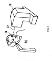
- an apparatus 10 for generating a laser beam 12 is shown.
- the laser beam 12 is directed onto an eye 14 of a patient 16.
- the apparatus 10 is capable of generating a pulsed laser beam 12 having physical characteristics similar to those of the laser beams generated by a laser system as disclosed and claimed in U.S. Patent No. 4,764,930 , which is exclusively licensed to the assignee of the present invention.
- Various laser sources may be used with the inventive system and method, including infrared, visible, and UV lasers.
- laser sources to be used with the inventive system may be continuous wave, Q-switched pulse, and mode-locked ultrashort pulse lasers. Although the following is not an exhaustive list, lasers of the foregoing type may be used with the present invention.
- the present invention contemplates the use of a pulsed laser beam 12 which has pulses with durations as long as a few nanoseconds or as short as only a few femtoseconds.
- a laser unit is controlled by software for photodisruption of the cornea utilizing a laser beam of constant energy, composed of an optical train of pulses with a duration of approximately 600 femtoseconds at a repetition rate of up to several hundred thousand Hz.
- the optical delivery system determines a 3-dimensional position in the patient's cornea at which the laser is focused.
- the energy delivered to the point of focus is sufficient to ionize tissue (photodisrupt) in a very small volume. Repetitively setting a focus point and pulsing the laser results in a resected plane of tissue.
- the method is useful for use in real-time, such that the information needed to correct the fluence variance of a ophthalmic surgery laser as described in the present invention is stored in a computer memory and can be instantly retrieved prior to surgery. It is contemplated that the fluence variance of the laser beam can thus be corrected during ophthalmic surgery.
- the fluence, or energy density is the irradiance multiplied by the exposure time, and is commonly measured in Joules/square centimeter.
- the laser is pulsed onto a focal plane prior to surgery. The fluence distribution over a predetermined pattern is determined, and the laser beam energy is calibrated such that the laser fluence variance is minimized between focal spots over the predetermined pattern.
- the calibrated laser is then focused through an applanation lens during surgery.
- the laser is calibrated over a predetermined pattern based on stored fluence variance data that is in a computer memory, and thus the laser does not have to be recalibrated prior to every surgery.
- the reference focal plane comprises a plurality of coordinate points on an X/Y axis.
- the laser is focused at a specific Z coordinate inside the aplanation lens preferably made of fused silica over a plurality of spots on the X/Y axis.
- the laser is pulsed onto the focal plane at energy above the optical breakdown threshold in fused silica.
- Plasma light which is proportional to fluence, is created at each point and its intensity is measured. Distribution of plasma light intensity over different X/Y coordinates is representative of fluence variance in the focal plane.
- the fluence data is then stored in a computer memory.
- the laser energy is adjusted such that the calibrated fluence variance over the predetermined path is minimized.
- the laser beam energy must be either increased or decreased in order to calibrate the laser beam so that the fluence variance is minimized over a predetermined pattern.
- the laser beam energy will be corrected such that the fluence variance is minimized.
- the predefined range is set for a fluence variance at a particular coordinate point greater than 5-10% variance from the desired fluence, greater than 10-15% variance from the desired fluence, greater than 15-20% variance from the desired fluence, greater than 20-25% variance from the desired fluence, greater than 25-30% variance from the desired fluence, greater than 30-35% variance from the desired fluence, greater than 35-40% variance from the desired fluence, greater than 40-45% variance from the desired fluence, or greater than 45-50% variance from the desired fluence.
- the laser beam will be calibrated at a particular Z coordinate point in the X/Y axis, and that the laser can then be adjusted to focus on a different Z coordinate for surgery.
- the invention is not limited to calibration at a single Z coordinate.
- the step of determining the fluence distribution over a focal plane is accomplished by determining the fluence at a plurality of spots on the focal plane.
- the focal plane is about 10mm in diameter.
- the spot separation is about 10 microns in a specific embodiment.
- a reference library of fluence measurements is stored in a computer memory corresponding to particular predetermined paths. It is contemplated that the reference library may comprise information that can be customized depending on the procedure for which the laser is utilized.
- the reference focal plane is substantially flat. In other embodiments of the invention, it is contemplated that the reference focal plane is curved.
- the predetermined patterns useful for the present invention include, but are not limited to, a circle, an oval, a raster, a spiral, or any combination thereof.
- the present invention recognizes that internal tissue "photodisruption,” can be effectively accomplished using a pulsed laser energy if the irradiance of the beam, its focal spot size, and the proper layering of photodisruption sites are effectively controlled. Accordingly, it is an object of the present invention to provide an improved method for performing intrastromal photodisruption on the cornea of an eye by providing a method for correcting fluence distribution in a focal plane of the laser beam.
- the physical characteristics of the laser beam, as well as the manner of focusing the laser beam, are important to the proper performance of the method of the present invention. As indicated above, these considerations are interrelated. Insofar as the characteristics of the laser beam are concerned, several factors are important.
- the laser beam should have a wavelength that allows the light to pass through the cornea without absorption by the corneal tissue. Generally, the wavelength should be in the range of 0.4-1.9 micrometers with a wavelength of approximately 1054nm being preferred.
- the irradiance of the beam for accomplishment of photodisruption of stromal tissue at the focal spot should be greater than the threshold for optical breakdown of the tissue. Preferably, the irradiance should not be more than ten (10) times greater than the threshold for optical breakdown.
- the spot size of the focused laser beam should be small enough to achieve optical breakdown of stromal tissue at the focal spot. Typically, this requires the spot size to be approximately 1.2 microns-10 microns in diameter. Additionally, it is preferable that the spot configuration be as close to circular as possible.
- the applanation lens as described herein may be glass, fused silica, or medical grade plastic.
- the threshold fluence plate is a means for measuring fluence distribution.
- the principle of operation is based on generating plasma light while optically damaging the sample, which, in certain embodiments, is a fused silica plate.
- the laser beam is focused inside the bulk of the plate to avoid contribution from surface.
- the optical damage of a material depends on threshold value defined as fluence - energy per unit area.
- High density plasma created in the process of optical damage emits visible light. Intensity depends on fluence value above the threshold. If the spot size of the beam is constant then the amount of emitted light depends on energy. In the range of 1 - 3 uJ (typical range of energy for surgery) the dependence is linear.
- Plasma light is proportional to energy, or fluence.
- a signal is collected by a photodetector.
- the signal is collected the following way: the scanner scans the beam over the focal plane located inside the sample. For each point, a computer records the position of the beam on X/Y plane and the intensity of plasma light measured with a built-in video camera. The beam moves over the predetermined pattern and plasma light distribution in the focal plane is recorded. Since the energy is constant, the variations in plasma light intensity are due to spot size variations. The image is stored in the computer for comparison.
- the user will choose a signal level on the fluence distribution image (reference) around which to minimize fluence distribution.
- the computer compares the reference value with the actual signal value.
- the energy is increased or decreased to minimize the fluence distribution.
- Energy adjustment in real time is accomplished by an electro-optic (EO) modulator. Transmission of light through the EO modulator is governed by feedback voltage controlled by the computer. Feedback control mechanisms for EO modulators are familiar to one with skill in the art.
- E out E in sin 2 V ⁇ ⁇ 2 ⁇ V ⁇ / 2
- V the applied voltage
- V ⁇ /2 half wave voltage
- E in and E out intensities at the input and output of the modulator.
- High voltage linear amplifier is used to provide feedback and to control transmission of the modulator.
- Block diagram of the feedback loop along with the detailed schematics are shown in FIGS. 5 and 6 .
- V in is the voltage generated by the computer to control the transmission of the modulator.
- the total transmission of the modulator can be varied between 30-70% for each pulse, thus controlling the fluence in the focal plane.
- the present invention is directed towards lasers used in the field of laser vision correction surgery.
- the scope of the invention is not limited thereto. It is contemplated that the method and apparatus of the present invention will be useful in other fields in which it is desirable to have an even distribution of fluence across a focal plane, or laser beam spot.
Description
- The present invention relates to a laser beam energy correction method for correcting the variation in fluence distribution on a focal plane due to aberrations in focusing optics, and a laser driving apparatus adopting the laser beam energy correction method. The method and apparatus of the present invention are particularly, but not exclusively, useful for ophthalmic laser surgery procedures.
- A specific application of the invention is in the use of a photodisruptive laser for defining a resection plane of a corneal layer to create corneal flap in ophthalmic surgical procedures for vision error correction. Vision impairment can occur for many reasons, and be the result of many causes. One common cause for vision impairment results from a defective condition of the eye which occurs when the refractive characteristics of the cornea do not cause parallel rays of light to focus on the retina. When the eye is at rest, and the rays of light focus in front of the retina, the condition is known as myopia (i.e. near-sightedness). On the other hand, when the rays of light focus behind the retina, the condition is known as hypermetropia or hyperopia (i.e. farsightedness). Both myopic and hyperopic conditions result in varying degrees of vision impairment. In most cases the conditions are correctable.
- Eyeglasses or contact lenses are commonly used to correct myopic or hyperopic conditions. For various reasons, however, many persons who suffer with these conditions prefer not to wear eyeglasses or contact lenses. Alternative ways to correct these conditions include known surgical procedures for reshaping the cornea in various ways that are effective in changing its refractive characteristics. For example, in
U.S. patents 4,665,913 and4,669,466 to L'Esperance , a laser system is described which photoablates corneal tissue from the anterior surface of the eye. Another procedure is described inU.S. patent 4,988,348 to Bille , whereby corneal tissue is first removed to correct vision, and then the newly created surface is smoothed. - Rather than remove and reshape portions of the anterior portion of the eye to correct refractive defects, other procedures have been developed using a technique called intrastromal photodisruption for removing internal stromal tissue. An example of such a procedure is described in
U.S. patent 4,907,586 to Bille et al. Another example of a procedure for removing stromal tissue is the procedure described inU.S. patent 6,110,166 to Juhasz . In this procedure, an anterior corneal layer can be defined by using a laser to create a series of overlapping photodisrupted areas. The surgeon then separates the corneal layer by lifting it, to gain access to the underlying corneal tissue, which is changed through photoablation. The corneal layer is then repositioned on the cornea. - The photodisruption procedure involves removal of tissue in a stroma in a cornea of an eye using pulsed laser beam which is sequentially focused to individual spots at a plurality of locations in the stroma. Each focus spot has a finite volume, rather than being a single point. Further, each spot has a central point at approximately the center of the finite volume. Photodisruption of stromal tissue occurs at each spot where the beam is focused when fluence is above the threshold value and the volume of stromal tissue disrupted at each spot is approximately equal to the volume of the spot. The amount of tissue damage is dependent on how much the fluence exceeds the threshold value An optimal fluence value exists for a given separation between photodisruption spots to achieve the best surgical result. For example, if the fluence is below the optimal value, then it is difficult to lift the flap. If the fluence is above the optimal value, then an excessive amount of gas is produced during the photodisruption process creating opacity in the cornea, thus complicating the next step of vision correction procedure, photoablation. Clinical studies show that noticeable differences in outcomes occur when fluence varies +/- 10%. Consequently, it is important to have a uniform distribution of the fluence between photodisruption points.
- Such a pulsed laser syste, (which includes the laser and focusing optics), ideally provides an even fluence distribution across the focal plane, thus providing uniform distribution of the photodisruptive effect. However, the laser systems used in these procedures present the problem of providing nonuniform fluence over a focal plane even when set at a constant energy because of variations of the focal spots in the focal plane. Thus, the variance in fluence distribution may be above the optimal value at some points in the focal plane and below the optimal value at other points in the laser focal plane. This, in turn, results in nonuniform distribution of photodisruption in the focal plane. One reason for the fluence variance is that the optic that the laser is focused through, although generally uniform, contains imperfections and small variations resulting in aberrations in the beam. Aberrations generally change the spot size in the focal plane. By correcting energy, the present invention minimizes the fluence variance at each point in the focal plane where the spot size varies because of aberrations in the laser beam.
-
U.S. Patent No. 6,287,299 describes a method of monitoring fluence from focus spot to focus spot by directing a portion of the laser beam energy to a fluence monitoring device to provide a picture of fluence distribution over a curved surface in overlaying pattern. Fluence is controlled by controlling the number of pulses irradiating a single point and by overlaying the spots. It is essential for that method to have multiple pulses irradiating the same point in X/Y plane. However, the 299 patent fails to address the issue of correcting fluence variance due to discrepancies in the focusing optics. Furthermore, the method is not useful for single pulse photodisruption in real-time surgical settings and for high numerical aperture focusing optics when the space between the focusing lens and focal plane is very limited. - Document
US 2004/0147910 A which is regarded as closest prior art discloses a method and an apparatus for obtaining the irradiation intensity of a laser beam in order to perform laser ablation more accurately. The laser beam is guided to a fluorescent glass where the laser beam causes the fluorescent glass to emit fluorescence radiation. The fluorescence radiation is collected by a camera. The irradiated area of the fluorescent glass corresponds in size to an object to be processed later by laser ablation. Based on the intensity of the fluorescence radiation, the irradiation intensity distribution of the laser beam in the ablation area is obtained. Then the irradiation is calibrated by adapting the number of pulses. - Herein, the inventors present a method and apparatus for overcoming the disadvantages of the prior art.
- The invention is defined by appended independent claims 1 and 17. Preferred embodiments are described in the dependent claims.
- An embodiment of the invention is a laser beam energy correction method for correcting fluence distribution of the laser beam through an optic onto a focal plane, said correction method comprising the steps of: determining the fluence distribution of the laser beam energy over a focal plane; and calibrating the laser beam energy for the predetermined pattern based upon the fluence distribution.
- In a specific embodiment, the step of determining the fluence distribution comprises the steps of: focusing the laser beam through an optic into fluence sensitive material positioned in the focal plane; pulsing said laser beam through the optic onto the focal plane; optically damaging the fluence sensitive material; creating fluence sensitive plasma light; measuring the plasma light intensity distribution over the focal plane; and storing the fluence distribution of the focal plane into a computer memory. In one embodiment of the invention, the pulsing comprises a constant energy. In one embodiment of the invention, the pulsing comprises a constant energy of at least about 5,000 pulses per second.
- In a specific embodiment, the step of determining the fluence distribution further comprises repeating the focusing, pulsing, measuring and storing steps.
- In one embodiment of the invention, measuring fluence distribution comprises capturing a plasma light image with a photodetector.
- In one embodiment of the invention, measuring fluence distribution comprises measuring the fluence at a plurality of coordinate points on the focal plane. In a specific embodiment, the plurality of coordinate points comprises at least 1000 points.
- In one embodiment of the invention, calibrating the laser beam energy for the predetermined pattern comprises retrieving the stored fluence distribution over the focal plane from the computer memory and modifying the laser beam energy at individual points in the predetermined pattern.
- In one embodiment of the invention, the calibrating comprises comparing the stored fluence distribution at points in the predetermined pattern with reference values. In a specific embodiment, the calibrating the laser beam energy for the predetermined pattern comprises minimizing the fluence variance over the predetermined pattern. In a specific embodiment, the calibrating the laser beam energy for the predetermined pattern comprises controlling the energy output of the laser beam through a feedback control mechanism.
- In one embodiment of the invention, the focal plane comprises a substantially flat surface. In an embodiment of the invention, the predetermined pattern comprises a spiral.
- One embodiment of the present invention is an apparatus for performing the method described herein, comprising a laser source, a focusing mechanism, an electro-optic modulator, and a computer-controlled driver for said EO modulator. It is contemplated that the computer-controlled driver may include an appropriate software program for analyzing the measured fluence distribution data, and calibrating the laser beam energy as a result of the analysis. The apparatus may also include a fluence sensitive image and a feedback mechanism.
- One embodiment of the present invention is a laser beam energy correction method for correcting fluence distribution of the laser beam over a predetermined pattern on an object, said correction method comprising the steps of: determining the fluence distribution over a focal plane, said focal plane comprising a fluence sensitive image of plasma light distribution; calibrating the laser beam energy over the predetermined pattern; focusing the laser beam on the object; and pulsing the adjusted laser beam over the predetermined pattern on the object.
- The patent or application file contains at least one drawing executed in color. Copies of this patent or patent application publication with color drawing(s) will be provided by the Office upon request and payment of the necessary fee. For a more complete understanding of the present invention, reference is now made to the following descriptions taken in conjunction with the accompanying drawing, in which:
-
FIG. 1 is a perspective view of a patient being treated with the method of the present invention; -
FIG. 2 is a block diagram of the laser system utilizing the method of the present invention; -
FIG. 3 is an exemplary photoablation response fluence distribution showing uneven fluence distribution across the focal plane; -
FIG. 4 shows the difference in fluence distribution when the feedback mechanism is activated; -
FIG. 5 shows the plasma light intensity dependence on laser pulse energy in fused silica sample at a typical spot size of 2.5 µm; and -
FIG. 6 shows the block diagram and schematics of the high voltage linear amplifier controlling the electro-optic modulator. - Referring initially to
FIG. 1 , depicting a prior art image, anapparatus 10 for generating alaser beam 12 is shown. Thelaser beam 12 is directed onto aneye 14 of apatient 16. For purposes of the present invention, theapparatus 10 is capable of generating apulsed laser beam 12 having physical characteristics similar to those of the laser beams generated by a laser system as disclosed and claimed inU.S. Patent No. 4,764,930 , which is exclusively licensed to the assignee of the present invention. Various laser sources may be used with the inventive system and method, including infrared, visible, and UV lasers. Further, laser sources to be used with the inventive system may be continuous wave, Q-switched pulse, and mode-locked ultrashort pulse lasers. Although the following is not an exhaustive list, lasers of the foregoing type may be used with the present invention. - In one embodiment, the present invention contemplates the use of a
pulsed laser beam 12 which has pulses with durations as long as a few nanoseconds or as short as only a few femtoseconds. - In one embodiment, a laser unit is controlled by software for photodisruption of the cornea utilizing a laser beam of constant energy, composed of an optical train of pulses with a duration of approximately 600 femtoseconds at a repetition rate of up to several hundred thousand Hz.
- The optical delivery system determines a 3-dimensional position in the patient's cornea at which the laser is focused. When the laser is pulsed, the energy delivered to the point of focus is sufficient to ionize tissue (photodisrupt) in a very small volume. Repetitively setting a focus point and pulsing the laser results in a resected plane of tissue.
- In one embodiment of the invention, the method is useful for use in real-time, such that the information needed to correct the fluence variance of a ophthalmic surgery laser as described in the present invention is stored in a computer memory and can be instantly retrieved prior to surgery. It is contemplated that the fluence variance of the laser beam can thus be corrected during ophthalmic surgery. One with skill in the art understands the fluence, or energy density, is the irradiance multiplied by the exposure time, and is commonly measured in Joules/square centimeter. In another embodiment, the laser is pulsed onto a focal plane prior to surgery. The fluence distribution over a predetermined pattern is determined, and the laser beam energy is calibrated such that the laser fluence variance is minimized between focal spots over the predetermined pattern. The calibrated laser is then focused through an applanation lens during surgery. In another embodiment of the invention, the laser is calibrated over a predetermined pattern based on stored fluence variance data that is in a computer memory, and thus the laser does not have to be recalibrated prior to every surgery.
- In one embodiment of the invention, the reference focal plane comprises a plurality of coordinate points on an X/Y axis. In certain embodiments of the invention, the laser is focused at a specific Z coordinate inside the aplanation lens preferably made of fused silica over a plurality of spots on the X/Y axis. The laser is pulsed onto the focal plane at energy above the optical breakdown threshold in fused silica. Plasma light, which is proportional to fluence, is created at each point and its intensity is measured. Distribution of plasma light intensity over different X/Y coordinates is representative of fluence variance in the focal plane. The fluence data is then stored in a computer memory. The laser energy is adjusted such that the calibrated fluence variance over the predetermined path is minimized. One with skill in the art realizes that at certain coordinate points, the laser beam energy must be either increased or decreased in order to calibrate the laser beam so that the fluence variance is minimized over a predetermined pattern. In a specific embodiment, it is contemplated that when the variance of the fluence is over a predefined range, then the laser beam energy will be corrected such that the fluence variance is minimized. In certain embodiments of the invention, the predefined range is set for a fluence variance at a particular coordinate point greater than 5-10% variance from the desired fluence, greater than 10-15% variance from the desired fluence, greater than 15-20% variance from the desired fluence, greater than 20-25% variance from the desired fluence, greater than 25-30% variance from the desired fluence, greater than 30-35% variance from the desired fluence, greater than 35-40% variance from the desired fluence, greater than 40-45% variance from the desired fluence, or greater than 45-50% variance from the desired fluence.
- It is contemplated that in a specific embodiment, the laser beam will be calibrated at a particular Z coordinate point in the X/Y axis, and that the laser can then be adjusted to focus on a different Z coordinate for surgery. However, the invention is not limited to calibration at a single Z coordinate.
- In an embodiment of the invention, the step of determining the fluence distribution over a focal plane is accomplished by determining the fluence at a plurality of spots on the focal plane. In a specific embodiment, the focal plane is about 10mm in diameter. The spot separation is about 10 microns in a specific embodiment.
- In another embodiment of the invention, a reference library of fluence measurements is stored in a computer memory corresponding to particular predetermined paths. It is contemplated that the reference library may comprise information that can be customized depending on the procedure for which the laser is utilized.
- In one embodiment of the invention, the reference focal plane is substantially flat. In other embodiments of the invention, it is contemplated that the reference focal plane is curved.
- It is contemplated that the predetermined patterns useful for the present invention include, but are not limited to, a circle, an oval, a raster, a spiral, or any combination thereof.
- Further, the present invention recognizes that internal tissue "photodisruption," can be effectively accomplished using a pulsed laser energy if the irradiance of the beam, its focal spot size, and the proper layering of photodisruption sites are effectively controlled. Accordingly, it is an object of the present invention to provide an improved method for performing intrastromal photodisruption on the cornea of an eye by providing a method for correcting fluence distribution in a focal plane of the laser beam.
- The physical characteristics of the laser beam, as well as the manner of focusing the laser beam, are important to the proper performance of the method of the present invention. As indicated above, these considerations are interrelated. Insofar as the characteristics of the laser beam are concerned, several factors are important. The laser beam should have a wavelength that allows the light to pass through the cornea without absorption by the corneal tissue. Generally, the wavelength should be in the range of 0.4-1.9 micrometers with a wavelength of approximately 1054nm being preferred. The irradiance of the beam for accomplishment of photodisruption of stromal tissue at the focal spot should be greater than the threshold for optical breakdown of the tissue. Preferably, the irradiance should not be more than ten (10) times greater than the threshold for optical breakdown.
- The spot size of the focused laser beam should be small enough to achieve optical breakdown of stromal tissue at the focal spot. Typically, this requires the spot size to be approximately 1.2 microns-10 microns in diameter. Additionally, it is preferable that the spot configuration be as close to circular as possible.
- The applanation lens as described herein may be glass, fused silica, or medical grade plastic.
- The threshold fluence plate, as described herein, is a means for measuring fluence distribution. The principle of operation is based on generating plasma light while optically damaging the sample, which, in certain embodiments, is a fused silica plate. The laser beam is focused inside the bulk of the plate to avoid contribution from surface. At a given pulse duration the optical damage of a material depends on threshold value defined as fluence - energy per unit area. High density plasma created in the process of optical damage emits visible light. Intensity depends on fluence value above the threshold. If the spot size of the beam is constant then the amount of emitted light depends on energy. In the range of 1 - 3 uJ (typical range of energy for surgery) the dependence is linear. Plasma light is proportional to energy, or fluence. A signal is collected by a photodetector. In practice, the signal is collected the following way: the scanner scans the beam over the focal plane located inside the sample. For each point, a computer records the position of the beam on X/Y plane and the intensity of plasma light measured with a built-in video camera. The beam moves over the predetermined pattern and plasma light distribution in the focal plane is recorded. Since the energy is constant, the variations in plasma light intensity are due to spot size variations. The image is stored in the computer for comparison.
- For example, in certain embodiments, it is contemplated that the user will choose a signal level on the fluence distribution image (reference) around which to minimize fluence distribution. During the procedure at each coordinate point on the X/Y plane, the computer compares the reference value with the actual signal value. Depending on the sign of the difference (i.e., positive or negative) and the magnitude of the difference, the energy is increased or decreased to minimize the fluence distribution. Energy adjustment in real time is accomplished by an electro-optic (EO) modulator. Transmission of light through the EO modulator is governed by feedback voltage controlled by the computer. Feedback control mechanisms for EO modulators are familiar to one with skill in the art. Intensity of light transmitted through an electro optic modulator, in this case Pockels cell placed between two crossed polarizes, is given by:

FIGS. 5 and6 . Vin is the voltage generated by the computer to control the transmission of the modulator. Depending on the sign and amplitude of the Vin-Voffset the total transmission of the modulator can be varied between 30-70% for each pulse, thus controlling the fluence in the focal plane. - In a preferred embodiment, the present invention is directed towards lasers used in the field of laser vision correction surgery. However, the scope of the invention is not limited thereto. It is contemplated that the method and apparatus of the present invention will be useful in other fields in which it is desirable to have an even distribution of fluence across a focal plane, or laser beam spot.
- The foregoing has outlined rather broadly the features and technical advantages of the present invention in order that the detailed description of the invention that follows may be better understood. Additional features and advantages of the invention will be described hereinafter which form the subject of the claims of the invention. It should be appreciated that the conception and specific embodiment disclosed may be readily utilized as a basis for modifying or designing other structures for carrying out the same purposes of the present invention. It should also be realized that such equivalent constructions do not depart from the invention as set forth in the appended claims. The novel features which are believed to be characteristic of the invention, both as to its organization and method of operation, together with further objects and advantages will be better understood from the following description when considered in connection with the accompanying figures. It is to be expressly understood, however, that each of the figures is provided for the purpose of illustration and description only and is not intended as a definition of the limits of the present invention.
- Although the present invention and its advantages have been described in detail, it should be understood that various changes, substitutions and alterations can be made herein without departing from the invention as defined by the appended claims. Moreover, the scope of the present application is not intended to be limited to the particular embodiments of the process, machine, manufacture, composition of matter, means, methods and steps described in the specification. As one will readily appreciate from the disclosure, processes, machines, manufacture, compositions of matter, means, methods, or steps, presently existing or later to be developed that perform substantially the same function or achieve substantially the same result as the corresponding embodiments described herein may be utilized. Accordingly, the appended claims are intended to include within their scope such processes, machines, manufacture, compositions of matter, means, methods, or steps.
Claims (19)
- A laser beam energy correction method for correcting fluence distribution of the laser beam over a predetermined scan pattern on an object, said correction method comprising the steps of:a) calibrating the laser beam by modifying the laser beam energy over the predetermined scan pattern inside a sample;b) focusing the calibrated laser beam on the object; andc) pulsing the adjusted calibrated laser beam over the predetermined pattern on the object,
whereby the sample and the object is not a living animal body or a living human body, and
wherein said calibrating step a) comprises the steps of
focusing the laser beam through an optic to a focal plane inside the sample;
pulsing said laser beam through the optic onto the focal plane inside the sample; and
measuring the fluence distribution over the focal plane at a plurality of coordinate points inside the sample;
storing the measured fluence distribution of the focal plane inside the sample intro a computer memory; and
retrieving the stored fluence distribution over the focal plane inside the sample from the computer memory; and
modifying the laser beam energy per pulse at individual points in the predetermined scan pattern based on the measured fluence distribution. - The method of claim 1, further comprising repeating the focusing, pulsing, measuring and storing steps.
- The method of claim 1, wherein fluence sensitive material is positioned within the focal plane.
- The method of claim 1, wherein measuring the fluence distribution comprises capturing a signal with a photodetector.
- The method of claim 4, wherein the signal comprises plasma light.
- The method of claim 1, wherein the plurality of coordinate points comprises at least 1000 points.
- The method of claim 1, wherein the pulsing of said laser beam onto the focal plane inside the sample comprises a constant energy.
- The method of claim 1, wherein the pulsing comprises at least about 10,000 pulses per second.
- The method of claim 1, wherein calibrating the laser beam energy over the predetermined pattern comprises controlling the energy output of the laser beam through a feedback control mechanism.
- The method of claim 1, wherein the focal plane comprises a substantially flat surface.
- The method of claim 1, wherein the focal plane comprises a curved surface.
- The method of claim 1, wherein the optic comprises a glass lens or a medical grade plastic lens.
- The method of claim 1, wherein the object is a threshold fluence plate.
- The method of claim 1, wherein the pulsing comprises at least about 1000 pulses per second.
- The method of claim 1, wherein the pulsing comprises comparing the stored fluence distribution at points in the predetermined pattern with reference values.
- The method of claim 1, wherein the predetermined pattern comprises a spiral.
- A fluence normalization apparatus for a laser beam energy correction method for correcting fluence distribution of the laser beam over a predetermined scan pattern on an object, said apparatus comprising a laser source, a focusing mechanism, an electro-optic modulator, a computer- controlled driver for said EO modulator for performing the correction method steps of
calibrating the laser beam energy by modifying the laser beam energy over the predetermined scan pattern inside a sample;
focusing the calibrated laser beam on the object; and
pulsing the adjusted calibrated laser beam over the predetermined scan pattern on the object,
wherein the computer controlled driver comprises a program for calibrating fluence distribution wherein said calibrating comprises the steps of
focusing the laser beam through an optic to a focal plane inside the sample;
pulsing said laser beam through the optic onto the focal plane inside the sample; and
measuring the fluence distribution over the focal plane at a plurality of coordinate points inside the sample;
storing the measured fluence distribution of the focal plane inside the sample into a computer memory; and
retrieving the stored fluence distribution over the focal plane inside the sample from the computer memory; and
modifying the laser beam energy per pulse at individual points in the predetermined scan pattern based on the measured fluence distribution. - The apparatus of claim 16, further comprising a feedback mechanism.
- The apparatus of claim 16, further comprising a fluence sensitive image.
Applications Claiming Priority (2)
| Application Number | Priority Date | Filing Date | Title |
|---|---|---|---|
| US10/919,710 US7584756B2 (en) | 2004-08-17 | 2004-08-17 | Apparatus and method for correction of aberrations in laser system optics |
| PCT/US2005/029220 WO2006023535A2 (en) | 2004-08-17 | 2005-08-17 | Apparatus and method for correction of abberations in laaser system optics |
Publications (3)
| Publication Number | Publication Date |
|---|---|
| EP1784149A2 EP1784149A2 (en) | 2007-05-16 |
| EP1784149A4 EP1784149A4 (en) | 2008-07-16 |
| EP1784149B1 true EP1784149B1 (en) | 2015-06-03 |
Family
ID=35968118
Family Applications (1)
| Application Number | Title | Priority Date | Filing Date |
|---|---|---|---|
| EP05785356.6A Active EP1784149B1 (en) | 2004-08-17 | 2005-08-17 | Apparatus and method for correction of aberrations in laser system optics |
Country Status (8)
| Country | Link |
|---|---|
| US (1) | US7584756B2 (en) |
| EP (1) | EP1784149B1 (en) |
| JP (1) | JP4825803B2 (en) |
| KR (1) | KR101023458B1 (en) |
| CN (1) | CN101010568B (en) |
| AU (1) | AU2005277499C1 (en) |
| CA (1) | CA2576929C (en) |
| WO (1) | WO2006023535A2 (en) |
Families Citing this family (49)
| Publication number | Priority date | Publication date | Assignee | Title |
|---|---|---|---|---|
| US9603741B2 (en) | 2000-05-19 | 2017-03-28 | Michael S. Berlin | Delivery system and method of use for the eye |
| DE60131273T2 (en) | 2000-05-19 | 2008-08-28 | Michael S. Beverly Hills Berlin | LASER APPLICATION SYSTEM AND METHOD FOR EYE-USE |
| US8679089B2 (en) | 2001-05-21 | 2014-03-25 | Michael S. Berlin | Glaucoma surgery methods and systems |
| US7361171B2 (en) | 2003-05-20 | 2008-04-22 | Raydiance, Inc. | Man-portable optical ablation system |
| US8921733B2 (en) | 2003-08-11 | 2014-12-30 | Raydiance, Inc. | Methods and systems for trimming circuits |
| US8173929B1 (en) | 2003-08-11 | 2012-05-08 | Raydiance, Inc. | Methods and systems for trimming circuits |
| US9022037B2 (en) * | 2003-08-11 | 2015-05-05 | Raydiance, Inc. | Laser ablation method and apparatus having a feedback loop and control unit |
| US8135050B1 (en) | 2005-07-19 | 2012-03-13 | Raydiance, Inc. | Automated polarization correction |
| US20070142827A1 (en) * | 2005-12-20 | 2007-06-21 | Curatu Eugene O | Dynamic laser beam characteristic measurement system for ophthalmic surgery and associated methods |
| US7444049B1 (en) | 2006-01-23 | 2008-10-28 | Raydiance, Inc. | Pulse stretcher and compressor including a multi-pass Bragg grating |
| US8232687B2 (en) | 2006-04-26 | 2012-07-31 | Raydiance, Inc. | Intelligent laser interlock system |
| US8189971B1 (en) | 2006-01-23 | 2012-05-29 | Raydiance, Inc. | Dispersion compensation in a chirped pulse amplification system |
| US8182471B2 (en) | 2006-03-17 | 2012-05-22 | Amo Manufacturing Usa, Llc. | Intrastromal refractive correction systems and methods |
| US7822347B1 (en) | 2006-03-28 | 2010-10-26 | Raydiance, Inc. | Active tuning of temporal dispersion in an ultrashort pulse laser system |
| US8308716B2 (en) * | 2006-06-30 | 2012-11-13 | Novartis Ag | Apparatus and method for auto-titrating a laser |
| US20080144038A1 (en) * | 2006-12-19 | 2008-06-19 | Richard Alan Leblanc | Performance and accuracy assessment system for refractive laser systems and associated methods |
| DE112008002383T5 (en) | 2007-09-06 | 2010-06-24 | LenSx Lasers, Inc., Aliso Viejo | Precise targeting of surgical photodisruption |
| US20170360609A9 (en) | 2007-09-24 | 2017-12-21 | Ivantis, Inc. | Methods and devices for increasing aqueous humor outflow |
| US20090160075A1 (en) * | 2007-12-21 | 2009-06-25 | Simpson Michael J | Methods for fabricating customized intraocular lenses |
| EP2259833A1 (en) | 2008-03-05 | 2010-12-15 | Ivantis, INC. | Methods and apparatus for treating glaucoma |
| US8125704B2 (en) | 2008-08-18 | 2012-02-28 | Raydiance, Inc. | Systems and methods for controlling a pulsed laser by combining laser signals |
| US7880869B2 (en) * | 2009-05-13 | 2011-02-01 | Topray Technologies, Inc. | Calibration apparatus and method for optical system assembly |
| WO2011006078A1 (en) | 2009-07-09 | 2011-01-13 | Ivantis, Inc. | Single operator device for delivering an ocular implant |
| CA2766192C (en) | 2009-07-09 | 2017-10-24 | Ivantis, Inc. | Ocular implants for residing partially in schlemm's canal |
| US8267925B2 (en) * | 2009-07-29 | 2012-09-18 | Alcon Lensx, Inc. | Optical system for ophthalmic surgical laser |
| US9504608B2 (en) * | 2009-07-29 | 2016-11-29 | Alcon Lensx, Inc. | Optical system with movable lens for ophthalmic surgical laser |
| US8262647B2 (en) * | 2009-07-29 | 2012-09-11 | Alcon Lensx, Inc. | Optical system for ophthalmic surgical laser |
| JP2013500768A (en) * | 2009-07-29 | 2013-01-10 | アルコン レンゼックス, インコーポレーテッド | Optical system for laser for ophthalmic surgery |
| US8500725B2 (en) * | 2009-07-29 | 2013-08-06 | Alcon Lensx, Inc. | Optical system for ophthalmic surgical laser |
| US8506559B2 (en) * | 2009-11-16 | 2013-08-13 | Alcon Lensx, Inc. | Variable stage optical system for ophthalmic surgical laser |
| US8298214B2 (en) * | 2009-12-22 | 2012-10-30 | Kera Harvest Inc. | Vision correction system and operating method thereof |
| RU2550666C2 (en) * | 2010-02-15 | 2015-05-10 | Уэйвлайт Гмбх | Method of determining discrepancies between coordinate systems of different technical systems |
| US8554037B2 (en) | 2010-09-30 | 2013-10-08 | Raydiance, Inc. | Hybrid waveguide device in powerful laser systems |
| US20120283557A1 (en) | 2011-05-05 | 2012-11-08 | Berlin Michael S | Methods and Apparatuses for the Treatment of Glaucoma using visible and infrared ultrashort laser pulses |
| US8657776B2 (en) | 2011-06-14 | 2014-02-25 | Ivantis, Inc. | Ocular implants for delivery into the eye |
| DE102011116759A1 (en) * | 2011-10-20 | 2013-04-25 | Carl Zeiss Meditec Ag | Ophthalmic laser system and method for cutting through eye tissue |
| US9849034B2 (en) | 2011-11-07 | 2017-12-26 | Alcon Research, Ltd. | Retinal laser surgery |
| US8663150B2 (en) | 2011-12-19 | 2014-03-04 | Ivantis, Inc. | Delivering ocular implants into the eye |
| PT2826447T (en) * | 2012-01-18 | 2019-02-01 | Wavelight Gmbh | Adjusting laser energy in accordance with effects of trial cuts |
| US10182943B2 (en) | 2012-03-09 | 2019-01-22 | Alcon Lensx, Inc. | Adjustable pupil system for surgical laser systems |
| US8852177B2 (en) | 2012-03-09 | 2014-10-07 | Alcon Lensx, Inc. | Spatio-temporal beam modulator for surgical laser systems |
| US9358156B2 (en) | 2012-04-18 | 2016-06-07 | Invantis, Inc. | Ocular implants for delivery into an anterior chamber of the eye |
| WO2014085450A1 (en) | 2012-11-28 | 2014-06-05 | Ivantis, Inc. | Apparatus for delivering ocular implants into an anterior chamber of the eye |
| US10709547B2 (en) | 2014-07-14 | 2020-07-14 | Ivantis, Inc. | Ocular implant delivery system and method |
| AU2016307951B2 (en) | 2015-08-14 | 2021-04-01 | Alcon Inc. | Ocular implant with pressure sensor and delivery system |
| CA3002763A1 (en) | 2015-10-21 | 2017-04-27 | Optimedica Corporation | Laser beam calibration and beam quality measurement in laser surgery systems |
| WO2017106517A1 (en) | 2015-12-15 | 2017-06-22 | Ivantis, Inc. | Ocular implant and delivery system |
| AU2022205382A1 (en) | 2021-01-11 | 2023-06-22 | Alcon Inc. | Systems and methods for viscoelastic delivery |
| US20240074904A1 (en) * | 2022-09-02 | 2024-03-07 | Alcon Inc. | Long-term monitoring of the incision performance of an ophthalmic laser surgical system |
Family Cites Families (30)
| Publication number | Priority date | Publication date | Assignee | Title |
|---|---|---|---|---|
| US4665913A (en) | 1983-11-17 | 1987-05-19 | Lri L.P. | Method for ophthalmological surgery |
| US4669466A (en) * | 1985-01-16 | 1987-06-02 | Lri L.P. | Method and apparatus for analysis and correction of abnormal refractive errors of the eye |
| US4656913A (en) * | 1985-01-31 | 1987-04-14 | Nippon, Gakki Seizo, Kabushiki Kaisha | Electronic musical instrument with keyboard and cover |
| US4907586A (en) * | 1988-03-31 | 1990-03-13 | Intelligent Surgical Lasers | Method for reshaping the eye |
| US5386827A (en) * | 1993-03-30 | 1995-02-07 | Nim Incorporated | Quantitative and qualitative in vivo tissue examination using time resolved spectroscopy |
| US4988348A (en) * | 1989-05-26 | 1991-01-29 | Intelligent Surgical Lasers, Inc. | Method for reshaping the cornea |
| US5474549A (en) * | 1991-07-09 | 1995-12-12 | Laserscope | Method and system for scanning a laser beam for controlled distribution of laser dosage |
| US5383199A (en) * | 1992-07-02 | 1995-01-17 | Advanced Interventional Systems, Inc. | Apparatus and method for optically controlling the output energy of a pulsed laser source |
| US5713893A (en) * | 1993-05-03 | 1998-02-03 | O'donnell, Jr.; Francis E. | Test substrate for laser evaluation |
| US5656186A (en) | 1994-04-08 | 1997-08-12 | The Regents Of The University Of Michigan | Method for controlling configuration of laser induced breakdown and ablation |
| US5980513A (en) * | 1994-04-25 | 1999-11-09 | Autonomous Technologies Corp. | Laser beam delivery and eye tracking system |
| US6110166A (en) * | 1995-03-20 | 2000-08-29 | Escalon Medical Corporation | Method for corneal laser surgery |
| US5980101A (en) * | 1997-10-31 | 1999-11-09 | General Electric Company | Method and apparatus for measuring laser pulse energy |
| AU1386899A (en) * | 1997-11-06 | 1999-05-31 | Visx Incorporated | Systems and methods for calibrating laser ablations |
| US5928221A (en) * | 1997-11-17 | 1999-07-27 | Coherent, Inc. | Fluence monitoring method for laser treatment of biological tissue |
| US6002706A (en) * | 1997-12-30 | 1999-12-14 | General Electric Company | Method and apparatus for controlling the size of a laser beam |
| US7649153B2 (en) * | 1998-12-11 | 2010-01-19 | International Business Machines Corporation | Method for minimizing sample damage during the ablation of material using a focused ultrashort pulsed laser beam |
| US6773430B2 (en) * | 1999-08-09 | 2004-08-10 | Visx, Inc. | Motion detector for eye ablative laser delivery systems |
| US6932807B1 (en) * | 1999-09-01 | 2005-08-23 | Nidek Co., Ltd. | Laser treatment apparatus |
| US6666855B2 (en) * | 1999-09-14 | 2003-12-23 | Visx, Inc. | Methods and systems for laser calibration and eye tracker camera alignment |
| AU2001227809A1 (en) * | 2000-01-12 | 2001-07-24 | Lasersight Technologies, Inc. | Laser fluence compensation of a curved surface |
| JP3946454B2 (en) * | 2001-02-28 | 2007-07-18 | 株式会社ニデック | Laser beam evaluation method |
| US7217266B2 (en) * | 2001-05-30 | 2007-05-15 | Anderson R Rox | Apparatus and method for laser treatment with spectroscopic feedback |
| US6610050B2 (en) * | 2001-07-27 | 2003-08-26 | 20/10 Perfect Vision, Optische Geraete Gmbh | Laser beam delivery system with multiple focal points |
| US6670577B2 (en) * | 2001-09-28 | 2003-12-30 | General Electric Company | Laser shock peening method and apparatus |
| US20030216719A1 (en) * | 2001-12-12 | 2003-11-20 | Len Debenedictis | Method and apparatus for treating skin using patterns of optical energy |
| JP3530893B2 (en) * | 2001-12-25 | 2004-05-24 | コーリンメディカルテクノロジー株式会社 | Atherosclerosis evaluation device |
| US6666857B2 (en) | 2002-01-29 | 2003-12-23 | Robert F. Smith | Integrated wavefront-directed topography-controlled photoablation |
| JP4113390B2 (en) * | 2002-08-01 | 2008-07-09 | 株式会社ニデック | Laser irradiation device |
| US7846152B2 (en) * | 2004-03-24 | 2010-12-07 | Amo Manufacturing Usa, Llc. | Calibrating laser beam position and shape using an image capture device |
-
2004
- 2004-08-17 US US10/919,710 patent/US7584756B2/en active Active
-
2005
- 2005-08-17 WO PCT/US2005/029220 patent/WO2006023535A2/en active Application Filing
- 2005-08-17 JP JP2007527965A patent/JP4825803B2/en not_active Expired - Fee Related
- 2005-08-17 EP EP05785356.6A patent/EP1784149B1/en active Active
- 2005-08-17 AU AU2005277499A patent/AU2005277499C1/en not_active Ceased
- 2005-08-17 CA CA2576929A patent/CA2576929C/en not_active Expired - Fee Related
- 2005-08-17 CN CN2005800276434A patent/CN101010568B/en not_active Expired - Fee Related
- 2005-08-17 KR KR1020077003908A patent/KR101023458B1/en active IP Right Grant
Also Published As
| Publication number | Publication date |
|---|---|
| CA2576929A1 (en) | 2006-03-02 |
| JP2008511022A (en) | 2008-04-10 |
| CN101010568A (en) | 2007-08-01 |
| KR101023458B1 (en) | 2011-03-25 |
| EP1784149A4 (en) | 2008-07-16 |
| AU2005277499A1 (en) | 2006-03-02 |
| AU2005277499B8 (en) | 2010-12-23 |
| AU2005277499B2 (en) | 2010-12-16 |
| US20060084954A1 (en) | 2006-04-20 |
| US7584756B2 (en) | 2009-09-08 |
| AU2005277499C1 (en) | 2011-05-12 |
| KR20070053713A (en) | 2007-05-25 |
| WO2006023535A3 (en) | 2007-01-25 |
| CA2576929C (en) | 2011-04-05 |
| EP1784149A2 (en) | 2007-05-16 |
| JP4825803B2 (en) | 2011-11-30 |
| CN101010568B (en) | 2012-06-06 |
| WO2006023535A2 (en) | 2006-03-02 |
Similar Documents
| Publication | Publication Date | Title |
|---|---|---|
| EP1784149B1 (en) | Apparatus and method for correction of aberrations in laser system optics | |
| EP1402860B1 (en) | Improved ophthalmic surgical laser | |
| US6648877B1 (en) | Method for custom corneal corrections | |
| AU2012272677B2 (en) | Ophthalmic range finding | |
| EP1534161B1 (en) | Corneal topography-based target warping | |
| US6641577B2 (en) | Apparatus and method for creating a corneal flap | |
| JP2003533277A5 (en) | ||
| AU2019201060A1 (en) | Ophthalmic range finding | |
| JP2003533277A (en) | Optimization of optical system ablation correction and related methods | |
| CN109152659B (en) | Ophthalmic surgical method | |
| EP2271250B1 (en) | High-order optical correction during corneal laser surgery | |
| US20220409434A1 (en) | Method for controlling an eye surgical laser, treatment apparatus, computer program as well as computer-readable medium | |
| US11730626B2 (en) | Method for providing control data for an eye surgical laser of a treatment apparatus, control device and treatment apparatus | |
| US20200188168A1 (en) | System and methods for depth detection in laser-assisted ophthalmic procedures |
Legal Events
| Date | Code | Title | Description |
|---|---|---|---|
| PUAI | Public reference made under article 153(3) epc to a published international application that has entered the european phase |
Free format text: ORIGINAL CODE: 0009012 |
|
| 17P | Request for examination filed |
Effective date: 20070209 |
|
| AK | Designated contracting states |
Kind code of ref document: A2 Designated state(s): AT BE BG CH CY CZ DE DK EE ES FI FR GB GR HU IE IS IT LI LT LU LV MC NL PL PT RO SE SI SK TR |
|
| AX | Request for extension of the european patent |
Extension state: AL BA HR MK YU |
|
| RIN1 | Information on inventor provided before grant (corrected) |
Inventor name: BOUVIER, MARCEL Inventor name: BOR, ZSOLT Inventor name: HOLLAND, GUY, VERN Inventor name: ZADOYAN, RUBEN |
|
| RIN1 | Information on inventor provided before grant (corrected) |
Inventor name: BOR, ZOLT Inventor name: ZADOYAN, RUBEN Inventor name: BOUVIER, MARCEL Inventor name: HOLLAND, GUY, VERN |
|
| RIN1 | Information on inventor provided before grant (corrected) |
Inventor name: BOUVIER, MARCEL Inventor name: HOLLAND, GUY, VERN Inventor name: BOR, ZOLT Inventor name: ZADOYAN, RUBEN |
|
| DAX | Request for extension of the european patent (deleted) | ||
| RAP1 | Party data changed (applicant data changed or rights of an application transferred) |
Owner name: AMO DEVELOPMENT, LLC |
|
| A4 | Supplementary search report drawn up and despatched |
Effective date: 20080613 |
|
| 17Q | First examination report despatched |
Effective date: 20081010 |
|
| GRAP | Despatch of communication of intention to grant a patent |
Free format text: ORIGINAL CODE: EPIDOSNIGR1 |
|
| RIC1 | Information provided on ipc code assigned before grant |
Ipc: A61B 18/20 20060101ALN20141217BHEP Ipc: A61F 9/008 20060101AFI20141217BHEP |
|
| INTG | Intention to grant announced |
Effective date: 20150109 |
|
| GRAS | Grant fee paid |
Free format text: ORIGINAL CODE: EPIDOSNIGR3 |
|
| GRAA | (expected) grant |
Free format text: ORIGINAL CODE: 0009210 |
|
| AK | Designated contracting states |
Kind code of ref document: B1 Designated state(s): AT BE BG CH CY CZ DE DK EE ES FI FR GB GR HU IE IS IT LI LT LU LV MC NL PL PT RO SE SI SK TR |
|
| REG | Reference to a national code |
Ref country code: GB Ref legal event code: FG4D |
|
| REG | Reference to a national code |
Ref country code: CH Ref legal event code: EP |
|
| REG | Reference to a national code |
Ref country code: AT Ref legal event code: REF Ref document number: 729540 Country of ref document: AT Kind code of ref document: T Effective date: 20150715 Ref country code: IE Ref legal event code: FG4D |
|
| REG | Reference to a national code |
Ref country code: DE Ref legal event code: R096 Ref document number: 602005046699 Country of ref document: DE |
|
| REG | Reference to a national code |
Ref country code: NL Ref legal event code: T3 |
|
| REG | Reference to a national code |
Ref country code: FR Ref legal event code: PLFP Year of fee payment: 11 |
|
| REG | Reference to a national code |
Ref country code: AT Ref legal event code: MK05 Ref document number: 729540 Country of ref document: AT Kind code of ref document: T Effective date: 20150603 |
|
| PG25 | Lapsed in a contracting state [announced via postgrant information from national office to epo] |
Ref country code: LT Free format text: LAPSE BECAUSE OF FAILURE TO SUBMIT A TRANSLATION OF THE DESCRIPTION OR TO PAY THE FEE WITHIN THE PRESCRIBED TIME-LIMIT Effective date: 20150603 Ref country code: ES Free format text: LAPSE BECAUSE OF FAILURE TO SUBMIT A TRANSLATION OF THE DESCRIPTION OR TO PAY THE FEE WITHIN THE PRESCRIBED TIME-LIMIT Effective date: 20150603 Ref country code: FI Free format text: LAPSE BECAUSE OF FAILURE TO SUBMIT A TRANSLATION OF THE DESCRIPTION OR TO PAY THE FEE WITHIN THE PRESCRIBED TIME-LIMIT Effective date: 20150603 |
|
| REG | Reference to a national code |
Ref country code: LT Ref legal event code: MG4D |
|
| PG25 | Lapsed in a contracting state [announced via postgrant information from national office to epo] |
Ref country code: BG Free format text: LAPSE BECAUSE OF FAILURE TO SUBMIT A TRANSLATION OF THE DESCRIPTION OR TO PAY THE FEE WITHIN THE PRESCRIBED TIME-LIMIT Effective date: 20150903 Ref country code: AT Free format text: LAPSE BECAUSE OF FAILURE TO SUBMIT A TRANSLATION OF THE DESCRIPTION OR TO PAY THE FEE WITHIN THE PRESCRIBED TIME-LIMIT Effective date: 20150603 Ref country code: GR Free format text: LAPSE BECAUSE OF FAILURE TO SUBMIT A TRANSLATION OF THE DESCRIPTION OR TO PAY THE FEE WITHIN THE PRESCRIBED TIME-LIMIT Effective date: 20150904 Ref country code: LV Free format text: LAPSE BECAUSE OF FAILURE TO SUBMIT A TRANSLATION OF THE DESCRIPTION OR TO PAY THE FEE WITHIN THE PRESCRIBED TIME-LIMIT Effective date: 20150603 |
|
| PG25 | Lapsed in a contracting state [announced via postgrant information from national office to epo] |
Ref country code: EE Free format text: LAPSE BECAUSE OF FAILURE TO SUBMIT A TRANSLATION OF THE DESCRIPTION OR TO PAY THE FEE WITHIN THE PRESCRIBED TIME-LIMIT Effective date: 20150603 |
|
| PG25 | Lapsed in a contracting state [announced via postgrant information from national office to epo] |
Ref country code: PT Free format text: LAPSE BECAUSE OF FAILURE TO SUBMIT A TRANSLATION OF THE DESCRIPTION OR TO PAY THE FEE WITHIN THE PRESCRIBED TIME-LIMIT Effective date: 20151006 Ref country code: SK Free format text: LAPSE BECAUSE OF FAILURE TO SUBMIT A TRANSLATION OF THE DESCRIPTION OR TO PAY THE FEE WITHIN THE PRESCRIBED TIME-LIMIT Effective date: 20150603 Ref country code: PL Free format text: LAPSE BECAUSE OF FAILURE TO SUBMIT A TRANSLATION OF THE DESCRIPTION OR TO PAY THE FEE WITHIN THE PRESCRIBED TIME-LIMIT Effective date: 20150603 Ref country code: IS Free format text: LAPSE BECAUSE OF FAILURE TO SUBMIT A TRANSLATION OF THE DESCRIPTION OR TO PAY THE FEE WITHIN THE PRESCRIBED TIME-LIMIT Effective date: 20151003 Ref country code: CZ Free format text: LAPSE BECAUSE OF FAILURE TO SUBMIT A TRANSLATION OF THE DESCRIPTION OR TO PAY THE FEE WITHIN THE PRESCRIBED TIME-LIMIT Effective date: 20150603 Ref country code: RO Free format text: LAPSE BECAUSE OF NON-PAYMENT OF DUE FEES Effective date: 20150603 |
|
| REG | Reference to a national code |
Ref country code: DE Ref legal event code: R097 Ref document number: 602005046699 Country of ref document: DE |
|
| PG25 | Lapsed in a contracting state [announced via postgrant information from national office to epo] |
Ref country code: MC Free format text: LAPSE BECAUSE OF FAILURE TO SUBMIT A TRANSLATION OF THE DESCRIPTION OR TO PAY THE FEE WITHIN THE PRESCRIBED TIME-LIMIT Effective date: 20150603 Ref country code: LU Free format text: LAPSE BECAUSE OF FAILURE TO SUBMIT A TRANSLATION OF THE DESCRIPTION OR TO PAY THE FEE WITHIN THE PRESCRIBED TIME-LIMIT Effective date: 20150817 |
|
| PLBE | No opposition filed within time limit |
Free format text: ORIGINAL CODE: 0009261 |
|
| STAA | Information on the status of an ep patent application or granted ep patent |
Free format text: STATUS: NO OPPOSITION FILED WITHIN TIME LIMIT |
|
| PG25 | Lapsed in a contracting state [announced via postgrant information from national office to epo] |
Ref country code: IT Free format text: LAPSE BECAUSE OF FAILURE TO SUBMIT A TRANSLATION OF THE DESCRIPTION OR TO PAY THE FEE WITHIN THE PRESCRIBED TIME-LIMIT Effective date: 20150603 Ref country code: DK Free format text: LAPSE BECAUSE OF FAILURE TO SUBMIT A TRANSLATION OF THE DESCRIPTION OR TO PAY THE FEE WITHIN THE PRESCRIBED TIME-LIMIT Effective date: 20150603 |
|
| 26N | No opposition filed |
Effective date: 20160304 |
|
| PG25 | Lapsed in a contracting state [announced via postgrant information from national office to epo] |
Ref country code: SI Free format text: LAPSE BECAUSE OF FAILURE TO SUBMIT A TRANSLATION OF THE DESCRIPTION OR TO PAY THE FEE WITHIN THE PRESCRIBED TIME-LIMIT Effective date: 20150603 |
|
| REG | Reference to a national code |
Ref country code: IE Ref legal event code: MM4A |
|
| REG | Reference to a national code |
Ref country code: FR Ref legal event code: PLFP Year of fee payment: 12 |
|
| PG25 | Lapsed in a contracting state [announced via postgrant information from national office to epo] |
Ref country code: IE Free format text: LAPSE BECAUSE OF NON-PAYMENT OF DUE FEES Effective date: 20150817 |
|
| PG25 | Lapsed in a contracting state [announced via postgrant information from national office to epo] |
Ref country code: BE Free format text: LAPSE BECAUSE OF FAILURE TO SUBMIT A TRANSLATION OF THE DESCRIPTION OR TO PAY THE FEE WITHIN THE PRESCRIBED TIME-LIMIT Effective date: 20150603 |
|
| PG25 | Lapsed in a contracting state [announced via postgrant information from national office to epo] |
Ref country code: HU Free format text: LAPSE BECAUSE OF FAILURE TO SUBMIT A TRANSLATION OF THE DESCRIPTION OR TO PAY THE FEE WITHIN THE PRESCRIBED TIME-LIMIT; INVALID AB INITIO Effective date: 20050817 |
|
| PG25 | Lapsed in a contracting state [announced via postgrant information from national office to epo] |
Ref country code: SE Free format text: LAPSE BECAUSE OF FAILURE TO SUBMIT A TRANSLATION OF THE DESCRIPTION OR TO PAY THE FEE WITHIN THE PRESCRIBED TIME-LIMIT Effective date: 20150603 Ref country code: CY Free format text: LAPSE BECAUSE OF FAILURE TO SUBMIT A TRANSLATION OF THE DESCRIPTION OR TO PAY THE FEE WITHIN THE PRESCRIBED TIME-LIMIT Effective date: 20150603 |
|
| REG | Reference to a national code |
Ref country code: FR Ref legal event code: PLFP Year of fee payment: 13 |
|
| PG25 | Lapsed in a contracting state [announced via postgrant information from national office to epo] |
Ref country code: TR Free format text: LAPSE BECAUSE OF FAILURE TO SUBMIT A TRANSLATION OF THE DESCRIPTION OR TO PAY THE FEE WITHIN THE PRESCRIBED TIME-LIMIT Effective date: 20150603 |
|
| REG | Reference to a national code |
Ref country code: FR Ref legal event code: PLFP Year of fee payment: 14 |
|
| PGFP | Annual fee paid to national office [announced via postgrant information from national office to epo] |
Ref country code: FR Payment date: 20180712 Year of fee payment: 14 |
|
| PGFP | Annual fee paid to national office [announced via postgrant information from national office to epo] |
Ref country code: GB Payment date: 20190815 Year of fee payment: 15 |
|
| PGFP | Annual fee paid to national office [announced via postgrant information from national office to epo] |
Ref country code: CH Payment date: 20190819 Year of fee payment: 15 |
|
| REG | Reference to a national code |
Ref country code: NL Ref legal event code: MM Effective date: 20190901 |
|
| PG25 | Lapsed in a contracting state [announced via postgrant information from national office to epo] |
Ref country code: FR Free format text: LAPSE BECAUSE OF NON-PAYMENT OF DUE FEES Effective date: 20190831 Ref country code: NL Free format text: LAPSE BECAUSE OF NON-PAYMENT OF DUE FEES Effective date: 20190901 |
|
| REG | Reference to a national code |
Ref country code: CH Ref legal event code: PL |
|
| GBPC | Gb: european patent ceased through non-payment of renewal fee |
Effective date: 20200817 |
|
| PG25 | Lapsed in a contracting state [announced via postgrant information from national office to epo] |
Ref country code: LI Free format text: LAPSE BECAUSE OF NON-PAYMENT OF DUE FEES Effective date: 20200831 Ref country code: CH Free format text: LAPSE BECAUSE OF NON-PAYMENT OF DUE FEES Effective date: 20200831 |
|
| PG25 | Lapsed in a contracting state [announced via postgrant information from national office to epo] |
Ref country code: GB Free format text: LAPSE BECAUSE OF NON-PAYMENT OF DUE FEES Effective date: 20200817 |
|
| PGFP | Annual fee paid to national office [announced via postgrant information from national office to epo] |
Ref country code: DE Payment date: 20230703 Year of fee payment: 19 |