EP1764615A1 - Tissue characteristic determination apparatus - Google Patents
Tissue characteristic determination apparatus Download PDFInfo
- Publication number
- EP1764615A1 EP1764615A1 EP06019041A EP06019041A EP1764615A1 EP 1764615 A1 EP1764615 A1 EP 1764615A1 EP 06019041 A EP06019041 A EP 06019041A EP 06019041 A EP06019041 A EP 06019041A EP 1764615 A1 EP1764615 A1 EP 1764615A1
- Authority
- EP
- European Patent Office
- Prior art keywords
- cdk
- data
- tissue
- obtainer
- expression level
- Prior art date
- Legal status (The legal status is an assumption and is not a legal conclusion. Google has not performed a legal analysis and makes no representation as to the accuracy of the status listed.)
- Withdrawn
Links
- 230000000694 effects Effects 0.000 claims abstract description 209
- 102000003903 Cyclin-dependent kinases Human genes 0.000 claims abstract description 138
- 108090000266 Cyclin-dependent kinases Proteins 0.000 claims abstract description 138
- 230000014509 gene expression Effects 0.000 claims abstract description 122
- 238000000034 method Methods 0.000 claims description 99
- 239000002246 antineoplastic agent Substances 0.000 claims description 97
- 229940041181 antineoplastic drug Drugs 0.000 claims description 97
- 108091007914 CDKs Proteins 0.000 claims description 76
- 206010028980 Neoplasm Diseases 0.000 claims description 70
- 101100439046 Caenorhabditis elegans cdk-2 gene Proteins 0.000 claims description 58
- 101150012716 CDK1 gene Proteins 0.000 claims description 53
- 201000011510 cancer Diseases 0.000 claims description 50
- 230000035945 sensitivity Effects 0.000 claims description 48
- 230000008569 process Effects 0.000 claims description 24
- 238000006243 chemical reaction Methods 0.000 claims description 20
- 229940043378 cyclin-dependent kinase inhibitor Drugs 0.000 claims description 20
- 238000002560 therapeutic procedure Methods 0.000 claims description 20
- 239000002875 cyclin dependent kinase inhibitor Substances 0.000 claims description 19
- 230000036210 malignancy Effects 0.000 claims description 16
- 239000000758 substrate Substances 0.000 claims description 15
- 230000035755 proliferation Effects 0.000 claims description 12
- 238000012545 processing Methods 0.000 claims description 6
- 239000002085 irritant Substances 0.000 claims description 4
- 231100000021 irritant Toxicity 0.000 claims description 4
- 239000000126 substance Substances 0.000 claims description 3
- 239000000523 sample Substances 0.000 description 109
- 210000001519 tissue Anatomy 0.000 description 84
- 108090000623 proteins and genes Proteins 0.000 description 47
- 210000004027 cell Anatomy 0.000 description 46
- 102000004169 proteins and genes Human genes 0.000 description 44
- 239000007788 liquid Substances 0.000 description 43
- 238000005259 measurement Methods 0.000 description 39
- 238000002651 drug therapy Methods 0.000 description 35
- 239000012530 fluid Substances 0.000 description 33
- 239000003153 chemical reaction reagent Substances 0.000 description 32
- 101000891649 Homo sapiens Transcription elongation factor A protein-like 1 Proteins 0.000 description 27
- 101000596402 Mus musculus Neuronal vesicle trafficking-associated protein 1 Proteins 0.000 description 27
- 101000800539 Mus musculus Translationally-controlled tumor protein Proteins 0.000 description 27
- 101000781972 Schizosaccharomyces pombe (strain 972 / ATCC 24843) Protein wos2 Proteins 0.000 description 27
- 101001009610 Toxoplasma gondii Dense granule protein 5 Proteins 0.000 description 27
- 102100040250 Transcription elongation factor A protein-like 1 Human genes 0.000 description 27
- 229940079593 drug Drugs 0.000 description 25
- 239000003814 drug Substances 0.000 description 25
- 102100032857 Cyclin-dependent kinase 1 Human genes 0.000 description 18
- 101710106279 Cyclin-dependent kinase 1 Proteins 0.000 description 18
- 229940123237 Taxane Drugs 0.000 description 17
- 239000000243 solution Substances 0.000 description 17
- DKPFODGZWDEEBT-QFIAKTPHSA-N taxane Chemical class C([C@]1(C)CCC[C@@H](C)[C@H]1C1)C[C@H]2[C@H](C)CC[C@@H]1C2(C)C DKPFODGZWDEEBT-QFIAKTPHSA-N 0.000 description 17
- 239000012528 membrane Substances 0.000 description 16
- 108010024986 Cyclin-Dependent Kinase 2 Proteins 0.000 description 14
- 102100036239 Cyclin-dependent kinase 2 Human genes 0.000 description 14
- 210000004881 tumor cell Anatomy 0.000 description 14
- 230000022131 cell cycle Effects 0.000 description 13
- 238000004140 cleaning Methods 0.000 description 12
- 230000007246 mechanism Effects 0.000 description 11
- 108010068192 Cyclin A Proteins 0.000 description 10
- 102100025191 Cyclin-A2 Human genes 0.000 description 10
- 230000001419 dependent effect Effects 0.000 description 10
- 238000003745 diagnosis Methods 0.000 description 10
- 238000004393 prognosis Methods 0.000 description 10
- 108010025464 Cyclin-Dependent Kinase 4 Proteins 0.000 description 9
- 108010025468 Cyclin-Dependent Kinase 6 Proteins 0.000 description 9
- 102100026804 Cyclin-dependent kinase 6 Human genes 0.000 description 9
- GHASVSINZRGABV-UHFFFAOYSA-N Fluorouracil Chemical compound FC1=CNC(=O)NC1=O GHASVSINZRGABV-UHFFFAOYSA-N 0.000 description 9
- 108091000080 Phosphotransferase Proteins 0.000 description 9
- 238000004458 analytical method Methods 0.000 description 9
- 229960002949 fluorouracil Drugs 0.000 description 9
- 238000001794 hormone therapy Methods 0.000 description 9
- 102000020233 phosphotransferase Human genes 0.000 description 9
- 102000002427 Cyclin B Human genes 0.000 description 8
- 108010068150 Cyclin B Proteins 0.000 description 8
- 102100036252 Cyclin-dependent kinase 4 Human genes 0.000 description 8
- AOJJSUZBOXZQNB-TZSSRYMLSA-N Doxorubicin Chemical compound O([C@H]1C[C@@](O)(CC=2C(O)=C3C(=O)C=4C=CC=C(C=4C(=O)C3=C(O)C=21)OC)C(=O)CO)[C@H]1C[C@H](N)[C@H](O)[C@H](C)O1 AOJJSUZBOXZQNB-TZSSRYMLSA-N 0.000 description 8
- 238000005063 solubilization Methods 0.000 description 8
- 230000007928 solubilization Effects 0.000 description 8
- 239000002699 waste material Substances 0.000 description 8
- CMSMOCZEIVJLDB-UHFFFAOYSA-N Cyclophosphamide Chemical compound ClCCN(CCCl)P1(=O)NCCCO1 CMSMOCZEIVJLDB-UHFFFAOYSA-N 0.000 description 7
- 229960004397 cyclophosphamide Drugs 0.000 description 7
- 238000011068 loading method Methods 0.000 description 7
- 125000002467 phosphate group Chemical group [H]OP(=O)(O[H])O[*] 0.000 description 7
- 102000003910 Cyclin D Human genes 0.000 description 6
- 108090000259 Cyclin D Proteins 0.000 description 6
- 102000003909 Cyclin E Human genes 0.000 description 6
- 108090000257 Cyclin E Proteins 0.000 description 6
- 238000001574 biopsy Methods 0.000 description 6
- 238000003825 pressing Methods 0.000 description 6
- 238000010206 sensitivity analysis Methods 0.000 description 6
- 238000005406 washing Methods 0.000 description 6
- 206010006187 Breast cancer Diseases 0.000 description 5
- 208000026310 Breast neoplasm Diseases 0.000 description 5
- 102000016736 Cyclin Human genes 0.000 description 5
- 108050006400 Cyclin Proteins 0.000 description 5
- 102000004190 Enzymes Human genes 0.000 description 5
- 108090000790 Enzymes Proteins 0.000 description 5
- NWIBSHFKIJFRCO-WUDYKRTCSA-N Mytomycin Chemical compound C1N2C(C(C(C)=C(N)C3=O)=O)=C3[C@@H](COC(N)=O)[C@@]2(OC)[C@@H]2[C@H]1N2 NWIBSHFKIJFRCO-WUDYKRTCSA-N 0.000 description 5
- 102000001253 Protein Kinase Human genes 0.000 description 5
- 238000002512 chemotherapy Methods 0.000 description 5
- 230000000977 initiatory effect Effects 0.000 description 5
- 238000002360 preparation method Methods 0.000 description 5
- 108060006633 protein kinase Proteins 0.000 description 5
- 238000001356 surgical procedure Methods 0.000 description 5
- 229930012538 Paclitaxel Natural products 0.000 description 4
- DQLATGHUWYMOKM-UHFFFAOYSA-L cisplatin Chemical compound N[Pt](N)(Cl)Cl DQLATGHUWYMOKM-UHFFFAOYSA-L 0.000 description 4
- 229960004316 cisplatin Drugs 0.000 description 4
- 229960004679 doxorubicin Drugs 0.000 description 4
- 230000004992 fission Effects 0.000 description 4
- 230000003211 malignant effect Effects 0.000 description 4
- 239000000463 material Substances 0.000 description 4
- 239000011159 matrix material Substances 0.000 description 4
- 210000000056 organ Anatomy 0.000 description 4
- 229960001592 paclitaxel Drugs 0.000 description 4
- 230000002980 postoperative effect Effects 0.000 description 4
- 230000008093 supporting effect Effects 0.000 description 4
- RCINICONZNJXQF-MZXODVADSA-N taxol Chemical compound O([C@@H]1[C@@]2(C[C@@H](C(C)=C(C2(C)C)[C@H](C([C@]2(C)[C@@H](O)C[C@H]3OC[C@]3([C@H]21)OC(C)=O)=O)OC(=O)C)OC(=O)[C@H](O)[C@@H](NC(=O)C=1C=CC=CC=1)C=1C=CC=CC=1)O)C(=O)C1=CC=CC=C1 RCINICONZNJXQF-MZXODVADSA-N 0.000 description 4
- AOJJSUZBOXZQNB-VTZDEGQISA-N 4'-epidoxorubicin Chemical compound O([C@H]1C[C@@](O)(CC=2C(O)=C3C(=O)C=4C=CC=C(C=4C(=O)C3=C(O)C=21)OC)C(=O)CO)[C@H]1C[C@H](N)[C@@H](O)[C@H](C)O1 AOJJSUZBOXZQNB-VTZDEGQISA-N 0.000 description 3
- 101150029707 ERBB2 gene Proteins 0.000 description 3
- HTIJFSOGRVMCQR-UHFFFAOYSA-N Epirubicin Natural products COc1cccc2C(=O)c3c(O)c4CC(O)(CC(OC5CC(N)C(=O)C(C)O5)c4c(O)c3C(=O)c12)C(=O)CO HTIJFSOGRVMCQR-UHFFFAOYSA-N 0.000 description 3
- 241000124008 Mammalia Species 0.000 description 3
- 239000000427 antigen Substances 0.000 description 3
- 108091007433 antigens Proteins 0.000 description 3
- 102000036639 antigens Human genes 0.000 description 3
- 238000003705 background correction Methods 0.000 description 3
- 238000010586 diagram Methods 0.000 description 3
- 201000010099 disease Diseases 0.000 description 3
- 208000037265 diseases, disorders, signs and symptoms Diseases 0.000 description 3
- 229960001904 epirubicin Drugs 0.000 description 3
- 238000002372 labelling Methods 0.000 description 3
- 230000003902 lesion Effects 0.000 description 3
- 229960004857 mitomycin Drugs 0.000 description 3
- 230000000394 mitotic effect Effects 0.000 description 3
- 239000000047 product Substances 0.000 description 3
- 239000006228 supernatant Substances 0.000 description 3
- XLYOFNOQVPJJNP-UHFFFAOYSA-N water Substances O XLYOFNOQVPJJNP-UHFFFAOYSA-N 0.000 description 3
- 229940126074 CDK kinase inhibitor Drugs 0.000 description 2
- 101100005789 Caenorhabditis elegans cdk-4 gene Proteins 0.000 description 2
- 206010009944 Colon cancer Diseases 0.000 description 2
- 108010058546 Cyclin D1 Proteins 0.000 description 2
- 108010058544 Cyclin D2 Proteins 0.000 description 2
- 108010058545 Cyclin D3 Proteins 0.000 description 2
- 102100034770 Cyclin-dependent kinase inhibitor 3 Human genes 0.000 description 2
- 108020004414 DNA Proteins 0.000 description 2
- 230000004543 DNA replication Effects 0.000 description 2
- 230000006820 DNA synthesis Effects 0.000 description 2
- 102100024165 G1/S-specific cyclin-D1 Human genes 0.000 description 2
- 102100024185 G1/S-specific cyclin-D2 Human genes 0.000 description 2
- 102100037859 G1/S-specific cyclin-D3 Human genes 0.000 description 2
- 101000945639 Homo sapiens Cyclin-dependent kinase inhibitor 3 Proteins 0.000 description 2
- 208000006994 Precancerous Conditions Diseases 0.000 description 2
- VYPSYNLAJGMNEJ-UHFFFAOYSA-N Silicium dioxide Chemical compound O=[Si]=O VYPSYNLAJGMNEJ-UHFFFAOYSA-N 0.000 description 2
- 208000005718 Stomach Neoplasms Diseases 0.000 description 2
- RYYWUUFWQRZTIU-UHFFFAOYSA-N Thiophosphoric acid Chemical group OP(O)(S)=O RYYWUUFWQRZTIU-UHFFFAOYSA-N 0.000 description 2
- BZHJMEDXRYGGRV-UHFFFAOYSA-N Vinyl chloride Chemical compound ClC=C BZHJMEDXRYGGRV-UHFFFAOYSA-N 0.000 description 2
- 230000009471 action Effects 0.000 description 2
- NLTUCYMLOPLUHL-KQYNXXCUSA-N adenosine 5'-[gamma-thio]triphosphate Chemical compound C1=NC=2C(N)=NC=NC=2N1[C@@H]1O[C@H](COP(O)(=O)OP(O)(=O)OP(O)(O)=S)[C@@H](O)[C@H]1O NLTUCYMLOPLUHL-KQYNXXCUSA-N 0.000 description 2
- 208000036878 aneuploidy Diseases 0.000 description 2
- 230000000903 blocking effect Effects 0.000 description 2
- 229960004562 carboplatin Drugs 0.000 description 2
- 190000008236 carboplatin Chemical compound 0.000 description 2
- 230000004663 cell proliferation Effects 0.000 description 2
- 208000029742 colonic neoplasm Diseases 0.000 description 2
- 150000001875 compounds Chemical class 0.000 description 2
- 238000001816 cooling Methods 0.000 description 2
- 230000002380 cytological effect Effects 0.000 description 2
- 230000002708 enhancing effect Effects 0.000 description 2
- 238000006911 enzymatic reaction Methods 0.000 description 2
- 230000005284 excitation Effects 0.000 description 2
- 238000001917 fluorescence detection Methods 0.000 description 2
- 206010017758 gastric cancer Diseases 0.000 description 2
- 238000012333 histopathological diagnosis Methods 0.000 description 2
- 239000003550 marker Substances 0.000 description 2
- 238000000691 measurement method Methods 0.000 description 2
- 230000001394 metastastic effect Effects 0.000 description 2
- 206010061289 metastatic neoplasm Diseases 0.000 description 2
- 239000000203 mixture Substances 0.000 description 2
- 210000002445 nipple Anatomy 0.000 description 2
- 238000011275 oncology therapy Methods 0.000 description 2
- 230000001575 pathological effect Effects 0.000 description 2
- 230000002829 reductive effect Effects 0.000 description 2
- 239000011347 resin Substances 0.000 description 2
- 229920005989 resin Polymers 0.000 description 2
- 239000012488 sample solution Substances 0.000 description 2
- 239000000741 silica gel Substances 0.000 description 2
- 229910002027 silica gel Inorganic materials 0.000 description 2
- 238000003756 stirring Methods 0.000 description 2
- 201000011549 stomach cancer Diseases 0.000 description 2
- 238000003860 storage Methods 0.000 description 2
- 229910052717 sulfur Inorganic materials 0.000 description 2
- RYYWUUFWQRZTIU-UHFFFAOYSA-K thiophosphate Chemical compound [O-]P([O-])([O-])=S RYYWUUFWQRZTIU-UHFFFAOYSA-K 0.000 description 2
- 238000001262 western blot Methods 0.000 description 2
- MTCFGRXMJLQNBG-REOHCLBHSA-N (2S)-2-Amino-3-hydroxypropansäure Chemical compound OC[C@H](N)C(O)=O MTCFGRXMJLQNBG-REOHCLBHSA-N 0.000 description 1
- VVIAGPKUTFNRDU-UHFFFAOYSA-N 6S-folinic acid Natural products C1NC=2NC(N)=NC(=O)C=2N(C=O)C1CNC1=CC=C(C(=O)NC(CCC(O)=O)C(O)=O)C=C1 VVIAGPKUTFNRDU-UHFFFAOYSA-N 0.000 description 1
- GAGWJHPBXLXJQN-UORFTKCHSA-N Capecitabine Chemical compound C1=C(F)C(NC(=O)OCCCCC)=NC(=O)N1[C@H]1[C@H](O)[C@H](O)[C@@H](C)O1 GAGWJHPBXLXJQN-UORFTKCHSA-N 0.000 description 1
- GAGWJHPBXLXJQN-UHFFFAOYSA-N Capecitabine Natural products C1=C(F)C(NC(=O)OCCCCC)=NC(=O)N1C1C(O)C(O)C(C)O1 GAGWJHPBXLXJQN-UHFFFAOYSA-N 0.000 description 1
- DLGOEMSEDOSKAD-UHFFFAOYSA-N Carmustine Chemical compound ClCCNC(=O)N(N=O)CCCl DLGOEMSEDOSKAD-UHFFFAOYSA-N 0.000 description 1
- 102100025064 Cellular tumor antigen p53 Human genes 0.000 description 1
- 108091060290 Chromatid Proteins 0.000 description 1
- 102100024458 Cyclin-dependent kinase inhibitor 2A Human genes 0.000 description 1
- 238000002965 ELISA Methods 0.000 description 1
- 208000000461 Esophageal Neoplasms Diseases 0.000 description 1
- 102000010834 Extracellular Matrix Proteins Human genes 0.000 description 1
- 108010037362 Extracellular Matrix Proteins Proteins 0.000 description 1
- 241000282412 Homo Species 0.000 description 1
- 108010058683 Immobilized Proteins Proteins 0.000 description 1
- FBOZXECLQNJBKD-ZDUSSCGKSA-N L-methotrexate Chemical compound C=1N=C2N=C(N)N=C(N)C2=NC=1CN(C)C1=CC=C(C(=O)N[C@@H](CCC(O)=O)C(O)=O)C=C1 FBOZXECLQNJBKD-ZDUSSCGKSA-N 0.000 description 1
- 206010027476 Metastases Diseases 0.000 description 1
- 229930192392 Mitomycin Natural products 0.000 description 1
- ZDZOTLJHXYCWBA-VCVYQWHSSA-N N-debenzoyl-N-(tert-butoxycarbonyl)-10-deacetyltaxol Chemical compound O([C@H]1[C@H]2[C@@](C([C@H](O)C3=C(C)[C@@H](OC(=O)[C@H](O)[C@@H](NC(=O)OC(C)(C)C)C=4C=CC=CC=4)C[C@]1(O)C3(C)C)=O)(C)[C@@H](O)C[C@H]1OC[C@]12OC(=O)C)C(=O)C1=CC=CC=C1 ZDZOTLJHXYCWBA-VCVYQWHSSA-N 0.000 description 1
- 206010030155 Oesophageal carcinoma Diseases 0.000 description 1
- 206010033128 Ovarian cancer Diseases 0.000 description 1
- 206010061535 Ovarian neoplasm Diseases 0.000 description 1
- 206010060862 Prostate cancer Diseases 0.000 description 1
- 208000000236 Prostatic Neoplasms Diseases 0.000 description 1
- 229940124158 Protease/peptidase inhibitor Drugs 0.000 description 1
- 108050002653 Retinoblastoma protein Proteins 0.000 description 1
- MTCFGRXMJLQNBG-UHFFFAOYSA-N Serine Natural products OCC(N)C(O)=O MTCFGRXMJLQNBG-UHFFFAOYSA-N 0.000 description 1
- 108050008367 Transmembrane emp24 domain-containing protein 7 Proteins 0.000 description 1
- ZKHQWZAMYRWXGA-KNYAHOBESA-N [[(2r,3s,4r,5r)-5-(6-aminopurin-9-yl)-3,4-dihydroxyoxolan-2-yl]methoxy-hydroxyphosphoryl] dihydroxyphosphoryl hydrogen phosphate Chemical compound C1=NC=2C(N)=NC=NC=2N1[C@@H]1O[C@H](COP(O)(=O)OP(O)(=O)O[32P](O)(O)=O)[C@@H](O)[C@H]1O ZKHQWZAMYRWXGA-KNYAHOBESA-N 0.000 description 1
- 230000002159 abnormal effect Effects 0.000 description 1
- XAGFODPZIPBFFR-UHFFFAOYSA-N aluminium Chemical compound [Al] XAGFODPZIPBFFR-UHFFFAOYSA-N 0.000 description 1
- 229910052782 aluminium Inorganic materials 0.000 description 1
- 210000004102 animal cell Anatomy 0.000 description 1
- 230000001093 anti-cancer Effects 0.000 description 1
- 238000011319 anticancer therapy Methods 0.000 description 1
- 230000001174 ascending effect Effects 0.000 description 1
- 230000008901 benefit Effects 0.000 description 1
- 210000004369 blood Anatomy 0.000 description 1
- 239000008280 blood Substances 0.000 description 1
- 210000001185 bone marrow Anatomy 0.000 description 1
- 210000000481 breast Anatomy 0.000 description 1
- 229960004117 capecitabine Drugs 0.000 description 1
- 229960005243 carmustine Drugs 0.000 description 1
- 210000000845 cartilage Anatomy 0.000 description 1
- 230000032823 cell division Effects 0.000 description 1
- 210000004756 chromatid Anatomy 0.000 description 1
- 238000004891 communication Methods 0.000 description 1
- 230000002860 competitive effect Effects 0.000 description 1
- 210000002808 connective tissue Anatomy 0.000 description 1
- 239000000110 cooling liquid Substances 0.000 description 1
- 239000013256 coordination polymer Substances 0.000 description 1
- 230000001086 cytosolic effect Effects 0.000 description 1
- 230000006378 damage Effects 0.000 description 1
- 238000001514 detection method Methods 0.000 description 1
- 239000010432 diamond Substances 0.000 description 1
- 238000010790 dilution Methods 0.000 description 1
- 239000012895 dilution Substances 0.000 description 1
- 238000007599 discharging Methods 0.000 description 1
- 229960003668 docetaxel Drugs 0.000 description 1
- 238000001647 drug administration Methods 0.000 description 1
- 230000004064 dysfunction Effects 0.000 description 1
- 238000003028 enzyme activity measurement method Methods 0.000 description 1
- 210000000981 epithelium Anatomy 0.000 description 1
- 201000004101 esophageal cancer Diseases 0.000 description 1
- 210000002744 extracellular matrix Anatomy 0.000 description 1
- 238000011049 filling Methods 0.000 description 1
- VVIAGPKUTFNRDU-ABLWVSNPSA-N folinic acid Chemical compound C1NC=2NC(N)=NC(=O)C=2N(C=O)C1CNC1=CC=C(C(=O)N[C@@H](CCC(O)=O)C(O)=O)C=C1 VVIAGPKUTFNRDU-ABLWVSNPSA-N 0.000 description 1
- 235000008191 folinic acid Nutrition 0.000 description 1
- 239000011672 folinic acid Substances 0.000 description 1
- 238000000227 grinding Methods 0.000 description 1
- 230000002209 hydrophobic effect Effects 0.000 description 1
- 230000003100 immobilizing effect Effects 0.000 description 1
- 238000001114 immunoprecipitation Methods 0.000 description 1
- 230000001976 improved effect Effects 0.000 description 1
- 238000001727 in vivo Methods 0.000 description 1
- 230000001939 inductive effect Effects 0.000 description 1
- 239000003112 inhibitor Substances 0.000 description 1
- 210000002429 large intestine Anatomy 0.000 description 1
- 229960001691 leucovorin Drugs 0.000 description 1
- 210000004185 liver Anatomy 0.000 description 1
- 238000004020 luminiscence type Methods 0.000 description 1
- 210000004072 lung Anatomy 0.000 description 1
- 210000001165 lymph node Anatomy 0.000 description 1
- 230000009401 metastasis Effects 0.000 description 1
- 208000037819 metastatic cancer Diseases 0.000 description 1
- 208000011575 metastatic malignant neoplasm Diseases 0.000 description 1
- 229960000485 methotrexate Drugs 0.000 description 1
- 230000003387 muscular Effects 0.000 description 1
- 230000035772 mutation Effects 0.000 description 1
- 210000001672 ovary Anatomy 0.000 description 1
- 230000002018 overexpression Effects 0.000 description 1
- 210000000496 pancreas Anatomy 0.000 description 1
- 210000002990 parathyroid gland Anatomy 0.000 description 1
- 239000002245 particle Substances 0.000 description 1
- 239000000137 peptide hydrolase inhibitor Substances 0.000 description 1
- 238000007781 pre-processing Methods 0.000 description 1
- 239000003755 preservative agent Substances 0.000 description 1
- 230000002335 preservative effect Effects 0.000 description 1
- 230000000750 progressive effect Effects 0.000 description 1
- 230000002062 proliferating effect Effects 0.000 description 1
- 230000002285 radioactive effect Effects 0.000 description 1
- 230000000306 recurrent effect Effects 0.000 description 1
- 230000008439 repair process Effects 0.000 description 1
- 230000000284 resting effect Effects 0.000 description 1
- 230000000405 serological effect Effects 0.000 description 1
- 210000002966 serum Anatomy 0.000 description 1
- 210000003491 skin Anatomy 0.000 description 1
- 210000002784 stomach Anatomy 0.000 description 1
- 125000004434 sulfur atom Chemical group 0.000 description 1
- 230000003319 supportive effect Effects 0.000 description 1
- 239000004094 surface-active agent Substances 0.000 description 1
- 230000004083 survival effect Effects 0.000 description 1
- WFWLQNSHRPWKFK-ZCFIWIBFSA-N tegafur Chemical compound O=C1NC(=O)C(F)=CN1[C@@H]1OCCC1 WFWLQNSHRPWKFK-ZCFIWIBFSA-N 0.000 description 1
- 229960001674 tegafur Drugs 0.000 description 1
- 238000012360 testing method Methods 0.000 description 1
- 210000001550 testis Anatomy 0.000 description 1
- 125000000341 threoninyl group Chemical group [H]OC([H])(C([H])([H])[H])C([H])(N([H])[H])C(*)=O 0.000 description 1
- 210000001685 thyroid gland Anatomy 0.000 description 1
- 229960000575 trastuzumab Drugs 0.000 description 1
- 210000004291 uterus Anatomy 0.000 description 1
Images












Classifications
-
- G—PHYSICS
- G01—MEASURING; TESTING
- G01N—INVESTIGATING OR ANALYSING MATERIALS BY DETERMINING THEIR CHEMICAL OR PHYSICAL PROPERTIES
- G01N33/00—Investigating or analysing materials by specific methods not covered by groups G01N1/00 - G01N31/00
- G01N33/48—Biological material, e.g. blood, urine; Haemocytometers
- G01N33/50—Chemical analysis of biological material, e.g. blood, urine; Testing involving biospecific ligand binding methods; Immunological testing
- G01N33/5005—Chemical analysis of biological material, e.g. blood, urine; Testing involving biospecific ligand binding methods; Immunological testing involving human or animal cells
- G01N33/5008—Chemical analysis of biological material, e.g. blood, urine; Testing involving biospecific ligand binding methods; Immunological testing involving human or animal cells for testing or evaluating the effect of chemical or biological compounds, e.g. drugs, cosmetics
- G01N33/5011—Chemical analysis of biological material, e.g. blood, urine; Testing involving biospecific ligand binding methods; Immunological testing involving human or animal cells for testing or evaluating the effect of chemical or biological compounds, e.g. drugs, cosmetics for testing antineoplastic activity
-
- G—PHYSICS
- G01—MEASURING; TESTING
- G01N—INVESTIGATING OR ANALYSING MATERIALS BY DETERMINING THEIR CHEMICAL OR PHYSICAL PROPERTIES
- G01N33/00—Investigating or analysing materials by specific methods not covered by groups G01N1/00 - G01N31/00
- G01N33/48—Biological material, e.g. blood, urine; Haemocytometers
- G01N33/50—Chemical analysis of biological material, e.g. blood, urine; Testing involving biospecific ligand binding methods; Immunological testing
- G01N33/53—Immunoassay; Biospecific binding assay; Materials therefor
- G01N33/5302—Apparatus specially adapted for immunological test procedures
-
- G—PHYSICS
- G01—MEASURING; TESTING
- G01N—INVESTIGATING OR ANALYSING MATERIALS BY DETERMINING THEIR CHEMICAL OR PHYSICAL PROPERTIES
- G01N2333/00—Assays involving biological materials from specific organisms or of a specific nature
- G01N2333/90—Enzymes; Proenzymes
- G01N2333/91—Transferases (2.)
- G01N2333/912—Transferases (2.) transferring phosphorus containing groups, e.g. kinases (2.7)
-
- G—PHYSICS
- G01—MEASURING; TESTING
- G01N—INVESTIGATING OR ANALYSING MATERIALS BY DETERMINING THEIR CHEMICAL OR PHYSICAL PROPERTIES
- G01N2800/00—Detection or diagnosis of diseases
- G01N2800/52—Predicting or monitoring the response to treatment, e.g. for selection of therapy based on assay results in personalised medicine; Prognosis
Definitions
- the present invention relates to tissue characteristic determination apparatus and in particular, to tissue characteristic determination apparatus capable of determining correctly, for example, various kinds of characteristics of human tissues and of being suitably used with a method for determining cancer malignancy levels and/or a method for predicting the effectiveness of anticancer drug therapy.
- cancer diagnosis methods such as serological diagnosis that examines tumor markers in serum, as well as tissue diagnosis and cytological diagnosis using biopsy, have been publicly known.
- these methods are used in diagnosing cancers, the results from examinations may be obtained only at lower reliabilities or a variation inevitably occurs in diagnoses between diagnosticians and between medical institutions.
- molecular diagnosis based on proteins expressing in vivo has attracted much attention as one of methods for uniform cancer diagnosis with less variation in diagnosis between diagnosticians.
- a patent document PCT No. WO99/42821 proposes a diagnosis method that uses the expression levels of CDK1 and CDK4 of the sample, as well as P53 mutation level thereof, if necessary, as indexes.
- Another patent document US No. 6709832B1 proposes a method for diagnosing cancer or precancerous condition based on an index of over-expression levels of CDK4, CDK6, and cyclin-dependent kinase inhibitor (CDK inhibitor).
- Further another patent document US No. 2002/164673A1 proposes a method for measuring CDK activity values using fluorescence and a method for diagnosing cancers based on the values from the measurements thereof.
- a patent document PCT No. WO99/42821 discloses a method for predicting the effectiveness of an anticancer drug therapy, one of cancer therapy methods, involving; a step of identifying an anticancer drug biocompatibility marker gene based on the sensitivity of a cultured cancer cells line to the anticancer drug and a gene expression profile of the cells in its intact condition; and a step of predicting the biocompatibility of the anticancer drug with the identified anticancer drug biocompatibility marker gene and unknown specimens using the gene itself.
- PCT No. WO99/42821 , US No. 6709832B1 , and US No. 2002/164673A1 disclose a method for determining whether test specimens have cancerated or proceeded into a precancerous condition, while they describe no method for determining malignancy grades of cancers, that is, for determining prognosis.
- US No. 2002/164673A1 discloses method for predicting the effectiveness of an anticancer drug therapy, though the results are obtained only at a lower accuracy when this method is used to predict the effectiveness of an anticancer drug therapy.
- An object of the present invention which has been achieved in view of this circumstance, is to provide tissue characteristic determination apparatus capable of obtaining information on the characteristics of the tissue useful in determining malignancy grades of cancers or predicting the effectiveness of an anticancer drug therapy at a higher accuracy.
- the tissue characteristic determination apparatus determines the characteristics of a tissue taken from a living organism, wherein it has: a first data obtainer for obtaining first data reflecting an activity of a first cyclin-dependent kinase(CDK) contained in a sample prepared from the tissue; a second data obtainer for obtaining second data reflecting an expression level of the first CDK; and a tissue characteristic information obtainer for obtaining information on the characteristic of the tissue based on first value obtained from the first and second data.
- a first data obtainer for obtaining first data reflecting an activity of a first cyclin-dependent kinase(CDK) contained in a sample prepared from the tissue
- a second data obtainer for obtaining second data reflecting an expression level of the first CDK
- a tissue characteristic information obtainer for obtaining information on the characteristic of the tissue based on first value obtained from the first and second data.
- the tissue characteristic determination apparatus determines the characteristics of a tissue, wherein it is capable of operating in a plurality of operation modes and has: a data obtainer for obtaining data reflecting CDK activity value or CDK expression level: a first tissue characteristic information obtainer for obtaining information on proliferation potency or malignancy level of cells contained in the tissue based on the data obtained by the data obtainer; a second tissue characteristics information obtainer for obtaining information on sensitivity of the tissue to irritant based on the data obtained by the data obtainer; and a mode selector for selecting operation mode from a first operation mode and a second operation mode, the first operation mode using the first tissue characteristic information obtainer and the second operation mode using the second tissue characteristic information obtainer.
- the tissue characteristic determination apparatus determines the characteristics of a tissue, wherein it has: a first data obtainer for obtaining first data reflecting a first CDK activity value contained in a sample prepared from the tissue; a second data obtainer for obtaining second data reflecting a first CDK expression level contained in the sample; a first sample processor for applying a predetermined process to the tissue to obtain the first data by the first data obtainer; a second sample processor for applying a predetermined process to the tissue to obtain the second data by the second data obtainer; a tissue characteristics information obtainer for obtaining information on the characteristic of the tissue based on the first and second data; and a controller for controlling operations of the first and second sample processor so that the processing in both the first and second sample processor is executed in parallel.
- FIG.1 is a perspective view explaining an embodiment of determination apparatus of the present invention.
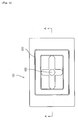
- FIG.2 is a top view showing a chip setting member in the determination apparatus shown in FIG.1.
- FIG.3 is a sectional view taken in the direction of the arrows substantially along the line A-A of FIG.2.
- FIG.4 is a top view of an upper template of a protein immobilization chip mounted in a chip setting member of the determination apparatus shown in FIG.1.
- FIG.5 is a perspective sectional view taken in the direction of the arrows substantially along the line B-B of FIG.4.
- FIG.6 is a top view of a lower template of the protein immobilization chip mounted in the chip set of the determination apparatus shown in FIG.1.
- FIG.7 is a perspective sectional view taken in the direction of the arrows substantially along the line C-C of FIG.6.
- FIG.8 is an assembly sectional view of the upper template shown in FIG.4 and the lower template shown in FIG.6.
- FIG.9 is a sectional view explaining a column of a sample preparation member of an activity measurement unit in the determination apparatus shown in FIG.1.
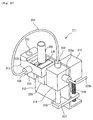
- FIG.10 is a perspective view of the sample preparation member of the activity measurement unit of the determination apparatus shown in FIG.1.
- FIG.11 is a top view of a fluid manifold of the sample preparation member shown in FIG.10.
- FIG.12 is a sectional view taken in the direction of the arrows substantially along the line D-D of FIG.11
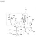
- FIG.13 is a fluid circuit view of the sample preparation member shown in FIG.10.
- FIG.14 is a block diagram showing hardware configuration of the control means.
- FIG. 15 is a block diagram showing a control system taking control of the determination apparatus.
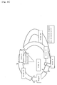
- FIG.16 is a view explaining a cell cycle.
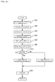
- FIG.17 is a view illustrating an overall process flow of the determination apparatus.
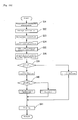
- FIG.18 is a view illustrating a process flow of preparing samples for expression level measurement.
- FIG.19 is a view illustrating a process flow of preparing samples for activity value measurement.
- FIG.20 is a view explaining an example of using reagents or the like with the measurement method of the present invention.
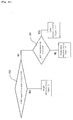
- FIG.21 is a view showing an overall flow of an example of the analysis process by the determination apparatus.
- FIG. 22 is a view showing an overall flow of another example of the analysis process by the determination apparatus.
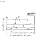
- FIG. 23 is a view of an example of using the determination results obtained by the measurement method of the present invention.
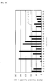
- Fig.24 is a view showing another implementation example of sensitivity analysis.
- FIG.25 is a view of an additional implementation example of sensitivity analysis.
- FIG.26 is a view of a further implementation example of sensitivity analysis.
- FIG. 27 is a view showing an overall flow of another example of the analysis process by the determination apparatus.
- FIG. 28 is a view showing an overall flow of another example of the analysis process by the determination apparatus.
- the determination apparatus may be suitable for use in measuring the expression levels and activity values of CDK to determine the malignancy grades (degrees of recurrence risk) of cancers based on the measured values and the effectiveness of anticancer drugs.
- the determination apparatus Prior to describing said determination apparatus, first of all, [1] a method for determining the characteristics of mammalian tissues (those containing cancer cells) and [2] a method for predicting the effectiveness of anticancer drug therapy (a method for determining the sensitivity of tissues to ittitants) are described.
- the method for determining the characteristics of mammalian tissues involves a step of measuring the expression levels and activity values of two or more kinds of cyclin-dependent kinase in mammalian tissues and a step of determining the characteristics of said mammalian tissues based on a CDK profile including a ratio of the activity value to expression level for a first cyclin-dependent kinase and a ratio of the activity value to expression level for a second cyclin-dependent kinase.
- the application of the determination method of the present invention to tissues containing tumor cells may make it possible to determine the characteristics of the tissues containing tumor cells and diagnose the malignancy grades of cancers.
- the CDK profile means information a certain tissue has including the ratio (for example, specific activity) of the activity value to expression level of at least one kind of CDK and/or values calculated from a plurality of activity values and expression levels of CDK (for example, a ratio of the ratio (A1) of the activity value to expression level for the first CDK to the ratio (A2) of the activity value to expression level for a second CDK (for example, A1/A2 or A2/A1)).
- the ratio for example, specific activity of the activity value to expression level of at least one kind of CDK and/or values calculated from a plurality of activity values and expression levels of CDK (for example, a ratio of the ratio (A1) of the activity value to expression level for the first CDK to the ratio (A2) of the activity value to expression level for a second CDK (for example, A1/A2 or A2/A1)).
- the mammals, characteristics of which tissues are determined by the determination method are, but not in particular limited to, humans, especially those, whose clinical conditions need to be determined, more specifically, those, whose pathologic conditions of cancers need to be determined.
- the tissues, of which characteristics are determined by the determination method may be the living tissues from mammals, more specifically, supportive tissues including fibrous connective tissue, cartilage tissue, bone tissue, blood, and lymph node; epithelial tissue; muscular tissue; nervous tissue; or the like.
- the determination method is suitable for use in determining the characteristics of the tissues, on which pathological information need to be obtained as in tissues containing tumor cells contributing to damage to harmonization in the individuals, consequently inducing a dysfunctional control mechanism of cell-proliferation.
- tissue suitable for these tissues include those cancer tissues developed in organs including breast, lung, liver, stomach, large intestine, pancreas, skin, uterus, testis, ovaries, thyroid gland, parathyroid gland, lymphoid system, and bone marrow.
- the characteristics of the mammalian tissues to be determined include the proliferating ability and malignancy levels of the cells contained in the target tissues.
- the proliferation ability of the cells means a growth activity level of the cells providing information on whether dysfunction has occurred in the proliferation control mechanism (whether the cells have been cancerated), as well as information on aneuploid cells.
- the malignancy level of the tumor cell means specifically metastatic and recurrent trends, and bad prognosis.
- the term "recurrence” means a case where after part of an organ containing the malignant tumor cells is cut off, the same malignant tumor cells have developed in the rest organ; and a case where tumor cells separated from a primary lesion were carried to remote tissues (or a remote organ) and have autonomously proliferated therein (metastatic reccurrence).
- the recurrence level of a tumor is higher in the case where the tumor has recurred within five years since its curing. Focusing on the individual stages of tumor, the recurrence rate in Stage III is 50%, higher than that in Stage II (recurrence rate: 20%).
- Prognosis is to predict the progress and outcome of a disease. The prognosis level is worse with a higher mortality five or ten years after. Giving an example, the mortality rate is 50% in Stage III, indicating the prognosis worse than the in Stage II (mortality rate: 20%).
- Cyclin-dependent kinase is a generic term representing a group of enzymes activated when binding to cyclin, to act their corresponding points of a cell cycle depending on the category of the enzymes.
- the CDK inhibitor is a generic term for group of factors, each of which binds to a cyclin-CDK complex to inhibit its activity.
- the cell cycle which is a cycle starting at a point when a cell proliferates until a point when returns to the starting point as two daughter cells, through a course of events, such as DNA replication, chromatid disjunction, nuclear fission, and cytoplasmic fission, are divided into four stages G1, S, G2, and M as shown in FIG.16.
- Stage S is a DNA replication period
- Stage M is a cell division period.
- Stage G1 is a period from the completion of mitotic division until the initiation of DNA synthesis, where preparatory checkup is made for entering Stage M. Once a critical point in Stage G1 (Point R in mammal cells) has passed, the cell cycle continues to take a round with no stop.
- Stage G2 is a period from the termination of DNA synthesis until the initiation of mitotic fission.
- main checkpoints are a point directly before the cell enters Stage S from Stage G1 and an entry point to mitotic fission in Stage G2.
- the checkpoint in Stage G1, which triggers the initiation of Stage S, in particular, is important. This is because once the critical point in Stage G1 has passed, the cell passes through the cell cycle S ⁇ G2 ⁇ M ⁇ G1 with no stop even if proliferation signals are no longer given. It should be noted that some of the cells, which have stopped its proliferation, are put in its resting stage (G0) having DNA contents in Stage G1, dropping out from the cell cycle. These cells, however, when their proliferation is induced, may proceed to Stage S after a period slightly longer than Stage G1 in the cell cycle.
- Cyclin-dependent-kinase (CDK) to be used with the determination method is preferably selected from a group of CDK1, CDK2, CDK4, CDK6, cyclin A-dependent-kinase, cyclin B-dependent-kinase, and cyclin D-dependent kinase.
- the cyclin A-dependent kinase is CDK, which is activated when binding to cyclin A. According to all we know now, it means both of CDK1 and CDK2.
- the cyclin B-dependent kinase is CDK, which is activated when binding to cyclin B. According to all we know now, it means CDK1.
- the cyclin D-dependent kinase is CDK, which is activated when binding to cyclin D. According to all we know now, it means both of CDK4 and CDK6.
- CDKs bind to their corresponding cyclins to form cyclin-CDK complexes (hereinafter, simply referred to as activated CDKs) as shown in Table 1 to activate given periods in the cell cycle as shown in Table 1.
- the CDK1 binds to cyclin A or B, the CDK2 to cyclin A or E, the CDK 4 and CDK6 to cyclin D1, D2, or D3 to be activated.
- the CDK activity may be inhibited by any of CDK inhibitors as shown in Table 1.
- p21 inhibits CDK 1 or CDK2
- p27 inhibits CDK 2
- CDK4, CDK6, and p16 inhibits CDK 4 and CDK6, respectively.
- CDK Combined cyclin Combined CDK inhibitor Action stage of activated CDK CDK4 Cyclin D1 p27, p16 G1 CDK6 Cyclin D2 Cyclin D3 CDK2 Cyclin E p27 Proceeds to S from G1 CDK2 Cyclin A p21, p27 Stage S activated CDK1 Cyclin A p21, Proceeds to M from G2 Cyclin B Cyclin Cyclin A p21, p27 CDK1: A-dependent-kinase proceeds to M from G2 CDK2: middle S stage Cyclin Cyclin B p21 CDK1:proceeds B-dependent-kinase to M from G2 Cyclin D-dependent-kinase Cyclin D p27, p16 CDK4, 6:G1
- the expression levels and activity values of two or more kinds of CDKs are measured and ratios (namely, CDK specific activity values or their reciprocal numbers) of the measured levels to the measured activity values to find CDK profiles.
- the CDK specific activity may be obtained by calculating CDK activity/CDK expression level.
- the CDK profiles include profiles (CDK specific activity profiles) containing CDK specific activity values and profiles (reciprocal number of CDK specific-activity profiles) containing the reciprocal numbers of the CDK specific activity values.
- the CDK activity value means the level of kinase activity (represented in units of U (unit)), which indicates kinase may phosphorylate how much substrate (for example, activated CDK1 and activated CDK2 phosphorylate Histon H1 and activated CDK4 and activated CDK6 phosphorylate Rb (Retinoblastoma protein)) when binding to its specific cyclin.
- This level may be measured by any the conventionally known enzyme activity measurement methods.
- one of these methods involves a step of preparing samples containing an activated CDK from cell-dissolved solution, which is the sample to be measured; a step of trapping 32P into a substrate protein using ATP labeled with 32 P ( ⁇ -[ 32 P]-ATP); a step of measuring the quantity of labeled phosphorylated substrate; and a step of determining the quantity in reference to an analytical curve drawn using a standard product.
- Another method using no radioactive labels is disclosed in JB No. 335997/2002.
- This method involves a step of preparing samples containing a target activated CDK from cell-solubilized solution, which is sample to be measured; a step of reacting the substrate with adenosine 5'-O-(3-thiotriphosphate) (ATP- ⁇ S); a step of introducing a monothiophosphate group into a serine or threonine residue of said substrate protein; a step of binding a labeling fluorescent substance or labeling enzyme to sulfur atom of introduced monothiophosphate group to label the substrate protein; a step of measuring the quantity of thiophosphate labeled (in the case where the labeling fluorescent substance, the quantity of fluorescent thiophosphate); and a step determining the quantity in reference to the analytical curve drawn using the standard product.
- adenosine 5'-O-(3-thiotriphosphate) adenosine 5'-O-(3-thiotriphosphate) (ATP- ⁇ S)
- ATP- ⁇ S adenosine 5'
- the samples to be used in measuring the CDK activity are prepared by specifically collecting the target CDK from the solution, in which the tissue to be measured is solubilized.
- an anti-CDK antibody specific to the target CDK may be used in preparing the samples.
- an anti-cyclin antibody may be used in preparing the samples.
- the prepared samples will contain any other CDK than the activated CDKs. Giving an example, a complex combining a CDK inhibitor with the cyclin-CDK complex also may be contained.
- samples may contain any CDK alone, a CDK-cyclin and/or CDK inhibitor complex, and CDK-any other compound complex. Accordingly, the activity values are measured in units (U) of phosphorylated substrate in the presence of activated, inactivated CDKs, and mixed competitive reactions between them.
- the CDK expression level which is the target CDK quantity (a unit equivalent to the number of molecules) measured based on cell-solubilized solution, can be measured conventionally known methods for measuring the quantity of a target protein from a protein mixture. Giving an example, it may be measured by any of the methods such as the ELISA and WESTERN BLOT methods or may be measured by a method disclosed in JB No. 130871/2003.
- the target protein (CDK) may be trapped using its specific antibody.
- an anti-CDK1 antibody allows all the CDK1 contained in the cell (including a CDK alone, a CDK-cyclin and/or CDK inhibitor complex, and CDK-any other compound complex).
- the specific activity calculated by the expression is equivalent to the rate of activated CDKs out of CDKs contained in the cell, indicating the CDK activity level based on the proliferation state of an animal cell to be determined.
- the CDK specific activity calculated this way does not depend on a method for preparing sample to be measured.
- the sample to be measured (cell-solubilized solution) prepared by the method for preparing sample to be measured, in particular prepared from a biopsy material is subject to the quantity of noncellular tissue contained in an actually collected tissue, for example, extracellular matrix. For this reason, it is of great significance that the specific activity values or their reciprocal numbers are used to avoid such a problem. Such specific activity values or their reciprocal numbers have higher correlation with clinical characteristics compared to the conventional simple activity values.
- the percentage of cells may be determined at any stage in the cell cycle or the stage in the cell cycle, at which the percentage of cells is dominant, may be known.
- the method for determining the characteristics of mammalian tissues is described focusing on the CDK specific activity profiles containing CDK specific activity values.
- the kind of CDK of which specific activity is to be measured, may be selected according to the kind of characteristic need to be determined.
- cancer cells escaping from normal proliferation control, actively proliferate.
- the percentage of cells in Stages S and G2 is considered to be higher suggesting the cells having been cancerated.
- Cancers stemming from these types of cancer cells show rapid progress, namely being of malignant type.
- Aneuploid cells may grow when they have undergone abnormal Stage M or when they have proceeded directly to Stage G1 bypassing Stage M and then entering Stage S.
- a lower percentage of cells in Stage M implies malignant cancers.
- CDK1 is used as first cyclin-dependent kinase and CDK2 as second cyclin-independent kinase to classify the cells into groups in the ascending order of CDK1 values.
- the CDK2 specific activity values reflect the percentage of cells in Stage S. If the number of cells in Stage S is large, it may be determined that the tissue composed of said cells is clinically malignant, namely a malignant cancer with worse prognosis, meaning that it is subject to metastasis.
- the percentage of cells in the given stage in the cell cycle may be estimated based on the kinds of CDKs and their known actions in reference to the CDK specific activity profiles containing two or more kinds of CDK the specific activity values, or the CDK specific activity profiles containing the specific activity values for two or more kinds of CDKs, which have been predetermined using their corresponding normal tissue cells as standard cells, is found for comparison with those of the normal cells.
- the ratio of the specific activity value of one kind of cyclin-dependent kinase to that of another kind of cyclin-dependent kinase is preferably used. In this case, the ratio between two kinds of cyclin-dependent kinase is compared with the given threshold corresponding to said ratio to determine the characteristics of the tissue.
- Thresholds used with the determination method may be appropriately determined according to the types of cells to be measured and the characteristics to be determined.
- the value for the ratio between the specific activity values which gives an indication of borderline for determining the characteristics, may be selected based on the a lot of cells involved in determining the target characteristics, a database of individuals, and a database of CDK specific activity values of the cells.
- tumor cells for which malignancy levels were determined by pathologists, have been collected from a plurality of patients.
- the ratios between the specific activity values of two kinds of CDKs considered to have correlation each other are obtained and the obtained ratios are arranged in the descending order.
- a median obtained by bisecting the group of ratios may be used as the threshold.
- Method for predicting the effectiveness of anticancer drug therapy involves; a step of comparing at least one parameter selected from a group of the activity values, expression levels, and ratios between the activity values and expression levels for the cyclin-dependent kinase in the tumor cells collected from patients with the threshold corresponding to a selected parameter; and a step of predicting the effectiveness of anticancer drug therapy on the patients based on the results of the comparison step.
- the effectiveness of therapy to be predicted by the prediction method includes those of pre-operative and post-operative therapy.
- pre-operative therapy such a case is determined to be effective that after an anticancer drug has been continuously administered to the patients with a primary lesion, the primary lesion reduces or disappears.
- post-operative therapy such a case is determined to be effective that the same type of cancer does not recur after an anticancer drug has been continuously administered since extirpative surgery.
- post-operative therapy it gives an indication of recurrence whether the anticancer drug is effective for invisible metastatic cancer or the like.
- the tissues to be used as samples in the prediction method are those containing tumor cells collected from patients.
- tissues available through extirpative surgery can be used.
- the tissues biopsy tissues collected from the tumor tissues of the patients or the like may be used.
- the cyclin-dependent kinase (CDK) to be used with the prediction method includes CDK1, CDK2, CDK4, CDK6, cyclin A-dependent kinase, cyclin B-dependent kinase, and cyclin D-dependent kinase, out of which appropriate ones may be selected according to the types of cancers and anticancer drugs.
- CDK1 CDK2, CDK4, CDK6, cyclin A-dependent kinase, cyclin B-dependent kinase, and cyclin D-dependent kinase, out of which appropriate ones may be selected according to the types of cancers and anticancer drugs.
- This means that cancers may be classified into many categories and the effectiveness of the anticancer drug therapy largely depends on the characteristics related to the cell cycles of the cancer cells of the respective patients.
- Target cancers include breast cancer, stomach cancer, colon cancer, esophageal cancer, prostate cancer, and the like.
- the anticancer drugs for breast cancer include a CMF group (three-drug combined administration of cyclophosphamide, methotrexate, and fluorouracil), taxane anticancer drugs such as docetaxel and paclitaxel, CE (two-drug combined administration of cyclophosphamide and epirubicin), AC (two-drug combined administration of doxorubicin and cyclophosphamide), CAF (three-drug combined administration of fluorouracil, doxorubicin, and cyclophosphamide), FEC (three-drug combined administration of fluorouracil, epirubicin, and cyclophosphamide), two-drug combined administration of trastuzumab and paclitaxel, capecitabine, and the like.
- CMF group three-drug combined administration of cyclophospham
- the anticancer drugs for stomach cancer include FAM (three-drug combined administration of fluorouracil, doxorubicin, and mitomycin C), FAP (three-drug combined administration of fluorouracil, doxorubicin, and cisplatin), ECF (three-drug combined administration of epirubicin, cisplatin, and fluorouracil), two-drug administration of mitomycin C and tegafur, two-drug combined administration of fluorouracil and carmustine, and the like.
- the anticancer drugs for colon cancer include two-drug combined administration of fluorouracil and leucovorin, two-drug combined administration of mitomycin and fluorouracil, and the like.
- the anticancer drugs for ovary cancer include TP (two-drug combined administration of paclitaxel and cisplatin), TJ (two-drug combined administration of paclitaxel and carboplatin), CP (two-drug combined administration of cyclophosphamide and cisplatin), CJ (two-drug-combined administration of cyclophosphamide and carboplatin) and the like.
- One or two selected from parameters of CDK activity values, CDK expression levels, and the ratio between the CDK activity values and expression levels give an indication for determination.
- the ratio of the activity values to the expression levels may be the CDK specific activity value obtained by calculating CDK activity value/CDK expression level or may be the value obtained by calculating CDK expression level/CDK activity value.
- the parameters can be compared with the given thresholds to determine whether anticancer drugs are effective.
- the parameters selected from the activity values, the expression levels, and the ratio between the activity values and the expression levels are those selected appropriately according to the kinds of the anticancer drugs and the type of cancer.
- the CDK activity values and expression levels of the tumor cells collected from the cancer patients, for whom anticancer therapy was applied and the effectiveness of therapy was known, prior to cancer therapy and stored are measured, the results of anticancer drug therapy are analyzed for the respective parameters, and the parameters having correlation with the results of anticancer drug therapy are selected.
- the parameters to be compared with the thresholds may be one parameter of the given CDK or may be a pair of two parameters of it. When the two parameters are selected, the individual parameters should be compared with their corresponding thresholds. Either one kind (effectiveness prediction method I) or two or more kinds (effectiveness prediction method II) of CDKs may give an indication for determination.
- the individual parameters of a plurality of CDKs may be compared with their corresponding thresholds to predict the effectiveness of anticancer drug therapy based on the combination of the results of comparison among kinases (effectiveness prediction method II-1).
- the parameters of the plurality of CDKs to be compared with their corresponding thresholds may be of the same kind (for example, expression level) or may be of different kinds (for example, for one CDK, its expression level is compared with its threshold while for another CDK, its activity value is compared with its corresponding threshold).
- the effectiveness of anticancer drug therapy may be predicted; by predicting the effectiveness on first CDK(s) using the effectiveness prediction method I; and comparing the parameters selected from the activity values, expression levels, and ratio between the activity values and expression levels for the CDK other than the first CDK (s) with their corresponding thresholds concerning the tumor cells, for which effectiveness of anticancer drug therapy was determined to be doubtful by the effectiveness prediction method I (effectiveness prediction method II-2).
- the CDK other than the first CDK(s) may be one kind of CDK (a second CDK) or may be a plurality kind of CDKs (third, fourth...CDK ).
- At least one selected from the parameters of the activity value, expression level, and ratio between the activity value and expression level for each CDK should be compared with the threshold corresponding to said parameter and based on the combination of the results of comparison to predict the effectiveness of anticancer drug therapy on the patients.
- the parameters to be used in determining the effectiveness of anticancer drug therapy are selected from the group. Only one parameter may be selected or two parameters may be selected for comparison with their corresponding thresholds.
- the second, third, fourth CDKs the same kind, such as the expression level, of parameters may be used or different kinds of parameters (for example, the expression level is used for the second CDK while the activity value for the third CDK) may be used to determine the effectiveness of anticancer drug therapy.
- the effectiveness prediction method II is has an advantage in that it has a higher percentage of right answers.
- the use of the effectiveness prediction method II is of significance because in many cases, anticancer drug therapy is determined to be effective by the effectiveness prediction method II-II even if it is not determined to be effective by the effectiveness prediction method I.
- the efficacy of anticancer drugs includes two levels; at one level, the progress of cancer is prevented and at another level, the tumor is reduced in size to improve the disease state.
- the effectiveness prediction method II, especially the effectiveness prediction method II-II enables the effectiveness to be predicted considering the efficacy level of anticancer drug therapy.
- the effectiveness of anticancer drug therapy on the patients may be predicted based on the combination of the results obtained by comparing the expression levels of CDK(s) with their corresponding to their thresholds and the results obtained by comparing the expression levels of the cyclin-dependent kinase inhibitor (CDK inhibitor) with their corresponding thresholds (effectiveness prediction method III).
- the CDK inhibitor composed of a group of factors, which bind to cyclin-CDK complexes to inhibit their activity, are classified into an INK 4 family and a CIP/KIP family.
- the CIP/KIP family, especially p21 is preferably used.
- p21 is an inhibitor, which inhibits the progress of cancer at both the checkpoints in Stages G1 and G2 in the cell proliferation cycle, affording enough time to repair damaged DNA.
- the effectiveness of anticancer drug therapy may be predicted based on the combination of the results obtained by comparing the given CDK parameters with their corresponding thresholds and the results obtained by comparing the CDK-inhibitor expression level with their corresponding thresholds or anticancer drug therapy considered to be highly effective may be selected by comparing the given CDK parameters with their corresponding thresholds to determine the effectiveness of anticancer drug therapy in a first step (effective prediction method I) and then by comparing the expression level of the CDK inhibitor with its corresponding threshold concerning the tumor cells, for which anticancer drug therapy has not been determined to be effective, in a second step.
- Her2 and p21 have been reported as effectiveness prediction factors for CMF administration therapy.
- the clinical trial by The International Breast Cancer Study Group (IBCSG) suggested that CMF administration was not effective for breast cancer patients with Her2 over-expressed, and demonstrated that the disease-free survival for a group of the patients with higher expression of p21 is significantly lower than that for a group of the patients with lower expression of p21.
- Both the Her2 and p21 are the factors for predicting the group of the patients, for whom the effectiveness of CMF therapy is low, and no factor for actively predicting the group of the patients, for whom the effectiveness is high, has been reported.
- the prediction methods not only the effectiveness of anticancer drug therapy is actively demonstrated but also such a case may be indicated by making the thresholds stricter that almost 100% of effectiveness may be expected.
- the thresholds are the values specified appropriately based on the kinds of anticancer drugs and the types of cancers. More specifically, the values are set so that the cases, in which the anticancer drug therapy is determined to be effective based on the parameters having correlation with the large amount of results of anticancer drug therapy, may be selected by examining the relationship between a large amount of results of anticancer drug therapy, in which given anticancer drugs were administered to the patients with given types of cancers, and the parameters.
- the thresholds are set so that only the cases, for which all the results of anticancer drug therapy are effective, may be selected.
- the thresholds are set based on the actual results of clinical anticancer drug therapy, allowing the effectiveness to be determined at a higher accuracy.
- results of anticancer drug therapy include those obtained by examining any variation in tumor size caused by continuous application of given anticancer drug therapy or by continuously examining any recurrence over five to six years.
- the ratio represented by the CDK activity value/CDK expression level (CDK specific activity value) or the CDK expression level/CDK activity value is equivalent to the percentage of activated CDKs out of all the CDKs contained in the cell, indicating the CDK activity level based on their own proliferation state shown by the tumor cells to be measured and does not depend on the method for preparing the samples to be measured.
- the cell solubilized solution prepared by method for preparing sample to be measured, especially from the biopsy material is subject to the quantity of noncellular tissues contained in the actual tissues collected, for example, extracellular substrate. Accordingly, using the ratio between the CDK expression level and activity values, inevitable influences may be eliminated during preparation of sample to be measured, enabling the effectiveness to be determined at a higher accuracy even if the determination method focusing on proteins is used.
- the expression level of the CDK inhibitor means the quantity (the unit corresponding to the number of molecules) of the target CDK inhibitor to be measured from the cell-solubilized solution, which may be measured by any of conventionally known methods for measuring the quantity of a target protein from a protein mixture. For example, the ELIZA method or the WESTERN BLOT method may be used.
- the target protein (CDK inhibitor) may be trapped using its specific antibody. Either a monoclonal or polyclonal antibody may be used provided that it can bind specifically to the target protein. For example, to trap p21, either an anti-p21 monoclonal antibody or an anti-p21 polyclonal antibody may be used.
- FIG.1 is a perspective view explaining determination apparatus A according to one embodiment of the present invention.
- the determination apparatus A which is capable of measuring the activity values and expression levels of cyclin-dependent kinases (CDKs) contained in the tissues to determine the malignancy levels (the level of recurrence risk) of the human cancer cells and the effectiveness of (sensitivity to) anticancer drugs based on the measured values, is composed mainly of; a detecting member 4, a chip setting member 1, a first reagent setting member 5 and second reagent setting member 6, which are disposed in a front part of an apparatus body 20; an activity measurement unit 2, a waste bath 7 for collecting waste solution, and a pipette washing bath 8 for washing a pipette, which are disposed in the rear part of the apparatus body 20; a dispensing mechanism 3 disposed in a upper part of the apparatus body 20, which is capable moving the pipette in three directions (X, Y, and Z directions) ; a fluid member 9 and an electronic board 10 disposed on the back of the apparatus body 20; and a personal computer 12, which is a control means connected to the
- the determination apparatus A has a pure water storage tank 13, a cleaning liquid tank 14, a waste tank 15, and a pneumatic source 11.
- the pure water storage tank 13 containing pure water for cleaning flow channel at the completion of measurement is connected to a fluid member 9 by means of piping 21.
- the cleaning liquid tank 14 containing the cleaning liquid for cleaning the pipette is connected to the pipette washing bath 8 by means of piping 22.
- the waste tank 15 for collecting waste solution is connected to the waste bath 7 by means of piping 23.
- a solubilization apparatus B for obtaining specimens capable of being processed in the determination apparatus A from living samples is arranged in parallel with the determination apparatus A.
- solubilization apparatus B and the determination apparatus A are described.
- the solubilization apparatus B which prepares the liquid specimens capable of being processed in the determination apparatus A from living samples such as the tissues collected from the patients prior to the processing in that determination apparatus A, is composed mainly of a housing 30, an operating member 31 disposed in the upper front of the housing 30, a driving member 32 equipped with a pair of pestel 34 for pressing or grinding the living samples, and a specimen setting member 33 with an inserted eppen tube 35 for containing the living samples.
- the driving member 32 is capable of moving the pestle 34 up and down while rotating it, and thereby, the living samples are pressed or ground injected in the eppen tube 35.
- the housing 30 contains a controlling member (not shown in the figure) for controlling the movement of the driving member 32 therein.
- the operating member 31 has an operating button 31a, an operation LED 31b, a display member 31c for indicating the apparatus state, error message, and the like therein.
- the specimen setting member 33 contains a cooling means (not shown in the figure), which keeps the living samples in the eppen tube inserted into a pocket on the upper surface of the specimen setting member 33 at a constant temperature.
- the supernatant liquid of living samples after being solubilized in the solubilization apparatus B and then separated in a centrifuge (not shown in the figure) is collected into given specimen vessels and then set in the first reagent setting member 5 of the determination apparatus A.
- the cooling means (not shown in the figure) is disposed for keeping the specimens in the vessels, such as screw caps, inserted in the pocket on the upper surface of the first reagent setting member 5, various types of antigens including CDK 1 antigens (calibration 1), CDK 2 antigens (calibration 2), a variety of fluorescence-labeled antibodies including fluorescence-labeled CDK 1 antibodies and fluorescence-labeled CDK 2 antibodies, and the like, at a constant temperature.
- 20 pockets in total are arranged in five columns and four rows, allowing up to 20 vessels, such as screw caps, to be set therein.
- the second reagent setting member 6 is disposed. Like the first reagent setting member 5, in the second reagent setting member 6, a plurality of pockets are arranged for setting the vessels, such as eppen tube and screw caps, containing a buffer, substrate solution, fluorescence enhancing reagent, or the like. Prior to the processing in the determination apparatus A, chips for protein immobilization are set in the chip setting member 1 and columns are set in the activity measurement unit 2.
- the chip setting member 1 composed of aluminum blocks has a pocket 132 for mounting the chip 121 for protein immobilization on its upper surface and a suction inlet 135 at its bottom, as shown in FIGS. 2 to 3. More specifically, the chip setting member 1 has the rectangular first pocket 132 on the upper surface and a similarly rectangular second pocket 133 at the bottom of the first pocket 132. At the bottom of the first pocket 132, a rectangular frame of rubber flexible gasket 137 is disposed on the circumference of the second pocket 133.
- the second pocket 133 has a cross-shaped groove 134 at the bottom thereof and the suction inlet 135 at the center of the bottom thereof, the bottom of the groove 134 inclines toward the center from the circumference of the second pocket 133, proceeding to the deeper point.
- the suction inlet 135 links to a nipple 136 disposed for connecting to an external suction pump (not shown in the figure).
- the chip 121 for protein immobilization which will be described in detail later, is horizontally loaded via a bottom gasket 137 of the first pocket 132.
- a fixing member for fixing the chip 121 for protein immobilization to the bottom of the first pocket 132 by pressing against the pocket may be disposed in the chip setting member 1.
- the chip 121 for protein immobilization is composed of a porous membrane 122, an upper template 101 and a lower template 102 for supporting the porous membrane 122 between them.
- the chip 121 for protein immobilization acts as a second contact means for bringing antibody solution containing an antibody for cyclin-dependent kinase into contact with the living samples (specimens).
- a matrix of 40 rectangular through-holes 102 arranged in four columns and ten rows are formed.
- a groove (pocket) 104 running along the circumference of the matrix of 40 through-holes 102 is formed.
- This groove 104 defines a rectangular porous membrane setting area 103 therein.
- eight jig through-holes 105 are formed.
- a matrix of 40 rectangular through-holes 112 are arranged in four columns and ten rows at the positions corresponding to those of the through-holes 102 of the upper template 101.
- the through-holes 112 have the same shape and area as those of the through-holes 102.
- a ribbed convex 114 running along the circumference of the matrix of 40 through-holes is formed at the position corresponding to that of the groove 104.
- the convex 114 defines the rectangular porous membrane setting area 113 therein.
- six notches are formed 115.
- the upper template 101 and lower template 111 may be made of, for example, a vinyl chloride resin.
- FIG. 8 shows the cross section of the chip 121 for protein immobilization.
- the upper template 101 and lower template 111 are overlapped and the convex 114 is detachably pressed in the groove 104.
- the through-holes 102 have the same axis as that of the through-holes 112.
- the rectangular hydrophobic membrane 122 is inserted between the porous membrane setting areas 103 and 113 and uniformly compressed when the convex 114 is pressed in the groove 104. Accordingly, the porous membrane 122 is hermetically defined by the through-holes 102, forming the same number of wells (solution vessels) as that of the through-holes 102.
- the activity measurement unit 2 is composed of a plurality of sample preparing members 211, each being equipped with the column 201 and a fluid manifold 213 and used in measuring the CDK activity values.
- the column 201 is composed of a cylinder made of the vinyl chloride resin and has a carrier holding vessel 202 for holding a carrier support 206 to be used in isolating the target protein in the liquid samples, an liquid introducing member 203 for introducing a liquid sample into the carrier holding vessel 202, and a liquid reservoir 204 for receiving the liquid sample from the carrier holding vessel 202 and reserving the received liquid.
- the column 201 acts as a first contact means for bringing the substrate solution containing a given substrate into contact with the living samples (specimens).
- the liquid reservoir 204 of the column 201 has an opening 205 capable of externally injecting or collecting the liquid samples on its upper part.
- the carrier support 206 composed of a cylindrical monolith silica gel, which has a structure combining a 3D network of skeleton and gaps, unlike a particle support. A given CDK antibody is fixed to the monolith silica gel.
- the carrier support 206 is inserted into the carrier holding member 102 from the lower opening of the column 201 and then flexibly pressed in by means of a fixing pipe 208 via an O ring 207 for support. It should be noted that the fixing pipe 208 is pressed in from the lower opening of the column 201 and the fixing pipe 208 and the hole of the ⁇ ring 207 form the liquid introducing member 203.
- FIG. 10 is a perspective view of the sample preparing members 211. As shown in the figure, the sample preparing members 211 has an L-shaped supporting plate 212, to which the fluid manifold 213, the syringe pump 214, and a stepping motor 215 with a decelerator are fixed.
- a screw shaft 216 is connected to the output axis of the stepping motor 215.
- a driving arm 217 in engagement with the screw shaft 216 is connected to the tip of a piston 218 of the syringe pump 214.
- the screw shaft 216 is rotated by means of the stepping motor 215, the piston 218 moves up and down.
- the syringe pump 214 is connected to the fluid manifold 213 by means of a solution sending tube 250 via connectors 219 and 220.
- the syringe pump 214 is also connected to a chamber 234 (see FIG. 13) containing liquid (cleaning liquid) filling the flow channel by means of a solution sending tube 220b via a connector 220a.
- the fluid manifold 213 has a column connection 221, to which the liquid introducing member 203 of the column 201, and a liquid sample receiving member 222 for receiving the liquid samples.
- the fluid manifold 213 has the flow channel 223 therein and a electromagnetic valve 224 for opening and closing between the liquid sample receiving member 222 and the flow channel 223 and a electromagnetic valve 225 for opening and closing between the flow channel 223 and the column connection 221 at the bottom thereof.
- the fluid manifold 213 has connector holes 226 for the connectors for connecting the connectors 220 to the sides thereof, and this connector hole 226 is connected to the flow channel 223.
- FIG. 13 is a schematic view showing the circuit of the sample preparing member 211, along which the fluid flows.
- the syringe pump 204 is connected to the fluid manifold 213 vie the connectors 20.
- the chamber 234 is connected via the electromagnetic valve 233, a positive pressure being applied to the chamber 234 from a positive pressure source 235.
- a column receiving pocket 227 for receiving the lower end of the column 201 is formed on the upper surface of the fluid manifold 213.
- the center of the bottom of the pocket 227 passes though the column connection and the O ring 228 is attached along the circumference at the bottom thereof.
- two pressing plates 229 and 230 with L-shape cross sections are fixed in parallel, between which a space wider than the width W and narrower than D is left at the center of the column loading pocket 227.
- the flange 209 is inserted passing through between the pressing plates 229 and 230, and then rotated clockwise or counterclockwise by 90 degrees. Thereby, the portion with D in diameter of the flange 209 is engaged with the pressing plates 229 and 230 and the flange 209 is fixed between the pressing plates 229 and 230 by means of flexibility exerted by the O ring 228. To remove the column 201, it may be rotated clockwise or counterclockwise by 90 while being held.
- the pocket 227 of the fluid manifold 213 is filled with the fluid in order to prevent air bubble from entering and the fluid is flown out due to the volume of the column as soon as the tip of the column 201 is inserted into the pocket 227.
- an overflow holding pocket 231 is formed around the column loading pocket 227 and at a point of the overflow holding pocket 231, an overflow discharging pocket 232 is formed for sucking the overflow using a pipette.
- Various kinds of specimens and reagents are injected or sucked into or from their given places by means of a dispensing mechanism 3 equipped with the pipettes.
- the electromagnetic valve 224 opens (the electromagnetic valves 225 and 233 close) and the syringe pump 214 starts its sucking operation. Thereby, the specimens and reagents are sucked on the side of syringe pump 214 passing through the electromagnetic valve 224.
- the electromagnetic valve 224 closes, the electromagnetic valve 225 opens, and the syringe pump 214 discharges the specimens or reagents. Accordingly, the specimens or reagents pass through the electromagnetic valve 225 and sent into the column 201.
- the dispensing mechanism 3 has a frame 352 for moving the pipette in the X direction, a frame 353 for moving the pipette in the Y direction, a plate 354 for moving the pipette in the Z direction.
- the frame 352 has a screw shaft 355 for moving the plate 354 in the X direction indicated by an arrow, a guide bar 356 for sliding the plate 354 while holding, and the stepping motor 357 for rotating the screw shaft 355.
- the frame 353 has a screw shaft 358 for moving the frame 352 in the Y direction indicated by an arrow, a guide bar 359 for sliding the frame 352 while holding, and a stepping motor 361 for rotating the screw shaft 358.
- the plate 354 has a screw shaft 367 for moving the arm 368 supporting a pipette 362 in the Z direction indicated by an arrow, guide bar for sliding the arm 368 while supporting, and a stepping motor 370 for rotating the screw shaft 367.
- the dispensing mechanism 3 has a pair of pipettes 362 and thereby, the specimens or reagents may be injected into or sucked from two specimen vessels at the same time, achieving efficient measurement.
- the fluid member 9 is connected to the pipette 362, the pipette washing bath 8, and the sample preparing members 211 for controlling the fluid.
- the fluid member 9 is composed of the electromagnetic valves 224 and 225 of the sample preparing members 211, the electromagnetic valve 233 for controlling the fluid when the fluid is filled in the syringe 214 from a cooling liquid chamber, an electromagnetic valve for controlling the fluid when the fluid is sucked or discharged by the pipette 362, the electromagnetic valve for controlling the fluid when the fluid discarded from pipette 362 in waste bath 7 is sucked, and an electromagnetic valve for controlling the fluid when the pipette 362 is washed in the pipette washing bath 8.
- the electronic board 10 is mounted for supplying driving signals to the sample preparing members 211, the stepping motors 357, 361, and 370, the fluid member 9, and the like.
- the detecting member 4 measures the quantity of fluorescent material, which reflects the quantity of protein trapped by the porous membrane 122 of the chip 121 for protein immobilization and the quantity of fluorescent material, which reflects the quantity of the phosphate group. To measure these quantities, the detecting member 4 irradiates an excitation light onto the chip 121 for protein immobilization, detects the generated fluorescent, outputs to the electronic board 10 an electric signal with a strength corresponding to the intensity of the detected fluorescence.
- the detecting member 4 may use a system such as the commonly used light source, lighting system, and light-receiving system appropriately.
- the personal computer 12 acting as a control means is composed of a controlling member 77 connected to the electronic board 10, an input member 78 for entering data in the controlling member 77, and a display member 79 for indicating the results of analysis and the like.
- the controlling member 77 configures an analytical means in the present invention, a first (second) activity value obtainer for obtaining the activity values from the fluorescence intensity using the analytical curve, and a first (second) expression level obtainer for obtaining the expression levels from the fluorescence intensity using the analytical curve.
- the controlling member 77 is composed of CPU91a, ROM91b, RAM91c, an I/O interface 91d, and an image output interface 91e.
- the ROM 91b contains an operating system, a control program for controlling the operation of apparatus, and data necessary in executing the control program.
- the CPU91a is capable of loading the control program into the RAM91c for execution or capable of directly executing it from the ROM91b. Data processed by the CPU91a this way is sent to the electronic board 10 via the I/O interface 91d and data necessary in processing by the CPU91a is received via the I/O interface 91d from the electronic board 10.
- the CPU91a may control the electronic board 10 by executing the control program.
- the CPU91a obtains the expression levels and activity values of the cyclin-dependent kinase based on the luminescence intensity obtained at the detecting member 4 and based on the obtained values, and acquires information on the characteristics of cells.
- the RAM91c stores the analytical curve, which is conversion data to be used in converting the fluorescence intensity into the expression levels or activity values.
- FIG. 15 is a block diagram showing the control system for controlling the determination apparatus A of the present embodiment.
- the control system is composed of the electronic board 10 containing a driver circuit for driving the members of the dispensing mechanism 3 and the personal computer 12 containing the controlling member 77 for controlling the electronic board 10 and analyzing the results from the detecting member 4, an input member 78 for entering data and the like into the controlling member 77, and the display member 79 for indicating the analysis results analyzed at the control member 77.
- the controlling member 77 outputs from the electronic board 10 the driving signal for driving the stepping motor 215 of the sample preparing members 211, the driving signal for controlling the temperature of the first reagent setting member 5, the driving signal for driving the stepping motor 357, 361 and 370, and the driving signal for driving the electromagnetic valve of the fluid member 9 by controlling the electronic board 10.
- the controlling member 77 receives the detected signals from the detecting member 4 via the electronic board 10.
- the solubilization apparatus B Prior to the processing by the determination apparatus A, the solubilization apparatus B is used to collect the liquid specimens from the tissues cut out from the patients. First, the specimens are put into the eppen tube using the tweezers. The eppen tube is set in the specimen setting member 33 of the solubilization apparatus B as shown in FIG.1 and the start button of the operating member 31 is pressed. Then, the pestel 34 moves down to the given position and the tissue in the eppen tube is pressed against the bottom of the tube.
- the solubilized liquid such as a buffer containing a surface active agent and a protease inhibitor is injected into the eppen tube automatically or manually.
- the pestel 34 is rotated to grind the tissues.
- the moving pestel 34 is stopped and then moved up, and finally the eppen tube is taken off from the specimen setting member 33. Then, the content of the eppen tube is centrifuged and the obtained supernatant is collected manually as specimens.
- the supernatant liquid is put into two specimen vessels, diluted using different dilution rates, and the specimen vessels are set in the given position of the first reagent setting member 5.
- One of two specimens is used for expression level measurement and another is used for activity value measurement.
- the chip 121 for protein immobilization is set in the chip setting member 1 and eight columns 201 are set the sample preparing members 211 of the activity measurement unit 2, respectively.
- FIG. 17 shows the overall flow of the process by the determination apparatus. If no “Yes” or “No” is shown for the decision diamonds in the flow chart, the downward direction indicates “Yes” and the rightward (leftward) direction indicates “No". All the steps described below are controlled by the controlling member 77.
- a step of accepting entries for measurement (step S1) is initiated. This step accepts entered data related to measurement such as specimen numbers.
- step S2 a step of accepting the selection of either one of the measurement mode, the prognosis predicting mode (mode for determining the malignancy levels of the human cancer cells (levels of recurrence risk)) or the sensitivity to anticancer drugs mode (mode for determining the effectiveness of (sensitivity to the anticancer drugs)) is initiated (step S2).
- the prognosis predicting mode mode for determining the malignancy levels of the human cancer cells (levels of recurrence risk)
- sensitivity to anticancer drugs mode mode for determining the effectiveness of (sensitivity to the anticancer drugs)
- step S2 Specifically, in the display member 79 of the personal computer 12, two buttons for entering these modes. The operator clicks the entry button of the desired mode.
- the sensitivity to a taxane anticancer drug is determined in the sensitivity to anticancer drug mode.
- step S3 it is determined whether the indication of initiating the measurement has been accepted. If Yes, the operator proceeds to the step S4 while if No, proceeds to the step S8.
- step S4 the specimens are sucked from the specimen vessels set in the first reagent setting member 5 and samples are prepared by applying the given steps to the sucked specimens to prepare the samples for fluorescence detection (step S4).
- This step involves a sub-step of preparing the samples for expression level measurement and a sub-step of preparing the samples for activity value measurement, both of them being described in detail later. These two sub-steps are executed in parallel.
- step S7 the fluorescence intensity is obtained and the results of analysis are output based on the obtained fluorescence intensity. It is determined whether the shout down indication for the determination apparatus has been received (step S8). If Yes, the operator proceeds to the step S9 while if No, returns to the step S1. Finally, a step of shutting the apparatus down is executed and the power switch is turned OFF. (step S9).
- FIG. 18 shows the flow of preparing the samples for expression level measurement in the step S4.
- the preservative liquid previously stored in the wells of the chip for protein immobilization is discharged and the wells are cleaned (step S11).
- the wells are cleaned by injecting the cleaning liquid into the wells from the upper side with the pipette of the dispensing mechanism 3 and the injected cleaning liquid is sucked under the negative pressure from the lower side of the chip for protein immobilization through the porous membrane.
- the same steps should be followed for the cleaning steps described below.
- the specimens for expression level measurement are sucked out from the specimen vessels set in the first reagent setting member 5 using the pipette, the specimens are injected into a plurality of given wells, and then sucked from the under side of the chip for protein immobilization under the negative pressure.
- This immobilizes the protein on the porous membrane of the chip for protein immobilization step S12).
- the given wells are cleaned with a cleaning liquid. Accordingly, the components other than protein are removed from the porous membrane of the chip for protein immobilization (step S13).
- a blocking liquid is injected into the given wells, the wells are left for 15 or more minutes (for example, 30 minutes) as they are, and the blocking liquid remaining in the wells is discharged (step S14).
- Any commercially available products may be used for the fluorescence labeled CDK 1 antibody, the fluorescence labeled CDK 2 antibody, and the fluorescence labeled p21 antibody.
- the fluorescence labeled CDK 1 antibody, the fluorescence labeled CDK 2 antibody, and the fluorescence labeled p21 antibody are injected into the given wells.
- Each of fluorescence labeled antibodies is injected into two wells. 20 to 30 minutes after, once the reaction between the fluorescence labeled antibodies and the protein (CDK1, CDK2, or p21) immobilized on the porous membrane has been finished, the injected fluorescence labels are discharged (step S15).
- the given wells are cleaned with the cleaning liquid (step S16).
- FIG. 19 shows the flow of preparing the samples for activity value measurement in the step S4.
- four sample preparing members 211 disposed on the near side in the figure and four sample preparing members 211 also disposed on the far side in the figure are used as activity measurement unit 2.
- sample preparing members 211 of the activity measurement unit 2 are called a first sample preparing member (Ac1), a second sample preparing member (Ac2), a third sample preparing member (Ac3), and a fourth sample preparing member (Ac4), respectively from the leftmost one on the far side in the figure and called a fifth sample preparing member (Ac5), a sixth sample preparing member (Ac6), a seventh sample preparing member (Ac7), and an eighth sample preparing member (Ac8), respectively from the leftmost on the near side in the figure.
- a buffer which is a reagent for cleaning, is injected into the liquid sample receiving member 222 for each of the first to eighth sample preparing members (Ac1 to Ac8) using the pipette in the dispensing mechanism 3.
- the syringe pump 214, the electromagnetic valves 224 and 225 operate for each of the first to eighth sample preparing members (Ac1 to Ac8) as described before to send the buffer into the column 201. Any excess buffer in all the columns 201 is discarded by sucking using the pipette of the dispensing mechanism 3 (step S21).
- step S22 immunoprecipitation (the reaction between the antibodies and the CDKs) is performed (step S22).
- the specimen 1 for activity value measurement is sucked from one specimen vessel set on the first reagent setting member 5 using one pipette and the specimen 2 for activity value measurement using another pipette.
- the specimen 1 for activity value measurement sucked from the specimen vessel is injected into the liquid sample receiving member 222 of the first sample preparing member (Ac1) as shown in FIG. 20.
- the specimen 1 is sent into the column 201 of the first sample preparing member (Ac1) by means of the operating syringe pump 214, electromagnetic valves 224 and 225 as described above.
- the specimen 1 also reciprocates the carrier support 206 in the column 201 by 1.5 times.
- the specimen 2 for activity value measurement sucked from the specimen vessels are first injected into the liquid sample receiving member 222 of the fifth sample preparing member (Ac5). Then, the specimen 2 is sent to the column 201 of the fifth sample preparing member (Ac5) in the same way described above.
- the carrier supports 206 in the columns 201 of the first sample preparing member (Ac1) and the fifth sample preparing member (Ac5) no CDK 1 or CDK 2 antibody was immobilized.
- the specimens 1 containing CDK1 and CDK2 are stored and in the columns 201 of the fifth sample preparing member (Ac5), the specimens 2 containing CDK 1 and CDK 2 are stored.
- the specimens 1 stored in the columns 201 of the first sample preparing member (Ac1) are sucked using the pipettes and injected into the liquid sample receiving member 222 of the third sample preparing member (Ac3).
- the specimens 1 are sent to the columns 201 of the third sample preparing member (Ac3) in the same way as that mentioned above.
- the specimens 2 stored in the columns 201 of the fifth sample preparing member (Ac5) are sucked using the pipettes and injected into the liquid sample receiving member 222 of the fourth sample preparing member (Ac4).
- the specimens 2 are sent to the columns 201 of the fourth sample preparing member (Ac4) in the same way as that mentioned above.
- the carrier supports 206 in the columns 201 of the third sample preparing member (Ac3) and the fourth sample preparing member (Ac4) the CDK 1 antibody has been immobilized.
- CDK 1 is immobilized, but CDK 2 is not, in the third sample preparing member (Ac3) and the fourth sample preparing member (Ac4) .
- the specimens 1 containing CDK 2 but no CDK 1 are stored, and in the columns 201 of the fourth sample preparing member (Ac4), the specimen 2 containing CDK 2 but no CDK 1 are stored.
- the specimens 1 stored in the columns 201 of the third sample preparing member (Ac3) are sucked using the pipettes and injected into the liquid sample receiving member 222 of the seventh sample preparing member (Ac7). Then, the specimens 1 are sent to the columns 201 of the seventh sample preparing member (Ac7) in the same way as that described above.
- the specimens 2 stored in the columns 201 of the fourth sample preparing member (Ac4) are sucked using the pipettes and injected into the liquid sample receiving member 222 of the eighth sample preparing member (Ac8). Then, the specimens 2 are sent to the columns 201 of the eighth sample preparing member (Ac8) in the same way as that described above.
- the CDK 2 antibody has been immobilized in the carrier supports 206 of the column for the seventh sample preparing member (Ac7) and the eighth sample preparing member (Ac8). Accordingly, in the seventh sample preparing member (Ac7) and the eighth sample preparing member (Ac8), CDK 2 . is immobilized. For this reason, in the columns 201 of the seventh sample preparing member (Ac7), the specimens 1 containing no CDK 1 and CDK 2 are stored and in the columns 201 of the eighth sample preparing member (Ac8), the specimens 2 containing no CDK 1 and CDK 2 are stored.
- the specimens 1 and 2 stored in the columns 201 of the seventh sample preparing member (Ac7) and the eighth sample preparing member (Ac8) are sucked using the pipettes and discarded into the waste bath 7.
- the first sample preparing member (Ac1) and the fifth sample preparing member (Ac5) are used for background activity measurement
- the third sample preparing member (Ac3) and the fourth sample preparing member (Ac4) are used for CDK1 activity measurement
- the seventh sample preparing member (Ac7) and the eighth sample preparing member (Ac8) are used for CDK 2 activity measurement.
- the buffer 1 is sent to the columns 201 (step S23).
- the buffer 2 is sent to the columns 201 for washing away the components of the buffer 1 mainly to establish the conditions suitable for these enzymatic reactions (step S24).
- a substrate reaction solution containing substrates HistonH1 and ATP ⁇ s is injected into the columns 201 and the piston 219 is reciprocated by 5.5 times (step S25).
- the liquid pressed into the columns 201 from the lower side thereof is stored as it is.
- This step introduces a phosphate group into HistonH1 using CDK 1 or CDK 2 as an enzyme.
- the quantity of the phosphate group is governed by the activity value of CDK1 or CDK 2 as an enzyme. Accordingly, by measuring the quantity of the phosphate group, the activity value of CDK 1 or CDK 2 may be obtained.
- the background activity values obtained using the first sample preparing member (Ac1) and the fifth sample preparing member (Ac5), shown in FIG. 20, are used for background correction as described later.
- the fluorescence labeled reagent is dispensed directly into the columns 201 from the upper side thereof using the pipettes to bind the fluorescence label to the phosphate group introduced into HistonH1 (step S26).
- the pipettes repeatedly suck and discharge the liquid in the columns 201 for a given time for stirring.
- the stop reagent is dispensed directly into the columns 201 in the same way as that of the fluorescence labeled reagent.
- the pipettes repeatedly suck and discharge the liquid in the columns 201 for the given time for stirring (step S27). This stops binding of the phosphate group to the fluorescence label.
- the liquid in the columns 201 of the first sample preparing member (Ac1), the third sample preparing member (Ac3), the fourth sample preparing member (Ac4), the fifth sample preparing member (Ac5), the seventh sample preparing member (Ac7), and the eighth sample preparing member (Ac8) is dispensed into the six wells of the chip 121 for protein immobilization and then, the chip 121 for protein immobilization is sucked from the lower side thereof (step S28). This immobilizes HistonH1 protein containing the phosphate group, to which the fluorescence label has bound, on the porous membrane of the chip 121 for protein immobilization.
- step S30 the fluorescence enhancing reagent is dispensed into the wells and discharged six times.
- the fluorescence intensity obtained at the detecting member is analyzed and the results of the analysis are output.
- the controlling member 77 obtains two values for the fluorescence intensities for CDK 1 activity, CDK 1 expression, CDK 2 activity, CDK 2 expression, p21 expression, background activity, and background expression from the light receiving system of the detecting member 4 via the electronic board 10 (step S31).
- the controlling member 77 calculates the average of obtained fluorescence intensities, two values for each item to be measured (step S32).
- the background activity value (average) is subtracted from the fluorescence intensity for CDK 1 activity (average)
- the background activity value (average) is also subtracted from and the fluorescence intensity for CDK 2 activity, for background correction for CDK 1 and CDK 2 activities.
- the background correction is made for the CDK 1 expression, CDK 2 expression, and p21 expression in the same way as that described above (step S33) .
- the expression levels and the activity values for each item are obtained using the analytical curves (step S34).
- the analytical curves are data used in converting the fluorescence intensities into the expression levels and activity values. They have been previously drawn using two or more kinds of specimens, of which the expression level or the activity value is known, when the lot of the reagent was changed, and stored in RAM91c of the controlling member 77.
- the CDK 1 and CDK 2 specific activity values are calculated (step S35).
- step S37 it is determined whether the ratio between the CDK 1 specific activity and the CDK 2 specific activity is at the first threshold or higher. If the ratio between the CDK 1 specific activity and the CDK 2 specific activity is at the first threshold or higher, the risk of cancer recurrence is determined to be higher and if it is lower, the risk is determined to be lower.
- the CDK 1 expression levels, activity values, specific activity values, the CDK 2 expression levels, activity values, specific activity values, and the ratio between the CDK 1 specific activity and the CDK 2 specific activity, all of which offer the basis for determination of recurrence risk levels, are indicated together with the results of determining the recurrence risk levels (step S38). FIG.
- step S39 it is determined whether the ratio between the CDK 1 specific activity and the CDK 2 specific activity is at the second threshold or higher. If the ratio between the CDK 1 specific activity and the CDK 2 specific activity is at the second threshold or higher, the risk of cancer recurrence is determined to be higher and if the ratio is lower than the second threshold, the risk is determined to be lower.
- step S40 it is determined whether the ratio between the CDK 1 specific activity and the CDK 2 specific activity is at the third threshold or higher (step S40). If the ratio between the CDK 1 specific activity and the CDK 2 specific activity is at the third threshold or higher, the sensitivity to the anticancer drug is determined to be higher, namely the anticancer drug is determined to be effective. If the ratio is lower than the third threshold, the sensitivity to the anticancer drug is determined to be moderate. For the third threshold, a larger value than the second threshold is used. For the second threshold, the same value as that of the first threshold is preferably used.
- the CDK 1 expression levels, activity values, specific activity values, the CDK 2 expression levels, activity values, specific activity values, and the ratio between the CDK 1 specific activity and the CDK 2 specific activity, all of which offer the basis for each determination, are indicated together with the results of determining the recurrence risk levels or the result of sensitivity determination (step S41).
- results of determination for example three types may be indicated; "low risk of recurrence”, “high risk of recurrence, high sensitivity to anticancer drug”, and "high risk of recurrence, moderate sensitivity to anticancer drug”.
- FIG. 23 shows an example of using the results of determination by a doctor indicated in the step S41 shown in FIG. 22.
- the possibility of breast cancer is confirmed in image diagnosis on the patients and biopsy is further done for histopatholigical or cytological diagnosis. If the result of histopathological diagnosis indicates early stage cancer, the cancer tissues are removed and the characteristics of the sample of the tissue are determined on the determination apparatus A. If the result of determination of tissue characteristics on the determination apparatus A indicates that the cancer is classified into a low risk group (the determination result "low risk of recurrence" is indicated on the determination apparatus A), a doctor selects Therapy 1 (administration of hormone therapy drug alone).
- the doctor selects Therapy 2 (combined administration of a hormone therapy drug and a taxane anticancer drug) or Therapy 3 (combined administration of a hormone therapy drug and a anticancer drug other than taxane).
- pre-operative chemotherapy chemotherapy prior to extirpative surgery
- the sensitivity of biopsy samples, which have been immersed in anticancer (taxane) solution for 24 hours, to the anticancer drug is determined on the determination apparatus A. If the determination result "high sensitivity to an anticancer drug" is indicated on the determination apparatus A, Therapy 2 (combined administration of a hormone therapy drug and a taxane anticancer drug) is selected.
- the sensitivity of the sample of the cancer tissue collected to the anticancer drugs is determined on the determination apparatus A. If the determination result "high sensitivity to an anticancer drug" is indicated on the determination apparatus A, Therapy 2 (combined administration of a hormone therapy drug and a taxane anticancer drug) is selected. If the determination result "moderate sensitivity to an anticancer drug” is indicated on the determination apparatus A, the taxane anticancer drugs are not always effective.
- the doctor selects Therapy 2 (combined administration of a hormone therapy drug and a taxane anticancer drug) or Therapy 3 (combined administration of a hormone therapy drug and an anticancer drug other than taxane) at his/her discretion.
- FIG. 24 shows another implementation example of sensitivity analysis.
- the sensitivity of the tissue to the taxane anticancer drugs is also determined.
- the CDK 2 specific activity is compared with the threshold "400" (step S50). If the CDK 2 specific activity is at 400 or higher, the sensitivity of the tissue is determined to be high (Type I). If the CDK 2 specific activity is lower than 400, the p21 expression level is further compared with the threshold "8" (step S51). If the p21 expression level is at 8 or higher, the sensitivity of the tissue is determined to be low (Type III). If the p21 expression level is lower than 8, the sensitivity of the tissue is determined to be moderate (Type II).
- the thresholds for the CDK 2 specific activity and the p21 expression level may be set based on the pre-stored patient data.
- FIG. 25 shows the other implementation example of sensitivity analysis.
- the sensitivity of the tissue to CE is determined based on the cyclin E expression level and the CDK 2 specific activity. Specifically, the ratio between the CDK 2 specific activity and the cyclin E expression level is compared with the given threshold to determine the sensitivity to CE (anticancer drug).
- the cyclin E expression level may be measured in the same way as that of the CDK 1 expression level by varying the reagent appropriately.
- FIG. 26 shows further another implementation example of sensitivity analysis.
- the CDK 1 specific activity is compared with the given threshold to determine the sensitivity of the tissue to CMF (anticancer drug).
- patients Nos. 1 to 16 are those, in whom no recurrence of cancer was observed after CMF (anticancer drug) had been continuously administered since extirpative surgery.
- the patients Nos. 17 to 25 are those, in whom cancer recurred even after CMF (anticancer drug) had been administered.
- "90" is set for the threshold (cutoff value)
- no recurrence of cancer was not observed in eight cases (Nos. 1, 6, 9, 11 to 15) with their CDK 1 activity at 90 or higher. This suggests the threshold "90" is correct.
- the tissues cryonically-preserved after being removed out were used.
- FIG. 27 shows a flow of other implementation example of analytical process indicated in FIG. 21.
- the steps S31 to S36 of the analytical process according to this implementation example is identical to those of the analytical process shown in FIG. 21.
- the mode selected in the step S2 is the prognosis or sensitivity mode. If it is the prognosis mode, it is determined that the ratio between the CDK 1 specific activity and the CDK 2 specific activity is at the fourth threshold or higher (step S43). If it is the sensitivity mode, it is determined that the CDK 2 specific activity is at the fifth threshold or higher (step S45).
- step S43 if the ratio between the CDK 1 specific activity and the CDK 2 specific activity is at the fourth threshold or higher, the risk of recurrence is determined to be high. If the ratio is lower than the fourth threshold, the risk of recurrence is determined to be low. If the risk of recurrence is determined high, the step of determining whether the sensitivity mode is selected is executed (step S44). Specifically, the bottoms for specifying whether the sensitivity mode is executed or only the results of determination of recurrence risk are indicated appear on the display member 79 of the personal computer 12. The step of accepting data entered by the operator is executed.
- step S44 if it is determined that the sensitivity mode is executed, the process proceeds to the step S45, and if it is determined that the sensitivity mode is not executed (that is, only the results of determination of recurrence risk are indicated) the process proceeds to the step S48.
- step S45 if the CDK 2 specific activity is at the fifth threshold or higher, the sensitivity is determined to high, namely the anticancer drug is effective. If the CDK 2 specific activity is lower than the fifth threshold, the p21 expression level is compared with the sixth threshold (step S46). If the p21 expression level is lower than the sixth threshold, the sensitivity is determined to be lower.
- the CDK 1 specific activity is compares with the seventh threshold (step S47). If the CDK 1 specific activity is lower than the seventh threshold, the sensitivity is determined to be relatively lower. If the CDK 1 specific activity is at the seventh threshold or higher, the sensitivity is determined to be moderate.
- the CDK 1 expression levels, activity values, specific activity values, the CDK 2 expression levels, activity values, specific activity values, the ratio between the CDK 1 specific activity and the CDK 2 specific activity, and the p21 expression level, all of which offer the basis for each determination, are indicated together with the results of determining the recurrence risk or sensitivity levels depending on the selection mode selected (step S48).
- the threshold values described in PCT/JP No. 009847/2005 and JA No. 158373/2005 may be used.
- the fourth threshold it is preferable to use the value identical to that of the first threshold.
- FIG. 28 shows the flow of the other embodiment of the analytical process shown in FIG. 27.
- a step of accepting the selected mode is not involved in the step S1 in FIG. 17.
- the risk of recurrence is determined. Only the specimens, for which the risk of recurrence is determined to be high, the sensitivity to anticancer drugs is determined (step S62).
- the determination apparatus A may determine the risk of recurrence and the sensitivity of the tissues to anticancer drugs.
- the present invention may be applied to the determination apparatus capable of executing only one of these functions.
Landscapes
- Health & Medical Sciences (AREA)
- Life Sciences & Earth Sciences (AREA)
- Immunology (AREA)
- Engineering & Computer Science (AREA)
- Biomedical Technology (AREA)
- Molecular Biology (AREA)
- Chemical & Material Sciences (AREA)
- Hematology (AREA)
- Urology & Nephrology (AREA)
- Food Science & Technology (AREA)
- Biochemistry (AREA)
- Cell Biology (AREA)
- Biotechnology (AREA)
- Medicinal Chemistry (AREA)
- Physics & Mathematics (AREA)
- Analytical Chemistry (AREA)
- Microbiology (AREA)
- General Health & Medical Sciences (AREA)
- General Physics & Mathematics (AREA)
- Pathology (AREA)
- Bioinformatics & Cheminformatics (AREA)
- Toxicology (AREA)
- Tropical Medicine & Parasitology (AREA)
- Measuring Or Testing Involving Enzymes Or Micro-Organisms (AREA)
- Apparatus Associated With Microorganisms And Enzymes (AREA)
Abstract
Description
- The present invention relates to tissue characteristic determination apparatus and in particular, to tissue characteristic determination apparatus capable of determining correctly, for example, various kinds of characteristics of human tissues and of being suitably used with a method for determining cancer malignancy levels and/or a method for predicting the effectiveness of anticancer drug therapy.
- Conventionally, cancer diagnosis methods, such as serological diagnosis that examines tumor markers in serum, as well as tissue diagnosis and cytological diagnosis using biopsy, have been publicly known. When, however, these methods are used in diagnosing cancers, the results from examinations may be obtained only at lower reliabilities or a variation inevitably occurs in diagnoses between diagnosticians and between medical institutions. Against this background, recently, molecular diagnosis based on proteins expressing in vivo has attracted much attention as one of methods for uniform cancer diagnosis with less variation in diagnosis between diagnosticians.
- For example, a patent
document PCT No. WO99/42821 US No. 6709832B1 proposes a method for diagnosing cancer or precancerous condition based on an index of over-expression levels of CDK4, CDK6, and cyclin-dependent kinase inhibitor (CDK inhibitor). Further another patent documentUS No. 2002/164673A1 proposes a method for measuring CDK activity values using fluorescence and a method for diagnosing cancers based on the values from the measurements thereof. - For example, a patent
document PCT No. WO99/42821 -
PCT No. WO99/42821 US No. 6709832B1 , andUS No. 2002/164673A1 disclose a method for determining whether test specimens have cancerated or proceeded into a precancerous condition, while they describe no method for determining malignancy grades of cancers, that is, for determining prognosis.
In addition,US No. 2002/164673A1 discloses method for predicting the effectiveness of an anticancer drug therapy, though the results are obtained only at a lower accuracy when this method is used to predict the effectiveness of an anticancer drug therapy. - The scope of the present invention is defined solely by the appended claims, and is not affected to any degree by the statements within this summary.
- An object of the present invention, which has been achieved in view of this circumstance, is to provide tissue characteristic determination apparatus capable of obtaining information on the characteristics of the tissue useful in determining malignancy grades of cancers or predicting the effectiveness of an anticancer drug therapy at a higher accuracy.
- The tissue characteristic determination apparatus according to the first aspect of the present invention determines the characteristics of a tissue taken from a living organism, wherein it has: a first data obtainer for obtaining first data reflecting an activity of a first cyclin-dependent kinase(CDK) contained in a sample prepared from the tissue; a second data obtainer for obtaining second data reflecting an expression level of the first CDK; and a tissue characteristic information obtainer for obtaining information on the characteristic of the tissue based on first value obtained from the first and second data.
- The tissue characteristic determination apparatus according to the second aspect of the present invention determines the characteristics of a tissue, wherein it is capable of operating in a plurality of operation modes and has: a data obtainer for obtaining data reflecting CDK activity value or CDK expression level: a first tissue characteristic information obtainer for obtaining information on proliferation potency or malignancy level of cells contained in the tissue based on the data obtained by the data obtainer; a second tissue characteristics information obtainer for obtaining information on sensitivity of the tissue to irritant based on the data obtained by the data obtainer; and a mode selector for selecting operation mode from a first operation mode and a second operation mode, the first operation mode using the first tissue characteristic information obtainer and the second operation mode using the second tissue characteristic information obtainer.
- The tissue characteristic determination apparatus according to the third aspect of the present invention determines the characteristics of a tissue, wherein it has: a first data obtainer for obtaining first data reflecting a first CDK activity value contained in a sample prepared from the tissue; a second data obtainer for obtaining second data reflecting a first CDK expression level contained in the sample; a first sample processor for applying a predetermined process to the tissue to obtain the first data by the first data obtainer; a second sample processor for applying a predetermined process to the tissue to obtain the second data by the second data obtainer; a tissue characteristics information obtainer for obtaining information on the characteristic of the tissue based on the first and second data; and a controller for controlling operations of the first and second sample processor so that the processing in both the first and second sample processor is executed in parallel.
- FIG.1 is a perspective view explaining an embodiment of determination apparatus of the present invention.
- FIG.2 is a top view showing a chip setting member in the determination apparatus shown in FIG.1.
- FIG.3 is a sectional view taken in the direction of the arrows substantially along the line A-A of FIG.2.
- FIG.4 is a top view of an upper template of a protein immobilization chip mounted in a chip setting member of the determination apparatus shown in FIG.1.
- FIG.5 is a perspective sectional view taken in the direction of the arrows substantially along the line B-B of FIG.4.
- FIG.6 is a top view of a lower template of the protein immobilization chip mounted in the chip set of the determination apparatus shown in FIG.1.
- FIG.7 is a perspective sectional view taken in the direction of the arrows substantially along the line C-C of FIG.6.
- FIG.8 is an assembly sectional view of the upper template shown in FIG.4 and the lower template shown in FIG.6.
- FIG.9 is a sectional view explaining a column of a sample preparation member of an activity measurement unit in the determination apparatus shown in FIG.1.
- FIG.10 is a perspective view of the sample preparation member of the activity measurement unit of the determination apparatus shown in FIG.1.
- FIG.11 is a top view of a fluid manifold of the sample preparation member shown in FIG.10.
- FIG.12 is a sectional view taken in the direction of the arrows substantially along the line D-D of FIG.11
- FIG.13 is a fluid circuit view of the sample preparation member shown in FIG.10.
- FIG.14 is a block diagram showing hardware configuration of the control means.
- FIG. 15 is a block diagram showing a control system taking control of the determination apparatus.
- FIG.16 is a view explaining a cell cycle.
- FIG.17 is a view illustrating an overall process flow of the determination apparatus.
- FIG.18 is a view illustrating a process flow of preparing samples for expression level measurement.
- FIG.19 is a view illustrating a process flow of preparing samples for activity value measurement.
- FIG.20 is a view explaining an example of using reagents or the like with the measurement method of the present invention.
- FIG.21 is a view showing an overall flow of an example of the analysis process by the determination apparatus.
- FIG. 22 is a view showing an overall flow of another example of the analysis process by the determination apparatus.
- FIG. 23 is a view of an example of using the determination results obtained by the measurement method of the present invention.
- Fig.24 is a view showing another implementation example of sensitivity analysis.
- FIG.25 is a view of an additional implementation example of sensitivity analysis.
- FIG.26 is a view of a further implementation example of sensitivity analysis.
- FIG. 27 is a view showing an overall flow of another example of the analysis process by the determination apparatus.
- FIG. 28 is a view showing an overall flow of another example of the analysis process by the determination apparatus.
- Referring now to the drawings, embodiments of the tissue characteristic determination apparatus of the present invention (hereinafter, simply referred to as the determination apparatus) are in detail described.
According to the present embodiment, the determination apparatus may be suitable for use in measuring the expression levels and activity values of CDK to determine the malignancy grades (degrees of recurrence risk) of cancers based on the measured values and the effectiveness of anticancer drugs. Prior to describing said determination apparatus, first of all, [1] a method for determining the characteristics of mammalian tissues (those containing cancer cells) and [2] a method for predicting the effectiveness of anticancer drug therapy (a method for determining the sensitivity of tissues to ittitants) are described. - The method for determining the characteristics of mammalian tissues involves a step of measuring the expression levels and activity values of two or more kinds of cyclin-dependent kinase in mammalian tissues and a step of determining the characteristics of said mammalian tissues based on a CDK profile including a ratio of the activity value to expression level for a first cyclin-dependent kinase and a ratio of the activity value to expression level for a second cyclin-dependent kinase. The application of the determination method of the present invention to tissues containing tumor cells may make it possible to determine the characteristics of the tissues containing tumor cells and diagnose the malignancy grades of cancers. It should be noted that the CDK profile means information a certain tissue has including the ratio (for example, specific activity) of the activity value to expression level of at least one kind of CDK and/or values calculated from a plurality of activity values and expression levels of CDK (for example, a ratio of the ratio (A1) of the activity value to expression level for the first CDK to the ratio (A2) of the activity value to expression level for a second CDK (for example, A1/A2 or A2/A1)).
- The mammals, characteristics of which tissues are determined by the determination method, are, but not in particular limited to, humans, especially those, whose clinical conditions need to be determined, more specifically, those, whose pathologic conditions of cancers need to be determined.
The tissues, of which characteristics are determined by the determination method, may be the living tissues from mammals, more specifically, supportive tissues including fibrous connective tissue, cartilage tissue, bone tissue, blood, and lymph node; epithelial tissue; muscular tissue; nervous tissue; or the like. The determination method is suitable for use in determining the characteristics of the tissues, on which pathological information need to be obtained as in tissues containing tumor cells contributing to damage to harmonization in the individuals, consequently inducing a dysfunctional control mechanism of cell-proliferation. The examples suitable for these tissues include those cancer tissues developed in organs including breast, lung, liver, stomach, large intestine, pancreas, skin, uterus, testis, ovaries, thyroid gland, parathyroid gland, lymphoid system, and bone marrow. - The characteristics of the mammalian tissues to be determined include the proliferating ability and malignancy levels of the cells contained in the target tissues. The proliferation ability of the cells means a growth activity level of the cells providing information on whether dysfunction has occurred in the proliferation control mechanism (whether the cells have been cancerated), as well as information on aneuploid cells. The malignancy level of the tumor cell means specifically metastatic and recurrent trends, and bad prognosis.
- Herein, the term "recurrence" means a case where after part of an organ containing the malignant tumor cells is cut off, the same malignant tumor cells have developed in the rest organ; and a case where tumor cells separated from a primary lesion were carried to remote tissues (or a remote organ) and have autonomously proliferated therein (metastatic reccurrence). In general, the recurrence level of a tumor is higher in the case where the tumor has recurred within five years since its curing. Focusing on the individual stages of tumor, the recurrence rate in Stage III is 50%, higher than that in Stage II (recurrence rate: 20%). Prognosis is to predict the progress and outcome of a disease. The prognosis level is worse with a higher mortality five or ten years after. Giving an example, the mortality rate is 50% in Stage III, indicating the prognosis worse than the in Stage II (mortality rate: 20%).
- Cyclin-dependent kinase is a generic term representing a group of enzymes activated when binding to cyclin, to act their corresponding points of a cell cycle depending on the category of the enzymes. The CDK inhibitor is a generic term for group of factors, each of which binds to a cyclin-CDK complex to inhibit its activity.
- Herein, the cell cycle, which is a cycle starting at a point when a cell proliferates until a point when returns to the starting point as two daughter cells, through a course of events, such as DNA replication, chromatid disjunction, nuclear fission, and cytoplasmic fission, are divided into four stages G1, S, G2, and M as shown in FIG.16. Stage S is a DNA replication period and Stage M is a cell division period. Stage G1 is a period from the completion of mitotic division until the initiation of DNA synthesis, where preparatory checkup is made for entering Stage M. Once a critical point in Stage G1 (Point R in mammal cells) has passed, the cell cycle continues to take a round with no stop. Stage G2 is a period from the termination of DNA synthesis until the initiation of mitotic fission. In the cell cycle, main checkpoints are a point directly before the cell enters Stage S from Stage G1 and an entry point to mitotic fission in Stage G2. The checkpoint in Stage G1, which triggers the initiation of Stage S, in particular, is important. This is because once the critical point in Stage G1 has passed, the cell passes through the cell cycle S→G2→M→G1 with no stop even if proliferation signals are no longer given. It should be noted that some of the cells, which have stopped its proliferation, are put in its resting stage (G0) having DNA contents in Stage G1, dropping out from the cell cycle. These cells, however, when their proliferation is induced, may proceed to Stage S after a period slightly longer than Stage G1 in the cell cycle.
- Cyclin-dependent-kinase (CDK) to be used with the determination method is preferably selected from a group of CDK1, CDK2, CDK4, CDK6, cyclin A-dependent-kinase, cyclin B-dependent-kinase, and cyclin D-dependent kinase. The cyclin A-dependent kinase is CDK, which is activated when binding to cyclin A. According to all we know now, it means both of CDK1 and CDK2. The cyclin B-dependent kinase is CDK, which is activated when binding to cyclin B. According to all we know now, it means CDK1. The cyclin D-dependent kinase is CDK, which is activated when binding to cyclin D. According to all we know now, it means both of CDK4 and CDK6.
- According to all we know now, these CDKs bind to their corresponding cyclins to form cyclin-CDK complexes (hereinafter, simply referred to as activated CDKs) as shown in Table 1 to activate given periods in the cell cycle as shown in Table 1. For example, The CDK1 binds to cyclin A or B, the CDK2 to cyclin A or E, the
CDK 4 and CDK6 to cyclin D1, D2, or D3 to be activated. On the other hand, the CDK activity may be inhibited by any of CDK inhibitors as shown in Table 1. For example, p21 inhibitsCDK 1 or CDK2, p27 inhibitsCDK 2, CDK4, CDK6, and p16 inhibitsCDK 4 and CDK6, respectively. -
[Table 1] CDK Combined cyclin Combined CDK inhibitor Action stage of activated CDK CDK4 Cyclin D1 p27, p16 G1 CDK6 Cyclin D2 Cyclin D3 CDK2 Cyclin E p27 Proceeds to S from G1 CDK2 Cyclin A p21, p27 Stage S activated CDK1 Cyclin A p21, Proceeds to M from G2 Cyclin B Cyclin Cyclin A p21, p27 CDK1: A-dependent-kinase proceeds to M from G2 CDK2: middle S stage Cyclin Cyclin B p21 CDK1:proceeds B-dependent-kinase to M from G2 Cyclin D-dependent-kinase Cyclin D p27, p16 CDK4, 6:G1 - Out of the CDKs, the expression levels and activity values of two or more kinds of CDKs are measured and ratios (namely, CDK specific activity values or their reciprocal numbers) of the measured levels to the measured activity values to find CDK profiles. The CDK specific activity may be obtained by calculating CDK activity/CDK expression level. For this reason, specifically, the CDK profiles include profiles (CDK specific activity profiles) containing CDK specific activity values and profiles (reciprocal number of CDK specific-activity profiles) containing the reciprocal numbers of the CDK specific activity values.
- The CDK activity value means the level of kinase activity (represented in units of U (unit)), which indicates kinase may phosphorylate how much substrate (for example, activated CDK1 and activated CDK2 phosphorylate Histon H1 and activated CDK4 and activated CDK6 phosphorylate Rb (Retinoblastoma protein)) when binding to its specific cyclin. This level may be measured by any the conventionally known enzyme activity measurement methods. Specifically, one of these methods involves a step of preparing samples containing an activated CDK from cell-dissolved solution, which is the sample to be measured; a step of trapping 32P into a substrate protein using ATP labeled with 32P (γ-[32P]-ATP); a step of measuring the quantity of labeled phosphorylated substrate; and a step of determining the quantity in reference to an analytical curve drawn using a standard product. Another method using no radioactive labels is disclosed in JB No. 335997/2002. This method involves a step of preparing samples containing a target activated CDK from cell-solubilized solution, which is sample to be measured; a step of reacting the substrate with adenosine 5'-O-(3-thiotriphosphate) (ATP-γS); a step of introducing a monothiophosphate group into a serine or threonine residue of said substrate protein; a step of binding a labeling fluorescent substance or labeling enzyme to sulfur atom of introduced monothiophosphate group to label the substrate protein; a step of measuring the quantity of thiophosphate labeled (in the case where the labeling fluorescent substance, the quantity of fluorescent thiophosphate); and a step determining the quantity in reference to the analytical curve drawn using the standard product.
- The samples to be used in measuring the CDK activity are prepared by specifically collecting the target CDK from the solution, in which the tissue to be measured is solubilized. In this case, an anti-CDK antibody specific to the target CDK may be used in preparing the samples. Alternatively, to measure the activity of specific cyclin-dependent kinase (for example, cyclin A-dependent kinase, cyclin B-dependent kinase, or cyclin E-dependent kinase), an anti-cyclin antibody may be used in preparing the samples. In any cases, the prepared samples will contain any other CDK than the activated CDKs. Giving an example, a complex combining a CDK inhibitor with the cyclin-CDK complex also may be contained. When the anti-CDK antibody is used, samples may contain any CDK alone, a CDK-cyclin and/or CDK inhibitor complex, and CDK-any other compound complex. Accordingly, the activity values are measured in units (U) of phosphorylated substrate in the presence of activated, inactivated CDKs, and mixed competitive reactions between them.
- The CDK expression level, which is the target CDK quantity (a unit equivalent to the number of molecules) measured based on cell-solubilized solution, can be measured conventionally known methods for measuring the quantity of a target protein from a protein mixture. Giving an example, it may be measured by any of the methods such as the ELISA and WESTERN BLOT methods or may be measured by a method disclosed in JB No. 130871/2003. The target protein (CDK) may be trapped using its specific antibody. For example, the use of an anti-CDK1 antibody allows all the CDK1 contained in the cell (including a CDK alone, a CDK-cyclin and/or CDK inhibitor complex, and CDK-any other compound complex).
- For this reason, the specific activity calculated by the expression is equivalent to the rate of activated CDKs out of CDKs contained in the cell, indicating the CDK activity level based on the proliferation state of an animal cell to be determined. The CDK specific activity calculated this way does not depend on a method for preparing sample to be measured. The sample to be measured (cell-solubilized solution) prepared by the method for preparing sample to be measured, in particular prepared from a biopsy material is subject to the quantity of noncellular tissue contained in an actually collected tissue, for example, extracellular matrix. For this reason, it is of great significance that the specific activity values or their reciprocal numbers are used to avoid such a problem. Such specific activity values or their reciprocal numbers have higher correlation with clinical characteristics compared to the conventional simple activity values.
- Examination of the CDK profiles containing two or more kinds of CDK specific activities or their reciprocal numbers gives information on which CDK activity is dominant over the other ones. Based on this information, the percentage of cells may be determined at any stage in the cell cycle or the stage in the cell cycle, at which the percentage of cells is dominant, may be known.
- Now, the method for determining the characteristics of mammalian tissues (especially, tissues containing cancer cells) is described focusing on the CDK specific activity profiles containing CDK specific activity values. The kind of CDK, of which specific activity is to be measured, may be selected according to the kind of characteristic need to be determined. In general, cancer cells, escaping from normal proliferation control, actively proliferate. Thus, the percentage of cells in Stages S and G2 is considered to be higher suggesting the cells having been cancerated. Cancers stemming from these types of cancer cells show rapid progress, namely being of malignant type. Aneuploid cells may grow when they have undergone abnormal Stage M or when they have proceeded directly to Stage G1 bypassing Stage M and then entering Stage S. Accordingly, a lower percentage of cells in Stage M implies malignant cancers. For this reason, CDK1 is used as first cyclin-dependent kinase and CDK2 as second cyclin-independent kinase to classify the cells into groups in the ascending order of CDK1 values. In the group of cells having similar CDK1 specific activity values, the CDK2 specific activity values reflect the percentage of cells in Stage S. If the number of cells in Stage S is large, it may be determined that the tissue composed of said cells is clinically malignant, namely a malignant cancer with worse prognosis, meaning that it is subject to metastasis.
- Alternatively, to determine the malignancy level of the cells, the percentage of cells in the given stage in the cell cycle may be estimated based on the kinds of CDKs and their known actions in reference to the CDK specific activity profiles containing two or more kinds of CDK the specific activity values, or the CDK specific activity profiles containing the specific activity values for two or more kinds of CDKs, which have been predetermined using their corresponding normal tissue cells as standard cells, is found for comparison with those of the normal cells. For the CDK specific activity profiles, the ratio of the specific activity value of one kind of cyclin-dependent kinase to that of another kind of cyclin-dependent kinase is preferably used. In this case, the ratio between two kinds of cyclin-dependent kinase is compared with the given threshold corresponding to said ratio to determine the characteristics of the tissue.
- Thresholds used with the determination method may be appropriately determined according to the types of cells to be measured and the characteristics to be determined. For the settings of the thresholds, the value for the ratio between the specific activity values, which gives an indication of borderline for determining the characteristics, may be selected based on the a lot of cells involved in determining the target characteristics, a database of individuals, and a database of CDK specific activity values of the cells. For example, tumor cells, for which malignancy levels were determined by pathologists, have been collected from a plurality of patients. The ratios between the specific activity values of two kinds of CDKs considered to have correlation each other are obtained and the obtained ratios are arranged in the descending order. A median obtained by bisecting the group of ratios may be used as the threshold.
- [2] Method for predicting the effectiveness of anticancer drug therapy (Method for determining the sensitivity of tissues to irritants) The method for predicting the effectiveness of anticancer drug therapy involves; a step of comparing at least one parameter selected from a group of the activity values, expression levels, and ratios between the activity values and expression levels for the cyclin-dependent kinase in the tumor cells collected from patients with the threshold corresponding to a selected parameter; and a step of predicting the effectiveness of anticancer drug therapy on the patients based on the results of the comparison step.
The effectiveness of therapy to be predicted by the prediction method includes those of pre-operative and post-operative therapy. For pre-operative therapy, such a case is determined to be effective that after an anticancer drug has been continuously administered to the patients with a primary lesion, the primary lesion reduces or disappears. For post-operative therapy, such a case is determined to be effective that the same type of cancer does not recur after an anticancer drug has been continuously administered since extirpative surgery. For post-operative therapy, it gives an indication of recurrence whether the anticancer drug is effective for invisible metastatic cancer or the like. - The tissues to be used as samples in the prediction method are those containing tumor cells collected from patients. For post-operative therapy, such tissues available through extirpative surgery can be used. For pre-operative therapy, the tissues (biopsy tissues) collected from the tumor tissues of the patients or the like may be used.
- The cyclin-dependent kinase (CDK) to be used with the prediction method includes CDK1, CDK2, CDK4, CDK6, cyclin A-dependent kinase, cyclin B-dependent kinase, and cyclin D-dependent kinase, out of which appropriate ones may be selected according to the types of cancers and anticancer drugs. This means that cancers may be classified into many categories and the effectiveness of the anticancer drug therapy largely depends on the characteristics related to the cell cycles of the cancer cells of the respective patients.
- Target cancers include breast cancer, stomach cancer, colon cancer, esophageal cancer, prostate cancer, and the like. The anticancer drugs for breast cancer include a CMF group (three-drug combined administration of cyclophosphamide, methotrexate, and fluorouracil), taxane anticancer drugs such as docetaxel and paclitaxel, CE (two-drug combined administration of cyclophosphamide and epirubicin), AC (two-drug combined administration of doxorubicin and cyclophosphamide), CAF (three-drug combined administration of fluorouracil, doxorubicin, and cyclophosphamide), FEC (three-drug combined administration of fluorouracil, epirubicin, and cyclophosphamide), two-drug combined administration of trastuzumab and paclitaxel, capecitabine, and the like. The anticancer drugs for stomach cancer include FAM (three-drug combined administration of fluorouracil, doxorubicin, and mitomycin C), FAP (three-drug combined administration of fluorouracil, doxorubicin, and cisplatin), ECF (three-drug combined administration of epirubicin, cisplatin, and fluorouracil), two-drug administration of mitomycin C and tegafur, two-drug combined administration of fluorouracil and carmustine, and the like. The anticancer drugs for colon cancer include two-drug combined administration of fluorouracil and leucovorin, two-drug combined administration of mitomycin and fluorouracil, and the like. The anticancer drugs for ovary cancer include TP (two-drug combined administration of paclitaxel and cisplatin), TJ (two-drug combined administration of paclitaxel and carboplatin), CP (two-drug combined administration of cyclophosphamide and cisplatin), CJ (two-drug-combined administration of cyclophosphamide and carboplatin) and the like.
- One or two selected from parameters of CDK activity values, CDK expression levels, and the ratio between the CDK activity values and expression levels give an indication for determination. The ratio of the activity values to the expression levels may be the CDK specific activity value obtained by calculating CDK activity value/CDK expression level or may be the value obtained by calculating CDK expression level/CDK activity value. The parameters can be compared with the given thresholds to determine whether anticancer drugs are effective. Herein, the parameters selected from the activity values, the expression levels, and the ratio between the activity values and the expression levels are those selected appropriately according to the kinds of the anticancer drugs and the type of cancer. To obtain the parameters to be used in determining the effectiveness of anticancer drugs, the CDK activity values and expression levels of the tumor cells collected from the cancer patients, for whom anticancer therapy was applied and the effectiveness of therapy was known, prior to cancer therapy and stored are measured, the results of anticancer drug therapy are analyzed for the respective parameters, and the parameters having correlation with the results of anticancer drug therapy are selected.
- The parameters to be compared with the thresholds may be one parameter of the given CDK or may be a pair of two parameters of it. When the two parameters are selected, the individual parameters should be compared with their corresponding thresholds. Either one kind (effectiveness prediction method I) or two or more kinds (effectiveness prediction method II) of CDKs may give an indication for determination.
- When two or more kinds of CDKs, the individual parameters of a plurality of CDKs may be compared with their corresponding thresholds to predict the effectiveness of anticancer drug therapy based on the combination of the results of comparison among kinases (effectiveness prediction method II-1). In this case, the parameters of the plurality of CDKs to be compared with their corresponding thresholds may be of the same kind (for example, expression level) or may be of different kinds (for example, for one CDK, its expression level is compared with its threshold while for another CDK, its activity value is compared with its corresponding threshold).
- When a plurality kind of CDKs are used, the effectiveness of anticancer drug therapy may be predicted; by predicting the effectiveness on first CDK(s) using the effectiveness prediction method I; and comparing the parameters selected from the activity values, expression levels, and ratio between the activity values and expression levels for the CDK other than the first CDK (s) with their corresponding thresholds concerning the tumor cells, for which effectiveness of anticancer drug therapy was determined to be doubtful by the effectiveness prediction method I (effectiveness prediction method II-2). In the effectiveness prediction method II-2, the CDK other than the first CDK(s) may be one kind of CDK (a second CDK) or may be a plurality kind of CDKs (third, fourth...CDK ). When the plurality kinds of CDKs are used, at least one selected from the parameters of the activity value, expression level, and ratio between the activity value and expression level for each CDK should be compared with the threshold corresponding to said parameter and based on the combination of the results of comparison to predict the effectiveness of anticancer drug therapy on the patients.
- For the second CDK, the parameters to be used in determining the effectiveness of anticancer drug therapy are selected from the group. Only one parameter may be selected or two parameters may be selected for comparison with their corresponding thresholds. To make measurement on the different CDKs, namely the plurality kinds of CDKs, the second, third, fourth CDKs, the same kind, such as the expression level, of parameters may be used or different kinds of parameters (for example, the expression level is used for the second CDK while the activity value for the third CDK) may be used to determine the effectiveness of anticancer drug therapy.
- The effectiveness prediction method II is has an advantage in that it has a higher percentage of right answers. The use of the effectiveness prediction method II is of significance because in many cases, anticancer drug therapy is determined to be effective by the effectiveness prediction method II-II even if it is not determined to be effective by the effectiveness prediction method I. The efficacy of anticancer drugs includes two levels; at one level, the progress of cancer is prevented and at another level, the tumor is reduced in size to improve the disease state. The effectiveness prediction method II, especially the effectiveness prediction method II-II enables the effectiveness to be predicted considering the efficacy level of anticancer drug therapy.
- According to the effectiveness prediction methods, the effectiveness of anticancer drug therapy on the patients may be predicted based on the combination of the results obtained by comparing the expression levels of CDK(s) with their corresponding to their thresholds and the results obtained by comparing the expression levels of the cyclin-dependent kinase inhibitor (CDK inhibitor) with their corresponding thresholds (effectiveness prediction method III). The CDK inhibitor composed of a group of factors, which bind to cyclin-CDK complexes to inhibit their activity, are classified into an
INK 4 family and a CIP/KIP family. With the effectiveness prediction methods, the CIP/KIP family, especially p21, is preferably used. p21 is an inhibitor, which inhibits the progress of cancer at both the checkpoints in Stages G1 and G2 in the cell proliferation cycle, affording enough time to repair damaged DNA. - According to the effectiveness prediction method III, the effectiveness of anticancer drug therapy may be predicted based on the combination of the results obtained by comparing the given CDK parameters with their corresponding thresholds and the results obtained by comparing the CDK-inhibitor expression level with their corresponding thresholds or anticancer drug therapy considered to be highly effective may be selected by comparing the given CDK parameters with their corresponding thresholds to determine the effectiveness of anticancer drug therapy in a first step (effective prediction method I) and then by comparing the expression level of the CDK inhibitor with its corresponding threshold concerning the tumor cells, for which anticancer drug therapy has not been determined to be effective, in a second step.
- Her2 and p21 have been reported as effectiveness prediction factors for CMF administration therapy. The clinical trial by The International Breast Cancer Study Group (IBCSG) suggested that CMF administration was not effective for breast cancer patients with Her2 over-expressed, and demonstrated that the disease-free survival for a group of the patients with higher expression of p21 is significantly lower than that for a group of the patients with lower expression of p21. Both the Her2 and p21, however, are the factors for predicting the group of the patients, for whom the effectiveness of CMF therapy is low, and no factor for actively predicting the group of the patients, for whom the effectiveness is high, has been reported. In contrast, according to the prediction methods, not only the effectiveness of anticancer drug therapy is actively demonstrated but also such a case may be indicated by making the thresholds stricter that almost 100% of effectiveness may be expected.
- In the prediction methods, the thresholds are the values specified appropriately based on the kinds of anticancer drugs and the types of cancers. More specifically, the values are set so that the cases, in which the anticancer drug therapy is determined to be effective based on the parameters having correlation with the large amount of results of anticancer drug therapy, may be selected by examining the relationship between a large amount of results of anticancer drug therapy, in which given anticancer drugs were administered to the patients with given types of cancers, and the parameters. Preferably, the thresholds are set so that only the cases, for which all the results of anticancer drug therapy are effective, may be selected. Thus, the thresholds are set based on the actual results of clinical anticancer drug therapy, allowing the effectiveness to be determined at a higher accuracy. By increasing the number of results of clinical anticancer drug therapy used for setting the thresholds, the accuracy of effectiveness determination may be improved. The results of anticancer drug therapy include those obtained by examining any variation in tumor size caused by continuous application of given anticancer drug therapy or by continuously examining any recurrence over five to six years.
- It should be noted that the ratio represented by the CDK activity value/CDK expression level (CDK specific activity value) or the CDK expression level/CDK activity value is equivalent to the percentage of activated CDKs out of all the CDKs contained in the cell, indicating the CDK activity level based on their own proliferation state shown by the tumor cells to be measured and does not depend on the method for preparing the samples to be measured. The cell solubilized solution prepared by method for preparing sample to be measured, especially from the biopsy material is subject to the quantity of noncellular tissues contained in the actual tissues collected, for example, extracellular substrate. Accordingly, using the ratio between the CDK expression level and activity values, inevitable influences may be eliminated during preparation of sample to be measured, enabling the effectiveness to be determined at a higher accuracy even if the determination method focusing on proteins is used.
- The expression level of the CDK inhibitor means the quantity (the unit corresponding to the number of molecules) of the target CDK inhibitor to be measured from the cell-solubilized solution, which may be measured by any of conventionally known methods for measuring the quantity of a target protein from a protein mixture. For example, the ELIZA method or the WESTERN BLOT method may be used. The target protein (CDK inhibitor) may be trapped using its specific antibody. Either a monoclonal or polyclonal antibody may be used provided that it can bind specifically to the target protein. For example, to trap p21, either an anti-p21 monoclonal antibody or an anti-p21 polyclonal antibody may be used.
- Next, the above-mentioned determination apparatus according to one embodiment of the present invention, which is capable of suitably applying the [1] method for determining the characteristics of mammalian tissues and [2] method for predicting the effectiveness of anticancer drug therapy, is described.
FIG.1 is a perspective view explaining determination apparatus A according to one embodiment of the present invention. The determination apparatus A, which is capable of measuring the activity values and expression levels of cyclin-dependent kinases (CDKs) contained in the tissues to determine the malignancy levels (the level of recurrence risk) of the human cancer cells and the effectiveness of (sensitivity to) anticancer drugs based on the measured values, is composed mainly of; a detectingmember 4, achip setting member 1, a firstreagent setting member 5 and secondreagent setting member 6, which are disposed in a front part of anapparatus body 20; anactivity measurement unit 2, awaste bath 7 for collecting waste solution, and apipette washing bath 8 for washing a pipette, which are disposed in the rear part of theapparatus body 20; adispensing mechanism 3 disposed in a upper part of theapparatus body 20, which is capable moving the pipette in three directions (X, Y, and Z directions) ; afluid member 9 and anelectronic board 10 disposed on the back of theapparatus body 20; and apersonal computer 12, which is a control means connected to the detectingmember 4 and theelectronic board 10 for communication between each other. In addition, the determination apparatus A according to this embodiment has a purewater storage tank 13, a cleaningliquid tank 14, awaste tank 15, and apneumatic source 11. The purewater storage tank 13 containing pure water for cleaning flow channel at the completion of measurement is connected to afluid member 9 by means of piping 21. The cleaningliquid tank 14 containing the cleaning liquid for cleaning the pipette is connected to thepipette washing bath 8 by means of piping 22. Thewaste tank 15 for collecting waste solution is connected to thewaste bath 7 by means of piping 23. Moreover, a solubilization apparatus B for obtaining specimens capable of being processed in the determination apparatus A from living samples is arranged in parallel with the determination apparatus A. - Now, generally following the procedures of the determination method or effectiveness prediction method described above, the solubilization apparatus B and the determination apparatus A are described.
- The solubilization apparatus B, which prepares the liquid specimens capable of being processed in the determination apparatus A from living samples such as the tissues collected from the patients prior to the processing in that determination apparatus A, is composed mainly of a
housing 30, an operatingmember 31 disposed in the upper front of thehousing 30, a drivingmember 32 equipped with a pair ofpestel 34 for pressing or grinding the living samples, and aspecimen setting member 33 with an insertedeppen tube 35 for containing the living samples. - The driving
member 32 is capable of moving thepestle 34 up and down while rotating it, and thereby, the living samples are pressed or ground injected in theeppen tube 35. Thehousing 30 contains a controlling member (not shown in the figure) for controlling the movement of the drivingmember 32 therein. - The operating
member 31 has anoperating button 31a, anoperation LED 31b, adisplay member 31c for indicating the apparatus state, error message, and the like therein. Thespecimen setting member 33 contains a cooling means (not shown in the figure), which keeps the living samples in the eppen tube inserted into a pocket on the upper surface of thespecimen setting member 33 at a constant temperature.
The supernatant liquid of living samples after being solubilized in the solubilization apparatus B and then separated in a centrifuge (not shown in the figure) is collected into given specimen vessels and then set in the firstreagent setting member 5 of the determination apparatus A. - In the first
reagent setting member 5, besides thespecimen setting member 33, the cooling means (not shown in the figure) is disposed for keeping the specimens in the vessels, such as screw caps, inserted in the pocket on the upper surface of the firstreagent setting member 5, various types ofantigens including CDK 1 antigens (calibration 1),CDK 2 antigens (calibration 2), a variety of fluorescence-labeled antibodies including fluorescence-labeledCDK 1 antibodies and fluorescence-labeledCDK 2 antibodies, and the like, at a constant temperature. According to this embodiment, 20 pockets in total are arranged in five columns and four rows, allowing up to 20 vessels, such as screw caps, to be set therein. - Next to the first
reagent setting member 5, the secondreagent setting member 6 is disposed. Like the firstreagent setting member 5, in the secondreagent setting member 6, a plurality of pockets are arranged for setting the vessels, such as eppen tube and screw caps, containing a buffer, substrate solution, fluorescence enhancing reagent, or the like.
Prior to the processing in the determination apparatus A, chips for protein immobilization are set in thechip setting member 1 and columns are set in theactivity measurement unit 2. - The
chip setting member 1 composed of aluminum blocks has apocket 132 for mounting thechip 121 for protein immobilization on its upper surface and asuction inlet 135 at its bottom, as shown in FIGS. 2 to 3. More specifically, thechip setting member 1 has the rectangularfirst pocket 132 on the upper surface and a similarly rectangularsecond pocket 133 at the bottom of thefirst pocket 132. At the bottom of thefirst pocket 132, a rectangular frame of rubberflexible gasket 137 is disposed on the circumference of thesecond pocket 133. - The
second pocket 133 has across-shaped groove 134 at the bottom thereof and thesuction inlet 135 at the center of the bottom thereof, the bottom of thegroove 134 inclines toward the center from the circumference of thesecond pocket 133, proceeding to the deeper point. Thesuction inlet 135 links to anipple 136 disposed for connecting to an external suction pump (not shown in the figure). Thechip 121 for protein immobilization, which will be described in detail later, is horizontally loaded via abottom gasket 137 of thefirst pocket 132. Once sample solution containing protein has been injected or dispensed into each of the wells of thechip 121 for protein immobilization, the suction pump (not shown in the figure) connected to thenipple 136 initiates its operation. - Accordingly, as soon as the
chip 121 for protein immobilization is hermetically adsorbed to the bottom of thefirst pocket 132 via thegasket 137, sample solution in each well is sucked via a porous membrane, which will be described in detail later, to form the immobilized protein to be measured on the porous membrane. In this case, a fixing member for fixing thechip 121 for protein immobilization to the bottom of thefirst pocket 132 by pressing against the pocket may be disposed in thechip setting member 1. - As shown in FIGS. 4 to 8, the
chip 121 for protein immobilization is composed of aporous membrane 122, anupper template 101 and alower template 102 for supporting theporous membrane 122 between them. Thechip 121 for protein immobilization acts as a second contact means for bringing antibody solution containing an antibody for cyclin-dependent kinase into contact with the living samples (specimens).
As shown in FIGS. 4 to 5, on the rectangular plate ofupper template 101, a matrix of 40 rectangular through-holes 102 arranged in four columns and ten rows are formed. At the bottom of theupper template 101, a groove (pocket) 104 running along the circumference of the matrix of 40 through-holes 102 is formed. Thisgroove 104 defines a rectangular porousmembrane setting area 103 therein. At the bottom of thegroove 104, eight jig through-holes 105 are formed. - On the other hand, on the rectangular plate of
lower template 111, a matrix of 40 rectangular through-holes 112 are arranged in four columns and ten rows at the positions corresponding to those of the through-holes 102 of theupper template 101. The through-holes 112 have the same shape and area as those of the through-holes 102. - On the upper surface of the
lower template 111, a ribbed convex 114 running along the circumference of the matrix of 40 through-holes is formed at the position corresponding to that of thegroove 104. The convex 114 defines the rectangular porousmembrane setting area 113 therein. On the circumference of thelower template 111, six notches are formed 115. Theupper template 101 andlower template 111 may be made of, for example, a vinyl chloride resin. - FIG. 8 shows the cross section of the
chip 121 for protein immobilization. As shown in the figure, theupper template 101 andlower template 111 are overlapped and the convex 114 is detachably pressed in thegroove 104. Thereby, the through-holes 102 have the same axis as that of the through-holes 112.
The rectangularhydrophobic membrane 122 is inserted between the porousmembrane setting areas groove 104. Accordingly, theporous membrane 122 is hermetically defined by the through-holes 102, forming the same number of wells (solution vessels) as that of the through-holes 102. - As shown in FIGS. 9 to 12, the
activity measurement unit 2 is composed of a plurality ofsample preparing members 211, each being equipped with thecolumn 201 and afluid manifold 213 and used in measuring the CDK activity values.
As shown in FIG. 9, thecolumn 201 is composed of a cylinder made of the vinyl chloride resin and has acarrier holding vessel 202 for holding acarrier support 206 to be used in isolating the target protein in the liquid samples, anliquid introducing member 203 for introducing a liquid sample into thecarrier holding vessel 202, and aliquid reservoir 204 for receiving the liquid sample from thecarrier holding vessel 202 and reserving the received liquid. Thecolumn 201 acts as a first contact means for bringing the substrate solution containing a given substrate into contact with the living samples (specimens). - The
liquid reservoir 204 of thecolumn 201 has anopening 205 capable of externally injecting or collecting the liquid samples on its upper part. Thecarrier support 206 composed of a cylindrical monolith silica gel, which has a structure combining a 3D network of skeleton and gaps, unlike a particle support. A given CDK antibody is fixed to the monolith silica gel. Thecarrier support 206 is inserted into thecarrier holding member 102 from the lower opening of thecolumn 201 and then flexibly pressed in by means of a fixingpipe 208 via anO ring 207 for support. It should be noted that the fixingpipe 208 is pressed in from the lower opening of thecolumn 201 and the fixingpipe 208 and the hole of the ○ring 207 form theliquid introducing member 203. - At the lower end of the
column 201, aloading flange 209 is formed for loading thecolumn 201 into thesample preparing members 211 and fixing them. Theflange 209 is a rectangular one with notches formed in parallel at the ends of a disk-shaped flange with D in diameter so that the widths W of the ends may be W<D.
FIG. 10 is a perspective view of thesample preparing members 211. As shown in the figure, thesample preparing members 211 has an L-shaped supportingplate 212, to which thefluid manifold 213, thesyringe pump 214, and a steppingmotor 215 with a decelerator are fixed. - A
screw shaft 216 is connected to the output axis of the steppingmotor 215. A drivingarm 217 in engagement with thescrew shaft 216 is connected to the tip of apiston 218 of thesyringe pump 214. At the same time thescrew shaft 216 is rotated by means of the steppingmotor 215, thepiston 218 moves up and down. Thesyringe pump 214 is connected to thefluid manifold 213 by means of asolution sending tube 250 viaconnectors syringe pump 214 is also connected to a chamber 234 (see FIG. 13) containing liquid (cleaning liquid) filling the flow channel by means of asolution sending tube 220b via aconnector 220a. - As shown in FIGS. 11 to 12, the
fluid manifold 213 has acolumn connection 221, to which theliquid introducing member 203 of thecolumn 201, and a liquidsample receiving member 222 for receiving the liquid samples.
Thefluid manifold 213 has theflow channel 223 therein and aelectromagnetic valve 224 for opening and closing between the liquidsample receiving member 222 and theflow channel 223 and aelectromagnetic valve 225 for opening and closing between theflow channel 223 and thecolumn connection 221 at the bottom thereof. Thefluid manifold 213 hasconnector holes 226 for the connectors for connecting theconnectors 220 to the sides thereof, and thisconnector hole 226 is connected to theflow channel 223. - FIG. 13 is a schematic view showing the circuit of the
sample preparing member 211, along which the fluid flows. As shown in the figure, thesyringe pump 204 is connected to thefluid manifold 213 vie theconnectors 20. To thesyringe pump 214, thechamber 234 is connected via theelectromagnetic valve 233, a positive pressure being applied to thechamber 234 from apositive pressure source 235. - Now, a method for loading the
column 201 into thefluid manifold 213 is described.
As shown in FIGS. 11 to 12, acolumn receiving pocket 227 for receiving the lower end of thecolumn 201 is formed on the upper surface of thefluid manifold 213. The center of the bottom of thepocket 227 passes though the column connection and theO ring 228 is attached along the circumference at the bottom thereof. On the upper surface of thefluid manifold 213, twopressing plates column loading pocket 227. - Then, after the
column 201 is loaded into thecolumn loading pocket 227, theflange 209 is inserted passing through between thepressing plates flange 209 is engaged with thepressing plates flange 209 is fixed between thepressing plates O ring 228. To remove thecolumn 201, it may be rotated clockwise or counterclockwise by 90 while being held. - When the
column 201 is loaded into thefluid manifold 213 of thesample preparing members 211, thepocket 227 of thefluid manifold 213 is filled with the fluid in order to prevent air bubble from entering and the fluid is flown out due to the volume of the column as soon as the tip of thecolumn 201 is inserted into thepocket 227. To prevent the fluid from flowing out into the periphery, anoverflow holding pocket 231 is formed around thecolumn loading pocket 227 and at a point of theoverflow holding pocket 231, anoverflow discharging pocket 232 is formed for sucking the overflow using a pipette.
Various kinds of specimens and reagents are injected or sucked into or from their given places by means of adispensing mechanism 3 equipped with the pipettes.
Now, the operation principle of the apparatus when specimens or reagents are injected into the liquidsample receiving member 222 is described. Once the specimens or reagents have been injected into the liquidsample receiving member 222, theelectromagnetic valve 224 opens (theelectromagnetic valves syringe pump 214 starts its sucking operation. Thereby, the specimens and reagents are sucked on the side ofsyringe pump 214 passing through theelectromagnetic valve 224. Next, theelectromagnetic valve 224 closes, theelectromagnetic valve 225 opens, and thesyringe pump 214 discharges the specimens or reagents. Accordingly, the specimens or reagents pass through theelectromagnetic valve 225 and sent into thecolumn 201. - As shown in FIG.1, the
dispensing mechanism 3 has aframe 352 for moving the pipette in the X direction, aframe 353 for moving the pipette in the Y direction, aplate 354 for moving the pipette in the Z direction.
Theframe 352 has ascrew shaft 355 for moving theplate 354 in the X direction indicated by an arrow, aguide bar 356 for sliding theplate 354 while holding, and the steppingmotor 357 for rotating thescrew shaft 355.
Theframe 353 has ascrew shaft 358 for moving theframe 352 in the Y direction indicated by an arrow, aguide bar 359 for sliding theframe 352 while holding, and a steppingmotor 361 for rotating thescrew shaft 358. Theplate 354 has ascrew shaft 367 for moving thearm 368 supporting apipette 362 in the Z direction indicated by an arrow, guide bar for sliding thearm 368 while supporting, and a steppingmotor 370 for rotating thescrew shaft 367. - According to this embodiment, the
dispensing mechanism 3 has a pair ofpipettes 362 and thereby, the specimens or reagents may be injected into or sucked from two specimen vessels at the same time, achieving efficient measurement. - As shown in FIG. 1, at the back of the
apparatus body 20, thefluid member 9 is connected to thepipette 362, thepipette washing bath 8, and thesample preparing members 211 for controlling the fluid. As shown in FIG. 13, thefluid member 9 is composed of theelectromagnetic valves sample preparing members 211, theelectromagnetic valve 233 for controlling the fluid when the fluid is filled in thesyringe 214 from a cooling liquid chamber, an electromagnetic valve for controlling the fluid when the fluid is sucked or discharged by thepipette 362, the electromagnetic valve for controlling the fluid when the fluid discarded frompipette 362 inwaste bath 7 is sucked, and an electromagnetic valve for controlling the fluid when thepipette 362 is washed in thepipette washing bath 8. - At the back of the
apparatus body 20, theelectronic board 10 is mounted for supplying driving signals to thesample preparing members 211, the steppingmotors fluid member 9, and the like. - The detecting
member 4 measures the quantity of fluorescent material, which reflects the quantity of protein trapped by theporous membrane 122 of thechip 121 for protein immobilization and the quantity of fluorescent material, which reflects the quantity of the phosphate group. To measure these quantities, the detectingmember 4 irradiates an excitation light onto thechip 121 for protein immobilization, detects the generated fluorescent, outputs to theelectronic board 10 an electric signal with a strength corresponding to the intensity of the detected fluorescence. The detectingmember 4 may use a system such as the commonly used light source, lighting system, and light-receiving system appropriately. - As shown in FIG.14, the
personal computer 12 acting as a control means is composed of a controllingmember 77 connected to theelectronic board 10, aninput member 78 for entering data in the controllingmember 77, and adisplay member 79 for indicating the results of analysis and the like. The controllingmember 77 configures an analytical means in the present invention, a first (second) activity value obtainer for obtaining the activity values from the fluorescence intensity using the analytical curve, and a first (second) expression level obtainer for obtaining the expression levels from the fluorescence intensity using the analytical curve. - As shown in FIG. 14, the controlling
member 77 is composed of CPU91a, ROM91b, RAM91c, an I/O interface 91d, and animage output interface 91e. TheROM 91b contains an operating system, a control program for controlling the operation of apparatus, and data necessary in executing the control program. The CPU91a is capable of loading the control program into the RAM91c for execution or capable of directly executing it from the ROM91b. Data processed by the CPU91a this way is sent to theelectronic board 10 via the I/O interface 91d and data necessary in processing by the CPU91a is received via the I/O interface 91d from theelectronic board 10. The CPU91a may control theelectronic board 10 by executing the control program. The CPU91a obtains the expression levels and activity values of the cyclin-dependent kinase based on the luminescence intensity obtained at the detectingmember 4 and based on the obtained values, and acquires information on the characteristics of cells. To obtain the expression levels and activity values, the RAM91c stores the analytical curve, which is conversion data to be used in converting the fluorescence intensity into the expression levels or activity values. - FIG. 15 is a block diagram showing the control system for controlling the determination apparatus A of the present embodiment. As shown in the figure, the control system is composed of the
electronic board 10 containing a driver circuit for driving the members of thedispensing mechanism 3 and thepersonal computer 12 containing the controllingmember 77 for controlling theelectronic board 10 and analyzing the results from the detectingmember 4, aninput member 78 for entering data and the like into the controllingmember 77, and thedisplay member 79 for indicating the analysis results analyzed at thecontrol member 77. - The controlling
member 77 outputs from theelectronic board 10 the driving signal for driving the steppingmotor 215 of thesample preparing members 211, the driving signal for controlling the temperature of the firstreagent setting member 5, the driving signal for driving the steppingmotor fluid member 9 by controlling theelectronic board 10. The controllingmember 77 receives the detected signals from the detectingmember 4 via theelectronic board 10.
Next, the method of determining the characteristics of the cells using the determination apparatus A according to this embodiment is described giving an example of the case where the malignancy levels (levels of recurrence risk) of human cancer cells and the effectiveness of (sensitivity to) the anticancer drugs are determined. - Prior to the processing by the determination apparatus A, the solubilization apparatus B is used to collect the liquid specimens from the tissues cut out from the patients. First, the specimens are put into the eppen tube using the tweezers. The eppen tube is set in the
specimen setting member 33 of the solubilization apparatus B as shown in FIG.1 and the start button of the operatingmember 31 is pressed. Then, thepestel 34 moves down to the given position and the tissue in the eppen tube is pressed against the bottom of the tube. - In this state, the solubilized liquid such as a buffer containing a surface active agent and a protease inhibitor is injected into the eppen tube automatically or manually. Then, the
pestel 34 is rotated to grind the tissues. After a given time has passed, the movingpestel 34 is stopped and then moved up, and finally the eppen tube is taken off from thespecimen setting member 33. Then, the content of the eppen tube is centrifuged and the obtained supernatant is collected manually as specimens. - The supernatant liquid is put into two specimen vessels, diluted using different dilution rates, and the specimen vessels are set in the given position of the first
reagent setting member 5. One of two specimens is used for expression level measurement and another is used for activity value measurement.
Thechip 121 for protein immobilization is set in thechip setting member 1 and eightcolumns 201 are set thesample preparing members 211 of theactivity measurement unit 2, respectively. - FIG. 17 shows the overall flow of the process by the determination apparatus. If no "Yes" or "No" is shown for the decision diamonds in the flow chart, the downward direction indicates "Yes" and the rightward (leftward) direction indicates "No". All the steps described below are controlled by the controlling
member 77. - First, at power on, a step of accepting entries for measurement (step S1) is initiated. This step accepts entered data related to measurement such as specimen numbers. Second, a step of accepting the selection of either one of the measurement mode, the prognosis predicting mode (mode for determining the malignancy levels of the human cancer cells (levels of recurrence risk)) or the sensitivity to anticancer drugs mode (mode for determining the effectiveness of (sensitivity to the anticancer drugs)) is initiated (step S2). Specifically, in the
display member 79 of thepersonal computer 12, two buttons for entering these modes. The operator clicks the entry button of the desired mode. In this example, the sensitivity to a taxane anticancer drug is determined in the sensitivity to anticancer drug mode. In addition to the two modes, the prognosis prediction - sensitivity to anticancer drugs mode may be selected. - Then, it is determined whether the indication of initiating the measurement has been accepted (step S3). If Yes, the operator proceeds to the step S4 while if No, proceeds to the step S8.
Next, the specimens are sucked from the specimen vessels set in the firstreagent setting member 5 and samples are prepared by applying the given steps to the sucked specimens to prepare the samples for fluorescence detection (step S4). This step involves a sub-step of preparing the samples for expression level measurement and a sub-step of preparing the samples for activity value measurement, both of them being described in detail later. These two sub-steps are executed in parallel. - The
chip setting member 1, on which thechip 121 for protein immobilization containing samples for fluorescence detection is set, is moved into the detectingmember 4 from the position shown in FIG.1 (step S5).
Then, an excitation light is irradiated onto the wells of thechip 121 for protein immobilization and fluorescence emitted from the samples for fluorescent detection is detected (step S6). - Next, at the controlling
member 77 of thepersonal computer 12, the fluorescence intensity is obtained and the results of analysis are output based on the obtained fluorescence intensity (step S7).
It is determined whether the shout down indication for the determination apparatus has been received (step S8). If Yes, the operator proceeds to the step S9 while if No, returns to the step S1.
Finally, a step of shutting the apparatus down is executed and the power switch is turned OFF. (step S9). - FIG. 18 shows the flow of preparing the samples for expression level measurement in the step S4.
First, the preservative liquid previously stored in the wells of the chip for protein immobilization is discharged and the wells are cleaned (step S11). The wells are cleaned by injecting the cleaning liquid into the wells from the upper side with the pipette of thedispensing mechanism 3 and the injected cleaning liquid is sucked under the negative pressure from the lower side of the chip for protein immobilization through the porous membrane. The same steps should be followed for the cleaning steps described below. - Then, the specimens for expression level measurement are sucked out from the specimen vessels set in the first
reagent setting member 5 using the pipette, the specimens are injected into a plurality of given wells, and then sucked from the under side of the chip for protein immobilization under the negative pressure. This immobilizes the protein on the porous membrane of the chip for protein immobilization (step S12).
In the same way as that of the step S11, the given wells are cleaned with a cleaning liquid. Accordingly, the components other than protein are removed from the porous membrane of the chip for protein immobilization (step S13). - Subsequently, a blocking liquid is injected into the given wells, the wells are left for 15 or more minutes (for example, 30 minutes) as they are, and the blocking liquid remaining in the wells is discharged (step S14). This prevents the
CDK 1 antibody (fluorescence labeledCDK 1 antibody), theCDK 2 antibody (fluorescence labeledCDK 2 antibody), and p21 antibody (fluorescence labeled p21 antibody), all of which are fluorescence labeled, from immobilizing on the portions of the porous membrane with no protein immobilized. Any commercially available products may be used for the fluorescence labeledCDK 1 antibody, the fluorescence labeledCDK 2 antibody, and the fluorescence labeled p21 antibody.
Next, the fluorescence labeledCDK 1 antibody, the fluorescence labeledCDK 2 antibody, and the fluorescence labeled p21 antibody are injected into the given wells. Each of fluorescence labeled antibodies is injected into two wells. 20 to 30 minutes after, once the reaction between the fluorescence labeled antibodies and the protein (CDK1, CDK2, or p21) immobilized on the porous membrane has been finished, the injected fluorescence labels are discharged (step S15).
Finally, in the same way as that of step S13, the given wells are cleaned with the cleaning liquid (step S16). - FIG. 19 shows the flow of preparing the samples for activity value measurement in the step S4. In the step of preparing the samples for activity value measurement, four
sample preparing members 211 disposed on the near side in the figure and foursample preparing members 211 also disposed on the far side in the figure are used asactivity measurement unit 2. It is assumed that thesample preparing members 211 of theactivity measurement unit 2 are called a first sample preparing member (Ac1), a second sample preparing member (Ac2), a third sample preparing member (Ac3), and a fourth sample preparing member (Ac4), respectively from the leftmost one on the far side in the figure and called a fifth sample preparing member (Ac5), a sixth sample preparing member (Ac6), a seventh sample preparing member (Ac7), and an eighth sample preparing member (Ac8), respectively from the leftmost on the near side in the figure.
First, a buffer, which is a reagent for cleaning, is injected into the liquidsample receiving member 222 for each of the first to eighth sample preparing members (Ac1 to Ac8) using the pipette in thedispensing mechanism 3. Thesyringe pump 214, theelectromagnetic valves column 201. Any excess buffer in all thecolumns 201 is discarded by sucking using the pipette of the dispensing mechanism 3 (step S21). - Next, immunoprecipitation (the reaction between the antibodies and the CDKs) is performed (step S22). First, the
specimen 1 for activity value measurement is sucked from one specimen vessel set on the firstreagent setting member 5 using one pipette and thespecimen 2 for activity value measurement using another pipette. Thespecimen 1 for activity value measurement sucked from the specimen vessel is injected into the liquidsample receiving member 222 of the first sample preparing member (Ac1) as shown in FIG. 20. Thespecimen 1 is sent into thecolumn 201 of the first sample preparing member (Ac1) by means of the operatingsyringe pump 214,electromagnetic valves piston 218 reciprocates up and down by 1.5 times (discharge-suction-discharge), thespecimen 1 also reciprocates thecarrier support 206 in thecolumn 201 by 1.5 times. On the other hand, thespecimen 2 for activity value measurement sucked from the specimen vessels are first injected into the liquidsample receiving member 222 of the fifth sample preparing member (Ac5). Then, thespecimen 2 is sent to thecolumn 201 of the fifth sample preparing member (Ac5) in the same way described above. On the carrier supports 206 in thecolumns 201 of the first sample preparing member (Ac1) and the fifth sample preparing member (Ac5), noCDK 1 orCDK 2 antibody was immobilized. Accordingly, in the first sample preparing member (Ac1) and the fifth sample preparing member (Ac5), noCDK 1 or CDK2 is immobilized and in thecolumns 201 of the first sample preparing member (Ac1), thespecimens 1 containing CDK1 and CDK2 are stored and in thecolumns 201 of the fifth sample preparing member (Ac5), thespecimens 2 containingCDK 1 andCDK 2 are stored. Next, thespecimens 1 stored in thecolumns 201 of the first sample preparing member (Ac1) are sucked using the pipettes and injected into the liquidsample receiving member 222 of the third sample preparing member (Ac3). Then, thespecimens 1 are sent to thecolumns 201 of the third sample preparing member (Ac3) in the same way as that mentioned above. On the other hand, thespecimens 2 stored in thecolumns 201 of the fifth sample preparing member (Ac5) are sucked using the pipettes and injected into the liquidsample receiving member 222 of the fourth sample preparing member (Ac4). Then, thespecimens 2 are sent to thecolumns 201 of the fourth sample preparing member (Ac4) in the same way as that mentioned above. In the carrier supports 206 in thecolumns 201 of the third sample preparing member (Ac3) and the fourth sample preparing member (Ac4), theCDK 1 antibody has been immobilized. Accordingly,CDK 1 is immobilized, butCDK 2 is not, in the third sample preparing member (Ac3) and the fourth sample preparing member (Ac4) . In thecolumns 201 of the third sample preparing member (Ac3), thespecimens 1 containingCDK 2 but noCDK 1 are stored, and in thecolumns 201 of the fourth sample preparing member (Ac4), thespecimen 2 containingCDK 2 but noCDK 1 are stored. - Next, the
specimens 1 stored in thecolumns 201 of the third sample preparing member (Ac3) are sucked using the pipettes and injected into the liquidsample receiving member 222 of the seventh sample preparing member (Ac7). Then, thespecimens 1 are sent to thecolumns 201 of the seventh sample preparing member (Ac7) in the same way as that described above. On the other hand, thespecimens 2 stored in thecolumns 201 of the fourth sample preparing member (Ac4) are sucked using the pipettes and injected into the liquidsample receiving member 222 of the eighth sample preparing member (Ac8). Then, thespecimens 2 are sent to thecolumns 201 of the eighth sample preparing member (Ac8) in the same way as that described above. In the carrier supports 206 of the column for the seventh sample preparing member (Ac7) and the eighth sample preparing member (Ac8), theCDK 2 antibody has been immobilized. Accordingly, in the seventh sample preparing member (Ac7) and the eighth sample preparing member (Ac8),CDK 2 . is immobilized. For this reason, in thecolumns 201 of the seventh sample preparing member (Ac7), thespecimens 1 containing noCDK 1 andCDK 2 are stored and in thecolumns 201 of the eighth sample preparing member (Ac8), thespecimens 2 containing noCDK 1 andCDK 2 are stored. Thespecimens columns 201 of the seventh sample preparing member (Ac7) and the eighth sample preparing member (Ac8) are sucked using the pipettes and discarded into thewaste bath 7.
The first sample preparing member (Ac1) and the fifth sample preparing member (Ac5) are used for background activity measurement, the third sample preparing member (Ac3) and the fourth sample preparing member (Ac4) are used for CDK1 activity measurement, and the seventh sample preparing member (Ac7) and the eighth sample preparing member (Ac8) are used forCDK 2 activity measurement.
Thus, by injecting the remaining specimens in the columns into other columns, background measurement andCDK 1 andCDK 2 activity measurement may be achieved even on a small amount of specimens. - Then, to wash away unwanted components from the specimens for removal, the
buffer 1 is sent to the columns 201 (step S23).
Subsequently, as thebuffer 1 affects the enzymatic reactions performed in the step S25, thebuffer 2 is sent to thecolumns 201 for washing away the components of thebuffer 1 mainly to establish the conditions suitable for these enzymatic reactions (step S24). - Next, a substrate reaction solution containing substrates HistonH1 and ATPγs is injected into the
columns 201 and thepiston 219 is reciprocated by 5.5 times (step S25). The liquid pressed into thecolumns 201 from the lower side thereof is stored as it is. This step introduces a phosphate group intoHistonH1 using CDK 1 orCDK 2 as an enzyme. The quantity of the phosphate group is governed by the activity value of CDK1 orCDK 2 as an enzyme. Accordingly, by measuring the quantity of the phosphate group, the activity value ofCDK 1 orCDK 2 may be obtained. The background activity values obtained using the first sample preparing member (Ac1) and the fifth sample preparing member (Ac5), shown in FIG. 20, are used for background correction as described later. - Subsequently, the fluorescence labeled reagent is dispensed directly into the
columns 201 from the upper side thereof using the pipettes to bind the fluorescence label to the phosphate group introduced into HistonH1 (step S26). At that time, the pipettes repeatedly suck and discharge the liquid in thecolumns 201 for a given time for stirring.
After a given time (for example, 20 minutes) after the initiation of the step S26 has passed, the stop reagent is dispensed directly into thecolumns 201 in the same way as that of the fluorescence labeled reagent. In the same way as that of the Step26, the pipettes repeatedly suck and discharge the liquid in thecolumns 201 for the given time for stirring (step S27). This stops binding of the phosphate group to the fluorescence label. - Then, the liquid in the
columns 201 of the first sample preparing member (Ac1), the third sample preparing member (Ac3), the fourth sample preparing member (Ac4), the fifth sample preparing member (Ac5), the seventh sample preparing member (Ac7), and the eighth sample preparing member (Ac8) is dispensed into the six wells of thechip 121 for protein immobilization and then, thechip 121 for protein immobilization is sucked from the lower side thereof (step S28). This immobilizes HistonH1 protein containing the phosphate group, to which the fluorescence label has bound, on the porous membrane of thechip 121 for protein immobilization.
Next, the wells are cleaned in the same way as that of the step S11 in the process of preparing the samples for expression level measurement (step S29).
Finally, to activate fluorescence, the fluorescence enhancing reagent is dispensed into the wells and discharged six times (step S30). - As shown in FIG. 21, the fluorescence intensity obtained at the detecting member is analyzed and the results of the analysis are output.
The controllingmember 77 obtains two values for the fluorescence intensities forCDK 1 activity,CDK 1 expression,CDK 2 activity,CDK 2 expression, p21 expression, background activity, and background expression from the light receiving system of the detectingmember 4 via the electronic board 10 (step S31). - Subsequently, the controlling
member 77 calculates the average of obtained fluorescence intensities, two values for each item to be measured (step S32).
Next, the background activity value (average) is subtracted from the fluorescence intensity forCDK 1 activity (average), the background activity value (average) is also subtracted from and the fluorescence intensity forCDK 2 activity, for background correction forCDK 1 andCDK 2 activities. The background correction is made for theCDK 1 expression,CDK 2 expression, and p21 expression in the same way as that described above (step S33) . - Subsequently, the expression levels and the activity values for each item are obtained using the analytical curves (step S34). The analytical curves are data used in converting the fluorescence intensities into the expression levels and activity values. They have been previously drawn using two or more kinds of specimens, of which the expression level or the activity value is known, when the lot of the reagent was changed, and stored in RAM91c of the controlling
member 77.
Next, following the expressions below, theCDK 1 andCDK 2 specific activity values are calculated (step S35). -
CDK 1 specific activity =CDK 1 activity value/CDK 1 expression level -
CDK 2 specific activity =CDK 2 activity value/CDK 2 expression level - Then, it is determined whether the ratio between the
CDK 1 specific activity and theCDK 2 specific activity is at the first threshold or higher (step S37). If the ratio between theCDK 1 specific activity and theCDK 2 specific activity is at the first threshold or higher, the risk of cancer recurrence is determined to be higher and if it is lower, the risk is determined to be lower.
TheCDK 1 expression levels, activity values, specific activity values, theCDK 2 expression levels, activity values, specific activity values, and the ratio between theCDK 1 specific activity and theCDK 2 specific activity, all of which offer the basis for determination of recurrence risk levels, are indicated together with the results of determining the recurrence risk levels (step S38).
FIG. 22 shows the flow of the other implementation example of the analytical process shown in FIG. 21. In this example, the sensitivity to the taxane anticancer drug is determined.
The steps S31 to S36 of the analytical process shown in this implementation example are identical to those of the analytical process shown in FIG. 21.
Then, it is determined whether the ratio between theCDK 1 specific activity and theCDK 2 specific activity is at the second threshold or higher (step S39). If the ratio between theCDK 1 specific activity and theCDK 2 specific activity is at the second threshold or higher, the risk of cancer recurrence is determined to be higher and if the ratio is lower than the second threshold, the risk is determined to be lower.
If the ratio between theCDK 1 specific activity and theCDK 2 specific activity is higher the second threshold, it is determined whether the ratio between theCDK 1 specific activity and theCDK 2 specific activity is at the third threshold or higher (step S40). If the ratio between theCDK 1 specific activity and theCDK 2 specific activity is at the third threshold or higher, the sensitivity to the anticancer drug is determined to be higher, namely the anticancer drug is determined to be effective. If the ratio is lower than the third threshold, the sensitivity to the anticancer drug is determined to be moderate. For the third threshold, a larger value than the second threshold is used. For the second threshold, the same value as that of the first threshold is preferably used.
TheCDK 1 expression levels, activity values, specific activity values, theCDK 2 expression levels, activity values, specific activity values, and the ratio between theCDK 1 specific activity and theCDK 2 specific activity, all of which offer the basis for each determination, are indicated together with the results of determining the recurrence risk levels or the result of sensitivity determination (step S41). For the results of determination, for example three types may be indicated; "low risk of recurrence", "high risk of recurrence, high sensitivity to anticancer drug", and "high risk of recurrence, moderate sensitivity to anticancer drug". - FIG. 23 shows an example of using the results of determination by a doctor indicated in the step S41 shown in FIG. 22.
The possibility of breast cancer is confirmed in image diagnosis on the patients and biopsy is further done for histopatholigical or cytological diagnosis. If the result of histopathological diagnosis indicates early stage cancer, the cancer tissues are removed and the characteristics of the sample of the tissue are determined on the determination apparatus A. If the result of determination of tissue characteristics on the determination apparatus A indicates that the cancer is classified into a low risk group (the determination result "low risk of recurrence" is indicated on the determination apparatus A), a doctor selects Therapy 1 (administration of hormone therapy drug alone). If the result indicates that the cancer is classified into a Taxane sensitivity group, for which taxane anticancer drugs are effective (the determination result "high risk of recurrence, high sensitivity to anticancer drugs" is indicated on the determination apparatus A), the doctor selects Therapy 2 (combined administration of a hormone therapy drug and a taxane anticancer drug) or Therapy 3 (combined administration of a hormone therapy drug and a anticancer drug other than taxane). - When the result of histopathological diagnosis indicates that the cancer is classified into a group of progressive cancers, a discussion is made between the doctor and the patient about whether pre-operative chemotherapy (chemotherapy prior to extirpative surgery) would be applied. If pre-operative chemotherapy is applied, the sensitivity of biopsy samples, which have been immersed in anticancer (taxane) solution for 24 hours, to the anticancer drug is determined on the determination apparatus A. If the determination result "high sensitivity to an anticancer drug" is indicated on the determination apparatus A, Therapy 2 (combined administration of a hormone therapy drug and a taxane anticancer drug) is selected. It should be noted that if the determination result "moderate sensitivity to an anticancer drug" is indicated on the determination apparatus A, the taxane anticancer drugs are not always effective. For this reason, the doctor selects Therapy 2 (combined administration of a hormone therapy drug and a taxane anticancer drug) or Therapy 3 (combined administration of a hormone therapy drug and an anticancer drug other than taxane) at his/her discretion. This type of pre-operative chemotherapy enables the cancer tissues to be removed out after being reduced in size using the anticancer drug.
- If no pre-operative chemotherapy is applied, extirpative surgery is done. The sensitivity of the sample of the cancer tissue collected to the anticancer drugs is determined on the determination apparatus A. If the determination result "high sensitivity to an anticancer drug" is indicated on the determination apparatus A, Therapy 2 (combined administration of a hormone therapy drug and a taxane anticancer drug) is selected. If the determination result "moderate sensitivity to an anticancer drug" is indicated on the determination apparatus A, the taxane anticancer drugs are not always effective. For this reason, the doctor selects Therapy 2 (combined administration of a hormone therapy drug and a taxane anticancer drug) or Therapy 3 (combined administration of a hormone therapy drug and an anticancer drug other than taxane) at his/her discretion.
- FIG. 24 shows another implementation example of sensitivity analysis. According to this example, the sensitivity of the tissue to the taxane anticancer drugs is also determined. The
CDK 2 specific activity is compared with the threshold "400" (step S50). If theCDK 2 specific activity is at 400 or higher, the sensitivity of the tissue is determined to be high (Type I). If theCDK 2 specific activity is lower than 400, the p21 expression level is further compared with the threshold "8" (step S51). If the p21 expression level is at 8 or higher, the sensitivity of the tissue is determined to be low (Type III). If the p21 expression level is lower than 8, the sensitivity of the tissue is determined to be moderate (Type II). According to this implementation example, the thresholds for theCDK 2 specific activity and the p21 expression level may be set based on the pre-stored patient data. - FIG. 25 shows the other implementation example of sensitivity analysis. According to this implementation example, the sensitivity of the tissue to CE (anticancer drug) is determined based on the cyclin E expression level and the
CDK 2 specific activity. Specifically, the ratio between theCDK 2 specific activity and the cyclin E expression level is compared with the given threshold to determine the sensitivity to CE (anticancer drug). The cyclin E expression level may be measured in the same way as that of theCDK 1 expression level by varying the reagent appropriately. - FIG. 26 shows further another implementation example of sensitivity analysis. According to this implementation example, the
CDK 1 specific activity is compared with the given threshold to determine the sensitivity of the tissue to CMF (anticancer drug). According to this example, patients Nos. 1 to 16 are those, in whom no recurrence of cancer was observed after CMF (anticancer drug) had been continuously administered since extirpative surgery. The patients Nos. 17 to 25 are those, in whom cancer recurred even after CMF (anticancer drug) had been administered. In this case, if "90" is set for the threshold (cutoff value), no recurrence of cancer was not observed in eight cases (Nos. 1, 6, 9, 11 to 15) with theirCDK 1 activity at 90 or higher. This suggests the threshold "90" is correct. To determine the sensitivity to the drugs, the tissues cryonically-preserved after being removed out were used. - FIG. 27 shows a flow of other implementation example of analytical process indicated in FIG. 21. The steps S31 to S36 of the analytical process according to this implementation example is identical to those of the analytical process shown in FIG. 21. In the analytical process according to this implementation example, it is determined in the step S42 that the mode selected in the step S2 is the prognosis or sensitivity mode. If it is the prognosis mode, it is determined that the ratio between the
CDK 1 specific activity and theCDK 2 specific activity is at the fourth threshold or higher (step S43). If it is the sensitivity mode, it is determined that theCDK 2 specific activity is at the fifth threshold or higher (step S45). In the step S43, if the ratio between theCDK 1 specific activity and theCDK 2 specific activity is at the fourth threshold or higher, the risk of recurrence is determined to be high. If the ratio is lower than the fourth threshold, the risk of recurrence is determined to be low.
If the risk of recurrence is determined high, the step of determining whether the sensitivity mode is selected is executed (step S44). Specifically, the bottoms for specifying whether the sensitivity mode is executed or only the results of determination of recurrence risk are indicated appear on thedisplay member 79 of thepersonal computer 12. The step of accepting data entered by the operator is executed. In the step S44, if it is determined that the sensitivity mode is executed, the process proceeds to the step S45, and if it is determined that the sensitivity mode is not executed (that is, only the results of determination of recurrence risk are indicated) the process proceeds to the step S48. In the step S45, if theCDK 2 specific activity is at the fifth threshold or higher, the sensitivity is determined to high, namely the anticancer drug is effective. If theCDK 2 specific activity is lower than the fifth threshold, the p21 expression level is compared with the sixth threshold (step S46).
If the p21 expression level is lower than the sixth threshold, the sensitivity is determined to be lower. If p21 expression level is at the sixth threshold or higher, theCDK 1 specific activity is compares with the seventh threshold (step S47). If theCDK 1 specific activity is lower than the seventh threshold, the sensitivity is determined to be relatively lower. If theCDK 1 specific activity is at the seventh threshold or higher, the sensitivity is determined to be moderate.
TheCDK 1 expression levels, activity values, specific activity values, theCDK 2 expression levels, activity values, specific activity values, the ratio between theCDK 1 specific activity and theCDK 2 specific activity, and the p21 expression level, all of which offer the basis for each determination, are indicated together with the results of determining the recurrence risk or sensitivity levels depending on the selection mode selected (step S48).
For the values for the fourth to seventh thresholds, the threshold values described inPCT/JP No. 009847/2005 - FIG. 28 shows the flow of the other embodiment of the analytical process shown in FIG. 27. In this embodiment, a step of accepting the selected mode is not involved in the step S1 in FIG. 17. In the step S61, the risk of recurrence is determined. Only the specimens, for which the risk of recurrence is determined to be high, the sensitivity to anticancer drugs is determined (step S62). Other steps are identical to those shown in FIG.27.
According to the embodiments, the determination apparatus A may determine the risk of recurrence and the sensitivity of the tissues to anticancer drugs. However, not limited to this type of apparatus, the present invention may be applied to the determination apparatus capable of executing only one of these functions.
Ratio between
Claims (24)
- A tissue characteristic determination apparatus for determining a characteristic of tissue collected from a living organism comprising:a first data obtainer for obtaining first data reflecting an activity of a first cyclin-dependent kinase(CDK) contained in a sample prepared from the tissue;a second data obtainer for obtaining second data reflecting an expression level of the first CDK; anda tissue characteristic information obtainer for obtaining information on the characteristic of the tissue based on first value obtained from the first and second data.
- The tissue characteristic determination apparatus according to claim 1, further comprising:a third data obtainer for obtaining third data reflecting an activity of second CDK contained in the sample; anda fourth data obtainer for obtaining fourth data reflecting an expression level of the second CDK,wherein the tissue characteristic information obtainer for obtaining the information on the characteristic of tissue based on the first value and a second value which is obtained from the third and fourth data.
- The tissue characteristic determination apparatus according to claim 2, wherein the tissue characteristic information obtainer obtains the information on the characteristic of the tissue by comparing the ratio between the first and second values with a first threshold.
- The tissue characteristic determination apparatus according to claim 2, wherein the tissue characteristic information obtainer comprises;
a first activity value obtainer for obtaining a first CDK activity value from the first data,
a first expression level obtainer for obtaining a first CDK expression level from the second data,
a second activity value obtainer for obtaining a second CDK activity value from the third data, and
a second expression level obtainer for obtaining a second CDK expression level from the fourth data,
wherein the first value is obtained based on the first CDK activity value and the first CDK expression level and the second value is obtained based on the second CDK activity value and the second CDK expression level. - The tissue characteristic determination apparatus according to claim 4, further comprising:a conversion data store for storing a first conversion data used in converting the first data into the first CDK activity value; a second conversion data used in converting the second data into the first CDK expression level; a third conversion data used in converting the third data into the second CDK activity value; a fourth conversion data used in converting the fourth data into the second CDK expression level,wherein,the first activity value obtainer obtains the first CDK value from the first data based on the first conversion data,the first expression level obtainer obtains the first CDK level from the second data based on the second conversion data,the second activity value obtainer obtains the second CDK activity value from the third data based on the third conversion data, andthe second expression level obtainer obtains the second CDK expression level from the fourth data based on the fourth conversion data.
- The tissue characteristic determination apparatus according to claim 4,
wherein the tissue characteristic information obtainer obtains a first ratio between the first CDK activity value and the first CDK expression level as the first value and the second ratio between the second CDK activity value and the second CDK expression level as the second value. - The tissue characteristic determination apparatus according to claim 2,
wherein the first CDK is CDK 1 and the second CDK is CDK 2. - The tissue characteristic determination apparatus according to claim 1,
wherein the information on characteristic of the tissue is proliferation potency or malignancy level of cells contained in the tissue. - The tissue characteristic determination apparatus according to claim 8,
wherein the information on characteristic of the tissue is used in determining appropriate therapy. - The tissue characteristic determination apparatus according to claim 1,
wherein the tissue characteristic information obtainer obtains the information on the characteristic of the tissue by comparing the first value with a second threshold. - The tissue characteristic determination apparatus according to claim 1,
wherein the tissue characteristic information obtainer comprises a first activity value obtainer for obtaining a first CDK activity value from the first data, a first expression level obtainer for obtaining a first CDK expression level from the second data, and obtains the first value based on the first CDK activity value and the first CDK expression level. - The tissue characteristic determination apparatus according to claim 11, further comprising:a conversion data store for storing a first conversion data used in converting the first data into the first CDK activity value and the second conversion data used in converting the second data into the second CDK activity value,wherein the first activity value obtainer obtains the first CDK activity value from the first data based on the first conversion data,the first expression level obtainer obtains the first CDK expression level from the second data based on the second conversion data.
- The tissue characteristic determination apparatus according to claim 11, wherein the tissue characteristic information obtainer obtains a ratio between the first CDK activity value and the first CDK expression level as the first value.
- The tissue characteristic determination apparatus according to claim 1, further comprising:a fifth data obtainer for obtaining fifth data reflecting CDK inhibitor expression level,wherein the tissue characteristic information obtainer obtains information on the characteristic of the tissue based on the first value and a third value which is obtained from the fifth data.
- The tissue characteristic determination apparatus according to claim 14, wherein the tissue characteristic information obtainer further comprises a third expression level obtainer for obtaining the CDK inhibitor expression level from the fifth data and obtains the information on the characteristic of the tissue based on the first value and the CDK inhibitor expression level.
- The tissue characteristic determination apparatus according to claim 15, wherein the tissue characteristic information obtainer obtains the information on the characteristic of the tissue by comparing the first value with a second threshold and the CDK inhibitor expression level with a third threshold.
- The tissue characteristic determination apparatus according to claim 1, wherein the characteristic of the tissue is sensitivity to irritant.
- The tissue characteristic determination apparatus according to claim 17,wherein the characteristic of the tissue is used in determining the use of anticancer drug.
- The tissue characteristic determination apparatus according to claim 1, further comprising:a first reactor for reacting a substrate solution containing a substrate of the first CDK with the first CDK,wherein the first data obtainer obtains the first data by detecting a first label added to a substance produced by the reaction.
- The tissue characteristic determination apparatus according to claim 1, further comprising:a second reactor for reacting antibody solution containing a first CDK antibody with the first CDK,wherein the second data obtainer obtains the second data by detecting a second label added to the antibody.
- A tissue characteristic determination apparatus for determining the characteristic of tissue, which is capable of operating in any of a plurality of operation modes, comprising:a data obtainer for obtaining data reflecting CDK activity value or CDK expression level;a first tissue characteristic information obtainer for obtaining information on proliferation potency or malignancy level of cells contained in the tissue based on the data obtained by the data obtainer;a second tissue characteristics information obtainer for obtaining information on sensitivity of the tissue to irritant based on the data obtained by the data obtainer; anda mode selector for selecting operation mode from a first operation mode and a second operation mode, the first operation mode using the first tissue characteristic information obtainer and the second operation mode using the second tissue characteristic information obtainer.
- The tissue characteristic determination apparatus according to claim 21, wherein the data contains a first data reflecting a CDK activity value and a second data reflecting a CDK expression level.
- The tissue characteristic determination apparatus according to claim 22, wherein the first data contains a third data reflecting a first CDK activity value and a fourth data reflecting a first CDK expression level and the second data contains a fifth data reflecting a second CDK activity value and a sixth data reflecting a second CDK expression level and the first tissue characteristic information obtainer obtains the information on the proliferation potency and malignancy level of cells based on the third, fourth, fifth, and sixth data.
- A tissue characteristic determination apparatus for determining a characteristic of tissue, comprising:a first data obtainer for obtaining first data reflecting a first CDK activity value contained in a sample prepared from the tissue;a second data obtainer for obtaining second data reflecting a first CDK expression level contained in the sample;a first sample processor for applying a predetermined process to the tissue to obtain the first data by the first data obtainer;a second sample processor for applying a predetermined process to the tissue to obtain the second data by the second data obtainer;a tissue characteristics information obtainer for obtaining information on the characteristic of the tissue based on the first and second data; anda controller for controlling operations of the first and second sample processor so that the processing in both the first and second sample processor is executed in parallel.
Applications Claiming Priority (1)
| Application Number | Priority Date | Filing Date | Title |
|---|---|---|---|
| JP2005266472A JP4766969B2 (en) | 2005-09-14 | 2005-09-14 | Organization property judgment device |
Publications (1)
| Publication Number | Publication Date |
|---|---|
| EP1764615A1 true EP1764615A1 (en) | 2007-03-21 |
Family
ID=37603446
Family Applications (1)
| Application Number | Title | Priority Date | Filing Date |
|---|---|---|---|
| EP06019041A Withdrawn EP1764615A1 (en) | 2005-09-14 | 2006-09-12 | Tissue characteristic determination apparatus |
Country Status (3)
| Country | Link |
|---|---|
| US (1) | US7700346B2 (en) |
| EP (1) | EP1764615A1 (en) |
| JP (1) | JP4766969B2 (en) |
Cited By (1)
| Publication number | Priority date | Publication date | Assignee | Title |
|---|---|---|---|---|
| EP1826568A1 (en) * | 2006-02-28 | 2007-08-29 | Sysmex Corporation | Method for judging feature of malignant tumor |
Families Citing this family (12)
| Publication number | Priority date | Publication date | Assignee | Title |
|---|---|---|---|---|
| US8986614B2 (en) * | 2010-02-23 | 2015-03-24 | Rheonix, Inc. | Self-contained biological assay apparatus, methods, and applications |
| JP4766969B2 (en) * | 2005-09-14 | 2011-09-07 | シスメックス株式会社 | Organization property judgment device |
| JP5111902B2 (en) * | 2007-03-14 | 2013-01-09 | シスメックス株式会社 | Cancer diagnosis support device |
| JP5274808B2 (en) * | 2007-10-02 | 2013-08-28 | シスメックス株式会社 | Cancer diagnosis support system |
| JP2009089672A (en) * | 2007-10-10 | 2009-04-30 | Sysmex Corp | How to determine the risk of cancer recurrence |
| JP2009240238A (en) * | 2008-03-31 | 2009-10-22 | Sysmex Corp | Method and apparatus for determining chemosensitivity of malignant tumor cell to anticancer agent anthracycline |
| AU2011221244B2 (en) * | 2010-02-23 | 2014-02-13 | Rheonix, Inc. | Self-contained biological assay apparatus, methods, and applications |
| US9102979B2 (en) | 2010-02-23 | 2015-08-11 | Rheonix, Inc. | Self-contained biological assay apparatus, methods, and applications |
| JP5167375B2 (en) * | 2011-02-14 | 2013-03-21 | 株式会社日立製作所 | Cell culture apparatus and control method thereof |
| WO2024145258A1 (en) * | 2022-12-27 | 2024-07-04 | AVITA Medical Americas, LLC | System for automated preparation of a regenerative epidermal suspension and related methods of use |
| US12570949B2 (en) | 2022-12-27 | 2026-03-10 | AVITA Medical Americas, LLC | Systems and devices for wound therapy and related methods of use |
| USD1112812S1 (en) | 2023-12-22 | 2026-02-10 | AVITA Medical Americas, LLC | Tissue processing cartridge |
Citations (2)
| Publication number | Priority date | Publication date | Assignee | Title |
|---|---|---|---|---|
| EP1233060A2 (en) * | 2001-02-14 | 2002-08-21 | Sysmex Corporation | Method for determining activity of cell cycle regulatory factor and method for diagnosing cancer using the same |
| WO2004076686A1 (en) * | 2003-02-26 | 2004-09-10 | Sysmex Corporation | Method of examining cell |
Family Cites Families (10)
| Publication number | Priority date | Publication date | Assignee | Title |
|---|---|---|---|---|
| JPH05328996A (en) * | 1992-05-27 | 1993-12-14 | Showa Denko Kk | Measurement of enzymatic activity and packing material therefor |
| JPH11166931A (en) * | 1997-12-05 | 1999-06-22 | Sysmex Corp | Method for detecting quantitative or qualitative anomaly in organism component |
| CA2321480A1 (en) | 1998-02-18 | 1999-08-26 | Laurence Anthony Seabra | Treating cancer |
| DE19829473C2 (en) | 1998-07-01 | 2000-08-10 | Magnus Von Knebel Doeberitz Ch | Procedure for early diagnosis of carcinomas |
| US6413731B1 (en) * | 1999-05-03 | 2002-07-02 | Synaptic Pharmaceutical Corporation | Methods of screening for compounds which bind to a human SNORF36A receptor |
| JP4036655B2 (en) * | 2001-02-14 | 2008-01-23 | シスメックス株式会社 | Method for measuring activity of cell cycle regulator and reagent used in the method |
| JP2003304884A (en) | 2002-02-13 | 2003-10-28 | Japan Found Cancer Res | Anti-cancer drug suitability prediction method |
| JP4113735B2 (en) * | 2002-06-18 | 2008-07-09 | シスメックス株式会社 | Protein solid phase chip and solid phase sample preparation device |
| JP4505230B2 (en) * | 2004-01-20 | 2010-07-21 | シスメックス株式会社 | Analysis equipment |
| JP4766969B2 (en) * | 2005-09-14 | 2011-09-07 | シスメックス株式会社 | Organization property judgment device |
-
2005
- 2005-09-14 JP JP2005266472A patent/JP4766969B2/en not_active Expired - Fee Related
-
2006
- 2006-09-12 EP EP06019041A patent/EP1764615A1/en not_active Withdrawn
- 2006-09-14 US US11/520,984 patent/US7700346B2/en not_active Expired - Fee Related
Patent Citations (2)
| Publication number | Priority date | Publication date | Assignee | Title |
|---|---|---|---|---|
| EP1233060A2 (en) * | 2001-02-14 | 2002-08-21 | Sysmex Corporation | Method for determining activity of cell cycle regulatory factor and method for diagnosing cancer using the same |
| WO2004076686A1 (en) * | 2003-02-26 | 2004-09-10 | Sysmex Corporation | Method of examining cell |
Non-Patent Citations (3)
| Title |
|---|
| KIM J H ET AL: "Amplified CDK2 and cdc2 activities in primary colorectal carcinoma", CANCER, AMERICAN CANCER SOCIETY, PHILADELPHIA, PA, US, vol. 85, no. 3, 1 February 1999 (1999-02-01), pages 546 - 553, XP002979096, ISSN: 0008-543X * |
| LI K K W ET AL: "Activation of cyclin-dependent kinases CDC2 and CDK2 in hepatocellular carcinoma", LIVER, COPENHAGEN, DK, vol. 22, 2002, pages 259 - 268, XP002979097 * |
| YAMAMOTO H ET AL: "Coexpression of cdk2/cdc2 and retinoblastoma gene products in colorectal cancer", BRITISH JOURNAL OF CANCER, LONDON, GB, vol. 71, 1995, pages 1231 - 1236, XP002979095, ISSN: 0007-0920 * |
Cited By (2)
| Publication number | Priority date | Publication date | Assignee | Title |
|---|---|---|---|---|
| EP1826568A1 (en) * | 2006-02-28 | 2007-08-29 | Sysmex Corporation | Method for judging feature of malignant tumor |
| US8041514B2 (en) | 2006-02-28 | 2011-10-18 | Sysmex Corporation | Method for judging feature of malignant tumor |
Also Published As
| Publication number | Publication date |
|---|---|
| JP4766969B2 (en) | 2011-09-07 |
| JP2007074975A (en) | 2007-03-29 |
| US20070077658A1 (en) | 2007-04-05 |
| US7700346B2 (en) | 2010-04-20 |
Similar Documents
| Publication | Publication Date | Title |
|---|---|---|
| US7700346B2 (en) | Tissue characteristic determination apparatus | |
| Volante et al. | Somatostatin receptor type 2A immunohistochemistry in neuroendocrine tumors: a proposal of scoring system correlated with somatostatin receptor scintigraphy | |
| US7754442B2 (en) | Reagent kit for determining characteristic of tissue | |
| EP2371968A1 (en) | Method for judging risk of cancer recurrence, computer program, and computer system | |
| DK2331953T3 (en) | PROCEDURE FOR ANALYSIS OF SOLID BIOLOGICAL OBJECTS | |
| JP5405059B2 (en) | Cancer diagnosis support system, cancer diagnosis support information provision device, and cancer diagnosis support information provision method | |
| EP1826568B1 (en) | Method for judging the risk of recurrence of malignant tumor | |
| JP5111902B2 (en) | Cancer diagnosis support device | |
| JP5274808B2 (en) | Cancer diagnosis support system | |
| EP2028600B1 (en) | Diagnosis support system for cancer, diagnosis support for information providing method for cancer, and computer program product | |
| US8014960B2 (en) | Method of supporting a diagnosis of a risk of cancer recurrence, and a device of supporting a diagnosis of a risk of cancer recurrence | |
| Kirchhoff et al. | Analytical assessment and validation of the ProteinSimple ELLA serum B-cell maturation antigen assay | |
| EP2600267A2 (en) | Method of judging risk of cancer recurrence and computer program | |
| Chen et al. | Quantitative lateral flow immunoassay for rapid detection of procollagen type I N-terminal propeptide in the monitoring of osteoporosis treatment | |
| Yap et al. | Bone turnover markers | |
| US8160821B2 (en) | Method for supporting a diagnosis of an effect of a treatment by using anthracycline anticancer drugs and a device for supporting a diagnosis for an effect of a treatment by using anthracycline anticancer drugs | |
| KR20200012895A (en) | Prediction of Gastric Cancer Treatment Results | |
| US20100250483A1 (en) | Effect predicting apparatus, computer program product, and method for predicting an effect | |
| CN109762888A (en) | A kind of kit based on rs6866306 detection juvenile form osteoporosis | |
| CN109762885A (en) | A kind of kit based on rs35079125 detection juvenile form osteoporosis | |
| CN109762887A (en) | A kind of kit based on rs73403830 detection juvenile form osteoporosis | |
| CN109762893A (en) | A kind of kit based on rs1992011 detection juvenile form osteoporosis | |
| CN109837337A (en) | A kind of kit based on rs12408576 detection juvenile form osteoporosis | |
| CN109609626A (en) | A kind of kit based on rs6866306 detection juvenile form osteoporosis | |
| CN109762894A (en) | A kind of kit based on rs310749 detection juvenile form osteoporosis |
Legal Events
| Date | Code | Title | Description |
|---|---|---|---|
| PUAI | Public reference made under article 153(3) epc to a published international application that has entered the european phase |
Free format text: ORIGINAL CODE: 0009012 |
|
| AK | Designated contracting states |
Kind code of ref document: A1 Designated state(s): AT BE BG CH CY CZ DE DK EE ES FI FR GB GR HU IE IS IT LI LT LU LV MC NL PL PT RO SE SI SK TR |
|
| AX | Request for extension of the european patent |
Extension state: AL BA HR MK YU |
|
| 17P | Request for examination filed |
Effective date: 20070919 |
|
| AKX | Designation fees paid |
Designated state(s): AT BE BG CH CY CZ DE DK EE ES FI FR GB GR HU IE IS IT LI LT LU LV MC NL PL PT RO SE SI SK TR |
|
| 17Q | First examination report despatched |
Effective date: 20110929 |
|
| STAA | Information on the status of an ep patent application or granted ep patent |
Free format text: STATUS: THE APPLICATION IS DEEMED TO BE WITHDRAWN |
|
| 18D | Application deemed to be withdrawn |
Effective date: 20120210 |