EP1747025B1 - Method for diagnosing infectious diseases - Google Patents
Method for diagnosing infectious diseases Download PDFInfo
- Publication number
- EP1747025B1 EP1747025B1 EP05737662A EP05737662A EP1747025B1 EP 1747025 B1 EP1747025 B1 EP 1747025B1 EP 05737662 A EP05737662 A EP 05737662A EP 05737662 A EP05737662 A EP 05737662A EP 1747025 B1 EP1747025 B1 EP 1747025B1
- Authority
- EP
- European Patent Office
- Prior art keywords
- map
- test sample
- antibody
- serum
- animal
- Prior art date
- Legal status (The legal status is an assumption and is not a legal conclusion. Google has not performed a legal analysis and makes no representation as to the accuracy of the status listed.)
- Expired - Lifetime
Links
- 238000000034 method Methods 0.000 title claims abstract description 129
- 208000035473 Communicable disease Diseases 0.000 title description 3
- 238000012360 testing method Methods 0.000 claims abstract description 70
- 208000015181 infectious disease Diseases 0.000 claims abstract description 36
- 241001465754 Metazoa Species 0.000 claims abstract description 26
- 244000005700 microbiome Species 0.000 claims abstract description 18
- 241000187482 Mycobacterium avium subsp. paratuberculosis Species 0.000 claims description 152
- 210000002966 serum Anatomy 0.000 claims description 49
- 241000283690 Bos taurus Species 0.000 claims description 42
- 238000000684 flow cytometry Methods 0.000 claims description 17
- 238000001514 detection method Methods 0.000 claims description 15
- 201000010099 disease Diseases 0.000 claims description 15
- 208000037265 diseases, disorders, signs and symptoms Diseases 0.000 claims description 15
- 108010032595 Antibody Binding Sites Proteins 0.000 claims description 13
- 238000004458 analytical method Methods 0.000 claims description 5
- 241000186359 Mycobacterium Species 0.000 claims description 4
- 241000124008 Mammalia Species 0.000 claims description 3
- 238000011179 visual inspection Methods 0.000 claims description 3
- 239000000523 sample Substances 0.000 description 38
- 238000002965 ELISA Methods 0.000 description 32
- 238000003745 diagnosis Methods 0.000 description 29
- 210000004080 milk Anatomy 0.000 description 17
- 239000008267 milk Substances 0.000 description 17
- 235000013336 milk Nutrition 0.000 description 17
- 238000002405 diagnostic procedure Methods 0.000 description 12
- 239000000872 buffer Substances 0.000 description 11
- LFQSCWFLJHTTHZ-UHFFFAOYSA-N Ethanol Chemical compound CCO LFQSCWFLJHTTHZ-UHFFFAOYSA-N 0.000 description 10
- 238000009448 modified atmosphere packaging Methods 0.000 description 10
- 230000035945 sensitivity Effects 0.000 description 9
- 239000000834 fixative Substances 0.000 description 8
- 235000019837 monoammonium phosphate Nutrition 0.000 description 8
- 241000283726 Bison Species 0.000 description 7
- 238000005119 centrifugation Methods 0.000 description 7
- WSFSSNUMVMOOMR-UHFFFAOYSA-N Formaldehyde Chemical group O=C WSFSSNUMVMOOMR-UHFFFAOYSA-N 0.000 description 6
- 239000012528 membrane Substances 0.000 description 6
- 239000000427 antigen Substances 0.000 description 5
- 102000036639 antigens Human genes 0.000 description 5
- 108091007433 antigens Proteins 0.000 description 5
- 230000000694 effects Effects 0.000 description 5
- 239000012530 fluid Substances 0.000 description 5
- 210000004379 membrane Anatomy 0.000 description 5
- 102000039446 nucleic acids Human genes 0.000 description 5
- 108020004707 nucleic acids Proteins 0.000 description 5
- 150000007523 nucleic acids Chemical class 0.000 description 5
- 239000000725 suspension Substances 0.000 description 5
- 208000024891 symptom Diseases 0.000 description 5
- 241000282412 Homo Species 0.000 description 4
- 239000000701 coagulant Substances 0.000 description 4
- 238000012136 culture method Methods 0.000 description 4
- 230000000813 microbial effect Effects 0.000 description 4
- 238000003752 polymerase chain reaction Methods 0.000 description 4
- 230000000405 serological effect Effects 0.000 description 4
- 241000186367 Mycobacterium avium Species 0.000 description 3
- 241000283973 Oryctolagus cuniculus Species 0.000 description 3
- 239000002033 PVDF binder Substances 0.000 description 3
- 241000282849 Ruminantia Species 0.000 description 3
- 230000001900 immune effect Effects 0.000 description 3
- 238000002156 mixing Methods 0.000 description 3
- 229920002981 polyvinylidene fluoride Polymers 0.000 description 3
- 102000004169 proteins and genes Human genes 0.000 description 3
- 239000007787 solid Substances 0.000 description 3
- 239000000126 substance Substances 0.000 description 3
- YBJHBAHKTGYVGT-ZKWXMUAHSA-N (+)-Biotin Chemical compound N1C(=O)N[C@@H]2[C@H](CCCCC(=O)O)SC[C@@H]21 YBJHBAHKTGYVGT-ZKWXMUAHSA-N 0.000 description 2
- CSCPPACGZOOCGX-UHFFFAOYSA-N Acetone Chemical compound CC(C)=O CSCPPACGZOOCGX-UHFFFAOYSA-N 0.000 description 2
- 241000283153 Cetacea Species 0.000 description 2
- 208000011231 Crohn disease Diseases 0.000 description 2
- 241000590002 Helicobacter pylori Species 0.000 description 2
- 241000180044 Mycobacterium avium subsp. avium Species 0.000 description 2
- 239000000020 Nitrocellulose Substances 0.000 description 2
- 108700022034 Opsonin Proteins Proteins 0.000 description 2
- 241000283080 Proboscidea <mammal> Species 0.000 description 2
- 239000012888 bovine serum Substances 0.000 description 2
- 238000006243 chemical reaction Methods 0.000 description 2
- 239000013068 control sample Substances 0.000 description 2
- LOKCTEFSRHRXRJ-UHFFFAOYSA-I dipotassium trisodium dihydrogen phosphate hydrogen phosphate dichloride Chemical compound P(=O)(O)(O)[O-].[K+].P(=O)(O)([O-])[O-].[Na+].[Na+].[Cl-].[K+].[Cl-].[Na+] LOKCTEFSRHRXRJ-UHFFFAOYSA-I 0.000 description 2
- 210000000416 exudates and transudate Anatomy 0.000 description 2
- 230000002550 fecal effect Effects 0.000 description 2
- 210000003608 fece Anatomy 0.000 description 2
- PCHJSUWPFVWCPO-UHFFFAOYSA-N gold Chemical compound [Au] PCHJSUWPFVWCPO-UHFFFAOYSA-N 0.000 description 2
- 239000001963 growth medium Substances 0.000 description 2
- 229940037467 helicobacter pylori Drugs 0.000 description 2
- 238000010562 histological examination Methods 0.000 description 2
- 230000002458 infectious effect Effects 0.000 description 2
- 238000002955 isolation Methods 0.000 description 2
- 238000011005 laboratory method Methods 0.000 description 2
- 229920001220 nitrocellulos Polymers 0.000 description 2
- 239000002953 phosphate buffered saline Substances 0.000 description 2
- 108090000623 proteins and genes Proteins 0.000 description 2
- 239000000243 solution Substances 0.000 description 2
- 241000894007 species Species 0.000 description 2
- 210000001519 tissue Anatomy 0.000 description 2
- 241000251468 Actinopterygii Species 0.000 description 1
- 229920001817 Agar Polymers 0.000 description 1
- 235000002198 Annona diversifolia Nutrition 0.000 description 1
- 241000894006 Bacteria Species 0.000 description 1
- 241000282832 Camelidae Species 0.000 description 1
- 241000282472 Canis lupus familiaris Species 0.000 description 1
- 241000283707 Capra Species 0.000 description 1
- 241001466804 Carnivora Species 0.000 description 1
- 208000010711 Cattle disease Diseases 0.000 description 1
- 241000938605 Crocodylia Species 0.000 description 1
- 108020004414 DNA Proteins 0.000 description 1
- 241000283086 Equidae Species 0.000 description 1
- 241000282326 Felis catus Species 0.000 description 1
- SXRSQZLOMIGNAQ-UHFFFAOYSA-N Glutaraldehyde Chemical compound O=CCCCC=O SXRSQZLOMIGNAQ-UHFFFAOYSA-N 0.000 description 1
- 241000589989 Helicobacter Species 0.000 description 1
- 241000283953 Lagomorpha Species 0.000 description 1
- 241000282838 Lama Species 0.000 description 1
- 241000283960 Leporidae Species 0.000 description 1
- 201000009906 Meningitis Diseases 0.000 description 1
- 241000699670 Mus sp. Species 0.000 description 1
- 241000428199 Mustelinae Species 0.000 description 1
- 241000187490 Mycobacterium scrofulaceum Species 0.000 description 1
- 241000588653 Neisseria Species 0.000 description 1
- 206010028980 Neoplasm Diseases 0.000 description 1
- 108091028043 Nucleic acid sequence Proteins 0.000 description 1
- 108091005461 Nucleic proteins Proteins 0.000 description 1
- 241000283203 Otariidae Species 0.000 description 1
- 229910019142 PO4 Inorganic materials 0.000 description 1
- 229930040373 Paraformaldehyde Natural products 0.000 description 1
- 208000026681 Paratuberculosis Diseases 0.000 description 1
- 208000037273 Pathologic Processes Diseases 0.000 description 1
- 241001494479 Pecora Species 0.000 description 1
- 241000283216 Phocidae Species 0.000 description 1
- 206010036790 Productive cough Diseases 0.000 description 1
- 241000700159 Rattus Species 0.000 description 1
- 206010039101 Rhinorrhoea Diseases 0.000 description 1
- 241000283984 Rodentia Species 0.000 description 1
- 241000555745 Sciuridae Species 0.000 description 1
- 208000032023 Signs and Symptoms Diseases 0.000 description 1
- 241000193990 Streptococcus sp. 'group B' Species 0.000 description 1
- 241000282887 Suidae Species 0.000 description 1
- 241000282458 Ursus sp. Species 0.000 description 1
- 241000251539 Vertebrata <Metazoa> Species 0.000 description 1
- 241000607447 Yersinia enterocolitica Species 0.000 description 1
- 230000003321 amplification Effects 0.000 description 1
- 210000003567 ascitic fluid Anatomy 0.000 description 1
- 238000003556 assay Methods 0.000 description 1
- 238000003149 assay kit Methods 0.000 description 1
- 230000001580 bacterial effect Effects 0.000 description 1
- 235000015278 beef Nutrition 0.000 description 1
- 238000001574 biopsy Methods 0.000 description 1
- 229960002685 biotin Drugs 0.000 description 1
- 235000020958 biotin Nutrition 0.000 description 1
- 239000011616 biotin Substances 0.000 description 1
- 210000004369 blood Anatomy 0.000 description 1
- 239000008280 blood Substances 0.000 description 1
- 201000011510 cancer Diseases 0.000 description 1
- 210000001175 cerebrospinal fluid Anatomy 0.000 description 1
- 230000000295 complement effect Effects 0.000 description 1
- 238000012258 culturing Methods 0.000 description 1
- 235000013365 dairy product Nutrition 0.000 description 1
- 208000013184 decreased milk production Diseases 0.000 description 1
- 230000001079 digestive effect Effects 0.000 description 1
- 210000002249 digestive system Anatomy 0.000 description 1
- 238000007865 diluting Methods 0.000 description 1
- 239000012895 dilution Substances 0.000 description 1
- 238000010790 dilution Methods 0.000 description 1
- 208000028659 discharge Diseases 0.000 description 1
- 230000017188 evasion or tolerance of host immune response Effects 0.000 description 1
- GNBHRKFJIUUOQI-UHFFFAOYSA-N fluorescein Chemical compound O1C(=O)C2=CC=CC=C2C21C1=CC=C(O)C=C1OC1=CC(O)=CC=C21 GNBHRKFJIUUOQI-UHFFFAOYSA-N 0.000 description 1
- 239000012634 fragment Substances 0.000 description 1
- 239000000499 gel Substances 0.000 description 1
- MNQZXJOMYWMBOU-UHFFFAOYSA-N glyceraldehyde Chemical class OCC(O)C=O MNQZXJOMYWMBOU-UHFFFAOYSA-N 0.000 description 1
- 244000144980 herd Species 0.000 description 1
- 230000000951 immunodiffusion Effects 0.000 description 1
- 238000000338 in vitro Methods 0.000 description 1
- 239000012678 infectious agent Substances 0.000 description 1
- 238000013101 initial test Methods 0.000 description 1
- 238000011835 investigation Methods 0.000 description 1
- 229920002521 macromolecule Polymers 0.000 description 1
- 229960002523 mercuric chloride Drugs 0.000 description 1
- LWJROJCJINYWOX-UHFFFAOYSA-L mercury dichloride Chemical compound Cl[Hg]Cl LWJROJCJINYWOX-UHFFFAOYSA-L 0.000 description 1
- 244000000010 microbial pathogen Species 0.000 description 1
- 239000000203 mixture Substances 0.000 description 1
- 210000000214 mouth Anatomy 0.000 description 1
- 208000010753 nasal discharge Diseases 0.000 description 1
- 238000003199 nucleic acid amplification method Methods 0.000 description 1
- 229920002866 paraformaldehyde Polymers 0.000 description 1
- 244000052769 pathogen Species 0.000 description 1
- 230000001717 pathogenic effect Effects 0.000 description 1
- 230000009054 pathological process Effects 0.000 description 1
- 239000008188 pellet Substances 0.000 description 1
- 210000003200 peritoneal cavity Anatomy 0.000 description 1
- NBIIXXVUZAFLBC-UHFFFAOYSA-K phosphate Chemical compound [O-]P([O-])([O-])=O NBIIXXVUZAFLBC-UHFFFAOYSA-K 0.000 description 1
- 239000010452 phosphate Substances 0.000 description 1
- 210000003281 pleural cavity Anatomy 0.000 description 1
- 210000004910 pleural fluid Anatomy 0.000 description 1
- 229920000136 polysorbate Polymers 0.000 description 1
- 238000012545 processing Methods 0.000 description 1
- 210000004915 pus Anatomy 0.000 description 1
- 210000004994 reproductive system Anatomy 0.000 description 1
- 230000000241 respiratory effect Effects 0.000 description 1
- 210000002345 respiratory system Anatomy 0.000 description 1
- 238000010079 rubber tapping Methods 0.000 description 1
- 210000003296 saliva Anatomy 0.000 description 1
- -1 saliva Substances 0.000 description 1
- 238000012216 screening Methods 0.000 description 1
- 230000028327 secretion Effects 0.000 description 1
- 230000001235 sensitizing effect Effects 0.000 description 1
- 210000003802 sputum Anatomy 0.000 description 1
- 208000024794 sputum Diseases 0.000 description 1
- 238000003756 stirring Methods 0.000 description 1
- 210000001138 tear Anatomy 0.000 description 1
- 238000012549 training Methods 0.000 description 1
- 210000002700 urine Anatomy 0.000 description 1
- 230000002792 vascular Effects 0.000 description 1
- 238000001262 western blot Methods 0.000 description 1
- 229940098232 yersinia enterocolitica Drugs 0.000 description 1
Images


Classifications
-
- G—PHYSICS
- G01—MEASURING; TESTING
- G01N—INVESTIGATING OR ANALYSING MATERIALS BY DETERMINING THEIR CHEMICAL OR PHYSICAL PROPERTIES
- G01N33/00—Investigating or analysing materials by specific methods not covered by groups G01N1/00 - G01N31/00
- G01N33/48—Biological material, e.g. blood, urine; Haemocytometers
- G01N33/50—Chemical analysis of biological material, e.g. blood, urine; Testing involving biospecific ligand binding methods; Immunological testing
- G01N33/53—Immunoassay; Biospecific binding assay; Materials therefor
- G01N33/569—Immunoassay; Biospecific binding assay; Materials therefor for microorganisms, e.g. protozoa, bacteria, viruses
- G01N33/56911—Bacteria
- G01N33/5695—Mycobacteria
Definitions
- the invention pertains to the field of diagnosing infection due to a microbial organism.
- Diagnosis by identification of genome nucleic acids is typically performed using either or both amplification of DNA by polymerase chain reaction (PCR) followed by identification of PCR fragments produced or by use of probes that bind specifically to a portion of the genome of a suspected causative organism.
- PCR polymerase chain reaction
- These methods especially when used in combination, can be very sensitive and specific methods to establish a diagnosis of a causative organism.
- Another significant disadvantage associated with diagnosis by detection of genome nucleic acids is that an organism must be isolated in order to obtain the genome nucleic acids.
- diagnosis based on DNA sequence may fail to distinguish between closely related microbial pathogens, such as between different strains of Mycobacterium, such as Mycobacterium avium subsp paratuberculosis and Mycobacterium avium subsp avium.
- ELISA enzyme-linked immunosorbent assay
- MAP Mycobacterium avium subsp. paratuberculosis
- fecal culture is considered to be the most accurate means of diagnosing Johne's Disease.
- this diagnostic test has low sensitivity (less than 50%) and is capable of detecting infections only in animals that are actively shedding MAP in their feces. Additionally, diagnosis of MAP by culture typically requires 8 to 16 weeks for growth of the organism.
- Coombs et a1 Immunology, v 34, no 6, 1037-1044, 1978 , relates to measuring a class of antibodies sensitizing bacteria.
- Jarlov et al ACTA Pathologica Microbiologica Et Immunologica Scandinavia, v 95, no 2, 115-120, 1987 , relates to antibodies to Saphylococcus-aureus.
- Figure 1 is a bar graph showing the high specificity of the method of the invention. Each bar represents the mean fluorescence intensity (mean +/standard deviation of triplicate data) as determined by flow cytometry. Figure 1 shows that high levels of antibody binding were detected by fluorescent intensity on flow cytometry when serum from cows infected with MAP were mixed with MAP organisms. Flow cytometry following mixing of samples with closely related mycobacterial species resulted in minimal antibody binding and a negative test result for MAP infection.
- Figure 2 is a bar graph showing the high specificity of the method of the invention. Each bar represents the mean fluorescence intensity (mean +/standard deviation of triplicate data).
- Figure 3 is a bar graph showing the high sensitivity of the method of the invention. Each bar represents the mean fluorescence intensity (mean +/standard error of triplicate data) as determined by flow cytometry.
- Figure 3 shows that serum from cows from farms determined to be MAP free (controls) binds only minimally to MAP organisms, serum from cows determined to be MAP positive show high levels of antibody binding, and that serum from cows from farms having MAP infection but which cows were determined by ELISA to be negative show levels of antibody binding higher than that of controls.
- Figure 4 is a bar graph showing the high sensitivity of the method of the invention utilizing the simplified procedure of Examples 2 and 7. Each bar represents the mean densimetric intensity (mean +/standard deviation of triplicate data) as determined by flow cytometry.
- Figure 5 is a bar graph showing results of the method of the invention for diagnosing MAP infection using a dot blot procedure. Each bar represents the mean fluorescence intensity. Figure 5 also contains an insert showing images of stained MAP organisms on dot blot.
- A sera from cows from MAP negative farm.
- B sera from cows found to be MAP positive by IDEXX ELISA.
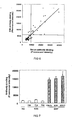
- Figure 6 is a graph showing the correlation between serum and milk antibody binding as analyzed by the method of the invention. Each point represents the average intensity of reaction obtained from serum and milk samples from one cow.
- Figure 7 is a bar graph showing the specificity of milk antibody binding to three strains of MAP as compared with negative reactions that were obtained with Mycobacterium scrofulaceum (MS) and three isolates of M . avium subsp.. avium (MAA).
- MS Mycobacterium scrofulaceum
- MAA M . avium subsp.. avium
- Figure 8 is a graph showing the detection of MAP in pooled serum by the method of the invention.
- the cut-off value (dotted line) was determined as the mean fluorescence obtained from six serum samples from a MAP-free farm +3 standard deviations for IgG binding.
- Figure 9 is a bar graph showing the detection of MAP infections in the American bison. All bison except VA 105 (arrow) were known to be MAP-positive as determined by fecal culture, ELISA, and histological examination.
- the invention is a method for diagnosing an infection in an animal caused by a MAP.
- the diagnostic method of the invention is based on antibody binding to one or more specific antibody binding sites that exist or existed on the surface of MAP.
- a test sample preferably a serum sample, obtained from an animal suspected of being infected with MAP, is exposed to a population of MAP. It is then determined if the test sample contains an antibody that binds to MAP, preferably to the surface of MAP. The test is positive for infection with MAP if antibodies in the test sample bind to MAP.
- the method of the invention is distinct from presently utilized methods for microbial diagnosis and provides several advantages that are unobtainable from such methods. Unlike culture methods, the method of the invention does not require isolation of MAP from an infected animal or the need to culture MAP in vitro. Therefore, the method of the invention provides results in a much shorter time period than is achievable with culture methods and, in contrast to culture methods, can provide a positive diagnosis even during times when MAP is not able to be isolated from a host animal.
- the method of the invention is not based on the determination of the presence of any specific macromolecule peculiar to MAP. Also, unlike tests based on antibody binding, such as ELISA testing, the method of the invention does not present an antibody to determine if it binds to an extract of MAP or portion of MAP that is present in a host animal. Rather, the method of the invention is based upon determining that one or more antibodies present in a test sample isolated from the body of a host animal binds to MAP that is brought into contact with the test sample.
- the method of the invention provides several advantages previously unobtainable by present diagnostic methods.
- the method of the invention may be performed rapidly.
- a positive or negative test result may be obtained rapidly, typically within about two hours.
- the method of the invention is extremely sensitive, more sensitive than presently available methods.
- the method of the invention can be used to provide a positive diagnosis even during periods when MAP is not detectable in, or isolatable from, a host animal. Additionally, the method of the invention has a specificity that is higher than is obtained with other presently available methods of diagnosis.
- the method of the invention is useful for the diagnosis of microbial infections in animals.
- animals include mammals, such as humans and non-human primates, carnivores such as dogs, cats, bears, and weasels, ungulate ruminants and non-ruminants such as horses, cattle, goats, sheep, pigs, non-ungulate ruminants such as camels and llamas, pinnipedia such as seals and sea lions, lagomorpha such as rabbits and hares, rodentia such as squirrels, rats, and mice, cetacea such as whales, dolphins, and porpoises, and proboscidea such as elephants.
- Such animals also include non-mammalian vertebrates such as birds, reptiles, amphibians, and fish.
- test sample that is obtained from an animal may be any fluid or tissue in which an antibody that specifically binds to a suspected causative organism would be present if the animal were infected with that organism.
- the test sample is blood or a portion thereof, such as plasma or preferably serum.
- sample sources may be utilized for the invention. The selection of such source of test sample will vary depending, primarily, on the symptoms and signs of an infected animal and the suspected cause of such symptoms or signs.
- test sample may be obtained from fluids such as saliva, milk, pus, tears and other ocular discharges, nasal discharges, sputum, cerebrospinal fluid, peritoneal or pleural fluid, urine, feces, and vaginal, uterine, or urethral secretions and discharges. Fluids may also include those that are produced as part of a pathologic process such as exudates or transudates, such as from the skin, the pleural or peritoneal cavity, the oral cavity, or from the digestive, respiratory, or genital system.
- the test sample may also be a solid tissue sample if appropriate for diagnosis of a particular disease.
- test sample may be obtained by whatever method is appropriate to obtain such a sample.
- the test sample may be obtained by methods such as syringe withdrawal of fluid, including vascular puncture, such as by venipuncture, or by withdrawal of fluid from other sources as described above, or by biopsy.
- the invention relates to Mycobacterium avium subsp. paratuberculosis (MAP) infections.
- Mycobacterium avium subsp. paratuberculosis (MAP) is the causative organism of Johne's Disease in cattle and Crohn's Disease in humans. This organism has proven to be a very difficult organism to establish as the cause of disease symptoms in cattle and in people and presents, therefore, a significant test to establish the efficacy, specificity, and sensitivity of the method of the present invention.
- the test sample may be exposed to a population of MAP in any way that permits antibodies that are contained in the test sample to interact with MAP.
- the test sample and MAP are combined in a vessel such as a test tube or a well and are mixed together, such as by stirring or tapping the exterior of the test tube or well.
- the test sample and the MAP may also be reacted together on a surface such as on a slide, filter, or membrane, such as a nitrocellulose membrane.
- the test sample is exposed to a population of intact whole MAP. In this way, antibody binding sites on the entire surface of the MAP are available for binding to antibodies in the test sample. It is preferred, if intact MAPs are used, that the MAPs be killed so as to avoid the risk of infection to humans and to other animals.
- a preferred method for killing the MAPs is by exposure of the MAPs to a chemical fixative.
- One preferred chemical fixative is formaldehyde which, when used to kill MAP organisms, maintains the ability of surface antibody binding sites of MAP to bind with antibodies in serum from animals infected with the organism.
- a preferred concentration of formaldehyde is about 1% to 10% v/v, with a concentration of about 2% most preferred.
- Other chemical fixatives that may be used to kill microorganisms for use in the method of the invention include non-coagulant fixatives such as acetone, glyceraldehydes, glutaraldehyde, and paraformaldehyde, and less preferred coagulant fixatives such as ethanol and mercuric chloride.
- Ethanol in concentrations tested by the inventors (70% v/v), destroyed the ability of surface antibody binding sites of MAP to bind with antibodies in serum from MAP infected animals. It is conceived that coagulant fixatives such as ethanol may be useful for killing MAPs to be used in the method of the invention, or that certain concentrations of such fixatives may be suitable for killing MAPs and may not render the killed MAPs unsuitable for the method of the invention. Because of this uncertainty concerning ethanol, coagulant fixatives such as ethanol are less preferred.
- the method of the invention may alternatively be performed by exposing the test sample to a population of disrupted or partial MAP, such as MAPs that have been fractionated, or to one or more isolated antibody binding sites of the surface of a MAP.
- a population of MAP includes exposing the test sample to intact MAP, to disrupted or partial organisms, or to one or more isolated antibody binding sites of MAP.
- the combination contains conjugates of antibodies from the test sample and MAP from the population. Any method that is suitable to detect the presence of antibody binding to an antigen is suitable for the method of the invention.
- antibody-MAP binding is determined by flow cytometry. Such flow cytometry determination may be performed by analysis of a sample obtained by mixing a suspension containing a serum sample and a population of MAP with a labeled anti-antibody, typically a fluorescein-labeled anti-antibody.. In another preferred embodiment, antibody-MAP binding is determined by blot analysis, such as dot blot or Western blot analysis.
- Such dot blot determination may be performed by mixing a suspension containing a serum sample and a population of MAP with an anti-antibody which is labeled, such as with biotin or colloidal gold, spotting this mixture on a membrane, such as a nitrocellulose or polyvinylidene fluoride (PVDF) membrane, and determining the presence of labeled MAP fixed on the membrane.
- a membrane such as a nitrocellulose or polyvinylidene fluoride (PVDF) membrane
- diagnosis of infection with such methods is accurate, sensitive, and specific. Determination of infection with methods such as dot blot analysis permits diagnosis to be made by visual inspection and such methods are therefore capable of being performed by individuals who are not technically trained in sophisticated laboratory techniques.
- the suspension was incubated at room temperature for 1 hr, washed three times with 100 microliters buffer A by centrifugation at 5000 rpm for 10 min, mixed with fluorescent-labeled rabbit anti-bovine IgG antibody (MP Biomedicals (formerly ICN Biomedicals), Irvine, CA, USA) diluted 1:50 in buffer A, incubated at room temperature for 1 hr, and washed twice with buffer A by centrifugation at 5000 rpm for 10 min.
- One tenth of the volume of the treated organisms was resuspended in 1 ml of buffer A and fluorescence on 10,000 organisms was analyzed by using a flow cytometer (LSR II, BD Biosciences, San Jose, CA, USA).
- the method of the invention was performed utilizing a simplified alternative technique for serological diagnosis by flow cytometry.
- This alternative technique requires a shorter time than the technique described in Example 1 and does not require centrifugation.
- bovine serum samples and 9 microliters of buffer A were added to whole organisms of MAP (5 microliters packed volume), and incubated at room temperature for 1 hr.
- the organisms were washed three times with 100 microliters of buffer A by centrifugation at 5000 rpm for 10 min, mixed with undiluted colloidal gold-labeled rabbit anti-bovine IgG antibody (Jackson ImmunoResearch Laboratories, Inc., West Grove, PA, USA), incubated at room temperature for 1 hr, and washed twice with 100 microliters of buffer A by centrifugation at 5000 rpm for 10 minutes.
- the treated organisms were resuspended in 100 microliters of buffer A and spotted on a PVDF membrane (Bio-Rad Laboratories, Inc., Hercules, CA, USA) by using a dot blot apparatus (Bio-Rad). Images of the stained organisms on the dot blot were captured, and their densitometric intensities measured, using a gel documentation system (ChemiDoc XRS, Bio-Rad).
- Serum samples from cows known to be infected with MAP were analyzed as described in Example 1. Serum samples from cows known to be infected with MAP were mixed with organism populations that were one of 5 strains of Mycobacterium avium subsp. avium (MAA), one strain of Mycobacterium scrofulaceum, or 4 strains of MAP, respectively prior to detection of binding by flow cytometry. Results are shown in Figure 1 .
- the method of the invention correctly identified infection with MAP in all samples and showed a lack of false positive diagnoses as the method of the invention did not show binding when bacterial populations closely related to MAP were used as the test organism.
- This study establishes the high specificity of the method of the invention, which is capable of distinguishing between very closely related organisms.
- Serum samples from 8 cows known to be infected with MAP were pooled.
- the pooled serum sample was tested as described in Example 1 by combining individual 2 microliter samples of the pooled serum sample with one of 8 different strains of MAP or with MAA. Results are shown graphically in Figure 2 .
- MAP Negative (n 5) - 2 of 5 samples (40%) positive by method of invention. Results of samples found negative by ELISA testing but positive by the method of the invention are underlined.
- the control sample (None) showed a low level of fluorescent activity as determined by flow cytometry. Additionally, the MAP positive serum showed a very high level of fluorescent activity indicating a high level of serum antibody binding with the MAP organisms. Unexpectedly, the fluorescent activity of the MAP free pooled sera, that found to be negative by ELISA test, showed a level of fluorescent activity about twice that of control. This result indicates that the ELISA test incorrectly identified MAP infected cows as being free of infection. The diagnostic test according to the invention, however, correctly identified that antibodies due to MAP infection were present in the pooled serum from this group.
- the dot blot technique of the invention showed a high degree of antibody binding in sera from cows previously found to be MAP positive by ELISA test. Additionally, antibody binding to MAP was detected by this method even in sera from cows previously found to be negative by ELISA testing, establishing the high sensitivity of the method of the invention as performed by dot blot technique.
- the dot blot technique is a simple method that can be performed easily in the laboratory by trained personnel. Additionally, because results of dot blot technique are evaluated visually, this technique can be performed in field conditions even by individuals not trained in laboratory techniques. Thus, the method of the invention is useful for both laboratory and field diagnosis of MAP infected individuals, and such diagnosis may be performed by either technical or non-technical personnel.
- Serum and milk samples were obtained from 48 cows and analyzed by the method of the invention using a flow cytometer.
- the serum and milk samples were diluted 1:50 and 1:2, respectively, with a phosphate buffered saline solution.
- the samples were tested for the presence of antibodies against MAP using the technique described in Example 1. The results are shown in Figure 6 .
- Milk from a cow diagnosed as positive for MAP by IDEXX ELISA was tested as described in Example 9 except that the milk samples were divided into samples and each sample was then mixed with organisms from one of three strains of Mycobacterium avium subsp. avium, one strain of M. scrofulaceaum (MS), or three strains of MAP, as described above in Example 4. The results are shown in Figure 7 .
- Each well of a 96-well plate was inoculated with 100 microliters of a MAP suspension as described in Example 1 and then pelleted by centrifugation at 3500 x g for 10 min. After aspiration, the MAP pellets were resuspended and incubated with serum from a MAP-positive cow that had been diluted by adding serum from a MAP-negative cow to obtain dilutions of 1:2, 1:4, 1:8, 1:16 and 1:32. The samples were then analyzed by flow cytometry. Results are shown in Figure 8 .
- Serum samples were obtained from 24 bison and analyzed by the method of the invention using a flow cytometer. Serum samples were diluted 1:50 with a phosphate buffered saline solution and tested for the presence of antibodies against MAP using the technique described in Example 1. Results are shown in Figure 9 .
Landscapes
- Health & Medical Sciences (AREA)
- Life Sciences & Earth Sciences (AREA)
- Immunology (AREA)
- Engineering & Computer Science (AREA)
- Urology & Nephrology (AREA)
- Hematology (AREA)
- Biomedical Technology (AREA)
- Chemical & Material Sciences (AREA)
- Molecular Biology (AREA)
- Medicinal Chemistry (AREA)
- Biochemistry (AREA)
- Cell Biology (AREA)
- Tropical Medicine & Parasitology (AREA)
- Biotechnology (AREA)
- Food Science & Technology (AREA)
- Virology (AREA)
- Physics & Mathematics (AREA)
- Analytical Chemistry (AREA)
- Microbiology (AREA)
- General Health & Medical Sciences (AREA)
- General Physics & Mathematics (AREA)
- Pathology (AREA)
- Measuring Or Testing Involving Enzymes Or Micro-Organisms (AREA)
- Investigating Or Analysing Biological Materials (AREA)
- Peptides Or Proteins (AREA)
- Measuring And Recording Apparatus For Diagnosis (AREA)
Applications Claiming Priority (2)
| Application Number | Priority Date | Filing Date | Title |
|---|---|---|---|
| US10/832,761 US7276350B2 (en) | 2004-04-27 | 2004-04-27 | Method for diagnosing infectious diseases |
| PCT/US2005/013446 WO2005106476A2 (en) | 2004-04-27 | 2005-04-19 | Method for diagnosing infectious diseases |
Publications (3)
| Publication Number | Publication Date |
|---|---|
| EP1747025A2 EP1747025A2 (en) | 2007-01-31 |
| EP1747025A4 EP1747025A4 (en) | 2008-05-14 |
| EP1747025B1 true EP1747025B1 (en) | 2010-10-20 |
Family
ID=35136956
Family Applications (1)
| Application Number | Title | Priority Date | Filing Date |
|---|---|---|---|
| EP05737662A Expired - Lifetime EP1747025B1 (en) | 2004-04-27 | 2005-04-19 | Method for diagnosing infectious diseases |
Country Status (5)
| Country | Link |
|---|---|
| US (2) | US7276350B2 (https=) |
| EP (1) | EP1747025B1 (https=) |
| AT (1) | ATE485060T1 (https=) |
| DE (1) | DE602005024254D1 (https=) |
| WO (1) | WO2005106476A2 (https=) |
Families Citing this family (6)
| Publication number | Priority date | Publication date | Assignee | Title |
|---|---|---|---|---|
| DE102008029834A1 (de) * | 2008-06-25 | 2010-01-14 | Justus-Liebig-Universität Giessen | Verfahren zum spezifischen Nachweis von MAP-Antikörpern |
| US8008033B2 (en) * | 2008-10-15 | 2011-08-30 | Gilles Reza Georges Monif | Fuidi herd management schema |
| US9696304B2 (en) | 2012-01-27 | 2017-07-04 | University Of Tennessee Research Foundation | Methods for detecting a biomarker by alternating current electrokinetics |
| AU2013212574C1 (en) | 2012-01-27 | 2017-03-30 | University Of Tennessee Research Foundation | Method and apparatus for detection of a biomarker by alternating current electrokinetics |
| US9128098B2 (en) | 2012-10-31 | 2015-09-08 | Gilles R. G. Monif | Fuidi herd management and risk stratification methods |
| CA2988297A1 (en) | 2016-12-28 | 2018-06-28 | University Of Tennessee Research Foundation | Methods for detecting a biomarker by alternating current electrokinetics |
Family Cites Families (1)
| Publication number | Priority date | Publication date | Assignee | Title |
|---|---|---|---|---|
| NL9202197A (nl) * | 1992-12-17 | 1994-07-18 | Kreatech Biotech Bv | Werkwijze en inrichting voor het identificeren van een voor een mycobacteriële infectie verantwoordelijk mycobacterium species. |
-
2004
- 2004-04-27 US US10/832,761 patent/US7276350B2/en not_active Expired - Lifetime
-
2005
- 2005-04-19 AT AT05737662T patent/ATE485060T1/de not_active IP Right Cessation
- 2005-04-19 WO PCT/US2005/013446 patent/WO2005106476A2/en not_active Ceased
- 2005-04-19 EP EP05737662A patent/EP1747025B1/en not_active Expired - Lifetime
- 2005-04-19 DE DE602005024254T patent/DE602005024254D1/de not_active Expired - Lifetime
-
2007
- 2007-02-01 US US11/701,202 patent/US7422869B2/en not_active Expired - Fee Related
Non-Patent Citations (1)
| Title |
|---|
| NASER S A ET AL: "Specific seroreactivity of Crohn's disease patients against p35 and p36 antigens of M. avium subsp. paratuberculosis", VETERINARY MICROBIOLOGY, ELSEVIER BV, NL, vol. 77, no. 3-4, 20 December 2000 (2000-12-20), pages 497 - 504, XP002445953, ISSN: 0378-1135 * |
Also Published As
| Publication number | Publication date |
|---|---|
| US20050239147A1 (en) | 2005-10-27 |
| US7276350B2 (en) | 2007-10-02 |
| EP1747025A2 (en) | 2007-01-31 |
| DE602005024254D1 (https=) | 2010-12-02 |
| ATE485060T1 (de) | 2010-11-15 |
| EP1747025A4 (en) | 2008-05-14 |
| US20070141653A1 (en) | 2007-06-21 |
| US7422869B2 (en) | 2008-09-09 |
| WO2005106476A2 (en) | 2005-11-10 |
| WO2005106476A3 (en) | 2005-12-29 |
Similar Documents
| Publication | Publication Date | Title |
|---|---|---|
| US6720160B2 (en) | Method for simultaneous detection of multiple microbial antigens in biological specimens from mastitic animals | |
| Köhler et al. | Immune reactions in cattle after immunization with a Mycobacterium paratuberculosis vaccine and implications for the diagnosis of M. paratuberculosis and M. bovis infections | |
| US7422869B2 (en) | Method for diagnosing infectious diseases | |
| Shuralev et al. | Application of the Enfer chemiluminescent multiplex ELISA system for the detection of Mycobacterium bovis infection in goats | |
| US7713715B2 (en) | Method for diagnosing infections | |
| US8158371B2 (en) | Assay for antibodies to Mycobacterium paratuberculosis | |
| Cvetnic | Brucellosis in wild boar (Sus scrofa) in the Republic of Croati | |
| Bale et al. | Serological and bacteriological study of bovine brucellae from livestock investigation and breeding centres in Nigeria | |
| Chappel et al. | Leptospira interrogans serovar hardjo is not a major cause of bovine abortion in Victoria | |
| Biswal et al. | Detection of Mycobacterium avium subsp. Paratuberculosis (MAP) from subclinical caprine paratuberculosis cases of Odisha. | |
| Bercovich et al. | Evaluation of the currently used diagnostic procedures for the detection of Brucella melitensis in sheep | |
| JP4359684B2 (ja) | ヨーネ病の検査方法 | |
| Ngwa et al. | Evaluation of the association between the lactation stage and serum and milk ELISA results in the diagnosis of ovine Paratuberculosis | |
| DE102008029833A1 (de) | Verfahren zum frühen Nachweis einer Infektion mit MAP | |
| Spaander | Risk factors for canine leptospirosis in the Netherlands in the years 2015 through 2020 | |
| Rao et al. | A Review on History and Detection of Salmonella Enterica Typhi | |
| Trimble | Prevalence of equine leptospiral shedding using urine polymerase chain reaction and serum microscopic agglutination testing | |
| Pokharel et al. | Seroprevalence of Chlamydia abortus in Anestrous Cattle of Nawalpur and Chitwan District, Nepal | |
| Juntautsa | Development of an indirect elisa to detect antibodies against actinobacillus suis using boiled whole cell antigen | |
| Leucosis | The Modification of Technical Annexes of Council Directive 64/432/EEC to take account of Scientific Developments regarding Tuberculosis, Brucellosis and Enzootic Bovine Leucosis | |
| Robbe-Austerman | Update on diagnostic testing for Johne's disease. | |
| BAKAR | MYCOBACTERIUM AVIUM SUBSPECIES PARATUBERCULOSIS (MAP) INFECTION IN DAIRY CATLLE IN TAMAN PERTANIAN UNIVERSITI | |
| Kalis et al. | Comparison of two absorbed ELISA's and a complement fixation test for the diagnosis of paratuberculosis. | |
| Robbe-Austerman et al. | Comparison of the γ-Interferon ELISA and the Skin Test for the Detection of Sub-clinical Johne's Disease in Cattle | |
| Palanivel et al. | DESCRIPTION OF THE INFECTIOUS STATUS IN MURRAH BUFFALO HERD NATURALLY INFECTED BY MYCOBACTERIUM AVIUM SUBSP. PARATUBERCULOSIS (MAP) IN TAMILNADU |
Legal Events
| Date | Code | Title | Description |
|---|---|---|---|
| PUAI | Public reference made under article 153(3) epc to a published international application that has entered the european phase |
Free format text: ORIGINAL CODE: 0009012 |
|
| 17P | Request for examination filed |
Effective date: 20061127 |
|
| AK | Designated contracting states |
Kind code of ref document: A2 Designated state(s): AT BE BG CH CY CZ DE DK EE ES FI FR GB GR HU IE IS IT LI LT LU MC NL PL PT RO SE SI SK TR |
|
| DAX | Request for extension of the european patent (deleted) | ||
| A4 | Supplementary search report drawn up and despatched |
Effective date: 20080414 |
|
| 17Q | First examination report despatched |
Effective date: 20090119 |
|
| GRAP | Despatch of communication of intention to grant a patent |
Free format text: ORIGINAL CODE: EPIDOSNIGR1 |
|
| GRAS | Grant fee paid |
Free format text: ORIGINAL CODE: EPIDOSNIGR3 |
|
| GRAA | (expected) grant |
Free format text: ORIGINAL CODE: 0009210 |
|
| AK | Designated contracting states |
Kind code of ref document: B1 Designated state(s): AT BE BG CH CY CZ DE DK EE ES FI FR GB GR HU IE IS IT LI LT LU MC NL PL PT RO SE SI SK TR |
|
| REG | Reference to a national code |
Ref country code: GB Ref legal event code: FG4D |
|
| REG | Reference to a national code |
Ref country code: CH Ref legal event code: EP |
|
| REG | Reference to a national code |
Ref country code: IE Ref legal event code: FG4D |
|
| REF | Corresponds to: |
Ref document number: 602005024254 Country of ref document: DE Date of ref document: 20101202 Kind code of ref document: P |
|
| REG | Reference to a national code |
Ref country code: NL Ref legal event code: VDEP Effective date: 20101020 |
|
| LTIE | Lt: invalidation of european patent or patent extension |
Effective date: 20101020 |
|
| PG25 | Lapsed in a contracting state [announced via postgrant information from national office to epo] |
Ref country code: LT Free format text: LAPSE BECAUSE OF FAILURE TO SUBMIT A TRANSLATION OF THE DESCRIPTION OR TO PAY THE FEE WITHIN THE PRESCRIBED TIME-LIMIT Effective date: 20101020 |
|
| PG25 | Lapsed in a contracting state [announced via postgrant information from national office to epo] |
Ref country code: FI Free format text: LAPSE BECAUSE OF FAILURE TO SUBMIT A TRANSLATION OF THE DESCRIPTION OR TO PAY THE FEE WITHIN THE PRESCRIBED TIME-LIMIT Effective date: 20101020 Ref country code: SE Free format text: LAPSE BECAUSE OF FAILURE TO SUBMIT A TRANSLATION OF THE DESCRIPTION OR TO PAY THE FEE WITHIN THE PRESCRIBED TIME-LIMIT Effective date: 20101020 Ref country code: BG Free format text: LAPSE BECAUSE OF FAILURE TO SUBMIT A TRANSLATION OF THE DESCRIPTION OR TO PAY THE FEE WITHIN THE PRESCRIBED TIME-LIMIT Effective date: 20110120 Ref country code: AT Free format text: LAPSE BECAUSE OF FAILURE TO SUBMIT A TRANSLATION OF THE DESCRIPTION OR TO PAY THE FEE WITHIN THE PRESCRIBED TIME-LIMIT Effective date: 20101020 Ref country code: IS Free format text: LAPSE BECAUSE OF FAILURE TO SUBMIT A TRANSLATION OF THE DESCRIPTION OR TO PAY THE FEE WITHIN THE PRESCRIBED TIME-LIMIT Effective date: 20110220 Ref country code: SI Free format text: LAPSE BECAUSE OF FAILURE TO SUBMIT A TRANSLATION OF THE DESCRIPTION OR TO PAY THE FEE WITHIN THE PRESCRIBED TIME-LIMIT Effective date: 20101020 Ref country code: PT Free format text: LAPSE BECAUSE OF FAILURE TO SUBMIT A TRANSLATION OF THE DESCRIPTION OR TO PAY THE FEE WITHIN THE PRESCRIBED TIME-LIMIT Effective date: 20110221 Ref country code: NL Free format text: LAPSE BECAUSE OF FAILURE TO SUBMIT A TRANSLATION OF THE DESCRIPTION OR TO PAY THE FEE WITHIN THE PRESCRIBED TIME-LIMIT Effective date: 20101020 |
|
| PG25 | Lapsed in a contracting state [announced via postgrant information from national office to epo] |
Ref country code: GR Free format text: LAPSE BECAUSE OF FAILURE TO SUBMIT A TRANSLATION OF THE DESCRIPTION OR TO PAY THE FEE WITHIN THE PRESCRIBED TIME-LIMIT Effective date: 20110121 Ref country code: BE Free format text: LAPSE BECAUSE OF FAILURE TO SUBMIT A TRANSLATION OF THE DESCRIPTION OR TO PAY THE FEE WITHIN THE PRESCRIBED TIME-LIMIT Effective date: 20101020 |
|
| PG25 | Lapsed in a contracting state [announced via postgrant information from national office to epo] |
Ref country code: ES Free format text: LAPSE BECAUSE OF FAILURE TO SUBMIT A TRANSLATION OF THE DESCRIPTION OR TO PAY THE FEE WITHIN THE PRESCRIBED TIME-LIMIT Effective date: 20110131 Ref country code: EE Free format text: LAPSE BECAUSE OF FAILURE TO SUBMIT A TRANSLATION OF THE DESCRIPTION OR TO PAY THE FEE WITHIN THE PRESCRIBED TIME-LIMIT Effective date: 20101020 Ref country code: CZ Free format text: LAPSE BECAUSE OF FAILURE TO SUBMIT A TRANSLATION OF THE DESCRIPTION OR TO PAY THE FEE WITHIN THE PRESCRIBED TIME-LIMIT Effective date: 20101020 |
|
| PLBE | No opposition filed within time limit |
Free format text: ORIGINAL CODE: 0009261 |
|
| STAA | Information on the status of an ep patent application or granted ep patent |
Free format text: STATUS: NO OPPOSITION FILED WITHIN TIME LIMIT |
|
| PG25 | Lapsed in a contracting state [announced via postgrant information from national office to epo] |
Ref country code: DK Free format text: LAPSE BECAUSE OF FAILURE TO SUBMIT A TRANSLATION OF THE DESCRIPTION OR TO PAY THE FEE WITHIN THE PRESCRIBED TIME-LIMIT Effective date: 20101020 Ref country code: RO Free format text: LAPSE BECAUSE OF FAILURE TO SUBMIT A TRANSLATION OF THE DESCRIPTION OR TO PAY THE FEE WITHIN THE PRESCRIBED TIME-LIMIT Effective date: 20101020 Ref country code: PL Free format text: LAPSE BECAUSE OF FAILURE TO SUBMIT A TRANSLATION OF THE DESCRIPTION OR TO PAY THE FEE WITHIN THE PRESCRIBED TIME-LIMIT Effective date: 20101020 Ref country code: SK Free format text: LAPSE BECAUSE OF FAILURE TO SUBMIT A TRANSLATION OF THE DESCRIPTION OR TO PAY THE FEE WITHIN THE PRESCRIBED TIME-LIMIT Effective date: 20101020 |
|
| 26N | No opposition filed |
Effective date: 20110721 |
|
| REG | Reference to a national code |
Ref country code: DE Ref legal event code: R097 Ref document number: 602005024254 Country of ref document: DE Effective date: 20110721 |
|
| PG25 | Lapsed in a contracting state [announced via postgrant information from national office to epo] |
Ref country code: MC Free format text: LAPSE BECAUSE OF NON-PAYMENT OF DUE FEES Effective date: 20110430 |
|
| REG | Reference to a national code |
Ref country code: CH Ref legal event code: PL |
|
| GBPC | Gb: european patent ceased through non-payment of renewal fee |
Effective date: 20110419 |
|
| PG25 | Lapsed in a contracting state [announced via postgrant information from national office to epo] |
Ref country code: IT Free format text: LAPSE BECAUSE OF FAILURE TO SUBMIT A TRANSLATION OF THE DESCRIPTION OR TO PAY THE FEE WITHIN THE PRESCRIBED TIME-LIMIT Effective date: 20101020 |
|
| REG | Reference to a national code |
Ref country code: FR Ref legal event code: ST Effective date: 20111230 |
|
| PG25 | Lapsed in a contracting state [announced via postgrant information from national office to epo] |
Ref country code: FR Free format text: LAPSE BECAUSE OF NON-PAYMENT OF DUE FEES Effective date: 20110502 Ref country code: LI Free format text: LAPSE BECAUSE OF NON-PAYMENT OF DUE FEES Effective date: 20110430 Ref country code: CH Free format text: LAPSE BECAUSE OF NON-PAYMENT OF DUE FEES Effective date: 20110430 Ref country code: DE Free format text: LAPSE BECAUSE OF NON-PAYMENT OF DUE FEES Effective date: 20111101 |
|
| REG | Reference to a national code |
Ref country code: IE Ref legal event code: MM4A |
|
| PG25 | Lapsed in a contracting state [announced via postgrant information from national office to epo] |
Ref country code: GB Free format text: LAPSE BECAUSE OF NON-PAYMENT OF DUE FEES Effective date: 20110419 |
|
| REG | Reference to a national code |
Ref country code: DE Ref legal event code: R119 Ref document number: 602005024254 Country of ref document: DE Effective date: 20111101 |
|
| PG25 | Lapsed in a contracting state [announced via postgrant information from national office to epo] |
Ref country code: IE Free format text: LAPSE BECAUSE OF NON-PAYMENT OF DUE FEES Effective date: 20110419 |
|
| PG25 | Lapsed in a contracting state [announced via postgrant information from national office to epo] |
Ref country code: CY Free format text: LAPSE BECAUSE OF FAILURE TO SUBMIT A TRANSLATION OF THE DESCRIPTION OR TO PAY THE FEE WITHIN THE PRESCRIBED TIME-LIMIT Effective date: 20101020 Ref country code: LU Free format text: LAPSE BECAUSE OF NON-PAYMENT OF DUE FEES Effective date: 20110419 |
|
| PG25 | Lapsed in a contracting state [announced via postgrant information from national office to epo] |
Ref country code: TR Free format text: LAPSE BECAUSE OF FAILURE TO SUBMIT A TRANSLATION OF THE DESCRIPTION OR TO PAY THE FEE WITHIN THE PRESCRIBED TIME-LIMIT Effective date: 20101020 |
|
| PG25 | Lapsed in a contracting state [announced via postgrant information from national office to epo] |
Ref country code: HU Free format text: LAPSE BECAUSE OF FAILURE TO SUBMIT A TRANSLATION OF THE DESCRIPTION OR TO PAY THE FEE WITHIN THE PRESCRIBED TIME-LIMIT Effective date: 20101020 |